BMP-Mediated Functional Cooperation between Dlx5;Dlx6 and Msx1;Msx2 during Mammalian Limb...
14
BMP-Mediated Functional Cooperation between Dlx5;Dlx6 and Msx1;Msx2 during Mammalian Limb Development Maxence Vieux-Rochas 1¤a , Kamal Bouhali 1 , Stefano Mantero 2¤b , Giulia Garaffo 2 , Paolo Provero 2 , Simonetta Astigiano 3 , Ottavia Barbieri 3,4 , Mariano F. Caratozzolo 5 , Apollonia Tullo 5 , Luisa Guerrini 6 , Yvan Lallemand 7 , Benoıˆt Robert 7 , Giovanni Levi 1. , Giorgio R. Merlo 2,8 * . 1 Evolution des Re ´gulations Endocriniennes, Centre national de la recherche scientifique, UMR-7221, Muse ´um National d’Histoire Naturelle, Paris, France, 2 Molecular Biotechnology Center, University of Torino, Torino, Italy, 3 Istituto Di Ricovero e Cura a Carattere Scientifico Azienda Ospedale Universita ` San Martino, IST Istituto Nazionale per la Ricerca sul Cancro, Genova, Italy, 4 Department of Experimental Medicine, University of Genova, Genova, Italy, 5 Institute for Biomedical Technologies, National Research Council, Bari, Italy, 6 Department of Biosciences, University of Milano, Milano, Italy, 7 Institut Pasteur, Department of Developmental Biology, Centre national de la recherche scientifique URA-2578, Paris, France, 8 Dulbecco Telethon Institute, University of Torino, Torino, Italy Abstract The Dlx and Msx homeodomain transcription factors play important roles in the control of limb development. The combined disruption of Msx1 and Msx2, as well as that of Dlx5 and Dlx6, lead to limb patterning defects with anomalies in digit number and shape. Msx1;Msx2 double mutants are characterized by the loss of derivatives of the anterior limb mesoderm which is not observed in either of the simple mutants. Dlx5;Dlx6 double mutants exhibit hindlimb ectrodactyly. While the morphogenetic action of Msx genes seems to involve the BMP molecules, the mode of action of Dlx genes still remains elusive. Here, examining the limb phenotypes of combined Dlx and Msx mutants we reveal a new Dlx-Msx regulatory loop directly involving BMPs. In Msx1;Dlx5;Dlx6 triple mutant mice (TKO), beside the expected ectrodactyly, we also observe the hallmark morphological anomalies of Msx1;Msx2 double mutants suggesting an epistatic role of Dlx5 and Dlx6 over Msx2. In Msx2;Dlx5;Dlx6 TKO mice we only observe an aggravation of the ectrodactyly defect without changes in the number of the individual components of the limb. Using a combination of qPCR, ChIP and bioinformatic analyses, we identify two Dlx/Msx regulatory pathways: 1) in the anterior limb mesoderm a non-cell autonomous Msx-Dlx regulatory loop involves BMP molecules through the AER and 2) in AER cells and, at later stages, in the limb mesoderm the regulation of Msx2 by Dlx5 and Dlx6 occurs also cell autonomously. These data bring new elements to decipher the complex AER- mesoderm dialogue that takes place during limb development and provide clues to understanding the etiology of congenital limb malformations. Citation: Vieux-Rochas M, Bouhali K, Mantero S, Garaffo G, Provero P, et al. (2013) BMP-Mediated Functional Cooperation between Dlx5;Dlx6 and Msx1;Msx2 during Mammalian Limb Development. PLoS ONE 8(1): e51700. doi:10.1371/journal.pone.0051700 Editor: Michael Schubert, Ecole Normale Supe ´rieure de Lyon, France Received June 28, 2012; Accepted November 5, 2012; Published January 29, 2013 Copyright: ß 2013 Vieux-Rochas et al. This is an open-access article distributed under the terms of the Creative Commons Attribution License, which permits unrestricted use, distribution, and reproduction in any medium, provided the original author and source are credited. Funding: Giorgio R. Merlo is supported by the Italian Telethon Foundation (GGP11097), the Fondazione per la Ricerca Biomedica (Torino, Italy), and the Compagnia di San Paolo (Torino, Italy). Ottavia Barbieri is supported by the Italian Telethon Foundation (GP0218Y01). Benoit Robert is supported by Institut Pasteur and the Centre National de la Recherche Scientifique (France). Benoit Robert and Giovanni Levi are supported by the Agence Nationale de la Recherche (France) grant ‘‘GENDACTYL.’’ The funders had no role in study design, data collection and analysis, decision to publish, or preparation of the manuscript. Competing Interests: The authors have declared that no competing interests exist. * E-mail: [email protected] . These authors contributed equally to this work. ¤a Current address: National Research Center Frontiers in Genetics, School of Sciences, Ecole Polytechnique Fe ´de ´rale, Lausanne, Switzerland ¤b Current address: Istituto Clinico Humanitas IRCCS, Rozzano, Milano, Italy Introduction The developing vertebrate limb is widely adopted as a model to study cell-cell signaling, pattern formation and morphogenesis, and has provided a wealth of knowledge of the function and regulation of specific transcription factors and signaling molecules [1,2,3]. The phenotype of a large set of mutant mice with limb defects led to the identification of genes and regulatory pathways essential for normal limb development. The spatio-temporal organization of the complex network of signaling and transcrip- tional regulations has been elucidated only in part. In brief, genes and regulatory modules can be related to the activation/ maintenance/regression of three signaling systems: a) Sonic hedgehog (SHH) and the Zone of Polarizing Activity (ZPA), for the control of digit patterning along the antero-posterior axis [4,5,6], b) Fibroblast Growth Factors (FGFs) and the Apical Ectodermal Ridge (AER), for the control of proximo-distal growth and for ZPA maintenance [7,8,9,10,11], and c) Lmx1B, in the mesoderm, and Wnt7a and En-1, in the ectoderm, for dorso- ventral specification [12,13,14,15,16]. The three signaling systems are organized in precise time- and space-restricted manners, and are integrated in self-regulatory modules that assure the acquisi- tion or the correct digit complements, limb morphogenesis and overall growth [4,17,18,19]. The signaling molecules of the Bone Morphogenetic Protein (BMP) class have been proposed to link PLOS ONE | www.plosone.org 1 January 2013 | Volume 8 | Issue 1 | e51700
Transcript of BMP-Mediated Functional Cooperation between Dlx5;Dlx6 and Msx1;Msx2 during Mammalian Limb...
BMP-Mediated Functional Cooperation betweenDlx5;Dlx6 and Msx1;Msx2 during Mammalian LimbDevelopmentMaxence Vieux-Rochas1¤a, Kamal Bouhali1, Stefano Mantero2¤b, Giulia Garaffo2, Paolo Provero2,
Simonetta Astigiano3, Ottavia Barbieri3,4, Mariano F. Caratozzolo5, Apollonia Tullo5, Luisa Guerrini6,
Yvan Lallemand7, Benoıt Robert7, Giovanni Levi1., Giorgio R. Merlo2,8*.
1 Evolution des Regulations Endocriniennes, Centre national de la recherche scientifique, UMR-7221, Museum National d’Histoire Naturelle, Paris, France, 2 Molecular
Biotechnology Center, University of Torino, Torino, Italy, 3 Istituto Di Ricovero e Cura a Carattere Scientifico Azienda Ospedale Universita San Martino, IST Istituto
Nazionale per la Ricerca sul Cancro, Genova, Italy, 4 Department of Experimental Medicine, University of Genova, Genova, Italy, 5 Institute for Biomedical Technologies,
National Research Council, Bari, Italy, 6 Department of Biosciences, University of Milano, Milano, Italy, 7 Institut Pasteur, Department of Developmental Biology, Centre
national de la recherche scientifique URA-2578, Paris, France, 8 Dulbecco Telethon Institute, University of Torino, Torino, Italy
Abstract
The Dlx and Msx homeodomain transcription factors play important roles in the control of limb development. Thecombined disruption of Msx1 and Msx2, as well as that of Dlx5 and Dlx6, lead to limb patterning defects with anomalies indigit number and shape. Msx1;Msx2 double mutants are characterized by the loss of derivatives of the anterior limbmesoderm which is not observed in either of the simple mutants. Dlx5;Dlx6 double mutants exhibit hindlimb ectrodactyly.While the morphogenetic action of Msx genes seems to involve the BMP molecules, the mode of action of Dlx genes stillremains elusive. Here, examining the limb phenotypes of combined Dlx and Msx mutants we reveal a new Dlx-Msxregulatory loop directly involving BMPs. In Msx1;Dlx5;Dlx6 triple mutant mice (TKO), beside the expected ectrodactyly, wealso observe the hallmark morphological anomalies of Msx1;Msx2 double mutants suggesting an epistatic role of Dlx5 andDlx6 over Msx2. In Msx2;Dlx5;Dlx6 TKO mice we only observe an aggravation of the ectrodactyly defect without changes inthe number of the individual components of the limb. Using a combination of qPCR, ChIP and bioinformatic analyses, weidentify two Dlx/Msx regulatory pathways: 1) in the anterior limb mesoderm a non-cell autonomous Msx-Dlx regulatory loopinvolves BMP molecules through the AER and 2) in AER cells and, at later stages, in the limb mesoderm the regulation ofMsx2 by Dlx5 and Dlx6 occurs also cell autonomously. These data bring new elements to decipher the complex AER-mesoderm dialogue that takes place during limb development and provide clues to understanding the etiology ofcongenital limb malformations.
Citation: Vieux-Rochas M, Bouhali K, Mantero S, Garaffo G, Provero P, et al. (2013) BMP-Mediated Functional Cooperation between Dlx5;Dlx6 and Msx1;Msx2during Mammalian Limb Development. PLoS ONE 8(1): e51700. doi:10.1371/journal.pone.0051700
Editor: Michael Schubert, Ecole Normale Superieure de Lyon, France
Received June 28, 2012; Accepted November 5, 2012; Published January 29, 2013
Copyright: � 2013 Vieux-Rochas et al. This is an open-access article distributed under the terms of the Creative Commons Attribution License, which permitsunrestricted use, distribution, and reproduction in any medium, provided the original author and source are credited.
Funding: Giorgio R. Merlo is supported by the Italian Telethon Foundation (GGP11097), the Fondazione per la Ricerca Biomedica (Torino, Italy), and theCompagnia di San Paolo (Torino, Italy). Ottavia Barbieri is supported by the Italian Telethon Foundation (GP0218Y01). Benoit Robert is supported by InstitutPasteur and the Centre National de la Recherche Scientifique (France). Benoit Robert and Giovanni Levi are supported by the Agence Nationale de la Recherche(France) grant ‘‘GENDACTYL.’’ The funders had no role in study design, data collection and analysis, decision to publish, or preparation of the manuscript.
Competing Interests: The authors have declared that no competing interests exist.
* E-mail: [email protected]
. These authors contributed equally to this work.
¤a Current address: National Research Center Frontiers in Genetics, School of Sciences, Ecole Polytechnique Federale, Lausanne, Switzerland¤b Current address: Istituto Clinico Humanitas IRCCS, Rozzano, Milano, Italy
Introduction
The developing vertebrate limb is widely adopted as a model to
study cell-cell signaling, pattern formation and morphogenesis,
and has provided a wealth of knowledge of the function and
regulation of specific transcription factors and signaling molecules
[1,2,3]. The phenotype of a large set of mutant mice with limb
defects led to the identification of genes and regulatory pathways
essential for normal limb development. The spatio-temporal
organization of the complex network of signaling and transcrip-
tional regulations has been elucidated only in part. In brief, genes
and regulatory modules can be related to the activation/
maintenance/regression of three signaling systems: a) Sonic
hedgehog (SHH) and the Zone of Polarizing Activity (ZPA), for
the control of digit patterning along the antero-posterior axis
[4,5,6], b) Fibroblast Growth Factors (FGFs) and the Apical
Ectodermal Ridge (AER), for the control of proximo-distal growth
and for ZPA maintenance [7,8,9,10,11], and c) Lmx1B, in the
mesoderm, and Wnt7a and En-1, in the ectoderm, for dorso-
ventral specification [12,13,14,15,16]. The three signaling systems
are organized in precise time- and space-restricted manners, and
are integrated in self-regulatory modules that assure the acquisi-
tion or the correct digit complements, limb morphogenesis and
overall growth [4,17,18,19]. The signaling molecules of the Bone
Morphogenetic Protein (BMP) class have been proposed to link
PLOS ONE | www.plosone.org 1 January 2013 | Volume 8 | Issue 1 | e51700
these three systems together [20,21,22,23,24,25], thus they are
regarded as key players in the coordination of limb patterning,
morphogenesis and growth along the three axes.
Proximo-distal limb development and digit extension is directly
controlled by the signaling activity of the AER via expression of
morphogens of the FGF family [7,8,10,26] and their regulation by
other signaling molecules such as SHH and BMP antagonists
[20,21,27,28]. In this scenario, the Dlx and Msx homeobox
transcription factors, expressed in the AER and the mesoderm of
the limb buds, play an important morphogenetic role, although
their functions and regulations are yet to be defined at the
molecular level. Dlx5;Dlx6 double knock-out (DKO) mice are
characterized by loss of the central digit(s) and/or fusion with the
lateral ones [29,30]; these mice constitute a model of the human
congenital defect ectrodactyly or Split Hand Foot Malformation
type I, a condition linked to genomic alterations encompassing the
DLX5;DLX6 region, and for which point mutations in the DNA-
binding domain of DLX5 have recently been found [31]. The
double inactivation of Msx1 and Msx2 genes results in moderate-
penetrance polydactyly of the forelimbs (FL) and oligodactyly of
the hindlimbs (HL) (loss of the anterior digits and part of the tibia
in the zeugopod) [32,33,34].
Studies on the cellular and molecular functions of Dlx and Msx
proteins during limb development have met with difficulties owing
to several reasons. First, none of the single knockout for Dlx5, Dlx6,
Msx1 or Msx2 shows evident limb defects
[29,32,35,36,37,38,39,40,41], suggesting some degree of function-
al redundancy. Second, members of the Dlx and Msx families are
expressed both in the AER and in restricted regions of the limb
mesoderm [42,43]. Third, in vitro the Dlx and Msx proteins
compete for the same DNA binding sites, form heterodimers via
their homeodomain and reciprocally inhibit their transcriptional
activities [44], due to the high degree of homology of their
homeodomains [37,43,45,46,47]. However, current literature
suggest that Dlx and Msx proteins have distinct functions: Msx1
and Msx2 are known to control cell proliferation and differenti-
ation in a variety of cell types [48,49,50,51], while Dlx genes are
implicated in the differentiation of specific cell lineages, such as
forebrain interneurons [52,53,54], olfactory receptor neurons [55],
osteoblasts [35,56], and the AER and ectoderm [29,30,57,58].
Notably, Dlx and Msx genes have been shown to cooperate only in
specific cases [59,60,61], but not at all sites where they are co-
expressed. However, Dlx5 has been shown to regulate Msx2
transcription in the AER, via homeodomain binding elements
present in the gene’s promoter [62,63].
We investigate possible interactions between Dlx5;Dlx6 and
Msx1 or Msx2 in limb development, by generating double and
triple Dlx;Msx compound mutant mice and analyzing their limb
phenotypes. The limb phenotype of Msx1;Dlx5;Dlx6 triple knock-
out (TKO) mice shows features of the Msx1;Msx2 DKO phenotype
[32], leading to the conclusion that Dlx5;Dlx6 control the
expression of Msx2, but not of Msx1. In contrast, the limb
phenotype of Msx2;Dlx5;Dlx6 TKO mice is consistent with a
severe aggravation of the ectrodactyly defect. We also re-examine
the spatio-temporal expression of Dlx and Msx genes in FLs and
HLs and observe that in the AER these genes are co-expressed
whereas in the anterior mesenchyme Msx expression precedes that
of Dlx, ruling out a direct regulation of Msx by Dlx in this territory.
Combining these findings with qPCR expression analyses on the
limb buds of different compound embryos, and with ChIP data,
we propose that two modes of regulation coexist during limb
development: 1) a direct transcriptional regulation of Msx2 by Dlx
proteins in the AER and, later on, in the limb mesoderm, and 2) a
Bmp2- and Bmp4-mediated non-cell autonomous regulatory loop
between the AER and the anterior limb mesoderm.
Materials and Methods
Mouse Strains and BreedingAll animal procedures were reviewed and approved by the
Ethical Committee of the University of Torino and University of
Genova, the Italian Ministry of Health and the French Ministere
de l’Enseignement superieur et de la Recherche. No surgery or
other manipulation on adult animals was used in this study. All
efforts were made to minimize suffering. Generation and
genotyping of the Dlx5lacZ/+ (hereafter named Dlx5+/2);
Dlx5;Dlx6neo/+ (hereafter named Dlx5;Dlx6+/2); Msx1lacZ/+ and
Msx2lacZ/+ (hereafter named, respectively, Msx1+/2 and Msx2+/2)
have been previously reported [29,32,35,39]. These mutations
were maintained on a C57B6;DBA F1 mixed genetic background,
throughout. TKO embryos and newborn were obtained by
crossbreeding either the Msx1+/2 or the Msx2+/2 single hetero-
zygous parents with the Dlx5;Dlx6+/2 double heterozygous ones,
and then crossing the triple heterozygotes. Following mating, the
day of the vaginal plug was considered as embryonic age 0.5
(E0.5).
Skeletal Preparation and b-galactosidase DetectionCartilage staining (with Alcian Blue) of E14.5 embryos as well as
bone and cartilage staining (with Alcian Blue and Alizarine Red) of
E18.5 embryos were carried out as previously described [35]. For
lacZ expression analysis E10.5 embryos were fixed for 15–30 min
in 2% paraformaldehyde in PBS, while E14.5 embryos were fixed
for 15–30 min in 4% PFA. X-gal staining was performed as
described [35]. For detection of b-gal on limb sections, E10.5 and
E11.5 FLs and HLs were fixed with 4% paraformadehyde for 8–
12 hrs at 4uC, washed in PBS, cryoprotected with 30% sucrose,
frozen at 270uC, sectioned (thickness 11 mm) and stained as
described [35].
Whole-mount RNA in situ HybridizationFor RNA:RNA whole-mount in situ hybridization (WMISH),
embryos at the desired age were dissected in cold RNAse-free PBS,
fixed in cold 4% PFA for 12–16 hrs, rinsed with PBS,
permeabilized by treatment with proteinase-K, prehybridized
and hybridized as previously published [60]. Digoxygenin (DIG)-
UTP (Roche)-labeled antisense RNA probes were used, synthe-
sized by in vitro transcription with conventional methods.
WMISH was carried out following described procedures [64],
the signal was detected using an alkaline phosphatase-conjugated
anti-DIG antibody and developed with the chromogenic mix
NBT-BCIP (Roche). With each probe, at least two normal and
two mutant specimens were examined. The Dlx5 probe comprised
780 bp and was linearized with EcoRI and transcribed with T7
RNA polymerase [57]. The Dlx6 probe is a 350 bp fragment
spanning exons 3–4 [53]. The Msx1 probe was a 550 bp 39
spanning the homeodomain, containing exon 1; the Msx2 probe
corresponded to 378 bp in the first exon of Msx2 cDNA, the Fgf8
probe corresponded to most of the mouse coding sequence, the
Bmp4 probe (a kind gift from B. Hogan) contains the 39 UTR and
most of the coding sequence from a mouse cDNA [65], the probe
for Gremlin corresponded to the entire murine coding sequence (a
kind gift from R. Zeller). After hybridization, the signal was
revealed with the NBT-BCIP chromogenic reaction.
Dlx and Msx Gene during Limb Development
PLOS ONE | www.plosone.org 2 January 2013 | Volume 8 | Issue 1 | e51700
RNA Quantification by Real-time PCREmbryonic FLs and HLs at the age E11 were dissected in cold
RNase-free PBS, the anterior and posterior halves were separated
and pooled in RNA-later (Ambion). Pools of two half limbs from
the same embryos were used to extract total RNA, using the
Tissue Lyser II reagent followed by elution through RNA micro-
kit plus (Qiagen). cDNA synthesis was done using standard
conditions, 3 ng of each cDNA sample were used to carry out
qPCRs on a CFX96 equipment (Biorad) using the SybrSafe system
(Invitrogen). Samples were analyzed in technical triplicates, and
for each genotypes (except for the Msx1+/2;Dlx52/2;Dlx62/2)
biological triplicates could be analyzed. Primer sequences were
designed with the Primer Express online tool. RNA quality, primer
efficiency and correct size were tested by RT-PCR and agarose gel
electrophoresis. Standard curve were performed using WT cDNA
with four calibration points: 1:10; 1:40; 1:160; 1:640. Specificity
and absence of primer dimers was controlled by denaturation
curves. Rps9 mRNA abundance was used for normalization
(primer sequences provided in Table S1).
Genome-wide Identification of Dlx Binding Sites andTarget Genes
We used the Position-Weight matrix (PWM) provided by
JASPAR under accession PH0024.1. The score of a site was
computed with standard log-likelihood ratios, using as null model
the nucleotide frequencies computed over the whole intergenic
fraction of the mouse genome. We considered for further analysis
putative sites scoring 50% of the maximum possible score or
better. We selected among the sites identified above the ones that
are conserved in at least two of 8 vertebrate species (genome
sequence version in parenthesis): mouse (mm9), human (hg19),
cow (bosTau4), opossum (monDom5), platypus (ornAna1), chicken
(galGal3), frog (xenTro2), zebrafish (danRer6) and lamprey
(petMar1). A site is defined as conserved with species S if it lies
in a region of the mouse genome which is aligned with a region of
the S genome and the aligned sequence in/S/is a site according to
the same definition used for mouse sites. All genomic sequences
and pre-computed ‘‘Net’’ alignments were obtained from UCSC.
A ranked list of putative Dlx target genes was obtained from the
sites determined above by associating each site conserved in at
least one species to its closest Refseq mRNA, and then selecting
the sites located either within 10 kb upstream of the TSS, or
within the non-coding portion of the first exon, or in the first
intron. We then associated to each putative target a score equal to
the sum of the conservation scores (number of species) of its
associated sites.
Chromatin ImmunoprecipitationA DLX5-myc expression vectors (OriGene, USA) containing the
full-length human DLX5 cDNA with an in-frame insertion of the
myc-TAG at the C-terminus, was used as described [66]. The
Q178P point mutation [31] was inserted in the DLX5-myc
expression vector indicated above, by site directed mutagenesis
and sequence verified (Bio-Fab Research, Rome).
The U2Os human osteosarcoma cells were used; these cells
express low or undetectable levels of DLX5 mRNA endogenously,
but have been shown to respond to Dlx expression with activation
of the p63 promoter [57]. Eight mg of the DLX5-myc expression
vectors were used for transfections, which yielded an efficiency of
35% (number of myc-positive cells over total counted nuclei).
Chromatin was crosslinked, sonicated, immunoprecipitated with
either the anti-myc TAG (A-14 sc-789, SantaCruz, USA) or the
anti-acetyl-Histone H4 (06-866, ChIP Grade, Upstate Biotech-
nology USA,) antibodies and de-crosslinked according to instruc-
tions (EZ Magna ChipG, Millipore). Fragments of the human
BMP2 and BMP4 loci spanning the identified conserved regions
were PCR-amplified and analysed by gel electrophoresis (sequenc-
es provided in Table S2). Total chromatin was used as positive
control (input), chromatin from cells transfected with an empty
vector was used as negative control.
Results
Msx and Dlx Coexpression Analyses, in silicoPrevious evidences indicate that Dlx5 binds to homeodomain-
responsive elements in a proximal region of the Msx2 promoter,
and thereby regulates its transcription [62,63]. To further support
this possibility, we have used a human-mouse co-expression
network, generated using published profiling datasets [67,68] and
found that DLX5 and MSX genes are connected, i.e. each ranks in
the first 1% of the co-expression lists of the other (p,0.01, data
not shown).
Next, we screened conserved regions of the vertebrate genome
for the presence of consensus Dlx DNA-binding sites, as defined by
the Dlx5 PWM [69] present in the Jaspar database (accession NuPH0024.1) [70] and reported in Table S3. As the PWM for Dlx5
reported in Jaspar is not highly informative, it is not surprising that
a total 565,995 putative binding sites were initially identified in the
mouse genome. However, by introducing evolutionary conserva-
tion with at least two (out of 8) species examined as a further
criteria, this number is reduced to 11,262 sites, and with
conservation in three species is further reduced to 4085 (Table
S3, complete lists available upon request). The full annotation on
the UCSC genome browser is available upon request. As positive
controls, the well defined Dlx sites present in the Dlx5;Dlx6
intergenic region [71] and in the Msx2 promoter [62,63] were
correctly predicted (Fig. S1). The evolutionary conservation of the
sites suggests the presence of positive selection pressure to maintain
the sites, confirming their functional relevance.
We then generated a ranked list of putative Dlx targets, based
on the position of predicted conserved Dlx sites in the genome, as
indicated in the Methods sections. The ranked list contains 3,051
Refseq mRNAs associated to 2,412 unique Entrez gene IDs
(available upon request). The Msx1 and Msx2 genes were found in
the top 10% of the list of putative Dlx targets, strengthening the
possibility that Dlx proteins might directly regulate Msx expres-
sion.
Expression of Msx and Dlx Genes during LimbDevelopment
We examined expression of Dlx5, Dlx6, Msx1 and Msx2 in the
FL and HL of normal embryos, at E10.5 and E11.5, by X-gal
staining of heterozygous embryos carrying an allele with inserted
lacZ reporter (Dlx5, Msx1, Msx2), and by WMISH (Dlx6) (Fig. 1).
WMISH for Dlx5, Msx1 and Msx2 have been previously reported
with comparable results. In the AER, all four genes are co-
expressed starting at E9.5/E10. On the contrary, in the limb
mesoderm they are expressed with a different time-of-onset. At
E10.5 Msx1 and Msx2 are mainly expressed in two mesoderm
territories (anterior and posterior) of both the FL and the HL
(Fig. 1E–H, S–V), whereas at the same stage the Dlx5 transcript is
detected only in the anterior mesoderm of the FL, and not that of
the HL (compare Fig. 1A–B with O–P). The Dlx6 transcript is not
detected in the mesoderm at this stage (Fig. 1C–D, Q–R). In the
HLs, mesodermal expression of Dlx5 starts around E11.5 and is
confined to the anterior margin (Fig. 1W–X). At this stage, Msx1
and Msx2 are expressed in the AER and in a larger region
Dlx and Msx Gene during Limb Development
PLOS ONE | www.plosone.org 3 January 2013 | Volume 8 | Issue 1 | e51700

underneath the AER (Fig. 1K–N, Y-AB). Histological sections of
X-gal stained limb buds from Dlx5+/2, Msx1+/2 and Msx2+/2
embryos (Fig. 1W’, W’’, Y’ and AA’) reveal that Msx1 and Dlx5 are
truly co-expressed in the AER (strong signal in W’ and W’’) and in
the anterior HL mesoderm (a weak signal is present in W’’,
indicated with black arrows).
In summary, Msx expression precedes that of Dlx in the HL
anterior mesoderm, consequently in this location Dlx genes are
unlikely to regulate Msx gene transcription cell-autonomously and
Dlx and Msx proteins are unlikely to interact in the anterior limb
mesoderm, at early stages.
Msx2 Expression is Downregulated in Dlx5;Dlx6 DKOLimbs
To investigate interactions between the Dlx and the Msx genes
during limb development, we used animals DKO for Dlx5;Dlx6
and analyzed the expression of the Msx-lacZ reporter in this
genetic background. To do this, we compared the expression
patterns of the lacZ reporter between Msx1+/2 and Msx1+/
2;Dlx52/2;Dlx62/2 mice (Fig. 2A–H), and between Msx2+/2 and
Msx2+/2;Dlx52/2;Dlx62/2 mice (Fig. 2I–P). We observed a clear
reduction of Msx2 expression in the anterior mesoderm and AER
exclusively in the HLs, whereas neither expression of Msx1 in the
HLs nor expression of Msx1 and Msx2 in the FLs were significantly
changed (Fig. 2A–P). To further document the reduction of Msx2
expression in the AER of Dlx5;Dlx6 DKO limbs, we carried out
WMISH for Msx2 on cryostatic sections of Dlx5;Dlx6 DKO HLs,
at E11. Msx2 expression was strongly reduced or absent in the
AER and the underlying mesoderm of a central wedge of the
mutant limbs, as compared to the same region of normal limbs
(Fig. 2Q,R).
Since WMISH is not a quantitative method, we used
quantitative Real Time PCR (qRT-PCR) to quantify the
reduction of Msx2 mRNA on samples extracted from the HLs of
normal and Dlx5;Dlx6 DKO embryos, at E11.5. Limb buds were
divided in two halves on the proximo-distal axis, to determine gene
expression in the anterior and posterior mesoderm separately. In
such samples, the AER cells contributed minimally, while most of
the RNA derives from the mesenchyme. Msx2 mRNA abundance
was reduced by 60% in the anterior half, and by 50% in the
posterior half of Dlx5;Dlx6 DKO HLs from the same embryo, as
compared to WT. In the same samples, expression of Msx1 was
minimally or not changed (Fig. 2S). We then determined the
abundance of Msx and Dlx mRNAs in the anterior half of the HLs
of Msx12/2 and Msx22/2 (single homozygous) mutant embryos,
as compared to WT. In the Msx22/2 mutant the expression level
of Dlx5, Dlx6 and Msx1 was unchanged, whereas in the Msx12/2
mutant the expression levels of Dlx5, Dlx6 and Msx2 were reduced
by 65%, 40% and 15%, respectively. Moreover, in the posterior
half of the HLs from the Msx12/2 mutant we observed a
reduction of 40% in the abundance of Dlx5 and Msx2 mRNA,
whereas Dlx6 did not change (Fig. 2T,U).
Limb Phenotype of Msx2;Dlx5;Dlx6 Triple MutantsTo reveal possible functional interactions between the Dlx and
the Msx gene products during limb development, we generated
TKO mice with genotype Msx12/2;Dlx52/2;Dlx62/2 or Msx22/
2;Dlx52/2;Dlx62/2. TKO newborns were obtained at a frequen-
cy lower than expected, and all died shortly after birth, due to
severe craniofacial malformations and consequent breathing
impairment. Quadruple knock-out embryos were never obtained
in spite of several attempts.
We examined the limb skeleton in the TKO animals at two
ages: E14.5 (with Alcian-Blue) and E18.5/birth (with Alcian-Blue
and Alizarine-Red, which stain, respectively, cartilages and
mineralized bones). In Msx2;Dlx5;Dlx6 TKO animals, the HLs
were severely affected (Fig. 3G,H) whereas no evident alteration
was observed in the FLs (data not shown). The central digit was
always missing, rarely (,10%) the two central digits were missing,
while the remaining digits extended pairwise towards the opposite
(anterior-posterior) sides. Importantly, no loss of anterior digits or
of zeugopod elements was ever observed (Fig. 3G,H). The limb
phenotype observed in the Msx2;Dlx5;Dlx6 TKO mutant animals
is consistent with a significant aggravation of the ectrodactyly
phenotype seen in Dlx5;Dlx6 DKO [29,30] (compare Fig. 3G with
3C, and 3H with 3D), without appearance of other recognizable
phenotypes. Remarkably a very similar phenotype, although less
severe, was observed in animals with genotype Msx2+/2;Dlx52/
2;Dlx62/2, i.e. in the presence of a single wild-type Msx2 allele
(Fig. 3E,F).
Limb Phenotype of Msx1;Dlx5;Dlx6 Triple MutantsIn Msx1;Dlx5;Dlx6 TKO mice the FL were usually normal,
although in one case (1/6) anterior polydactyly was observed (data
not shown). Of note, polydactyly has been reported for the FLs in
Msx1;Msx2 DKO mice with a moderate penetrance [32]. On the
contrary, the TKO HLs displayed a severe phenotype consisting
in the loss of the tibia and of 3–4 preaxial digits (Fig. 3I,J). Since
the HLs of Msx1;Msx2 DKO usually exhibit loss of 1–2 anterior
digits and of the tibia [32], we interpreted the TKO phenotype as
a combination of ectrodactyly, caused by the loss of Dlx5;Dlx6
[29,30] (Fig 3C,D), and the preaxial adactyly as observed upon the
loss of Msx1;Msx2 [32] (Fig. 3K).
The appearance of features of the Msx1;Msx2 DKO phenotype
in the Msx1;Dlx5;Dlx6 TKO animals, which retain two functional
Msx2 alleles, argues in favor of a severe reduction of Msx2
expression in the TKO limbs, as compared to the Dlx5;Dlx6 DKO.
Indeed, removing a single Msx1 allele in the context of
Dlx5;Dlx6 DKO background (i.e.Msx1+/2;Dlx52/2;Dlx62/2 em-
bryos) results in a 50% reduction of Msx2 mRNA in both the
anterior and the posterior half of the embryonic HLs, as compared
to WT. Moreover, expression of Msx2 in the anterior and posterior
halves of Msx1 homozygus mutant HLs was reduced by 30%
compared to Msx1 heterozygous HLs (data not shown). This
indicates that reduced Msx2 expression may result from the
combination of (1) loss of Dlx5 and Dlx6 (Fig. 2U, Fig. S2),
upstream regulators of Msx2, and (2) the loss of Msx1, with the
consequent decrease of Msx2 expression (Fig. 2V).
Expression of Bmp4 and Gremlin in Dlx5;Dlx6 MutantLimbs
The phenotype of the Msx1;Dlx5;Dlx6 TKO HLs shares some
similarities with the Msx1;Msx2 DKO phenotype [32]. However,
defects in the anterior autopod and zeugopod cannot be simply
explained by a direct Dlx5;Dlx6 control on Msx2 expression, since
in the presumptive territory of the anterior zeugopod and autopod
the expression of Msx1 and Msx2 precedes that of Dlx5 and Dlx6
(Fig. 1). Therefore we hypothesized that a non-cell autonomous
regulation should take place, linking a defective AER with Msx2
misexpression in the anterior limb mesoderm. We focused on
Bmps as candidate signaling molecules since they are expressed in
the AER as well as the anterior and posterior mesoderm [21], and
since BMP signaling is known to induce Msx expression in several
embryonic territories [20,72].
We carried out WMISH for Bmp4 on Dlx5;Dlx6 DKO embryos
at E11 and found that expression is decreased specifically in the
central sector of the AER of the mutant HLs (3 of 4 embryos) but
not significantly in the anterior mesoderm (Fig. 4C,D). Bmp4
Dlx and Msx Gene during Limb Development
PLOS ONE | www.plosone.org 4 January 2013 | Volume 8 | Issue 1 | e51700

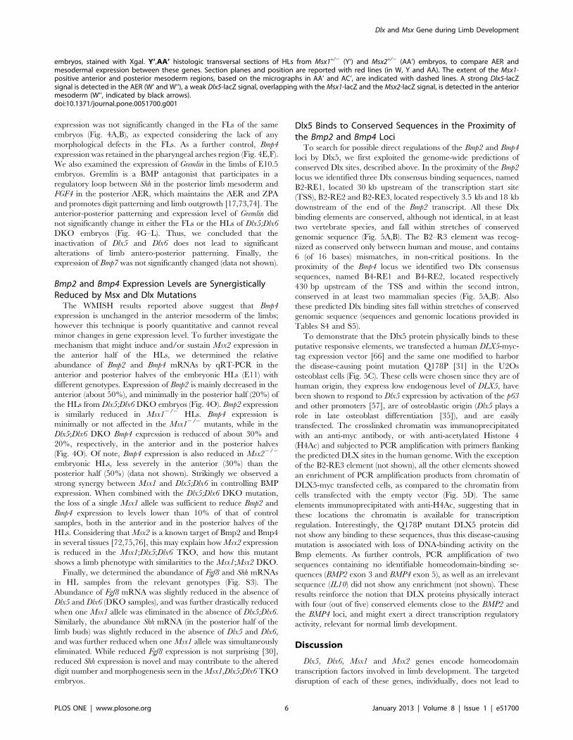
Figure 1. Spatio-temporal expression of Dlx-Msx in developing limbs. A-N, forelimbs. O-AB, hindlimbs, at E10.5 (A-H, O-V) and E11.5 (I-N, W-AB). Whole-mount X-gal staining on FLs and HLs from Dlx5+/2, Msx1+/2 and Msx2+/2 (lacZ+) heterozygous embryos are shown. Expression of Dlx6 wasdetected by WMISH on embryonic limbs at the same ages and shown. At E10.5 Msx2 and Msx1 are expressed in the AER and the anterior andposterior mesoderm of HLs and FLs. Dlx5 and Dlx6, at E10.5, are expressed in the AER of HLs and FLs and in the anterior limb mesoderm only of theFLs, but not of the HLs. At later stages (E11.5), Dlx5 and Dlx6 are then expressed in the anterior mesoderm of HLs. Black arrows indicate mesodermalexpression. The AER is also indicated. Black asterisks indicate absence of expression. W’,W’’ histologic transversal sections of E11.5 HLs from Dlx5+/2
Dlx and Msx Gene during Limb Development
PLOS ONE | www.plosone.org 5 January 2013 | Volume 8 | Issue 1 | e51700
expression was not significantly changed in the FLs of the same
embryos (Fig. 4A,B), as expected considering the lack of any
morphological defects in the FLs. As a further control, Bmp4
expression was retained in the pharyngeal arches region (Fig. 4E,F).
We also examined the expression of Gremlin in the limbs of E10.5
embryos. Gremlin is a BMP antagonist that participates in a
regulatory loop between Shh in the posterior limb mesoderm and
FGF4 in the posterior AER, which maintains the AER and ZPA
and promotes digit patterning and limb outgrowth [17,73,74]. The
anterior-posterior patterning and expression level of Gremlin did
not significantly change in either the FLs or the HLs of Dlx5;Dlx6
DKO embryos (Fig. 4G–L). Thus, we concluded that the
inactivation of Dlx5 and Dlx6 does not lead to significant
alterations of limb antero-posterior patterning. Finally, the
expression of Bmp7 was not significantly changed (data not shown).
Bmp2 and Bmp4 Expression Levels are SynergisticallyReduced by Msx and Dlx Mutations
The WMISH results reported above suggest that Bmp4
expression is unchanged in the anterior mesoderm of the limbs;
however this technique is poorly quantitative and cannot reveal
minor changes in gene expression level. To further investigate the
mechanism that might induce and/or sustain Msx2 expression in
the anterior half of the HLs, we determined the relative
abundance of Bmp2 and Bmp4 mRNAs by qRT-PCR in the
anterior and posterior halves of the embryonic HLs (E11) with
different genotypes. Expression of Bmp2 is mainly decreased in the
anterior (about 50%), and minimally in the posterior half (20%) of
the HLs from Dlx5;Dlx6 DKO embryos (Fig. 4O). Bmp2 expression
is similarly reduced in Msx12/2 HLs. Bmp4 expression is
minimally or not affected in the Msx12/2 mutants, while in the
Dlx5;Dlx6 DKO Bmp4 expression is reduced of about 30% and
20%, respectively, in the anterior and in the posterior halves
(Fig. 4O). Of note, Bmp4 expression is also reduced in Msx22/2
embryonic HLs, less severely in the anterior (30%) than the
posterior half (50%) (data not shown). Strikingly we observed a
strong synergy between Msx1 and Dlx5;Dlx6 in controlling BMP
expression. When combined with the Dlx5;Dlx6 DKO mutation,
the loss of a single Msx1 allele was sufficient to reduce Bmp2 and
Bmp4 expression to levels lower than 10% of that of control
samples, both in the anterior and in the posterior halves of the
HLs. Considering that Msx2 is a known target of Bmp2 and Bmp4
in several tissues [72,75,76], this may explain how Msx2 expression
is reduced in the Msx1;Dlx5;Dlx6 TKO, and how this mutant
shows a limb phenotype with similarities to the Msx1;Msx2 DKO.
Finally, we determined the abundance of Fgf8 and Shh mRNAs
in HL samples from the relevant genotypes (Fig. S3). The
Abundance of Fgf8 mRNA was slightly reduced in the absence of
Dlx5 and Dlx6 (DKO samples), and was further drastically reduced
when one Msx1 allele was eliminated in the absence of Dlx5;Dlx6.
Similarly, the abundance Shh mRNA (in the posterior half of the
limb buds) was slightly reduced in the absence of Dlx5 and Dlx6,
and was further reduced when one Msx1 allele was simultaneously
eliminated. While reduced Fgf8 expression is not surprising [30],
reduced Shh expression is novel and may contribute to the altered
digit number and morphogenesis seen in the Msx1,Dlx5;Dlx6 TKO
embryos.
Dlx5 Binds to Conserved Sequences in the Proximity ofthe Bmp2 and Bmp4 Loci
To search for possible direct regulations of the Bmp2 and Bmp4
loci by Dlx5, we first exploited the genome-wide predictions of
conserved Dlx sites, described above. In the proximity of the Bmp2
locus we identified three Dlx consensus binding sequences, named
B2-RE1, located 30 kb upstream of the transcription start site
(TSS), B2-RE2 and B2-RE3, located respectively 3.5 kb and 18 kb
downstream of the end of the Bmp2 transcript. All these Dlx
binding elements are conserved, although not identical, in at least
two vertebrate species, and fall within stretches of conserved
genomic sequence (Fig. 5A,B). The B2–R3 element was recog-
nized as conserved only between human and mouse, and contains
6 (of 16 bases) mismatches, in non-critical positions. In the
proximity of the Bmp4 locus we identified two Dlx consensus
sequences, named B4-RE1 and B4-RE2, located respectively
430 bp upstream of the TSS and within the second intron,
conserved in at least two mammalian species (Fig. 5A,B). Also
these predicted Dlx binding sites fall within stretches of conserved
genomic sequence (sequences and genomic locations provided in
Tables S4 and S5).
To demonstrate that the Dlx5 protein physically binds to these
putative responsive elements, we transfected a human DLX5-myc-
tag expression vector [66] and the same one modified to harbor
the disease-causing point mutation Q178P [31] in the U2Os
osteoblast cells (Fig. 5C). These cells were chosen since they are of
human origin, they express low endogenous level of DLX5, have
been shown to respond to Dlx5 expression by activation of the p63
and other promoters [57], are of osteoblastic origin (Dlx5 plays a
role in late osteoblast differentiation [35]), and are easily
transfected. The crosslinked chromatin was immunoprecipitated
with an anti-myc antibody, or with anti-acetylated Histone 4
(H4Ac) and subjected to PCR amplification with primers flanking
the predicted DLX sites in the human genome. With the exception
of the B2-RE3 element (not shown), all the other elements showed
an enrichment of PCR amplification products from chromatin of
DLX5-myc transfected cells, as compared to the chromatin from
cells transfected with the empty vector (Fig. 5D). The same
elements immunoprecipitated with anti-H4Ac, suggesting that in
these locations the chromatin is available for transcription
regulation. Interestingly, the Q178P mutant DLX5 protein did
not show any binding to these sequences, thus this disease-causing
mutation is associated with loss of DNA-binding activity on the
Bmp elements. As further controls, PCR amplification of two
sequences containing no identifiable homeodomain-binding se-
quences (BMP2 exon 3 and BMP4 exon 5), as well as an irrelevant
sequence (IL10) did not show any enrichment (not shown). These
results reinforce the notion that DLX proteins physically interact
with four (out of five) conserved elements close to the BMP2 and
the BMP4 loci, and might exert a direct transcription regulatory
activity, relevant for normal limb development.
Discussion
Dlx5, Dlx6, Msx1 and Msx2 genes encode homeodomain
transcription factors involved in limb development. The targeted
disruption of each of these genes, individually, does not lead to
embryos, stained with Xgal. Y’,AA’ histologic transversal sections of HLs from Msx1+/2 (Y’) and Msx2+/2 (AA’) embryos, to compare AER andmesodermal expression between these genes. Section planes and position are reported with red lines (in W, Y and AA). The extent of the Msx1-positive anterior and posterior mesoderm regions, based on the micrographs in AA’ and AC’, are indicated with dashed lines. A strong Dlx5-lacZsignal is detected in the AER (W’ and W’’), a weak Dlx5-lacZ signal, overlapping with the Msx1-lacZ and the Msx2-lacZ signal, is detected in the anteriormesoderm (W’’, indicated by black arrows).doi:10.1371/journal.pone.0051700.g001
Dlx and Msx Gene during Limb Development
PLOS ONE | www.plosone.org 6 January 2013 | Volume 8 | Issue 1 | e51700

Figure 2. Reduction of Msx2 expression in Dlx5/Dlx6 DKO HLs. A-P. Whole-mount X-gal staining to detect Msx1 and Msx2 expression inDlx5;Dlx6 DKO. In the FL (A-D,I-L) no changes of expression is observed whereas in the HL (E-H, M-P), Msx2 expression is reduced in the AER and in theanterior limb mesoderm of the Dlx5;Dlx6 DKO HLs. Q,R. Sections of WT (Q) and Dlx5;Dlx6 DKO mutant HLs (R) hybridized in situ to detect Msx2,
Dlx and Msx Gene during Limb Development
PLOS ONE | www.plosone.org 7 January 2013 | Volume 8 | Issue 1 | e51700
limb defects, however double Msx1/Msx2 [33,34] and Dlx5/Dlx6
[29,30] mutant mice present severe limb malformations indicating
that these genes participate in the control of digit number and
morphogenesis. The Dlx and Msx homeodomains show a high
degree of sequence similarity and similar DNA binding sequences
in vitro [43]. In spite of these similarities, there is no evidence
suggesting that these homeoproteins cooperate or interact, except
in two specific developmental processes: elevation and fusion of the
palatal shelves [60,61] and development of the frontal bone [59].
In this work, we shed light on direct and indirect interactions
between these two classes of homeodomain genes during limb
development.
Direct and Indirect Dlx-Msx Regulations during Limb BudDevelopment
Although Msx2;Dlx5;Dlx6 TKO mice show an aggravation of
the ectrodactyly phenotype, as compared to Dlx5;Dlx6 DKO, they
do not present any obvious additional defect. Furthermore,
inactivation of either Dlx5;Dlx6 or Msx2 does not modify the level
of Msx1 expression, suggesting that the Msx2;Dlx5;Dlx6 TKO
showing a drastically reduced Msx2 signal in the AER and in the underlying mesoderm, but not in a proximal mesoderm territory. S-U. Quantificationof the expression of Dlx5, Dlx6, Msx1 and Msx2 mRNAs by qRT-PCR in HLs from Dlx5;Dlx6 DKO (S), Msx22/2 (T) and Msx12/2 (U), relative to WT. Theresults show a reduction of 45% of Msx2 expression in the Dlx5;Dlx6 DKO HLs compared to WT, but not of Msx1. Dlx5 and Dlx6 expression isdownregulated in Msx1 KO HLs but not in Msx2 KO HLs. Expression of the knocked-out genes was also tested, as control, and always found to bereduced to undetectable levels (not shown).doi:10.1371/journal.pone.0051700.g002
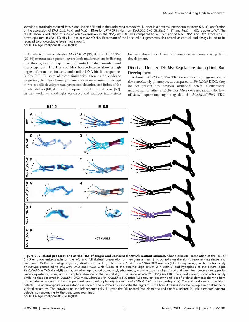
Figure 3. Skeletal preparations of the HLs of single and combined Msx;Dlx mutant animals. Chondroskeletal preparation of the HLs ofE14.5 embryos (micrographs on the left) and full skeletal preparation on newborn animals (micrographs on the right), representing single andcombined Dlx;Msx mutant genotypes (indicated on the left). The HLs of Msx2+/2;Dlx5;Dlx6 DKO animals (E,F) display an aggravated ectrodactylyphenotype compared to Dlx5;Dlx6 DKO ones (C,D), with fusion of the external digit (1with 2, 4 with 5) and hypoplasia of the central digit.Msx2;Dlx5;Dlx6 TKO HLs (G,H) display a further aggravated ectrodactyly phenotype, with the external digits fused and extended towards the opposite(anterior-posterior) sides, and a complete absence of the central digit. The limbs of Msx1+/2;Dlx5;Dlx6 DKO mice (not shown) show ectrodactylysimilar to that observed in Dlx5;Dlx6 DKO mice, whereas Msx1;Dlx5;Dlx6 TKO mice (I,J) show ectrodactyly and loss of skeletal elements deriving fromthe anterior mesoderm of the autopod and zeugopod, a phenotype seen in Msx1;Msx2 DKO mutant embryos (K). The stylopod shows no evidentdefects. The anterior-posterior orientation is shown. The numbers 1–5 indicate the digits (1 is the toe). Asterisks indicate hypoplasia or absence ofskeletal structures. The drawings on the left schematically illustrate the Dlx-related (red elements) and the Msx-related (purple elements) skeletaldefects, corresponding to the genotypes examined.doi:10.1371/journal.pone.0051700.g003
Dlx and Msx Gene during Limb Development
PLOS ONE | www.plosone.org 8 January 2013 | Volume 8 | Issue 1 | e51700

phenotype represents the exclusive contribution of the absence of
Msx2 in the Dlx5;Dlx6 mutant context. This leads us to conclude
that either Msx2 has a rather minor function in limb development,
or that its function is largely compensated by Msx1, in agreement
with the limited defects observed in Msx1+/2;Msx22/2 compound
mutant animals [32] (Y. Lallemand, unpublished). On the
contrary, Msx1;Dlx5;Dlx6 TKO mice display a limb defect which
can be interpreted as the sum of the limb anomalies found in the
Msx1;Msx2 DKO and in the Dlx5;Dlx6 DKO. These results
strongly suggest that the expression of Msx2 is suppressed in this
genetic context, and that therefore Dlx5;Dlx6 are genetically
upstream of Msx2.
We show that in Dlx5;Dlx6 DKO limbs Msx2 expression is
diminished in the central sector of the AER. As starting at E10.5
Figure 4. Expression of Bmp4 in Dlx5;Dlx6 DKO limbs. A-D. Detection of Bmp4 mRNA by WMISH on WT (left) and Dlx5;Dlx6 DKO mutant (right)limbs, at E11. FLs are on the top, HLs are on the bottom. E,F. In situ detection of Bmp4 mRNA in the pharyngeal arches region of WT (left) andDlx5;Dlx6 DKO mutant (right), at E11, as a control for RNA preservation. G-L. Detection of Gremlin mRNA in FLs (G,H) and HLs (I-L) of WT (left) andDlx5;Dlx6 DKO mutant embryos (right), at E10.5. While Bmp4 expression in the anterior mesoderm of the FLs (A,B) or the HLs (C,D) is unchanged(green arrowheads), expression in the central wedge of the AER of mutant embryos is diminished in the HLs, but not in the FLs (red arrows in D).Gremlin expression is unchanged both in the FLs (G,H) and in the HLs (I-L) of Dlx mutant embryos (red arrowheads). Genotypes and probes arereported on the top. The Anterior-Posterior (A-P) orientation is indicated. M,N. whole-mount photographs documenting the reduced size of mutantembryos and justifying the slightly reduced size of the mutant limbs, often observed. O. Quantification of Bmp2 and Bmp4 mRNAs by qRT-PCR in theanterior and posterior halves of HLs from embryos with the genotype indicated on the top of each graph, compared to WT.doi:10.1371/journal.pone.0051700.g004
Dlx and Msx Gene during Limb Development
PLOS ONE | www.plosone.org 9 January 2013 | Volume 8 | Issue 1 | e51700
Dlx and Msx genes are co-expressed in the AER, it is possible to
hypothesize that in this territory Dlx proteins bind directly on the
Msx2 promoter, as previously reported [62,63]. On the contrary,
in the anterior limb mesenchyme, the expression of Msx1 and
Msx2 precedes that of Dlx5 and Dlx6. This precludes the possibility
of a direct regulation of Msx genes by Dlx proteins. Nevertheless,
our qRT-PCR analyses show that Msx2 expression is reduced in
the anterior limb mesoderm of Dlx5;Dlx6 DKO limbs, implying
the existence of a non cell-autonomous mode of regulation
between the AER and the anterior limb mesoderm. It is possible,
therefore, that a diffusible protein, expressed by AER cells in a
Dlx-dependent fashion, is required to initiate and/or sustain Msx2
expression in the anterior limb mesoderm.
Bmps as Signaling Relays between Dlx and MsxMsx genes are well documented downstream effectors of BMP
signaling in several developing structures [72,77,78,79,80,81,82].
Enhanced BMP signaling in the limb in mice deficient for the
BMP antagonist Gremlin, results in upregulation of both Msx1 and
Msx2 expression [73]. Conversely, blocking BMP signaling in the
limb ectoderm by ectopic expression of Noggin, another BMP
antagonist, results in decreased Msx2 expression [23]. In the chick
limb buds, expression of a constitutively-active BMP receptor, or
misexpression of Msx1, in the dorsal ectoderm, induces the
formation of ectopic AERs [20]. In addition, the combined
inactivation of Msx1 and Msx2 leads to a phenotype that mimics,
in some aspects, the loss of BMP signaling [32].
Noticeably, Msx genes are also upstream of Bmp4 in several
developmental systems. During tooth germ development, meso-
dermal Msx1 is needed for efficient Bmp4 expression
[83,84,85,86]. Palatal development, which is impaired in
Msx12/2 mice, can be rescued by a Bmp4-expressing transgene
[87]. In the limb itself, Msx genes are required in the mesoderm for
maintenance of Bmp4 expression [34]. Thus, BMP signaling is
both upstream and downstream of Msx genes, depending on the
context and the developing structure. The possibility that Bmp2
and Bmp4 participate in a Dlx-Msx signaling loop between the
limb bud AER and mesoderm is clearly in line with previously
identified roles of these molecules.
Therefore, Bmps represented likely candidates to mediate a non
cell-autonomous regulation between AER-expressed Dlx5;Dlx6
and mesodermal Msx2. Indeed, we find that Bmp2 and Bmp4
expression is significantly reduced in Dlx5;Dlx6 DKO hindlimbs, at
E11 when no evident defect is yet visible. Reduction for Bmp2 and
Bmp4 in the anterior part of the HL is too high (50 and 30%,
respectively) to be accounted for by the ectoderm alone, and rather
indicates a downregulation of these Bmps in the mesoderm, too.
This could mean that Bmps produced in the ectoderm activate
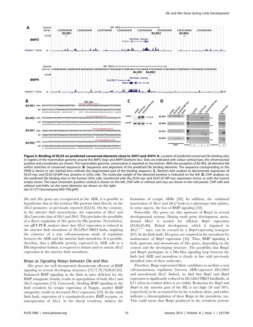
Figure 5. Binding of DLX5 on predicted conserved elements close to BMP2 and BMP4. A. Location of predicted conserved Dlx binding sitesin regions of the mammalian genome around the BMP2 (top) and BMP4 (bottom) loci. Sites are indicated with colour vertical bars, the chromosomalposition and coordinates are shown. The mammalian genomic conservation is reported on the bottom. With the exception of B2-RE3, all elements fallwithin stretches of conserved sequences. B. Sequences and alignment of the predicted Dlx binding elements. The sequence corresponding to thePWM is shown in red. Dashed lines indicate the degenerated part of the binding sequence. C. Western blot analysis to demonstrate expression ofDLX5-myc and DLX5-Q184P-myc proteins in U2Os cells. The molecular weight of the detected proteins is indicated on the left. D. ChIP analyses onthe predicted Dlx binding sites in the human U2Os cells, transfected with the DLX5-myc and DLX5-Q178P-myc expression vector, or with the controlempty vector. The input chromatin (positive control) is shown on the left, ChIP with or without anti-myc are shown in the mid panels. ChIP with andwithout anti-H4Ac on the same elements are shown on the right.doi:10.1371/journal.pone.0051700.g005
Dlx and Msx Gene during Limb Development
PLOS ONE | www.plosone.org 10 January 2013 | Volume 8 | Issue 1 | e51700
Bmps also in the mesoderm, either directly or via Msx1. Indeed,
further inactivation of one Msx1 allele in the context of the loss of
Dlx5;Dlx6 nearly abolishes Bmp2 and Bmp4 expression, necessarily
implying both the ectoderm and the mesoderm (Fig. 4K). This
downregulation would explain a reduction in Msx2 expression in
the anterior mesenchyme in the Dlx5;Dlx6 mutant limbs, at the
same embryonic ages. The direct action of Dlx over Bmp is further
supported by our ChIP data which indicate that DLX5, but not its
mutated variant Q178P [31], binds to conserved regions near the
BMP2 and BMP4 loci. However formal evidence that DLX5
activates BMP2 and BMP4 transcription is still lacking, as no
suitable AER-related cell line is available for such experiments. It
would be of interest to further investigate whether MSX1 also
binds to the same conserved sequences in the BMP2 and BMP4
promoters.
Such a non cell-autonomous mode of regulation is strikingly
similar to that occurring during development of the palatal shelves
and the tooth primordia, both of which involve diffusion of Bmp
between adjacent epithelial/mesodermal cell layers [60,87], and in
the case of the tooth germ, an induction of Bmp4 expression in the
mesoderm by ectodermal Bmp4 via expression of the Bmp target
Msx1 [85,86].
The loss of mesenchymal Bmp2, 4 and 7 expression, on the other
side, has been shown to be required for osteogenic differentiation,
in a dose-dependent fashion and with Bmp molecules acting in a
partially redundant way [88]. In their work, the selective loss of
Bmp2 and Bmp4 in the limb mesenchyme affects zeugopod
development and skeletogenesis, and less severely the autopod.
On a similar note, another work [22] shows that the gradual
elimination of Bmp4 from the limb mesenchyme is required to
rescue the Grem12/2 phenotype and a normal digit organization,
implying a right amount of mesenchyme-derived Bmps is essential
for AER function and for autopod morphogenesis. In the
Msx1;Dlx5;Dlx6 TKO mutants we observe a quite severe autopod
defect (loss of digits) accompanied, however, by less severe
zeugopod defect. Thus, it appears that both AER- and mesen-
chyme-derived Bmps are involved in the Msx;Dlx defects. The
possibility that misexpression of AER-Bmps alone directly cause
the TKO defects is unlikely. Rather, in light of the well known self-
regulatory system of signalling loops comprising FGFs, SHH and
BMPs [22], and considering that we observe changes in the
expression levels of Fgf8 and Shh upon loss of Msx1, Dlx5 and Dlx6
genes, most likely the overall nature of the limb defects in TKO
mutant embryos is a quantitative misregulation of the ‘‘slow
module’’ of this loop. Dlx and Msx genes can be regarded as new
players in this complex regulation. Since Shh participates in the
‘‘slow module’’, a more critical role for it could be envisioned, as
suggested by resemblance of the phenotype of Msx1;Dlx5;Dlx6
TKO limbs with that exhibited by Shh KO embryos [89].
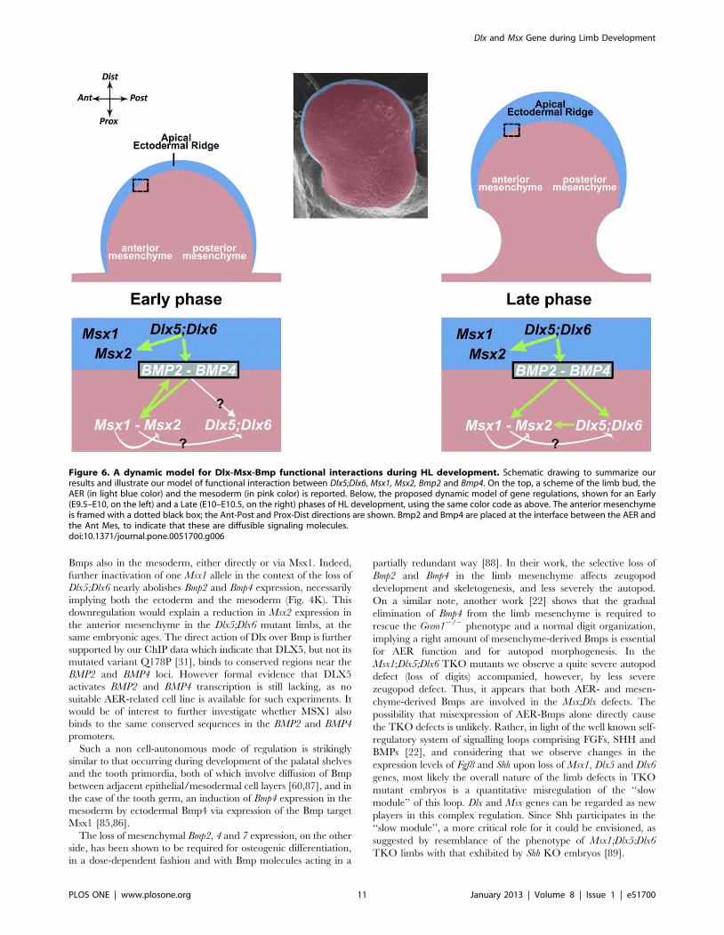
Figure 6. A dynamic model for Dlx-Msx-Bmp functional interactions during HL development. Schematic drawing to summarize ourresults and illustrate our model of functional interaction between Dlx5;Dlx6, Msx1, Msx2, Bmp2 and Bmp4. On the top, a scheme of the limb bud, theAER (in light blue color) and the mesoderm (in pink color) is reported. Below, the proposed dynamic model of gene regulations, shown for an Early(E9.5–E10, on the left) and a Late (E10–E10.5, on the right) phases of HL development, using the same color code as above. The anterior mesenchymeis framed with a dotted black box; the Ant-Post and Prox-Dist directions are shown. Bmp2 and Bmp4 are placed at the interface between the AER andthe Ant Mes, to indicate that these are diffusible signaling molecules.doi:10.1371/journal.pone.0051700.g006
Dlx and Msx Gene during Limb Development
PLOS ONE | www.plosone.org 11 January 2013 | Volume 8 | Issue 1 | e51700

In conclusion, we propose a model that involves a complex
epithelial-mesodermal dialogue between Dlx and Msx (Fig. 6),
entailing two distinct modes of regulation in the limb buds: a
direct, cell-autonomous, regulation intrinsic to AER cells, and an
indirect regulation between the AER cells and the anterior limb
mesoderm. We further provide data suggesting that Bmp2 and
Bmp4 mediate a non cell-autonomous control of Dlx over Msx,
establishing a dialogue between the AER and the anterior
mesoderm of the developing limb.
Genotypes, Morphotypes, Gene Dosage and ExpressionLevels
When comparing the limb phenotypes of the genetic series
Dlx5;Dlx6 DKO vs. Msx2+/2;Dlx52/2;Dlx62/2 vs. Msx2;Dlx5;Dlx6
TKO (Fig. 3C–H), we observe a clear increase in severity of the
ectrodactyly defect. This interesting observation can be explained
by introducing the notion that Msx2 expression depends on allelic
dosage, and that a threshold level of Msx2 expression is critical to
drive normal morphogenesis.
The importance of allelic dosage, hence quantitative gene
expression, is increasingly being recognized, even when pheno-
types are not evident (see [88,90]). As a further example, we detect
a reduction of Msx2 mRNA level in the Msx2+/2 mice, in which
no evident phenotype can be seen. Our explanation, in this case, is
that reduced Msx2 expression alone is not sufficient to cause limb
malformations due to the presence of two functional Msx1 alleles.
Loss of one Msx2 allele in the context of Dlx5;Dlx6 DKO, instead,
aggravates the phenotype. We explain this by proposing that Msx2
expression is severely reduced, approaching that of the null
condition, due to the combination of a) the genetic inactivation of
one allele, and b) the lack of Dlx5;Dlx6 genes.
Likewise, in Msx1;Dlx5;Dlx6 TKO animals, Msx2 expression is
further reduced. A residual level is nonetheless observed; however,
this is not sufficient to compensate for the loss of Msx1. In
conclusion, allelic dosage and quantitative gene expression are
crucial factors to be considered in the interpretation of a series of
phenotypes, especially when related genes are involved.
ConclusionsIn human, the DLX5 and DLX6 genes cause the SHFM-type-1
congenital malformation when lost or mutated, while the MSX1
and MSX2 genes cause cleft palate and tooth agenesis. By
crossbreeding mutant mouse strains, we show that the Dlx5;Dlx6
and Msx1;Msx2 genes cooperate for normal limb development and
morphogenesis. At least two modes of regulation have emerged,
one in which Dlx5;Dlx6 control expression of Msx2 cell-autono-
mously, the other in which the AER and the anterior mesenchyme
interact non cell-autonomously, entailing Bmps as signaling
molecules. We further show that the BMP2 and BMP4 loci
comprise Dlx5-binding elements, occupied by Dlx5. Thus, the
highly related homeodomain genes Dlx and Msx are two key
players of a novel set of molecular and histological interactions
during limb development.
Supporting Information
Figure S1 Top. Location of predicted conserved Dlx binding
sites in the Dlx5-Dlx6 intergenic genomic region. Sites are
indicated with colour vertical bars (asterisk) and annotated with
the species conservation. The chromosomal position and coordi-
nates are also reported. The mammalian genomic conservation is
reported on the bottom. The known i56i element is correctly
predicted by the PWM bioinformatic approach we have adopted.
Bottom. Same as above, relative to the Msx2 proximal promoter.
Two known conserved Dlx binding sites are correctly predicted.
(PDF)
Figure S2 Quantification of the Msx2 mRNAs by qRT-PCR in the anterior and posterior halves of HLs fromMsx1+/2;Dlx52/2;Dlx62/2 embryos, relative to thecorresponding WT samples (set = 1).
(PDF)
Figure S3 Quantification of the Fgf8 and Shh mRNAs byqRT-PCR in the HLs from Msx12/2 (top left), Dlx52/2;Dlx62/2 (top right), Msx1+/2;Dlx5+/2;Dlx6+/2
(bottom left) and Msx1+/2;Dlx52/2;Dlx62/2 (bottomright) embryos, relative to the corresponding WTsamples (set = 1).
(PDF)
Table S1 Sequences of the oligonucleotides used forreal-time qPCR on mouse embryonic tissues.
(PDF)
Table S2 Sequences of the oligonucleotides used forChIP analysis on the predicted Dlx elements near thehuman BMP2 and BMP4 loci.
(PDF)
Table S3 The Dlx5 Position-Weight matrix and resultsof the prediction of Dlx5 binding sites based on genomicconservation.
(PDF)
Table S4 Sequences of the mouse and human conservedgenomic regions containing predicted Dlx5 binding sitesnear the BMP2 locus.
(PDF)
Table S5 Sequences of the mouse and human conservedgenomic regions containing predicted Dlx5 binding sitesnear the BMP4 locus.
(PDF)
Acknowledgments
We thank Dr. Rolf Zeller (Univ. of Basel, Switzerland) for providing probes
and reagents. We thank Dr. Ferdinando DiCunto (Univ. of Torino, Italy)
for bioinformatic analyses. We also thank Dr. Denis Duboule (Geneva,
Switzerland), Drs. Massimo Santoro and Emilio Hirsch (Univ. of Torino,
Italy) for helpful comment on the manuscript.
Author Contributions
Conceived and designed the experiments: MV-R PP SA OB MFC AT YL
BR GL GRM. Performed the experiments: MV-R KB SM GG SA OB LG
MFC AT YL GL GRM. Analyzed the data: MV-R GG PP SA OB MFC
AT YL BR GL GRM. Contributed reagents/materials/analysis tools: GG
PP OB LG MFC AT YL BR. Wrote the paper: MV-R PP OB YL BR GL
GRM.
References
1. Allard P, Tabin CJ (2009) Achieving bilateral symmetry during vertebrate limb
development. Semin Cell Dev Biol 20: 479–484.
2. Zakany J, Duboule D (2007) The role of Hox genes during vertebrate limb
development. Curr Opin Genet Dev 17: 359–366.
3. Zeller R (2010) The temporal dynamics of vertebrate limb development,
teratogenesis and evolution. Curr Opin Genet Dev 20: 384–390.
4. Bastida MF, Ros MA (2008) How do we get a perfect complement of digits?
Curr Opin Genet Dev 18: 374–380.
Dlx and Msx Gene during Limb Development
PLOS ONE | www.plosone.org 12 January 2013 | Volume 8 | Issue 1 | e51700

5. Riddle RD, Johnson RL, Laufer E, Tabin C (1993) Sonic hedgehog mediates the
polarizing activity of the ZPA. Cell 75: 1401–1416.
6. Zeng X, Goetz JA, Suber LM, Scott WJ Jr, Schreiner CM, et al. (2001) A freely
diffusible form of Sonic hedgehog mediates long-range signalling. Nature 411:
716–720.
7. Mariani FV, Ahn CP, Martin GR (2008) Genetic evidence that FGFs have an
instructive role in limb proximal-distal patterning. Nature 453: 401–405.
8. Sun X, Mariani FV, Martin GR (2002) Functions of FGF signalling from the
apical ectodermal ridge in limb development. Nature 418: 501–508.
9. Niswander L, Tickle C, Vogel A, Booth I, Martin GR (1993) FGF-4 replaces the
apical ectodermal ridge and directs outgrowth and patterning of the limb. Cell
75: 579–587.
10. Lu P, Yu Y, Perdue Y, Werb Z (2008) The apical ectodermal ridge is a timer for
generating distal limb progenitors. Development 135: 1395–1405.
11. Fernandez-Teran M, Ros MA (2008) The Apical Ectodermal Ridge:
morphological aspects and signaling pathways. Int J Dev Biol 52: 857–871.
12. Loomis CA, Harris E, Michaud J, Wurst W, Hanks M, et al. (1996) The mouse
Engrailed-1 gene and ventral limb patterning. Nature 382: 360–363.
13. Parr BA, McMahon AP (1995) Dorsalizing signal Wnt-7a required for normal
polarity of D-V and A-P axes of mouse limb. Nature 374: 350–353.
14. Cygan JA, Johnson RL, McMahon AP (1997) Novel regulatory interactions
revealed by studies of murine limb pattern in Wnt-7a and En-1 mutants.
Development 124: 5021–5032.
15. Chen H, Johnson RL (2002) Interactions between dorsal-ventral patterning
genes lmx1b, engrailed-1 and wnt-7a in the vertebrate limb. Int J Dev Biol 46:
937–941.
16. Qiu Q, Chen H, Johnson RL (2009) Lmx1b-expressing cells in the mouse limb
bud define a dorsal mesenchymal lineage compartment. Genesis 47: 224–233.
17. Benazet JD, Zeller R (2009) Vertebrate limb development: moving from classical
morphogen gradients to an integrated 4-dimensional patterning system. Cold
Spring Harb Perspect Biol 1: a001339.
18. Zeller R, Lopez-Rios J, Zuniga A (2009) Vertebrate limb bud development:
moving towards integrative analysis of organogenesis. Nat Rev Genet 10: 845–
858.
19. Tickle C (2006) Making digit patterns in the vertebrate limb. Nat Rev Mol Cell
Biol 7: 45–53.
20. Pizette S, Abate-Shen C, Niswander L (2001) BMP controls proximodistal
outgrowth, via induction of the apical ectodermal ridge, and dorsoventral
patterning in the vertebrate limb. Development 128: 4463–4474.
21. Robert B (2007) Bone morphogenetic protein signaling in limb outgrowth and
patterning. Dev Growth Differ 49: 455–468.
22. Benazet JD, Bischofberger M, Tiecke E, Goncalves A, Martin JF, et al. (2009) A
self-regulatory system of interlinked signaling feedback loops controls mouse
limb patterning. Science 323: 1050–1053.
23. Wang CK, Omi M, Ferrari D, Cheng HC, Lizarraga G, et al. (2004) Function of
BMPs in the apical ectoderm of the developing mouse limb. Dev Biol 269: 109–
122.
24. Zuniga A, Haramis AP, McMahon AP, Zeller R (1999) Signal relay by BMP
antagonism controls the SHH/FGF4 feedback loop in vertebrate limb buds.
Nature 401: 598–602.
25. Ahn K, Mishina Y, Hanks MC, Behringer RR, Crenshaw EB 3rd (2001)
BMPR-IA signaling is required for the formation of the apical ectodermal ridge
and dorsal-ventral patterning of the limb. Development 128: 4449–4461.
26. Tickle C (2003) Patterning systems–from one end of the limb to the other. Dev
Cell 4: 449–458.
27. Robert B, Lallemand Y (2006) Anteroposterior patterning in the limb and digit
specification: contribution of mouse genetics. Dev Dyn 235: 2337–2352.
28. Zeller R (2004) It takes time to make a pinky: unexpected insights into how SHH
patterns vertebrate digits. Sci STKE 2004: pe53.
29. Merlo GR, Paleari L, Mantero S, Genova F, Beverdam A, et al. (2002) Mouse
model of split hand/foot malformation type I. Genesis 33: 97–101.
30. Robledo RF, Rajan L, Li X, Lufkin T (2002) The Dlx5 and Dlx6 homeobox
genes are essential for craniofacial, axial, and appendicular skeletal development.
Genes Dev 16: 1089–1101.
31. Shamseldin HE, Faden MA, Alashram W, Alkuraya FS (2012) Identification of a
novel DLX5 mutation in a family with autosomal recessive split hand and foot
malformation. J Med Genet 49: 16–20.
32. Lallemand Y, Nicola MA, Ramos C, Bach A, Cloment CS, et al. (2005) Analysis
of Msx1; Msx2 double mutants reveals multiple roles for Msx genes in limb
development. Development 132: 3003–3014.
33. Lallemand Y, Bensoussan V, Cloment CS, Robert B (2009) Msx genes are
important apoptosis effectors downstream of the Shh/Gli3 pathway in the limb.
Dev Biol 331: 189–198.
34. Bensoussan-Trigano V, Lallemand Y, Saint Cloment C, Robert B (2011) Msx1
and Msx2 in limb mesenchyme modulate digit number and identity. Dev Dyn
240: 1190–1202.
35. Acampora D, Merlo GR, Paleari L, Zerega B, Postiglione MP, et al. (1999)
Craniofacial, vestibular and bone defects in mice lacking the Distal-less-related
gene Dlx5. Development 126: 3795–3809.
36. Beverdam A, Merlo GR, Paleari L, Mantero S, Genova F, et al. (2002) Jaw
transformation with gain of symmetry after Dlx5/Dlx6 inactivation: mirror of
the past? Genesis 34: 221–227.
37. Depew MJ, Liu JK, Long JE, Presley R, Meneses JJ, et al. (1999) Dlx5 regulatesregional development of the branchial arches and sensory capsules. Develop-
ment 126: 3831–3846.
38. Depew MJ, Simpson CA, Morasso M, Rubenstein JL (2005) Reassessing the Dlxcode: the genetic regulation of branchial arch skeletal pattern and development.
J Anat 207: 501–561.
39. Houzelstein D, Cohen A, Buckingham ME, Robert B (1997) Insertional
mutation of the mouse Msx1 homeobox gene by an nlacZ reporter gene. Mech
Dev 65: 123–133.
40. Satokata I, Ma L, Ohshima H, Bei M, Woo I, et al. (2000) Msx2 deficiency in
mice causes pleiotropic defects in bone growth and ectodermal organ formation.Nat Genet 24: 391–395.
41. Satokata I, Maas R (1994) Msx1 deficient mice exhibit cleft palate and
abnormalities of craniofacial and tooth development. Nat Genet 6: 348–356.
42. Ferrari D, Sumoy L, Gannon J, Sun H, Brown AM, et al. (1995) The expression
pattern of the Distal-less homeobox-containing gene Dlx-5 in the developingchick limb bud suggests its involvement in apical ectodermal ridge activity,
pattern formation, and cartilage differentiation. Mech Dev 52: 257–264.
43. Bendall AJ, Abate-Shen C (2000) Roles for Msx and Dlx homeoproteins invertebrate development. Gene 247: 17–31.
44. Zhang H, Hu G, Wang H, Sciavolino P, Iler N, et al. (1997) Heterodimerization
of Msx and Dlx homeoproteins results in functional antagonism. Mol Cell Biol17: 2920–2932.
45. Davidson D (1995) The function and evolution of Msx genes: pointers andparadoxes. Trends Genet 11: 405–411.
46. Panganiban G, Rubenstein JL (2002) Developmental functions of the Distal-
less/Dlx homeobox genes. Development 129: 4371–4386.
47. Merlo GR, Zerega B, Paleari L, Trombino S, Mantero S, et al. (2000) Multiple
functions of Dlx genes. Int J Dev Biol 44: 619–626.
48. Liu YH, Tang Z, Kundu RK, Wu L, Luo W, et al. (1999) Msx2 gene dosageinfluences the number of proliferative osteogenic cells in growth centers of the
developing murine skull: a possible mechanism for MSX2-mediated craniosyn-ostosis in humans. Dev Biol 205: 260–274.
49. Odelberg SJ, Kollhoff A, Keating MT (2000) Dedifferentiation of mammalian
myotubes induced by msx1. Cell 103: 1099–1109.
50. Hu G, Lee H, Price SM, Shen MM, Abate-Shen C (2001) Msx homeobox genes
inhibit differentiation through upregulation of cyclin D1. Development 128:2373–2384.
51. Ishii M, Merrill AE, Chan YS, Gitelman I, Rice DP, et al. (2003) Msx2 and
Twist cooperatively control the development of the neural crest-derivedskeletogenic mesenchyme of the murine skull vault. Development 130: 6131–
6142.
52. Levi G, Puche AC, Mantero S, Barbieri O, Trombino S, et al. (2003) The Dlx5
homeodomain gene is essential for olfactory development and connectivity in the
mouse. Mol Cell Neurosci 22: 530–543.
53. Perera M, Merlo GR, Verardo S, Paleari L, Corte G, et al. (2004) Defective
neuronogenesis in the absence of Dlx5. Mol Cell Neurosci 25: 153–161.
54. Long JE, Garel S, Alvarez-Dolado M, Yoshikawa K, Osumi N, et al. (2007) Dlx-
dependent and -independent regulation of olfactory bulb interneuron differen-
tiation. J Neurosci 27: 3230–3243.
55. Merlo GR, Mantero S, Zaghetto AA, Peretto P, Paina S, et al. (2007) The role of
Dlx homeogenes in early development of the olfactory pathway. J Mol Histol 38:347–358.
56. Muraglia A, Perera M, Verardo S, Liu Y, Cancedda R, et al. (2008) DLX5
overexpression impairs osteogenic differentiation of human bone marrowstromal cells. Eur J Cell Biol 87: 751–761.
57. Lo Iacono N, Mantero S, Chiarelli A, Garcia E, Mills AA, et al. (2008)
Regulation of Dlx5 and Dlx6 gene expression by p63 is involved in EEC andSHFM congenital limb defects. Development 135: 1377–1388.
58. Radoja N, Guerrini L, Lo Iacono N, Merlo GR, Costanzo A, et al. (2007)Homeobox gene Dlx3 is regulated by p63 during ectoderm development:
relevance in the pathogenesis of ectodermal dysplasias. Development 134: 13–
18.
59. Chung IH, Han J, Iwata J, Chai Y (2010) Msx1 and Dlx5 function synergistically
to regulate frontal bone development. Genesis 48: 645–655.
60. Levi G, Mantero S, Barbieri O, Cantatore D, Paleari L, et al. (2006) Msx1 and
Dlx5 act independently in development of craniofacial skeleton, but converge on
the regulation of Bmp signaling in palate formation. Mech Dev 123: 3–16.
61. Han J, Mayo J, Xu X, Li J, Bringas P Jr, et al. (2009) Indirect modulation of Shh
signaling by Dlx5 affects the oral-nasal patterning of palate and rescues cleftpalate in Msx1-null mice. Development 136: 4225–4233.
62. Pan ZZ, Kronenberg MS, Huang DY, Sumoy L, Rogina B, et al. (2002) MSX2
expression in the apical ectoderm ridge is regulated by an MSX2 and Dlx5binding site. Biochem Biophys Res Commun 290: 955–961.
63. Sumoy L, Wang CK, Lichtler AC, Pierro LJ, Kosher RA, et al. (1995)Identification of a spatially specific enhancer element in the chicken Msx-2 gene
that regulates its expression in the apical ectodermal ridge of the developing limb
buds of transgenic mice. Dev Biol 170: 230–242.
64. Vieux-Rochas M, Coen L, Sato T, Kurihara Y, Gitton Y, et al. (2007) Molecular
dynamics of retinoic acid-induced craniofacial malformations: implications forthe origin of gnathostome jaws. PLoS One 2: e510.
65. Jones CM, Lyons KM, Hogan BL (1991) Involvement of Bone Morphogenetic
Protein-4 (BMP-4) and Vgr-1 in morphogenesis and neurogenesis in the mouse.Development 111: 531–542.
Dlx and Msx Gene during Limb Development
PLOS ONE | www.plosone.org 13 January 2013 | Volume 8 | Issue 1 | e51700
66. Paina S, Garzotto D, DeMarchis S, Marino M, Moiana A, et al. (2011) Wnt5a is
a transcriptional target of Dlx homeogenes and promotes differentiation ofinterneuron progenitors in vitro and in vivo. J Neurosci 31: 2675–2687.
67. Ala U, Piro RM, Grassi E, Damasco C, Silengo L, et al. (2008) Prediction of
human disease genes by human-mouse conserved coexpression analysis. PLoSComput Biol 4: e1000043.
68. Piro RM, Ala U, Molineris I, Grassi E, Bracco C, et al. (2011) An atlas of tissue-specific conserved coexpression for functional annotation and disease gene
prediction. Eur J Hum Genet 19: 1173–1180.
69. Berger MF, Badis G, Gehrke AR, Talukder S, Philippakis AA, et al. (2008)Variation in homeodomain DNA binding revealed by high-resolution analysis of
sequence preferences. Cell 133: 1266–1276.70. Portales-Casamar E, Thongjuea S, Kwon AT, Arenillas D, Zhao X, et al. (2010)
JASPAR 2010: the greatly expanded open-access database of transcription factorbinding profiles. Nucleic Acids Res 38: D105–110.
71. Zerucha T, Stuhmer T, Hatch G, Park BK, Long Q, et al. (2000) A highly
conserved enhancer in the Dlx5/Dlx6 intergenic region is the site of cross-regulatory interactions between Dlx genes in the embryonic forebrain. J Neurosci
20: 709–721.72. Brugger SM, Merrill AE, Torres-Vazquez J, Wu N, Ting MC, et al. (2004) A
phylogenetically conserved cis-regulatory module in the Msx2 promoter is
sufficient for BMP-dependent transcription in murine and Drosophila embryos.Development 131: 5153–5165.
73. Khokha MK, Hsu D, Brunet LJ, Dionne MS, Harland RM (2003) Gremlin isthe BMP antagonist required for maintenance of Shh and Fgf signals during
limb patterning. Nat Genet 34: 303–307.74. Michos O, Panman L, Vintersten K, Beier K, Zeller R, et al. (2004) Gremlin-
mediated BMP antagonism induces the epithelial-mesenchymal feedback
signaling controlling metanephric kidney and limb organogenesis. Development131: 3401–3410.
75. Ma L, Lu MF, Schwartz RJ, Martin JF (2005) Bmp2 is essential for cardiaccushion epithelial-mesenchymal transition and myocardial patterning. Develop-
ment 132: 5601–5611.
76. Matsubara T, Kida K, Yamaguchi A, Hata K, Ichida F, et al. (2008) BMP2regulates Osterix through Msx2 and Runx2 during osteoblast differentiation.
J Biol Chem 283: 29119–29125.77. Vainio S, Karavanova I, Jowett A, Thesleff I (1993) Identification of BMP-4 as a
signal mediating secondary induction between epithelial and mesenchymaltissues during early tooth development. Cell 75: 45–58.
78. Marazzi G, Wang Y, Sassoon D (1997) Msx2 is a transcriptional regulator in the
BMP4-mediated programmed cell death pathway. Dev Biol 186: 127–138.
79. Bei M, Maas R (1998) FGFs and BMP4 induce both Msx1-independent and
Msx1-dependent signaling pathways in early tooth development. Development
125: 4325–4333.
80. Hollnagel A, Oehlmann V, Heymer J, Ruther U, Nordheim A (1999) Id genes
are direct targets of bone morphogenetic protein induction in embryonic stem
cells. J Biol Chem 274: 19838–19845.
81. Sirard C, Kim S, Mirtsos C, Tadich P, Hoodless PA, et al. (2000) Targeted
disruption in murine cells reveals variable requirement for Smad4 in
transforming growth factor beta-related signaling. J Biol Chem 275: 2063–2070.
82. Daluiski A, Engstrand T, Bahamonde ME, Gamer LW, Agius E, et al. (2001)
Bone morphogenetic protein-3 is a negative regulator of bone density. Nat
Genet 27: 84–88.
83. Zhao X, Zhang Z, Song Y, Zhang X, Zhang Y, et al. (2000) Transgenically
ectopic expression of Bmp4 to the Msx1 mutant dental mesenchyme restores
downstream gene expression but represses Shh and Bmp2 in the enamel knot of
wild type tooth germ. Mech Dev 99: 29–38.
84. Zhang Y, Zhang Z, Zhao X, Yu X, Hu Y, et al. (2000) A new function of BMP4:
dual role for BMP4 in regulation of Sonic hedgehog expression in the mouse
tooth germ. Development 127: 1431–1443.
85. Bei M, Kratochwil K, Maas RL (2000) BMP4 rescues a non-cell-autonomous
function of Msx1 in tooth development. Development 127: 4711–4718.
86. Chen Y, Bei M, Woo I, Satokata I, Maas R (1996) Msx1 controls inductive
signaling in mammalian tooth morphogenesis. Development 122: 3035–3044.
87. Zhang Z, Song Y, Zhao X, Zhang X, Fermin C, et al. (2002) Rescue of cleft
palate in Msx1-deficient mice by transgenic Bmp4 reveals a network of BMP
and Shh signaling in the regulation of mammalian palatogenesis. Development
129: 4135–4146.
88. Bandyopadhyay A, Tsuji K, Cox K, Harfe BD, Rosen V, et al. (2006) Genetic
analysis of the roles of BMP2, BMP4, and BMP7 in limb patterning and
skeletogenesis. PLoS Genet 2: e216.
89. Kraus P, Fraidenraich D, Loomis CA (2001) Some distal limb structures develop
in mice lacking Sonic hedgehog signaling. Mech Dev 100: 45–58.
90. Guerrini L, Costanzo A, Merlo GR (2011) A symphony of regulations centered
on p63 to control development of ectoderm-derived structures. J Biomed
Biotechnol 2011: 864904.
Dlx and Msx Gene during Limb Development
PLOS ONE | www.plosone.org 14 January 2013 | Volume 8 | Issue 1 | e51700