Biosynthesis of Antinutritional Alkaloids in Solanaceous Crops Is Mediated by Clustered Genes
6
DOI: 10.1126/science.1240230 , 175 (2013); 341 Science et al. M. Itkin Mediated by Clustered Genes Biosynthesis of Antinutritional Alkaloids in Solanaceous Crops Is This copy is for your personal, non-commercial use only. clicking here. colleagues, clients, or customers by , you can order high-quality copies for your If you wish to distribute this article to others here. following the guidelines can be obtained by Permission to republish or repurpose articles or portions of articles ): January 13, 2014 www.sciencemag.org (this information is current as of The following resources related to this article are available online at http://www.sciencemag.org/content/341/6142/175.full.html version of this article at: including high-resolution figures, can be found in the online Updated information and services, http://www.sciencemag.org/content/suppl/2013/06/19/science.1240230.DC1.html can be found at: Supporting Online Material http://www.sciencemag.org/content/341/6142/175.full.html#ref-list-1 , 7 of which can be accessed free: cites 27 articles This article http://www.sciencemag.org/cgi/collection/botany Botany subject collections: This article appears in the following registered trademark of AAAS. is a Science 2013 by the American Association for the Advancement of Science; all rights reserved. The title Copyright American Association for the Advancement of Science, 1200 New York Avenue NW, Washington, DC 20005. (print ISSN 0036-8075; online ISSN 1095-9203) is published weekly, except the last week in December, by the Science on January 13, 2014 www.sciencemag.org Downloaded from on January 13, 2014 www.sciencemag.org Downloaded from on January 13, 2014 www.sciencemag.org Downloaded from on January 13, 2014 www.sciencemag.org Downloaded from on January 13, 2014 www.sciencemag.org Downloaded from on January 13, 2014 www.sciencemag.org Downloaded from
Transcript of Biosynthesis of Antinutritional Alkaloids in Solanaceous Crops Is Mediated by Clustered Genes
DOI: 10.1126/science.1240230, 175 (2013);341 Science
et al.M. ItkinMediated by Clustered GenesBiosynthesis of Antinutritional Alkaloids in Solanaceous Crops Is
This copy is for your personal, non-commercial use only.
clicking here.colleagues, clients, or customers by , you can order high-quality copies for yourIf you wish to distribute this article to others
here.following the guidelines
can be obtained byPermission to republish or repurpose articles or portions of articles
): January 13, 2014 www.sciencemag.org (this information is current as of
The following resources related to this article are available online at
http://www.sciencemag.org/content/341/6142/175.full.htmlversion of this article at:
including high-resolution figures, can be found in the onlineUpdated information and services,
http://www.sciencemag.org/content/suppl/2013/06/19/science.1240230.DC1.html can be found at: Supporting Online Material
http://www.sciencemag.org/content/341/6142/175.full.html#ref-list-1, 7 of which can be accessed free:cites 27 articlesThis article
http://www.sciencemag.org/cgi/collection/botanyBotany
subject collections:This article appears in the following
registered trademark of AAAS. is aScience2013 by the American Association for the Advancement of Science; all rights reserved. The title
CopyrightAmerican Association for the Advancement of Science, 1200 New York Avenue NW, Washington, DC 20005. (print ISSN 0036-8075; online ISSN 1095-9203) is published weekly, except the last week in December, by theScience
on
Janu
ary
13, 2
014
ww
w.s
cien
cem
ag.o
rgD
ownl
oade
d fr
om
on
Janu
ary
13, 2
014
ww
w.s
cien
cem
ag.o
rgD
ownl
oade
d fr
om
on
Janu
ary
13, 2
014
ww
w.s
cien
cem
ag.o
rgD
ownl
oade
d fr
om
on
Janu
ary
13, 2
014
ww
w.s
cien
cem
ag.o
rgD
ownl
oade
d fr
om
on
Janu
ary
13, 2
014
ww
w.s
cien
cem
ag.o
rgD
ownl
oade
d fr
om
on
Janu
ary
13, 2
014
ww
w.s
cien
cem
ag.o
rgD
ownl
oade
d fr
om
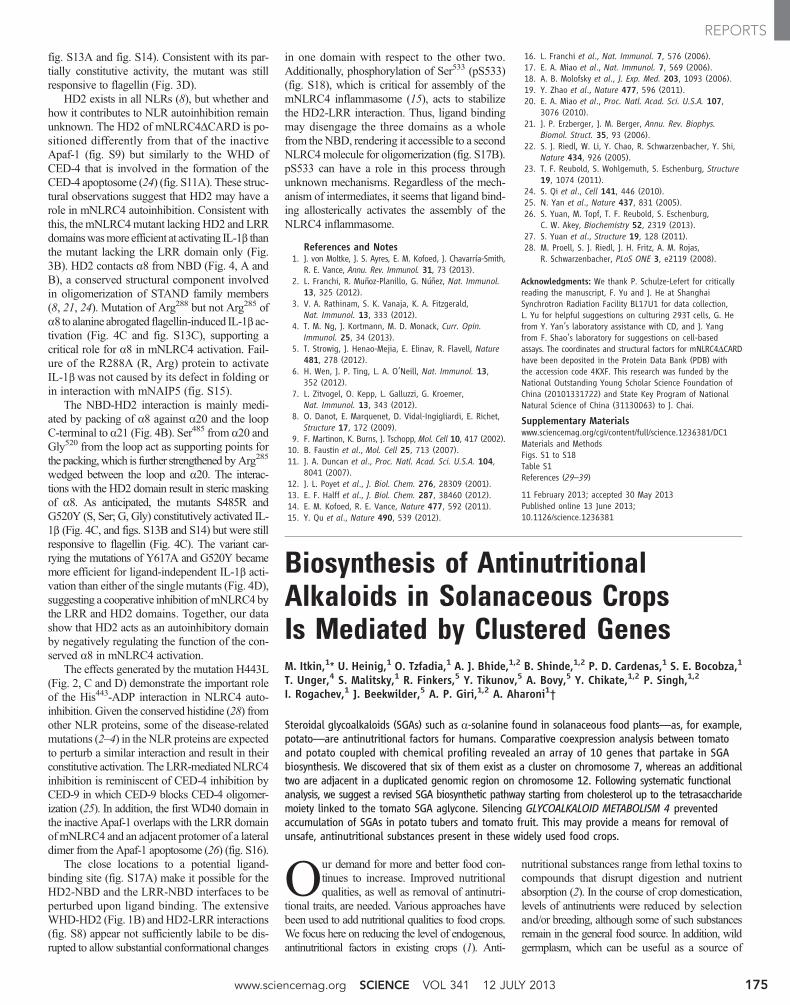
fig. S13A and fig. S14). Consistent with its par-tially constitutive activity, the mutant was stillresponsive to flagellin (Fig. 3D).
HD2 exists in all NLRs (8), but whether andhow it contributes to NLR autoinhibition remainunknown. The HD2 of mNLRC4DCARD is po-sitioned differently from that of the inactiveApaf-1 (fig. S9) but similarly to the WHD ofCED-4 that is involved in the formation of theCED-4 apoptosome (24) (fig. S11A). These struc-tural observations suggest that HD2 may have arole in mNLRC4 autoinhibition. Consistent withthis, the mNLRC4mutant lacking HD2 and LRRdomainswasmore efficient at activating IL-1b thanthe mutant lacking the LRR domain only (Fig.3B). HD2 contacts a8 from NBD (Fig. 4, A andB), a conserved structural component involvedin oligomerization of STAND family members(8, 21, 24). Mutation of Arg288 but not Arg285 ofa8 to alanine abrogated flagellin-induced IL-1b ac-tivation (Fig. 4C and fig. S13C), supporting acritical role for a8 in mNLRC4 activation. Fail-ure of the R288A (R, Arg) protein to activateIL-1b was not caused by its defect in folding orin interaction with mNAIP5 (fig. S15).
The NBD-HD2 interaction is mainly medi-ated by packing of a8 against a20 and the loopC-terminal to a21 (Fig. 4B). Ser485 from a20 andGly520 from the loop act as supporting points forthe packing,which is further strengthened by Arg285
wedged between the loop and a20. The interac-tions with the HD2 domain result in steric maskingof a8. As anticipated, the mutants S485R andG520Y (S, Ser; G, Gly) constitutively activated IL-1b (Fig. 4C, and figs. S13B and S14) but were stillresponsive to flagellin (Fig. 4C). The variant car-rying the mutations of Y617A and G520Y becamemore efficient for ligand-independent IL-1b acti-vation than either of the single mutants (Fig. 4D),suggesting a cooperative inhibition ofmNLRC4bythe LRR and HD2 domains. Together, our datashow that HD2 acts as an autoinhibitory domainby negatively regulating the function of the con-served a8 in mNLRC4 activation.
The effects generated by the mutation H443L(Fig. 2, C and D) demonstrate the important roleof the His443-ADP interaction in NLRC4 auto-inhibition. Given the conserved histidine (28) fromother NLR proteins, some of the disease-relatedmutations (2–4) in the NLR proteins are expectedto perturb a similar interaction and result in theirconstitutive activation. The LRR-mediatedNLRC4inhibition is reminiscent of CED-4 inhibition byCED-9 in which CED-9 blocks CED-4 oligomer-ization (25). In addition, the first WD40 domain inthe inactive Apaf-1 overlaps with the LRR domainofmNLRC4 and an adjacent protomer of a lateraldimer from the Apaf-1 apoptosome (26) (fig. S16).
The close locations to a potential ligand-binding site (fig. S17A) make it possible for theHD2-NBD and the LRR-NBD interfaces to beperturbed upon ligand binding. The extensiveWHD-HD2 (Fig. 1B) andHD2-LRR interactions(fig. S8) appear not sufficiently labile to be dis-rupted to allow substantial conformational changes
in one domain with respect to the other two.Additionally, phosphorylation of Ser533 (pS533)(fig. S18), which is critical for assembly of themNLRC4 inflammasome (15), acts to stabilizethe HD2-LRR interaction. Thus, ligand bindingmay disengage the three domains as a wholefrom the NBD, rendering it accessible to a secondNLRC4molecule for oligomerization (fig. S17B).pS533 can have a role in this process throughunknown mechanisms. Regardless of the mech-anism of intermediates, it seems that ligand bind-ing allosterically activates the assembly of theNLRC4 inflammasome.
References and Notes1. J. von Moltke, J. S. Ayres, E. M. Kofoed, J. Chavarría-Smith,
R. E. Vance, Annu. Rev. Immunol. 31, 73 (2013).2. L. Franchi, R. Muñoz-Planillo, G. Núñez, Nat. Immunol.
13, 325 (2012).3. V. A. Rathinam, S. K. Vanaja, K. A. Fitzgerald,
Nat. Immunol. 13, 333 (2012).4. T. M. Ng, J. Kortmann, M. D. Monack, Curr. Opin.
Immunol. 25, 34 (2013).5. T. Strowig, J. Henao-Mejia, E. Elinav, R. Flavell, Nature
481, 278 (2012).6. H. Wen, J. P. Ting, L. A. O’Neill, Nat. Immunol. 13,
352 (2012).7. L. Zitvogel, O. Kepp, L. Galluzzi, G. Kroemer,
Nat. Immunol. 13, 343 (2012).8. O. Danot, E. Marquenet, D. Vidal-Ingigliardi, E. Richet,
Structure 17, 172 (2009).9. F. Martinon, K. Burns, J. Tschopp, Mol. Cell 10, 417 (2002).10. B. Faustin et al., Mol. Cell 25, 713 (2007).11. J. A. Duncan et al., Proc. Natl. Acad. Sci. U.S.A. 104,
8041 (2007).12. J. L. Poyet et al., J. Biol. Chem. 276, 28309 (2001).13. E. F. Halff et al., J. Biol. Chem. 287, 38460 (2012).14. E. M. Kofoed, R. E. Vance, Nature 477, 592 (2011).15. Y. Qu et al., Nature 490, 539 (2012).
16. L. Franchi et al., Nat. Immunol. 7, 576 (2006).17. E. A. Miao et al., Nat. Immunol. 7, 569 (2006).18. A. B. Molofsky et al., J. Exp. Med. 203, 1093 (2006).19. Y. Zhao et al., Nature 477, 596 (2011).20. E. A. Miao et al., Proc. Natl. Acad. Sci. U.S.A. 107,
3076 (2010).21. J. P. Erzberger, J. M. Berger, Annu. Rev. Biophys.
Biomol. Struct. 35, 93 (2006).22. S. J. Riedl, W. Li, Y. Chao, R. Schwarzenbacher, Y. Shi,
Nature 434, 926 (2005).23. T. F. Reubold, S. Wohlgemuth, S. Eschenburg, Structure
19, 1074 (2011).24. S. Qi et al., Cell 141, 446 (2010).25. N. Yan et al., Nature 437, 831 (2005).26. S. Yuan, M. Topf, T. F. Reubold, S. Eschenburg,
C. W. Akey, Biochemistry 52, 2319 (2013).27. S. Yuan et al., Structure 19, 128 (2011).28. M. Proell, S. J. Riedl, J. H. Fritz, A. M. Rojas,
R. Schwarzenbacher, PLoS ONE 3, e2119 (2008).
Acknowledgments: We thank P. Schulze-Lefert for criticallyreading the manuscript, F. Yu and J. He at ShanghaiSynchrotron Radiation Facility BL17U1 for data collection,L. Yu for helpful suggestions on culturing 293T cells, G. Hefrom Y. Yan’s laboratory assistance with CD, and J. Yangfrom F. Shao’s laboratory for suggestions on cell-basedassays. The coordinates and structural factors for mNLRC4DCARDhave been deposited in the Protein Data Bank (PDB) withthe accession code 4KXF. This research was funded by theNational Outstanding Young Scholar Science Foundation ofChina (20101331722) and State Key Program of NationalNatural Science of China (31130063) to J. Chai.
Supplementary Materialswww.sciencemag.org/cgi/content/full/science.1236381/DC1Materials and MethodsFigs. S1 to S18Table S1References (29–39)
11 February 2013; accepted 30 May 2013Published online 13 June 2013;10.1126/science.1236381
Biosynthesis of AntinutritionalAlkaloids in Solanaceous CropsIs Mediated by Clustered GenesM. Itkin,1* U. Heinig,1 O. Tzfadia,1 A. J. Bhide,1,2 B. Shinde,1,2 P. D. Cardenas,1 S. E. Bocobza,1
T. Unger,4 S. Malitsky,1 R. Finkers,5 Y. Tikunov,5 A. Bovy,5 Y. Chikate,1,2 P. Singh,1,2
I. Rogachev,1 J. Beekwilder,5 A. P. Giri,1,2 A. Aharoni1†
Steroidal glycoalkaloids (SGAs) such as a-solanine found in solanaceous food plants—as, for example,potato—are antinutritional factors for humans. Comparative coexpression analysis between tomatoand potato coupled with chemical profiling revealed an array of 10 genes that partake in SGAbiosynthesis. We discovered that six of them exist as a cluster on chromosome 7, whereas an additionaltwo are adjacent in a duplicated genomic region on chromosome 12. Following systematic functionalanalysis, we suggest a revised SGA biosynthetic pathway starting from cholesterol up to the tetrasaccharidemoiety linked to the tomato SGA aglycone. Silencing GLYCOALKALOID METABOLISM 4 preventedaccumulation of SGAs in potato tubers and tomato fruit. This may provide a means for removal ofunsafe, antinutritional substances present in these widely used food crops.
Our demand for more and better food con-tinues to increase. Improved nutritionalqualities, as well as removal of antinutri-
tional traits, are needed. Various approaches havebeen used to add nutritional qualities to food crops.We focus here on reducing the level of endogenous,antinutritional factors in existing crops (1). Anti-
nutritional substances range from lethal toxins tocompounds that disrupt digestion and nutrientabsorption (2). In the course of crop domestication,levels of antinutrients were reduced by selectionand/or breeding, although some of such substancesremain in the general food source. In addition, wildgermplasm, which can be useful as a source of
www.sciencemag.org SCIENCE VOL 341 12 JULY 2013 175
REPORTS

novel traits such as pathogen resistance, may alsobe complicated by co-occurrence of antinutritionalcompounds that need to be removed. Current tech-nologies include extensive backcrossing, whichcan be a slow and imperfect process (3).
Steroidal glycoalkaloids (SGAs), found instaple vegetable crops such as potato (Solanumtuberosum) and tomato (S. lycopersicum), are aclass of antinutritional substances that remain inour food chain and daily diet (4). The glyco-alkaloids a-solanine (5) and a-chaconine arethe principal toxic substances in potato. TheseSGAs cause gastrointestinal and neurologicaldisorders and, at high concentrations, may be le-thal to humans. Mechanisms of toxicity include
disruption of membranes and inhibition of acetyl-choline esterase activity (6). For this reason, to-tal SGA levels exceeding 200 mg per kg freshweight of edible tuber are deemed unsafe for hu-man consumption (7). SGA biosynthesis requiresgenes encoding uridine 5´-diphosphate (UDP)–glycosyltransferases (UGTs) that decorate thesteroidal alkaloid (SA) skeleton with various sugarmoieties (8, 9). The tomato GLYCOALKALOIDMETABOLISM 1 (GAME1) glycosyltransferase,a homolog of the potato SGT1 (8), catalyzesgalactosylation of the alkamine tomatidine (9).Cholesterol is the proposed common precursorfor biosynthesis of both steroidal alkaloids (SAs)and non-nitrogenous steroidal saponins (STSs)(Fig. 1 and fig. S1) (10). Conversion of choles-terol to the alkamine SA should require severalhydroxylation, oxidation, and transamination reac-tions (10). Here, we identify genes encoding en-zymes performing the conversion of cholesterolto SGAs and use them to engineer Solanaceaeplants with reduced SGA content.
To discover genes associated with SGA bio-synthesis, we carried out coexpression analysisusing transcriptome data from tomato and potatoplants (11). Sixteen genes from each species werecoexpressed with GAME1/SGT1 (Fig. 2). One ofthese genes, which we namedGLYCOALKALOIDMETABOLISM 4 (GAME4), encodes a member of
the 88D subfamily of cytochrome P450 proteins(fig. S2).GAME4 andGAME1/SGT1 display a verysimilar expression profile in tomato and potato (fig.S3, B and C, and fig. S4). We then discovered thatthe GAME1/SGT1 and GAME4 genes in tomatoand potato are positioned in chromosomes 7 and12, respectively, such that they are physically nextto several of their coexpressed genes (Fig. 3).
A cluster of GAME1/SGT1 coexpressed genesspans a ~200 kilo–base pair genomic region onchromosome 7. Together with GAME1, the to-mato cluster is composed of seven coexpressedgenes. These include three UDP-glycosyltransferases[GAME2 (termed SGT3 in potato), GAME17, andGAME18], a cytochrome P450 of the 72A subfam-ily (GAME6), a 2-oxoglutarate–dependent dioxy-genase (GAME11), and a cellulose synthase-likeprotein. It appears that in potato this cluster con-tains five coexpressed genes as it lacks homologsof the tomato GAME17 and GAME18 UDP-glycosyltransferases. We performed enzyme ac-tivity assays with the four recombinant clusteredtomato UDP-glycosyltransferases. GAME17 andGAME18 exhibited UDP-glucosyltransferase ac-tivity when incubated with tomatidine galactoside(T-Gal) and g-tomatine (T-Gal-Glu) as a sub-strate, respectively, whereas GAME2 was shownto have a UDP-xylosyltransferase activity whenincubated with b1-tomatine (T-Gal-Glu-Glu) as a
1Department of Plant Sciences, Weizmann Institute of Sci-ence, Rehovot 76100, Israel. 2Plant Molecular Biology Unit,Division of Biochemical Sciences, Council of Scientific and In-dustrial Research–National Chemical Laboratory, Pune 411008,MS, India. 3Department of Plant Pathology and Microbiology,The Robert H. Smith Faculty of Agriculture, Food and Environ-ment, The Hebrew University of Jerusalem, Rehovot 76100,Israel. 4Israel Structural Proteomics Center, Weizmann Instituteof Science, Rehovot 76100, Israel. 5Plant Research Interna-tional, Wageningen University and Research Centre, POB 16,6700 AA, Wageningen, Netherlands.
*Present address: Agricultural Research Organization, TheVolcani Center, Bet Dagan 50250, Israel.†Corresponding author. E-mail: [email protected]
Fig. 1. Biosynthesis of steroidal alkaloids and saponins in the triter-penoid biosynthetic pathway in Solanaceae plants. Suggested bio-synthetic pathway from cholesterol toward a-tomatine. Dashed and solidarrows represent multiple or single enzymatic reactions in the pathway,
respectively. The proposed activity of GAME1, GAME4, and GAME8 was sup-ported by investigating transgenic plants; of GAME11, GAME12, and GAME18by VIGS assays; and of GAME1, GAME17, GAME18, and GAME2 by activityassays of the recombinant enzymes.
12 JULY 2013 VOL 341 SCIENCE www.sciencemag.org176
REPORTS

substrate (Fig. 4, F to H, and fig. S5). GAME1was previously shown to act as a tomatidine UDP-galactosyltransferase in tomato (9). When incu-
bating the four recombinant UGT enzymes in asingle test tube, with tomatidine, and all glycosidedonors (UDP-galactose, -glucose and -xylose), we
observed the accumulation of the final SGAproduct, a-tomatine (Fig. 4I and fig. S5). The roleof GAME18 in creating the tetrasaccharide moiety
Fig. 2. Steroidal alkaloids gene discovery through coex-pression network analysis in Solanaceae plants. Sharedhomologs of coexpressed genes for “bait-genes” from tomato(SlGAME1 and SlGAME4) and potato (StSGT1 and StGAME4).Continuous [correlation coefficient (r) > 0.8] and dashed (r >0.63) lines connect coexpressed genes. *, located in the tomatoor potato chromosome 7 cluster. St, Solanum tuberosum; Sl,S. lycopersicum. Color background of gene names correspondsto the bait with which they were found to be coexpressed(as above). (For more details, see tables S1 to S10.) SP, serineproteinase; PI, proteinase inhibitor; UPL, ubiquitin protein li-gase; ELP, extensin-like protein; PK, protein kinase; SR, sterolreductase; RL, receptor-like.
Fig. 3. Schematic map of genes identified in the duplicated genomicregions in tomato and potato and their coexpression. Coexpression withGAME1/SGT1 (chromosome 7) and GAME4 (chromosome 12) as baits in eitherpotato or tomato are presented in the form of a heat map (table S12). Specific
gene families are indicated by colored arrows, whereas members of other genefamilies are shown by white arrows. Note the homology in genes flanking the high-coexpression regions and positioned in a matching sequence along the genome,suggesting a common origin of the regions on both chromosomes (see fig. S11).
www.sciencemag.org SCIENCE VOL 341 12 JULY 2013 177
REPORTS

of a-tomatine was supported by virus-induced genesilencing (VIGS) assays asGAME18-silenced fruitaccumulated g-tomatine that was not present in thecontrol sample (fig. S6, A to E). Analysis of thetomato leaves, silenced (VIGS) in GAME11, a puta-tive dioxygenase in the cluster, revealed a significantreduction in a-tomatine levels and accumulation ofseveral cholestanol-type steroidal saponins, confirm-ing its function in the SGA pathway (Fig. 4B andfig. S6, F to I). Additionally, GAME6, encoded byanother cluster gene, was previously suggested tobe associated with SGA metabolism (12).
GAME4 and a putative transaminase (GAME12)that was highly coexpressed were positioned inclose proximity to each other on chromosome 12of both species (Fig. 3). Silencing GAME4 in po-tato by RNA interference (RNAi) (GAME4i plants),showed a reduction by a factor of up to 74 in thelevels of a-solanine/chaconine and other SGAs inboth leaves and tubers (fig. S7, A to E). In the dark,normal quantities of a-solanine and a-chaconineare 200 and 370 mg/kg, respectively (fig. S7C). Afterlight exposure, levels of a-solanine and a-chaconineincrease in tuber skin, and quantities are 510 and
870 mg/kg, respectively. With the GAME4 genesilenced, the concentrations of both a-solanine anda-chaconine remained below 5 mg/kg and did notchange with light exposure (fig. S7, C to E).
In the domesticated tomato, the dominant SGAin leaves and mature green fruit is a-tomatine thatwas reduced by a factor of ~100 inGAME4i plants(figs. S7F and S8 and table S14). During the tran-sition from green to red fruit, a-tomatine is con-verted to lycoperosides and esculeosides. These twoclasses of compounds represent hydroxylated, gly-cosylated, and often acetylated a-tomatine deriva-tives (13). Hence, reduced a-tomatine accumulationin the green fruit stage resulted in reduced accu-mulation of lycoperosides and esculeosides in thered-ripe fruit stage (fig. S7G). Complementary re-sults were obtained in GAME4-overexpressing to-mato plant leaves (GAME4oe), as they accumulated2.5 times as much a-tomatine as the controls (fig.S8B). Furthermore, GAME4oe red-ripe fruit ex-hibited 2.9 times more esculeoside A (fig. S8C),demonstrating once more the central role ofGAME4 in SGA biosynthesis. It appeared thatSGA precursors [i.e., cholesterol, cycloartenol,
and (S)-2,3-oxidosqualene] and the phytosterolscampesterol and b-sitosterol accumulated in leavesof GAME4-silenced tomato plants (fig. S9). De-spite altered phytosterol levels, GAME4-silencedplants were not affected in their morphology un-der the conditions examined in this study (14).
Tomato and potato GAME4i plants with de-creased levels of SGAs accumulated nitrogen-lacking compounds identified as STSs (fig. S7, Hand I, and fig. S10). Greater reduction in SGAscorrelated with greater accumulation of STSs (fig.S7, D, E, H, and I). Levels of STSs were significant-ly induced by light in several wild-type andGAME4ilines examined (fig. S7, H and I). These resultsindicate that SGAs and STSs originate from thesame precursor and that GAME4 is positioned ina branch point before the incorporation of nitrogenfor SGA generation in the diverging biosyntheticpathways that produce these two classes of ste-roidal compounds (Fig. 1 and fig. S1).
GAME12 (transaminase)–silenced tomato leaveswere found enriched with a furostanol-type saponin(Fig. 4D and fig. S6, J to M), suggesting additionalhydroxylation of its accumulated substrate. We also
Fig. 4. Functional analysis of tomato GAME genes. (A) GAME8-silencedtransgenic (RNAi) leaves accumulated 22-(R)-hydroxycholesterol comparedto wild type. (B) An array of cholestanol-type steroidal saponins (STSs) accu-mulates in GAME11 VIGS-silenced leaves. (C) An STS annotated as Uttroside Baccumulates in GAME4-silenced transgenic leaves. (D) An STS [mass/charge ratio(m/z) = 753.4] accumulates in GAME12 VIGS-silenced leaves. (E) Tomatidine,the steroidal alkaloid aglycone, accumulates in GAME1-silenced transgenicleaves. (F to I) Enzyme activity assays of the four recombinant tomato GAMEglycosyltransferases (14). Reactions containing GAME17 (F) and GAME18 (G)
recombinant proteins with UDP-glucose as donor-substrate, and T-Gal orT-Gal-Glu gama-tomatine as an acceptor-substrate, respectively, producedproducts with m/z = 740.4 and m/z = 902.5, respectively. Reaction productswere identified as g-tomatine for GAME17 (F) and T-Gal-Glu-Glu for GAME18(G). Reaction containing b1-tomatine, the GAME2 recombinant protein, andUDP-xylose produced a-tomatine (H). Reaction containing tomatidine assubstrate, UDP -galactose, -glucose and -xylose as sugar donors, and theGAME1, GAME2, GAME17, and GAME18 recombinant proteins resulted inaccumulation of a-tomatine (I). See also figs. S5, S6, and S10.
12 JULY 2013 VOL 341 SCIENCE www.sciencemag.org178
REPORTS
functionally examined genes that were tightly coex-pressed and positioned elsewhere in the genomethat belong to the CYP72 subfamily of cytochromeP450s (i.e., GAME7 and GAME8). GAME7 wascoexpressed in both species, whereas StGAME8aand StGAME8b were strongly coexpressed withStSGT1 and StGAME4 in potato. At present, wecould not demonstrate SGA-related activity forGAME7, although as for GAME6, it was sug-gested to be involved in SGA metabolism (12).Yet, GAME8-silenced tomato leaves accumulated22-(R)-hydroxycholesterol (fig. S6, N to Q), aproposed intermediate in the SGA biosyntheticpathway (Fig. 1).
The above findings allowed us to propose apathway from cholesterol to a-tomatine. Cholesterolis hydroxylated at C22 by GAME7 (12), followedby GAME8 hydroxylation at the C26 position (Fig.1). The 22,26-dihydroxycholesterol is then hydrox-ylated at C16 and oxidized at C22, followed byclosure of the E-ring by GAME11 and GAME6to form the furostanol-type aglycone. This orderof reactions is supported by the accumulation ofcholestanol-type saponins, lacking hydroxylation atC16 and the hemi-acetal E-ring when silencingGAME11 (fig. S6, F to I). The furostanol-intermediateis oxidized by GAME4 to its 26-aldehyde, which isthe substrate for transamination catalyzed byGAME12. Nucleophilic attack of the amino nitrogenat C22 leads to the formation of tomatidenol, whichis dehydrogenated to tomatidine. Tomatidine issubsequently converted by GAME1 to T-Gal (9).T-Gal in its turn is glucosylated by GAME17 intog-tomatine, which is further glucosylated by GAME18to b1-tomatine that is finally converted to a-tomatineby GAME2 (Fig. 1).
Some specialized plant metabolites, particu-larly terpenoids, are the result of activities fromclusters of genes (15, 16). The existence of meta-bolic gene clusters raises questions about theadvantages of such genomic organization (17).Reducing the distance between loci, resulting incoinheritance of advantageous combinations ofalleles, may be one benefit of clustering (17). Clus-tering glycosyltransferases and core pathway genes,as observed here for SGAs, could maintain alleliccombinations that support the metabolic outcomeneeded by the plant and reduce formation of phy-totoxic aglycone compounds (9, 18). We found thatthe regions of coexpressed genes in both chromo-somes (i.e., 7 and 12) were flanked by similarlyannotated genes and positioned identically alongthe genome, although poorly coexpressed withGAME1/SGT1 and GAME4 and likely not re-lated to SGA metabolism (fig. S11 and table S13).This suggests a duplication event that facilitatedthe positioning alongside each other on chromo-some 12 ofGAME4 andGAME12, both STSs andSGAs branch-point genes. Subsequent evolution ofenzyme function of this gene pair likely allowedplants in the Solanaceae family to start producingthe nitrogen-containing steroidal alkaloids.
We have shown that SGA levels can be severelyreduced in potato tubers by modifying expressionof an enzyme in the biosynthetic pathway. The lack
of SGAs in such plants might make them sensitiveto biotic stress, and the increased production ofSTSs (as occurred in GAME4-silenced plants)—which are nontoxic to warm-blooded species, in-cluding humans (19)—might provide a compen-satory defense mechanism (20). The findings openthe way for developing new strategies, throughgenetic engineering or more classical breeding pro-grams, to reduce quantities of the antinutritionalSGAs in key crops of the Solanaceae, including po-tato, tomato, and eggplant. At the same time, it pro-vides a platform for studying the SGA and STSbiosynthetic pathways, transport, and regulatorysystems that control the production of thousandsof these chemicals in specific plant lineages.
References and Notes1. N. N. Narayanan, U. Ihemere, C. Ellery, R. T. Sayre,
PLoS ONE 6, e21996 (2011).2. L. C. Dolan, R. A. Matulka, G. A. Burdock, Toxins 2,
2289–2332 (2010).3. L. L. Sanford, S. P. Kowalski, C. M. Ronning, K. L. Deahl,
Am. J. Potato Res. 75, 167–172 (1998).4. M. Friedman, J. Agric. Food Chem. 54, 8655–8681 (2006).5. M. Desfosses, J. Pharmacie 6, 374–376 (1820).6. J. G. Roddick, Phytochemistry 28, 2631–2634 (1989).7. FDA Poisonous Plant Database, www.accessdata.fda.gov/
scripts/Plantox/8. K. F. McCue et al., Plant Sci. 168, 267–273 (2005).9. M. Itkin et al., Plant Cell 23, 4507–4525 (2011).10. E. Eich, Solanaceae and Convolvulaceae: Secondary
Metabolites: Biosynthesis, Chemotaxonomy, Biologicaland Economic Significance (Springer, Berlin, 2008).
11. The Potato Genome Sequencing Consortium, Nature 475,189–195 (2011).
12. N. Umemoto, S. Katsunori, U.S. Patent application20120159676 A1 (2012).
13. T. Yamanaka et al., J. Agric. Food Chem. 57, 3786–3791(2009).
14. Materials and methods are available as supplementarymaterials on Science Online.
15. D. J. Kliebenstein, A. Osbourn, Curr. Opin. Plant Biol. 15,415–423 (2012).
16. T. Winzer et al., Science 336, 1704–1708 (2012).17. A. E. Osbourn, Plant Physiol. 154, 531–535 (2010).18. B. Field, A. E. Osbourn, Science 320, 543–547 (2008).19. P. F. Dowd, M. A. Berhow, E. T. Johnson, J. Chem. Ecol.
37, 443–449 (2011).20. S. G. Sparg, M. E. Light, J. van Staden, J. Ethnopharmacol.
94, 219–243 (2004).
Acknowledgments: We thank A. Tishbee and R. Kramer foroperating the UPLC-qTOF-MS instrument and the EuropeanResearch Council (SAMIT-FP7 program) for supporting the work inthe A.A. laboratory. A.A. is the incumbent of the Peter J. CohnProfessorial Chair. J.B. was supported by the European Union 7thFrame Anthocyanin and Polyphenol Bioactives for HealthEnhancement through Nutritional Advancement (ATHENA) Project(FP7-KBBE-2009-3-245121-ATHENA). U.H. was partially supportedby fellowship AZ: I/82 754, Volkswagen Foundation, Hannover,Germany. We thank the Council of Scientific and Industrial Research(India) for support to A.P.G. (Raman Research Fellowship), A.J.B.,Y.C., and P.S. (Research Fellowship) and the University GrantsCommission (India) for supporting B.S. We also thank D. R. Nelsonfor assistance with CYP450 gene classification and R. Last forcritically reading the manuscript. A.A. and M.I. are inventorson publication number WO2012095843 A1, submitted by YedaResearch and Development Co. Ltd, which covers the use ofthe GAME4 gene for generating low-alkaloid fruit and tubers.
Supplementary Materialswww.sciencemag.org/cgi/content/full/science.1240230/DC1Materials and MethodsFigs. S1 to S15Tables S1 to S16References (21–31)
8 May 2013; accepted 6 June 2013Published online 20 June 2013;10.1126/science.1240230
Genome-Wide Comparisonof Medieval and ModernMycobacterium lepraeVerena J. Schuenemann,1* Pushpendra Singh,2* Thomas A. Mendum,3* Ben Krause-Kyora,4*Günter Jäger,5* Kirsten I. Bos,1 Alexander Herbig,5 Christos Economou,6 Andrej Benjak,2
Philippe Busso,2 Almut Nebel,4 Jesper L. Boldsen,7 Anna Kjellström,8 Huihai Wu,3
Graham R. Stewart,3 G. Michael Taylor,3 Peter Bauer,9 Oona Y.-C. Lee,10 Houdini H.T. Wu,10
David E. Minnikin,10 Gurdyal S. Besra,10 Katie Tucker,11 Simon Roffey,11 Samba O. Sow,12
Stewart T. Cole,2† Kay Nieselt,5† Johannes Krause1†
Leprosy was endemic in Europe until the Middle Ages. Using DNA array capture, we have obtainedgenome sequences of Mycobacterium leprae from skeletons of five medieval leprosy cases from theUnited Kingdom, Sweden, and Denmark. In one case, the DNA was so well preserved that full de novoassembly of the ancient bacterial genome could be achieved through shotgun sequencing alone.The ancient M. leprae sequences were compared with those of 11 modern strains, representingdiverse genotypes and geographic origins. The comparisons revealed remarkable genomic conservationduring the past 1000 years, a European origin for leprosy in the Americas, and the presence of anM. leprae genotype in medieval Europe now commonly associated with the Middle East. The exceptionalpreservation of M. leprae biomarkers, both DNA and mycolic acids, in ancient skeletons has majorimplications for palaeomicrobiology and human pathogen evolution.
Leprosy, which results from infection withthe unculturable pathogenMycobacteriumleprae, was common in Europe until the
16th century, when it essentially disappeared. Incontrast, disease prevalence has remained high inthe developing world. During the past 20 years,
www.sciencemag.org SCIENCE VOL 341 12 JULY 2013 179
REPORTS