Materials Engineering Name of the Unit : Crystal structure ...

6
SUPERHYDROPHOBICSURFACES
Ankur Gupta,1 Milind R. Joshi,1 Neelima Mahato,1,2and Kantesh Balani1
1Department of Materials Science and Engineering, Indian Institute of TechnologyKanpur, Kanpur, India
2Department of Applied Chemistry, Institute of Technology, Varanasi, India
The development of biomaterials for specific application with the desired functionrequires a careful combination of surface roughness, surface energy (and their polar anddispersion component), the presence of functional group, and the response of materialsto the body fluid. The main constituent of body fluid is water; therefore, materialresponse to water can be used to predict the behavior of the material implanted in thehuman body. Protein adsorption, cell and bacteria proliferation, alkaline phosphateactivity, and so on are some of the assessments that are conducted during the in vitrostudies on the materials, which depend on the hydrophilic or hydrophobic nature of thematerial in order to provide first insight into the material response. These responsesof the material further depend on surface roughness, surface energy and the presenceof functional group on the surface. Hence, learning the wetting behavior of materialis necessary for its engineering. Thus, to nurture the material behavior in the bodyenvironment, understanding of contact angle (CA), its dependence on various factorsand fabrication of desired surface is extremely necessary. In this chapter, the basicsof CA in contrast to natural phenomena present in the surrounding environment suchas surfaces of lotus, rice and ramie leaves, correlations between the morphological
Biosurfaces: A Materials Science and Engineering Perspective, First Edition.Edited by Kantesh Balani, Vivek Verma, Arvind Agarwal, Roger Narayan.© 2015 The American Ceramic Society. Published 2015 by John Wiley & Sons, Inc.
170

171
features, mechanical properties and Laplacean pressure, fabrication of artificial surfacesto mimic superhydrophobic surfaces and their potential applications are discussed.
6.1 INTRODUCTION
Mysteries of natural phenomena keep unfolding unexpectedly, and the nature, thus, hasalways been the motivation of science. Be it an airplane or an igloo, engineers and sci-entists have always tried to mimic nature and shall continue to do so. Nature’s intricateengineering displayed in nacre (shell), dentin (human teeth) and bone is intriguing, asthese materials show higher fracture toughness by three orders of magnitude than that ofthe ceramic materials comprising it [1–9]. Other examples of nature’s incredible engi-neering are termite mounds, gecko’s grip, peacock’s feathers, rhino’s horn, elephant’stusks, and so on. Termite mounds are made up of low energy-intensive materials thatcool themselves by passive air conditioning. This particular concept is used during theconstruction of mud houses in the deserts [10, 11]. A Gecko can grip vertical surfacesby the virtue of seta (foot hair) and spatulae (each seta splits into hundreds of nanotips) in its foot. The number of spatulae is huge, approximately 8× 10⁶ [12, 13]. Eachspatula is capable of providing van der Waals forces of attraction, and hence 8× 10⁶
spatulae together get so close to a surface that they bond at the atomic level and generateextremely powerful adhesive forces.
“Nature is simple but its science is not,” and hence researchers worldwide attemptto understand nature and mimic it in our daily life [14]. Apart from mechanical proper-ties, nature has mastered in attaining certain properties such as superhydrophobicity: forexample, a lotus leaf. A water droplet on the lotus leaf subtends a CA of approximately160∘, which lies in the range necessary for superhydrophobicity [15–17]. By suitablearrangement of tiny protrusions and nanohairs on the leaf surface, the nature has not onlycreated a superhydrophobic surface, but also rendered it a self-cleaning property. Thus,the water drop, while rolling, carries the dirt off. This self-cleaning property might beone such interesting reason that certain eastern cultures consider lotus as a symbol ofdivinity.
6.2 SURFACES AND SUPERHYDROPHOBICITY IN NATURE
Atoms present on the surface are partially bonded, while those in the bulk, due tocomplete bonding, experience a net zero force. The excess energy of incompletelybonded surface atoms induces a contracting force or surface tension. Hence, the freeenergy of the surface is always higher than that of the bulk. This available surfaceenergy plays an important role in the liquid–solid surface interaction and decides theshape of a droplet on the surface defined by CA “𝜃c” [18]. Surface energy as well asthe morphological features makes wettability a characteristic property of the surface,thus, CA largely depends on both factors. The nature of a surface can be classifiedon the basis of CA. It is called hydrophilic when 𝜃c < 90∘, and the water dropletsplaced over it tend to spread out. When 𝜃c > 90∘, the surface is hydrophobic, andthe water droplets tend to bead-up. Superhydrophobic surfaces are the one with CA

172
“𝜃c”> 150∘, and in this condition, water droplets become highly beaded, repelled. Inthe case of materials with lowest surface energy, a maximum CA of 120∘ is obtained.This angle is subtended with no curvature on the surface, that is, with straight edgesjoining as a hexagon [19, 20]. This is similar to the grain growth phenomena in whichflat-faced, six-sided grains subtend internal angles of 120∘ [21]. Thus, for hydrophobicsurfaces, an increased surface roughness can further enhance the CA. Since surfaceenergy and texture both play important roles, therefore superhydrophobicity maybe stated as a combination of surface chemistry (hydrophobicity) and structure(roughness).
In this chapter, an endeavor is made to understand (i) why certain objects in natureare superhydrophobic, (ii) theoretical and practical aspects of superhydrophobicity,(iii) statics and dynamics of wetting and (iv) how these objects can be mimickedto explore and understand the structural and mechanical aspects of such surfaces.Elaborate information cum details of techniques, such as electrospinning, sol-gel, softlithographic imprinting, layer-by-layer deposition, plasma treatment, and so on, skill-fully exercised during recent years, in order to fabricate hydrophobic/superhydrophobicsurfaces, have been presented. Successful preparation of metallic superhydrophobicsurfaces, controlled wettability surfaces (CWS), polystyrene nanotubes for drugdelivery applications, and so on and future directions of artificially fabricated surfaceshave also been addressed. This chapter summarizes recent updates in available datapertaining to the utilization of this concept in interesting engineering applications andis meant to supplement the previously written materials in the similar field [15, 22, 23].
6.3 CLASSIFICATION OF SURFACES
To explain the effect of surface roughness on hydrophobicity, Wenzel and Cassie/Baxterput forward their independent theories of Wenzel’s and Cassie’s state, respectively.Figure 6.1 shows the schematic of classical model of liquid–solid–vapor interaction.According to Wenzel, water droplets pin the rough surface in contact mode anddemonstrate high sliding angle when tilted, resulting in high adhesion forces. Thus,surface roughness factor (r), a ratio of total surface area to projected surface area, isintroduced in the Young’s equation as Eq. 6.1 to calculate CA “𝜃c.” It can be derivedby balancing body line forces in the horizontal direction. When r≥ 1, the apparent CA(𝜃′) in a Wenzel’s state can be obtained from Eq. 6.2 [24].
cos 𝜃c =𝛾sv − 𝛾sl
𝛾lv(6.1)
cos 𝜃′ =r (𝛾sv − 𝛾sl)
𝛾lv= r cos 𝜃c (6.2)
By analyzing this equation, a conclusion can be made that if 𝜃 < 90∘, the rough-ness decreases the apparent CA, that is, 𝜃′ <𝜃c, and if 𝜃 > 90∘, contrastingly, roughnessincreases apparent CA, that is, 𝜃′ >𝜃c.
According to Cassie/Baxter’s model, a water droplet rests on the heterogeneoussurface composed of two different materials and adapts a non-contact mode due to which

173
Liquid
Solid
Air
θc
γLG
γSGγSL
Figure 6.1. Schematic of solid–liquid–vapor interaction.
it can easily roll off resulting in a low sliding angle. This state is represented by Eq. 6.3[25].
cos 𝜃′ = f1 cos 𝜃1 + f2 cos 𝜃2 (6.3)
In this equation, f1 and f2 are surface area fractions of the two materials (air andthe base). 𝜃1 and 𝜃2 are their CAs respectively. Since air is always present betweenthe two hills of the surface, therefore, the water droplet subtends a CA of 180∘ in air.Incorporating this factor in Eq. 6.3 we get Eq. 6.4.
cos 𝜃′ = f cos 𝜃c + (1 − f ) cos 180∘ (6.4)
which now becomescos 𝜃′ = f cos 𝜃c + f − 1 (6.5)
This shows that “𝜃” increases with the decrease in the area fraction of the solid. TheCassie–Baxter’s state exists when the (i) contact line forces overcome the body forcesor weight of an unsupported droplet and (ii) the microstructures are tall enough to preventthe droplet from touching the base of the surface.
Wang et al. [26] have further classified the superhydrophobic surfaces into five cat-egories illustrated in Fig. 6.2a–e. These are Wenzel’s state, Cassie/Baxter’s state, Lotusstate, the transition state between Wenzel’s and Cassie’s state and Gecko state. Lotusstate is considered as a special case of Cassie Baxter’s state, which imparts minimalresistance on the droplet to roll-off. And the Gecko state is considered as a modifiedform of Cassie/Baxter’s state, which is brought into being superhydrophobic when hol-low tubes are incorporated on the surface and roughness increases. The main differencebetween the Cassie’s state (Fig. 6.2b) and the Gecko’s one (Fig. 6.2e) is the presence oftwo types of air interfaces below the droplet. In Gecko state, firstly, a drop gets trappedin between the tubes, and second, droplet following the previous one remains in directcontact with the atmosphere. Consequently, van der Waals forces of attraction are gen-erated between the trapped air and the liquid surface, and the water droplet present onsuch type of surface experiences a very high resistance to flow.

174
(a)
(d) (e)
(b) (c)
Figure 6.2. Various types of superhydrophobic states (a) Wenzel’s state, (b) Cassie–Baxter’s
state, (c) Lotus leaf state, (d) transition state between Wenzel’s and Cassie–Baxter’s states and
(e) Gecko state. (Reprinted from [26].)
6.3.1 Learning from Nature
Researchers have discovered various kinds of leaves in nature that show high waterrepelling tendency along with self-cleaning property [27]. Guo et al. [28] examinedthe surfaces of a variety of leaves, such as lotus, rice, ramie, and so on, under scan-ning electron microscopy (SEM). They identified two types of microstructures: the onecomprising micro- and nano-roughness features (called binary structure) and the othercomprising micro-line features (called unitary structure). Details of different leaves aresummarized in Table 6.1.
For a material to be superhydrophobic, its surface should subtend a high CAand impart small resistance to flow. The latter also facilitates the surface to acquire aself-cleaning property. Since the lotus leaf possesses both the properties, it has attracteda lot of attention from the researchers worldwide.
A lotus leaf exhibits two scales of roughness, namely micro-level bumps calledprotrusions (of diameter 5 μm) and nano-hairs (of length 0.30–1.0 μm and diameter50–120 nm). These structures facilitate the surface to subtend a CA of 160.4± 0.7∘[29]. Figure 6.3a–d shows microstructures of the lotus leaf at various levels of mag-nification. The protrusions are uniformly distributed along all directions and thus pro-vide self-cleaning property invariant with respect to direction, in an isotropic way. Themicrostructure of a rice leaf (Fig. 6.4b) is similar to that of lotus leaf. However, theonly difference between the two is the distribution of micro-protrusions. In rice leaf,protrusions are aligned in the longitudinal direction (shown by an arrow in Fig. 6.4a)imparting a good self-cleaning property along the length (sliding angle ≈4∘), whereasalong the width, it is diminished (sliding angle ≈12∘) [28]. Balani et al. [14] have cal-culated that 490–1256 micro-protrusions (4.29–5.65× 10⁻³ MPa per protrusion) arerequired in a lotus leaf to balance the forces exerted on the surface of a water droplet(41.2–138.5× 10⁻⁶ N). A second type of structure is found in ramie leaf. SEM imagesof the leaf surface are shown in Fig. 6.5a–c. It is apparent that there is an absence of

175
TABLE 6.1. Details of Various Leaves Showing Superhydrophobicity [28]
Type of leaf Type ofstructure
Contact angle(CA)
Number offaces showing
superhydrophobicity
LotusNelumbo nuciferagaertn
Binary 161± 2∘ One
RiceOryza sativa L.
Binary 157± 2∘ One
Indian CannaCanna generalis bailey
Binary 165± 2∘ One
TaroColocasia esculenta
Binary 159± 2∘ One
Chinese watermelonBennincasa hispida cogn
Unitary 159± 2∘ One
RamieBoehmeria iongispica
steud
Unitary 38± 2∘ and 164± 2∘ One
Perfoliate KnotweedPolygonum perroliatum L.
Binary 167± 2∘ and 165± 2∘ Two (both)
Purple SetcreaseaSetcreasea purpurea boom
Binary 162± 2∘ and 163± 2∘ Two (both)
nano-level roughness, both on the micro-line structure and at the base. However, it stillsubtends a CA of 164± 2∘. A conclusion is therefore made that only micro-level rough-ness is necessary to make a surface superhydrophobic, although nano-level roughnessfurther increases the CA. Schematic of both types of superhydrophobic leaf structuresis illustrated in Fig. 6.6a–c.
6.3.2 Role of Chemical Composition and Two-Level Roughness
Apart from the dual level of roughness, a waxy composition of epicuticular wax is alwayspresent on the leaf surfaces, which is essentially hydrophobic in nature. Cheng et al. [30]conducted an experiment to determine the composition of wax and worked out the roleof two-level structure on the self-cleaning property of lotus leafs. Peaks at 2915 and2850 cm−1 in the FTIR spectra (Fig. 6.7) correspond to C—H stretching mode, whichconfirms that the epicuticular wax is mainly composed of cellulose [30–32]. FTIR spec-trum of epicuticular wax has shown resemblance to that of carnauba wax, which suggestsa similar means to measure the CA subtended on the latter. However, it was found to be74∘, which proves that the wax is actually hydrophilic! This is attributed to the nature offunctional groups present in the wax [33]. Hence, carnauba wax does not represent theproperties of epicuticular wax, as the latter is hydrophobic in nature. In order to inves-tigate the significance of chemical composition in making a surface superhydrophobic,Chang et al. annealed lotus leaves at 150 ∘C for 1 h, which was supposed to have notchanged the composition of the wax. The observed CA on a dry leaf was 142∘, but as aconsequence of annealing, it was found to be 126∘. The microstructures of an untreated

176
Protrusion(a) (b)
(c) (d)
Protrusion
Nano-hairs
Nano-hairs
Base
Base
Figure 6.3. SEM micrographs of the lotus leaf surface showing multi-scale hierarchy of lotus
leaf (a) micro-sized protrusions on lotus leaf surface, (b) protrusions with hairy structure, (c) and
(d) nano hairs present on the base as well as on protrusions respectively. (Reproduced with
permission from Institute of Physics Publishing, [14].)
(a) (b)
50 μm 1 μm
(c)
Figure 6.4. Micrographs showing multi-scale hierarchy of rice leaf. (Reproduced with permis-
sion from Elsevier Limited, [28].)
lotus leaf and that undergone annealing treatment is shown in Fig. 6.8a and b [30]. Thisis because annealing completely damaged all the nano-hairs on the surface, except themicrometer-sized protrusions, which were left comparatively unharmed (Fig. 6.8b) [30].Thus, it was concluded that the micro-scale roughness of the surface makes the leafhydrophobic, and the nano-hairs further enhance this property by increasing the CA by

177
(a) (b)
100 μm 5 μm
(c)
Figure 6.5. Micrographs of ramie leaf showing unitary structure. (Reproduced with permis-
sion from Elsevier Limited, [28].)
Top view Front view (cross section)
Lotus leaf
Protrusions
Base hairs
Rice leaf
Ramie leaf
Figure 6.6. Schematic representation of lotus, rice and ramie leaf.

178
Annealed lotus leaf
Without annealing
Annealed carnauba
wax
Cellulose
Without annealed
Carnauba wax
Wavenumbers (cm–1)
Absorp
tion
3500
0.5
1.00.0
0.5
0
10.02
0.03
0.010
0.015
0.020(a)
(b)
(c)
(d)
(e)
3400
3400
2915
2915
2915
2915
3400
2850
2850
2850
2850
3000 2500 2000 1500 1000
Figure 6.7. FTIR of lotus leaf in various condition and comparison with cellulose. (Reproduced
with permission from Institute of Physics Publishing, [30].)
(a) (b)
10 μm 10 μm
Figure 6.8. SEM images of (a) without annealed and (b) annealed lotus leaf. (Reproduced
with permission from Institute of Physics Publishing, [30].)
16∘. An annealed leaf, due to the absence of nano-hairs on its surface, imparts resistanceagainst a rolling-off droplet and renders a poor self-cleaning property.
Balani et al. [14] carried out computational and mechanical modeling of the lotusleaf structure and identified certain parameters, such as ideal protrusion spread andheight leading to hydrophobicity. When these parameters are changed, leaf loses itswater repelling property and gets wet. The increased spaces amid the protrusions resultin self-sagging behavior, whereas increased heights result in pinching effect. Pinchingoccurs due to the capillary action since a micro-cavity is formed. Nature has intelligentlydesigned the ideal protrusion spread, and as the water droplet shifts its center of grav-ity, its self weight makes the droplet roll off from the surface of lotus leaf. A base hair(400–1000 nm in length and 50–130 nm in diameter) is longer as well as thicker thanthe protrusion hair (300–600 nm in length and 50–100 nm in diameter). A large numberof such hairs present on the protrusions require a high flexing stress (18.6–87.5 MPa)

179
without puncturing the water droplet and thus providing support analogous to that of anIndian nail bed.
6.3.3 Mechanical Aspects of Surfaces
The concepts explained in the previous sections are dependent not only on the morpho-logical features of a substrate, but also on the mechanical properties of the surface. Awater droplet cannot slide or roll off without a low coefficient of friction (COF), and thustribological properties of the substrate under consideration are important. According toBowden and Tabor’s law, the frictional force is directly proportional to the real area ofcontact; a reduction in the area of contact declines the COF. Singh et al. carried outan interesting work in this regard. They fabricated structures resembling lotus [34, 35]and Colocasia [34] leaf surfaces: dry and fresh: on poly methly meth acrylate (PMMA)film spin coated over silicon wafers using poly(dimethylsiloxane) (PDMS) molds bymeans of capillarity directed soft lithographic technique. The tribological propertiesshowed great improvement in terms of the COF. The COFs calculated were approxi-mately 0.65 for un-patterned PMMA [34, 35], 0.47 for silicon wafer [34, 35], 0.1 [35]and 0.15 [34] for lotus-leaf-like patterned PMMA, and 0.15 [34] for both fresh anddry Colocasia-leaf-like patterned PMMA. This dramatic improvement is attributed tothe fact that the real area of contact is reduced when the size of the asperity becomessmaller than the size of counterface slider. They have reported that the patterned sur-faces posses low surface energy, and thus, by lowering the area of contact, COF maybe further reduced. Bhushan et al. [22] investigated the COF and the adhesive forcesof hydrophobic and hydrophilic surfaces using atomic force microscopy (AFM). Theyobserved a lower adhesive force in the case of dried leaves compared to the fresh leaves.In addition, a low COF was observed for the hydrophobic surfaces compared to thatfor the hydrophilic one. COF declines with the reduction of scan size from micro- tonano-scale.
Apart from the tests on the lotus leaf or the surfaces developed by patterning ofnaturally available superhydrophobic surfaces, many researchers have developed arti-ficial hydrophobic surfaces similar to the one found in nature. Waters et al. [36] havegrown 50–100 nm long carbon nanotubes (CNTs) with inner and outer diameters of 40and 50 nm, respectively. This provided an aspect ratio of approximately 1:2 to study itsshell buckling properties. Nanotubes undergo column buckling for the aspect ratios ofapproximately 1:100 and shell buckling for approximately 1:20 [36]. On comparing theCNT structures developed by Waters et al. with that of a lotus leaf, it was observed thatthe protrusions and hairs on the leaf have similar aspect ratios, namely 1:2 (protrusionslength 5–10 μm and diameter 5 μm) and 3–10 (hair length 300–1000 nm and diame-ter 100 nm) [14]. The spacing between CNTs (100 nm) and that between protrusions(10–15 μm) is also similar if normalized with respect to their heights. They also carriedout nanoindentation experiments using a Berkovich indenter tip to find the shell buck-ling load. Since a 10-μm spherical indenter was used, it was difficult to image a singleCNT amid its array for indentation. The images are shown in Fig. 6.9. In the view of thefact that the topographies are apparently similar, such a study can be carried out on thelotus leaf as well, and also the buckling characteristics of the protrusions and nanohairs

180
(a)
Nanoindenter
loading
10 μm radius
spherical indenter
1 μm 100 nm
(b) (c)
Figure 6.9. Indentation images of CNTs. (a) 10 μm indenter indenting the CNTs (Schematic).
(b) SEM of indented CNTs. (c) Image (b) magnified to show distorted and deformed CNTs.
(Reproduced with permission from American Institute of Physics, [36].)
can be evaluated. Quasistatic nanoindentation results [14] also yielded the average val-ues of Young’s Moduli, that is, approximately 868 MPa for base and 358–359 MPa forprotrusion hairs. The protrusion hairs have the aspect ratio approximately similar to thebase hairs, and it is therefore expected that the protrusion hairs will have similar moduli.However, this is not so, because the flexing is contributed both by hairs and by pro-trusions, thus reducing the net stiffness. Hence, the modulus of the base hairs is muchhigher (∼867 MPa) than that of protrusion hairs (∼358 MPa). In addition, the protrusionhair diameter is approximately 50–130 nm, whereas the Berkovich tip size itself is of100 nm; it is probable that more than one hair is indented simultaneously.
Critical flexing strength of the protrusion hair is vital, because due to this property,water droplets can bounce or roll off the leaf surface. Water droplet, or for that matter anyexternal media, first comes in contact with the protrusion hairs, which provide the pri-mary resistance to damage. Protrusion hairs prevent contact with other surfaces, but theyflex when the force exceeds a critical value. In this regard, Balani et al. [14] have corre-lated the mechanical properties such as Young’s modulus and flexing strength of the hairson the protrusions and base of the lotus leaf to its superhydrophobic behavior. They alsoattempted to study the effect of protrusion spread and roughness on the wetting behaviorusing computational fluid dynamics simulations carried out using SimDrop© software.The critical flexing strength values for lotus leaf reported by the research group [14] are18.6–87.5 MPa and 11.5–47.7 MPa for the base and protrusion hairs, respectively.
6.4 MECHANICS AND NATURE OF WETTING
A surface cannot be hydrophobic if the morphological features do not have sufficientstrength to withstand the pressure applied by the weight of the water droplet [37]. Lapla-cian pressure is evaluated to express the hydrophobic behavior, which is governed bythe pressure difference arising due to surface tension of the interface between the waterdroplet and air. Thus, a higher Laplace pressure will not lead to wetting and is given bythe following equation [38]:
ΔP = 𝛾 cos(𝜃 − 𝛼)p2+ h tan 𝛼
(6.6)
whereΔP is Laplacian pressure, 𝛾 is surface tension of water, 𝜃 is the CA (taken as 160∘),p is the protrusion spread distance, h is the protrusion height (assuming non-wetting)

181
and 𝛼 is the inclination angle. When the protrusions have an ideal spread and height,Laplacian pressure exceeds critical pressure, and this leads to hydrophobicity. However,for a wider spread, the critical pressure exceeding Laplacian pressure results in wet-ting. When the protrusion height is greater than ideal height, the Laplacian pressure islower than that in the ideal case. It is seen that as the spread of the protrusion increases,sagging of the water droplet takes place, whereas increased protrusion height causesdroplet penetration. The latter finishes into droplet fragmentation and ultimately wetsthe substrate.
The presence of nanohairs increases the inclination angle from 23.5∘ to 55.2∘ fora Cassie Baxter’s to Wenzel’s state transition. It also increases the Laplacian pressureby two orders of magnitude [14]. Transition from a Wenzel’s to Cassie’ state can beachieved completely by an application of electric current and voltage leading to vapor-ization at the liquid–solid interface [39]. It can also be achieved by (i) the coalescenceof energetically metastable Wenzel’s drop with a Cassie’s one where the former is incontact with only four posts and (ii) when a dynamic movement occurs along with acoalescence leading to overcoming the energy barrier [40]. The growth of a Wenzel’sdrop is shown in the schematic Fig. 6.10. In the first step, a droplet is formed at a convexcorner (Fig. 6.10a), which is followed by its growth due to condensation in the sec-ond step (Fig. 6.10b). Consequently, in the third step, the growing droplet touches theopposite poles (Fig. 6.10c), and finally, it either continues to grow by condensation orcoalesces with other such droplets to form a bigger drop while still in contact with thefour poles (Fig. 6.10d). When the Wenzel’s drop coalesces with a Cassie’s one, it maylead to transition from Wenzel’s to Cassie state. For this, the transition energy barrier hasto be overcome. During this process, the Wenzel’s drop grows upwards by condensationtoward the Cassie’s drop (Fig. 6.11a) and coalesces with it (Fig. 6.11b). This coales-cence is an unstable and highly dynamic step and has sufficient energy to reach into theCassie’s state. It cannot be observed during the experiment because of its rapid kinetics.The final state of the Wenzel’s drop after going through the transition to Cassie’s state isshown in Fig. 6.11c. Mere coalescence does not ensure overcoming of the energy barrier,but dynamic movement of the liquid along with the coalescence may help.
Lundgren et al. [41] carried out molecular dynamics simulation studies to under-stand the effect of certain factors, such as topography, composition of the solid surfaceand CA variation with protrusion height on the hydrophobic behavior of a material[42]. Their simulation experiments have observed a transition state between Wenzel’sand Cassie’s due to an increase in the pillar (protrusion) height. Koishi et al. [43]prepared density maps of the water molecules in the vicinity of a flat surface, andnanoscale pillars were obtained using similar methodology [41]. They attempted toexplain the dependence of water droplet equilibrium position on the pillar, top andthe nanostructured bottom side surfaces. A high local density of the water moleculesindicates wetting, whereas a low density indicates hydrophobic nature. They alsoprepared three-dimensional potential energy maps to study the effect of pillar surface onthe local density of water molecules. Gennes et al. [44] have mathematically explainedthe statics and dynamics of the wetting behavior of the materials in general. Manymathematical models, such as regression method for the hydrophobicity ruler, werealso approached to determine octanol-water partition coefficients of superhydrophobiccompounds [45].

182
Droplet
formed
Droplet (i)
(ii)Pole
Growth
(a) (b) (c) (d)
Opposite
pole
contact
Four pole
contact
Figure 6.10. Schematic showing growth of a Wenzel’s drop. (i) Top view. (ii) Side view as
indicated by arrow in the figure. (a) Droplet is formed at a pole. (b) Droplet grows and connects
two poles. (c) Droplet grows to develop contact with four poles. (d) Droplet grows in height.
(Reproduced with permission from American Chemical Society, [40].)
Growth of
Wenzel drop
Coalescence of
Wenzel drop with
Cassie drop
Final Transitioned
state
Cassie drop
Pole
Wenzel drop
(a) (b) (c)
Figure 6.11. Growth of Wenzel’s drop and coalescence with a Cassie’s drop to undergo Wen-
zel’s to Cassie’s transition. (a) Growth of Wenzel’s drop upwards. The drop on the poles is a
Cassie’s drop. (b) Coalescence of the two drops (unstable not observed in the experiment).
(c) Wenzel’s drop undergone transition to Cassie’s state. (Reproduced with permission from
American Chemical Society, [40].)
Boreyko et al. [46] predicted that the morphology of a lotus leaf helps the droplet toovercome the Wenzel’s to Cassie’s state energy barrier by utilizing energy from the vibra-tions brought about by wind. They observed that due to vibrations, a water droplet dewetsand bounces off the leaf surface. Vibration-induced transition from a Wenzel’s state to aCassie’s one [46] may involve bouncing of the water droplet and also requires some flex-ing strength. They have reported the release of surface energy on coalescence of waterdroplets that leads to jumping of the drops out of the plane at a speed of approximately1 m/s and thus affecting their removal [47]. Most importantly, the transition energy bar-rier can be overcome by using mechanical energy in the form of vibrations as observedin the case of lotus leaf [46]. This is where the flexing strength of the leaf protrusionsand base is required. Vibration tests were also performed by Bhushan et al. [22]. How-ever, once the drop loses its kinetic energy, it lands in a Cassie’s state on the leaf. Theyexperimentally observed that on a fixed cold plate, the lotus leaf is sticky but becomessuperhydrophobic on a vibrating one.
183
Superhydrophobicity of the leaves is not retained in every condition. Cheng et al.[48] performed condensation experiment on lotus leaves at 90 ∘C for 10 min and reportedthe trapping of small water droplets in between the nano-hairs and their merging intoa bigger one. In such a condition, the water droplets stick to the leaf surface and donot roll off in a natural manner on turning the face up. Even if they do, microscopicdroplets placed on the leaf does neither wet nor roll off the latter readily. They [48]classified the droplet behavior into three categories (i) rolling drops in a normal manner(superhydrophobic surface), (ii) sticky drops of large CA that do not roll off the surface(hydrophobic surface) and (iii) drops with a CA less than 90∘ (hydrophilic surface).
Zhang et al. [37] have shown that a lotus leaf can wet by immersing it into waterto a depth of 50 cm for 2 h and can be reverted to its original superhydrophobic stateby passing N2 gas. They have demonstrated the importance of external pressure such asthe hydraulic pressure. Apart from the morphology, a material must have the ability towithstand this pressure in order to exhibit hydrophobicity.
6.5 FABRICATION OF ARTIFICIAL SUPERHYDROPHOBIC SURFACES
Many researchers have attempted successful fabrication of artificial superhydrophobicsurfaces. This has also led to the development of a wide range of processing techniquesin the last two decades, some of which are discussed herewith.
6.5.1 Soft Lithographic Imprinting
In this technique, the topography of the material surface is changed using externalmechanical hand without changing the surface chemistry. Soft lithographic imprintingtechnique is similar to casting, and its schematic is presented in Fig. 6.12. In thismethod, the material is selected whose replica is to be created, followed by filling theoriginal textured surface features with a slurry (step 1). Slurry is hardened and thenremoved from the original surface (step 2) so that it captures the negative replica ofsurface features. In the next step (step 3), this replica is pressed/filled against the desiredmaterial to produce the positive features. Finally, the replica is removed and positivesurface is obtained with topographical features similar to that of the initial surface(step 4).
Tsinghua University, Beijing team [49] have used this method to fabricate thereplica of lotus leaf. They used PDMA as slurry and made a stamp, which was usedon BP-AZ-CA layers (azo polymer) and finally given proper heat treatment for thepeel off. Figure 6.13 shows the features created on azo polymer. On conducting thewater droplet experiment, CA of 154.6∘ and water sliding angle less than 5∘ wereobserved [49].
6.5.2 Plasma Treatment
In this technique, plasma is generated in a closed chamber, and the material surfaceis exposed to modify the surface properties through material by ions. Ions generatefrom low potential area and deposit back at the high potential regions, generating higher

184
SlurryNegative replica of
original surface
Negative replica of
original surface
Desired materialPositive replica on desired material
SubstrateSubstrate
(2)
(1)
(3)
Original textured surface
Figure 6.12. Schematic of soft-lithographic imprinting technique. (Reproduced with permis-
sion from John Wiley and Sons, [49].)
20 μm 5 μm
154.6ο
(a) (b) (c)
Figure 6.13. SEM image of the replica of lotus leaf on BP-AZ-CA (a) top view, (b)
cross-sectional view, (c) CCD image of placed water droplet. (Reproduced with permission from
John Wiley and Sons, [49].)
roughness on the surface. This occurs by ionizing the valleys (low potential area) anddepositing the same at hills (high potential areas). Owing to enhanced roughness, thebasic hydrophobic nature of the surface will impart increased CA, thus increasing thecoating barrier property. Barrier property is very important for food packaging industry,because water should not be absorbed by the packaging material. Polylactic acid (PLA)used in the food packaging industry is not supposed to wet the surface and must providehigh barrier property. Water absorption rate and permeability are the two factors that areused to measure the barrier property. Chaiwong et al. [50] used sulfur hexafluoride (SF6)to alter the surface characteristics by plasma treatment on PLA to make the packaging

185
Z: 1.4
nm
Z: 28.6
nm
Y: 1.0 μmX: 1.
0 μm Y: 1.0 μm X: 1.0 μm
(a) (b)
Figure 6.14. AFM image showing topological change in surface structure (a) before treat-
ment (b) after treatment. (Reproduced with permission from Elsevier, [50].)
Untreated
0 20 40 60
120
80
SF6 Pressure (mTorr)
100 120 140 160
100
Co
nta
ct
an
gle
(°)
80
60
40
20
0
Untreated
25 W
50 W
75 W
Figure 6.15. Variation in contact angle with pressure and power. (Reproduced with permis-
sion from Elsevier, [50].)
material superhydrophobic and improve its barrier property. Time, power and pressurewere varied during the treatment to optimize this property. The alteration of surface fea-tures on treatment are shown in Fig. 6.14. Figure 6.15 shows the variation of CA withpressure at different powers. It is apparently clear from the figure that the maximumchange in CA is obtained for 25-W samples at 100 mTorr pressure. Therefore, PLA sur-face is changed from hydrophilic to hydrophobic, evincing the non-wetting behavior. Aplot of absorption time versus treatment time is given in Fig. 6.16. The maximum absorp-tion time is near 160 min for the sample treated for 10 min. This treatment increases theabsorption time by 80 min and indicates that plasma treatment can successfully inducehydrophobicity.

186
Untreated
0
120
140
160
180
Treatment time (sec)
100 200 300 400 500 600 700
100
Absorp
tion tim
e (
min
)
80
60
40
20
0
Figure 6.16. Variation in adsorption time with treatment time. (Reproduced with permission
from Elsevier, [50].)
6.5.3 Sol-Gel Technique
Sol-gel technique is widely used to coat the material surface by immersing the materialin a solution. The structure and chemistry of the coated material depend on the con-centration of solute in the solution, immersion time and temperature. Cotton fabrics areone of the widely used fabrics. However, the stains induced due to sweat, and so onare unpleasant issues and are required to be addressed. Stains are caused because the—OH groups of the cellulose in cotton soak water from the sweat and leave behind dis-solved salts as residue. Bae et al. [51] tried to change the hydrophilic cotton fibers intoa superhydrophobic one by incorporating SiO2 particles followed by treating it withwater repelling (WR) agent. They created silica particles of different sizes using sol-gelmethod. Fig. 6.17 shows the changes in the surface morphology after the treatment. Theincorporation of SiO2 also increased the surface roughness of the fibers. Figure 6.18illustrates that a small quantity of WR agent with silica does not show hydrophobicity,but on increasing its concentration, the hydrophilic cotton fibers turn into hydropho-bic and in the later stages become superhydrophobic. This occurs because WR agent inlow concentrations is incapable of coating the entire surface of the fiber. This exampledemonstrates that both surface roughness and hydrophobicity are necessary to createsuperhydrophobic surfaces.
6.5.4 Combination Based on Chemical Vapor Deposition
During this process, vapors of the precursor material are deposited on the material sur-face, and chemical bond is established. It is the nature of these bonds that defines thesurface properties. Since CNTs are recently developed and find enormous applicationsin many fields [52–56], a group of researchers [57, 58] have developed controlled den-sity of vertically aligned CNTs on solid surfaces. Controlled alignment can be attained

187
(a) (b) (c)
Figure 6.17. SEM images of (a) cotton fiber (b) fiber treated with silica particles (c) fiber
treated with WR agent. (Reproduced with permission from Elsevier, [51].)
180
160
140
120
Conta
ct angle
(degre
e)
100
80
60
40
20
0.0 0.2 0.4
Concentration of water-repellent agent (wt%)
0.6 0.8 1.0
Neat cotton
S1-COT
S2-COT0
Figure 6.18. Variation of contact angle with WR concentration and varying particle size.
(Reproduced with permission from Elsevier, [51].)
by using self-assembled monolayers as substrates for aligning CNTs, PAN fibers, and soon followed by tailoring these surfaces to render superhydrophobicity. The air entrappedbetween these surfaces provides a platform to support water droplets similar to that oflotus leaf, making these surface superhydrophobic. SEM images of the aligned CNTs,the top and cross-sectional views of the same are shown in Fig. 6.19a–c. The mea-sured values of CA on dense and porous surfaces were 158.5± 1.5∘ and 173.8± 1.3∘,respectively. However, contact hysteresis was found to be more than 30∘ for both thesurfaces. This implies that Cassie/Baxter’s state is responsible for the superhydrophobicsurfaces. Li et al. also synthesized the aligned CNTs on micro-sized particles and founda honeycomb-like structure [59] as shown in the SEM images at various magnificationsin Fig. 6.20a–c. In this case, the measured CA was 163.4± 1.4∘, and the hysteresiswas less than 5∘. This exemplar indicates that both micro- and nano-level roughness areimportant. Researchers [60, 61] have also tried to mimic the structure of the rice leaf

188
2 μm 2 μm
5 μm
(a) (b) (c)
Figure 6.19. SEM images of aligned CNTs (a) top view, (b) and (c) cross-sectional view of dense
and porous aligned CNTs. (From John Wiley and Sons, [84].)
)b()a(
25 μm 2 μm
(c)
80 μm
Figure 6.20. SEM micrograph of honeycomb-like structure. (From John Wiley and Sons, [59];
Reproduced with permission from American Chemical Society, [84].)
50 μm 100 μm
(a) (b)
Figure 6.21. SEM images of (a) rice leaf and (b) unidirectional alignment of CNTs. (From John
Wiley and Sons, [84].)
(Fig. 6.21a) using CNTs to achieve anisotropic dewetting. Figure 6.21b shows CNTsarranged in continuous arrays and aligned in vertical direction. The hysteresis valuesmeasured along and the perpendicular directions to the arrow shown in the Fig. 6.21bare (3–5∘) and (9–15∘), respectively [60, 61].
189
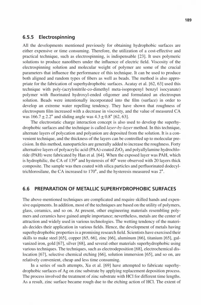
6.5.5 Electrospinning
All the developments mentioned previously for obtaining hydrophobic surfaces areeither expensive or time consuming. Therefore, the utilization of a cost-effective andpractical technique, such as electrospinning, is indispensable [23]. It uses polymericsolutions to produce nanofibers under the influence of electric field. Viscosity of theelectrospinning solution and molecular weight of polymer are some of the crucialparameters that influence the performance of this technique. It can be used to produceboth aligned and random types of fibers as well as beads. The method is also appro-priate for the fabrication of superhydrophobic surfaces. Acatay et al. [62, 63] used thistechnique with poly-(acrylonitrile-co-dimethyl meta-isopropenyl benzyl isocyanate)polymer with fluorinated hydroxyl-ended oligomer and formulated an electrospunsolution. Beads were intentionally incorporated into the film (surface) in order todevelop an extreme water repelling tendency. They have shown that roughness ofelectrospum film increased with a decrease in viscosity, and the value of measured CAwas 166.7± 2.2∘ and sliding angle was 4.3± 0.8∘ [62, 63].
The electrostatic charge interaction concept is also used to develop the superhy-drophobic surfaces and the technique is called layer-by-layer method. In this technique,alternate layers of polycation and polyanion are deposited from the solution. It is a con-venient technique, and the thickness of the layers can be controlled up to molecular pre-cision. In this method, nanoparticles are generally added to increase the roughness. Fortyalternative layers of polyacrylic acid (PAA)-coated ZrO2 and polyallylamine hydrochlo-ride (PAH) were fabricated by Han et al. [64]. When the exposed layer was PAH, whichis hydrophilic, the CA of 139∘ and hysteresis of 40∘ were observed with 20 layers thickcomposite. The sample was then coated with silica particles and perfluorinated dodecyl-trichlorosilane, the CA increased to 170∘, and the hysteresis measured was 2∘.
6.6 PREPARATION OF METALLIC SUPERHYDROPHOBIC SURFACES
The above-mentioned techniques are complicated and require skilled hands and expen-sive equipments. In addition, most of the techniques are based on the utility of polymers,glass, ceramics, and so on. At present, other engineering materials resembling poly-mers and ceramics have gained ample importance; nevertheless, metals are the center ofattraction and widely used in various technologies. The wetting tendency of the materi-als decides their application in various fields. Hence, the development of metals havingsuperhydrophobic properties is a promising research field. Scientists have exercised theirskills to make steel [65], copper [65, 66], zinc [66], aluminum [66], titanium [65], gal-vanized iron, gold [67], silver [68], and several other materials superhydrophobic usingvarious techniques. The techniques, such as electrodeposition [68], electrochemical dis-location [67], selective chemical etching [66], solution immersion [65], and so on, arerelatively convenient, cheap and less time consuming.
In a series of such attempts, Xu et al. [69] have attempted to fabricate superhy-drophobic surfaces of Ag on zinc substrate by applying replacement deposition process.The process involved the treatment of zinc substrate with HCl for different time lengths.As a result, zinc surface became rough due to the etching action of HCl. The extent of
190
170
165
160
155
Co
nta
ct
an
gle
/i ã
150
145
140
135
130
0 2 4
Rinsing time/min
6 8 10
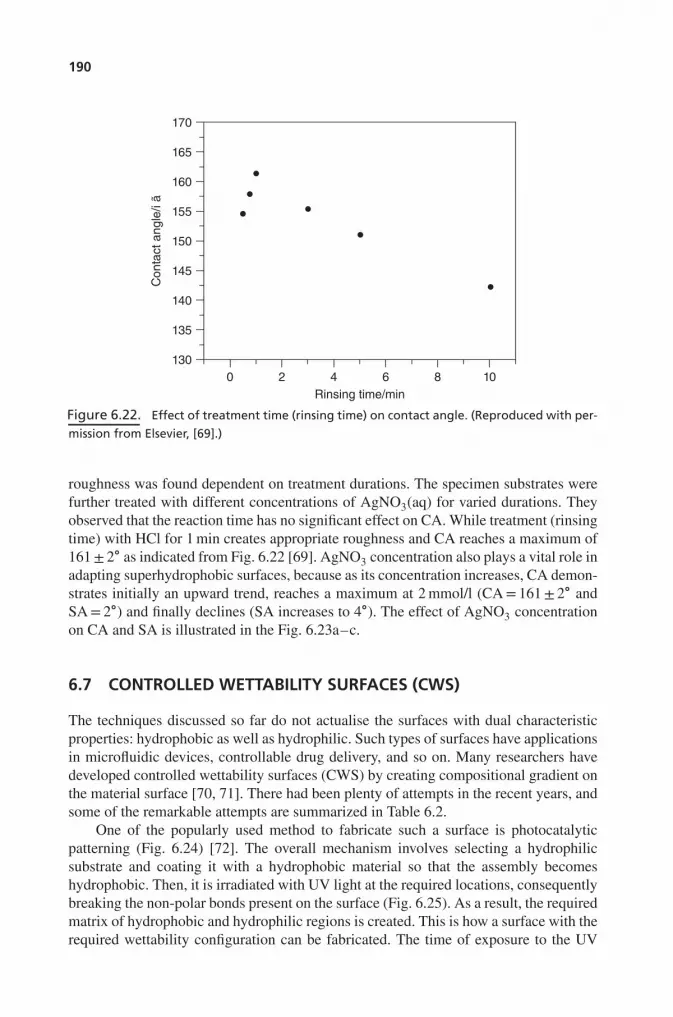
Figure 6.22. Effect of treatment time (rinsing time) on contact angle. (Reproduced with per-
mission from Elsevier, [69].)
roughness was found dependent on treatment durations. The specimen substrates werefurther treated with different concentrations of AgNO3(aq) for varied durations. Theyobserved that the reaction time has no significant effect on CA. While treatment (rinsingtime) with HCl for 1 min creates appropriate roughness and CA reaches a maximum of161± 2∘ as indicated from Fig. 6.22 [69]. AgNO3 concentration also plays a vital role inadapting superhydrophobic surfaces, because as its concentration increases, CA demon-strates initially an upward trend, reaches a maximum at 2 mmol/l (CA= 161± 2∘ andSA= 2∘) and finally declines (SA increases to 4∘). The effect of AgNO3 concentrationon CA and SA is illustrated in the Fig. 6.23a–c.
6.7 CONTROLLED WETTABILITY SURFACES (CWS)
The techniques discussed so far do not actualise the surfaces with dual characteristicproperties: hydrophobic as well as hydrophilic. Such types of surfaces have applicationsin microfluidic devices, controllable drug delivery, and so on. Many researchers havedeveloped controlled wettability surfaces (CWS) by creating compositional gradient onthe material surface [70, 71]. There had been plenty of attempts in the recent years, andsome of the remarkable attempts are summarized in Table 6.2.
One of the popularly used method to fabricate such a surface is photocatalyticpatterning (Fig. 6.24) [72]. The overall mechanism involves selecting a hydrophilicsubstrate and coating it with a hydrophobic material so that the assembly becomeshydrophobic. Then, it is irradiated with UV light at the required locations, consequentlybreaking the non-polar bonds present on the surface (Fig. 6.25). As a result, the requiredmatrix of hydrophobic and hydrophilic regions is created. This is how a surface with therequired wettability configuration can be fabricated. The time of exposure to the UV

191
(a)
(b)
(c)
Figure 6.23. Droplet shape as a function of concentration of AgNO3 (aq) solution (a) con-
centration = 20 mmol/L, CA=147±2∘, SA=4∘ (b) concentration = 0.05 mmol/L, CA=148±2∘,SA=4∘, and (c) concentration = 2 mmol/L, CA= 161± 2∘, SA=2∘. (Reproduced with permission
from Elsevier, [69].)
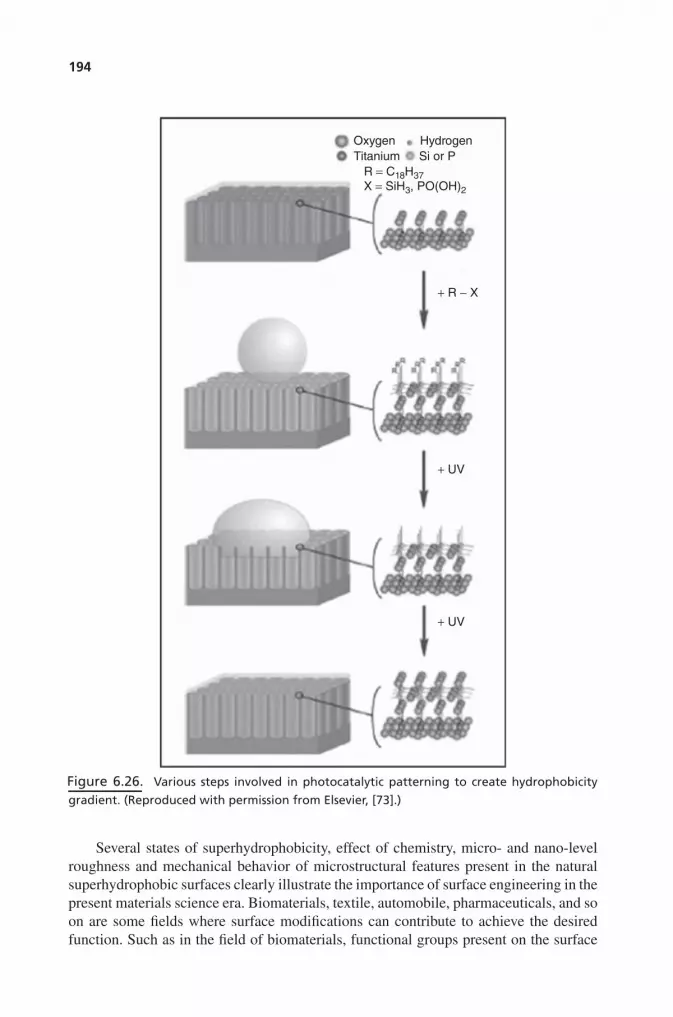
radiations is a critical parameter regarding this context. Because larger exposure dura-tions can lead to complete breaking of bonds, thus the surface will become hydrophilicagain. The effect of radiation intensity has been demonstrated in Fig. 6.24. In this case,the lengths of arrows indicate the intensity of UV radiation. Figure 6.25 indicates thatCA decreases with increasing intensity, and later on, the droplet completely spreadsout indicating superhydrophilic property. An elaborated illustration of the technique ispresented in Fig. 6.26. Balaur et al. [73] fabricated CWS on TiO2: a superhydrophilicsubstrate was made hydrophobic after coating it with hydrophobic material. Whenthis coated substrate is exposed to controlled photocatalytic decomposition at differentlocations, CWS is produced. Silicon oxide nanowires are also being used in theproduction of tunable wettability surfaces [74].
Jin et al. [27] have grown aligned polystyrene nanotube films on a substrate toprepare superhydrophobic surfaces, which mimics Gecko state. This Gecko state canbe used as a mechanical hand to transport the samples for micro-analysis and drugdelivery in biological studies. A droplet transportation test in which a water dropletis transferred from the superhydrophobic to hydrophilic surface with the help of

192
TABLE 6.2. Principal Articles on Synthesizing Artificial Superhydrophobic Surfaces
Author Processingtechnique
Materials used Contact angle(in degrees)
References
Jin et al. Templation Polystyrenenanotubes array
162± 1.7 [27]
Liu et al. Soft-lithographicimprinting
BP-AZ-CA 154.6 [49]
Chaiwong et al.Fresnais et al.Minko et al.
Plasma treatmentPlasma treatmentPlasma treatment
Polylactic AcidLDPEPTFE
110170160
[50, 75][75][76]
Hosono et al. Chemical bathdeposition
CoCl2,NH2CO andLauric acid
178 [77]
Zhang et al. Colloidalassemblies
CaCO3 on siliconsubstrate
156–160 [78]
Zhai et al. Layer-by-layerdeposition
PAH/PAAmulti-layers
∼170 [79]
Ma et al. Electrospinningand CVD
PCL andPPFEMA
175 [80]
Han et al. Micelles Block copolymerof PtBA-b-PDMS-b-ptBA
163 (without SiO2
particles)170 (with SiO2
particles)
[64]
Erbil et al. Combinationbased onmembranecasting
Isotacticpolypropylene
160 [81]
Zhu et al. Combinationbased onchemical vapordeposition
CNT array 166 [82]
Furstner et al. Photolithography Silicon wafers 113–161 [83]Bae et al. Sol-gel Silica 149 [51]
Hydrophobic
Treatment UV/O3
(Photocatalytic
decomposition)Wettability-controlledsurface
Hydrophilic Hydrophobic
Figure 6.24. Schematic of steps involving in photocatalytic patterning. Length of arrows rep-
resents the intensity of UV light and different colors shows change in wettability behavior.
(Reproduced with permission from Royal Society of Chemistry, [72].)

193
180° 160°120°
45° 10°
UV Treatment
Super hydrophobic Super hydrophilic
UV TreatmentUV Treatment UV Treatment
Figure 6.25. Effect of intensity of UV light on surface properties. (Reproduced with permis-
sion from Springer, [72].)
micro-electromechanical systems (MEMS) to measure the loss of water precisely wasperformed. The test elicited no water loss in the process, which suggests that materialswith surfaces mimicking the Gecko State can be used for drug delivery applications.
Minko et al. [76] developed self-adaptive surfaces of polymer material. PTFEsample was first plasma treated, generated using oxygen and ammonia. This treatmentgenerated needle-like structure on the surface, which rendered measured CA of 160∘.Furthermore, it was brushed with PSF-COOH and PVP-COOH creating second-levelroughness. Further exposure of this surface to water, toluene and 1,4-dioxane devel-oped self-adaptive properties and provided CA of 25∘, 118∘, and 75∘, respectively.Hosono et al. [77] also fabricated the needle-like structure of BCH-LA [(brucite-typecobalt hydroxide (BCH, Co(OH)1.13Cl0.09(CO3)0.39⋅0.05H2O), lauric acid (LA)] viachemical bath deposition process and obtained CA of 178∘ (superhydrophobic). Zhanget al. [78] created the CaCO3-loaded hydrogel spheres on silicon substrate throughcolloidal assemblies. Layer-by-layer technique was used by Zhai et al. [79] to createthe honeycomb-like structure of PAH/PAA [poly(allylamine hydrochloride) (PAH)and poly(acrylic acid) (PAA)] on silicon substrate. This structure exhibited CA ofapproximately 170∘. Ma et al. [80] fabricated superhydrophobic fabric possessing CAof 175∘. A mat of PCL [poly(caprolactone)] was synthesized using electrospinning,followed by a coating of PPFEMA (polymerized perfluoroalkyl ethyl methacrylate)via chemical vapor deposition to generate superhydrophobic surface. Erbil et al. [81]have used less tedious and time-consuming membrane casting method for producinggel-like superhydrophobic surfaces using porous polypropylene, which result in aCA of 160∘. Zhu et al. [82] grew the array of aligned CNTs, which were coated withfluorocarbon using chemical vapor deposition and rendered CA of 166∘. Silicon waferswith controlled height and distance of spikes were produced by Frustner et al. [83] tomimic the lotus-like structure using X-ray lithography and measured the CA between113∘ and 161∘.
With the extensive developments in theoretical, experimental and mathematicalmodeling investigations during the recent years, researchers have proposed fabricatingsophisticated surfaces for significant and ambitious technical applications in satellitedishes, solar energy panels, photovoltaics, exterior architectural glasses and greenhouses, heat transfer surfaces in air conditioning equipments, water repellent glasses,turbulent drag reduction, self-cleaning automobile windshields, paints, fabrics togenerate low friction surfaces, and so on [15, 27, 30, 49–51, 58, 69, 73, 81]. Insummary, infancy of the surface modification to enhance the non-wetting of surfaceneeds to master the mimicking of nature successfully.
194
+ R − X
Oxygen
Titanium Si or P
R = C18H37
X = SiH3, PO(OH)2
Hydrogen
+ UV
+ UV
Figure 6.26. Various steps involved in photocatalytic patterning to create hydrophobicity
gradient. (Reproduced with permission from Elsevier, [73].)
Several states of superhydrophobicity, effect of chemistry, micro- and nano-levelroughness and mechanical behavior of microstructural features present in the naturalsuperhydrophobic surfaces clearly illustrate the importance of surface engineering in thepresent materials science era. Biomaterials, textile, automobile, pharmaceuticals, and soon are some fields where surface modifications can contribute to achieve the desiredfunction. Such as in the field of biomaterials, functional groups present on the surface

195
provide the active sites to the receptor legends existing on the protein and cell surfacesand facilitate the strong cell adhesion. It must be noted that hard tissue replacementrequires strong cell adhesion on the implanted material and their proliferation, whereasartificial heart or arteries require the absence of cell adhesion. Since surface roughnessenhances either hydrophilic or hydrophobic nature of the surface, favorable sites forsticking or non-sticking of the cells can be easily engineered for producing a potentialbiomaterial with required functionality.
6.8 CONCLUSIONS
The surfaces with CA greater than 90∘ are hydrophobic while those greater than150∘ are superhydrophobic. Hydrophobic surfaces can be classified into Wenzel’s,Cassie/Baxter’s, Lotus, the transition state between Wenzel’s and Cassie’s state andthe Gecko’s state. Objects with superhydrophobic surfaces are abundant in nature,and most of them owe this property to their surface morphology. Lotus and rice leafexhibit a binary structure, while Chinese watermelon and ramie leaf exhibit unitarystructure, indicating that unitary structure is self-sufficient to elicit superhydrophobicity.Mechanical aspects such as flexing strength, modulus, tribological properties, andso on are important in addition to the morphological features in making the surfacesuperhydrophobic. The correlation between Laplacian pressure and critical pressureprovides a means to determine whether a surface can retain hydrophobicity under thereign of nature’s design. Various simulations and modeling developed and carried outby researchers provide an efficient tool to gain deeper understanding of the phenomena.A variety of techniques have been developed so far to fabricate hydrophobic surfaces forpractical applications. Soft lithography imprinting can imitate lotus-leaf-like surfacesand used to fabricate such a surface imparting low COF. Sol-gel technique is used formaking cotton fibers superhydrophobic, whereas photocatalytic patterning finds itsapplication in the fabrication of wettability gradient surface to acquire control over thehydrophobicity/hydrophilicity. Most of the techniques in one way or the other try tomimic nature. Newer techniques and applications are proposed by various researchersto fabricate suitable superhydrophobic surfaces on a large scale for ambitious scientificand engineering applications.
QUESTIONS
1. What are superhydrophobic surfaces?
2. What are Cassie Baxter and Wenzel wetting states? Describe these showing theircorresponding schematics.
3. What is the role of roughness in affecting apparent contact angle?
4. Give a few examples of non-wetting surfaces in nature.
5. What is the role of two-level roughness in lotus leaf? Also emphasize the role ofsurface chemistry in highlighting the wetting property.

196
6. How “mechanical property” can be linked to wettability of surfaces?
7. What is Laplacian pressure? What is the role of inclining the surface on wetting ofsurface?
8. Provide a few techniques in fabricating artificial superhydrophobic surfaces.
9. What are controlled wettability surfaces? What is their practical utility?
REFERENCES
1. Hoppenbrouwers PMM, Driessens FCM, Stadhouders AM. Morphology, Composition, andWetting of Dentinal Cavity Walls. J Dent Res 1974;53:1255.
2. Zioupos P, Currey JD. Pre-Failure Toughening Mechanisms in the Dentine of the NarwhalTusk: Microscopic Examination of Stress/Strain Induced Microcracking. J Mater Sci Lett1996;15:991.
3. Davidson CL, Gee AJd, Feilzer A. The Competition between the Composite-Dentin BondStrength and the Polymerization Contraction Stress. J Dent Res 1984;63:1396.
4. Sellinger A, Weiss PM, Nguyen A, Lu Y, Assink RA, Gong W, Brinker CJ. ContinuousSelf-Assembly of Organic-Inorganic Nanocomposite Coatings that Mimic Nacre. Nature1998;394:256.
5. Wang RZ, Suo Z, Evans AG, Yao N, Aksay IA. Deformation Mechanisms in Nacre. J MaterRes 2001;16:2485.
6. Jackson AP, Vincent JFV, Turner RM. The Mechanical Design of Nacre. Proc R Soc Lond B1988;234:415
7. Tang Z, Kotov NA, Magonov S, Ozturk B. Nanostructured Artificial Nacre. Nat Mater2003;2:413.
8. Allen MR, Reinwald S, Burr DB. Alendronate Reduces Bone Toughness of Ribs withoutSignificantly Increasing Microdamage Accumulation in Dogs Following 3 Years of DailyTreatment. Calcif Tissue Int 2008;82:354.
9. Wang X, Puram S. The Toughness of Cortical Bone and Its Relationship with Age. AnnBiomed Eng 2004;32:123.
10. Korb J. Thermoregulation and Ventilation of Termite Mounds. Naturwissenschaften2003;90:212.
11. Hesse PR. A Chemical and Physical Study of the Soils of Termite Mounds in East Africa. JEcol 1955;43:449.
12. Yurdumakan B, Raravikar NR, Ajayanb PM, Dhinojwala A. Synthetic Gecko Foot-Hairs fromMultiwalled Carbon Nanotubes. Chem Commun 2005. DOI: 10.1039/B506047H.
13. Geim AK, Dubonos SV, Grigorieva IV, Novoselov KS, Zhukov AA, Shapoval SY. Micro-fabricated Adhesive Mimicking Gecko Foot-Hair. Nat Mater 2003;2:461.
14. Balani K, Batista RG, Lahiri D, Agarwal A. The Hydrophobicity of a Lotus Leaf: A Nanome-chanical and Computational Approach. Nanotechnology 2009;20:305707.
15. Shirtcliffe NJ, McHale G, Atherton S, Newton MI. An Introduction to Superhydrophobicity.Adv Colloid Interface Sci 2009;161:124.
16. Feng C, Zisheng G, Dongxu L, Wuhan J. Preparation of Material Surface Structure Similarto Hydrophobic Structure of Lotus Leaf. Univ Technol 2008;23:513.
17. Yuan Z, Chen H, Zhang J. Facile Method to Prepare Lotus-Leaf-Like Super-HydrophobicPoly(Vinyl Chloride) Film. Appl Surf Sci 2008;254:1593.

197
18. Voronov RS, Papavassiliou DV. Review of Fluid Slip over Superhydrophobic Surfaces andIts Dependence on the Contact Angle. Ind Eng Chem Res 2008;47:2455.
19. Ming W, Tian M, Grampel RDvd, Melis F, Jia X, Loos J, Linde R v d. Low Surface EnergyPolymeric Films from Solventless Liquid Oligoesters and Partially Fluorinated Isocyanates.Macromolecules 2002;35:6920.
20. Tsibouklis J, Nevelll TG. Ultra-Low Surface Energy Polymers: The Molecular DesighRequirements. Adv Mater 2003;15:647.
21. Gupta A, Sharma S, Joshi MR, Agarwal P, Balani K. Grain Growth Behavior of Al2O3 Nano-materials: A Review. Mater Sci Forum 2010;653:87.
22. Bhushan B, Jung YC. Natural and Biomimetic Artificial Surfaces for Superhydrophobicity,Self-Cleaning, Low Adhesion, and Drag Reduction. Prog Mater Sci 2010;56:1.
23. Li XM, Reinhoudt D, Crego-Calama M. What Do We Need for Superhydrophobic Surface?A Review on the Recent Progress in the Preparation of Superhydrophobic Surfaces. ChemSoc Rev 2007;36:1350.
24. Wenzel RN. Resistance of Solid Surfaces to Wetting by Water. Ind Eng Chem 1936;28:988.
25. Cassie ABD. Wettability of Porous Surfaces. Trans Faraday Soc 1944;40:546.
26. Wang S, Jiang L. Defination of Superhydrophobic States. Adv Mater 2007;19:3423.
27. Jin M, Feng X, Feng L, Sun T, Zhai J, Li T, Jiang L. Superhydrophobic Aligned PolystyreneNanotube Films with High Adhesive Force. Adv Mater 2005;17:1977.
28. Guo Z, Liu W. Biomimic from the Superhydrophobic Plant Leaves in Nature: Binary Structureand Unitary Structure. Plant Sci 2007;172:1103.
29. Barthlott W, Neinhuis C. Purity of the Sacred Lotus, or Escape from Contamination in Bio-logical Surfaces. Planta 1997;202:1.
30. Cheng YT, Rodak DE, Wong CA, Hayden CA. Effects of Micro- and Nano-Structures on theSelf-Cleaning Behaviour of Lotus Leaves. Nanotechnology 2006;17:1359.
31. Ivanova NV, Korolenko EA, Korolik EV, Zhbankov RG. IR Spectrum of Cellulose. J ApplSpectrosc 1990;51:847.
32. Oh SY, Yoo DI, Shinb Y, Seo G. FTIR Analysis of Cellulose Treated with Sodium Hydroxideand Carbon Dioxide. Carbohyd Res 2005;340:417.
33. Wagner P, Furstner R, Barthlott W, Neinhuis C. Quantitative Assessment to the StructuralBasis of Water Repellency in Natural and Technical Surfaces. J Exp Bot 2003;54:1295.
34. Singh RA, Yoon ES, Kim HJ, Kim J, Jeong HE, Suh KY. Replication of Surfaces of NaturalLeaves for Enhanced Micro-Scale Tribological Property. Mater Sci Eng C 2007;27:875.
35. Singh RA, Yoon ES, Kim HJ, Kong H, Park S, Jeong HE, Suh KY. Enhanced TribologicalProperties of Lotus Leaf-Like Surfaces Fabricated by Capillary Force Lithography. Surf Eng2007;23:161.
36. Waters JF, Guduru PR, Jouzi M, Xu JM, Hanlon T, Suresh S. Shell Buckling of IndividualMultiwalled Carbon Nanotubes using Nanoindentation. Appl Phys Lett 2005;87:103109.
37. Zhang J, Sheng X, Jiang L. The Dewetting Properties of Lotus Leaves. Langmuir2009;25:1371.
38. Xiu Y, Zhu L, Hess DW, Wong CP. Hierarchical Silicon Etched Structures for ControlledHydrophobicity/Superhydrophobicity. Nano Lett 2007;7:3388.
39. Krupenkin TN, Taylor JA, Wang EN, Kolodner P, Hodes M, Salamon TR. ReversibleWetting-Dewetting Transitions on Electrically Tunable Superhydrophobic NanostructuredSurfaces. Langmuir 2007;23:9128.
198
40. Dorrer C. J. Rühe. Condensation and Wetting Transitions on Microstructured Ultrahydropho-bic Surfaces. Langmuir 2007;23:3820.
41. Lundgren M, Allan NL, Cosgrove T. Modeling of Wetting: A Study of Nonwetting at Roughand Heterogeneous surfaces. Langmuir 2007;23:1187.
42. Lundgren M, Allan NL, Cosgrove T. Molecular Dynamics Study of Wetting of a Pillar Sur-face. Langmuir 2003;19:7127.
43. Koishia T, Yasuokab K, Zengc XC, Fujikawad S. Molecular Dynamics Simulation of WaterDroplet on a Rough Surface. Proceedings of the 15th International Conference on Proper-ties of Water and Steam, 2008. Berlin: VDI-Society Energy Technology and InternationalAssociation for the Properties of Water and Steam; 2008, p. 8.
44. Gennes PGd. Wetting: Statics and Dynamics. Rev Mod Phys 1985;57:827.
45. Kong XQ, Shea D, Baynes RE, Riviere JE, Xia XR. Regression Method of the HydrophobicityRuler Approach for Determining Octanol/Water Partition Coefficients of Very HydrophobicCompounds. Chemosphere 2007;66:1086.
46. Boreyko JB, Chen CH. Restoring Superhydrophobicity of Lotus Leaves with Vibration-Induced Dewetting. Phys Rev Lett 2009;103:174502.
47. Boreyko JB, Chen CH. Self-Propelled Dropwise Condensate on Superhydrophobic Surfaces.Phys Rev Lett 2009;103:184501.
48. Cheng YT, Rodak DE. Is the Lotus Leaf Superhydrophobic? Appl Phys Lett 2005;86:144101.
49. Liu B, He Y, Fan Y, Wang X. Fabricating Super-Hydrophobic Lotus-Leaf-Like Surfacesthrough Soft-Lithographic Imprinting. Macromol Rapid Commun 2006;27:1859.
50. Chaiwong C, Rachtanapun P, Wongchaiya P, Auras R, Boonyawan D. Effect of Plasma Treat-ment on Hydrophobicity and Barrier Property of Polylacticacid. Surf Coat Tech 2010;2:2933.
51. Bae GY, Min BG, Jeong YG, Lee SC, Jang JH, Koo GH. Superhydrophobicity of CottonFabrics Treated with Silica Nanoparticles and Water-Repellent Agent. J Colloid Interf Sci2009;337:170.
52. Balani K, Chen Y, Harimkar SP, Dahotre NB, Agarwal A. Tribological Behavior of PlasmaSprayed Carbon Nanotube Reinforced Hydroxyapatite-Coating in Physiological Solution.Acta Biomater 2007;3:944.
53. Balani K, Agarwal A. Damping Behavior Of Carbon Nanotube Reinforced Aluminum OxideCoatings By Nanomechanical Dynamic Modulus Mapping. J Appl Phys 2008;104:063517.
54. Balani K, Bakshi S, Lahiri D, Agarwal A. Grain Growth Behavior of Aluminum Oxide Rein-forced with Carbon Nanotube During Plasma Spraying and Post-Spray Consolidation. Int JAppl Ceram Technol 2009;7:946.
55. Keshri A, Balani K, Bakshi SR, Singh V, Laha T, Seal S, Agarwal A. Structural Transforma-tion in Carbon Nanotubes During Thermal Spray Processing. Surf Coat Tech 2009;203:2193.
56. Singh V, Diaz R, Balani K, Agarwal A, Seal S. Chromium Carbide-CNT Nanocompositeswith Enhanced Mechanical Properties. Acta Mater 2009;57:335.
57. Li H, Wang X, Song Y, Liu Y, Li Q, Jiang L, Zhu D. Super-“Amphiphobic” Aligned CarbonNanotube Films. Angew Chem Int Ed 2001;40:1743.
58. Feng L, Li S, Li H, Jhai J, Song Y, Jiang L, Jhu D. Super-Hydrophobic Surface of AlignedPolyacrylonitrile Nanofibers. Angew Chem Int Ed 2002;41:1221.
59. Li S, Li H, Wang X, Song Y, Liu Y, Jiang L, Jhu D. Super-Hydrophobicity of Large-AreaHoneycomb-Like Aligned Carbon Nanotubes. J Phys Chem B 2002;106:9274.
60. Gleiche M, Chi LF, Fuchs H. Nanoscopic Channel Lattices with Controlled Anisotropic Wet-ting. Nature 2000;403:173.

199
61. Higgins AM, Jones RAL. Anisotropic Spinodal Dewetting as a Route to Self-Assembly ofPatterned Surfaces. Nature 2000;404:476.
62. Acatay K. Generation of Superhydrophobic Surfaces by Electrospinning Process Spring.Sabanc𝚤 University; 2004.
63. Acatay K, Simsek E, Ow-Yang C, Menceloglu YZ. Tunable, Superhydrophobically StablePolymeric Surfaces by Electrospinning. Angew Chem Int Ed 2004;43:5210.
64. Han JT, Zheng Y, Cho JH, Xu X, Cho K. Stable Superhydrophobic Organic-Inorganic HybridFilms by Electrostatic Self-Assembly. J Phys Chem B 2005;109:20773.
65. Qu MN, Zhang BW, Song SY, Chen L, Zhang JY, Cao XP. Fabrication of Superhydropho-bic Surfaces on Engineering Materials by a Solution-Immersion Process. Adv Funct Mater2007;17:593.
66. Qian BT, Shen ZQ. Fabrication of Superhydrophobic Surfaces by Dislocation-SelectiveChemical Etching on Aluminum, Copper, and Zinc Substrates. Langmuir 2005;21:9007.
67. Praig VG, Piret G, Manesse M, Castel X, Boukherroub R, Szunerits S. Seed-mediated Elec-trochemical Growth of Gold Nanostructures on Indium Tin Oxide Thin Films. ElectrochimActa 2008;53:7838.
68. Zhao N, Shi F, Wang ZQ, Zhang X. Combining Layer-by-Layer Assembly with Elec-trodeposition of Silver Aggregates for Fabricating Superhydrophobic Surfaces. Langmuir2005;21:4713.
69. Xu W, Ning T, Yang X, Lu S. Fabrication of Superhydrophobic Surfaces on Zinc Substrates.Appl Surf Sci 2010;257:4801.
70. Ueda-Yukoshi T, Matsuda T. Cellular Responses on a Wettability Gradient Surface with Con-tinuous Variations in Surface Compositions of Carbonate and Hydroxyl Groups. Langmuir1995;11:4135.
71. Lu X, Zhang J, Zhang C, Han Y. Low-Density Polyethylene (LDPE) Surface With a Wetta-bility Gradient by Tuning its Microstructures. Macromol Rapid Commun 2005;26:637.
72. Verplanck N, Coffinier Y, Thomy V, Boukherroub R. Wettability Switching Techniques onSuperhydrophobic Surfaces. Nanoscale Res Lett 2007;2:577.
73. Balaur E, Macak JM, Taveira L, Schmuki P. Tailoring the wettability of TiO2 nanotube layers.Electrochem Commun 2005;7:1066.
74. Coffinier Y, Janel S, Addad A, Blossey R, Gengembre L, Payen E, Boukherroub R. Prepara-tion of Superhydrophobic Silicon Oxide Nanowire Surfaces. Langmuir 2007;23:1608.
75. Fresnais J, Benyahia L, Poncin-Epaillard F. Dynamic (de)wetting properties of superhy-drophobic plasma-treated polyethylene surfaces. Surf Interface Anal 2006;38:144.
76. Minko S, Muller M, Motornov M, Nitschke M, Grundke K, Stamm M. Two-Level StructuredSelf-Adaptive Surfaces with Reversibly Tunable Properties. J Am Chem Soc 2003;125:3896.
77. Hosono E, Fujihara S, Honma I, Zhou HS. Superhydrophobic Perpendicular Nanopin Filmby the Bottom-Up Process. J Am Chem Soc 2005;127:13458.
78. Zhang G, Wang DY, Gu ZZ, Mohwald H. Fabrication of Superhydrophobic Surfaces fromBinary Colloidal Assembly. Langmuir 2005;21:9143.
79. Zhai L, Cebeci FC, Cohen RE, Rubner MF. Stable Superhydrophobic Coatings from Poly-electrolyte Multilayers. Nano Lett 2004;4:1349.
80. Ma ML, Mao Y, Gupta M, Gleason KK, Rutlegde GC. Superhydrophobic Fabrics Producedby Electrospinning and Chemical Vapor Deposition. Macromolecules 2005;38:9742.
81. Erbil HY, Demirel AL, Avci Y, Mert O. Transformation of a Simple Plastic into a Superhy-drophobic Surface. Science 2003;299:1377.

200
82. Zhu LB, Zin YH, Xu JW, Tamirisa PA, Hess DW, Wong CP. Superhydrophobicity onTwo-Tier Rough Surfaces Fabricated by Controlled Growth of Aligned Carbon NanotubeArrays Coated with Fluorocarbon. Langmuir 2005;21:11208.
83. Furstner R, Barthlott E, Neinhuis C, Walzel P. Wetting and Self-Cleaning Properties of Arti-ficial Superhydrophobic Surfaces. Langmuir 2005;21:956.
84. Feng L, Li S, Li Y, Li H, Zhang L, Zhai J, Song Y, Liu B, Jiang L, Jhu D. Super-HydrophobicSurfaces: From Natural to Artificial. Adv Mater 2002;14:1857.
Copyright © 2022 FDOKUMEN