BIOLOGY GUIDANCE FOR TEACHING - WJEC
81
WJEC Eduqas GCSE in BIOLOGY GUIDANCE FOR TEACHING Teaching from 2016 This Ofqual regulated qualification is not available for candidates in maintained schools and colleges in Wales. ACCREDITED BY OFQUAL GCSE
-
Upload
khangminh22 -
Category
Documents
-
view
1 -
download
0
Transcript of BIOLOGY GUIDANCE FOR TEACHING - WJEC

WJEC Eduqas GCSE in
BIOLOGY
GUIDANCE FOR TEACHING
Teaching from 2016
This Ofqual regulated qualification is not available forcandidates in maintained schools and colleges in Wales.
ACCREDITED BY OFQUAL
GCSE

2
Contents
CELL BIOLOGY
CELLS BIOLOGY
1.1 PROKARYOTIC AND EUKARYOTIC CELLS 3
1.2 GROWTH AND DEVELOPMENT OF CELLS 8
1.3 CELL METABOLISM 9
TRANSPORT SYSTEMS
2.1 TRANSPORT IN CELLS 17
2.2 TRANSPORT SYSTEMS IN HUMANS 22
2.3 TRANSPORT SYSTEMS IN PLANTS 26
HEALTH, DISEASE AND THE DEVELOPMENT OF MEDICINE
3.1 HEALTH AND DISEASE 28
3.2 COMMUNICABLE DISEASE 29
3.3 TREATING, CURING AND PREVENTING DISEASE 33
3.4 NON-COMMUNICABLE DISEASES IN HUMANS 40
COORDINATION AND CONTROL
4.1 NERVOUS COORDINATION AND CONTROL IN HUMANS 41
4.2 HORMONAL COORDINATION AND CONTROL IN HUMANS 47
4.3 HOMEOSTASIS IN HUMANS 49
4.4 PLANT HORMONES 54
PHOTOSYNTHESIS 55
ECOSYSTEMS
6.1 LEVELS OF ORGANISATION WITHIN AN ECOSYSTEM 60
6.2 THE PRINCIPLE OF MATERIAL CYCLING 62
6.3 BIODIVERSITY 66
6.4 SOME OF THE BIOLOGICAL CHALLENGES OF INCREASING FOOD YIELDS USING FEWER RESOURCES
71
COMPONENT 7 – INHERITANCE, VARIATION AND EVOLUTION
7.1 THE GENOME AND GENE EXPRESSION 73
7.2 INHERITANCE 77
7.3 VARIATION AND EVOLUTION 78
7.4 SELECTIVE BREEDING AND GENE TECHNOLOGY 80

3
1.1 PROKARYOTIC AND EUKARYOTIC CELLS
Spec Statement Comment
(a) draw and label animal and plant cells
Including labels for nucleus, cytoplasm, cell membrane, cell wall, chloroplast and vacuole.
(b) describe the differences between eukaryotic and prokaryotic cells
Prokaryotic cells consist of a cell wall, cell membrane and cytoplasm. There is no distinct nucleus, no mitochondria and no chloroplasts. Prokaryotic cells are much smaller than most eukaryotic cells
(c) explain how the following sub-cellular structures of eukaryotic cells (plants and animals) and prokaryotic cells (bacteria) are related to their functions: nucleus/DNA, plasmids, mitochondria, chloroplasts, cell membranes, cytoplasm, vacuole, cell wall
Cell membrane: controls the entry and exit of substances. Cytoplasm: site of most cell reactions. Nucleus: in plants and animal cells, contains chromosomes which carry genetic information consisting of DNA. The genetic information in bacterial cells is carried in a single loop of DNA. Plasmids: small circular sections of DNA often found in the cytoplasm of bacterial cells, separate from the rest of the cell`s DNA, can transfer genetic information between one cell and another. Mitochondria: site of aerobic respiration. Cell wall containing cellulose : structural support for plant cells. Chloroplast: site of photosynthesis in plant cells. Vacuole: in plant cells, contains a watery sugar solution (sap), a swollen vacuole pushes the rest of the cell contents against the cell wall, making the cell firm.
(d) explain how the development of the microscope (light, electron, laser imaging) increased the understanding of the sub cellular structure of organisms and the proposal that the cell is the basic unit of life
A simple understanding of how the light and electron microscopes and laser imaging work. Cell theory was devised and refined as microscopes have developed. Calculation of total magnification is achieved by the multiplication of the power of the eyepiece lens by the power of the objective lenses.
how a slide is prepared, including that biological staining allows more detail of the cell to be seen
the limitations of light microscopy in studying cell structure: restriction in maximum magnification
a simple comparison with the electron microscope: greater magnification but can only be used to view dead tissue
SPECIFIED PRACTICAL WORK

4
SP1.1 Examination of plant and animal cells using a light microscope and production
of labelled scientific drawings from observation
Examination of animal and plant cells using a light microscope and production
of labelled scientific diagrams from observation
Introduction
Cheek cells are typical animal cells, they have a cell membrane, cytoplasm and a nucleus.
Onion cells are plant cells, they have a cell wall, cell membrane, cytoplasm, nucleus and
vacuole. This practical requires you to prepare cheek cell slides and onion cell slides. These
slides can then be observed using a microscope.
Apparatus
light microscope
2 glass slides
2 cover slips
cotton wool bud
mounted needle
forceps
freshly cut onion
0.1 % methylene blue solution
iodine solution
Access to:
beaker of disinfectant
Diagram of Apparatus
Method

5
Cheek Cells:
1. Put a drop of methylene blue on a glass slide.
2. Gently rub the inside of your cheek with a cotton bud.
3. Wipe the end of the cotton bud in the drop of methylene blue on the glass slide.
4. Place the cotton bud in the beaker of disinfectant.
5. Use the mounted needle to gently lower a coverslip onto the glass slide.
6. Using a light microscope, examine the slide using the 10 objective lens.
7. Use the 40 objective lens to identify some of the cell structures.
8. Draw a cell diagram. Identify and label: cell membrane, cytoplasm and nucleus.
Onion Cells:
1. Using forceps, peel a thin layer of epidermis from the inside of a freshly cut onion
piece.
2. Lay the epidermis onto a glass slide.
3. Add a drop of iodine solution to the onion epidermis on the glass slide.
4. Use the mounted needle to gently lower a coverslip onto the glass slide.
5. Using a light microscope, examine the slide using the 10 objective lens.
6. Use the 40 objective lens to identify some of the cell structures.
7. Draw a cell diagram. Identify and label: cell wall, cell membrane, cytoplasm and
nucleus.
Analysis
1. Calculate the total magnification of the image seen by multiplying the power of the
objective lens by the power of the eyepiece.
2. Your teacher will tell you the actual size of the cell, calculate the magnification of your
diagram.
Risk Assessment
Hazard Risk Control measure

6
Methylene blue
is harmful
and/or irritant
Methylene blue can irritate the eyes
and lungs. Skin contamination
should be avoided.
Use the lowest concentration possible.
Wear eye protection when preparing
the cheek cell slide. Methylene blue is a
stain- avoid contact with skin.
Cheek cells are
a biohazard
There is a very small risk of virus
transmission.
Only handle samples from your own
body. After use, hygienically dispose of
cotton buds and slides in a disinfectant
such as Milton or Virkon.
Coverslips/ mounted needles are sharp
Can cut skin Handle carefully
Teacher/Technician notes
Methylene blue and iodine solution are stains. Avoid contact with the skin. Iodine is a low
hazard chemical as a dilute solution.
Suitable disinfectant would include Milton or Virkon which would need to be diluted to
suitable concentrations.
If the lamp is not an integral part of the microscope, a desk lamp will be needed for each
group.
Freshly cut onion is recommended. This should be prepared for student use in pieces
approximately 1 cm2.
Students will need to be briefed regarding safe and effective microscope use prior to this
practical activity. This practical activity is effective at developing microscope skills and
biological drawing skills.
Students can calculate the total magnification of the image as the power of the objective lens
multiplied by the power of the eyepiece. The actual size of the cells can be given to the
students to enable them to calculate the magnification of their diagrams.

7
Practical techniques covered
B3 Use of appropriate apparatus and techniques for the observation and measurement
of biological changes and or processes.
B4 Safe and ethical use of living organisms (plants or animals) to measure physiological
functions and responses to the environment.
B7 Use of appropriate apparatus, techniques and magnification, including microscopes,
to make observations of biological specimens and produce labelled scientific
drawings.

8
1.2 GROWTH AND DEVELOPMENT OF CELLS
Spec Statement Comment
(a) describe the process of mitosis in growth, including the cell cycle; cell division by mitosis enables organisms to grow, replace worn out cells and repair damaged tissues
The chromosome number remains constant and the genetic composition of the daughter cells is identical to the mother cell. The exact spelling of mitosis is required.
(b) explain the importance of cell differentiation to produce specialised cells for greater efficiency
Through differentiation in multicellular organisms, many types of cells are produced which are adapted to particular functions.
(c) describe cancer as the result of changes in cells that lead to uncontrolled growth and division
A simple understanding that cancer is a result of uncontrolled mitosis.
(d) describe the function of stem cells in embryonic and adult animals and meristems in plants; some cells, both plant and animal, do not lose the ability to differentiate and are called stem cells
The bodies of multicellular organisms consist of a variety of different cells that are adapted for particular functions. These different cells originate from undifferentiated stem cells that have the capacity to develop into specialised cells.
(e) discuss the potential benefits, risks and ethical issues surrounding stem cell technology in medicine including the implications for society e.g. the use of embryonic stem cells
Stem cells are able to treat damaged or diseased tissue, providing a potent medical tool. The benefits of using your own stem cells include: no rejection, no need to find a donor, no need for tissue typing. However, the use of embryonic stem cells raises particular ethical issues.
(f) explain the role of meiotic cell division in halving the chromosome number to form gametes; each meiotic division produces four cells that are genetically different because genes separate and are reshuffled during the process of gamete formation
The exact spelling of meiosis is required.

9
1.3 CELL METABOLISM
Spec Statement Comment
(a) explain that chemical reactions in cells are controlled by enzymes. Enzymes are proteins made by living cells. Different proteins are composed of different amino acids linked together to form a chain which is then folded to form a specific shape held by chemical bonds. The specific shape of an enzymes active site enables it to function. This is called the 'lock and key' hypothesis. Enzymes function by the formation of the enzyme-substrate complex at the active site
Enzymes are involved in all metabolic reactions building large molecules from small ones as well breaking down large molecules into small ones. Apply knowledge of ‘lock and key’ to the analysis of simple, stylised diagrams of enzyme/substrate interactions.
(b) explain that enzymes speed up/catalyse the rate of chemical reactions. Each enzyme has its own optimum pH and temperature. Interpret enzyme activity in terms of molecular collisions. Boiling denatures most enzymes by altering their shape
Understand the term optimum as a particular condition (such as temperature or pH) at which the rate of enzyme action is greatest as increased temperature results in increased collisions between enzymes and substrates. In a denatured enzyme the specific shape of the active site is destroyed and can no longer bind with its substrate, so no reaction occurs. Analyse data to show how enzyme action is affected by temperature and pH.
(c) describe cellular respiration as an exothermic reaction which is continuously occurring in all living cells, enabling cells to carry out cell processes. Aerobic respiration occurs in cells when oxygen is available. It is a series of chemical reactions within the cell, controlled by enzymes. Glucose and oxygen are used and carbon dioxide, water and energy are produced. The energy released is in the form of ATP. Recall the word equation for aerobic respiration
Use germinating peas to show that energy is released as heat during respiration. This should include the role of Thermos flasks and disinfectant in the experiment.

10
(d) explain that in the absence of oxygen, anaerobic respiration may occur. This is less efficient than aerobic respiration. In humans energy is released from glucose and lactic acid is produced. An oxygen debt may occur. In yeast, glucose is broken down and ethanol and carbon dioxide are produced. Recall the word equation for anaerobic respiration in human cells and fermentation in yeast. Explain that there is less ATP released per molecule of glucose in anaerobic respiration than in aerobic respiration because of the incomplete breakdown of glucose
Lactic acid is harmful to the body. It has to be removed from cells and broken down following the resumption of aerobic respiration (to repay the oxygen debt).
(e) compare the processes of aerobic and anaerobic respiration
Aerobic and anaerobic respiration compared in terms of oxygen requirement, use of glucose and release of ATP. Aerobic respiration is more efficient.
(f) explain that fats, made up of fatty acids and glycerol, proteins, made up of amino acids, and starch (a carbohydrate), made up of a chain of glucose molecules, in our food are insoluble. They are broken down during digestion into soluble substances so that they can be absorbed
The role of the following enzymes in digestion: carbohydrase – breaks down starch into glucose; protease – breaks down protein into amino acids; lipase - breaks down fats into fatty acids and glycerol. The products of these enzymes are absorbed into the bloodstream.
SPECIFIED PRACTICAL WORK
SP1.3A Investigation into factors affecting enzyme action
SP1.3B Qualitative identification of starch (iodine), glucose (Benedict's) and protein (biuret)

11
Investigation into factors affecting enzyme action
Introduction
Iodine is an indicator that turns blue/black when starch is present, but is otherwise brown.
In this investigation a blue/black solution of starch and iodine will change to brown as the
enzyme amylase digests/breaks down the starch into sugar.
The time taken for this reaction to occur is affected by temperature.
Apparatus
test tube rack and six test tubes
marker pen
stopwatch
25 cm3 measuring cylinder
10 cm3 measuring cylinder
beaker of 1 % starch solution
dropper bottle of iodine solution
beaker of 10 % amylase solution
spotting tile
dropping pipette
Access to:
water bath or alternative method of heating water
Method
1. Measure 10 cm3 of 1 % starch solution into a test tube.
2. Measure 2 cm3 of 10 % amylase solution into a second test tube.
3. Place both tubes into a water bath set at 20 ºC for 3 minutes.
4. Place a drop of iodine in six wells of a spotting tile.
5. Remove both test tubes from the water bath. Pour the amylase into the starch/iodine
solution and start the stopwatch.
6. Immediately, use the dropping pipette to place one drop of the mixture onto the first
drop of iodine. Record the colour of the solution.
7. Repeat step 6 every minute for five minutes.
8. Repeat steps 1-7 at 30 ºC, 40 ºC, 50 ºC, 60 ºC.
Analysis
1. Use your observations to reach a conclusion regarding the effect of temperature on
enzyme action.
2. Evaluate your method and suggest possible improvements.
12
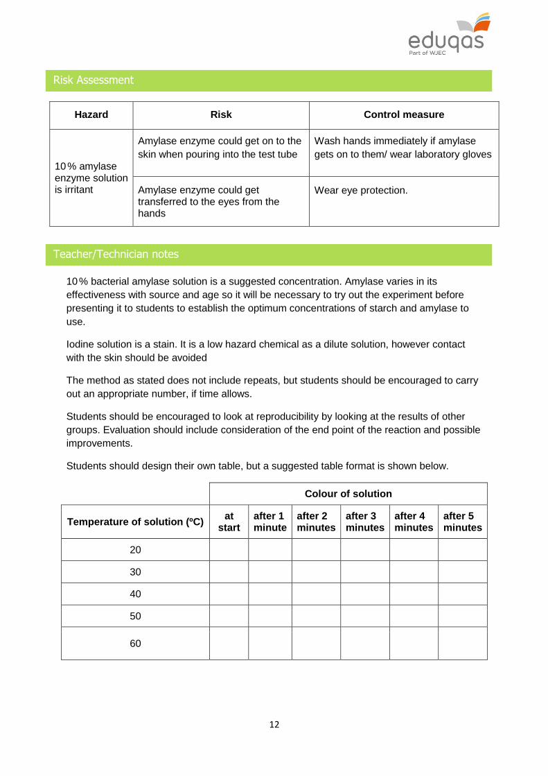
Risk Assessment
Hazard Risk Control measure
10 % amylase enzyme solution is irritant
Amylase enzyme could get on to the
skin when pouring into the test tube
Wash hands immediately if amylase
gets on to them/ wear laboratory gloves
Amylase enzyme could get transferred to the eyes from the hands
Wear eye protection.
Teacher/Technician notes
10 % bacterial amylase solution is a suggested concentration. Amylase varies in its
effectiveness with source and age so it will be necessary to try out the experiment before
presenting it to students to establish the optimum concentrations of starch and amylase to
use.
Iodine solution is a stain. It is a low hazard chemical as a dilute solution, however contact
with the skin should be avoided
The method as stated does not include repeats, but students should be encouraged to carry
out an appropriate number, if time allows.
Students should be encouraged to look at reproducibility by looking at the results of other
groups. Evaluation should include consideration of the end point of the reaction and possible
improvements.
Students should design their own table, but a suggested table format is shown below.
Colour of solution
Temperature of solution (ºC) at
start after 1 minute
after 2 minutes
after 3 minutes
after 4 minutes
after 5 minutes
20
30
40
50
60

13
Practical techniques covered
B1 Use of appropriate apparatus to make and record a range of measurements
accurately, including length, area, mass, time, temperature, volume of liquids and
gases, and pH.
B2 Safe use of appropriate heating devices and techniques including use of a Bunsen
burner and a water bath or electric heater.
B3 Use of appropriate apparatus and techniques for the observation and measurement
of biological changes and or processes.
B4 Safe and ethical use of living organisms (plants or animals) to measure physiological
functions and responses to the environment.
B5 Measurement of rates of reaction by a variety of methods including production of gas,
uptake of water and colour change of indicator.
B8 Use of appropriate techniques and qualitative reagents to identify biological
molecules and processes in more complex and problem solving context including
continuous sampling in an investigation.

14
Qualitative identification of starch (iodine), glucose (Benedict's) and protein
(biuret)
Introduction
The identification of the different food types can be carried out using different chemical tests.
A positive result for each food type is determined by a colour change. In this activity you will
carry out the chemical tests for starch, glucose and protein.
Apparatus
3 × test tubes
3 × dropping pipettes
3 × 5 cm3 syringe
iodine solution with dropping pipette
Benedict's reagent with dropping pipette
biuret reagent with dropping pipette
starch solution
glucose solution
albumen (protein) solution
Test for Starch
1. Add 2 cm3 of the starch solution to a test tube.
2. Add 2 drops of iodine solution and record the colour change.
Test for Glucose
1. Mix 2 cm3 of the glucose solution with 2 cm3 of the Benedict's reagent.
2. Heat the mixture in a water bath at a temperature of 60 oC.
3. Observe and record the colour changes.
Testing for Protein
1. Mix 2 cm3 of the protein solution with the 2 cm3 of biuret reagent.
2. Record the colour change.
Use these three tests to identify the contents of three unknown samples and some different
types of food.

15
Risk Assessment
Hazard Risk Control measure
Biuret is an
irritant
Could splash onto hands or into
eyes when transferring to a test tube Wear gloves/eye protection
Hot water can
burn
Splashing water onto skin when
using water bath could burn
Care must be taken when removing
tubes from the water. Avoid splashing
hot water onto the skin
Benedict's and iodine solutions are classed as low hazard by CLEAPSS at these
concentrations.
Teacher / Technician notes
Iodine solution
Iodine is only sparingly soluble in water (0.3 g per litre); it is usual to dissolve it in potassium
iodide solution (KI) to make a 0.01 M solution (by tenfold dilution of a 0.1 M solution) to use
as a starch test reagent. Refer to CLEAPSS recipe card 33.
Benedict's reagent
Benedict's reagent can be purchased from a laboratory supplier or it can be made.
1 dm3 of Benedict's reagent contains:
100 g anhydrous sodium carbonate
173 g sodium citrate
17.3 g copper(II) sulfate pentahydrate.
Biuret reagent
Biuret reagent can be purchased from a laboratory supplier or potassium hydroxide and
dilute copper(II) sulfate could be used as an alternative.
Once students are familiar with the tests and positive results they could be asked to
investigate unknown samples or real foods for their chemical make-up by grinding small
portions of the food in water and carrying out the three tests.
The semi-quantitative nature of the Benedict's test could be discussed or further
investigated.

16
Concentration of Glucose (%) Colour of precipitate
0.5 Green
1 Yellow
1.5 Orange
2 Brick Red
Standards of these precipitates could be useful for students investigating real foods to
estimate the amount of glucose in the foods tested rather than just its presence or absence.
Practical techniques covered
B2 Safe use of appropriate heating devices and techniques including use of a Bunsen
burner and a water bath or electric heater.
B3 Use of appropriate apparatus and techniques for the observation and measurement
of biological changes and or processes.
B8 Use of appropriate techniques and qualitative reagents to identify biological
molecules and processes in more complex and problem solving context including
continuous sampling in an investigation.
17
2 – TRANSPORT SYSTEMS
2.1 TRANSPORT IN CELLS
Spec Statement Comment
(a) explain that diffusion is a passive process and that only certain substances pass through the cell membrane in this way
(b) explain that diffusion is the movement of substances down a concentration gradient including the use of Visking tubing as a model of living material. Explain the role of the cell membrane in diffusion
(c) explain the process of osmosis as the diffusion of water through a selectively permeable membrane, from a region of high water (low solute) concentration to a region of low water (high solute) concentration
(d) explain that active transport allows substances to enter cells against a concentration gradient and requires energy
Respiration provides the energy required in the form of ATP. (No detail is required of the process of ATP synthesis or how it is used to release energy)
(e) explain the need for exchange surfaces and a transport system in multicellular organisms in terms of surface area:volume ratio
Unlike single-celled organisms, larger multicellular organisms cannot exchange substances simply by diffusion through the body surface. This is because the area is too small, in relation to the volume of the body. In a larger organism, such as an animal or plant, specialised organs with large surface areas are needed for the exchange of gases and other substances. A transport system is necessary to bring substances to and from these organs and to distribute substances throughout the body.

18
(f) describe how oxygen, carbon dioxide, water, dissolved food molecules, mineral ions and urea maybe transported into and out of humans, green plants and single celled organisms
Single-celled organisms – exchange of substances occurs by diffusion through the cell surface. Humans – oxygen and carbon dioxide pass to and from the air through the lungs and the respiratory system. The blood carries oxygen and carbon dioxide to and from all parts of the body. Dissolved food molecules, water and minerals pass into the blood, mainly though the intestines and are distributed to all parts of the body. Urea diffuses into the blood from cells throughout the body. It is carried to the kidneys and then excreted. Plants – oxygen and carbon dioxide pass in and out through the stomata of leaves. Water and minerals are taken in through the roots.
SPECIFIED PRACTICAL WORK
SP2.1 Investigation into the effect of solute concentration on osmosis in potato chips

19
Investigation into the effect of solute concentration on osmosis in potato chips
Introduction
In this investigation, you will investigate osmosis in potato cells. You will prepare a range of
dilutions of blackcurrant squash and allow osmosis to occur. The concentration of the
blackcurrant squash will affect osmosis.
Apparatus
100 cm3 beaker containing approximately 95 cm3 of blackcurrant squash
50 cm3 measuring cylinders
6 × boiling tubes
white tile
scalpel
ruler
cork borers
distilled water
marker pen for labelling of boiling tubes
Access to:
blackcurrant squash
electronic balance ± 0.1 g
Method
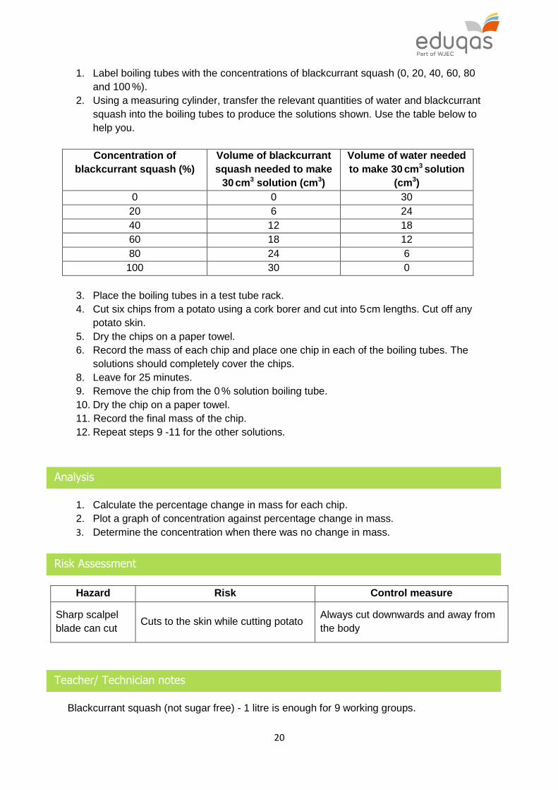
20
1. Label boiling tubes with the concentrations of blackcurrant squash (0, 20, 40, 60, 80
and 100 %).
2. Using a measuring cylinder, transfer the relevant quantities of water and blackcurrant
squash into the boiling tubes to produce the solutions shown. Use the table below to
help you.
Concentration of
blackcurrant squash (%)
Volume of blackcurrant
squash needed to make
30 cm3 solution (cm3)
Volume of water needed
to make 30 cm3 solution
(cm3)
0 0 30
20 6 24
40 12 18
60 18 12
80 24 6
100 30 0
3. Place the boiling tubes in a test tube rack.
4. Cut six chips from a potato using a cork borer and cut into 5 cm lengths. Cut off any
potato skin.
5. Dry the chips on a paper towel.
6. Record the mass of each chip and place one chip in each of the boiling tubes. The
solutions should completely cover the chips.
8. Leave for 25 minutes.
9. Remove the chip from the 0 % solution boiling tube.
10. Dry the chip on a paper towel.
11. Record the final mass of the chip.
12. Repeat steps 9 -11 for the other solutions.
Analysis
1. Calculate the percentage change in mass for each chip.
2. Plot a graph of concentration against percentage change in mass.
3. Determine the concentration when there was no change in mass.
Risk Assessment
Hazard Risk Control measure
Sharp scalpel
blade can cut Cuts to the skin while cutting potato
Always cut downwards and away from
the body
Teacher/ Technician notes
Blackcurrant squash (not sugar free) - 1 litre is enough for 9 working groups.

21
Large baking potatoes,1 per working group.
A potato chipper will quickly cut chips of uniform size. It saves time if the chips are cut (using
a chipper) just before or at the beginning of the lesson by a technician. Students will still
need to trim the chips to fit into the beaker/ boiling tube. If using a cork borer, each group
should use the same size borer for each of their potato cores.
No repeats are planned, but groups can compare results to discuss reproducibility.
The results should show that the chips gain mass in dilute concentrations of squash, but lose
mass in strong concentrations. The graph should be drawn with the x-axis across the centre
of the page, to show the increase and decrease in mass. Where the plotted line crosses the
x-axis shows the concentration where there is no gain or loss in mass. Students should
understand that this is the isotonic point and the concentration inside the potato cells is
equal to that in the external concentration.
Students should design their own table, but a suggested table format is shown below.
Concentration (%)
Mass at start (g)
Mass at end (g)
Change in mass (g)
Percentage change in mass (%)
Practical techniques covered
B1 Use of appropriate apparatus to make and record a range of measurements
accurately, including length, area, mass, time, temperature, volume of liquids and
gases, and pH.
B3 Use of appropriate apparatus and techniques for the observation and measurement
of biological changes and or processes.
B4 Safe and ethical use of living organisms (plants or animals) to measure physiological
functions and responses to the environment.
B5 Measurement of rates of reaction by a variety of methods including production of gas,
uptake of water and colour change of indicator
2.2 TRANSPORT SYSTEMS IN HUMANS

22
Spec Statement Comment
(a) describe the human circulatory system as a double circulatory system and its relationship with the gaseous exchange system. The blood passes through the heart twice in every complete circulation. The right side of the heart pumps the blood to the lungs and the left hand side pumps it around the rest of the body
The human circulatory system is a double circulatory system, as it involves one system for the lungs – pulmonary and one for the other organs of the body – systemic.
(b) label on a given diagram of the heart: the left and right atria and ventricles, semi-lunar, bicuspid and tricuspid valves, pulmonary artery, pulmonary vein, aorta and vena cava
Observe a dissected/ model of the heart to include coronary arteries and internal structure.
(c) explain how the structure of the heart is adapted to its function
The heart is made of cardiac muscle, which contracts to pump blood around the body. Understand the significance of the difference in thickness of the muscle in the atria and ventricles and between the right and left ventricles.
(d) describe the passage of blood through the heart including explaining the functions of the valves in preventing backflow of blood
Deoxygenated blood enters the right atrium from the vena cava. It then goes through the tricuspid valve to the right ventricle. From the right ventricle, it goes through the semilunar valve into the pulmonary artery to the lungs. From the lungs, oxygenated blood is returned to left atrium in the heart through the pulmonary veins. The blood then flows through the bicuspid (mitral) valve into the left ventricle. From the left ventricle, it goes through the semi-lunar valve into the aorta. Blood is distributed to the rest of the body (systemic circulation) from the aorta.
(e) describe and be able to compare the structure of arteries and veins
In diagrams of arteries and veins, label: tough outer coat, muscle layer, endothelium and lumen. Compare the relative thickness of the blood vessel walls and the size of the lumen in arteries and veins. Veins contain valves.
(f) explain how arteries and veins are adapted to their functions
Arteries have thick muscular walls and a smaller lumen to carry blood under pressure away from the heart to all organs of the body. Veins carry blood under lower pressure back to the heart and contain valves which ensure flow of blood in one direction only.
(g) describe that in the organs blood flows through very small
Capillary walls are one cell thick - very short diffusion pathway.
23
SPECIFIED PRACTICAL WORK
SP2.2 Examination of artery and vein using a light microscope and production of labelled scientific drawings of these from observation
blood vessels called capillaries which allow exchange of substances. Explain that the thin walls of the capillaries are an advantage for diffusion and that capillaries form extensive networks so that every cell is near to a capillary carrying blood
There are a large number of capillaries which maintain the diffusion gradient between the tissue and the blood.
(h) describe the functions of the four main parts of the blood: plasma (transport of water, nutrients, hormones, urea, antibodies), red cells (carry oxygen), white cells (defence) and platelets (clotting). Explain how red blood cells, white blood cells, platelets and plasma are adapted to their functions in the blood
Red blood cells have no nucleus and contain haemoglobin to carry oxygen. White blood cells include lymphocytes and phagocytes. Lymphocytes secrete antibodies and antitoxins and phagocytes ingest and digest micro-organisms. Platelets are fragments of larger cells which have no nucleus. They stick to the walls of damaged blood vessels forming a plug. This triggers a chain of reactions which results in a mesh forming across the damage (to form a scab).
24
Examination of artery and vein using a light microscope and production of
labelled scientific drawings of these from observation
Introduction
This practical requires you to observe and draw a prepared slide of an artery, and of a vein.
Apparatus
Light microscope
Slide of a Transverse Section (T.S.) of an artery
Slide of a Transverse Section (T.S.) of a vein
Method
1. Use a light microscope to examine a T.S artery using the × 10 objective lens.
2. Use the × 40 objective lens to identify the tough outer layer and the muscular and
elastic fibres.
3. Draw a diagram to show the distribution of tissues in the correct proportion.
4. Identify and label: tough outer layer; muscular and elastic fibres; lumen.
5. Repeat steps 1 - 4 using a T.S. vein.
Analysis
1. Compare the structure of the artery and vein and relate this to their function.
2. Calculate the total magnification of the image seen under the microscope by
multiplying the power of the objective lens by the power of the eyepiece.
Risk Assessment
Hazard Risk Control measure
No significant risks are associated with this investigation.
25
Teacher/Technician notes
A lamp may be required, if not part of the microscope.
× 10 and × 40 objective lenses are suggested for viewing the slides. It will be necessary to
determine the optimum magnification before presenting the slides to students.
Students will need to be briefed regarding safe and effective microscope use prior to this
practical activity.
Students should produce low power plans of the blood vessels, no individual cells should be
drawn. This practical activity is effective at developing microscope skills and biological
drawing skills. Drawing skills should include using a pencil to draw smooth, continuous lines
with no overlapping or gaps. No shading or colour should be used.
Students can calculate the total magnification of the image as the power of the objective lens
multiplied by the power of the eyepiece. Students could also calculate the magnification of
their drawing if given the mean diameter of the blood vessel used.
A virtual microscope for demonstration purposes is available on the link below.
http://medsci.indiana.edu/a215/virtualscope/docs/chap7_3.htm
Practical techniques covered
B3 Use of appropriate apparatus and techniques for the observation and measurement
of biological changes and or processes.
B7 Use of appropriate apparatus, techniques and magnification, including microscopes,
to make observations of biological specimens and produce labelled scientific
drawings.

26
2.3 TRANSPORT SYSTEMS IN PLANTS
Spec Statement Comment
(a) explain that xylem tissue contains tubes of dead cells called xylem vessels and explain how the vessels are adapted to their role in the transport of water and minerals from the roots upwards within plants
The tubes of dead cells in xylem have very strong cell walls and are hollow. This means they do not collapse under pressure and water passes through easily. The tubes pass up from the roots through the stem and into the veins in every leaf.
(b) explain how phloem is adapted to carry sugar from the photosynthetic areas to other parts of the plant. Sugar is moved to other parts of the plant for use in respiration and converted into starch for storage. This is called translocation
Phloem consists of long, narrow, living cells which have large spaces inside for transporting sugar solutions. These phloem cells carry sugars from the veins in every leaf to all other part of the plant. ( No details of mechanisms are required)
(c) explain the significance of root hairs in increasing the area for absorption, the role of osmosis in the uptake and movement of water through a plant and how mineral salts are taken up by root hairs by active transport
(d) describe the structure of a leaf and be able to label the following structures on a diagram of a T.S. leaf: cuticle, epidermis, stomata, palisade layer, spongy layer, xylem and phloem
(e) describe the structure of stomata to include guard cells and stoma and how stomata can open and close to regulate transpiration
When the guard cells surrounding a stoma change shape the stoma becomes larger or smaller. This opening and closing of stomata is important in controlling how much water vapour passes out of the leaves. Guard cells and stoma should be identified on a given diagram. ( No details of mechanisms are required)
(f) describe the process of transpiration resulting in the movement of water through a plant
Transpiration occurs when water vapour, passing out of the leaves of a plant, creates a negative pressure on water in the xylem. Water then rises up through the xylem from the roots. Transpiration occurs even in conditions of water shortage.

27
(g) explain the environmental factors that can affect transpiration, including light intensity, air movement and temperature and that this can be investigated with the use of a simple potometer
Environmental factors affect the rate of loss of water vapour from leaves and hence the rate of transpiration. Increased temperatures and increased air movements tend to increase the rate of transpiration. Light levels affect the opening and closing of stomata.

28
3 – HEALTH, DISEASE AND THE DEVELOPMENT OF MEDICINE
3.1 HEALTH AND DISEASE
Spec Statement Comment
(a) describe the relationship between health and disease
In the past, health was thought of simply as the absence of disease. In modern times there is greater emphasis on the promotion of health and prevention of disease.
(b) describe diseases as being communicable and non-communicable diseases as exemplified by influenza and cardiovascular disease
Communicable diseases – illnesses which result from infections by micro-organisms (pathogens) and are contagious among individuals. e.g. Influenza- caused by a virus, can be caught from an infected person by contact or droplet infection.. Non-communicable disease – illnesses which are often the result of unhealthy lifestyles and sometimes inheritance. e.g. cardiovascular disease (CVD)- linked to obesity, lack of exercise, smoking In former times most disease–related deaths were caused by communicable diseases. Now, the majority of serious illnesses are related to non-communicable diseases.
(c) describe the interactions between different types of disease, as exemplified by the increased risk of developing skin cancer when HIV positive and the increased risk of cardiovascular disease in diabetes patients
HIV – The virus weakens the immune system and reaches high levels in the blood. Skin cells, which become infected by the virus, divide and grow uncontrollably, leading to skin cancers such as Koposi's Sarcoma. Diabetes- In type II diabetes, blood sugar levels can become very high (hyperglycaemia). This is can damage the walls of blood vessels and lead to CVD (high blood pressure, stroke , heart attack).
29
3.2 COMMUNICABLE DISEASE
Spec Statement Comment
(a) explain the means by which communicable diseases caused by viruses, bacteria, protists and fungi can be spread in animals and plants. This should include by contact, aerosol, body fluids, water, insects, contaminated food.
(b) describe the following diseases, this should include the causative agent, the effect on the infected organism and how they can be prevented from spreading
HIV/AIDS
Chlamydia
Ash die back
Malaria
AIDS (Acquired Immune Deficency Syndrome) is caused by HIV (Human Immunodeficiency Virus). The virus infects lymphocytes which are part of the body's immune system. Without immunity, the body can become infected with a variety of micro-organisms, e.g. tuberculosis or pneumonia. The virus is spread by blood to blood contact, especially during sexual intercourse. Methods of prevention include the use of condoms and disposable gloves should be used where there is any danger of contact with contaminated blood. Antiviral agents can be used, but they only prevent the multiplication of the virus inside cells and must be taken throughout life. Chlamydia, this is the most common sexually transmitted disease in Britain. It is caused by the bacterium Chlamydia trachmatis and is spread during sexual intercourse via the vagina and urethra. Its spread can be prevented by the use of condoms. It can be treated with antibiotics such as tetracycline or erythromycin. However, if left untreated, it could cause infertility in adults. It could also cause conjunctivitis in babies during the process of birth if the mother is infected. It can also spread to the babies lungs. Ash die back. This disease, which has caused widespread damage in continental Europe, was not reported in the UK before 2010. Since then it has spread to many areas and has the potential to cause very significant damage to ash trees in the UK. The disease is caused by the fungus Hymenoscyphus fraxineus and spreads when the fungal spores are carried by wind. Young trees are most easily infected. The fungus causes the loss of leaves, prevents the growth of branches and damages the bark. The tree usually dies. Efforts to prevent the spread of the disease in the UK include a ban on the import and movement of ash

30
trees as well as selective breeding from trees identified as being resistant to fungus. When infected trees are cut down, the dead leaves and branches should be destroyed on the site by burning. Malaria - This kills over a million people in the world each year. It is caused by the single celled organism – Plasmodium. Plasmodium is spread via female mosquitoes of the genus Anopheles. Anopheles mosquitoes bite humans and inject Plasmodium into the blood stream. Plasmodium causes a fever when it destroys red blood cells in humans. Treatment consists of killing Plasmodium with anti-malarial drugs, such as paludrine or daraprim. A vaccine against Plasmodium has been developed. Prevention methods include: killing mosquitoes with insecticide, releasing large numbers of infertile male mosquitoes, biological control of mosquitoes, use of mosquito nets and repellents.
(c) describe the non-specific defence systems of the human body against pathogens, including intact skin forming a barrier against microorganisms and blood clots sealing wounds to seal the skin
(d) explain the role of the immune system of the human body in defence against disease. This should include the roles of lymphocytes in secreting antibodies and antitoxins and phagocytes which ingest and digest micro-organisms. Explain the process by which antigens from micro-organisms trigger lymphocytes to release antigen specific antibodies and that antibodies activate phagocytes
Lymphocytes are activated by the antigens on micro-organisms and then multiply to form clones. Each clone produces large quantities of identical antibodies, specific to a particular antigen. These antibodies attack and destroy the infecting micro-organisms. After the infection is cleared, some of the lymphocytes, called memory cells, remain in the body. The first time an infection occurs, the immune system may respond rather slowly but if the infection returns a second time the response is much faster. This because memory cells produce larger quantities of antibodies and in a much shorter time than on the first infection.

31
(e) describe how monoclonal antibodies are produced from activated lymphocytes which are able to divide continuously. Consequently very large numbers of identical antibodies, specific to one antigen, are produced continuously in very large numbers
B-lymphocytes are fused with tumour cells forming a hybridoma - this divides rapidly in laboratory conditions to form a clone. The hybridoma continuously produces specific antibodies called monoclonal antibodies.
(f) describe some of the ways in which monoclonal antibodies can be used including:
diagnosis of diseases including Chlamydia and HIV
tissue typing for transplants
monitoring the spread of malaria
supporting chemotherapy for cancers
Immunoassays are used in the diagnosis of diseases caused by Chlamydia trachmatis, HIV, and Plasmodium. Labelled (via radioactivity or fluorescence) monoclonal antibodies are added to test samples of infected body fluids and attach to specific antigens. The extent of the infection is related to the extent of the labelling. Tissue typing for transplants - The concentration of non-self-antigens in tissues is assessed. Monoclonal antibodies can be used against helper T-cells (T-lymphocytes) so B-lymphocytes, normally causing rejection, are prevented from functioning. Monitoring the spread of malaria – blood is taken from samples of people (even if they do not show any malarial symptoms) and tested with labelled monoclonal antibodies. Monoclonal antibodies will detect the presence of Plasmodium in the bloodstream (even if they are dead - killed by anti-malarial drugs) as they have specific antigens and will attach to the labelled monoclonal antibodies. This enables the success of anti-malarial drugs and the potential spread of malaria to be monitored. The destruction of cancer cells can be targeted with the use of monoclonal antibodies. Some types of cancer cells have specific antigens called tumour markers. Monoclonal antibodies can be produced that act against tumour markers. If these are attached to anti-cancer drugs, they will deliver the drug directly to the cancer cells.

32
(g) describe the following as physical defence responses in plants forming a barrier to pathogens:
cellulose cell walls, which may be strengthened by other chemical substances
leaf cuticle, which forms a waxy layer on the outside of the leaf Physical defences to herbivores include:
specialised hardened cells
specialised structures including stinging cells and trichomes
Pectins, added to cell walls, make them stronger and prevent infections by mildew fungi.
A thick waxy cuticle helps to prevent bacteria and fungi from penetrating the surface of the leaf.
Spines and thorns on stems and leaves of plants prevent them from being eaten by animals.
Stinging cells and trichomes ( specialised hairs) inject irritating chemicals, such as histamines, into animal skin and this deters animals from eating them
(h) describe chemical plant defence responses, including that many plants produce enzymes or toxic chemicals which attack insects and disease-causing bacteria and fungi
Some plants produce terpenes in response to attack by herbivores or invasion by micro-organisms (pathogens). These toxic chemicals destroy insects, fungi and bacteria. Peroxidase enzymes are produced by many fruit and vegetable plants as a defence response.
(i) describe different ways plant diseases can be detected and identified, in the lab and in the field. In the field, diseased plants can be identified by abnormal growth or by signs of the disease-causing organism, such as bacterial slime or eggs of insects. In the laboratory, pathogens can be grown on agar plates and viruses can be cultured in controlled conditions
Signs of abnormal growth include stunted growth, excessive production of flowers or altered colours of leaves. Biochemical tests are used to identify bacteria and viruses which have been cultured.

33
34
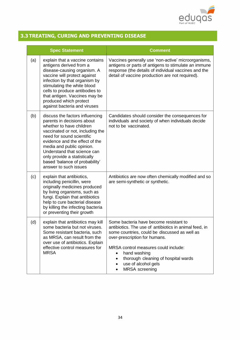
3.3 TREATING, CURING AND PREVENTING DISEASE
Spec Statement Comment
(a) explain that a vaccine contains antigens derived from a disease-causing organism. A vaccine will protect against infection by that organism by stimulating the white blood cells to produce antibodies to that antigen. Vaccines may be produced which protect against bacteria and viruses
Vaccines generally use ‘non-active’ microorganisms, antigens or parts of antigens to stimulate an immune response (the details of individual vaccines and the detail of vaccine production are not required).
(b) discuss the factors influencing parents in decisions about whether to have children vaccinated or not, including the need for sound scientific evidence and the effect of the media and public opinion. Understand that science can only provide a statistically based ‘balance of probability’ answer to such issues
Candidates should consider the consequences for individuals and society of when individuals decide not to be vaccinated.
(c) explain that antibiotics, including penicillin, were originally medicines produced by living organisms, such as fungi. Explain that antibiotics help to cure bacterial disease by killing the infecting bacteria or preventing their growth
Antibiotics are now often chemically modified and so are semi-synthetic or synthetic.
(d) explain that antibiotics may kill some bacteria but not viruses. Some resistant bacteria, such as MRSA, can result from the over use of antibiotics. Explain effective control measures for MRSA
Some bacteria have become resistant to antibiotics. The use of antibiotics in animal feed, in some countries, could be discussed as well as over-prescription for humans. MRSA control measures could include:
hand washing
thorough cleaning of hospital wards
use of alcohol gels
MRSA screening
35
(e) explain and understand the safe use of basic aseptic techniques involved in inoculating, plating and incubating microorganisms
bacteria and fungi can be grown on nutrient agar in a Petri dish, to produce an agar plate.
Petri dishes and nutrient agar should be sterilised before the agar is poured.
an inoculating loop is used to transfer bacteria and is sterilised before and after use by heating it to red heat in a Bunsen flame.
the Petri dish lid prevents micro-organisms from the air contaminating the culture and vice versa.
after inoculation the lid of the Petri dish should be secured in place by strips of adhesive tape for safety reasons
inoculated agar plates are incubated
at 250C in school laboratories, which encourages growth of the culture without growing pathogens
for safety reasons plates and equipment should be sterilised after use.
(f) describe the process of discovery and development of potential new medicines, including preclinical and clinical testing. New drug treatments may have side effects and extensive, large scale, rigorous testing is required including risk management. Preclinical stages involve testing on human cells grown in the laboratory, then on animals and finally a group of healthy volunteers. The new medicines are then taken for clinical testing using small groups of patients
All drugs may have side effects. New drugs, including medicinal drugs, may cause side effects that do not show up until lots of people use them. The use of the terms blind, double blind and placebo in the context of drug development should be understood.
SPECIFIED PRACTICAL WORK
SP3.3 Investigation into the effect of antibiotics on bacterial growth
36
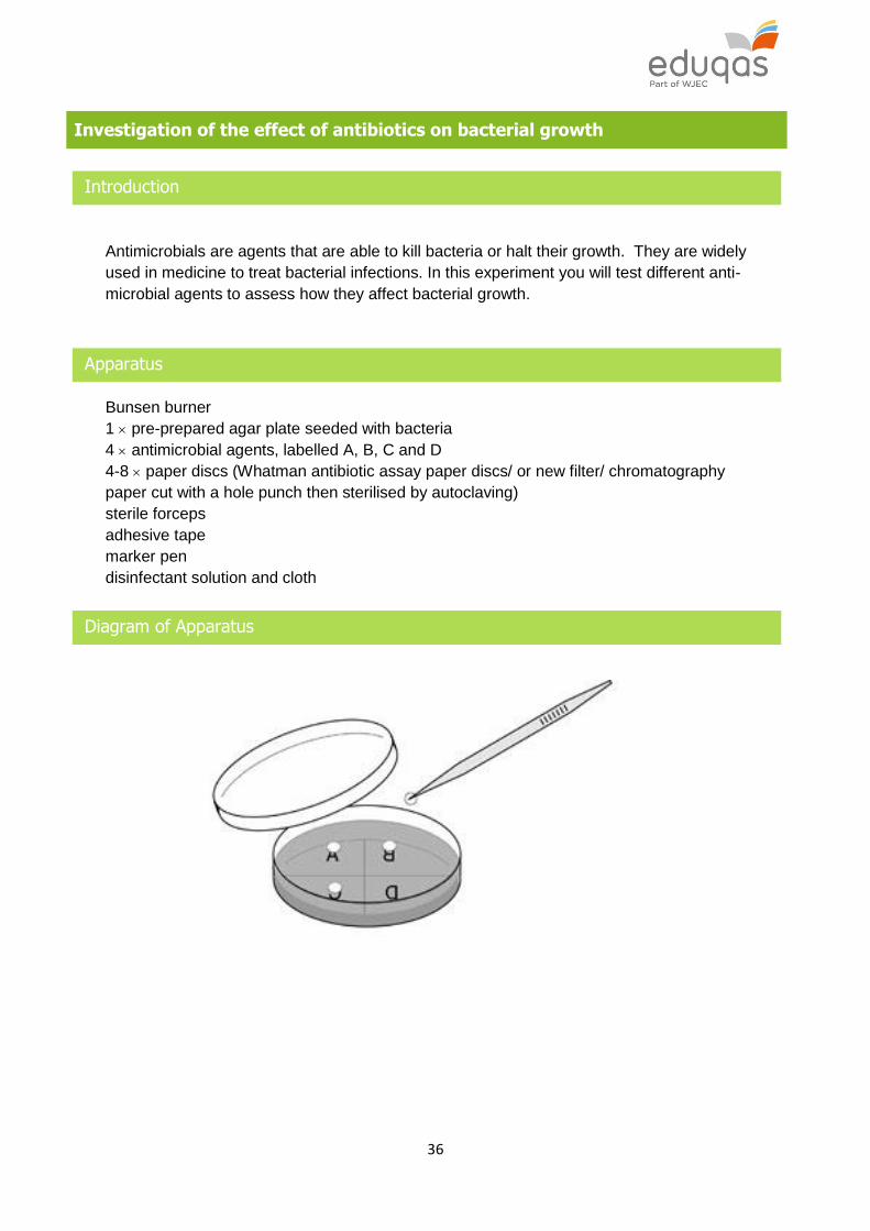
Investigation of the effect of antibiotics on bacterial growth
Introduction
Antimicrobials are agents that are able to kill bacteria or halt their growth. They are widely
used in medicine to treat bacterial infections. In this experiment you will test different anti-
microbial agents to assess how they affect bacterial growth.
Apparatus
Bunsen burner
1 pre-prepared agar plate seeded with bacteria
4 antimicrobial agents, labelled A, B, C and D
4-8 paper discs (Whatman antibiotic assay paper discs/ or new filter/ chromatography
paper cut with a hole punch then sterilised by autoclaving)
sterile forceps
adhesive tape
marker pen
disinfectant solution and cloth
Diagram of Apparatus

37
Method
1. Wash your hands with the soap or handwash. Wipe down the working area thoroughly
with the disinfectant.
2. Work very close to a lit Bunsen burner. Flame the forceps and use them to pick up a
filter paper disc and dip the disc into antibiotic A.
3. Allow them to dry for 5 minutes on an open, sterile Petri dish, next to a lit Bunsen
burner.
4. Repeat step 3 for antibiotics B, C and D.
5. Use the agar plate that has already been prepared and seeded with bacteria.
6. Turn the dish upside down. Divide the base into four sections by drawing a cross with
the marker pen. Label the sections A, B, C, D.
7. Flame the forceps and then use them to pick up antibiotic disc A. Raise the lid of the
Petri dish at an angle and place the disc onto the agar in the centre of section A.
8. Repeat step 5 for the other 3 discs. Make sure the discs are placed in the centre of
each section.
9. Label the agar plate with your name and date. Tape the lid securely. Incubate inverted
for 2-3 days at 20-25 °C.
10. Observe the plates without opening them.
11. Record the width of the clear zone around each antimicrobial. A piece of squared
paper under the agar plate might be helpful here.
Analysis
1. Which antimicrobial agent was the most efficient in your investigation? Give reasons
for your answer.

38
Risk Assessment
Hazard Risk Control measure
Bacteria can be
pathogenic
Touching bacteria when plate is
open
Wash hands
Incubate plates at room temperature
Seal plate so that they are not opened
Bunsen burner
flame and
Forceps can
burn
Burning skin when placing discs on
plate
Care must be taken to keep hands a
safe distance away from the flame.
Do not touch tip of forceps after flaming
Teacher / Technician notes
Detailed instructions are given on the link below.
http://www.nuffieldfoundation.org/practical-biology/investigating-anti-microbial-action
Making agar and pouring plates
a Calculate the quantity required and prepare just enough agar for the investigation – around 15 cm3 for normal depth in a 90 mm Petri dish. Any surplus will keep for 6-12 months in tightly-sealed screw-top bottles if sterile.
b Weigh out the agar medium powder containing the gel and chosen nutrients, add water and sterilise the mixture for the time, and at the temperature, specified by the manufacturer.
c Heat agar and water at 95 °C to dissolve the agar. Always use a water bath to boil agar, and never add agar to boiling water.
d Stopper flasks with a well-fitting plug of non-absorbent cotton wool. Cover with greaseproof paper or aluminium foil before sterilising by autoclaving.
e After autoclaving, transfer to a water bath to equilibrate at 50 °C. Stack plates after pouring to minimise condensation except in the top plate(s).
f Warm the Petri dishes before pouring to minimise condensation.
g Keep poured plates in a sealed plastic bag until needed to reduce dehydration of the media.

39
Making a spread plate
1 Sterile spreaders are used to distribute inoculum of Bacillus subtilis over the surface of prepared agar plates. You can sterilise a wrapped glass spreader in a hot air oven or sterilise by flaming with alcohol.
2 To flame a spreader with alcohol: a Dip the lower end of the spreader into a small volume of alcohol (70% IDA) contained in a vessel with a lid (either a screw cap or aluminium foil) or in a glass (not plastic) Petri dish with a lid. Keep the alcohol container covered and 1 metre away from the Bunsen burner flame.
b Pass quickly through a Bunsen burner flame to ignite the alcohol. Ensure the spreader is pointing downwards when and after igniting the alcohol to avoid burning yourself.
c Remove the spreader from the flame and allow the alcohol to burn off. The burning alcohol will sterilise the glass.
d Do not put the spreader down on the bench.
3 Cotton wool swabs can be used instead of glass spreaders. They may be preferable as they avoid the need for using alcohol as a sterilising agent. Prepare them by rolling small pieces of absorbent cotton wool around one end of a cocktail stick. Wrap individually in aluminium foil or place inside a universal bottle to sterilise in an autoclave or pressure cooker. These sterile swabs can then be dipped into the solution or culture to be transferred, rubbed on the surface of the agar plate, and immediately disposed of into disinfectant. (Note: Cotton buds from a pharmacist are not sterile and may be impregnated with an antimicrobial agent.)
4 Use agar plates with a well-dried surface so that the inoculum dries quickly. Dry the surface of agar plates by incubating for several hours (perhaps overnight) or put them in a hot air oven (at 55-60 °C) for 30-60 minutes with the two halves separated and the inner surfaces directed downwards.
The antibiotics can be bought as ready made discs or solutions can be made from everyday ingredients. Many types of toothpaste contain low concentrations of anti-microbials, and mouthwashes claim plaque-killing potential. The ten spices with the most potent antibacterial effects are garlic, onion, allspice, oregano, thyme, cinnamon, tarragon, cumin, cloves and lemon grass. Many spices with relatively weak antibacterial effects become much more potent when combined; examples are in chili powder (typically a mixture of red pepper, onion, paprika, garlic, cumin and oregano) and five-spice powder (pepper, cinnamon, anise, fennel and cloves). Lemon and lime juice, while weak inhibitors themselves, also have synergistic effects. It is also possible to investigate different dilutions of a particular anti-microbial. Students should be made aware of aseptic techniques before starting the practical activity. It is possible that students can prepare their own pour plates and inoculate them if you wish.

40
Practical techniques covered
B1 Use of appropriate apparatus to make and record a range of measurements
accurately, including length, area, mass, time, temperature, volume of liquids and
gases, and pH.
B3 Use of appropriate apparatus and techniques for the observation and measurement
of biological changes and or processes.
B4 Safe and ethical use of living organisms (plants or animals) to measure physiological
functions and responses to the environment.
B5 Measurement of rates of reaction by a variety of methods including production of gas,
uptake of water and colour change of indicator.

41
3.4 NON-COMMUNICABLE DISEASES IN HUMANS
Spec Statement Comment
(a) recall that many non-communicable human diseases, including cardiovascular disease, lung cancer, skin cancer, emphysema, type 2 diabetes and cirrhosis can be caused by the interaction of a number of life style factors
Candidates should understand the importance of exercise and fitness to the individual combined with the need for a healthy diet to control body mass. This helps to prevent type 2 diabetes and cardio vascular disease. Life style choices should also take regard of the negative effects of smoking, drinking alcohol and exposure to ultra violet light. The links between these life style choices and emphysema, cirrhosis and cancer should be known.
(b) explain the effect of the following lifestyle factors on the incidence of non-communicable diseases at local, national and global levels: exercise, diet, alcohol, smoking and exposure to UV radiation
(c) evaluate the advantages and disadvantages of the following treatments for cardiovascular disease
statins
angioplasty
changes to lifestyle diet/exercise
Research the advantages and disadvantages of the treatments for cardiovascular disease such as:
Statins, a daily medication to control blood cholesterol levels, but may cause side effects.
Angioplasty, surgery to place a small balloon in a blood vessel, which is inflated to remove a blockage. This results in improved blood flow e.g. in coronary vessels, but sometimes is only a temporary remedy.
Changes to diet/ lifestyle. These include stopping smoking, taking up regular exercise, eating more healthy food. These can reduce risk and lower blood pressure. However, a high level of self- discipline is needed to maintain these long-term changes.
42
4 – COORDINATION AND CONTROL
4.1 NERVOUS COORDINATION AND CONTROL IN HUMANS
Spec Statement Comment
(a) describe sense organs as groups of receptor cells, which respond to specific stimuli: light, sound, touch, temperature, chemicals, and then relay this information as electrical impulses along neurones to the central nervous system
(b) describe the structure of the nervous system, including the brain, spinal cord, sensory neurones, motor neurones and sensory receptors and the central nervous system consisting of the brain and spinal cord
(c) explain how the structure of the nervous system (including CNS, sensory and motor neurones and sensory receptors) is adapted to its functions
Nerve impulses begin at receptors and pass along sensory neurones to a coordinator. Motor neurones pass on impulses to effectors. The function of synapses should be known but knowledge of the chemical nature of synaptic neuro transmitters is not needed.
(d) describe the properties of reflex actions. These reactions are fast and automatic and some are protective, as exemplified by the withdrawal reflex, blinking and pupil size
(e) explain how the structure of a reflex arc is related to its function and be able to label a diagram to show: receptor, sensory neurone, relay neurone in spinal cord, motor neurone, effector and synapses
Candidates need to be able to indicate, on a diagram of a reflex arc, the direction of travel of the impulse.

43
(f) explain the functions of the following parts of the eye: sclera, cornea, pupil, iris, lens, choroid, retina, blind spot and optic nerve recognise and be able to label these parts on a diagram of a vertical section through the eye
The functions include: sclera – protective, tough white outer coat cornea – clear part of sclera allows light to enter and refracts light entering pupil – hole in centre of iris which allows light to enter iris – muscles that alter size of pupil to control amount of light entering lens – changes shape to focus light onto retina choroid – a pigmented layer which absorbs light to prevent reflection, also contains blood vessels retina – light sensitive layer , an image is formed here, impulses sent to optic nerve blind spot – where the optic nerve leaves the eye, there are no light sensitive cells here optic nerve – carries impulses from retina to brain
(g) describe common defects of the eye and explain how some of these problems may be overcome as exemplified by long-sightedness, short-sightedness and cataracts
The ability of the lens to change its shape to focus near and distant objects is called accommodation. This becomes less efficient with age. Cataracts can occur when changes in the lens of the eye cause it to become less transparent. This can lead to blurred vision and can be treated by replacing affected lenses with new lens substitutes within lens' capsules. Long-sightedness, affects a person's ability to see objects close to them. This occurs when the eyeball is too short or the lens is not thick enough. Convex lenses can be used to correct long sightedness. Short-sightedness, causes distant objects to appear blurry. This occurs when the eyeball is too long. Concave lenses can be used to correct short sightedness.
(h) describe the structure and function of the following parts of the brain: the cerebral hemispheres, cerebellum and medulla
Cerebral hemispheres control conscious activity
and act as centres for retaining past sensations (memory). The cerebellum controls balance and muscular
coordination. The medulla controls automatic reactions taking
place in the body such as heart beat, peristalsis, and breathing.
44
(i) explain that brain function is difficult to study and involves the use of brain scans, such as MRI and electrical stimulation. Discuss the ethical implications of studying patients with brain damage.
Magnetic resonance imaging (MRI) avoids surgery in the diagnosis of brain damage. High frequency radio waves are absorbed and transmitted in tissues placed in a strong magnetic field. A computer maps out the variation in signals to produce a 3D image of the brain. Oxygenated blood causes the image to become brighter. MRI scans can therefore be used to follow the activities of parts of the brain and can be used to analyse function as well as structure.
(j) explain some of the limitations in treating damage and disease in the brain and other parts of the nervous system as exemplified by Parkinson's disease and multiple sclerosis
The major limitation in treating physical brain damage is that brain cells do not regenerate when they are lost. Degeneration of nervous tissue in Parkinson's disease, multiple sclerosis and dementia is permanent.
SPECIFIED PRACTICAL WORK
SP4.1 Investigation into factors affecting reaction times

45
Investigation into factors affecting reaction time
Introduction
If you notice a ball moving towards your head, the time it takes from when you first notice the
ball to when your arm reaches up to catch it is an example of reaction time. Even though
nervous impulses travel very quickly through your nervous system, your body doesn’t react
instantly. In this activity, you will conduct a simple, measurable experiment to study reaction
time and investigate the hypothesis that reaction time improves with practice.
Apparatus
30 cm ruler
Diagram of Apparatus

46
Method
1. Ask your first volunteer to sit in the chair with good upright posture and eyes looking
across the room.
2. Have the volunteer place their forearm (the part of the arm from elbow to hand) so it
extends over the edge of the table.
3. Ask the volunteer to place their thumb and index (pointer) finger on either side of the
bottom of the vertically placed ruler. The number “1” should be on the bottom, the “30”
near the top.
4. Let your volunteer practice holding the ruler with those two fingers.
5. Now, ask your volunteer to remove their fingers from the ruler while you continue to
hold it so that the bottom of the ruler is at a height of 2 cm above the fingers.
6. Tell your volunteer that you will release the ruler without warning. Their job will be to
catch it with their thumb and forefinger as soon as they sense it dropping.
7. Drop the ruler. When your volunteer catches it, record the number on the ruler
displayed just over the thumb. The lower the number, the faster the reaction time.
8. Conduct five trials with the same volunteer, dropping the ruler from 2 cm above their
fingers each time.
9. Repeat the experiment with at least five other volunteers and record your results in a
suitable table
Analysis
1. Use the conversion table below to convert the distance measured to a reaction time
for each volunteer
Catch distance (cm)
Reaction time (milliseconds)
Catch distance (cm)
Reaction time (milliseconds)
1 50 16 180
2 60 17 190
3 701 18 190
4 80 19 200
5 90 20 200
6 100 21 210
7 120 22 210
8 130 23 220
9 140 24 220
10 140 25 230
11 150 26 230
12 160 27 230
13 160 28 240
14 170 29 240
15 170 30 250
2. Discuss the extent to which your results support the hypothesis.

47
Risk Assessment
Hazard Risk Control measure
There are no significant risks associated with this procedure
Teacher / Technician notes
A possible alternative activity could be to compare the volunteer's dominant hand with their
non-dominant hand.
Students should design their own table, but a suggested table format is shown below.
Trial 1 Trial 2 etc
Volunteer Distance
(cm) Reaction time (ms)
Distance (cm)
Reaction time (ms)
Practical techniques covered
B1 Use of appropriate apparatus to make and record a range of measurements
accurately, including length, area, mass, time, temperature, volume of liquids and
gases, and pH.
B3 Use of appropriate apparatus and techniques for the observation and measurement
of biological changes and or processes.
B4 Safe and ethical use of living organisms (plants or animals) to measure physiological
functions and responses to the environment.
B5 Measurement of rates of reaction by a variety of methods including production of gas,
uptake of water and colour change of indicator.

48
4.2 HORMONAL COORDINATION AND CONTROL IN HUMANS
Spec Statement Comment
(a) describe and be able to label the positions of the following glands on a diagram of the human body: pituitary, adrenal, thyroid, pancreas, ovaries and testes
(b) describe hormones as chemical messengers, produced by glands and carried by the blood, which control many body functions
(c) describe the principles of negative feedback mechanisms in maintaining optimum conditions inside the body
Any change from the balance in optimal internal conditions results in the body's hormonal and nervous systems compensating for the change and restoring the balance.
(d) explain the role of thyroxine in the body as an example of negative feedback. Description should be limited to effects of TRH and TSH in the release of thyroxine.
Thyrotropin-releasing hormone (TRH) is made in the hypothalamus, an area at the base of the brain. The nerve fibres that come out of it carry TRH and release it into the blood surrounding the pituitary gland. The TRH regulates the formation and secretion of thyroid stimulating hormone in the pituitary gland. The pituitary gland secretes thyroid stimulating hormone (TSH) which causes the thyroid to secrete thyroxine. A decrease in thyroxine levels stimulates TRH and therefore TSH secretion, whereas an increase in thyroxine slows down TRH secretion.
(e) explain the action of adrenaline in the body as an example of positive feedback. Description should be limited to the effects of adrenaline on the heart, breathing and muscles. Adrenaline is converted into a less active compound by the liver.
The positive feedback mechanism of adrenalin production causes an increase in pulse rate and volume of blood pumped by the heart with each beat. It increases the depth of breathing and dilates the blood vessels supplying muscles.
(f) describe the roles of hormones in human reproduction, including the menstrual cycle
Testosterone regulates the development of male
secondary sexual characteristics. Oestrogen stimulates the thickening of the uterine
wall during ovulation. Progesterone prepares the uterus for pregnancy.
49
(g) explain the interactions of FSH, LH, oestrogen and progesterone in the control of the menstrual cycle
At the beginning of the menstrual cycle, two hormones from the pituitary gland, follicle stimulation hormone (FSH) and luteinizing hormone (LH), stimulate the development of egg-bearing follicles in the ovaries. As the follicles mature, they secrete oestrogen.
(h) explain the use of hormones in contraception and evaluate hormonal and non-hormonal methods of contraception
Contraceptive pills or implants contain combinations of synthetic oestrogen-like and progesterone-like chemicals. These stop the secretion of FSH and LH so neither follicle development (an oestrogen effect) nor ovulation (a progesterone effect) occurs. Non hormonal forms of contraception such as condoms reduce the chance of sexual transmission of diseases. Also some hormonal forms of contraception may have a variety of negative side effects such as nausea and thrombosis.
(i) explain the use of hormones in modern reproductive technologies to treat infertility
Hormones are used to increase fertility. Synthetic chemicals are used to stimulate ovulation. They either act as FSH, which stimulates the development of follicles or they inhibit the production of oestrogen. Lack of oestrogen (the normal FSH inhibitor) results in more FSH being produced which stimulates multiple follicle development.

50
4.3 HOMEOSTASIS IN HUMANS
Spec Statement Comment
(a) explain the importance to animals of maintaining a constant internal environment in response to internal and external change
Metabolism operates only within a narrow range of temperature and pH and requires appropriate nutrients and water.
(b) explain why and how glucose levels need to be kept within a constant range. When the blood glucose level rises, the pancreas releases the hormone insulin, a protein, into the blood. This causes the liver to reduce the glucose level by converting glucose to insoluble glycogen and then storing it
(c) explain how glucagon interacts with insulin to control blood sugar levels in the body
(d) compare type 1 and type 2 diabetes and explain how they can be treated. Diabetes is a common disease in which a person has a high blood sugar (glucose) level. In Type 1 diabetes this is because the body does not produce enough insulin. In Type 2 diabetes the body cells do not properly respond to the insulin that is produced
The detection of glucose in urine is a symptom of diabetes. Candidates should test artificially prepared urine samples for the presence of glucose using Benedict's solution. The methods of treating diabetes include regularly injecting insulin, a low sugar and low carbohydrate diet and possible transplant of pancreatic tissue.
(e) describe the function of the skin in the control of body temperature. Label a diagram of a vertical section through the skin to show: hair, erector muscle, sweat gland, sweat duct, sweat pore, blood vessels. Explain the role of these structures in temperature regulation: change in diameter of blood vessels, sweating, erection of hairs; shivering as a means of generating heat

51
(f) describe and be able to label a diagram of the human excretory system to show kidneys, renal arteries, renal veins, aorta, vena cava, ureters, bladder, urethra and be able to indicate the direction of blood flow in the blood vessels associated with the kidney
(g) describe the function of the kidneys in maintaining the water balance of the body and remove waste products from the blood and explain why this is necessary. The waste, a solution containing urea and excess salts called urine, passes from the kidneys in the ureters to the bladder where it is stored before being passed out of the body. The presence of blood or cells in the urine indicates disease in the kidney
(h) label a section through a kidney to include: renal artery, renal vein, cortex, medulla, pelvis, ureter
The position of nephrons within the kidney.
(i) label a diagram of a nephron and its associated blood supply to show: capillary knot, Bowman's capsule, tubule, collecting duct, capillary network, arteriole to and from capillary knot
(j) explain the process of filtration under pressure and that selective reabsorption of glucose, some salts, and much of the water takes place in the tubule
The significance of the difference in the diameter of the blood vessels entering and leaving the capillary knot in Bowman's capsule. Small molecules, including urea, glucose, salts and water are forced from capillary knot into the Bowman's capsule.

52
(k) describe the effect of ADH on the permeability of the kidney tubules. The kidneys regulate the water content of the blood by producing dilute urine if there is too much water in the blood or concentrated urine if there is a shortage of water in the blood. ADH increases the permeability of the collecting duct walls to water. More ADH is produced if there is a shortage of water in the blood, more water is reabsorbed and so a more concentrated urine is produced
(l) explain the effect on cells of osmotic changes in body fluids
Osmotic changes in body fluids will result in changes in the concentration of chemicals in cellular cytoplasm. Homeostasis aims to keep the cytoplasm and body fluids isotonic. Dehydration of cells will occur by osmosis if the water content of body fluids is very low
(m) explain the response of the body to different temperature and osmotic challenges
High external temperatures result in excess water loss via sweating. In turn, this leads to body fluids becoming very concentrated compared to the cellular cytoplasm. Osmosis then results in dehydration if the water balance is not restored via drinking.
SPECIFIED PRACTICAL WORK
SP4.3 Dissection of mammalian kidney

53
Dissection of mammalian kidney
Introduction
The kidney Is a vital component of the urinary system and is responsible for filtering the
blood and the production of urine. In this practical you will examine the external structure
and internal structure of the kidney following dissection.
Apparatus
kidney
scalpel
newspaper and dissection board
disposable gloves and goggles
Diagram of Apparatus

54
Method
1. Place dissection board on top of the newspaper and place the kidney on top of it.
2. BEFORE YOU DISSECT:
a. Examine the outside of the kidney. The ureter, renal artery and renal vein all enter
the kidney in the same area. It may be possible to observe these structures.
b. Draw a diagram of the kidney.
3. Slice longitudinally through the kidney as shown in the diagram.
4. Arrange the kidney with the cut surface facing upwards.
5. Locate the cortex, which is the outer dark red / brown layer; the medulla, which is the
lighter striped layer just below the cortex and the pelvis, which is the central white
mass that branches out into the medulla.
6. Draw a diagram of the kidney after dissection and label the following layers: cortex,
medulla and pelvis.
Risk Assessment
Hazard Risk Control measure
Sharp scalpel
blade could cut Cuts to the skin whilst cutting kidney
Always cut downwards onto the
dissecting board
Use of animal
tissue is a
biohazard
Transfer of infection from animal
tissue to student
Ensure cuts are covered
Wear gloves
Teacher / Technician notes
Lamb or pig kidneys are widely available at a relatively low cost. Pig's kidneys are larger
and therefore easier to manipulate by the student. Some pre-packed kidneys that have
been freeze-thawed are often slippery to handle and difficult to dissect, therefore fresh
kidneys from a butcher would be preferable to use with students.
Practical techniques covered
B3 Use of appropriate apparatus and techniques for the observation and measurement
of biological changes and or processes.
B4 Safe and ethical use of living organisms (plants or animals) to measure physiological
functions and responses to the environment.
B7 Use of appropriate apparatus, techniques and magnification, including microscopes,
to make observations of biological specimens and produce labelled scientific
drawings.
55
4.4 PLANT HORMONES
Spec Statement Comment
(a) explain how auxins are important in the control and coordination of plant growth and development, including the positive response of plant shoots to light, phototropism, and plant roots to gravity, gravitropism
Auxins are made in the tips of stems. They cause cell elongation which enables stems to grow towards light. The effect of auxin is inhibited by light. If a stem is growing horizontally, the upper side is exposed to more light than the upper side. The lower side elongates more than the upper side and therefore the tip of the stem grows up towards light. Roots grow towards gravity because of auxin.
(b) describe some of the effects of plant hormones, relating to auxins, gibberellins and ethene
Gibberellins increase the stem length by stimulating cell division and cell elongation. They also break the dormancy of many types of seeds. Ethene is a gas released from ripening fruit. It speeds up the rate at which fruit ripens.
(c) describe some of the different ways in which people use plant hormones to control plant growth
Synthetic auxin is used in rooting powers to encourage root growth in plant cuttings. Ethene is used to control the ripening of fruit when it is transported over long distances to be sold out of season far from its site of production.

56
5 – PHOTOSYNTHESIS
Spec Statement Comment
(a) describe the process of photosynthesis and describe photosynthesis as an endothermic reaction, whereby green plants and other photosynthetic organisms use chlorophyll and light to convert carbon dioxide and water into glucose, producing oxygen as a by-product. Recall the word equation for photosynthesis
The chemical reactions of photosynthesis within the cell are controlled by enzymes. (Details of the enzymes involved in photosynthesis are not required.). The practical techniques used to investigate photosynthesis: the use of sodium hydroxide to absorb carbon dioxide; how to test a leaf for the presence of starch; how oxygen and carbon dioxide sensors and data loggers could be used. The steps within the method for testing a leaf for starch include: killing the leaf by placing in boiling water, decolouration using ethanol, washing to soften, testing with iodine. Understand the need for de-starching the leaf prior to the procedure.
(b) explain the effect of temperature, light intensity and carbon dioxide concentration on the rate of photosynthesis
(c) explain the interaction of these factors in limiting the rate of photosynthesis
SPECIFIED PRACTICAL WORK
SP5 Investigation into factors affecting the rate of photosynthesis

57
Investigation of the factors affecting photosynthesis
Introduction
Light is one of the factors which affects the rate of photosynthesis.
In this investigation a green plant named Canadian pondweed (Elodea) will produce bubbles
of oxygen as a result of photosynthesis.
The number of bubbles of oxygen produced is affected by light intensity.
Apparatus
250 cm3 beaker
lamp
glass funnel
plasticine
test tube
8 cm length of pondweed (Elodea)
metre ruler ± 1 mm
sodium hydrogen carbonate powder
clamp stand, clamp and boss
spatula
Diagram of Apparatus
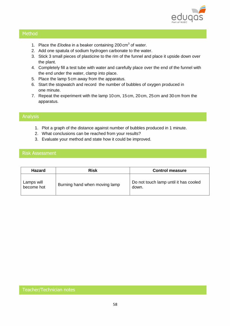
58
Method
1. Place the Elodea in a beaker containing 200 cm3 of water.
2. Add one spatula of sodium hydrogen carbonate to the water.
3. Stick 3 small pieces of plasticine to the rim of the funnel and place it upside down over
the plant.
4. Completely fill a test tube with water and carefully place over the end of the funnel with
the end under the water, clamp into place.
5. Place the lamp 5 cm away from the apparatus.
6. Start the stopwatch and record the number of bubbles of oxygen produced in
one minute.
7. Repeat the experiment with the lamp 10 cm, 15 cm, 20 cm, 25 cm and 30 cm from the
apparatus.
Analysis
1. Plot a graph of the distance against number of bubbles produced in 1 minute.
2. What conclusions can be reached from your results?
3. Evaluate your method and state how it could be improved.
Risk Assessment
Hazard Risk Control measure
Lamps will become hot
Burning hand when moving lamp Do not touch lamp until it has cooled down.
Teacher/Technician notes

59
If the plant is not producing bubbles then the stem might have started to ‘heal’ up, cutting off
the end off may improve bubbling.
Begin the experiment with the lamp closer to the plant and move the plant further away as
this seems to give better results.
Cabomba caroliana (and Elodea crispa) are no longer available to buy. They have been banned for culturing or sale under European regulations controlling invasive non-native plants. CLEAPSS have worked with native plants (Hornwort and red Cabomba), and they are OK for use. The CLEAPSS method (see the link below) overcomes the problems of the native aquatic plants bubbling slowly. http://science.cleapss.org.uk/Resource-Info/GL184-Using-video-recording-to-measure-the-
rate-of-photosynthesis.aspx
If students have any difficulty in obtaining results, the link below can be used.
http://www.reading.ac.uk/virtualexperiments/ves/preloader-photosynthesis-full.html
The method as stated does not include repeats, but students should be encouraged to carry
out an appropriate number, if time allows.
This experiment is ideal for a discussion of the limiting factors of photosynthesis and how
they are controlled variables in this experiment. There is also a clear opportunity to discuss
the limitations of the investigation such as the difficulty in controlling temperature.
Students should design their own table, but a suggested table format is shown below.
Number of bubbles produced in one minute
Distance from plant
to lamp (cm) Trial 1 Trial 2 Trial 3 Mean
Practical techniques covered

60
B1 Use of appropriate apparatus to make and record a range of measurements
accurately, including length, area, mass, time, temperature, volume of liquids and
gases, and pH.
B2 Safe use of appropriate heating devices and techniques including use of a Bunsen
burner and a water bath or electric heater.
B3 Use of appropriate apparatus and techniques for the observation and measurement
of biological changes and or processes.
B4 Safe and ethical use of living organisms (plants or animals) to measure physiological
functions and responses to the environment.
B5 Measurement of rates of reaction by a variety of methods including production of gas,
uptake of water and colour change of indicator.

61
6 – ECOSYSTEMS
6.1 LEVELS OF ORGANISATION WITHIN AN ECOSYSTEM
Spec Statement Comment
(a) describe different levels of organisation in an ecosystem from individual organisms through populations and communities to the whole ecosystem
The total number of organisms, of the same species, in a given geographical area or location make up a population. All the interacting populations of different species within that location form a community. The term ecosystem is used to describe a community of organisms interacting with each other and with their environment.
(b) explain how some abiotic factors affect communities as exemplified by pH, light, temperature and salinity
Abiotic (non-living) factors in a community are constantly changing and these changes affect living organisms. Plants in a community interact with each other by competing for light, space and mineral ions.
(c) explain how some biotic factors affect communities as exemplified by predation, disease and food availability
Biotic (living) factors in a community are constantly changing. A change in a biotic factor might affect a community e.g. by a change in the availability of food or the presence of a new predator or pathogen.
(d) describe the importance of interdependence and competition in a community
(e) describe photosynthetic organisms as the main producers of food and therefore biomass for life on Earth. Green plants, and other photosynthetic organisms such as algae use the light from the sun to produce organic materials
Radiation from the sun is the source of energy for living organisms. Green plants capture only a small percentage of the solar energy which reaches them.
(f) describe the differences between the trophic levels of organisms within an ecosystem including producers; first, second and third stage consumers; herbivores and carnivores
Candidates should be aware that alternative terms for the organisms in the trophic levels include: primary consumers, secondary consumers and tertiary consumers.
(g) investigate data about food chains and food webs and explain that they show the transfer of biomass between organisms
Analyse data in terms of: efficiency of energy transfer, numbers of organisms and biomass.

62
(h) use data to construct and interpret pyramids of numbers
Draw and label pyramids of number and biomass (to include names and values at each trophic level).
(i) describe pyramids of biomass and explain, with examples, how biomass is lost between the different trophic levels. At each stage in the food chain biomass is used in repair and in the maintenance and growth of cells whilst biomass is lost in waste materials and respiration which is exothermic
(j) calculate the efficiency of biomass transfers between trophic levels and explain how this affects the number of organisms at each trophic level

63
6.2 THE PRINCIPLE OF MATERIAL CYCLING
Spec Statement Comment
(a) recall that many different materials cycle through the abiotic and biotic components of an ecosystem. Nutrients are released in decay, e.g. nitrates and phosphates and these nutrients are then taken up by other organisms resulting in nutrient cycles. In a stable community the processes which remove materials are balanced by processes which return materials
Only the general principle of the cycling of elements is required here (no detail of nitrate or phosphate cycles).
(b) explain why it is important that carbon is constantly cycled in nature by the carbon cycle via photosynthesis which incorporates it and respiration which releases it
Carbon is taken up by green plants in photosynthesis and is passed to animals when they eat the plants. Some of this carbon then becomes part of carbohydrates, fats and proteins which make up their bodies. Animals and plants release carbon dioxide during respiration.
(c) explain that microorganisms, bacteria and fungi, feed on waste materials from organisms and that when plants and animals die their bodies are broken down by microorganisms bringing about decay. These micro-organisms respire and release carbon dioxide into the atmosphere. Burning fossil fuels releases carbon dioxide
Micro-organisms digest materials from their environment for growth and other life processes. These materials are returned to the environment either in waste products or when living things die and decay. When decay is prevented, fossil fuels such as coal, oil and gas are formed and these store energy in carbon compounds.
(d) explain the importance the water cycle to living organisms
Water evaporates from large bodies of water (seas and oceans) and condenses to form clouds. Precipitation provides fresh water for plants and animals on land. Water from the land drains into the seas and oceans.
64
(e) explain the effects of factors such as temperature and water content on rate of decomposition in aerobic and anaerobic environments
If all other factors are equal then decomposition occurs at a faster rate under aerobic conditions than under anaerobic conditions. This is because decomposers can metabolize more quickly in the presence of oxygen. Generally an increase in temperature increases the rate of decomposition up to the point at which enzymes are denatured and/or decomposers are killed. In waterlogged conditions anaerobic decomposition may occur but it will be very slow. In extremely dry conditions, such as those found in hot deserts and the Dry Valleys of Antarctica decomposition does not occur and the mummification of animal remains is common.
(f) evaluate the evidence for the impact of environmental changes on the distribution of organisms, with reference to water and atmospheric gases
Interpret information and data providing evidence for the possible changes in the distribution of organisms as a result of increased desertification, reported rise in sea levels, reported increases in atmospheric carbon dioxide levels resulting in a lowering of atmospheric pH and the acidification of oceans.
SPECIFIED PRACTICAL WORK
SP6.2 Investigation into factors affecting decomposition
65
Investigation into factors affecting decomposition
Introduction
Decomposers secrete enzymes into the soil to break down waste. In this investigation the
enzyme urease catalyses the breakdown of urea into ammonia. The ammonia dissolves in
water to create an alkaline solution. The rate at which the solution becomes alkaline is
affected by the temperature. During this reaction the red acidic solution changes to an
alkaline blue.
Apparatus
10 × boiling tubes
test tube rack
labels
3 × syringes
dropping pipette
pH colour chart
1.5 % urease solution
1.25 % ethanoic acid
universal indicator in a dropper bottle
1 % urea solution
stopwatch
Method
1. Label a test tube as 20 C.
2. Use a syringe to add 2.5 cm3 of urea to the test tube.
3. Add 1 cm3 of ethanoic acid and 10 drops of universal indicator to the test tube.
4. Add 10 cm3 of urease to another test tube and place both tubes into a 20 C water bath.
5. Pour the urease into the tube labelled 20 C.
6. Start the stopwatch and record the time taken to turn from red to blue.
7. Repeat steps 1-6 for temperatures of 30, 40, 50 and 60 C.
Analysis
1. Plot a graph of temperature against the time taken to change colour.

66
Risk Assessment
Hazard Risk Control measure
Urea, urease
and
ethanoic acid
are all irritants.
Chemicals could get onto the skin
when adding to test tube
Wash hands immediately if any
chemical gets onto them / wear
laboratory gloves.
Chemicals could get transferred from the hands to the eyes
Wear eye protection.
Teacher/Technician notes
Thermostatically controlled water baths can be set up at specific temperatures and students
can fill beakers with water from these baths to use as water baths at their desks.
Thermometers could be provided and students asked to regularly check the temperature
allowing them to analyse the effectiveness of the method.
Urease enzyme has an optimum temperature of 60 C, so it is possible that the denaturing of
the enzyme may not be seen in the range of temperatures given. Students could be asked to
predict what would happen to the time taken at higher temperatures.
Students should relate the results of their investigation to the enzymes involved in
decomposition and the effect of temperature on the rate of decomposition.
Students should design their own table, but a suggested table format is shown below.
Temperature (ºC)
Time taken to change from red to blue (s)
Practical techniques covered
B1 Use of appropriate apparatus to make and record a range of measurements
accurately, including length, area, mass, time, temperature, volume of liquids and
gases, and pH.
B2 Safe use of appropriate heating devices and techniques including use of a Bunsen
burner and a water bath or electric heater.
B3 Use of appropriate apparatus and techniques for the observation and measurement
of biological changes and or processes.

67
6.3 BIODIVERSITY
Spec Statement Comment
(a) describe how to use quadrats to investigate the abundance of species e.g. a comparison of different sides of a hedge or mown and unmown grassland
Any suitable location could be used to show the effect of different environmental factors. This should include the use of line transects and random quadrat distribution. An understanding that the number/ distribution of quadrats used should be enough to give valid results.
(b) describe how transects can be used to measure changes in the abundance and distribution of species e.g. seashore
(c) describe the principles of sampling, the need to collect sufficient data and use of appropriate statistical analysis. (Details of statistical tests are not required.) Describe the principles of capture/recapture techniques including simple calculations on estimated population size
Candidates should know how to use the equation:
population size =
number in 1st sample x number in 2nd sample
number in 2nd sample previously marked
When using capture-recapture data, assumptions made include: there is no death, immigration or emigration and that the marking technique does not affect chances of survival.
Candidates will not be expected to recall the equation.
(d) explain what is meant by biodiversity, the variety and number of different species in an area, and why it is important. Explain that indicator species are an important set of organisms whose numbers and changing population can tell us a lot about the changing state of ecosystems
Biodiversity is important as it provides food, potential foods, industrial materials, new medicines and for human well-being. Indicator species and changes in pH and oxygen levels may be used as signs of pollution in a stream and lichens can be used as indicators of air pollution.
(e) describe both positive and negative human interactions within ecosystems and explain their impact on biodiversity
An increase in the size of the human population results in an increased demand for food, water and other resources. These interactions, together with urbanisation, industrialisation and globalisation, can reduce biodiversity because ecosystems are destroyed, reduced in area or damaged. The extreme case is where populations die out or organisms become extinct. If the resources in an ecosystem are used in a sustainable way the human interaction within the ecosystem can be considered as positive.

68
SPECIFIED PRACTICAL WORK
SP6.3 Investigation into factors affecting the abundance and distribution of a species
(f) describe the ways in which biodiversity and endangered species can be protected locally and globally, including issues surrounding the use of legislation. Explain the need for and issues associated with the collection of reliable data and ongoing environmental monitoring
Biodiversity and endangered species can be conserved and protected by the following:
Convention on International Trade in Endangered Species
Sites of Special Scientific Interest
captive breeding programmes
national parks
seed/ sperm banks
local biodiversity action plans
(g) explain the use of biological control agents and the introduction of alien species and their effects on local wildlife. Explain the issues surrounding the use of biological control agents and how the approach to using this method of control has changed as requirements for detailed research and scientifically based trials and analysis are now more fully understood
Candidates should know that some animals and plants have been introduced, deliberately and accidentally, into areas where they do not naturally occur and some have become invasive and caused problems. Invasive species may grow faster than native species and upset the natural eco-system. Native species may not be able to compete with them. Research into the use of biological control agents takes place, on a world-wide basis, in order to understand how best to control alien species. During the research, trials are needed to assess the effects of biological control agents particularly on non-targeted native species.
(h) explain some of the benefits and challenges of maintaining local and global biodiversity
Maintaining biodiversity means that there is a greater chance of the discovery of potential treatments for many diseases and health problems. It ensures the sustainable productivity of soils and provides genetic resources for all crops, livestock and marine species harvested for food. Climate change, global warming and acidification of the oceans all have a negative impact on biodiversity. Controlling both climate change and global warming is one of the greatest challenges facing mankind at the present time.
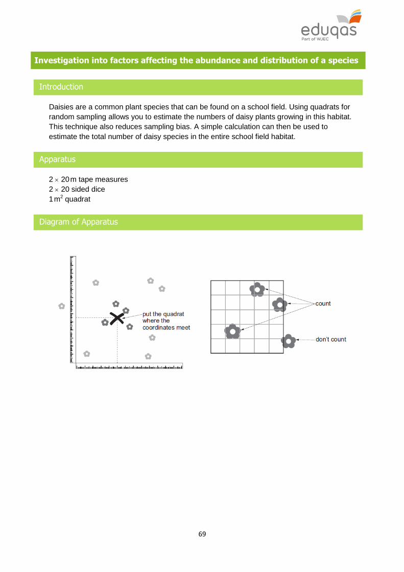
69
Investigation into factors affecting the abundance and distribution of a species
Introduction
Daisies are a common plant species that can be found on a school field. Using quadrats for
random sampling allows you to estimate the numbers of daisy plants growing in this habitat.
This technique also reduces sampling bias. A simple calculation can then be used to
estimate the total number of daisy species in the entire school field habitat.
Apparatus
2 20 m tape measures
2 20 sided dice
1 m2 quadrat
Diagram of Apparatus

70
Method
1. Lay two 20 m tape measures at right angles along two edges of the area to survey.
2. Roll two 20 sided dice to determine the coordinates.
3. Place the 1 m2 quadrat at the place where the coordinates meet.
4. Count the number of daisy plants within the quadrat. Record this result.
5. Repeat steps 2-4 for at least 25 quadrats.
Analysis
1. Use the following equation to estimate the total number of daisy plants in the field
habitat:
Total number of daisy plants in the habitat = total number in sample × total area (m2)
total sample area (m2)
Where:
total area = 400 m2
total sample area = number of 1 m2 quadrats used
Risk Assessment
Hazard Risk Control measure
Some plants have
thorns, sting or are
poisonous
Adverse skin response Cover skin at all times
Biting and stinging insects
Adverse skin response Cover skin at all times. Use insect repellent.
Tripping Strains and sprains Care where walking
Teacher/Technician notes
Students could compare data for mown and unmown areas.
This practical activity is effective at developing practical fieldwork skills. Students can
discuss the need for a large sample of data in ensuring that there is confidence in a valid
conclusion. Also, students can describe the importance of random sampling techniques in
reducing/eliminating bias.
Alternative methods of generating coordinates can be used, such as using a random number
generator or random number tables.

71
Practical techniques covered
B1 Use of appropriate apparatus to make and record a range of measurements
accurately, including length, area, mass, time, temperature, volume of liquids and
gases, and pH.
B3 Use of appropriate apparatus and techniques for the observation and measurement
of biological changes and or processes.
B4 Safe and ethical use of living organisms (plants or animals) to measure physiological
functions and responses to the environment.
B6 Application of appropriate sampling techniques to investigate the distribution and
abundance of organisms in an ecosystem vis direct use in the field.

72
6.4 SOME OF THE BIOLOGICAL CHALLENGES OF INCREASING FOOD YIELDS
USING FEWER RESOURCES
Spec Statement Comment
(a) describe the issues surrounding the need to balance the human requirements for food and economic development with the needs of wildlife. Discuss how the collection of detailed, reliable scientific information and monitoring by biologists could help to inform, manage and reduce the impact of development on the environment e.g. the role of the Environment Agency
The rising human population is causing increased effects on the environment. This includes that more space is needed for housing, industry and agriculture. Where development is proposed, data collected by biologists is used in an assessment of environmental impact, including effects on endangered species. The assessment is used to decide whether the development should be allowed to go ahead, be refused or modified to reduce the effect on wildlife. Government agencies have an important role in monitoring, protecting and improving the environment.
(b) describe some of the biological factors affecting levels of food security including increasing human population, changing diets in wealthier populations, new pests and pathogens, environmental change, sustainability and cost of agricultural inputs
Factors include:
An increasing human population has threatened food security in some countries – these countries cannot produce enough food to feed their own population and often these countries are financially too poor to import food from other countries. Conflicts have arisen in some parts of the World over the availability or food.
Changing diets in wealthier countries has resulted in ‘seasonal’ fruits and vegetables being required all the year round. These fruits and vegetables are often grown in financially poor countries and transported all around the World.
New pests and pathogens appear resulting in an increased financial burden on countries to find eradication solutions.
Environmental changes affect farming practices throughout the World. Lack of rainfall can result in crops failing causing widespread famine. Conflicts have arisen in some parts of the World over the availability of water.
Attempts to increase crop yields throughout the World have resulted in the increased cost of agricultural inputs – fertilizers, pesticides, herbicides, fungicides. Research must continue to develop sustainable methods to feed the World’s population.

73
(c) describe and explain some possible biotechnological and agricultural solutions, including genetic modification, to the demands of the growing human population
Alternative foods such as mycoproteins can be grown on an industrial scale. Mycoprotein can be produced from the fungus Fusarium and is protein rich. The fungus is grown in large industrial vats on a sugar syrup and under aerobic conditions. Mycoprotein can be used to produce vegetarian ‘meat’ products such as ‘beefburgers’ or veggieburgers. Crop yields can be significantly increased by genetic modification (GM). Increased production of mycoprotein and the further use of GM crops could provide more food for the increasing world population.

74
7 – INHERITANCE, VARIATION AND EVOLUTION
7.1 THE GENOME AND GENE EXPRESSION
Spec Statement Comment
(a) describe chromosomes as linear arrangements of genes. Chromosomes that are found in pairs in body cells are strands of DNA
DNA has a ladder-like structure, the bases forming the rungs. They should have an understanding of complementary base pairing - A (adenine) pairs with T (thymine) and that C (cytosine) pairs with G (guanine). Each triplet code identifies a particular amino acid. In the cytoplasm, these triplet codes are used to identify and link amino acids together to form proteins.
(b) describe DNA as a polymer made up of two strands forming a double helix
(c) describe DNA as a polymer made from four different nucleotides; each nucleotide consisting of a common sugar and phosphate group with one of four different bases attached to the sugar
(d) recall a simple description of protein synthesis. There are four bases, A, T, C and G within DNA and that it is the order of these bases which forms a code to produce proteins
(e) explain simply how the structure of DNA affects the proteins made in protein synthesis. There is complementary base pairing between adenine and thymine, cytosine and guanine. The order of bases determines the order in which different amino acids are linked together to form different proteins. The bases are read in groups of three, this is called the triplet code
(f) describe how the genes for a particular protein may occur in different forms (alleles or variants) which have different sequences of bases in their DNA. These variants,
Genes occur along chromosomes and different forms of the same gene can occur. These different forms of the same gene are known as alleles or variants. These alleles would be found at the same position on a pair of chromosomes. The alleles would differ slightly in the arrangement of the base

75
therefore, will give rise to differences in the order of amino acids in the protein and change its activity, affecting a characteristic of an organism
sequence in their DNA. The alleles of the same gene deal with the same feature or characteristic in an organism but each allele would make the characteristic appear slightly differently in the organism.
(g) describe that a large amount of DNA does not occur in genes and the bases do not form part of the genetic code for producing proteins. Instead, this non-coding DNA coordinates and controls how and when particular genes become active and produce proteins
Some of the DNA in a cell is responsible for coding for the sequence of amino acids during protein synthesis. This DNA is known as coding DNA or genome DNA. Another type of DNA does not code for amino acids during protein synthesis. This is known as non-coding DNA. The ratio of coding to non-coding DNA in a cell varies from organism to organism. In humans it is about 80% non-coding : 20% coding. The function of much of the non-coding DNA is not known but some of it is important in switching on or activating genes so that they can take place in protein synthesis.
(h) describe how an organism's DNA can be analysed by 'genetic profiling' and how this can be used to show the similarity between two DNA samples. The process involves cutting the DNA into short pieces which are then separated into bands. The pattern of the bands produced can be compared to show the similarity between two DNA samples
The term genetic profiling should be used in place of genetic fingerprinting to avoid confusion with fingerprinting. No detail of the process is required.
(i) describe the genome as the entire genetic material of an organism
All the genetic material of an organism is known as its genome. It is the organism’s complete set of DNA, including all of its genes. Each genome contains all the information to build and maintain that organism. In humans, a copy of the entire genome – more than 3 billion DNA base pairs – is contained in all cells that have a nucleus. The entire genome of many organisms, including humans, has now been studied. This will have important medical significance in future.
(j) discuss the potential importance for medicine of our increasing understanding of the human genome
An understanding that the human genome is important because it uses information from DNA to develop new ways to treat, cure, or even prevent disease.
SPECIFIED PRACTICAL WORK
SP7.1 Simple extraction of DNA from living material

76
Simple extraction of DNA from living material
Introduction
DNA is the hereditary material found in all living things. In this practical you will extract the
DNA from strawberries. Strawberries can have up to 8 copies of each chromosome and so
contain a lot of DNA. When extracted from the strawberry this DNA is visible.
Apparatus
re-sealable plastic bag
strawberry
10 cm3 washing up liquid (detergent)
1 g sodium chloride
100 cm3 water
2 250 cm3 beakers (one beaker will be used for the filtering apparatus below)
filter funnel
coffee filter paper
ice-cold 90 % alcohol
ice lolly stick or plastic coffee stirrer
Method
1. Remove the green top from the strawberry.
2. Put the strawberry into the plastic bag, seal it and crush for about 2 minutes.
3. In a beaker mix together 10 cm3 of washing up liquid, 1 g of salt and 100 cm3 water. This
mixture is the DNA extraction liquid.
4. Add 10 cm3 of the extraction liquid to the bag with the strawberry.
5. Re-seal the bag and gently mix the extraction liquid with the strawberry for 1 minute.
6. Place the coffee filter inside the beaker and gently pour the strawberry mixture into it.
7. Pour 10 cm3 of ice-cold 90 % ethanol down the side of the beaker into the strawberry
mixture, do not mix or stir.
8. Within a few seconds you should see a white cloudy substance form in the clear layer
above the strawberry mixture. Use a lolly stick to pull strands of this out of the top
layer, this is the strawberry DNA.

77
Risk Assessment
Hazard Risk Control measure
90% ethanol
could act as an
irritant
Inhalation could cause irritation of
the nose/throat
Use in well-ventilated area/wear safety
glasses.
Ethanol could touch skin during the experiment
Use gloves when pouring ethanol.
Teacher/ Technician notes
Chill the ethanol by keeping in the freezer for at least 2 hours or overnight. Keep on ice
during the experiment.
A variety of fruits can be used as an alternative to strawberries, such as kiwi or banana, but
check students have no allergies to the fruit used.
This experiment could be used as stimulus material to begin a piece of extended writing
describing what was observed in the experiment and linking it to what the student already
knows about the structure of DNA. It could also lead to a discussion of the ethical issues
surrounding DNA.
Practical techniques covered
B2 Safe use of appropriate heating devices and techniques including use of a Bunsen
burner and a water bath or electric heater.
B3 Use of appropriate apparatus and techniques for the observation and measurement
of biological changes and or processes.
B4 Safe and ethical use of living organisms (plants or animals) to measure physiological
functions and responses to the environment.

78
7.2 INHERITANCE
Spec Statement Comment
(a) explain the following terms: gamete, chromosome, gene, allele/variant, dominant, recessive, homozygous, heterozygous, genotype phenotype
The terms gene and allele are not interchangeable.
(b) describe genes as sections of DNA molecules that determine inherited characteristics and that are in pairs. Genes have different forms, called alleles
(c) explain single gene inheritance and be able to complete Punnett squares to show this
(d) predict the outcomes of monohybrid crosses including ratios
An understanding of probability is also required.
(e) recall that most phenotypic features are the result of multiple genes rather than single gene inheritance
(f) describe sex determination in humans. In human body cells, one of the pairs of chromosomes, XX or XY, carries the genes which determine sex. These separate and combine randomly at fertilisation
The use of Punnett squares to show the inheritance of sex chromosomes.
(g) describe the development of our understanding of genetics including the work of Gregor Mendel. Discuss why the significance of his work was not recognised and validated by scientists for many years
Gregor Mendel proposed the mechanism of inheritance after a scientific process of meticulous experimentation using large numbers of pea plants, observation, further methodical experimentation, statistical analysis and deduction. Mendel read his paper at two meetings of The Natural History Society of Brunn in Bohemia in 1865. It was published in 1866 and circulated to many academic institutions. However, the significance of Mendel’s work was not recognised and validated until scientists rediscovered his paper and replicated his experiments in the early 1900s. The structure of DNA and genes was unknown and not understood until the 1950s.

79
7.3 VARIATION AND EVOLUTION
Spec Statement Comment
(a) explain some of the advantages and disadvantages of asexual and sexual reproduction in a range of organisms
Advantages of sexual reproduction: introduces variation into a population the species can adapt to new environments a disease is less likely to affect all the
individuals in a population Disadvantages of sexual reproduction:
time and energy are needed to find a mate not possible for an isolated individual
Advantages of asexual reproduction: population can increase rapidly can exploit a suitable habitat quickly
Disadvantages of sexual reproduction: does not lead to variation in a population the species may only be suited to one
habitat disease may affect all the individuals in a
population
(b) describe simply how the genome, and its interaction with the environment, influence the development of the phenotype of an organism. Variation may be due to environmental or genetic causes or a combination of the two
The variation in height/length in organisms could be used to show that individuals of the same species are similar but they are never exactly the same. Continuous and discontinuous variation should be illustrated graphically – bell shaped curve for continuous variation and discontinuous variation as discrete groups.
(c) state that there is usually extensive genetic variation within a population of a species
(d) recall that all variants result from changes, mutations, in existing genes and that mutations occur at random. Most mutations have no effect on the phenotype but some influence phenotype and very few determine phenotype. Mutation rates can be increased by ionising radiation
The greater the dose/exposure to ionising radiation the greater the chance of mutation. (No reference to specific ionising radiation is required.)

80
(e) describe evolution as a change in the inherited characteristics of a population over time through a process of natural selection which may result in the formation of new species. Genes which enable better adapted individuals to survive are passed on to the next generation. This may results in new species being formed. The process of natural selection is sometimes too slow for organisms to adapt to new environmental conditions and so organisms may become extinct
The term natural selection should be understood. The term ‘Survival of the fittest’ should be used with care as it must be qualified in the context of breeding i.e. survival of the fittest to breed.
(f) explain how individuals with characteristics adapted to their environment are more likely to survive and breed successfully. This results in evolution
(g) describe that evolution is ongoing as shown by the development of resistance to antimicrobial chemicals by bacteria or Warfarin resistance in rats and that evolution can also be evidenced by fossils
(h) describe the impact of developments in biology on classification systems; biological classification systems continue to be modified in the light of ongoing research. Recently, the three Domain system (based on differences in RNA) proposes two Domains of prokaryotes and one further Domain containing four main eukaryote kingdoms – Protists, Fungi, Plants and Animals
There is a need for a systematic scheme to divide the variety of living organisms into groups in order to make the understanding of the variety of living things manageable and to understand trends and relationships. The classification system maybe based on morphological features or DNA analysis. A simple understanding of possible groupings. The five Kingdom classification using morphological features: Bacteria, Single celled organisms, Plants, Fungi, and Animals. The three Domain classification based on analysis of DNA: Ancient bacteria, Bacteria and All organisms with a nucleus. No features of these groups required.
(i) describe the work of Darwin and Wallace in the development of the theory of evolution by natural selection and explain the impact of these ideas on modern biology
An understanding that Wallace and Darwin were both working on the ideas of evolution and natural selection at around the same time.
81
7.4 SELECTIVE BREEDING AND GENE TECHNOLOGY
Spec Statement Comment
(a) explain the impact of the selective breeding of food plants and domesticated animals
Humans have been carrying out selective breeding of plants and animals for many thousands of years. Parents, of the same species, with desirable genetic features are bred together. Offspring, which have inherited the desirable features, are selected and also bred together. This continues over many generations until all the offspring show the desirable features. Examples of these desirable features include:
wheat plants with large seed grains
sheep with fine wool
cattle with high milk yields
horses with fine features and a very fast pace
However, selective breeding can result in the inheritance of undesirable characteristics. This is ‘inbreeding’ and can lead to breeds becoming particularly sensitive to disease or inherited defects such as hip dysplasia in domestic dogs.
(b) describe genetic engineering as a process which involves modifying the genome of an organism to introduce desirable characteristics
The genome of an organism is modified by introducing into it a gene from another organism. The introduced gene would allow the required desirable characteristic to be expressed in the genetically engineered organism. Crops that have had their genomes modified in this way are known as genetically modified (GM) crops.
(c) describe the main steps in the process of genetic engineering
The steps include:
the gene for the desirable feature is identified in an host organism
enzymes are used to cut out the required gene from the DNA of the host organism
the gene is inserted into a vector, either a bacterial plasmid or a virus.
the vector is used to insert the genes into cells of animals, plants or microorganisms at an early stage in their development
organism develops the desirable features.
(d) explain some of the possible benefits and risks, including practical and ethical considerations, of using gene technology in modern agriculture and medicine
Concerns include the risks of the spread of inserted genes to other organisms and moral and ethical concerns about modifying genomes. Benefits include the possible use of genetic engineering to treat or overcome inherited human diseases.