Biological responses in edible crab, Callinectes amnicola that could serve as markers of heavy...
10
Biological responses in edible crab, Callinectes amnicola that could serve as markers of heavy metals pollution Adebayo A. Otitoloju Olugbenga K. Elegba Adesola O. Osibona Published online: 16 July 2008 Ó Springer Science+Business Media, LLC 2008 Abstract Responses of lagoon crab, Callinectes amni- cola were explored as useful biological markers of heavy metal pollution. The toxicity level of the metals based on the 96-h LC50 values showed that copper with LC50 value of 0.018 mM was found to be two times more toxic than Lead (0.041 mM) against the lagoon crab, C. amnicola. The exposure of the lagoon crab to sublethal concentrations (1/100th and 1/10th of 96-h LC50 values) of Cu and Pb compound, respectively, resulted in the bioaccumulation of the test metals to varying degrees in the selected organs that were dependent on the type of metal and concentration of metal compound in the test media. The degree of metal (Cu and Pb) accumulation was generally in the following order: gills [ muscle [ heptopancrease. Exposure of the crabs to sublethal concentrations of the metals also caused pathological changes such as the disruption of the gill fil- aments and degeneration of glandular cells with multifocal areas of calcification in the hepatopancreas. A reduction in the weight of the exposed animals over a 14-day period of observation was also recorded. The significance of these results and the usefulness of the biological endpoints in monitoring programmes aimed at establishing the total environmental level of heavy metals in aquatic ecosystems were discussed. Keywords Heavy metals Biomarkers Histopathology Bioaccumulation Biomonitoring 1 Introduction Real life tragic experiences of heavy metal pollution such as Minamata and Itai-Itai diseases have shown the devas- tating effects that the accumulation of heavy metals in animal/plant tissues which serve as food source could have on higher predators, particularly man ((Kurdland 1960; Harada and Smith 1975). As a result, heavy metals have been categorized as priority pollutants which must be continually monitored especially in edible organisms even where they occur at levels below their toxic thresholds in the ambient environment, since the environmental persis- tence and non-biodegradable nature, over the years create a bioconcentration or bioaccumulation in organism that cannot excrete the absorbed metals (Otitoloju and Don- Pedro 2002, 2004). Although relatively high concentrations of metal is generally required to cause an acute toxic response to macro-invertebrates, chronic toxicity studies have shown lethal or sublethal responses at very low levels. Many essential metals that are required in trace concentrations for a number of biological processes have been found to become highly toxic to exposed organisms when presented over long periods at relatively low concentrations. As a result, it is pertinent to carry out ecotoxicological studies on such animals aimed at establishing the toxicity levels of the changing concentrations, distribution in the various compartment of the organism and to demonstrate the effects of the sublethal concentration in the exposed organisms. The increasing emphasis on the need for a more com- plete assessment and monitoring of aquatic ecosystems other than the mere chemical monitoring of water and sediment have highlighted the need to deploy appropriate biological indices for these locations. Bioaccumulation A. A. Otitoloju (&) O. K. Elegba Department of Zoology, Ecotoxicology Laboratory, University of Lagos, Lagos, Nigeria e-mail: [email protected] A. O. Osibona Department of Marine Sciences, University of Lagos, Lagos, Nigeria 123 Environmentalist (2009) 29:37–46 DOI 10.1007/s10669-008-9180-6
-
Upload
independent -
Category
Documents
-
view
1 -
download
0
Transcript of Biological responses in edible crab, Callinectes amnicola that could serve as markers of heavy...

Biological responses in edible crab, Callinectes amnicola that couldserve as markers of heavy metals pollution
Adebayo A. Otitoloju Æ Olugbenga K. Elegba ÆAdesola O. Osibona
Published online: 16 July 2008
� Springer Science+Business Media, LLC 2008
Abstract Responses of lagoon crab, Callinectes amni-
cola were explored as useful biological markers of heavy
metal pollution. The toxicity level of the metals based on
the 96-h LC50 values showed that copper with LC50 value
of 0.018 mM was found to be two times more toxic than
Lead (0.041 mM) against the lagoon crab, C. amnicola.
The exposure of the lagoon crab to sublethal concentrations
(1/100th and 1/10th of 96-h LC50 values) of Cu and Pb
compound, respectively, resulted in the bioaccumulation of
the test metals to varying degrees in the selected organs
that were dependent on the type of metal and concentration
of metal compound in the test media. The degree of metal
(Cu and Pb) accumulation was generally in the following
order: gills [ muscle [ heptopancrease. Exposure of the
crabs to sublethal concentrations of the metals also caused
pathological changes such as the disruption of the gill fil-
aments and degeneration of glandular cells with multifocal
areas of calcification in the hepatopancreas. A reduction in
the weight of the exposed animals over a 14-day period of
observation was also recorded. The significance of these
results and the usefulness of the biological endpoints in
monitoring programmes aimed at establishing the total
environmental level of heavy metals in aquatic ecosystems
were discussed.
Keywords Heavy metals � Biomarkers � Histopathology �Bioaccumulation � Biomonitoring
1 Introduction
Real life tragic experiences of heavy metal pollution such
as Minamata and Itai-Itai diseases have shown the devas-
tating effects that the accumulation of heavy metals in
animal/plant tissues which serve as food source could have
on higher predators, particularly man ((Kurdland 1960;
Harada and Smith 1975). As a result, heavy metals have
been categorized as priority pollutants which must be
continually monitored especially in edible organisms even
where they occur at levels below their toxic thresholds in
the ambient environment, since the environmental persis-
tence and non-biodegradable nature, over the years create a
bioconcentration or bioaccumulation in organism that
cannot excrete the absorbed metals (Otitoloju and Don-
Pedro 2002, 2004).
Although relatively high concentrations of metal is
generally required to cause an acute toxic response to
macro-invertebrates, chronic toxicity studies have shown
lethal or sublethal responses at very low levels. Many
essential metals that are required in trace concentrations for
a number of biological processes have been found to
become highly toxic to exposed organisms when presented
over long periods at relatively low concentrations. As a
result, it is pertinent to carry out ecotoxicological studies
on such animals aimed at establishing the toxicity levels of
the changing concentrations, distribution in the various
compartment of the organism and to demonstrate the
effects of the sublethal concentration in the exposed
organisms.
The increasing emphasis on the need for a more com-
plete assessment and monitoring of aquatic ecosystems
other than the mere chemical monitoring of water and
sediment have highlighted the need to deploy appropriate
biological indices for these locations. Bioaccumulation
A. A. Otitoloju (&) � O. K. Elegba
Department of Zoology, Ecotoxicology Laboratory,
University of Lagos, Lagos, Nigeria
e-mail: [email protected]
A. O. Osibona
Department of Marine Sciences, University of Lagos,
Lagos, Nigeria
123
Environmentalist (2009) 29:37–46
DOI 10.1007/s10669-008-9180-6

studies in addition to histopathological biomarkers have
been identified as definite biological endpoints of historical
exposure and thus serve as better indicators of environ-
mental stress (Matthiessen et al. 1993; Stentiford et al.
2003; Stentiford and Feist 2005). The use of biomarkers
may provide important information which once validated
through laboratory studies can provide direct measures of
actual effects of heavy metals upon living organisms in the
field, thereby overcoming large areas of uncertainty
implicit in normal risk assessment (Varanasi et al. 1987;
Vethaak and Jol 1996).
With regards to copper toxicity, Cu was found to be
toxic to aquatic invertebrates such as Typantonous fuscatus
Clibaranus africanus, Sesarma huzardi (Otitoloju and
Don-Pedro 2002). Exposure to Cu has been reported to
cause cellular stress apparent due to reduced lysosomal
stability (Svendsen and Weeks 1997; Viarengo et al. 2000).
Physiological impairment is also manifested in a number of
ways including catabolism, reduction in haemolymph
protein content (Weeks et al. 1993; Lin and Chen 2001),
decrease or increase in heart beat rate (Curtis et al. 2000;
Bamber and Depledge 1997) and induction of methallo-
thionein production in crabs (Brown et al. 2004).
Lead (Pb) is also one of the extensively exploited metals
that has been found in very high concentrations in the
environment. The inclusion of Pb in gasoline as an anti-
knock contribute to its occurrence in the air, which is
transported to the streams and rivers by run-off where fish
and other aquatic organisms take it up and incorporate in
their bodies. The transfer of lead between a primary con-
sumer, the zooplanktonic crustacean Daphnia magna and a
secondary consumer, the fish (Poecilia reticulum) hence
establishing its biomagnification potential and the inherent
public health risk status was also reported (Kakkar and
Jaffery 2005). Exposure to Pb in the environment has been
recognized to be a serious public health problem (WHO
1995) hence its inclusion in this study.
Callinectes amnicola is one of the most common estu-
arine macro-invertebrates and is generally abundant
throughout the Lagos lagoon. Lagoon crabs are a crucial
factor in the brackish water food web because of their
opportunistic feeding habits, high abundance levels and
their great tolerance of salinity. The crabs are being widely
used as food and feed supplement and are considered as an
important shell fishery product. Due to its abundance and
the fact that the crab is being consumed by a large popu-
lation of people especially those living around the coastal
region, the public health risk associated with its con-
sumption need to be monitored continually. and its
usefulness as a biological monitor for assessing the health
status of aquatic ecosystem determined.
In view of the above, the objectives of this study are to
determine the level of some heavy metals accumulated in
various organs of the edible crabs and also establish other
biological endpoints which can serve as useful biomarkers
of metal pollution for assessing the health status of aquatic
ecosystem.
2 Materials and methods
2.1 Test animal
2.1.1 Callinectes amnicola (Crustacean; Decapoda,
Brachyura, Portunidae) (L)
Animals of similar sizes (6–8 cm carapace width and
27–32 g weight) were collected using crab traps and dip
net from the Lagos lagoon. The animals were collected
from the same end of the lagoon in order to reduce vari-
ability in biotype. These were transported as quickly as
possible to the laboratory in baskets from the collection site
and kept in plastic holding tanks (50 cm x 35 cm x 30 cm),
which contained aerated lagoon water (6 l).
Lagoon crabs collected were maintained in the plastic
holding tanks with a maximum of four specimens per tank
to avoid overcrowding. These were shaded with black
polythene to prevent excess light penetration. The animals
were allowed 8 days to acclimatize to laboratory (R.H
70 ± 2%; temperature 27 ± 2�C) and experimental con-
ditions before being exposed to chemicals. During this
period the animals were fed with either Tilapia fingerlings
or commercial fish pellets once daily.
All bioassays were carried out using dechlorinated tap
water. This was achieved by aerating the tap water for
several days. Only inter molt crabs were used and indi-
viduals with mutilated appendages prior to experimentation
were discarded.
2.2 Test chemicals
The heavy metals used for this study were obtained as
metallic salts of Fisons laboratory reagents, analytical
grades of the following types;
(a) Copper as CuSO4 � 5H2O
(b) Lead as Pb(NO3)2
The test chemicals were chosen as they are wide spread
contaminant in the Lagos lagoon (Otitoloju et al. 2007).
2.3 Preparation of test media including application
of toxicants
A pre-determined amount of each heavy metal compound
was weighed out (using an Oertling 30TD analytical bal-
ance) and to this was added a given volume of diluent to
38 Environmentalist (2009) 29:37–46
123

obtain stock solution of known strength. Individual test
solution of copper and lead compound were prepared by
diluting the appropriate volume of stock solution with
dechlorinated water.
2.4 Assessment of mortality
Individual crabs were taken to be dead if no movement of
the body and appendages was observed. This can be further
confirmed by prodding the individual with a glass rod and
those which failed to respond by moving away from initial
position or raised their appendages were considered dead.
2.5 Bioassay
2.5.1 Relative acute toxicity of copper and lead
compounds against Callinectes amnicola
Following the establishment of the range of activity of the
test compounds in the preliminary range finding experi-
ment, the test animals were exposed to several concentrations
of each heavy metal salt and untreated control as follows.
Copper (CuSO4 � 5H2O) against C. amnicola at 0.0001,
0.0003, 0.0006, 0.0013, 0.0063 mM and untreated control.
Lead {Pb(NO3)2} against C. amnicola at 0.0003,
0.0017, 0.0033, 0.005, 0.0067 mM and untreated control.
Eight (8) live crabs of similar sizes were carefully
handpicked at random into the test media in bioassay
container. Four replicates were set up for each treatment
i.e. two crabs were exposed per container for each test
concentration and control.
Mortality assessments were carried out once every 24 h
over a 96 h period.
2.6 Sublethal studies
2.6.1 Bioaccumulation studies
After determining the 96-h LC50 values for each of the two
metals, two sublethal concentrations (1/100th and 1/10th
LC50 values) of each metal and control were prepared. A
total of eight test animals were exposed per sublethal
concentration or control in four replicates i.e. two crabs per
replicate to the metal solution for 14 days in order to
investigate the rate of bioaccumulation of the test metals in
the gills, hepatopancrease and muscle.
Sublethal concentrations under which bioaccumulation
of test metals by C. amnicola was investigated are as
follows:
(a) Cu (CuSO4 � 5H2O) was tested at:
0.0018 mM (1/10th of the 96 h LC50)
0.00018 mM (1/100th of the 96 h LC50)
(b) Pb (Pb(NO3)2 was tested at:
0.0041 mM (1/10th of the 96 h LC50)
0.00041 mM (1/100th of the 96 h LC50)
2.6.2 Determination of concentration of metals
accumulated in selected organs of Callinectes
amnicola
2.6.2.1 Digestion of samples The whole animal samples
(deshelled) were properly cleaned (by copiously rinsing all
exposed and partially exposed parts) with distilled water to
remove debris and other external adherents. The gills,
hepatopancrease and flesh were collected and oven dried at
60�C for 48 h. The dried samples were then grounded
using a mortal and pestle and sieved using a 200 lm to
normalize for particle size. A portion of 2 g from the dried
sample was placed in a glass beaker and digested using a
freshly prepared mixture 1:2 of deionized water and Conc.
HNO3. (FAO/SIDA 1986).
2.6.2.2 AAS determination of heavy metal in samples
(gills, hepatopancrease and flesh) All digestates and
extracts obtained were filtered through Whatman No.1
filter paper and made to mark with deionized water in a
100 cc volumetric flask. The heavy metal concentration in
each digested sample was determined by comparing their
absorbences with those of standards (Solution of known
metal concentrations) using a Perkin Elmer (Precisely) A
Analyst 200 AAS. For data quality, particular attention was
paid to the cleanliness of all wares. In addition, factory
prepared AAS standard solutions were run as samples for
accuracy check after every five measurements.
2.6.3 Histopathological studies
A similar experiment as described above under sublethal
studies was carried out. For these studies, at pre-deter-
mined day intervals (days 0, 7, 14), 2 live C. amnicola per
replicate making four per treatment including control were
randomly selected and dissected. The gills, hepatopan-
crease and flesh were carefully removed and fixed in
Boiun’s fluid pending further histology works. The fixed
samples were transferred to phosphate buffer (pH 6.8),
after 7 h of fixation in Boiun’s fluid. The tissues were then
dehydrated in graded alcohol, cleared in xylene before
embedding in paraffin wax (melting point 56.0�C). Serial
sections of 2–5 lm thickness was cut in rotary microtome
then passed through xylene followed by absolute alcohol
and water. The sections were stained with Haematoxylin
and Eosin, dehydrated in graded alcohol, cleared in more
Environmentalist (2009) 29:37–46 39
123

xylene and mounted in Canada balsam. The slides were left
to dry on the hot plate for 24 h before observation under
the light microscope.
2.6.4 Weight changes experiment
A total of eight test animals were exposed per sublethal
concentration or control to the metal solution for 14 days.
The average weight of the crabs was taken on day 0 and 14
of the experiment with the aid of a top loading compact
weighing balance.
For all the sublethal experiments, a semi-static bioassay
procedure was adopted to avoid drastic changes in con-
centration of test media evaporation and excessive
reduction in dissolved oxygen level. In this semi-static
procedure, each test media was changed into a fresh
solution once every 3 days, to exactly the same concen-
tration of heavy metal salt or untreated control. The same
exposed test animals were transferred into the freshly
prepared test media over a 14 days period of experimen-
tation. During these experiments, the animals were fed with
periwinkle flesh.
2.7 Statistical analysis
Toxicological dose-response data involving quantal
response (mortality) were analyzed by probit analysis
(Finney 1971). This was done using a computer-based
programme designed by Ge Le Pattourel, Imperial College,
London, as adopted by Don-Pedro (1989). Indices of tox-
icity measurement from this analysis were:
LC50 = Median lethal concentration that causes 50%
response (mortality) of exposed organism.
LC95 = Lethal concentration that causes 95% (mortal-
ity) of exposed organism.
TF = Toxicity factor for relative potency measurement,
e.g. 96-h LC50 of Lead/96-h LC50 of Copper tested
against C. amnicola.
Bioaccumulation factor (BAF) was also estimated as the
ratio of the concentration of the metal ion in animal organ
after 14 days of exposure to the concentration of metals
exposed to in the test media.
Bioaccumulation factor BAFð Þ steady state � 14 daysf g
¼ Concentrationofmetalioninselectedorgan
Concentration of metal ion in test media
3 Results
3.1 Physico-chemical conditions in bioassays during
toxicity testing
The results of the physico-chemical parameters of the test
media during the period of experimentation reveals that the
dissolved oxygen level ranged from 8.30 mg/l (at the
beginning of the experiment) to 6.69 mg/l (before change
to a clean media). The pH and salinity of the test media
remained fairly constant with values ranging from 6.9 to
7.2, and 3% to 5%, respectively. The conductivity and
total dissolved solids in the test media increased from
1.69mS/cm and 0.84ppt to 2.32mS/cm and 1.16ppt,
respectively, over the period of observation.
3.2 Relative acute toxicity of heavy metal salts acting
against C. amnicola
On the basis of the 96-h LC50, CuSO4 (0.018 mM) was
found to be more toxic than Pb(NO3)2 (0.041 mM) tested
against C. amnicola (Table 1). The computed toxicity
factor, based on ratios of 96-h LC50 revealed that Cu was
two times more toxic than Pb when tested against the
lagoon crab. The log-dose probit response plot, depicting
the toxicity profile of copper and lead against C. amnicola
were non-parallel, indicating that comparison of the tox-
icity test chemical are only valid for a level of dose and
response.
3.3 Sublethal studies
3.3.1 Bioaccumulation of Cu ions in selected organs of
Callinectes amnicola
3.3.1.1 Gills Post-treatment analysis of the gills showed
that the animals exposed to sublethal concentrations
Table 1 Relative acute toxicity of CuSO4 and Pb(NO3)2 salts against Callinectes amnicola (Lagoon crab)
Treatment Time (h) LC50 (50% C.L mM) LC95 (95% C.L mM) Slope ± S.E Probit line equation D.F T.F
CuSO4 96 0.018 (0.145–0.002) 2.348 (1.232–4.267) 0.779 ± 0.624 Y = 6.361 + 0.779x 2 1
Pb(NO3)2 96 0.041 (0.052–0.032) 0.073 (0.122–0.044) 6.566 ± 0.624 Y = 14.117 + 0.779x 1 2.27
C.L = Confidence Limit
D.F = Degree of Freedom
T.F = Toxicity Factor
S.E = Standard Error
40 Environmentalist (2009) 29:37–46
123

(0.00018 mM and 0.0018 mM) of the copper ions, accu-
mulated measurable quantities of the metal that were
approximately two times above the levels accumulated by
the animals in untreated control media. Furthermore, at the
end of the 14 days exposure period, there was an overall
net gain of 0.219 mg/l and 0.378 mg/l of Cu over the
respective initial amount of the metal in the gills exposed
to the sublethal concentrations 0.00018 mM (1/100th
LC50) and 0.0018 mM (1/10th LC50), respectively
(Table 2). Comparisons of the concentration of Cu ions
accumulated in the gills exposed to sublethal concentra-
tions, 1/100th and 1/10th LC50 of 96-h LC50 to that of
concentration in the test media revealed that the level
accumulated in the organ were found to be 11.8 and 1.533
times, respectively, higher than the level in the test media
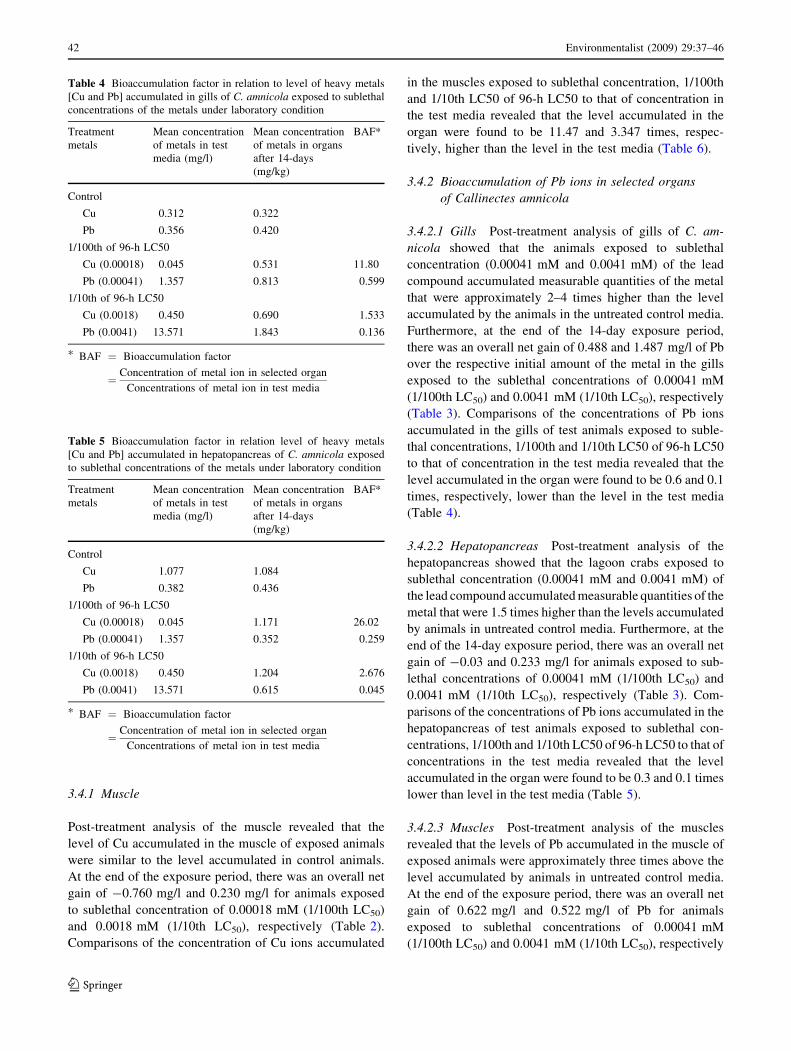
(Table 4).
3.4 Hepatopancreas
Post-treatment analysis of the hepatopancreas revealed that
the level of Cu accumulated in the hepatopancreas of
exposed animals were similar to the level accumulated in
control animals. At the end of the exposure period, there
was an overall net gain of 0.094 and 0.127 mg/l for animals
exposed to sublethal concentration of 0.00018 mM
(1/100th LC50) and 0.0018 mM (1/10th LC50), respectively
(Table 2). Comparisons of the concentration of Cu ions
accumulated in the hepatopancreas exposed to sublethal
concentrations, 1/100th and 1/10th LC50 of 96-h LC50 to
that of concentration in the test media revealed that the
level accumulated in the organ were found to be 26.02 and
2.676 times, respectively, higher than the level in the test
media (Table 5).
Table 2 Accumulation of
heavy metal [Cu] ion by
C. amnicola exposed to
sublethal concentrations
of the metal under laboratory
conditions
* Overall net
gain = Concentration in animal
after 14 days - Concentration
in organ at zero daya 1/100th 96-h LC50 = Values
of the metals ion in the test
compoundsb 1/10th 96-h LC50 = Values
of the metals ions in the
compounds
Treatments (mM) Mean concentration of copper in selected organs
(mg/l wet weight basis)
Overall net gain*
0 day 14 days
Gills
Untreated control 0.312 0.322
0.00018a [1/100th LC50] 0.312 0.531 0.219
0.0018b [1/10th LC50] 0.312 0.690 0.378
Hepatopancrease
Untreated control 1.077 1.084
0.00018a [1/100th LC50] 1.077 1.171 0.094
0.0018b [1/10th LC50] 1.077 1.204 0.127
Muscle
Untreated control 1.276 1.297
0.00018a [1/100th LC50] 1.276 0.516 –0.760
0.0018b [1/10th LC50] 1.276 1.506 0.230
Table 3 Accumulation of
heavy metal [Pb] ion by C.amnicola exposed to sublethal
concentrations of the metal
under laboratory conditions
* Overall net
gain = Concentration in animal
after 14 days - Concentration
in organ at zero daya 1/100th 96-h LC50 = Values
of the metals ion in the test
compoundsb 1/10th 96-h LC50 = Values
of the metals ions in the
compounds
Treatments (mMl) Mean concentration of lead in different organs
(mg/l wet weight basis)
Overall net gain*
0 day 14 days
Gills
Untreated control 0.356 0.420
0.00041a [1/100th LC50] 0.356 0.813 0.457
0.0041b [1/10th LC50] 0.356 1.843 1.487
Hepatopancrease
Untreated control 0.382 0.436
0.00041a [1/100th LC50] 0.382 0.352 –0.03
0.0041b [1/10th LC50] 0.382 0.615 0.233
Muscle
Untreated control 0.327 0.363
0.00041a [1/100th LC50] 0.327 0.949 0.622
0.0041b [1/10th LC50] 0.327 0.849 0.522
Environmentalist (2009) 29:37–46 41
123
3.4.1 Muscle
Post-treatment analysis of the muscle revealed that the
level of Cu accumulated in the muscle of exposed animals
were similar to the level accumulated in control animals.
At the end of the exposure period, there was an overall net
gain of -0.760 mg/l and 0.230 mg/l for animals exposed
to sublethal concentration of 0.00018 mM (1/100th LC50)
and 0.0018 mM (1/10th LC50), respectively (Table 2).
Comparisons of the concentration of Cu ions accumulated
in the muscles exposed to sublethal concentration, 1/100th
and 1/10th LC50 of 96-h LC50 to that of concentration in
the test media revealed that the level accumulated in the
organ were found to be 11.47 and 3.347 times, respec-
tively, higher than the level in the test media (Table 6).
3.4.2 Bioaccumulation of Pb ions in selected organs
of Callinectes amnicola
3.4.2.1 Gills Post-treatment analysis of gills of C. am-
nicola showed that the animals exposed to sublethal
concentration (0.00041 mM and 0.0041 mM) of the lead
compound accumulated measurable quantities of the metal
that were approximately 2–4 times higher than the level
accumulated by the animals in the untreated control media.
Furthermore, at the end of the 14-day exposure period,
there was an overall net gain of 0.488 and 1.487 mg/l of Pb
over the respective initial amount of the metal in the gills
exposed to the sublethal concentrations of 0.00041 mM
(1/100th LC50) and 0.0041 mM (1/10th LC50), respectively
(Table 3). Comparisons of the concentrations of Pb ions
accumulated in the gills of test animals exposed to suble-
thal concentrations, 1/100th and 1/10th LC50 of 96-h LC50
to that of concentration in the test media revealed that the
level accumulated in the organ were found to be 0.6 and 0.1
times, respectively, lower than the level in the test media
(Table 4).
3.4.2.2 Hepatopancreas Post-treatment analysis of the
hepatopancreas showed that the lagoon crabs exposed to
sublethal concentration (0.00041 mM and 0.0041 mM) of
the lead compound accumulated measurable quantities of the
metal that were 1.5 times higher than the levels accumulated
by animals in untreated control media. Furthermore, at the
end of the 14-day exposure period, there was an overall net
gain of -0.03 and 0.233 mg/l for animals exposed to sub-
lethal concentrations of 0.00041 mM (1/100th LC50) and
0.0041 mM (1/10th LC50), respectively (Table 3). Com-
parisons of the concentrations of Pb ions accumulated in the
hepatopancreas of test animals exposed to sublethal con-
centrations, 1/100th and 1/10th LC50 of 96-h LC50 to that of
concentrations in the test media revealed that the level
accumulated in the organ were found to be 0.3 and 0.1 times
lower than level in the test media (Table 5).
3.4.2.3 Muscles Post-treatment analysis of the muscles
revealed that the levels of Pb accumulated in the muscle of
exposed animals were approximately three times above the
level accumulated by animals in untreated control media.
At the end of the exposure period, there was an overall net
gain of 0.622 mg/l and 0.522 mg/l of Pb for animals
exposed to sublethal concentrations of 0.00041 mM
(1/100th LC50) and 0.0041 mM (1/10th LC50), respectively
Table 4 Bioaccumulation factor in relation to level of heavy metals
[Cu and Pb] accumulated in gills of C. amnicola exposed to sublethal
concentrations of the metals under laboratory condition
Treatment
metals
Mean concentration
of metals in test
media (mg/l)
Mean concentration
of metals in organs
after 14-days
(mg/kg)
BAF*
Control
Cu 0.312 0.322
Pb 0.356 0.420
1/100th of 96-h LC50
Cu (0.00018) 0.045 0.531 11.80
Pb (0.00041) 1.357 0.813 0.599
1/10th of 96-h LC50
Cu (0.0018) 0.450 0.690 1.533
Pb (0.0041) 13.571 1.843 0.136
* BAF ¼ Bioaccumulation factor
¼ Concentration of metal ion in selected organ
Concentrations of metal ion in test media
Table 5 Bioaccumulation factor in relation level of heavy metals
[Cu and Pb] accumulated in hepatopancreas of C. amnicola exposed
to sublethal concentrations of the metals under laboratory condition
Treatment
metals
Mean concentration
of metals in test
media (mg/l)
Mean concentration
of metals in organs
after 14-days
(mg/kg)
BAF*
Control
Cu 1.077 1.084
Pb 0.382 0.436
1/100th of 96-h LC50
Cu (0.00018) 0.045 1.171 26.02
Pb (0.00041) 1.357 0.352 0.259
1/10th of 96-h LC50
Cu (0.0018) 0.450 1.204 2.676
Pb (0.0041) 13.571 0.615 0.045
* BAF ¼ Bioaccumulation factor
¼ Concentration of metal ion in selected organ
Concentrations of metal ion in test media
42 Environmentalist (2009) 29:37–46
123

(Table 3). Comparisons of the concentrations of Pb ions
accumulated in the muscles of test animals exposed to
sublethal concentrations, 1/100th and 1/10th LC50 of 96-h
LC50 to that of concentrations in the test media revealed
that the levels accumulated in the organ were found to be
0.70 and 0.06 times, respectively, lower than the level in
the test media (Table 6).
3.4.3 Hispathological studies
The results of the histopathological effects of Cu and Pb on
the gills and hepatopancreas are shown in Figs. 1 and 2.
The results show that there were various degrees of
histological alterations observed in the organs examined
(gills—Fig. 1a, b and Hepatopancreas—Fig. 2a, b). Expo-
sure of lagoon crab to sublethal concentrations of Pb in
particular was found to result in the disruption of the gill
filaments (Fig. 1b) accompanied by precipitation of Pb
granules (Fig. 1b). Additionally, sections of crab’s hepa-
topancreas exposed to Pb compounds also revealed a
general degeneration of glandular cells with multifocal
areas of calcification in the hepatopancreas.
3.4.4 Weight changes experiment
Results of the weight changes in C. amnicola exposed to
sublethal concentrations of Cu salt showed that the test
animals lost about 12.5–19.05% of its weight over the
14 days period of observation. On the otherhand lagoon
crabs exposed to sublethal concentrations of Pb lost about
10.5–12.9% of the body weight over the period of observa-
tion. Animals in control media however gained about 10% of
the body weight during the period of observation (Fig. 3).
4 Discussion
In this study, the differential toxicity of two metallic salts;
CuSO4 and Pb(NO2)3 against C. amnicola was demon-
strated. From the established toxicity result, CuSO4 (96-h
LC50 value of 0.018 mM) was found to be two times more
toxic than Pb(NO2)3 (96-h LC50 0.041 mM) when acting
singly against the lagoon crab, C. amnicola. This result
agrees with those of several authors including Otitoloju and
Don-Pedro (2002) who reported the general toxicity rank-
ing order of heavy metals on benthic animals of the Lagos
Table 6 Bioaccumulation factor in relation level of heavy metals [Cu
and Pb] accumulated in muscles of C. amnicola exposed to sublethal
concentrations of the metals under laboratory condition
Treatment metals Mean concentration
of metals in test
media (mg/l)
Mean concentration
of metals in organs
after 14-days (mg/l)
BAF*
Control
Cu 1.276 1.297
Pb 0.327 0.363
1/100th of 96-h LC50
Cu (0.00018) 0.045 0.516 11.47
Pb (0.00041) 1.357 0.949 0.716
1/10th of 96-h LC50
Cu (0.0018) 0.450 1.506 3.347
Pb (0.0041) 13.571 0.849 0.063
* BAF ¼ Bioaccumulation factor
¼ Concentration of metal ion in selected organ
Concentrations of metal ion in test media
Fig. 1 Gills Filament of
Callinectes amnicola. (a)
Normal section of gills for
control animals. (b) Disrupted
gill filaments (black arrows) in
crabs exposed to sublethal doses
of Pb compound accompanied
by precipitation of Pb granules
(white arrow)
Environmentalist (2009) 29:37–46 43
123

lagoon as follows: Hg [ Cd [ Cu [ Zn [ Pb. Clark
(1992), also reported the same order of toxicity with
stickleback. The observation of increased concentration of
these metals in single action laboratory studies has also
been well established by Bryan and Langston 1992, Baron
1995 and Oyewo 1998.
The observed toxicity of different heavy metals can be
attributed to several factors such as metal compound tested,
solubility of salts, predominant ions in test solution, and
physico-chemical characteristics of the test solution and the
mechanism of action of the different metals. Other factors
which may affect metal’s toxicity or susceptibility of test
animals include the formation of complexes with protein,
e.g. metallothionein complexes formation of encapsulated
metal granules, metabolism and excretability. The low
toxicity nature of Pb compound may be because of the free
inorganic ion Pb2+ which is usually dominant in Pb is not
lipid soluble, hence transfer across membranes may be
inhibited (Otitoloju 2002). This study also revealed that Cu
is more toxic than Pb to aquatic macro-invertebrates.
Hence, their introduction into the aquatic environment via
industrial effluent must be minimized through effective
control/management strategies and where possible Pb
should be used in place of copper.
The level of the metals accumulated in the organs of the
test animal was found to be higher in animals exposed to
1/10th LC50 than in those exposed to 1/100th LC50. The
concentrations in exposed animals were also generally
higher than the concentrations detected in control animals.
This observation therefore implies that the concentration of
metals accumulated was directly related to the concentra-
tion of the metal present in their environment. This result
of metal uptake is in agreement with studies of Chukwu
(1991) and Oyewo (1998) who demonstrated that the
concentration of metals in exposed animals such as Pal-
aemonetes africanus, Tympanotonous fuscatus, Tilapia
guieenensis and Clibaranus africanus increased with time
of exposure and test medium concentration.
On the basis of the concentration of metals accumulated
in the different organs, the order of metal accumulation in
the organs is as follows: gills [ muscle [ hepatopan-
crease. This result is somewhat similar to the result of
Radhakrishnaiah (1988) who reported the order of Cu
accumulation in freshwater fish Labeo rohita (Hamlton) as
gill [ brain [ muscle [ liver. According to Otitoloju and
Don-Pedro (2006), the bioaccumulation of heavy metals in
animal tissues occurs as a result of competing rates of
chemical uptake and excretion. Accumulation of metals
will therefore occur when the rate of uptake is higher than
rate of excretion. Furthermore, the synthesis of low
molecular weight proteins e.g. metallothionein which form
complexes with the metal ion, as well as the formation of
exposed animals have also been reported to be responsible
Fig. 2 Hepatopancreas of
Callinectes amnicola. (a)
Normal section of
hepatopancreas showing distinct
hepatocytes (H) in control
animals. (b) Degenerated
glandular cells with multifocal
areas of calcification (arrows) in
crabs exposed to sublethal doses
of Pb compound
-25
-20
-15
-10
-5
0
5
10
15
Cu 1/100 LC50 Cu 1/10 LC50 Pb 1/100 LC50 Pb 1/10 LC50Control
% W
eigh
t Gai
n
Concentrations
Fig. 3 Weight changes in Callinectes amnicola exposed to sublethal
concentrations of heavy metal (Cu and Pb) compounds over a 14-day
observation period
44 Environmentalist (2009) 29:37–46
123

for metal accumulation in some aquatic mollusc (Langston
and Zhou 1987; Clark 1992).
The detection of higher concentration of Cu in the gills
could be as a result of Cu being a component of haemocyanin
which is a respiratory pigment with its site of action at the
gills. Also the fact that the gills are in direct contact with the
surrounding water may account for the higher level of copper
accumulation in the organ. The detection of lower concen-
tration of Cu and Pb ions in the hepatopancrease of the
Lagoon crab exposed to sublethal concentrations indicates
that this organ may be the primary organ for metal detoxi-
fication and excretion.
Generally, a higher concentration of Pb was accumulated
in the different organs of the crab than Cu. In fact the con-
centration of Cu accumulated in the organs were found to
fluctuate significantly over the observation period. Therefore
suggestive that the test animal may have capacity to
metabolize and excrete its body burden of Cu over time. The
ability of aquatic invertebrates such as Tympanotonus fusc-
atus to excrete rapidly its body burden of Cu has also been
reported by Otitoloju and Don-Pedro (2002). With regards to
lead however, the concentration of lead accumulated
increased steadily with time of exposure. Therefore indi-
cating that the animal may not have the potential to excrete
the metal ions from its body, but rather have the capacity to
store it up in a harmless state e.g. by storing the metal within
membrane in the cytoplasm or secretion of low molecular
weight proteins which binds the metals. The particular
mechanism(s) by which T. fuscatus accumulate Pb in its
body tissues should therefore merit further investigation.
This study also showed that the exposure of C. amnicola
to sublethal concentration of Cu and Pb caused a marked
decrease in weight of the exposed animals compared to
control organisms. The weight loss was also found to be
more pronounced in crabs exposed to Cu than Pb exposed
ones. The loss of weight in the exposed animals may be
attributable to the observed reduction in locomotory and
feeding activities in the exposed animals.
In conclusion, the range of biological responses
observed in this study such as the ability of lagoon crab
to accumulate heavy metals to levels that are several
folds higher than the environment, the toxicopathic tissue
alterations from the histopathological studies and the
remarkable weight loss in exposed crabs represent impor-
tant biological endpoints of contaminant exposure which
reinforces the inclusion of biological responses in moni-
toring anthropogenic contamination of aquatic ecosystem.
The combination of these biological endpoints with phys-
ical/chemical parameters will provide a clear picture of the
total environmental quality and provide environmental
managers with the necessary tools to make important
decisions that will prevent such cases of metal pollution
resulting in human death.
Acknowledgement The authors are grateful to Dr. J.K. Saliu for his
useful suggestions in the preparation of the manuscript.
References
Bamber SD, Depledge MH (1997) Responses of shore crabs to
physiological changes following exposure to selected environ-
mental contaminants. Aquat Toxicol 40:79–92. doi:10.1016/
S0166-445X(97)00040-4
Baron MG (1995) Bioaccumulation and bioconcentration in aquatic
organisms. In: Hoffman GA, Rattner BA Jr, Ciaras GA Jr (eds)
Handbook of ecotoxicology. CRC press Inc. Lewis Publishers,
London, pp 652–662
Brown RJ, Galloway TS, Lowe D, Browne MA, Dissanayake A,
Jones MB et al (2004) Differential sensitivity of three marine
invertebrates to copper assessed using multiple biomarkers.
Aquat Toxicol 66:267–278. doi:10.1016/j.aquatox.2003.10.001
Bryan GW, Langston WJ (1992) Bioavailability, accumulation and
effects of heavy metals in sediments with special reference to
United Kingdom estuaries: a review. Environ Pollut 76(2):89–
131. doi:10.1016/0269-7491(92)90099-V
Chukwu LO (1991) Studies on heavy metal contamination of water
sediments and decapod crustaceans from River sasa. PhD Thesis,
University of Lagos, 164 pp
Clark RB (1992) Marine pollution, 3rd edn. Oxford University Press,
Oxford, p 169
Curtis TM, Williamson R, Depledge MH (2000) Simultaneous
monitoring of valve and cardiac activity in the blue mussel
Mytilus edulis exposed to copper. Mar Biol (Berl) 136:837–846.
doi:10.1007/s002270000297
Don-Pedro KN (1989) Mode of action fixed oils against eggs of
Callosobrochus maculatus (f). Pestic Sci 26:107–115. doi:10.1002/
ps.2780260202
FAO/SIDA (1986) Manual of methods in aquatic environmental
research, Part 9. Analyses of metal and organochlorines in fish.
FAO Fish Tech Pap 212:21–33
Finney DJ (1971) Probit analysis, 3rd edn. Cambridge University
Press, London
Harada M, Smith AM (1975) Minamata disease: a medical report. In:
Smith WE, Smith AM (eds) Minamata: a warning to the world.
Chatto and Windus, London, pp 180–192
Kakkar P, Jaffery F (2005) Biological markers for metal toxicity.
Environ Toxicol Pharmarcol 19:335–349
Kurdland L (1960) Minamata disease. World Neurol 1:370–385
Langston WJ, Zhou M (1987) Cadmium accumulation, distribution
and metabolism and in gastropod Littorina littorea: the role of
metal-binding protein. J Mar Biol Assoc UK 67:585–601
Lin CH, Chen JC (2001) Haemolymph oxyhaemocyanin and protein
levels and acid-base balance in the tiger shrimps Penaeusmonodon exposed to copper sulfate. J World Aquacult Soc
32:335–341. doi:10.1111/j.1749-7345.2001.tb00457.x
Matthiessen P, Thain JE, Law RJ, Fileman TW (1993) Attempts to
assess the environmental hazard posed by complex mixtures of
organic chemicals in UK estuaries. Mar Pollut Bull 26:90–95.
doi:10.1016/0025-326X(93)90097-4
Otitoloju AA (2002) Evaluation of the joint action toxicity of binary
mixtures of heavy metals against the mangrove periwinkle of
Tympanotonous fuscatus var radula (L). Ecotoxicol Environ Saf
53:404–415. doi:10.1016/S0147-6513(02)00032-5
Otitoloju AA, Don-Pedro KN (2002) Bioaccumulation of heavy
metals (Zn, Pb, Cu and Cd) by Tympanotonous fuscatus var
radula (L) exposed to sublethal concentrations in laboratory
bioassays. W Afr J Appl Ecol 3:17–29
Environmentalist (2009) 29:37–46 45
123

Otitoloju AA, Don-Pedro KN (2004) Integrated laboratory and field
assessments of heavy metals accumulation in edible periwinkle,
Tympanotonus fuscatus var radula (L.). Ecotoxicol Environ Saf
57:354–362. doi:10.1016/j.ecoenv.2003.09.002
Otitoloju AA, Don-Pedro KN (2006) Influence of joint application of
heavy metals on level of each metal accumulated in the
periwinkle Tympanotonus fuscatus Gastropoda: Potamididae.
Int J Trop Biol 54(3):803–814
Otitoloju AA, Don-Pedro KN, Oyewo EO (2007) Assessment of
potential ecological disruption based on heavy metal toxicity,
accumulation and distribution in media of the Lagos lagoon. Afr
J Ecol 45:454–463. doi:10.1111/j.1365-2028.2007.00754.x
Oyewo EO (1998) Industrial sources and distribution of heavy metals
in Lagos lagoon and their biological effects on estuarine animals,
PhD Thesis, University of Lagos, 274 pp
Radhakrishnaiah K (1988) Accumulation of copper in the organs of
freshwater fish, Labeo rohita (Hamilton), on esposure to lethal
and sublethal concentration of copper. J Environ Biol 9(3)(Sup-
pl):319–326
Stentiford GD, Feist SW (2005) A histopathological survey of shore
crab (Carcinus maenas) and brown shrimp (Crangon crangon)
from six estuaries in the United Kingdom. J Invertebr Pathol
88:136–146. doi:10.1016/j.jip. 2005.01.006
Stentiford GD, Longshaw M, Lyons BP, Jones G, Green M, Feist SW
(2003) Histopathological biomarkers in estuarine fish species for
the assessment of biological effects of contaminants. Mar
Environ Res 55:137–159. doi:10.1016/S0141-1136(02)00212-X
Svendsen C, Week JM (1997) Relevance and applicability of a simple
earthworm.biomarker of copper exposure. 1. Links of ecological
effects in a laboratory study with Eisenia andrei. Ecotoxicol
Environ Saf 36:72–79. doi:10.1006/eesa.1996.1491
Varanasi U, Stein JE, Nishimoto M, Reichert WL, Collier TK (1987)
Chemical carcinogenesis in feral fish: uptake, activation and
detoxication of organic xenobiotics. Environ Health Perspect
71:155–170. doi:10.2307/3430423
Vethaak AD, Jol JG (1996) Diseases of flounder Platichthys flesus in
Dutch coastal and estuarine waters, with particular reference to
environmental stress factors II Epizootiology of gross lesions.
Dis Aquat Org 26:81–97. doi:10.3354/dao026081
Viarengo A, Burlando B, Giordana A, Bolobnesi C, Gabroelides GP
(2000) Networking and expert system analysis: next frontier in
biomonitoring. Mar Environ Res 49:483–486. doi:10.1016/S0141-
1136(00)00027-1
Weeks JM, Jensen FB, Depledge MH (1993) Acid-base status,
haemolymph composition and tissue copper accumulation in the
shore crab Cacinus maenas exposed to copper and salinity stress.
Mar Ecol Prog Ser 97:91–98. doi:10.3354/meps097091
World Health Organisation (1995) Human exposure to lead, World
Health Organization Geneva
46 Environmentalist (2009) 29:37–46
123