BIOLOGICAL FUNDAMENTALS OF RESISTANCE
306
A.E. Gabidova, V.A. Galynkin BIOLOGICAL FUNDAMENTALS OF RESISTANCE Monograph Moscow 2020 Recommended by Academic Methodological Association According to classical University and technical education as a textbook for researchers, engineers, technologists working in the pharmaceutical and biotechnological fields, as well as students of higher educational institutions engaged in the field of medicinal treatment, studying in the areas of training: 230201 – Information system and technology; 190000 – Industrial ecology and biotechnology; 060108 – Pharmacy; 240901 – Biotechnology; 240902 – Food biotechnology; 020209 – Microbiology
-
Upload
khangminh22 -
Category
Documents
-
view
2 -
download
0
Transcript of BIOLOGICAL FUNDAMENTALS OF RESISTANCE

A.E. Gabidova, V.A. Galynkin
BIOLOGICAL FUNDAMENTALS OF RESISTANCE
Monograph
Moscow2020
Recommended by Academic Methodological Association According to classical University and technical education as a textbook for researchers, engineers, technologists working in the pharmaceutical and biotechnological fi elds, as well as students of higher educational institutions engaged in the fi eld of medicinal treatment, studying in the areas of training:230201 – Information system and technology; 190000 – Industrial ecology and biotechnology; 060108 – Pharmacy; 240901 – Biotechnology;240902 – Food biotechnology; 020209 – Microbiology

Г12
УДК 615.076ББК 35.66
Г12
Reviewers:Garabadju Aleksander Vasilievich – doctor chemicheskich science,chief
sPB Technicheskie Universitet;Meledinae Tatiana Victorovna – doctor technicheskich science, chief kafed-
rae biotechnologii Federation Universitet ITMO.
Gabidova A.E., Galynkin V.A.Biological Fundamentals of Resistance: monograph / A.E. Gabidova,
V.A. Galynkin. – М.: Publishing House of the “Academy Natural History”, 2020. – 306 р.
ISBN 978-5-91327-619-3DOI 10.17513/np.396
“We are entering a post-antibiotic era”, the head of WHO stressed, “and every antibiotic de-veloped at any time can become useless”. Cascade model of resistance occurrence includes at the fi rst stage, there is a circulation of plasmids of soil fungi, actinomyces and bacteria to the plants and invertebrate animals in biofi lms and symbioses, which represent complex cascade systems; at the next stage, there is circulation of plasmids from invertebrate animals to higher animals, from animals to humans and from humans to animals – and this contributes to fast spreading of drug resistance all over the world. The key role belongs to the genes which are contained in R-plas-mids. Anthropogenic stress infl uences counteract the most important function of the biosphere – the regular recreation of living matter and energy accumulated in it. During the formation of the biosphere, a cascade of population structures was formed in the form of biological fi lms and symbioses of plants, animals and microorganisms. These structures are united by a single genetic system, which because of the circulation of plasmids regulates the expression of genes under stress and leads to the emergence of resistance.
© Gabidova A.E., Galynkin V.A., 2020© PH “Academy Natural History”© АNО “Academy Natural History”
ISBN 978-5-91327-619-3
3
CONTENTAbstract ........ ........................................................................................................................ 5Introduction. Global issues of the world civilization ........................................................... 7Chapter I. “Combating antimicrobial resistance: time for action” ................................. 10
1.1. Global strategy for combating antimicrobial resistance ................................ 101.2. Strategy for the prevention of the spread of antimicrobial resistance in the
Russian Federation for the period until 2030 ................................................ 16Chapter II. State and development of pharmacy in the 21st century .............................. 23
2.1. Concept of sustainable development ............................................................. 232.1.1. Qualitative global model – the transition to sustainable
development ................................................................................ 252.2. Routes of establishment of a global world .................................................... 29
2.2.1. The main factors of environmental degradation at the global level: ...... 302.2.2. The main factors of theenvironmental degradation
in the Russian Federation: ........................................................... 302.2.3. Environmental Doctrine of the Russian Federation.................... 31
2.3. The historical sequence of civilization development .................................... 342.3.1. Environmental safety .................................................................. 36
Chapter III. Microbiological risk problems in medicine and pharmacy ........................... 393.1. Microorganisms – the primary and main cause of biological risks .............. 393.2. Epidemics of «drug infection» ...................................................................... 39
3.2.1. The emergence of “drug infection” in the EU and the USA ...... 393.2.2. The emergence of “drug infection” in the Russian Federation .. 40
Chapter IV. Soil ................................................................................................................. 444.1. History of the soil′s origin ............................................................................. 444.2. Soil architectonics .......................................................................................... 464.3. Soil microorganisms ...................................................................................... 484.4. Interactions, existing between microorganisms and plants .......................... 554.5. Epigenous microorganisms ............................................................................ 584.6. Rhizosphere microorganisms......................................................................... 63
4.6.1. Microbial vegetative interactions in rhizosphere and rhizoplane by seed germination ........................................... 69
4.7. Ecological relationships in microbiocenoses ................................................ 73Chapter V. Population forms of microorganisms in the environment ............................. 77
5.1. Planktonic form of microorganisms .............................................................. 775.2. Colonial organization of microorganisms ..................................................... 815.3. Biofi lms.......................................................................................................... 82
Chapter VI. Signal information in populations of microorganisms .................................. 866.1. Quorum systems of microorganisms ............................................................. 866.2. Quorum-sensing reaction of Gram-negative microorganisms ..................... 906.3. Quorum-sensing reactions of Gram-positive microorganisms ..................... 926.4. Quorum-sensing in multicellular formations ............................................... 946.5. Interspecies interactions of microorganisms ................................................ 95
Chapter VII. Symbioses ................................................................................................... 977.1. Classifi cation and specifi cations of symbioses. ............................................ 97
7.1.1. Symbiology ............................................................................... 103

4
7.2. Rhizobacteria. Symbiosis of plants and rhizospheric bacteria ................... 1087.3. Mycorrhiza ....................................................................................................110
7.3.1. Fungi symbionts ........................................................................ 1107.3.2. Other forms of mutually benefi cial microbial vegetative
interactions ................................................................................ 1177.4. Phytopathogenic plant protective system ....................................................1177.5. Genetic basis for microbial vegetative symbiosis ...................................... 1227.6. Soil actinomyces ......................................................................................... 1267.7. Plant- actinomyces symbiosis (actinorhiza) ................................................ 135
Chapter VIII. Historical aspects of resistance occurrence ............................................. 1398.1. Protection system of pathogenic microorganisms ....................................... 1418.2. Immune system of bacteria: CRISPR. Bacterial immunity ........................ 1478.3. Global danger – antibacterial resistance ...................................................... 151
8.3.1. Plasmid or transposon option of resistance transfer (quick type) ........ 1548.3.2. Chromosomal type of resistance transfer (slow type) .............. 1568.3.3. Hsd plasmids and their replicons: mechanisms
of distribution of plasmids between bacteria ............................ 1608.4. Basic mechanisms of AB-resistance ............................................................ 170
8.4.1. Origin of AB-resistance ............................................................ 1748.4.2. Conjugative plasmids and supergene ........................................ 1758.4.3. Superbacteria and superresistance ............................................ 1808.4.4. Strategy of counteraction and treatment ................................... 181
8.5. Interaction of actinomyces and a human ..................................................... 1858.6. Enzymatic AG inactivation in actinomyces ................................................ 1888.7. AG resistance of actinomyces to, determined by the modifi cation of 16S rRNA ......... 1928.8. Theoretical basis of the emergence of β-lactams resistance ....................... 196
Chapter IX. Mechanisms of resistance of microorganisms ............................................. 2019.1. Mechanisms of antibacterial antibiotic resistance ...................................... 2019.2. Mechanisms of resistance to broad-spectrum antibiotics ............................ 2039.3. Mechanisms of resistance to quinolone group of drugs .............................. 2049.4. Mechanisms of resistance to the tetracycline group of antibiotics ............. 2069.5. Multiple resistance of microorganisms ....................................................... 2099.6. Gene transfer between gram-positive and gram-negative bacteria ............. 209
Chapter X. Preservation of resistance at the animal world stage ...................................21110.1. The role of the parasitical symbiosis ............................................................21110.2. Soil animal world ........................................................................................ 21410.3. Digestive processes in fauna ....................................................................... 234
10.3.1. Microfl ora of gastro-intestinal tract as an example of symbiosis of animals and microorganisms ........................... 241
10.3.2. Mode of microorganism existence in intestinal biofi lm ........... 24610.3.3. Modern ideas of the structure of intestinal microbiota
according to data of molecular studies ..................................... 251Chapter XI. Cascade model of resistance occurrence ..................................................... 274References.... ..................................................................................................................... 285List of scientifi c names ..................................................................................................... 298Subject Index .................................................................................................................... 301

5
ABSTRACTThe book outlines the existing materials and authoring related to the emer-
gence of resistance of pathogenic microorganisms in ecology, nature and phar-macy. When analyzing the development of civilization, it was shown that a rapid destruction of the biosphere and its basis, bacterial-mushroom-plant symbiosis, is occurring. At the end of the 20th century humankind realized that the biosphere and its constituent parts have limits of self-regulation and self-restoration, fol-lowed by irreversible degradation.
The current state of the environment is characterized as an environmen-tal crisis, the distinguishing features of which are chemical pollution of the biosphere and the critical state of natural resources. In accordance with the concept of sustainable development, humanity should not only strive to reduce the anthropogenic load on ecosystems, but also to take over the functions of restoring natural balance.
In a number of problems facing modern society, the state of the habitat oc-cupies one of the fi rst places. A huge number of alien living organisms and syn-thetic chemicals circulate in the biosphere. The rapid spread of various resistance mechanisms poses serious questions for clinical medicine and fundamental biolo-gy. Currently for the optimization of antibiotic therapy it is completely insuffi cient to assess the level of antibiotic resistance of the microorganism (that is the causa-tive agent of the infection) using phenotypic methods. With similar phenotypes, but different mechanisms of resistance, the clinical effi cacy of antibacterial drugs can vary signifi cantly. For the formation of an antibacterial therapy strategy at the national and regional levels, it is also needed to have information on the dynamics of the spread of individual resistance mechanisms and knowledge of the molecular mechanisms of resistance that is necessary for the development of new antibacte-rial drugs and tools for diagnosing resistance.
Ms. Chan said that humanity was dealing with such a level of antibiotic resist-ance that this situation can mean “the end of medicine as we know it”.
“We are entering a post-antibiotic era”, the head of WHO stressed, “and every antibiotic developed at any time can become useless”.
First of all, doctors are deprived of the so-called “fi rst-line antibiotics”, as a result of which manipulations, formerly routine ones, will simply become im-possible – be it the treatment of such infections as tuberculosis, malaria or conven-tional surgical treatment of cuts. “Things as common as strep throat or a child′s
6
A.E. Gabidova, V.A. Galynkin
scratched knee could once again kill” the head of WHO said. “Therefore, the lack of funds in the arsenal of doctors requires innovation”.
The most common in the soil are representatives of almost all species of actinomyces. Usually, a quarter of bacteria that grow is traditional environment consist of actinomyces. Their ecological role is most often in the decomposition of complex stable substrates; presumably they are involved in the synthesis and decomposition of humic substances. They can act as nitrogen-fi xing symbiotes of invertebrates and higher plants. Different species of actinomyces gradually partici-pate in the process of decomposition of organic substances in the soil as part of an actinomyces complex. Under certain conditions (soil type, succession stage), species of actinomyces, traditionally considered as rare, may have an equal with streptomycetes share in the actinomyces complex, and sometimes dominate in it. The book shows that actinomyces form synergistic associations with plants, with invertebrates′ protozoa, with all kinds of animal organisms and with humans at all stages. This allows antibiotic therapy to be transmitted from soil to the person. This lays the foundation for the developed concept of a multi-level cascade of the emergence of resistance of microorganisms to the antibiotic therapy.
The book can be used by researchers in the fi eld of ecology and medicine, engineers and technologists working in the pharmaceutical and biotechnological fi eld, as well as students of higher educational institutions engaged in the fi eld of medicinal circulation.

7
Introduction.GLOBAL ISSUES OF THE WORLD CIVILIZATION
According to the World Health Organization (WHO), at the beginning of the XXI century about 500 thousand chemical compounds and substances were used in industry and agriculture, more than 40 thousand of which are harmful to human health and about 12 thousand are toxic. A signifi cant part of these substances gets into the air, soil, surface or ground water. With inhaled air and drinking water, pollutants enter the human body.
Pollutants of the atmosphere, hydrosphere and soil lead to the ingress of harmful substances into food chains, including those in which human is the final consumer. Under the anthropogenic impact, there is a change in air and soil microbial flora, which is accompanied by a change in the florula of the plant during its development.
WHO experts believe that the relative contribution of factors, caused by the state of the environment and affecting human health, is at least 25 %.
In addition, at the beginning of the twenty-fi rst century, infectious diseases continue to cause signifi cant damage to humanity. The biological threat associated with infectious diseases and their pathogens hangs over the entire planet. In partic-ular, among 51 million people who die every year in the world, almost 17 million die from infections, while 9,7 million people die from cardiovascular diseases.
At the end of the last century, a return to natural resources was again ob-served, both in the form of ideas for the pharmaceutical industry (the synthesis of derivatives of natural molecules) and the use of new active ingredients in modern medicine, and in the form of traditional medicine, which is becom-ing more common. However, according to the WHO, about half of the world population uses traditional medicine. The global market of herbal medicines is estimated at more than $ 60 billion.
Phytotherapy deals with herbal medicines that are made exclusively from natural sources.
The effectiveness of medicine made of plant materials is often uncritically overestimated and, conversely, there is a view about the total harmfulness of all chemicals. In addition, herbal remedies are perceived as completely safe and harmless drugs (if considered as drugs at all). There is a lack of awareness of the fact that they contain a number of active substances, which can them-selves cause complications (at higher doses or long-term use) or in combina-tion with other drugs.

8
A.E. Gabidova, V.A. Galynkin
Recently the use of phytopreparations and phytomaterials in pharmacy has signifi cantly expanded. Medicinal plant raw materials which form the basis of herbal remedies are in their origin most contaminated by microfl ora and are the most likely transmitter of spores of microorganisms. Microbial contamination of plants depends on the environment (soil, air, water). A wide variety of bacterial microorganisms, fungi and viruses, as well as traces of rodents and insects, has been found on plants and inside them. Preliminary studies have shown that among these types of microfl ora can be found pathogenic and conditionally pathogenic microorganisms, as well as pathogenic compounds. Consequently, phytomateri-als are a source of microorganisms, the content of which depends on external and internal factors. In connection with the thoughtlessness of the consequences of the intensive technogenic development of civilization, which leads to a change in the natural conditions in the biosphere, global problems of the present arise.
The global problems of the present are a set of problems of the biosphere, on the solution of which social progress and the preservation of civilization depend.
Global issues include:● environmental problems;● global health threats;● biodiversity loss;● demographic problem;● food problem;● energy and raw materials problem;● drinking water problem;● reduction of forest area. Associated with deforestation and pollution.Ecological problems have arisen with the beginning of industrial human activity
and became especially aggravated in the second half of the 20th century and are asso-ciated with pollution of the environment. Environmental problems can be divided into local, regional and global. The environmental problems of the global and regional lev-els include: pollution of the World Ocean, destruction of the ozone layer, air pollution, desertifi cation of territories, acid precipitation, etc. In the course of its existence, the human population, striving to meet their physical needs and developing the economy, simultaneously improved the social organization of society, creating a socio-economic security system. Consequently, despite the increase in the number of harmful effects on the environment, the level of human safety has increased. V.I. Vernadsky is one of the fi rst scientists who formulated “that danger threatens humanity” [1].
Vernadsky V.I. (1925) fi rst emphasized: “Man destroyed the virgin nature. He introduced into it a mass of previously unknown chemical compounds and forms of life – cultural breeds of animals and plants. He changed the course of all geochemical reactions. The face of the planet has become new and has come to a state of incessant shocks”.

9
The destructive human activity has evoked a confl ict between society and na-ture; it has created risks that are called environmental. The most important func-tion of any biocoenosis, biogeocoenosis and biosphere is the regular recreation of living matter and the energy accumulated in it. It is shown that the appearance of emergent and uncultivated microorganisms is accompanied by the need to pay more attention to the analysis of microbiological risk [2].
Margaret Chan at the conference in the report “Combating antimicrobial resistance– time for action” noted that EU Member States can do to solve a problem that is, as you correctly recognize, serious,growing and global threat to health. Drug-resistant pathogens are notorious globetrotters. According to World Health Organization Director-General Margaret Chan, the world is on the verge of a crisis caused by microbial resistance to antibiotics. The most common resistant bacteria Escherichia coli (E. coli), Klebsiella pneumoniae (causative agent of pneumonia), Staphylococcus aureus (S. aureus),
Streptococcus pneumoniae (pneumococcus) and Salmonella spp (Sal-monella) are no longer afraid of antibiotics. Meanwhile, they are the cause of meningitis, staphylococcus, pneumonia, salmonellosis, bowel problems, bloodstream infections and other diseases [3].
“We are entering a post-antibiotic era”, the head of WHO stressed, “and every antibiotic developed at any time can become useless”. First of all, doc-tors are deprived of the so-called “fi rst-line antibiotics”, as a result of which manipulations, formerly routine ones, will simply become impossible – be it the treatment of such infections as tuberculosis, malaria or conventional surgical treatment of cuts. “Things as common as strep throat or a child′s scratched knee could once again kill” the head of WHO said. “Therefore, the lack of funds in the arsenal of doctors requires innovation”.
Margaret Chan explained that at present, drugs destined to replace antibiotics that have lost their activity are becoming more expensive, and to achieve the same effect, more and more prolonged courses of treatment are needed. In this case, the use of rare antibiotics often requires hospitalization and is associated with toxic effects on the patient′s organism. As a result, in some cases, the mortality rate of patients infected with antibiotic-resistant strains of microorganisms increases by 50 percent. “The conditions for this crisis have been forming for decades” and “the main reasons for it are the incorrect use of antibacterial drugs that are chosen incorrectly, are taken too often or too long. WHO calls on governments around the world to support antibiotic resistance research”.

10
Chapter I . “COMBATING ANTIMICROBIAL RESISTANCE: TIME FOR ACTION”
1.1. Global strategy for combating antimicrobial resistanceMargaret Chan at the conference in the report “Combating antimicrobial re-
sistance – time for action” (Copenhagen, Denmark, March 14, 2012) noted that EU Member States can do to solve a problem that is, as you correctly recognize, se-rious, growing and global threat to health. Drug-resistant pathogens are notorious globe-trotters. They travel well in infected air passengers and through global trade in food. In addition, the growth of medical tourism has accelerated the international spread of hospital-acquired infections that are frequently resistant to multiple drugs.
WHO Director-General made an acknowledgement to the work of the Euro-pean Centre for Disease Prevention and Control, or ECDC, in so quickly conduct-ing risk assessments of the spread of NDM-1-producing bacteria within Europe.
This kind of rapid response to an emerging threat speaks well of the EU′s ca-pacity to protect its citizens. It also demonstrates the EU′s capacity to generate mod-els, useful elsewhere, for combating antimicrobial resistance on multiple fronts.
The EU has recently adopted a range of policies, directives, technical reports, strategies, and regulatory decisions designed to reduce antibiotic consumption among humans and animals. They have achieved remarkable success, as exem-plifi ed by several EU-wide networks for surveillance of both resistance and con-sumption and for susceptibility testing, pointing to a clear need to share experi-ences and harmonize best practices.
In fact greater quantities of antibiotics are used in healthy animals than in unhealthy humans is a cause for great concern. Analysis of the results of using fl uoroquinolones by farm animals may lead to a decrease in sensitivity to fl uoro-quinolones in cultures of Campylobacter sp. and Sallmonella sp. It is recommend-ed to give the drug to animals, basing on the individual principle, where possible; the time of taking the drug should be appropriate and should be consistent with the scientifi c data on how the resistance of bacteria and the optimal use of fl uo-roquinolones, in accordance with their pharmacokinetics and pharmacodynamics, reduces the selection of resistant microbes. A very important point is also the con-stant quality control of the applied fl uoroquinolones (since most of these active substances produced in Asian countries are of poor quality!). Thus, it is necessary to defi ne a global world strategy.
For antimicrobial agents around the world (especially in more developed countries), strict registration rules apply to both: to the issuance of permits for the

11
release of a drug on the market and to the restriction of residual quantities of these active substances in animal food. All veterinary drugs should be safe for animals, general practitioners and pet owners, high-quality and effective and should not cause a danger to the environment.
In particular, Denmark has tackled the problem of antibiotic use in food-pro-ducing animals in a pioneering way. Recognizing the potential for a health crisis, this country progressively ended the administration of antibiotics as growth-pro-moters in the late 1990s, well before the EU-wide ban.
An international review panel, set up by WHO at the request of the Danish government, concluded that the ban reduced human health risks without signifi -cantly harming animal health or farmers′ incomes.
In Denmark, after the ban was introduced, the production of livestock prod-ucts and poultry has increased, and levels of resistance to antibiotics on farms and in meat have declined.
The termination of the use of antibiotics (Danish model) as growth promoters had a voluntary component on the part of industry, strongly motivated by con-sumer concerns. I congratulate industry for its responsible actions.
The importance of consumer groups and civil society in combating antimicro-bial resistance should never be underestimated. They are important movers, initia-tors, and front-line players, especially in this age of social media.
The antimicrobial threat is easy to describe. It has an irrefutable logic.Antimicrobial resistance is on the rise in Europe, and elsewhere in the world.
We are losing our fi rst-line antimicrobials. Replacement treatments are more cost-ly, more toxic, need much longer durations of treatment, and may require treat-ment in intensive care units.
Among patients infected with some drug-resistant pathogens, mortality has been shown to increase by around 50 %. Among the world′s 12 million cases of tuberculosis in 2010, WHO estimates that 650,000 involved multidrug-resistant TB strains. Treatment of MDR-TB is extremely complicated, typically requiring two years of medication with toxic and expensive medicines, some of which are in constant short supply. Even with the best of care, only slightly more than 50 % of these patients will be cured.
Many other pathogens are developing resistance to multiple drugs, some to near-ly all. Hospitals have become hotbeds for highly-resistant pathogens, like MRSA, ESBL, and CPE, increasing the risk that hospitalization kills instead of cures. These are end-of-the-road pathogens that are resistant to last-line antimicrobials.
If current trends continue, the future is easy to predict. Some experts believe that we are moving back to the pre-antibiotic era. No. It will be a post-antibiotic era. In terms of new replacement antibiotics, they are virtually not developed, es-pecially for gram-negative bacteria. Resources are almost exhausted.

12
A.E. Gabidova, V.A. Galynkin
Prospects for changing this situation look dim. The pharmaceutical industry lacks incentives to bring new antimicrobials to market for many reasons, some of which fall on the shoulders of the medical and public health professions. It is our inability to combat the gross misuse of these medicines.
Why should the industry invest signifi cant sums of money in the development of a new antimicrobial drug if its irrational use quickly leads to its ineffi ciency before investing in research and development?
A post-antibiotic era actually means an end to modern medicine as we know it. Things as common as strep throat or a child′s scratched knee could once again kill.
Some sophisticated interventions, like hip replacements, organ transplants, cancer chemotherapy, and care of preterm infants, would become far more dif-fi cult or even too dangerous to undertake.
At a time of multiple calamities in the world, we cannot allow the loss of essential antimicrobials, essential cures for many millions of people, to be-come the next global crisis.
After World Health Day on antimicrobial resistance last year, WHO re-leased a new document outlining options for action to combat antimicrobial resistance. Namely – prescribe antibiotics appropriately and only when need-ed. Follow medical prescriptions during treatment correctly. Restrict the use of antibiotics in food production to therapeutic purposes. And tackle the problem of substandard and counterfeit medicines.
The EU is doing many of the right things well; in particular they provided a framework of WHO European strategic action plan on antibiotic resistance, launched last year. This sets the foundation for many jointly-undertaken activities. Last year, the WHO Regional Offi ce for Europe also issued a guide to options for the prevention and containment of antibiotic resistance from a food safety per-spective. The EU is making good use of regulatory tools, and has solid technical support from agencies like the European Food Safety Authority and ECDC. Now the EU is making an unprecedented collaborative R&D effort to bring new an-timicrobials to market, realizing the need to prevent infections in the fi rst place, whether through vaccines or better hygiene, also in animals. Political will at the highest level is essential. Over many years, WHO and the EU have repeatedly drawn attention to this threat in appropriately dramatic statements. But despite this, the problem is still not given regular attention, and the measures taken are far from suffi cient. But the threat is indeed global, extremely serious and growing, given the immense challenges facing developing countries.
Many countries are crippled by lack of capacity, including laboratory, diag-nostic, quality assurance, regulatory, and surveillance capacity, and control over how antimicrobials are obtained and used. Counterfeit and substandard antibiot-ics abound. In many countries, the pharmaceutical industry is the principal source

13
Chapter I. “Combating antimicrobial resistance: time for action”
of prescribing information for doctors. Appropriate public health practices are under-mined by extreme poverty. When resources are extremely limited, will a doctor use precious money to treat as many patients as possible, or invest in diagnostic tests?
WHO work, aided by international partners, including the EU, pioneered the way forward through laboratory and surveillance networks set up to track multid-rug-resistant TB and HIV-associated drug resistance.
The European Commission approves a new plan to counter bacterial resistance to antibiotics. In particular, the Russian Federation was a co-sponsor of the resolu-tion on the global antimicrobial resistance strategy adopted in 2014 and action plans to combat antimicrobial resistance adopted by the WHO Assembly in 2015 [3].
Brussels, June 29, 2017. The European Commission approved a new plan to counter bacterial resistance to antibiotics, offi cially titled “Communication from the Commission to the Council and the European Parliament: European One Health Action Plan against Antimicrobial Resistance” [4].
This new One Health Action plan is motivated by the need to combat antimi-crobial resistance with the support of the EU member states through EU actions with added value leading and contributing to solving this global problem. Its main task is to counteract the emergence of resistances, as well as their spread, and to ensure the continuity of the availability of effective treatment of infections in humans and animals. It provides the basis for long-lasting, consistent and more extensive anti-microbial resistance actions.
Each of the organizations: ECTB, Results UK (United Kingdom), and Global Health Advocates for Human Health (GHA) participated in the EU consultancies on the Action Plan for the retroaction of Bacterial Resistance to Antibiotics in April. Below are the highlights of the new single health action plan:
● “Particular attention will be paid to the WHO list of antibiotic-resistant “priority pathogens”, as well as to tuberculosis, HIV/AIDS, malaria and neglected infectious diseases.
● “Bacterial resistance to antimicrobials associated with numerous common infections (such as urinary tract infections, pneumonia, tuberculosis and gonor-rhea) is observed in all WHO regions. Resistance to antiviral drugs, such as those used to treat HIV, is also increasing.
● “Additional research is also needed to speed up the re-use of older anti-microbials, improve their performance, and develop new combination therapies, including for multidrug-resistant tuberculosis (MDR-TB).”
The EU is committed to supporting research in the development of new eco-nomic models:
● the development, with the support of the OECD, of a model aimed at assisting member States in assessing the economic burden of antimicrobial agents imposed on people and assessing the economic effectiveness of their national policies to reduce it;

14
A.E. Gabidova, V.A. Galynkin
● increasing the evidence base for understanding the social costs and benefi ts of various antimicrobial resistance strategies, including an understanding of the factors affecting the adoption of innovations, such as new diagnostic or preventive measures;
● analysis of regulatory tools and the EU incentive system – in particular, orphan and pediatric legislation – because of their use for new antimicrobial drugs and alternative innovative medicines (for example, vaccines, antibacterial, antifun-gal, antiviral drugs) that currently do not provide suffi cient return on investment;
● encourage member states to study the results and recommendations of EU research projects on new economic business models.
The European Commission has adopted a new action plan to combat antimi-crobial resistance, which is a growing threat to human health. Due to antibiotic resistance, 25 thousand people die every year, which causes economic losses of 1,5 billion euros per year in all EU countries. The action plan is based on the “One Health” approach, which considers antibiotic resistance in humans and animals as a single problem. At the same time, the Commission adopted the document, which is the fi rst result of the action plan. This is the EU Guideline for the prudent use of antimicrobials in humans.
Vaytenis Endriukaitis, the Food Safety and Health Commissioner, said: “Anti-microbial resistance is a growing global threat, and if we don′t step up our action and commitment now, by 2050 it could cause more deaths than malignant tumors. The ambitious agenda we present today focuses actions on key areas: promoting prudent use of antimicrobials in people and animals, consolidating surveillance, improving data collection and boosting research”. All this will help to make the EU a best practice leader, which has a real impact on the global antimicrobial resistance program in the modern world [4].
The plan includes guidelines to promote the prudent use of antimicrobials in people. The guidelines target all actors – doctors, nurses, pharmacists and others who play a role in antimicrobial use. They complement infection prevention and control guidelines which may exist at national level.
The plan foresees more than 75 actions built on three main pillars:1. Ensuring that the EU becomes the region with the best antimicrobial re-
sistance practices. This will require reliable data, effective coordination and su-pervision, and better control measures. The European Commission will assist EU Member States in the development, implementation and monitoring of na-tional “One Health” action plans for antimicrobial resistance in accordance with the commitments they made at the 2015 World Health Assembly. The European Commission will provide evidence-based information, assist in updating the rules that ensure monitoring and reporting on antimicrobial resistance in humans and animals, organize mutual learning, ensure the exchange of innovative ideas, and provide funding for Member States to combat resistance to antimicrobial drugs.

15
Chapter I. “Combating antimicrobial resistance: time for action”
The provisions of the action plan will be extended to environmental aspects be-cause they are one of the most important factors contributing to the development and spread of antimicrobial resistance.
2. Boosting research, development and innovation. Actions under this pillar aim to boost research and further incentivise innovation, provide valuable input for sci-ence-based policies and legal measures to combat antimicrobial resistance and address knowledge gaps. The Commission will work in partnerships with Member States and industry, including small and medium enterprises, to address AMR in bacteria, fungi and parasites. Special attention will be given to the WHO priority list of pathogens as well as tuberculosis, HIV/AIDS, malaria and neglected infectious diseases.
Funding and partnership programs will focus on improving knowledge on ef-fective infection control and surveillance including new diagnostics, and develop-ing new therapeutics and preventive vaccines. Actions within these priority ar-eas will help to improve public health and deliver economic and societal benefi ts throughout Europe and beyond.
3. Shaping the global agenda to combat antimicrobial resistance. Whereas ar-eas of action have been agreed upon internationally, the EU will work towards reinforcing engagement and collaboration with multilateral organizations, and in-tensifying cooperation with the most affected developing countries [5]. As one of the largest importers of agricultural products, the EU can play an important role in combating antimicrobial resistance through its standards and activities in this area. The EU will continue its successful international initiatives, for example, the part-nership between Europe and developing countries in the fi eld of clinical research. The EU will also continue to build a global system of improved and comprehen-sive research on antimicrobial resistance.
The new Action Plan builds on the fi rst Action Plan for antimicrobial resist-ance which ran from 2011 to 2016. WHO announced that in the course of regu-larly updating the list of its recommendations on essential medicines, it conduct-ed the largest revision of its recommendations on antibiotic treatment in the last 40 years, grouping them into three categories. The organization emphasizes that these categories only apply to antibiotics used to treat the 21 most common infec-tions. If shown to be useful, in the future they may be broadened to other drugs to treat other less common infections. WHO divided antibiotics into 3 categories, changing the approach to their use:
The fi rst category called Access includes drugs that the organization recom-mends for mass availability in the treatment of the most common infl ammatory diseases – pneumonia, etc. This group includes such drugs as ampicillin, amoxicil-lin, etc. At the same time, WHO notes that even antibiotics from this list should be used strictly for the purpose if there are relevant symptoms, and careful monitor-ing of the patient during the application is required.

16
A.E. Gabidova, V.A. Galynkin
In the second category, called Watch, WHO included antibiotics that signifi -cantly increase the risk of antibiotic resistance and which for this reason are recom-mended for use with caution and only to treat a narrower list of infectious diseases. In particular, it is stated that “the use of ciprofl oxacin, used to treat cystitis and up-per respiratory tract infections (such as bacterial sinusitis and bacterial bronchitis), should be dramatically reduced to avoid further development of resistance”.
The third category, Reserve, includes 8 antibiotics such as colistin and some cephalosporins “that should be considered last-resort options, and used only in the most severe circumstances when all other alternatives have failed, such as for life-threatening infections due to multidrug-resistant bacteria”.
WHO experts have added 10 antibiotics to the list for adults, and 12 for children. WHO notes that changing the approach to the use of antibiotics is aimed at
their more correct and careful use. This should increase the effectiveness of treat-ment and reduce the development of antibiotic resistance, which can be critical if you need to apply the “last resort” means.
“The rise in antibiotic resistance stems from how we are using – and misusing – these medicines”, said Dr Suzanne Hill, Director of Essential Medicines and Health Products. “The new WHO list should help health system planners and and doctors who have the authority to prescribe such drugs”. (This blog is the result of activities funded under the operational grant of the European Union Health Program (2014–2020)).
1.2. Strategy for the prevention of the spread of antimicrobial resistance in the Russian Federation for the period until 2030In the coming years, the rapid growth of antibiotic resistance can be a real
threat, with not only serious medical, but also socio-economic consequences. This conclusion was made by leading international and Russian experts who recently discussed the problem of antibiotic resistance at a round-table conference [5].
For the sixth year in a row, on November 18, European scientists hold an An-tibiotic Awareness Day, where they strongly urge their colleagues and doctors from all over the world to coordinate actions to preserve the effectiveness of antibiotics.
The new report, written by a group of 26 experts, notes that antibiotic resist-ance is a manifestation of Darwinian evolution. The difference is that it is man who plays the role of natural selection of pathogenic microorganisms. Researchers note that in the fi eld of animal husbandry, antibiotics are used four times more often than in medicine, and mainly for non-therapeutic purposes (for example, to improve growth). In increasing frequency the antibiotic resistance, appearing in bacteria that infect animals, shows itself in human pathogens.
The problem is complicated by the fi nancial dependence of some hospi-tals on drug sales, which include antibiotics. New antibiotics cost signifi cantly higher than those drugs that most often provoke the development of resistance

17
Chapter I. “Combating antimicrobial resistance: time for action”
in bacteria. At the same time, the development of new drugs around the world is currently in a state of decline.
Swedish researchers from Uppsala University made a statement about the need to develop a global strategy to combat the resistance of bacterial microorganisms to antibiotics. Otherwise, according to their version, in the next few years humanity will face serious problems from a medical, social and economic point of view.
In Moscow, during the round-table conference, the Interregional Associa-tion for Clinical Microbiology and Antimicrobial Chemotherapy (IACAC) and the international biopharmaceutical company AstraZeneca signed of a Memo-randum of Cooperation.
“In our country, the problem of the resistance of a number of microorganisms to antibiotics has become rampant”, says the director of the Scientifi c Research Institute of Antimicrobial Chemotherapy at Smolensk State Medical Academy, President of the Interregional Association for Clinical Microbiology and Antimi-crobial Chemotherapy (IACAC), MD Professor Roman Kozlov. Up to 16 % of P. aeruginosa bacteria, one of the main causative agents of pyoinfl ammatory pro-cesses, in Russian hospitals are resistant to all antibiotics used in the clinic, which prolongs hospital stay of the patient and requires the use of combination antibiotic therapy with reserve drugs, and is also accompanied by a higher rate of mortality”.
The problem has been widely discussed for quite a long time. The fi nal dec-laration of the G8 countries summit in St. Petersburg of July 16, 2006 contained an appeal calling for mobilization of efforts to solve the problem on the global scale. The fi nal declaration stated: “We call for greater attention to the growing problem of resistance of infectious agents to antimicrobial drugs, which has already led to the fact that an increasing number of infectious diseases cannot be treated with existing drugs”. At the summit of the G8 countries, held on June 12, 2013 in London, offi cial representatives of the participating countries made a commitment “to concentrate on the scientifi c research necessary to reduce antibiotic resistance”.
“Research and development institutes are engaged in analyzing the problem of antibiotic resistance in the study of problematic hospital pathogens and resistance in community-acquired conditions around the world. The results of their research interest representatives of all medical specialties that work with bacterial infections: from clinical pharmacologists to surgeons, infectiologists and pulmonologists (Irina Shestakova). In Russia in recent years it has become possible to integrate research on antimicrobial resistance into WHO programs, which will be an excellent incen-tive to develop innovative approaches to reducing antibiotic resistance”.
Reducing the incidence and mortality of the population from infectious diseases is one of the priorities of the State Program of the Russian Federation “Health Care Development”. However, the situation is aggravated by the fact that there are not enough new effective drugs aimed at combating it with severe resistant infections.

18
A.E. Gabidova, V.A. Galynkin
“Research and development in the fi eld of antibiotic therapy remains the fo-cus of our company”, said Karin Otter, medical director of AstraZeneca Russia. – We are fully aware of the signifi cance and global scope of the problem associ-ated with the growing resistance of microorganisms to antibiotics. Our company is actively involved in the study of this issue, the search for innovative medical technologies, as well as their implementation in practical health care. We are striv-ing to become the number one partner for the scientifi c and medical community in Russia in solving this problem”.
The memorandum of cooperation between IACMAC and AstraZeneca is the result of a long and fruitful partnership. Among the joint initiatives of the organi-zations are: CERBERUS – a study of antibiotic resistance of gram-negative and gram-positive clinical strains of bacteria, PeHASus – a study of the resistance of the main causative agents of respiratory infections, as well as the International Resistance Monitoring Program.
Within the framework of the Memorandum, the parties agreed to direct joint efforts to increase the effectiveness of the treatment of infectious diseases, as well as increasing the awareness of the medical and general public about the principles of rational antibiotic therapy.
According to the WHO, today about 1 billion people in the world suffer from infectious diseases. Despite medical advances, infections remain one of the leading causes of death worldwide. For example, due to hospital infections in the United States at least 90,000 people die annually, and in Europe this fi gure reaches 25,000.
In November 2009, the Infectious Diseases Society of America (IDSA) an-nounced the 10×′20 initiative. As part of this project, it is planned to develop ten new antibiotics by 2020. The achievement of this goal, according to the authors of the initiative, will be possible due to the expansion of scientifi c research and partner-ship between research institutions, government agencies and private business. First of all, it is planned to create antibiotics that are active against pathogens of noso-comial infections (Enterococcus faecium, Staphylococcus aureus, Klebsiella pneu-moniae, Acinetobacter baumannii, Pseudomonas aeruginosa and Enterobacter spp).
Resistance to antimicrobial therapy in the modern world goes beyond a pure-ly medical problem and is of tremendous socio-economic importance. Infections caused by resistant strains, characterized by a more severe course, increase the duration of the patient′s stay in the hospital, often require the use of combination antibiotic therapy with the use of reserve drugs, as well as accompanied by higher mortality. Acknowledgment of the signifi cance of these facts is the presentation in 2013 of the Chief Medical Offi cer of Great Britain, Professor Sally Davis, who suggested including antibiotic resistance in the list of threats to national security along with terrorism. The Russian government has approved a strategy to prevent the spread of antimicrobial resistance in the Russian Federation for the period up

19
Chapter I. “Combating antimicrobial resistance: time for action”
to 2030. The corresponding order № 2045-p from 09/25/2017 was signed by Prime Minister Dmitry Medvedev [6].
The strategy determines the state policy to prevent and limit the spread of mi-crobial resistance to antimicrobial agents, chemical and biological agents. The prob-lem of antimicrobial resistance has become particularly acute in countries with a de-veloped health care system and intensive agriculture over the past 20 years.
The main causes of the emergence and spread of antimicrobial resistance are:● irrational and (or) uncontrolled use of antimicrobial agents, chemical and
biological agents in health care, agriculture, including animal husbandry, plant growing, in aquaculture, as well as in the food industry;
● insuffi cient availability of diagnostic tools for drug resistance of microor-ganisms in practical public health and veterinary medicine;
● violation of the qualitative and quantitative composition of the normal mi-crobiota of humans or animals;
● environmental pollution and the emergence of resistance associated with the use of genetically modifi ed organisms and harmful microorganisms of plants;
● lack of interdepartmental cooperation mechanisms to prevent the spread of antimicrobial resistance and its monitoring.
A long period of virtually uncontrolled antimicrobial use in healthcare, veteri-nary medicine and agriculture has led to the spread of forms of microorganisms, including pathogens infectious diseases with genetic traits that determine resist-ance to antimicrobials, including antibiotics, antituberculosis, antiviral, antipara-sitic and antifungal agents, and to disinfectants, including sterilizing, disinfecting, antiseptic, insecticidal and acaricide. According to international experts, antimi-crobial resistance causes more than 700 thousand deaths in the world every year, among them 22 thousand occur in European countries. According to international experts, by 2050 this fi gure could increase to 10 million.
The purpose of the Strategy is to prevent and limit the spread of antimicrobial resistance in the Russian Federation.
The strategy, in particular, provides for:● study of the mechanisms of occurrence of antimicrobial resistance and sys-
tem monitoring of its distribution;● improving measures to prevent and limit the spread and circulation of path-
ogens with antimicrobial resistance;● development of antimicrobial agents and alternative methods, technologies
and means for the prevention, diagnostics and treatment of infectious diseases of humans, animals and plants;
● development and introduction of biological drugs, including medicines based on bacteriophages, immunobiological preparations, immunomodulators, probiotics, preparations based on antimicrobial peptides of animal, plant and microbial origin;

20
A.E. Gabidova, V.A. Galynkin
● development of disinfectants that do not contain components that contribute to the formation of resistance of microorganisms to chemical and biological agents;
● informing the public about antimicrobial use and antimicrobial resistance issues;● ensuring interdepartmental cooperation and the development of internation-
al cooperation in the prevention and limitation of antimicrobial resistance.The strategy is planned to be implemented in two stages.At the fi rst stage (until 2020), it is planned to increase public awareness of
the rational use of antimicrobial drugs, their adequate replacement, the inadmis-sibility of self-treatment; an increase in coverage with the promotion of immu-noprophylaxis and a healthy lifestyle; an increase in the detection of resistance to antimicrobials, chemical and biological agents of the forms of infectious diseases of people, animals and plants; the establishment of basic indicators characterizing the prevalence of antimicrobial resistance.
At the second stage (until 2030) it is planned to reduce the number of cases related to the provision of medical care for infectious diseases that are caused by multi-drug resistant microorganisms.
“The implementation of the strategy will make it possible to increase the ef-fectiveness of the prevention and treatment of infectious and parasitic diseases of people, animals and plants, and reduce the severity and duration of treatment of diseases”, the government′s press service noted. The Ministry of Health of Rus-sia has developed and introduced to the Government of the Russian Federation a Strategy to prevent the spread of antimicrobial resistance in the Russian Federa-tion for the period up to 2030.
The strategy defi nes the tasks to contain the biological threat associated with the spread of antimicrobial resistance, and aims to prevent and limit the spread of micro-bial resistance to antimicrobial (including antiviral, antifungal and antiparasitic) drugs (AMR), as well as the resistance of microorganisms, including harmful microorgan-isms of plants, to other antimicrobial chemical and biological agents, including pes-ticides (other types of resistance). The Ministry of Agriculture, Ministry of Industry and Trade, The Russian Federal Service for Surveillance on Consumer Rights Pro-tection and Human Wellbeing (Rospotrebnadzor), Federal Service for Veterinary and Phytosanitary Surveillance (Rosselkhoznadzor), Ministry of Finance, Ministry of Eco-nomic Development, Federal Agency for Scientifi c Organizations (FANO), Russian Academy of Sciences (RAS) took an active part in the development of the strategy.
To achieve the target of the Strategy, an action plan has been formed for its implementation, which provides for the statutory regulation of relations in the prevention of the spread of antimicrobial resistance in the Russian Federation, the implementation of measures that exclude the uncontrolled use of antimicrobial drugs, and the provision of certain measures to prevent the spread of antimicrobial resistance including the program-target method.

21
Chapter I. “Combating antimicrobial resistance: time for action”
Implementation of the Strategy will allow to increase the public awareness about the correct use of antimicrobial drugs, their adequate replacement, the inad-missibility of self-treatment and, as a result, increase the effectiveness of prevention and treatment of infectious and parasitic diseases of humans, animals and plants; reduce the severity and duration of these diseases; reduce the number of cases infec-tious diseases associated with the provision of medical care caused by multi-drug re-sistant microorganisms; reduce mortality among the population, the death of animals and plants associated with the spread of AMR and other types of resistance; increase the level of professional training of specialists in relevant industries, detectability of forms of infectious diseases of humans, animals and plants resistant to antimicrobial agents, chemical and biological agents, establish basic indicators characterizing the prevalence of AMR and other types of resistance.
The strategy determines the state policy for the prevention and limitation of the spread of the antimicrobial resistance in the Russian Federation [6].
The strategy is the basis for organizing the activities and interaction of state authorities of the Russian Federation, state authorities of the subordinate entities of the Russian Federation, local governments, state and other organizations par-ticipating in the implementation of measures aimed at preventing and limiting the spread of AMR and other types of sustainability in the Russian Federation.
In recent years there have been a number of positive changes:● clinical guidelines for the determination of antimicrobial susceptibility were
developed and approved (harmonized with the European guidelines for the deter-mination of antimicrobial susceptibility);
● the methodological base has been signifi cantly improved;● new methods and methodological approaches have been registered in the
assessment of antimicrobial susceptibility and the detection of individual resist-ance mechanisms;
● the list of diagnostic microbiological materials has been updated;● Internet resources have been developed, allowing in the future to get online
access to data on the epidemiology of drug resistance (map.antibiotic.ru);● a WHO collaborating center on antibiotic resistance has been approved;● preparation of the national strategy to contain antibiotic resistance has begun.As the Minister of the Russian Federation Veronika Skvortsova noted at the
UN General Assembly Meeting:“The problem of antimicrobial resistance really comes to the forefront of rel-
evance. It is very signifi cant for the whole world and Russia as well. Antimicro-bial resistance has evolved over the past decades. This led to the fact that many familiar drugs from the group of antibiotics no longer act on a number of patients when signs of infectious diseases appear. We obtained very good data at the level of preclinical studies. If they are now confi rmed at the level of clinical trials, then

22
A.E. Gabidova, V.A. Galynkin
we will be among the fi rst in the world to propose a fundamentally different vector for the development of antimicrobial resistance”.
Offi cial information has appeared on the website of the Ministry of Health of Russia on the submission to the Government of the Russian Federation of the “Strategy for the Prevention of the Spread of Antimicrobial Resistance in the Rus-sian Federation for the Period up to 2030”. The document defi nes the tasks of con-taining the biological threat of the spread of antimicrobial resistance and aims to prevent and limit the spread of microbial resistance to antimicrobial (including an-tiviral, antifungal and antiparasitic) drugs, as well as the resistance of microorgan-isms, including harmful microorganisms of plants, to other antimicrobial chemical and biological agents including pesticides.
The Strategy, prepared and submitted to the court of the Russian government, is the result of the joint work of the Ministry of Agriculture, the Ministry of Industry and Trade of Russia, the Russian Federal Service for Surveillance on Consumer Rights Protection and Human Wellbeing, Federal Service for Veterinary and Phytosanitary Surveillance, the Ministry of Finance, the Ministry of Economic Development, Feder-al Agency for Scientifi c Organizations (FANO), Russian Academy of Sciences (RAS).
To achieve the target of the Strategy, an action plan has been formed for its implementation, which provides for the statutory regulation of relations in the fi eld of prevention of the spread of antimicrobial resistance in the Russian Federation, the implementation of measures that exclude the uncontrolled use of antimicrobial drugs, and the provision of certain measures to prevent the spread of antimicrobial resistance, including program-target method.
Implementation of the Strategy will allow to increase the public awareness about the correct use of antimicrobial drugs, their adequate replacement, the inad-missibility of self-treatment and, as a result, increase the effectiveness of prevention and treatment of infectious and parasitic diseases of humans, animals and plants; reduce the severity and duration of these diseases; reduce the number of cases of infectious diseases associated with the provision of medical care caused by multid-rug-resistant microorganisms; reduce mortality among the population, the death of animals and plants associated with the spread of AMR and other types of resistance; increase the level of professional training of specialists in relevant industries; de-tectability of forms of infectious diseases of humans, animals and plants resistant to antimicrobial agents, chemical and biological agents; establish the basic indica-tors characterizing the prevalence of AMR and other types of resistance.
The strategy determines the state policy to prevent and limit the spread of antimi-crobial resistance in the Russian Federation. It is the basis for organizing the activity and interaction of the state authorities of the Russian Federation, the state authorities of the subordinate entities of the Russian Federation, the local governments, state and oth-er organizations participating in the implementation of measures aimed preventing and limiting the spread of AMR and other types of sustainability in the Russian Federation.

23
Chapter II . STATE AND DEVELOPMENT OF PHARMACY IN THE 21ST CENTURY
2.1. Concept of sustainable developmentSustainable development is a model of socio-economic life of the society. Dur-
ing the implementation of such model, the satisfaction of the vital needs of the cur-rent generation of people is achieved without depriving such a possibility for future generations. Ensuring sustainable development requires not just investments in the environment or some kind of new technologies, but above all social innovations, a change of priorities and goals for the development of civilization [7].
The concept of sustainable development implies a shift in the paradigms of the traditional economy, the humanization and ecologization of its main princi-ples, the search for common approaches and the consistency of the concepts of development of ecological and economic systems. Over the past years, this stream of ecological and socio-economic consciousness of the scientifi c community has resulted in a new interdisciplinary fi eld of applied science – ecological economics.
Sustainability as a maintenance of life-support systems involves the determi-nation of such a volume of consumption, which, without destroying natural re-sources, could be maintained at a level indefi nitely long in time. Therefore, it is important to develop mechanisms for sustainable development, with the help of which humanity can exist in a series of generations with each person prosper-ing. On April 1, 1996, the President of the Russian Federation, by his Decree, ap-proved the “Concept of Russia′s Transition to Sustainable Development” [8]. One of the most effective instruments of environmental policy was the development of international standards in this area, a licensing and certifi cation system, and an ef-fective environmental audit.
In economics, capital stocks include fi xed assets (buildings, equipment), serv-ing as means of production. Natural capital is land, the atmosphere with its com-ponents, fl ora and fauna – all that taken together forms the basis of all ecosystems, cenoses and biogeocenoses. These natural capital stocks use primary sources of energy (sunlight, for example) in order to produce a range of ecosystem servic-es and physical fl ows of natural resources. Natural resource fl ows include coal and oil, wood and output yield. Most part of economists view natural and an-thropogenic capital as mutually interconvertible. In this case, neither one nor the other are limiting factors. Environmental economists consider natural capital and man-made as complementary, which allows one of them to act as a limiter [9].

24
A.E. Gabidova, V.A. Galynkin
In the USSR, natural resources did not have a price, and the wasteful use of natural resources for many years was not only a consequence, but also a condition for the viability of the administrative-command economic system. However, since the 1950s, realizing that the most effective protection of nature is eco-nomical one, leading Soviet economists at all levels raised the question of the urgent need to treat natural resources as natural capital, which needs to be assessed. The new system for regulating natural resources is refl ected in the Federal Law “On Environmental Protection” (1992). And in February 1994, Presidential Decree № 236 “On the National Strategy of the Russian Federa-tion for Environment Protection and Sustainable Development” approved the main principles of the state strategy of the Russian Federation on sustainable development of the country. To achieve sustainability, ecosystem services and natural resources must be included as commodities in our economic account-ing. For this purpose, it is necessary to establish their costs comparable to the values of the products and services created by labor [10]. The modern stage of evolution is considered as follows: the noosphere (according to V.I. Ver-nadsky) as a supersystem within which the sustainable development of its sub-systems – nature and society – can be realized.
The risk factors of industrial accidents and disasters include such a social factor as the level of development of the social environment [11]. It is referred to the relationship between the cultural level and the current state of practice. In those cases when the adequacy of practice to the level of education and production culture is violated, the effect of destruction occurs (for example, the Chernobyl NPP disaster). The only way to counter this effect is improv-ing education and the intellectual potential of society. A number of studies on innovative environmental management tools include domestic treatises on as-sessing the assimilative potential of the environment as a natural resource and concept already embodied in practice, creating extrabudgetary environmental funds at the federal and regional levels. In the activities of extrabudgetary environmental funds (new institutions for regulating environmental manage-ment), the functions inherent to the fi nancial system of the state (collection of tax payments, centralized fi nancing), the banking system (credit operations) and commercial manufacturing organizations are intertwined.
The solution of this problem is seen in two existing ways of achieving environ-mental safety: – improvement of the industrial structure of the economy. And the second way is based on the use of the achievements of scientifi c-and-technological advance and the transition to an environmentally clean technological structure of production. Both strategies of the environmental safety require large capital invest-ments and a long time for their implementation [7, 8].

25
Chapter II State and development of pharmacy in the 21st century
The threat to natural systems comes from a variety of accumulating local human influences. Their protection and preservation require an understanding of the direct and indirect effects of anthropogenic activity over long periods of time and over large areas. Globally, the world community sets the task of stabilizing the population, equalization of the development levels of coun-tries, and producing goods with safe production processes that ensure envi-ronmental sustainability.
2.1.1. Qualitative global model – the transition to sustainable development
Domestic scientifi c development [12] proposes a qualitative global model that includes three main blocks: the natural environment, the population, and the production of goods. If the transition to sustainable development is successful, the features of the future society will clearly become similar to the forecasts of the 1970s made by apologists of the “post-industrial society” and later forecasts (the 1980s) made by supporters of the “information society”. In accordance with the concepts, the current production will be replaced by non-waste, low-energy and material-intensive environmentally friendly technologies, and the service sector, science and education will play a leading role in society. The produc-tion, distribution and consumption of information will play a crucial role. The sectoral and professional division of labor will deepen. The social structure of society should change dramatically in the direction of increasing the number of workers in intellectual labor and service.
The specified conditions of the model are: the population of the planet and its qualitative characteristics, which must be maintained at an optimal level. At the same time, a lot of material goods and services consumed by the population should not lead to a deterioration of environmental parame-ters. The quality of the population is estimated by two factors. The health coefficient is defined as the ratio of the average life expectancy (actual av-erage age, died during the current year) to the biological species life ex-pectancy. The coefficient of the quality of reproduction of the population is understood as the ratio of the quality of a full-fledged natural increase of the population to the total increase.
In the framework of the proposed model, the geopolitical position of Russia in the world looks as shown below [13]. The geographical location, the territory and their development require an expanded reproduction of the main resource – the population. At the same time, the health status of the Russians and the qual-ity of their reproduction tend to decrease. In aggregate, one can come to an une-quivocal conclusion: Russia′s geopolitical position is unsatisfactory and continues

26
A.E. Gabidova, V.A. Galynkin
to deteriorate. The model can be detailed for particular regions of Russia. Un-like the Forrester and Meadows models, in the described model the production of goods and services, as well as environmental parameters, depend on the popu-lation size. Human life is recognized as the highest value, the numerical measure of this value is the degree of approximation of the average life expectancy to the biological species life expectancy of a person. The highest national value is the population of the country itself and ensuring the conditions for its survival.
These three indicators and 10 moral principles are the basis of the global model of a sustainable world development system proposed by the author, which implies a planned and optimally managed community of equal countries aimed at preserving and improving humanity and the environment. The model of an optimally develop-ing Russia, preserving its spiritual and cultural national characteristics, harmonically fi ts into the development model of a sustainable world system [12]. The strategic goal of sustainable development of Russia is to increase the level and quality of life of the population based on scientifi c and technological progress, the dynamic devel-opment of the economy and the social sphere while maintaining the reproductive potential of the country′s natural complex as part of the Earth′s biosphere, as well as technological potential for the benefi t of present and future generations.
The main prerequisites for sustainable development of Russia are: a large area with preserved non-renewable natural resources and natural ecosystems, human potential and economic resources. In order to achieve sustainable de-velopment, it is necessary to preserve territories with natural ecosystems to the maximum extent, to use rationally non-renewable natural resources and human potential, and also, due to a special demographic situation, to direct economic resources to human development.
The initial positions and conditions of the evolutionary process of sus-tainable development in different countries have their own characteristics and predetermine the need for moving towards the strategic goal of identifying the relevant stages with their purpose and objectives. The stability of Russia (in the widest sense) is determined, and for a long time it will be determined by the stock of its natural resources. Russia′s natural wealth is the basis for solving its economic and social problems. At the present stage, probably the only way to go on the path of sustainable development is a gradual abandon-ment of the intensive sale of resources, their reasonable saving and honest distribution of natural rent. Moreover, the natural resources of Russia serve the whole mankind: having 65 % of the wild forests of the planet in its area, we purify the air consumed by all of humanity. Therefore, Russia has the right to raise the question of global environmental rent with the world. The Kyoto Protocol is the first step towards a fair solution of this problem [9].

27
Chapter II State and development of pharmacy in the 21st century
The goal of the fi rst stage of sustainable development of Russia (short-term perspective) is to overcome the long-lasting socio-economic, environmental and structural crisis that engulfed the period of the country′s transition to a market economy and to a democratic civil society.
The objectives of this stage should be realized in the actions of the Govern-ment to overcome the socio-economic crisis and provide conditions for the coun-try′s transition to a stable, socially oriented market economy based on the use of mainly its internal resources: rent for natural resources, intellectual potential and high-tech industries.
At this stage, the fundamentals of the new Russian economy should be laid, ensuring effective reproduction and having the potential for long-term dynamic growth, allowing to solve the problems of raising the level and quality of life, modernizing the production apparatus, preserving the integri-ty and security of the country, which will require strengthening the economic function of the state related to the necessary adjustment of the market mech-anism and its regulators.
State support should be provided for the development of high-effi ciency in-dustries, small and medium-sized businesses; one should refuse to implement the projects that are detrimental to the environment, or projects with not suffi ciently clear consequences. As part of this stage, it is important to begin the process of overall stabilization of the environmental situation in the country, its improvement in the most disadvantaged regions.
The main domestic political goal of sustainable development should be the consolidation of a stratifi ed Russian society.
At the second stage (in the medium term), the goal of sustainable develop-ment is to ensure a dynamic socio-economic development of the country based on the effective use of its economic resources (including the achievements of scien-tifi c-and-technological advance) and the advantages of the international division of labor while maintaining the reproductive potential of the natural complex and establishing more equitable global economic cooperation.
Achieving this goal will require solving the following main tasks:In the economic sphere – the further development of an effective socially and
environmentally oriented market economy (with a gradual increase in the role of planning), ensuring a decent standard of living for people, saving natural resourc-es, ecological cleanliness and competitiveness of products; introduction of citi-zenship rent for natural resources. The introduction of resource-saving and waste-free technologies, the modernization of production as a condition for increasing economic effi ciency and preventing emergency situations of a technogenic-natural character will remain the important task of the stage.

28
A.E. Gabidova, V.A. Galynkin
In the environmental sphere – preservation and restoration of natural ecosys-tems, stabilization and improvement of the quality of the environment, reduction of discharges and emissions of harmful substances into water bodies and the at-mosphere, reduction of the formed mass of waste, especially toxic ones, organiza-tion of their safe processing and disposal.
In the social sphere – the elimination of poverty and poorness reduction, improvement of the human environment, development of human social activity, strengthening the social function of the state, providing equal opportunities in obtaining education, medical care and restoring health, ensuring the social pro-tection of citizens.
In the sphere of development of federalism and regional development – strengthening statehood through improving the work of state institutions and public structures, their democratization and debureaucratization; improvement of legislation, primarily in the fi eld of ownership and relations between federal struc-tures, regions and municipalities.
In the fi eld of science – the priority development of basic research in com-bination with scientifi c and applied research aimed at creating new high-tech, resource-saving and waste-free technologies for products and industries; signifi -cant progress in the study of the biosphere, the interaction of society and nature, modeling of their development, taking into account the interaction and acceptable controls. The main content of the social sciences will be the study of the transition of modern civilization to a sustainable society.
The goal of the third stage of sustainable development (long-term perspec-tive, taking several decades of the twenty-fi rst century) is to harmonize the rela-tionship between society and nature on a global scale and in the country by:
● development of economic activities within the reproduction capacity of the biosphere;
● the shift of emphasis in the system of human values ( from material to spiritual and moral), which corresponds to the further noospheric orientation of the development of society;
● overall awareness of the need for rational consumption.The survival of a country and a civilization depends on which time in-
tervals the changes required by the adopted strategy of sustainable devel-opment meet. In the current crisis-unstable situation in Russia, the tran-sition to the path of sustainable development generally cannot take place at a growing rate, following the new “shock ecotherapy”. Environmental radicalism is unacceptable, as it leads to the immediate closure of industrial facilities, deprivation of the population of electricity, medicines and neces-sary consumer products.

29
Chapter II State and development of pharmacy in the 21st century
The strategy of a gradual state-controlled transition to sustainable de-velopment should be carried out taking into account the peculiarities of Russian regions. In some cities and regions of the Federation, the pace and timing of transformations may be different, but the environmental impera-tive should be strictly coordinated with the requirement of general stability, with the actual situation, not with abstract non-realizable slogans of radical environmentalists. The social policy of sustainable development is based on a different value system than in the previous and current periods of social transformation. Fundamentally new principle is the recognition of the prior-ity and interdependence of the life-determining interests of the majority of the population. The state should assume the function of ensuring the prin-ciples of social justice and a high quality of life in society on the basis of harmonizing and maintaining the balance of interests of all citizens, which will make it possible to turn society into a stable, balanced and at the same time developing system.
The need to form and conduct an active social policy stems from the fundamental, strategic nature of long-term public interests, and is the prin-ciple target of state regulation and is enshrined in the current Constitution of the Russian Federation as a social state whose policy is aimed at creating conditions ensuring a decent life and free development of society. In this context, the functions of the state are very important as a guarantor of the development and implementation of an effective social policy and the net-working of relations between the social and economic spheres of life.
The contradictory results of the century turned out to be enormous scien-tifi c and technical achievements, almost universal improvement of the quality of life, affi rmation of liberal values in human life, human rights and freedoms, unprecedented growth of the anthroposphere and human wealth accumulated by mankind, multiple growth of the population, intractable global problems, including the environmental crisis.
2.2. Routes of establishment of a global worldThe current ecological crisis threatens the possibility of sustainable de-
velopment of human civilization. Further degradation of natural systems leads to destabilization of the biosphere, loss of its integrity and ability to maintain the environmental qualities necessary for life. Overcoming the crisis is possi-ble only on the basis of the formation of a new type of relationship between man and nature, excluding the possibility of destruction and degradation of the natural environment.
30
A.E. Gabidova, V.A. Galynkin
Sustainable development of the Russian Federation, high quality of life and health of its population, as well as national security can be ensured only if the natural systems are preserved and the environmental quality is maintained. In this regard, it is necessary to formulate and consistently implement a unifi ed state pol-icy in the fi eld of ecology aimed at protecting the environment and rational use of natural resources. The preservation and restoration of natural systems should be one of the priorities of the state and society.
Russia plays a key role in maintaining the global functions of the bio-sphere, since its vast territories occupied by various natural ecosystems represent a significant part of the biodiversity of the Earth. The scale of the natural resource, intellectual and economic potential of the Russian Federa-tion determines the important role of Russia in solving global and regional environmental problems.
2.2.1. The main factors of environmental degradation at the global level:● anincrease in consumption of natural resources while reducing their reserves;● an increase in the population of the planet while reducing the territories
suitable for human habitation;● a degradation of the main components of the biosphere, including the re-
duction of biological diversity, the associated decline in nature′s ability to self-reg-ulate and, as a result, the impossibility of the existence of human civilization;
● possible climate change and depletion of the ozone layer of the Earth;● increasing environmental damage from natural and man-made disasters;● insufficient for the transition to the sustainable development of human
civilization level of coordination of the world community in the field of solv-ing environmental problems and regulating globalization processes;
● ongoing military conflicts and terrorist activities.
2.2.2. The main factors of theenvironmental degradationin the Russian Federation:
● the predominance of resource-extracting and resource-intensive sectors in the structure of the economy, which leads to a rapid depletion of natural resources and degradation of the natural environment;
● low effi ciency of mechanisms for nature use and environmental protection, including the lack of rent payments for the use of natural resources;
● a sharp weakening of the managerial, and above all control, functions of the state in the fi eld of environmental management and environmental protection;
● a high share of the shadow economy in the use of natural resources;

31
Chapter II State and development of pharmacy in the 21st century
● low technological and organizational level of the economy, a high degree of depreciation of fi xed assets;
● the effects of the economic crisis and the low standard of living of the population;● low level of ecological awareness and ecological culture of the country′s
population.These factors should be taken into account when pursuing a unifi ed state poli-
cy in the fi eld of ecology in the Russian Federation.
2.2.3. Environmental Doctrine of the Russian FederationThe environmental doctrine of the Russian Federation defi nes the goals, di-
rections, tasks and principles of a unifi ed state policy in the fi eld of ecology for a long-term period.
The preservation of nature and the improvement of the environment are priorities of the state and society. The natural environment should be included in the system of socio-economic relations as the most valuable component of the national wealth. The formation and implementation of the country′s socio-economic development strategy and the state policy in the fi eld of ecology must be interconnected, since the health, social and ecological well-being of the population are inseparably united.
The environmental doctrine of the Russian Federation is based on the Con-stitution of the Russian Federation, federal laws and other regulatory legal acts, international treaties of the Russian Federation in the fi eld of environmental pro-tection and rational use of natural resources, as well as:
● on fundamental scientifi c knowledge in the fi eld of ecology and related sciences;● on the assessment of the current state of the environment and its impact on
the quality of life of the population of the Russian Federation;● on the recognition of the importance of the natural systems of the Russian
Federation for global biospheric processes;● ontaking into account the global and regional characteristics of the interac-
tion between man and nature.This document also takes into account the recommendations of the United
Nations Conference on Environment and Development and subsequent interna-tional forums on the environment and sustainable development [2].
The strategic goal of the state policy in the fi eld of ecology is the preser-vation of natural systems, maintaining their integrity and life-supporting func-tions for the sustainable development of society, improving the quality of life, improving public health and the demographic situation, ensuring the ecological safety of the country. This calls for:
● preservation and restoration of natural systems, their biological diversity and ability to self-regulate as a necessary condition for the existence of human society;

32
A.E. Gabidova, V.A. Galynkin
● ensuring rational nature management and equal access to the natural re-sources of current and future generations of people;
● ensuring a favorable state of the environment as a necessary condition for improving the quality of life and public health.
Sustainable development of the Russian Federation, high quality of life and health of its population, as well as national security can be ensured only if the natural systems are preserved and the environmental quality is main-tained. This requires formulating and consistently implementing a unifi ed state policy in the fi eld of ecology aimed at protecting the environment and rational use of natural resources. The preservation and restoration of natural systems should be one of the priorities of the state and society.
Russia plays a key role in maintaining the global functions of the bio-sphere, since its vast territories occupied by various natural ecosystems represent a signifi cant part of the biodiversity of the Earth. The scale of the natural resource, intellectual and economic potential of the Russian Federa-tion determines the important role of Russia in solving global and regional environmental problems.
Faced with the negative aspects of their economic activities: air, wa-terand soil pollution, and, as a consequence, the deterioration of people′s health, humanity began to look for a way out of this situation. One of the fi rst, who seriously began to talk about the global problems of mankind, was the Italian economist Aurelio Peccei. Since 1968 he annually gathered specialists from different countries in Rome to discuss questions about the future of civilization. These meetings were called the Club of Rome. Scien-tists under the leadership of Dennis L. Meadows built a mathematical world model for studying global processes. The authors of the report conclud-ed that if current trends in population growth, industrialization, pollution of the environment, food production and depletion of resources continue, then in the 21st century the world will approach the limits of growth, after which a catastrophe will occur.
1972 is a turning point in environmental protection. The fi rst interna-tional environmental conference, the UN Conference on the Human Envi-ronment, was held in Stockholm, where representatives from 113 countries discussed issues of general concern. The conference adopted a declaration on environmental protection. In the same year, a special structure was creat-ed – the United Nations Environment Programme (UNEP). One of the most important conclusions was formulated at the conference: the further develop-ment of human society is impossible without taking into account the state of the environment. The decisions made in Stockholm have infl uenced the ac-
33
Chapter II State and development of pharmacy in the 21st century
tivities of governments and business at various levels. Environmental issues have been prioritized at regional and national levels. Ministries, departments and committees for the protection of the environment established in differ-ent countries, and their main purpose was to monitor the environment and combat pollution to preserve public health. The development of environmen-tal legislation and “green” social movements, including the world famous Greenpeace, also received a powerful impetus.
In 1983 under the auspices of the UN, the World Commission for the Environment and Development (WCED) was formed. In 1987 the Com-mission prepared the report “Our Common Future”. The report first intro-duced the concept of sustainable socio-economic development in equilib-rium with the environment (sustainable development). The essence of the concept of sustainable development is as follows: human society through demographic processes, production, and other activities creates strong pressure on the biosphere, leading to its degradation, and only the transi-tion to the path of sustainable development will save nature and meet the needs of present and future generations.
The above-described dramatic global environmental changes affect the economy and human health. They showed that in its development, human-ity has passed the permissible ecological limits determined by the natural capacity of the biosphere. A long period of conditional independence of mankind from the laws of the biosphere ended. Now a mankind is depend-ent on these laws.
It is generally recognized that socio-economic development and the level of health of the population are interrelated, that is, the level of health in parallel with the implementation of socio-economic programs is auto-matically increased. The evolution of natural processes and phenomena, including man, has led the world community to concern about the fate of the biosphere, which represents the indivisible unity of natural, man-made and spiritual elements. The risk of threatening the existence of an earthly home is associated with the threat of destruction of the spiritual one. An ecological system is an aggregate of organisms living together and their conditions of existence that are in a regular relationship with each other and form a system of interdependent biotic and abiotic phenomena and processes. There is only one global ecosystem – the biosphere, the largest and most sustainable ecological system. Throughout its history, man has always influenced the environment; in the history of mankind′s develop-ment, one can distinguish five periods, differing in time and intensity of human impact on nature [2].

34
A.E. Gabidova, V.A. Galynkin
2.3. The historical sequence of civilization developmentThe historical sequence of the development of civilization is presented in Fig. 1.
Fig. 1. The historical sequence of civilization development
The fi rst period covers the era of the most primitive culture of the Stone Age and the primitive communal way of life. Human activity was limited to gathering, fi shing and hunting. During the infanthood of human society, there was a unity of society and nature; this means that man was completely dependent on nature. It was the longest period of human interaction with nature, when the impact on na-ture was minimal. During this period, a person could be exposed to environmen-tal risk – climate change, river diversion, infection (pandemia), animal extinction, earthquake, volcanic eruption, fl ood, meteorite fall.
The second period is characterized by the active development of cattle breed-ing and agriculture, as well as crafts. This period corresponds to the time from the beginning of land use, i. e. from the VIII–VII centuries BC,to the establishment of industrial production in the XV century AC. This is the period of slave-owning and feudal society. The habitat is actively transformed: forests are cut down, fi elds are irrigated, the landscape changes. Humanity creates the prerequisites for the emergence of environmental catastrophe. Human society startsthe intensive nature

35
Chapter II State and development of pharmacy in the 21st century
destruction at all levels – the microbiological, plant world, water and air world – the beginning of the destruction of the biosphere; environmental load increases.
The third period covers from the XVI to XIX century. This is the time of the formation and development of capitalism, characterized by the development of industrial production. Revolutionary changes are taking place in engineering and science – machine technology arises and develops, science is emerging as a system of knowledge about the world. The demand for various types of mineral raw ma-terials (coal, metals) has increased, industrial enterprises are emerging. Production and population are concentrated in industrial areas (the process of urbanization has begun). The development of industrial production is accompanied by a sharp in-crease in demand for natural sources, which is accompanied by the growth of the mining industry – intensive destruction of the earth′s surface, intensive hunting for land and marine animals, which leads to the extermination of the animal world. The creation of large industrial enterprises leads to intensive pollution of the bio-sphere, changes in the microbiocenosis and destruction of the microbial-vegetable complex. The development of geographical travel is accompanied not only by the discovery of new continents and countries, but also leads to the destruction of the existing way of life and nature. Conditions for the intense destruction of the nat-ural, original conditions of life have emerged. Pandemics of unknown infections have occurred. Intensive deforestation, destruction of rivers and seas continues. Environmental risks arise and increase due to all of the above reasons. The envi-ronment is being destroyed in all directions, but man has not yet reached a level when he began to represent a force comparable to the biosphere itself.
The fourth period is associated with the development of the scientifi c and technological progress and covers the period from the end of the XIX to the end of the XX century. This period is characterized by the concentration of production, the organization of large industrial associations that have the infl uence on many parts of the world. The population of the planet began to grow at an unprecedent-ed rate: from 1,6 billion in 1890, it had increased by 1970 to 3,6 billion. The rapid growth of the world′s population was called the population explosion. Especially serious changes have undergone the direction and pace of development of life af-ter the end of the Second World War (1939–1945). First of all, this was refl ected in the unprecedented high growth of the economy, mainly in industrialized coun-tries. World industrial production and energy consumption increased by more than 3 timesin the period from 1950 to 1970. Oil and gas production increased at a gi-gantic pace, and the chemical industry developed at an accelerated pace. The use of fertilizers and pesticides has increased in agriculture. The latest scientifi c and technical directions are being developed: cybernetics, automation, nuclear power engineering, and the creation of artifi cial materials. Due to excessive environmen-tal stress, local environmental disasters arise (smog in industrial cities, pollution

36
A.E. Gabidova, V.A. Galynkin
of the Great Lakes in the United States, mercury poisoning in Japan, pollution of the Rhine River, etc.). In this regard, an awareness of the importance of the problem of environmental pollution, as well as the need to develop an environ-mental strategy, has come.
The fi fth period is characterized by the emergence of post-industrial or in-formational society sprouts (from the late 80s of the 20th century to the present). Simultaneously with the decline of industrial civilization and in the confrontation with it, a post-industrial civilization is born.
2.3.1. Environmental safetyFor some time the impressive progress in the development of science and
technology created the illusion of the complete independence of man from na-ture and overall subjection to human. At present, there comes an awareness of the possibility of risk realization of global, territorial, regional and local events and the interconnectedness of human activity and the natural environ-ment. New views are inherent in the development of a risk management sys-tem of the irreversible effects of anthropogenic transformation of the human environment (anthropopression) and the need for not only evaluating, but also forecasting the immediate and remote consequences of human habitation [12]. A paradoxical situation has developed in society: the issues of nature conser-vation attract much more attention of politicians, high-ranking administrators and ordinary citizens, than urgent health problems. Many of the proposed pro-jects and measures to protect the environment do not take into account the risk they bear to the interests of the human and his health protection. The evolu-tion of natural processes and phenomena, including man, ultimately led the world community to concern about the fate of the biosphere, which is now the inseparable unity of natural, man-made and spiritual elements. The risk of threatening the existence of an earthly home is associated with the threat of destruction of the spiritual one. An ecological system is an aggregate of organisms living together and their conditions of existence that are in a regu-lar relationship with each other and form a system of interdependent biotic and abiotic phenomena and processes. The global ecosystem is one – it is the biosphere, the largest and most sustainable ecological system. Environmental safety is a set of measures aimed at reducing the harmful effects of modern in-dustrial production and air emissions. Ecological safety is a state of protection of the biosphere and human society, at the state level – it is the state security from threats arising as a result of anthropogenic and natural environmental impacts. One of the most important environmental problems associated with the degradation of the natural environment is acid rain, which is formed during industrial emissions of sulfur dioxide and nitrogen oxides into the atmosphere.

37
Chapter II State and development of pharmacy in the 21st century
The concept of environmental safety includes a system of regulation and man-agement that allows preventing, and in case of occurrence, eliminating the devel-opment of emergency situations [11]. Environmental safety is implemented at the global, regional and local levels. The global level of environmental safety involves the prediction and tracking of processes in the state of the biosphere as a whole and its constituent spheres. In the second half of the 20th century, these processes are expressed in global climate change, the emergence of the «greenhouse effect», destruction of the ozone layer, desertifi cation of the planet and pollution of the World Ocean. The essence of global risk management is the preservation and res-toration of the natural mechanism of the reproduction of the environment by the biosphere, which is guided by a set of living organisms that make up the bio-sphere. Managing global environmental security is a prerogative of intergovern-mental relations at the level of the UN, UNESCO, UNEP and other international organizations. Management methods at this level include the adoption of interna-tional environmental protection acts at the scale of the biosphere, the implementa-tion of interstate environmental programs, the creation of intergovernmental forc-es to eliminate environmental disasters that are natural or man-made [13, 14, 15].
At the global level, a number of international environmental problems have been resolved. A great success of the international community was the ban on nu-clear tests in all environments, except for underground tests. The regional level includes large geographic or economic zones, and sometimes the territories of sev-eral states. Control and management is carried out at the level of the government of the state and at the level of interstate relations (united Europe, union of Afri-can states). At this level, the environmental safety management system includes: greening the economy, new environmentally friendly technologies, maintaining economic growth rates that do not impede the restoration of environmental quality and promote the rational use of natural resources. The local level includes cit-ies, districts, enterprises of metallurgy, chemical, oil refi ning, mining and defense industries, as well as monitoring emissions, sinks, etc. Environmental safety is managed at the level of administration of individual cities, districts, enterprises with the involvement of relevant services state and environmental activities. Risk management of specifi c local problems determines the possibility of achieving the goal of risk management at the regional and global levels. The goal of manage-ment is achieved while observing the principle of transmitting information on the state of the environment from the local to the regional and to the global level.
Regardless of the level of environmental safety management, risk manage-ment objects are necessarily the environment, that is, complexes of natural eco-systems, and socio-natural ecosystems. That is why the analysis of the economy, fi nances, resources, legal issues, administrative measures, education and culture is necessarily present in the scheme for managing environmental safety at any lev-

38
A.E. Gabidova, V.A. Galynkin
el. For an objective quantitative assessment, comparison, analysis, management of impact of pollutants of various and diverse nature, risk methodology has been actively developed over the last decades abroad and in Russia. The risk of pollut-ant exposure of a species is defi ned as the likelihood of a person or his offspring having any harmful effect as a result of this exposure. The methodology of risk analysis allowsbuilding a «scale», with which you can assess and compare the impact on the environment and human health adverse factors [11, 16]. Among the problems facing modern society, the state of the habitat occupies one of the fi rst places not only in the presence of chemical pollution, but also in the presence of a large number of living organisms, in particular microorganisms. A huge num-ber of alien living organisms circulate in the biosphere, which increases the risk of infectious diseases. In the fi eld of state environmental development policy, it is necessary to develop waste management schemes at the state and municipal levels, establish the responsibility of economic agents for compliance with envi-ronmental safety requirements, and introduce mechanisms for economic stimula-tion of “green growth”. One of the most important areas of ensuring sustainable development of environmental safety is the transition to environmentally friendly production [17]. In accordance with this direction, waste-free, low energy- and material-intensive environmentally friendly technologies will come to replace the current ones; and the service sector, science and education will play a leading role in society. A huge place in life will receive the production, distribution and con-sumption of information.

39
Chapter III. MICROBIOLOGICAL RISK PROBLEMS IN MEDICINE AND PHARMACY
3.1. Microorganisms – the primary and main cause of biological risksMicroorganisms – the primary and main cause of biological risks associated
with food and drugs – are everywhere in the environment. Microbial exposure may be benefi cial, harmless or harmful to human health. Most species of microor-ganisms are not dangerous to human, but there are also those that can be danger-ous and even deadly. In recent years, the number of foodborne infections caused by emergent microorganisms (newly emerged) has been growing [12].
Therefore, the need arose for other system-forming approaches to ensuring microbiological safety. Such an approach is microbiological risk analysis. The concept of microbiological risk is a function of the likelihood of a negative effect on health and the magnitude of this effect as a consequence of a harmful factor (microbe, microbial toxin) present in products [13, 14]. Is it possible to quantify this health hazard caused by pollution of the habitat? It is possible, if we carry out a microbiological risk analysis for all man-made phenomena.
3.2. Epidemics of «drug infection»3.2.1. The emergence of “drug infection” in the EU and the USA
The unprecedented outbreak of salmonellosis in Stockholm (1962), which hit al-most 237 people at the same time, served as the beginning of a broad survey of the microbiological purity of non-sterile drugs. The microbial contamination of the non-sterile drug was carried out in Sweden and in the EU countries, the USA and Australia.
Medical journals of the late 1960s published numerous cases of the so-called “drug infection”, i. e., infection of patients with drugs containing pathogenic micro-organisms. In England in 1964, infection of the eyeball was observed in two eye hospitals in 15 patients as a result of application of an eyewash, in which microor-ganisms were later found. 65–In 1962 There were 7 cases of urinary tract infection in children – patients of the Bristol Children′s Hospital after using chlorhexidine solution for washing the urinary bladder during cystoscopy. In 1966, postoperative infection of the respiratory tract was also reported in England as a result of the use of lidocaine ointment used for anesthesia for endotracheal intubation. In 1965, cases of skin infection after the application of steroids containing cream were recorded.
In all the cases mentioned above, the cause of the infection was the pres-ence of the microorganism Pseudomonas aeruginosa in medicines. In 1966, in the USA, there were cases of a hospital infection caused by Salmonella cubana, found in carmine, a dye used to stain the capsules of some drugs. Cases of infection of the lungs

40
A.E. Gabidova, V.A. Galynkin
with the Klebsiella pneumonia microorganism after inhalation of patients with aerosol have been noted. The list of cases of drug infections registered by clinicians compiled several volumes of Dr. Kallings′s report “Microbial contamination of drugs”, submitted in 1965 by the Swedish National Ministry of Health for review by the Riksdag.
Assessing the distribution of microorganisms in solid drugs, it was noted that microbial contamination of synthetic drugs was signifi cantly lower than in medi-cines from natural raw materials. In most cases, in synthetic preparations, the total number of bacteria in 1 g did not exceed 103, and total number of fungi – 102. How-ever, in tablets of vitamin preparations in 10,2 % of the samples more than 104 aero-bic bacteria were found, and in 6,1 % – more than 104 fungi in 1 g. In preparations from natural raw materials 44 % of samples contained over 104 bacteria in 1 g, and in some of them Escherichia coli and Staphylococcus aureus were discovered. In the enzyme and organ preparations, spore bacteria and micrococci were mainly found. In preparations from organic raw materials, E. coli, Proteus and rarely anaerobes were detected. Mycobiota was represented by the species Penicillium, Aspergillus, Cladosporium, Paecilomyces, Scopulariopsis, and yeasts. Microbial contamination of aqueous medicinal solutions was 102–105 bacteria in 1 ml with, as a rule, spore sticks and micrococci. In syrups, the total number of microorganisms varied within 10–104 in 1 ml, in 60 % of items Pseudomonas, Serratia and Flavobacter were de-tected. Fungi are almost never found in liquid medicines.
In 5,0 % of the preparations of eye drops, which at that time did not yet require “sterility”, P. aeruginosa was detected despite the presence of a preservative; 35 % of eye drop samples were infected with various types of microorganisms, and fungi were found in 17 % of the samples. More than 50 % of these drugs were subjected to secondary contamination within a week after opening the vial. A lower degree of contamination of lipophilic ointments was noted in comparison with hydrophilic, regardless of the presence or absence of antibiotics. The review authors paid special attention to the fact that bactericidal and antiseptic drugs, as well as preservatives, can be a source of infectious diseases in ophthalmological, urological, and other clinics. Antiseptics (chlorhexidine, hexachlorophene, etc.) arise contaminated with P. aeruginosa, Klebsiella species and other microorganisms resistant to them.
3.2.2. The emergence of “drug infection” in the Russian FederationIn our country a large-scale study of microbial contamination of non-sterile
drugs began in 1973–1974 according to orders of the Ministry of Health of the USSR and the Ministry of medical industry. The work was carried out by micro-biologists of 45 enterprises producing medicines and the State Research Institute of Standardization and Control of Medicines of the USSR Ministry of Health. The main objectives of the research were the analysis of the results of microbiological control of non-sterile drugs and raw materials, the development of control meth-ods, recommendations regarding the establishment of standards for permissible

41
Chapter III. Microbiological risk problems in medicine and pharmacy
microbial contamination for domestic medicines. For 6 years (1974–1979), more than 12,000 non-sterile drug series were inspected.
During the period from 1973 to 1979, there was a pronounced tendency of reducing the total number of microorganisms in fi nished products due to sanitary-hygienic measures taken at manufacturing plants. So, in 1973–1975 only 39,4 % of domestic drugs met the standards recommended by the WHO, namely: no more than 103 aerobic bacteria and 102 fungi in 1 g (ml) of non-sterile drugs.
In 1976–1977 the number of such drugs increased to 76,9 %, in 1978–1979 – up to 77.6 %. Continuous quality improvement is confi rmed by data from later studies: the percentage ratio of the inconsistency of non-sterile drugs quality to the overall structure of spoilage in terms of “Microbiological purity” was 6,7 % in 1994, and 5.5 % in 2002 (Yagudina, 2003). In 1973–1975 pathogenic bacteria were detected in 9,4 % of the non-sterile drug series, in 1976–1977 – in 1.9 %, in 1978–1979 – in 1 % of the series.
It should be noted that these data are not differentiated and refl ect the average level of microbial contamination of the studied non-sterile drugs, regardless of the nature of their raw materials. Obviously, preparations from raw materials of plant or animal origin are much more contaminated by microorganisms, including pathogens, than synthetic drugs.
In some cases, microbial contamination was also observed in drugs that in-cluded antibiotics and preservatives. Most often they contained P. aeruginosa, spore-forming bacteria and micromycetes.
Extensive factual material served as the basis for raising the question of the need for mandatory microbiological quality control of non-sterile drugs and the establishment of permissible norms for the quantitative and qualitative composi-tion of the microbiota of medicines.
In 1972, the recommendations of the WHO and the FIP (International Phar-maceutical Federation) were published, containing general principles and methods for the determination of microorganisms in the non-sterile drugs. A wide discus-sion between microbiologists of different countries on the issue of the permissible amount of microorganisms in 1 g (ml) of the medicinal product, as well as on the types of bacteria that should be absent in the non-sterile drugs, has developed.
The priority in establishing the norms of microbial contamination belongs to Czechoslovakia. Back in 1954, in the Pharmacopoeia of Czechoslovakia II edi-tion, the requirements were formulated for permissible microbial contamination of tablets and some other drugs, according to which in 1 g (ml) of non-sterile drug no more than 50 thousand viable bacteria were allowed in the absence of patho-genic microorganisms, fecal contamination and mold fungi.
In 1966, offi cial recommendations “Industrial hygiene and bacteriological control in the manufacture of medicines” were published in Sweden. In 1967, the II edition of the International Pharmacopoeia included an indication that the de-gree of microbiological contamination of drugs should not exceed the degree of contamination allowed for food according to the law of the relevant country.

42
A.E. Gabidova, V.A. Galynkin
It should be noted that some foreign pharmaceutical companies have begun to in-troduce offi cial standards for the microbiological purity of non-sterile drugs produced by them, without waiting for the introduction of these standards in the pharmacopoeia. These are, fi rst of all, large Swiss companies, some Hungarian enterprises and US companies.
In establishing the norms of microbial contamination, microbiologists from the pharmaceutical industry and the control service of European countries and the United States faced a number of issues that caused heated debates while dis-cussing their expediency and practical signifi cance. First of all, the question of the permissible quantitative content of microorganisms in non-sterile drugs was discussed. Based on the results of numerous studies in various countries, which included the analysis and comparison of data on the degree of microbial contami-nation of non-sterile drugs and incidence when used by patients, supplemented, if necessary, with tests on volunteers, the allowed number of microorganisms (ex-cluding pathogens) was recommended to be set between 102 and 104 in 1 g (ml) of the drug. In this case, a general agreement was reached that the establishment of quantitative standards should be differentiated in accordance with the drug route.
According to the generally accepted conception, drugs should not contain pathogenic bacteria, but it is quite impossible to carry out a microbiological analy-sis covering all types of pathogenic microorganisms in the conditions of the pro-duction laboratory. Microbiological control is limited to only a few types of bacte-ria, primarily those that can cause patient intoxication or the presence of which is a sign of poor sanitary and hygienic production conditions.
When analyzing drugs taken orally, the most important microorganisms for which the test should be carried out are the bacteria of the family Enterobacteriace-ae. Representatives of Enterobacteriaceae can cause infection or food poisoning, or are indicators of fecal contamination. Testing for the presence of bacteria of the fam-ily Enterobacteriaceae as a whole in certain specifi c cases can be supplemented by a specifi c study on Escherichia coli or Salmonella sp. (for animal products), as well as for the presence of P. aeruginosa or Staphylococcus aureus.
In external preparations the most dangerous bacteria are those that cause lo-cal infection, primarily S. aureus and P. aeruginosa. Detecting enteric bacteria in these medicines is also necessary to assess the sanitary state of production.
The problem of developing and establishing standards for the permissible con-tent of microorganisms in non-sterile drugs is closely connected with the choice of methods for their detection and identifi cation, which determine the effectiveness of the analysis. The test scheme should:
1) be as short as possible in terms of the time and number of tests used; 2) include modern effi cient methods for the identifi cation of pathogenic bacteria; 3) to ensure the accuracy of the quantitative determination of microorganisms. Reliability and good reproducibility of the methods of microbiological analy-
sis are the basis for the fi nal conclusion on the compliance of the microbiological purity of the studied drug with the pharmacopoeial requirements.

43
Chapter III. Microbiological risk problems in medicine and pharmacy
By the end of the 1960s, there have been two approaches to establish standards. The original principle of microbiological evaluation of non-sterile drugs, proposed in Sweden in 1968, was called the “Total number of bacteria”. In the study of in-dustrial hygiene in enterprises producing drugs, a direct correlation was established between the sanitary conditions of production and the microbial contamination of the fi nished product. Under good sanitary and hygienic conditions, the total number of microorganisms in 1 g or 1 ml of the preparation, as a rule, did not exceed 102 in the absence of pathogenic microbiota. If the number of microorganisms in 1 g of non-sterile drug exceeded 102, additional control was conducted for the presence of pathogenic bacteria: enterobacteria, P. aeruginosa, S. aureus, etc.
In 1968, a different approach was proposed, based mainly on the existing re-quirements for certain foods. The method consists in counting aerobic bacteria and proving the absence (or detection) of pathogens using a special research scheme. These proposals were taken into account for developing standards and methods for the determination of microbial contamination of non-sterile drugs recommended by WHO and FIP when introducing this type of control into national pharmacopoeias.
According to the recommendations of the WHO, published in 1974, the drugs were divided into 4 categories; each category was presented with special standards in terms of the indicator “Microbiological purity”.
The 1st category included injectants, which must be sterile.The 2nd category – eye preparations; drugs injected into body cavities that
are free of microorganisms; drugs used topically (for the treatment of severe burns, open wounds, etc.). In 1 g (ml) of these preparations, the presence of viable microorganisms was not allowed.
The 3rd category included other drugs for local use (on the affected skin, the affected mucous membrane of the ear, throat, nose). In 1 g (ml) of these prepara-tions no more than 102 viable microorganisms were allowed in the absence of bac-teria of the family Enterobacteriaceae, P. aeruginosa, S. aureus.
The 4th category included all other drugs, except the above listed. In 1 g or 1 ml of these preparations no more than 103 viable bacteria and 102 cells of fungi (yeast and mold) were allowed in the absence of bacteria of the family Enterobac-teriaceae, P. aeruginosa, S. aureus.
Preparations from natural raw materials were considered separately.Over the past 30 years, the requirements imposed on the non-sterile drugs
in terms of the “Microbiological Purity” indicator in European countries and in the USA have repeatedly changed. The requirements of microbiological purity of medicines and food products are considered separately, despite the fact that a signifi cant part of non-sterile drugs is taken orally. The reason for this lies in the differences between the conditions for the development and preservation of microorganisms in perishable food products and in pharmaceutical preparations with a long (as a rule) validity period.

44
Chapter IV. SOIL
4.1. History of the soil′s originThe soil is the most important ecosystem of the Earth which presents itself the
heterogeneous medium with changing conditions and living beings, existing dif-fi cult mutual relations between chemical and physical processes [15].
On the one hand, the soil itself is the result of a number of physical, chemi-cal and biological factors, and on the other hand, – it is multifunctional, open and capable for self-arrangement and supports the constant cyclic process of life re-production on the Earth – cyano-bacterial mats had existed on the land at least 2 billion years ago. The modern biocenosis of deserts, takyras, shors, where the biocenosis of lower plants prevails, may serve as analogue of such biocenosis. The soils which don′t have a vegetative cover are considered to be the areas of the initial soil formation process [16].
Since the root layer started existing on the Earth only ~ 300 million years ago, and for 3 billion years biota had been developing without vascular plants, then to regard pre-Cambrian (to be more exact, pre-Silurian) “paleo-soils” it will be more convenient to use the term pedosphere to signify the superfi cial layer of the land surface, being subjected to the intensive impact of biota and causing formation of the ancient rocks. You shouldn′t regard the vegetative cover like something “over soil” with leaf fall, without powerful transport system of the vascular plants. The root system not only organized the transportation of organic compounds deep into the soil at the expense to the root exudates and mortmass of root residues, but formed the current of the soil solution due to the transportation from the soil′s depth, compared to evaporation of the surface due to the root absence.
The pure cultures of microorganisms are observed in nature only under ex-treme circumstances. When extracting microorganisms from the natural substrate, a researcher creates artifi cial conditions, which limit the growth of certain groups of microorganisms [16, 17]. In natural conditions of any natural medium avail-ability of microorganisms is defi ned not only by the environmental conditions, but also by control of other microorganisms. It is this control that is one of the reasons for formation of microbial associations in natural ecosystems. The cur-rently known relations between microorganisms can be represented in the form of a spectrum, on the one end of which there are close associations, which differ with a high degree of specifi city and mutual penetration of cells – intracellular symbiosis with specifi c morphological changes of symbiont – Blochman body and chloroplast [18], on the other end – symbiotic associations created on the basis

45
of trophic relations, power exchange, demand in growth stimulators and etc. The main channel of relations between microorganisms in the system is the trophic channel (metabiosis), based on the usage by one organism of byproducts of the other like substrate.
Examination of natural systems gives opportunity of getting data upon the structure of the microbial communities, which include the microbial associations. Modelling of communities is a necessary step to understand the activity of micro-organisms in their natural habitat. The model grounding should refl ect the existing interaction in the system under study that is why the culture selection and combin-ing is based on the analysis of the natural materials [19].
In addition to physical and chemical withering the microorganisms when be-ing biologically withered act as active factors of transformation of the top slice of the crust and its gradual change into the soil. Points of microorganism participation in processes of initial soil formation were fi rst raised in papers by V.V. Dokuchaev, V.I. Vernadsky, B.B. Polynov [1]. It is obvious that from the very start the carri-ers of life, which can ensure the stability of its existence, weren′t separate types, but biocenosis, which components gradually performed all required steps of the initial cycle of matters [20, 21]. At present there is no doubt of the statement that processes being observed in the soil are often not the sum of activities of separate organisms, but the result of their cooperation. All microorganisms living on the planet exist in close connection with each other, being in symbiotic relations.
In particular, they include stromatolites and microfossils. Stromatolites are fi s-sile sediments, which present themselves byproducts of cyanobacteria biocenosis. Analysis of modern cyanobacterial mats allowed concluding that they are similar to cyanobacterial biocenosis of the past. Microfossils are mineralized residues of microorganisms – also verify conservatism of prokaryotes in the area of their evo-lution. It was established that within the pre-Cambrian period the majority of the currently known morphological types of bacteria was formed. The cyanobacterial biocenosis was overtop for about 2,7 billion years, i. e. 2/3 of biosphere evolution time, and has changed very little up to now. Recently new data upon availability of fossil microorganisms in meteorites (chondrites carbonaceous) have appeared. The lithifi ed residues of microorganisms available in chondrites carbonaceous, in-cluded into cyanobacterial mats, – are forms of cyanobacteria types and fi laments of actinomyces. Therefore, the life on bacterial level had already existed outside the Earth 4,5 billion years ago, and the autotrophic microorganisms found them-selves in close vicinity to and cooperation with heterotrophic life forms. Hypoth-esis about physical stability and capability to survive in the unfavorable environ-ment of microbial biocenosis compared to pure cultures assumes that the level of nutrition can′t serve as manifestation of effective strategy to adapt one organism to the other. Capability of the microbial biocenosis to withstand the changes in its

46
A.E. Gabidova, V.A. Galynkin
structure and show new metabolic features compared to the pure cultures plays an important role. The study of structural peculiarities of the symbiotic system revealed its complexity, and this caused the appearance of the term “associative symbiosis”. Lichen ecosystems may serve as an example, where the secretion of soluble carbo-hydrates by constituents is a unique strategy of resistance, and the assimilate is a physiological buffer of the association′s resistance [22].
4.2. Soil architectonicsThe soil as habitat of microorganisms is a heterogenic three-phase system,
which includes the soil air, the soil moisture and the mineral particles. As a result of the activity of vital organisms a dead organic substance is added to the soil – humus and the vital mass of roots and microorganisms. Physical conditions of ex-istance in soil with pore space between mineral particles, lack in moisture cause the advangates of the mycelial structure of microorganisms. Vegetative residues are the main power source for microorganisms. The high concentration of humus and nitro-gen is the most important to form the microbial associations. The quantity of humus reasonably decreases in all genetic horizons. Redox soil conditions mostly infl uence the presence of microbial associations in soils. This is one of the factors which de-fi ne the activity of soil enzymes. The mycelium can cover solid vegetative residues and perofm hydrolysis in direct contact with it. Bacteria develop in the soil moisture and form biofi lms on the surface of mineral and organic particles, that it why the soil can be regarded as the vegetative microbial system [23].
Three subdivisions with different environments for micro-biota′s life can be distinguished in the soil:
а) primary plant producers with their root system, inhabited by the consortium of microorganisms, including mycorhiza, actinorhiza and bacteria of the rhizosphere;
b) microbial biocenosis performing destruction of the mortmass, the fi nal product of which is humus;
c) microbial biocenosis cooperating with the mineral part.The result of such cooperation is transformation of minerals of parent rock
into paedogenetic minerals with the most common group of argillites. The soil mi-croorganisms, unlike the water systems, have different physiological peculiarities:
1) The way of the carbon leads to the formation of humus reservoir as a re-sistent organic substance;
2) The mycelial organisms – fungi and actinomyces form the dominating group;3) The production branch in the rock mass of soil is caused by the plant root system.The polymer compound – lignocellulose comes into the soil together with
plants and microorganisms. The soil heterogeneity (hierarchyof sizes) is caused by available particles of different size and different sizes of pores between the parti-cles. The pore space in soil of about one micron is available for microorganisms

47
Chapter IV. Soil
to develop, this space is fi lled wih water and soil air. The microorganisms occupy less than 0,01 of the pore space. The position inside pores to the known degree protects microorgnisms from predators. Micropores of less than 0,2 mcm, for ex-ample, the sedimentary cover of argillites, physically protect organic substances from impact of exoenyzmes.
Formation of aggregates takes place with participation of microorganisms. It is caused by the adhesive activity of polysaccharide muci on small mineral and argillic soil elements. When executing microscopic analysis of aggregates, in their central part the chumps of bacteria are detected, and outside the covering mat of mycelium. Bacteria penetrate the aggregate′s pore space via pores of not less than 0,6 mcm, if pores are not fi lled with soil moisture. The internal space of the aggre-gate remains wet, while the surface is dried and is kept in balanced condition with moistire of the soil air. The soil aggregate turns out to be the habitat of the micro-bial biocenosis, various groups of microorganisms, i. e. they form biofi lms [24].
Dustribution of microfl ora in soil aggregates is defi ned by the oxygen dif-fusion speed in water phase. The distribution of moistire in the aggregate leads to the fact that its central part turns out to be recovered due to the slow diffu-sion of О2 through capillars and О2 absorption in the external layer by aerobial organotrophs. Establishment of anaerobiosis in the central part of the aggregate depends on the diffusion processes and availability of easily accessible organic substance. It is known that if the aggregate has the size of 10 mm, the anaero-bial zone should appear inside. The soil air is located in aeration pores, which form either conduction channels, for example, along decayed roots, or labyrinth between aggregates, and the length of the gas path to atmosphere depends upon it. The pore space depends upon its nature and usually composes 50–60 % of the volume and this together with the soluble oxygen creates the reservoir of the oxidizer available for the soil biota, which is in dynamic balance with the atmosphere layer in the vegetative cover (aerotop).
The pores are locked by water with diffusion coeffi cient of 10 000 times less than in the air. The soil particles which are larger than 3 mm don′t have oxygen in their central part. Correlation between anaerobial and aerobial microorganisms de-pends on the oxygen absorption speed of aerobes, and this speed is defi ned by the substance available for oxidation. By absence of breath of roots (20–40 % from the total soil breath) and microorganisms (60–80 %), for example, in the frozen soil, composition of the soil air is quickly balanced with that of the atmosphere.
The soil water is regarded as gravitational, capillar and hygroscopic. After re-moval of the gravitational water the water is left, which is defi ned by the soil wa-ter capacity. The capillar water has the water potential of from –0,01 to –0,03 MPa and is available for the growth of microorganisms, when their biggest activ-ity is observed. By water potential of less than –1,5 MPa plants start withering.

48
A.E. Gabidova, V.A. Galynkin
By –30 MPa the organisms save only 10 % of their activity, measured by the speed of organic substance decomposition.
Distribution of organisms according to their soil profi le is critical: 1) on the soil surface in the area of underlayer and fall; 2) in the aerated layer with developed root system; 3) below the level of the soil waters.Decaying vegetative fall, which contains the lignocellulose, is the growth area
for hydrolytic aerobial organisms of saprotrophic fungi. The annual fall composes 100–300 g/m2 for grassy systems and 200–800 g/m2 for the wood. The underlayer contains Alternaria, Cladosporium, “Mycelia sterilia”, in the below located hu-mus layer Penicillium, Trichoderma, Fusarium prevail. The fall decay leads to the typical fungi succession – they compose the main part of the microbial biomass, especially in wood soils.
Decomposition products of fungi and rags fall serve as a substrate for mycophilic bacteria, and of particular importance is the decomposition of phenolic compounds. The dead mycelium of fungi also serves as a substrate for bacteria with actinomyces as a characteristic group going through the dead mycelium. Decomposition of residues in coniferous forests leads to formation of dark-colored humus waters.
4.3. Soil microorganismsThe soil is the most important habitat of microorganisms – the biomass of soil
microbes composes one third of the total biomass of the Earth′s surface [24, 25]. The main groups of microorganisms are as follows:
1) symbiotic, 2) pathogenic, 3) rhizospheric and rhizoplane, 4) epigenous. The main peculiarity of the soil as a habitat, which distinguishes it against
the water ecosystems – is its high polyphase composition and heterogeneity, it is the most complicated and diversifi ed habitat for microorganisms. Bacteria use the soil pores as a habitat, growing on the border of phase separation: air-soil solution. Symbiosis with plants forms a particular habitat, where the rhizoplane and rhizo-sphere are formed containing a great variety of microbial forms. The microbes live inside a thin water fi lm on the surface of particles. 2 types of soil are dis-tinguished: Mineral soils – less than 20 % of organics and organic soils – more than 20 %. The most part of plant biomass is used by microbes, the least part is formed by organic soil components. It consists of the non-humus part (20 %) and humus part, a complex of phenolic compositions, polysaccharides and proteins with average age of 150–1500 years old. The hydrolyze processes of organic com-pounds take place in the soil: soluble polysaccharides and proteins (half of them

49
Chapter IV. Soil
is oxidized up to СО2), structural substances: cellulose, lignin and others. The mi-crobial population achieves 109–1010 cells per one gram of dry soil. The fungi are represented by hyphas, which occupy thousands of meters per one gram. Some mycelia achieve the age of 1500 years and weight of 100 tons. The soil is the main reservoir and natural habitat for microorganisms, which take part in the processes of its formation and self-cleaning and also cycle of matters (nitrogen, carbon, se-rum and ferrum) in nature. The soil can be regarded as a vegetative fungous. The content of soil microorganisms includes microbacteria, Pseudomonas, spore-form-ing bacteria, nitrogen-fi xing and nitrifying bacteria, actinomyces, fungi. Around the plant roots there is a zone of intensive growth and increased activity of rhizo-sphere microorganisms. The root system surface is mainly colonized by the Pseu-domonas and fungi. The latter enter symbiotic relations with plants and form the mycorhiza (fungous root), which stimulates the growth of both partners. Bacteria grow in the soil moisture and form biofi lms on the surface of mineral and organic particles. The soil system can be divided into three branches with different life conditions for microbiota:
а) primary plant producers with their root system, inhabited by consortium of microorganisms, including the mycorhiza, rhizosphere bacteria;
b) microbial biocenosis, which destructs vegetative organic residues and mi-croorganisms (mortmass), the fi nal product of which is humus;
c) microbial biocenosis which interacts with the mineral part, the fi nal result of which is transformation of parent rock minerals into soil formation minerals with the most typical group of argillites.
Between these system branches there is a close interaction caused, for example, by action of the root exudates and decomposition of mortmass products into miner-als. The most interesting is the association of microorganisms with the root system of vital plants. The roots are located in the soil horizon, which is mostly enriched with organic substance. Interaction with the root system includes three areas:
1) the soil area with the direct infl uence of roots – rhizosphere, 2) root surface – rhizoplane; 3) root tissue.In rhizosphere the action of root exudates is observed, which contain vari-
ous organic substances and root fall. The exudates contain a wide set of carbo-hydrates, aminoacids, organic acids. The root fall gives the lignocellulose and mucous polysaccharides (mucigel), which compose 80 % of carbon loss by the root. 11 % of the daily assimilation are spent on microbial breath and 2 % – for the organic substance of the soil.
In rhizosphere the population of microorganisms from the root surface decreases:Distance, mm 0–1 1–5 5–10 10–15 15–20population, bln/cm3 120 96 41 34 13 >

50
A.E. Gabidova, V.A. Galynkin
The variety of microorganisms close to the root is defi ned by the variety of coming substances, variety of trophic interactions between microorganisms (hy-drolitics and dissipotrophs, bacteriolytic loop) and infl uence of specifi c plant sub-stances. Here you can expect a very wide circle of organotrophic aerobial micro-organisms, the food demand of which is oriented to organic carbon exudates and limiting content of nitrogen and phosphorus.
On the leaf surface in phyllosphere organisms grow, which specifi cally inter-act with the plant, and also parasites – baterial and fungous. Saprotrophs use the mucilaginous discharge.
The soil microorganisms live in water and colloidal fi lms, which cover the soil particles [20, 26], they are characterized by a great variety and take part in soil formation and self-cleaning processes, nature′s cycle of nitrogen, carbon and other elements. The soil is inhabited by the bacteria, actinomyces, fungi, lichens (symbiosis of fungi with cyanobacteria) and protozoa. The soil surface contains a relatively small number of microorganisms, since they are fatally affected by UV-rays, drying and etc. The biggest amount of microorganisms lives in the upper soil layer which is up to 10 cm thick. With the depth of the soil the number of mi-croorganisms decreases and at the depth of 3,4 m they are almost absent. The con-tent of the soil microfl ora is diversifi ed and includes mostly spore forming bacte-ria, actinomyces, spirochetes, archaebacteria, protozoa, cyanophyta, mycoplasmas, fungi and viruses. The microfl ora composition depends on the soil type, methods of its processing, content of organic substances, moisture, climate conditions and other reasons. In sandy soils aerobial organisms prevail, in argillaceous – anaer-obes. The soil microorganisms reproduce by 25–45 °С, thermophile species (for example, bacteria of Thermomonospora, Thermococcus families) – can reproduce by higher temperature. The root (rhizosphere) plant zone [from Greek. rhiza, root] is particularly enriched with microbes, which form the zone of intensive reproduc-tion and increased activity, specifi c for each plant species. In this case there exists a constant struggle for food sources and oxygen. The number of microorganisms in the soil achieves several billions per 1 g. Most of them are in the dunged and overturned soil processing (tilling and aeration) – up to 4,8–5,2 bln per 1 g. The least part of microbes lives in the wood soil, lesser – in sands (0,9–1,2 bln per 1 g). The mass of soil microorganisms per 1 hectare on average composes about 1000 kg. The soil microfl ora content is infl uenced by the human activity; in par-ticular, regular soil re-digging has a negative impact on the existing biocenosis, especially light soils (due to the death of anaerobial bacteria).
To understand functions of soil microorganism complex all studies are to be checked in dynamics. Recently a new idea has appeared that temporary changes are not incidental and caused not only by external infl uences, but in many cases are defi ned by internal processes existing in the soil and in the soil microorganism
51
Chapter IV. Soil
complex. In the soil-microorganism system regular and gradual changes in quality and quantity of microorganisms, orientation and intensity of microbial processes take place, and this is called microbial succession. This means successive regular changes in soil microorganism complex and existing microbiological processes regardless the fact, if such changes (succession) lead to establishment of a new microbial system or the system returns to its primary condition. The regular mi-crobial succession helps study temporary changes.
The clear detection of microbial succession in the soil is achieved very hard due to the appearance of the unforeseen incidental factors, which obstruct getting the full picture.
In nature the microbial succession can to the clearest and easiest degree be observed in the forest fl oor. The well-formed forest fl oor includes three layers: L (leafs) – fallen leaves, F – enzyme and lower Н (humus) – humus. The up-per layer includes leaves, which fell down in the current or last year, the layer F includes vegetative residues, which fell down two-four years ago, the layer Н includes the material which fell 5–10 years ago. When studying microorganisms and processes in different layers, the researcher can at once get a picture of soil bi-ota changes and processes caused by it for 5–10 years. The forest fl oor structure is a result of decomposition stages of vegetative residues which change one another and are caused by the replacement of functioning microorganism complexes, i. е. microbial succession. There exists a “conveyor” processing of vegetative material.
The layer L is characterized with abundance and great variety of organisms. There are a lot of epiphytes and other organisms in this layer, which come into the forest fl oor with fall: the share of fungi is high, there are a lot of macromyces: Marasmius, Мусепа and Collydia, there are a lot of nonspore-forming bacteria, Nematoda, Collembola and oribatid mites – oribatids are met in a big amount. In the layer L decomposition of sugar, starch, pectin and proteins takes place.
The layer F is marked with the most active breath due to the very high popu-lation and activity of microorganisms, which variety is great. The basidium fungi and micromyces prevail – representatives of ecological group of cellulose destruc-tion: Chaetomium, Trichoderma, Mycogonea. They are accompanied by hydrolyt-ic bacteria. This layer contains especially a big amount of microarthropods – Col-lembola, mites. Here decomposition of resistant polymers takes place (cellulose, chitin, xylan, and lignin). Simultaneously the synthesis of melanin, included into humiс acids takes place.
In the layer Н as a result of the decreased concentration of available organic substances the total population of microorganisms decreases. The breath intensity decreases. Variety of fungi decreases. A lot of actinomyces, coryneforms and ba-cilli are left from bacteria. Here processes of compound polymer decomposition are fi nished and humus is accumulated. The most part of the humus layer consists of discharges of rain worms and other invertebrates.

52
A.E. Gabidova, V.A. Galynkin
With transit to the mineral soil horizons the content of ecological and taxo-nomical groups of organisms sharply changes. Succession study in the world of microorganisms is more promising, when one microbes are changed by other with time in the soil. It should be taken into account that microorganisms reproduce signifi cantly faster than plants. Their reproduction speed is ten and hundred times higher. That is why succession events, which last for plants tens and hundreds of years, happen in the world of soil microorganisms within several months.
Succession studies relate to the biodynamic type and a lot explain the biody-namics of soil. Despite the microzonal soil structure, succession events take place synchronously in the entire soil mass, and if the agent which causes succession, acts simultaneously in all microzones (humifying, thawing, introduction into the soil of a soluble organic substance).
The traditional approach to the succession study assumes the study of features of species composition change, and this was studied in details for the fungi, espe-cially according to various fl oor layers. The study on the species level gives more detailed information, than the study on the genus level. In the lower horizons not the entire genus of Mortierella prevailed, but only one of its species.
To describe the microbial complex integral criteria are also used. They are numeric values, and this is convenient for comparison. Microorganisms are di-vided into two groups due to their ecological strategies. Two extreme variants of ecological strategies are divided – G- and К-strategies. The fi rst one is typical for organisms with a high growth speed, trying to occupy as much space as pos-sible, with unclear trend to defi nite level of stabilization, with a relatively wide niche, simple life cycles, subtle dependence of the growth speed on the population density, wide range of stabilization levels. They have more chances to dominate at early, unsaturated stages of substrate colonization due to the high productivity and niche expansion. In balanced cases the last stages of succession are mostly occupied by organisms with higher capability to survive in terms of competitions, which use food sources with high effi ciency, clear trend to stabilization, compli-cated life cycles, and clear dependence of the growth speed on population density. These are microorganisms which possess К-strategy. K coeffi cient is an important indicator, and you can judge by it about the microbial succession stage in the soil. It is also useful when comparing microbial biocenosis in various horizons of the soil profi le. K coeffi cient changes from the forest fl oor to the upper coil horizon from 10–15 to 200–300, and in the lower horizons it grows up to 1000–1300. The results of K coeffi cient dynamics study in different horizons of the black soil show that the microfl ora of the A horizon is from the very start “younger” compared to the horizon АВ. It is clear because it is into the upper horizons that the vegetative fall comes, and the speed of microorganism reproduction is signifi cantly higher here compared to the below located horizons, and this is expressed in regular
53
Chapter IV. Soil
growth of К value down the soil profi le. In other words, in the upper horizons of the soil profi le the early succession stages are supported, and at the bottom of the profi le more “mature” microbial biocenosis exists.
It is very important to describe the fungi change during succession, since they are often main active components of the soil biota. Actinomyces join to fungi; both groups are mycelial microorganisms and their ecologies have much in common. During the joint succession of microorganisms in soil after abundant fungi growth a growth of soil bacteria and actinomyces complex takes place. The soil fungi grow at fi rst stages of microbial succession, and their maximal biomass is twice larger than the biomass of the soil bacteria and actinomyces, and this allows considering fungi to be absolute dominants. The fungous mycelium has 1–2 times higher linear growth speed (50–1000 mcm/h) compared to actinomyces (1–10 mcm/h), the fungi colonize substrate more effi ciently. The growth of bacteria and actinomyces at late stages of succession is ensured by the use of fading fungous mycelium. The widely spread chitinase activity of actinomyces can be reasonably related to the high con-tent of chitin in the cell wall of the fungous mycelium.
All approaches used at the current moment for fungi are applied for actinomy-ces. It can be noted that actinomyces compose a very unique and special group of soil microorganisms regarding ecological point of view, though systematically they occupy at present a rather timid position as a branch of gram-positive bacteria. The mycelial structure gives them special ecological features and differ them from other prokaryotes. They differ from the fungi because they are prokaryotes with a thinner mycelium (5–10 times thinner) and have a relatively low growth speed.
Thus, K and Kg are important quantitative criteria, which allow registering succession changes in the complex of soil microorganisms. By means of such in-dicators and analysis of microbial groups it was possible to show that after intro-ducing different organic substances into one and the same soil the biocenosis of different stage of maturity may form.
The soil microfl ora is described with a great variety of microorganisms, which participate in the processes of soil formation and self-cleaning, nature′s cycle of nitrogen, carbohydrate and other elements. Bacteria, fungi, lichens (symbiosis of fungi with cyanobacteria) and protozoa exist in the soil. On the soil surface the number of microorganisms is relatively small, since they are negatively affected by the UV-rays, drying and etc. The biggest number of microorganisms exists in the upper layer of soil up to 10 cm thick. With depth into the soil the number of microorganisms decreases and at the depth of 3–4 m they are almost absent. The composition of the soil microfl ora changes with type and condition of soil, vegeta-tive composition, temperature, humidity and etc. The majority of the soil microor-ganisms can grow by neutral рН, high relative humidity, by temperature of from 25 to 45 °С. Bacteria, which can adopt dinitrogen (nitrogen-fi xing bacteria) live

54
A.E. Gabidova, V.A. Galynkin
in the soil, they are related to the genus of Azotobacter, Azomonas, Mycobacteri-um and etc. The nitrogen-fi xing types of cyanobacteria, or cyanophyta, are applied to increase fertility of the rice fi elds. Such bacteria as Pseudomonades, actively participate in mineralizing organic substances and also recovery of nitrates to di-nitrogen [26, 27]. Colibacilli (family of Enterobacteriaceae), E. coli, agents of ty-phoid fever, salmonellosis, dysentery can come into the soil with feces. However there are conditions in the soil for their growth and they gradually fade away. In clean soils E. coli and Proteus can be rarely met; if they are discovered in signifi -cant quantities, this indicates that the soil is polluted with human or animal feces and confi rms its sanitary and epidemiological inconsistence (the pass of infectious disease agent is possible). The soil is the habitat for spore-forming coli of Bacillus and Clostridium genes. Non-pathogenic bacilli (Вас. megatherium, Вас. subtilis and) together with Pseudomonades, Proteus and some other bacteria are ammoni-fying; they form the group of putrefaction bacteria, which perform protein min-eralization. Pathogenic rod bacteria (bacillus anthracix agent, botulism, tetanus, emphysematous gangrene) can stay in the soil for a long time. A lot of fungi rep-resentatives are also located in the soil. The fungi take part in the soil formation processes, transformation of nitrogen compounds, produce biologically active sub-stances, including antibiotics and toxins. The toxin-producing fungi, when coming into the human foodstuff, cause intoxication, mycotoxicosis and afl atotoxicosis. The soil micro-fauna is represented by the protozoa, the quantity of which hesitates from 500 to 500 000 per 1 g of the soil. Feeding on bacteria and organic residues, the protozoa cause changes in the composition of soil organic substances. Among the fungi there are single-celled organisms, the yeast, and, therefore, there exists an in-teraction between the single-celled fungi with single-celled and multi-celled, inferior and superior plants. However, thalloid and mycelial fungi are more abundant. By interaction of fungi with the plants there exist common patterns, but there are also differences, for example, different stages of the fungi evolution can pass both in one host-plant, and in different plants. Viruses, other microscopic biological objects enter the system of the microbial vegetative interactions, but the position of viruses in this vital hierarchy remains disputable, they are likely to infl uence the plants, than to interact with them, but by movement of virus particles in the plant plasmadesmas you can talk about the viral-vegetative interaction. In any case relations between viruses and plants present itself a very specifi c branch of science.
The sphere of microbial-vegetative interactions also includes interactions of microscopic animals (protozoa, nematodes) and plants. These relations are often of parasitic nature, where plants play the role of the host.
The microbial biocenosis is signifi cantly damaged by the soil pollution with waste, which contains toxic products. The microfl ora composition is negatively affected by regular human and animal discharges coming into the soil, which

55
Chapter IV. Soil
cause extra repoduction of separate groups of microorganisms. Saturation of various soils with microbes varies – there are much more of them in the soil, rich in organic substances and subjected to mechanic aeration. The largest insemination of soils with microbes is registered in fi elds with fecal irrigation, waste deposits, places of cattle grazing. It is often that the composition of microbial cenosis of such places includes bacteria, which are pathogenic for a numan. Microorganisms are unevenly spread in the soil. On the soil surface and upper layers (1–2 mm thick) there is a relatively small amount of them due to the microbicidal activity of sun beams and drying. The soil is mostly diversifi ed and manifold at the depth of 10–20 cm, where exist main processes of transformation of organic substances, which are caused by the microbial activity. In deep soil layers the microfl ora becomes poor. In soil the most part of representatives of normal and pathogenic microfl ora of human and animals can′t survive for a long time. Though some bacteria, which form a part of the normal human microfl ora, are included into the soil biocenosis, and separate species remain its constant inhabitants. These facts explain the diffi culties of dividing soil microfl oras into resident and transit.
4.4. Interactions, existing between microorganisms and plants The history of studying microbial-vegetative interactions actually comes from
the period of the fi rst “microbial hunters”, where there was a process of the fi rst active and reasonable discovery of the microbial world. Evolutionally microorgan-isms are more ancient living beings than plants. Bacteria ae considered to appear 5 bln years ago (Archaean), single-celled plants and algae appeared in Proterozoic period (1,6 bln years ago), and the fi rst microscopic terraneous algae are likely to appear on the border of the Proterozoic and Paleozoic periods (0,6–0,5 bln years ago). And it only in the early Devonian period (about 0,4 bln years ago) that supe-rior plants were rather manifold and had roots and rudiments of vessels. The fungi, as expected, appeared in Cambrian period, i. e., at least 0,6 bln years ago. Since plants appeared later than bacteria, therefore, their interaction developed gradu-ally, and the plants had to enter eco-niches which had already been occupied by bacteria, the plants compete successfully and can use the products of metabolism of microorganisms. On the other hand, microorganisms managed to use plants for their growth, evolution and expansion. The plants deliver oxygen and foodstuff for a human, animals and most part of the micro-world. Microorganisms return nutri-ents for plants, decaying and using as substrates both faded and often vital plants. In the last case we are dealing with parasitism of microorganisms with plants. In a whole microorganisms and plant successfully exist with each other. Moreover, it is the joint growth of bacteria related to genus Rhizobium and plants of bean family, and also mycorhiza fungi and various plants – a widely spread example of symbiosis. Microbial-vegetative interactions can be divided into specifi c, evolu-tionally fi xed, even obligate, and non-specifi c, temporary and incidental.

56
A.E. Gabidova, V.A. Galynkin
Microorganisms play a signifi cant role in soil formation processes, i. e. create environment for the plants. Decomposition of biologically compound polymers by microorganisms gives back to the environment compounds, which are required for the plant growth and evolution. During the process of dinitrogen binding, typical only for prokaryotes, the soil is enriched with nitrogen compounds available for plants. The fungi can provide plants with phosphorus compounds. Superfi cial and internal plant structures, their dead residues, and also different discharges while alive can be a source of nutrition and environment for microorganisms. The plants in some way can infl uence the microbial association, discharging substances-repellents. The cover and often internal structures of the feed already have cells and resting forms of microorganisms. Among them the most frequently met are such representatives of such genus of aerobial and anaerobial bacteria as Bacillus, Clostridium, Arthrobacter, Agrobacterium, Erwinia, Pseudomonas, Xanthomonas, Streptomyces. Metabolites discharged both by microorganisms and seed have certain specifi city, stimulating and holding the growth of various groups of mi-croorganisms and affecting the growth and evolution processes of a young plant. Besides, by formation of the germ a part of microorganism population is mechani-cally taken from the soil into the air environment. That is why when analyzing interactions between plants and microorganisms they are traditionally divided into subsurface and above-surface.
The growing root system, penetrating deep into the soil, interacts with coil micro-organisms, animals and roots of other plants. Round the corner a special exosymbiosis is formed – the so-called rhizosphere. It is a soil space surrounding the root, which is characterized with a high density of microorganisms. The space of the root surface is often defi ned as a separate environment of microorganisms – rhizoplane [28].
The growth stimulation of the rhizosphere microbial biocenosis takes place at the expense of the plant root system byproducts (Fig. 2). They include root exudates (discharges), high-molecular metabolites and lost plant parts (exfoliated cells, faded root parts, root cap and etc.). The root exudates present itself low-molecular organic substances (sugar, alcohol, organic acids and aminoacids, vita-mins, hormones and etc.), and high-molecular metabolites include polysaccharide and protein mucus and enzymes [29]. Growing on plant root deposits, rhizosphere microorganisms during metabolism and after fading of microbial cells produce nutrients in forms which can be used by the plants. The rhizosphere is success-fully colonized by the Streptomyces, which tend to antibiotics formation. They can compete with fast-growing rhizosphere bacteria, such as Pseudomonades and bacilli. Increased population of rhizosphere microorganisms attracts soil proto-zoa, which feed with microbial cells. The rhizosphere microorganisms infl uence the plant not only through transformation of compound organic substances into the form available for plants, but also at the expense of the growth stimulators

57
Chapter IV. Soil
(for example, gibberellins), which infl uence the plant morphology and physiol-ogy, and also other specifi c metabolites, for example, ethylene, which causes early blooming. Pathogenic microorganisms are likely to signal to rhizosphere micro-organisms, which can form different bio-controlling agents (antibiotics, enzymes, siderophores and etc.), which inhibit their growth.
At present it is seen that there exist an uninterrupted pollution and change in chemical and physical properties of the soil, the concentration of nitrogen and phos-phorus compounds available for plants decreases, and as a result accumulation of the vegetative biomass decreases. The use of strains is expected to be actual, which can not only dispose the toxic compounds, but stimulate the growth of plants due to the improvement of plant mineral nutrition or synthesis of phyto-hormones.
It was shown that the death and inhibition of plant growth may occur not only due to the toxic impact of the polluting substance on them, but also as a result of the strong damage of plants with phyto-pathogenic fungi and accumulation of fun-gous metabolites in the soil [30, 31].
Thus, to clean polluted soils it is required to use strains which can hydrolyze organic polluters and inhibit the growth of phyto-pathogenic fungi.
It is known that some representatives of rhizosphere bacteria of Pseudomonas genus can improve the plant growth at the expense of various mechanisms [30, 31].
The brightest example of the positive infl uence of Pseudomonades is the syn-thesis of phyto-hormones and plant protection from phyto-pathogens. Such use-ful for the plants bacteria are now related to a specifi c group, which used to be called PGPR Pseudomonas (Eng. PGPR – Plant Growth-Promoting Rhizobacte-ria – rhizobacteria, which stimulate the plant). In environmental conditions the population of PGPR Pseudomonas isn′t high and composes less than 1 % from the total amount of cultivated strains isolated from the rhizosphere.
The plants, as shown on Fig. 2, have a substrate (roots) and the above-surface part (phyllosphere), and this is required to be taken into account when analyzing their microbial insemination. The substrate (soil) part is located in the soil and is constantly contacting with soul microorganisms (fungi, actinomyces, bacteria, viruses and phages), which can pass into the roots and colonize root surface. The species to be present on the roots are in-cluded into different physiological groups. With help of enzymes discharged by them they transform insoluble nutrients of soil and fertilizers into condi-tion available for the plants. The phyllosphere′s microflora (epigeous mi-croorganisms), reproduce on the surface of above-surface plant organs, it also discharges byproducts, which penetrate the plant tissues. Some of them are vitamins, auxins, gibberellins catalyze vital actions and act in negligibly small concentrations. The stimulating influence on the growth of microor-ganisms, active producers of these substances, is proved [32, 33].

58
A.E. Gabidova, V.A. Galynkin
Fig. 2. Scheme of microorganisms coming into the growing plant:1 – Caulicle (hypocotyl); 2 – root; 3 – seed cover (aril); 4 – cotyledons; 5 – leaf primordia;
6 – buds with meristems; 7 – node; 8 – vascular tissues; 9 – lateral roots; 10 – main root; 11 – root hair; 12 – root cap
4.5. Epigenous microorganismsThe quantity of epigenous microorganisms which inhabit the above-surface
part of the plant can be compared with the population of microorganisms in soil and achieves 108 cells per 1 g of the leaf mass. The space around the above-sur-face plant parts, and also the tissues of this plant form the phyllosphere, which includes the plant surface itself called the phylloplane. The composition of the phyllosphere′s microbial biocenosis doesn′t principally differ from the bioceno-sis, typical for the plant seeds. Among its representatives both saprotroph and pathogenic species can be defi ned [32, 33]. The composition and population

59
Chapter IV. Soil
of the particular microbial biocenosis of the phyllosphere depends on the plant species and combination of physical and chemical factors of its habitat. Micro-organisms which inhabit the plant leaves, in addition to those listed above, relate to Beijerinkia, Enterobacter, Zymomonas, Acetobacter, Gluconobacter, Methylo-bacterium, Frateuria, Rhodotorula and other species. When rowing the composi-tion and quantitative correlations of microbial biocenosis′ components, taken to the air environment from the soil, will change infl uenced by the environmental factors. Location of microorganism cells on the leaf surface also changes: some of them are distributed diffusely; others form accumulation around the stomata. These are the main places where the plants exchange their metabolites with environment, where there is a gas exchange, discharge of volatile and non-volatile matters, which serve as nutritive substrates for microorganisms. Through stomata the pathogenic micro-organisms can pass and phytoncides can be discharged, compounds of antimicrobial action, which inhibit the growth of microorganisms. Such substances can synthetize conifers, tea plants, plants of garlic, onion, spice plants and etc.
The above-surface plant part is in constant contact with microorganisms and they can settle down with dust and water drops [33]. The composition of the air microfl ora can change from time to time with the wind change; it also depends on the industrial companies available. When manufacturing drugs from vegetative saw materials it is important not only to control the initial materials, conditions of storage and re-processing, but also its origin (background). Important factors which infl uence the quality of the vegetative raw material, and respectively, defi ne the future quality and effi ciency by application, are agrophyto-indicators (soil fer-tility), selection of the culture, and conditions of growing, gathering and drying. These factors may infl uence the stability of the fi nished product. The plant micro-organisms are divided into epigenous and rhizosphere. Adaptation of microorgan-isms to (co-existence with plants was formed in the process of long evolution of the plant world. The basis for this was their interdependence, which meant that the green plants are autotrophs (they produce organic substance during photosyn-thesis), and microorganisms in their great majority and by their nutrition type are heterotrophs (they feed with fi nished organic compounds, and in this case one of them use dead vegetative residues, an d the other – discharges of roots and above-surface organs of vegetating plants). The epigenous microorganisms are inhabit-ants of above-surface plant surfaces (leaves, stems, fl owers, fruit and etc.) – all surfaces of health plants are inhabited by bacteria, actinomyces, fungi, yeast [34]. They eat organic plant discharges and prevent the plant from pathogenic microor-ganisms. The epigenous microorganisms are usually of red, yellow or black colors and contain carotenoid or melanoid pigments, which protect them from negative impact of the sun radiation. The epigenous microorganisms inhabit the leaf sur-face unevenly, they are located along the stomata and conducting vessels, and they

60
A.E. Gabidova, V.A. Galynkin
can′t penetrate through the cell cover of vegetative cells that is why the plant tis-sues deeply located do not contain microorganisms as usual. The typical epigenous microorganisms are nonspore-forming bacteria of Pseudomonas genus. For exam-ple, P. herbicola, P. fl uorescens. All plants in the environment grow and develop in close contact with the microfl ora, which inhabits the surface of all their organs. Their presence ensures the increased resistance to volatile plant phytoncides; oli-gotrophic nature, i. e. ability to use nutrients by their small concentrations.
Some of the ordinary epigenous microorganisms are potential pathogens and if the plant immunity weakens they can start parasitizing on it. The epigenous bacteria fi x up to 10 % of nitrogen from total nitrogen fi xation, performed by bacteria of rhizoplane, rhizosphere and phylloplane [35]. The qualitative composition of epig-enous microbes on plant fruit and seeds is of great importance by their storage. The epigenous microorganisms form a fi rm and specifi c adhesion on the plant surface.
Some epigenous bacteria (Pseudomonas syringae) showed their ability to synthesize protein, which is the center of the ice formation by decreased tempera-ture. The ice damages vegetative cells and the leaves fade. If this bacterium is pre-sent, the plants suffer much more from the frost, than if there were no bacterium. Attempts to supersede these bacteria with GM mutants, which don′t produce any dangerous protein, are made. When coming to the soil surface with the fall, the epigenous bacteria participate in fi rst stages of vegetative residue decomposition. Many of them are kept in the stage of rest until they come to new plants, thus, the fall (fl oor) is not the habitat for them, but place of their localization. It was shown that plants which greatly differ from each other have different dominating epigenous microorganisms. Epigenous bacteria are likely to accompany plants for several generations, coming from leaves and stems to fruit and seeds, and then from seeds to new plants. This gives big opportunities for selection of the most favorable epigenous bacteria and their fi xation on plants. The question about simi-larity and difference of microbes of different plant species, and also rhizosphere and rhizoplane microorganisms remains open, but they seem to differ a lot.
Reproduction of microorganisms on the plant surface is limited by a small amount of discharges, serving as food, and by presence of antibacterial substances in plant discharges – phytoncides.
The most visual proof of the positive infl uence of microorganisms on the plant growth is growing of the latter in sterile environment. In this case even by presence of available nutrients the plants grow poorly and their harvest decreases compared to plants, which grow by available root and epigenous microfl ora. The infl uence of microfl ora on the plant growth is rather diversifi ed and is caused by the variety of properties of different microorganism species, which are included into complexes of epigenous microfl ora. We discovered from roots and phyllo-sphere of 53 plant species about 1000 cultures, among them we found 68 bacterial

61
Chapter IV. Soil
and 16 yeast species. They are not present on plants at the same time, and they don′t have specifi c adaptation to separate plant species.
Interactions between the plants and microorganisms are like separate symbio-trophism. Microorganisms feed with plant discharges, reproduce at the expense of them and infl uence the life activity of the latter.
The epigenous microfl ora strengthens the natural immune system of plants and prevents them from some phyto-pathogenic fungi. Competing with the latter for nutrients, the epigenous bacteria support the state of fungistasis on the leaf surface. Discharge of antibiotics by some antagonists suppresses the growth of fungous spores [36].
In environment which is unfavorable for the plants, for example, in lack of nutrients, weakened process of photosynthesis or low temperature, interactions between organisms may be broken and become of pathologic nature. This is ex-pressed in competition for nutrients, accumulation of toxic products of microbial origin in the zone of root systems, in strengthening reproduction of phyto-patho-genic species, which cause the plant diseases.
In its turn the distress of the microfl ora′s life activity by lack of nutrients, poor aeration, introduction of toxic pesticides and etc., infl uences the fl ow of biochemi-cal processes in soil and plant, and this causes the decrease in their productivity. The epigenous microorganisms, which grow on the plant surface, don′t do them any harm, but are antagonists of some phyto-pathogenic microorganisms, which grow due to ordinary plant discharges and organic pollutions of plant surfaces. The epigenous microfl ora protects from penetration of phyto-pathogenic microor-ganisms into vegetative tissues, therefore, strengthening the plant immune system. Microorganisms growing on the plant surface were called epigenous, or microbes of the phyllosphere. These microorganisms don′t parasitize on the plant, they grow due to normal discharges of its tissues and available on its surface small amounts of organic substances (dust and etc.). The epigenous microorganisms, when repro-ducing on the plant surface, form a biological barrier, preventing parasites from coming into vegetative tissues. By strengthening reproduction of epigenous mi-crofl ora by means of plant spraying with nutritive solutions, it became possible to increase the antagonistic action of epigenous bacteria on phyto-pathogenic micro-organisms. In principle it is possible to struggle against some diseases, infl uencing on their epigenous microfl ora.
The epigenous microorganisms play an important role by storage of grains and seeds. During the grain ripening the humidity greatly decreases and reaches the level, when reproduction of microorganisms becomes impossible. In ripe grain the entire moisture is in bound state and unavailable for microorganisms.
Not all microorganisms can be satisfi ed with scanty reserves of nutrients on plant surface. That is why the composition of the epigenous plant microfl ora is

62
A.E. Gabidova, V.A. Galynkin
very specifi c. The epigenous microorganisms, when reproducing on plant surfac-es, form a biological barrier, preventing parasites from coming into the vegetative tissues. When strengthening the epigenous microfl ora by means of plant spraying with nutritive solutions, it became possible to increase the antagonistic action of epigenous bacteria on phyto-pathogenic microorganisms. In principle it is possible to struggle against some diseases, infl uencing on their epigenous microfl ora.
The epigenous microorganisms play an important role by storage of grains and seeds. During the grain ripening the humidity greatly decreases and reaches the level, when reproduction of microorganisms becomes impossible. In ripe grain the entire moisture is in bound state and unavailable for microorganisms [37].
The cells Erwinia herbicola (Pseudomonas herbicola) compose up to 80 % from total number of epigenous microorganisms. This nonspore-forming bacteri-um forms golden-yellow colonies on beef-extract agar. Here in some quantity also other bacteria are discovered, in particular, those fi xing dinitrogen. Obviously they play some role in nitrogen accumulation. Upon data given by M.M. Uvarov, about 15 % of dinitrogen are being fi xed in the phyllosphere from the total amount of ni-trogen, bound by non-bean plant by means of free living microorganisms. Among epigenous microorganisms there are very few bacilli and actinomyces, germs of different fungi can be met more often (Penicillium, Fusarium, Mucor and etc).
The existence of epigenous microorganisms on the healthy plant is mostly con-nected with the climate. In humid weather their population increases, in dry weather, it, on the contrary, decreases. Those plants, which discharge byproducts on the tissue surface more intensely, the microfl ora is richer and more diversifi ed. When the grain gets slightly wet the epigenous microfl ora typical for it quickly disappears. Different moulds start growing, mostly the representatives of genus Penicillium and Aspergil-lus. The last genus dominates by increased temperature (above 25 °С), as for bacte-ria fi rst on the grain mostly micrococci reproduce, which fully supersede Erwinia herbicola, later different nonspore-forming rod bacteria appear, and by increased temperature – bacilli (Bacillus mesentericus, Вас. subtilis and etc).
The largest quantity of the epigenous microfl ora includes Gram-negative bac-teria Erwinia herbicola, Pseudomonas fl uorescens, rarely Bacillus mesentericus and a small amount of fungi. The microorganisms are located not only on the leaves, stems, but on the plant seeds. The breakage of plant surfaces and their seeds contributes to accumulation on them of a big amount of dust and microor-ganisms. Composition of the plant microfl ora depends on the species, plant age, soil type and environmental temperature. With increase of humidity the population of epigenous microorganisms increases, with decrease of humidity – decreases.
The nitrogen fi xation exists not only in the soil, in the association with plant roots or without outside them, but also on the plant surface. Cyanobacte-rium АпаЬаепа forms on the lower part of leaves of the tropic water fern Azolla

63
Chapter IV. Soil
mucous cavities, which deepen with time (invaginate) into the fern leave. АпаЬаепа fi xes dinitrogen, and Azolla, as it is expected from the plant, provides cyanobacterium with nutritive substances necessary for it. Other tropical plants, such as Paretta and Psichotria, form unique nodules on leaves, in which bacteria of genus Chromobacterium and Klebsiella perform nitrogen fi xation respectively.
4.6. Rhizosphere microorganismsWhen consuming the root discharges, the microorganisms make absorption of
nutritive substances by roots easier, since they facilitate the breakage of balance be-tween concentration of substances in root cells and external solution [33]. When reproducing in the root zone, the microorganisms in their life activity discharge a number of metabolites, which include physiologically active substances, which infl uence the activity of synthetic processes in roots, and this facilitates absorption of substances from the soil. The superior plants, as a main source of nutritive sub-stances for prevailing number of microbial population of heterotroph soils, – have a great infl uence on microcenosis. The zones, which directly adjoin the roots of vital plants, are zones of active growth of microorganisms. First of all it is connected with discharges from roots (exosmosis) of organic substances, synthesized by the plants. Due to many reasons the intensity of the exosmosis maybe higher or lower. The number of compounds, emitted by plants during their life, can reach up to 10 % of the vegetative mass and more. With root exosmosis different organic acids are formed – apple acid, amber acid, tartaric acid, citric acid, ethanediotic acid and etc. Sugars represented by aldose and ketose were discovered, and also some aminoacids (alanine, lysine and etc.). The composition of the exosmosis′ products of separate plants differs. The root discharges contain physiologically active compounds – vi-tamins, growth-substances, sometimes alkaloids and etc. Many of them in some quantities are discharged by above-surface plant organs. That is why on the plant roots and above-surface organs saprotroph microorganisms reproduce a lot. Such situation explains the formation of biocenosis, based on interaction of plants with a wide range of soil microorganisms, which settle down the root surface or penetrate the vegetative tissues. When getting an available organic substance from plants (the root discharges of some plants compose up to 30 % of the synthetized biomass), the soil microorganisms deliver to their partners digestible compounds of nitrogen and phosphorus, synthetize the stimulating growth of plant phyto-hormones and vita-mins, reduce population and inhibit the activity of the soil phyto-pathogens.
The microfl ora′s composition of the root zone can be divided into two groups. The root microorganisms, which settle down on the very root surface, – micro-organisms of the rhizoplane, and microbes, living in the soil layer, adjoining the root – the microorganisms of the rhizosphere. The number of microorganisms on the root surface and in the rhizosphere is hundreds of times more, than in the rest

64
A.E. Gabidova, V.A. Galynkin
mass of the soil. In the zone of the young root nonspore-forming bacteria repro-duce mainly (Pseudomonas, Mycobacterium and etc). Here the microscopic fungi, yeast, algae and other microorganisms can be met.
On Fig. 3 the scheme of the rhizosphere part with colonization of soil micro-fl ora is shown – as a result of the plant′s life activity the nitrogen consumption in the form of ion NH4 takes place, which forms by amoeba′s metabolism (con-sumption of low-active bacteria), the plant synthetizes organic carbon, which is discharged by the root and in these places the biggest number of microorganisms is marked. A part of them is used to form the symbioses (mycorhiza, actinorhiza, bacteriorhiza), and the rest of them are gradually consumed by the nematodes.
Fig. 3. Part of the rhizosphere: A – amoeba eating bacteria; BL – low-active bacteria; BU – active bacteria;
RC – carbon obtained by the root; SR – peeling root hair; F – fungous hyfa; N – nematode
Ability of specifi c microorganism groups to grow in the rhizosphere of cer-tain plant species and have a positive or negative infl uence defi ned the demand in interchange of cultures, i. e. use of the crop rotation. The reasonability and even necessity of the introduction of culture interchange (crop rotation) appeared when the negative impact on the soil fertility of continuous cropping on the fi eld of one and the same culture was defi ned.
Some plants, for example, the maize and potato, are less sensitive to the mo-no-culture. Sometimes the precursor substance improves the growth of the succes-sor, and this mainly relates to the pea family – what is the role of the microbiologi-cal factor in this case? Here we meet a complex of cases. Some plants one-sidedly

65
Chapter IV. Soil
impoverish the soil as for the separate nutrition elements. Under the cultivated crop the soil not only runs out, buts its structure signifi cantly gets worse. It is not recommended to cultivate agricultural plants one after another, which have common depredators and diseases. The fact that soil fatigue can be caused by the microorganisms is verifi ed by the experience of N.A. Krasilnikov [29]. The clo-ver seeds are put into the fl asks with agar nutritive environment. In some fl asks a small amount of the “fatigue” soil is placed. This causes a quick death of germs under the infl uence of microorganisms. The same soil, but sterilized, doesn′t give any unfavorable effect. Substances which are toxic for the plants can accumulate in the soil many microorganisms, which grow in the rhizosphere of plants and on vegetative residues. Thus, upon result of the life cycle of Pseudomonas bacteria genus the phenazine-carboxylic acid, diacetylphloroglucinol and etc. are formed. The phytotoxins produce many soil fungi: Aspergillus fumigatus – Helvellic acid, fungi of Penicillium genus – penicidin, Trichoderma – viridian and etc. Since each plant in the soil is accompanied with a certain number of microorganism cenosis, this infl uences accumulation of certain phytotoxic compounds. There are other reasons, which cause the infl uence of one plant on the other, in particular, of chemical nature [34]. This is the so-called allelopathic activity of the plants. The term “allelopathy” was offered by the German scientist G. Molish to defi ne the chemical impact of one plant on the other. Many metasperms can produce these or those toxic substances, including alkaloids, which accumulate in vegeta-tive tissues and are often discharged into the soil. The property marked above is typical for the most part of the cultural plants. Thus, the oat root system produces scopoletin (a substance which is close to coumarin), lint – a number of aromatic compounds (ferulic, 4-hydroxybensoic acid and etc., Lucerne – alkaloids, sugar beet – also aromatic compounds (hydroxybensoic, coumaric, ferulic, vanillic acids). Thus, the stover of the cereals contains coumaric, hydroxybensoic, feru-lic, syringic acids and etc. The great allelopathic infl uence is made by quinones. A number of researchers discovered that the allelopathic infl uence is made by many volatile plant compound, among which are aldehydes, terpenes, ethyl-ene, essential oils and etc. In stubble residues of cultural plants some substances were discovered, which infl uence the plant toxically. The substances of vegeta-tive organisms, which have a chemical infl uence on other plants, were offered by G. Grummer to be called “colins”. These substances inhibit with high concentra-tions, and in low concentrations they stimulate the plant growth. It is obvious that the interchange of cultures should be built with account of the allelopathic factor. It is known that after the sugar beet the maize makes a poor growth, and after the oat the wheat′s germination capacity skids, by secondary planting of barley its crop yield sharply drops. The strong “fatigue” of the soil is observed with mono-culture of sugar beet, linen, pea, clover, Lucerne, and many fruit plants. However,

66
A.E. Gabidova, V.A. Galynkin
the maize, potato, rye, tobacco, grape and some vegetables don′t have any inhibi-tion in the monoculture. As a rule, the bean plants have a positive infl uence on successors (especially long-term) because in symbiosis with nodule bacteria they enrich the soil with nitrogen. Upon the data given by D.N. Pryanishnikov, after the crop rotations with clover were introduced in Europe, the average crop yield of the cereals grew from 0,7 to 1,6 t per 1 ha. On the black soil of VoroneJo region in the four-fi eld crop rotation without bean plants and fertilizers the winter wheat gave about 2 t/ha. If one-year clover was used in the crop rotation, the crop yield grew up to 2,5, and of two-year – up to 2,8 t/ha. Such crops have been resistant for 17 years. The high effi ciency of such successors of the cotton plant, as Lu-cerne and colza, is generally known. Their activity is mostly caused by the fact that the root system of the above mentioned plants discharges compounds into the soil (alkaloids and other substances), which inhibit the cotton wilt agents [29, 31]. Besides, the Lucerne enriches the soil with the nitrogen. The composition of the rhizosphere′s microfl ora changes with plant age (Table 1).
T a b l e 1Group composition of the wheat microfl ora, thousands per 1 g of soil
Growth stage of the plant Bacteria Among
themActinomy-
ces Fungi Cellulose decomposi-tion microorganisms
Nonspore-forming bacilliTillering 300 000 295 000 5 000 20 40 100Heading 420 000 417 000 3 000 80 55 100Blossoming 560 000 546 000 14 000 100 70 1000Ripening 280 000 205 000 75 000 300 45 10 000
The Table 1 shows that bacilli, actinomyces and cellulose decomposition mi-croorganisms, which are almost absent in the rhizosphere of young plants, appear at later stages of their growth. It is obvious that the indicated group of microorganisms lives not at the expense of the plant exosmosis, but actively participates in decom-position of fading roots. The microfl ora of the root surface and microbial cenosis of the rhizosphere differs to some extent in its composition – the rhizoplane is enriched with Pseudomonas genus, Azotobacter reproduce weakly, like cellulose decomposi-tion and some other microorganisms, which number increases in the rhizosphere.
Numerous studies allowed revealing the difference in the microbial com-position in the root zone of each plant species, and this is defi ned by composi-tion of root discharges and organic residues, which are individually particular for the plants. For example, the nodule bacteria are known to reproduce more intensely in the rhizosphere of bean plants. Azotobacter grows better in the

67
Chapter IV. Soil
pre-root zone of some plants. In the plant root zone some specifi c species of fungi and actinomyces reproduce.
The infl uence of GM of plants on population, composition and activity of the rhizosphere microorganisms is of a special interest. In 1982 an English scientist J. Lynch (1982) discovered that introduction of a pair of 513 chromosomes into the wheat cells signifi cantly changed the activity and population of its rhizosphere microfl ora; the fungi appeared which cause root decay, the population of cellulose destructing, pectinolytic, amylolitic and ammonifying bacteria increased, the total number of microorganisms changed. As a result of rhizosphere of the plant-re-cipient started looking like a type of the rhizosphere, which appears in tetraploid, but not in diploid wheat. The microfl ora of the root zone presents itself a defi nite biological barrier, which infl uences the interaction between the superior plants and parasites. Recently it has been discovered that among different representa-tives of rhizosphere microorganisms there are separate species, which can not only locate or reproduce on the plant roots, but penetrate in them, and then migrate into stems and leaves. Such microorganisms are related to endophyte rhizobac-teria, i. e. organisms, which can live and reproduce in tissues of superior plants (in roots, stems, leaves). In microbiological department of the Moscow Academy of Agriculture the endophyte rhizobacterium Klebsiella planticola was produced, which tends to be invasive and persistent, i. e. capable to penetrate the internal organs of plants, reproduce actively and stay there for a long time, migrating from roots to leaves and from leaves to roots. Such features of Klebsiella planticola al-lowed using this microorganism as a microbial bio-drug (bioplant-К) to speed up the growth of agricultural plants and struggle against root phyto-pathogens, since this bacterium, when reproducing in plant tissues, synthetizes growth substances and antibiotics, which have a positive infl uence on the plant productivity. By plant growth in places with low concentration of bound nitrogen the value of nitrogen fi xing rhizosphere bacteria of such genus as Azotobacter, Azospirillum and Azoar-cus increases, which perform associative nitrogen fi xation.
Activities, performed by a human being, have enormous infl uence on the microbial associations. Among them the most signifi cant is application of chem-ical means of struggle against weeds (herbicides), all possible seed dressers, and mineral fertilizers. All this is combined with different types of soil treatment (tilling, irrigation, melioration) changes the microbial cenosis, often steadfast and not always favorably for the household. The soil can′t be only regarded as analogue of the nutritive environment, since the colonization of microorganisms is of a periodical, temporary nature. Then these or those regulative mechanisms start working, and this leads to excesses of microorganisms. The soil coloniza-tion with microorganisms (including those which are not contained in associa-tions on this site) is locally possible.

68
A.E. Gabidova, V.A. Galynkin
If the above mentioned conditions and some of them aren′t observed, the ac-tive settlement of microorganisms in the soil becomes impossible. Moreover, as it was already indicated above, the microbes included into cenosis if it is impossible for them to adapt to changed environmental conditions, fall out of the active par-ticipation in cenosis′ activity, moving to the experiencing state.
The mycorhiza (or actinorhiza), i. e. the symbiosis between the plant roots and fungous mycelium (or actinomyces), can already be regarded as a partial or full en-dosymbiosis, since the mycelium penetrates inside the tissues and even root cells. By co-existence the mycelium component functions as additional root hair, it also provides the plant with ammonium forms of nitrogen, decaying the soil organics. The plant, in its turn, delivers fi nished sugars to the fungus of actinomyces. At the expense of occupying a larger soil volume by means of mycelium the mycorhiza (actinorhiza) signifi cantly increases the absorption of moisture, phosphates and oth-er mineral substances by the plant; it also ensures the plant protection from dis-eases and hard metals. The mycelial components of such symbiosis can synthetize anti-microbe compounds and hold the colonization and infecting of the root with phyto-pathogenic microorganisms. To form mycorhiza it is required that there ex-isted bacteria-helpers with a complex of hydrolase, which make the penetration of the fungous hyfas into the root cells easier. Traditionally the mycorhiza is divided into two types. The ektomycorhiza is formed by hundreds of fungi, the most part of them are basidiomyces, which come into symbiosis with woody plants (the pine tree, oak and etc). The fungous mycelium covers the plant root, forming the pal-lium. The internal layer of the pallium is connected with hyfas, which landed be-tween the cells of epidermis and root cortex and formed mycelial network. The en-domycorhiza is mainly formed by zygomyces, coming into association with many plants, including ericales and orchidaceous. The fungous hyfas penetrate through covers of plant cortical cells and form the dichotomically branched structures, called arbuscules. In other cells of the root cortex the fungous hyfas can form bub-ble-like structures (vesicles). The arbuscules and vesicles are considered to be the main place of exchange with nutritive substances between the plant and the fungus.
The process of symbiotic nitrogen fi xation can be regarded as a bright mani-festation of mutualistic symbiosis of microorganisms and plants; in this case the microorganism provides the plant with bound forms of nitrogen, and the plant delivers nutritive substances and power and protects the nitrogenase complex from oxygen infl uence. The microorganisms which can take part in symbiotic nitrogen fi xation are related to the genus of Rhizobium, Bradyrhizobium, which form nodules of roots of bean plants, and Frankia, which come into the symbio-sis with some bilobular woody plants (birch, alder, sea-buckthorn and etc). The root hair of bean plants form attractants of fl avonoid nature, which specifi cally at-tract microorganisms and launch the synthesis of microbial Nod-factors, ensuring

69
Chapter IV. Soil
the interaction with the plant [38]. The microorganisms also synthetize lectins, which participate in adhesion of bacterial cells on the root surface, and stimulators of the plant growth (indole acetic acid and its analogues). The growth stimula-tion of superfi cial layers of root hair cells leas to its spiraling, and in this case the microorganisms fi nd themselves inside the spiral. The softening of covers of superfi cial root structures infl uenced by the enzyme of polygalacturonase gives possibility for bacterial cells to penetrate inside the plant cells and form the infec-tion thread. In tetraploid cells of the root cortex the infection thread is divided into expanded formations of irregular shape (bacteroids). At the same time the tetra-ploid cells grow intensely and form nodes (nodules) on plant roots. Inside nodules the process of symbiotic nitrogen fi xation takes place, which is accompanied by a joint synthesis of complicated compound of leghemoglobin, which protein part is formed by the plant, and the heme part – by bacterium [29, 30].
The mycelial organism of Frankia genus, when penetrating the plant root cells, stimulates the local expansion growth of root tissues with formation of nod-ules. In this case hyfas inside the roof form branches and infl ate in the ends, turn-ing into vesicles, which fi x the dinitrogen [31, 32, 39].
4.6.1. Microbial vegetative interactions in rhizosphere and rhizoplane by seed germination
The plant which germinates and grows from the seed in the soil comes across different microscopic biological objects: microscopic animals, protozoa, fungi, bacteria and viruses (Fig. 2). The plant contacts with microorganisms of the root system which is being formed and future above-soil part – stem, still germ [40].
As a starting point for interactions between microorganisms and plants it is reasonable to select the seed germination in the soil (see Fig. 4). The studies of the microbial content of seeds showed that the plant seeds, which come into the soil, have already been contaminated with microorganisms, i. e. the microbial vegeta-tive interactions start much earlier. It is typical for seeds to include microorgan-isms (phyto-pathogens), which are already located inside the ripe seed.
Potentially the plant seed can carry bacterial cells, their endospores or cysts, conidiospores or pieces of actynomyces hyfas, pieces of fungi mycelium or their conidiospores, cysts of the protozoa, and also maybe, ovicells of nematodes and viruses. Population of different groups of microorganisms varies and depends on many factors: geographic and climatic factors and to signifi cant extent they are de-fi ned by the biology of the microorganisms themselves. Actually one can′t predict how many bacteria and fungi can be on the surface of health seed.
The root contacts with microorganisms which aren′t specifi c for it, the contact with these microorganisms doesn′t lead to its infecting, and with specifi c, which infect the root with microorganisms. Among infecting there are non-pathogenic
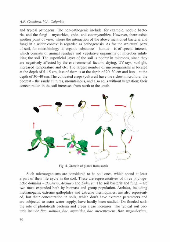
70
A.E. Gabidova, V.A. Galynkin
and typical pathogens. The non-pathogenic include, for example, nodule bacte-ria, and the fungi – mycorhiza, endo- and ectomycorhiza. However, there exists another point of view, where the interaction of the above mentioned bacteria and fungi in a wider context is regarded as pathogenesis. As for the structural parts of soil, for microbiology its organic substance – humus – is of special interest, which consists of animal residues and vegetative organisms of microbes inhib-iting the soil. The superfi cial layer of the soil is poorer in microbes, since they are negatively affected by the environmental factors: drying, UV-rays, sunlight, increased temperature and etc. The largest number of microorganisms is located at the depth of 5–15 cm, less of them is at the depth of 20–30 cm and less – at the depth of 30–40 cm. The cultivated crops (cultures) have the richest microfl ora; the poorest – the sandy cultures, mountainous, and also soils without vegetation; their concentration in the soil increases from north to the south.
Fig. 4. Growth of plants from seeds
Such microorganisms are considered to be soil ones, which spend at least a part of their life cycle in the soil. These are representatives of three phyloge-netic domains – Bacteria, Archaea and Eukarya. The soil bacteria and fungi – are two most expanded both by biomass and group population. Archaea, including methanogens, extreme gallophiles and extreme thermophiles, are also represent-ed, but their concentration in soils, which don′t have extreme parameters and are subjected to extra water supply, have hardly been studied. On fl ooded soils the role of phototroph bacteria and green algae increases. The typical soil bac-teria include Вас. subtilis, Вас. mycoides, Вас. mesentericus, Вас. megatherium,

71
Chapter IV. Soil
Cl. tetani, Cl. perfringens, Cl. oedomaticus, Cl. histolyticus, Cl. botulinum, Cl. chauvoeij and also thermophile, pigmental, non-pigmental and other micro-organisms, which sometimes compose 80–90 % of the total soil microfl ora. The space and the soil, surrounding the root being formed, are called the rhizosphere. At present the rhizosphere means the space round the root from 0 to 2–8 mm in diameter, where a more intense growth of microorganisms due to their growth stimulation exists by root exudates (discharges), and more generally – root de-posits [41]. The rhizosphere′s space is sometimes called the endorhizosphere; this term includes tissues of the root itself compared to rhizoplane, which means only what is located directly on the root surface and is fi xed to the root. The root exudates present themselves low-molecular organic substances, products of pho-tosynthesis and plant metabolism. They include sugars, organic and aminoacids, alcohols, hormones, vitamins and etc., they come into the soil near the root tip, more exactly near the root “stretching” zone during its growth and development. The root rhizodeposits have a wider meaning. They include not only exudates, but all other substances – high-polymer mucous of polysaccharide and protein nature, enzymes, fading and peeling superfi cial cells with their composition, pieces of tis-sues, in particular cortex of the upper maturing root parts, root cap, root hair, vola-tile organic substances and etc. It is considered that in the form of rhizodeposits the plant loses more than 30–40 % of the photosynthesis products.
In addition to the chemical infl uence on the soil and microorganisms avail-able on it, including via change in pH and Eh there exists a pure mechanic effect of the growing root on the eco-niche surrounding it. The phenomenon of a higher density of microorganisms around the root due to consumption of exudates and rhizodeposits is called the rhizospheric effect. In comparative experiments with cultivation of sterile plants in sterile and non-sterile soil it was shown that in the rhizosphere the microbial vegetative interactions are expressed, in particular, in stimulating the plant exudation. The population of microorganisms in the rhizo-sphere can exceed their population in the environment from several per cents to tens of per cents and even more.
The total population of microorganisms in the rhizosphere can be hardly named. It depends on the soil type, plant and other factors and can hesitate from millions to hundreds of billions cells per gram of the dry soil.
It turned out that the spatio-temporal design of the microbial biocenosis of the rhizosphere not only refl ects the zones of the root exudation, but has its specifi c structure. The microbial biocenosis grows along the developing root in waves, i. e. zones with higher density of microorganisms interchange with zones of low-er density. At the same time the peaks with different density of microorganisms shift along the root with time, therefore, it is possible to say about the “moving wave” of the growing microbial biocenosis of the rhizosphere. The wavy growth

72
A.E. Gabidova, V.A. Galynkin
of microbial populations was observed also in the rhizoplane and in direction perpendicular to the root, i. e. the wavy growth phenomenon of microorganisms in the rhizosphere presents itself a general form of evolution of the rhizosphere microbial biocenosis [41].
The microorganisms, which colonize the above-soil surfaces of plants, are sometimes called the epigenous microorganisms (Greek. epi – around; phytos – plant). The quantity of microorganisms, discovered on the leaf surface, may some-times achieve 108 cells per gram of fresh leaves, or 106 per 1 cm2, and this can be quite comparable with population of microorganisms in the soil gram. The popula-tion and variety of microorganisms, for example bacteria, signifi cantly depends on the plant species and its habitat, environment, weather conditions and some other circumstances. Some of them have already been mentioned by discussion of avail-able microorganisms on plant seeds. These are again saprotroph and phytotroph representatives of Pseudomonas (Р. syringae, P. fl uorescens and etc.), Erwinia (Е. carotovora, Е. amylovora), Xanthomonas (X. сатpestris), Agrobacterium (A. tumefaciens), of genus Beijerinckia, Enterobacter, Klebsiella, Methylobacte-rium and many others. There are some differences in the microbial biocenosis be-tween the upper side of the leaf and the lower side. Important role here is played by light and temperature. It is natural that plants of deserts, the succulents, contain much less microorganisms per one unit of surface area or per gram, than plants of moist and tropical forests.
By germination the plant carries on its surface from the soil typical; soil mi-croorganisms. However, with time, under the infl uence of the environment, the qualitative composition of the microbial biocenosis on the plant surfaces changes. Localization or space confi nedness of microorganisms on the leaf surface has spe-cifi c genus and species.
Experiments with use of GM bacteria, which can constitutively synthetize green fl uorescent protein revealed that some bacteria (Pantoea agglomerans) are mostly dissipatedly localized on surfaces of epidermal cells, while others (Pseu-domonas syringae) are mostly grouped along stomatas.
Stomatas, available on leaves and other plant parts, serve as main places of plant gas exchange (СО2 and О2) with environment and at the same time – places of discharge of some other substances, in particular volatile organic substances (VOS), which, in their turn, can serve as substrates for microorganisms. The most part of nonvolatile substances is composed by sugars and organic acids, the VOS include essential oils, superior alcohols and low-molecular organic acids and their ethers. It is the stomata that turned out to often serve as “entry gates” for infec-tions. Some phyto-pathogenic bacteria and fungi even adapted to penetrate into the plants via stomatas and don′t have any other ways of penetration into internal tissues and cells of plants [42].
73
Chapter IV. Soil
However, not all organic substances, discharged by the plants, are always sub-strates and useful for microorganisms. Some of them – phytocides – have an in-hibiting infl uence. They include glycosides, terpenoids and etc. The pine (Pinus sylvestris) produces substances, inhibiting the growth of the tubercula bacillus (Mycobacterium tuberculosis), and due to this fact the anti-tuberculosis sanatoria are often located in the pine forests.
The symbiosis, as defi ned by the term author Antoine de Bary (1879), – it is “co-existence of different organisms”. However, traditionally it happened that as a classical example of symbiosis of microorganisms and plants the mutual advanta-geous co-existence of bacteria of, Rhizobium genus, which can fi x nitrogen, is given. From organizational and morphological point of view the prokaryotes are repre-sented both as monocellular (mainly), and multicellular, to be more exact – myce-lial organisms – actinomyces [43]. Therefore, the bacterial and vegetative interac-tions include interactions of prokaryotic monocellular and mycelial organisms with monocellular, multicellular and formed in tissues and organs superior plants.
The fungi kingdom is related to microorganisms. The fungous vegetative in-teractions have their peculiarities, caused by different level of organization. The fungi and plants relate to eukaryotes, i. e. formally they belong to the equal lev-el of cell organization. It is thanks to the high mutual organization of fungi and plants that their interactions have a wide nature. According to different estima-tions, from 120 000 to 250 0001; species of fungi are known, and among them there are 8 000 species of phyto-pathogenic, whereas among bacteria only about 200 phyto-pathogenic species are known.
4.7. Ecological relationships in microbiocenosesIn environment the microorganisms don′t exist in the form of pure cultures
and are subjected to infl uence of the abiotic and biotic factors. [43, 44]. Probably, that is why their participation in biotic relationships takes place in composition of resistant populations, which appear not only on the level of microorganisms, not only on the level of microbiocenoses with plants and animals. Microorganisms as a part of the eco-system are in constant relationships both with other microorgan-isms and with macroorganisms. The global reach of microorganisms, with great fl exibility of their genetic systems and enzymes allows them participating:
● In mineralization of compound organic substances up to СО2, NH3, H2, CH4, H2O and etc. in different ranges of physical and chemical environment, which are used by macroorganisms and other groups of microorganisms as nutritive components
● In modifi cation and transit of microbial metabolism reactions into soluble and gas form of unavailable compounds;
● In providing biological processes with metabolic regulators of microorganisms, plants and animals (growth stimulators, bacteriocins, antibiotics, toxins and etc).

74
A.E. Gabidova, V.A. Galynkin
● According to their ability to execute certain functions the microorganisms are divided into physiological groups.
Microorganisms use as sources of carbon and power all known chemical and biological substances. The genetic systems of microorganisms allow effi -ciently controlling the metabolism re-building by change of the environment. The analysis of various animals proves that every species in environment oc-cupy a particular habitat (ecological niche). The ecological niches for micro-organisms are countless. To characterize exactly the place of habitat of the mi-croorganism, its micro-environment, i. e. a complex of factors in close vicinity of the microbial cells should be taken into account. Many specialized groups of microorganisms exist in such micro-environment, and they feel the minimal competition from other microorganisms [45].
In the habitat microorganisms can be both in free (measured in water), and in the attached state. The most part of microorganisms in environment are in the attached state, since on the border of phase separation, the concentration of sub-stances is usually higher due to their adsorption. The attachment of fi laments to solid surfaces with formation of mane can be observed in running waters. In this case one end of the strand is attached to the solid substrate by means of special cul-tures, and the other freely fl uctuates in the running water. However, the main part of soil microorganisms is located in compound architectural structures – biofi lms. The biofi lm formation is a complicated multi-stage process. The biofi lm formation starts with formation of monolayer from cells of the same species, which has high adhesive properties. The mucoid discharge of these cells makes attachment of oth-er microorganisms possible. Regarding the habitat peculiarities of microorganisms (light, available nutritive substances and diffusion speed) the biofi lm structure can be more complicated. The layers, represented by different types of microorgan-isms, micro-colonies can form, located in the general polysaccharide matrix [46]. The matrix is the byproduct of the microbial biocenosis, which includes surface cell structures and exometabolites, mainly, of polysaccharide nature and combined cell aggregates. Such compound fi lm evolutions as three-dimensional structure, which components are connected with internal pores and through channels. The biofi lm can grow up to the macroscopic size, forming the microbial mat. Some-times layers of microorganisms are of different color, and then the mat structure is ill-disguised. The state of cells in the biofi lms signifi cantly differs from the state of freely inhabiting cells. Included into aggregates the microorganisms are in close neighborhood and located in the matrix. The physical and chemical en-vironment of the matrix differs from the environment of the water solutions. The matrix restricts the delivery of drugs from environment and concentrates in itself nutritive substances, byproducts and signal molecules. The delivery of substances to cells is executed not only with diffusion, but also through channels and pores

75
Chapter IV. Soil
in the matrix. In different parts of the aggregate, which includes even the micro-organisms of the same species, the cells are of a different physiological state due to the different in delivery of the nutritive substances. The restricted space and close neighborhood of microbial cells included into aggregates leads to a faster exchange of byproducts. The matrix doesn′t only hold the microbial cells in space; it con-nects them with each other. The shape retention under the infl uence of external loads allows it protecting cells from mechanical damages and “softening” hesitations of physical and chemical factors, ensuring the resistant micro-environment. The ma-trix makes cell differentiation inside the population possible and can ensure “collec-tive” reactions (“the sense of quorum”). If a certain density of cells is reached in the limited space of the matrix, a signal substance is accumulated, which infl uences all population cells and leads to the “collective” response. In the biocenosis, including several species of microorganisms, reactions will be much more diversifi ed.
The organic components of the soil are byproducts of phototroph organisms – these are plants, algae, cyanobacteria and chemolithotrophic bacteria. The plants are the main producers of the soil organic compounds. The soil fertility depends not only on the chemical composition of the soil, but it is signifi cantly defi ned by metabolic activity of the soil microorganisms. The polymer sources of carbon, nitrogen and phosphorus in the non-utilized form for the plants are accumulated in the soil. The hydrolyze of the polymer structures takes place by metabolism under the infl uence of various groups of bacteria and fungi, and this leads to transfor-mation of low-soluble biopolymers into higher-soluble monomer form, and trans-formation of monomers into non-organic ions is accompanied by succession of soil microbial populations. All appearing monomer organic compounds are used as a source of power, carbon and other biogenic elements for growth and evolu-tion of microorganisms. The processes of hydrolyze take place both in aerobial and anaerobial environment. In anaerobial environment the process includes both the microorganisms-wanderers, and anaerobic breathing groups. Organic fermen-tation products can migrate in the aerobial habitats, where they will be subjected to oxidation and support the growth of aerobial bacteria, archaea and fungi. The anaerobial breath is one of the ways to get the power for a number of heterotroph bacteria, archaea are, and maybe, some fungi and protozoa, which is accompanied with destruction of the soil organic compounds. The non-organic substances be-ing formed are consumed by the chemolithoautotrophic microorganisms, which produce a new biomass. The microbial mass of the soil microorganisms is also de-structed, being an important source of nutrients for other soil inhabitants. In soils ecological niches for bacteria, fungi, protozoa, algae, insects, nematodes, small animals are formed, which are necessary for the soil formation and retention.
As a result of the running processes the compound polymers come into the soil, which don′t hydrolyze for several years (lignin, melanin, sulfated polysaccharides,

76
A.E. Gabidova, V.A. Galynkin
and microbial cell walls) and used for the humus formation, characterized with low solubility and available aromatic chemical groupings in the molecule. The soils contain a lot of surfaces, which infl uence the availability of the nutrients and interaction of different microorganisms. The different size of pores makes them to a different extent available for usage and colonization.
Usually the density of the microbial population decreases from the surface into the depth due to decrease in concentration of organic carbon and molecu-lar oxygen. The soil parts around the plant roots are characterized with higher densities of microbial populations, higher values of microbial biomass and more signifi cant level of total microbial activity. Redox conditions and availability of these or those acceptors of electrons signifi cantly defi ne which groups of micro-organisms will be habitat in this soil.
The soils are forms in different environments. Where there exists the weather-ing of new geological material or after eruption, or after earthquake, its colonization with microorganisms starts. The pioneers here are cyanobacteria, which can perform the photosynthesis and nitrogen fi xation. The important role in the soil formation and functioning is played by Gram-positive actinobacteria, which have a different degree of mycelium branching and growth. The typical soil odor depends on the discharged substance – geosmin. These groups play and important role in carbon hydrate digestion, minerals of vegetative materials and soil humus. When analyzing the microbial soil population it should be taken into account that at present it be-comes possible to cultivate only about 1–10 % of soil microorganisms. Non-cultivat-ed forms can be revealed by means of molecular-biological methods, with extraction of microbial DNA and its amplifying in polymerase chain reactions with subsequent sequencing of nucleotides in the gene, coding 16S рРНК.
Many microorganisms in soils are located in the metabolically inert state, how-ever by occurrence of favorable environment their activity can signifi cantly increase. Viruses, which are intra-cell obligate parasites of many soil inhabitants, can be per-sistent in the soil for years. They can play a regulative role in the soil population.
The soil microorganisms have a positive effect on the atmosphere, destructing the air “pollutants” such as methane, hydrogen, СО, benzol, trichloroethylene, for-maldehyde. The soil microorganisms infl uence the global composition of different gases. Relatively stable gases (СО2, NO, N2O, methane) are called the greenhouse gases, since they refl ect thermal beams, and don′t allow the heat going away from the ground surface, and cause the global warming. Methane can be consumed by methanotrophes, inhabiting in soil and water. The critical factor, which infl uences on the methane consumption by the soil, is the ammonium ion concentration. With increase of the ammonium concentration in the soil due to the agricultural activity or pollution, the methane consumption decreases. Thus, soil and water methano-trophes can be regarded as a unique bacterial gas fi lter.

77
Chapter V. POPULATION FORMS OF MICROORGANISMS IN THE ENVIRONMENT
5.1. Planktonic form of microorganismsThe gradual change of microbiological paradigm which takes place at pres-
ent is a transit from ideas about the monocellular structure of microorganisms to the idea about microbial colonies as entire “superior organisms” – is refl ected in the growing interest to the shape, picture, macro- and microstructure of bacterial colonies. “The colonies of almost all prokaryotic species show ability for cell differentiation and multicellular organization. This ability, surely, exists by bac-teria and environmental habitats, where they mainly exist like biofi lms, chains, mats and micro-colonies”. [46]. In modern microbiology a gradual transit to bio-social (“biopolytical”) approach to microorganisms is seen, and this is caused by detailed analyses of intra-cell (intra-population) interactions with help of genetic engineering, fl ow cytofl uorometry, scanning electronic microscope, time-lapse video recording and etc. [47].
The numerous works about colonial organization of microorganisms prove the morphological and physiological heterogeneity of cells included into its composi-tion. The colony seems to consist of several different “tissues” – cell clusters as understood by S.G. Smirnov [42, 48]. As typical clusters of Shigella he regarded
1) actively dividing;2) resting;3) spontaneously autolyzing cells [49]. The similar data of works by А.S. Kaprelyanz with employees are well-known.
Thus, the population of the starving bacteria for 3–6 months included viable, resting and nonviable cells, as the study shows by means of the cell sorter (for rhodamine binding 123) and in the bi-phase system of water polymer solutions [50].
There is both the vertical lamination of the colony, and available in them hori-zontally divided zones (sectorial and concentric). The vertical layers are well-seen by observance of the painted colonial cuts (toluidine blue, methylene blue). Thus, in colonies of Escherichia coli and Shigella fl exneri three layers are discovered:
1) the lower painted (with 6 mcm thick in the studied colony of E. coli);2) the middle, mainly light, seems to be composed of non-viable cells (often
of irregular shape), which includes separate well-painted viable cells; the thick-ness of this layer by E. coli – 16 mcm;

78
A.E. Gabidova, V.A. Galynkin
3) the upper painted (40 mcm by E. coli), which in case of E. coli has a well-distinguished future differentiation into two layers – a lower thin layer (with 1–3 cell layers thick), with clear border, particularly bright and thick lay-er (40 mcm by E. coli), which contains separate unpainted cells [51]. It is in-teresting that the paint for β-galactosidase by use of the genetically engineered strains E. coli with gene lac Z gives a similar picture in a whole: a thin layer of β-galactosidase containing cells, adjoining the substrate, changes (with movement to the top) by cell layer without β-galactosidase, above which β-galactosidase containing cells are located. The uppermost colonial layer is of a combined structure, including groups of β-galactosidase containing cells and cells, which don′t contain this enzyme. The layers of different cells from the morphological and biochemical point of view were observed in colonies of cholera agent Vibrio cholera as early as in 1920.
Many researchers note in their works the availability in the colonies of the system of air bearing micro-cavities, which are often crossed with “beams” made of chorda. The complicated system of micro-cavities actually turns the colonies into a combination of partially isolated from each other concentration focuses (micro-colonies). The micro-colonies formed by the mucous matrix and divided by the open (often fi lled with water) channels, are typical also for the internal structure of the biofi lms. This is an analogue of the primitive “circulatory system”, which delivers nutritive substrates and removes products of metabolism [52]. In colonies of Alcaligenes sp. bacteria, strain d2, the pores and channels were discov-ered, and also more specialized structures (“gas bottles”), surrounded by a unique “membrane” and containing extracellular hemoproteins. Such structures are ex-pected to facilitate the transportation of О2 to cells in colonies (aggregates), i. e. here one can talk about the analogue to the respiratory system of organs.
It was noted that in addition to the vertical lamination, the sectorial and con-centration zones are also typical for the microorganism colonies on the dense environ-ment. The sectors correspond to the genetically different clones, and this is refl ected in their different paint, consistence, shape, growth speed, activity of enzymes and etc. A visual example is a phase dissociation of bacteria into R-, S- and М-forms, which differ in thickness of cell wall (thus, representatives of Brucella have a cell wall thick-ness of R-version larger than of S-version [53]), available or absent micro-capsule, features of fi brillary (R- and S-versions) or vesicle-tubular (М-version) intercellular matrix and etc. The phase dissociates explain the difference in architectonics of col-ony sectors. The S-version of the rodococcus cells are spread evenly by colony thickness, the number of cells which contact with each other isn′t large. As for the R-version, then in the respective sector the cells of lower layers are located perpendicularly or at an angle to the nutritive medium, the cells of the upper lay-ers – are located radially and parallel to the agar surface. In the М-sector the cells are located in large groups and don′t contact with each other.

79
Chapter V. Population forms of microorganisms in the environment
The concentration zones refl ect the stages of “ontogenesis” of bacterial cells – they correspond to different stages of individual growth programs of cells. The sector can be revealed (for example, by E. coli in the minimal synthetic nutri-tive medium М9) by simple visual observance. In the agar medium with tryptone and glucose the concentration circles can be revealed by addition into the agar of 2,3,5-trihelyltetrazoleum chlorous, which is regenerated with cells of some (not all) sectors up to the painted into red color formazan. As a result the colony turns out to consist of white and red concentration circles. Shapiro [51] visualized these circles of E. coli, in the indicator medium, which allows revealing available or absent β-galactosidase activity of the cells (as it is described above, the differences in this parameter exist between vertical layers of cells in the colony).
If on the way of the spreading bacterial colony a mechanical disturbance is formed, for example, glass fi bers are placed (in natural habitats the role of obstruction can be played by, for example, folds and crypts in the intestinal tract, as ecological niche for the microbiota), then only the local change in re-spective concentration circles appears, which are in any case of uninterrupted nature. When obstruction is created beside the colonial front, the circles are formed behind this obstruction according to the same geometrical laws as in other colonial sites. The disturbance of the non-mechanical nature is created for normal growth of the colony when the cells bear mutation for the impor-tant for ontogenesis gene. Thus, mutant E. coli with damaged gene (by means of insertion) of DNA-polymerase I during the fi rst hours of its growth forms abnormal microcolonies out of cells of fi lamental structure. However, in this case the colony fi nds the ways to overcome the defect: in 2–4 days the coloni-al mutants become morphologically undistinguished against normal colonies, the cells – against cells of the wild type. The overcoming of genetic defect signifi cantly speeds up, if in the neighborhood there are mature (aged 2 days) normal colonies, which are, apparently, diffusing chemical factors of commu-nication. The older colonies make the younger ones, also upon result of the infl uence of negative agents, “adapt” their age to the age of the “older” – for example, form external concentration circle without preliminary formation of internal circles (to get detailed data upon communication of microorganisms please see the respective section of the survey).
If the colonial concentric zoning is combined with the sectorial, then the faster growing sectors have concentric circles pulled to the edge, i.e, formation of circles is controlled not in the space (by interaction of neighboring cells), but in time (pulsation of “biological hours”). The latter is the most evident for bacterial species (for example, for representatives of the genus Proteus, Serratia and Salmonella, for E. coli), which form from time to time squibs – cells with extra amount of fl agella and which aren′t capable for division. The squibs form
80
A.E. Gabidova, V.A. Galynkin
a colonial structure from concentration terraces as a result of the interchange of the following processes:
1) growth and division of vegetative cells (lag-phase before ordinary forma-tion of squibs);
2) mass formation of centrifugally migrating squibs; 3) transformation of squibs into vegetative cells with formation of one more
“terrace” (consolidation stage). The obtained data upon the dependence of the rhythm of “biological hours”
on the cell population density, in particular, upon relation of the inoculate Proteus mirabilis density and lag-phase duration before the appearance of the fi rst “wave” of squibs, means the available complicated system of the intracolonial commu-nication. The Serratia liquefaciens has identifi ed nature of the chemical signal agent, which is the acidated derivative of the homoserine lactone [54] (a class of the widely-known signal molecules of Gram-negative bacteria see below). As the colony grows there exists a trend to a larger synchronization of separate cell be-havior with more perfect circular colonial symmetry in a whole, despite perturba-tion [55]. This trend for synchronous behavior is kept by decrease in glucose con-centration as nutritive substrate and by growth of agar-agar concentration in the medium. In last case the speed of squib movement reduces, which fl agella need the drop-liquid moisture, absorbed by them from the agar gel by means of a spe-cial polysaccharide capsule [55]. The very cells of Serratia marcescens produce the moistening cyclic lipopeptide. There exists a genetic trigger, which switches the cells from synthesis of proteins of later stages of cell division to synthesis of fl agella protein (fl agelline) and therefore determining the mutual transformation of squibs and dividing vegetative cells.
From the point of view of colonial organization it′s interesting that ac-cording to the agar, which isn′t occupied by the growing colony, only entire groups of squibs can move. The single cells, which came outside the colony borders, lose their fl exibility, until they are “picked up” by this or that group of squibs. This observance means the coordination of behavior in scale of each group. In addition to that, the coordination of the squib migration in the scale of the entire colony is also well-known. Thus, in the bacterial colony there are at least two levels of integration:
1) an independent group deals with the coordination of migrating squibs;2) the entire colony, including many similar groups. There exists some analogy with organism of multi-cellular beings, where
exist coordinating systems of intra-tissue level (paracrine systems), which produce both locally efficient histohormones (histamine, serotonin and etc.), and generalized systems on the level of the entire organism (nervous and en-docrine systems) [44].
81
Chapter V. Population forms of microorganisms in the environment
5.2. Colonial organization of microorganismsBefore starting describing the interaction between the plants and microorgan-
isms on the biological level, let′s regard the interaction levels from the matter or-ganization point of view. The most important is the chemical level of interactions. On this level there exist such interactions as, host “recognition”, “information” exchange, perception and abruption of molecules and organisms. The physical level is the physical contact between the microorganism and the plant. Keeping microorganisms on the plant surfaces often turns out to be the leading factor in future development of the situations. The biological level is both the molecular-genetic level of interactions, and the level of cells, which is already for many mi-croorganisms the level of organisms. The cell level of interactions in relation to microorganisms can hardly be separated from the population level. In the micro-bial vegetative interactions there exists a level of the biocenosis (or biocenoses), which is the most complicated. This level of interactions is usually associated with epidemiology. However, with attempts to extract and use as bio-controlling agents against some phyto-pathogenic fungi bacterial biocenoses instead of the monocul-ture it becomes obvious that this level of interactions is much wider.
The interactions between the microorganisms and plants after fading of one of the components should be also regarded. The plant fading is accompanied with its destruction and utilization by microorganisms and vice versa. Thus, the problem of the microbial vegetative interactions is diversifi ed.
It was shown that colonies of all prokaryotic species tend to cell differentia-tion and multicellular organization, which is the basis for transit to the biosocial (“biopolitical”) approach to microorganisms [46, 47]. The colony includes the fol-lowing cell clusters [48]:
1) actively dividing;2) resting;3) spontaneously autolyzing cells.For colonies of microorganisms it is typical to form functional organs of above-
organism level. Firstly it is related to the formation of a single biopolymer matrix, which is formed by fusion of capsules of separate cells or glycolipids, glycoproteins, polyglutamic acid [58]. Like the intercellular matrix of the animal tissues, the micro-bial matrix also includes fi brillary elements [59]. The similarity between the animal and microbial matrix is supplemented with community of some chemical compo-nents (sialic acids serve as an example). It is probable that the matrix of microorgan-isms plays the roles, related to the supracellular level of organization:
– Structure-forming role. Due to the matrix the colony includes, strictly speaking, not the single cells, but subcolonial associations, which can be met both by Gram-positive, and Gram-negative bacteria (including – pathogenic species of both groups). The structure of the colonies includes also hollow tubes made

82
A.E. Gabidova, V.A. Galynkin
of extracellular polysaccharides and other biopolymers (Pseudomonas aerug-inosa) – expected micro-channels for transportation of substances. Besides, through such tubes the colony cells migrate, usually in the form of small L-forms, which is applicable for bacterial species, included into the symbiotic microbiota of a human or animal.
– Defending (protective) role. The matrix covering the cells serves as an in-ternal buffer medium of the colony, which protects separate cells and the colony in a whole from negative external impacts (Drying, heating/cooling, attack of hy-drolytic enzymes and etc.). The polysaccharide and peptide matrix components, in particular, include the xeroprotectors [59].
– Communicative role. The exometabolites and products of cell autolyze are discharged in the matrix and distributed there, including the chemical signal sub-stances, which are used to estimate the density of the own population (Quorum systems of microorganisms). Many types of bacteria keep the supracellular organi-zation also by cultivation in liquid medium.
Like eukaryotic cells included into the tissues of multicellular animal, vegeta-tive or fungous organism, the prokaryotes form intracolonial intercellular contacts which, probably, facilitate the distribution of signal molecules in population, es-pecially if we talk about non-diffusing in medium communication factors (bio-fi lms). The intercellular contacts are formed at the expense of the various surface structures – microfi brilla, pineal crests, cell wall evaginates, glycocalyx, which tells about the genetic expression by transit of microbial populations into self-reg-ulative multicellular systems.
5.3. Biofi lmsThe main forms of bacterial existence in natural conditions are biofi lms which
are connected with the biocenosis surface. The biofi lms are highly organized, fl ex-ible, uninterruptedly changing heterogenic populations of microorganisms. Bac-teria are social organisms, which form the multicellular associations. At present it was found out that the biofi lms have a compound three-dimensional structural organization. The formation of biofi lms is a diffi cult complex dynamic process, which includes several stages:
1. Reversible attachment to the surface. In environment the majority of micro-organisms try to attach to the surface and form a biofi lm.
2. Permanent adhesion to the surface. As bacteria reproduce they adhere to the surface more strongly, differentiate, share genes, this ensures their survival.
3. Formation of the mucous protective matrix/biofi lm. Having fi rmly at-tached, bacteria start forming the exopolysaccharide environmental matrix, known as extracellular polymeric substance (EPS-matrix). The small colonies of bacteria in matrix form the initial biofi lm [59].

83
Chapter V. Population forms of microorganisms in the environment
The key moment without which it′s impossible to form the microbial biofi lm is the process of the microorganisms adhesion to the surface available for the fu-ture colonization and re-distribution of the cell mass; to form cell clusters; forma-tion of the exopolymer mucous matrix [60].
After the irreversible adhesion and active division of cells intensive prolif-eration with formation of multicellular layers and the synthesis of components of exopolymer matrix EPS-matrix are marked – this is one of the key moments in biofi lm formation. The composition of the matrix mucus varies with regard to its microorganisms and includes polysaccharides, proteins, glycolipids and bacterial DNA [61]. At the same time the main components are polysaccharides (dextran, hyaluronic acid, cellulose and etc). This fraction composes from 40 to 95 % of the total mass of the biofi lm; the concentration of other chemical substances var-ies signifi cantly. The share of proteins in the biofi lm can compose up to 60 %, of lipids – up to 40 % and of nucleic acids – up to 1–20 %. About 80–90 % of the biofi lm volume is occupied by water that is why all its constituents are in hy-drated state. The biofi lm matrix is divided by channels, fi lled with water, it also has cavities. The nutritive substances are transported though the channels and the convection fl ows of oxygen come from external to internal parts of the biofi lm; simultaneously metabolites of the bacterial cells are moved out [62].
Formation, growth, migration of the planktonic cell forms for colonization in bio-fi lms is controlled on the level of population by means of intercellular communication mechanisms. “Quorum sensing” (QS) – this is a process of collective coordination of gene expression in bacterial populations, which defi nes the specifi c behavior of cells.
In biofi lm the microorganisms exist in two forms: a fi xed one to the surface and planktonic, free movable, which is the source of infection spread from its ini-tial locus. They can include one or, which is met more often; several species of microorganisms, i. e. polymicrobial (for example, contain numerous different spe-cies of microorganisms). In biofi lms – independent microbial groupings, where each microorganism is in its micro-niche in the single matrix of the biofi lm. Tran-sit of microorganism to the growth program included into biofi lm is accompanied by signifi cant changes in expression of tens of genes with regard to the stage of the colony growth. The fi lm usually includes 15–20 % of the bacterial mass, which has fi rmly attached to this or that surface, and 80–85 % of the protective matrix. The availability of the matrix reduces the impact level of antibiotics and antisep-tics on microcultures-targets in tens and hundreds of times, and also activity of the immune protection of host organism [61]. In this case the change of phenotype takes place, which is expressed in change of growth parameters and expression of specifi c genes. Ability of bacteria to form biofi lms is a signifi cant factor of patho-genicity. The microbial biofi lms are responsible for etiology and pathogenesis of many acute and, especially, chronic bacterial infections of a human [63]. They are

84
A.E. Gabidova, V.A. Galynkin
discovered more than by 80 % chronic infectious and infl ammatory diseases. This became the basis for concept of chronic diseases, as biofi lm diseases [64].
The formation of biofi lms takes place with change of external conditions and includes the intercellular transfer of signals and transcription of genes, which are in inactive condition by individual growth of cells. As bacteria repro-duce, they more fi rmly adhere to the surface. The microbial cell attachment to the substrate surface is performed due to the activity of electrostatic, hydropho-bic forces, van der Waals forces, non-specifi c adhesion. Adhesion to biological surfaces is caused by specifi c interaction of proteins-adhesins or lectins of bacte-rial cell′s exoplasmic compartment fi mbria with receptors or certain domains of the cells-targets′ membrane surfaces. Then they differentiate, exchange genes, and this causes their survival. After attachment to the solid surface the cells re-produce, forming the monolayer. Then separate cells (if clearweeds of IV type are available) are movable on the surface, as a result the microbial cells form the so-called microcolonies. Then the microcolonies differentiate, forming a mature biofi lm. The cells in these structures are packed in extracellular polysaccharide matrices. The fi rst bacteria of the biofi lm attached to the site of the substrate, fa-cilitate attachment of the others, both by means of expression of special proteins of adhesion, and through construction of the above mentioned “matrix” of extra-cellular polymer substances and strengthen the biofi lm. While being attached to the substrate the bacteria synthetize “signal compounds – molecules”, attracting new bacteria to the growing biofi lm, and this stimulates the future distribution of bacteria which already attached to it [65].
After attachment of the initial colony the fi lm grows due to division of its components and their coming from the environment. The growth potential of any bacterial biofi lm is limited with the quantity of nutritive substances in the environment, their availability for the cells, contained inside the biofi lm, and possibility of removal of the metabolic products. Besides, there exists a hydrodynamic optimum of the environment fl ow rate, which ideally speeds up the biofi lm growth at the expense of the optimized speed of coming nutri-ents and removed exometabolites, and with high speed – it leads to erosion of the external biofi lm layers.
After the fi nal ripening in the biofi lm there forms an optimal growth and death speed of cells, physiological cooperation and metabolic effi ciency, which ensure the fi lm growth. All biofi lms, which are highly hydrated up to 73 %, contain the extracellular material, including the water channels and exopoly-saccharides. The most species have a polysaccharide matrix consisting of al-ginate, which is mostly anionic. The matrix has a three-dimensional structure, which surrounds, fi xes and protects the microbial colonies attached to different surfaces. The matrix is divided by channels, fi lled with water, and it also has
85
Chapter V. Population forms of microorganisms in the environment
cavities. Through these channels nutrients are transported and convective ox-ygen fl ows pass from external to internal parts of the biofi lm, simultaneously metabolites of bacterial cells are taken out from the bacterial cells. The pores and channels, which penetrate the entire biofi lm – is a very important part of its structure. Metaphorically they can be compared to the blood system of the biofi lm. The polysaccharide and peptide matrix components, in particular, in-clude a number of crio-, thermo – and xeroprotectors [66].
The biofi lms signifi cantly increase the tolerance of microorganisms lo-cated in its matrix, to the immune system of the host, anti-microbial agents and stresses (for example, limited oxygen or nutrition). This tolerance can contribute to the full resistance to the factors, which could easily eliminate these very microbes in case of their growth in the unprotected, plankton-ic state. The ways of bacteria protection with use of biofi lms (locking mi-crobes) – are simple, as a result of this the extracellular polysaccharide ma-trix protects the microbes and prevents from coming of large molecules (for example, antibodies) and cells, which cause infl ammation. The mutual pro-tection is another unique property of polymicrobial biofi lm – joint protective properties, which are acquired by bacteria of various species due to the gene exchange or discharge of respective factors into the environment. Thus, an-tibiotic resistant bacteria can produce protective enzymes or proteins, which can protect the neighboring antibiotic sensitive bacteria in the biofi lm. They can also share with other bacteria genes, responsible for the antibiotic resist-ance (even to other bacterial species) [61]. Specifi c features of EPS biofi lms, attributable to some bacterial species, can play a signifi cant role in ability of other species to join to the existing biofi lms. In order for the antibiotic to infl uence the bacteria, the latter should be metabolically active, that is why inactive bacteria in biofi lms can hardly be infl uenced by antibiotics, which usually eliminate active bacteria.

86
Chapter VI. SIGNAL INFORMATIONIN POPULATIONS OF MICROORGANISMS
6.1. Quorum systems of microorganismsThe problem of biofi lm formation by saprophyte and opportunistic pathogenic
microorganisms in the in vivo environment fi nally wasn′t cleared out, but the con-nection of this phenomenon with manifestation of bacterial social behavior was proved, which was called “the feeling of quorum” [66]. The term “Quorum sensing” was offered in 1994, it means the perception of environmental changes by the cells, which appear when the bacterial culture reaches some limited population and reac-tion to these changes. This phenomenon also includes availability of the apoptosis, i. e. the programmed death of separate cells in favor of the population in a whole. The apoptosis phenomenon of animals and vegetative cells is a normal integral part of the individual growth of the organism. The apoptosis of vegetative cells, attacked by the infectious agent, prevents from the further infection distribution [67].
The death of a part of cell population E. coli or Bacillus subtilis in stasis envi-ronment can also be regarded as analogue of apoptosis – the stop in growth of bac-terial population (for example, by lack of the nutritive substrate). The starving pop-ulation E. coli is gradually divided into two sub-populations; one of them dies and is subjected to autolyze, while another sub-population uses the autolyze products as substrate and continues growing and creating colony forming units. The molecular mechanism has been decoded, which regulates interactions inside the biocenosis of Bacillus subtilis soil bacteria, when there appears the lack in food, one half of bac-teria kills another one with poison. Those which died serve as food for their killers, the latter don′t die from their own poison due to a special protective protein.
Quorum Sensing (QS) – is a special type of regulation of bacterial gene expres-sion, which depends on the density of its population. QS-systems include two obligato-ry components: low-molecular regulator (auto-inductor), which easily diffuses through the cell membrane, and receptor regulatory protein, with which the auto-inductor (AI) is bound [68]. As far as the bacterial population grows and reaches the critical level, the number of AI accumulates up to the required threshold value and interacts with the respective regulatory proteins, and this causes an acute activation (induction) of expression of defi nite bacterial genes. With use of AI communication of bacteria is performed – information transfer between separate bacterial cells, which belong to one and the same or different species, genus or even families; that is why the signal mol-ecules are considered to be “words” in this unique bacterial “language” [69].

87
To support the quorum signalization bacteria constantly produce specifi c signal molecules, discharged into the environment. Oligopeptides are such signal molecules in the Gram-positive bacteria, N-acyl homoserine lactones – in the Gram-negative bacteria [70]. Numerous studies of the recent years have shown that microorgan-isms, including probiotic bacteria, synthetize and detect a wide range of auto-induc-tors of different chemical nature. Potential auto-inductors of probiotic bacteria are also known: lactones; peptide pheromones; inductors of AI type, including furans; volatile fatty and other organic acids; certain groups of enzymes (lactases, glycosi-dase, oxidase); stress proteins; proteins and peptides, which imitate signal molecules of eukaryotic cells; some aminoacids (glutamate, beta-alanine); vitamins (biotin); amines (histamine, serotonin) and polyamines (spermine, spermidine); some li-popolysaccharides (peptidoglycan, lipoteichoic acids); antimicrobial agents (bacteri-ocin, nitrogen oxide, active oxygen forms); lectins; biosurfactants [71].
The mechanism of QS activity is based on the diffi cult hierarchy regulation of target loci of bacterial cell′s genome. In this case the regulation is performed on different levels of infl uence: transcription and post-translation. A particular cell signal is specifi cally responded by cells. Now the cell-cell interactions are known to infl uence the intra-population differentiation, expression of virulence genes, which regulate the growth processes, movement character and direction (taxis), and also bacterial apoptosis and toxin formation. The principle of this mechanism′s activity is based on the activation of transcription of specifi c genes with reach in threshold level of binding the transcription protein-activator (LuxR) with low-molecular auto-inductor. The mechanism described above mediates the well-known phenomenon of high speed of microorganism growth with big values of daily dose. QS regulates an important process of switching of bacterial cell′s phenotype from planktonic form to its location in the biofi lm. This is a required stage in biofi lm formation for benefi cial parasitizing of the macroorganism. In the beginning of the infective process the high level objective of the pathogen – pen-etration and adhesion in tissues of macroorganism. At the same time, as it was mentioned above, the cell resources are focused on the biosynthesis of fl agella and specifi c proteins – adhesins. However, components of fi mbria, fl agella and adhesin proteins are string immunogenes; they also stimulate formation of interleukins. Therefore, for the future survival of the population inside the affected area of for-mation of fl agella and adhesion systems will be unprofi table from the biological point of view. That is why at the stage of biofi lm ripening QS inhibits formation of fl agella and adhesins. The reverse process passes in a similar way – formation of the movable cell forms in the biofi lm or discharge of the entire cluster of cells (detachment cell) to colonize the surrounding substrate. It is obvious that when opportunistic microorganisms reach in the microbial biocenosis their critical popu-lation, the QS-system strengthens the adhesive properties of the potential agent

88
A.E. Gabidova, V.A. Galynkin
and initiates the synthesis of pathogenicity factors, and this in future defi nes the growth of the infection process [72].
The adhesion mechanism of the Gram-positive bacteria differs from the mechanism of the Gram-negative ones. Thus, for example, the most important element in the process of staphylococcus adhesion is the polysaccharide (Poly-saccharide Intercellular Adhesin – PIA), which participates both in the cell sub-strate adhesion and consequent formation of cell clusters. The fl agella and fi m-bria of IV type play an important role in adhesion and cell aggregation of the Gram-negative microorganisms. The movement, caused by fl agella, facilitates distribution and formation of the cell monolayer on the substrate, the fi mbria of IV type participates in cell aggregation at the expense of the lectin interactions [73]. As bacteria reproduce they attach to the surface more fi rmly, differentiate, exchange genes, this ensures their ability to survive. Formation of bacterial bio-fi lms on the medical equipment (for example, catheters, artifi cial heart valves, lenses and etc.) causes the growth of a number of serious chronic diseases. The microbial biofi lms are responsible for the etiology and pathogenesis of many acute and, in particular, chronic bacterial infections of a human. In the environ-mental eco-systems the biofi lms are microbial groupings, where each microor-ganism is in its own micro-niche in the single matrix of the biofi lm [74].
The matrix covering cells serves as a buffer internal medium of the colony, which protects separate cells and the entire colony from unfavorable external in-fl uences (drying, heating or cooling, attack of hydrolytic enzymes and etc). The polysaccharide and peptide matrix components, in particular, include a row of crio-, thermo- and xeroprotectors [75]. Specifi c features of EPS biofi lms, attribut-able to one bacterial specie, and play a signifi cant role in ability of other species to join and build in the existing biofi lms. The general communications scheme of Gram-positive bacteria can be represented as follows: fi rst the forerunner synthe-tizes in the cell, which, modifying, turns into the mature oligopeptide. The latter is excreted outside with exporter. The oligopeptide molecules accumulate in the intercellular space with growth of density of bacterial cells. The two-componential sensor kinase, penetrating the membrane, detects the signal and transmits it to the cells in the process of cascade phosphorylation. In the cell the oligopeptide inter-acts with the target gene (genes) [77, 78].
Existence of bacteria inside biofi lms ensures their multiple advantages com-pared to isolated cells. For practical medicine it is especially important for bacte-ria in biofi lms to have increased surviving ability with availability of aggressive substances, immune protection factors and antibiotics. The increased survival is based on properties of cells and extracellular matrix. The resistance caused by the biofi lm cell properties is associated with reduction of their free surface at the ex-pense of contacts with each other and formation of bacteria, which were called

89
Chapter VI. Signal information in populations of microorganisms
“persisters”, which are in condition of full resistance practically to all drugs [79]. Of special interest are cells-persisters – altruistic intact cells, which can survive even by high doses of antibiotics, lethal for the rest microbial cells. Upon data given by some authors their quantity varies from 1 to 5 % of the total popula-tion. They are metabolically inactive, and their main designation, as it seems, de-positing and saving genetic material for the consecutive population recovery. The phenotype of persisters is characterized with interesting biology; they slow down all physiological processes and become tolerant to infl uence of different factors, including the infl uence of the microbial drugs [80]. The property of the antibiotic tolerance differs from the resistance mechanisms.
Persisters are altruistic cells, which appear at the stationary growth stage. They are metabolically inactive and ensure survival of the mother population with avail-ability of factors lethal for all cells. The infl uence of all mechanisms of bacterial resistance, in actual fact, can be modelled down to one phenomenon – prevention from interaction between antibiotic with its target (by means of changing the targets themselves or synthesis of enzymes, which neutralize antibiotics). The tolerance is mediated through ability of the microbial cell to survive with availability of antibiot-ic at the expense of slowing down metabolism and “switching off” the main biologi-cal processes of the cell. The time of transit to the state of the biofi lm is defi ned with the “feeling of quorum” – reaction to concentration of specifi c regulatory peptides.
The mechanism of QS activity is based on the diffi cult hierarchical regulation of targeted loci of bacterial cell′s genome. At the same time the regulation is ex-ecuted on different levels of infl uence: transcription, translation, post-translation. The cells response specifi cally to the particular cell signal. Today it has been es-tablished that cell-cell interactions infl uence the intrapopulation differentiation of cells, expression of virulence genes, regulate growth processes, movement nature and direction (taxis), and also bacterial apoptosis and toxin formation. The work of QS can be compared to hormonal regulation of functional activity of different organs and tissues in the multicellular organism. Gram-positive and Gram-nega-tive organisms use different signal systems and various signal transmitters [81].
Signifi cant resistance of microorganisms to antibiotics inside biofi lms com-pared to planktonic forms is caused by capability of bacteria to accumulate in matrix extracellular enzymes, which destruct antibiotics, and aggregation nature of biofi lms, connected with reduction of area of open cell surface, this leads to physical unavailability of molecules. A special role is also played by the resistant cell phenotype and reduced metabolism of microorganisms in the biofi lm, which is achieved due to their multilayer topography and leads to reduction in antibiotic susceptibility. The structure of biofi lms ideally contributes to the genetic infor-mation exchange processes, including resistance to the anti-microbial chemical drugs, at the expense of close contact and stable spacious localization of cells.

90
A.E. Gabidova, V.A. Galynkin
In vitro studies show that the conjugation level in biofi lms is much higher compared to planktonic bacteria forms. Moreover, the conjugation processes can be regulated on the population level at the expense of the bacterial communication, for example, virulent enterococci for genetic information transfer use the signal systems.
The infl uence of all mechanisms of bacterial resistance, actually, can be mod-elled down to one phenomenon – this is prevention from interaction between antibi-otics and its target (at the expense of changes of the targets themselves, or by means of synthesis of enzymes, which neutralize antibiotics). The tolerance is mediated through the ability of microbial cell to survive in availability of antibiotics at the expense of the slowing down metabolism and “switching off” the main biological processes of the cell. Antibiotics effectively show their infl uence on the intensively dividing cells with high level of synthetic processes. And when the cell is at the stage of the physiological rest (“cellular anabiosis”), antibacterial agent can′t show to the fullest extent its biochemical function. For example, the erythromycin inhibits the protein biosynthesis, this causes its bacteriostatic effect (the cell doesn′t grow, doesn′t reproduce, metabolism slows down). But the cell doesn′t die due to direct infl uence of the drug. Streptomycin, aminoglycosides, fl uoroquinolone violate the processes of translation, replication. Temporarily “switching off” the activity of ri-bosomes, the cell-persister will show tolerance against aminoglycosides, macrolides. Since persisters don′t grow, don′t divide, the chromosome and protein systems of replication, reparation, and transcription are in intact state, and then the infl uence of the fl uoroquinolone will not be seen. The result of the above mentioned “switching off” of biological cell functions is also the stop of synthesis of the peptidoglycan, the building of the cell′s wall stops, and therefore, β-lactam antibiotics won′t be effec-tive. Proteins of persisters switch off the activity, function of all targets of antibiot-ics, thus, mediating the multi-tolerance (MDT, multi-drug tolerance). Therefore, the bactericide antibiotics will have only the bacteriostatic effect on persisters [82].
6.2. Quorum-sensing reaction of Gram-negative microorganisms More than 450 species of Gram-negative bacteria reveal the Quorum-depend-
ent systems, where different acylhomoserinelanctones serve as signal molecules. In the quorum-sensing systems of the Gram-negative bacteria proteins of Luxl family are synthases of acylhomoserinelanctone autoinductor molecules, they freely diffuse through membrane and accumulate with increased cell density. Pro-teins of LuxR family are bound with autoinductors relative to them with reach of the high concentration of signal molecules. The complex LuxR – autoindictor is bound with the target gene promotor, with launch of their transcription [82].
Bactria of Erwinia (E.carotovora, E.chrysanthemii) genus are pathogens for the plants. They split the vegetative cell walls by means of pectinases and cellu-lases. Formation of these enzymes is an important factor of virulence and depends

91
Chapter VI. Signal information in populations of microorganisms
on population density. Erwinia have the functioning gene system called expI-expR, similar to the system luxI-luxR by V.fi sheri. In quorum-sensing reactions the regulatory system works, ensured by transcription of genes rsmA-rsmB. Synthesis of carbapenem antibiotics also depends on the population density of E.carotovora. Production of this antibiotic is controlled by the cluster of sagA-sagN genes, and, probably, is required for elimination of competitive microor-ganisms in locus of plant infection.
Another example of using homoserinlactones as signal molecules is shown for Pseudomonas aeruginosa – animal pathogen. Pathogenicity of Р.aeruginosa is caused by a wide range of virulence factors. Some of them are associated with the cell (pili, adhesins, lipopolysaccharides), the other secrete (proteases, ramnolipids, exoenzyme S, exotoxin А, antibiotic pyocyanin and etc.). Formation of many ex-tracellular virulence factors is controlled with systems of intercellular interaction. The central components of such interactions are las- and rhl-quorum-sensing sys-tems, which activate the gene expression depending on the value of cell density in microorganism. Each system is represented by two genes: one of them codes the enzyme, with the help of which a specifi c autoinductor synthetizes – acidated ho-moserinelactone (lasl/rhll); the other codes transcription activator, with which the respective autoinductor is bound (lasR/rhIR). N-(3-oxododecanoil)-L-homoser-inelactone (З-oxo-С12-HSL) is the autoinductor for las and rhi systems, a special system serves to export it from the cell called MexEF-OprN-pompa and N-butyril-L-homoserinelactone (C4-HSL) respectively [83].
las controls the gene expression, which code such virulence factors as, elastases А, В, and also the alkaline protease; rhi system controls the biosynthe-sis enzymes of ramnolipids, pyocyanin. Recently the third signal molecule has been discovered, which takes part in quorum-sensing reactions of P. aeruginosa – 2-heptyl-З-hydroxy-4-hinolon (PQS). This signal molecule can control the expres-sion level of las В, which codes elastase Las В, and also the expression level of rhil, which codes synthase C4-HSL.
The bacteria Agrobacterium tumefaciens causes the formation of crown galls of many plant species. Galls are vegetative analogues of malignant tumor and are formed as a result of the transfer of oncogenic DNA fragments from bacterium to the core of the vegetative cell by means of Ti-plasmids. Some of Ti-plasmid genes cause the synthesis of opins by vegetative cells, which serve as nutritive substrate for A.tumefaciens. The homology luxI-luxR gene system traI-traR stimulates the distribution of Ti-plasmids in bacterial population. The plasmid DNA tends to distribute in bacterial population and, as soon as suffi cient quorum is formed, it makes the cells which carry the plasmid conjugate with other bacterial cells. At the same time the conjugative transfer of Ti-plasmids depends on opins. In par-ticular, traR transcription is stimulated by factor OccR, activated by octopin.

92
A.E. Gabidova, V.A. Galynkin
6.3. Quorum-sensing reactions of Gram-positive microorganisms A serious problem of the clinical practice is the wide spread resistant forms
of microorganisms, which reduce the effi ciency of the applied antibacterial drugs. A special diffi culty is caused by the increased drug resistance of bacteria in biofi lms. To synthetize virulence factors, antibiotics and form bacterial biofi lms the quorum-sensing reactions are often used. That is why the study of mechanisms of such reac-tions reveals new opportunities to prevent and treat such diseases, caused by the mi-crobial agents, and also allows in a different way looking at the complicated process of interspecies bacterial interactions in environmental habitats of microorganisms.
The quorum-sensing reaction mechanisms are different in Gram-positive and Gram-negative bacteria; that is why it makes sense to regard them separately [84].
The Gram-positive bacteria usually communicate by means of the oligopeptide signal molecules. In most cases the signal transmission includes a two-component mechanism of phosphorylation. As a rule, the quorum state is reached by transfer of bacterial cell population to the stationary stage of growth. It is at these moments that signal molecules are discovered, by means of which the cells contact with each other. The general communication scheme of Gram-positive bacteria can be repre-sented as follows: fi rst a forerunner synthetizes in the cell, which, modifying, turns into a mature oligopeptide. The latter excretes outside the cell by exporter. The oli-gopeptide molecules are accumulated in the intercellular space with growth of the bacterial cells. The two-component sensor kinase, penetrating the membrane, detects the signal and transmits it into the cell during the cascade phosphorylation. In the cell the oligopeptide molecule interacts with the target gene (genes).
The system, responsible for the conjugative transfer of plasmids by Enterococ-cus faecalis and relative bacterial species can be considered to be a classical peptide quorum-dependent system. This system stimulates distribution of attributes in mi-crobial population, which are important for the interaction between the microorgan-ism and animal-host, and also for competition elimination. The plasmid pPDl trans-ferred by the peptide quorum-dependent system is responsible for the synthesis of hemolysines, plasmid pCDl – for bacteriocin formation, plasmid pCFlO – for resist-ance of E.faecalis against tetracycline. Each hexa- or octapeptide induces adhesion of bacterial cells and their conjugation with transfer from the donor to recipient of a certain plasmid. For example, octapeptide cPDl stimulates the conjugative transfer of plasmid pPDl. The plasmid codes the receptor, located on the protein-repressor of the respective operon. Interaction between the oligopeptide and receptor causes dissociation of the repressor from the DNA, thus launching the synthesis of the re-spective product. The plasmid pPDl includes also the gene traC, which produces the protein, which assists the peptide in penetrating through the cell wall. The oligopep-tide signals are intensely synthetized by cells, which don′t carry the respective plas-mids (recipients), while the synthesis of such signals by cells-donors is inhibited, moreover, the plasmid codes the inhibiting peptide.

93
Chapter VI. Signal information in populations of microorganisms
Plasmid pPDl produces peptide рPDl [85].Another quorum-depending process, discovered by E.faecalis, is production
of two virulent factors: gelatinase (GelE) and serine protease (SprE).The system of quorum-sensing, which controls the synthesis of exotoxins in the
late logarithmical growth stage of Staphylococcus aureus can serve as an example of peptide signal use to perform intercellular interactions. In this system the pro-tein AgrD synthetizes in the form of the forerunner, which includes 46 aminoacids, which during the export of protein AgrB turns into a mature peptide AIP (autoinduc-ing peptide), including 8 aminoacids. AIP is detected by the two-component sensor kinase AgrC, which transmits the signal inside the cell by phosphorylation of the response regulator – AgrA. AgrA ~ P activates transcription of target genes, stimu-lates transcription of operons agrB, D, С, А (positive auto-regulatory loop), and also “prohibits” transcription of genes, coding other exotoxins. Based on differences in AIP and its receptor strains S. aureus can be related to four and more groups. Oligo-peptides, synthetized by one of the groups, induce pathogenicity in this group and specifi cally inhibit the systems of Agr-virulence in other groups.
The population density infl uences the formation of competence in the late log-arithmic growth stage of Streptococcus pneumoniae. The gene соmС is coded by the forerunner, which includes 41 aminoacid residues. The latter transforms into a mature peptide, including 17 aminoacid residues during interaction with the pep-tide export system (АВС-system), which is formed by products of соmАВ genes. The peptide contacts with its receptor on the surface of the cell – histidinekinase, produced by gene comD. The activated histidinekinase phosphorylates the product of gene соmЕ. With accumulation of cells the number of peptide signals grows and reaches its critical level in the environment. The quantity of the phosphoryl-ated protein cоmЕ increases respectively, which, starting from certain concentra-tion, is bound with the promotor of operon comCDE, which stimulates its activity (positive auto-regulatory loop), activates promotor of operon соmАВ (system of protein export from the cell), activates operon sotХ, which includes a full chain of late genes of competence; responsible for binding and absorption of transforming DNA and all other, late stages of transformation [86, 87].
The Gram-positive microorganisms-actinomyces (Actinomycetales) as signal molecules use both peptide compounds, and substances of low-molecular nature, which contain the lactone grouping – butyrolactones. The best of all studies actin-omycetic regulator is А-factor (2-izo-capriolil-3-oxymethyl-у-butyrolactone) [89].
At the early growth stages, when concentration of А-factor is low, the recep-tor of А-factor (АgrА) is bound and represses the expression of activator of strep-tomycin and spore-formation biosynthesis. With discharge of АgrА from the cell lysate S. griseus IFO 13350 it was shown that this protein consists of 276 ami-noacids and has the molecular mass of 29,1 kDa. With increase in culture density

94
A.E. Gabidova, V.A. Galynkin
the concentration of А-factor reaches its critical level, on which it binds the АgrА, causing dissociation of the latter from DNA, including transcription of the key gene adpA, coding AdpA (protein, consisting of 405 aminoacids, which contains in its central site the binding with DNA, similar to transcription regulators from protein family AraC/XylS). The protein AdpA is a positive regulator of cytoplas-mic activator of cluster of biosynthesis genes of streptomycin and spore-forma-tion. The cytoplasmic activator, being bound with the DNA in the area of gene promotor of specifi c regulation of streptomycin biosynthesis cluster strR, induces transcription of this gene, located after it the gene of resistance to its own antibiot-ic – aphD, gene adsA, coding the extracytoplasmatic a-factor of RNK-polymerase, required to form the air mycelium, and also gene sgmA, coding the protein-pepti-dase, which participates in addition to other hydrolytic enzymes in substrate myce-lium protein degradation due to formation of air mycelium. The regulatory product of gene strR causes the start of transcription of structural genes of biosynthesis in composition of the cluster with StrR-dependent promotors. The start of expression from promotor of strR gene under the infl uence of cytoplasmic activator ensures the production of gene aphD – aminoglycosidephosphotranspherase, and therefore, for-mation of the basic level of strain resistance to its own antibiotics [90].
It was proved that different species of Streptomyces have homology between the structural elements of regulators. The nucleotide sequences, homological to gene agrА of S. griseus, were discovered also in other Streptomyces. For example, in S. coelicolor A3 (2) two genes srgА and srgВ were discovered, which code AgrА-like proteins SrgА and SrgВ, which by 90,7 % are similar to each other and by 35 % – to AgrА [91].
6.4. Quorum-sensing in multicellular formations Ability of bacteria to form biofi lms is interesting due to the fact that rep-
resentatives of pathogenic for human and animal agents show their resistance against activity of antimicrobial substances by their growth in biofi lms. Biofi lms are a highly organized bacterial biocenosis, which allow bacteria′s living in the attached state. The biofi lms can consist of one or several species of bacteria. They are penetrated with a network of water channels, ensuring the delivery of nutritive substances to participants of biocenosis and removing the products of metabolism. One biofi lm can contain different samples of gene expression- this proves that in-dividual participants of biocenosis have their “specifi c obligations”, which, com-bined with the other ones, strengthen the survivability of the entire consortium.
Biofi lms are formed in lungs by pathogenic microorganism P. aerugino-sa. The thickness of such biofi lm composes several hundreds of micrometers. The microcolonies in the mature biofi lm are located in the extra-cellular poly-saccharide matrix. Inside the biofi lm the heterogeneity is indicated: the oxygen gradient exists there – the oxygen concentration reduces from periphery into the depth. It is assumed that similar gradients will be discovered for рН and nutritive

95
Chapter VI. Signal information in populations of microorganisms
substances [82]. These gradients ensure the physiological variety among indi-vidual cells of the biofi lm: thus, at the depth the cells grow much slower than on the periphery. Bacterium in such mature biofi lm is phenotypically resistant to bactericide agents. Thus, the biofi lms cause different types of chronic bacterial in-fections. Formation of biofi lms at P. aeruginosa is controlled by the quorum-sensing reactions. Mutations of gene lasI violate the biofi lm ripening, since the protein LasI doesn′t synthetize 3-oxo-C12-HSL, and after the stage of the microcolony the for-mation of the biofi lm doesn′t continue. The role of C4-HSL in the formation pro-cesses remains unknown. The biofi lms formed by mutants according to LasI-pro-tein, are sensitive to detergents, while the normal biofi lms are resistant. This gives a reason to think that the therapy aimed at violating the regulation of mechanism of the quorum-sensing of P. aeruginosa, can lead to stop in formation of the biofi lm, and this will increase the sensibility of this bacterium against antimicrobial agents.
The biofi lm formation of pathogenic bacteria Burkholderia cepacia is also de-fi ned by the “feeling of the quorum”. When growing in the biofi lms this microorgan-ism like P. aeruginosa reveals a signifi cant resistance to antimicrobial agents [85, 86].
6.5. Interspecies interactions of microorganisms The interspecies interactions of bacteria can serve for synchronization of special
functions of species in the group [88]. The variety, which exists in each population, can increase survivability of the entire biocenosis. Moreover, effi cient interactions based on quorum-sensing can facilitate the growth of multi-species bacterial organi-zations, such as biofi lms and also the formation of specifi c symbiotic associations with hosts – eukaryotes. The studies at the intersection of disciplines have always been the most diffi cult and hard-interpreted. Moreover, for quite a long time the microorganisms have been related to the plants, and still many microbiologists in-correctly call a combination of microorganisms the microfl ora. However, we know that the microbial vegetative interactions are relations and interactions between the representatives of the bacteria, fungi and plants. The area of interactions between the microorganisms and plants is of a fundamental importance, which promises new discoveries and a big practical effect of the scientifi c school [91].
The interspecies interactions of microorganisms are to the fullest extent studies on the example of the microbial biocenosis of the mouth cavity and human teeth surface. In biofi lms of the teeth surface about 500 bacterial species have been re-vealed, which function as a coordinated biocenosis with intra- and interspecies com-munications. The Streptococci compose from 60 to 90 % of bacteria, which colonize the teeth surface for the fi rst four hours after it has been cleaned by the dentist. Other species of the “early colonizers” include the representatives of Actinomyces, Capno-cytophaga, Eikenella, Haemophilus, Prevotella, Propionibacterium and Veillonella.
The ways of communication between the genetically identical cells, most likely, differ from signals by interspecies communications. There is no evidence

96
A.E. Gabidova, V.A. Galynkin
that among the bacterial signal molecules of the mouth cavity typical representa-tives of the acylhomoserinelactones are present, which regulate the intra-species gene expression of the Gram-negative bacteria [93, 94, 95].
The main signal molecule by inter-species communications is AI-2 (autoin-ductor). It is proved by the discovery of gene luxS, which codes the enzyme, re-quired for synthesis of molecule AI-2, of several geni of mouth cavity bacteria.
AI-2 was fi rst discovered by the sea luminous bacteria Vibrio harveyi, for which it serves as a signal molecule, regulating the process of the biolumines-cence. Later availability of AI-2 was discovered more than by 30 bacterial species, including Gram-positive and Gram-negative microorganisms.
Sometimes it can be useful for one bacterial group to negatively infl uence the cycle of quorum-sensing reactions of the competing bacterial group. The studies in this area re-veal some examples of strategies of anti-quorum-sensing, which use the co-existing bac-terial populations. Thus, Staphylococcus epidermidis uses the peptide to control the level of its agr virulence and also to inhibit the virulence of Staphylococcus aureus [96, 97].
The strain Bacillus sp. 240B1 shows the ability for enzymatic activation of acylhomoserinelactones – signals molecules of Gram-negative bacteria. It was shown that with available AIA, homoserinelactonase, including 250 aminoacids, the molecules of homoserinelactones, produced by pathogens of plants Erwinia саrоtovora are destroyed. The genes, which are homologous to the gene аiiА, were also discovered by 16 sub-species of Bacillus thuringiensis, therefore, these organisms can also perform the degradation of homoserinelactones [98, 99, 100].
The soil bacterium Variovorax paradoxus can use the acylhomoserinelactones as a single source of carbon and nitrogen. This fact shows that in its environmental habitats V.paradoxus can grow on acylhomoserinelactones, get the best of the in-tensifi ed competition in the environment. In this case the enzyme, which destructs acylhomoserinelactones, differs from AiiA-lactonase: it is ACY1, which removes the lactonic ring from the acyl group.
Due to the fact that many pathogens of animals and plants of the quorum-sensing system control the virulence, these systems can be regarded as potential targets for the activity of the anti-microbial agents [101, 102, 103]. First of all, one of the strategies in-cludes the inhibition of the molecule synthesis – forerunners of acylhomoserinelactones or acylhomoserinelactones themselves. Secondly, the systems, which control emission and diffusion of acylhomoserinelactones, can be the target of drugs. Thirdly, acylho-moserinelactone-like antagonists can compete with acylhomoserinelactones for binding with homologues LuxR. Fourthly, it is possible to use enzymes, which split the acylho-moserinelactones, and also antibodies to these molecules. And genes аiiА, which code the lactonase, perform degradation of acylhomoserinelactones; can be implemented into the plant genome, being expressed in which they could ensure the protection of the plant-host from the pathogenic microorganisms. Thus, transgenic plants of tobacco with implemented аiiА-genome successfully stood against contamination by E.carotovora.

97
Chapter VII. SYMBIOSES
7.1. Classifi cation and specifi cations of symbioses. Multiple scientifi c researches show that all of the single-cell and multicellu-
lar organisms are living in symbiotic relationships in natural conditions. Whether a biological object is settling in an ecological niche, or different organisms com-monly settling a territory, organisms are living close to each other – all of these relationships are symbiotic. Such symbioses are different, depending on the sym-bionts′ position. Researches on the structural peculiarities of the symbiotic system have discovered its complexity: there is a macro partner called a host (human be-ings, animals, plants, microorganisms), which acts as a center of biocenosis build-up; stable dominant micro symbionts and its mutualistic focus towards the host, and associative micro symbionts that either support or destroy the symbiosis. Mi-crobial vegetative interaction is the most common type of symbioses. [109, 110].
It is well-known that microbial vegetative interactions mean relationships and interactions between the bacteria, fungi and plants. Microbial vegetative interac-tion is a fundamentally important fi eld of science which promises further discover-ies and huge practical effect. The symbiosis paradigm is changing nowadays, and the new term has recently appeared – “associative symbiosis”. Even though there is a wide variety of organisms′ biotope, there are few common features worth mentioning. There is a “key” (main) type of the normal microfl ora which possess-es a generic amount of characteristics of bacterial antagonism in order to protect the biotope. The complex system “human being – microorganisms” may “fail”, looking like the colonizational resistance damage syndrome, where the extreme factors or any changes in living conditions may act as triggers, which, in turn, pro-voke signifi cant changes in normal microfl ora by reducing the amount of bifi dus bacteria and lactobacillus (in human GI tract). Degree of manifestation of dysbiot-ic reconstruction of microfl ora is determined by the initial state of microecological status. [111, 112]. The history of microbial vegetative interactions traces its origin to the fi rst “bacteria hunters” – the period of the initial deliberate study of the bac-terial world. Evolutionally, microorganisms are more ancient creatures than plants. It is believed that bacteria have appeared more than 5 billion years ago (Archean), single-cell plants and seaweed have appeared during Proterozoic age (1,6 billion years ago), and fi rst microscopic terraneous seaweed have likely been appeared on the edge of Proterozoic age and Paleozoic age (0,6–0,5 billion years ago). Only in early Devonian period (around 0,4 billion years ago) higher plants have already been diverse enough and had roots and vessel anlages. Fungi, as supposed, have

98
A.E. Gabidova, V.A. Galynkin
appeared during Cambrian period – minimum 0,6 billion years ago. As plants have appeared before bacteria, their interactions have been developing gradually; plants had to interfere into the ecological niches that have already been taken by bacteria. It means that plants possess the features that allow them to compete with microor-ganisms, inhabit the ecological niches already taken by bacteria, and, moreover, to use the microorganisms. The microorganisms, in turn, learned to use the plants in order to grow, develop and settle. The plants supply oxygen and carbon nutrients for humans, animals and signifi cant part of the micro world. Microorganisms re-turn the nutrients for plants by rotting and consuming as substrates both dead and alive plants. The latter is an example of parasitism of microorganisms over the plants. In general, the microorganisms and plants coexist with each other success-fully. All of the microorganisms that inhabit the plants, including medical herbs, may be divided into 2 groups: representatives of the normal fl ora of the plants and plant pathogens (antiplant agents). In natural conditions microorganisms de-velop in complex microbial communities called microbiocenoses that consist of different kinds of microorganisms, which have certain interactions between each other [113]. The peculiarities of such interactions depend on the biological fea-tures of the developing species, amount and availability of nutrients, and physical and chemical conditions of the environment. Interactions between the microorgan-isms may be divided into symbiotic (symbiosis, metabiosis and satellitism) and competitive (antagonism, parasitism and predatism). Symbiosis is an interaction of two different species (symbionts) that favor each other. In case of symbiotic cohabitation the symbionts stimulate each other, support each other′s development and grow together more productively than separately. A special function partition is established which makes the exchange of the metabolic byproducts inevitable. Such interactions include the already existing microbial vegetative interactions.
Bacteria association is a coexistence of two or more species of microorgan-isms possessing different biological features in natural or artifi cial environment, regardless of the any genetic connections [114, 115, 116]. The microorganisms may live in such condition either constantly (autofl ora) or temporarily (parasito-cenosis) in the body of human or animal.
Natural association (autofl ora) is created during the evolutional development between the microorganism and macroorganism. Such symbiosis form provides the mutual agreed conditions and microorganisms do not damage each other.
Parasitic association (parasitocenosis) appears in case if pathogen interferes into the organisms and interacts with the autofl ora representatives. Thus autofl ora obtains pathogenic features and interacts with the pathogen. Other rare microor-ganisms (hemolytic Escherichia, Pseudomonas aeruginosa, Proteus morganii, etc.) might join the associate bacteria. In terms of parasitic associations, the quali-tative changes in species composition of autofl ora may occur, especially in the

99
Chapter VII. Symbioses
course of disease. Association also benefi ts the favorable conditions for transfer-ring the genetic features by transformation, transduction, conjugation and episo-mal factors [117, 118].
Parasitism is a form of antagonistic interactions of two different organisms when one of the (parasite) uses the other (host) as a habitat (fi rst-line environ-ment) or a food source (second-line environment), thus making him regulate its relationship with the ambient. There are different levels of parasites′ specializa-tions and specifi city like confi nedness of a parasite to different organs and tissues and confi nedness of a certain parasite kind to certain host kinds respectively. Para-sites take part in hosts′ population control and sometimes determine the directions of microevolutionary processes. Parasites are divided into obligatory and faculta-tive. Parasite microorganisms possess certain pathogenic features, which are di-vided into the following group (depending on its functional role):
● invasive ones contribute to the interference into the microorganism tissues, i. e. when bacteria stick to epithelial cells of mucous membrane (hyaluronidase, neuraminidase, mucinase);
● those which contribute to immunity to protective factors of an organism (capsu-lar polysaccharides and polypeptides, lipopolysaccharide and cell wall proteides, etc.);
● exo- and endotoxins, which provoke the host′s tissue necrosis in the par-asite′s location site and thus determine the specifi c character of clinical disease.
Interactions between microorganisms and higher organisms (symbioses, myc-orrhiza, actinorhiza and bacteriorhiza) have its own peculiarities. Microorganisms may live both on surfaces and in different cavities and tissues of higher animals and plants. Residential microorganisms constantly live and reproduce in animal and plant organisms; transient microorganisms may come there from the environment. Symbiotic interactions with microorganisms are based on the exchange of metab-olism products and providing the place of inhabitance. Vital discharges and loose scales of higher animals are sources of nourishment for microorganisms [119].
Macroorganism provides relatively constant conditions for bacteria growth, and protects them from the environment. In case of symbiosis with the higher organism, microorganisms interact with host′s protective systems and with other microorganisms-symbionts of that macroorganism. Symbiosis of macro- and mi-croorganism may differ. When pathogenic microorganisms start pathogenic sym-biosis with macroorganism, they damage the host by its development, causing the infectious diseases, and sometimes the death of the host organism. In case of mutualistic symbiosis microorganisms play an important role in the microorgan-isms′ (animals and plants) life by providing nutrients and vitamins and suppress-ing pathogens. Natural macroorganism cannot exist without symbiotic microor-ganisms [120]. In natural conditions microorganisms are most likely to interact with other microorganisms the fi rst place, being a part of a microbial community.

100
A.E. Gabidova, V.A. Galynkin
Microbial community is a combination of functionally different microorganisms that interact with each other during a signifi cant amount of time and are localized in a certain place. Microbial community is characterized by a certain species di-versity; the qualitative ratio of its members may change over time under different conditions. In case of changing conditions, the community succession will take place, including changes in dominant species, fl uctuation of microorganisms of different group and changes in community members. If physiological parameters of a certain group suit the community conditions, this group would predominate among functional duplicates.
Food chains are the key pillars of any community; they provide the substance and energy fl ow inside the community. Cooperation interactions prevail in the mi-crobial community when using the nutritious substrate; metabolic products of one group are food for the other group. Each stage of the chain of consistent chemical reactions of oxidation of initial nutritious substrate in community has to provide the group of microorganisms with the necessary energy. Inside the microbial communi-ty the interactions are regulated by different methods on different levels. Consorti-ums are microorganism associations which are formed structurally. There are close positional connections inside the consortiums which are determined by the physical contact of the cells, and therefore the formation of integral structure called matrix.
Such microbial community is a functional fi gure which interacts with the en-vironment [121].
In case of mutualistic symbiosis, the interactions are based on mutual im-provement of the environment for each other, i. e. the protection from external actions, reception of different nutrients and advantages for reproduction. Thus, the yeast living in kefi r grains ferment sugars and generate vitamin complex needed for lactobacilli development. Lactobacilli turn lactose into lactate by re-ducing the acidity of the environment and prevent the septic microbiota from growing. A lot of the nonmotile photosynthetic microorganisms are constantly living on the surface of the motile sulphate-reducing partners. Such associations of photosynthetic microorganisms with sulphate-reducing organisms are based not only on the possibility to change its location, but also on periodic usage of sulphur compounds. Some protozoaires contain in its cells cyanobacteria or unicellular algae as endosymbionts. Associations of aerobiotic and anaerobic microorganisms are the example of commensalism. Aerobic organisms consume oxygen and provide the conditions for the anaerobic microorganisms′ growth, but they do not receive any advantage themselves. Putrefactive bacteria produce ammonia which is used by nitrobacteria. The association of microorganisms which carry out both phases of nitrifi cation is based on the fact that the second culture is making a toxic nitrous acid less toxic. The maintenance of the consist-ent level of pH is vital in such situations.

101
Chapter VII. Symbioses
As it was noted before, pathogens create parasitic symbioses. Prokaryote′s in-tracellular parasites are viruses, including bacteria-, actino- and cyanophages, and also Bdellovibrio and Campylobacter. Development of obligatory intracellular par-asite sometimes leads to the host′s death. Thus, Bdellovibrio bacteriovorans goes inside of periplasm of gram-negative bacteria, turns into a thread and slowly lyse host′s protoplast. Then the thread sporulates and new vibrions are created which enter the environment after the host′s death in order to fi nd new victims. This is how Campylobacter microorganisms damage the cells of green microalgae.
Nowadays the mutualistic symbioses of microorganisms and animals has been studied and described in details. Symbiont partners can perform different functions towards each other. Macroorganism creates and supports the constant performance of physical and chemical parameters needed for bacteria, and protects the inside and cavity microbiota from rugged environment. Microorganisms, in turn, help the host to use the nutrients more effectively, protect the host from toxins, and prevent the invasion of pathogens. In case of coexisting in symbiosis the functioning and repro-duction of the partners is well-coordinated. Sometimes the mutualistic symbiosis is based on the balance of aggressive and protective functions oа the partners, when part of the population′s cells of microsymbiont is lysed and used as food.
Microbiota of body cavity of animals and humans is called exosymbiont as it is located outside of the host′s tissues. Such exosymbioses include the associations that live in the digestive tract (GIT), mouth cavity and mucous membrane. Gar-dener ants breed “fungi gardens” in its colonies. From time to time worker ants put pieces of fungi colony on the fresh piles of fertilized chewed leaves. Thus, a necessary temperature is maintained in the “fungi garden”. After 1,5 months worker ants feed its larvae with fungal mycelium, and used leaves are put into “garbage”. Then worker ants add fresh leaves and fertilize them. In order to boost the fungi growth, the worker ants tear the mycelium apart and use the pieces to inoculate the nutritious substrate. In order to protect the “garden” from pathogens, worker ants use the dense granules of actinobacteria, which acts as an antibiotic. Ants′ special glandules produce the substances that stimulate the growth of ac-tinomyces. The seed material is shared between the generations. Thus, the young female takes some of the mycelium with them when going on the mating fl ight. Before the new ants appear in the new colony, the female takes care about the “fungi garden” herself and feed the larvae with the fungi.
GTI of ruminants (cattle, goats, sheep, giraffes, camels) has a complex struc-ture with a four-chamber stomach. One of its chambers (rumen) contains a huge amount of microorganisms that allows the animal to live on protein-free diet. Neither rumen, nor saliva contain cellulose, so the fatty acids (formate, acetate, propionate, butyrate) only form by the cellulose-digesting microorganisms. Mi-croorganisms also recycle the СО2 and Н2. Methanogens turn the СО2, Н2, acetate

102
A.E. Gabidova, V.A. Galynkin
and formate into methane. Ruminants′ protein is built from aminoacids during the catabolism of microorganisms′ biomass in the intestine. The rumen is populated by different bacteria, archaea and fungi. The vegetable food is moisturized with saliva in the mouth cavity and swallowed by the animal. Additional moisturizing and mechanical grinding is done during regurgitation of food bolus and its pro-longed chewing. Animals also delete extra gas when regurgitating. The main di-gesting of cellulose-containing meal is done in the rumen, which is a perfect place for anaerobic microorganisms to live and grow as there is a constant temperature of 37–39 °С and рН ~ 6,5–7,0. The volume of cow′s rumen is from 80 to 100 ml. Normal rumen microbiota is situated along the mucous membrane surface. It is es-timated that 1 g of rumen contains up to ~1012 of prokaryote cells. Little amounts of oxygen that comes into rumen with the food is quickly consumed by facultative anaerobic microorganisms. Though the saliva bicarbonate helps to keep relevant pH, big amounts of fatty acids (i. e. lactate) may lead to the signifi cant acidulation. Different rumen prokaryotes react differently to the changes in pH. Cellulosolytic bacteria and methanogenic archaea are very sensitive to the changes in pH, where-as the starch-fermenting bacteria are usually pH-resistant. Some rumen prokary-otes are high-specialized groups and others are substrate-specifi c ones. It is esti-mated that there are big amounts ((~107 cells per 1 g) of 20 prokaryote species in the rumen, but there are much less bacteria and archaea. Molecular methods and microscopic observations show that there are 10-100 times more prokaryote spe-cies in the rumen than in is possible to cultivate in laboratory.
Agent microorganisms of one specie may differ in terms of its pathogenic levels (virulence). Non-virulent strains may not damage its host at all, whereas high-virulent aggressive representatives always cause a disease. The action of the most of pathogens is are very specifi c: a certain type of agent causes a certain in-fectious disease. In order to perform aggressively, parasite has to possess certain pathogenic factors; each of those factors is responsible for a certain stage of the infectious process development. Adhesion and colonization factors are in charge of the attachment of a parasite to the microorganism, further reproduction and set-tling of the agent. Such factors are represented by surface structure of microbial cell or virus particle. Invasion factors are in charge of the agent invasion into the host′s cells and tissues; this factor is represented by the proteins of agent′s outside membrane. Protective factors are responsible for the inhibition of host′s immunity reactions – they reduce phagocytosis and provide the agent′s molecular mimicry by imitating some of the metabolites and cell structures of microorganism. Ag-gression factors (ferments and toxins) destroy host′s protective system, weaken the immune system and help the agent to spread.
Agent′s level of addiction from the host may be different. Macroorganisms is the only natural environment for the obligatory microorganisms. They are tightly

103
Chapter VII. Symbioses
connected to the host and never live the outside environment. Obligatory intracel-lular parasite include viruses, bacteriophage, chlamydia and rickettsia. Facultative parasites may survive in the outside environment during looking for a new host or in uncultivatable state. Thus, Vibrio cholerae may survive in water for a long time. The outside environment is an obligatory and natural habitat for incidental para-site (anthrax bacillus, legionella, etc.), which may start a pathogenic symbiosis inside the macroorganism and kill the host. This way a parasitical stage does not matter for survival in the wilderness.
7.1.1. SymbiologyThe paradigm of symbiosis has started to change recently. It is not pre-
sented as a bi-component system anymore; now symbiosis is a multi-com-ponent system where there is not only a dominant microsymbiont but also several associative symbionts. Today associative systems (or associations) are presented as interactions between organisms without any high-specializes ob-ligatory connections between partners that may belong to different kingdoms and positively infl uence on each other [120, 122].
A new line of research has come up within the symbiology – associative sym-biology which studies the symbiosis as a multi-component system [123]. Accord-ing to O.V. Bukharin, associative symbiosis is a multi-component integral system which includes the host (as a macropartner), stable microsymbiont and associative microsymbionts with multidirectional actions which determine the formation, sta-ble existence and the productiveness of the symbiosis. Microorganisms associated with plants have been studied since the mid-70s. The term PGPR (plant growth promoting rhizobacteria) has been chosen to determine the rhizosphere bacteria that positively infl uence on plants and increase the plants′ productiveness. Mecha-nisms of the positive infl uence of rhizobacteria may be divided into direct and indirect. Direct methods of infl uence include associative nitrogen bonding, for-mation of growth-stimulating substances, provision of digestible forms of ferrum, phosphorus (and/or consuming the above from soil and bringing them into plants), formation of specifi c food chains, reducing the amount of ethylene. Indirect meth-ods include the preventing or reducing the growth of phytopathogenic soil micro-organisms by discharging antibacterial or antiphungal metabolites.
Moreover, mutual development of Rhizobium bacteria and pea family plants, along with mycorrhizal fungi and different plants is a popular example of symbio-sis. Microbial vegetative interaction may be divided into specifi c (set by evolu-tion, even obligatory) and non-specifi c (temporal, random). The environment for such symbioses may be above-ground and subsurface. As shown on the Fig. 2, the plants have substrate part (roots) and above-ground part. This is necessary to note this peculiarity when analyzing its bacterial content. Substrate (soil) part is
104
A.E. Gabidova, V.A. Galynkin
localized in the soil and constantly contacts with the soil microorganisms (fungi, actinomyces, bacteria), viruses and protobes, which may penetrate into the roots or colonize the root surface. The above-ground part is constantly contacting with the microorganisms which come with dust or water drops. The composition of the aerial microfl ora depends on the position of industrial facilities nearby and also may change due to the wind direction. In case of production of medicine it is not only important to control the storage conditions and recycling of raw materials, but also the history and background of them. There are several important factors that infl uence the quality of the raw materials, and therefore on the further quality of product and its effectiveness. Such factors include acrofyte parameters, cultiva-tion choice, conditions of growth, collecting and drying. These factors may also infl uence the stability of the fi nal product.
The symbiosis between the plant roots and fungi mycelia is called mycor-rhiza, which may be understood as a partial or complete endosymbiosis as the mycelia penetrates inside the tissues and even inside the root cells [124].
In case of coexisting, the mycelia component acts as additional root hairs and provides the plant with the ammonium forms of nitrogen by rotting the soil organ-ics. Plant, in turn, supplies the sugars for the fungi or actinomyces. Mycelia helps to acquire the bigger amount of soil, and thus the mycorrhiza increases the plant′s consumption of water, phosphates and other minerals, and also protects the plant from diseases and heavy metals. Mycelia component of such symbiosis can pro-duce antibacterial compounds and suppress the colonization and infection of the root by pathogenic microorganisms. Mycorrhiza works only if there is a hydrolyze of bacteria that helps the fungi to penetrate the root. Traditionally mycorrhiza is divided into two types: ectomycorrhiza and endomycorrhiza. Ectomycorrhiza is formed by thousands of fungi, mostly by basidiomycetes. The fungi participate in the symbiosis with arboreal plant (pine, oak). Fungi mycelia is located around the root forming a cover. The inside layer of this cover is connected with fl occus that are located between the epidermis cells and root cortex, and formed in a mycelia net. Endomycorrhiza is formed mostly by zygomycetes that associate with a lot of plants, including Ericaceae and Orchidaceae. Fungi hyphes penetrate through the cortical cells of root and form dichotomically arm-like structure called arbusculs. In other cells of root cortex, the fungi focus may form bubble-like bumps (vesicu-lars). Arbusculs and vesiculars are believed to be the main place of exchange of nutrients between the plant and fungi [125, 126].
Symbiotic nitrogen bonding is a typical example of mutualistic symbiosis when a microorganism provides the plant with connected nitrogen forms, and the plant provides the microorganism with the nutrients and energy and protects the nitrogenic complex from the oxygen activity. Microorganisms that tend to form the symbiotic nitrogen bonding include those of Rhizobium, Bradyrhizobium
105
Chapter VII. Symbioses
(make tubercles on the roots of leguminous plant) and Franki (form symbiotic relationships with some bilobular arboreal plants). Legume bacteria penetrate into the leguminous plants through the root hairs and form tubercles. The presence of some amount of large pink tubercles shows us that there takes place the effective symbiosis, and nitrogen bonding will be successful. The formation of big tubercles also prevents its further development. But if there are a lot of tubercles without pink colour shows that the bacteria entered the pathogenic stage.
Plant infection happens if the legume bacteria touch the root hairs during the seed sprouting. This interactional is genetically programmed and is deter-mined by specifi c metabolites which form both the bacteria and plants. There are also presented the subsequent stages of tubercle formation and its composition. A microcolony made from the cells of legume bacteria is formed near the tu-bercule hair. The root hair obtains the shape of umbrella handle. Then the veg-etative cell wall starts to invaginate, and the infectious thread, that contains the bacteria cells, is formed and it goes inside the root. Infectious thread is formed with the speed of 100–200 μm a day.
When the thread touches tetraploid cells, it stimulates the splitting of both the tetraploid cell and nearby diploid cells. Legume bacteria help the tissues to grow and as the result the tubercles are formed. Bacteria reproduce fast inside the tubercles, and they form big cells of weird shapes (bacteroides); the volume of such bacteroides may exceed the volume of normal cells by 10 times. Bac-teroides may be localized either individually or form groups. Leghemoglobin helps the oxygen to go from the plant cell into the bacteroid. Leghemoglobin possesses certain features which, on the one hand, allow bacteroides to get as much oxygen as needed for its growth and energy, and on the other hand do not create too high partial pressure. The bacteroides do not grow inside the formed tubercle; and all the energy goes into the nitrogen bonding. Therefore, the tu-bercles fi x the nitrogen very effectively in comparison with the nonsymbiotic bacteria, where nitrogen bonding stimulates the cell growth.
Tubercles may form different kinds of legume bacteria within one plant. Root hairs of leguminous plants form the attractants of fl avonoid nature that
attract the microorganism and start the synthesis of bacterial Nod-factors that helps to interact with the plant. Microorganisms also produce the lectins that par-ticipate in adhesion of bacterial cells on the root surface and plant growth stimu-lants (indoleacetic acid and similar). The growth stimulation of the surface lev-el of the root hair cells leads to its spiralling, and the microorganisms may be located inside the spiral. A special ferment called polygalacturonase smooths the membrane of root surface structure which helps the bacterial cells to penetrate in-side the plant cells and form the infectious thread. The infectious thread is broken into extended formations of weird form (bacteroides) inside the tetraploid cells

106
A.E. Gabidova, V.A. Galynkin
of the root cortex. Simultaneously the tetraploid cells are quickly reproducing and create tubercles on the plant roots. The nitrogen bonding happens inside the tubercles; this process goes along with the mutual synthesis of a complex com-pound of leghemoglobin where the plant represents the protein part and bacteria represent the heme part [126].
Frankia is a mycelia organism; it penetrates inside the plant roots and stimu-lates local expansion of root tissues with the formation of tubercles. Hyphes, that are located inside the root, form branches and swellings at the ends turning into vesicles that fi x the molecular nitrogen.
If plants grow in places with low amounts of connected nitrogen, the amount of nitrogen bonding rhizospheric bacteria (Azotobacter, Azospirillum and Azoar-cus), that perform associated nitrogen fi xing, increases.
The number of epiphytical microorganisms that live in above-ground part of plant may be compared with the number of microorganisms in soil; this amount may reach 108 cells per 1 g of leaves′ mass. Space around the above-ground parts of plant and plant′s tissue form phyllosphere which is mostly presented as the plant′s surface itself, called phylloplane [127, 128]. The composition of microbial community of phyllosphere is not different from the one of the seeds′ commu-nity. There are both saprotroph and pathogens. The composition and population of each community of phyllosphere depends on the plant′s type and on the balance of physical and chemical factors of the environment. Microorganisms that live on the plant′s leaves belong to Beijerinkia, Enterobacter, Zymomonas, Acetobacter, Glu-conobacter, Methylobacterium, Frateuria, Rhodotorula, etc. The composition and population of the community will be changing due to the environmental factors during sprouting [129, 130]. Cell arrangement on the leave surface also changes as some of the bacteria are arranged diffusively and others form lodgments around stomates. These are the main locations of metabolite exchange between plant and environment. Gas exchange, discharge of volatile and involatile compounds also happens there. Pathogen microorganisms may also penetrate inside the plant through stomates. Phytoncides which suppress the development of microorgan-isms may also form on stomates. Such elements can synthase coniferous trees, tea bushes, garlic, onion and spices plants, etc.
One of the most important mechanisms in vegetative bacteria associations is the production of phytohormone (auxin, cytokinin and gibberellin), vitamins and other biologically active elements. Auxins are well-known for its infl uent on root growth, side root development and root hairs. All the above leads to the faster growth, consumption of nutrients and stress tolerance [131, 132]. The morphol-ogy of root hairs is changing – they bend, curl and branch. Cytokines help the seeds to sprout, positively infl uence on the plant that is located in hostile environ-ment (i. e., high concentration of salts and herbicide, low temperatures, drought).

107
Chapter VII. Symbioses
Gibberellins are produced mostly in leaves and stimulate the vegetation growth by activating the processes of cell extension and reproduction. The ability of rhizospheric bacteria to dissolute soil phosphates has been noted as an important mechanism of pos-itive infl uence on the plant′s phosphorous alimentation. Almost all of the phosphorous that plants need is presented in the form which is not accessible for roots – as com-pounds with minerals, insoluble salts and organic compounds. Soil microorganisms rot these compounds [133]. In case of phosphorous shortage the plants become thin-stalked, small-leaved; the branching goes slower and the plants themselves are smaller. Usually, plants become less tolerable to diseases, low temperatures and droughts; as the result, the amount of harvest and its quality goes down [134].
In terms of pathogen microfl ora, the biocontrol function is done by improve-ment of plant′s life status (i. e. increased amounts of mineral elements like nitrogen, phosphorous, potassium); by discharge of antifungal elements and by removal of phytopathogenic bacteria or fungi from rhizosphere [135]. Many associative micro-organisms can discharge antibiotic elements which can suppress the activity of oth-er microorganisms in low concentrations [136]. Other mechanism of competitive relationships of associative bacteria with pathogen microfl ora is bacteria′s ability to provide themselves with ferrum which a vital element for both the bacteria and plant. Ferrum is low soluble, and therefore is hard to access. Bacterial siderophores play an important role in increasing the nutrients′ availability. These are low-mo-lecular elements; they chelate ferrum and other metals and form stable complexes. Chelated complex is not available for foreign microorganisms as it is only utilized in case specifi c receptor protein is situated on the outside membrane. The most studies siderophores are those which are produced by Pseudomonas bacteria [137]. The production of siderophores by rhizobacteria is connected with the amounts of ferrum consumed by the bacteria and inhibition of competitive microfl ora by pro-ducing unavailable Fe-siderophic complexes. New metabolites of Pseudomonas bacteria have been discovered over the last decade. These metabolites possess fungicidal activity. This feature may be connected with the new physical chemi-cal methods of analysis [138]. These anti-fungi metabolites have different chemi-cal structure; some of them can create complexes with exometabolites of plants by forming stable complexes unavailable for phytopathogens. However, such ability of metabolites of Pseudomonas bacteria has not been studied enough [139].
Symbiosis is described as a biological basis for infectious process. A lot of at-tention is put on the changing paradigm of symbiology and the introduction of the new term “associative symbiosis”. New structure functional elements of associated symbiosis are evaluated; there has been detected 3 vectors of infectious process:
1) host-normochlora; 2) host-associates; 3) associates – indigenous microfl ora (microsymbiocenosis).

108
A.E. Gabidova, V.A. Galynkin
The functions of microsymbionts that determine host′s colonizational resistance and formation of dysbiosis and pathobiocenosis have been studied. The protection of biotopes is connected with substrates which are passed over by associates with per-sistent potential. Antagonism and changes in persistent potential of both infectious agent and commensal microorganisms are the basis of symbionts′ interactions. There is also described the material which characterizes the role of intracellular organisms on the prokaryote and eukaryote levels within infectious pathological conditions.
7.2. Rhizobacteria. Symbiosis of plants and rhizospheric bacteria Plants and rhizospheric bacteria (rhizobacteria) “exchange” so-called “signals” –
chemical elements that allow the partners to start the mutualistic relationship. Rhizo-bacteria produce different elements and one of them is plant growth stimulators. There are the growth hormones, particularly, auxin (indole acetic acid, IAA). There are dif-ferent active producers of IAA, including bacteria Aeromonas veronii, Edwardsiella tarda, Llstonella anguillarum, Pantoea ananas, Vibrio fl uvialis, Vibrio furnissii, and soil bacteria, i. e. Arthrobacter, Agrobacterium, Pseudomonas, etc. Apart from auxin some rhizobacteria produce N-acidated lactone of homoserine (ALH) which acts as an autoinducer of bacterial population activity and infl uences on the interactions be-tween bacteria, environment and host plant. Agrobacterium tumefaciens is a wide-known ALH producer. A lot of low-molecular substances mentioned above play an important role in microbial vegetative interaction. Some high-molecular substanc-es are also important for such interactions, i. e. lectins [137].
Lectins are carbohydrate-containing proteins (of non-immunoglobulin nature) that can reversibly connect carbohydrates and carbohydrate epitope of biopolymers without changing its covalent structure. Lectins are produced by almost all the liv-ing organisms and, therefore, play an important role in all the inter-organisms′ interactions of all levels. In terms of interactions between bacteria and plants, the lectins play a mediator role. It is well-presented on the example of those lectins that are produced by Azospirillum bacteria that are symbionts of many nonlegu-minous plants, particularly, gramineous. Different types of Azospirillum produce lectins that participate in adhesion of the relevant strains to the wheat roots and, fi nally, in the formation of nitrogen-fi xing association. Apart from adhesion, lec-tins of microorganisms perform other functions as well, they spontaneously and indirectly infl uence on plants. Azospirillum brasilense lectins selectively infl uence on the seed sprouting: they suppress the seed sprouting in case the solution con-centration is 0,5 mg/mL; and stimulate the seed sprouting in case the solution con-centration is 10 mg/mL. Plant-produced lectins can play a protective role as well. For example, the wheat lactins can suppress the growth of some fungi. Lectin con-nects to hypha′s apical part and inhibits the synthesis of chitin, which is the main constructive component of the cell wall of most fungi.

109
Chapter VII. Symbioses
Evolution of the symbiosis of Rhizobium and legumes is not well-understood yet. According to one of the hypothesis, such symbiosis has appeared as the re-sult of the protective reaction of legumes from Rhizobium bacteria which acted as pathogen. Interactions between Rhizobium bacteria and legume plants are a com-plex process which is controlled by many different bacterial and plant genes. Mi-crobiologists, plant physiologists, molecular biologists and other scientists are very interested in this process. Some of the details of the process have been stud-ied and described in books on microbiology and bacterial ecology. This is why the mechanism of the symbiosis of legume plants and bacteria (particularly Rhizo-bium, Azorhizobium и Bradyrhizobium) is described below briefl y [139].
Nitrogen bonding is done by ferment bacterial complex – nitrogenase. Nitroge-nase is very sensitive to oxygen, it inactivates in its presence. Therefore, the nitrogen bonding process may often be limited by oxygen presence. It is believed that this way the tubercle tissue helps to protect ferment bacterial complex from oxygen.
Infectious process begins with bacteria cells adhesion on the surface of root hairs. Root hairs of legumes produce special elements – chemoattractant for bac-teria. Such elements include fl avonoids and isofl avonoids. Lactins participate in identifi cation process and help the bacteria to connect themselves to the root hairs. Flavonoids and isofl avonoids induce the expression of bacterial nod-genes which are in charge of elements synthesis (Nod-factors). Nod-factors are responsible for interspecies interactions. Legume and rhizobacteria symbiosis is very spe-cie- and strain-specifi c for plants and bacteria respectively. Today there have been discovered more than 24 products of nodif-genes expression, which are mostly ferments. These ferments can transform the component of root exudate into in-doleacetic acid. IAA is basically a growth hormone which stimulates the plant cell growth [140]. There is a big amount of bacteria in place of exudate discharge (the edge of root hair) and therefore, there is a big discharge of IAA there as well. This leads to active surface cells growth, the hairs start to curl and bacteria turns out to be inside the spiral. Polygalacturonase ferment, which can be produces by both bacteria and plants, also participates in this process.
Polygalacturonase ferment hydrolyses pectins and smooths the hair surface. Bacteria penetrate inside the plant cells through root hairs and for an infectious thread. Infectious thread develops and contaminates tetraploid cells. Nodule bacteria cells exit the infectious thread and change its shape, turning into bacteroides. Inten-sive growth and reproduction of tetraploid cells and bacteroides leads to the growth of bumps (tumors) on the plant roots – tubercles (or nodules). Nitrogen bonding happens inside the tubercles. Nitrogen bonding is in fact a biological transformation of atmospheric nitrogen into ammonia which is accessible for plants [141, 142].
Leghemoglobin plays an important role in nitrogen bonding process. This pigment may be found in plant cells and its synthesis is done partly by bacteria

110
A.E. Gabidova, V.A. Galynkin
(prothema) and partly by plant (protein part), therefore we can say that the sym-biosis takes place on the molecular genetic level [143, 144]. The main feature of such microbial vegetative interaction is that the Rhizobium and Bradyrhizobium bacteria, in fact, cannot fi x atmospheric nitrogen without any connection with plants, whereas the Azorhizobium bacteria can. Therefore, we can see an evolu-tional formation of a specifi c plant “organelle” – bacteroides.
7.3. Mycorrhiza
7.3.1. Fungi symbiontsAs bacteria, fungi may enter symbiotic (mutualistic) relationships. Symbio-
trophic fungi that participate in mycorrhiza creation are called mycorrhiza fungi, or mycorrhiza-formers. These fungi do not have any reproductive organs which would determine its systematic location. The simplest natural observation method is based on external connection between mycorrhiza and above-ground (mostly pileate) fun-gi. The connections between fungi and plants have been notices long ago, this is how some of the fungi were named, for example, birch boletus live under birches. Webcap (Cortinarius hemitridus) is a good example of close connection between plants and fungi; a well-known Russian researcher E. Melin once said that webcam follows the birch “like dolphin follows the ship”. Natural observation gave start to further researches and still did not lose its value as an auxiliary method.
Fungi take important place in the biology of high or vascular plants. Mycor-rhiza (mean “fungus root” in Greek) appears as the result of symbiotic coexisting of the plant with the root of high plant. Mycorrhiza can be found among forest trees, herbaceous plants and cultivated plants (i. e. wheat). Mycorrhiza was also found in plants of Paleozoic, Devonian and coal formation.
The signifi cance of mycorrhizas for living plants was fi rst explained in Russia in the fi rst half of XIX century by Russian scientist F.M. Kamensky who studied symbiotic relationships between fungi and Indian pipe. Symbiosis of fungi and roots benefi ts the plants′ alimentation. There are three main types of mycorrhizas according to the relationships between the roots of high plant and fungi – endo-trophic (internal), ectotrophic (external) and ectendotrophic mycorrhiza.
The majority of herbaceous plants have endotrophic mycorrhizas. Fungi my-celia are situated mostly in the upper part of the root; fungi do not penetrate inside the root growing point. Fungi mycelia may penetrate inside the cells of root hairs creating fl occus glomus, arborization or bubble-like swellings there. Root cells with fungi in them stay alive and slowly digest the mycelia that penetrate inside them, and then produce nitrogen which soil often lacks. Herbaceous plants, espe-cially orchids, start mycorrhiza relationship with microscopic fungi which do not create fruit bodies. Seeds of most orchids cannot sprout without fungi′s participation

111
Chapter VII. Symbioses
which explains the failures in case of artifi cial orchids cultivation. Blossoming orchids were collected in tropical countries and brought to Europe where they cost a lot even nowadays. This explains scientists′ wish to cultivate orchids from seed in order to breed hybrids. Bird′s-nest orchid is a simple mycorrhizanous orchid plant without chlorophyll. The latest researches show that fungi fl occus infl uence on the bird′s-nest orchid′s seed sprouting. Bird′s-nest orchid relies on the fungi throughout its life. Some orchids need more than 10 years to form rootstock, and only after that the plant is able to blossom. Green-leaves orchids, though, do not depend on mycorrhizas a lot. When interacting with fungi, the plant produces bio-logical active ingredients that stimulate the plant′s growth.
Mycorrhiza fungi provide the tree plants with the mineral elements and vita-mins. However, other fungi take part in mycorrhiza creation is case of herbaceous plants – so-called imperfect fungi. Ectotrophic mycorrhiza is mostly found among tree plants, and almost never found among herbaceous plants. Fungi hyphe form an outside case on the roots of tree plants. There are no root hairs on the roots of tree plants; its role is performed by fungi hyphe.
Ectendotrophic mycorrhiza often happens among tree plants. Fungi hyphe cover the root surface and give branches which penetrate inside the root. Outside fungi hyphe take water, mineral salts, soluble nitrogen and other organic elements from the soil. These elements are partly used by plant and partly help the mycelia to grow and form fungi′s fruit bodies. There are no mycorrhizal fungi in vital parts of the root (cylinder) as such fungi is digested by plant′s cells there. Mycorrhiza′s symbionts cannot survive without each other. If mycorrhizal fungi do not meet any tree roots, they will not form fruit bodies. This is why it is diffi cult to grow cepe in artifi cial environment.
Mycorrhizal fungi take, in fact, a very small part among the fungi kingdom. For example, only 91 representatives of 900 basidiomycetes may form mycorrhi-zas. Today there are more than 200 000 of high plants that form relationships with mycorrhizal fungi. The best environment for mycorrhizas is soils with lack of sol-uble nitrogen and phosphorous. There are almost no mycorrhizas in soils contain-ing enough phosphorous and nitrogen.
Symbiotic relationship of mycorrhizal fungi and plants is superior evolution-ary stage of parasitism. Such balanced relationships do not always work perfectly in nature, they are controlled by the environmental conditions. The weak part-ner will die in case optimal environmental conditions fail. Basidiomycetes are the main generators of mycorrhizas. There are a lot of fungi hyphes inside the soil near the trees. On the soil surface, especially after the rain, there are a lot of fruit bodies. All the boletes – high basidiomycetes – are obligatory producers of mycor-rhizas. Among ascomycetes usually only truffl e produce mycorrhizas. Mycorrhiza fungi may differ in terms of its specialization. One type of fungi often may only

112
A.E. Gabidova, V.A. Galynkin
produce mycorrhizas among one type of high plants. For example, annulated bo-letus may only produce mycorrhizas among larixes. Cepe is known for producing mycorrhizas among 27 types of tree plants. It forms mycorrhizas among birch-es, oaks, pines, spruces, hornbeams and beeches. Aspen mushroom, birch boletus, false fl ax, girolles and paxils also form mycorrhizas.
Boletes form mycorrhizas with many higher plants, which may belong to dif-ferent types, for example, with cone-bearing and deciduous. Sometimes certain producers of mycorrhizas have mycotrophic relationship with different types of trees. For example, annulated boletus interacts with different types of pines in Leningrad Oblast and with other trees on Sakhalin island. Amanita is a mycorrhiza fungus and it interacts with 26 types of trees, including silver-fi r, larix, spruce, pine, birch, poplar, oak, etc.
All types of soils that are situated on the territory of former Soviet Union are suitable for mycorrhiza fungi. Sometimes the formation of mycorrhizas is ob-served in territories far away from the woods. The formation of mycorrhizas is going especially intensively in the northern ash grey soils.
Mycorrhiza fungi play an important part in planting protective forest strip. Artifi cial forests create positive environment for water conservation in steppe part of the country, which, in turn, increases the amount of harvest. One of the important tasks of mycology today is to fi nd out the mycorrhiza′s role in trees′ establishment and development in different climatic zones. For example, it is known that the formation of mycorrhizas goes slower in southern regions, and therefore in is recommended to place more fungi there. It is important to protect the mycorrhiza producers for successful forest management. For example, there are heaps of such fungi in Leningrad Oblast.
It is important to mention another natural phenomenon that infl uence on the mycorrhiza formation in soil. Today the growth of many different trees goes much slower than in 1930s – 1950s due to so-called acid rains that contain the contami-nated discharges. Such acid compounds kill the mycorrhiza fungi on the tree roots, which leads to tree′s death. Acid rains have already been spotted in Russia, the USA, Japan and other countries.
Many types of mycorrhiza fungi are edible. They are not only delicious but also full of nutrients. Fungi do not have any vegetative starch, they contain gly-cogen and sugars that produce the sweet taste. Cepes, birch boletus and annulated boletus contain a lot of sugar. There is more sugar in stipes than in caps. There is more protein in fungi than in meat, eggs, peas or rye. Caps contain the most of the protein. Fat amount is around 1–6 %. As noted before, almost all of the ed-ible fungi contain vitamins А, В, В1 В2, С, D and PP. There is same amount of vitamin PP than in yeast or liver, and the amount of vitamin D is relatively same as in butter. Mycorrhiza fungi are detected based on the fungi hyphes in nature

113
Chapter VII. Symbioses
or cultivated artifi cially. The determination methods have been changed and im-proved. For example, there has been offered a new method in order to detect the species of mycorrhiza producers. The method is based on the identifi cation of my-corrhiza mycelium with the soil mycelium of those fungi that are connected with mycorrhiza producers. Pure culture and sterile culture methods are the most accu-rate ones for detecting which fungi can form mycorrhizas.
Different research methods, especially pure culture method, helped the sci-entists to determine the composition of mycorrhiza fungi for many tree plants, including pine, spruce, larix, oak, birch, etc.
Many Soviet and foreign scientists have created the lists of mycorrhiza fungi for different forest trees. Different authors mention different amounts of fungi for different types of trees.
Speaking about the systemic structure of fungi, all the scientists believe that all the mycorrhiza fungi belong to Aphillophorales and Agaricales classes of Basidi-omycetes. Ectotrophic mycorrhiza is often formed by Amanita, Boletus, Cantharel-lus, Hebe-loma, Lactarius, Tricholoma, etc. The representatives of gasteromycetales of basidiomycete class (geaster, rhisopogon), ascomycetes (gyromitra, tuber), and Fungi inperfecti (Phoma) also take part in the formation of mycorrhizas.
There is a partial list of mycorrhiza fungi that include the fungi interactions with different trees that grow on the territory of the former USSR. This list was created based on the published materials.
The list of fungi that form Ectotrophic mycorrhiza with the different trees states that there are different amounts of fungi among different types of trees. For example, there are 47 types of fungi that interact with pine, 39 – with oak, 27 – with silver-fi r, 26 – with birch, and 21 – with spruce. Moreover, both hyme-nomycete and gasteromycetes (basidiomycetes) and ascomycetes belong to this list. Other types of tree have less mycorrhiza fungi, for example, there are only 15 types of fungi for larix, 6 types for quaking aspen and 4 types for lime tree.
Mycorrhiza fungi also differ based on its biological features – on its speciali-zation concerning one particular type of tree.
Most fungi that participate in ectotrophic mycorrhiza do not specialize on one particular host plant, they form mycorrhizas with many different plants. For exam-ple, fl y agaric (Amanita muscaria Quel.) can form mycorrhizas with many differ-ent coniferous and deciduous trees. Some of the Boletus, Lactarius, Russula fungi are specialized; its fruit bodies can be found among certain types of forest trees. For example, Boletus luteus L.-Ixocomus grows mostly in pine or spruce forests and forms mycorrhizas on pines; rough-stalked boletus (Boletus scaber Bull. var. scaber Vassilkov-Krombholzia) mostly forms mycorrhizas on the birch roots.
The less specialized fungi is Cenoccocum graniforme. It was found in the root system of more than 20 types of trees, including pine, spruce, larix, oak, beech,

114
A.E. Gabidova, V.A. Galynkin
birch, lime tree, etc. This fungus is widely spread in the soils where no host grows. Other non-specialized fungi ((Boletus bovinus L.-Ixocomus and Воletus scaber Bull. var. scaber Vassilkov-Kroincholzia) may grow in soils as rhizomorphs.
Low speciality also means that sometimes different mycorrhiza fungi form ectotrophic mycorrhiza on the roots of the same plant. Such ectotrophic mycorrhi-za that is formed by different symbionts fungi is called multi-infection. As many different fungi do not have any strict specialization on certain types of trees, host plants also have no specialization on certain fungi. Most of host plants may form mycorrhiza with different types of fungi. It means that one tree may act as a sym-biont for different types of fungi.
Therefore, the fungi that form ectotrophic mycorrhiza differ based on its systematic structure and on its biological features. Most of such fungi are non-specialized and may form mycorrhiza with both coniferous and deciduous trees acting as rhizomorphs. Only few mycorrhiza fungi have a certain specialization.
The fungi that form endotrophic mycorrhizas also differ a lot and belong to dif-ferent systemic categories. Endotrophic mycorrhiza is formed by lower fungi with noncellular (nonseptate) mycelia and by higher fungi with multicellular (septate) mycelia. If endotrophic mycorrhiza is formed by noncellular fungi, it is called phy-comycetous mycorrhiza because only Phycomycetes have septate mycelia. Mycelia of phycomycetous mycorrhiza are characterized by big hyphe diameter, its entophyt-ic distribution within root tissues and the formation of vesicles and arbusculs. This is why endotrophic mycorrhiza is sometimes called vesicular-arbuscular mycorrhiza.
Phycomycetous fungi Endogone and Pythium (Rhizophagus) form endotrophic mycorrhiza. These fungi are very different based on its cultural and other features.
The composition of fungi that form endotrophic mycorrhiza with septate my-celia may be change based on the type of mycorrhiza and on the plant group. Orchids (Orchidaceae) draw a lot of attention to themselves by the diversity of its forms, ways of reproduction and economical value. Their mycorrhiza has also been studied in details as all of the orchids tend to be infected my fungi and con-tain the fungi mycelia in its cells. Fungi of orchids form an isolated group. On the one hand, such fungi have septate mycelia with buckles, and therefore may be classifi ed as basidiomycetes. On the other hand, they form fruit bodies and may be classifi ed as imperfect fungi – Rhizoctonia – Rh. lenuginosa, Rh. Repens, etc.
There has been identifi ed and described many types of Rhizoctonia from orchid seeds and plants, including perfect stages of basidiomycetes, i. e. Corticium catoni. Ba-sidiomycetes′ mycelia with buckles was extracted from orchids by fruit bodies and oth-er features. For example, Marasmius coniatus forms mycorrhizas with Didymoplexis; and Xeritus javanicus with types of Gastrodia. Honey fungus (Armillaria mellea Quel) does not form buckles but it can be defi ned in vegetative form by rhizomorphs. It forms mycorrhiza in liana (Galeola septentrional is), gastrodia and other orchids.

115
Chapter VII. Symbioses
Erica fungi (Ericaceae) were fi rst extracted from the roots of red bilberry (Vaccinium vitis idaea), heather (Erica carnea), and butterbur. Such fungi form pycnidia and are called Phoma radicis with 5 races. Each race was named after a plant it was extracted from. Later on it was proved that such fungi form mycor-rhizas among ericas.
Species of Phoma are widely distributed both as saprophytes and parasites of seeds and caulis. A special mycelium that can form (synthese) mycorrhiza was extracted from bilberry (Vaccinium myrtillus). This fungus was named after the plant – Mycelium raddicis myrtilli. For a long time, different scientists were trying to extract mycorrhiza fungi from erica plants. Particularly, a sterile mycelium was extracted from Indian pipe′s roots (Monotropa). This mycelium was growing suc-cessfully in pure culture; its hyphe was determined as a Boletus type.
This way, fungi with septate mycelia in endotrophic mycorrhizas belong to different systematic categories among different plants. Fungi of orchids belong to Rhizoctonia; its species are widely spread as parasites. Honey fungus forms myc-orrhizas among other orchids. Phoma radicis is an imperfect fungus of Phoma; it forms mycorrhizas among erica and pyrola family plants.
There is little information about fungi that form peritrophic mycorrhiza. Most likely, those are some soil fungi that may appear in rhizosphere of different trees in different soil conditions. Mycorrhiza fungi (from Greek, mykes – fungi; rhiza – root) form symbiosis with high plants. This is a specifi c community of fungi and plants; the names of fungi often refer to such symbiosis (i. e., birch mushroom). The phe-nomenon of mycorrhiza was found thanks to such community. Actinomyces meet small roots of certain plants, and covers its surface forming a fungi case. It is benefi -cial both for the plant and for the fungus. The plant provides the fungi with carbo-hydrates, carbon dioxide, and sometimes with oxygen. Fungus, in turn, provides the plant with macro- and microelements, and water. Moreover, the mycorrhiza fungus protects the plant from dangerous microorganisms. Mycelia of symbiotic fungi may exist in soil without mycorrhiza, but it would never form any fruit bodies. This is why it is impossible to obtain the fruit bodies of cepes, russulaceae and fl y-agaric as all of them form mycorrhizas and without a certain tree nearby they would not form any fruit bodies. The plant, in turn, grows poorly without its fungi symbionts, it tends to be infected and may die. This is why the young pines, for example, may only grow strong if its roots are covered with certain types of fungi.
By the middle of 1950s it was decided to plant a “green ring” around Elista (Kalmykia) in order to protect the city from strong steppe winds; acacia was cho-sen. After fi rst three attempts to plant the trees, they all died. The scientists have studied the soil composition and arrived to the conclusion that there were no my-corrhiza fungi needed for acacia. Next year the soil was colonized with the myce-lium of relevant fungi and all the trees took roots.

116
A.E. Gabidova, V.A. Galynkin
Orchid seeds are extremely small and do not have any nutrients; therefore, they can only sprout in case there is a special symbiont fungus. First, orchid sprouts get the necessary elements from fungi which digest the nutrients. This is how the orchids form relationships with many different fungi that help them to get the necessary nutrients. After some time, the orchids do not need symbiosis anymore and the fungi die. Nevertheless, before that the fungi manage to form enough spores that will be spread by wind and water and later help other orchids.
The examples mentioned above show the importance of mycorrhiza fungi: they help the plants to grow and develop successfully. Mycorrhiza fungi include the most of micromycetes, almost all of the pore fungi, all of the russulaceae and 35 % of agaric fungi. This is why people should not destroy the unknown and even poisonous fungi in the woods as the forest and its inhabitants need the fungi.
The symbiosis of fungi mycelia with the roots of high plants is called mycor-rhiza – it improves the plant′s state. In case of mycorrhiza the fungi use the plant as the provider of nutrients like phosphorous, nitrogen, potassium and water, but do not act as a phytopathogen [3]. The fungi itself are protected from other organ-isms of biosphere. The fungi “protect” the infected plant form real phytopatho-gens, particularly from Fuzarium, and also help the plant to become tolerant to toxins and synthetic compounds. Induced resistance (IR) happens if vesicular-ar-buscular mycorrhiza (VAM) takes place as there is a direct exchange of carbohy-drates between the roots of different plants.
The representatives of Zygomycota (Glomus, Acaulospora, Scutellospora, Gi-gaspora, etc.), Ascomycota, and especially Basidiomycota (agarics and boletes) participate in symbiosis a lot. The fungi mentioned above enter the relationships with higher plants and develop symbiosis with almost 80 % of higher plants. The integration of the plant and fungi is so tense that it is possible to talk about the new functional unit [135, 139].
Mycorrhiza is divided into ectotrophic and endotrophic. Ectotrophic mycor-rhiza fungi (i. e. truffl e, boletes, russula, webcaps, etc.) form an outer anther case on the surface of roots; and hyphes penetrate the root tissues but not root cells. Imperfect fungi participate in endotrophic mycorrhiza; its mycelium does pene-trate both the tissues and the cells. Such type of mycorrhiza often forms in case of erica, orchid and other plants. Orchids are obligatory “carriers” of mycorrhiza which is formed with Armillaria mellea and Rhizoctonia solani. This is why the mycorrhiza fungi occupy an important niche by connecting “outside” environment and internal plant environment. [140, 141].
Most of the fungi that form endotrophic mycorrhizas create special morphologi-cal structures called vesicles and arbusculs (VAM). After penetrating the root (usu-ally it′s the root hair) the hyphe grows in 2 directions – to the edge and to the root. The hyphes form bipalmate structures called arbusculs (from Latin arbusculum –

117
Chapter VII. Symbioses
dendriform structure) inside the root cortex. Fungi hyphe also form bubble-like blubs called vesicles in other cells of the root cortex. The chemical interaction between the plant and fungi happens inside the vesicles. It is possible that such structures exist-ed 350–450 million years ago. However, some of the endomycorrhiza fungi do not form vesicles, in this case only arbuscular mycorrhiza (AM) takes place. VAM are obligatory symbionts which cannot be cultivated in artifi cial environment.
VAM happen on the roots of both multicyclic and perennial wild and culti-vated plants, including wheat, corn, solanaceous, grapes, etc. Mycorrhiza fungi have many peculiarities. First of all, they are polykaric. There are bacteria-like structures in the cytoplasm of many VAM, like Glomus caledonium, Acaulospora laevis, Gigaspora margarita, etc. It was stated that there are bacteria genetically similar to Burkholderia in G. margarita spores by the combination of electrical-microscopical and molecular-biological methods. Therefore, it is possible to men-tion that there is a three-partner interaction: bacteria, fungi and plants.
7.3.2. Other forms of mutually benefi cial microbial vegetative interactionsInteraction of microorganisms and plants leads to the creation of microbial
vegetative complexes in various ecological regions. The composition of micro-biocenoses is not the same; it depends on the environmental conditions in a cer-tain region. Multiple researches in different parts of the world state that microbial vegetative complexes happen everywhere, but they differ according to the envi-ronment. All the growing parts of the plant are settled with microorganisms very quickly. Microfl ora diversity on the different plants depends on the environmental conditions and on the microbial content in soil, water and air.
Lichen is a good example of obligatory symbiosis of single-cell photosyn-thetic organisms and fungi. Specifi c morphology and metabolism peculiarities of lichens are formed as the result of higher fungi (Ascomycetes) and sea weed (Chlorophyta), sometimes cyanobacteria. Cyanobacteria themselves perform ni-trogen bonding and provide the lichen thallus with nitrogen components.
7.4. Phytopathogenic plant protective system Phytopathology studies the parasites, which may emerge on plants in the
form of bacteria, fungi, viruses and several animalcules, as well as plant diseases that may be provoked by these organisms. There are more fungi phytopathogenes than bacteria phytopathogenenes. For example, the cultivated plant of rye (Secale cereale) may have up to 70 fungi plant diseases. Some phytopathogenes may be saprotrophic and some types of fungi phytopathogenes have the so called sapro-trophic stage. The microorganisms that may switch from the saprotrophic stage to a parasitic one, are sometimes called «opportunistic» types. The pseudomonas syringae is the common representative of this type among bacteria. When conditions

118
A.E. Gabidova, V.A. Galynkin
change, for example, when the plant is wounded or in other circumstances, under which such an «opportunist» may accidentally penetrate into the plant, it may «switch» to a parasitic stage. The mechanisms of these switches are poorly explored, though it is obvious that they can be provoked by sudden en-vironmental changes, for example, abrupt temperature and humidity changes, i.е. shocks. There are many obligate parasites as well.
Parasite contacts with the host through phylloplane or rhizoplane respectively. The pathogene penetrates into the plant by different ways: through natural open-inigs, for example, stomas and sites of lateral roots formation, after active enzy-matic destruction of induviate covers, tissues and cell walls of plants (this destruc-tion is caused by cellulases and pectinases), through wounds in case of mechanical impacts, for example, damages, induced by insects. Plants are commonly known to contain much pectin, especially some plant fruits. The pectinolytic enzymes se-cretion is one of the ways for the pathogene to penetrate into host plant. In this case pectin hydrolysis and plant cells and tissues fractionation take place.
The pectinolytic system of the phytopathogenic bacteria Erwinia chrysan-themi includes 16 genes that are involved in encoding and pectin-destructive enzymes synthesis management. The pathogenic impact on the plant may result in the plant tissues destruction (wound or molding areas emergence); in the toxi-geriesis that may lead to local tissues necrosis; in hormonal balance changes (oc-curs a rapid local tissues growth that causes tumors formation, galls for exam-ple); in nutrient and energy intake that may be the reason for a decrease in the host plant growth or a complete cease of this growth; in disorders of the transport routes for nutrient substances and water or their capture, which may provoke the plant withering or substantial development violations. Finally, disorders or a total destruction of the transcriptional and translational processes system can occur, the consequences of such disorders are quite obvious [143].
Agrobacterium tumefaciens is the example of one of the most aggressive phy-topathogenetic bacteria that can do a substantial harm to cultivated plants. These bacteria affect both monocotyledonous and dicotyledonous plants. The plants in-fection results in swellings on leaves. They are called galls. Galls are enlarge-ments, more precisely, plant tissues tumors that contain A. tumefaciens.
The plants are considered not to have their own immune system. Neverthe-less, they have various protective mechanisms. Some of these mechanisms are based on low and high- molecular compounds with antimicrobial activity, synthe-sized by plants. The fi rst and actually all-round reaction of the plant on the contact with the pathogene is the implementation of the «hypersensitive death scenario» for the plant cells, as the result of which the cell produces active oxygen radicals that have negative impact both on pathogene and plant. Hypersensitive response of the plant cells is the fi rst line of the protection from the pathogene. The plant

119
Chapter VII. Symbioses
cells death, connected with the hypersensitivity, is the part of a more common occurrence, to be exact, it refl ects induced resistance response of the plant that facilitate strengthening of the plant protective means, which can be further used against pathogenes.
The two best known and explored types of induced resistance response are accumulation of the special proteins that are induced by pathogenesis and the ac-cumulation of others nonproteous «protective» secondary metabolites. The pro-teins are synthesized both locally, at the site of the pathogene penetration and sys-tematically in the whole plant. Thus, exist «local acquired resistance» (LAR) and «systematic acquired resistance». Glucanases and chitinases are the two largest groups of the pathogenic proteins, generated by a plant during systematic acquired resistance. Due to the impact of these enzymes on cell walls of the pathogenes (in this case we mean fungi) occurs the accumulation of glucosan and chitosan oli-gomers, which, in their turn, can function as the substances that stimulate further plant protective reaction that has nothing to do with the pathogenic proteins. These substances are called elicitors.
Another type of the plant protective reactions, connected with hypersensitiv-ity, occurs when the plant accumulates some secondary metabolites, including phytoalexins. The phytoalexins are extremely specifi c not only in terms of species, but also in terms of organs and tissues for the same plant.
The positive forms of microbial-vegetative interactions can be used to protect plants from phytopathogenic microorganisms with the help of other microorganisms. Microorganisms, which are used to protect plants from phytopathogenes, are called biocontrol agents. The biocontrol doesn′t mean total elimination of the unfavorable microorganism – it means limitation of its prevalence and uncontrolled multiplication that may lead to the death of the host plant. When we deal with phytopathogenes, the advantages of biocontrol agents in comparison with pesticides are obvious. This method helps to avoid environmental and plant contamination. The phytopathogenes hardly adapt to the corresponding control agent or don′t adapt at all.
Nowadays many microorganisms are used as biocontrol agents. There are many methods of biocontrol infl uence of suppressor microorganism on the phy-topathogene. Biocontrol agent and host plant are supposed to exchange «signals» in the form of the chemical substances, which help this «agent» to colonize the plant successfully. Biocontrol effect can take place as a consequence of the emis-sion of antibiotic substances by the plant. For example – some bacteria of Pseu-domonas fl uorescens are able to produce 2,4-fl uoroglucinol, which suppresses the growth of fungus Gaeumannomyces gram inis,, which causes the «withering» of wheat plants. Biocontrol agent can displace the phytopathogene, competing with it for similar substrata; lyse it, emitting lytic enzymes etc. Microorganisms-plant in-teraction triggers the emergence of «microbial-vegetative complexes» in different

120
A.E. Gabidova, V.A. Galynkin
environmental regions. Many studies, conducted in different parts of the earth, proved that microbial-vegetative complexes exist everywhere, but they have dif-ferent compositions, depending on environmental conditions. Over times of co-existence the pattern of phytopathogenic plant protective mechanism has been worked out. The microorganisms that create protective barriers at the root sur-face can control phytopathogenes rather effectively. The most common biocontrol agents are bacteria agents, which belong to the class of Pseudomonas (Р. fl uore-scens, Р. chlororaphis, Р. corrugata, Р. putida), as well as some species of Ser-ratia (S. marcescens) and Bacillus (В. cereus, В. subtilis). These microorganisms belong to the group, called PGPR (Plant Growth Promoting Rhizobacteria). They protect the plants from pathogenic fungi infection (Fusarium, Trichoderma, Verti-cillium), employing various methods [144].
One of them is the phytopathogenic microorganism′s growth inhibition by means of bacterial antibiotics. In particular, many Pseudomonas strains produce phenazines, for example, phenazine-1-carboximide (PCN) effi cient against Fusar-ium oxysporum [145]. Bacterial mutations that result in PCN phenotype (that doesn′t contain phenazine), cause the lack of the biocontrol function. PCN synthe-sis genes transcription begins in the host rhizosphere with the participation of the root exudates. The synthesis of these antibiotics can be accompanied by the vola-tile antifungal metabolites secretion, cyanides, for example, participation of which in the phytopathogenes biocontrol was demonstrated during experiments with the mutual cultivation of PGPR and Fusarium [146].
The second method of phytopathogenes suppression by PGPR-bacteria im-plies the competition for nutrition sources. The siderophores from bacteria are rather effective means of this competition, for they have a higher affi nity with the iron ions than fungi siderophores. The importance of siderofores from bacteria in the process of pathogenic biocontrol was proved with the use of genetically modi-fi ed PGPR strains that have either a high capacity to synthesize these compounds or have completely lost it [147].
The outcomes of the competitive phytopathogenic exclusion are of the utmost effi ciency, when bacteria have high potency of root surface colonization, but not colonize their inner tissues and only penetrate into outer layers of root cortical tissues in small numbers. The main environmental niches, which are occupied by PGPR, are the areas of the root exudates active emission, which may amount to 30 % of the plant photosynthesis. A mayor part of bacteria is located in the areas of root fi brillas development, as well as root elongation and epidermal cells junc-tions, where bacteria create microcolonies and biofi lms.
The importance of the root colonization for the manifestation of the rhizobac-teria protective properties is obvious, for the genes that encode bacterial adhesion factors (lipopolysaccharides, fl agella), are vital for this type of protection. These

121
Chapter VII. Symbioses
genes inactivation triggers the loss in the phytoprotective functions of PGPR, while the increase in the genes activity (for example, when they are amplifi ed or connected to the strong promotors), can contribute to their functions strengthen-ing. Fusarium′s growth suppression may be related to the fact that Р. chlororaphis cells are being attached both to the root surface and pathogenic fi laments [148].
As a result, some PGPR strains carry out biocontrol procedures, acting as hy-perparasites for the pathogenetic fungi or mycophagists. These functions may be as-sociated with fact that bacteria generate enzymes that destruct pathogenic cell walls: Some Serratia and Bacillus strains produce extracellular chitinases, which inhibit the Fusarium development at various stages, including conidia penetration and fi la-ments development. It is logical to assume that such protective symbiotes were de-rived from bacteria, which were the natural antagonists for phytopathogenic fungi.
A number of studies demonstrate that PGPR inoculation can be associated with the induced systemic resistance development (ISR), and that places the roots beyond the reach of the pathogenes. Initially, ISR was considered to be specifi c for non-path-ogenic systems and substantially different from the systemic acquired resistance reac-tion that is typical for pathogenesis. Both reaction types are induced during pathogenic and non-pathogenic interactions, however, differ from each other in terms of endogenic elicitors. Typical SAR reaction is characterized by the salicylic acid and pathogen reg-ulated proteins, acting as signals, while the ISR is based on the jasmonate and ethylene emission [149]. Systemic reactions of both types can get started by the signals that the plant receives from the PGPR cells, attached to the root surfaces or penetrated into outer tissues. Under the infl uence of some molecules produced by PGPR (lipopolysac-charides, glucans, fl agella components, exzoenzymes, phytohormones, siderophores, protein-based effectors for the secretion systems of the type III), the host has the same protective responses as in the process of living bacteria inoculation.
PGPR phytoprotective functions are performed under direct control of the host, which emits easily consumable nutrition and energy sources into the rhizos-phere. PGPR cells, which colonize root elongation area, are the most active antibi-otics producers. This is the area, where these bacteria have their peak values. The importance of trophic interactions in the process of pathogenic biocontrol is obvi-ous, for among the specifi cally activated phytogenes Р. fl uorescens rhizosphere has more catabolic processes genes.
Both rapid growth and PGRP genes expression, connected with the biocontrol, depend more on organic acids, emitted by roots than on sugars, and this makes pro-tective associations similar with nitrogen-fi xing associations of plants and Azospiril-lum. This similarity is confi rmed by mutational analysis: Р. Fluorescens mutants, which have disorders in organic acids recycling enzymes (malate dehydrogenase), possess a drastically reduced biocontrol activity, though sugar recycling mutants (glucose-6-phosphate dehydrogenase) haven′t revealed such a decrease

122
A.E. Gabidova, V.A. Galynkin
High bacterial protective potency can be stipulated by the fact that it is regu-lated by the plants. It can be performed, for example, by root exudate composition change. If the plants, which suffered from pathogenic attacks inoculated PGPR, the number of acids in exudates increased. Besides, it is proved that the plants are able to manage PGPR, by emitting compounds, which imitate bacterial signals. They are the quorum sensing systems regulators that track root colonization and microorganisms′ antifungal activity [146].
Phytohormones synthesis is essential for revealing rhizobacteria biocon-trol functions. For example, high biocontrol activity, revealed by Pseudomonas in the radish rhizosphere, can be linked to the synthesis of the indoleacetic acid from the root exudates tryptophane, the amount of which in radish is 30–100 times higher than in wheat or in tomatoes. Aminocyclopropane car-boxylate deaminase also takes part in rhizobacteria protective properties ex-pression. It catalyzes ACC catabolism (1-aminocyclopropane-1-carboxylate) – ethylene phytohormone predecessor. Exploration of the ability to recycle ACC as the nitrogen source showed that only several PGPR strains contain this en-zyme. ACC deaminase genes transfer from Erwinia cloacae to Р. fl uorescens is accompanied by a substantial increase in capability of recombinants to sup-press pathogenic fungi. Almost all the plants keep various endophytic microor-ganisms in their tissues. These microorganisms generate protective substances that struggle vegetable-feeders or phytopathogenes. Ergot fungi are one of the most important protective endophyte types. They inhabit grass plants, includ-ing crops (wheat, rye, panic grass) and food plants (ryegrass, fescue). Deep interest in this fungi group can be stipulated by the mechanisms of symbiosis, which comprise a puzzle of mutualism and antagonism.
7.5. Genetic basis for microbial vegetative symbiosis Genetic basis for evolution of microbial-vegetative symbiosis is respon-
sible for different functions: matrix processes, metabolism, membranes and surface structures formation, cell cycle. In addition to chromosome the «uni-tary» genome has the genetic elements that don′t depend on chromosomes: plasmids, episomes, transposones, and prophages, regulating adjustive func-tions, including antibiotics′ synthesis, rare feed sources employment, heavy metals or xenobiotics resistance. However, these replicons aren′t obligatory for cell work and reproduction. Besides, symbionts chromosomes consist of the chromosomes that include a chain from one and a chain from the other symbiont. Plants bacterial symbiont research has revealed that many of them have much more complicated, compounded genomes (consisting of several replicons similar in size) that ensure the microorganisms existence in complex environmental systems «host-environment» (Fig. 5).
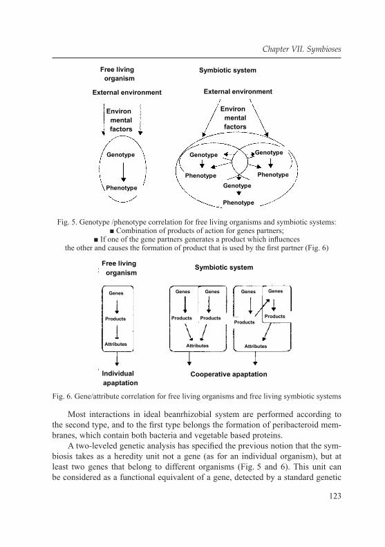
123
Chapter VII. Symbioses
Fig. 5. Genotype /phenotype correlation for free living organisms and symbiotic systems:■ Combination of products of action for genes partners;
■ If one of the gene partners generates a product which infl uences the other and causes the formation of product that is used by the fi rst partner (Fig. 6)
Fig. 6. Gene/attribute correlation for free living organisms and free living symbiotic systems
Most interactions in ideal beanrhizobial system are performed according to the second type, and to the fi rst type belongs the formation of peribacteroid mem-branes, which contain both bacteria and vegetable based proteins.
A two-leveled genetic analysis has specifi ed the previous notion that the sym-biosis takes as a heredity unit not a gene (as for an individual organism), but at least two genes that belong to different organisms (Fig. 5 and 6). This unit can be considered as a functional equivalent of a gene, detected by a standard genetic

124
A.E. Gabidova, V.A. Galynkin
analysis: couples of interrelated Avr/R genes in symbioses turn out to be functional equivalents for couples of А/а alleles in diploid organism [150]. The similarity of basic inheritance scheme for pathogenic system features with monogenic feature inheritance scheme, created by Mendeleev, indicates that symbiotic partners rela-tionship can be described both in terms of intergenic and interallelic interactions. However, it should be emphasized that heredity unit in symbiotic system consists of nonhomologous genes, and this implies the principal difference between meth-ods and results of the genetic analysis for the symbiosis and free living organisms.
It is demonstrated that rhizobacterial genomes are signifi cantly larger than free living bacteria genomes; they range from 4300 to 7000 kbp. For species with high or moderate growth speed (Rhizobium, Sinorhizobium, Mesorhizobium) and can exceed 9000 kbp for species with low growth speed (Bradyrhizobium). Thus, the rhizobacterial genomes may amount to or exceed low eucaryotes in size (for yeast Saccharomyces cerevisiae the genome consists of 6600 kbp.). This is stipu-lated by the necessity to support genetic systems which operate on different life cycle stages (symbiotic and free living) A-proteobacteria wide range compara-tive analysis revealed that on the average plant symbionts have larger genomes (6,73 ± 1,26 mln base pairs) than free living bacteria (4,34 ± 0,99 mln base pairs). Rapid growing species genomes (Zuda) Rhizobium have multiple plasmids in ad-dition to chromosomes. These plasmids can reach extremely large sizes. For the fi rst time multicomponent genomes have been detected in lucerne rhizobacteria (S. meliloti): it was showed that almost all the strains contain two enormous plas-mids (1100–1900 kpb), in addition to the chromosome (about 3600 kpb.), and sometimes 1–6 «moderate» plasmids with the size of 10–400 kpb as well. [151]. Not less complicated genomes were detected in agrobacteria – pathogenes, that cause tumors on plants. These bacteria are taxonomically close to the fast-growing rhizobacteria. It was proved that Zuda-plasmids determine the principal rhizobac-teria symbiotic features: virulence, nitrogen-fi xing activity and host specifi city. Some Sym-plasmids are recommended to control adaptively signifi cant features, important for survival in soils, for example, for complex and rare nutritious sub-strates catabolism, cell and bacteriocins surface components synthesis, as well as for acid resistance and growth speed. For the effective symbiosis development, (associated with active N2 fi xation), other plasmids are also vital: loss of any of them results in a sharp increase in nodule formation speed and/or nitrogenase ac-tivity. A part of genome that is involved in the symbiosis accounts for 15–20 % for rhizobacteria, but less than 1 % for plants, demonstrating that microsymbionts genomes are more adjusted for symbiotic purposes than host pants genomes.
Extremely high fl exibility is the all-round and symbiotically signifi cant fea-ture of rhizobacterial genomes. Adaptive role of symbiotic microbes′ high fl ex-ibility lies in regular transgenations that are the sources of the «raw material»

125
Chapter VII. Symbioses
for coevolution with the hosts. V. Heyman was the fi rst to perform rhizobactera gene horizontal transfer. Model experiments, conducted for Е. coli, demonstrated that the mutations that reduce reproduction speed occur with the frequency of 10–4, per cell, while the mutations that increase it occur with the frequency of 4∙10–9 per cell
The ability of the Sym-plasmids of wide range rhizobacteria to exist in the reсipients who are alien to the donor may derive from the fact that these bacteria got used to living in the nodules that belong to different hosts. This Sym-plasmids´ feature is critically important for symbiosis evolution, for wide range rhizobacteria (for example, R. tropici) can be the donors of the могут быть донорами sym-genes, transferred to different soil bacteria, and that causes new symbiotic nitrogen fi xers
Some rhizobacteria have another peculiarity – their sym-genes form part of special genomic elements – «symbiotic islands» (SI). Having been transferred to the recipient strain the SI are usually integrated into phene-tRNA or val-tRNA, and that is typical for mobile genomic islands, encoding various bacterial prop-erties (including pathogenic ones) [143]. Being deprived of their own replication system, the symbiotic islands can be moved between different strains Mesorhizo-bium spp. by Р 4. Intergrase. The symbiotic islands were also detected in some Bradyrhizobium strains that are able to deliver their sym-genes to soil bacteria, as well as to bacteria, linked to plants.
The symbiotic islands distinctive feature is the reduced quantity of GC (gua-nine cytosine) kb in comparison with other parts of the genome, and that is typical for DNA-elements that have undergone severe transfers in populations. Evolution-ary implication of this transfer is that it can force free living organisms to trans-form into symbiotically active forms.
Transcriptomic and metabolomlic methods have proved that metabolic genes as well as different nutrition sources transport genes are the key symbiotic ele-ments in the rhizobacterial genome [151]. Most symbiotic bacteria comprise both the capacity for genetically controlled biotrophic and necrotrophic interactions with host plants and saprophytic soil ecological niches exploitation. It should be noted that rhizobacteria are signifi cantly inferior to enterobacteria (Escherichia, Salmonella) in terms of transfer severity. These enterobacteria have systems, in-dicated for high-frequent genes transfer (F- and F-factors, effi cient transducing phages), and nevertheless, they exhibit strictly clonal population structure.
Under environmental conditions microorganisms are subject both to physical-chemical factors exposure and other creatures′ exposure (biological factors). The fact that microorganisms participate in biotic relations as a part of population, not as a single cell can be considered as their distinctive feature.
The microorganism-higher organism symbiosis has its own peculiarities. Mi-crobial cells may inhabit both surfaces and different cavities and tissues of higher animals and plants The residential microorganisms are constantly present in the

126
A.E. Gabidova, V.A. Galynkin
higher animal or plant and are reproducing there, and the transient ones can come there from the environment Symbiotic relationship with the macroorganism is mainly based on metabolic products exchange and living space providing. Intravi-tal excretas and mortifi ed parts of the higher organisms are the main alimentation sources for the microorganisms. Macroorganism ensures rather stable conditions for microbial growth and frequently protects microbial population from externa exposures. When microorganisms create a symbiosis with a macroorganism, they contact with the host protective systems and with the other microorganisms-sym-bionts of this macroorganism. Macro-microorganisms symbioses may have differ-ent nature. For example, disease microorganisms, which join parasitic symbiosis with the macroorganism, do a substantial harm to it, causing infectious diseases and sometimes even death of the host organism. In the mutualistic symbiosis, on the contrary, microorganisms are vital for plants and animals. They provide them with some nutritive substances and vitamins and suppress pathogenes. None of natural macrooganisms can exist without «friendly» symbiotic microorganisms.
All-round pattern, which many microbial vegetative symbioses stick to, in-cludes signaling microbial vegetative interaction, metabolite interchange, as well as the production of cell and tissue structures that are vital for the process of sym-biosis. Signal exchange is the key process for all the symbioses. It controls cross regulation and coordinated partners gene expression [124].
Functionally and genetically integrated symbioses have emerged and evolved mainly as perception, transformation and storage systems and in number of cas-es – for inheritance of the signal information, received from the partners
The fact that plants strongly depend on these interactions results in that their genomes are fi lled with DNA sequences, encoding supposed receptors for the sig-nals, received from microsymbionts.
7.6. Soil actinomyces In 1877 Bollinger, a pathologist, and Harz, a plant scientist, studied cow′s
tumors (actinomycetic nodes) and discovered their agent, which was called Ac-tinomyces, because its threads formed a ray This name has become a collective one for several close kinds.
In 1884 in Israel the fi rst pure actinomycetic culture was cultivated (Actinomy-ces israelii). Hereinafter, many pathogenic forms were detected (in 1888 Nocard extracted the fi rst representative of the kind Nocardia from the foot of the man, who had Madura disease), in 1890–1892. Gospirini made a list of actinomyсetic kinds
In 1912–1916.the descriptions of non-pathogenic actinomyces that were ex-tracted from standard natural substrates began to appear. In this period such sci-entists as S.A Vaksman, Krainsky and Rudolf Liske made a great contribution to actinomycetology. 1939 marked a new stage in the science development, when

127
Chapter VII. Symbioses
Krasilnikov got the original kind of the antibiotic mycetin, usually secreted by streptomyces. In 1945 г. Vaksman, Shatz and Bugi extracted streptomycin. Scien-tists paid much attention to actinomyces, but mainly were focused on the applied actinomycetology braches, related to getting and application of antibiotics.
Almost all the known actinomyces kinds were extracted from soil or found in it. The soil is the natural substrate, in which there is a great number of various actinomyces. The number of representatives of some actinomyces kinds (Strep-toverticillium, Micromonospora) is signifi cantly greater in fl oor than in soil. There is always a certain number of small microbial populations that are capable of in-creasing in numbers rapidly and join microbial community, if conditions change quickly or if any part of the trophic chain is missing [152].
Prokaryotic microorganism′s succession in different soils is generally char-acterized by the prevalence of gram-negative bacteria at the initial stages and increase in actinomyces numbers at the latest ones. The actinomyces increase is observed, when the fungi biomass starts to decrease. Probably, the actinomyces frequently use the mortifi ed fungi mycelium that contains chitin. However, until recently the mycelial prokaryotes distribution patterns in soils have been studied for kinds of Streptomyces and Streptoverticillium only. The notion of soil actino-mycetic complex, which explains time-space correlation of certain taxons (kinds, species) of mycelial prokaryotic microorganisms helped to determine common distribution patterns for these microorganisms in environmental substrates, main-ly in soil. Soil actinomycetic complex structure depends on the composition and quantity of typical kinds and species and species range size. Every biogeocenosis has its own soil actinomycetic complex. In the forest biogeocenosis there is one domimant kind of Streptomyces with the typical dominant species of a single sec-tion and class. Forest biogeocenosis soil actinomycetic species function in such circumstances that the principal environment forming factors for them are fl oor, small humus quantity and low pH value. Due to these conditions a mayor part of sorest soil (65 %) is fi lled with the streptomyces that belong to the sort and class Cinereus Achromogenes. It was demonstrated that ecological niches of the explored actinomyces populations in sod-podzolic soils aren′t totally disconnect-ed but overlap to a certain extent. In the forest biogeocenoses the so called oli-gosporic actinomyces (kinds Actinomadura, Saccharomonospora, Microbispora, Saccharopolyspora, Termomonospora, Nocardia) have been encountered in all the layers – the aboveground (shrub leaves, grass), ground (moss, fl oor layers L, F), soil (soil mineral horizons). The most favorable areas for oligosporic actinomyces in coniferous forests are shrub leaves, low fl oor layers (F) and upper soil horizon that are rich in plant remnants of a different decomposition degree, and there their quantity may amount to hundreds of thousands of colony-forming units (CFU) per 1 g of substrate and the total share of actinomyces in the complex is 50 %.

128
A.E. Gabidova, V.A. Galynkin
The actinomyces represent a united branch of a trophic chain in any ecosystem, acting as microbe-reducers. The principal mycelial prokaryotes function is that they decompose such complex polymers as lignin, chitin cellulose and humus compounds. Actinomyces also accumulate biologically active substances in soil and maintain soil nitrogen balance.
One of the features of actinomyces as nitrogen fi xer symbionts is that at the certain stage of their development they are able to form branched mycelium with the diameter of 0,4–1,5 μm, and that results in optimal living conditions [153]. They have a gram-positive cell wall structure and high quantity of GC couples in their DNA (60–75 %) The most favorable place for them is the soil, almost all the actinomyces kinds can be found there. Actinomyces usually account for a quarter of bacteria that grow in traditional environments, if their soil suspensions and 5–15 % prokaryotic biomass are planted. As a rule, their environmental role is to decompose complex stable substrates; they are supposed to participate in hu-mus substances synthesis and decomposition. Can act as invertebrate animals and higher plants symbionts [154].
Mycelium differentiation is the amplifi cation that occurs during actinomycetic colonies development. It mainly manifests in the division process. The mycelium is divided into two types primary (substrate) and secondary (air) mycelium. Air myceli-um is thicker, it is hydrophobic, contains more DNA and enzymes, it has various struc-tures on the cell surface (rod – like fi brils). The spore-forming species air mycelium has thin barriers, in case of non-spore generic species air mycelium has the thick ones (septas). The septa formation begins with cytoplasmic membrane embolying.
Vegetative cells of most of the species are divided by cross barriers, for Geo-dermatophilus and Dermatophilus – in orthogonally related directions, some ac-tinomyces have cells with septas that go in opposite directions (Micromonospora, sporangia Frankia vesicules). Vesicules are Frankia encapsulated nitrogen-fi xing compounds. The branching occurs according to budding mechanism [155].
When cell cytoplasm ages, it acquires uneven electron density, the ribosomes distinguishing stops, nucleoid border blurs, cell wall becomes thin and crumbly, a microcapsule appears. During autolysis in the cytoplasm appear large light areas, nucleoid decomposes, in the cell wall emerge openings, membrane structures fi ll the cell and destroy it.
Nocardial actinomyces seldom form spores and reproduce mainly by means of quickly decomposing mycelium. Actinomyces with long mycelial stages are different from each other in the way of spore formation.
Actinomyces (especially of a kind Micromonospora) are found in waters and bottom deposits, however, it is not clear, if they are constant inhabitants or come from soil. The study of actinomycetic complexes structure has enabled to establish principles of mycelial actinomycetic prokaryotes distribution in biogenocenoses

129
Chapter VII. Symbioses
of basic natural zones, as well as to demonstrate that the actinomycetic kinds, which are traditionally considered to be rare, in particular Мiсrотопоsроrа, Sассharотопоsроrа, Sассhаrоро1уsроrа etc. in certain circumstances can be equal to streptomyces in numbers, and sometimes dominate actinomycetic com-plex [156]. We should increase our knowledge about microbial diversity, and for this purpose we must describe the structure of actinomycetic communities in ter-restrial ecosystems and establish the ecologic status of actinomycetic kinds that are being constantly extracted from soils and vegetative substrates.
Great studies of actinomyces-plant symbioses have been conducted by now. The plants in question are Hippophae, Elaeagnus, Myrica. Oleaster family ac-tinorhizal plants enter bacterial symbiotic relations with the soil actinomyces of Frankia kind. Actinomyces of Frankia kind can produce some phytohormones y siderophores. It is demonstrated that the above mentioned plants have a strong ability to interact with the rhizospheric actinomyces; which grow on roots and form nodules (actinorhiza) [157].
Bacterial symbiosis is one if the positive forms of higher plants and bacteria interactions. Microbial symbionts modify plant mineral elements, produce biolog-ically active compounds, perform protective function, and assimilate molecular nitrogen. With the participation of such plants the nitrogen accumulation volume may reach 150–300 кг per 1 ha per year
Nodular bacteria have an antagonistic impact on many pathogenic fungi. Nod-ular system on the roots of the oleaster plants family has the name of actinorhiza by analogy with mycorhiza Nowadays this type of metaspherm plants and nitro-gen fi xer actinomyces relationship is described for more than 200 species mainly for the tree ones. It is worth mentioning that in the symbiosis with the rhizobial bactera only bean family plants may be treated as macrosymbionts, and in case of actinomyces the range of macrosymbiont plant families is wider.
E.N. Mishustin believes that actinomyces infl uence nitrogen soil balance [150]. Thus, Frankia representatives associate with non-leguminous plants (alder tree, sea buckthorn, etc.). The plant generates nodules at roots in response for ac-tinomyces invasion. In such circumstances the actinomyces begin to fi x air ni-trogen rapidly. Nowadays there is strong evidence that Frankia actinomyces are able to fi x nitrogen in vitro. The kind Frankia, to which belong the actinomyces that are able to form nitrogen-fi xing nodules, was described on the basis of the actinomyces in vivo study. The fi rst pure actinomycetic cuture was obtained only in 1975, under anaerobic conditions in the environment that was challenging for tissue culture by Lalonde method. The kind Frankia consists of the species with a strong mycelium, only a part of thread cells is involved in sporangia formation. Tumors can be either intercalary or terminal. When sporangia decompose, agile or fi xed spores come out of them Air mycelium is absent. All the group members are

130
A.E. Gabidova, V.A. Galynkin
chemoheterothrophic plants with severe food demands, aerobic organisms (mainly microaerophiles), and mesophiles. Frankia nitrogen fi xation process is realized through the special structures (vesicules) that have a size of 3–5 μm, interca-lary located at hyphas terminal ends or at short mycelium sprouts, stringing like beads G.G. Maistrenko argues that vesicules can be surrounded by a capsule layer thickness and density of which depends on age and state of the host plant. Generally, the mycelial confi guration can be found in small nodules of plants and in single top cells of mature active nodules. The vesicules emerge in the plant nodule cells, when the nodules start to branch and prevail, when the nod-ules start to perform nitrogen-fi xation actively.
L.V. Kalakutsky [153] points out that Frankia are considerably superior to nodular bacteria on terms of fi xed nitrogen quantity per nodule mass unit, and the actinomyces-plant symbiosis surpasses all the types of nitrogen-fi xing symbioses in terms of energy saving.
Lately some interesting facts have been obtained that reveal a number of se-crets, related to actinomyces-higher plants symbiosis. Without it active forestry hardly exists. Plant breeders cultivate the plants with certain properties that match to the most effi cient Frankia strains. The development of the advanced methods of their cultivation and selection is a great merit of microbiologists. Taking into con-sideration their achievements, the novel methods of destroyed land have been de-veloped, nitrogen biological fi xation effi ciency in agrosystems has been increased. It has become clear that the actinomyces defi ciency in soil can be a insuperable obstacle for pioneer plants securing in sterile areas, as the actinomyces are indis-pensable for root system infection and nodules formation Meanwhile, you should only infect the plant material with pure Frankia cultures to boost the actinorhizal symbioses without substantial losses.
Prospects of these methods have been undoubtfully proved by numerous experiments with the black alder, planted in artifi cial sand dunes. Nowadays the cultivation of various Frankia stains is becoming biotechnological production ob-ject. They can be widely used for the infecting of young plants, indicated for the recultivation of the destroyed territories, related to open-pit mineral resources op-eration. Plant material suppled with the actinorhizal germs in advance favorably compares with the other kinds of material in growth speed, biomass accumulation rate and nitrogen-fi xation effi ciency.
Soil is the best environment for actinomyces. They are typical for all the con-tinents. In the wild the mutually profi table union of the frankia kinds and plants exists throughout the vegetation period. It proves the energy saving. Energy needs are satisfi ed by the phtosyntesis that occurs in the leaves of the macrosymbiont. Nitrogen accumulation may be stimulated by the light increase and addition of the superphosphate to the soil, molibdenium and cobalt are also essential.

131
Chapter VII. Symbioses
The mycorhizal infl uence on the process of preservation and growth of the tree plants seedings has been repeatedly demonstrated, the stimulating effect of the root exudations on the fungi-symbionts was noted [157]. In the ecotrophic mycor-rhiza the fungi symbionts provide more opportunities for plant-soil contact due the surface enlargening, biochemical activity increase and mushroom caps formation. Besides, the ectomycorhiza protective function with regard to the phytopatho-genes is proved. It manifests in root carbohydrates and other nutritive substances recycling, in the emission of antibiotics that maintain the rhizospheric population of other kinds of microorganisms. On the other hand, the microbial symbionts recycle and prepare mineral nutrition for the plants, produce biologically active compounds, perform protective function and assimilate molecular nitrogen.
In the wild all the plants make up a continuum of the associated with the roots microorganisms that expand from the rhizospere to the rhizoplane and to the plant tissues, including epidermis cortex, endoderma and root vascular system. Soil and plants interact with each other through rhizosphere and rhizospheric organisms during soil microbial regulation.
The analysis of oleaster plants nitrogenase activity dynamics gives us the right to suppose that nitrogen fi xation may be connected with oleaster plants bio-logical features. It has been proved by now that in the beanrhizobial symbiosis, which is kindred to the actinorhizal one, host plant genes affect many symbiotic processes, determining the nodule-forming capacity infl uencing their quantity, as well as intracellular differentiation and nitrogen fi xation effi ciency In the south of the West Syberia when the oleaster plants are cultivated at plantations, they have the following development cycle: the vegetation starts in the fi rst ten days of May, the sea-buckthorn blossoms in the middle-end of May, the moss blossoms in June. The oleaster plants study has revealed the connection of the vegetation with the root growth. The root formation and branching, in its turn is related to nodule development and Frankia infection. Sprout growth depends deeply on the root sys-tem development. Therefore, the nitrogen fi xation process reaches its peak in this period Other relations of the oleaster plants nodular nitrogen fi xation fl uctuations that happen during vegetative development are in line with the infected tissue metastructure seasonal changes and the peculiarities of the endophyte functioning.
The actinomyces are vital for soil microbial complex, they account for a quar-ter of the total quantity of bacteria, grown in the commonly used nutritive media, when they are cultivated from dissolved soil suspensions The actinomycetic my-celium accounts for 5–15 % от of the total bacterial biomass in soil.
For a long time the number of emitted streptomyces has been the principal factor for actinomyces distribution in soils and related natural substrates. The development of the selective combined methods for micromonospores sphingo-sporangia and oligosporic actinomyces extraction has helped to establish certain
132
A.E. Gabidova, V.A. Galynkin
patterns in the hierarchy of the actinomycetic complexes in biogeocenoses: the vertical stratifi cation of the actinomycetic distribution in biogeocenoses, which is characterized by the continuity (for streptomyces) and discontinuity (for the other kinds). Various kinds of actinomyces participate in the process of organic sub-stances decomposition step by step, occupying certain space and time position in the ecosystem, according to land existence adaptation, mycelium and spores, en-vironmental strategies and the type of the relationship of this kind with the other kinds in the actinomycetic complex. In certain circumstances (soil type, succes-sion stage) the actinomycetic kinds that are usually considered to be rare can be equal to streptomyces in the actinomycetic complex and sometimes exceed them.
It is demonstrated that the representatives of Мiсrотопоsроrа kind are the most frequently extracted from soils and vegetative substrates.
The number of micromonospores in mineral soil horizon is minor, it is infe-rior to streptomyces.
The forest biogeocenoces are characterized by the sporangial actinomyces which are found in all the layers, they are comparable with streptomyces in num-bers. The sporangia are known to endure acid environmental conditions.
In forest biogeocenoces the oligosporic actinomyces are minor; their share in the actimomycetic complex seldom amounts to 50 %. The oligosporic actinomy-ces, slowly growing species as a rule, require additional alimentation sources. The numeric dominance of micromonosporic actinomyces over Streptomyces is one of the distinctive features of biogeocenoses in drained peat-bogs. In the ac-tinomycetic complexes of these biogeocenoses the representatives of oligosporic actinomyces are commonly found.
Thus, the micromonospores and streptosporangia can′t be regarded as rare species in the forest area soils. The number of actinomyces of these kinds is in some cases comparable with the number of streptomyces and sometimes even ex-ceeds it. The oligosporic actinomyces are rare for the forest area.
In the arid area biogeocenoses micromonosporous actinomyces are con-stantly present in all the layers – aboveground, land and soil. In the steppe mat micronospores exceed the streptomyces, in the vegetative and soil layers of the steppe biomes the streptomyces and micromonospores are represented in equal shares. Streptosporangia are less common for the biogeocenotic actino-mycetic complexes in the arid area.
Since 1980s–1990s the scientists′ attention has been focused on actinomy-ces ecological functions study, their natural relationship with animals, plants and microorganisms. There has been a systemic review of actinomycetic ge-nome data collection [104, 105].
Structural trends for streptomycetic complex in ground ecosystems can be defi ned as follows:
133
Chapter VII. Symbioses
● The study of the soil streptomycetic complexes structure, pursuant to which the structure depends on typical kind′s composition, dominants number and spe-cies range size, has enabled to fi nd out the differences of these complexes in terms of basic bioclimatic zones soils. It has been identifi ed that human impact results in creation of specifi c streptomycetic complexes that are signifi cantly different from natural land ecosystem complexes. A streptomycetic complex. A streptomycetic complex is used to compile rating scale for increasing lime and mineral fertilizers doses effect on soil microbial system state and homeostasis preservation.
● The succession method of streptomycetic complexes studying has allowed to determine and evaluate ecological features of the streptomyces types that are constantly present in soil – adaptiveness, population ecological strategy, ecologi-cal niches overlapping – and to use these features for resolving issues of ecosys-tem microbial diagnostics.
● It has been identifi ed that human impact results in creation of specifi c strep-tomycetic complexes that are signifi cantly different from natural land ecosystem complexes. A streptomycetic complex. A streptomycetic complex is used to com-pile rating scale for increasing lime and mineral fertilizers doses effect on soil microbial system state and homeostasis preservation.
Thus, it has been established that apart from ubiquitous Streptomyces, which are found in all the explored soil types of the basic soil-climatic zones and are present in all the vertical structure layers of the basic biogeocenoses types, monosporous and sporangial actinomyces are also essential elements of the actinomycetic communities in the land ecosystems. The Мiсrотопоsроrа is the most common representative of these actinomyces kinds, their function in these areas is unknown.
Soils are the natural substrates which contain plenty of actinomyces. How-ever, a mayor part of actinomycetic biomass is represented by spores which make colonies, if the population are controlled by seeding technique, mycelium aссounts only for 1–4 % of the biomass. It is located in the microzones with the high organic substance content.
Actinomyces are prevalent at the latest stages of the microbial succession, when exist conditions for remote substrates use. Atinomycetic microbial fl ora ac-tivation occurs, when such substances as starch, chitin, oil products etc. penetrate into the soil. At the same time due to the slow growth the actinomyces aren′t able to compete with non-mycelic bacteria for the easily reachable substances. It is possible that the secondary metabolites (melanoid enzymes, in particular) some-how participate in humus formation Actinomyces fulfi l cenosis-forming function at the sites of primary soil formation, being associated with the seaweed in this case. Under laboratory conditions these associations have resulted in lichen-like thallome (actinolichen).

134
A.E. Gabidova, V.A. Galynkin
Actinomyces (kinds Streptomyces, Streptosporangium, Micromonospora, Ac-tinomadura) constantly reside in the intestine of earthworms, termites and many other invertebrates. They destroy cellulose and other biopolymers and turn out to be their symbionts. Pathogenic forms, triggering actinomycosis in the human body, are located in oral cavity, intestine, in the respiratory ways, on skin, in den-tal deposit, in decaying teeth, at tonsils.
Most actinomyces are aerobial bacteria; facultative anaerobes are present only among the actinomyces with the short mycelial stage. In this case we can draw some analogies with the fungi, among which only non-mycelial yeast can exist in anaerobic conditions. The less effective anaerobic metabolism type is supposed to be successful, if the relative cell surface becomes larger and this can be obtained by mycelium fragmentation.
Actinomyces are more resistant to drying than non-mycelial bacteria that is why, they prevail in desert soils. Sclerotia produced by Chainia kind can be pre-served at drying for quite a durable period of time. It is demonstrated that when aw = 0,50, some spores sprout (k. Streptomyces, Micromonospora), however the emerged mycelium doesn′t branch. At aw = 0,86 almost all the actinomyces spores sprout, some of them have a branching mycelium, the microcolonies are formed, at aw = 0,95 the optimal results can be reached.
In most cases the actinomyces are neutrophils however some kinds are aci-dophilic or alkalophilic. Acidoresistance is the actinomyces′ distinctive feature, owning to which their share in forest soil microbial complex is relatively big. It is noted that in acid media the vegetative phase is longer, while in the alkalic one the spore formation speeds up.
The actimomyces don′t require high organic carbon content in the environ-ment; any of them are able to grow on the «starvation» agar. Nocardia members are able to perform chemosynthesis, oxygenating hydrogen, methane and metha-nol. Heterotrophic CO2 fi xation is quite common among actinomyces
The ability to synthesize physiologically active substances antibiotics, color-ants fragrant compounds (substances geosmin, argosmin, mucidon, 2-methyl-isob-orneol). They are responsible for specifi c smell of soil and sometimes of water. The actinomyces are active producers of antibiotics they form up to the half of all the known kinds of them [155].
Actinomyces are the symbionts that are able to infect only the parenchymal cells of the root cortex. Similar to the bean family plants infection; the microorgan-ism penetrates into roots through soil, through root fi brils that get crooked in the end. At the contamination site the root fi bril walls are thickening and the hyphas that have penetrated into the cell are being covered with a thick case. As the hyphas are going through the root fi brils, the case is getting thinner and there appears a cap-sule around hyphas. It is considered to be created both by plant and actinomyces.

135
Chapter VII. Symbioses
From the root fi bril the hyphas go to epidermis and root cortex, causing divi-sion and hypertrophy of the infected cells. Hyphas nodules fi ll the center of the plant cells, at the cell walls occur extension and division of the hyphas ends, then special structures, the so called vesicules, are formed. In the nodules emerge the substance that is similar to leghemoglobin of the bean family plants. When the vegetation ends, the vesicules lapse, but the plant cells still contain hyphas that contaminate new tissues in spring. As a rule, when we deal with the nonlegumi-nous plants, the Frankia actinomyces have more nitrogen-fi xation energy than le-guminous plants nodular bacteria [156].
7.7. Plant- actinomyces symbiosis (actinorhiza)Mycorhizal fungi (fron Greek. mykes – fungus, rhiza – root enter into sym-
biosis with higher plants. Mycorhiza is a very interesting and even unique nat-ural occurrence: the organisms that have a totally different structure and life principles join and support each other during their life cycles. There is a good saying. “If there is no forest, there are no mushrooms”. We deal with a spe-cial plant-mushroom community, and the names of mushrooms confi rm that this community really exists (birch bolete, aspen mushroom). The discovery of the mycorhiza is the scientifi c rationale for the existence of such a community. This seems as follows: when a certain mycelium initially spreads in soils, at the cer-tain stage of development it fi nds small rootlets that belong to a certain type of a plant, it covers them with a special invisible “clutch”, creating a mushroom cap. This is benefi cial for both organisms. The plant supplies a mycorhizal fun-gus with carbohydrates, carbon dioxide and oxygen, if necessary. The fungus, in its turn, provides plant with macro-and microelements and water. Besides, the mycorhizal fungus protects the plant from harmful microorganisms.
Symbiotic fungi mycelium can exist in soil without mycorhiza for a certain period of time, but in this case it is unable to form fruit bodies. Thus, we can′t obtain fruit bodies from ceps, Russulaceae and amanitas – they all are mycorhiza formers and are incapable of bearing fruits without certain kind of tree. In its turn, the plant without the fungi symbionts grows and develops slowly, is subject to dif-ferent diseases and can even die. For instance, pine seedings can become strong and high pines, only when their roots are covered with a certain type of fungi.
In the middle of the 1950s it was decided to make “green ring” around the city of Elista (Kalmukia) to protect the city from severe prairie winds. Acacia was cho-sen for this purpose. When it was planted for the fi rst time – all the plants died, again and again – the result was the same. Then they had to resort to the scientists. Soil and wood remnants analysis showed that there were no acacia mycorhiza form-ers in soil. Next year new young plants were planted, they struck to root and started to grow, for the necessary fungi mycelium had been added to soil beforehand.

136
A.E. Gabidova, V.A. Galynkin
Orchid seeds are rather tiny and don′t contain any nutritive substances and therefore can sprout, only if they have a fungus-symbiont. The orchid sprouts should get food through symbionts for a long time. These symbionts destroy ready-made organic substances. Due to the mentioned above, the orchids initial-ly have a lot of mycorhizal fungi that help them to absorb nutritive substances. When a given period of time passes, the orchids don′t need the symbiosis any more, and the fungus dies, but before that he manages to generate many ripe spores, which will be spread by water or wind, and then these fungi spores make a symbiosis with the new orchid′s sprouts.
The mentioned above examples demonstrate that the mycorhizal fungi are of utmost importance: they facilitate growth and successful development of tree, shrub and herb plants. Many micromycetes almost all the tubularids, all the Rus-sulaceae and about 35 % of gill fungi belong to the mycorhizal fungi. Therefore, everyone, who goes to the forest, shouldn′t destroy any fungi that are unknown to him, even if they are poisonous, because they are vital for the forest itself and its inhabitants. The fourth experiment showed that the shortened variants of SYMRK gene are enough for actinomyces, but not for nodular symbioses [157].
Like in the third experiment, here the Japanese lotus mutant kind was used. This kind neither generates actinomyces, nor enters plant symbiosis. “The gene of moderate length”, borrowed from tomato and “shortened gene”, borrowed from rice was transferred to plants. In both cases the mutant lotus restored the capacity for generating actinomyces, but still wasn′t able to enter plant symbiosis. Taking into consideration these and a number of other experiments; the scientists concluded that SYMRK protein is apparently vital for special intracellular structures formation – a kind of “symbiont-receivers” or “(pre-infection threads”, which are further settled by symbiotic bacteria (and after that they are called “infection threads”). Similar “symbiont-receivers” are formed in the root cells and before fungi symbionts ac-ceptance in the process of АМ formation (these structures are called pre-penetration apparatus). Both structural similarity and similarity in the mechanisms of formation of these “symbiont-receivers” probably refl ect the entity of the genetic scenario, re-sponsible for all three types of the intracellular symbiosis: АМ, PS and АR [158].
Mutations that occur in some “common symbiotic genes” result in faults in “symbiont-receivers” structure (SYMRK, naturally, is not the only gene, required for their formation). It should be noted that “long” SYMRK protein is vital, but not suffi cient for nodular symbioses formation. This is obvious, for not all the organisms can enter these symbioses, but only those that have a long variant. The plants that generate nodules apparently must have some other genetic pecularity as well, but they haven′t been identifi ed yet.
The ability to form nodular symbioses (АR and PS) has been developed due to ancient АМ genetic scenario. The key evolutionary event is that the root cells

137
Chapter VII. Symbioses
have acquired the ability to react both on symbiotic fungi presence and nitrogen-fi xing bacteria proximity by forming “symbiont-receivers”. It seems that this event has happened due to SYMRK restructuring, to be exact, due to addition of two new receptor domains to its intracellular part. Both of them could be bor-rowed from other proteins in the higher plants genome. Thus, nodular symbioses are striking examples of new function development by modifying of gene com-plex that used to fulfi l other functions. Arbusculas, mycelium modifi cations in my-corrhiza formers fungi, which are similar to haustoria, are complex repeatedly and dichotomically branched hyphas that penetrate into root parenchymal cells. Within the cell they are surrounded by the cellular plasmalemma. The most active metab-olite exchange between mycorhiza elements takes place in arbusculas, though they exist only for a few days (afterwards dissolve). The scientists believe that arbus-culas are formed under the infl uence of plant cells protective reaction (ref. Fig. 7).
Fig. 7. Morphological traits for various mycorrhiza types

138
A.E. Gabidova, V.A. Galynkin
Plant symbiosis with the actinomyces of the kind Frankia – is another ex-ample of a nodular symbiosis. The nitrogen-fi xation for this type is less effi cient than for rhizobial one. However, the latest data on the expression of some genes in actinorhizal plants allow to deduce that their nitrogen fi xation process has much in common with the bean family. At least seven common genes are involved in these two kinds of symbiosis. These genes are called “common symbiosis genes”. For instance, to this group belong the genes that are engaged in the formation of “pre-infection threads”, in which further live symbiotic bacteria. It is probable that actinorhizal lectins (just like bean ones) initiate the symbiosis with Frankia [104, 105]. Thus, transgenic actinorhizal plants may become the fi rst pattern non-leguminous plants to enter the symbiosis with the rhizobacteria and to be recog-nized by them (in particular, sea-buckthorn, the symbiosis of which is similar to parasponia and some species of the bean family [100].
Some cyanobacteria and actinomyces may enter symbiotic relationship with plants, in particular, Frankia species. Frankia can establish symbiotic relationship with more than 200 species of dicotyledonous arboreal plants from eight different families, for instance Aims, Hippophae, Dryas, etc. At penetrating into the plant some hyphas of Frankia evolve into morphologically unique structures, able to perform nitrogen-fi xation. They are called vesicules. As a result, on the infected plant roots appear nitrogen-fi xer nodules and there occur nitrogenase synthesis and nitrogen fi xation. It is worth mentioning that Frankia bacteria are able to per-form nitrogen fi xation even in a free living state, i. е. without plant contact.
We have found out that the sea-buckthorn in question has not so large nodules in the shape of thick root networks (branched like corals), which mainly are locat-ed at the lateral roots in the top soil layer (5–20 cm). Microorganisms′examination showed that they penetrate into roots from the soil, through root fi brils that have crooked in the end. At the contamination site the root fi bril walls are thickening and the hyphas that have penetrated into the cell are being covered with a thick case. As the hyphas are going through the root fi brils, the case is getting thinner and there appears a capsule around hyphas. It is considered to be created both by plant and actinomyces. From the root fi bril the hyphas go to epidermis and root cortex, causing division and hypertrophy of the infected cells. Hyphas nodules fi ll the center of the plant cells, at the cell walls occur extension and division of the hyphas ends, then special structures, the so called vesicules, are formed with the size of 3–5 μm. The root nodules may be located at any place, they are of moder-ate size and are mainly situated at the lateral roots

139
Chapter VIII. HISTORICAL ASPECTS OF RESISTANCE OCCURRENCE
Resistance (stability) is understood as the capability of a microorganism to sur-vive much larger concentrations of the preparation than other microorganisms of this strain (type), or to develop under such concentrations which exceed those achievable in the macroorganism in case of administration of antibiotics in therapeutic doses.
In 1905, Franke и Roehle, who worked with Ehrlich, discovered a phenome-non of resistance to medicinal substances. P. Ehrlich thought that resistance occurs due to gradual reduction of a number of receptors or their masking in the organ-ism. Nevertheless, Yorke and his colleagues (1931) showed that, at least in 331. Trypanosomiasis, resistance occurs due to reduction of penetration of the medical substance to the parasite′s organism. Highly stable strains absorb medicinal prepa-ration, and that′s why its concentration in the environment remains suffi cient to kill sensitive strains. These researchers had a signifi cant advantage towards Ehr-lich: they learned to cultivate trypanosomes in vitro.
Ehrlich found three various types of resistance of trypanosomes. Parasites, which acquired resistance to the action of the trypan red, became resistant to all azo dyes. Other strains, resistant to the action of atoxyl (para-aminobenzene arsenic acid), manifested resistance to the action of all the phenyl-arsenic acids. The third, resistant to the action of parafuchsine, proved to be resistant to all other derivatives of triphenylmethane. However, trypanosomes strains resistant to the drug of one of these classes of compounds, turned to be sensitive to drugs of other classes, if resist-ance to them was not specially developed. Resistance of agents to medical products results in a sharp decline of effectiveness of causal treatment of infectious diseas-es. The general term for defi nition of this phenomenon is antibacterial resistance (ABR) – resistance of bacterial agents of infectious diseases to various therapeutic drugs. The main difference from other substances having a toxic effect on a bacterial cell is their high selectivity; they inhibit metabolic processes unique for a prokary-otic cell and absent in eukaryotic cells, in concentrations suppressing vital activity of bacteria, and usually have no impact on the macroorganism.
From the position of microbiology, antibiotic resistance is the ability of micro-organisms to survive in presence of therapeutic concentrations of ABA. Over the last years, stable tendency of increase of therapeutic doses of drugs is observed. In the circumstances of constant selective exposure of the environment, forma-tion of strains of microorganisms takes place with resistance levels signifi cantly exceeding therapeutic concentrations. The notion of antibiotic resistance may be

140
A.E. Gabidova, V.A. Galynkin
interpreted as the ability of microorganisms to withstand their concentrations, ex-ceeding those achieved in the human organism.
By genetic mechanisms, antibiotics resistance of microorganisms can be pri-mary (natural) and acquired. Primary natural resistance is characterized by absence in microorganisms of the target for action of the antibiotic, or by impermeability of the cell wall for certain medicines. In case of presence of natural resistance in bacteria, antibiotics are clinically ineffi cient. Natural resistance is a permanent species character of microorganisms and is easily forecast. Antibiotic resistance is defi ned and maintained by resistance genes (r-genes) and by the conditions, add-ing to their dissemination in microbial populations. Acquired drug resistance may occur in case of mutations in the chromosome, transfer of transmissive resistance plasmids (R-plasmids) and transfer of transposons carrying r-genes. In addition, formation of resistance to individual drugs, that was observed at dawn of the “an-tibiotic era», has lost its actuality as of today, and these days, the focus is made on formation of poly-and pan-resistance. The concept of resistance should be consid-ered taking into account the following principles:
● Pharmacological principle. Peculiarities of the drug should be taken into account: its pharmacokinetics and pharmacodynamics, distribution in the body, fre-quency, of administration, possibility of combining drugs, etc. Drug doses should be suffi cient to ensure bacteriostatic or bacteriocidal concentrations in biological fl uids and tissues. One should imagine the optimal duration of treatment, as clinical improvement is not the ground for withdrawal of the drug, because pathogens may persist in the body, and there can be a relapse of the disease. Also optimal routes of drug administration should be accounted, as many antibiotics are poorly absorbed from the digestive tract or do not penetrate through blood-brain barrier.
● Clinical principle –when prescribing the drug, it is necessary to take into account how safe it will be for the given patient, which depends on individual condition of the patient (severity of infection, immune status, sex, pregnancy, age, state of the liver and kidneys functions, concomitant diseases, etc.) In severe, life-threatening infections timely antibiotic treatment is of particular importance. Such patients prescribed combination of two or three drugs to ensure the widest possi-ble range of action. When a combination of several drugs is prescribed, one should know, how effective a combination of these drugs will be against the agent and safe for the patient, i. e., that there should be no antagonism of drugs with respect to antibacterial activity and no summing up of their toxic effects.
● Epidemiological principle implies selection of the drug, especially for an in-door patient, it should take into account state of resistance of microbial strains, which circulate in the given unit, hospital and even in the region. One should remember that antibiotic resistance may not only be acquired, but also be lost; herewith, natural sensitivity of the microorganism to the drug is restored. Only natural stability does not change.
141
Chapter VIII. Historical aspects of resistance occurrence
● Pharmaceutical principle: one should take into account shelf life and abide by the rules of drug storage, because in case of violation of these rules, antibiotic may not only lose its activity, but become toxic due to degradation. The cost of the drug is of greatest signifi cance, as well as absence of counterfeit drugs.
It was found out that for normal functioning of human physiological systems, crypt compartment has primary importance. It forms placenta biofi lm, which pro-duces metabolites and biologically active substances, defi nes their food – trophic, energy and other links between all microorganisms and the external world.
Now presence of the diversity of species in the crypt compartment is proved, the dominant role of bifi d bacteria in the intestinal microbiota, but at present, availability of eubacteria, clostridia and actinomyces, which, according to contemporary estimates, are contained in the intestine in the amount massively more than bifi d bacteria.
8.1. Protection system of pathogenic microorganismsInfection is a complex biological process, resulting from penetration of patho-
genic microbes into the body and violation of constancy of its internal environment. Agents of infectious diseases include microorganisms of plant and animal origin – bacteria, spirochetes, lower fungi, protozoa, viruses and rickettsiae. Infectious agents are primary and mandatory reason of development of the infectious disease, they de-fi ne the “specifi city” of the infectious disease, peculiarities of clinical aspects of pa-thology, but not every case of penetration of an infectious agent into the body ends with development of a disease. In response to the action of infectious pathogenic factors, specifi c immunological defense mechanisms are activated and nonspecifi c resistance factors, release of adaptation hormones takes place. Transformation of the preimmune response into the disease is determined by the grade of pathogenicity, virulence, invasiveness, organotropy and toxigenicity of microorganisms, as well as by the initial state of the microorganism with its reactivity and resistance [159].
Pathogenicity is the ability of a particular kind of microbe cause an infectious disease characteristic for it in appropriate circumstances [160]. Thus, pathogenic-ity is a species character. Pathogenicity as a special quality of disease-causing type of microbe is manifested in its aggressive properties and toxic impact on the body. Pathogenicity is an unlimited sign, which is realized with participation of many factors, including, in particular, toxins, adhesins and pathogenicity enzymes [161].
Pathogenicity attributes include infectivity pathogenicity, invasiveness and toxigenicity. Factors of pathogenicity of infectious agents based on their biologi-cal activity in the body can be divided into 4 groups:
1. Defi ne interaction of bacteria with the epithelium of the relevant ecological niches.2. Ensuring reproduction of the agent in vivo.3. Bacterial modulins, inducing synthesis of cytokines and of infl ammation
mediators.

142
A.E. Gabidova, V.A. Galynkin
4. A special group of pathogenicity factors consists of toxins and toxic prod-ucts with direct or indirect cytopathic effect.
Penetration of a microorganism to the macroorganism is designated as infec-tivity. Factors of spreading of infectious agents in the inner body environment are: enzymes (hyaluronidase, collagenase, neuraminidase); fl agella (of cholera germ, es-cherichia coli, proteus); undulating membrane (in spirochetes and some protozoa).
Virulence is the degree of pathogenicity of a certain strain of microbe, i. e., an individual trait. Virulence of a microbe may be enhanced in course of its pas-sages through a sensitive organ. Virulence of a microbe can drop in natural condi-tions under the infl uence of sunlight, drying, etc.
Aggressiveness is the ability of a pathogenic microbe to live, reproduce and spread in the organism, to withstand adverse infl uence caused by the body. Some pathogenic microbes, when reproduced in the body or in nutritional medium in vitro, producing soluble products called aggressines. Purpose of aggressines – is to sup-press action of phagocytes. Aggressines themselves are harmless for the organism.
Toxicity – ability of a pathogenic microbe to produce and excrete poisonous substances, having harmful impact on the organism [162]. There are two types of toxins: exotoxins and endotoxins. Toxins are biomolecules of bacteria which induce development of specifi c symptoms of an infectious disease. As a rule, tox-ins manifest their effect in miniscule concentrations compared to other factors of pathogenicity. Real toxins are produced by representatives of both gram-positive and gram-negative microorganisms are determined not only by cytopathic effects of lipopolysaccharide (LPS), but also by biological effects of relative exotoxins and factors of pathogenicity. Exotoxins – are released into the environment in course of microbial life in the body or on artifi cial nutrient media, as well as in food products. They are very poisonous. Endotoxins are fi rmly bound with the body of microbial cells and are released only after its death and destruction. They are very resistant under the impact of high temperatures and do not disintegrate even after several hours of boiling. Poisonous effect of many bacterial exotoxins is associated with enzymes – lecithinase (destroys red blood cells), collagenase, hya-luronidase (breaks down hyaluronic acid) and a number of other enzymes, which destroy vitally important compounds in the organism. Some pathogenic bacteria (diphtherial staphylococci and streptococci) secrete enzyme deoxyribonuclease.
Probably, reduced virulence of the agent can be considered as a mechanism of evasion from host′s protective reactions. In course of the disease, change in the agent′s properties is observed. It is shown that prolonged forms of the infec-tious process are explained by reservation phase of development of populations of microorganisms, in which a decline in the quantity of agent and its virulence is noted. Qualitative analysis of heterogeneity of populations of staphylococ-ci showed, that before cessation of acute infectious phase of the disease, only

143
Chapter VIII. Historical aspects of resistance occurrence
colonies with a low level of antilysozymic activity remain, and clones are elimi-nated with pathogenicity factors.
Reduced virulence is also observed in the process of circulation of poliomy-elitis virus. According to molecular epidemiology, virus circulation in the nature and its replication in an individual organism are accompanied by accumulation of mutations due to inaccuracies in copying of RNA molecules. These mutations result in reduced viability and virulence of the poliovirus. Such strains lose their ability to cause human disease. Passaging of L. major, strain, which reduced viru-lence, through the organism of highly vulnerable laboratory animals leads to acute ascending of the level of virulence of the strain and to return of the capability of causing a specifi c pathologic process in humans [163].
Factors of adhesion and colonization ensure interaction of the pathogenic in-fectious agent with specifi c receptors of cells of those organs and tissues, to which tropism was detected. Adhesive molecules are substances of protein and polysac-charide nature, expressed on the surface of the cells. Following adhesion, propa-gation and formation of a large number of homogeneous microbes (colonies) is inevitable in the event of lack of local and systemic resistance mechanisms and specifi c immunological protection mechanisms. Penetration of a microorganism to the macroorganism is designated as infectivity. Factors of spread of infectious agents in the inner body environment are: enzymes (hyaluronidase, collagenase, neuraminidase); fl agella (of cholera germ, Escherichia coli, proteus); undulating membrane (spirochetes and some protozoa); microbe resistance in the macroor-ganism is determined by factors, specifi c for this or that agent [164].
At present, genetic mechanisms of determination of factors of pathogenicity of infectious agents become more and more evident. It is shown that genetic con-trol of synthesis of pathogenicity factors determinants adhesion and colonization of the intestinal epithelium in pathogens Escherichia, penetration and intracellular multiplication of shigella, salmonella and yersinia, are ensured by chromosomes and plasmids. Herewith, plasmid genes determine factors of interaction of the agent with epithelium, while chromosome one – existence and the reproduction of bacteria outside the epithelium. Pathogenicity factor may participate in various phases of the infectious process, and various factors of pathogenicity take part in the same phase. Intense synthesis occurs of bacterial toxins, possessing direct or indirect cytopathogenic impact on cellular structures of various organs and tissues. The effect of infectious pathogenic factors led to development of direct and cy-tokine-determined systemic functional and metabolic disorders, immune reaction, allergic reactions, states of immunodefi ciency, as well as autoimmune aggression against own damaged or undamaged cellular structures.
An effective mechanism of protection against immunity factors is generation by the agent of biologically active substances, capable of destroying or inactivating

144
A.E. Gabidova, V.A. Galynkin
the substances which have adverse impact on the agent. Anticomplementary activ-ity of staphylococci, gonococci, coliform bacteria and Klebsiella was described and anti-IFN activity in a wide range of pathogenic enterobacteria obtained from sick people. Trypsin-like bacterial proteases are able to cleave and inactivate se-cretory IgA. Excretion of such proteases has been found in protea, pseudomona-daceae, cocci e many other microorganisms – human pathogens. IgA proteases of various kinds of microbes differ by antigenic properties and substrate specifi city. Proteases are known decomposing and inactivating lysozyme. The results of study of correlation between reduction of the level of lysozyme of host cells and the amount of the antilysozymic activity of shigella were used in clinical practice to predict development of bacteria carriage in patients with bacterial dysentery [165].
Infections are one of the leading causes of formation of acquired immuno-defi ciency. Agents of infectious and parasitic diseases are able to actively block certain links of the immune response or cause pronounced defects in functioning of the immune system, which hinders effi cient elimination of the pathogen and re-covery. A universal mechanism of prevention of adverse effect of the immune sys-tem on agents of human diseases is immunosuppression induced by the pathogen.
Immunodefi ciency is formed not only in case of diseases of parasitical nature. Most viral and bacterial infectious diseases occurs on the background of developing immunodefi ciency. Pathology of the immune system is detected at the background of active tuberculosis, relapsing herpes, measles and many other infections.
One of the defense mechanisms aimed at maintaining its homeostasis is apop-tosis, or a death of the cell as the result of programmed self-destruction. In this way the organism effectively gets rid of genetically defective material – cancer, defective cells or cells infected by the cell agent, etc. Death of infected cells with subsequent elimination of the destroyed cells and agents by the cells of the immune system is able to prevent spread of the infection process in the organism of the infected person [166]. It is well known that programmed cell death is the fundamental natural mechanism of positive and negative selection of Т-and В-lymphocytes, aimed at elimination of cells with defective antigen-recognizing receptors or of those having the ability to react against “own”. Up to 95 % of T-cells are eliminated in the thymus in course of mat-uration based on similar mechanism. Controlled apoptosis is considered as the main mechanism for maintaining of the optimal balance in the immune system. It is shown that microbial pathogens are capable of managing host cells apoptosis for the prolonga-tion of their own existence [167]. Currently, effective mechanisms to control apoptosis have been found in many agents of human diseases. It was shown that appearance of thrombocyte- bacterial associations, leading to morphological changes of blood cells, is a pathogenetic mechanism of hemodynamic disorders. In case of relapses of typhoid fever and severe progress of yersiniosis, functionally defective segmented leucocytes appear in the blood with ultrastructural signs of apoptosis and necrobiosis.

145
Chapter VIII. Historical aspects of resistance occurrence
Infectious agents parasitizing strictly intracellularly – rickettsia, chlamydia or shigella can prevent apoptosis of the infected cell [168]. Cowpox virus pos-sesses pronounced properties of suppression of apoptosis of infected cells. The mechanism of prevention of apoptosis of infected cells, i. e., ensuring life cycle of a pathogen, has been found in several viruses – adenoviruses, Epstein–Barr virus, baculovirus and infl uenza A and B viruses. In case of infection caused by HIV-1, infected cells defend themselves against apoptosis, whereas death of uninfected cells of the immune system can lead to severe immunosuppression [169].
For any agent of infectious and parasitic disease, in addition to ensuring the possibility of existence and reproduction of the host organism, it is vital to ensure transition from one hot-blooded host to another. For many agents, this process is associated with stay in the natural environment or in the body of an arthro-pod carrier. Change of habitat, in all respects differing by living conditions of the pathogen, is accompanied by a stress impact on the agent. In case of such trans-fer, some agents completely change their morphology, others generate specialized forms, resistant to adverse environmental factors. It should be noted that there are agents, expressed morphological changes of which occur in case of transfer from extracellular to intracellular parasitizing, even within a single host organism. The last option, a metamorphosis, is characteristic of trypanosomes – agents of Chagas disease, or South American trypanosomiasis. In case of primary clinical manifes-tations, the agent is present in the blood in its typical form of an elongated cell with long fl agella, which ensures mobility of the parasite. At the later stages char-acterized by lesions of the smooth muscles of the heart and of the intestines, the parasite lives intracellularly in smooth muscular fi bers. At this stage, parasites lose their fl agella and turn into a roundish cell, which actively divides inside the cell of human smooth muscular fi bers, causing dysfunctions of the latter.
During stay in the external environment, many bacterial agents form spores. Spores are a collective name for those stages of development of a microorganism in which the bacterial cell resides without reproduction in abiotic environment with prac-tically full cessation of metabolism. Similar stages of protists (protozoa) are called cysts or oocysts, depending on the structure of protozoal cells in the dormant phase, and those of helminths are called eggs. In the stage of an egg the larva of some hel-minths passes certain development accompanied by a noticeable metabolism. Howev-er, having reached an invasive stage inside the egg, helminths′ larva keeps its viability and ability to contaminate for a long time. For the egg of a human roundworm, keep-ing of invasive properties in favorable conditions may last for up to 10 years.
With the discovery in 1985 by R. Colwell with co-authors of uncultivated forms of bacteria, which retain viability and virulence, it became clear that the idea about the destiny of pathogenic microbes in the environment, based only on bacteriological studies, is certainly incomplete [170]. The state of the parasite,

146
A.E. Gabidova, V.A. Galynkin
which was considered a “die-away”, proved to be adaptive variability. Passage of microorganisms into the dormant state is accompanied by a decrease in the level of metabolism and cessation of reproduction. Such dormant forms of some patho-genic bacteria, including cholera and plague agents, were found in water basins and soils on endemic and enzootic territories. In case of change of the conditions, dormant forms reverse into vegetative, capable of causing epidemic manifesta-tions. These mechanisms are purposed to ensure preservation of nonsporeforming bacterial agents into adverse inter-epidemic or inter-epizootic periods.
Bacterial cells in uncultivated state morphologically differ from the same bacteria in active vegetative state. As a rule, uncultivated forms have smaller size compared with vegetative cells, they have higher density of cytoplasm and modifi ed cell membrane. Presence of uncultivated cells was found in the exter-nal environment in three states – viable (form colonies when on nutritive me-dium), semi-viable (do not form colonies, but shows signs of some metabolic activity) and dormant (do not have metabolic activity). Uncultivated forms of bacteria in a dormant state are able to restore vital functions only after special procedures, such as passage through an animal, placement into an isolated intes-tinal loop, adding of fetal serum, live or dead ciliate of Tetrachymena, hetero-auxin phytohormone to the nutritional medium, etc.
Probably, formation of uncultivated forms of bacteria takes place solely in the external environment. Now it is proved that such forms appear also in the body of a sick person. Moreover, even in case of many infectious diseases, which previously were considered infections with local organ or limited system agent localization, circulation of uncultivated forms of the agent in the blood of the patient was proved using the method of transmission electronic microscopy. Herewith, in the organism of patients with classical infections such as typhoid fever, yersiniosis and dysentery, morphologically altered agents are logically formed, not identifi ed by classical bacteriology methods [171]. These obser-vations demonstrate the breadth of the arsenal of methods of protection of the agent against the impact of the human immune system.
The results of modern researches prove that periods of active growth and vital activity of microorganisms, like in other organisms, are followed by the periods of relative quietude. Both of these states of microorganisms, unlike more highly organized creatures, have extreme manifestation: microbes proliferate extremely fast, and they can pass into the state of quietude with almost complete inhibition of metabolism. The ability of microorganisms to form dormant forms is a geneti-cally assigned mechanism of preservation of the specie in case of impact of ad-verse habitat conditions. This ability is equally manifested in case of parasitism in the patient′s body and is case of staying in an abiotic environment. Rest is a re-versible condition of low metabolic activity, in which the cells exist for a long

147
Chapter VIII. Historical aspects of resistance occurrence
time without division. Formation of dormant forms should be considered as one of the ways of adaptation of microorganisms to changing environmental conditions.
Of great interest are microorganisms preserved in the frozen subsoil depos-its in permafrost conditions for the period from 10 thousand to 3 million years, and perhaps longer. When melting, the samples quickly restore their activity. This mechanism of preservation of the microorganism in the dormant condition, prob-ably, is not identical to uncultivated state, but has some other nature.
Dormant state not only helps to survive in adverse conditions, but also en-sures the possibility of germination from resting cells of the clone most adapted to new conditions, as the result of intra-genome rearrangements.
The effect of antibacterial drugs, including antibiotics, initiates loss of sen-sitive cells of microorganisms, but at the same time contributes to formation of agent′s cells not susceptible to the impact of chemical preparations. In course of subsequent germination of dormant forms, agent′s clone may realize resistant to the used medicinal agent [172].
Mechanisms of preservation of the populations of microorganisms should include also the ability to form biofi lms, which protect bacteria against adverse effects of environmental factors, both outside the host′s body and inside the organ-ism. The process of formation of biofi lms is associated with the peculiarities of social behavior of bacteria.
8.2. Immune system of bacteria: CRISPR. Bacterial immunityIn 2005 it became known, that functional basis of bacterial immunity is CRISPR
system [16], European scientists have deciphered one of the stages of functioning of CRISPR immune system in bacteria: this system is widespread among bacteria and archaeons, and its components in many cases were highly specifi c. In addition, the enzymes involved in development of the immune response were in no way bound with other areas of bacterial metabolism. Now the scientists have shown that CRISPR bacteria fi rstly use a universal enzyme – ribonuclease at one of the necessary stages of operation, and secondly, the mechanism of preparation of the immune response com-mon for eukaryotic microorganisms, similar to RNA interference [119, 173].
The immune response is ensured by special RNA, genes of which are located in specifi c loci known as CRISPR. These RNA recognize an alien DNA and help to de-stroy it. It is remarkable, that, when introducing a new virus, new relevant genes in the CRISPR system are formed in the infected bacteria, and the parent cell transmits the acquired immunity by inheritance. In the CRISPR system, there also is a built-in mechanism for protection of own DNA against autoimmune destruction.
Immunity helps all living beings to cope with intrusion of alien agents, in-cluding parasites. Immune system is a complex biochemical apparatus; in higher animals, it is focuses on rapid detection of a parasite and increased production

148
A.E. Gabidova, V.A. Galynkin
of antibodies., which help to neutralize it. A critical link of the immune machine of the vertebrata, elaboration of necessary specifi c antibodies, is a witty invention of nature: from millions available lymphocytes one or more of those is selected, surface proteins of which are complementary to the antigen of the planted para-site. Formation of the antigen-antibody complex causes increased reproduction of this very type of lymphocytes, which ensures rapid immune response.
The source of remarkable diversity of lymphocytes are innumerable combina-tions of several short pieces of nucleotide sequences, from which antibody genes are assembled in the maturing lymphocyte. Thus, the cell should not necessarily keep individual genes for each antibody, it can store a set of blanks, and then se-lect the desired combination of blanks when required, and descendants receive just sets of blanks. Invertebrate animals and plants have mainly congenital immunity (see Immunity of plants), and even if there are elements of acquired immunity, protective means developing during lifetime of the organism are not passed from parents to descendants: only the ability to produce them is inherited. While bacte-ria and archaeons, as studies of the recent years have shown, are able to transmit the acquired immunity to descendants. In 2002, systematic study started of specif-ic sections (loci) of bacterial genome, which represent short palindromic repeats located in groups (CRISPR, clustered regularly interspaced short palindromic re-peats). These loci have been found in 90 % of archaeons and 40 % of bacteria.
Loci of the CRISPR consist of several discontiguous palindromic replays with intervals – spacers between them. A spacer is a short section of a viral or plasmid DNA. The size of the CRISPR-repeat amounts to 23–47 nucleotide pairs, while that of spacers – from 21 to 72 nucleotide pairs. The number of groups “repeat/spacer” may arrive at 375, but usually it is less than 50. Bacterial genome may contain not one but several CRISPR loci.
In the immediate vicinity from the CRISPR, genes of special proteins are lo-cated called CAS (CRISPR associated). CAS usually is nucleases, polymerases, nucleotide-binding proteins; this group unites about 40 families of proteins in total.
Repeats of CRISPR-sequences are very conservative within each species of microbes, but vary greatly from species to species.
At present, the scientists have proposed the following mechanism of acquisi-tion and inheritance of immunity.
In 2012, it has been established that presence of protein Cas9 is necessary for operation of this system. Usually Cas are nucleases, polymerases, nucleotide-binding proteins; this group unites about 40 families of proteins in total.
Repeats of CRISPR-sequences are very conservative within each species of microbes, but vary greatly from species to species.
Inactivation of cas-genes leads to reduction or loss of the ability to inte-grate viral or plasmid DNA as spacers. If an alien DNA penetrates into the cell,

149
Chapter VIII. Historical aspects of resistance occurrence
Cas-proteins recognize and cut out a section of the alien DNA and build into CRISPR into the locus, a new working unit “repeat/spacer”. Renewed bacterial DNA later on duplicates normally and is transmitted by inheritance to the de-scendants of these bacteria. In case of transcription, CRISPR forms a RNA chain (CRISPR RNA, or crRNA), which is then cut by Cas-proteins into short pieces, consisting of two halves repeat of a palindromic repeat and a spacer between them. The result is an impressive set of short crRNA with various viral spac-ers. There is also the one among them which was acquired in course of the re-cent infection. CrRNA combine with some Cas-proteins. If this virus again gets into such a cell, RNA, which carries the corresponding spacer, recognizes the complementary section of the viral DNA, and Cas-proteins ensure inactivation and getting rid of the parasitic DNA. It goes without saying that recognition of an alien DNA using crRNA is much more effi cient and faster than the original recognition, form which the immunity began to form.
Just recently, American scientists from the Northwestern University (Evan-ston, Illinois) have discovered, how bacteria manage to avoid an autoimmune re-action in case of crRNA functioning. Hypothetically, crRNA recognize the section of the “native” CRISPR locus in the bacterial chromosome and inactivate it. But this does not happen. It turned out that not only spacers participate in recognition of alien DNA, but the fragments of palindromic repeats which limit the spacer. If, during pairing of crRNA with DNA, not only the spacer turns to be complemen-tary, but also it′s surrounding nucleotides, crRNA recognizes the “native” DNA, and the immune attack does not take place. If only a spacer of crRNA couples, it means an alien chain, and then Cas-proteins get rid of the parasitic DNA.
Operation of CRISPR-Cas system is based on the fact that a small fragment, cut out of the phage DNA penetrated into the bacterial cell, is built into the special section (CRISPR locus) of the bacteria genome. Each CRISPR locus contains many such inserts (spacers) – fragments of DNA of all ever encountered phages, plasmids and mobile genetic elements. Based on spacers, molecules of RNA are synthesized complementary to the phage genome section. These RNA, in conjunction with Cas proteins, are then used for recognition and neutralization of an alien DNA with the same sequence of nucleotides. Thus, if a phage DNA has evert penetrated into the cell, but the cell has survived and built in a fragment of the alien genome into its own nucleoid, subsequent attempts of the same phages to embed its DNA into the genome of this cell or of its descendants will be ineffi cient (for more detailed infor-mation about CRISPR system see Medach′s article dedicated to this topic.
However, bacteriophages, in their turn, due to random mutation and selection know how to bypass CRISPR-Cas system. To make this spacer lose its effi ciency, it′s enough for the complementary fragment of a phage genome to change insignif-icantly. That′s why phages successfully and rather rapidly surmount the acquired

150
A.E. Gabidova, V.A. Galynkin
immunity of bacteria due to point mutations. On the other hand, CRISPR systems are very widespread in bacteria and, apparently, ensure reliable protection to their owners. Effi ciency of CRISPR system is ensured by the fact that even two different bacteria of the same strain build into their genome various spacers, corresponding to different sections of the phage genome. As the result, the population of bacteria rap-idly gains genetic diversity, which greatly increases their chances of survival. Point mutations, detouring one spacer, allow phages infect only a small part of the popula-tion of bacteria. Besides, bacteriophage cannot determine in advance which spac-ers are available in specifi c bacteria. Therefore, most of phages in the polymorph bacteria population die, even in case of high speed of emergence of point mutations.
This phenomenon of collective bacterial immunity was demonstrated by a group of scientists on bacteria Pseudomonas aeruginosa and DMS3vir phages. Making sure for the purpose of control that CRISPR system really protects bacte-ria against this specie of phages, and bacteria cultures with the disabled CRISPR system are actively affected by the phage, although they too have evolved a form of protection, but on a different basis: they have widespread mutations, which alter the surface protein phage-specifi c receptor, to which the phage is attached. However, this method is less effi cient, because after 30 days of the experiment, bacteriophages were still in the population.
To prove that a variety of spacers for the CRISPR-Cas9 system is the basis of effectiveness of the collective immune protection, the scientists compared resist-ance to phages in populations with different levels of spacer′s diversity.
It turned out, that phages in monocultures of bacteria acquire mutations al-ready on the fi rst day which make the corresponding spacer ineffective.
Resistance of pages in bacterial populations, composed of several clones with different spacers, was formed only in few cases. In populations composed of 24–48 clones, phages were unable to overcome CRISPR-Cas system.
It follows that a single mutation in the monoculture which provides protec-tion against a specifi c spacer allows the phage infect any bacterium, while in the polymorphic culture composed of 48 clones the same mutation will ensure success only with probability of 1/48. Even if DNA of the phage builds into the bacterium which protection it has overcome, its descendants will again face the same problem, and it will worsen as the number of bacteria goes down which are sensitive to the said phage.
Thus, point mutations and selection become not enough effi cient evolutionary strategy for viruses, which explains effi cacy of the CRISPR-Cas system and its wide dissemination in bacteria. In such a case, why haven′t bacteriophages yet ex-tincted, once this system so effective? Quite recently special genes have been dis-covered in phages which suppress CRISPR system. In this case, what can the bac-teria set against, if there are genes completely suppressing CRISPR? The answer
151
Chapter VIII. Historical aspects of resistance occurrence
again lies in the diversity: there are many different options of CRISPR systems, each of which is vulnerable only for certain variants of genes of anti-CRISPR and is protected from others. And it is impossible to have many additional genes for the bacteriophage in one′s genome, because their selection supports mainly ge-nome compaction in favor of increase of the rate of reproduction.
Such antagonistic coevolution of phages and bacteria, taking place concur-rently at different levels and in different time scales (formation of new spacers by bacteria – point mutations of phages, development of new genes of anti-CRISPR – formation of new options of CRISPR system) lets you maintain balance in bacteri-ophage – bacterium system, both within one population and biocenose in general.
Thus, pathogens have a wide range of methods and tools to enable micro-organisms exist in adverse conditions, both in abiotic natural environment and inside the host′s organism. Decoding of processes of avoidance of the impact of macroorganism′s defense mechanisms, including human immune system, enables you to identify the most promising fi elds in development of means of prevention and treatment of infectious and parasitic diseases.
8.3. Global danger – antibacterial resistanceCurrently, natural and acquired resistance is distinguished. So, agents of tu-
berculosis, Mycobacterium, possess natural resistance to penicillin, while many strains of Staphylococcus aureus, sensitive to the action of penicillin, easily ac-quire resistance to it, while Streptococci do not possess natural resistance to peni-cillin and do not acquire it. Spirochetes, syphilis agents, acquire resistance neither to arsenic nor to penicillin preparations.
Resistance may take place as the result of natural selection as the sequence of replication of resistant strains, after destruction of sensitive strains with medicinal substances. Use of mutagens as medicinal substances or pesticides is prohibited, so resistance very rarely occurs as the result of mutations caused by medicinal sub-stance. But gene transfer, occurring during copulation of insects, conjugation of gram-negative bacteria and transformation of pneumococci, can entail resistance.
Another reason of resistance development is amplifi cation of genes. It was demonstrated by the example of aphids, females of which can breed without be-ing fertilized by males. At that, doubling of the genetic material contained in the parent body takes place. If such insects are subject to increasing concentration of parathion, the fi fteenth generation turns to be 15 times less sensitive to its ef-fects. Resistance arises, because, as the result of activation of the respective genes the quantity of enzymes increase which hydrolyzing parathion. Most often, drug resistance develops when selecting natural mutants. Usually strains possessing in-nate resistance, constitute a negligible portion of the source culture: thus, from 10 million bacteria, only one may be resistant to the action of this substance, but

152
A.E. Gabidova, V.A. Galynkin
only one bacterium of 1014 is resistant, to the action of two various substances, and three – one of 1021 – to the action of three various substances. Therefore, to prevent possible origin of resistance of harmful cells, simultaneous use of multiple drugs is optimal. While use of one drug contributes to reproduction of cells re-sistant to its action. The phenomenon of resistance is widespread, but not univer-sally. The possibility of occurrence of resistance, which is of some danger, is real in some cases, but is not unlimited.
There are no general methods of overcoming resistance. There are four main types of resistance arising as the result of natural selection or gene transfer:
● The fi rst type of resistance: isolation of medicinal substance;● The second type of resistance: increase of synthesis of enzymes and DNA
amplifi cation;● The third type of resistance: decrease of synthesis of enzymes;● The fourth type of resistance: increase of metabolites formation.Resistance of bacteria to antibiotics: danger which is nearby. Doctors and sci-
entists beat an alarm. The discoverer of DNA structure, Nobel Laureate James Watson has drawn attention of the scientifi c community to this problem. In 2011, he and another 30 biologists from Canada, France, Finland, Belgium, Germany, Britain and the United States gathered in New York at the Conference dedicated to the problem of bacterial resistance to antibiotics. By the results of the confer-ence, its participants issued a joint statement in which it was stated with undis-guised anxiety: “Development and spread of antibiotic resistance in bacteria is a universal threat for humans and animals, which is usually hard to prevent, but nevertheless it can be kept under control, and this task needs to be resolved in the most effective ways. General public should know the facts regarding the crucial role of bacteria in life and well-being of people, as well as regarding nature and importance of their reasonable use”. The next loud statement was made in 2012. The Director General of WHO Margaret Chan spoke in Copenhagen at the confer-ence “Combating antimicrobial resistance: it′s time to act”. Today, the process of emergence and spread of resistant clinical strains of bacteria takes place at a great speed, just by-sight of doctors and researchers. Over the past 10–15 years, as the result of ongoing intensive use of antibacterials (AB), bacterial “monsters”, re-sistant to various antibiotics, have almost completely replaced strains resistant to only one type of AB [173]. In recent years origin has been noted of so-called su-perbugs panresistant superbacteria, resistant to absolutely all currently used AB. How does antibiotic resistance occur and spreads. Why then once all-powerful AB have suddenly lost their effi cient impact on bacteria? Resistance of bacteria to AB may be natural and acquired. Some types of microbes are naturally resistant to certain families of antibiotics, or as the result of absence of an appropriate target (for example, mycoplasmas have no cell wall, therefore are not sensitive to all
153
Chapter VIII. Historical aspects of resistance occurrence
drugs acting at this level), or due to bacterial impenetrability for this drug (e. g., gram-negative microbes are less permeable to large-molecular compounds than gram-positive bacteria, as their outer membrane has small pores).
A classic example of gene transfer is origin of pneumococci resistant to peni-cillin in case of cultivation of sensitive strains in the medium with addition of extract made of resistant pneumococci; herewith, stability is preserved also in sub-sequent reinoculations. It has been shown that DNA fragment containing a bacte-rial gene is the transforming factor in this case. This method allows to develop cell resistance to the action of the medicinal drug in its absence.
Transfer of genes is also the cause of infectious appearance of resistance to the effect of several drugs.
In some cases, this phenomenon leads to ineffi ciency of treatment of severe gastro-intestinal infectious diseases with usually highly effective drugs. In 1960, it was fi rst found that many gram-negative bacteria contain plasmid (i. e., extra-chromosomal portable DNA molecule) in the gastrointestinal tract, also known as “R-factor” or episome. When two kinds of bacteria are in contact, one of which contains “R-factor”, the second type can be contaminated with “R-factor”, result-ing in transfer of resistance from one species to the other. Bacteria, among which such transfer of genetic material is possible, include agents of dysentery, cholera, typhoid fever, tularemia and plague. Presence of “R-factor» often causes resist-ance of these bacteria to sulfadiazine, streptomycin, tetracyclines and to analogues all these drugs because of their chemical inactivation. E. coli plasmids transmit to bacteria the ability to inactivate streptomycin by esterifi cation of its molecule with adenylic acid, plasmids of E. coli and Staph. aureus, moreover, can inactivate laevomycetin by acetylation and kanamycin by phosphorylation.
“R-factors” are particles of extra-chromosomal genetic material, which ex-ists regardless of actions of medicinal substances. One of their main functions is detoxifying of alien compounds like ER functions in mammals. Study of banks of bacteria showed that these plasmids have been met in various enterobacteria long before commencement of use of antibiotics, just as often as in modern strains, sen-sitive to these drugs, but resistance of old strains was met much less.
Drug resistance transferred by plasmids creates serious problems in treatment of enterobacterial infections. Most often, these diseases are represented by bacil-lary dysentery and salmonellosis, often associated with food poisoning and caused by approximately 1200 serotypes of Salmonella (it should be noted that typhoid fever is also caused by Salmonella – S. typhi). From the medical point of view, the problem of resistance originated in 1959–1969, when origin of resistance of Shi-gella to the action of tetracyclines, streptomycin, sulfonamides and laevomycetin was identifi ed in Japan. The epidemic of 1968–1969, caused by drugs-resistant Shigella dysenteriae I, claimed more than 12000 lives in Guatemala and covered

154
A.E. Gabidova, V.A. Galynkin
also Mexico. In 1972, similar resistant strains were discovered in India, Mexico and Viet Nam: S. Typhi.
Acquired resistance is a biological logic associated with adaptation of micro-organisms to external environmental conditions. Albeit to various degrees, it is fair for all bacteria and for all antibiotics. Not only bacteria adapt to chemical prepara-tions, but other microbes, too – from eukaryotic forms (protozoa, fungi) to viruses. The problem of formation and distribution of drug resistance of microbes is partic-ularly signifi cant for nosocomial infections caused by so-called “hospital strains”, which manifest multiple antibiotic resistance (multyresistance). Currently, there are two options as to the rate of formation of acquired resistance: chromosomal (slow type) and plasmid or transposon (quick type).
8.3.1. Plasmid or transposon option of resistance transfer (quick type)The most frequent genetic basis of resistance is presence in bacteria of extra-
chromosomal factors of drugs resistance – plasmids and transposons. Plasmids are stably existing extra-chromosomal elements constituting cova-
lently locked rings of DNA, capable of autonomously replicating in a bacterial cell. In one cell, several plasmids can be simultaneously located responsible for various properties of the microorganism, such as resistance, colicinogeny and toxicity.
Bacterial plasmids associated with transfer of markers of drug resistance in the process of cells conjugation are called R-factors. Resistance plasmids R (conjugating) consists of two components – RTF stability transfer factor, en-suring transfer of genetic information, and r-factor responsible for antibiotic re-sistance. In certain cases, r-factors (non-conjugating plasmids) exist in bacterial cells independently. Interbacterial transfer of such r-factors may be performed by means of their mobilization and cointegration with conjugating plasmids. R-factor can simultaneously contain 1–10 or more determinants of resistance to various antibacterial compounds.
Transposon elements are fragments of DNA, which freely move from one replicon to another. Transposons defi ne various phenotypic features of a bac-terial cell, in particular, resistance to antibiotics, and contribute to transfer of antibiotic resistance determinants between the chromosome, plasmids and phag-es. They do not obey to rec-cell systems of the cell which restrict transfer of chromosomal markers between unallied species. Genes included in transposons are surrounded by special of nucleotide sequences (IS-elements), which guaran-tee their inclusion into the non-homologous genome. Occurrence of resistance determinants in the composition of transposons with permanently active in the conditions of production selective pressure of antimicrobial agents on bacterial populations can entail formation of hybrid plasmids, determining new combina-tions of resistance to chemotherapeutic agents.

155
Chapter VIII. Historical aspects of resistance occurrence
Transposons can move within the same species, as well as get into new spe-cies and genus of microorganisms. It has been found out that transposons Т1699 and Т1700, present in non-conjugative plasmids S. marcescens, initially penetrate into the conjugative plasmid of this specie, together with which they move into other genus of Enterobacteriaceae family (Fig. 8).
Fig. 8 Structure of transfer of plasmids and transposons among different speciesof microorganisms [174]
In the digestive tract, enormous quantities of enterobacteria are contained, which creates the conditions for transmissible resistance plasmids transfer. From the epidemic point of view, the most dangerous is transfer of resistance determi-nants from one type of microorganism to another.
Circulation of plasmids from animals to animals, from animals to man and from man to animals adds to fast dissemination of medicinal resistance throughout the world.
Use of antibiotics destined for causal treatment for the purpose of increase of animal productivity led to selection of microfl ora resistant to medicinal prepa-rations. As the result of widespread use of tetracycline antibiotics as a fodder ad-ditive in animal production, most strains of salmonella and escherichia acquired resistance to the drugs of this group. In the countries where in recent years it was forbidden to use medical antibiotics for stimulation of animals growth, decrease in frequency of selection of resistant strains of enterobacteria both in animals and in humans is observed. In Holland, after prohibition of use of tetracycline as a fodder additive, the frequency of selection of resistant strains of salmonella from pigs decreased from 90 % in 1974 to 34 % in 1980.
Thus, plasmid or transposon variant of resistance transfer (transfer of genetical in-formation to surrounding cells) is characterized by rapid type, and lasts in problem de-partments (resuscitation and intensive care unit, burn and obstetric units) for 1–2 years.

156
A.E. Gabidova, V.A. Galynkin
8.3.2. Chromosomal type of resistance transfer (slow type)Spontaneous mutations occur at low frequency, approximately one mu-
tation per 108–109 of microbial cells within one cell generation. However, with a huge number of cells in the bacterial population, probability of origin of a mutation in some gene which leads to transformation of cells sensitive to this drug into resistant ones is quite high. However, for securing of this mutation in the populations of microorganisms, transfer should take place to daughter cells of this mutation in the chromosome. In this context, formation of chromosomal type of resistance transfer in microorganisms is performed in hospitals within 5–10 years.
Resistance of microorganisms to antimicrobial drugs in case of both plasmid and chromosomal localization of resistance determinants may be explained by several main mechanisms (Fig. 9.):
● Inactivation of the antibiotic. ● Action target modifi cation. ● Active excretion of antibiotic from microbial cells (effl ux). ● Violation of permeability of external structures of a microbial cell. ● Formation of a metabolic “shunt”.
Fig. 9. Basic mechanisms of microbial resistance to AMP [175]
Genetic bases of acquired resistance are defi ned and maintained by resistance genes (r genes) and by the conditions adding to their dissemination in microbial

157
Chapter VIII. Historical aspects of resistance occurrence
populations. Acquired drug resistance may occur and spread in the bacteria popu-lation as the result of:
● Mutations in the chromosome of a bacterial cell with subsequent selection (i. e., choice) of mutants. Selection takes place especially easily in presence of an-tibiotics, since in these conditions mutants get advantage over other population cells sensitive to the drug. Mutations arise independently on use of the antibiotic, i.e; the drug itself does not affect frequency of mutations and is not their cause, but serves as a selection factor. Further on, resistance cells give posterity and may be transferred to the organism of the next host (human or animal), forming and spreading resistant strains. Mutations may be:
1) individual (if the mutation occurred in a single cell, which resulted in syn-thesis of modifi ed proteins in it);
2) multiple (a series of mutations, resulting in change of not just one, but a set of proteins, such as penicillin-binding proteins in penicillin-resistant pneumococcus);
● Transfer of transmissible resistance plasmids (R-plasmids). Resistance plas-mids (transmissible) usually encode cross resistance to several families of anti-biotics. Such multiple resistance was for the fi rst time described by Japanese re-searchers with regard to intestinal bacteria. Now it is known that it is present in other groups of bacteria, too. Some plasmids may be transferred between bacteria of different species, so the same resistance gene may be found in bacteria, taxo-nomically distant from each other. For example, beta-lactamase, encoded by plas-mid ТЕМ-1, is widely spread among gram-negative bacteria and can be met in e. coli and other intestinal bacteria, as well as gonococcus resistant to penicillin, and in haemophilus infl uenzae resistant to ampicillin;
● Transfer of transposons bearing r-genes (or migrating genetic sequences). Transposons may migrate from the chromosome to the plasmid and back, as well as from a plasmid to another plasmid. Thus, resistance genes can be transferred further to daughter cells, or in case of recombination by other recipient bacteria.
Implementation of acquired resistance. Changes in the genome of bacteria cause changes in some properties of bacterial cells, which as the result become re-sistant to antibacterial medicines. Usually, antimicrobial effect of the drug is real-ized in the following way: the agent should be bound with the bacterium and pass through its capsule, then it should be delivered to the place of action, after which the drug interacts with intracellular targets.
Implementation of the acquired drug resistance is possible at each of the fol-lowing stages:
● Target modifi cation. Enzyme target may be changed so that its functions are not violated, but its ability to bind with chemical preparation (affi nity) is dramati-cally reduced, or “bypass route” of metabolism can be activated, i. e., another en-zyme may be activated in the cell which is not subject to the impact of this drug.

158
A.E. Gabidova, V.A. Galynkin
● Target “inaccessibility” due to reduced penetrability of cell walls and cell membranes or “effl ux-mechanism”, when the cell expresses antibiotic into the ex-ternal environment.
● Drug inactivation with bacterial enzymes. Some bacteria are able to pro-duce special enzymes which make drugs inactive (for example, β-lactomase, aminoglycoside-modifying enzymes, and chloramphenicol acetyl transferase).
β-lactomase are enzymes destroying β-lactam ring with formation of inactive connections. Genes encoding these enzymes are widely spread among bacteria and may form parts of both chromosomes and plasmid. With inactivating action of β-lactomase, inhibiting substances are used (for example, clavulanic acid, sul-bactam and tazobactam). These substances contain β-lactam ring and are capable of binding with β-lactomases, preventing their destructive impact on β-lactams. Herewith, proper antibacterial activity of such inhibitors is low. Clavulanic acid inhibits most known β-lactomases. It is combined with penicillins: amoxicillin, ticarcillin and piperacillin. To reduce dissemination of resistance, it is necessary to use antimicrobial agents: to use antibiotics strictly as per indications, avoid their use for phylactic purposes, change preparation after 10–15 days of antibiotic therapy, if possible, use narrow-spectrum preparations, use antibiotics in limited quantity in veterinary and not to use them as growth factor.
Over a long period of time, evolutionary theory was based only on availabil-ity of vertical transfer, when the body receives genetic material from its ancestor. However, calculations showed that with such isolated gene transfer and evolution of certain species based on random mutation and selection, life simply did not have enough time to evolve from the simplest forms to such a highly organized form, as mammals, within a relatively short period of its existence.
At the end of the twentieth century and in the early twenty-fi rst century, inten-sive study of horizontal gene transfer (HGT) is performed of a process, in which the organism transfers genetic material to another organism – not its descendant. With discovering of HGT between various kinds and even kingdoms of living or-ganisms, it became apparent that gene transfer is an important factor in the evolu-tion of prokaryotes. Publications in the fi eld of genomics have demonstrated that HGT was and remains one of the main mechanisms of speciation. Possibility of HGT and fi xation of genes in an alien surrounding makes a “great invention” of one type available for others, and provides ample opportunities for borrowing for almost any organism. Biosphere in this case acts as a common information envi-ronment, in which various mobile genetic elements (MGE) are able to dissemi-nate information. HGT includes exchange of genetic material between different organisms within one generation. Contribution of HGT into the evolution of living organisms is signifi cant and has great practical and theoretical aftermaths, which include understanding of interspecies permeability of borders, building of the tree
159
Chapter VIII. Historical aspects of resistance occurrence
of life, rules of taxonomic nomenclature, as well as ensuring biodiversity of living essences. Active gene transfer may occur in symbiotic, parasitic or associative sys-tems, where physical contact between cells is performed. In essence, modern genetic engineering, using different vectors, is based on the principles of HGT, though there was no clear understanding of the fact that this kind of genetic engineering had al-ways been widespread in the nature, and plays an important role in the evolution of all fl esh. Restriction-modifi cation systems (RMS), involved into limiting of growth of phages with some strains of bacteria with subsequent modifi cation of their DNA by the enzyme system of the host, are widely spread among various microorganisms and play an important role in ensuring intercellular barriers, limiting HGT, and, thus, contributing to increase of biodiversity of microorganisms. Typically, RMS include restriction endonuclease (ER) and modifying DNA-methyltransferase (MT), which recognize similar DNA sequences 4–8 base pairs long. RMS functioning starts with methylation of MT recognition sequences. All non-modifi ed recognition sites are cleaved by the corresponding ER, thus, RMS belong to the systems ensuring stability control of bacterial genomes. RMS act as a bacterial immune system, which destroys the alien DNA penetrating into the cell, limits intercellular bacterial transfer of genes (migration control) and is responsible for preservation and ensures biological variety.
RMS genes can be considered the simplest form of life, whose “selfi sh exist-ence” has no purpose except purposes common for all biological organisms and social structures – ensuring comfortable existence, effective replication and the most widespread dissemination. A bright example of intercellular (bacterial) in-teractions may is a family of enterobacteria, the natural habitat of which is hu-man and animal intestine. Scientifi c literature contains more and more evidences of participation of RMS not only in limiting HGT, but of ability of the very RMS genes to perform HGT between various separate kinds of microorganisms. Study of RMS as a participant of interbacterial gene transfer expands our understanding of the mechanisms of this transfer and its biological value.
Integrated studies of RMS will allow to decipher the basic mechanisms ensuring the possibility of penetration, existence and fi xation of functioning RMS in bacterial cells. Restriction-modifi cation systems (RMS), involved into limiting of growth of phages with some strains of bacteria with further modifi cation of their DNA by en-zyme system of the host. Typically, RMS include restriction endonuclease (RE) and modifying DNA-methyltransferase (MT), which recognize similar DNA sequences 4–8 base pairs long. RMS functioning starts with methylation of MT recognition sequences. All non-modifi ed recognition sites are cleaved with the corresponding ER, thus, RMS belong to systems providing stability control of bacterial genomes. RMS act as a bacterial immune system, which destroys the alien DNA penetrating into the cell, limits intercellular bacterial transfer of genes (immigration control) and is responsible for preserving and provides biological variety.

160
A.E. Gabidova, V.A. Galynkin
An example of intercellular (bacterial) interactions may be a family of entero-bacteria, natural habitat of which is human and animal intestine. Scientifi c litera-ture contains more and more evidences of participation of RMS not only in limit-ing HGT, but of ability of the very RMS genes to perform HGT between various separate kinds of microorganisms. Study of RMS as a participant of interbacterial gene transfer expands our understanding of the mechanisms of this transfer and its biological value. Thus, importance of performance integrated studies of RMS is unquestioned, and defi nes understanding of the main mechanisms, which possible penetration, existence and securing of functioning RMS in bacterial cells.
8.3.3. Hsd plasmids and their replicons: mechanisms of distribution of plasmids between bacteria
Experimental research under the international “Human genome” program made possible issuing of full genome maps of prokaryotic and eukaryotic or-ganisms. One of the most important results of this total study is the conclusion about variegation of genomes. Phylogenetic analysis of 144 prokaryotic ge-nomes has shown that genetic information is transmitted vertically (the organ-ism receives genetic material from its ancestor) and horizontally (the organism transfers genetic material to another organism which is not its descendant). Currently, gene material of one of the organisms becomes publicly available and can be used all other microorganisms capable of physical contact, either directly or through intermediaries. One of the most important problems in the fi eld of HGT research is complexity of understanding and identifying of fac-tors, on which the possibility depends of successful transfer of genes and their preservation after transfer in the evolutionary process.
The key role of HGT is shown in adaptation of organisms to new envi-ronmental niches, in acquisition by entire populations and individual bacteria of new features, in development of metabolic networks and, which the main thing, in speciation is. HGT allows bacteria to adapt to ecological niches and survive in stress conditions, when traditional regulation of genes is not enough. Exchange of genes between different species of microorganisms is a particular form of genetic changes.
Gene transfer among bacteria also has important medical value, due to the fact, that it manifests itself through HGT, both in origin of new virulent strains and in wide dissemination of multiple antibiotic resistance. Thus, study of dynamics and of factors which determine effectiveness of HGT is important in evolution-ary, environmental and molecular biological researches. HGT is a widespread, but a really rare phenomenon, which under a specifi c selective pressure acquires par-ticular importance in all above mentioned processes. This type of gene fl ow is so common that underlies many modern topics in microbiology.
161
Chapter VIII. Historical aspects of resistance occurrence
HGT probably takes part in rapid global spread of antibiotic and drug resistance, it forms microbial communities in humans and animals, i. e., the processes are asso-ciated with formation of biofi lms of CRISPR/CAS system of immunity virulence, immunity and other critical cell functions. The role of HGT in genomic evolution, for example, is illustrated by E. coli: in most strains of E. coli, 40 % of the genome consists of variable genetic islands. These genetic elements typically encode specifi c properties, which defi ne the ability to survive in a particular ecological niche and lifestyle of E. coli. The diversity is so great that pangene (a complete set of genes), found among all strains of E. coli, is nine times more than a set of main genes in all the described strains of E. coli. The main characteristics ensuring survival of any biological system, from microorganisms to communities and business, is presence of three main interrelated features: comfortable existence, widest possible dissemina-tion and effective reproduction. Absence of one of these conditions seriously limits the possibility of survival. A huge amount of potentially important genes involved in HGT ensures rapid development of new strains with specifi c features, which defi ne survival and comfortable existence in various environmental niches.
Final fi xation of genes in alien surroundings depends on availability of mo-lecular mechanisms for embedding of alien pieces of DNA into the host genome due to general homologous or nonhomologous recombination. For effective HGT, except certain intermediary for “transportation” of genetic information between organisms and cells need a molecular mechanism for embedding of alien DNA in the genome of master pieces and (or) selective maintenance postponed genes. HGT effectiveness in some cases depends on the possibility of maintaining alien genetic information in the form of individual genetic structures, such as plasmids and bacteriophages. Bacteriophages for bacteria and viruses for eukaryotic cells are capable of performing functions of HGT, as they can include into their genome sections of chromosomal DNA and “cross” species boundaries. In essence, mod-ern genetic engineering, using various vectors, is based on the principles of HGT, though until recently there has no clear understanding of the fact that this kind of genetic engineering is widespread in the nature and plays an important role in evo-lution. Contribution of HGT into the evolution of living organisms is large enough and has great practical and theoretical implications. The latter include understand-ing of permeability of interspecifi c boundaries, building of the tree of life, rules of taxonomic nomenclature, as well as ensuring biodiversity. Study of horizon-tal gene transfer may affect our understanding of evolutionary variability, includ-ing the importance of building phylogenetic trees as the basic model of biologi-cal history, up to search of universal common ancestor and redefi ning of general (highest) taxonomic nomenclature. In the evolution of bacteria, horizontal genetic exchange plays the same role as sexual reproduction in higher organisms. Am-phimictic organisms have parents′ genes are mixed in the genome of the offspring.

162
A.E. Gabidova, V.A. Galynkin
It allows natural selection to work not with whole genomes, but with individual genes, thus supporting successful variants and rejecting failed. It was considered that selec-tion in bacteria mainly works at the level of entire genomes, which is less effi cient. Theoretically, horizontal exchange of genes can partly replace sexual reproduction to bacteria. However, it was unclear to what extent it is typical for natural populations of microbes. Having studied two closely related populations of marine bacteria Vibrio tasmaniensis sp. nov., isolated from Atlantic salmon and marine shrimps, which have recently started to adapt to various ecological niches, it was concluded that adaptive evolution of bacteria is performed due to dissemination of individual genes and not entire genomes, i. e., just in the same way as in amphimictic organisms. It means that in course of adaptation to new conditions, horizontal genetic exchange successfully re-places sexual reproduction to bacteria. One of the main participants of horizontal gene transfer are restriction-modifi cation systems, which ensure limitation of penetration of an alien DNA. The main function of these systems is to prevent penetration of alien DNA into the cell and, thus, to ensure stability of bacterial genomes. These systems are a signifi cant factor, defi ning biodiversity of microorganisms.
Since the time of discovery of the phenomenon of limitation of phage growth with some strains of bacteria and subsequent modifi cation of their DNA by host′s enzymic system, the issue is widely discussed concerning mechanisms of distri-bution and biological role of RMS. These systems are widely distributed among various microorganisms: their sequences can make up to 10 % of the genome of microorganisms. In some strains, up to 22 RMS are present at the same time. Up to now, over 10 000 of RM of only II type were revealed and characterized, with more than 400 specifi cities of recognition if nucleotide sequences. MT protects host DNA against cleavage of the respective ER. RMS serves as a protective mechanism against penetration of an alien DNA into the cell, ensuring mainte-nance of integrity of the genome of microorganisms, and add to increase of diver-sity of their species. RMS genes are genes providing bacterial genomes stability control. In its turn, it makes possible to allocate them to so called evolution genes. RMS plays an important selective role, for example, in colonization of new habi-tats with bacteria. Despite some likelihood that phages can overcome defense of RMS, bacteria possessing RMS turn to be more resistant to bacteriophages and, consequently, benefi t from colonization of a new habitat. This advantage is esti-mated by the authors as the value contrary to effi ciency of phage restriction. Pro-tective effect of RMS can reduce the density of population required for settlement of a new habitat. Their function is to generate genetic variability, helping to in-crease the rate of evolution of this bacterial population and increase its adaptabil-ity. RMS plays an important selective role, for example, in colonization of new habitats with bacteria. Some authors consider the RMS genes the simplest form of life, “selfi sh existence” of which has no purpose but for that inherent in all biological
163
Chapter VIII. Historical aspects of resistance occurrence
organisms – to adapt to the ecological niche, to reproduce and to disseminate. Thus, we can say that studies on RMS intersect with the issues directly related to HGT.
There are some evidences that genes of these systems are subject to horizontal transfer and can be found on a variety of mobile genetic elements, such as plasmids, transposons, genomic islands and integrons. In this regard, RMS genes can spread quickly among various microorganisms. In 1996, a research was published, dedicat-ed to clarifi cation of the mechanisms of wide spread of RMS type II genes due to their horizontal transfer between various bacterial populations. When comparing fre-quency of occurrence of codons in known nucleotide DNA sequences of 29 type II RMS genes and known sections of DNA genome of bacterial host cells, it was found that DNA nucleotide sequences of RMS genes: EcoRI, EcoRV, KpnI, SinI, SmaI и TthHB81 and of DNA strains-producers differ greatly among each other. It has also been shown that DNA nucleotide sequences of genes of various ER signifi cantly vary in frequency of use of codons between themselves, while between DNA nucle-otide sequences of MT genes such differences were revealed. Data obtained by the authors testify that horizontal RMS type II gene transfer between various bacterial strains may be effected through transfer fi rst of MT gene and afterwards of ER gene.
In course of establishment of these systems in new host cells, advancing of expression of genes of methyltransferases regarding restriction genes of endonu-cleases, because ER are able to attack an unmodifi ed host DNA and cause cell death. On the other hand, as it was noted earlier, there are special mechanisms for maintaining of these systems in host cells – so-called “selfi shness” of genes. RMS genes can be included into the genome both inside operons and inside intergenic areas. Introduction of RMS induces other genome adjustments, such as amplifi ca-tion and inversion. This mobility expands biological signifi cance of RMS. HGT may be performed not only by a complete system of genes, but also by certain parts of genes, transferring certain domains, ensuring evolutionary advantageous RMS restructuring. Multilevel participation of mobile systems as epigenetic ele-ments provide wideness of prokaryotic evolution of both various bacteria genes and of RMS genes themselves. On the other hand, selfi shness of RMS genes ex-pressed in death of bacterial cells with loss of these genes, ensures maintenance of these systems in host cells. Study of mechanisms of RMS HGT will determine the extent of our understanding of this transfer among microorganisms in general, and of evolutionary mechanisms of horizontal transfer of such an important class of mobile genes as MS genes. Study of RMS as a participant of interbacterial gene transfer expands our understanding of the mechanisms of this transfer and its bio-logical value. It determines relevance of integrated studies of RMS, which include
(1) comparative characteristics of various RMS and their components,(2) identifi cation of localization of these genes together with the description
of nucleotide sequences – members of the environs of RMS genes,

164
A.E. Gabidova, V.A. Galynkin
(3) detailed description of mechanisms of regulation of RMS genes expression, (4) biochemical characteristics of products of these genes, (5) identifying of ways of RMS genes evolution. Methylation of DNA of eukaryotic organisms is being studied for already
fi ve decades: in 1948, 5-methylcytosine (m5C) was for the fi rst time found in the DNA of multicellular in animals, and then in 1951 – in plants. Dur-ing these decades, the idea was formed about specifi c, ontogenetic, tissue and cellular specifi city of DNA methylation phenomena. The uniqueness of this phenomenon is that it is probably the only type of modifi cation of the primary structure of DNA of a eukaryotic cell controlled by its genome.
It is diffi cult now to designate such genetic processes which would not be infl uenced by methylation of cytosine residues in DNA. Change of the status of methylation of specifi c sites in the genome may affect gene restriction and expres-sion; DNA reparations and replication; transformation, transfection, transposition and recombination; frequency of mutations and polymorphism of genes, conden-sation, (in) activation of chromosomes and imprinting; breaks, exchange and aber-rations of chromosomes; formation of a Z-DNA form of DNA; differentiation and immortalization of cells and other processes. According to most researchers, the universal role of DNA methylation can be performed by change of accessibility of functionally important sections of genes for binding with relevant regulatory proteins. Thus, the role of a highly specialized epigenetic code transferable by inheritance is allocated to this modifi cation. Moreover, it is not yet clear, for ex-ample, what comes fi rst: infl uence of methylation of gene regulatory elements on their ability to bind with protein factors, which control these genes, or vice versa: gene activation as the result of binding with these proteins makes their regulatory sites inaccessible for MT (Mazin and Vanushin, 1987). The question is also fully open of which mechanisms control DNA methylation itself.
Moreover, the idea about participation of DNA methylation in genetic regula-tion is in principle diffi cult to combine with three groups of facts. Firstly, if DNA methylation really carries out any vital functions, then, how to explain complete absence of this system in many eukaryotic species, for example, drosophila. Sec-ondly, it turned out that DNA methylation is one of the most powerful genera-tors of mutations in the cell, and can therefore seriously destabilize gene activ-ity. Thirdly, stochastic loss was detected of a great part of m5C remainders from DNA, if not of all, in course of body tissues and cellular structures aging. Thus, nearly 50 years after the discovery, biological function of enzymatic methylation of DNA is still not defi nitively identifi ed.
In the brief analysis of bacterial MT given in this review, we have not af-fected detailed mechanisms of transfer of a methyl group by various subfamilies of MT catalytic mechanisms of these enzymes, the role of metal ions in enzymatic

165
Chapter VIII. Historical aspects of resistance occurrence
reactions, diversity of kinetic mechanisms of DNA-MT and much more. It seems that a detailed analysis of these enzymes requires a separate review, although the information presented above is apparently suffi cient for common understanding of possible participation of MT encoding genes in horizontal gene transferring among microorganisms. As it was stated above for prokaryotic organisms, chang-es in gene expression are often associated with observation of changes in DNA methylation. Such changes are transferred by inheritance from one generation to the other without changes in the DNA sequence. DNA methylation affects thermo-dynamic stability of DNA; at the same time, methylation of specifi c residues on DNA can affect various proteins – DNA interactions. DNA methylation in micro-organisms regulates many physiological processes in the bacterial cell, including DNA transcription, reparation and initiation of replication, etc. Scientifi c literature contains many reports about the role of DNA methylation in regulation of expres-sion of several virulence genes and in pathogenesis, thus making MT DNA pos-sible targets for development of therapeutics.
The fi rst ER of II type which was isolated and cleared is HindII from Haemo-philus infl uenzae Rd. This event was described by Hamilton Smith in his Nobel lec-ture on December 8, 1978. According to the author of the report, they were able to isolate the enzyme in this period through use of viscosity meters, as DNA of phage R22 after treatment with individual fractions of bacterial cells extract dramatically lost viscosity. Later on Smith and his colleagues cleared R. HindII enzyme and char-acterized the structure of recognition sites and the place of DNA section cleavage.
Acquired resistance spreads due to horizontal resistance gene transfer (GHT). Now it′s just horizontal transfer of various resistance genes is recognized as the main cause of rapid origin of multidrug resistance in bacteria. HGT is a process in which the organism transfers genetic material to another organism – not its descendant. This transferred DNA integrates into the genome and then is stably inherited. The central role in this process is played by various mobile genetic ele-ments, plasmids, transposons, IS-elements and integrons. In recent years, a clear understanding has been formed that HGT is one of the leading mechanisms of bacteria evolution. Figure 9 presents the structure contain mobile antibiotic re-sistance genes (transposons). Amp-ampicillin, tet – tetracycline, kan – kanamycin. Plasmid R. can transmit antibiotic resistance through mobile genes to other patho-genic strains. The hypothesis that actinomyces – producers of antibiotics living in soils are the source of antibiotic resistance genes, was formulated back in 1973 by American scientists Benveniste and Davies. But subsequently it turned out that genes of AB producers have very little in common with the genes of pathogenic bacteria. Therefore, it was assumed that any natural bacteria, and not only produc-ers themselves, are the source of AB resistance genes [176]. The fi rst evidences in favor of this assumption were obtained by French scientists in studying the origin

166
A.E. Gabidova, V.A. Galynkin
of genes of beta-lactomase and genes of resistance to quinolones. In both cases, it became possible to detect natural bacteria carrying genes, almost identical to clini-cal ones. However, these were only single examples; moreover, it was impossible to exclude the possibility of gene transfer in the opposite direction, from clinical strains of bacteria to natural bacteria. For the convincing confi rmation of this hypothesis, it was necessary to isolate genes, identical or nearly identical to clinical ones, from natural ecosystems not subjected to anthropogenic effects. Russian geneticists from the Institute of molecular genetics of RAS, M.A. Petrova and her colleagues, man-aged to reveal such AB resistance genes from absolutely untouched ecosystems for the fi rst time in 2008. For these studies samples of “permafrost” were used aged from 20 thousand to 3 million years. In 2011, Canadian researchers also found re-sistance genes in the DNA extracted from the permafrost sample from Klondike aged 30 thousand years. Currently, genome researches in this direction are actively conducted in the laboratories of a number of countries. Thanks to all these studies, nobody already has any doubts that resistance to AB has deep evolutionary roots and existed long before commencement of use of AB in medical practice.
Human activities are the main cause of expansion of resistance to AB. Although AB resistance genes in bacteria have appeared in already in ancient times, wide spread of such genes among microorganisms began only with the use of antibacterials in medicine. Active and all-round use of antibacterials served as a powerful evolution-ary tool, which contributed to selection and spread of bacteria with altered genome. A global problem is also constant environmental pollution with industrial wastes and various chemicals. Annually, biosphere receives up to 100 thousands of various chem-icals, from which 60 million tons are synthetic materials, 500 million tons are ferti-lizers, 5 million tons are pesticides and 50 million tons is iron. As the result of fuel combustion, more than 20 billion tons of carbon dioxide and more than 700 million tons of gases, vapors and particulates are discharged into the atmosphere, including about 150 million tons of sulfur dioxide. A signifi cant source of pollution are munici-pal sewage of big cities which are not completely treated, i. e., are discharged polluted into natural waters. The amount effl uents in the world reach 450 km3.
More than 100 000 t of annually produced AB force microorganisms to show miracles of adaptability [177]. In fact, having commenced active use of antibiotics, the man suddenly launched a large-scale and systematic experiment on selection of resistant bacteria. It should be emphasized that, as the result, selection took place in the clinic of not only resistance genes, but also of special systems, signifi cantly ac-celerating acquisition of new resistance genes through horizontal transfer. It entailed that AB, which until recent times have been successfully used for combatting vari-ous infectious agents, now are overwhelmingly ineffective. The result of this experi-ment was the understanding: bacteria in course of evolution have developed many mechanisms allowing them to change quickly and thus survive in the conditions of

167
Chapter VIII. Historical aspects of resistance occurrence
the most drastic selection, either natural or artifi cial. The current dangerous situa-tion in combating infections is directly linked to a huge number of AB produced in the world. Most of them are poorly absorbed by organisms of humans and animals. As the result, from 25 % to 75 % of consumed antibacterials are excreted from the organism unchanged with feces and urine, then getting into natural reservoirs to-gether with water Worldwide the scientists regularly register high concentrations of AB in urban wastewaters after their use in medicine and animal husbandry. And no treatment plants are able to resist it. This situation directly adds to dis-semination of AB resistance: bacteria living in their natural habitat, after a contact with small doses of AB from wastewater treatment facilities acquire resistant to them. This fact is confi rmed by the circumstance that in the places of wastewater discharge, biologists constantly detect bacteria with genes of AB resistance and bacteriophages, which transfer these genes to bacteria. In addition, use of manure as fi eld fertilizer from animal treated with antibiotics also leads to signifi cant in-crease of the number of bacteria in the soil which contain resistance genes. These genes can then be transferred to bacteria living on the plants, and then plant food they get into the human intestine and are captured by microfl ora. The practice of organization of large complexes with livestock of many thousands in no small part adds to dissemination of AB resistance. Plasmids with resistance genes, R-plasmids (Fig. 10) spread very quickly within a limited space with lots of animals.
Fig. 10. Genetic mechanisms of transfer of resistance to antibiotic compounds

168
A.E. Gabidova, V.A. Galynkin
And here you can already see social causes of increased resistance to AB. Gradual migration of rural residents to urban areas leads to disappearance of small livestock farms and their replacement with giant complexes, which are perfect containers for accumulation of resistance factors. What is more, AB resistance genes in such complexes are acquired not only by animals, but also by people from service personnel. Another important factor of spread of AB resistance is today′s rule to use of subtherapeutic doses of AB in animal breeding as growth factors. WHO Director M. Chan announced overwhelming data about the fact, that more than one half of all antibiotics manufactured today is fed to animals for their rapid growth: “The amount of antibiotics used among healthy animals exceeds the number of antibiotics used among unhealthy people”. According to RAAS Academician E.S. Voronin and his colleagues, widest and uncontrolled use of tetracycline antibiotics in animal husbandry as a feed additive has resulted in the fact that most strains of salmonella and escherichia of animals stopped re-sponding to drugs of this group. The main sources of AB discharge in the bio-sphere and the global network of paths of horizontal transfer of resistance genes to them. Red arrows show infl ow of antibiotics, orange -movement of AB and genes of resistance to them, blue – transfer only of resistance genes. Unfounded pre-scription by doctors and self-medication with antibiotics has also been recognized one of the key reasons of spread of AB resistance. Surprisingly it may be, but any contacts with the health sector imply an elevated risk of becoming infected with bacteria resistant to a wide range of AB. And any visit to the polyclinic or hospital care rather than health promotion may give quite the opposite effect. So, the main reservoir for reproduction and accumulation of resistant AB of Staphylococcus au-reus are hospital, resuscitation units and medical personnel. In addition, today′s most dangerous bacteria -bacteria with β-lactomases of extended range (ESBL) nowadays are widely spread in hospitals and resuscitation units. Β-lactomase is a special enzyme helping bacteria in cleaving AB from the group most common as of today -β-lactams. The fi rst ESBL were discovered in RUSSIA in 1996 by specialists from the State scientifi c center of antibiotics in sewage waters of an ag-ricultural farm. In the 2000, these β-lactomases were already detected in alarm-ing quantities nearly everywhere in intensive care units and in-patients facilities. ESBL give to bacteria almost complete invulnerability to many AB and quickly spread in hospitals among staff and patients. To prevent this, truly sterile cleanness is necessary, as well as tidiness and responsibility. But often it is not like this, so today an amazing situation was created: any contact with the health sector is po-tentially dangerous (!) for human health. But there is a solution! But there is a way out even from this diffi cult situation. Let′s give just two examples. Denmark in the late 1990-ies was the fi rst in Europe to impose the ban on use of antibiotics as growth factor in animals. The results of such a step were not long in coming.

169
Chapter VIII. Historical aspects of resistance occurrence
An international team of experts has confi rmed that refusal of Denmark from AB in cattle production not only did not cause large damage to farmers′ income, but also added to great reduction of AB resistance factors at the farms and in animals. The winners were all but AB manufacturers. Germany, having prohibited use of AB avoparcin at the farms, has also achieved impressive results: the number of enterococci resistant to vancomycin (analogue of avoparcin) in 4 years after the ban fell three-fold. Today, the situation which has created is really complicated. Humanity has faced a very complex and multifaceted problem. Scientifi c studies have shown how intricately biological processes in living organisms are organ-ized, and how carefully one should interfere with their natural course. Emergence in recent decades of drug-resistant superbacteria and many new infections is the best proof. Careless use of antibiotics has created a real threat for humanity. And to eliminate or, at least, reduce this threat, considerable efforts of the whole world will be necessary, primarily, of governments of developed countries and of the scientifi c and medical community. This review presents an exhaustive list of argu-ments in support of the essential strategic principle of antibacterial therapy: its maximum possible limitation. The real trouble of applied medicine is that tactical moments of antimicrobial therapy (various pseudo-scientifi c delusions, doctor′s fear of liability, patients′ requirements, the position of medical inspectors, etc.) move to the forefront and, as the result, entail irreparable strategical damage, turn-ing effective antibacterials in useless pacifi ers, jeopardizing the very possibility of antibiotic therapy. It should also be added that bacteria possess the ability to exist in the patient′s body in the form of biofi lms, formed by one or more species of var-ious bacteria. Thanks to their organization, biofi lm distinguish by elevated resist-ance to antibiotics, even if they are formed by sensitive bacteria. It is they which are responsible for origin of chronic diseases not curable with antibiotics. So now the widespread search take place of new ways of combatting such infections, and one of them is photodynamic inactivation of bacteria”. A.V. Tutelyan: “One of the priority tasks of the medical science at the present stage is to reduce the incidence of microorganisms resistant to antimicrobial drugs (AMD) and of the rate of their dissemination on the territory of individual countries and in the world as a whole. The main causes of current rather serious situation with antibiotic resistance, as well contains the fundamental provisions, on which scientifi cally substantiated system should be base of combatting resistance of microorganisms to AMD. The WHO report on antimicrobial resistance (Antimicrobial resistance: global report on surveillance, April 2014) contains data and implementation of the provisions of WHO Global Strategy for Containment of Antimicrobial Resistance, 2001) [4].
Dissemination of AB resistance is promoted by use of subtherapeutic doses of AB as growth factors in poultry farming and in animal husbandry. Wide spread of multidrug resistance (MDR) in the world is the main reason of renaissance

170
A.E. Gabidova, V.A. Galynkin
of infectious diseases. Acquired antimicrobial resistance leads to increased mor-bidity and mortality, dramatically increases cost of treatment and induces emer-gence of new pathogens with unpredictable properties.
8.4. Basic mechanisms of AB-resistanceMicroorganisms have a large arsenal of mechanisms enabling them to over-
come the effect of antimicrobial substances. These mechanisms are discussed below.The basic mechanisms of AB-resistance are:1. Neutralization of AB through its modifi cations (hydrolysis, phosphoryl-
ation, acetylation, glycosylation). The structure of any component of the live cell is subject to variability from natural mutations in its coding genes, which is the fundamental basis of the evolutionary process. Some of these mutations have no effect on functions (mute), the other lead to loss of functional activ-ity of (lethal), but some are manifested in reduced (or lost) ability to bind with AMA, while maintaining functional activity. Because one or more genes correspond to each of the known mechanisms of resistance, it is essential for practice to know their localization (on a bacterial chromosome or on mobile genetic elements – plasmids, transposons or replicons).
2. Localization of genetic determinants on plasmids. In this case, when genetic determinants of resistance are localized in plasmids, their fast and in-traspecific and interspecific spread is possible. Since in many cases exchange of genetic information between microorganisms takes place very intensive-ly, it is extremely difficult to combat such expansion of resistance, if not at all impossible. For wide dissemination of resistance, even a minor selective pressure of AMD is sufficient. But if resistance determinants are localized on the chromosome, expansion of resistance occurs mainly by clonal type, amid a strong selective pressure of AMD. In absence of selective pressure, resistant clones tend to be pushed out by sensitive ones. In case of clonal expansion of resistance, it is much easier to identify the source of resistant strains, specific mechanisms of their transfer and, accordingly, to plan and implement epi-demic prevention measures.
3. Active excretion of AMA from the microbial cell (effl ux) This mechanism with regard to tetracycline antibiotics has been known
for a long time, but recently, intensive accumulation of has been taking place data about the role of this mechanism in resistance of microorganisms to AM of other groups.
Microorganisms manifested complex transport systems of protein nature ex-ercising excretion from the internal environment of a microbial cells of whole classes of chemicals; the primary function of these transport systems (like their specifi city) is not completely clear (Fig. 11).

171
Chapter VIII. Historical aspects of resistance occurrence
Fig. 11. The phenomenon of efflux.
4. Violation of permeability of external membranes of a microbial cell. This mech-anism is mainly spread among gram-negative bacteria possessing an outer membrane, and is least specifi c with regard to AB of various groups. Transport of hydrophilic AB inside the microbial cells is carried out through porin channels. Transport effi ciency (as well as effl ux effi ciency) determines the level of natural sensitivity of bacteria to AB. In case of violation of porin channels or of their loss, AB transport effi ciency decreases sharply, which is manifested in formation of resistance to several classes of drugs.
5. Protection of the target. Protection of the target refers to one of the least studied mechanisms of ABR. It was found out that bacteria are capable of syn-thesizing proteins preventing binding of AB with the target, and it is known that these proteins bind not with BPO, but with the action target, and somehow modify it. This mechanism was previously known only for tetracyclines, but relatively recently it has been described also for quinolones.
5. Change of the target. If the microorganism produces an antibiotic for com-petition, there should be a mechanism for protection of the producer against its own antibiotic; after performance of its function, each regulatory molecule should be inactivated. From the practical point of view it is important, that mechanisms of inactivation of antibiotics existed well before commencement of their use by humans for purely utilitarian purposes. Unlike antibiotics (substances of natural origin), chemotherapeutic drugs are usually not inactivated by the microbial cell. β-lactomase, produced by gram-“positive” microbes are excreted from the cell into the intercellular space, while those produced by gram-“negative” bacteria do

172
A.E. Gabidova, V.A. Galynkin
not leave the cell and circulate between the outer and inner membranes. This type of resistance is most common for antimicrobials of the group of β-lactam antimi-crobials, which destroys the β-lactam ring of antibiotics (Fig. 12).
Fig. 12. Enzyme inactivation of antimicrobial drugs
All currently known β-lactomases are divided into 4 molecular classes (A, B, C and D), within which the enzymes are characterized by common properties and pro-nounced homology. It is supposed that β-lactomase of classes A, C and D have evolved from bacterial penicillin-binding proteins in soil ecosystems as the result of selective pressure, of β-lactam antibiotics, produced by certain microorganisms. β-lactomase of listed classes refer to enzymes “serine” type (by the amino acid located in the active center of the enzyme). Enzymes of B class belong to metalloenzymes, because atom pf zinc is present in them as coenzyme, and their origin is not clear. The most com-mon enzymes include staphylococcal β-lactomases (occur in 60–80 % of strains) and β-lactomases of wide range of gram-negative bacteria (E. coli are found in 30–40 % of cases among strains). Despite widespread of the listed enzymes, they do not constitute a serious problem for therapy, as many modern β-lactams (II–IV generations of cepha-losporins, inhibitor-protected penicillins, carbapenem) are not sensitive to hydrolysis.

173
Chapter VIII. Historical aspects of resistance occurrence
Currently, the most important for clinical practice are plasmid β-lactomases of expanded range of gram-negative bacteria (ESBL), because they are able to de-stroy cephalosporins of the III and, to a lesser extent, of IV generation. Most often ESBL are found in microorganisms of Klebsiella genus, rather often in E. coli and Proteus spp., more rarely in other gram-negative bacteria. In certain institutions in Russia, frequency of popularity of these enzymes among Klebsiella reaches 90 %.
6. Violation of permeability of external structures of a microbial cell. Al-most all action targets of antibiotics are localized either in the cytoplasmic membrane of a microbial cells, or in deeper cytoplasmic structures. In order to achieve the sensitive target, antimicrobial agent must overcome external struc-ture of microbial cells. The main obstacle for the antimicrobial agent is the li-popolysaccharide layer gram-negative microorganisms, through which the main part of antimicrobial agents are not able to diffuse passively because of their hydrophilic molecule. Transport of antimicrobial agents inside the microbial cell is performed via porin channels of protein nature, which are the naturally way of entry of nutrients inside the microorganism and excretion of products of metabo-lism. The structure of the porin channels is subject to natural variability, and in some situations they become less permeable for large molecules. The described resistance mechanism is not specifi c and tends to affect various classes of AMA. The indicated resistance mechanism can be met almost among all gram-negative bacteria, usually in combination with other mechanisms.
Bacteria can ensure resistance to action of one or another AB in several vari-ous modes at the same time. Use of AB, can lead to occurrence of resistance in previously sensitive resident strains or ensure dominance in the microbial popula-tion of initially resistant strains, i. e., resistance is the result of mutative changes or acquisition of genetic material which codes resistance. Determinants of resist-ance may be located on the bacterial chromosome or in extrachromosomal wat on mobile genetic elements (MGE). Movable R-genes easily tolerate different de-terminants of resistance and pathogenicity, not only among bacteria of the same population, but also between geni of bacteria. Perfection of modern technologies does not lead to invention of non-waste technologies, which entails penetration of multi-ton wastes to the environment. Thousands of tons of AB fall into the bio-sphere, thus providing selective advantages to strains with resistance genes. So, one of the pharmaceutical companies in the Central India daily discharges over 50 kg of ciprofl oxacin into the river.
R-genes have been found in the intestinal microfl ora of people living in iso-lated areas far from civilization, and which have never used AB [178].
Recent studies have shown that a large number of R-genes are a component of the natural microbiota genome. It gave life to an important question about the ecological role of AB and AB-resistance. For example, it has been shown that the

174
A.E. Gabidova, V.A. Galynkin
sequence of recently isolated conjugative plasmid, which determines MDR of Vi-brio cholerae, is by 99,99 % identical to the sequence of the conjugative plasmid, previously isolated from MDR-strain of bubonic plague agent Yersinia pestis [179]. This mobile element ensures resistance to six various AB, including tetracycline and chloramphenicol, which are drugs of the fi rst line of defense against plague agent.
8.4.1. Origin of AB-resistanceEmergence and dissemination of pathogens resistance to AB are substanti-
ated, as a rule, by selection of resistant variants, which takes place in course of various genetic processes: gene cutting, recombination, heterological expression, horizontal gene transfer (HGT) and mutagenesis, which retains high importance in development of resistance to AB in many species. Previously it was believed that MDR strains in absence of selective pressure are characterized by instability and short period of existence. This view, however, turned out to be erroneous. In one of the researches, in course of a 3-month treatment of the patient with nosoco-mial infection Staphylococcus aureus with vancomycin, 35 different vancomycin-resistant mutants. have been sequentially isolated and identifi ed. Resistance to AB is pleiotropic by its nature. However, the phenotype of resistance does not neces-sarily occur in response to selection of AB [180].
It is known, that strains of bacteria resistant to AB can be identifi ed by grow-ing bacteria obtained from natural ecosystems on the medium containing AB. And this is not surprising, since many of these bacteria (actinomyces, streptomycetes, etc.) themselves are sources of obtaining AB [178]. Screening was performed of the collection of sporulating actinomyces for resistance to 21 various AB, and a signifi cant number of strains with natural MDR was revealed (to 7–8 AB in the average). These genes may be expressed into various pathogenic microorganisms, which lead to spread of resistance determinants of used inhibitors.
Many pathogenic strains successfully grew on mediums which contained AB. Most of these strains are identifi ed as proteobacteria, 40 % of them repre-sented Burkholderia spp., pseudomonades were also present. Soil bacteria, prob-ably, are the source of R-genes in human microbiome, which is explained by saprophile soil bacteria which live in the intestine, and these genes are quite accessible for pathogenic bacteria [177].
The fact of discovery of R-genes in microbiomes of two unrelated healthy people confi rms common origin of genes AB-resistance in the saprophytic micro-fl ora of the intestine. They are extremely close by composition and are practically identical by nucleotide sequences (> 90 %) present in databases [181]. Obviously, the surrounding microbiota, even in case of complete absence of anthropogenic AB contains a vast reservoir of various R-genes, which penetrate into the human intestine when it is settled by saprophytic microfl ora soon after birth. Discovery of

175
Chapter VIII. Historical aspects of resistance occurrence
R-genes in recent years in bacteria, conserved millions years ago in the permafrost, closed in underground oil cavities and ocean thermal springs attests their ancient origins. Carriers of AB resistance determinants in the composition of human micro-biome, in their turn, may turn into pathogenic strains by acquiring of virulent genes from pathogens got into the human intestine, which just happens in some cases.
This fact is diffi cult to explain from anthropocentric position which fi xes its attention on clinical aspects of use of AB, such as effectiveness of treatment of in-fections and origin of pathogens resistant to AB. From the Darwinian point of view AB seem to represent a part of the signal system, which has evolved in various ecosystems for the purpose of exchange of information within individual popu-lations/microbiomes and between them. Such signals in subthreshold, preclinical concentrations stimulate phenotypic and genotypic responses of microbiota and of other community members. In some experiments it is shown that strains – produc-ers of antibiotics – simultaneously synthesize also the mean of protection against it.
Thus, before the human era and AB, resistance genes were the attribute of only saprophytic, but not pathogenic microfl ora, for which the man and other ver-tebrata are the main or even the only habitat [181].
8.4.2. Conjugative plasmids and supergeneDuring their existence, prokaryotes have developed unique and effective mech-
anisms of genome modifi cations, which let them adapt to almost any changes in ex-istence conditions and colonize the pleiotropic ecological niches. Unlike eukaryotes, genetic changes and proliferation among prokaryotes are independent processes. It means that a part of direct mutagenic changes in bacterial genomes is not transmit-ted vertically, i. e., it is not inherited. Modifi cation of their genomes is carried out by means of acquisition or loss of genes through HGT mechanism. Thus acquired, new genes are embedded into the genome using the MGE, such as plasmids, phages, transposons, or through direct penetration into a bacterial cell and embedding of DNA fragments. HGT mechanism is an adequate substitution to sexual recombina-tion inherent in eucaryotes. Moreover, this mechanism makes possible overcoming the boundaries even between Bacteria and Archaea. The analysis of 20 000 genes from genomes of eight free-living prokaryotes confi rmed that HGT speeds up ob-taining of new genes by bacteria 10 000 times. Studies have shown that factors of pathogenicity or resistance to antimicrobial agents are frequently acquired through transfer of genomic islands containing these determinants [182].
One can state that HGT is a universal property of bacteria and the most es-sential tool of microbial evolution, and not only microbial. In the human genome containing about 35 000 genes, some 400–500 genomes were revealed of retro-viruses and over 65 000 sequences of bacterial origin. The fi rst evidences have been recently received of the possibility of a reversal process: in the genome

176
A.E. Gabidova, V.A. Galynkin
of gonococcus, a small fragment of a human DNA was found, functionally associ-ated with integration of intervention sequences.
But even more important is the role of lateral transport in the evolution and functioning of prokaryotic communities. HGT forms a wide transporta-tion network of genes between members of these communities. The results of recent studies have shown that a large proportion of a microbiome occu-pying one ecological niche contains MGE-sequences in the form of phages, prophages and plasmids, which make up a general pool of genes, independent on individual bacteria, so-called supergene [181]. The proposed concept of a supergene supposes that evolution of specific prokaryotes is closely linked with the environment in which they live, and with an accessible reservoir of MGE in this environment, i. e., each microbiome contains a pool of genes (supergene), easily accessible for all members of the microbial community [183]. The supergene consists of private pool of genes fixed on bacterial chromosomes, and of a separate pool of genes encoded by MGE (Fig. 13).
Fig. 13. Bacterial supergene (consists of chromosomal genes of certain microorganisms and of common pool of genes encoded by MGE)
Figure 13 evidences that gene cassettes of various compositions can be built, by means of site-specifi c integration, into the integron, which in its turn gets into variable transposons. The later integrate into transport systems

177
Chapter VIII. Historical aspects of resistance occurrence
like conjugative plasmids (Fig. 13), which carry out transfer of genetic in-formation within the common pool of genes. Transfer of plasmids in case of conjugation is performed through genital pili when establishing contacts between two cells. Herewith, replication of plasmid DNA takes place in the donor cell (R +), one circuit of which penetrates into the recipient cell (R), where it forms a new plasmid. If plasmids are integrated with the chromo-some, then in course of conjugation capture is possible of genetic material from the chromosome of plasmid DNA. Herewith, resistance determinants may be transferred localized into the chromosome. R-plasmid transfer of re-sistance to drug substances is the most important mechanism of origin of resistance in the bacterial population, especially in the family of enterobac-teria. From the epizootic point of view, the most dangerous is transfer of resistance determinants from one species of microorganism to another.
Fig. 14. Modular nature and hierarchy of MGE
In vitro tests revealed that resistance to three antibiotics from the re-sistant strain of salmonella can be transferred in course of conjugation to sensitive strains of shigella and salmonella. E. coli as donors are able to transfer determinants of resistance to streptomycin, laevomycetin, kana-mycin, neomycin and sulfanilamides, salmonella and shigella. Possibility of transfer of resistance plasmids from staphylococci to escherichia with

178
A.E. Gabidova, V.A. Galynkin
further replication of the plasmid in the recipient cell. Another example of interspecies transmission of drug resistance is transfer of the R-plasmid, determining resistance to gentamicin, amikacin, carbenicillin, cephaloridin, laevomycetin from K. pneumoniae and E. coli to Pr. morganii. Enormous quantities of enterobacteria are contained in the digestive tract, which cre-ates the conditions for transmissible resistance plasmids transfer. Transfer has been carried out experimentally of R-factors among escherichia, salmo-nella and shigella in the gastrointestinal tract of laboratory animals, chick-ens, pigs, sheep and calves. Transfer of R-factors in the digestive tract takes place less intensively than in vitro tests.
Conjugative plasmids generally have a modular structure, as they of-ten consist of discrete regions of genes, which are gathered into functional groups and are responsible for various aspects of existence and distribu-tion of plasmids.
Practically any additional genetic elements are able to acquire and transfer R-genes. The type of transport depends on the kind of a pathogen. Despite differences in transport systems of gram-positive and gram-negative bacteria, they share a common mechanism of HGT [178]. At the same time, transport system based on bacteriophages is rarely used, although their ele-ments are identified in various vectors rather often. HGT is the most com-mon transport process. It was proved, for example, that gene transfer in hu-man intestines is performed by adlibitum.
According to some researchers, plasmids carrying resistance genes and multiresistant strains of bacteria have evolved relatively recently. However, the analysis of a collection of bacterial pathogens isolated prior to the AB era, showed that the plasmids were widely spread also in the past, and re-sistance genes were rare. Pathogens just didn′t need them! However, the situation changed dramatically as the result of massive penetration of AB into ecosystems, and of emergence of powerful selective press, which led to outwashing of AB-sensitive strains out of bacterial populations and their replacement with those resistant. Phenotype and, in some cases, genotype of the majority of pathogens causing genotype the most common human infec-tions, has changed dramatically. A jump in the evolution of a microcosm has occurred. Bacteria sometimes are called final, i. e., a perfect pathogen, be-cause their ability to change and their plasticity attained perfection. Former-ly, when considering biofilms, it was shown that when the dilemma “change or die” occurred, persistent bacteria die, while the remaining bacteria will always choose the first.
The most important genetic element ensuring building-in or exception of imported genes are integrons (Fig. 15).

179
Chapter VIII. Historical aspects of resistance occurrence
Fig. 15. Structure of integron and mechanism of embedding of genes
The composition of integron includes the gene of integrase (intl) with promot-ers Pint and Pc at the 3′-end of the gene and with the embedding section (attl). Gene cassettes looped by integrase are built through recombination to the indicat-ed site, thus forming a structure of operon type. After that, intensive transcription of R-genes from an effi cient Pc-promoter takes place.
Genes of AB-resistance Gammaproteobacteria are transferred in the form of compact sets of genes (cassettes) and are integrated into the inte-gron under a high-performance promoter [179]. Recently it has been proven that capture of genes and their expression are activated by the SOS system. Currently, three classes of integrons are identified, different by genes en-coding the enzyme of integrase, and over 100 genes cassettes overlapping all core classes of AB [183]. Amazing was discovery in the nature of a vast number of cassette of integrons which do not encode any known determi-nants of resistance, i. e., bacteria have developed a system of protection against weapons, which does not yet exist in the nature, which is similar to a clonal models of immunogenesis of the vertebrata. Therefore, inte-grons and gene cassette play an important role in the evolution of bacte-rial genomes and in the phenomenon of plasticity of the bacteria kingdom. A recent breakout of enterocolitis in Europe caused by STES (a rare entero-aggregative strain С227-11 of Escherichia coli O104:H4, which produces Shiga toxin of type 2), showed greatest importance of horizontal transfer of pathogenicity determinants using conjugative plasmids. Even small quanti-ties of STEC got into the digestive route proved to be enough to trigger an avalanche-type of the process of exchange of plasmids between STES and

180
A.E. Gabidova, V.A. Galynkin
non-pathogenic E. coli of the intestine, which entailed production of large quantities of toxin and development of a severe hemolytic-uremic syndrome. Genes that control synthesis and release of Shiga toxin (stxAB2), lie in the area of prophage under the supervision of two promoters. One of them, as it turned out, is activated by ciprofloxacin: addition of 25 ng/ml (recommend-ed therapeutic concentration in the intestine) of the drug 80 times increases expression of these genes [184]. It is clear that treatment of patients with ciprofloxacin sharply burdened the course of the disease.
8.4.3. Superbacteria and superresistanceMany bacterial pathogens associated with epidemic diseases developed into
MDR-forms, in particular, among them there are M. tuberculosis, agents of noso-comial infections Acinetobacter baumannii, Burkholderia cepacia, Campylobacter jejuni, Citrobacter freundii, Clostridium diffi cile, Enterobacter spp., Enterococ-cus faecium, Enterococcus faecalis, E. coli, Haemophilus infl uenzae, Klebsiella pneumoniae, Proteus mirabilis, Pseudomonas aeruginosa, Salmonella spp., Serra-tia spp., S. aureus, S. epidermidis, Stenotrophomonas maltophilia, Streptococcus pneumoniae. The term “superbacteria” refers to microbes which cause infections with elevated morbidity and mortality explained by high resistance to AB recom-mended for their treatment. Therapeutic possibilities for diseases caused by these bacteria are limited, and the length and cost of treatment are elevated. In some cases, superresistant strains acquire increased virulence and transmissibility, i. e., AB-resistance can be considered as a factor of virulence. Causative agent of tu-berculosis is a typical example of superbacteria, now it infects at least one third of the whole world population. If in the fi fties “cocktails” of antimicobacterial agents proved to be very effi cient in treatment of tuberculosis, now treatment with 3–4 drugs of the fi rst-line is often inconclusive due to appearance of extremely (XDR) and even totally (TDR) resistant strains of M. tuberculosis. Participation of HGT in formation of MDR in у M. tuberculosis was not proven. Probably, the only mechanism of its occurrence is spontaneous mutations.
Among the most signifi cant gram-negative pathogens, E. coli, Salmonella enterica, K. pneumoniae, which cause various diseases in humans and animals, a strict correlation between use of AB for treatment of these infections and AB-resistance. It primarily refers to the class of β-lactam AB and to β-lactomases – enzymes which inactivate them. To date, several groups and classes of these en-zymes were identifi ed with more than 1000 items. This list also includes several new classes of genes and their mutant derivatives. Genes encoding β-lactomase (TEM) are transferred by an ancient plasmid and are extremely widespread in the nature and in the microbial kingdom. The source of new β-lactomase of extended spectrum of activity (CTX-M) is a natural Kluyvera strain. This enzyme appeared

181
Chapter VIII. Historical aspects of resistance occurrence
in the nineties proved to be capable of cleaving broad-spectrum cephalosporins at clinically signifi cant level. CTX-M genes and modifi cations (more than 100 various amino-acid substitutions have been already revealed) are effectively trans-ferred in horizontal direction. Generally, HGT plays a pivotal role in evolution and transfer of resistance to β-lactam AB among enterobacteria and coccus, both in nature and in nosocomial infections.
Superbacteria is a gram-positive bacterium S. aureus, which is associated with man: not less than 30 % of the population are carries of this diseases (nasal commensals). S. aureus causes many diseases, and in recent years is considered the main reason of nosocomial infections. Penicillin used for treatment of infec-tions caused S. aureus was also the fi rst modifi ed AB (1959, methicillin) not sen-sitive to penicillinase, but in just 3 years strains appeared resistant to methicillin (methicillin-resistant S. aureus, MRSA).
Recently it has been found that MRSA emerged outside hospitals, having become the main environmental pathogen with increased virulence and transmis-sive characteristics (С А-MRSA). The latter retained MRSA genes and acquired a number of new ones, for example, cytotoxical leukocidin encoding gene. As the result, MRSA acronym is now deciphered as multiantibiotic-resistant S. aureus.
Another major hospital pathogen is C. diffi cile which, due to toxin production, became hypervirulent. This gram-positive sporogenous bacterium is easily transmit-ted b patients and staff, as well as in the form of spray. Its expansion is connected with massive use of expanded spectrum AB in hospitals (cephalosporins, PC), caus-ing serious depression of gram-negative intestinal microfl ora, which favors coloni-zation of C. diffi cile. Thus, this infection is a direct result of use of AB.
Superbacteria now are universally present in the biosphere. Their appearance aggravates natural and anthropogenic cataclysms.
8.4.4. Strategy of counteraction and treatmentDespite emergence of more and more effective AB, morbidity and mortal-
ity from contagious diseases raises after decades of decline in the recent years. It has been already stated that the reason for this is inadequate use of AB, leading to selection of resistant strains and elimination of resident microfl ora, capable of combatting pathogenic microorganisms, as well as widespread use of AB in agri-culture. As the result, development and implementation of prevention programs have become a necessity. The heart of such programs is signifi cant reduction in clinical use of some AB. One should remember that feature not supported by natu-ral selection is gradually disappearing.
Increased use of 3rd generation cephalosporins led to generalized development of resistance reduces to β-lactams previously sensitive bacterial population. Reduced use of these drugs (as well as of imipinem and vancomycin) with simultaneous

182
A.E. Gabidova, V.A. Galynkin
increase in use of broad spectrum penicillin and of combination therapy with par-ticipation of aminoglycosides led to partial recovery of bacterial sensitivity. In Finland, reduced use of macrolides by 40 % over 4 years led to a decrease in re-sistance of A group streptococcus from 16,5 to 8,6 %. In Germany, after cessation of use of avoparcin as a food additive in poultry farms, contents enterobacteriace-ae resistant to vancomycin in intestinal microfl ora of healthy people over 4 years decreased from 12 to 3 %. Alongside with that, sensitivity of Enterobacteriaceae to streptomycin did not recover even 20 years after termination of its clinical use.
Clinical tactics of use of AB is an important aspect of resistance dissemina-tion. Repeated prescription of AB in sub-optimal doses is a risk factor. Suble-thal concentrations of the drug lead to selection of resistant strains of pathogens without killing them.
By their pharmacokinetic parameters, antimicrobials can be divided into two large groups: concentration-dependent and concentration-independent:
● the effect of preparations of the fi rst group, including of PC and aminogly-cosides, critically depends on their concentration: effectiveness of their actions is directly proportional to the concentration;
● for preparation of the second group, composed of β-lactam AB, not high concentration is critical, but time of maintaining of the minimal inhibiting concentration (MIC).
If drugs of the fi rst group (aminoglycosides) should be prescribed in maxi-mum tolerated doses, probably, the method of optimizing of treatment with PC is administration of doses of drugs elevated compared to conventional doses, or use of combinations of AB. The purpose of treatment with concentration-independent AB is maintenance of MIC in the patient′s organism for a long time. It is especial-ly important when treating immune-compromised patients and infection, patho-gens of which have high MIC (continuous infusion of drugs). Its safety has been confi rmed by long infusions more of ceftazidime to healthy volunteers and criti-cally ill patients. However, the most diffi cult task is eradication of pathogen with multi-drug resistance. In such case, treatment tactics should include mandatory laboratory characterization of determinants of resistance and use of combinations of AB, for which minimum values of the MIC have been established. Treatment should be prolonged, with regular change of medicinal “cocktails”.
When analyzing thermal resistance of shigella, it was revealed that 96 % of archival bacterial strains isolated in the 50–60-ies of the last century, were sensi-tive to temperature of 70 °C with exposure of 1 min (milk pasteurization mode). At the same time, more than 80 % of actual isolates retain viability even after 45 min-utes of warming up at that temperature. Almost 84 % of actual strains of Shigella fl exneri and 55 % of actual strains shigella sonnei retain reproductive capacity even after warming up at 90 °C for 20 s. It confi rms activation of heat shock genes

183
Chapter VIII. Historical aspects of resistance occurrence
(HSG) and, possibly, of the entire complex of anxiety (SOS) genes. The same is proven by more and more frequent breakouts of previously unknown infections (avian fl u, Californian fl u, atypical pneumonia, SARS, enterohaemorrhagic coliba-cillosis, etc.). The are no doubts that this list will be continued. Unfortunately, we cannot predict where a new threat will occur, and what its character will be like.
Evidently, at present, the epidemic of genes of resistance with effi cient horizon-tal genes transfer and fast development of mutant variants is impossible to control.
To combat inactivating action of β-lactomase, inhibiting substances are used (for example, clavulanic acid, sulbactam and tazobactam. These substances con-tain β-lactame ring and are capable of associating with β-lactomases, preventing their destructive impact on β-lactames. Herewith, proper antibacterial activity of such inhibitors is low. Clavulanic acid inhibits most known β-lactomases. It is combined with penicillins: amoxicillin, ticarcillin and piperacillin.
It is practically impossible to prevent development of antibiotic resistance in bacteria, but it is necessary to use antimicrobial preparations in such a way not to add to development and dissemination of resistance (in particular, to use antibiotics strictly as per indications, avoid their use for phylactic purposes, change preparation after 10–15 days of antibiotic therapy, if possible use narrow-spectrum preparations, use antibiotics in limited quantity in veterinary and not use them as growth factor).
Circulation of plasmids from animals to animals, from animals to the man and from the man to animals adds to fast dissemination of medicinal resistance throughout the world.
Plasmids of resistance dissipate as the result of contact recontamination with microorganisms resistant to medicines of large groups of animals, concentrated within limited areas of livestock premises. Transfer of JR-factors from animals to man was noted. Thus, a personnel working in cattle-breeding has the quantity of resistant microfl ora several times higher the people not coming in contact with an-imals. High load of carcasses of slaughtered animals and poultry with microorgan-isms resistant to medicines adds to dissemination of D-factors among the person-nel of meat-processing plants, as well as among persons dealing with processing of meat products and consuming meat not subject to necessary thermal processing.
Most strains of E. coli are intestine commensals which move easily both in-side human and animal populations and between them, which is proven by a simi-lar set of resistance plasmids. The main part of these strains is resistance to the majority of antibacterial compositions. Apathogenic escherichia serve as a per-manent reservoir of resistance plasmids, in which the agent penetrating into the organism and not bearing R-factor as such may acquire determinants of resist-ance towards medicinal preparations in case of conjugation. It is ascertained that in case of breakout of salmonellosis in the UK, the determinant of resistance to laevomycetin was derived from nonpathogenic collibacillus.

184
A.E. Gabidova, V.A. Galynkin
Transfer of determinants of resistance in the organism of animals is not the only factor of spread of drug-resistant strains of microorganisms. As important is the role of selection of resistant cells determined by use of antimicrobial substanc-es or by higher pathogenetic activity of such microorganisms. In course of selec-tion, resistant cells survive under the infl uence of antibiotics due to sensitive and, multiplying, become a dominant part of the microfl ora. Increase in the number of drug-resistant cells entails acceleration of transfer of determinants of resistance.
In some cases, selective advantage of resistant strains of enterobacteria is related to their best ability, compared to susceptible microorganisms, of colonization of lin-ing of intestines. This is because antimicrobials reduces adhesive properties of sensi-tive microfl ora and do not affect adhesive properties of resistant bacteria, which al-lows them quickly settle around the whole intestines. Another mechanism explaining selective advantage of resistant microfl ora presence in individual strains of E. coli of pKMR-plasmids, simultaneously controlling drug resistance and adhesiveness.
Use of antimicrobial drugs in reduced doses, increase of intervals between drug ad-ministration led to creation of sub-therapeutic concentrations of antibacterial compounds in the organism and, consequently, to selection of resistant forms of microorganisms.
Use of antibiotics destined for causal treatment for the purpose of increase of animal productivity led to selection of microfl ora resistant to medicinal prepara-tions. As the result of widespread use of tetracycline antibiotics as a fodder ad-ditive in animal production, most strains of salmonella and escherichia acquired resistance to the drugs of this group. In the countries where in recent years it was forbidden to use medical antibiotics for stimulation of animals growth, decrease in frequency of selection of resistant strains of enterobacteria both in animals and in humans is observed. In Holland, after prohibition of use of tetracycline as a fod-der additive, the frequency of selection of resistant strains of salmonella from pigs decreased from 90 % in 1974 to 34 % in 1980.
Dependence has been revealed between the intensity of manifestation of se-lective action of antibacterial drugs and breadth of distribution of resistance plas-mids. Increase in the number of used antimicrobial preparations is directly propor-tional to increase in antibiotic-resistant strains of microorganisms.
Resistance of microorganisms to antimicrobial drugs in case of both plasmid and chromosomal localization of resistance determinants may be explained by several mechanisms.
Most often, drug resistance is linked to the ability of microorganisms to pro-duce enzymes inactivate antibacterials. A typical example of this type of resistance is the ability of β-lactomases (penicillinases) of bacteria to hydrolyze β-lactam rings of penicillins and cephalosporins. As the result of breach of β-lactam link, antibi-otics lose their specifi c activity against microorganisms. There are β-lactomases of both broad-spectrum action, which cleave penicillins and cephalosporins,

185
Chapter VIII. Historical aspects of resistance occurrence
and of narrow-spectrum action, which are active with regard to only one group of these antibiotics. Penicillinases of gram-positive microorganisms serve as in-duced enzymes, therefore their synthesis begins only at the moment of contact of the bacteria with β-lactams. Herewith, penicillinase is released from bacterial cells and inactivate antibiotic in the intercellular space. At the same time, β-lactomases of gram-negative bacteria antibiotic detox in the periplasmatic space. Thus, they inactivate β-lactams which have penetrated through the external membrane just before the antibiotic was linked with enzymes, involved in cell walls synthesis. Penicillinases of resistant gram-negative microorganisms are structurally synthe-sized and permanently residing in the periplasmatic space.
For aminoglycoside antibiotics, constitutive enzyme inactivation in the periplas-matic space is characteristic. Herewith, the initial rate of inactivation should exceed the rate of infl ow of aminoglycoside into the cell. Only in this case the antibiotic modifi ed with enzymes breaches the system of transportation of polyamines respon-sible for supply of new aminoglycoside molecules into the cell. Modifi ed antibiotic does not suppress synthesis of protein in microorganisms, as it cannot interact with the active center of ribosomes, with is accompanied by resistance to the drug [187].
Development of resistance is an inevitable consequence of wide clinical ap-plication of antimicrobial agents. The variety of mechanisms of acquisition of re-sistance to antibiotics by bacteria is astounding. All this requires efforts on search of more effi cient ways of use of the existing drugs, aimed at minimization of re-sistance development and determination of the most effective methods of treat-ment of infections caused by multi-drug resistant microorganisms.
In this period, given genetic plasticity of bacteria under anthropogenic pres-sure, which was fi rst predicted by V.I. Vernadskiy, destabilization of bacterial ge-nomes takes place. It intensifi ed the process of formation of new species (biofi lm, symbioses, persistent and emergent forms) and acceleration of evolution of the microcosm as a whole.
8.5. Interaction of actinomyces and a humanIn the circulation of substances in nature, microorganisms with the proper-
ties of bacteria and fungi – 0,5–2,0 μm in diameter – actinomyces (Actinomice-tes), which have fi lamentous intertwining hyphal cells capable of growing into a substrate, are actively involved. Actinomicetes – ray fungi are named for their ability to form druses in the affected tissues – granules of intertwined fi laments in the form of rays beginning from the center and ending with phialine bulges. Their aerial hyphae form spores, not heat-resistant, serving for reproduction. Ac-tinomicetes may be rod-shaped, fi lamentous, or coccoid, with side branches and excrescences, resembling the shape of bacteria. The genera of Corynebacterium, Mycobacterium, Nocardia form a collective group of rod-shaped nocardioform
186
A.E. Gabidova, V.A. Galynkin
actinomyces – bacteria of irregular shape. The lipids of their cell walls and mycol-ic acids (specifi c in GC-CM analysis) make acid resistance of bacteria, especially pathogenic mycobacteria.
These microorganisms are represented by 8 families: Actinomycetaceae, Frankiaceae, mycobacteria, nocardias, streptomycetes, Actinoplanaceae, Dermat-ophilaceae, Micromonosporaceae; 49 genera, and number 670 species.
Up to the present moment, in many manuals on microbiology, as before, the genus Bifi dobacterium belongs to the family Actinomycetaceae, which indicates the phylogenetic resemblance of actinomyces to known bacteria that form the pa-rietal microbiota and biofi lm on the intestinal mucosa.
Actinomyces are widely spread in the environment – in the water of natural reservoirs, soil, air, there are many of them on plant and animal remains, they are found in hay, cereals, on the inner walls of residential and industrial premises. But there are particularly many of them in cultivated soil – from 1 g of soil can be plated from several hundred to billions of actinomyces. More often they are saprophytes involved in the breaking up of substances of animal and vegetable origin. Breaking up the substrates that are inaccessible to other microorganisms, for example, paraffi n, kerosene, wax, resin, they favour the formation of humus and rock decay. Actinomyces are predominantly aerobic, a number of species are facultative anaerobes. There are actinomyces – plant symbionts, but there are spe-cies that are pathogenic for humans, animals, and plants.
Many actinomyces metabolites are biologically active compounds: enzymes, antibiotics, vitamins, hormones. About 1,000 antibiotic-like substances active against fungi, bacteria, protozoa, viruses, and tumors were singled out. Some of them have received practical application – Streptomycin, Aureomycin, Terramy-cin, etc. Some of their toxins have an antimicrobial effect, for example, gliotoxin is highly toxic to animals and plants. A wide variety of enzymes – chitinases, li-pases, amylases, proteases, keratinases, invertases – increases the ability of ac-tinomyces to use plant and animal remains, substrates which are not used by other microorganisms, this signifi cantly increases their survival rate and prevalence. Having autolysis, they also have a lytic effect on other microorganisms.
In the process of metabolism, actinomyces synthesize vitamins (B12, biotin, nicotinic, pantothenic acid, pyridoxine and ribofl avin), amino acids (methionine, cysteine, glutamic, aspartic, valine, cystine) and form aromatic substances with smells of fruit, camphor, hydrogen sulfi de, ammonia, citrus, cystine, or soil, that is most characteristic of them.
With such a wide spread, their habitat in the human body and a high degree of colonization of the intestine by actinomyces becomes a natural phenomenon.
Healthy people have actinomyces in the oral cavity, dental plaque, calcareous deposit, tonsil lacunae, and the mucous membrane of the gastrointestinal tract.

187
Chapter VIII. Historical aspects of resistance occurrence
Among actinomyces, pathogenic forms are distinguished which cause actino-mycosis, corynebacteria – diphtheria, mycobacteria – tuberculosis, and nocardia – nocardiosis. Actinomyces spores can cause allergies. More often, the infection gets into the body from the external environment, but sometimes from the nidus of a chronic infection in the human body itself.
Being a saprophyte, actinomyces remain in the human body for a long time, waiting for favorable conditions. With a decrease in the protective properties of mu-cous membranes, weakening of the immune system or the development of infl amma-tory processes on the mucous membranes (stomatitis, colitis, bronchitis, vaginitis and others), actinomyces are activated and become pathogenic microorganisms which damage the tissues on which they are located. When penetrated, they form infectious granuloma, inclined to decay, adhering into the surrounding tissues. Necrosis begins from the center of the granuloma, then an abscess occurs and next a fi stula can form.
With the formation of typical skin changes, at a late stage, the diagnostics of actinomycosis is not diffi cult. At the early stage of the disease, an intracutaneous test with actinolysate is used. However, it must be remembered that weakly posi-tive probe can be found in practically all persons suffering from dental diseases, periodontal disease and others. The negative testing is also not unequivocal, since in severe forms the development of anergy is possible. The assignment of actino-myces cultures from fi stulous tracts, biopsies of affected tissues has a diagnostic signifi cance. The most reliable is the reaction of complement binding actinolysate, which is positive in 80 % of patients.
Actinomycosis is more often a primary chronic infection with a long, pro-gressive course. The incubation period is unknown. Several forms of actinomy-cosis are singled out: thoracic actinomycosis; skin actinomycosis; actinomycosis of head, tongue and neck; abdominal actinomycosis; actinomycosis of the urinary organs; actinomycosis of the central nervous system, mycetoma (Madurian foot).
Actinomycosis of the lungs can proceed like other serious diseases: pulmonary tuberculosis, lung abscess, lung cancer process, deep mycoses – aspergillosis, his-toplasmosis, nocardiosis, which requires additional diagnostic tests to confi rm it.
Abdominal actinomycosis can be masked under the clinic of surgical diseases of the abdominal cavity: “acute abdomen” – appendicitis, peritonitis, and others.
Practically any clinical form of the disease is accompanied by typical second-ary skin lesions. The skin becomes purple-bluish, a dense, painless center of in-fl ammation is determined, then fl uctuation occurs, and after a break, a long-lasting fi stula develops. With a good outcome – a dense scar tissue is formed. A second-ary infection, mainly staphylococcal fl ora, plays a role in the development of in-fl ammation and suppuration.
Suspected actinomycosis is an indication for hospitalization. The treat-ment necessarily involves surgical and therapeutic methods. The treatment

188
A.E. Gabidova, V.A. Galynkin
of the affected area, removal of granulations, and excision of the affected tissues are performed. Simultaneously, etiotropic therapy is applied – mainly antibiotic therapy and immunotherapy.
High pathogenicity of actinomyces, altered sensitivity to antibiotics, diffi cul-ties in their bacterial diagnostics and cultivation have become an obstacle to the widespread use of these microorganisms in clinical practice. Primarily with many diseases associated with changes in the intestinal microfl ora and skin.
Fortunately, actinomycosis is not a widespread infectious disease.
8.6. Enzymatic AG inactivation in actinomycesActinomyces are a particularly interesting object in terms of analyzing the
mechanisms of antibiotic resistance. Actinomyces, the producers of most known antibiotics, are characterized by natural multiple resistance to antibiotics. They are considered as a source of genetic determinants of antibiotic resistance in nature. Actinomyces genes which control the biosynthesis of antibiotics and their resist-ance to them are usually linked and function consistently. Effective expression of resistance genes can be one of the main factors for achieving high antibiotic activ-ity of actinomyces strains. Therefore, the study of genetic control of actinomyces AG resistance is also very important for the creation and selection of producers.
The study of a large collection of representatives of Streptomyces genus has shown that AG resistance is rarely found in strains which do not produce them [188]. The information on the genetic control of actinomyces AG resistance is lim-ited mainly by data on the strains AG producer. Their AG resistance is determined by two mechanisms – enzymatic inactivation of antibiotics and (or) modifi cation of the target –methylation of 16S rRNA of 30S ribosome subunit. AG producers have no resistance mechanisms to its own antibiotic associated with its release. Ac-tinomyces have two types of enzymatic AG modifi cation – N-acetylation of amino groups (AAC activity) and O-phosphorylation of hydroxyls (APH activity). The cofactors for these reactions are acetyl CoA and ATP, respectively. Both phospho-transferases and acetyltransferases of different actinomyces differ signifi cantly in their specifi city of action. This determines a wide variety of phenotypes of AG-re-sistance. The similar enzymes inactivating antibiotics were found in the producers AG related. Therefore, the consideration of these enzymes, is advisable to carry in certain groups of producer strains. Enzymatic AG modifi cation of the producers of the neomycin group of antibiotics (S. fradiae, S. rimosus forma paromomycinus, Micromonosporachalcea) identifi es the enzymes, acetylizing AG amino groups in 3-position (AAC (3)-activity, and phosphorylating hydroxyl groups in 3′-position (APH (3′) – activity), and ribosomes of these producers remain AG sensitive dur-ing their synthesis [155]. The aminoglycosideacetyltransferase genes in neomycin producers S. fradie (aacC8) and M. chalcea (aacC9) are similar in size (coding

189
Chapter VIII. Historical aspects of resistance occurrence
sequences are 861 and 846 bp, respectively). A high level of homology (66–72 %) of aasC genes S. fradiae, M. chalcea and S. rimosus forma paromomycinus, as well as amino acid sequences of the corresponding acetyltransferase, was shown. It is interesting that codon functions are different in aac genes of Micromonos-pora and Streptomyces. The share of G and C in the third positions of codons in M. chalcea is 70 % versus 95 % in S. fradiae and 92 % in S. rimosus forma paro-momycinus. The increase in the percentage of A and T in aac gene of M. chalcea is associated with the presence of codons rare for actinomyces – TTA, GTA, TCT, and SSA. Certain conservative areas were found in ACC proteins (S. fradiae, M. chalcea, and S. rimosus forma paromomycinus). Some of them are supposed to be involved in linking acetyl-CoA. AacC8 and aphA5 genes (ARN (3 ′) encodes in S. fradiae) are expressed constitutively and not linked. AphA5 gene is a part of neomycin biosynthesis gene cluster. Obviously, its product, unlike AAC (3), is directly involved in the biosynthesis of antibiotic. The main role of AAC (3) con-sists in the modifi cation of an exogenous antibiotic [156].
The promoter of S. phradiae aphA5 gene overlaps with the oppositely direct-ed promoter of the unidentifi ed gene. It is believed that this is one of the genes of neomycin synthesis. Inactivation of promoters of resistance genes can also breaks the expression of biosynthesis genes. This is a rather effective way to protect cells from their own antibiotics if the expression of the resistance gene is damaged. The regulatory elements of homologous aac C genes of S. fradiae (S. rimosus forma paromomycinus and M. chalcea) are very different. A similar situation was found for the regulatory elements of aph genes included in clusters of streptomy-cin S. griseus and S. glaucescens biosynthesis genes. This contradicts the idea that clusters of antibiotic biosynthesis genes and resistance to them can be transferred between species as a whole. The comparison with known AAC indicates a signifi -cant homology between AAC (3) of actinomyces producers of neomycin and paro-momycin and class of AACIII proteins encoded by plasmids of antibiotic resistance from clinical strains of gram-negative bacteria. The hypothesis about the origin of antibiotic resistance genes from organisms producing antibiotics has one more confi rmation in the study of a bifunctional gene, which probably arose as a result of gene fusion. It encodes two enzymatic activities: AAC (6′) and APH (2”) and is included in the plasmid рIР800 Streptococcus faecalis. The nucleotide sequence of part of this gene encoding N-terminal part of the enzyme is homologous to the gene chloramphenicolacetyltransferase of Bacillus pumilus, and encoding C-terminal part is homologous to aph gene of neomycin producer S. fradiae.
Enzymatic AG inactivation was found in the producers of kanamycin group antibiotics. The enzymes modifying AG are found only in the producer of non-nebramycin complex S. tenebrarius (AAC (2′) and AAC (6′)) and the producer of kanamycin S. kanamyceticus (AAC (6′)). In addition to AAC (6′), S. kanamyceticus

190
A.E. Gabidova, V.A. Galynkin
contains the enzyme N-acetylcanamycinaminehydrolase, capable of restoring acetylated kanamycin in its biologically active form. AAS activity (6′) increases rapidly during the lag period and then falls sharply at the beginning of the loga-rithmic growth period. The antibiotic synthesis begins in this period and reaches its maximum in the stationary phase. Thus, the activity of AAC (6′) is not a factor determining AG-resistance of S. kanamyceticus during the active synthesis of kana-mycin. At the same time, the activity of N-acetylkanamineaminogrolases increases with the growth of culture and correlates with the antibiotic activity of the culture.
Cloning of S. kanamyceticus gene encoding AAC (6′) was undertaken, but data on the gene structure are not available. When cloning aac gene in the high-copy vector pIJ702 in S. lividans 66, S. kanamyceticus ATCC 12853 and S. freera-diae ATCC 10745, different phenotypes of AG-resistance of the recipient strains were observed. These data show that the spectrum of AG-resistance, determined by the cloned gene, depends on a specifi c genetic “background” specifi c to each recipient. The studied transformants S. kanamyceticus and S. fradiae signifi cantly exceeded the initial strains in the level of kanamycin and neomycin synthesis, i. e. specifi c amplifi cation of aac gene (6′) provides synthesis conditions.
Hygromycinphosphotransferase genes. Hygromycin producer cells of S. hy-groscopicus NRRL 2387 contain hygromycinphosphotransferase (HPH), which inactivates antibiotic by 7”-O-phosphorylation. Hyg gene encoding HPH (7”) of S. hygroscopicus is cloned into S. lividans. Two different genes are cloned into S. lividans from another producer of hygromycin Streptoverticillium eurocidi-cus, each of which determined HPH activity. One of them is hygV1 hybridized with the previously cloned hyg gene from S. hygroscopicus and probably encodes HPH (7”). HygV2 gene does not hybridize with either hygV1 or hyg. However, hygV2 gene hybridizes with the total S. hygroscopicus DNA. This indicates that in this strain, as in S. eurocidicus, another hygromycin resistance gene is also pre-sent, which is obviously not expressed.
Spectinomycin phosphotransferase gene of S. fl avopersicus. SpcN gene, which determines the resistance of this spectinomycin producer to its own antibiotic, has been cloned into S. lividans. It encodes ATP-dependent phosphotransferase, which phosphorylates actinamine and spectinomycin (obviously, in position 6-OH). In its amino acid sequence this protein is close to phosphotransferase encoded by strN gene from the cluster of streptomycin biosynthesis genes, and protein is the product of scpR located next to spcN – to strR regulatory protein from the same cluster.
Enzymatic AG inactivation in producers of streptomycin antibiotics. In cells of the producers of streptomycin S. griseus and S. bikiniensis and hydroxistrep-tomycin S. glaucescens, streptomycinphosphotransferase (SPH (6)) functions that phosphorylates this antibiotiandompounds similar in structure to 3′-a-phosphotrans-ferase that as also found in S. biikiniensis (it uses 6-phosphoryldihydrostreptomycin
191
Chapter VIII. Historical aspects of resistance occurrence
as a substrate and does not affect streptomycin and its 6-phosphorylated deriva-tives), and in S. griseus – SPH (3”). SPH (6) is formed constitutively and is the main enzyme modifying streptomycin (dihydrostreptomycin) in strains producing it. AphD genes (strA), coding SPH (6), are included in the cluster of streptomycin biosynthesis genes of S. griseus and hydroxistreptomycin of S. glaucescens. Strep-tidine (the key intermediate of streptomycin biosynthesis) and its precursor are SPH (6) substrates. Therefore, the intermediate products of streptomycin synthesis after streptidine are a series of 6-phosphorylated compounds devoid of biological activity. This is very important because the ribosomes of the producers remain constantly sensitive to streptomycin. The fi nal stage of this process is the con-version of streptomycin-6-phosphate to streptomycin, catalyzed by streptomycin-phosphate phosphatase (a product of strK gene) and passes outside the cell. In the cluster of S. griseus str-genes, aphD gene is located next to strR positive regulation gene. However, StrR protein is linked with DNA section which separates aphD gene and strB1 amidinotransferase gene, and activates the expression of the lat-ter. AphD gene has two promoters: the fi rst – aphDP1 provides for the expression of aphD in the logarithmic growth phase, and aphDP2 functions in the stationary phase. These promoters differ signifi cantly from the promoters of E. coli, but are similar to the promoters of aphA S. phradiae genes and A3 afsB (2) S. coelicolor genes. AphDP1 promoter is located next to strR. In logarithmic growth phase, aphD and strR genes are cotranscribed from it. The second promoter aphDP2 is located in strR gene. AphD gene is a part of a group of genes functioning of which is controlled by A factor, formed by S. griseus and a number of other actinomyces [104]. SPH (6) in the cells of the producer 5′-hydroxistreptomycin S. glaucescens performs the same role as the analogous enzyme S. griseus. A high (75 %) ho-mology of the nucleotide genes sequences of these strains and the corresponding amino acid sequences (74 %) is shown.
Gene instability of actinomyces, which determine the enzymes of AG modi-fi cation. The signs of streptomycin resistance of S. griseus, S. bikiniensis and S. glaucescens are genetically unstable. It is shown [28] that one of A-factor bio-synthesis genes afsA is located in the terminal section of the linear chromosome of S. griseus. The left end of the chromosome together with afsA gene is lost as a re-sult of deletions, the size of which ranges from 180 to 350 kb. In turn, deletions of afsA gene, which occur with a high frequency, cause a whole cascade of sign chang-es, including the loss of streptomycin resistance. Streptomycin-sensitive mutants of S. glaucescens occur due to frequent deletions of the gene encoding SPH (6). Some of these mutants contain amplifi cations of sections adjacent to sph gene.
The phenomenon of genetic instability can be the cause of the expression of “silent” genes of AG resistance. A signifi cant (200-multiple) increase in resist-ance to kanamycin of S. griseus clones obtained after protoplast regeneration was
192
A.E. Gabidova, V.A. Galynkin
shown. This increase in resistance is due to the activity of AAC (3), which is con-trolled by kan gene, which normally is “silent”. Different events led to its acti-vation: amplifi cation to 100–200 copies of DNA section having aac gene (3), or replacement of a pair of nucleotide in (–10) section of the promoter of this gene, causing an increase in its transcription. A high homology with aac C7 gene of the producer of paromomycin S. rimosus forma paromomycinus is characteristic of the coding section of Kan gene.
The instability of the determinants of AG-resistance is also shown for some actinomyces which do not synthesize these antibiotics. So, the producer of oxytet-racycline S. rimosus 183 is characterized by natural resistance to a number of AG. AVR (3′), ARN (3″) and AAS (3′), whose activity is due to the expression of genes that are “silent” in the original strain, were found in phased selection mutants of this strain resistant to high concentrations of kanamycin. The increased resistance of S. rimosus to kanamycin is due to the amplifi cation of chromosomal DNA se-quence 15 kb in size (ADS-SrI), which includes the gene encoding AVN (3′).
8.7. AG resistance of actinomyces to, determined by the modifi cation of 16S rRNA
One of the mechanisms of actinomyces AG resistance is the methylation of ri-bosomal RNA. Post-transcriptional methylation of ribosomal RNA using as a cofac-tor is a mechanism responsible for the resistance of many actinomyces to antibiotics. An interesting question is the signifi cance of this rRNA section with AG-resistance of actinomyces is determined by methylation of 16S rRNA with S-adenosyl methionine (SАМ), i. e., the target of which are ribosomes. Despite differences in the structure of AG, their producers only have two methylation sites of 16S rRNA residues of guanine G-1405 and adenine A-1408. The 16S rRNA E. coli sections, including nucleotide resi-dues 515–536, 1394–1408 and 1492–1506, are highly conservative. This fact indicates their important role in the functioning of ribosomes. Methylated products (1-methyl-adenosine and 7-methylguanosine) at a neutral pH are positively charged, and this may be important, since AG are also positively charged under these conditions.
Therefore, the addition of antibiotic can be blocked both sterically and elec-trochemically. Not only has the modifi cation of the conservative rRNA section, but also point mutations in it leaded to AG-resistance. This is possible when there is only one copy of rRNA gene in the genome. So, replacing C with G in 15S rRNA gene of the yeast mitochondrial in a site equivalent to C-1409 16S rRNA E. coli leads to paromomycin resistance.
Methylation of 16S rRNA, which causes resistance to AG, was found in the producers of kanamycin (S. kanamyceticus), the nebramicin complex (S. tenebrar-ius) of gentamicins (M. purpurea), sizomycin (M. zionensis, M. inyoensis, M. ro-sea) and fortimycins.
193
Chapter VIII. Historical aspects of resistance occurrence
The existence of the gene of ribosomal AG-resistance in S. kanamyceticus is shown in the experiments on cloning of the determinant of kanamycin resist-ance in S. lividans. This gene, designated kmr, determines resistance to kanamy-cin, sizomycin, tobramycin, amikacin, and gentamicin. The data obtained show that the function of this gene is induced only under conditions of kanamycin biosynthesis, whereas in heterologous hosts it is expressed constitutively. The evidences have been obtained that the expression of kmr gene of S. kanamyceti-cus is regulated at the transcriptional level. The authors concluded that S. kana-myceticus has a common transcriptional control of ribosomal resistance to AG and synthesis of kanamycin. This is considered as evidence of the involvement of kmr gene in providing producer resistance to its own antibiotic during its ac-tive synthesis. Kmr gene encodes 16S rRNA methylase, which has a high level of homology (35–53,9 %) with similar enzymes S. tenebrarius, M. rosea, M. zi-onensis, M. purpurea and M. olivasterospora. Amplifi cations of kmr gene have been found in some S. kanamyceticus mutants resistant to increased AG con-centrations. The homology of sections adjacent to kmr gene was shown with some DNA fragments of the producer of neomycin S. fradiae and the producer of sizomycin M. zionensis. It is assumed that these sections contain homologous genes, control stages, common for the biosynthesis of these antibiotics.
S. tenebrarius is the only studied actinomyces that has 16S rRNA methylases acting on both sites – G1405 and A1408. As noted above, it is also character-ized by the presence of three enzymes modifying AG. Thus, it has no equal in breadth of the spectrum of resistance to AG among the studied producers of anti-biotics. Two different determinants of ribosomal resistance are cloned into S. liv-idans from S. tenebrarius. One of them, designated as kgmB (from kanamycin – gentamycin resistance methylation), gave the recipient resistance to kanamycin and gentamycin, the second – kamB (kanamycin – apramycin resistance meth-ylation) – to kanamycin and apramycin. The leading sequence mRNA of kgmB gene contains pentanucleotide CGUCA, which is also found near the site of action (G-1405) of methylase in 16S rRNA. This indicates the presence of a translational autoregulation mechanism of the expression of kgmB gene in S. tenebrarius, simi-lar to that described for ermC gene of staphylococcal 23S rRNA methylase. The product of this gene is bound to its own mRNA (the binding site for ermC mRNA and the target of methylase in 23S rRNA are identical), reducing the effi ciency of the synthesis of this enzyme. KamB gene is transcribed from the tandem promot-ers kamBp1 and kamBp2, located respectively at a distance of 72 and 175 bp. from the start codon. KamBp1 promoter is similar in its sequence to “vegetative” promoters of Streptomyces, whereas kamBp2 promoter is not. 16S rRNA methyl-ase gene, designated as nbrB, is also found in the genome of another producer of nebramycin complex – Streptoalloteichus hindustanus.

194
A.E. Gabidova, V.A. Galynkin
Representatives of the genus Micromonospora are producers of 2-deoxis-treptamine AG – M. purpurea, M. zionensis, M. inyoensis, M. rosea, unlike S. ten-ebrarius and S. kanamyceticus, have one defense mechanism against their own antibiotics – 16S rRNA modifi cation. It is assumed that the site of their methylases action is also G-1405. It is interesting that the content of GC-pairs in gmrA gene of M. purpurea is lower (64 %) than the average GC-composition of Streptomyces genes (73 %). However, the percentage of GC-pairs in the fi rst, second and third positions of codons (65,46 and 82 %, respectively) correspond to the indices for genes with high GC-composition.
Genes homologous to gmrA are found in the genomes of fi ve more strains of Micromonospora: M. inyoensis, M. danubiensis, M. rhodorangea, M. zionensis, and M. rosea. From the producers of sizomycin in M. rosea and 6′-N-methylsisomycin (G-52) in M. zionensis the genes, designated as gmr and sgm were cloned. Gmr M. purpurea and M. rosea genes and the methylases encoded by them have a high level of homology. 90,4 % of nucle-otides in genes and 89,4 % of amino acid residues in their products are iden-tical. The comparison of the amino acid sequences of the products of sgm and grm, sgm and kgmB, sgm and kamB genes showed the presence of 90,54 and 23 % identical amino acid residues, respectively. Grm genes M. purpurea and M. rosea and ORF located next to them, the products of which are not identifi ed, are transcribed as a single mRNA. These ORF of both species are characterized by high homology. It has been suggested that these ORF may be the biosynthesis genes of the corresponding antibiotics. It is interesting that grm, sgm, and kgm genes differ in spectrum of resistance to AG only when cloned into M. melanosporea in comparison with the original strains: this recipient also becomes resistant to hygromycin B. Thus, the expression of these genes has a strain-specifi c character.
The comparison of various SAM-dependent methyltransferases has shown that glycine-rich section of the enzyme can be responsible for binding to SAM. However, 16S rRNA methylases determined by sgm, grm, and kgm genes; do not have such glycine-rich sections. One possible explanation for this difference be-tween 16S rRNA methylases and two classes of SAM-dependent methylases that methylate 23S rRNA and DNA is that the enzymes of these two classes use free nucleic acids as a substrate, while 16S rRNA methylases recognize only 30S subu-nits, but not free 16S rRNA and not 70S ribosomes. Since enzymes have different substrates, the structure of cofactor binding sites can also be different. The analy-sis of the amino acid sequences of 16k rRNA methylases of S. kanamyceticus, M. purpurea, M. zionensis, S. tenebrarius, M. olivasterospora showed the pres-ence of several extended highly conservative motifs that are obviously important for the functioning of these enzymes.

195
Chapter VIII. Historical aspects of resistance occurrence
The sporemicin producer Saccharopolyspora hirsuta CL102 detected kamC gene, which is homologous to kamB gene of S. tenebrarius. A high level of ho-mology (65 %) of amino acid sequences of methylases encoded by kamB and kamC genes was shown. The third kam-type gene (kamA) has been cloned from S. ténjimariensis – a producer of istamycin. The product of this gene causes meth-ylation of A-1408 16S rRNA. KamA gene of S. tenjimariensis and kamB genes of S. tenebrarius and kamC of S. hirsuta are homologous, as evidenced by the hybridization between them.
The producers of fortimicins identify and characterize two types of genes of resistance to AG. Three genes – fmrT in S. tenjimariensis, fmrS in S. sannanensis and fmrH in S. hirsuta determine resistance to fortimicin, kanamycin and neomy-cin, but not to gentamycin (fmrT-type of resistance). At the same time, fmrО genes in M. olivasterospora, fmrM in Micromonospora sp. SF-2098 and fmrD in Dac-tylosporangium matsuzakiense determine resistance to fortimicin A, kanamycin and gentamicin, but not to neomycin (fmRO-type of resistance). The homology between these two types of resistance genes was not found in blot hybridization experiments. FmrT gene in S. tenjimariensis, as well as kamC genes in S. hirsuta CL102 and kamB in S. tenebrarius is characterized by a high level of homology (66,3 and 72,8 % identical nucleotides, respectively). The hybridization data al-lowed the authors to assume that fmrT and Kama genes are identical, although there are some differences in their physical maps.
FmrO gene of M. olivasterospora encodes a methylase modifying 16S rRNA. This protein and gmrA gene products of M. purpurea and M. rosea contain respec-tively 30,8 % and 35 % identical amino acids. FmrO gene is obviously included in operon, which includes two more genes. In the genome region adjacent to fmrO, there are at least 10 genes of fortimicin A biosynthesis. The existence of two fami-lies of differing fortimycin resistance genes indicates that fortimycin biosynthesis genes and genes of producers resistant to them have evolved independently. In the genome of M. purpurea, a gene homologous to fmrО and distinct from grm, which determines its resistance to fortimicin was found. M. olivasterospora fmrО gene and both M. purpurea AG-resistance genes are believed to have a common evolutionary origin. It is interesting that the resistance of the producer of kasuga-mycin in S. kasugaensis to fortimicins is determined by AAS (2′), which is ca-pable of acetylating fortimicins in two different sites – 1-NH2 istamycin B and 2′-NH2 fortimycin A and istamycin A.
In contrast to the genes controlling the enzymatic AG modifi cation and wide-ly found both among actinomyces and among other bacteria, 16S pRHK methyl-ase genes determining AG resistance are not identifi ed in clinical bacterial strains. As a rule, bacteria have multiple rRNA genes, therefore, mutations of some rRNA genes, determining AG-resistance, may not appear at the phenotypic level. On the

196
A.E. Gabidova, V.A. Galynkin
other hand, in AG strains producers, for example, in S. kanamyceticus, the meth-ylation process is induced during the biosynthesis of antibiotic in the stationary growth phase, that is, it is a regulated process. Horizontal gene transfer of rRNA methylase into other microorganisms can lead to constitutive expression of this gene, which negatively affects protein synthesis, cell growth and division. Ob-viously, this, can cause a gradual elimination of such a gene from a population. Thus, if the genes of enzymatic AG inactivation can spread among bacteria with the help of horizontal transfer, this process for the genes of ribosomal AG-resist-ance has a number of limitations.
8.8. Theoretical basis of the emergence of β-lactams resistanceAntimicrobial resistanceFrom the general biological point of view, the plasticity of the gene pool of
microorganisms arose under the infl uence of continuously changing conditions of their existence. As a result, to preserve their vital functions, systems have emerged for their protection, in particular, the resistance should be considered as one of the manifestations of the ability of microorganisms to adapt to unfavourable environ-mental conditions. All pathogens of infectious diseases (bacteria, viruses, protozoa) can form the resistance to therapeutic drugs. The most common term for defi ning this phenomenon is “antimicrobial resistance” – AMR. The resistance of pathogens of infectious diseases to various therapeutic drugs is defi ned as “antibacterial re-sistance” (ABR), and their resistance to antibiotics (β-lactams, aminoglycosides, macrolides, tetracyclines, etc.) – substances of biological origin or semi-synthetic derivatives obtained on their basis – as “antibiotic resistance” (AR).
Mechanisms of resistance to antibacterial drugsThe general principles of the effect of various antimicrobial drugs are presented
in modern clinical practice and several mechanisms of resistance can be distinguished, leading to extremely serious social and economic consequences. These mechanisms include: – resistance to β-lactams among gram-positive and gram-negative bacteria associated with the production of β-lactamases; – resistance to glycopeptides among Enterococcus spp.; – resistance to β-lactams and vancomycin among Staphylococcus aureus; – resistance to fl uoroquinolones among gram-positive and gram-negative bac-teria; – resistance to macrolides among Streptococcus spp. [157].
Mechanisms of resistance to β-lactams among gram-positive and gram-negative bacteria associated with the production of β-lactamaseβ-lactam antibiotics are widely used to treat various infectious diseases. In
2002 β-lactam antibiotics were sold worldwide for more than $ 10 billion USA dollars that is about 50 % of the total sum received for all antimicrobial drugs. Considering that the cost of their production is lower than that of many other an-

197
Chapter VIII. Historical aspects of resistance occurrence
tibiotics, in the weight ratio the proportion of β-lactams in total consumption of ABD becomes much more than 50 %. The main mechanism that ensures the sta-bility of almost all clinically important strains of gram-positive and gram-negative bacteria with a few exceptions (see, for example, below in Staphylococcus au-reus) is the presence of one or several different β-lactamases in these strains. Since penicillin is a product of mold vital activity for tens and hundreds of millennia, many bacteria developed a mechanism of resistance to this antibiotic long ago. For example, the destruction of penicillin by cell-free extracts of E. coli cells was fi rst described by E. Abraham and E. Cheyn in 1940 before the active use of this antibiotic in practice.
β-lactamases classifi cationCurrently, more than 400 different β-lactamases are known, and in recent
years this number has been growing rapidly – up to several dozens per year. These enzymes differ from each other both in origin (plasmid or chromosome encoded), and in amino acid sequence. Even more β-lactamases differ in their kinetic properties (maximum speed and MMN with different antibiotics, sensi-tivity to inhibitors). The attempts have been made to classify these enzymes re-peatedly. In 1960s and 1970s, the classifi cation was based on substrate specifi c-ity – according to the nature of the breaking up of β-lactams of various classes: penicillins, cephalosporins, carbapenems, etc. As biochemical and genetic meth-ods of studying the classifi cation system developed, such properties as isoelec-tric point which interacts with immune sera against known enzymes, localiza-tion of the enzyme in the genomic material of the host cell were considered. Modern classifi cation of β-lactamases subdivides these enzymes according to both functional and molecular biological features [187].
The fi rst type of classifi cation is especially important for practitioners, since it is the type of functional activity that determines the choice of antibiotic (s) for effective therapy [158, 159]. Recently, molecular classifi cation is becoming in-creasingly important, since only the use of data on the localization and structure of β-lactamases genes, as well as information on mutations in these enzymes, will allow developing fast and effective methods for determining the type of resist-ance. The need for such methods has become particularly relevant lately, since more than 50 % of antibiotic-resistant strains contain two or three types of differ-ent β-lactamases. Under such conditions it is very diffi cult to conclude about the nature of a particular type of enzyme, basing only on data of resistance to different types of antibiotics and inhibitors. Even experiments on the conjugation of plas-mids and isoelectric focusing do not identify accurately the type of enzyme, and besides, these methods require a long (two or three days) time as well, while the diagnostics of β-lactamase types using DNA chips takes from 2 to 6 hours now.

198
A.E. Gabidova, V.A. Galynkin
Functional classifi cation of β-lactamaseThe functional classifi cation of β-lactamases is based on their different abil-
ity to hydrolyze certain β-lactam antibiotics [188]. The studied clinical strain is cultivated for this at several concentrations of a specifi c number of antibiotics, as a result the corresponding values of MIC are determined. The cultivation of strains on solid media in Petri dishes has also become widespread. Disks from fi lter paper with a certain concentration of different antibiotics or antibiotic in combination with an inhibitor (double disks method) are placed on the surface of the agar at a certain distance from each other, which not only makes the analysis much easier, but also allows you to fi nd out the degree of synergism of the action of two differ-ent antibiotics or antibiotic and β-lactamase inhibitor. In the era of using ABD on the basis of various variants of penicillin (more precisely, on the basis of its core – 6-aminopenicillanic acid), 3–5 β-lactams were suffi cient. As the number of clini-cal strains capable of destroying effectively penicillin-type antibiotics grows, new classes and types of β-lactam antibiotics – cephalosporins of I–IV generations, carbapenems have been used to treat infectious diseases. In addition, the effective-ness of these antibiotics signifi cantly increased by combining them with various inhibitors of β-lactamases. Fig. 9 shows the structures of cores of three main types of β-lactam antibiotics and three most widely used in practice β-lactamase inhibi-tors. To determine suffi ciently accurate the functional type of resistance, it is nec-essary to use up to 10–15 antibiotics of all three classes and one or three inhibi-tors, which signifi cantly increases the complexity and cost of analysis. At present the functional classifi cation divides all known β-lactamases into three functional groups. The fi rst group includes enzymes of molecular class C (β-lactamases of AmpC type, see below). A characteristic feature of this group is their higher ac-tivity against cephalosporin antibiotics compared with penicillin (therefore, they are often called cephalosporinases). In addition, these enzymes are insensitive to the action of inhibitors of β-lactamases. The strains having resistance of the fi rst group are sensitive to the action of carbapenems, although β-lactamase ACT1 and CMY4 have recently been described, which in combination with the modifi cation of porin channels, increased MIC to carbapenem from 16 to 64 times. 10–15 years ago it was believed that these were only chromosomally encoded enzymes.
However, in recent years an increasing number of publications on the pres-ence of genes of these enzymes in the composition of plasmids appear [161]. The second functional group, the most wide and diverse, includes β-lactamases of molecular classes A and D. Genes of enzymes of the second group are a part of plasmids, therefore the effi ciency of their transfer between different strains, and consequently, the spreading speed are very high. Group 2a includes β-lactamases of gram-positive bacteria Staphylococcus spp. and Bacillus spp. The enzymes of this group are most effective against penicillin antibiotics (with the exception

199
Chapter VIII. Historical aspects of resistance occurrence
of oxacillin and its analogues). The best-known representatives of group 2b are β-lactamases TEM1, TEM2 and SHV1. These enzymes most effectively hydro-lyze various penicillins (with the exception of ureidopenicillins) and are much less active against cephalosporins. 2be subgroup includes numerous β-lactamases mutants of TEM and SHV types. Additional mutations led to the fact that, along with penicillins, they began to break up effectively cephalosporins of IIV genera-tions. The broad substrate specifi city of the enzymes of this group is the reason for another widely used name – β-lactamases of “extended” spectrum of action (“extended spectrum betalactamases” – ESBL) (classifi cation of β-lactamases). This group also includes numerous β-lactamases of CTXM and TOHO types. A characteristic feature of the latter type of enzymes is their high hydrolyzing ac-tivity to cefotaxime and low activity with ceftazidime. The enzyme activity of the functional groups 2a, 2b and 2be is suppressed effectively by β-lactamases inhibi-tors. However, during the evolution, mutants insensitive to the action of inhibitors appeared in the enzymes of TEM and SHV types (they are also often referred to as β-lactamases of IRT type). These enzymes were placed into a separate group 2br. β-lactamases PSE of gram-negative bacteria belonging to group 2c are often called as carbenicillinases for their high ability to hydrolyze carbenicillin. They, like the enzymes of group 2d, are sensitive to the action of inhibitors. β-lactamases OXA of group 2d are in a separate molecular class D. They received their name oxacillinase for their high activity with β-lactams of oxacillin group. The genes of the enzymes of group 2e are localized on chromosomes. They can be both under the control of inducible (P. vulgaris, C. diverbsus) and constitutive (Bacteroides spp., Stenotrophomonus maltohilia) promoters. β-lactamases capable of breaking up carbapenems – the “last line of defense” make up group 2f in the treatment with β-lactam antibiotics. The enzymes of this group are still quite rare, since the high cost of carbapenems (these are the most expensive β-lactams) limits their active application in practice. The third functional group includes zinc-containing β-lactamases. Initially they were found in the genome of a number of pathogenic strains: blm gene in Bacillus cereus, BlaB1 – BlaB8 genes in Chryseobacterium meningosepticum, bla2 gene in Bacillus anthracis [186]. Later they were found in plasmids (IMP1 gene in Serratia marcescens, IMP9 in Shigella fl exneri, IMP10 and 11 genes in P. aeruginosa [188]). The enzymes of this group effectively break up all types of β-lactams, including carbapenems, and they are insensitive to the action of inhibitors. The development of new β-lactam antibiotics and their in-troduction into practice for the treatment of infectious diseases caused by strains resistant to known antibiotics reminds of continuous struggle of armor and pro-jectile, because after a certain period of time there are strains that are resistant to the action of new ABD. We note that recently the period between the use of a new drug and the appearance of resistance to it has been increasingly reduced.

200
A.E. Gabidova, V.A. Galynkin
The production and use of a large number of β-lactams led to the fact that only known β-lactamases exceeded 400. In this situation, the functional clas-sification of β-lactamases becomes ineffective even when using a large set (20 or more) of various ABD.
Conclusion● the genes encoding AG – acetyl and phosphotransferases in actinomyces,
and the products of these genes are similar in structure to the analogous genes and proteins of clinical strains, although there is no direct evidence of the transfer of these genes between these cultures;
● the genes of actinomyces resistance to AG have chromosomal localization, they are bound to the biosynthesis genes of the corresponding antibiotic and their expression is subject to joint regulation. In some cases, it has been shown that resistance genes, such as aphD gene in S. griseus, are directly involved in biosyn-thesis of antibiotics. However, for the majority of AG producers, such involve-ment of resistance genes has not been proven yet;
● it was shown that the level of AG-resistance of producer strains limits their antibiotic activity. Therefore, the information on the structure and function of AG-resistance genes is of great importance for the selection of highly active AG pro-ducers – there is little information about the mutations of the genes controlling AG modifi cation enzymes, as well as 16S rRNA methylase genes, the infl uence of these mutations on the functions of the corresponding proteins, antibiotic resist-ance spectrum and antibiotic producer activity;
● the study of gene mutations of AG-resistance in actinomyces can also be used as a model for predicting mutational changes of similar genes of other bacte-ria, in particular, pathogens of infectious diseases.

201
Chapter IX. MECHANISMS OF RESISTANCE OF MICROORGANISMS
9.1. Mechanisms of antibacterial antibiotic resistance This kind of antibiotic resistance is a really global and urgent problem even
for modern medicine. For example, the development and spread of vancomycin-resistant forms of Staphylococcus aureus and the danger it poses to hospital pa-tients (“hospital strains”) is a direct result of evolution through natural selection. Another example is the development of shigella strains resistant to antibiotics from sulfonamide group [164].
The basis of the therapeutic effect of antibacterial drugs is the suppression of the vital activity of the causative agent of an infectious disease as a result of the inhibition of a more or less specifi c metabolic process for microorganism. The suppression occurs as a result of binding an antibiotic to a target, which can be either an enzyme or a structural molecule of a microorganism.
The resistance of microorganisms to antibiotics can be natural and acquired.True natural resistance is characterized by the absence of the action of an-
tibiotic in target′s microorganisms or the unavailability of the target due to the initially low permeability or enzymatic inactivation. When bacteria have natural resistance, antibiotics are clinically ineffective. Natural resistance is a constant species feature of microorganisms and is easily predicted.
Under the acquired resistance we understand the property of individual strains of bacteria to maintain viability at those concentrations of antibiotics which sup-press the bulk of the microbial population. There can be situations when a large part of the microbial population shows the acquired resistance. The acquired re-sistance of bacteria is not necessarily accompanied by a decrease in clinical ef-fi ciency of the antibiotic. The formation of resistance in all cases is genetically determined: the acquisition of new genetic information or a change in the expres-sion level of their own genes [166].
The following biochemical mechanisms of bacterial resistance to antibiotics are known:
1. Modifi cation of the target action.2. Inactivation of antibiotic.3. Active removal of the antibiotic from the microbial cell (effl ux).4. Violation of the permeability of the external structures of the microbial cell.5. Formation of metabolic “bypass”.

202
A.E. Gabidova, V.A. Galynkin
The most common mechanism of microbial resistance to β-lactam antibiotics is their enzymatic inactivation as a result of hydrolysis of one of the bonds of β-lactam ring by enzymes of β-lactamases. At present more than 200 enzymes differing in the following practically important properties have been described (Table 2):
– Substrate profi le (the ability to primary hydrolysis of certain β-lactams, for example, penicillins or cephalosporins, or both equally).
– Localization of coding genes (plasmid or chromosomal). This characteristic determines the epidemiology of resistance. With the plasmid localization of genes, a rapid intra-and interspecifi c spread of resistance occurs, with chromosomal – the spread of a resistant clone is observed.
– Sensitivity to inhibitors used in medical practice: clavulanic acid, sulbactam and tazobactam.
T a b l e 2Characteristics of the main enzymes of β-lactam antibiotics,
determining their resistance to antimicrobial drugs
Enzymes CharacteristicsPlasmid β-lactamases of class A of staphylococci
They hydrolyze natural and semi-synthetic penicillins except methicillin and oxacillin. They are sensitive to inhibitors.
Plasmid β-lactamases of a wide spectrum of class A of gram-negative bacteria
They hydrolyze natural and semi-synthetic penicillins, cephalosporins of I generation. They are sensitive to inhibitors.
Plasmid β-lactamases of ex-tended spectrum of class A of gram-negative bacteria
They hydrolyze natural and semi-synthetic penicillins, cephalosporins of I–IV generations. They are sensitive to inhibitors
Chromosomal β-lactamases of class C of gram-negative bacteria
They hydrolyze natural and semi-synthetic penicillins, cephalosporins of I–III generations. They are not sensi-tive to inhibitors.
Chromosomal β-lactamases of class A of gram-negative bacteria
They hydrolyze natural and semi-synthetic penicillins, cephalosporins of I–II generations. They are sensitive to inhibitors.
Chromosomal β-lactamases of class B of gram-negative bacteria
They hydrolyze effectively almost all β-lactams, includ-ing carbapenems. They are not sensitive to inhibitors.
Plasmid β-lactamases of class D of gram-negative bacteria (mainly P. aeruginosa)
They hydrolyze natural and semi-synthetic penicillins, cephalosporins of I–II generations. Many are also able to hydrolyze cephalosporins of III generation. Most of them are not sensitive to inhibitors.

203
Chapter IX. Mechanisms of resistance of microorganisms
9.2. Mechanisms of resistance to broad-spectrum antibioticsThe main mechanism of resistance to aminoglycosides is their enzymatic in-
activation by modifi cation. Modifi ed aminoglycoside molecules (AMP) lose their ability to bind to ribosomes and inhibit protein biosynthesis [166].
Three groups of AMP that carry out the inactivation of aminoglycosides by binding them to various molecules are described: AAS-attaching a molecule of acetic acid, APH-attaching a molecule of phosphoric acid, nucleotidyl or ANT-attaching a molecule of adenine nucleotide. The total number of described AMP exceeds 50, each of which is characterized by a more or less unique substrate profi le. Enzyme genes are localized, as a rule, on plasmids, which leads to rapid intra- and interspecifi c propagation of resistance. Among gram-positive and gram-negative bacteria various enzymes are widespread (Table 3).
T a b l e 3The main enzymes of microorganisms that determine
their resistance to aminoglycoside antibiotics.
Enzymes Antibiotic resistanceGram-positive microorganisms
APH (3′)-III Kanamycin, Neomycin, AmikacinANT (4′)-I Tobramycin, AmikacinANT (6)-I StreptomycinААС (6′)-APH (2″) Gentamicin, Tobramycin, Netilmicin, Amikacin
Gram-positive microorganismsANT (2″) Kanamycin, Gentamicin, TobramycinААС (2′) Gentamicin, Tobramycin, NetilmicinAAC (3)-V Gentamicin, Tobramycin, NetilmicinAAC (3)-I GentamicinAAC (6′)-I Tobramycin, Netilmicin, AmikacinAPH (3′)-I Kanamycin, NeomycinAPH (3′)-II Kanamycin, NeomycinAPH (3′)-VI Kanamycin, Amikacin
In practice among gram-negative bacteria almost all combinations of resist-ance to individual aminoglycosides can occur.
This is due to the diversity of substrate profi les of individual enzymes and the possibility of the presence of several AMP genes in a bacterium at the same

204
A.E. Gabidova, V.A. Galynkin
time. The number of AMP that can be detected in gram-positive bacteria is not so great. Of particular clinical signifi cance is the spread among gram-positive bacte-ria of the bifunctional AAC (6′) – APH (2″) enzyme, which destroys the majority of clinically signifi cant aminoglycosides, except streptomycin and spectinomycin. As follows from the table, a marker of the presence of this enzyme is resistance to gentamicin; other enzymes that are common among gram-positive bacteria do not inactivate this antibiotic.
Another mechanism of resistance is reducing the permeability of external structures. The penetration of aminoglycosides through the outer and cytoplas-mic membranes of bacteria is a complex process. Low natural sensitivity to ami-noglycosides of some microorganisms (for example, B. cepacia) is associated precisely with insuffi cient permeability for AMP of the outer membrane of these microorganisms. Their mutations, leading to a change in the structure of the li-popolysaccharide in E. coli and P. aeruginosa, can lead to a signifi cant increase in resistance to aminoglycosides.
Natural resistance to aminoglycosides of anaerobes is due to the fact that the transport of these antibiotics through the cytoplasmic membrane is associ-ated with electron transfer systems that are absent in anaerobes. For the same reason, facultative anaerobes under anaerobiosis conditions become much more resistant to aminoglycosides than under aerobic conditions.
A practically important fact is the natural resistance to aminoglycosides of streptococci and enterococci, associated with the predominantly anaerobic me-tabolism of these bacteria and, accordingly, the impossibility of transporting an-tibiotics to the sensitive targets.
With a joint effect on the microbial cell of aminoglycosides and β-lactams, the latter destroy the structure of the cytoplasmic membrane of bacteria and fa-cilitate the transport of aminoglycosides. As a result, pronounced synergism ap-pears between β-lactams and aminoglycosides.
9.3. Mechanisms of resistance to quinolone group of drugsThe leading mechanism of resistance to quinolones (fluoroquinolo-
nes) (Table 4) is the modification of targets for their action – two bacte-rial enzymes of DNA-gyrase and topoisomerase IV, which mediate the conformational changes in the bacterial DNA molecule necessary for its normal replication [170].
Each enzyme consists of four subunits. DNA-gyrase consists of two gyrA and two gyrB subunits (the corresponding gyrA and gyrB genes). Topoisomerase IV consists of the subunits parC and parE (the correspond-ing parC and parE genes). The genes of both enzymes are localized on the bacterial chromosome.

205
Chapter IX. Mechanisms of resistance of microorganisms
T a b l e 4Classifi cation of quinolones
I generation – non-fl uorinated
II generation – «gram-negative»
III generation – «respiratory»
IV generation – «respiratory +
anti-anaerobic»Nalidixic acidOxolinic acidPipemidic acid
Ciprofl oxacinNorfl oxacinOfl oxacinPefl oxacinLomefl oxacin
Sparfl oxacinLevofl oxacin
Moxifl oxacin
Since topoisomerases perform slightly different functions, to suppress the vital activity of a microbial cell, it is suffi cient to inhibit the activity of only one enzyme; the activity of the second can be maintained. This feature explains the fact that for all quinolones can be distinguished primary and secondary target for action. The primary target is the enzyme to which this quinolone exhibits the greatest affi nity. Quinolones that exhibit exactly the same affi nity for both topoisomerases do not exist [187].
In gram-negative bacteria, quinolones exhibit the greatest affi nity for DNA-gyrase due to which this enzyme is the primary target of their action. In gram-positive bacteria, for most quinolones, the primary target of action is topoisomer-ase IV, but for sparfl oxacin and gatifl oxacin is DNA-gyrase. Moxifl oxacin and hemifl oxacin are likely to have approximately the same affi nity for both enzymes.
The main mechanism of the resistance to quinolones is the change in the structure of topoisomerases as a result of mutations in the corresponding genes and amino-acids replacements in the enzyme molecules [188].
Amino-acid replacements, in turn, lead to a decrease in the quinolone affi n-ity for enzymes and an increase in MIC of drugs. The frequency of the mutations emergence is probably little dependent on the effects of quinolones, however, the formation of resistible strains is possible only as a result of selection against the background of the action of drugs. In the vast majority of cases, resistance is formed in steps. After the emergence and selection of mutations in the genes of the enzyme, which is the primary target of quinolone action, MIC of drugs usually increases 4–8 times, and the antibacterial effect is manifested by inhibition the activity of the enzyme, which is the secondary target.
If the effect of quinolones on the microorganism continues, the emergence and the selection of mutations in the secondary target are possible and, as a consequence, the increasing of MIC of 4–8 times. In strains of bacteria with a high level of resist-ance, several mutations in the genes of both topoisomerases are usually detected.
Fluoroquinolones, which have approximately the same affi nity for both topoi-somerases, are thought to be the least facilitate to selection of the resistance. This

206
A.E. Gabidova, V.A. Galynkin
is due to the fact that for the formation of a resistant strain, mutations must occur simultaneously in the genes of both enzymes; the probability of double mutations is signifi cantly lower than that of single ones.
It is important to note that, with some exceptions, mutations in the genes of topoisomerases result in approximately the same decrease in the affi nity to en-zymes for all quinolones. However, it acquires clinical signifi cance only if BMD becomes higher than the pharmacodynamically justifi ed sensitivity criterion. So, for example, with baseline MIC values of levofl oxacin and moxifl oxacin against pneumococcal strain of 1,0 and 0,12 mg/l respectively, the reducing of the affi nity of quinolones for topoisomerase IV by 8 times will lead to an increase in MIC to 8,0 and 1,0 mg/l. By pharmacodynamically justifi ed criteria, the mutant strain will be resistant to the levofl oxacin, but will remain sensitive to the moxifl oxacin.
The main mechanism of microbial resistance to the antimicrobial action of an-tibiotics from the group of macrolides, ketolides and lincosamides also is a modi-fi cation of the target of action The main target of action of these antibiotics is the subunit 50S of the bacterial ribosome. Despite differences in the structure, all these antibiotics have a common ribosome binding site. In most bacteria, resistance arises from the methylation of the 23S rRNA subunit. About 20 genes (erm – erythromycin ribosome methylation) are known that encode the enzyme methylase; they are asso-ciated with transposons and can be localized on both plasmids and chromosomes. Methylases are widely distributed among many aerobic and anaerobic gram-positive and gram-negative bacteria. The methylation of the target of macrolide action pro-vides a high level of resistance to these antibiotics (MIC > 32–64 mg/l).
9.4. Mechanisms of resistance to the tetracycline group of antibioticsAnother mechanism of increasing resistance is the active excretion of the
drug. Active excretion of macrolides and lincosamides is carried out by several transport systems [174].
The major clinical signifi cance has the excretion system encoded by the mef-gene, and widespread among S. pneumoniae, S. pyogenes and many other gram-positive bacteria. The appropriate protein-transporter removes 14- and 15-mem-bered macrolides and provides a low level of resistance (MIC from 1 to 32 mg/l). Lincosamides and 16-membered macrolides remain active.
Mef-genes are localized on chromosomes in the composition of conjuga-tive elements, which provides a fairly effective intra – and interspecifi c spread-ing. For staphylococci and enterococci, active excretion of macrolides, but not lincosamides, is carried out by transport systems of a different type, encoded by the msr genes. There are also transport systems that selectively excrete certain drugs, for example, lincomycin or oleandomycin
Enzymes that inactivate macrolides and lincosamides – that is, the mechanism of re-sistance on the type of enzymatic inactivation – are described among gram-positive and

207
Chapter IX. Mechanisms of resistance of microorganisms
gram-negative microorganisms. Some of them have a broad substrate profi le (mac-rolide phosphotransferase of E. coli and Staphylococcus spp.), others inactivate only certain antibiotics (erythromycinesterase, common among Enter-obacteriaceae family, lincomycin acetyltransferase of staphylococci and enterococci) [175].
Two variants of methylase synthesis have been described: constitutive and in-ducible. In the constitutive variant, the synthesis of the enzyme does not depend on external conditions. Accordingly, bacteria are resistant to all macrolides and lincosamides. In the inducible variant of the enzyme synthesis, induction is neces-sary to its start. Synthesis of streptococcal methylases is induced by all macrolides and lincosamides, accordingly, microorganisms are resistant to all the listed antibi-otics. In contrast, the synthesis of staphylococcal methylases is able to induce only 14- and 15-membered macrolides, accordingly, the microorganisms are resistant to the listed antibiotics, but remain sensitive to 16-membered macrolides and lin-cosamides. Thus, in clinical practice can be detected staphylococcus resistant both to all macrolides and lincosamides, and only to 14- and 15-membered macrolides.
Among the row of microorganisms (S. pneumoniae, Mycobacterium spp., Brachyspira hyodysenteriae, Propionibacterium spp., B. pertussis, H. infl uen-zae, H. pylori) also known another mechanism for modifying the target for macrolides and lincosamides – as a result of mutations in the V domain of 23S rRNA the affi nity for antibiotics decreases and clinically signifi cant resistance is formed. In this mechanism, the cross-resistance to all macrolides and lin-cosamides is observed. Decrease in sensitivity to macrolides/lincosamides of strains of S. pneumoniae, S. pyogenes and S. oralis also cause mutations in the genes of the ribosomal proteins L4 and L22.
The frequency of tetracycline resistance among the most clinically signifi cant microorganisms is quite high, which makes it impossible to consider them as the means of choice for the treatment of most infections.
The mechanism of active excretion is the most common among gram-negative and gram-positive microorganisms. Resistance determinants are usually located on plasmids, which ensure their rapid intra- and interspecifi c spreading. One part of the genes and the appropriate proteins (TetA, TetE) are spread among gram-nega-tive bacteria, the other (TetK, TetL) are among gram-positive.
Also known is the family of protective proteins that allows bacteria to syn-thesize a protein, even despite the binding of a tetracycline molecule to the ribo-some – «the protection of ribosome» mechanism. The mechanism of such protec-tion is still little studied. At least 5 genes encoding protective proteins have been described; they are common among gram-negative and gram-positive bacteria and determine the resistance to all tetracyclines.
The mechanism of action of glycopeptides is to block the fi nal stage of pep-tidoglycan synthesis by binding an antibiotic molecule with terminal amino ac-ids in the side peptide chain (D-alanine-D-alanine) – that is, the mechanism

208
A.E. Gabidova, V.A. Galynkin
of resistance according to the type «Modifi cation of the target of action». The mech-anism of resistance to glycopeptides has been studied most thoroughly in enterococci; it is associated with the synthesis of a modifi ed polypeptide side chain by bacteria. Three resistance phenotypes are known: VanA, VanB and VanC. The determinants of VanA phenotype resistance are localizing on plasmids, and the VanB phenotype mainly on chromosomes. VanA phenotype is characterized by high level resistance to vanco-mycin and teicoplanin and VanB – by variable resistance to vancomycin and sensitivity to teicoplanin. The VanC phenotype is characteristic of E. gallinarum, E. casselifl avus and E. fl avescens, having a natural low level resistance to vancomycin.
Reports on the selection of single strains of methicillin-resistant and me-thicillin-sensitive S. aureus with reduced vancomycin sensitivity (GISA) have begun to appear in various countries since 1997. For strains with reduced sen-sitivity, a thickening of the cell wall and a decrease in autolytic activity are characteristic. The possibility of excess production of targets for the action of glycopeptides is discussed. Decrease in sensitivity to glycopeptides has been described previously among КНС(SAB – Scientifi c Advisory Board). In prac-tice, when isolating vancomycin-resistant enterococci and staphylococci, it is necessary to exercise caution, carefully check the purity of the studied culture and the accuracy of its identifi cation. So, it is necessary to keep in mind that some gram-positive bacteria (Lactobacillus spp., Leuconostoc spp., Pediococ-cus spp.) have a naturally resistance to glycopeptides.
Sulfonamides and trimethoprim (included in the Biseptol combination (Bactrim, Co-Trimoxazole)) block a various stages of the same metabolic pathway of bacteria – folic acid synthesis, due to which an expressed syner-gism is noted between them. Sulfonamides, which are a structural analogue of PABA, are competitive inhibitors of dihydropteroate synthetase. Trimethoprim inhibits dihydrofolate reductase activity.
This is an example of a mechanism for the development of resistance, such as «Formation of a metabolic shunt». The resistance to trimethoprim may be the result of the acquisition of dihydrofolate reductase genes, which are insensitive (or little-sensitive) to inhibition; the resistance to sulfonamides may be the result of the acquisition of genes of dihydropteroate synthase. There are several types of each of the resistant enzymes, but their origin is not entirely clear. Genes of enzymes resistant to inhibition are often found as part of motile genetic elements (transposons) in association with genes determining resistance to other antibiotics.
The resistance can also form as a result of mutations in the genes of specifi ed enzymes – that is, through the mechanism «Modifi cation of the target of action».
The enzymatic inactivation (acetylation) is the main mechanism of resistance to chloramphenicol. Genes of enzymes – chloramphenicol-acetyltransferases, as a rule, are localized on plasmids and are part of transposons in association with resistance genes to other antimicrobial drugs.
209
Chapter IX. Mechanisms of resistance of microorganisms
9.5. Multiple resistance of microorganismsMultiple resistances of microorganisms, associated with a decrease in the
permeability of the external structures of the bacterial cell, is the least specifi c mechanism of resistance and usually leads to the formation of resistance to several groups of antibiotics simultaneously. The most common cause of this phenomenon is the complete or partial loss of purine proteins. In addition, the MAR system (multiple antibiotic resistance) is relatively well studied. On the background of the use of tetracyclines or chloramphenicol, the resistance is formed not only to these antibiotics, but also to β-lactams and quinolones. Activation of the MAR sys-tem leads to a simultaneous decrease in the amount of one of the purine proteins (OmpF) and an increase in the activity of one of the active excretion systems.
A decrease in permeability due to the loss or reduction in the amount of pu-rine proteins is found in association with the production of extended-spectrum β-lactamases. The loss of one of the purine proteins (D2) of P. aeruginosa leads to a selective decrease in the sensitivity of the microorganism to imipenem.
Thus, knowledge of the considered mechanisms for the development of resistance of microorganisms to the main antimicrobial drugs widely used in modern medical practice, combined with strict adherence to the rules and principles of rational anti-biotic therapy and antimicrobial chemotherapy in general, will allow practitioners to signifi cantly improve the quality and reduce terms of treatment of bacterial infections.
9.6. Gene transfer between gram-positive and gram-negative bacteriaIt is proved that horizontal gene transfer between gram-positive and gram-negative
bacteria occurs quite often not only in experimental conditions, but also in nature [173]. An analysis of the specifi c data obtained in studies of clinical strains of bacteria
performed in the 1980s – 1990s reveals a numerous facts of this kind. In some cases, it was possible not only to fi x the facts of migration of the gene (genes) of stabil-ity between gram-positive and gram-negative strains of bacteria, but also to approxi-mately determine the time when this event occurred. Thus, the gene of resistance to erythromycin ermB, widespread among strains of streptococci and enterococci, was discovered in 1987 on the conjugative plasmid pIP1527 of the clinical E. coli strain. The homology at the DNA level of ermB genes detected in gram-positive and gram-negative bacteria approached 100 %, and the content of G + C in the DNA of the E. coli gene was 33 mol %, which is typical for streptococci, but not for E. coli.
The gene for resistance to kanamycin (aphA3), spread in strains of gram-posi-tive streptococci, was fi rstly discovered in 1985 in a strain of gram-negative bacte-ria Campylobacter coli BM2509 on the conjugative plasmid pIP1433.
Similar facts were found for the tetM gene, originally detected in streptococ-ci, and only after some time in several species of gram-negative bacteria. All three of these ermB, aphA3 and tetM genes were localized in the streptococcus genome

210
A.E. Gabidova, V.A. Galynkin
on transposons Tn1545 and Tn917, and it was shown that these transposons are able to move from gram-positive to gram-negative bacteria [172].
Cases of detection of highly-homologous determinants in gram-positive and gram-negative bacteria have been described and in a later studies. Thus, genes aminoglycoside acetyltransferase AAC(6′)-I, providing the resistance to amikacin, gentamicin and other aminoglycosides, until a certain time were identifi ed only in strains of staphylococci and streptococci. In the 1990s, simi-lar determinants were detected on conjugative plasmids in strains of entero-bacteria in Slovakia and Germany.
Further detailed studies of the determinants of the resistance to kanamycin and the determinants of resistance to streptothricin and streptomycin of gram-positive cocci linked to them presented a new fact of horizontal transfer between bacteria of the two groups. It turned out that in many strains of gram-positive Enterococ-cus faecium, isolated from various sources, there is a group of linked genes aadE-sat4-aphA3. These genes are almost identical not only to those of other species of gram-positive cocci (Staphylococcus aureus and Staphylococcus intermedius), but also to those of the gram-negative bacteria Campylobacter coli BE/G4. During sequencing, only one nucleotide replacement of A by G in the sat4 Campylobacter coli gene was detected and, accordingly, the replacement of the amino acid.
Well-documented data was also obtained for a number of other determinants of resistance to tetracycline and erythromycin (Table 3). In all the above cases, the researchers recorded the facts of spread of almost identical determinants of resistance (the homology at the DNA level of 99-100 %) among both gram-posi-tive and gram-negative bacteria.
Based on the above data, it is possible to make an unequivocal conclusion: horizontal gene transfer between gram-positive and gram-negative bacteria is not an exceptional event, but was occurred and occurs with a high frequency. In the conducted studies, the mechanisms of resistance gene transfer were investigated in detail using conjugative transposons and, in part, plasmids and transposons.
It should be noted that, at least in some cases, it has been shown that the source of the resistance genes, detected in gram-negative bacteria, are gram-positive bacteria. Thus, the content of G + C in the ermG gene of the conjugative transposon 7853 of the gram-negative bacterium Bacteroides thetaiotaomicron is 27 %, while the content of G + C in the chromosome of this bacterium is 42 %. The G + C of the Bacillus sphaericus chromosome, in which the ermG gene was originally found, is 47 %. Therefore, the authors suggest that the original source of the ermG gene detected in the Bacteroides thetaiotaomicron chromosome was likely to be some gram-positive cocci (ermG has the highest level of homology with ermC of the Staphylococcus aureus) from which they could get into B. sphaericus.

211
Chapter X. PRESERVATION OF RESISTANCE AT THE ANIMAL WORLD STAGE
10.1. The role of the parasitical symbiosisThe relationships of microorganisms with higher organisms (symbiosis, myc-
orrhiza, actinoriza and bacterioriza) have their features. Microorganisms can live both on surfaces and in various cavities and tissues of higher animals and plants. Resident microorganisms are constantly present and reproduce in the body of an animal or a plant, and transit organisms can get there from the environment. The symbiotic relationship with the macro-organism is based primarily on the ex-change of metabolic products and the provision of living space. The intravital ex-cretions and dead parts of higher organisms are the source of food for microorgan-isms. The macro-organism provides fairly constant conditions for the growth of microbes and protects them from external infl uences. In the formation of symbio-sis with the higher organism, microorganisms interact with host defense systems and with other microorganisms-symbionts of this macro-organism. The nature of the symbiosis of macro-and microorganisms may be different. For example, pathogenic microorganisms, entering into parasitic symbiosis with a macro-organ-ism, cause signifi cant damage by their development, causing infectious diseases, and sometimes death of the host organism. In contrast, in mutualistic symbiosis, microorganisms play an important role in the life of animals and plants, supply-ing them with certain nutrients and vitamins and suppressing pathogenic forms. A natural macro-organism cannot exist without symbiotic microorganisms.
In the natural conditions, microorganisms practically always interact with oth-er microorganisms, being components of the microbial community (heterocommu-nity, heterobiofi lm). A microbial community is a collection of functionally differ-ent microorganisms that interact with each other for a long time and are localized in a certain place. The microbial community is characterized by certain species diversity; and the quantitative ratios of community members may vary over time depending on the prevailing conditions. When conditions change, community succession will occur, that is, its change over time, accompanied by a change of dominant species, fl uctuations in the number of microorganisms of different groups and even a change in the composition of community members. Among the functional doublers, the groups whose physiological parameters are most suited to these specifi c conditions of community existence will prevail numerically.
At present, a signifi cant number of mutualistic symbioses of microorganisms with animals is known and described in detail. In relation to each other, symbiont

212
A.E. Gabidova, V.A. Galynkin
partners can perform many different functions [182]. The macro-organism creates constancy of the physical and chemical parameters of the microbial habitat and protects the internal and cavitary microbiota from adverse environmental condi-tions. Microorganisms, in turn, help the host more effi ciently use nutrients, protect it from toxins and prevent the infi ltration of pathogenic microbes. With the joint existence in symbiosis, the vital activity and reproduction of partners are coordi-nated. Sometimes mutualistic symbiosis is based on a balance between the ag-gressive and protective functions of partners, when part of the cells of the micro-symbiont population is lysed and used as food.
The microbiota of the cavities of the body of animals and humans is consid-ered to be an exosymbiont, since it occupies an external position with respect to the host tissues. Such exosymbiosis includes associations that inhabit the gastro-intestinal tract (GIT), the oral cavity and mucous membranes. Leaf-cutting ants breed the «mushroom gardens» in their anthills. Working ants periodically transfer a piece of the mushroom colony to a fresh mass of leaves chewed by them, fer-tilized with excrement. They maintain an optimal air regime in the «mushroom garden» by opening and closing the ventilation holes in the anthill. After a month and a half, working ants feed larvae with a part of the grown mycelium of fungi, and the processed leaves are placed in a «cesspool». Then they add fresh leaves and fertilize them with feces. To enhance the growth of fungi, ants tear the my-celium into pieces and inoculate the nutrient substrate with them. To protect the “garden” from pathogenic fungi, ants wear on the body surface dense granules of branching actinobacteria, which form an antibiotic. In a certain period, the special glands of ants form the substances that stimulate the growth of actinomyces. Sow-ing material is passed on from generation to generation: a young female, going to the mating fl ight, takes with her a piece of mycelium. Prior to the appearance of worker ants in the anthill, the female herself takes care of the “mushroom garden” and feeds the newly emerged larvae with mushrooms.
In ruminant mammals (cattle, goats, sheep, giraffes, camels), the gastroin-testinal tract has a complex structure, with a four-chamber stomach becoming of particular importance [195]. One of its sections, a rumen, containing a huge num-ber of microorganisms, provides animals with the opportunity to eat practically protein-free food. The rumen itself and saliva do not contain cellulases, and only as a result of the activity of cellulose-destroying microorganisms, fatty acids (for-mate, acetate, propionate, butyrate) are formed, which are absorbed by animals. The microorganisms utilized also evolved CO2 and H2. Methanogens convert them, as well as acetate and formate to methane. The protein of ruminants is built from amino acids during the decomposition of the biomass of microorganisms in the distant parts of the intestine. The rumen is inhabited by a variety of bacteria and archaea, as well as protozoa and fungi. In the oral cavity of an animal, plant

213
Chapter X. Preservation of resistance at the animal world stage
food is wetted by saliva and ingested. Additional wetting and mechanical grinding of undigested residues is carried out with regurgitation of the food lump and its long chewing. The resulting gases are also removed by regurgitation. The main breakdown of cellulose-containing feed occurs in the rumen, which is an ideal place for the growth of anaerobic microorganisms, ensuring their “continuous cultivation” at a constant temperature (37–39 °C) and pH ~ 6,5–7,0. The volume of the rumen of a cow is from 80 to 100 liters. The normal rumen microbiota is contained in the rumen fl uid and cover the surface of the mucous membrane. It is calculated that up to ~ 012 prokaryotic cells are present in 1g of the rumen content. A small amount of oxygen that gets into the rumen with food is quickly consumed by the facultative anaerobic microorganisms existing in the community. The saliva bicarbonate promotes to the maintenance of the optimum pH value; however, the rapid accumulation of fatty acids, for example, lactate, lowers the pH value. The rumen prokaryotes signifi cantly differ in their sensitivity to pH changes. Cellulo-lytic bacteria and methanogenic archaea are very sensitive to lower pH, whereas starch-splitting bacteria are usually pH-resistant. Some prokaryotes of the rumen are highly specialized, while others have broad substrate specifi city. It was shown that a signifi cant number (~107 cells per 1g of content) in the rumen is reached by representatives of 20 prokaryotic species, however, the biodiversity of bacteria and archaea of this unique habitat is estimated to be much higher. Molecular meth-ods and direct microscopic observation indicate that there are 10–100 times more types of prokaryotes in the rumen.
Parasitic symbiosis is a joint existence in which one of the partners (the para-site) lives at the expense of another (the host) and can harm it. A signifi cant part of parasitic symbiosis is formed by microorganisms that have the property to cause disease (pathogenicity). Pathogenic microorganisms are existing among viruses, bacteria, fungi and protozoa. Pathogenic microorganisms of one species may vary in the degree of pathogenicity (virulence). Avirulent strains can cause no harm to the host organism, whereas highly virulent representatives, characterized by con-siderable aggressiveness, always cause the disease. The specifi city of the action is characteristic of most pathogens: a certain type of pathogen usually causes a con-crete infectious disease. In order to carry out an aggression, the parasite must have the special compounds and structures – factors of pathogenicity, each of which is responsible for a specifi c stage in the development of the infectious process. At-taching the parasite to the cells of the macro-organism, the subsequent reproduc-tion and settlement of the pathogen are provided by adhesion and colonization factors represented by the surface structures of the microbial cell or virus particle. With the help of invasion factors, which are often the proteins of the outer mem-brane of the pathogen, it penetrates into the host tissues and cells. For “restrain-ing” the host′s immune reactions protective factors are responsible, that promote

214
A.E. Gabidova, V.A. Galynkin
reducing of the phagocytosis and providing of the molecular mimicry of the path-ogen by imitating certain metabolites and cell structures of the macro-organism. Aggression factors (enzymes, toxins) destroy the host′s defense system, weaken the macro-organism and promote to the spread of the pathogen.
The degree of dependence of the pathogen on the host may be different. Macroorganism serves as the only habitat for obligate parasites. They are most closely connected with the host and never perform vital functions in the external environment. Obligatory intracellular parasites include viruses, including bacteriophages, chlamydia and rickettsia. Facultative parasites are able to exist for some time in the external environment in the process of changing the host or in an uncultivated state. So, Vibrio cholerae can re-main for a long time in reservoirs. The external environment is an obligatory and common habitat for random parasites (anthrax bacilli, legionella, etc.), which, if they get into the macroorganism, can enter into parasitic symbiosis and kill the host. In this case the parasitic stage does not play a signifi cant role for conservation in nature.
10.2. Soil animal worldThe living substance of the biosphere is represented by biomass of land plants,
fungi and microorganisms by 98 %. Animals make up only 1,4 % of the total mass of living material. With a relatively small biomass, the number of animals is four times larger than the number of plant species. The number of land animals is 93 % of the total number of species, while aquatic animals make up only 3 % of species. The total biomass of the biosphere is about 4,9 trillion tons.
The great predominance of speciation on land mainly refers to invertebrates. The most characteristic part of the soil complex are saprophages. They make up the bulk of soil animals. The biogeocenotic role of the saprophilic complex con-sists both in direct biochemical and physical effect on organic residues and in stimulating the activity of the saprophytic complex [189].
The role of the living material of the biosphere:● the speed of chemical reactions in living organisms during the process of
metabolism increases by times (for 150–200 years earthworms pass a meter-thick layer of soil through themselves);
● living organisms are able to occupy (develop) quickly all free space (the leaf area of 1 ha of plants is 8–10 ha);
● the movement of living organisms is not only passive, but also active (against the fl ow of water, gravity, pressure of air fl ow);
● stability in life and rapid decomposition after death (inclusion in the cycle), while keeping a high physical and chemical activity;
● a high adaptability (adaptation) to various conditions;

215
Chapter X. Preservation of resistance at the animal world stage
● a high rate of renewal of living material (on average it is 8 years for the biosphere, for land – 14 years, for ocean – 33 days).
● thanks to enzymes, living organisms fi x nitrogen molecules in their bod-ies at ordinary temperature and pressure (in industry atmospheric nitrogen can be bound at t = 5000 °С and pressures 30–500 atmospheres);
● Animals in ground ecosystems, and soil inhabitants dominate among them by biomass and production, receive energy, nitrogen and phosphorus through pop-ulations of microorganisms, mainly soil, which transform liquid and sublimated plant metabolites. In those ecosystems (broad-leaved forests and forest-steppe), where the relationships between populations of plants, microorganisms and ani-mals in the nitrogen cycle are most intense, the dominant groups of animals use the microorganisms of the digestive tract as a source of vital substances [190].
The production of microorganisms in these ecosystems is higher than one can assume, based only on the production of populations of free-living. Large saprophages in the forest-steppe increase the production of microor-ganisms, at least by the value of the weighted average biomass of soil micro-organisms. The value of microorganisms in the biological cycle of elements is higher than that of animals and plants combined, whereas the participation of animals in this process is comparable with the participation of plants. The role of microorganisms populations as the main component of the nitrogen, phosphorus and sulfur cycle within ecosystems is coming to the foreground as compared with plant and animal populations. Animal communities in-clude: substrate-microorganisms – plant-substrate (phytoconsuments) ani-mals – animal-substrate (zooconsuments) animals [191].
It takes into account the fact that phytophagous and phyto-saprophagous in re-lation to amino acid (protein) nutrition are consumers of microorganisms or preda-tors; the ways of obtaining energy (energy carriers) and elements (compounds) turn out to be disconnected, the microbial part is essentially the central part of the food chain in relation to the migration of material and energy, and also allows us to explain a number of facts and phenomena related to the migration of toxicants in the food chain, the regulation of this migration – the digestive tract microorgan-isms. The production of microorganisms in these ecosystems is higher than the production of populations of free-living microorganisms. Large saprophages in the forest-steppe increase the production of microorganisms, at least by the value of the weighted average biomass of soil microorganisms.
Animals as well as plants receive accessible forms of elements through mi-croorganisms and this fl ow in many ways exceeds the fl ow of the elements that animals receive using plant tissues. The importance of animals is in the regula-tion of circulation of the cycle in ecosystems within the triad of plant-microorgan-isms-animals, but mainly through the regulation of microorganism populations.

216
A.E. Gabidova, V.A. Galynkin
The place of animals in the mechanical migration of elements is unique and is comparable in fl ow with the erosion processes [192].
Mechanical migration itself in some ecosystems is comparable to biogenic migration of elements within ecosystems. Each biocenosis consists of certain eco-logical groups of organisms that may have different species composition, although they occupy similar ecological niches. So, saprophagi dominate in the forests, phytophagous in the steppe zones, predators and detritus eaters in the depths of the World Ocean. In the process of feeding at all trophic levels, “waste” is formed. Green plants annually partially or completely shed their leaves, a signifi cant part of the organisms for one reason or another constantly die off – all produced organic material must be replaced as a result of the mineralization of organic components. This is due to the presence of special trophic chains in the ecosystem – chains of destructors, destroyers – mainly bacteria, fungi, protozoa, small invertebrates. They break up organic residues of all trophic levels of producers and consumers to mineral substances. Decomposing organic residues serve as food for destructors. Minerals, as well as carbon dioxide, releasing during the breathing of saprophag-es, again come into the possession of producers. Tracing the nutritional relation-ships between members of the biocenosis, it is possible to build food chains and food networks of organisms [193].
An example of a food chain is the following sequence: Detrital chains, in-cluding only decomposers (“fallen leaves → mold fungi → bacteria”), can also include detritophages consumers (worms, insect larvae). The fi rst trophic level is formed by the producers in all ecosystems – plants; the second – the primary con-sumers (phytophages); the third – secondary consumption (zoophages), etc. Many animals (for example, grey rat, brown bear, human) feed not at one but at several levels. Endoosmotic nutrition is characteristic of protozoa that do not have diges-tive organs, for example, trypanosomes, leishmania, gregarines, some infusoria, and many more. In such cases nutrition occurs due to the absorption of organic solutes from the environment; this form of nutrition is also called saprophytic. Swallowed nutrients enter the endoplasm, where they are digested. Unused resi-dues are thrown out in any place of the surface of the body of the protozoa or in a certain part of it (analogy of the process of defecation).
The protozoa have adapted to life in the soil, in the thinnest fi lms of water surrounding the soil particles and fi lling the capillary gaps in the soil.
V. Dunger attributed to the primary destroyers the large soil and underlying invertebrates, feeding with the dead organs of the plants, which completely pre-served the tissue structure. Animals grind and macerate particles of plant tissue in the mouth and intestines and destroy the connection between individual cells. This mechanical treatment is of great importance for further microbial decomposition. Secondary destroyers consume already crushed plant tissues, partially digested

217
Chapter X. Preservation of resistance at the animal world stage
by the enzymes of animals and microorganisms. This group includes copropha-gous and detritofagi. The similarity of the physiology of digestion of detritophag-es and coprophages determines the possibility of combining these two regimes, which is observed in a number of soil animals (fl ower chafers and earthworms). A signifi cant difference in the nature of the digestion of primary and secondary destroyers is that the fi rst are able to digest the structural components of the resi-due of higher plants – fi ber, hemicellulose, and pectins, while the second assimi-late mainly lightly hydrolyzable decomposition products of plant tissues. Thus, the concept of saprophagy includes a wide range of nutrition regimes characteristic of invertebrates with different levels of organization and fundamental physiological differences. Soil saprophages have almost all types of food types described for invertebrates. Therefore, saprophagy cannot be considered as one type or nutri-tion regime along with predation or phytophagy. The uncertainty of the concept of saprophagia and the seeming omnivorous nature of saprophages are explained pre-cisely by the fact that this is not a single trophic group of animals, as it was previ-ously thought. Saprophagia is a complex of nutrition regimes of animals utilizing the energy of dead autotrophic organisms. The group of saprophages combines the forms that feed directly on the remains of green plants and their decomposition products or saprophytes. In the intestine of cyclopod, maceration of plant tissues occurs, fragments of various tissues are found in their excrements, in which the mechanical connections between the individual structural elements are broken and there are patches of amorphous detritus between the “loosened” cells. At a micro-scopic examination of the intestinal contents it was found that together with the plant material animals swallow large mineral particles which make up one third of the total mass. The grains of sand are obviously used to grind plant tissues during the passage of a food lump through the intestines. The process of digesting takes about a day food in millipedes. They have a certain rhythm of nutritional activity, consisting of short cycles lasting about 2.5 hours. In some cases, it is impossible to distinguish strictly phyto-and saprophagy in primary destroyers. The structure of the oral apparatus, morphofunctional characteristics of the intestine, the compo-sition of digestive enzymes and symbionts allow them to consume and assimilate plant tissues that preserve the cellular structure. Animals assimilate the same com-ponents in living and dead plant tissues – soluble carbohydrates, fi ber and amino acids. Primary destroyers do not feed on saprotrophic microfl ora. Symbiotic re-lationships with microorganisms, which they ingest with food, are more charac-teristic for them. The activity of microfl ora in animal excrements, as a rule, is higher than in litter. Thus, the primary destroyers are very similar to phytophages, in which the nature of digestion depends on the activity of symbionts. Consumers of soil microorganisms represent the second heterotrophic trophic level. These in-clude many microarthropods that can eat fungi.

218
A.E. Gabidova, V.A. Galynkin
Soil fungi are the main food source for many soil invertebrates. At present, mycophagy is very widespread among soil invertebrates. Modern forms of soil fungi are characterized by a very powerful enzymatic apparatus, allowing them to become the primary destroyers of various plant organs and go to predation and phytoparasitism. Soil animals and fungi may have opposite food relationships. Among the predatory fungi – hyphomycetes there are specialized nematode con-sumers reacting to the presence of these animals by the formation of trap rings. Mycophagous animals become selective. The evolutionary development of the di-gestion physiology of nematodes has gone far beyond than of other soil-inhabiting invertebrates. Many modern forms of soil saprophages cannot digest the structural substances of plant cells with the help of not only their own enzymes and keep their dependence on the enzymatic activity of microorganisms. The main role in their utilization in many soil saprophage is played by symbionts – bacteria, fungi, and protozoa. A peculiar form of temporary symbiotic relationships in soil inver-tebrates with saprotrophic microfl ora, which develops in plant residues and in the soil, is described. Favorable conditions are created in the body of invertebrates for the development of certain forms of microorganisms, which animals swallow with food. An outbreak of mass decomposition of microfl ora occurs in the intestine, which multiplies the animal′s own enzymatic activity many times over or comple-ments it. Microorganisms are released together with excrements from the intestine into the soil, where they continue to decompose undigested residues, in this way microorganisms with an altered gene pool are distributed. Soil saprophages are of great importance in the spread of soil microfl ora and stimulation of its activity. Thus, in the complex of soil animals-saprophages at a certain stage of ecological evolution, the replacement of trophic relations with symbiotic microfl ora occurred that is characteristic of a number of the most specialized forms. In those groups where there are consumers of microfl ora and residues of higher plants, this change in the relationship between two groups of saprotrophic organisms can be traced by the composition of the digestive enzymes of animals. Obviously, the most ancient nutrition regime was detritus in the complex of soil invertebrate-saprophages.
1. Passing a large mass of plant residues through their intestines, the ani-mals perform their mechanical grinding. At the same time, the total surface area of plant tissues, which is available for the enzymatic activity of microor-ganisms, primarily aerobic, as well as for precipitation effect and soil moisture increases many times. As a result, the decomposition of plant residues and microbial activity in the soil are stimulated.
2. Using their own enzymes, as well as symbiotic organisms, saprophages break down some cellular inclusions and cellulose components of the cell walls in the dead plant tissues and release lignin, which is in complex compound with fi b-er. In the intestines of many soil animals, lignin reacts with ammonia, after which

219
Chapter X. Preservation of resistance at the animal world stage
it is quickly involved in chemical processes leading to humifi cation of organic residues. As a result of food digestion, easily mineralized compounds and initial structural units of humic substances are formed in the intestines of saprophages. The mineralization and humifi cation of plant residues ends in the soil with the participation of microorganisms, however, the initial stages of these processes are carried out in the intestines of saprophages. Separate animal forms stimulate either mineralization or humifi cation of organic material.
3. In the intestines of saprophages the development of individual groups of mi-croorganisms that get there with food is selectively stimulated. They perform a partial transformation of plant residues in the body of animals, which then continues in their excrements. The latter have a higher level of microbial activity than the surrounding soil.
4. Saprophages that feed on soil microorganisms (microphytophages), have a great infl uence on the composition of microfl ora. Feeding selectively some groups, they thereby stimulate the development of others. For example, myco-phages contribute to the change of fungal phase into bacterial of the decomposi-tion of plant residues. In the absence of mycophages, fungal decomposition prod-ucts accumulate in the soil – low molecular weight organic acids, coarse or sour humus, and mineralization of organic material slows down.
5. Digging activity of invertebrates and their vertical migrations contribute to the formation of a soil profi le. Animals bring plant residues and microfl ora into deep horizons along with their excrements. When digging passages, they eject a signifi cant mass of soil from deep layers, enriched with mineral salts.
The differences in the composition of saprophage complexes of various soil types and the functional features of the dominant animal forms determine the na-ture of animal participation in the decomposition and humifi cation of plant resi-dues. Thus, the nutritional relationships of soil saprophages and trophic structure of their complexes are the main factors in determining the role of invertebrates.
Invertebrates include an overwhelming number of animals inhabiting the globe. About 1 million 260 thousand species of invertebrates are known, while there are only 45 thousand species of vertebrates. Insects are the most numerous among invertebrates – it is known more than 1 million species (in fact, probably not less than 2 million). The groups are represented by the following number of species: protozoa 25 thousand, sponges 5 thousand, coelenterates 9 thousand, low-er worms 20 thousand, mollusks 107 thousand, and segmented (excluding insects) not less than 79 thousand. The sizes of the protozoa vary from 2 to 50 microns and more, with a diverse body shape. In some cases, the cyst is a method of distribu-tion, among them there are both free-living and parasitic forms. In nature they are involved in soil formation, form deposits of chalk; protozoa can cause a number of diseases in humans and animals (malaria, leishmaniasis, etc.). The soil repre-sents an extensive system of communicating water channels for eumicrofauna.

220
A.E. Gabidova, V.A. Galynkin
The protozoa and nematodes are concentrated in the centers of decomposition of organic material, where watered decomposition products are formed. There they cause outbreaks of reproduction, which end when the food reserves fi nish (micro-bial mass) or when these centers are drying out. Small invertebrates (microfauna), inhabitants of air areas inhabit mainly the underlying and humus layer of the soil, an important role for their spatial distribution is played by the concentration of available organic material enriched with microfl ora, which serves as the main food source of microarthropods, as well as the moisture factor. The complex of soil in-vertebrates includes various functional-cenotic groups: phytophages – animals that feed on living plant tissues; zoophages – animals that feed on other animals (these include predators and parasites); saprophages – animals that feed on decomposing remains of organisms; mixophages – forms with mixed nutrition.
Soil invertebrates use dead plants as food, and they also stimulate the activity of microorganisms – yeast [193]. The maximum degree of digestion (more than 80 %), which is regarded as a result of selective yeast activity of the digestive fl uid of the middle section of millipedes intestines, among enzymes of the digestive tract of invertebrates, a complex of hydrolytic enzymes takes place, which is necessary for lysis of microorganism cells. However, animals that feed on plant residues do not have some of the enzymes of this complex. Microorganisms serve as animal sources of growth factors – essential amino acids, vitamins, etc., which are poor in plant tissues that serve as food for animals. Only due to populations of soil mi-croorganisms, soil saprophages and herbivorous animals cannot satisfy their needs for essential amino acids. Based on the concentration of amino acids in soil mi-croorganisms and considering that methionine is a limiting amino acid for animal populations, to satisfy its defi cit in animal populations, the biomass of soil micro-organisms must be at least 20 mg/g of soil. According to the available estimates, its value ranges from 1,8 to 5 mg/g of soil in the soils of the studied ecosystems.
Obviously, there are two ways to overcome the defi ciency of amino acids. The fi rst is predation. The second and most probable way for soil saprophages and phytophages is to use microorganisms living in the digestive tract of animals and breeding on absorbed dead organic residues as a source of amino acids. In this case microorganisms can be the carrier of new genetic information that is trans-mitted to other types of microorganisms.
Animals, in turn, changing the ratio of the fungal and bacterial blocks in the microbial community of the soil, affect the structure of the latter. There are signifi -cant changes in the taxonomic composition of soil microorganisms as they move through the intestinal tract of invertebrates. Microfl ora of intestine and excrements of invertebrate animals differs signifi cantly from food microfl ora. Conditions in the intestine can be highly specifi c, and the rapid increase in the number of indi-vidual species can be associated with both the reproduction of some species and

221
Chapter X. Preservation of resistance at the animal world stage
the elimination of others swallowed with food. When plants are eaten by animals of different levels of organization, microorganisms enter the gastrointestinal tract of these animals. At the same time, a part of microorganisms dies, undergoing physicochemical and biological (enzymatic) effects. Another part (sometimes even with a higher concentration due to reproduction in the digestive tract) again gets to the surface of plants and into the soil with excrements. The cycle is marked both on the soil surface and in the soil, where the plant roots are eaten by animals and part of the microorganisms are returned with excrements into the soil.
Actinomyces are one of the least studied groups of microorganisms inhabiting zoomicrobial complexes. In the last decade, studies of the interaction of actino-myces with invertebrates in soil have been carried out using the example of many groups of soil saprotrophic mesofauna – earthworms. It is obvious that the relation-ship of soil animals and mycelial prokaryotes can largely be realized due to the ability of actinomyces to utilize organic substances that are diffi cult of access for other microorganisms. The biomass of actinomyces mycelium as a percentage of the total biomass of the prokaryotic complex of microorganisms decreases sharply in the goiter of worms compared to vermicompost, and then slowly increases as food passes through the intestinal tract in all parts of the food chain during the time. Actinomyces can be utilized not only by animals, but also develop in the intestinal tract of these animals, and the speed of their development and concentration in certain parts of the intestinal tract depend on the type of animal. Passing through the intestinal tract of animals, actinomyces do not lose their ability to germinate. Probably, actinomyces with chitinase activity can participate in the decomposition of chitin of the cell walls of fungi, yeast, which, in turn, can be food for animals.
It is likely that actinomyces mycelium differentiation, spore formation occurs in the intestine, which together with intestinal bacteria, constitute the dominant share of the prokaryotic component of the intestinal microbial complex, since actinomyces are capable of producing antibiotics, which is accompanied by the competition of actinomyces in animals with a minor part of the intestinal bacterial block – bacilli, rhizoid bacteria. At the same time, actinomyces ensure a domi-nant position among the minor components of the bacterial block of the intestinal community. Actinomyces, as well as unicellular bacteria, were able to develop not only on the surface of the roots, but also to penetrate into their tissues. Thus, ac-tinomyces were found in the endorysosphere inside the cortical cells of the old wheat roots. The specifi c content of actinomyces in macerated root tissue of dune plants turned out to be 2,5–10 times higher than in the rhizosphere, and reached 8 million CFU/g. It is possible that, like fungi, actinomyces are able to supply plants with phosphorus using compounds that are diffi cult to access for plants; they also form several groups of compounds that actively bind iron – siderophores (mycobacins, nocobacins, hydroxamates). The latter are also formed by fungi.

222
A.E. Gabidova, V.A. Galynkin
On the other hand, many types of actinomyces are phytopathogens and cause various plant diseases (potato scab, various rot). Similarly to legume-rhizobial complexes, actinomyces of the genus Frankia were able to penetrate into the root system of many non-legume (mainly woody and shrubby plants), to cause the formation of specialized root tubers and actively fi x atmospheric nitrogen. Such a symbiosis is called “actinoris”. Frankias – slow-growing sporangioactinomyces. They form three types of cells: branching and rarely seped mycelium, sporangias with fi xed spores and special terminal swelling, called “vesicles”. The group is ge-netically inhomogeneous, the degree of DNA homology between the strains varies from 39 to 94 %, they also differ in the ability to assimilate carbohydrates and specifi city with respect to host plants.
The effect of actinorysis turned out to be much more widespread in terms of the number of infected plants, and the scale of nitrogen fi xation. The scale of an-nual biological nitrogen fi xation in plantations of actinorysis plants is comparable to that in the fi elds of alfalfa and is an order of magnitude superior to the results of the activity of free-living prokaryotes. The spectrum of plants infected by frankias includes more than 160 species belonging to 23 genera, 8 families and 7 orders.
Actinomyces are an integral part of the soil microbial complex. Characteristic of actinomyces is as nitrogen-fi xing symbionts. Actinomyces (the outdated name is ray fungi) are bacteria that have the ability to form a branching mycelium of 0,4–1,5 microns in diameter at some stages of development, which manifests in optimal conditions for their existence. They have a gram-positive type of cell wall and a high (60–75 %) content of GC pairs in DNA. They are most common in the soil: The representatives of almost all genera of actinomyces are found in it. Actinomyces usually make up a quarter of bacteria that grow on traditional media when plating their diluted soil suspensions and 5–15 % of prokaryotic biomass. Their ecological role is most often in the decomposition of complex stable sub-strates; presumably they are involved in the synthesis and decomposition of humic substances. They can act as symbionts of invertebrates and higher plants.
Saprophages accelerate the decomposition of plant residues. They not only di-rectly process plant litter, but also stimulate the activity of microorganisms. In the absence of animals, microbes decompose litter at two to six times slower. Scat-tering excrements over the surface and in the soil layer, the animals spread the microbes, making favorable centers for their reproduction and activity. Favorable conditions for the development of microfl ora are made in the intestines of sap-rophages. As a result, the total surface of the substrate, which is available for pre-cipitation effect and soil moisture, increases many times. Moreover, these functions are not duplicated by other groups of living organisms [194]. In the process of transformation of organic material, the activity of ammonifi ers- microorganisms, molecular nitrogen fi xers and fi ber destroyers is of great importance. Soil inverte-brates successfully cohabit with the representatives of all these microfl ora groups.

223
Chapter X. Preservation of resistance at the animal world stage
Many soil animals swallow together with organic food substances mineral soil particles, contributing to the grinding of food in the intestine. Using their own en-zymes and enzymes of symbiotic microorganisms, invertebrates break down the cel-lulose components of the cells and release lignin, which is in complex compound with fi ber, which is of great importance for the development of humifi cation pro-cesses in organic residues in soil. During digestion in the intestine of soil inverte-brates a partial mineralization of plant residues occurs, and in some groups a partial humifi cation as well. Animal excrements is one of the components of soil humus. In the process of life, invertebrates enrich the soil with biologically active substanc-es that have a high stimulating effect on seed germination and plant growth [195]. When plants are eaten by animals of different levels of organization, microorgan-isms enter the gastrointestinal tract of these animals. But a part of microorganisms dies, being exposed to physical and chemical and biological (enzymatic) effects. Another part (sometimes even at a higher concentration due to reproduction in the digestive tract) with excrements again gets on the surface of plants and into the soil.
Thus, we can talk about the cycle of microorganisms, where plants play the role of both carriers and substrates at the same time. It is marked both on the soil surface and in the soil, where the roots of plants are eaten by animals and a part of microorganisms return with excrements into the soil.
As we noted earlier, are invertebrate saprophages that feed on plant debris are directly involved in the transformation of organic material in the soil. Passing through their intestines a large mass of dead plant tissue, saprophages perform their mechanical destruction and mix them with the mineral mass. These animal func-tions are not duplicated by any other groups of living organisms. The activity of soil animals is one of the main factors in the formation of soil covering on Earth.
In highly productive natural ecosystems (deciduous forests, meadows of the temperate zone) animals consuming living plant tissues utilize not more than 10 % of primary production. The rest of it gets into the soil in the form of plant residues and is a source of energy for saprotrophic microorganisms and for ani-mals. Saprophilic soil invertebrate complex is divided into true saprophages – consumers of dead organisms and consumers of saprotrophic microfl ora. Among mycophages there are forms with extra-intestinal digestion (nematodes), which suck out the contents of fungal hyphae, as well as invertebrates, swallowing spores and fragments of mycelium.
The group of coprophages includes invertebrates consuming undigested plant residues in the excrements of large herbivores or soil invertebrates.
Animals grind and macerate particles of plant tissue in the oral cavity and intestines and destroy the bonds between individual cells (primary destroyers). Secondary destroyers consume already crushed plant tissues, partially digested by animal and microbial enzymes. After the tissue dies, the lignocellulosic complex

224
A.E. Gabidova, V.A. Galynkin
forms the structural basis of plant tissues. It is destroyed when exposed to a com-plex of living organisms and is repeatedly subjected to enzymatic processing in the intestines of animals, and lignin – of basidium fungi. For many saprophages, the main food products are soil microscopic fungi, which constitute a signifi cant part of the microbial biomass in the soil and litter.
Dead organic material, constituting the source of life for saprophages, is a very dynamic environment, characterized by continuous cyclic changes in its composition and physical and chemical properties. This specifi city determines the variety of food links of animals of the saprophilic complex. Up to 5,000 nema-tode species have been found in the soil. These include both free-living real soil forms and phytoparasites reproducing in the soil. The nematode biomass ranges from 9–230 mg/ha and makes up 2 % of the total zoomass in the soil and 90–99 % of the total number of invertebrates. The value of nematodes in the processes of destruction consists mainly in the regulation of the group composition of micro-fl ora and the acceleration of microbial succession. Micophages contribute to the replacement of the fungal phase of decomposition with bacterial and prevent the fungi from removing nutrients from decomposing plant residues.
In saprophagous mites the ability to digest plant cells is mainly determined by the activity of symbionts.
Saprotrophs are organisms that feed on dead organic material or animal ex-crements. These include bacteria, actinomyces, fungi, as well as saprophytes (parasitic fl owering plants and some algae). Among animals saprotrophs (sap-rhaphages) are carrion beetles, dung beetles, earthworms, hyenas, vultures, crows, etc. Saprotrophs play a signifi cant role in the circulation of substances, performing the function of decomposers.
Each species of animal takes its specifi c place in nature –in ecosystem – a niche and performs a specifi c work (function). Some organisms consume plant leaves (phyllophagi), others – woody tissues (xylophagi), and third – dead organ-isms (saprophagi), supporting energy metabolism and life itself in ecosystems.
The balance of consumption of pure primary production in natural ecosystems also indicates the determining value in the material cycle of these groups of organ-isms. A consecutive series of gradually and regularly replacing each other in the succession of communities is called the succession series. It is observed in nature not only in forests, swamps and lakes, but also on the trunks of dying trees and in stumps, where a consistent change of saprophytes and saprophages takes place, in puddles and ponds, etc. In other words, successions are of different scale and hierarchical as well and the ecosystems themselves.
Technogenic succession is accompanied by a decrease in biodiversity, a drop in productivity and a simplifi cation of structure, a slowdown and a break in the cycle of biogenes. Both production and destruction processes are inhibited;

225
Chapter X. Preservation of resistance at the animal world stage
the balance between them is disturbed. A number of structural elements are com-pletely eliminated (forest grasses, soil saprophages, epiphytic lichens). The last two stages are pathological and the transition to them means for the ecosystem a complete loss of stability as the ability to return to its original state.
Decomposition includes both abiotic and biotic processes. However, usually dead plants and animals are decomposed by heterotrophic microorganisms and saprophages. Such decomposition is the way in which bacteria and fungi receive food for themselves. Therefore, decomposition occurs due to energy transforma-tions in organisms and between them. This process is absolutely necessary for life, since without it all the nutrients would be bound in dead bodies and no new life could arise. In bacterial cells and mycelium of fungi there are sets of enzymes necessary for the implementation of specifi c chemical reactions. These enzymes are released into the dead substance; some of its decomposition products are ab-sorbed by decomposing organisms for which they serve as food, others remain in the environment; in addition, some products are derived from cells. No type of saprotrophs can perform complete decomposition of a dead body. However, the heterotrophic population of the biosphere consists of a large number of species that, acting together, produce a complete decomposition. Different parts of plants and animals are destroyed in the soil with varying speed. Fats, sugars and proteins are decomposed quicker, and cellulose and lignin of plants, chitin, hair and bones of animals are destroyed very slowly. Note that about 25 % of the dry weight of herbs decomposed during the month, while the remaining 75 % decomposed more slowly. After 10 months there was still 40 % of the initial mass of herbs. The re-mains of crabs disappeared by this time completely.
The main function of animal organisms in the soil is the conversion of organic material. Soil and land animals take part in soil formation. In the soil environment animals are mainly represented by invertebrates and protozoa. Vertebrates (mole, voles, etc.), constantly living in the soil, are also of some importance. Soil animals are divided into two groups: biophages that feed on living organisms or tissues of living organisms, and saprophages, which use dead organic matter for food. The main mass of soil animals are saprophages (nematodes, earthworms, etc.). There are more than 1 million protozoa per 1 ha of soil, there are dozens of worms, nem-atodes and other saprophages per 1 m2. A huge mass of saprophages, eating dead plant residues, throws excrements into the soil. According to the calculations of Ch. Darwin, the soil mass completely passes through the digestive tract of worms for several years. Saprophages infl uence the formation of the soil profi le, the hu-mus content, the thickness of the humus horizons, and the soil structure.
Autotrophic organisms serve as food (energy source) and the original material ensuring the existence of heterotrophic organisms. For consumers the only source of food are autotrophs (for herbivorous animals) or other organisms (for carnivores).

226
A.E. Gabidova, V.A. Galynkin
In the process of life consumers also use oxygen and emit carbon dioxide. Sap-rophages feed on mortmass – dead organic matter, organic residues (hyenas, vultures, some crustaceans, fl y larvae, etc.). Saprophytes (most fungi and microorganisms) feed on organic matter (excrements, mucus, etc.) secreted by other organisms. In gen-eral, decomposers contribute to the mineralization of organic matter, its transition to the state that is absorbed by producers, and are the fi nal part in the biological cycle.
Invertebrates include an overwhelming number of animals inhabiting the globe. About 1 million 260 thousand species of invertebrates are known, while vertebrate′s only 45 thousand species. The most numerous among invertebrates are insects: they are known for more than 1 million species (in fact, probably not less than 2 million). Other groups are represented by the following number of spe-cies: protozoa 25 thousand, sponges 5 thousand, coelenterates 9 thousand, lower worms 20 thousand, mollusks 107 thousand, segmented (excluding insects) not less than 79 thousand. The sizes of the protozoa vary from 2 to 50 microns and more. Their body shapes vary. Many representatives of fl agellated and some in-fusoria have elongated body; radiolarians, dories, sporozoa often have a spheri-cal shape, amoebae and some other protozoa have irregular shapes. The body of the protozoa consists of the outer shell, protoplasm, nucleus and organelles. In the majority of protozoa protoplasm, one can distinguish: the outer layer – ho-mogeneous –ectoplasm and the inner part, the thinner one – endoplasm; it usu-ally contains a nucleus, a vacuole, and a large number of inclusions. The number of invertebrate species existing in nature is obviously much larger; annually sev-eral thousand previously unknown invertebrate species are described. Protozoa undergo unfavorable conditions in the state of cysts (dense shell). In some cases cyst is a way of spreading. There are both free-living and parasitic forms. There are about 30 000 thousand types of protozoa. In nature they are involved in soil formation, form deposits of chalk; protozoa can cause a number of diseases in humans and animals (malaria, leishmaniasis, etc.). Systematics of the protozoa is based on the methods of movement, morphological features and development cycles. The type of protozoa is divided into fi ve classes: sarcodic (Rhizopoda), fl agellate (Mastigophora), sporozoa (Sporozoa), cnidosporidia (Cnidosporidia), infusoria (Infusoria). Sarcodic include a large number of free-living inhabitants of seas, oceans, freshwater reservoirs. Some of them – representatives of the genera Entamoeba, Endolimax, Jodamoeba, Hartmanella, etc. – are human and animal parasites. Flagellates include many free-living inhabitants of various wa-ter bodies. Many of them are parasites of animals and humans (see Amebiasis, Leishmaniasis, Giardiasis, Trypanosomiasis, Trichomoniasis, Toxoplasmosis). sporozoa are all parasites of animals and humans.
The most numerous are the types of sarcodic fl agellates bearers and the type of infusorian.

227
Chapter X. Preservation of resistance at the animal world stage
Invertebrates are widespread – in fresh waters, in the seas and oceans, on land, in the thickness of the soil; many are animal and plant parasites. The role of invertebrates in nature is very large. Solid residues of invertebrates that lived in former geological epochs became part of various sedimentary rocks. Sometimes these residues are the main mass of the rock (limestone, for example, consists almost entirely of skeletons of extinct invertebrates – foraminifera, corals, bryo-zoans, mollusks, etc.). The value of invertebrates for humans is great and diverse. Many invertebrates or the products they produce serve as food for humans (honey of bees, crustaceans, mollusks, etc.), as food for various game animals, birds and fi sh. The waste products of some invertebrates are of economic and technical im-portance (bees wax, silkworm silk threads, coccid shellac, colouring agents, such as sepia of cuttlefi sh, pearls and shells of mollusks, skeleton of coral polyps). In some cases invertebrates, parasites and predators which destroy these animals are used to control harmful animals (a biological method of controlling pests of use-ful plants and animals). In geology the study of fossil invertebrate remains is of particular importance to determine the age of sedimentary rocks.
Along with useful invertebrates, there are many harmful ones: animals that carry pathogens of infectious and parasitic diseases, intermediate hosts of parasitic worms and carriers of vector-borne diseases, poisonous animals, pests of grain and grain products, pests of agricultural plants, forest pests, etc.
The structural complexity of the soil layer is also formed due to organic material coming in the form of ground litter and the mass of dead roots. There-fore, the soil is an environment where organisms can live, radically different in ecophysiological requirements.
The size of the soil invertebrates varies from 10 microns to 20–30 cm. Tradi-tionally the animal population of the soil is divided into four size classes [7]. This classifi cation refl ects not only the formal differences in size, but also the resulting ecological and physiological differences of individual groups.
The groups related to eumicrofauna are represented essentially by physiologically aquatic animals breathing oxygen dissolved in water. They live in the capillary water cavities of the soil and, with a lack of droplet moisture, are adhered to the surface of solid particles of soil surrounded by a water fi lm. The thinnest layer in one water molecule is suffi cient for immersion of these organisms, and representatives of eu-microfauna can survive long-term unfavorable conditions in an inactive state. When the soil is saturated with moisture, they are able to migrate freely through the system of aquatic micro water bodies. Soil represents an extensive system of communicating water channels for eumicrofauna. The protozoa and nematodes are concentrated in the centers of decomposition of organic material, where watered decomposition products are formed. In this way they give outbreaks of reproduction, which end when the food reserves are depleted (microbial mass) or when these centers dry out.
228
A.E. Gabidova, V.A. Galynkin
Larger size groups of invertebrates are true land forms. Small worms and mi-croarthropods live in soil cavities fi lled with air and move freely in the system of subsurface passages. For this group soil represents a certain system of cavities, the surface of which they use to move and collect food. The total surface of the walls of the cavities determines the amount of space for the active life of animals. The volume of water microcavities in the soil limits the size of such physiologically-aquatic animals, their size is measured in tens of microns.
The composition of protozoa in water bodies and in soil is similar at the level of superspecifi c taxons. But the population of protozoa in soil is usually repre-sented by smaller species, the size of which is 5–10 times smaller than that of sys-tematically close representatives of aquatic forms [85]. Small invertebrates (mi-crofauna), inhabitants of air cavities inhabit mainly the litter and humus layer of the soil. Enchitreides are concentrated in the upper 10 cm of soil and only in fi eld soils are more evenly distributed over the entire arable horizon. The availability of organic material and moisture are the main factors determining the nature of the spatial distribution of enchitreids. Microarthropods (mites and collembolan) also inhabit mainly the uppermost horizon of the soil, where up to 90 % of their bio-mass is concentrated. A more important role for their spatial distribution is played by the concentration of available organic material enriched in microfl ora, which serves as the main food source of microarthropods, as well as the moisture factor.
The forms that live in deep layers of the soil differ in small sizes: deep-soil species Oppiidae (Oribatei), Mesaphorura, Folsomia, Anurida (Collembola) have sizes less than 1 mm. The subsurface life forms of collembolan have signs of re-duction of the springs, especially well developed in surface living groups, as well as partial or complete loss of organs of vision. At the same time, they have a com-plication of the antennal and post- antennal organs, indicating an increase in the development of tactile and chemoreceptors. Thus, morphological changes in the subsurface forms of microarthropods are clearly secondary. The development by these small inhabitants of air cavities of mineral horizons of the soil is obviously associated with a more constant moisture regime of the soil air. The animal popu-lation of the soil is characterized by a clear separation of spatial niches in the soil profi le. Among them there are the inhabitants of the litter and the surface horizon, the inhabitants of the mineral layer of the soil and the forms that make regular vertical migrations. The animals are localized in water or air pores that ensure their spatial segregation within the same horizon. Among the inhabitants of the air pores there are groups using the existing passages and digging forms capable of creating and maintaining a long-lasting system of passages, actively regulating the degree of aeration of the lower soil horizons. Vertical migrations of animals play a signifi cant role in the enrichment of deep horizons with organic remains, which get into the intestines of animals or are buried in the passages. This contributes
229
Chapter X. Preservation of resistance at the animal world stage
to the colonization of deep layers of microfl ora, and zoogenic aeration of the soil stimulates the development of aerobic microbial processes. In addition, making a system of passages, digging forms create specifi c niches suitable for smaller non-digging forms. There is no difference from the atmospheric composition of gases close to the surface, and in the depth of the soil O2 concentration was low-ered to 12–14 %, CO2 increased to 6–8 %. Soil invertebrates have two respiratory enzymes – hemoglobin and hemocyanin. The latter is characteristic only for wood lice. In invertebrates hemoglobin serves as a carrier of oxygen at its normal par-tial pressure. The protozoa in soil are an example of the highest density of living organisms known under natural conditions. Soil protozoa, mainly aerobes, breathe with oxygen dissolved in water, which diffuses through the cell wall.
Both types of adaptation to breathing with atmospheric air provide a relative-ly high level of energy exchange, allowing to carry out a fl ight in the air (winged insects) and movement in a dense substrate (soil, plant tissue). At the same time soil invertebrates need air, in which the partial pressure of oxygen is close to at-mospheric. Only few forms are able to breathe air with a high content of carbon dioxide. Surface invertebrates differentiate clearly two main types of adaptations to breathing on land – the formation of the tracheal system, represented in the most developed form in insects, and the concentration of respiratory functions in the vascular system in the presence of respiratory pigments in the blood or hemo-lymph, characteristic of earthworms. Both types of adaptation to breathing with atmospheric air provide a relatively high level of energy exchange, allowing to carry out a fl ight in the air (winged insects) and movement in a dense substrate (soil, plant tissue). At the same time soil invertebrates need air, in which the par-tial pressure of oxygen is close to atmospheric. Only few forms are able to breathe air with a high content of carbon dioxide.
The complex of soil invertebrates includes different functional-cenotic groups, differing both in the type of feeding and in the form of activity. The groups allo-cated by type of food are following:
● phytophages – animals that feed on living plant tissue;● zoophages – animals that feed on other animals. These include predators
and parasites;● saprophages – animals that feed on decaying remains of organisms;● mixophages – forms with mixed nutrition.The most characteristic part of the soil complex is saprophages. They account
for the bulk of soil animals. The biogeocenotic role of the saprophilic complex consists both in direct biochemical and physical effects on organic residues and in stimulating the activity of the saprophytic complex. Basing on his own material and data from the world literature, B.R. Striganovoy conducted a detailed analy-sis of the nutrition of saprophages and revealed the main features of detrital food
230
A.E. Gabidova, V.A. Galynkin
chains in the soil [193]. The complex of soil saprophages is not uniform in terms of the feeding habits of its constituent animals. Trophic groups are distinguished in it: phytosaprophages, microbophages (microphytophages), and detritophages.
The destruction of the dying biomass of plant and animal origin is performed in the soil as a result of jointly strictly balanced activity of the saprotrophic zoomi-crobial complex. This traditional view is based more on the results of an integrat-ed assessment of the aggregate infl uence of microorganisms and animals on the decomposition process, rather than on clarifying the functional role of specifi c mi-croorganisms and the mechanisms of their interaction with the animal body, which remain virtually unknown. Microorganisms can serve as food for many represent-atives of soil invertebrates. Animals tend to show selectivity for microorganisms.
Soil invertebrates use yeast as food. The maximum degree of digestion (more than 80 %), which is regarded as a result of the selective yeast-lithic activity of the digestive fl uid of the middle section of the millipedes′ intestine, is noted for the underlying yeast species. Among the enzymes of the digestive tract of inver-tebrates there is a complex of hydrolytic enzymes necessary for the lysis of cells of microorganisms. However, in animals that eat of plant residues, some of the enzymes of this complex are absent. Microorganisms serve animals as sources of growth factors – essential amino acids, vitamins, etc., which are poor in tissues of the plants that serve as food for animals. Animals, in turn, changing the ratio of the fungal and bacterial blocks in the microbial community of the soil, affect the structure of the latter. Signifi cant changes in the taxonomic composition of soil microorganisms occur in moving through the intestinal tract of invertebrates. The intestinal and excrement microfl ora of invertebrate animals signifi cantly dif-fers from the soil microfl ora, so that the conditions in the intestine can be highly specifi c, and the rapid increase in the number of individual species may be due to both the reproduction of some species and the elimination of others swallowed with food. Actinomyces are one of the least studied groups of microorganisms in-habiting zoomicrobial complexes. In the last decade, the studies of the interaction of actinomyces with invertebrate animals in the soil were carried out using the ex-ample of many groups of soil saprotrophic mesofauna – earthworms, two-winged insect larvae, termites, diplopods [192]. The search for mycelial prokaryotes in the links of the chain: litter – intestinal tract – excrements of saprotrophic inverte-brates. However, the problem of the existence of specifi c actinomyces complexes of the intestine and the role of animals in the formation of actinomyces complexes in the soil has not been solved yet. It is obvious that the relationships of soil ani-mals and mycelial prokaryotes can largely be realized due to the ability of actino-myces to utilize organic substances that are diffi cult for other microorganisms. The biomass of actinomyces mycelium as a percentage of the total biomass of the prokaryotic complex of microorganisms decreases sharply in the goiter of worms

231
Chapter X. Preservation of resistance at the animal world stage
compared to vermicompost, and then slowly increases as food passes through the intestinal tract in all links of the food chain over time. Our studies have shown that actinomyces can not only be utilized by animals, but also develop in the intestinal tract of these animals and the speed of their development and concentration in certain parts of the intestinal tract depend on the type of animal. Passing through the intestinal tract of animals, actinomyces do not lose their ability to germinate, as evidenced by the fact of an increase in the number of actinomyces in the excre-ments. The obtained data on the conduct of actinomyces in the intestinal tract of invertebrate animals suggest that mycelial prokaryotes with chitinase activity may participate in the decomposition of chitin of the cell walls of fungi, yeast, which, in turn, can serve as food for animals. [192].
It can be assumed that actinomyces mycelium differentiation occurs in the intestine, the formation of spores, which, together with intestinal bacteria, con-stitute the dominant share of the prokaryotic component of the intestinal micro-bial complex. Similar views are found in other researchers. For the fi rst time, fl uorescent microscopy established the eating of an actinomyces mycelium by soil invertebrates (the length of an actinomyces mycelium in the intestine decreases 3–18 times). When comparing the antagonistic activity of actinomyces isolated from the soil, intestines and excreta of Eisenia fetida worms and Pachyiulus fl a-vipes diplopodia against bacteria isolated from the soil, intestines and excrements of these animals, it was found that actinomyces isolated from associations with animals showed higher antagonistic activity than actinomyces isolated from soil. At the same time, bacteria isolated from associations with invertebrates were more sensitive to actinomyces-associantes of invertebrates than soil bacteria. It has been shown that the intestines of soil invertebrates is a specifi c niche where rare strains (Streptoverticillium, Streptosporangium, Actinomadura, Micromonos-pora) reproduce and begin to dominate among actinomyces; while in a number of other natural substrates (soil, litter, vermicompost) usually the streptomycetes dominates. Soil animals eat the mycelium and spores of actinomyces from natural substrates. In the intestinal tracts of different soil invertebrates the fate of actino-myces is different. In earthworms, some of the spores entering with the substrate and the mycelium are digested, and some actively develops in the intestinal tract and then accumulate in the excrement. The soil cover together with its inhabitants plays the role of a universal biological adsorbent and neutralizer of pollution, min-eralizer of various organic substances. More than two thousand large invertebrates and up to two hundred thousand individuals of small arthropods, nematodes and rotifers lives on one square meter of soil. In the complexes of soil invertebrates, saprophages, which feed on organic residues, consists up to 80 % and more of the total zoomass. Passing through the intestines a large mass of plant and animal tissues, saprophages carry out of their mechanical destruction and mix with the

232
A.E. Gabidova, V.A. Galynkin
mineral mass. They take part not only in the formation of a humifi ed soil layer, but also play a large role in the distribution of organic matter over the soil profi le. Saprophages also accelerate the decomposition of plant residues. They not only directly process plant waste, but also stimulate the activity of microorganisms. In the absence of animals microbes decompose the plant waste of two to six times slower. Scattering excrement over the surface and in the soil, animals spread the microbes, creating favorable breeding ground for their reproduction and activity. In the intestine of saprophages, favorable conditions for the mass development of certain representatives of the microfl ora are created. As a result, the total surface of the substrate available for exposure to precipitation and soil moisture increases many times. Moreover, these functions are not duplicated by other groups of liv-ing organisms. In the process of transformation of organic matter a great impor-tance has the activity of microorganisms-ammonifi cators, fi xers of the molecular nitrogen and destroyers of the fi ber. Soil invertebrates successfully cohabit with representatives of all these groups of microfl ora. Many soil animals swallow to-gether with organic nutrients mineral soil particles that contribute to the rubbing of food in the intestine. In the intestines of earthworms, scarab larvae, tipulidae and larger inhabitants of the soil and in the intestines of smaller animals – En-chytraeidae, Collembola – occurs the mixing of mineral soil particles with or-ganic – water stable structural separateness are created, that provides a favorable for the plants the aeration of the soil and its water regime, the most favorable conditions of delivery of the mineral nutrients in plant roots. With the help of their own enzymes and enzymes of symbiotic microorganisms, invertebrates break down the cellulose components of cells and release lignin, which is in a complex compound with fi ber, which is important for the development of humifi cation of organic residues in the soil. During digestion in the intestines of soil invertebrates occurs a partial mineralization of plant residues, and in some groups – and a par-tial humifi cation. Animal excrement is one of the components of soil humus. Soil fauna has a great importance in enriching the soil with enzymes, vitamins and trace elements. In the process of life, invertebrates enrich the soil with biologi-cally active substances that have a high stimulating effect on seed germination and plant development [41]. A signifi cant part of the soil population consists of insects. Suffi ce it to say that about 98 % of the free-living members of this class at some point in their lives associated with the soil. More primitive groups (primar-ily apterygote insects) are associated with soil throughout the life cycle. Special place in them are the mites (Acari) and springtails (Collembola), which differ by the diversity of species composition, by wide range of life forms, as well as sig-nifi cant numbers and biomass in most habitats. Their role in the regulation of soil communities, mineralization and humifi cation of organic matter is generally rec-ognized. The ecological signifi cance of these groups of small arthropods in soil

233
Chapter X. Preservation of resistance at the animal world stage
life is diverse. Together with nematodes and microfl ora the microarthropods play an important role in the processes of transformation of organic matter in the soil. Passing through the intestine the remains of plants, they increase the surface of the consumed substances, which signifi cantly accelerates the infl uence of microorgan-isms on the mineralization and humifi cation of organic matters. Feeding on bac-teria, spores of fungi, microarthropods not only regulate the number of the latter, but contribute to their resettlement in the soil. A major role in these processes belongs to the mites-saprophages: armored, acaroid, tarsonemina. Saprophagia, microphagia, algophagia, and mycophagia are the most typical ways of feeding of the microarthropods, which explains their close connection with the soil, the practical absence of direct dependence on the land cover type. The consumption of fungi and bacteria, which accelerates their mineralization, as well as the abil-ity to capture mineral particles with food, makes the microarthropods important soil formers; their excrements become small structural soil units. The micro-arthropods play a signifi cant role in the food chains of the soil tier of biogeo-cenosis, as they themselves are the food base for many representatives of higher trophic levels. The microarthropods respond quickly to integrated changes in the microbiological environment caused by various factors.
It is believed that among the enzymes of the digestive tract of invertebrates there is a complex of hydrolytic enzymes necessary for the lysis of cells of mi-croorganisms. However, in animals that eat plant residues, some of the enzymes of this complex are absent. Microorganisms serve animals as sources of growth factors – essential amino acids, vitamins, etc., which are poor in tissues of the plants that serve as food for animals. Animals, in turn, changing the ratio of the fungal and bacterial blocks in the microbial community of the soil, affect the structure of the latter. Signifi cant changes in the taxonomic composition of soil microorganisms during the movement of invertebrates through the intestinal tract were noted by many authors. Researchers usually note the dependence of these changes on the animal species, its physiological state, type of food, etc. Intestinal microfl ora and the excrement of invertebrates differ signifi cantly from food microfl ora. It is known that intestinal conditions can be highly specifi c, and the rapid increase in the number of individual species may be associated with both the reproduction of some species and the elimination of others ingested with food. A kinetic model describing the dynamics of growth and consump-tion of microorganisms in the digestive tract of invertebrates is proposed. In the excrement of Julidae, pill bugs and earthworms discovered a sharp decline in the biomass of fungal mycelium and the increase in the concentration of bacte-rial cells compared to the consumed substrate. Saprophages also accelerate the decomposition of plant residues. They not only directly process plant litter, but also stimulate the activity of microorganisms.

234
A.E. Gabidova, V.A. Galynkin
When animals of different levels of organization eat plants, the microorgan-isms enter the gastrointestinal tract of these animals. At the same time, a part of microorganisms dies, being exposed to physicochemical and biological (enzymat-ic) effects. Another part (sometimes even in higher concentrations due to repro-duction in the digestive tract) with excrements again gets to the surface of plants and soil. Thus, we can talk about a cycle of microorganisms, where plants play the role of both carriers and substrates at the same time. It is marked on the surface of the soil and in the soil, where the animals eating the roots of plants, and the micro-organisms return with the excrements into the soil.
Numerous studies have shown that actinomyces can not only be disposed by animals, but also develop in the intestinal tract of these animals and the speed of their development and concentration in certain parts of the intestinal tract depend on the type of animal.
Passing through the intestinal tract of animals, actinomyces do not lose their ability to germinate, as evidenced by the increase in the number of ac-tinomyces in excrement.
The obtained data on the conduct of actinomyces in the intestinal tract of in-vertebrate animals suggest that mycelial prokaryotes with chitinase activity may participate in the decomposition of chitin of the cell walls of fungi, yeast, which, in turn, can serve as food for animals.
These results suggest that actinomyces are indeed capable of forming antibiotics, which is logical to expect actinomyces to compete in the digestive tract of animals with a minor part of the intestinal bacterial block – bacilli, koryne-like bacteria, and possibly other actinomyces. At the same time, actinomyces ensure a dominant position among the minor components of the bacterial block of the intestinal community.
Earlier, the formation of the antibiotic heliomycin in soil and the formation of the antibiotic heliomycin in the intestines of invertebrates were shown, which is another evidence of the possibility of the synthesis of antibiotics in the intestinal tract of invertebrates. During digestion in the intestines of the soil invertebrates occurs a partial mineralization of plant residues, and in some groups – and partial humifi cation. Animal excrements are one of the components of soil humus.
Soil fauna has a great importance in enriching the soil with enzymes, vitamins and trace elements. In the process of life, invertebrates enrich the soil with bio-logically active substances that have a high stimulating effect on seed germination and plant development.
10.3. Digestive processes in faunaInvertebrates (Invertebrata) are a group of animals who don′t have a spine
and a bony skeleton.Soil fauna is versatile by its species composition, and its biomass many times
exceeds mass of all the animal population of the Earth. The group of arthropoda is

235
Chapter X. Preservation of resistance at the animal world stage
the most numerous, and more and more new species unknown to the science be-fore, are constantly discovered. Class of insects is most widespread among the ar-thropoda, over 70 % of all the species falls to its share. Grown-up insects (imago) and their larvae constantly inhabit in all types of soils. In association with different groups of live organisms, they can provide stable balance of biological processes in the soil, which condition its fertility. It is activity of animals, to which soil of-ten owes its granular structure. People started to study the question how animals participate in formation of soil fertility in the end of the 70th – beginning of the 80th of XIX century. One of the fi rst publications which demonstrate the meaning of animals (termites) in plant residue decomposition belongs to English entomolo-gist V. Kirby. Studies of earthworm activity and their role in formation of fertile layer of soil, whose results were published by V. Hensen and Ch. Darwin almost at the same time, deserve special attention [15]. It was Ch. Darwin who evaluated activity of invertebrates inhabiting in the soil as one of the most important fac-tors of formation of the Earth′s top-soil. In this connection, it is worth mentioning works of the Russian scientist- forestry specialist A. Polimpsestov, in which it was fi rst shown that, except for earthworms, signifi cant role in the process of soil for-mation belongs to other invertebrates, for example, wood-louses and other insect species [10]. Simultaneously, the soil scientist V.V. Dokucheev, in his fundamental work “Russian black soil”, highlighting a great number of animals who populate the soil, paid attention to their role in improving the structure of soil layer, and increase of its fertility[6]. Dokuchaev′s contemporary, P.A. Kostychev, basing on the results of experimental studies, assigned to the animals more important role in black soil formation[8]. Such complex approach to soil study contributed to real estimation of the role of biological factor in humus formation processes.
By now, about one million of invertebrates have been discovered, but it is only a small part of all the species, that populate our planet. Plant feeders (cows, sheep, horses, rabbits) have well-developed sections, which are responsible for processing of cellular tissue, with participation of microorganisms – proventricu-lus and large intestine (mainly, blind gut). Predators have gastro-intestinal type of digestion. Protein and fatty food they eat is digested, The food they consume, is mainly digested in the stomach and small intestines, relative volume of the stom-ach being large. Omnivorous animals (pigs) have balanced development of all the sections of gastro-intestinal tract, but the key role in food digestion belongs to the intestines which have larger volume and extension than the predators have.
In spite of wide-spread ideas of prevalence on land of food chains like a plant → plant feeders → predators, which arose due to great attention paid to plant feeders in scientifi c researches (especially, to graze mammals), the basis of most food chains consists not of live vegetative tissues, nor their nibble, but de-composing organic residue. Main fl ows of energy go through animals, consuming

236
A.E. Gabidova, V.A. Galynkin
detritus and forest litter, with participation of bacteria, protists and fungi. Most of invertebrates have a “through” intestine, with mouth opening located in front and serving for capturing food, and anal opening moved backward and serves for removal of undigested residue and excretes from the intestine to the environment. Some animals have a “blind” ending of the intestine, and the only opening is used both for taking up food, and for removal of the “wastes”. Here we can fi nd mainly cnidarium and fl atworms. Primitive forms, mainly, have intracellular digestion, when the parts of the food are phagocyted by the cells of the lining of absorbing section of the digestive system and decomposed in their vacuoles. Such method of nutrition prevails in spongia, coelenterates, fl atworms and some other animals, in whom the food gets into the mid-intestine in fi ne-particle state – the animals must eat carbons, fats, proteins and vitamins, and, if they cannot soak immediately – digest them into digestible forms. The species whose food is scanty with proteins and/or vitamins, usually need symbiotic intestinal bacteria synthesizing these sub-stances, and consumers of composite polymeric carbons – symbiotic bacteria and protists decomposing these substances into simpler organic molecules. Signifi cant part of faeces consists of intestinal bacteria. Small animals, for example, inverte-brate plant feeders, have to eat food of higher quality and provide themselves ac-cess to the content of plant cells, piercing them, scraping off cellulose walls with radula, or biting through with their trophi. As a rule, intake of larger amount of food is impossible here just because their bodies are too small.
The main function of animal organisms in soil is transformation of organic substances. Both soil and land animals participate in soil formation. In the soil environment, animals are mainly represented by invertebrates and protozoa. Some vertebrates (for example, moles) who constantly inhabit in the soil, also have some meaning. Soil animals are divided into two groups: biophages eating live organ-isms or tissues of live organisms, and saprophages eating organic substance. Sap-rophages (earthworms, nematoda, etc.) constitute main number of soil animals. Over 1 million of protozoa fall on one ha of soil;, on one meter- tens of worms, nematoda and other saprophages. While eating dead residue of plants, huge mass of saprophages emits excrements into the soil. Saprophages infl uence soil profi le formation, content of humus, power of humus horizons, soil structure. Limiting mechanisms of population and extra-population nature (the later are integrated by the rule of relative internal self-consistency) must arise in the course of evolution. Anabiosis is the state of the organism in which life processes are so slowed that the signs of life are not present visually. Anabiosis happens both under infl uence of low temperatures, and high ones, with great lack of water. Thus, land animals have come not so far from their water ancestry in terms of ability to “process” different types of food, and even nowadays, fytophages consume less than 3 % of forest products in the form of live vegetative material [15]. Most of invertebrates

237
Chapter X. Preservation of resistance at the animal world stage
have a “through” intestine, with the mouth opening located more or less in front and used for food capturing,, and the anal opening moved more or less backwards and used for removal of indigestible residue and excretes from the intestine to the environment. Some animals have “blind” ending of intestine, and the only opening is used both for the food intake, and for removal of the “wastes.” Here we can fi nd mainly cnidarium and fl atworms, but also a number of other animals, in most of whom such structure is secondary. Other forms (some parasites, and also free spe-cies) have no alimentary channel at all, and food comes either from internal sym-bionts, or through the surface of the body. There are also many digressions from these variants. For example, the forms with stationary way of life often have their mouth on the external part of the upper side of the body- not in its front part; some free forms, whose ancestors were stationary, for example, mobile echinoderms, have their mouths in the center of the lower surface of the body. Alimentary chan-nel of chordates and of some other animals has a lot of external openings [196].
In terms of development, alimentary channel of animals includes 3 sec-tions: ectodermal anterior intestine (formed during retraction of part of ex-ternal surface of the body), also ectodermal posterior and endodermal mid-intestine, where digestion and absorption occur, often equipped with blind diverticula. Each of these main sections is divided into parts which differ by their functions. Anterior intestine may consist of:
A) mouth cavity, to which mouth opening leads and salivary gland ductus open (sometimes they specialize in formation of sticky secretions, anti-coagulants or toxins);
B) muscular gullet, sometimes involved in food intake, or acting as a pump, or forming an organ turned inside out for grabbing the prey, or (like in chordates) used as a fi lter in eating suspension;
C) a short osophagus; D) accumulating organ- a crop, especially well-developed in animals, eating
in long intervals great amount of food which gradually gets into the mid-intestine.Mid-intestine is usually divided into A) muscular stomach, B) different blind secretory appendices, C) intestines. In the stomach, the swallowed food is mechanically pounded and sorted;
sometimes it has a special section for grinding food particles. Sometimes digestion begins right there, but, more often, digestion and/or absorption occur in different diverticula – blind appendices, which deviate from the intestines right behind the stomach. In mollusks and shellfi sh, these appendices turn into large complex or-gans – hepatopancreas.
Primitive forms usually have intra-cellular digestion, when the food parti-cles are phagocyted by cells of the lining of absorbing section of digestive system

238
A.E. Gabidova, V.A. Galynkin
and disintegrated in their vacuoles. Such way of nutrition is prevalent in spongia, coelenterates, fl atworms and some other animals, in whom the food gets into the mid-intestine in fi ne-particle state. In more complex animals, especially in those who absorb food in separate large masses, digestion mainly occurs by means of enzymes secreted to the opening of the intestine, and its products then absorbed by the cells of its wall. In such types, diverticula, as a rule, have only secretory func-tion, and absorption occurs in the intestines. Extra-cellular digestion contributes to enzyme specialization both of separate cells, and different sections of the intestine, however, causes the necessity of producing larger total amount of enzymes, as the intestine opening is a large open system where it is diffi cult to keep their con-centration optimum for digestive processes. Finally, posterior section of digestive tract (if exists) is presented by rectum where water absorption may occur (in some land animals) and formation of faeces before they are egested through anal orifi ce.
All the animals need to get from food energy containing substances which can be used immediately or a bit later, amino acids necessary for synthesis of structur-al and metabolic proteins, and some other chemical elements and compounds, for example, vitamins involved in catalysis of chemical reactions. In other words, ani-mals must eat carbons, fats, proteins and vitamins, and, if they cannot be absorbed at once – digest them into digestible forms. The species in whose food there is a lack of proteins and vitamins, usually need symbiotic intestinal bacteria synthe-sizing these substances, and consumers of complex polymeric carbons – symbiotic bacteria and protists decomposing these substances to simpler organic molecules. Signifi cant part of faeces consists of intestinal bacteria. Though nutrition of rela-tively few animals completely depends on photosynthesis of their endosymbionts, some specimens of such phylogenetically different groups as spongia, mollusks, cnidarium, chordates, etc. To some extent use waste products of their photosynthe-sizing partners for food. There can be cyanobacteria (for example, in some spongia and echiuroidea), prochlorophytes (in tunicates), and more often – dinofl agellates (in the form of zooxanthellas in many water invertebrates), or green algae present-ed by zoochlorellas (usually in freshwater species). Symbiosis can supplement to plant feeding. Sacoglossa mollusks suck out cells of macroscopic green (and oth-er) algae; some of their species may save in their mid-intestine diverticula whole functioning chloroplasts of their prey captured by digestive cells- phagocytes, but continue to photosynthesize, in some cases, over two months. Up to the half of the fi xed carbon is transmitted to the mollusk, which is quite suffi cient to satisfy its energetic needs. In quite homogeneous appearance of a “perfect worm”, nemato-da strongly differ by the type of nutrition: among them, there are bacteriophages, algophages, detritophages, carnivores and parasites. Depending on it, they have different mouth apparatus: without mouth cavity and armament(bacteriophages), with mouth cavity, but without armament (detritophages), with small scraping and

239
Chapter X. Preservation of resistance at the animal world stage
pricking teeth (algophages scraping of and pricking the algae cells), with large teeth (carnivores grabbing protists and other nematoda). Gullet of nematoda usu-ally does rhythmic throbbing motions, due to which the content of the mouth cav-ity gets into the intestines– drunk out (by bacteriophages, algophages and para-sites), or swallowed (by detritophages and carnivores). Solution and carry-over of nutritive elements from the soil by sewage are connected with its depletion and reduction of its fertility. Therefore, to maintain soil fertility at high level, it is necessary to create conditions under which the process of loss of nutritive ele-ments by the soil would be the least. Soil constantly interacts with other elements of the nature and plays important role in general circulation of substances. First of all, soil, along with organisms (plants, animals, bacteria) form complex ecological systems which perform the most important functions in the biosphere of our plan-et, providing the life itself. These functions consist in: fi rst, continuous process of biogenic accumulation, transformation and redistribution of energy which comes to the Earth from the sun; secondly, in maintenance of global circulation of chemi-cal elements on Earth, especially such biophiles as oxygen and carbon, hydrogen, nitrogen, phosphor and sulfur, calcium, copper, zinc, cobalt, iodine, etc. The men-tioned functions are performed by organism-soil system by creation of plant organic substance which is used by numerous chains of parasites, carnivores, microphages, soil invertebrates and microbes. Through the soil, lithosphere and atmosphere in-teract. Soil dust lifted to the atmosphere contributes, as it was mentioned before, to precipitation formation, reduces air transparency and amount of radiation reaching the surface of the Earth. Chemical elements which get to the soil from the atmos-phere interact with it and sometimes form new compounds which may have differ-ent effect on soils, plants and animals. Moving under the infl uence of the wind and water, soil changes its micro-relief and fi lls up hollows. Fertile soil covered with plants, prevents bedrocks from carry-over and wash-out. In the course of drainage, a great number of organic substances gets from the soil into the water bodies, which contributes to development of water organisms. Soil is a habitat and substrate for many species of animals. Due to the mentioned correlations, any changes in the soil inevitably cause changes in other components of the nature.
Role of soil in human life is very important. Both in the ancient times, and currently, in spite of all the achievements of science and technology, people get from the soil almost everything (except for water resources) which is necessary for keeping their existence. Cark Marks called soil a “great laboratory”, “arsenal delivering both means, and object of labour, and place of population.” Soil and its fertility is the most important, irreplaceable source of food resources for human-ity, the main wealth on which our lives depend. Therefore, we always must take care of it and do our best to leave it improved to the future generations. It is the main means of agricultural production and forestry. Besides, along with mother

240
A.E. Gabidova, V.A. Galynkin
rocks, soil is used in different ground facilities. Except for the listed above, soil has an important sanitary and medical meaning. It is the habitat for many infe-rior animals and microorganisms which have pathogenic effect on people. A lot of infectious and parasitic diseases are known to have focal nature. These foci represent natural environment favourable for pathogens. Soil often serves as such environment. For example, the disease of histoplasmosis caused by specifi c fungi, was recorded in the USA in the areas occupied by red-yellow podzolic soils in warm climate, with precipitation 1000 to 2000 mm per year. This disease is almost absent in the areas with other soils. In the rural areas of Bengal (India), the highest rate of mortality from cholera is recorded on hydromorphic soils (alluvial, water-logged, deltaic) which are, evidently, most favourable for development of omma bacillus. Spread of some infectious diseases is connected with soils preferred by the animals- agents of infectious diseases. Soil is used as a sort of incubator by many helminthes (intestinal worms), shelters blood-sucking insects, ticks, rodents- carriers of some diseases. Soil may contain pathogens of typhoid, tuberculosis, dysentery, brucellosis, anthrax, tetanus, etc.
Chemical composition of soil may infl uence on human health (through plants and animals). Defi cit (or excess) of some chemical elements may be so great that it causes metabolic disorders in the organism and endemic diseases of population. There are a lot of cases of illness because of lack of calcium, iron, iodine, as well as some chemical elements contained in the composition of vitamins, enzymes and hormones. As an example, we can mention that such wide-spread human disease as endemic goiter is connected to the defi cit of ac-tive forms of iodine in the soil. In 1960s, about 7 % of the Earth population suf-fered from this disease. Along with the functions of temporary storage of food, its decomposition (digestion), absorption of nutrients, removal and emission of undigested residue, digestive apparatus performs excretory, exchanging, synthe-sizing (involving microorganisms) and incretory functions.
Microorganism populations play in human and animal organisms the role which is still not completely studied. In particular, intestines contain numerous and various microfl ora. In normal state, it provides organism with vitamins, neuromediators (for example, g-aminobutyric acid), volatile fatty acids. Disharmony (dysbacteriosis) may lead to lack of microbial products, and, moreover, to turning “friends into en-emies.” For example, our symbiont – colon bacillus – may cause urinary tract infec-tion and even sepsis. Therefore, study of population (in particular, colonial) organi-zation of microorganisms is not only theoretically interesting, but also practically important, as it has great biomedical meaning. Symbiotic bacteria located in gastro-intestinal tract of piglets, play the most important role in general health and illness than it had been thought before. As a whole, these microorganisms are called in-testinal microbiota, and 1000–3000 different species are presented.
241
Chapter X. Preservation of resistance at the animal world stage
Intestinal microbiota performs a lot of functions, and is also an important con-dition of general health.
In many aspects, microfl ora is the same in all the animals in compared bio-topes, but there are individual features in the microbiocenosis. Automicrofl ora of a healthy animal remains stable and is maintained by homeostasis. Tissues and organs which don′t communicate to the external environment are sterile. Organism and its normal microfl ora constitute unifi ed ecological system: microfl ora serves as a sort of “extracorporal” organ” which plays an important role in animal′s vital functions. Being a biological factor of protection, normal microfl ora is the bar-rier, whose breach induces triggering of non-specifi c protective mechanisms. In the intestinal tract of carnivores or insectivores, there is food which represents great substrate for development of microorganisms. So here develop competitive relations between the microorganisms and the host. The later cannot completely exclude possibility of their development, but can restrict it due to secretion of acid and fast digestion, and, as a result, almost all the products of digestive enzyme activity are consumed by the animal. Slower movement of food through large in-testines contributes to rapid development of microorganisms, and in the posterior intestine, there is a great number of them.
10.3.1. Microfl ora of gastro-intestinal tract as an example of symbiosis of animals and microorganisms
Ruminants′ stomach has the largest volume and consists of four chambers lo-cated as follows: rumen, net, book and rennet bag. The fi rst three chambers are called proventriculus, and the fourth, the rennet is a true stomach. Four-chamber stomach is typical for cattle, sheep, goats, deer, three-chamber one – for camels (no book). Processes which occur in the rumen are catalyzed by enzymes of microorganisms (protozoa, fungi and bacteria). The optimum conditions for microbiological process are the temperature of 38–40 °C and medium pH close to neutral (pH 6,5–7,4). In the rumen, there is decomposition of cellulose involving enzymes of cellulose com-plex (cellulose and cellobiose), processed by rumen microfl ora [198]. A vegetative food contains up to 40–50 % of cellulose, and, with involvement of enzymes of cel-lulolytic bacteria, up to 60–70 % of cellulose is decomposed in the proventriculus. Final product of cellulose hydrolysis is r-D-glucose. The products of hydrolysis in the rumen are mono-and aligosaccharides, whose oxidation leads to accumulation of volatile fatty acids in the rumen (acetic, propionic and butyric ones). At the same time, 75 % of all volatile fatty acids fall to acetic acid, 15 % to propionic, and 10 % to butyric acid. Microorganisms populate gastro-intestinal tract most actively due to abundance and variety of contained nutrients [199].
Symbiont digestion is typical for plant a feeder which is caused by necessity of decomposition of β-glycans which cannot be processed by their own digestive

242
A.E. Gabidova, V.A. Galynkin
apparatus, but decomposed only with the help of bacteria- symbionts. That′s why it is clear that proventriculus is the main organ of plant feeders where the process of vegetative food preparation occurs, for further utilization of its components. It is the site not only for absorption of metabolites formed under the effect of bac-teria- symbionts, but also intake of these bacteria themselves, who are the main source of proteins for plant feeders.
Intestinal tract of animals is a usual habitat for various microorganisms, mainly, anaerobic. Nature of relationships of these microorganisms with the host may be different and, fi rst of all, depends on features of its ration. Great amount of cellulose gets into plant feeders′ intestines. It is well-known that only some invertebrates can digest cellulose on their own. In most cases, cellulose digestion occurs due to its destruction by bacteria, and animals use for food products of its degradation, and the cells of microorganisms themselves, e. g., there is a symbiosis. In ruminants, we can observe symbiotic type of interaction. In their rumens, the food is detained, and the components of plant fi bers accessible for the microorganisms are hydrolyzed, using signifi cant part of vegetable protein. However, in many animals, interaction with intestinal microfl ora has intermediate nature: in intestines of horses, rabbits, mice, at large extent, food is used before rapid development of bacteria begins.
The strongest vital activity of microorganisms always takes place in large in-testine. Anaerobic microorganisms develop in the process of fermentation during which organic acids form– mainly acetic, propionic and butyric. In limited entrance of carbons, formation of these acids is energetically more benefi cial than formation of ethanol and lactic acid. Protein decomposition which takes place right there, leads to reduction of the medium acidity. The animal can use the accumulated acids.
Content of intestine is favourable habitat for microorganisms, but, in the large intestine, bile acids are accumulated up to the concentrations which start to depress growth of some bacteria. Butyric and acetic acids also have bactericide properties.
In different animals, composition of intestinal microfl ora includes bacteria ca-pable of hydrolyzing cellulose, hemicellulose, and pectins. In intestines of many mammals (horses, cows, sheep, antelopes, rats, monkeys), there are Bacteroides, Ruminococcus. R. albus and R. fl avefaciens, B. succinogenes, which actively de-compose cellulose. Intestinal bacteria which ferment cellulose also include Bu-tyrivibrio fi brisolvens and Eubacterium cellulosolvens. Types of Bacteroides and Eubacterium are presented in mammals′ intestines by a number of species, some of which also break protein substrates.
There are distinctive differences in the structure of intestinal microflora of different animals. For example, in dogs, there are relatively many strepto-cocci and clostridia.
In the intestines, rumen and other organs of ruminants, specimens of normal microfl ora are distributed in particular way. Some forms are attached to cell sur-
243
Chapter X. Preservation of resistance at the animal world stage
face, the other are at some distance from the tissue. Composition of the attached forms may change in weakening or illness of the host, even in stress. In nervous stress, for example, due to protease activation, protein is decomposed on the sur-face of gullet epithelium, which enables attachment of the cells of conditionally pathogenic bacteria Pseudomonas aeruginosa, which begin to multiply actively there, instead of harmless specimens of normal microfl ora. The formed population of Ps. Aeruginosa may cause lung affection in future.
Ruminants′ stomach is densely populated with great number of bacteria and protozoa species. Anatomic structure and conditions in the rumen almost perfectly meet requirements for vital activity of microorganisms. On average, according to the data of different authors, number of bacteria accounts for 109 to 1010 cells per 1 gram of rumen content.
Except for bacteria, different types of yeast, Actinomyces and protozoa de-compose food and synthesize important organic compounds in the rumen of ani-mals. In 1 ml, there can be a few (3–4) million of infusoria.
Species composition of rumen microorganisms changes in the course of time.In suckling period, lactobacilla and certain types of proteolytic bacteria are
prevalent in calves′ rumens. Complete formation of rumen microfl ora fi nishes when the animals start to get crude fodder. Some authors assume that, in grown-up ruminants, the species composition of rumen bacteria is stable, and doesn′t change signifi cantly depending on feeding, season and some other factors. The most important role from functional point of view belongs to the following species of bacteria: Bacteroides succinogenes, Butyrivibrio fi brisolvens, Ruminococcus fl avefaciens, R. aibus, Cillobacterium cellulosolvens, Clostridium cellobioparus, Clostridium locheadi, etc.
The main products of cellulose and other carbon fermentation are butyric acid, carbonic acid and hydrogen. Many species of rumen bacteria participate in starch transformation, including cellulolytic ones.
From the rumen are allocated: Bact. amylophilus, Bact. Ruminicola, etc. Cer-tain types of infusoria also participate in starch decomposition. The main products of fermentation are acetic acid, siccine and formic acids, carbon dioxide and, in some cases, hydrogen sulfi de.
Utilization of monosaccharides in ruminants′ stomach (glucose, fructose, xy-lose, etc.), which come with the food, and, mainly, formed in polysaccharide hy-drolysis is processed mainly by rumen microorganisms.
Because of anaerobic conditions in the rumen, the carbons in the cells of ru-men microorganisms are oxidized incompletely, organic acids are fi nal products of fermentation, carbonic acid, ethanol, hydrogen, methane. Part of glycolysis prod-ucts (lactic, siccine, valeric acids and some other substances) are used by bacteria themselves as a source of energy and for synthesis of cell junctions. Final products

244
A.E. Gabidova, V.A. Galynkin
of carbonic exchange in ruminants′ stomach are volatile fatty acids– used in the hosting animal′s metabolism.
Acetate – one of main products of rumen metabolism is the precursor of fat contained in milk, and source of energy for animals. Propionate and butyrate are used by animals for carbon synthesis.
In rumen content, there is a great variety of bacteria species which utilize dif-ferent monosaccharides. Except for the listed above, which have ferments decom-posing polysaccharides and disaccharides, ruminants′ stomach contains a number of bacteria species which prefer using monosaccharides, mainly, glucose. They in-clude: Lachnospira multiparus, Selenomonas ruminantium, Lactobacillus acidophi-lus, Bifi dobacterium bidum, Bacteroides coagulans, Lactobacillus fermentum, etc.
Currently, it is known that in rumen, protein is decomposed under the action of proteolytic enzymes of microorganisms, forming peptides and amino acids which, in turn, are exposed to action of deaminases with ammonia formation. Deaminizing properties can be found in specimens related to the following species: Selenomonas ruminantium, Megasphaera eisdenii, Bacteroides ruminicola, etc.
Most of vegetative protein consumed with the food is turned into microbial protein in the rumen. As a rule, processes of protein synthesis and decomposition take place simultaneously. Signifi cant part of rumen bacteria, being heterotrophs, use inorganic compounds of nitrogen for protein synthesis. The most important rumen microorganisms, from functional point of view (Bacteroides ruminicola, Bacteroides succinogenes, Bacteroides amylophilus, etc.) use ammonia for synthe-sis of nitrogenous substances of their cells.
A number of rumen microorganisms (Streptococcus bovis, Bacteroides succi-nogenes, Ruminococcus fl avefaciens, etc.) use sulfi des for building sulfur-contain-ing amino acids, if there is cysteine, methionine or homocysteine.
Small intestine contains comparatively small amount of microorganisms. In this section of intestines, enterococci resistant to bile exposure, colon bacillus, aci-dophilic and spore bacteria, actinomyces, yeast, etc. are most common. [200].
Large intestines contain most of microorganisms. Their main inhabitants are enterobacteria, enterococci, thermophils, acidophils, spore bacteria, actinomyces, yeast, fungi, great number of saprogenic and some pathogenic anaerobes (Cl. sporo-genes, Cl. putrifi cus, Cl. perfringens, Cl. tetani, F. Necrophorum). 1 gram of plant-feeder excrements may contain up to 3,5 billion of different microorganisms. Micro-bial mass accounts about 40 % of dry substance of excrements.
Complex microbiological processes connected with cellulose decomposition, pectin substances and starch take place in large intestines. Microfl ora of gastroin-testinal tract is usually divided into obligate (lactic-acid bacteria, E. coli, entero-cocci, Cl. perfringens, Cl. sporogenes, etc.) which has adapted to the environment conditions and become its constant inhabitant, and optional which changes de-pending on the type of food and water.

245
Chapter X. Preservation of resistance at the animal world stage
Role of normal microfl oraNormal microfl ora plays an important role in protection of organisms from
pathogenic microbes, for example, by stimulating immune system, and participat-ing in metabolic reactions. At the same time, this fl ora may lead to development of infectious diseases [201].
Normal microfl ora competes with pathogenic one; mechanisms of depression of the growth of the later are quite various. The main mechanism is selective link-ing of superfi cial cellular receptors, especially, epithelial ones. Most specimens of residential microfl ora show pronounced antagonism regarding pathogenic species. These properties are especially pronounced in befi dobacteria and lactobacila; an-tibacterial potential is formed by secretion of acids, alcohols, lysozyme, bacterio-cyns and other substances. Besides, high concentration of these products inhibits metabolism and toxin excretion by pathogenic species (for example, thermolabile toxin by enteropathogenic escherichia) [202].
Normal microfl ora is a non-specifi c stimulator (“stimulus”) of immune system; absence of normal microbial biocenosis causes numerous disorders of immune system. Another role of microfl ora was defi ned after abacterial animals were obtained. Antigen of specimens of normal microfl ora causes antibody formation in low titres. Mainly, they are presented by IgA which are discharged to the surface of mucous membranes. IgA constitute basis for local immunity to penetrating pathogens and don′t allow commensals to pen-etrate into deep tissues.
Normal intestinal microfl ora plays a great role in metabolic processes of the organism and maintenance of their balance.
Absorption ensuringMetabolism of some substances includes liver excretion (as a component of
bile) to the intestinal opening, with subsequent return to the liver. Such intesti-nal-hepatic circulation is typical for certain sex hormones and salts of bile acids. These products are excreted, as a rule, in the form of glucouronides and sulfates which are unable to inverse absorption. Absorption is ensured by intestinal bacte-ria processing glucouronidases and sulfatases.
Exchange of vitamins and mineral substancesIt is commonly accepted that leading role of normal microfl ora in provision
of the organism with Fe2+, Ca2+ ions, vitamins K, D, B group (especially, B1, ri-bofl avin), nicotinic, folic and pantothenic acids. Intestinal bacteria are involved in inactivation of toxic products of endo-and exogenous origin. Acids and gasses ex-creted in the course of vital activity of intestinal microbes, have favourable effect on intestinal peristalsis and its timely emptying [203].
Thus, effect of microfl ora on organism comprises following factors.

246
A.E. Gabidova, V.A. Galynkin
First, normal microfl ora plays the most important role in formation of immuno-logic reactivity of the organism. Secondly, specimens of normal microfl ora, due to production of various antibiotic compounds and pronounced antagonistic activity, protect the organs communicating to the external environment from penetration and uncontrolled reproduction of pathogenic organisms in them. Third, fl ora has pro-nounced morfokinetic effect, especially regarding mucous membrane of small intes-tine, which has pronounced effect on physiologic functions of alimentary channel. Fourth, microbial associations is an important link in hepatic-intestinal circulation of such substantial bile components salts of bile acids, cholesterol and bile pigments. Fifth, In the course of vital activity, microfl ora synthesizes vitamin K and number of group B vitamins, some enzymes, and, probably, other bioactive compounds which are still unknown to the science. Sixth, microfl ora plays the role of additional en-zyme apparatus, decomposing cellulose and other indigestible components of food.
Disorder of species composition of normal microfl ora under the infl uence of infectious and somatic diseases, as well as due to durable and irrational use of antibiotics, causes dysbacteriosis characterized by changes in correlation of dif-ferent species of bacteria, disorders in product digestion, changes of fermentative processes, decomposition of physiologic secretions. For dysbacteriosis correction, it is necessary to eliminate the factors which caused this process [204].
10.3.2. Mode of microorganism existence in intestinal biofi lmCurrently, it is shown that the main mass of human microorganisms inhabit
in gastrointestinal tract in the form of biofi lm which covers intestinal wall like a stocking, having weight about two kilos and containing about 1014 cells, which 10 times exceeds number of human own cells [205]. Such structure is possible, as microorganisms are always in attached state, in the form of biofi lm – a community balanced by its species composition and functional distribution of its members. Epithelial bottle cells of intestinal mucous membrane form a layer of mucin which looks like dense gel, and consists of peptidoglycan. Such medium seems suitable for microorganism existence in fi ne layers of mucin mucosa in the form of even-ly distributed cells at quite close distance (about size of a microbial cell) from each other. Such location must ensure contact with chymus diffusing into mucin and interaction of cells, for rapid exchange of metabolic products, heterogeneous chemical and corpuscular fl ows running through mucin lengthwise and across. Ac-cording to physiological fundamentals, every day 10 liters of liquid go through and along intestinal wall, including saliva, gastric juices with food chymus, bile and liver secretions, etc. Mucin moves in the direction opposite to absorption, and microbes of intestinal wall have to digest it in time into monomeric components at the rate of its formation by the mucous membrane. Separate microorganisms create separate microcolonies which trigger creation of biofi lms. The association

247
Chapter X. Preservation of resistance at the animal world stage
organizes unifi ed genetic system in the form of plasmids – circular DNAs contain-ing behavioral code for the members of the biofi lm defi ning their food (trophic), energetic and other connections between each other and the outer world. The later was specially defi ned as social behavior (quorum sensing) of microorganisms. Re-sponse of microorganisms to changes of conditions in the biofi lm medium strongly differs from that of every separate species in monoculture. Such organization en-sures its physiologic and functional stability and, consequently, serves as a pledge of its competitive survival in ecological niche. Associative symbiosis is a multi-com-ponent, integral system which includes a host as a macro-partner, stable dominant microsymbiont (normal, indigenous microfl ora), and minor associated microsymbi-onts (pathogenic, conditionally pathogenic and other microorganisms),with various effects defi ning formation, stability of existence and productivity of the symbiosis in general [206]. In these terms, infectious process is a model system of associative symbiosis. As an additional argument of these theses, we can also consider presence of three functional vectors of symbiont interaction:
1) host – dominant partner; 2) host – associative microorganisms;3) dominant microfl ora – associative microorganisms (micro-symbiocenosis). Symbiotic association of humans and bacteria is often considered as a com-
plex regulatory system which controls potentially pathogenic agents which may penetrate exogenously and endogenously. It is possible for the host due to its co-lonial resistance maintained by indigenous microfl ora. Regarding that association of human organism and bacteria is a vital functional fi eld, it becomes clear that host′s colonial resistance should be considered as a general biologic phenomenon directed to maintenance of microbiologic homeostasis as a result of symbiotic in-teractions of organism and its indigenous microfl ora. Indigenous microfl ora has dual effect in infectious pathology – immunomodulating regarding the host, and antimicrobial (anti-persistent) regarding associative pathogens, which, eventually, becomes substantial if there is an infection. Associative microorganisms, along with blocking effector mechanisms of host protection, in some cases, may con-tribute to strengthening of pathogenic potential of the same associants if there is an infection. Active forms of oxygen in sublethal concentrations, modifying super-fi cial structures of bacteria, regulate interaction of microorganisms with each other and with the host, defi ning its colonial resistance [205].
In human organism, specifi c advantage of such organization consists in ensuring homeostasis of organs whose functionality depends on the microbes which populate them. It should be stressed that collective response in biofi lms deeply changes proper-ties of microorganisms, in particular, reduce their sensitivity to antibiotics, other drug and toxic compounds. Microorganisms in biofi lm communicate by means of signal mol-ecules which are absent in other microorganisms not included into biofi lm structure.

248
A.E. Gabidova, V.A. Galynkin
The main part of microorganisms (70 % in empty intestine – 90 % in faeces) in all sections of intestines falls to anaerobes. Second position by quantity in emp-ty intestine belongs to aerobic actinomyces – 17 % (in faeces they account only for 0,7 %). Aerobic cocci (staphylococci, streptococci, enterococci) and coryneformic bacteria) account for 5 % of small intestines colonization as compared to 0,7 % in faeces. Share of enterobacteria and enterococci by intestines sections and in faeces is about 2 %. Evidently, an unexpected result is discovery of signifi cant amount of aerobic actinomyces (bacteria of Streptomyces and Nocardiopsis species).
Currently, there is no exact description of microbial association architecture in parietal layer of intestines. We′ll try to suggest biofi lm model basing on well-known facts. Microorganisms in the amount of 1011 cells/cm3 must be distributed in mu-cin wall layer– relatively dense gel consisting of peptidoglycan, produced by bottle cells of epithelium of intestinal mucous membrane. It should be noted that, by its chemical nature, it is similar to polysaccharide protective capsule surrounding a lot of microbes, e. g., in mucin, they should “feel right at home”. Such medium seems suitable for microorganism existence in fi ne layers of mucin mucosa in the form of evenly distributed cells at quite close distance (about size of a microbial cell) from each other. Such location must ensure contact with chymus diffusing into mucin and interaction of cells, for rapid exchange of metabolic products. It should correspond to the idea of biofi lm as pseudocytological structure (Photo 1).
Photo 1. Gathering of bacteria colonizing mucin of large intestines (borrowed from the website of University of Dundee, Scotland)
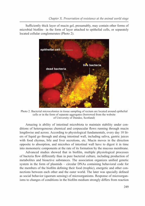
249
Chapter X. Preservation of resistance at the animal world stage
Suffi ciently thick layer of mucin gel, presumably, may contain other forms of microbial biofi lm– in the form of layer attached to epithelial cells, or separately located cellular conglomerates (Photo 2).
Photo 2. Bacterial microcolonies in tissue sampling of rectum are located around epithelial cells or in the form of separate aggregates (borrowed from the website
of University of Dundee, Scotland)
Amazing is ability of intestinal microbiota to maintain stability under con-ditions of heterogeneous chemical and corpuscular fl ows running through mucin lengthwise and across. According to physiological fundamentals, every day 10 lit-ers of liquid go through and along intestinal wall, including saliva, gastric juices with food chymus, bile and liver secretions, etc. Mucin moves in the direction opposite to absorption, and microbes of intestinal wall have to digest it in time into monomeric components at the rate of its formation by the mucous membrane.
Advanced studies showed that in biofi lm, multiple physiological processes of bacteria fl ow differently than in pure bacterial culture, including production of metabolites and bioactive substances. The association organizes unifi ed genetic system in the form of plasmids – circular DNAs containing behavioral code for the members of the biofi lm defi ning their food (trophic), energetic and other con-nections between each other and the outer world. The later was specially defi ned as social behavior (quorum sensing) of microorganisms. Response of microorgan-isms to changes of conditions in the biofi lm medium strongly differs from reaction

250
A.E. Gabidova, V.A. Galynkin
of every separate species in monoculture. Such organization ensures its physiolog-ic and functional stability and, consequently, serves as a pledge of its competitive survival in ecological niche. In human organism, specifi c advantage of such or-ganization consists in ensuring homeostasis of organs whose functionality depends on the microbes which populate them.
Photo 3. Revealing of intestinal bacteria of separate species in artificial medium simulating intestinal microbiota. FISH method was applied (Fluorescent In Situ Hybridization) –
hybridization of test DNA directly in the sample, with coloring with different fluorescent markers (borrowed from the website of University of Dundee, Scotland)
Healthy-looking skin, normal digestion, resistance to external infection (state of immunity) of people in many aspects, is defi ned by stability, in other words, “health” of their microfl ora.
Advantage of collective response also has its negative aspect: it is diffi cult to control such association from beyond, for example, to treat diseases of polymi-crobial origin when sensitivity to antibiotics of microorganisms associated into biofi lm does not correspond to the same defi ned in laboratory tests on clinical isolates of pure bacterial cultures. Collective immunity of biofi lm practically nul-lifi es a good idea of dysbacteriosis correction using probiotics – preparations of life cultures of key intestinal microorganisms – bifi dobacteria, lactobacilla, en-terobacteria, etc. No doubt, they have effect, but not always, and not the one sup-posed initially. It occurs because of collective immunity of intestinal microbiota (organized microfl ora). Microbes cultivated artifi cially are alien, just like organs

251
Chapter X. Preservation of resistance at the animal world stage
and tissues of other people or animals grafted to humans. They are rejected as a result of biological incompatibility. Biotechnological probiotics have no “pass-word” allowing microbes to enter inside the intestinal biofi lm, and therefore their staying there is transitory just like food microfl ora. Companies producing probiot-ics admit this fact and don′t claim that their additives physically replenish defi cit of microorganisms′ correspondent to the probiotic content, but stimulate growth of the depressed population. Absence of survival of alien microbes of the same kind is an indirect evidence that human microfl ora exists as an independent organ. There is a new term – “biofi lm paracytology” as a structure similar to tissue of higher organisms, which implies observance of the laws inherent in it.
There are many other circumstances due to which human microbiota should be considered as an individually specifi c, genetically determined and, evidently, inherited [204]. Basing on such conclusion, B.A. Shenderov suggested (in the form of inventor′s certifi cate received in the beginning of 1990s) the idea of indi-vidual microfl ora conservation in young, healthy period of life of a person, with the purpose of its future doping if there are serious disorders of intestinal byo-sis, with the purpose of its recovery, even rejuvenation. As for gastroenterologists, they know cases of microfl ora “transplantation” to patients from their relatives “per clismum”, which had positive effect of intestinal dysbacteriosis correction.
10.3.3. Modern ideas of the structure of intestinal microbiota according to data of molecular studies
Information on the nature of intestinal microbiocenosis accumulated by now looks suffi cient to understand its functioning as a physiologically active human organ. However, to implement them in controlling this organ in pathologies which have cause-effect relation to dysbacteriosis, there is a lack of quantitative method of defi ning changes in the structure of quite wide range of key microorganisms and their monitoring in the process of correction. In this connection, it is more preferable to analyze composition of parietal intestinal microbiota than faeces mi-crofl ora as accepted everywhere. It is mucous layer covering mucous membrane of intestines, where occur digestion of food chymus getting from the stomach, digestion of essential nutrients by epithelial cells of intestinal wall, and addition-al production of great number of bioactive substances by microorganisms: en-zymes, vitamins, antibiotics, immunostimulators, as well as toxins and metabolites harmful for humans. Presumably, imbalance of their production is connected to pathological manifestations of different nature: intestinal disorders, skin diseases, sexual dysfunctions, and cardiac insuffi ciency. Faeces microfl ora is the waste of these processes, where production of microorganisms continues, but under other conditions compared to upper sections of intestines. According to renown special-ist in the fi eld of clinical microbiology, prof. A.N. Mayansky, it more likely re-

252
A.E. Gabidova, V.A. Galynkin
fl ects cavitary (free-living, or planctonic) than parietal biofi lm which is more stable and physiological. Referring to Mayansky in terms of revealing intestinal dysbacteriosis by faeces, we cannot help agreeing that “actually, this is an expensive (considering that it is recommended to make tests in dynamic), laborious study, with low (even zero) output”.
The method suitable for solving this problem appeared in Russia in early 1990s. It was developed on the basis of studies of Research Institute of Biological Instrument Engineering supported by the member of RSA, G.A. Zavarzin and the grant of the Ministry of Ecology and Soil Protection of Russian Federation “En-vironmental safety of Russia”. The method is based on revealing microorganisms in the objects of environment (water, soil, sewage, etc.) by chemical elements spe-cifi c to them– markers among fatty acids, aldehydes and sterols constituting their cytoderm. Specifi city means that such substances are contained only in lipids of microorganisms, but not in their habitat. Therefore, using quite sensitive method of analysis, they can be detected and measured numerically directly in the habitat, without necessity of cultivating them in artifi cial media. It became possible using the method of gas chromatography, combined with mass-spectrometry (GC-MS). Essence of the analysis consists in direct extraction of higher fatty acids from the sample subject to study (soil, silt, clinical material) using a chemical procedure, their disintegration with chromatograph on capillary column with high resolution, and analysis of their structure in dynamic mode on mass-spectrometer. While the chromatograph is united in one device with mass-spectrometer and equipped with computer, with relevant programs of automatic data analysis and processing, the analysis itself takes 40 minutes. As a result, composition of microbial markers can be defi ned accurate within 2 % relative. Along with sample preparation and cal-culation of microbial association using separate program, a standard procedure of controlling 170 microorganisms in the sample takes approximately 5 hours.
Method of microorganism detection by fatty-acid markers is similar to genetic (PCR, defi nition of succession of nucleotides 16sRNA, etc.), as the structure of fatty acids is determined in DNA and reproduced by means of genome region rep-lication by transport RNAs and subsequent synthesis of fatty acids in mitochon-dria by matrix RNAs. Therefore, profi le of bacterial fatty acids is their “business card”, or fi ngerprint, just like human fi ngerprints. It is as conservative as DNA structure, but also as prone to mutations under the infl uence of environmental factors. Stability of the pattern of fatty acids constituting microbial cells, is con-fi rmed by the studies in the fi eld of bacterial paleontology, which indicate that, for 2,5 billion years, composition of fatty acids of separate microbes and pool of their fatty acids, as a whole, has remained unchanged. Bacteria which were conserved in bottom deposits of ancient lakes of Antarctica (at the age of 1,5 million years), can be revived in laboratory studies and show their identity to modern species by molecular signs – composition of fatty acids of cell walls [199].

253
Chapter X. Preservation of resistance at the animal world stage
Basing on this principle, hemodifferentiation of microorganisms is built, widely used as a method of their identifi cation and confi rmation of their taxonomic posi-tion. It is applied for working with monocultures of microorganisms, and based on using extremely large databases, containing information on fatty-acid structure of thousands of bacterial strains and microscopic fungi. Example of such system is the specialized chromatograph Microbial Identifi cation System, produced by MIDI Inc. Company, Delaware, USA. Features of fatty-acid structure are now used, along with other parameters, in bacterial taxonomy and clinical bacterial diagnostics.
Methodology of microbial association analysis by GC-MS method was published both in the report on the subject of the grant of the Ministry of Ecol-ogy of Russian Federation “Environmental safety of Russia”, and in the subse-quent description of the patent for the method of the analysis. It is distributed under support of RMSA specialists, Yu.F. Isakov and A.A. Vorobyev and pro-fessor N.V. Beloborodova to diagnostics of infl ammatory processes and dys-bioses in clinical practice. As a whole, the method, applied to environmental, biotechnological and clinical problems, was described in doctoral thesis by G.A. Osipov (1996), fi ve PHD theses, publications in national and foreign pe-riodical press, and manuals for practicing doctors.
We managed to measure concentration of microbial components directly in the habitat, where there are cells of intestinal wall microbes. So we are entitled to directly correlate marker concentration and numbers of microbial cells under con-ditions of absence of food lipid component, as the tissue samples were obtained on empty stomach. Such logic persuades us that we have measured the leading microfl ora of intestinal wall, leading in respect of quantity, as it became clear that in the presence of tissue sample with the mass of 4 mg, we can detect microorgan-isms, beginning with the concentration of 104–105 cells/g, so signifi cant part of the microfl ora remained out of our reach. As it became clear, total number of intesti-nal wall microorganisms in normal state is up to (0,5–1,3) × 1011 cells/g, depend-ing on the section of the intestines.
Density of population of intestinal wall changes insignifi cantly in distal di-rection: in ileum, it is twice less, and in large intestine – 15 times larger than in empty intestine. Parietal microfl ora we have measured turned out to be much more concentrated than the one of the opening (according to the data from literature [205], which is six orders lower in small intestine, and in ileum – fi ve orders lower by quantity (up to 105–106 cells/ml, respectively), and only in colon, corresponds to the same in its content. Species composition of microorganisms corresponds to existing ideas on components of intestinal microfl ora, especially, faeces microor-ganisms. However, the similarity is limited by general categories: qualitative com-position and priority (rank) content of key elements of intestinal microbiocenosis. In fact, there are signifi cantly more anaerobes in large intestines and faeces.

254
A.E. Gabidova, V.A. Galynkin
The obtained total quantity of microorganisms for faeces lies within the limit of 0,6–5 × 10¹¹ cells/g, which is consistent with published data on measuring by genetic and cultural-chemical methods. Relative amount of anaerobes in them also coincides with existing estimations, which, according to our data, accounts for 88 %. It is diffi cult to compare spreading of species to the data from literature, as they give a very wide range of values –within the limit of 3–6 orders.
Nevertheless, our estimation of Eubacterium priority, whose quantity has order of 1011 cells/g (109–1012, according to the literature data), on amount of bacteroids 1010 cells/g (1010–1012, according to existing data), clostridia – 6 × 10¹⁰ cells/g (105–1011, respectively), bifi dobacteria 1010 cells/g (1010–1012), and also by enterococci, enterobacteria, lactobacilli and staphylococci.
This result allows to maintain the analysis of faeces microbiota by GC-MS method, by fatty acids of cell walls of microorganisms, provides authentic data on their quantity. Consequently, the information on composition of microorganisms in tissue samples of intestinal wall, presented here, can be also considered authentic.
Different study results of faecal microbiota leave to befi dobacteria in their composition almost from 100 % to 0,1 %. Three-order range can hardly be caused by the fact that “people are different” – in each study, there are authentic statistics and reliable analytical procedure. More likely, the difference can be related to the features of the compared methods of quantitative measurements. Without going into details, we can conclude that effect of befi dobacterial preva-lence is created by routine practice of analysis of sole befi dobacteria and condi-tionally pathogenic microfl ora in studying dysbacterioses. As we can see, here microbiologist loses track of eubacteria, bacteroids and clostredia, whose con-tent inn faeces, according to modern estimations, is at least several times higher than befi dobacteria. This misconception looks natural, if we remember that, in the frameworks of general microbiology, it is considered in microbial associa-tion, on average, only 20 % of microorganisms of any habitat are cultivated. As for faeces, according to estimations by molecular-genetic methods, we can also fi nd out that identifi cation of 60–80 % of their microbiocenosis is inaccessible for cultural methods. Data of mass-spectrometry correlate to genetic ones (in the frameworks of comparability of microbiological quantitative measurements) and indicate in similar way that there are 10 times more eubacteria, bacteroids and clostridia together and separately than befi dobacteria.
Application of mass-spectrometry method allowed to measure number of more than 50 taxones of intestinal microorganisms not only in faeces, but also in the section on intestines themselves, by means of their marker analysis (fatty acids) directly in tissue samples obtained in intestinoscopy and colonoscopy, with retrograde ileoscopy. These data show that eubacteria also dominate there, and their species composition signifi cantly changes with the length of the intestines.

255
Chapter X. Preservation of resistance at the animal world stage
We should note phylogenetic affi nity of eubacteria and clostredia. Berge′s manual, 9th edition directly indicates that Eubacterium genus was created for convenience, for placing there all clostridia which form the spores weakly.
Thus, intestinal microbiota presents dominant continuum of strains and spe-cies of Clostridium and Eubacterium genera, in equivalent total amount of bacte-roids, befi dobacteria and lactobacilla.
The data given is the evidence of importance of Eubacterium genus in forma-tion and functioning of intestinal microbiota. After the analysis of phylogenetic as-sociations, now it is diffi cult to detach it from Clostridium genus (at least, C. coc-coides group), and consider them as digestively important group of peptolytic and cellulolytic organisms. We should note a very important feature of Eubacterium specimens consisting in hydrogen formation. This key property of microorganism consortia digesting organic substrate, in anaerobic processes in the nature (marsh), in ruminants′ stomach, and in biotechnology in anaerobic fermentation of differ-ent wastes and obtaining biogas. Mucous layer of human intestines, in its essence, is the similar bioreactor. Methane is formed there; consequently, this is activity of archaebacteria-methanogens, whose effi ciency is strictly dependent on hydro-gen concentration in the system. In methanogenic association, hydrogen bacteria also play key regulatory role because of reverse infl uence of process of hydro-gen production and consuming on primary process of carbon decomposition and formation of acetate. As it is evident from our measurements, in IBS, number of eubacteria changes most signifi cantly, which should lead to increase of hydrogen concentration in the system. In fact, earlier, in the course of experiments, 4-time increase of hydrogen concentration in the air exhaled by the patients, and its nor-malization after relief of the symptoms by means of restrictive diet was shown.
The main part (from 70 % in empty intestine to 90 in faeces) of microorgan-isms in all sections of the intestine falls on anaerobes. The second position by quantity in the empty intestine belongs to aerobic actinomyces – 17 % (in faeces, there are only 0,7 %). Aerobic cocci (staphylococci, streptococci, enterococci) and coreniformic bacteria) account for 5 % of small intestine colonization, compared to 0,7 % in faeces. Fraction of enterobacteria and enterococci by intestinal sections and in faeces is approximately 2 %.
Undeniably, unexpected result is discovery of signifi cant amount of aerobic actinomyces. Specifi city of their markers, ramifi ed fatty acids with methyl group in Δ10 position, doesn′t allow assuming any other taxonomic groups of microor-ganisms, except for specimens of Actinomycetales order, containing mycolic ac-ids in their cell walls, which is the source of 10ME-rammifi ed fatty acids. They are contained in mycobacteria, nocardia, rodocci, Actinomadura and other actino-myces, but not found in higher organisms (fungi, plants, animals). Presence of these molecules in intestinal tissue samples, in blood and other organs and fl uids

256
A.E. Gabidova, V.A. Galynkin
of humans is confi rmed by mass-spectrum, as well as by their analysis in the struc-ture of museum cultures of the respective microorganisms. Bacteria of Streptomyces and Nocardiopsis genera are also confi rmed by unique marker – isohexadecane acid. Besides, we allocated Nocardiopsis dassonvilley from intestines as a pure culture.
Photo 4. Colony of anaerobic actinomyces Nocardiopsis dassonvilley allocated from parietal layer of small intestines
Here we should add the following microorganisms: Propionibacterium, Actino-myces, Brevibacterium which were also allocated in pure culture, and coreniformic bacteria. Finally, taking into account, that, until the present time, some manuals on microbiology relate Bifi dobacterium genus to Actinomycetaceae family, we can con-clude that actinomyces are phylogenetically close to traditionally known specimens of parietal intestinal microbiota. They increase signifi cance of intestinal microbiota for the host′s organism, as actinomyces excel all other microorganisms in antibiotic and vitamin production and have powerful fermentative apparatus. High degree of intestines colonization by actinomyces doesn′t seem unusual, regarding that they are widely spread in the environment– soil, water, air, on interior walls of residential and industrial buildings. Their inhabitance in human organism, under such circum-stances, looks natural. In fact, manuals on clinical microbiology mention discovery of actinomyces and related microorganisms, such as Mycobacterium, Actinomadura, Propionibacterium, Actinomyces, Corynebacterium, Bifi dobacterium, in human in-testines and other organs. They appear there (including befi dobacteria) as partici-pants of infectious and infl ammatory processes (Manual on Clinical Microbiology).

257
Chapter X. Preservation of resistance at the animal world stage
We found out that their concentration correlates with microbial concen-tration on intestinal wall measured in tissue samples. It looks natural from physiologic point of view, as in natural death of microbial cells, their lipids are decomposed by enzyme system of the intestines, fatty acids which were unused on its wall getting to the bloodstream.
This phenomenon allows to control condition of intestinal microbiota according to the fi ndings of microbial fatty acids in a drop of blood (40 mcl) taken from fi nger.
We also should consider that microbial molecules of microorganisms inhabit-ing on other human mucous membranes (fauces, urogenital tract) also penetrate into blood, as well as from foci of infl ammation caused by indigenous (own) mi-crofl ora or external pathogen. Presence of markers of such microorganisms in the blood is theoretically conditioned by mechanism of immune response to appear-ance of the pathogen. Phagocyting cells of human organism adsorb and digest antigens of microorganisms, including whole cells, and deliver lyses products to the fl ow of lymphatic and blood systems. Except for phagocytosis, microorganism cells are decomposed under infl uence of other mechanisms, including their own apoptosis and lyses by enzymes of protein complement of the blood and other components of immune protection. In any case, eventually, they are decomposed into monomeric components of biopolymers. Basing on fundamentals of physiol-ogy of human biological fl uid exchange [205], exchange of 70 % of plasma fl uid with interstitial space takes 1 minute. Therefore, small fragments of biopolymers formed within this space, get into the blood almost immediately. That is, presence of microbial markers in the blood refl ects composition of microbial associations of human body, regardless of microorganism habitat or focus of infl ammation.
In other words, by measuring microbial marker concentration in the blood, we can estimate microbiological status of a person in general.
However, in normal state, that is, if there are no symptoms of infl ammation or local infection, intestinal microfl ora remains their main source. While earlier we indicated existence of microbial marker homeostasis in the blood, in view of the above, we can talk about homeostasis of intestinal microbiocenosis. This is another confi rmation of similarity of its composition in people in normal state, and difference in pathological state. Signifi cant change in microbial marker com-position in the blood, compared to statistically grounded norm, can be discovered in peritonitis, infl ammations of urogenital organs, and in skin diseases – atopic dermatitis, seborrheic dermatitis, eczemas, psoriasis, endocarditis, pericarditis and fevers of unknown etiology. Homeostasis of parietal intestinal microfl ora supposes its stability in respect of everyday dietary nuances. However, visible changes in composition of intestinal microorganisms can be expected in long-time dietary orientations: in vegetarians or, on the contrary, in the residents of the Far North, who have limitations in vegetable food. Shifts in homeostasis can be assumed due

258
A.E. Gabidova, V.A. Galynkin
to geographical, racial, religious or national constant characteristics of nutrition. For example, residents of Lund (Sweden) have more actinomyces in their intes-tines than residents of Moscow.
On the other hand, it is diffi cult to imagine a short-term (during one day or week) dietary dependence of composition of intestinal microfl ora. By its essence, this organ must be stable (homeostatic) while it defi nes composition of many chemical substances which get into the host′s organism.
These substances, in their turn, defi ne quality of physiological processes in all the other organs and tissues. They are also homeostatic, which requires stability of the composition and concentration of the substances necessary for cellular cycles, on the one hand, and minimum of their inhibitors, on the other hand. It is natural to expect that any signifi cant digressions trigger pathological effect. But it does not happen because for lunch you ate fi sh or vegetables, and meat or curds- for dinner. The explanation is easy to get, if we remember that, in fact, it is not veg-etables or meat itself which gets into intestines, but products of their primary deg-radation in the intestines under the infl uence of enzymes in acid medium. That is, simply speaking, they are proteins, fats and carbs. These components getting into the small intestines, along with secretions of digestive glands, are called chymus. Within microbial cycle of population alternation (just from one day to one week), fl uctuations in chymus composition are microbiologically averaged. This ensures smooth delivery of those two-thirds of molecules necessary for human vital activ-ity, which are formed due to microbial metabolism of chymus. (Gourmets may be disappointed: in fact, when they devour delicacies, they feed not themselves, but their microbes.) If a person gets enough various foods, their cells have set of chemicals necessary for normal functioning.
We can say that human rational nutrition, to a great extent, equals to rational nutrition of microbes of their intestinal wall.
Long-term defi cit of any functionally important products, as well as sur-plus of harmful ones, or food deterioration becomes the reason of discomfort, decrease of life quality, up to pathological changes of tissues and organs, and reduction of life expectancy itself.
Basis of ecological platform is vegetative-microbial interactions, which is the basis for maintaining life on our planet – they had begun to form long before hu-man appeared. Contamination of medicinal preparations with conditionally patho-genic and pathogenic microorganisms, which can harm to human health, is one of the main issues of pharmacy. Besides, many phytopreparations are directly as-sociated with plant cultivation, and it is the very stage where microbial semination occurs. In the nature, all animals and plants keep associations with specifi c, and very often, obligatory specimens of microbiota – symbionts. During colonization of roots and roots fuzzes, microorganisms form biofi lms, where they demonstrate
259
Chapter X. Preservation of resistance at the animal world stage
change of phenotype, refl ected in changes of growth parameters and expression of specifi c genes. Ability of bacteria for biofi lm formation is signifi cant factor of pathogenicity. Besides, similar fi lms of microorganisms can be also revealed in animal and human organisms. Role of procaryotes and eucaryotes in symbiosis is being discussed. Association of microorganisms organized unifi ed genetic system in the form of plasmids – circular DNAs carrying behavioral code for the biofi lm members, defi ning their food (trophic), energetic and other relations between each other and the outer world. At all these stages, resistance of microorganisms to an-tibiotics, disinfecting substances and synthesized compounds is formed. Anomaly of metabolic processes up to qualitative changes of cellular structures in affected plants is of great importance.
Frequency of infections caused by antibiotic-resistant bacteria is being in-creased among the population. General distribution of microorganisms in bio-sphere in the form of multicellular populations, in particular, symbioses, contrib-utes to increase of microorganisms resistant to preparations. Besides, symbioses of animals and humans and microorganisms in a great extent contribute to develop-ment of resistance not only in the soil, but also in humans. All these stages con-tribute to development of resistance of pathogenic microorganisms to preparations lead to increase of morbidity and mortality from infections.
Soil is most sensitive to anthropogenic exposure. Of all the covers of the Earth, soil is the thinnest one, Thickness of the most fertile humus layer, even in black soils, does not exceed, as a rule, 80–100 cm. By the present, by different reasons, the world has lost about two billion hectares of agricultural soils. Loss of soils caused by irrigation only, in the last 300 years, accounted about 100 million hectares, and approximately the same area is now occupied by soils with low pro-ductivity, due to salinization. Losses of humus, on which almost all the important properties of soils and their resistance to unfavourable situations depend, are very signifi cant. Protection of soils and their rational use have essential role for eco-nomic and social development of any country. In the second half of the 20th cen-tury, scope and rate of technogenetic pollution of environment increased so much that it became necessary to develop special international programs of environmen-tal protection. In 1972, the environmental program of UN was developed, which included issues of the monitoring of the environment, with the purpose of early reporting about coming natural or anthropogenic changes, which may be harmful for human health or welfare. Recovery of damaged soils requires long time and large investments. Microbial-vegetative interactions are the basis for maintaining life on our planet, and if pathogenic resistant microorganisms appear. Carriers of life capable of securing stability of its existence, from the very beginning were not separate species, but biocenoses, whose components consistently performed all the necessary links of primary circulation of substances. Currently, there are

260
A.E. Gabidova, V.A. Galynkin
no doubts about the fact that the processes observed in the soil are often not the sum of activity of separate microorganisms, but the result of their interaction.
Moreover, joint development of bacteria of Rhizobium genus and pod-bearing plants, and mycorrhizal fungi, actinorhizas, bacteria and various plants is a well-known example of symbiosis.
Currently, study of symbioses plays one of priority roles among urgent prob-lems of modern biology. Numerous fi ndings indicate that symbiotic associations represent one of the main forms of organism existence in biosphere. Currently, instead of ideas about symbiosis as a two-component system, comes its under-standing as a multi-component system, where, except for dominant microsymbi-ont, there are several associative symbionts. According to A.V. Bukharin [123], associative symbiosis is a multi-component integral system, which includes a host as a macropartner, a stable microsymbiont and associated microsymbionts with multidirectional effects, defi ning formation, stability and productivity of the symbiosis in general. Positive effect of associative symbionts on development of host-plant and symbiosis in general takes place also in the realm of plants. In the formed ectosymbiosis, root exudates of plants represent substrate and growth factors for some groups of microbial associations, which play the role of anti-phytopathogens, utilizers of undesirable products of vegetative metabolism, regu-lators of aggregate microorganism concentration in the soil, regulators of mobility and circulation of mineral substances in agroecosystem. It is refl ected in improve-ment of mineral nutrition of plants, intensifi cation of partnership of the host with the dominant symbiont due to local production of phytohormones; maintaining in the soil pool of potential microsymbionts, with release of spores of the dominant symbiont, and, fi nally, direct protection of plants from phytopathogens. Mechanisms of positive infl uence of rhizobacteria on plant vital activity can be divided into di-rect and indirect ones. We suggest including into direct ways of effect: associative nitrogen-fi xation, formation of growth-stimulating substances, provision with easily-digestible forms of iron, phosphor and/or their absorption from the soil and delivery to the plants, formation of specifi c trophic relations, and reduction of ethylene level. Indirect ways include: prevention or reduction of phytopathogenic soil microorgan-isms due to discharge of bactericidal or anti-fungal metabolites. A lot of associa-tive microorganisms can, in the process of growth, discharge antibiotic substances, which can suppress activity of other microorganisms if in low concentrations.
Resuming the discussed problem, we should note that infection is a model system of associative symbiosis, in whose structural-functional relation, we can distinguish three vectors: host- normal fl ora, host- associants, and microsymbio-cenosis. Symbionts′ functions defi ne colonization resistance of the host, forma-tion of dysbioses and pathobiocenoses. Antimicrobial resistance of organism bio-topes is connected to substrates, whose inactivation is performed by symbionts

261
Chapter X. Preservation of resistance at the animal world stage
with persistent characteristics. Interaction of symbionts during infection is based on changes of their persistent potential, and antagonism of the pathogen and the commensal partners.
Biosphere has tremendous self-cleaning abilities; however, they are not boundless. Anthropogenic effect on the environment endangered normal perfor-mance biotic processes inherent in it, and today achieved such scale that it led to global environmental crisis. Destructive human activity generated confl ict be-tween society and the nature, and created the risks called ecological. The most important function of any biocenosis, biogeocenosis and biosphere is regular re-construction of live substance and energy accumulated in it.
Vegetative-microbial interactions are the basis for maintaining life on our planet, and they had begun to form long before human appeared. We should not that both fresh-water, and marine macro-and micro-plants are colonized by micro-organisms, and their interactions are defi ned by regional conditions – by soil, air and water. Considering circulation in the nature, plants supply oxygen and “food-stuff” for humans, animals and signifi cant part of micro-world, and microorgan-isms process return of nutrients for the plants, decomposing and using as substrate both mortifi ed plants, and often live ones, and protection of plants from pathogenic and conditionally pathogenic organisms. Interaction of microorganisms and plants leads to appearance of microbial-vegetative complexes (symbiosis) in different ecological regions. Structure of microbiocenosis is not identical, but depends on environmental conditions in the region. Today, microbiologists admit that signifi -cant amount of microorganisms in natural (soil) and artifi cial media exists in the form of structured associations attached to the surface – biofi lms. Substrate (soil) part of plants lies in the soil and continuously contacts with soil microorganisms (fungi, actinomyces, and bacteria), viruses and phages, which can penetrate into roots or colonize root surface. During colonization of roots and roots fuzzes, mi-croorganisms form biofi lms, where they demonstrate change of phenotype, refl ect-ed in changes of growth parameters and expression of specifi c genes. Ability of bacteria for biofi lm formation is signifi cant factor of pathogenicity. At all these stages, in the soil, forms resistance of microorganisms to antibiotics, disinfecting substances and synthesized compounds. Use of antibiotics has become one of the greatest achievements of medical science, but, currently, former weapon of multi-target use often fails in struggle with infectious diseases. Antibiotic resistance of infectious agents becomes more and more threatening. Theoretically, resistance may appear during any contact of bacterial population with an antibiotic, whether in soil, water body or organism of the host.
Cascade path, beginning from the soil, is one of the key paths of resistance. General distribution of microorganisms in biosphere in the form of multicellular populations, in particular, symbioses and biofi lm contributes to increase of amount

262
A.E. Gabidova, V.A. Galynkin
of microorganisms resistant to medicinal preparations. Besides, symbioses of mi-croorganisms with plants, animals and humans lead to prevalence of resistance not only in the soil, but also at each stage. All these stages contribute to development of pathogenic microorganism resistance to drugs; lead to increase of morbidity and mortality from infections. We can come to the conclusion that at least part of antibiotic-resistant genes, which are currently wide-spread among clinical strains of bacteria, originated from determinants of resistance of antibiotic producers (or of the same origin with them). High level of divergence between determinants of strains-producers and strains of bacteria, allocated from clinical practice, is the evidence of extremely old origin of these determinants. Evidently, determinants of resistance got into cells of bacteria inhabiting in animal and human organisms by means of successive acts of horizontal transmission by plasmids and transpo-sons. Such data, in particular, were obtained in studying comparative structure of aph genes (3′), revealed in cells of gram-positive bacteria S. fradiae, B. circulans, S. aureus, and as a part of transposons of gram-negative bacteria Tn5 and Tn903. Supposing that some determinants of resistance initially appeared from antibiotic producers, at certain stage of evolution, inevitably, they must have been replaced from gram-positive to gram-negative bacteria. The data obtained is the evidence of high frequency of horizontal gene transmission between gram-positive and gram-negative bacteria in natural populations of bacteria. It was shown that horizon-tal gene transmission between gram-positive and gram-negative bacteria is quite common not only under experiment, but also in the nature. Specimens of actino-myces and cyanobacteria can come into symbiotic relations with plants, in particu-lar, specimen of Frankia genus, which is shown below. Actinomyces is an integral part of soil microbial complex. Characteristic of actinomyces as nitrogen-fi xing symbionts, able at some stages of development, to form ramifi ed mycelium with diameter of 0,4–1,5 mcm, which they manifest in conditions optimum for their ex-istence, have gram-positive type of cell wall and high (60–75 %) concentration of GC pairs in their DNAs. They are widely spread in the soil: there it is possible to fi nd almost all the genera of actinomyces. Actinomyces usually account for quar-ter of all bacteria growing in traditional media, in inoculation of their dissolved soil suspensions, and 5 to 15 % of prokaryote biomass.
Actinomyces, soil microorganisms, producers of most existing antibiotics are characterized by natural multiple resistance to antibiotics. They are considered as a source of genetic determinants of antibiotic resistance in the nature. Genes of actinomyces controlling biosynthesis of antibiotics and resistance to them are usu-ally linked and function consistently. Effective expression of resistance genes can be one of the key factors of achieving high antibiotic activity by strains of actino-myces. According to modern ideas, determinants of bacterial antibiotic resistance widely spread in clinical practice, originate at least from two sources. The fi rst is

263
Chapter X. Preservation of resistance at the animal world stage
strains-producers of antibiotics, mainly, actinomyces containing genes of resist-ance to their own antibiotic. The second is own genes of bacteria which ensure normal metabolism of bacterial cells (house-keeping genes). According to this hy-pothesis, genes of antibiotic resistance fi rst appeared in their producers as a nec-essary protective mechanism from their own products of metabolism, and then, by means of horizontal transmission, got into genomes of other bacteria occupy-ing different ecological niches. In fact, genomes of many streptomyces-producers were revealed to contain genes of resistance to their own antibiotics. Actinomyces are especially interesting from the point of view of analysis of antibiotic resist-ance mechanisms. Actinomyces-producers of most existing antibiotics (producers of 80 % of existing antibiotics), are characterized by natural multiple resistance to antibiotics. They are considered as a source of genetic determinants of antibiotic resistance in the nature. Genes of actinomyces controlling biosynthesis of antibiot-ics and resistance to them are usually linked and function consistently. Effective expression of resistance genes can be one of the key factors of achieving high antibiotic activity by strains of actinomyces. Therefore, study of genetic control of actinomyces resistance to aminoglycoside antibiotics (AG) is also very impor-tant for construction and selection of these antibiotics. Study of large collection of specimens of Streptomyces genus showed that resistance to AG is rare in strains which do not produce them [3]. Evidence of origin of resistant genes of pathogen-ic bacteria from respective genes of antibiotic producers was also obtained in stud-ies of determinant structure of resistance to tetracycline. In clinical strains of fast-growing mycobacteria (Mycobacterium fortuitum, Mycobacterium peregrinum) and Streptomyces (Streptomyces sp.) genes of resistance to tetracycline were revealed (tetK and tetL), which were earlier revealed in other species of gram-positive bacte-ria. Except for these genes, in the same strains of mycobacteria and Streptomyces, genes of resistance to tetracycline, otrA and otrB were revealed, described earlier in producer of oxitetracycline S. rimosus. Homology at the level of amino acid succes-sion between genes of resistance to neomycin/canamycin [aph (3′)] Streptomyces fradiae and transposons of gram-negative bacteria Tn5 and Tn903 equaled 36–40 %; highly conservative regions of proteins were found mainly on their S- ends. Level of homology between genes 3-N-aminoglycoside acetyltranspherases [aac (3)] from actinomyces and clinical strains varied within 39–41 %
Wide spread of the both phenomena –ability of some microorganisms to syn-thesize antibiotics, and of other microorganisms to have resistance to them, con-ditioned by the fact that antibiotics in concentrations found in natural ecosystems, play the role of intracellular signaling molecules, regulating gene transcription. Change of response of bacterial communicative association to a certain signal, caused by acquisition, or, on the contrary, loss of antibiotic resistance, leads to formation of new ecotopes. Therefore, problem of antibiotic resistance among

264
A.E. Gabidova, V.A. Galynkin
clinically signifi cant microorganisms has its roots in complex ecological and evo-lutional relations between microorganisms themselves, and had formed long be-fore humans appeared as a biological species. Mechanism of spreading genes of antibiotic resistance among bacteria is based on exchange of plasmids and con-jugative transposons. In antibiotic resistance evolution, plasmids and conjugative transposons function as genetic platforms, where, by means of recombinant bacte-ria systems, genes of antibiotic resistance, included in transposons, integrons, cas-settes of genes and insertion cryptic successions, are assembled and sorted.
Saprophages is the most typical part of soil complex. The great bulk of soil animals fall to their share. Biogeocenotic role of saprophile complex consists both in direct biochemical and physical effect on organic residue, and in stimulation of saprophytic complex activity. Animal association includes: substrate-microor-ganisms – plant-substrate (phytoconsuments) animals – animal-substrate (zoocon-suments) animals. It considers the fact that phytophages and phyto-saprophages, in respect of amino acid (protein) nutrition, are consumers of microorganisms or predators, ways of getting energy (energy carriers) and elements (compounds) turned out to be unrelated, microbial link, in respect of substance migration and energy, is, in its essence, the central link of food chain, as well as allows to ex-plain some facts and phenomena associated with toxicant migration in the food chain. Regulation of this substance migration is processed by microorganisms of digestive tract. Production of microorganisms in these ecosystems is higher than production of populations of free-living microorganisms. Large saprophages in forest-steppe increase production of microorganisms, at least, by value of average weighted biomass of soil microorganisms.
As well as plants, animals also get accessible forms of elements through mi-croorganisms, and this fl ow many times exceeds fl ow of elements received by ani-mals using plant tissues. Meaning of animals consists in regulation of circulation fl ow in ecosystems inside the triad plants- microorganisms- animals, but, above all, through regulation of microorganism population.
Role of animals in mechanical migration is unique, and by the size of its fl ow, can be compared to erosion processes. In some ecosystems, mechanical migration itself is comparable to biogenic migration of elements inside ecosystems. Every biocenosis is formed by certain ecological groups of organisms, which may have different species composition, though may occupy similar ecological niches. For example, saprophages are prevalent in forests, phytophages in steppe areas, preda-tors and detritophages – in the depth of the world ocean. In the course of nutrition, “wastes” are formed at all trophic levels. Every year, green plants throw away their leaves partially or completely, signifi cant number of organisms constantly dies out by certain reasons, all the organic substance that was created should be replaced due to mineralization of organic components. It occurs due to presence

265
Chapter X. Preservation of resistance at the animal world stage
of special trophic chains in ecosystem – chains of destructors – mainly, bacte-ria, fungi, protozoa, small invertebrates. They decompose organic residue of all trophic levels of producers and consumers into mineral substances. Tracing food relations between members of biocenosis, we can build food chains and food nets of organism nutrition. The following sequence can serve as an example of a food chain. As primary destructors, V. Dunger identifi es large soil and litter in-vertebrates eating mortifi ed organs of plants, which completely saved their tissue structure. Animals grind and macerate particles of vegetative issue in their mouth cavity and intestines, destroying associations between separate cells. This com-pletely mechanical processing is highly signifi cant for further microbial decom-position. During maceration, summary surface of vegetative residue, accessible for aerobic microfl ora, is many times increased. Its activity is always higher in vegetative residue destroyed by animals than in absence of primary destructors. They 3–8 times accelerate litter decomposition, depending on quantity and exter-nal conditions, secondary destructors consume already grinded vegetative tissues, partially digested by enzymes of animals and microorganisms. This group in-cludes caprophages and detritophages. Similarity of digestion physiology in detri-tophages and coprophages defi nes possibility of combination of these two modes, which is observed in some soil animals (Cetoniinae and earthworms). Signifi cant difference in nature of digestion of primary and secondary destructors is that the fi rst are able to digest structural components of higher plants residue – cellulose, hemicellulose and pectins, and the second digest mainly easily hydrolyzed detri-tus of vegetative tissues. Thus, concept of saprophagy includes a wide range of food modes, typical for invertebrates with different levels of organization and sig-nifi cant physiological differences. Among soil saprophages, it is possible to fi nd almost all types of nutrition described for invertebrates. Saprophagy is complex of food modes of animals utilizing energy of mortifi ed autotrophic organisms. The group of saprophages unites forms using for food directly residue of green plants and their detritus, or saprophytes. In diplopod intestines, vegetative tis-sues are macerated; fragments of different tissues were found in their faeces, mechanical relations between separate structural elements being compromised, and insertions of amorphous detritus were found between the “loosened” cells. Animals digest the same components in live and mortifi ed tissues of plants – soluble carbons, cellulose and amino acids. Primary destructors don′t use sap-rotrophic microfl ora for food. Symbiotic relations with microorganisms, which they intake with the food, are more typical for them. Activity of microfl ora in animal faeces, as a rule, is higher than in tree waste. Thus, primary destructors are in many ways similar to phytophages, whose nature of digestion depends on symbiont activity. Consumers of soil microorganisms represent second hetero-trophic trophic level; they include a lot of microarthropodes which can eat fungi.

266
A.E. Gabidova, V.A. Galynkin
Soil fungi are the main source of food for many soil invertebrates. Currently, mycophagy is widely spread among invertebrates inhabiting in soil, modern forms of soil fungi are characterized by very powerful enzymatic apparatus, which al-lows them to become primary destructors of different plant organs and turn to preying and parasitism. For soil invertebrates, the authors describe unique form of temporary symbiotic relations with saprotrophic microfl ora, developing in veg-etative residue and in the soil. In organisms of invertebrates, there are favour-able conditions for development of certain forms of microorganisms, which are devoured by animals with food. In the intestines, there is an outbreak of mass microfl ora decomposition, which many times intensifi es own enzymatic activity of animals or supplements to it. Microorganisms are emitted together with the faeces from the intestine to the soil, where they continue decomposition of indigested residue. That′s how the spreading of microorganisms with modifi ed gene pool oc-curs. Soil saprophages play an important role in spreading of soil microfl ora and stimulation of its activity. Thus, in the complex of soil animals- saprophages, at some stage of environmental evolution, trophic relations with microfl ora were re-placed by symbiotic ones – typical for some most specifi c forms.
In saprophage intestines, there is selective stimulation of development of separate groups of microorganisms, which get there with the food. They partially transform vegetative residue in animal organisms, which then continues in their faeces. The later are distinguished by higher level of microbial activity than the surrounding soil.
Saprophages eating soil microorganisms (microphytophages), have great in-fl uence on composition of microfl ora. Selectively using for food one groups, by this, they stimulate development of others. For example, mycophages contribute to replacement of fungal phase of plant residue decomposition by bacterial one. In absence of mycophages, products of fungal decomposition accumulate in the soil – low-molecular organic acids, rough or sour humus, and organic substance mineralization is slowed down.
Invertebrates and their vertical migrations contribute soil profi le formation and at the same time, carry vegetative residue and microfl ora to deep horizons, together with their faeces. In eating plants by animals of different levels of organization, microor-ganisms get into gastrointestinal tract of these animals. In this process, some microor-ganisms die, exposed to physicochemical and biological (fermentative) infl uence. The other part (sometimes, even in higher concentration due to reproduction in digestive tract) again gets on the surface of plants and in the soil together with the faeces. This cycle is recorded both on the surface of the soil, and inside it, where animals eat roots of plants, and part of microorganisms returns to the soil with the faeces.
Relations of soil animals and mycelial procaryotes, to the great extent, can be performed due to ability of actinomyces to utilize organic substances which are diffi -cult of access for other microorganisms. Biomass of actinomyces mycelium in terms

267
Chapter X. Preservation of resistance at the animal world stage
of percent from total biomass of prokaryote complex of microorganisms dramatically reduces in the crop of worms compared with vermicompost, and then slowly increas-es, as the food moves through intestinal tract in all links of the food chain in time. Actinomyces can not only be utilized by animals, but also develop in intestinal tracts of these animals, speed of their development and concentration in certain sections of intestinal tract being dependent on the animal species. Moving through intestinal tract of animals, actinomyces don′t lose their ability for germination. Probably, actinomy-ces which have chitinase activity, can participate in decomposition of chitin-cellular walls of fungi and yeast, which, in their turn, can be used by animals for food.
Probably, in the intestines, formation of actinomyces mycelium takes place, as well as formation of spores, which, along with bacteria inhabiting in the intestines, constitute dominant part of prokaryote component of intestinal microbial complex, while actinomyces can form antibiotics, which is accompanied by competition of ac-tinomyces in digestive tract of animals with minor part of intestinal bacterial unit, – bacilla and coreniformic bacteria. Probably, in the intestines, formation of actinomy-ces mycelium takes place, as well as formation of spores, which, along with bacteria inhabiting in the intestines, constitute dominant part of prokaryote component of intestinal microbial complex. Actinomyces allocated from associations with animals showed higher antagonistic activity than those allocated from soil. At the same time, bacteria isolated from association with invertebrates were more sensitive to action of actinomyces-associants of invertebrates than soil bacteria. It was shown that intes-tines of soil invertebrates represents specifi c niche, where rare genera are reproduced and begin to dominate among actinomyces (Streptoverticillium, Streptosporangium, Actinomadura, Micromonospora), while in some other natural substrates (soil, litter, vermicompost), Streptomyces usually dominate. Soil animals consume spores and mycelium of actinomyces from natural substrates. In intestinal tracts of different soil invertebrates, actinomyces may have different fate. In earth worms, part of spores received together with the substrate and mycelium is digested, and some are actively developed in the intestinal tract and then accumulated in the faeces. Saprophages ac-celerate decomposition of plant residue. They not only directly process tree waste, but also stimulate activity of microorganisms. In intestines of saprophages, there are favourable conditions for development of microfl ora. As a result, total surface of sub-strate, accessible to action of precipitation and soil moisture, is many times increased. And these functions are not duplicated by other live organisms. In the process of organic substance transformation, a signifi cant role belongs to activity of microor-ganisms-ammonifi cators, catchers of molecular nitrogen and destructors of cellu-lose. Soil invertebrates successfully cohabit with specimens from all these groups of microfl ora. In eating plants by animals of different levels of organization, microor-ganisms get into gastrointestinal tract of these animals. In this process, some micro-organisms die, exposed to physicochemical and biological (fermentative) infl uence.
268
A.E. Gabidova, V.A. Galynkin
The other part (sometimes, even in higher concentration due to reproduction in diges-tive tract) again gets on the surface of plants and in the soil together with the faeces.
Thus, we can talk about cycle of microorganisms, where plants play the role both of carriers, and substrates simultaneously. It is recorded both on the surface of the soil, and inside it, where animals eat roots of plants, and part of microorgan-isms returns to the soil with faeces. Activity of soil animals is one of key factors of top soil formation on Earth.
In highly productive natural ecosystems (deciduous forests, meadows of tem-perate area), animals consuming live vegetative tissues utilize no more than 10 % of primary products. The rest of it gets into soil in the form of plant residue and serves as a source of energy for saprotrophic microorganisms and animals. Saprophile com-plex of soil invertebrates is divided into true saprophages – consumers of mortifi ed organisms – and consumers of saprotrophic microfl ora. Among mycophages, there are forms with extra-intestinal digestion (nematoda), who suck out content of fungal hyfas, as well as invertebrates swallowing spores and fragments of mycelium. In the soil medium, animals are mainly represented by invertebrates and protozoa.
The bulk of soil animals include saprophages (nematoda, earthworms, etc.). On 1 hectare of soil falls over 1 million protozoa, on 1 m2 – tens of worms, nematoda and other saprophages. Great amount of saprophages, eating mortifi ed vegetative resi-due, emit faeces into the soil. According to Ch. Darwin, within a few years, soil mass completely goes through worms′ digestive tract. Saprophages infl uence on soil profi le formation, content of humus, power of humus horizons, and structure of the soil.
Evidence of origin of resistant genes of pathogenic bacteria from the respec-tive genes of antibiotic producers was also obtained during studies of structure of determinants of resistance to tetracycline. In clinical strains of fast-growing mycobacteria (Mycobacterium fortuitum, Mycobacterium peregrinum) and Strep-tomyces (Streptomyces sp.) genes of resistance to tetracycline were unexpectedly revealed (tetK and tetL), which were earlier revealed in other species of gram-positive bacteria. Except for these genes, in the same strains of mycobacteria and Streptomyces, genes of resistance to tetracyclines, otrA and otrB were revealed, described earlier in producer of oxitetracycline S. rimosus. Interestingly, tetracy-clines have been used for treatment of skin infections caused by the mentioned bacteria, only since the end of 1970s. Fermentative AG modifi cation was observed in producers of antibiotics from neomycin group. In this group of producers – S. fradiae, S. rimosus forma paromomycinus, and Micromonospora chalcea – we identifi ed ferments, acetylating AG aminogroups in 3-position (AAS (3)-activity) and phosphorylating hydroxyl groups in 3′-position (ARN (3′)-activity). Ribo-somes of these producers remain sensitive to AG during their synthesis [5, 6]. In 1985, gene of resistance to canamycin (aphA3), spread in strains of gram-positive streptococci, was fi rst found in strain of gram-negative bacteria Campylobacter
269
Chapter X. Preservation of resistance at the animal world stage
coli BM2509 on conjugative plasmid pIP1. We should note that, at least in some cases, it was shown that the source of resistant genes found in gram-negative bac-teria, is gram-positive bacteria. So, content of G + C in gene ermG of conjugative transposon 7853 of gram-negative bacteria Bacteroides thetaiotaomicron equaled 27 %, while content of G + C in chromosome of this bacteria equals 42 %. G + C chromosomes Bacillus sphaericus, in which ermG gene was fi rst found, equals 47 %.
Thus, we can conclude that at least part of genes of resistance to antibiotics, which currently spread among clinical strains of bacteria, originated from deter-minants of resistance of antibiotic producers (or of the same origin with them). High level of divergence between determinants of strains-producers and strain of bacteria, allocated from clinical practice, is the evidence of extremely old origin of these determinants. Probably, determinants of resistance got into cells of bacteria inhabiting in human and animal organisms, by means of successive acts of hori-zontal transmission by means of plasmids and transposons. In fact, In considering evolutional affi nity of different determinants of resistance and respective species of microorganisms, where they were found, it is impossible to build congruent phylogenetic trees. In some cases, we could not only record the facts of migra-tion of resistant gene (genes) between gram + and gram- strains of bacteria, but also approximately defi ne the time when it occurred. So, gene of resistance to erythromycin ermB, widely spread among strains of streptococci and enterococ-ci, was found in 1987 on conjugative plasmid pIP1527 of E. coli clinical strain. Homology at the level of DNA of ermB-genes, found in gram + and gram- bac-teria amounted to 100 %, and content of G + C in DNA of E. coli gene equaled 33 mol %, which is typical for streptococci, but not for E. coli.
In 1985, gene of resistance to canamycin (aphA3), spread in strains of gram-positive streptococci, was fi rst found in strain of gram-negative bacteria Campy-lobacter coli BM2509 on conjugative plasmid pIP1. We should note that, at least in some cases, it was shown that the source of resistant genes found in gram-negative bacteria, is gram-positive bacteria. So, content of G + C in gene ermG of conjugative transposon 7853 of gram-negative bacteria Bacteroides thetaiotaomi-cron equaled 27 %, while content of G + C in chromosome of this bacteria equaled 42 %. G + C chromosomes Bacillus sphaericus, in which ermG gene was fi rst found, equals 47 %. Therefore, the authors suppose that, initially, the source of ermG gene found in Bacteroides thetaiotaomicron chromosome, most likely, were some gram-positive cocci (ermG has the highest level of homology with ermC Staphylococcus aureus), from there they may get to B. sphaeric.
Currently, science knows at least four biochemical mechanisms responsible for de-velopment of antibiotic resistance in bacteria: detoxifi cation of antibiotic; reduction of permeability of microorganism wall for antibiotics or its pumping from the cell; chang-es in structure of molecules which are targets of antibiotics; production of alternative

270
A.E. Gabidova, V.A. Galynkin
targets for antibiotics. High levels of antibiotic resistance in gram-negative bacteria are conditioned by their ability to detoxify antibiotics in periplasmatic space. There is no periplasmatic space in cell walls of gram-positive bacteria, Therefore, mechanisms of their detoxifying ability to antibiotics are less effective than in gram-negative bacteria.
Fermentative AG modifi cation was observed in producers of antibiotics from ne-omycin group. In this group of producers – S. fradiae, S. rimosus forma paromomyci-nus, Micromonospora chalcea – we identifi ed ferments, acetylating AG aminogroups in 3-position (AAS (3)-activity) and phosphorylating hydroxyl groups in 3′-position (ARN (3′)-activity). Ribosomes of these producers remain sensitive to AG during their synthesis. Genes of amino-glycoside-acetyl-transferases in producers of neomy-cin S. fradiае (aasS8) and М. shаlsea (aasS9) have similar size (coding successions constitute 861 and 846 p.n., respectively). High level of homology (66–72 %) of aasS genes S. fradiae, M. chalcea and S. rimosus forma paromomycinus was shown, as well as of amino acid successions of respective acetyl-transferases. Interestingly, functions of codons are different in genes aas in Micromonospora and Streptomy-ces. Fraction of G and C in third positions of codons in M. chalcea constitutes 70 % versus 95 % in S. fradiae, and 92 % – in S. rimosus forma paromomycinus. Increase of percentage of A and T in aas М. chalcea gene is connected with presence of co-dons not typical for actinomyces – TTA, GTA, TСT, SSA. In ASS-proteins of S. fra-diae, М. chalcea and S. rimosus forma paromomycinus, certain conservative regions were found. Some of them, presumably, participate in acetyl-CoA linking. AasS8 and aphA5 genes (codes ARN (3′) in S. fradiae) are expressed constitutively and not linked. AphA5 gene is included in the cluster of genes of neomycin biosynthesis. Evidently, its product, unlike AAS (3), directly participates in antibiotic biosynthesis. As for AAS (3), its key role consists in modifi cation of exogenous antibiotic.
Primary destructors include large soil and litter invertebrates, eating mortifi ed plants, but not decomposed ones, with intact tissue structure. However, they begin to digest plant residue after leaching of polyphenolic compounds or preliminary de-struction. These animals grind and macerate particles of plant tissue in their mouth cavities and intestines, destroying linkage between separate cells. They can quickly grind and swallow soft tissues of decaying wood, especially, wet one. Doing vertical migrations, invertebrates in their intestines carry organic residue to the deep layers and emit them forming coprogenic mass. This way, they stimulate activity of sapro-trophic organisms. Important is the fact that primary destructors can digest structural components of higher plants residue – cellulose, hemicellulose and pectins. But sap-rophytophages destroy “fragile” types of leaves faster than the residue with strongly cuticulized covers (roots, bark, leaves). Among active destructors of vegetative resi-due, there are red earthworms, diplopods, oniscidae, larvae of nematocera (tipulidae, bibionidae, licoriidae), some species of collembolans and aribatidae.
Availability and depth of plant residue transformation depend on mechanical strength of plant tissues, content of sparingly decomposed substances – cellulose,

271
Chapter X. Preservation of resistance at the animal world stage
lignin, pectin and number of toxic substances, suppressing activity of microorgan-isms and animals, speed of their leaching (tanning agents, tannin, etc.). Among secondary destructors, there are animals consuming mortifi ed vegetative residue, grinded in the intestines of primary destructors, partially digested by enzymes of animals and microorganisms, and enriched with the product of their metabolism. They digest easily hydrolyzed detritus of plant tissues. This category includes forms with mixed nutrition: combination of such food modes as saprophagy, mi-crophagy, preying on small animals. These invertebrates participate in destruction processes, regulating composition of saprophytic microfl ora and contributing to replacement of fungal phase of decomposition by microbial one. Among soil mi-crophytophages, consumers of micromycetes prevail.
This group includes coprophages and detritophages (earthworms, coprinae, cetoniinae). Signifi cant part falls on small forms (microarthropods, enchytraeidae) inhabiting in fermentative layer of the litter and plant rhizosphere, enriched with decomposed organic residue.
The main conclusion: in the complex of soil invertebrates- saprophages, we can observe successive transition to different food modes. Most probably, non-se-lective detritophagy was the most ancient food mode. Detritophages have features of the most pronounced biological primitiveness among soil saprophages, as well as consumers of bacteria, algae, protozoa. Probably, at earlier stages of top soil development, plant residue was destroyed by microfl ora.
Specialization of soil animal nutrition developed in the direction of myco-phagy, preying on animals, and, eventually, saprophagy. Saprophages digesting structural elements of plant cells (primary destructors) and phytophages are the youngest groups from evolutional point of view.
Zonal variability of animals inhabiting in the soil has some tendencies. Their total number and biomass increase from tundra to broad-leaved forests and mead-ow steppes, and reduce towards arid regions. Each natural zone has its own spe-cifi c complex of animals). For saprophages, food supply in the soil, in most natu-ral habitats, is not limited, and much exceeds needs of the animals. Therefore, in conditions of larch forests of the Northern Taiga, lichens and pine forests of the Middle Taiga, development of large saprophage complex and its food activity is limited, fi rst of all, by the worst edaphic-climatic conditions for invertebrates.
Lineal dependence of decomposition constant on food activity of invertebrate destructors demonstrates that different trophic potential of northern and southern habitats conditions different mechanisms of functional activity of the invertebrates. While for forest-tundra and the Northern Taiga, contribution of saprophage food activity to loss of phytodetritus mass in decomposition does not exceed 13 %, and functional activity of invertebrates is mainly performed through metabiotic relations of microarthropods and microfl ora, in forest ecosystems of the Southern Taiga, it equals 30–61 %. Increase of food and habitat-forming activity of large soil invertebrates from

272
A.E. Gabidova, V.A. Galynkin
the North to the South of boreal region contributes to increase of capacity and in-tensity of biological circulation. Recovery (or formation of absolutely new) asso-ciations of soil invertebrates occurs due to indigenous inhabitants, and depends on power of exogenous factors, on durability of their effect and on buffer capacity of the system itself. The observed transition of structural change of edaphon into func-tional one is refl ected in contribution of soil invertebrates into ecosystemic processes and can lead to local changes of zonal characteristics of biological circulation.
It is considered that, among enzymes of digestive tract of invertebrates, complex of hydrolytic ferments necessary for lyses of microorganism cells, plays an important role. However, in the animals eating plant residue, some of these ferments are ab-sent. Microorganisms serve as sources of growth factors for animals – irreplaceable amino acids, vitamins, etc., lacking in plant tissues used by animals for food [194]. Animals, in turn, changing correlation of fungal and bacterial units in microbial as-sociation of the soil, infl uence on the structure of the later. Signifi cant changes of soil microorganisms in moving through intestinal tract of invertebrates were highlighted by many authors [189]. Researchers usually indicate dependence of these changes on the animal species, its physiological state, type of nutrition, etc. Intestinal and faecal microfl ora of invertebrates signifi cantly differs from food microfl ora. Conditions in the intestines may be highly specifi c, and fast increase of number of certain species may be connected both with reproduction of one species, and with elimination of oth-ers, in swallowing with food. There is a kinetic model describing dynamics of growth and consummation of microorganisms in digestive tract of invertebrates. In faeces of julidae, oniscidae and earthworms, we found reduction of biomass of fungal myce-lium and increase of concentration of bacterial cells as compared to consumed sub-strate. In eating plants by animals of different levels of organization, microorganisms get into gastrointestinal tract of these animals. In this process, some microorganisms die, exposed to physicochemical and biological (fermentative) infl uence. The other part (sometimes, even in higher concentration due to reproduction in digestive tract) again gets on the surface of plants and in the soil together with the faeces.
Thus, we can talk about cycle of microorganisms, where plants play the role both of carriers, and substrates simultaneously. Evidently, such cycle of microor-ganisms appeared since holozoic nutrition and preying on animals occurred. It is recorded both on the surface of the soil, and inside it, where animals eat roots of plants, and part of microorganisms returns to the soil together with the faeces. Sig-nifi cance and scale of this cycle haven′t been completely estimated yet. Numer-ous studies showed that actinomyces can not only be utilized by animals, but also develop in the intestinal tracts of these animals, speed of their development and concentration in certain sections of intestinal tract being dependent on the species.
Moving through intestinal tract of animals, actinomyces don′t lose their abil-ity for germination, which is confi rmed by the fact of increase of actinomyces number in faeces.

273
Chapter X. Preservation of resistance at the animal world stage
The obtained data on behavior of actinomyces in intestinal tract of inverte-brates allow to assume that mycelial procaryotes having chitinase activity, can participate in decomposition of chitin-cellular walls of fungi, and yeast, which, in turn, can be used by animals for food [141].
These results allow to assume that actinomyces are really able to form an-tibiotics, and that it is natural to expect competition of actinomyces, in animal intestinal tract, with minor part of intestinal bacterial unit – bacilla, coreniformic bacteria and, probably, with other actinomyces [195]. At the same time, actino-myces provide themselves dominant position among minor components of bac-terial unit of intestinal association.
Earlier it was shown that antibiotic of heliomicin forms in the soil [142], and that antibiotic of heliomicin forms in the intestines of invertebrates, which is one more evidence that antibiotic can be synthesized in intestinal tract of invertebrates. Probably, in natural habitats, where 3,0 > hp > 9,0, growth and activity of most ex-isting actinomyces will be strongly suppressed, which is proved by the following facts: liming of acid soils usually leads to increase of relative amount of estimated streptomyces; chalk is often added to soil specimens, in order to stimulate devel-opment of Streptomyces in them in vitro. Plant diseases caused by Streptomyces (such as actinomyces scabies), are usually connected with soils which have neutral or alkaline reaction. As well as unicellular bacteria, actinomyces proved to be able not only to develop on root surface, but also penetrate into their tissues. For exam-ple, actinomyces were found in endorhizosphere inside cortical cells of old wheat roots. Specifi c concentration of actinomyces in macerated root tissue of dune plants turned out to be 2,5–10 times higher than in rhizosphere, and amounted up to 8 mil-lion COE/g. Probably, just like fungi, actinomyces can provide plants with phosphor using compounds which are diffi cult of access for the plants. They also form some groups of compounds actively linking iron – siderophores (mycobactins, nocobac-tins, hydroxamates). The later are also formed by fungi[108]. On the other hand, many species of actinomyces are phytopathogenic and cause different plant diseases (actinomyces scabies, different molds). Just like legume-rizobial complexes, actino-myces of Frankia genus showed their ability to penetrate into root system of many plants of another origin (mainly, trees and bushes), causing formation of specifi c tubercles and actively fi x nitrogen from the atmosphere. Such symbiosis is called “actinorhiza.” Analysis of this symbiosis is described in reviews [151, 152] and periodicals with the outcomes of special international symposia. Frankia are slow-growing sporangia-actinomyces. They form three types of cells: ramifi ed and rarely-septate mycelium, sporangia with immobile spores, and specifi c terminal swellings called vesicules. The group is genetically heterogeneous, degree of DNA homology between the strains fl uctuates from 39 to 94 %, they are also distinguished by their ability to digest carbons and specifi city to host plants.

274
Chapter XI. CASCADE MODEL OF RESISTANCE OCCURRENCE
The detailed analysis of theoretical and practical regularities of all the stages shows that, at every stage, microorganisms go through creation and destruction of bio-fi lm, e. g., each time, expression of genetic apparatus of microorganisms takes place, which leads to reduction of their sensitivity to antibiotics, and other drug and toxic compounds. In the biofi lm, microorganisms communicate by means of signaling mol-ecules, which are absent in other microorganisms not included in biofi lm structure.
Among the problems which modern society encounters, state of environment takes one of the top positions [1]. Biosphere, external cover of the Earth, which includes atmosphere until the ozone layer, all the hydrosphere and upper part of lithosphere. In biosphere, there is a great number of alien live organisms and syn-thesized chemical substances [2]. Currently, problems of pathogenic bacteria re-sistance to antibiotics, synthesized preparations and disinfecting compounds is ex-tremely urgent all over the world, as medical staff more and more often encounter strains of pathogenic microorganisms which are completely insensitive to many, even most effective antibiotics– in XXI century, population of the Earth will be under the threat of continuous infectious pandemics [3].
At each stage in trophic chain, among microorganisms there are actinomyces which produce antibiotics and have powerful fermentative systems hydrolyzing nat-ural polymers. We have shown that, at all the stages, in biofi lms, there are separate microorganisms which are able to synthesize antibiotics, and others – to have resist-ance to these antibiotics. It is conditioned by the fact that antibiotics in concentra-tions which are found in natural ecosystems play the role of intracellular signaling molecules, regulating gene transcription. Change of response of bacterial commu-nicative association to a certain signal, caused by acquisition, or, on the contrary, loss of antibiotic resistance, leads to formation of new ecotopes. Therefore, problem of antibiotic resistance among clinically signifi cant microorganisms has its roots in complex ecological and evolutional relations between microorganisms themselves, and had formed long before humans appeared as a biological species.
In the nature, all the microorganisms exist not in planktonic state, but in the form of populations. One of the aspect of population organization of prokaryotic and eukaryotic microorganisms is morphological and physiological heterogeneity of the cells which constitute them, forming microcolonies within larger multi-spe-cifi c population, including macroorganisms in the soil and in animal organisms in the form of biofi lms and symbioses [4].

275
Their relationships are regulated at the level of population through mecha-nisms with intracellular communication – Quorum sensing (QS) – the process of collective organization of gene expression in bacterial population mediating specifi c cellular behavior [5]. Cells in population respond to particular cellular signal reply with specifi c response, which is controlled by acillacton deriva-tives of homoserine.
Thus, plasmid DNA seeks to spread in the population of bacteria, and as soon as there is a suffi cient “quorum”, initiates the cells carrying the plasmid to conju-gate with other bacterial cells [6]. Some systems with homoserine lactons as pher-omones, contribute to elimination of competing microorganisms, by synthesizing antibiotics, toxins and bacteriocynes [7]. The above mentioned processes require searching for effective methods of available drug application, directed to reduc-tion of resistance development and defi nition of most effective methods of treating infections, caused by multi-resistant microorganisms, which occur due to horizon-tal transmission of genes by formed intensive fl ows of genes among members of prokaryotic populations – super-genome [8]. Super-genome consists of personal pool of genes fi xed on bacterial chromosomes, and communal pool of genes coded by mobile gene elements (MGE). Conjugative plasmids, as a rule, have module structure, as they often consist of discrete regions of genes, gathered into func-tional groups and responsible for different aspects of existence and distribution of plasmids. Multicellular associations act as chromosome analogue of numerous bacterial plasmids and transposons.
Microbial-plant interactions are the basis of maintaining life on our planet also when resistant pathogenic microorganisms appear. From the very beginning, not separate species were carriers of life able to maintain its existence, but bio-cenoses, whose components consistently processed all the necessary links of pri-mary substance circulation [9, 10, 11].
Cascade-based process of resistance occurrence in biosphere represents multi-staged processes based on universal strategy – signal exchange, which plays key role in all symbioses and biofi lms, defi ning cross-regulation and coordinated gene expression of partners [12]. Its scheme is shown in Fig. 2 and 16:
● Colonization of seeds by microorganisms;● Symbiosis of plants and fungi (mycorrhiza);● Symbiosis of plants and actinomyces (actinorrhiza);● Symbiosis of soil microfl ora and invertebrates;● Symbiosis of plants and rhizobacteria;● Physiological and genetic state of medicinal plants;● Symbiosis of animals and microorganisms;● Symbiosis of humans and microorganisms;● Resistant microorganisms.

276
A.E. Gabidova, V.A. Galynkin
Fig. 16. Biological fundamentals of cascade model of microorganism resistance occurrence
Development of plants and microorganisms in the soilPlants have substrate (rhizospheric microorganisms) and overground parts
(epiphytic microorganisms). The substrate part lies in the soil and constantly contacts soil microorganisms – fungi, actinomyces, bacteria, viruses and phages, which may penetrate into roots or colonize root surface. Co-development of bac-teria of Rhizobium genus and plants from legume family, as well as mycorrhizal fungi, actinorhizas and various plants is a typical example of symbiosis.
Colonization of plant seeds by microorganismsAs a starting point for microorganism and plant interaction, it will be reason-
able to choose seed germination in the soil. Plant seeds getting into the soil, are already populated by microorganisms, e. g., microbial-plant relations begin much earlier, while phytopathogenic microorganisms already exist inside the ripe seed [14]. Potentially, a plant seed may carry bacterial cells, their endospores or cysts,

277
Chapter XI. Cascade model of resistance occurrence
actinomyces conidiospores and/or fragments of hyfas: fragments of fungal myceli-um and/or their canidiospores, cysts of protozoa, and, possibly, nematode eggs and viruses. On the surface and in the coverings, and, in some cases, in the tissues of different seeds, we can fi nd bacteria belonging to genera Agrobacterium, Arthro-bacter, Bacillus, Burkholderia, Clavibacter, Clostridium, Curtobacterium, Erwin-ia, Pseudomonas, Rhizobacter, Rhizomonas, Streptomyces, Xanthomonas и др., fungi of genera: Acremonium, Alternaria, Aureobasidium, Aspergillus, Botrytis, Cephalosporidium, Cfaviceps, Drechslera, Fusarium, Gibberella, Helminthospori-um, Humicola, Penicillium, Perenospora, Phoma, Phytophthora, Plasmopara, Puc-cinia, Pythium, Rhizoctonia, Septoria, Trichothecium, Ustilago, Verticillium, etc. Among the listed genera of fungi and bacteria, there are a lot of true phytopatho-gens. Symbiotic way of life of plants is closely connected with their relatively high, as compared to animals, tolerance to alien organisms in their tissues.
Microbial associations of biofi lmSymbiotic way of life of plants is closely connected with their relatively high,
as compared to animals, tolerance to alien organisms in their tissues. Microorgan-ism populations create unifi ed genetic system in the form of plasmids, which have integral properties, connected with collective behavior of the population within more complex ecosystems. In environmental conditions, presence of microorgan-isms is conditioned by control on the part of other microorganisms (“quorum-sensing” (QS)). This control is one of the reasons for creation of microbial associ-ations: biofi lms and symbioses. Biofi lm formation includes successive attachment of microbes to the surface, then permanent adhesion to the substrate, and, as the microorganisms reproduce, and the colonies grow –differentiation and exchange of genes. At the last stage of biofi lm formation, extracellular matrix is formed (mainly, of polysaccharide nature), which leads to strong attachment of bacteria to the surface, which increases tolerance of microorganisms to antimicrobial agents, host′s immune system and environmental stresses. Presence of microorganisms in the matrix is accompanied by reduction of sensitivity to antibiotics and other drug compounds. Therefore, to eliminate pathogenic microorganisms in the biofi lm, it is necessary to administer dosage of drugs which actually exceed admissible ther-apeutic doses. In the biofi lm, resistant microorganisms transmit antibiotic-resistant genes to different genera and species of bacteria. Mechanism of gene distribution is based on exchange of plasmids and conjugative transposons.
Symbiosis of plants and bacteria (cyanobacteria)Microorganisms and plants enter into plant-microbial symbiosis (PMS). The
main channel of microorganism interaction in the system is trophic channel (me-tabiosis). First of all, partners in PMS are bacteria, higher fungi and actinomyces, and, on the part of plants – legume plants and perennial trees. There are three types

278
A.E. Gabidova, V.A. Galynkin
of nitrogen-fi xing symbioses in the soil: cyanobacteria with gymnospermae, tu-bercular fungi with legume plants (micorrhiza), and actinomyces with metasperm trees and bushes (actinorhiza). The later type of symbiosis is the least studied. It is distinguished by signifi cant effi ciency of nitrogen-fi xation process and plays spe-cial role in ecosystems of temperate climate.
Rhizobacteria are studied most profoundly (plant growth promoting rhizo-bacteria”, or “PGPR”). They have a nitrogenase ferment, which allows them to fi x nitrogen in the air and transmit it to the plants. Rhizobacteria included in the symbiosis, have augmented size of DNAs, containing loci, with the information about having receptors for interaction with the plants. A signal for the organisms about entrance into an association can be compounds secreted by one of the part-ners, attracting the other, and discovering specifi city to each other. In addition to the chromosome, genome of “rut” type has smaller replicons, performing private adaptive functions, including antibiotic synthesis. PGPR specimens, bacteria of Pseudomonas, Serratia (S. marcescens) and Bacillus (B. cereus, B. subtilis) gen-era form biofi lms in the form of a stocking around root fuzzes of the legume, by which protecting the plant from phytopathogens. Besides, they form symbiosis with the plant, stimulating bacterial growth and antibiotic production, which imi-tate bacterial signals, regulating systems of quorum sensing.
For symbiosis, the unit of heredity is not a gene (like in planktonic form), but at least a pair of genes belonging to different organisms. In symbiotic sys-tem, a unit of heredity consists of non-homological genes, which principally dis-tinguishes strategy and results of genetic analysis of symbiosis and a free-living organism. Genomes of rhizobia are characterized by extremely high plasticity, which consists in regular genetic reconstructions.
Growth of plant organisms in environmental conditions is limited by insuffi -ciency of nitrogen. Signifi cant amount of nitrogen in the atmosphere is inaccessible for the plants, because they don′t have nitrogenase ferment. This group of ferments is contained in nitrogen-fi xing microorganisms (eubacteria). Complex of NIF-genes, which code synthesis and regulation of nitrogenase, can be found in gram-positive and gram-negative eubacteria, cyanobacteria, actinomyces and archeans.
Molecular mechanism regulating relations in the biofi lm, in the association of soil bacteria Bacillus subtilis, was deciphered: when there is not enough food, one half of bacteria kills the other with poison. Signifi cant resistance of microor-ganisms within biofi lms to antibiotics, as compared to planktonic forms, is con-ditioned by ability of bacteria to accumulate “signaling system” and extracellular ferments in the matrix, which destroy antibiotics, also in reduction of the area of the open cellular space [15]. Bacterial symbionts of plants seem much more com-plex, multi-component (consisting of several replicons comparable by size) ge-nomes, securing existence of microorganisms in complex ecosystems “host-medium”,
279
Chapter XI. Cascade model of resistance occurrence
Besides, for symbiosis, the unit of heredity is not a gene (like in an individual organism), but at least a pair of genes, belonging to different organisms, and, in addition to the chromosome, contains numerous plasmids, which may have ex-tremely large size. Adaptive role of high genome plasticity in symbiotic microbes consists in regular genetic reconstructions, which is the source of “raw material” for co-evolution with the hosts [16].
Symbiosis of plants and fungi (mycorrhiza)Just like bacteria, fungi can enter into symbiotic (mutualistic) relations with
plants. First of all, partners of such symbiosis are higher fungi, and on the part of plants – non-legume plants, including perennial trees. Here we talk about mycor-rhiza formation (from Greek “mykes” – fungus, and “rhiza” – root, verbally, fun-gal root). Symbiosis of legume plants with tubercular bacteria represents system of biological nitrogen-fi xation. The main form of endomycorrhiza is arbuscular mycorrhiza (AM). Mycorrhiza formation is an example of triple bacterial-fungal-vegetative interaction. The fi rst signal inducing symbiosis development on the part of the fungus, is mouse-factor of non-protein nature.
In the roots of М. truncatula we can reveal 300–400 genes induced in AM de-velopment, not less than 100 genes being mutual for mycorrhiza development. Some of these mutual genes participate in signal interactions performed in the both types of endosymbiosis (EM). The partners synthesize a number of new proteins which indicate differential gene expression, defi ned by the partners′ exchange by molecular signals. Numerous studies show that the symbionts reveal specifi city to each other, and fungi cause induced resistance to toxins and antibiotics in the plants.
In such relationships, the fungus uses the plant as a source of nutrients, with-out causing its illness. For the fungus which enters into relationship with the plant, competitive pressure of other micro-and macroorganisms is signifi cantly reduced. In turn, the fungus provides the plant with the relevant nutrients, fi rst of all, phos-phor, nitrogen and potassium, as well as moisture. The fungus “protects” the in-fected plant from real phytopathogens, in particular, from Fuzarium fungi, and contributes to formation of induced resistance in it – mycorrhiza formation does not lead to any signifi cant visible changes of the roots, and can be revealed only in formation of vesicular-arbuscular mycorrhiza. (VAM). For symbiosis, the unit of heredity is not one gene (as in an individual organism), but at least a pair of genes belonging to different organisms.
Symbiosis of plants and actinomyces (actinorhiza)Actinomyces are an integral part of microbial complex of the soil, and usually
account for quarter of bacteria [17]. They are included in the structure of nitrogen-fi xing symbionts and can provide plants with phosphor and iron. Actinomyces can penetrate into root system of many non-legumes (mainly, trees and bushes), cause

280
A.E. Gabidova, V.A. Galynkin
formation of specifi c root tubercles and actively fi x nitrogen from the atmosphere. Such symbiosis is called “actinorhiza”. Both by range of infected plants, and by the scope of nitrogen fi xation, actinomyces turned out to be much more wide-spread than mycorrhiza, and they participate in synthesis and decomposition of humus sub-stances. Actinomyces can be symbionts of invertebrates and higher plants.
Actinomyces, producers of most existing antibiotics, are characterized by natural multiple resistance to them, closely related to modifi cation of synthesized neomycins by acetylating and phosphorylating enzymes in 3-position. They are considered as a source of genetic determinants of antibiotic-resistance in the na-ture. Expression of resistant genes may be one of the main factors of achieving high antibiotic activity in them. Different genera of actinomyces at different stages participate in the process of organic substance decomposition in the soil, as a part of actinomyces complex. Process of Streptomyces spore germination is controlled by specifi c intrapopulation mechanism, which is described as autoregulatory in-hibition, manifested if the average distance between the spores less than 15 mcm [18]. Autoregulatory inhibition cannot be adequately described in terms of intra-population competition. Actinomyces synthesize about 80 % of existing antibiot-ics; in concentrations common for ecosystems, they play the role of intracellular signaling molecules regulating gene transcription. Changes in response of bacte-rial communicative association to a certain signal, caused by acquisition, or, on the contrary, loss of antibiotic resistance, lead to formation of new ecotopes.
It was shown that the speed of adaptive mutogenesis of plasmid genes is twice higher than that of chromosome genes. Some conjugative plasmids carry reparation genes, which increase of the host-cell to DNA damage. According to the above material, succession of transition of soil microorganisms from biofi lm system and symbioses with plants was shown, which was accompanied by gene expression, as response of the organisms to environmental changes.
Symbiosis of microorganisms with soil invertebratesWe can observe the further way of resistance origin in symbiosis of microor-
ganisms and soil invertebrates. Actinomyces is an integral part of soil microbial complex and appear as symbionts of invertebrates and higher plants [19]. Decom-position includes both abiotic- and biotic processes. However, usually mortifi ed plants and animals are decomposed by heterotrophic organisms and saprophages.
Protozoa in the soil is an example of the highest density of live organisms known in natural conditions. Currently, about million species of invertebrates were discovered, but it is only a small fraction of total number of species popu-lating our planet [20]. Complex of saprophages inhabiting in the soil is hetero-geneous by the type of nutrition of the animals which form it. It includes fol-lowing trophic groups: phytosaprophages, microbophages (microphytophages), detritophages. Mainly, soil protozoa are aerobic and breathe with oxygen diluted

281
Chapter XI. Cascade model of resistance occurrence
in the water, defusing through the cellular membranes. Saprophile complex of soil invertebrates is divided into true saprophages consuming mortifi ed plants, and con-sumers of saprotrophic microfl ora. Saprotrophs are organisms eating mortifi ed or-ganic substance, or animal faeces. They include bacteria, actinomyces, fungi and saprophytes (parasitic fl owering plants and some algae). Microorganisms serve as a growth factor for animals – irreplaceable amino acids, vitamins, etc., lacking in plant tissues used by animals for food. In turn, animals, changing correlation of fun-gal and bacterial units in soil microbial association, infl uence on the structure of the later. In the latest decade, studies of interaction of actinomyces and invertebrates in the soil were conducted by the example of many groups of soil saprotrophic mes-ofauna – earthworms, diptera larvae, termites, diplopods [21]. Search of mycelial procaryotes in the chain links: litter – intestinal tract – faeces of saprotrophic in-vertebrates. Passing through intestinal tract of animals, actinomyces don′t lose their ability for germination. Eating bacteria and fungal spores, microarthropods not only regulate the number of the later, but also contribute to their population in the soil.
Symbiosis of microorganisms and higher animalsNature of symbioses of macro-and microorganisms can be different. A natural
macroorganism cannot exist without symbiotic microorganisms. Microbial associ-ation is characterized by certain species variety – if the conditions change, succes-sion of the association will take place, e. g., its alteration in time, accompanied by replacement of dominant species, fl uctuations of the number of different groups of microorganisms, even changes of composition of the association members. Microbiota of cavities of animal and human bodies is considered exosymbiont, as it occupies external position towards host′s tissues, in particular, associations populating gastrointestinal tract (GIT), mouth cavity and mucous membranes. Plant feeders (cows, sheep, horses, rabbits) have well-developed sections, where processing of cellulose involving microorganisms occurs – proventricula and large intestines (mainly, blind gut). Symbiont digestion is typical for plant feed-ers which are caused by necessity of decomposition of β-glycans which cannot be processed by their own digestive apparatus, but decomposed only with the help of bacteria- symbionts. That′s why it is clear that proventriculus is the main organ of plant feeders where the process of vegetative food preparation occurs, for further utilization of its components. It is the site not only for absorption of metabolites formed under the effect of bacteria- symbionts, but also intake of these bacteria themselves, which are the main source of proteins for plant feeders.
Predators have gastrointestinal type of digestion. Protein and fatty food they con-sume is mainly digested in the stomach and small intestine section, relative volume of the stomach being large. In omnivorous animals (pigs), all sections of gastrointestinal tract are developed more or less evenly, but the key role in food digestion belongs to the intestines, which has larger volume and extension than in the predators.

282
A.E. Gabidova, V.A. Galynkin
Ruminant mammals (cattle, goats, sheep, giraffes, camels) have a very complex structure of gastrointestinal tract; in this connection, four-chamber stomach has a spe-cial meaning [22]. One of its sections, the rumen, containing a great number of micro-organisms, provides the possibility for animals to eat food containing practically no proteins. Rumen is populated with various bacteria and Archean, as well as protozoa and fungi. Normal rumen microbiota is contained in the rumen juices and lines surface of the mucous membrane. It was calculated that in 1 g of rumen content, there are up to ~ 1012 procaryote cells. Symbiotic bacteria in gastrointestinal tract of piglets have strong infl uence on general health and illness. In total, these microorganisms are called “intestinal microbiota”, and contain approximately 1000–3000 of different species.
In many aspects, microfl ora is the same in all the animals in compared bio-topes, but there are individual features in the microbiocenosis. Automicrofl ora of a healthy animal remains stable and is maintained by homeostasis. Tissues and or-gans which don′t communicate to the external environment, are sterile. Organism and its normal microfl ora constitute unifi ed ecological system: microfl ora serves as a sort of “extracorporal organ” which plays an important role in animal′s vital functions. Being a biological factor of protection, normal microfl ora is the barrier, whose breach induces triggering of non-specifi c protective mechanisms.
Small intestine contains comparatively small amount of microorganisms. In this section of intestines, enterococci resistant to bile exposure, colon bacillus, aci-dophilic and spore bacteria, actinomycets, yeast, etc. are most common. [200].
Large intestines contain most of microorganisms. Their main inhabitants are enterobacteria, enterococci, thermophils, acidophils, spore bacteria, actinomi-cetes, yeast, fungi, great number of saprogenic and some pathogenic anaerobes (Cl. sporogenes, Cl. putrifi cus, Cl. perfringens, Cl. tetani, F. Necrophorum).
Anaerobes develop there, triggering fermentation, during which organic acids are formed – mainly, acetic, propionic and butyric. Complex microbiological pro-cesses connected with cellulose decomposition, pectin substances and starch take place in large intestines. Microfl ora of gastrointestinal tract is usually divided into obligate (lactic-acid bacteria, E. coli, enterococci, Cl. perfringens, Cl. sporogenes, etc.) which has adapted to the environment conditions and become its constant inhabitant, and optional which changes depending on the type of food and water.
Normal microfl ora competes with pathogenic one; mechanisms of growth suppression of the later are quite various. The main mechanism is selective link-age of superfi cial cell receptors by normal microfl ora, especially, of epithelial ones. Normal microfl ora is a non-specifi c stimulator – stimulus of nervous sys-tem; absence of normal microbial biocenosis is the cause of numerous disorders of immune system. Antigen of specimens of normal microfl ora causes formation of IgA antibodies in low titres. IgA is the basis of local insensitivity to penetrating pathogens, and they don′t allow commensals to penetrate into deep tissues. Micro-organism association organizes unifi ed genetic system in the form of plasmids –

283
Chapter XI. Cascade model of resistance occurrence
circular DNAs carrying behavioral code for members of population structures, their feeding (trophic), energetic and other relations between each other and the outer world. Microorganism populations have integral properties related to collec-tive behavior of population within more complex ecosystems, for example, para-site (symbiont – host). Ability of microorganisms to create population structures is a substantial factor of changes in genetic properties of cells. They are character-ized by special detachment of microcolonies of each species in natural habitats; e. g., phenotypic heterogeneity of a culture is the basis for cell differentiation by culture integrity in the process of development, presence of its integral properties absent in separate individuals; – ability of colony to infl uence on environmental characteristics in suffi cient density of the population («quorum effect»).
The bulk of human microorganisms is in gastrointestinal tract in the form of biofi lm, covering intestinal wall like a stocking weighing about two kilos and con-tains about 1014 cells, which 10 times exceeds number of human own cells.
Symbiosis was considered as a biological fundamental of infectious process. We paid attention to the shift of paradigm in symbiology and appearance of the new term – “associative symbiosis.” Key structural and functional elements of associative symbiosis were evaluated, and three vectors of infectious process were allocated:
1) host – normal fl ora, 2) host – associants, 3) associants – indigenous microfl ora (microsymbiocenosis). Associative symbiosis is a multicomponent, integral system which includes
host as a macropartner, stable dominant microsymbiont (normal, indigenous mi-crofl ora), and minor associated microsymbionts (pathogenic, conditionally path-ogenic, etc. microorganisms) with multidirectional effects defi ning formation, stability of existence and productivity of the symbiosis in general [22]. How do these interactions of symbionts occur, and what is their mechanism in infection, considering the mentioned vectors? Symbiotic association of humans and bac-teria is often considered as a complex regulatory system controlling potentially pathogenic endogenous and exogenously penetrating agents. The host can do it due to colonization resistance maintained by the indigenous microfl ora. Associa-tion of human organism and bacteria is a critical biosystemic volumetric space, and host′s colonization resistance (human or animal) is a general biological fac-tor defi ning microbiological homeostasis, as a result of symbiotic interactions of the organism and its indigenous protection. It represents its immunity, both con-genital (constitutional), and acquired (adaptive). During infection, its protection consists in its pattern-recognizing receptors responsible for selective differen-tiation of “alien” (infectious) from “own” (noninfectious). This function falls on dominant (indigenous) microfl ora, which occupies key biotopes of the organism, considering environmental possibilities of “inhabiting”. It is normal fl ora which has the main antagonistic effect on associative pathogens due to irritating effect

284
A.E. Gabidova, V.A. Galynkin
of peptidoglycan of the associants themselves. But this, probably, is the dual function of peptidoglycan as a factoe of aggression (pathogenicity) for the agent, and a factor of immunity switching – protection for the host. Recognition of bacterial peptidogly-can is processed by mechanisms of congenital immunity of the organism [23].
Bacteria gave adequate response to every new antibiotic: strains resistant to it appeared which nullifi ed biological activity of this preparation. So it was and so it will always be. It must be considered, and we must take it into account. Therefore, we should foresee ways of continuous overcoming of this obstacle, for, as long as infectious diseases exist, it is necessary to treat them effectively.
There is a question: what are possibilities and ways of drug resistance formation in bacteria? While they are mediated only at genetic level, there is another question: where do new genes of drug resistance come from? The key role belongs to the genes which are contained in R-plasmids. Because they cannot arise immediately, de novo, it is impossible. Consequently, there must exist, so-called, fund of drug-re-sistant genes in the nature, where bacteria can constantly “capture” the genes which they need in certain situation. Most probably, such gene pool is formed due to genes present in antibiotic producers. Each of them is protected from the antibiotic it syn-thesizes. This self-protection is controlled by relevant gene. Consequently, however many antibiotics in the nature may be, there also must be a gene of self-protection against each of them, gene of resistance to this antibiotic. In the nature, especially, in the soil, as well as in intestines of humans and animals, microorganisms coexist in such close relationships, that this always ensures them possibility of exchanging genetic material through different mechanisms, including conjugation. While a lot of drug-resistant genes carry transposed elements, this ensures them possibility of great mobility. They can move inside the chromosome, transit from chromosomes into plasmids, form new variants of plasmids and undergo other transformations.
Thus, Exchange of drug-resistant genes between microorganisms in natural conditions is, evidently, quite possible. Key role in their spreading among infectious agents of humans and animals now proceeds to antibiotic itself. Life has shown that, fi rst of all, drug-resistant gene to every new antibiotic appear in clinical strains, and them their further circulation begins in the nature. With certain mobility, these genes are themselves exposed to modifi cations, mutations, and, as a result, form groups and families of genes defi ning resistance to different variants of the modifi ed anti-biotic. Though, much yet has to be studied in this connection, general tendency and scale of bacterial drug resistance development are already quite clear.
Thus, cascade model of resistance occurrence includes (see Fig. 16): at the fi rst stage, there is a circulation of plasmids of soil fungi, actinomyces and bacteria to the plants and invertebrate animals in biofi lms and symbioses, which represent complex cascade systems; at the next stage, there is circulation of plasmids from invertebrate animals to higher animals, from animals to humans and from humans to animals – and this contributes to fast spreading of drug resistance all over the world.

285
REFERENCES1. V.I. Vernadsky, Biosphere. Mysl, – 1967. – P. 374. 2. Rio de Janeiro congress- UN conference declaration on environment and
development. “Agenda for the 21st century” 1992.3. Message of the committee for the Council and European Parliament:
“European unifi ed action plan in the healthcare fi eld against resistance to antimi-crobial preparations”. Brussels, June 29, 2017.
4. Healthcare program of the European Union. 2014–2020.5. Margaret Chen – “Struggle with resistance to antimicrobial preparations –
time to act.” Speech in Copenhagen at the conference. 2012.6. Strategy for prevention of spreading of antimicrobial resistance in the Rus-
sian Federation for the period up to 2030. The respective order no. 2045-r as of Sep. 25 201. Was signed by the Prime Minister Dmitry Medvedev.
7. Strategy of national safety of the Russian federation up to 2020.8. The new strategy of national safety of Russia identifi es new independent
line of the country in foreign policy. Accepted by the RF president in Jan. 2016.9. N.A. Piskulova, Kyoto Protocol; opportunities for Russia. M.: MGIMO,
WWF of Russia, 2006. – P. 88.10. P.A. Vaganov. Human. Risk. Safety. Ed. SPU. 2002. P. 160.11. National system of chemical and biological safety of the Russian Fed-
eration for 2009 – 2013. Approved by the order of the Russian government no. № 74 – as of Jan. 20 2008.
12. Gabidova A.E., Galynkin V.A., Garabadju A.V. Role of Microbiologi-cal Monitoring in Ecology// St.Russian Journal of General Chemistry. – 2012. – Vol. 82, № 13. – P. 2207–2209.
13. Zavarzin G.A. Formation of biosphere// microbiology. 1997. vol. 66, № 6. P. 725-734.
14. A.E. Gabidova. Analysis of microbiological risk in food and drug produc-tion and. Students book. – SPb. Nauki street.2016 – P. 384.
15. N.A. Krasilnikov – Soil microorganisms and higher plants. M.: publishing house of USSR Science Academy, 1958. P. 464.
16. T.G. Dobrovolskaya. Structure of soil bacterial associations. M.: EKC “Akademkniga”, 2002. P. 282.
17. A.I. Netrusov, ecology of microorganisms, M.: “Akademiya, 2004, P. 272.18. G.A. Zavarzin. Lectures on natural microbiology. Nauka. 2004. P. 386.19. N.S. Pechurkin. Population microbiology. M.: Nauka, 1978.

286
A.E. Gabidova, V.A. Galynkin
20. T.V. Aristovskaya. Microbiology of soil formation processes. L.: Nauka, Leningrad department.
21. V.M. Gorlenko, G.A. Dubinina, S.I. Kuznetsov. Ecology of water micro-organisms. M.: Nauka, 1977. P. 289.
22. Golovnenko A.V., L.M. Polynskaya, T.G. Dobrovolskaya et al. Features of spatial distribution and structure of microbial complexes of marsh and forest ecosystems// soil science. 1993. № 10. P. 78-89.
23. D.T. Zvyagintsev. Microorganisms and soil protection. M. 1989.24. T.G. Dobrovolskaya, L.V. Lysak, D.G. Zvyagintsev. Soils and microbial vari-
ety. 1996. Ed. In MGU, faculty of soil Science of MGU named after M.V. Lomonosov.25. D.G. Zvyagintsev, T.G. Dobrovolskaya, L.V. Lysak. Vertical continuum of
bacterial associations in land biogeocenoses // Magazine of basic Biology. 1991. Vol. 52. P. 162-171.
26. D.G. Zvyagintsev, I.P. Babyeva, T.G. Dobrovolskaya et al. Vertical tier or-ganization of microbial associations in forest biogeocenoses// microbiology. 1993. Vol. 62. P. 5-36.
27. A.V. Aleskin, I.V. Botvinko, E.A. Tsafkelova. Colonial organization and inter-cellular communication in microorganisms // microbiology. 2000. Vol. 69, № 40. P. 309-327.
28. L.V. Kalakutsky, N.S. Agri. Development of actinomyces. M.: Nauka, 1977. P. 286.
29. M.N. Artamonova, N.I. Potaturkina-Nesterova. Characteristic of rhizos-phere microbial association and rhizoplans. CucurbitapepoL // Fundamental stud-ies. – 2013. – № 10. – P. 3067-3070.
30. D.G. Zvyagintsev, G.M. Zenova. Ecology of actinomyces. – M.: Geos, 2001. – P. 256.
31. V.N. Kudiyarov, G.A. Zavarzin, S.A. Blagodatsky et al.; [chief editor is G.A. Zavarzin] Pools and streams of carbon in land ecosystems of Russia; insti-tute of physical, chemical and biological issues of soil science, RSA. – M.: Nauka, 2007. – P. 315.
32. A.S. Alekseeva, N.I. Potaturkina- Nesterova. Mechanisms of positive ef-fect of rhizobacteria on vital activity of plants.
33. Rhizosphere microorganisms and their infl uence on plants. Source: https://studwood.ru/1627191/meditsina/mikroorganizmy_rizosfery_vliyanie_rastenie.
34. Relationships of microorganisms and plants. Rhizospheric and Epiphytic microfl ora. Source: https://students-library.com/library/read/29333-vzaimoot-nosenie-mikroorganizmov-i-rastenij-rizosfernaa-i-epifi tnaa-mikrofl ory.
35. Epiphytic plant microorganisms and harvest storage. Source: https://agro-inf.com/mikrobiologiya/vzaimootnosheniya-mikroorganizmov-i-rasteniy/epifi t-nye-mikroorganizmy-rastenij-i-xranenie-urozhaya.html.

287
36. Epiphytic microfl ora and its structure. Role of epiphytic microbes in life of plants. Source: http://megapredmet.ru/1-67772.html.
37. Epiphytic microfl ora. Source: http://www.activestudy.info/nekotorye-sto-rony-vzaimootnosheniya-rastenij-s-epifi tnymi-mikroorganizmami.
38. Yu.A. Nikolaev, V.K. Plakunov. Biofi lm – “city of microbes” or ana-logue of multicellular organism. Magazine of Microbiology, 2007, Vol. 76, № 2, P. 149–163.
39. N.A. Provorov. Genetic-evolutional fundamentals of symbiosis theory // Magazine of basic biology. – 2001. – Vol. 62. – P. 472-495.
40. I.A. Tikhonovich, N.A. Provorov. Cooperation of plants and microorgan-isms: new approaches to creation of ecologically stable agrosystems // achieve-ments of modern biology 2007. Vol. 127. № 4. P. 339 – 357.
41. A.S. Hokhlov. Low-molecular microbial autoregulators. M.: Nauka. 1988.42. S.G. Smirnov. Bacterial ethology – new direction in prokaryote study //
Physical and chemical studies of pathogenic enterobacteria in the course of culti-vation. Ivanovo. IVGU. 1985. P. 5-10.
43. V.A. Galynkin, A.E. Gabidova. Role of microbial-plant interaction in environmental risk prevention // ecological chemistry. – 2014. – № 23 (3). – P. 167–174.
44. S. Vorobey, A.S. Voronkova, A.I. Vinnikov. Bacterial biofi lms. Quorum-sensing in bacteria. Dnepropetrovsk National University named after Ales Gon-char, biology. Ecology. – 2012. – Issue 20, Vol. 1. – P. 13-22.
45. A.V. Aleskin. Meta-organism level of interaction in microbial popula-tions// microbiology. 1993. Vol.62. № 3. P. 389-403.
46. Oleskin A.V. Social behaviour of microbial populations // J. Basic Micro-biol. 1994. V.34. № 6. P. 425-439.
47. Losick R., Kaiser D. Why and how bacteria communicate // Sci. Amer. 1997. February. P. 68-73.
48. Shapiro J.A., Dworkin M. (eds.). Bacteria as multicellular organisms. Ox-ford. Oxford Univ. Press. 1997.
49. Shapiro J.A. Pattern and control in bacterial colonies // Sci. Progr. 1994. V. 76. P. 399-424.
50. V.G. Babsky. Phenomenon of self-organization in bacteria at cellular and population levels // non-linear waves. Dynamics and evolution. 1989. P. 299-303.
51. E.O. Budrene. Formation of spatial organized structures in colonies of mobile bacteria on agar // Pre-cell USSR Academy of Science. 1985. Vol.283. № 2. P. 470-473.
52. Revenchon S., Bouillant M.L., Salmond G., Nasser W. Integration of the quorum-sensing system in the regulatory networks controlling virulence factor synthesis in Erwinia chrysanthemii // Mol. Microbiol. 1998. V. 29. P. 1407-1418.

288
A.E. Gabidova, V.A. Galynkin
53. Dong Y., Xu J., Li X., Zhang L. AiiA, an enzyme that inactivates the acyl-homoserine lactone quorum-sensing signal and attenuates the virulence of Erwinia carotovoru. Proc Natl Acad Sci 2000; 97:7: P. 3526-3531.
54. V.V. Gostev, S.V. Sidorenko. Bacterial biofi lms and infections. Magazine of Infectiology. Vol. 2, № 3, 2010. P. 4–13.
55. Balaban N.Q. Bacterial Persistence as a Phenotypic Switch / N.Q. Bala-ban, [et. al.] // Science. – 2004. – Vol. 305, № 5690. – P. 1622–1625.
56. Moons P. Bacterial interactions in biofi lms / P. Moons, [et. al.] // Crit. Rev. Microbiol. – 2009. – Vol. 35, № 3. – P. 157–168.
57. Mohamed J.A. Biofi lm formation by enterococci / J.A. Mohamed, D.B. Huang // J. of Med. Microb. – 2007. – № 56. – P. 1581–1588.
58. Hǿibya N. Antibiotic resistance of bacterial biofi lms / Niels Hǿibya, [et. al.] // Int. J. of Antimic. Agents. – 2010. – № 35. – P. 322–332.
59. Das. T. Role of Extracellular DNA in Initial Bacterial Adhesion and Surface Aggregation T. Das, [et al.] // Appl. Environ. Microbiol. – 2010. – P. 806–811. P. 18.
60. Davey M.E. Microbial biofi lms: from ecology to molecular genet-ics / M.E. Davey, G.A. O′Toole // Microbiol. Mol. Biol. Rev. – 2000. – № 64. – P. 847–867.
61. Yarmolinsky M.B. Programmed cell death in bacterial populations // Sci-ence. 1995. V. 267. P. 836-837.
62. Lee S., Park S., Lee J., et al. Genes encoding the N-acyi homoserine lac-tone-degrading enzyme are widespread in many subspecies of Bacillus thuringien-sis. Appl Environ Microbiol 2002; 68: 8: P. 3919-3924.
63. B.M. Mednikov. Background of altruism// Human. 1995. № 6. P. 26-36. 64. V.I. Duda, S.V. Pronin, G.I. El-registan, A.S. Kaprelyants, L.L. Mityushin.
Formation of resting refractal cells in Bacillus cereus under the infl uence of au-toregulatory factor// microbiology. 1982. Vol.51. № 1. P. 77-81.
65. V.A. Svetlichny, G.I. El-registan, A.K Romanova, V.I. Duda. Characteris-tics of autoregulatory factor d2, causing autolysis of Pseudomonas carboxydofl ava and Bacillus cereus cells// microbiology. 1983. Vol.52. № 1. P. 33-38.
66. Fuqua W.C., Winans S.C., Greenberg E.P. Quorum sensing in bacteria: the LuxR-LuxI family of cell density-responsive transcriptional regulators // J. Bacte-riol. 1994. V. 176. № 2. P. 269-275
67. Greenberg E., Winans S., Fuqua C. Quorum-sensing by bacteria. Ann Rev Microbiol 1996; 50: P. 727-751.
68. T.S. Ilyina. Biofi lms as a way of existence of bacteria in the environment and host organism: phenomenon, genetic control and systems of regulation of their development / T.S. Ilyina, Yu.M. Romanova, A.L. Gintsburg // Genetics. – 2004. – № 40. – P. 1-12

289
69. I.A. Khmel. Quorum-sensing-regulation of gene expression–a prospective target for creating drugs against bacterial pathogenicity / I.A. Khmel, A.Z. Metlits-kaya // Molecular biology. – 2006. – Vol. 40. – № 2. – P. 195–210.
70. A.A. Prozorov. Pheromones of competence in bacteria. Microbiology 2001; 70: P. 1:5-14.
71. Salmond G., Bycroft В., Stewart С., Williams P. The bacterial «enigma»: cracking the code of cell-cell communication. Mol Microbiol 1995; 16:4: P. 615-624.
72. A. Lindsay and B.M.M. Ahmer A. Effect of sdiA on Biosensors of N-Acylhomoserine Lactones J. Bacteriol. – 2005. – V. 187. – P. 5054–5058.
73. I.Yu. Safronova, I.V. Botvinko. Intra-cellular matrix in Bacillus sub-tilis 271: polymeric structure and functions // Microbiology 1998. Vol.67. № 1. P. 55-60.
74. Otto M., Sussmuth R., Vuong C. et al. Inhibition of virulence factor ex-pression in Staphylococcus aureus by the Staphylococcus epidermidis agr phero-mone and derivatives. FEBS Lett. 1999; 450: P. 257-262.
75. Oleskin A.V., Samuilov V.D. Technical bioenergetics and ecosystem bio-technology // J. Basic Microbiol. 1992. V. 32. P. 129-149.
76. Bennett P.M. Plasmid encoded antibiotic resistance: acquisition and transfer of antibiotic resistance genes in bacteria // Brit. J. Pharmacol. – 2008. – V. 153. – P. S347-S357.
77. Salyers A., Abile-Cuevas C.F. Why are antibiotic resistance genes so re-sistant to elimination? // Antimicrob. Agents Chemother. – 1997. – V. 41, № 11. – P. 2321-2325.
78. Yim G., Wang H.H. Davies J. Antibiotics as signaling molecules // Phil. Trans. R. Soc. B. – 2007. – V. 362. – P. 1195-1200.
79. Depardieu F., Podglajen I., Leclercq R. et al. Modes and modulations of antibiotic resistance gene expression // Clin. Microbiol. Rev. – 2007. – V. 20, № 1. – P. 36-45
80. Watnick P, Kolter R. Biofi lm, city of microbes // J. Bacteriol. 2000. V. 182. P. 2675-2679.
81. T.C. Ilyina, Yu.M. Romanova, A.L. Gintsburg. Biofi lms as a way of bac-teria existence in the environment and host organism: the phenomenon, genetic control and system of regulation of their development// genetics. 2004. Vol. 40. P. 1445-1456.
82. Davies D.G., Parsek M.R., Pearson J.P., Iglewski B.H., Costerton J.W, Greenberg E.P. The involvement of cell-to-cell signals in the development of a bacterial biofi lm // Science. 1998. V. 280. P. 295-298.
83. MacLeod FA, Guiot S.R., Costerton J.W. Layered structure of bacterial aggregates produced in an upfl ow anaerobic sludge bed and fi lter reactor // Appl. Environ. Microbiol. 1990. V. 56. P. 1598-1607.

290
A.E. Gabidova, V.A. Galynkin
84. O′Toole G.A., Kolter R. Flagellar and twitching motility are necessary for Pseudomonas aeruginosa biofi lm development // Mol. Microbiol. 1998. V. 30. P. 295304.
85. Pratt L.A., Kolter R. Genetic analysis of Escherichia coli biofi lm forma-tion: roles of fl agella, motility, chemotaxis and type I pili // Mol. Microbiol. 1998. V. 30. P. 285-293.
86. Watnick P.I., Kolter R. Steps in the development of a Vibrio cholerae bio-fi lm // Mol. Microbiol. 1999. V. 34. P. 586-595.
87. Onaka H., Horinouchi S. DNA-binding activity of the A-factor receptor protein and its recognition DNA sequences. Mol Microbiol 1997; 24: P. 991-1000.
88. Ohnishi Y., Kameyama S., Onaka H., Horinouchi S. The A-factor regu-latory cascade leading to streptomycin biosynthesis in Streptomyces griseus: identifi cation of a target gene of the A-factor receptor. Mol Microbiol 1999; 34: P. 102-111.
89. McNab R., Ford S., EI-Sabaeny A. et al. LuxS-based signaling in Strep-tococcus gordonii: Autoinducer 2 controls carbohydrate metabolism and biofi lm formation with Porphyromonas gingivalis. J Bacteriol 2003; 185: 1: P. 274-284.
90. Berg G., Eberl L., Hartmann A. The rhizosphere as a reservoir for op-portunistic human pathogenic bacteria Environ. Microbiol. 2005. Vol. 71. P. 4203-4213.
91. Stanley N.R., Britton RA, Grossmann A.D., La- zazzeraBA. Identifi cation of catabolite repression as a physiological regulator of biofi lm formation by Bacil-lus subtilis by use of DNA microarrays // J. Bacterol. 2003. V. 185. P. 1951-1957.
92. Prigent-Combaret C., Brombacher E., Vidal O., Ambert A, LeieunePh, Landini P., Dorel C. Complex regulatory network controls initial adhesion and bi-ofi lm formation in Escherichia coli via regulation of the csdD gene // J. Bacteriol. 2001. V. 183. P. 7213-7223.
93. V.D. Gruzinin. Communicative signals of bacteria. “Antibiotics and chemotherapy”, 2003, 48 (10): P. 32-39.
94. Chatterjee A., Cui Y., Chatterjee A.K. RsmA and the quorum-sensing sig-nal, N-[3-oxohexanoyl]-L-homoserine lactone, control the levels of rsmB RNA in Erwinia carotovora subsp. carotovora by affecting its stability. J Bacteriol 2002; 184:15: P. 4089-4095.
95. Kolenbrander P., Andersen R., Blehert D. et al. Communication among oral bacteria. Microb. Molecular Biology Rev 2002; 66: 3: P. 486-505.
96. Miller M., Bassler B. Quorum sensing in bacteria. Annu Rev Microbiol 2001; 55: P. 165-199.
97. Parsek М., Greenberg P. Acyl-homoserine lactone quorum sensing in gram-negative bacteria: a signaling mechanism involved in associations with higher organisms. Proc Natl Acad Sci 2000; 97: 16: P. 8789-8793.

291
98. Conway В.А., Уепи V., Speert D. Biofi lm formation and acyl-homoserine lactone production in the Burkholderia cepacia complex. J Bacteriol 2002; 184:20: P. 5678-5685.
99. Byers J., Lucas C., Salmond G., Welch М. Nonenzymatic turnover of an Erwinia carotovora quorum-sensing signaling molecule. J Bacteriol 2002; 184:4: P. 1163-1171.
100. C. D. Ellermeier, E. C. Hobbs, J. E. Gonzalez-Pastor, R. Losick. A Three-Protein Signaling Pathway Governing Immunity to a Bacterial Cannibal-ism Toxin // Cell. 2006. P. 124.
101. Perego M. A peptide export-import control circuit modulating bacterial development regulates protein phosphatases of the phosphorelay // Proc. Natl. Acad. Sci. USA. 1997. V. 94. № 16. P. 8612-8617.
102. Onaka H., Ando N., Nihira Т., Yamada Y. et al. Cloning and characterisa-tion of the A-factor receptor gene from Streptomyces griseus. J Bacteriol 1995; 177: 21: P. 6083-6092.
103. Onaka H., Nikagawa Т., Horinouchi S. Involvement of two A-factor re-ceptor homologues in Streptomyces coelicolor A3(2) in the regulation of second-ary metabolism and morphogenesis. Mol. Microbiol. 1998; 28:4: P. 743-753.
104. Leadbetter J., Greenberg E. Metabolism of acylhomoserine lac-tone quorum-sensing signals by Variovorax paradoxus. J Bacteriol 2000; 182: P. 6921-6926.
105. L.V. Kalakutsky, L.S. Sharaya. Actinomyces and higher plants// Achieve-ments of microbiology. 1990. № 24. P. 26-65.
106. Mantelin S., Touraine B. Plant growth-promoting bacteria and ni-trate availability: impacts on root development and nitrate uptake. J. Exp. Bot., 2004, 55: P. 27-34.
107. A.S. Sgibnev. Mechanisms of bacteria survival under oxidative stress and iron ion defi cit// Magazine of microbiology. 2006. № 4. P. 20–22
108. Neilands J.B. Siderophores: structure and function of microbial iron transport compounds. J. Biol. Chem., 1995, 45: P. 26723-26726.
109. Rodriguez H., Fraga R. Phosphate solubilizing bacteria and their role in plant growth promotion. Biotech. Adv., 1999, 17: P. 319-339.
110. L.V. Kravchenko, N.M. Makarova, T.S. Azarova et al. Allocation and phenotypic characteristic of growth-stimulating rhizobacteria (PGPR), combining high activity of root colonization and inhibition of phytopathogenic fungi // Mi-crobiology. 2002. – 71(4): P. 521-525.
111. Losick R., Kaiser D. Why and how bacteria communicate // Sci. Amer. 1997. February. P. 68-73.
112. Shapiro J.A., Dworkin M. (eds.). Bacteria as multicellular organisms. Oxford. Oxford Univ. Press. 1997.

292
A.E. Gabidova, V.A. Galynkin
113. T.G. Dobrovolskaya, I.N. Skvortsova, L.V. Lysak. Methods of allocation and identifi cation of soil bacteria. M.: MSU publishing house, 1989. P. 72.
114. Eberl L., Tümmler B. Pseudomonas aeruginosa and Burkholderia ce-pacia in cystic fi brosis: genome evolution, interactions and adaptation // J Med Microbiol. 2004 Sep; 294(2-3): P. 123-131.
115. L.V. Kravchenko. Role of root exometabolites in integration of microor-ganisms and plants: abstract of the doctoral thesis. – M., 2000.
116. L. Margulis. Role of symbiosis in cell evolution. M.: Mir, 1983. P. 352.117. Lange R. Bacterial symbiosis with plants // Symbiosis. Vol. 1 /
Ed. S.M. Henry. New York: Academic Press, 1966. P. 99–170.118. O.V. Bukharin, E.S. Lobakova, N.V. Nemtsova, SS.V. Cherkassov. Asso-
ciative symbiosis. Ekaterinburg, Ura RSA, 2007. P. 264.119. I.A. Tikhonovich, N.A. Provorov. Symbioses of plants and microorgan-
isms. Publishing house of Saint-Petersburg University. 2009. P. 210.120. A.V. Semyonov. Characteristic of antagonistic activity of bacteria in inter-
microbial interactions: Abstract of the thesis... PHD in biology. Orenburg, 2009. P. 22.121. Weller D.M., Raaijmakers J.M., McSpadden Gardener B.B. e.a. Micro-
bial populations responsible for specifi c soil suppressiveness to plant pathogens. Annu. Rev. Phytopathol., 2002, 40: P. 309-348.
122. Kuiper I., Lagendijk E.L., Bloemberg G.V., Lugtenberg B.J. Rhizoreme-diation: a benefi cial plant microbe interaction. MPMI, 2004, 17(1): P. 6-15.
123. A.B. Shinkarenko, V.A. Bandyukova. A.L. Kazakov. Methods of study-ing natural fl avanoids. – Pyatigorsk, 1977. P. 177
124. Li L.,J. Van Staden, Jager A.K. Effects of plant growth regulators on the antioxidant system in seedlings of two maize cultivars subjected to water stress/ Plant Growth Regulation.Vol. 25. 1998. P. 81–87.
125. O.G. Polesskaya. Vegetative cell and active forms of oxigen. Moscow.: Knizhny Dom University. – 2007. P. 140
126. L.A. Lutova, T.A. Ezhova, I.E. Dodueva, M.A. Osipova. Genetics of plant development. SPb. N-L publishing house, 2010. – P. 432.
127. Belimov A.A, Dodd I.C., Safronova V.I., Dumova V.A., Shaposhnik-ov A.I., Ladatko A.G., Davies W.J. Abscisic acid metabolizing rhizobacteria de-crease ABA concentrations in planta and alter plant growth. Plant Physiology and Biochemistry, 2014, 74, P. 84-91.
128. Safronova V.I. Kimeklis A.K., Chizhevskaya E.P., Belimov A.A., An-dronov E.E., Pinaev A.G., Pukhaev A.R., Popov K.P., Tikhonovich I.A. Genetic diversity of rhizobia isolated from nodules of the relic species Vavilovia formosa (Stev.) Fed. Antonie van Leeuwenhoek, 2014, 105, P. 389–399.
129. Woodward A.W., Bartel B. / Auxin: regulation, action and interaction // Annals of Botany.2005/ Vol/95. P.707–735.
293
130. A.I. Shaposhnikov, A.A. Belimov, L.V. Kravchenko, D.M. Vivanko. Interaction of rhizospheric bacteria and plants: Mechanisms of formation and effi ciency factors of associative symbioses(review). Agricultural biology, 2011, № 3, P. 16-22.
131. I.A. Tikhonovich, L.V. Kravchenko, A.I. Shaposhnikov. Root discharges as an important factor of formation of nanomolecular rhizospheric structures // RASHM reports, 2011, 1: P. 25-27.
132. Whipps J.M. Microbial interactions and biocontrol in the rhizosphere. J. Exp. Bot., 2001, 52: P. 487-511.
133. Douglas A.E. Symbiotic Interactions. Oxford etc.: Oxford Univer Press, 1994. P. 148.
134. T.G. Dobrovolskaya, L.V. Lysak, D.G. Zvyagintsev. Soils and microbial variety. Soil science Faculty of MGU named after M.V. Lomonosov © 1996.
135. L.M. Polynskaya., T.G. Mirchink, P.A. Kozhevin, D.G. Zvyagintsev. Changes in the structure of soil micromyces complex in the course of microbial successions // Microbiology. 1990.issue 2. P. 349-354.
136. Polyanskaya L.M., Zvyagintsev D.G. Microbial succession in soil // Sov. Sci. Rev. F. Physiol. Gen. Biol. 1995. V. 9. Part. 1. P. 1-67.
137. Becker, A. Puler. Exopolysaccaride synthesis// Rhizobiaceae. Molecular biology of bacteria interacting with plants/ edited by G. Spink et al. SPb., Biont, 2002. P. 119-142.
138. Chin-A-Woeng T.F.C.,VAN de Brock D. De Voer G. et al. Phenazine3-1-carboxamide production in the biocontrol strain Pseudomonas chlororaphis PCL 1391 is regulated by multiple factors secreted in the growth medium// Mol. Plant- Microbe Interact 2001.Vol. 14. P. 969-979.
139. Thomashow L.S., Weller D.M. Current concepts in the use of introduced bacteria for biological disease contral: mechanisms and antifungal metabolites// Plant – Microbe Interact.1996.Vol. 6. P. 187–235.
140. Preston G.M. Plant perceptions of plant Growth – promoting Pse-domonas// Phil. Trans. R. Soc. Lond. B. 2004. Vol. 359. P. 907-918.
141. S.F. Bagirova, V.G. Javakhia, Yu.T. Dyakov, A.L. Ozveretskovskaya, N.A. Provorov, I.A. Tikhonovich, L.A. Scherbakova. Fundamental phytopatholo-gy. M.: Krasan, 2012, P. 512.
142. I.A. Tikhonovich, N.A. Provorov. Symbiogenetics of plant-microbial in-teractions // ecological genetics 2003. E.1 № 0. P. 36-46.
143. Maptin F., Boiffi n V., Pfeffer P.E. Carbohydrate and amino acid metabo-lizmhe EUCFCALIPtusglobosus – Pisolithus tincturius ectomycorrhiza during glucosus utilization // Plant Physiol. 1998. Vol. 118. № 2.3. P. 627-635
144. N. Brevin. Colonization of cells and tissues of Rhizobium. Structure and development of infectious threads and symbiosis // ibidem. P. 451-465.

294
A.E. Gabidova, V.A. Galynkin
145. L.V. Kravchenko, T.S. Azarova, Makarova // I.A. Tikhonovich. Role of triptophane in root metabolites for phytostimulating activity of rhizobacteria. Mi-crobiology. 2004. Vol. 73. M2. P. 195-198.
146. L.V. Kravchenko, E.I. Leonova. Use of triptophane of root exometabo-lites in biosynthesis of indolil-3-acetic acid by associative bacteria // microbiol-ogy. 1993. Vol. 62. M3. P. 453-459.
147. I. Kaminsky, J. Batut, I. Boystart. Control of nitrogen symbiotic fi xation by rhizobia // Rhizobiaceae. Molecular biology of bacteria interacting with the plants / Edited by G. Spink et al. SPb, Biont, 2002. P. 465-492.
148. A. Keucneji. Evolutional aspects of symbiotic adaptations: Rhizobium contribution to association evolution // ibidem. P. 519-540.
149. A. Kondoroshi, E. Kondoroshi, Z. Banfalvi et al. Analysis of nitrogen symbiotic fi xation genes contained in megaplasmid of Rhizobium meliloti // Molec-ular genetics of bacterial-plant interaction / Edited by Puler; Translated from English by A. Rusinov edited by V.G. Nikiforov. M.: Agropromizdat, 1988. P. 65-73.
150. Weiss M., Mikolajewski S., Peipp H., Tissue-specifi c and develop-de-pen dent accumulation of phenilpropfnoids in larch mycorrhizas // Plant Physiol. 1997. Vol. 114. P. 15-27.
151. L.V. Kalakutsky, N.S. Agri. Development of actinomyces. M.: Наука, 1977. P. 287.
152. L.V. Kalakutsky, G.M. Zenova. Ecology of actinomyces // Achievements of microbiology. 1984. Vol. 19. P. 203-222.
153. D.G. Zvyagintsev, I.P. Babyeva, G.M. Zenova, L.M. Polyanskaya. Vari-ety of fungi and actinomyces and their ecological functions. Soil science faculty of MSU named after M.V. Lomonosov. 1996.
154. V.A. Fedorenko. Antibiotics and chemotherapy. 1999. № 9, P. 29-36.155. Richard H. Baltz. Antimicrobials from Actinomycetes: Microbe Vol-
ume 2, № 3, 2007 / Y P. 125-131.156. Baltz, R.H. 2006. Combinatorial biosynthesis of novel antibiotics and
other secondary metabolites in actinomycetes. SIM News 56: P. 148–158.157. McAlpine, J.B., B.O. Bachmann, M. Piraee, S. Tremblay, A.M. Alar-
co, E. Zazopoulos. Molecular engineering approaches to peptide, polyketide and other antibiotics. Nature Biotechnol. 24: P. 1533–1540.
158. C.M. Farnet. Microbial genomics as a guide to drug discovery and struc-tural elucidation: ECO-02301, a novel antifungal agent, as an example.J.Nat. Prod. 2005. 68: P. 493–496.
159. Newman D.J., G.M. Cragg, andK. M. Snader. Natural рroducts as sourc-es of new drugs.J. Nat. Prod. 2002.66: P. 1022–1037.
160. Yu.V. Ezepchuk. Pathogenicity as a function of biomolecules. – M., 1985.

295
161. M.V. Supotnitsky. Bacterial toxins. Their nature, mechanisms of impact, possibilities of constructing hybrid and modifi ed toxins // Biopreparats (Biophar-maceuticals). – 2011. – № 1. – P. 6-15.
162. Practical guidelines on anti-infectious chemotherapy (edited by L.S. Strachunsky, Yu.B. Belousov, S.N. Kozlov). Mechanisms of microorganism resistance. Mechanisms of resistance to untibacterial drugs in individual groups. β-lactamic antibiotics.
163. M.V. Supotnitsky. Microorganisms, toxins and epidemics. M., 2005.164. Finlay B., Falkow S. Common themes in microbial pathogenicity // Mi-
crobiol. Rev. – 1997. – Vol. 53, № 2. – P. 210-230.165. M.V. Supotnitsky. Mechanisms of development of resistance to antibiot-
ics in bacteria // biopreparations. – 2011. – № 2. – P. 4–44.166. M.A. Titok. Plasmids of gram-positive bacteria. – Minsk, 2004.167. Bennett P.M. Plasmid encoded antibiotic resistance: acquisition and
transfer of antibiotic resistance genes in bacteria // Brit. J. Pharmacol. – 2008. – V. 153. – P. S347–S357.
168. Matsuhashi M. et al. Cellular signals regulating antibiotic sensitivities of bacteria // Microbial Drug Res. 1996. V. 2. № 1. P. 91-93.
169. Bryan L. Mechanisms of plasmid mediated drug resistance // Plasmids and Transposons. – N.Y., 1980. – P. 51–81.
170. Hawkey P.W. The origins and molecular basis of antibiotic resistance // BMJ. – 1998. – V. 317. – P. 557–560.
171. Martinez-Martinez L., Pascual A., Jacoby G.A. Quinolone resistance from a transferable plasmid // Lancet. – 1998. – V. 51. – P. 797–799.
172. A.G. Dyachenko. Antibiotic resistance of bacteria and its evolution. “Clinical immunology. Allergology. Infectiology”, 2012, № 4
173. Practical guidelines on anti-infectious chemotherapy. Mechanisms of microorganism resistance / S.V. Sidorenko // Edited by L.S. Strachunsky, Yu. B. Belousov, S.N. Kozlov, 2007. – P. 426.
174. L.S. Strachunsky, S.N. Kozlov. Modern antimicrobial chemotherapy,Borges 2001, P. 432.
175. Woods J.B. Antimicrobials for biological warfare agents // Biological weapons defense. N.J., – 2005. – P. 285-315.
176. Girgis H., Hottes A., Tavazoie S. Genetic architecture of intrinsic antibi-otic susceptibility // PLoS ONE. – 2009. – V. 4, Is. 5. – e 5629.
177. Depardieu F., Podglajen I., Leclercq R. et al. Modes and modulations of antibiotic resistance gene expression // Clin. Microbiol. Rev. – 2007. – V. 20, № 1. – P. 79-114.
178. Recchia G.D., Hall R.M. Gene cassettes: a new class of mobile ele-ment // Microbiology. – 1995. – V. 141. – P. 3015-3027.

296
A.E. Gabidova, V.A. Galynkin
179. Yu.V. Yakovets. Prospects of civilization development and updated strategy of global sustainable development. Materials for IV “Civilized” forum, Shanghai. October 12-14, 2010. – P. 21-26.
180. Costerton J.W. Microbial interactions in biofi lms // Beijerinck Centenni-al. Microbial Physiology and Gene Regulation: Emerging Principles and Applica-tions. Book of Abstracts / Ed. W.A. Scheffers, J.P. van Dijken. Delft. Delft. Univ. Press. 1995. P. 20-21.
181. Kell D.G., Kaprelyants A.S., Grafen A. Pheromones, social behaviour and the functions of secondary metabolism in bacteria // Tree. 1995. V. 10. – P. 126-129.
182. Matson М., Armitage J., Hoch J., Macnab R. Bacterial locomotion and signal transduction. J Bacteriol 1998; 180: 5: P. 1009-1022.
183. Costerton J.W. Microbial interactions in biofi lms // Beijerinck Centenni-al. Microbial Physiology and Gene Regulation: Emerging Principles and Applica-tions. Book of Abstracts / Ed. W.A. Scheffers, J.P. van Dijken. Delft. Delft. Univ. Press. 1995. P. 20-21.
184. Mutzel R. Introduction. Molecular biology, growth and development of the cellular slime mold Dictyostellium discoideum // Experientia. 1995. V. 51. № 12. – P. 1103-1110
185. Toleman M.A., Bennett P.M., Walsh T. Common regions e.g. orf513 and antibiotic resistance: IS91-like elements evolving complex class 1 integrons // J. Antimicrob. Chemother. – 2006. – V. 58. – P. 1–6.
186. Gallagher L., McKnight S., Kuznetsova М. el al. Functions required for extracellular quinolone signaling by Pseudomonas aeruginosa. Ibid 2002; 184:23: P. 6472-6480.
187. V.A. Fedorenko. Genetic mechanisms of actinomyces resistance to aminoglycoside antibiotics. Antibiotics and chemotherapy // 1999-N9, P. 29-36.
188. E.I. Simonovich. Analysis of ecosystemic role of soil fauna in forma-tion of soil fertility. International magazine of experimental education // № 10. 21. P. 213 –236.
189. Yu.G. Geltser. Soil protozoa as a test of studying bioactive substances // Edition of the Moscow University. 1967, № 2. P 39.
190. M.S. Gilyarov. Soil animal components of biocenoses // Basic biology. 1965. 26. 3. P. 276-289.
191. G.F. Kurcheva. Role of soil animals in decomposition and humifi cation of vegetative residue. – M.: Nauka, 1971. P. 154.
192. B.R. Striganova. Nutrition of soil saprophages. M., 1980. P. 242.193. V.V. Rogozhin. Animal biochemistry. SPb.: GEORD. 2009., P. 552.194. Mark Lewis. Flora of gastrointestinal tract of piglets bred in the open air.
Prospective pig-breeding. Theory and practice // 2014. № 5. P. 34–39.

297
195. M. Lewis. Pig microbiota – results of numerous factors. Prospective pig-breeding // Theory and practice. 2013. № 6. P. 8–11.
196. A.G. Bakhtin. Gastrointestinal disorders in pigs. M.: Kolos, 1967.197. N.V. Kurilov, A.P. Krotkova. Physiology and biochemistry of digestion
in ruminants. –M.: Kolos, 1971.198. B.M. Mitruka. Application of gas chromatography in microbiology and
medicine. “Meditsina” publishing house, 1978 – P. 608.199. B.V. Pinegin et al. Intestinal dysbacterioses. – M.: Nauka, 1984.200. G. Osipov. Invisible organ – human microfl ora.201. A.A. Ponomareva, E.V. Simonova. Role of normal microfl ora in main-
taining human health “Biology”, Siberian Medical Magazine 2008. No. 8.202. V.M. Bondarenko, V.G. Petrovskaya. Early stages of infectious process
development and binary role of normal microfl ora // RAMN edition. – 1997. – № 3. – P. 7-10.
203. B.A. Shendorov. Medical microbial ecology and functional nutrition. M.; Grant, 1998. P. 288.
204. N.B. Perunova. Characteristic of biological properties of microorgan-isms in bacterial-fungal associations of human intestines: Abstract to the thesis, MD. Orenburg, 2003. P. 27.
205. V.M. Pokrovsky, G.F. Korotko. Human Physiology, M.: Mir, P. 447.

298
LIST OF SCIENTIFIC NAMES
1. A. solani 1132. Agrobacterium 54, 69, 88, 104, 115, 2643. Ascomycetes 109, 1144. Aspergillus 39, 60,63,2645. Aureobasidium spp. 2646. Azospirillum 65, 118, 1057. B. cepacia (P. (Burkholderia) cepacia) 1948. Bacillus 52, 54, 60, 83, 92, 117, 181, 189, 198, 200, 231, 257, 264, 2659. Bacillus anthracis 18910. Bacillus mesentericus 6011. Bacteroides 189, 200, 230, 231, 232, 25712. Bifi dobacterium 177, 231, 236, 24413. Burkholderia 91, 167, 17114. Campylobacter coli 199, 200, 256, 25715. Chromobacterium 6016. Citrobacter 17217. Cl.tetani 68, 232, 26918. Cl. Chauvoeij 6819. Cl. diffi cile 172, 17320. Cl. perfringens 68, 232, 26921. Cl. sporogenes 232, 26922. Cladosporium 39, 4623. Clavibacter 26424. Clostridium 52, 54, 172, 231, 242, 26425. Cortinarius hemitridus 10626. Corynebacterium 177, 236, 24427. E. coli (Escherichia coli) 9, 39, 41, 74, 75, 76, 78, 83, 122, 147, 154,
165, 166, 169, 171, 172, 175, 182, 183, 187, 194, 197, 232, 257, 269

299
28. Enterobacter 19, 57, 69, 102, 17229. Enterobacteriaceae 41, 42, 52, 148, 173, 19730. Enterococcus 19, 89, 172, 187, 20031. Erwinia 54, 60, 69, 87, 92, 115, 119, 26432. Erwinia herbicola 6033. Flavobacter 3934. Fusarium 47, 60, 117, 26435. Fusarium oxysporum 11736. Klebsiella 9, 19, 39, 60, 65, 69, 165, 17237. Klebsiella pneumonia 9, 19, 39, 172, 16938. Mesorhizobium 121, 12239. Mucor 6040. Orchidaceae 11141. P. aeruginosa 17, 19, 39, 40, 41, 42, 79, 87, 88, 91, 172, 189, 192, 194,
199, 23042. Paecilomyces 3943. Penicillium 39, 47, 60, 63, 26444. Prevotella 9245. Proteus 76, 78, 166, 17246. Pseudomonas 19, 39, 54, 55, 58, 60, 61, 63, 64, 69, 79, 87, 104, 114, 116,
117, 118, 172, 230, 264, 26547. Rhizobium 53, 66, 70, 100, 101, 105, 106, 121, 122, 247, 26448. Rhodotorula 57, 10249. S. marcescens (Serratia marcescens) 76, 79, 117, 148, 189, 26550. S. meliloti 12151. S. pyogenes (Serratia pyogenes) 196, 19752. Saccharomyces cerevisiae 12153. Salmonella 9, 39, 41, 76, 122, 147, 17254. Salmonella cubana 3955. Scopulariopsis 3956. Serratia 39, 76, 78, 79, 116, 117, 172, 189, 26557. Shigella 74, 147, 189

300
A.E. Gabidova, V.A. Galynkin
58. Shigella dysenteriae 14759. Shigella dysenteriae I 14760. Shigella fl exneri 74, 18961. Sinorhizobium 12162. Streptococcus 9, 89, 172, 180, 187, 23263. Streptococcus bovis 23264. Streptococcus faecalis 18065. Streptococcus pneumoniae 9, 89, 17266. Streptomyces 54, 124, 130, 179, 180, 184, 235, 243, 251, 256, 258, 26467. Streptoverticillium 123, 124, 181, 220, 25568. Streptoverticillium eurocidicus 18169. Thermococcus 4970. Thermomonospora 4871. Trichoderma 47, 50, 63, 11772. Trichothecium 26473. Verticillium 117, 26474. Vibrio 74, 92, 99, 104, 155, 166, 20375. Xanthomonas 54, 69, 26476. Yersinia pestis 16677. В. Cereus 117, 26578. В. Subtilis 117, 26579. Е. carotovora (Erwinia carotonova) 69, 87, 92, 9380. Р. Chlororaphis 116, 11781. Р. Corrugata 11682. Р. Fluorescens 58, 60, 116, 118, 11983. Р. Putida 116

301
SUBJECT INDEX1. β-glycans2. β-lactomase3. Abdominal actinomycosis4. Avirulent strains5. Expression autoregulation6. Actinomyces7. Actinorhiza8. Algophages9. Amoxicillin10. Microbial risk analysis (MRA)11. Antibiotic resistance12. Antigen13. Anti-persistent14. Anthropogenic15. Apathogenic escherichia16. Soil architectonics17. Associative symbiosis18. Association19. Acetylation20. Acetyltransferase21. Bacterial-fungoid-vegetative system22. Bacterioses23. Bacteriophage24. Abacterial animals25. Invertebrate protozoa26. Biological basis27. Biofi lm28. Biosphere29. Bifi dus bacteria30. Vancomycin31. Vesicular-arbuscular mycorhiza;32. Virulence33. Vitamins34. External structures35. Intracellular symbiosis36. Agents of plant diseases

302
A.E. Gabidova, V.A. Galynkin
37. Damage to the environment38. World Health Organization (WHO)39. Haemoglobin40. Hemocyanin41. Genetic basis for evolution42. Hybridization43. Hygromycin44. hygromycin phosphotransferase45. Gliotoxin46. Global problems of the present47. Horizontal gene exchange48. Fungous hyfas49. Detrital chains50. detritophages51. diverticula52. Carbon dioxide53. Diplopod54. Actinomycete mycelium differentiation55. drosophila56. Ruminants57. Flagellates58. Life-cycle59. Environmental pollution60. Ribosome protection61. Zoophages62. Indigenous microfl ora63. Indicator64. Integron65. Infectious diseases66. Information67. Infusoria68. Kanamycin69. Carbapenems70. Cascade model of resistance occurrence71. Cascade path72. Gene cassettes73. Qualitative global model74. Quorum-sensing75. Quorum-microbial system76. Julidae

303
77. Kinetic model78. Clavulanic acid79. Classifi cation80. Collembola81. Annular DNA82. Commensals83. Consumers84. UN conferences85. Infectious plasmids86. Coprophages87. Criteria88. Lactobacillus89. Levofl oxacin90. Crude herbal medicine91. Drug, medicinal preparation, medical product92. Methanogens93. DNA methylation94. Resistance mechanism95. Mycobiota96. Mycorhiza97. Microarthropods98. Microbial quality99. Microbiocenosis100. Microbial contamination101. Microbial vegetative interaction102. Intestinal microfl ora103. Myxophages104. Mycelial prokaryote105. Multiple resistance106. Target modifi cation107. Modifi ed amino glycoside molecules (AMF)108. Mollusks109. Monitoring110. gardener ants111. Muscular stomach112. Mucin113. Nematoda114. Nonspecifi c stimulator115. Low-molecular organic acids116. Normal microfl ora

304
A.E. Gabidova, V.A. Galynkin
117. Norfl oxacin118. Environment119. Dangerous situation120. Danger, risk121. Ofl oxacin122. Parasitic diseases123. Pathogenic microorganisms124. Persisters125. piperacillin126. Plasmids127. Planktonic form of microorganisms128. Post-antibiotic age129. Soil invertebrate130. Environment131. Protozoa132. Vegetative microbial system133. Vegetable-feeders134. Regulatory elements135. Resistance136. Resistant bacteria137. Ribosomes138. Rhizobacteria139. Rhizospheric microorganisms140. Rhizospheric effect141. Rumen142. Saprophages143. Sequenation144. Plant seeds145. siderophores146. Symbiosis of animals and microorganisms147. Symbioses148. System of plant protection149. Restriction-modifi cation systems (RMS)150. Society151. Sparfl oxacine152. Spectinomycintransferase153. Sterile154. Preventive strategy for antimicrobial resistance development155. Sulbactam156. sulfanilamides

305
157. Superbacteria158. Superresistance150. Tazobactam160. Tetracyclines161. Technogeneous succession162. Technogeneous163. Technology164. Ticarcillin165. Toxoplasmosis166. Topoisomerase167. Transposons168. Trimethoprim169. Trypanosomiasis170. Trichomoniasis171. Pathogenicity factor172. Plant pathogens173. Phytosaprofages174. Fortimycin175. Phosphotransferases176. Fluroquinolone177. Hemodifferentiation of microorganisms178. Hemoreceptors179. Quinolones180. Predators181. Chromosome182. Cellulosolytic bacteria183. Cephalosporins184. Civilization185. Ciprofl oxacin186. Environmental aspects187. Ecological crisis188. Droppings189. Ectoplasm190. Endoplasm191. Epigenous lichens192. Epigenous microorganisms193. Erythromycin194. Eucaryotes195. Soil eumicrofauna196. Effl ux
Educational edition
Gabidova Alfi a ErkinovnaGalynkin Valery Abramovich
BIOLOGICAL FUNDAMENTALS OF RESISTANCE
Monograph
Технический редактор Кулакова Г.А.Подписано в печать 13.04.2020
Бумага офсетная.Гарнитура Times New Roman
Формат 60×84 1/16Печать трафаретная. Печ. л. 19,13.Тираж 500 экз. Заказ № 07-20.
Отпечатано в типографии ИД «Академия Естествознания»,440026, г. Пенза, ул. Лермонтова, 3