Biological Diversity and Conservation - DergiPark
158
ISSN 1308-5301 Print ISSN 1308-8084 Online Biological Diversity and Conservation CİLT / VOLUME 8 SAYI / NUMBER 1 NİSAN / APRIL 2015 Biyolojik Çeşitlilik ve Koruma Üzerine Yayın Yapan Hakemli Uluslararası Bir Dergidir An International Journal is About Biological Diversity and Conservation With Refree
-
Upload
khangminh22 -
Category
Documents
-
view
1 -
download
0
Transcript of Biological Diversity and Conservation - DergiPark

ISSN 1308-5301 Print
ISSN 1308-8084 Online
Biological Diversity and Conservation
CİLT / VOLUME 8 SAYI / NUMBER 1 NİSAN / APRIL 2015
Biyolojik Çeşitlilik ve Koruma Üzerine Yayın Yapan Hakemli Uluslararası Bir Dergidir
An International Journal is About Biological Diversity and Conservation With Refree

BioDiCon
Biyolojik Çeşitlilik ve Koruma
Biological Diversity and Conservation
Biyolojik Çeşitlilik ve Koruma Üzerine Yayın Yapan Hakemli Uluslararası Bir Dergidir
An International Journal is About Biological Diversity and Conservation With Refree
Cilt / Volume 8, Sayı / Number 1, Nisan / April 2015
Editör / Editor-in-Chief: Ersin YÜCEL
ISSN 1308-5301 Print
ISSN 1308-8084 Online
Açıklama
“Biological Diversity and Conservation”, biyolojik çeşitlilik, koruma, biyoteknoloji, çevre düzenleme, tehlike altındaki
türler, tehlike altındaki habitatlar, sistematik, vejetasyon, ekoloji, biyocoğrafya, genetik, bitkiler, hayvanlar ve
mikroorganizmalar arasındaki ilişkileri konu alan orijinal makaleleri yayınlar. Tanımlayıcı yada deneysel ve sonuçları
net olarak belirlenmiş deneysel çalışmalar kabul edilir. Makale yazım dili Türkçe veya İngilizce’dir. Yayınlanmak üzere
gönderilen yazı orijinal, daha önce hiçbir yerde yayınlanmamış olmalı veya işlem görüyor olmamalıdır. Yayınlanma
yeri Türkiye’dir. Bu dergi yılda üç sayı yayınlanır.
Description
“Biological Diversity and Conservation” publishes original articles on biological diversity, conservation, biotechnology,
environmental management, threatened of species, threatened of habitats, systematics, vegetation science, the ecology,
biogeography, genetics and interactions among plants and animals or microorganisms. Descriptive or experimental
studies presenting clear research questions are accepted. The submitted paper must be original and unpublished or under
consideration for publication. Manuscripts in English or in Turkish languages are welcome. Printed in Turkey. This
journal is published triannullay.
Dergiyi tarayan veri tabanları / Abstracted-Indexed in
AGRICOLA; Bibliotheken; Buscador de Archives; Dayang Journal System; DOAJ-Directory of Open Access Journals;
EBSCO Publishings databases; Genamics JournalSeek Database; HealthHaven; HKU Scholars Hub.; ICAAP-database;
Index Copernicus; InfoBaseIndex; Journal Directory, News-of-Science; OhioLINK Databases-OPC4-Online-Katalog
der Bibliothek der Fachhochschule Anhalt; Online-Katalog der UB Clausthal; Paper Search Engine; ProQest-Central To
Rechearch Araund The World; Thomson Reuters; Ulakbim.
Kütüphaneler / Libraries
EZB Electronic Journals Library; Feng Chia University Library; GAZİ Gazi University Library; HEC-National Digital
Library; Kaohsinug Medical University Library; KYBELE Anadolu University Library; Libros PDF; National Cheng
Kung University Library; National ILAN University Library; Shih Hsin University Library; Smithsonian Institution
Libraries; The National Agricultural Library (NAL); The Ohio Library and Information NetWork; Vaughan Memorial
Library.
Makale yazım kuralları ve dergi ile ilgili diğer ayrıntılar için ” http:// www.biodicon.com“ adresini ziyaret ediniz /
Please visit ” http:// www.biodicon.com“ for instructions about articles and all of the details about journal
Yazışma Adresi / Correspondance Adres: Prof. Ersin YÜCEL, P.K. 86, PTT Merkez, 26010 Eskişehir / Turkey
E-posta / E-mail : [email protected] ; Web Address: http://www.biodicon.com

Biyolojik Çeşitlilik ve Koruma/Biological Diversity and Conservation ; Cilt / Volume 7, Sayı / Number 3, Aralık / December 2014
ISSN 1308-5301 Print; ISSN 1308-8084 Online
© Biyolojik Çeşitlilik ve Koruma 2008 / © Biological Diversity and Conservation 2008
Sahibi / Publisher : Ersin YÜCEL
Bu dergi yılda üç sayı yayınlanır. Yayınlanma yeri Eskişehir/Türkiye’dir. / This journal is published three numbers in a year. Printed in Eskişehir/Turkey.
Dergide yayınlanan makalelerin her türlü sorumluluğu yazarlarına aittir / All sorts of responsibilities of the articles published in this journal are belonging to the authors
Editör / Editor-In-Chief : Prof.Dr. Ersin YÜCEL
Yayın Kurulu / Editorial Board
Amed A. Azmani, Tangier (Morocco)
Ahmet Aksoy, Antalya (Turkey)
Ali Dönmez, Ankara (Turkey)
Atilla Ocak, Eskişehir (Turkey)
Cemil Ata, İstanbul (Turkey)
Cheruth Abdul Jaleel, Al-Ain (United
Arab Emirates)
Geraldine Raabe, Eskişehir (Turkey)
Hakan Ulukan, Ankara (Turkey)
Hayri Duman, Ankara (Turkey)
İbrahim Turna, Trabzon (Turkey)
Ignacy Kitowski, Lublin (Poland)
Ilgaz Akata, Ankara (Turkey)
Iqrar Ahmad Khan, Faisalabad (Pakistan)
Ian C. Hedge, Edinburgh, (Scotland, UK)
Lyutsiya Aubakirova, Astana
(Kazakhstan)
İlkin Yücel Şengün, İzmir (Turkey)
M. Hakkı Alma, Maraş (Turkey)
Masoud Hedayatifard, Ghaemshahr (Iran)
M.N.V. Prasad, Hyderabad (India)
Mecit Vural, Ankara, (Turkey)
Mirza Barjees Baig, Riyadh (Kingdom of
Saudi Arabia)
Metin Sarıbaş, Bartın (Turkey)
Muhammad Ashraf, Faisalabad (Pakistan)
Münir Öztürk, İzmir (Turkey)
Özcan Seçmen, İzmir (Turkey)
Rıdvan Tuncel, Eskişehir (Turkey)
Yunus Doğan, İzmir (Turkey)
Hakemler/ Reviewer
Abdeslam Annabili, Mezraoua (Morocco)
Abdullah Kaya, Karaman (Turkey)
Ahmed Bakrim, Kbouribga (Morocco)
Ahmet Emre Yaprak, Ankara (Turkey)
Ahmet Zafer Tel, Adıyaman (Turkey)
Ali Gül, Ankara (Turkey)
Amed Aarab Azmani, Tangier (Morocco)
Anath Bandhu Das Ankara (Turkey)
Angeles Alonso Vargas, Alicante (Spain)
Aykut Güvensen, İzmir (Turkey)
Ayşe Ak, İstanbul (Turkey)
Ayşe Kaplan, Ankara (Turkey)
Ayşe Tansu Koparal, Eskişehir (Turkey)
Aziz Türkoğlu, Muğla (Turkey)
Bahar Bayhan, İzmir (Turkey)
Barış Bani, Kastamonu (Turkey)
Bekir Doğan, Konya (Turkey)
Cahit Doğan, Ankara (Turkey)
Celal Yarcı, İstanbul (Turkey)
C. Cem Ergül, Bursa (Turkey)
Cem Vural, Kayseri (Turkey)
Dalila Bousta, Taounate (Morocco)
Derya Aktan, İstanbul, (Turkey)
Duygu Kışla, İzmir (Turkey)
Ekrem Akçiçek, Balıkesir (Turkey)
Elif Yamaç, Eskişehir (Turkey)
Emel Sözen, Eskişehir (Turkey)
Ender Makineci, İstanbul (Turkey)
Ergin Hamzaoğlu, Ankara (Turkey)
Ernaz Altundağ, Düzce (Turkey)
Ersin Karabacak, Çanakkale (Turkey)
Ertuğrul Sesli, Trabzon (Turkey)
Ersen Aydın Yağmur, Manisa (Turkey)
Esat Çetin, Sakarya (Turkey)
Evren Cabi, Tekirdağ (Turkey)
Eylem Akman Gündüz, Samsun (Turkey)
Faik A. Karavelioğlu, Hakkari (Turkey)
Fatih Satıl, Balıkesir (Turkey)
Fazıl Özen, Kocaeli (Turkey)
Fazle Subhan, Peshawar, (Pakistan) Ferhat Celep, Nevşehir (Turkey)
Filiz Savaroğlu, Eskişehir (Turkey)
Gökhan Abay, Çankırı (Turkey)
Gönül Eroğlu, Konya (Turkey)
Güler Ekmekçi, Ankara (Turkey)
Gülriz Bayçu, İstanbul (Turkey)
Hülşah Çobanoğlu, İstanbul (Turkey)
Haider Abbas, Karachi (Pakistan)
Halil Koç, Sinop (Turkey)
Halil Solak, Muğla (Turkey)
Hasan Akan, Şanlıurfa (Turkey)
Hasan Akgül, Gaziantep (Turkey)
Hasan Hüseyin Doğan, Konya (Turkey)
Hasan Özçelik, Isparta (Turkey)
Hatice Kiremit Özenoğlu, Aydın (Turkey)
Hayat Badshah, Peshawar (Pakistan)
Hüseyin Mısırdalı, Tunceli (Turkey)
Ihsan A. Al-Shehbaz, Missouri (USA)
Iltaf Ullah , Nowshera (Pakistan)
İbrahim İlker Özyiğit, İstanbul (Turkey)
İbrahim Türkekul, Tokat (Turkey)
İsa Gökler, İzmir (Turkey)
İsa Telci, Tokat (Turkey)
İskender Gülle, Burdur (Turkey)
İsmühan Potoğlu, Eskişehir (Turkey)
Kadriye Uruç Parlak, Ağrı (Turkey)
Karel Sutorý
Kuddisi Ertuğrul, Konya (Turkey)
Kürşat Özkan, Isparta (Turkey)
Lütfü Behçet, Bingöl (Turkey)
Malik Faisal, İslamabad (Pakistan)
Manzoor Ahmad, İslamabad (Pakistan)
Marjan Komnenov, Skopje (Republic of
Macedonia)
Maryam Keshavarzi, Tehran (Iran)
Mehmet Candan, Eskişehir (Turkey)
Mehmet Gökhan Halıcı (Turkey)
Menderes Çenet, Osmaniye (Turkey)
Meral Avcı, İstanbul (Turkey)
Mirza Barjees Baig, Riyadh (Kingdom of
Saudi Arabia)
Mohammed Merzouki, Fez (Morocco)
Muammer Bahşi,Elazığ(Turkey)
Muhammad Iqbal, Nowshera, NWFP
(Pakistan)
Muhammad Sarwar, Tando Jam
(Pakistan)
Muhammad Yasin Ashraf, Faisalabad
(Pakistan)
Muhammet Ören, Zonguldak (Turkey)
Muhittin Arslanyolu, Eskişehir (Turkey)
Muhittin Dinç, Konya (Turkey)
Murad Aydın Şanda, Konya (Turkey)
Murat Olgun, Eskişehir (Turkey)
Murat Barlas, Muğla (Turkey)
Murat Dinçer Çekin, İstanbul (Turkey)
Mustafa Cemal Darılmaz, Konya (Turkey)
Mustafa Işıloğlu, Muğla (Turkey)
Mustafa Korkmaz, Erzincan (Turkey)
Mustafa Yamaç, Eskişehir (Turkey)
Müge Sayitoğlu, İstanbul (Turkey)
N. Münevver Pınar, Ankara (Turkey)
Nedim Değirmenci, Eskişehir (Turkey)
Nevzat Batan, Maçka/Trabzon (Turkey)
Nilüfer Erkasap, Eskişehir (Turkey)
Orhan Erman, Elazığ (Turkey)
Osman Tugay, Konya (Turkey)
Osman Karabacak, Polatlı (Turkey)
Özdemir Şentürk, Burdur (Turkey)
Perihan Kendirci, İzmir (Turkey)
Rasime Demirel, Eskişehir (Turkey)
Perihan Güler, Kırıkkale (Turkey)
Rıdvan Polat, Giresun (Turkey)
Rıza Binzet, Mersin (Turkey)
Rüştü Hatipoğlu, Adana (Turkey)
Saleem Ahmad, Islamabad (Pakistan)
Seher Karaman Erkul, Aksaray (Turkey)
Selima Khatun, West Bengal (India)
Sema Leblebici, Bilecik (Turkey)
Semra Soydam Aydın, Niğde (Turkey)
Serdar Aslan, Düzce (Turkey)
Serdar Makbul, Rize (Turkey)
Seyit Ahmet Sargın, Alanya (Turkey)
Sezgin Ayan, Kastamonu (Turkey)
Sezgin Çelik, İstanbul (Turkey)
Şengül Aksan, Akseki-Antalya (Turkey)
Shyam Singh Yadav, Lae (Papua New
Guinea)
Sıdıka Ekren, İzmir (Turkey)
Snejana Doncheva, Sofia (Bulgaria )
Sumaira Sahren, Islamabad (Pakistan)
Sunil Kumar Khan, Magra,Hooghly
(India)
Süleyman Başlar, İzmir (Turkey)
Şeniz Karabıyıklı,Tokat(Turkey)
Şule Öztürk, Bursa (Turkey)
Tahir Atıcı, Ankara (Turkey)
Tamer Keçeli, Çankırı (Turkey)
Tarık Danışman, Kırıkkale (Turkey)
Tuğba Bayrak Özbucak, Ordu (Turkey)
Tuna Uysal, Konya (Turkey)
Tuncay Dirmenci, Balıkesie, (Turkey)
Turan Özdemir, Trabzon (Turkey)
Tülay Ezer, Niğde (Turkey)
Uğur Çakılcıoğlu, Elazığ (Turkey)
Ünal Akkemik, İstanbul (Turkey)
Yeşim Kara, Denizli (Turkey)
Yunus Serin, Kayseri (Turkey)
Yusuf Menemen, Kırıkkale (Turkey)
Yusuf Uzun, Van (Turkey)
Volkan Altay, Hatay (Turkey)
Zafer Cemal Özkan, Trabzon (Turkey)
Zeki Aytaç, Ankara (Turkey)
----------------------------------------------------------------------------------------------------------------------------- ------------------
Genel Yayın Yönetmeni/Executive Editor: Mine YÜCEL
Editörlük Ofisi/Editorial Office; Yasemin YÜCEL, Hilal ERSOY Kapak Düzeni/ Cover Layout; Dilge YÜCEL
Kapak Resmi/ Cover Image: . Dilge YÜCEL, Ersin YÜCEL Dizgi/Compositor; Umut KILINÇ, Mine YÜCEL, Bilge KILINÇ
Baskı/Printing Press; Cetemenler, Eskişehir/Turkey; Web Consultant; Alper YARGIÇ

www.biodicon.com Biological Diversity and Conservation
ISSN 1308-8084 Online; ISSN 1308-5301 Print 8/1 (2015) 1-6
Research article/Araştırma makalesi
Effects of α-lipoic acid and resveratrol on rats cancer model induced by 7, 12-DMBA
Muammer BAHSI *1, Okkes YILMAZ 2
1 Department of Primary School Education, Faculty of Education, University of Firat, 23119 Elazig, Turkey 2 Department of Biology, Faculty of Science, University of Firat, 23119 Elazig, Turkey
Abstract
α-Lipoic acid (LA) has been intensely investigated as a therapeutic agent for several diseases, including hepatic
disorder and diabetic polyneuropathy. However, the effects of LA or its reduced form, dihydrolipoic acid (DHLA), on
cancer chemoprevention has been slightly reported. In the present study, we examined the effects of DHLA/LA on rat
cancer model induced by 7,12-dimethylbenz(a)anthracene (DMBA). Another therapeutic agent; Resveratrol, was found
in high concentrations in berry, grape, peanut, pine and wine and act as an antioxidant and antimutagen. In addition,
resveratrol and α-lipoic acid inhibited the development of preneoplastic lesions in carcinogen-treated rat cancer model.
Datas in this study suggest that resveratrol and α-Lipoic acid, a common constituent of the human diet, merits
investigation as a potential cancer chemopreventive agent in humans.
Key words: α-Lipoic acid, resveratrol, DMBA, fatty acids, vitamins
---------- ----------
Ratlarda 7, 12-DMBA ile oluşturulan kanser modelinde α-lipoik asit ve resveratrol’un etkileri
Özet
α-Lipoik asit (LA) diyabetik polinöropati ve hepatik hasarları kapsayan çeşitli hastalıklarda terapatik ajan
olarak yoğun olarak araştırılmıştır. Bununla birlikte LA ve indirgenmiş formu olan dihidrolipoik asit (DHLA) kanserin,
kimyasal yollardan önlenmesi üzerine etkileri rapor edilmiştir. Bu çalışmada DHLA/LA’in 7,12-dimetilbenzentrasen
tarafından oluşturulan kanser modeline etkileri araştırılmıştır. Diğer bir terapatik ajan olan resveratrol, antioksidan ve
antimutajen olarak dut, üzüm, fıstık, çam ve şarapta yüksek konsatrasyonlarda bulunmuştur. Bunlara ek olarak LA ve
resveratrol, ratlarda oluşturulan kanser modelinde kanserojen tedavide preneoplastik lezyonların gelişimini inhibe
etmiştir. Bu çalışmada elde edilen veriler, resveratrol ve α-lipoik asitin insanların beslenmelerinde yaygın bir bileşen ve
insanlarda kanserin kimyasal olarak önlenmesinde potansiyel bir ajan olduğunu göstermiştir.
Anahtar kelimeler: α-Lipoik asit, resveratrol, DMBA, yağ asitleri, vitaminler
1. Introduction
Polycyclic aromatic hydrocarbons (PAH) were identified as carcinogens in 1921 (Bloch, 1921). Since then,
studies have been conducted on how the carcinogen functions and; how the mechanism functions between key
biological components and PAH (Bloch, 1921). DMBA is widely used as laboratory carcinogen. DMBA has potential
mutagenic and carcinogenic characteristics. In vivo and in vitro studies indicate that rat models which are administered
with DMBA or are detected with mutagenic responses show a significant increase in number of tumor cells (Khaidakov
et al., 2001).
Resveratrol is a phytoalexin found in high concentration in peanuts, grapes and red wine; it exhibits
antitumoral, cardioprotective, anti-inflammatory, antioxidant and estrogenic activities (Yang et al., 2013; Park et al.,
2012; Bobermin et al., 2012). Resveratrol also presents neuroprotective properties and has been investigated in several
* Corresponding author / Haberleşmeden sorumlu yazar: Tel.: +905326078937; Fax.: +904242365064; E-mail: [email protected] © 2008 All rights reserved / Tüm hakları saklıdır BioDiCon. 413-1014

Muammer BAHSI et al., Effects of α-lipoic acid and resveratrol on rats cancer model induced by 7, 12-DMBA
2 Biological Diversity and Conservation – 8 / 1 (2015)
neurodegenerative models, such as epilepsy, stroke and Alzheimer’s and Parkinson’s diseases (Bastianetto and Quirion
2010; Fukui et al., 2010; Shetty, 2011).
Lipoic acid is a natural compound widely distributed in plants and animals and functions as a cofactor within
mitochondrial enzymes to catalyze the oxidative decarboxylation of alpha-keto acids and glycine cleavage (Ide et al.,
2013) however, some researchers have reported protective effect of α-lipoic acid against acetaminophen-hepatotoxicity
(Shimaa et al., 2014), miyocardial post ischemia (Magdalena et al., 2014), hypothermia (Sylvia et al., 2014) and liver
cirrhosis (Shimaa et al., 2014).
2. Materials and methods
In this study, a total 42-old (16 month aged) male Wistar rats were used after the approval of Firat University
local research ethics committee (Official form date and number: 27.07.2006 and 9/3). The animals were housed in cages
with air-conditioned room with a 12-h light/12-h dark cycle, and were randomly divided into four groups. The first
group was used as a control, the second group DMBA, third group α-LA+DMBA and fourth group R+DMBA. Rats in
the DMBA, α-LA+DMBA and R+DMBA groups were injected intrapeitoneally three dose DMBA total 55 mg/kg in
sesame oil. After administration of DMBA 6 days, the rats in α-LA+DMBA group was injected α-lipoic acid 25mg/kg
and R+DMBA group was injected resveratrol 25 mg/kg on alternate days for 6 weeks. In addition, the rats in control
group were injected sesame oil. These treatments were continued for 7 weeks, after which time each experimental rat
was decapitate and serum samples were collected and stored in -25oC prior to biochemical analysis.
2.2. Determination of lipid soluble vitamins
Two hundred microliters of serum samples were homogenized in 3ml acetonitrile/methanol/isopropanol (2/1/1,
v/v/v) containing tubes and the samples were vortexed for 30s and centrifuged at 6000 x g for 10 min at 4 oC.
Supernatants were transferred to autosampler vials of the HPLC instrument. For lipophylic vitamins, the mixture of
acetonitrile/methanol (3/1, v/v) was used as the mobile phase and the elution was performed at a flow-rate of 1ml/min.
The temperature of column was kept at 40 oC. SupelcosilTM LC 18 DB column (250 mm x 4.6mm, 5µm; Sigma, USA)
was used as the HPLC column and detection was performed at 320 nm for retinol (vitamin A) and 215 nm for vitamins
D2, D3 and K1, α-tocopherol. Identification of the individual vitamins was performed by frequent comparison with
authentic external standard mixtures analyzed under the same conditions. Quantification was carried out by external
standardization using Class VP Software. The results of analysis were expressed as µg/dl.
2.3. Cholesterol analysis
Two hundred microliters of serum was extracted in 3 ml acetonitrile/ isopropanol (70:30, v/v)-containing tubes
and the mixture were mixed by vortex for 30 s and centrifuged at 6000 x g for 10 min at 4oC. Supernatants were
transferred to autosampler vials of the HPLC instrument. Acetonitrile/isopropol (70:30) was used as mobile phase at a
flow rate of 1ml/min. Supelcosil LC 18TM DB column (250 mm x 4.6 mm, 5µm) was used as the HPLC column.
Detection was performed by UV at 202 nm and 40 oC column oven (Katsanidis and Addis, 1999). Quantification was
carried out by external standardization using Class VP software. The results were expressed as µmol/dl.
2.4. Vitamin C and MDA
Two hundred microliters of serum sample was homogenized in 2.5 ml 5mM 1-hexane sulphonic acid sodium
salt % 0.1 H3PO4 containing tubes. Protein were precipitated by the addition 0.5 ml metaphosphoric acid (5 %, w/v) and
then samples were centrifuged at 10.000 x g for 5 min. Supernatants were transferred to autosampler vials of the HPLC
instrument. Discovery RPAmide C16 (150 mm x 4.6 mm, 5µm) was used as the HPLC column. 5 mM 1-hexane
sulphonic acid sodium salt % 0.1 H3PO4 and acetonitrile (90% + 10%) were used as the mobile phase and the flow rate
was 1ml/min. Detection was performed at UV 244 nm by UV detector and 37 oC column oven. Quantification was
carried out by external standardization using Class VP software. The results were expressed as nmol for MDA and µg
for VC.
2.5. Extraction of lipids and preperation of fatty acid methyl esters
The lipids of serum samples were extracted by the method of Hara and Radin (1978). 0.5 serum samples were
homogenized in 5 ml hexane isopropanol mixture at 3:2 (v/v) for 30 seconds. Samples were centrifuged at 4500 rpm for
10 minutes; supernatant parts were transfered to covered tubes Fatty acids of the lipid extract were converted to methyl
esters by using 2 % sulfuric acid (v/v) in methanol (Christie,1992).
Fatty acid methyl ester forms were extracted with n - hexane. Analysis was performed in a Shimadzu GC-17A
V3 intstrument gas chromatograph equipped with a flame ionization detector (FID) and a 25 m, 0.25 mm i.d.

Muammer BAHSI et al., Effects of α-lipoic acid and resveratrol on rats cancer model induced by 7, 12-DMBA
Biological Diversity and Conservation – 8 / 1 (2015) 3
Permabond fused-silica capillary column (Machhery –Nagel, Germany). The oven temperature was programmed
between 120–220 C, 5 C/min. Injector and FID temperatures were 240 and 280 C, respectively. The nitrogen carrier
gas flow was 1 ml / min. The methyl esters of fatty acids were identified by comparison with authentic external standard
mixtures analyzed under the same conditions. Class GC 10 software version 2.01 was used to process the data. The
results were expressed as percent (%).
2.6. Statistical analysis
The experimental results were reported as mean ± SEM. Statistical analysis was performed using SPSS
Software. Analysis of variance (ANOVA) and an LSD test were used to compare the experimental groups with the
controls.
3. Results and discussion
Serum cholesterol level increased in the DMBA+ α-LA group when compared with the control group, and that
it decreased in the DMBA+R group. The high cholesterol level in the DMBA+LA group can be explained by the effect
of α-LA. α-LA is a potent antioxidant and also functions as the cofactor (Navari-Izzo et al., 2002) of pyruvate
dehydrogenase enzyme (PDH) which is responsible for energy metabolism. Under normal conditions, α-LA is partially
synthesized cell. However, injected α-LA increases the activity of the enzyme, in addition to being an antioxidant
(Packer, 1998). PDH causes increased formation of citrate because of increased PDH activity and turnover of the
tricarboxylic acid cycle. Following the necessary energy provision by the cell, it carries the citrate molecules to the
cytoplasm and transforms to cholesterol and fatty acid. The increase of serum cholesterol level in DMBA+ α-LA can be
explained by the increase of this mechanism. A study by Mukhopadhyay et al, (2008) reported that cholesterol uptake
and content were significantly increased in the tumors.
Table 1. The results of some biochemical parameters in serum
Parameters Control DMBA DMBA+α-LA DMBA+R
Retinol (µg/dl) 0.261±0.009 0.181±0.008c 0.270±0.007 0.215±0.004 b
D2 (µg/dl) 0.12±0.008 0.23±0.015 c 0.51±0.031 c 0.32±0.011 c
D3 (µg/dl) 1.32±0.12 4.00±0.20 c 4.70±0.13 c 3.57±0.11 c
α-tocopherol (µg/dl) 4.38±0.15 3.30±0.18 c 2.02±0.15 c 3.57±0.24 b
K1 (µg/dl) 0.095±0.003 0.151±0.005c 0.120±0.003 b 0.116±0.006 a
Cholesterol(µmol/dl) 119.76±2.78 131.40±5.91 145.42±5.80 c 103.82±5.20 a
VC (µg/dl) 22.25±0.94 19.58±0.90 19.70±1.23 20.45±1.43
MDA (nmol/dl) 72.38±3.79 87.42±4.03 a 63.00±2.92 a 54.00±2.34 b
a- p<0.05 b-p<0.01 c-p<0.001 d-p<0.0001
The amount of MDA, is one of end-product of lipid peroxidation (Senturk et al., 2012), increased in serum
samples of the DMBA group but decreased in samples of the DMBA+R group. Free radicals which are formed as
endogenous or exogenous agents cause lipid peroxidation by affecting biomolecules in membrane structure because of
their high reactivity. Free radicals, which are formed as endogenous or exogenous agents, cause lipid peroxidation by
affecting biomolecules in membrane structure because of their high reactivity. The peroxides are easily catabolized to
produce secondary products such as MDA. The high level of MDA in the DMBA group indicates that 7, 12-DMBA
increased the oxidative stress by having a radical effect or forming reactive radical molecules. The level of MDA in
groups which were administered with lipoic acid and resveratrol as well as DMBA was even lower than in the control
group. This finding shows that these two antioxidants are effective against lipid peroxidation. The results of the present
study are in line with the findings of previous studies within the literature. A study by Arivazhagan et al. (2003)
reported that supplementation of lipoic acid in aged rats prevents the elevated levels of lipids.
Tocopherol has antioxidant properties (Kan, 2014). The level of serum α-tocopherol decreased in the DMBA,
DMBA+ α-LA and DMBA+R groups (p<0.01) when compared with the control group. The amount of α-tocopherol
decreased as a result of oxidative harm of 7, 12-DMBA and this damage was partially tolerated with resveratrol and
lipoic acid. This reduction is line with the findings of Anbuselvam et al. (2007). The study by Anbuselvam reported that
non-enzymic antioxidants like glutathione, ascorbic acid and α-tocopherol were decreased in cancer-bearing animals
induced by DMBA when compared to control animals. The significant reduction of α-tocopherol in the group which
was administered with lipoic acid can arise from the insufficiency of lipoic acid in activating α-tocopherol radical or
usage of α-tocopherol in neutralizing radical molecules (Scholich et al., 1989).
The levels of D2 and D3 vitamins increased in the DMBA, DMBA+ α-LA and DMBA+R groups. A study by
Zinser et al. (2003) reported that 1,25-dihydroxy-vitamin-D-3, the active molecule of D-3, prevented cellular
development in breast cancer in vivo and in vitro in their model study with 7,12-DMBA. Active D3 increases

Muammer BAHSI et al., Effects of α-lipoic acid and resveratrol on rats cancer model induced by 7, 12-DMBA
4 Biological Diversity and Conservation – 8 / 1 (2015)
absorbtion of calcium and phosphate from intestinal epithelium. The increases of active D3 in plasma, controls with
calcium ion concentration with inverse ratio (Guyton and Hall, 1996).
The increase of D2 and D3 can be a result of the capacity increase due to the resveratrol which was
administered intraperitoneally and interoperationally with the antioxidants.
Table 2. The results of fatty acids in serum
Fatty Acids Control DMBA DMBA+LA DMBA+RES
16:0 19.30±0.30 16.95±0.71 b 16.68±0.39 b 17.72±0.08
16:1n7 1.84±0.23 1.11±0.21 1.34±0.17 0.97±0.06 a
18:0 8.54±0.37 11.48±0.46 c 6.85±0.33 8.81±1.04
18:1 n-9 11.61±0.37 12.74±0.70 11.89±0.86 13.74±1.60
18:1 n-7 2.15±0.11 2.39±0.12 2.23±0.08 2.42±0.28
18:2 n-6 25.00±0.97 21.56±1.29 b 22.99±0.56 a 24.78±1.05
18:3 n-3 0.72±0.11 0.66±0.07 0.50±0.06 0.64±0.39
20:4 n-6 27.00±1.31 23.02±1.26 c 31.60 ±1.93 d 25.67±1.16 b
20:5 n-3 0.57±0.03 0.52±0.12 0.74±0.05 0.31±0.03
22:4 n-6 0.40±0.03 3.77±0.66 0.53±0.03 0.63±0.04
22:5 n-6 0.20±0.01 0.37±0.03 0.82±0.05 0.41±0.05
22:5 n-3 0.54±0.04 0.87±0.04 b 0.83±0.09 0.83±0.07
22:6 n-3 2.13±0.10 4.60±0.40 c 2.90±0.19 b 3.10±0.36 b
∑ Saturated 27.84±0.67 28.43±1.17 b 23.53±0.72 b 26.53±1.12 a
∑ Unsaturated 72.16±3.31 71.61±4.90 a 76.37±4.07 b 73.50±5.09
∑ MUFA 15.60±0.71 16.24±1.03 a 15.46±1.11 17.13±1.94 b
∑ PUFA 56.56±2.60 55.37±3.87 b 60.91±2.96 d 56.37±3.15
∑ n-3 3.96±0.28 6.65±0.63 c 4.97±0.39 b 4.88±0.85 b
∑ n-6 52.60±2.32 48.72±3.24 c 55.94±2.57 b 51.49±3.30 b
a- p<0.05 b-p<0.01 c-p<0.001 d-p<0.0001
The fatty acids of serum fatty acid composition such as myristoleic (14:0), palmitic (16:0), palmitoleic (16:1,
n–7), stearic (18:0), and oleic (18:1 n–9) are synthesized endogenously in the tissues. 16:0 is the ultimate product of
lipogenesis. The fatty acid is synthesized by fatty acid synthase (FAS) and released with different enzymatic reactions.
Then, according to cellular needs, it was used in the synthesis of phospholipids, sphingolipids, triglycerides, cholesterol
esters, and also the synthesis of palmitoleic acid (16:1, n–7–16:1 n–9) by Δ9 desaturase (stearoyl CoA) and the synthesis
of stearic acid by elongase enzymes (Ntambi, 1999).
Stearoyl CoA desaturase (Δ9 desaturase) using palmitic (16:0) and stearic acid (18:0) as substrate catalyzes the
formation of single double bonds such as 16:1 with 16:0, n9 with n7, 18:1 with 18:0 and n-7 with n-9. 18:0 fatty acid is
synthesized by chain extension reaction from 16:0 fatty acid. When the amount of endogenously synthesized fatty acids
is taken into consideration, the biochemical and physiological importance of stearoyl CoA desaturase enzyme for
organism, tissue and cells can be better understood. This enzyme, which provides a single double bond access between
9-10th C following the –COO carbon atom, has an important function in the viscosity of cellular membranes. The
activities of both FAS and Δ9 desaturase are affected by nutrition, different hormones and substances in diets (Ntambi,
1999).
The level of palmitic acid and palmitoleic acid partially or significantly decreased in the DMBA and
antioxidant groups. A study by Çelik and Özkaya et al. (2002) conducted a study on animals which were administered
lipoic acid in their muscle tissues. They found that the amount of palmitic acid was lower in their lipoic acid group than
the control group; however, the same study reported that lipoic acid in the liver did not affect the amount of palmitic
acid.
The level of serum linoleic acid (18:2 n-6) decreased in all groups when compared with the control group. The
increase of arachidonic acid in the DMBA+ α-LA group can be a sign of intensive activation of the 6 desaturation
pathway. This can result from the metabolic effects of lipoic acid. A previous study assessed that lipoic acid which was
administered against the effects of H2O2 reduced the level of linoleic acid in brain tissues of subjects and increased the
level of arachidonic acid (Çelik and Özkaya, 2002). A study examining the effects of resveratrol on old female rats
which were administered with KBrO3 found that linoleic acid decreased in lung, serum, kidney and liver tissues and
that the level of arachidonic acid increased (Keser, 2006).
The decrease of both linoleic acid and arachidonic acid in the DMBA group when compared with the control
group can be associated with eikosanoid (prostaglandins, leukotrienes an thromboxanes) synthesis. Eicosanoids are
synthesized by the lipoxygenase and cyclooxygenase enzymes from the arachidonic acid. The molecules are found in
cells at a certain level; however, this can vary in line with physiological, biochemical and toxological conditions. The

Muammer BAHSI et al., Effects of α-lipoic acid and resveratrol on rats cancer model induced by 7, 12-DMBA
Biological Diversity and Conservation – 8 / 1 (2015) 5
decrease of arachidonic acid can be associated with the increase in activities of the lipoxygenase and cyclooxygenase in
eikosanoid synthesis.
Docosahexaenoic acid (DHA) (22:6 n–3) is one of the ultimate products of Δ6 desaturation. Studies state that
the docosahexaenoic level increases in disease conditions. The enzymes responsible for synthesis can be affected by
disease factors (Jelinska et al., 2003; Brener, 2000). In the present study, the level of docosahexaenoic acid was high in
the DMBA group. The level also increased in both the disease and DMBA+antioxidant groups. However, these
increases were less than the DMBA group. This can be associated with the effects of resveratrol and lipoic acid.
Acknowledgements
I would like to thank FUBAP (Firat University Scientific Research Found) for the financial support it provided
to this project (Project Code No: 1312).
References
Anbuselvam, C., Vijayavel, K., Balasubramanian M.P. 2007. Protective effect of Operculina turpethum against 7,12-
dimethylbenz(a)anthracene induced oxidative stress with reference to breast cancer in experimental rats. Chem-
Bio Int. 168: 229–236.
Arivazhagan, P., Panneerselvam, S.R., Panneerselvam, C. 2003. Effect of DL-alpha-lipoic acid on the status of lipid
peroxidation and lipids in aged rats. J Gerontol A Biol Sci Med Sci. 58: 788–791.
Bastianetto, S. & Quirion, R. 2010. Heme oxygenase 1: another possible target to explain the neuroprotective action of
resveratrol, a multifaceted nutrient-based molecule. Exp Neurol. 225: 237–239.
Bloch, B. 1921. Drefuss W: Veber die experimentelie erzuegung carcinomen mitlymphdrüsen and iungenmetastagen
Durch Teerbestandteile. Schweiz Med, Noschshr. 5: 1033-1037.
Bobermin, L.D., Quincozes-Santos, A., Guerra, M.C., Leite, M.C., Souza, D.O., Goncalves, C.A., Gottfried, C. 2012.
Resveratrol prevents ammonia toxicity in astroglial cells. PLoS One. 7 (12): e52164.
Brener, R.R. 2000. Effects of experimental diabetes on the fatty acids composition, molecular species of
phosphatidylcholina and physical properties of hepatic microsomal membranes. Prost Leuk and Ess F Acids.
63:167-176.
Çelik, S. & Özkaya, A. 2002. Effects of intraperitoneally administered lipoic acid, vitamin E, and linalool on the level
of total lipid and fatty acids in guinea pig brain with oxidative stres induced by H2O2. J of Bioch and Mol Biol.
35(6): 547–52.
Çelik, S. & Özkaya, A. 2002. Increased unsaturated fatty acid levels in liver and muscle of guinea pig induced by
vitamin-E, ALA and linalool. Pak J of Biol Sci. 5(8): 881–884.
Christie, W.W. 1992. Gas chromatography and lipids. The Oil Pres Glaskow 302.
Fukui, M., Choi, H.J., Zhu, B.T. 2010. Mechanism for the protective effect of resveratrol against oxidative stress-
induced neuronal death. Free Radic Biol Med. 49: 800–813.
Guyton, A.C. & Hall, J.E. 1996. Textbook of medical physiology. W.B, Sauders Company, Philadelphia.
Hara, A. & Radin, N.S. 1978. Lipid extraction of tissues with a low-toxicity solvent. Anal Bioch. 90: 420-426.
Ide, T., Azechi, A., Suzuki N., Kunimatsu, Y., Nakajima, C., Kitade, S. 2013. Effects of dietary a-lipoic acid
enantiomers on hepatic fatty acid metabolism in rats. J of func foods. 5: 71-79.
Jelinska, M., Tokarz, A., Oledzka, R., Alicja, C.S. 2003. Effects of dietary linseed, evening primrose or fish oils on fatty
acid and prostaglandin E2 contents in the rat liver and 7,12-dimethylbenz(a)anthracene-induced tumours. Bioch
et Biophy Acta (BBA)- mol Basis of Dis. 1637: 193–199.
Kan, A. 2014. A new plant for Turkey; Buckwheat (Fagopyrum esculentum). Biological Diversity and Conservation.
7/2: 154-158
Katsanidis, E. & Addis, P.B. 1999. Novel HPLC analysis of tocopherols and cholesterol in tissue. Free Radic Biol Med.
27: 1137-1140.
Keser, S. 2006. The effect of trans-3,5,4'-trihydroxystilbene on biochemical variations in some tissues of old female rats
exposed to effect of potassium bromated. Firat Uni Sci Ins mast thesis, Elazig.
Khaidakov, M., Bishop, M.E., Manjanatha, M.G., Lyn-Cook, L.E., Desai, V.G.,Chen, J.E., Aidoo, A. 2001. Influence of
dietary antioxidants on the mutagenicity of 7,12- dimethylbenz(a)anthracene and bleomycin in female rats. Fund
and Mol Mech of Mut. 480–481: 163–170.
Magdalena, D., Joanna, K., Marek, B., Leszek, N., Małgorzata, Z., Anna, B.W.W., Małgorzata, I., Monika, O., Iwona,
Z., Jacek, S., Lidia, W., Barbara, F. 2014. Alpha lipoic acid protects the heart against myocardial post ischemia–
reperfusion arrhythmias via KATP channel activation in isolated rat hearts. Pharmacological Reports. 66: 499–
504.
Mukhopadhyay, S., Rajaratnam, V., Mukherjee, S., Das, S.K. 2008. Control of peripheral benzodiazepine receptor-
mediated breast cancer in rats by soy protein. Mol Carcino. 47: 310-319.

Muammer BAHSI et al., Effects of α-lipoic acid and resveratrol on rats cancer model induced by 7, 12-DMBA
6 Biological Diversity and Conservation – 8 / 1 (2015)
Navari-Izzo, F., Quartacci, M.F., Sgherri, C. 2002. Lipic acid: a unique antioxidant in the detoxification of activated
oxygen species. Plant Physiol Biochem. 40: 463–470.
Ntambi, J.M. 1999. Regulation of stearoyl-CoA desaturase by polyunsaturated fatty acids and cholesterol. Journal of
Lip Res. 40(9): 1549–1558.
Packer, L. 1998. α-lipoic acid: a metabolic antioxidant which regulates NF-kB signal transduction and protects against
oxidative injury. Drug Metab Rev. 30: 245–275.
Park, S.J., Ahmad, F., Philp, A., Baar, K., Williams, T., Luo, H., Ke, H., Rehmann, H., Taussig, R., Brown, A.L., Kim,
M.K., Beaven, M.A., Burgin, A.B., Manganiello, V., Chung, J.H. 2012. Resveratrol ameliorates aging-related
metabolic phenotypes by inhibiting cAMP phosphodiesterases. Cell. 148: 421–433.
Scholich, H., Murpy, M.E., Sies, H. 1989. Antioxidant activity of dihydrolipoate against microsomal lipid peroxidation
and its dependence on alpha-tocopherol. Biochem Biophys Acta. 1001: 256–261.
Senturk, H., Bayramoglu, G., Dokumacioglu, A., Aral, E., Kanbak, G., İnal, M.,Oglakci A. 2012. Protective effects of
Echinacea purpurea in an acetic acid induced rat model of colitis. Biological Diversity and Conservation. 5/2:
59-65
Shetty, A.K. 2011. Promise of resveratrol for easing status epilepticus and epilepsy. Pharmacol Ther. 131: 269–286.
Shimaa, M.E., Mohamed, A.E., Waleed, B. 2014. Insights in the mechanism underlying the protective effect of α-lipoic
acid against acetaminophen-hepatotoxicity. Euro J of Pharm. 726: 116-123.
Shimaa, O.A., Hebatallah, A.D., Nabila, A.I. 2014. Modulatory effects of curcumin, silybin-phytosome and alpha-R-
lipoic acid against thioacetamide-induced liver cirrhosis in rats. Chem-Biol Int. 216: 26-33.
Sylvia, H., Robert, D., Longquan, X., Jennifer, R., Witold, W., Oscar, A., Stephen, S., Nobuyo, M., Xianwen, Y. 2014.
α-Lipoic acid protects mitochondrial enzymes and attenuates lipopolysaccharide-induced hypothermia in mice.
Free Rad Bio and Med. 71: 362-367.
Yang, C.Z., Casey, W., Stoner, M.A., Kollessery, G.J., Wong, A.W., Bittner, G.D. 2014. A robotic MCF-7:WS8 cell
proliferation assay to detect agonist and antagonist estrogenic activity. Toxicol Sci. 137(2): 335-349.
Zinser, G.M., MeEleney, K., Welsh, J. 2003. Characterization of mammary tumor cell lines type and vitamin D-3
receptor knockout mice. Mol and Cell Endoc. 200 (1–2): 67–80.
(Received for publication 16 October 2014; The date of publication 15 April 2015)

www.biodicon.com Biological Diversity and Conservation
ISSN 1308-8084 Online; ISSN 1308-5301 Print 8/1 (2015) 7-15
Research article/Araştırma makalesi
Threat categories and endemism status of plants in the distibution areas of pulley oak in the Lakes District
Münevver ARSLAN *1, Rıza KARATAŞ 2 Ş.Teoman GÜNER 1 Kürşad ÖZKAN 2
1 Orman Toprak ve Ekoloji Araştırmaları Enstitüsü Müdürlüğü, Eskişehir, Turkey 2 Süleyman Demirel Üniversitesi Orman Fakültesi, Orman Mühendisliği, Isparta, Turkey
Abstract
This research was carried out in the distribution areas of pulley oak (Quercus vulcanica Boiss. and Heldr. ex
Kotschy) on the lakes district. In the study area, 33 sample plots were taken and it was determined 396 taxa The threat
categories of the endemic and rare species of pulley oak distribution areas were determined and evaluated according to
‘Red Data Book of Turkish Plants’(Ekim vd., 2000) and IUCN 3.1 criteria. A total of 56 plant taxa were identified as 54
endemic (13.6% of all taxa) and 2 non-endemic. Highest ratios of endemic taxa were from families Fabaceae (14.81%)
and Lamiaceae (13.00%). Phytogeographic elements among endemic taxa were listed as Mediterranean (37.0%), Irano-
Turanian (31.5%), Euro-Siberian (3.7%), while phytogeographic origin of 27.8 % of endemic taxa were unknown. The
number of endemic taxa and their threat categories are as follow, respectively 2 (3.6%) species was found to in
Critically, 4 (7.1%) in Endangered, 6 (10.7%) in Vulnerable, 14 (25.0%) in Near Threatened, 28 (50.0%) in Least
Concern. Two taxa are Vulnerable category that are non-endemic. In this study, threatened categories of Cota fulvida
(Grierson) Holub. in the category of DD (Data Deficient) were determined as CR (Critically). Although Peucedanum
gramminifolium Boiss. spreads in northern Anatolia, it has been identified on Dedegöl Mountains in southern Anatolia..
Key words: Kasnak Oak, endemic plants, Cota fulvida, threat category, lakes disrict
---------- ----------
Kasnak meşesinin Göller Bölgesi yayılış alanlarındaki bitkilerin tehdit sınıfları ve endemizm durumları
Özet
Bu çalışma Göller Bölgesi’ndeki (Afyon: Sultan Dağları, Isparta: Şarkikaraağaç, Yenişarbademli ve Eğirdir-
Yukarıgökdere) kasnak meşesi (Quercus vulcanica Boiss. and Heldr. ex Kotschy) yayılış alanlarında 33 örnek alanda
gerçekleştirilmiş ve 396 adet bitki taksonu belirlenmiştir. Endemik ve nadir taksonların tehdit sınıfları ‘Türkiye Bitkileri
Kırmızı Kitabı’na (Ekim vd., 2000) ve IUCN 3.1 kriterlerine göre belirlenmiştir. Tehdit sınıfları belirlenen 56 bitki
taksonunun 54’ü endemik (tüm taksonlara oranı %13,6) ve 2’si endemik değildir. En yüksek endemizm oranına sahip
familyalar Fabaceae (%14,81) and Lamiaceae (%13,00)’dir. Endemik taksonların fitocoğrafik bölgeleri sırasıyla,
Akdeniz (%37,0), İran-Turan (%31,5), Avrupa-Sibirya (%3,7) olup, çok bölgeli takson oranı ise %27,8’dir. Endemik
taksonların tehdit sınıfları sırasıyla, 2’si Kritik, 4’ü Tehlikede, 6’sı Hassas, 14’ü Tehdite Yakın ve 28 takson Düşük
Riskli’dir. Endemik olmayan iki takson ise Hassas sınıfında yer almaktadır. Bu çalışmada, DD (Veri Yetersiz) sınıfında
bulunan Cota fulvida (Grierson) Holub.’nın tehdit sınıfı CR (Kritik) olarak belirlenmiştir. Peucedanum graminifolium
Boiss. Kuzey Anadolu’da yayılış gösteren bir tür olmasına rağmen, güney Anadolu’da Dedegöl Dağları’nda tespit
edilmiştir.
Anahtar kelimeler: Kasnak meşesi, endemik bitkiler, Cota fulvida, tehdit sınıfı, göller bölgesi
1. Giriş
* Corresponding author / Haberleşmeden sorumlu yazar: Tel.: +902223240248; Fax.: +902223241802; E-mail: [email protected] © 2008 All rights reserved / Tüm hakları saklıdır BioDiCon. 347-0813

Münevver ARSLAN et al., Threat categories and endemism status of plants in the distibution areas of pulley oak in the Lakes District
8 Biological Diversity and Conservation – 8 / 1 (2015)
Türkiye zengin ve çeşitli bitki türlerine sahip bir ülkedir. Yapılan son çalışmalara göre vasküler takson sayımız
11466 ve endemik bitkilere ait takson sayımız ise 3649 adettir (Güner vd., 2012). Ancak bazı bitki türlerimiz çeşitli
çevresel baskılar ve doğrudan habitatlarının yok edilmesi nedeniyle tehlike altına girmekte veya tamamen yok
olmaktadırlar. Uluslararası düzeyde çeşitli örgütler ve sözleşmelerle ve ülkelerin kendi koruma faaliyetleri ile türlerin
korunmasına yönelik çalışmalar yapılmaktadır. Uluslararası örgütlerden biri olan Uluslararası Tabiat ve Tabiat
Kaynaklarını Koruma Birliği (UICN)’nin amacı, türlerin tükenme riskine göre açık ve tarafsız bir çerçevede geniş bir
sınıflamasını yapmak ve yüksek risk altındaki taksonlara dikkati çekebilmektir (UICN, 2001).
Son yıllarda DD kategorisinde bulunan türlerin koruma durumu (Uzunhisarcıklı ve Vural, 2009; Akkurt ve
Sümbül, 2011), nadir taksonların yeni lokaliteleri ile tehdit kategorileri (Şenol ve Yıldırım, 2010; Daşkın ve Kaynak,
2011; Yılmaz vd., 2011; Daşkın ve Kaynak, 2011), yeni ve yeni kayıt bitki türlerinin koruma durumları (Özgökçe ve
Ünal, 2007; Hamzaoğlu vd., 2010; Hamzaoğlu vd., 2011; Öztürk, 2013) ile ilgili birçok çalışmalar yapılmıştır.
Bu çalışmada endemik meşe türlerimizden Quercus vulcanica Boiss. and Heldr. ex Kotschy (kasnak
meşesi)’nın en geniş yayılış yaptığı Sultan Dağları (Afyonkarahisar), Kasnak Meşesi Tabiatı Koruma Alanı (KMTKA)
(Eğirdir – Yukarıgökdere Köyü ) ile gruplar halinde bulunduğu Dedegöl Dağları (Yenişarbademli) ve Şarkikaraağaç –
Gedikli Köyü’ndeki alanlarında yayılış gösteren endemik ve nadir taksonlar belirlenmiş, aynı alanlarda ve yakın
çevresinde daha önce yapılan flora ve vejetasyon çalışmaları ile de karşılaştırılmıştır. Böyle bir bilgi gelecekte tehdit
sınıflarının yeniden değerlendirilmesi konusunda yapılacak olan geniş kapsamlı çalışmalara veri sağlayacaktır. Ayrıca
bazı taksonların yeni yayılış alanına ait bilgiler ile Yetersiz Verili (DD) sınıfındaki taksonun IUCN 2001 version 3.1’e
göre değerlendirmesi yapılmıştır.
Kasnak meşesi endemik bir türümüz olup, en geniş yayılışını Göller Bölgesi’nde Afyon–Sultan Dağı’nda
yapmakta olup, bu alan “Gen Koruma Ormanı” olarak, Eğirdir–Yukarıgökdere Köyü’ndeki yayılış alanı ise “Kasnak
Meşesi Tabiatı Koruma Alanı” olarak belirlenmiştir. Kasnak meşesi bu alanlardaki yayılışında saf orman toplumları
oluşturduğu gibi Sultan Dağı’nda diğer meşe türlerinden Quercus cerris L. ve Q. trojana Webb subsp. trojana ile
Yukarıgökdere köyünde ise Q. trojana, Cedrus libani A.Rich. var. libani ve Acer hyrcanum Fisch. & C.A.Mey. subsp.
spaerocaryum Yalt. ile karışım oluşturur. Bu geniş yayılış alanları haricinde çalışma alanları olarak Isparta:
Şarkikaraağaç-Gedikli Köyü’nde Cedrus libani, Yenişarbademli–Dedegöl Dağı’nda Pinus nigra J.F.Arnold subsp.
pallasiana (Lamb.) Holmboe var. pallasiana ile karışım yaptığı meşcerelerden de örnek alanlar alınmıştır. Kasnak
meşesi bu alanlarda 1400-1855 m (metre) yükseltiler arasında yayılış yapmaktadır (Şekil 1). İki farklı flora bölgesinin
kesişiminde bulunan araştırma alanlarından Sultan Dağları Davis’in grid sistemine göre B3 karesi, İran-Turan flora
bölgesinde, Yukarıgökdere köyü, Yenişarbademli Beldesi (Dedegöl Dağları), Gedikli Köyü C3 karesi, Akdeniz flora
bölgesinde yer almaktadır.
Şekil 1. Çalışma alanlarının konumu (Genç vd., 2011)
Figure 1. Location of the research areas (Genç et al., 2011)

Münevver ARSLAN et al., Threat categories and endemism status of plants in the distibution areas of pulley oak in the Lakes District
Biological Diversity and Conservation – 8 / 1 (2015) 9
Kasnak meşesi Sultandağı’nda fillit, arduvaz ve mermer, Eğirdir’de mikritik kireçtaşı, Şarkikaraağaç’da
kireçtaşı Yenişarbademli’de ise arduvaz anakayalar üzerinde yayılış göstermektedir (Genç vd., 2011). Yenişarbademli
ve Eğirdir nemli, Şarkikaraağaç ve Sultandağı yarı nemli iklim tiplerine sahip olup en düşük yağış Şarkikarağaç’da
(462,1 mm (milimetre)) ve en yüksek yağış ise Eğirdir’de (825,5 mm)’dir. Araştırma alanlarındaki yıllık ortalama
sıcaklık 6,8-9,9 ºC (santigrad derece), yıllık yağış 661-1165 mm, yıllık su açığı 109-261 mm arasında değişmektedir. C.
W. Thornthwaite yöntemine göre araştırma alanlarının iklimi, nemli ile çok nemli arasında değişmektedir. Kasnak
meşesinin Göller Bölgesi’ndeki doğal yayılış alanlarında, su açığının, temmuz-eylül ayları arasındaki üç aylık dönemde
yaşandığı söylenebilir (Genç vd., 2011). Kargıoğlu vd. (2009) kasnak meşesi yayılış alanlarının (Kovada-Gökbelenköy,
Sultan Dağları ve Karaman-Karadağ) iklimini Emberger metoduna göre biyokilimatik açıdan değerlendirmişler ve
Sultan Dağları’nın yarı kurak çok soğuk ve soğuk Akdeniz biyoiklim tipinde olduğunu ortaya koymuşlardır.
2. Materyal ve yöntem
Bu çalışmanın verileri, daha önce çalışma alanlarımızda kasnak meşesinin boy gelişimi ile yetişme ortamı
özellikleri arasındaki ilişkilerin belirlenmesine yönelik yapmış olduğumuz araştırmanın vejetasyon tablolarından (33
örnek alan) elde edilmiştir (Karataş vd., 2013). Örnek alanlar, 400 m2 büyüklüğünde bakı, yükselti, eğim, yamaç
konumu ve meşcere gelişimi bakımından farklılık gösteren toplam 33 örnek alanda (Sultandağı 21, Yukarıgökdere 8,
Dedegöl Dağları 3, Gedikli 1) belirlenmiştir. Örnek alanlar ve örnek alanlar haricinde alanda nadir bulunan türlerden de
2010 yılı mayıs, temmuz ve ağustos ile 2012 yılı ağustos aylarında bitki örnekleri alınmıştır. Herbaryum tekniklerine
göre kurutulan bitki örneklerinin teşhisleri Türkiye Florası’na göre (Davis, 1965-1985; Davis vd., 1988; Güner vd.,
2000) yapılmış ve botanik terimleri için Baytop (1998)’den yararlanılmıştır.
Moleküler biyoloji çalışmaları sonucunda değişen familya, cins, tür ve tür altı taksonların yeni isimleri,
endemiklik durumu, fitocoğrafik bölgeleri “Türkiye Bitkileri Listesi”ne göre verilmiştir (Güner vd., 2012). Endemik ve
endemik olmayan taksonların tehdit sınıfları ‘Türkiye Bitkileri Kırmızı Kitabı’na (Ekim vd., 2000) yetersiz verili
taksonun tehdit sınıfı (risk sınıfı) ise IUCN 3.1 kırmızı liste sınıfı ve ölçütlerinde göre belirlenmiştir (IUCN, 2001).
Çalışma alanımızdaki tehdit kategorileri bilinen ve yeni belirlenen taksonlar ile aynı alanlarda ve yakın çevrelerinde
daha önce yapılan flora ve vejetasyon çalışmaları (Peşmen ve Güner, 1976; Ocakverdi ve Çetik, 1982; Kurt vd., 1996;
Mutlu ve Serik, 2003; Fakir vd., 2009; Sağlam, 2013) ile belirlenen taksonlar karşılaştırılmış ve farklı olan taksonlar
belirlenmiştir.
3. Bulgular
Çalışma alanlarında 396 adet takson belirlenmiş ve tüm taksonları fitocoğrafik bölgelerine göre
değerlendirdiğimizde ilk sırada Akdeniz (80), Avrupa-Sibirya (42) ve hemen ardından İran-Turan (41) elementlerinin
yer aldığı görülmektedir (Tablo 1). Çalışma alanları ayrı olarak değerlendirildiğinde Yenişarbademli (Dedegöl Dağları)
kasnak meşesi yayılış alanlarındaki taksonların endemizm oranı %17,9 olup, ilk sırada yer almaktadır.
Tablo 1. Çalışma alanlarındaki bitki taksonlarının fitocoğrafik bölgeleri ve endemizm oranları
Table 1. The phytogeographic regions and endemism rates of plant taxa in the study areas
Fitocoğrafik
Bölge
Sultandağları Yukarıgökdere Dedegöl Dağları Gedikli Tüm alanlarda
TS % TS % TS % TS % TS %
Akdeniz 34 14,4 54 26,6 25 17,8 18 29,5 80 20,2
Avrupa-Sibirya 29 12,3 22 10,8 20 14,3 6 9,8 42 10,6
İran-Turan 22 9,3 12 5,9 19 13,6 2 3,3 41 10,4
Çok bölgeli 151 64,0 115 56,7 76 54,3 35 57,4 233 58,8
Toplam 236 100 203 100 140 100 61 100 396 100
Endemik 23 9,7 20 9,9 25 17,9 10 16,4 54 13,6
TS: Takson Sayısı
Çalışma alanlarında UICN sınıfına giren endemik ve endemik olmayan taksonlar, ait olduğu familyalar, yaşam
formları ve flora bölgeleri Tablo 2’de listelenmiştir. Ayrıca çalışma alanları ile aynı alanlarda ve yakın çevrelerinde
daha önce yapılan flora ve vejetasyon çalışmaları (Peşmen ve Güner, 1976; Ocakverdi ve Çetik, 1982; Kurt vd., 1996;
Mutlu ve Serik, 2003; Fakir vd., 2009; Sağlam, 2013) ile tespit ettiğimiz taksonlar da karşılaştırılmıştır. Bu alanların
IUCN sınıfında yer alan florasına yeni tespit edilen 25 endemik ve 1 adet endemik olmayan taksonun ilave edilebileceği
görülmektedir (Tablo 2). Trifolium caudatum Boiss., Lathyrus czeczottianus Bässler ve Asyneuma michauxioides
(Boiss.) Damboldt Sultan Dağları, Acer hyrcanum subsp. spaerocaryum, Fraxinus ornus L. subsp. cilicica
(Lingelsh.)Yalt., Astrantia maxima Pall. subsp. haradjianii (Grintz.) Rech.., Carex coriogyne Nelmes ve Cynoglottis
chetikiana Vural & Kit Tan subsp. paphlagonica (Bornm.) Vural & Kit Tan

Münevver ARSLAN et al., Threat categories and endemism status of plants in the distibution areas of pulley oak in the Lakes District
10 Biological Diversity and Conservation – 8 / 1 (2015)
Tablo 2. Endemik ve nadir bitki taksonların belirlendiği alanlar, yaşam formları, fitocoğrafik bölgeleri ve IUCN Kırmızı
Liste Sınıfları (versiyon 3.1)
Table 2. The determined areas, life forms, phytogeographic regions and IUCN Red List Categories (version 3.1) of
endemic and rare plant taxa
Familya A B Takson Endemik
Takson
Yaşam
Formu
Flora
Bölgesi
Tehdit
Sınıfı
1 RANUNCULACEAE Delphinium fissum subsp. anatolicum X H ÇB LC
2 RANUNCULACEAE § ò Ranunculus heterorrhizus X Kr ÇB NT
3 PAPAVERACEAE §IÏ I Papaver pilosum subsp. pilosum X H ÇB VU
4 BRASSICACEAE Ï IòÏ Arabis aubrietioides X H ÇB LC
5 BRASSICACEAE § I Alyssum aurantianum X H A NT
6 BRASSICACEAE I Aubrieta anamasica X H A NT
7 BRASSICACEAE Ï Hesperis cilicica X H A NT
8 BRASSICACEAE I I Hesperis bicuspidata X H İ-T LC
9 BRASSICACEAE Ï I Isatis cappadocica subsp. alyssifolia X H İ-T NT
10 CARYOPHYLLACEAE I ò Eremogone acerosa X K ÇB LC
11 CARYOPHYLLACEAE I > Minuartia anatolica var. anatolica X H İ-T LC
12 CARYOPHYLLACEAE § Bolanthus cherlerioides X K A NT
13 CARYOPHYLLACEAE IòÏ Saponaria kotschyi X T A LC
14 CARYOPHYLLACEAE § I Paronychia chionaea subsp. chionaea
var. latifolia X H ÇB EN
15 VIOLACEAE I Iò Viola heldreichiana X T A VU
16 GUTTIFERAEA § I Hypericum uniflorum X H İ-T NT
17 EUPHORBIACEAE Ï Euphorbia rhytidosperma X H A VU
18 SAPINDACEAE §IÏ IÏ Acer hyrcanum subsp. spaerocaryum X F A VU
19 FABACEAE § / Lathyrus czeczottianus X H A-S LC
20 FABACEAE § Trifolium caudatum X H ÇB LC
21 FABACEAE §I I/ ò> Trifolium elongatum X H ÇB LC
22 FABACEAE § / Astragalus condensatus X K İ-T LC
23 FABACEAE Astragalus mitchellianus X K İ-T NT
24 FABACEAE I I Astragalus sibthorpianus X K A LC
25 FABACEAE §I Astragalus scholerianus X H İ-T CR
26 FABACEAE § ò Astragalus tmoleus var. bounacanthus X K ÇB LC
27 ROSACEAE ò Prunus cocomilia - F İ-T VU
28 ROSACEAE I I Rosa dumalis subsp. antalyensis X F ÇB VU
29 APIACEAE I Peucedanum graminifolium X H ÇB EN
30 APIACEAE § IÏ Astrantia maxima subsp. haradjianii X H ÇB LC
31 RUBIACEAE I ò Asperula stricta subsp. monticola X H A LC
32 CAPRIFOLIACEAE I Cephalaria speciosa X H İ-T LC
33 CAPRIFOLIACEAE Scabiosa cosmoides - T A VU
34 ASTERACEAE I Carduus olympicus subsp. olympicus X H ÇB LC
35 ASTERACEAE §I Lactuca variabilis X H ÇB LC
36 ASTERACEAE I Anthemis aciphylla var. aciphylla X H A LC
37 ASTERACEAE I Cota fulvida X H İ-T CR
38 ASTERACEAE I Iò Helichrysum arenarium subsp.
aucheri X H İ-T LC
39 CAMPANULACEAE § Asyneuma michauxioides X H A LC
40 OLEACEAE Ï IÏ Fraxinus ornus subsp. cilicica X F A LC

Münevver ARSLAN et al., Threat categories and endemism status of plants in the distibution areas of pulley oak in the Lakes District
Biological Diversity and Conservation – 8 / 1 (2015) 11
Tablo 2’nin devamı
41 BORAGINACEAE I Onosma bornmuelleri X H İ-T LC
42 BORAGINACEAE I Cynoglottis chetikiana subsp.
paphlagonica X H A-S LC
43 SCROPHULARIACEAE § Verbascum afyonense X H İ-T EN
44 SCROPHULARIACEAE Verbascum glomerulosum X H A NT
45 SCROPHULARIACEAE § Verbascum microsepalum X H A VU
46 SCROPHULARIACEAE §I Verbascum phrygium X H İ-T NT
47 LAMIACEAE Iò Nepeta cadmea X H A LC
48 LAMIACEAE § Marrubium astracanicum subsp.
macrodon X H ÇB LC
49 LAMIACEAE § òÏ Stachys iberica subsp. iberica var.
densipilosa X K İ-T LC
50 LAMIACEAE Salvia dichroantha X H İ-T LC
51 LAMIACEAE Ï Sideritis condensata X K A NT
52 LAMIACEAE I Sideritis phrygria X K İ-T NT
53 LAMIACEAE Ï Sideritis pisidica X H A NT
54 FAGACEAE §IÏ Ï Quercus vulcanica X F A NT
55 ARACEAE § Arum rupicola var. rupicola X Kr İ-T EN
56 CYPERACEAE Carex coriogyne X H A LC
Toplam Endemik Takson Sayısı 54
Toplam Takson Sayısı 56
Yaşam Formu: F: Fanerofit; K: Kamefit; H: Hemikriptofit; Kr: Kriptofit; T: Terofit
Flora Bölgesi: A: Akdeniz; İ-T: İran-Turan; A-S: Avrupa-Sibirya; ÇB: Çok Bölgeli Tehdit Sınıfı: CR: Kritik; EN: Tehlikede; VU: Hassas; NT: Tehdite Açık; LC: Düşük Riskli
A: Çalışma Alanları (Sultan Dağları: §; Yukarıgökdere Köyü: ; Dedegöl Dağları: I; Gedikli Köyü: Ï)
B: Çalışma alanında ve yakın yerlerde yapılan diğer çalışmalar [Dedegöl Dağları I (Peşmen ve Güner 1976); Sultan Dağları - Doğan
Hisar / (Ocakverdi ve Çetik 1982) ; Eğirdir - Kasnak Meşesi Vejetasyon (Kurt ve ark. 1996); Kızıldağ Flora ò (Mutlu ve Serik
2003); Yukarıgökdere flora ve fauna Ï (Fakir ve ark. 2009); Kızıldağ Vejetasyon > (Sağlam 2013)]
Yukarıgökdere Köyü – KMTKA kasnak meşesi yayılış alanlarındaki yaygın endemiklerdir. Verbascum afyonense Hub.-
Mor., Hypericum uniflorum Boiss. & Heldr. ve Ranunculus heterorrhizus Boiss. & Balansa Sultan Dağları, Delphinium
fissum Waldst. & Kit. subsp. anatolicum Chowdhuri & P.H.Davis ve Salvia dichroantha Stapf KMTKA, Hesperis
cilcica (Siehe ex Bornm.) A.Duran Gedikli Köyü kasnak meşesi yayılış alanları için nadir endemiklerdir.
Göller Bölgesi kasnak meşesi yayılış alanlarında 22 familyaya ait 54 endemik takson belirlenmiştir (Tablo 2,
Tablo 3). Tehdit sınıfları bellirlenen 56 bitki taksonunun 54’ü endemik olup, 2’si endemik değildir. En yüksek
endemizm oranına sahip familyalar Fabaceae (%14,81) ve Lamiaceae (%13,00)’dir. Bu familyaları Brassicaceae
(%11,11) ve Asteraceae (%9,26) ile Caryophyllaceae (%9,26) takip etmektedir (Tablo 3).
Tablo 3. Endemik bitki taksonlarının familyalara dağılımı
Table 3. The distribution of endemic plant taxa according to families
Familya Endemik Takson Sayısı Yüzde Oranı (%)
1 FABACEAE 8 14,81
2 LAMIACEAE 7 13,00
3 BRASSICACEAE 6 11,11
4 ASTERACEAE 5 9,26
5 CARYOPHYLLACEAE 5 9,26
6 SCROPHULARIACEAE 4 7,41
7 RANUNCULACEAE 2 3,70
8 APIACEAE 2 3,70
9 CAPRIFOLIACEAE 2 3,70
10 DİĞERLERİ 13 24,05
TOLAM 54 100
Endemik ve endemik olmayan taksonların risk sınıfları Tablo 4’de verilmiştir. Nesli tehlike altında olan
(Kritik, Tehlikede ve Hassas) endemik takson sayısı 12, endemik olmayan takson sayısı 2 adettir.

Münevver ARSLAN et al., Threat categories and endemism status of plants in the distibution areas of pulley oak in the Lakes District
12 Biological Diversity and Conservation – 8 / 1 (2015)
Tablo 4. Çalışma alanlarında belirlenen bitki türlerinin risk sınıfı ve oranı (%)
Table 4. Risk categories and percentage of determined plant species in the study areas
Risk Sınıfı Endemik Takson Sayısı / (%) Tüm Çalışma Alanında
Sultan Dağları Yukarı-
gökdere
Dedegöl
Dağları Gedikli
Endemik
Takson Sayısı (%)
Kritik (CR) 1/4,4 - 2/8,0 - 2 3,6
Tehlikede (EN) 3/13,0 1/4,6 1/4,0 - 4 7,1
Hassas (VU) 3/13,0 3/13,6 4/16,0 3/30,0 6 10,7
Tehdite Açık (NT) 6/26,1 5/22,7 4/16,0 4/40,0 14 25,0
Düşük Riskli (LC) 10/43,5 11/50,0 14/56 3/30,0 28 50,0
Endemik Olmayan Takson Sayısı / %
Hassas (VU) 2/9,1 2 3,60
TOTAL 23/100 22/100 25/100 10/100 56 100
Endemik taksonların fitocoğrafik bölgeleri sırasıyla, Akdeniz (%37,0), İran-Turan (%31,5), Avrupa-Sibirya
(%3,7) olup çok bölgeli takson oranı ise %27,8’dir (Tablo 5).
Tablo 5. Endemik taksonların fitocoğrafik bölgelere dağılımı
Table 5. The distribution of endemic plant taxa according to phytogeographic regions
Fitocoğrafik Bölge Endemik Takson Sayısı Yüzde Oranı(%)
Akdeniz (A) 20 37,0
İran-Turan (İ-T) 17 31,5
Avrupa-Sibirya (A-S) 2 3,7
Çok Bölgeli (ÇB) 15 27,8
TOPLAM 54 100
Çalışma alanlarımızdan Dedegöl Dağları’nda tespit edilen, DD (Veri Yetersiz) sınıfında bulunan Cota fulvida
(Grierson) Holub (Sin.: Anthemis fulvida Grierson) (Şekil 2), Bornmueller‟i tarafından 1899 yılında Sultan Dağları’nda
1850 metrede alpin zona geçişte toplanmış ve Grierson tarafından bilim dünyasına tanıtılmıştır (Davis, 1975).
Tip: Turkey B3 Afyon: Sultandagh in jugis alpinis supra Engeli (Geneli?), 1850 m, 28 vi 1899. Bornmüller 4656 (holo.
E! iso. K! W!) (Davis, 1975).
Şekil 2. Cota fulvida’nın gövde, yaprak ve kapitulaları
Figure 2. The stems, leaves and capitulas of Cota fulvida

Münevver ARSLAN et al., Threat categories and endemism status of plants in the distibution areas of pulley oak in the Lakes District
Biological Diversity and Conservation – 8 / 1 (2015) 13
Cota fulvida’nın tip lokalitesi Türkiye florasında B3 Afyon-Sultan Dağları alpin zona geçiş olarak
belirtilmiştir. Ancak bu türün habitatına benzer şekilde Dedegöl Dağları’nda toplu bir popülasyonu 2 farklı yılda (2010-
2012) takibi yapılan arazi çalışması sonunda tarafımızca tespit edilmiştir.
Cota fulvida’nın ekolojisi ve habitatı; Orman üst zonundan (çok bozuk kasnak meşesi-karaçam
meşcerelerinden) subalpin kuşağa geçişte yer almaktadır. Çiçeklenme temmuz sonu ile ağustos, meyvelenme ağustos
aylarındadır. Alanda 1800-1840 metreler arasında dağınık olmayan bir popülasyonu vardır. Anakayası arduvaz olup,
genellikle sığ, orta derinlikte ve taşlı topraklarda gelişmektedir. Toprak türü kumlu-killi-balçıktır. Toprakların aktüel
asitliği 5,8 ile 6,5 (hafif asit) arasında değişmektedir (Genç vd., 2011).
Cota fulvida Isparta: Yenişarbademli-Dedegöl Dağları’nda küçük bir alanda yayılış göstermektedir. Bitki
birden fazla gövdeye sahip olup, her bir gövde tek kapitulaya sahiptir. Alanda yaklaşık 300 civarında bitki vardır.
Ortalama kapitula sayısı 1000 civarındadır. IUCN ölçütlerine göre C2a(ii) ölçütünde ve EN (Tehlikede) sınıfına
girmektedir. Ancak çok küçük tek bir alanda yayılış göstermesi tip lokalitesinde bulunamaması (Adıgüzel vd., 2010)
yani tip lokalitesinin yok olması nedenleriyle türün popülasyon eğiliminin azaldığı sonucuna ulaşılmaktadır. Ayrıca
yeni bulduğumuz tek lokalitesinde (Dedegöl Dağları) yayılış alanı yaklaşık 1,5 ha civarındadır. Bu bakımdan B1ab(i)
ölçütü CR (Kritik) sınıfında yer almaktadır. Dolayısı ile bu türün kritik (CR) tehdit sınıfında yer alması önerilmiştir
(Tablo 6).
Tablo 6. Cota fulvida tehdit sınıfı (IUCN versiyon 3.1 kritelerine göre)
Table 6. The treatened categories of Cota fulvida (according to IUCN version 3.1 criteria)
Sınıfı CR
Ölçütü CR B1ab(i); EN C2a(ii)
Yayılış alanı 1,5 ha
Lokalite 1
Ortalama birey sayısı 300
Peucedanum graminifolium Boiss. Kuzey Anadolu’da yayılış gösteren bir tür olmasına rağmen (Davis, 1972; Vural ve
Adıgüzel, 1996) güney Anadolu’da Yenişarbademli – Dedegöl Dağları’nda yeni bir yayılış alanı tespit edilmiştir.
Burada 1800–1850 m yükseltiler arasında yayılış göstermektedir. Bu lokalitesinde küçük bir alanda, bitki sayısı 10-15
civarında ve tek örnek alanda tespit edilmiştir.
4. Sonuçlar ve tartışma
Tüm çalışma alanlarında endemik takson sayısı 54, endemizm oranı %13,6’dır. Dedegöl Dağları’nda 3 örnek
alanda, Gedikli’de 1 örnek alanda çalışmalar yapılmış olmasına rağmen en fazla endemizm oranı %17 ile Dedegöl
Dağları’nda ve hemen ardından %16 ile Gedikli’de çıkmıştır. Kasnak meşesi Gedikli’de Cedrus libani ile Dedegöl
Dağları’nda alpin zona geçişte Pinus nigra susbp. pallasiana ile karışım oluşturmaktır. Dolayısı ile bu durum, karışık
meşcerelerin saf meşcerelere oranla daha fazla bitki türünü ihtiva etmesinden kaynaklandığı şeklinde açıklanabilir.
Flora bölgeleri açısından tüm alanlarda %20,2 oranı ile Akdeniz flora elemanları birinci sırada, %10,6 ile Avrupa-
Sibirya ikinci ve %10,4 ile İran-Turan üçüncü sırada yer almaktadır. Akdeniz ile İran-Turan fitocoğrafik bölgelerinin
geçiş kuşağında yer alan çalışma alanlarımızda Avrupa-Sibirya fitocoğrafik elementlerinin ikinci sırada yer alması
dikkat çekicidir. Bu durum, çalışma alanlarının ikliminin nemli ve çok nemli arasında değişmesinin (Genç vd., 2011)
bir sonucu olup Avrupa-Sibirya elementlerinin bu alanlarda varlığını sürdürebildiğinin bir göstergesidir.
Tehdit sınıfları belirtilen 56 taksonun 2’si endemik değildir ve endemik olmayan bu iki takson VU sınıfındadır.
Endemik taksonların tehdit sınıfları sırasıyla, 2’si Kritik, 4’ü Tehlikede, 6’sı Hassas, 14’ü Tehdite Açık ve 28 takson
Düşük Riskli’dir. En yüksek endemizm oranına sahip familyalar Fabaceae (%14,81) ve Lamiaceae (%13,00)’dir.
Endemik taksonların fitocoğrafik bölgeleri sırasıyla, Akdeniz (%37,0), İran-Turan (%31,5), Avrupa-Sibirya (%3,7)
olup, çok bölgeli takson oranı ise %27,8’dir.
Türkiye’nin Anthemis L. cinsi Cota (J. Gay) Rupr. seksiyonuna ait yapılan çalışmada tip örneği Sultan
Dağları’nda olan Cota fulvida’nın bulunamadığı belirtilmektedir (Adıgüzel vd., 2010). Yetersiz Verili (DD) kırmızı
liste sınıfında (Ekim vd., 2000) yer alan, Cota fulvida, ilk kez bu çalışma ile tip lokalitesinden farklı bir alan olan
Dedegöl Dağları’nda bulunmuştur. Türe ait elde ettiğimiz verilere dayanarak tek bir yayılış alanı olan Dedegöl
Dağları’ndaki popülasyonu ve yayılış alanı büyüklüğü, IUCN kriterlerine göre değerlendirilmiş ve CR (Kritik) kırmızı
liste sınıfında yer alması önerilmiştir..
Peucedanum graminifolium’un Türkiye’deki yayılışı B2 : Uludağ ve A5 Samsun: Akdağ (Davis, 1972) ile A4
Kastamonu: Bozkurt-Devrekani arasında (Vural ve Adıgüzel, 1996) bilinmektedir. Çalışma alanlarımızdan
Yenişarbademli – Dedegöl Dağları’nda lokal bir alanda yeni bir yayılış alanı belirlenmiş olup IUCN kriterlerine göre
EN sınıfında yer almaktadır. Bu yeni lokalitesi ve popülasyonu çok sınırlı olduğundan yine aynı kırmızı liste sınıfında
yer alması önerilmektedir.

Münevver ARSLAN et al., Threat categories and endemism status of plants in the distibution areas of pulley oak in the Lakes District
14 Biological Diversity and Conservation – 8 / 1 (2015)
Çalışma alanlarımız ile aynı alanlarda ve yakın çevrelerinde daha önce yapılan flora ve vejetasyon çalışmaları (Peşmen
ve Güner, 1976; Ocakverdi ve Çetik, 1982; Kurt vd., 1996; Mutlu ve Serik, 2003; Fakir vd., 2009; Sağlam, 2013) ile
karşılaştırılmış ve toplam 25 adet endemik bitki taksonunun bu alanların endemik taksonlarına yeni ilave edilecek
taksonlar olduğu ortaya çıkarılmıştır (Tablo 2).
Hesperis bicuspidata (Willd.) Poir. (Davis, 1965), Astragalus mitchellianus Boiss. ve Astrantia maxima subsp.
haradjiani (Davis, 1972) endemik sınıfında yer almaktadır. Ancak bu türler, Türkiye Bitkileri Kırmızı Kitabı’nda
endemik taksonlar ve nadir bitkiler listesinde yer almamaktadır (Ekim vd., 2000). Bu taksonların Türkiye endemikleri
olduğu “Türkiye Bitkileri Listesi”nde tekrar teyit edilmiştir (Güner vd., 2012). Hesperis bicuspidate (Güneydoğu
Anadolu Bölgesi haricinde tüm Türkiye) ve Astrantia maxima subsp. haradjiani (Güney Marmara, Batı ve Orta
Karadeniz, Yukarı Sakarya, orta ve Yukarı Kızılırmak, Yukarı Fırat Bölümleri ile Akdeniz Bölgesi) taksonlarının
yayılış alanları geniş olduğundan (Davis, 1965; Davis, 1969: Davis, 1972; Güner vd., 2012) IUCN kriterlerine göre LC
sınıfında yer almaları uygun olacaktır. Astragalus mitchellianus Batı Karadeniz ve İç Batı Anadolu Bölümleri’nde
(Güner ve ark. 2012) ancak çalışma alanlarımızda yaşam alanları çok az olduğundan tehdit sınıfı NT olarak önerilmiştir.
Viola heldreichiana Boiss. Davis (1965) ve Ekim vd. (2000)’e göre endemik sınıfında yer almamaktadır. Daha sonra
Güner vd. (2012)’e göre endemik takson olarak belirlenmiş olup, Asıl Ege Bölümü ve Akdeniz Bölgesi’nde yayılış
göstermesi ve çalışma alanlarımızda yaşam alanı oldukça dar ve az sayıda bireyle temsil edildiğinden yine NT sınıfı
önerilmiştir. Ancak bu taksonların sınıflarını netleştirmek için ayrıca çalışmaların yapılmasına ihtiyaç vardır.
Teşekkür
Bu çalışma, Orman Toprak ve Ekoloji Araştırma Enstitüsü Müdürlüğü’nün “Göller Bölgesindeki Doğal
Yayılış Alanlarında Kasnak Meşesinin (Quercus vulcanica Boiss. and Heldr. ex Kotschy) Boy Gelişimi ile Yetişme
Ortamı Özellikleri Arasındaki İlişkiler” (Proje Numarası: ESK-08.6305) isimli araştırmadan elde edilen verilerden
yararlanılarak hazırlanmıştır. Ayrıca yardımlarından dolayı M.Ufuk Özbek’e de teşekkür ederiz.
Kaynaklar
Adıgüzel, N., Vural, M., Açık, L., Ünal, F., Özbek, M.U. 2010. Türkiye’nin Anthemis L. (Compositae) Cinsine Ait Cota
(J. Gay) Rupr. Seksiyonunun Revizyonu. Türkiye Bilimsel ve Teknik Araştırma Kurumu (TÜBİTAK), Proje No:
105T353, Ankara.
Akkurt, C., Sümbül, H. 2011. Rediscovery of Convolvulus germaniciae Boiss. & Hausskn. (Convolvulaceae).
Biological Diversity and Conservation, 4/2: 55-59.
Aytaç, Z., Türkmen, Z. 2011. A new Onosma (Boraginaceae) species from southern Anatolia, Turkey. Turkish Journal
of Botany, 35: 269-274.
Baytop, A. 1998. Botanik Kılavuzu (İngilizce-Türkçe). İstanbul Üniversitesi Yayın No: 4058, Eczacılık Fakültesi Yayın
No: 70, İstanbul Üniversitesi Basımevi ve Film Merkezi, İstanbul.
Daşkın, R. Kaynak, G. 2011. Conservation status of five endemic species distributed in Northwest Turkey. Phytologia
Balcanica, 17 (2): 213 – 219.
Davis, p.H. 1965-1985. Flora of Turkey and the East Aegean Islands. Edinburg University Press, Vol: 1,2,3,4,5,6,7,9,
Edinburg.
Davis, P.H., Mill, R.R., Tan, K. 1998. Flora of Turkey and the East Aegean Islands (Supplement 1). Edinburg
University Press, Vol: 10, Edinburg.
IUCN 2001. IUCN Red List Categories and Criteria Version 3.1, Second edition 2012, The World Conservation Union,
Switzerland and Cambridge, UK. 32 pp. https://www.iucn.org/knowledge/publications_doc/
Ekim, T., Koyuncu, M., Vural, M., Duman, H., Aytaç, Z., Adıgüzel, N. 2000. Türkiye Bitkileri Kırmızı Kitabı, Red
Data Book of Turkish Plants (Pteridophyta and Spermatophyta). 246 s., Turkish Association for the Conservation of
Nature-Van Centennial University, Ankara.
Fakir, H., Gündoğdu, E., Şentürk, Ö. 2009. Mapping of Fauna and Flora in Functional Planning Based on Information
Systems: A Case Study From Yukarıgökdere, Isparta, Turkey. Fresenius Environmental Bulletin, 18/11: 1992-1998.
Genç, M., Güner., Ş.T., Çömez, A., Deligöz, A., Yıldız, D. 2011. Kasnak Meşesinin (Quercus vulcanica Boiss. and
Heldr. ex ) Kotschy ) Ekolojisi ve Mesçere Kuruluş Özellikleri. Orman ve Su İşleri Bakanlığı, Orman Toprak ve
Ekoloji Araştırmaları Enstitüsü Müdürlüğü Yayını, Müdürlük Yayın No: 5, 168 s., Eskişehir.
Güner, A., Özhatay, N., Ekim, T., Başer, K.H.C. 2000. Flora of Turkey and the East Aegean Islands (Supplement 2).
Edinburg University Press, Vol: 11 (Supplement 2), Edinburg.
Güner, A., Aslan, S., Ekim, T., Vural, M., Babaç, M.T., (edlr.), 2012. Türkiye Bitkileri Listesi (Damarlı Bitkiler).
Nezahat Gökyiğit Botanik Bahçesi ve Flora Araştırmaları Derneği Yayını. İstanbul.
Hamzaoğlu, E., Aksoy,A., Martin, E., Pınar, N.M., Colgecen, H. 2010. A new record for the flora of Turkey:
Scorzonera ketzkhovelii Grossh. (Asteraceae). Turkish Journal of Botany, 34: 57-61.

Münevver ARSLAN et al., Threat categories and endemism status of plants in the distibution areas of pulley oak in the Lakes District
Biological Diversity and Conservation – 8 / 1 (2015) 15
Hamzaoğlu, E., Koç, M.,Budak, Ü. 2011. A new species of Silene (Caryophyllaceae) from East Anatolia (Turkey):
Silene gevasica Hamzaoğlu sp. nova A new species of Silene (Caryophyllaceae) from East Anatolia (Turkey): Silene
gevasica Hamzaoğlu sp. Nova. Turkish Journal of Botany, 35: 67-70.
Karataş, R., Arslan, M., Güner, Ş.T., Çömez, A., Özkan, K., 2013. Göller Bölgesi’ndeki Doğal Yayılış Alanlarında
Kasnak Meşesinin (Quercus vulcanica Boiss. and Heldr. ex Kotschy) Boy Gelişimi ile Yetişme Ortamı Özellikleri
Arasındaki İlişkiler. Orman Genel Müdürlüğü, Orman Toprak ve Ekoloji Araştırmaları Enstitüsü Yayın No: 9,
Teknik Bülten No: 5, 67 s., Eskişehir.
Kargıoğlu, M., Şenkul, Ç., Serteser, A., Konuk, M. 2009: Bioclimatic Requirements of Quercus vulcanica Boiss et
Heldr. ex Kotschy An Endemic Species in Turkey. Polish Journal of Ecology (57) 1: 197-200.
Kurt, L., Akman, Y., Quezel, P., Ekim, T., Demiryürek, E. 1996. Etude synécologique des forets de Quercus vulcanica
des environs d'Isparta-Eğirdir (Turquie). Ecologia Mediterranea, XXII (3/4): 53-57.
Mutlu, B., Serik, S. 2003. Flora of Kızıldağ Mountain (Isparta) and Environs. Turkish Journal of Botany 27: 463-493.
Ocakverdi, H., Çetik, A.R. 1982. Sultan Dağları – Doğanhisar Bölgesinin, (Konya) Fitososyolojik ve Fitoekolojik
Yönden İncelenmesi. Selçuk Üniversitesi Fen Fakültesi Dergisi, Sayı 2, Seri: B - Biyoloji: 73-90, Konya.
Özgökçe, F., Unal, M. 2007. A New Record for Turkey: Malcolmia exacoides (DC.) Spreng.(Brassicaceae). Turkish
Journal of Botany, 31: 345-347.
Öztürk, M. 2013. Asperula anatolica (Rubiaceae), a new species from south-east Anatolia, Turkey. Turkish Journal of
Botany, 37: 46-54.
Peşmen, H., Güner, A. 1976. Dedegöl Dağı (Isparta) Florası. Türkiye Bilimsel ve Teknik Araştırma Kurumu, Temel
Bilimler Araştırma Grubu Proje No: TBAG – 164, Ankara.
Sağlam, C. 2013. A phytosociological study of the forest, shrub, and steppe vegetation of Kızıldağ and environs
(Isparta, Turkey). Turkish Journal of Botany 37: 316-335.
Şenol, S.G., Yıldırım, H. 2010. A new distribution area of Asperula daphneola (Rubiaceae) in Western Turkey and it’s
new recommended IUCN threat category. Biological Diversity and Conservation, 3/2: 123-127.
Uzunhisarcıklı, M.E., Vural, M. 2009. Taxonomy and IUCN categories of two Alcea L. (Malvaceae) species cited in
the data deficient (DD) category. Biological Diversity and Conservation, 2/2: 90-95.
Vural, M., Adıgüzel, N. 1996. Türkiye florasıyla ilgili notlar: Peucedanum graminifolium ve Olymposciadium
caespitosum (Apiaceae / Umbelliferae). OT Sistematik Botanik Dergisi 3(1): 59-64.
Yılmaz, Ö., Daşkın, R., Kaynak, G. 2011. IUCN categories of three Linum L. (Linaceae) taxa endemic to Turkey.
Biological Diversity and Conservation, 4/1: 144-149.
(Received for publication 19 August 2013; The date of publication 15 April 2015)

www.biodicon.com Biological Diversity and Conservation
ISSN 1308-8084 Online; ISSN 1308-5301 Print 8/1 (2015) 16-18
Research article/Araştırma makalesi
Three new myxomycete records from Turkey
Gönül EROĞLU *, Gıyasettin KAŞIK, Celâleddin ÖZTÜRK
Selçuk University, Science Faculty, Department of Biology, Konya, Turkey
Abstract
This study has been made on the specimens obtained from different study areas in Turkey. One of the new
records has been found in the field as sporocarp while the others have been grown by moist chamber culture method.
These new records are Licea testudinacea Nann.-Bremek., Didymium vaccinum (Durieu & Mont.) Buchet., Physarum
famintzinii (natural) Rostaf.. The new myxomycete records were added to the myxobiota of Turkish.
Key words: Myxomycete, new records, Denizli, Isparta, Konya, Turkey
---------- ----------
Türkiye’den üç yeni miksomiset kaydı
Özet
Bu çalışma Türkiye’nin farklı bölgelerinden elde edilen örnekler üzerine yapılmıştır. Yeni kayıtlardan biri
arazide sporokarp olarak bulunmuşken diğerleri nem odası kültürü metoduyla geliştirilmiştir. Bu yeni kayıtlar Licea
testudinacea Nann.-Bremek., Didymium vaccinum (Durieu & Mont.) Buchet., Physarum famintzinii Rostaf. (doğal)’dir.
Yeni miksomiset (cıvıkmantar) kayıtları Türkiye’nin miksobiyotasına eklenmiştir.
Anahtar kelimeler: Miksomiset, yeni kayıtlar, Denizli, Isparta, Konya, Türkiye
1. Introduction
Myxomycetes are defined as eukaryotic microorganisms with a plasmodium as the assimilative stage and a
sexually produced resting spore as the propagule (Ing, 1999). Myxomycetes (plasmodial slime moulds) are best known
from temperate and boreal forests, where they are commonly found in association with decaying coarse woody debris
and leaf litter on the forest floor (Martin and Alexopoulos, 1969). The wide range of temperature and precipitation
conditions and the irregular topographic structure of Turkey have created a great diversity of ecological conditions.
Neverthless, the number of identified fungi species in Turkey does not show parallelism. In recent years some
macrofungi and myxomycete have been added to the Turkish mycobiota by some researchers (Atila and Kaya, 2013;
Eroğlu and Kaşık, 2013). To date, only 231 myxomycete taxa have been determined in Turkey (Sesli and Denchev,
2008). However, the number of myxomycete taxa in the word is about 750 but there are approximately 1000
myxomycete taxa recognized up to 2011(Joshaghani et al., 2013).
2. Materials and methods
The materials appropriate for the growth of myxomycete were collected from different districts in Turkey. Tree
bark, leaves, branches, and decayed wood pieces that could host plasmodium or spores were collected. After these
materials were taken into the laboratory, moist chamber culture was applied. This technique is highly suitable for the
growth of myxomycete. During the culture process the materials were moistened to prevent the myxomycete from
drying out and their development was followed by stereomicroscope. The developed myxomycete was extracted
carefully from their substrate. The samples were placed on cartons and into boxes of the same size. Photos of
sporophore and the microscopic features of the samples were taken. Diagnoses of the samples were carried out using
* Corresponding author / Haberleşmeden sorumlu yazar: Tel.: +903322233462; Fax.: +903322410549; E-mail: [email protected] © 2008 All rights reserved / Tüm hakları saklıdır BioDiCon. 371-0214

Gönül EROĞLU et al., Three new myxomycete records from Turkey
Biological Diversity and Conservation – 8 / 1 (2015) 17
Nannenga-Bremekamp (1991), Neubert et al. (1993, 1995), and Martin and Alexopoulos (1969) as sources. The
myxomycete samples are kept at the Mycological Application and Research Centre Fungarium at Selçuk University.
3. Results
Short descriptions, habitats, localities, collection dates, fungarium numbers (Fungarium N.), and images of the
taxa are given. As a result of laboratory studies, three new records are listed below. Licea testudinacea Nann.-Bremek.,
Didymium vaccinum (Durieu & Mont.) Buchet., Physarum famintzinii Rostaf. were added to the myxobiota of Turkish.
Myxomycetes Renault
Liceales E. Jahn
Liceaceae Chevall.
3.1. Licea testudinacea Nann.-Bremek. Fig. 1. a-b
Sporocarp groups, pulvinate, 0.2-0.8 mm in diameter, very dark brown to black with a net of ridges across the
surface. Peridium double, pale orange-brown in transmitted light, edges of lobes and the rather small plates with a dense
row of knobs on the margin. Spores in mass dark brown, brown-grey in transmitted light, (10-)11-13(-15) μm in
diameter, wall thick but with a very conspicuous pale thin germination area occupying about one third of the surface,
minutely warted.
Turkey-Isparta, Karacaören, on wood of Pinus sp., 198 m, 16.05.2011, Fungarium N. 618, Fungarium N. 651,
Fungarium N. 655.
Figure 1. Licea testudinacea a- sporocarp, b- peridium and spores
Physarales T. Macbr.
Didymiaceae Rostaf.
3.2. Didymium vaccinum (Durieu & Mont.) Buchet Fig. 2. a-d
Sporocarp groups, usually short stalk, sometimes sessile, the sporangia hemispherical, wide. Stalk up to half
the height of the sporocarp and very thick, grooved, limy, ochraceous. Peridium double, the outer layer a smooth lime
shell, composed of stellate lime crystals which are equal as large as spore, inner layer colourless. Columella
hemispherical, covering more than half of the base, pale ochraceous. Capillitial tubules sparsely branched, colourless,
pale yellow-brown, radiating from and connected to the columella. Spores in mass black, dark purple-brown in
transmitted light, with a paler area, 9-12 μm in diameter, covered with large dark warts.
Turkey-Konya, Hadim, on wood of Pinus sp., 1880 m, 23.07.2009, Fungarium N. 67
Figure 2. Didymium vaccinum a- columella, b- sporocarp, c- spores and stellate lime crystals, d- capillitial tubules
connected to the columella and spores

Gönül EROĞLU et al., Three new myxomycete records from Turkey
18 Biological Diversity and Conservation – 8 / 1 (2015)
Physaraceae Chevall.
3.3. Physarum famintzinii Rostaf. Fig. 3.a-b
Sporocarp sessile, heaped, rugulose, brownish orange, globose or reniform, about 0.4-0.6 mm in diameter.
Peridium is membranous, bearing clustered deposits of yellowish brown lime granules. Capillitium is abundant, elastic,
the nodes white, angular and branching. Spores are purplish brown, spinulose, 9-12 μm in diameter.
Turkey-Denizli, Çivril, Hacıgadirler, pine forests, on litter of Pinus sp., 840 m, 08.11.2007, Fungarium N. 653,
(natural).
Figure 3. Physarum famintzinii a- sporocarp, b- capillitium and spores
4. Conclusions
Licea testudinacea can be distinguished from other Licea species by the greyish spores with a large
germination pore. Didymium vaccinum is easily recognised with a hand lens by the hemispherical sporangia, the thick
short stalks and the large convex columella. Physarum famintzinii has the heaped sporocarp as P. confertum T. Macbr.,
but with a brownish orange colour is different. As a conclusion of, these new records have distinctive features to
facilitate diagnosis.
References
Atila, O. Y., Kaya, A. 2013. Macromycetes on Sarıkız (Kayseri/Turkey) district. Biological Diversity and Conservation
(BioDiCon). 6/2. 50-54.
Eroğlu, G., Kaşık, G. 2013. Myxomycete of Hadim and Taşkent districts (Konya/Turkey) and their ecology. Biological
Diversity and Conservation (BioDiCon). 6/3. 120-127.
Eroğlu, G., Kaşık, G., Öztürk, C., Aktaş, S. 2014. Karacaören Baraj Gölü (Bucak-Burdur) çevresinden bazı
miksomisetler. 22. National Biology Congress Book of Abstract, pp. 1581. 23-27.06.2014, Eskişehir, Türkiye.
Eroğlu, G., Öztürk, C., Kaşık, G., Aktaş, S. 2011. Some Myxomycetes from Çivril (Denizli-Turkey) province. XVI.
Congress of European Mycologists Book of Abstract, pp. 262. Halkidiki, Porto Carras 19-23.9.2011, Thessaloniki,
Greece.
Ing, B. 1999. The Myxomycetes of Britain and Ireland. Slough, England: The Richmond Publishing Co.
Joshaghani, A.A., Falahian, F., Nejadsattari, T., Khavarinejad, R.A., Saadatmand, S. 2013. Two new Myxomycetes
records for Iran mycoflora. Pakistan Journal of Botany, 45(4): 1813-1816.
Martin, G.W., Alexopoulos, G.J. 1969. The Myxomycetes. Iowa City: University of Iowa Press.
Nannenga-Bremekamp, N.E. 1991. A Guide to Temperate Myxomycetes. Bristol: Biopress Limited.
Neubert, H., W. Nowotny and K. Baumann. 1993. Die Myxomyceten (Band I). Gomaringen: Karlheinz Baumann
Verlag.
Neubert, H., W. Nowotny and K. Baumann. 1995. Die Myxomyceten (Band II). Gomaringen: Karlheinz Baumann
Verlag.
Sesli, E., and C.M. Denchev. 2008. Checklists of the Myxomycetes, larger Ascomycetes, and larger Basidiomycetes in
Turkey. Mycotaxon, 106: 65–67. + [complete version, 1–138, new version uploaded in January 2013]. Website
http://www.mycotaxon.com/resources/checklists/sesli-v106-checklist.pdf. .
(Received for publication 04 February 2014; The date of publication 15 April 2015)

www.biodicon.com Biological Diversity and Conservation
ISSN 1308-8084 Online; ISSN 1308-5301 Print 8/1 (2015) 19-21
Research article/Araştırma makalesi
Two new Ascomycete records for Turkish Mycota
Halil GÜNGÖR *1, İsmail ŞEN 1, Hakan ALLI 1, Mehmet Halil SOLAK 2
1 Muğla Sıtkı Koçman University, Science Faculty, Biology Department, Muğla, Turkey 2 Muğla Sıtkı Koçman University, Education Faculty, Science Education Department, Muğla, Turkey
Abstract
Trichoglossum variabile (E.J. Durand) Nannf. and Helvella helvellula (Durieu & Mont.) Dissing were recorded
for the first time from Turkey. The new taxa are described and illustrated.
Key words: Trichoglossum, Helvella, new record, Turkey
---------- ----------
Özet
Trichoglossum variabile (E.J. Durand) Nannf. ve Helvella helvellula (Durieu & Mont.) Dissing Türkiye’den
ilk defa kaydedilmiştir. Yeni taksonlar tanımlanmış ve resimleri verilmiştir.
Anahtar kelimeler: Trichoglossum, Helvella, yeni kayıt, Türkiye
1. Introduction
The genus Trichoglossum Boud. is a member of the family Geoglossaceae and is known as earth tongue.
Fruiting body of Trichoglossum is brownish black and hymenium not distinctly delimited from the stem. Hymenium
and stem covered with stiff, acicular brown setae. Asci large, 4 or 8 spored, apical pore amyloid. They grow on the
ground in meadows, forests and bogs. Trichoglossum differs from Geoglossum with brown setae and Microglossum
Gillet with black ascostroma (Hanlin, 1998; Hansen and Knudsen, 2000).
The genus Helvella L., a member of the Helvellaceae is easily recognized in the field with its macroscopic
features such as cup shaped to saddle shaped apotechia and whitish, greyish, brownish or black hymenium. It mostly
grows as terrestrial but rarely on rottening wood (Hansen and Knudsen, 2000).
Because of the suitable climatic conditions for growth of fungi, type of vegetation and geology, Turkish
mycota is very rich. However every part of the Turkey hasn’t been studied yet. In recent years, studies on macro and
microfungi are raised in Turkey. By these kind of studies Turkey’s fungal diversity will be presented (Doğan and Aktaş,
2010; Akata and Yaprak, 2013; Güngör et al., 2013). The aim of the present study is to contribute Turkish mycota with
new macrofungal records.
2. Materials and methods
Macrofungi samples were collected during routine field trips in different localities of Aydın and Isparta
provinces in spring 2013. Morphological and ecological characteristics of the specimens were recorded and
photographed. After field studies, specimens were taken to the laboratory and identified with the help of Mains (1954),
Dissing (1966), Hansen and Knudsen (2000), and Kučera et al. (2008). The identified specimens were deposited at the
fungarium of Muğla Sıtkı Koçman University.
* Corresponding author / Haberleşmeden sorumlu yazar: Tel.: +90252211153; Fax.: +90252211153; E-mail: [email protected] © 2008 All rights reserved / Tüm hakları saklıdır BioDiCon. 381-0414

Halil GÜNGÖR et al., Two new Ascomycete records for Turkish mycota
20 Biological Diversity and Conservation – 8 / 1 (2015)
3. Results
After the laboratory studies, T. variabile and H. helvellula which is called as “Parmak mantarı” and “Kara kulak
mantarı” respectively were identified. In the light of the checklists and current literature both of them are new records
for Turkish mycota (Solak et al., 2007; Sesli and Denchev, 2008).
3.1 Helotiales
3.1.1 Geoglossaceae Corda
3.1.1.1 Trichoglossum variabile (E.J. Durand) Nannf. (Figure 1).
Fruiting body 30-45 mm, dark brown to black, shiny, stipitate, clavate to subcapitate. Fertile part compressed,
ellipsoidal and inflated. Setae are not distinctive in fertile part, but distinctive in sterile part of the fruiting body. Asci
203–224 × 16-17.6 μm, 8-spored, cylindrical to clavate, the pore weakly blued in Melzer’s reagent. Spores (70) 92–
114.1 (120) × 5–6 μm, 9 septate, brown, tapering towards ends, early pigmented, straight or slightly curved. Paraphyses
2.0–2.5 μm in diameter, filiform, enlarged towards the apex. It grows among pine needles or in meadows, on soil.
Isparta, Isparta-Antalya way 50 km, Yanıklar district, in pine forest, on soil, 21.04.2013, H 306.
Figure 1. Trichoglossum variabile a. frutingbody, b. ascus, c. spores, d. paraphyses and setae, H 306
3.2 Pezizales
3.2.1 Helvellaceae Fr.
3.2.1.1 Helvella helvellula (Durieu & Mont.) Dissing (Figure 2).
Fruiting body small, 0.5-2 cm in diameter, cup to disc shaped, folded, margins scattered like a star, hymenium
black, outside naked, concolorous with hymenium near the margin, but paler to almost whitish through the stipe. Stipe
small, indistinct, with 2-4 anastomoses. Asci 8 spored, cylindrical to clavate, 14-17 μm broad. Spores 20-23 × 12–13.8
μm, with one large and a few small drops, often with pustules when young, later smooth, ellipsoidal, hyaline.
Paraphyses straight, clavate, above enlarged, dark yellow to brown (Dissing, 1966).
Aydın, Bozdoğan, Örentaht village, in pine forest, 26.04.2013, H 382.
Figure 2. Helvella helvellula a. frutingbody, b. ascus and spores, c. paraphyses, H 382
4. Conclusion
In the present study, T. variabile and H. helvellula are new records for Turkish mycota. Trichoglossum variabile is
closely related to T. hirsutum but can be distinguished with spores having fewer septa. Spores of T. hirsutum are 15

Halil GÜNGÖR et al., Two new Ascomycete records for Turkish mycota
Biological Diversity and Conservation – 8 / 1 (2015) 21
septate at the maturity. Another related species of T. hirsutum is T. walteri. But T. walteri differs with 9-14 septate
spores (Kučera et al., 2008). In Turkey, only Trichoglossum hirsutum var. hirsutum was reported by Akata and Kaya
(2013). We added second taxa of Trichoglossum for Turkish mycota.
Helvella helvellula and H. leucomelaena (Pers.) Nannf. are similar species with subsesile fruiting bodies and black
hymenium. But H. helvellula differs from H. leucomalaena with ellipsoid and longer spores. The black colour of H.
helvellula reminds of H. corium (O. Weberb.) Massee, but the two species can be differentiated other characteristics
such as apothecium shape, anatomy, spore size, and distribution (Dissing, 1966). In Turkey, 21 taxa of Helvella have
been reported by several researchers before (Solak et al., 2007; Sesli and Denchev, 2008; Akata and Kaya, 2012). H.
helvellula is the twenty second taxon of the genus Helvella for Turkish mycota.
Acknowledgements
We would like to thank TUBİTAK the Scientific and Technical Research Council of Turkey) for supporting
this project (212T133) financially.
References
Akata, I., Kaya, A. 2012. Two new Helvella records for Turkish mycobiota. Journal of Applied Biological Sciences.
6/3: 31-33.
Akata, I., Kaya, A. 2013. Two earth-tongue genera new for Turkey. Mycotaxon. 125: 87-90.
Akata, I., Yaprak, A.E. 2013. A new Peziza record for Turkish mycobiota, Biological Diversity and Conservation. 6/1:
32-34.
Dissing, H. 1966. The genus Helvella in Europe with specieal emphasis on the species found in norden. Dansk Botanisk
Arkiv, Kobenhavn.
Doğan, H.H., Aktaş, S. 2010. Two new Ascomycetes records from Mediterranean part of Turkey. Biological Diversity
and Conservation. 3/1: 83-86.
Güngör, H., Allı, H., Işıloğlu, M. 2013. Three new macrofungi records for Turkish mycota. Turkish Journal of Botany.
37: 411-413.
Hanlin, R.T. 1998. Illustrated genera of Ascomycetes, Vol 2. APS Press, Minnesota.
Hansen, L., Knudsen, H. 2000. Nordic macromycetes (Ascomycetes). Vol. 1. Nordsvamp, Copenhagen.
Kučera, V., Lizoň, P., Kautmanová I. 2008. Geoglossaceous fungi in Slovakia: rare and new taxa for the territory.
Biologia. 67/4: 482-486.
Mains, E.B. 1954. North American species of Geoglossum and Trichoglossum. Mycologia. 46/5: 586-631.
Sesli, E., Denchev, C.M. 2013. Checklists of the myxomycetes, larger ascomycetes, and larger basidiomycetes in
Turkey. Mycotaxon. 106: 65-67 + online version [2013]: 1-138. (http://www.
mycotaxon.com/resources/checklists/sesli-v106-checklist.pdf).
Solak, M.H., Işıloğlu, M., Kalmış, E., Allı, H. 2007. Macrofungi of Turkey checklist. Üniversiteliler ofset, İzmir.
(Received for publication 18 April 2014; The date of publication 15 April 2015)
www.biodicon.com Biological Diversity and Conservation
ISSN 1308-8084 Online; ISSN 1308-5301 Print 8/1 (2015) 21-27
Research article/Araştırma makalesi
Comparative age determination from different bony structures of Perch (Perca fluviatilis Linnaeus, 1758) in
Lake Ladik (Samsun/Turkey)
Semra SAYGIN *1, Savaş YILMAZ 1, Okan YAZICIOĞLU 2, Nazmi POLAT 1
1 Ondokuz Mayis University, Faculty of Arts and Sciences, Biology Department, Samsun, Turkey
2 Ahi Evran University, Tecnical Vocational Schools of Higher Education, Botanic and Animal Production Department,
Organic Farming Programme, Kırşehir, Turkey
Abstract
The aim of this research is to make comparative age analysis from different bony structures and to estimate the
most reliable calcified structure for age determination of perch inhabiting Lake Ladik. Sampling was carried out
monthly between November 2009 and October 2010. Fish were caught using gill nets with various mesh size. A total of
858 specimens were obtained. Four different bony structures such as vertebra, otolith (sagittae), opercle and scale from
114 individuals collected in January and February were removed for age determination. Bony structures were prepared
to age determination with appropriate techniques. All calcified structures were read three times by one reader using
binocular microscope with x10 magnification. Percent agreement (PE), average percent error (APE) and coefficient of
variation (CV) were considered in estimation of reliable bony structure. Age compositions of calcified structures were
between 1 and 9 years old. Maximum age was determined as 5, 6, 9 and 9 for otolith, vertebra, scale and opercle,
respectively. The dominant age class was 3 for each bony structure. Mean ages of vertebra, otolith, opercle and scale
were calculated as 3.16, 3.04, 4.09 and 3.79, respectively. The highest percent agreement (77.19%), the lowest average
percent error (3.40) and coefficient of variation (4.53) were obtained for vertebra. It was concluded that vertebra was
the most reliable bony structure for age determination of Perca fluviatilis population in Lake Ladik.
Key words: Perca fluviatilis, Perch, Lake Ladik, age determination, vertebra
---------- ----------
Ladik gölü (Samsun/Türkiye)’ndeki Tatlısu Levreği (Perca fluviatilis Linnaeus, 1758)’nin farklı kemiksi
yapılarından karşılaştırmalı yaş tayini
Özet
Bu çalışmada Ladik Gölü’nde yaşayan tatlısu levreğinin farklı kemiksi yapılarından karşılaştırmalı yaş analizi
yapılarak, yaş belirleme için en güvenilir kemiksi yapının tespit edilmesi amaçlanmıştır. Örnekleme Kasım 2009-Ekim
2010 tarihleri arasında aylık olarak yapılmıştır. Örneklemede değişik göz açıklığına sahip fanyasız ağlar kullanılmıştır.
Toplam 858 birey elde edilmiştir. Yaş tayini çalışması için Ocak-Şubat aylarına ait 114 bireyden omur, otolit (sagitta),
operkül ve pul olmak üzere 4 farklı kemiksi yapı alınmıştır. Kemiksi yapılar uygun tekniklerle yaş belirlemeye
hazırlanmıştır. Tüm kemiksi yapılar x10 büyütmeli binoküler mikroskopta bir okuyucu tarafından değişik zamanlarda 3
kez okunmuştur. Güvenilir kemiksi yapının tespit edilmesinde yüzde uyum (YU), ortalama yüzde hata (OYH) ve
değişim katsayısı (DK) değerleri dikkate alınmıştır. Kemiksi yapılarda 1-9 arası yaşlar gözlenmiştir. En yüksek yaş
otolitte 5, omurda 6, pul ve operkülde 9 olarak belirlenmiş ve tüm yapılarda 3 yaş grubu baskın yıl sınıfını
oluşturmuştur. Ortalama yaşlar omur, otolit, operkül ve pulda sırasıyla; 3.16, 3.04, 4.09, 3.79 olarak hesaplanmıştır. En
yüksek yüzde uyum (% 77.19), en düşük ortalama yüzde hata (3.40) ve değişim katsayısı (4.53) değeri omurda elde
edilmiştir. Perca fluviatilis’in Ladik Gölü populasyonunda yaş tayini için en güvenilir kemiksi yapının omur olduğu
sonucuna varılmıştır.
Anahtar kelimeler: Perca fluviatilis, Tatlısu levreği, Ladik Gölü, yaş tayini, omur
* Corresponding author / Haberleşmeden sorumlu yazar: Tel.: +903623121919; Fax.: +903623121919; E-mail: [email protected] © 2008 All rights reserved / Tüm hakları saklıdır BioDiCon. 383-0414

Semra SAYGIN et al., Comparative age determination from different bony structures of Perch (Perca fluviatilis Linnaeus, 1758) in Lake Ladik
(Samsun, Turkey)
Biological Diversity and Conservation – 8 / 1 (2015) 23
1. Giriş
Balıkların biyolojik özelliklerinin ortaya çıkarılması için her şeyden önce yaş tayininin doğru yapılması
gerekmektedir. Yaş bilgilerinin en az hata ile elde edilmesi populasyon hesaplamalarının en hassas ve can alıcı
noktalarından biridir (Gümüş ve Polat, 1999). Öncelikle yaş verilerinin türe özgü olduğu bilinmelidir. Annulus oluşum
zamanı, büyümenin durduğu dönem, üreme veya göç faaliyetleri sebebiyle oluşabilecek halkaların varlığı ve en
önemlisi, yaşın belirleneceği ideal kemiksi yapı türden türe değişebilir. Öyle ki ideal yaş tayini metodu, aynı türün farklı
populasyonlarına göre değişebileceği gibi, aynı stok içerisinde, yaşa bağlı olarak da değişebilir. Bu yüzden çok sayıda
değişkenin söz konusu olduğu yaş tayini işlemi üzerinde hassas olunmalı ve metodun güvenirliliğini artıracak
çalışmalara, özellikle doğrulama çalışmalarına ağırlık verilmelidir (Polat, 2000).
Türün yaş tayini için güvenilir kemiksi yapının belirlendiği çalışmalar sınırlı sayıda (Polat vd., 2004) olup Ladik
Gölü’ndeki populasyon üzerine herhangi bir çalışma yapılmamıştır. Bu nedenle Ladik Gölü’nde yaşayan tatlısu
levreğinin dört kemiksi yapısından karşılaştırmalı yaş tayini yaparak en güvenilir kemiksi oluşumun tespit edilmesi
çalışmanın amacını oluşturmaktadır.
2. Materyal ve yöntem
Dünyada yüzen adacıklara sahip nadir göllerden biri olan Ladik Gölü (Şekil 1) 35°40'-36°05' doğu boylamları
ile 40°50'-41°00' kuzey enlemleri arasında yer almaktadır (Anonim, 1997). Gölün uzunluğu 5 km, genişliği 2 km,
derinliği 2.5-6 m, rakımı ise 867 m’dir (Anonim, 2007).
Şekil 1. Çalışma alanı haritası
Figure 1. Map of study area
Kasım 2009-Ekim 2010 tarihleri arasında gölün değişik bölgelerinde fanyasız ağlar kullanılarak
gerçekleştirilen aylık örneklemeler neticesinde toplam 858 birey elde edilmiştir. Yaş belirleme çalışması için Ocak ve
Şubat aylarında yakalanan 114 balıktan pul, omur, otolit (sagitta) ve operkül olmak üzere 4 kemiksi yapı alınmıştır.
Kemiksi yapılar gerekli işlemlerle yaş belirlemeye hazır hale getirilmiştir (Chugunova, 1963). Ön incelemelerle tüm
kemiksi yapıların yaş tayininde değerlendirilebileceğine karar verilmiştir. Kemiksi oluşumlar 10x büyütmeli binoküler
mikroskopta bir okuyucu tarafından farklı zamanlarda 3 kere okunmuştur.
Tekrarlı yaş okumalarının analizi için ortalama yaş (Baker ve Timmons, 1991), yüzde uyum, ortalama yüzde hata
(Beamish ve Fournier, 1981) ve değişim katsayısı (Chang, 1982) hesaplamaları yapılarak yaş tayininin yapılacağı
güvenilir kemiksi yapı tespit edilmiştir. Güvenilir kemiksi yapı ile diğer kemiksi yapıların ortalama yaşları
karşılaştırılarak yapılar arasındaki yaş belirleme uyumu değerlendirilmiştir.

Semra SAYGIN et al., Comparative age determination from different bony structures of Perch (Perca fluviatilis Linnaeus, 1758) in Lake Ladik
(Samsun, Turkey)
24 Biological Diversity and Conservation – 8 / 1 (2015)
3. Bulgular
Kemiksi yapılarda 1-9 arası yaşlar gözlenmiştir. Maksimum yaş otolitte 5, omurda 6, pul ve operkülde 9 olarak
belirlenmiş ve tüm yapılarda 3 yaş grubu baskın yıl sınıfını oluşturmuştur (Şekil 2).
Şekil 2. Kemiksi yapılarda elde edilen yaş kompozisyonları (A: Omur; B: Otolit; C: Operkül; D: Pul)
Figure 2. Age compositions obtained from bony structures (A: Vertebra; B: Otolith, C: Opercle, D: Scale)
Ortalama yaşlar omur, otolit, operkülde ve pulda sırasıyla; 3.16, 3.04, 4.09, 3.79 olarak hesaplanmıştır.
Ortalama yaşların değişim aralığı 1.05 yıl olarak belirlenmiştir. En yüksek yüzde uyum % 77.19 ile omurda
gözlenmiştir. Diğer yönden hiçbir uyumun olmadığı örnek yüzdesi yine omur için daha düşük hesaplanmıştır. Benzer
şekilde OYH ve DK değerlerinin diğer yapılara oranla omurda daha düşük olduğu görülmüştür (Tablo 1).
Tablo 1. Kemiksi oluşumlarda yüzde uyum değerleri (% N) ve birey sayıları (N).
Table 1. Percent agreement (N%) and number of individuals for bony structures
Kemiksi Yapı Uyum Grupları Ortalama Yüzde
Hata
(OYH±Sh)
Değişim
Katsayısı
(DK±Sh)
Toplam 3/3 3/2 3/1
Omur %N 77.19 19.29 3.52
3.40±0.62 4.53±0.83 114
N 88 22 4
Otolit %N 57.01 38.59 4.40
6.56±0.75 8.69± 1.01 114
N 65 44 5
Operkül %N 49.12 43.86 7.02
7.35±0.81 9.72± 1.07 114
N 56 50 8
Pul %N 49.12 34.21 16.67
8.26±0.85 11.26±1.17 114 N 56 39 19
Yüzde uyum, ortalama yüzde hata ve değişim katsayısı verileri birlikte değerlendirildiğinde; en yüksek yüzde
uyum (% 77.19), en düşük ortalama yüzde hata (3.40) ve değişim katsayısı (4.53) değeri nedeniyle, omurun Ladik

Semra SAYGIN et al., Comparative age determination from different bony structures of Perch (Perca fluviatilis Linnaeus, 1758) in Lake Ladik
(Samsun, Turkey)
Biological Diversity and Conservation – 8 / 1 (2015) 25
Gölü’nde yaşayan Perca fluviatilis’in yaş tayininde en güvenilir kemiksi yapı olarak tercih edilmesi gerektiği sonucuna
varılmıştır (Şekil 3).
Şekil 3. Perca fluviatilis’in omuru (3 yaş)
Figure 3. Vertebra of Perca fluviatilis (3 age)
Omurla diğer kemiksi yapıların YU, OYH ve DK aracılığıyla karşılaştırmaları Şekil 4-6’da verilmiştir. En yüksek
YU, en düşük OYH ve DK değeri omur-otolit karşılaştırmasında elde edilmiştir.
Şekil 4. Perca fluviatilis populasyonunda omur yaşları ile otolit yaşlarının karşılaştırılması
Figure 4. Comparison of vertebrae and otolith age in population of Perca fluviatilis
Şekil 5. Perca fluviatilis populasyonunda omur yaşları ile operkül yaşlarının karşılaştırılması
Figure 5. Comparison of vertebrae and opercle age in population of Perca fluviatilis

Semra SAYGIN et al., Comparative age determination from different bony structures of Perch (Perca fluviatilis Linnaeus, 1758) in Lake Ladik
(Samsun, Turkey)
26 Biological Diversity and Conservation – 8 / 1 (2015)
Şekil 6. Perca fluviatilis populasyonunda omur yaşları ile pul yaşlarının karşılaştırılması
Figure 6. Comparison of vertebrae and scale age in population of Perca fluviatilis
4. Sonuçlar ve tartışma
Omurdan yaş okunması esnasında dikkat edilmesi gereken hususların başında ilk yaş halkasının tespit edilmesi
gelmektedir. Bunun yanı sıra yüksek yaş tayinine neden olabilecek çift halkalara karşı da dikkatli olunmalıdır.
Sagittal otolitlerden yaş okuması genç bireylerde genellikle kolayken yaşlı bireylerde özellikle merkez
bölgesindeki kalınlaşma ve kenar kısımda yoğunlaşan halka karakteri sebebiyle zorlaşmaktadır. Bu şekildeki otolitlere
uygulanan kırma-yakma veya kesit tekniği gibi yöntemlerin okunabilirliği artırmadığı gözlenmiştir. Literatürler
incelendiğinde, Perca fluviatilis’in yaşının belirlenmesinde otolitlerin kullanıldığı çalışmalar mevcuttur (Beğburs, 2010;
Olin vd., 2010; Troynikov vd., 2011).
Operkül kemiklerinin alınması, yaş tayini için hazırlanması ve sonrasında muhafazasının kolay olduğu
gözlenmiştir. Küçük boylu örneklerde operkülden yaş analizi sorunsuz iken 4 yaşından büyük olan bireylerde
kalınlaşmaya bağlı okuma zorlukları yaşanabilmektedir. İlerleyen yaşla birlikte kenar kısımda sıkışan yaş halkaları ile
diğer bazı halkaların ayrımı güçleşmektedir. Bu nedenle bazı çalışmalarda ilk yaşların belirlenmesinde operkül, sonraki
yaşların tespitinde ise otolitlerin kullanıldığı görülmektedir (Linløkken ve Seeland, 1996; Holmgren ve Appelberg,
2001). Buna karşın türün yaş tayininde operkül kullanımı oldukça yaygındır (Afolabi, 2011; Ceccuzzi vd., 2011). Bu
durumun operkül alımının kolaylığı ve balığın öldürülmesine gerek olmaması gibi nedenlerden kaynaklandığı
düşünülmektedir.
Pullarda yalancı halka varlığı sıklıkla gözlenmiştir. Özellikle ilerleyen yaşlarda pul kenar bölgesinde yoğunlaşan
halkalar arasından gerçek yaş halkalarını ayırt etmek oldukça zor olmuştur. Literatürde türün yaş tayininde pulların
tercih edildiği çalışmalar mevcuttur (Szypuła, 2000; Rechulicz, 2008).
Polat vd. (2004) tarafından Derbent Baraj Gölü’nde yapılan bir çalışmada yaş tayini uyumunun en yüksek
omurda (% 68.75), en düşük pulda (% 41.08) olduğu bildirilmiştir. Bulgularımız önceki çalışmayla örtüşmektedir.
Sonuç olarak, Ladik Gölü’ndeki Perca fluviatilis türünün yaş tayininde omurun tercih edilmesi gerektiği anlaşılmıştır.
Bununla birlikte omur yöntemi başka bir populasyona uygulanmadan önce mutlaka doğrulanmalıdır.
Teşekkür
Bu çalışma, Ondokuz Mayıs Üniversitesi Proje Yönetim Ofisi (PYO.Fen.1901.09.005 nolu proje) tarafından
desteklenmiştir.
Kaynaklar
Afolabi, A. B. 2011. Growth and population structure of Perch in relation to diet in a Small Humik Lake, Valkea-
Kotinen. Master’s thesis, University of Jyväskylä, Helsinki, Finland.
Anonim. 1997. Ladik projesi İbi ve Havza ovaları sulaması planlama raporu. DSİ VII. Bölge Müdürlüğü, Planlama
Şube Müdürlüğü, Samsun.
Anonim. 2007. Doğal alanları, kuş ve balık çeşitliliği ile geleceğe iyi bir miras; temiz Ladik Gölü, Ladik Doğayı ve
Çevreyi Koruma Derneği Yayınları, No: 2, Samsun.
Baker, T. T., Timmons, L. S. 1991. Precision of ages estimated from five bony structures of Arctic char (Salvelinus
alpinus) from the Wood River System, Alaska. Can. J. Fish. Aquat. Sci. 48: 1007-1014.
Beamish, R. J., Fournier, D. A. 1981. A method for comparing the precision of a set of age determinations. Can. J. Fish.
Aquat. Sci. 38: 982-983.

Semra SAYGIN et al., Comparative age determination from different bony structures of Perch (Perca fluviatilis Linnaeus, 1758) in Lake Ladik
(Samsun, Turkey)
Biological Diversity and Conservation – 8 / 1 (2015) 27
Beğburs, C. R. 2010. Investigation of growth features of perch (perca fluviatilis L. 1758) population in Urkmez Dam
Lake (Izmir-Turkey). 2nd International Symposium on Sustainable Develeopment, Sarajevo. 693-699.
Ceccuzzi, P., Terova, G., Brambilla, F., Antonini, M., Saroglia, M. 2011. Growth, diet, and reproduction of European
perch Perca fluviatilis L. in Lake Varese, Northwestern Italy. Fish Sci. 77: 533-545.
Chang, W. Y. B. 1982. A statistical method for evaluating the reproducibility of age determination. Can. J. Fish. Aquat.
Sci. 39: 1208-1210.
Chugunova, L. P. 1963. Age and Growth Studies in Fish, National Science Foundation, Washington.
Gümüş, A., Polat, N. 1999. Yaş tayininde hata kaynakları, X. Ulusal Su Ürünleri Sempozyumu, Adana, 506-523.
Holmgren, K., Appelberg, M. 2001. Effects of environmental factors on size related growth efficiency of perch, Perca
fluviatilis. Ecology of Freshwater Fish. 10/2: 247-256.
Linløkken, A., Seeland, P. A. H. 1996. Growth and production of perch (Perca fluviatilis L.) responding to biomass
removal. Ann. Zool. Fennici. 33: 427-435.
Olin, M., Vinni, M., Lehtonen, H., Rask, M., Ruuhijärvi, J., Saulamo, K., Ala-Opas, P. 2010. Environmental factors
regulate the effects of roach Rutilus rutilus and pike Esox lucius on perch Perca fluviatilis population in Small
Boreal Borest Lakes. Journal of Fish Biology. 76: 1277-1293.
Polat, N. 2000. Balıklarda yaş belirlemenin önemi. IV. Su Ürünleri Sempozyumu, Erzurum, 9-20.
Polat, N., Bostancı, D., Yılmaz, S. 2004. Age analysis on different bony structures of perch (Perca fluviatilis l. 1758)
inhabiting Derbent Dam Lake (Bafra, Samsun). Turkish Journal of Veterinary and Animal Sciences. 28: 465-469.
Rechulicz, J. 2008. Age and growth rate of perch (Perca fluviatilis L.) from a special angling Lake Skomielno. DOI:
10.2478/v10083-008-0002-6.8, 26/1: 8–19.
Szypuła, J. 2000. Age, growth and condition of perch in the Szczecin Lagoon. Electronic Journal of Polish Agricultural
Universities, Fisheries, 3/2.
Troynikov, V. S., Gorfine, H. K., Ložys, L., Pūtys, Z., Jakubavičiūtė, E., Day, R. W. 2011. Parameterization of
european perch Perca fluviatilis length-at-age data using stochastic Gompertz Growth Models. Journal of Fish
Biology. 79/7: 1940-1949.
(Received for publication 15 April 2014; The date of publication 15 April 2015)

www.biodicon.com Biological Diversity and Conservation
ISSN 1308-8084 Online; ISSN 1308-5301 Print 8/1 (2015) 28-34
Research article/Araştırma makalesi
Macrofungal diversity of Hani (Diyarbakır/Turkey) district
İsmail ACAR1, Yusuf UZUN *2, Kenan DEMİREL 3, Ali KELEŞ 4
1 Yüzüncü Yıl University, Başkale Vocational High School, Department of Organic Agriculture, 65080, Van, Turkey 2 Yüzüncü Yıl University, Faculty of Pharmacy, Department of Pharmaceutical Sciences, 65080, Van, Turkey
3Yüzüncü Yıl University, Faculty of Science, Department of Biology, 65080, 65080, Van, Turkey 4Yüzüncü Yıl University, Faculty of Education, Department of Science and Mathematics Field in Secondary Education,
65080, Van, Turkey
Abstract
The current study was based on the macrofungi collected from Hani (Diyarbakır) district between 2009 and
2010. As a result of field and laboratory studies, 102 species belonging to 28 families were identified. Fifteen taxa
belong to Ascomycota and 87 to Basidiomycota. All the species determined except for Ciboria amentacea are new
records for the study area.
Key words: biodiversity, macrofungi, Hani, Turkey
---------- ----------
Hani (Diyarbakır/Türkiye) ilçesinin makrofungal çeşitliliği
Özet
Mevcut çalışma 2009 ve 2010 yılları arasında Hani (Diyarbakır) ilçesinden toplanan makrofunguslar üzerine
yapılmıştır. Arazi ve laboratuvar çalışmaları sonucunda 28 familya’ya mensup 102 tür tespit edilmiştir. Belirlenen
taksonlardan 15’i Ascomycota, 87’ si ise Basidiomycota bölümünde yer almaktadır. Tespit edilen türlerden Ciboria
amentacea hariç diğerlerinin tamamı araştırma alanı için yeni kayıttır.
Anahtar kelimeler: biyoçeşitlilik, makrofunguslar, Hani, Türkiye
1. Introduction
Hani is a district of Diyarbakır province with a surface area of 415 km² and located in the South-East Anatolian
part of Turkey. It is surrounded by Lice to the north east, east and southeast, Diyarbakır to the south, Elazığ and Bingöl
to the north. Dicle is located in west, Lice is located in north east, east and southeast, Diyarbakır is located in south,
Elazığ and Bingöl are located in north of Hani (Figure 1). The district is predominantly influenced by terrestrial climate.
In other words, the winter are fairly cold and snowy while the summer are hot and dry. During the year, the mean
temperature and rainfall are 15.5 ºC and 947.7 mm.
The plant cover of the area is mostly dominated by oak, poplar, willow, walnut and plane. Planted cedar, pine
and some fruit trees can also be seen especially in higher elevations (Anonymus, 2005).
Numerous studies on Turkish macrofungal diversity have been conducted and they were given as checklists in
different times (Solak et al., 2007; Sesli and Denchev, 2008). New data of recent studies were also added to them which
were not included in the above checklists (Akata and Kaya, 2013; Akata et al., 2014; Kaya et al., 2013; Uzun et al.,
2013; 2014). There is not any record on macrofungal diversity in Hani district located in Diyarbakır province where a
related study was carried out by Yıldız and Ertekin, (1996; 1997). Some investigations on macrofungal diversity around
the region were previously conducted by several researchers (Gücin, 1990; Demirel; 1996; Işıloğlu, 1997; Demirel et
* Corresponding author / Haberleşmeden sorumlu yazar: Tel.: +904324865414; Fax.: +904324865413; E-mail: [email protected] © 2008 All rights reserved / Tüm hakları saklıdır BioDiCon. 386-0514

İsmail ACAR et al., Macrofungal diversity of Hani (Diyarbakır/Turkey) district
Biological Diversity and Conservation – 8 / 1 (2015) 29
al., 2004; Demirel and Nacar, 2000; Yeşil and Yıldız, 2004; Demir et al., 2007; Kaya, 2010; Kaya et al., 2012; Uzun et
al., 2009; 2010a; 2010b).
The purpose of this study is to determine the macrofungal diversity of Hani (Diyarbakır) district, and to make
contribution to Turkish mycobiota.
Figure 1. Map of study area
2. Materials and methods
The fungi specimens were collected periodically from Hani (Diyarbakır) district between 2009 and 2010.
Relevant morphological and ecological characteristics of the samples were noted and they were photographed in their
natural habitats. Then the samples were taken to the fungarium. After obtaining necessary macroscopic and microscopic
data, they were identified with the help of relevant literature (Phillips, 1981; Buczacki, 1989; Breitenbach and Kränzlin,
1984-2000; Hansen and Knudsen, 1992-2000; Dähncke, 2004; Jordan, 2004). The identified specimens were deposited
at Fungarium of Van Yüzüncü Yıl University (VANF).
3. Results
As a result of this study, 102 species belonging to 28 families were identified. Among them, 15 belong to
Ascomycota and 87 to Basidiomycota. The systematics of the taxa are in accordance with Cannon & Kirk (2007), Kirk
et al. (2008) and Index fungorum (http://www.indexfungorum.org: accessed 1 May 2014). All taxa are presented as a
list with their localities, habitats, collecting dates and fungarium numbers (İsmail Acar: Acar).
Ascomycota Caval.-Sm.
Helotiales
Helotiaceae Rehm
1. Hymenoscyphus scutula (Pers.) W. Phillips: Centrum, stream edge, on tree remains, 38° 24'680"K, 40° 23'437"D,
880 m, 18.10.2009, A. 31.
2. Ciboria amentacea (Balb.) Fuckel: Salix sp. Centrum, dicle way, roadside, on tree remains, 38° 24'118"K, 40°
23'709"D, 870 m, 28.03.2010, A. 91; Serenköy, Salix sp., on tree remains, 38° 24'258"K, 40° 30'623"D, 864 m,
16.04.2010, A. 104.
Pezizales J. Schröt.
Discinaceae Benedix
3. Gyromitra gigas (Krombh.) Cooke: Okur village, on bried wood, 38° 22' N, 40° 24' E, 823 m, 16.04.2010, Acar
143.
Helvellaceae Fr.
4. Helvella acetabulum (L.) Quél.: Seren village, under fir, 38° 24' N, 40° 30' E, 863 m, 22.05.2010, Acar 229.
5. H. leucopus Pers.: Topçular village, under poplar., 38° 24' N, 40°30' E, 852 m, 22.05.2010, Acar 223.
6. H. queletii Bres.: Kalaba village, under oak, 38° 26' N, 40° 25' E, 1169 m, 21.05.2010, Acar 214.
Morchellaceae Rchb.
7. Mitrophora semilibera (DC.) Lév.: Seren village, under poplar, 38° 24'N, 40° 30' E, 872 m, 16.04.2010, Acar 113.

İsmail ACAR et al., Macrofungal diversity of Hani (Diyarbakır/Turkey) district
30 Biological Diversity and Conservation – 8 / 1 (2015)
8. Morchella deliciosa Fr.: Abacılar village, under willow, 38° 26' N, 40° 18' E, 1034 m, 19.04.2010, Acar 174.
9. M. elata Fr.:Okur village, under poplar, 38° 22' N, 40° 24'E, 812 m, 16.04.2010, Acar 154; Seren village, under
willow, 38°24'N, 40°30' E, 856 m, 16.04.2010, Acar 108.
10. M. esculenta (L.) Pers.: Okur village, under willow, 38°22' N, 40°24' E, 807 m, 16.04.2010, Acar 124.
11. Verpa bohemica (Krombh.) J. Schröt.: Kalaba village, under poplar, 38° 26' N, 40° 25' E, 1170 m, 19.04.2010,
Acar 163.
12. V. conica (O.F. Müll.) Sw.: Hani centre- Bahçeler street, under poplar, 38° 24' N, 40° 23' E, 876 m, 28.03.2010,
Acar 092.
Pezizaceae Dumort.
13. Peziza phyllogena Cooke: Okur village, on rotting wood remains, 38° 22' N, 40° 24' E, 846 m, 16.04.2010, Acar
142.
14. P. varia (Hedw.) Fr.: Abacılar village, on rotting wood remains, 38° 26' E, 40° 18' E, 1034 m, 19.04.2010, Acar
178.
Pyronemataceae Corda
15. Geopora arenicola (Lév.) Kers: Seren village, under poplar, 38° 24' N, 40° 30' E, 874 m, 16.04.2010, Acar 109.
Basidiomycota Whittaker ex Moore
Agaricales Underw.
Agaricaceae Chevall.
16. Agaricus bitorquis (Quél.) Sacc.: Seren village, pastureland, 38° 24' N, 40° 30' E, 862 m, 14.11.2009, Acar 063.
17. A. moelleri Wasser: Hani Centre, under, near poplar, 38° 24' N, 40° 23' E, 884 m, 17.10.2009, Acar 016.
18. Bovista pila Berk. & M.A. Curtis: Seren village, in meadow, 38° 24' N, 40° 30' E, 868 m, 22.05.2010, Acar 232.
19. B. plumbea Pers.: Belen village, in meadow, 38° 25'N, 40° 21' E, 892 m, 14.05.2010, Acar 198.
20. Coprinus comatus (O.F. Müll.) Pers.: Seren village, in meadow, 38° 24' N, 40° 30' E, 869 m, 16.04.2010, Acar
118; Belen village, 38° 24' N, 40° 21' E, 946 m, 14.05.2010, Acar 200.
21. Cyathus olla (Batsch) Pers.: Hani Centre, on wood remains, 38° 24' N, 40° 23' E, 881 m, 13.05.2010, Acar 185.
22. Lepiota cristata (Bolton) P. Kumm.: Seren village, in meadow, 38° 24' N, 40° 30' E, 855 m, 14.11.2009, Acar
056.
23. L. oreadiformis Velen.: Seren village, under oak, 38° 24' N, 40° 30' E, 862 m, 14.11.2009, Acar 063.
24. L. subincarnata J.E. Lange: Seren village, stream edge, under willow, 38° 24' N, 40° 30' E, 866 m, 14.11.2009,
Acar 055.
25. Leucoagaricus leucothites (Vittad.) Wasser: Hani centre, in meadow, 38° 24' N, 40° 23' E, 879 m, 18.10.2009,
Acar 027.
26. Lycoperdon marginatum Vittad.: Kalaba village, in meadow, 38° 26' N, 40° 25' E, 1172 m, 22.05.2010, Acar 222.
27. Macrolepiota excoriata (Schaeff.) Wasser: Abacılar village, in meadow, 38° 26' N, 40° 18' E, 1029 m, 19.04.2010,
Acar 177.
Amanitaceae R. Heim ex Pouzar
28. Amanita verna (Bull.) Lam.: Seren village, under fir, 38° 24' N, 40° 30' E, 870 m, 22.05.2010, Acar 228.
Bolbitiaceae Singer
29. Conocybe apala (Fr.) Arnolds: Seren village, in meadow, 38° 24' N, 40° 30' E, 867 m, 14.11.2009, Acar 048.
30. C. aporos Kits van Wav.: Abacılar village, in meadow, 38° 26' N, 40° 18' E, 1034 m, 19.04.2010, Acar 173.
31. C. arrhenii (Fr.) Kits van Wav.: Hani centre, in meadow, 38° 24' N, 40° 23' E, 881 m, 17.10.2009 Acar 014.
32. C. filaris (Fr.) Kühner: Hani-Dicle main road, in meadows, 38° 24' N, 40° 23' E, 870 m, 28.03.2010, Acar 090.
33. Conocybe pulchella (Velen.) Hauskn.: Meadow, centrum, stream edge, 38° 24'684"K, 40° 23'562"D, 874 m,
15.11.2009, A. 79.
34. C. semiglobata Kühner & Watling: Okur village, in meadow, 38° 22' N, 40° 24' E, 844 m, 16.04.2010, Acar 130.
35. C. tenera (Schaeff.) Fayod: Okur village, in meadow, 38° 22' N, 40° 24' E, 774 m, 16.04.2010, Acar 137.
Cortinariaceae R. Heim
36. Hebeloma leucosarx P.D. Orton: Okur village, in meadows, near willow, 38° 22' N, 40° 24' E, 822 m, 16.04.2010,
Acar 141.
37. H. populinum Romagn.: Hani centre,, under poplar, 38° 24' N, 40° 23' E, 879 m, 18.10.2009, Acar 032.
Entolomataceae Kotl. & Pouzar
38. Entoloma clypeatum (L.) P. Kumm.: Seren village, in meadow, 38° 24' N, 40° 30' E, 861 m, 14.11.2009, Acar 061.
39. Entoloma saepium (Noullet-Dassier) Richon & Roze: Topçular village, meadow, 38° 27'581"K, 40° 24'168"D,
976 m, 15.05.2010, A. 205.
Hydnangiaceae Gäum. & C.W.Dodge
40. Laccaria laccata (Scop.) Cooke: Belen village, under oak, 38° 25' N, 40° 21' E, 942 m, 24.05.2010, Acar 250.
Inocybaceae Jülich
41. Inocybe dulcamara (Alb. & Schwein.) P. Kumm.: Abacılar village, under poplar, 38° 26' N, 40° 18' E, 1036 m,
19.04.2010, Acar 170.
42. I. flocculosa (Berk.) Sacc.: Okur village, under oak, 38° 22' N, 40° 24' E, 842 m, 16.04.2010, Acar 150.

İsmail ACAR et al., Macrofungal diversity of Hani (Diyarbakır/Turkey) district
Biological Diversity and Conservation – 8 / 1 (2015) 31
43. Inocybe glabripes Ricke: Serenköy, sandy area, on tree remains, 38° 24'246"K, 40° 30'596"D, 864 m, 23.05.2010,
A. 245.
44. I. rimosa (Bull.) P. Kumm.: Hani centre, under poplar, 38° 24' N, 40° 23' E, 886 m, 13.05.2010, Acar 180.
45. I. subcarpta Kühner & Boursier: Topçular village, in meadow, 38° 27' N, 40° 24' E, 974 m, 21.05.2010, Acar 213.
Marasmiaceae Roze ex Kühner
46. Macrocystidia cucumis (Pers.) Joss: Serenköy, in meadow, 38° 24' N, 40° 30' E, 780 m, 22.05.2010, Acar 231.
47. Marasmius oreades (Bolton) Fr.: Serenköy, in meadow, 38° 24' N, 40° 30' E, 866 m, 23.05.2010, Acar 247.
48. M. rotula (Scop.) Fr.: Hani centre, on plant remains, 38° 24' N, 40° 23' E, 880 m, 18.10.2009, Acar 33.
Mycenaceae Overeem
49. Mycena leptocephala (Pers.): Seren village, among leaves, 38° 24' N, 40° 30' E, 861 m 14.11.2009, Acar 47.
Omphalotaceae Bresinsky
50. Gymnopus dryophilus (Bull.) Murrill: Serenköy, under fir, 38° 24' N, 40° 30' E, 840 m, 22.05.2010, Acar 235.
51. Gymnopus ocior (Pers.) Antonín & Noordel.: Serenköy, stream edge, under oak, 38° 24' N, 40° 30' E, 843 m,
14.11.2009, Acar 64.
Physalacriaceae Corner
52. Armillaria borealis Marxm. & Korhonen: Hani centre, on poplar stump, 38° 24'548"K, 40° 23'502"D, 881 m,
17.10.2009, Acar 10.
Pleurotaceae Kühner
53. Pleurotus eryngii (DC.) Quél: Okur village, on root, 38° 22' N, 40° 24' E, 844 m, 16.04.2010, Acar 129.
54. P. ostreatus (Jacq.) P. Kumm.: Hani centre, on wilow stump, 38° 24' N, 40° 23' E, 885 m, 18.10.2009, Acar 028;
Seren village, on poplar stump, 38° 24' N, 40° 30' E, 875 m, 23.05.2010, Acar 241.
Pluteaceae Kotl. & Pouzar
55. Pluteus cervinus (Schaeff.) P. Kumm.: Seren village, on wood remains, 38° 24' N, 40° 30' E, 865 m, 23.05.2010,
Acar 239.
56. P. petasatus (Fr.): Abacılar village, on wood remains,, 38° 26' N, 40° 18' E, 1036 m, 19.04.2010, Acar 179.
57. P. podospileus Sacc. & Cub.: Seren village, on wood remains, 38° 24' N, 40° 30' K, 871 m, 14.11.2009, Acar 052.
58. P. salicinus (Pers.) P. Kumm.: Hani centre, stream edge, on rotten wood, 38° 24' N, 40° 23' E, 881 m, 17.10.2009,
Acar 021.
59. Volvopluteus gloiocephalus (DC.) Vizzini, Contu & Justo: Belen village, in meadow, 38° 25' N, 40° 21' E, 943 m,
24.05.2010, Acar 248.
Psathyrellaceae Vilgalys, Moncalvo & Redhead
60. Coprinellus disseminatus (Pers.) J.E. Lange: Seren village, on willow stump 38° 24' N, 40° 30' E, 867 m,
14.11.2009, Acar 054.
61. C. domesticus (Bolton) Vilgalys, Hopple & Jacq. Johnson: Belen village, on plant remains, 38° 24' N, 40° 21' E,
949 m, 13.11.2009, Acar 043.
62. C. micaceus (Bull.) Vilgalys, Hopple & Jacq. Johnson: Hani centre, Anka park, on poplar stump, 38° 24' N, 40°
23' E 883 m, 03.05.2009, Acar 002.
63. C. xanthothrix (Romagn.) Vilgalys, Hopple & Jacq. Johnson: Hani centre, on wood remains, 38° 24' N, 40° 23' E,
882 m, 17.10.2009, Acar 012.
64. Coprinopsis atramentaria (Bull.) Redhead, Vilgalys & Moncalvo: Topçular village, in meadow, 38° 27' N, 40° 24'
E, 968 m, 20.05.2010, Acar 210.
65. C. friesii (Quél.) P. Karst.: Belen village, on plant remains, 38° 24' N, 40° 21' E, 947 m, 14.05.2010, Acar 192.
66. C. nivea (Pers.) Redhead, Vilgalys & Moncalvo: Seren village, in meadow, on dung, 38° 24' N, 40° 30' E, 887 m,
16.04.2010, Acar 117.
67. Parasola kuehneri (Uljé & Bas) Redhead, Vilgalys & Hopple: Dicle highway, on roadside, under poplar, 38° 24'
N, 40° 23' E, 870 m, 28.03.2010, Acar 89.
68. Psathyrella candolleana (Fr.): Hani centre, Anka park, on poplar stump, 38° 24' N, 40° 23' E, 882 m, 03.05.2009
Acar 001.
69. P. marcescibilis (Britzelm.) Singer: Abacılar village, under poplar, 38° 26' N, 40° 18' E, 1039 m, 19.04.2010, Acar
176.
70. P. spadiceogrisea (Schaeff.) Maire: Hani centre, on wood remains, 38° 24' N, 40° 23' E, 886 m, 13.05.2010, Acar
181.
71. P. tephrophylla (Romagn.) M.M. Moser: Belen village, in meadow, 38° 25' N, 40° 21' E, 981 m, 14.11.2009, Acar
034.
72. Panaeolus foenisecii (Pers.) J. Schröt.: Topçular village, in meadow, 38° 27' N, 40° 24' E, 960 m, 15.05.2010,
Acar 209.
73. P. papilionaceus (Bull.) Quél.: Okur village, in meadows, on dung, 38° 22' N, 40° 30' E, 842 m, 16.04.2010, Acar
135.
Schizophyllaceae Quél.
74. Schizophyllum commune Fr.: Hani centre, on plum stump, 38° 24' N, 40° 23' E, 873 m, 31.03.2010, Acar 100.

İsmail ACAR et al., Macrofungal diversity of Hani (Diyarbakır/Turkey) district
32 Biological Diversity and Conservation – 8 / 1 (2015)
Strophariaceae Singer & A.H. Sm.
75. Agrocybe cylindracea (DC.) Maire: Deregen, on poplar stump, 38° 24' N, 40° 23' E, 882 m, 03.05.2009, Acar 003.
76. A. paludosa (J.E. Lange) Kühner & Romagn. ex Bon: Abacılar village, on wood remains, 38° 26' N, 40° 18' E,
1038 m, 19.04.2010, Acar 175.
77. A. pediades (Fr.) Fayod: Seren village, in meadow, 38° 24' N, 40° 30' E, 765 m, 22.05.2010, Acar 233.
78. A. praecox (Pers.) Fayod: Seren village, under poplar, 38° 24' N, 40° 30' E, 861 m, 23.05.2010, Acar 243.
79. Hypholoma fasciculare (Huds.) P. Kumm.: Kalaba village, on rotting root, 38° 26' N, 40° 25' E, 1172 m,
22.05.2010, Acar 227.
80. Pholiota gummosa (Lasch) Singer: Hani centre, under poplar, 38° 24' N, 40° 23' E, 879 m, 18.10.2009, Acar 025.
81. P. lucifera (Lasch) Quél.: Hani centre, on root, 38° 24' N, 40° 23' E, 880 m, 18.10.2009, Acar 24.
82. P. tuberculosa (Schaeff.) P. Kumm.: Belen village, on wood remains, 38° 25' N, 40° 21' E, 988 m, 15.05.2010,
Acar 203.
83. Psilocybe montana (Pers.) P. Kumm.: Topçular village, in grass,, 38° 27' N, 40° 24' E, 971 m, 15.05.2010, Acar
207.
84. Stropharia coronilla (Bull.) Quél.: Belen village, in meadow, 38° 25' N, 40° 21' E, 940 m, 14.05.2010, Acar 197.
85. S.semiglobata (Batsch) Quél.: Okur village, on dung, 38° 22' N, 40° 24' E, 832 m, 16.04.2010, Acar 155.
Tricholomataceae R. Heim ex Pouzar
86. Clitocybe rivulosa (Pers.) P. Kumm.: Hani centre, under poplar, 38° 24' N, 40° 23' E, 885 m, 13.05.2010, Acar
183.
87. Collybia cookei (Bres.) J.D. Arnold: Serenköy, stream edge, under oak, 38° 22' N, 40° 24' E, 822 m, 14.11.2009,
Acar 069.
88. Melanoleuca exscissa (Fr.) Singer: Hani centre, under poplar, 38° 24' N, 40° 23' E, 882 m, 18.10.2009 Acar 26;
Belen village, in pasteurland, 38° 25' N, 40° 21' E, 940 m, 14.05.2010, Acar 197.
89. M. grammopodia (Bull.) Murrill: Deregen, in meadow, 38° 26' N, 40° 25' E, 1171 m, 19.04.2010, Acar 165.
90. M. polioleuca (Fr.) G. Moren: Belen village, in meadow, 38° 24' N, 40° 21' E, 947 m, 13.11.2009, Acar 036.
91. M. subalpina (Britzelm.) Bresinsky & Stangl: Okur village stream edge, in meadow, 38° 22' N, 40° 30' E, 856 m,
16.04.2010, Acar 134.
92. Tricholoma populinum J.E. Lange: Hani centre, under poplar, 38° 24' N, 40° 23' E, 879 m, 18.10.2009, Acar 023.
Tubariaceae Vizzin
93. Tubaria conspersa (Pers.) Fayod: Seren village, in meadow, on plant remains, 38° 24' N, 40° 30' E, 856 m,
14.11.2009, Acar 059.
Atheliales Jülich
Atheliaceae Jülich
94. Athelia neuhoffii (Bres.) Donk: Hani-Dicle highway, on roadside, on plant remains, 38° 24' N, 40° 23' E, 881 m,
28.03.2010, Acar 094.
Boletales E.-J. Gilbert
Paxillaceae Lotsy
95. Paxillus involutus (Batsch) Fr.: Seren village, in meadow, 38° 24' N, 40° 30' E, 871 m, 14.11.2009, Acar 050.
Phallales E. Fisch.
Phallaceae Corda
96. Phallus hadriani Vent: Topçular village, in meadow, 38° 27' N, 40° 24' E, 979 m, 21.05.2010, A. 218.
Polyporales Gäum.
Polyporaceae Fr. ex Corda
97. Fomes fomentarius (L.) J. Kickx: Hani centre, on walnut, 38° 24' N, 40° 23' E, 878 m, 17.10.2009, Acar 020.
98. Lentinus tigrinus (Bull.) Fr.: Deregen, on poplar stump, 38° 24' N, 40° 23' K, 884 m, 03.05.2009, Acar 005;
Kalaba village, on willow stump, 38° 26' N, 40° 25' E, 1170 m, 19.04.2010, Acar 167.
99. Trametes hirsuta (Wulfen) Lloyd: Topçular village, on poplar stump, 38° 27' N, 40° 24' E, 971 m, 15.05.2010,
Acar 204.
100. T. trogii Berk.: Hani-Dicle highway, near road, on poplar stump, 38° 24' N, 40° 23' E, 867 m, 28.03.2010, Acar
088.
101. T. versicolor (L.) Lloyd: Hani centre, on plum, 38° 24' N, 40° 23' E, 873 m, 31.03.2010, Acar 099.
Tyromyces chioneus (Fr.) P. Karst.: Seren village, on poplar stump, 38° 24' N, 40° 30' E, 886 m, 23.05.2010, Acar 244..
4. Conclusions
Ascomycota (Helotiaceae 2, Discinaceae 1, Helvellaceae 3, Morchellaceae 6, Pezizaceae 2, Pyronemataceae 1)
and 84 Basidiomycota (Agaricaceae 12, Atheliaceae 1, Amanitaceae 1, Bolbitiaceae 7, Cortinariaceae 2,
Entolomataceae 2, Hydnangiaceae 1, Inocybaceae 5, Marasmiaceae 3, Mycenaceae 1, Omphalotaceae 2, Paxillaceae
1, Phallaceae 1, Physalacriaceae 1, Pleurotaceae 2, Pluteaceae 5, Polyporaceae 6, Psathyrellaceae 14,
Schizophyllaceae 1, Strophariaceae 11, Tricholomataceae 7 and Tubariaceae 1).

İsmail ACAR et al., Macrofungal diversity of Hani (Diyarbakır/Turkey) district
Biological Diversity and Conservation – 8 / 1 (2015) 33
Figure 2. Distribution of macrofungi according to the family.
Thirty-seven of the determined taxa are edible (Figure3) but only Morchella deliciosa, M. elata, M. esculenta
and Pleurotus eryngii are collected and consumed by local people. There are also 12 poisonous species in the region.
These are; Agaricus moelleri, Lepiota cristata, L. subincarnata, Amanita verna, Conocybe filaris, Entoloma clypeatum,
Inocybe dulcamara, I. flocculosa, I. glabripes I. rimosa, Hypholoma fasciculare and Psilocybe montana. There is not
any record of poisoning event in the region, because the local people are keen on collecting only well-known edible
mushrooms.
Figure 3. Edibility condition of macrofungi
The taxa determined in the district reflect similarity with those of the investigations conducted in its close
environs. These investigations and the similarity percentages are given in Table 1. The reason of the similarity may be
common vegetation and climate.
With the present study, the macrofungal composition of the district was determined and a contribution was
made to the Turkish mycobiota.
Table 1. Similarity percentage of the results to the studies completed neighbouring districts.
Studies completed on neighbouring districts Total taxa Number of
identical taxa
Similarity
percentage (%)
Elazığ (Gücin, 1990)
Van (Demirel, 1996)
58
50
18
21
32
42
Diyarbakır (Yıldız ve Ertekin, 1996; 1997)
Çemişgezek/Tunceli (Demirel and Nacar, 2000)
33
30
16
11
49
37
Batman (Yeşil and Yıldız, 2004; Demir et al., 2007) 76 29 38
Genç/Bingöl (Uzun et al., 2009) 78 27 35
Acknowledgements
Thanks to Yüzüncü Yıl University, Coordination of Scientific Research Projects for financial support (Project
Number:2009-FBE-YL065).

İsmail ACAR et al., Macrofungal diversity of Hani (Diyarbakır/Turkey) district
34 Biological Diversity and Conservation – 8 / 1 (2015)
References
Akata, I., Kaya, A. 2013. Two earth-tongue genera new for Turkey. Mycotaxon. 125: 87-90.
Akata, I., Uzun Y., Kaya, A. 2014. Macromycetes determined in Yomra (Trabzon) district. Turkish Journal of Botany.
38: 999-1012.
Anonymus, 2005. Hani ilçe kaymakamlığı. Arkadaş matbaası. Diyarbakır.
Breitenbach, J., Kränzlin, F. 1984-2000. Fungi of Switzerland, Volume (No 1-5). Luzern: Verlag Mykologia.
Buczacki, S. 1989. Fungi of Britain and Europe. William Collins Sons & Co Ltd. Glasgow.
Cannon, P.F., Kirk, P.F. 2007. Fungal families of the world. Wallingford, CAB International.
Dähncke, M.R., 2004. 1200 Pilze in farbfotos. AT Verlag Aarau, Schweiz.
Demir, S., Demirel, K., Uzun, Y. 2007. Batman yöresinin makrofungusları. Ekoloji. 64: 37-42.
Demirel, K. 1996. Van yöresi makrofungusları. Tr. J. Of Botany. 20: 165-169.
Demirel, K., Nacar, M. 2000. Macrofungi of Çemişgezek (Tunceli) district. Hacettepe Bulletin of Natural Sciences and
Engineering Series A. 28: 1-7.
Demirel, K., Uzun, Y., Kaya, A. 2004. Some poisonous fungi of East Anatolia. Turkish Journal of Botany. 28: 215-219.
Gücin, F. 1990. Macrofungi found surroundings of Elazığ. Tr J. Of Botany. 14: 171-177.
Hansen, L., Knudsen, H. 1992–2000. Nordic Macromycetes: Polyporales, Boletales, Agaricales, Russulales. Volume
(No:1-3). Copenhagen, Nordsvamp.
Işıloğlu, M. 1997. Macrofungi of Sarıçiçek yaylası (Malatya). Turkish Journal of Botany. 21: 63-65.
Jordan, M. 2004. The Encyclopedia of Fungi of Britain and Europe. Frances Lincoln, London, UK.
Kaya, A. 2010. Macrofungal diversity of Adıyaman Province (Turkey). Mycotaxon. 110: 43–46.
Kaya, A., Demirel, K., Uzun, Y. 2012. Macrofungal diversity of Araban (Gaziantep/Turkey) district. Biological
Diversity and Conservation. 5/3: 162-166.
Kaya, A., Akata, I., Uzun., Y. 2013. Two new records for Turkish Agaricales. Biological Diversity and Conservation.
6/3: 150-152.
Kirk, P.F., Cannon, P.F., Minter, D.W., Stalpers, J.A. 2008. Dictionary of the fungi, 10th edn. CAB International.
Wallingford, UK.
Phillips, R., 1981. Mushrooms and Other Fungi of Great Britain and Europe. Pan Books Ltd., London, UK.
Sesli, E., Denchev, C.M. 2008. Checklists of the myxomycetes, larger ascomycetes, and larger basidiomycetes in
Turkey. Mycotaxon 106: 65–67. [Sesli, E. & Denchev, C.M. 2014. Checklists of the myxomycetes, larger
ascomycetes, and larger basidiomycetes in Turkey. 6th edn. Mycotaxon Checklists Online
(http://www.mycotaxon.com/resources/checklists/sesli-v106-checklist.pdf): 1–136. ].
Solak, M.H., Işıloğlu, M., Kalmış, E., Allı, H. 2007. Macrofungi of Turkey, Checklist, Volume- I. Üniversiteliler Ofset,
Bornova, İzmir.
Uzun, Y., Kaya, A., Keleş, A., Akçay, M.E., Acar, İ., 2009. Macromycetes of Genç district (Bingöl-Turkey).
International Journal of Botany. 5/4: 301-306.
Uzun, Y., Kaya, A., Akçay, M.E., Demirel, K. 2010a. New additions to the Turkish macromycota from Bingöl province
(Turkey). Turkish Journal of Botany. 34: 63-66.
Uzun, Y., Demirel, K., Kaya, A., Gücin, F. 2010b. Two new genus records for Turkish mycota. Mycotaxon. 111: 477-
480.
Uzun, Y., Acar, İ., Akata, I., Akçay, M.E. 2013. Three new records for Turkish Cortinarius from Bingöl province.
Biological Diversity and Conservation. 6/3: 160-163.
Uzun, Y., Acar, İ., Akçay, M.E., Akata, I. 2014. Additions to the Turkish Discomycetes. Turkish Journal of Botany. 38:
617-622.
Yıldız, A., Ertekin, A.S. 1996. Two new records of basidiomycetes for Turkey. The Herb Journal of Systematic Botany.
3/1: 55-58.
Yıldız, A., Ertekin, A.S. 1997. Contributions to the macrofungal flora of Diyarbakır. Turkish Journal of Botany.
21/4:119–122.
Yeşil, O.F., Yıldız, A. 2004. Contributions to the macrofungi flora of Batman province. F.U. Fen ve Mühendislik
Bilimleri Dergisi. 16/1: 11-16.
(Received for publication 05 May 2014; The date of publication 15 April 2015)

www.biodicon.com Biological Diversity and Conservation
ISSN 1308-8084 Online; ISSN 1308-5301 Print 8/1 (2015) 35-43
Research article/Araştırma makalesi
The Bryophyte flora of Abant Mountains (Bolu/Turkey)
Mevlüt ALATAŞ *1, Güray UYAR 2
1 Elazığ Science and Art Center, Department of Biology, Elazığ, Turkey 2Gazi University, Polatlı Faculty of Science and Arts, Department of Biology, Ankara, Turkey
Abstract
In this study, the bryophyte flora of Abant Mountains which is one of 122 important plant area in Turkey were
investigated. Between the years 2010-2011, as a result of identification of the 840 bryophyte samples collected from the
study area; total of 166 taxa (21 liverworts, 145 mosses) belonging to 94 genera and 39 families were determined. From
this taxa, 65 for Bolu province, 2 according to the grid-square system of Henderson (1961) are new for A2 grid
square.Cephalozia lunulifolia (Dumort.) Dumort. is recorded the second for bryophyte flora of Turkey. The families in
terms of the highest number of taxa are Pottiaceae (26), Brachytheciaceae (17), Mniaceae (13) and Orthotrichaceae
(12). In addition, the most taxaof floristic listare determimed subneutrophyt, mesophyt, and sciophytas a character.
Key words: : bryophyte, flora, Abant Mountains, Turkey
---------- ----------
Abant (Bolu/Türkiye) Dağlarının Bryofit Florası
Özet
Bu çalışmada, Türkiye’deki 122 önemli bitki alanından birisi olan Abant Dağları’nın bryofit florası
araştırılmıştır. 2010-2011 yılları arasında araştırma alanlarından toplanan 840 bryofit örneğinin teşhis edilmesi
sonucunda 39 familya ve 94 cinse ait 166 takson (21 ciğerotu, 145 karayosunu) belirlenmiştir. Bu taksonlardan
65’iBolu ili için, 2 tanesi iseHenderson (1961) kareleme sistemine göre A2 karesi için yenidir. Cephalozia lunulifolia
(Dumort.) Dumort.ise Türkiye bryofit florası için ikinci kayıttır. Takson sayısı bakımından en zengin familyalar
Pottiaceae (26), Brachytheciaceae (17), Mniaceae (13) ve Orthotrichaceae (12)’dir. Ayrıca, floristik listedeki
taksonların çoğu; subnötrofit (yarı nötral), mezofit (yarı kurak) ve sciofit (gölge seven) karakterdedir.
Anahtar kelimeler: bryofit, flora, Abant Dağları, Türkiye
1. Giriş
Son yıllarda, Türkiye’de bryofitler üzerine yapılan detaylı arazi çalışmalarının sonucunda önemli ve kayda
değer bryofit kayıtları tespit edilmiş ve edilmeye devam edilmektedir (Ezer ve Kara, 2011; Ursavaş ve Çetin, 2012;
Batan vd., 2013a,b,c; Abay ve Keçeli, 2014; Ören ve Keçeli, 2014; Özdemir ve Batan, 2014; Kara vd., 2014, Alataş vd.,
2014). Yapılan bu çalışmalara rağmen Türkiye bryofit florasının tamamlanabilmesi için daha çok çalışmaya ihtiyaç
duyulmaktadır.
Dünya Doğayı Koruma Vakfı (WWF)’na göre Türkiye’deki 122 Önemli Bitki Alanı’ndan biri olan Abant
Dağları, başta sulak alan (göl, dere kıyısı ve turbalıklar) habitatları ve uluslararası Bern Sözleşmesi’nde (Avrupa Yaban
Hayatı ve Doğal Yaşam Ortamlarının Korunması Sözleşmesi) yer alan nadir ve endemik çok sayıda bitki türünü
yapısında barındırmasından dolayı çalışma alanı olarak seçilmiştir (Özhatay vd., 2005). Çalışma alanında tohumlu
bitkiler (Uçar Türker ve Güner, 2003), likenler (Çobanoğlu, 1999) ve fitoplanktonlar (Çelekli vd., 2007) üzerine
çalışmalar yapılmış olmasına rağmen bryofitler üzerine detaylı bir çalışma yapılmamıştır. Yapılan bu çalışma ile Abant
Dağlarının bryofit listesini çıkarmak ve Türkiye bryofit florasına bir katkı sağlamak amaçlanmıştır.
* Corresponding author / Haberleşmeden sorumlu yazar: Tel.: +904242483260; Fax.: +904242483260; E-mail: [email protected] © 2008 All rights reserved / Tüm hakları saklıdır BioDiCon. 392-0614

Mevlüt ALATAŞ et al., The Bryophyte flora of Abant Mountains (Bolu/Turkey)
36 Biological Diversity and Conservation – 8 / 1 (2015)
Karadeniz Bölgesinin Batı Karadeniz bölümünde yer alan Abant Dağları, Bolu’nun güneybatısında Kretase-
Jura jeolojik zamanlarında kireçtaşı sırtlarından oluşmuş bir dağ silsilesidir. Kuzeyinde Düzce, batısında Akyazı,
güneybatısında Göynük, güneyinde Mudurnu ve doğusunda Bolu il merkezi bulunmaktadır. Henderson (1961) kareleme
sistemine göre A2 karesi içerisinde kalan Abant Dağları, Avrupa-Sibirya fitocoğrafik bölgesinin Öksin zonundadır
(Zohary, 1973; Şekil 1). Alanın yükseklikleri 1000 ile 1784 metre arasında olup, yüksekliği çoğunlukla 1500 m’yi aşan
bu dağ silsilesinde, 1700 m’nin üzerinde en az iki zirve (1756 m ve 1784 m) bulunur. Çalışma alanında; Abant Gölü,
Sülüklü Göl, Karamurat Gölü, Mudurnu Çayı ve irili ufaklı birçok akarsu yer almaktadır. Doğa koruma amacıyla
bunlardan; Abant Gölü Tabiat Parkı, Sülüklü Göl ve yakın çevresi Tabiatı Koruma Alanı ve Karamurat Gölü ise I.
Derece Doğal Sit Alanı olarak ilan edilmiştir (Özhatay vd., 2005).
Şekil 1. Henderson (1961) kareleme sistemi ve çalışma alanının haritası
Deniz seviyesinden 743 m (c) yükseklikte bulunan Bolu meteoroloji (a) istasyonuna ait 33 yıllık (b) iklim
verilerini değerlendirdiğimizde; yıllık ortalama sıcaklık (e) 10.42ºC, yıllık ortalama yağış (d) 543.2 mm, en soğuk ayın
minimum sıcaklık ortalaması (f) -2.9 ºC ve en sıcak ayın maksimum sıcaklık ortalaması(g) 27.4 ºC’dir (Şekil 2).
Kuraklık indisinin 3.85 olması çalışma alanımızın Akdeniz ikliminin etkisinde, yağış-sıcaklık katsayı değerinin 62.84
olması ise Yarı-Kurak Akdeniz Biyoiklim katında olduğunu gösterir. Bolu’nun güneybatısında bulunan Mudurnu’da ise
yıllık yağış miktarı 562.9 mm olup KIYS Akdeniz eğilimli geçiş iklimi yağış rejimi ikinci tipi görülmektedir (Akman,
1999).
Alanın 1200-1500 m’leri arasında iğne yapraklı türlerden Pinus sylvestris L. ve Abies nordmannia (Stev.)
Spach subsp. bornmuelleriana (Matff.) Coode & Cullen. dominant iken yaprak döken türlerden Fagus orientalis
Lipsky. ve Carpinus betulus L. dominant türler arasındadır. Ostrya carpinifolia Scop. Ve Quercus cerris L. alanın diğer
yaprak döken ağaçlarıdır. 1500 m’den daha yukarı seviyelerde A. nordmanniana subsp. bornmuelleriana ve P.
sylvestris ormanlarının biyotik etkiler nedeni ile tahribi sonucu oluşan alanlar ise genellikle bozuk orman, çalılık ve
çayır görünümündedir. Bu katta Abies’lerle birlikte Pinus sylvestris’in yer yer bozuk orman şeklinde rastlandığı ve dağ
stepi olarak adlandırılan yerlerde Astragalus angustifoliusLam., Genista lydia Boiss. ve bodur çalı olarak Juniperus
communis L. subsp. alpina (Suter) Celak. eğemen durumdadır (MPDB, 2002). Daha dik ve kurakolan alanın güney
yamaçlarında ise karaçam (Pinus nigra J.F.Arnold subsp. nigra var. pallasiana Schneid) bireyleri bulunmaktadır.
2. Materyal ve yöntem
Araştırma materyalimizi, 2010-2011 yıllarının farklı vejetasyon dönemlerinde yapılan arazi çalışmalarında, 62
örnekleme noktasından toplanan 840 bryofit örneği oluşturmaktadır (Tablo 1). Toplanan bryofit örneklerinin
teşhislerinde çeşitli flora ve revizyon eserleri kullanılmıştır (Nyholm, 1981; Zander, 1993; Paton, 1999; Pedrotti, 2001;
Smith, 2004; Kürschner ve Frey, 2011). Bitki listesinin hazırlanışı sırasında, öncelikle geçerli isim, sinonimlik
durumlarının tespiti ve sistematik düzenlemede, ciğerotları için Ros ve ark. (2007), karayosunları için Ros ve ark.
(2013)’nın eseri dikkate alınmıştır. Örneklerin toplandıkları ortamın nem, ışık ve asidite gibi ekolojik özellikleri ise
Dierβen (2001)’e göre düzenlenmiştir. Teşhis edilen taksonların Türkiye bryofit florası için yeni olup olmadıkları
Kürschner ve Frey (2011) ve Ros ve ark. (2013) tarafından yayınlanan son kontrol listelerine göre değerlendirilmiştir.
Taksonların, Henderson (1961) kareleme sistemine göre A2 karesi için yeni olup olmadıkları konusunda ise Ursavaş ve
Abay (2009), Özenoğlu Kiremit ve Keçeli (2009) ve Ören ve ark. (2012) çalışmaları dikkate alınmıştır. Teşhis edilen

Mevlüt ALATAŞ et al., The Bryophyte flora of Abant Mountains (Bolu/Turkey)
Biological Diversity and Conservation – 8 / 1 (2015) 37
taksonlar Bülent Ecevit Üniversitesi Bryofit Herbaryumunda muhafaza edilmektedir. Ayrıca teşhisleri yapılan
örneklerin floristik listede; Bolu için yeni olanları (*), Henderson (1961) kareleme sistemine göre A2 karesi için yeni
olanları (**), Türkiye bryofit florası için ikinci kayıt olan takson (***), LN: lokalite numarası, substrat (ÇK: çürümüş
kütük, A: ağaç (t: taban, g: gövde), T: toprak, K: kaya) ve bazı ekolojik özellikleri (N: nemlilik (m: mezofit; h: higrofit;
k: kserofit), I: ışıklanma (S: sciofit, f: fotofit), A: asidite (a: asidofit, s: subnötrofit, b: bazifit) belirtilmiştir. Ayrıca bazı
taksonlar birden çok istasyonda bulunduğundan dolayı her istasyona ait lokalite numarası verilmemiş olup yalnızca ilk
ve son bulunan istasyonların numarası verilmiştir. Diğer bulunma lokaliteleri bu iki nokta arasında kalmaktadır
(örneğin: 3-58) (Tablo 2).
Şekil 2. Bolu meteoroloji istasyonuna ait ombro-termik iklim diyagramı
3. Sonuçlar ve tartışma
Arazide toplanan bryofit örneklerinin teşhis edilmesi sonucunda; ciğerotlarından 12 familya ve 15 cinse ait 21,
karayosunlarından 27 familya ve 79 cinse ait 145 olmak üzere toplamda 166 takson belirlenmiştir (Tablo 2).
Tanımlanan taksonlardan Crossidium squamiferum (Viv.) Jur. var. pottioideum (De Not.) Mönk. ve Rhynchostegiella
curviseta (Brid.) Limpr. Henderson (1961) kareleme sistemine göre A2 karesi için yeni kare kaydıdır. Cephalozia
lunulifolia (Dumort.) Dumort. ise Türkiye bryofit florası için ikinci kayıttır. Bu takson, ilk kez Kastamonu ili Pınarbaşı
ilçesinin Sorgun yaylasından tespit edilmiştir (Keçeli vd., 2012). Bolu ilinde bryofitler ile ilgili yapılmış çalışmalar
(Çetin ve Yurdakulol, 1985;1988; Şimşek ve Çetin, 2012) dikkate alındığında ciğerotlarından 6, karayosunlarından 59
taksonun Bolu ili için yeni olduğu belirlenmiştir (Tablo 2).
Takson sayısı bakımından en kalabalık familyalar; Pottiaceae (26 takson), Brachytheciaceae (17), Mniaceae
(13), Orthotrichaceae (12), Amblystegiaceae ve Hypnaceae (9), Bryaceae (8) ve Grimmiaceae (7) olup çalışma alanında
tanımlanan taksonların % 70’inioluşturmaktadırlar. Çalışmamızda en yüksek takson sayısına sahip Pottiaceae familyası,
çalışma alanının da içinde yer aldığı A2 karesinde yapılan Batı Karadeniz (Uyar ve Çetin, 2006), Yenice Ormanları
(Uyar vd., 2007) ve Kaplandede Dağı (Cangül ve Ezer, 2010) bryofit flora çalışmalarında ikinci sıradadır.
Çalışmamızda 17 taksonla ikinci sırada yer alan Brachytheciaceae familyası ise diğer çalışmalarda ilk sıradadır.
Brachytheciaceae ve Pottiaceae familyası üyelerinin yüksek oranda temsil edilmesinin nedeni ise farklı ortam
koşullarına adapte olabilen çok sayıda tür içermeleri ile açıklayabiliriz. İçerdiği takson açısından çalışmamızda ve Batı
Karadeniz’de üçüncü sırada yer alan Mniaceae familyası diğer çalışmalarda düşük oranda kalmıştır. Yenice
Ormanları’nda % 5.2’lik oranı ile üçüncü sırada yer alan Amblystegiaceae ve Hypnaceae familyaları çalışmamızda %
5’lik oranı ile beşinci sırada yer almaktadır. Hypnaceae familyası diğer çalışmalarda üçüncü sırada yer almaktadır.
Nemli orman örtüsü ve sulak alanlarca zengin çalışma alanımızda, Mniaceae ve Amblystegiaceae familya oranlarının
yüksek olması doğaldır. Orthotrichaceae familyası, birbirine yakın oranlarla Batı Karadeniz (% 6.3), Yenice Ormanları
(% 4.6), Kaplandede Dağı (% 4.9) ve çalışmamızda (% 7) dördüncü sırada yer almaktadır. Çoğunluğu epifitik olarak
yayılış gösteren Orthotrichaceae familyası üyelerinin çokluğunu, zengin mikrohabitatlara sahip ağaç gövdelerinin
varlığına bağlayabiliriz. Ciğerotları açısından ise çoğunlukla ortak taksonlara sahip olmalarından dolayı çalışmalar
benzerdir. Zengin biyoçeşitliliğin göstergesi olan familya, cins ve takson sayısındaki fazlalık, Abant dağlarının kendine
has olan iklimsel ve topoğrafik yapısının floraya yansımasıdır.

Mevlüt ALATAŞ et al., The Bryophyte flora of Abant Mountains (Bolu/Turkey)
38 Biological Diversity and Conservation – 8 / 1 (2015)
Tablo1. Lokalite bilgileri Lokalite
No
Yükseklik
(m) Tarih GPS Kaydı Lokalite
1 1067 18.09.2010 N 40º 37' 44. 3" E 031º 19' 25. 5" Orman deposu
2 1127 18.09.2010 N 40º 38' 35. 8" E 031º 20' 55. 9" İkizler (oflu) Yaylası yolu
3 1263 18.09.2010 N 40º 39' 10. 7" E 031º 21' 24. 5" İkizler (oflu) Yaylası yolu
4 1388 18.09.2010 N 40º 38' 59. 2" E 031º 20' 38. 9" İkizler (oflu) Yaylası
5 1365 18.09.2010 N 40º 39' 03. 2" E 031º 20' 22. 1" İkizler (oflu) Yaylası
6 1344 18.09.2010 N 40º 39' 09. 2" E 031º 20' 15. 8" İkizler (oflu) Yaylası
7 1393 18.09.2010 N 40º 38' 54. 6" E 031º 19' 39. 2" Erelti Yaylası
8 1323 18.09.2010 N 40º 38' 30. 9" E 031º 19' 33. 1" Erelti Yaylası
9 1117 19.09.2010 N 40º 37' 34. 7" E 031º 19' 34. 0" Abant II. Orman deposu yakınları
10 1273 19.09.2010 N 40º 37' 11. 4" E 031º 19' 48. 7" Bulanık Yaylasına gelirken
11 1415 19.09.2010 N 40º 37' 05. 0" E 031º 19' 17. 9" Bulanık Yaylası, orman altı gölgelik
12 1318 19.09.2010 N 40º 36' 38. 6" E 031º 17' 59. 8" Samat Yaylası
13 1313 19.09.2010 N 40º 35' 18. 5" E 031º 16' 03. 9" Abant Gölü-Mudurnu yolu
14 1457 19.09.2010 N 40º 35' 32. 8" E 031º 16' 52. 1" Abant Gölü-Mudurnu yolu
15 1250 07.04.2011 N 40º 32' 59. 2" E 031º 06' 29. 3" Karapınar kavağı mevki
16 1102 08.04.2011 N 40º 31' 20. 9" E 030º 52' 33. 4" Sülüklü Göl kıyısı
17 1074 08.04.2011 N 40º 31' 20. 1" E 030º 52' 38. 9" Sülüklü Göl yolu
18 1075 08.04.2011 N 40º 31' 34. 2" E 030º 52' 25. 9" Sülüklü Göl yolu
19 850 08.04.2011 N 40º 32' 07. 6" E 030º 53' 10. 1" Sülüklü Göl yolu
20 747 09.04.2011 N 40º 34' 05. 4" E 030º 58' 08. 9" Taşkesti- Sülüklü göl arası
21 720 09.04.2011 N 40º 33' 58. 9" E 030º 57' 16. 4" Karamurat gölü civarı
22 537 09.04.2011 N 40º 33' 12. 0" E 030º 52' 20. 2" Sülüklü Göl yolu, dere kenarı
23 780 09.04.2011 N 40º 32' 15. 5" E 030º 52' 57. 9" Sülüklü Göl yolu, dere kenarı
24 675 09.04.2011 N 40º 32' 26. 4" E 030º 52' 32. 7" Sülüklü Göl yolu, dere kenarı
25 415 09.04.2011 N 40º 33' 59. 4" E 030º 53' 28. 6" Sülüklü Göl yolu, dere kenarı
26 1073 14.06.2011 N 40º 32' 21. 8" E 031º 10' 42. 1" Kilözü
27 1205 14.06.2011 N 40º 33' 01. 8" E 031º 10' 53. 8" Sümeli yolu
28 1275 14.06.2011 N 40º 33' 04. 2" E 031º 11' 38. 6"
29 1200 14.06.2011 N 40º 33' 32. 0" E 031º 12' 20. 2"
30 965 14.06.2011 N 40º 33' 07. 0" E 031º 13' 44. 8" Delice Köyü
31 1120 14.06.2011 N 40º 34' 36. 8" E 031º 16' 21. 7" Örencik Köyüne giderken
32 945 14.06.2011 N 40º 33' 26. 0" E 031º 17' 47. 9" Samat Köyü
33 925 14.06.2011 N 40º 32' 55. 2" E 031º 20' 13. 0" Yazılar Köyü çıkışı
34 1450 15.06.2011 N 40º 38' 39. 0" E 031º 18' 53. 3" Abant 2. Orman Deposu üstü
35 1410 15.06.2011 N 40º 38' 59. 9" E 031º 19' 11. 8" Erelti Yaylası
36 1380 15.06.2011 N 40º 38' 58. 9" E 031º 20' 38. 7" Erelti Yaylası
37 1366 06.07.2011 N 40º 36' 20. 5" E 031º 14' 02. 9" Abant -Taşkesti arası
38 1235 06.07.2011 N 40º 36' 12. 9" E 031º 13' 09. 5" Abant -Taşkesti arası, Elmacık
Bölgesi
39 940 06.07.2011 N 40º 35' 46. 1" E 031º 11' 10. 2" Abant -Taşkesti arası
40 750 06.07.2011 N 40º 35' 13. 2" E 031º 08' 49. 8" Gölcük yolu
41 655 06.07.2011 N 40º 34' 44. 3" E 031º 06' 27. 4" Gölcük-Taşkesti arası
42 1411 07.07.2011 N 40º 38' 14. 0" E 031º 17' 00. 0" Sinekli Yaylası
43 1346 07.07.2011 N 40º 40' 02. 5" E 031º 17' 48. 0" Samandere
44 1410 07.07.2011 N 40º 40' 32. 5" E 031º 17' 28. 0" Orman Gözetleme Kulesi
45 1415 07.07.2011 N 40º 39' 52. 5" E 031º 17' 43. 0" Çakırsayvan
46 1550 07.07.2011 N 40º 39' 23. 8" E 031º 18' 19. 2" Yeniyurt Yaylası
47 1315 08.07.2011 N 40º 36' 42. 2" E 031º 10' 26. 0" Samat Yaylası
48 1450 08.07.2011 N 40º 36' 56. 5" E 031º 19' 07. 2"
49 1420 08.07.2011 N 40º 37' 03. 5" E 031º 19' 13. 7"
50 680 16.09.2011 N 40º 32' 16. 9" E 031º 01' 48. 0" Ekinören Köyü civarı
51 755 16.09.2011 N 40º 31' 39. 2" E 031º 03' 17. 4" Çevreli Köyü civarı
52 805 17.09.2011 N 40º 29' 05. 4" E 031º 01' 14. 0" Güveytepe Köyü civarı
53 820 17.09.2011 N 40º 27' 19. 9" E 030º 56' 29. 3" Yeşilyazı, dere kenarı
54 1125 17.09.2011 N 40º 26' 51. 1" E 030º 54' 09. 3" Arıkçayırı civarı
55 1160 17.09.2011 N 40º 27' 06. 8" E 030º 53' 13. 8" Bozcaarmut Köyü civarı
56 930 17.09.2011 N 40º 26' 26. 7" E 030º 57' 29. 5" Sünnet Gölü civarı
57 665 17.09.2011 N 40º 29' 21. 9" E 031º 02' 11. 0" Göynük yolu ayrımı, dere kenarı
58 865 18.09.2011 N 40º 41' 24. 6" E 031º 26' 35. 5" Ömerler mevkii
59 1025 18.09.2011 N 40º 40' 17. 8" E 031º 25' 31. 4" Yukarı Akçaalan civarı
60 995 18.09.2011 N 40º 40' 36. 6" E 031º 26' 12. 6" Yukarı Akçaalan civarı
61 1000 18.09.2011 N 40º 44' 12. 3" E 031º 24' 35. 1" Çayırboyu mevkii
62 1005 18.09.2011 N 40º 43' 12. 6" E 031º 22' 59. 5" Dipsizgöl civarı (Çamlıpınar göleti)

Mevlüt ALATAŞ et al., The Bryophyte flora of Abant Mountains (Bolu/Turkey)
Biological Diversity and Conservation – 8 / 1 (2015) 39
Tablo 2. Floristik liste ve taksonların ekolojik özellikleri.
Familya LN Takson A N I
Substrat
ÇK A T K
t g
MARCHANTIOPSIDA
Aneuraceae 25 Aneura pinguis (L.) Dumort. s h f +
Cephaloziaceae 3 *Cephalozia bicuspidata (L.) Dumort. a h S +
3 ***Cephalozia lunulifolia (Dumort.) Dumort. a h S +
Lophocoleaceae 12 Chiloscyphus polyanthos (L.) Corda. s h S
+
Conocephalaceae 25 Conocephalum conicum (L.) Dumort. b h S
+
Frullaniaceae Şub.62 Frullania dilatata (L.) Dumort. a h S
+
36 *Frullania fragilifolia (Taylor) Gottsche & al. a m S
+
6,35,36,45 Frullania tamarisci (L.) Dumort. s m S
+
Lophocoleaceae 3,4,9,34 Lophocolea heterophylla (Schrad.) Dumort. s m S + + +
14 Lophocolea minor Nees. s m S +
Lunulariaceae 22 Lunularia cruciata (L.) Lindb. s m f
+
Marchantiaceae 35 Marchantia polymorpha L. b h S
+ Metzgeriaceae 3-58 Metzgeria furcata (L.) Dumort. a m S +
+
Cephaloziaceae 3 *Nowellia curvifolia(Dicks.) Mitt. s h S +
Pelliaceae 3 Pellia endiviifolia (Dicks.) Dumort. s h S +
Plagiochilaceae 3,35 Plagiochila porelloides (Torrey ex Nees) Lindenb. s m S
+
Porellaceae 9,15,38 *Porella cordaeana (Huebener) Moore. s h S
+
+ 2-60 Porella platyphylla (L.) Pfeiff. a m S + + +
Radulaceae 3-62 Radula complanata (L.) Dumort. s h f + + +
6,9,14,37,45 Radula lindenbergiana Gottsche ex C. Hartm. s m S
+
Aneuraceae 3 *Riccardia palmata (Hedw.) Carruth. a h S +
BRYOPSIDA
Thuidiaceae 26 Abietinella abietina (Hedw.) M. Fleisch. s k f
+
12,14,16 Abietinella abietina var. hystricosa (Mitt.) Sakurai. s k f
+
Neckeraceae 2,16,17 *Alleniella besseri (Lobarz.) S. Olsson, Enroth & D.
Quandt. s k S + + +
3-58 Alleniella complanata (Hedw.) S. Olsson, Enroth & D.
Quandt. s m S +
+
Amblystegiaceae 3,56 Amblystegium serpens (Hedw.) Schimp. s m S
+ Anomodontaceae 2,58,61 Anomodon attenuatus (Hedw.) Huebener. s m S
+ + +
9,16,23,41,58 Anomodon viticulosus (Hedw.) Hook. & Taylor. s m S
+ +
+
Leucodontaceae 3-58 Antitrichia curtipendula (Hedw.) Brid. s h S +
+
Polytrichaceae 4,6,7 Atrichum undulatum (Hedw.) P.Beauv. a m S
+
Pottiaceae 10 Barbula convoluta Hedw. a m f
+ 2,22,26 Barbula unguiculata Hedw. a k f
+ +
Brachytheciaceae 3-56 Brachytheciastrum velutinum (Hedw.) Ignatov &
Huttunen. s m S + + +
+
4,22 *Brachythecium mildeanum (Schimp.) Schimp. ex
Milde. s h f
+
+
3,6 Brachythecium rivulare Schimp. s h S + +
6,9 Brachythecium rutabulum (Hedw.) Schimp. s h S
+
12,55,61 Brachythecium salebrosum (Hoffm. ex F. Weber &
D.Mohr) Schimp. s h S +
+
Pottiaceae 8 Bryoerythrophyllum recurvirostrum (Hedw.) P.C. Chen. s m S
+ Bryaceae 20,24 *Bryum argenteum Hedw. s k f
+
Buxbaumiaceae 35 *Buxbaumia viridis (Moug. ex Lam. & DC.) Brid. ex
Moug. & Nest. s h S +
Hypnaceae 12 Calliergonella cuspidata (Hedw.) Loeske. s h f
+ Amblystegiaceae 5 Campyliadelphus chrysophyllus (Brid.) R.S. Chopra. s m f
+
12 *Campylium protensum (Brid.) Kindb. s h S
+
Hypnaceae 10 *Campylophyllum calcareum (Crundw. & Nyholm)
Hedenäs. b k f
+
Brachytheciaceae 9 *Cirriphyllum crassinervium (Taylor) Loeske &
M.Fleisch. s m S
+
Amblystegiaceae 1,3,6,12,15,39 Cratoneuron filicinum (Hedw.) Spruce. b h f +
+ +
Pottiaceae 13 **Crossidium squamiferum (Viv.) Jur. var. pottioideum
(De Not.) Mönk. b k f
+
Hylocomiaceae 8 Ctenidium molluscum (Hedw.) Mitt. s h S
+ Dicranaceae 34,39 *Dicranella heteromalla (Hedw.) Schimp. s m f
+
7 *Dicranella howei Renauld & Cardot. b k f
+
12 *Dicranella varia (Hedw.) Schimp. b h f
+
7,12,14,34,58 Dicranum scoparium Hedw. a m s
+ + +
6,7,8,14,34 *Dicranum tauricum Sapjegin. a m s + + +
Pottiaceae 3,36 Didymodon fallax (Hedw.) R.H. Zander. s m f
+ +
5 *Didymodon nicholsonii Culm. b h f
+ 4,9,31 Didymodon vinealis (Brid.) R. H. Zander. s k f
+ +

Mevlüt ALATAŞ et al., The Bryophyte flora of Abant Mountains (Bolu/Turkey)
40 Biological Diversity and Conservation – 8 / 1 (2015)
Tablo 2. devam ediyor
Encalyptaceae 8,10,26,29 Encalypta streptocarpa Hedw. s k S
+ + 24 *Encalypta vulgaris Hedw. s k f
+
Pottiaceae 10 Eucladium verticillatum (With.) Brunch & Schimp. b h f
+
Brachytheciaceae 9 Eurhynchiastrum pulchellum (Hedw.) Ignatov &
Huttunen. s m S
+
Neckeraceae 3,9,24 Exsertotheca crispa (Hedw.) S. Olsson, Enroth & D.
Quandt. s m S
+ +
+
Fissidentaceae 3 *Fissidens bryoides Hedw. s h S
+ 3,6,7,23 Fissidens taxifolius Hedw. s m S
+ +
15,53 *Fissidens viridulus (Sw. Ex anon.) Wahlenb. s m S
+ + Funariaceae 14,17 Funaria hygrometrica Hedw. s m f
+
Grimmiaceae 4 Grimmia ovalis (Hedw.) Lindb. s k f
+
4,13,13,15,20 Grimmia pulvinata (Hedw.) Sm. s k f
+
+ Pottiaceae 22 *Gymnostomum calcareum Nees & Hornsch. b h S
+
Hypnaceae 3 Herzogiella seligeri (Brid.) Z. Iwats. a h S +
Brachytheciaceae 8,14,26,27,31,37,45,60 Homalothecium lutescens (Hedw.) H. Rob. s k f
+ + + + 2,10,13 *Homalothecium philippeanum (Spruce) Schimp. b k S
+
8-58 Homalothecium sericeum(Hedw.) Schimp. b k f
+ +
Amblystegiaceae 35 *Hygroamblystegium fluviatile (Hedw.) Loeske. s h S
+
3,1 *Hygrohypnum luridum (Hedw.) Jenn. b h f
+ Hylocomiaceae 14 Hylocomium splendens (Hedw.) Schimp. a m S
+
Hypnaceae 7,15,17 Hypnum andoi A.J.E. Sm. a m S
+ +
2-58 Hypnum cupressiforme Hedw. var. cupressiforme s m S + + +
+
11 Hypnum cupressiforme Hedw. var. lacunosum Brid. a k f +
51,62 *Hypnum cupressiforme Hedw. var. resupinatum
(Taylor) Schimp. a m S
+
3,12 Hypnum jutlandicum Holmen & Warncke. a h f + +
Bryaceae 7 Imbribryum alpinum (Huds. ex With.) N. Pedersen. b m f
+
7 *Imbribryum mildeanum (Jur.) J.R. Spence. s m f
+
Lembophyllaceae 3-42 Isothecium alopecuroides (Lam. ex Dubois) Isov. s m S + + +
9,34 Isothecium myosuroidesBrid. s h S
+ +
Leskeaceae 16 Leskea polycarpa Hedw. s h S
+
Leucodontaceae 1-62 Leucodon sciuroides (Hedw.) Schwägr. a m f + + +
Brachytheciaceae 22 *Microeurhynchium pumilum (Wilson) Ignatov &
Vanderp. s k S
+
Mniaceae 3 *Mnium marginatum (Dicks.) P. Beauv. s m S
+
12,14,34 Mnium spinosum (Voit) Schwägr. a m S + +
+
9,10,29 Mnium stellare Hedw. s h S
+
+
Orthotrichaceae 1-62 Orthotrichum affine Schrad. ex Brid. s m S
+ +
13,16,17,20,23,26 *Orthotrichum anomalum Hedw. s k f
+ +
+ 27 Orthotrichum cupulatum Hoffm. ex Brid. var. cupulatum s m f
+
2,6,20 *Orthotrichum diaphanum Brid. s k f
+
2-57 Orthotrichum lyellii Hook. & Taylor. s m f + + +
2,20 *Orthotrichum pallens Bruch ex Brid. s h S
+
17-51 *Orthotrichum pumilum Sw. ex anon. s k f
+
+ 4-51 *Orthotrichum speciosum Nees. s m f
+ +
2-51 *Orthotrichum stramineum Hornsch. ex Brid. s h S
+ +
1-62 *Orthotrichum striatum Hedw. b m f
+
34,37 *Orthotrichum tenellumBruch ex Brid. s k f
+
Brachytheciaceae 35 Oxyrrhynchium hians (Hedw.) Loeske s m S
+
10,22 Oxyrrhynchium speciosum (Brid.) Warnst. s h S +
+
Amblystegiaceae 8,12 Palustriella commutata (Hedw.) Ochyra. b h f
+
Bartramiaceae 8,36 Philonotis calcarea (Bruch & Schimp.) Schimp. b h f
+
Mniaceae 9,14 Plagiomnium affine (Blandow ex Funck) T.J.Kop. a h S
+
34 Plagiomnium elatum (Bruch & Schimp.) T.J.Kop. s h S
+
22 Plagiomnium ellipticum (Brid.) T.J.Kop. a h S
+
4,22 Plagiomnium rostratum (Schrad.) T.J.Kop. s h S
+
+
4,9,12, Plagiomnium undulatum (Hedw.) T.J.Kop. s h S
+
Plagiotheciaceae 3 *Plagiothecium cavifolium (Brid.) Z. Iwats. a h S
+
34 *Plagiothecium curvifoliumSchlieph. ex Limpr. a h S
+
3 Plagiothecium denticulatum (Hedw.) Schimp. a h S
+ 45 Plagiothecium nemorale (Mitt.) A. Jaeger. a h S
+
3 *Plagiothecium succulentum (Wilson) Lindb. a m S
+
Brachytheciaceae 6 *Plasteurhynchium striatulum (Spruce) M. Fleisch. s m S
+
Ditrichaceae 35 *Pleuridium acuminatum Lindb. s m f
+
Polytrichaceae 6,34,35 *Pogonatum aloides (Hedw.) P. Beauv. a m S +
+
Mniaceae 35 *Pohlialudwigii (Spreng. ex Schwägr.) Broth. s h f
+
9 *Pohlia melanodon (Brid.) A.J. Shaw. s h S
+

Mevlüt ALATAŞ et al., The Bryophyte flora of Abant Mountains (Bolu/Turkey)
Biological Diversity and Conservation – 8 / 1 (2015) 41
Tablo 2. devam ediyor
6,8 Pohlia wahlenbergii (F.Weber & D.Mohr) A.L. Andrews. a h f
+
6 *Pohlia wahlenbergii (F.Weber & D.Mohr) A.L.Andrews
var. calcarea (Warnst.) E.F. Warb. a h f
+
Polytrichaceae 35 Polytrichastrum formosum Hedw. a m f
+
3,4,6,7 Polytrichum juniperinum Hedw. a k f
+
Leskeaceae 4 *Pseudoleskeella catenulata (Brid. ex Schrad.) Kindb. b k f
+
2,15,16 Pseudoleskeella nervosa (Brid.) Nyholm. s m f
+ +
+
Brachytheciaceae 8,26 Pseudoscleropodium purum (Hedw.) M. Fleisch. s m S
+
Pterigynandraceae 2-62 Pterigynandrum filiforme Hedw. s m S + + +
Pottiaceae 24 *Pterygoneurum ovatum (Hedw.) Dixon. b k f
+
Bryaceae 26,29 Ptychostomum imbricatulum (Müll. Hal.) Holyoak & N.
Pedersen. s m f
+
8,9,12,22,56 Ptychostomum capillare (Hedw.) Holyoak & N.
Pedersen. s m S + +
+
3-59 Ptychostomum moravicum (Podp.) Ros & Mazimpaka. s m S + + + +
26 Ptychostomum torquescens (Bruch & Schimp.) Ros &
Mazimpaka. b m f
+
Hypnaceae 9 *Pylaisia polyantha (Hedw.) Schimp. a m S
+
Grimmiaceae 14 Racomitrium canescens (Hedw.) Brid. a m f
+
Mniaceae 1,3,34 Rhizomnium punctatum (Hedw.) T.J.Kop. s h S +
+ +
Bryaceae 14 Rhodobryum roseum (Hedw.) Limpr. a m S
+
Brachytheciaceae 9 **Rhynchostegiella curviseta (Brid.) Limpr. a h S
+
10,24 Rhynchostegium riparioides (Hedw.) Cardot. s h S
+ Hylocomiaceae 144 Rhytidiadelphus triquetrus (Hedw.) Warnst. s h S
+
Amblystegiaceae 8,25,34 *Sanionia uncinata (Hedw.) Loeske s h S
+ +
+
Grimmiaceae 2,4 Schistidium apocarpum (Hedw.) Bruch & Schimp. b h s
+
+
50 *Schistidium atrofuscum (Schimp.) Limpr. b k f
+
13,20,26 *Schistidium confertum (Funck) Brunch & Schimp. s k f + +
+
13 *Schistidium crassipilum H.H.Blom. b k f
+
Seligeriaceae 3 *Seligeria pusilla (Hedw.) Bruch & Schimp. b m S
+
Amblystegiaceae 6 *Serpoleskea confervoides (Brid.) Kartt. s m S
+
Pottiaceae 52 *Syntrichia laevipila Brid. s k f
+
21 *Syntrichia princeps (De Not.) Mitt. a m S
+
2-58 Syntrichia ruralis (Hedw.) F. Weber & D. Mohr. s k f
+ +
+
1,13,20,27 Syntrichia virescens (De Not.) Ochyra s k f
+
+
Neckeraceae 3 Thamnobryum alopecurum (Hedw.) Gangulee. s h S
+
Timmiaceae 10,29 Timmia austriaca Hedw. s m S
+
+
Pottiaceae 3 *Tortella fragilis (Hook. & Wilson)
Limpr. s m f
+
5 Tortella inclinata (R.Hedw.) Limpr. var. densa (Lorentz
& Molendo) Limpr. b k f
+
5,10,14,50 Tortella tortuosa (Hedw.) Limpr. b k f
+ +
21 *Tortula acaulon (With.) R.H. Zander. s m f
+
21 *Tortula brevissima Schiffn. b k f
+
4 Tortula muralis Hedw. s m f
+
6 *Tortula obtusifolia (Schwägr.) Mathieu. b h S
+
2,7,8,12 Tortula schimperi M.J.Cano, O.Werner & J.Guerra. s m S + +
+
6,10,12 Tortula subulata Hedw. s m S
+
+
53 *Trichostomum brachydontium Bruch. b m S
+
Orthotrichaceae 9,15,44,46,62 Ulota crispa (Hedw.) Brid. s h S +
+
Pottiaceae 36 Weissia brachycarpa (Nees & Hornsch.) Jur. s k f
+
4 *Weissia controversa Hedw. s k f +
Çalışma alanında en yaygın bulunan türler karayosunlarından; Leucodon sciuroides, Pterigynandrum filiforme,
Homalothecium sericeum, Orthotrichum affine iken ciğerotlarından; Frullania dilatata, Porella platyphylla, Radula
complanata ve Metzgeria furcata’dır.
Alanda belirlenen karayosunlarının 86’sı akrokarp, 59’u ise pleurokarptır. Yaz kuraklığıile tipik olan Akdeniz
ikliminde kuraklığa dayanıklı akrokarp bireylerin hâkimiyeti doğaldır. Bu veriler alanda farklı habitatların olduğunu
göstermektedir. Nitekim akrokarplar kurak, pleurokarplar ise nemli ve gölgeli alanların varlığını gösterir.
Çalışma alanımıza ait taksonlar IUCN (International Union for Conservation of Nature) kriterlerine göre
değerlendirildiğinde; Buxbaumia viridis önemli (V: Vulnerable), Syntrichia princeps bölgesel tehdit (RT: Regional
Threatened), Tortula brevissima ender (R: Rare), Frullania fragilifolia, Ptychostomum moravicum ve Tortella inclinata
var. densa taksonlarının, Avrupa ölçeğinde düşük risk (LR: Lower Risk) kategorisinin “Yakın Tehdit (NT: Near
Threat)’’ alt kategorisinde değerlendirildiği görülmüştür (ECCB, 1995).
Taksonların substrat tercihleri göz önüne alındığında bazı taksonların birden fazla substratta bulunabildiği
görülmektedir (Tablo 2). En çok tercih edilen substrat, 65 farklı taksona ev sahipliği yapan toprak olurken 63 taksona ev
sahipliği yapan ağaç gövdeleri ikinci sırada yer almaktadır. 60 taksonu üzerinde barındıran kaya üçüncü, 37 taksona ev

Mevlüt ALATAŞ et al., The Bryophyte flora of Abant Mountains (Bolu/Turkey)
42 Biological Diversity and Conservation – 8 / 1 (2015)
sahipliği yapan çürümüş ağaç kütükleri ise son sıradadır. Substratlara ait veriler, Karadeniz bölgesi ile İç Anadolu
bölgesi arasında geçiş zonunda bulunan araştırma alanımızdaki iklim çeşitliliği ile uyumlu olup, epifitik, epilitik ve
epigaeik habitatların zenginliğini göstermektedir.
Taksonların ekolojik özellikleri, hayat formları ve yaşam stratejileri değerlendirilirken literatür bilgilerinin
yanı sıra arazi gözlemleri de dikkate alınmıştır. Nem isteklerine göre taksonların; % 42’sinin mezofit, % 37’sinin
higrofit ve % 21’inin kserofit karakterde olduğu görülmektedir (Şekil 3). Işık isteklerine göre ise taksonların; % 58’i
sciofit karakterde olup gölgelik ortamlarda dağılım gösterirler. Mezofit ve sciofit karakterli taksonların dominant
olmasını, akrokarp Orthotrichaceae ve Pottiaceae üyeleri ile pleurokarp Brachytheciaceae ve Hypnaceae üyelerinin
varlığına bağlayabiliriz. Bu veriler, çalışma alanının Karadeniz bölgesi ile İç Anadolu bölgesi arasında geçiş zonunda
olmasından dolayı nemli ve yarı kurak habitatların bir arada olduğunu göstermektedir. Ortam asiditesi açısından
taksonların, % 60’ının subnötrofit (pH= 5,7-7), % 22’sinin asidofit (pH < 5,7) ve % 17’sinin bazifit (pH > 7) karakterde
olduğu görülmektedir (Şekil 3).
Şekil 3. Taksonların ekolojik tercihleri
Teşekkür
Arazi çalışmalarındaki yardımlarından dolayı Tülay Ezer, Recep Kara ve Yasin Hazer’e teşekkür ederiz.
Kaynaklar
Abay, G., Keçeli, T. 2014. Sphagnum molle (Sphagnaceae, Bryophyta) in Turkey and SW Asia. Cryptogamie,
Bryologie. 35: 105-112.
Akman, Y. 1999. İklim ve Biyoiklim. (Biyoiklim metodları ve Türkiye İklimleri). Kariyer Matbaacılık, Ankara.
Alataş, M., Batan, N., Hazer, Y. 2014. The Moss Flora of Elazığ-Sivrice (Turkey) Province. Biodicon. 7/2: 148-153.
Batan, N., Özdemir, T., Alataş, M. 2013a. Bryophyte flora of the Iğdır province (Turkey). Phytologia Balcanica.19
/2.179-191.
Batan, N., Alataş, M., Özdemir, T. 2013b. Schistidium sordidum New to Turkey and Southwest Asia.Archives Of
Biological Sciences.65/4: 1505-1509.
Batan, N., Alataş, M., Özdemir, T. 2013c. Leptoscyphus cuneifolius (Lophocoleaceae, Marchantiophyta) new to
Southwest Asia.Cryptogamie Bryologie.34/3: 373-377.
Cangül, C., Ezer, T. 2010. Th e Bryophyte Flora of Kaplandede Mountain (Duzce, Turkey). Folia Cryptogamica
Estonica. 47: 3-12.
Çelekli, A.,Obalı, O., Külköylüoğlu, O. 2007. Abant Gölü (Bolu-Türkiye) Fitoplankton Komünitesi (Bacillariophyceae
hariç), Turkish Journal of Botany. 31: 109-124.
Çetin, B., Yurdakulol, E. 1985. Gerede-Aktaş (Bolu) Ormanlarının Karayosunları (Musci) Florası. Doğa Bilim Dergisi.
9 (1): 29-38.
Çetin, B., Yurdakulol, E. 1988. Yedi Göller Milli Parkı’nın Karayosunu (Musci) Florası. Doğa Türk Botanik Dergisi.
12(2):128-146.
Çobanoğlu, G. 1999. Bolu-Abant Tabiat Parkı ve Çevresi Likenleri Üzerinde Taksonomik İncelemeler. Marmara
Üniversitesi, Fen Bilimleri Enstitüsü, Doktora Tezi, 209 s.

Mevlüt ALATAŞ et al., The Bryophyte flora of Abant Mountains (Bolu/Turkey)
Biological Diversity and Conservation – 8 / 1 (2015) 43
Dierssen, K. 2001. Distribution, ecological amplitude and phytosociological characterization of European bryophytes
Bryophytorum Bibliotheca. 56: 1-289.
European Committee for Conservation of Bryophytes 1995. Red Data Book of European Bryophytes. ECCB,
Trondheim. 291.
Ezer, T., Kara, R.2011. New national and regional bryophyte records, 26. 15. Pterygoneurumsquamosum Segarra &
Kürschner, Turkey. Journal of bryology. 33: 69-70.
Henderson, D.M. 1961. Contribution to the Bryophyte Flora of Turkey: IV. Notes fromRoyal Botanic Garden
Edinburgh. 23: 263-278.
Kara, R., Ezer, T.,Can Gözcü, M., Gül Bozdoğan, Ş. 2014.Bryophyte flora of Erciyes Mountain in Turkey, with 6
bryophyte records from the country. Turk. J. Bot. 38: 763-781.
Keçeli, T., Ören, M., Uyar, G. 2012. Türkiye Ciğerotları (Marchantiophyta) Florası için İki Yeni Kayıt: Cephalozia
lunulifolia (Cephaloziaceae) ve Lophocolea fragrans (Lophocoleaceae). XXI. Ulusal Biyoloji Kongresi, İzmir. 576.
Kürschner, H., Frey, W. 2011. Liverworts, Mosses and Hornworts of Southwest Asia 240, in der Gebr. Borntraeger
Verlagsbuchhandlung. Stutgart.
MPDB. 2002. Milli Parklar Daire Başkanlığı, Abant Gölü Tabiat Parkı Uzun Devreli Gelişme Planı Analitik Etüt
Raporu, Milli Parklar ve Av Yaban Hayatı Genel Müdürlüğü. Ankara.
Nyholm, E. 1981. Illustrated Moss Flora of Fennoscandia, Swedish Natural Science- Research Council. Fasc. 1-6.
Ören, M., Uyar, G., Keçeli, T. 2012. The bryophyte flora of the western part of the Kure Mountains (Bartın,
Kastamonu), Turkey. Turk. J. Bot. 36: 538-557.
Ören, M., Keçeli, A. 2014. The moss flora Ihlara Valley (Aksaray/Turkey). Biological diversity and conservation. 7:
88-93.
Özdemir, T., Batan, N. 2014. New and noteworthy moss records for Turkey and Southwest Asia. Telopea. 17: 35-42.
Özenoğlu Kiremit, H., Keçeli, T. 2009. An Annotated Check-list of the Hepaticae and Anthocerotae of Turkey.
Cryptogamie Bryologie. 30 (3): 343-356.
Özhatay, N., Byfield, A., Atay, S. 2005. Türkiye’nin 122 Önemli Bitki Alanı.Mas Matbaacılık, İstanbul.
Paton, J. 1999. The Liverworts Flora of the British Isles, Harley Books. England.
Pedrotti, C.C. 2001. Flora dei muschi d’Italia, Sphagnopsida, Andreaopsida, Bryopsida (I parte). Roma: Antonia
Delfino Editore.
Ros, R.M., Mazimpaka, V., Abou-Salama, U., Aleffi, M., Blockeel, T.L., Brugues, M., Cano, M.J., Cros, R.M., Dia,
M.G., Dirkse, G.M., El Saadawi, W., Erdağ, A., Ganeva, A., Gonzalez-Mancebo, J.M., Herrnstadt, I., Khalil, K.,
Kürschner, H., Lanfranco, E., Losada-Lima, A., Refai, M.S., Rodriguez-Nunez, S., Sabovljeviç, M., Sergio, C.,
Shabbara, H., Simsim, M., Soderstrom, L. 2007. Hepatics and Anthocerotes of the Mediterranean, an annotated
checklist. CryptogamieBryologie.28: 351-437.
Ros, R.M., Mazimpaka, V., Abou-Salama, U., Aleffi, M., Blockeel, T.L., Brugués, M., Cros, R.M., Dia, M.G., Dirkse,
G.M., Draper, I., El-Saadawi, W., Erdağ, A., Ganeva, A., Gabriel, R., González-Mancebo, J.M., Granger, C.,
Herrnstadt, I., Hugonnot, V., Khalil, K., Kürschner, H., Losada-Lima, A., Luís, L., Mifsud, S., Privitera, M., Puglisi,
M., Sabovljević, M., Sérgio, C., Shabbara, H.M., Sim-Sim, M., Sotiaux, A., Tacchi, R., Vanderpoorten, A., Werner,
O. 2013. Mosses of the Mediterranean, an annotated checklist.Cryptogamie Bryol. 34: 99-283.
Smith, A.J.E. 2004. The Moss Flora of Britain and Ireland. Cambridge University Press. London.
Şimşek, Ö., Çetin, B. 2012. Yedigöller Milli Parkı (Bolu) Ciğerotları (Marchantiophyta) Florasına Katkılar. XXI.
Ulusal Biyoloji Kongresi, İzmir. 580.
Uçar Türker, A., Güner, A. 2003. Plant Diversity in Abant Nature Park (Bolu), Turkey. Turk J Bot. 27: 185-221.
Ursavaş, S., Abay, G. 2009. Türkiye‟nin A2 Karesinin Karayosunları (Musci) Kontrol Listesi. Bartın Orman Fakültesi
Dergisi. 11 (16): 33-43.
Ursavaş, S., Çetin, B. 2012. Seligeria donniana (Sm.) Mull. Hal. (Seligeriaceae) a new record to the bryophyte flora of
Turkey. Biological diversity and conservation. 5: 70-72.
Uyar, G., Çetin, B. 2006. Contribution to the Moss Flora of Turkey: Western Black Sea Region (Bolu, Katamonu,
Karabük, Bartın and Zonguldak). International Journal of Botany. 2: 229-241.
Uyar, G., Alataş, M., Ören, M., Keçeli, T. 2007. The Bryophyte Flora of Yenice Forests (Karabük, Turkey). Intl. J.
Bot.3: 129-146.
Zander, R.H. 1993. Genera of The Pottiaceae: Mosses of Harsh Enviroments Vol. 32. 378, Bullettin of the Buffalo
Society of Naturel Sciences.
Zohary, M. 1973. Geobotanical Foundations of the Middle East Band 1-2. Gustave Fischer Verlag. Amsterdam.
(Received for publication 10 June 2014; The date of publication 15 April 2015)

www.biodicon.com Biological Diversity and Conservation
ISSN 1308-8084 Online; ISSN 1308-5301 Print 8/1 (2015) 43-61
Research article/Araştırma makalesi
A morphological investigation on non-apendix Ophrys L. (Orchidaceae) taxa in Antalya province
İsmail Gökhan DENİZ *1 Hüseyin SÜMBÜL2, Ekrem SEZİK 3
1 Akdeniz University, Faculty of Education, Department of Biology Education, Antalya, Turkey 2 Akdeniz University, Faculty of Science, Department of Biology, Antalya, Turkey
3 Yeditepe University, Faculty of Pharmacy, Department of Pharmacognosy, İstanbul, Turkey
Abstract
In this research, the morphological characteristics of non-appendix 7 Ophrys L. taxa which were collected or
examined in situ from different localities in Antalya were examined. Typifications, synonym lists, descriptions,
ecology, phytogeography and distribution maps are provided for all non-apendix groups of Ophrys taxa and
relationships to similar taxa are discussed. The morphological descriptions are supported by detailed dissectional hand
drawings. As a result of this study, new characteristics which had not been previously described in Turkish Flora were
observed. These detailed morphological differences and a useful illustrated identification key with flower diagrams
were prepared for all non-appendix Ophrys taxa distributed in the Antalya Province. In addition, we added new
localities to the known distribution areas of taxa.
Key words: Ophrys, Orchidaceae, Antalya, morphology, taxonomy
---------- ----------
Antalya ili’nde yayılış gösteren eksiz Ophrys L. (Orchidaceae) taksonları üzerine morfolojik bir araştırma
Özet
Bu araştırmada, Antalya İli’ndeki farklı lokalitelerden toplanan veya yerinde gözlemlenen eksiz 7 Ophrys L.
taksonuna ait örneklerin morfolojik özellikleri çalışılmıştır. Tüm taksonlar için tipifikasyonları, sinonim listeleri,
betimleri, habitat özellikleri, fitocoğrafik bölgeleri ve yayılış haritaları sunulmuş, morfolojik olarak yakınlık gösterdiği
taksonlarla morfolojik özellikleri tartışılmıştır. Morfolojik tanımlar, ayrıntılı disseksiyonel çizimlerle desteklenmiştir.
Çalışma sonucunda, Türkiye Florası’nda bu cinse ait taksonların tanımlarında yer almayan ve taksonlar arasında
farklılıklar gösteren yeni karakterler tespit edilmiştir. Bu özelliklerin de ilave edilmesi ile Antalya İli’nde yayılış
gösteren eksiz Ophrys L. taksonlarına ait detaylı tanımlar ve çiçek fotoğraflı daha kullanışlı bir teşhis anahtarı
hazırlanmıştır. Bununla birlikte taksonların mevcut yayılış alanlarına tarafımızdan yeni yayılış alanları da eklenmiştir.
Anahtar kelimeler: Ophrys, Orchidaceae, Antalya, morfoloji, taksonomi
1. Introduction
Orchid species of the Mediterranean countries contrary to their relatives in the tropic regions are terrestrial
plants. This group is referred to as middle zone orchids or soil-growing (terrestrial) orchids. Turkey is one of the richest
country in Europe and the Middle East regarding terrestrial orchids. 122 orchid species, belonging to 23 genera have
distribution in Turkey (Sezik, 1984; Renz and Taubenheim, 1984; Kreutz, 2000; Kreutz and Çolak, 2009; Özhatay et
al., 2009; Güner et al., 2012).
Although Turkey is rich in terms of terrestrial orchids, orchid populations of these species are under serious
threat due to collection of tubers for production of salep and animal grazing in these species distribution area. Species
belonging to the family Orchidaceae are in danger of extinction from in Turkey and particularly in Antalya province,
where these species are extensively spread. Salep is obtained from tmost of (about 85%) tuberous orchid species and an
* Corresponding author / Haberleşmeden sorumlu yazar: Tel.: +902423106670; Fax.: +902422261953; E-mail: [email protected] © 2008 All rights reserved / Tüm hakları saklıdır BioDiCon. 396-0714

İsmail Gökhan DENİZ et al., A morphological investigation on non-apendix Ophrys L. (Orchidaceae) taxa in Antalya province
Biological Diversity and Conservation – 8 / 1 (2015) 45
average of 80 million orchid plants are destroyed in Turkey each year to produce salep (Sezik, 2002; Koyuncu et al.,
2011).
In order of evolutionary development, Orchidaceae family are presented in the last sections of the Flora of
Turkey (Davis, 1965-1985). Generally, it is easier to distinguish the morphological charicteristics of more stable
families. One of the most important causes making a problematic genus undistinguishable taxonomically is taxonomic
characteristics among the species. In particular, the most important reason of the taxonomic challenges of the
Dactylorhiza, Serapias, Orchis and Ophrys genus’ belonging to the Orchidaceae family found in Turkey is that rather
wide variations of the distinguishing morphological characteristics have caused researchers to specify a different
opinion on almost every taxon in the past. Orchidaceae taxonomy when considered species or genus hybrids have
become inextricable and taxonomic problems have increased exponentially. The best example is Ophrys, which
contains the most prevalent species of the Orchidaceae family found in Turkey.
Ophrys genus is represented by about 200 species distributed on the European continent, North Africa,
Anatolia, the Middle East, and the Caucasus. Recent data indicate that Ophrys genus is represented by 65 species in
Turkey (Sezik, 1984; Renz and Taubenheim, 1984; Kreutz, 2000; Kreutz and Çolak, 2009). The “Ophrys” term
meaning “eyebrow” in ancient Greek first passed into use in the Natural History written by Plinus Secundus (Pliny the
Elder) who lived between 23-79 years A.D. (Füller, 1982). Although Linne didn’t distinguish Orchidaceae as a seperate
family in his work ‘Species Plantarum’ published in 1753, he identified numerous species belonging to 8 genera of the
family (Linne, 1753). One of these genus was Ophrys whose type species O. insectifera that had long, dense hairy
eyebrows at the base of the labellum. Various local names such are used for Ophrys species in Turkey as “kazankara”,
“kedigözü”, “keditırnağı” and “tülekdokuyan” (Baytop, 1994).
Approximately three out of every four of the Orchidaceae species are found within the borders of Antalya and
Muğla Province in Turkey. In the the light of information obtained from literature and ten years of field studies
conducted in Antalya province, it is known that 68 Orchidaceae taxa show distribution in Antalya province
(Renz&Taubenheim, 1984; Kreutz, 2000; Kreutz and Çolak, 2009; Deniz, 2010).
The first detailed morphological properties of the Turkish Ophrys were reported by Sezik (1984) and Renz &
Taubenheim (1984). These studies were followed by a number of taxonomic or ecologic investigations (Aybeke, 2007;
Güler et al., 2008). However, until the present study there have been no detailed morphological studies on all Ophrys
species distributed in the Antalya or Turkey. In this research, detailed morphological features were studied for 7 non-
appendix Ophrys L. taxa in the Antalya Province of Turkey.
2. Materials and methods
The present study is part of the PhD dissertation entitled “A Taxonomic Investigation on Ophrys L.
(Orchidaceae) Species in Antalya”. Plant samples were collected or examined in situ in the Antalya Province of Turkey
between 2004 and 2010. Because Ophrys generations in Turkey are under serious threat, as are other tuberous orchids,
only a small number of samples were collected in field studies in order to prevent damage to the natural populations.
Morphological measurements were made on the samples examined in situ, and field observation forms were completed
during field studies. Some collected Ophrys specimens for morphological studies were dried according to standard
herbarium techniques and preserved in the Akdeniz University herbarium (AKDU).
The Orchids of Turkey (Sezik, 1984; Kreutz and Çolak, 2009), Flora of Turkey (Renz and Taubenheim, 1984;
Kreutz, 2000) and the other related floras (Butcher, 1961; Mouterde, 1966; Heywood, 1978; Soo, 1980; Füller, 1982;
Pignatti, 1982; Boissier, 1884; Meikle, 1985; Feinbrun-Dothan, 1986; Delforge, 1995; Govaerts, 2003; Pedersen and
Faurholdt, 2007) were utilised in the identification of the specimens and confirmed by comparison with the herbarium
samples of the examined species in the ANK, EGE, GAZI, HUB, ISTE, ISTF, MUH, VANF and AKDU herbaria. The
specimens were evaluated according to IUCN threat categories (Ekim et al., 2000; IUCN, 2014). The abbreviations
used in the text are as follows: CR=Critically endangered; EN=Endangered).
3. Results and Discussion
3.1. Taxa in Non-apendix groups of Ophrys L. (Orchidaceae) in Antalya Province
1. Ophrys speculum Link subsp. speculum
2. Ophrys iricolor Desf.
3. Ophrys omegaifera H.Fleischm. israelitica (H. Baumann & Künkele) G.Morschek & K. Morschek
4. Ophrys phaseliana D. Rückbr. & U. Rückbr.
5. Ophrys cinereophila Paulus & Gack
6. Ophrys urteae Paulus
7. Ophrys lutea Cav. subsp. minor (Guss) O. & E. Danesch

İsmail Gökhan DENİZ et al., A morphological investigation on non-apendix Ophrys L. (Orchidaceae) taxa in Antalya province
46 Biological Diversity and Conservation – 8 / 1 (2015)
3.2. Key to Taxa
1. Labellum with dense and long pubescent at the edges; petals brownish purple
O. speculum subsp. speculum
1. Labellum glabrous or velvety at the edges; petals yellowish gren
2. Middle and lateral lobes of the labellum inrolled, brown, purple or blackish, sometimes yellow
striped edges
3. Labellum base ribbed, lower surface dark brown-purple; speculum distinctly 2 ovoid lobed,
shiny blue-purple O. iricolor
3. Labellum base not ribbed, lower surface yellow-green; speculum distinctly W-shaped, dull
gray-purple
4. Labellum straight and narrower than the stigmatic cavity at the base
O. omegaifera subsp. israelitica
4. Labellum strongly convex and broader than the stigmatic cavity at the base
5. Inflorescence 1-5 flowered; flowers vertical stance; labellum spotted or mottled
O. phaseliana
5. Inflorescence 5-7 (-10) flowered; flowers horizontal stance; labellum not spotted or
mottled O. cinereophila
2. Middle and lateral lobes of the labellum spreading, yellow
6. Labellum ovoid to round, convex at the middle, lateral lobes broad toward the base and wing
like O. lutea subsp. minor
6. Labellum obtriangular to oblong, straight at the middle, lateral lobes narrow toward the base
and not wing like O. urteae
3.3. Taxa Descriptions
3.3.1. Ophrys speculum Link subsp. speculum in Schrader, J. Bot. 1799 (2): 324 (1800). (Figure 1, 4).
Typus: [Portugal] Lusitania, Setubal (1800).
Synonyms: =O. vernixia Brot. Fl. Lusit. 1:24 (1804). =O. scolopax Willd., Sp. Pl. 4: 69 (1805), [nom. illeg.]. =O.
ciliata Biv., Sicul. Pl. 1: 60 (1806). =O. vernixia Brot. subsp. ciliata (Biv.) Del Prete, Webbia 37: 252 (1984). =O.
vernixia subsp. orientalis Paulus, Ber. Arbeitskreis. Heimische Orchid. 18: 43 (2001). =O. ciliata var. orientalis
(Paulus) Kreutz, Kompend. Eur. Orchid.: 91 (2004). =O. ciliata subsp. orientalis (Paulus) Kreutz, Orchidee (Hamburg)
57: 101 (2006). =O. speculum subsp. orientalis (Paulus) Paulus & Salk., Ber. Arbeitskreis. Heimische Orchid. 24(2): 7
(2007). =O. eos Devillers & Devillers-Tersch., Naturalistes Belges 90: 289 (2009).
Description: Plant slender, 7-30 cm. Tubers 2 (-3), ovoid to globose, 10-21 x 6-16 mm. Leaves 3-6, generally at the
base, oblong-lanceolate to ovoid, acute, 3-7.5 x 1.3-3.1 cm. Inflorescence generally lax, 2-9 flowered. Flowers showy,
spreading. Bracts elliptic-spatulate to lanceolate, almost equal in length with ovary at flowering, much longer at fruiting
time, 14-27 x 5-9 mm. Sepals oblong to ovoid, curved back at the edges, greenish to brownish-dark purple at the base
or whole sepals, 6-10 x 3.5-5 mm; dorsal sepal distinctly concave, incurved over column like helmet; lateral sepals with
longitudinal stripes that is darker than dorsal sepal, spreading. Petals lanceolate, curved back, inrolled at the tip, broad
at the base, base more ribbed at the outer side, velvety pubescent, purple brownish at the base, more light towards tip,
2-4.5 x 1-1.8 mm. Labellum roundish or narrow elongated, distinctly 3-lobed, with densely black grooved at the base,
margins revolute and densely brown to brownish purple villous, 10-16 x 8-12 mm; middle lobe ovoid, convex,
emarginate; lateral lobes triangular-oblong, spreading or curved, sometimes erect. Labellum without apical appendix.
Speculum glabrous, shining blue, with or without yellow stripes at the edges. Stigmatic cavity smooth and black at the
base, 2.5-3 mm broad. Pseudoeyes at the base of labellum, blackish, ribbed. Ovary slightly resupinate at the lower half,
10-18 x 2-4.5 mm. Column 5-5.5 mm; connective rounded, obtuse. Polinaria 2.8-3.4 mm. Capsule cylindrical, 18-24 x
4.5-5.5 mm. Seeds broadly oblong to spatulate, 0.45-0.62 x 0.17-0.22 mm.
Flowering time: March-April; Fruiting time: April; Habitat: Under and clearing of Pinus, maquis places, phrygana,
calcareous meadows, olive groves; Altitudinal range: 0-700 m; Phytogeographical status: East Mediterranean
element; Turkey distribution: Marmara, Aegean, Mediterranean and Central Anatolia Regions (EN).

İsmail Gökhan DENİZ et al., A morphological investigation on non-apendix Ophrys L. (Orchidaceae) taxa in Antalya province
Biological Diversity and Conservation – 8 / 1 (2015) 47
Figure 1. O. speculum: a- Habit (x0.5), b- Side view of the flower (x3), c- Front view of the flower (x3), d- Side view of
the ovary and column (x6), e- Front view of the column (x6), f- Polinaria (x12), g- Dorsal sepal (x6), h- Petals (x6), i-
Lateral sepals (x6), j- Labellum (x6), k- Bract (x6).

İsmail Gökhan DENİZ et al., A morphological investigation on non-apendix Ophrys L. (Orchidaceae) taxa in Antalya province
48 Biological Diversity and Conservation – 8 / 1 (2015)
Specimens examined: A1 Çanakkale: Arıburnu, 28.iv.1968, A.&T. Baytop (ISTE-12585) - Arıburnu-Suvla road,
8.iv.1975, A. Baytop (ISTE-31439). A2 Bursa: Mudanya, Kumyaka, 5.v.1974, B. Çubukçu, T. Avcıgil (ISTE-27850) -
Mudanya, Kumyaka, 5.v.1974, B. Çubukçu, T. Avcıgil (ISTE-27850). İstanbul: Merter Site, 24.iv.1965, N. Tanker
(ISTE-8077) - Florya District, 7.iv.1961, A. Baytop (ISTE-6316) - Upper regions of Küçükçekmece, 24.iv.1960, A.
Baytop (ISTE-5800) - Opposite slopes of Halkalı Train station, 6.v.1974, A. Baytop, G. Ertem (ISTE-27840a) -
Opposite slopes of Halkalı Train station, 17.iv.1973, G. Ertem (ISTE-24177) - Halkalı, behind of the the customs
buildings, slopes, 23.iv.1971, N.&E.Özhatay, G. Ertem (ISTE-19372) - Hadımköy, 23.iv.1961, A.&T. Baytop (ISTE-
6345) - Yeşilköy, 23.iv.1955, A.&T. Baytop (ISTE-4217) - Yeşilköy, 22.iv.1953, Baytop, Berk (ISTE-3017) - Slopes of
Yeşilköy, 22.iv.1974, A. H. Meriçli (ISTE-27573) - Büyükçekmece, End of the Ayazma, damp slopes, 5.v.1974, F.
Serin (ISTE-27845) - Bakırköy, Florya, 23.iv.1967, H. Demiriz (ISTF-21985) - Bakırköy, vi.1944, A. Heillbronn, N.
Başarman (ISTF-3796) - Pendik, Dolayba, 21.iv.1953, H. Demiriz (ISTF-12285) - Yeşilköy, 22.iv.1953, H. Demiriz
(ISTF-12293) - Halkalı, meadows, slopes, 05.iv.1965, Betül Tutel (ISTF-19150) - Küçükçekmece Lake, meadows,
29.iv.1956, Betül Tutel (56/29B) (ISTF-14526) - Küçükçekmece, 29.iv.1956, H. Demiriz (ISTF-14517). Kocaeli:
Gebze, Eskihisar, 23.iv.1943, A. Narer (ISTF-2158) - İstanbul-İzmit road, before Gebze-Darıca junction, 22.iv.1973,
N.&E. Özhatay (ISTE-24210) - Gebze-Tuzla, 2. km, 130 m, 30.iv.1967, H. Demiriz, Tutel, Enberker (ISTF-21999). B1
İzmir: İzmir-Çeşme road, Before İçmeler, marl terrain, 21.iii.1967, A.&T. Baytop (ISTE-10693) - Ahmetbeyli, maquis
places, iii.1966, H. Peşmen (748-b), (EGE-5621) - Akkum, 27.ii.1968, Meyer, Peşmen (EGE-3055) - Bornova, around
Ege Üniversitesi housing, 4.iv.1969, N. Zeybek (EGE-8599) - Bornova, Between Hacılarkırı-Sabuncubeli, 10 km from
Bornova, maquis places, 13.iii.1967, M. Aydar (EGE-8591) - Bornova, University Campus, behind of the dormitory,
fieldedges, 25.iii.1974, Oğuz, Yürül (EGE-11671) - NE of Bornova, slopes, 26.v.1969, K. Fitz (EGE-4399)- Karaburun,
Akdağ, 450 m, 25.v.1980, L. Bekat (330), Y. Gemici (EGE-21449) - Between Karaburun-Kaynarpınar, peninsula,
13.iv.1969, K. Fitz (EGE-4242) - Karşıyaka, Between Yamanlar-Kavaklıdere, maquis places, 21.iv.1967, H. Peşmen,
M. Aydar (EGE-8601) - Between Yeniköy-Zeytindağ, SW of Bergama, 2.iv.1969, F. Spitzenberger (EGE-23297).
Manisa, Manisa Mountain, Mevlevihane, 18.iv.1974, G. Ertem (ISTE-27395). C1 Aydın: Kuşadası District, 19.iv.1971,
A.&T. Baytop (ISTE-19151) - Between Kuşadası-Selçuk, 31.iii.1968, Meyer, Peşmen, Oflas, Oğuz, Leblebici (EGE-
3038)- Lalelik, Çakman Village, 15.iv.1965, N.&M. Tanker (ISTE-8069b) - Kuşadası, 20 m, 22.iii.1956, Davis
(25169), O. Polinin (ANK) - Between Kuşadası-Selçuk, 31.iii.1968, Meyer, Peşmen, Oflas, Oğuz, Leblebici (EGE-
2943). İzmir: Meryemana road, maquis places, 26.iv.1967, Peşmen, Aydar (EGE-3045). Muğla: Bodrum, iii.1937,
Gleisberg (300), (ANK) - Milas, around Beçin Village, 12.iv.1965, E. Sezik (ISTE-8361) - Milas, Yeniköy, 14.iv.1965,
E. Sezik (ISTE-8378). C2 Muğla: Bodrum, Güvercinlik, maquis places, 30 m, 9.iv.2002, Ö. Varol 3996, (MUH) -
Cemetery of Düğerek district, 620 m, 03.iv.2003, E. Kaya (600), (MUH) - Fethiye-Hisarönü Village, Belceğiz location,
250 m, 11.v.1967, Peşmen, Oğuz, Leblebici (EGE-3051) - Köyceğiz, Between Çandır-Boğaz, maquis places, 200-250
m, 15.iv.1992, A. Güner (10412), H. Duman, A.A. Dönmez (HUB-36353) - Milas, 2 km to Kızılağaç Village, P. brutia
forest, 200 m, 9.iii.2002, Ö. Varol (3986), (MUH) - Old Cemetery of Muğla, under Cupressus, 670 m, 21.iii.2001, B.
Sahrancı (137), (MUH). C3 Antalya: 1.iv.1939, Gassner, (ANK) - Akdeniz Üniversitesi Campus, Quercus coccifera
forest, 22 m, 19.iii.2006, İ.G. Deniz (2534) (AKDU) - Akkuyu District, maquis places, 15 m, 19.iii.2006, İ.G. Deniz
(2532), E. Deniz (AKDU) - Gleisberg 61, (ANK) - Kepez, 28.iii.1957, M. Heilbronn (ISTF-16557) - Kepez, foresty
places, 150 m, 28.iv.1994, R.S. Göktürk (3359) - Kumluca, Adrasan, Pinus brutia forest, 46 m, 25.iii.2005, İ.G. Deniz
(2468), (AKDU) - Kumluca, Olimpos Ancient City, Pinus brutia forest, 46 m, 09.iii.2007, İ.G. Deniz (2568), (AKDU) -
Kumluca, Between Olimpos Ancient City-Adrasan turnout, Pinus brutia forest, meadows, 50 m, 12.iii.2004, İ.G. Deniz
(2331), (AKDU) - Manavgat, Bucakşeyhler Village, scrubby places, 25 m, 15.iii.2005, İ.G. Deniz (2455), (AKDU) -
Manavgat, Çayiçi District, meadows, 51 m, 16.iii.2006, İ.G. Deniz (2525), H. Kaplan (AKDU) - Manavgat, Manavgat-
Oymapınar road, 9.-10. km, Pinus brutia forest, 21 m, 20.iii.2004, İ.G. Deniz (2340), (AKDU) - Manavgat, Between
Manavgat-Yaylaalan, maquis places, 170 m, 16.iii.2006, İ.G. Deniz (2526), H. Kaplan (AKDU) - Manavgat,
Oymapınar-Manavgat road, 5.-6. km, Pinus brutia forest, meadows, 117 m, 15.iii.2005, İ.G. Deniz (2450), (AKDU). C4
Antalya: Alanya, Between Demirtaş-Sapadere, Pinus brutia forest, meadows, 133 m, 15.iii.2007, İ.G. Deniz (2573),
(AKDU). C5 Konya: Ereğli, Aydos Mountain, Delimahmutlu, Otlaktepe vadisi, nemli çayır, 1500 m, 1.vi.1978, S. Erik
2932 (HUB-36352).
3.3.2. Ophrys iricolor Desf. in Ann. Mus. Hist. Nat. (Paris) 10:224, (1807). (Figure 2, 4).
Typus: [Orient] Described from the Levant, Tournefort (‘Orchis orientalis fucum referens, flore maximo, scuto
azureo’)
Synonyms: =O. fusca Link var. iricolor (Desf.) Mutel in Mem. Soc. Hist. Nat. Strasbourg 2:4 (1835); =O. fusca Link
subsp. iricolor (Desf.) Richter, PI. Europ. 1:261 (1890).
Description: Plant slender, 8-30(-40) cm. Tubers 2, globose to ovoid, 1.4-2 x 1-1.5 cm. Leaves 3-4, broadly ovoid to
lanceolate, obtuse or subacute, shiny and distinctly veined, margins flat or slightly undulate, 5.1-8.6 x 1.3-4 cm.
Inflorescence dense, 1-3 (-5) flowered. Flowers showy, spreading. Bracts lanceolate, longer than the ovary, 18-30 x 7-
11 mm. Sepals oblong to ovoid, curved back at the edges, greenish to yellowish-green, 11-18 x 6.5-10 mm; dorsal sepal

İsmail Gökhan DENİZ et al., A morphological investigation on non-apendix Ophrys L. (Orchidaceae) taxa in Antalya province
Biological Diversity and Conservation – 8 / 1 (2015) 49
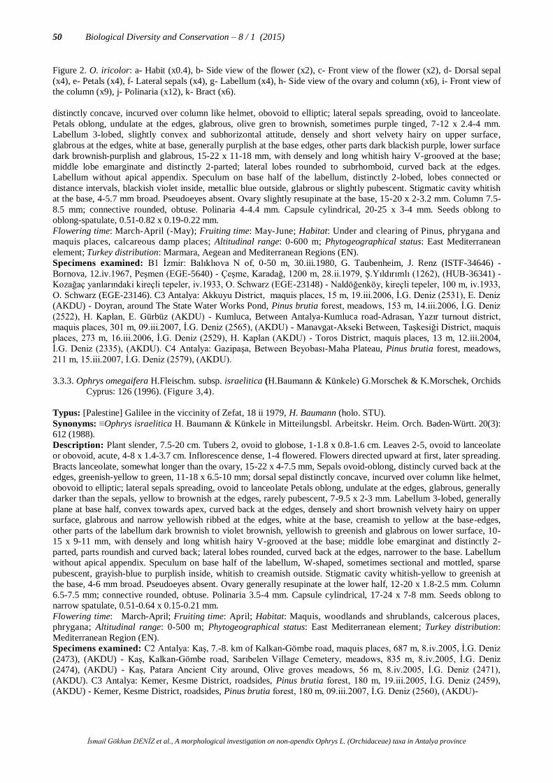
İsmail Gökhan DENİZ et al., A morphological investigation on non-apendix Ophrys L. (Orchidaceae) taxa in Antalya province
50 Biological Diversity and Conservation – 8 / 1 (2015)
Figure 2. O. iricolor: a- Habit (x0.4), b- Side view of the flower (x2), c- Front view of the flower (x2), d- Dorsal sepal
(x4), e- Petals (x4), f- Lateral sepals (x4), g- Labellum (x4), h- Side view of the ovary and column (x6), i- Front view of
the column (x9), j- Polinaria (x12), k- Bract (x6).
distinctly concave, incurved over column like helmet, obovoid to elliptic; lateral sepals spreading, ovoid to lanceolate.
Petals oblong, undulate at the edges, glabrous, olive gren to brownish, sometimes purple tinged, 7-12 x 2.4-4 mm.
Labellum 3-lobed, slightly convex and subhorizontal attitude, densely and short velvety hairy on upper surface,
glabrous at the edges, white at base, generally purplish at the base edges, other parts dark blackish purple, lower surface
dark brownish-purplish and glabrous, 15-22 x 11-18 mm, with densely and long whitish hairy V-grooved at the base;
middle lobe emarginate and distinctly 2-parted; lateral lobes rounded to subrhomboid, curved back at the edges.
Labellum without apical appendix. Speculum on base half of the labellum, distinctly 2-lobed, lobes connected or
distance intervals, blackish violet inside, metallic blue outside, glabrous or slightly pubescent. Stigmatic cavity whitish
at the base, 4-5.7 mm broad. Pseudoeyes absent. Ovary slightly resupinate at the base, 15-20 x 2-3.2 mm. Column 7.5-
8.5 mm; connective rounded, obtuse. Polinaria 4-4.4 mm. Capsule cylindrical, 20-25 x 3-4 mm. Seeds oblong to
oblong-spatulate, 0.51-0.82 x 0.19-0.22 mm.
Flowering time: March-April (-May); Fruiting time: May-June; Habitat: Under and clearing of Pinus, phrygana and
maquis places, calcareous damp places; Altitudinal range: 0-600 m; Phytogeographical status: East Mediterranean
element; Turkey distribution: Marmara, Aegean and Mediterranean Regions (EN).
Specimens examined: B1 İzmir: Balıklıova N of, 0-50 m, 30.iii.1980, G. Taubenheim, J. Renz (ISTF-34646) -
Bornova, 12.iv.1967, Peşmen (EGE-5640) - Çeşme, Karadağ, 1200 m, 28.ii.1979, Ş.Yıldırımlı (1262), (HUB-36341) -
Kozağaç yanlarındaki kireçli tepeler, iv.1933, O. Schwarz (EGE-23148) - Naldöğenköy, kireçli tepeler, 100 m, iv.1933,
O. Schwarz (EGE-23146). C3 Antalya: Akkuyu District, maquis places, 15 m, 19.iii.2006, İ.G. Deniz (2531), E. Deniz
(AKDU) - Doyran, around The State Water Works Pond, Pinus brutia forest, meadows, 153 m, 14.iii.2006, İ.G. Deniz
(2522), H. Kaplan, E. Gürbüz (AKDU) - Kumluca, Between Antalya-Kumluca road-Adrasan, Yazır turnout district,
maquis places, 301 m, 09.iii.2007, İ.G. Deniz (2565), (AKDU) - Manavgat-Akseki Between, Taşkesiği District, maquis
places, 273 m, 16.iii.2006, İ.G. Deniz (2529), H. Kaplan (AKDU) - Toros District, maquis places, 13 m, 12.iii.2004,
İ.G. Deniz (2335), (AKDU). C4 Antalya: Gazipaşa, Between Beyobası-Maha Plateau, Pinus brutia forest, meadows,
211 m, 15.iii.2007, İ.G. Deniz (2579), (AKDU).
3.3.3. Ophrys omegaifera H.Fleischm. subsp. israelitica (H.Baumann & Künkele) G.Morschek & K.Morschek, Orchids
Cyprus: 126 (1996). (Figure 3,4).
Typus: [Palestine] Galilee in the viccinity of Zefat, 18 ii 1979, H. Baumann (holo. STU).
Synonyms: ≡Ophrys israelitica H. Baumann & Künkele in Mitteilungsbl. Arbeitskr. Heim. Orch. Baden-Württ. 20(3):
612 (1988).
Description: Plant slender, 7.5-20 cm. Tubers 2, ovoid to globose, 1-1.8 x 0.8-1.6 cm. Leaves 2-5, ovoid to lanceolate
or obovoid, acute, 4-8 x 1.4-3.7 cm. Inflorescence dense, 1-4 flowered. Flowers directed upward at first, later spreading.
Bracts lanceolate, somewhat longer than the ovary, 15-22 x 4-7.5 mm, Sepals ovoid-oblong, distincly curved back at the
edges, greenish-yellow to green, 11-18 x 6.5-10 mm; dorsal sepal distinctly concave, incurved over column like helmet,
obovoid to elliptic; lateral sepals spreading, ovoid to lanceolate Petals oblong, undulate at the edges, glabrous, generally
darker than the sepals, yellow to brownish at the edges, rarely pubescent, 7-9.5 x 2-3 mm. Labellum 3-lobed, generally
plane at base half, convex towards apex, curved back at the edges, densely and short brownish velvety hairy on upper
surface, glabrous and narrow yellowish ribbed at the edges, white at the base, creamish to yellow at the base-edges,
other parts of the labellum dark brownish to violet brownish, yellowish to greenish and glabrous on lower surface, 10-
15 x 9-11 mm, with densely and long whitish hairy V-grooved at the base; middle lobe emarginat and distinctly 2-
parted, parts roundish and curved back; lateral lobes rounded, curved back at the edges, narrower to the base. Labellum
without apical appendix. Speculum on base half of the labellum, W-shaped, sometimes sectional and mottled, sparse
pubescent, grayish-blue to purplish inside, whitish to creamish outside. Stigmatic cavity whitish-yellow to greenish at
the base, 4-6 mm broad. Pseudoeyes absent. Ovary generally resupinate at the lower half, 12-20 x 1.8-2.5 mm. Column
6.5-7.5 mm; connective rounded, obtuse. Polinaria 3.5-4 mm. Capsule cylindrical, 17-24 x 7-8 mm. Seeds oblong to
narrow spatulate, 0.51-0.64 x 0.15-0.21 mm.
Flowering time: March-April; Fruiting time: April; Habitat: Maquis, woodlands and shrublands, calcerous places,
phrygana; Altitudinal range: 0-500 m; Phytogeographical status: East Mediterranean element; Turkey distribution:
Mediterranean Region (EN).
Specimens examined: C2 Antalya: Kaş, 7.-8. km of Kalkan-Gömbe road, maquis places, 687 m, 8.iv.2005, İ.G. Deniz
(2473), (AKDU) - Kaş, Kalkan-Gömbe road, Sarıbelen Village Cemetery, meadows, 835 m, 8.iv.2005, İ.G. Deniz
(2474), (AKDU) - Kaş, Patara Ancient City around, Olive groves meadows, 56 m, 8.iv.2005, İ.G. Deniz (2471),
(AKDU). C3 Antalya: Kemer, Kesme District, roadsides, Pinus brutia forest, 180 m, 19.iii.2005, İ.G. Deniz (2459),
(AKDU) - Kemer, Kesme District, roadsides, Pinus brutia forest, 180 m, 09.iii.2007, İ.G. Deniz (2560), (AKDU)-

İsmail Gökhan DENİZ et al., A morphological investigation on non-apendix Ophrys L. (Orchidaceae) taxa in Antalya province
Biological Diversity and Conservation – 8 / 1 (2015) 51
Figure 3. O. omegaifera subsp. israelitica: a- Habit (x3,5), b- Side view of the flower (x2), c- Front view of the flower
(x2), d- Dorsal sepal (x4), e- Petals (x4), f- Lateral sepals (x4), g- Labellum (x4), h- Side view of the ovary and column
(x6), i- Front view of the column (x9), j- Polinaria (x12), k- Bract (x6).

İsmail Gökhan DENİZ et al., A morphological investigation on non-apendix Ophrys L. (Orchidaceae) taxa in Antalya province
52 Biological Diversity and Conservation – 8 / 1 (2015)
Kemer, Kuzdere Valley, roadsides, Pinus brutia forest, meadows, 183 m, 12.iii.2004, İ.G. Deniz (2334), (AKDU) -
Kumluca, Olimpos Ancient City, Pinus brutia forest, edges of the creek, meadows, 35 m, 31.iii.2004, İ.G. Deniz
(2362), (AKDU) - Manavgat, Çayiçi District, meadows, 70 m, 15.iii.2005, İ.G. Deniz (2448), (AKDU) - Manavgat,
Manavgat-Oymapınar road, 9.-10. km, Pinus brutia forest, 21 m, 20.iii.2004, İ.G. Deniz (2339), (AKDU) - Yukarı
Karaman, slopes between Yukarı Karaman-Bahtılı, Pinus brutia forest, 217 m, 24.iii.2007, İ.G. Deniz (2610), (AKDU)
- C4 Antalya: Alanya, Between Demirtaş-Sapadere, Pinus brutia forest, 133 m, 15.iii.2007, İ.G. Deniz (2574), (AKDU)
- Gazipaşa, Between Beyobası-Maha Plateau, Pinus brutia forest, meadows, 192 m, 15.iii.2007, İ.G. Deniz (2575),
(AKDU) - C5 İçel: Ulaş, 8 km N of Tarsus to Çamlıyayla, 270 m, 08.04.1980, Renz & Taubenheim 80.76 (ISTF
34.657)
3.3.4. Ophrys phaseliana D. Rückbr. & U. Rückbr. in J. Eur. Orchideen 28: 395 (1996). (Figure 5, 9).
Typus: [Turkey] Antalya, Phaselis, 80 m, 26.iv.1996, D.&U. Rückbrodt (holo. B.).
Synonyms: =O. fusca Link subsp. phaseliana (D.Rückbr. & U.Rückbr.) Kreutz, Kompend. Eur. Orchid.: 96 (2004).
=O. parosica P.Delforge var. phaseliana (D.Rückbr. & U.Rückbr.) P.Delforge, Naturalistes Belges 88: 247 (2007).
Description: Plant slender, 15-40 cm. Tubers 2 , ovoid to globose, 1-1.7 x 0.8-1.6 cm. Leaves 2-6, generally at the
base, ovoid to lanceolate, acute, 4-8.5 x 1.4-3.7 cm. Inflorescence lax, 1-5 flowered. Flowers large, directed upward at
first, later spreading. Bracts lanceolate, equal in length or longer than ovary, 15-18 x 4-7.5 mm. Sepals oblong-ovoid to
elliptic, distincly curved back at the edges, yellowish-green to greenish, 11-16 x 5-8 mm; dorsal sepal distinctly
concave, incurved over column like helmet, lateral sepals spreading, Petals oblong, undulate at the edges, somet imes
slightly emarginate, glabrous, yellowish-green to greenish, 7.5-12 x 2.5-4 mm. Labellum 3-lobed, directed downward,
strongly convex, not pressed on the middle part, curved back at the all edges, brownish at the base, yellowish to
brownish ribbed or tessellate at apex part, densely and short brownish velvety hairy on upper surface, edges glabrous,
yellowish to greenish at the base-edges, lower surface greenish to pale brown, 13.5-18.5 x 11.5-16.5 mm, with densely
and long whitish hairy V-grooved at the base; middle lobe emarginat and distinctly 2-lobed, lobes curved back, apex
greenish part spreading; lateral lobes rounded, distant from middle lobe, narrower to the base. Labellum without apical
appendix. Speculum on whole labellum, W-shaped, sectional and mottled, sometimes 2-lobed, lobes ovoid, connected
or distance intervals, sparse pubescent, grayish-blue to purplish brown inside, grayish blue outside, mottled on apex part
of the labellum. Stigmatic cavity whitish-yellow to greenish at the base, 5-6 mm broad. Pseudoeyes absent. Ovary
straight, not angled with column, 12-20.5 x 2.3-4 mm. Column 7-8 mm; connective rounded, obtuse. Polinaria 3.5-4.5
mm. Capsule cylindrical, 17-23 x 4.5-6 mm. Seeds broad oblong-spatulate , 0.51-0.65 x 0.15-0.21 mm.
Flowering time: April-May; Fruiting time: May; Habitat: Openings in forest, phrygana, shrublands, calcerous
places; Altitudinal range: 0-200 m; Phytogeographical status: East Mediterranean element; Turkey distribution:
Antalya and Muğla Provinces. Endemic (CR).
Specimens examined: C3 Antalya: Kumluca, Kumluca-Altınyaka road, 5.-6. km, Pinus brutia forest, meadows, 191
m, 17.iv.2007, İ.G. Deniz (2627), (AKDU).
Figure 4. Turkey distribution of (a) O. speculum subsp. speculum, (b) O. iricolor, (c) O. omegaifera subsp. israelitica
and (d) O. lutea subsp. minor.

İsmail Gökhan DENİZ et al., A morphological investigation on non-apendix Ophrys L. (Orchidaceae) taxa in Antalya province
Biological Diversity and Conservation – 8 / 1 (2015) 53
Figure 5. O. phaseliana: a- Habit (x0.4), b- Side view of the flower (x2), c- Front view of the flower (x2), d- Dorsal
sepal (x4), e- Petals (x4), f- Lateral sepals (x4), g- Labellum (x4), h- Side view of the ovary and column (x5.5), i- Front
view of the column (x7), j- Polinaria (x12), k- Bract (x5.5).

İsmail Gökhan DENİZ et al., A morphological investigation on non-apendix Ophrys L. (Orchidaceae) taxa in Antalya province
54 Biological Diversity and Conservation – 8 / 1 (2015)
3.3.5. Ophrys cinereophila Paulus & Gack in J. Eur. Orchideen 30: 170 (1998). (Figure 6, 9).
Typus: [Greece] E. Crete in the vicinity of Nikithianou near Neapolis, 3 iv 1987, Paulus (holo. In herb. Paulus, Wien).
Synonyms: =O. fusca Link subsp. cinereophila (Paulus & Gack) Faurh., Orchidee (Hamburg) 53: 345 (2002). =O.
subfusca (Rchb.f.) Batt. subsp. cinereophila (Paulus & Gack) Kreutz, Orchidee (Hamburg) 57: 102 (2006).
Description: Plant slender, 7-23 cm. Tubers 2 , ovoid to globose, 1-1.6 x 0.6-1.4 cm. Leaves 2-6 , generally at the
base, ovoid to lanceolate, acute, 2.2-9.4 x 0.8-3.5 cm. Inflorescence lax, 5-7 (-10) flowered. Flowers small, directed
upward at first, later distinctly spreading. Bracts lanceolate, equal in length or somewhat longer than ovary, 11-18 x 3.8-
6 mm. Sepals broadly ovoid to lanceolate, distincly curved back at the edges, yellowish to greenish, 7-11 x 4-7.5 mm;
dorsal sepal distinctly concave, incurved over column like helmet lateral sepals spreading, Petals oblong, undulate at the
edges, rarely slightly emarginate, glabrous, yellowish-green to greenish, 5-9 x 2-2.8 mm. Labellum 3-lobed, directed
downward, strongly convex, pressed on the middle part, curved back at the all edges, blackish to dark brown, not
striped or tessellate at apex part, densely and short brownish to purplish velvety hairy on upper surface, edges glabrous
or lax pubescent, whitish to yellowish at the base-edges, lower surface greenish to pale brown, 7-13 x 5-10.5 mm, with
densely and long whitish hairy V-grooved at the base; middle lobe emarginat and distinctly 2-lobed, lobes curved back,
apex yellow greenish part spreading; lateral lobes straight or rounded, clerly distant from middle lobe, narrower to the
base. Labellum without apical appendix. Speculum on base half of the labellum, W-shaped, 2-lobed, lobes ovoid,
always connected, sparse pubescent, grayish-blue to purplish brown inside, grayish blue outside, apex part straight.
Stigmatic cavity whitish-yellow to greenish at the base, 3.5-5 mm broad. Pseudoeyes absent. Ovary straight, angled
with column, 10-18 x 1.5-3.5 mm. Column 5-6 mm; connective rounded, obtuse. Polinaria 2.5-3.5 mm. Capsule
cylindrical, 16-22 x 4.5-5.5 mm. Seeds broad oblong-spatulate, 0.44-0.65 x 0.15-0.21 mm.
Flowering time: March-April; Fruiting time: April; Habitat: Maquis places, openings in forest, meadows, phrygana;
Altitudinal range: 0-800 m; Phytogeographical status: East Mediterranean element; Turkey distribution: Aegean and
Mediterranean Regions (EN).
Specimens examined: C2 Antalya: Kaş, Kaş-Kasaba road, 7.-8. km, Pinus brutia forest, 206 m, 08.iv.2005, İ.G.
Deniz (2477), (AKDU). C3 Antalya: Kumluca, Between Adrasan-Olimpos Ancient City, meadows, 50 m, 25.iii.2005,
İ.G. Deniz (2465), (AKDU) - Kumluca, Antalya-Kumluca road-Adrasan Between, Yazır turnout district, maquis places,
294 m, 09.iii.2007, İ.G. Deniz (2564a), (AKDU) - Kumluca, 1 km to Olimpos Ancient City, meadows, 50 m,
12.iii.2004, İ.G. Deniz (2332), (AKDU) - Kumluca, around Olimpos Ancient City, Pinus brutia forest, 46 m,
09.iii.2007, İ.G. Deniz (2569), (AKDU) - Kumluca, Between Olimpos Ancient City-Çavuşköy, Pinus brutia forest,
edges of the creek, meadows, 35 m, 31.iii.2004, İ.G. Deniz (2361), (AKDU).
3.3.6. Ophrys urteae Paulus, Ber. Arbeitskreis. Heimische Orchid. 26(2): 10 (2009). (Figure 7, 9)
Typus: [Turkey] Antalya, 7.4 km ne. Taşağıl, kleiner Friedhof, 152 m, 18.iv.1998, H.F. Paulus (holo. WU).
Description: Plant slender, 6-8 (-15) cm. Tubers 2 , ovoid to globose, 1-2.3 x 0.7-1.6 cm. Leaves 3-6 , generally at the
base, ovoid to broadly lanceolate, acute, 2.5-9 x 1-3.5 cm. Inflorescence generally lax, 1-3 (-6) flowered. Flowers
small, spreading. Bracts ovoid to lanceolate, equal in length or somewhat longer than ovary, 9-19 x 4-7 mm. Sepals
ovoid-lanceolate to broadly ovoid, curved back at the edges, yellowish to greenish, 12-18 x 5-8 mm; dorsal sepal
distinctly concave, incurved over column like helmet lateral sepals spreading, Petals oblong-lanceolate, undulate at the
edges, obtuse, glabrous, greenish yellow, sometimes brownish at the edges, 7-9 x 1-3 mm. Labellum obtriangular-
oblong, distinctly straight and flat, 3-lobed, dark brownish to blackish violet, 9-12 x 7-11 mm, densely and short
brownish to purplish velvety hairy on upper surface, with densely and whitish hairy V-grooved at the base; middle lobe
emarginat and distinctly 2-lobed, lobes spreading, apex part generally directed upwards; lateral lobes long and narrow,
round at apex, not wing-like, spreading or somewhat directed downward, sometimes undulate with narrow yellow
edges, clerly distant from middle lobe. Labellum without apical appendix. Speculum on base half of the labellum, W-
shaped, not sectional, sometimes connected 2-lobed, lobes ovoid, sparse pubescent, mat shining dark brownish to blue
grayish, nearly to lateral notches. Stigmatic cavity whitish at the base, 4-5 mm broad. Pseudoeyes absent. Ovary
straight, 10-16 x 1.3-2.5 mm. Column 4.5-6 mm; connective rounded, obtuse. Polinaria 3-4 mm. Capsule cylindrical,
15-23 x 4-5 mm. Seeds narrow oblong to spatulate, 0.38-0.55 x 0.14-0.18 mm.
Flowering time: March-April; Fruiting time: April; Habitat: Under and clearing of Pinus, maquis places, phrygana,
calcareous meadows, olive groves; Altitudinal range: 0-400 m; Phytogeographical status: East Mediterranean
element; Turkey distribution: Antalya Province. Endemic (CR).
Specimens examined: C3 Antalya: Manavgat, Oymapınar, 6.-7. km of Oymapınar-Tilkiler road, Pinus brutia forest,
155 m, 01.iv.2009, İ.G. Deniz (2868), (AKDU).

İsmail Gökhan DENİZ et al., A morphological investigation on non-apendix Ophrys L. (Orchidaceae) taxa in Antalya province
Biological Diversity and Conservation – 8 / 1 (2015) 55
Figure 6. O. cinereophila: a- Habit (x0.6), b- Side view of the flower (x3), c- Front view of the flower (x3), d- Dorsal
sepal (x6.5), e- Petals (x6.5), f- Lateral sepals (x6.5), g- Labellum (x6.5), h- Side view of the ovary and column (x7.5),
i- Front view of the column (x10), j- Polinaria (x18), k- Bract (x7.5).

İsmail Gökhan DENİZ et al., A morphological investigation on non-apendix Ophrys L. (Orchidaceae) taxa in Antalya province
56 Biological Diversity and Conservation – 8 / 1 (2015)
Figure 7. O. urteae: a- Habit (x0.9), b- Side view of the flower (x2.5), c- Front view of the flower (x2.5), d- Dorsal
sepal (x5), e- Petals (x5), f- Lateral sepals (x5), g- Labellum (x5), h- Side view of the ovary and column (x6), i- Front
view of the column (x9), j- Polinaria (x12), k- Bract (x6).

İsmail Gökhan DENİZ et al., A morphological investigation on non-apendix Ophrys L. (Orchidaceae) taxa in Antalya province
Biological Diversity and Conservation – 8 / 1 (2015) 57
3.3.7. Ophrys lutea Cav. subsp. minor (Guss.) O. & E. Danesch in Pl. Syst. Evol. 124:82 (1975). (Figure 4, 8).
Typus: [Italy] Sicilya, Palermo, S. Biaggio sotto Cammarata, Gussone.
Synonyms: ≡Arachnites lutea (Cav.) Tod. var. minus Tod., Orchid. Sicul.: 97 (1842). ≡O. lutea Cav. var. minor Guss., Fl. Sic. Syn. 2(2):550 (1844). ≡O. lutea Cav. f. sicula (Tin.) Soo in Feddes Rep. 24:25 (1927).
Description: Plant slender, 7-30 (-40) cm. Tubers 2 , ovoid to globose, 1-2.3 x 0.6-1.7 cm. Leaves 2-6 , generally at the base,
ovoidden geniş to lanceolate, acute, 2.2-9.4 x 1-3.5 cm. Inflorescence dense, 1-8 flowered. Flowers small, upward at first, later
distinctly spreading. Bracts ovoid to lanceolate, equal in length or somewhat shorter than ovary, 8-20 x 4-8 mm. Sepals ovoid, curved back at the edges, yellowish to greenish, 6.5-9.5 x 3-6 mm; dorsal sepal distinctly concave, incurved over column like helmet; lateral
sepals spreading. Petals oblong, generally undulate at the edges, obtuse, glabrous, yellowish green to green, 4-4.5 x 1-1.5 mm.
Labellum ovoid-roundish, 3-lobed close to apex part, directed horizontal, convex at middle part, yellow to brownish, sometimes
striped or tessellate to lateral and middle lobes, whitish to yellowish at the base, lateral lobes yellow, densely whitish to brownish pubescent, outside glabrous, 8-15 x 8.5-12 mm, with densely and long whitish hairy V-grooved at the base, hairs longer to the
staminal part; middle lobe short, emarginat and distinctly 2-lobed, lobes angled to each other, spreading, apex part generally directed
upwards; lateral lobes long and broad, wing-like, spreading or somewhat directed downward, sometimes undulate with broad yellow
edges, clerly distant from middle lobe. Labellum without apical appendix. Speculum on base half of the labellum, W-shaped, mottled and connected inside or distinctly 2-lobed, lobes ovoid, sparse pubescent, shining grayish-brown to bluish purple inside, whitish-
cream to blue outside, not extend to lateral notches. Stigmatic cavity whitish-yellow to greenish , 4-5 mm broad. Pseudoeyes absent.
Ovary straight, 11-18 x 1.4-2.4 mm. Column 4.5-5.5 mm; connective rounded, obtuse. Polinaria 2.8-3.2 mm. Capsule, cylindrical,
15-23 x 4-5 mm. Seeds narrow oblong to spatulate, 0.38-0.55 x 0.14-0.18 mm.
Flowering time: March-April; Fruiting time: April; Habitat: Maquis and forest places, calcareous meadows, phrygana, olive
groves, meadows; Altitudinal range: 0-600 m; Phytogeographical status: East Mediterranean element; Turkey distribution:
Marmara, Aegean, Mediterranean, Southeastern Anatolia Regions (EN).
Specimens examined: A1 Balıkesir: Marmara Island, Marmara District, Gündoğdu Village, 15.iii.1977, E. Tuzlacı (ISTE-36495). Çanakkale: Arıburnu-Suvla road, 8.iv.1975, A. Baytop (ISTE-31438) - Çanakkale: Gökçeada, Kuzulimanı, 10 m, 10.iv.1976, Ö.
Seçmen, E. Leblebici (1296), (EGE-22553). A2 İstanbul: Çatalca, upper parts of the cemetery, 20.iv.2003, İ. Genç (ISTE-82370) -
Halkalı, slopes across the train station, 24.iv.1960, A. Baytop (ISTE-5797) - E of Halkalı train station, 23.iv.1971, N.&E. Özhatay,
G. Ertem (ISTE-19374) - Çatalca, hills across the cemetery, 1.v.1965, A. Baytop (ISTE-7620) - Hadımköy, SE slopes of the Bahçeşehir, open places, 90 m, 09.iv.1997, E. Üzen, O. Küçüker (ISTF-36940) Bakırköy, Ataköy, edges of the railway, 19.iv.1968,
A. Glg., G.San. (ISTF-23119) - Bakırköy, Halkalı train station, meadows, 15.iv.1967, H. Demiriz (ISTF-21955) - Kartal, Cevizli,
Ankara roadnun 1 km E of, Quercus Between, çayır, 27.iv.1968, A. Çır., O. Sut. (ISTF-23019) - Kartal, Yakacık, openings of the
Quercus coccifera groves, 16.iv.1968, H. Demiriz, E. Tut., A. Ayd., P. Kok. (ISTF-22965) - Halkalı, meadows, slopes, 05.iv.1965, Betül Tutel (65/13), (ISTF-19153) - Bakırköy, Florya, edges of the railway, meadows, 23.iv.1967, H. Demiriz (ISTF-21984). B1
Balıkesir: Ayvalık, Dolap Island, 10 m, 22.iii.1996, K. Alpınar (ISTE-71315). Manisa: Manisa Mountain, Mevlevihane, 18.iv.1974,
G. Ertem (ISTE-27393). Çanakkale: Bozcaada, N regions of the Island, around Killik Hill, 0-50 m, 14.iv.1977, Ö. Seçmen, E.
Leblebici (2448), (EGE-20761) - Çanakkale: Bozcaada, around Hacıhasan Hill, 40 m, 14.iv.1976, Ö. Seçmen, E. Leblebici (1494), (EGE-20659) - Çanakkale: Bozcaada, 1 km from the center, 0-50 m, 13.iv.1977, Ö. Seçmen, E. Leblebici (2376), (EGE-20762) -
Çanakkale: Bozcaada, Tuzburnu Hill, around Hacımahmu, 70 m, 15.iv.1976, Ö. Seçmen, E. Leblebici (1578), (EGE-20657). İzmir:
İzmir Çeşme road, before İçmeler, 21.iii.1967, A.&T. Baytop (ISTE-10965) - İzmir: Akkum, 27.ii.1968, Meyer, Peşmen (EGE-3054)
- Bornova, N of Çiçekli Village, 110 m, C. Ödemiş, A. Yayıntaş, U. Zeybek (EGE-23446) - Bornova, Hacılarkırı, Sabuncubeli, 31.iii.1967, M. Aydar (EGE-8619) - Bornova, Between Hacılarkırı-Sabuncubeli, maquis places, 31.iii.1967, M. Aydar (EGE-8593) -
Bornova, on marl, iv.1932, N. İ. (EGE-23159) - Çeşme District, 28.iii.1984, Ö. Seçmen (2557) (EGE-28918) - Çeşme-Altınkum,
18.iv.1983, Y. Gemici (1505), Ö. Seçmen, G. Görk, L. Bekat (EGE-21777) - Gümüldür, 200 m, 25.iv.1976, T. Kesercioğlu (EGE-
15094) - Kemalpaşa-Karabel road, 19.iv.1967, H. Peşmen, M. Aydar (EGE-8596) - Kemalpaşa-Kavaklıdere road, maquis places, 19.iv.1967, H. Peşmen (EGE-8595) - Seferihisar, 23.iii.1972, T. Gözler, Ödemiş (EGE-9910). B2 Balıkesir: Pelitköy, Karantılık
District, olive groves, 0-50 m, 8.iii.2001, Tuğrul Körüklü (188), (GAZI). C1 Aydın: Lalelik, Çakman Village, 15.iv.1965, E. Sezik
(ISTE-8069a). Kuşadası, Pino Bay otel road, 90 m, 25.iv.1996, E. Üzen (ISTF-36663) - Güzelbahçe (Priene), 12.iv.1962, A.
Pelshenke (EGE-8879). Kuşadası, S slopes, 210 m, 4.iv.1977, G. Oğuz, A. Yayıntaş, (EGE-23448) - Between Kuşadası-Selçuk, 31.iii.1968, Meyer, Peşmen, Oflas, Oğuz, Leblebici (EGE-3031) Söke, Atburgazı District, on Priene road, 28.iii.1968, Meyer,
Peşmen, Oflas, Oğuz, Leblebici (EGE-3041). Muğla: Söke-Milas road, egdes of the Bafa Lake, olive groves, 10.iv.1971, A.&T.
Baytop (ISTE-19219) - Milas, Yeniköy, 14.iv.1965, E. Sezik (ISTE-8374) - Milas, Yayladağ, Yukarı Mazı Village, 17.iv.1965, E.
Sezik (ISTE-8382) - Milas, Kızılağaç, Kazıklı Village, 13.iv.1966, E. Sezik (ISTE-8731) - Taşlıca, Karaçalca District, 300 m,
22.iii.1981, T. Baytop, A. Atilla ISTE-46090). C2 Muğla: Köyceğiz, Yangı Village, calcareous valley,

İsmail Gökhan DENİZ et al., A morphological investigation on non-apendix Ophrys L. (Orchidaceae) taxa in Antalya province
58 Biological Diversity and Conservation – 8 / 1 (2015)
Figure 8. O. lutea subsp. minor: a- Habit (x0.6), b- Side view of the flower (x2.5), c- Front view of the flower (x2.5), d- Dorsal sepal
(x6), e- Petals (x6), f- Lateral sepals (x6), g- Labellum (x6), h- Side view of the ovary and column (x6), i- Front view of the column (x9), j- Polinaria (x12), k- Bract (x6).

İsmail Gökhan DENİZ et al., A morphological investigation on non-apendix Ophrys L. (Orchidaceae) taxa in Antalya province
Biological Diversity and Conservation – 8 / 1 (2015) 59
Figure 9. Turkey distribution of (a) O. cinereophila, (b) O. phaseliana () and O.urteae ().
40-220 m, 22.iii.1991, A. Güner (8410), H. Duman, H. Şağban (HUB-36344) - Bodrum, Mumcular, Yılan Yatağı District, roadsides,
Pinus brutia forest, 150-200 m, 14.iv.2000, M. Karauzun, E. Dalkesen (0049), (VANF). Antalya: Kaş, 7.-8. km of Kaş-Kasaba road, Pinus brutia forest, 206 m, 08.iv.2005, İ.G. Deniz (2482), (AKDU) - Kaş, around Patara Ancient City, olive groves, meadows, 62 m,
12.iv.2007, İ.G. Deniz (2621), (AKDU). Denizli: Denizli-Tavas, 23.iii.1957, M. Heilbronn (ISTF-16430). Muğla: Bodrum,
Mumcular, Yılanyatağı District, P. brutia forest, 150-200 m, 16.iv.2000, M. Karauzun, E. Dalkesen (0049), (MUH) - Datça, city
center, around holiday houses, maquis places, 10-20 m, 16.ii.2002, Ö. Varol (3958), (MUH) - Old road Mumcular-Güvercinlik, around second forests store, P. brutia forest, 110 m, 17.ii.2001, Ö. Varol (3575), (MUH) - Köyceğiz, Horozlar Village, fieldedges, 10
m, 17.iii.1991, A. Güner (8234), H. Duman, H. Şağban (HUB-36343) - Old cemetery of Muğla, under Cupressus groves, 670 m,
21.iii.2001, B. Sahrancı (135), (MUH) - Muğla Cemetery, 620 m, 17.iii.2003, E. Kaya (70), (MUH) - Muğla-Milas, 10 km to Milas,
Pinus forest, 100-150 m, 16.iv.2000, M. Karauzun, E. Dalkesen (0039), (MUH). C3 Antalya: Between Kemer-Kumluca, 1 km from Olimpos junction, P. brutia forest, 350 m, 23.iii.1979, H. Peşmen (4216), A. Güner (HUB-36346) - Kumluca, Adrasan, Pinus brutia
forest, 46 m, 25.iii.2005, İ.G. Deniz (2469), (AKDU) - Kumluca, Adrasan, Yazır turnout district, maquis places, 294 m, 09.iii.2007,
İ.G. Deniz (2564b), (AKDU) - Kumluca, 5.-6. Km of Kumluca-Altınyaka road, Pinus brutia forest, meadows, 180 m, 17.iv.2007,
180 m, İ.G. Deniz (2635), (AKDU) - Kumluca, around Olimpos Ancient City, Pinus brutia forest, 46 m, 09.iii.2007, İ.G. Deniz (2570), (AKDU) - Between Harbour-Tünektepe, edges of the creek, maquis places, 81 m, 05.iii.2006, İ.G. Deniz (2511), B.E.
Tarıkahya, D. Şirin (AKDU) - Manavgat, Çayiçi District, meadows, 70 m, 15.iii.2005, İ.G. Deniz (2446), (AKDU) - Manavgat,
Köprülü Kanyon National Park, 5 km N of Sağırin, 148 m, 16.iii.2006, İ.G. Deniz (2524), (AKDU) - Manavgat, Köprülü Kanyon
National Park, Between Taşağıl-Sağırin, Pinus brutia forest, 56 m, 11.iii.2005, İ.G. Deniz (2443), H. Sümbül, C. Kemaloğlu, Ö. Tufan (AKDU) - Manavgat, 3 km W of Oymapınar Dam Lake, olive groves, 170 m, 15.iii.2005, İ.G. Deniz (2453), (AKDU) -
Manavgat, Between Oymapınar-Tilkiler, olive groves, 62 m, 20.iii.2004, İ.G. Deniz (2344a), (AKDU) - Manavgat, Taşkesiği
Village, fieldedges, 1000 m, 26.iii.1992, Ali A. Dönmez (3108), R.S. Göktürk, (HUB-36342) - Manavgat, Sorgun Forest, Pinus
brutia forest, 16 m, 19.iii.2007, İ.G. Deniz (2600), C. Aykurt, A. Barut (AKDU). C4 Antalya: Alanya, Between Demirtaş-Sapadere, Pinus brutia forest, meadows, 133 m, 15.iii.2007, İ.G. Deniz (2571), (AKDU) - Gazipaşa, Between Beyobası-Maha Plateau, Pinus
brutia forest, meadows, 223 m, 15.iii.2007, İ.G. Deniz (2581), (AKDU). C6 Hatay: Güney, Habib Neccar Mountain, Antakya
district, 15.iii.1975, A. Baytop (ISTE-31331).
4. Conclusions and discussion
In this part of the present study, morphological characteristics of non-appendix 7 Ophrys L. taxa have been discussed and its
differerences and relationship with the close taxa were given. The first taxa of the study is Ophrys speculum Link subsp. speculum with different shape and indumentum structure of the labellum. The specimens of O. speculum Link were described as a new species
by J.H. Friedrich Link (1800) from the Setubal Province of Portugal. Because the taxa has a blue and shiny labellum, the name was
derived from “speculum”, which means mirror in Latin. The name of O. speculum was not included in the Flora of Turkey at the
species level. Renz and Taubenheim (1984) showed it as the synonym of O. vernixia subsp. vernixia. Putting aside taxonomic similarities and differences between taxa, the fact that the priority rules do not apply, the nomenclature of this taxon is incorrect in
Flora of Turkey. Link identified O. speculum in Latin and also showed the location clearly in his book, which was prepared for
publication in 1799 and published in 1800. Therefore, the publication is sufficiently correct and valid. Whereas O. vernixia Brot was
defined in Flora Lusitanica in 1804,- four years later, it is remarkable that the type location of both names in close regions of Portugal. Thus, according to 2006 World nomenclature codes and with the decisions taken within the framework of the 17th World
International Botanical Congress in 2005, organized in Vienna (Austria), O. speculum is in the protected names class and has priority
to other names (Menemen and Dönmez, 2006). For all these reasons, Ophrys speculum Link should be a valid taxon name in Flora of
Turkey. In Turkey the species have two subspecies, O. speculum subsp. speculum and O. speculum subsp. regis-ferdinandii, showing distribution in the İzmir, Aydın and Muğla Provinces of Turkey. O. speculum subsp. speculum differs from subsp. regis-
ferdinandii with inflorescence 2-9 flowered (not 2-12), the labellum has more dense indumentum and soborbicular (not stretched and
narrowly elongate), middle lobe flattened when compared (not convex), lateral lobes triangular-oblong, and not or slighly spreading
(not linear-oblong and spreading) characteristics. The first examples of the O. iricolor were collected by Joseph Pitton de Tournefort from the Levant about three hundred
years ago. It is not known exactly where he collected these samples during his Anatolian travels in 1701-1702. Some information
obtained suggests the location to be Samos or İzmir (Delforge, 1995). Tournefort identified O. iricolor as “Orchis orientalis fucum
referens, flore maximo, scuto azureo”. These examples were named with binomial nomenclature by Par M. Desfontaines and published in Choix De Plantes Corollaire Des Instituts De Tournefort. When defining the species, Desfontaines wrote “Ophrys irise”
in parentheses because of the metallic blue color of the flowers and like Iris. O. iricolor is taxonomically close to O. atlantica which
has distribution in Spain, Algeria, Morocco and Tunisia. At first glance these species differ from each other by labellum shapes. The
labellum of O. atlantica is downward from base to end of the speculum (deeply concave at middle) and horizontal to the apex whereas O. iricolor labellum is slightly convex and in subhorizontal attitude. Furthermore, O. iricolor differs from O. atlantica; the

İsmail Gökhan DENİZ et al., A morphological investigation on non-apendix Ophrys L. (Orchidaceae) taxa in Antalya province
60 Biological Diversity and Conservation – 8 / 1 (2015)
petals are shorter and less undulate, the lateral lobe of the labellum short and smaller than the middle lobe, base of the labellum is V-
shaped.
The first examples of the Ophrys omegaifera subsp. israelitica was collected in 1979 by H. Baumann from Zefat in Israel,
and published as O. israelitica (Baumann and Künkele, 1988). After O. israelitica was identified in this region, it was found by the addition of new locations that taxon has a wide distribution area from Israel to Greece. Having a wide distribution area and having
been published recently, it can be interpreted that specimens of the taxon were collected by other researchers previously, but had
been considered different taxa. Indeed, O. fleischmanni and O. omegaifera are very close to each other, as observed by Baumann and
Künkele (1988), but they have taxonomic differences that can not be ignored. One of the four recorded collected specimens from C5 Icel: Ulas, Tarsus-Çamlıyayla and given in the Flora of Turkey by Renz and Taubenheim are evaluated in ISTF herbaria. Plant
samples belonging to O. israelitica were examined in the province of Antalya at the many locations listed under the ‘specimens
examined’ title in the present study. According to results of the field observations and evaluation of many herbarium specimens, it is
determined that those examples identified as O. fleischmanni previously do in fact belong to O. israelitica. O. omegaifera subsp. israelitica is related to O. omegaifera subsp. omegaifera. It differs from the other subspecies distributed in Turkey by having
labellum of dark brownish to violet brownish, plane on base half and convex (labellum not light brown colored, strongly convex base
and apex like knee) speculum without omega shaped border.
O. phaseliana was described by Dietrich and Ursula Rückbrodt from Phaselis ancient city (Kemer/Antalya), and then the specimens were described as a new species in 1996 (Rückbrodt and Rückbrodt, 1996). Distribution area of this species, one of the
endemic species of the Turkey, named the ancient city where it was discovered, is limited to the provinces of Muğla and Antalya.
Despite many field studies in the area of the ancient city, no specimens of the species have been identified. Until 2008, O. phaseliana
specimens could not be found in any of the locations mentioned in the literature, and this shows us that the species is critically
endangered in the Antalya Province. The morphological evaluations in the present study are deduced from a single location where the
specimens were found near Kumluca District. O. phaseliana is related to O. cinereophila distributed in the Antalya Province. The
first examples of the O. cinereophila were collected from Neapolis (Greece ) by H.F. Paulus in 1987 (Paulus, 1998). Distribution
area of the species is restricted to the western parts of the Antalya Province, and it is quite remarkable that introduction of the species collected from Aegean Islands and Cyprus to Turkey after publication was just 16 years ago. O. phaseliana differs from O.
cinereophila by 1-5 flowered inflorescence (not 5-7 (-10) flowered), sepals 11-16 x 5-8 mm (not 7-11 x 4-7.5 mm), labellum not
pressed on the middle part (O. cinereophila labellum pressed on the middle part), yellowish to brownish ribbed or tessellate at apex
part (O. cinereophila without ribbed or tessellate labellum) and 13.5-18.5 x 11.5-16.5 mm (not 7-13 x 5-10.5). First examples of Ophrys urtea were collected in Taşağıl (Serik/Antalya) in 1998 by H.F. Paulus and it was introduced to
the scientific community in 2009 (Paulus, 2009). Ophrys blitopertha is one of the species which is added to the Turkish Ophrys in
the 11th volume of Flora of Turkey. The distribution area of the species is limited to the Izmir and Mugla Provinces of Turkey, and
outside Turkey, it was recorded in the Aegean Islands. The first record of the species in Antalya Province was registered by Kreutz with 20 individuals in Oymapınar (Manavgat) in 1997. Specimens belonging to the species were examined only from this region
were identified as Ophrys blitopertha. Subsequently, specimens distributed in Tasağıl and Manavgat populations were identified as a
new species, Ophrys urteae by Paulus (2009) due to differences of labellum wing shape, flowering time and dedicated pollinator.
Indeed, the epithet name of Ophrys blitopertha comes from its pollinator, Blitopertha lineolata. However, the pollinator of Ophrys urteae is Blitopertha nigripennis. Besides, O. urteae differs from O. blitopertha by smaller habit and flowers. O. blitopertha has
very flat and dark labellum which is 9-12 in length (not 12-15 mm).
Ophrys lutea was described by Antonio J. Cavanilles in the Valencia Region of Spain, in 1793 (Cavanilles, 1793). Epithet
name of the species was given because of the yellow color of labellum. Due to its morphological characteristics displaying wide variations, Ophrys lutea is one of the most frequently transferred-to species or subspecies taxa. Ophrys lutea subsp. minor which is
characterized by small flowers primarily denominated as Arachnites lutea var. minor by Augustino Todaro in 1842 . Then in 1844,
Gussone transferred to the Ophrys genus and introduced as Ophrys lutea var. minor. The transmission of the taxon to subspecies
level in Flora of Turkey by O. & E. Danesch took place in 1975. During the publication period, this small flowered variety belonging to Ophrys lutea was named as Ophrys sicula at species level and Ophrys lutea var. minor Guss. and Arachnites lutea var. minor
Todaro were showed as basionyms of this name by Vincenzo Tineo in 1846 (Tineo, 1846). When Tineo’s study was examined, it is
considered that differences with Ophrys lutea were only flowers size. In fact it is remarkable that Tineo uses the Ophrys sicula name
instead of Ophrys lutea when transferring a taxon to species level. Soo has issued Ophrys lutea subsp. sicula by Ophrys sicula to subspecies of Ophrys lutea in 1927. Instability among Orchidaceae researchers about Ophrys sicula name has continued until the
present day. Researchers have used various names for the same taxon in different periods and different taxonomic levels. Ophrys
sicula wasn’t included in the 8th and 11th volumes of Flora of Turkey and taxon was evaluated O. lutea when first published. O.
lutea is represented by only one subspecies in our country; Ohrys lutea subsp. minor. This subspecies is related to O. minor subsp.
melena which is distributed from South Balkan Peninsula to the Island of Crete and introduced by Renz in 1928. It differs from this
subspecies by smaller habit and the yellow labellum edges (not orange or dark brownish).
After the present study, taxonomic evaluations will continue with the Ophrys taxa in different groups of Antalya and
Turkey. The wide variation of the characteristics should not be considered as a new taxon in order not to increase the taxonomic problems of the Ophrys genus. Distributions of the taxa should be well examined and unusual morphological samples taken from the
ends of the distribution region should be evaluated prioritarily as a variation before publishing as a new taxa. In addition to advanced
taxonomic studies, orchid conservation efforts should be carried out to prevent the destruction caused by salep collection. The protection efforts for critically endangered plant species in Antalya are organised and led by the authors of this study (Deniz, 2011).
One of the projects is the “Ophrys lycia (Lycian Kaş Orchid) Conservation Project” which is in it’s second year, supported by The
Scientific and Technological Reseach Council of Turkey (TUBITAK). In order to preserve Ophrys lycia for future generations, a
conservation and research project was created. The other conservation project led by the authors is “Five Wonders of Antalya”. The project is being carried out by the Association for the Conservation of Antalya Orchids and Biodiversity (CAOB), Akdeniz
University and Antalya Regional Directorate of Forestry and supported by the Turkey’s Life Grant Programme of WWF-Turkey. The
project focuses on five critically endangered species in urgent need of protection and one of them is Ophrys climacis Heimeier &
Perschke (Kemer Orchid). The current distribution range of the plants is being assessed by field studies. The required combinations

İsmail Gökhan DENİZ et al., A morphological investigation on non-apendix Ophrys L. (Orchidaceae) taxa in Antalya province
Biological Diversity and Conservation – 8 / 1 (2015) 61
of environmental and ecological factors needed for the growth of the plant species are being studied, and seeds are being collected
and stored in Turkey’s Seed and Gene Bank. Informing the relevant institutions about the studies on endangered species is required
for these and similar studies. Projects which integrate nature conservation efforts and interdisciplinary studies are pioneers for future
regional projects.
Acknowledgements
We would like to thank Akdeniz University Scientific Research Projects Coordination Unit (20.02.0121.03) and TUBITAK
(partly supported by TBAG-Ç-SEK/23 103T008) for financial support.
References
Aybeke, M. 2007. Pollen and Seed Morphology of Some Ophrys L. (Orchidaceae) Taxa, Journal of Plant Biology, 50/4: 387-395.
Baumann, H., Künkele, S. 1988. Neue Beiträge zur Taxonomie europäischer und mediterraner Orchideen. Arbeitskreis Heimische Orchideen, 20/3: 610-651.
Baytop, T. 1994. Türkçe Bitki Adları Sözlüğü. Türk Dil Kurumu Yayınları, Ankara.
Boissier, E. 1884. Flora Orientalis. Vol. 5, Geneve Botany Press, Geneve.
Butcher, R. W. 1961. British Flora. Vol. 2, Leonard Hill Limited, London. Cavanilles, A.J. 1793. Ophrys lutea Cav. In: Icon.
Davis, P.H. 1965-1985. Flora of Turkey and the East Aegean Islands. Vol. 1-9, Edinburg University Press, Edinburg.
Delforge, P. 1995. Orchids of Britain and Europe (Collins Field Guide). Harper Collins publishers, London.
Deniz, İ.G. 2010. Orchids of Antalya, XIII. Optima Meeting, Antalya. 122.
Deniz, İ.G. 2011. Ophrys lycia (Lycian Kaş Orchid) Conservation Project, Bağbahçe, 37: 26-27.
Desfontaines, P. M. 1808. Choix De Plantes Corollaire Des Instituts De Tournefort. De L’imprimeire De Levrault, Rue Meizeres,
No: 8, Paris.
Ekim,T., Koyuncu, M., Vural, M., Duman, H., Aytaç, Z. and Adıgüzel, N. 2000. Türkiye Bitkileri Kırmızı Kitabı (Eğrelti ve Tohumlu Bitkiler), Türkiye Tabiatını Koruma Derneği-Van Yüzüncü Yıl Üniversitesi Yayını, Ankara.
Feinbrun-Dothan, N. 1986. Flora Palaestina. Vol. 4, The Israel Academy of Sciences and Humanities Press, Jerusalem.
Füller, F. 1982. Ophrys. A. Ziemsen Verlag, Wittenberg Lutherstadt.
Govaerts, R. 2003. World Checklist of Monocotyledons Database in Access: 1-71827. The Board of Trustees of the Royal Botanic Gardens, Kew.
Güler, N., Sezik, E., Olgun, G. 2008. Morphological and Chorological Studies on Orchids (Orchidaceae) of the Ida Mountains (NW-
Turkey): I.- J. Eur. Orch. 40/2: 177-224.
Güner, A., Aslan, S., Ekim, T., Vural, M. & Babaç, M.T. (Eds.). 2012. Türkiye Bitkileri Listesi (Damarlı Bitkiler). Nezahat Gökyiğit Botanik Bahçesi ve Flora Araştırmaları Derneği Yayını. İstanbul.
Heywood, V. H. 1978. Flowering Plants of the World. Oxford University Press, England.
IUCN Standards and Petitions Subcommittee. 2014. Guidelines for Using the IUCN Red List Categories and Criteria. Version 11.
Prepared by the Standards and Petitions Subcommittee. Koyuncu, O., Yaylacı, Ö.K., Öztürk, D., Erkara, İ.P., Savaroğlu, F., Osoydan, K., Ardiç, M. 2011. Distribution, elements of
destruction and evaluation of risk categories of Orchids in Osmaneli (Bilecik/Turkey) and its environs. Biological Diversity and
Conservation (BioDiCon), Volume 4/1. 122-133.
Kreutz, C.A.J. 2000. Ophrys. In: A. Güner, N. Özhatay, T. Ekim, K. H. C. Başer (Eds.), Flora of Turkey and the East Aegean Islands. Vol. 11, Edinburgh University Press, Edinburgh.
Kreutz, C.A.J., Çolak, H.A. 2009. Türkiye Orkideleri. Rota Yayınları, İstanbul.
Link, J.H.F. 1800. Ophrys speculum, J. Bot. (Schrader) 2: 324.
Linne, C. 1753. Species Plantarum. Vol. 1-2, Laurentius Salvius, Holmiae (Stockholm). Meikle, R. D. 1985. Flora of Cyprus. Vol. 2, Royal Botanic Gardens Press, Kew.
Menemen, Y. and Dönmez, A.A. 2006. Uluslararası Botanik Adlandırma Yasası (Viyana Yasası Türkçe Çevirisi), Doğan
Matbaacılık, Ankara.
Mouterde, P. 1966. Nouvelle Flore Du Liban et de la Syrie. Vol. 2, Dar El-Machreq Éditeurs, Beyrouth, Liban. Özhatay, N., Kültür, Ş., Aslan, S. 2009. Check-list of additional taxa to the Supplement Flora of Turkey IV. Turk J Bot 33: 191-226.
Paulus, H.F. 1998. Der Ophrys fusca s. str.-Komplex auf Kreta und anderer Ägäisinseln mit Beschreibung von O. blitopertha, O.
creberrima, O. cinereophila, O. cressa, O. thriptiensis und O. creticola ssp. nov. (Orchidaceae). Journal Europäischer
Orchideen, 30: 157-201.
Paulus, H. F. 2009. Bestäubungsbiologie einiger Ophrys-Arten der Süd-Türkei (Prov. Antalya) mit Beschreibung einer weiteren
"Käfer-fusca" Ophrys urteae spec. nov. (Orchidaceae und Coleoptera, Scarabaeidae). Ber. Arbeitskrs. Heim. Orchid. 26/2: 6-24.
Pedersen, H.A and Faurholdt, N. 2007. Ophrys: The Bee Orchids of Europe. Royal Botanic Gardens Press, Kew.
Pignatti, S. 1982. Flora D’Italia. Vol. Terzo, Edagricole Press, Roma. Renz, J., Taubenheim, G. 1984. Ophrys. In: P.H. Davis (Editor), Flora of Turkey and the East Aegean Islands. Vol. 8, Edinburgh
University Press, Edinburgh.
Rückbrodt, D., Rückbrodt, U. 1996. Ophrys phaseliana, eine neue Ophrys aus dem komplex Ophrys fusca s. str. aus der südwestlichen Türkei. Journal Europäischer Orchideen, 28: 391-403.
Sezik, E. 1984. Orkidelerimiz - Türkiye'nin Orkideleri, Sandoz Kültür Yayınları, İstanbul.
Sezik, E. 2002. Turkish Orchids and Salep. Acta Pharmaceutica Turcica, 44: 151-157.
Soo, R. 1980. Ophrys. In: T. G. Tutin, V. H. Heywood, N. A. Burges, D. M. Moore, D. H. Valentine, S. M. Walters, D. A. Webb (Eds.), Flora Europaea, Vol. 5, Cambridge University Press, Cambridge.
Tineo, V. 1846. Ophrys sicula Tin. In: Plant Rar. Sicilia.
(Received for publication 18 July 2014; The date of publication 15 April 2015)

www.biodicon.com Biological Diversity and Conservation
ISSN 1308-8084 Online; ISSN 1308-5301 Print 8/1 (2015) 62-68
Research article/Araştırma makalesi
Investigation of some morphometric and otolith features of Engraulis encrasicolus L., 1758 fished in Blacksea
and Marmara Sea
Melek ZENGİN *1, Nazmi POLAT 1, Semra SAYGIN 1
1 Ondokuz Mayıs Üniversitesi, Fen Edebiyat Fakültesi, Biyoloji Bölümü, Atakum, Samsun, Turkey
Abstract
Anchovy (Engraulis encrasicolus L., 1758) is a fish species that the most consumed and have intensive fishing
potential in Turkey. Anchovy make a major contribution to fisheries especially in Blacksea, as well as worldwide. In
this study, it was aimed to determine differences in terms of both morphometric and otolith features among Blacksea
and Marmara Seas, 281 individuals obtained from commercial fisherman from between December and February 2014.
24 different type of measurement were taken from the samples to use in morphometry study with the digital
calipers (±0,001 mm). Logarithmic transformation were used to eliminate the variation depending on body size and
shape for morphometric measurements. Otolith breadth (OB) and length (OL), (± 0.001 mm) were determined by Leica
Application Suit Ver. 3.8 Imaging Software. Otolith weights (OW) were detected for left and right using Pressicia
precision scales (0.0001 g). The results showed that there are statistically differences between Blacksea and Marmara
Sea for otolith length and otolith weight (p<0,05). 15 morphometric measurement of received 24 measurement were
found statistically different acoording to localities (MANOVA, p˂0,05).
Key words: Engraulis encrasicolus, Marmara, Blacksea, morphometry, population
---------- ----------
Karadeniz ve Marmara denizlerinde avlanan Engraulis encrasicolus L., 1758’un bazı morfometrik ve otolit
özelliklerinin incelenmesi
Özet
Hamsi (Engraulis encrasicolus L., 1758) Türkiye’de en fazla tüketilen ve yoğun avcılık potansiyeline sahip bir
balık türüdür. Dünya çapında olduğu gibi hamsi başta Karadeniz olmak üzere oldukça önemli miktarda balıkçılığa
katkı sağlamaktadır. Bu çalışmada, Karadeniz ve Marmara Denizlerinden Aralık 2013 ve Şubat 2014 tarihleri arasında
ticari balıkçılardan temin edilen 281 adet örnek hem morfometrik hem de otolit özellikleri açısından incelenerek
lokaliteler arasındaki farklılıklar belirlenmeye çalışılmıştır. Tür üzerinden 24 farklı ölçüm morfometri çalışmalarında
kullanılmak üzere dijital kumpas yardımıyla (±0,001 mm) alınmıştır. Türe ait morfometrik ölçümlerde vücut büyüklüğü
ve şekillerine bağlı varyasyonların ortadan kaldırılması için logaritmik dönüşümler kullanılmıştır. Otolitler üzerinden,
en ve boy Leica Application Suit Ver. 3.8 marka görüntü analiz programı ile belirlenmiştir. Otolit Ağırlıkları sağ ve sol
otolitler için Pressicia marka hassas terazi (0,0001 g) kullanılarak tartılmıştır. Elde edilen sonuçlar Marmara ve
Karadeniz populasyonları arasında otolit boyu ve otolit ağırlıklarının istatistiksel olarak farklı olduğunu göstermiştir
(p<0,05). Alınan 24 morfometrik ölçümünün 15’inin istatistiksel olarak iki lokalite açısından farklılık taşıdığı
saptanmıştır (MANOVA, p˂0,05).
Anahtar kelimeler: Engraulis encrasicolus, Marmara, Karadeniz, morfometri, populasyon
1. Giriş
Hamsi (Engraulis encrasicolus L., 1758) Türkiye’de en fazla tüketilen ve yoğun avcılık potansiyeline sahip
balık türlerinden bir tanesidir. Dünya çapında olduğu gibi hamsi, başta Karadeniz olmak üzere Türkiye’de de oldukça
* Corresponding author / Haberleşmeden sorumlu yazar: Tel.: +903623121919; Fax.: +903623121919; E-mail: [email protected] © 2008 All rights reserved / Tüm hakları saklıdır BioDiCon. 399-0714

Melek ZENGİN et al., Investigation of some morphometric and otolith features of Engraulis encrasicolus L., 1758 fished in Blacksea and Marmara
Sea.
Biological Diversity and Conservation – 8 / 1 (2015) 63
önemli bir miktarda balıkçılığa katkı sağlamaktadır. Tür kısa bir hayat döngüsüne sahiptir. Hamsi (Engraulis
encrasicolus L., 1758) oldukça önemli bir zooplanktivordur ve sistemdeki diğer planktivorlar ile yarış halindedir
(Daskalov vd., 2007). Avrupa hamsisi olarak da bilinen tür, Doğu Atlantik sahilleri boyunca İskandinavya’dan Batı
Afrika’ya kadar yayılan Akdeniz, Karadeniz ve Azak Deniz’lerinde de bulunan küçük bir Clupeoid’dir (Whitehead vd.,
1988). Bununla birlikte, son zamanlardaki kanıtlar türün dağılımının daha da güneye doğru kaydığını, Hint
Okyanusu’nun bir kısmına ve Güney Afrika’ya kadar uzandığını ileri sürmektedir (Borsa vd., 2004; Kristoffersen ve
Magoulas, 2008). Dağılım alanı bu kadar geniş olan türün farklı stoklara sahip olduğu bilinmektedir. Farklı birim
stokları ayırt etmek balıkçılık bilimi ve yönetimi için temel bir gerekliliktir (Cushing, 1968). Stok ayrımının
gerçekleştirilmesi için farklı yöntemler kullanılmakla birlikte, en yaygın olarak tercih edilen yöntemlerden bir tanesi
morfometrik tanımlamadır.
Tür içi varyasyonların saptanmasında morfometrik özellikler oldukça önemli belirteçlerdir. Morfometri, stok
tanımlaması için kullanılan çoklu yöntemlerden sadece birisidir (Ihssen vd., 1981). Aynı türün farklı alanlarda yaşayan
populasyonları arasındaki farklılıklar morfometrik çalışmalar ile açıklanabilmektedir. Şekil ölçümleri ile birlikte
morfometrik çalışmalar, populasyonlar arasındaki varyasyonların anlaşılmasını, tanımlanmasını ve analiz edilmesini
sağlamaktadır (Caneco vd., 2004). Morfolojik ve morfometrik varyasyonlar çevresel şartlardan etkilenmektedirler, bu
sebeple farklı çevresel şartlara sahip alanlarda yaşayan grupların karşılaştırılmasında çok kullanışlı değillerdir
(Kristoffersen ve Magoulas, 2008). Bu anlamda gerçekleştirilecek olan çalışmaların genetik veriler ile de desteklenmesi
çok daha iyi sonuçlar ortaya çıkarabilecektir.
Otolitler de, tür içi ve türler arası varyasyonların karşılaştırıldığı çalışmalarda çoğunlukla kullanılmaktadırlar.
Otolit özellikleri çevresel, fizyolojik ve genetik faktörlerin kontrolü altındadır (Javor vd., 2011). Bu sebeple otolit
morfometrisi ve biyometrisi sistematik tabanlı çalışmalarda olduğu gibi birçok farklı çalışmada da kullanılmaktadır
(Lombarte ve Lleonart, 1993; Tuset vd., 2003; Swan vd., 2006; Bani vd., 2013).
Hamsinin morfolojik özellikleri ile ilgili ilk çalışmalar türün taksonomisini ilgilendiren çalışmalardır
(Aleksandrov, 1927). Ülkemiz sularında mevcut olan hamsi populasyonlarının çeşitli biyolojik özelliklerinin incelendiği
birçok çalışma mevcuttur (Gordina vd., 1997; Turan vd., 2004; Erdoğan vd., 2009; Bingel ve Gücü, 2010). Son
yıllarda gerçekleştirilen çalışmalarda türün genetik yapısının belirlenmesi ön plana çıkmaktadır (Ivanova ve
Dobrovolov, 2006; Kristoffersen ve Magoulas, 2008; Erdoğan vd., 2009).
Bu çalışma ile birlikte Türkiye’de balıkçılık faaliyetlerinde önemli bir role sahip olan hamsinin Karadeniz ve
Marmara Denizi populasyonlarının bazı morfometrik ve otolit özelliklerinin incelenerek aralarındaki farklılıkların tespit
edilmesi amaçlanmıştır.
2. Materyal ve yöntem
Bu çalışmada, Karadeniz ve Marmara Denizleri’nden Aralık 2013 ve Şubat 2014 tarihleri arasında aylık olarak
ticari balıkçılardan temin edilen 281 adet örnek kullanılmıştır. Örnekler inceleninceye kadar derin dondurucuda
muhafaza edilmiştir. Laboratuvara getirilen örneklerin incelenmeye başlanmadan önce üzerlerindeki artık materyal
uzaklaştırılmıştır. Örnekler mumlu küvetler içerisine sabitlenerek morfometrik ölçümler alınmıştır. Tür üzerinden 24
farklı ölçüm değeri (Total Boy, Baş Yüksekliği, Baş Genişliği, Predorsal Uzunluk, Postdorsal Uzunluk, Dorsal Yüzgeç
Uzunluğu, Dorsal Yüzgeç Yüksekliği, Pektoral Yüzgeç Uzunluğu, Anal Yüzgeç Uzunluğu, Ventral Yüzgeç Uzunluğu,
Kaudal Yüzgeç Yüksekliği, Kaudal Yüzgeç Uzunluğu, Göz Çapı, İnterorbital Mesafe, Postorbital Mesafe, İnternasal
Mesafe, Dorsal-Kaudal Arası Mesafe, Pektoral-Ventral Arası Mesafe, Ventral-Anal Arası Mesafe, Anal-Kaudal Arası
Mesafe, Kaudal Pedankül Yüksekliği, Minimum Vücut Genişliği, Maksimum Vücut Genişliği) morfometri
çalışmalarında kullanılmak üzere dijital kumpas yardımıyla (±0,001 mm) alınmıştır. Türe ait morfometrik ölçümlerde
vücut büyüklüğü ve şekillerine bağlı varyasyonların ortadan kaldırılması için logaritmik dönüşüm kullanılmıştır.
Morfometrik karakterlerde meydana gelebilecek varyasyonların çoğu boyutla alakalıdır (Junquera ve Perez-
Gándaras, 1993). Bu sebeple şekil analizleri sonuçların yanlış yorumlanmasının engellenebilmesi için boyut etkisi göz
önünde bulundurulmadan gerçekleştirilmelidir (Strauss, 1985).
Otolit ağırlıkları (OA) Presicia marka hassas terazi kullanılarak (0,0001 g) sağ ve sol otolitler için ayrı ayrı
tartılmıştır. Zarar görmüş olan otolitler değerlendirmeye tabi tutulmamıştır. Otolitler üzerinden en (OE) ve boy (OB)
(±0,001 mm) ölçümleri Leica Application Suit Ver. 3.8 marka görüntü analiz programı ile belirlenmiştir. Tüm otolitler
distal yüzeylerinden Leica DFC295 marka dijital kamera ile fotoğraflanmıştır. Gerçekleştirilen analizlerde total boy
(±0,1 cm) esas alınmıştır.
Örneklerin cinsiyet tayinleri makroskobik olarak gerçekleştirilmiştir. Öncelikle örneklerin hem sağ ve sol
otolitleri hem de cinsiyetleri arasında fark olup olmadığı t testi ile test edilmiştir. Tüm değişkenlerin normal dağılıma
uygunluğu Kolmogorov-Smirnov testi kullanılarak belirlenmiştir.
İstatistiksel analizler Tek Yönlü Varyans Analizi (ANOVA) ve Çok Değişkenli Varyans Analizi (MANOVA)
kullanılarak SPSS 20, Minitap 15.0 ve Excell paket programları kullanılarak gerçekleştirilmiştir.

Melek ZENGİN et al., Investigation of some morphometric and otolith features of Engraulis encrasicolus L., 1758 fished in Blacksea and Marmara
Sea.
64 Biological Diversity and Conservation – 8 / 1 (2015)
3. Bulgular
Karadeniz (150) ve Marmara Denizleri’nden (131) toplamda 281 örnek temin edilmiştir. Gerçekleştirilen
analizler sonucunda dişi ve erkek bireyler arasında istatistiki açıdan herhangi bir farklılık bulunamamıştır (p˃0,05).
Tüm populasyonun boy dağılımı 9,2-14.7 cm ve ağrlık dağılımı ise 4,15- 20,80 g arasında değişmektedir (Tablo 1).
İstatistiksel hesaplamalar yapılmadan önce örneklerden alınan tüm ölçüm değerlerinin normal dağılıma uygunlukları
test edilmiştir. Lokalitelere göre Engraulis encrasicolus türünün tanımlayıcı istatistik değerleri Tablo 2 ve Tablo 3’te
gösterilmiştir.
Tablo 1. Karadeniz ve Marmara örneklerinin cinsiyet dağılımları, boy ve ağırlık değerleri
Table 1. Sex distribution, length and weight values of Black and Marmara Seas LOKALİTE N CİNSİYET (D/E) TOTAL BOY (cm) AĞIRLIK (g)
KARADENİZ 150 85/65 9,2-13,0 4,15-13,68
MARMARA 131 67/64 9,7-14,7 5,12-20,80
Tablo 2. Karadeniz örneklerine ait tanımlayıcı istatistik değerleri
Table 2. Descriptive statistics of Blacksea samples
DEĞİŞKENLER Minimum Maximum Ort. St. Hata St. Sapma
Total Boy 9,20 13,00 11,1900 ,06963 ,85277
Baş Yüksekliği 5,24 24,35 11,8661 ,14315 1,75316
Baş Boyu 12,34 29,81 23,6301 ,17503 2,14363
Predorsal Uzunluk 36,83 59,94 48,4363 ,34373 4,20987
Postdorsal Uzunluk 31,87 68,70 58,8667 ,47886 5,86479
Dorsal Y. Uzunluğu 8,13 21,78 11,2608 ,12521 1,53349
Dorsal Y. Yüksekliği 8,11 17,05 12,6436 ,11274 1,38084
Pektoral Y. Uzunluğu 7,53 15,87 12,7480 ,10502 1,28620
Anal Y. Uzunluğu 9,65 20,42 14,9877 ,13403 1,64157
Ventral Y. Uzunluğu 4,93 13,70 8,6444 ,10002 1,22494
Kaudal Y. Uzunluğu 13,44 22,11 17,0541 ,13038 1,59679
Kaudal Y.Yüksekliği 7,36 28,80 22,1705 ,26768 3,27841
Göz Çapı 4,63 8,39 6,0770 ,05052 ,61878
Interorbital Mesafe 2,30 5,38 4,2197 ,03808 ,46640
Preorbital Mesafe 2,77 5,84 4,1210 ,04373 ,53562
Postorbital Mesafe 9,47 13,15 11,1756 ,06309 ,77271
Internasal Mesafe ,80 2,60 1,7872 ,02259 ,27664
K-D Arası 24,97 63,18 35,6737 ,36752 4,50113
P-V Arası 14,71 29,88 21,9051 ,21718 2,65985
V- A Arası 9,15 94,00 20,7366 ,53613 6,56626
A-K Arası 1,83 25,13 15,6448 ,23033 2,82099
Kaudal Pedankül Y. 5,80 10,89 6,8507 ,04669 ,57179
Min. Vücut Yük. 5,97 11,24 7,5909 ,05642 ,69100
Max. Vücut Yük. 11,70 20,00 15,5165 ,12037 1,47426
Cinsiyetlere göre morfometrik ölçümlerin istatistiki açıdan herhangi bir farklılık taşıyıp taşımadıkları ANOVA
ile test edilmiştir (p˃0,05). Cinsiyetler arasında herhangi bir istatistiksel farklılık saptanmadığı için tüm hesaplamalar
populasyon genelinde gerçekleştirilmiştir.
Örneklerin total boyları Karadeniz ve Marmara için sırasıyla 9.2-13.0 ile 9.7-14-7 cm, ağırlık değerleri 4,15-
13,68 g ve 5,12-20,80 g arasında değişmektedir. Tür üzerinden alınan morfometrik ölçümler lokalitelere göre
değerlendirildiğinde 15 ölçüm değerinin istatistiki olarak farklı olduğu belirlenmiştir (MANOVA, P˂0.001, Tablo 4)
Morfometrik ölçümlerde boyut farklılıklarından kaynaklanacak hataların minimum seviyeye düşürülmesi için
logaritmik dönüşüm kullanılmıştır. İstatistiksel olarak önemli olan bu 15 ölçümden baş yüksekliği, baş boyu, predorsal
uzunluk, postdorsal uzunluk, kaudal yüzgeç-dorsal yüzgeç arası mesafe, anal yüzgeç-kaudal yüzgeç arası mesafe ve
kaudal pedankül yüksekliğinin istatistiksel olarak populasyonlar arasında önemli düzeyde farklılık taşıdıkları
belirlenmiştir.
Eldeki verilerin desteklenmesi için türün bazı otolit özellikleri de lokalitelere göre karşılaştırılmıştır.
Analizlerde kullanılmak üzere türün sağ ve sol otolitleri alınmıştır. Ancak sağ ve sol otolitler arasında herhangi bir
farklılık olmadığı için analizlerde sadece sol otolitler tercih edilmiştir (P˃0,05). Örneklenen Engraulis encrasicolus
türünün otolit boyu, otolit eni ve otolit ağırlıkları değerlendirilmiştir. Elde edilen sonuçlar Marmara ve Karadeniz
populasyonlarında dişi ve erkek bireylerde otolit boyu ve otolit ağırlıkları arasında istatistiksel olarak önemli derecede
farklılıkların olduğunu ortaya çıkarmıştır (p<0,05). Otolit eni ise lokaliteler arasında farklılık göstermemektedir (Tablo
5).

Melek ZENGİN et al., Investigation of some morphometric and otolith features of Engraulis encrasicolus L., 1758 fished in Blacksea and Marmara
Sea.
Biological Diversity and Conservation – 8 / 1 (2015) 65
Tablo 3. Marmara örneklerine ait tanımlayıcı istatistik değerleri
Table 3. Descriptive statistics of Blacksea samples
DEĞİŞKENLER Minimum Maximum Ort. St. Hata St. Sapma
Total Boy 9,70 14,70 12,0359 ,09108 1,04249
Baş Yüksekliği 10,04 16,36 12,8186 ,10055 1,15083
Baş Boyu 19,74 30,59 24,7076 ,16903 1,93469
Predorsal Uzunluk 40,57 64,25 51,4921 ,39609 4,53350
Postdorsal Uzunluk 50,56 77,70 62,6965 ,47456 5,43161
Dorsal Y. Uzunluğu 8,67 14,04 11,2956 ,10420 1,19263
Dorsal Y. Yüksekliği 10,43 14,96 12,9472 ,08779 1,00486
Pektoral Y. Uzunluğu 10,30 16,32 12,8634 ,09699 1,11012
Anal Y. Uzunluğu 11,04 18,33 14,4307 ,13098 1,49919
Ventral Y. Uzunluğu 6,75 11,49 8,6965 ,08399 ,96131
Kaudal Y. Uzunluğu 12,24 21,31 17,4282 ,13207 1,51157
Kaudal Y.Yüksekliği 18,80 29,50 23,2422 ,18550 2,12317
Göz Çapı 4,28 8,71 6,0759 ,06048 ,69226
Interorbital Mesafe 3,28 5,81 4,2182 ,04184 ,47890
Preorbital Mesafe 3,33 5,15 4,1382 ,03422 ,39165
Postorbital Mesafe 9,20 13,70 11,2812 ,07312 ,83688
Internasal Mesafe 1,07 2,26 1,6963 ,01917 ,21944
K-D Arası 26,67 47,94 37,6323 ,37264 4,26505
P-V Arası 15,89 32,11 22,7630 ,28097 3,21585
V- A Arası 13,88 29,41 21,2125 ,21374 2,44641
A-K Arası 8,56 21,84 16,7650 ,17184 1,96683
Kaudal Pedankül Y. 5,97 9,55 7,2844 ,05452 ,62399
Min. Vücut Yük. 6,35 9,79 7,8008 ,06216 ,71148
Max. Vücut Yük. 12,20 19,99 15,6789 ,16599 1,89985
Tablo 4. Morfometrik ölçümlerin lokalitelere göre karşılaştırılması
Table 4. Comparison of morphometric measurements according to localities
MARMARA-KARADENİZ
DEĞİŞKENLER P
Baş Yüksekliği 0,000*
Baş Boyu 0,000*
Predorsal Uzunluk 0,000*
Postdorsal Uzunluk 0,000*
Dorsal Y. Uzunluğu 0,688
Dorsal Y. Yüksekliği 0,023*
Pektoral Y. Uzunluğu 0,362
Anal Y. Uzunluğu 0,004*
Ventral Y. Uzunluğu 0,520
Kaudal Y. Uzunluğu 0,043*
Kaudal Y.Yüksekliği 0,001*
Göz Çapı 0,908
Interorbital Mesafe 0,988
Preorbital Mesafe 0,558
Postorbital Mesafe 0,290
Internasal Mesafe 0,010*
K-D Arası 0,000*
P-V Arası 0,021*
V- A Arası 0,039*
A-K Arası 0,000*
Kaudal Pedankül Y. 0,000*
Min. Vücut Yük. 0,012*
Max. Vücut Yük. 0,557

Melek ZENGİN et al., Investigation of some morphometric and otolith features of Engraulis encrasicolus L., 1758 fished in Blacksea and Marmara
Sea.
66 Biological Diversity and Conservation – 8 / 1 (2015)
Tablo 5. Otolit özelliklerinin karşılaştırma tablosu
Table 5. Comparison of otolith features LOKALİTE CİNSİYET DEĞİŞKEN P
KARADENİZ
MARMARA
D-D
Otolit Boy* 0.000*
Otolit En 0.172
Otolit Ağırlık* 0.000*
E-E
Otolit Boy* 0.000*
Otolit En 0.229
Otolit Ağırlık* 0.000*
4. Sonuçlar ve tartışma
Engraulis encrasicolus L., 1758 türü dünya çapında balıkçılığa oldukça ciddi boyutlarda katkı sağlayan balık
türlerinden bir tanesidir. Hamsi Türkiye sularında da Karadeniz, Marmara ve Ege Denizleri’nde bulunan ve Marmara ve
Karadeniz’de en fazla avcılığı yapılan balık türüdür (Turan vd., 2004). Türün morfometrik, genetik ve otolit
özelliklerinin incelenmiş olduğu farklı çalışmalar mevcuttur (Caneco vd., 2004; Turan vd., 2004; Gonzales- Salas ve
Lenfant, 2007; Kristoffersen ve Magoulas, 2008; Erdoğan vd., 2009). Morfometri çalışmaları balık stokları arasındaki
farklılıkların ortaya çıkarılması için geleneksel olarak tercih edilen yöntemlerden bir tanesidir (Silva, 2003; Chen vd.,
2010; Khan vd., 2012; Mollah vd., 2012). Balık stoklarının birbirlerinden ayırt edilebilmeleri etkili bir balıkçılık
yönetiminin ortaya çıkarılmasını sağlamaktadır (Kutkuhn, 1981). Bu sebeple gerçekleştirilen bu çalışmada, Karadeniz
ve Marmara’dan örneklenmiş olan hamsinin bazı morfometrik ve otolit özellikleri incelenerek populasyonlar arasındaki
farklılıklar değerlendirilmeye çalışılmıştır.
Türün total boyu Karadeniz ve Marmara denizlerine göre sırasıyla 9,2-13,0 ve 9,7-14,7 cm arasında değişim
göstermektedir. Örneklerin hem cinsiyetleri hem de sağ ve sol otolitleri arasında istatistiki bir farklılık tespit
edilememiştir. Toplamda 281 bireyden alınan 24 morfometrik ölçüm değerinin 15’inin populasyonlara göre farklılık
taşıdıkları ortaya çıkarılmıştır. Turan ve diğerleri (2004) Karadeniz, Ege ve Akdeniz’den temin edilen örneklerin
morfometrik yapılanmasını incelemişler ve bu çalışma ile benzer sonuçlar elde etmişlerdir. Baş yüksekliği, baş boyu,
predorsal uzunluk, postdorsal uzunluk, dorsal yüzgeç yüksekliği, anal yüzgeç uzunluğu, kaudal yüzgeç uzunluğu,
kaudal yüzgeç yüksekliği, intersal mesafe, kaudal yüzgeç-dorsal yüzgeç arası mesafe, pektoral yüzgeç-ventral yüzgeç
arası mesafe, ventral yüzgeç-anal yüzgeç arası mesafe, kaudal pedankül yüksekliği ve minimum vücut yüksekliği
değerleri lokaliteler arasında istatistiksel olarak farklılık taşımaktadır. Bazı araştırmacılar çevresel şartların türlerin
morfolojik özelliklerinin değişimleri üzerinde etkili olduğunu ileri sürmektedirler (Winans, 1984; Kinsey vd., 1994).
Traina ve diğerleri (2011) Sicilya, Tunus ve Adriyatik Denizi’nden temin etmiş oldukları örneklerin morfometrik
özelliklerini incelemişler, Tunus ve Sicilya populasyonlarının birbirlerine daha yakın olduklarını ileri sürmüşlerdir.
Türkiye sularında, hamsi Karadeniz ve Marmara denizleri arasında göç gerçekleştirmektedir. Ivanov ve
Beverton’a (1985) göre hamsi yumurtlamak ve beslenmek için Karadeniz’in kuzey kesimine özellikle de kuzey batı
sahanlık alanına göç etmektedir. Marmara Denizi hamsileri beslenmek ve yumurtlamak için Karadeniz’e girmekte ve de
kışın Marmara’ya dönmektedirler (Bingel ve Gücü, 2010). Hamsi bazı alanlarda genellikle üremek için estuarin
bölgelere, lagünlere ve göllere giriş yapabilmektedir (Gonzales-Salas ve Lenfant, 2007). Bahsedilen tüm bu hamsilerin
tek bir stoğa dâhil bireyler mi yoksa birden fazla stoğun bireyleri mi oldukları konusunda halen soru işaretleri
mevcuttur. Bu sebeple de Türkiye balıkçılığında oldukça önemli bir role sahip olan türün başta morfometrik özellikleri
olmak üzere diğer özellikleri de kullanılarak stok durumları değerlendirilmeye çalışılmaktadır.
Otolit özellikleri de stok ayrım çalışmalarında çoğunlukla kullanılmaktadır (Cadrin ve Friedland, 1999; Tuset
vd., 2006; Javor vd., 2011; Kristoffersen ve Magoulas, 2008; Sadighzadeh vd., 2014). Bu çalışmanın sonuçlarına göre
otolit ağırlığı ve otolit boyu değerleri Karadeniz ve Marmara örnekleri arasında istatistiksel olarak farklılık taşırken
otolit eni farklı değildir (P˂0,001). Stok karşılaştırma çalışmalarında otolit ağırlığı ve otolit boyunun birer ayırıcı
karakter olarak kullanılabilecekleri bu anlamda önerilmektedir.
Türün farklı stokları arasında otolit özelliklerinin karşılaştırıldığı bir çalışmaya rastlanılmadığı için
karşılaştırma yapılamamıştır. Ancak Gonzales-Salas ve Lenfant (2007) çalışmalarında farklı harmonik değerleri
kullanarak Engraulis engrasicolus türünün otolit şeklini incelemişlerdir. Zorica ve diğerleri (2010)’de yine türe ait bazı
otolit özelliklerini ortaya çıkarmaya çalışmışlardır.
Gerçekleştirilen bu çalışma ile birlikte elde edilen veriler değerlendirildiğinde Karadeniz ve Marmara
Denizleri’nde yoğun avcılık baskısına maruz kalan hamsinin (Engraulis encrasicolus L.,1758) bazı morfometrik ve
otolit ölçümleri incelenerek aralarındaki farklılıklar değerlendirilmiştir. Populasyonlar arasında ölçüm değerlerinin
farklılıklar taşımasının bundan sonra gerçekleştirilecek olan stok ayrım çalışmalarına katkı sağlayacağı
düşünülmektedir. Aynı türün örneklerinin farklı çevresel şartlardaki yaşamları hem otolit şekillerini hem de türlerin
morfometrik özelliklerini etkilemektedir. Dolayısıyla Engraulis encrasicolus L.,1758 türünün morfometrik özellikleri
ve otolit özellikleri kullanılarak stok ayrımlarının yapılmasını amaçlayan çalışmalarda genetik özelliklerin ve çevresel
faktörlerin göz önünde bulundurulması önerilmektedir.

Melek ZENGİN et al., Investigation of some morphometric and otolith features of Engraulis encrasicolus L., 1758 fished in Blacksea and Marmara
Sea.
Biological Diversity and Conservation – 8 / 1 (2015) 67
Kaynaklar
Aleksandrov, A. 1927. Anchois de la mer d’Azoff et de la mer Noire. Rep. Sci. Stat. Fish. Kertch 1: 2-3.
Bani, A., Poursaeid, S., Tuset, V.M. 2013. Comparative morphology of the sagittal otolith in three species of south
Caspian gobies. Journal of Fish Biology 82: 1321–1332.
Bingel, G., Gücü, A.C. 2010. Karadeniz hamsisi ve stok tespiti çalışmaları. 1. Ulusal Hamsi Çalıştayı: Sürdürülebilir
Balıkçılık – 17-18 Haziran 2010.
Borsa, P., Collet, A., Durand, J.D. 2004. Nuclear- DNA markers confirm the presence of two anchovy species in the
Mediterranean. C.R. Biol. 327, 1113-1123.
Cadrin, S.X., Friedland, K.D. 1999. The utility of image processing techniques for morphometric analysis and stock
identification. Fisheries Research 43: 129-139.
Caneco, M., Silva, A., Morais, A. 2004. Morphometric variation among anchovy (Engraulis encrasicholus, L.)
populations from the Bay of Biscay and Iberian waters. ICES CM 2004/EE:24.
Chen, C.S., Tzeng, C.H., Chiu, T.S. 2010. Morphological and molecular analyses reveal separations among
spatiotemporal populations of anchovy (Engraulis japonicus) in the Southern East China Sea. Zoological Studies
49(2): 270-282.
Cushing, D.H. 1968. Fisheries Biology - A Study in Population Dynamics. Univ. Wisconsin Press, WI, 200 p.
Daskalov, G.M., Grishin, A.N., Rodionov, S., Mihneva, V. 2007. Trophic cascades triggered by overfishing reveal
possible mechanisms of ecosystem regime shifts. PNAS, Jun 2007; 104: 10518-10523.
Erdoğan, Z., Turan, C., Torcu Koç, H. 2009. Morphologic and Allozyme Analyses of European anchovy (Engraulis
encrasicolus (L. 1758)) in the Black, Marmara and Aegean Seas. Acta Adrıat 50(1): 77 – 90.
Gonzales-Salas, C., Lenfant, P. 2007. Interannual variability and intraannual stability of the otolith shape in European
anchovy Engraulis encrasicolus (L.) in the Bay of Biscay. Journal of Fish Biology 70: 35–49.
Gordina, A.D., Nikolskiy, V.N., Niermann, U., Bingel, F., Subbotin, A.A. 1997. New data on the morphological
differences of anchovy eggs (Engraulis encrasicolus) in the Black Sea. Fish. Res. 31: 139-145.
Ihseen, P.E., Brooke, H.E., Casselman, J.M., McGlade, J.M., Payne, N.R., Utter, F.M. 1981. Stock identification:
materials and methods. Canadian Journal of Fisheries and Aquatic Sciences 38: 1838-1855.
Ivanov, L., Beverton, R.J.H. 1985. The fisheries resources of the Mediterranean. Part two: Black Sea. Etud. Rev.
CGPM/Stud. Rev. GFCM. (60): 135 s.
Ivanova, P.P., Dobrovolov, I.S. 2006. Population-genetic structure on European anchovy (Engraulis encrasicolus,
Linnaeus, 1758) (Osteichthyes: Engraulidae) from Mediterranean Basin and Atlantic Ocean. Acta Adriat., 47(1): 13-
22.
Javor, B., Lo, N., Vetter, R. 2011. Otolith morphometrics and population structure of Pacific sardine (Sardinops sagax)
along the west coast of North America. Fish. Bull. 109(4): 402–415.
Junquera, S., Perez-Gándaraz, G. 1993. Population diversity in Bay of Biscay anchovy (Engraulis encrasicolus L.
1758) as revealed by multivariate analysis of morphometric and meristic characters. ICES J. Mar. Sci.,50 (4):383-
391.
Khan, M.A., Miyan, K., Khan, S., Patel, D. K., Ansari, N.G. 2012. Studies on the elemental profile of otoliths and truss
network analysis for stock discrimination of the threatened stinging catfish Heteropneustes fossilis (Bloch 1794)
from the Ganga River and its tributaries. Zoological Studies 51(7): 1195-1206.
Kinsey, S.T., Orsoy, T., Bert, T.M., Mahmoudi, B. 1994. Population structure of the Spanish sardine Sardinella aurita:
natural morphological variation in a genetically homogeneous population. Mar. Biol., 118: 309-317.
Kristoffersen, J.B., Magoulas, A. 2008. Population structure of anchovy Engraulis encrasicolus L. in the
Mediterranean Sea inferred from multiple methods. Fisheries Research, 91: 187-195.
Kutkuhn, J.H. 1981. Stock definition as a necessary basis for cooperative management of Great Lakes fish resources.
Can. J. Fish. Aquat. Sci. 38: 1476-1478.
Lombarte, A., Lleonart, J. 1993. Otolith size changes related with body growth, habitat depth and temprature.
Environmental Biology of Fishes 37: 297-306.
Mollah, M.F.A., Yeasmine, S., Hossen, M.B., Ahammad, A.K.S 2012. Landmark-based morphometric and meristic
variations of Glossogobius giuris in three stocks. J. Bangladesh Agril. Univ. 10(2): 375–384.
Sadighzadeh, Z., Valinassab, T., Vosugia, G., Motallebi, A.A., Fatemi, M.R., Lombarte, A., Tuset, V.M. 2014. Use of
otolith shape for stock identification of John’s snapper, Lutjanus johnii (Pisces: Lutjanidae), from the Persian Gulf
and the Oman Sea. Fisheries Research 155: 59–63.
Silva, A. 2003. Morphometric variation among sardine (Sardina pilchardus) populations from the northeastern Atlantic
and the western Mediterranean. ICES Journal of Marine Science, 60: 1352-1360.
Strauss, R.E. 1985. Evolutionary allometry and variation in body form in the South American catfish genus Corydoras
(Callichthyidae). Syst. Zool., 34: 381-396.
Swan, S.C., Geffen, A.J., Morales-Nin, B., Gordon, J.D.M., Shimmield, T., Sawyer, T., Massutì, E. 2006. Otolith
chemistry: an aid to stock separation of Helicolenus dactylopterus (bluemouth) and Merluccius merluccius
(European hake) in the Northeast Atlantic and Mediterranean. ICES Journal of Marine Science 63: 504-513.

Melek ZENGİN et al., Investigation of some morphometric and otolith features of Engraulis encrasicolus L., 1758 fished in Blacksea and Marmara
Sea.
68 Biological Diversity and Conservation – 8 / 1 (2015)
Traina, A., Basilone, G., Saborido-Rey, F., Ferreri, R., Quinci, E., Masullo, T., Aronica, S., Mazzola, S. 2011.
Assessing population structure of European Anchovy (Engraulis encrasicolus) in the Central Mediterranean by
means of traditional morphometry. Advances in Oceanography and Limnology, 2 (2): 141-153.
Turan, C., Ergüden, D., Gürlek, M., Başusta, N., Turan, F. 2004. Morphometric Structuring of the Anchovy (Engraulis
encrasicolus L.) in the Black, Aegean and Northeastern Mediterranean Seas. Turk J Vet Anim Sci 28: 865-87.
Tuset, V.M., Rosin, P.L., Lombarte, A. 2006. Sagittal otolith shape as useful tool for the identification of fishes. Fish
Res 81: 316-325.
Tuset, V.M., Lombarte, A., González, J.A., Pertusa, J.F., Lorente, M.J. 2003. Comparative morphology of the sagittal
otolith in Serranus spp. Journal of Fish Biology 63: 1491–1504.
Whitehead, P.J.P., Nelson, G.J., Wongratana, T. 1988. Clupeoid fishes of the World (Suborder Clupeoidei). An
annotated and illustrated catalague of the herrings, sardines, pilchards, sprats, shads, anchovies and wolf-herrings.
FAO species catalogue, Vol. 7, Part 2-Engraulidae, 579 pp.
Winans, G.A. 1984. Multivariate morphometric variability in Pasific salmon: technical demonstration. Can. J. Fish.
Sci., 41: 1150-1159.
Zorica, B., Snovčić, G., Čıkeś Keč, V. 2010. Preliminary data on the study of otolith morphology of five pelagic fish
species from the Adriatic Sea (Croatia). Acta Adriat 51(1): 89 – 96.
(Received for publication 22 July 2014; The date of publication 15 April 2015)

www.biodicon.com Biological Diversity and Conservation
ISSN 1308-8084 Online; ISSN 1308-5301 Print 8/1 (2015) 69-77
Review article/Derleme
Antimicrobial properties of wild fruits
İlkin YÜCEL ŞENGÜN *1, Ersin YÜCEL 2
1 Ege Üniversitesi, Mühendislik Fakültesi, Gıda Mühendisliği Bölümü, İzmir, Türkiye 2 Anadolu Üniversitesi, Fen Fakültesi, Biyoloji Bölümü, Eskişehir, Türkiye
Abstract
It is known that wild fruits, naturally grown in forest regions, are very important sources in terms of nutrient
contents. In recent years, people are beginning to take care for consuming natural foods, because of their positive effects
on health and the studies related with natural plants and fruits have also increased. Our country has a fairly rich
diversity in wild fruits, which are consumed as food. In previous studies, wild fruits have been mostly examined in
terms of food ingredients, while the antimicrobial properties of this type of fruits have been investigated in a limited
number of studies. In this review, antimicrobial agents found in wild fruits, their antimicrobial mechanism and the
effects on pathogenic microorganisms, important in food microbiology, were evaluated.
Key words: wild fruit, phenolic compounds, antimicrobial, pathogen
---------- ----------
Yabani meyvelerin antimikrobiyal özellikleri
Özet
Yaygın olarak ormanlık alanlarda doğal olarak yetişen yabani meyvelerin besin içerikleri açısından oldukça
önemli kaynaklar olduğu bilinmektedir. Son yıllarda insanların sağlık üzerine olumlu etkilerinden dolayı doğal gıdalarla
beslenmeye özen göstermeye başlamasıyla birlikte doğal bitki ve meyvelerle ilgili olarak yapılan çalışmalar da artış
göstermiştir. Ülkemiz, gıda olarak tüketilen yabani meyveler açısından oldukça zengin çeşitliliğe sahiptir. Bu konuda
yürütülen önceki çalışmalarda yabani meyveler daha çok gıda içerikleri açısından incelenmiş, bu tip meyvelerin
antimikrobiyal özelliklerinin belirlenmesine yönelik olarak ise sınırlı sayıda çalışma yapılmıştır. Bu derlemede, yabani
meyvelerde antimikrobiyal özellik gösteren maddeler, bu maddelerin antimikrobiyal etki mekanizması ve gıda
mikrobiyolojisi açısından önemli olan patojen mikroorganizmalar üzerine etkilerini inceleyen çalışmalar
değerlendirilmiştir.
Anahtar kelimeler: yabani meyve, fenolik bileşikler, antimikrobiyal, patojen
1. Giriş
Yabani meyve terimi, ekimi yapılmadan belli bir bölgede gelişen bitkiler için kullanılmaktadır (Chua-Barcelo,
2014). Son yıllarda yabani meyveli orman ağaçlarının korunması adına yapılan çalışmalar ve eylem planları dahilinde,
bozulan ekosistem dengesine olumlu katkı sağlanarak, yaban hayatının doğal ortamı kendi yaşam döngüsü içerisinde
desteklenmeye çalışılmaktadır (BOBM, 2012). Ekolojik dengenin korunmasının yanı sıra bu tip meyvelerin tüketiminin
birçok faydası da bulunmaktadır. Genel olarak dut, çilek, erik, böğürtlen, ahududu, Frenk üzümü, yaban mersini,
mürver meyvesi gibi türleri içeren bu tip meyvelerin antioksidan kapasiteleri oldukça yüksektir (Tosun ve Yüksel,
2003). Günümüzde tüketicilerin herhangi bir kimyasal koruyucu içermeyen, doğal ve sağlıklı gıdaları tüketme konusuna
önem göstermeye başlamasıyla birlikte, tıbbi özellikleri, besin değeri, vitamin ve mineral içerikleri açısından önemli
* Corresponding author / Haberleşmeden sorumlu yazar: Tel.: +902323113028; Fax.: +902323427592; E-mail: [email protected] © 2008 All rights reserved / Tüm hakları saklıdır BioDiCon. 408-0914

İlkin YÜCEL ŞENGÜN et al., Antimicrobial properties of wild fruits
70 Biological Diversity and Conservation – 8 / 1 (2015)
kaynaklar olarak kabul edilen yabani meyveler üzerine yapılan çalışmalar da artış göstermiştir (Nahar et al., 1990; Fu et
al., 2010). Meyvelerin besin değerinin belirlenmesinde en önemli faktörün mineral içerikleri olduğu bildirilmektedir
(Valvi ve Rathod, 2011). Yabani meyvelerde bulunan en önemli minerallerin potasyum, kalsiyum ve magnezyum
olduğu, sağlıklı bir yaşam sürmek için bu minerallerin günlük diyette yeterli miktarda alınması gerektiği
bildirilmektedir (Lechaudel et al., 2005; San et al., 2009).
Farklı meyve türlerinin tüketiminin insan sağlığı üzerinde önemli faydalar sağladığı bildirilmekte, bunun
nedeni olarak meyvelerin hastalıkları önleme konusunda önemli olan fitokimyasallar açısından zengin kaynaklar olması
gösterilmektedir. Yapılan epidemiyolojik çalışmalar, meyve sebze tüketimi ile kardiyovasküler hastalıkların (Hu, 2003;
Ikram et al., 2009), bazı kanser hastalıklarının (Ikram et al., 2009), bağışıklık sistemi problemlerinin, artrit, inflamasyon
ve beyin fonksiyonu bozukluklarının oluşumlarının (Leong ve Shui, 2002) azaldığını göstermiştir. Özellikle yabani
meyveler, makromoleküllerin oksidasyonunu engelledikleri ve oksidatif stresi azaltarak dejeneratif hastalıkların riskini
azalttıkları için insan sağlığına faydalı olarak kabul edilen flavonoidler, fenolikler, karotenoidler ve vitaminler gibi
farklı türde antioksidan bileşikleri içermektedirler (Prior et al., 2003; Heber, 2004; Rangkadilok et al., 2007; Kubola et
al., 2011). Meyve ve sebzelerin rengi, lezzeti ve dayanıklılığı üzerine etkili olan bu fenolik maddeler, antioksidan,
antikarsinojen, antimutajen ve antimikrobiyal aktivite göstermeleri bakımından da insan sağlığı ile yakından
ilişkilidirler (Argiles et al., 1998; De Groot ve Rauen 1998; Parthasarathy 1998; Smith et al., 2000).
2. Doğal antimikrobiyal maddeler
Antimikrobiyal bileşikler çoğunlukla ikincil metabolitler olan fenoller ve bunların oksijen-ikame türevleri
olarak bitki ve/veya meyvelerde bulunmaktadır. Bitkilerde antimikrobiyal etkiden sorumlu olan temel bileşikler
fenolikler, fenolik asitler, kuinonlar, saponinler, flavonoidler, taninler, kumarinler, terpenoidler ve alkaloidler olarak
bilinmektedir (Gyawali ve Ibrahim 2014). Bitki ekstraktlarında bulunan biyoaktif fitokimyasalların kalitatif ve
kantitatif olarak değişim göstermesi, farklı antimikrobiyal etkilerin oluşmasına neden olmaktadır. Yeterli etkinliğin
sağlanmasında gerekli doğal koruyucu seviyesi, gıdalarda laboratuar ortamında kullanılan besiyerlerine oranla daha
yüksek olabilmekte ve bu durum gıdanın organoleptik özelliklerini olumsuz yönde etkileyebilmektedir (Negi, 2012).
Antimikrobiyal etkinin sağlanmasında bitki ve meyvelerden elde edilen esansiyel yağlar ve ekstraktlar rol almaktadır.
Esansiyel yağlar bitkilerden (çiçek, sap, tohum, yaprak, ince dal, yabani ot, meyve ve kökler) destilasyon
yoluyla elde edilen aromatik yağlardır. Gıdalarda kullanılan esansiyel yağların her bir bileşeni ya bitki materyalinden
ekstrakte edilmekte, veya sentetik olarak üretilmektedir. Esansiyel yağlar terpenler, alkoller, asetonlar, fenoller, asitler,
aldehidler ve esterler gibi farklı bileşiklerin karışımından oluşmakta ve genellikle gıdalarda aroma artırıcı ve
antimikrobiyal bileşik olarak veya ilaçlarda fonksiyonel bileşikler olarak kullanılmaktadırlar (Burt, 2004; Corbo et al.,
2009). Yapılan farklı çalışmalar esansiyel yağlar ve bunların aktif bileşenlerinin patojenler ve bozulma yapan
mikroorganizmalar üzerine etkisinin esansiyel yağın pH değerine, kimyasal yapısına, konsantrasyonuna bağlı olarak
değişim gösterdiğini, bununla birlikte mikroorganizmanın tür ve sayısının da önemli olduğunu ortaya koymaktadır
(Negi, 2012).
Bitki ekstraktlarının antimikrobiyal etkisi, farklı komponentlerden kaynaklanmakta ve bunların bazılarını
fitokimyasal bileşikler oluşturmaktadır. Antimikrobiyal etkinin oluşumunda, polifenollerin bakteri membranı tarafından
absorbsiyonu neticesinde membranın parçalanması ve hücre içi maddenin dışarı sızması, ayrıca polifenollerden de
hidroperoksitlerin üretilmesi önemli rol almaktadır (Ikigai et al., 1993; Akagawa et al., 2003). Fenolik maddelerce
zengin tat verici yabani ot ve baharat ekstraktlarının birçoğu bakteri, küf ve mayalara karşı antimikrobiyal ve
antioksidan etki göstermektedir (Yanishlieva et al., 2006; Tajkarimi et al., 2010).
3. Meyvelerde bulunan fenolik maddeler
Yapısında bir veya birden fazla hidroksil grubunun bulunduğu, aromatik halka içeren, gıda renginin sarı veya
yoğun kırmızıdan maviye değişim göstermesinden sorumlu maddeler olan fenolik bileşikler, bitkilerde fazla miktarda
bulunan sekonder metabolitlerdir (Acar ve Gökmen, 2014). Fenolik bileşikler kılcal dolaşım sisteminde geçirgenliği
düzenleyici ve kan basıncını düşürücü etki göstermekte ve bu nedenle de P faktörü veya P vitamini olarak da
bilinmektedir. Özellikle yabani meyvelerde yaygın olarak bulunan fenolik asitler Tablo 1’ de verilmiştir.
enolik maddeler basit fenolik maddeler ve polifenoller olmak üzere iki gruba ayrılmakla beraber meyve ve
sebzelerde yaygın olarak bulunan fenolik maddeler fenolik asitler (hidroksibenzoik asitler ve hidroksisinamik asitler),
flavonoidler ve küçük moleküllü ve çoğunlukla uçucu bileşikler olmak üzere üç kısımda incelenmekte, flavonoidler ise
antosiyanidinler, flavonoller, flavonlar, flavanonlar, flavanoller ve izoflavonoidler olmak üzere altı alt gruba
ayrılmaktadırlar (Acar ve Gökmen, 2014). Meyvelerde fenolik bileşikler dış yüzeyde yer almakta olup olgunlaşma
süresince miktarları değişmektedir (Mphahlelea et al., 2014). Polifenol yapısında ikincil metabolitler olarak bilinen
taninler, özellikle hasat öncesi meyvelerde meydana gelebilecek mikrobiyal enfeksiyonlara karşı doğal bir savunma
mekanizması oluştururlar. Taninler de diğer fenolik maddeler gibi bakteriler, küfler, mayalar ve bazı virüsler üzerine
antimikrobiyal etkileri bulunmaktadır (Scalbert, 1991). Tablo 2’de yabani meyvelerde yaygın olarak bulunan
flavonoidler ve taninler yer almaktadır.

İlkin YÜCEL ŞENGÜN et al., Antimicrobial properties of wild fruits
Biological Diversity and Conservation – 8 / 1 (2015) 71
Tablo 1. Bazı meyvelerde yaygın olarak bulunan fenolik asitler (Acar ve Gökmen, 2014’den adapte edilmiştir).
Fenolik asitler Armut Ahududu Böğürtlen Çilek Erik Frenk üzümü
Ellaik asit +
Ferulik asit + + + +
Gallik asit +
Gentisik asit +
İzoklorojenik asit + +
Kafeik asit +
Klorojenik asit + + + + + +
Kriptoklorojenik asit + +
Kuinik asit
Neoklorojenik asit + + + + + +
p-hidroksibenzoik asit +
p-kumarik asit + + +
Prokateşuik asit +
Salisilik asit +
Vanillik asit +
Fenolik maddelerin farmakoloji alanında kullanımları oldukça geniştir. İlaç sanayinde fenolik maddelerin
özellikle antimikrobiyal özelliklerinden yararlanılmaktadır. Gallik asit, p-hidroksibenzoik asit gibi bazı fenoliklerin
Clostridium botulinum tip A ve B sporlarına karşı etkili olduğu, hidroksisinamatların uygun koşullarda küflere ve
Saccharomyces cerevisiae, Pseudomonas fluorescens gibi mikroorganizmalara karşı antimikrobiyal etki gösterdiği,
belirtilmiştir (Yıldız ve Baysal, 2003). Flavonal polimerlerinin, Aeromonas, Bacillus, Clostridium botulinum,
Clostridium perfringens, Enterobacter, Klebsiella, Proteus, Pseudomonas, Shigella, Staphylococcus aureus,
Streptococcus ve Vibrio gibi mikroorganizmalara karşı antimikrobiyal etki gösterdiği bildirilmiştir (Coşkun, 2006).
Tablo 2. Bazı meyvelerde bulunan flavonoidler ve taninler*
Bulunduğu meyveler
Flavonoidler
Antosiyaninler Yabanmersini, siyah ve kırmızı kuş üzümü, kiraz, üzüm, çilek, böğürtlen, ahududu
Flavanoller Yabanmersini, üzüm
Flavanonoller Üzüm
Flavonoller Yaban mersini, kızılcık, böğürtlen, ahududu, çilek, Frenk üzümü
Flavonlar Böğürtlen
Taninler
Kondanse Üzüm, erik, armut
Hidroliz olabilen Nar, ahududu
*Kaynak: Tosun ve Yüksel, 2003; Ignat et al., 2011
4. Doğal antimikrobiyal maddelerin etki mekanizması
Antimikrobiyal maddelerin etkinliği, gıdanın su aktivitesi ve pH değerine, depolama sıcaklığına, atmosfer
kompozisyonuna, kontaminant mikroorganizmanın tür ve sayısına bağlı olarak değişim göstermekte, antimikrobiyal
maddenin tür ve konsantrasyonu da antimikrobiyal etki üzerinde belirleyici rol oynamaktadır (Gould, 1989; Friedman et
al., 2004). Bunun dışında taninlerde, biyolojik aktivitenin belirlenmesinde orto-fenolik hidroksil gruplarının
konfigürasyonunun etkili olduğu bildirilmektedir (Schofield et al., 2001). Doğal antimikrobiyal maddelerin etki
mekanizmaları tam olarak anlaşılamamış olmakla birlikte, terpenoidler ve fenoliklerin hücre membranını parçaladığı,
flavonoidlerin metal şelasyonuna neden olduğu, kumarin ve alkaloidlerin ise genetik materyal üzerine etki ederek
mikroorganizma gelişimini engellediği (Cowan, 1999). Bununla birlikte çoğu zaman aynı anda birden fazla bölgede ani
etkiler oluşturabilen doğal antimikrobiyallerin tam olarak etki ettikleri bölgeyi tanımlamak oldukça güçtür. Hücre
duvarının parçalanması, sitoplazmik membran ve membran proteinlerinde hasar, hücre içeriğinin dışarı sızması,
sitoplazmanın koagülasyonu ve proton motiv kuvvetinin tükenmesi hücre ölümüne neden olabilmektedir (Nychas et al.,
2003; Burt, 2004; Tiwari et al., 2009). Timol, öjenol ve karvakrol gibi antimikrobiyal bileşiklerin hücre zarını
parçaladığı, ATPaz enzim aktivitesini engellediği ve mikroorganizmanın ATP ve diğer hücre içi bileşenlerinin
salınımına neden olduğu bildirilmektedir (Burt, 2004; Gill ve Holley, 2006; Raybaudi-Massilia et al., 2009).
Fenolik maddeler toksik etkilerini genellikle membran seviyesinde göstermektedirler. Farklı fenolik
bileşiklerin hidrofobisite ve toksisiteleri arasında yüksek bir korelasyon olduğu gözlemlenmiştir (Sierra-Alvarez ve

İlkin YÜCEL ŞENGÜN et al., Antimicrobial properties of wild fruits
72 Biological Diversity and Conservation – 8 / 1 (2015)
Lettinga, 1991). Örneğin fenol, membran fonksiyonlarını değiştirmekte, membran protein yağ oranını etkilemekte ve
potasyum iyonlarının dışarı akışını teşvik etmektedir (Keweloh et al., 1990; Heipieper et al., 1991). Kateşinler ve
epigallokateşin gallat, lipozomlarda lipid tabakasının dış polar bölgesinde etki göstererek membranın parçalanmasına
neden olmaktadır (Hashimoto et al., 1999). Vanilin, membran fonksiyonlarını etkileyerek ve bazı bakterilerin
respirasyonlarını engelleyerek antimikrobiyal etki göstermektedir (Fitzgerald et al., 2004). Terpenler ise memranda
birikim yaparak membran bütünlüğünü bozmakta ve proton motiv kuvvetinin hızla harcanmasına neden olmaktadırlar
(Sikkema et al., 1995).
5. Antimikrobiyal özelliklere sahip yabani meyveler
Gıdaların mikrobiyolojik güvenilirliği, gıda endüstrileri ve tüketiciler başta olmak üzere tüm dünyada en çok
önem verilen konu olma özelliğini sürdürmektedir (Negi, 2012). Sadece Amerika Birleşik Devletlerinde her yıl 48
milyon kişinin gıda kaynaklı mikrobiyal hastalık geçirdiği, 128,000 kişinin hastanede tedavi edildiği ve yaklaşık 3,000
kişinin öldüğü ve ekonomik anlamda büyük kayıpların olduğu rapor edilmektedir (CDC, 2011). Bu durum, doğal olarak
antimikrobiyal etkiye sahip bileşenler içeren yabani meyveler gibi ürünlerin önemini bir kez daha vurgular niteliktedir.
Yabani meyvelerle ilgili yapılmış çalışmalarda ağırlıklı olarak antioksidant etki ve fenolik madde içeriklerinin
incelendiği görülmektedir (Ndhlala et al., 2006; Lamien-Meda et al., 2008; Ikram et al., 2009; Egea et al., 2010; Fu et
al., 2010). Fitokimyasal maddeler açısından zengin meyvelerin işlenme aşamalarında fitokimyasal içeriklerinde bazı
değişikliklerin oluşabildiği bildirilmektedir. Özellikle yüksek derecede ısıl işlem ve dehidrasyon işlemleri gıda
içerisinde bulunan fitokimyasalların kaybına neden olmaktadır. Örneğin ahududu ve yaban mersinin de bulunan toplam
antosiyaninlerin, ısıl işlem sonucunda önemli derecede azaldığı belirlenmiştir (Brownmiller et al., 2008; Hager et al.,
2008). Bu durum, taze olarak tüketilmeyecek olan meyvelerin işlenmesi sırasında uygun yöntemlerin kullanılması
gerekliliğini ortaya koymaktadır.
Yabani meyveler özellikle içerdikleri fenolik bileşenler sayesinde antioksidatif etki göstermelerinin yanı sıra
antimikrobiyal etki de göstermektedirler. Antimikrobiyal etkisi araştırılan meyveler arasında ahududu, alıç, armut,
böğürtlen, erik, çilek, Frenk üzümü, kara dut, kekremiş, kızılcık, kum dikeni, mürver, nar ve yaban mersini
bulunmaktadır.
Alıç (Crataegus spp.), Türkiye'de fazla miktarda doğal olarak yetişmesine rağmen meyve değeri tam olarak
anlaşılamamış ancak fenolik bileşikler açısından oldukça zengin meyvelerden birisidir. Alıç, kalp-damar sistemi
üzerinde pozitif etkiler gösteren triterpenoid saponinler, aminler ve flavonoidler olmak üzere 3 grup ana bileşik
içermektedir. Alıç’ın antioksidant etkisi sayesinde serbest radikal oluşumunu engelleyerek kalbin tümünü olumlu yönde
etkilediği belirtilmektedir (Batu, 2012). Güven vd. (2006), Sarı alıç (Crataegus tanacetifolia) ve Türk alıcı/Kırmızı alıç
(Crataegus x bornmüellerii) meyvelerinin antimikrobiyal etkisini belirlemek üzere 28 bakteri, 3 maya ve 9 küf türü
kullanmışlar, Sarı alıç ekstraktlarının, bakteri ve mayaların birçoğu üzerinde antimikrobiyal etki gösterdiğini, ancak
Salmonella Typhimurium, Pseudomonas fluorescens ve P. gingeri isolatları üzerine etkisi bulunmadığını, küfler
içerisinden ise sadece Penicillium notatum üzerine antifungal etkinin gözlemlendiğini belirtmişlerdir. Aynı çalışmada
Türk alıcı/Kırmızı alıç ekstraktının bazı bakteri ve maya türleri üzerine antimikrobiyal etki gösterdiği, ancak Yersinia
enterocolitica ve P. fluorescens üzerine herhangi bir etkisinin bulunmadığı, ayrıca bu meyve ekstraktının antifungal
etkisinin de olmadığı belirlenmiştir.
Armut (Pyrus spp.), farklı türleri ile yaygın tüketilen meyveler arasında yer almaktadır. Güven vd. (2006),
yaptıkları bir çalışmada farklı armut türlerinin (Pyrus spp.) mikroorganizmalar üzerine etkilerini incelemiş, Serik
armudunun (Pyrus serikensis) incelenen tüm bakteriler üzerinde (28 adet) belli seviyelerde antibakteriyel etki
gösterdiği, ancak maya ve küflerin sadece bazıları üzerinde etkili olduğunu belirlemişlerdir. Aynı çalışma kapsamında
incelenen armut (Pyrus communis) ekstraktının ise Salmonella Typhimurium, Brucella spp., Streptococcus faecalis,
Pseudomonas fluorescens, P. tobacco, P. lachrymans, P. syringae pv. phaseolicola, P.gingeri ve Rhodotorula rubra
dışında incelenen diğer tüm bakteri ve mayaların gelişimini engellediği, ancak küfler üzerinde herhangi bir inhibitif
etkisi bulunmadığını belirlemişlerdir.
Erik (Prunus domestica) püresinin (kurutulmuş erik), sığır etinde Escherichia coli ve Salmonella türlerinin
sayısını düşürdüğü bildirilmiştir (Pszczola, 2002). Başka bir çalışmada kurutulmuş eriğin sıvı, toz ve püre formlarının
Salmonella Typhimurium, Listeria monocytogenes, Escherichia coli O157:H7, Yersinia enterocolitica ve
Staphylococcus aureus üzerine antimikrobiyal etkisi hem sıvı besi ortamında ve hem de sığır eti üzerinde belirlenmiştir
(Fung ve Thomson, 2001). Kotzekidou et al., (2008) disk difüzyon metodu ile erik meyvesinin Escherichia coli,
Salmonella enteritidis ve Bacillus cereus üzerine antimikrobiyal etkisini belirlemişlerdir.
Kara dut (Morus nigra) meyvesi olgunlaştıktan sonra toplanan, taze yada kurutulduktan sonra meyve olarak
veya şurup yapıldıktan sonra tüketilen bir meyvedir. Bileşiminde bulunan papiriflavonal A, kuraridin, saforaflavanon D
ve saforaiso flavanon A güçlü antimikrobiyal aktivite göstermektedir (Barron ve İbrahim, 1996). Yapısında bulunan 2-
arilbenzofuran metisilin dirençli stafilokoklar üzerinde antimikrobiyal etki göstermektedir (Fukai et al., 2005). Kara dut
meyve ekstrelerinin Candida albicans, C. parapsilosis, C.tropicalis ve Geotricum candidum izolatları üzerinde
antikandidal aktiviteleri belirlenmiştir (Yiğit vd., 2007).

İlkin YÜCEL ŞENGÜN et al., Antimicrobial properties of wild fruits
Biological Diversity and Conservation – 8 / 1 (2015) 73
Kızılcık (Cornus mas) tozunun % 1, % 2 ve % 3 seviyelerinde kürlenerek pişirilmiş et modeli sisteminde
kullanımının, Listeria monocytogenes sayısını 2-4 log kob/g düşürdüğü tespit edilmiştir (Xi et al., 2011). Bunun dışında
kızılcık meyve suyunun antibakteriyel özellikleri farklı araştırmacılar tarafından incelenmiştir (Wu, 2008; Côté et al.,
2011a; Côté et al., 2011b). Caillet et al., (2012), kızılcık ve ürünlerinin yedi farklı gıda patojeni (vankomisine dirençli
Enterococcus faecium, Escherichia coli O157:H7, Escherichia coli, Listeria monocytogenes, Pseudomonas
aeruginosa, Salmonella Typhimurium ve Staphylococcus aureus) üzerine etkisini incelenmişler, kızılcık suyunun
teknolojik olarak işlenmesi ile fenolik fraksiyonların antimikrobiyal etkisinin azalacağını belirtmişlerdir. Kızılcık-
oregano (yabani mercanköşk) ekstrakt karışımının et dilimlerinin toplam mikrobiyal yükü üzerine pH 6’da daha yüksek
antimikrobiyal etki gösterdiği, ayrıca elde edilen etkinin, ekstraktların ayrı ayrı kullanılması ile elde edilen etkiden daha
fazla olduğu saptanmıştır (Lin et al., 2004).
Kum dikeni (Hippophae rhamnoides) dünyanın birçok yerinde geleneksel olarak özellikle medikal amaçla
kullanılmaktadır. Kum dikeni meyvesi karotenoidler (-karoten, likopen, lutein ve zeaksantin), tokoferoller,
tokotrienoller, esansiyel çoklu doymamış yağ asitleri, organik asitler, flavonoidler (isorhamnetin, kuersetin,
isorhamnetin-3-betad-glucozit; isorhamnetin-3-beta-d-glukozaminid, kamferol vd.) ve diğer biyoaktif bileşikleri
içermektedir (Yang ve Kallio, 2002; Zheng et al., 2009). Bitkinin yağ, su, yaprak ve kabuklarının bazı hastalıkların
tedavisinde kullanıldığı, tüm kısımlarının, biyoaktif maddelerce zengin olduğu ve antimikrobiyal ve antiosidant
etkisinin olduğu bildirilmektedir (Li ve Schroeder, 1996; Li ve Wang, 1998; Yang et al., 2000; Rosch et al., 2003; Negi
et al., 2005; Chauhan et al., 2005, 2007; Gupta et al., 2011). Kum dikeni tohumlarının su ekstraktının Listeria
monocytogenes ve Yersinia enterocolitica üzerine antimikrobiyal etki gösterdiği belirlenmiştir (Chauhan et al., 2007).
Yapılan diğer bir çalışmada, kum dikeni yapraklarından elde edilen ekstraktın Bacillus cereus, Pseudomonas
aeruginosa, Staphylococcus aureus ve Enterococcus faecalis üzerine antimikrobiyal etkisi kanıtlanmıştır (Upadhyay et
al., 2011).
Nar (Punica granatum) ülkemizde yaygın olarak üretilen, hem taze meyve olarak, hem de özellikle son yıllarda
popülerlik kazanan nar ekşisi, nar sirkesi gibi yan ürünlere işlendikten sonra tüketilen bir meyvedir. Nar ekstraktı, fenol,
tanin ve flavonoidler gibi fitobileşikler açısından oldukça zengin bir kaynak olması nedeniyle medikal olarak da önem
taşımaktadır (Jurenka, 2008). Nar bitkisinin farklı kısımları kullanılarak yapılan birçok çalışmada elde edilen
ekstraktların, Gram pozitif ve Gram negatif bakterilerin gelişimi üzerine etkili olduğu belirlenmiştir (De et al., 1999;
Ahmad ve Beg, 2001; Machado et al., 2003; Rani ve Khullar, 2004). Nar meyvesinin kabuğundan elde edilen
ekstraktların Escherichia coli ve Salmonella Typhi üzerinde antimikrobiyal etki gösterdiği farklı araştırmacılar
tarafından saptanmıştır (Perez ve Anesini, 1994; Nimri et al., 1999; Prashanth et al., 2001; Voravuthikunchai et al.,
2004).
Yaban mersini (Vaccinium spp.), antosiyanin ve flavonoid içeriği ile birlikte sağlık üzerine potansiyel yararları
en iyi bilinen meyvelerinden biridir. Yaban mersininin antimikrobiyal özelliğini belirlemek üzere yapılan birkaç
çalışma bulunmaktadır (Wang et al., 1989; Polashock et al., 2007; Miles et al., 2013; Shen et al., 2014). Yaban mersini
ekstraktında bulunan klorojenik asit, kuersetin, ellaik asit ve kuersetin-3-galaktosit gibi fenolik bileşiklerin
antimikrobiyal aktitiviteye sahip olduğu bildirilmiştir (Shen et al., 2014). Yapılan çalışmalarda yaban mersini
ekstraktının Listeria monocytogenes, Salmonella Enteritidis (Shen et al., 2014) ve Escherichia coli O157:H7 (Lacombe
et al., 2012) üzerine antimikrobiyal etkisi kanıtlanmıştır. Miles et al., (2013) tarafından yapılan bir çalışmada ise, yaban
mersininden elde edilen ekstraktın Colletotrichum acutatum üzerine antifungal etki gösterdiği saptanmıştır.
Antimikrobiyal etkinin yanısıra yaban mersininin antioksidan ve antikarsinojen özellikleri de belirlenmiştir. Smith et al.
(2000), yabani yaban mersininin (Vaccinium angustifolium) antioksidant aktivitesi, kardiyoprotektif kapasitesi ve
antikarsinojen özelliklerini incelemişler ve bu meyveden elde ettikleri ekstraktın, özellikle içerdiği yüksek orandaki
antosiyanin ve proantosiyaninler sayesinde antioksidant özellikte olduğunu, magnezyum, sitrat ve trikalsyum fosfat
içeriği ile de kanser önleyici ve kardiyoprotektif özellik gösterdiğini belirlemişlerdir. Başka bir çalışmada yaban
mersinindeki fenolik maddelerin Vaccinium myrtillus ile fermente edildikten sonra ekstrakte edilerek damarların
korunması amacıyla kullanılan ilaçlarda temel bileşeni oluşturdukları bildirilmiştir (Shahidi ve Naczk, 1995).
Farklı meyvelerin bir arada incelendiği çalışmalardan Puupponen-Pimiä et al. (2001) tarafından yürütülen bir
çalışmada, böğürtlen (Rubus chamaemorus), ahududu (Rubus idaeus), Frenk üzümü (Ribes aureum), kekremiş
(Vaccinium vitis-idaea), çilek (Fragaria virginiana), yabanmersini (Vaccinium spp.), kızılcık (Cornus mas) ve kum
dikeni (Hippophae rhamnoides) meyvelerinde doğal olarak bulunan fenolik maddelerin antimikrobiyal aktiviteleri
incelemişlerdir. Çalışmada elde edilen meyve ekstraktlarının genel olarak Gram negatif bakterilerin gelişimini etkilediği
ancak Gram pozitifler üzerinde etkili olmadığı saptanmıştır. Böğürtlen, ahududu ve çilek ekstraktlarının Salmonella
gelişimi üzerine kuvvetli inhibitif etki gösterdiği, kum dikeni ve Frenk üzümünün ise Gram negatif bakteriler üzerine en
düşük aktiviteyi gösteren meyveler olduğu tespit edilmiştir. Benzer bir çalışma Cavanagh et al., (2003) tarafından
yapılmış, ahududu, Frenk üzümü, kızılcık ve yaban mersini taze meyveleri ve % 100 meyveden hazırlanmış suları 12
farklı bakteri üzerinde test edilmiş ve bu meyvelerin şüpheli su kaynaklarında suyun mikroorganizmalardan
arındırılması ve gıda ürünlerinin raf ömrünü arttırmak üzere kullanımları önerilmiştir. Başka bir çalışma kapsamında
mürver (Sambucus nigra) ve Frenk üzümü (Ribes aureum) meyve suları ve konsantrelerinin Escherichia coli ve
Staphylococcus aureus gelişimini inhibe ettiği tespit edilmiştir (Werlein et al., 2005).

İlkin YÜCEL ŞENGÜN et al., Antimicrobial properties of wild fruits
74 Biological Diversity and Conservation – 8 / 1 (2015)
6. Sonuç
Yabani meyveler üzerine yapılmış çalışmalar incelendiğinde, birçok yabani meyvenin antimikrobiyal etkiye
sahip olduğu görülmektedir. Bu meyveler arasında ahududu, alıç, armut, böğürtlen, erik, çilek, Frenk üzümü, kara dut,
kekremiş, kızılcık, kum dikeni, mürver, nar, yaban mersini gibi meyveler bulunmaktadır. Özellikle fenolik bileşikler
açısından zengin olan yabani meyvelerin insan sağlığı üzerine pozitif etkileri üzerinde durulmakta, bu meyvelerin
antimikrobiyal etkilerinin yanı sıra önemli antioksidan ve antikarsinojen etkilere sahip olduğu bildirilmektedir. Yabani
meyvelerde bulunan etken bileşikler; bitki çeşidi, bitkinin yetiştirildiği coğrafik yapı, iklim, yetiştirilme şartları, bitkinin
kullanılan kısmı, etken maddenin ekstrakte edilmesinde kullanılan yöntem veya hazırlık, paketleme ve depolama gibi
şartlara bağlı olarak değişim göstermektedir. Yabani meyvelerin özellikle antimikrobiyal etkisi üzerine yapılacak olan
çalışmalarla bu konu hakkında daha detaylı verilere ulaşılması gerekmektedir. Literatür incelendiğinde bu konuda
yapılan çoğu çalışmanın yabancı orijinli olduğu görülmektedir. Ancak zengin biyolojik çeşitliliğe sahip ülkemizin de
araştırmacılara materyal açısından önemli bir avantaj sağladığı düşünülmektedir. Bu nedenle ülkemizde yetişen yabani
meyvelerin, yapılacak bilimsel çalışmalarla daha detaylı olarak incelenmesi, özellikle endemik türlerin koruma altına
alınması ve bu tip meyvelerin tüketiminin insan sağlığı açısından faydaları konusunda halkın bilinçlendirilmesi
gerekmektedir..
Kaynaklar
Acar, J., Gökmen, V. 2014. Fenolik bileşikler ve doğal renk maddeleri. (Ed.) Saldamlı, İ., Gıda Kimyası, Hacettepe
Üniversitesi Yayınları, Ankara. 557-585.
Ahmad, I., Beg, A.Z. 2001. Antimicrobial and phytochemical studies on 45 Indian medicinal plants against multi-drug
resistant human pathogens. Journal of Ethnopharmacology. 74: 113-123.
Akagawa, M., Shigemitsu, T., Suyama, K. 2003. Production of hydrogen peroxide by polyphenols and polyphenol-rich
beverages under quasi-physiological conditions. Bioscience, Biotechnology, and Biochemistry. 67: 2632-2640.
Argiles, J.M., Carbo, N., Costelli, P., López-Soriano, F.J. 1998. Prevention of cancer and cardiovascular diseases - A
common strategy. Medicinal Research Reviews. 18: 139-148.
Barron, D. ve İbrahim, R.K. 1996. Isoprenylated Flavonoids-A Survey. Phytochemistry. 43: 921-982.
Batu, A. 2012. Alıç meyvesinin fonksiyonel gıda olarak değerlendirilmesi ve insan sağlığı bakımından önemi. Türk
Bilimsel Derlemeler Dergisi. 5: 1-5.
BOBM (Bursa Orman Bölge Müdürlüğü). 2012. Yabani meyveli orman ağaçları eylem planı, 2012-2016.
http://www.bursaobm.ogm.gov.tr/ documents/subeler/ silvikultur/ ymep.pdf. Erişim tarihi: Mart 2015.
Brownmiller, C., Howard, L.R., Prior, R.L. 2008. Processing and storage effects on monomeric anthocyanins, percent
polymeric color, and antioxidant capacity of processed blueberry products. Journal of Food Science. 73: H72–
H79.
Burt, S. 2004. Essential oils: their antibacterial properties and potential application in foods: a review. International
Journal of Food Microbiology. 94: 223-253.
Caillet, S., Côté, J., Sylvain, J.F., Lacroix, M. 2012. Antimicrobial effects of fractions from cranberry products on the
growth of seven pathogenic bacteria. Food Control. 23: 419-428.
Cavanagh, H.M.A., Hipwell, M., Wilkinson, J.M. 2003. Antibacterial activity of berry fruits used for culinary purposes.
Journal of Medicinal Food. 6: 57-61.
CDC (Center for Disease Control and Prevention). 2011. CDC 2011 estimates: Findings.
http://www.cdc.gov/foodborneburden/2011-foodborne-estimates.html. Erişim tarihi: Mart 15.
Chauhan, A.S., Negi, P.S., Ramteke, R.S. 2005. Preparation of antibacterial fraction of seabuckthorn useful as natural
preservative, involves powdering dried seabuckthorn seeds to preset particle size, extracting with leotropic solvents
and methanol and drying crude extract. US Patent No. US 6946154-B2.
Chauhan, A.S., Negi, P.S., Ramteke, R.S. 2007. Antioxidant and antibacterial activities of aqueous extract of
Seabuckthorn (Hippophae rhamnoides) seeds. Fitotherapia. 78: 590-592.
Chua-Barcelo, R.T. 2014. Ethno-botanical survey of edible wild fruits in Benguet, Cordillera administrative region, the
Philippines. Asian Pacific Journal of Tropical Biomedicine. 4(Suppl 1): S525-S538.
Corbo, M.R., Bevilacqua, A., Campaniello, D., D' Amato, D., Speranza, B., Sinigaglia, M. 2009. Prolonging microbial
shelf life of foods through the use of natural compounds and non-thermal approaches—a review. International
Journal of Food Science and Technology. 44: 223-241.
Coşkun, F. 2006. Gıdalarda bulunan doğal koruyucular. Gıda Teknolojileri Elektronik Dergisi.2: 27-33.
Côté, J., Caillet, S., Doyon, G., Dussault, D., Sylvain, J.-F., Lacroix, M. 2011a. Antimicrobial effect of cranberry juice
and extracts. Food Control. 22: 1413-1418.
Côté, J., Caillet, S., Dussault, D., Sylvain, J.-F., Lacroix, M. 2011b. Effect of juice processing on cranberry antibacterial
properties. Food Research International. 44: 2922-2929.
Cowan, M.M. 1999. Plant products as antimicrobial agents. Clinical Microbiology Review. 12: 564-582.

İlkin YÜCEL ŞENGÜN et al., Antimicrobial properties of wild fruits
Biological Diversity and Conservation – 8 / 1 (2015) 75
De Groot, H., Rauen, U. 1998. Tissue injury by reactive oxygen species and the protective effects of flavonoids.
Fundamental and Clinical Pharmacology. 12: 249-255.
De, M., Krishna-De, A., Banerjee, A.B. 1999. Antimicrobial screening of some Indian spices. Phythotherapy Research.
13: 616-618.
Egea, I., Sanchez-Bel, P., Romojaro, F., Pretel, M.T. 2010. Six edible wild fruits as potential antioxidant additives or
nutritional supplements. Plant Foods for Human Nutrition. 65: 121-129.
Fitzgerald, D.J., Stratford, M., Gasson, M.J., Ueckert, J., Bos, A., Narbad, A. 2004. Mode of antimicrobial action of
vanillin against Escherichia coli, Lactobacillus plantarum and Listeria innocua. Journal of Applied Microbiology.
97: 104-113.
Friedman, M., Henika, P.R., Levin, C.E., Mandrell, R.E. 2004. Antibacterial activities of plant essential oils and their
components against Escherichia coli O157:H7 and Salmonella enterica in apple juice. Journal of Agricultural and
Food Chemistry. 52: 6042-6048.
Fu, L., Xu, B.T., Xu, X.R., Qin, X.S., Gan, R.Y., Li, H.B. 2010. Antioxidant capacities and total phenolic contents of 56
wild fruits from South China. Molecules. 15: 8602-8617.
Fukai, T., Kaitou, K., Terada, S. 2005. Antimicrobial activity of 2-Arylbenzofurans from Morus species against
methicillin-resistance Staphylococcus aureus. Fitoterapia. 76:708-711.
Fung, D., Thomson, L. 2001. Effects of dried plums on suppression of growth of foodborne pathogens in liquid medium
and ground meat. A final report to the California dried plum board, Department of Animal Sciences and Industry
Kansas State University, August, 2001. 27 s.
Gill, A.O., Holley, R.A. 2006. Disruption of Escherichia coli, Listeria monocytogenes and Lactobacillus sakei cellular
membranes by plant oil aromatics. International Journal of Food Microbiology. 108: 1-9.
Gould, G.W. 1989. Mechanisms of Action of Food Preservation Procedures. Elsevier Applied Science, London. 1-42.
Gupta, S.M., Gupta, A.K., Ahmed, Z., Kumar, A. 2011. Antibacterial and antifungal activity in leaf, seed extract and
seed oil of Seabuckthorn (Hippophae salicifolia D. Don) plant. Journal of Plant Pathology and Microbiology. 2:
105.
Guven, K., Yucel, E., Cetintas, F. 2006. Antimicrobial activities of fruits of crataegus and pyrus species.
Pharmaceutical Biology. 44: 79-83.
Gyawali, R., Ibrahim, S.A. 2014. Natural products as antimicrobial agents. Food Control. 46: 412-429.
Hager, T.J., Howard, L.R., Liyanage, R., Lay, J.O., Prior, R.L. 2008. Ellagitannin composition of blackberry as
determined by HPLC-ESI-MS and MALDI-TOF-MS. Journal of Agricultural and Food Chemistry. 56, 661-669.
Hashimoto, T., Kumazawa, S., Nanjo, F., Hara, Y., Nakayama, T. 1999. Interaction of tea catechins with lipid bilayers
investigated with liposome systems. Bioscience, Biotechnology, and Biochemistry. 63: 2252–2255.
Heber, D. 2004. Vegetables, fruits and phytoestrogens in the prevention of diseases. Journal of Postgraduate Medicine.
50: 145-149.
Heipieper, H.J., Keweloh, H., Rehm, H.J. 1991. Influence of phenols on growth and membrane permeability of free and
immobilized Escherichia coli. Applied and Environmental Microbiology. 57: 1213-1217.
Hu, F.B. 2003. Plant-based foods and prevention of cardiovascular disease: An overview. American Journal of Clinical
Nutrition. 78: 544S-551S.
Ignat, I., Volf, I., Popa, V.I. 2011. A critical review of methods for characterisation of polyphenolic compounds in fruits
and vegetables. Food Chemistry. 126: 1821-1835.
Ikigai, H., Nakae, T., Hara, Y., Shimamura, T. 1993. Bactericidal catechins damage the lipid bilayer. Biochemistry
Biophysics Acta. 1147: 132-136.
Ikram, E.H.K., Eng, K.H., Jalil, A.M.M., Ismail, A., Idris, S., Azlan, A., Nazri, H.S.M., Diton, N.A.M., Mokhtar,
R.A.M. 2009. Antioxidant capacity and total phenolic content of Malaysian underutilized fruits. Journal of Food
Composition and Analysis. 22: 388-393.
Jurenka, J.M.T. 2008. Therapeutic applications of pomegranate (Punica granatum L.): a review. Alternative Medicine
Review. 13: 128-144.
Keweloh, H., Weyrauch, G., Rehm, H.N. 1990. Phenol-induced membrane changes in free and immobilized
Escherichia coli. Applied Microbiology and Biotechnology. 33: 66-71.
Kotzekidou, P., Giannakidis, P., Boulamatsis, A. 2008. Antimicrobial activity of some plant extracts and essential oils
against foodborne pathogens in vitro and on the fate of inoculated pathogens in chocolate. LWT-Food Science and
Technology. 41: 119-127.
Kubola, J., Siriamornpun, S., Meeso, N. 2011. Phytochemicals, vitamin C and sugar content of Thai wild fruits. Food
Chemistry. 126: 972-981.
Lacombe, A., Wu, V. C. H., White, J., Tadepalli, S., Enroe, A. 2012. The antimicrobial properties of the lowbush
blueberry (Vaccinium angustifolium) fractional components against foodborne pathogens and the conservation of
probiotic Lactobacillus rhamnosus. Food Microbiology. 30: 124-131.
Lamien-Meda, A., Lamien, C.E., Compaore, M.M.Y., Meda, R.N.T., Kiendrebeogo, M., Zeba, B., Millogo, J.F.,
Nacoulma, O.G. 2008. Polyphenol content and antioxidant activity of fourteen wild edible fruits from Burkina Faso.
Molecules. 13: 581-594.

İlkin YÜCEL ŞENGÜN et al., Antimicrobial properties of wild fruits
76 Biological Diversity and Conservation – 8 / 1 (2015)
Lechaudel, M., Joas, J., Caro, Y., Genard, M., Jannoyer, M. 2005. Leaf: fruit ratio and irrigation supply affect seasonal
changes in minerals, organic acids and sugars of mango fruit. Journal of the Science of Food and Agriculture. 85:
251-260.
Leong, L.P., Shui, G. 2002. An investigation of antioxidant capacity of fruits in Singapore markets. Food Chemistry.
76: 69-75.
Li, T.S.C., Schroeder, W.R. 1996. Seabuckthorn (Hippophae rhamnoides): a multipurpose plant. Horticulture
Technology. 6: 370-378.
Li, T.S.C., Wang, L.C.H. 1998. Physiological components and health effects of ginseng, echinacea and seabuckthorn.
(Ed.) Mazza, G., Functional Foods: Biochemical and Processing aspects. Technomic Publishing, Lancaster. 329-
356.
Lin, Y.T., Labbe, R.G., Shetty, K. 2004. Inhibition of Listeria monocytogenes in fish and meat systems by use of
oregano and cranberry phytochemical synergies. Applied and Environmental Microbiology. 70: 5672–5678.
Machado, T.B., Pinto, A.V., Pinto, M.C.F.R., Leal, I.C.R., Silva, M.G., Amaral, A.C.F., Kuster, R.M., Netto-dosSantos,
K.R. 2003. In vitro activity of Brazilian medicinal plants, naturally occurring naphthoquinones and their analogues,
against methicillin-resistant Staphylococcus aureus. International Journal of Antimicrobial Agents. 21: 279.
Miles, T.D., Vandervoort, C., Nair, M.G., Schilder, A.C. 2013. Characterization and biological activity of flavonoids
from ripe fruit of an anthracnose resistant blueberry cultivar. Physiological and Molecular Plant Pathology. 83: 8-16.
Mphahlelea, R.R., Stander, M.A., Fawolea,O.A., Oparaa, U.L. 2014. Effect of fruit maturity and growing location on
the postharvest contents of flavonoids, phenolic acids, vitamin C and antioxidant activity of pomegranate juice (cv.
Wonderful). Scientia Horticulturae. 179: 36-45.
Nahar, N., Rahaman, S., Mosiihuzzaman, M. 1990. Analysis of carbohydrates in seven edible fruits of
Bangladesh. Journal of the Science of Food and Agriculture. 5: 185-192.
Ndhlala, A.R., Mupure, C.H, Chitindingu, K., Benhura, M.A.N., Muchuweti, M. 2006. Antioxidant potentials and
degrees of polymerization of six wild fruits. Scientific Research and Essays. 1: 87-92.
Negi, P.S., Chauhan, A.S., Sadia, G.A., Rohinishree, Y.S., Ramteke, R.S. 2005. Antioxidant and antibacterial
activities of various Seabuckthorn (Hippophae rhamnoides L.) seed extracts. Food Chemistry. 92: 119-124.
Negi, P.S. 2012. Plant extracts for the control of bacterial growth: efficacy, stability and safety issues for food
application. International Journal of Food Microbiology. 156: 7-17.
Nimri, L.F., Meqdam, M.M., Alkofahi, A. 1999. Antibacterial activity of Jordanian medicinal plants. Pharmaceutical
Biology. 37: 196-201.
Nychas, G.J.E., Skandamis, P.N., Tassou, C.C. 2003. Antimicrobials from herbs and spices. (Ed.) Roller, S.,
Natural Antimicrobials for the Minimal Processing of Foods. CRC Press, Washington DC. 177-199.
Parthasarathy, S. 1998. Mechanisms by which dietary antioxidants may prevent cardiovascular diseases. Journal of
Medicinal Food. 1: 45-51.
Perez, C., Anesini, C. 1994. In vitro antibacterial activity of Argentinian folk medicinal plants against Salmonella
Typhi. Journal of Ethnopharmocology. 44: 41-46.
Polashock J, Saftner R, Kramer M. 2007. Postharvest highbush blueberry fruit antimicrobial volatile profiles in relation
to anthracnose fruit rot resistance. Journal of the American Society for Horticultural Science. 132: 859-868.
Prashanth, D., Asha, M.K., Amit, A., 2001. Antibacterial activity of Punica granatum. Fitoterapia. 72: 171-173.
Prior, R. L., Hoang, H., Gu, L., Wu, X., Bacchiocca, M., Howard, L., 2003. Assays for hydrophilic and lipophilic
antioxidant capacity (oxygen radical absorbance capacity (ORACFL) of plasma and other biological and food
samples. Journal of Agricultural and Food Chemistry. 51: 3273-3279.
Pszczola, D.E. 2002. Beefing up innovations for meat and poultry ingredients. Food Technology. 56: 54-67.
Puupponen-Pimiä, R., Nohynek, L., Meier, C., Kähkönen, M., Heinonen, M., Hopia, A. 2001. Antimicrobial properties
of phenolic compounds from berries. Journal of Applied Microbiology. 90: 494-507.
Rangkadilok, N., Sitthimonchai, S., Worasuttayangkurn, L., Mahidol, C., Ruchirawat, M., Satayavivad, J. 2007.
Evaluation of free radical scavenging and antityrosinase activities of standardized longan fruit extract. Food and
Chemical Toxicology. 45: 328-336.
Rani, P., Khullar, N. 2004. Antimicrobial evaluation of some medicinal plants for their potential against multidrug
resistant Salmonella Typhi. Phytotherapy Research. 18: 670-673.
Raybaudi-Massilia, R.M., Mosqueda-Melgar, J., Soliva-Fortuny, R., Martin-Belloso, O. 2009. Control of pathogenic
and spoilage microorganisms in fresh cut fruits and fruit juices by traditional and alternative natural antimicrobials.
Comprehensive Reviews in Food Science and Food Safety. 8: 157-180.
Rosch, D., Bergmann, M., Knorr, D., Kroh, L.W. 2003. Structure–antioxidant efficiency relationships of phenolic
compounds and their contribution to the antioxidant activity of seabuckthorn juice. Journal of Agricultural and Food
Chemistry. 51: 4233-4239.
San, B., Yildirim, A.N., Pola, T.M., Yildirim, F. 2009. Mineral composition of leaves and fruits of some promising
jujube (Zizyphus jujuba Miller) genotypes. Asian Journal of Chemistry. 21: 2898-2902.
Scalbert, A. 1991. Antimicrobial properties of tanen. Phytochemistry. 30: 3875-3883.

İlkin YÜCEL ŞENGÜN et al., Antimicrobial properties of wild fruits
Biological Diversity and Conservation – 8 / 1 (2015) 77
Schofield, P., Mbugua, D.M., Pell, A.N. 2001. Analysis of condensed tannins: a review. Animal Feed Science and
Technology. 91: 21-40.
Shahidi, F., Naczk, M. 1995. Food Phenolics, Chemistry, Effects, Applications. Technomic, USA.
Shen, X., Sun, X., Xie, Q., Liu, H., Zhao, Y., Pan, Y., Hwang, C. A., Wu, V. C. H. 2014. Antimicrobial effect of
blueberry (Vaccinium corymbosum L.) extracts against the growth of Listeria monocytogenes and Salmonella
Enteritidis. Food Control. 35: 159-165.
Sierra- Alvarez, R., Lettinga, G. 1991. The effect of aromatic structure on the inhibition of acetoclastic methanogenesis
in granular sludge. Applied Microbiology and Biotechnology. 34: 544-550.
Sikkema, J., De Bont, J.A.M., Poolman, B. 1995. Mechanisms of membrane toxicity of hydrocarbons. Microbiological
Reviews. 59: 201-222.
Smith, M.A.L., Marley, K.A., Seigler, D., Singletary, K.W., Meline, B. 2000. Bioactive properties of wild blueberry
fruits. Sensory and Nutritive Qualities of Food. 65: 352-356.
Tajkarimi, M.M., Ibrahim, S.A., Cliver, D.O. 2010. Antimicrobial herb and spice compounds in food. Food Control. 21:
1199-1218.
Tiwari, B.K., Valdramidi, V.P., O'Donnell, C.P., Muthukumarappan, K., Bourke, P., Cullen, P.J. 2009. Application of
natural antimicrobials for food preservation. Journal of Agricultural and Food Chemistry. 57: 5987-6000.
Tosun, İ., Yüksel, S. 2003. Üzümsü meyvelerin antioksidan kapasitesi. Gıda. 28: 305-311.
Upadhyay, N.K., Kumar, R., Siddiqui, M.S., Gupta, A. 2011. Mechanism of wound healing activity of Hippophae
rhamnoides L. leaf extract in experimental burns. Evidence based Complementary and Alternative Medicine.
doi:10.1093/ecam/nep 189.
Valvi, S.R., Rathod, V.S 2011. Mineral composition of some wild edible fruits from Kolhapur district. International
Journal of Applied Biology and Pharmaceutical Technology. 2: 392-396.
Voravuthikunchai, S., Lortheeranuwat, A., Jeeju, W., Sririrak, T., Phongpaichit, S., Supawita, T. 2004. Effective
medicinal plants against enterohaemorrhagic Escherichia coli O157:H7. Journal of Ethnopharmacology. 94: 49-54.
Wang Y, Hamburger M, Gueho J, Hostettmann K. 1989. Antimicrobial flavonoids from Psiadia trinervia and their
methylated and acetylated derivates. Phytochemistry. 28: 2323-2327.
Werlein, H.-D., Kütemeyer, C., Schatton, G., Hubbermann, E. M., Schwarz, K. 2005. Influence of elderberry and
blackcurrant concentrates on the growth of microorganisms. Food Control. 16: 729-733.
Wu, V. C. H., Qiu, X., Bushway, A., Harper, L., 2008. Antibacterial effects of American cranberry (Vaccinium
macrocarpon) concentrate on foodborne pathogens. LWT - Food Science and Technology. 41: 1834-1841.
Xi, Y., Sullivan, G.A., Jackson, A.L., Zhou, G.H., Sebranek, J.G. 2011. Use of natural antimicrobials to improve the
control of Listeria monocytogenes in a cured cooked meat model system. Meat Science. 88: 503-511.
Yang, B., Kallio, H. 2002. Supercritical CO2-extracted sea buckthorn (Hippophae rhamnoides) oils as new food
ingredients for cardiovascular health. In:Proceedings of Health Ingredients of Europe, Paris, France. 17-19.
Yang, B., Kallio, H., Tahvonen, R., Kalimo, K., Mattila, L., Kallio, S. 2000. Effects of dietary supplementation of
Seabuckthorn (Hippophae rhamnoides) oils on fatty acids in patients with atopic dermatitis. Journal of Nutrition and
Biochemistry. 1: 338-340.
Yanishlieva, N.V., Marinova, E., Pokorny, J. 2006. Natural antioxidants from herbs and spices. European Journal of
Lipid Science and Technology. 108: 776-793.
Yıldız, H., Baysal, T. 2003. Bitkisel fenoliklerin kullanım olanakları ve insan sağlığı üzerine etkileri. GMO Gıda
Mühendisliği. 7: 29-35.
Yiğit, N., Yiğit, D., Özgen, U., Aktaş, A.E. 2007. Kara dut (Morus nigra L.)’un antikandidal aktivitesi. Türk
Mikrobiyol Cemiyeti Dergisi. 37: 169-173.
Zheng, R.X., Xu, X.D., Tian, Z., Yang, J.S. 2009. Chemical constituents from the fruits of Hippophae rhamnoides.
Natural Product Research. 23: 1451-1456.
(Received for publication 11 September 2014; The date of publication 15 April 2015)

www.biodicon.com Biological Diversity and Conservation
ISSN 1308-8084 Online; ISSN 1308-5301 Print 8/1 (2015) 78-82
Research article/Araştırma makalesi
New species and new hybrid of genus Verbascum L. (Verbascum mecit-vuralii Karavel. Verbascum x ersin-yücelii
Karavel.) from Turkey
Faik Ahmet KARAVELİOĞULLARI *
Hakkari University, Yüksekova Vocational School, Sümbül Street, Hakkari, Turkey
Abstract
Verbascum mecit-vuralii Karavel. sp. nov. is described firstly for science world from Turkey. The diagnostic
morphological characters of the these new species and the closely related other species Verbascum oreophilum and V.
pyramidatum are discussed. Moreover, distribution maps of two new species and the related species of it are given. New
hybrids of Verbascum (Scrophulariaceae) are described. Verbascum x ersin-yücelii Karavel. nothosp. nova (=V.
heterobarbatum Hub.-Mor. x V. caudatum Freyn & Bornm. ) from ANK Herbarium.
Key words: Verbascum, Trabzon, Ankara, hybrid, Turkey
---------- ----------
Türkiye’den Verbascum L. cinsinden yeni tür ve hibrit. (Verbascum mecit-vuralii Karavel.
Verbascum x ersin-yücelii Karavel.)
Özet
Türkiye’den bilim dünyası için Verbascum mecit-vuralii Karavel. sp. nov. tanımlanmıştır. Ayırıcı morfolojik
karakterler yakın türlerle Verbascum oreophilum ve V. pyramidatum tartişılmıştır. Ayrıca yayılış haritaları yakın türlerle
birlikte verilmiştir. Verbascum x ersin-yücelii Karavel. nothosp. nova (=V. heterobarbatum Hub.-Mor. x V. caudatum
Freyn & Bornm. ) yeni hibrit olarak ANK herbaryumundan tanımlanmıştır.
Anahtar kelimeler: Verbascum, Trabzon, Ankara, Hibrit, Turkey
1. Introduction
Verbascum Linnaeus (1753: 177) (Scrophulariaceae) includes about 360 species in the world (Mabberley,
2008). In Turkey, with the additional 130 hybrids, the genus is represented by 246 species, 6 imperfectly known or
doubtful records (Huber-Morath, 1978, Davis et al. , 1988, Karavelioğulları, 2012, 2014a, 2014b). The endemism ratio
of the genus is very high with 196 endemic species (80%) (Karavelioğulları, 2012, 2014a, 2014b).
Verbascum is divided into two sections (Murbeck 1933): Aulacospermae Murbeck (1933: 593) and
Bothrospermae (Murbeck, 1933: 82). The most important difference between these is the seed morphology. In sect.
Aulacospermae, the seeds are longitudinal corrugated, while in sect. Bothrospermae the seeds are transversal corrugated
alveolate. All Turkish species of Verbascum, belong to sect. Bothrosperma (Murberck, 1933; Huber-Morath, 1971).
The first revision of Turkish Verbascum for Flora of Turkey was carried out by Huber-Morath (1978).
Fourteen species and six hybrids were later described and three new records have been identified (Karavelioğulları,
2009, 2012, 2014a, 2014b).
2. Materials and methods
Then, during the field research studies to Trabzon province in 2009 (within the TÜBİTAK 106T510 project)
an unusual population of Verbascum was established. At the first glance, it seemed to be close to Verbascum
* Corresponding author / Haberleşmeden sorumlu yazar: Tel.: +9053624425 98; Fax.: ; E-mail: fakverbascum@gmail. com © 2008 All rights reserved / Tüm hakları saklıdır BioDiCon. 410-0914

Faik Ahmet KARAVELİOĞULLARI, New species and new hybrid of genus Verbascum L.
( Verbascum mecit-vuralii Karavel. Verbascum x ersin-yücelii Karavel. ) from Turkey
Biological Diversity and Conservation – 8 / 1 (2015) 79
oreophilum ve V. pyramidatum were observed . They are formally described here. All specimens were cross-checked
with the keys provided by Huber-Morath (1978) and the Verbascum accounts given in various relevant literatures such
as Fedchenko (1955), as well as all specimens of this new species have been cross checked with the material keeped at
various herbaria BRNU!, GB!, B!, LE!, KW!, E (Photo!), GAZI!, EGE!, ISTF!, ISTE!, HUB!, ANK!, VANF!, KNYA!
(Holmgren et al, 1990) (Table 1). The abbreviations of the authors of plant names were checked from Brummitt and
Powell (1992) and http:www. ipni. org. index. html. Also, the threat category assessment of the new species was made
according to IUCN criteria (IUCN, 2013). During works in ANK Herbarium new hybrid were observed. This is
formally described here. Verbascum x ersin-yücelii Karavel. (=V. heterobarbatum Hub.-Mor. x V. caudatum Freyn &
Bornm. ). New hybrids are mentioned both Murbeck’s monography (1933) and Huber-Morath’s works (1971, 1978).
3. Results and discussion
Verbascum mecit-vuralii Karavel. sp. nov.−Figures 1 and 2.–Vural sığırkuyruğu
Type: Turkey, A8 Trabzon: Hamsiköy, 650−700 m, 15 June 2009, FAK 3875. (Holotype: ANK, Isotype: GAZI).
Description: Biennial, 60−100 cm, with densely stellate, short stalked glands throughout. Basal leaves 10−50 x 5−30
cm, lanceolate-oblong, semicordate, crenate, acute; with petiole 5−15 cm. Stem branched at base. Cauline leaves 5−10 x
3−6 cm, lanceolate semicordate, crenate acute. Inflorescence panicle. Bracts 30−45 x 4−10 mm, ovate to linear-
lanceolate, cordate, entire, acuminate. Pedicels 10−20 mm, ebracteolate. Calyx 3−6 mm, almost divided to the base,
lobes oblong-lanceolate, acute. Corolla 15−30 mm diam, yellow, lobes 5−10 mm, with pellucid glands, stellate and
glandular hairy outside. Stamens 4, 7−9 mm. Filaments 6−7 mm, with mixed whitish-yellow and purplish-violet woolly,
two anterior filaments glabrous near apex. Anthers 1−2 mm, reniform. Ovary 1−2 x 0. 5−1 mm, ovate. Style 6−7 mm,
filiform. Stigma 2 mm, capitate. Capsule 4−6 x 2−5 mm, ovate.
3.1. Phenology
Flowering from June to early July. Capsules ripening from the beginning of August.
3.2. Examined specimens
Verbascum oreophilum K. Koch var. oreophilum
A4 Kastamonu: Küre mountain, Pınarbaşı, 650 m, 23 Agust 2002, N 41° 36 15’, E 33° 07 11’, FAK 3397
(GAZI); Azdavay, Kayabaşı village, Nalbantoğlu, Çatak canyon, 600−700 m, 25 Agust 2002, FAK 3403 (GAZI, G,
EGE); Tosya-Gökçeviz village 5. km, 1000−1300 m, 25 Jun 2003, FAK 3426 (GAZI); A9 Kars: Akçay to Cumaçay E.
of Kağızman, 2200 m, 17 June 1966, Davis. 46735 (E photo); 10 km S. of Akçay, 1760 m, 29 June1984, M. Nydegger
18490 (GAZI); Artvin: Ardanuç, Kutul, 14 June 1947, M. Heilbronn & M. Başaran (ISTF); Ardanuç, Kurdevan
mountain, 1900−2900 m, 29 June 1982, N. Demirkuş 1646 (HUB); B6 Yozgat: Akdağmadeni, Ortaköy, c. 1300 m, 07
June 1979, T. Ekim & A. Düzenli 4261 (ANK); B8 Erzurum: 29. km from Erzurum to Pasinler, 1800 m, 12 June 1966,
Davis 46421 (E photo); Palandöken mountain, Terman, 2050 m, 11 June 2002, N 39° 41 32’, E 41° 23 42’, FAK 3350
(GAZI, ANK, ISTE); Karaköse 54. km. c. 2300 m, 20 June 1956, H. Birand 574 (ANK); Bingöl: Karlıova-Çat, 2250 m,
27 June 1970, T. Baytop 18281 (ISTE); B9 Van: d. Çatak, Kavuşşahap mountain, 2700 m, Davis 23067 (E photo);
Bahçesaray-Uçum 4. km, 1560 m, 07 Jun 2001, N 38° 05 64’, E 42° 48 12’, FAK 3044 (GAZI); Bitlis: Bitlis-Tatvan,
1730 m, 30 Jun 1954, Davis 22340 (E photo); ibid., 1730 m, 30 June 1954, Davis & O. Polunin 22340 (ANK); ibid.,
Tepe, 1600−1700 m, 14 September 1995, Y. Gemici 16713 (EGE); Kotum, Karz Dag above Kamer, 2300 m, 24 Agust
1954, Davis & O. Polunin 24597 (ANK); Tuğ, 1780 m, 26 June 19566, H. Demiriz 3520 (ISTF); Gevaş-Tatvan, 2023
m, 13 June 2002, N 33° 22 51’, E 42° 46 27’, FAK 3373 (GAZI); Ağrı: Karaköse-Tutak, Murat river, Batmış-
Aşağıarabalı, 1500 m, 24June 1956, H. Demiriz 3471 (ISTF); Karaköse-Tutak, Hamur, 1650 m, 24 June 1956, H.
Demiriz 3457 (ISTF); B10 Van: Özalp, Yukarı Balçıklı village, Beştepe, 2311 m, 18 October 1997, F 5561 (VANF);
C10 Hakkari: Van-Hakkari 113. km, c. 2400 m, 19 June 1956, H. Birand & K. Karamanoğlu 471 (ANK).
var. joannis (Bordz. ) Hub.-Mor.
A9 Kars: Kağızman, Paslı-Çilehane, 1600 m, 13 June 1981, O. Güneş 2009 (HUB); 10 km S. of Sarıkamış, 21
June 2001, N 40° 15 46’, E 42° 38 14’, FAK 3140 (GAZI); B7 Erzincan: Keşiş mountain above Cimin, 2500−2600 m,
26 June 1957, Davis & Hedge 31664 (ANK, E photo); Spikordağı, Kırklar-Sipikor, 2225 m, 10 June 2001, N 39° 50
51’, E 39° 32 17’, FAK 3326 (GAZI, HUB, ANK, EGE); B8 Bingöl: Bingöl-Karlıova, 12 km S. of Karlıova, 1700 m,
19 June 1982, M. Nydegger 17251 (GAZI); Solhan-Muş, 23 Agust 1969, T. Gözler 15837 (ISTE); Muş: Cingilli, 15
June 1956, H. Birant 343/328 (ANK); Solhan-Muş 12. km, Buğlan, Gediklitepe, 13 June 2001. 1640 m, FAK 3119

Faik Ahmet KARAVELİOĞULLARI, New species and new hybrid of genus Verbascum L.
( Verbascum mecit-vuralii Karavel. Verbascum x ersin-yücelii Karavel. ) from Turkey
80 Biological Diversity and Conservation – 8 / 1 (2015)
(GAZI, G); Erzurum: Palandöken Dağı, Terman road, 2050 m, 11 June 2002, N 39° 41 32’, E 41° 23 42’, FAK 3351
(GAZI); B9 Erzurum: 5 km W. of Karayazı, Jelink 2401 (E photo); Van: Muradiye waterfall, 1900 m, 17 June 2001,
FAK 3129 (GAZI, ISTE); Muradiye, Adaklı village-Süphan, 2500 m, 14 June 1997, MÜ 1538 (VANF); Muradiye,
Köseveli mountain, 2300 m, 17 June 1997, MÜ 695 (VANF); Ağrı: Taşlıçay, Balıklı Gölü, 2350−2400 m, 18 June
2001, FAK 3131 (GAZI); C9 Van: Çatak, Dalbastı villag-Belbuka mountain pasture, 1800 m, 28 June 2003, B. Bani
2435; B10 Iğdır: Suveren-Çili village, 1750 m, 19 June 2001, FAK 3135 (GAZI); C4 Konya: Seydişehir-Şahinkayası,
1700, 21 Jun 1980, H. Ocakverdi 1107 (KNYA); C6 Adana: Karsantı-Hızar (Pos forest), c. 1180, 28 Jun 1973, E.
Yurdakulol 22 (ANK); C10 Hakkari: Sat mountain above Veregöz, 1800−2000 m, Davis 45561 (E photo); Yüksekova-
Esendere, 28. km, Altınbaşak village, 1180 m, 06 June 2001, N 37° 42 41’, E 44° 31 60’, FAK 3036 (GAZI); ibid., 18.
km, 2140 m, 23 June 1983, M. Nydegger 18421 (GAZI, E photo).
Verbascum pyramidatum Bieb.
A4 Kastamonu: İnebolu to Küre, 450 m, D. 21673 (ANK); Çankırı: Top of Ilgaz Da, 2300 m, Nydegger 11348
(GAZI); A5 Çorum: Kırkdilim, Boğazı, 22km from Çorum to Osmancık, 1120−1250 m, 24 Jun 1955, Simon (ANK);
A6 Samsun: S. of Samsun, 300 m, Krause 3881 (G photo); Tokat: 16 km S. of Tokat, 1120 m, Hub.-Mor. 12931 (G
photo); Ordu: 25 km from Ordu to Gölköy, 680 m, Hub.-Mor. 14741 (G photo); Sivas: 16 km from Zara to Şerefiye Y,
1500 m, Hub.-Mor. 11805 (G photo); A7 Giresun: below Tamdere, 1600 m, D. 20636; Trabzon: hills above Trabzon,
14 Jun 1890, Sammier & Levier ( Type of Verbascum anatolicum) (KW); A8 Rize: 25 km W. of Rize, s.l, M. & D.
Zohary 1295; Erzurum: 8 km from İspir to Rize, 1500 m, Stn. & Hend. 6247 (LE)
Table 1. Diagnostic characters of Verbascum mecit-vuralii with the related V. oreophilum and V. pyramidatum
Characters V. mecit-vuralii V. oreophilum * V. pyramidatum*
Basal leaves 10−50 x 5−30 cm 10−45 x 3−25 cm 7−40 x 3−15 cm
Pedicels 10−20 mm 4−8 mm 3−8 mm
Bracts 30−45 x 4−10 mm 3−7 x 2−4 mm 8−15 x 5−10 mm
Calyx lobes oblong-lanceolate lobes oblong lobes oblong
Corolla glandular and stellate
tomentose hairy outside
stellate tomentose
outside
stellate tomentose outside
Stamens 4 4−5 (−7) 5(−4)
Capsule ovate, glandular hairy . ovate-elipsoid stellate-
tomentose
elliptic to ovate, stellate-
tomentose,
* Karavelioğulları And Aytaç.
Figure 1. Distribution map V. mecit-vuralii (MV), V. oreophilum var. oreophilum (OR), V. oreophilum var. joannis
(JO), V. pyramidatum (PY), Verbascum x ersin-yucelii (ERS)

Faik Ahmet KARAVELİOĞULLARI, New species and new hybrid of genus Verbascum L.
( Verbascum mecit-vuralii Karavel. Verbascum x ersin-yücelii Karavel. ) from Turkey
Biological Diversity and Conservation – 8 / 1 (2015) 81
Figure 2. Verbascum mecit-vuralii Karavel. (FAK 3875)
Verbascum x ersin-yucelii Karavel. nothosp. nova (=V. heterobarbatum Hub.-Mor. x V. caudatum Freyn &
Bornm.)−Figures 1.–Yücel sığırkuyruğu.
Type: Turkey, A4 Ankara: Kalecik, nr. Çukur Köy, Kılınç 1975 (Holotype ANK).
Description: Biennial herb, c. 90 cm tall, similar in habit to V. heterobarbatum, indumentum greynish floccose-
tomentose. Stem terete, brached. Basal leaves oblong-lanceolate (as in V. caudatum) greynish, sparsely hairy above,
floccose-tomentose beneath of V. heterobarbatum. Upper cauline leaves cordate-ovate to ovate, resembling the form
and size of leaves of V. heterobarbatum. Inflorescence with flowers not so 2−4 (as in V. caudatum). Bracteols 2.
Pedicels 1−2 mm. Calyx floccose-tomentose, interspersed with microglands. Corolla 25−35 mm, without pellucide
glands, pubescent outside. Stamens 5, 3 posterior flaments with whitish-yellow wool, two antheriors anthers decurrent,
thereof with a line of purple-violet hairy (as in V. heterobarbatum). Ripe capsules not developed, ovate, tomentose.
3. 3. Examined specimens
Verbascum heterobarbatum
Type: [Turkey B4] Ankara: d. Çankaya. Wegrand 9 km südlich Gölbaşi, 870 m, 1 june 1956, Huber-Morath 14075
(holo. Hb. Hub.-Mor.); A4 Ankara: Kaleçik, nr Çukur Köy, Kilinç 1975: 141; B4 Ankara: Beynam, D. 13005 (ANK).
Verbascum caudatum
Type: [Turkey B5 Kayseri/Yozgat] Cappadocia, ad vias inter et Caesaream (Kayseri) et Yasgad (Yozgat), inter pagos
Köprüzköi et Keller, 1300 m, 23 June 1890, Bornmüller 2181 (holotype BRNU, isotype B); A4 Ankara: Ankara airport,
A. & T. Baytop (ISTE 15211); B4 Ankara: Ankara to Bala, Beynam forest, Akman 1969: 446 (ANK); B5 Yozgat: 17
km S. W. of Yozgat, 1070 m, Hub.-Mor. 12946; Kayseri: Himmetdede between Kayseri and Kirşehir, 1170 m, Hub.-
Mor. 14771; B6 Kayseri: Sultanhane, 65 km S. of Şarkişla, 1210 m, Hub.-Mor. 11818; Malatya: Darende, 13 June

Faik Ahmet KARAVELİOĞULLARI, New species and new hybrid of genus Verbascum L.
( Verbascum mecit-vuralii Karavel. Verbascum x ersin-yücelii Karavel. ) from Turkey
82 Biological Diversity and Conservation – 8 / 1 (2015)
1906, G. & B. Post (ANK); B7 Malatya: 3 km E. of Malatya, 920 m, Hub.-Mor. 8838; Elaziğ: Charput (Harput), nr
Apaud, Sint. 1889: 801; C5 Niğde: Niğde, 1300 m, Krause 2656 (ANK).
The hybrid plants grow together with their parent and their occurrence is limited by the distribution of the endemic V.
heterobarbatum to the Central Anatolia part of the A4 square.
3.4. Red list assessment
Known only from type locality. The extent of occurrence was nearly as 10 km2. There new species are distributed in a
single location. V. mecit-vuralii grows on rockly place. Its’ habitat continues to decline due to agricultural activities and
other local uses (criteria B1ab (iii) under CR). The threat category of V. mecit-vuralii is suggested as CR [B1ab (iii)].
3.5. Etymology
This is one of the new species is named in honour of the Turkish Legend Botanist Prof. D. Mecit Vural. Hybrid is
named in honour of the Turkish Ecolojist Prof. Ersin Yücel.
Acknowledgements
The authors thank to Curators of Herbaria, GAZI, EGE, ISTF, HUB, ANK, VANF, KNYA, who allowed them
to study their Verbascum specimens, to Karel Sutorý for sending of some papers, to BRNU, GB, B, LE, KW, E (Photo),
GAZI, EGE, ISTF, ISTE, HUB, ANK, VANF and KNYA herbaria for sending some photos and specimens of the type
specimens. I also thank to TÜBİTAK (Project Number: TBAG-HD/54-105T271), for the support.
References
Brummitt, R.K. and Powell, C.E. 1992. Authors of Plant Names. Kew: Royal Botanic Gardens.
Davis, P.H, Mill, R.R, Tan, K. 1988. Verbascum L. In: Davis, P.H. et al. (Ed. ). Flora of Turkey and the East Aegean,
191−193, Islands. V. 10 (Suppl. 1), Edinburgh Univ. Press, Edinburgh,
Fedchenko, B.A. 1955. Verbascum L. In: Schischkin, B.K. And Bobrow, E.G. (eds.), Flora of U. S. S. R. Vol. 22.
Leningrad: Izdatel’stvo Akademii Nauk S. S. S. R, 132−197.
Holmgren, P.K. and Holmgren, N.H. 1990. Index Herbariorum. New York Botanical Garden.
Huber-Morath, A. 1971. Die Türkishchen Verbasceen. Kommissionsverlag von Gebruder Fretz A. G. Zurih.
Huber-Morath, A. 1978. Verbascum L. In: Davis, P.H. (ed.), Flora of Turkey and the East Aegean Islands. Vol. 6.
Edinburgh: Edinburgh Univ. Press, 461−603.
IUCN. 2013. Guidelines for Using the IUCN Red List Categories and Criteria version 10. 1. Website http://www.
iucnredlist. org/about/publications-links [accessed 25 January 2014].
Karavelioğulları, F.A. 2009. A new record Verbascum szovitsianum Boiss. var. szovitsianum (Scrophularıaceae) from
Turkey. Biodicon 2 (2): 68−70.
Karavelioğulları, F.A. 2012. Verbascum L. In: Güner, A, Aslan, S, Ekim, T, Vural, M. & Babaç, M.T. (eds. ) Türkiye
Bitkileri Listesi (Damarlı Bitkiler). Nezahat Gökyiğit Botanik Bahçesi ve Flora Araştırmaları Derneği Yayını,
İstanbul, 850−870.
Karavelioğulları, F.A, Yüce, E. & Başer, B. 2014a. Verbascum duzgunbabadagensis (Scrophulariaceae), a new species
from eastern Anatolia, Turkey, Phytotaxa. 181 (1): 047–053.
Karavelioğulları, F.A, Vural, M, Şahin, B. & Aslan, S. 2014b. İç Anadolu Bölgesi’nden (Türkiye) yeni bir tür:
Verbascum aydogdui (Scrophulariaceae), Bağbahçe Bilim Dergisi. 1(3): 63–71.
Linnaeus, C. 1753. Species Plantarum 1. Switzerland, Geneva, 177.
Mabberley, D.J. 2008. Mabberley’s plant-book (3th ed. ). Cambridge University Press, Cambridge: XVIII + 1021.
Murberck, S. 1933. Monographie Der Gattung Verbascum. Acta Universitatis Lundensis ser. 29 (2): 1–630.
(Received for publication 16 September 2014; The date of publication 15 April 2015)

www.biodicon.com Biological Diversity and Conservation
ISSN 1308-8084 Online; ISSN 1308-5301 Print 8/1 (2015) 83-89
Research article/Araştırma makalesi
Assesment of genotoxicity induced by lead pollution in tomato (Lycopersicum esculentum) by molecular and
population markers
Semra SOYDAM AYDIN*1, Zeynep KILIÇ 2, Çiğdem DÖNMEZ3, Ersin ALTUNKAYNAK 2, Sümer ARAS 2
1 Medical Biological Products Laboratories, Department of Medicine, Biological and Medical Products Laboratory,
Turkish Medicines and Medical Device Agency, Ministry of Health of Turkey, Ankara, Turkey 2 Ankara University, Faculty of Science, Department of Biology, Ankara, Turkey
3 Gazi University, Faculty of Medicine, Department of Medical Biology and Genetic, Ankara, Turkey
Abstract
Heavy metal contamination is an important environmental problem that may lead to alterations in vital growth
processes, mineral nutrition, transpiration, photosynthesis, enzyme activity and nucleic acids. The effects of
environmental pollutants on plants can be monitored using various techniques at different levels. Random amplified
polymorphic DNA (RAPD) is a semi-quantitative technique that has been used for genetic mapping, taxonomy,
phylogeny and the detection of various kinds of DNA damage and mutations that result from genotoxic agents such as
heavy metal contamination. In this study, tomato (Lycopersicum esculentum L.) seeds that had germinated in various
concentrations of Pb(NO3)2 solutions were used for measuring population parameters such as dry weight, total soluble
protein content, root length and, ultimately, inhibitory rate (IR) values. The seeds were also used to determine the
genotoxic effect of the lead, reflected as the appearance or disappearance of bands in RAPD profiles. Inhibition or
activation of root elongation was found to be the first effect of metal toxicity to show up in the plants that were tested.
Also, total soluble protein content was significantly affected by increased Pb2+ concentrations. The data obtained from
RAPD band profiles and genomic template stability (GTS) revealed results that were consistent with the population
parameters.
Key words: Tomato (Lycopersicum esculentum L.), RAPD, lead (Pb2+), population parameters
---------- ----------
Domates Bitkisinde (Lycopersicum esculentum L.) Kurşun Kirliliği ile İndüklenen Genotoksisitenin Moleküler
ve Populasyon Markörleri ile Değerlendirilmesi
Özet
Ağır metal kirliliği bitkilerin gelişim süreci, mineral beslenme, terleme, fotosentez, enzim aktivitesi ve nükleik
asit sentezi gibi bir çok alanda farklılaşmalara sebep olan çevresel bir problemdir. Çevresel kirleticilerin bitkiler
üzerindeki etkileri çeşitli teknikler kullanılarak belirlenebilir. Rastgele çoğaltılmış polimorfik DNA (RAPD) yöntemi
genetik haritalama, taksonomi, filogeni ve ağır metal kontaminasyonu gibi genotoksik ajanların neden olduğu çeşitli
DNA hasarları veya mutasyonlarının belirlenmesinde kullanılan yarı-nicel bir tekniktir. Bu çalışmada, domates
(Lycopersicum esculentum L.) tohumları farklı konsantrasyonlardaki Pb(NO3)2 solüsyonu içerisinde çimlendirilmiş ve
bu tohumların kuru ağırlık, toplam çözünebilir protein içeriği, kök uzunluğu ve sonuçta engelleyici oranı (IR)
belirlenmiştir. Tohumlar aynı zamanda kurşun solüsyonunun RAPD profilinde yeni bir band oluşumu veya kayboluşu
olarak akseden genotoksik etkisini belirlemek amacıyla kullanılmıştır. Kök uzamasının inhibisyonu veya aktivasyonu
bitkilerde metal toksisitesinin ilk belirtisi olarak gözlenmiştir. Aynı zamanda toplam çözünebilir protein içeriği artan
Pb2+ konsantrasyonundan önemi oranda etkilenmiştir. RAPD band profillerinden ve genomik kalıp stabilitesinden
(GTS) elde edilen veriler populasyon parametreleri ile uyumludur.
Anahtar kelimeler: Domates (Lycopersicum esculentum L.), RAPD, kurşun (Pb2+), populasyon parametreleri
* Corresponding author / Haberleşmeden sorumlu yazar: Tel.: +903125655360; Fax.: +903125655748; E-mail: [email protected] © 2008 All rights reserved / Tüm hakları saklıdır BioDiCon. 411-1014

Semra SOYDAM AYDIN et al., Assesment of genotoxicity induced by lead pollution in tomato (Lycopersicum esculentum) by molecular and
population markers
84 Biological Diversity and Conservation – 8 / 1 (2015)
1. Introduction
Heavy metals are considered primarily to be beneficial micronutrients and play an important role as cofactors
in plant growth, enzymes and the structure of proteins. However, in excess concentrations of these metals can also be
toxic to organisms (Nedelkoska and Doran, 2000). The major causes of heavy metal contamination in the environment
relate to antropogenic activities such as industrial activities, the development of new technologies, exhaust gases from
motor vehicles, agricultural fertilizers and pesticides. General responses to heavy metal toxicity include the inhibition of
growth parameters, the reduction of biomass production (dry weight biomass etc.) and alterations in mineral nutrition,
transpiration, photosynthesis, enzyme activity and DNA (Munzuroğlu and Geçkil, 2002; Chojnacka et al., 2005; Aksoy-
Körpe and Aras, 2011).
In recent years several organisms, such as lichens, mosses, fungi and plants, have started to be used widely for
biomonitoring the effects of environmental pollution (Cansaran-Duman et al., 2011). Many plant species, such as
Solanum melongena, Oryza sativa, Silene paradoxa, Hordeum vulgare, Hydrilla verticillata, Allium cepa, have recently
been used as effective bio-indicators of genetic toxicity resulting from environmental pollutants (Liu et al., 2007; Gupta
and Sarin, 2009; Soudek et al., 2009; Aksoy-Körpe and Aras, 2011).Although the plant genome is very stable, its DNA
could be damaged through exposure to heavy metals. DNA damage or mutations (e.g., rearrangements, point mutations,
small insertions or deletions of DNA and ploidy changes) cause many physiological and cytological effects that, in turn,
affect the growth and development of the entire organism (Atienzar et al., 2000). Comet assay, micronucleus assay or
chromosome aberration assay techniques were used to measure the genotoxic effect of metals on plants (Angelis et al.,
2000; Liu et al., 2005). The changes that genotoxic chemicals cause in DNA may be monitored using different
biomarker assays, at both population and molecular levels (Savva, 1998). Advances in molecular biology have led to the
development of selective and sensitive assays for DNA damage that could also be applied to the field of
genotoxicology. RAPD analysis has also been able to detect DNA damage or mutations due to genotoxicity (Williams
et al., 1990). Heavy metals acting as genotoxic agents could lead to visible alterations in oligonucleotide priming sites
and could cause variations in the activity of the Taq DNA polymerase, which turns as change occur in the
electrophoretic profiles of RAPD reaction products. These changes include the appearance of extra-amplified bands and
the disappearance of amplified bands, or alternations in amplified band fluorescence (Atienzar et al., 1999).
The aim of this study is to describe DNA alterations in the exposed tomato (Lycopersicum esculentum L.)
seedlings through RAPD analysis, to reveal the pattern of genetic variation influenced by the various concentrations of
Pb2+ (40, 80, 120, 160 and 240 mg l-1) contaminations. The study also compares the changes in RAPD profiles with
respect to population parameters such as root length, dry weight and total soluble protein content.
2. Materials and methods
Plant material, growth conditions and total soluble protein level
Tomato (Lycopersicum esculentum L., Falcon type) seeds were surface-sterilized with 70% alcohol and 30%
sodium hypochlorite solution and then washed with distilled water and germinated to primary roots of 2 mm long in
petri dishes containing 15 ml test solutions (40, 80, 120, 160 and 240 mg l-1 Pb(NO3)2. Each treatment was replicated
three times. After 21 days of incubation, the root lengths, dry weights and total soluble protein levels of tomato
seedlings were measured. Inhibitory rate (IR, %) was calculated by the following formula: IR = (1- x/y)x 100, where x
and y are the average values detected in the control and each sample treated, respectively. After 21 days of incubation,
root dry weights were measured; following the incubation period at 70ºC for 48 h. Roots of tomato seedlings were
homogenized with (1:1, w/v) 0.2 M phosphate buffer (pH 7.0) with a cold mortar and pestle. The homogenate was
centrifuged at 27,000 g for 20 min. The supernatant was used for assays of total soluble protein content according to
Bradford method using bovine serum albumin (BSA) as a standard (Bradford, 1976).
DNA extraction and RAPD procedure
After 21 days of growth, seedlings were collected, ground in liquid nitrogen, and total genomic DNA was
extracted according to the protocol defined by Aras et al., (2003). DNA concentration of each sample was quantified by
NanoDrop ND-1000 Spectrophotometer.
PCR conditions were optimized according to ref. 10, with some minor modifications. PCR was performed in a
reaction mixture of 25 µl containing approximately 200 ng of genomic DNA, 0.2 µM primer, 20 µM dNTPs, 2.5 mM
MgCl2, 0.5 U of Taq DNA polymerase (Promega) and 1X reaction buffer. 12 decamer primers were tested and 6 of
them displayed clear and reproducible results. The sequences of 6 primers were given in Table 1. The PCR program had
an initial denaturing step of 2 min at 94ºC, followed by 35 cycles of; 94ºC for 30s (denaturation), 36ºC for 60s
(annealing) and 72ºC for 90s (extension) and final extension period of 8 min at 72 ºC. A negative control of PCR mix
without any template DNA was also used to test any other kinds of contamination. The amplifications were carried out
in duplicate. To test the reproducibility of the bands in biological replicates, RAPD assay was performed from each of
the treatments prepared in triplicate. PCR products and 100 bp DNA ladder (Fermentas) were resolved

Semra SOYDAM AYDIN et al., Assesment of genotoxicity induced by lead pollution in tomato (Lycopersicum esculentum) by molecular and
population markers
Biological Diversity and Conservation – 8 / 1 (2015) 85
electrophoretically in a 1.6 % agarose gels containing 0.05µl/ml ethidium bromide, and run at 60 V for about 4h.
Samples were visualized and analyzed under UV light using the system Gene Genius, Syngene.
Table 1. The sequence of the primers used in the study
Data Analysis
Polymorphisms in RAPD profiles included disappearance and appearance of bands in comparison to control
and the average was calculated for each test group exposed to different concentrations of Pb(NO3)2 treatment (Table 2).
To compare the sensitivity of each parameter (root length, root dry weight, root total soluble protein content), changes
in these values were calculated as a percentage of their control (set to 100%).
Estimation of genomic template stability
Genomic template stability (GTS) values were also calculated according to the results of RAPD analysis. GTS
% was calculated as GTS %= (1- a/n)* 100, where ‘a’ indicates the RAPD polymorphic profiles in each sample, and ‘n’
is the number of total bands in the control. Changes in the RAPD patterns were expressed as a decrease in GTS, a
qualitative measure showing the obvious changes to the number of RAPD profiles generated by the tomato samples
exposed to Pb(NO3)2, in relation to profiles obtained from the control samples (Table 3).
Statistical analysis
The SPSS statistical package software (Windows 15.0) was used to analyze the changes in root length, dry
weight, and total soluble protein content. Data were tested by performing variance analysis (ANOVA). The Duncan’s
test was used to reveal the statistical differences of distinct groups.
3. Results
Effect of Pb(NO3)2 on root growth (length, dry weight) and total soluble protein level
This study evaluates the inhibition of seed germination in response to lead pollution by treating tomato seeds
with different concentrations of Pb2+ (40, 80,120, 160, 240 mg l-1) solutions. It has found that seed germination and root
length were substantially reduced after 21 days of exposure to all of the increased Pb2+ concentrations (*p <0.05) except
for the 40 mg l-1 concentration. The maximum inhibitory rate (IR) of the Pb2+ on root length was observed at a
concentration of 240 mg l-1, with a 77% value (Table 2).
The increased concentrations of Pb2+ also lowered the dry weight as root biomass production in comparison to
the weights of the tomato seedlings used as controls. All concentrations of lead caused a decrease in the development of
root and dry weights. The maximum IR (95.8%) of dry weight was determined to be with treatment at 160 mg l-1
Pb(NO3)2 (Table 2).
The IR percentage of the lead on total soluble protein levels is shown in Table 2. Significantly decreased levels
were determined in the samples exposed to Pb2+ contaminations of 40 mg l-1, 80 mg l-1 and 240 mg l-1. However, Pb2+
concentrations of 120 mg l-1 and 160 mg l-1 tended to increase the total soluble protein levels in tomato seedlings. All of
the changes detected in total protein levels, due to Pb2+ contaminations, were found to be statistically important.
Table 2. Effect of Pb on dry weight, root lenght and total soluble protein level of root tips in tomato seedlings after 21 d
of treatment
Code of primers Sequences of primers ( 5' 3' )
Tube A09 GGGTAACGCC
Tube A11 CAATCGCCGT
Tube A13 CAGCACCCAC
Tube A17 GACCGCTTGT
Tube A19 CAAACGTCGG
Tube A20 GTTGCGATCC
Concentration
(ppm)
Total Protein Average % IR Dry Weight
(mg)
% IR Radikula Length
(mm)
% IR
Control 1,029 0 0,017 0 2,76 0
40 ppm 0,895 13,02 0,003 82,35 3,75 35,86
80 ppm 0,89 13,5 0,001 94,11 1,6 42,02
120 ppm 1,136 10,39 0,002 88,23 1,6 42,02
160 ppm 1,195 16,13 0,0007 95,88 1,46 47,1
240 ppm 0,842 18,17 0,0009 94,7 0,63 77,17

Semra SOYDAM AYDIN et al., Assesment of genotoxicity induced by lead pollution in tomato (Lycopersicum esculentum) by molecular and
population markers
86 Biological Diversity and Conservation – 8 / 1 (2015)
Effect of Pb (NO3)2 stress on RAPD profile
In order to verify the genotoxic effect of Pb2+ on tomato seedlings, DNA was isolated from the samples that
had been exposed to 21 days of incubation in test solutions. 12 decamer RAPD primers were tested and 6 of them
displayed clear and reproducible results. The sequences of primers used in the study are given in Table 1. All three
replications were tested twice, in order to verify the reproducibility of all polymorphic bands. A representative example
of the results obtained by RAPD analysis is shown in Fig. 1. A total of 81 bands were observed; 22 of them were
polymorphic (Table 3). RAPD patterns for samples exposed to Pb2+ concentrations were clearly different from the non-
stressed samples, with the appearance of new bands and the disappearance of control bands.
Figure 1. RAPD profiles of genomic DNA from root tips tomato seedlings exposed to Pb2+ for 21 days generated by the
primer Tube A 11. (M: Marker, C: Control, 40, 80, 120, 160 and 240: mg l -1 Pb2+)
Table 3. The polymorphism ratios of the primers
Primer Total
Band
Polymorphic
Band
Ratio %
Tube A 09 15 2 13,33
Tube A 11 13 8 61,53
Tube A 13 19 1 5,26
Tube A 17 14 3 21,42
Tube A 19 9 4 44,44
Tube A 20 11 4 36,36
TOTAL 81 22
The highest number of new band appearances was observed in the samples treated with the 40 mgl-1 Pb2+
solution and band disappearances were determined in the samples treated with the 80 mgl-1 and 240 mgl-1 Pb2+ solutions.
The DNA isolated from seedlings germinated in the 40 mgl-1 and 120 mgl-1 Pb2+ concentrations displayed the maximum
polymorphic bands in RAPD assays (Table 4). In addition, the genomic template stability ratios (GTS), which imply a
qualitative measure to reflect the changes in RAPD profiles, are shown in Table 5. The lowest GTS value, of 78.14%,
was obtained in the seedlings that were exposed to the 40 mgl-1 Pb2+ solution. Pb2+ treatments of 80 mgl-1 and 240 mgl-1
yielded the highest GTS values, at 90.62% and 90.08%, respectively (Table 5).
Table 4 . Changes of total bands in control and of polymorphic bands in exposed samples in tomato seedlings
Primer Pb
40 ppm
Pb
80 ppm
Pb
120 ppm
Pb
160 ppm
Pb
240 ppm
C a b a b a b a b a b
Tube A 09 15 2 0 2 0 2 0 2 0 1 0
Tube A 11 13 7 0 2 0 6 1 4 1 3 2
Tube A 13 19 0 0 0 0 1 0 0 0 0 0
Tube A 17 14 2 0 0 1 2 0 2 0 2 0
Tube A 19 9 2 0 1 0 2 0 1 0 0 0
Tube A 20 11 3 0 0 1 0 0 0 0 0 0
Total 81 16 0 5 2 13 1 9 1 6 2
a+b 16 7 14 10 8
C: Control sample, a: Appearance of new bands, b: Disapperance of control bands, a+b: Indicates polymorphic bands

Semra SOYDAM AYDIN et al., Assesment of genotoxicity induced by lead pollution in tomato (Lycopersicum esculentum) by molecular and
population markers
Biological Diversity and Conservation – 8 / 1 (2015) 87
Table 5. GTS values for all primers
Pb
Concentration
Tube A09
% GTS
Tube A11
% GTS
Tube A13
% GTS
Tube A17
% GTS
Tube A19
% GTS
Tube A20
% GTS
Averange
% GTS
40 mg/lt 86,6 46,15 100 85,71 77,7 72,72 78,14
80 mg/lt 86,6 84,61 100 92,85 88,8 90,9 90,62
120 mg/lt 86,6 46,15 94,7 85,71 77,7 100 81,81
160 mg/lt 86,6 61,5 100 85,71 88,8 100 87,10
240 mg/lt 93,3 61,5 100 85,71 100 100 90,08
% GTS: (1- a/n)* 100 a: polimorphic band n: total bands in the control
Comparison of RAPD profiles, root growth and total soluble protein content
GTS is a qualitative measurement reflecting the changes in RAPD profiles. The average GTS values of the
samples exposed to different concentrations of Pb2+ are presented in Table 5. Minimum and maximum GTS values were
determined as 78.14% and 90.62%, at 40 mg l-1 and 80 mg l-1, respectively. Modifications to IR values for total soluble
protein levels, root lengths and dry weights were also calculated (Table 2). The minimum IR value of total soluble
protein levels was recorded as 10.39% for tomato seedlings exposed to Pb2+ concentrations at 120 mgl-1, while the
maximum was 18.17%, at 240 mg l-1. The minimum and maximum IR values were 82.35% and 95.88%, at 40 mg l-1
and 160 mg l-1, respectively, for dry weights and 35.86% and 77.17% for 40 mg l-1 and 240 mg l-1, respectively, for
radicula lengths. A positive correlation between total soluble protein levels and root lengths was also recorded for
samples with Pb2+ concentrations at 240 mg l-1. Ultimately, the maximum polymorphic band pattern was observed in
the RAPD assay for tomato seedlings with Pb2+ concentrations of 40 mg l-1 (Table 4).
4. Conclusions
In order to fully understand the effects of heavy metals on plants, it is essential to accumulate the data at
different levels of the biological organizations. Measuring various parameters of the population level can facilitate the
interpretation of the data at the molecular level. Simultaneous application of more than one biomarker can enhance the
detection of toxic effects, since different biomarker responses vary at different stages of plant development. In the
current research, the population biomarkers, such as root length, dry weight and total soluble protein content, were
compared to the assessment of genotoxicity results achieved by RAPD assay, to evaluate the effects of Pb2+. Other
studies have reported similar clear advantages to using population and molecular markers together (Theodorakis et al.,
2006; Aksoy-Körpe and Aras, 2011).
The roots are expected to be the parts of the plants that are most affected by metal accumulation, with impacts
reflected through changes in germination, morphology and anatomy. Indeed, the inhibition of root elongation is
considered to be the first sign of metal toxicity in plants. Cell division at the root tips and cell elongation in the
extension zones, which are two different mechanisms in root growth, are both affected by the presence of heavy metals
(Arduini et al., 1994). In this study, the increased Pb2+ concentrations significantly reduced all the root lengths (*p
<0.05) except for the Pb2+ treatments at 40 mg l-1. However, germination and root elongation showed an increase with
the treatment of 40 mg l-1 Pb2+ concentrations in the tomatoes. (Table 2). The highest level of inhibition in the
germination was observed with treatment at 240 mg l-1 Pb2+ concentrations. Also, DNA damage or mutations in the root
cells, which are reflected as changes in the electrophoretic profiles of RAPD reaction products, might be related to the
inhibition or alteration in root elongation.
The IR percentage rates of dry weights were significantly higher than the IR percentage rates of root
elongations and total soluble protein contents. The highest level of IR percentage rate for dry weight was observed in
the 160 mg l-1 Pb2+ treatment (Table 2). These results are consistent with the data indicated in the references (Sinha et
al., 1996).
Alteration in the total soluble protein content is one of the important biomarkers of heavy metal toxicity in
plants (Mollema and Cole, 1996). Also, in this study, all concentrations of Pb2+ tended to alter the total soluble protein
content in tomato seedlings. However, the 40 mg l-1, 80 mg l-1 and 240 mg l-1 concentrations of Pb2+ decreased the total
soluble protein level, while the 120 mg l-1 and 160 mg l-1 concentrations of Pb2+ increased the level of protein (Table 2).
This research confirmed the potential application of soluble protein levels as a valuable biomarker, which was also
reported in (Singh and Tewari, 2003).
The RAPD assay is able to reveal the genetic variation that is due to heavy metal toxicity (Atienzar et al.,
1999; Aksoy-Körpe and Aras, 2011). The variation in band appearances or disappearances may be attributed to the
presence of DNA photoproducts (e.g. pyrimidine dimers, 6-4 photoproducts), which can block or reduce (bypass event)
the polymerization of DNA in the PCR reaction (Nelson et al., 1996). In the current research the changes in bands, both
the disappearance of bands and the appearance of new PCR products in the RAPD profiles in comparison to controls,
are shown for the primer Tube A 11 (Fig. 1) and summarized in Table 4. The maximum appearance of new bands was
observed in the 40 mg l-1 Pb2+ contamination. The results of this research suggest that higher concentrations of heavy
metal contamination may induce less DNA damage than lower concentrations, in germinating tomato seedlings. Other

Semra SOYDAM AYDIN et al., Assesment of genotoxicity induced by lead pollution in tomato (Lycopersicum esculentum) by molecular and
population markers
88 Biological Diversity and Conservation – 8 / 1 (2015)
experiments conducted in our laboratory obtained similar results with the seedlings of other higher plants, such as
cucumber (Soydam-Aydın et al. 2012) and okra (Soydam-Aydın et al. 2013). However, this situation seems not to be
the case for lichens, a non-vascular cryptogam, in which band differences were observed with higher metal
concentrations (Aras et al., 2010). Higher GTS values obtained at higher concentrations than 40 mg l-1 Pb2+ might be an
indication of introduction of an effective DNA repair system or any other kind of cellular adaptation and/or defense
sytem (Table 5). According to Liu et al., (2005) this situation was explained by the plateau effect, ascribed to multiple
changes in RAPD profiles (appearance of new bands, disappearance of the existing bands) that tend to counterbalance
each other. In addition, the 40 mg l-1 Pb2+ concentration is remarkable for stimulating root elongation (Table 2).
Genomic template stability and total soluble protein levels were both increased with the 80 mg l-1 Pb2+ contamination,
compared to Pb2+ treatment at 40 mg l-1 (Table 2, Table 5).
The RAPD assay has potantial to detect point structural modifications and the temporary DNA changes such as
DNA methylation and histone deacetylation that could not finally manifest themselves as mutations (Castano and
Becerril, 2004). Linking molecular/cellular effects to changes such as variations in population parameters can further
enhance the value of RAPD procedures (Theodorakis et al., 2006). In addition, the multiple biomarker approach
presented in the current study can lead to better identification of DNA damage, temporary changes in DNA and
structural variations. The higher sensitivity of RAPD, combined with total soluble protein levels, root lengths and dry
weights, can show clearer impacts of Pb2+ contaminations on tomato seedlings. This research evaluates the suitability of
combining RAPD assays with population biomarkers, to provide a better understanding of the effect of Pb2+
contamination on tomato seedlings. The simple and straightforward method that this study presents for identifying the
effects of heavy metals on germination parameters, total soluble protein level and DNA may also prove useful in the
future for both assessment of the risks of environmental contamination and remediation.
Acknowledgements
The authors are thankful to Ankara University Biotechnology Institute for providing some of the equipments
used in the study by the projects no 61 and 171.
References
Aksoy Körpe, D., Aras, S. 2011. Evaluation of copper-induced stress on eggplant (Solanum melongena L.) seedlings at
the molecular and population levels by use of various biomarkers. Mutation Research. 719: 29–34.
Angelis, J., McGuffie, M., MenKe, M., Schubert, I. 2000. Adaptation to Alkylation Damage in DNA Measured by the
Comet Assay. Environmental and Molecular Mutagenesis. 36:146–150.
Aras, S., Duran, A., Yenilmez, G. 2003. Isolation of DNA for RAPD Analysis From Dry Leaf Material of Some
Hesperis L. Specimens. Plant Molecular Biology Reporter. 21: 461-461.
Aras, S., Kanlıtepe, Ç., Cansaran-Duman, D., Halıcı, M.G., Beyaztaş, T. 2010. Assessment of air pollution genotoxicity
by molecular markers in the exposed samples of Pseudevernia furfuracea (L.) Zopf in the province of Kayseri
(Central Anatolia). Journal of Environmental Monitoring. 12: 536-543.
Arduini, I., Godbold, D.L., Onnis, A. 1994. Cadmium and copper change root growth and morphology of Pinus pinea
and Pinus pinaster seedlings. Physiologia Plantarum. 92: 675-680.
Atienzar, F.A., Conradi, M., Evenden, A.J., Jha, A.N., Depledge, M.H. 1999. Qualitative assessment of genotoxicity
using random amplified polymorphic DNA: Comparison of genomic template stability with key fitness parameters
in Daphnia magna exposed to benzo[a]pyrene. Environmental Toxicology and Chemistry. 18: 2275–2282.
Atienzar, F.A., Cordi, B., Donkin, M.B., Evenden, A.J., Jha, A.N., Depledge, M.H. 2000. Comparison of ultraviolet-
induced genotoxicity detected by random amplified polymorphic DNA with chlorophyll fluorescence and growth
in a marine macroalgae, Palmaria palmate. Aquatic Toxicology. 50: 1–12.
Bradford, M.M. 1976. A rapid and sensitive method for the quantitation of microgram quantities of protein utilizing the
principle of protein-dye binding. Analytical Biochemistry. 72: 254-284.
Cansaran-Duman, D., Atakol, O., Aras, S. 2011. Assessment of air pollution genotoxicity by RAPD in Evernia prunastri
L. Ach. from around iron-steel factory in Karabük, Turkey. Journal of Environmental Sciences. 23: 1171-1178.
Castano, A., Becerril, C. 2004. In vitro assessment of DNA damage after short-and long-term exposure to benzo (a)
pyrene using RAPD and the RTG-2 fish cell line. Mutation Research. 552: 141-151.
Chojnacka, K., Chojnacki, A., Gorecka, H., Gorecki, H. 2005. Bioavailability of heavy metals from polluted soils to
plants. Science of the Total Environment. 337: 175-182.
Gupta, M., Sarin, N.B. 2009. Heavy metal induced DNA changes in aquatic macrophytes: Random amplified
polymorphic DNA analysis and identification of sequence characterized amplified region marker. Journal of
Environmental Sciences. 21: 686-690.
Liu, W., Li, P.J., Qi, X.M., Zhou, Q.X., Zheng, L., Sun, T.H. 2005. DNA changes in barley (Hordeum vulgare)
seedlings induced by cadmium pollution using RAPD analysis. Chemosphere. 61: 158–167.

Semra SOYDAM AYDIN et al., Assesment of genotoxicity induced by lead pollution in tomato (Lycopersicum esculentum) by molecular and
population markers
Biological Diversity and Conservation – 8 / 1 (2015) 89
Liu, W., Yang, Y., Zhou, Q., Xie, L., Li, P., Sun, T. 2007. Impact assessment of cadmium contamination on rice (Oryza
sativa L.) seedlings at molecular and population levels using multiple biomarkers. Chemosphere. 67: 1155–1163.
Mollema, C., Cole, R.A. 1996. Low aromatic amino acid concentrations in leaf proteins determine resistance to
Frankliniella occidentalis in four vegetable crops. Entomologia Experimentalis et Applicata. 78:325-333.
Munzuroğlu, Ö., Geçkil, H. 2002. Heavy metal effect on seed germination, root elongation, coleoptile and hypocotyl
growth in Triticum aestivum and Cucumis sativus. Archives of Environmental Contamination and Toxicology. 43:
203-213.
Nedelkoska, T.V., Doran, P.M. 2000. Characteristics of heavy metal uptake by plant species with potential for
phytoremediation and phytomining. Minerals Engineering. 13: 549-561.
Nelson, J.R., Lawrence, C.W., Hinkle, D.C. 1996. Thymine-Thymine Dimer Bypass by Yeast DNA Polymerase ζ.
Science. 272: 1646-1649.
Singh, P.K., Tewari, R.K. 2003. Cadmium toxicity induced changes in plant water relations and oxidative metabolism
of Brassica juncea L. plants. Journal of Environmental Biology. 24: 107-112.
Sinha, S., Gupta, M., Chandra, P. 1996. Bioaccumulation and biochemical effects of mercury in the plant Bacopa
monnieri (L). Environmental Toxicology and Water Quality. 11: 105-116.
Soudek, P., Kotyza, J., Lenikusova´, I., Petrova´, S., Benesˇova´, D., Vaneˇk, T. 2009. Accumulation of heavy metals in
hydroponically cultivated garlic (Allium sativum L.), onion (Allium cepa L.), leek (Allium porrum L.) and chive
(Allium schoenoprasum L.). Journal of Food, Agriculture and Environment. 7: 761–769.
Soydam Aydın, S., Gökçe, E., Büyük, İ., Aras, S. 2012. Characterization of Copper and Zinc induced stress on
Cucumber (Cucumis sativus L.) by molecular and population parameters. Mutation Research. 746(1): 49-55.
Soydam Aydın, S., Başaran , E., Cansaran-Duman, D., Aras, S. 2013. Genotoxic effect of cadmium in okra seedlings:
Comperative investigation with population parameters and molecular markers. Journal of Environmental Biology.
34: 985-990.
Theodorakis, M.J., Carlson, O., Michopoulos, S., Doyle, M.E., Juhaszova, M., Petraki, K., Egan, J.M. 2006. Human
duodenal enteroendocrine cells: source of both incretin peptides, GLP-1 and GIP. American Journal of Physiology
- Endocrinology and Metabolism. 290: 550–559.
Williams, J.G.K., Kubelik, A.R., Livak, K.J., Rafalski, J.A., Tingey, S.V. 1990. DNA polymorphisms amplified by
arbitrary primers are useful as genetic markers. Nucleic Acids Research. 18: 6531– 6535.
(Received for publication 09October 2014; The date of publication 15 April 2015)

www.biodicon.com Biological Diversity and Conservation
ISSN 1308-8084 Online; ISSN 1308-5301 Print 8/1 (2015) 90-93
Research note/Araştırma notu
Agropyron pinifolium Nevski (Poaceae): a new species record for the flora of Turkey
Evren CABİ *1, Ersin KARABACAK 2, Burçin ÇINGAY 3
1 Department of Biology, Namık Kemal University, 59030, Tekirdağ, Turkey 2 Department of Biology, 18 Mart University Çanakkale Botanic Garden, 17020, Çanakkale, Turkey
3 Nezahat Gökyiğit Botanic Garden, İstanbul, Turkey
Abstract
An addition to the known Turkish grasses, Agropyron pinifolium Nevski (Poaceae) is recorded for the first time
from Turkey. This new record is confined to Vize province in Kırklareli. An updated description and notes on ecology
and phenology of the new record are also presented. In addition, distribution map and illustration of this new record
from Turkey is given.
Key words: Poaceae, Agropyron pinifolium Nevski, new record, Turkey
---------- ----------
Agropyron pinifolium Nevski (Poaceae): Türkiye florası için yeni bir tür kaydı
Özet
Bilinen Türkiye Buğdaygillerine ek olarak, Agropyron pinifolium Nevski (Poaceae) türü Türkiye’den ilk defa
kaydedildi. Bu yeni kayıt Kırklareli ilinin Vize ilçesinde sınırlanmaktadır. Güncel bir betim ve bunun yanında türün
ekolojisi ve fenolojisi üzerine notlar sunuldu. Ek olarak bu türe ait bir çizim ve dağılım haritası verildi.
Anahtar kelimeler: Buğdaygiller, Agropyron pinifolium Nevski,, yeni kayıt, Türkiye
1. Introduction
The taxonomy of Agropyron Gaertn. has been studied in different ways by various researchers. In a broad
sense it was once thought to be one of the largest genera in the Triticeae, encompassing more than 100 species (Dewey,
1983). Nevski (1934) restricted Agropyron to perennial taxa with keeled glumes, a group of species referred to in
English as the Crested Wheat grasses. He placed the other taxa in Elytrigia Desv, Roegneria C.Koch and Elymus L..
Subsequent works showed that members of Agropyron sensu Nevski are diploids, tetraploids, or hexaploids in which
only the P genome is present) (Dewey and Asay, 1975; Dewey, 1983; Melderis, 1978; Assadi, 1995; Jensen et al., 2006;
Genome designations as recommended by the International Triticeae Consortium; http://herbarium.usu.edu/Triticeae/genmsymb.htm). This narrow concept of Agropyron is now followed in most
taxonomic works (e.g., Tzvelev, 1976; Melderis et al., 1980; Clayton and Renvoize, 1986; Wu et al., 2006; Barkworth
et al., 2007; Cabi and Doğan, 2012). It is also supported by intergeneric crossing experiments (Assadi and Runemark,
1995). Much confusion prevails in regard to the number of species in Agropyron.
In the Flora of Turkey, Melderis (1985) recognized only one species, A. cristatum s.l. in Agropyron and
subdivided it into two subspecies (subsp. incanum and subsp. pectinatum). He stated that subsp. incanum was confined
to the high mountain steppes of East Anatolia whereas subsp. pectinatum grew throughout Turkey. Agropyron cristatum
subsp. pectinatum was further divided into two varieties, one of which was var. imbricatum having pilose spikelets and
the other one var. pectinatum having glabrous spikelets (Melderis, 1985).
Löve (1984) recognized another species from Turkey that he named A. deweyi. The seeds of this species were
collected by J.R. Harlan in 1948 and cultivated in Evans Farm, Utah, U.S.A. He noted that this species might be a
* Corresponding author / Haberleşmeden sorumlu yazar: Tel.: +902822502670; Fax.: +902822502670; E-mail: [email protected] © 2008 All rights reserved / Tüm hakları saklıdır BioDiCon. 415-1014

Evren CABİ et al., Agropyron pinifolium Nevski (Poaceae): a new species record for the flora of Turkey
Biological Diversity and Conservation – 8 / 1 (2015) 91
variant of A. cristatum that arose under cultivated conditions far from its native habitat. Baum et al. neotypified the
species in 2008.
The authors carried out intensive field studies between 2010 and 2014 and collected huge amount of herbarium
specimens of the tribe Poeae R.Br. Population size, phenological traits and ecological preferences were observed in the
field (Davis and Heywood, 1973). During these field studies particular attention was paid to a Agropyron polulation
collected from edge of Vize to Kıyıköy (A1 Kırklareli, A1 sensu Davis, 1965) in 2013. Upon closer examination and
going through Flora of Turkey (Davis, 1985) and other relevant floras, such as Flora Orientalis (Boissier, 1884), Flora
of Syria, Palestine and Sinai (Post, 1933), Flora of Iraq (Bor, 1968), Flora Iranica (Bor, 1970), and Grasses of the Soviet
Union (Tzvelev, 1983), it was identified as A. pinifolium. This species was only known from Crimea and Caucasus
(From Novorossiisk to Tuapse) so far.
2. Materials and methods
The specimens were collected from Kırklareli: Vize to Kıyıköy, about 5 km from Vize, calcareous stony
places, 41°59257 N 27°82273 E, 400 m, 16 June 2013, E.Cabi & E.Karabacak (Figure 3). Related herbarium specimens
were studied in ANK, GAZI, ISTE, ISTF, VANF, E, K, and BM herbarium.
3. Results
Taxonomy
AGROPYRON Gaertn., Novi Comment. Acad. Sci. Imp. Petrop. 14(1): 539 (1770). / OTLAK AYRIĞI
Syn. Agropyrum Roem. & Schult., Syst. Veg. 2: 750 (1817). Australopyrum (Tzvelev) Á.Löve, Feddes Repert. 95(7-8):
442 (1984).
Lectotype: A. cristatum (L.) Gaertn., To Nash: “N.L. Britton & A. Brown, Ill. Fl. N.U.S. ed. 2. 1: 283 (1913)”.
Agropyron pinifolium Nevski, Trudy Sredne-Aziatsk. Gosud. Univ., Ser. 8b, Bot. 17: 57 1934. / İbreli ayrık (Figure1).
Syn.: Agropyron sclerophyllum (Novopokr.) Novopokr. Sci. J. Rostov-on-Don State Univ. 6: 39 1935. Agropyron
cristatum subsp. sclerophyllum Novopokr. Uchen. Zap. Rostovsk. Univ. V. M. Molotova, Trudy Biol. Fak. 6: 39 1935.
A. karadaghense Kotov Journ. Bot. Acad. Sci. Ukraine, 5(1): 32. 1948. Agropyron cristatum subsp. pinifolium (Nevski)
Bondar ex Korovina Byull Vses Ord. Lenina Inst. Rast. N.I. Vavilov, 81: 35. 1978.
Description
Caespitose perennial, Flowering shoots 15-24 cm. Sterile shoots forms dense mats with stems more or less
thickened at base, and with numerous shortened sterile shoots up to 5 cm with distichously arranged, short, convolute
and bent leaves. Cauline leaves linear, convolute. Spike up to 2.5 cm long and 1.4 cm wide, dense, rachis segments are
visible. Spikelets 3-5 flowered. Glumes subequal, ovate-lanceolate with prominent keel and awn up to 4 mm long,
tangled ciliate at keel. Lemma 5.5-6.5 cm long with an awn up to 4 mm long, glabrescent or scabrous. Palea nearly as
long as the lemma, bidentate at the summit, sparsely ciliate on the keels (Figure 2).
Described from the vicinity of Gelendzhik. Type in Leningrad.
Flowers in May - July. Anemophilous. 2n=?
Specimens examined. Kırklareli: Vize to Kıyıköy, about 5 km from Vize, calcareous stony places, 41°59257 N
27°82273 E, 400 m, 16 June 2013, E.Cabi & E.Karabacak (Figure 3).
Habitat and elevation in Turkey - Calcareous stony places, woodlands’ openings, 400 m. Flowering. May to July. It
grows together with the following taxa Stipa sp., Galium sp., Salvia sp., Poa sp., Catapodium sp., Bromus sp. and
Aegilops sp..
Distribution outside Turkey -West Transcaucasia, Crimea.

Evren CABİ et al., Agropyron pinifolium Nevski (Poaceae): a new species record for the flora of Turkey
92 Biological Diversity and Conservation – 8 / 1 (2015)
Figure 1. Agropyron pinifolium (=ibreli ayrık) in its habitat
Figure 2. Agropyron pinifolium (=ibreli ayrık). A, a general view of the species; B, spike; C, leaf detail; D, glume; E,
lemma; F, palea. Illustrated by E.Karabacak from materials at NAKU and CBB.

Evren CABİ et al., Agropyron pinifolium Nevski (Poaceae): a new species record for the flora of Turkey
Biological Diversity and Conservation – 8 / 1 (2015) 93
Figure 3. The distribution map of Agropyron pinifolium ( ) in Turkey
Use and economic value. - Forage grass.
Intensive studies in literature, herbaria and databases for identifying the specimens have been revealed that the identification is
correct and it is a new record for the Flora of Turkey.
The herbarium specimens of A. pinifolium from Turkey are deposited at the herbaria of NAKU, CBB, and NGBB.
Acknowledgements Thanks are due to the curators of herbaria ANK, GAZI, ISTE, ISTF, VANF, E, K, and BM, who allowed us to study their
Agropyron specimens, and to the Namık Kemal University (NKUBAP.00.10.AR.12.10) for their financial assistance for the study.
References
Assadi, M., Runemark H. 1995. Hybridization, genomic constitution and generic delimitation in Elymus s.l. (Poaceae, Triticeae).
Plant Systematics and Evolution 194:189–205.
Assadi, M. 1995. Meiotic configuration and chromosome number in some Iranian species of Elymus. Botanical Journal of Linnean Society 117:159–168.
Barkworth M.E., Campbell J.J.N., Salomon, B. 2007. Agropyron. In: Barkworth, M.E., Capels, K.M., Long, S., Anderton, L.K. &
Piep, M.B. (eds.) Flora of North America, Vol. 24, Poaceae, part 1, pp. 277-279. New York: Oxford University Press.
Baum, B.R., Yen, C., & Yang, J.L. 2008. Neotypification of Agropyron deweyi (Poaceae, Triticeae). Taxon 18: 415–417. Boissier, P.E., 1884. Flora Orientalis 5, 749, Basileae, Geneva & Lugduni.
Bor, N.L., 1968. Gramineae, in Townsend, C. C., Guest E., Al-Rawi (ed), Flora of Iraq, vol. 9. Ministry of Agriculture, Baghdad.
Bor, N.L., 1970. Gramineae, in Rechinger, K. H. (ed), Flora Iranica, Vol. 70/30. Graz, Austria: Akademische Druk-Und
Verlagsanstalt, Wien. Cabi, E., Doğan, M. 2012. Poaceae.: Güner, A., Aslan, S., Ekim, T., Vural, M. & Babaç, M.T. (eds.). Türkiye Bitkileri Listesi
(Damarlı Bitkiler). Ali Nihat Gökyiğit (ANG) Vakfı Yayın No 1, Flora Araştırmaları Derneği Yayın No: 1, İstanbul.
Clayton, S.D., Renvoize, S.A. 1986. Genera Graminum: Grasses of the world. Kew Bulletin. Additional Series 13: 1–389.
Davis, P.H., Heywood, V.H. 1973. Principles of Angiosperm Taxonomy. Huntington, New York: Robert E. Kieger Publishing Co. Davis, P.H. (ed.) 1965. Flora of Turkey and East Aegean Islands, Vol. 1, Edinburgh Univ. Press., Edinburgh.
Davis, P.H. (ed.) 1985. Flora of Turkey and East Aegean Islands, Vol. 9, Edinburgh Univ. Press., Edinburgh.
Dewey, D.R., Asay, K.H. 1975. The crested wheatgrasses of Iran. Crop Science 15:844–849. Dewey, D.R. 1983.Historical and current taxonomic perspectives of Agropyron, Elymus, and related genera. Crop Sci. 23:637–642.
Jensen, K.B., Larson, S.R., Waldron, B.L., Asay, K.H. 2006. Cytogenetic and molecular characterization of hybrids between 6x, 4x,
and 2x ploidy levels in crested wheatgrass. Crop Science 46:105–112.
Knowles, R.P. 1955. A study of variability in crested wheatgrass. Canadian Journal of Botany 33: 534–546. Löve, A. 1984. Conspectus of the Triticeae. Feddes Repertorium. 95: 425-521.
Melderis, A. 1978. Taxonomic notes on the tribe Triticeae (Gramineae) with special reference to the genera Elymus L. s.l. and
Agropyron Gaertner s.l. Botanical Journal of Linnean Society 76: 369–384.
Melderis, A. 1985 Agropyron (Gaertner). In: Davis, P.H. (ed.) Flora of Turkey and the East Aegean Islands, Vol. 9, pp. 204–206. Edinburgh Univ. Press., Edinburgh,
Melderis, A, Humpries, C.J., Tutin, T.G., Heathcote, S.A. (1980). Tribe Triticeae Dumort. In: Tutin, T.G., Heywood V.H., Burges
N.A., Moore, D.M., Valentine, D.H., Walters, S.M., Webb, D.A. (eds). Flora Europaea, Vol. 5. pp. 190–200. Cambridge
University Press, Cambridge, England. Nevski, S.A. 1934. Tribe XIV. Hordeae Benth. In: Komorov V.L. Flora of the U.S.S.R. pp. 590–722. The Botanical Institute of the
Academy of Sciences of the USSR. Leningrad, USSR.
Post, G.E., 1933. Flora of Syria, Palestine and Sinai. (ed. 2, revised and enlarged by J.E. Dinsmore) Beirut: American Press.
Tzvelev, N.N. 1983. Grasses of the Soviet Union, vol. 1-2. New Delhi: Amerind Publishing Company]. Tzvelev, N.N. 1976. Poaceae URSS. Tribe 3. Triticeae Dumort. Genus 17. Agropyron. pp. 143–150. U.S.S.R Academy of Science
Press, Leningrad.
Wu, Z.Y., Raven, P.H., Hong, D.Y. 2006. (Eds.). Flora of China. Vol 22. Poaceae. Science press, Beijing and Missouri Botanical
Garden Press, St. Louis.
(Received for publication 20 October 2014; The date of publication 15 April 2015)

www.biodicon.com Biological Diversity and Conservation
ISSN 1308-8084 Online; ISSN 1308-5301 Print 8/1 (2015) 94-103
Research article/Araştırma makalesi
Investigation of morphological, morphometric and anatomical characteristics of endemic Verbascum orgyale
Boiss. & Heldr.
Sevim ALAN *1, Arif Barış GÖKMAN 2
1 Anadolu University, Faculty of Pharmacy, Pharmaceutical Botany, 26470-Eskişehir, Turkey 2 Anadolu University, Faculty of Pharmacy, Pharmaceutical Botany, 26470-Eskişehir, Turkey
Abstract
The genus Verbascum L. (Scrophulariaceae) known as “sığır kuyruğu” in Anatolia is represented by 245
species, 194 of them endemic. The flowers contain essential oil and glycosides. Some species’ of Verbascum have some
folkloric usages as expectorant, sedative and treatment of dysmenorrhoea and rheumatalgia. It was also notified the
usege for healing wounds in animal care. Verbascum orgyale Boiss. & Heldr. is an endemic species in Turkey counted
in Mediterranean phytogeographic region. Morphological, morphometric and anatomical properties of Verbascum
orgyale Boiss. & Heldr. is described in the present study. Obtained data after investigations was compared with the data
in the Davis’s “Flora of Turkey” and represended in a table. Morphological and anatomical knowledges were supported
by photographs and illustrations.
Key words: Scrophulariaceae, Verbascum orgyale, morphology, anatomy, endemic
---------- ----------
Endemik Verbascum orgyale Boiss. & Heldr. türünün morfolojik, morfometrik ve anatomik özelliklerinin
incelenmesi
Özet
Anadolu’da “Sığır kuyruğu” olarak adlandırılan Verbascum L.(Scrophulariaceae) cinsi, ülkemizde 194’ü
endemik olmak üzere 245 tür ile temsil edilmektedir. Çiçekleri müsilaj, uçucu yağ veglikozitlertaşır. Bazı Verbascum
türleri halk arasında göğüs yumuşatıcı ve balgam söktürücü olarak, adet sancısını gidermede, yatıştırıcı, romatizma
ağrılarını giderici ve hayvan yaralarını tedavi etmekte kullanılmaktadır. Verbascum orgyale Boiss. & Heldr. türü
Akdeniz fitocoğrafik bölgesinde yetişen ve ülkemize endemik bir türdür.Verbascum orgyale. (sığır kuyruğu) türünün
morfolojik, morfometrik özellikleri ve anatomik yapısı ilk kez bu çalışmada verilmiştir. Elde edilen bulgular Davis'in
"Flora of Turkey" adlı eserindeki betimlemesi ile karşılaştırılmış ve tablo haline getirilmiştir. Ayrıca türün morfolojik
ve anatomik yapısı fotoğraf ve çizimlerle desteklenmiştir.
Anahtar kelimeler: Anadolu, araneofauna, dağılım Atypidae, Atypus muralis
1. Giriş
Anadolu’da ‘’Sığırkuyruğu’’ olarak da tanınan Verbascum L. cinsi, Scrophulariaceae familyasının en geniş
cinslerinden birisidir (Heywood, 1993). Dünya’da 360 tan fazla türle temsil edilen cins, Kuzey Yarımküre' nin ılıman
bölgeleriyle, özellikle Avrasya'nın doğu kesiminde yaygın olarak yetişir (Mabberley, 2008). Türkiye’de ise % 80’i
endemik 130’u hibrit olmak üzere 13 grup altında toplam 245 tür yayılış göstermektedir (Karavelioğulları, 2012, 2014).
Verbascum türlerinin genel yayılış alanı Anadolu, ağırlıklı olarak ise İran-Turan fitocoğrafik bölgesidir (P.H. Davis,
1988). Sığırkuyruğu bitkisi, güneşli; soğuktan ve rüzgardan korunmalı yerleri sever. Döktüğü tohumlarıyla çoğalır (
Güner ve ark., 2000). Verbascum türleri üzerinde yapılan fitokimyasal çalışmalarda, iridoit glikozitleri, neolignan
* Corresponding author / Haberleşmeden sorumlu yazar: Tel.: +90222335 05 80 /3706; Fax.: +902223306809; E-mail: [email protected] © 2008 All rights reserved / Tüm hakları saklıdır BioDiCon. 420-1114

Sevim ALAN et al.,Investigation of morphological, morphometric and anatomical characteristics of endemic Verbascum orgyale Boiss. & Heldr.
Biological Diversity and Conservation – 8 / 1 (2015) 95
glikozitleri, oleanan tipi terpenler, flavonoitler, polisakkaritler, saponinler, steroit ve alkoloitler elde edilmiştir (Turker
ve Camper, 2002)
Bazı Verbascum türleri halk arasında adet sancısını, romatizma ve kulak ağrılarını gidermede, hemoroide karşı,
akciğer ve şeker hastalığının tedavisinde, damar sertliğine karşı ve hayvan yaralarını tedavi etmekte kullanılmaktadır
(Baytop, 1999). Verbascum phlomoides, V. densiflorum ve V. thapsus türleri ile bunların grubunda bulunan diğer
türlerin çiçeklerinden elde edilen Flos verbasci infusyon şeklinde balgam söktürücü ve göğüs yumuşatıcı etkilere
sahiptir. Ayrıca Verbascum türlerinin kurutulmuş yaprakları Folium verbasci yine infusyon şeklinde terletici, balgam
söktürücü, yatıştırıcı, idrar arttırıcı ve kabız olarak kullanılmaktadır (Tuzlacı, 2006). Yapılan farmakolojik çalışmalarda
ise, bazı Verbascum türlerinin antihistaminik, antifungal, antibakteriyal ve antioksidan etkileri belirlenmiştir (Alan ve
ark., 2009; Abougazar ve ark., 2003).
Verbascum cinsi genel olarak problemli olan, teşhis ve taksonomisinde çeşitli sorunların olduğu bilinen ve
içerdiği tür sayısı bakımından en büyük cinslerden biridir. Cinsin morfolojik ve anatomik özellikleri ile ilgili bazı
çalışmaların sayısı azdır (Çakır ve Bağcı, 2006). Verbascum orgyale türü, Akdeniz fitocoğrafik bölgesinde Aydın,
Muğla, Denizli ve Antalya'da yetişen ve ülkemizde endemik olan türdür (Huber-Morath, 1978). Davis'in kareleme
sistemine göre Verbascum orgyale C1, C2 ve C3 karesinde yayılış göstermektedir.
Bu araştırmada endemik Verbascum orgyale türünün taksonomik, morfolojik ve anatomik özellikleri
incelenmiş, bu karakterler Davis'in"Flora of Turkey"adlı kitabında kaydettiği verilerle karşılaştırılmıştır. Ayrıca bitkinin
kök, gövde ve yapraklarından alınan enine kesitlerle anatomik özellikleri de ilk kez bu çalışmada incelenmiştir.
Şekil 1.Verbascum orgyale' nin Türkiye' deki Yayılışı (Flora of Turkey)
Şekil 2a.Verbascum orgyale’nin arazide genel görünüşü AKDU 6064 (Foto: R. Süleyman Göktürk)

Sevim ALAN et al.,Investigation of morphological, morphometric and anatomical characteristics of endemic Verbascum orgyale Boiss. & Heldr.
96 Biological Diversity and Conservation – 8 / 1 (2015)
Şekil 2b.Verbascum orgyale’nin arazide genel görünüşü AKDU 6064 (Foto: R. Süleyman Göktürk).
2. Materyal ve yöntem
2.1. Bitkisel Materyal
Araştırma materyali C3:Antalya: Antalya-Geyikbayırı arası, Feslikan Yaylası, Pinus brutia ormanı açıklıkları
ve kayalık yamaçlar, 36o 52I 41II N - 30o 26I 37II E, 1008 m, 15.07.2007, ESSE 14622, AKDU 6064’dan toplanmıştır.
Bitkilerin bir kısmı numaralanıp herbaryum örneği haline getirilmiş ve Anadolu Üniversitesi Eczacılık Fakültesi
Herbaryumu’na (ESSE 14622) ve Akdeniz Üniversitesi Biyoloji Bölümü Herbaryum’una (AKDU 6064)
yerleştirilmiştir. Bir kısmı anatomik çalışmalar için % 70’lik alkolde kavanozlara konularak etiketlenmiştir.
2.2. Morfolojik
Toplanan örneklerin tanınmasında Davis'in "Flora of Turkey"adlı eserinden yararlanılmıştır. Türün
deskripsiyonu ve ayrıntılı çizimleri, bitkinin kurutulmuş örneklerine dayanarak yapılmıştır. Taksonun morfolojik
özelliklerini belirlemek amacıyla genel görünüşleri çizilmiş, yaprak, brakte, brakteol, kaliks, korolla ve meyva şekilleri
ilave edilmiştir. Morfolojik çizimlerde Wild M5 A steromikroskobun resim çizme tübünden yararlanılmıştır.
2.3. Anatomik
Anatomik çalışmalarda çiçekli bitkilerin kök, gövde ve yaprakları kullanılmıştır. Kök, gövde ve yaprakların
orta bölgelerinden elle enine ve ayrıca yaprak alt ve üst yüzey yüzeysel kesitler alınmıştır. Bu kesitler Sartur Reaktifi ile
boyandıktan sonra gliserin-jelatin ile sabitlenmiştir. Kesitlerin çizimleri Leitz’in SM-LUX model binoküler
mikroskobun resim çizme tübü yardımı ile fotoğrafları ise Olympus BX 51 trinokuler dijital kameralı mikroskop
kullanılarak gerçekleştirilmiştir.
3. Bulgular
3.1.Morfolojik Gözlemler
Verbascum orgyale’ye ait morfolojik ve morfometrik bulgular Tablo 1’de verilmektedir. Bu tabloda çalışma
sonucunda elde edilen veriler Türkiye Florası’nda verilen betimlerle karşılaştırılarak değerlendirilmiştir. İncelenen
örneklerin genel olarak Türkiye Florası’nda verilen karakterlerle uyum gösterdiği, bunun yanında bazı karakterlerin
benzerlik göstermediği saptanmıştır. Ayrıca Tablo 1’de Türkiye Florası’nda belirtilen karakterlere ilaveten türün ve
cinsin sistematiğinde önemli olabilecek yeni karakterler belirlenmiştir. Bu türün betimlemesinde önemli olan
karakterlerin çizimleri Şekil 3’de verilmiştir.
Verbascum orgyale Boiss.& Heldr. in Boiss., Diagn. Ser. 1(12):15 (1853)
İki yıllık. 50-110 cm. Gövde sık dallanmış kuvvetli, silindirik, sivri olmayan ve köşeli birleşik spika, ince
grimsi yumuşak şamdan tüylerle kaplı ve üste doğru tüysüz. Taban yaprağının boyu ve eni 4.5-18 x 1.5-5 cm, lanseolat,
yaprak yüzeyi alt tarafı damarlı ve şamdan tüylü, kenarı küçük girintili-çıkıntılı, ucu ise sivri. Yaprak sapı uzunluğu 2-6

Sevim ALAN et al.,Investigation of morphological, morphometric and anatomical characteristics of endemic Verbascum orgyale Boiss. & Heldr.
Biological Diversity and Conservation – 8 / 1 (2015) 97
cm. Gövde yaprağı boyu ve eni 3-9 x 1.5-3 cm, taban yaprağına benzer fakat daha küçüktür. Gövde yaprağı tabanı
damarlı ve şamdan tüylerle kaplı, ovat şekilli, kenarları girintili-çıkıntılı, ucu ise akuminattır. Çiçek sayısı 2-7. Çiçek
durumu genişçe ovat, uzun panikuladır. Brakte boyu ve eni 1-4 x 0.4-2 cm, lanseolat şekilli, tabanı damarlı, kenarı
küçük girintili-çıkıntılı, ucu subulat şekilli ve yüzeyi yine şamdan tüylerle kaplıdır. Brakteol boyu ve eni 0.3-0.8 x 0.2-
0.8 cm, brakteol subulat şekilli, kenarları küçük oymalı, ucu subulat şekilli, yüzeyi ise şamdan tüylerle kaplıdır. Kaliks
1-3 mm, 5 dişli, Dişlerin şekli lanseolat, uçları sivri, yüzeyi ise şamdan tüylerle kaplıdır. Korolla çapı 1.5-2 cm, rengi
sarı, lobların şekli ise çizgisel lanseolattır. 5 lob bulunduran korollanın loblarının boyu ve eni 0.5-1 x 0.3-0.7 cm.
Korollanın dış yüzeyi şamdan tüylerle kaplıdır. 5 stamene sahiptir ve boyları 4- 9 mm. Anterler böbrek şeklinde ve
boyları 0.5-1 mm. Filament boyu ise 3-8 mm., filamentler antere kadar şamdan tüylü ve tüylerin rengi beyazımsı-
sarıdır. Ovaryum boyu ve eni 2-4 mm x 2-4 mm, yuvarlak ve loblu, yüzeyi ise şamdan tüylerle kaplıdır. Stilus boyu 5-8
mm ve yüzeyi şamdan tüylerle kaplıdır. Stigma ise küçük oyuntulu kapitat. Kapsül silindirik, ovat şekilde, ucu
girintilidir, 3-5 x 1-2 mm. Yüzeyi ise şamdan tüylü ve kaliksten uzaklaştıkça çıplaklaşır. Tohum boyu ve eni 0.5-1 x
0.5-1 mm, tohum rengi ise koyu kahverengi ve siyah, prizmatik-oblong ve alveolat, hilum kısa ve orbikular, yüzeyi
retikulat-rugostur.
Çiçeklenme dönemi: Nisan- temmuz
Habitat: Pinus (Çam) ormanları, Quercus (Meşe) çalılıkları, kireçtaşı kayalıklar
Yükseklik: 1500 m
Türkiye’deki yayılışı: GüneyBatı Anadolu (Davis, 1978)
Tehlike kategorisi:NT (Demirelma, 2009), Endemik
Şekil 3.Verbascum orgyale (ESSE 14622). A: Bitki genel görünüş, a: Taban yaprakları, b:Gövde yaprakları, c: Brakte,
d: Brakteol, e:Kaliks ve pistil, f:Korolla (üstten), g:Stamen, h: Meyve, ı: Tohum

Sevim ALAN et al.,Investigation of morphological, morphometric and anatomical characteristics of endemic Verbascum orgyale Boiss. & Heldr.
98 Biological Diversity and Conservation – 8 / 1 (2015)
Tablo 1. Verbascum orgyale'nin Morfolojik ve Morfometrik Karakterleri Morfolojik ve Morfometrik
Karakterler Türkiye Florası (Davis) Bizim Bulgularımız
Hayat formu İki yıllık Aynı
Bitki boyu 70-200 cm 50-110 cm
Gövde şekli Kuvvetli, silindirik veya obtus-
köşeli Aynı
Dallanma Dallanmış Aynı
Tüy durumu İnce grimsi stellat-tomentoz örtü
tüylü, üste doğru glabressens,
eglandular
Aynı
Taban yaprak boyu-eni 10-30x1.5-10 cm 4.5-18x1.5-5 cm
Şekli Lanseolattan linear-lanseolat Lanseolat
Tabanı --- Kuneat
Kenarı Krenulat-entir Aynı
Tepesi Akut Aynı
Tüy durumu --- Stellat-tomentoz örtü vesalgı tüylü
Yaprak sapı boyu 2-6 cm Aynı
Gövde yaprak boyu-eni --- 3-9x1.5-3 cm
Şekli Taban yaprağıyla aynı ama daha
küçük, üstekiler genişçe ovattan orbikulara kadar
Aynı
Tabanı Kulakçıklı Kuneat
Kenarı --- Krenulat-entir
Tepesi Akuminat Aynı
Tüy durumu --- Stellat-tomentoz örtü ve salgı tüylü
Çiçek sayısı 2-7 Aynı
Çiçek durumu İnce uzun dallı Aynı
Çiçek durumu şekli --- Panikula
Brakte boyu-eni ---- 1-4x0.4-2 cm
Şekli Linear-lanseolat Deltat
Tabanı --- Trunkat
Kenarı --- Entir düz
Tepesi Subulat Akut
Tüy durumu --- Stellat-tomentoz örtü ve salgı tüylü
Brakteol boyu-eni --- 0.3-0.8x0.2-0.8 cm
Şekli Subulat Linear-lanseolat
Kenarı --- Entir düz
Tepesi --- Akut
Tüy durumu --- Stellat-tomentoz örtü ve salgı tüylü
Pedisel boyu 5 mm’ye kadar Aynı
Pedisel tüy durumu --- Stellat-tomentoz örtü ve salgı tüylü
Kaliks boyu 1.5-3 mm 1-3 mm
Dişlerin şekli Linear-lanseolat Aynı
Diş sayısı --- 5
Dişlerin tepesi Akut Aynı
Kaliks tüy durumu --- Stellat-tomentoz örtü ve salgı tüylü
Korollanın rengi Sarı Aynı
Korollanın çapı 1.5-20 mm CM ? Aynı
Lobların şekli --- Oblong
Lob sayısı --- 5
Lob boyu-eni --- 0.5-1x0.3-0.7 cm
Korolla tüy durumu Dışı stellat örtü tüylü, şeffaf salgı
tüylü, Stellat-tomentoz örtü ve salgı tüylü
Stamen sayısı 5 Aynı
Stamen boyu --- 0.4-0.9 cm
Anter şekli Reniform Aynı
Anter boyu --- 0.5-1 mm
Filament boyu --- 0.3-0.8 cm
Tüylerin rengi Beyazımsı-sarı Aynı
Tüyler Antere kadar tüylü Aynı
Ovaryum boyu --- 2-4 x 2-4 mm
Ovaryum şekli --- Loblu, oblong

Sevim ALAN et al.,Investigation of morphological, morphometric and anatomical characteristics of endemic Verbascum orgyale Boiss. & Heldr.
Biological Diversity and Conservation – 8 / 1 (2015) 99
Tablo 1. devam ediyor
Ovaryum tüy durumu --- Şamdan tüylü
Stilus boyu --- 0.5-0.8 cm
Stilus tüy durumu --- Şamdan tüylü
Stigmanın şekli --- Küçük oyuntulu, kapitat
Kapsül şekli Eliptik-silindirik, ucu obtus veya
emarginat Aynı
Kapsül boyu-eni 3.5-5x1.5-2 mm 3-5x1-2 mm
Kapsülün tüy durumu Stellat-tomentoz, glabressens Aynı
Tohum şekli --- Prizmatik-oblong
Tohum boyu-eni --- 0.5-1x0.5-1 mm
Tohum rengi --- Koyu kahverengi, siyah
Tohum yüzeyi Retikulat-rugos (ağsı-pürüzlü)
3.2.Anatomik Bulgular
Verbascum orgyale' nin kök ve gövdelerinden alınan enine ve yapraklardan alınan yüzeysel ve enine
kesitlerden bitkilere ait anatomik özellikler belirlenmiş ve Şekil 4-8’de gösterilmiştir.
3.2.1.Kök
Kök bu türde, dışta yer alan koruyucu doku periderma, 5-9 sıra ışınsal sırası bozulmuş mantar doku ile 4-8 sıra
fellodermadan oluşmuştur. En dıştaki mantar hücreleri parçalanmış veya üzerinde yer yer ezilmiş primer kortekse ait
doku kalıntıları bulunmaktadır. Peridermanın altında 4-9 sıra, düzensiz sıralı, halka şeklinde, oval şekilsiz hücrelerden
oluşmuş, sekonder floem yer alır. Kambiyum belirsizdir. Sekonder ksilem geniş bir alanı kaplar ve sklerankimatik bir
temel doku içinde büyük veya küçük çaplı trakeal elemanlardan oluşmuştur. Öz kolları 2-3 sıralıdır. Dar bir alanı
kaplayan öz bölgesi sklerankimatiktir(Şekil 4).
Şekil 4.Verbascumorgyale (ESSE 14622). Kök enine kesiti anatomik,pe:periderma, m:mantar, fe:felloderma,
sf:sekonder floem, sök:sekonder öz kolu, sks:sekonder ksilem, ö:öz
3.2.2. Gövde
Bu bitkinin gövdesinden alınan enine kesitler incelendiğinde; gövde de sekonder gelişme olduğu gözlenmiştir.
Epiderma tek sıra, kalın çeperli oval veya yuvarlak hücrelerden oluşmuştur. Alt ve üst çeperleri kalın, yan çeperleri
incedir. Üzeri ince bir kutikula ile örtülüdür. Örtü ve salgı tüyü gözlenmiştir. Örtü tüyleri dallanmış çok hücreli şamdan
tüylerdir. Salgı tüyleri 3 tiptir. Bunlar sapı 1 başı 1 hücreli, sapı 2 başı 1 hücreli, sapı 2 başı 2 hücreli salgı tüyleridir.
Epidermisin altında 8-10 sıra halinde parenkimatik korteks bulunmaktadır. Primer kortekste epidermisin altına doğru
kollenkima hücreleri, iç kısımda ise oval şekilli kloroplast içeren parenkima hücreleri yer almaktadır. Parenkima
hücrelerinde druz kristalleri gözlenmiştir. Endoderma basık hücrelerden oluşmuş, korteks parenkimasından zor
ayırdedilir. Sekonder floemin üzerinde irili ufaklı ve kesintili 4-6 sıralı sklerankima demetlerine rastlanmaktadır.

Sevim ALAN et al.,Investigation of morphological, morphometric and anatomical characteristics of endemic Verbascum orgyale Boiss. & Heldr.
100 Biological Diversity and Conservation – 8 / 1 (2015)
Floem3-8 sıra olmak üzere, basık şekilsiz yada oval hücrelerden oluşmuş bir halka şeklindedir. Kambiyum belirsizdir.
Sekonder ksilemde demetler primer ksileme doğru daralmaktadır. Trake ve trakeitlerden oluşan bu kısımda sklerankima
hücreleri radyal yöneltide düzenli sıralar oluşturmaktadır. Öz kolları 1-2 sıra halindedir. Öz büyük çokgen veya
yuvarlak şekilli, çeperleri odunlaşmış parenkimatik hücrelerden oluşmuştur. Bu hücrelerde de belirgin olarak druz
kristalleri bulunmaktadır (Şekil 5).
Şekil 5.Verbascum orgyale (ESSE 14622). Gövde enine kesiti anatomik; ku: kutikula, e epiderma, ko: kollenkima, kp:
korteks parenkiması, sk: sklerankima, f floem, tr: trake, tk: trakeid, ks ksilem, ö öz
3.2.3. Yaprak
Orta damar ve damarlar arası bölgeden alınan enine kesitlerde,özellikle alt yüz belirgin çıkıntılıdır.Epiderma
tek sıra basık, dikdörtgen, yuvarlak veya oval hücrelerden oluşmuştur. Üzeri kıvrımlı ve ince bir kutikula tabakası ile
kaplı olup, üst epiderma hücreleri alt epiderma hücrelerinden daha büyüktür. Üst çeperler alt ve boyuna çeperlerden
daha kalındır, orta damar bölgesinde ise epiderma hücrelerinin alt çeperlerinin de kalınlaştığı gözlenmiştir. Örtü ve salgı
tüyleri gövdedeki gibi olup, her iki epidermada da gözlenmiştir. Örtü tüyleri dallanmış çok hücreli şamdan tüylerdir.
Salgı tüyleri 3 tiptir. Bunlar sapı 1 başı 1 hücreli, sapı 2 başı 1 hücreli, sapı 2 başı 2 hücreli salgı tüyleridir. Yaprağın her
iki yüzünde bulunan stomalar (amfistomatik), alt yüzde daha yoğundur. Enine kesitte epiderma hücrelerinden daha
yukarı seviyededirler (higromorf stoma).
Türün yaprak enine kesitinde üst ve alt epiderma hücreleri arasında üst epiderma hücrelerinin lehine hissedilir
bir büyüklük farkı görülmektedir. Her iki epiderma üzerinde amaryllis tipi stoma mevcuttur. Yaprak amfistomatik
özellik taşımaktadır. Yüzeysel kesitte; alt ve üst epiderma üzerindeki stoma hücreleri çoğunlukla 3 epiderma hücresi
tarafından kuşatılmıştır. Epidermis hücrelerinin birbirine olan bağlantıları hissedilir derece de girintili çıkıntılıdır.
Yaprak alt ve üst yüzünden alınan enine kesitler üzerinde yapılan incelemelerde (Şekil 7); stomaların 2-4 komşu hücre
ile çevrildiği görülmüştür. Mezofil tüm üst epidermanın altında yeralan 2-3 sıra halinde dizilmiş, bol kloroplastlı palizat
parenkiması ile onun altında yer alan 3-5 sıra sünger parenkimasından oluşmuştur (bifasial, dorsiventral yaprak). İletim
demetleri kolateraldir. İyi gelişmiş olup, içe doğru kıvrılan hilal şeklindedir.Ksilem üst epidermaya, floem ise alt
epidermaya bakan yönde yer almıştır. Ksilemde trakeal elemanlar ışınsal olarak dizilmiş ve aralarında ince çeperli
parenkimatik hücreler bulunmaktadır. Floem ksilemin altında yer alır. Üst epidermanın altında 1-2 sıra kollenkimadan
sonra 12-15 sıralı belirgin parenkima hücreleri yer alır. Floemin altında alt epidermaya kadar 10-20 sıra parenkimatik
doku bulunmaktadır. Orta damarın her iki yanında kalın yan damarlar ayanın kenarına kadar sıralanmış olup, orta damar
belirgin olarak dışa doğru derin bir çıkıntı yapmıştır. Anatomik bakımdan yan damarlar, orta damar ile aynı yapıdadır,
ancak iletim demetleri daha da indirgenmiştir (Şekil 6).
4. Tartışma ve sonuçlar
Verbascum cinsiyle ilgili daha önce yapılmış morfolojik ve anatomik çalışmaların sayısı azdır (Coşkunçelebi
ve ark., 1999; Çakır ve ark., 2003-2005-2006; Alan ve ark 2009). Bu yüzden cinse ait türleri birbiriyle karşılaştırma
imkanı da fazla değildir. Bu çalışmada V. orgyale’nin morfolojik ve anatomik özellikleri ilk kez incelenmiştir.

Sevim ALAN et al.,Investigation of morphological, morphometric and anatomical characteristics of endemic Verbascum orgyale Boiss. & Heldr.
Biological Diversity and Conservation – 8 / 1 (2015) 101
Şekil 6.Verbascum orgyale (ESSE 14622). Yaprak orta damar enine kesiti anatomik;ku:kutikula, üe:üst
epiderma,ko:kollenkima, ks:ksilem, f:floem, p:parenkima, ae:alt epiderma, pp:palisat parenkiması, sp:sünger
parenkiması
Şekil 7.Verbascum orgyale (ESSE 14622). Yaprak alt ve üst epiderma yüzeysel kesiti; üe:üst epiderma, ae:alt epiderma,
st:stoma, pp:palisat parenkiması, sp:sünger parenkiması
V. orgyale Doğu Akdeniz elementi olup, Türkiye Florası’nda M grubunda grubunda bulunmaktadır (Davis,
1988). Bu grupta bulunan diğer türlerden taban yapraklarının kenarı, kapsül ve brakte şekli ve tepesinin farklılığı ile
ayrılır (Davis, 1978).Bu tür ile ilgili tespit ettiğimiz ve Tablo 1’de verilen morfolojik özellikler, Türkiye Florası’ndaki
verilerle genel olarak uygunluk göstermektedir. Ancak bizim bulgularımızda V. orgyale’de bitki ve taban yaprağının
boy-eninin alt sınırları Türkiye Florası’nda belirtilen değerlerden daha düşüktür. Türkiye Florası’nda kaliks boyu alt
sınırı 1.5mm iken, bizim gözlemlerimiz sonucunda 1 mm ölçülmüştür.

Sevim ALAN et al.,Investigation of morphological, morphometric and anatomical characteristics of endemic Verbascum orgyale Boiss. & Heldr.
102 Biological Diversity and Conservation – 8 / 1 (2015)
Şekil 8a.Verbascum orgyale (ESSE 14622). Gövde, yaprak ve korolla’da a: Örtü tüyleri, b: Salgı tüyleri
Şekil 8b.Verbascum orgyale (ESSE 14622). Gövde, yaprak ve korolla’da a: Örtü ve Salgı tüyleri
Bu farklılıkların incelenen örnek sayısı ve ekolojik nedenlerle ilgili olduğu görüşündeyiz. Ayrıca Türkiye
Florası’ndaki betiminde belirtilmeyen, diğer bazı morfolojik karakterler de türün sistematiği ve türler arası taksonomik
ilişkilerin belirlenmesine katkı sağlaması amacıyla incelenmiştir. Bu özellikler: Taban yaprağının lamina tabanının
şekli, alt ve üst yüz tüy durumu, gövde yaprağının lamina şekli, boyu-eni, alt ve üst yüz tüy durumu, brakte boyu-eni,
tabanı, kenarı, alt ve üst yüz tüy durumu, brakteol boyu-eni, tabanı, kenarı, tepesi, alt ve üst yüz tüy durumu, pedisel
durumu, tüy durumu, kaliks diş sayısı, tüy durumu, korolla loblarının şekli, sayısı, boyu, stamen, anter, filament boyu,
ovaryum boyu, şekli, tüy durumu, stilus boyu, tüy durumu, stigma şekli, tohum şekli, boyu-eni ve rengidir.
Türün kök, gövde ve yapraklarının anatomik yapısı ilk kez bu çalışmada aydınlatılmıştır. Kök dışta yer alan
koruyucu doku periderma, ışınsal sırası bozulmamış mantar doku ile fellodermadan oluşmuştur. Gövdede floem ve
ksilem ise silindir şeklinde devamlı dokular halindedir. Yapraklar tüm taksonlarda dorsiventral ve
amfistomatiktir.Yaprak amfistomatik özellik taşımaktadır. Yüzeysel kesitte; alt ve üst epiderma üzerindeki stoma
hücreleri çoğunlukla 3 epiderma hücresi tarafından kuşatılmıştır.Mezofil tüm üst epidermanın altında yer alan 2-3 sıra
halinde dizilmiş, bol kloroplastlı palizat parenkiması ile onun altında yer alan 3-5 sıra sünger parenkimasından
oluşmuştur (bifasial, dorsiventral yaprak). İletim demetleri kolateraldir. İyi gelişmiş olup, içe doğru kıvrılan hilal
şeklindedir.Orta damarın her iki yanında kalın yan damarlar ayanın kenarına kadar sıralanmış olup, orta damar belirgin

Sevim ALAN et al.,Investigation of morphological, morphometric and anatomical characteristics of endemic Verbascum orgyale Boiss. & Heldr.
Biological Diversity and Conservation – 8 / 1 (2015) 103
olarak dışa doğru derin bir çıkıntı yapmıştır. Örtü ve salgı tüyleri gövdedeki gibi olup, her iki epidermada da
gözlenmiştir. Örtü tüyleri dallanmış çok hücreli şamdan tüylerdir. Salgı tüyleri 3 tiptir. Bunlar sapı 1 başı 1 hücreli, sapı
2 başı 1 hücreli, sapı 2 başı 2 hücreli salgı tüyleridir.
Türe ait anatomik özellikler Metcalfe ve Chalk (Metcalfe ve Chalk, 1979)’ın cins üzerinde belirtilen
bulgularıyla ve daha önce yapılmış çalışmalarla (Coşkunçelebi ve ark., 1999; Çakır ve ark., 2003-2005-2006; Alan ve
ark 2009) genel olarak uygunluk göstermektedir.
Endemik bir tür olan V. orgyale üzerinde yapılmış taksonomik, morfolojik ve anatomik amaçlı bir çalışmanın
olmayışı, yapılan araştırmayı türün sistematik yönden tanıtılması bakımından önemli kılmaktadır.
Teşekkür
Bitkisel materyalin ve bitki fotoğraflarının temininde yardımcı olan R. Süleyman Göktürk’e teşekkür ederim.
Kaynaklar
Abougazar, H., Bedir, E., Khan, I.A., Çalış,I. 2003. Wiedemannioside A-E: New Phenylethanoid Glycosides from the
Roots of Verbascum wiedemannianum. Planta Medica 69 (9) 814-819.
Alan, S., Saltan, F. Z., Göktürk, R. S., Sökmen, M. 2009. Taxonomical Properties of Three Verbascum L. Species
Growing In Tukey and Their Antioxidant Activities, Asian Journal of Chemistry, Vol. 21.
Baytop, T., 1999. Türkiye’de Bitkiler ile Tedavi (Geçmişte ve Bugün), 2. Baskı, Nobel Tıp Kitabevleri, İstanbul
Coşkunçelebi,K., İnceer,H., Beyazoğlu, O. 1999. Verbascum varians Freyn & Sint. var. trapezunticum Murb.
(Scrophulariaceae)’un Morfolojik, Anatomik ve Sitolojik Yönden İncelenmesi. Ot Sistematik Bot. Dergisi 6(2),25-
34.
Çakır, T., Bağcı,E. 2003.Verbascum diversifolium Hub.-Mor. ve V. birandianum (Hocst. (Scrophulariaceae) türlerinin
taksonomik yönden karşılaştırılması. Ot Sistematik Dergisi, 10:2, 107-127.
Çakır, T., Bağcı,E. 2005.Verbascum anatolicum (Fisch. &Mey.) Hub.-Mor.(Scrophulariaceae) ‘a ait Taksonomik
Karakterler Üzerinde Gözlemler, Fırat Üniv. Fen ve Müh. Bil. Dergisi, 17 (1), 151-153.
Çakır, T., Bağcı, E., 2006. Verbascum euphraticum Bentham ve V. melitenense Boiss (Scrophulariaceae) Türleri
Üzerinde Taksonomik Bir Çalışma, Fırat Üniv. Fen ve Müh. Bil. Dergisi, 18 (4), 445-458.
Davis, P.H., Mill, R.R., Tan, K. (Eds), 1988. Flora of Turkey and The East Aegean Islands (Suplement), Vol.10,
University Press, Edinburgh, pp.190-192.
Demirelma,,H., Ertuğrul, K., 2009. KONYA Derebucak (Konya), İbradı-Cevizli (Antalya) Arasında Kalan Bölgenin
Endemik Bitkileri ve Tehlike Kategorileri, S Ü Fen Fak Fen Derg Sayı 34 137-148.
Güner, A., Özhatay, N.,Ekim, T., Başer, K.H.C. (Eds.), 2000.Flora of Turkey and The East Aegean Islands (Supplement
2), Vol.11, University Press, Edinburgh, p.193.
Heywood, V.H. 1993. Flowering Plants of the World. Oxford Univ. Pres, New York.
Huber-Morath, A. Verbascum L., In: Davis, P.H. (Ed.), 1978. Flora of Turkey and The East Aegean Islands, Vol.6,
University Press, Edinburgh, pp.453-603.
Karavelioğulları, F.A. 2012.Verbascum L. In: Güner, A., Aslan, S., Ekim, T., Vural, M. & Babaç, M.T. (eds.). Türkiye
Bitkileri Listesi (Damarlı Bitkiler). Nezahat Gökyiğit Botanik Bahçesi ve Flora Araştırmaları Derneği Yayını,
İstanbul, pp. 850-870.
Karavelioğulları, F.A., Yüce, E. & Başer, B. 2014. Verbascum duzgunbabadagensis (Scrophulariaceae), a new species
from eastern Anatolia, Turkey. Phytotaxa 181 (1): 047-053.
Mabberley, D.J. 2008. Mabberley’s plant-book (3th ed.). Cambridge University Press, Cambridge: XVIII + 1021 p.
Metcalfe, G.R., Chalk, L. 1979. Anatomy of the Dicotyledons, 1, Oxford Üniv. Press, Second edition, London.
Turker, A.U., Camper,N.D. 2002. Biological Activity of Common Mullein, A Medicinal Plant, J.of Ethnopharmacology
82, 117-125.
Tuzlacı, E. 2006. Şifa Niyetine, Türkiye’nin Bitkisel Halk İlaçları, Alfa Yayınları 1702, İstanbul.
http://web.ogm.gov.tr/.
(Received for publication 18 November 2014; The date of publication 15 April 2015)

www.biodicon.com Biological Diversity and Conservation
ISSN 1308-8084 Online; ISSN 1308-5301 Print 8/1 (2015) 104-113
Research article/Araştırma makalesi
Impact of waterlogging stress on yield components and chemical characteristics of Barley (Hordeum vulgare)
Murat OLGUN *1, Metin TURAN 2, Zekiye BUDAK BAŞÇİFTÇİ 1, N. Gözde AYTER 1, Murat ARDIÇ 3,
Sinem TAŞCI 2, Onur KOYUNCU 3, Celalettin AYGÜN 4
1 Osmangazi University, Faculty of Agriculture, 26160, Eskişehir, Turkey 2 Yeditepe University, Institute of Science and Engineering, Department of Genetics and Bioengineering 34755
Ataşehir, Istanbul, Turkey 3 Osmangazi University, Faculty of Science and Letters, Department of Biology, 26480, Eskişehir, Turkey
4 Transitional Zone Agricultural Research Institute, Ziraat street, No: 396 Karabayır Mevkii Eskişehir, Turkey
Abstract
The aim of this trial was to assess the effect of waterlogging on spike weight, grain weight per spike, dry leave
weight, dry culm weight, total weight, contents of chlorophyll and mineral, amino acids and organic acids in barley.
Barley under waterlogging stresses exhibited growth reduction and photosynthesis declination as reflected by decline in
spike weight grain weight per spike dry leave weight dry culm weight total weight and chlorophyll content. Prolonging
waterlogging caused decrease in N, P, K, Ca, Mg, Na, Zn and total amount of minerals; whereas toxic minerals, Fe, Cu
and Mn increased. Increased timing in excess water made a considerable increase in levels of amino acids organic acids.
While oxalic, propionic, butyric, lactic, citric, malic and abscisic acids increased; decreases were recorded in levels of
giberellic, salicylic, indole acetic acids and total organic acids with increasing timing of waterlogging. In conclusion,
prolonging waterlogging has significant effect on yield components, levels of minerals, amino acids and organic acids
in barley. Ince genotype showed better performance and more resistance to waterlogging than Kalaycı.
Key words: Barley, waterlogging, yield components, chemical characteristics
---------- ----------
Aşırı su stresinin Arpa’da (Hordeum vulgare) verim unsurları ve kimyasal bileşenler üzerine etkisi
Özet
Bu çalışmada aşırı su basmasının arpada başak ağırlığı, başakta tane ağırlığı, yaprak ve sap kuru ağırlığı,
toplam ağırlık, klorofil miktarı, mineral miktarı, amino ve organik asit düzeyleri üzerindeki etkileri belirlenmiştir.
Uzayan su basmasına bağlı olarak arpada gelişim geriliklerine ve fotosentez oranında önemli düşüşler belirlenmiş;
bunun göstergesi olarak ta başak ağırlığı, başakta tane ağırlığı, yaprak ve sap kuru ağırlığı, toplam kuru ağırlık ve
klorofil miktarında önemli düşüşler belirlenmiştir. Uzayan su basmasına bağlı olarak N, P, K, Ca, Mg, Na, Zn ve toplam
mineral miktarında önemli düşüşler kaydedilirken; Fe, Cu ve Mn miktarında artışlar kaydedilmiştir. Bu üç elementin
(Fe, Cu ve Mn) fotosentezde önemli görev üstlenmelerinin yanısıra aşırı su basmalarında toksit etki yapacak kadar bir
artışa neden olduğu belirlenmiştir. Buna ilaveten uzayan su basmasına bağlı olarak amino ve organik asit seviyelerinde
artışlar belirlenmiştir. Okzalik asit, propiyonik asit, butirik asit, laktik asit, sitrik asit, malik asit ve absisik asit
miktarlarında önemli artışlar belirlenirken gibberelik asit, salisilik asit, IAA ve toplam organik asit düzeylerinde
düşüşler kaydedilmiştir. Sonuç olarak uzayan su basmasına bağlı olarak verim unsurları, mineral miktarları, amino asit
ve organik asit miktarlarında önemli değişimler ortaya konmakla birlikte; arpa çeşitlerinden İnce arpa genotipi Kalaycı
arpa genetipine göre uzayan su basmasına daha dayanıklı arpa çeşiti olduğu belirlenmiştir.
Anahtar kelimeler: Arpa, aşırı su basması, verim unsurları, kimyasal içerikler, mineraller, amino ve organik asitler
* Corresponding author / Haberleşmeden sorumlu yazar: Tel.: +902222393750; Fax.: +9022232429; E-mail: [email protected] © 2008 All rights reserved / Tüm hakları saklıdır BioDiCon. 421-1114

Murat OLGUN et al., Impact of waterlogging stress on yield components and chemical characteristics of Barley (Hordeum vulgare)
Biological Diversity and Conservation – 8 / 1 (2015) 105
1. Introduction
Being one of the major crop in cereals, barley (Hordeum vulgare L.) is mostly used for animal feed and
malting (Zhou et a., 2007). It is the fourth most important cereal crop after wheat, maize and rice (Setter and Waters,
2003). Barley is grown in many different environments due to adaptability to various environments including irrigated
and dry land conditions. Importance of barley has been increasing more and more in this era for not only significant
deficit in animal feed and industrial purpose (Jayahar, 2012). Like the other cereals, waterlogging is one of the most
stress factors limiting huge amount of barley production in many parts of the world and estimated more than 8 million
ha is waterlogged each year (Sayre et al., 1994).
Having two main sources, rainfall and irrigation, water remains on the soil surface for long certain periods
without infiltrating the soil. When timing of waterlogging extends, ethanolic fermentation and a number of recovery
mechanisms could occur in plants (Setter and Waters, 2003; Pang et al., 2004). Excess water in the root zone by
reducing oxygen concentration, creates energy crisis in roots (Colmer and Voesenek, 2009) and reducing plant growth
at any growth stage (Setter and Waters, 2003) including yield and yield component (Luxmoore et al., 1973; Gardner and
Flood, 1993). As in wheat, barley is very sensitive to waterlogging at sowing time- seedling, flowering, and grain-filling
periods; excess water for almost 30 days during these periods reduces plant growth, therefore photosynthesis and grain
yield (Luxmoore et al., 1973). Besides, excess water plays important changes in minerals (Setter, 2000), amino acids
lit and organic acids (Jayahar, 2012). The aim of this study was to assess the effect of waterlogging on yield
components, mineral contents, amino acids and organic acids of barley genotypes.
2. Materials and methods
This study, was carried out in greenhouse conditions at Osmangazi University, Agricultural College Eskişehir,
Turkey (30°32’E 39°46’ N, at an altitude of 792 m) in the 2012–2013 cropping seasons. Seeds were sown in PVC
containers (0.75 m width, 1 m length, and 0.75 m height) containing 70 kg of loamy textured soil (31.7% sand, 34.5%
silt, and 33.8% clay). Soil also had 0.48% CaCO3, 301.5 mmol/kg P2O5, 395.1 mmol/kg K2O, and 2.11% organic
matter, 6.99 pH, and 2.62 dS/m electrical conductivity. Barley was sown during the first two weeks of September at a
seed rate of 475 seed/m2. Sixty kg N ha−1 (½ at sowing stage and ½ at tillering stage) and 60 kg ha−1 P2O5 (at sowing)
were applied. Ammonium sulfate (21% N) and triple superphosphate (46% P2O5) were used as fertilizers in the study.
Containers in the experiment were protected from bird damage by netting. Two barley genotypes were used: c.v.
Kalaycı-97 and Ince-04 are two-rowed, feed barleys. Normal quality water (EC = 1.0–2.5 dS m−1) was selected in the
study. Experimental design was a randomized complete block design (RCBD) with three replications. Normal irrigation
as a control (C) at sowing, at stem elongation (Feekes 6.0), and at flowering (Feekes 10.51) was applied, and after this
stage waterlogging was applied. Barley was allowed to grow until flowering stage and, starting from the beginning of
the flowering stage, waterlogging treatments consisted of six treatments: control, 7 days waterlogging (W7), 14 days
waterlogging (W14), 21 days waterlogging (W21), 28 day waterlogging (W28). Waterlogging was accomplished by using
water from a nearby water service, flooding the containers assigned to the waterlogging treatment. Soil was kept
saturated with water above field capacity by continuous flooding, usually every day to create an oxygen-deficiency
environment.
Yield component analysis
Yield components, spike weight (Bhuiya and Kamal, 1994), grain weight per spike (Fathi and Rezaeimoghddam, 2000),
dry leave weight (Fathi and Rezaeimoghddam, 2000), dry culm weight (Paull et al., 1988; Kumar and Ramesh, 2001),
total weight (Kumar and Ramesh, 2001), chlorophyll content (Uddling et al., 2007) were measured.
Amino acid analysis
For the amino acid analysis, 5 mL of 0.1 N HCl was added to 5 mg plant sample. The samples were homogenized and
dispersed using an IKA Ultra Turrax D125 Basic homogenizer and incubated at 40°C for 12 hours. Then, the
homogenized samples were vortexed. After these sample suspensions were centrifuged at 1200 rpm for 50 minutes, the
supernatants were filtered using a 0.22 µm Millex Millipore filter. Next, the supernatants were transferred to vials for
amino acid analysis using HPLC as described (Henderson et al. 1999). The quantities of amino acids found in the plant
samples, including aspartate, glutamate, and asparagine, were determined after 26 minutes of HPLC derivation and are
reported as pmol µl-
Organic acid analysis
For the analysis of organic acids, 10 mL of deionized water was added to mg plant sample, which were homogenized
using an IKA Ultra Turrax D125 Basic homogenizer. After centrifugation at 1200 rpm for 50 minutes, the supernatants
were filtered through a 0.22 µm pore Millex Millipore filter and collected in vials. The supernatants were subjected to
HPLC analysis using a Zorbax Eclipse-AAA 4.6 x 250 mm, 5 µm column (Agilent 1200 HPLC), and the absorbance at
220 nm was read using a UV detector. The flow speed was 1 mL µl-1, and the column temperature was 250°C. The
organic acid contents of the bacterial suspensions, including oxalic and propionic acids, were determined using 25 mM
potassium phosphate pH 2.5 as the mobile phase.

Murat OLGUN et al., Impact of waterlogging stress on yield components and chemical characteristics of Barley (Hordeum vulgare)
106 Biological Diversity and Conservation – 8 / 1 (2015)
Hormone analysis
The extraction and purification processes were executed as described (Davies, 1995). For hormone analysis, 5 mL of
cold (-40 °C) 80% methanol was added to 5 mg plant sample. The plant suspensions were homogenized for 10 minutes
using an IKA Ultra Turrax D125 Basic homogenizer, and then the plant suspensions were incubated for 24 hours in the
dark. The plant suspensions were filtered using a Whatman No: 1 filter, and the supernatants were filtered again using a
0.45 µm pore filter. The hormones were analyzed by HPLC using a Zorbax Eclipse-AAA C-18 column (Agilent 1200
HPLC), and the absorbance was read at 265 nm using a UV detector. Gibberellic acid, salicylic acid, indole acetic acid
(IAA), and abscisic acid (ABA) were determined using 13% acetonitrile (pH 4.98) as the mobile phase.
Enzyme activities of PGPR
Phosphatase activity was determined using para-nitro-phenyl phosphate (pNPP) as an ortho-phosphate monoester
analog substrate (Tabatabai, 1982). The p-nitrophenol content was determined using a calibration curve obtained with
standards containing 0, 10, 20, 30, 40 and 50 ppm of p-nitrophenol.
Antioxidant enzymes analysis of PGPR
For antioxidant enzyme assays, frozen plant samples were ground to a fine powder with liquid nitrogen and extracted
with ice-cold 0.1 mM phosphate buffer, pH 7.8, containing 1 mM ethylenediaminetetraacetic acid (EDTA), 1 mM
phenylmethanesulfonylfluoride (PMSF) and 0.5% polyvinylpyrrolidone (PVP). The superoxide dismutase (SOD),
peroxidase (POD) and catalase (CAT) enzyme activities in the apoplastic fractions were measured using a
spectrophotometer (Sairam and Srivastava, 2002).
Element analysis
The Kjeldahl method and a Vapodest 10 Rapid Kjeldahl Distillation Unit (Gerhardt, Konigswinter, Germany) were used
to determine the total N content (Bremner, 1996) of PGPR strains. The Ca, Mg, Na, K, P, S, Fe, Cu, Mn, Zn, Pb, Ni and
Cd contents were determined using an Inductively Coupled Plasma spectrometer (Perkin-Elmer, Optima 2100 DV,
ICP/OES, Shelton, CT 06484-4794, USA (Mertens, 2005)
Statistical analysis
Data were analysed by SAS and Minitab 15 statistical software programs. Data in yield components sorted by plant
species and waterlogging applications differences were identified using the Duncan test option in the analysis of
variance (Düzgüneş et al., 1987). Cluster analyses were made to determine similarities/dissimilarities in parameters and
waterlowwing applications.
3. Results and discussion
Yield and yield components are the effected by crop growth environment, management practices, diseases and
pests and formed as a result of genotype x environment interaction in crops (Kumar and Ramesh, 2001). Waterlogging
tolerance is termed as resistance to it and sustainability of performance in dry matter production and transportation,
relatively yield and yield components for stress conditions such as waterlogging (Setter and Waters, 2003). Depending
upon intensity and duration of waterlogging in flowering stage, developmental processes could be defective with
inappropriate consequences for both vegetative and generative developmental processes. Exposing barley to excess
water during flowering may have a damaging effect on plant grain development, leading to lower yield components
(Evans and Wardlaw, 1976). In response to waterlogging significant reduction in grain weight per spike, dry plant
weight and chlorophyll occur (Uddling et al., 2007). Similar to their findings significant differences (p<0.05/0.01)
between waterlogging stress and genotypes and interactions in all yield components except grain weight per spike and
chlorophyll content. Significant differences occurred in both genotypes and waterlogging treatments. Kalaycı genotype
showed better performance than Ince genotype in spike weight and total weight, whereas Ince genotype had superior
capacity in dry leave and culm weights. Yield components decreased with increasing excess water stress and
exponential relationship were determined between waterlogging and all yield components (Table 1 and Figure 1). 0.024
g decline in spike weight occurs per day under waterlogging (y = 1.2498e-0,03x R²= 0.987). Decline in W7 was 1.043 g
(12.5%), it was 0.530 g (55.6%) in W28 and by regression it could be reach minimum level with 0.02 g in 49th day. In
grain weight per spike, daily declining was 0.023 g (y = 0.9613e-0,046x R²= 0.978). W7 had 0.752 g (15,5%) loss, W28
had 0.262 g (70.5%) and minimum level was found as 0.007 g in 38th day. Decline in dry leave weight, dry culm weight
and total weight per day for waterlogging were 0.101 g (y = 4.1037e-0,076x R²= 0.906), 0.358 g (y = 14.195e-0,047x R²=
0.989) and 0.459 g, (y = 18.041e-0,051x R²= 0.981), respectively. Similarly, minimum levels of dry leave weight, dry
culm weight and total weight were found as 0.03 g in 31st day, 0.132 g in 36th day, 0.269 g in 29th day, respectively. W7
had 2.565 g (16,0% loss), 10.167 g (25.8% loss) and 12.727 g (24.1 loss); while W28 had 0.382 g (87.4% loss), 3.748 g
(72.6% loss) and 4.133g (73.3% loss) in dry leave weight, dry culm weight and total weight occurred, respectively.
Decline in chlorophyll content per day under waterlogging was found as 1.808 SPAD (y= 81.841e-0,082x R²= 0.811).
Decline in W7 was 49.250 SPAD (7.4%), it was 4.783 SPAD (91.6%) in W28 and by regression it could be reach
minimum level with 0.374 SPAD in 32nd day. Besides, while Kalaycı genotype was better in spike weight, grain weight
per spike and chlorophyll content, Ince was better in dry leave weight, dry culm weight and total weight. Prolonging
waterlogging stress leads significant reduction of photosynthetic and metabolic activities, transportation of
photoassimilates, plant growth and development, sterile florets, lowed kernel weights and finally reduced grain yield

Murat OLGUN et al., Impact of waterlogging stress on yield components and chemical characteristics of Barley (Hordeum vulgare)
Biological Diversity and Conservation – 8 / 1 (2015) 107
(Uddling et al., 2007). Moreover, cluster analysis showed that three groups occurred in waterlogging stresses and yield
components (Figure 1). Total weight (TW), dry culm weight (DCW) created one group, while dry leave weight (DLW),
grain weight per spike (SGW) and spike weight (SW) participated in one group. Chlorophyll content (CHRPYL)
became alone. Similarly, waterlogging stresses were determined as: W28 alone; W7 and Control in one group; W2l, W14
and Mean in the other group (Figure 1).
Table 1. The effect of waterlogging in yield components in barley.
Spike Weight (g) Grain Weight per Spike (g) Dry Leave Weight (g)
Kalaycı Ince Mean Kalaycı Ince Mean Kalaycı Ince Mean
C 1.270 1.117 1.193a 0.933 0.847 0.890a 2.643 3.467 3.055a
W7 1.130 0.957 1.043b 0.773 0.730 0.752b 1.957 3.173 2.565a
W14 0.853 0.837 0.845c 0.543 0.447 0.495c 1.683 2.137 1.928b
W21 0.683 0.657 0.670d 0.347 0.330 0.338d 1.007 0.650 0.828c
W28 0.557 0.503 0.530d 0.280 0.243 0.262d 0.297 0.467 0.382c
Mean 0.899a 0.814b 0.856 0.575 0.519 0.547 1.512b 1.986a 1.752
L.S.D.(%): WL: 0.149, G: 0.067 L.S.D.(%): WL: 0.116 L.S.D.(%): WL: 0.493, G:0.322, WL x G: 0.720
WL: 73.018**, G: 8.028*, WL x G: 1.207ns WL: 120.194**, G: 3.718ns, WL x G: 0.277ns WL: 118.380**, G: 21.287**, WL x G: 7.056**
Dry Culm Weight (g) Total Weight (g) Chlorophyll Content (SPAD)
Kalaycı Ince Mean Kalaycı Ince Mean Kalaycı Ince Mean
C 12.147 15.277 13.712a 14.793 18.737 16.765a 53.833 52.500 53.167a
7 Day 9.273 11.060 10.167b 11.227 14.227 12.727b 49.267 49.233 49.250a
14 Day 6.183 8.650 7.417c 7.863 10.827 9.345c 38.167 37.733 37.950b
21 Day 4.617 5.357 4.987cd 5.627 6.003 5.815d 22.333 16.533 19.433c
28 Day 3.613 3.883 3.748d 3.913 4.353 4.133d 4.833 4.733 4.783d
Mean 7.167b 8.845a 8.006 8.685b 10.939a 9.757 33.687 32.147 32.917
L.S.D.(%): WL: 2.484, G:0.875, WL x G: 1.377 L.S.D.(%): WL: 2.735, G:0.757, WL x G: 1.714 L.S.D.(%): WL: 5.082
WL: 59.156**, G: 36.934**, WL x G: 3.673* WL: 79.246**, G: 78.590**, WL x G: 9.114** WL 365.214**, G: 1.463ns, WL x G: 0.733ns
*: significant at 5%, **: significant at 1% and ns: no significant at 5%; WL: Waterlogging, G: Genotype
Mineral nutrients, taken from soil, are classified macro and micro nutrients. Macro nutrients are N, P, K, Ca
and Mg. N, P, K and Mg are essential for plant growth, various biochemical processes such as photosynthesis and
proteins synthesis etc (Baque et al., 2006). Ca is vital for strength of plant structure and element transport (Trought and
Drew, 1982). Excess water may make changes in availability of mineral nutrients by either removal or oxidation or
leaching (Huang, 2001); nitrogen, phosphorus, potassium and calcium decreases, however iron, sodium and chloride
increases, finally mineral nutrient deficiencies and microelement toxicities occur (Hocking et al., 1987; Setter et al.,
1997). N deficiency may be induced by the low redox potential in waterlogged soils promoting denitrification of NO3.
Chrpyl: Clorophyll content, T.W: Total Weight, D.C.W: Dry culm weight, D.L.W: Dry leave weight, S.G.W:
Grain weight per spike, S.W: Spike weight
Figure 1. Dendogram of yield components and waterlogging applications in barley.
In waterlogged soils, root metabolism and root growth are inhibited, deficiency in O2 plays important role in
plant energy status (Drew 1991); besides leaf superoxide dismutase activity, leaf catalase activity, and root
oxidiazibility reduced and this phenomenon decrease N, P, K, Mg, Zn and Ca uptake (Zhou et al., 2007). Our findings
were similar to other studies that decreases in N, P, K, Zn, Mg, Ca and total mineral uptake were found and exponential
relationship were determined between waterlogging and all mineral contents (Table 2). Decreases on N in W28 versus C

Murat OLGUN et al., Impact of waterlogging stress on yield components and chemical characteristics of Barley (Hordeum vulgare)
108 Biological Diversity and Conservation – 8 / 1 (2015)
were observed as 24.32% in Kalaycı genotype and as 11.32% in Ince genotype. Besides, as a mean of genotypes
decrease in N 17.59%. Decline in N per day occurred as % 0.011 in waterlogging (y= 2.004e-0,006x R²=0.940) .
Moreover, Zn decreased as 11.90% in Kalaycı genotype, whereas Ince barley had 10.73% increase and 0.23% decrease
has taken place in mean. 0.01 mg/kg decline in Zn occurs per day under waterlogging (y= 27.092e-3E-04x R²=0.118). In
genotypes, Ince in N, P, K, Fe, Cu, Mn, Zn and total; Kalaycı in Mg, Ca and Na showed higher performance.
Table 2. The effect of waterlogging in minerals in barley. % mg/kg
N P K Ca Mg Na Fe Cu Mn Zn Total
Kalaycı C 1.85 2542.66 3526.34 450.12 756.32 86.51 28.51 1.32 8.51 25.63 7426,77
W7 1.76 2456.75 3485.56 442.34 745.24 85.27 32.15 1.65 10.12 26.51 7285,19
W14 1.70 2401.12 3265.02 431.26 721.65 82.38 36.17 2.45 14.25 24.15 6978,72
W21 1.62 2235.77 3102.64 426.76 702.06 80.86 38.41 3.47 20.36 23.26 6633,12
W28 1.40 2035.58 2865.45 415.87 685.36 76.37 42.45 3.4 25.63 22.58 6171,46
Mean 1,66 2334.37 3249.00 433.27 722.12 82.27 35.53 2.45 15.77 24.42 6899.05
Ince C 2.12 2815.26 3754.11 445.13 775.51 80.38 30.15 1.45 12.14 27.31 7942,17
W7 2.03 2756.45 3688.63 432.65 712.56 76.44 34.25 1.65 15.24 29.65 7746,82
W14 2.02 2745.18 3541.48 410.24 703.58 75.35 38.69 2.45 19.85 28.54 7565,55
W21 1.95 2602.66 3421.37 400.08 685.43 70.53 41.47 3.67 18.62 32.15 7285,86
W28 1.88 2458.42 3269.88 368.43 680.75 62.12 49.65 3.98 24.51 30.24 6947,26
Mean 2,00 2675.59 3535.09 411.31 711.57 72.96 38.84 2.64 18.07 29.58 7497.53
Means of
Genotypes
C 1.99 2678.96 3640.23 447.63 765.92 83.45 29.33 1.39 10.33 26.47 7684,47
W7 1.90 2606.60 3587.10 437.50 728.90 80.86 33.20 1.65 12.68 28.08 7516,01
W14 1.86 2573.15 3403.25 420.75 712.62 78.87 37.43 2.45 17.05 26.35 7272,14
W21 1.79 2419.22 3262.01 413.42 693.75 75.70 39.94 3.57 19.49 27.71 6959,49
W28 1.64 2247.00 3067.67 392.15 683.06 69.25 46.05 3.69 25.07 26.41 6559,36
Mean 1,84 2504.99 3392.05 422.29 716.85 77.63 37.19 2.55 16.92 27.00 7198.29
Decreases in W28 reached to 19.94% in Kalaycı genotype, 12,68% in Ince genotype and 16.12% in mean for P.
Daily P decrease was found as 15.01 mg/kg (y= 2723.5e-0,006x R²=0.917). In the same way, similar trends occurred in K,
Ca and Mg. Should be rewritten 18.74%, 12.90% and 15.73% in K, 7.61%, 17.23% and 12.39% in Ca, and 9.38%,
12.22% and 10.82% in Mg. Decreases per day in K, Ca and Mg were 21.00 mg/kg (y= 3696e-0,006x R²=0.967), 1.92
mg/kg (y= 449.92e-0,005x R²=0.967) and 2.87 mg/kg (y= 757.38e-0,004x R²=0.957), respectively. On the other hand,
changes in available ion concentration, electron excess, reductions from Fe+3 and Mn+4 to Fe+2 and Mn+2 occur. Barley
are not capable of oxidize Fe and Mn and Mn and Fe reached toxic level under waterlogged conditions (Mengel and
Kirkby, 2001). Waterlogging decreased oxygen diffusion and reduced ion uptake, such as N, P, K., Ca, Mg and Zn, and
caused to increase in Na, Fe, Cu and Mn (Najafi et al., 2012). In our study, Na decreased while increased waterlogging.
Na in Kalaycı genotype, Ince genotype and mean were decreased by 11.72%, 22.72% and 17.02%, respectively (Table
2). Daily decline in Na occurs as 0.48 mg/kg under waterlogging (y= 84.605e-0,006x R²=0.926). Significant increases
were found in Kalaycı genotype, Ince genotype and mean for Fe, Cu and Mn (48.90%, 64.68% and 57.01% in Fe,
157.58%, 174.48% and 165.47% in Cu, 201.18%, 101.89% and 142.69% in Mn). Increase per day in Fe, Cu and Mn
were determined as 0.57 mg/kg (y= 29.628e0,0155x R²=0.989), 0.09 mg/kg (y= 1.3889e0,0389x R²=0.945) and 0.52 mg/kg
(y= 10.464e0,0315x R²=0.987), respectively.
Meanwhile, Figure 2 showed that both waterlogging applications and minerals formed three groups. W28
seemed alone, while W7 and C; W14, W21 and mean formed in two and three groups, respectively. K and P, Mg and Ca
were grouped two; Na, Zn, Mn, Fe, Ca and N created one group. We can conclude that increasing waterlogging leads
rapid reductions in most minerals such as N, P, K., Ca, Mg except Fe, Cu, Mn that are toxic minerals (Table 2).
Moreover, amino acids are essential components of metabolic activities and proteins and these are drastically required
to draw up crop yield and quality in cereals (Ashraf et al., 2013). Not only yield and yield components but amino acids
are significantly affected from stress conditions.
Amino acids are building parts of crops, and they play vital role on protein synthesis, photosynthesis, action of
stomas, chelating effect, pollination and fruit formation and stress resistance. Amino Acids make a assistance crop to
prevent or recuperate against flooding (Frizzi et al., 2008). Table 3 shows changes of amino acids with prolonging
waterlogging. Ince genotype had higher amino acid levels in all amino acids. Increased time of waterlogging causes

Murat OLGUN et al., Impact of waterlogging stress on yield components and chemical characteristics of Barley (Hordeum vulgare)
Biological Diversity and Conservation – 8 / 1 (2015) 109
increase in accumulation of amino acids (Ashraf et al., 2013). Asparagine, β-alanine, cysteine, cystine, isoleucine,
lysine, γ-methylene glutamine, phenylalanine, proline, tyrosine and tryptophan in the seed and asparagine, cysteine,
phenylalanine, tryptophan and valine significantly increased by prolonging waterlogging (Asha and Rao, 2002).
Figure 2. Dendogram of minerals and waterlogging applications in barley.
Table 3. The effect of waterlogging in amino acids in barley.
pmol/ul
Asparagine Glutamine Glycine Valine Methionine Tryptophan Phenylalanine Lysine Hydroxy
Proline Proline
Total
Amino
Acids
Kalaycı
C 8914.23 8265.98 3309.64 1011.36 2098.76 1811.68 1799.47 4751.86 1785.46 112.97 33861.41 W7 9260.16 8770.56 3563.52 1040.64 2192.64 1968.00 1925.76 4878.72 1969.92 125.26 35695.18
W14 9404.85 8907.60 3619.20 1056.90 2226.90 1998.75 1955.85 4954.95 2000.70 127.22 36252.92
W21 9790.69 9273.04 3767.68 1100.26 2318.26 2080.75 2036.09 5158.23 2082.78 132.44 37740.22
W28 10224.76 9684.16 3934.72 1149.04 2421.04 2173.00 2126.36 5386.92 2175.12 138.31 39413.43
Mean 9520.60 9017.23 3663.74 1069.91 2254.31 2023.35 1979.92 5015.93 2025.32 128.78 36699.11
Ince
C 10121.00 9452.00 3755.00 1149.04 2376.00 2054.00 2025.00 5247.00 2086.00 126.00 38391.04
W7 10431.80 9821.20 3990.40 1165.30 2455.30 2203.75 2156.45 5463.15 2205.90 140.27 40033.52
W14 10625.88 10003.92 4064.64 1186.98 2500.98 2244.75 2196.57 5564.79 2246.94 142.88 40778.33
W21 10868.48 10232.32 4157.44 1214.08 2558.08 2296.00 2246.72 5691.84 2298.24 146.14 41709.34
W28 11450.72 10780.48 4380.16 1279.12 2695.12 2419.00 2367.08 5996.76 2421.36 153.97 43943.77
Mean 10699.58 10057.98 4069.53 1198.90 2517.10 2243.50 2198.36 5592.71 2251.69 141.85 40971.20
Means of Genotypes
C 9521.78 8951.40 3594.30 1075.87 2244.35 1975.13 1940.28 4973.93 1992.05 123.35 36392.42
W7 9845.98 9295.88 3776.96 1102.97 2323.97 2085.88 2041.11 5170.94 2087.91 132.76 37864.35
W14 10015.37 9455.76 3841.92 1121.94 2363.94 2121.75 2076.21 5259.87 2123.82 135.05 38515.62
W21 10329.59 9752.68 3962.56 1157.17 2438.17 2188.38 2141.41 5425.04 2190.51 139.29 39724.78
W28 10837.74 10232.32 4157.44 1214.08 2558.08 2296.00 2246.72 5691.84 2298.24 146.14 41678.60
Mean 10110,09 9537,61 3866,64 1134,41 2385,70 2133,43 2089,14 5304,32 2138,51 135,32 38835,15
All amino acid levels increased with prolonging waterlogging and these increased were determined as exponential.
Increases as a percentage on all amino acids in Kalaycı and Ince barley genotypes were 13.36-22.43% and 11.32-
22.20%, respectively, besides mean increase occurred as 13.87-22.31%. It was interesting that similar increases
occurred in amino acids rates. Increases were higher in Kalaycı genotypes than the other one. Increases as mg/kg in

Murat OLGUN et al., Impact of waterlogging stress on yield components and chemical characteristics of Barley (Hordeum vulgare)
110 Biological Diversity and Conservation – 8 / 1 (2015)
prolonging waterlogging (W28 versus C) for asparagine, glutamine and glycine in Kalaycı genotype, Ince genotype and
mean were 1310.53, 1329.73 and 1320.13; 1418.18, 1328.48 and 1373.33; 625.08, 625.16 and 625.16, respectively.
Meanwhile, these increases (as mg/kg) in valine, methionine, tryptophan, fenilananine and lysine in Kalaycı genotype,
Ince genotype and mean were found as 137.68, 130.08 and 133.88; 322.28, 319.12 and 320.70; 361.32, 365.00 and
363.16; 326.89, 342.08 and 334.49; and 635.06, 749.76 and 692.41, respectively (Table 3). Proline as an anti-stress
organic molecule is another amino acid that accumulates so fast in crop and it is well indicator when crop induces stress
condition. Moreover, its conversion to hydroxyl proline takes place from proline residues in polypeptide chain (Capone
et al., 2004). Hydroxy proline and proline increased with prolonged waterlogging (increase of hydroxy proline and
proline in Kalaycı genotype, Ince genotype and mean; 389.66, 335.36 and 362.51; and 25.34, 27.97 and 26.65,
respectively). Asha and Rao (2002) reported that amino acid synthesis (asparagine, β-alanine, cysteine, cystine,
isoleucine, lysine, γ-methylene glutamine, phenylalanine, proline, tyrosine and tryptophan) increased after 96 h of
waterlogging and fresh synthesis and accumulation of amino acids occurred with increased time of waterlogging.
Pr: Proline, Fenl: Fenilalanine, Hdpr: Hydroxy proline, Tryp: Tryptophan, Meth: Methionine, Val: Valine,
Lys: Lysine, Glyc: Glycine, Glu: Glutamine, Asp: Asparagine
Figure 3. Dendogram of amino acids and waterlogging applications in barley.
Daily increases in amino acids versus waterlogging as a mg/kg were 44.78 (y= 9500.6e0,0044x R²=0.974) in
asparagine, 39.80 in glutamine (y= 8946.8e0,0045x R²=0.976), 16.17 in glycine (y= 3609.6e0,0048x R²=0.977), 4.72 in
valine (y= 1069.8e0,0041x R²=0.963), 9.95 in methionine (y= 2240.6e0,0044x R²=0.975), 8.93 in tryptophan (y= 1987.5e0,005x
R²=0.973), 8.74 in phenylalanine (y= 1949.4e0,0049x R²=0.976), 22.14 in lysine (y= 4973.6e0,0045x R²=0.977), 8.94 in
hydroxy proline (y= 1998.4e0,0048x R²=0.977), 0.56 in proline (y= 125.07e0,0055x R²=0.955) (Table 3). Gadallah (1994)
reported that having lower osmotic potential due to solutes accumulation and high root resistance to water flow plants
under waterlogging has reduced soluble proteins and increased free amino acid levels. Increased synthesis and
decreased utilization of amino acids or inhibition of them for protein synthesis under waterlogging caused increase in
free amino acids. Dendogram in Figure 3 shows grouping of amino acids and timings waterlogging. Both amino acids
and timings waterlogging were grouped in three. When W28 and C draw alone groups; W7, W14, W21 and mean
participated in same group. On the other glutamine and asparagine, lysine and glycine were grouped into twos. Proline,
valine, methionine, tryptophan, phenylalanine and hydroxy proline created one group.
Crops seem to have varieties of organic acids that bear vital role in anabolic and catabolic reactions comprising
of energy production, making up leading indicators amino acid synthesis and regulating crop to environmental
conditions (Gleixner and Mügler, 2007). Besides, produced mainly in mitochondria and stored in vacuole, organic acids
act a part in photosynthesis, nutrient deficiencies, metal tolerance and stress conditions (Roberts et al., 1984). A
number of studies reported that crops are significantly damaged from soil waterlogging by causing reduce in
metabolites such as organic acids. Besides, in hypoxia or anoxia the pyruvic acid is partially oxidized to in some
chemical compounds including ethanol, aldehydes, and organic acids (oxalic, lactic, butyric, citric acids etc.) that harm
plant metabolism in case of high levels (Setter and Waters, 2003). O2 shortage caused by waterlogging restricts
respiration , electron transport and ATP formation; unbalance of oxidation and reduction stability between cell
membranes and increases membrane permeability. This causes increases in level of solutes inducing increase in amount
of carbohydrates, amino acids, organic acids, ions and electric conductivity (Blum and Ebercon, 1981; Al-Ani et al.,
2011). Besides, function and level of organic acids in plants are rigorously related to other biochemical activities in
plants and relationship between plant-soil conditions (Johnson et al., 1994). In the study, relationship between
waterlogging and all organic acid levels were found as exponential. Prolonging waterlogging caused increase in oxalic

Murat OLGUN et al., Impact of waterlogging stress on yield components and chemical characteristics of Barley (Hordeum vulgare)
Biological Diversity and Conservation – 8 / 1 (2015) 111
propionic butyric and lactic acids level. Accumulation of oxalic, propionic, malic, citric and butyric acids could be from
these consequences. Increases (W28 versus C) as % in Kalaycı genotype, Ince genotype and mean for oxalic propionic,
butyric and lactic acids were as 15.45%, 12.96% and 14.11%; 28.66%, 14.74% and 20.93%; 13.07%, 12.99% and
13.02%, respectively. Moreover, increases on Kalaycı genotype, Ince genotype and mean in citric and malic acids
were as 18.08%, 11.27% and 14.50%; and 19.25%, 13.06% and 15.91%, respectively. Increases per day waterlogging
in oxalic, propionic, butyric, lactic, citric and malic acids as ng/ul were 0.005 (y= 1.0954e0,0042x R²=0.978), 0.007 (y=
1.7384e0,0041x R²=0.960), 0.002 (y= 3.8493e0,0041x R²=0.960), 0.08 (y= 16.797e0,0041x R²=0.964), 0.001 (y= 2.4854e0,0041x
R²=0.967) and 0.006 (y= 1.492e0,004x R²=0.953), respectively (Table 4). Ince genotype had higher organic acid levels in
all organic acids. Giberellic, indol acetic acid and salicylic acid act in plant growth and development, photosynthesis,
transpiration, ion uptake and transport, elongation and division of cells Negative environmental effects such as
waterlogging in level of such organic acids (Fuchs and Leberman, M., 1968).
Table 4. The effect of waterlogging in organic acids in barley. ng/ul
Oxalic
Acid
Propionic
Acid
Butyric
Acid
Lactic
Acid
Citric
Acid
Malic
Acid
Giberellic
Acid
Salicylic
Acid
Indole
Acetic
Acid
Abscisic
Acid
Total
Organic
Acids
Kalaycı
C 1.02 1.63 3.61 15.74 2.33 1.40 89.08 32.23 2.39 0.13 149.55
W7 1.06 1.69 3.74 16.34 2.42 1.45 84.74 30.66 2.27 0.15 144.52
W14 1.07 1.72 3.80 16.59 2.46 1.47 81.86 29.61 2.19 0.16 140.94
W21 1.12 1.79 3.96 17.28 2.56 1.53 78.00 28.22 2.09 0.18 136.72
W28 1.17 1.87 4.13 18.04 2.67 1.60 67.41 24.39 1.81 0.24 123.32
Mean 1.09 1.74 3.85 16.80 2.49 1.49 80.22 29.02 2.15 0.17 139.01
Ince
C 1.19 1.87 4.13 18.04 2.67 1.60 102.08 36.93 2.73 0.15 171.39
W7 1.20 1.89 4.19 18.30 2.71 1.63 97.74 35.36 2.62 0.18 165.83
W14 1.23 1.93 4.27 18.64 2.76 1.66 97.26 35.19 2.61 0.22 165.75
W21 1.25 1.97 4.37 19.06 2.82 1.69 93.89 33.97 2.52 0.23 161.78
W28 1.32 2.08 4.60 20.08 2.97 1.78 90.52 32.75 2.43 0.26 158.80
Mean 1.24 1.95 4.31 18.82 2.79 1.67 96.30 34.84 2.58 0.21 164.71
Means of Genotypes
C 1.10 1.75 3.87 16.89 2.50 1.50 95.58 34.58 2.56 0.14 160.47
W7 1.13 1.79 3.97 17.32 2.56 1.54 91.24 33.01 2.44 0.17 155.17
W14 1.15 1.82 4.04 17.62 2.61 1.56 89.56 32.40 2.40 0.19 153.35
W21 1.19 1.88 4.16 18.17 2.69 1.61 85.95 31.09 2.30 0.21 149.25
W28 1.24 1.97 4.37 19.06 2.82 1.69 78.97 28.57 2.12 0.25 141.06
Mean 1.16 1.84 4.08 17.81 2.64 1.58 88.26 31.93 2.36 0.19 151.86
Similar to this, prolonging excess water resulted in retardation, deterioration in plant growth, metabolic
activities and decline level of giberellic, Indol acetic acid and salicylic acids in the study. Abscisic acid has vital
importance to cope with environmental stress and adaptation of ambient conditions. When crop induce excess water
stress, therefore O2 shortage; abscisic acid is produced, level of it increases with regulating some biochemical and
physiological processes such as leaf water potential, stomatal opening, osmoregulation, reductions in growth and
productivity (Blum and Ebercon, 1981). Besides, we found that level of total organic acid decreased with increasing
effect of waterlogging. Similar to literatures, decreases of giberellic, salicylic, indole acetic acids in Kalaycı genotype,
Ince genotype and mean were as 25.27%, 10.85% and 17.64%; 21.96%, 9.63% and 15.34%; 25.06%, 8.14% and
16.21%, respectively. Decreases per day waterlogging in giberellic, salicylic, indole acetic acids as ng/ul were 0.550
(y= 96.244e-0,006x R²=0.941), 0.199 (y= 34,82e-0,006x R²=0.941) and 0.02 (y= 2,5753e-0,006x R²=0.943), respectively
(Table 4). Increases on Kalaycı genotype, Ince genotype and mean in abscisic acid were as 118.18%, 52.94% and
78.57%, respectively. Increase per day waterlogging in abscisic acid was 0.004 ng/ul (y= 0,1437e0,0196x R²=0.984).
Dendogram of organic acids was given in Figure 3 organic acid groups and timings waterlogging. Similar to amino acid
dendogram, W28 and C accounted for alone groups; then other ones (W7, W14, W21 and mean) comprised same group.

Murat OLGUN et al., Impact of waterlogging stress on yield components and chemical characteristics of Barley (Hordeum vulgare)
112 Biological Diversity and Conservation – 8 / 1 (2015)
Giberellic, salicylic and lactic acids presented one group, such group included butyric, abscisic, indol acetic, citric,
malic, propionic and oxalic acids (Figure 4).
Gib.A:Giberellic Acid, Sal. A: Salicylic Acid, Lac. A: Lactic Acid, But. A: Butyric Acid, ABA: Absisic Acid,
IAA: Indole Acetic Acid, Cıt. A: Citric Acid, Mal. A: Maleic Acid, Pro. A: Propionic Acid, Ox. A: Oxalic
Acid.
Figure 4. Dendogram of organic acids and waterlogging applications in barley.
Consequently, prolonging waterlogging has significant effect in barley. Ince genotype showed better
performance and more resistance to waterlogging than Kalaycı. Barley under waterlogging stresses exhibited growth
reduction and photosynthesis declination by determining significant decline in spike weight grain weight per spike dry
leave weight dry culm weight total weight and chlorophyll content. Prolonging waterlogging caused decrease in N, P,
K, Ca, Mg, Na, Zn and total amount of minerals; whereas toxic minerals, Fe, Cu and Mn increased. Increased timing in
excess water made a considerable changes in amino and organic acid levels. Levels of amino acids, asparagine,
glutamine, glycine, valine, methionine, tryptophan, phenylalanine, lysine, hydroxy proline, proline and total amino
acids increased with excess water stress. In organic acids, while oxalic, propionic, butyric, lactic, citric, malic and
abscisic acids increased; decreases were recorded in levels of giberellic, salicylic, indole acetic acids and total organic
acids with increasing timing of waterlogging.
References
Al-Ani, N. K., Al-Zubaidi, F.S., Mohamad, R. H., 2011. Effect of Suaeda aegyptiaca extracts on some microorganisms
in vivo and in vitro. J Bio. Life Sci. 2: 16–21.
Asha, S., Rao K. N. 2002. Effect of simulated waterlogging on the levels of amino acids in groundnut at the time of
sowing. Indian J. Plant Physiol. 7 (3): 288-291.
Ashraf, M., Shahbaz, M., Ali, Q. 2013. Drought-Induced Modulation in Growth and Mineral Nutrients in Canola
(Brassica napus L.). Pak. J. Bot. 45(1): 93-98.
Baque, M. A., M. A. Karim, A. Hamid & H. Tetsushi. 2006. Effects of fertilizer potassium on growth, yield and
nutrient uptake of Wheat (Tritcum aestivum) under water stress condition. South pacific Studies, 27: 1.
Bhuiya, M. S. U., Kamal, A. M. A., 1994. Developmental stages and grain yield components of wheat. Bangladesh J.
Agric. Sci. 21: 335-341.
Blum, A., Ebercon, A., 1981. Cell membrane stability as a measure of drought and heat tolerance in wheat. Crop Sci.
21: 43-47.
Capone, R., Tiwari, B. S., Levine, A. 2004. Rapid transmission of oxidative and nitrosative stress signals from roots to
shoots in Arabidopsis. - Plant Physiol. Biochem. 42: 425-428.
Colmer, T. D., Voesenek, L. A. C. J., 2009. Flooding tolerance: Suites of plant traits in variable environments. Funct.
Plant Biol. 36: 665–681.
Drew, M. C., 1991. Oxygen deficiency in the root environment and plant mineral nutrition,” in Plant Life Under
Oxygen Deprivation, M. B. Jackson et al., Ed.: 301–316, Academic Publishing, The Hague, The Netherlands.
Düzgüneş, O. Kesici, T. Kavuncu, O. Gürbüz, F. 1987, Research and Experımental Methods (Statistical Methods II), A.
Ü. Agricultural Fakulty Pub. No: 1021, Ankara, pp: 295.
Evans, L. T., Wardlaw, F. 1976. Aspects of comparative physiology of grain yield in cereals. Adv. Agron. 28: 301-359.
Fathi, G. H., Rezaeimoghddam, K. 2000. Path analysis of grain yields and yields components for some wheat cultivars
in Ahvaz region. Agricultural Science and Technology. 14(1): 39-48.

Murat OLGUN et al., Impact of waterlogging stress on yield components and chemical characteristics of Barley (Hordeum vulgare)
Biological Diversity and Conservation – 8 / 1 (2015) 113
Frizzi, A., Huang, S., Gilbertson, L.A., Armstrong, T. A., Luethy, M. H., Malvar, T. M. 2008. Modifying lysine
biosynthesis and catabolism in corn with a single bifunctional expression/silencing transgene cassette. J. Plant
Biotechnol. 6: 13–21.
Fuchs, Y., Lleberman, M. 1968. Effect of kinetin, IAA and gibberellin on ethylene production and tbeir interaction in
growth of seedlings. Pl. Physiol, Lancaster, 43: 2029
Gadallah, M. A. A. 1994. The combined effects of acidification stress and kinetin on chlorophyll content, dry matter
accumulation and transpiration coefficient in Sorghum bicolor plants. Biol. Plant. 36: 149-153.
Gardner, W. K., Flood, R. G. 1993. Less waterlogging damage with long season wheats. Cereal Res. Comm. 21: 337–
343.
Gleixner, G., Mügler, I. 2007. Compound-specific hydrogen isotope ratios of biomarkers: Tracing climatic changes in
the past. In: Dawson, T. & R. Siegwolf (eds.): Stable isotopes as indicators of environmental change: 249-267.
Henderson, J. W. Ricker, R. D., Bidlingmeyer, B. A., Woodward, C. 1999. Amino acid analysis using Zorbax Eclipse-
AAA Columns and the Agilent 1200 HPLC.
Hocking, P.J., Reicosky, D. C., Meyer, W. S. 1987. Effects of intermittent waterlogging on the mineral nutrition of
cotton. Plant Soil. 101: 211-221.
Huang, B. 2001. Nutrient Accumulation and Associated Root Characteristics in Response to Drought Stress in Tall
Fescue Cultivars. Hortsci. 36(1): 148-152.
Jayahar, R. P., 2012, Physiological and Anatomical Implications of Salinity on Rice as a Semi-Aquatic Species.
Cambridge Scholars Publishing: 1-5.
Johnson, J. R., Cobb, B. G., Drew, M. C., 1994. Hypoxic induction of anoxia tolerance in roots of Adh null Zea mays.
Plant Physiol. 105: 61-67.
Kumar, B. S. T., Ramesh, B., 2001, Correlation between spike development and internode elongation in barley
(Hordeum vulgare L.). Indian J. Agric. Sci. 71 (11): 717-718.
Luxmoore, R.J., Fischer, R.A., Stolzy, L.H. 1973. Flooding and soil temperature effects on wheat during grain filling.
Agron. J. 65: 361–364.
Mengel, K., Kirkby, E. 2001. Principles of plant nutrition. 5th edition, Kluwer Academic Publishers, Dordrecht, The
Netherlands.
Mertens, D. AOAC 2005. Official Method 975.03. Metal in Plants and Pet Foods. Official Methods of Analysis, 18th
edn. Horwitz, W, and G.W. Latimer, (Eds). Chapter 3, pp 3-4, AOAC-International Suite 500, 481. North Frederick
Avenue, Gaitherburg, Maryland 20877-2417, USA.
Najafi, M., Haeri, M., Knox, B.E., Schiesser, W.E., Calvert, P.D., 2012, Impact of signaling microcompartment
geometry on GPRC dynamics in live retinal photoreceptors. J Gen. Physiol. 140(3): 249-266.
Pang, J.Y., Zhou, M.X., Mendham, N., Shabala, S. 2004. Growth and physiological responses of six barley genotypes to
waterlogging and subsequent recovery. Aust. J. Agric Res. 55(8):895–906.
Paull. J.G., Cartwright. B., Rathjen. A. J. 1988. Responses of Wheat and Barley Genotypes to Toxic Concentrations of
Soil Boron. Euphytica, 39:137-144.
Roberts, J.K.M., Callis, J., Jardetzky, O., Walbot, V., Freeling, M. 1984. Cytoplasmic acidosis as a determinant of
flooding intolerance in plants. - Proc. nat. Acad. Sci. USA 81: 6029-6033.
Sairam., R.K., Srivastava, G.C. 2002. Changes in antioxidant activity in subcellular fractions of tolerant and susceptible
wheat genotypes in response to long term salt stress. Plant Science 162, 897-904.
Sayre, K. D., Van Ginkel, M., Rajaram, S., Ortiz-Monasterio, I. 1994. Tolerance to waterlogging losses in spring bread
wheat: effect of time of onset on expression. In Annual Wheat Newsletter: 165–171. Colorado State University,
p:40pp.
Setter, T. L., Ellis, M., Laureles, E.V., Ella, E.S., Senadhira, D., Mishra, S.B., Sarkarung, S., Datta, S. 1997. Physiology
and genetics of submergence tolerance in rice. Ann. Bot. 79: 67–77.
Setter, T.L., Waters, I. 2003. Review of prospects for germplasm improvement for waterlogging tolerance in wheat,
barley and oats. Plant Soil, 253:1–34.
Tabatabai, M.A. 1982. Soil enzymes. In: Methods of Soil Analysis, p. 903 in Part 2, Microbiological and Biochemical
Properties. Soil Science Society of America, Madison,
Trought, M. C. T., Drew, M. C. 1982. Effects of waterlogging on young wheat plants (Triticum aestivum L.) and on soil
solutes at different soil temperatures. Plant and Soil. 69(3): 311-326.
Uddling, J., Gelang-Alfredsson J. Piikki, K., Pleijel, H. 2007. Evaluating the relationship between leaf chlorophyll
concentration and SPAD-502 chlorophyll meter readings. Photosynthesis Res. 91(1): 37-46.
Zhou, M.X., Li, H.B., Mendham, N.J., 2007. Combining ability of water logging tolerance in barley. Crop Sci. 47, 278–
284.
(Received for publication 18 November 2014; The date of publication 15 April 2015)

www.biodicon.com Biological Diversity and Conservation
ISSN 1308-8084 Online; ISSN 1308-5301 Print 8/1 (2015) 114-118
Research article/Araştırma makalesi
Changes in morphology and physiology of Convolvulus oleifolius var. Deserti in different habitats
Bengu TURKYILMAZ UNAL *1, Serdar Gokhan SENOL 2
1 Nigde University, Ulukısla Voc. Sch., Plant and Animal Production Dept., 51900 Ulukısla- Nigde, Turkey 2 Ege University, Science Faculty, Biology Department, 35100 Bornova- Izmir, Turkey
Abstract
Morphological features, photosynthetic pigment contents, proline and total protein amounts of Convolvulus
oleifolius Desr. var. deserti Pamp./Surmeli yayilgan in different vegetation zones of Apostol Island are studied. Samples
were collected from littoral-epilittoral and interior vegetation zones of Apostol Island, Bodrum-Turkbuku-Mugla-
Turkey. There are obvious differences in terms of stem length, organ size, hair density and inflorescence.
Photosynthetic pigments in littoral-epilittoral vegetation zone are less than in interior vegetation zone. Prolin amount is
significantly higher, but total protein amount is significantly lower in littoral-epilittoral vegetation zone than in interior
vegetation zone. It may be thought that the morphological and physiological features of the C. oleifolius var. deserti
exhibited considerable differences which appeared be the product of adaptive mechanisms to different habitat. So, our
study is important for improving and selecting crops tolerant of and/or adapted to different habitat.
Key words: : Convolvulus, Photosynthetic pigment, proline, protein, vegetation zone
---------- ----------
Farklı habitatlarda Convolvulus oleifolius var. Deserti’nin morfoloji ve fizyolojisinde değişimler
Özet
Apostol adasının farklı vejetasyon zonlarında bulunan Convolvulus oleifolius Desr. var. deserti Pamp./Sürmeli
yayılgan’ın morfolojik özellikleri, fotosentetik pigment içerikleri, prolin ve toplam protein miktarları çalışılmıştır.
Örnekler Bodrum-Türkbükü-Muğla-Türkiye, Apostol adasının littoral-epilittoral ve interior vejetasyon zonlarından
toplanmıştır. Gövde uzunluğu, organ büyüklüğü, tüy yoğunluğu ve çiçeklenme durumunda belirgin farklılıklar vardır.
Fotosentetik pigmentler littoral-epilittoral vejetasyon zonda, interior vejetasyon zondakinden daha azdır. Littoral-
epilittoral vejetasyon zonda prolin miktarı interior vejetasyon zondakinden önemli derecede daha yüksekken, toplam
protein miktarı önemli derecede daha düşüktür. C. oleifolius var. deserti’nin morfolojik ve fizyolojik özelliklerinin
çeşitli habitatlara uyum mekanizması sonucu ortaya çıkan önemli farklılıkları sergilediği düşünülebilir. Bu nedenle,
çalışmamız farklı habitatlara uyumlu ve/veya toleranslı bitkilerin seçilmesi ve değerlendirilmesi için önemlidir.
Anahtar kelimeler: Convolvulus, Fotosentetik pigment, Prolin, Protein, Vejetasyon zonu
1. Introduction
The family Convolvulaceae (bindweed or morning glory) are a group of about 60 genera and more than 1.650
species of mostly herbaceous climbers, herbs, shrubs and rarely trees, some species with milky sap (Austin and Zosimo,
1996). It is presented throughout temperate and tropical regions of the world, and has a wide range of habitats
(Heywood, 1985). According to Flora of Turkey, belong to 33 species (36 taxa) of genus Convolvulus were distributed
in Turkey (Paris, 1978; Davis et al., 1988).
Convolvulus oleifolius Desr. var. deserti Pamp./Surmeli yayilgan (Güner et al., 2012) is a woody based
prennials, shrublets or shrubs and distributing rocky and stony slopes, macchie areas, phrygana and sand dunes near the
sea in Agean and Mediterranean regions in Turkey (Aykurt and Sümbül, 2010).
* Corresponding author / Haberleşmeden sorumlu yazar: Tel.: +903885118586; Fax.: +903885118627; E-mail: [email protected] © 2008 All rights reserved / Tüm hakları saklıdır BioDiCon. 424-1114

Bengu TURKYILMAZ UNAL et al., Changes in morphology and physiology of Convolvulus oleifolius var. Deserti in different habitats
Biological Diversity and Conservation – 8 / 1 (2015) 115
Excessive water, water temperature and especially salinity are environmental stress for plants in coastal areas.
In those environments, seawater infiltrations can occur or the sea provides the only source of water for irrigation
(Baccio et al., 2004). The injurious effects of salinity are associated with water deficit, ionic imbalance, stomatal
behaviour, photosynthetic efficiency, protein reduction, proline accumulation and oxidative damage (Parida et al., 2002;
Ashraf and Haris, 2004). This kind of response leads to ecotype differentiation (Amzallag et al., 1993). Understanding
the physiological bases of salinity adaptation could help us to improve and select crops tolerant of and/or adapted to
different habitat.
In the present study, some morphological and physiological differences between C. oleifolius var. deserti in
littoral-epilittoral and interior vegetation zones of Apostol Island in Bodrum-Turkbuku-Mugla are presented.
2. Materials and methods
2.1. Sample collection
Convolvulus oleifolius Desr. var. deserti Pamp. samples were colllected during spring and summer from
littoral-epilittoral and interior vegetation zones in Apostol Island in Bodrum-Turkbuku-Mugla-Turkey for determination
of morphological and physiological differences by author (Senol, 2006). All herbarium samples deposited in Herbarium
EGE.
2.2. Morphological characters
All morphological data presented and used in the description were directly observed by the authors. All the
morphological characters traditionally used to identify. Convolvulus species were studied and compared with other
members of sect. Convolvulus. Features of gross morphology were exemined under a stereoscopic microscope. Also,
growth parameters of ten plants were measured for each vegetation zone.
2.3. Photosynthetic pigment content
Chlorophyll and carotenoid concentration were determined from fresh leaves. A fresh leaf sample of 0.1 g was
ground and extracted with 5 ml of 80% (v/v) acetone in the dark (Arnon, 1949). The mixture was filtered and
absorbancies (Jenway 6105 UV/VIS, Spectrophotometer) were determined at 645, 663 and 450 nm. Concentration of
chlorophyll a (Chl a), chlorophyll b (Chl b) and carotenoids were estimated by the equations of Witham et al. (1971).
2.4. Proline content
Proline content was determined according to the modified method of Bates et al. (1973). Fresh leaf samples
were homogenized in 3% (w/v) sulfosalicylic acid solution and then centrifuged. The supernatant was taken into a test
tube to which glacial acetic acid and acid ninhydrin solution were added. Tubes were incubated in a boiling water bath
for 1 h and then allowed to cool to room temperature. After adding toluene, the mixture was vortexed and allowed to
separation of toluene and aqueous phase. The absorbance of toluene phase was measured at 520 nm in a
spectrophotometer. The concentration was calculated from a proline standard curve and expressed as μmol/g FW.
2.5.Total protein
Total soluble protein was extracted from fresh leaves and quantified by the Bradford protein assay (1976)
using BSA as a standard. In the Bradford assay, protein concentration is determined by quantifying the binding of the
dye, Coomassie Brilliant Blue G-250, to the unknown protein solution, as compared to known standards. Tubes
containing 100 μl aliquots of known concentrations of Bovine Serum Albumin (BSA; 0.156 mg l-1 to 10 mg l-1 in 0.15
M NaCl), were prepared. Blank tubes containing 100 μl of 0.15 M NaCl were also prepared. One ml Coomassie
Brilliant Blue solution was added to each tube and the mixtures vortexed. The reactions were left at room temperature
for 2 min. The absorbance at wavelength of 595 nm was determined against the blank and the standard curve of
absorbance versus protein concentration plotted (Copeland, 1994).
2.6. Statistical analysis
All the results were statistically analyzed through independent-samples T test at p<0.05 level.

Bengu TURKYILMAZ UNAL et al., Changes in morphology and physiology of Convolvulus oleifolius var. Deserti in different habitats
116 Biological Diversity and Conservation – 8 / 1 (2015)
3. Results
During the study of Flora and Vegetation in South Aegean Sea Islands (Çeşme-Antalya), C. oleifolius var.
deserti were collected from Mugla-Turkbuku Apostol Island (Senol, 2006) (Figure 1). Three vegetation zones (littoral,
epilittoral and interior) were distinguished and described in the island. The littoral vegetation zone was restricted to
calcerous rocky up to 3-4m high above sea level and was characterized by a loose floristic composition. Other plant
taxa in this zone accompanying study materials were Lotus cytisoides L., Limonium gmelini (Willd.) Kuntze, Frankenia
hirsuta L., Hilimione portulacoides (L.) Allen, Malcolmia flexuosa (Sibth. Et Sm.) Sibth. Et Sm.. Also, this zone was
exposed to the wave effect. Epilittoral vegetation zone was only weakly influenced by wave action and sea spray.
Closeness of vegetation in this zone is 50-60% and accompanying other taxa were Helichrysum stoechas (L.) Moench
subsp. barrelieri (Ten.) Nyman. and Phagnalon graecum Boiss.. After a height of 8-10m, the calcareous interior
vegetation zone took place. In this zone, 3-4 m tall shurb communities dominated by Olea europeae L. and Juniperus
phoenica L. were observed and other woody species were Pistacia lentiscus L., Spartium junceum L., Phillyrea latifolia
L. (Senol, 2006).
Figure 1. (■) Distribution of C. oleifolius var. deserti according to Aykurt and Sümbül (2010), (●) Apostol Islands
where the study species were collect
C. oleifolius var. deserti samples collected from littoral-epilittoral vegetation zone were 15-20 cm, woody-
based shrublets and scattered branched. It had a dense white indumentum. Inflorescence was axillary and solitary
(Figure 2). The samples collected from interior vegetation zone were 40-50cm, woody-based shrub. It had sparsely
silvery pilose. Infloresence was axillary and terminal, especially 4-5 flowered cymes (Figure 3).
Figure 2. Habitat of C. oleifolius. var. deserti in littoral-
epilittoral vegetation zone
Figure 3. Habitat of C. oleifolius var. deserti in interior
vegetation zone
Each of the three vegetation zones are on calcareous rocks. The plants were located in the rock cracks in
littoral-epilittoral zone, but on calcareous soils formed from the host rock in interior zone. Samples were collected from
the same slope and the same direction of Apostol Island. The temperature difference was very small. The most
important factor of habitat differences was the distance from the sea. Littoral-epilittoral vegetation zone exposed to sea
water spray. Members of the species to cope with salt spray, have developed adaptation mechanisms and demostrated
some morphological and physiological differences.
Table 1. summarize the differences between littoral-epilittoral and interior vegetation zones in morphology of
Convolvulus. The results clearly show that seven statistically significant morphological differences are recorded. Also,
plant forms, inflorescence and hair density are different.

Bengu TURKYILMAZ UNAL et al., Changes in morphology and physiology of Convolvulus oleifolius var. Deserti in different habitats
Biological Diversity and Conservation – 8 / 1 (2015) 117
Table 1. Morphological differences between Convolvulus oleifolius var. Derserti in littoral-epilittoral and interior
vegetation zones (n=10)
Littoral-Epilittoral Zone Interior Zone
Shoot Fresh Weight (g) 22.2540±8.0914 61.9640±16.953*
Shoot Dry Weight (g) 2.0549±1.1413 7.6717±2.0188*
Root Fresh Weight (g) 4.0320±0.8464 12.9000±1.0040*
Root Dry Weight (g) 1.3066±0.0750 1.4275±0.1120
Leaf Fresh Weight (g) 0.0130±0.0016 0.0607±0.0157*
Leaf Dry Weight (g) 0.0024±0.0003 0.0118±0.0010*
Leaf number 39.2000±3.5637 42.4000±0.5477
Leaf area (mm) 30.0610±1.7006 181.8576±35.021*
Stem length (cm) 15-20 40-50*
Plant form Woody based shrublest Woody based shrub
Inflorescence Axillary-solitary Axillary -terminal
Hairs Dense white indimentum Sparsely silvery pilose
Within each line, * Significantly different at alpha 0.05 according to independent-samples T test.
Table 2. Physiological differences between Convolvulus oleifolius var. deserti in littoral-epilittoral and interior
vegetation zones (n=4)
Littoral-Epilittoral Zone Interior Zone
Chl a (mg ml-1) 0.3859±0.1353 0.5640±0.2072
Chl b (mg ml-1) 0.3966±0.1061 0.7422±0.3333
Total Chl (mg ml-1) 0.7822±0.2331 1.3058±0.5371
Carotenoid (mg ml-1) 3.3533±0.8068 5.0356±1.6139
Prolin (µmol g-1) 8.8329±0.7016 5.5392±1.3700*
Total Protein (mg ml-1) 0.9880±0.1449 4.0484±0.9144*
Within each line, * Significantly different at alpha 0.05 according to independent-samples T test.
Tolerance to environmental stresses as salinity of plants can be determined by using different parameters.
Chlorophyll a, b and carotenoids in the fresh leaves of the C. oleifolius var. deserti are presented in Table 2. The results
show that, chlorophyll a- b, total chlorophyll and carotenoids in littoral-epilittoral vegetation zone are less than in
interior vegetation zone. However the differences are not significant . Some studies have shown that leaf chlorophyll
and carotenoid contents were reduced in salt stress (Sultana et al., 2002; Sabir et al., 2009; Turkyilmaz, 2012). The
observed chlorophyll depletion may be considered to be a result of the inhibition of chlorophyll biosynthesis (Khan,
2003).
Prolin amount is significantly higher, but total protein amount is significantly lower in littoral-epilittoral
vegetation zone than in interior vegetation zone (Table 2). Aghaleh et al. (2009) showed that salinity triggered some
solutes (proline and soluble saccharides) accumulation and blocked protein production in Salicornia persica and S.
europaea. High level of proline content can be considered beneficial to stressed plants. Significant correlation between
enhanced tolerance and proline accumulation in plants under saline condition has been reported in Ashraf and Foolad’s
study (2007). Plants need to have special mechanisms for adjusting internal osmotic conditions and changing of
osmotic pressure in the root environment. In salt stress plants osmotic potential of vacuole decreased by proline
accumulation (Yoshiba et al., 1997). The protein content showed a statistically significant decrease upon salt treatment
in Aegiceras corniculatum (Parida et al., 2005) too. Agastian et al. (2000) reported that proteins level decreased under
salinity is due to low uptake of nitrate ions.
It is worth mentioning that the morphological and physiological features of the recognized plants exhibited
considerable differences which appeared be the product of the adaptive mechanisms to environment (Batanouny et al.,
1991; Morsy, 2008).
4. Conclusions
There are obvious differences in terms of stem length, organ size, hair density, prolin and total protein amounts
between C. oleifolius Desr. var. deserti Pamp. in littoral-epilittoral and interior vegetation zones. Other species like
Limonium gimelinii, Frankenia hirsuta, Malcomia flexiosa and Lotus cytisoides that share the littoral vegetation zone in
the study area, especially in terms of size is likely to see the differences. So that the perennial sea water-resistant
individuals are exposed to salt stress, the reaction can be expressed by. These differences can be the product of genetic
responses of their habitats. The results have demonstrated that morphological and physiological differences can play an
important role in the adjustment of this genus to various habitats. Also, knowing these differences provide us to
improve and select crops tolerant of and/or adapted to different habitat.

Bengu TURKYILMAZ UNAL et al., Changes in morphology and physiology of Convolvulus oleifolius var. Deserti in different habitats
118 Biological Diversity and Conservation – 8 / 1 (2015)
References
Agastian, P., Kingsley, S.J., Vivekanandan, M. 2000. Effect of salinity on photosynthesis and biochemical
characteristics in mulberry genotypes. Photosynthetica. 38. 287-290.
Aghaleh, M., Niknam, V., Ebrahimzadeh, H., Razavi, K. 2009. Salt stress effects on growth, pigments, proteins and
lipid peroxidation in Salicornia persica and S. Europaea. Biologia plantarum. 53/2. 243-248.
Amzallag, G.N., Seligmann, H., Lerner, H.R. 1993. A developmental window for salt-adaptation in Sorghum bicolor.
Journal of Experimental Botany. 44. 645-652.
Arnon, D.I. 1949. Copper enzymes in isolated chloroplasts. Polyphenoloxidase in Beta vulgaris. Plant Physiology. 24.
1-10.
Ashraf, M., Foolad, M.R. 2007. Roles of glycinebetaine and proline in improving plant abiotic stress tolerance.
Environmental and Experimental Botany. 59. 206-216.
Ashraf, M., Haris, P.J.C. 2004. Potential biochemical indicators of salinity tolerance in plants. Plant Science. 166. 3-16.
Austin, D.F., Zosimo, H. 1996. A synopsis of Ipomoea (Convolvulaceae) in the Americas. Taxon. 45. 3-38.
Aykurt, C., Sümbül, H. 2010. Varieties and chorology of Convolvulus oleifolius Desr. (Convolvulaceae) in Turkey.
Biodicon 3/2. 155-162.
Baccio, D.D., Navari-Izzo, F., Izzo, R. 2004. Seawater irrigation: antioxidant defence responses in leaves and roots of a
sunflower (Helianthus annuus L.) ecotype. Journal of Plant Physiology. 161/12. 1359-1366.
Batanouny, K.H., Hassan, A.H., Sawaf, N.A. 1991. Ecotypes of Phragmites ausrtalis in Egypt.Pro.Intern.Conf: Plant
Growth, Drought and Salinity in the Arab Region. Cairo Univ., Egypt, pp 97-114.
Bates, L.S., Waldren, R.P., Tevre, I.U. 1973. Rapid determination of free proline for waterstress studies. Plant and Soil.
39. 205-207.
Bradford, M. 1976. A rapid and sensitive method for the quantification of microgram quantities of protein utilizing the
principle of protein dye binding. Analytical Biochemistry. 72. 248-254.
Copeland, R.A. 1994. Methods for protein analysis: A practical guide to laboratory protocols. Chapman and Hall, New
York.
Davis, P.H., Mill, R.R., Tan, K. 1988. Convolvulus L. In: Davis, P.H., Mill R.R., Tan, K. (eds.), Flora of Turkey and the
East Aegean Islands (Suppl. I) 10. 182. Edinburgh Univ. Press, Edinburgh.
Güner, A., Aslan, S., Ekim, T., Vural, M., Babaç, M.T., (edlr). 2012. Türkiye Bitkileri Listesi (Damarlı Bitkiler).
Nezahat Gökyiğit Botanik Bahçesi ve Flora Araştırmaları Derneği Yayını, İstanbul. 1290.
Heywood, V.H. 1985. Flowering plants of the world. Oxford University Press, London, Sydney.
Khan, N.A. 2003. NaCl inhibited chlorophyll synthesis and associated changes in ethylene evolution and antioxidative
enzyme activities in wheat. Biologia Plantarum. 47. 437-440.
Morsy, A.A. 2008. Ecophysiological studies on Atriplex farinosa Forssk. under different habitat conditions. Australian
Journal of Basic and Applied Sciences. 2/2. 272-281.
Parida, A., Das, A.B., Das, P. 2002. NaCl stress biocauses changes in photosynthetic pigments, proteins and other
metabolic components in the leaves of a Camtrue mangrove, Bruguiera parviflora, in hydroponic cultures. Journal
of Plant Biology. 45. 28-36.
Parida, A.K., Das, A.B., Sanada, Y., Mohanty, P. 2005. Effects of salinity on biochemical components of the mangrove,
Aegiceras orniculatum. Aquatic Botany. 80. 77-85.
Paris, B.S. 1978. Convolvulus L. In: Davis, P.H. (ed.), Flora of Turkey and the East Aegean Islands 6. 198-221.
Edinburgh Univ. Press, Edinburgh.
Sabir, P., Ashraf, M., Hussain, M., Jamil, A. 2009. Relationship of photosynthetic pigments and water relations with
salt tolerance of proso millet (Panicum miliaceum L.) accessions. Pakistan Journal of Botany. 41/6. 2957-2964.
Senol, S.G. 2006. Flora and vegetation of South Aegean Sea Islands (Çeşme-Antalya). Ph.D. Thesis, Ege University,
Institution of Science, Bornova-Izmir.
Sultana, N., Ikeda, T., Kashem, M.A. 2002. Effect of seawater on photosynthesis and dry matter accumulation in
developing rice grains. Photosynthetica. 40/1. 115-119.
Turkyilmaz, B. 2012. Effects of salicylic and gibberellic acids on wheat (Triticum aestivum L.) under salinity stress.
Bangladesh Journal of Botany. 41/1. 29-34.
Witham, F.H., Blaydes, D.R., Devlin, R.M. 1971. Experiments in plant physiology. Van Nostrand Reinhold, New York.
1-11.
Yoshiba, Y., Kiyosue, T., Nakashima, K., Yamaguchi-Shinozaki, K.Y., Shinozaki, K. 1997. Regulation of levels of
proline as an osmolyte in plants under water stress. Plant Cell Physiology. 38/10. 1095-1102.
(Received for publication 22November 2014; The date of publication 15 April 2015)

www.biodicon.com Biological Diversity and Conservation
ISSN 1308-8084 Online; ISSN 1308-5301 Print 8/1 (2015) 119-129
Research article/Araştırma makalesi
Azerbaijan's water-marsh vegetation
Murat MUSAYEV 1, Vagif ATAMOV *2, Musa CABBAROV 3
1 Azerbaycan Milli Bilimler Akademisi, Botanik Enstitüsü, Patamdar şosesi 40, Baku, Azerbaycan 2 Recep Tayyip Erdoğan Üniversitesi, Fen-Edebiyat Fakültesi, Biyoloji Bölümü, 53100, Rize, Turkey
3 Baku Devlet Üniversitesi, Biyoloji Fakültesi, Botanik Kürsüsü, Z. Halilov 23, Baku, Azerbaycan
Abstract
This study was carried out about the plants spread in Azerbaijan's water-marsh ecosystem with the aim of
determining the associations in the years 2007-2012. 33 aquatic associations, 31 coastal marshes associations, 13
pasture associations, 8 aquatic forest associations, and 17 hidrohalophytic plant associations were determined. 15 new
plant associations were identified for Azerbaijan water-marsh vegetation. The dominant families and genera are as
follows; Poaceae, Cyperaceae, Ranunculaceae, Potomagetonaceae, Juncaceae, Tamarixaceae and Carex, Juncus,
Ranunculus, Potomageton, Schoeneplectus, Cyperus, Salix, Tamarix, Phragmites, Arundo, et al. The Phragmites
austuralis, Juncus litoralis, J. acutus, Typha latifolia and T. angustifolia, Schoeneplectus maritimus, Potomageton
salicifolius, P.pectinatus, Najas minor ssp. intermedia, N. minor ssp. armata, N. minor ssp. marina, Ceratophyllum
submerse, C. demersum, Polygonum amfibium et al. populations are dense in the vegetation. 92% percent of plants are
herbaceous plants in the water-marsh ecosystem, while the woody plants are 7% percent. Cryptophytes are represented
with 219 taxa (43,6% ), terophytes are with 139 species (27,7%), and hemicryptophytes are with 101 species (20,1%).
Phanerophytes are represented with 39 species (7.8%). The chamaephytes are least with 0.6% percent. According to
the habitat diversity, the flora of brooks, streams and water channel sides is richer (312 taxa). The swamp (160), slow
flowing streams, stagnant lakes (127), salt lakes and coast (114 taxa) flora is poorer.
Key words: water-marsh, vegetation, ecosystem, associations, Azerbaijan
---------- ----------
Azerbaycan’ın su-bataklık vejetasyonu
Özet
Bu çalışma 2007-2012 yılları arasında Azerbaycan’ın su-bataklık ekosistemlerinde yayılış gösteren bitki
birliklerininin belirlenmesi amacı ile gerçekleştirilmiştir. Sucul vejetasyona ait 33, kıyı bataklık vejatasyonuna ait 31,
çayır vejetasyonuna ait 13, sucul orman vejetasyonuna ait 8, hidrohalofitlere ait 17 bitki birliği belirlenmiştir.
Azerbaycan’ın su-bataklık vejetasyonu için 15 yeni bitki birliği tanımlanmıştır. Vejetasyonda Poaceae, Cyperaceae,
Ranunculaceae, Potomagetonaceae, Juncaceae, Tamarixaceae familyaları ve Carex, Juncus, Ranunculus,
Potomageton, Schoeneplectus, Cyperus, Salix, Tamarix, Phragmites, Arundo, vd. cinslere ait olan taksonlar hakim
durumdadır. Pragmites austuralis, Juncus litoralis, J. acutus, Typha latifolia, T. angustifolia, Schoeneplectus
marittimus, Potomageton salicifolius, P.pectinatus, Najas minor ssp. intermedia, N. minor ssp. armata, N. minor ssp.
marina, Ceratophyllum submersum, C. demersum, Polygonum amfibium vd. taksonların popülasyonları yoğun olarak
bulunmaktadır. Su-bataklık ekosistemlerinde yayılış gösteren bitkilerin % 92’si otsu, az bir kısmı ise (% 7) odunlu
bitkilerdir. Kriptofitler 219 taksonla su-bataklık florasının % 43,6’nı, terofitler 139 türle % 27,7’ni, hemikriptofitler ise
101 türle % 20,1’ni oluşturmaktadır. Fanerofitler 39 türle (% 7,8), kamefitler ise daha az türle (% 0,6) temsil
edilmektedir.. Habitat çeşitliliğine göre değerlendirdiğimizde çay, dere ve su kanalları etrafında (312 takson ile) flora
daha zengin, bataklık (160), durgun sulu (127) ve tuzlu göller ile deniz kıyısı (114 takson) gibi habitatlarda daha fakir
olduğu belirlenmiştir.
Anahtar kelimeler: su-bataklık, vejetasyon, ekosistem, asosiasyon, Azerbaycan
* Corresponding author / Haberleşmeden sorumlu yazar: Tel.: +00904642236126/1824; Fax.: +00904642234019; E-mail: [email protected]
© 2008 All rights reserved / Tüm hakları saklıdır BioDiCon. 425-1114

Murat MUSAYEV et al., Azerbaijan's water-marsh vegetation
120 Biological Diversity and Conservation – 8 / 1 (2015)
1. Giriş
Bu çalışmadaki amacımız son dönemlerde artan olumsuz antropojenik etkilerin sonucu olarak su-bataklık
vejetasyonunu ve bitki birliklerini belirlemek, ve biyolojik çeşitliliği değerlendirmektir.
Azerbaycan florasın da 125 familya ve 920 cinse ait 4500 vasküler bitki kayıtlıdır. (Azerbaycan Florası, 1-8
cilt, 1950-1961). Türlerin genel sayına gore Azerbaycan`ın bitki örtüsü Kafkas`ların diğer cumhuriyetlerine göre
oldukça zengindir. Azerbaycan’da bulunan bitki türleri Kafkasya’da görülen bitki türlerinin % 66’sını kapsamaktadır
(Grosshaym,1936, 1948). Hacıyev ve Musayev (1996) Kafkasya ve diğer bölgelerde yalnız Azerbaycan’a ve onun
küçük bölgelerine has olan 240’a yakın endemik bitki türü olduğunu bildirmişlerdir. Ancak yeni basılmış
“Azerbaycan’ın kırmızı kitabı”(2013) eserine göre ise Azerbaycan arazisinde 140 endemik bitki türü mevcuttur.
Azerbaycan’da yüz ölçümü toplam 395 km2 olan 450 göl vardır. Bunlar: 1) buzul kaynaklı göller, 2) subasar
kaynaklı göller, 3) bent kaynaklı göller, 4) lagun kaynaklı göller, 5) karst kaynaklı göller, 6) toprak kayması kaynaklı
göller, ve 7) rölikt kaynaklı göller olarak 7 tipe ayrılırlar.
Azerbaycan’ın farklı bölgelerinde yerleşen; Sarısu (65 kv.km), Ağzıbirçala (13,8), Akgöl(56,2), Candargöl
(10,6), Büyük Alagöl (5,1), Gökgöl (0,79), Hacıgabul (8,4), Büyük-Şor (16,2), Aşık Kara (1,76), ve Karaçug (0,45
kv.km) gölleri su-bataklık vejetasyonunun yayılış gösterdiği en önemli habitatları oluşturmaktadırlar (Memmedov,
2011).
Derinliği 6 metreden az, bazı ortak özelliklere sahip, suyu durgun, tuzlu ve acı olan göller ve bataklıkları,
akarsuların durgun taşmış sulardan oluşan kıyıları ve aynı zamanda deniz ve körfez kıyıları, nehirlerin denizlere,
göllere ve diğer akarsulara karıştığı akarsu ağızlarına sulak alanlar denir.
Azerbaycan'ın en uzun nehri 1364 km uzunlukta olan ve Hazar Denizi'ne dökülen Kur Nehri'dir. Aras nehri ise
1072 km uzunluğundadır. En büyük doğal gölü 67 km² ile Sarısu Gölü, en büyük yapay gölü ise 605 km² ile Mingeçevir
Baraj Gölü'dür (Şekil 1).
Azerbaycan arazisinin önemli bir kısmını oluşturan bu göllerde, onların çevrelerinde, bataklık ve durgun
sularda, kanal içlerinde ve çevrelerinde, akarsu, göl, gölet, yapay baraj gölleri çevresinde çok sayıda bitkilere
rastlanılmaktadır. Bu bitkilere Azerbaycan’ın diğer bölgelerinde de rast gelinmektedir. Su-bataklık bitki birlikleri relikt
bitki birliklerinden olup, buzlaşma devrinin etkisinde kalmamış ve sucul ekosenozların oluşmasına neden olmuştur
(Grossheym, 1940). Su-bataklık bitkileri uzun süren evrimsel gelişme sürecinde kazandıkları adaptasyonlarla göl,
bataklık, su rezervleri ve kanalların kenarlarında geniş yayılış göstermişlerdir.
Su-bataklık ekosistemleri dünyanın her yerinde yayılış göstermektedirler (Kats, 1961, Grandstein &
Smittenberg, 1977; Katanskaya, 1981, Seçmen & Leblebici, 1996; Beçet & Altan, 1994; Golub ve ark., 1991; Dubina,
2006).
Azerbaycan’ın bitki örtüsünde bu ekosistemler düzlük kesimlerde daha geniş yayılmakla birlikte dağlık
bölgelerde de rastlanılmaktadır.
Azerbaycan su-bataklık vejetasyonu bir çok botanikçi (Grossheym, 1936, 1948; Prilipko, 1970; Aliyev, 1969;
Babayev, 1974; Efendiyeva, 1998; Hacıyev,1970, Hacıyev, ve ark. 1991; Atamov, 2008; Talıbov, İbrahimov, 2008;
İbrahimov, 2008; Musayev, 2010, ve b.) tarafından araştırılmıştır. Bu ekosistemlerin en yaygın olduğu bölge
Azerbaycan’ın güney doğusunda yer alan Lenkeran düzlüğüdür. Bu bölgede çok sayıda göl, gölet, bataklık ve akarsu
bulunmaktadır (Memmedov, 2011).
Bu bölgede su-bataklık ekosistemi oluşturan ve Ramsar Sözleşmesi listesinde yer alan Büyük ve Küçük
Kızılağaç Körfezi bu anlamda en önemli alanlardandır ( Sultanov, 2000).
Çok sayıda kuş türünün göç yolu olarak bilinen bu alanda su-bataklık florası ve vejetasyonu daha önce
Grossheym (1936, 1948), daha detaylı olarak Aliyev (1969), ve daha sonraları öğrencileri; Babayev (1974), Efendiyeva
(1989), son dönemlerde ise Musayev (2010) tarafından araştırılmıştır.
Aliyev (1969) Azerbaycan’ın su-bataklık vejetasyonunun yaygın olduğu Lenkeran düzlüğünde, Babayev
(1974) Küçük Kafkasya’nın yüksek dağ kesimlerinde, Efendiyeva (1989) Apşeron yarımadasında ve Musayev (2010)
ise su-bataklık ekosistemlerinin yaygın olduğu Kür-Araz ovalığında uzun yıllar araştırmalar yapmışlardır.
Nahçıvan bölgesinin genel bitki örtüsü, aynı zamanda su-bataklık bitki örtüsü ve florası uzun yıllar boyu
Talıbov ve İbrahimov (2008) tarafından araştırılmaktadır. Bu araştırmacılar tarafından yapılan çalışmalar sonucu
Azerbaycan’ın farklı bölgelerinde bulunan su-bataklık ekosistemlerinde flora zenginliğinin farklı olduğu, çok sayıda
yeni bitki türlerinin yeni kayıt olarak floraya eklendiğini görmekteyiz (Talıbov, İbrahimov, 2008).
Su-bataklık vejetasyonu dünyanın bir çok bölgesinde araştırılmıştır. Bunlardan Hırıvnak (2002a. 2002b)
Slovakya ve Bulgaristan’ın aquatik bitki kommunitelerinin yayılışı ve sınıflandırılması ile ilgili çalışmalarında

Murat MUSAYEV et al., Azerbaijan's water-marsh vegetation
Biological Diversity and Conservation – 8 / 1 (2015) 121
Şekil 1. Azerbaycan’ın su-bataklık alanlarının fiziki-coğrafik haritası
Potametea, Lemnetea ve Charetea sınıflarına ait vejetasyonun sınıflandırılmasını vermiş ve bu sınıflara ait olan bitki
birliklerinin fitososyolojik özelliklerini incelemiştir.
Memmedov (2011) Kür ovalığındaki göllerde farklılıkların ortaya çıkmasında ortamın ekolojik şartları, suyun
terkibi, kimyasal içeriği, tuzluluk derecesi, deniz seviyesinden yükseltisi, sıcaklık gibi faktörlerin önemli rol oynadığını
göstermiştir.
Gecheva ve ark. (2013) Bulgaristan’daki akarsuların sucul makrofitlerinin floristik kompozisyonunu araştırmış
ve bu çalışmada bölgenin sucul florasını hidrofit, helofit ve amfifitler olarak üç gruba ayırmıştır.
Ocakverdi ve ark. (2009) Türkiye’nin Kars ve Ardahan il sınırları içerisinde yer alan Kısır Dağının bitki
örtüsünde su-bataklık birliklerinin de yaygın olduğunu ve bunların dere içlerinden subalpin ve alpin kuşaklara kadar
yayıldığını ortaya koymuşlardır. Bu birliklerin bazılarına Azerbaycan’ın yüksek dağlık bölgelerinin su-bataklık
alanlarında da rastlanılmaktadır.
Chang-Hung Chou ve ark. (2000) Tayvan’ın Yuanyang Gölü etrafı orman vejetasyonuna ait bitki birliklerini
incelemiş ve su-bataklık tipi orman birliklerinin deniz seviyesinden 1650-2432 m yüksekliklerde yer aldığını
göstermişlerdir. Korkmaz ve ark. (2012) Türkiye’nin Samsun ili sınırları içerisinde Golardi Yaban hayatı Koruma
alanında yapmış olduğu bir araştırmada; psammofit, higrofit, ve orman tipli vejetasyonu olduğunu ve bu tiplerin
sintaksonomik değerlendirmesini yapmıştır. Bu araştırma sonucu belirlenen vejetasyon tipleri ile Azerbaycan’ın Hazar
denizi kıyı kesimlerinde rastlanan bitki birlikleri arasında benzerlik derecesinin yüksek olduğu görülmektedir (Atamov,
2007).
Yurdumuzun bitki örtüsü ve florasının zengin olmasının nedenlerinden biri de farklı ekolojik ortamların ve
habitat çeşitliliğinin olmasıdır. Bu ekolojik ortamlardan biri de su-bataklık ekosistemlerinde olan iklim çeşitliliğidir.
2, 3, 4 ve 5 No’lu diyagramlardan göründüğü gibi yüksekliğin artması ve mevsimlerin değişmesine bağlı
olarak sıcaklık ve yağmur eğrileri yıl boyu farklılık göstermemektedir. Düzenlik kesimlerde hazirandan ekim ayına,
aşağı dağ kuşaklarında hazirandan eylül, orta dağlık kesimlerde hazirandan ağustosa ve yüksek dağ kesimlerinde ise
temmuzdan ağustosa kadar kuraklık dönemi görünmektedir. Bu dönemlerde sıcaklığın aşırı artması ve yağmurların aşırı
azalması sonucu bu parametreleri gösteren eğrilerin kesişmesi, İlkbahar ve Sonbahar dönemlerinde ise tam tersi
yağmurların artması ve sıcaklığın azalması ile kurak dönemin bitmesi görünmektedir (Şekil 2, 3, 4, 5). İkim verileri
1990-2012 yıllarına ait ortalama yıllık yağmur (P.mm) ve sıcaklık (Co) değerlerine göre hazırlanmıştır.
Bitki örtüsünün gelişmesini bölgenin fiziki-coğrafi bakımdan yapılanması, toprak iklim şartları, dikey zonallık
ve bir çok diğer özellikler de tetikler.
Azerbaycan’ın ovalık kesiminde 200 metre yüksekliklere kadar çöl, yarı çöl ve su-bataklık bitkileri gelişmiştir.
Su-bataklık ekosistemlerine düzlük kesimlerle beraber Azerbaycan’ın dağlık ve yüksek dağlık bölgelerinde de
rastlanılmaktadır. Özellikle Büyük ve Küçük Kafkas dağlarının subalpin ve alpin kuşaklarında çok sayıda dağ göllerine
ve akarsulara rastlanılır (Prilipko, 1970; Babayev, 1974).
Su-bataklık bitki birliklerine göllerin sahil kesimlerinde ve özellikle Hazar sahilinde, Mil, Muğan ve Şirvan
düzlüklerinde rastlanmaktadır. Yarı çöl bitki örtüsü Şirvan, Muğan, Mil, Ceyrançöl, Qobustan ve Aras nehri boyu
düzlüklerine hastır. Kür-Aras, Qobustan, Ceyrançölde zonal formasyon olarak yavşanlıklar (Artemisetum) daha büyük
alanlara kaplamıştır. Ayrıca, Salsoletum dendroidesae, (Kür-Aras) ve Salsoletum nodulosae (Qobustan, Nahçıvan)
birlikleri de yaygındır. Bu alanlar için en karakteristik bitki birliklerinden biri de galeri ormanlarıdır. Özellikle, Kür ve
Aras nehri vadileri boyunca uzanan ormanların başlıca türleri Populus, Alnus, Fraxinus, Salix, Eleagnus, Tamarix vd.
cinslerine ait türlerdir.

Murat MUSAYEV et al., Azerbaijan's water-marsh vegetation
122 Biological Diversity and Conservation – 8 / 1 (2015)
Şekil 2. Düz kesimlerinin iklim diyagramı Şekil 3. Aşağı dağ kuşağının iklim diyagramı
Şekil 4. Orta dağ kuşağının iklim diyagramı Şekil 5. Yüksek dağ kuşağının iklim diyagramı
Azerbaycan’da ormanların yüzölçümü 1213,7 bin hektardır. Bunun 989,5 bin hektarı dağlık ormanlarla
kaplıdır ve bu arazi bütününün % 11,4’nü teşkil etmektedir. Azerbaycan’da kişi başına 0,12 ha orman arazisi
düşmektedir ki bu da dünya standartlarından 4 kat daha azdır (0,48). Azerbaycan’da ormanlık alanlar az olsa da tür
çeşitliliği hayli zengindir. Bu ormanlarda 107 ağaç (7’si kozalaklı, bunlar: Pinus eldarica, Pinus sylvestris var. hamata,
Juniperus foetidissima, J. oblonga, J.polycarpos, J. communis, Taxsus baccata) ve 328 çalı türüne rastlanılmaktadır.
Tüm ülke arazisinde geniş yapraklı ormanlar yaygındır. Bu tip ormanlara Büyük ve Küçük Kafkas, ve Talış Dağları`nın
aşağı ve orta dağlık bölgelerinde rastlanmaktadır. Bu ormanlar özellikle 600-1600 m yükseklikte bir çok yerde tek bir
kuşak oluşturmaktadır. Geri kalan araziler dar kuşak şeklinde muhafaza edilmektedir. Ormanlarda esas ağaç
türlerinden: Fagus orientalis, Carpinus betulus, C. caucasica, Quercus longifolia, Q. macranthera, Alnus glutinosa ssp.
barbata vd. vardır. Bu türler ormanların % 86,2’ni teşkil etmektedir. Bunlardan başka, Ulmus , Populus, Betula, Salix,
Acer, Fraxinus, vd. cinslere ait geniş yapraklı ağaçlar da vardır. Kozalaklı ormanlar ülke ormanlarının % 1,7’ni teşkil
etmektedir.
Talış Dağlarında üçüncü devre ait ağaçlardan: Parrotia persica, Quercus castaneifolia, Dryospyros lotus,
Pterocaria fraxinifolia, Ficus hyrcana, Betula hyrcanica, Albizia julibrisini, Zelkova carpinifolia, , Z. hyrcana, Buxsus
sempervirens karakteristikdirler.
2. Materyal ve yöntem
Arazi çalışmaları 2008-2013 yılları arasında Azerbaycan’ın düzlük ve dağlık bölgelerinde bulunan göl ve
bataklıklarda, deniz kenarı alanlarda, özellikle Kızılağaç Körfezinde, Çandar Gölünde, Nahçıvan’da (Batabat gölleri,
Araz deryaçası, Ordubad arazisindeki Göygol gölü), Kür-Araz ovalığında (Hacıgabul, Sarısu) ve, Abşeronda (Masazır
gölü, Lökbatan ve Kanlıgöl) gerçekleştirilmiştir.
Bitkilerin toplanması değişik vejetasyon sürelerinde ve her mevsimde toplanmıştır. Toplanan bitki örneklerinin
teşhisi 8 ciltlik “Azerbaycan Florası” (1950-1961) eserine göre yapılmıştır. Teşhis edilmiş bitki örnekleri Azerbaycan
Milli Bilimler Akademisi Botanik Enstitüsü Herbaryum Laboratuvarında saklanmaktadır. Nadir, endemik ve tehdit
altındaki türler Azerbaycan’ın Kırmızı Kitabı (2013) eserine göre değerlendirilmiş, bitkilerin hayat formları Raunkier
(1934) sistemine göre verilmiştir. Bitki birliklerinin fitososyolojik özellikleri ve vejetasyon tablolarının oluşturulması
Braun-Blanquet metoduna göre ve “en küçük alan” prensibine dayanarak, birliklerin sintaksonomik adlandırılması
Weber ve ark. (2000)’a göre yapılmıştır.
3. Bulgular
Bu araştırma sonucu Azerbaycan’ın su-bataklık vejetasyonu ilk defa olarak Braun- Blanquet metoduna göre
araştırılmış ve sintaksonomik değerlendirme sonucu aşağıda özetlenmiş şekilde verilmiştir.
1.1. Su içi birlikler
Bu birlikler yavaş akan akarsu, göl ve deniz kıyısı yerlerde, körfezlerde, su derinliği 6 m’den çok olmayan
yerlerde gövdeleri su üzerinde veya su içerisinde yer alan bitkilerden oluşur (Şekil 6, 7, 8).

Murat MUSAYEV et al., Azerbaijan's water-marsh vegetation
Biological Diversity and Conservation – 8 / 1 (2015) 123
Sınıf:Charetea Fukarek ex Krausch 1964
Ordo: CharetaliaSauer ex Krause ex Krausch 1964
Alyans: Charion vulgaris (Krause et Leng 1977)
Krause 1981
Birlik: Charetum vulgaris Collion 1957
Potamogeteneto natansi-Charetum vulgaris ass.nova
Sınıf: Ruppietea J.Tx.1960
Ordo: RuppietaliaJ.Tx.1960
Alyans: Ruppion marittimi Beguinot
Birlik:Ruppietum maritimae Beguinot
Sınıf: Lemnetea De Bolos et Masclans 1955
Ordo: Lemnetalia minoris De Bolos et Masclans 1955
Alyans: Lemnion minoris De Bolos et Masclans 1955
Birlik:Lemnetum minoris (Oberq. 1957) Millee et Görs
1960
Lemnetum gibbae Miyawaki et J. Tx. 1960
Alyans: Lemnion trisulcae Den Hartog et Segal 1964
Birlik:Lemnetum trisulcae Soo 1927
Sınıf: Potametea R.Tx.et Preising 1942
Ordo: Potametalia Koch 1926
Alyans: Potamion pusilli Heyny 1978
Birlik:Potametum pectinati Carstensen 1955
Alyans:Potamion lucentisRivas-Martinez 1973
Birlik: Potametum lucentis Hueck 1931
Elodeetum canadensis Eggler 1933
Valisnerietum spiralis ass. nova
Alyans:Potamion pusilli Heyny 1978
Birlik:Potametum perfoliati Koch 1926 em Pass 1964
Potametum pectinato-perfoliati Den Hortog Segal 1964
Potameto-Najadetum (Hartovic 1931) Micevski 1958
Potamogeteneto heterophylli-Vallisnerietum spiralis
ass. nova
Najadetum marinae (Oberd. 1957) Fukarek 1964
Najadeto marinae-Zannichellietum palustris ass. nova
Ordo: Hydrocharitetalia Rubel 1933
Alyans: Hydrocharition Rubel 1933
Birlik:Hydrocharitetum morsus-ranae Van
Langendonck 1935
Salvinio-Hidrocharitetum(Oberdorfer 1957) Boscaui
1966
Alyans: Ceratophyllion demersi (Soo 1928) Eggler
1933
Ceratophylletum demersi (Soo 1928) Eggler 1933
Ceratophylletum submersum ass. nova
Alyans: Nymphaeion albae Oberd.1957
Birlik:Nymphaetum albae Vollmar 1957
Nelimbio caspici-Nymphaetum albae (Now.1930)
Tomaz 1977
Trapetum hyrcanae (Aliev 1969) MM & VA 2014
Polygonetum amphibii Soo 1927
Potametum natantis (Soo 1927) Oberdorfer 1977
Alyans:Urticularion vulgaris Passarge 1978
Lemno-Urticularietum vulgaris Soo (1928) 1938
Spirodeletum polyrrhiza ass. nova
Ordo: Callitricho-Batrachietalia Passarge 1978
Alyans:Ranunculion fluitantis Neuhause 1957
Birlik:Myriophylletum verticillati Soo. 1927
Myriophylletum spicati Soo 1927
Alyans: Ranunculion aquatilis Passarge 1964
Birlik:Potamo perfoliati-Ranunculetum scleratus ass.
nova
Ranunculetumtrichophyllauae ass. nova
1.2. Kıyı bataklık birliği
Akarsu, göl ve bataklıklarda, derinliği çok olmayan
yerlerde görülen birliklerdir (Şekil 9). Bu birlikler
gövdelerinin alt kısımları her zaman veya mevsimsel
olarak su altında kalan, kökleri ile su altı toprağa veya
bataklığa yerleşmiş bitkilerden oluşur (Helofit
Bitkiler). Bu sınıfa ait çeşitli türlerden oluşan çok
sayıda birliğe rastlanılmaktadır. Bunların kesin
sınırlarını belirlemek çok zordur. Çünkü birliklerdeki
bitkilerin çoğu ortak ve diğer birliklerde de
görülebilmektedirler.
Sınıf: Phragmitetea Koch 1926
Ordo: Phragmitetalia Koch 1926
Alyans: Phragmetion Koch 1926
Birlik:Phragmetum communisae(Gams 1927) Schmale
1939
Typho angustifoliae-Phragmetum communisR. Tx
Preis. 1942
Scirpo-Phragmetum communis W. Koch 1926
Calamagrostetum epigejosae ass. nova
Şekil 6. Potameto-Najadetum (Kızılağaç, 2012) Şekil 7. Polygono-Potamogetenetum (Batabat, 2012)

Murat MUSAYEV et al., Azerbaijan's water-marsh vegetation
124 Biological Diversity and Conservation – 8 / 1 (2015)
Typhetum angustifoliae Pignatti 1953
Typhetum latıfoliae (Soo 1927) Lang 1973
Typhetum angustifolia- latıfoliae (Egler 1953)
Schmale1933
Potamogeto-Typhetum domingensis Vural Duman et al.
1994
Typhetum domingensis ass. nova
Polygonetum amphibii-hydropiperae ass. nova
Irisetum pseudocorusae ass. nova
Ordo: Bolboschoenetalia marıtımi Heyny in Holub et
al. 1967
Alyans: Scirpion maritimi Dahl et Hadac 1941
Birlik:Scirpetum lacustris Schamale 1939
Scirpetum marıtımı Beeft 1957
Hippurisetum vulgarisae Passarge 1955
Schoenoplectetum litoralis ass. nova
Potamogeton-Schoenoplectetum litoralis Vural Duman
et. Al.
Cladio-Schoenetum lacustris ass. nova
Butometum umbellati (Konczak 1968) Phillippi 1973
Butomo-Eleocharitetum palustris Golub al. 1991
Triglochino-Bolboschoenetum marıtımı Behçet 1994
Sınıf: Phragmito-magnocaricetea Klika in Klika et
Novak 1941
Ordo: Nasturtio-Glycerietalia Pignatti 1953
Alyans: Glyserio-Sparganion Br.-Bl. Et Siasindh in
Boer 1942
Birlik:Caricetum paniculatae Wong 1926
Caricetum vesicariae Br.-Bl. et Denis 1926
Mentho aquatici-Caricetum ripariae ass. nova
Sparganieto-Menyantetum trifoliae ass. nova
Sparganieto neglecti-Paspalietum digitariae ass. nova
Rumici hydrolopatum- Sparganietum neglecti ass. nova
Heleocharetum iupalustrisi ass. nova
Heleocharetum euuniglumisi ass. nova
Atropietum giganteae ass. nova
Aeluropetum littoralis-repensae ass. nova
Aeluropo aequalis-Alopecuretum ventricosusae ass.
nova
Alopecuruetum ventricosusiass. nova
Mentho pulegio-Lithretum salicariae ass. nova
Mentho pulegio-Oenanthetum aquaticae ass. nova
Şekil 8. Nymphaetum albae (Kızılağaç, 2012) Şekil 9.Typhetum latıfoliae (Şabran düzü, 17.07.2012)
1.3. Çayır birlikleri
Nem oranı yüksek olan çayırlarda ve
bataklıklarda rast gelinir ve higromezofit karakterli
bitkilerin hakim olduğu birliklerdir.
Sınıf: Phragmito-magnocaricetea Klika in Klika et
Novak 1941
Ordo: Nasturtio-Glycerietalia Pignatti 1953
Alyans: Glyserio-Sparganion Br.-Bl. Et Siasindh in
Boer 1942
Birlik:Carici-Menyantetum trifoliatae Behçet 1994
Rumici hydrolapathum-Sparganietum neclectum ass.
nova
Geranio-Heracleum trachylomae ass. nova
Geranio-Heracleum trachylomae ass. nova
Veronico anagalis-aquatico-Calthetumpolypetalae ass.
nova
Cyperetum longusiae ass. nova
Paspalietum paspalodesi ass. nova
Veronico anagalis-aquatico-Equsetum arvensae ass.
nova
Potamogetono natansi-Equisetum palustrsae ass. nova
Alyans: JuncuionBraun-Blanq. 1931
Birlik: Juncetum lampoloides ass. nova
Juncetum inflexusi ass. nova
Digraphietum arundinaceae ass. nova
Beckmannietum eruciformi-syzigachnei ass. nova
Catabrosetum aquaticae ass. Nova
1.4. Su- bataklık orman birlikleri
Göl ve akarsu kenarlarında, su seven boylu ağaçların
oluşturduğu galeri tipli orman birlikleridir (Şekil 10)
Sınıf: Ammophiletea Br.-Bl. & Tuxen ex Westhoff,
Dijk & Passchier 1946
Ordo: Ammophiletalia Br.-Bl. 1933
Alyans: Salici purpureae-Populetea nigrae Rivas-
Martinez, Fernandez Conzalez, Loidi, Lousa et Penas
2001
Birlik:Salici purpureae-Populetum nigrae Rivas-
Martinez, Fernandez Gonzalez, Loidi, Louse et Penas
2001
Populetum nigrae ass. nova
Salixetum albae ass. nova

Murat MUSAYEV et al., Azerbaijan's water-marsh vegetation
Biological Diversity and Conservation – 8 / 1 (2015) 125
Pterocaryo fraxinifoliae–Alnetum barbati Vural,
Ocakverdi et al. 2009
Alyans: Populetalia albae Br.-Bl.ex Thou 1948
Birlik:Populetum albae Br- Bl. ex Thou 1948
Alyans: Ammophilion australis Br.-Bl. 1921 corr.
Rivas-Martinez, Costo& İzco in Revas-Martines,
Lousa, T.E.iaz, Fernandez-Gonzalez & J.C.Costa 1990
Birlik:Verbasco thapsus-Hippophaetum rhamnoidesae
ass. nova
Rubeto-Elagnetum angustifoliae ass. nova
Sınıf:Phragmito-Magnocaricetea Klika et Novak 1941
Ordo:Phragmitetalia Koch 1926
Alyans: Phragmition Koch 1926
Birlik: Phragmito communisae-Tamaricetum
ramosissimae ass. nova
Typheto-Tamaricetum ramosissimae ass. nova
Şekil 10. Tamaricetum (Araz, 12.08.2011) Şekil 11. Salicornio europeo-Halocnemetum strobilacei (Masazır)
3.5. Hidrohalofit bitki birlikleri
Tuzlu göl, deniz sahili gibi yerlerde ve tuzlu
bataklıklarda yetişen hidrohalofit bitkilerin oluşturduğu
birliklerdir (Şekil 11, 12).
Sınıf: Salicorniotea Braun-Blanq. 1931
Ordo: Halostachyetalia(Grossh.) Topa 1938
Alyans: Halocnion Oberd 1957
Birlik:Halocnetum strobilacei Oberd 1957
Haliminetum verruciferae E.Topa 1938
Carici extensor-Holocnetum strobilacei Vural, Duman
et al. 1994
Salicornio europeo-Halocnemetum strobilacei ass.
nova
Carici extenco-Salicornietum europaei ass. nova
Spergulario-Haliminetum verruciferae ass. nova
Franketum hirsutae ass. nova
Sınıf:Juncetea Braun-Blanq. 1931
Ordo:Juncetalia marittimi Braun-Blanq. 1931
Alyans: Juncuion marittimi Braun-Blanq. 1931
Birlik:Limonio-Juncetum littoralis Vural, Duman et al.
1994
Halimio-Juncetum littoralis Vural, Duman et al. 1994
Phragmito-Juncetum maritime Vural, Duman et
al.1994
Juncetum maritimum Pignatti 1953
Juncetum litoralisal ass. nova
Juncetum acutusae ass. nova
Juncetum gerrdiae ass. nova
Junco-Tamarici parviflorae Vural, Duman et al. 1994
Alopecuretum ventricosal ass. nova
4. Sonuçlar ve tartışma
Bitki birlikleri “EUNIS habitat sınıflandırmas 2004” sistemine göre değerlendirilecek olursa araştırma
alanımızda 5 habitat tipine ait 101 bitki birliğinin olduğu ortaya çıkmaktadır.
Su içi birliklere ait 33, sahil bataklık birliklerine 31, yaş (nemli) çimen birliklrine13, sucul orman birliklerine
8, hidrohalofitlere ait 17 bitki birliği belirlenmiştir. Bu birliklerin her birine ait vejetasyon tabloları; birliğin florasını
oluşturan bitki taksonlarının bolluğu ve örtüş dereceleri, hayat formları, rastgelme sınıfı gibi parametreler göz önünde
bulundurularak oluşturulmuştur.
Azerbaycan’ın su-bataklık ekosistemlerinde yayılış gösteren toplam 502 takson; ağaç, çalı, yarı çalı ve otsu
gövdeli olmak üzere dört tip hayat formuna rastlanılmıştır. Bu bitkilerin çoğunluğu (% 92’i) otsu gövdeli, az bir kısmı
ise (% 7’i) odunsu gövdelidir. Otsu gövdeli bitkilerin % 63’ü çok yıllık, % 26,5’i tek yıllık, % 2,6’sı ise iki yıllıktır. Su-
bataklık ekosistemlerine uyum sağlayan, sucul ortam seven 12 (% 2,4) ağaç türüne de rastlanılmıştır. Bunlardan yaygın
olarak: Elaeagnus angustifolia, Salix acmophylla, Populus nigra, P.alba, P. hybrida, Tamarix ramosissima, Alnus
glutinosa, vd. türleri örnek verebiliriz. Çalı ve yarı çalı bitkiler 27 taksonla (% 5,4’ü) ağaç türlerinden yaklaşık iki kat
daha fazla olmuştur (Musayev, ve ark., 2013).

Murat MUSAYEV et al., Azerbaijan's water-marsh vegetation
126 Biological Diversity and Conservation – 8 / 1 (2015)
Şekil 12. Juncetum acutusae (Abşeron, 14.07.2011) Şekil 13. Yüzen ada (2. Batabat gölü, 12.08.2011)
Azerbaycan’ın su-bataklık ekosistemlerinde rastlanan bitkiler Raunkier’in hayat formlarına göre
değerlendirildiğinde; kriptofitler 219 türle floranın % 43,6’nı oluşturmakta, terofitler 139 türle (% 27,7) ikinci sırada,
hemikriptofitler ise 101 türle(% 20,1) üçüncü sırada yer almaktadırlar. Fanerofitler 39 türle (% 7,8), kamofitler ise daha
az türle (% 0,6) temsil edilmiştir (Musayev ve ark, 2013).
Su-bataklık ekosistemlerinin florasında kriptofit, hemikriptofit ve terofitlerin sayıca daha fazla olması bu
ekosistemlerin özelliklerinden birisidir. Bu tip hayat formuna sahip olan bitkiler su-bataklık ortamına kolayca
adaptasyon gösterebilmekte, özellikle su içerisinde ve bataklıklarda gelişmeleri ve hem tohum yolu ile hem de vejetatif
yolla çoğalmaları kolaylıkla gerçekleşmektedir. Otsu gövdeli, rizomlu ve çok yıllık su bitkilerinin bazıları tuzlu göl ve
bataklıklarda, bazıları ise tatlı sulu göl ve bataklıklarda kolaylıkla gelişme gösterirler. Bu tip bitkilerin su-bataklık bitki
birliklerinde karakteristik ve ayırt edici türler olması, bazen saf, bazen de karışık birlikler oluşturduğu görünmektedir.
Su-bataklık ortamında yaşayan bitkilerin ekseriyetinin otsu gövdeli olduğu görünmektedir. Özellikle de
rizomlu ve sürünücü gövdeli ve otsu gövdeli bitkilerin vejetasyonun fitososyolojik yapısında yaygın ve baskın türler
olduğu dikkat çekmektedir. Morfolojik olarak değişik tiplere ayrılmış olan, ancak otsu hayat formuna sahip olan toplam
412 tür tesbit edilmiştir. Bunlardan 128’i rizomlu(toplam taksonların % 49’u), 37’si çayır oluşturan (% 7,4), 33’ü
sürünücü (% 6,6), 14’ü soğanlı (% 2,8), 4’ü ise sarılıcı (% 0,8) otsu gövdelidir.Su-bataklık ortamında yaşamını sürdüren
40 odunsu gövdeli bitkiye rastlanılmıştır. Bu ise toplam su-bataklık florasının % 8,0’ini oluşturmaktadır (tablo 1).
Tablo 1. Azerbaycan’ın su-bataklık florasının biyomorfolojik özelliklere göre dağılımı
Su-bataklık bitki birlikleri Azerbaycan’ın her yerinde yayılış göstermektedir. Deniz seviyesinden başlayarak
yüksek dağ kesimlerine kadar su-bataklık ekosistemlerine her yerde rast gelinmektedir. Su-bataklık ekosistemleri
Azerbaycan’da düzlük kesimlerde daha geniş alanları kapsamaktadır. Özellikle Kür-Araz ovalığı ile beraber, Abşeron,
Samur-Deveçi, Alazan-Eyriçay, Mil ve Muğan, Ceyrançol ve Qobustan düzlüklerinde ve Nahcıvan’ın düz kesimlerinde
bu ekosistemlere daha geniş olarak rastlanılmaktadır.
Dağlık kesimlerle karşılaştırıldığında düzlük kesimlerde su-bataklık vejetasyonunun kapsadığı alanın daha
geniş ve bitki çeşitliliğinin de daha zengin olduğu görülür. Habitat çeşitliliğine göre değerlendirdiğimizde floranın
akarsu ve su kanalları etrafı habitatlarda (312 takson) çok zengin olduğu, bataklık (160), durgun tatlı sulu göller (127),
tuzlu göller ve deniz kıyısı (114) gibi habitatlarda ise daha fakir olduğu görülür (tablo 2).
Morfolojik Tipi Tür sayısı %
Odunsu 40 8,0
Otsu 246 49.0
Rizomlu otsu 128 25.5
Çim oluşturan otsu 37 7.4
Sürünen otsu 33 6.6
Soğanlı otsu 14 2.8
Sarılıcı 4 0.8
Toplam 502 100

Murat MUSAYEV et al., Azerbaijan's water-marsh vegetation
Biological Diversity and Conservation – 8 / 1 (2015) 127
Tablo 2. Azerbaycan’ın su-bataklık florasının ekosistem tiplerine göre dağılımı Yayıldığı Ortam Toplam / %
Yüksekliğe göre
Düzlük 401 / 79,8
Dağlık 200 / 39,8
Habitata göre
Çay, Dere, Su Kanalı Etrafı 312 / 62,1
Bataklık 160 / 31,8
Durgun Yavaş Akan Su, Şirin Sulu Göl 127 /25,3
Tuzlu Göl, Deniz Sahili 114 / 22,7
Toplam 502 / 100
Aliyev (1969) Azerbaycan’ın su-bataklık florası ve vejetasyonu ile ilgili yapmış olduğu araştırmada su
içerisinde gelişen 20, hem suda hem de karada rastlanan 46 olmak üzere toplam 94 bitki birliğinin olduğunu
belirlemiştir (tablo 3).
Babayev(1974) Azerbaycan sınırları içerisinde Küçük Kafkas’ın yüksek dağlık kesimlerinin su-bataklık
vejetasyonunda su içerisinde gelişen 20, hem suda hem de karada rastlanan 31 olarak, toplam 51 bitki birliğini
belirlemiştir (tablo 3).
Tablo 3. Azerbaycan’ın farklı bölgelerinde su-bataklık vejetasyonunun karşılaştırılması Sistematik kategoriler ve
habitatlar
Azerbaycan
arazisinde
(Əliyev, 1969)
Sayı/%
Küçik Kafkasya
yüksek dağ
gölleri
(Babayev, 1974) Sayı/%
Abşeronun
yarımadası
(Əfəndiyeva,
1989) Sayı/%
Kür-Araz
ovalığı
(Musayev,
2010) Sayı/%
Bu
araştırmaya
göre
Sayı/%
Vejetasyon
Tıp 1 1 1 1 1
Formasya sinifi/Sinif 2 2 2 2 12
Formasya grupu/Ordo 12 8 9 8 16
Form. alt grupu - 8 2 4 -
Formasya/Alyans 46 33 18 13 26
Assosasya/Birlik 94 62 37 30 101
Sahil su-bataklk - - - - 34
Yaş çimən - - - - 10
Su-bataklık orman - - - - 9
Tuzlu Göl ve bataklık - - - - 17
Su içi 20 22 11 12 32
Suda ve karada 31 73 30 20 68
Efendiyeva (1989) Apşeron yarımadasının su-bataklık vejetasyonunda su içerisinde gelişen 11, hem suda hem
de karada rastlanan 30 olmak üzere, toplam 41 bitki birliğinin olduğunu belirlemiştir (tablo 3). Musayev (2010) Kür-
Araz ovalığının su-bataklık vejetasyonunda su içerisinde yayılış gösteren 12, hem suda hem de karada rastlanan 20
olmak üzere, toplam 32 bitki birliğinin olduğunu belirlemiştir (tablo 3).
Tarafımızdan gerçekleştirilen araştırmalar ve yapılmış sintaksonomik sınıflandırma Azerbaycan’ın su-bataklık
vejetasyonunun 12 sınıf, 16 ordo, 26 alyans olmak üzere toplam 101 bitki birliğinden oluştuğunu göstermiştir (tablo 3).
Vejetasyonla ilgili son yıllarda yaptığımız arazi çalışmaları göstermektedir ki, su-bataklık ekosistemleri bu
güne kadar yapılmış çalışmalarla önemli ölçüde araştırılmıştır. Ancak bitki örtüsünün sınıflandırılmasında dünyada
geçerli olan metodlardan faydalanılmamıştır. Özellikle habitatlar, birliklerin fitosenolojik yapısı, ekolojik şartlar göz
önünde bulundurulmadan sadece dominantlık prensibine dayanarak yapılmıştır.
Bundan sonraki aşamada Azerbaycan’da yer alan büyük göl ve su-bataklık ekosistemlerinin daha detaylı bir
şekilde araştırılması yeni flora kayıtları ile birlikte yeni bitki birliklerinin de ortaya çıkacağını göstermektedir.
Sonuç
1.Azerbaycan’ın bitki örtüsünde vejetasyon tiplerinden biri su-bataklık vejetasyonudur. Bu vejetasyon
Azerbaycan arazisinde düzlüklerden yüksek dağ kesimlerine kadar lokal olarak yayılış göstermektedir. Özellikle bunlar,
göller ve nehirlerin, sulama kanalları ve suni baraj göllerinin, bataklık çevrelerinde yayılış gösteren bitki birlikleridir.
2.Su-bataklık ekosistemleri habitat tiplerine göre 5 tipe ayrılmıştır: 1. Su içi birlikler;toplam 33; 2. Sahil bataklık
birliklerine ait 31; 3. Çayır birliklerine ait 13; 4. Sucul orman birliklerine ait 8, 5. Hidrohalofitlere ait 17 bitki birliğine
rastlanılmıştır.
3. Su içi birliklere ait; Ruppietum maritimae Beguinot, Lemnetum minoris (Oberq. 1957) Millee et Görs 1960,
Lemnetum gibbae Myyaw et J. Tx. 1960, Lemnetum trisulce Soo 1927, Potametum pectinati Carstensen 1955,
Potametum lucentis Hueck 1931, Potametum perfoliati Koch 1926 em Pass 1964, Potametum pectinato – perfoliati

Murat MUSAYEV et al., Azerbaijan's water-marsh vegetation
128 Biological Diversity and Conservation – 8 / 1 (2015)
Den Hortog Segal 1964, Potameto Najadetum (Hartovic 1931) Micevski 1958, Myriophylletum spicati Soo 1927,
Ceratophylletum demersi (Soo 1928) Eggler 1933, Nymphaetum albae Vollmar 1957, Nelimbio caspici – Nymphaetum
albae (Nowinski 1930) Tomaz 1977, Potametum natantis Soo 1927, Trapetum hycanae (Muller et Görs 1960) MM &
VA 2014, Polygonetum amphibii Soo 1927, Najadetum marinae (Oberd. 1957) Fukarek 1961, Ceratophylletum
submersum ass. nova, Myriophylletum verticillati Soo. 1927;
Kıyı-bataklık birliklerine ait: Phragmitetum communis (Gams 1927) Schmale 1939, Typho angustifoliae-
Phragmitetum communis R. Tx Preis. 1942, Scirpo-Phragmitetum communis W. Koch 1926, Typhetum angistifolia
Pignatti 1953, Typhetum latıfoliae (Soo 1927) Lang 1973, Potamogetono Typhetum domingensis Vural Duman et al.
1994, Scipetum lacustris Schmale 1939, Potamogeton-Schoenoplectetum litoralis Vural Duman et al. 1994, Butomo –
Eleocharitetum palustris Golub et al. 1991, Cladio – Schoenetum lacustrisass. nova, Scirpetum marıtımı Beeft 1957,
Triglochino- Bolboschoenetum marıtımı Behçet 1994, Sparganietum poluedrum ass. nova, Butometum umbellati
(Konczak 1968) Phillippi 1973, Caricetum paniculatae Wong 1926, Caricetum vesicariae Br-Bl et Denis 1926,
Typhetum domingensis ass. nova, Schoenoplectetum litoralis ass. nova, Calamagrostisetum epigejosae ass. nova,
Hippurisetum vulgarisae ass. nova, Caricetum ripariae ass. nova, Cyperetum pseudocyperae ass. nova, Heleocharetum
iupalustrisi ass.nova, Heleocharetum euuniglumisi ass. nova; Çayır birliklerine ait: Carici-Menyantetum trifoliatae
Behçet 1994, Paspalietum paspalodesi ass. nova, Juncetum lampolodesi ass. nova, Juncetum inflexusi ass. nova,
Cyperetum longusiae ass. nova, Sparganieto – Menyantetum trifoliatea ass. nova; Su- bataklık orman birliklerine
ait: Salixetum albae ass. nova, Phragmeto – Alnetum barbati ass. nova, Populetum nigrae ass. nova, Rubeto-
Elagnetum angustifoliae ass. nova, Verbasco thapsus-Hippophaetum rhamnoidesae (Kılıç və Kanca 1991) ass. nova,
Phragmeto communisae-Tamaricetum ramosissimae ass. nova, Populetum albae Br- Bl. Ex Tchou 1948, Salici
purpureae- Populetum nigrae Rivas-Martinez, Fernandez Gonzalez, Loidi, Louse et Penas; Hidrohalofit bitki
birliklerine ait: Halocnetum strobilacei Oberd 1957, Haliminetum verruciferae E.Topa 1938,Carici extensor-
Holocnetum strobilacei Vural, Duman et al. 1994, Salicornio europeo-Halocnemetum strobilacei ass. nova, Carici
extenco-Salicornietum europaei ass. nova, Spergulario-Haliminetum verruciferae ass. nova,Franketum hirsutaeass.
nova, Limonio-Juncetum littoralis Vural, Duman et al. 1994, Halimio-Juncetum littoralis Vural, Duman et al 1994,
Phragmito-Juncetum maritime Vural, Duman et al.1994, Juncetum maritimum Pignatti 1953, Juncetum litoralisal ass.
nova, Juncetum acutusae ass. nova, Juncetum gerrdiae ass. nova, Junco-Tamarici parviflorae Vural, Duman et al 1994,
Tamaricetum ramosissimae ass. nova, Alopecuretum ventricosal ass. nova birlikleri tanımlanmıştır.
4. Azerbaycan’ın su-bataklık ekosistemlerinde 62 familya, 208 cinse ait toplam 502 takson tesbit edilmiştir.
Vejetasyonda; Poaceae, Cyperaceae, Ranunculaceae, Potomagetonaceae, Juncaceae, Tamarixaceae familyalarına ve
Carex, Juncus, Ranunculus, Potomageton, Schoeneplectus, Cyperus, Salix, Tamarix, Phragmites, Arundo, vd. cinslere
ait olan taksonlar fazla temsil edilmektedir.
5. Populasyon durumu: Pragmites austuralis, Juncus litoralis, J. acutus, Typha latipholia, T. angustifolia,
Schoeneplectus marittimus, Potomageton salicifolius, P.pectinatus, Najas minor ssp.intermedia, N. minor ssp.armata,
N. minor ssp.marina, Ceratophyllum submersum, C. demersum, Polygonum amfibium vd. yaygın olan taksonlardır.
6. Su-bataklık ekosistemlerinde yayılış gösteren bitkilerin % 92’si otsu gövdeli, az bir kısmı ise (% 7’i) odunsu
gövdelidir. Otsu gövdeli bitkilerin % 63’i çok yıllık, % 26,5’i tek yıllık, % 2,6’i ise iki yıllıktır. Su-bataklık
ekosistemlerine uyum sağlayan 12 (% 2,4) ağaç türü tesbit edilmiştir. Çalı ve yarı çalı bitkiler ağaçlardan yaklaşık iki
kat daha fazla oranda (toplam 27 takson veya toplam floranın % 5,4’ü) tesbit edilmiştir.
7. Kriptofitler su-bataklık florasının % 43,6’nı, terofitler 139 türle % 27,7’ni, hemikriptofitler ise 101 türle
%20,1’ni oluşturmaktadır. Fanerofitler 39 türle % 7,8, kamefitler ise daha az türle(%0,6) temsil edilmiştir.
8. Habitat çeşitliliğine göre değerlendirdiğimizde akarsu ve su kanalları etrafında (312 takson), bataklık (160),
durgun veya yavaş akan ve tatlı sulu göller (127), tuzlu göller ve deniz kıyısı (114 takson) gibi habitatlarda floranın
daha zengin olduğu belirlenmiştir.
9. Azerbaycan’ın su-bataklık ekosistemlerinde tehlike kategorilerine giren toplam 68 taksonun olduğu ve
bunların da toplam su-bataklık florasına ait olan bitkilerin % 13,5’ini oluşturduğu belirlenmiştir. Bu bitkilerin ekseriyeti
VU (25 takson, %13,5), Lr (cd), Lr (lc) (her biri 11 takson olmakla % 2,2) ve Lr (nt) (10 taksonla %2,0)
kategorilerinde, EN ve DD kategorilerine ise 5’er taksonun (%1,0) olduğu ortaya konmuştur.
Kaynaklar
Aliyev, C., 1969. Flora i Rastitelnost vodoyomov Azerbaydjana i ich khozyaystvennaya znaçenie. Avtoref. Dis… dokt.
Biol. Nauk., Baku, 52.
Aliev, C., Efendiyeva, Ş., 1984. Flora gruntovıch vod i zaboloçennıch mestoobitaniy Apşeronskogo poluostrova. Vı
sb.:Biologiçeskiy produktivnost poleznıch rasteniy florı Kobıstana i Apşeronskogo poluostrova, Baku, 63-66
Anonim., 2004. EUNİS Habitat classification.http://eunis.eea.europa.eu/upload/Eunis 2004List.pdf ANNEX1 Index
number and names of all EUNİS Habitats 2004
Atamov, V., 2008. Phytosociological Characteristics the Vegetation of the Caspian`s Shores in Azerbaijan.International
J.of Botany ISSN 1811- 9700, V: 4/1;1-13.

Murat MUSAYEV et al., Azerbaijan's water-marsh vegetation
Biological Diversity and Conservation – 8 / 1 (2015) 129
Azerbaycanın Raspublikasının Kırmızı Kitabı, 2013, 667.Babayev, F., 1974. Flora i rastitelnost gornıch ozer Makogo
Kavkaza.-Avtoref. diss… K.b.n., Baku, 31.
Behçet, L., 1994a. Van Gölü Makrofit Vejetasyonunun fitososyolojik Yönden Araştırılması. Turkish Journal of
Botany.18: 229-243.
Behçet, L., 1994b. Erçek, Turna ve Bostaniçi (Van) Göllerinin Vejetasyonu. Turkish J. of Botany. 18: 305-312.
Braun-Blanquet, J., 1964. Pflanzensoziologie-Grundzüge der Vegetationskunde. Springer Verlag, Wien and New York.
Dubyna, D. V., 2006. Rastitelnost Ukrainı. Higher Aquatic vegetation. NAN Ukrainı, Kiev , 411.
Çerepanov, S., 1981. Sosudistıe rastenie SSSR. – L., Nauka, 450.
Efendiyeva, Ş., 1989. Vodno-bolotnaya flora i rastitelnost Apşeronskogo poluostrova i prilegayuşich ostrovov.-
Avtoref.dis….kand biol.nauk. Baku, 22.
Flora Azerbaydjana., 1950-1961. Iz-vo AN Az.SSR, Baku, t.1-8
Gadjiev, V., Mailov, A., Atamov, V., Ponomarenko, L., 1991. Resursı Phragmites australis(Cav.) i Arundo donax L. Vı
Azerbaydjane.-Rastitelnıe resursı,27/3: 42-47.
Gadjiev, V., Alizade, V., Abdiyeva, R., 2008. Primenenie kartografirovaniya dlya oçenki vidov krasnogo spiska florı
Azerbaydjana. Vı sb. Trudı İn-ta Botaniki NANA/Baku, Elm, 2: 13-18.
Gecheva, G., Yurukova, L., Cheshmedjiev, S., 2013. Patterns of aquatic macrophyte species composiition and
distribition in Bulgarian rivers. Turkish Journal of Botany 37: 99-110.
Golub, V. B., Losev, G. A. and Mirkin, B. M., 1991. Aquatic and hydrophytic Vegetation , of the lower Volga Valley.
Phytocoenologia, 20/1: 1-63.
Grandstein, S. R., Smittenberg, J. H., 1977. The Hydrophilus vegetation of Western Crete. Vegetatio, 34/2: 65-86.
Grossgeym, A. A., 1936. Analiz florı Kavkaza.-Tr.in-ta Bot.Az.FANSSSR, t.1, Baku, 257.
Grossgeym, A. A., 1940. Reliktı Vostoçnogo Zakavkazya. İz-vo AzFAN, Baku, 42.
Grossgeym, A. A., 1948. Rastitelnıy pokrov Kavkaza.-İzd-vo MPİP, Moskov, 51-59.
Hacıyev, V., Musayev, S., 1996. Azerbaycan’ın Qırmızı ve Yaşıl Kitablarına Tövsiye Olunan Bitki ve Bitki
Formasiyaları. Bakı: Elm, 40.
Hırıvnak, R., 2002. Aquatic plant communities in the catchment area of the Ipel’river in Slovakia and Hyngary. PartI.
Classes Lemnetea and Charetea fragilis. Thaiszia Journal of Botany. Kosice. 12: 25-50.
Hırıvnak, R., 2002. Aquatic plant communities in the catchment area of the Ipel’river in Slovakia and Hyngary. Part II.
Classes Potametea. Thaiszia Journal of Botany. Kosice. 12: 137-160.
Ibrahimov, E., 2008. Rastitelnost Nachçıvanskoy Avtonomnoy Respubliki, produktivnost i botaniko geografiçeskiy
rayonirovanie. Diss. Dokt. Biol. Nauk., Baku, 437.
Katanskaya, V. M., 1956. Metodika issledovaniy vısşey vodnoy rastitelnosti.-Vı kn.:Jizn presnıch vod CCCR, 4, N. 2,
M., 320.
Katanskaya, V. M., 1981. Vısşaya vodnaya rastitelnost kontinentalnıkhvodoyomov SSSR.-L., Nauka, 185.
Kats, N. Y.,1961.O klassifikatsii bolot. – Botaniçeskiy J., 46/4: 538-548.
Korkmaz, H., Mumcu, U., Alkan, S., Kutbay, H. G., 2012. Golardi (Terme/Samsun)Yaban hayatı Koruma Alanı
Psammofil, Higrofil ve Orman Vejetasyonu Üzerine Sintaksonomik Bir Araştırma. Ekoloji 21/85:64-79.
Memmedov, V. A., 2011. Kür köçekliğinin göllerinin ekohidroloji problemleri ve onların tenzimlenmesinin esas
prinsipleri. Nafta-Press, Baku. 340.
Musayev, M. Q., 2010. Kür-Araz ovalığının şirin su hövzelerinin flora ve bitkiliğinin müasir ekoloji veziyeti. Baku,
“Elm”, 140.
Ocakverdi, H., Vural, M., Adıgüzel, N., 2009. Vegetation of Kısır Dağı (Kars-Ardahan/Turkey). J. Biological Diversity
and Conservation, 2/2: 1-37.
Prilipko, L. I., 1970. Rastitelnıy pokrov Azerbaydjana. İz-vo “Elm”, Baku, 169.
Raunkier, C., 1934. The life forms of plants and statistical plnt geography. Oxford, 48-51.
Seçmen, Ö., Leblebici, E., 1996. Türkiye sulak alan bitkileri ve bitki örtüsü, Ege Üniversitesi Fen fakültesi yayınları,
No:158: 404.
Sultanov, E., 2000. Azerbaycan’ın Potensial Ramsar saheleri. Baku, 121.
Talıbov, T., Ibrahimov, E., 2008. Nahçıvan Muhtar Respublikası florasının taksonomik spektri. Nahçıvan, 350.
Weber, H.E., Moravec, J., Theurillat, J., 2000. Code of phytosociological nomenclature. Journal of Vegetation Science.
11: 739-768.
(Received for publication 24 November 2014; The date of publication 15 April 2015)

www.biodicon.com Biological Diversity and Conservation
ISSN 1308-8084 Online; ISSN 1308-5301 Print 8/1 (2015) 130-137
Research article/Araştırma makalesi
Species diversity of Hydraenidae (Coleoptera: Insecta) in Gediz river basin
Nesil ERTORUN *1, Caner AYDINLI 1
1 Anadolu University, Faculty of Science, Dept. of Biology, 26470, Eskişehir, Turkey
Abstract
In the present paper, specimens of Hydraenidae were examined in the Gediz River Basin. As a result of
identification of these 1763 specimens, twenty-one species belonging to 3 genera were determined from thirty-six
different localities. Of the identified species, 12 species from Kütahya, 14 species from Uşak and 10 species from
Manisa were reported for the first time. Also three species (H. turcica, L. claviger and O. arator) are endemic to
Turkey.
Key words: Hydraenidae, Gediz river, Coleoptera, species diversity
---------- ----------
Gediz Nehri ve havzasının Hydraenidae (Coleoptera: Insecta) tür çeşitliliği
Özet
Gediz Nehri havzasının Hydraenidae (Insecta) tür zenginliğinin ortaya konmasına yönelik olarak yapılan bu
çalışmada, toplam 36 farklı lokaliteden, 1763 adet örnek toplanmış ve 3 cinse ait 21 tür tespit edilmiştir. Tespit edilen
türlerden Kütahya ilinden 12, Uşak ilinden 14 ve Manisa ilinden 10 Hydraenidae türü ilk kez bildirilmiştir. Ayrıca H.
turcica, L. claviger ve O. arator türleri Türkiye faunası için endemiktir.
Anahtar kelimeler: Hydraenidae, Gediz nehri, Coleoptera, tür çeşitliliği
1. Giriş
Coleoptera takımının bir familyası olan Hydraenidae Türkiye’de Aulacochthebius, Hydraena, Limnebius ve
Ochthebius olmak üzere 4 cins ile temsil edilmektedir. Türkiye’den şimdiye kadar Hydraenidae familyasının
Aulacochthebius cinsine ait 2 tür, Limnebius cinsine ait 21 tür, Ochthebius cinsine ait 74 tür ve 2 alttür, Hydraena
cinsine ait 111 tür ve 2 alttür olmak üzere toplam 212 tür/alttür tespit edilmiştir.
Günümüze kadar, Gediz nehrinin geçtiği İzmir ilinden 29 tür ve 1 alttür, Kütahya ilinden 2 tür, Uşak ilinden 1
tür ve Manisa ilinden 5 tür bildirilmiştir (Ertorun vd., 2011; Ertorun ve Jäch, 2014).
2. Materyal ve yöntem
1.1. Arazi çalışması ve örneklerin toplanması
Örneklerin toplanması için gözenek açıklığı 1 mm olan elekler, su kepçeleri ve pensler kullanılmış ve çalışma
alanındaki kaynak, dere, su birikintisi, göl, gölet, bataklık gibi tüm sucul habitatlar araştırılmıştır.
1.2. Toplanan örneklerin korunması, etiketlenmesi ve teşhis çalışmaları
Örnekler, içerisinde %70’lik etil alkol bulunan şişelerde öldürülerek etiketlenmiş, morfolojik ve taksonomik
özellikleri değerlendirilerek teşhisleri yapılmıştır. Teşhis edilen örnekler etiketlenerek 5x10 mm boyutlarındaki böcek
* Corresponding author / Haberleşmeden sorumlu yazar: Tel.: +902223350580; Fax.: +902223354594; E-mail: [email protected] © 2008 All rights reserved / Tüm hakları saklıdır BioDiCon. 437-0115

Nesil ERTORUN et al., Species diversity of Hydraenidae (Coleoptera: Insecta) in Gediz river basin
Biological Diversity and Conservation – 8 / 1 (2015) 131
yapıştırma kartlarına yapıştırılmış, straforlar üzerine iğnelenmiş ve müze materyali olarak muhafaza edilmiştir. Teşhis
çalışmalarında Leica Mz12.5 stereo mikroskop kullanılmıştır.
Toplanan örnekler Anadolu Üniversitesi Fen Fakültesi Biyoloji Bölümü Zooloji Müzesi (AÜZM)’nde
muhafaza edilmektedir.
Örneklerin teşhislerinin yapılmasında Audisio vd. (1996); Jäch (1984; 1987; 1988a; 1988b; 1989a; 1989b;
1990; 1991; 1992a; 1992b; 1992c; 1993; 1995; 1998; 1999); Jäch ve Diaz (2001); Jäch ve Kasapoğlu (2006)’dan
yararlanılmıştır.
1.3. Çalışma alanının özellikleri
Gediz havzası, yağış miktarı açısından ülkemizin 20. sıradaki havzasıdır. Gediz Nehri, İç Batı Anadolu'daki
Murat ve Şaphane dağlarının batı ve güney yamaçlarından inen suların birleşmesiyle kaynağını alır ve havza boyunca
doğu-batı düzleminde batı yönünde ilerleyerek çoğunlukla kuzey-güney doğrultusundaki çok sayıda irili ufaklı dere,
çay ve diğer yüzey akıntılarını bünyesine katar. Pek çok yan kollarla beslenen nehir, havzasındaki Kütahya, Uşak ve
Manisa illerinden geçtikten sonra İzmir kent merkezinin 44 km batısından denize dökülür.
Gediz Nehri Havzası’nda yer alan ve havzayı etkileyen yerleşim birimleri ise Gediz, Simav, Manisa,
Kemalpaşa, Akhisar, Saruhanlı, Turgutlu, Ahmetli, Alaşehir, Demirci, Gördes, Salihli, Eşme, Sarıgöl, Kula, Selendi,
Gölmarmara, Köprübaşı, Ödemiş, İzmir, Menemen ve Foça’dır.
1.4. Örneklerin toplandığı lokaliteler
Yapılan arazi çalışmalarında örnekleme yapılan lokaliteler (Şekil 1) ve deniz seviyesinden yükseklikler i ile
GPS kayıtları aşağıda verilmiştir (Tablo 1). Çalışma alanından örneklerin toplandıkları lokaliteler, bulgular kısmında,
toplanan örnek sayısından sonra parantez içerisinde belirtilmiştir.
Şekil 1. Çalışma Alanından Örnek Toplanan Lokaliteler
3. Bulgular
Çalışma alanından Hydraenidae familyasına ait 21 tür tespit edilmiştir. Tespit edilen türler, toplanan birey
sayısı ve lokaliteleri aşağıdaki gibidir. Toplanan örnek sayısından sonra parantez içinde verilen rakamlar lokalite
numaralarıdır.
Ordo: Coleoptera
Familya: Hydraenidae Genus: Ochthebius Leach, 1815
1. Ochthebius arator Ertorun & Jäch, 2014
İncelenen materyal: 04.07.2011, 1 ♂ (1); 04.07.2011, 1 ♂ (2); 05.07.2011, 4 ♂, 2 ♀; 23.05.2012, 2 ♂, 1 ♀;
19.06.2012, 9 ♂, 10 ♀ (6); 20.06.2012, 2 ♂, 4 ♀ (9); 24.05.2012, 1 ♂ (12); 19.06.2012, 1 ♂ (18); 23.05.2012, 9 ♂, 1 ♀
(30).
Not: Bu tür Türkiye için endemiktir.

Nesil ERTORUN et al., Species diversity of Hydraenidae (Coleoptera: Insecta) in Gediz river basin
132 Biological Diversity and Conservation – 8 / 1 (2015)
Tablo 1. Örneklerin toplandığı lokalitelere ait il, enlem/boylam ve yükseklik bilgileri
Lokalite No. Lokalite İl adı Enlem/Boylam Yükseklik (m)
1 Gediz-Altıntaş yolu 12. km Kütahya K: 38°.57'.26"
D: 29°.30'.39" 798
2 Gediz-Muratdağı yolu, Karaağaç köyü Kütahya K: 38°.56'.17"
D: 29°.33'.37" 1206
3 Gediz-Altıntaş yolu, Murat dağı yol ayrımı Kütahya K: 38°.58'.38"
D: 29°.34'.22" 926
4 Gediz-Uşak yolu, Aksaklar köyü Kütahya K: 38°.56'.40"
D: 29°.20'.15" 691
5 Gediz-Uşak yolu, Uşak’a 25 km. kala Uşak K: 38°.45'.21"
D: 29°.11'.51" 582
6 Güre-Selendi yolu, Çatalbayır köyü Uşak K: 38°.39'.39"
D: 28°.51'.17" 512
7 Selendi, Selendi çayı Manisa K: 38°.44'.40"
D: 28°.52'.35" 437
8 Kula-Alaşehir yolu, 5. km Manisa K: 38°.31'.10"
D: 28°.36'.35" 697
9 Kula-Alaşehir yolu, 10. km Manisa K: 38°.30'.42"
D: 28°.35'.38" 714
10 Salihli-Demirci yolu, Borlu köprüsü Manisa K: 38°.45'.09"
D: 28°.28'.47" 235
11 Salihli-Demirci yolu, Karaveli köyü yol ayrımı Manisa K: 38°.51'.01"
D: 28°.30'.60" 320
12 Salihli Hidroelektrik Santrali önü Manisa K: 38°.36'.15"
D: 28°.17'.18" 182
13 Gölmarmara-Akhisar yolu 3. km Manisa K: 38°.44'.25"
D: 27°.55'.39" 113
14 Osmancalı-Manisa yolu, 5. km, Avdal köyü Manisa K: 38°.43'.26"
D: 27°.16'.45" 133
15 Osmancalı-Üçpınar yolu 8. km Manisa K: 38°.42'.26"
D: 27°.16'.56" 89
16 Muradiye-Menemen yolu 5. km İzmir K: 38°.38'.53"
D: 27°.16'.42" 25
17 Güre-Selendi yolu, Yeniköy Uşak K: 38°.39'.40"
D: 29°.01'.19" 487
18 Uşak-Selendi yolu, Eşme yol ayrımı, Geren çay Uşak K: 38°.39'.32"
D: 29°.03'.14" 498
19 Güre-Kula yolu, Gediz I köprüsü Manisa K: 38°.36'.06"
D: 28°.48'.49" 413
20 Köprübaşı-Demirci yolu, Yeniköy Manisa K: 38°.48'.38"
D: 28°.30'.47" 275
21 Manisa, Gördes çayı Manisa K: 38°.54'.39"
D: 28°.17'.50" 478
22 Gölmarmara-Akhisar yolu 8. km Manisa K: 38°.46'.06"
D: 27°.54'.38" 102
23 Menemen-Aliağa yolu, Gediz nehri İzmir K: 38°.39'.36"
D: 27°.01'.38" 22
24 Salihli-Uşak yolu, Azmak III Manisa K: 38°.30'.25"
D: 28°.14'.06" 88
25 Kula-Uşak yolu, Ulucak köyü Uşak K: 38°.37'.42"
D: 28°.56'.32" 456
26 Salihli-Uşak yolu Azmak II köprüsü Uşak K: 38°.46'.41"
D: 29°.12'.42" 593
27 Salihli-Gölmarmara yolu 4. Km. Manisa K: 38°.31'.07"
D: 28°.08'.08" 88
28 Ahmetli-Salihli yolu ödemiş yol ayrımı Manisa K: 38°.28'.58"
D: 28°.03'.49" 116
29 Gediz-Uşak yolu, Uşak’a 20 km kala Derbent köyü Uşak K: 38°.51'.21"
D: 29°.16'.34" 623
30 Uşak-Güre Uşak K: 38°.39'.08"
D: 29°.09'.04" 554
31 Gediz-Yenikent yolu 10 km kala Kütahya K: 38°.57'.40"
D: 29°.22'.24" 660
32 Borlu-Demirci yolu Kılavuzlar kahvesi köprüsü Manisa K: 38°.56'.29"
D: 28°.31'.06" 448
33 Gediz-Altıntaş yolu 5. Km Kütahya K: 38°.58'.40"
D: 29°.23'.46" 724
34 Güre-Alaşehir yolu Ulubey çıkışı Manisa K: 38°.35'.32"
D: 28°.47'.55" 394
35 Salihli-Gölmarmara yolu 10 km kala, Gölmarmara
gölü Manisa
K: 38°.37'.28"
D: 27°.56'.13" 94
36 Manisa, Ahmetli çayı, Ataköy Manisa K: 38°.33'.12"
D: 27°.57'.16" 92

Nesil ERTORUN et al., Species diversity of Hydraenidae (Coleoptera: Insecta) in Gediz river basin
Biological Diversity and Conservation – 8 / 1 (2015) 133
2. Ochthebius (Asiobates) striatus (Castelnau, 1840)
İncelenen materyal: 04.07.2011, 45 ♂, 43 ♀; 15.08.2011, 2 ♂, 6 ♀; 22.05.2012, 8 ♂, 5 ♀; 19.06.2012 32 ♂, 40 ♀;
25.07.2012, 1 ♂, 4 ♀ (1); 04.07.2011, 6 ♂, 6 ♀; 22.05.2012, 5 ♂, 1 ♀ (2); 04.07.2011, 5 ♂, 4 ♀; 15.08.2011, 3 ♂ (3);
04.07.2011, 1 ♂; 19.06.2012, 2 ♂, 3 ♀; (4); 24.05.2012, 2 ♂ (12); 24.05.2012, 4 ♂, 6 ♀ (14); 24.05.2012, 13 ♂, 9 ♀
(15); 23.05.2012, 1♂ (22); 24.05.2012, 1 ♂ (28); 19.06.2012, 1 ♂, 3 ♀(29).
Not: Kütahya, Uşak ve Manisa illerinden ilk kez bildirilmiştir.
3. Ochthebius lividipennis Peyron, 1858
İncelenen materyal: 04.07.2011, 1 ♂; 15.08.2011, 1 ♂ (2); 04.07.2011, 1 ♂; 17.10.2012, 1 ♂, 2 ♀ (5); 23.05.2012, 1 ♂,
3 ♀ (6); 23.05.2012, 2 ♂; 19.06.2012, 1 ♂ (7); 05.07.2011, 10 ♂, 10 ♀; 23.05.2012, 3 ♂, 2 ♀; 20.06.2012, 6 ♂, 7 ♀ (8);
20.06.2012, 1 ♂ (10); 16.08.2011 2 ♂, 3 ♀ (12); 23.05.2012, 11 ♂, 13 ♀; 18.10.2012, 1 ♂ (13); 06.07.2011, 1 ♂, 3 ♀
(14); 24.05.2012, 4 ♂, 1 ♀; 20.06.2012, 8 ♂, 8 ♀ (15); 06.07.2011 11 ♂, 9 ♀; 24.05.2012, 10 ♂, 12 ♀; 21.06.2012, 30
♂, 18 ♀ (16); 19.06.2012, 1 ♂ (18); 23.05.2012, 2 ♂, 3 ♀; 23.05.2012, 2 ♂, 3 ♀ (22); 17.08.2011, 5 ♂, 2 ♀;
24.05.2013, 15 ♂, 11 ♀; 21.06.2012, 10 ♂, 9 ♀ (23); 17.08.2011, 10 ♂, 9 ♀ (24); 17.10.2012, 1 ♂ (25); 23.05.2012, 8
♂; 26.07.2012, 20 ♂, 10 ♀ (26); 23.05.2012, 25 ♂; 18.10.2012, 16 ♂, 12 ♀ (27); 24.05.2012, 3 ♂, 1 ♀; 21.06.2012, 1
♂, 1 ♀ (28); 26.07.2012, 1 ♂ (31); 17.10.2012, 1 ♂, 2 ♀ (33); 18.10.2012, 3 ♂ (35); 18.10.2012, 48 ♂, 46 ♀ (36).
Not: Kütahya, Uşak ve Manisa illerinden ilk kez bildirilmiştir.
4. Ochthebius fausti Sharp, 1887
İncelenen materyal: 04.07.2011, 3 ♂, 4 ♀; 17.10.2012, 5 ♂, 4 ♀ (5); 23.05.2012, 7 ♂, 12 ♀ (6); 23.05.2012, 12♂;
19.06.2012, 6 ♂ (7); 05.07.2011, 10 ♂, 21 ♀; 20.06.2012, 1 ♂, 7 ♀ (10); 05.07.2011, 1 ♂, 5 ♀ (11); 15.08.2011, 2 ♂, 2
♀; 19.06.2012, 5 ♂, 4 ♀ (18); 16.08.2011, 11 ♂, 8 ♀; 23.05.2012, 2 ♂; 20.06.2012, 12 ♂, 9 ♀; 25.07.2012, 17 ♂, 27 ♀
(19); 16.08.2011, 5 ♂, 13 ♀; 20.06.2012, 3 ♂, 7 ♀ (20); 17.08.2011, 4 ♂, 2 ♀; 19.06.2012, 5♂, 13 ♀; 17.10.2012, 13
♂, 13 ♀ (25); 18.10.2012, 23 ♂, 8 ♀ (34).
Not: Uşak ve Manisa illerinden ilk kez bildirilmiştir.
5. Ochthebius bernhardi Jäch & Delgado, 2008
İncelenen materyal: 04.07.2011, 5 ♂, 3 ♀; 25.07.2012, 1 ♂, 1 ♀; 17.10.2012, 5 ♂, 6 ♀ (5); 19.06.2012, 2 ♂ (7);
16.08.2011, 1 ♂ (8); 18.10.2012, 3 ♂ (13); 06.07.2011, 1 ♂, 1 ♀ (14); 15.08.2011, 4 ♂, 4 ♀ (17); 15.08.2011, 3 ♂;
19.06.2012, 1 ♂, 3 ♀ (18); 16.08.2011, 1 ♂ (19); 19.06.2012, 1 ♂, 1 ♀ (26); 18.10.2012, 2 ♂ (27); 18.10.2012, 12 ♂, 4
♀ (35); 18.10.2012, 8 ♂ (36).
Not: Uşak ve Manisa illerinden ilk kez bildirilmiştir.
6. Ochthebius magnannulatus Delgado & Jäch, 2009
İncelenen materyal: 25.07.2012, 1 ♂, 2 ♀ (5); 05.07.2011, 1 ♂ (6); 05.07.2011, 9 ♂, 9 ♀; 15.08.2011, 8 ♂, 4 ♀;
23.05.2012, 17 ♂; 19.06.2012, 2 ♂ (7); 16.08.2011, 6 ♂, 6 ♀ (11); 23.05.2012, 2 ♂ (13); 23.05.2012, 3 ♂ (19);
16.08.2011, 16 ♂, 5 ♀ (21); 19.06.2012, 3 ♂, 3 ♀ (26).
Not: Uşak ve Manisa illerinden ilk kez bildirilmiştir.
7. Ochthebius difficilis Mulsant 1844
İncelenen materyal: 05.07.2011, 3 ♂, 1 ♀ (6); 19.06.2012, 6 ♂, 6 ♀ (18); 25.07.2012, 4 ♂, 4 ♀ (19).
Not: Uşak ilinden ilk kez bildirilmiştir.
8. Ochthebius ragusae Kuwert, 1887
İncelenen materyal: 05.07.2011, 5 ♂, 2 ♀ (6).
Not: Uşak ilinden ilk kez bildirilmiştir.
9. Ochthebius meridionalis Rey, 1885
İncelenen materyal: 19.06.2012, 1 ♂, 1 ♀ (4); 23.05.2012, 1 ♂ (13); 17.08.2011, 1 ♂ (24); 23.05.2012, 6 ♂, 2 ♀;
26.07.2012, 2 ♂, 1 ♀ (26); 23.05.2012, 3 ♂, 1 ♀; 18.10.2012, 1 ♂, 1 ♀ (27); 18.10.2012, 1 ♂ (34).
Not: Kütahya, Uşak ve Manisa illerinden ilk kez bildirilmiştir.
10. Ochthebius minimus (Fabricius, 1792)
İncelenen materyal: 19.06.2012, 3 ♂, 5 ♀ (26).
Not: Uşak ilinden ilk kez bildirilmiştir.
11. Ochthebius pusillus Stephens, 1835
İncelenen materyal: 17.10.2012, 2 ♂ (5); 06.07.2011, 1 ♂ (14).
Not: Uşak ve Manisa illerinden ilk kez bildirilmiştir.

Nesil ERTORUN et al., Species diversity of Hydraenidae (Coleoptera: Insecta) in Gediz river basin
134 Biological Diversity and Conservation – 8 / 1 (2015)
Cins: Limnebius Leach, 1815
12. Limnebius claviger Jäch, 1993
İncelenen materyal: 19.06.2012, 1 ♂ (4); 25.07.2012, 1 ♂ (5); 19.06.2012, 2 ♂, 5 ♀ (26).
Not: Türkiye için endemik olan bu tür Kütahya ve Uşak illerinden ilk kez bildirilmiştir.
13. Limnebius corfidius Orchymont, 1945
İncelenen materyal: 04.07.2011, 1 ♂, 2 ♀ (2).
Not: Kütahya ilinden ilk kez bildirilmiştir.
14. Limnebius rubropiceus Kuwert, 1890
İncelenen materyal: 15.08.2011, 1 ♂, 1 ♀; 25.07.2012, 1 ♂ (1); 25.07.2012, 1 ♂ (2).
Not: Kütahya ilinden ilk kez bildirilmiştir.
15. Limnebius perparvulus Rey, 1884
İncelenen materyal: 22.05.2012, 1 ♂ (1); 25.07.2012, 13 ♂, 12 ♀; 17.10.2012, 9 ♂, 11 ♀ (5); 05.07.2011, 2 ♂;
23.05.2012, 3 ♂, 3 ♀ (6); 05.07.2011, 10 ♂, 6 ♀; 15.08.2011, 5 ♂, 7 ♀; 19.06.2012, 6 ♂, 4 ♀ (7); 05.07.2011, 1 ♂, 1 ♀
(9); 05.07.2011, 1 ♂; 20.06.2012, 3 ♂, 5 ♀; 26.07.2012, 10 ♂, 7 ♀ (10); 05.07.2011, 1 ♂; 16.08.2011, 8 ♂, 6 ♀ (11);
06.07.2011, 5 ♂, 21 ♀ (14); 06.07.2011, 6 ♂, 4 ♀; 24.05.2012, 3 ♂, 2 ♀; 20.06.2012, 10 ♂, 14 ♀ (15); 15.08.2011, 5
♂, 15 ♀ (17); 19.06.2012, 1 ♂ (18); 16.08.2011, 23 ♂, 8 ♀; 20.06.2012, 1 ♂, 1 ♀; 25.07.2012, 9 ♂, 10 ♀ (19);
16.08.2011, 11 ♂, 4 ♀; 20.06.2012, 1 ♂, 1 ♀ (20); 19.06.2012, 1 ♂ (26); 26.07.2012, 3 ♂, 3 ♀ (32).
Not: Kütahya, Uşak ve Manisa illerinden ilk kez bildirilmiştir.
16. Limnebius corybus Orchymont, 1945
İncelenen materyal: 06.07.2011, 1 ♂ (12); 20.06.2012, 1 ♂ (19).
Not: Manisa ilinden ilk kez bildirilmiştir.
Cins: Hydraena Kugelann, 1794
17. Hydraena helena Orchymont, 1929
İncelenen materyal: 15.08.2011, 5 ♂, 8 ♀; 25.07.2012, 8 ♂, 4 ♀ (1); 04.07.2011, 2 ♂; 15.08.2011, 3 ♂, 2 ♀;
22.05.2012, 3 ♂, 1 ♀; 25.07.2012, 1 ♂, 3 ♀ (2); 15.08.2011, 8 ♂ (3).
Not: Kütahya ilinden ilk kez bildirilmiştir.
18. Hydraena speciosa Orchymont, 1944
İncelenen materyal: 19.06.2012, 1 ♂ (1); 15.08.2011, 2 ♂ (3); 19.06.2012, 1 ♂, 2 ♀ (6); 23.05.2012, 1 ♂ (13);
06.07.2011, 1 ♂; 24.05.2012, 4 ♂, 9 ♀; 20.06.2012, 1 ♂ (14); 24.05.2012, 1 ♂ (15).
Not: Kütahya, Uşak ve Manisa illerinden ilk kez bildirilmiştir.
19. Hydraena turcica Janssens, 1965
İncelenen materyal: 15.08.2011, 2 ♂ (3).
Not: Türkiye için endemik olan bu tür Kütahya ilinden ilk kez bildirilmiştir.
20. Hydraena grata Orchymont, 1944
İncelenen materyal: 25.07.2012, 1 ♂, (2); 15.08.2011, 1 ♂ (3).
Not: Kütahya ilinden ilk kez bildirilmiştir.
21. Hydraena grandis Reitter, 1885
İncelenen materyal: 04.07.2011, 1 ♂ (4); 19.06.2012, 7 ♂, 3 ♀ (6); 05.07.2011, 2 ♂ (9); 24.05.2012, 4 ♂, 13 ♀ (14);
24.05.2012, 2 ♂, 6 ♀ (15); 19.06.2012, 4 ♂, 2 ♀ (18); 19.06.2012, 1 ♂ (29).
Not: Kütahya ve Uşak illerinden ilk kez bildirilmiştir.
4. Sonuçlar ve tartışma
Gediz Nehri Havzasının Hydraenidae tür çeşitliliği ile ilgili çalışmalar oldukça azdır. Bu bölgeden günümüze
kadar Hydraenidae familyasına ait İzmir ilinden 29 tür ve 1 alttür, Kütahya ilinden 1 tür ve Manisa ilinden 4 tür
bildirilmiştir. Bildirilen bu türlerden 12 tür Hydraena cinsine, 16 tür ve 1 alttür Ochthebius cinsine ve 2 tür de
Limnebius cinsine aittir. Uşak ilinden ise familya ile ilgili herhangi bir çalışma bulunmamaktadır (Ertorun vd., 2011).
Çalışma ile tespit edilen türlerden Kütahya’dan tespit edilen türlerin tamamı (O. striatus, O. lividipennis, O.
meridionalis, L. claviger, L. corfidius, L. rubropiceus, L. perparvulus, H. helena, H. speciosa, H. turcica, H. grata, H.
grandis) ile Uşaktan tespit edilen türlerin tamamı (O. striatus, O. lividipennis, O. fausti, O. bernhardi, O.
magnannulatus, O. difficilis, O. ragusae, O. meridionalis, O. minimus, O. pusillus, L. claviger, L. perparvulus, H.

Nesil ERTORUN et al., Species diversity of Hydraenidae (Coleoptera: Insecta) in Gediz river basin
Biological Diversity and Conservation – 8 / 1 (2015) 135
speciosa, H. grandis) ve Manisa’dan tespit edilen O. striatus, O. lividipennis, O. fausti, O. bernhardi, O.
magnannulatus, O. meridionalis, O. pusillus, L. perparvulus, L. corybus, H. speciosa türleri araştırma bölgesi içerisinde
kalan ilgili illerden ilk kez tespit edilmişlerdir (Tablo 2). İzmir ilinden tespit edilen O. lividipennis türü daha önceki
çalışmalarda bildirilmiştir (Jach, 1992b). Ayrıca çalışmada tespit edilen H. turcica, L. claviger ve O. arator türleri
Türkiye faunası için endemik türlerdir (Ertorun vd., 2011).
Örnekleme yapılan lokalitelerden toplanan birey sayıları ele alındığında, birey sayısı bakımından en zengin
lokalitenin, toplanan tüm bireylerin %12,31’ini içeren 1. istasyon olduğu, en fakir lokalitenin ise %0,06 ile 31. istasyon
olduğu belirlenmiştir (Şekil 2). Ayrıca 31. istasyondaki tür sayısına bakıldığında, birey sayısının az olmasının yanında
sadece 1 tür tespit edilmiştir. Bunun başlıca nedeni örnekleme noktasındaki organik kirliliğin ve bulanıklığın fazla
olmasıdır (Şekil 3).
Tablo 2. Gediz Nehri Havzası’ndan tespit edilen Hydraenidae türleri
GEDİZ NEHRİ HAVZASI
Kütahya Uşak Manisa İzmir
* O. striatus * O. striatus * O. striatus O. lividipennis
* O. lividipennis * O. lividipennis * O. lividipennis
* O. meridionalis * O. fausti * O. fausti
O. arator * O. bernhardi * O. bernhardi
* L. claviger * O. magnannulatus * O. magnannulatus
* L. corfidius * O. difficilis O. difficilis
* L. rubropiceus * O. ragusae * O. meridionalis
* L. perparvulus * O. meridionalis * O. pusillus
* H. helena * O. minimus O. arator
* H. speciosa * O. pusillus * L. perparvulus
* H. turcica O. arator * L. corybus
* H. grata * L. claviger * H. speciosa
* H. grandis * L. perparvulus H. grandis
* H. speciosa
* H. grandis
* İlgili iller için yeni kayıt
Şekil 3. A. Birinci lokalite, B. Otuz birinci lokalite
Tespit edilen tür sayıları esas alındığında, tür sayısı bakımından en zengin lokalitenin 9 tür ile temsil edilen 6.
istasyon olduğu, en fakir lokalitelerin ise 1’er tür ile temsil edilen 16, 21, 23, 30, 31, 32 ve 33. istasyonlar olduğu
belirlenmiştir (Şekil 4). Altıncı istasyon çok küçük bir dere olmasının yanı sıra birçok farklı habitatı da içermektedir.
Bir kısmı akıntılı, bir kısmı durgun, bazı kesimleri bitkili ve kumlu, bazı kesimleri ise çamurludur. Bu nedenle farklı
türlerin gereksinim duydukları habitatları içermekte ve tür sayısı bakımından en zengin nokta özelliği göstermektedir.
On altıncı istasyonun çok fazla insan etkisi altında kalması; 23. istasyonun çok geniş ve derin bir su kütlesi olması ve
bölgedeki yoğun tarımsal faaliyetlere maruz kalması, ayrıca yapılan zirai ilaçlama çalışmalarının sucul makroomurgasız

Nesil ERTORUN et al., Species diversity of Hydraenidae (Coleoptera: Insecta) in Gediz river basin
136 Biological Diversity and Conservation – 8 / 1 (2015)
faunasını olumsuz yönde etkilemesi; 30. istasyonun ise küçük bir su birikintisi olması nedeniyle bu lokalitelerin en az
sayıda tür ile tür ile temsil edildikleri düşünülmektedir (Şekil 5).
Şekil 4. Örnekleme yapılan lokalitelere göre teşhis edilen tür sayıları
Şekil 5. A. Altıncı lokalite, B. On altıncı lokalite, C. Yirmi üçüncü lokalite, D. Otuzuncu lokalite
Teşekkür
Bu çalışma Anadolu Üniversitesi Bilimsel Araştırma Projeleri Komisyonu tarafından 1105F093’nolu proje ile
desteklenmiştir.

Nesil ERTORUN et al., Species diversity of Hydraenidae (Coleoptera: Insecta) in Gediz river basin
Biological Diversity and Conservation – 8 / 1 (2015) 137
Kaynaklar
Audisio, P., DeBiase, A., Jäch, M.A. 1996. Greek Species of the Genus Hydraena (Subgenus Haenydra Rey)
(Coleoptera, Hydraenidae). Aquatic Insects. 18, 65-90.
Ertorun, N., Jäch, M.A. 2014. Two new species of Ochthebius Leach (Coleoptera: Hydraenidae) from Turkey. Zootaxa.
3802 (3): 395-399.
Ertorun N., Jäch M.A., Kasapoğlu A., Darılmaz M.C. 2011. Checklist of the Hydraenidae (Coleoptera) of Turkey, with
notes on distribution. Zootaxa. 3055: 22–42.
Jäch, M.A. 1984. New and little known Palearctic species of the genus Ochthebius (Col. Hydraenidae). Aquatic Insects.
6 (2), 109–114.
Jäch, M.A. 1987. New and little known Hydraena from western Asia (Hydraenidae, Coleoptera). Zeitschrift der
Arbeitsgemeinschaft Österreichischer Entomologen. 39 (1/2), 51–55.
Jäch, M.A. 1988a. Updating the Hydraena Fauna of Turkey (Coleoptera, Hydraenidae). Entomologica Basiliensia. 12,
241-258.
Jäch, M.A. 1988b. Results of the Vienna Natural History Museum entomological mission to Turkey, 1987 Part I:
Hydraena and Haenydra (Col., Hydraenidae). Linzer Biologische Beiträge. 20 (2), 739–770.
Jäch, M.A. 1989a. Revision of the Palearctic species of the genus Ochthebius Leach I. The so-called subgenus
"Bothochius" (Hydraenidae, Coleoptera). Koleopterologische Rundschau. 59, 95–126.
Jäch, M.A. 1989b. Revision of the Palearctic species of the genus Ochthebius Leach III. The metallescens-group
(Hydraenidae, Coleoptera). Linzer biologische Beiträge. 21 (2), 351–390.
Jäch, M.A. 1990. Revision of the Palearctic species of the genus Ochthebius Leach V. The subgenus Asiobates
(Coleoptera: Hydraenidae). Koleopterologische Rundschau. 60, 37–105.
Jäch, M.A. 1991. Revision of the Palearctic species of the genus Ochthebius VII. The foveolatus group (Coleoptera:
Hydraenidae). Koleopterologische Rundschau. 61, 61–94.
Jäch M.A. 1992a. New and little known Palearctic species of the genus Hydraena (s.l.) Kugelann (Coleoptera:
Hydraenidae). Koleopterologische Rundschau. 62, 77–125.
Jäch, M.A. 1992b. Revision of the Palearctic species of the genus Ochthebius Leach X. the punctatus species group
(Hydraenidae: Coleoptera). Bulletin et Annales de la Société royale belge de l'Entomologie. 128, 167–195.
Jäch, M.A. 1992c. Revision of the Palearctic species of the genus Ochthebius Leach VI. the marinus group
(Hydraenidae, Coleoptera). Entomologica Basiliensia. 14 (1991), 101–145.
Jäch, M.A. 1993. Taxonomic revision of the Palearctic species of the genus Limnebius Leach, 1815 (Coleoptera:
Hydraenidae). Koleopterologische Rundschau. 63, 99–187.
Jäch, M.A. 1995. The Hydraena (Haenydra) gracilis Germar Species Complex, (Insecta: Coleoptera: Hydraenidae).
Annalen des Naturhistorischen Museums in Wien, Vienna. 97 B, 177-190.
Jäch, M.A. 1998. 7. Familie: Hydraenidae, In Lohse, G.A. & Klausnitzer, B. (Eds.). Die Käfer Mitteleuropas XV (4th
suppl.). pp. 83–97, Goecke & Evers, Krefeld.
Jäch, M.A. 1999. Revision of the Palearctic species of the genus Ochthebius Leach. XVI. Additional notes on the
metallescens group (Coleoptera: Hydraenidae). Koleopterologische Rundschau. 69, 83–98.
Jäch, M.A., Diaz, J.A. 2001. New and Little Known Palearctic Species of the Genus Hydraena Kugelann VII (Insecta:
Coleoptera: Hydraenidae). Annalen des Naturhistorischen Museums in Wien, Vienna. 103 B, 363-373.
Jäch, M.A., Kasapoğlu, A. 2006. Hydraena (s.str.) emineae sp. n. from Antalya, southern Turkey (Coleoptera:
Hydraenidae). Zootaxa. 1133: 39-43.
(Received for publication 16. January 2014; The date of publication 15 April 2015)

www.biodicon.com Biological Diversity and Conservation
ISSN 1308-8084 Online; ISSN 1308-5301 Print 8/1 (2015) 138-142
Research article/Araştırma makalesi
A new record for Turkish mygalomorph spiders: Atypus muralis Bertkau, 1890
(Araneae, Mygalomorphae, Atypidae)
Recep Sulhi ÖZKÜTÜK *1, Kadir Boğaç KUNT 1, Gökhan GÜNDÜZ 2, Mert ELVERİCİ 3
1 1 Department of Biology, Faculty of Science, Anadolu University, TR- 26470 Eskişehir, Turkey 2 Graduate School of Natural and Applied Sciences, Muş Alparslan University, TR- 49250 Muş, Turkey
3 Department of Biological Sciences, Middle East Technical University, TR-06800 Ankara, Turkey 4 Department of Biology, Faculty of Science and Arts, University of Erzincan, TR-24100 Erzincan, Turkey
Abstract
With this study, the Mygalomorph spider species Atypus muralis Bertkau, 1890 and the family Atypidae are
recorded for the first time in Turkey. Pictures of the male palpus of an adult male A. muralis specimen are provided
together with information on distribution of the species.
Key words: Anatolia, araneofauna, distribution, Atypidae, Atypus muralis
---------- ----------
Türkiye'nin migalomorf örümcekleri için yeni bir kayıt: Atypus muralis Bertkau, 1890
(Araneae, Mygalomorphae, Atypidae)
Özet
Bu çalışma ile Atypidae familyası ve Atypus muralis Bertkau, 1890 türü migalomorf örümcek Türkiye’den ilk
kez kaydedilmektedir. A. muralis'e ait ergin erkek bireyin ait üreme organının detaylı fotoğrafları, türün dağılımı ile
beraber verilmiştir.
Anahtar kelimeler: Anadolu, araneofauna, dağılım Atypidae, Atypus muralis
1. Giriş
Günümüzde 15 familya'ya ait 325 cins ve yaşayan 2747 türle temsil edilen migalomorf (Araneae,
Mygalomorphae) örümcekler abdomenlerinin segmentsiz olmaları, dört adet kitapsı akciğerlerinin oluşu, ön-yan örü
memelerinin eksikliği ve paraksiyel konumlu zehir dişlerinin varlığıyla tanınmaktadırlar. Bunların yanısıra istisnalar
haricinde üç adet tarsal tırnak taşımaları, sekiz gözlü olmaları, rastellumun varlığı, dört adet örü memesi, çeşitli
şekillerde olabilen foveanın varlığı da migalomorf örümceklerin genel özelliklerindendir (WSC, 2015; Jocqué ve
Dippenaar-Schoeman, 2006).
Migalomorf örümcekler ayrıca yaşam tarzlarıyla da dikkat çekmektedirler. Büyük bir kısmı toprak altında
çevrelerini ağlarıyla kapladıkları tünellerde yaşamaktadırlar. Bu tünellerin yapıları, kapılı ya da kapısız oluşları gruplar
arasında değişkenlik göstermektedir. İstisnai olarak bazı migalomorf familyalarının cinsleri ise arboreal formlar olup
özellikle tropikal ormanlarda ağaçlar üzerinde serbest olarak yaşayan türleri içerebilir.
Türkiye'nin migalomorf örümceklerine dair ilk kayıt Rigler (1852)'ye aittir. Yazar "Die Turkei und deren
Bewohner" adlı İstanbul ve yakın çevresini konu aldığı kitabında yer verdiği örümcek listesinde Mygale caementaria ve
M. sauvagesii türlerini anmaktadır. Adı geçen örümcek türleri günümüzde Nemesiidae familyasından Nemesia
caementaria (Latreille, 1799)'un sinonimi olarak kabul görmektedir. Bununla beraber N. caementaria'nın nereden
toplandığı, teşhisinin kimin tarafından yapıldığı ve nerede muhafaza edildiği meçhul olduğundan günümüz Türkiye
* Corresponding author / Haberleşmeden sorumlu yazar: Tel.: +902223350580/4705; Fax.: +902223350580; E-mail: [email protected] © 2008 All rights reserved / Tüm hakları saklıdır BioDiCon. 438-0115

Recep Sulhi ÖZKÜTÜK et al., Türkiye'nin migalomorf örümcekleri için yeni bir kayıt: Atypus muralis Bertkau, 1890
(Araneae, Mygalomorphae, Atypidae)
Biological Diversity and Conservation – 8 / 1 (2015) 139
Örümcekleri Tür Listesinde varlığına yer verilmemiştir (Bayram vd., 2015). Türkiye'deki 53 örümcek familyasından 4
tanesi (Ctenizidae Thorell, 1887; Hexathelidae Simon, 1892; Nemesiidae Simon, 1892; Theraphosidae Thorell, 1870)
bu grupta yer almaktadır ve 5 cinste toplam 7 tür bulunur.
Bu çalışmanın amacı migalomorf örümcek familyası olan ve Türkiye'den henüz kaydı bulunmayan Atypidae
familyasını ve bu familyanın bir bireyi olan Atypus muralis Bertkau, 1890'ı Türkiye örümcek faunasına kazandırmaktı.
2. Materyal ve yöntem
İncelenen örnekler Türkiye’nin farklı illerinden düşürme tuzağı ya da aktif araştırma yöntemleri ile toplanmış;
%70’lik etil alkole alınan örnekler Anadolu Üniversitesi Zooloji Müzesine (AUZM) taşınıp, korunmaya alınmıştır.
Gerekli vücut kısımlarının fotoğraflanmasında Leica DFC295 kamera kullanılmış ve 2-10 adet arasında aynı
çerçeveden farklı odak noktalarına göre çekilen fotoğraflar daha sonra “Combine ZP-Resim Yığma Yazılımı”
kullanılarak en net fotoğraf elde edilmeye çalışılmıştır. Fotoğrafların Photoshop CS6 yazılımı ile düzenlenmesinin
ardından, CorelDRAW Graphics Suite X6 yazılımı kullanılarak levhalar hazırlanmıştır.
Şekil 1. Çalışma alanı Kastamonu İstanbul Erzincan Muş
Figure 1. Study Area Kastamonu İstanbul Erzincan Muş Provinces
3. Bulgular
Familya Atypidae Thorell, 1870
Kese Ağ Örümcekleri
Cins Atypus Latreille, 1804
Atypus muralis Bertkau, 1890 Şekil 2
A. piceus; Herman, 1879: sayfa 210, 361, levha 9, şekiller 203-204 (♂♀, yanlış betimleme).
A. muralis; Bertkau, 1890: sayfa 78 (betimleme ♀).
A. muralis; Chyzer ve Kulczyński, 1897: sayfa 279, levha 10, şekil 73 (♀, betimleme ♂).
A. muralis; Ehlers, 1937: sayfa 257, şekiller 2c-8c (♂♀).
Proatypus muralis; Miller, 1947: sayfa 52, levha I, şekil 1c, 4-5 (♀).
A. muralis; Wiehle, 1953: sayfa 8, şekiller 14-20 (♂♀).
P. muralis; Loksa, 1969: sayfa 17, şekiller 10A-B, 11A (♂♀).
A. muralis; Miller, 1971: sayfa 54, levha II, şekiller 4-7 (♂♀).
A muralis; Kraus ve Baur, 1974: sayfa 106, şekiller 10-11, 17-18, 23, 30, 42 (♂♀).
A. muralis; Zonstein ve Fet, 1985: sayfa 65, şekil 1 (♂).
A. muralis; Schwendinger, 1990: sayfa 358, şekiller 16, 30, 32 (♀).
Ayrıntılı sinonim listesi için bakınız WSC (2015).
İncelenen örnek: 1 ♂, Kastamonu İli, Azdavay-Pınarbaşı Yolu (41°35'18.70"N 33° 8'44.30"E), 28 Nisan ↔ 8 Temmuz
2013, Leg. K.B. Kunt & R.S. Özkütük (Pitfall Tuzak).
Betimleme. Ölçümler (♂): Toplam uzunluk (keliserler dahil) 14.3 Karapaks 5.4 uzunluk, 5.2 genişlik; 0.74 göz
yumrusu uzunluğu, 1.52 genişliği Gözler AME 0.43, ALE 0.29, PME 0.27, PLE 0.23; AME-AME 0.15, AME-ALE

Recep Sulhi ÖZKÜTÜK et al., Türkiye'nin migalomorf örümcekleri için yeni bir kayıt: Atypus muralis Bertkau, 1890
(Araneae, Mygalomorphae, Atypidae)
140 Biological Diversity and Conservation – 8 / 1 (2015)
0.11, PME-PME 0.87, ALE-PLE-PME bitişik Sternum 3.7 uzunluk 3.3 genişlik Labium 0.7 uzunluk 1.2 genişlik
Abdomen 5.7 uzunluk 3.2 genişlik.
Karapaks kırmızımsı kahverengi. Pürüzsüz parlak. Baş bölgesi göğüs bölgesine oranla oldukça yüksek. Fovea belirgin,
öne doğru bükülmüş. Gözler karapaksın diğer bölgelerine nazaran nispeten daha koyu bir yumrunun üzerinde
konumlanmış. Sternum kızılımsı siyah. Ön sigillalar sternumun ön kenarlarına son derece yakın (Şekil 2c). Keliserler
karapaksla aynı renkte. Keliseral dişler birbirlerinden farklı boy ve aralıklarla sıralanmışlar. Abdomen siyahımsı
kahverengi. Dorsal kısmının ön tarafında siyah parlak bir scutum mevcut. Arka örü memeleri dört segmentli (Şekil 2d).
Bacaklar vücudun diğer kısımları ile hemen hemen aynı renkteler. Bacak formülü 4>1>2>3, bacak ölçüleri için Tablo
1'e bakınız.
Tablo 1. Atypus muralis'in bacak ölçümleri
Table 1. Leg measurements of Atypus muralis
♂ 1. Bacak 2. Bacak 3. Bacak 4. Bacak Palp
Femur 5.4 4.8 4.2 5.3 2.4
Patella 2.3 2.2 2.2 2.3 1.1
Tibia 3.3 2.5 2.4 3.1 1.4
Metatarsus 4.5 3.9 3.9 4.7 —
Tarsus 3.1 2.9 2.8 3.5 1.5
Toplam 18.6 16.3 15.5 18.9 6.4
Şekil 2. Atypus muralis a ve b Erkek palpi; sırasıyla retrolateral ve prolateral görünüş c Sternum, oklar ön sigillalara
işaret etmektedir. Resme göre sağdaki sigilla tarafımızdan renklendirilmiştir d Örü memeleri, dorsalden görünüş. Oklar
arka örü memelerinin segmentasyonunu göstermektedir
Figure 2. Atypus muralis a and b Male palp; retrolateral and prolateral view, respectively c Sternum, arrows indicate
anterior sigillae. Right sigilla encoloured for indication. d Spinnerets, dorsal view. Arrows indicate segmentation of
posterior spinnerets

Recep Sulhi ÖZKÜTÜK et al., Türkiye'nin migalomorf örümcekleri için yeni bir kayıt: Atypus muralis Bertkau, 1890
(Araneae, Mygalomorphae, Atypidae)
Biological Diversity and Conservation – 8 / 1 (2015) 141
Tür teşhisi yapılamayan diğer Atypus örnekleri (Şekil 3): 1 ♂ (?), İstanbul İli, Beykoz İlçesi, Çavuşbaşı-Polonezköy
Yolu (c. 41°6'30.89"N 29°10'5.44"E), 31 Mayıs 2007, fotoğraflayan A.Y. Güler — 1 Erginaltı, Erzincan İli, Yaylabaşı
Beldesi, Ardıçlı Gölü Mevkii (39°38'16.21"N 39°30'21.46"E), 3 Temmuz 2011, Leg. S. Sevsay — 1 Erginaltı dişi, Muş
İli, Hasköy İlçesi, Azıklı Köyü (38°38'34.09"N 41°43'48.14"E), 16 Mayıs 2014, Leg. G. Gündüz.
Şekil 3. a. İstanbul'dan toplanan erkek birey, teraryumunda fotoğraflandı b. Erzincan'dan toplanan erginaltı birey c.
Pusu ağı d. Muş'tan toplanan erginaltı dişi birey e. Habitat fotoğrafı (Muş ili, Azıklı Köyü)
Figure 3. a. Adult male collected from İstanbul, pictured in a terrarium b. Subadult collected from Erzincan c. Purse
web d. Subadult female from Muş e. Habitat (Muş province, Azıklı Village)
3.1. Dağılım
Orta Avrupa [Almanya, Avusturya, Çek Cumhuriyeti, İsviçre, İtalya, Makedonya, Moldovya, Polonya,
Romanya, Rusya Federasyonu, Slovakya, Slovenya, Ukrayna, Yunanistan] (van Helsdingen, 2014), Güney Kafkasya
[Azerbaycan, Gürcistan], Kuzey Kafkasya [Dağıstan Cumhuriyeti] (Otto, 2014), Türkmenistan (Zonstein ve Fet, 1985).
3.3. Yorumlar
Örümcekler içerisinde gizli yaşam biçimi, yuva ve vücut morfolojisi itibariyle belki de en fazla dikkat
çekenlerden bir tanesi olan Atypus cinsine ait bir türün ya da türlerin günümüze kadar Türkiye'den kaydedilememiş
olmasının altında yatan neden yine cins mensuplarının gizli yaşam biçimleriyle izah edilebilir. Toprak altına dikey
uzanan yaşam tüneli, toprak üstünde son derece iyi gizlenmiş bir pusu ağı (kese) ile bağlantılıdır. Normal şartlar altında
yaşam tünelinde istirahat halinde olan örümcek, pusu ağına herhangi bir av teması olması halinde hızla pusu ağına
intikal ederek avını etkisiz hale getirir.
Atypus cinsi hali hazırda geneli Güney ve Güneydoğu Asya'da dağılım gösteren 30 türü barındırmaktadır.
Bunlardan Yeni Dünya'dan bilinen sadece A. snetsingeri Sarno, 1973 olup; cinsin Avrupa ve Türkiye'nin yakın
coğrafyasından bilinen türleri: A. affinis Eichwald, 1830, A. muralis Bertkau, 1890 ve A. piceus (Sulzer, 1776)'dır.
Bunlardan A. piceus'un Türkiye'nin Trakya bölgesinde de uzantısı bulunan Istranca Dağlarının Bulgaristan kesiminde
yoğun bir popülasyonu mevcuttur (K.B. Kunt, kişisel gözlem). Bununla beraber tarafımızca Kastamonu ilinden

Recep Sulhi ÖZKÜTÜK et al., Türkiye'nin migalomorf örümcekleri için yeni bir kayıt: Atypus muralis Bertkau, 1890
(Araneae, Mygalomorphae, Atypidae)
142 Biological Diversity and Conservation – 8 / 1 (2015)
örneklenen tek erkek bireyin örü memelerinin dört segmentli olmasından ve diğer taksonomik karakterlerinden dolayı
"A. muralis" türü olduğundan şüphemiz yoktur. Ancak İstanbul'un Anadolu yakasından fotoğraflanan ve inceleme şansı
bulamadığımız bireyin türü fotoğrafının diagnostik karakterlerini yansıtmamasından; inceleme şansı bulduğumuz diğer
örneklerin ise henüz ergin altı oluşlarından dolayı türleri hakkında yorum yapabilme imkanımız oluşmamıştır.
Güneydoğu Asya türlerinin hemen hemen tamamı nemli ormanaltı tabanında yaşayan Atypus cinsinin Avrupa
temsilcileri kserotermik habitatlarda dağılım gösterirler (Řezáč vd., 2007). Bizim A. muralis'i topladığımız düşürme
tuzağımız Kastamonu ilinin Azdavay-Pınarbaşı ilçelerini birbirine bağlayan otoyolunun kenarında yeralan makiliklerin
arasına kazılmıştı, toprak sert ve kuru, ortamsa henüz Nisan ayı olmasına rağmen sıcaktı. Yani birey türünün Avrupa
temsilcileriyle uyum içerisinde bir habitatta yaşamaktaydı. Yine Muş ilindeki örnek de yarı kurak çalılık, çayırlık
alandan toplanmıştır.
Türkiye'nin gerek Avrupa, gerekse Ortadoğu ve Kafkasya'nın örümcek faunaları ile olan bağlantı ve
benzerlikleri irdelendiğinde Atypidae familyasının Türkiye'den kaydı kesinlikle şaşırtıcı olmamakla birlikte bu durum
yeni migalomorf örümcek kayıtlarının ve hatta türlerinin de işaretçisidir.
Teşekkür
Bu çalışma Anadolu Üniversitesi Bilimsel Araştırma Projeleri Komisyonunca kabul edilen 1301F007 no'lu
proje kapsamında desteklenmiştir. Dr. Mykola Kovblyuk (Ukrayna) ve Dr. Peter Schwendinger (İsviçre)'e tür
hakkındaki yorumlarından; Sn. Ahmet Yasin Güler (Türkiye)'e İstanbul'dan fotoğrafladığı birey ile alakalı bilgileri
bizimle paylaştığından dolayı çok teşekkür ederiz.
Kaynaklar
Bayram, A., Kunt, K.B., Danışman, T. 2014. The Checklist of the Spiders of Turkey. Version 2014, Online at
http://www.spidersofturkey.info [accessed 15 January 2015].
Bertkau, P. 1890. Das Weibchen einer vierten deutschen Atypus Art. Verhandlungen des Naturhistorischen Vereins der
Preussischen Rheinlande und Westfalens. 47: 76-77.
Chyzer, C., Kulczyński, W. 1897. Araneae hungariae. Budapest. 2: 151-366.
Ehlers, M. 1937. Neues über Vorkommen and Lebensweise der märkischen "Vogelspinne" Atypus affinis Eichw., und
über die Unterscheidung der deutschen Atypus-Arten. Märkische Tierwelt. 2: 257-276.
Helsdingen, P.J. van, 2014. Araneae, IN: Fauna Europaea. Database European spiders and their distribution. Version
2010.1. Online at http://www.european-arachnology.org/reports/fauna.shtml [accessed 15 January 2015].
Herman, O. 1879. Magyarország pók-faunája. Budapest. 3: 1-394.
Jocqué, R., Dippenaar-Schoeman, A.S. 2006. Spider Families of the World. Musée Royal de l'Afrique Central,
Tervuren. 336 pp.
Kraus, O. & Baur, H. 1974. Die Atypidae der West-Paläarktis: Systematik, Verbreitung und Biologie (Arach.:
Araneae). Abhandlungen und Verhandlungen des Naturwissenschaftlichen Vereins in Hamburg (N.F.). 17: 85-116.
Loksa, I. 1969. Araneae I. Fauna Hungariae. 97: 1-133.
Miller, F. 1947. Pavoučí zvířena hadcových stepí u Mohelna. Archiv Svazu na Výzkum a Ochranu Přírody i Krajiny v
Zemi Moravskoslezské. 7: 1-107.
Miller, F. 1971. Pavouci-Araneida. Klíč zvířeny ČSSR. 4: 51-306.
Otto, S. 2014. Caucasian Spiders. A faunistic database on the spiders of the Caucasus. Version 1.4. Internet:
http://caucasus-spiders.info [accessed 15 January 2015].
Řezáč, M., Řezáčová, V., Pekár, S. 2007. The distribution of purse-web Atypus spiders (Araneae: Mygalomorphae) in
central Europe is constrained by microclimatic continentality and soil compactness. J. biogeogr. 34 (6): 1016–1027.
Rigler, L. 1852. Die Turkei und Deren Bewohner. Bd:I: Wien. Germany. 111-113.
Schwendinger, P. J. (1990b). A synopsis of the genus Atypus (Araneae, Atypidae). Zool. Scripta. 19: 353-366.
Wiehle, H. 1953. Spinnentiere oder Arachnoidea (Araneae) IX: Orthognatha-Cribellatae-Haplogynae-Entelegynae
(Pholcidae, Zodariidae, Oxyopidae, Mimetidae, Nesticidae). Tierwelt Deutschlands. 42: i-viii, 1-150.
World Spider Catalog. 2015. World Spider Catalog. Natural History Museum Bern, online at http://wsc.nmbe.ch,
version 15.5, [accessed 15 January 2015].
Zonstein, S. L., Fet, V. Y. 1985. A data on spider fauna of Turkmenia. II. Fam. Atypidae. Izvestiya Akademii Nauk
Turkmenskoi SSR Seriya Biologicheskikh Nauk (Biol. Nauk) 1985(6): 65-68.
(Received for publication 18 January 2015; The date of publication 15 April 2015)

www.biodicon.com Biological Diversity and Conservation
ISSN 1308-8084 Online; ISSN 1308-5301 Print 8/1 (2015) 143-146
Research article/Araştırma makalesi
Three Phragmites Adans. inhabiting fungi taxa, new for Turkey
Abdullah KAYA *1, İbrahim Halil KARACAN 2, Yasin UZUN 1
1 Karamanoğlu Mehmetbey University, Kâmil Özdağ Science Faculty, Department of Biology, 70100 Karaman, Turkey 2Ömer Özmimar Religious Anatolian High School, 27220, Gaziantep, Turkey
Abstract
Three Phragmites Adans. inhabiting fungi, Mollisia hydrophila (P. Karst.) Sacc., Trichobelonium kneiffii
(Wallr.) J. Schröt. (Dermataceae) and Lasiobelonium horridulum var. capitatum Dougoud (Hyaloscyphaceae) are
recorded for the first time from Turkey. Short descriptions of the taxa are given together with their photographs related
to macro and micromorphologies.
Key words: new records, Mollisia, Trichobelonium, Lasiobelonium, Turkey
---------- ----------
Phragmites Adans. üzerinde yetişen Türkiye için yeni üç mantar taksonu
Özet
Phragmites Adans. üzerinde yetişen üç mantar taksonu, Mollisia hydrophila (P. Karst.) Sacc., Trichobelonium
kneiffii (Wallr.) J. Schröt. (Dermataceae) ve Lasiobelonium horridulum var. capitatum Dougoud (Hyaloscyphaceae)
Türkiye’den ilk kez kaydedilmiştir. Makro ve mikro morfolojilerine ilişkin fotoğrafları ile birlikte türlerin kısa
betimlemeleri verilmiştir.
Anahtar kelimeler: yeni kayıtlar, Mollisia, Trichobelonium, Lasiobelonium, Türkiye
1. Introduction
Fungi are saprophytic or biotrophic organisms. Biothrophic or saprotrophic fungi grow on different substrates,
and many of them can be grouped according to the substrates they grow on. Together with bacteria they are assumed to
be the main contributers to the macrophyte decomposition. As well as other habitats, there is an increased awareness of
the importance of fungi in nutrient cycling in reed dominated wetlands and a high fungal diversity is known from
Phragmites australis (Cav) Trin. ex Steud. from both tropical and temperate regions (Van Ryckegem and Verbeken,
2005; Van Ryckegem et al., 2007).
Though more then 600 species of fungi have been recorded from the litter of Phragmites (Gessner & Van
Ryckegem, 2003), only four taxa (Puccinia magnusiana Korn., Puccinia phragmitis (Schumach.) Korn., Puccinia
tepperi F. Ludw., Puccinia trabutii Roum.) have so far been recorded from Turkey, inhabiting the Phragmites
(Bahçecioğlu and Kabaktepe, 2012).
During routine field studies in Gaziantep province, some ascomyceteous fungi samples were collected on
Phragmites Adans. sp., and determined as Mollisia hydrophila (P. Karst.) Sacc., Trichobelonium kneiffii (Wallr.) J.
Schröt. and Lasiobelonium horridulum var. capitatum Dougoud. On the basis of the current checklists (Solak et al.,
2007; Sesli and Denchev, 2008), and latest records (Kaya et al., 2013; Akata et al., 2014; Güngör et al., 2014; Keleş et
al., 2014; Sesli, 2014; Sesli et al., 2014; Uzun et al., 2014), all the three taxa are new records for the mycobiota of
Turkey.
The current study aims to make a contribution to the macrofungi of Turkey by adding new records.
* Corresponding author / Haberleşmeden sorumlu yazar: Tel.: +903382262170; Fax.: +903382262080; E-mail: [email protected] © 2008 All rights reserved / Tüm hakları saklıdır BioDiCon. 442-0215

Abdullah KAYA et al., Three Phragmites Adans. inhabiting fungi taxa, new for Turkey
144 Biological Diversity and Conservation – 8 / 1 (2015)
2. Materials and methods
Fungi samples were collected from Fırat River basin within the boundaires of Karkamış district of Gaziantep
province in 2014. Morphological and ecological properties of the samples were noted and they were photographed in
their natural habitats. Then they were taken to the laboratory for necessary macroscopic and microscopic investigations.
Microscopic structures were obtained and photographed under Nikon eclipse Ci trinocular light microscope by DS-Fi2
digital camera. Identification was performed with the help of Breitenbach and Kränzlin (1984), Ellis and Ellis (1997)
and Dougoud (2012). Specimens are kept at Karamanoğlu Mehmetbey University, Kamil Özdağ Science Faculty,
Department of Biology, Karaman, Turkey.
3. Results
Ascomycota Whittaker
Leotiomycetes O.E. Erikss. & Winka
Helotiales Nannf. ex Korf & Lizoň
Dermateaceae Fr.
Mollisia hydrophila (P. Karst.) Sacc.
Synonym: Belonopsis hydrophila (P. Karst.) Nannf., Peziza hydrophila P. Karst., Tapesia hydrophila (P. Karst.) Rehm,
Macroscopic and microscopic features: Apothecia 0.5-1.5 mm across, flat or saucer-shaped to disk-shaped
with slightly concave hymenium, sessile or with very short stipe, white when young, pale yellow-ochre when dry
(Figure 1a), outer surface darker, brown at base, seated on small, brown hyphal mats which remain behind after the
apothecia have dropped off. Asci 60-85 5-7 m, cylindrical to somewhat spathulate, biseriate and eight spored
(Figure 1b). Paraphyses filiform, septate, sometimes longer than asci. Spores 9.5-11.5 1.5-2.5 m, elliptical,
somewhat tapering to fusiform, smooth, hyaline (Figure 1c), aseptate, usually with a small guttule at both ends.
Ecology: On dead bases of reed stems. Most often during spring and summer (Ryckegem, 2005).
Specimen examined: TURKEY—Gaziantep: Karkamış, Yurtbağı village, Fırat River bank, on dead
Phragmites sp. stem, 36°50'N-38°00'E, 334 m, 05.04.2014, K. 8778.
Figure 1. Mollisia hydrophila: a. ascocarps, b. asci and paraphyses, c. ascospores
Trichobelonium kneiffii (Wallr.) J. Schröt.
Synonym: Belonidium rhenopalaticum Rehm, Belonium retincola (Rabenh.) Sacc., Belonium rhenopalaticum
(Rehm) Boud., Belonopsis retincola (Rabenh.) Le Gal & F. Mangenot, Belonopsis rhenopalatica (Rehm) Dennis,
Helotium retincola (Rabenh.) Rabenh., Mollisia cyanites W. Phillips, Mollisia retincola (Rabenh.) P. Karst., Niptera
rhenopalatica (Rehm) Dennis, Peziza kneiffii Wallr.,Peziza retincola Rabenh., Pyrenopeziza cyanites (W. Phillips)
Boud., Tapesia kneiffii (Wallr.) J. Kunze, Tapesia retincola (Rabenh.) P. Karst., Trichobelonium retincola (Rabenh.)
Rehm.
Macroscopic and microscopic features: Apothecia 0.5-2 mm across, bud-like when young, later spreading
open, sessile, greyish yellow or ochre yellow, margin more greyish (Figure 2a). Asci 94-115 8 μm, cylindrical, 8-

Abdullah KAYA et al., Three Phragmites Adans. inhabiting fungi taxa, new for Turkey
Biological Diversity and Conservation – 8 / 1 (2015) 145
spored (Figure 2b). Paraphyses about 100 μm long, 3-4 μm wide, usually with one large guttule, 1-2 septate near the
base. Spores 15-25 2.5-3 μm, cylindrical, straight to slightly curved, mostly aseptate, some 1-septate, with many small
guttules near the ends (Figure 2c).
Ecology: It is a common colonizer on basal parts of standing dead Phragmites australis (Cav) Trin. ex Steud.
stems during spring (Breitenbach and Kränzlin, 1984).
Specimen examined: TURKEY—Gaziantep: Karkamış, Yurtbağı village, Fırat River bank, on dead
Phragmites sp. stem, 36°50'N-38°00'E, 334 m, 08.03.2014, K. 8548.
Figure 2. Trichobelonium kneiffii: a. ascocarps, b. asci and paraphyses, c. ascospores
Hyaloscyphaceae Nannf.
Lasiobelonium horridulum var. capitatum Dougoud
Macroscopic and microscopic features: Apothecia 0.3-0.5 mm in diameter, sessile, based on a narrow
receptacle. Cup light brown, decorated with subconcolorous paler hairs (Figure 3a). Hymenium beige, smooth. Asci 65-
80 × 5.5-7 m, cylindrical (Figure 3b). Paraphyses lanceolate, septate. Ascospores 10-15 × 1.7-2.3 m, cylindrical
fusiform, straight or slightly curved (Figure 3c), sometimes with small droplets at the poles. Hairs 120-160 × 4-6 m,
cylindrical, multiseptate, thick walled, brown except 1-3 discolored, hyaline cells at the top.
Figure 3. Lasiobelonium horridulum var. capitatum: a. ascocarps, b. asci and paraphyses, c. ascospores

Abdullah KAYA et al., Three Phragmites Adans. inhabiting fungi taxa, new for Turkey
146 Biological Diversity and Conservation – 8 / 1 (2015)
Ecology: On dead stems of Phragmites australis (Cav.) Steud. (Dougoud, 2012).
Specimen examined: TURKEY— Gaziantep: Karkamış, Yurtbağı village, Fırat River bank, on dead
Phragmites sp. stem, 36°50'N-38°00'E, 334 m, 05.04.2014, K. 8780..
4. Conclusions and discussion
With this study, Mollisia hydrophila, Trichobelonium kneiffii and Lasiobelonium horridulum var. capitatum
were added as new records for the mycobiota of Turkey. Trichobelonium kneiffii is the first member of the genus
Trichobelonium in Turkey while L. horridulum var. capitatum is the second and M. hydrophila is the fourth members of
the genera Lasiobelonium and Mollisia respectively (Solak et al., 2007; Sesli and Denchev, 2008).
Both M. hydrophila and T. kneiffii could be confused with each other due to the same substrate they inhabit.
The two taxa differ from each other by the subiculum and fruit body colors, and the microscopic features (Breitenbach
and Kränzlin, 1984). The former one differ with smaller and spathulate asci and smaller spores from the latter.
Acknowledgements
The authors would like to thank TUBİTAK (KBAG 212T112) for its financial support.
References
Akata, I., Uzun Y., Kaya, A. 2014. Macromycetes determined in Yomra (Trabzon) district. Turkish Journal of Botany
38: 999-1012.
Bahçecioğlu, Z., Kabaktepe, Ş. 2012. Checklist of rust fungi in Turkey. Mycotaxon 119: 494.
Breitenbach, J. and Kränzlin, F. 1984. Fungi of Switzerland. Vol: 1, Ascomycetes. Verlag Mykologia CH-6000 Luzern
9, 310 p., Switzerland.
Dougoud, R. 2012. Lasiobelonium horridulum comb. nov. et L. horridulum var. capitatum var. nov. (Lachnaceae).
Mycol. Monten. 15: 7-14.
Ellis, B.M., Ellis J.P. 1997. Microfungi on land plants. An idendification handbook. The Richmond Publishing Co.,
Slough SL2 England.
Gessner, M.O., Van Ryckegem, G. 2003. Water fungi as decomposers in freshwater ecosystems. In: Encyclopedia of
Environmental Microbiology (ed. G. Bitton). Wiley, New York (online edition: DOI:
10.1002/0471263397.env314).
Güngör, H., Solak, M.H., Allı, H., Işıloğlu, M., Kalmış, E. 2014). Biological Diversity and Conservation 7/3: 126-129.
Kaya, A., Akata, I., Uzun Y. 2013. Two new records for Turkish Agaricales. Biological Diversity and Conservation
6/3: 150-152.
Keleş, A., Demirel, K., Uzun, Y., Kaya, A. 2014. Macrofungi of Ayder (Rize/Turkey) high plateau. Biological
Diversity and Conservation 7/3: 177-183.
Sesli, E. 2014. Studies on new fungal records for Turkish Mycota from Trabzon
Turkish Journal of Botany 38: 608-616.
Sesli, E., Contu, M., Vila, J., Moreau, P.E., Battistin, E. 2015. Taxonomic studies on some agaricoid and boletoid fungi
of Turkey. Turkish Journal of Botany 39: 134-146.
Sesli, E., Denchev, C.M. 2008. Checklists of the myxomycetes, larger ascomycetes, and larger basidiomycetes in
Turkey. – Mycotaxon 106: 65–67. + [complete version, 1–145, new version uploaded in January 2013].
Solak, M.H., Işıloğlu, M., Kalmış, E., Allı, H. 2007. Macrofungi of Turkey, Checklist, Volume- I. Üniversiteliler Ofset,
Bornova, İzmir.
Uzun, Y., Acar, İ., Akçay, M.E., Akata, I. 2014. Additions to the Turkish DiscomycetesTurkish Journal of Botany 38:
617-622.
Van Ryckegem, G. and Verbeken, A. 2005. Fungal ecology and succession on Phragmites australis in a brackish tidal
marsh. I. Leaf sheaths. Fungal Diversity 19: 157-187.
Van Ryckegem, G., Gessner, M.O., Verbeken, A. 2007. Fungi on Leaf Blades of Phragmites australis in a Brackish
Tidal Marsh: Diversity, Succession, and Leaf Decomposition Volume 53, 600–611.
(Received for publication 11 February 2015; The date of publication 15 April 2015)

i
Makale Yazım Kuralları / Instructions for Authors
1. Yayınlanmak üzere gönderilen yazı orijinal, daha önce hiçbir yerde yayınlanmamış olmalı
veya işlem görüyor olmamalıdır. Yazılar internet ortamında gönderilmelidir. Yazı ile ilgili tüm
sorumluluk yazar(lar)a aittir.
1. The original and all reproductions of the manuscripts must be legible. Two copies of the
manuscript should be mailed or submitted personally to the relevant field editor. In the case of
quotations all responsibility will be on the author(s)
2. Yazar(lar) yazının telif haklarını dergi sahibine devrettiklerini bildiren bir telif sözleşmesi
imzalar ve bunu posta ile dergi adresine gönderir.
2. A Copyright Agreement will be signed by the author(s) and it is sent to the journal address by
postal service.
3. Gönderilecek eserler, Biyolojik Çeşitlilik ve Koruma dallarında olmalıdır. Eserler Türkçe veya
İngilizce olarak sunulabilir.
3. The manuscript submitted and written either in English or Turkish should be on Biological
Diversity and Conservation
4. Makale A-4 boyutundaki kağıda bilgisayar 1 aralıklı olarak yazılmalıdır. Sayfa kenar boşlukları 2
cm olmalıdır. Sayfaların üst sağ köşesine sayfa numarası konmalıdır. Font büyüklüğü 10 punto
olmalıdır.
4. Manuscripts should be printed on A4 papers with a minimum of 1 line spacing. Margins on the
page should be 2cm. Page numbers must be placed in the upper right corner. Font size should be
10 pt.
5. Makalenin ilk sayfasında yazının başlığı, yazarların adları ve adresleri, özet ve anahtar kelimeler
bulunmalıdır. Yazı başlığı, özet ve anahtar kelimeler, hem Türkçe hem de İngilizce olarak
yazılmalıdır. Yazışmaların yapılacağı yazar dipnot ile belirtilmeli ve kendisinin açık posta adresi
ve elektronik posta adresi verilmelidir.
5. First page of the manuscript should include title, authors’ names and institutions, an abstract,
and keywords. Title, abstract, and keywords must be provided both in English and Turkish.
Corresponding author should be indicated by a footnote and besides his/her full mailing
address, and an e-mail address should also be provided.
6. Özet 400 kelimeyi geçmeyecek şekilde İngilizce ve Türkçe yazılmalıdır. Makale Türkçe ise
Türkçe özet adresten sonra bir satır boşluk bırakılarak yazılmalı; Türkçe özetten sonra bir satır
boşluk bırakıldıktan sonra yabancı dildeki başlık ve özet verilerek yazılmalıdır.
6. Abstract for a maximum of 400 words should be placed after the address of the author an one
blank line. If the paper is in Turkish, the abstract should follow the addresses and a blank line.
Then the key words (in Turkish “Anahtar Kelimeler”) may be placed after the Turkish abstract
following a blank line.
7. Anahtar kelimeler özetten sonra (5 kelime), yabancı dildeki özetten sonra ise o dildeki anahtar
kelimeler bir satır boşluk bırakılarak yazılmalıdır.
7. The English title, abstract and key words should follow the Turkish key words with the same
style. Key words (Anahtar Kelimeler) should be written with blank line and should not exceed 5
words.
8. Metin giriş bölümüyle başlamalı ve uygun bölümlere ayrılmalıdır. Bölümler, ardışık olarak
numaralandırılmalıdır. Bölüm başlıkları numaralarıyla birlikte sola dayalı olarak küçük harflerle
(1. Giriş, 2. Materyal ve yöntem, 3. Bulgular, 4. Sonuçlar ve tartışma şeklinde) ve koyu

ii
renkli yazılmalıdır. Alt bölümler, her bölüm içinde bölüm numarası da kullanılarak "1.1", "1.2"
şeklinde numaralandırılmalı ve sola dayalı olarak yazılmalıdır. Son bölümde Teşekkür (varsa),
Kaynakça ve Ekler (varsa) yer almalıdır.
8. The text should start with the Introduction, and be divided into appropriate sections. Sections
must be numbered consecutively. Section headings must be written in lower case with their
numbers (as, 1. Introduction, 2. Material and method, 3. Results, 4. Conclusions and
discussion) and must be written left justified and bold. Subsections must be numbered as “1.1”,
“1.2”, etc., using the section number and must be written left justified and lower case. The final
section must be Acknowledgements, References and Appendices must follow this section.
9. Şekiller, grafikler, fotoğraflar ve çizelgeler metin içerisinde ilgili yere yerleştirilmelidir.
9. Figures , tables and illustrations should be inserted to the appropriate positions where they are
mentioned in the text.
10. Tüm çizimler, grafikler, fotoğraflar, vb. şekil olarak değerlendirilmeli ve ardışık olarak
numaralanmalıdır (Şekil 1.).
10. All drawings, graphics, photographs, etc. should be regarded as figures. Figures should be
numbered consecutively (as Figure 1.).
11. Tablolar ardışık olarak "Tablo 1." şeklinde numaralandırılmalıdır.
11. Tables should be numbered as "Table 1." consecutively
12. Metin içinde diğer eserlere yapılan atıflar, yazar soyadı ve yıl kullanılarak "(Yazar, 2008)"
veya "Yazar (2008)" şeklinde yapılmalıdır. İki yazarlı eserlerde iki yazarın soyadı da "(Yazar ve
Yazar, 2008)" veya "Yazar ve Yazar (2008)" şeklinde kullanılmalıdır. Daha çok yazarlı eserler,
yalnızca ilk yazarın soyadı verilerek "Yazar vd., 2008" şeklinde ve yine benzer biçimde yıl
yazılarak kullanılmalıdır. Atıfta bulunulan eserler Kaynaklar bölümünde ilk yazarın soyadına
göre alfabetik olarak sıralanmalıdır. Kaynaklar’da tüm yazarların soyadları ve diğer adlarının ilk
harfleri yer almalıdır. (Yayımlanmamış çalışmalar Kaynakça’da yer alamaz.) Kaynaklar
aşağıdaki örneklere uygun olarak yazılmalıdır:
Yücel, E. 1998. Galanthus gracilis’in yeni bir yayılış alanı ve ekolojik özellikleri. Ekoloji (Çevre
Dergisi). 8/29: 3-5.
Yücel, E., Ocak, A., Özkan, K., Soydam, S. 2006. Türkiye’de süs bitkileri olarak yetiştirilen ağaçlar
ve çalılar. (Ed.) Zambak, E., III. Ulusal Süs Bitkileri Kongresi, İzmir. 66-77.
Yücel, E. 2013. Türkiye’de yetişen çiçekler ve yerörtücüler. Türmatsan, Eskişehir.
12. Citations to other publications should be mentioned in the text by using surname of the author
and year as "(Author, 2008)" or "Author (2008)". For publications with two authors, surnames
of both authors should be used as "(Author and Author, 2008)". Publications having more than
two authors should be cited by giving only the surname of the first author as "Author et al.,
2008", and by typing the year in the aforementioned manner. Cited publications should be listed
alphabetically in the References according to the surnames of the first authors. Surnames and
initials of all authors must appear in the References. (Unpublished works should not be included
in the References.). References should be written according to the following examples:
Çelik, S., Özkan, K., Yücel, E. 2008. Morphological variation and plant nutrients effects of two
taxonomically distant Centaurea species. Asian Journal of Chemistry. 20/4. 3171-3181.
Yücel, E. 2000. Effects of different salt, nitrate and acid concentrations on the germination of Pinus
sylvestris seeds, In (Ed.) Gözükırmızı, N., Proceedings of the 2nd Balkan Botanical Congress,
Istanbul, Turkey. Volume II, 129-136.

iii
Richardson, D. M. 1998. Ecology and biogeoraphy of Pinus. Cambridge University Press,
Cambridge, England.
13. Yazar (lar) yayınlamak istedikleri makaleyi aşağıda EK:1 de örneği verilen tanımlayıcı bir üst
yazıya ekleyerek “e-posta” ile göndermeleri gerekir.
13. Author(s) should send their submissions together with a cover letter of manuscript via e-mail
an example of which is given in the following ADDITION: 1
14. Yazarlar göndermiş oldukları makaleye hakemlik yapmak üzere, konunun uzmanı olan 5 hakem
adı önerir (Adı, Adresi, e-posta adresi).
14. Author(s) should send a list of 5 reviewers names for their (his/her) manuscript(s) (Name,
Address, e-mail).
15. Bir yazının yayımlanmasına, editör ve yayın kurulu, hakemlerden gelecek raporları
değerlendirerek karar verir.
15. The final decision about the manuscript will be made by the editor and editorial board
considering the views of the referees .
16. Dergiye abone olan yazarların makalelerine yayın sırasında öncelik verilir.
16. The authors subscribe to the journal that priority is given to the time of publication of their
articles.
====================================================================================
EK: 1, Tanımlayıcı mektup örneği, Türkçe
Sayın, Ersin Yücel
“……………………………………………………………………...” başlıklı makale orijinal olup, “Biological
Diversity and Conservation” adlı derginizde yayınlanmasını istiyorum.
Makale daha önce hiç bir yerde yayınlanmış ve yayınlanmak üzere işlem görmemektedir.
………………………………………………………………………………………………………………………
ATTACHMENT: 1, Sample cover letter
Dear, Editor Ersin Yücel,
I would like to submit my manuscript titled as “ …………………………………………………….... ” to be published in “Biological Diversity and Conservation” as an original research article.
I confirm that the attached submission was not been published before and has not been under consideration for publication
elsewhere.
Sincerely Yours.
Date Author Name
Signatuıre

iv
Biological Diversity and Conservation
www.biodicon.com Biyolojik Çeşitlilik ve Koruma
ISSN: 1308-8084 Online ISSN: 1308-5301 Print
Hakem Değerlendirme Formu / Reviewer Evaluation Form
Makale adı / Article Title:
Makale No / Article No:
Lütfen sizin için uygun olan seçeneği “X“ koyarak işaretleyiniz / Please indicate your answer with an “X“.
1. Makale orijinal mi? / Is the article original?
Evet / Yes
Hayır / No
2. Problem uygun şekilde belirlenmiş mi? / Is the problem properly stated?
Evet / Yes
Hayır / No
3. Problem uygun şekilde ele alınmış mı veya çözülmüş mü? / Is the problem adequately treated or solved?
Evet / Yes
Değişiklikler veya ekler gerekli /Changes or elaborations required
Hayır / No
4. Belgeleme / Documentation:
Tablolar / Tables:
İyi / Good
Zor anlaşılır (Tablo no: ) / Unclear (Table no: )
Gereksiz (Tablo no: ) / Unnecessary (Table no: )
Doğru değil (Tablo no: ) / Incorrect (Table no: )
Grafikler / Graphs:
İyi / Good
Zor anlaşılır (Şekil no: ) / Unclear (Figure no: )
Gereksiz (Şekil no: ) / Unnecessary (Figure no: )
Doğru değil (Şekil no: ) / Incorrect (Figure no: )
Diğer çizimler / Other illustrations:
İyi / Good
Değişiklik gerekli (Şekil no: ) / Change needed (Figure no: )
Gereksiz (Şekil no: ) / Unnecessary (Figure no: )
Düşük kalitede (Şekil no: ) / Poor quality (Figure no: )
İstatistikler / Statistics:
Uygun / Suitable
Uygun değil / Unsuitable
5. Sonuçların yorumu / Interpretation of results:
Doğru veya uygun / Correct or appropriate
Düzeltilmeli / Should be amended
Bulunamadı / Not found
6. Literatür alıntısı / Literature cited:
Uygun / Appropriate
Çok geniş / Too broad
Tam değil / Incomplete
7. Dil ve üslup / Language and style:
İyi yazılmış / Well written
Daha kısa ve öz olmalı / Should be made more concise
Tekrar yazılmalı veya düzenlenmeli / Should be rewritten or edited
8. Makale başlığı / Article title:
Uygun / Appropriate

v
Çok uzun / Too long
Çok genel / Too general
9. Özet / Abstract:
Uygun / Appropriate
Çok uzun / Too long
Çok genel / Too general
Makalenin içeriğini yansıtmıyor / Does not reflect the paper’s content
10. Anahtar kelimeler / Key words:
Uygun / Appropriate
Uygun değil / Inappropriate
11. Makale hakkında genel fikir / General opinion about the paper:
Yeni bulgular sağlıyor / Provides new findings
Yeni bulgular sağlıyor ama az etkili / Provides new findings but is of marginal interest
Önceki bulguların gerekli bilgisini sağlıyor / Provides needed confirmation of previous findings
Önceden bilinen çalışmaların tekrarı / Repeats already known work
12. Öneriler / Recommendations:
Değiştirilmeden basılmalı / Should be published without changes
Değişikliklerden sonra basılmalı / Should be published after changes
Yeniden yazım veya düzeltme sonrasında bir karar için yeniden sunulmalı / Should be resubmitted for a
decision after rewriting or editing
Basılmamalı / Should not be published
13. Detaylı işaretlemeler (Lütfen eğer gerekliyse sayfaları ekleyin) / Detailed remarks (Please attach pages if
necessary):
--------------------------------------------------------------------------------------------------------------
Belirtmek istediğiniz diğer ayrıntılar (varsa) / Other details wanted to be indicated (if exist)
--------------------------------------------------------------------------------------------------------------
Hakemin adı / Name of reviewer:
Adres / Address:
Tel / Fax no:
e-mail:
(Hakem adı gizli tutulur / Reviewer name is kept secret)

vi
Telif Hakkı Devir Formu/ The Copyright Agreement Form Biyolojik Çeşitlilik ve Koruma/Biological Diversity and Conservation
ISSN 1308-5301 Print; ISSN 1308-8084 Online
Prof. Dr. Ersin YÜCEL, P.K. 86, PTT Merkez, 26010 Eskişehir / Türkiye
Derginin Adı / Journal Title: Biyolojik Çeşitlilik ve Koruma/Biological Diversity and Conservation
Makalenin Adı / Manuscript title: …………………………………………………………………………………
……………………………………………………………………………………...………………………………
……………………………………………………………………………………...………………………………
……………………………………………………………………………………...………………………………
Yazarların Adı / Full Names of All Authers: ………………………………………………………………………
……………………………………………………………………………………...………………………………
………………………………………………………..………………………….…………………………………
Yazışmaların Yapılacağı Yazarın Adı ve Adresi / Name, Adres Of Corresponding Author: ……………………..
…………………………………………………………………………………………………………...…………
………………………………………………………………………………………………………...……………
TC Kimlik No. / ID Number: ………………………….………… e-posta: ………………………………………
Yazar(lar) / The Author(s) warrant(s) that:
Sunulan makalenin orijinal olduğunu; makalenin başka bir yerde basılmadığını veya basılmak için sunulmadığını; diğer
şahıslara ait telif haklarını ihlal etmediğini taahüt eder. /
The manuscript submitted is his own orijinale work; the manuscript has not been published and is not being submitted
or considered for publication elsewhere; the manuscript do not infringre upon any existing copyright.
“Telif Hakkı Devir Formu” tüm yazarlarca imzalanmalıdır. / This copyright form must be signed by all authors.
TC Kimlik No. / ID Number:……………………….……………………………………..……..…………………….
Adı Soyadı / Full name:……………………….……………………….……..………………...………………………
Tarih / Date: …………………………………….…………İmza / Signature…………………………………………
TC Kimlik No. / ID Number:……………………….……………………………………..……..…………………….
Adı Soyadı / Full name:……………………….……………………….……..………………...……………………….
Tarih / Date: …………………………………….…………İmza / Signature…………………………………………
TC Kimlik No. / ID Number:……………………….……………………………………..……..………………….…
Adı Soyadı / Full name:……………………….……………………….……..………………...………………………
Tarih / Date: …………………………………….…………İmza / Signature…………………………………………
TC Kimlik No. / ID Number:……………………….……………………………………..……..……………………
Adı Soyadı / Full name:……………………….……………………….……..………………...………………………
Tarih / Date: …………………………………….…………İmza / Signature…………………………………………
TC Kimlik No. / ID Number:……………………….……………………………………..……..………………………
Adı Soyadı / Full name:……………………….……………………….……..………………...……………………….
Tarih / Date: …………………………………….…………İmza / Signature…………………………………………
TC Kimlik No. / ID Number:……………………….……………………………………..……..………………………
Adı Soyadı / Full name:……………………….……………………….……..………………...………………………
Tarih / Date: …………………………………….…………İmza / Signature…………………………………………
TC Kimlik No. / ID Number:……………………….……………………………………..……..………………………
Adı Soyadı / Full name:……………………….……………………….……..………………...………………………
Tarih / Date: …………………………………….…………İmza / Signature…………………………………………

vii
ABONE FORMU / SUBSCRIPTION FORM
Adı / Name :
Soyadı / Surname :
Adres / Address :
Semt – İlçe / City- State :
Posta kodu / Postal Code :
İl / Country :
Telefon / Telephone :
Faks / Fax :
e-posta / e-mail :
* Yurtiçi Abone Ücreti, Yıllık 3 Sayı 150TL / Annual Subscirption Rate for Outside Turkey is
70 USD or 60 EUR for 3 issues. *Abone olmayanlar için her bir sayı 100 TL 'dir (Türkiye içi) / Each volume is 50 USD or 40 EUR
for non-subscribers (Outside of Turkey).
Sadece belirttiğim sayıyı olmak istiyorum / I would like to have an issue;
Vol. 1/1…….( ), Vol. 1/2 ……. ( )
Vol. 2/1 …....( ) , Vol. 2/2 ……. ( ) , Vol. 2/3…… ( )
Vol 3/1. ……( ) , Vol. 3/2……. ( ) , Vol .3/3…….( )
Vol 4/1. ……( ) , Vol. 4/2……. ( ) , Vol .4/3….…..( )
Vol 5/1. ……( ) , Vol. 5/2……. ( ) , Vol .5/3……. ( )
Vol 6/1. ……( ) , Vol. 6/2……. ( ) , Vol .6/3……. ( )
Vol 7/1. ……( ) , Vol. 7/2……. ( ) , Vol .7/3……. ( )
Abone olmak istiyorum / I would like to have an annual subscription;
…………………………………………………...…… ( ) 2015……………….( ) 2016
Dergi isteğiniz ile ilgili ücreti “AKBANK, Ersin Yücel, Müşteri No : 0003312765, Şube
Kodu: 1100, Atatürk Bulvarı-Eskişehir/TURKEY,
IBAN : TR46 0004 6011 0088 8000 0039 67” numaralı hesaba yatırdıktan sonra bu formu
“[email protected]” adresine ulaştırınız. Dergi adresinize posta ile adresinize gönderilecektir.
The payment of the article offering, please transfer total amount to the following bank
account: “AKBANK, Ersin Yücel, Müşteri No : 0003312765, Şube Kodu : 1100, Atatürk
Bulvarı-Eskişehir/TURKEY,
IBAN : TR46 0004 6011 0088 8000 0039 67”. After that, this form is going to be sent the
following address “[email protected]”. The journal with cash on delivery will be sent your
address.
Tarih / Date: ….../….../2015 İmza / Signature:
(Abone formunu Word belgesi olarak gönderiniz/Please send subscription form as a Word
document)

viii
Biological Diversity and Conservation
Cilt / Volume 8, Sayı / Number 1, Nisan / April 2015
Contents / İçindekiler
1 Effects of α-lipoic acid and resveratrol on rats cancer model induced by 7, 12-DMBA
7 Threat categories and endemism status of plants in the distibution areas of pulley oak in the Lakes District
16 Three new myxomycete records from Turkey
19 Two new Ascomycete records for Turkish Mycota
21 Comparative age determination from different bony structures of Perch (Perca fluviatilis Linnaeus, 1758) in Lake
Ladik (Samsun/Turkey)
28 Macrofungal diversity of Hani (Diyarbakır/Turkey) district
35 The Bryophyte flora of Abant Mountains (Bolu/Turkey)
43 A morphological investigation on non-apendix Ophrys L. (Orchidaceae) taxa in Antalya province
62 Investigation of some morphometric and otolith features of Engraulis encrasicolus L., 1758 fished in Blacksea
and Marmara Sea
69 Antimicrobial properties of wild fruits
78 New species and new hybrid of genus Verbascum L. (Verbascum mecit-vuralii Karavel. Verbascum x ersin-yücelii
Karavel.) from Turkey
83 Assesment of genotoxicity induced by lead pollution in tomato (Lycopersicum esculentum) by molecular and
population markers
90 Agropyron pinifolium Nevski (Poaceae): a new species record for the flora of Turkey
94 Investigation of morphological, morphometric and anatomical characteristics of endemic Verbascum orgyale
Boiss. & Heldr.
104 Impact of waterlogging stress on yield components and chemical characteristics of Barley (Hordeum vulgare)
114 Changes in morphology and physiology of Convolvulus oleifolius var. Deserti in different habitats
119 Azerbaijan's water-marsh vegetation
130 Species diversity of Hydraenidae (Coleoptera: Insecta) in Gediz river basin
138 A new record for Turkish mygalomorph spiders: Atypus muralis Bertkau, 1890 (Araneae, Mygalomorphae,
Atypidae)
143 Three Phragmites Adans. inhabiting fungi taxa, new for Turkey
Dergiyi tarayan veri tabanları / Abstracted-Indexed in: AGRICOLA; Bibliotheken; Buscador de Archives; Dayang Journal
System; DOAJ-Directory of Open Access Journals; EBSCO Publishings databases; Genamics JournalSeek Database; HealthHaven; HKU Scholars Hub.; ICAAP-database; Index Copernicus; InfoBaseIndex; Journal Directory, News-of-Science; OhioLINK
Databases-OPC4-Online-Katalog der Bibliothek der Fachhochschule Anhalt; Online-Katalog der UB Clausthal; Paper Search
Engine; ProQest-Central To Rechearch Araund The World; Thomson Reuters; Ulakbim.
Kütüphaneler / Libraries: EZB Electronic Journals Library; Feng Chia University Library; GAZİ Gazi University Library; HEC-
National Digital Library; Kaohsinug Medical University Library; KYBELE Anadolu University Library; Libros PDF; National
Cheng Kung University Library; National ILAN University Library; Shih Hsin University Library; Smithsonian Institution Libraries;
The National Agricultural Library (NAL); The Ohio Library and Information NetWork; Vaughan Memorial Library.
Dergide yayınlanan makalelere” http:// www.biodicon.com” adresinden ulaşabilir.
This journal is available online at http:// www.biodicon.com
© 2008 Tüm hakları saklıdır/All rights reserved
ISSN 1308-5301 Print
ISSN 1308-8084 Online

ix
Biological Diversity and Conservation/Biyolojik Çeşitlilik ve Koruma dergisinin 1 Ocak 2014 - 31 Aralık 2014
yılı internet üzerinden (online) izlenirliği (Görüntülenen trafik bilgileri)
Oca 2014
Şub 2014
Mar 2014
Nis 2014
May 2014
Haz 2014
Tem 2014
Agu 2014
Eyl 2014
Eki 2014
Kas 2014
Ara 2014
Ay Ayrı Ziyaretçi Ziyaretçi sayısı Sayfa Hit KBayt
Oca 2014 2269 3928 9305 11093 1.70 GB
Şub 2014 2080 3553 8144 9515 1.35 GB
Mar 2014 2623 4325 9102 10546 2.35 GB
Nis 2014 2645 4491 10035 11613 2.14 GB
May 2014 2630 4237 9500 10883 2.39 GB
Haz 2014 1894 3470 7200 8167 1.79 GB
Tem 2014 1723 3147 6939 7984 1.30 GB
Agu 2014 1840 3190 7187 8480 950.48 MB
Eyl 2014 2006 3504 8352 9788 1.57 GB
Eki 2014 2274 3451 7685 8965 1.54 GB
Kas 2014 2340 3412 6814 8441 1.13 GB
Ara 2014 2962 4421 8958 10719 1.83 GB
Toplam 27286 45129 99221 116194 20.03 GB
“Biological Diversity and Conservation” dergisinin online baskının (ISSN 1308-8084 Online) 2014 yılı aylık
geçmişi (veriler “http://www.biodicon.com/loganaliz/ “ den alınmıştır)