Biochemical identification of a hydroperoxide derivative of the free 8-oxo-7,8-dihydroguanine base
18
Biochemical identification of a hydroperoxide derivative of the free 8-oxo-7,8-dihydroguanine base Gyorgy Hajas 1 , Attila Bacsi 1,5 , Leopoldo Aguilerra-Aguirre 1 , Peter German 1 , Zsolt Radak 1,4 , Sanjiv Sur 3 , Tapas K. Hazra 2,3 , and Istvan Boldogh 1,2 1 Department of Microbiology and Immunology, University of Texas Medical Branch at Galveston, Galveston, Texas 77555 2 Sealy Center for Molecular Medicine, University of Texas Medical Branch at Galveston, Galveston, Texas 77555 3 Department of Internal Medicine, University of Texas Medical Branch at Galveston, Galveston, Texas 77555 4 Research Institute of Sport Science, Semmelweis University, Budapest, Hungary 5 Department of Immunology, Medical and Health Science Center, University of Debrecen, Debrecen, Hungary Abstract 8-Oxo-7,8-dihydroguanine is one the most abundant base lesions in pro- and eukaryotic DNA. In mammalian cells, it is excised by the 8-oxoguanine DNA glycosylases (OGG1) during DNA base excision repair, and the generated free 8-oxoG base (8-oxoG) is one of the DNA-derived biomarkers of oxidative stress in biological samples. The modification of 8-oxoG in the context of nucleoside and DNA has been the subject of many studies; however, the oxidative transformation of the free 8-oxoG base has not been described. By using biochemical and cell biological assays, we showed that in the presence of molecular oxygen, the free 8-oxoG base transformed to a highly reactive hydroperoxide (8-oxoG*). Specifically, 8-oxoG* oxidizes Amplex Red to resorufin, H 2 DCF to DCF, Fe 2+ to Fe 3+ , and GSH to GSSG. This property of 8-oxoG* was diminished by treatment with catalase, glutathione peroxidase, but not superoxide dismutase. 8-oxoG* formation was prevented by reducing agents or nitrogen atmosphere. Its addition to H 2 DCF-DA-loaded cells rapidly increased intracellular DCF fluorescence. There were no such properties observed for 8- oxodeoxyguanosine, 2,6-diamino-4-hydroxy-5-formamidopyrimidine, 2’-deoxyguanosine, guanine, adenine, guanosine and 8-hydroxyadenine. These data imply that a free 8-oxoG base is more susceptible to oxidation than is its nucleoside form and, consequently, it stands as unique among intact and oxidatively modified purines. © 2011 Elsevier Inc. All rights reserved. Corresponding author: Istvan Boldogh, Ph.D, School of Medicine, Department of Microbiology, and Immunology University of Texas Medical Branch at Galveston, 301 University Blvd, Galveston, Texas 77555, Telephone: 409-772-9414, Fax: 409-747-6869, ([email protected]). COMPETING FINANCIAL INTEREST The authors declare no competing financial interest. Publisher's Disclaimer: This is a PDF file of an unedited manuscript that has been accepted for publication. As a service to our customers we are providing this early version of the manuscript. The manuscript will undergo copyediting, typesetting, and review of the resulting proof before it is published in its final citable form. Please note that during the production process errors may be discovered which could affect the content, and all legal disclaimers that apply to the journal pertain. NIH Public Access Author Manuscript Free Radic Biol Med. Author manuscript; available in PMC 2013 February 15. Published in final edited form as: Free Radic Biol Med. 2012 February 15; 52(4): 749–756. doi:10.1016/j.freeradbiomed.2011.11.015. NIH-PA Author Manuscript NIH-PA Author Manuscript NIH-PA Author Manuscript
-
Upload
independent -
Category
Documents
-
view
5 -
download
0
Transcript of Biochemical identification of a hydroperoxide derivative of the free 8-oxo-7,8-dihydroguanine base

Biochemical identification of a hydroperoxide derivative of thefree 8-oxo-7,8-dihydroguanine base
Gyorgy Hajas1, Attila Bacsi1,5, Leopoldo Aguilerra-Aguirre1, Peter German1, ZsoltRadak1,4, Sanjiv Sur3, Tapas K. Hazra2,3, and Istvan Boldogh1,2
1Department of Microbiology and Immunology, University of Texas Medical Branch at Galveston,Galveston, Texas 775552Sealy Center for Molecular Medicine, University of Texas Medical Branch at Galveston,Galveston, Texas 775553Department of Internal Medicine, University of Texas Medical Branch at Galveston, Galveston,Texas 775554Research Institute of Sport Science, Semmelweis University, Budapest, Hungary5Department of Immunology, Medical and Health Science Center, University of Debrecen,Debrecen, Hungary
Abstract8-Oxo-7,8-dihydroguanine is one the most abundant base lesions in pro- and eukaryotic DNA. Inmammalian cells, it is excised by the 8-oxoguanine DNA glycosylases (OGG1) during DNA baseexcision repair, and the generated free 8-oxoG base (8-oxoG) is one of the DNA-derivedbiomarkers of oxidative stress in biological samples. The modification of 8-oxoG in the context ofnucleoside and DNA has been the subject of many studies; however, the oxidative transformationof the free 8-oxoG base has not been described. By using biochemical and cell biological assays,we showed that in the presence of molecular oxygen, the free 8-oxoG base transformed to a highlyreactive hydroperoxide (8-oxoG*). Specifically, 8-oxoG* oxidizes Amplex Red to resorufin,H2DCF to DCF, Fe2+ to Fe3+, and GSH to GSSG. This property of 8-oxoG* was diminished bytreatment with catalase, glutathione peroxidase, but not superoxide dismutase. 8-oxoG* formationwas prevented by reducing agents or nitrogen atmosphere. Its addition to H2DCF-DA-loaded cellsrapidly increased intracellular DCF fluorescence. There were no such properties observed for 8-oxodeoxyguanosine, 2,6-diamino-4-hydroxy-5-formamidopyrimidine, 2’-deoxyguanosine,guanine, adenine, guanosine and 8-hydroxyadenine. These data imply that a free 8-oxoG base ismore susceptible to oxidation than is its nucleoside form and, consequently, it stands as uniqueamong intact and oxidatively modified purines.
© 2011 Elsevier Inc. All rights reserved.
Corresponding author: Istvan Boldogh, Ph.D, School of Medicine, Department of Microbiology, and Immunology University of TexasMedical Branch at Galveston, 301 University Blvd, Galveston, Texas 77555, Telephone: 409-772-9414, Fax: 409-747-6869,([email protected]).
COMPETING FINANCIAL INTERESTThe authors declare no competing financial interest.
Publisher's Disclaimer: This is a PDF file of an unedited manuscript that has been accepted for publication. As a service to ourcustomers we are providing this early version of the manuscript. The manuscript will undergo copyediting, typesetting, and review ofthe resulting proof before it is published in its final citable form. Please note that during the production process errors may bediscovered which could affect the content, and all legal disclaimers that apply to the journal pertain.
NIH Public AccessAuthor ManuscriptFree Radic Biol Med. Author manuscript; available in PMC 2013 February 15.
Published in final edited form as:Free Radic Biol Med. 2012 February 15; 52(4): 749–756. doi:10.1016/j.freeradbiomed.2011.11.015.
NIH
-PA Author Manuscript
NIH
-PA Author Manuscript
NIH
-PA Author Manuscript

Keywordsfree 8-oxoguanine base; oxidative stress
IntroductionReactive oxygen and other oxidizing species are the most common chemical entities tomodify constituents of DNA, RNA and the nucleotide pool. Among oxygen radicals, thehydroxyl radical (•OH) is one of the most damaging to DNA components, resulting incarbon-centered sugar radicals and OH− or H-adduct radicals of heterocyclic bases viaabstractions and addition reactions [1–3]. These reactions yield OH-adduct radicals of bases,while abstraction reactions result in both allyl radicals of thymine and carbon-centered sugarradicals. The most susceptible base among the DNA and RNA bases is guanine (Gua), dueto its lowest reduction potential (midpoint potential is −1.29 mV vs. nickel hydrogenelectrode: NHE) [4, 5]. In vivo, Gua in DNA and RNA can be modified not only by •OH butalso by other reactive species, including reactive oxygen (superoxide anion: O2•– ), non-radical (ozone: O3; singlet oxygen: 1O2; hydrogen peroxide: H2O2), and nitrogen species(nitric oxide: NO•; peroxinitrite: ONOO–), as well as nitrosoperoxycarbonate (ONOOCO2
–),carbonate anions (CO3 –) and the UVA component of solar light [2, 6, 7]. For example, thereaction of •OH with Gua results in C4-OH-, C5-OH-, and C8-OH-adduct radicals [8], whileone-electron oxidation of Gua-C8-OH results in 7,8-dihydro-8-oxoguanine (8-oxoG). Theone-electron reduction of the Gua-C8-OH-adduct radical undergoes a ring opening, resultingin 2,6-diamino-4-hydroxy-5-formamidopyrimidine (FapyG) or its isomer 2,5-diamino-4-hydroxy-6-formamidopyrimidine [3, 9].
Estimates show that under physiological conditions, several hundred 8-oxoG lesions couldbe formed in DNA per eukaryotic cell daily (rev in [10]). 8-oxoG is one of the mostabundant DNA lesions formed in oxidative stress conditions, such as those that exist indiseased and aged cells/tissues [6, 11]. In mammals, the intra-helical 8-oxoG is recognizedby its unique electronic properties [12] and excised by the E. coli Fpg homolog 8-oxoguanine DNA glycosylase 1 (OGG1) from nuclear and mitochondrial genomes duringbase excision repair (BER) processes [13, 14]. Unrepaired 8-oxoG may be paired withadenine during DNA replication, resulting in transversion mutations (rev in [15]). DuringmRNA synthesis, it may serve as a template to transcriptional mutagenesis [16]. The free 8-oxoG base exists in both neutral (N9-H) and anionic (N9:−) forms at physiologic pH. Itspresence as a free base in extracellular fluids is one of the most reliable gauges of theoxidative stress load of an organism [2, 3, 17, 18].
Due to its low redox potential, 8-oxoG is more reactive than guanine and serves as a primarytarget of reactive oxygen species and considered as a protective element in DNA [5, 19, 20].These observations were supported by findings showing that oligodeoxynucleotide damageand plasmid cleavage by reactive oxygen species (ROS) were inhibited in the presence of 8-oxodG [21]. We studied whether the free 8-oxoG base could function in a manner similar tothat of its intrahelical nucleoside form. Here we show that in an oxygenated environment the8-oxoG base, but not other nucleotides or nucleosides, is transformed into a hydroperoxide-like specie(s) that oxidized 10-acetyl-3,7-dihydroxyphenoxazine (Amplex red, AR) toresorufin, H2DCF to DCF and GSH to GSSG in the presence of peroxidases. Because of thisunexpected behavior, the 8-oxoG base stands uniquely among the intact and oxidativelymodified purines by being not only a marker (product) of oxidative stress, but also asubstrate for further oxidation.
Hajas et al. Page 2
Free Radic Biol Med. Author manuscript; available in PMC 2013 February 15.
NIH
-PA Author Manuscript
NIH
-PA Author Manuscript
NIH
-PA Author Manuscript
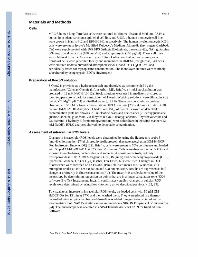
Materials and MethodsCells
MRC-5 human lung fibroblast cells were cultured in Minimal Essential Medium. A549, ahuman lung adenocarcinoma epithelial cell line, and U937, a human monocytic cell line,were grown in Ham’s F12 and RPMI-1640, respectively. The human myelomonocytic KG-1cells were grown in Iscove's Modified Dulbecco's Medium. All media (Invitrogen, Carlsbad,CA) were supplemented with 10% FBS (Atlanta Biologicals, Lawrenceville, GA), glutamine(292 mg/L) and penicillin (100 units/ml) and streptomycin (100 μg/ml). These cell lineswere obtained from the American Type Culture Collection. Balb/c mouse embryonicfibroblast cells were generated locally and maintained in DMEM (low glucose). All cellswere cultured under a humidified atmosphere (95% air and 5% CO2) at 37°C andperiodically tested for mycoplasma contamination. The monolayer cultures were routinelysubcultured by using trypsin-EDTA (Invitrogen).
Preparation of 8-oxoG solution8-OxoG is provided as a hydroacetate salt and dissolved as recommended by themanufacturer (Cayman Chemical, Ann Arbor, MI). Briefly, a 4-mM stock solution wasprepared in 12 mM NaOH (pH 12). Stock solutions were used immediately or stored atroom temperature in dark for a maximum of 1 week. Working solutions were diluted in PBS(w/o Ca2+, Mg2+; pH 7.4) or distilled water (pH 7.0). There was no solubility problemobserved at 100 μM or lower concentrations. HPLC analysis [250 x 4.6 mm i.d. ACE C18column (MAC-MOD Analytical, Chadd Ford, PA)] of 8-oxoG showed no detectablecontamination (data not shown). All nucleotide bases and nucleosides (2’-deoxyguanosine,guanine, adenine, guanosine, 7,8-dihydro-8-oxo-2'-deoxyguanosine, 8-hydroxyadenine and2,6-diamino-4-hydroxy-5-formamidopyrimidine) were solubilized in the same manner (12mM NaOH). HPLC analyses showed no detectable contamination.
Assessment of intracellular ROS levelsChanges in intracellular ROS levels were determined by using the fluorogenic probe 5-(and-6)-chloromethyl-2'7'-dichlorodihydrofluorescein diacetate acetyl ester (CM-H2DCF-DA; Invitrogen, Eugene, OR) [22]. Briefly, cells were grown to 70% confluence and loadedwith 50 μM CM-H2DCF-DA at 37°C for 30 minutes. Cells were then washed with PBS andexposed to nucleobases, nucleosides, and solvents. As positive controls, tert butylhydroperoxide (tBHP; ACROS Organics, Geel, Belgium) and cumene hydroperoxide (CHP;Spectrum, Gardena, CA) or H2O2 (Fisher, Fair Lawn, NJ) were used. Changes in DCFfluorescence were recorded on an FLx800 (Bio-Tek Instruments Inc., Winooski, VT)microplate reader at 485 nm excitation and 528 nm emission. Results are expressed as foldchange or arbitrarily in fluorescence units (FU). The mean V is a calculated value of themean slope by determining regression on points that are in a linear calculation zone (KC4software, Bio-Tek Instruments, Inc.). In confirmatory studies, changes in cellular ROSlevels were determined by using flow cytometry as we described previously [22, 23].
To visualize an increase in intracellular ROS levels, we loaded cells with 50 μM CM-H2DCF-DA for 15 min at 37°C and then washed them. They were placed in a thermo-controlled microscopic chamber, and 8-oxoG was added. Images were captured with aPhotometrix CoolSNAP Fx digital camera mounted on a NIKON Eclipse Ti UV microscope[24]. The microscope was operated via NIS-Elements AR Ver3.22.09 for 64bit editionSoftware.
Hajas et al. Page 3
Free Radic Biol Med. Author manuscript; available in PMC 2013 February 15.
NIH
-PA Author Manuscript
NIH
-PA Author Manuscript
NIH
-PA Author Manuscript

Amplex Red (AR) assayAmplex® UltraRed (10-acetyl-3,7-dihydroxyphenoxazine; Invitrogen, Eugene, OR) reactswith H2O2 (Fisher, Fair Lawn, NJ) in the presence of horseradish peroxidase (HRP; Sigma-Aldrich, St. Louis, MO) to generate a stable product, resorufin [25]. Amplex Red assayswere carried out as we previously described [26]. Briefly, test materials (e.g., 8-oxoG andcontrols; 0.01-100 μM) were diluted in reaction buffer and incubated at 37°C for 30 minwith 50 μM Amplex® UltraRed and 0.05 U/ml of HRP (optimal concentrations weredetermined in preliminary studies). Changes in resorufin fluorescence were determined at560 nm and 620 nm (excitation and emission, respectively) by using a BioTek FLx800fluorimeter. To establish the standard curve, increasing concentrations of H2O2 (0 to 10 μM)were used. The addition of catalase (5 U/ml, Sigma-Aldrich, St. Louis, MO) decreased H2O2levels by ~95%. As additional controls, tBHP or CHP were used.
Assessment of GSH/GSSG ratioA Bioxytech GSH/GSSG-412 (OxisResearch; Portland, OR) assay kit was used to determinelevels of GSH and GSSG by following the basic manufacturer's protocol with a slightmodification as described [27]. Briefly, GSSG samples (in triplicate were prepared by gentlymixing thiol-scavenging reagent 1-methyl-2-vinylpyridinium trifluoro-methanesulfonatecontaining H2O2 or 8-oxoG in an assay buffer (50 mM Na3PO4 with EDTA, pH 7.4).Samples were incubated at room temperature for 10 minutes, and the reaction was stoppedwith cold 5% metaphosphoric acid (MPA, Sigma-Aldrich). Samples were vortexed andcombined with the provided GSSG buffer. GSH samples were made by combining 50 μL of100 μM H2O2 or 8-oxo-Gua in the assay buffer with 350 μL MPA. The samples werecombined with chromogen (5, 5’-dithiobis-(2-nitrobenzoic acid)±glutathione peroxidase(GPx, bovine erythrocytes; Sigma-Aldrich). After 5 min incubation at room temperature,NADPH (Sigma-Aldrich) solution (1 μM) was added and changes in absorbance recorded at412 nm for 3 min on a Beckman DU530 spectrophotometer. Linear regressions werecalculated on a five-point curve by using 0, 0.75, 1.50, 2.25 and 3.00 μM for GSH and afour-point curve for GSSG by using 0, 0.10, 0.25 and 0.50 μM.
Glutathione peroxidase assayGlutathione peroxidase assays were conducted according to the manufacturer’s instructions(Sigma-Aldrich; St. Louis, MO). Briefly, assay buffer (pH = 7.0) contained 48 mM sodiumphosphate, 0.12 mM ß-NADPH, 0.95 mM sodium azide, 3.2 units of purified glutathionereductase (GR; Sigma-Aldrich), 1 mM GSH, 0.02 mM dithiothreitol (DTT) and 0.2 unitsGPx (pH 7.0) [28]. Next, 0.1 to 100 μM of 8-oxoG (or other nucleic acid bases) was addedto assay buffer, and changes in NADPH absorbance were monitored at 340 nm in aBeckman DU530 (Beckman Coulter, Inc) spectrophotometer at a constant temperature(25°C) at a light path of 1 cm. H2O2 was used as a positive control. Changes in ∆A340nm/minute were calculated by a formula provided by the manufacturer.
Liquid Chromatography/Mass Spectrometry (LC/MS)Test samples were placed into liquid nitrogen, then transferred into the freeze-dryer (Dura-Dry MP, Stone Ridge, NY) and lyophilized at −80°C (35 mT). The lyophilized materialswere reconstituted and the 8-oxoG level measured by liquid chromatography/isotopedilution mass spectrometry (LC/IDMS). As an internal standard, the stable isotope-labeledanalogs of 8- oxoG were used as previously described [11].
PeroXOquant AssayAssays were performed according to the manufacturer’s recommendations (Pierce,Rockford, IL). Briefly, one volume of reaction buffer A (25 mM ammonium ferrous (II)
Hajas et al. Page 4
Free Radic Biol Med. Author manuscript; available in PMC 2013 February 15.
NIH
-PA Author Manuscript
NIH
-PA Author Manuscript
NIH
-PA Author Manuscript

sulfate and 2.5 M H2SO4) was mixed with 99 volumes of reagent B (100 mM sorbitol and125 μM xylenol orange). After addition of 8-oxoG (or H2O2 as positive control; 0 μM to 10μM) mixtures were incubated for 20 min at room temperature. OD was measured at 595 nm(Beckman DU530). Peroxide concentrations in 8-oxoG (and other nucleotides, nucleosidesolutions) were calculated from the standard curve of H2O2 [29].
ReagentsThe following were purchased from Sigma-Aldrich (St. Louis, MO): L-glutathione reduced(GSH); β-Nicotinamide adenine dinucleotide 2’-phosphate reduced tetrasodium salt (β-NADPH); 2’-deoxyguanosine, guanine, adenine, guanosine, 7,8-dihydro-8-oxo-2'-deoxyguanosine (8-oxodG); N-acetyl-L-cysteine (NAC), and maleic acid diethyl ester(DEM). 8-Aminoguanine was purchased from Carbosynth Inc, Berkshire, UK; 8-hydroxyadenine (8-OHA, Biolog Life Science Institute, Bremen, Germany); and FapyG(2,6-diamino-4-hydroxy-5-formamidopyrimidine) was provided by Dr. Miral Dizdaroglu(National Institute of Standards and Technology, Gaithersburg, MD).
Statistical AnalysisData are expressed as the mean ±SEM. Results were analyzed for significant differences byusing ANOVA procedures and Student’s t-tests (Sigma Plot 11.0). Differences wereconsidered significant at p<0.05 (*p<0.05, **p<0.01, ***p<0.001, ****p<0.0001).
Results8-OxoG base transformed to a hydroperoxide-like molecule
Exposure of cells to a week-old 8-oxoG solution resulted in increased cellular ROS levels,while the freshly solubilized 8-oxoG base did not do so. We speculate that the observeddifferences were due to changes in the properties of the 8-oxoG base over time. Indeed whenthe 8-oxoG base solution was stored for a week, it exhibited a concentration-dependentreactivity with Amplex Red (Fig. 1A). Fig. 1C shows that the one-week-old 8-oxoG solutionextensively oxidized AR to resorufin, while a 2h old (freshly made) one did so only poorly.8-OxoG solution also oxidized H2DCF to DCF, in the presence of HRP similar to H2O2(data not shown). In fact one-week-old 8-oxoG base solution (100 μM) mediatedsignificantly higher resorufin fluorescence than that of organic hydroperoxides, tBHP (100μM) or CHP (100 μM) (Fig. 1B). On the other hand, the same concentration of H2O2 was~10-times stronger than 8-oxoG solution. In controls, 8-oxodG, FapyG, guanine, 2-deoxyguanosine (2-dG), 8-hydroxyadenine 8-OHA), oxazolone or adenine preparedsimilarly and kept for 7 days did not react with AR (Fig. 1D). When taken together, thesedata imply the presence of a hydroperoxide-like molecule (8-oxoG*) in 8-oxoG solution.
To test further the possibility that 8-oxoG solution contains a hydroperoxide derivative, wemixed it with catalase or glutathione peroxidase (GPx) ± GSH or superoxide dismutase(SOD) in appropriate reaction buffers [30]. Aliquots from parallel mixtures were subjectedto AR assays. GPx+GSH and catalase diminished the reactivity of the 8-oxoG* base withAR (Fig. 2A). GPx alone or SOD did not have an effect on 8-oxoG*-mediated AR oxidation(Fig. 2A). In the GPx+GSH-mediated detoxification of hydroperoxides, GSSG was reportedto be formed [31]. 8-OxoG* decreased the GSH:GSSG ratio in a manner similar to thatfound with H2O2 (Fig. 2B). We postulated that if indeed GSSG is formed, then glutathionereductase (GR) should convert it to GSH in the presence of NADPH. As shown in Fig. 2C,there was a significant decrease in NADPH levels in GPx+GSH+8-oxoG*-containingreaction mixtures (Fig. 2C), a finding that suggests the existence of GSSG. Similar resultswere observed when H2O2 was present. A decrease in NADPH levels could not be measuredwhen GPx or GSH was omitted from these reactions (Fig. 2C). To gain further evidence of
Hajas et al. Page 5
Free Radic Biol Med. Author manuscript; available in PMC 2013 February 15.
NIH
-PA Author Manuscript
NIH
-PA Author Manuscript
NIH
-PA Author Manuscript

8-oxoG* in 8-oxoG solution, we assessed the conversion of ferrous iron (Fe2+) to ferric iron(Fe3+) in PeroXOquant assays [29]. 8-oxoG*, but not the freshly solubilized 8-oxoG,showed a significant increase in absorbance (Fig. 2D) similar to that seen in the controlsCHP, tBHP and H2O2 (Fig. 2D). These results also confirm that a fraction of 8-oxoG isconverted to a molecule with the characteristics of hydroperoxide.
8-oxoG* formation requires molecular oxygenTo define conditions that facilitate the formation of 8-oxoG*, freshly made 8-oxoG base (1mM, pH: 12) solutions were kept under an ambient oxygen or nitrogen (N2) atmosphere (atroom temperature in dark). 8-OxoG* formation was assayed immediately and at 1, 2, 3 and 7days. Under O2 atmosphere, the 8-oxoG* levels were higher by >4.4- (day 1st), 6.1- (day2nd) and 7.3-fold (day 7th) when compared to freshly prepared 8-oxoG solution, asdetermined in AR assays. At ambient conditions, there was a ~50% lower 8-oxoG* level atall time points. In contrast, 8-oxoG solution kept under N2 atmosphere did not acquirehydroperoxide properties, between 1 and 7 days (Fig. 3A). The addition of the reducingagent dithiothreitol (DTT; 1 mM), 2-β-mercaptoethanol (100 μM), or FeCl2 (100 μM)prevented 8-oxoG* formation (data not shown). It should be noted that we observedincreased fluorescence signals generated by freshly made 8-oxoG solutions or those keptunder N2 atmosphere. These fluorescent signals may be due to the rapid formation of 8-oxoG* in aqueous solutions during the solubilization process and mandatory incubations(Fig. 3A). To determine real-time formation of 8-oxoG* is our future challenge.
Next, if a fraction of 8-oxoG transformed to 8-oxoG*, the 8-oxoG content of the solutionshould be lower. Indeed, quantification by LC/MS shows a significant decrease in 8-oxoGlevels at days 1 and 7 (Fig. 3B). To reinforce the presence of the 8-oxoG*, the solution wasincubated for 15 min with Fe2+ (100 μM FeCl2 or solvent), and 8-oxoG levels wereassessed. The addition of FeCl2 restored 8-oxoG levels, as assessed by LC/MS (Fig. 3C). Inparallel, Fe2+ prevented an increase in 8-oxoG*-mediated resorufin fluorescence (Fig. 3D) inline with the results from the PeroXOquant assay (Fig. 2D).
To evaluate relative quantities of 8-oxoG* generated at alkaline and physiological pH wediluted an alkaline solution of 8-oxoG in PBS (pH, 7.4) and kept it for 24 hours. Thecapacity of 8-oxoG solutions (PBS) to oxidize AR to resorufin was found to be increasedsignificantly (Fig. 3E) compared to those kept in the alkaline solution. To exclude that thealkaline pH imparted 8-oxoG to form the hydroperoxide-like molecule, we added 8-oxoGdirectly to PBS (pH 7.4). In PBS ~8 μM (±0.45 μM) of 8-oxoG could be dissolved. Whencompared to the freshly made one (2 h), the 12-h and 24-h 8-oxoG solutions mediated asignificant increase in resorufin fluorescence (Fig. 3F). Taken together, these results provideevidence that a fraction of 8-oxoG was indeed transformed into 8-oxoG*, and its formationoccurred both at an alkaline and physiological pH.
8-oxoG* increases cellular oxidative stress levelsNext we evaluated whether 8-oxoG* increases intracellular ROS levels in the same manneras other hydroperoxides. Fig. 4A shows that 8-oxoG* rapidly increased DCF fluorescence,which peaked at 30 min post-exposure. The changes in fluorescence were similar to thoseinduced by H2O2 (10 μM) and also were cell-type dependent (Fig. 4BC). For example,human (MRC-5; Fig. 4AB and D upper panels) and murine fibroblast cells (MEF, Fig. 4C)were the most sensitive to 8-oxoG,* showing a 3.2- to 4.5-fold increase, while establishedcell lines, A549, KG1 or U937, exhibited only a ~2-fold increase in DCF fluorescence (Fig.4C). In parallel with an increase in intracellular DCF fluorescence (Fig. 4A), we observed arapid decrease (90% at 15 min) in 8-oxoG* levels in cell supernatants (Fig. 5A). In a 100-μM 8-oxoG solution, the level of 8-oxoG* was equivalent to ~12.5 μM H2O2, ~5.1 μM
Hajas et al. Page 6
Free Radic Biol Med. Author manuscript; available in PMC 2013 February 15.
NIH
-PA Author Manuscript
NIH
-PA Author Manuscript
NIH
-PA Author Manuscript

tBHP or ~6.25 μM CHP (MRC-5 cells, Fig. 5B). Freshly prepared 8-oxoG did not increasecellular DCF fluorescence (Fig. 4D, lower panel). Pre-incubation of 8-oxoG solution withcatalase (2.5 U/ml) or GPx (0.2 U/ml)+GSH (1 mM) prevented an 8-oxoG*-mediatedincrease in cellular DCF fluorescence. GPx, GSH alone (controls) or SOD had no effects onredox properties of 8-oxoG* (Fig. 5D). We made similar observation when H2O2 was usedin control experiments (data not shown). N-Acetyl-L-cysteine (NAC) pre-treatment of cells(5 mM, for 3 h) prevented a rise in ROS levels, both in 8-oxoG*- and H2O2-treated cells(Fig. 5C). When glutathione was depleted with maleic acid diethyl ester (DEM, 1.25 mM,for 30 min [27]), 8-oxoG* exposure further increased ROS levels (Fig. 5D, inset). Incontrols, the addition of FapyG, guanine, 8-oxodG, 2-deoxyguanosine (2-dG), 8-hydroxyadenine, oxazolone or adenine did not alter DCF fluorescence (Fig. 5E, MRC-5cells).
In another study, we used KG-1 cells which express a temperature-sensitive, functionallyinactive OGG1 variant (OGG1R229Q) [32]. Moreover, OGG1R229Q regains its 8-oxoGexcision activity at 25°C and it is comparable to wild-type OGG1 [32]. Thus we utilizedthese cells to test whether endogenously generated 8-oxoG forms 8-oxoG*, which oxidizesH2DCF to DCF. Parallel KG-1 cultures were loaded with H2DCF-DA at 37°C and incubatedat 25°C for 30 min. We observed an increase in DCF fluorescence of KG1 cells at 25°C,compared to those remaining at 37°C (Fig. 5F). In controls, U937 cells (Fig. 5F) that expresswild-type OGG1 showed no change in DCF fluorescence at 25°C. These data imply thatwhen released from DNA by OGG1, the free 8-oxoG base may be converted into 8-oxoG*.
DiscussionAmong DNA and RNA bases, guanine is the most susceptible to modification by reactiveoxygen and nitrogen species, due to its lowest redox potential [5, 8, 33, 34]. One of itsoxidation products is 8-oxoG. Further oxidation of 8-oxoG is well established, and itsoxidized derivatives are well-characterized in the context of DNA and nucleosides (rev in[6, 11, 35]. It has also been shown that purine ribonucleotides are scavengers of radicalsincluding •OH [36], and that free 8-oxodeoxyguanosine provides the strongest protectionagainst oxidation of DNA damage [5, 21, 36, 37]. By utilizing biochemical and biologicalassays, we have documented that the free 8-oxoG base is also a target of oxidation reactionsand is transformed to a hydroperoxide (8-oxoG*) form.
In order to be solubilized, free 8-oxoG base was de-protonated in NaOH. When kept insolution, at alkaline or physiological pH (pH: 7.4) it accumulated a derivative showingcharacteristics of hydroperoxides. These changes were independent from de-protonation byNaOH, as the 8-oxoG base directly dissolved in PBS showed identical characteristics.Formation of 8-oxoG* was prevented by the addition of dithiothreitol, 2-β-mercaptoethanol,FeCl2, in the lack of molecular oxygen (storage under N2 atmosphere) or when it wasdissolved in DMSO. The existence of hydroperoxide in 8-oxoG solution was shown byoxidation of AR to resorufin, H2DCF to DCF and GSH to GSSG and Fe2+ to Fe3+. Byutilizing the AR assay, the hydroperoxide generated in 8-oxoG solution was a strongeroxidant than organic peroxides, e.g., CHP or tBHP. When added to H2DCF-loaded cells, 8-oxoG* rapidly increased DCF fluorescence. In contrast, 8-oxodG, 8-hydroxyadenine,guanine or other bases (dissolved and stored as 8-oxoG) showed no hydroperoxide-likereactivity. These results strongly support a unique property of the free 8-oxoG base.
The redox potential of the free 8-oxoG base is lower (-0.48 V vs. NHE) than 8-oxodG [20].Thus, 8-oxoG should be more susceptible to further oxidation and far more reactive than 8-oxodG. Therefore we speculate that the oxidation of the free 8-oxoG base takes place evenunder mild oxidative conditions such as those in the presence of molecular oxygen (O2).
Hajas et al. Page 7
Free Radic Biol Med. Author manuscript; available in PMC 2013 February 15.
NIH
-PA Author Manuscript
NIH
-PA Author Manuscript
NIH
-PA Author Manuscript

This process could be similar to that reported for guanosine in the presence of 1O2 (rev in[35, 38]). In this scenario, the free 8-oxoG base may undergo a cycloaddition reaction,forming an endoperoxide, which then could rearrange to a reactive hydroperoxy form. Wewere not able to show a hydroperoxide when 8-oxodezoxyguanosine was dissolved andstored under similar conditions. As an analogy, further oxidation of 8-oxodeoxyguanosinevia singlet oxygen (1O2) resulted in an endoperoxide, which rearranges into a 5-hydroperoxyform, at neutral pH [35]. Also, the deoxyguanosine forms an 8-hydroperoxy derivative, anextremely reactive molecule [20, 39] via electron transfer from 1O2 [40, 41].
It is known that activation of dissolved oxygen to the singlet state may occur withoutsensitizer(s) [42], which may serve as another possible explanation for hydroperoxideformation in 8-oxoG solutions. To our knowledge, there was no known photosensitizerpresent that was required for 1O2 generation in the solution. We are not aware of pH- and/orheat-initiated 1O2 or other ROS formation, as solvents stored under identical conditions didnot show changes in their redox properties and were inactive in AR and DCF assays. Theonly difference between solvents and 8-oxoG solution is the 8-oxoG base itself. It is extremeto propose that 8-oxoG acts as a photosensitizer-like molecule (e.g., upon light exposure)and initiates formation of 1O2 in an oxygenated environment. If it were correct, that wouldbe a new property of the free 8-oxoG base.
8-OxoG solution has accumulated a ~12 μM H2O2 equivalent 8-oxoG*. This level did notincrease significantly after seven days of storage, which implies an equilibrium between 8-oxoG and 8-oxoG* molecules. This observation is supported by LC/MS analysis, as duringequilibrium there was a 10-15% decrease in 8-oxoG concentration when compared to afreshly made one. This decrease is in line with an increase in hydroperoxide content in 8-oxoG solution. Ferrous iron added to the solution decreased the level of 8-oxoG* to onewhich was undetectable (AR assays) and, interestingly, re-established the original 8-oxoGconcentration (as shown by LC/MS). Ferrous iron and organic hydroperoxide reactionsshould follow Fenton’s chemistry, and, indeed, we show generation of ferric iron(PeroXOquant assay). The accurate chemistry surrounding this important observation isbeyond the scope of this publication and will be part of future studies.
Despite a significant amount of free 8-oxoG base that converted into 8-oxoG*, we were notable to purify and chemically characterize it. During purification steps, we lost activefractions, most likely a consequence of its high reactivity and/or instability. A highlyreactive 8-hydroperoxy derivative of guanosine as strong as peracid was previouslyidentified [20]. 2'-Deoxyguanosine oxidation by 2,2'-azobis(2-amidino-propane)dihydrochloride resulted in a relatively stable product and was characterized [43]. On theother hand, a hydroperoxide form of 8-oxoguanosine was identified only by NMR but wasnot purified [1, 38].
The addition of 8-oxoG* to cells increased the intracellular DCF fluorescence (a well-established measure of cellular ROS levels) in a time- and dose-dependent manner. Theimpact on cells was prevented by antioxidants, which leads us to suggest that 8-oxoG*
indeed behaved like such other oxidants as H2O2 or organic hydroperoxides. To provewhether these effects were specific, we showed that freshly made 8-oxoG, 8-hydroxydeoxy-guanosine, 2,6-diamino-4-hydroxy-5-formamidopyrimidine, 2’-deoxyguanosine, guanine,adenine, guanosine and 8-hydroxyadenine did not change intracellular DCF fluorescence.Primary human and mouse diploid cells were more sensitive to 8-oxoG* compared toestablished cell lines (A549, U937). This observation may be explained by the lowerantioxidant capacity of primary cells [44]. It is documented that A549 cells contain a higherlevel of GSH, due to their enhanced γ-glutamylcysteine synthethase expression [45], andU937 cells possess an increased expression of antioxidant enzymes due to an increase in
Hajas et al. Page 8
Free Radic Biol Med. Author manuscript; available in PMC 2013 February 15.
NIH
-PA Author Manuscript
NIH
-PA Author Manuscript
NIH
-PA Author Manuscript

NOX activity [46]. KG-1 cells accumulate excessive amounts of genomic 8-oxoG andbehave as OGG1 knockout cells at 37°C due to the expression of a homozygoustemperature-sensitive (at 37°C) polymorphic variant of OGG1 (R229Q). At lowertemperatures, e.g., at 25°C, R229Q-OGG1 activity is similar to that of wild-type OGG1[32]. Thus, KG-1 cells allowed us to show that, parallel with the release of a free 8-oxoGbase, there was an increase in DCF fluorescence. Similar results were observed aftertransgenic OGG1 overexpression in Ogg1-/- cells in independent studies [22]. The increasedfluorescence rather signifies a rapid reaction between 8-oxoG* and H2DCF than a change inoverall cellular redox state. We speculate that in oxidative stress conditions after itsliberation by OGG1, the 8-oxoG base rapidly further oxidizes providing protection to DNA.It is also possible that the hydroperoxide form of 8-oxoG serves as a compartment-specificsignaling molecule in a manner similar to that of H2O2 (rev in [47]).
8-OxoG is excised by OGG1 during BER processes, and the free 8-oxoG base in theintercellular space is considered as one of the biomarkers of oxidative stress. In the contextof DNA, 8-oxoG has been shown to be a primary target of oxidation reactions and has beenproposed to play a protective antioxidant role in the genome [37]. Here we show for the firsttime that the free 8-oxoG base is far more sensitive to oxidation reactions than other intactor oxidized purine nucleotides and nucleosides. According to its biochemical properties, the8-oxoG* is a hydroperoxide, of which formation requires only molecular oxygen and takesplace at physiological pH. When added to cells or generated intracellularly, 8-oxoG*oxidized H2DCF. Accordingly, these results lead us to suggest that 8-oxoG base standsuniquely among the intact and oxidatively modified purines and it may have an impact oncellular environment beyond its being a biomarker of oxidative stress.
AcknowledgmentsWe are grateful to Sankar Mitra for his scientific advice, intellectual input, discussions, and reading/editing of themanuscript. We thank Mardelle Susman, technical editor, for editing our manuscript. We thank Dr. MiralDizdaroglu (Chemical Science and Technology Laboratory, National Institute of Standards and Technology,Gaithersburg, MD USA) for assessment of 8-oxoG base levels. This work was supported by grants NIEHS RO1ES018948 (I.B), NIAID/AI062885-01 (I.B), NIA/AG 021830 (I.B), and the, TAMOP 4.2.1/B-09/1/KONV-2010-0007 by European Union and the European Social Fund (A.B).
Abbreviations1O2 singlet oxygen
8-oxoG 8-Oxo-7,8-dihydroguanine
8-oxoG* hydroperoxy derivative of 8-oxoG
OGG1 8-oxoguanine DNA glycosylases
Gua guanine
AR Amplex Red (10-acetyl-3,7-dihydroxyphenoxazine)
DCF dichlorofluorescein
CHP cumene hydroperoxide
CHP cumene hydroperoxide
FapyG 2,6-diamino-4-hydroxy-5-formamidopyrimidine
GPx glutathione peroxidase
HRP horseradish peroxidase
Hajas et al. Page 9
Free Radic Biol Med. Author manuscript; available in PMC 2013 February 15.
NIH
-PA Author Manuscript
NIH
-PA Author Manuscript
NIH
-PA Author Manuscript

H2DCF-DA 5-(and-6)-chloromethyl-2'7'-dichlorodihydrofluorescein diacetate acetylester
F.U fluorescence units
NAC N-acetyl-L-cysteine•OH hydroxyl radical
tBHP tert butyl hydroperoxide
ROS reactive oxygen species
SOD superoxide dismutase
References1. Von Sonntag, C. The chemical basis of radiation biology. Taylor and Francis; New York: 1987.
2. Dizdaroglu M, Jaruga P, Birincioglu M, Rodriguez H. Free radical-induced damage to DNA:mechanisms and measurement. Free Radic Biol Med. 2002; 32:1102–1115. [PubMed: 12031895]
3. Dizdaroglu M, Kirkali G, Jaruga P. Formamidopyrimidines in DNA: mechanisms of formation,repair, and biological effects. Free Radic Biol Med. 2008; 45:1610–1621. [PubMed: 18692130]
4. Candeias LP, Steenken S. Reaction of HO* with guanine derivatives in aqueous solution: formationof two different redox-active OH-adduct radicals and their unimolecular transformation reactions.Properties of G(-H)*. Chemistry. 2000; 6:475–484. [PubMed: 10747414]
5. Steenken S, Jovanovic SV. How easily oxidizable is DNA? One-electron reduction potentials ofadenosine and guanosine radicals in aqueous solution. J Am Chem Soc. 1997; 119:617–618.
6. Cadet J, Douki T, Ravanat JL. One-electron oxidation of DNA and inflammation processes. NatChem Biol. 2006; 2:348–349. [PubMed: 16783334]
7. Dizdaroglu M. Formation of an 8-hydroxyguanine moiety in deoxyribonucleic acid on gamma-irradiation in aqueous solution. Biochemistry. 1985; 24:4476–4481. [PubMed: 4052410]
8. Steenken S. Purine bases, nucleosides, and nucleotides: aqueous solution redox chemistry andtransformation reactions of their radical cations and e- and OH adducts. Chem Rev. 1989; 89:503–520.
9. Jaruga P, Kirkali G, Dizdaroglu M. Measurement of formamidopyrimidines in DNA. Free RadicBiol Med. 2008; 45:1601–1609. [PubMed: 18926902]
10. Lindahl T, Barnes DE. Repair of endogenous DNA damage. Cold Spring Harb Symp Quant Biol.2000; 65:127–133. [PubMed: 12760027]
11. Dizdaroglu M, Jaruga P, Rodriguez H. Measurement of 8-hydroxy-2'-deoxyguanosine in DNA byhigh-performance liquid chromatography-mass spectrometry: comparison with measurement bygas chromatography-mass spectrometry. Nucleic Acids Res. 2001; 29:E12. [PubMed: 11160914]
12. Markus TZ, Daube SS, Naaman R, Fleming AM, Muller JG, Burrows CJ. Electronic structure ofDNA--unique properties of 8-oxoguanosine. J Am Chem Soc. 2009; 131:89–95. [PubMed:19128174]
13. Mitra S, Izumi T, Boldogh I, Bhakat KK, Hill JW, Hazra TK. Choreography of oxidative damagerepair in mammalian genomes. Free Radic Biol Med. 2002; 33:15–28. [PubMed: 12086678]
14. Dizdaroglu M. Base-excision repair of oxidative DNA damage by DNA glycosylases. Mutat Res.2005; 591:45–59. [PubMed: 16054172]
15. Nishimura S. Involvement of mammalian OGG1(MMH) in excision of the 8-hydroxyguanineresidue in DNA. Free Radic Biol Med. 2002; 32:813–821. [PubMed: 11978483]
16. Saxowsky TT, Meadows KL, Klungland A, Doetsch PW. 8-Oxoguanine-mediated transcriptionalmutagenesis causes Ras activation in mammalian cells. Proc Natl Acad Sci U S A. 2008;105:18877–18882. [PubMed: 19020090]
Hajas et al. Page 10
Free Radic Biol Med. Author manuscript; available in PMC 2013 February 15.
NIH
-PA Author Manuscript
NIH
-PA Author Manuscript
NIH
-PA Author Manuscript
17. Fraga CG, Shigenaga MK, Park JW, Degan P, Ames BN. Oxidative damage to DNA during aging:8-hydroxy-2'-deoxyguanosine in rat organ DNA and urine. Proc Natl Acad Sci U S A. 1990;87:4533–4537. [PubMed: 2352934]
18. Svoboda P, Maekawa M, Kawai K, Tominaga T, Savela K, Kasai H. Urinary 8-hydroxyguaninemay be a better marker of oxidative stress than 8-hydroxydeoxyguanosine in relation to the lifespans of various species. Antioxid Redox Signal. 2006; 8:985–992. [PubMed: 16771688]
19. Kim KH, Rodriguez AM, Carrico PM, Melendez JA. Potential mechanisms for the inhibition oftumor cell growth by manganese superoxide dismutase. Antioxid Redox Signal. 2001; 3:361–373.[PubMed: 11491650]
20. Sheu CF, Christopher S. Reactivity toward Singlet Oxygen of a 7,8-Dihydro-8-oxoguanosine (“8-Hydroxyguanosine”) Formed by Photooxidation of a Guanosine Derivative. Journal of theAmerican Chemical Society. 1995; 117:6439–6442.
21. Kim JE, Choi S, Yoo JA, Chung MH. 8-Oxoguanine induces intramolecular DNA damage but free8-oxoguanine protects intermolecular DNA from oxidative stress. FEBS Lett. 2004; 556:104–110.[PubMed: 14706835]
22. Bacsi A, Chodaczek G, Hazra TK, Konkel D, Boldogh I. Increased ROS generation in subsets ofOGG1 knockout fibroblast cells. Mech Ageing Dev. 2007; 128:637–649. [PubMed: 18006041]
23. Das A, Hazra TK, Boldogh I, Mitra S, Bhakat KK. Induction of the human oxidized base-specificDNA glycosylase NEIL1 by reactive oxygen species. J Biol Chem. 2005; 280:35272–35280.[PubMed: 16118226]
24. Boldogh I, Bacsi A, Choudhury BK, Dharajiya N, Alam R, Hazra TK, Mitra S, Goldblum RM, SurS. ROS generated by pollen NADPH oxidase provide a signal that augments antigen-inducedallergic airway inflammation. J Clin Invest. 2005; 115:2169–2179. [PubMed: 16075057]
25. Zhou M, Diwu Z, Panchuk-Voloshina N, Haugland RP. A stable nonfluorescent derivative ofresorufin for the fluorometric determination of trace hydrogen peroxide: applications in detectingthe activity of phagocyte NADPH oxidase and other oxidases. Anal Biochem. 1997; 253:162–168.[PubMed: 9367498]
26. Bacsi A, Woodberry M, Widger W, Papaconstantinou J, Mitra S, Peterson JW, Boldogh I.Localization of superoxide anion production to mitochondrial electron transport chain in 3-NPA-treated cells. Mitochondrion. 2006; 6:235–244. [PubMed: 17011837]
27. Das GC, Bacsi A, Shrivastav M, Hazra TK, Boldogh I. Enhanced gamma-glutamylcysteinesynthetase activity decreases drug-induced oxidative stress levels and cytotoxicity. Mol Carcinog.2006; 45:635–647. [PubMed: 16491484]
28. Wendel A, Cikryt P. The level and half-life of glutathione in human plasma. FEBS Lett. 1980;120:209–211. [PubMed: 7439398]
29. Nourooz-Zadeh J, Tajaddini-Sarmadi J, Wolff SP. Measurement of plasma hydroperoxideconcentrations by the ferrous oxidation-xylenol orange assay in conjunction withtriphenylphosphine. Anal Biochem. 1994; 220:403–409. [PubMed: 7978285]
30. Asayama K, Janco RL, Burr IM. Selective induction of manganous superoxide dismutase in humanmonocytes. Am J Physiol. 1985; 249:C393–397. [PubMed: 4061626]
31. Giblin FJ, McCready JP, Reddy VN. The role of glutathione metabolism in the detoxification ofH2O2 in rabbit lens. Invest Ophthalmol Vis Sci. 1982; 22:330–335. [PubMed: 7061205]
32. Hill JW, Evans MK. A novel R229Q OGG1 polymorphism results in a thermolabile enzyme thatsensitizes KG-1 leukemia cells to DNA damaging agents. Cancer Detect Prev. 2007; 31:237–243.[PubMed: 17651912]
33. Wagenknecht HA. Electron transfer processes in DNA: mechanisms, biological relevance andapplications in DNA analytics. Nat Prod Rep. 2006; 23:973–1006. [PubMed: 17119642]
34. Margolin Y, Cloutier JF, Shafirovich V, Geacintov NE, Dedon PC. Paradoxical hotspots forguanine oxidation by a chemical mediator of inflammation. Nat Chem Biol. 2006; 2:365–366.[PubMed: 16751762]
35. Pratviel G, Meunier B. Guanine oxidation: one- and two-electron reactions. Chemistry. 2006;12:6018–6030. [PubMed: 16791886]
36. Gudkov SV, Shtarkman IN, Smirnova VS, Chernikov AV, Bruskov VI. Guanosine and inosinedisplay antioxidant activity, protect DNA in vitro from oxidative damage induced by reactive
Hajas et al. Page 11
Free Radic Biol Med. Author manuscript; available in PMC 2013 February 15.
NIH
-PA Author Manuscript
NIH
-PA Author Manuscript
NIH
-PA Author Manuscript

oxygen species, and serve as radioprotectors in mice. Radiat Res. 2006; 165:538–545. [PubMed:16669708]
37. Radak Z, Boldogh I. 8-Oxo-7,8-dihydroguanine: links to gene expression, aging, and defenseagainst oxidative stress. Free Radic Biol Med. 49:587–596. [PubMed: 20483371]
38. Ravanat JL, Martinez GR, Medeiros MH, Di Mascio P, Cadet J. Mechanistic aspects of theoxidation of DNA constituents mediated by singlet molecular oxygen. Arch Biochem Biophys.2004; 423:23–30. [PubMed: 14989260]
39. McCallum JE, Kuniyoshi CY, Foote CS. Characterization of 5-hydroxy-8-oxo-7,8-dihydroguanosine in the photosensitized oxidation of 8-oxo-7,8-dihydroguanosine and itsrearrangement to spiroiminodihydantoin. J Am Chem Soc. 2004; 126:16777–16782. [PubMed:15612716]
40. Kasai H, Yamaizumi Z, Yamamoto F, Bessho T, Nishimura S, Berger M, Cadet J. Photosensitizedformation of 8-hydroxyguanine (7,8-dihydro-8-oxoguanine) in DNA by riboflavin. Nucleic AcidsSymp Ser. 1992:181–182. [PubMed: 1289810]
41. Hickerson RP, Prat F, Muller JG, Foote CS, Burrows CJ. Sequence stacking dependence of 8-oxoguanine oxidation: Comparison of one-electron vs singlet oxygen mechanisms. Am Chem Soc.1999; 121:9423–9428.
42. Bruskov VI, Malakhova LV, Masalimov ZK, Chernikov AV. Heat-induced formation of reactiveoxygen species and 8-oxoguanine, a biomarker of damage to DNA. Nucleic Acids Res. 2002;30:1354–1363. [PubMed: 11884633]
43. Sakakibara H, Ashida H, Kanazawa K. A novel method using 8-hydroperoxy-2'-deoxyguanosineformation for evaluating antioxidative potency. Free Radic Res. 2002; 36:307–316. [PubMed:12071350]
44. Lorenz M, Saretzki G, Sitte N, Metzkow S, von Zglinicki T. BJ fibroblasts display high antioxidantcapacity and slow telomere shortening independent of hTERT transfection. Free Radic Biol Med.2001; 31:824–831. [PubMed: 11557321]
45. Hatcher EL, Chen Y, Kang YJ. Cadmium resistance in A549 cells correlates with elevatedglutathione content but not antioxidant enzymatic activities. Free Radic Biol Med. 1995; 19:805–812. [PubMed: 8582653]
46. Lledias F, Hansberg W. Oxidation of human catalase by singlet oxygen in myeloid leukemia cells.Photochem Photobiol. 1999; 70:887–892. [PubMed: 10628300]
47. Veal EA, Day AM, Morgan BA. Hydrogen peroxide sensing and signaling. Mol Cell. 2007; 26:1–14. [PubMed: 17434122]
Hajas et al. Page 12
Free Radic Biol Med. Author manuscript; available in PMC 2013 February 15.
NIH
-PA Author Manuscript
NIH
-PA Author Manuscript
NIH
-PA Author Manuscript

Highlights
• Free 8-oxoG base can be transformed to a highly reactive hydroperoxide (8-oxoG*).
• 8-oxoG* oxidizes Amplex Red, H2DCF, Fe2+, and GSH in vitro.
• 8-oxoG* act as an organic hydroperoxide and substrate of peroxidases
• Increases cellular oxidative stress levels.
• 8-oxoG* may serve as a compartment-specific signaling molecule.
Hajas et al. Page 13
Free Radic Biol Med. Author manuscript; available in PMC 2013 February 15.
NIH
-PA Author Manuscript
NIH
-PA Author Manuscript
NIH
-PA Author Manuscript

Figure 1.Hydroperoxide activity in 8-oxoG base solution. (A) Concentration-dependent oxidation ofAmplex red by 8-oxoG*. (B) Direct oxidation of AR by 8-oxoG* CHP and tBHP. H2O2 wasused as a control. (C) 8-oxoG*, but not 8-oxoG or 8-oxodG oxidizes AR. (D) 8-oxoG*, butnot Gua, 2-dG, 8-OHA, 8-oxodG, oxazolone or FapyG solution, has peroxide-like activity.Gua, guanine, 2-dG, 2-desoxyguanine; 8-OHA, 8-hydroxy adenine; 8-oxodG, 7,8-dihydro-8-oxo-2'-deoxyguanosine; FapyG, 2,6-diamino-4-hydroxy-5-formamidopyrimidine;CHP, cumene hydroperoxide; tBHP tert-butyl hydroperoxide. Results are means ±SEM(n=5-11).
Hajas et al. Page 14
Free Radic Biol Med. Author manuscript; available in PMC 2013 February 15.
NIH
-PA Author Manuscript
NIH
-PA Author Manuscript
NIH
-PA Author Manuscript

Figure 2.Effect of peroxidases and ferrous iron on 8-oxoG*. (A) Catalase, GPx ,but not SOD,diminishes 8-oxoG*-mediated AR oxidation. (B,C) 8-OxoG* oxidizes GSH to GSSG. 8-oxoG* was incubated with GSH ±GPx, and the GSH:GSSG ratio was determined (Materialsand Methods). H2O2 was used as a positive control (B). Utilization of NADPH ± GR wasdetermined at 340 nm (C). H2O2 was used as a positive control. (D) 8-OxoG* oxidizesferrous iron to ferric iron by a process similar to that seen with CHP, tBHP and H2O2 asshown by the PeroXoquant assay (O.D 595 nm). Cat, catalase, GPx, glutathione peroxidase;GR, glutathione reductase; SOD, superoxide dismutase; GSH, glutathione; GSSG, oxidizedGSH; CHP and tBHP, as in legend to Fig 1. Results are means ±SEM (n=3-5).
Hajas et al. Page 15
Free Radic Biol Med. Author manuscript; available in PMC 2013 February 15.
NIH
-PA Author Manuscript
NIH
-PA Author Manuscript
NIH
-PA Author Manuscript

Figure 3.Formation of 8-oxoG* requires O2. (A) Formation of 8-oxoG* under ambient (■), O2 (▲)and N2 (◇) atmosphere. Aliquots of 8-oxoG solution were subjected to AR assays at days 1,2, 3 and 7. (B) Changes in 8-oxoG levels as a function of time and (C) reconstitution of 8-oxoG base level by Fe2+ as determined by LC/MS. (D) AR reactivity of 8-oxoG* and H2O2is diminished in the presence of ferrous iron. (E) Conversion of 8-oxoG to 8-oxoG* atphysiological (■) and alkaline pH (◆). (F) Formation of 8-oxoG* from 8-oxoG whendissolved and stored in PBS. Results are means ±SEM (n=3–6).
Hajas et al. Page 16
Free Radic Biol Med. Author manuscript; available in PMC 2013 February 15.
NIH
-PA Author Manuscript
NIH
-PA Author Manuscript
NIH
-PA Author Manuscript

Figure 4.Exposure of cells to 8-oxoG* increases cellular ROS levels. (A) MRC-5 cells were exposedto 8-oxoG* (◆), 8-oxoG (■) or H2O2 (□) and cellular ROS levels determined at timesindicated. (B) Dose- and (C) cell type-dependent changes in ROS level upon 8-oxoG*
exposure. (D) 8-OxoG*, but not 8-oxoG, causes changes in cellular ROS levels as shown bymicroscopic imaging (NIKON Eclipse Ti system). Magnification: 60x. In panels A, B, andC, cells were H2DCF-DA-loaded, and DCF fluorescence was determined at 485/528 nm.Results are means ±SEM (n=3–11).
Hajas et al. Page 17
Free Radic Biol Med. Author manuscript; available in PMC 2013 February 15.
NIH
-PA Author Manuscript
NIH
-PA Author Manuscript
NIH
-PA Author Manuscript

Figure 5.Added to cells or generated intracellularly 8-oxoG* increased cellular ROS. (A) 8- oxoG*level decreased in supernatant when added to cells as shown by AR assays. (B) Changes inROS levels by 8-oxoG* compared to CHP, tBHP, H2O2. The indicated concentrations ofperoxides were determined in preliminary studies. (C) NAC pretreatment decreased 8-oxoG*-mediated changes in ROS levels. H2O2 was used as a control. Inset: GSH depletionby DEM increased 8-oxoG*-induced cellular ROS levels. (D) Catalase and GPx(+GSH) pre-treatment of 8-oxoG* prevented an increase in ROS levels. (E) Only 8-oxoG*, but not othernucleotides or nucleosides, changed cellular ROS levels. (F) Endogenously generated 8-oxoG increased DCF fluorescence in KG-1 cells. In B,C,D,E and F, cells were H2DCF-loaded, and DCF fluorescence was assessed at 485/528 nm. NAC, N-acetyl-L-cysteine;DEM, maleic acid diethyl ester; CHP, tBHP, 2-dG, 2-OHA, and FapyG as in legend to Fig1. GPx, GSH, SOD as in legend to Fig. 2. Results are means ±SEM (n=5–7).
Hajas et al. Page 18
Free Radic Biol Med. Author manuscript; available in PMC 2013 February 15.
NIH
-PA Author Manuscript
NIH
-PA Author Manuscript
NIH
-PA Author Manuscript