Biochemical, histopathological and morphological profiling of a rat model of early immune...
16
RESEARCH ARTICLE Biochemical, Histopathological and Morphological Profiling of a Rat Model of Early Immune Stimulation: Relation to Psychopathology Anna Kubesova 1,2,3 *, Hana Tejkalova 1,2 , Kamila Syslova 4 , Petr Kacer 4 , Jana Vondrousova 4 , Filip Tyls 1,2,3 , Michaela Fujakova 1,2,3 , Tomas Palenicek 1,2,3 , Jiri Horacek 1,2,3 1 Prague Psychiatric Center, Prague, Czech Republic, 2 National Institute of Mental Health, Klecany, Czech Republic, 3 Third Faculty of Medicine, Charles University, Prague, Czech Republic, 4 Institute of Chemical Technology, Prague, Czech Republic * [email protected] Abstract Perinatal immune challenge leads to neurodevelopmental dysfunction, permanent immune dysregulation and abnormal behaviour, which have been shown to have translational validi- ty to findings in human neuropsychiatric disorders (e.g. schizophrenia, mood and anxiety disorders, autism, Parkinson’s disease and Alzheimer’s disease). The aim of this animal study was to elucidate the influence of early immune stimulation triggered by systemic post- natal lipopolysaccharide administration on biochemical, histopathological and morphologi- cal measures, which may be relevant to the neurobiology of human psychopathology. In the present study of adult male Wistar rats we examined the brain and plasma levels of mono- amines (dopamine, serotonin), their metabolites, the levels of the main excitatory and inhibi- tory neurotransmitters glutamate and γ-aminobutyric acid and the levels of tryptophan and its metabolites from the kynurenine catabolic pathway. Further, we focused on histopatho- logical and morphological markers related to pathogenesis of brain diseases - glial cell acti- vation, neurodegeneration, hippocampal volume reduction and dopaminergic synthesis in the substantia nigra. Our results show that early immune stimulation in adult animals alters the levels of neurotransmitters and their metabolites, activates the kynurenine pathway of tryptophan metabolism and leads to astrogliosis, hippocampal volume reduction and a de- crease of tyrosine hydroxylase immunoreactivity in the substantia nigra. These findings support the crucial pathophysiological role of early immune stimulation in the above men- tioned neuropsychiatric disorders. PLOS ONE | DOI:10.1371/journal.pone.0115439 January 20, 2015 1 / 16 OPEN ACCESS Citation: Kubesova A, Tejkalova H, Syslova K, Kacer P, Vondrousova J, Tyls F, et al. (2015) Biochemical, Histopathological and Morphological Profiling of a Rat Model of Early Immune Stimulation: Relation to Psy- chopathology. PLoS ONE 10(1): e0115439. doi:10.1371/journal.pone.0115439 Academic Editor: Anna Dunaevsky, University of Nebraska Medical Center, UNITED STATES Received: August 14, 2014 Accepted: November 24, 2014 Published: January 20, 2015 Copyright: © 2015 Kubesova et al. This is an open access article distributed under the terms of the Creative Commons Attribution License, which permits unrestricted use, distribution, and reproduction in any medium, provided the original author and source are credited. Data Availability Statement: All relevant data are within the paper and its Supporting Information files. Funding: The study was supported by The Ministry of Health of the Czech Republic (www.mzcr.cz) - In- ternal Grant Agency (NT13843), Development of Re- search Organization (PCP, 00023752); Charles University in Prague (www.cuni.cz) - Specific Aca- demic Research (260045/SVV/2014), Charles Univer- sity Research Development Schemes (PRVOUK P34); The Ministry of Education, Youth and Sports of the Czech Republic (www.msmt.cz) - The National Programme of Sustainability (NPU I (LO)
-
Upload
independent -
Category
Documents
-
view
3 -
download
0
Transcript of Biochemical, histopathological and morphological profiling of a rat model of early immune...
RESEARCH ARTICLE
Biochemical, Histopathological andMorphological Profiling of a Rat Model ofEarly Immune Stimulation: Relation toPsychopathologyAnna Kubesova1,2,3*, Hana Tejkalova1,2, Kamila Syslova4, Petr Kacer4,Jana Vondrousova4, Filip Tyls1,2,3, Michaela Fujakova1,2,3, Tomas Palenicek1,2,3,Jiri Horacek1,2,3
1 Prague Psychiatric Center, Prague, Czech Republic, 2 National Institute of Mental Health, Klecany, CzechRepublic, 3 Third Faculty of Medicine, Charles University, Prague, Czech Republic, 4 Institute of ChemicalTechnology, Prague, Czech Republic
AbstractPerinatal immune challenge leads to neurodevelopmental dysfunction, permanent immune
dysregulation and abnormal behaviour, which have been shown to have translational validi-
ty to findings in human neuropsychiatric disorders (e.g. schizophrenia, mood and anxiety
disorders, autism, Parkinson’s disease and Alzheimer’s disease). The aim of this animal
study was to elucidate the influence of early immune stimulation triggered by systemic post-
natal lipopolysaccharide administration on biochemical, histopathological and morphologi-
cal measures, which may be relevant to the neurobiology of human psychopathology. In the
present study of adult male Wistar rats we examined the brain and plasma levels of mono-
amines (dopamine, serotonin), their metabolites, the levels of the main excitatory and inhibi-
tory neurotransmitters glutamate and γ-aminobutyric acid and the levels of tryptophan and
its metabolites from the kynurenine catabolic pathway. Further, we focused on histopatho-
logical and morphological markers related to pathogenesis of brain diseases - glial cell acti-
vation, neurodegeneration, hippocampal volume reduction and dopaminergic synthesis in
the substantia nigra. Our results show that early immune stimulation in adult animals alters
the levels of neurotransmitters and their metabolites, activates the kynurenine pathway of
tryptophan metabolism and leads to astrogliosis, hippocampal volume reduction and a de-
crease of tyrosine hydroxylase immunoreactivity in the substantia nigra. These findings
support the crucial pathophysiological role of early immune stimulation in the above men-
tioned neuropsychiatric disorders.
PLOS ONE | DOI:10.1371/journal.pone.0115439 January 20, 2015 1 / 16
OPEN ACCESS
Citation: Kubesova A, Tejkalova H, Syslova K, KacerP, Vondrousova J, Tyls F, et al. (2015) Biochemical,Histopathological and Morphological Profiling of a RatModel of Early Immune Stimulation: Relation to Psy-chopathology. PLoS ONE 10(1): e0115439.doi:10.1371/journal.pone.0115439
Academic Editor: Anna Dunaevsky, University ofNebraska Medical Center, UNITED STATES
Received: August 14, 2014
Accepted: November 24, 2014
Published: January 20, 2015
Copyright: © 2015 Kubesova et al. This is an openaccess article distributed under the terms of theCreative Commons Attribution License, which permitsunrestricted use, distribution, and reproduction in anymedium, provided the original author and source arecredited.
Data Availability Statement: All relevant data arewithin the paper and its Supporting Information files.
Funding: The study was supported by The Ministryof Health of the Czech Republic (www.mzcr.cz) - In-ternal Grant Agency (NT13843), Development of Re-search Organization (PCP, 00023752); CharlesUniversity in Prague (www.cuni.cz) - Specific Aca-demic Research (260045/SVV/2014), Charles Univer-sity Research Development Schemes (PRVOUKP34); The Ministry of Education, Youth and Sports ofthe Czech Republic (www.msmt.cz) - The NationalProgramme of Sustainability (NPU I (LO)
IntroductionNegative environmental insults occurring during the intrauterine or early postnatal periodmay increase the risk of developing neuropsychiatric disorders in adulthood [1]. In recentyears, attention has been focused on early immune stimulation caused by a maternal or perina-tal infection. In various animal models early bacterial immune stimulation has been inducedby administration of endotoxin, lipopolysaccharide (LPS) [2]. LPS is a part of the outer mem-brane of Gram-negative bacteria with a strong ability to induce an immune response [3]. LPS isrecognized by Toll-like receptor 4 (TLR4) which is expressed in the central nervous systemmainly on microglia and on other cell types in lower levels [4–6]. Activation of TLR4 leads to acascade of steps that results in a release of pro-inflammatory and anti-inflammatory cytokines[3]. Immune challenge experienced in early postnatal days leads to a permanent immune dys-regulation and alterations in the hypothalamic-pituitary-adrenal axis [7, 8].
Early immune stimulation might be one of underlying environmental factors which can pre-dispose individuals to develop schizophrenia [9]. In our previous experiments, postnatal LPStreatment led to psychotic-like changes in behaviour and an increase of circulating cytokines inadult rats. These changes were normalized by the administration of clozapine [10–12].Psychotic-like behaviour as well as anxiety-like, autistic-like and depression-like behaviour inadult animals perinatally treated with LPS has also been demonstrated in other studies [13–17].
Although the perinatal immune challenge (modelling both pre- and postnatal infection inhumans) has been associated with the development of diverse neuropsychiatric disorders, littleis known about its impact on biochemical actions in the adult brain. Histopathological changes,which might underlie the modelled behavioural phenotypes, have been investigated in severalstudies but the results are still contradictory [18–21].
Our study was designed to elucidate the influence of early immune stimulation on biochem-ical and histopathological changes in an adult rat brain and their potential relation to humanpsychopathology. Firstly, with respect to the role of major neurotransmitter systems in affectiveand psychotic disorders, we determined the levels of monoamines (dopamine, serotonin) andtheir metabolites, and the levels of the main excitatory and inhibitory neurotransmitters gluta-mate and γ-aminobutyric acid (GABA) in the brain. The activation of the kynurenine pathwayof tryptophan metabolism has been associated with various neuropsychiatric disorders [22],therefore we mapped the brain levels of tryptophan and its metabolites from the kynureninecatabolic pathway regarding the effect of proinflammatory cytokines on the enzymes of thispathway. Since some of the neurotransmitters and metabolites do not cross the blood brainbarrier (BBB) but in human subjects are determined from blood, we also measured their plas-ma levels. Secondly, we detected histopathological markers related to pathogenesis of brain dis-eases such as the ionized calcium-binding adaptor molecule (Iba-1, microglial markerupregulated during activation of these cells), glial fibrillary acidic protein (GFAP, astroglialmarker), Fluoro-Jade B (marker of neurodegeneration) and Hoechst 33258 (marker of cell nu-clei). Thirdly, to evaluate the effect of early immune stimulation on brain morphology, we mea-sured the hippocampal volume, whose reduction is consistently described in patients withmental disorders [23–27]. The hypothesis of altered dopaminergic activity has been comple-mented by subsequent evaluation of tyrosine hydroxylase (TH) immunoractivity in the sub-stantia nigra (SN) to elucidate the interaction between dopamine levels and the number ofdopaminergic cells.
We expected an alteration in brain and plasma levels of neurotransmitters and metabolitesand the activation of the kynurenine pathway of tryptophan metabolism. We assumed thatearly immune stimulation leads to a prolonged activation of glial cells, neurodegeneration, andhippocampal volume reduction.
Profiling of a Rat Model of Early Immune Stimulation
PLOS ONE | DOI:10.1371/journal.pone.0115439 January 20, 2015 2 / 16
MSMT - 34870/2013); European Regional Develop-ment Fund (www.europa.eu) - The Operational Pro-gramme Prague Competitiveness (CZ.2.16/3.1.00/22197, CZ.2.16/3.1.00/21537), National Institute ofMental Health (CZ.1.05/2.1.00/03.0078). The fundershad no role in study design, data collection and analy-sis, decision to publish, or preparation of themanuscript.
Competing Interests: The authors have declaredthat no competing interests exist.
Methods
Animals and housingAll of the experiments were carried out on male Wistar/Hann rats (Velaz Ltd., Czech Repub-lic). Male rat pups remained in the breeding plastic cages with their mothers until PD 28 andwere then divided into groups of three or four animals per cage. The animal facility was airconditioned at a standard room temperature and humidity with a normal 12h light/dark cycle.The rats had unlimited access to a standard diet and water. All procedures utilized in this arti-cle comply with the Czech Government Requirements under the Policy of Humans Care ofLaboratory Animals and with regulations of the Ministry of Agriculture of the Czech Republic(No. 419/2012) and obtained statement of the Expert Committee for experimental animal pro-tection (3rd Faculty of Medicine, Charles University in Prague) and were approved by the Com-mittee for Care and Use of Laboratory Animals of Ministry of Health (MHCR, No. 17/2012).
Postnatal LPS treatmentFrom PD 5 to PD 9, the rat pups were each day briefly removed from their home cages,weighed, and administered intraperitoneally with either 2 mg/kg/day LPS (Escherichia coli, se-rotype 026:B6; Sigma–Aldrich) or an equivolume of non-pyrogenic 0.9% saline as describedpreviously [10]. The animals were allocated to treatment groups randomly.
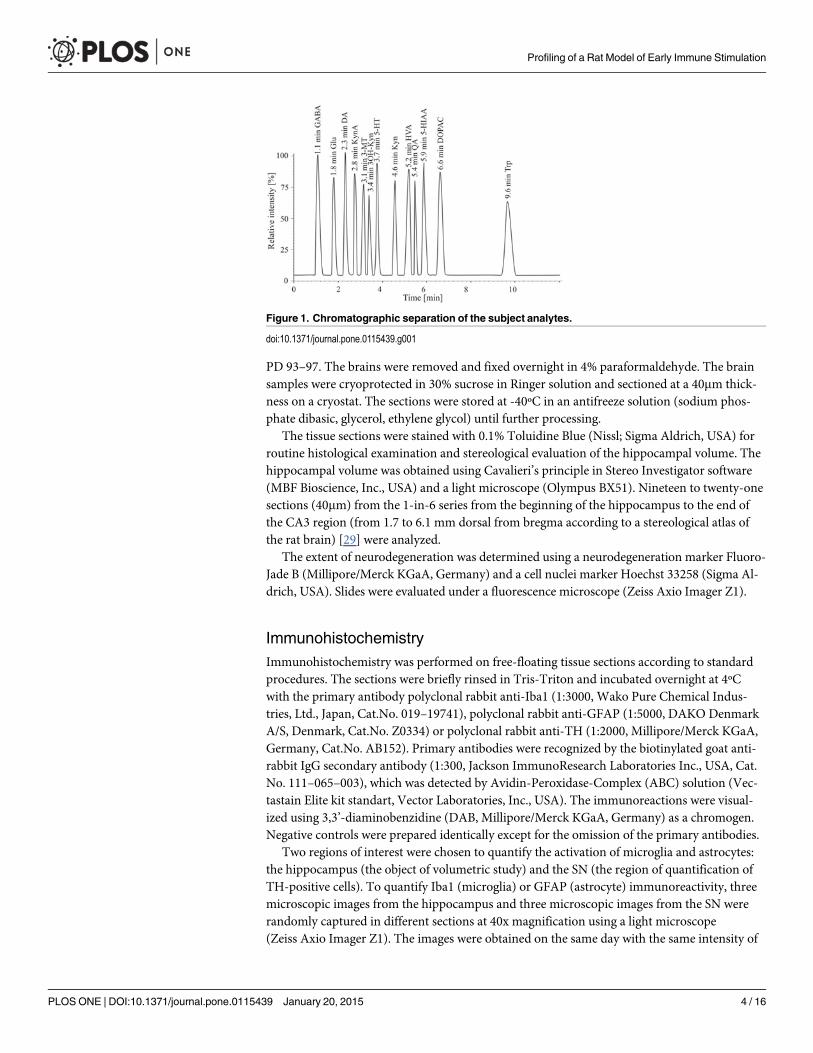
Determination of neurotransmitters and metabolites in brain and plasmaThe adult animals (n = 19–24/group) were euthanized by decapitation at PD 90–95. The col-lected brain tissue (striatum, hippocampus and prefrontal cortex) and plasma were immediatelyfrozen at -80ºC until further processing. The analytical method used for qualitative and quanti-tative determination of neurotransmitters consisted of a pre-treatment step i.e. extraction by amixed solution of acetonitrile, hydrochloric acid (c = 0.1 M) and EDTA (c = 27 M) at a ratio of5:4:1 v / v / v, to separate and concentrate analytes (dopamine (DA), 3,4-dihydroxyphenylacetic acid (DOPAC), 3-methoxytyramine (3-MT), homovanilic acid (HVA),serotonin (5-HT), 5-hydroxyindolacetic acid (5-HIAA), glutamate (GLU), γ-aminobutyric acid(GABA), tryptophan (TRP), kynurenine (KYN), kynurenic acid (KYNA), 3-hydroxykynure-nine (3-OH-KYN) and quinolinic acid (QUIN)) from the brain tissue or plasma, and a detec-tion step using liquid chromatography combined with electrospray ionization tandemmassspectrometry (UHPLC–ESI-MS/MS). The analytic system consists of an Accela 1250 pump,Accela autosampler and a TSQ Vantage mass spectrometer (Thermo Scientific, USA). The ana-lytes were separated on Kinetex C18 100 mm×2.1mm x 1.7um and mobile phase (solvent A:aqueous solution of acetic acid (pH 2); solvent B: methanol) in gradient elution at a flow rate of300ul/min. The HPLC elution program was as follows: 5% B (2 min)! 30% B (linear increasein 1 min)! 30% B (5 min)! 5% B (linear decrease in 1 min)! 5% B (3 min). The columntemperature was maintained at 25°C. The injection volume was 5 ul. The mass spectrometerequipped with an electrospray ion source was used for detection of DA, 5-HT, GABA, 3-MT,TRP, KYN, KYNA and 3-OH-KYN in the positive ionization mode (ESI+), and GLU, HVA,DOPAC, 5-HIAA and QUIN were analyzed in the negative ionization mode (ESI-). The selec-tive reaction monitoring (SRM) mode was used. The details of the method were described else-where [28]. The chromatographic separation of the analytes is depicted in Fig. 1.
Tissue preparation and histologyThe adult animals (n = 8/group) were weighed and anesthetized with isofluran and perfusedtranscardially with Ringer solution followed by 4% paraformadehyde in Ringer solution at
Profiling of a Rat Model of Early Immune Stimulation
PLOS ONE | DOI:10.1371/journal.pone.0115439 January 20, 2015 3 / 16
PD 93–97. The brains were removed and fixed overnight in 4% paraformaldehyde. The brainsamples were cryoprotected in 30% sucrose in Ringer solution and sectioned at a 40mm thick-ness on a cryostat. The sections were stored at -40ºC in an antifreeze solution (sodium phos-phate dibasic, glycerol, ethylene glycol) until further processing.
The tissue sections were stained with 0.1% Toluidine Blue (Nissl; Sigma Aldrich, USA) forroutine histological examination and stereological evaluation of the hippocampal volume. Thehippocampal volume was obtained using Cavalieri’s principle in Stereo Investigator software(MBF Bioscience, Inc., USA) and a light microscope (Olympus BX51). Nineteen to twenty-onesections (40mm) from the 1-in-6 series from the beginning of the hippocampus to the end ofthe CA3 region (from 1.7 to 6.1 mm dorsal from bregma according to a stereological atlas ofthe rat brain) [29] were analyzed.
The extent of neurodegeneration was determined using a neurodegeneration marker Fluoro-Jade B (Millipore/Merck KGaA, Germany) and a cell nuclei marker Hoechst 33258 (Sigma Al-drich, USA). Slides were evaluated under a fluorescence microscope (Zeiss Axio Imager Z1).
ImmunohistochemistryImmunohistochemistry was performed on free-floating tissue sections according to standardprocedures. The sections were briefly rinsed in Tris-Triton and incubated overnight at 4ºCwith the primary antibody polyclonal rabbit anti-Iba1 (1:3000, Wako Pure Chemical Indus-tries, Ltd., Japan, Cat.No. 019–19741), polyclonal rabbit anti-GFAP (1:5000, DAKO DenmarkA/S, Denmark, Cat.No. Z0334) or polyclonal rabbit anti-TH (1:2000, Millipore/Merck KGaA,Germany, Cat.No. AB152). Primary antibodies were recognized by the biotinylated goat anti-rabbit IgG secondary antibody (1:300, Jackson ImmunoResearch Laboratories Inc., USA, Cat.No. 111–065–003), which was detected by Avidin-Peroxidase-Complex (ABC) solution (Vec-tastain Elite kit standart, Vector Laboratories, Inc., USA). The immunoreactions were visual-ized using 3,3’-diaminobenzidine (DAB, Millipore/Merck KGaA, Germany) as a chromogen.Negative controls were prepared identically except for the omission of the primary antibodies.
Two regions of interest were chosen to quantify the activation of microglia and astrocytes:the hippocampus (the object of volumetric study) and the SN (the region of quantification ofTH-positive cells). To quantify Iba1 (microglia) or GFAP (astrocyte) immunoreactivity, threemicroscopic images from the hippocampus and three microscopic images from the SN wererandomly captured in different sections at 40x magnification using a light microscope(Zeiss Axio Imager Z1). The images were obtained on the same day with the same intensity of
Figure 1. Chromatographic separation of the subject analytes.
doi:10.1371/journal.pone.0115439.g001
Profiling of a Rat Model of Early Immune Stimulation
PLOS ONE | DOI:10.1371/journal.pone.0115439 January 20, 2015 4 / 16
light. The percentage of the area of the whole image containing Iba1 or GFAP immunoreactivi-ty was quantified using ImageJ software (http://imagej.nih.gov/ij/). The results were averagedfor each animal and area and used for the statistical analysis.
Stereological counts of TH-positive cell bodies in the substantia nigra pars compacta (SNpc)were obtained using an optical fractionator method. in Stereo Investigator software (MBF Bio-science, Inc., USA) and a light microscope (Olympus BX51). SNpc was outlined at low magnifi-cation (2x objective), with reference to a stereological atlas of the rat brain [29]. Nine to tensections containing SNpc from the 1-in-6 series were analyzed from each brain. Clearly stainedcells were counted at higher magnification (60x objective), only if they did not intersect for-bidden lines. The counting variables were as follows: sampling grid 200×200mm, countingframe 75×75mm, disector height 8mm, guard zone 2 mm. The coefficient of error in all sampleswas below 0.1.
Statistical analysisAll data are presented as means� S.E.M. Neurotransmitters and metabolites from brain tissueand plasma were analysed using the T-test for independent samples. Using Bonferroni correc-tion the significance was set as a p-value<0.001. The Pearson correlation was used where ap-propriate. Animal weights, Iba1 and GFAP immunoreactivity, hippocampal volume and thenumber of TH-positive cells in the SNpc were analysed using the Mann-Whitney U-test. Sig-nificance was set as a p-value<0.05. Statistical analyses were performed using Statistica 9.0software (Statsoft).
Results
Brain and plasma levels of monoamines and their metabolitesWe found significantly increased levels of DOPAC, HVA and 5-HIAA and decreased levels of5-HT and 3-MT in each measured brain area in LPS treated animals compared with the controlgroup. DA levels were significantly increased in the striatum and prefrontal cortex and had atrend towards an increase in the hippocampus. The plasma levels of DA and its metabolites fol-lowed the brain levels with the exception of 3-MT, which was elevated in the plasma of LPStreated animals. LPS treatment had no effect on plasma levels of 5-HT, but there was a trendfor higher plasma levels of its metabolite 5-HIAA. Detailed results are included in Table 1.
Brain and plasma levels of glutamate and GABAThere were significantly increased levels of GLU in each measured brain area, decreased levelsof GABA in the hippocampus and a decreasing trend of GABA in the prefrontal cortex in LPStreated animals compared with the control group. The plasma levels of GLU and GABA wereboth significantly elevated in LPS treated animals. Detailed results are included in Table 2.
Brain and plasma levels of tryptophan and kynurenine pathwaymetabolitesWe detected significantly increased levels of TRP, KYN, 3-OH-KYN and QUIN in each mea-sured brain area and plasma in LPS treated animals compared with the control group. The LPStreatment had no effect on KYNA levels. Detailed results are included in Table 3.
Animal weightsWe did not find any significant weight reduction at PD 93–97 in LPS treated animals(401�11g) compared with the control group (413�18 g) [U(14) = 23, Z = 0.89, p = 0.37].
Profiling of a Rat Model of Early Immune Stimulation
PLOS ONE | DOI:10.1371/journal.pone.0115439 January 20, 2015 5 / 16

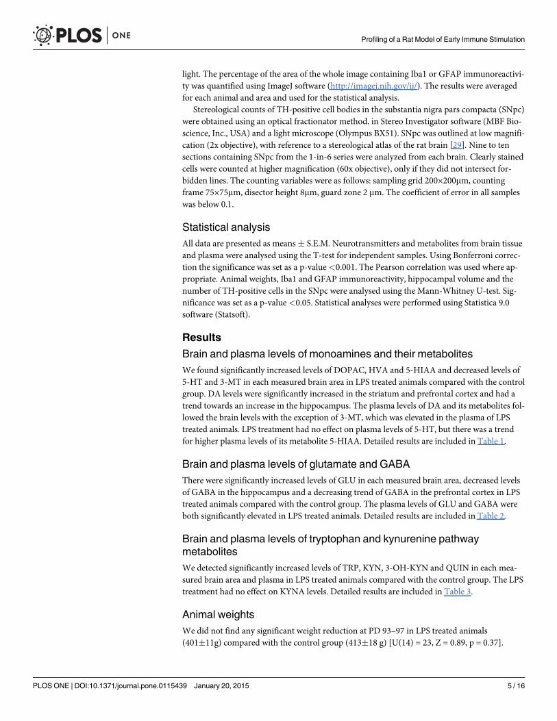
Activation of microglia and astrocytesMicroglia marked by the Iba1 antibody were observed in the whole brain in both LPS treatedanimals and saline treated controls. In both tested groups we found Iba1-positive cells with asmall round to oval cell body and long thin ramified branches. Hyperthrophied microglia withan atypical shape of the cell body and/or thicker processes which represent a transitional stageto the activated microglia were occasionally found in both groups. The percentage of the areacontaining Iba1 immunoreactivity in the hippocampus of LPS treated animals (8.25�0.89%)did not differ from the control group (7.35�1.5%) [U(14) = 26, Z = -0.58, p = 0.56]. We alsodid not reveal any difference in the SNpc between LPS treated animals (9.23�1.1%) and thecontrol group (8.46�1.37%) [U(14) = 26, Z = -0.58, p = 0.56] (Fig. 2).
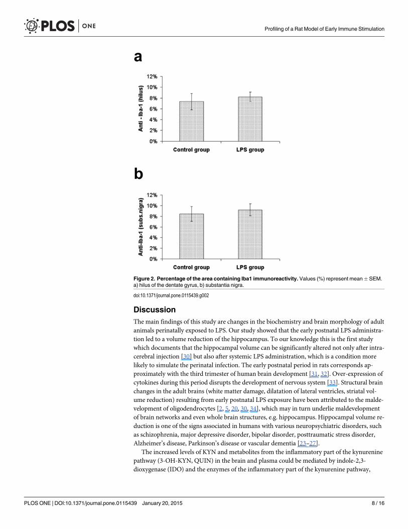
Astrocytes marked by the GFAP antibody were localized in the whole brain in both LPStreated animals and saline treated controls. However, the morphological characteristic ofGFAP-positive cells was different in LPS treated animals (hypertrophied cell body and thicker
Table 1. Brain and plasma levels of monoamines and their metabolites.
Striatum Hippocampus Prefrontal cortex Plasma
Control 51.13�1.10 32.14�1.67 23.81�0.69 160.45�3.70
DA LPS 80.61�2.79 38.52�1.00 33.26�1.33 213.55�4.51
T-test t(41) = -10.65, p<0.001 t(41) = -3.07, p<0.01 t(41) = -6.68, p<0.001 t(41) = -9.19, p<0.001
Control 15.43�0.50 24.91�0.52 16.03�0.46 7.42�0.19
DOPAC LPS 25.40�1.08 29.30�0.77 22.55�0.78 8.95�0.13
T-test t(41) = -8.97, p<0.001 t(41) = -4.91, p<0.001 t(41) = -7.52, p<0.001 t(41) = -6.39, p<0.001
Control 7.45�0.27 16.81�0.53 14.84�0.69 4.94�0.18
3-MT LPS 4.53�0.17 14.02�0.38 9.28�0.60 7.90�0.30
T-test t(41) = 8.62, p<0.001 t(41) = 4.07, p<0.001 t(41) = 5.91, p<0.001 t(41) = -8.74, p<0.001
Control 12.76�0.38 27.79�1.26 18.77�0.66 5.69�0.28
HVA LPS 21.70�0.69 38.35�1.94 25.73�0.70 9.23�0.24
T-test t(41) = -11.88, p<0.001 t(41) = -4.73, p<0.001 t(41) = -7.19, p<0.001 t(41) = -9.36, p<0.001
Control 24.80�0.77 33.47�1.50 25.30�0.60 46.33�2.04
5-HT LPS 18.62�1.12 22.54�0.90 19.53�0.59 41.89�1.79
T-test t(41) = 4.68, p<0.001 t(41) = 5.84, p<0.001 t(41) = 6.75, p<0.001 t(41) = 1.59, p = 0.12
Control 13.59�0.69 17.51�0.48 22.67�1.00 230.50�3.53
5-HIAA LPS 20.00�0.85 22.96�1.57 30.29�0.98 245.32�2.35
T-test t(41) = -5.91, p<0.001 t(41) = -3.64, p<0.001 t(41) = -5.36, p<0.001 t(41) = -3.30, p<0.01
Values (ng/ml) represent mean � SEM.
doi:10.1371/journal.pone.0115439.t001
Table 2. Brain and plasma levels of glutamate and γ-aminobutyric acid.
Striatum Hippocampus Prefrontal cortex Plasma
Control 34.46�0.81 29.25�0.94 30.30�0.58 13.86�0.35
GLU LPS 46.31�1.23 42.49�1.48 40.85�1.09 18.02�0.41
T-test t(41) = -8.32, p<0.001 t(41) = -7.85, p<0.001 t(41) = -9.08, p<0.001 t(41) = -8.07, p<0.001
Control 5.32�0.39 6.28�0.21 5.98�0.16 9.01�0.14
GABA LPS 6.01�0.42 3.62�0.14 4.97�0.37 12.47�0.30
T-test t(41) = -1.20, p = 0.24 t(41) = 9.99, p<0.001 t(41) = 2.73, p<0.01 t(41) = -11.21, p<0.001
Values (ng/ml) represent mean � SEM.
doi:10.1371/journal.pone.0115439.t002
Profiling of a Rat Model of Early Immune Stimulation
PLOS ONE | DOI:10.1371/journal.pone.0115439 January 20, 2015 6 / 16

processes) compared to those in the control group. Although the astroglial hypertrophy waspresent in multiple brain regions (especially in periventricular areas, but less so in the cortex),the most distinct changes were found in the hippocampus (hilus of the dentate gyrus, stratumlacunosum moleculare and stratum oriens) and in the SNpc. The percentage of the area con-taining GFAP immunoreactivity of LPS treated animals was significantly higher than in thecontrol group in the hippocampus (22.97�2.02% vs. 9.26�1.11%) [U(6) = 0, Z = -2.17,p<0.05] and in the SNpc (8.75�1.85% vs. 3.25�0.56%) [U(6) = 0, Z = -2.17, p<0.05](Figs. 3 and 4).
NeurodegenerationWe did not detect any Fluoro-Jade B positivity at PD 93–97 in the brains of animals perinatallytreated with LPS. The analysis of cell nuclei did not show any sign of nuclear pathology, e.g. nu-clear shrinkage or fragmentation. The saline treated controls exhibited the same results.
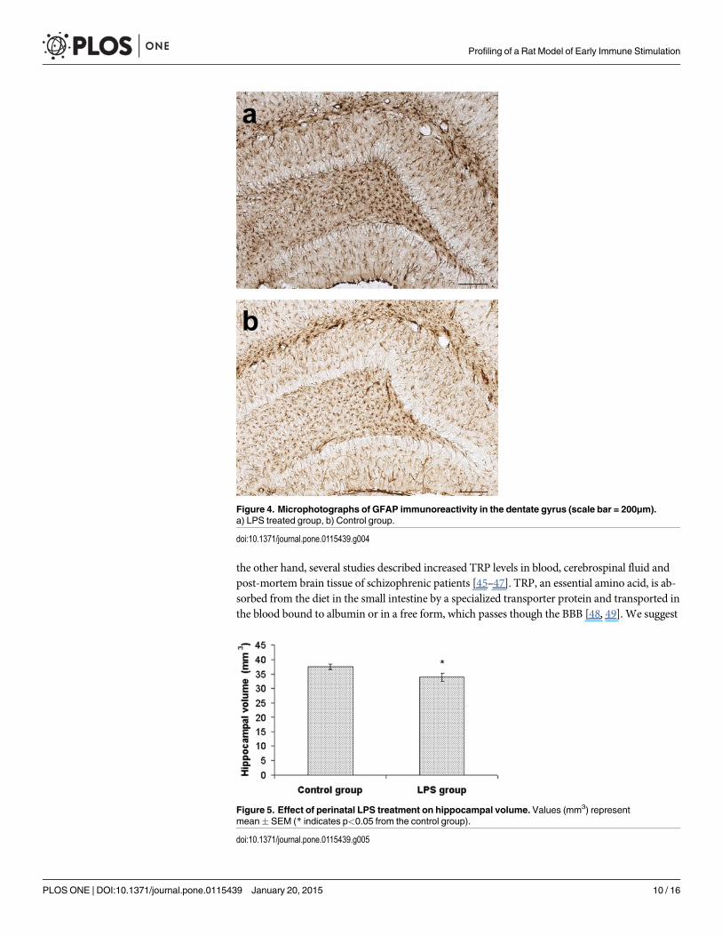
Hippocampal volumeWe found a significant reduction of hippocampal volume in LPS treated animals(33.9�1.4 mm3) compared with the control group (37.6�0.9 mm3) [U(14) = 11.5, Z = 2.1,p<0.05]. The reduction of hippocampal volume in LPS treated animals was approximately10% (Fig. 5).
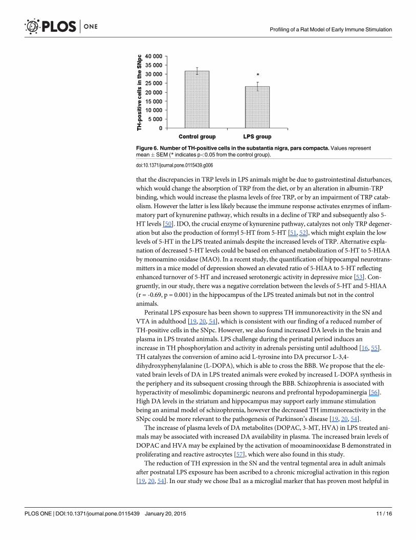
Tyrosine hydroxylase-positive cells in the substantia nigra, parscompactaThere was a significant reduction of the number of TH-positive cells in the SNpc in LPS treatedanimals (23 172�2 438) compared with the control group (31 830�1 891) [U(13) = 9,Z = 2.14, p<0.05]. The reduction of TH-positive cells in LPS treated animals was approximate-ly 27% (Fig. 6).
Table 3. Brain and plasma levels of tryptophan and kynurenine pathway metabolites.
Striatum Hippocampus Prefrontal cortex Plasma
Control 6.63�0.26 6.25�0.27 5.95�0.27 4.09�0.11
TRP LPS 20.17�1.08 20.46�1.70 21.57�1.57 6.67�0.24
T-test t(41) = -13.48, p<0.001 t(41) = -9.25, p<0.001 t(41) = -10.98, p<0.001 t(41) = -10.34, p<0.001
Control 63.93�1.00 75.59�1.66 66.99�0.89 65.13�1.40
KYN LPS 111.83�1.89 107.46�2.86 109.86�1.48 94.58�1.83
T-test t(41) = -23.76, p<0.001 t(41) = -10.10, p<0.001 t(41) = -25.94, p<0.001 t(41) = -12.99, p<0.001
Control 23.15�2.13 23.18�2.11 23.23�2.08 1.89�0.08
KYNA LPS 23.89�2.95 23.01�3.15 22.23�3.08 1.80�0.10
T-test t(41) = -0.20, p = 0.84 t(41) = 0.05, p = 0.96 t(41) = 0.28, p = 0.78 t(41) = 0.71, p = 0.48
Control 17.81�0.74 21.18�0.71 19.30�0.60 47.88�0.72
3-OH-KYN LPS 36.20�1.15 40.48�0.75 37.10�1.26 75.21�2.03
T-test t(41) = -13.96, p<0.001 t(41) = -18.50, p<0.001 t(41) = -13.59, p<0.001 t(41) = -13.84, p<0.001
Control 32.84�0.72 32.33�0.52 32.35�0.61 16.65�0.43
QUIN LPS 43.41�0.46 41.05�0.78 40.43�0.96 22.24�0.46
T-test t(41) = -11.88, p<0.001 t(41) = -9.38, p<0.001 t(41) = -7.33, p<0.001 t(41) = -8.83, p<0.001
Values (ng/ml) represent mean � SEM.
doi:10.1371/journal.pone.0115439.t003
Profiling of a Rat Model of Early Immune Stimulation
PLOS ONE | DOI:10.1371/journal.pone.0115439 January 20, 2015 7 / 16
DiscussionThe main findings of this study are changes in the biochemistry and brain morphology of adultanimals perinatally exposed to LPS. Our study showed that the early postnatal LPS administra-tion led to a volume reduction of the hippocampus. To our knowledge this is the first studywhich documents that the hippocampal volume can be significantly altered not only after intra-cerebral injection [30] but also after systemic LPS administration, which is a condition morelikely to simulate the perinatal infection. The early postnatal period in rats corresponds ap-proximately with the third trimester of human brain development [31, 32]. Over-expression ofcytokines during this period disrupts the development of nervous system [33]. Structural brainchanges in the adult brains (white matter damage, dilatation of lateral ventricles, striatal vol-ume reduction) resulting from early postnatal LPS exposure have been attributed to the malde-velopment of oligodendrocytes [2, 5, 20, 30, 34], which may in turn underlie maldevelopmentof brain networks and even whole brain structures, e.g. hippocampus. Hippocampal volume re-duction is one of the signs associated in humans with various neuropsychiatric disorders, suchas schizophrenia, major depressive disorder, bipolar disorder, posttraumatic stress disorder,Alzheimer’s disease, Parkinson’s disease or vascular dementia [23–27].
The increased levels of KYN and metabolites from the inflammatory part of the kynureninepathway (3-OH-KYN, QUIN) in the brain and plasma could be mediated by indole-2,3-dioxygenase (IDO) and the enzymes of the inflammatory part of the kynurenine pathway,
Figure 2. Percentage of the area containing Iba1 immunoreactivity. Values (%) represent mean� SEM.a) hilus of the dentate gyrus, b) substantia nigra.
doi:10.1371/journal.pone.0115439.g002
Profiling of a Rat Model of Early Immune Stimulation
PLOS ONE | DOI:10.1371/journal.pone.0115439 January 20, 2015 8 / 16
which are activated by pro-inflammatory cytokines [22]. The persistent elevation of pro-inflammatory cytokines in adulthood has been described previously by our group in this ani-mal model of early immune stimulation [12]. Unlike KYN and 3-OH-KYN, QUIN is not ableto cross the blood brain barrier (BBB) [35]; therefore the production of QUIN in the brain isregulated by local enzyme activity. Hippocampal formation is very sensitive to QUIN inducedexcitotoxicity [36], which is consistent with our finding of decreased hippocampal volume inLPS treated animals. However, we did not detect any Fluoro-Jade B positivity or changes ofneuronal nuclei typical for cell death at PD 93–97, which indicates a lack of ongoing neurode-generation. This corresponds with the results of studies that did not detect neurodegenerationin adult LPS treated animals hours or weeks after LPS administration [37, 38]. The activationof the inflammatory part of the kynurenine pathway has been described in relation with majordepressive disorder, Alzheimer’s disease, Parkinson’s disease, Huntington’s disease and epilep-sy [22]. On the other hand, elevated KYN levels have been found in the post-mortem prefrontalcortex [39] and elevated KYN and KYNA levels in cerebral spinal fluid [40] of schizophrenicpatients. Although we identified increased levels of KYN in LPS treated animals, we did notfind any change of KYNA levels in the brain or plasma compared to the controls.
The higher TRP plasma levels in the LPS group represent an interesting and unexpected find-ing. Neuropsychiatric disorders such as depression, anxiety and sometimes schizophrenia areusually associated with low TRP levels, which result in a decrease of 5-HT synthesis [41–44]. On
Figure 3. Percentage of the area containing GFAP immunoreactivity. Values (%) representmean� SEM (* indicates p<0.05 from the control group). a) hilus of the dentate gyrus, b) substantia nigra.
doi:10.1371/journal.pone.0115439.g003
Profiling of a Rat Model of Early Immune Stimulation
PLOS ONE | DOI:10.1371/journal.pone.0115439 January 20, 2015 9 / 16
the other hand, several studies described increased TRP levels in blood, cerebrospinal fluid andpost-mortem brain tissue of schizophrenic patients [45–47]. TRP, an essential amino acid, is ab-sorbed from the diet in the small intestine by a specialized transporter protein and transported inthe blood bound to albumin or in a free form, which passes though the BBB [48, 49]. We suggest
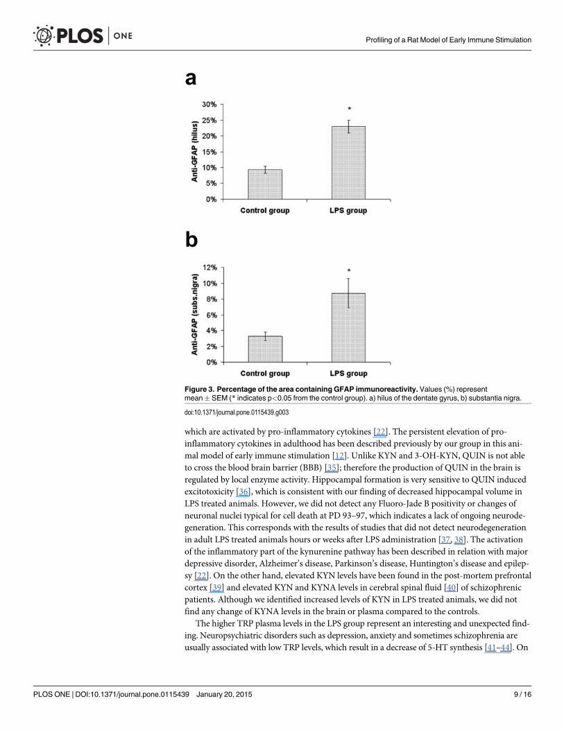
Figure 4. Microphotographs of GFAP immunoreactivity in the dentate gyrus (scale bar = 200μm).a) LPS treated group, b) Control group.
doi:10.1371/journal.pone.0115439.g004
Figure 5. Effect of perinatal LPS treatment on hippocampal volume. Values (mm3) representmean� SEM (* indicates p<0.05 from the control group).
doi:10.1371/journal.pone.0115439.g005
Profiling of a Rat Model of Early Immune Stimulation
PLOS ONE | DOI:10.1371/journal.pone.0115439 January 20, 2015 10 / 16
that the discrepancies in TRP levels in LPS animals might be due to gastrointestinal disturbances,which would change the absorption of TRP from the diet, or by an alteration in albumin-TRPbinding, which would increase the plasma levels of free TRP, or by an impairment of TRP catab-olism. However the latter is less likely because the immune response activates enzymes of inflam-matory part of kynurenine pathway, which results in a decline of TRP and subsequently also 5-HT levels [50]. IDO, the crucial enzyme of kynurenine pathway, catalyzes not only TRP degener-ation but also the production of formyl 5-HT from 5-HT [51, 52], which might explain the lowlevels of 5-HT in the LPS treated animals despite the increased levels of TRP. Alternative expla-nation of decreased 5-HT levels could be based on enhanced metabolization of 5-HT to 5-HIAAby monoamino oxidase (MAO). In a recent study, the quantification of hippocampal neurotrans-mitters in a mice model of depression showed an elevated ratio of 5-HIAA to 5-HT reflectingenhanced turnover of 5-HT and increased serotonergic activity in depressive mice [53]. Con-gruently, in our study, there was a negative correlation between the levels of 5-HT and 5-HIAA(r = -0.69, p = 0.001) in the hippocampus of the LPS treated animals but not in the controlanimals.
Perinatal LPS exposure has been shown to suppress TH immunoreactivity in the SN andVTA in adulthood [19, 20, 54], which is consistent with our finding of a reduced number ofTH-positive cells in the SNpc. However, we also found increased DA levels in the brain andplasma in LPS treated animals. LPS challenge during the perinatal period induces anincrease in TH phosphorylation and activity in adrenals persisting until adulthood [16, 55].TH catalyzes the conversion of amino acid L-tyrosine into DA precursor L-3,4-dihydroxyphenylalanine (L-DOPA), which is able to cross the BBB. We propose that the ele-vated brain levels of DA in LPS treated animals were evoked by increased L-DOPA synthesis inthe periphery and its subsequent crossing through the BBB. Schizophrenia is associated withhyperactivity of mesolimbic dopaminergic neurons and prefrontal hypodopaminergia [56].High DA levels in the striatum and hippocampus may support early immune stimulationbeing an animal model of schizophrenia, however the decreased TH immunoreactivity in theSNpc could be more relevant to the pathogenesis of Parkinson’s disease [19, 20, 54].
The increase of plasma levels of DA metabolites (DOPAC, 3-MT, HVA) in LPS treated ani-mals may be associated with increased DA availability in plasma. The increased brain levels ofDOPAC and HVA may be explained by the activation of mooaminooxidase B demonstrated inproliferating and reactive astrocytes [57], which were also found in this study.
The reduction of TH expression in the SN and the ventral tegmental area in adult animalsafter postnatal LPS exposure has been ascribed to a chronic microglial activation in this region[19, 20, 54]. In our study we chose Iba1 as a microglial marker that has proven most helpful in
Figure 6. Number of TH-positive cells in the substantia nigra, pars compacta. Values representmean� SEM (* indicates p<0.05 from the control group).
doi:10.1371/journal.pone.0115439.g006
Profiling of a Rat Model of Early Immune Stimulation
PLOS ONE | DOI:10.1371/journal.pone.0115439 January 20, 2015 11 / 16
visualizing microglia with details of their processes and which expression increases with micro-glial activation [58]. However, the microglia in the SN and hippocampus mostly showed signsof a resting state with an occasional occurrence of a hyperthrofied microglial body in both theLPS and control groups. According to some studies intracerebral as well as systemic postnatalLPS administration leads to microglial activation, which persists until adulthood [15, 30, 34,59]. In contrast, a recent study showed that early postnatal systemic LPS administration (PD5)increased microglial density in the hippocampus 48 hours after the administration, but inPD21 and PD74 animals the microglial density and the number of microglia did not differfrom saline treated controls [21]. The authors also speculated on the possible existence of a tar-get or maximum density of microglia in the brain [21].
Astrocytes influence a series of postnatal developmental steps, e.g. neuronal synapse forma-tion, myelination and homeostasis of the central nervous system [60–62]. The transition of as-trocytes into a reactive state, as we found in the SN and hippocampus of LPS treated animals,can possibly interfere with its functions because reactive astrocytes in vitro produce neurotoxicmolecules such as NO, reactive oxygen species and TNFα [63]. On the other hand, pro-inflam-matory mediators produced by microglia support neuroprotective astrocyte responses [64, 65].Astrogliosis was reported in patients with schizophrenia, autism and neurodegenerative disor-ders such as Alzheimer’s or Parkinson’s disease [66–69]. In contrast, major depressive disorderis characteristic of the reduction in the density of astrocytes [70].
Proinflammatory mediators induce the synthesis of QUIN, which acts as an NMDA recep-tor agonists and an important neurotoxic compound [22]. At high levels, QUIN promotesGLU release by neurons, inhibits its reuptake by astrocytes and inhibits astroglial glutaminesynthetase (GS) resulting in an accumulation of GLU [35], as was found in the present study.GLU serves as a precursor for the major inhibitory neurotransmitter GABA. Our finding of de-creased GABA levels in the hippocampus and prefrontal cortex in the LPS group may be medi-ated by the inhibition of GS [35]. The inhibition of GS is also induced by reactive astrogliosis[71]. The increased plasma levels of GABA in the LPS group may be due to synthesis fromGLU in peripheral tissues [72, 73]. In our study there was a negative correlation between thelevels of GLU and GABA (r = -0.51, p = 0.03) in the hippocampus of LPS treated animals butnot in control animals. Abnormalities in glutamatergic and GABAergic systems such as thecombination of increased GLU and decreased GABA levels in the brain have been associatedwith mood and anxiety disorders or autism [74–77].
In conclusion, the early immune stimulation induced by postnatal systemic administrationof LPS leads to biochemical actions resulting in alterations of neurotransmitter metabolismand activation of the kynurenine pathway of tryptophan metabolism as well as astrogliosis, hip-pocampal volume reduction and a decrease of tyrosine hydroxylase immunoreactivity in thesubstantia nigra. Our results suggest a pathogenetic link between early immune stimulationand neuropsychiatric disorders such and schizophrenia, mood disorders, anxiety disorders, au-tism, Parkinson’s disease and Alzheimer’s disease.
Supporting InformationS1 ARRIVE Guidelines Checklist.(DOC)
AcknowledgmentsWe would like to thank Tomas Novak M.D., Ph.D. for help with the statistical analysis,Craig Hampson BSc (Hons) for language correction and the Institute of Physiology at the
Profiling of a Rat Model of Early Immune Stimulation
PLOS ONE | DOI:10.1371/journal.pone.0115439 January 20, 2015 12 / 16
Academy of Sciences of the Czech Republic for use of their Stereo Investigator software, in-struction and support.
Author ContributionsConceived and designed the experiments: JH HT AK. Performed the experiments: AK KS PKJV. Analyzed the data: AK. Contributed reagents/materials/analysis tools: KS PK. Wrote thepaper: AK. Provided technical support and conceptual advice: FT MF TP. Critically reviewedthe manuscript: JH.
References1. Schmitt A, Malchow B, Hasan A, Falkai P (2014) The impact of environmental factors in severe psychi-
atric disorders. Front Neurosci 8: 19. doi: 10.3389/fnins.2014.00019 PMID: 24574956
2. Wang X, Rousset CI, Hagberg H, Mallard C (2006) Lipopolysaccharide-induced inflammation and peri-natal brain injury. Semin Fetal Neonatal Med 11: 343–353. doi: 10.1016/j.siny.2006.04.002 PMID:16793357
3. Spencer SJ, Galic MA, Pittman QJ (2011) Neonatal programming of innate immune function. AmJ Physiol Endocrinol Metab 300: E11–E18. doi: 10.1152/ajpendo.00516.2010 PMID: 21045175
4. Jack CS, Arbour N, Manusow J, Montgrain V, Blain M, et al. (2005) TLR signaling tailors innate immuneresponses in human microglia and astrocytes. J Immunol 175: 4320–4330. doi: 10.4049/jimmunol.175.7.4320 PMID: 16177072
5. Lehnardt S, Lachance C, Patrizi S, Lefebvre S, Follett PL, et al. (2002) The toll-like receptor TLR4 isnecessary for lipopolysaccharide-induced oligodendrocyte injury in the CNS. J Neurosci 22:2478–2486. PMID: 11923412
6. Rolls A, Shechter R, London A, Ziv Y, Ronen A, et al. (2007) Toll-like receptors modulate adult hippo-campal neurogenesis. Nat Cell Biol 9: 1081–1088. doi: 10.1038/ncb1629 PMID: 17704767
7. Shanks N, Larocque S, Meaney MJ (1995) Neonatal endotoxin exposure alters the development of thehypothalamic-pituitary-adrenal axis: early illness and later responsivity to stress. J Neurosci 15:376–384. PMID: 7823142
8. Shanks N, Windle RJ, Perks PA, Harbuz MS, Jessop DS, et al. (2000) Early-life exposure to endotoxinalters hypothalamic-pituitary-adrenal function and predisposition to inflammation. Proc Natl Acad SciU S A 97: 5645–5650. doi: 10.1073/pnas.090571897 PMID: 10779563
9. Potvin S, Stip E, Sepehry AA, Gendron A, Bah R, et al. (2008) Inflammatory cytokine alterations inschizophrenia: a systematic quantitative review. Biol Psychiatry 63: 801–808. doi: 10.1016/j.biopsych.2007.09.024 PMID: 18005941
10. Tejkalová H, Jelínek F, Klaschka J, Šťastný F (2007) Vliv perinatální zánětlivé reakce na rozvoj psy-chóze podobného chování: experimentální studie (Effect of perinatal inflammatory reaction on the in-duction of psychotic-like behaviour: experimental study). Psychiatrie 11: 12–15.
11. Tejkalová H, Jelínek F, Klaschka J, Šťastný F (2008) Vliv chronického podávání klozapinu na chovánípotkanů v neuroinfekčnímmodelu schizofrenie (Effect of chronic clozapine administration on the beha-vioural pattern of rats in the neuroinfectional model of schizophrenia). Psychiatrie 12: 68–72.
12. Tejkalová H, Růžičková Š, Klaschka J (2010) Vliv chronického podávání antipsychotik na expresi cyto-kinů v neuroinfekčním modelu schizofrenie (Effect of antipsychotic chronic administration on cytokineexpression in neuroinfectious model of schizophrenia). Psychiatrie 14: 19–21.
13. Doosti MH, Bakhtiari A, Zare P, Amani M, Majidi-Zolbanin N, et al. (2013) Impacts of early interventionwith fluoxetine following early neonatal immune activation on depression-like behaviors and bodyweight in mice. Prog Neuropsychopharmacol Biol Psychiatry 43: 55–65. doi: 10.1016/j.pnpbp.2012.12.003 PMID: 23270703
14. Kirsten TB, Chaves-Kirsten GP, Chaible LM, Silva AC, Martins DO, et al. (2012) Hypoactivity of the cen-tral dopaminergic system and autistic-like behavior induced by a single early prenatal exposure to lipo-polysaccharide. J Neurosci Res 90: 1903–1912. doi: 10.1002/jnr.23089 PMID: 22714803
15. Sominsky L, Walker AK, Ong LK, Tynan RJ, Walker FR, et al. (2012) Increased microglial activation inthe rat brain following neonatal exposure to a bacterial mimetic. Behav Brain Res 226: 351–356. doi:10.1016/j.bbr.2011.08.038 PMID: 21907243
16. Sominsky L, Fuller EA, Bondarenko E, Ong LK, Averell L, et al. (2013) Functional programming of theautonomic nervous system by early life immune exposure: implications for anxiety. PLoS One 8:e57700. doi: 10.1371/journal.pone.0057700 PMID: 23483921
Profiling of a Rat Model of Early Immune Stimulation
PLOS ONE | DOI:10.1371/journal.pone.0115439 January 20, 2015 13 / 16
17. Zhu F, Zhang L, Ding YQ, Zhao J, Zheng Y (2014) Neonatal intrahippocampal injection of lipopolysac-charide induces deficits in social behavior and prepulse inhibition and microglial activation in rats: Impli-cation for a new schizophrenia animal model. Brain Behav Immun 38: 166–174. doi: 10.1016/j.bbi.2014.01.017 PMID: 24530999
18. Bilbo SD, Schwarz JM (2009) Early-life programming of later-life brain and behavior: a critical role forthe immune system. Front Behav Neurosci 3: 14. doi: 10.3389/neuro.08.014.2009 PMID: 19738918
19. Cai Z, Fan LW, Kaizaki A, Tien LT, Ma T, et al. (2013) Neonatal systemic exposure to lipopolysaccha-ride enhances susceptibility of nigrostriatal dopaminergic neurons to rotenone neurotoxicity in later life.Dev Neurosci 35: 155–171. doi: 10.1159/000346156 PMID: 23446007
20. Fan LW, Tien LT, Zheng B, Pang Y, Lin RC, et al. (2011) Dopaminergic neuronal injury in the adult ratbrain following neonatal exposure to lipopolysaccharide and the silent neurotoxicity. Brain BehavImmun 25: 286–297. doi: 10.1016/j.bbi.2010.09.020 PMID: 20875849
21. Smith PL, Hagberg H, Naylor AS, Mallard C (2014) Neonatal Peripheral Immune Challenge ActivatesMicroglia and Inhibits Neurogenesis in the Developing Murine Hippocampus. Dev Neurosci. doi: 10.1159/000359950
22. Campbell BM, Charych E, Lee AW, Moller T (2014) Kynurenines in CNS disease: regulation by inflam-matory cytokines. Front Neurosci 8: 12. doi: 10.3389/fnins.2014.00012 PMID: 24567701
23. Brambilla P, Hatch JP, Soares JC (2008) Limbic changes identified by imaging in bipolar patients. CurrPsychiatry Rep 10: 505–509. doi: 10.1007/s11920-008-0080-8 PMID: 18980734
24. Bremner JD, Narayan M, Anderson ER, Staib LH, Miller HL, et al. (2000) Hippocampal volume reduc-tion in major depression. Am J Psychiatry 157: 115–118. PMID: 10618023
25. Haukvik UK, Hartberg CB, Agartz I (2013) Schizophrenia—what does structural MRI show? TidsskrNor Laegeforen 133: 850–853. doi: 10.4045/tidsskr.12.1084 PMID: 23612107
26. Laakso MP, Partanen K, Riekkinen P, Lehtovirta M, Helkala EL, et al. (1996) Hippocampal volumes inAlzheimer’s disease, Parkinson’s disease with and without dementia, and in vascular dementia: AnMRI study. Neurology 46: 678–681. doi: 10.1212/WNL.46.3.678 PMID: 8618666
27. Woon FL, Sood S, Hedges DW (2010) Hippocampal volume deficits associated with exposure to psy-chological trauma and posttraumatic stress disorder in adults: a meta-analysis. Prog Neuropsychophar-macol Biol Psychiatry 34: 1181–1188. doi: 10.1016/j.pnpbp.2010.06.016 PMID: 20600466
28. Najmanova V, Rambousek L, Syslova K, Bubenikova V, Slamberova R, et al. (2011) LC-ESI-MS-MSMethod for Monitoring Dopamine, Serotonin and Their Metabolites in Brain Tissue. Chromatographia73: 143–149. doi: 10.1007/s10337-011-1959-9
29. Paxinos G. andWatson C. (1-1-2007) The Rat Brain In Strereotactic Coordinates. Elsevier Inc.
30. Wang KC, Fan LW, Kaizaki A, Pang Y, Cai Z, et al. (2013) Neonatal lipopolysaccharide exposure in-duces long-lasting learning impairment, less anxiety-like response and hippocampal injury in adult rats.Neuroscience 234: 146–157. doi: 10.1016/j.neuroscience.2012.12.049 PMID: 23298854
31. Craig A, Ling LN, Beardsley DJ, Wingate-Pearse N, Walker DW, et al. (2003) Quantitative analysis ofperinatal rodent oligodendrocyte lineage progression and its correlation with human. Exp Neurol 181:231–240. doi: 10.1016/S0014-4886(03)00032-3 PMID: 12781996
32. Feleder C, Tseng KY, Calhoon GG, O’Donnell P (2010) Neonatal intrahippocampal immune challengealters dopamine modulation of prefrontal cortical interneurons in adult rats. Biol Psychiatry 67:386–392. doi: 10.1016/j.biopsych.2009.09.028 PMID: 19914600
33. Ratnayake U, Quinn T, Walker DW, Dickinson H (2013) Cytokines and the neurodevelopmental basisof mental illness. Front Neurosci 7: 180. doi: 10.3389/fnins.2013.00180 PMID: 24146637
34. Pang Y, Cai Z, Rhodes PG (2003) Disturbance of oligodendrocyte development, hypomyelination andwhite matter injury in the neonatal rat brain after intracerebral injection of lipopolysaccharide. Brain ResDev Brain Res 140: 205–214. doi: 10.1016/S0165-3806(02)00606-5 PMID: 12586426
35. Guillemin GJ (2012) Quinolinic acid, the inescapable neurotoxin. FEBS J 279: 1356–1365. doi: 10.1111/j.1742-4658.2012.08493.x PMID: 22248144
36. St’astny F, Skultetyova I, Pliss L, Jezova D (2000) Quinolinic acid enhances permeability of rat brainmicrovessels to plasma albumin. Brain Res Bull 53: 415–420. doi: 10.1016/S0361-9230(00)00368-3PMID: 11136997
37. Chung DW, Yoo KY, Hwang IK, Kim DW, Chung JY, et al. (2010) Systemic administration of lipopoly-saccharide induces cyclooxygenase-2 immunoreactivity in endothelium and increases microglia in themouse hippocampus. Cell Mol Neurobiol 30: 531–541. doi: 10.1007/s10571-009-9477-0 PMID:19908141
38. Terrazzino S, Bauleo A, Baldan A, Leon A (2002) Peripheral LPS administrations up-regulate Fas andFasL on brain microglial cells: a brain protective or pathogenic event? J Neuroimmunol 124: 45–53.doi: 10.1016/S0165-5728(02)00013-9 PMID: 11958821
Profiling of a Rat Model of Early Immune Stimulation
PLOS ONE | DOI:10.1371/journal.pone.0115439 January 20, 2015 14 / 16

39. Schwarcz R, Rassoulpour A, Wu HQ, Medoff D, Tamminga CA, et al. (2001) Increased cortical kynure-nate content in schizophrenia. Biol Psychiatry 50: 521–530. doi: 10.1016/S0006-3223(01)01078-2PMID: 11600105
40. Linderholm KR, Skogh E, Olsson SK, Dahl ML, Holtze M, et al. (2012) Increased levels of kynurenineand kynurenic acid in the CSF of patients with schizophrenia. Schizophr Bull 38: 426–432. doi: 10.1093/schbul/sbq086 PMID: 20729465
41. Almeida-Montes LG, Valles-Sanchez V, Moreno-Aguilar J, Chavez-Balderas RA, Garcia-Marin JA,et al. (2000) Relation of serum cholesterol, lipid, serotonin and tryptophan levels to severity of depres-sion and to suicide attempts. J Psychiatry Neurosci 25: 371–377. PMID: 11022402
42. Lee M, Jayathilake K, Dai J, Meltzer HY (2011) Decreased plasma tryptophan and tryptophan/largeneutral amino acid ratio in patients with neuroleptic-resistant schizophrenia: relationship to plasma cor-tisol concentration. Psychiatry Res 185: 328–333. doi: 10.1016/j.psychres.2010.07.013 PMID:20699195
43. Gauthier C, Hassler C, Mattar L, Launay JM, Callebert J, et al. (2014) Symptoms of depression andanxiety in anorexia nervosa: links with plasma tryptophan and serotonin metabolism. Psychoneuroen-docrinology 39: 170–178. doi: 10.1016/j.psyneuen.2013.09.009 PMID: 24135616
44. Bell C, Abrams J, Nutt D (2001) Tryptophan depletion and its implications for psychiatry. Br J Psychiatry178: 399–405. doi: 10.1192/bjp.178.5.399 PMID: 11331552
45. Miller CL, Llenos IC, Cwik M, Walkup J, Weis S (2008) Alterations in kynurenine precursor and productlevels in schizophrenia and bipolar disorder. Neurochem Int 52: 1297–1303. doi: 10.1016/j.neuint.2008.01.013 PMID: 18328600
46. Issa F, Gerhardt GA, Bartko JJ, Suddath RL, Lynch M, et al. (1994) A multidimensional approach toanalysis of cerebrospinal fluid biogenic amines in schizophrenia: I. Comparisons with healthy controlsubjects and neuroleptic-treated/unmedicated pairs analyses. Psychiatry Res 52: 237–249.
47. Ravikumar A, Deepadevi KV, Arun P, Manojkumar V, Kurup PA (2000) Tryptophan and tyrosine cata-bolic pattern in neuropsychiatric disorders. Neurol India 48: 231–238. PMID: 11025626
48. Bohmer C, Broer A, Munzinger M, Kowalczuk S, Rasko JE, et al. (2005) Characterization of mouseamino acid transporter B0AT1 (slc6a19). Biochem J 389: 745–751. doi: 10.1042/BJ20050083 PMID:15804236
49. Pratt OE (1979) Kinetics of tryptophan transport across the blood-brain barrier. J Neural Transm Suppl29–42: .
50. Maes M, Leonard BE, Myint AM, Kubera M, Verkerk R (2011) The new ‘5-HT’ hypothesis of depression:cell-mediated immune activation induces indoleamine 2,3-dioxygenase, which leads to lower plasmatryptophan and an increased synthesis of detrimental tryptophan catabolites (TRYCATs), both of whichcontribute to the onset of depression. Prog Neuropsychopharmacol Biol Psychiatry 35: 702–721.PMID: 21185346
51. Stone TW, Darlington LG (2002) Endogenous kynurenines as targets for drug discovery and develop-ment. Nat Rev Drug Discov 1: 609–620. doi: 10.1038/nrd870 PMID: 12402501
52. Stone TW, Forrest CM, Darlington LG (2012) Kynurenine pathway inhibition as a therapeutic strategyfor neuroprotection. FEBS J 279: 1386–1397. doi: 10.1111/j.1742-4658.2012.08487.x PMID:22248239
53. Huang F, Li J, Shi HL, Wang TT, Muhtar W, et al. (2014) Simultaneous quantification of seven hippo-campal neurotransmitters in depression mice by LC-MS/MS. J Neurosci Methods 229: 8–14. doi: 10.1016/j.jneumeth.2014.04.004 PMID: 24735530
54. Fan LW, Tien LT, Lin RC, Simpson KL, Rhodes PG, et al. (2011) Neonatal exposure to lipopolysaccha-ride enhances vulnerability of nigrostriatal dopaminergic neurons to rotenone neurotoxicity in later life.Neurobiol Dis 44: 304–316. doi: 10.1016/j.nbd.2011.07.011 PMID: 21798348
55. Ong LK, Sominsky L, Dickson PW, Hodgson DM, Dunkley PR (2012) The sustained phase of tyrosinehydroxylase activation in vivo. Neurochem Res 37: 1938–1943. doi: 10.1007/s11064-012-0812-3PMID: 22684282
56. Brisch R, Saniotis A, Wolf R, Bielau H, Bernstein HG, et al. (2014) The role of dopamine in schizophre-nia from a neurobiological and evolutionary perspective: old fashioned, but still in vogue. Front Psychia-try 5: 47. doi: 10.3389/fpsyt.2014.00047 PMID: 24904434
57. Gulyas B, Pavlova E, Kasa P, Gulya K, Bakota L, et al. (2011) Activated MAO-B in the brain of Alzhei-mer patients, demonstrated by [11C]-L-deprenyl using whole hemisphere autoradiography. NeurochemInt 58: 60–68. doi: 10.1016/j.neuint.2010.10.013 PMID: 21075154
58. Kettenmann H, Hanisch UK, Noda M, Verkhratsky A (2011) Physiology of microglia. Physiol Rev 91:461–553. doi: 10.1152/physrev.00011.2010 PMID: 21527731
Profiling of a Rat Model of Early Immune Stimulation
PLOS ONE | DOI:10.1371/journal.pone.0115439 January 20, 2015 15 / 16
59. Patro N, Singh K, Patro I (2013) Differential microglial and astrocytic response to bacterial and viral in-fection in the developing hippocampus of neonatal rats. Indian J Exp Biol 51: 606–614. PMID:24228384
60. Dong Y, Benveniste EN (2001) Immune function of astrocytes. Glia 36: 180–190. doi: 10.1002/glia.1107 PMID: 11596126
61. Meyer-Franke A, Shen S, Barres BA (1999) Astrocytes induce oligodendrocyte processes to align withand adhere to axons. Mol Cell Neurosci 14: 385–397. doi: 10.1006/mcne.1999.0788 PMID: 10588392
62. Molofsky AV, Krencik R, Ullian EM, Tsai HH, Deneen B, et al. (2012) Astrocytes and disease: a neuro-developmental perspective. Genes Dev 26: 891–907. doi: 10.1101/gad.188326.112 PMID: 22549954
63. Carson MJ, Thrash JC, Walter B (2006) The cellular response in neuroinflammation: The role of leuko-cytes, microglia and astrocytes in neuronal death and survival. Clin Neurosci Res 6: 237–245. doi: 10.1016/j.cnr.2006.09.004 PMID: 19169437
64. Bsibsi M, Persoon-Deen C, Verwer RW, Meeuwsen S, Ravid R, et al. (2006) Toll-like receptor 3 onadult human astrocytes triggers production of neuroprotective mediators. Glia 53: 688–695. doi: 10.1002/glia.20328 PMID: 16482523
65. Okada K, Yamashita U, Tsuji S (2005) Modulation of Na(+)-dependent glutamate transporter of murineastrocytes by inflammatory mediators. J UOEH 27: 161–170. PMID: 15986771
66. Catts VS, Wong J, Fillman SG, Fung SJ, Weickert CS (2014) Increased expression of astrocyte mark-ers in schizophrenia: Association with neuroinflammation. Aust N Z J Psychiatry. doi: 10.1177/0004867414531078 PMID: 24744400
67. Forno LS, DeLanney LE, Irwin I, Di MD, Langston JW (1992) Astrocytes and Parkinson’s disease.Prog Brain Res 94: 429–436. doi: 10.1016/S0079-6123(08)61770-7 PMID: 1287728
68. Vargas DL, Nascimbene C, Krishnan C, Zimmerman AW, Pardo CA (2005) Neuroglial activation andneuroinflammation in the brain of patients with autism. Ann Neurol 57: 67–81. doi: 10.1002/ana.20315PMID: 15546155
69. Wisniewski HM, Wegiel J (1991) Spatial relationships between astrocytes and classical plaque compo-nents. Neurobiol Aging 12: 593–600. doi: 10.1016/0197-4580(91)90091-W PMID: 1770990
70. Rajkowska G, Stockmeier CA (2013) Astrocyte pathology in major depressive disorder: insights fromhuman postmortem brain tissue. Curr Drug Targets 14: 1225–1236. doi: 10.2174/13894501113149990156 PMID: 23469922
71. Ortinski PI, Dong J, Mungenast A, Yue C, Takano H, et al. (2010) Selective induction of astrocytic glio-sis generates deficits in neuronal inhibition. Nat Neurosci 13: 584–591. doi: 10.1038/nn.2535 PMID:20418874
72. van Bemmelen FJ, Schouten MJ, Fekkes D, Bruinvels J (1985) Succinic semialdehyde as a substratefor the formation of gamma-aminobutyric acid. J Neurochem 45: 1471–1474. doi: 10.1111/j.1471-4159.1985.tb07214.x PMID: 2864395
73. White HL (1981) Glutamate as a precursor of GABA in rat brain and peripheral tissues. Mol Cell Bio-chem 39: 253–259. doi: 10.1007/BF00232578 PMID: 6118823
74. Krystal JH, Sanacora G, Blumberg H, Anand A, Charney DS, et al. (2002) Glutamate and GABA sys-tems as targets for novel antidepressant and mood-stabilizing treatments. Mol Psychiatry 7 Suppl 1:S71–S80. doi: 10.1038/sj.mp.4001021 PMID: 11986998
75. Meyerhoff DJ, Mon A, Metzler T, Neylan TC (2014) Cortical gamma-aminobutyric Acid and glutamatein posttraumatic stress disorder and their relationships to self-reported sleep quality. Sleep 37:893–900. doi: 10.5665/sleep.3654 PMID: 24790267
76. Pollack MH, Jensen JE, Simon NM, Kaufman RE, Renshaw PF (2008) High-field MRS study of GABA,glutamate and glutamine in social anxiety disorder: response to treatment with levetiracetam.Prog Neuropsychopharmacol Biol Psychiatry 32: 739–743. doi: 10.1016/j.pnpbp.2007.11.023 PMID:18206286
77. Quaak I, Brouns MR, Van de BM (2013) The dynamics of autism spectrum disorders: how neurotoxiccompounds and neurotransmitters interact. Int J Environ Res Public Health 10: 3384–3408. doi: 10.3390/ijerph10083384 PMID: 23924882
Profiling of a Rat Model of Early Immune Stimulation
PLOS ONE | DOI:10.1371/journal.pone.0115439 January 20, 2015 16 / 16