DNA demethylation increases sensitivity of neuroblastoma cells to chemotherapeutic drugs
Academic Journal of Cancer Research 7 (2): 152-161, 2014ISSN 1995-8943© IDOSI Publications, 2014DOI: 10.5829/idosi.ajcr.2014.7.2.83286
Corresponding Author: Tarek M. Okda, Department of Biochemistry, Faculty of Pharmacy, Damanhour University Damanhour - Egypt. E-mail: [email protected]
152
Biochemical and Molecular Assessments of Possible Roles of Soybean in 1, 2-dimethylhydrazine-induced Colon Cancer Chemotherapeutic.
Tarek M. Okda, Mohamed. A. Abd EL-Aziz, N.T. El-Melegy,1 2 3
H.H. Taha and Sary. Kh. Abd Elghaffar2 4
Department of biochemistry, Faculty of Pharmacy, Damanhour University Damanhour, Egypt1
Department of biochemistry, Faculty of Pharmacy, Al-Azhar University Assiut, Egypt2
Departments of medical biochemistry, Faculty of Medicine, Assuit University Assiut, Egypt3
Department of Pathology, Faculty of Vet. Medicine, Assuit University Assiut, Egypt4
Abstract: Background: Colon cancer is one of the most common cancers worldwide. 1, 2-dimethyl hydrazine(DMH) is considered as one of the carcinogens that induce colon cancer in animal model. The aim of this workto assess the role of soybean as a preventative and/or therapeutic agent for colon cancer induced by DMH.Methods :sixty male rats were used in this study; the animals were divided into six major groups: (1) controlgroup, (2) carcinogen group, (3) prophylactic (Pro) group, (4) treatment (Tr) group, (5) self limited group and(6) standard group (10 rats in each group). Results: there were over expression of mRNA and the protein of bothgalectin-3 and C4.4A in both the carcinogenic and self limited groups while low expression in the prophylacticand treated soybean also in. There was also decrease in tissue caspase-3 activity, antioxidant markers ofcarcinogenic group. While, there was an increase in these markers in 5FU treated group as well as bothsoybeans prophylactic and treated groups. Conclusion: the biochemical, molecular and histopathologicalanalysis associated with the administration of soybeans exhibit its ability to minimize the frequency of DMH-induced colon carcinomas through increasing the apoptotic tumors cell loss and regulating the level ofdetrimental oxidative stress markers.
Key words: Colon Cancer Soybeans Galectin-3 and C4.4A
INTRODUCTION rat pancreatic adenocarcinoma line. In the rat C4.4A is
Colon cancer is one of the major causes of cancer blastocyst and is highly expressed in the placenta. In themortality worldwide, which results from interactions of adult, expression is restricted mostly to stratified epitheliadifferent factors such as aging, family history and dietary of the skin and squamous epithelia of the upperstyle, colon cancer represents the fourth most common gastrointestinal tract [3].cause of cancer related mortality. The development of The advantage of C4.4A in colon cancer diagnosiscolon cancer is associated with (a) perturbations of the relies not only on the high frequency and stability ofdelicate balance between cell proliferation and apoptotic expression, but particularly on the de novo expression ofcell loss (b) alterations in the oxidative stress state and (c) this molecule in colorectal cancer tissues or the stronglyalterations in proteins related to colon derangement; up regulated expression as for example in transitional cellmainly C4.4A and galectin-3 (Gal-3) [1]. cancer [4].
The C4.4A is glycosyl-phosphatidyl-inositol Dramatic changes in cellular glycosylation are aanchored membrane glycoproteins with 30% homology to hallmark of cancer cells and significant correlations arethe urokinase-type plasminogen activator receptor which now established between some glycosylation patternsis implicated in cancer invasion and metastasis [2]. The and clinical prognosis [5]. Gal-3 is a member of galectins
C4.4A protein was first identified in a highly metastasizing
strongly up regulated during implantation of the
Academic J. Cancer Res., 7 (2): 152-161, 2014
153
that are a family of carbohydrate-binding proteins Soybean is being considered as a potentialthat share a carbohydrate recognition domain for chemopreventive agent. In vitro it inhibits transcription
-galactosides and are involved in cell adhesion, and the activity of lipoxygenase or cyclooxygenasemigration, differentiation, angiogenesis, proliferation and enzymes, which facilitate tumor progression. In vivo it isapoptosis [6]. During development, Gal-3 is detected in protective in rodent models of chemical carcinogenesis.most types of cells including the hepatocytes in human Soybean contains an á, -unsaturated ketone, a reactiveand mouse embryos and colorectal cells [7]. chemical substituent that is responsible for its repression
Alterations in Gal-3 expression have been related to of transcription [15]. Crude soybean saponin mixtures areneoplastic transformation and metastatic progression of growth inhibitory against multiple human colonhuman colon cancers. Several studies reported a adenocarcinoma cell lines in vitro. Treatment of humansignificant increase of galectin-3 content in the tumors colon cancer cell lines with soyasaponins suppressedthat progressed to the metastatic stage relative to the non growth, induced morphological alterations, increasedmetastatic tumors or normal colon tissue. However the multiple markers of differentiation and inhibited proteinincreased content of Gal-3 in colon cancer and whether it kinase C (PKC) activity [16]. has a role in apoptosis is poorly understood, so we try to To date, the understanding of the possible protectivefind a relation between Gal-3 in colon cancer and effects of soybeans in the colon cancer is still incomplete.apoptosis modulation by some natural products; mainly Therefore, the present study aims to assess thesoybeans [8]. hypothesized alterations by evaluation of (a) mRNA and
Paret et al. [9] explained that Gal-3 associates with protein of both C4.4A and Gal-3 (Markers ofC4.4A and that C4.4A ligand binding confers a migratory tumorigenicity), (b) tissue levels of caspase-3 activitiesphenotype that was in line with the supposed metastasis (Marker of apoptosis); and (d) lipid peroxidation, NOs,association, in the current study we will try to find their reduced glutathione and superoxide dismutase (Markersassociation in colon cancer as candidate markers and their of oxidative stress). influence by natural product treatment.
In colon carcinogenesis, apoptosis is mediated by MATERIAL AND METHODSseveral molecules such as caspases. Caspases(cysteinyl-aspartate-specific proteinases), a family of Preparations of Soybean Extract: Soybean extract wasintracellular cysteine proteases causing apoptotic cell purchased from Sigma Aldrich company code no.:death [10]. The activation of caspases then targets their (ERMBF-410AK). Soybean extract was prepared bydownstream molecules Poly-ADP-Ribose-Polymerase, dissolving 100mg in 10 ml distilled water. The rats were(PARP) which specifically binds at DNA strand breaks given 0.5 ml of Soybean extract twice weekly for one[11]. month orally by a dose of (120 mg/kg) according to Raju
Oxidative stress induces a cellular redox imbalance et al. [17]. which has been found to be present in various cancercells compared with normal cells. The redox imbalance Treatment and Sampling: Male Albino rats (n= 60)thus may be related to oncogenic stimulation. DNA (150±25 g body weight) were purchased from animalmutation is a critical step in carcinogenesis and elevated house of Assiut University. The dose of 1,2 DMH waslevels of oxidative DNA lesions (8-OH-G) have been purchased from Sigma Aldrich company (20 mg/kg) twicenoted in various tumours, strongly implicating such weekly for one month, the dose of 1,2 DMH in our studydamage in the etiology of cancer [12]. was determined according to Hiromichi and Michael [18].
Over the last decades, natural compounds have Rats were divided into six groups ten rats in each; firstattracted considerable attention as cancer control healthy group. The second is carcinogenic groupchemopreventive agents and also as cancer therapeutics. (DMH), were treated subcutaneously with 1,2- DMH.Among their various biological activities, natural Third Group is prophylactic group (Pro) group, wereproducts can modulate apoptosis signaling pathways treated subcutaneously with 1,2- DMH and orally with[13]. Soybeans have long been recognized as an excellent soybean extract (120 mg/kg) for one month. The fourthsource of high-quality protein. The soybean also contains Group is soybean treated group (Tr) group, were injecteda wide variety of chemical compounds that have potent subcutaneously with 1,2- DMH, then treated orally withbioactivity; among these compounds are isoflavones and soybean extract for another month.The Fifth Group is selfsaponins [14]. limited group was treated subcutaneously with 1,2- DMH,
Academic J. Cancer Res., 7 (2): 152-161, 2014
154
then left to another month without any treatment. The The Primer Sets as Following:sixth group is 5FU treated group, were treatedsubcutaneously with 1,2- DMH, then treated with 5FU(100 mg/kg) for another month, the dose of 5FU in ourstudy was determined according to the modified methodmade by Ragunath et al. [19]. At the end of the experimentand after the exact period for each group, rats wereanaesethized with light ether then the rats were scarifiedaccording to National Institute of Health Guidelines foranimal care. Blood samples were collected by cardiacpuncturing method, centrifuged and sera were isolated forserological studies. Colon were excised rapidly and usedfor RNA preparation by homogenization in 20 mM Tris,100 mM NaCl, 1 mM EDTA and 0.5% Triton X100 buffer.Protein content of colon homogenate was determinedusing Biuret reagent and bovine serum albumin (BSA) asstandard. Samples were aliquotted and stored at -80°C tilluse.
SDS-PAGE and Western Blotting: Gal-3 and C4.4Aantibody was obtained from Santa Cruz Technology.Secondary antibody (Peroxidase labelled antirabbitantibody) was obtained from Amersham™ company(1, 2-DMH and Soybean extract, 5FU were purchased fromSigma Aldrich company. All the other chemicals andsolvents used in this study were of analytical grade andobtained from Sigma Aldrich Company. 50 µg from eachprotein homogenate were denatured by boiling for 5min in2% SDS and 5% 2-mercaptoethanol and loaded in eachlane. SDS–PAGE was done at 100 volts for 2 hrs using12% gel. The electro-transfer was done using T-77 ECLsemidry transfer unit (Amersham Biosciences) for 2 hrs.The membrane was blocked in TBS buffer that contains0.05 Tween and 5% non-fat milk for one hour. The primaryantibodies that used were rabbit polyclonal anti-galectin-3, anti-C4.4A and -actin (SANTA CRUZ Biotechnology,INC). Polyclonal goat anti-rabbit immunoglobulin,conjugated to alkaline phosphatase (Sigma–Aldrich,Schelldorf, Germany), diluted 1:5000 served as asecondary antibody.
Oligonucleotides Used for Amplifications: NCBIreference sequence: NM_031832.1 and NM_031144.2 wereused for design primers for rat Gal-3, C4.4A and -actinrespectively. The coding sequences were used to designthe primer pairs and the distance between the two primerswas 407, 410 and 500 bases for the Gal-3, C4.4A and
-actin respectively.
Gal-3 upper: 5'-GGC AGA CGG CTT CTC ACT T-3'Gal-3 lower: 5'-GGG CAT ATC GTA GGG CAC T-3'C4.4A upper: 5'-TGC TAC AGC TGC GTG CAA-3'C4.4A lower: 5'-TTG GAA CTG CGG ATG CTG-3'
-actin upper: 5'-CAT GGA TGA CGA TAT CGC TG-3'
-actin lower: 5'-CAT AGA TGG GCA CAG TGT GG-3'
RNA Preparation and RT-PCR: Total RNA Kit(Omega Bio-tek) that provides a rapid method for theisolation of total RNA and (Bioron RT/PCR preMix kit(Cat No: 122020-96). PCR was performed using Tagmaster/high yield (Jena Bioscience). DNA ladder wasperformed using low range DNA ladder 50-1kbp linearscale (Jena Bioscience). Total RNA fractions wereprepared using total RNA extraction kit. The first strandcDNA was synthesized according to the instructionmanual of RevertAid First strand cDNA synthesis kitTM
(Fermentas) from rat colon total RNA. The PCR wasperformed using Tag master/high yield (Jena Bioscience)as the following condition: pre-denaturing for 5 min at94°C then denaturing at 94°C for 30sec, annealing at 55°Cand extension at 72°C for one minute. The amplificationwas carried out in 28 cycles using Biometra cycler(Germany).
Assessment of Serum Oxidative Stress, Caspas-3 andAnti-Oxidants: Blood samples were collected by cardiacpuncturing method, centrifuged and sera were isolated forserological studies. Oxidative stress markers (nitrite andMDA), nitrite was determined according the methoddescribed by Van Bezooijen et al. [20]. and MDA wasdetermined according the method described by Buege etal. [21], apoptosis marker (caspase-3), Caspase-3proteolytic activity was determined using a modifiedprocedure described by Kim et al. [22] and some anti-oxidants (GSH and SOD activity), GSH content in colonhomogenate was determined using Ellman’s reagentaccording to the method earlier described by Ellman [23],SOD was determined using the method described byMarklund [24], were estimated by using commerciallyavailable kits according to the manufacturer's instructions(Biodiagnostic, Egypt).
Histopathological Study: Colon was dissected from eachrat and washed several times with 0.9% sterile salinesolution to removal any feces. Colon was cut to small
Academic J. Cancer Res., 7 (2): 152-161, 2014
155
parts and was kept in 10% formalin solution. Formalin-fixed colon specimens were transferred to 70% ethanoland embedded in paraffin. Tissue sections were stainedwith haematoxylin and eosin (HE) and were examinedunder Optical microscope for detection of pathologicalchanges.
RESULTS
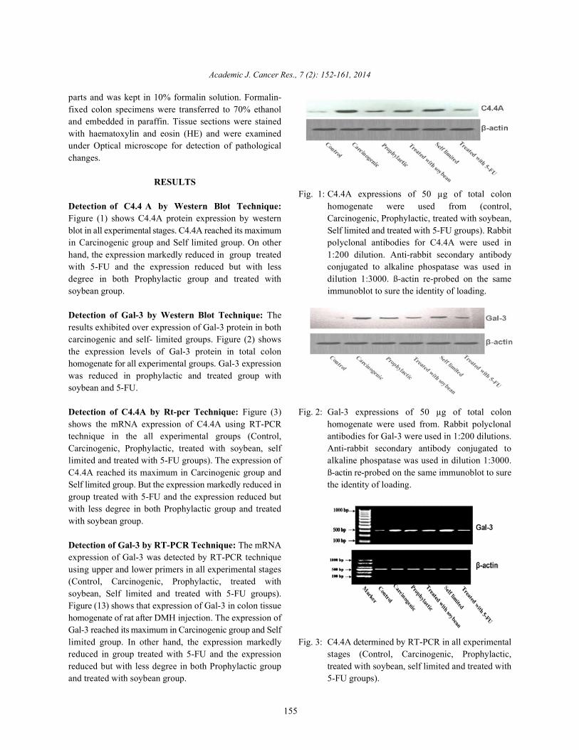
Detection of C4.4 A by Western Blot Technique: homogenate were used from (control,Figure (1) shows C4.4A protein expression by western Carcinogenic, Prophylactic, treated with soybean,blot in all experimental stages. C4.4A reached its maximum Self limited and treated with 5-FU groups). Rabbitin Carcinogenic group and Self limited group. On other polyclonal antibodies for C4.4A were used inhand, the expression markedly reduced in group treated 1:200 dilution. Anti-rabbit secondary antibodywith 5-FU and the expression reduced but with less conjugated to alkaline phospatase was used indegree in both Prophylactic group and treated with dilution 1:3000. ß-actin re-probed on the samesoybean group. immunoblot to sure the identity of loading.
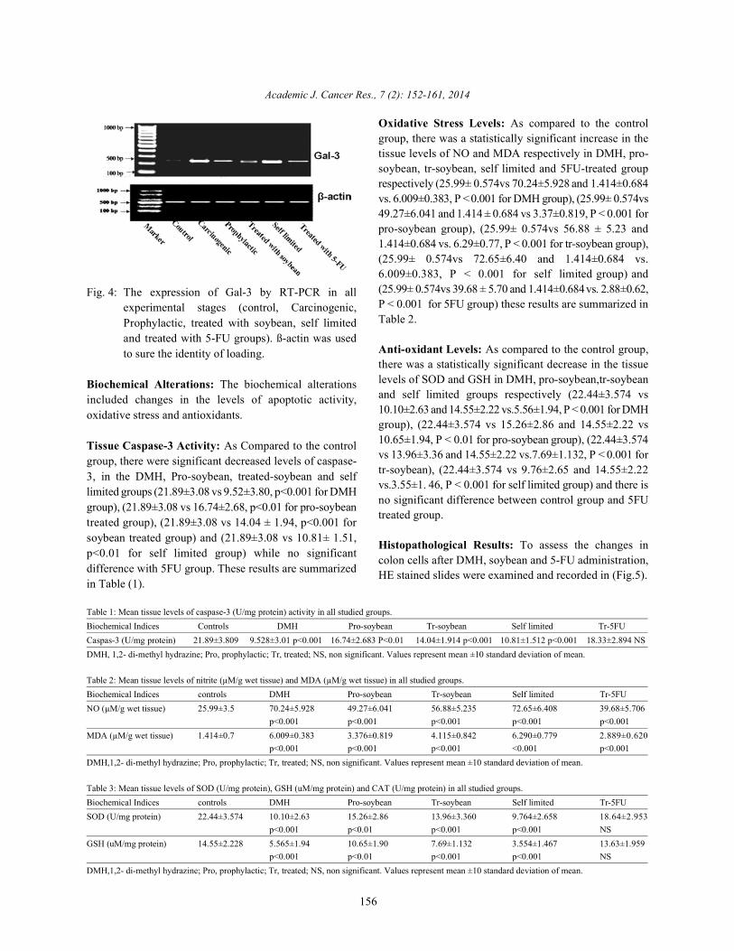
Detection of Gal-3 by Western Blot Technique: Theresults exhibited over expression of Gal-3 protein in bothcarcinogenic and self- limited groups. Figure (2) showsthe expression levels of Gal-3 protein in total colonhomogenate for all experimental groups. Gal-3 expressionwas reduced in prophylactic and treated group withsoybean and 5-FU.
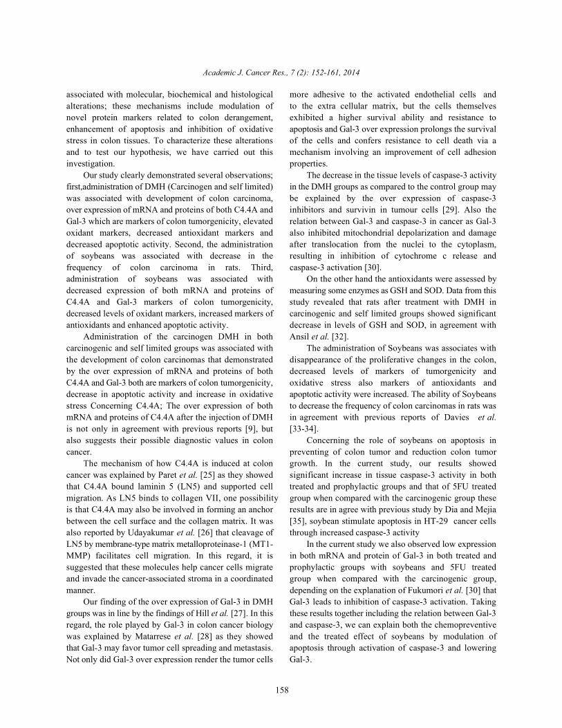
Detection of C4.4A by Rt-pcr Technique: Figure (3) Fig. 2: Gal-3 expressions of 50 µg of total colonshows the mRNA expression of C4.4A using RT-PCR homogenate were used from. Rabbit polyclonaltechnique in the all experimental groups (Control, antibodies for Gal-3 were used in 1:200 dilutions.Carcinogenic, Prophylactic, treated with soybean, self Anti-rabbit secondary antibody conjugated tolimited and treated with 5-FU groups). The expression of alkaline phospatase was used in dilution 1:3000.C4.4A reached its maximum in Carcinogenic group and ß-actin re-probed on the same immunoblot to sureSelf limited group. But the expression markedly reduced in the identity of loading.group treated with 5-FU and the expression reduced butwith less degree in both Prophylactic group and treatedwith soybean group.
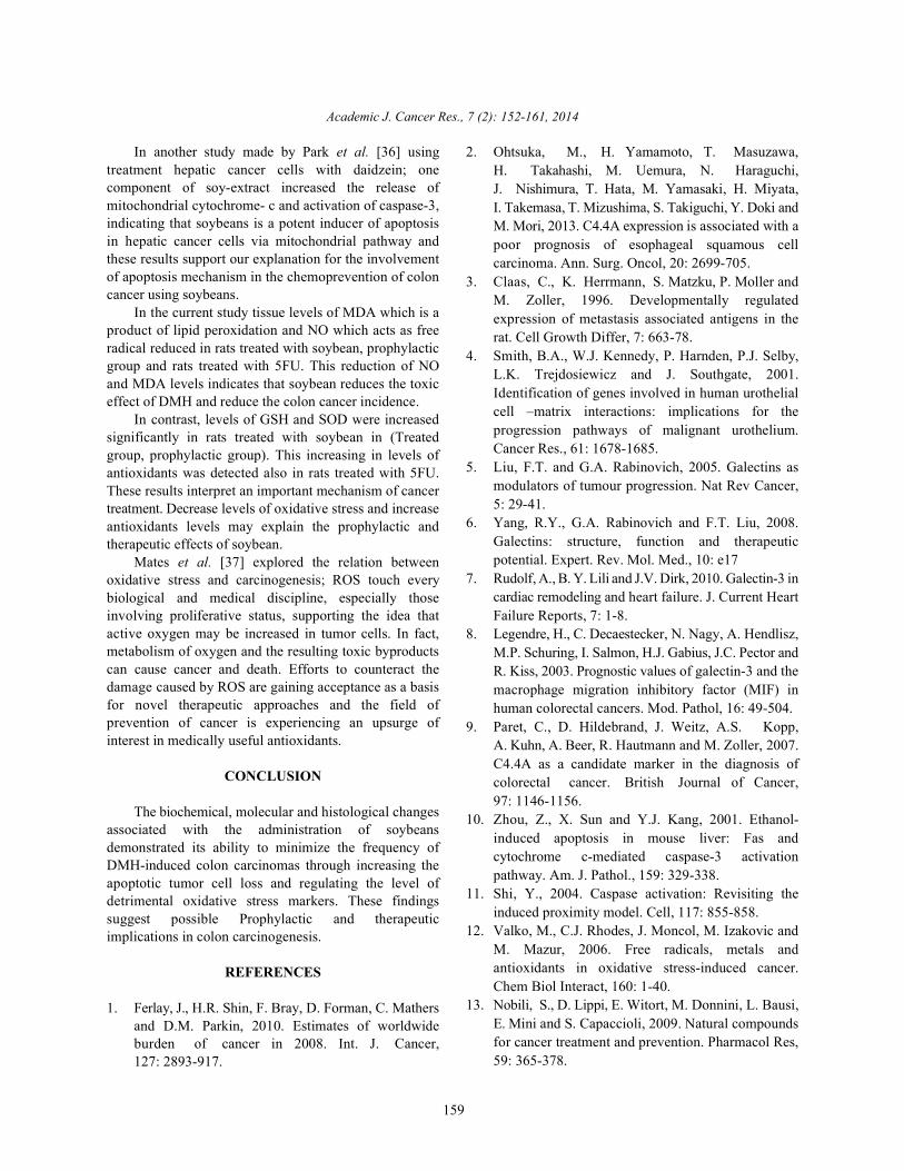
Detection of Gal-3 by RT-PCR Technique: The mRNAexpression of Gal-3 was detected by RT-PCR techniqueusing upper and lower primers in all experimental stages(Control, Carcinogenic, Prophylactic, treated withsoybean, Self limited and treated with 5-FU groups).Figure (13) shows that expression of Gal-3 in colon tissuehomogenate of rat after DMH injection. The expression ofGal-3 reached its maximum in Carcinogenic group and Selflimited group. In other hand, the expression markedly Fig. 3: C4.4A determined by RT-PCR in all experimentalreduced in group treated with 5-FU and the expression stages (Control, Carcinogenic, Prophylactic,reduced but with less degree in both Prophylactic group treated with soybean, self limited and treated withand treated with soybean group. 5-FU groups).
Fig. 1: C4.4A expressions of 50 µg of total colon
Academic J. Cancer Res., 7 (2): 152-161, 2014
156
Fig. 4: The expression of Gal-3 by RT-PCR in allexperimental stages (control, Carcinogenic,Prophylactic, treated with soybean, self limitedand treated with 5-FU groups). ß-actin was usedto sure the identity of loading.
Biochemical Alterations: The biochemical alterationsincluded changes in the levels of apoptotic activity,oxidative stress and antioxidants.
Tissue Caspase-3 Activity: As Compared to the controlgroup, there were significant decreased levels of caspase-3, in the DMH, Pro-soybean, treated-soybean and selflimited groups (21.89±3.08 vs 9.52±3.80, p<0.001 for DMHgroup), (21.89±3.08 vs 16.74±2.68, p<0.01 for pro-soybeantreated group), (21.89±3.08 vs 14.04 ± 1.94, p<0.001 forsoybean treated group) and (21.89±3.08 vs 10.81± 1.51,p<0.01 for self limited group) while no significantdifference with 5FU group. These results are summarizedin Table (1).
Oxidative Stress Levels: As compared to the controlgroup, there was a statistically significant increase in thetissue levels of NO and MDA respectively in DMH, pro-soybean, tr-soybean, self limited and 5FU-treated grouprespectively (25.99± 0.574vs 70.24±5.928 and 1.414±0.684vs. 6.009±0.383, P < 0.001 for DMH group), (25.99± 0.574vs49.27±6.041 and 1.414 ± 0.684 vs 3.37±0.819, P < 0.001 forpro-soybean group), (25.99± 0.574vs 56.88 ± 5.23 and1.414±0.684 vs. 6.29±0.77, P < 0.001 for tr-soybean group),(25.99± 0.574vs 72.65±6.40 and 1.414±0.684 vs.6.009±0.383, P < 0.001 for self limited group) and(25.99± 0.574vs 39.68 ± 5.70 and 1.414±0.684 vs. 2.88±0.62,P < 0.001 for 5FU group) these results are summarized inTable 2.
Anti-oxidant Levels: As compared to the control group,there was a statistically significant decrease in the tissuelevels of SOD and GSH in DMH, pro-soybean,tr-soybeanand self limited groups respectively (22.44±3.574 vs10.10±2.63 and 14.55±2.22 vs.5.56±1.94, P < 0.001 for DMHgroup), (22.44±3.574 vs 15.26±2.86 and 14.55±2.22 vs10.65±1.94, P < 0.01 for pro-soybean group), (22.44±3.574vs 13.96±3.36 and 14.55±2.22 vs.7.69±1.132, P < 0.001 fortr-soybean), (22.44±3.574 vs 9.76±2.65 and 14.55±2.22vs.3.55±1. 46, P < 0.001 for self limited group) and there isno significant difference between control group and 5FUtreated group.
Histopathological Results: To assess the changes incolon cells after DMH, soybean and 5-FU administration,HE stained slides were examined and recorded in (Fig.5).
Table 1: Mean tissue levels of caspase-3 (U/mg protein) activity in all studied groups.Biochemical Indices Controls DMH Pro-soybean Tr-soybean Self limited Tr-5FUCaspas-3 (U/mg protein) 21.89±3.809 9.528±3.01 p<0.001 16.74±2.683 P<0.01 14.04±1.914 p<0.001 10.81±1.512 p<0.001 18.33±2.894 NSDMH, 1,2- di-methyl hydrazine; Pro, prophylactic; Tr, treated; NS, non significant. Values represent mean ±10 standard deviation of mean.
Table 2: Mean tissue levels of nitrite (µM/g wet tissue) and MDA (µM/g wet tissue) in all studied groups. Biochemical Indices controls DMH Pro-soybean Tr-soybean Self limited Tr-5FUNO (µM/g wet tissue) 25.99±3.5 70.24±5.928 49.27±6.041 56.88±5.235 72.65±6.408 39.68±5.706
p<0.001 p<0.001 p<0.001 p<0.001 p<0.001MDA (µM/g wet tissue) 1.414±0.7 6.009±0.383 3.376±0.819 4.115±0.842 6.290±0.779 2.889±0.620
p<0.001 p<0.001 p<0.001 <0.001 p<0.001DMH,1,2- di-methyl hydrazine; Pro, prophylactic; Tr, treated; NS, non significant. Values represent mean ±10 standard deviation of mean.
Table 3: Mean tissue levels of SOD (U/mg protein), GSH (uM/mg protein) and CAT (U/mg protein) in all studied groups. Biochemical Indices controls DMH Pro-soybean Tr-soybean Self limited Tr-5FUSOD (U/mg protein) 22.44±3.574 10.10±2.63 15.26±2.86 13.96±3.360 9.764±2.658 18.64±2.953
p<0.001 p<0.01 p<0.001 p<0.001 NSGSH (uM/mg protein) 14.55±2.228 5.565±1.94 10.65±1.90 7.69±1.132 3.554±1.467 13.63±1.959
p<0.001 p<0.01 p<0.001 p<0.001 NSDMH,1,2- di-methyl hydrazine; Pro, prophylactic; Tr, treated; NS, non significant. Values represent mean ±10 standard deviation of mean.

Academic J. Cancer Res., 7 (2): 152-161, 2014
157
Table 4: Correlation between biochemical parameters (ALT, AST, Urea, Albumin, Total tissue proteins, Total tissue lipid, NO, MDA,SOD and GSH).
Biochemical indices ALT AST Urea albumin Total tissue proteins Total tissue lipid NO MDA Caspas-3 SOD GSH
ALT ----- 0.83283 -0.8334 -0.7751 -0.89076 0.81995 0.87297 0.87322 -0.78002 -0.78101 -0.79565AST 0.83283 ----- -0.7419 -0.6608 -0.87081 0.86560 0.82984 0.85006 -0.76844 -0.76092 -0.83199Urea -0.8334 -0.7419 ----- 0.74501 0.74501 -0.74769 -0.72362 -0.73512 0.74569 0.63229 0.69108albumin -0.7751 -0.6608 0.74501 ----- 0.76438 -0.70051 -0.75414 -0.77758 0.62423 0.65931 0.69108Total tissue proteins -0.8907 -0.8708 0.74501 0.76438 ----- -0.828103 -0.86323 -0.85554 0.78852 0.75717 0.84929Total tissue lipid 0.81995 0.86560 -0.7476 -0.7005 -0.828103 ----- 0.83992 0.80352 -0.74792 -0.78617 -0.83691NO 0.87297 0.82984 -0.7236 -0.7541 -0.86323 0.83992 ----- 0.89800 -0.84152 -0.78379 -0.86815MDA 0.87322 0.85006 -0.7351 -0.7775 -0.85554 0.80352 0.89800 ----- -0.80679 -0.77438 -0.81881Caspas-3 -0.7800 -0.7684 0.63229 0.62423 0.78852 -0.74792 -0.84152 -0.80679 ----- 0.76002 0.72438SOD -0.7810 -0.7609 0.63229 0.65931 0.75717 -0.78617 -0.78379 -0.77438 0.76002 ----- 0.76878GSH -0.7957 -0.8319 0.69108 0.69108 0.84929 -0.83691 -0.86815 -0.81881 0.72438 0.76878 -----
Fig . 5: Histopathological changes in colon tissues. (Ia) Colon section from rats in the control group show normalhistological structure (H&E. X 40). IIa) Colon section from rats in the carcinogenic group showing moreprogressive colon adinocarcinoma, in which the tumor cell (arrows) infiltrating all layers the colon and replacingthe normal structure (H&E. X 40). IIIa) Colon section from rats in the prophylactic group showing more or lossnormal histological appearance (H&E. X 40). IVa) Colon section from rats in the treated group showinginfiltrating tumor cells undergoing either necrosis (arrow) or apoptosis (double arrows) with infiltration of mastcells (arrow head) (H&E. X 40). Va) Colon section from rats in self limited group showing colon adinocarcinomawith tumor cell necrosis and heavy infiltration with mast cells (arrow heads) (H&E. X 40). VIa) Colon section fromrats in the 5 FU group showing sever necrosis associated with sloughing of the necrosed tumor cells (arrows)(H&E. X 40).
Examination of colon section from rats in the control section from rats in the 5 FU group showed sever necrosisgroup showed normal histological structure (Fig.5,Ia). associated with necrosis and sloughing of the tumor cellsExamination of colon section from rats in the carcinogenic (Fig.5, VIa).group revealed colon adinocarcinoma with proliferation oftumor cell from the epithelial layer infiltrating and Statistical Analysis: Statistical analysis was achievedreplacing the colon histological structure. The tumor cells using Graph Pad In Stat. software Inc, Program, versionwere large polyhedral cell with hyperchromatic nuclei 4.0 Philadelphia, San Diego CA, (2003). Data were(Fig.5, IIa). Examination of colon section from rats in the presented as mean ± SD and the levels of significanceprophylactic group show more or less normal histological were accepted with p <0.05. Multiple comparisons wereappearance (Fig.5, IIIa). Examination of colon section from done using one way ANOVA followed by Tukey-Kramerrats in the treated group showed neoplastic cells test as multiple comparison post ANOVA test.undergoing either necrosis or apoptosis with infiltrationof mast cells (Fig.5, IVa). Examination of colon section DISCUSSIONfrom rats in the self limited group showed colonadinocarcinoma with tumor cell necrosis and heavy In this investigation, we hypothesized that in coloninfiltration with mast cells (Fig.5, Va). Examination of colon carcinogenesis, the administration of soybeans is
Academic J. Cancer Res., 7 (2): 152-161, 2014
158
associated with molecular, biochemical and histological more adhesive to the activated endothelial cells andalterations; these mechanisms include modulation of to the extra cellular matrix, but the cells themselvesnovel protein markers related to colon derangement, exhibited a higher survival ability and resistance toenhancement of apoptosis and inhibition of oxidative apoptosis and Gal-3 over expression prolongs the survivalstress in colon tissues. To characterize these alterations of the cells and confers resistance to cell death via aand to test our hypothesis, we have carried out this mechanism involving an improvement of cell adhesioninvestigation. properties.
Our study clearly demonstrated several observations; The decrease in the tissue levels of caspase-3 activityfirst,administration of DMH (Carcinogen and self limited) in the DMH groups as compared to the control group maywas associated with development of colon carcinoma, be explained by the over expression of caspase-3over expression of mRNA and proteins of both C4.4A and inhibitors and survivin in tumour cells [29]. Also theGal-3 which are markers of colon tumorgenicity, elevated relation between Gal-3 and caspase-3 in cancer as Gal-3oxidant markers, decreased antioxidant markers and also inhibited mitochondrial depolarization and damagedecreased apoptotic activity. Second, the administration after translocation from the nuclei to the cytoplasm,of soybeans was associated with decrease in the resulting in inhibition of cytochrome c release andfrequency of colon carcinoma in rats. Third, caspase-3 activation [30]. administration of soybeans was associated with On the other hand the antioxidants were assessed bydecreased expression of both mRNA and proteins of measuring some enzymes as GSH and SOD. Data from thisC4.4A and Gal-3 markers of colon tumorgenicity, study revealed that rats after treatment with DMH indecreased levels of oxidant markers, increased markers of carcinogenic and self limited groups showed significantantioxidants and enhanced apoptotic activity. decrease in levels of GSH and SOD, in agreement with
Administration of the carcinogen DMH in both Ansil et al. [32].carcinogenic and self limited groups was associated with The administration of Soybeans was associates withthe development of colon carcinomas that demonstrated disappearance of the proliferative changes in the colon,by the over expression of mRNA and proteins of both decreased levels of markers of tumorgenicity andC4.4A and Gal-3 both are markers of colon tumorgenicity, oxidative stress also markers of antioxidants anddecrease in apoptotic activity and increase in oxidative apoptotic activity were increased. The ability of Soybeansstress Concerning C4.4A; The over expression of both to decrease the frequency of colon carcinomas in rats wasmRNA and proteins of C4.4A after the injection of DMH in agreement with previous reports of Davies et al.is not only in agreement with previous reports [9], but [33-34].also suggests their possible diagnostic values in colon Concerning the role of soybeans on apoptosis incancer. preventing of colon tumor and reduction colon tumor
The mechanism of how C4.4A is induced at colon growth. In the current study, our results showedcancer was explained by Paret et al. [25] as they showed significant increase in tissue caspase-3 activity in boththat C4.4A bound laminin 5 (LN5) and supported cell treated and prophylactic groups and that of 5FU treatedmigration. As LN5 binds to collagen VII, one possibility group when compared with the carcinogenic group theseis that C4.4A may also be involved in forming an anchor results are in agree with previous study by Dia and Mejiabetween the cell surface and the collagen matrix. It was [35], soybean stimulate apoptosis in HT-29 cancer cellsalso reported by Udayakumar et al. [26] that cleavage of through increased caspase-3 activity LN5 by membrane-type matrix metalloproteinase-1 (MT1- In the current study we also observed low expressionMMP) facilitates cell migration. In this regard, it is in both mRNA and protein of Gal-3 in both treated andsuggested that these molecules help cancer cells migrate prophylactic groups with soybeans and 5FU treatedand invade the cancer-associated stroma in a coordinated group when compared with the carcinogenic group,manner. depending on the explanation of Fukumori et al. [30] that
Our finding of the over expression of Gal-3 in DMH Gal-3 leads to inhibition of caspase-3 activation. Takinggroups was in line by the findings of Hill et al. [27]. In this these results together including the relation between Gal-3regard, the role played by Gal-3 in colon cancer biology and caspase-3, we can explain both the chemopreventivewas explained by Matarrese et al. [28] as they showed and the treated effect of soybeans by modulation ofthat Gal-3 may favor tumor cell spreading and metastasis. apoptosis through activation of caspase-3 and loweringNot only did Gal-3 over expression render the tumor cells Gal-3.
Academic J. Cancer Res., 7 (2): 152-161, 2014
159
In another study made by Park et al. [36] using 2. Ohtsuka, M., H. Yamamoto, T. Masuzawa,treatment hepatic cancer cells with daidzein; onecomponent of soy-extract increased the release ofmitochondrial cytochrome- c and activation of caspase-3,indicating that soybeans is a potent inducer of apoptosisin hepatic cancer cells via mitochondrial pathway andthese results support our explanation for the involvementof apoptosis mechanism in the chemoprevention of coloncancer using soybeans.
In the current study tissue levels of MDA which is aproduct of lipid peroxidation and NO which acts as freeradical reduced in rats treated with soybean, prophylacticgroup and rats treated with 5FU. This reduction of NOand MDA levels indicates that soybean reduces the toxiceffect of DMH and reduce the colon cancer incidence.
In contrast, levels of GSH and SOD were increasedsignificantly in rats treated with soybean in (Treatedgroup, prophylactic group). This increasing in levels ofantioxidants was detected also in rats treated with 5FU.These results interpret an important mechanism of cancertreatment. Decrease levels of oxidative stress and increaseantioxidants levels may explain the prophylactic andtherapeutic effects of soybean.
Mates et al. [37] explored the relation betweenoxidative stress and carcinogenesis; ROS touch everybiological and medical discipline, especially thoseinvolving proliferative status, supporting the idea thatactive oxygen may be increased in tumor cells. In fact,metabolism of oxygen and the resulting toxic byproductscan cause cancer and death. Efforts to counteract thedamage caused by ROS are gaining acceptance as a basisfor novel therapeutic approaches and the field ofprevention of cancer is experiencing an upsurge ofinterest in medically useful antioxidants.
CONCLUSION
The biochemical, molecular and histological changesassociated with the administration of soybeansdemonstrated its ability to minimize the frequency ofDMH-induced colon carcinomas through increasing theapoptotic tumor cell loss and regulating the level ofdetrimental oxidative stress markers. These findingssuggest possible Prophylactic and therapeuticimplications in colon carcinogenesis.
REFERENCES
1. Ferlay, J., H.R. Shin, F. Bray, D. Forman, C. Mathersand D.M. Parkin, 2010. Estimates of worldwideburden of cancer in 2008. Int. J. Cancer,127: 2893-917.
H. Takahashi, M. Uemura, N. Haraguchi,J. Nishimura, T. Hata, M. Yamasaki, H. Miyata,I. Takemasa, T. Mizushima, S. Takiguchi, Y. Doki andM. Mori, 2013. C4.4A expression is associated with apoor prognosis of esophageal squamous cellcarcinoma. Ann. Surg. Oncol, 20: 2699-705.
3. Claas, C., K. Herrmann, S. Matzku, P. Moller andM. Zoller, 1996. Developmentally regulatedexpression of metastasis associated antigens in therat. Cell Growth Differ, 7: 663-78.
4. Smith, B.A., W.J. Kennedy, P. Harnden, P.J. Selby,L.K. Trejdosiewicz and J. Southgate, 2001.Identification of genes involved in human urothelialcell –matrix interactions: implications for theprogression pathways of malignant urothelium.Cancer Res., 61: 1678-1685.
5. Liu, F.T. and G.A. Rabinovich, 2005. Galectins asmodulators of tumour progression. Nat Rev Cancer,5: 29-41.
6. Yang, R.Y., G.A. Rabinovich and F.T. Liu, 2008.Galectins: structure, function and therapeuticpotential. Expert. Rev. Mol. Med., 10: e17
7. Rudolf, A., B. Y. Lili and J.V. Dirk, 2010. Galectin-3 incardiac remodeling and heart failure. J. Current HeartFailure Reports, 7: 1-8.
8. Legendre, H., C. Decaestecker, N. Nagy, A. Hendlisz,M.P. Schuring, I. Salmon, H.J. Gabius, J.C. Pector andR. Kiss, 2003. Prognostic values of galectin-3 and themacrophage migration inhibitory factor (MIF) inhuman colorectal cancers. Mod. Pathol, 16: 49-504.
9. Paret, C., D. Hildebrand, J. Weitz, A.S. Kopp,A. Kuhn, A. Beer, R. Hautmann and M. Zoller, 2007.C4.4A as a candidate marker in the diagnosis ofcolorectal cancer. British Journal of Cancer,97: 1146-1156.
10. Zhou, Z., X. Sun and Y.J. Kang, 2001. Ethanol-induced apoptosis in mouse liver: Fas andcytochrome c-mediated caspase-3 activationpathway. Am. J. Pathol., 159: 329-338.
11. Shi, Y., 2004. Caspase activation: Revisiting theinduced proximity model. Cell, 117: 855-858.
12. Valko, M., C.J. Rhodes, J. Moncol, M. Izakovic andM. Mazur, 2006. Free radicals, metals andantioxidants in oxidative stress-induced cancer.Chem Biol Interact, 160: 1-40.
13. Nobili, S., D. Lippi, E. Witort, M. Donnini, L. Bausi,E. Mini and S. Capaccioli, 2009. Natural compoundsfor cancer treatment and prevention. Pharmacol Res,59: 365-378.
Academic J. Cancer Res., 7 (2): 152-161, 2014
160
14. Macdonald, R.S., J.Y. Guo, J. Copeland, 24. Marklund, S.L., 1985. Supeoxide dismutaseJ.D. Browning, J.D. Sleper, G.E. Rottinghaus andM.A. Berhow, 2005. Environmental Influences onIsoflavones and Saponins in Soybeans and TheirRole in Colon Cancer. J. Nutr., 135: 1239-1242.
15. Philip, J.M., E. Kornelia, E.M. James and A.F. Frank,2004. Soybean impairs tumor suppressor p53function in colon cancer cells. Carcinogenesis,25: 1611-1617.
16. Oh, Y. and M. Sung, 2001. Soybean saponins inhibitcell proliferation by suppressing PKC activation andinduce differentiation of HT-29 human colonadenocarcinoma cells. Nutr. Cancer, 39: 132-138.
17. Raju, J., A. Bielecki, D. Caldwell, E. Lok, M. Taylor,K. Kapal, L. Curran, G.M. Cooke, R.P. Bird and R.Mehta, 2009. Soy isoflavones modulateazoxymethane-induced rat colon carcinogenesisexposed pre- and postnatally and inhibit growth ofDLD-1 human colon adenocarcinoma cells byincreasing the expression of estrogen receptor-beta.The Journal of Nutrition, 139: 474-481.
18. Hiromichi, S. and J.W. Michael, 1990.Chemoprevention of 1,2-Dimethylhydrazine-inducedColon Cancer in Mice by Naturally OccurringOrganosulfur. Cancer Res., 50: 5084-5087.
19. Ragunath, M., T. Prabu, S. Nadanasabapathy, R.Revathi and V. Manju, 2013. Synergistic andindividual effects of umbelliferone with 5-flurouracilon the status of lipid peroxidation and antioxidantdefense against 1,2-dimethylhydrazine induced ratcolon carcinogenesis. Biomedicine and PreventiveNutrition, 3: 74-82.
20. Van Bezooijen, R.L., I. Que, A.G. Ederveen,H.J. Kloosterboer, S.E. Papapoulos and C.W. Lowik,1998. Plasma nitrate and nitrite level are regulated byovarian steroids but do not correlate with trabecularbone mineral density in rats. J. Endocrinology,159: 27-34.
21. Buege, J.A. and S.D. Aust, 1978. Microsomal lipidperoxidation, methods enzymology, modified byadding the bht solution to prevent further lipidperoxidation during boiling, 52: 302-10.
22. Kim, H.S., D.B. Hausman, M.M. Comton, R.G. Dean,G.J. Hausman, D.L. Hartzell and C.A. Baile, 2000.Induction of apoptosis by all trans retinoic acid andC2 ceramide treatment in rat stromal vascular culture.Biochem. Biophys. Res. Comm., 270: 76-80.
23. Ellman, G.L., 1959. Tissue sulfhydryl groups. Arch.Biochem. Biophys, 82: 70-79.
isoenzyme in tissue and plasma from New Zealandblack mice. Mutat. Res., 148: 129-134.
25. Paret, C., M. Bourouba, A. Beer, K. Miyazaki,M. Schno¨lzer, S. Fiedler and M. Zo¨ller, 2005. Ly6family member C4.4A binds laminins 1 and 5,associates with galectin-3 and supports cellmigration. Int. J. Cancer, 115: 724-733.
26. Udayakumar, T.S., M.L. Chen, E.l. Bair, D.C. VonBredow, A.E. Cress, R.B. Nagle and G.T. Bowden,2003. Membrane type-1-matrix metalloproteinaseexpressed by prostate carcinoma cells cleaves humanlaminin-5 beta3 chain and induces cell migration.Cancer Res., 63: 2292- 2299.
27. Hill, M., D. Mazal, V.A. Biron, L. Pereira, L. Ubillos,E. Berriel, H. Ahmed, T. Freire, M. Rondán,G.R. Vasta, F.T. Liu, M.M. Iglesias and E. Osinaga,2010. A novel clinically relevant animal model forstudying galectin-3 and its ligands during coloncarcinogenesis. J. Histochem Cytochem,58: 553-65.
28. Matarrese, P., O. Fusco, N. Tinari, C. Natoli, F.T.Liu, M.L. Semeraro, W. Malorni and S. Iacobelli S,2000. Galectin-3 overexpression protects fromapoptosis by improving cell adhesion properties. Int.J. Cancer, 85: 545-554.
29. Krajewski, S., M. Krajewska, B.C. Turner, C. Pratt,B. Howard, J.M. Zapata, V. Frenkel, S. Robertson,Y. Ionov, H. Yamamoto, M. Perucho, S. Takayamaand J.C. Reed, 1999. Prognostic significance ofapoptosis regulators in breast cancer. Endocr. Relat.Cancer, 6: 29-40.
30. Fukumori, T., N. Oka, Y. Takenaka, P. Nangia-Makker,E. Elsamman, T. Kasai, M. Shono, H.O.Kanayama, J. Ellerhorst, R. Lotan and A. Raz 2006.Galectin-3 regulates mitochondrial stability andantiapoptotic function in response to anticancer drugin prostate cancer. Cancer Res., 66: 3114-9.
31. Slattery, M.L., A. Lundgreen, B. Welbourn,R.K. Wolff and C. Corcoran, 2012. Oxidative balanceand colon and rectal cancer: interaction of lifestylefactors and genes. Mutat Res., 734: 30-40.
32. Ansil, P.N., S.P. Prabha, A. Nitha and M.S. Latha,2013. Chemopreventive effect of Amorphophalluscampanulatus (Roxb.) blume tuber against aberrantcrypt foci and cell proliferation in 1,2-dimethylhydrazine induced colon carcinogenesis.Asian Pac J. Cancer Prev., 14: 5331-9.
Academic J. Cancer Res., 7 (2): 152-161, 2014
161
33. Davies, M.J., E.A. Bowey, H. Adlercreutz, 36. Park, H.J., Y.K. Jeon, H.D. You and M.J. Nam, 2013.I.R. Rowland and P.C. Rumsby, 1999. Effects of soy Daidzein causes cytochrome c mediated apoptosisor rye supplementation of high-fat diets on colon via the Bcl-2 family in human hepatic cancer cells.tumour development in azoxymethane-treated rats. Food Chem Toxicol, 60: 542-9.Carcinogenesis, 20: 927-31. 37. Mates, J.M., J.A. Segura, F.J. Alonso and J. Marquez,
34. Li, Q.S., C.Y. Li, Z.L. Li and H.L. Zhu, 2012. 2012. Oxidative stress in apoptosis and cancer: anGenistein and its synthetic analogs as anticancer update. Arch Toxicol, 86: 1649-65.agents. Anticancer Agents Med. Chem., 12: 271-81.
35. Dia, V.P. and E.G. Mejia, 2010. Lunasin promotesapoptosis in human colon cancer cells bymitochondrial pathway activation and induction ofnuclear clusterin expression. Cancer Lett., 295: 44-53.
Copyright © 2022 FDOKUMEN