Binding ability of Allura Red with food proteins and its impact on protein digestibility
6
Binding ability of Allura Red with food proteins and its impact on protein digestibility S. Umer Abdullah a , Muhammad Badaruddin a , S. Asad Sayeed b , Rashida Ali a , Mian N. Riaz c, * a Division of Food Research, H.E.J. Research Institute of Chemistry, University of Karachi, Karachi, Pakistan b Department of Food Science and Technology, University of Karachi, Karachi, Pakistan c Food Protein R&D Center, Texas A and M University, College Station, TX 77843, United States Received 3 December 2007; received in revised form 4 February 2008; accepted 14 February 2008 Abstract Allura Red-40 is a safe colour additive (permissible by the FDA and Health Canada) that is used in a variety of foods to make them more attractive and appealing for consumers. However, limited information is available about its binding to macronutrients that are responsible for its uniform distribution in food products. In the present study, the binding capacity of Allura with food proteins is com- pared with Coomassie Brillant Blue R 250, which is an established staining agent for visualizing electrophoretically resolved proteins. The data illustrate that Allura is a fast reacting dye and binds with a variety of food proteins including peanut, rice bran, garlic and mixture of proteins [(Takadiastase, nisin, a microbial protein and bovine serum albumin (BSA)]. The Allura bound proteins retained their colour at high and low temperatures and in a wide range of pH. The experiments on the resolution of proteins and staining with Allura have shown that the dye is highly sensitive, rapid, lasting and is easily linked with a variety of proteins. The binding of Allura to various proteins had almost no adverse effect on protein digestibility, as predicted by in vitro digestibility determinations. Ó 2008 Elsevier Ltd. All rights reserved. Keywords: Allura; Protein binding; PAGE; Protein digestibility 1. Introduction Colour plays a very important role in choice and quality perception of food by appealing to the consumers. The col- our of a food is a primary food quality characteristic, set naturally by a consumer of any age or group. That is why food processing industries are always anxious about the right quality and standard of food colourant to be used for decoration of food for consumer’s attraction. Even the natural unprocessed foods are judged by their colour inten- sity that reflects the maturity and health of fruits and other commodities. Most of the natural colours belong to the either antho- cyanin or carotene families in general, which are heat labile and are faded during processing. One of the reasons to add synthetic colours, which are heat resistent like Allura Red, is to overcome such losses. Commercial colourants are added to give natural appealing look to foods. Allura has been used as a food colourant for decades (Anonymous., 2003) however, the nature of its binding to food compo- nents has not been fully investigated. The uniform distribu- tion of a colourant in food systems is an authentic indication that a strong affinity and stable complexation between the dye and a ligand exists throughout the food processing and storage. Allura belongs to the monoazo class of food colourants and is identified as C1 Food Red 17, Food Drug & Cos- matic (FD&C) Red No. 40, C1 (1975) No. 16035, INS No. 129. Chemically it is disodium 6-hydroxy-5-(2 meth- oxy-5-methyl-4-sulphophenylazo)-2-naphthalenesulpho- nate with a molecular weight of 496. It is commercially 0308-8146/$ - see front matter Ó 2008 Elsevier Ltd. All rights reserved. doi:10.1016/j.foodchem.2008.02.049 * Corresponding author. Tel.: +1 979 845 2774; fax: +1 979 458 0019. E-mail address: [email protected] (M.N. Riaz). www.elsevier.com/locate/foodchem Available online at www.sciencedirect.com Food Chemistry 110 (2008) 605–610
-
Upload
independent -
Category
Documents
-
view
1 -
download
0
Transcript of Binding ability of Allura Red with food proteins and its impact on protein digestibility

Available online at www.sciencedirect.com
www.elsevier.com/locate/foodchem
Food Chemistry 110 (2008) 605–610
Binding ability of Allura Red with food proteinsand its impact on protein digestibility
S. Umer Abdullah a, Muhammad Badaruddin a, S. Asad Sayeed b,Rashida Ali a, Mian N. Riaz c,*
a Division of Food Research, H.E.J. Research Institute of Chemistry, University of Karachi, Karachi, Pakistanb Department of Food Science and Technology, University of Karachi, Karachi, Pakistan
c Food Protein R&D Center, Texas A and M University, College Station, TX 77843, United States
Received 3 December 2007; received in revised form 4 February 2008; accepted 14 February 2008
Abstract
Allura Red-40 is a safe colour additive (permissible by the FDA and Health Canada) that is used in a variety of foods to make themmore attractive and appealing for consumers. However, limited information is available about its binding to macronutrients that areresponsible for its uniform distribution in food products. In the present study, the binding capacity of Allura with food proteins is com-pared with Coomassie Brillant Blue R 250, which is an established staining agent for visualizing electrophoretically resolved proteins.The data illustrate that Allura is a fast reacting dye and binds with a variety of food proteins including peanut, rice bran, garlic andmixture of proteins [(Takadiastase, nisin, a microbial protein and bovine serum albumin (BSA)]. The Allura bound proteins retainedtheir colour at high and low temperatures and in a wide range of pH. The experiments on the resolution of proteins and staining withAllura have shown that the dye is highly sensitive, rapid, lasting and is easily linked with a variety of proteins. The binding of Allura tovarious proteins had almost no adverse effect on protein digestibility, as predicted by in vitro digestibility determinations.� 2008 Elsevier Ltd. All rights reserved.
Keywords: Allura; Protein binding; PAGE; Protein digestibility
1. Introduction
Colour plays a very important role in choice and qualityperception of food by appealing to the consumers. The col-our of a food is a primary food quality characteristic, setnaturally by a consumer of any age or group. That iswhy food processing industries are always anxious aboutthe right quality and standard of food colourant to be usedfor decoration of food for consumer’s attraction. Even thenatural unprocessed foods are judged by their colour inten-sity that reflects the maturity and health of fruits and othercommodities.
Most of the natural colours belong to the either antho-cyanin or carotene families in general, which are heat labile
0308-8146/$ - see front matter � 2008 Elsevier Ltd. All rights reserved.
doi:10.1016/j.foodchem.2008.02.049
* Corresponding author. Tel.: +1 979 845 2774; fax: +1 979 458 0019.E-mail address: [email protected] (M.N. Riaz).
and are faded during processing. One of the reasons to addsynthetic colours, which are heat resistent like Allura Red,is to overcome such losses. Commercial colourants areadded to give natural appealing look to foods. Allura hasbeen used as a food colourant for decades (Anonymous.,2003) however, the nature of its binding to food compo-nents has not been fully investigated. The uniform distribu-tion of a colourant in food systems is an authenticindication that a strong affinity and stable complexationbetween the dye and a ligand exists throughout the foodprocessing and storage.
Allura belongs to the monoazo class of food colourantsand is identified as C1 Food Red 17, Food Drug & Cos-matic (FD&C) Red No. 40, C1 (1975) No. 16035, INSNo. 129. Chemically it is disodium 6-hydroxy-5-(2 meth-oxy-5-methyl-4-sulphophenylazo)-2-naphthalenesulpho-nate with a molecular weight of 496. It is commercially
606 S. Umer Abdullah et al. / Food Chemistry 110 (2008) 605–610
available as dark red or maroon powder or granules, whichare soluble in water but insoluble in ethanol. The jointFAO/WHO Expert Committee on Food Additives has sug-gested acceptable daily intake (ADI) for Allura as 7 mg/kg/day (Ito, 2000). Allura is reduced by human intestinalanaerobes (Chung, Fulk, & Egan, 1978) and is approvedby FDA and Health Canada as a nontoxic food colourant.
The dye can easily be isolated and estimated in foodproducts by spectrophotometric determination at 504 nmand reverse phase ion pair high-performance liquid chro-matography (HPLC) (Lancaster & Lawrence, 1991; Rich-field-Fratz, Baczynskyj, Miller, & Bailey, 1989). The foodcolours are also estimated by capillary electrophoresis innano quantities (Huang, Shih, & Chen, 2002), and recentlythree new techniques including adsorptive stripping vol-tammetry (Alghamdi, 2005), microemulsion electrokineticchromatography (Huang, Chuang, Chiu, & Chung, 2005)and complex spectral-pH three-way array (Marsili,Lista, Band, Goicoechea, & Olivieri, 2005) have beendescribed for quantitative analysis of food colours includ-ing Allura.
The purpose of this investigation was to study the bind-ing capacity of Allura with food proteins. Allura was usedfor staining proteins such as peanut, garlic, and rice branproteins (RBP), and a mixture of proteins (Takadiastase,BSA, and nisin, a microbial protein often used as a preser-vative in variety of foods), and was then separated onsodium dodecyl sulphate–polyacrylamide gel electrophore-sis (SDS–PAGE) just to prove that it exclusively binds withprotein molecules in the complex system of different foods.The garlic extract being acidic in nature has better keepingquality and was separated well on acrylamide gel, garlicextract contains garlic proteins, especially lectins (Ahmad,Pischetsrieder, & Ahmed, 2007; Ghananfari, Hassan, &Khamesipour, 2006; Smeets et al., 1997; Wang & Ng,2001). Furthermore, the Allura–protein complexes werestudied to assess the effect of colour binding on proteindigestibility.
2. Materials and methods
2.1. Materials
The N,N0-methylene-bis-acrylamide was purchased fromScharlau (Scharlau Chemie, Barcelona, SPAIN) while Tris(hydroxymethyl aminomethane) was obtained fromResearch Organics (Research Organics, Inc. Cleveland,OH, USA). Sodium dodecyl sulphate (SDS), acrylamide,ammonium peroxodisulphate (APS), glycine, coomassie(coomassie brilliant blue R 250), BSA, N-tetramethylethyl-enediamine (TEMED), bromophenol blue, trypsin andprotease were supplied from Merck (Darmstadt, Ger-many), while Nisaplin (nisin) was purchased from SuzhouHengliang Imp and Exp, Jiangsu, China, and 2-mercap-toethanol was supplied from Riedel-deHaen (Riedal-deHaen AG Seelze, Germany). The ficin and bromelain weregiven as a gift from the Department of Food Science, Uni-
versity of California, Davis, while milk powder, liquid milkand eggs were purchased from Top ten super market, Kar-achi, Pakistan, and protease was obtained from sigma(Louis, USA). Allura (Allura Red – 40) was obtained fromNational Foods (Pvt.) Ltd. Karachi, Pakistan as a gift. Theother chemicals used were of analytical grade. All the solu-tions were prepared in doubled distilled deionized (DDD)water.
2.2. Electrophoresis
2.2.1. Preparation of slab gel
The following solutions were first prepared to prepare aGel for SDS.
Acrylamide (30 g) and bis-acrylamide (0.80 g) were dis-solved in 100 mL water and filtered through Whatman fil-ter paper no. 1 to prepare a solution A. Tris–HCl buffer(1.5 M) was prepared by mixing 18.02 g of Tris dissolvedin 80 mL of water and pH was adjusted to 8.8 by using1 M HCl, and the volume was finally made up to100 mL. Tris (12.114 g) was dissolved in 80 mL water, thepH was adjusted to 6.8 with 1 M HCl and the volumewas increased to 100 mL with water to prepare its solution.SDS (10 g) was dissolved in water and the volume wasmade up to 100 mL to make SDS solution. APS (1 g)was dissolved in water and the volume was increased to10 mL, while TEMED was used as supplied.
The 12.5% slab gel of thickness 0.75 mm was preparedby using 2.083 mL of solution A, 0.666 mL of solution B,0.500 mL of solution D and 0.250 mL of solution E. Themixture was degassed and 1.7 lL of TEMED was addedbefore use. The gel was left for 15 min for settling.
2.2.2. Staking gel
Solutions were added in the ratio of0.416:0.830:0.333:0.166, respectively. The mixture wasdegassed immediately, and finally 1.7 lL of TEMED wereadded before the pouring of the stacking gel. The wellswere prewashed with a reservoir buffer after the gel wassettled.
Reservoir buffer: Tris (0.9 g), glycine (3.6 g) and SDS(1 g) were dissolved in 50 mL water and the volume wasmade up to 1 L.
2.2.3. Staining solutions
A. Coomassie (0.2 g) was dissolved in 7.5 mL of glacialacetic acid and 5 mL of methanol. The volume wasincreased to 100 mL with water and filtered to avoid anycontamination and stored at room temperature (R).
B. Allura: The solution was prepared by dissolving 0.2 gof the dye in 7.5 mL of glacial acetic acid and 5 mL ofmethanol. The volume was made up to 100 mL with water(R).
C. Allura (acidic): The staining solution B (25 mL) wasadjusted at pH 2.0 by using 2 N HCl (R).
D. Allura (basic). The staining solution B (25 mL) wasadjusted at pH 10.0 by using 5 N NaOH (R).

S. Umer Abdullah et al. / Food Chemistry 110 (2008) 605–610 607
2.2.4. Destaining solution
Destaining solution was prepared by mixing 10 mL ofglacial acetic acid and 30 mL of methanol together. Thevolume was increased up to 100 ml with water (R). Allthe solutions were stored at 4 �C in the dark to preventauto-oxidation, except where (R) is given at the end ofthe preparation method, which represents storage at roomtemperature.
2.3. Preparation of samples
2.3.1. Peanut proteins
The protein solubilizing solution (PSS) (5 mL) was spe-cially prepared by mixing 1.2 mL each of aqueous solutionsof 2.5% SDS, 0.0025% bromophenol blue and 20% glycerolto which 1 mL Tris–HCl buffer and 0.2 mL 2-mercap-toethanol were added. A dehulled mass of peanuts (Arachis
hypogaea) was ground to a fine powder of mesh size 60, and50 mg of the above sample were separately mixed with1.5 mL of PSS. The mixture, after having been left over-night at room temperature, was continuously shaken in aboiling water bath for 30 min. It was centrifuged at2000 rpm (500g) for 15 min. The clear supernatant was sep-arated by using a pipette and used as peanut proteinsample.
2.3.2. Mixture of proteins
The equal quantities of takadiastase, nisin and BSAwere dissolved in water to a final concentration of 20 mg/mL. Twenty microliters of this sample were mixed with20 lL of PSS. The sample was boiled for 3 min and thencentrifuged at 2000 rpm (500g) for 15 min and the clearsupernatant was collected as the mixed sample.
2.3.3. Rice bran proteins (RBP)The RBPs were prepared in the same way as the peanut
proteins. Fifty milligrams (50 mg) of fresh, defatted anddried rice bran of mesh size 60 was suspended in 1.5 mLof PSS.
Table 1Comparison of staining and destaining procedures used for Allura andCoomassie
2.3.4. Garlic proteins
Fresh cloves of the garlic bulbs obtained from top tensuper market, Karachi, Pakistan, were peeled, blendedand suspended in 20 mM phosphate buffer saline (PBS)(100 g:100 mL)) overnight with continuous orbital shakingin a refrigerated incubator (LabTech, LabTech, Nam-yangju-city, Kyungki-Do Korea) at 10 �C. The extractwas concentrated up to a protein content of 10 mg/ml ina rotary evaporator at a temperature of 40 �C.
Methodology Allura Red-40 Coomassie BrilliantBlue R 250
1. Concentrationof dye used
0.2% Solution 0.2% Solution
2. Staining time 15 min Overnight3. Destaining time 30 min 48 h4. Intensity of bands Light but clear Distinct, dark5. Stability of bands More than six months More than three months
2.4. Electrophoretic resolution
A small quantity of 50 lL of the supernatant of eachsample was resolved on SDS–PAGE gel using reservoirbuffer for upper and lower chambers at a constant voltageof 100 V for 4 h.
2.5. Staining and destaining of the gel
2.5.1. Procedure A
In the case of Allura, the gel was left overnight in thestaining solution and was washed twice with the destainingsolution with a gap of 15 min to produce clear red bands oncolourless gel, while the other half portion of the gel wasstained overnight with Coomassie and destained by wash-ing several times with the same solution. The proceduresof staining and destaining with the two dyes are comparedin Table 1.
2.5.2. Procedure B
The same procedure for resolution of proteins was fol-lowed and half of the gel was stained by heating the gelin the Allura solution for 15 min at 60 �C. The other halfwas stained in Coomassie; both were destained in the sameway.
2.5.3. Procedure C
In order to explore the effect of the acidic or the basicpH on the extent of binding of the dye with the proteinbands, the samples were resolved in the same way asdescribed above. The gel was cut into two halves; both ofthem were stained separately with the Allura staining solu-tions C and D overnight and destained accordingly.
2.5.4. Procedure D
In order to see the impact of heating on the method ofstaining at acidic and basic pH, the samples were resolvedin the same way. The gel was also stained with solutions Cand D, with the only difference being that they were heatedat 60 �C for 15 min. All gels were destained with the samedestaining solution.
2.6. Digestibility of Allura bound proteins
2.6.1. Protein standard solution
The standard solutions of 15 mg/ml of nisin and BSAwere separately prepared and diluted to different concen-trations ranging from 1.5 mg/ml to 15 mg/ml.
2.6.2. Allura solution
Allura Red solution was prepared by dissolving 10 mgof Allura in 1 ml water and diluted to 2 mg/ml.
608 S. Umer Abdullah et al. / Food Chemistry 110 (2008) 605–610
2.6.3. Protein binding
Equal volumes of proteins and Allura solutions weremixed in separate test tubes and incubated at 37 �C fortwo hours. The colour bound proteins were precipitatedwith 40% trichloroacetic acid (TCA). One (1) milliliter ofthe supernatant was diluted up to 10 ml with DDD waterand protein binding capacity was determined by measuringthe absorbance at 504 nm.
2.6.4. Tryptic digestionThe agar plate assay (Schumacher & Schill, 1972) was
carried out by using commercial casein as substrate toensure the digestibility of proteins present in foods. Theincubated mixtures of proteins (BSA and nisin) and Allurabound proteins as mentioned above were digested sepa-rately with trypsin and protease (at an enzyme concentra-
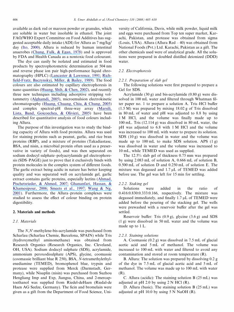
Fig. 1. In this figure, electrophoretically resolved proteins stained with Allura[takadiastase and casein mixture (extremes)], garlic proteins and BSA respectivout heating the gel separately in presence of Allura and Coomassie (a:a), (b:b) ashowing gel stained at pH 2 (right) and 10 (left) and is heated.
tion of 1 mg/ml of substrate). The source of trypsin waspancreas and that of protease was fungal type XIII (fromAspergillus saitoi) at various intervals of times (Pfleinderer& Krauss, 1965). The extent of proteolytic activity wasmeasured spectrophotometrically by observing the opticaldensities at 280 nm.
2.7. Statistical analysis
The colour binding assay was performed at least fourtimes while the determination of digestibility by spectropho-tometer and agar plate method was performed five times. Allthe other experiments were repeated at least five times. Torepresent the data, the bar diagrams were developed usingMicrosoft Excel (MS Office). To analyze the data statisticalanalysis software Minitab version 13.1 was used.
(right) and Coomassie (left). The samples a:a, b:b and c:c represent theely. While in figure 1a electrophoretically resolved proteins with and with-nd (c:c) represents nisin, peanut protein and BSA respectively. Figure 1b is
0
0.51
1.5
2
2.53
3.5
4
0 1.5 3 4.5 6 7.5 9 10.5 12 13.5 15
Concentration of protein (mg/ml)
Abs
orba
nce
BSA
NISIN
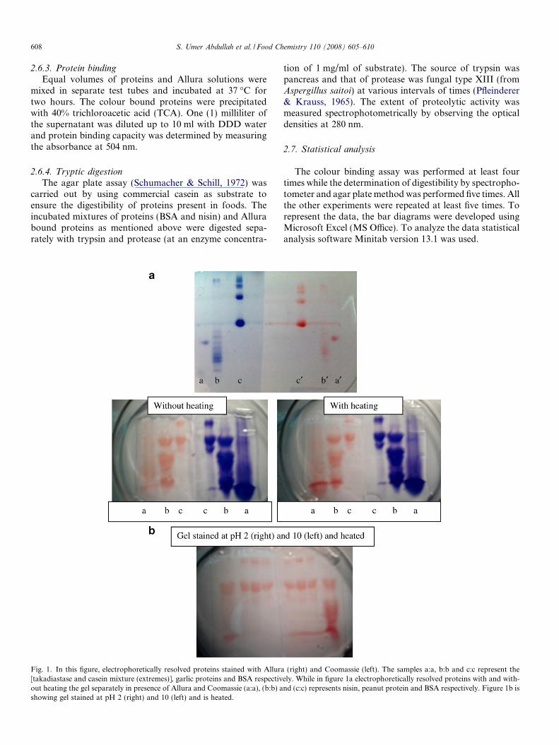
Fig. 2. The decline in absorbance due to Allura–protein complex forma-tion with increase in protein concentration.
0
0.02
0.04
0.06
0.08
0.1
0.12
0.14
0.16
10 20 30 40 50
Time of digestion (min.)
Abs
orba
nce
BSA
BSA-C
NISIN
NISIN-C
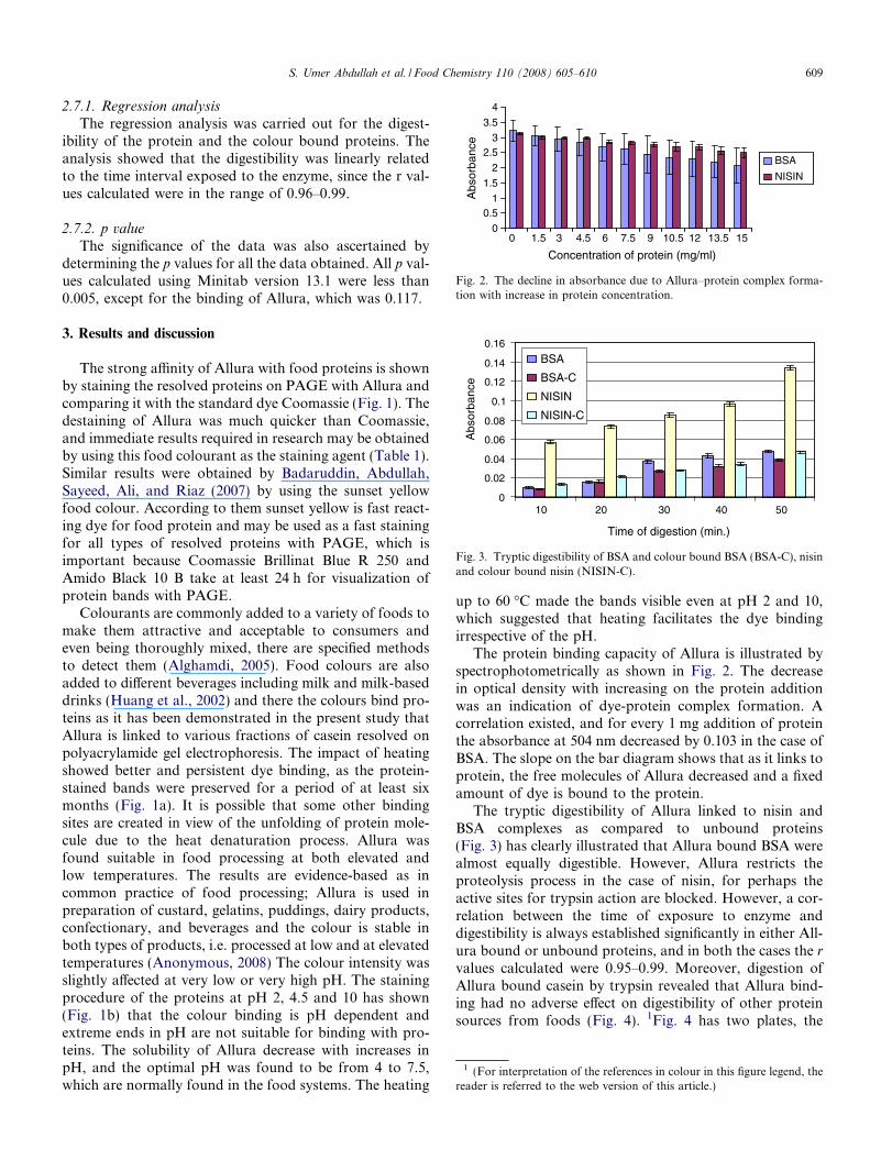
Fig. 3. Tryptic digestibility of BSA and colour bound BSA (BSA-C), nisinand colour bound nisin (NISIN-C).
1 (For interpretation of the references in colour in this figure legend, thereader is referred to the web version of this article.)
S. Umer Abdullah et al. / Food Chemistry 110 (2008) 605–610 609
2.7.1. Regression analysis
The regression analysis was carried out for the digest-ibility of the protein and the colour bound proteins. Theanalysis showed that the digestibility was linearly relatedto the time interval exposed to the enzyme, since the r val-ues calculated were in the range of 0.96–0.99.
2.7.2. p value
The significance of the data was also ascertained bydetermining the p values for all the data obtained. All p val-ues calculated using Minitab version 13.1 were less than0.005, except for the binding of Allura, which was 0.117.
3. Results and discussion
The strong affinity of Allura with food proteins is shownby staining the resolved proteins on PAGE with Allura andcomparing it with the standard dye Coomassie (Fig. 1). Thedestaining of Allura was much quicker than Coomassie,and immediate results required in research may be obtainedby using this food colourant as the staining agent (Table 1).Similar results were obtained by Badaruddin, Abdullah,Sayeed, Ali, and Riaz (2007) by using the sunset yellowfood colour. According to them sunset yellow is fast react-ing dye for food protein and may be used as a fast stainingfor all types of resolved proteins with PAGE, which isimportant because Coomassie Brillinat Blue R 250 andAmido Black 10 B take at least 24 h for visualization ofprotein bands with PAGE.
Colourants are commonly added to a variety of foods tomake them attractive and acceptable to consumers andeven being thoroughly mixed, there are specified methodsto detect them (Alghamdi, 2005). Food colours are alsoadded to different beverages including milk and milk-baseddrinks (Huang et al., 2002) and there the colours bind pro-teins as it has been demonstrated in the present study thatAllura is linked to various fractions of casein resolved onpolyacrylamide gel electrophoresis. The impact of heatingshowed better and persistent dye binding, as the protein-stained bands were preserved for a period of at least sixmonths (Fig. 1a). It is possible that some other bindingsites are created in view of the unfolding of protein mole-cule due to the heat denaturation process. Allura wasfound suitable in food processing at both elevated andlow temperatures. The results are evidence-based as incommon practice of food processing; Allura is used inpreparation of custard, gelatins, puddings, dairy products,confectionary, and beverages and the colour is stable inboth types of products, i.e. processed at low and at elevatedtemperatures (Anonymous, 2008) The colour intensity wasslightly affected at very low or very high pH. The stainingprocedure of the proteins at pH 2, 4.5 and 10 has shown(Fig. 1b) that the colour binding is pH dependent andextreme ends in pH are not suitable for binding with pro-teins. The solubility of Allura decrease with increases inpH, and the optimal pH was found to be from 4 to 7.5,which are normally found in the food systems. The heating
up to 60 �C made the bands visible even at pH 2 and 10,which suggested that heating facilitates the dye bindingirrespective of the pH.
The protein binding capacity of Allura is illustrated byspectrophotometrically as shown in Fig. 2. The decreasein optical density with increasing on the protein additionwas an indication of dye-protein complex formation. Acorrelation existed, and for every 1 mg addition of proteinthe absorbance at 504 nm decreased by 0.103 in the case ofBSA. The slope on the bar diagram shows that as it links toprotein, the free molecules of Allura decreased and a fixedamount of dye is bound to the protein.
The tryptic digestibility of Allura linked to nisin andBSA complexes as compared to unbound proteins(Fig. 3) has clearly illustrated that Allura bound BSA werealmost equally digestible. However, Allura restricts theproteolysis process in the case of nisin, for perhaps theactive sites for trypsin action are blocked. However, a cor-relation between the time of exposure to enzyme anddigestibility is always established significantly in either All-ura bound or unbound proteins, and in both the cases the r
values calculated were 0.95–0.99. Moreover, digestion ofAllura bound casein by trypsin revealed that Allura bind-ing had no adverse effect on digestibility of other proteinsources from foods (Fig. 4). 1Fig. 4 has two plates, the

Fig. 4. Shows the zones of tryptic digestion in plate (right) consisting casein and (left) consisting Allura bound casein with the same area hydrolysed.
610 S. Umer Abdullah et al. / Food Chemistry 110 (2008) 605–610
red one consisting of casein bound with Allura Red and leftone without Allura, the big circle shows the digestibilitywith trypsin in two cases and supports finding that Allurabound proteins are almost equally digestible with proteolit-ic enzymes.
In summary, the results of the present study have shownfor the first time that Allura Red-40 binds with all proteinsand the dye is suitable in food processing at high or lowtemperatures. The colour intensity is not much affected atmildly acidic or basic pH, and the dye may be used in var-ious processed foods, as most of the food systems are nor-mally in the range of pH 4–7.5 where dye binding isoptimal.
Acknowledgement
We are grateful to the Higher Education Commission(HEC), Pakistan for the financial support.
References
Ahmad, M. S., Pischetsrieder, M., & Ahmed, N. (2007). Aged garlic ex-
tract and S-allyl cysteine prevent formation of advanced glycation end-
products. European Journal of Pharmacology, 561, 32–38.Alghamdi, A. H. (2005). Determination of Allura red in some food sam-
ples by adsorptive stripping voltammetry. Journal of AOAC Interna-
tional, 88, 1387–1393.Anonymous (2003). United States international trade commission allura
red coloring from india. Investigations Nos. 701-TA-433 (Preliminary)and 731-TA-1029 (Preliminary). Determination and views of the com-
mission, (USITC Publication No. 3595, April 2003). <http://hot-docs.usitc.gov/docs/pubs/701_731/pub3595.pdf> Accessed January2008.
Anonymous (2008). <http://chemicalland21.com/specialtychem/fine-chem/ALLURA%20RED.htm> Accessed January 2008.
Badaruddin, M., Abdullah, S. U., Sayeed, S. A., Ali, R., & Riaz, M. N.(2007). Sunset yellow: A food color for protein staining with SDS–
PAGE. Cereal Food World, 52(1), 12–14.
Chung, K., Fulk, G. E., & Egan, M. (1978). Reduction of azo dyes by
intestinal anaerobes. Applied and Environmental Microbiology, 35,558–562.
Ghananfari, T., Hassan, Z. M., & Khamesipour, A. (2006). Enhancement
of peritoneal macrophage phygocytic activity against Leishmania ma-
jor by garlic (Allium sativum) treatment. Journal of Ethnopharmacol-
ogy, 103, 333–337.Huang, H. Y., Chuang, C. L., Chiu, C. W., & Chung, M. C. (2005). Deter-
mination of food colorants by microemulsion electrokinetic chroma-
tography. Electrophoresis, 26, 867–877.Huang, H. Y., Shih, Y. C., & Chen, Y. C. (2002). Determining eight col-
orants in milk beverages by capillary electrophoresis. Journal of Chro-
matography, 959, 317–325.Ito, Y. (2000). Recent state and the investigation of daily intake of food
additives in Japan-21 years (1976–1996). Food Sanitation Research,
50, 89–125.Lancaster, F. E., & Lawrence, J. F. (1991). Determination of total non-
sulphonated aromatic amines in tartrazine, sunset yellow FCF and All-
ura red by reduction and derivatization followed by high-performance
liquid chromatography. Food Additives and Contaminants, 8, 249–263.
Marsili, N. R., Lista, A., Band, B. S., Goicoechea, H. C., & Olivieri, A. C.(2005). Evaluation of complex spectral-pH three-way arrays by modi-
fied bilinear least-squares: Determination of four different dyes in
interfering systems. Analyst, 130, 1291–1298.Pfleinderer, G., & Krauss, A. (1965). Spectrophotometric assay for the
detection of protease activity. Biochemische Zeitschrift, 85, 342–344.
Richfield-Fratz, N., Baczynskyj, W. M., Miller, G. C., & Bailey, J. E.(1989). Isolation, characterization and determination of trace organic
impurities in FD&C red No. 40. Journal of Chromatography, 467,167–176.
Schumacher, G. F., & Schill, W. B. (1972). Radial diffusion in gel for mi-
cro determination of enzymes II. Plasminogen activator, elastase, and
nonspecific proteases. Analytical Biochemistry, 48, 9–26.Smeets, K., Damme, E. J. M. V., Verhaert, P., Barre, A., Rouge, P., Leu-
ven, F. V., et al. (1997). Isolation, characterization and molecular clon-
ing of the mannose-binding lectin from leaves and roots of garlic
(Allium sativum L.). Journal of Plant Molecular Biology, 33, 223–234.Wang, H. X., & Ng, T. B. (2001). Purification of allivin, a novel antifungal
protein from bulbs of the round-clove garlic. Journal of Elsevier, 70,357–365.