beta-Arrestin links endothelin A receptor to beta-catenin signaling to induce ovarian cancer cell...
6
-Arrestin links endothelin A receptor to -catenin signaling to induce ovarian cancer cell invasion and metastasis Laura Rosano ` a , Roberta Cianfrocca a , Stefano Masi a , Francesca Spinella a , Valeriana Di Castro a , Annamaria Biroccio b , Erica Salvati b , Maria Rita Nicotra c , Pier Giorgio Natali a,d , and Anna Bagnato a,1 Laboratories of a Molecular Pathology, b Experimental Chemotherapy, and d Immunology, Regina Elena Cancer Institute, 00158 Rome, Italy; and c Molecular Biology and Pathology Institute, National Research Council, 00185 Rome, Italy Edited by Robert J. Lefkowitz, Duke University Medical Center, Durham, NC, and approved December 24, 2008 (received for review July 24, 2008) The activation of endothelin-A receptor (ET A R) by endothelin-1 (ET-1) has a critical role in ovarian tumorigenesis and progression. To define the molecular mechanism in ET-1-induced tumor invasion and metastasis, we focused on -arrestins as scaffold and signaling proteins of G protein-coupled receptors. Here, we demonstrate that, in ovarian cancer cells, -arrestin is recruited to ET A R to form two trimeric complexes: one through the interaction with Src leading to epithelial growth factor receptor (EGFR) transactivation and -catenin Tyr phosphorylation, and the second through the physical association with axin, contributing to release and inacti- vation of glycogen synthase kinase (GSK)-3 and -catenin stabi- lization. The engagement of -arrestin in these two signaling complexes concurs to activate -catenin signaling pathways. We then demonstrate that silencing of both -arrestin-1 and -arres- tin-2 inhibits ETAR-driven signaling, causing suppression of Src, mitogen-activated protein kinase (MAPK), AKT activation, as well as EGFR transactivation and a complete inhibition of ET-1-induced -catenin/TCF transcriptional activity and cell invasion. ET A R block- ade with the specific ETA R antagonist ZD4054 abrogates the en- gagement of -arrestin in the interplay between ET A R and the -catenin pathway in the invasive program. Finally, ET A R is ex- pressed in 85% of human ovarian cancers and is preferentially co-expressed with -arrestin-1 in the advanced tumors. In a xeno- graft model of ovarian metastasis, HEY cancer cells expressing -arrestin-1 mutant metastasize at a reduced rate, highlighting the importance of this molecule in promoting metastases. ZD4054 treatment significantly inhibits metastases, suggesting that spe- cific ETA R antagonists, by disabling multiple signaling activated by ETA R/-arrestin, may represent new therapeutic opportunities for ovarian cancer. beta-arrestin beta-catenin endothelin A receptor metastasis ovarian cancer I dentification of critical signaling effectors of cancer cells is mandatory in defining mechanisms relevant to metastases that can be therapeutically targeted. Endothelin-1 (ET-1) has a relevant role on initiation and progression of a wide spectrum of malignan- cies, including ovarian carcinoma (1, 2). Our earlier studies have shown that ET-1 and the selective ET A-receptor (ET A R) subtype, a G-protein-coupled receptor (GPCR), are overexpressed in pri- mary and metastatic human ovarian carcinomas correlating with tumor grade (3), and that ET-1 is present at high levels in ovarian tumor effusions (4). In ovarian tumor cells, the autocrine ET-1/ ET A R axis triggers the activation of multiple signaling pathways, which concurrently drive cell proliferation, survival, angiogenesis and invasion (3– 6). ET-1 is also capable of transactivating epithelial growth factor receptor (EGFR) through a Src-dependent mecha- nism, thus contributing to the ET A R-dependent invasive and migratory capability of ovarian cancer cells (7, 8). The sustained autocrine ET A R signaling drives inhibition of glycogen synthase kinase-3 (GSK-3) to stabilize Snail and -catenin proteins in a coordinated manner to engage transcriptional programs that mod- ulate epithelial-to-mesenchymal transition and cell invasion (9). In this context, the ability of ET-1 to control the tumor-host interac- tions (6, 9–11), underlines its key role allowing close coordination in the cellular signaling network in ovarian cancer growth and progression. These findings complement and extend the analysis of gene expression profile of late-stage ovarian cancer whereby ET A R has been identified as a metastasis-associated gene (12). To char- acterize downstream mediators in ET-1-induced ovarian cancer invasion and metastasis, we focused on the role of -arrestins, as scaffold proteins of GPCR, in the -catenin signaling pathway. -Arrestins are adapter proteins that, through the formation of multiprotein complexes, play a central role in the interrelated processes of most GPCR desensitization, trafficking, and signaling (13). Comprehensive studies demonstrated that Src activation upon GPCR stimulation required -arrestins (14, 15). Recently, a new signaling mechanism, that involves the activation of -arrestins in routing signals from GPCR to Src and EGFR, has been identified (16, 17). Moreover, -arrestin has been shown to be a necessary component in the Wnt/-catenin pathway by forming a complex with axin and the cytoplasmic molecule dishevelled (18, 19). These observations require further attention in the light of the recent report that -arrestin-1 plays a prominent role in the metastases of human colorectal cancer (17). Previous studies showed the involve- ment of -arrestins in the regulation of the ET receptors, in terms of internalization and intracellular trafficking pathways, demon- strating that agonist-activated ET A R is able to recruit with different affinities both -arrestin-1 and -2, at the plasma membrane (20, 21). Moreover, ET-1 via the ET A R forms a molecular complex with the Src family Tyr kinase Yes, and -arrestin-1 in adipocytes (22). Few studies have so far examined the involvement of -arrestin in mediating ET-1-stimulated signaling pathways, leaving unan- swered the exact mechanisms by which ET-1 mediates its effects on tumor cells. In this study, we tested whether -arrestins could be recruited to the ET A R to regulate molecular events involved in tumor progres- sion, such as -catenin signaling. The results obtained in in vitro and in vivo models of ovarian cancer establish the functional role of -arrestin-1 or -2 in ET A R-induced cross-talk with EGFR, -catenin signaling, cell invasion, and metastasis. Results -Arrestin-1 and -2 Associate with ET A R in Ovarian Cancer Cells. Upon agonist stimulation, -arrestin translocates from the cytosol to the Author contributions: L.R. and A. Bagnato designed research; L.R., R.C., S.M., F.S., V.D.C., A. Biroccio, E.S., and M.R.N. performed research; L.R., P.G.N., and A. Bagnato analyzed data; L.R. and A. Bagnato wrote the paper. The authors declare no conflict of interest. This article is a PNAS Direct Submission. 1 To whom correspondence should be addressed. E-mail: [email protected]. This article contains supporting information online at www.pnas.org/cgi/content/full/ 0807158106/DCSupplemental. © 2009 by The National Academy of Sciences of the USA 2806 –2811 PNAS February 24, 2009 vol. 106 no. 8 www.pnas.orgcgidoi10.1073pnas.0807158106
-
Upload
independent -
Category
Documents
-
view
1 -
download
0
Transcript of beta-Arrestin links endothelin A receptor to beta-catenin signaling to induce ovarian cancer cell...

�-Arrestin links endothelin A receptor to �-cateninsignaling to induce ovarian cancer cell invasionand metastasisLaura Rosanoa, Roberta Cianfroccaa, Stefano Masia, Francesca Spinellaa, Valeriana Di Castroa, Annamaria Birocciob,Erica Salvatib, Maria Rita Nicotrac, Pier Giorgio Natalia,d, and Anna Bagnatoa,1
Laboratories of aMolecular Pathology, bExperimental Chemotherapy, and dImmunology, Regina Elena Cancer Institute, 00158 Rome, Italy; and cMolecularBiology and Pathology Institute, National Research Council, 00185 Rome, Italy
Edited by Robert J. Lefkowitz, Duke University Medical Center, Durham, NC, and approved December 24, 2008 (received for review July 24, 2008)
The activation of endothelin-A receptor (ETAR) by endothelin-1(ET-1) has a critical role in ovarian tumorigenesis and progression.To define the molecular mechanism in ET-1-induced tumor invasionand metastasis, we focused on �-arrestins as scaffold and signalingproteins of G protein-coupled receptors. Here, we demonstratethat, in ovarian cancer cells, �-arrestin is recruited to ETAR to formtwo trimeric complexes: one through the interaction with Srcleading to epithelial growth factor receptor (EGFR) transactivationand �-catenin Tyr phosphorylation, and the second through thephysical association with axin, contributing to release and inacti-vation of glycogen synthase kinase (GSK)-3� and �-catenin stabi-lization. The engagement of �-arrestin in these two signalingcomplexes concurs to activate �-catenin signaling pathways. Wethen demonstrate that silencing of both �-arrestin-1 and �-arres-tin-2 inhibits ETAR-driven signaling, causing suppression of Src,mitogen-activated protein kinase (MAPK), AKT activation, as wellas EGFR transactivation and a complete inhibition of ET-1-induced�-catenin/TCF transcriptional activity and cell invasion. ETAR block-ade with the specific ETAR antagonist ZD4054 abrogates the en-gagement of �-arrestin in the interplay between ETAR and the�-catenin pathway in the invasive program. Finally, ETAR is ex-pressed in 85% of human ovarian cancers and is preferentiallyco-expressed with �-arrestin-1 in the advanced tumors. In a xeno-graft model of ovarian metastasis, HEY cancer cells expressing�-arrestin-1 mutant metastasize at a reduced rate, highlighting theimportance of this molecule in promoting metastases. ZD4054treatment significantly inhibits metastases, suggesting that spe-cific ETAR antagonists, by disabling multiple signaling activated byETAR/�-arrestin, may represent new therapeutic opportunities forovarian cancer.
beta-arrestin � beta-catenin � endothelin A receptor � metastasis �ovarian cancer
Identification of critical signaling effectors of cancer cells ismandatory in defining mechanisms relevant to metastases that
can be therapeutically targeted. Endothelin-1 (ET-1) has a relevantrole on initiation and progression of a wide spectrum of malignan-cies, including ovarian carcinoma (1, 2). Our earlier studies haveshown that ET-1 and the selective ET A-receptor (ETAR) subtype,a G-protein-coupled receptor (GPCR), are overexpressed in pri-mary and metastatic human ovarian carcinomas correlating withtumor grade (3), and that ET-1 is present at high levels in ovariantumor effusions (4). In ovarian tumor cells, the autocrine ET-1/ETAR axis triggers the activation of multiple signaling pathways,which concurrently drive cell proliferation, survival, angiogenesisand invasion (3–6). ET-1 is also capable of transactivating epithelialgrowth factor receptor (EGFR) through a Src-dependent mecha-nism, thus contributing to the ETAR-dependent invasive andmigratory capability of ovarian cancer cells (7, 8). The sustainedautocrine ETAR signaling drives inhibition of glycogen synthasekinase-3� (GSK-3�) to stabilize Snail and �-catenin proteins in acoordinated manner to engage transcriptional programs that mod-
ulate epithelial-to-mesenchymal transition and cell invasion (9). Inthis context, the ability of ET-1 to control the tumor-host interac-tions (6, 9–11), underlines its key role allowing close coordinationin the cellular signaling network in ovarian cancer growth andprogression. These findings complement and extend the analysis ofgene expression profile of late-stage ovarian cancer whereby ETARhas been identified as a metastasis-associated gene (12). To char-acterize downstream mediators in ET-1-induced ovarian cancerinvasion and metastasis, we focused on the role of �-arrestins, asscaffold proteins of GPCR, in the �-catenin signaling pathway.�-Arrestins are adapter proteins that, through the formation ofmultiprotein complexes, play a central role in the interrelatedprocesses of most GPCR desensitization, trafficking, and signaling(13). Comprehensive studies demonstrated that Src activation uponGPCR stimulation required �-arrestins (14, 15). Recently, a newsignaling mechanism, that involves the activation of �-arrestins inrouting signals from GPCR to Src and EGFR, has been identified(16, 17). Moreover, �-arrestin has been shown to be a necessarycomponent in the Wnt/�-catenin pathway by forming a complexwith axin and the cytoplasmic molecule dishevelled (18, 19). Theseobservations require further attention in the light of the recentreport that �-arrestin-1 plays a prominent role in the metastases ofhuman colorectal cancer (17). Previous studies showed the involve-ment of �-arrestins in the regulation of the ET receptors, in termsof internalization and intracellular trafficking pathways, demon-strating that agonist-activated ETAR is able to recruit with differentaffinities both �-arrestin-1 and -2, at the plasma membrane (20, 21).Moreover, ET-1 via the ETAR forms a molecular complex with theSrc family Tyr kinase Yes, and �-arrestin-1 in adipocytes (22). Fewstudies have so far examined the involvement of �-arrestin inmediating ET-1-stimulated signaling pathways, leaving unan-swered the exact mechanisms by which ET-1 mediates itseffects on tumor cells.
In this study, we tested whether �-arrestins could be recruited tothe ETAR to regulate molecular events involved in tumor progres-sion, such as �-catenin signaling. The results obtained in in vitro andin vivo models of ovarian cancer establish the functional roleof �-arrestin-1 or -2 in ETAR-induced cross-talk with EGFR,�-catenin signaling, cell invasion, and metastasis.
Results�-Arrestin-1 and -2 Associate with ETAR in Ovarian Cancer Cells. Uponagonist stimulation, �-arrestin translocates from the cytosol to the
Author contributions: L.R. and A. Bagnato designed research; L.R., R.C., S.M., F.S., V.D.C., A.Biroccio, E.S., and M.R.N. performed research; L.R., P.G.N., and A. Bagnato analyzed data;L.R. and A. Bagnato wrote the paper.
The authors declare no conflict of interest.
This article is a PNAS Direct Submission.
1To whom correspondence should be addressed. E-mail: [email protected].
This article contains supporting information online at www.pnas.org/cgi/content/full/0807158106/DCSupplemental.
© 2009 by The National Academy of Sciences of the USA
2806–2811 � PNAS � February 24, 2009 � vol. 106 � no. 8 www.pnas.org�cgi�doi�10.1073�pnas.0807158106

membrane, where it associates with ETAR (15, 21). Therefore, weanalyzed the recruitment of �-arrestin to ETAR in HEY and OVCA433 ovarian cancer cell lines, in which the ET-1/ETAR axis is wellcharacterized (6). Cell lines co-expressed �-arrestin-1 and -2 (Fig.1A), which associate with ETAR in a time- and agonist-dependentmanner as demonstrated by immunoprecipitation experiments(Fig. 1B). Consistent with their physical association, we demon-strated that �-arrestin-1 and ETAR co-localized in the membraneof ET-1-stimulated HEY cells (Fig. 1C). Immunoblotting analysesclearly showed that �-arrestin-1, which in quiescent cells is localizedin the cytosolic fraction, translocated to the plasma membranecompartment after ET-1 stimulation in a time-dependent manner(Fig. 1D). Confocal microscopy confirmed these results and re-vealed that in the absence of ETAR activation, �-arrestin-1 wasdistributed in the cytosol. After ET-1 stimulation, �-arrestin-1
staining was evident at the plasma membrane and in vesicle-likestructures (Fig. 1E). Of note, the translocation of �-arrestin-1 isspecific for ETAR activation, as it was completely abrogated by thetreatment with ZD4054, a selective ETAR antagonist (Fig. 1E).
�-Arrestin and ETAR Form a Molecular Signaling Complex with Src.Because previous studies have shown that Src activation by GPCRrequires �-arrestin (16, 17), we determined by combinatorial im-munoprecipitations whether ETAR, �-arrestin-1 or -2 could form amolecular complex with Src. In both HEY and OVCA 433 cells, theassociation of �-arrestin-1 and -2 with the ETAR and Src began 2minutes after ET-1 exposure and declined after 10 minutes (Fig. 2Aand [supporting information (SI) Fig. S1 A, B, and D]. ZD4054inhibited this signalplex formation, indicating that ETAR activationby ET-1 is necessary for this trimeric complex formation (Fig. 2A
Fig. 1. ET-1 induces the association of �-arrestin-1and -2 with ETAR in ovarian cancer cells. (A) Expressionof �-arrestin-1 and -2 in HEY and OVCA 433 cells byusing anti-�-arrestin-1/-2. Anti-GAPDH was used forinternal control. (B) HEY and OVCA 433 cells, treatedwith ET-1 (100 nM) for the indicated times, were im-munoprecipitated (IP), with anti-�-arrestin-1 or -2-conjugated beads or IgG control beads and IB withanti-ETAR, anti–�-arrestin-1, and -2. (C) Confocal mi-croscopic analysis of HEY cells incubated with 100 nMET-1 for 1 minute using anti-�-arrestin-1 (red) andanti-ETAR (green). Co-localization of �-arrestin-1 andETAR is represented as yellow in the merge image. Boxin the merged image indicates the location of theenlarged view. Similar results were observed in threeseparate experiments. (D) Cytosolic and pure plasmamembrane fractions of HEY and OVCA 433 cells, incu-bated for indicated times with 100 nM ET-1, wereanalyzed by IB with anti-�-arrestin-1. Anti-GAPDH andanti-Na/K-ATPase were used as markers of cytosolicand plasma membrane compartments, respectively. (E)Confocal microscopy analysis of HEY cells untreated (C)or incubated for indicated times with 100 nM ET-1and/or ZD4054 (1 �M) using anti-�-arrestin-1. Similarresults were observed in three separated experiments.
Fig. 2. ET-1 induces the formation of the ETAR/�-arrestin/Src signaling complex to transactivate EGFR.(A) HEY cells, treated with ET-1 (100 nM) for the indi-cated times or in combination with ZD4054 (1 �M) for5 minutes, were IP with anti-�-arrestin-1 or -2. The IPsamples were analyzed by IB with anti-ETAR, anti-Src,anti-�-arrestin-1 and -2. (B) HEY cells, treated with ET-1(100 nM) for the indicated times, were IP with anti-Src-conjugated beads or IgG control beads and IB withanti-ETAR and anti-Src. (C) IP with anti-Src of HEY celllysates after knockdown with �-arrestin-1 siRNA, sub-sequent rescue with FLAG-tagged WT- or mutantS412D-�-arrestin-1, and treatment for indicated timeswith 100 nM ET-1. The IP samples and cell lysates wereanalyzed by IB with anti-FLAG, anti-phospho-Tyr, andanti-Src. (D) HEY cells, transfected with scrambled (si-control) or siRNA targeting either �-arrestin-1 or -2 orboth, were treated with ET-1 (100 nM). Cell lysateswere analyzed by IB with anti-phospho-EGFR, anti-EGFR, anti-phospho-Src, anti-Src, anti-phospho-p42/44 MAPK, anti-p42/44 MAPK, anti-phospho-AKT,and anti-AKT. (E) IB of HEY cell lysates treated as in (C),using anti-pEGFR and anti-EGFR.
Rosano et al. PNAS � February 24, 2009 � vol. 106 � no. 8 � 2807
MED
ICA
LSC
IEN
CES

and Fig. S1 A, B, and D). The presence of ETAR in Src immuno-precipitates (Fig. 2B and Fig. S1 C and E) indicated that ET-1induces the formation of an ETAR/�-arrestin/Src signaling com-plex, or signalplex. In parallel with the ability to promote bindingbetween �-arrestin-1 and Src, ET-1 induced a time-dependentdephosphorylation on serine-412 �-arrestin-1 in both cell lines (Fig.S1F), which causes reduced affinity for Src (14, 23). Then weperformed siRNA knockdown of �-arrestin-1 followed by rescuewith expression of FLAG-tagged WT or mutant S412D-�-arrestin-1. Specificity of siRNA oligos was confirmed by WesternBlotting analysis, which showed an 80% knockdown of �-arrestin-1(Fig. S2A), and by rescue (90%) of knockdown effects withexpression of FLAG-tagged �-arrestin-1 (Fig. S2B). In cells ex-pressing WT-, but not S412D-�-arrestin-1, ET-1 induced the asso-ciation of �-arrestin-1 with Src and its Tyr phosphorylation (Fig.2C), confirming that the association of �-arrestin-1 with Src iscritical for its activation. To demonstrate that �-arrestin-1 is criticalfor the formation of the complex with ETAR and Src, we silenced�-arrestin-1 in HEY cells, proving that ETAR cannot bind Srcindependently of �-arrestin-1 (Fig. S2C).
ETAR/�-Arrestin/Src Signaling Complex Is a Critical Event in EGFRTransactivation and Downstream Pathways. Because emerging evi-dence indicates that �-arrestins organize and scaffold an activesignaling complex with Src, leading to EGFR transactivation (16,17), we evaluated the potential functional role of �-arrestin-1 and�-arrestin-2 in ET-1-dependent multiple signaling pathways byspecifically silencing either �-arrestin-1 or -2 or both (Fig. S2A). InHEY cells, ET-1 induced rapid Src and EGFR phosphorylation andan increase in the activation of p42/44 mitogen-activated proteinkinase (MAPK) and AKT (Fig. 2D). Interestingly, knockdown of�-arrestin-1 or -2 inhibited the ET-1-induced Src and EGFRactivation and their downstream pathways, which were completelyblocked in the presence of siRNA targeting both �-arrestin-1 and-2 (Fig. 2D), indicating that both �-arrestins are required inETAR-induced signaling. ZD4054, gefitinib, an EGFR inhibitor, orPP2, a Src inhibitor, reduced the ET-1-induced Src and EGFRactivation. However, gefitinib incompletely reduced the ET-1-mediated MAPK and AKT activation. A combination of ZD4054plus gefitinib resulted in a greater inhibition of all these pathways
(Fig. S3), indicating the critical role of ETAR and EGFR intercon-nected signaling systems (24). Furthermore ET-1-induced EGFRphosphorylation was inhibited in HEY cells silenced for �-arrestin-1 and rescued with S412D-�-arrestin-1 mutant comparedwith WT-�-arrestin-1- expressing cells (Fig. 2E). Altogether thesedata demonstrate that silencing of both �-arrestin-1 and -2 inhibitsETAR-driven signaling and that �-arrestin/Src complex formationis a critical event for activation of EGFR and related-pathways. Thematrix metalloproteinases (MMP) inhibitor, GM6001, did notaffect the ET-1-induced EGFR transactivation, demonstrating thatthis process is MMP independent (Fig. S4).
An ETAR/�-Arrestin/Src Signaling Complex Is Required for ET-1-In-duced �-Catenin Tyrosine Phosphorylation. Because recent studieshave revealed that Tyr phosphorylation of �-catenin enhances�-catenin/TCF signaling (25–27), we tested whether ET-1 inducesTyr phosphorylation of �-catenin through the EGFR transactiva-tion mediated by �-arrestin. In both HEY and OVCA 433 cells,ET-1 induced �-catenin Tyr phosphorylation, starting at 5 minutesand lasting for 15 to 30 minutes, indicating that its phosphorylationstate was tightly regulated by ET-1 (Fig. 3A). This effect was alsodose dependent, with a maximum at 100 nM of ET-1 in bothcell lines (Fig. S5A). Moreover, in HEY cells, the ET-1-induced�-catenin Tyr phosphorylation mediated by the ETAR/�-arrestin/Src complex was completely blocked by ZD4054 (Fig. 3B). Theknockdown of EGFR with siRNA confirmed that EGFR transac-tivation is required for ET-1-induced Tyr phosphorylation of�-catenin (Fig. S5B and C). Interestingly, �-arrestin-1 siRNAmarkedly downregulated Tyr phosphorylation of EGFR and of�-catenin in ET-1-treated cells compared with control cells (Fig.3C). The inhibition of �-catenin Tyr phosphorylation observed with�-arrestin-1 knockdown was rescued by the expression of WT- butnot S412D-�-arrestin-1, indicating the critical role of �-arrestin-1-driven signalplex formation in EGFR-mediated �-catenin Tyrphosphorylation induced by ET-1 (Fig. 3D). It is noteworthy thatET-1 treatment promoted the binding between Tyr phosphorylated�-catenin and TCF-4 in nuclear extracts in scrambled but not in�-arrestin-1 siRNA-transfected HEY and OVCA 433 cells, sug-gesting that the ET-1-induced �-catenin Tyr phosphorylation can
Fig. 3. ET-1 triggers tyrosine phosphorylation of �-catenin through signalplex and EGFR. (A) HEY and OVCA 433 cells were incubated for different timeswith 100 nM ET-1. IP were performed with anti-�-catenin and IB with anti-pTyr and anti-�-catenin. (B) Lysates of HEY cells, treated with 100 nM ET-1 and/or1 �M ZD4054, were IP with anti-�-catenin and IB with anti-pTyr and anti-�-catenin. (C) Lysates of HEY cells transfected with scrambled (si-control) or�-arrestin-1 siRNA and incubated for the indicated times with ET-1 (100 nM), were IP with anti-�-catenin and IB with anti-pTyr and anti-�-catenin. The samelysates were IB with anti-pEGFR and anti-EGFR. (D) HEY cells, after knockdown with �-arrestin-1 siRNA and subsequent rescue with FLAG-tagged WT- orS412D-�-arrestin-1, were treated with 100 nM ET-1 for the indicated times. IP was performed with anti-�-catenin and IB with anti-pTyr and anti-�-catenin.The same lysates were IB with anti-�-arrestin-1/-2 and anti-FLAG. (E) Nuclear and cytosolic extracts of HEY cells, transfected with scrambled (si-control)or �-arrestin-1 siRNA and treated with ET-1 (100 nM), were IP with anti-�-catenin and IB with anti-pTyr, anti-TCF4, and anti-�-catenin.
2808 � www.pnas.org�cgi�doi�10.1073�pnas.0807158106 Rosano et al.

represent a transcriptional active pool in a �-arrestin-1-dependentmanner (Fig. 3E and Fig. S5D).
ET-1 Promotes Linking of �-Arrestin to Axin in the Activation of�-Catenin Signaling. Based on previous findings on the interactionbetween �-arrestin-1 and axin in Wnt signaling, we exploredwhether �-arrestin-1 may also provide a link between ETAR and�-catenin signaling by its capacity to bind axin directly. In HEYcells, axin coimmunoprecipitated with ETAR and �-arrestin-1 in anET-1-dependent manner, with a peak at 5 minutes (Fig. 4A).Moreover, we demonstrated by coimmunoprecipitation experi-ments that in these cells axin and Src are not present in the samecomplex (Fig. S6). We also observed that ET-1 caused a reductionin the amounts of GSK-3� bound to axin, whereas this associationwas still present in �-arrestin-1-silenced cells, suggesting that theET-1-induced binding of �-arrestin-1 to axin is required to induce
the displacement of GSK-3� from an axin-containing complex (Fig.4B). Because the inhibition of GSK-3� led to dephosphorylationand stabilization of �-catenin in response to ET-1 (9), we evaluatedthe role of �-arrestin-1 in the phosphorylation of GSK-3�, whichrenders it inactive. ET-1 treatment led to rapid phosphorylation ofGSK-3� on serine 9, which was impaired after knockdown of�-arrestin-1, suggesting that the release of GSK-3� from axin-containing complexes regulated by �-arrestin-1 is associated with itsfunctional inhibition (Fig. 4B). We observed that the ET-1-dependent pattern of �-catenin dephosphorylation, evaluated withan Ab against active �-catenin, was abolished in �-arrestin-1-silenced cells (Fig. 4B). Altogether, these results strongly imply thatthe association of �-arrestin-1 with ETAR may signal through twoparallel coordinated mechanisms that concur in the stabilization of�-catenin. One that is dependent on EGFR-mediated Tyr phos-phorylation of �-catenin and another through axin and dephos-phorylation of �-catenin in a manner similar to that of canonicalWnt signaling (Fig. S7).
�-Arrestin-Driven Signalplexes Are Required in �-Catenin Transcrip-tional Activity and Cell Invasion Induced by ET-1. Because dephos-phorylated �-catenin is stabilized and leads to activation of tran-scription in a TCF/Lef-dependent manner, we also investigated therole of �-arrestin-1 or -2 in ET-1-induced activation of the TOP/Flash luciferase reporter construct. As shown in Fig. 4C, althoughHEY and OVCA 433 cells strongly respond to ET-1 stimulation,the silencing of either �-arrestin-1 or -2 or both significantlyinhibited the TCF/Lef reporter activity. Moreover, the knockdownof both �-arrestins caused complete inhibition of ET-1-inducedinvasion (Fig. 4D), highlighting a new role of �-arrestins in ET-1-induced �-catenin transcription and the invasive potential of ovar-ian cancer cells. The stimulatory effect of ET-1 on �-catenintranscriptional activity (Fig. 4C) and cell invasion (Fig. 4D) was alsoblocked by pretreatment with ZD4054. These results underline therelevant role of �-arrestin-dependent ETAR-induced �-cateninsignaling and invasiveness.
ETAR Links �-Arrestin to Promote Metastasis in HEY Xenografts. Todetermine the role of �-arrestin-1 in ovarian cancer metastasisformation, we used HEY cells, as well as clonally derived HEY celllines overexpressing WT- or S412D-�-arrestin-1, in an i.p. meta-static model. After 4 weeks of tumor cell injection, multiplemetastatic seeding tumors were distributed on the peritonealsurface, omentum, small bowel, mesentery, and in both ovaries (Fig.5A). Interestingly, HEY cells produced a number of metastasessimilar to those produced by clonally derived HEY cell lines thatoverexpress WT-�-arrestin-1 (Fig. 5B). Differentially, clonal HEYcells overexpressing S412D-�-arrestin-1 mutant showed reducedmetastatic ability by nearly 50% (mean, 4.5 � 0.7 lesions vs. 9 � 1.9of WT-�-arrestin-1-expressing cells; P � 0.001), suggesting that�-arrestin-1 may serve a pivotal role in ovarian carcinoma metas-tasis (Fig. 5B). To explore the therapeutic potential of ETARblockade in the control of ovarian peritoneal metastasis, we testedthe efficacy of ZD4054. The number of intra-abdominal metastaseswas significantly decresased in ZD4054-treated mice (mean, 4.1 �0.7 lesions) when compared with controls (mean, 10.8 � 1.9 lesions;Fig. 5B). The treatment at the dose and schedule tested was welltolerated, as evaluated by the absence of weight loss or other signsof acute or delayed toxicity. Because in vitro data indicate that�-arrestin is essential for �-catenin signaling and invasive propertiesof ovarian cancer cells, we sought to determine the status of�-catenin in metastases expressing WT or S412D-�-arrestin-1. Weobserved by immunoblotting a strong decrease in the active�-catenin in the metastatic nodules derived from clonal HEY cellsoverexpressing S412D-�-arrestin-1 mutant when compared withHEY cells overexpressing WT-�-arrestin-1 (Fig. 5C). Similar re-sults were obtained in the metastatic lesions from ZD4054-treatedmice when compared with control. These results, together with the
Fig. 4. �-Arrestin links ETAR to axin in the activation of �-catenin signaling,its transcriptional activity, and cell invasion. (A) Lysates of HEY cells, stimulatedwith ET-1 (100 nM) for different times, were IP with anti-�-arrestin-1-conjugated beads or IgG control beads and IB with anti-axin, anti-ETAR, andanti-�-arrestin-1. (B) HEY cells, transfected with scrambled (si-control) or�-arrestin-1 siRNA, were treated for the indicated times with 100 nM ET-1. IPwas performed with anti-axin and IB with anti-GSK-3� and anti-axin. Lysatesof the same cells were IB with anti-pGSK-3� (Ser 9), anti-GSK-3�, anti-active(act) �-catenin, and anti-�-actin for internal control. (C) �-catenin/TCF tran-scriptional activity was evaluated in HEY and OVCA 433 cells transfected siRNAtargeting either �-arrestin-1 or -2 or both, incubated with ET-1 (100 nM)and/or ZD4054 (1 �M). Bars, �SD. *P � 0.001 with control; **P � 0.05compared with ET-1. (D) Invasion assay of HEY and OVCA 433 cells treatedas in C. Bars, � SD. *P � 0.01 compared with control; **P � 0.05 comparedwith ET-1.
Rosano et al. PNAS � February 24, 2009 � vol. 106 � no. 8 � 2809
MED
ICA
LSC
IEN
CES
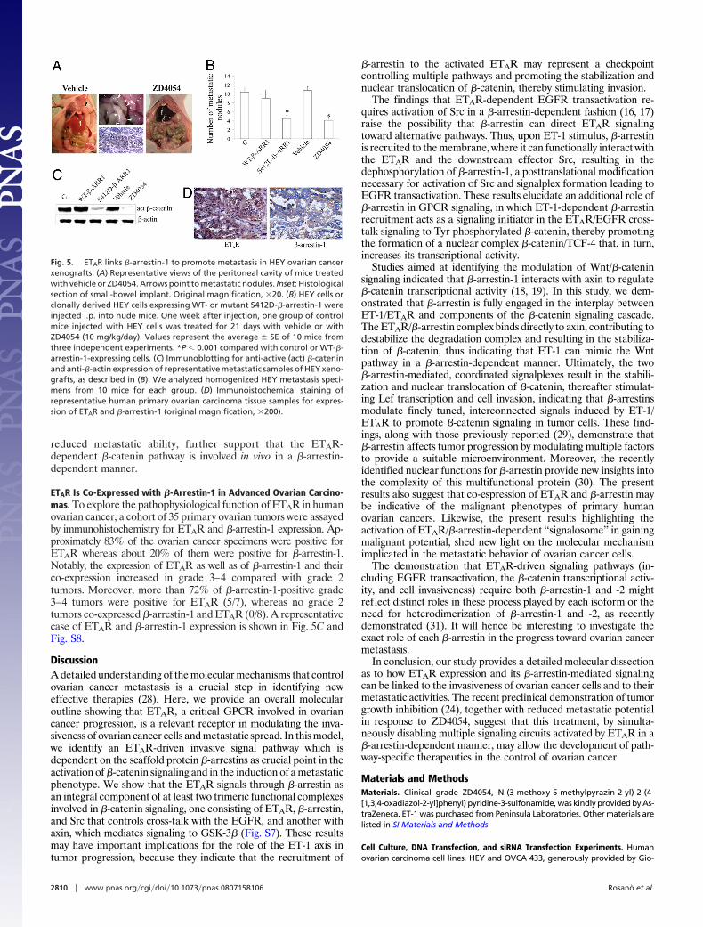
reduced metastatic ability, further support that the ETAR-dependent �-catenin pathway is involved in vivo in a �-arrestin-dependent manner.
ETAR Is Co-Expressed with �-Arrestin-1 in Advanced Ovarian Carcino-mas. To explore the pathophysiological function of ETAR in humanovarian cancer, a cohort of 35 primary ovarian tumors were assayedby immunohistochemistry for ETAR and �-arrestin-1 expression. Ap-proximately 83% of the ovarian cancer specimens were positive forETAR whereas about 20% of them were positive for �-arrestin-1.Notably, the expression of ETAR as well as of �-arrestin-1 and theirco-expression increased in grade 3–4 compared with grade 2tumors. Moreover, more than 72% of �-arrestin-1-positive grade3–4 tumors were positive for ETAR (5/7), whereas no grade 2tumors co-expressed �-arrestin-1 and ETAR (0/8). A representativecase of ETAR and �-arrestin-1 expression is shown in Fig. 5C andFig. S8.
DiscussionA detailed understanding of the molecular mechanisms that controlovarian cancer metastasis is a crucial step in identifying neweffective therapies (28). Here, we provide an overall molecularoutline showing that ETAR, a critical GPCR involved in ovariancancer progression, is a relevant receptor in modulating the inva-siveness of ovarian cancer cells and metastatic spread. In this model,we identify an ETAR-driven invasive signal pathway which isdependent on the scaffold protein �-arrestins as crucial point in theactivation of �-catenin signaling and in the induction of a metastaticphenotype. We show that the ETAR signals through �-arrestin asan integral component of at least two trimeric functional complexesinvolved in �-catenin signaling, one consisting of ETAR, �-arrestin,and Src that controls cross-talk with the EGFR, and another withaxin, which mediates signaling to GSK-3� (Fig. S7). These resultsmay have important implications for the role of the ET-1 axis intumor progression, because they indicate that the recruitment of
�-arrestin to the activated ETAR may represent a checkpointcontrolling multiple pathways and promoting the stabilization andnuclear translocation of �-catenin, thereby stimulating invasion.
The findings that ETAR-dependent EGFR transactivation re-quires activation of Src in a �-arrestin-dependent fashion (16, 17)raise the possibility that �-arrestin can direct ETAR signalingtoward alternative pathways. Thus, upon ET-1 stimulus, �-arrestinis recruited to the membrane, where it can functionally interact withthe ETAR and the downstream effector Src, resulting in thedephosphorylation of �-arrestin-1, a posttranslational modificationnecessary for activation of Src and signalplex formation leading toEGFR transactivation. These results elucidate an additional role of�-arrestin in GPCR signaling, in which ET-1-dependent �-arrestinrecruitment acts as a signaling initiator in the ETAR/EGFR cross-talk signaling to Tyr phosphorylated �-catenin, thereby promotingthe formation of a nuclear complex �-catenin/TCF-4 that, in turn,increases its transcriptional activity.
Studies aimed at identifying the modulation of Wnt/�-cateninsignaling indicated that �-arrestin-1 interacts with axin to regulate�-catenin transcriptional activity (18, 19). In this study, we dem-onstrated that �-arrestin is fully engaged in the interplay betweenET-1/ETAR and components of the �-catenin signaling cascade.The ETAR/�-arrestin complex binds directly to axin, contributing todestabilize the degradation complex and resulting in the stabiliza-tion of �-catenin, thus indicating that ET-1 can mimic the Wntpathway in a �-arrestin-dependent manner. Ultimately, the two�-arrestin-mediated, coordinated signalplexes result in the stabili-zation and nuclear translocation of �-catenin, thereafter stimulat-ing Lef transcription and cell invasion, indicating that �-arrestinsmodulate finely tuned, interconnected signals induced by ET-1/ETAR to promote �-catenin signaling in tumor cells. These find-ings, along with those previously reported (29), demonstrate that�-arrestin affects tumor progression by modulating multiple factorsto provide a suitable microenvironment. Moreover, the recentlyidentified nuclear functions for �-arrestin provide new insights intothe complexity of this multifunctional protein (30). The presentresults also suggest that co-espression of ETAR and �-arrestin maybe indicative of the malignant phenotypes of primary humanovarian cancers. Likewise, the present results highlighting theactivation of ETAR/�-arrestin-dependent ‘‘signalosome’’ in gainingmalignant potential, shed new light on the molecular mechanismimplicated in the metastatic behavior of ovarian cancer cells.
The demonstration that ETAR-driven signaling pathways (in-cluding EGFR transactivation, the �-catenin transcriptional activ-ity, and cell invasiveness) require both �-arrestin-1 and -2 mightreflect distinct roles in these process played by each isoform or theneed for heterodimerization of �-arrestin-1 and -2, as recentlydemonstrated (31). It will hence be interesting to investigate theexact role of each �-arrestin in the progress toward ovarian cancermetastasis.
In conclusion, our study provides a detailed molecular dissectionas to how ETAR expression and its �-arrestin-mediated signalingcan be linked to the invasiveness of ovarian cancer cells and to theirmetastatic activities. The recent preclinical demonstration of tumorgrowth inhibition (24), together with reduced metastatic potentialin response to ZD4054, suggest that this treatment, by simulta-neously disabling multiple signaling circuits activated by ETAR in a�-arrestin-dependent manner, may allow the development of path-way-specific therapeutics in the control of ovarian cancer.
Materials and MethodsMaterials. Clinical grade ZD4054, N-(3-methoxy-5-methylpyrazin-2-yl)-2-(4-[1,3,4-oxadiazol-2-yl]phenyl) pyridine-3-sulfonamide, was kindly provided by As-traZeneca. ET-1 was purchased from Peninsula Laboratories. Other materials arelisted in SI Materials and Methods.
Cell Culture, DNA Transfection, and siRNA Transfection Experiments. Humanovarian carcinoma cell lines, HEY and OVCA 433, generously provided by Gio-
Fig. 5. ETAR links �-arrestin-1 to promote metastasis in HEY ovarian cancerxenografts. (A) Representative views of the peritoneal cavity of mice treatedwith vehicle or ZD4054. Arrows point to metastatic nodules. Inset: Histologicalsection of small-bowel implant. Original magnification, �20. (B) HEY cells orclonally derived HEY cells expressing WT- or mutant S412D-�-arrestin-1 wereinjected i.p. into nude mice. One week after injection, one group of controlmice injected with HEY cells was treated for 21 days with vehicle or withZD4054 (10 mg/kg/day). Values represent the average � SE of 10 mice fromthree independent experiments. *P � 0.001 compared with control or WT-�-arrestin-1-expressing cells. (C) Immunoblotting for anti-active (act) �-cateninand anti-�-actin expression of representative metastatic samples of HEY xeno-grafts, as described in (B). We analyzed homogenized HEY metastasis speci-mens from 10 mice for each group. (D) Immunoistochemical staining ofrepresentative human primary ovarian carcinoma tissue samples for expres-sion of ETAR and �-arrestin-1 (original magnification, �200).
2810 � www.pnas.org�cgi�doi�10.1073�pnas.0807158106 Rosano et al.

vanni Scambia (Catholic University School of Medicine, Rome, Italy), were cul-tured as previously described (6). For the silencing of �-arrestin-1 or �-arrestin-2,cells were transiently transfected with duplex siRNAs (30 nM) targeting human�-arrestin-1 or -2 (Hs�ARRB1�11 and Hs�ARRB2�10 HP Validated siRNA, Qiagen,respectively), negative control (scrambled sequence) or no-RNA (MOCK), usingRNAiFect transfection reagent (Qiagen). The specificity of the siRNA sequencesfor �-arrestin-1 and -2 have previously been validated (32). After 48 hours ofincubation, cells were divided into six-well plates for further experiments and for�-arrestin immunoblotting. Each knockdown experiment described herein wasdetected for specific reduced expression of �-arrestins (75–90%) with A1CT Ab, arabbit polyclonal Ab to �-arrestin-1/-2 kindly provided by Robert Lefkowitz(Howard Hughes Medical Institute, Duke University). In the rescue experiments,we performed transient transfection of pcDNA3 plasmid or 2 �g FLAG epitope-tagged WT or -S412D �-arrrestin-1 expression plasmids, kindly provided by Rob-ert Lefkowitz, two ‘‘wobble’’ mutant constructs encoding rat �-arrestin-1 se-quences resistant to siRNA targeting, using LipofectAMINE reagent (Invitrogen).Further details are available in SI Materials and Methods.
Confocal Fluorescence Microscopy. HEY cells were stimulated as described, fixedin 2% formaldehyde, permeabilized in 0.25% Triton X-100 in phosphate-buffered saline solution (PBS), and then immunostained with the primary Ab to�-arrestin-1 (Santa Cruz Biotechnology Inc.), anti-ETAR (BD Transduction Labora-tories). The TRITC conjugated donkey anti-goat and the FITC conjugated goatanti-mouse (Jackson Immunoresearch) were used as secondary Abs. Fluorescencesignals were analyzed in confocal vertical (x-z) sections captured with a ZeissConfocal Laser Scanning Microscope. For each image the entire thickness of thecells has been sectioned into optical slices of 7 �m and the focal plane corre-sponding to the basal level of the cells has been chosen to highlight the mem-brane staining.
Luciferase Reporter Gene Assay. To measure the transcriptional activity of�-catenin,cellsweretransientlycotransfectedusingLipofectAMINEreagent(Invitro-gen) with 1 �g pTOP/Flash (Upstate Biotech) and 100 ng pCMV-�-galactosidase(Promega) vectors. Reporter activity was measured using the Luciferase assay system(Promega) and normalized to �-galactosidase activity. The mean of three indepen-dent experiments performed in sextuplicate was reported.
Immunoblotting and Immunoprecipitation. For Western blotting analysis, cellswere detached by scraping, collected by centrifugation, and lysed in lysis buffer[250 mM NaCl, 50 mM HEPES (pH 7.4), 1 mM ethylenediaminetetraacetic acid(EDTA), 1% Nonidet P-40, protease inhibitors]. Whole-cell lysates or homoge-nized HEY metastases specimens or separated fractions were resolved by sodium
dodecylsulfate-polyacrylamide gel electrophoresis (SDS/PAGE), followed by im-munoblotting (IB) using Abs to: anti-phosphoTyr (PY-20) (BD Transduction Lab-oratories), EGFR, phospho-EGFR (Tyr-845), phospho-GSK-3� (pSer9), GSK-3�,phospho-�-arrestin-1 (Ser-412), p42/44MAPK, phospho-p42/44MAPK, phospho-Akt (Ser-473), AKT, GAPDH, Na, K-ATPase and FLAG (Cell Signaling Technology),active-�-catenin (an Ab against non-serine-threonine phosphorylated, nonubiq-uitinated) and TCF-4 (Upstate), �-catenin, �-arrestin-1, �-arrestin-2, phospho-Src(Tyr-416) and Src (Santa Cruz Biotechology), ETAR (Abnova GmbH), �-actin (On-cogene), and axin (Zymed Laboratories). Further details are available in SI Mate-rials and Methods.
Chemoinvasion Assay. Chemoinvasion assay was performed as previously de-scribed (6). The filters were coated with an even layer of 10 mg/ml CultrexBasement Membrane Extract Matrigel (Trevigen). After 6 hours of incubation at37 °C, the filters were removed, stained with Diff-Quick (Merz-Dade), and themigrated cells in 10 high-power fields were counted. Each experimental pointwas analyzed in triplicate.
Metastasis Assay. HEY cells or clonally derived HEY cells, stably transfected withWT-orS412D-�-arrestin-1 (1.8�106),were i.p. injected intofemaleathymicnudemice (CharlesRiverLaboratories), followingtheguidelines foranimalexperimen-tation of the Italian Ministry of Health. In all experiments, each group consistedof 10 mice. In the treatment experiments, one week after injection of cancer cells,one group was treated i.p. for 21 days with ZD4054 (diluted in PBS) (10 mg/kg/day), and one group received the same volume saline solution (vehicle). At theend of the treatment, mice were killed; the number of metastases was countedand the removed tumors were weighed, carefully dissected, and snap-frozen forimmunohistochemical and immunoblot analysis. For the immunohistochemicalanalysis of human tissue samples see SI Materials and Methods.
Statistical Analysis. Densitometric quantifications and normalizations were per-formed using National Institutes of Health Scion Image 1.63 software. Statisticalanalysis was done using the Student t test, Fisher’s exact test, or one-way analysisof variance (ANOVA) to correct for multiple comparisons, as appropriated. Allstatistical tests were two-sided and were done using SPSS software (SPSS Inc.,Chicago, IL).
ACKNOWLEDGMENTS. We gratefully acknowledge Valentina Caprara and AldoLupo for excellent study assistance, Maria Vincenza Sarcone for secretarial assis-tance, and Robert Lefkowitz (Howard Hughes Medical Institute, Duke University)for kindly providing �-arrestin-1 expression vectors and A1CT Ab. This study wasfunded in part by Associazione Italiana Ricerca sul Cancro and Italian Ministry ofHealth.
1. Rubanyi GM, Polokoff MA (1994) Endothelins: Molecular biology, biochemistry, phar-macology, physiology and pathophysiology. Pharmacol Rev 46:325–415.
2. Nelson J, Bagnato A, Battistini B, Nisen P (2003) The endothelin axis: Emerging role incancer. Nat Rev Cancer 3:110–116.
3. Bagnato A, Spinella F, Rosano L (2005) Emerging role of the endothelin axis in ovariantumor progression. Endocr Relat Cancer 12:761–772.
4. Salani D, et al. (2000) Role of endothelin in neovascularization of ovarian carcinoma.Am J Pathol 157:1537–1547.
5. Del Bufalo D, et al. (2002) Endothelin-1 protects against paclitaxel-induced apoptosis:Requirement for Akt activation. Mol Pharmacol 61:524–532.
6. Rosano L, et al. (2001) Endothelin-1 induces tumor proteinase activation and invasive-ness of ovarian carcinoma cells. Cancer Res 61:8340–8346.
7. Vacca F, Bagnato A, Catt CK, Tecce R (2000) Transactivation of epidermal growth factorreceptor in endothelin-1-induced mitogenic signaling in human ovarian carcinomacells. Cancer Res 60:5310–5317.
8. Spinella F, et al. (2004) Inhibition of cyclooxygenase-1 and -2 expression by targetingthe endothelin A receptor in human ovarian carcinoma cells. Clin Cancer Res 10:4670–4679.
9. Rosano L, et al. (2005) Endothelin-1 promotes epithelial-to-mesenchymal transition inhuman ovarian cancer cells. Cancer Res 65:11649–11657.
10. Spinella F, et al. (2003) Endothelin-1 decreases gap junctional intercellular communi-cation by inducing phosphorylation of connexin 43 in human ovarian carcinoma cells.J Biol Chem 278:41294–41301.
11. Rosano L, et al. (2006) Integrin-linked kinase functions as a downstream mediator ofendothelin-1 to promote invasive behavior in ovarian carcinoma. Mol Cancer Ther5:833–842.
12. Donninger H, et al. (2004) Whole genome expression profiling of advance stagepapillary serous ovarian cancer reveals activated pathways. Oncogene 23:8065–8077.
13. Barki-Harrington L, Rockman HA (2008) �-Arrestins: Multifunctional cellular media-tors. Physiology 23:17–22.
14. Luttrell LM, et al. (1999) �-Arrestin-dependent formation of �2-adrenergic receptor-Srcprotein kinase complexes. Science 283:655–661.
15. Lefkowitz RJ, Shenoy SK (2005) Transduction of receptor signals by �-arrestins. Science308:512–517.
16. Noma T, et al. (2007) �-Arrestin-mediated �1-adrenergic receptor transactivation ofthe EGFR confers cardioprotection. J Clin Invest 117:2445–2458.
17. Buchanan FG, et al. (2006) Role of �-arrestin 1 in the metastatic progression ofcolorectal cancer. Proc Natl Acad Sci USA 103:1492–1497.
18. Chen W, et al. (2001) �-Arrestin-1 modulates lymphoid enhancer factor transcriptionalactivity through interaction with phosphorylated dishevelled proteins. Proc Natl AcadSci USA 98:14889–14894.
19. Bryja V, Gradl D, Schambony A, Arenas E, Schulte G (2007) �-Arrestin is a necessarycomponent of Wnt/beta-catenin signaling in vitro and in vivo. Proc Natl Acad Sci USA104:6690–6695.
20. Bremnes T, et al. (2000) Regulation and intracellular trafficking pathways of theendothelin receptors. J Biol Chem 275:17596–17604.
21. Oakley RH, Laporte SA, Holt JA, Caron MG, Barak LS (2000) Differential affinities ofvisual arrestin, beta arrestin1, and beta arrestin2 for G protein-coupled receptorsdelineate two major classes of receptors. J Biol Chem 275:17201–17210.
22. Imamura T, et al. (2001) �-Arrestin-mediated recruitment of the Src family kinase Yesmediates endothelin-1-stimulated glucose transport. J Biol Chem 276:43663–43667.
23. Miller WE, et al. (2000) �-Arrestin-1 interacts with the catalytic domain of the tyrosinekinase c-Src. Role of �-arrestin1-dependent targeting of c-Src in receptor endocytosis.J Biol Chem 275:11312–11319.
24. Rosano L, et al. (2007) Combined targeting of endothelin A receptor and epidermalgrowth factor receptor in ovarian cancer shows enhanced antitumor activity. CancerRes 67:6351–6359.
25. Roura S, Miravet S, Piedra J, García de Herreros A, Dunach M (1999) Regulation ofE-cadherin/catenin association by tyrosine phosphorylation. J Biol Chem 274:36734–36740.
26. Gujral TS, et al. (2008) A novel RET kinase-beta-catenin signaling pathway contributesto tumorigenesis in thyroid carcinoma. Cancer Res 68:1338–1346.
27. Coluccia AM, et al. (2007) Bcr-Abl stabilizes �-catenin in chronic myeloid leukemiathrough its tyrosine phosphorylation. EMBO J 26:1456–1166.
28. Naora H, Montell DJ (2005) Ovarian cancer metastasis: Integrating insights fromdisparate model organisms. Nat Rev Cancer 5:355–366.
29. Zou L, Yang R, Chai J, Pei G (2008) Rapid xenograft tumor progression in �-arrestin1transgenic mice due to enhanced tumor angiogenesis. FASEB J 22:355–364.
30. Kang J, et al. (2005) A nuclear function of �-arrestin1 in GPCR signaling: Regulation ofhistone acetylation and gene transcription. Cell 123:833–847.
31. Storez H, et al. (2005) Homo- and hetero-oligomerization of beta-arrestins in livingcells. J Biol Chem 280:40210–40215.
32. Ahn S, Nelson CD, Garrison TR, Miller WE, Lefkowitz RJ (2003) Desensitization, inter-nalization, and signaling functions of beta-arrestins demonstrated by RNA interfer-ence. Proc Natl Acad Sci USA 100:1740–1744.
Rosano et al. PNAS � February 24, 2009 � vol. 106 � no. 8 � 2811
MED
ICA
LSC
IEN
CES