PROGRAM OVERVIEW - The International Conferences on Endothelin
134
7 PROGRAM OVERVIEW Sunday, September 16 6.00 pm INTRODUCTORY REMARKS 6.15 - 7.15 pm KEYNOTE LECTURE 7.15 - 9.30 pm WELCOME RECEPTION - Open to all attendees and families Monday, September 17 8.00 - 9.30 am REGULATION OF ENDOTHELIN GENE EXPRESSION 9.30 - 9.45 am COFFEE BREAK 9.45 - 10.45 am ENDOTHELIN RECEPTORS - SIGNALING AND FUNCTION 10.45 am - 12.30 pm POSTER SESSION I 12.30 - 1.50 pm LUNCH 1.50 - 3.10 pm PHYSIOLOGY OF ENDOTHELINS 3.10 - 3.30 pm COFFEE BREAK 3.30 - 5.00 pm ATHEROSCLEROSIS, COMMON AND RARE VASCULAR DISEASES 5.00 - 5.30 pm RAPID ORAL PRESENTATION OF HIGHLIGHTED POSTERS I 5.30 - 6.30 pm ANCILLARY SESSION Tuesday, September 18 8.00 - 9.30 am HYPERTENSION AND CARDIAC DISEASES 9.30 - 9.50 am COFFEE BREAK 9.50 - 10.50 am PULMONARY DISEASES 10.50 am - 12.30 pm POSTER SESSION II 12.30 - 1.45 pm LUNCH 2.00 - 3.30 pm RENAL FAILURE, TRANSPLANTATION AND DIABETES 3.30 - 3.45 pm COFFEE BREAK 3.45 - 5.15 pm NEURAL PATHWAYS - CNS AND EYE - AND NOCICEPTION 5.15 - 5.45 pm RAPID ORAL PRESENTATION OF HIGHLIGHTED POSTERS II 7.30 - 10.00 pm SOCIAL DINNER Wednesday, September 19 8.30 - 10.15 am CANCER 10.15 - 10.30 am COFFEE BREAK 10.30 - 11.30 am INFLAMMATION, IMMUNOLOGY AND POLYMORPHISMS OF ENDOTHELIN GENES 11.30 am - 12.45 pm POSTER SESSION III 12.45 - 2.00 pm LUNCH 2.00 - 3.00 pm EMERGING TARGETS 3.00 - 3.20 pm COFFEE BREAK 3.20 - 3.50 pm RAPID ORAL PRESENTATION OF HIGHLIGHTED POSTERS III 3.50 - 4.20 pm PRESENT STATE AND FUTURE OF ENDOTHELIN RESEARCH 4.20 pm CLOSING REMARKS
-
Upload
khangminh22 -
Category
Documents
-
view
1 -
download
0
Transcript of PROGRAM OVERVIEW - The International Conferences on Endothelin

7
PROGRAM OVERVIEW
Sunday, September 166.00 pm INTRODUCTORY REMARKS
6.15 - 7.15 pm KEYNOTE LECTURE
7.15 - 9.30 pm WELCOME RECEPTION - Open to all attendees and families
Monday, September 178.00 - 9.30 am REGULATION OF ENDOTHELIN GENE EXPRESSION
9.30 - 9.45 am COFFEE BREAK
9.45 - 10.45 am ENDOTHELIN RECEPTORS - SIGNALING AND FUNCTION
10.45 am - 12.30 pm POSTER SESSION I
12.30 - 1.50 pm LUNCH
1.50 - 3.10 pm PHYSIOLOGY OF ENDOTHELINS
3.10 - 3.30 pm COFFEE BREAK
3.30 - 5.00 pm ATHEROSCLEROSIS, COMMON AND RARE VASCULAR DISEASES
5.00 - 5.30 pm RAPID ORAL PRESENTATION OF HIGHLIGHTED POSTERS I
5.30 - 6.30 pm ANCILLARY SESSION
Tuesday, September 188.00 - 9.30 am HYPERTENSION AND CARDIAC DISEASES
9.30 - 9.50 am COFFEE BREAK
9.50 - 10.50 am PULMONARY DISEASES
10.50 am - 12.30 pm POSTER SESSION II
12.30 - 1.45 pm LUNCH
2.00 - 3.30 pm RENAL FAILURE, TRANSPLANTATION AND DIABETES
3.30 - 3.45 pm COFFEE BREAK
3.45 - 5.15 pm NEURAL PATHWAYS - CNS AND EYE - AND NOCICEPTION
5.15 - 5.45 pm RAPID ORAL PRESENTATION OF HIGHLIGHTED POSTERS II
7.30 - 10.00 pm SOCIAL DINNER
Wednesday, September 198.30 - 10.15 am CANCER
10.15 - 10.30 am COFFEE BREAK
10.30 - 11.30 am INFLAMMATION, IMMUNOLOGY AND POLYMORPHISMSOF ENDOTHELIN GENES
11.30 am - 12.45 pm POSTER SESSION III
12.45 - 2.00 pm LUNCH
2.00 - 3.00 pm EMERGING TARGETS
3.00 - 3.20 pm COFFEE BREAK
3.20 - 3.50 pm RAPID ORAL PRESENTATION OF HIGHLIGHTED POSTERS III
3.50 - 4.20 pm PRESENT STATE AND FUTURE OF ENDOTHELIN RESEARCH
4.20 pm CLOSING REMARKS
8
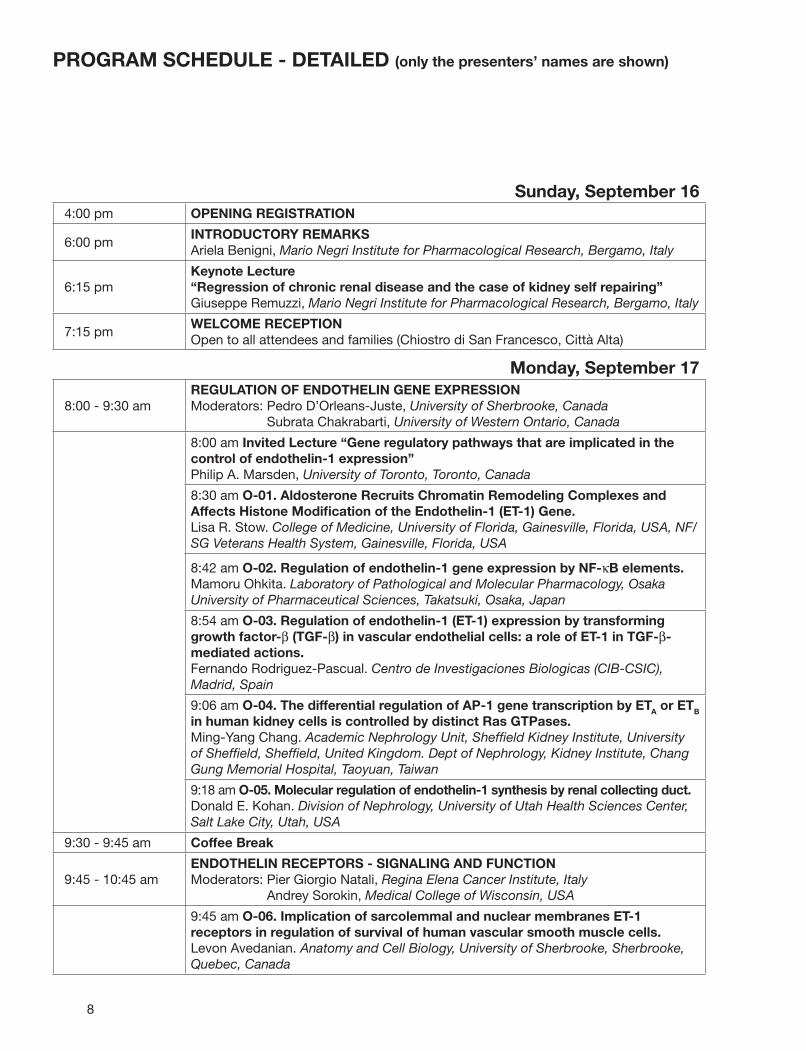
PROGRAM SCHEDULE - DETAILED (only the presenters’ names are shown)
Sunday, September 164:00 pm OPENING REGISTRATION
6:00 pmINTRODUCTORY REMARKSAriela Benigni, Mario Negri Institute for Pharmacological Research, Bergamo, Italy
6:15 pmKeynote Lecture“Regression of chronic renal disease and the case of kidney self repairing”Giuseppe Remuzzi, Mario Negri Institute for Pharmacological Research, Bergamo, Italy
7:15 pmWELCOME RECEPTIONOpen to all attendees and families (Chiostro di San Francesco, Città Alta)
Monday, September 17
8:00 - 9:30 amREGULATION OF ENDOTHELIN GENE EXPRESSION Moderators: Pedro D’Orleans-Juste, University of Sherbrooke, Canada
Subrata Chakrabarti, University of Western Ontario, Canada
8:00 am Invited Lecture “Gene regulatory pathways that are implicated in the control of endothelin-1 expression”Philip A. Marsden, University of Toronto, Toronto, Canada
8:30 am O-01. Aldosterone Recruits Chromatin Remodeling Complexes and Affects Histone Modifi cation of the Endothelin-1 (ET-1) Gene.Lisa R. Stow. College of Medicine, University of Florida, Gainesville, Florida, USA, NF/SG Veterans Health System, Gainesville, Florida, USA
8:42 am O-02. Regulation of endothelin-1 gene expression by NF-κB elements.Mamoru Ohkita. Laboratory of Pathological and Molecular Pharmacology, Osaka University of Pharmaceutical Sciences, Takatsuki, Osaka, Japan
8:54 am O-03. Regulation of endothelin-1 (ET-1) expression by transforming growth factor-β (TGF-β) in vascular endothelial cells: a role of ET-1 in TGF-β-mediated actions.Fernando Rodriguez-Pascual. Centro de Investigaciones Biologicas (CIB-CSIC), Madrid, Spain
9:06 am O-04. The differential regulation of AP-1 gene transcription by ETA or ETB in human kidney cells is controlled by distinct Ras GTPases.Ming-Yang Chang. Academic Nephrology Unit, Sheffi eld Kidney Institute, University of Sheffi eld, Sheffi eld, United Kingdom. Dept of Nephrology, Kidney Institute, Chang Gung Memorial Hospital, Taoyuan, Taiwan
9:18 am O-05. Molecular regulation of endothelin-1 synthesis by renal collecting duct.Donald E. Kohan. Division of Nephrology, University of Utah Health Sciences Center, Salt Lake City, Utah, USA
9:30 - 9:45 am Coffee Break
9:45 - 10:45 amENDOTHELIN RECEPTORS - SIGNALING AND FUNCTIONModerators: Pier Giorgio Natali, Regina Elena Cancer Institute, Italy
Andrey Sorokin, Medical College of Wisconsin, USA
9:45 am O-06. Implication of sarcolemmal and nuclear membranes ET-1 receptors in regulation of survival of human vascular smooth muscle cells.Levon Avedanian. Anatomy and Cell Biology, University of Sherbrooke, Sherbrooke, Quebec, Canada
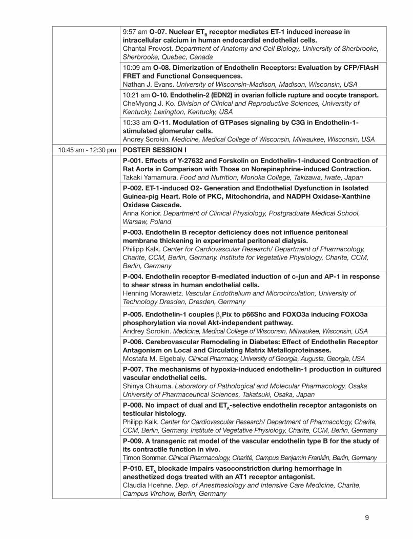
9
9:57 am O-07. Nuclear ETB receptor mediates ET-1 induced increase in intracellular calcium in human endocardial endothelial cells.Chantal Provost. Department of Anatomy and Cell Biology, University of Sherbrooke, Sherbrooke, Quebec, Canada
10:09 am O-08. Dimerization of Endothelin Receptors: Evaluation by CFP/FlAsH FRET and Functional Consequences.Nathan J. Evans. University of Wisconsin-Madison, Madison, Wisconsin, USA
10:21 am O-10. Endothelin-2 (EDN2) in ovarian follicle rupture and oocyte transport.CheMyong J. Ko. Division of Clinical and Reproductive Sciences, University of Kentucky, Lexington, Kentucky, USA
10:33 am O-11. Modulation of GTPases signaling by C3G in Endothelin-1-stimulated glomerular cells.Andrey Sorokin. Medicine, Medical College of Wisconsin, Milwaukee, Wisconsin, USA
10:45 am - 12:30 pm POSTER SESSION I
P-001. Effects of Y-27632 and Forskolin on Endothelin-1-induced Contraction of Rat Aorta in Comparison with Those on Norepinephrine-induced Contraction.Takaki Yamamura. Food and Nutrition, Morioka College, Takizawa, Iwate, Japan
P-002. ET-1-induced O2- Generation and Endothelial Dysfunction in Isolated Guinea-pig Heart. Role of PKC, Mitochondria, and NADPH Oxidase-Xanthine Oxidase Cascade.Anna Konior. Department of Clinical Physiology, Postgraduate Medical School, Warsaw, Poland
P-003. Endothelin B receptor defi ciency does not infl uence peritoneal membrane thickening in experimental peritoneal dialysis.Philipp Kalk. Center for Cardiovascular Research/ Department of Pharmacology, Charite, CCM, Berlin, Germany. Institute for Vegetative Physiology, Charite, CCM, Berlin, Germany
P-004. Endothelin receptor B-mediated induction of c-jun and AP-1 in response to shear stress in human endothelial cells.Henning Morawietz. Vascular Endothelium and Microcirculation, University of Technology Dresden, Dresden, Germany
P-005. Endothelin-1 couples β1Pix to p66Shc and FOXO3a inducing FOXO3a phosphorylation via novel Akt-independent pathway.Andrey Sorokin. Medicine, Medical College of Wisconsin, Milwaukee, Wisconsin, USA
P-006. Cerebrovascular Remodeling in Diabetes: Effect of Endothelin Receptor Antagonism on Local and Circulating Matrix Metalloproteinases.Mostafa M. Elgebaly. Clinical Pharmacy, University of Georgia, Augusta, Georgia, USA
P-007. The mechanisms of hypoxia-induced endothelin-1 production in cultured vascular endothelial cells.Shinya Ohkuma. Laboratory of Pathological and Molecular Pharmacology, Osaka University of Pharmaceutical Sciences, Takatsuki, Osaka, Japan
P-008. No impact of dual and ETA-selective endothelin receptor antagonists on testicular histology.Philipp Kalk. Center for Cardiovascular Research/ Department of Pharmacology, Charite, CCM, Berlin, Germany. Institute of Vegetative Physiology, Charite, CCM, Berlin, Germany
P-009. A transgenic rat model of the vascular endothelin type B for the study of its contractile function in vivo.Timon Sommer. Clinical Pharmacology, Charité, Campus Benjamin Franklin, Berlin, Germany
P-010. ETA blockade impairs vasoconstriction during hemorrhage in anesthetized dogs treated with an AT1 receptor antagonist.Claudia Hoehne. Dep. of Anesthesiology and Intensive Care Medicine, Charite, Campus Virchow, Berlin, Germany
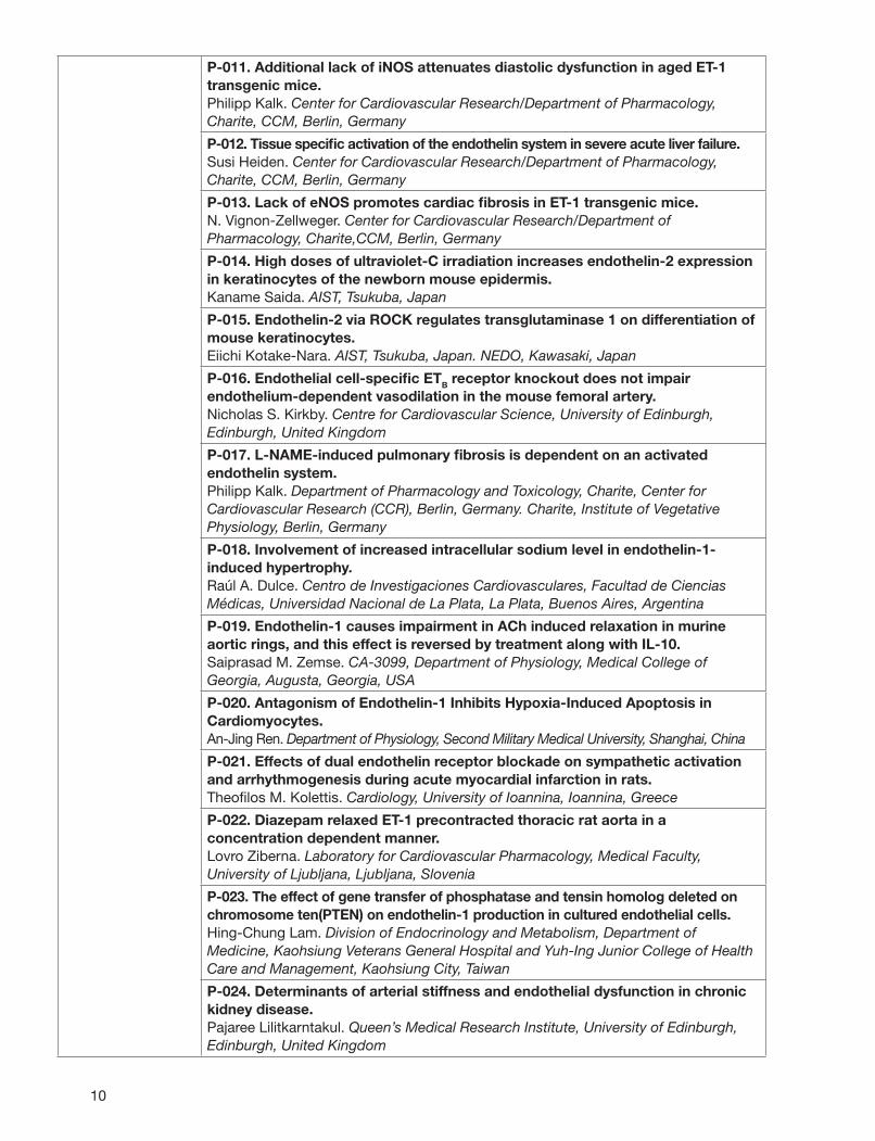
10
P-011. Additional lack of iNOS attenuates diastolic dysfunction in aged ET-1 transgenic mice.Philipp Kalk. Center for Cardiovascular Research/Department of Pharmacology, Charite, CCM, Berlin, Germany
P-012. Tissue specifi c activation of the endothelin system in severe acute liver failure.Susi Heiden. Center for Cardiovascular Research/Department of Pharmacology, Charite, CCM, Berlin, Germany
P-013. Lack of eNOS promotes cardiac fi brosis in ET-1 transgenic mice.N. Vignon-Zellweger. Center for Cardiovascular Research/Department of Pharmacology, Charite,CCM, Berlin, Germany
P-014. High doses of ultraviolet-C irradiation increases endothelin-2 expression in keratinocytes of the newborn mouse epidermis.Kaname Saida. AIST, Tsukuba, Japan
P-015. Endothelin-2 via ROCK regulates transglutaminase 1 on differentiation of mouse keratinocytes.Eiichi Kotake-Nara. AIST, Tsukuba, Japan. NEDO, Kawasaki, Japan
P-016. Endothelial cell-specifi c ETB receptor knockout does not impair endothelium-dependent vasodilation in the mouse femoral artery.Nicholas S. Kirkby. Centre for Cardiovascular Science, University of Edinburgh, Edinburgh, United Kingdom
P-017. L-NAME-induced pulmonary fi brosis is dependent on an activated endothelin system.Philipp Kalk. Department of Pharmacology and Toxicology, Charite, Center for Cardiovascular Research (CCR), Berlin, Germany. Charite, Institute of Vegetative Physiology, Berlin, Germany
P-018. Involvement of increased intracellular sodium level in endothelin-1-induced hypertrophy.Raúl A. Dulce. Centro de Investigaciones Cardiovasculares, Facultad de Ciencias Médicas, Universidad Nacional de La Plata, La Plata, Buenos Aires, Argentina
P-019. Endothelin-1 causes impairment in ACh induced relaxation in murine aortic rings, and this effect is reversed by treatment along with IL-10.Saiprasad M. Zemse. CA-3099, Department of Physiology, Medical College of Georgia, Augusta, Georgia, USA
P-020. Antagonism of Endothelin-1 Inhibits Hypoxia-Induced Apoptosis in Cardiomyocytes.An-Jing Ren. Department of Physiology, Second Military Medical University, Shanghai, China
P-021. Effects of dual endothelin receptor blockade on sympathetic activation and arrhythmogenesis during acute myocardial infarction in rats.Theofi los M. Kolettis. Cardiology, University of Ioannina, Ioannina, Greece
P-022. Diazepam relaxed ET-1 precontracted thoracic rat aorta in a concentration dependent manner.Lovro Ziberna. Laboratory for Cardiovascular Pharmacology, Medical Faculty, University of Ljubljana, Ljubljana, Slovenia
P-023. The effect of gene transfer of phosphatase and tensin homolog deleted on chromosome ten(PTEN) on endothelin-1 production in cultured endothelial cells.Hing-Chung Lam. Division of Endocrinology and Metabolism, Department of Medicine, Kaohsiung Veterans General Hospital and Yuh-Ing Junior College of Health Care and Management, Kaohsiung City, Taiwan
P-024. Determinants of arterial stiffness and endothelial dysfunction in chronic kidney disease.Pajaree Lilitkarntakul. Queen’s Medical Research Institute, University of Edinburgh, Edinburgh, United Kingdom
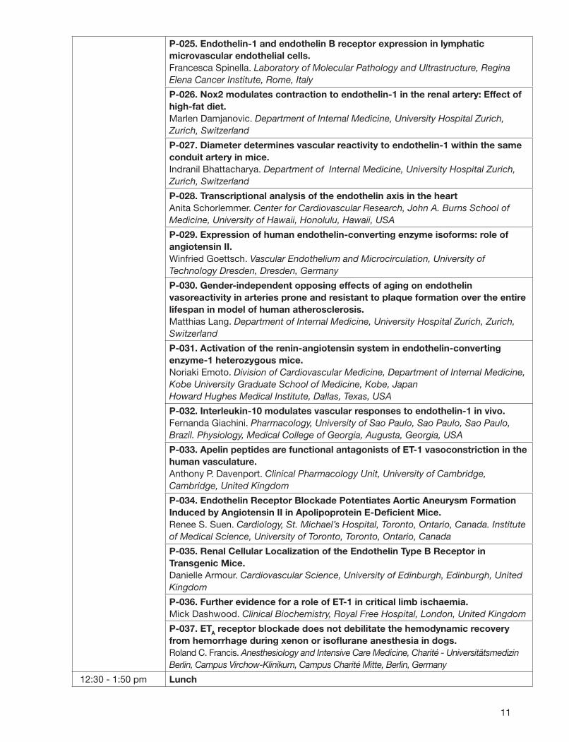
11
P-025. Endothelin-1 and endothelin B receptor expression in lymphatic microvascular endothelial cells.Francesca Spinella. Laboratory of Molecular Pathology and Ultrastructure, Regina Elena Cancer Institute, Rome, Italy
P-026. Nox2 modulates contraction to endothelin-1 in the renal artery: Effect of high-fat diet.Marlen Damjanovic. Department of Internal Medicine, University Hospital Zurich, Zurich, Switzerland
P-027. Diameter determines vascular reactivity to endothelin-1 within the same conduit artery in mice.Indranil Bhattacharya. Department of Internal Medicine, University Hospital Zurich, Zurich, Switzerland
P-028. Transcriptional analysis of the endothelin axis in the heartAnita Schorlemmer. Center for Cardiovascular Research, John A. Burns School of Medicine, University of Hawaii, Honolulu, Hawaii, USA
P-029. Expression of human endothelin-converting enzyme isoforms: role of angiotensin II.Winfried Goettsch. Vascular Endothelium and Microcirculation, University of Technology Dresden, Dresden, Germany
P-030. Gender-independent opposing effects of aging on endothelin vasoreactivity in arteries prone and resistant to plaque formation over the entire lifespan in model of human atherosclerosis.Matthias Lang. Department of Internal Medicine, University Hospital Zurich, Zurich, Switzerland
P-031. Activation of the renin-angiotensin system in endothelin-converting enzyme-1 heterozygous mice.Noriaki Emoto. Division of Cardiovascular Medicine, Department of Internal Medicine, Kobe University Graduate School of Medicine, Kobe, JapanHoward Hughes Medical Institute, Dallas, Texas, USA
P-032. Interleukin-10 modulates vascular responses to endothelin-1 in vivo.Fernanda Giachini. Pharmacology, University of Sao Paulo, Sao Paulo, Sao Paulo, Brazil. Physiology, Medical College of Georgia, Augusta, Georgia, USA
P-033. Apelin peptides are functional antagonists of ET-1 vasoconstriction in the human vasculature.Anthony P. Davenport. Clinical Pharmacology Unit, University of Cambridge, Cambridge, United Kingdom
P-034. Endothelin Receptor Blockade Potentiates Aortic Aneurysm Formation Induced by Angiotensin II in Apolipoprotein E-Defi cient Mice.Renee S. Suen. Cardiology, St. Michael’s Hospital, Toronto, Ontario, Canada. Institute of Medical Science, University of Toronto, Toronto, Ontario, Canada
P-035. Renal Cellular Localization of the Endothelin Type B Receptor in Transgenic Mice.Danielle Armour. Cardiovascular Science, University of Edinburgh, Edinburgh, United Kingdom
P-036. Further evidence for a role of ET-1 in critical limb ischaemia.Mick Dashwood. Clinical Biochemistry, Royal Free Hospital, London, United Kingdom
P-037. ETA receptor blockade does not debilitate the hemodynamic recovery from hemorrhage during xenon or isofl urane anesthesia in dogs.Roland C. Francis. Anesthesiology and Intensive Care Medicine, Charité - Universitätsmedizin Berlin, Campus Virchow-Klinikum, Campus Charité Mitte, Berlin, Germany
12:30 - 1:50 pm Lunch

12
1:50 - 3:10 pmPHYSIOLOGY OF ENDOTHELINS Moderators: Matthias Barton, University Hospital, Zurich, Switzerland
Anthony P. Davenport, University of Cambridge, UK
1:50 pm Invited Lecture “Tails of endothelin: advances in understanding cardiorenal physiology using genetically modifi ed animals”Donald E. Kohan, University of Utah, Salt Lake City, USA
2:20 pm O-12. Being starved and cold - The life without endothelin-2.Inik Chang. Department of Molecular Genetics, University of Texas Southwestern Medical Center, Dallas, Texas, USA. Howard Hughes Medical Institute, Chevy Chase, Maryland, USA
2:32 pm O-13. Identifi cation of ECE independent pressor response to Big endothelin-1 in the mouse.Elie Simard. Pharmacology, Université of Sherbrooke, Sherbrooke, Quebec, Canada
2:44 pm O-14. Urinary protein profi ling with surface enhanced laser desorption/ionisation-time of fl ight-mass spectrometry (SELDI-TOF-MS) in endothelin B receptor-defi cient rats.Jens Raila. Institute of Nutritional Science, University of Potsdam, Potsdam, Brandenburg, Germany
2:56 pm O-15. Existence of Immunoreactive ETA and ETB Receptors in the Urine of Normal Volunteers.Tony Karram. Vascular Surgery, Rambam Medical Center, Haifa, Israel
3:10 - 3:30 pm Coffee Break
3:30 - 5:00 pmATHEROSCLEROSIS, COMMON AND RARE VASCULAR DISEASES Moderators: Adviye Ergul, Medical College of Georgia, USA
Michael Dashwood, Royal Free Hospital, London, UK
3:30 pm O-16. Disturbed Flow induced Lower SMC-rich Neointimal Lesion in Mice Lacking Vascular Endothelial Cell Endothelin-1 : Role of ET-1 in Vascular Infl ammation.Dyah Wulan Anggrahini. Division of Cardiovascular Medicine, Department of Internal Medicine, Kobe University Graduate School of Medicine, Kobe, Hyogo, Japan
3:42 pm O-17. Vascular hyperreactivity to ET-1 in aorta of apoE-/- mice on high fat diet is rescued by apoptotic ablation of smooth muscle cells in novel transgenic mouse model of atherosclerosis.Janet Maguire. Clinical Pharmacology Unit, University of Cambridge, Cambridge, United Kingdom
3:54 pm O-18. Oxidative stress-induced, poly (ADP-ribose) polymerase-dependent, upregulation of ET-1 expression in chronic diabetic complications.Jane Chiu. Pathology, The University of Western Ontario, London, Ontario, Canada
4:06 pm O-19. Endothelin Receptor A antagonism ameliorates hypoperfusion and improves behavioral outcome following brain trauma.Christian Kreipke. Anatomy and Cell Biology, Wayne State University, Detroit, Michigan, USA
4:18 pm O-20. Endothelin-1 activates Mesenchymal Progenitor Cells in Tissue Fibrosis.Xu Shiwen. Rheumatology, UCL, London, United Kingdom
4:30 pm O-21. Endothelin receptor blockade improves endothelial function in patients with dysglycemia and coronary artery disease on aggressive lipid-lowering.Magnus Settergren. Karolinska University Hospital, Stockholm, Sweden
4:42 pm O-22. Impaired Vascular Mechanics and Function in Hyperglycemia and Hyperlipidemia - Association with Endothelin.Kamakshi Sachidanandam. Clinical and Administrative Pharmacy, University of Georgia, Augusta, Georgia, USA
13
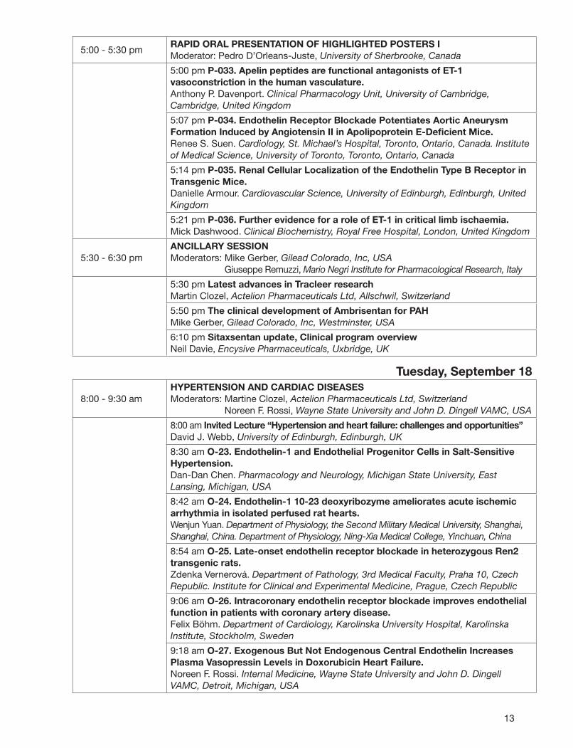
5:00 - 5:30 pmRAPID ORAL PRESENTATION OF HIGHLIGHTED POSTERS I Moderator: Pedro D’Orleans-Juste, University of Sherbrooke, Canada
5:00 pm P-033. Apelin peptides are functional antagonists of ET-1 vasoconstriction in the human vasculature.Anthony P. Davenport. Clinical Pharmacology Unit, University of Cambridge, Cambridge, United Kingdom
5:07 pm P-034. Endothelin Receptor Blockade Potentiates Aortic Aneurysm Formation Induced by Angiotensin II in Apolipoprotein E-Defi cient Mice.Renee S. Suen. Cardiology, St. Michael’s Hospital, Toronto, Ontario, Canada. Institute of Medical Science, University of Toronto, Toronto, Ontario, Canada
5:14 pm P-035. Renal Cellular Localization of the Endothelin Type B Receptor in Transgenic Mice.Danielle Armour. Cardiovascular Science, University of Edinburgh, Edinburgh, United Kingdom
5:21 pm P-036. Further evidence for a role of ET-1 in critical limb ischaemia.Mick Dashwood. Clinical Biochemistry, Royal Free Hospital, London, United Kingdom
5:30 - 6:30 pmANCILLARY SESSION Moderators: Mike Gerber, Gilead Colorado, Inc, USA
Giuseppe Remuzzi, Mario Negri Institute for Pharmacological Research, Italy
5:30 pm Latest advances in Tracleer researchMartin Clozel, Actelion Pharmaceuticals Ltd, Allschwil, Switzerland
5:50 pm The clinical development of Ambrisentan for PAHMike Gerber, Gilead Colorado, Inc, Westminster, USA
6:10 pm Sitaxsentan update, Clinical program overviewNeil Davie, Encysive Pharmaceuticals, Uxbridge, UK
Tuesday, September 18
8:00 - 9:30 amHYPERTENSION AND CARDIAC DISEASESModerators: Martine Clozel, Actelion Pharmaceuticals Ltd, Switzerland
Noreen F. Rossi, Wayne State University and John D. Dingell VAMC, USA
8:00 am Invited Lecture “Hypertension and heart failure: challenges and opportunities”David J. Webb, University of Edinburgh, Edinburgh, UK
8:30 am O-23. Endothelin-1 and Endothelial Progenitor Cells in Salt-Sensitive Hypertension.Dan-Dan Chen. Pharmacology and Neurology, Michigan State University, East Lansing, Michigan, USA
8:42 am O-24. Endothelin-1 10-23 deoxyribozyme ameliorates acute ischemic arrhythmia in isolated perfused rat hearts.Wenjun Yuan. Department of Physiology, the Second Military Medical University, Shanghai, Shanghai, China. Department of Physiology, Ning-Xia Medical College, Yinchuan, China
8:54 am O-25. Late-onset endothelin receptor blockade in heterozygous Ren2 transgenic rats.Zdenka Vernerová. Department of Pathology, 3rd Medical Faculty, Praha 10, Czech Republic. Institute for Clinical and Experimental Medicine, Prague, Czech Republic
9:06 am O-26. Intracoronary endothelin receptor blockade improves endothelial function in patients with coronary artery disease.Felix Böhm. Department of Cardiology, Karolinska University Hospital, Karolinska Institute, Stockholm, Sweden
9:18 am O-27. Exogenous But Not Endogenous Central Endothelin Increases Plasma Vasopressin Levels in Doxorubicin Heart Failure.Noreen F. Rossi. Internal Medicine, Wayne State University and John D. Dingell VAMC, Detroit, Michigan, USA
14
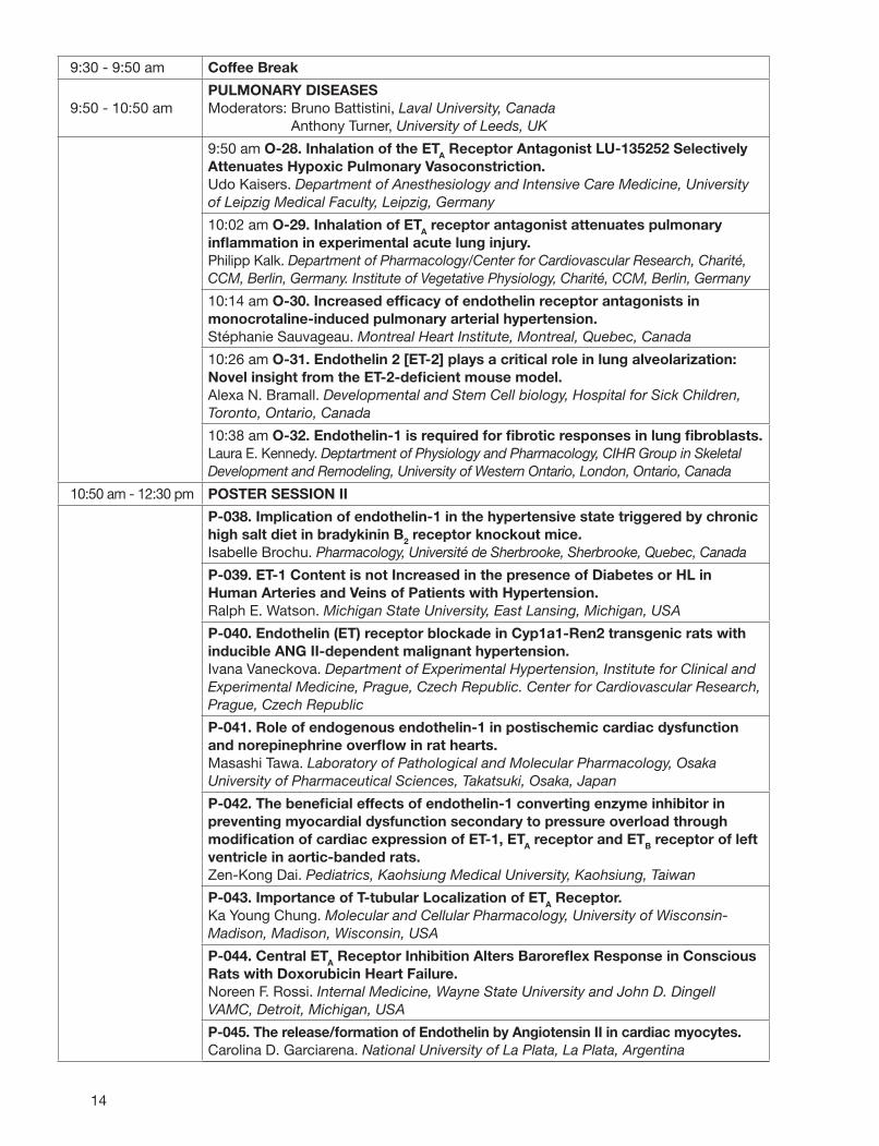
9:30 - 9:50 am Coffee Break
9:50 - 10:50 amPULMONARY DISEASESModerators: Bruno Battistini, Laval University, Canada
Anthony Turner, University of Leeds, UK
9:50 am O-28. Inhalation of the ETA Receptor Antagonist LU-135252 Selectively Attenuates Hypoxic Pulmonary Vasoconstriction.Udo Kaisers. Department of Anesthesiology and Intensive Care Medicine, University of Leipzig Medical Faculty, Leipzig, Germany
10:02 am O-29. Inhalation of ETA receptor antagonist attenuates pulmonary infl ammation in experimental acute lung injury.Philipp Kalk. Department of Pharmacology/Center for Cardiovascular Research, Charité, CCM, Berlin, Germany. Institute of Vegetative Physiology, Charité, CCM, Berlin, Germany
10:14 am O-30. Increased effi cacy of endothelin receptor antagonists in monocrotaline-induced pulmonary arterial hypertension.Stéphanie Sauvageau. Montreal Heart Institute, Montreal, Quebec, Canada
10:26 am O-31. Endothelin 2 [ET-2] plays a critical role in lung alveolarization: Novel insight from the ET-2-defi cient mouse model.Alexa N. Bramall. Developmental and Stem Cell biology, Hospital for Sick Children, Toronto, Ontario, Canada
10:38 am O-32. Endothelin-1 is required for fi brotic responses in lung fi broblasts.Laura E. Kennedy. Deptartment of Physiology and Pharmacology, CIHR Group in Skeletal Development and Remodeling, University of Western Ontario, London, Ontario, Canada
10:50 am - 12:30 pm POSTER SESSION II
P-038. Implication of endothelin-1 in the hypertensive state triggered by chronic high salt diet in bradykinin B2 receptor knockout mice.Isabelle Brochu. Pharmacology, Université de Sherbrooke, Sherbrooke, Quebec, Canada
P-039. ET-1 Content is not Increased in the presence of Diabetes or HL in Human Arteries and Veins of Patients with Hypertension.Ralph E. Watson. Michigan State University, East Lansing, Michigan, USA
P-040. Endothelin (ET) receptor blockade in Cyp1a1-Ren2 transgenic rats with inducible ANG II-dependent malignant hypertension.Ivana Vaneckova. Department of Experimental Hypertension, Institute for Clinical and Experimental Medicine, Prague, Czech Republic. Center for Cardiovascular Research, Prague, Czech Republic
P-041. Role of endogenous endothelin-1 in postischemic cardiac dysfunction and norepinephrine overfl ow in rat hearts.Masashi Tawa. Laboratory of Pathological and Molecular Pharmacology, Osaka University of Pharmaceutical Sciences, Takatsuki, Osaka, Japan
P-042. The benefi cial effects of endothelin-1 converting enzyme inhibitor in preventing myocardial dysfunction secondary to pressure overload through modifi cation of cardiac expression of ET-1, ETA receptor and ETB receptor of left ventricle in aortic-banded rats.Zen-Kong Dai. Pediatrics, Kaohsiung Medical University, Kaohsiung, Taiwan
P-043. Importance of T-tubular Localization of ETA Receptor.Ka Young Chung. Molecular and Cellular Pharmacology, University of Wisconsin-Madison, Madison, Wisconsin, USA
P-044. Central ETA Receptor Inhibition Alters Barorefl ex Response in Conscious Rats with Doxorubicin Heart Failure.Noreen F. Rossi. Internal Medicine, Wayne State University and John D. Dingell VAMC, Detroit, Michigan, USA
P-045. The release/formation of Endothelin by Angiotensin II in cardiac myocytes.Carolina D. Garciarena. National University of La Plata, La Plata, Argentina
15
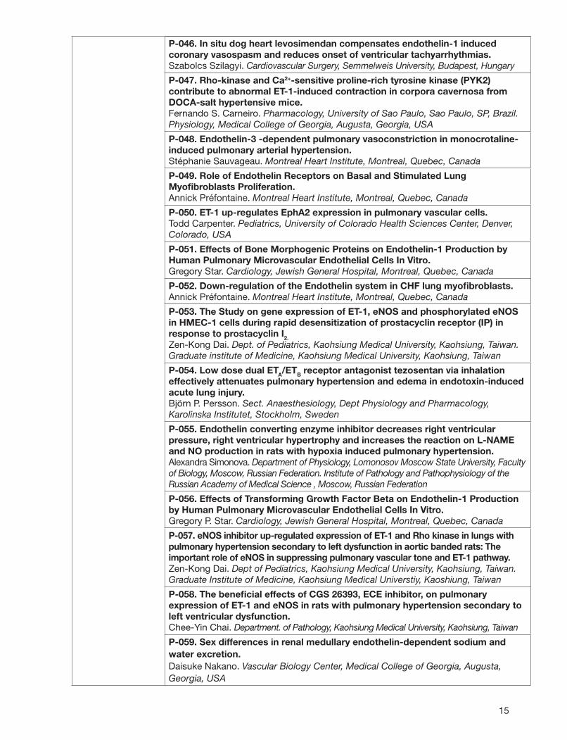
P-046. In situ dog heart levosimendan compensates endothelin-1 induced coronary vasospasm and reduces onset of ventricular tachyarrhythmias.Szabolcs Szilagyi. Cardiovascular Surgery, Semmelweis University, Budapest, Hungary
P-047. Rho-kinase and Ca2+-sensitive proline-rich tyrosine kinase (PYK2) contribute to abnormal ET-1-induced contraction in corpora cavernosa from DOCA-salt hypertensive mice.Fernando S. Carneiro. Pharmacology, University of Sao Paulo, Sao Paulo, SP, Brazil. Physiology, Medical College of Georgia, Augusta, Georgia, USA
P-048. Endothelin-3 -dependent pulmonary vasoconstriction in monocrotaline-induced pulmonary arterial hypertension.Stéphanie Sauvageau. Montreal Heart Institute, Montreal, Quebec, Canada
P-049. Role of Endothelin Receptors on Basal and Stimulated Lung Myofi broblasts Proliferation.Annick Préfontaine. Montreal Heart Institute, Montreal, Quebec, Canada
P-050. ET-1 up-regulates EphA2 expression in pulmonary vascular cells.Todd Carpenter. Pediatrics, University of Colorado Health Sciences Center, Denver, Colorado, USA
P-051. Effects of Bone Morphogenic Proteins on Endothelin-1 Production by Human Pulmonary Microvascular Endothelial Cells In Vitro.Gregory Star. Cardiology, Jewish General Hospital, Montreal, Quebec, Canada
P-052. Down-regulation of the Endothelin system in CHF lung myofi broblasts.Annick Préfontaine. Montreal Heart Institute, Montreal, Quebec, Canada
P-053. The Study on gene expression of ET-1, eNOS and phosphorylated eNOS in HMEC-1 cells during rapid desensitization of prostacyclin receptor (IP) in response to prostacyclin I2.Zen-Kong Dai. Dept. of Pediatrics, Kaohsiung Medical University, Kaohsiung, Taiwan. Graduate institute of Medicine, Kaohsiung Medical University, Kaohsiung, Taiwan
P-054. Low dose dual ETA/ETB receptor antagonist tezosentan via inhalation effectively attenuates pulmonary hypertension and edema in endotoxin-induced acute lung injury.Björn P. Persson. Sect. Anaesthesiology, Dept Physiology and Pharmacology, Karolinska Institutet, Stockholm, Sweden
P-055. Endothelin converting enzyme inhibitor decreases right ventricular pressure, right ventricular hypertrophy and increases the reaction on L-NAME and NO production in rats with hypoxia induced pulmonary hypertension.Alexandra Simonova. Department of Physiology, Lomonosov Moscow State University, Faculty of Biology, Moscow, Russian Federation. Institute of Pathology and Pathophysiology of the Russian Academy of Medical Science , Moscow, Russian Federation
P-056. Effects of Transforming Growth Factor Beta on Endothelin-1 Production by Human Pulmonary Microvascular Endothelial Cells In Vitro.Gregory P. Star. Cardiology, Jewish General Hospital, Montreal, Quebec, Canada
P-057. eNOS inhibitor up-regulated expression of ET-1 and Rho kinase in lungs with pulmonary hypertension secondary to left dysfunction in aortic banded rats: The important role of eNOS in suppressing pulmonary vascular tone and ET-1 pathway.Zen-Kong Dai. Dept of Pediatrics, Kaohsiung Medical University, Kaohsiung, Taiwan. Graduate Institute of Medicine, Kaohsiung Medical Universtiy, Kaoshiung, Taiwan
P-058. The benefi cial effects of CGS 26393, ECE inhibitor, on pulmonary expression of ET-1 and eNOS in rats with pulmonary hypertension secondary to left ventricular dysfunction.Chee-Yin Chai. Department. of Pathology, Kaohsiung Medical University, Kaohsiung, Taiwan
P-059. Sex differences in renal medullary endothelin-dependent sodium and water excretion.Daisuke Nakano. Vascular Biology Center, Medical College of Georgia, Augusta, Georgia, USA
16
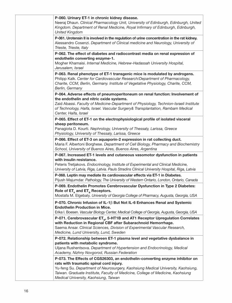
P-060. Urinary ET-1 in chronic kidney disease.Neeraj Dhaun. Clinical Pharmacology Unit, University of Edinburgh, Edinburgh, United Kingdom. Department of Renal Medicine, Royal Infi rmary of Edinburgh, Edinburgh, United Kingdom
P-061. Urotensin II is involved in the regulation of urine concentration in the rat kidney.Alessandro Cosenzi. Department of Clinical medicine and Neurology, University of Trieste, Trieste, Italy
P-062. The effect of diabetes and radiocontrast media on renal expression of endothelin converting enzyme-1.Mogher Khamaisi. Internal Medicine, Hebrew-Hadassah University Hospital, Jerusalem, Israel
P-063. Renal phenotype of ET-1 transgenic mice is modulated by androgens.Philipp Kalk. Center for Cardiovascular Research/Department of Pharmacology, Charite, CCM, Berlin, Germany. Institute of Vegetative Physiology, Charite, CCM, Berlin, Germany
P-064. Adverse effects of pneumoperitoneum on renal function: Involvement of the endothelin and nitric oxide systems.Zaid Abassi. Faculty of Medicine-Department of Physiology, Technion-Israeli Institute of Technology, Haifa, Israel. Vascular Surgery& Transplantation, Rambam Medical Center, Haifa, Israel
P-065. Effect of ET-1 on the electrophysiological profi le of isolated visceral sheep peritoneum.Panagiota D. Kourti. Nephrology, University of Thessaly, Larissa, GreecePhysiology, University of Thessaly, Larissa, Greece
P-066. Effect of ET-3 on aquaporin-2 expression in rat collecting duct.Maria F. Albertoni Borghese. Department of Cell Biology, Pharmacy and Biochemistry School, University of Buenos Aires, Buenos Aires, Argentina
P-067. Increased ET-1 levels and cutaneous vasomotor dysfunction in patients with insulin resistance.Peteris Tretjakovs. Endocrinology, Institute of Experimental and Clinical Medicine, University of Latvia, Riga, Latvia. Pauls Stradins Clinical University Hospital, Riga, Latvia
P-068. Leptin may mediate its cardiovascular effects via ET-1 in Diabetes.Pijush Majumdar. Pathology, The University of Western Ontario, London, Ontario, Canada
P-069. Endothelin Promotes Cerebrovascular Dysfunction in Type 2 Diabetes: Role of ETA and ETB Receptors.Mostafa M. Elgebaly. University of Georgia College of Pharmacy, Augusta, Georgia, USA
P-070. Chronic Infusion of IL-1β But Not IL-6 Enhances Renal and Systemic Endothelin Production in Mice.Erika I. Boesen. Vascular Biology Center, Medical College of Georgia, Augusta, Georgia, USA
P-071. Cerebrovascular ETB, 5-HT1B and AT1 Receptor Upregulation Correlates with Reduction in Regional CBF after Subarachnoid Hemorrhage.Saema Ansar. Clinical Sciences, Division of Experimental Vascular Research, Medicine, Lund University, Lund, Sweden
P-072. Relationship between ET-1 plasma level and vegetative dysbalance in patients with metabolic syndrome.Uljana Rushentsova. Department of Hypertension and Endocrinology, Medical Academy, Nizhny Novgorod, Russian Federation
P-073. The Effects of CGS26303, an endothelin-converting enzyme inhibitor on rats with traumatic spinal cord injury.Yu-feng Su. Department of Neurosurgery, Kaohsiung Medical University, Kaohsiung, Taiwan. Graduate Institute, Faculty of Medicine, College of Medicine, Kaohsiung Medical University, Kaohsiung, Taiwan
17
P-074. Differential expression of endothelin receptors in rat’s hippocampus following global ischemia.Syed Mohiuddin. Anatomy and Cell Biol., Wayne State Univ., Detroit, Michigan, USA
P-075. Phospholipase C, Protein Kinase C and MAP Kinases Mediate Overt Nociception and Thermal Hyperalgesia Induced by ET-1 in Rats.Giles A. Rae. Pharmacology, Universidade Federal Santa Catarina, Florianopolis, Santa Catarina, Brazil
P-076. The Immune Mandatory Effect of 6-Mercaptopurine Attenuates Endothelin and Chronic Vasospasm.Aij-Lie Kwan. Department of Neurosurgery, Kaohsiung Medical University Hospital, Kaohsiung, Taiwan. Faculty of Medicine, Graduate Institute of Medicine, College of Medicine, Kaohsiung Medical University, Kaohsiung, Taiwan
P-077. Phosphoenolpyruvate analogue -Fosfomycin Attenuates Endothelin in Experimental Vasospasm.An-Kuo Chou. Faculty of Medicine, Graduate Institute of Medicine, College of Medicine, Kaohsiung Medical University, Kaohsiung, Taiwan. Department of Anesthesiology, Chang Gung Memorial Hospital-Kaohsiung Medical Center, Chang Gung University College of Medicine, Kaohsiung, Taiwan
P-078. Cell downstream signal inhibitor Sirolimus alleviates production of endothelin and prevents experimental subarachnoid hemorrhage induced vasospasm.Tai-I Chen. Department of Anesthesiology, Kaohsiung Medical University, Kaohsiung, Taiwan
P-079. Plasma ET-1 level in patients with primary or metastatic brain tumors.ShenLong Howng. Neurosurgery, Kaohsiung Medical University, Kaohsiung, Taiwan. Faculty of Medicine, Graduate Institute of Medicine, College of Medicine, Kaohsiung Medical University, Kaohsiung, Taiwan
P-080. The Role of Rho-associate Kinase and Soluble guanylyl cyclase in Cerebral Vasospasm following SAH.Pei-yu Lee. Graduate Institute of Pharmacology, Kaohsiung Medical University, Kaohsiung, Taiwan
P-081. CGS 26303 treatment for two days upregulates mRNA expression of endothelial nitric oxide synthase in brain tissue of rats subjected to experimental subarachnoid hemorrhage.Sheng-I Lue. Department of Physiology, Kaohsiung Medical University Hospital, Kaohsiung, Taiwan
P-082. Increase in the expression of ET-1 in the rat lung after experimental subarachnoid hemorrhage.Ann-Shung Lieu. Neurosurgery, Kaohsiung Medical University, Kaohsiung, Taiwan. Faculty of Medicine, Graduate Institute of Medicine, College of Medicine, Kaohsiung Medical University, Kaohsiung, Taiwan
P-083. ETB2 stimulation relaxes the iris sphincter muscle.Amândio A. Rocha-Sousa. Department of Physiology, Faculty of Medicine University of Porto, Porto, Portugal. Department of Ophthalmology, Faculty of Medicine, University of Porto, Porto, Portugal
P-084. Obesity induces the endothelin system in the murine heart.Gregor Muller. Vascular Endothelium and Microcirculation, University of Technology Dresden, Germany
P-085. Overexpression of human ET-2 aggravates diabetic cardiomyopathy in rats.Bartosz Rylski. Department of Nephrology, Charité - Universitätsmedizin Berlin, Campus Charité Mitte, Berlin, Germany
P-086. Activation of renal medullary ETB receptor induces diuresis and natriuresis via nitric oxide synthase 1, cGMP and protein kinase G pathways.Daisuke Nakano. Vascular Biology Center, Medical College of Georgia, Augusta, Georgia, USA

18
P-087. Contralateral nociceptive sensitization following the subcutaneous injection of ET-1: Evidence for central sensitization.Gary Strichartz. Pain Research Center, Brigham and Women’s Hospital, Boston, Massachusetts, USA
P-088. Palosuran, an oral U-II antagonist, prevents the cardiac effects of high fructose diet in rats.Alessandro Cosenzi. Department of Clinical Medicine and Neurology, University of Trieste, Trieste, Italy
P-089. The Effect of KMUVS-1 in Cerebrovasospasm after SAH via Inhibition of ET-1 Production.Jwu-Lai Yeh. Graduate Institute of Pharmacology, Kaohsiung Medical University, Kaohsiung, Taiwan
P-090. Increased expression of urotensin II-related peptide in the kidney of rats with hypertension or chronic renal failure.Kazuhiro Takahashi. Department of Analytical Medical Technology, Tohoku University School of Health Sciences, Sendai, Japan. Tohoku University 21st COE Program Comprehensive Research and Education Center for Planning of Drug Development and Clinical Evaluation (CRESCENDO), Sendai, Japan
P-091. Clonidine and endothelin antagonist sulfi soxazole combination produces potent analgesia in mice.Anil Gulati. Chicago College of Pharmacy, Midwestern University, Downers Grove, Illinois, USA
P-092. 17β-estradiol prevents cerebral vasospasm and endothelin-1 expression in the brain stem after subarachnoid hemorrhage.Chih-Lung Lin. Neurosurgery, Kaohsiung Medical University, Kaohsiung, Taiwan. Faculty of Medicine, Graduate Institute of Medicine, College of Medicine, Kaohsiung Medical University, Kaohsiung, Taiwan
P-093. Potential of endothelin antagonist in experimental model of cerebral stroke.Yogendra K. Gupta. Department of Pharmacology, All India Institute of Medical Sciences, New Delhi, Delhi, India
P-094. Effects of TNF-alpha blocking peptide on Endothelin-1 levels in lungs in endotoxemic rat model.Atsushi Sawamura. Department of Anesthesiology and Critical Care Medicine, Hokkaido University, Sapporo, Japan. Center of Medical Science, Ibaraki Prefectural University, Ami, Japan
12:30 - 1:45 pm Lunch
2:00 - 3:30 pmRENAL FAILURE, TRANSPLANTATION AND DIABETESModerators: Carla Zoja, Mario Negri Institute for Pharmacological Research, Italy
David Pollock, Medical College of Georgia, USA
2:00 pm O-33. ETB receptor blockade aggravates cystic disease progression in pkd2 mice via vasopressin-dependent and independent mechanisms.Ming-Yang Chang. Academic Nephrology Unit, Sheffi eld Kidney Institute, University of Sheffi eld, Sheffi eld, United Kingdom. Dept of Nephrology, Kidney Institute, Chang Gung Memorial Hospital, Taoyan, Taiwan
2:12 pm O-34. Nitric oxide mediates collecting duct ET-1 effects on blood pressure.Markus P. Schneider. Medical College of Georgia, Augusta, Georgia, USA
2:24 pm O-35. Effect of a selective ETA receptor antagonist on podocyte function and permselective properties of the glomerular barrier in experimental diabetes.Elena Gagliardini. Department of Molecular Medicine, Mario Negri Institute for Pharmacological Research, Bergamo, Italy
2:36 pm O-36. Genetic Inactivation of Vascular Endothelial Cell ET-1 in Mice Is Protective against Cardiac and Renal Complication of Diabetes.Bambang Widyantoro. Division of Cardiovascular Medicine, Department of Internal Medicine, Kobe University Graduate School of Medicine, Kobe, Japan

19
2:48 pm O-37. Impaired insulin-mediated vasorelaxation in a non-obese model of type 2 diabetes: role of endothelin-1.Mostafa M. Elgebaly. University of Georgia College of Pharmacy, Augusta, Georgia, USA
3:00 pm O-38. Endothelin-A receptor antagonism improves cardiovascular function and reduces proteinuria in patients with chronic kidney disease.Neeraj Dhaun. Clinical Pharmacology Unit, University of Edinburgh, Edinburgh, United Kingdom. Department of Renal Medicine, Royal Infi rmary of Edinburgh, Edinburgh, United Kingdom
3:12 pm O-39. Role of ETA and ETB Subtype Receptors in Tubulo-interstitial, Vascular and Glomerular Fibrosis of a Model of Ang II-Dependent Hypertension.Teresa M. Seccia. DMCS-Clinica Medica 4, University of Padova, Padova, Italy
3:30 - 3:45 pm Coffee Break
3:45 - 5:15 pmNEURAL PATHWAYS - CNS AND EYE - AND NOCICEPTIONModerators: Giles A. Rae, Universidade Federal Santa Catarina, Brazil
Thomas Yorio, University of North Texas, USA
3:45 pm O-40. Inhibition of cerebrovascular raf activation reduces late cerebral ischemia and ETB, 5-HT1B and AT1 receptor upregulation after subarachnoid hemorrhage.Saema Ansar. Clinical Sciences, Division of Experimental Vascular Research, Medicine, Lund University, Lund, Sweden
3:57 pm O-41. Attenuation of endothelin-1 (ET-1) mRNA ameliorates hypoperfusion after traumatic brain injury (TBI).Theodor Petrov. Anatomy and Cell Biol., Wayne state Univ., Detroit, Michigan, USA
4:09 pm O-42. Endothelinergic cells in the brain of mice with ischemic cortical lesions.Angela M. Suburo. Universidad Austral, Facultad de Ciencias Biomédicas, Pilar, B1629AHJ, Argentina
4:21 pm O-43. Role of the ETB receptor in retinal ganglion cell death in glaucoma.Thomas Yorio. Graduate School of Biomedical Sciences, UNT Health Science Center, Fort Worth, Texas, USA
4:33 pm O-44. Anatomical and Biochemical Mechanisms of Mechanical Nocifensive Sensitization Induced by Injection of ET-1 into the Rat Paw.Gary Strichartz. Pain Research Center, Brigham and Women’s Hospital, Boston, Massachusetts, USA
4:45 pm O-45. Peripheral Up-Regulation of Sensory Nerve ETA and ETB Receptor-Operated Mechanisms is Implicated in Neuropathic Pain Induced by Spinal Nerve Ligation.Maria Fernanda P. Werner. Pharmacology, Federal University of Santa Catarina - UFSC, Florianópolis, Santa Catarina, Brazil
4:57 pm O-46. The absence of endothelin-2 partially rescues photoreceptor (PR) death in two models of inherited photoreceptor degeneration (IPD).Alexa Bramall. Developmental and Stem Cell Biology, Hospital for Sick Children, Toronto, Ontario, Canada. Medical and Molecular Genetics, University of Toronto, Toronto, Ontario, Canada
5:15 - 5:45 pmRAPID ORAL PRESENTATION OF HIGHLIGHTED POSTERS IIModerator: Donald E. Kohan, University of Utah, USA
5:15 pm P-084. Obesity induces the endothelin system in the murine heart.Gregor Muller. Vascular Endothelium and Microcirculation, University of Technology Dresden, Germany
5:22 pm P-085. Overexpression of human ET-2 aggravates diabetic cardiomyopathy in rats.Bartosz Rylski. Department of Nephrology, Charité - Universitätsmedizin Berlin, Campus Charité Mitte, Berlin, Germany
20
5:29 pm P-086. Activation of renal medullary ETB receptor induces diuresis and natriuresis via nitric oxide synthase 1, cGMP and protein kinase G pathways.Daisuke Nakano. Vascular Biology Center, Medical College of Georgia, Augusta, Georgia, USA
5:36 pm P-087. Contralateral nociceptive sensitization following the subcutaneous injection of ET-1: Evidence for central sensitization.Gary Strichartz. Pain Research Center, Brigham and Women’s Hospital, Boston, Massachusetts, USA
7:30 - 10:00 pmSOCIAL DINNER(Centro Daccò, Mario Negri Institute for Pharmacological Research, Ranica, Bergamo)
Wednesday, September 19
8:30 - 10:15 amCANCER Moderators: Giulia Taraboletti, Mario Negri Institute for Pharmacological Research, Italy
Gregory Clines, University of Virginia, USA
8:30 am Invited Lecture: “The endothelin axis in cancer: the promise and challenges of molecularly targeted therapy”Anna Bagnato, Regina Elena Cancer Institute, Rome, Italy
9:00 am O-47. Radiation-induced survival and reduction in tumor volume in Dalton’s Lymphoma Ascites tumor model was signifi cantly enhanced by IRL-1620.Anil Gulati. Chicago College of Pharmacy, Midwestern University, Downers Grove, Illinois, USA
9:12 am O-48. Degree of differentiation of pancreatic adenocarcinoma cells determines up-regulation of ETA receptors in response to hypoxia.Eliane Angst. Clinic of Visceral and Transplant Surgery, University of Berne, Berne, Switzerland
9:24 am O-49. ET-1 Promotes Desmoplasia In Colorectal Cancer.Jonathan Knowles. Academic Surgery, UCL, London, United Kingdom
9:36 am O-50. Stromal ETB receptor-defi ciency inhibits breast cancer growth and metastasis.Hannelore Ehrenreich. MPI of Experimental Medicine, Göttingen, Germany
9:48 am O-51. siRNA molecules targeting ECE-1 as a means to inhibit ET-1 synthesis in endothelial and ovarian carcinoma cells.Oleg Rayhman. Animal Sciences, The Hebrew University of Jerusalem, Rehovot, Israel
10:00 am O-52. The interplay between the endothelin axis and hypoxic melanoma microenvironment: therapeutic implication.Francesca Spinella. Laboratory of Molecular Pathology and Ultrastructure, Regina Elena Cancer Institute, Rome, Italy
10:15-10:30 am Coffee Break
10:30 - 11:30 amINFLAMMATION, IMMUNOLOGY AND POLYMORPHISMS OF ENDOTHELIN GENESModerators: Marina Noris, Mario Negri Institute for Pharmacological Research, Italy
Noriaki Emoto, Kobe University Graduate School of Medicine, Japan
10:30 am O-53. Polymorphisms of ETBR, ATG and ACE in salt-sensitive hypertension.Jessica Caprioli. Transplant Research Center Chiara Cucchi de Alessandri e Gilberto Crespi, Mario Negri Institute for Pharmacological Research, Ranica, Bergamo, Italy
10:42 am O-54. The SNP5333 gene polymorphism of ETA receptor is independently associated with increased albuminuria in Type 2 diabetes.Silvia Orisio. Department of Molecular Medicine, Mario Negri Institute for Pharmacological Research, Bergamo, Italy
10:54 am O-55. Endothelin Receptors Play a Critical Pathophysiological Role in Sickle Cell Disease.Pierre-Louis Tharaux. Cardiovascular Research Centre Inserm Lariboisière, U689, INSERM, Paris, France. Hematology, Hôpital Tenon, Assistance Publique-Hôpitaux de Paris, Paris, France
21
11:06 am O-56. The specifi c ETA receptor antagonist ZD4054 reduces tumour-induced angiogenesis in a preclinical model.J. Curwen. Cancer & Infection Bioscience, AstraZeneca, Alderley Park, Macclesfi eld, United Kingdom
11:18 am O-57. Cell adhesion activates a fi brogenic program in fi broblasts via the ETA receptor.Andrew Leask. Oral Biology, University of Western Ontario, London, Ontario, Canada
11:30 am - 12:45 pm POSTER SESSION III
P-095. Upregulation of functional ETA receptor and downregulation of functional ETB receptor in cancer and stromal cells within colorectal cancer.Moinuddin M. Hoosein. Department of Surgery, Royal Free and University College Medical School, UCL, London, United Kingdom
P-096. Toxicokinetic Evaluation of IRL-1620 in a 4-Week Toxicology Study in Rats.Guru Reddy. Spectrum Pharmaceuticals, Inc., Irvine, California, USA
P-097. Effect of IRL-1620 on Respiration Rate and Tidal Volume in Sprague Dawley Rats.Guru Reddy. Spectrum Pharmaceuticals, Inc., Irvine, California, USA
P-098. Doxorubicin suppresses ET-1 mRNA expression in endothelial cells.Katalin Keltai. 3rd Dept of Medicine, Semmelweis University, Budapest, Hungary
P-099. ETB receptor antagonists inhibit cell proliferation in human glioblastoma cell lines.Mayra Paolillo. Dipartimento di Farmacologia Sperimentale ed Applicata, Università degli Studi di Pavia, Pavia, Italy
P-100. Improvement in the uptake and effi cacy of chemotherapeutic agents by IRL-1620 in prostate tumor rats.Anil Gulati. Chicago College of Pharmacy, Midwestern University, Downers Grove, Illinois, USA
P-101. The ET-1 metabolising proteases, ECE-1 and NEP, in human cancers.Yue Hong. Institute of Molecular and Cellular Biology, University of Leeds, Leeds, United Kingdom
P-102. The role of the ECE-1 isoforms in prostate cancer invasion.Louise A. Dawson. Institute of Molecular and Cellular Biology, University of Leeds, Leeds, United Kingdom
P-103. Kisspeptins, regulators of metastasis are novel vasoconstrictors in human arteries and veins with potency comparable to ET-1.Rhoda E. Kuc. Clinical Pharmacology, University of Cambridge, Cambridge, United Kingdom
P-104. Expression of ET-1 in human adrenal tumors and attached non-neoplastic adrenal tissues.Ryo Morimoto. Department of Nephrology, Endocrinology, and Vascular Medicine, Tohoku University Graduate School of Medicine, Sendai, Miyagi, Japan
P-105. ET-1 production by mononuclear cells in portal hypertensive patients.Abeya Lotfi . Theodor Bilharz Research Institute, Cairo, Egypt
P-106. Increased expression of endothelin-converting enzyme (ECE)-1d isoform is associated with chronic enteroviral myocarditis in mice.Hans-Dieter Orzechowski. Clinical Pharmacology and Toxicology, Charité, Berlin, Germany
P-107. Endothelin in gut radiation damage: a therapeutic target to prevent intestinal fi brosis?Nicolas Jullien. IRSN/DRPH/SRBE/LRPAT, Fontenay aux Roses, France
P-108. The effects of a dual endothelin receptor antagonist, bosentan, on metabolic parameters in the patients with pulmonary hypertension.Naoko Iwasa. Division of Cardiovascular Medicine, Department of Internal Medicine, Kobe University Graduate School of Medicine, Kobe, Japan

22
P-109. ETA antagonist TBC3214 does not affect bone formation during orthodontic tooth movement in rats.Špela Sprogar. Institute of Pharmacology and Experimental Toxicology, University of Ljubljana, Faculty of Medicine, Ljubljana, Slovenia
P-110. Synthesis of 1,3,6-Trisubstituted-2-Carboxy-Quinol-4-ones as Selective ETA Antagonists and Their Role in Controlling Preterm Labor in a Mouse Model.Hardik J. Patel. Department of Pharmaceutical Sciences, St. John’s University, Jamaica, New York, USA
P-111. Human Metabolism and Plasma Protein-Binding Properties of Ambrisentan.J. Craig Hartman. R&D, Gilead Colorado, Westminster, Colorado, USA
P-112. Effects of Ambrisentan, Darusentan, Bosentan, and Sitaxsentan on Human Hepatic Uptake and Effl ux Transporters.J. Craig Hartman. R&D, Gilead Colorado, Westminster, Colorado, USA
P-113. Plasma ET-1 as a biochemical marker of endothelial dysfunction in endocrine diseases with increased cardiovascular risk.Georgi G. Kirilov. Clinical Center of Endocrinology, Sofi a, Bulgaria
P-114. ETA antagonist TBC3214 decreases orthodontic tooth movement in rats.Špela Sprogar. Institute of Pharmacology and Experimental Toxicology, University of Ljubljana, Faculty of Medicine, Ljubljana, Slovenia
P-115. ETA receptor promotes β-catenin signaling pathway through β-Arrestin-1 in human ovarian carcinoma.Laura Rosanò. Laboratory of Molecular Pathology and Ultrastructure, Regina Elena Cancer Institute, Rome, Italy
P-116. Anti-invasive activity of the specifi c ETA receptor antagonist, ZD4054, in A673 rhabdomyosarcoma cells.Jim Growcott. Discovery Medicine, AstraZeneca, Alderley Park, Macclesfi eld, United Kingdom
P-117. Shigatoxin-2 reduces nephrin gene expression via ET-1/ETA receptor mechanism in cultured podocytes.Simona Buelli. Department of Molecular Medicine, Mario Negri Institute for Pharmacological Research, Bergamo, Italy
P-118. Determination of Endothelin Receptor Antagonist Affi nities and Selectivities in Human Cardiac Membranes.Scott Greene. Cardiovascular Institute, University of Colorado Health Science Center, Denver, Colorado, USA
P-119. Neonatal Rat Survival and Growth Following Maternally Administered Endothelin Receptor Antagonism.Larry G. Thaete. Obstetrics & Gynecology, Evanston Northwestern Healthcare, Evanston, Illinois, USA. Obstetrics & Gynecology, Northwestern University Feinberg School of Medicine, Chicago, Illinois, USA
P-120. Oxygen Saturation in Neonatal Rats Following Maternally Administered Endothelin Receptor Antagonism.Larry G. Thaete. Obstetrics & Gynecology, Evanston Northwestern Healthcare, Evanston, Illinois, USA. Obstetrics & Gynecology, Northwestern University Feinberg School of Medicine, Chicago, Illinois, USA
P-121. Effects of protease activated receptor-2 (PAR2) on the upregulated levels of ET-1 and TNF-α in acute liver injury in a rat model of endotoxemia.Atsushi Sawamura. Department of Anesthesiology and Critical Care Medicine, Hokkaido University, Sapporo, Japan. Center of Medical Science, Ibaraki Prefectural University, Ami, Japan
23
P-122. The specifi c endothelin A receptor antagonist ZD4054 reduces tumour-induced angiogenesis in a preclinical model.J. Curwen. Cancer & Infection Bioscience, AstraZeneca, Alderley Park, Macclesfi eld, United Kingdom
12:45 - 2:00 pm Lunch
2:00 - 3:00 pmEMERGING TARGETSModerators: David J. Webb, University of Edinburgh, UK
Tommy Brock, Encysive Pharmaceuticals, USA
2:00 pm O-58. Early life stress activates the ET pathway and augments the blood pressure response to acute stress in adulthood.Jennifer S. Pollock. Vascular Biology Center, Medical College of Georgia, Augusta, Georgia, USA
2:12 pm O-59. Attenuation of Angiogenesis and Macrophage accumulation in adipose tissue in Vascular Endothelial ET-1 Knockout Mice.Naoko Iwasa. Division of Cardiovascular Medicine,Department of Internal Medicine, Kobe University Graduate School of Medicine, Kobe, Hyogo, Japan
2:24 pm O-60. Targeted Deletion of the Osteoblast ETA receptor Alters Bone Formation in Mice.Gregory A. Clines. Medicine, The University of Virginia, Charlottesville, Virginia, USA
2:36 pm O-61. PS433540 - A Dual Acting Angiotensin and Endothelin Receptor Antagonist.David M. Floyd. Research and Development, Pharmacopeia, Inc, Princeton, New Jersey, USA
2:48 pm O-62. Atrasentan, a selective ETA receptor antagonist, attenuates colitis induced by 2,4,6-trinitrobenzene sulfonic acid (TNBS) in mice.Giles A. Rae. Pharmacology, Universidade Federal Santa Catarina, Florianopolis, Santa Catarina, Brazil
3:00 - 3:20 pm Coffee Break
3:20 - 3:50 pmRAPID ORAL PRESENTATION OF HIGHLIGHTED POSTERS IIIModerator: Janet Maguire, University of Cambridge, UK
3:20 pm P-115. Endothelin A receptor promotes β-catenin signaling pathway through β-Arrestin-1 in human ovarian carcinoma.Laura Rosanò. Laboratory of Molecular Pathology and Ultrastructure, Regina Elena Cancer Institute, Rome, Italy
3:27 pm P-116. Anti-invasive activity of the specifi c ETA receptor antagonist, ZD4054, in A673 rhabdomyosarcoma cells.Jim Growcott. Discovery Medicine, AstraZeneca, Alderley Park, Macclesfi eld, United Kingdom
3:34 pm P-117. Shigatoxin-2 reduces nephrin gene expression via ET-1/ETA receptor mechanism in cultured podocytes.Simona Buelli. Department of Molecular Medicine, Mario Negri Institute for Pharmacological Research, Bergamo, Italy
3:41 pm P-118. Determination of Endothelin Receptor Antagonist Affi nities and Selectivities in Human Cardiac Membranes.Scott Greene. Cardiovascular Institute, University of Colorado Health Science Center, Denver, Colorado, USA
3:50 – 4:20 pmInvited Lecture “ET Research: the Present State, and predicting the Future”Masashi Yanagisawa, University of Texas, Dallas, USA
4:20 pmCLOSING REMARKSAriela Benigni, Mario Negri Institute for Pharmacological Research, Bergamo, Italy


Oral Abstracts

27
O-01Aldosterone Recruits Chromatin Remodeling Complexes and Affects Histone Modifi cation of the Endothelin-1 (ET-1) Gene.Lisa R.Stow1,2, Michelle Gumz1,2, Chris Baylis1, Jorg Bungert1, Brian Cain1, Charles Wingo1,2
1College of Medicine, University of Florida, Gainesville, FL2NF/SG Veterans Health System, Gainesville, FLAldosterone and ET-1 have well known but opposing effects on renal Na transport. Aldosterone stimulates Na reabsorption, whereas ET-1 promotes Na excretion. We previously reported that aldosterone increases mRNA and the precursor protein of ET-1 in inner medullary collecting duct cells (mIMCD-3). We hypothesize that aldosterone-activated mineralocorticoid receptor (MR) binds to the ET-1 promoter to drive transcription, and that the resulting increase in ET-1 acts in a negative feedback system to attenuate aldosterone-stimulated Na reabsorption. We fi rst determined that aldosterone stimulated a signifi cant increase in inner medullary ET-1 peptide levels in rats (n=4, p<0.05) compared to vehicle alone (303.0 ± 62.8 vs. 185.2 ± 26.5 pg/mg protein, respectively). Next, the ET-1 promoter was cloned and positioned to direct transcription of luciferase after transfection of mIMCD-3 cells in a reporter gene assay. Surprisingly, the promoter was hyperactive yielding excessive luciferase activity. Clearly, the artifi cial system did not refl ect endogenous ET-1 gene activity. Thus, a chromatin immunoprecipitation (ChIP) assay was adopted to identify proteins bound to the native ET-1 promoter. ChIP assays were run with 3 different MR-specifi c antibodies on mIMCD-3 cells exposed to 1 µM aldosterone or 10 µM of the MR antagonist spironolactone. All 3 antibodies verifi ed that MR was selectively recruited to the ET-1 promoter in the presence of aldosterone but not spironolactone (2.84, 5.47, and 4.78 fold increases). Chromatin remodeling complexes SRC1 and CBP, which interact with MR, were recruited to the promoter only in the presence of aldosterone (7.75 and 2.48 fold increases). Aldosterone treatment also resulted in a signifi cant association of dimethylated K4-histone 3 at the ET-1 promoter. This particular histone modifi cation is associated with transcriptional activation. In summary, these data indicate that aldosterone mediates ET-1 transcription via action of MR and modulation of local chromatin structure.
O-02Regulation of endothelin-1 gene expression by NF-κB elements.Mamoru Ohkita, Yuki Ka, Miwa Kanematsu, Masanori Takaoka, Yasuo MatsumuraLaboratory of Pathological and Molecular Pharmacology, Osaka University of Pharmaceutical Sciences, Takatsuki, JapanOur previous studies have shown that the suppression of a transcriptional factor nuclear factor-kappa B (NF-κB) results in the reduction of endothelin-1 (ET-1) production in cultured porcine aortic endothelial cells (PAECs). We have also demonstrated that NF-κB suppressors such as proteasome inhibitors and antioxidants have preventive effects on various vascular diseases with aberrant ET-1 production. Thus, although these fi ndings suggest that NF-κB inhibition may be a pertinent treatment for several ET-1-related diseases, it remains unclear whether NF-κB regulates ET-1 production at a transcriptional level. In the present study, we examined the possible involvement of NF-κB in the expression of swine endothelin-1 (EDN1) gene. According to the analysis of complete nucleotide sequences of 5’-fl anking sequence of swine EDN1 gene, we searched for the consensus sequence of NF-κB binding elements and detected six candidates at nucleotides -968, -1327, -2253, -2680, -4045, and -4239. Electrophoretic mobility shift assay showed that nuclear extracts from PAECs interacted specifi cally with these six elements. Additionally, supershift assay using antibodies for NF-κB p50 and p65 subunits showed that these elements were specifi c motif to bind to NF-κB. The functional activity of these NF-κB binding sites was examined in COS-7 cells by transient transfection using luciferase reporter constructs which are pET[-1009]Luc, pET[-1372]Luc, pET[-2335]Luc, pET[-2711]Luc, pET[-4100]Luc, and pET[-4348]Luc containing one to six NF-κB binding elements. Stimulation of COS-7 cells with tumor necrosis factor-α (TNF-α) exhibited an increase in promoter activity after transient transfection. This induction was decreased by BAY 11-7082, which suppresses NF-κB activation through the inhibition of TNF-α-induced IκBα phosphorylation. Furthermore, transient transfection of COS-7 cells with plasmids overexpressing NF-κB p50 or p65 subunit increased ET-1 promoter activity. These fi ndings suggest that NF-κB regulates ET-1 production at a transcriptional level. In addition, it seems likely that NF-κB suppression could be a novel therapeutic strategy for the ET-1-related diseases such as atherosclerosis and ischemia/reperfusion.
28
O-03Regulation of endothelin-1 (ET-1) expression by transforming growth factor-β (TGF-β) in vascular endothelial cells: a role of ET-1 in TGF-β-mediated actions.Fernando Rodriguez-Pascual, Cristina Castañares, Noemi Magan-Marchal, Mariano Redondo-Horcajo, David Lagares, Santiago LamasCentro de Investigaciones Biologicas (CIB-CSIC), Madrid, SpainSince its molecular identifi cation, endothelin-1 (ET-1) has been regarded as playing a signifi cant role in the pathophysiology of cardiovascular diseases. Elevated levels of ET-1 have been associated with hypertension or fi brosis. Therefore, analysis of the regulation of the expression of ET-1 is a matter of increased interest. One of the most potent regulators of ET-1 levels is the cytokine transforming growth factor-β (TGF-β). Recently, we have described the molecular mechanism by which TGF-β induces expression of the ET-1 gene in vascular endothelial cells. This mechanism implies the functional cooperation of Smads and AP-1 transcription factors at specifi c sites within the ET-1 gene promoter. We have also investigated the TGF-β receptor forms involved in the induction of the ET-1 gene. Endothelial cells are particularly interesting as they coexpress two isoforms of TGF-β type I receptor: the ubiquitous ALK5 and the more-restricted ALK1. Both ALK5 and ALK1 are coupled to Smad-dependent transcriptional activation of different sets of genes giving to specifi c vascular responses to TGF-β. Our experiments indicate that ET-1 can be assigned, together with profi brotic genes like collagen type I or fi bronectin, as examples, to the group of genes preferentially induced through the ALK5/Smad3 pathway. TGF-β activation of most vascular endothelial cells results in inhibition of cellular migration and proliferation, an effect that can be totally suppressed by ALK5 inhibitors. We have observed that the specifi c antagonism of ET receptors by the ETA/B blocker bosentan, partially reverted the effect of TGF-β, indicating that a signifi cant portion of the anti-migratory and anti-proliferative actions of TGF-β is mediated by ET-1 acting in an autocrine manner on endothelial cells. We have currently investigating how ET-1 in conjunction with TGF-β exerts these anti-angiogenic actions, which may of clinical relevance in diseases where endothelial cell proliferation and migration play important roles.
O-04The differential regulation of AP-1 gene transcription by ETA or ETB in human kidney cells is controlled by distinct Ras GTPases.Ming-Yang Chang1,2, Albert C.Ong1
1Academic Nephrology Unit, Sheffi eld Kidney Institute, University of Sheffi eld, Sheffi eld, United Kingdom2Dept of Nephrology, Kidney Institute, Chang Gung Memorial Hospital, Taoyuan, TaiwanEndothelin-1 (ET-1) has been shown to activate multiple signal transduction pathways through two G-protein coupled receptors, ETA and ETB, which mediate its pleiotropic effects on cell behaviour. Activator protein-1 (AP-1) is a family of transcription factors (Jun, Fos, ATF and Maf) that function as heterodimers to control the transcription of genes involved in controlling cellular functions such as proliferation, apoptosis and differentiation. In this study, we compared ETA and ETB activated AP-1 gene transcription patterns in human kidney cells (HEK 293) and examined the possible role of monomeric G protein (Ras, Cdc42, Rac, Rho) activation in their responses. To distinguish between Jun:Fos or Jun:ATF mediated transcription, we utilized two luciferase reporters which preferentially detect the activity of either heterodimer. ETA and ETB mediated signaling was investigated by transient transfection using full-length human ET receptor cDNA whose activity can be specifi cally blocked by ETA (ABT-627) and ETB (A-192621) receptor antagonists respectively. Initial studies revealed that ET-1 activated both Jun:Fos and Jun:ATF reporters through ETA or ETB stimulation. The effect of ETA or ETB on Jun:Fos activity was similar and could be abolished by the ERK inhibitor, PD98059, or by co-transfection with dominant-negative (DN) H-Ras. In contrast, ETA and ETB stimulated Jun:ATF activity was different in that ETA activation was suppressed by either PD98059 or the JNK inhibitor, SP600127 whereas ETB activation was only suppressed by SP600127. Similarly, although DN N-Ras alone could block either response, DN K-Ras also inhibited the ETA response but not that of ETB. Unlike the Jun:Fos response, DN H-Ras had no effect on Jun:ATF activation by ETA or ETB. Likewise, DN constructs to Cdc42, Rac-1 or Rho-A had no effect. Our results indicate that apart from G protein mediated signal transduction, ET-1 can activate distinct MAPK/AP-1 gene transcription patterns via the coupling of different Ras GTPases to ETA and ETB. This level of fi ne tuning allows ET-1 to specify its multiple actions in renal epithelial cells.
29
O-05Molecular regulation of endothelin-1 synthesis by renal collecting duct.Donald E.Kohan, Peter K.Stricklett, Jessica L.Kohan, Margaux Miller, Kevin A.StraitDivision of Nephrology, University of Utah Health Sciences Center, Salt Lake City, UTCollecting duct (CD)-derived endothelin-1 (ET-1) modulates systemic blood pressure and Na excretion, however little is known about control of CD ET-1 synthesis. To address this, rat inner medullary CD (IMCD) were studied in primary culture. ET-1 release and mRNA levels were unchanged by blockade of NO synthase, cyclooxygenase, ERK, JNK or p38 MAPK, but were reduced (by 40%) after inhibition of PKC. ET-1 release and mRNA levels were decreased by 60-80% by chelation of intracellular (BAPTA) or extracellular (EDTA) Ca2+, inhibition of calmodulin (W-7) or CaMKII (KN-93), and decreased by 35% by nifedipine. Transfection of IMCD with rat ET-1 promoter-luciferase constructs revealed maximal activity within 1.9 kb proximal to the transcription start site; 10, 30, and 80% of this activity were in the 0.25, 0.56 kb, and 3.2 kb promoter regions, respectively. W-7 markedly inhibited activity of the 1.9 kb, but not 0.56 kb, promoter region. Transfected rat aortic endothelial cells had maximal activity in the 0.56 kb region (as compared to the 1.9 and 3.2 kb regions). In summary, IMCD ET-1 synthesis is regulated by PKC and a Ca2+/calmodulin-dependent pathway. The calcium/calmodulin-sensitive pathway is active in IMCD, but not endothelial cells. This suggests that that IMCD-specifi c enhancer elements exist within the ET-1 promoter that confer unique calcium responsiveness.
O-06Implication of sarcolemmal and nuclear membranes ET-1 receptors in regulation of survival of human vascular smooth muscle cells.Levon Avedanian1, Sana Choufani1, Pierre Vachon1, Nathalie Rivard1, Ghassan B.Bkaily Jr1, Danielle Jacques1, Pedro D’orleans-Juste2, Ghassan Bkaily1
1Anatomy and Cell Biology, University of Sherbrooke, Sherbrooke, QC, Canada2Pharmacology, University of Sherbrooke, Sherbrooke, QC, CanadaGrowing evidence in the literature supports the idea, that ET-1 receptors may play a role in the survival of several cell types. However, whether ET-1 prevents or induces apoptosis in human vascular smooth muscle cells (hVSMCs) is not known. The purpose of this study was to test the hypothesis that ET-1 may prevent apoptosis in hVSMCs, and that this effect could be mediated via stimulation of sarcolemmal ETA receptor as well as nuclear membrane ETB receptors. Using 3-D confocal microscopy coupled to immunofl uorescent labelling and Western blotting, our results showed that ETA receptors were mostly localized at the sarcolemmal membrane, however the ETB receptor was the receptor present at the nuclear membranes level. Treatment with ET-1 10-7 M had no effect on survival of hVSMCs, however, treatment with a tyrosine kinase inhibitor, genistein, induced apoptosis. Pre-treatment with ET-1 prevented genistein from inducing apoptosis in hVSMCs. As in case of ET-1, treatment with the ETB receptor agonist IRL1620 did not induce apoptosis. However, as ET-1, pre-treatment with IRL1620 also prevented the apoptotic effect of genistein on hVSMCs. Using Western blot, studies showed that stimulation of ETA or/and ETB receptors induced only a fi rst phase increase of p42-p44 MAP kinase levels. Using 3-D confocal microscopy coupled with the Calcium dye Fluo-4, studies showed that sequential increase of cytosolic calcium induced a concentration dependent increase of nuclear free calcium. Cytosolic ET-1, as well as IRL1620, prevented cytosolic sustained increase of calcium from inducing sustained increase in nuclear free calcium. This effect was prevented by ETB receptor antagonist. These results suggest that ET-1 probably via activation of its sarcolemmal ETA and nuclear membranes ETB receptors may increase survival of hVSMCs, and this effect does not seem to be mediated via the modulation p42-p44 MAP Kinase level. However, this anti-apoptotic effect of ET-1 could be due, in part, to the prevention by ET-1, of nuclear calcium overload. This work was supported by CIHR grant to Dr. G. Bkaily.
30
O-07Nuclear ETB receptor mediates ET-1 induced increase in intracellular calcium in human endocardial endothelial cells.Chantal Provost, Farah Jules, Lena Ahmarani, Danielle JacquesDepartment of Anatomy and Cell Biology, University of Sherbrooke, Sherbrooke, QC, CanadaThe endocardial endothelium (EE) constitutes a fi ne monolayer of cells that lines the heart cavities and plays the role of a physico-chemical barrier between cardiomyocytes and circulating blood. Evidence in the literature suggest, that the EE cells (EECs) may play a role in the regulation of cardiac function by releasing ET-1, which in turn modulates excitation-secretion coupling by increasing both cytosolic and nuclear calcium. Our recent results showed that both ETA and ETB receptors are present at the sarcolemmal and nuclear membranes levels, in both right and left human ventricular EECs (hEEC). However, nuclear membranes ETB receptor density is higher in the left ventricular EECs compared to the right EECs. In the present work, we tested the hypothesis that nuclear ETB receptors may contribute to the overall effect of ET-1 on calcium homeostasis of right and left hEECs. Using 3-D confocal microscopy and Fluo-3 calcium dye, our results showed, that in left hEECs, extracellular ET-1 induced a sustained increase of cytosolic and nuclear calcium, via activation of both ETA and ETB receptors. However, in right hEECs, this effect was mediated only via ETA receptor activation. Using isolated intact nuclei, cytosolic application of ET-1 induced a concentration-dependent increase of nuclear free calcium with an EC50 near 5x10-11 M and this effect was mediated via ETB receptor activation. As in case of isolated intact nuclei of right hEECs, isolated nuclei of left hEECs also responded in a concentration dependent manner to cytosolic ET-1. However, its sensitivity to ET-1 was far higher (1x10-14 M) than that of isolated nuclei of right EECs. These results clearly demonstrate that nuclear ETB receptors in human EECs are functional and contribute to the overall regulation by ET-1, of nuclear calcium homeostasis. Furthermore, our results clearly demonstrate that differences may exist between right and left ventricular EECs at the level of nuclear membranes ETB receptor density. This work was supported by National Science and Engineering Research Council of Canada (NSERC) to Dr. D. Jacques, who is a scholar of Heart and Stroke Foundation of Canada.
O-08Dimerization of Endothelin Receptors: Evaluation by CFP/FlAsH FRET and Functional Consequences.Nathan J.Evans, Jeffery W.WalkerPhysiology, University of Wisconsin-Madison, Madison, WIEvidence suggests that endothelin A (ETA) and B (ETB) can form dimers that infl uence receptor function. Here we used the CFP/FlAsH fl uorescent resonance energy transfer (FRET) pair to investigate ETA and ETB dimers. Full-length human ETA and ETB were C-terminally tagged with a tetracysteine motif (C4), which binds the FRET acceptor FlAsH. HEK293 cells stably expressing one of these constructs (ETA-C4 or ETB-C4) were transfected with ETA or ETB C-terminally tagged with CFP (ETA-CFP, ETB-CFP). FRET effi ciencies of 27.4±3.5%, 14.5±2.8% and 22.0±1.7% were observed for ETA:ETB, ETA:ETA and ETB:ETB, respectively, indicative of robust receptor dimerization. In contrast to similar studies using a CFP/YFP pair, ET-1 (10 nM) signifi cantly reduced FRET effi ciency in all dimers which was blocked by BQ123 in ETA:ETA and by BQ788 in ETB:ETB. Both antagonists were required to block the ET-1 induced decrease in FRET effi ciency for ETA:ETB heterodimers. Next we investigated the role in dimer formation of a C-terminal PDZ binding motif within ETA. Both a PDZ truncated mutant and a double-point mutant showed complete loss of FRET for ETA:ETB and ETA:ETA linking this motif to dimer formation. ET-1 stimulation of HEK293 cells expressing ETA:ETA or ETB:ETB produced a transient elevation in intracellular calcium that was blocked by the appropriate antagonist. In contrast, ETA:ETB demonstrated a sustained calcium rise over 30 minutes that was blocked only by inclusion of both antagonists. In addition, ETA:ETA and ETB:ETB internalized over 30 minutes whereas ETA:ETB did not. Heterodimers containing PDZ mutations reverted to a transient calcium response to ET-1 and also internalized. The results suggest that ETA receptors form functional homo- and heterodimers in part through a C-terminal PDZ binding motif. Moreover, heterodimers appear to function distinctly from homodimers or monomers through delayed internalization and a sustained calcium response to ET-1 stimulation that required both an ETA and ETB antagonist for pharmacological inhibition.

31
O-10Endothelin-2 (EDN2) in ovarian follicle rupture and oocyte transport.CheMyong J.Ko1, Giyoun Na1, Youngbum Koo1, Phillip Bridges2
1Division of Clinical and Reproductive Sciences, University of Kentucky, Lexington, KY2School of Biotechnology and Biomedical Sciences, Inje University, Kimhae, South KoreaThe ovulatory process is activated by a surge of LH, which initiates a cohort of dramatic changes in biochemical, physical, and gene expression in the ovary, leading to follicle rupture and oocyte release. We recently identifi ed endothelin-2 (EDN2) as a last moment-trigger of follicle rupture. The objectives of this study were to determine (1) a key factor involved in the regulation of END2 expression in the granulosa cells, (2) whether EDN2 induces oviductal contraction, and (3) endothelin receptor subtype that mediates ovarian and oviductal contractile activity. EDN2 mRNA expression is restricted to granulosa cells of periovulatory follicles immediately prior to follicle rupture, at a time when the periovulatory follicle is to experience hypoxia. Granulosa cell culture followed by real-time PCR assay revealed that EDN2 mRNA expression was signifi cantly increased in the cells cultured under hypoxia. Subsequent EDN2 gene promoter analysis showed that the promoter region between -1 and -1.5 kb was responsible for the hypoxia-mediated EDN2 expression. In the ovary, expression of both ETA and ETB mRNA was detected by DNA microarray. Isometric tension analysis showed that while BQ-123, ETA antagonist, abolished EDN2-induced ovarian contraction, no such inhibitory activity was induced by BQ-788, ETB specifi c antagonist. Once ovulated, oocyte is transported to the site of fertilization, which is facilitated by a regulated oviductal contraction and relaxation. Immunohistochemical examination revealed that ETA protein was the dominant isoform in the oviduct. Subsequent isometric tension analysis indicated that EDN2 was a potent oviductal constrictor and that the contractile effect of EDN2 was mediated by the ETA and not the ETB receptor subtype. In summary, this study demonstrates that hypoxia is a contributing factor in inducing EDN2 expression in the mouse granulosa cells and that ETA receptor mediates EDN2 actions in inducing follicle rupture and oviductal contraction. A conditional EDN2 knockout mouse line that we recently generated using the ‘cre-loxp’ approach will be used to validate present fi ndings. This study was supported by NIH RO1HD052694 (C Ko).
O-11Modulation of GTPases signaling by C3G in Endothelin-1-stimulated glomerular cells.Andrey Sorokin1, Victoriya A.Rufanova1, Anna Alexanian1, Mukut Sharma1, Elena Sorokina3, Elias Lianos2
1Medicine, Medical College of Wisconsin, Milwaukee, WI2Medicine, Robert Wood Johnson Medical School, New Brunswick, NJ3Pediatrics, Medical College of Wisconsin, Milwaukee, WIThe underlying cause of most glomerular diseases, characterized by an infl ammatory reaction, with leukocyte infi ltration and proliferation of the glomerular cells, remains obscure. Endothelins, particularly endothelin-1 (ET-1), have an important role in both the normal and the diseased kidney. ET-1 functions as a potent mitogen for mesangial cells in vivo in an autocrine and paracrine fashion and glomerular ET-1 level is signifi cantly increased during the development of mesangial proliferative glomerulonephritis (GN). The progression of GN depends upon modulation of signaling via Ras family of GTPases. Guanine nucleotide exchange factor C3G mediates GTP loading of small GTPases Rap1, Rap2 and R-Ras and, hence, facilitates the activation of corresponding downstream signaling pathways. We demonstrated a signifi cant increase in glomerular expression of C3G in the accelerated anti-GBM and Anti-Thy1.1 models of experimental GN. In order to examine the consequences of upregulation of C3G expression in glomerular cells we used adenovirus mediated gene transfer of C3G into cultured glomerular cells and studied activation of small GTPases Rap1, Rap2 and R-Ras in response to ET-1 using corresponding affi nity binding assays. Overexpression of C3G resulted in enhanced activation of Rap1 in ET-1-stimulated glomerular mesangial cells (GMC), but not in ET-1-stimulated podocytes. On the other hand, C3G over-expression in podocytes decreased basal and enhanced later ET-1-mediated activation of R-Ras, but failed to affect R-Ras GTP binding in GMC. Signaling via Rap2 was not affected. C3G overexpression in GMC led to decreased response to ET-1 stimulation during cell spreading measured by analysis of digital images of stained cells in semi-automatic mode by Metaview software. Amplifi ed C3G protein in podocytes caused enhanced adhesion to regular cell culture-treated plastic. Taken together these data suggest the existence of differential regulatory mechanisms in glomerular mesangial and epithelial cells. Our fi ndings provide a molecular basis for the differential activation of glomerular small GTPases by ET-1 and suggest physiological signifi cance for ET-1 signaling via C3G in GN.
32
O-12Being starved and cold - The life without endothelin-2.Inik Chang1,2, Clay Williams1, Amy Baynach1,2, Amber Skach1, Masashi Yanagisawa1,2
1Howard Hughes Medical Institute, Chevy Chase, MD2Department of Molecular Genetics, University of Texas Southwestern Medical Center, Dallas, TXAmong the three endothelins (ETs), in vivo roles of ET-2 still remain unknown. To explore the developmental or physiological function of ET-2, we generated ET-2 null mice. We found that ET-2 null mice were viable at birth, but failed to thrive and died by 3-4 weeks of age. Although ET-2 null mice consumed similar amounts of milk with wild-type littermates, they suffered from an apparent “internal starvation” characterized by hypoglycemia, ketonemia and increased expression of liver genes involved in gluconeogenesis. In small intestine, ET-2 is specifi cally expressed in the crypt epithelium, with the ETA receptor expressed in the lamina propria underneath. We found that ET-2 null mice had defects in mitotic renewal of epithelial cells in crypts as shown by BrdU staining. ET-2 null mice also showed a severe hypothermia. Kept in a 22°C room, core body temperature of ET-2 null mice went down to 25-27°C when they were separated for 2 hours from the mother and siblings, which may have served as heat source. Despite the profound hypothermia, UCP-1 expression in brown adipose tissues was down-regulated, suggesting an abnormal central thermoregulation. Their median lifespan was increased from ~3 weeks to >9 weeks by rearing them in a warm environment (cage fl oor kept at 33 °C) and this rescue effect was further enhanced by feeding them high fat diet (24gm% fat). In addition to the gut, we also detected low levels of ET-2 mRNA in the brainstem, spinal cord, pituitary, adrenals and thyroid by quantitative PCR. Based on these results, we hypothesize that ET-2 regulates the homeostasis of intestinal epithelial cells in crypts, and is also involved in the central thermoregulatory pathways.
O-13Identifi cation of ECE independent pressor response to Big endothelin-1 in the mouse.Elie Simard, Pedro D’Orléans-JustePharmacology, Université of Sherbrooke, Sherbrooke, QC, CanadaThe chymase dependent production of the intermediate peptide, ET-1 (1-31), is suggested as an alternative to the conventional endothelin-converting enzyme (ECE) derived processing of Big ET-1. We have shown that intra-cardiac administered ET-1 (1-31) increases the production of the pharmacologically active ET-1 (Fecteau et al. 2005). Conventional chymase inhibitors such as chymostatin are toxic in most species but not in the mouse (Maurer et al. 2004). We therefore identifi ed the contribution of chymase and/or ECE in the cardiovascular responses to Big ET-1 and ET-1 (1-31) in the mouse. Mice were anesthetised (Ketamine/Xylazine 87/13 mg/Kg; i.m.) and canulated (left jugular vein for drug administration and right carotid artery for monitoring the variation of mean arterial pressure). Big ET-1 (1 nmol/Kg) induced an increase in MAP of 29.4±2.9 mmHg which was abolished (1.1±0.8 mmHg) by phosphoramidon (10 mg/Kg), an ECE and NEP inhibitor, whereas thiorphan (1 mg/Kg), an inhibitor of NEP, and chymostatin (20 mg/Kg; i.p.), only reduced the pressor response to Big ET-1 (16.0±3.8 and 19.1±3.0 mmHg, respectively). Chymostatin more markedly reduced the pressor response to Big ET-1 in mast cell defi cient mice when compared to wild type congeners (61.6±4.1 and 35.0±10.3 % reduction, respectively). In addition, the pressor response to ET-1 (1-31) (1 nmol/Kg) (33.9±2.9 mmHg) was abolished by either phosphoramidon (4.5±3.3 mmHg) or thiorphan (1.9±2.2 mmHg) but ill-affected by chymostatin. Moreover, BQ-123 (1 mg/Kg), an ET-A receptor antagonist, reduced the pressor response to ET-1 (1-31) (11.4±1.7 mmHg) whereas BQ-788 (0.25 mg/Kg), an ET-B receptor antagonist, was devoid of effect. In conclusion, our results suggest that in the mouse model, ECE and NEP are the main enzymes involved in the pressor responses to Big ET-1 and ET-1 (1-31), respectively. Moreover, chymase produced by mast cells is not signifi cantly involved in Big ET-1 processing. The signifi cant effect of chymostatin on the pressor response to Big ET-1 supports the contribution of an alternate pathway in the conversion of Big ET-1; a phenomenon which occurs however, distally from ETB receptors located on vascular endothelium.

33
O-14Urinary protein profi ling with surface enhanced laser desorption/ionisation-time of fl ight-mass spectrometry (SELDI-TOF-MS) in endothelin B receptor-defi cient rats.Jens Raila1, Philipp Kalk2,3, Thiemo Pfab2,5, Christa Thone-Reineke2, Michael Godes2, Masashi Yanagisawa4, Florian J Schweigert1, Berthold Hocher2
1Institute of Nutritional Science, University of Potsdam, Potsdam, Germany2Center for Cardiovascular Research/Department of Pharmacology, Charite, CCM, Berlin, Germany3Institute of Vegetative Physiology, Charite, CCM, Berlin, Germany4Howard Hughes Medical Institute, University of Texas, Dallas, TX5Department of Nephrology, Charite, CBF, Berlin, GermanyBackground: The role of endothelin in renal protein handling is still unknown. Purpose of the study was to identify modifi cations of urinary protein expression in ETB receptor-defi cient (ETBRd) rats using high-throughput surface-enhanced laser desorption ionization time-of-fl ight mass spectrometry (SELDI-TOF MS) technology. Methods: Adult ETBRd rats (n = 9) and wild-type (WT) controls (n = 6) were placed in metabolic cages for 24-hour urine collection. Blood pressure was measured using tail-cuff method. Animals were sacrifi ced and kidney morphology was analyzed using computer-aided image analysis software. Peptides and proteins were enriched on a hydrophobically coated aluminium ProteinChip array (H50) and nonbound proteins were removed from the chip surface with deionised distilled water. Arrays were analysed with Ciphergen Protein Chip Reader PS2 model and data was analysed with Ciphergen ProteinChip software. All peak intensities were standardized to urine volume. Results: Kidney morphology was essentially similar in both groups. Blood pressure was slightly but not signifi cantly higher in ETBRd rats as compared to wildtype rats. A total of 119 peaks were detected with SELDI-TOF MS analysis. Peak intensities of 9 proteomic features were signifi cantly different between both groups (P < 0.02). All of these potential markers were elevated in urine of ETBRd rats. Using the SWISS-Prot protein database, 7 peaks were tentatively identifi ed as hepcidin (2720 Da); C-type natriuretic peptide precursor (2980 Da); neutrophil defensin 4 (3345 Da); galanin message-associated peptide (6466 Da); brain-specifi c polypeptide (6682 Da); prostatic steroid-binding protein (8550 Da) and major urinary protein (18753 Da). Conclusions: The results demonstrate for the fi rst time that ETBR defi ciency causes independent of structural and functional changes of the kidneys highly specifi c differences in the urinary peptide and protein excretion pattern. SELDI-TOF MS may be a valuable tool for the characterization of urinary biomarkers helping to uncover the mechanism of ETBR action in the kidney.
O-15Existence of Immunoreactive ETA and ETB Receptors in the Urine of Normal Volunteers.Tony Karram1, Bishara Bishara2, Samy Nitecki1, Rawi Ramadan3, Aaron Hoffman1, Zaid Abassi1,4
1Vascular Surgery, Rambam Medical Center, Haifa, Israel2General Surgery A, Rambam Medical Center, Haifa, Israel3Nephrology, Rambam Medical Center, Haifa, Israel4Faculty of Medicine-Department of Physiology, Technion-Israeli Institute of Technology, Haifa, IsraelBackground: A growing body of evidence suggests the existence of complete intrarenal endothelin system. Endothelin-1 (ET-1) is produced by most renal cell types but there is preferential production of ET-1 by the inner medullary collecting duct (IMCD). Moreover, renal tubular epithelial cells express a high density of endothelin receptors ETA and ETB with predominance of the latter. ET-1 is a potent modulator of kidney function, where it acts through ETA to induce renal vasoconstriction and through ETB to provoke vasodilation and inhibition of sodium and water reabsorption by the nephron. Signifi cant amounts of ET-1 of renal origin were detected in the human urine. Therefore, urinary ET-1 was used as an index of the capacity of renal ET-1 production. Aims: To examine the existence of additional components of inrtrarenal ET system, namely endothelin ETA and ETB receptors subtypes in the urine of normal subjects. Methods: Concentrated urine samples from healthy human volunteers were obtained by either TCA precipitation or speedvac. The ETA or ETB receptors were immunoprecipitated with appropriate antibody. Human pulmonary suspension was utilized as positive control and underwent similar procedure. Immunoreactive ETA and ETB were determined by western blot analysis. Results: Western blot analysis revealed the existence of ~50kDa immunoreactive protein corresponding to ETB and ~55 kDa immunoreactive protein corresponding to ETA in both urinary TCA precipitate and concentrated urine. These molecular weights are similar to those observed in human pulmonary tissue and previously published values from bovine tissues. Similar results were observed when the urinary samples and pulmonary suspension were subjected to immunoprecipitation procedure, indicating that intact ETA and ETB are excreted in the urine of healthy subjects. Conclusion: Urinary excretion of ET receptors may be used as an index of ETA/ETB abundance in the renal tissue and may be of relevance to the pathophysiology of intrarenal endothelin dependent diseases such as hypertension.
34
O-16Disturbed Flow induced Lower SMC-rich Neointimal Lesion in Mice Lacking Vascular Endothelial Cell Endothelin-1 : Role of ET-1 in Vacular Infl ammation.Dyah Wulan Anggrahini1, Suko Adiarto1, Naoko Iwasa1, Kazuhiko Nakayama1, Bambang Widyantoro1, Hidemi Nonaka1, Yaz Y.Kisanuki2, Masashi Yanagisawa2, Noriaki Emoto1
1Division of Cardiovascular Medicine, Department of Internal Medicine, Kobe University Graduate School of Medicine, Kobe, Japan2Howard Hughes Medical Institute, University of Texas Southwestern Medical Centre, Dallas, TXExpression of vascular adhesion molecules followed by infl ammatory cells recruitment to the vessel wall is the early response in atherosclerosis and vascular injury. ET-1 was previously reported in many in vitro studies to increase the expression of several adhesion molecules and mediated the leukocyte-endothelial interaction. In the present study we aim to elucidate whether local ET-1 plays a direct effect on mediating early infl ammation and neointimal formation as a response to vascular injury in vivo. To address that question, we completely stopped arterial blood fl ow by ligating the left common carotid artery in vascular endothelial specifi c endothelin-1 knockout (VEETKO) mice and their wild type (WT) littermates. Without any intervention, ET-1 mRNA level was reduced almost 4-fold in carotid artery of VEETKO mice. Inhibition of vascular endothelial ET-1 further prevented the increase in ET-1 level after ligation, in contrast to the 4-fold increase observed in WT mice. No hemodynamical change was measured before and after ligation where blood pressure was maintain signifi cantly lower in VEETKO mice as compared to WT mice (105.7±1.4 vs 115.9±1.6 and 105.7±2.4 vs. 119.08±1.08 respectively, p<0.05). Early infl ammatory response at 3 days and 1 week showed reduction of infl ammatory cells recruitment to the endothelium of VEETKO mice as compare to that of WT mice (20.7±4.9vs54.6±3.9 respectively, p<0.05). These ET-1 induced chronic infl ammation was partly mediated by PECAM-1 and ICAM-1 as revealed by immunostaining and quantitative PCR. Further observation marked a lower neointimal lesion as well as intimal/medial ratio in VEETKO mice as compared to WT mice after 4 weeks ligation (29.964±7066vs62.421±12.043 and 0.2±0.03vs0.8±0.25 respectively, p<0.05 ). These data demonstrated that the inhibition of vascular endothelial ET-1 reduced infl ammatory response of vascular wall followed by reduction of neointimal formation and confi rmed the role of local ET-1 in mediating infl ammation in vascular injury, in addition to its potent vasoconstrictor effect.
O-17Vascular hyperreactivity to ET-1 in aorta of apoE-/- mice on high fat diet is rescued by apoptotic ablation of smooth muscle cells in novel transgenic mouse model of atherosclerosis.Janet Maguire1, Rhoda E.Kuc1, Murray C.Clarke2, Martin R.Bennett2, Anthony P.Davenport1
1Clinical Pharmacology Unit, University of Cambridge, Cambridge, United Kingdom2Cardiovascular Medicine, University of Cambridge, Cambridge, United KingdomApoptosis of vascular smooth muscle cells (VSMC) occurs in human atherosclerosis resulting in a thinned media and plaque rupture. Tissue and plasma ET-1 are increased and enhanced ECE activity is observed in diseased human coronary artery (CA). We have shown there is no compensatory down-regulation of ET receptors in the media of diseased CA and no decrease in constrictor response to ET-1. In SM22αDTR transgenic mice, that exhibit VSMC apoptosis of large arteries, ET-1 produced an increase in maximum response in aorta from these animals compared to controls. We have extended this investigation to determine vasoconstrictor responses to ET-1 in the ApoE-/- mouse model of atherosclerosis and to identify the consequence of VSMC loss in this model by generation of SM22αDTR/ApoE-/- mice (Clarke et al., 2006). ApoE-/- or SM22αDTR/ApoE-/- mice, on a high fat diet for 12 weeks, received 1ng/g DT for the last 9 weeks to trigger VSMC apoptosis in the SM22αDTR/ApoE-
/- group. Adjacent segments of thoracic aorta (1-2mm) were set up in wire myographs for isometric tension recordings in Krebs’ solution (37°C). Following normalisation, cumulative concentration-response curves were constructed to ET-1 (1x10⁻¹⁰-3x10⁻⁷M) and experiments terminated by addition of 100mM KCl to obtain maximum constrictor response. ET-1 responses were expressed as %KCl and analysed to determine potency (pD2) and maximum response. n-Values are the number of mice. ApoE-/- mice showed enhanced sensitivity to ET-1 (pD2 9.63±0.25, n=6) compared to previous data from ablated and non-ablated SM22αDTR mice. However, SM22αDTR/ApoE-/- exhibited a normalised agonist sensitivity (8.63±0.57, n=5) compared to the ApoE-/- group, comparable to that seen in normal and diseased human coronary artery. These data suggest that normal arteries can withstand VSMC loss with little effect on vascular reactivity to vasoconstrictors including ET-1, however, VSMC apoptosis in established atherosclerosis recapitulates features of vulnerable lesions in human atherosclerosis. Clarke, M.C. et al., (2006) Nat Med., 12(9):1075-1080.
35
O-18Oxidative stress-induced, poly (ADP-ribose) polymerase-dependent, upregulation of ET-1 expression in chronic diabetic complications.Jane Chiu1, Bing Ying Xu1,2, Shali Chen1, Subrata Chakrabarti11Pathology, The University of Western Ontario, London, ON, Canada2Forensic Sciences, Kunming Medical College, Kunming, ChinaIn diabetes, hyperglycemia-induced ET-1 upregulation the retina, kidney and the heart may lead to hemodynamic impairment, permeability alteration and increased extracellular matrix (ECM) protein production. Chronically elevated blood glucose levels may cause oxidative stress in the target tissues of diabetic complications. Poly (ADP-ribose) polymerase (PARP) is a nuclear enzyme activated by DNA strand breaks due to oxidative stress. We investigated the role of PARP in regulating ET-1 expression and ET-1-induced abnormalities in the targets organs of diabetic complications. Male Sprague-Dawley rats were injected with streptozotocin to induce diabetes. Once diabetes was established, half of the diabetic rats were randomly chosen to receive PARP inhibitor 3-aminobenzamide for a period of 4 months. These animals and their age- and sex-matched controls were then sacrifi ced and their tissues were harvested. In a second set of experiments, PARP -/- mice and their controls were fed either a normal rodent diet or a 30% galactose diet to induce a normoinsulinimic hyperhexosemic state. These animals, treated for 2 months, were subsequently sacrifi ced and their tissues were harvested. The tissues were subjected to real time RT-PCR analysis for mRNA expression and immunohistochemical assessment of oxidative stress. In both experiments, the hyperhexosemic state upregulated expression of ET-1 mRNA in the retina, kidney and heart. Furthermore, upregulation of ET-1-dependent ECM transcripts such as fi bronectin (FN) and extradomain B containing FN were noted in all tissues. These tissues also demonstrated oxidative stress, as evidenced by presence of 8-hydroxy-2’deoxyguanosine positive nuclei. PARP inhibition, either through a chemical means in the diabetic rats or by genetic manipulation in the galactose fed animals, prevented oxidative stress and hyperhexosemia-induced upregulation of these genes. These results suggest that in diabetes, oxidative stress and PARP activation my produce their effects through ET-1. Hence, blockade of such pathways may constitute potential adjuvant treatment modalities in chronic diabetic complications.
O-19Endothelin Receptor A antagonism ameliorates hypoperfusion and improves behavioral outcome following brain trauma.Christian Kreipke1, Patrick Schafer1, Chris Bedford1, Theodor Petrov1, Noreen Rossi2, Jose Rafols1
1Anatomy and Cell Biology, Wayne State University, Detroit, MI2Physiology, Wayne State University, Detroit, MIPrevious work from our laboratory has shown that rat cerebral cortical microvessels undergo signifi cant ultrastructural alterations after traumatic brain injury (TBI), including a nearly 40% reduction in luminal area of the vessels. Such alterations were temporally correlated with impaired microcirculation as expressed by a 37% reduction in cerebral blood fl ow (CBF), detected by Anterior Spin Labeling magnetic resonance imaging (ASL-MRI). In addition the alterations were accompanied by enhanced contractility of smooth muscle (SM) in the wall of reacting arterioles which suggests that the dysfunctional microcirculation after TBI is causally related to increased vascular muscle tone and vasoconstriction. Endothelin-1 (ET-1), acting through its receptors, A (ETRA) and B (ETRB), signals downstream cellular and molecular changes in SM ultimately leading to vasoconstriction. Our laboratory has shown upregulation of ET-1 protein and mRNA that temporally coincides with the state of sustained hypoperfusion after TBI. In addition, we have detected signifi cant increases in ETRA following injury, corresponding to the observed hypoperfusion of the brain parenchyma. Here we sought to block ETRA activation and assess CBF and cognitive outcomes after TBI. Adult male rats (N=6, 350-450 g) were administered the ETRA antagonist, BQ123 (10µg), via intracerebroventricular cannulae. 24 hours after injection animals were subjected to TBI using Marmarou’s model of diffuse brain injury. 4 hours post injury CBF was measured using ASL-MRI. Immediately after MRI, animals were tested for cognitive outcome. CBF measurements continued at 24 and 48 hours as well as 30 days post injury and behavioral outcome was assessed for 30 days post trauma. Results show that blocking ETRA ameliorates hypoperfusion after injury. This result is causally associated with improved performance on the radial arm maze as compared to vehicle injected animals. Collectively, these results indicate a role of ETRA in mediating the alterations in microcirculation that follow TBI and suggest that attenuating post TBI hypoperfusion may restore cognitive outcome. (Supported by NIH-R01 NS39860).
36
O-20Endothelin-1 activates Mesenchymal Progenitor Cells in Tissue Fibrosis.Xu Shiwen1, V. Rajkumar1, M. Dashwood2, E. Renzoni1, C. Black1, C. Denton1, A. Leask3, D. Abraham1
1Rheumatology, UCL, London, United Kingdom2Molecular Pathology, UCL, London, United Kingdom3University of Western Ontario, London, ON, CanadaEndothelin-1 (ET-1) from endothelial cells, is elevated in systemic sclerosis (SSc) and believed to have an important role in fi brosis. However, little is known of the role of ET-1 in the recruitment/activation of other resident or circulating mesenchymal cells that contribute to tissue fi brosis. Fibroblasts (n=6) were obtained from control and SSc tissue. Pericytes were isolated from human placenta. Gene expression profi les of fi broblasts and pericytes was assessed by U133A gene chip. The effect of ET-1 (100nM) on the phenotype of normal, SSc fi broblasts and pericytes was also assessed by functional assays. Cell migration was performed by in vitro scratch wounding assays. Proteins including ET-1, endothelin type A and B receptors (ETA-R, ETB-R, a-smooth muscle actin (a-SMA), connective tissue growth factor (CTGF) and collagen type 1 (Col-1) were examined by immunohistochemistry staining and/or Western blot analysis. The ability of ET-1 to infl uence matrix remodelling in 3-D collagen contraction models was also examined. Hierarchical cluster analysis of Affymetrix gene arrays revealed that pericytes expressed matrix genes, such as collagen type I and fi bronectin. Early cultured pericytes (passage < 4) expressed ETB-R, a-SMA and CTGF, but little or no expression of the fi broblast marker AS02 or collagen type I (p<0.05). In late passage pericytes (passage >5) the expression of matrix production and the fi broblast-specifi c marker were signifi cantly increased (p< 0.05). Moreover, ET-1 signifi cantly stimulated pericytes and normal fi broblasts to produce collagen expression (p<0.05). ET-1 signifi cantly enhanced the ability of normal fi broblasts to contract collagen lattices (p<0.05), whilst only marginally modulated pericytes contraction. In contrast ET-1 increased cell migration following in vitro scratch wounding. These data strongly suggest that pericytes and fi broblasts can be phenotypically linked in SSc and elevated endogenous ET-1 and downstream signalling cascades contribute to the pro-fi brotic phenotype observed in SSc fi broblasts, and the development of myo-fi broblasts. Pericytes represent an important cell type when considering pathogenic mechanisms and therapeutic targets in SSc.
O-21Endothelin receptor blockade improves endothelial function in patients with dysglycemia and coronary artery disease on aggressive lipid-lowering.Magnus Settergren, Felix Böhm, Lars Rydén, John PernowKarolinska University Hospital, Stockholm, SwedenBackground: In vitro studies indicate that statins reduce the production of ET-1 thereby attenuating its negative effect on endothelial function. Aim: To evaluate the effect of different means of lipid-lowering on ET-1 mediated vascular effects in patients with dysglycaemia and coronary artery disease (CAD). Methods: Thirty-nine patients were randomized to simvastatin 80 mg daily (S80; n=20) or ezetimibe 10 mg and simvastatin 10 mg daily (E10/S10; n=19) for 6 weeks, aiming at similar cholesterol reduction. Endothelium-dependent vasodilatation (EDV), as determined by acetylcholine (3, 10 and 30 µg/min), was evaluated by forearm plethysmography before and after intra-arterial infusion of the selective ETA and ETB receptor antagonists BQ123 and BQ788, respectively (both 10 nmol/min). Results: LDL cholesterol decreased by a similar extent from 3.1 mmol/L (2.7-3.6) to 1.5 (1.1-1.7) and CRP decreased from 3.1 mg/L (1.7-7.6) to 2.3 mg/L (0.9-6.5) (P<0.01) in the two groups groups. Baseline EDV was signifi cantly enhanced after 60 min infusion of ET receptor blockade in both groups (P<0.01). After 6 weeks of aggressive lipid lowering, dual ET receptor blockade still induced a signifi cant increase in EDV (P<0.05). Nitroprusside-induced endothelium-independent vasodilatation was unaffected by ET receptor blockade both at baseline and follow-up. Furthermore, ET receptor blockade increased forearm blood fl ow (FBF) by 20% (-5-45), (P<0.001) at baseline, a response that remained at follow-up (24% (7-43)), (P<0.001). Conclusion: Dual ET receptor blockade improved endothelial function and exerted direct vasodilator effects before and after aggressive lipid-lowering by means of two therapeutic strategies. Thus ET receptor blockade may have therapeutic benefi ts even on top of such treatment in patients with dysglycemia and coronary artery disease.

37
O-22Impaired Vascular Mechanics and Function in Hyperglycemia and Hyperlipidemia - Association with Endothelin.Kamakshi Sachidanandam1, Jim R.Hutchinson2, Adviye Ergul2,1
1Clinical and Administrative Pharmacy, University of Georgia, Augusta, GA2Department of Physiology, Medical College of Georgia, Augusta, GAType 2 diabetic patients oftentimes present with co-morbid obesity which accelerates cardiovascular complications. Endothelin-1 (ET-1) is involved in the development and progression of diabetic complications. However, the relative role(s) of hyperlipidemia and mild Type 2 diabetes on vascular function/mechanics and their association with alterations in ET system need to be elucidated. Accordingly, we investigated vascular compliance and function of third order mesenteric arteries in control Wistar and Goto-Kakizaki (GK) rats, a lean model of mild Type 2 diabetes. A high-fat diet was administered to a subset of Wistar and GK rats for 7 weeks. After week 18, contractile responses to ET-1 (0.1-100 nM) and relaxation responses to acetylcholine (ACh, 0.1 nM-10 uM) were assessed by arteriograph studies. Although contractile response to ET-1 were not altered between groups, vasorelaxation to ACh was impaired in the high-fat diet treated GK rats (EC50, 817 ± 366 nM vs. 10 ± 5 nM in Wistar; Rmax, 72 ± 5% vs. 96 ± 6% in Wistar, n= 6 per group, p<0.05) but not in other groups. Vessel stiffness (β-coeffi cient) was increased in this group as well (12 ± 0.8 vs. 9 ± 0.5 in Wistar, n= 5-8 per group, p<0.001). Plasma ET-1 levels that were elevated in diabetes were further augmented with diet-induced hyperlipidemia in both GK and Wistar rats (Table). However, changes in plasma ET-1 were not associated with increased stiffness or plasma insulin levels (Pearson’s correlation, p>0.05). It is therefore important to examine potential mechanisms where ET-1 can be used as a central target to prevent or treat cardiovascular disease in diabetes and metabolic syndrome.
WISTAR WISTAR + HF GK GK + HF
Blood Glucose (mg/dl) 129 ± 11 99 ± 3 188 ± 33 291 ± 37 +Cholesterol (mg/dl) 70.5 ± 4.6 72.1 ± 5.6 88.6 ± 3.5 * 123.4 ± 9.2 +Triglycerides (mg/dl) 63.1 ± 2.6 87.0 ± 18.7 37.8 ± 0.5 * 60.6 ± 8.1 **Insulin (mg/dl) 1.5 ± 0.3 1.6 ± 0.5 1.2 ± 0.2 2.1 ± 0.2 **Plasma Endothelin-1 (fmol/ml) 0.43 ± 0.03 0.81 ± 0.24 * 1.43 ± 0.15 * 2.35 ± 0.23 +
(Mean ± SEM, n = 5-8 per group; *, **, + p < 0.05 vs Wistar, GK or both)
O-23Endothelin-1 and Endothelial Progenitor Cells in Salt-Sensitive Hypertension.Dan-Dan Chen, James J.Galligan, Gregory D.Fink, Alex F.ChenPharmacology and Neurology, Michigan State University, East Lansing, MIBackground: The circulating endothelial progenitor cells (EPC) are reduced in hypertension that correlates inversely with its mortality, but the mechanisms are poorly understood. DOCA-salt hypertension has increased ET-1 level and oxidative stress. We hypothesized that ET-1-induced oxidative stress contributes to EPC reduction in DOCA-salt rats. Methods: Circulating EPC was quantifi ed by fl ow cytometry as CD34- and Flk-1-positive cells, and further confi rmed by double positive stainings for Dil-acLDL and isolectin. Intracellular ROS level was assessed by DCF fl uorescence microscopy. Apoptosis and telomerase activity were assayed by TUNEL and telomeric repeat amplifi cation, respectively. Results: Average systolic blood pressure (SBP) was markedly higher in DOCA-salt rats vs. Sham controls (199.3±4.8 vs. 133.4±4.6 mmHg, n=5-15, p<0.05). In vivo blockade of ETA receptor with ABT-627 (5 mg/kg/d) or NADPH oxidase with apocynin (1.5 mmol/L) for 4 weeks signifi cantly lowered SBP (167.8±6.8 mmHg and 156.9±16.6 mmHg, respectively). The percentage of CD34+ and Flk-1+ positive cells was signifi cantly decreased in DOCA-salt rats compared with Sham rats (31.5±0.2 % vs. 18.3±0.8 % and 21.1±0.9 % vs. 16.3±0.7 %, respectively, n=7-8, p<0.05). Dil-acLDL and isolectin double-staining also showed a signifi cant reduction of EPC in DOCA-salt vs. Sham rats (4.47±0.38 vs. 10.21±0.30 cells/high power fi eld, n=5-15, p<0.01), which was rescued by in vivo treatment with ABT-627 (10.05±1.66 cells/hpf) or apocynin (8.87±0.79 cells/hpf). ROS level in EPC of DOCA-salt rats was markedly higher upon treatment with ROS generator LY83583 (10 µM, 18 hrs) (212.4±8.3% vs. Sham), with a concomitant increase in apoptosis (180.0±3.9% vs. Sham) that was reversed by in vivo treatment with ABT-627 or apocynin (108.1±11.82% and 108.1±5.37%, respectively, n=5-6, p<0.05). The telomerase activity of EPC was blunted in DOCA-salt vs. Sham rats (154.5±33.65% vs. 456.1±42.47%, n=6, p<0.05). Conclusion: These fi ndings indicate that in DOCA-salt hypertensive rats, EPC number is signifi cantly reduced, with increased levels of ROS, telomerase inactivation, and apoptosis, which are attributable to an ETA/NADPH oxidase pathway.

38
O-24Endothelin-1 10-23 deoxyribozyme ameliorates acute ischemic arrhythmia in isolated perfused rat hearts.wenjun yuan1,3, Xiu-Jie Pan2, An-Jing Ren1, Dong-Feng Li1, Xiao-Xia Chen3, Yu-Xiang Li3, Li Lin1
1Department of Physiology, the Second Military Medical University, Shanghai, China2Academy of Military Medical Sciences, Beijing, China3Department of Physiology, Ning-Xia Medical College, Yinchuan, ChinaWe previously reported that increased myocardial endothelin-1(ET-1)was contributive to ischemic arrhythmias. The present study aimed to fi nd ET-1 10-23 deoxyribozymes that can suppress ET-1 production and potentially ameliorate the ischemic arrhythmias in an isolated rat heart model of ischemia. ET-1 10-23 deoxyribozymes were designed using bioinformatical methods and were synthesized. The ET-1 10-23 deoxyribozymes labeled with FAM were applied to ET-1 full-length RNA transcript in vitro to pick out the ones that were capable of cleave ET-1 RNA. FAM-labeled deoxyribozymes were also intravenously injected to intact rats to determine the effi ciency for their uptake by the heart. Acute ischemic arrhythmias generated by the occlusion of the left anterior descending coronary artery (LAD) were recorded in isolated perfused rat heart 4 h after intravenous injection of 10-23 deoxyribozyme in vivo. We successfully found an ET-1 10-23 deoxyribozyme that cleft ET-1 RNA substrates effi ciently. At 4 h after intravenous injection, ubiquitous distribution of FAM-labeled ET-1 10-23 deoxyribozyme was detected in the myocardium. In vivo administration of ET-1 10-23 deoxyribozyme dose-dependently reduced the arrhythmia score, total ventricular premature beats, ventricular tachycardia and ventricular fi brillation after LAD occlusion in vitro, while the control 10-23 deoxyribozyme with reverse arms did not affect the acute arrhythmias. In conclusion, the 10-23 deoxyribozyme cleaving ET-1 RNA was successfully selected and applied to reduce acute ischemic arrhythmias, supporting that endogenous ET-1 plays an important role in the generation of acute ischemic arrhythmias.
O-25Late-onset endothelin receptor blockade in heterozygous Ren2 transgenic rats.Zdenka Vernerová1,3, Herbert J.Kramer2, Angela Backer2, Ludek Cervenka3,4, Ivana Vanecková3,4
1Department of Pathology, 3rd Medical Faculty, Praha 10, Czech Republic2Section of Nephrology, Medical Policlinic, Bonn, Germany3Institute for Clinical and Experimental Medicine, Prague, Czech Republic4Center for Cardiovascular Research, Prague, Czech RepublicObjective: Our recent studies have shown that endothelin system, especially through ETA receptors, plays an important role in the development of hypertension and end-organ damage in homozygous and heterozygous Ren-2 transgenic rats (TGR). In homozygous animals, better results were seen when the treatment was started in young rats. Design and Methods: Male Ren-2 transgenic rats (TGR) at the age of 52 days were used for the experiment. On this day, drug regimen was started either with nonselective ETA/ETB receptor blocker bosentan or with selective ETA receptor blocker atrasentan. Concomitantly, animals were fed high salt diet 2 % NaCl). Twice a week, systolic blood pressure and body weight were measured. At the end of the experiment (day 90 of age), rats were sacrifi ced and endothelin and plasma and tissue ANG II concentrations were determined. The ratio of left ventricle (mg) to body weights (g) (LVW/BW) was used as a measure of cardiac hypertrophy. Results: Survival rate was partly increased by bosentan (83 %) and fully normalized with atrasentan (92 %). Bosentan transiently decreased blood pressure (BP), whereas atrasentan signifi cantly reduced BP as early as one week after start of the treatment. This effect persisted for the whole experimental period. Atrasentan also substantially reduced cardiac hypertrophy, proteinuria, glomerulosclerosis and left ventricle ET-1 content. Conclusions: Our results demonstrate that selective ETA receptor blockade has more favorable effects than nonselective ETA/B receptor blockade and, unlike observed in homozygous TGR, ETA receptor blockade has similar effects when applied in adult rats with established hypertension as in young animals with developing hypertension.
39
O-26Intracoronary endothelin receptor blockade improves endothelial function in patients with coronary artery disease.Felix Böhm, Jens Jensen, Bertil Svane, Magnus Settergren, John PernowDepartment of Cardiology, Karolinska University Hospital, Karolinska Institute, Stockholm, SwedenBackground: Endothelin-1 (ET) has previously been shown to reduce endothelial function in the forearm of humans in vivo. ET receptor blockade improves endothelial function in the forearm of patients with atherosclerosis. Objective: To investigate whether intra-coronary ET receptor blockade improves endothelial function in patients with coronary artery disease. Methods: 10 male patients (age; 67±3 years) with a single coronary stenosis scheduled for elective percutaneous coronary intervention received prior to intervention a 60 min infusion into a non-stenosed coronary artery of either the selective ETA receptor antagonist BQ123 (40 nmol/min; n=6) or BQ123 + the ETB receptor antagonist BQ788 (40 nmol/min; n=4). Before and following these infusions endothelium-dependent vasodilatation was determined by a 4 min intra-coronary infusion of Substance P (20 pmol/min). Blood fl ow was determined by coronary fl ow reserve with thermodilution (CFRThermo) and the area of the studied artery segment by quantitative coronary angiography (QCA). Results: Substance P failed to induce any signifi cant response on endothelium-dependent vasodilatation at baseline in all 10 patients both as determined by CFRThermo (0.71±0.14 during NaCl vs. 0.59±0.14 s during Substance P; P=ns) and as determined by QCA (2.74±0.16 vs. 2.83±0.20 mm2; P=ns). However, following 60 min infusion of the ET receptor antagonists the response to Substance P was signifi cantly improved both as determined by CFRThermo (0.62±0.14 during NaCl vs. 0.48±0.10 s during Substance P; P<0.05) and as determined by QCA (2.70±0.18 vs. 2.85±0.19 mm2; P<0.05). In addition, following 60 min infusion of the ET receptor antagonists in all patients CFRThermo showed an increase in blood fl ow of 16±10 % as compared to baseline (n=10; P<0.05) as well as in the group receiving BQ123 alone (22±16 %; P<0.05). Conclusion: Intracoronary ET receptor blockade improves endothelial function in coronary arteries in patients with coronary artery disease in vivo. These fi ndings indicate that ET receptor blockade may be a new therapeutic strategy to improve coronary vascular function in patients with coronary artery disease.
O-27Exogenous But Not Endogenous Central Endothelin Increases Plasma Vasopressin Levels in Doxorubicin Heart Failure.Noreen F.Rossi, Maria Maliszewska-Scislo, Haiping ChenInternal Medicine, Wayne State University and John D. Dingell VAMC, Detroit, MIEndothelin (ET) within the hypothalamus modulates vasopressin (AVP) secretion. Intracerebroventricular (icv) injection of ET1 increases mean arterial pressure (MAP). AVP increases in response to icv ET1 only after sinoaortic denervation, consistent with barorefl ex inhibition of AVP release due to the simultaneous increase in MAP. In the coronary ligation model of left ventricular heart failure, AVP increases in response to central ET1. We hypothesize that ET1 will increase plasma AVP levels in a cardiomyopathic model of heart failure due to a diminished pressor response and barorefl ex sensitivity. Hemodynamic parameters and AVP were assessed female Sprague Dawley rats treated with doxorubicin, 1 mg/kg ip weekly for 8 weeks (doxoHF) or saline vehicle (C). ET1 given icv dose dependently increased MAP but the rise was signifi cantly blunted in the doxoHF rats compared with C (p < 0.01). Baseline MAP and heart rate were similar in both groups, but AVP was higher in the doxoHF group 7.2 +/-2.1 pg/ml than in C, 1.2 +/- 0.2 pg/ml (p < 0.01). After icv injection of 10 pmol ET1, MAP increased signifi cantly in C (p < 0.02) but not in doxoHF rats (p = 0.82). Despite higher basal plasma levels of AVP, ET1 evoked a rise in plasma AVP of 13.6 +/- 3.2 pg/ml in doxoHF, but only 0.4 +/- 0.4 pg/ml in C (p < 0.02). The AVP response in doxoHF rats, 13.4 +/- 2.8 pg/ml, was comparable to that of sinoaortic denervated control rats, 13,4 +/- 5.8 pg/ml. ETA antagonism with BQ123 blocked both the pressor response in C and the increase AVP in both doxoHF and sinoaortic denervated rats to exogenous ET1, but did not decrease AVP signifi cantly from baseline (-0.25 +/- 0.68 pg/ml). As with myocardial infarction, the rise in MAP to central ET is attenuated in doxoHF. The rise in plasma AVP levels due to exogenous central ET1 in doxoHF is potentiated consistent with a diminished systemic pressor response and decreased barorefl ex inhibition of AVP secretion, but the elevated basal levels of AVP in doxoHF are not due to endogenous central ETA receptor activation.

40
O-28Inhalation of the ETA Receptor Antagonist LU-135252 Selectively Attenuates Hypoxic Pulmonary Vasoconstriction.Udo Kaisers1, Petersen Bodil1, Maria Deja2, Roland Bartholdy1, Bernd Donaubauer1, Sven Laudi1, Thilo Busch1
1Dept. of Anesthesiology and Intensive Care Medicine, University of Leipzig Medical Faculty, Leipzig, Germany2Dept. of Anesthesiology and Intensive Care Medicine, Charité - University Medical Center, Berlin, GermanyAn increased endogenous release of endothelin-1 (ET-1) modulates hypoxic pulmonary vasoconstriction (HPV). Consequently, intravenously applied ETA receptor antagonists reduce pulmonary artery pressure during hypoxia, but this effect is accompanied by a reduction in systemic blood pressure. We hypothesized that inhalation of an ETA receptor antagonist during hypoxia might act selectively on the pulmonary vasculature and investigated the effects of aerosolized LU-135252 (Knoll AG, Lugwigshafen, Germany) in an experimental model of HPV. Sixteen piglets (weight: 27±1 kg) were anesthetized and mechanically ventilated in a volume controlled mode at FiO2 0.3. After 1 h of hypoxia at FiO2 0.15, animals were randomly assigned to either receive aerosolized LU-135252 as bolus (0.3 mg/kg for 20 min) (n=8, LU group), or to receive no further treatment (n=8, controls). In all animals, 1 h of hypoxia signifi cantly increased mean pulmonary artery pressure (MPAP: 23±1 vs. 32±1 mmHg; p<0.01; mean±SEM) and increased endothelin release (plasma ET-1: 0.37±0.05 vs. 0.52±0.04 fmol/ml) when compared to FiO2 0.3. Inhalation of LU-135252 during hypoxia induced a signifi cant and sustained decrease in MPAP while this parameter remained at high level in controls (LU group: 27±1 mmHg; controls: 32±1 mmHg; values at 4 h of hypoxia; p<0.01). In parallel, mean systemic arterial pressure and cardiac output remained stable in the LU group and were not signifi cantly different from the values in controls. Consequently, in our experimental model of HPV the inhaled ETA receptor antagonist LU-135252 induced selective pulmonary vasodilation without adverse systemic effects.
O-29Inhalation of ETA receptor antagonist attenuates pulmonary infl ammation in experimental acute lung injury.Philipp Kalk1,2, Claus-M. Richter1,3, Philine Senf1, Maria Deja4, Bodil Petersen4, Thilo Busch4, Christian Bauer5, Willehad Boemke4, Udo Kaisers4, Berthold Hocher1
1Department of Pharmacology/Center for Cardiovascular Research, Charite, CCM, Berlin, Germany2Institute of Vegetative Physiology, Charite, CCM, Berlin, Germany3Department of Internal Medicine IV, Nephrology, Charite, CBF, Berlin, Germany4Department of Anesthesiology and Intensive Care Medicine, Charite, CVK, Berlin, Germany5Institute of Molecular Biology and Biochemistry, Free University, Berlin, GermanyIntroduction: We recently demonstrated that inhalation of an endothelin A receptor (ETA) antagonist (LU 135252) improved arterial oxygenation and reduced pulmonary artery pressure in experimental acute lung injury (ALI). In this study we analyzed potential immune modulatory effects of inhaled LU 135252 in experimental ALI. Methods: ALI was induced by repeated lung lavage in intubated (100 % O2) and anesthetized piglets. Animals were randomly assigned to inhale either nebulized LU-135252 (0.3mg/kg, ALI+LU group, n=8) or to inhale saline buffer (ALI, n=16), both for 30 min. Surviving animals were sacrifi ced 6 h after induction of ALI and lung tissue specimen were obtained from all animals for histology and immunhistochemistry. Results: Induction of ALI signifi cantly decreased arterial oxygenation in all animals. Inhalation of LU-135252 signifi cantly reduced mortality and induced signifi cant and sustained increase in PaO2 compared to controls. (ALI 53±3mmHg vs. ALI+LU: 316±47mmHg; p<0.001). We measured a signifi cant reduction of pulmonary leukocyte antigen L1 positive cells in treated animals (ALI 12±2 % positive cells versus ALI+LU: 8±1 % positive cells; p< 0.05). The number of CD3 positive cells was not altered by treatment with LU-135252. Pulmonary tissue concentration of IL-6 was signifi cantly suppressed by LU-135252 inhalation (ALI 7±1 pg/100 mg wet weight vs. ALI+LU: 4±1 pg/100 mg wet weight; p< 0.05). Concentration of TNFalpha, IL-1beta, and ET-1 in pulmonary tissue was not infl uenced by inhalation of LU-135252. Conclusion: Along with the improvement in mortality and gas exchange, we demonstrated that inhalation of LU-135252 blunts the local immune response in experimental acute lung injury.
41
O-30Increased effi cacy of endothelin receptor antagonists in monocrotaline-induced pulmonary arterial hypertension.Stéphanie Sauvageau, Eric Thorin, Jocelyn DupuisMontreal Heart Institute, Montreal, QC, CanadaIntroduction: Activation of the endothelin (ET) system contributes to the development and maintenance of pulmonary arterial hypertension (PAH). Modifi cations of ET-1-induced pulmonary vasoreactivity as well as the effectiveness of ET-R antagonists (ET-RA) in PAH compared to controls has never been evaluated. Methods: Sham or MCT-injected rats were evaluated after 5 weeks. Reactivity of isolated pulmonary resistance arteries to ET-1 was measured in the presence of ET-RA: ETA-RA (10 nM), ETB-RA (1 µM) and the dual ET-RA bosentan (10 µM). Gene expression of prepro-ET-1, ETA-R and ETB-R were evaluated in pulmonary resistance arteries using RT-PCR. Protein expression of ETA-R and ETB-R were evaluated by western blot analysis. Results: MCT animals developed severe PAH with elevation of right ventricular systolic pressure from 26±1 to 77±3 mmHg (mean ± SEM, p<0.001). ET-1 induced similar vasoconstrictions in sham (Emax 93±1%, EC50 7±2 nM) and MCT rats (Emax 80±1%, EC50 8±2 nM). In sham animals, the ETA-RA (Emax 84±4%, p<0.05, EC50 9±4 nM,) slightly reduced the response to ET-1 while the ETB-RA (Emax 91±1%, EC50 8±3 nM) had no effect. At the dosage used, dual blockade with bosentan had no effect on either the maximal response or the EC50 (Emax 89±1%, EC50 6±1 nM). However, in PAH and when used at the same concentrations, the effect of ETA-RA was markedly increased (Emax 25±4%, p<0.001, EC50 22±10 nM, p=NS) while the ETB-RA alone had no signifi cant effect. The effi cacy of bosentan was also greatly increased in PAH compared to sham animals (Emax 45±3%, p<0.05) with a notable shift of the EC50 (65±19 nM, p<0.01). The gene expression of both prepro-ET-1 (0.66±0.08 dRn, p<0.05) and the ETB-R (0.52±0.05 dRn, p<0.001) were reduced. Moreover, protein expression of the ETB-R was reduced in pulmonary resistance arteries. Conclusion: In PAH, the effi cacies of the selective ETA-R antagonist and of the dual antagonist bosentan (A/B ratio of ~40) are greatly increased. These results could be partially explained by the marked down-regulation of the ETB-R in this pathological model. These fi ndings support the use of ET-R antagonist therapy in PAH.
O-31Endothelin 2 [ET-2] plays a critical role in lung alveolarization: Novel insight from the ET-2-defi cient mouse model.Alexa N.Bramall1, Robin N.Han2, Yupu Deng2, Masashi Yanagisawa3, Roderick R.McInnes1, Duncan J.Stewart2
1Developmental and Stem Cell biology, Hospital for Sick Children, Toronto, ON, Canada2Cardiology, St. Michael’s Hospital, Toronto, ON, Canada3Molecular Genetics, University of Texas Southwestern Medical Center, Dallas, TXBronchopulmonary Dysplasia [BPD] is a chronic lung disease seen in premature neonates subjected to high oxygen and pressure ventilation. The pathological changes are consistent with lung and vascular dysmorphogenesis that lead to septal simplifi cation and abnormal alveologenesis, the hallmark of BPD. Postnatal alveolarization in mice is a highly complex process that requires fi ne coordination between growth and differentiation of pnuemocytes, myofbroblasts and endothelial cells (ECs). Recently, it has been recognized that normal vascular formation is critical for normal alveolarization. In contrast to the other endothelins, the functional importance of ET-2 has remained unclear. However, ET-2 knockout mice are known to be runted and die shortly after weaning age. A post mortem survey of ET-2 null mice rescued on liquid diet until PN40 pointed to signifi cant lung pathology and right ventricular hypertrophy as early as PN15. ET-2 knockout lungs exhibited engorged vessels congested with red blood cells and attenuated vascular smooth muscle, multiple intraalveolar hemorrhages and numerous hemosiderin-laden macrophages at PN40. H&E staining of lung sections at PN7, PN14, PN25 and PN30 showed a marked lack of secondary septation with substantial simplifi cation of alveoli and emphysematous-like lung structure. CD31 [PECAM-1] immunostaining in knockout lungs was signifi cantly reduced in microvascular ECs, although this appeared to be normal in ECs of the major vasculature. Overall, the number of CD31 (+) microvascular ECs was greatly reduced in ET-2 knockout lungs compared to age-matched control WT lungs. Thus, preliminary evidence suggests that ET-2 is critical for postnatal lung vascularization and alveologenesis. The ET-2 knockout phenotype resembles BPD in human neonates, and may therefore provide some insight into the pathogenesis and treatment of BPD in newborn infants.
42
O-32Endothelin-1 is required for fi brotic responses in lung fi broblasts.Laura E.Kennedy1, Xu Shi-wen2, Elisabetta A.Renzoni3, George Bou-Gharios4, Roland M.du Bois3, Carol M.Black2, Christopher P.Denton2, David J.Abraham2, Andrew Leask1
1Deptartment of Physiology and Pharmacology, CIHR Group in Skeletal Development and Remodeling, University of Western Ontario, London, ON, Canada2Centre for Rheumatology and Connective Tissue Diseases, University College London (Royal Free Campus), London, United Kingdom3Interstitial Lung Disease Unit, Brompton Hospital, Imperial College, London, United Kingdom4Division of Medicine, Imperial College, London, United KingdomPulmonary fi brosis is characterized by the excessive scarring resulting from the accumulation and contraction of extracellular matrix. Endothelin-1 (ET-1) may play a key role in the initiation and maintenance of fi brosis in lung. ET-1 is induced by transforming growth factor (TGF)-β in normal lung fi broblasts through the TGFβ type I (ALK5) receptor, yet is overexpressed, in an ALK5-independent fashion, by fi brotic fi broblasts cultured from the lungs patients with fi brosing alveolitis associated with systemic sclerosis (FASSc). Bosentan competitively inhibits the binding of ET-1 to both the endothelin A and B (ETA/B) receptors. To investigate the contribution of ET-1 to fi brotic responses in lung fi broblasts, two approaches were used. First, mRNAs were isolated from FASSc lung fi broblasts which had been treated for 24 hours with or without bosentan. Second, normal lung fi broblasts were pretreated for 1 hour with or without bosentan, prior to incubation with or without TGFβ1 (6 hours, 4 ng/ml). The impact of bosentan treatment on gene expression in lung fi broblasts was visualized by Affymetrix genome-wide expression profi ling. Of the transcripts elevated greater than 2-fold in TGFβ-treated normal or fi brotic fi broblasts, expression of approximately one-third was reduced greater than two-fold by bosentan treatment. Transcripts whose induction by TGFβ in normal lung fi broblasts and overexpression in FASSc cells were bosentan-sensitive encoded key pro-fi brotic proteins such as: type I collagen, α-smooth muscle actin, tropomyosin-1 and connective tissue growth factor. Results were confi rmed by real-time polymerase chain reaction and Western blot analyses and functional cell adhesion and collagen gel contraction assays. Signifi cantly, and in contrast to TGFβ receptor (ALK5) inhibition, bosentan did not appear to affect normal, basal activity of fi broblasts. Our results show that endothelin signaling, through the ETA/B receptors, contributes to pro-fi brotic responses in lung fi broblasts that bosentan therapy may be a viable, selective approach to treating pulmonary fi brosis.
O-33ETB receptor blockade aggravates cystic disease progression in pkd2 mice via vasopressin-dependent and independent mechanisms.Ming-Yang Chang1,2, Emma Parker1, Meguid El Nahas1, John L.Haylor1, Albert C.Ong1
1Academic Nephrology Unit, Sheffi eld Kidney Institute, University of Sheffi eld, Sheffi eld, United Kingdom2Dept of Nephrology, Kidney Institute, Chang Gung Memorial Hospital, Taoyan, TaiwanAutosomal dominant polycystic kidney disease (ADPKD) is the most common genetic disease causing kidney failure and accounts for 10% of all patients on renal replacement therapy. However the marked phenotypic variation between patients carrying the same PKD1 or PKD2 mutation implies that non-allelic factors may have a greater infl uence on the cystic phenotype. Endothelin-1 (ET-1) transgenic mice have been reported to develop profound renal cystic disease and interstitial fi brosis without hypertension. However, ETA blockade has been reported to aggravate cystic disease in a non-synthenic PKD model (male Han:SPRD rat) by increasing ETB mediated tubular cell proliferation. We utilised a novel orthologous mouse model of ADPKD (Pkd2WS25/-) generated by gene targeting to test the hypothesis that ET-1 acts as a modifying factor for cyst expansion and disease progression in ADPKD. Four experimental groups (n=8-11) were treated from 5-16 weeks of age with the highly selective orally active receptor antagonists, ABT-627 (ETA) and A-192621 (ETB) singly or in combination. Unexpectedly, ETB blockade led to accelerated cystic kidney disease. Of signifi cance, this was associated with a reduction in urine volume and increases in urine osmolarity and renal cyclic AMP concentrations implying enhanced vasopressin action. The deleterious effect of chronic ETB blockade was neutralized by simultaneous ETA blockade. Notably, ETA blockade alone resulted in a three-fold increase in tubular cell proliferation (BrDU incorporation) but did not alter the cystic phenotype. There were no signifi cant differences in systolic blood pressure. We conclude that (1) the deleterious effect of chronic ETB blockade in Pkd2 WS25/- mice is probably mediated through the unopposed action of the ETA receptor; (2) disease progression induced by ETA action is uncoupled from an increase in tubular proliferation; (3) ET-1, acting via both vasopressin-dependent and independent pathways, is a major modifying factor for cystic disease progression in ADPKD.

43
O-34Nitric oxide mediates collecting duct ET-1 effects on blood pressure.Markus P.Schneider2, Yuqiang Ge1, Jennifer Pollock2, David Pollock2, Donald E.Kohan1
1Division of Nephrology, University of Utah Health Sciences Center, Salt Lake City, UT2Medical College of Geogia, Augusta, GACollecting duct (CD)-derived endothelin-1 (ET-1) modulates systemic blood pressure (BP) and Na excretion, but the mechanisms by which ET-1 exerts these effects are poorly understood. To address this, mice with CD-specifi c ET-1 knockout (CD ET-1 KO) and controls were studied (n=6-8). On a normal (0.3%) Na diet, systolic BP (radiotelemetry) in CD ET-1 KO mice was 19 + 3 mmHg greater than controls. L-NAME (1 mg/ml drinking water) administration for 3 days increased BP in CD ET-1 KO by 11 + 2 mmHg and in control mice by 28 + 4 mmHg, thereby abolishing the difference in BP between the two groups. High (4%) Na diet + L-NAME increased BP by 10 + 3 mmHg in both groups. Urinary nitric oxide (NO) (nitrate+nitrite) excretion was reduced by 23 + 4 % in CD ET-1 KO, as compared to control, mice on a normal or high Na diet. In acute studies wherein renal perfusion pressure was controlled at two different levels, urine volume and NO excretion rate were reduced in CD ET-1 KO mice as compared to controls at both perfusion pressures. Measurement of NO synthase (NOS) 1, 2 and 3 activity in inner medullae revealed reduced NOS3 activity in CD ET-1 KO mice on a normal Na diet, while NOS1 and NOS3 activities were decreased in CD ET-1 KO mice on a high Na diet when compared to control mice, respectively. In conclusion, these data suggest that hypertension in CD ET-1 KO mice is due to reduced renal NO production, the latter due, at least partially, to decreased medullary NOS activity.
O-35Effect of a selective ETA receptor antagonist on podocyte function and permselective properties of the glomerular barrier in experimental diabetes.Elena Gagliardini1, Daniela Corna1, Fabio Sangalli1, Fabiola Carrara1, Matteo Rossi1, Sara Conti1, Daniela Rottoli1, Carla Zoja1, Giuseppe Remuzzi1,2, Ariela Benigni11Department of Molecular Medicine, Mario Negri Institute for Pharmacological Research, Bergamo, Italy2Unit of Nephrology and Dialysis, Azienda Ospedaliera, Ospedali Riuniti di Bergamo, Bergamo, ItalyEndothelin-1 (ET-1) receptor blockade prevented the development of proteinuria and delayed the progression of diabetic nephropathy. The mechanism underlying the protective effect of ETA receptor antagonist (ETARA) on glomerular function has not been addressed so far. The aim of this study was to evaluate the impact of SPP301, a selective ETARA in clinical development by Speedel Pharma Ltd., on podocyte function and permselective properties of the glomerular barrier in experimental diabetes. Diabetes was induced in uninephrectomized rats by streptozotocin injection. Animals were treated from month 4 after disease induction (time at which proteinuria developed) to month 8 with placebo or SPP301 (30mg/kg by gavage) or lisinopril (12.5mg/L in drinking water). A group of healthy rats was used as control. SPP301 did not affect serum transaminase levels. ETARA partially limited the increase of blood pressure, instead normalized by ACE inhibitor. Either treatment reduced albuminuria and proteinuria at comparable extent, i.e. about 40%, in respect to levels of placebo group. Glomerular and tubulointerstitial damage of placebo diabetic rats was limited by both drugs. Enlarged glomerular tuft volume of placebo diabetic rats was normalized by SPP301. In parallel, loss of podocytes per glomerulus was prevented by SPP301 and lisinopril at a comparable extent. Immunostaining of nephrin, podocyte protein instrumental for glomerular permeability, was signifi cantly decreased in placebo diabetic rats in respect to controls, and normalized by both therapies. Preliminary data on the sieving properties of the glomerular basement membrane by Ficoll fractional clearance showed an increased presence of large pores in placebo diabetic rats, similarly prevented by SPP301 and lisinopril. Our fi ndings suggest that SPP301 given at the time of overt proteinuria, has a renoprotective potential as ACE inhibitor, possibly due to its action of preserving podocyte structure and function and permselective properties of glomerular barrier.
44
O-36Genetic Inactivation of Vascular Endothelial Cell ET-1 in Mice Is Protective against Cardiac and Renal Complication of Diabetes.Bambang Widyantoro1, Suko Adiarto1, Naoko Iwasa1, Kazuhiko Nakayama1, Dyah Wulan Anggrahini1, Hidemi Nonaka1, Takashi Suzuki2, Yaz Y.Kisanuki3, Masashi Yanagisawa3, Noriaki Emoto1
1Division of Cardiovascular Medicine, Department of Internal Medicine, Kobe University Graduate School of Medicine, Kobe, Japan2Department of Pathology, Tohoku University, Tohoku, Japan3Howard Hughes Medical Institute, University of Texas Southwestern Medical Center, Dallas, TXDespite good glycemic control, cardiac and renal complications remained prevalent in diabetic patients. This condition has been associated with persistently high plasma endothelin-1 (ET-1) level. Since endothelial dysfunction, which is characterized by exaggeration of ET-1 expression, is associated with development of diabetic complication, we hypothesized that disruption of ET-1 in endothelial cell would signifi cantly attenuate cardiac and renal complication of diabetes. To test this hypothesis, we injected streptozotocin to Vascular Endothelial cell Specifi c Endothelin-1 Knockout (VEETKO) mice, of which ET-1 expression in major organs including heart and kidney were reduced by 60%. Twenty four weeks of diabetes increased systolic blood pressure (SBP) similarly in both genotypes, with the SBP of DM-VEETKO mice remained lower than DM-WT mice (124±1.08 vs. 131.33±1.33 mmHg, p<0.01). Diabetes also exaggerated the difference in cardiac ET-1 mRNA expression between both groups. The heart of DM-VEETKO mice showed lower area of interstitial fi brosis as compared to DM-WT mice, and this is associated with lower expression of fi brotic genes (TGF-β, CTGF and Collagen-1), higher capillary density (measured by CD 31 staining) and higher VEGF mRNA expression. Thirty two weeks of diabetes decreased systolic function of mice, however, the decrease is signifi cantly attenuated in DM-VEETKO mice (fractional shortening 45.67±0.61% vs. 38.27±2.2%, p<0.01). Similarly, DM-VEETKO mice also preserved renal function better. Diabetes caused an increase in urinary albumin and protein excretion with the values in DM-VEETKO mice being approximately one forth of DM-WT mice. Morphologically, DM-VEETKO mice have less glomerular fi brosis which is associated with lower expression of ICAM-1, and further leads to lower macrophage recruitment in the glomerulus. Taken together, these fi ndings indicate that endothelial cell-derived ET-1 play an important role in mediating diabetic cardiovascular and renal complication, and may provide additional basic rationales for the use of ET-1 blockade for the prevention of cardiac and renal complication of diabetes.
O-37Impaired insulin-mediated vasorelaxation in a non-obese model of type 2 diabetes: role of endothelin-1.Mostafa M.Elgebaly2, Kamakshi Sachidanandam2, Adviye Ergul1,2
1Medical College of Georgia, Augusta, GA2University of Georgia College of Pharmacy, Augusta, GAImpaired insulin receptor signaling such as decreased phosphorylation of Akt is a hallmark of insulin resistance (IR) leading to decreased glucose uptake in skeletal muscle and adipose tissue. In the vasculature, IR modulates Akt phosphorylation which may cause impaired vasorelaxation via decreased eNOS activation. In diet-induced IR, enhanced endothelin-1(ET-1)-mediated vasoconstriction prevents vasodilatation to insulin. However, vascular insulin signaling and reactivity in a non-obese model of type 2 diabetes and the role of ET-1 in this process remained unclear. Thus, the goals of this study were 1) to evaluate insulin-mediated vascular relaxation, 2) to assess molecular markers of insulin signaling pathway, and 3) determine the involvement of ET-1 in response to insulin by using selective ETA or ETB receptor blockade in a lean model of type 2 diabetes. Dose-response curves to insulin (0.01-100 ng/ml) were generated with wire myograph using thoracic aorta rings from control Wistar or diabetic Goto-Kakizaki (GK) rats (n=3-11). For ET-1 studies, aortic rings were preincubated with 1 uM BQ123 or BQ788 for ETA and ETB receptor blockade, respectively, for 30 min. Insulin signaling was evaluated by immunoblotting for native and phosphorylated Akt. In vivo IR was assessed using hyperinsulinemic euglycemic clamp which showed decreased glucose utilization in GK rats indicative of IR. As shown in the Table maximum relaxation to insulin (Rmax) was signifi cantly impaired and sensitivity to insulin was decreased in the GK group (*p<0.05 vs C, # p<0.05 vs GK). Both ETA and ETB blockade improved sensitivity to insulin. Akt phosphorylation (pAkt/total Akt) was signifi cantly decreased (125±38 vs 35±4 pixel, n=4-7, p<0.05) in the GK group as compared to control rats providing a possible mechanism for impaired insulin relaxation in this model. These fi ndings provide evidence vascular IR occurs in a non-obese model of diabetes and both ET receptor subtypes are involved in vascular relaxation to insulin.
C C BQ123 C BQ788 GK GK BQ123 GK BQ788 EC50 (ng/ml) 5.1 ± 2.4 1.3 ± 0.6 3.3 ± 1.7 46.4 ± 27.3* 0.7 ± 0.6# 3.6 ± 2.8#
Rmax (% relaxation) 98 ± 7 98 ± 4 94 ± 7 69 ± 3* 80 ± 11 78 ± 6
45
O-38Endothelin-A receptor antagonism improves cardiovascular function and reduces proteinuria in patients with chronic kidney disease.Neeraj Dhaun1,2, Vanessa Melville1, Neil Johnston1, Jane Goddard2, David Webb1
1Clinical Pharmacology Unit, University of Edinburgh, Edinburgh, United Kingdom2Department of Renal Medicine, Royal Infi rmary of Edinburgh, Edinburgh, United KingdomIntroduction Endothelin 1 (ET-1) is implicated in the development and progression of chronic kidney disease (CKD). We therefore studied the effects of selective ETA receptor antagonism with BQ-123 on systemic and renal haemodynamics, arterial stiffness and endothelial dysfunction, and proteinuria in CKD. Methods & Results We conducted a randomised, placebo-controlled, double-blind study comparing BQ123 with an open-label active control arm (nifedipine). These were acute studies using standard clearance techniques in 12 patients with proteinuric CKD treated with ACE inhibitors and/or angiotensin receptor blockers. Both BQ123 and nifedipine reduced BP (mean arterial pressure: BQ123 -11±2%, nifedipine -16±2%, both p<0.01 vs. placebo). Despite the signifi cantly greater BP reduction seen with nifedipine, both BQ123 and nifedipine led to a similar reduction in arterial stiffness (pulse wave velocity: BQ123 -9±1%, nifedipine -11±2%, both p<0.01 vs. placebo). Whilst BQ123 had little effect on endothelial function as measured by fl ow-mediated dilatation (FMD: 1±1%), nifedipine worsened FMD (-3±1%, p<0.05 vs. placebo). Despite the fall in BP, both BQ123 and nifedipine signifi cantly increased renal blood fl ow (BQ123 26±6%, nifedipine 25±10%, both p<0.01 vs. placebo), and reduced renal vascular resistance (BQ123 -30±7%, nifedipine -38±9%, both p<0.01 vs. placebo) to a similar degree. BQ123 reduced proteinuria (-14±7%, p<0.01 vs. placebo), whereas nifedipine, by marked contrast, substantially increased proteinuria (41±7%, p<0.01 vs. placebo). Subjects with higher baseline proteinuria had a greater absolute reduction in proteinuria after BQ123, but the % reduction was similar. Neither BQ123 nor nifedipine caused sodium retention in these studies. Conclusion Selective ETA receptor antagonism is effective at reducing BP and arterial stiffness in patients with CKD currently treated for hypertension. Furthermore, these acute studies suggest a reduction in proteinuria independent of BP. If maintained longer term, selective ETA receptor antagonism would confer both cardiovascular and renoprotective effects in patients with CKD.
O-39Role of ETA and ETB Subtype Receptors in Tubulo-interstitial, Vascular and Glomerular Fibrosis of a Model of Ang II-Dependent Hypertension.Teresa M.Seccia1, Carmen Maniero1, Anna S.Belloni2, Achille C.Pessina1, Gian Paolo Rossi11DMCS-Clinica Medica 4, University of Padova, Padova, Italy2Human Anatomy and Pathology (Section of Anatomy), University of Padova, Padova, ItalyAim. We investigated the effects of blockade of the renin angiotensin system (RAS) and the endothelin(ET)-1 system on the perivascular, tubulo-interstitial and glomerular fi brosis in a model of severe angiotensin (Ang) II-dependent hypertension, the (mRen2)27 transgenic rat [TGRen2]. Methods. 5-wk-old TGR rats were randomly assigned to one of the following treatments: 1) placebo, 2) irbesartan (50 mg/Kg), 3) BMS-182874, a selective antagonist of ETA subtype receptor (BMS; 52 mg/Kg), 4) irbesartan (50 mg/Kg) combined to BMS (52 mg/Kg), or 5) bosentan (100 mg/Kg). At sacrifi ce after 4 wks of treatment, collagen was quantitatively measured on serial equatorial 5 µm-kidney sections stained with Sirius-red using a Leica DM photomicroscopy equipped with QWin Standard Leica Image software. Thick and thin collagen fi bres, corresponding to type I and III collagens, respectively, were identifi ed under polarization microscopy. Blood pressure (BP) and plasma aldosterone levels were measured by tail-cuff method and RIA, respectively. Results. A signifi cant decrease in BP (P<0.001, vs. placebo) and plasma aldosterone (P<0.01) was observed in irbesartan-treated rats. Vascular fi brosis was reduced after bosentan (P<0.05) and BMS (P<0.03) treatments, with no signifi cant change in collagen I/III ratio. A signifi cant reduction in tubulo-interstitial fi brosis was measured after bosentan (P<0.01) and irbesartan (P<0.01), whereas glomerular fi brosis was reduced after BMS-treatment (P <0.001). Conclusions. 1) Hypertension-associated renal fi brosis occurs at least partially in a pressure-independent manner and involves different mechanisms. 2) Tubulointerstitial fi brosis is modulated by both the RAS and the ET-1 system, while glomerular and perivascular fi brosis are mostly regulated by the ET-1 system; 3) both ETA and ETB mediate tubulo-interstitial and vascular fi brosis, whereas ETA mainly intervenes in glomerular fi brosis.
46
O-40Inhibition of cerebrovascular raf activation reduces late cerebral ischemia and ETB, 5-HT1B and AT1 receptor upregulation after subarachnoid hemorrhage.Saema Ansar, Lars EdvinssonClinical Sciences, Division of Experimental Vascular Research, Medicine, Lund University, Lund, SwedenIntroduction: Late cerebral ischemia carries high morbidity and mortality after subarachnoid hemorrhage (SAH) due to CBF reduction and subsequent cerebral ischemia caused by upregulation of contractile ETB, 5-HT1B and AT1 receptors in the vascular smooth muscle cells via activation of mitogen-activated protein kinase (MAPK) of the extracellular signal-regulated kinase ERK1/2 subtype. Because the initial rise in intracranial pressure initiates cerebrovascular ERK1/2 activation we suggest that inhibition of molecular events upstream will provide a therapeutic window for a raf inhibitor. Presently we wanted to fi nd out the therapeutic window for an ERK1/2 inhibitor and thereby investigate its clinical relevance. Methods: SAH was induced by injecting 250 µl blood into the prechiasmatic cistern. Six, 12 and 24 h after the induced SAH the raf inhibitor SB-386023-b was injected intracisternally. Two days after the SAH the cerebral arteries were harvested and contractile responses to endothelin-1 (ET-1), 5-carboxamidotryptamine (5-CT) and angiotensin II (Ang II) were investigated in myographs. In addition, the mRNA and protein levels of ET, 5-HT1 and AT1 receptors were investigated by real time PCR and immunohistochemistry. Results: Treatment with the raf inhibitor SB-386023-b at 6 h after the SAH decreased the elevated maximum contraction elicited by application of ET-1, 5-CT and Ang II in cerebral arteries considerably compared to SAH. The ERK1/2 inhibition prevented the upregulated ETB, 5-HT1B and AT1 receptor mRNA and protein levels seen after SAH. Conclusion These results provide evidence for a role of ERK1/2 activation in cerebrovascular smooth muscle, resulting in upregulation of contractile receptors and reduction in CBF. This clearly suggests that raf inhibition may reduce late cerebral ischemia after SAH and may be an important approach towards new stroke therapies in man.
O-41Attenuation of endothelin-1 (ET-1) mRNA ameliorates hypoperfusion after traumatic brain injury (TBI).Theodor PetrovAnatomy and Cell Biol., Wayne state Univ., Detroit, MIET-1 and nitric oxide (NO) have respectively vasoconstrictor and vasodilator effects in their control of brain’s microcirculation. We have previously shown after TBI that attenuation of the inducible form of NO synthase (iNOS) exacerbates hypoperfusion by causing excessive upregulation of ET-1. The purpose of this study is to elucidate the effect of ET-1 mRNA blockade with antisense oligodeoxynucleotides (ODNs) on brain perfusion after TBI. Male Sprague- Dawley rats were infused intracerebroventricularly with 2 nmol (in 2?l) antisense ET-1 ODNs (Biognostik, Gottingen, Germany) 10-12 h prior to injury. The Marmarou’s model was used to infl ict brain trauma by dropping a weight (450 g) from 2 m onto the skull of the animals. Laser Dopler fl owmetry in control noninjured rats revealed a constant (~14 mlLD/100g) cerebral blood fl ow (BF). In animals that received control ODNs (mismatched, a sequence that does not match any known gene, CBF was reduced to ~10mlLD/100g during the fi rst hour, gradually decreased to ~8 mlLD/100g and remained at these low levels up to 48 h. Animals that received antisense ET-1 ODNs during the fi rst hour post TBI showed values similar to those in control animals and remained at CBF values ~17.5 mlLD/100g up to 48 h. Toluidine blue staining revealed a reduction of luminal area of brain vessels by 30% in comparison to controls in the animals that received mismatched ODNs. In contrast only a 15% reduction of the luminal area was observed in rats that received antisense ET-1 ODNs. In the latter animals ET-1 immunocytochemistry revealed decreased labeling in the vascular walls in comparison to the animals that received mismatched ODNs. The results indicate that this direct “in vivo ET-1 knockout” approach is an effective therapeutic intervention for the restoration of CBF following TBI. Supported by NIH- NINDS Grant RO1 NS39860
47
O-42Endothelinergic cells in the brain of mice with ischemic cortical lesions.Angela M.Suburo, Mauricio M.Castañeda, María Iribarne, Vanesa TorbidoniUniversidad Austral, Facultad de Ciencias Biomédicas, Pilar, ArgentinaAIMS: Ischemic cortical lesions induce neurogenesis in the adult subventricular zone (SVZ) and newly generated cells migrate toward the ischemic boundary. To understand the role of the endothelinergic axis in adult neurogenesis, we studied ET-1 and its receptors ETR-A and ETR-B in the brain of mice submitted to localized devascularizing lesions and treated with granulocyte-colony stimulating factor (G-CSF). MATERIALS AND METHODS: After anesthesia, pial vessels were removed from areas M1, M2 and S1 on the right hemisphere of male C57Bl/6 mice (5-7 weeks old). Mice received saline or G-CSF (50 mg/kg/day, IP) and were euthanatized on day 4. Cryosections were immunostained with antibodies against ET-1, panET-1, ETR-A, ETR-B and various neural progenitor cell markers. RESULTS: In control animals, ET-1 and pan-ET immunoreactivities were present in SVZ cells and cell processes underlying the ependymal epithelium (EE). These immunoreactivities decreased after cortical lesions but signifi cantly increased after G-CSF treatment. ETR-A immunoreactivity was not detected in the SVZ, whereas ETR-B appeared in SVZ and EE cells. ETR-B-labeling signifi cantly increased after treatment with G-CSF. Strong ETR-B immunostaining was observed in cell processes surrounding blood vessels and embedded between EE cells. The ischemic cortical region was surrounded by astrocytes labeled by panET and ETR-B antisera. Some ETR-A-immunoreactive cells were also observed in the ischemic boundary. These cells became much more abundant after G-CSF administration. In treated animals, ET-1 and ETR-A immunoreactivities were frequently observed in neuroblast-like cells, showing a bipolar phenotype and a radial orientation. CONCLUSIONS: The SVZ, an important neurogenic niche of the adult brain, showed signifi cant immunoreactivity for ET and ETR-B. Expression was reduced 4 days after cortical devascularizing lesions, but could be enhanced by treatment with G-CSF. Functional and structural recovery promoted by this cytokine could perhaps be associated with the increase of endothelinergic cells in the SVZ, and also with appearance in the ischemic region of neuroblast-like cells exhibiting ET-1 and ETR-A immunoreactivities.
O-43Role of the ETB receptor in retinal ganglion cell death in glaucoma.Thomas Yorio, Raghu KrishnamoorthyGraduate School of Biomedical Sciences, UNT Health Science Center, Fort Worth, TXPurpose: Administration of ET-1 produces optic nerve axonal loss and apoptosis of retinal ganglion cells, similar to that seen in glaucoma. Increases in ETB receptor expression in astrocytes is also present following elevation of intraocular pressure in animal models. The purpose of this study was to determine if ETB receptor activation contributes to cell death of retinal ganglion cells. Methods: Wild-type and ETB-defi cient rats were intravitreally injected with 2 nmole ET-1, sacrifi ced 48 hr post-injection and retina sections analyzed for apoptotic changes by TUNEL. ETB expression was analyzed by immunohistochemistry. RGC-5 cells were also treated with 100 nM ET-1 for 24 hr and analyzed for phosphorylation of c-Jun N-terminal kinase (p-JNK). Cytochrome c release was measured in RGC-5 cells treated with ET-1 for 24 hr. Apoptotic changes were also assessed by fl ow cytometry in RGC-5 cells treated with ET-1 in the presence or absence of endothelin receptor antagonists. Results: Intravitreal ET-1 treatment produced an appreciable increase in apoptotic cell death of retinal ganglion cells in wild-type rats. The ET-1 mediated increase in retinal ganglion cell death was attenuated in ETB-defi cient rats. ET-1 mediated retinal ganglion cell death in wild-type rats was accompanied by increased expression of ETB receptors, particularly in the retinal ganglion cells. Measurements by fl ow cytometry showed that ET-1 mediated apoptosis of RGC-5 cells in culture, which was also blocked by pretreatment with the ETB receptor antagonist, BQ788. ET-1 (100 nM) treatment of RGC-5 cells for 24 hr produced an increase in phosphorylation of c-JNK and also promoted cytochrome c release into the cytosol, indicative of apoptotic changes mediated possibly through mitochondrial pathways. Conclusions: Elevations in ocular endothelin concentrations (as seen in primary open angle glaucoma) produce increased ETB receptor expression and ETB receptors contribute to apoptosis of retinal ganglion cells. Interfering with the ETB receptors could provide a target for developing potential neuroprotective agents.
48
O-44Anatomical and Biochemical Mechanisms of Mechanical Nocifensive Sensitization Induced by Injection of ET-1 into the Rat Paw.Gary Strichartz1, Alla Khodorova1, Michael R.Vasko2, Judith Richter2, Philip Albrecht3, Frank L.Rice3
1Pain Research Center, Brigham and Women’s Hospital, Boston, MA2Pharmacology & Toxicology,, Indiana Univ. School of Medicine,, Indianapolis, IN3Center for Neuropharmacology and Neuroscience, Albany Medical College,, Albany, NYET-1 (10?M, s.c.) sensitizes the rat’s hindpaw to light touch (allodynia), the later phase (> 30 min) of which, but not the initial rise, involves local TRPV1 receptors. Cutaneous sensory endings express ET-1 and TRPV1 receptors, and release glutamate and neuropeptides upon stimulation. Epidermal keratinocytes also are activated by ET-1, and express TRPV-1, and sharply elevate their release of glutamate and CGRP at noxious temperatures (48o C). Therefore, we hypothesized that early ET-1-induced allodynia involves glutamate and/or neuropeptides. Initial allodynia after 10µM ET-1 was suppressed ~80% by local inhibitors of NMDA/glutamate receptor: (+)MK-801 (100µM, n=11) or D-AP5 (5mM, n=7). Local injection of an antagonist of CGRP1 receptors also reduced allodynia from ET-1 (by ~60%, n=8). Primary cultures of rat sensory neurons show a maximally increased basal release of both glutamate (2.4-fold, n=24) and CGRP (5.7-fold, n=24) by 10 nM ET-1; EC50 ~0.5nM. Removal of extracellular Ca+2 or inhibition of ETA receptors (BQ-123, 3mM) suppressed ET-1 stimulated glutamate release by ~30%, but suppressed CGRP release by 95% and ~80%, respectively. The ETB antagonist BQ-788 (3 ?M) had no effect. Neuronal CGRP release was stimulated ~20-fold and glutamate release ~2-fold by TRPV1 agonist capsaicin (10 ?M). ET-1 (0.3 - 30 nM) doubled capsaicin-stimulated CGRP release but not the enhanced release of glutamate. ET-1 did not change CGRP or glutamate release from neurons stimulated by high [K+], showing that Ca+2 entry through voltage-gated pathways is not modifi ed by ET-1. Immunostaining of hindpaw skin 30 min after s.c. ET-1 (200?M) showed that glutamate and CGRP were increased diffusely within the entire epidermis, and TRPV1 receptor labeling of epidermal nerve fi bers (co-localized with PGP9.5) was increased. These biochemical and anatomical changes indicate that the reciprocal release of glutamate and neuropeptides from neurons and keratinocytes, following stimulation of receptors that transduce noxious stimuli, entrains a positive feedback system that may underlie the acute tactile hypersensitivity caused by cutaneous ET-1.
O-45Peripheral Up-Regulation of Sensory Nerve ETA and ETB Receptor-Operated Mechanisms is Implicated in Neuropathic Pain Induced by Spinal Nerve Ligation.Maria Fernanda P. Werner1, Marcello Trevisani2, Barbara Campi2, Pierangelo Geppetti2, Giles A. Rae1
1Pharmacology, Federal University of Santa Catarina - UFSC, Florianópolis, Brazil2Experimental Clinical Medicine, University of Ferrara, Ferrara, ItalySpinal L5/L6 nerve ligation (SNL) infl icts neuropathic hypersensitivity of the ipsilateral hind paw to thermal stimuli. Endothelin-1 (ET-1) causes pain and hyperalgesia to noxious stimuli, but little is known about its role in the sensory changes seen during neuropathy. This study aimed to investigate, in SNL rats, the: i) effects of ETA (BQ-123) or ETB (BQ-788) receptor antagonists against cold allodynia, heat hyperalgesia and ET-1-induced overt nociception; ii) infl uence of SNL injury on endothelin receptor expression in spinal nerves; iii) responses of cultured L4 and L5L6 DRG cells to ET-1. SNL was induced in male Wistar rats by placing tight 6-0 silk sutures unilaterally around L5 and L6 spinal nerves (no ties in sham-operated rats). SNL increased nocifensive responses evoked by cooling the ipsilateral paw with a 100 µl spray of acetone and reduced paw-withdrawal latency (PWL) to heat stimulation (Hargreaves method). The cold allodynia seen on Day 6 after SNL (Sham 2 ± 1, SNL 13 ± 2 responses) was reversed, for up to 90 min, by intraplantar (i.pl.) injection of BQ-123 or BQ-788 (3 and 10 nmol; by 50 ± 5% and 60 ± 6%, or 32 ± 11% and 52 ± 3%, respectively). On Day 12, the heat hyperalgesia induced by SNL (PWL: Sham 12 ± 0.4, SNL 7 ± 0.5 s) was reduced by BQ-123 (by 66 ± 10 % at 30 min after 10 nmol), but not BQ-788. Likewise, the greater overt nociception caused by i.pl. ET-1 (10 pmol; response duration in fi rst 5 min: Sham 56 ± 8 s, SNL 146 ± 7 s) was reduced only by BQ-123 (10 nmol, by 45 ± 10%). SNL increased ETA and ETB receptor protein expression (Western blot) 2- to 3-fold in L4-L6 spinal nerves collected on Day 12 after surgery. In cultures of DRG cells collected on Day 12 after SNL surgery, ET-1 (100 nM) caused greater increases, relative to sham, in [Ca+2]i (Fura 2 fl uorescence ratio) in neurons from L5/L6 (injured) and L4 (intact) DRG, but glial cell responses were unchanged. Thus, SNL induces marked hind paw hypersensitivity to thermal stimulation in part via up-regulation of peripheral sensory nerve pronociceptive ETA and ETB receptor-operated mechanisms.

49
O-46The absence of endothelin-2 partially rescues photoreceptor (PR) death in two models of inherited photoreceptor degeneration (IPD).Alexa Bramall1,2, Michael Szego1,2, Laura Pacione1,2, Pedro D’Orleans-Juste3, Masashi Yanagisawa4, Roderick McInnes1,2
1Developmental and Stem Cell Biology, Hospital for Sick Children, Toronto, ON, Canada2Medical and Molecular Genetics, University of Toronto, Toronto, ON, Canada3Pharmacology, Universite de Sherbrooke, Sherbrooke, QC, Canada4Molecular Genetics, University of Texas Southwestern medical Center, dallas, TXIPDs are the most common monogenic cause of blindness in humans. The mutant PRs are at a constant risk of death (Clarke et al. Nature 2000) but function remarkably well for years to decades in humans. To identify genes that may infl uence the risk of death, we performed microarrays and real-time PCR on wild-type (wt) and mutant retinas. Endothelin-2 (Edn2) mRNA was 32-fold (p< 0.0006) and 14-fold (p< 0.009) up-regulated in the Rds+/- and Tg(RHO P347S) mouse models of IPD, respectively. In Rds+/- retinas, in situ hybridization revealed increased Edn2 mRNA solely in PRs; no Edn2 mRNA was detectable in wt retinas. By HPLC and RIA, the Edn2 peptide was also increased: minimally 3-fold, but likely more, as Edn2 was undetectable in wt retinas. The mRNA levels of Edn1, Edn3, and of both endothelin receptors, were unchanged. To determine if Edn2 is pathogenic or protective in IPDs, we crossed Edn2-/- mice to three separate models of IPD: Rds-/-, Tg(RHO P347S) and Rd1-/-. The loss of Edn2 in Tg(RHO P347S) retinas resulted in a 41% rescue of photoreceptors at postnatal day 40 (PN40) (p<0.003; n=5/genotype). Similarly, the absence of Edn2 in Rd1-/- retinas resulted in a 49% rescue of photoreceptors at PN15 (p<0.007; n=3/genotype). Interestingly, the loss of Edn2 in the slower degenerating Rds-/- model was not protective to PRs at PN40, suggesting that Edn2 pathogenicity is manifest only after a threshold fraction of the PRs have died, or only if a threshold rate of death is surpassed. We conclude that the protective effect of the Edn2-/- genotype in Tg(RHO P347S) and Rd1-/- retinas is due to either i) a pathogenic role of Edn2, or ii) secondary effects of the Edn2-/- genotype, including a lack of secondary alveolar septation and pronounced alveolar simplifi cation. To address the potential for Edn2 as a pathogenic signaling peptide in IPD, a dual endothelin receptor antagonist will be administered directly to the retina using biodegradable microspheres. If blocking the endothelin receptors results in enhanced PR survival during IPD, then endothelin antagonists have signifi cant therapeutic potential for the treatment of IPDs.
O-47Radiation-induced survival and reduction in tumor volume in Dalton’s Lymphoma Ascites tumor model was signifi cantly enhanced by IRL-1620.Anil Gulati1, Sunila Es2, Girija Kuttan2
1Chicago College of Pharmacy, Midwestern University, Downers Grove, IL2Department of Immunology, Amala Cancer Research Center, Thrissur, IndiaThe use of oxygen in radiotherapy has been recognized for about fi fty years (Sanger and Matteo 1964). Oxygenation of the tumor is dependent on oxygen consumption by and supply to the tumor cells. The oxygen supply is determined by the blood supply to the tumor. Structural and functional abnormalities of the tumor blood vessels lead to a poor tissue oxygen status in the tumor and in the past modifi cation of tumor blood fl ow has been attempted to improve tumor oxygenation (Evans and Gable 1967). ETB receptor agonist, IRL-1620, has been demonstrated to selectively and transiently increase tumor blood fl ow (Rai, Rajeshkumar et al. 2005; Rajeshkumar, Rai et al. 2005) this could lead to better tumor oxygenation and increase in radiation sensitivity of the tumor. Hence, administration of IRL-1620 prior to radiation may synergistically act to enhance the effi cacy of radiotherapy. The present was therefore planned to determine whether IRL-1620 administration prior to radiation can enhance the effi cacy of radiotherapy in reducing tumor volume and improving survival. The study was conducted in tumor bearing mice inoculated with Dalton’s Lymphoma Ascites cells. Tumors were allowed to grow for 30 days to a size of 1.10 to 1.29 cm3 before starting the treatment. The animals were exposed to radiation (4 Gy/dose) with and without IRL-1620 on every alternate day for a total of 5 doses. Radiation alone was found to be not effective in reducing the tumor volume indicating that tumors have become non-responsive (possible due to hypoxic condition), however, treatment with IRL-1620 followed by radiation signifi cantly increased (more than 64%) the reduction in tumor volume. Survival of mice improved from 0/10 at 56 days after tumor inoculation in vehicle plus radiation group to 6/10 at 70 days in IRL-1620 (9 nmol/kg) plus radiation group. It is concluded that IRL-1620 improves the effi cacy of radiation treatment in tumor bearing mice by enhancing the reduction in tumor volume and improving survival.
50
O-48Degree of differentiation of pancreatic adenocarcinoma cells determines up-regulation of ETA receptors in response to hypoxia.Eliane Angst1, Uta Herden1, Yanik Bütikofer1, Cynthia Furer1, Elvira Haas2, Matthias Barton2, Daniel Candinas1, Philipp C.Nett1
1Clinic of Visceral and Transplant Surgery, University of Berne, Berne, Switzerland2Department of Medicine, Internal Medicine I, Medical Policlinic, Univerity of Zurich, Zurich, SwitzerlandBackground: The degree of cellular differentiation is a prognostic marker of pancreatic tumor growth. Endothelin (ET)-1, a potent mitogen, contributes to proliferation of tumor cells mainly via activation of the ETA receptor. The pathways by which pancreatic tumor cells produce increased levels of ET-1 are not known, but appear to involve hypoxia as well as gene regulation of its receptors, ETA and ETB. We therefore determined the effects of cellular differentiation and hypoxia on gene expression of the ET system and the role of the ETA receptor for ERK1/2 phosphorylation in pancreatic adenocarcinoma cells. Methods: Human pancreatic adenocarcinoma cell lines, Panc-1 (undifferentiated), MiaPaCa-2 (poorly differentiated), HPAF-II (moderately differentiated), and Capan-1 (well differentiated) were cultured under normoxic (21% O2) and hypoxic (1.5% O2) conditions for 24 hours. Gene expression of human ppET-1, ETA and ETB were analyzed by real-time PCR and ERK1/2 was determined by immunoblotting. Results: Constitutive expression of ppET-1, ETA and ETB mRNA was detected in all cell lines. Compared to ppET-1 and ETA, ETB mRNA was expressed by all cell lines at a very low level. Following exposure to hypoxia, both ppET-1 and ETA expression increased with a maximum effect in undifferentiated cells (Panc-1, p<0.01). The increase was less pronounced in poorly and moderately differentiated cells (MiaPaCa-2 and HPAF-II, p<0.05), whereas in well differentiated Capan-1 cells no regulation was seen after hypoxia (n.s.). ET-1 stimulated ERK1/2 phosphorylation in HPAF-II cells, and this effect was blocked by the ETA selective receptor antagonist BSF461314. Conclusion: The degree of cellular differentiation appears to determine hypoxia-induced gene expression of ETA receptors in pancreatic adenocarcinoma cells, which might represent a mechanism promoting cell growth and cancer progression. Also, ERK1/2 phosphorylation is regulated in an ETA receptor-dependent fashion in pancreatic tumor. Thus, ETA receptor expression levels might represent a novel marker of tumor invasiveness and possibly prognosis in patients with pancreatic cancer.
O-49ET-1 Promotes Desmoplasia In Colorectal Cancer.Jonathan Knowles1, Shiwen Xu2, Anita Grigoriadis4, David Abraham2, Irving Taylor1, Felicity Savage1, Mick Dashwood3, Marilena Loizidou1
1Academic Surgery, UCL, London, United Kingdom2Rheumatology, Royal Free & University College Medical School, London, United Kingdom3Clinical Biochemistry, Royal Free & University College Medical School, London, United Kingdom4Ludwig Institute for Cancer Research, London, United KingdomEndothelin-1 (ET-1) promotes tumour growth and progression, in various cancers, by directly modulating the proliferation of the cancer cells. ET-1 may also promote the formation of supporting tumour stroma by stimulating a desmoplastic response in surrounding fi broblasts. We have used gene chip analysis to identify genes altered when colorectal fi broblasts were stimulated by ET-1. Human primary colorectal fi broblasts were grown from colorectal cancer resection specimens (n=2). Cells were cultured from macroscopically uninvolved tissue. Appropriate cell type specifi c immunohistochemistry confi rmed >99.5% culture purity. Fibroblasts were incubated with ET-1 or control medium and the mRNA harvested and hybridized to Human Genome U133A microarrays. Gene expression profi les were analysed and clustered based on functional class. Genes upregulated in both ET-1 groups, compared to controls included: (1) signalling molecules, eg, notch1, frizzled, rho/rac guanine nucleotide exchange factor and kinases: src, mitogen activated protein kinase MAPKK4, phosphoinositide-3 kinase PI3K; (2) adhesion molecules, eg, intercellular adhesion molecule ICAM3; (3) extracellular matrix components and enzymes, eg, biglycan, fi bronectin-1, collagens Iα and IVα3, cathepsin F, procollagen III endopeptidase, heparin sulphate sulfotransferase; and (4) growth factors and growth factor receptors, eg, vascular endothelial growth factor VEGF and transforming growth factor beta receptor, TGFβRII. Creation of a supporting stroma is pivotal to successful tumour growth and progression. ET-1 upregulates many genes critical for activating fi broblasts to produce tumour stroma (desmoplasia). Endothelin receptor antagonism is a potential therapeutic target to oppose direct colorectal tumour cell growth and the development of its tumour stroma.

51
O-50Stromal ETB receptor-defi ciency inhibits breast cancer growth and metastasis.Hannelore EhrenreichMPI of Experimental Medicine, Göttingen, GermanyBinder C.1, Hagemann T.1,2, Sperling S.3, Schulz M.1, Grimshaw M.J.4, Ehrenreich H.3 1Dept. of Haematology/Oncology, Georg-August-University, D-37099 Goettingen. Germany; 2 Centre for Translational Oncology, Institute of Cancer and the CR-UK Clinical Centre, Barts & The London School of Medicine, London EC1M 6BQ, United Kingdom; 3 Division of Clinical Neuroscience, Max Planck Institute of Experimental Medicine, D-37075 Goettingen, Germany; 4 Breast Cancer Biology Group, King’s College London School of Medicine, Guy’s Hospital, London SE1 9RT, United Kingdom Endothelins (ETs) and their receptors ETA and ETB are overexpressed in many cancer cells and tissues. Expression correlates with increased invasiveness and poor prognosis. While previous studies have mostly focussed on the biological characteristics of the tumor cells, there is now growing evidence that interactions with benign cells in the stromal compartment play a critical role for cancer progression. The function of the stromal ET receptors in this context is still unclear. Here we demonstrate that rat mammary adenocarcinoma cells, which do not express ETB, show decreased proliferation rate and local tumor growth in homozygous ETB-defi cient spotting lethal rats (sl/sl) in contrast to their non-defi cient littermates. Metastasis to the lungs is also signifi cantly reduced. The lack of functional ETB receptors in the stromal compartment is associated with diminished infi ltration of tumor-associated macrophages (TAM) and reduced production of TNF-alpha, both known as powerful promoters of tumor progression. Most of these effects, especially on metastatic dissemination, are undetectable in transgenic rescue sl/sl rats, expressing a fully functional ETB transgene under the human dopamine B-hydroxylase promoter. In conclusion, tumor growth and metastasis are critically dependent on ETB function in cells of the tumor microenvironment, such as the TAM. These results emphasize the importance of stroma-directed strategies in cancer treatment and may encourage the development of ETB receptor antagonists as clinical anticancer agents.
O-51siRNA molecules targeting ECE-1 as a means to inhibit ET-1 synthesis in endothelial and ovarian carcinoma cells.Oleg Rayhman1, Eyal Klipper1, Reuven Reich2, Sarah Wagner3, Laurent Muller3, Rina Meidan1
1Animal Sciences, The Hebrew University of Jerusalem, Rehovot, Israel2Pharmacology and Experimental Therapeutics, Faculty of Medicine, The Hebrew University of Jerusalem, Jerusalem, Israel3INSERM U 36, College de France 75005, Paris, FranceEndothelin converting enzyme-1 (ECE-1) is a key enzyme in the biosynthesis of ET-1. We found that along with ET-1, ovarian carcinoma (OVC) cell lines and tumors from different anatomical sites expressed high levels of ECE-1. Moreover, ECE-1 was 2.5 fold higher in metastatic OVC as compared with primary tumors. ET-1 is positively correlated with tumor progression, therefore this study examined the use of siRNA targeting ECE-1 as means to silence ECE-1 expression and ET-1 production in OVC and endothelial cells (bovine aortic; BAEC). Using FITC-labeled control siRNA molecules, high transfection effi ciency (> 70%) was attained using lipofectamine and 20 nM concentration of fl uorescent siRNA. Selective 21-mer siRNAs targeting sequences common to all ECE-1 isoforms, either bovine - 5’UGCUGAACAACUACAUGAUtt3’, or human specifi c (designed by QIAGEN) were transfected into their respective cell type. Cells were harvested for RNA and protein 48 and 72 h later, respectively. In BAEC, ECE-1 mRNA were reduced to levels 35% of cells transfected with scrambled siRNA. Using C-terminal end specifi c antibody (Ab; recognizing all four ECE-1 isoforms) in western analysis, the amount of ECE-1 protein levels was reduced to 60-70% of original values. The concentrations of ET-1 peptide were also signifi cantly lowered. In HEK-293 cells overexpressing bECE-1d the siRNA produced 80% inhibition of ECE-1 of the mRNA and protein levels. ECE-1 activity in these cells (measured by fl uorescent peptide as a substrate) was similarly lowered. siRNA targeting human ECE-1 were then examined in ES-2, OVCAR-433 and OVCAR-3 lines. mRNA levels were reduced by 92, 80 and 96 %, respectively and ECE-1 protein was nearly abolished. This was evident using anti C-terminal hECE-1 antiserum or Ab directed against hECE-1bcd. Preliminary data demonstrated that knocking down ECE-1 levels in OVCAR-433 reduced VEGF mRNA in these cells without affecting ETA receptors. Collectively, these studies show that siRNA is an effective tool for manipulation of ECE-1 expression and ET-1 biosynthesis in EC and in OVC. It could lay a foundation for development of RNAi therapeutics in ET-1 dependent OVC tumorogenesis.
52
O-52The interplay between the endothelin axis and hypoxic melanoma microenvironment: therapeutic implication.Francesca Spinella1, Valentina Caprara1, Laura Rosanò1, Valeriana Di Castro1, Maria Rita Nicotra3, Pier Giorgio Natali2, Anna Bagnato1
1Laboratory of Molecular Pathology and Ultrastructure, Regina Elena Cancer Institute, Rome, Italy2Laboratory of Immunology, Regina Elena Cancer Institute, Rome, Italy3Molecular Biology and Pathology Institute, National Research Council, Rome, ItalyPhenotypic and genotypic analyses of cutaneous melanoma have identifi ed the endothelin B receptor (ETBR) as tumor progression marker, thus representing a potential therapeutic target. Hypoxia-inducible factor (HIF)-1α is the transcriptional factor that conveys signalings elicited by hypoxia and growth factors. We previously demonstrated that upon activation by endothelin (ET)-1, ETBR promotes melanoma progression through a mechanism that involves induction of HIF-1α under normoxic condition and to a greater extent under hypoxia. Here we demonstrated that melanoma cells expressed besides ETBR, also ET-1 and ET-3 that increased in response to hypoxic stimulus indicating, for the fi rst time, the presence of an autocrine loop that could be amplifi ed by the interplay between hypoxia and ET axis. Analysis of the mechanisms by which ETs induced HIF-1α activity showed that ET-1/-3 regulated HIF-1α accumulation and activity by increasing HIF-1α protein stability. In particular, ET-1/-3 decreased both mRNA and protein levels of a HIF-1α-associated prolyn hydroxylase 2 (PHD2), the critical oxygen sensor controlling the low steady-state levels of HIF-1α in normoxia. Concomitantly, ET-1 impaired HIF-1α degradation, as determined by the use of reporter protein containing the HIF-1α oxygen-dependent degradation domain encompassing the PHD-targeted prolines. These effects were blocked by the selective ETBR antagonist, BQ788, as well as by ETBR siRNA. These results demonstrate that in human melanoma cells, ET-1 and ET-3 act in an autocrine fashion through ETBR to control HIF-1α stability and activity and that the increased levels of HIF-1α, in turn, may sustain ET-1 and ET-3 expression. Moreover, in melanoma xenografts, ETBR antagonist suppressed tumor growth, neovascularization and invasion-related effectors, indicating that targeting ETBR related signaling cascade may represent a novel treatment of melanoma by impairing the positive feedback loop between ET axis and hypoxic melanoma microenvironment.
O-53Polymorphisms of ETBR, ATG and ACE in salt-sensitive hypertension.Jessica Caprioli1, Simona Brioschi1, Barbara Amadei1, Laura Gallizioli1, Gilberta Giacchetti3, Marina Noris1, Ariela Benigni1, Giuseppe Remuzzi1,2
1Transplant Research Center Chiara Cucchi de Alessandri e Gilberto Crespi, Mario Negri Institute for Pharmacological Research, Ranica, Bergamo, Italy2Department of Nephrology and Dialysis, Ospedali Riuniti di Bergamo, Bergamo, Italy3Division of Endocrinology, Ospedali Riuniti di Ancona, Ancona, ItalyEndothelin (ET-1) is a physiological regulator of blood pressure through its effects on blood vessels, lung, heart and kidneys to the extent that ET system is overactive in disorders such as pulmonary hypertension, heart failure and renal disease. Activation of endothelin type B receptor (ETBR) causes bidirectional changes in vascular tone, ie, vasodilation and vasoconstriction, and induces diuresis and natriuresis. ETBR defi cient rats fed a high-salt diet develop “salt sensitive” (SS) hypertension as a result of activation of the distal tubule apical epithelial sodium channel (ENaC). Whether ETBR gene is involved in genetic predisposition to SS hypertension in humans has not been studied so far. Single nucleotide polymorphisms in ETBR gene have been identifi ed which might affect ETBR activity. To investigate their role in SS hypertension, the most frequent polymorphism (G830A) was studied in 105 hypertensive patients and 110 controls. 49 patients were classifi ed as SS as the difference between the mean arterial pressures at the end of the salt-loading and salt depletion period was greater than 10 mmHg (17.43±7.51 mmHg). 56 patients were “salt-resistant, SR (0.48±6.07 mmHg). SS and SR patients were matched for age, sex, BMI, systolic and diastolic BP. All subjects were screened also for AGT M235T and ACE I/D polymorphisms. The ETBR G830A polymorphism was associated with salt-sensitivity: the GG genotype was signifi cantly more frequent in SS patients (p=0.0068) as compared to SR patients. A signifi cantly higher frequency of the TT genotype (AGT M235T polymorphism) was found in SS and SR patients taken together (p=0.044) as compared to controls. Genotype frequencies for the ACE I/D polymorphism were similar in all groups. The BP variation in homozygous GG SS patients was not signifi cantly different than that observed in AA and GA SS patients (p=0.46). The same result was obtained if we considered also the infl uence of sex, age, weight, BMI, basal SBP and basal DBP in a multivariate analysis (p=0.46). The results obtained from this study suggest an association between ETBR G830A polymorphism and salt-sensitivity. However, the effect of this polymorphism remains controversial.
53
O-54The SNP5333 gene polymorphism of ETA receptor is independently associated with increased albuminuria in Type 2 diabetes.Silvia Orisio1, Aneliya I. Parvanova2, Dario Cattaneo2, Annalisa Perna2, Piero Ruggenenti3, Ariela Benigni1, Giuseppe Remuzzi31Department of Molecular Medicine, Mario Negri Institute for Pharmacological Research, Bergamo, Italy2Clinical Research Center for Rare Diseases “Aldo & Cele Dacco”, Mario Negri Institute for Pharmacological Research, Bergamo, Italy3Division of Nephrology and Dialysis, Azienda Ospedaliera, Ospedali Riuniti di Bergamo, Bergamo, ItalyInsulin resistance (IR) is a risk factor for diabetic complications, and genetic factors have been associated with both IR and increased risk. The endothelin system may play a role in the pathogenesis of IR in people with diabetes, obesity and/or hypertension. We evaluated whether endothelin (ET)-1 and ETA receptor (ETAR) gene polymorphisms were associated with IR and other clinical variables in a cohort of type 2 diabetics (ADA criteria) with hypertension (systolic/diastolic>130/80 mmHg and/or antihypertensive therapy) and normo-/micro-albuminuria (albuminuria <200µg/min in 3 consecutive overnight urine collections). The fi rst 115 (103 males) consecutive consenting patients (age: 60+6 years, known diabetes duration: 8+6 years) referred to our Clinical Research Center entered this cross-sectional study. The Lys198Asn (G/T) polymorphism of ET-1 gene (SNP5370) and the C323T polymorphism of ETAR gene (SNP5333) were studied by single strand conformation polymorphism (SSCP) analysis. The relationship between the SNP genotypes and Insulin Sensitivity Index (measured by hyperinsulinemic euglycemic clamp), Body Mass Index (BMI), glycosylated hemoglobin (HbA1c), albuminuria, lipid profi le and blood pressure were evaluated by single and multiple regression models adjusting for gender, age, cholesterol, triglycerides, blood pressure and HbA1c. BMI (24.9+2.37 vs 29.69+4.07kg/m2, p<0.05) was lower in patients with the TT (vs TG+GG) genotype of ET-1, albuminuria (43.38+40.03 vs 24.68+28.66µg/min, p<0.01) was higher in those with the TT(vs TC+CC)genotype of the ETAR gene; HbA1c (6.63+0.99% vs 5.52+1.41%, p<0.05) was higher in those with the CC (vs TC+TT) genotype of ETAR gene. At multiple regression analyses the TT genotype of ET-1 was associated with BMI (p<0.05), the TT genotype of ETAR with albuminuria (p<0.01) and, marginally, HbA1C (p=0.07). No correlation was found between ET-1 or ETAR genotypes and IR. Thus, a polymorphism of the ETAR gene is associated to increased albuminuria and poorer metabolic control. The association with these risk factors supports the role of the endothelin system in the pathogenesis of chronic diabetic complications.
O-55Endothelin Receptors Play a Critical Pathophysiological Role in Sickle Cell Disease.Pierre-Louis Tharaux1,3, Nathalie Sabaa1,2, Lucia De Franceschi5, Philippe Bonnin1, Haythem Debbabi1, Robert Girot3, Yves Beuzard6, Stéphane Germain4, Jean-Claude Dussaule2
1Cardiovascular Research Centre Inserm Lariboisière, U689, INSERM, Paris, France2U702, INSERM, Paris, France3Hematology, Hôpital Tenon, Assistance Publique-Hôpitaux de Paris, Paris, France4U36, Inserm and Collège de France, Paris, France5Dept of Clinical and Experimental Medicine, University of Verona, Verona, Italy6U733, Institut Universitaire d’Hématologie, INSERM, Paris, FranceVaso-occlusive (VOC) crisis is a major manifestation of sickle cell disease (SCD) leading to acute organ damages. Increased levels of endothelin-1 (ET-1) have been reported in SCD patients and may mediate detrimental vascular effects by increasing ischemia in target organs. In addition, ETB receptor has been identifi ed in red blood cells (RBCs) and in vitro, ET-1 increases the activity of Gardos channel, which is important in generation of dehydrated sickle RBCs. First, we evaluated the effects of the dual-endothelin-receptor (ETR) antagonist bosentan in transgenic sickle SAD mice in a model of hypoxia-reoxygenation (H/R)-induced VOCs. Bosentan or vehicle was administered in both control and sickle (SAD) mice prior to or after H/R (8%, 18 h). We fi rst evaluated the cardiac output, and for the fi rst time, renal artery blood fl ow (RBF) by ultrasound color imaging. In sickle SAD mice only, hypoxia induced a marked reduction in RBF (- 40%; p<0.01) and in the time-average mean blood velocities (p<0.01). Such changes, not explained by changes in cardiac output, indicated a marked increase in renal vascular resistance (RVR). Acute infusion of bosentan in untreated SAD mice reversed the increase in RVR by 50% (p<0.01) within 10 minutes, suggesting an ETR-dependent vasoconstriction on the renal vasculature. We next evaluated the effect of ETR blockade on RBCs density and lung and kidney damages. Normal and SAD mice were exposed to 46 h hypoxia (8%) followed by 2 h recovery. In SAD mice exposed to H/R, bosentan ameliorated i) RBCs dehydration; ii) the fall in renal perfusion; iii) the congestion of the renal microcirculation; iv) lung injury; v) the importance of renal and pulmonary leukocytic infi ltrates and nitrative stress-associated damages. In conclusion, acute ETR blockade unraveled a novel paradigm in SCD where VOC is due not only to cell entrapment within the microvasculature but also to marked ET-dependent vasoconstriction. In addition, chronic ETR inhibition prevented RBC dehydration in vivo and was benefi cial by protecting SAD mice from kidney and lung injury. These data provide a proof of principle for the possible use of bosentan in the treatment of acute VOCs in SCD.

54
O-56The specifi c ETA receptor antagonist ZD4054 reduces tumour-induced angiogenesis in a preclinical model.J. Curwen1, G. Hughes1, J. Growcott2
1Cancer & Infection Bioscience, AstraZeneca, Alderley Park, Macclesfi eld, United Kingdom2Discovery Medicine, AstraZeneca, Alderley Park, Macclesfi eld, United KingdomThe endothelin A receptor (ETAR) has been implicated in pathological angiogenesis by increasing the levels of angiogenic factors such as VEGF1 and by direct action on endothelial cells to induce motility.2 Clinical measures support a correlation between the endothelin axis, VEGF expression and disease outcome.3 We used an intradermal model of early tumour development to measure the ability of ZD4054 (N-(3-Methoxy-5-methylpyrazin-2-yl)-2-(4-[1,3,4-oxadiazol-2-yl]phenyl)pyridine-3-sulfonamide), a specifi c ETAR antagonist, to inhibit pathological angiogenesis in vivo. Human tumour cells were inoculated via intradermal injection into the abdominal area of athymic mice (Alderley Park nu/nu strain). Control animals were inoculated with an equal volume of acellular tissue culture media. ZD4054 or vehicle was administered daily for 5 days before the animals were euthanized. To estimate blood vessel count, a 1 cm2 area of skin was taken, with the developing tumour at the centre and the number of blood vessel bifurcations within the fi eld counted as a surrogate metric of total vessel count. ZD4054 produced a consistent and highly statistically signifi cant decrease in vessel count around the tumours (table). Compared with control tumours, treated tumours showed obvious areas of necrosis and vasodilation of vessels in the skin. These data show that in an in vivo tumour setting, inhibition of ETA with ZD4054 produced a modest anti-angiogenic outcome and that other aspects of tumour development appeared to be normalized. References 1. Bagnato et al. Endocr Relat Cancer 2005;12:761-772. 2. Bagnato & Spinella. Trends Endocrinol Metab 2003;14:44-50. 3. Wulfi ng et al. Clin Cancer Res 2004;10:2393-2400.
Cell line Tumour type ZD4054 dose (mg/kg) Percent reduction in blood vessel count (compared with control) LOVO Colon 50 20 (P=0.001) LOVO Colon 50 25 28 (P<0.001) 28 (P<0.001) DU145 Prostate 50 25 30 (P<0.05) 38 (P<0.001)
PC3 Prostate 50 25 24 (P=0.009) 15 (P=0.08)
O-57Cell adhesion activates a fi brogenic program in fi broblasts via the ETA receptor.Andrew Leask1, Xu Shi-wen2, Laura Kennedy1, Michael Dashwood3, David Abraham2, Carol Black2
1Oral Biology, University of Western Ontario, London, ON, Canada2Rheumatology, University College London (Royal Free Campus), London, United Kingdom3Molecular Pathology, University College London (Royal Free Campus), London, United KingdomHyperactive adhesive signaling contributes to the persistently activated phenotype of fi brotic lung fi broblasts, including those isolated from scleroderma patients. We show using real time polymerase chain reaction (RT-PCR) and Western blot analyses that adhesion of fi broblasts to matrix is suffi cient to induce pro-fi brotic gene expression, including alpha-smooth muscle actin (alpha-SMA) and CTGF/CCN2. This induction does not occur in focal adhesion kinase (FAK)-defi cient fi broblasts. Affymetrix gene profi ling was used to identify immediate-early genes induced upon adhesion that were also FAK-dependent. These mRNAs included the ETA receptor. Results were verifi ed using RT-PCR, Western blot and indirect immunofl uorescence analyses. The ability of ETA to mediate the induction of pro-fi brotic genes in response to adhesion was evaluated by reintroducing the ETA receptor in to FAK-/- fi broblasts, and by specifi c ET receptor inhibition. Functionality of our observations was examined by evaluating the ability of the FAK/src inhibitor PP2 and specifi c ET receptor antagonists to alleviate the persistent fi brotic phenotype of scleroderma lung fi broblasts. Our results suggest that ET signaling through the ETA receptor plays an essential role in mediating the ability of activated adhesive signaling to perpetuate pulmonary fi brosis and support the notion that ET receptor inhibition is a viable anti-fi brotic approach.
55
O-58Early life stress activates the ET pathway and augments the blood pressure response to acute stress in adulthood.Jennifer S.Pollock, Gerard D’Angelo, David M.PollockVascular Biology Center, Medical College of Georgia, Augusta, GAEarly life stress in humans are associated with increased cardiovascular disease. Previous studies in rats using maternal separation, an established model of early life stress, have demonstrated that separated pups exhibit exaggerated neuroendocrine responses to subsequesnt stressors as young adults. We hypothesized that early life stress augments the ET pathway and the blood pressure response to acute stress. Wild-type (wt) and ETB receptor defi cient (sl/sl) rats were separated from day 2-14 of life. At 10-12 weeks of age, rats were exposed to a 3 min burst of air directed at their head (acute air jet stress); non-separated rats served as controls. Blood samples were obtained from rats previously implanted with jugular vein catheters at baseline and during air jet stress to assess plasma ET levels. Acute stress signifi cantly increased plasma ET levels in both the non-separated and separated wt rats (non-sep: 1.0+0.1 pg/ml (baseline) vs. 1.5+0.1 pg/ml (stress), p<0.05; sep: 1.3+0.1 pg/ml (baseline) vs. 1.7+0.1 pg/ml (stress), p<0.05). Interestingly, maternal separation signifi cantly increased baseline plasma ET levels, indicating a possible early life stress-mediated dysfunction in the ETB receptors and the clearance of circulating ET. As expected, ET levels were very high in sl/sl and were not affected by separation. In telemetry instrumented adult rats, early life stress did not alter baseline blood pressure in either the wt or sl/sl rats. Additionally, acute stress-mediated pressor responses reported as area under the curve (AUC) were similar between non-separated wt and sl/sl rats. Maternal separation augmented the air jet stress-induced blood pressure response and blunted the recovery (post-stress) from acute stress in wt rats (non-sep vs. sep: 43+6 vs. 54+2 mmHg x 3 min stress AUC; -49+21 vs. 17+28 mmHg x 20 min post-stress AUC). In contrast, early life stress did not affect the acute stress-mediated pressor responses in sl/sl rats. These data indicate that early life stress activates the ET pathway and augments the pressor response to acute stress and delays the blood pressure recovery from the acute stress in adulthood.
O-59Attenuation of Angiogenesis and Macrophage accumulation in adipose tissue in Vascular Endothelial ET-1 Knockout Mice.Naoko Iwasa1, Suko Adiarto1, Kazuhiko Nakayama1, Dyah W.Anggrahini1, Bambang Widyantoro1, Kazuya Miyagawa1, Hidemi Nonaka1, Takashi Suzuki2, Yaz Y.Kisanuki3, Masashi Yanagisawa3, Noriaki Emoto1
1Division of Cardiovascular Medicine,Department of Internal Medicine, Kobe University Graduate School of Medicine, Kobe, Japan2Department of Pathology, Tohoku University, Sendai, Japan3Howard Hughes Medical Institute, University of Texas Southwestern Medical Center, Dallas, TXAlthough ET-1 has been shown to be elevated in obesity and insulin resistance, there has been no study that provides the direct evidence of the role of ET-1 in the development of obesity. We found that the vascular endothelial cell Endothelin-1 knockout (VEETKO) mice, which were generated using Cre-lox technology and Tie-2 Cre promoter, were attenuated high fat diet (HFD)-induced obesity and insulin resistance. Following 8 weeks of HFD, the body weights and adipose tissue weights of WT mice were markedly increased with the increase of serum ET-1 levels as compared with VEETKO mice (p<0.01). In the obese adipose tissue, ET-1 mRNA expressions are signifi cantly increased and expressed mainly in stromal vascular fractions, not in adipocyte fractions. These observations led us to investigate the pro-angiogenic and pro-infl ammatory function of ET-1. Growth of adipose tissue requires the formation of a functional and mature vasculature. Matrigel implantation study showed matrigels recovered from VEETKO mice contained less vasculatures and much lower hemoglobin contents than those recovered from WT mice. Immunohistochemical analysis showed that the expression of CD31, a marker of vascular endothelial cell, was much increased in white adipose tissue of obese WT mice induced by HFD and, at the same time, the expression of ET-1 was increased in those cells. This increase of vascular density in white adipose tissue was attenuated in VEETKO mice with HFD (p<0.05). From these data, we speculate that impaired angiogenesis in VEETKO mice could be a cause of reduced adipogenesis On the other hand, obesity and insulin resistance are associated with infl ammation and macrophage infi ltration in white adipose tissue. Immunohistochemistry of F4/80, a marker of macrophage, revealed that following HFD macrophage infi ltration in WAT was intensifi ed in both genotypes, but to a much higher degree in WT mice (p<0.05). These fi ndings indicate that endothelial ET-1 plays an important role in the development of obesity and insulin resistance through pro-angiogenic and pro-infl ammatory function of ET-1.
56
O-60Targeted Deletion of the Osteoblast ETA receptor Alters Bone Formation in Mice.Gregory A.Clines1, Khalid S.Mohammad1, Maria Niewolna1, Christopher R.McKenna1, Masashi Yanagisawa2, Thomas L.Clemens3, Larry J.Suva4, John M.Chirgwin1, Theresa A.Guise1
1Medicine, The University of Virginia, Charlottesville, VA2Molecular Genetics, UT Southwestern, Dallas, TX3Pathology, U. Alabama Birmingham, Birmingham, AL4Orthopaedics, U Arkansas Med Sci, Little Rock, AREndothelin-1 secreted by breast and prostate cancer cells activates the osteoblast endothelin A receptor (ETAR), leading to pathologic bone formation. ETAR blockade has clinical benefi t in reducing the progression of prostate cancer osteoblastic bone metastases. Here, we examine the role of osteoblast ETAR in normal bone remodeling. Mice containing the Cre recombinase transgene under the transcriptional control of the osteocalcin promoter were bred to knock-in mice in which the ETAR gene is fl anked by loxP, to selectively delete ETAR from osteoblasts. Trabecular bone volume (BV/TV) from 4, 8 and 12 week-old knock-out (KO) vs. wild-type (WT) and male vs. female mice was analyzed using microCT and histomorphometric analysis. Tibia but not spine BV/TV was signifi cantly lower in 12 week-old male (0.1570 vs. 0.2358, p<0.01) and female (0.1481 vs. 0.1905, p<0.05) mice. The rate of bone formation and osteoblast bone density were also lower in KO vs. WT mice. We next examined the effect of ETAR deletion in aging mice. Percent change in bone mineral density (BMD), as measured by DXA, and femur length were examined beginning at 8 weeks and ending at 52 weeks of age in male and female, and ETAR KO and WT littermates. No signifi cant differences were found in total body, spine, femur and tibia BMD, or femur length in females. However, KO males had a signifi cantly larger percent increase in femur (13.3% vs. -2.6%, p=0.0001) and tibia (28.2% vs. 17.4%, p=0.0194) BMD. Previous work suggested modulation of ETAR action by sex steroids; we therefore examined the effects of castration at 8 weeks of age. Castration of female mice resulted in no signifi cant percent differences between KO and WT animals. However, castrated male KO mice had reduced BMD percent change compared to WT mice in the spine (-47.6% vs. -33.6%, p=0.0223), femur (7.7% vs. 15.6%, p=0.0028) and tibia (4.8% vs. 15.6%, p=0.0001), and in femur length (11.2% vs. 17.5%, p<0.0001). Our results indicate that the osteoblast ETAR is important in early bone formation of male and female mice and that sex steroid-dependent and -independent mechanisms modulate ETAR action in the maintenance of bone in older mice.
O-61PS433540 - A Dual Acting Angiotensin and Endothelin Receptor Antagonist.David M.Floyd, Matthew A.SillsResearch and Development, Pharmacopeia, Inc, Princeton, NJPS433540 is a single chemical entity that simultaneously blocks the actions of both angiotensin II (AII) and the endothelins (ET) with high affi nity for the AT1 and ETA receptors respectively. The concept of a dual acting receptor antagonist (DARA) compound arose from an ET receptor antagonist program conducted over many years at Bristol-Myers Squibb and was predicated on signifi cant preclinical data demonstrating that simultaneously blocking the actions of AII and ET results in clearly additive if not synergistic effects in models of hypertension, kidney failure and heart failure (see Kowala et al., JPET. 2004; 309:275. Montanari et al., Hypertension. 2002; 39:715. Bohlender et al. Hypertension. 2000;35:992). PS433540 binds potently to the AT1 receptor (Ki = 0.8 nM) and ETA receptor (Ki = 24 nM) with excellent selectivity over the AT2 (Ki = 210 nM) and ETB (Ki > 10,000 nM) receptors. Upon oral dosing in the rat, PS433540 blocks the pressor effects of both AII (ED50 = 0.9 mg/kg) and big-ET (ED50 = 7.6 mg/kg). The compound lowers blood pressure in both SHR and DOCA salt rat models in a dose-dependent manner. PS433540 appears more potent than irbesartan in the SHR. The results of early clinical studies demonstrate that PS433540 is safe over a wide range of doses in normal males and possesses a pharmacokinetic profi le consistent with QD dosing. The presentation will focus on the synthetic and pharmacological studies that resulted in the identifi cation of PS433540 as well as review the results of early clinical studies.

57
O-62Atrasentan, a selective ETA receptor antagonist, attenuates colitis induced by 2,4,6-trinitrobenzene sulfonic acid (TNBS) in mice.Giles A.Rae, Allisson F.Bento, Joao B.Calixto, Rafaela F.ClaudinoPharmacology, Universidade Federal Santa Catarina, Florianopolis, BrazilEndothelins play active roles in different aspects of infl ammation. The aim of this study was to determine the susceptibility of colonic infl ammation in a mouse model of TNBS-induced infl ammatory bowel disease (IBD) to reversal by treatment with the selective ETA receptor antagonist atrasentan. To this effect, male Balb/c mice (20-25 g) were fasted for 24 h, anesthetized and colitis was induced by 0.1 ml of TNBS (1.5 mg in 50% ethanol) injected via a catheter inserted 4 cm into the rectum. Twenty four hours later, mice were treated with atrasentan (1-10 mg/kg once daily, i.v.) or dexamethasone (1 mg/kg twice daily, s.c.). Control mice received 0.1 ml of 0.9% NaCl solution using the same protocol. Seventy two hours after colitis induction, mice were sacrifi ced and their colons were removed for evaluation of macroscopic damage, MPO activity and tissue levels of KC (the murine cytokine equivalent to human IL-8). Spleen weight was also determined. Both macroscopic damage and MPO activity were increased 20-fold in colon tissue from TNBS-treated mice as compared those seen in the control group. Treatment with dexamethasone (1 mg/kg) or Atrasentan (3 and 10 mg/kg) reduced macroscopic damage by 92.1 ± 2.8, 49.7 ± 14.3 and 75.0 ± 8.3%, respectively. MPO activity was also reduced by dexamethasone (1 mg/kg) or atrasentan (10 mg/kg) by 91.8 ± 3.9 and 78.6 ± 8.1%, respectively, but the ETA receptor antagonist was ineffective at 1 or 3 mg/kg. While dexamethasone reduced spleen weight signifi cantly by 55.7 ± 3.2% relative to control values, atrasentan did not affect the weight of this organ. TNBS-induced colitis caused a marked 40-fold increase in colonic KC levels. Treatment with dexamethasone (1 mg/kg) or atrasentan (10 mg/kg) fully reversed the amount of this cytokine in colon tissues to basal levels. Thus, the curative treatment with the highly selective ETA receptor antagonist atrasentan signifi cantly attenuated parameters of colonic injury and infl ammation induced by TNBS in mice. Experiments aiming to further elucidate the mechanisms underlying these actions of endothelins in colonic infl ammation are now in progress.


Poster Abstracts


61
P-001Effects of Y-27632 and Forskolin on Endothelin-1-induced Contraction of Rat Aorta in Comparison with Those on Norepinephrine-induced Contraction.Takaki Yamamura, Tetsuya SakajiriFood and Nutrition, Morioka College, Takizawa, JapanIn isolated rat aorta without endothelium, under extracellular Ca(II)-free condition, 1 µM norepinephrine evoked a transient contraction (due to Ca(II) released from Ca(II) store sites) followed by a 6 % sustained contraction of the control contraction in the normal physiological salt solution (PSS), and 100 nM endothelin-1 (ET-1) elicited a 23% sustained contraction of the control contraction in the normal PSS without the transient contraction. Under the Ca(II)-free condition, pre-treatment with the Rho-associated kinase inhibitor Y-27632 at 10 µM completely prevented the sustained contractions induced by 100 nM ET-1 and 1 µM norepinephrine, but only a trace of the norepinephrine-induced transient contraction was observed. These results demonstrate that the sustained contractions induced by ET-1 and norepinephrine under the Ca(II)-free condition are caused by phosphorylation of myosin light chain (MLC) by Rho-associated kinase. In pre-treatment with 10 µM Y-27632 in the normal PSS, 100 nM ET-1 produced an 89 % contraction of the control contraction at the peak and the contraction reversed until the basal level, while 1 µM norepinephrine induced a 47 % sustained contraction of the control contraction. Pre-treatment with 0.1 µM forskolin, an activator of adenylate cyclase, under the Ca(II)-free condition, did not affect the ET-1-induced contraction, whereas it completely inhibited the norepinephrine-induced sustained contraction. In pre-treatment with 0.1 µM forskolin in the normal PSS, 100 nM ET-1 induced a contraction with almost equal amplitude to the control contraction at the peak, and then the contraction gradually decreased, and 1 µM norepinephrine induced a 54 % contraction of the control contraction at the peak and then the contraction declined. In summary, the data of this study show that 1) ET-1 induces a contraction caused by phosphorylation of MLC by Rho-associated kinase and this contraction is much greater than that induced by norepinephrine, 2) the ET-1-induced contraction is more resistant to activation of myosin phosphatase than the norepinephrine-induced contraction.
P-002ET-1-induced O2- Generation and Endothelial Dysfunction in Isolated Guinea-pig Heart. Role of PKC, Mitochondria, and NADPH Oxidase-Xanthine Oxidase Cascade.Anna Konior, Emilia Klemenska, Andrzej BeresewiczDepartment of Clinical Physiology, Postgraduate Medical School, Warsaw, PolandET-1-mediated vascular oxidative stress is implicated in the mechanism of various pathologies. Its cellular mechanism is not fully understood. We aimed at studying the mechanism in which ET-1 mediates myocardial overproduction of superoxide (O2-) and resulting coronary endothelial dysfunction. Langendorff-perfused guinea-pig hearts were perfused with 20 nM ET-1 for 10 min in the absence or presence of various pharmacological inhibitors. Coronary fl ow responses to acetylcholine (ACh) served as a measure of endothelium-dependent vascular function. Myocardial outfl ow of O2- was measured with the cytochrome c method. NADPH oxidase (NOX) and xanthine oxidase (XO) activities were measured in cardiac homogenates. ET-1 infusion augmented the activity of NOX and XO (by 370% and 44%, respectively), increased the outfl ow of O2- (by 400%), and impaired ACh response (by 68%). The latter two effects were prevented by superoxide dismutase. Apocynin treatment, that resulted in NOX inhibition, prevented ET-1 mediated XO activation and O2- generation. The inhibition of XO by allopurinol prevented ET-1 mediated O2- generation and endothelial dysfunction, although it had no effect on the NOX activity, implicating that NOX maintains the activity of XO, and that XO-derived O2- mediates the endothelial injury caused by ET-1. The ET-1-induced activation of the NOX-XO cascade was prevented by inhibitors of PKC (chelerytrine), mitochondial ATP-dependent potassium channel (mKATP) (5-HD, glibenclamide), mitochondrial complex II (TTFA), and cell-membrane permeable O2- scavenger, tempol. Likewise, the PKC activator, phorbol, activated the cascade, the effect prevented by 5-HD, glibenclamide, TTFA, and tempol. Also the mKATP opener, diazoxide, activated the cascade, and 5-HD, glibenclamide, TTFA, and tempol prevented this diazoxide effect. The results suggest that in guinea-pig heart, ET-1 mediates endothelial dysfunction via the activation of NOX-XO cascade and that PKC, mKATP, mitochondrial complex II, and O2- (probably of mitochondrial origin) are upstream activators of the cascade.
62
P-003Endothelin B receptor defi ciency does not infl uence peritoneal membrane thickening in experimental peritoneal dialysis.Philipp Kalk1,2, Matthias Rückert3, Michael Godes1, Susi Heiden1, Yuliya Sharkovska1, Karoline von Websky1, Katharina Relle1,2, Stanislao Morgera3, Berthold Hocher1
1Center for Cardiovascular Research/ Department of Pharmacology, Charite, CCM, Berlin, Germany2Institute for Vegetative Physiology, Charite, CCM, Berlin, Germany3Department of Nephrology, Charite, CCM, Berlin, GermanyIntroduction: Peritoneal fi brosis is a serious complication of peritoneal dialysis (PD); however, the mechanisms are poorly understood. The endothelin system exhibits potent pro-fi brotic properties and is known to be stimulated in peritoneal fi brosis. Thus our study aimed at elucidating the impact of the ETB receptor on peritoneal membrane thickening by means of an ETB defi cient rat model (ETB-/-) in experimental PD. Methods: Wildtype (WT) and ETB -/- rats were randomly allocated to 4 groups (each group n=10) 1. WT+ Sham, 2. WT+ PD, 3. ETB -/- +Sham and 4. ETB-/- + PD. All animals underwent surgical implantation of a port for ip administration and 1 week of habituation to the procedure by administration of 2 ml of saline once daily. Afterwards all animals were switched to 12 weeks of 15 ml of saline (sham groups) or commercially available peritoneal dialysis fl uid containing 3.86% glucose (PD groups) administered twice daily. Afterwards animals were sacrifi ced and samples from visceral as well as parietal peritoneum were obtained. The samples were stained with Sirius-Red and at 10 different sites per sample peritoneal membrane thickness was measured using computer-aided histomorphometry devices. Results: Mean peritoneal membrane thickness was increased by PD in both WT and ETB-/- rats versus respective sham controls (WT+Sham: 22.3+/-0.7µm / ETB+ Sham: 22.3+/-0.9µm versus WT+PD: 26.5+/-1.5µm / ETB+PD: 28.7+/-1.2µm; p< 0.05 respectively). However, no difference in peritoneal membrane thickness was detected between WT+PD and ETB-/- + PD groups. Conclusion: Our study demonstrates that PD increases peritoneal membrane thickness in a rat model, but defi ciency of the ETB receptor has no detectable impact on this process.
P-004Endothelin receptor B-mediated induction of c-jun and AP-1 in response to shear stress in human endothelial cells.Henning Morawietz1, Andreas H.Wagner2, Markus Hecker2, Winfried Goettsch1
1Vascular Endothelium and Microcirculation, University of Technology Dresden, Dresden, Germany2Institute of Physiology, University of Heidelberg, Heidelberg, GermanyEndothelial cells in vivo are constantly exposed to shear stress by the fl owing blood. Short-term exposure of endothelial cells has been shown to induce endothelin-1 release. However, whether this shear stress-dependent endothelin-1 release affects the expression and activity of transcription factors is currently unknown. In this study, primary cultures of human umbilical vein endothelial cells were exposed to laminar shear stress of 1, 15 or 30 dyn/cm2 (mean shear stress in veins, arteries or large arteries, e.g. aorta ascendens) in a cone-and-plate viscometer. In these cells, mRNA expression of immediate-early genes c-jun and c-fos was quantifi ed by Northern analysis. Functional activation of DNA binding was quantifi ed by electrophoretic mobility shift assay. Laminar shear stress induced after 30 min 2-fold c-jun, but not c-fos mRNA expression in human endothelial cells. Endothelin receptor type B (ETB) blockade (1 µM BQ788) prevented this shear stress-dependent induction of c-jun. The induction of c-jun by shear stress involves protein kinase C (1 µM RO-31-8220) and endothelial NO synthase (100 µM L-NNA). In contrast, angiotensin II receptor type 1 blockade (1 µM Losartan) had no effect. In addition, exposure of endothelial cells to arterial laminar shear stress (30 dyn/cm2) for 1 h increased binding of transcription factor AP-1 to its consensus sequence 1.7-fold. This induction involves an ETB-dependent pathway as well. Supershift analysis supports a c-jun, but not c-fos containing AP-1 complex in human endothelial cells. In conclusion, our data suggest an endothelin-1-mediated induction of c-jun expression and AP-1 activation (e.g. as c-jun homodimer) by laminar shear stress in human endothelial cells. The induction of c-jun might involve an ETB-mediated NO release in response to shear stress.
63
P-005Endothelin-1 couples β1Pix to p66Shc and FOXO3a inducing FOXO3a phosphorylation via novel Akt-independent pathway.Andrey Sorokin, Ahmed ChahdiMedicine, Medical College of Wisconsin, Milwaukee, WIEndothelin-1 activates a variety of signaling systems in glomerular mesangial cells to effect alterations in cell contraction, spreading, hypertrophy, proliferation and extracellular matrix accumulation. The phosphorylation of forkhead transcription factor FOXO3a by Akt is a critical event in regulation of cell proliferation induced by a number of mitogenic factors. We show that endothelin-1 enables previously unknown signaling pathway resulting in Akt-independent phosphorylation of FOXO3a, therefore causing inactivation of this negative regulator of cell proliferation. Stimulation of primary human mesangial cells with ET-1 induces increase in guanine nucleotide exchange factor βPix and adaptor protein p66Shc expression resulting in the formation of βPix/p66Shc complex to which transcription factor FOXO3a is recruited. The expression of β1Pix induces FOXO3a phosphorylation through activation of Rac1, ERK1/2 and p66Shc. Using either p66Shc or Akt depleted cells (siRNAs) we show that β1Pix-induced FOXO3a phosphorylation requires p66Shc but not Akt. β1Pix-induced p27kip1 downregulation was blocked by U0126 but not by wortmannin. Both endogenous β1Pix and FOXO3a are constitutively associated with endogenous p66Shc in transformed mesangial cells, but in primary mesangial cells the formation of trimeric signaling complex is signifi cantly enhanced by ET-1. β1Pix mutant which interferes with the formation of this multi-molecular complex also abrogates β1Pix-induced p27kip1 downregulation and cell proliferation. Our results identify p66Shc and FOXO3a as novel partners of β1Pix and represent the fi rst direct evidence of novel Akt-independent mechanism of FOXO3a phosphorylation (inactivation) by ET-1.
P-006Cerebrovascular Remodeling in Diabetes: Effect of Endothelin Receptor Antagonism on Local and Circulating Matrix Metalloproteinases.Mostafa M.Elgebaly1, Alex K.Harris1, Weiguo Li2, Adviye Ergul2,1
1Clinical Pharmacy, University of Georgia, Augusta, GA2Physiology, Medical College of Georgia, Augusta, GAWe have previously shown that antagonism of the ETA receptor attenuates dysregulation of matrix degrading enzymes (MMP) and blunts collagen deposition and remodeling of middle cerebral arteries (MCA) in the Goto-Kakizaki (GK) model of Type-2 diabetes. Also, GK rats exhibit signifi cant hemorrhagic transformation following stroke. Since an earlier study showed that inhibition of the ETB receptor pharmacologically or genetically promotes pathological vascular remodeling in a vascular injury model, this study tested the hypotheses that 1) Pharmacological inhibition of the ETB receptor augments MCA remodeling in Type-2 diabetes.2) ETA vs ETB receptor antagonism on plasma MMPs activity, may underline higher bleeding after stroke. 14-week old rats were treated for 4 weeks with the ETA (atrasentan 5 mg/kg/day) or ETB receptor blocker A-192621 (low, 15 or high, 30 mg/kg/day). High dose receptor blockade signifi cantly reduced MCA MMP-2 protein (200±13 vs 44±15 pixels, p<0.05) and activity (18,296±11,801 vs 2,151±1004 pixels) while lower dose inhibition had no signifi cant effect. Morphometric analysis of cross sections showed that similar to MMP-2 protein changes; low dose receptor blockade had no effect on vascular structure. However, high dosage signifi cantly reduced wall to lumen ratio in a manner similar to that previously observed with ETA receptor antagonism (0.5±0.04 vs 0.2±0.03). Plasma MMP-2 activity (integrated density) was signifi cantly lower in diabetes (814±38 vs 1105±51, p<0.0001 n=5). Treatment with either antagonist didn’t affect MMP-2 activity across groups. On the other hand MMP-9 activity was increased signifi cantly in diabetes (218±12 vs 119±15, p<0.0064 n=5). There was a signifi cant treatment and disease interaction such that high dose blockade decreased MMP-9 in GKs but not in controls (p<0.0017, n=4). These data suggest a differential regulation of local and systemic MMPs in diabetes. Moreover, ETB receptor antagonism results in ETA-like effects in cerebrovascular remodeling as opposed to anticipated vasculoprotective effects via activation of endothelial ETB receptors.
64
P-007The mechanisms of hypoxia-induced endothelin-1 production in cultured vascular endothelial cells.Shinya Ohkuma, Mamoru Ohkita, Ayako Kitamura, Ayano Yanagawa, Yasuo MatsumuraLaboratory of Pathological and Molecular Pharmacology, Osaka University of Pharmaceutical Sciences, Takatsuki, JapanHypoxia induces various gene expressions through the activation of a transcriptional factor hypoxia inducible factor-1α (HIF-1α). Although it has been shown that HIF-1α activation regulates endothelin-1 (ET-1) gene expression, the infl uence of hypoxia on ET-1 production remains controversial. Recent studies have shown that AMP-activated protein kinase (AMPK), which is a sensor of cellular energy , plays an important role in the regulation of gene expression and protein synthesis under hypoxic conditions. In the present study, we investigated the possible involvement of AMPK in the mechanisms of hypoxia-induced ET-1 production in cultured porcine aortic endothelial cells (PAECs).HIF-1α and phosphorylated AMPK protein levels were determined by Western blot analysis. The interaction of nuclear extracts from PAECs with a HIF-1α consensus oligonucleotide (5’-TCTGTACGTGACCACACTCACCTC-3’) or a double-stranded oligonucleotide containing the hypoxia responsive element in 5’-fl anking of swine ET-1gene (ET-1HIF-1:5’-CTCCGGCTGCACGTTGCCTG-3’) was analysed by electrophoretic mobility shift assay and supershift assay. Prepro ET-1 mRNA expression was measured by quantitative real-time PCR/RT-PCR. The amount of ET-1 secretion from PAECs was determined using radioimmunoassay. Hypoxia (0-1% O2) increased binding of HIF-1α to ET-1HIF-1 oligonucleotide. Although hypoxia signifi cantly augmented prepro ET-1 mRNA expression, the ET-1 release from PAECs to the culture medium markedly decreased. The effects of ATP depletion (antimycin A and glucose deprivation) on HIF-1α activation and ET-1 production were similar to results seen under hypoxic conditions. On the other hand, hypoxia or ATP depletion increased AMPK phosphorylation . In addition, 5-aminoimidazole-4-carboxamide-1β- riboside (AICAR), which is known as an activator of AMPK , signifi cantly enhanced HIF-1α activation , prepro ET-1 mRNA expression, and AMPK phosphorylation, but markedly decreased the ET-1 release from PAECs. Taken together, these fi ndings suggest that ET-1 production under hypoxic conditions is regulated by not only HIF-1α at a transcriptional level but also AMPK at a translational level.
P-008No impact of dual and ETA-selective endothelin receptor antagonists on testicular histology.Philipp Kalk1,2, Budiman Widjaya1, Anja Schwarz1, Michael Godes1, Katharina Relle1,2, Susi Heiden1, Yuliya Sharkovska1, Karoline von Websky1, Berthold Hocher1
1Center for Cardiovascular Research/ Department of Pharmacology, Charite, CCM, Berlin, Germany2Institute of Vegetative Physiology, Charite, CCM, Berlin, GermanyIntroduction: Endothelin (ET) is a potent vasoconstrictor acting via 2 g-protein coupled receptors termed ETA and ETB. As both receptors are present in testicular tissue concerns have been raised dealing with possible adverse effects of endothelin receptor antagonist use in male patients. Thus our study was conducted in order to investigate the impact of endothelin receptor antagonist treatment on testicular morphology in rats. Methods: Male Wistar rats (age 15-16 weeks) were randomly allocated to three groups: Control (n=10), BOS (n=10) receiving dual ET receptor antagonist bosentan at a daily dose of 200 mg/kg BW mixed with food and AMB (n=10) receiving selective ETA receptor antagonist ambrisentan at a daily dose of 150 mg/kg BW mixed with food. The animals were weighed weekly. After 4 weeks of treatment animals were sacrifi ced, blood samples were obtained and testis/epididymis was harvested for histology/computer-aided histomorphometry. Results: Treatment with both bosentan and ambrisentan was tolerated well without detectable side effects with regard to mortality as well as plasma CK, Crea, AST, ALT and protein levels. The group treated with ETA antagonist ambrisentan exhibited a signifi cantly enhanced testicular weight versus control (AMB left/right testis: 3.3+/-0.06g/ 3.3+/-0.04g versus control: 3.0+/-0.11g / 3.0+/-0.08g; p< 0.05, respectively), whereas no such effect was observed in the group treated with bosentan (BOS left/right testis: 2.9+/-0.05g / 3.0+/-0.06g versus control: 3.0+/-0.11g / 3.0+/-0.08g; p< 0.05, respectively). However, no impact of treatment with both substances on testicular morphology such as fi brosis, tubulus diameter and sperm quantity was detected. Conclusion: Our study demonstrates for the fi rst time treatment with both dual and ETA selective ET receptor antagonists has no detectable impact on testicular morphology. However, in our study ambrisentan enhanced testicular weight which may be due to moderate testicular edema formation. Further studies are warranted to elucidate if this process is progressive and thus might impair testicular function in long-term studies.
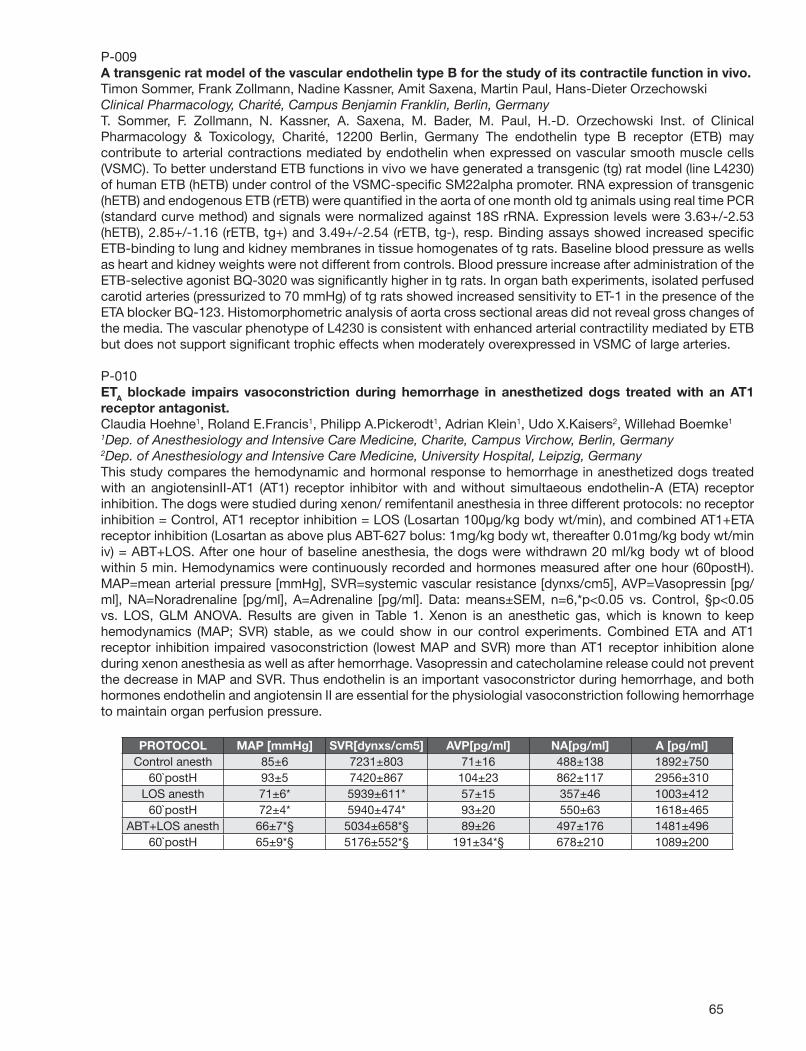
65
P-009A transgenic rat model of the vascular endothelin type B for the study of its contractile function in vivo.Timon Sommer, Frank Zollmann, Nadine Kassner, Amit Saxena, Martin Paul, Hans-Dieter OrzechowskiClinical Pharmacology, Charité, Campus Benjamin Franklin, Berlin, GermanyT. Sommer, F. Zollmann, N. Kassner, A. Saxena, M. Bader, M. Paul, H.-D. Orzechowski Inst. of Clinical Pharmacology & Toxicology, Charité, 12200 Berlin, Germany The endothelin type B receptor (ETB) may contribute to arterial contractions mediated by endothelin when expressed on vascular smooth muscle cells (VSMC). To better understand ETB functions in vivo we have generated a transgenic (tg) rat model (line L4230) of human ETB (hETB) under control of the VSMC-specifi c SM22alpha promoter. RNA expression of transgenic (hETB) and endogenous ETB (rETB) were quantifi ed in the aorta of one month old tg animals using real time PCR (standard curve method) and signals were normalized against 18S rRNA. Expression levels were 3.63+/-2.53 (hETB), 2.85+/-1.16 (rETB, tg+) and 3.49+/-2.54 (rETB, tg-), resp. Binding assays showed increased specifi c ETB-binding to lung and kidney membranes in tissue homogenates of tg rats. Baseline blood pressure as wells as heart and kidney weights were not different from controls. Blood pressure increase after administration of the ETB-selective agonist BQ-3020 was signifi cantly higher in tg rats. In organ bath experiments, isolated perfused carotid arteries (pressurized to 70 mmHg) of tg rats showed increased sensitivity to ET-1 in the presence of the ETA blocker BQ-123. Histomorphometric analysis of aorta cross sectional areas did not reveal gross changes of the media. The vascular phenotype of L4230 is consistent with enhanced arterial contractility mediated by ETB but does not support signifi cant trophic effects when moderately overexpressed in VSMC of large arteries.
P-010ETA blockade impairs vasoconstriction during hemorrhage in anesthetized dogs treated with an AT1 receptor antagonist.Claudia Hoehne1, Roland E.Francis1, Philipp A.Pickerodt1, Adrian Klein1, Udo X.Kaisers2, Willehad Boemke1
1Dep. of Anesthesiology and Intensive Care Medicine, Charite, Campus Virchow, Berlin, Germany2Dep. of Anesthesiology and Intensive Care Medicine, University Hospital, Leipzig, GermanyThis study compares the hemodynamic and hormonal response to hemorrhage in anesthetized dogs treated with an angiotensinII-AT1 (AT1) receptor inhibitor with and without simultaeous endothelin-A (ETA) receptor inhibition. The dogs were studied during xenon/ remifentanil anesthesia in three different protocols: no receptor inhibition = Control, AT1 receptor inhibition = LOS (Losartan 100µg/kg body wt/min), and combined AT1+ETA receptor inhibition (Losartan as above plus ABT-627 bolus: 1mg/kg body wt, thereafter 0.01mg/kg body wt/min iv) = ABT+LOS. After one hour of baseline anesthesia, the dogs were withdrawn 20 ml/kg body wt of blood within 5 min. Hemodynamics were continuously recorded and hormones measured after one hour (60postH). MAP=mean arterial pressure [mmHg], SVR=systemic vascular resistance [dynxs/cm5], AVP=Vasopressin [pg/ml], NA=Noradrenaline [pg/ml], A=Adrenaline [pg/ml]. Data: means±SEM, n=6,*p<0.05 vs. Control, §p<0.05 vs. LOS, GLM ANOVA. Results are given in Table 1. Xenon is an anesthetic gas, which is known to keep hemodynamics (MAP; SVR) stable, as we could show in our control experiments. Combined ETA and AT1 receptor inhibition impaired vasoconstriction (lowest MAP and SVR) more than AT1 receptor inhibition alone during xenon anesthesia as well as after hemorrhage. Vasopressin and catecholamine release could not prevent the decrease in MAP and SVR. Thus endothelin is an important vasoconstrictor during hemorrhage, and both hormones endothelin and angiotensin II are essential for the physiologial vasoconstriction following hemorrhage to maintain organ perfusion pressure.
PROTOCOL MAP [mmHg] SVR[dynxs/cm5] AVP[pg/ml] NA[pg/ml] A [pg/ml] Control anesth 85±6 7231±803 71±16 488±138 1892±750
60`postH 93±5 7420±867 104±23 862±117 2956±310 LOS anesth 71±6* 5939±611* 57±15 357±46 1003±412
60`postH 72±4* 5940±474* 93±20 550±63 1618±465 ABT+LOS anesth 66±7*§ 5034±658*§ 89±26 497±176 1481±496
60`postH 65±9*§ 5176±552*§ 191±34*§ 678±210 1089±200
66
P-011Additional lack of iNOS attenuates diastolic dysfunction in aged ET-1 transgenic mice.Philipp Kalk1, Dirk Westermann2, Sophia Herzfeld1, Katharina Relle1, Christian Bauer3, Carsten Tschoepe2, Berthold Hocher1
1Center for Cardiovascular Research/Department of Pharmacology, Charite, CCM, Berlin, Germany2Department of Cardiology, Charite, CBF, Berlin, Germany3Institute for Molecular Biology and Biochemistry, Free University, Berlin, GermanyIntroduction: Endothelin-1 (ET-1) exhibits pro-infl ammatory/pro-fi brotic properties. Infl ammation is a potent stimulus for inducible NO synthase (iNOS) as well which has been shown to contribute to cardiac injury. We thus hypothesized that ET-1 induced cardiac injury might be attenuated by concomitant lack of iNOS. Methods: We established crossbred animals of ET-1 transgenic mice (ET+/+) and iNOS knock out mice (iNOS-/-). 4 study groups (age 13 months): wildtype (WT; n=8), ET+/+ (n=10), iNOS-/- (n=7) and crossbred animals (ET+/+ iNOS-/-; n=15). Left ventricular function was determined using a tip catheter. Afterwards animals were sacrifi ced and hearts were harvested for further evaluation. Results: No differences between the groups regarding cardiac weight and myocyte diameter existed. Cardiac perivascular fi brosis was signifi cantly increased in ET+/+ and iNOS-/- groups versus WT, whereas ET+/+ iNOS -/- mice did not differ from wildtype controls (score; WT: 1.36+/- 0.26, ET+/+: 1.67+/-0.39, iNOS-/-: 1.63+/-0.29, ET+/+ eNOS-/-: 1.50+/-0.34; p< 0.05 for ET+/+ and iNOS-/- vs WT). Regarding left ventricular function plasma BNP was elevated in ET+/+ and iNOS-/- mice, but again in crossbred animals this effect was blunted (BNP pg/ml: WT: 46.0+/-3.6, ET+/+ : 65.9+/-6.1, iNOS-/-: 68.7+/-6.4, ET+/+ iNOS-/-: 42.6+/-1.7; p< 0.05 for ET+/+ and iNOS-/- vs WT). Heart catheterization revealed a signifi cantly increased stiffness constant in both ET overexpressing groups versus WT, but this increase was signifi cantly attenuated in the ET+/+iNOS-/- group (stiffness constant/µ-1: WT: 0.16+/-0.02, ET+/+: 0.32+/-0.02, ET+/+ iNOS-/-: 0.24+/-0.03; p<0.05 both transgenic groups vs WT and ET+/+ vs ET+/+iNOS-/-, respectively). However, parameters indicating systolic heart failure (EF, cardiac output) were not different between all study groups. Conclusion: Our study demonstrates that ET transgenic mice develop left ventricular stiffening with subsequent diastolic dysfunction in a slow age-dependent manner. Additional knock out of iNOS signifi cantly attenuates cardiac injury. We thus conclude that ET-1-induced cardiac injury is at least partially mediated by iNOS.
P-012Tissue specifi c activation of the endothelin system in severe acute liver failure.Susi Heiden1, Thiemo Pfab2,1, Karoline von Websky1, Michael Godes1, Katharina Relle1, Philipp Kalk1,3, Walter Zidek2, Berthold Hocher1
1Center for Cardiovascular Research/Department of Pharmacology, Charite, CCM, Berlin, Germany2Department of Nephrology, Charite, CBF, Berlin, Germany3Institute of Vegetative Physiology, Charite, CCM, Berlin, GermanyBackground: The endothelin system has been implicated in the pathogenesis of acute liver failure and especially in the concomitant hepatorenal syndrome. However, the endothelin system in experimental severe acute liver failure has not yet been assessed in a tissue specifi c manner. Methods: Acute liver failure was induced in rats with galactosamine (two administrations at 1.3 g/kg, at a 12-hr interval). Nine control rats (cont) and twenty rats with liver failure (gal) were analyzed. The animals were sacrifi ced 48h after induction of acute liver failure followed by routine clinical laboratory evaluations including liver enzymes and creatinine and urea. Plasma as well as kidney and liver tissue concentrations of big-ET-1 and ET-1 were measured. The expression of both endothelin receptor subtypes (ETA/ETB) was analysed by Western blotting. Both kidney and liver morphology was assessed by computer aided image analysis. Results: Liver enzymes were elevated: AST (cont: 266 ± 80 IU/l; gal: 5802 ± 5810 IU/l; p< 0.01), ALT (cont: 46 ± 22 IU/l; gal: 2937 ± 3131 IU/l; p< 0.05), GGT (cont: 2 IU/l; gal: 7.3 ± 5.5 IU/l; p<0.01), bilirubin (cont: 0.1 mg/dl; gal: 3.17 ± 2.52 md/dl; p< 0.01). Liver morphology confi rmed these fi ndings. Kidney function was impaired as well (creatinine: cont: 0.25 ± 0.03 mg/dl; gal: 0.33 ± 0.1 mg/dl; p< 0.01). Plasma concentrations of ET-1 (cont: 0.021 ± 0.028 pg/ml; gal: 1.23 ± 1.70 pg/ml; p< 0.01) and big ET (cont: 0.13 ± 0.15 pg/ml, gal: 0.65 ± 0.98 pg/ml; p= 0.07) were increased. However, tissue concentrations of ET-1/big-ET in liver and kidney were not signifi cantly altered by galactosamine administration. Conclusion: The endothelin system is activated in a tissue specifi c manner in acute severe liver failure (60-fold in the plasma, no signifi cant changes in liver and kidney). Particularly, the vascular system is activated in this specifi c disease model.
67
P-013Lack of eNOS promotes cardiac fi brosis in ET-1 transgenic mice.N. Vignon-Zellweger1, K. Relle1, J. Rahnenführer1, Philipp Kalk1, C. Bauer2, F. Theuring1, B. Hocher1
1Center for Cardiovascular Research/Department of Pharmacology, Charite,CCM, Berlin, Germany2Institute of Molecular Biology and Biochemistry, Free University, Berlin, GermanyIntroduction: ET-1 is antagonized by NO mainly derived from endothelial NO-synthase (eNOS). We recently established cross-bred animals of ET-1 transgenic mice (ET+/+) and eNOS knock out mice (eNOS-/-) characterized by endothelial dysfunction and slightly elevated blood pressure. Using this model we aimed at elucidating the impact of lack of eNOS in ET+/+ mice on cardiac phenotype. Methods: 4 study groups (each group n=20/age 9 months) were established: wildtype (WT), ET+/+, eNOS-/- and crossbred mice (ET+/+ eNOS-/-). Cardiac interstitial fi brosis was assessed using computer-aided histomorphometry. Cardiac ETA/ETB receptor expression was assessed using in-situ-hybridization (ISH) and Western Blot. Results: Cardiac interstitial fi brosis was increased only in ET+/+ eNOS-/-(fi brotic tissue per section; WT: 1.1+/- 0.1%, ET+/+: 1.3+/-0.1%, eNOS-/-: 1.3+/-0.1%, ET+/+ eNOS-/-: 2.0+/-0.2%; p< 0.05 for ET+/+ eNOS vs all other groups). No differences in heart weight were detected. ISH revealed normal distribution pattern of ETA/ETB receptor expression when compared to wildtype controls as both receptors were detected on cardiac myocytes as well as in vasculature. Western Blot analysis showed a signifi cantly decreased ETA receptor expression in eNOS-/- only (rel. ETA expression; WT: 1.00+/- 0.07, ET+/+: 1.04+/-0.11, eNOS-/-: 0.57+/-0.08, ET+/+ eNOS-/-: 0.81+/-0.15; p< 0.05 for eNOS-/- vs WT), whereas cardiac ETB receptor expression was suppressed in both eNOS-/- and in ET+/+ eNOS-/- groups (rel. ETB expression; WT: 1.00+/- 0.10, ET+/+: 1.15+/-0.13, eNOS-/-: 0.66+/-0.04, ET+/+ eNOS-/-: 0.54+/-0.06; p< 0.05 for eNOS-/- and ET+/+ eNOS-/- vs WT). Conclusion: Our study demonstrates that additional eNOS knock out in ET-1 transgenic mice promotes myocardial fi brosis in absence of hypertrophy. We thus conclude that our ET+/+ eNOS-/- mice represent a novel model of myocardial fi brosis due to imbalance between the ET and NO system. As in ET++ eNOS-/- mice the ETB receptor was downregulated without concomitant downregulation of the ETA receptor, we suggest imbalance of cardiac ETA and ETB signaling as a possible mechanism promoting cardiac fi brosis in our model.
P-014High doses of ultraviolet-C irradiation increases endothelin-2 expression in keratinocytes of the newborn mouse epidermis.Kaname Saida1, Javier Adur1,2, Satoshi Takizawa1, Victor Casco3, Tsuyoshi Uchide4
1AIST, Tsukuba, Japan2NEDO, Kawasaki, Japan3National University of Entre Rios, Entre Rios, Argentina4Kitasato Univ., Towada, JapanWe examined the expression profi les of endothelin-2 / vasoactive intestinal contractor (ET-2/VIC) at both gene and peptide level in skin irradiated with different ultraviolet wavelengths. We found that ET-2/VIC gene expression is sensitive only to ultraviolet-C (UVC) irradiation and has an immediate response. These results provide direct evidence that high doses of UVC irradiation induce an increase in gene expression and protein production of ET-2/VIC and endothelin (ET) receptors in a dose-dependent manner in epidermal keratinocytes. We suggest that ET-2/VIC can play an essential role in the maintenance, protection and hyperpigmentation of the epidermis exposed to UVC irradiation from artifi cial or natural sources.
68
P-015Endothelin-2 via ROCK regulates transglutaminase 1 on differentiation of mouse keratinocytes.Eiichi Kotake-Nara1,2, Kaname Saida1, Satoshi Takizawa1
1AIST, Tsukuba, Japan2NEDO, Kawasaki, JapanWe previously found that endothelin-2/vasoactive intestinal contractor (ET-2/VIC) greatly increased in mouse epidermis after birth. In the present study, we evaluated whether ET-2/VIC expression was associated with the calcium-induced differentiation of cultured mouse keratinocytes. The differentiation induction was revealed by morphological change, cornifi ed envelope (CE) formation, and involucrin and transglutaminase 1 (TG 1) expressions. ET-2/VIC gene expression and peptide production subsequently increased in the induction of the differentiation. We also found that Y-27632, a Rho-associated coiled-coil forming protein serine/threonine kinase (ROCK) inhibitor, suppressed up-regulation of ET-2/VIC gene expression, the induction of morphological change, the CE formation, and TG 1 expression, but not involucrin expression. These results indicate new three fi ndings, (1) ET-2/VIC expression increases and has potential as a differentiation marker, (2) ET-2/VIC expression is mediated by ROCK, and (3) the ROCK regulated TG 1 expression, on the calcium-induced differentiation of mouse keratinocytes.
P-016Endothelial cell-specifi c ETB receptor knockout does not impair endothelium-dependent vasodilation in the mouse femoral artery.Nicholas S.Kirkby, Patrick W.Hadoke, Alan J.Bagnall, Yuri V.Kotelevtsev, David J.WebbCentre for Cardiovascular Science, University of Edinburgh, Edinburgh, United KingdomIntroduction ET-1 dependent contraction may be modulated by ETB-mediated release of vasodilators from endothelial cells (EC). Cre-LoxP mediated EC-specifi c removal of ETB in mice causes increased plasma ET-1, impaired aortic relaxation to acetylcholine (ACh), but not hypertension. We hypothesised that functional changes may be territory-and age-dependent in this model. Methods EC ETB knockout mice were generated as previously described (Bagnall et al. 2006). Femoral arteries were isolated from young (2-4month) and aged (8-10month) EC ETB knockout mice and Cre-negative littermate controls and mounted for myography. Isometric responses to ET-1, phenylephrine (PE), ACh, sodium nitroprusside (SNP) and sarafotoxin 6c (S6c) were measured. Dilator responses were obtained in PE constricted vessels. Responses to ET-1 were measured in the presence of ETA (BQ123) or ETB (A192621) antagonists or vehicle. Data are as mean±SEM for n=6-10 unless stated. Maximum responses (Emax) expressed as a percentage of responses to 125mM KCl and sensitivity as -logEC50 (pD2). Results ET-1 concentration-dependently contracted femoral rings from young mice (Emax 103.2+4.2%; pD2 8.26+0.10) and this was shifted to the right (P<0.001) by BQ-123 (pD2 7.20+0.14; n=4) but not A192621. Deletion of EC ETB did not alter the amplitude (Emax 101.9+5.%) or sensitivity (pD2 8.32+0.07) of the contraction to ET-1 or the effect of ETA antagonism (pD2 7.40+0.12). S6c produced a variable contraction of PE constricted vessels but did not appear to cause relaxation. Responses to ACh, SNP and PE were not unaltered genotype. All effects were maintained with ageing. Conclusions ET-1-mediated contraction of the femoral artery is mediated by ETA but unaffected by either deletion of EC ETB or pharmacological ETB inhibition. These responses were independent of age and are in contrast to those observed in the aorta, where EC ETB deletion impaired ACh-mediated relaxation and ET-1 had little contractile effect. This suggests that the functional effects of EC ETB knockout are dependent on vessel type and this may partially explain the absence of hypertension in this model.
69
P-017L-NAME-induced pulmonary fi brosis is dependent on an activated endothelin system.Philipp Kalk1,2, Alexander Mach1, Anja Schwarz1, Christa Thöne-Reineke1, Michael Godes1, Susi Heiden1, Yuliya Sharkovska1, Karoline von Websky1, Katharina Relle1,2, Berthold Hocher1
1Department of Pharmacology and Toxicology, Charite, Center for Cardiovascular Research (CCR), Berlin, Germany2Charite, Institute of Vegetative Physiology, Berlin, GermanyIntroduction: Activation of the endothelin (ET) system promotes vasoconstriction, infl ammation and fi brosis in various tissues including the lung. Therefore, ET-1 transgenic mice overexpressing ET-1 develop pulmonary fi brosis in a slow, age-dependent manner. In vivo, NO is the most important counterregulatory mediator of the ET-system. The aim of our study was to elucidate the impact of interaction between NO and ET-System on pulmonary infl ammation and fi brosis in young ET-1 transgenic mice before the onset of pulmonary fi brosis. Methods: Male ET-1 transgenic mice and wildtype littermates at the age of 8 weeks were randomly allocated to the following groups: WT (n=11): wildtype animals without treatment, WT + L-NAME (n=14): wildtype animals receiving L-NAME, WT + L-NAME + LU 302 (n=13): wildtype animals receiving L-NAME as mentioned above plus LU 302872, a dual ET-receptor antagonist, ET1tg (n=10), ET1tg + L-NAME (n=13): ET1tg + L-NAME + LU 302 (n=13). After 6 weeks animals were sacrifi ced and hearts and lungs were harvested for histology/immunhistochemistry. Results: In the lung of ET1tg mice infl ammation -as indicated by macrophage infi ltration- and interstitial fi brosis was signifi cantly enhanced by L-NAME, whereas in WT groups and in ET1tg mice additionally treated with LU 302872 no such effect was detected. Moreover, heart weight weight of ET1tg mice treated with L-NAME was increased versus both other ET1tg groups whereas in WT groups the effect was absent. Perivascular fi brosis, media-lumen-ratio of pulmonary bronchi and arteries did not differ between all study groups. Conclusion: In our study L-NAME- induced pulmonary fi brosis and infl ammation only in ET1tg mice without LU 302872. We thus conclude the pro-fi brotic and pro-infl ammatory effect of NO-defi ciency in the lung to be dependent on an activated endothelin system.
P-018Involvement of increased intracellular sodium level in endothelin-1-induced hypertrophy.Raúl A.Dulce, Carolina D.Garciarena, Irene L.Ennis, Claudia Caldiz, Gladys E.Chiappe de Cingolani, María C.Camilión de HurtadoCentro de Investigaciones Cardiovasculares, Facultad de Ciencias Médicas, Universidad Nacional de La Plata, La Plata, ArgentinaElevation of intracellular sodium concentration ([Na+]i) has been documented in hypertrophied failing myocardium. The prohypertrophic peptide endothelin-1 (ET-1) stimulates Na/H exchange (NHE), which constitutes an important Na+ infl ux pathway. The aim of this work was to explore whether NHE-mediated increases in [Na+]i participates in the hypertrophic effect of ET-1 in cultured neonatal rat ventricular myocytes (NRVM). ET-1-treated (5 nmol/L) NRVM showed, in comparison with control NRVM, a) increased NHE activity (proton extrusion 13.7±2.8 vs. 2.5±1.2 mmol/L/min, n=5, P<0.05) with normal exchanger protein expression; b) higher [Na+]i (8.1±1.2 vs. 4.2±1.3 mmol/L, n=4, P<0.05) and total cell [Ca2+]i (636±117 vs. 346±85 nmol/L, n=9 vs. 11 respectively, P<0.05); c) larger cell surface area and 3H-phenylalanine incorporation (131±3 and 220±12 % of control, respectively, P<0.05). These effects were cancelled by NHE inhibition with 1 µmol/L cariporide. Elevated [Na+]i may reduce Ca2+ effl ux through Na/Ca exchange (NCX) or promote the reverse NCX mode, thereby increasing cell Ca2+. ET-1 treatment did not modify the relaxation of caffeine-induced Ca2+ transients (Tau 4.6±1.3 and 3.5±0.8 sec, n=5 in control and ET-1-treated NRVMs, respectively) rejecting the former possibility. Reverse NCX mode was inhibited either with 5 µmol/L KB R7943 or raising extracellular [Na+] from 137 to 237 mmol/L to compensate for the rise in [Na+]i and keep driving force for NCX similar to that of control conditions. With KB R7943, total cytosolic Ca2+ was reduced to 326±27 nmol/L in ET-1-treated NRVM (n=12, NS compared to controls). In addition, both ways of inhibition of reverse NCX mode prevented the hypertrophic effect of ET-1 in NRVM. The results show the involvement of NHE-mediated Na+ infl ux and the secondary stimulation of reverse NCX mode in the ET-1 hypertrophic effect in NRVM. In addition, they give support to propose the inhibition of reverse NCX mode, as it was for NHE inhibition, to be a potential preventive/therapeutic strategy for cardiac hypertrophy.
70
P-019Endothelin-1 causes impairment in ACh induced relaxation in murine aortic rings, and this effect is reversed by treatment along with IL-10.Saiprasad M.Zemse1, Rob H.Hilgers1, G. Bryan Simkins2, R. Daniel Rudic2, R. Clinton Webb1
1CA-3099, Dept. of Physiology, Medical College of Georgia, Augusta, GA2Dept. of Pharmacology, Medical College of Georgia, Augusta, GABackground: Interleukin-10 (IL-10) is an anti-infl ammatory cytokine causing decrease in superoxide production and restoring endothelial integrity following injury. Endothelin 1(ET-1) is implicated in the development of vascular injury through the generation of reactive oxygen species (ROS) by NADPH oxidase activation. In this study, we tested whether IL-10 attenuates ET-1 induced endothelial dysfunction. Methods & Results: Aortic rings (2mm long) of C57BL6 mice were incubated in 2 mL Dulbecco’s modifi ed Eagle’s medium (DMEM) containing 120 U/ml penicillin and 120µg/ml streptomycin in the presence of either vehicle (ddH2O), ET-I (10 nmol/l), recombinant mouse IL-10 (300ng/ml) or in the presence of both ET-I and IL-10 for 22 hrs at 37°c. Following incubation, rings were mounted in a wire myograph (Danish Myotech) and stretched to a tension of 5mN. Endothelium-dependent vasorelaxation was assessed by constructing cumulative concentration-response curves to acetylchloline (ACh, .001-10µmol/l) during phenylephrine (PE, 10 µmol/l) induced contraction. Overnight exposure of aortic rings with ET-1 resulted in statistically signifi cant endothelial dysfunction. Maximal vasorelaxation (expressed as % relaxation of PE- induced contraction) was lower in ET-1 treated rings compared to untreated rings (54 + 3% versus 76 + 4 %, respectively). IL-10 treatment partially, but signifi cantly, restored this endothelial function (71 + 4%).Western blotting showed decreased eNOS expression in response to ET-1. Vessels treated with a combination of ET-1 and IL-10 showed increased expression of eNOS. Immuno-histochemical analysis showed decreased eNOS expression in ET-1 treated vessels as compared to those treated combined with ET-1 and IL-10. Conclusion: The anti-infl ammatory cytokine IL-10 prevents impairment in endothelium dependent relaxation induced in response to long term incubation with ET-1 via normalization of eNOS expression.
P-020Antagonism of Endothelin-1 Inhibits Hypoxia-Induced Apoptosis in Cardiomyocytes.An-Jing Ren1, Hong Lu1, Xiao-Hong Yan1, Jie Bai2, Wen-Jun Yuan1,2, Li Lin1
1Department of Physiology, Second Military Medical University, Shanghai, China2School of Beaic Medical Science, Ning-Xia Medical College, Yinchuan, ChinaApoptosis is well demonstrated to be a common feature of many pathological disorders of heart. Exogenous endothelin-1 (ET-1) has been shown to be pro-apoptotic or anti-apoptotic, depending on ET-1 concentration, cell type and the ratio of ETA/ETB receptors. However, the role of endogenous ET-1 in cardiomyocyte apoptosis is not clarifi ed. This study observed the effects of ETA receptor antagonist BQ610 and BQ123, and ETB receptor antagonist BQ788 on hypoxia-induced apoptosis in primary cultured neonatal rat cardiomyocytes. Hypoxic apoptosis was induced by incubating cardiomyocytes in serum-free medium under 3% O2 - 5% CO2 for 24 h. Apoptosis was evaluated by TUNEL analysis and fl ow cytometry (FCM). TUNEL analysis showed that the positive-staining apoptotic cardiomyocytes reached a percentage of 24.2 ± 2.2% under hypoxia. The treatment of BQ610 (5 umol/L) signifi cantly reduced the apoptosis rate to 13.2 ± 3.7% (counted from four independent experiments, p < 0.01 vs. hypoxia). FCM showed that the apoptotic cells positively stained with Annexin V and propidium iodide were 42.76 ± 4.45 % (n = 12) in cultures subjected to hypoxia. BQ123 at the doses of 0.04, 0.2 and 1.0 umol/L dose-dependently reduced the apoptosis rate to 34.00 ± 10.35 % (n = 6, p < 0.05), 31.37 ± 8.28 % (n = 6, p < 0.01) and 22.89 ± 4.19 % (n = 6, p < 0.01), respectively. In contrast, BQ788 did not affect the hypoxic apoptosis. These fi ndings suggest that endogenous ET-1 contributes to hypoxia-induced apoptosis in cultured cardiomyocytes, which is mediated by ETA receptors, but not ETB receptors.
71
P-021Effects of dual endothelin receptor blockade on sympathetic activation and arrhythmogenesis during acute myocardial infarction in rats.Theofi los M.Kolettis1, Giannis G.Baltogiannis1, Dimitrios G.Tsalikakis2, Alexandros T.Tzallas2, Maria G.Agelaki1, Apostolos Papalois5, Andreas Fotopoulos3, Dimitrios I.Fotiadis2, Zenon S.Kyriakides4
1Cardiology, University of Ioannina, Ioannina, Greece2Computer Sciences, University of Ioannina, Ioannina, Greece3Nuclear Medicine, University of Ioannina, Ioannina, Greece4Cardiology, Red Cross Hospital, Athens, Greece5ELPEN Research Laboratory, Athens, GreeceObjective: To examine the effects of dual endothelin A- and B-receptor blockade on ventricular arrhythmogenesis during acute myocardial infarction. Methods: We randomly allocated Wistar rats to bosentan (100mg/kg daily, n=24), a dual endothelin receptor antagonist, or vehicle (n=23). After 7 days of treatment, myocardial infarction was generated by permanent coronary ligation. Ventricular tachyarrhythmias were evaluated for 24 hours following ligation, using a miniature telemetry electrocardiogram recorder. Action potential duration was measured from monophasic epicardial recordings and sympathetic activation was assessed by heart rate variability and catecholamine serum levels. Results: Compared to controls (1012±185sec), bosentan (59±24sec) markedly decreased (p<0.00001) the total duration of ventricular tachyarrhythmias during the delayed (1-24hr) phase post-ligation, with a modest effect during the early (0-1hr) phase (132±38sec, versus 43±18sec, respectively, p=0.053), without affecting infarct size or total mortality. Action potential duration at 90% repolarization prolonged in controls (from 93.1±4.7ms to 117.6±6.9ms), displaying increased temporal dispersion (from 4.14±0.45ms to 10.42±2.51, both p<0.001), but was preserved in treated animals. Bosentan decreased norepinephrine but increased epinephrine levels 24 hours post-ligation. Low frequency spectra of heart rate variability, an index of net sympathetic tone, were lower in bosentan-treated rats. Conclusions: Dual endothelin receptor blockade decreases ventricular tachyarrhythmias during myocardial infarction without reperfusion, potentially by preventing repolarization inhomogeneity. Diverse treatment effects on sympathetic activation may ameliorate the antiarrhythmic action.
P-022Diazepam relaxed ET-1 precontracted thoracic rat aorta in a concentration dependent manner.Lovro Ziberna, Jernej Berden, Blaz Groselj, Gorazd DrevensekLaboratory for Cardiovascular Pharmacology, Medical Faculty, University of Ljubljana, Ljubljana, SloveniaAIM: Diazepam is a benzodiazepine drug which pharmacological effects are mediated by binding to GABAA receptors in CNS but also acts on many peripheral tissues. The effect of diazepam on isolated thoracic rat aorta, precontracted by ET-1, was tested. METHOD: Aortic rings (3-4 mm wide) were placed in organ baths, fi lled with oxygenated Krebs-Henseleit solution at 37 °C. After 60 min equilibration (2.0 g tension), rings were repeatedly contracted with 60 mM KCl until stable response. Endothelial function was assessed on precontracted rings by the degree of relaxation caused by 0.1 mM acetylcholine. Precontraction was induced by 0.1 microM noradrenaline. There were 3 experimental groups: intact aortic rings, endothelium denuded rings and rings with inhibited nitric oxide synthase (NOS) and cyclooxygenase (COX) by application of LNNA and indomethacine. Relaxation of precontracted rings for more than 40 % in non-inhibited rings indicated intact endothelium. The rings precontracted by 10 nM endothelin (ET-1) followed by application of Apaurin (diazepam) in concentrations ranging from 1 microM to 80 microM. RESULTS: Diazepam applied to the aortic rings in concentrations of 1, 10, 20, 40, 60 and 80 microM relaxed precontracted rings on average by 26%, 42%, 55%, 71%, 81% and 91%, respectively. In higher concentrations complete relaxation was observed. Rings with inhibited NOS and COX relaxed rings on average by 13%, 25%, 37%, 40% and 42% by the application of 1, 10, 20, 40 and 60 microM diazepam, respectively. Maximum relaxation of the endothelium denuded rings was attained at 55% by 20 microM diazepam. Additional application of diazepam did not enhance relaxation. CONCLUSIONS: Diazepam showed concentration dependent relaxation of ET-1 precontracted aortic rings. Mechanism for the relaxation is only partially dependent of nitric oxide synthase activity.
72
P-023The effect of gene transfer of phosphatase and tensin homolog deleted on chromosome ten(PTEN) on endothelin-1 production in cultured endothelial cells.Hing-Chung LamDivision of Endocrinology and Metabolism, Department of Medicine, Kaohsiung Veterans General Hospital and Yuh-Ing Junior College of Health Care and Management, Kaohsiung City, TaiwanDepartment of Medical Education and Research, Kaohsiung Veterans General Hospital, Kaohsiung City, TaiwanDepartment of Biological Sciences, National Sun Yat-Sen University, Kaohsiung City, TaiwanAngiogenesis, or neovascularization, is a process by which new vessels are formed from the pre-existing blood vessels via endothelial cells migration as well as proliferation. Angiogenesis has critical roles in normal vascular development and in important pathologies including cancer, wound healing and infl ammation. Recently, it has been shown that ET-1 has a direct angiogenic effect on endothelial and peri-vascular cells as well as an indirect action through the increased release of the potent pro-angiogenic substance vascular endothelial growth factor (VEGF), via hypoxia inducible factor-1. Recently we have shown that blockade of endothelin-1 release may contribute to the anti-angiogenic effects of POMC overexpression in endothelial cells via the inhibition of endothelial cells migration and tube-forming capability. Our fi ndings further support ET-1 is involved in the process of angiogenesis. PTEN (phosphatase and tensin homolog deleted on chromosome ten) was discovered in 1997 as a new tumor suppressor, and it is now known to play major roles not only in suppressing cancer but also in embryonic development, cell migration and apoptosis. PTEN appears to play particularly important roles in regulating anoikis (apoptosis of cells after loss of contact with extracellular matrix) and cell migration. It has been shown that PTEN reconstitution or overexpression inhibits cell migration, and this inhibition can be accompanied by transient effects on cell adhesion and spreading. We hypothesize that one of the mechanisms of PTEN’s anti-angiogenic effect may be mediated via the alteration (probably suppression) of ET-1 production and/or secretion. By employing gene delivery technique, we have transferred the PTEN gene into the EA.hy926 endothelial cells and our results showed that both the endothelial’s ET-1 mRNA and ET-1 levels were decreased by PTEN overexpression. These results support our hypothesis and will add new data on the possible interaction between PTEN and ET-1.
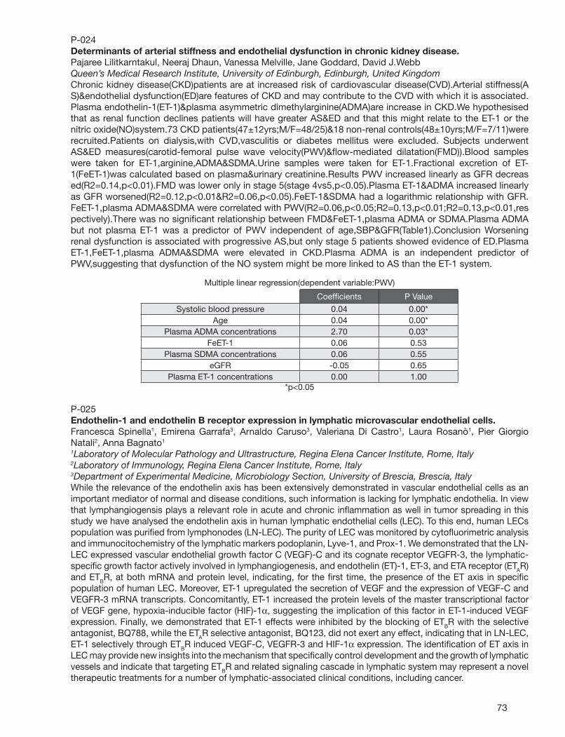
73
P-024Determinants of arterial stiffness and endothelial dysfunction in chronic kidney disease.Pajaree Lilitkarntakul, Neeraj Dhaun, Vanessa Melville, Jane Goddard, David J.WebbQueen’s Medical Research Institute, University of Edinburgh, Edinburgh, United KingdomChronic kidney disease(CKD)patients are at increased risk of cardiovascular disease(CVD).Arterial stiffness(AS)&endothelial dysfunction(ED)are features of CKD and may contribute to the CVD with which it is associated.Plasma endothelin-1(ET-1)&plasma asymmetric dimethylarginine(ADMA)are increase in CKD.We hypothesised that as renal function declines patients will have greater AS&ED and that this might relate to the ET-1 or the nitric oxide(NO)system.73 CKD patients(47±12yrs;M/F=48/25)&18 non-renal controls(48±10yrs;M/F=7/11)were recruited.Patients on dialysis,with CVD,vasculitis or diabetes mellitus were excluded. Subjects underwent AS&ED measures(carotid-femoral pulse wave velocity(PWV)&fl ow-mediated dilatation(FMD)).Blood samples were taken for ET-1,arginine,ADMA&SDMA.Urine samples were taken for ET-1.Fractional excretion of ET-1(FeET-1)was calculated based on plasma&urinary creatinine.Results PWV increased linearly as GFR decreased(R2=0.14,p<0.01).FMD was lower only in stage 5(stage 4vs5,p<0.05).Plasma ET-1&ADMA increased linearly as GFR worsened(R2=0.12,p<0.01&R2=0.06,p<0.05).FeET-1&SDMA had a logarithmic relationship with GFR.FeET-1,plasma ADMA&SDMA were correlated with PWV(R2=0.06,p<0.05;R2=0.13,p<0.01;R2=0.13,p<0.01,respectively).There was no signifi cant relationship between FMD&FeET-1,plasma ADMA or SDMA.Plasma ADMA but not plasma ET-1 was a predictor of PWV independent of age,SBP&GFR(Table1).Conclusion Worsening renal dysfunction is associated with progressive AS,but only stage 5 patients showed evidence of ED.Plasma ET-1,FeET-1,plasma ADMA&SDMA were elevated in CKD.Plasma ADMA is an independent predictor of PWV,suggesting that dysfunction of the NO system might be more linked to AS than the ET-1 system.
Multiple linear regression(dependent variable:PWV)
Coeffi cients P Value
Systolic blood pressure 0.04 0.00* Age 0.04 0.00*
Plasma ADMA concentrations 2.70 0.03* FeET-1 0.06 0.53
Plasma SDMA concentrations 0.06 0.55 eGFR -0.05 0.65
Plasma ET-1 concentrations 0.00 1.00 *p<0.05
P-025Endothelin-1 and endothelin B receptor expression in lymphatic microvascular endothelial cells.Francesca Spinella1, Emirena Garrafa3, Arnaldo Caruso3, Valeriana Di Castro1, Laura Rosanò1, Pier Giorgio Natali2, Anna Bagnato1
1Laboratory of Molecular Pathology and Ultrastructure, Regina Elena Cancer Institute, Rome, Italy2Laboratory of Immunology, Regina Elena Cancer Institute, Rome, Italy3Department of Experimental Medicine, Microbiology Section, University of Brescia, Brescia, ItalyWhile the relevance of the endothelin axis has been extensively demonstrated in vascular endothelial cells as an important mediator of normal and disease conditions, such information is lacking for lymphatic endothelia. In view that lymphangiogensis plays a relevant role in acute and chronic infl ammation as well in tumor spreading in this study we have analysed the endothelin axis in human lymphatic endothelial cells (LEC). To this end, human LECs population was purifi ed from lymphonodes (LN-LEC). The purity of LEC was monitored by cytofl uorimetric analysis and immunocitochemistry of the lymphatic markers podoplanin, Lyve-1, and Prox-1. We demonstrated that the LN-LEC expressed vascular endothelial growth factor C (VEGF)-C and its cognate receptor VEGFR-3, the lymphatic-specifi c growth factor actively involved in lymphangiogenesis, and endothelin (ET)-1, ET-3, and ETA receptor (ETAR) and ETBR, at both mRNA and protein level, indicating, for the fi rst time, the presence of the ET axis in specifi c population of human LEC. Moreover, ET-1 upregulated the secretion of VEGF and the expression of VEGF-C and VEGFR-3 mRNA transcripts. Concomitantly, ET-1 increased the protein levels of the master transcriptional factor of VEGF gene, hypoxia-inducible factor (HIF)-1α, suggesting the implication of this factor in ET-1-induced VEGF expression. Finally, we demonstrated that ET-1 effects were inhibited by the blocking of ETBR with the selective antagonist, BQ788, while the ETAR selective antagonist, BQ123, did not exert any effect, indicating that in LN-LEC, ET-1 selectively through ETBR induced VEGF-C, VEGFR-3 and HIF-1α expression. The identifi cation of ET axis in LEC may provide new insights into the mechanism that specifi cally control development and the growth of lymphatic vessels and indicate that targeting ETBR and related signaling cascade in lymphatic system may represent a novel therapeutic treatments for a number of lymphatic-associated clinical conditions, including cancer.

74
P-026Nox2 modulates contraction to endothelin-1 in the renal artery: Effect of high-fat diet.Marlen Damjanovic, Indranil Bhattacharya, Alexa L.Mundy, Martin Kretz, Corinne C.Widmer, Julia Rasi, Matthias BartonDept. Internal Medicine, University Hospital Zurich, Zurich, SwitzerlandThe renal artery plays a major role in the perfusion of the kidney. Obesity is associated with changes of kidney perfusion, development of hypertension and renal injury. The role of the NADPH-oxidase subunit Nox2 (gp91phox) in the regulation of endothelin-1 (ET-1)-mediated renal artery function is not known. Therefore, this study investigated ET-1-induced contractions in isolated mouse renal and femoral artery rings of wild-type control and Nox2-/- mice and the effect of high-fat diet (41% kcal from fat, for 15 weeks). Rings were exposed to ET-1 (10-11 - 10-7 M) in the presence of L-nitro-arginine-methyl ester (L-NAME, 300 µM) to block ETB receptor-mediated NO release. In control animals, although maximum contractility was similar in the renal and femoral artery on normal diet, high-fat diet reduced contractions in the renal artery (p<0.05). In renal arteries of Nox2-/- mice on normal diet, contractions to ET-1 were markedly lower (p<0.05 vs. wild-type control), but remained unaffected by high-fat diet (n.s.). High-fat diet had no effect on ET-1-induced contractions in the femoral artery, and irrespective of a functional Nox2 gene (n.s.). In conclusion, these fi ndings demonstrate that high-fat diet selectively reduces ET-1-induced contractions in the renal but not in the femoral artery of mice. The data also suggest that Nox2 is required for ET-1-mediated contraction in the renal artery only, a function that remains unaffected by high-fat diet. These observations could explain the increased perfusion in early stages of obesity and may be important for the pathogenesis of obesity-associated diseases such as hypertension and renal disease.
P-027Diameter determines vascular reactivity to endothelin-1 within the same conduit artery in mice.Indranil Bhattacharya, Alexa L.Mundy, Marlen Damjanovic, Corinne C. Widmer, Martin Kretz, Matthias BartonDept. Internal Medicine, University Hospital Zurich, Zurich, SwitzerlandConduit arteries are susceptible to atherosclerosis in humans and animals. Elevated endothelin-1 (ET-1) levels are associated with cardiovascular risk factors such as hypertension and hypercholesterolemia, which promote the development of atherosclerosis. We have previously reported that ET-1-mediated vasoreactivity varies between different vascular beds in mice (Widmer et al., Exp Biol Med 2006; 231: 777). In the present study, we investigated contractile responses to ET-1 in segments of the abdominal aorta of adult C57BL/6 mice at 15 weeks of age. Segments of the abdominal aorta were defi ned by the anatomical location within the abdomen (proximal, mid-proximal, mid-distal, and distal) which were associated with decreasing diameter. Vascular rings were exposed to ET-1 in a concentration-dependent manner (10-10 - 10-7 M) in the presence of L-nitro-arginine-methyl ester (L-NAME, 3x10⁻⁴ M) to block the effects of endothelin-stimulated nitric oxide. Contractions were normalized to KCl (10-2 M). A 15-fold difference in ET-1-mediated contraction was observed in the distal as compared to the proximal segment (120±13% vs. 8±3%, p<0.01). Interestingly, even between mid-distal and distal segment there was a difference in contraction (2.5 fold, 46±8% vs. 120±13%, p<0.01). No signifi cant difference in contractile responses was observed between proximal and mid-proximal arterial rings (8±3% vs. 14±3%, n.s.). In conclusion, the data indicate that ET-1-mediated contraction increases from the proximal to the distal abdominal aorta, which showed the most potent contractions to ET-1. In addition, these data suggest that caution is advised when performing vascular reactivity experiments investigating mouse “abdominal aorta” since this aortic region displays considerable variation of the contractile response depending on its anatomic location. The differences in sensitivity to ET-1 in the different segments of abdominal aorta may also contribute to atherosclerotic lesion development, which in mice originates from the arch region and the most distal part of the abdominal aorta.
75
P-028Transcriptional analysis of the endothelin axis in the heartAnita Schorlemmer, X-S. Zhao, Ralph V. ShohetCenter for Cardiovascular Research, John A. Burns School of Medicine, University of Hawaii, Honolulu, Hawaii, USAIn this study, transcriptional profi les of mouse hearts withcardiomyocyte-specifi c deletion of ET-1 and ETA genes were determined using comprehensive long-oligo microarrays. Changes in cellular gene transcription were confi rmed for selected genes by semi-quantitative RT-PCR analysis. We found distinct profi les, including several transcripts regulated in both models. A transcript of particular interest is that for Plunc, which is markedly less abundant in both models. It is a gene of unknown function expressed in tracheal epithelium and thought to possibly participate in immune response. We also found Plunc to be abundantly expressed in stressed hearts and suggest that it is a downstream mediator of endothelin signaling in the heart.
P-029Expression of human endothelin-converting enzyme isoforms: role of angiotensin II.Winfried Goettsch1, Andreas Schubert2, Henning Morawietz1
1Vascular Endothelium and Microcirculation, University of Technology Dresden, Dresden, Germany2AG Vascular Biology, Fraunhofer Institute for Cell Therapy and Immunology, Leipzig, GermanyBackground: A key step in endothelin-1 synthesis is the proteolytic cleavage of big-ET-1 by the endothelin-converting enzyme-1 (ECE-1). Four alternatively spliced isoforms ECE-1a-d have been discovered. However, the expression level of specifi c ECE-1 isoforms in human endothelial cells in response to angiotensin II and in internal mammary arteries of patients with coronary artery disease is not well understood. Material and Methods: Human endothelial cells (HUVEC, HCAEC) were stimulated with angiotensin II. The expression of ECE-1 isoforms a-d was compared by standard calibrated competitive RT-PCR using a novel multi-standard of all isoforms. In addition, we analysed endothelial release of endothelin-1 peptide after angiotensin II stimulation. Furthermore, expression of ECE-1 isoforms was determined in internal mammary arteries of patient undergoing coronary artery bypass grafting surgery. Patients received therapies without interfering in the renin-angiotensin system, angiotensin-converting enzyme inhibitors (ACE-I), angiotensin II receptor type 1 blockers (ARBs) or HMG-CoA reductase inhibitors (statins). Results: Under control conditions ECE-1a is the dominant isoform in human endothelial cells (4.5±2.8 amol/µg RNA), followed by ECE-1c (2.7±1.0), ECE-1d (0.49±0.17) and ECE-1b (0.17±0.04). Stimulation with Ang II (1µM) for 1, 3, 7 and 24h did not signifi cantly change this expression pattern. Furthermore, short-term (3 and 6h) stimulation of HUVEC with Ang II (100nM, 1µM) did not signifi cantly affect endothelial endothelin-1 release. In biopsies of patients, we found a signifi cant higher ECE-1 mRNA expression in patients treated with statins (5.8±0.76 RU) compared to ARB (3.0±0.40 RU) therapy. This higher expression is preferentially attributed to the main isoform ECE-1a, combined with an induction of ECE-1b. In addition, a signifi cant lower expression of ECE-1a mRNA expression was measured in patients receiving ACE-I (1.68±0.27 RU) compared to ARB (0.83±0.07 RU) therapy. Conclusion: We conclude that ECE-1a is the major ECE-1 isoform in human endothelial cells. Its expression can be regulated by ACE-I, ARB or statin therapy in arteries of patients with coronary artery disease.
76
P-030Gender-independent opposing effects of aging on endothelin vasoreactivity in arteries prone and resistant to plaque formation over the entire lifespan in model of human atherosclerosis.Matthias Lang1, Wolfram Jochum2, Erling Falk3, Tobias Traupe1, Elvira Haas1, Regina Hofmann-Lehmann4, Lukas Bestmann5, Matthias Barton1
1Dept. Internal Medicine, University Hospital Zurich, Zurich, Switzerland2Dept. Pathology, University Hospital Zurich, Zurich, Switzerland3Dept. Cardiology, Aarhus University Hospital, Aarhus, Denmark4Clinical Laboratory, Vetsuisse Faculty, University of Zurich, Zurich, Switzerland5Clinical Chemistry, University Hospital Zurich, Zurich, SwitzerlandAtherosclerosis is a systemic infl ammatory disease of the vasculature for which heterogeneities with regard to gender and anatomical localization have been reported. Both angiotensin (Ang) and endothelin (ET) have been implicated in atherogenesis and vasoconstriction and cell growth in atherosclerosis. We studied vasoreactivity in thoracic aorta and common carotid artery in a hypercholesterolemic model of human atherosclerosis over the entire lifespan. Male and female apoE null mice were used at age 4, 12, and 24 months and contractions to big ET-1, ET-1, Ang I, and Ang II were were performed in the presence of L-NAME to exclude effects on ETB or AT2 receptor mediated NO release. Histological analyses of vascular specimens were performed. Despite simlar cholesterol plasma levels, atherosclerosis development was age-dependent in all animals. Unlike the aorta, the common carotid artery remained free of lesions even at the age of 24 months. In 4 months old animals, contractions to ET-1 (4.9 -fold) and to Ang II (3.1-fold) were higher in the carotid artery than in the aorta, and no gender differences were observed. While a decrease of ET-1 mediated contractility was observed in the carotid artery over 24 mo (from 14.2±2 to 9.1±2% KCl), contractions to ET-1 increased in the aorta during aging (from 2.9±0.5 to 7.2±2%, p<0.05, 4 vs. 24 months). Similarly, Ang I and Ang II induced contractions were higher in the carotid artery than in the aorta, and increased with age in aorta only (p<0.05). All changes were similar in males in females. Interestingly, no changes were seen with regard to big ET-1 or Ang I induced contractions. In conclusion, contractility to ET-1 and Ang II (but not to their inactive precursors big ET-1 or Ang I) increases in arteries prone to atherosclerosis but not those resistant to atherosclerosis in apoE null mice despite hypercholesterolemia and aging. The data suggest that resistance to atherosclerosis is associated with preserved function of receptors of vasoconstrictor peptides independent of gender and aging.
P-031Activation of the renin-angiotensin system in endothelin-converting enzyme-1 heterozygous mice.Noriaki Emoto1,2, Suko Adiarto1, Naoko Iwasa1, Dyah W.Anggrahini1, Bambang Widyantoro1, Kazuhiko Nakayama1, Kazuya Miyagawa1, Hidemi Nonaka1, Yaz Y.Kisanuki2, Masashi Yanagisawa2, Sunu B.Raharjo1
1Division of Cardiovascular Medicine, Department of Internal Medicine, Kobe University Graduate School of Medicine, Kobe, Japan2Howard Hughes Medical Institute, Dallas, TXAlthough a series of knockout mice studies proved that endothelin-converting enzyme-1 (ECE-1) is the relevant enzyme that produces endothelin-1 from bigET-1 in the particular developmental stage, the physiological roles of ECE-1 in adult mice remain unclear. ECE-1−/− mice die shortly after birth due to cardiac and craniofacial abnormalities derived from the fi rst branchial arch. This early lethality has prevented us from studying on the role of ECE-1 in cardiovascular physiology and pathophysiology. In the present study, we aimed to investigate how ECE-1+/− mice control their cardiovascular phenotypes and functions. ECE-1+/− mice develop normally and no abnormal pathology was observed in major organs as compared to wild type (WT) mice. Besides, the blood pressure (BP) of these mice was similar to wild-type mice in spite of the fact that ECE-1 protein level in ECE-1+/− mice is about half as compared to that of wild type WT mice. Surprisingly, we fond that the mRNA and protein levels of components of renin-angiotensin system (RAS) were signifi cantly higher in lungs and heart of ECE-1+/− mice than WT mice. Following the treatment with an angiotensin receptor blocker (valsartan 10 mg/kg/d, n=5) for two weeks, systolic blood pressure of ECE-1+/− mice was signifi cantly decreased as compared to that of WT mice (p<0.01) without changing their heart rates. To investigate whether the decreased level of ET-1 mediates the RAS activation, we also examined RAS expression in vascular endothelial cells-specifi c ET-1 knockout (VEETKO) mice that were generated with Cre-loxP technique driven by Tie-2 promoter. Indeed, we observed the increased expression of AT1 mRNA and protein in the heart of VEETKO mice. In conclusion, ECE-1+/− mice have developed an unusual mechanism of compensation leading to an activation of RAS, which may provide a physiological basis for synergistic BP lowering effects of a combined blockade of both neurohumoral systems.

77
P-032Interleukin-10 modulates vascular responses to endothelin-1 in vivo.Fernanda Giachini1,2, Saiprasad M.Zemse1, Fernando S.Carneiro1,2, Adviye Ergul1, Clinton Webb1, Rita Tostes1,2
1Physiology, Medical College of Georgia, Augusta, GA2Pharmacology, University of Sao Paulo, Sao Paulo, BrazilInterleukin-10 (IL-10) counteracts the endothelial dysfunction induced by endothelin-1 (ET-1) in isolated murine aortic rings. In this study we evaluated whether IL-10 also modulates vascular ET-1 responses in vivo. We hypothesized that in a condition where ET-1 expression is upregulated [upon infusion of tumor necrosis factor-alpha (TNF-α)], IL-10 confers vascular protection to ET-1-mediated responses. Male C57BL/6 (wild type, WT) and IL-10 knockout (IL-10 KO) mice were treated with human recombinant TNF-α (220 ng/Kg/day) or vehicle (saline) for 14 days. Aortic rings (Ao) and fi rst-order mesenteric arteries (Mes) were mounted in a myograph and contractile responses to KCl, phenylephrine (PE), ET-1, IRL-1620 (ETB receptor agonist) were evaluated. Vascular ETA receptor gene expression was evaluated by RT-PCR. ETA receptor-mediated contractile responses to ET-1, but not to IRL-1620, were markedly enhanced in vessels from TNF-α-infused IL-10 KO mice (Ao, 21.5±0.5; Mes, 6.2±0.6, p≤0.01), vs. WT mice infused with TNF-α (Ao, 0.9±0.7; Mes, 2.6±0.3) or vehicle (Ao, 1.3±1.0; Mes, 2.3±0.3), or saline-infused IL-10 KO mice (Ao, 0.7±0.6; Mes, 2.4±0.1). Values are mean±SEM of maximal force of contraction (mN), n=5. KCl and PE responses were not different among the groups. Vascular ETA receptor gene expression was signifi cantly increased in vessels from IL-10 KO mice (vs. vehicle- and TNF-α-infused WT mice). No signifi cant vascular ETB receptor mRNA expression was detected in either of the groups. IL-10 counteracts both ET-1 mediated vascular responses and ETA receptor expression in vivo and may play a protective role in pathophysiological conditions where ET-1 is involved.
P-033Apelin peptides are functional antagonists of ET-1 vasoconstriction in the human vasculature.Anthony P.Davenport, Janet Maguire, Sarah L.Pitkin, Matthias J.Kleinz, Rhoda E.KucClinical Pharmacology Unit, University of Cambridge, Cambridge, United KingdomWe have shown that the novel G-protein coupled apelin receptor is present on human vascular smooth muscle cells, endothelium and cardiomyocytes. This receptor is activated by a family of apelin peptides to elicit cardiovascular effects in experimental animals, including hypotension. Our aim was to identify the role of apelin receptors in human vascular and cardiac tissues. We hypothesised that activation of endothelial receptors by the three predominant endogenous isoforms [Pyr1]apelin-13, apelin-13 and apelin-36 is linked to vasodilatation, that might oppose ET-1 constriction in normal blood vessels. Additionally we investigated the consequence of endothelium removal on apelin responses, to mimic conditions of endothelial dysfunction in disease, and used human saphenous vein and paced atria to discover the role of apelin receptors on vascular smooth muscle and cardiomyocytes. In human endothelium-intact mammary artery all three apelins induced concentration dependent reversal of ET-1 (10nM) constriction with comparable potency and effi cacy: [Pyr1]apelin-13, pD2 8.8±0.1, Emax 39±3% reversal; apelin-13, pD2 9.2±0.2, Emax 51±7%; apelin-36: pD2 9.1±0.2, Emax 43±6%. This vasodilatation was abolished by endothelial removal or preincubation of endothelium-intact mammary artery with indomethacin, but unaffected by preincubation with L-NAME. Apelins were potent constrictors of endothelium-denuded saphenous vein: [Pyr1]apelin-13, pD2 8.8±0.5, Emax(%KCl) 30±5; apelin-13, pD2 9.1±0.2, Emax 19±5; apelin-36, pD2 9.2±0.5, Emax 17±6. In human paced atrial strips, all three peptides increased force of contraction with subnanomolar potencies: [Pyr1]apelin-13, pD2 9.9±0.2, Emax 49±12% CaCl2; apelin-13: pD2 10.1±0.3, Emax 64±16; apelin-36: pD2 10.4±0.2, Emax 39±14. These data demonstrate for the fi rst time that the three principal endogenous forms of apelin have comparable potency and effi cacy in human isolated vascular tissues. In normal vasculature we hypothesise that apelin peptides will oppose ET-1 induced tone whereas in conditions of endothelial dysfunction these peptides will contribute to unwanted vasoconstriction. Apelins are the most potent inotropic agents yet discovered.

78
P-034Endothelin Receptor Blockade Potentiates Aortic Aneurysm Formation Induced by Angiotensin II in Apolipoprotein E-Defi cient Mice.Renee S.Suen1,3, David W.Courtman4,1, Duncan J.Stewart1,2
1Cardiology, St. Michael’s Hospital, Toronto, ON, Canada2McLaughlin Centre for Molecular Medicine, Toronto, ON, Canada3Institute of Medical Science, University of Toronto, Toronto, ON, Canada4Department of Surgery, University of Toronto, Toronto, ON, CanadaChronic angiotensin II (AII) administration accelerates atherosclerosis and induces aortic aneurysm (AN) formation in apoE-/- mice. We previously showed that endothelin receptor antagonists (ERA) paradoxically increased the incidence of AN in this model. ET-1 is known to promote the synthesis of extracellular matrix and hence, we hypothesized that ET-1 plays a role in preventing AN in AII-apoE-/- mice by enhancing tensile strength of the aortic wall. Methods: Tensile strength was examined by tissue mechanical testing in segments of thoracic aortic of 6 month old apoE-/- mice treated for 28 days with saline or AII (1000ng/kg/day), with or without the ERA, Bosentan (10mg/kg/day). Collagen and elastin contents were quantifi ed through amino acid analysis. Results: Thoracic aorta wall thickness, cross sectional area, perimeter and radius increased only in AII mice with AN compared to controls, and these changes were greater in ERA AN mice (p<0.01, n=8-11). Decreased elasticity (remaining stress-relaxation value at 100sec) was found in AII+ERA AN mice (54.49%; p<0.01, n=9). Loading tissues to failure revealed increased stiffness of AII AN tissues, which was further enhanced in AII+ERA AN vessels (decreased yield strain, p<0.01; increased ultimate tensile stress, p<0.05, n=8-11). The AII+ERA treatment resulted in an increase in the collagen to elastin ratio (p<0.05 vs. control; n=10-11), but was enhanced in AII+ERA AN tissues (p<0.01 vs. ERA, n=9). Conclusions: The changes in viscoelastic properties of aortic tissue in the AII-infused apoE-/- model indicate that ERAs may contribute to aneurysm formation by increasing wall stiffness and reducing aortic tensile strength. These data suggest a paradoxical role for endogenous ET-1 in the prevention of aortic aneurysm formation.
P-035Renal Cellular Localization of the Endothelin Type B Receptor in Transgenic Mice.Danielle Armour, David J.Webb, Yuri V.KotelevtsevCardiovascular Science, University of Edinburgh, Edinburgh, United KingdomBackground Long-term regulation of blood pressure homeostasis is partially achieved through the sensitivity of the renal vasculature to endothelin-1 (ET-1), the highest local concentration of which is found in inner medullary collecting duct epithelial cells (IMCD cells). There is substantial evidence to suggest that the endothelin system is highly expressed within the renal medulla. However, there are few data concerning the localisation of the components to specifi c cell types. Aims The fi rst aim of this investigation involved using a transgenic mouse line (ENRB-LacZ) in which part of the ENRB gene was replaced by cDNA encoding LacZ, in order to determine the renal cellular localisation of ETBR. The second aim was to determine the renal cell types that express Tie2, achieved using a transgenic mouse line (Rosa-Tie2) in which LacZ expression was under the control of the Tie2 promoter. Methods Kidneys from homozygous, heterozygous and wild type transgenic mice were perfused, fi xed, and sectioned (10µm). Subsequently, histological and immunohistochemical analyses were performed using X-gal staining and CD31 specifi c antibodies, respectively. Results Analysis of ENRB-LacZ transgene expression revealed intense staining of IMCD cells. In contrast to this, endothelial cells (ECs) of both large vessels and vasa recta were negative for transgene expression. Cortical staining was localized to glomerular tufts and medial layers of blood vessels. Wild type controls were devoid of transgene expression. Analysis of Rosa-Tie2 kidneys revealed transgene expression in glomeruli, afferent and/or efferent arterioles, cortical capillary ECs and medullary ray ECs. This pattern mirrored that produced by CD31 immunostaining. Conclusions The results of this study are indicative of an autocrine mode of action, whereby ET-1 released from IMCD cells has the potential to activate ETBR situated on these same cells. These observations enhance the understanding of the molecular mechanisms underlying ET-1/ETB-mediated natriuresis. In addition, Tie2 is expressed exclusively in ECs and thus its promoter can be reliably utilised in the study of EC-specifi c gene manipulations.
79
P-036Further evidence for a role of ET-1 in critical limb ischaemia.Mick Dashwood, Janice C.TsuiClinical Biochemistry, Royal Free Hospital, London, United KingdomCritical limb ischaemia (CLI) due to atherosclerotic arterial occlusion affects 20,000 people per year in the UK with approximately 25% facing major lower limb amputation. Since a role for ET-1 in atherosclerosis is established, we have studied the potential involvement of this peptide in CLI patients. Atherosclerotic popliteal arteries from 10 patients undergoing major lower limb amputation for CLI were used to identify ET-1 and its (ETA/ETB) receptors using immunohistochemistry and in vitro receptor autoradiography. Plasma ET-1 level was measured by ELISA and ‘peripheral’ blood obtained from the brachial vein of CLI patients (n=16) was compared with age-matched controls (n=15). When possible ‘local’ levels were measured in blood obtained from the femoral vein of CLI patients during surgery (n= 12). Tissue ET-1 was associated with the luminal endothelium, vasa vasorum and atherosclerotic regions of popliteal arteries, in particular macrophages. ETA and ETB receptors were associated with the vasa nervorum of perivascular nerves and adventitial vasa vasorum as well as macrophages and neovascularisation in atherosclerotic plaques. Peripheral ET-1 plasma levels were signifi cantly higher (p<0.001) in CLI patients (0.96 [0.1-9.4] fmol/ml, median [range]), than in controls (0.22 [0.02-1.6]) with a further elevation in local levels (1.35 [0.1-9.4]) although this did not reach signifi cance. We have identifi ed atherosclerotic regions of popliteal arteries that are potential sources of the increased blood level of ET-1 in CLI patients undergoing major lower limb amputation. The receptors identifi ed in close proximity to these regions may be associated with ETA- or ETB-mediated constriction, cell proliferation and neovascularisation. In addition the elevated blood levels of ET-1 may result in other features of CLI such as ETA-receptor-mediated microvascular ischaemia or ETB-receptor-mediated angiogenesis in skeletal muscle (Tsui et al 2002). Our results underscore the therapeutic potential of selective ET receptor targeting. Tsui JC, Baker DM, Biecker E, Shaw S, Dashwood MR. Potential role of endothelin-1 in ischaemia-induced angiogenesis in critical leg ischaemia. Brit J Surg 2002;89:741-7.
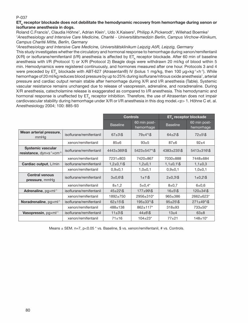
80
P-037ETA receptor blockade does not debilitate the hemodynamic recovery from hemorrhage during xenon or isofl urane anesthesia in dogs.Roland C.Francis1, Claudia Höhne1, Adrian Klein1, Udo X.Kaisers2, Philipp A.Pickerodt1, Willehad Boemke1
1Anesthesiology and Intensive Care Medicine, Charité - Universitätsmedizin Berlin, Campus Virchow-Klinikum, Campus Charité Mitte, Berlin, Germany2Anesthesiology and Intensive Care Medicine, Universitätsklinikum Leipzig AöR, Leipzig, GermanyThis study investigates whether the circulatory and hormonal response to hemorrhage during xenon/remifentanil (X/R) or isofl urane/remifentanil (I/R) anesthesia is affected by ETA receptor blockade. After 60 min of baseline anesthesia with I/R (Protocol 1) or X/R (Protocol 2) Beagle dogs were withdrawn 20 ml/kg of blood within 5 min. Hemodynamics were registered continuously, and hormones measured after one hour. Protocols 3 and 4 were preceded by ETA blockade with ABT-627 (Atrasentan®) IV (bolus 1 mg/kg, then 100 µg×kg-1×h-1). While hemorrhage of 20 ml/kg reduces blood pressure by up to 25% during isofl urane/nitrous oxide anesthesia1, arterial pressure and cardiac output remain stable after hemorrhage during X/R and I/R anesthesia (Table). Systemic vascular resistance remains unchanged due to release of vasopressin, adrenaline, and noradrenaline. During X/R anesthesia, catecholamine release is exaggerated as compared to I/R anesthesia. This hemodynamic and hormonal response is unaffected by ETA receptor inhibition. Therefore, the use of Atrasentan does not impair cardiovascular stability during hemorrhage under X/R or I/R anesthesia in this dog model.<p> 1. Höhne C et. al. Anesthesiology 2004; 100: 885-93
Controls ETA receptor blockade
Baseline 60 min post-hemorrhage
Baseline 60 min post-hemorrhage
Mean arterial pressure, mmHg
isofl urane/remifentanil 67±3\$ 79±4*\$ 64±2\$ 72±5\$
xenon/remifentanil 85±6 93±5 87±6 92±4
Systemic vascular resistance, dyn×s-1×cm-5
isofl urane/remifentanil 4443±369\$ 5423±547*\$ 4383±235\$ 5413±316\$
xenon/remifentanil 7231±803 7420±867 7030±888 7448±684
Cardiac output, L/min isofl urane/remifentanil 1,2±0,1\$ 1,2±0,1 1,1±0,1\$ 1,1±0,3
xenon/remifentanil 0,9±0,1 1,0±0,1 0,9±0,1 1,0±0,1
Central venous pressure, mmHg
isofl urane/remifentanil 3±0,6\$ 1±1\$ 2±0,3\$ 1±0,2\$
xenon/remifentanil 8±1,2 5±0,4* 8±0,7 6±0,6
Adrenaline, pg×ml-1 isofl urane/remifentanil 45±22\$ 177±99\$ 16±5\$ 120±34\$
xenon/remifentanil 1892±750 2956±310* 965±386 2662±623*
Noradrenaline, pg×ml-1 isofl urane/remifentanil 62±15\$ 195±33*\$ 95±25\$ 271±49*\$
xenon/remifentanil 488±138 862±117* 318±93 733±50*
Vasopressin, pg×ml-1 isofl urane/remifentanil 11±3\$ 44±6\$ 13±4 63±8
xenon/remifentanil 71±16 104±23* 77±21 148±10*
Means ± SEM. n=7, p<0.05 * vs. Baseline, $ vs. xenon/remifentanil, # vs. Controls.

81
P-038Implication of endothelin-1 in the hypertensive state triggered by chronic high salt diet in bradykinin B2 receptor knockout mice.Isabelle Brochu1, Eurode Legros1, Élie Simard1, Ghassan Bkaily2, Terry J.Opgenorth3, Pedro D’Orléans-Juste1
1Pharmacology, Université de Sherbrooke, Sherbrooke, QC, Canada2Anatomy and Cellular Biology, Université de Sherbrooke, Sherbrooke, QC, Canada3Pharmaceutical Products Division, Abott Laboratories, Abott Park, ILIt has been recently established that high salt intake interferes with the effi cacy of anti-hypertensive therapy (Weir et al.1995). Afro-Americans are particularly sensitive to a high salt diet since they develop hypertension even in conditions of aggressive treatment with angiotensin converting enzyme inhibitors. Polymorphism of the B2 receptor (Gainer et al. 2000) and increased plasma endothelin-1 (ET-1) (Ergul et al.1998) were identifi ed as possible factors for this sensitivity. As a principal aim, the implication of ET-1 in a mouse model of high salt induced hypertension and the validation of a functional cross-talk between the kinins and ET-1 were assessed. B2KO mice subjected to normal (NSD: 0.49 %) or high salt diet (HSD: 8 %) were monitored by radiotelemetry during 18 weeks. At the end of the 12th week of diet subgroups under NSD or HSD received the orally available ETA antagonist ABT-627 administered (5 mg/kg bid) during 5 days. HSD triggered an increase of MAP of 16.23 ± 0.61 mmHg when compared to NSD treated group (105.51 ± 2.54 mmHg, n=7). ABT-627 corrected the HSD induced hypertensive state to similar MAP levels as those found in NSD mice. Blood samples were collected from these animals and following euthanasia, the lungs, heart and kidneys were extracted, homogenized and assayed for ET-1 by RIA. Signifi cant increases of immunoreactive ET-1 were detected only in the lungs (NSD: 1479.61 ± 128.04; HSD: 2406.93 ± 232.22 fmol/g of tissue; n=14-16). The ETA and ETB protein receptor levels in NSD and HSD mice were found to be similar as evaluated by western blot analysis. Additionally, mRNA levels for precursors and receptors of ET-1 and angiotensin-II were not modifi ed by HSD. On the other hand, metabolic studies showed that excretion of sodium was decreased in the knockout mice (wild type mice: 156.33 ± 22.11, B2KO mice: 60.30 ± 19.23 ueq/24H). The present results suggests that notwithstanding renal excretory impairment afforded by repression of the B2 receptor, these KO mice respond to a prolonged HSD by an ABT-627 sensitive high blood pressure most probably caused by increased pulmonary production of ET-1.
P-039ET-1 Content is not Increased in the presence of Diabetes or HL in Human Arteries and Veins of Patients with Hypertension.Ralph E.Watson1, Cristiane N.Pereira-Hicks1, Melissa W.Li1, Mohammad Pervaiz1, Nandu Gourineni1, John Talbot2, Gregory D.Fink1
1Michigan State University, East Lansing, MI2Sparrow Hospital, Lansing, MIET-1 content in vessels increases remodeling, calcifi cation, and oxidative stress, so it is important to determine if it is increased in certain conditions. Previous studies have found increased ET-1 content in arteries and veins of humans with hypertension (HTN) and type 2 diabetes mellitus (T2DM). No one has reported ET-1 levels in blood vessels of humans with HL (HL). We determined ET-1 content in arteries and veins in 20 human subjects, who underwent coronary artery, bypass graft surgery for atherosclerotic disease. The tissue was left-over internal mammary artery and saphenous vein graft tissue. Methods: Chemiluninescent ELISA kit was used to measure ET-1 content in the vessels. The patients were interviewed and their hospital charts were abstracted to obtain the demographic information. We determined whether the patients had HTN, T2DM, and HL. The unpaired t-test was used to do the statistical analysis. Results: Looking at all subjects, the ET-1 content was signifi cantly higher in internal mammary arteries than saphenous veins, 0.67±0.11vs. 0.36±0.41fmol/mg of tissue (p=0.0035). In patients with HTN, whether they also had T2DM had no effect on the ET-1 level. Patients with HTN and T2DM had ET-1 levels of 0.59±0.05 in arteries and 0.47±0.15 in veins. Those with HTN without T2DM had levels of 0.84±0.23 in arteries and 0.36±0.05 in veins (p=0.37 for arteries and p=0.37 for veins). In patients with HTN, whether they also had HL had no effect on the ET-1 level. Patients with HTN and HL had levels of 0.63±0.16 in arteries and 0.35±0.06 in veins. Those with HTN without HL levels of 0.92±0.2 in arteries and 0.44±0.07 in veins (p=0.28 for arteries and p=0.38 for veins). Conclusions: We found that, in patients with HTN, the addition of the diagnosis of T2DM or HL has no effect on the ET-1 level in human internal mammary artery or saphenous vein.
82
P-040Endothelin (ET) receptor blockade in Cyp1a1-Ren2 transgenic rats with inducible ANG II-dependent malignant hypertension.Ivana Vaneckova1,4, Herbert J.Kramer2, Angela Backer2, Zuzana Husková1,4, Zdenka Vanourková1,4, Zdenka Vernerová3, Ludek Cervenka1,4
1Department of Experimental Hypertension, Institute for Clinical and Experimental Medicine, Prague, Czech Republic2Section of Nephrology, Medical Policlinic, Bonn, Germany3Department of Pathology, 3rd Medical Faculty, Prague, Czech Republic4Center for Cardiovascular Research, Prague, Czech RepublicObjective:We have recently shown that the ET system, especially through ETA receptors, plays an important role in the development of hypertension and end-organ damage in Ren2 transgenic rats (TGR). On the basis of these TGR a new transgenic rat strain with inducible hypertension (iTGR) was generated, in whom the inserted murine Ren2 renin gene is under control of the Cyp1a1 promotor that is induced by various aryl hydrocarbons such as indole-3-carbinole (I3C). Thus, dietary I3C induces malignant hypertension that can be easily turned on and off.Design and Methods: Studies were performed in 3 months old male iTGR with a body weight (BW) of 290±6 g. Treatment with either nonselective ET receptor blocker bosentan or with selective ETA receptor blocker atrasentan was started on day 2 of the experiment. Feeding with I3C (0.3%) began on day 3 and lasted for 4 days. Every day, systolic blood pressure and BW were measured and ANG II concentrations were determined. Left ventricular and renal cortical tissue ET-1 were determined on days 1,3 and 6. The ratio of left ventricle (mg)to BW (g)(LVW/BW)was used as a measure of cardiac hypertrophy. Results: Severe hypertension developed as early as 1 day after beginning of I3C feeding (172±5 mm Hg on day 4 vs. 134±4 mm Hg on day 3). This was accompanied by a substantial reduction in BW from 290±6 g on day 3 to 268±6 g on day 6 and by cardiac hypertrophy with LVW/BW of 2.12±0.048 on day 6 vs.1.87±0.05 on day 3 (p<0.05).Tissue (kidney: 63±9 on day 6 vs.25±5 fmol/g on day 3; left ventricle: 39 ±7 on day 6 vs.23±6 fmol/g on day 3) and plasma ANG II concentrations (95±16 on day 6 vs.46±7 fmol/ml on day 3) increased signifi cantly with I3C-treatment. In the left ventricle it was accompanied by a rise in ET-1 from 0.52±0.12 to 0.95±0.10 fmol/mg protein (p<0.05) on day 6 which was prevented by atrasentan and bosentan (0.42±0.09 and 0.35±0.06 fmol/mg protein, resp.;p< 0.05).Conclusions: Although short-time ET receptor blockade suppressed left ventricular ET-1, our data suggest that ET system does not play a substantial role in the development of acute hypertension and end-organ damage in rats with inducible hypertension.
P-041Role of endogenous endothelin-1 in postischemic cardiac dysfunction and norepinephrine overfl ow in rat hearts.Masashi Tawa, Taiki Fukumoto, Mamoru Ohkita, Yasuo MatsumuraLaboratory of Pathological and Molecular Pharmacology, Osaka University of Pharmaceutical Sciences, Takatsuki, JapanEndothelin-1 (ET-1) and norepinephrine (NE) are involved in myocardial ischemia/reperfusion injury. Recently, we demonstrated that exogenously applied ET-1 is detrimental to the postischemic heart injury, at least partly via the excessive NE overfl ow. In the present study, we investigated the role of endogenous ET-1 in ischemia/reperfusion-induced cardiac dysfunction and NE overfl ow using a selective endothelin converting enzyme (ECE) inhibitor SM-19712 and a metalloproteinase inhibitor phosphoramidon. According to the Langendorff technique, isolated rat hearts were subjected to 40-minute global ischemia followed by 30-minute reperfusion. Ischemia/reperfusion-induced increases in left ventricle (LV) ET-1 levels were suppressed by treatment with SM-19712. SM-19712 improved ischemia/reperfusion-induced cardiac dysfunction. In addition, SM-19712 suppressed excessive NE overfl ow in the coronary effl uent from the postischemic heart. On the other hand, phosphoramidon enhanced increase of LV ET-1 levels and NE overfl ow, and worsened cardiac dysfunction after ischemia/reperfusion. The reason for this may be that phosphoramidon suppressed degradation of ET-1 by inhibiting neutral endopeptidase (NEP). These fi ndings suggest that endogenous ET-1 modulates NE overfl ow, at least in part, in association with ischemia/reperfusion-induced cardiac dysfunction. Next, we investigated the infl uence of exogenously applied big ET-1, an ET-1 precursor, against the ischemia/reperfusion injury. In consequence, big ET-1 suppressed increase of LV ET-1 levels and NE overfl ow, and improved cardiac dysfunction after ischemia/reperfusion. Also, nitrite/nitrate (NOx) release in the coronary effl uent from the postischemic heart was increased by exogenously applied big ET-1 compared with no treatment. These effects of big ET-1 were abolished by concomitant treatment with a nonselective NO synthase inhibitor NOARG (NG-nitro-L-arginine). Thus, our fi ndings suggest that exogenous big ET-1 have a benefi cial effect on ischemia/reperfusion injury by releasing NO. However, endogenous big ET-1 have an adverse effect on ischemia/reperfusion injury, via the conversion to mature ET-1.
83
P-042The benefi cial effects of endothelin-1 converting enzyme inhibitor in preventing myocardial dysfunction secondary to pressure overload through modifi cation of cardiac expression of ET-1, ETA receptor and ETB receptor of left ventricle in aortic-banded rats.Zen-Kong Dai1, Mian-Shin Tan3, Chee-Yin Chai2, Jiunn-Ren Wu1, Jong-Hau Hsu1, Arco Y Jeng4, Ing-Jun Chen5, Jwu-Lai Yeh5
1Pediatrics, Kaohsiung Medical University, Kaohsiung, Taiwan2Pathology, Kaohsiung Medical University, Kaohsiung, Taiwan3School of Biology, Kaohsiung Medical University, Kaohsiung, Taiwan4Metabolic and Cardiovascular Diseases Research, Novartis Institute for Biomedical Research, Summit, NJ5Pharmacology, Kaohsiung Medical University, Kaohsiung, TaiwanHeart failure as a consequence of sustained pressure overload is among the most prevalent diseases in developed country. In the present study, we investigated whether the long term repeated administration of endothelin-1 converting enzyme inhibitor (ECEI), CGS 26393, ameliorated cardiac myocardial remodeling, and how altered the cardiac expression of ET-1, ET-A receptor and ET-B receptor of left ventricle following ascending aortic banding. The banded rats were randomized to receive saline from Day 1-28 (untreated group, n=9), CGS 26393 (30 mg/Kg, twice a day) from Day 1- 28 day (treated group, n=9). Subsequently, there was signifi cant development of (1) myocardial remodeling with increased perivascular fi brosis in Masson staining, and (2) increased mean left atrial pressure and increased mean pulmonary arterial pressure in untreated group. Compared with untreated group, there were signifi cant decrease in both mean pulmonary arterial pressure (sham, 14 ± 1.0 (Mean ± SE) mmHg; untreated, 30 ± 2.0 mmHg; treated, 21 ± 1.1 mmHg, p<0.05 ) and mean left atrial pressure ( sham, 5 ± 0.6 mmHg; untreated, 10 ±1 mmHg; treated, 8 ± 0.6 mmHg, p<0.05) and attenuation in perivascular fi brosis in left ventricle in treated group. Also there were decreased serum ET-1 level in treated group (sham, 1.2 ± 0.16; untreated, 3.2 ± 0.21 fmol/ml; treated, 2.1 ± 0.16 fmol/ml, p<0.05). Interestingly, the cardiac expression of ET-A receptor mRNA in left ventricle in untreated group was less than by in sham-operated group (1.0 ± 0.26; 0.6 ± 0.12, p<0.05), but not different from in treated group. The cardiac expression of ET-B receptor mRNA in left ventricle in untreated group was not different from sham-operated group, but signifi cantly less than in treated group (1.0 ± 0.09; 0.77 ± 0.06, p<0.05). Conclusively, long term CGS 26393 could preventing cardiac dysfunction through inhibiting ET-1 system by decreasing ET-A receptor and ET-1 generation in load-induced cardiac pathology.
P-043Importance of T-tubular Localization of ETA Receptor.Ka Young Chung1, Jeffery W.Walker2
1Molecular and Cellular Pharmacology, University of Wisconsin-Madison, Madison, WI2Department of Physiology, University of Wisconsin-Madison, Madison, WIEndothelin-1 (ET-1) regulates contractility and growth of the heart by binding G-protein-coupled receptors (GPCRs) of the ETA/ETB family. ETA, the predominant ET-1 receptor subtype in myocardium, is thought to localize preferentially in cardiac T-tubules, but the functional consequences of this preferential localization are not fully understood. In pathological conditions such as failing myocardium, T-tubule integrity is often compromised and inotropic responsiveness to ET-1 is blunted despite upregulation of ET-1 and ETA expression. In this study, we explored the functional consequences of upregulation of ETA in conjunction with T-tubule loss in cultured adult rat ventricular myocytes. The normally robust positive inotropic response to ET-1 was lost in myocytes cultured for 3-days, which could be the result of decreased ETA expression, degeneration of T-tubules or both. To rescue the amount of receptors, functionally competent ETA-CFP was overexpressed in cultured myocytes using an adenovirus vector. ETA-CFP appeared fi rst in perinuclear Golg, then in striated puncta radiating out from the Golgi, and fi nally (at day 3) in surface sarcolemma (SSL). ET-1 failed to induce positive inotropic effects in 3-day cultured myocytes overexpressing ETA-CFP in SSL indicating that increse of receptor concentration is not enough to rescue the responsiveness to ET-1. Inclusion of the actin polymerization inhibitor, cytochalasin D (CD), during culture prevented gross morphological changes including rounding of intercalated discs and loss of T-tubules, and largely rescued the responsiveness to ET-1. Furthermore, overexpression of ETA-CFP in 3-day cultured myoytes incubated with CD showed twitch response similar to fresh isolated myocytes. These results supports the idea that T-tubular localization of ETA is important for its normal physiological function
84
P-044Central ETA Receptor Inhibition Alters Barorefl ex Response in Conscious Rats with Doxorubicin Heart Failure.Noreen F.Rossi, Haiping Chen, Maria Maliszewska-ScisloInternal Medicine, Wayne State University and John D. Dingell VAMC, Detroit, MIBarorefl ex activity is typically decreased in heart failure. It has been suggested that central endothelin-1 (ET1) mechanisms modulate the barorefl ex. Both central and peripheral ET1 is increased in heart failure and may contribute to sympathetic hyperactivity. The present study was designed to assess if central ETA receptor blockade alters the barorefl ex response of renal sympathetic nerve activity (RSNA) and heart rate in rats developing cardiomyopathy after weekly injections with 1 mg/kg doxorubicin ip (doxoHF) or vehicle (control, C). After 8 weeks, the rats were instrumented, conditioned to the study environment, and studied in the awake, non-restrained state. Resting mean arterial pressure (MAP), heart rate and RSNA did not differ between doxoHF and C rats. Baseline barorefl ex control of heart rate and RSNA was similar in both groups. ETA blockade with 4 nmol BQ123 intracerebroventricularly signifi cantly decreased both the upper plateau (84±4% of maximum) and the range (from 95 +/- 1 to 80 +/- 4%, p < 0.02) of the barorefl ex response of RSNA in doxoHF rats. The upper plateau and range of the heart rate refl ex response were similarly decreased (p < 0.02) in doxoHF rats. ETA blockade did not change the barorefl ex response of RSNA or HR in C rats. These results support the conclusion that endogenous ET1 does not infl uence barorefl ex regulation in C rats, but that central ETA mechanisms modulate refl ex control of both HR and RSNA in doxoHF particularly during baroreceptor unloading.
P-045The release/formation of Endothelin by Angiotensin II in cardiac myocytes.Carolina D.Garciarena, María C.Villa-Abrille, Ernesto A.Aiello, Irene L.Ennis, Horacio E.CingolaniNational University of La Plata, La Plata, ArgentinaIn a dose response curve to angiotensin II (Ang II), we determined that the maximal positive inotropic effect (PIE) elicited by Ang II (100 nM) was partially blocked by the non-selective endothelin (ET) receptor blocker TAK044. Moreover, the PIE induced by a smaller dose of Ang II (1 nM) was completely abolished by TAK044, indicating that at this dose this effect was mediated by the action of endogenous ET released/formed by Ang II. Since this dose of Ang II better resembles physiological concentration of interstitial Ang II, we focused our study on the intracellular pathways triggered by 1 nM Ang II. We used isolated cat cardiomyocytes for measuring sarcomere shortening (SS) with a video-camera and preproET-1 mRNA levels by real time RT-PCR. These parameters were measured in control and after 15 min treatment with 1 nmol/L Ang II. Ang II increased the level of preproET-1 mRNA from 100±3.4 % (n=15) to 173.3±28.1 % (n=11, p<0.05) as an indicator of the replenishment of the ET-1 intracellular pool that might have been reduced after the release of this peptide by Ang II. This increase in the level of preproET-1 mRNA was abrogated by the AT1 receptor blocker losartan (1 µmol/L; 90.5±21.4 %, n=4), the NADPH oxidase inhibitor apocinin (0.3 mmol/L; 77.7±15.8 %, n=5) and the reactive oxygen species (ROS) scavenger mercapto-propionyl-glycine (MPG, 1 mmol/L; 115.7±15.6 %, n=3). The PIE of Ang II (29.2±3.1 %, n=15, p<0.05) was abrogated by losartan (-1±4.9 %, n=6), by TAK044 (-4.5±4.9 %, n=6), by apocinin (8.7±7.3 %, n=8) and by MPG (-8.4±3.8 %, n=6). The fact that apocinin and MPG abrogated both the increase in ET-1 mRNA and the PIE induced by 1 nM Ang II allow us to conclude that Ang II through the release/formation of ET-1 and by stimulating NADPH oxidase, induces an increase in O2-. that triggers the increase in contractility.

85
P-046In situ dog heart levosimendan compensates endothelin-1 induced coronary vasospasm and reduces onset of ventricular tachyarrhythmias.Szabolcs Szilagyi, Gabor Szucs, Pal Soos, Katalin Turi, Orsolya Kiss, Bela Merkely, Violetta KekesiCardiovascular Surgery, Semmelweis University, Budapest, HungaryIntroduction: The calcium-sensitiser levosimendan is a new type inotropic and vasodilator agent, which improves the cardiac function of the failing and ischaemic heart. The cardiovascular effects of levosimendan were studied in coronary spasm induced by intracoronary (i.c.) administration of endothelin-1 (ET-1). Methods: The investigations were carried out in situ dog hearts (n=15), in three series of experiments. One group of animals was treated with ET-1 of 60 pmol/min i.c. for 30 minutes. In the second group levosimendan (LEV) was administrated intravenously (24µg/kg followed by infusion of 0.4 µg/kg/min for 90 minutes). In the third group of animals ET-1 (60 pmol/min i.c.) was applied between the 30-60 minutes of the 90-minutes levosimendan infusion period. Coronary blood fl ow, systematic blood pressure and standard ECG were recorded continuously. Myocardial contractile force was characterised by end-systolic pressure-volume relationships (ESPVR) obtained from the pressure-volume loops measured in the left ventricle. Results: ET-1 decreased the basal coronary conductance (C) by 61±2%, and induced non-sustained ventricular tachycardias, which accelerated to ventricular fi brillation in 60% of animals. Levosimendan exerted positive inotropic effect (ESPVR: +84 ±5%) and vasodilator response (C: +48±5%) characteristic to the agent. The positive inotropic effect of LEV was preserved in ET-1 induced myocardial ischaemia (ESPVR: +69±7%) while the coronary constriction evoked by ET-1 was counterbalanced by parallel infusion of LEV (C basal: 0.22±0.01 vs. C LEV+ET-1: 0.21±0.01 mL/min*mmHg, n.s.). No malignant ventricular tachyarrhythmias were observed in animals treated with LEV and LEV+ET-1. Conclusion: Beside its positive inotropic action levosimendan exerts signifi cant antiischaemic effect by reducing coronary vascular tone enhanced by endothelin-1, moreover its benefi cial effect may also be realized as a decrease in ventricular tachyarrhythmias.
P-047Rho-kinase and Ca2+-sensitive proline-rich tyrosine kinase (PYK2) contribute to abnormal ET-1-induced contraction in corpora cavernosa from DOCA-salt hypertensive mice.Fernando S.Carneiro1,2, Fernanda R.Giachini1,2, Romulo Leite2, Adviye Ergul2, Clinton Webb2, Rita C.Tostes1,2
1Pharmacology, University of Sao Paulo, Sao Paulo, Brazil2Physiology, Medical College of Georgia, Augusta, GAPenile smooth muscle cells not only respond to, but also synthesize, ET-1. Hypertension is a risk factor for erectile dysfunction (ED) and ET-1 is directly involved in end-organ damage in salt-sensitive forms of hypertension. We hypothesize that ET-1 responses are enhanced in penes from DOCA-salt mice. C57BL/6 mice were uninephrectomized and submitted to DOCA-salt (DOCA 2g/Kg sc; water+1% NaCl, n=10) or vehicle (placebo pellets, normal tap water, n=9) treatment for 5 weeks. Penes were excised, dissected to remove the tunica albuginea, and one crural strip was obtained from each corpus cavernosum. Changes in isometric force were recorded. Systolic arterial pressure was elevated in DOCA mice [(mmHg), 126±6 vs. control (C) 103±4, p≤0.05]. No differences in responses mediated by nonadrenergic-noncholinergic or sympathetic nerve stimulation, acetylcholine, or phenylephrine were observed between cavernosal strips from DOCA and C mice. ET-1-mediated contractions were greatly enhanced in DOCA cavernosal strips [(mN), 0.96±0.1 vs. C, 0.46±0.1, p≤0.05). ABT-627, ETA antagonist, abrogated ET-1-induced cavernosal contractions and no contractile responses were observed upon stimulation with IRL-1620, ETB agonist. IRL-1620 induced relaxation of cavernosal strips and responses were enhanced in DOCA cavernosum [(%), 56±11 vs. C, 24±4, p≤0.05]. ET-1 contraction was inhibited by Rho kinase (Y-27632) and PYK2 (AG-17) inhibitors, with DOCA cavernosal strips displaying decreased relaxation [(%), Y-27632: DOCA, 80 vs. C, 125; AG-17: DOCA, 3.7 vs. C, 35.5]. Changes in ET-1-mediated responses occur before other functional alterations in penes from DOCA-salt mice. Although ETB-mediated relaxation is increased in cavernosal strips from DOCA mice, this is not suffi cient to overcome increased Rho kinase/PYK2-mediated contractile responses to ET-1. Changes in the endothelin system may play a role in salt-sensitive hypertension-associated ED.
86
P-048Endothelin-3 -dependent pulmonary vasoconstriction in monocrotaline-induced pulmonary arterial hypertension.Stéphanie Sauvageau, Eric Thorin, Jocelyn DupuisMontreal Heart Institute, Montreal, QC, CanadaIntroduction: Activation of the endothelin (ET) system contributes to the development and maintenance of pulmonary arterial hypertension (PAH). The contribution of ET-3 and its interaction with the ET-Rs have not been characterized in pulmonary resistance vessels. Methods: Sham or monocrotaline (MCT)-injected rats were evaluated after 5 weeks. Using confocal imaging, we investigated ET-3 localisation. Gene expression of prepro-ET-3 was evaluated in pulmonary vessels using RT-PCR. Reactivity of pulmonary resistance arteries to ET-3 was measured in the presence of ET-R antagonists (ET-RA): ETA-RA (10 nM), ETB-RA (1 µM) and combination of both. Results: MCT animals developed severe PAH with elevation of right ventricular systolic pressure (p<0.001). ET-3 is mainly localized in the smooth muscle layer of pulmonary resistance arteries. The gene expression of preproET-3 were reduced in pulmonary resistance arteries of MCT (p<0.05) compared to sham animals. ET-3 induced similar pulmonary vasoconstrictions in both sham (Emax 77±1%, EC50 21±7 nM, mean±sem) and MCT-treated rats (Emax 68±2%, EC50 33±9 nM,). In sham animals, the ETA-RA greatly reduced the maximal response to ET-3 (Emax 46±2%, p<0.001, EC50 41±29 nM) while the ETB-RA shifted the EC50 without affecting Emax (Emax 68±2%, EC50 59±16 nM, p<0.05). However, the combination of both completely abolished ET-3 response (p<0.001). In PAH, the ETA-RA also markedly reduced the maximal response and shifted the EC50 (Emax 23±2%, p<0.001, EC50 104±24 nM, p<0.05) while the ETB-RA only shifted the EC50 without affecting the maximal response (Emax 52±3%, EC50 123±36 nM, p<0.05). The combination of both antagonists did not further reduce the constriction compared to ETA-R inhibition alone (Emax 23±7%, p<0.001, EC50 36±21 nM). Conclusion: In control rats, the effi cacy of the ET-3 response is reduced by an ETA-RA while its potency is sensitive to an ETB-RA. The pronounced effi cacy of dual ETA/ETB-Rs inhibition suggests that interdependence develops between the two receptor-subtypes for optimal contraction. In PAH, however, the increased effi cacy of the ETA-RA suggests a modifi cation of the receptors interdependence.
P-049Role of Endothelin Receptors on Basal and Stimulated Lung Myofi broblasts Proliferation.Annick Préfontaine2, Angelino Calderone2, Jocelyn Dupuis2
1Montreal Heart Institute, Beaconsfi eld, QC, Canada2Montreal Heart Institute, Montreal, QC, Canada3Montreal Heart Institute, Montreal, QC, CanadaBackground: Myofi broblasts (MYF) proliferation contributes to the pathophysiology of numerous lung disorders. Pulmonary endothelin-1 (ET-1) production is increased in various lung diseases and could directly contribute to deleterious lung remodelling. However, the respective roles of ETA and ETB receptors (ETA-R, ETB-R) and of endogenous ET-1 production by lung MYF on their proliferation remain uncertain. Methods: Rat lung MYF were isolated and cultured. ET-R expression on MYF was verifi ed by immunofl uorescence confocal imaging. Protein and DNA synthesis of lung MYF induced by ET-1 (10 nM) was assessed by 3H-thymidine and 3H-leucine incorporation. These experiments were also performed in the absence and presence of ET-R antagonist (ET-RA): ETA-R (BQ-123, 1µM), ETB-R (BQ-788, 1µM) and combination of both. ET-1 levels in culture supernatants were measured by ELISA. Results: Immunofl uorescence studies revealed that both ETA-R and ETB-R were abundantly expressed in lung MYF. ET-1 stimulated lung MYF protein and DNA synthesis as measured by the incorporation of [3H]-leucine (19.2% ± 5.4) and [3H]-thymidine (22.0±4.4%). Selective ETA-R blockade (BQ-123) or selective ETB-R blockade alone (BQ-788) did not inhibit proliferation or protein synthesis. However, the combination of both antagonists completely abolished [3H]-leucine (3.4±0.7%, p < 0.05) and [3H]-thymidine (1.1 ± 2.9%, p<0.05) incorporation induced by ET-1. Surprisingly, in basal conditions, the incubation of lung MYF with either the selective ETA-R antagonist (BQ-123) or with the selective ETB-R antagonist (BQ-788) alone induced a signifi cant increased of MYF proliferation (10.1 ± 2.0% and 7.8% ± 2.5, P < 0.05). On the other hand, the presence of both ET-R alone did not induced MYF proliferation (3.4 ± 0.7%) and ET-1 levels in the supernatants were not affected by the antagonists. Conclusion: Our fi ndings indicate that both the ETA-R and the ETB-R regulate basal and stimulated lung MYF proliferation and suggest possible interactions between the receptors.
87
P-050ET-1 up-regulates EphA2 expression in pulmonary vascular cells.Todd Carpenter, Jacqueline Larson, Stacey Schomberg, Christine BankersPediatrics, University of Colorado Health Sciences Center, Denver, COAngiogenesis is thought to contribute to diverse pathologic processes in the lung, including pulmonary vascular remodeling in pulmonary hypertension and airway remodeling in asthma, and perhaps pulmonary edema formation in lung injury. Endothelin (ET) may contribute to angiogenesis not only directly but also indirectly via upregulation of the expression of vascular growth factors such as VEGF. Ephrin family receptor tyrosine kinases, in particular the EphA2 receptor and ephrin-a1 ligand, have also been implicated in the process of post-natal angiogenesis. The expression of ephrins in pulmonary vascular cells, however, is not well described and whether endothelin regulates lung ephrin expression is unknown. We investigated ephrin expression in primary cultured pulmonary artery endothelial cells (PAEC), vascular smooth muscle cells (PASMC), and adventitial fi broblasts (AF) by RT-PCR, Western blotting and immunohistochemistry. All three cell types expressed mRNA for ephrin-a1, EphA2, EphA4, EphA5, and EphA6 at baseline. In addition, PASMC and AF also expressed EphA3, whereas PAEC also expressed ephrin-a2 and ephrin-a5. ET-1 stimulation for 6 hours led to a signifi cant dose-dependent increase in EphA2 mRNA and protein expression in PA adventitial fi broblasts and, to a lesser extent, PASMC. ET-1 did not change the expression of other ephrin mRNAs. Lung tissue from both rat and cow exposed to hypoxia, a stimulus known to increase lung ET levels, also demonstrated increased vascular EphA2 expression by immunostaining. These results suggest that ET-1 can directly and specifi cally regulate the expression of EphA2 in pulmonary vascular cells and may represent a novel mechanism by which ET contributes to angiogenic processes in the lung.
P-051Effects of Bone Morphogenic Proteins on Endothelin-1 Production by Human Pulmonary Microvascular Endothelial Cells In Vitro.Gregory Star, Michele Giovinazzo, Tali Dworkin, David LanglebenCardiology, Jewish General Hospital, Montreal, QC, CanadaIntroduction: Bone morphogenic proteins (BMP) have been implicated in the pathogenesis of pulmonary arterial hypertension (PAH). Elevated levels of the vasoconstrictor and mitogenic peptide, endothelin-1 (ET-1) have been described in PAH, and ET-1 blockade provides therapeutic benefi t to patients. The effects of BMPs on ET-1 synthesis by lung microvascular endothelium are unclear. Methods: We studied the effects of varying concentrations of BMP 4, and 7 on ET-1 synthesis by cultured human pulmonary microvascular endothelial cells (HMVEC-LB, Lonza Inc) in vitro. HMVEC-LB were grown to confl uency in culture dishes, in EGM-2MV medium containing 5% FBS and supplements. They were then exposed to BMPs (0 - 100 ng/ml) in EGM-2MV for 24 and 48 hours. At the end of the exposure period, the supernatant was collected and the protein content of each well was measured with the BCA protein assay. ET-1 was measured in the supernatant using ELISA (Assay Designs) and expressed as pg/ml. The ET-1 concentrations in the supernatant were then normalized to protein concentration per well. Results: Expressed as pg ET-1/µg protein, or as absolute levels, as compared to CONTROL, at 24 hours, neither BMP4 or 7 affected ET-1 production. Total protein was increased by BMP-7 but not BMP-4. At 48 hours, BMP-4 did not increase absolute or normalized ET-1 levels, or protein levels. BMP-7 (50 ng/ml) increased absolute and normalized ET-1 levels at 48 hours, but there was no effect on protein levels. Conclusion: Pulmonary microvascular endothelial ET-1 production is not affected by BMP-4 and is minimally affected by BMP-7 at relatively high doses. Thus, there is little evidence that these peptides contribute to the maintenance of low ET-1 synthetic levels seen in normals.
88
P-052Down-regulation of the Endothelin system in CHF lung myofi broblasts.Annick Préfontaine2, Angelino Calderone2, Dupuis Jocelyn2
1montreal heart institut, Beaconsfi eld, QC, Canada2Montreal Heart Institute, Montreal, QC, CanadaBackground: Congestive heart failure (CHF) causes lung morphological changes characterized by thickening of the alveolar septa with abundant proliferation of myofi broblasts (MYF) and collagen synthesis. Endothelin-1 (ET-1) production is increased in CHF and may contribute to lung remodelling by stimulating MYF proliferation, chemotaxis and collagen synthesis. We therefore investigated the effect of ET-1 on MYF from sham and CHF rats induced by 3 week’s myocardial infarction. Methods: CHF was induced in rats by myocardial infarction and three weeks post surgery lung MYF were isolated and cultured. A group of rats were injected with 5’-bromodeoxyuridine (BrdU) intraperitonealy and immunofl uorescence confocal imaging was used for confi rming lung remodelling. The mitogenic and protein synthesis response of MYF to ET-1 (10nM) was assessed by 3H-thymidine and 3H-leucine incorporation respectively. ET-1 levels in culture supernatants was measured by ELISA and ETA and ETB receptors (ETA-R, ETB-R) expression was examined by western immunoblotting. Results: CHF rats injected with BrdU demonstrated lung remodelling with thickening of the alveolar septa and signifi cant increase in BrdU uptake and vimentin expression (p< 0.05). The mitogenic response to ET-1 in CHF (19.0 ± 3.0%) was signifi cantly less than for sham rats (35 ± 5.4%, p< 0.05). This effect was associated with a signifi cantly lower production of ET-1 by CHF MYF (15.15 ± 5.67 fmol/ml) compared to sham MYF (33.66 ±13.22 fmol/ml, p<0.05). Additionally, expression of both ETA-R (0.36 ± 0.038AU) and ETB-R (0.24 ± 0.075 AU) were reduced in CHF compared to sham’s (0.65 ± 0.086 and 0.81 ± 0.21 AU respectively, p< 0.05). Conclusion-Our results reveals a down-regulation of the ET-1 system of lung MYF in CHF rats. This down-regulation of the ET-1 axis in lung MYF may represent a protective adaptation in response to chronic exposure to high levels of ET-1.
P-053The Study on gene expression of ET-1, eNOS and phosphorylated eNOS in HMEC-1 cells during rapid desensitization of prostacyclin receptor (IP) in response to prostacyclin I2.
Zen-Kong Dai1,5, Mian-Shin Tan2, Chee-Yin Chai3, Jiunn-Ren Wu1, Arco Y Jeng6, Ing-Jun Chen4, Jwu-Lai Yeh4, Shah-Hwa Chou7
1Dept. of Pediatrics, Kaohsiung Medical University, Kaohsiung, Taiwan2School of Biolog, Kaohsiung Medical University, Kaohsiung, Taiwan3Dept.of Pathology, Kaohsiung Medical University, Kaohsiung, Taiwan4Department of Pharmacology, Kaohsiung Medical University, Kaohsiung, Taiwan5Graduate institute of Medicine, Kaohsiung Medical University, Kaohsiung, Taiwan6Metabolic and Cardiovascular Diseases Research, Novartis Institute for Biomedical Research, Summit, NJ7Dept.of Chest Surgery, Kaohsiung Medical University, Kaohsiung, TaiwanWhen lungs under chronic iloprost infusion were acutely challenged with inhaled iloprost, a corresponding complete loss of vasoreactivity was observed. The rapid desensitization of the human prostacyclin (IP) in response to agonist binding has been shown in cell culture experiment. Phosphorylation of the IP receptor by protein kinase C (PKC) has been suggested to be involved in this process. The aim of the present study is to investigate the role of Iloprost, a stable prostacyclin I2 analogue analog, in modulation of gene expression of endothelin-1(ET-1) and endothelial nitric oxide synthase (eNOS) in cultured Human Microvascular Endothelial Cell-1 (HMEC-1) during desensitization by continuously stimulated with 10-6 M and 10-5 M iloprost for 12 and 24 hours, respectively. We compared preproET-1 mRNA and eNOS mRNA by competitive RT-PCR, eNOS and phosphorylated eNOS at (P-eNOS) by western blotting, ET-1 by EIA and cyclic AMP (c AMP) by RIA. Subsequently, there were reduction of iloprost induced c AMP formation to 70 % of control in 10-5 M iloprost-12 hour incubated group(P<0.05, N=4), to 15 % of control in 10-5 M iloprost-24 hour incubated group (P<0.05, N=4) In 10-5 M iloprost-12 hour incubated group, the intracellar ET-1 content was decreased signifi cantly, also the preET-1 mRNA was comparably decreased signifi cantly. The phosphorylated eNOS was increased signifi cantly although both eNOS mRNA and eNOS were not signifi cantly altered in 10-5 M iloprost-12 hour incubated group (1.0 ± 0.08, 2.25 ± 0.1, p<0.01). Conclusively, it indicated that iloprost may down-regulate ET-1 system, also may augment time-dependent phosphorylation of eNOS through PK C during rapid desensitization of IP.

89
P-054Low dose dual ETA/ETB receptor antagonist tezosentan via inhalation effectively attenuates pulmonary hypertension and edema in endotoxin-induced acute lung injury.Björn P.Persson, Patrik Rossi, David Konrad, Michael Wanecek, Eddie Weitzberg, Anders OldnerSect. Anaesthesiology, Dept Physiology and Pharmacology, Karolinska Institutet, Stockholm, SwedenSepsis-induced lung injury is a major constituent of morbidity and mortality in ICU patients. A hallmark of the pathophysiology is formation of pulmonary edema with progressive gas exchange abnormalities. Previous studies have indicated that endothelin-1 is highly involved in edema formation during sepsis-induced lung injury. Our group has shown that systemically administered dual ETA/ETB receptor antagonist tezosentan (TZ) counteracts edema formation and improves gas exchange, possibly via reduced pulmonary venous constriction. However, systemically administered tezosentan has potential disadvantages in the hypotensive ICU patient. To investigate the possibility of a more selective pulmonary therapy we conducted a study with inhaled, nebulized, TZ in a porcine model of endotoxin-induced lung injury. Material and methods: 28 pigs were anesthetized, catheterized and mechanically ventilated. Hemodynamic and blood gas-exchange parameters were monitored. Extra vascular lung water (EVLW) was measured with single thermal indicator technique (PiCCO, Pulsion). After baseline measurements and two hours of endotoxic shock (E. coli, 250 ng/kg/h) the animals were randomized to four groups receiving either inhaled TZ 0.5 mg/kg or 0.05 mg/kg or I.V. TZ 0.5 mg/kg or inhaled placebo, all in single bolus doses. The animals were studied for another three hours, after which they were sacrifi ced. Results: Endotoxin induced a profound pulmonary hypertension that was effectively counteracted by TZ no matter dose or way of administration. EVLW was reduced by inhaled TZ (0.5 mg/kg (p 0.03); 0.05 mg/kg (p 0.06)) but not by I.V. TZ. Oxygen tension remained unaffected by all treatment regimens. Systemic hypotension as further enhanced by I.V. treatment and high dose inhalation but not by low dose inhalation. Conclusion: Inhaled TZ may counteract endotoxin-induced pulmonary hypertension and edema with less systemic effects as compared with intravenous administration. Future studies with a prolonged, continuous inhalation treatment are warranted for evaluation of effects on gas exchange.
P-055Endothelin converting enzyme inhibitor decreases right ventricular pressure, right ventricular hypertrophy and increases the reaction on L-NAME and NO production in rats with hypoxia induced pulmonary hypertension.Alexandra Simonova1,2, Vladimir Pozdnev3, Oleg Gomazcov3, Natalia Medvedeva1
1Department of Physiology, Lomonosov Moscow State University, Faculty of Biology, Moscow, Russian Federation2Institute of Pathology and Pathophysiology of the Russian Academy of Medical Science, Moscow, Russian Federation3Orekhovich Institute of Biomedical Chemistry, Moscow, Russian FederationPulmonary arterial hypertension (PAH) - disease, characterized by the elevated right ventricular systolic pressure, pulmonary vessels remodeling and right heart ventricle hypertrophy. The pathogenesis of PAH is not completely understood, but its accepted mechanisms include vasoconstriction, endothelial disfunction and smooth muscle cell proliferation. Two of the most important factors produced by the vascular endothelium are the endothelium-derived relaxing factor nitric oxide (NO) and the endothelium-derived vasoconstrictor peptide endothelin-1 (ET-1).Its role in PAH is supported by increased levels of circulating ET-1, as well as increased local production. There is a correlation between ET plasma levels and the severity of PAH in patients. The aim of our study was to investigate the effects of original endothelin converting enzyme inhibitor (ECEI) (Orekhovich Institute of Biomedical Chemistry, Russian) - PP36 (Pozdnev et al.1998) on development of right ventricular overload, systolic right ventricular pressure (RVSP) and reactivity of the pulmonary vessels in Wistar rats with hypoxia induced pulmonary hypertension (hPAH). Methods: Pulmonary hypertension was induced by the adaptation to hypobaric hypoxia (O2 concentration reduced to 10%) during 2 weeks. PP36 (1,4 mg/kg) was added to the drinking water during two weeks too. Nitrate and nitrite were detected in urine with spectrophotometric method. Results: Chronic treatment with PP36 decreased RVSP by 25% (p<0.05). Right ventricular hypertrophy reduced from 46.6±1mg in rats with hPAH to 39.6±1.1mg (p<0.05) in rats with hPAH treated with PP36. ECEI didnt change the reaction RVSP on phenylephrine but increased the hypertensive reaction on NO-synthase inhibitor- L-NAME on 35% (p<0.05). ECEI increase products of the urine nitrate and nitrite by 57% in rats with hPAH. Conclusion: Chronic treatment with ECE inhibitor signifi cantly reduced pathological changes due to hypoxia induced pulmonary hypertension. One of the reasons may be the increased production and role of NO in pulmonary artery tone.
90
P-056Effects of Transforming Growth Factor Beta on Endothelin-1 Production by Human Pulmonary Microvascular Endothelial Cells In Vitro.Gregory P.Star, Michele Giovinazzo, David LanglebenCardiology, Jewish General Hospital, Montreal, QC, CanadaIntroduction: Members of the Transforming Growth Factor Beta (TGF) superfamily of molecules, including bone morphogenic proteins, have been implicated in the pathogenesis of pulmonary arterial hypertension (PAH). Elevated levels of the vasoconstrictor and mitogenic peptide, endothelin-1 (ET-1) have been described in PAH, and ET-1 blockade provides therapeutic benefi t to patients. The high ET-1 levels result from excessive synthesis rather than reduced clearance, and the effects of TGFβ and its related molecules on ET-1 synthesis by the lung microvasculature are unclear. Methods: We studied the effects of varying concentrations of TGFβ on ET-1 synthesis by cultured human pulmonary microvascular endothelial cells (HMVEC-LB, Lonza Inc) in vitro. HMVEC-LB were grown to confl uency in culture dishes, in EGM-2MV medium containing 5% FBS and supplements. They were then exposed to TGFβ (0, or 0.1, 0.5, 1, and 5 ng/ml) in EGM-2MV for 24 and 48 hours. At the end of the exposure period, the supernatant was collected and the protein content of each well was measured with the BCA protein assay. ET-1 was measured in the supernatant using ELISA (Assay Designs) and expressed as pg/ml. The ET-1 concentrations in the supernatant were then normalized to protein concentration per well. Results: Expressed as pg ET-1/µg protein, as compared to CONTROL, at 24 hours, ET-1 production was signifi cantly increased by TGFβ at 0.5, 1, and 2 ng/ml. However, absolute (non-normalized ) ET-1 production increased at all concentrations of TGFβ. Total protein was not affected by any TGFβ concentration at 24 hours. At 48 hours, expressed as pg ET-1/µg protein, and as compared to CONTROL, TGFβ increased ET-1 production at all concentrations. Absolute ET-1 production was increased at all TGFβ concentrations except for 0.1 ng/ml. TGFβ 0.1 - 5 ng/ml also decreased total protein at 48 hours. Conclusion: ET-1 production is modulated by various concentrations of TGFβ in HMVEC-LB. Since ET-1 is an important mediator of PAH, and TGFβ signalling has been implicated in the disease process, our fi ndings may provide one mechanism by which the ET-1 levels are increased in-vivo in PAH.
P-057eNOS inhibitor up-regulated expression of ET-1 and Rho kinase in lungs with pulmonary hypertension secondary to left dysfunction in aortic banded rats: The important role of eNOS in suppressing pulmonary vascular tone and ET-1 pathway.Zen-Kong Dai1,8, Chee-Yin Chai2, Mian-Shin Tan3, Arco Y Jeng7, Shah-Hwa Chou4, Ing-Jun Chen5, Jwu-Lai Yeh5, Yu-Ju Chen6, Jiunn-Ren Wu1
1Dept of Pediatrics, Kaohsiung Medical University, Kaohsiung, Taiwan2Department of Pathology, Kaohsiung Medical University, Kaohsiung, Taiwan3School of Biolog, Kaohsiung Medical University, Kaohsiung, Taiwan4Department of Chest Sugery, Kaohsiung Medical University, Kaohsiung, Taiwan5Department of Pharmacology, Kaohsiung Medical University, Kaohsiung, Taiwan6Department of Nuclear Medicine, Kaohsiung Medical University, Kaohsiung, Taiwan7Metabolic and Cardiovascular Diseases Research, Novartis Institute for Biomedical Research, Summit, NJ8Graduate Institute of Medicine, Kaohsiung Medical Universtiy, Kaoshiung, TaiwanEndothelial nitric oxide synthase (eNOS) is an important regulatory enzyme that catalyzes production of NO from arginine. NO is a potent vasodilator and has anti-proliferativie effect on pulmonary vasucature. L-NAME is nonspecifi c inhibitor of eNOS. Previously, we reported that there was up-regulated expression of both eNOS and endothelin-1 (ET-1) in lungs with pulmonary hypertension of aortic-banded rats. The aim of the present study is to investigate the contributory role of eNOS in suppressing the pulmonary vascular tone in the model. Wistar rats (200-300 gm) with left ventricular dysfunction caused by an ascending aortic banding. The banded rats were randomized to receive saline from Days 1-28(AOB28, n=8), (3 mg/100g/day, PO) from Day 15-28 (AOB28/L-NAME15-28, n=8). There was signifi cant development of pulmonary hypertension with pulmonary vascular remodeling 4 weeks after ascending aortic-banding. Although there was signifi cant increase of thickened medial layer of pulmonary arterioles in both L-NAME groups (% wall thickness: AOB28, 31.7 ± 2.0 % (mean ± SEM); AOB28/L-NMAE15-28, 42 ± 1.7 % ; P<0.05), the increase of the mean pulmonary arterial pressure were signifi cantly noted in the AOB28/L-NAME15-28 group AOB28, 33 ± 2.0 mmHg; AOB28/L-NAME15-28 (42 ± 1.1 mmHg, p<0.05 ). PreproET-1 mRNA, eNOS mRNA and Rho kinase mRNA were measured by competitive RT-PCR, eNOS was measured by western blotting, and ET-1 was checked by EIA. Subsequently, the L-NAME resulted in increased pulmonary ET-1(AOB, 3.5 ± 0.9 pmol/g protein; AOB28/L-NAME15-28 12.7 ± 2.0 pmol/g protein, p<0.05), pulmonary preproET-1 mRNA, and pulmonary eNOS in AOB28/L-NAME15-28 rats. These results suggest that repeated administration of L-NAME exacerbated both the pulmonary arterial pressure and pulmonary vascular remodeling in aortic-banded rats, and that eNOS still plays an important role on the anti- proliferative effects against the up-regulation of preproET-1 gene and Rho kinase in aortic banded rats.
91
P-058The benefi cial effects of CGS 26393, ECE inhibitor, on pulmonary expression of ET-1 and eNOS in rats with pulmonary hypertension secondary to left ventricular dysfunction.Chee-Yin Chai1, Zen-Kong Dai5,2, Jiunn-Ren Wu5, Mian-Shin Tan3, Arco Y Jeng6, Shah-Hwa Chou5, Ing-Jun Chen4, Jwu-Lai Yeh4, Chaw-Chi Chiu5
1Dept. of Pathology, Kaohsiung Medical University, Kaohsiung, Taiwan2Graduate Institute of Medicine, Kaohsiung Medical University, Kaohsiung, Taiwan3Shool of Biology, Kaohsiung Medical University, Kaohsiung, Taiwan4Dept. of Pharmacology, Kaohsiung Medical University, Kaohsiung, Taiwan5Dept.of Pediatrics, Kaohsiung Medical University, Kaohsiung, Taiwan6Metabolic and Cardiovascular Diseases Research, Novartis Institute for Biomedical Research,, Summit, NJIncreasing evidence has implicated endothelin-1 (ET-1), a potent vasoconstrictive peptide, in the pathophysiology of pulmonary hypertension. Endothelin-converting enzyme-1 (ECE-1), the protease involved in the fi nal step of post-translational processing of ET-1, cleaves the inactive precursor big ET-1 at the Trp(21)-Val (22) peptide bond. CGS 26393, is an orally active, long-acting inhibitor of ECE-1. In our previous study, we found that there were signifi cant development of pulmonary hypertension with pulmonary vascular remodeling and up-regulated pulmonary gene expression of eNOS and ET-1 4 weeks after aortic-banding in rats. We investigated whether the long term administration of CGS 26393 attenuated pulmonary hypertension, and how altered the expression of ET-1, Big ET, and eNOS in aortic-banded rats. The Wistar adult rats are ascending aortic-banded. The banded rats were randomized to receive saline from Days 1-28 (n=8), CGS 26393 (30 mg/kg, P.O. twice a day) from Day 15-28 (AOB28 /CGS15-28, n=8) or from Day 1- 28 days (AOB28/CGS1-28, n=8). Subsequently, CGS26393 could attenuate both thickened medial layer of pulmonary arteriole and pulmonary arterial pressure in AOB28/CGS1-28 group (sham, 16 ± 1.0 (Mean ± SE) mmHg; AOB28, 32 ± 2.0 mmHg; AOB28/CGS1-28, 24 ± 1.1 mmHg, p<0.05 ). The increased pulmonary Big ET (AOB28, 2.9 ± 0.2 pmol/g protein; AOB28/ CGS1-28, 4.5 ± 0.2 pmol/g protein, P<0.05 ), decreased pulmonary ET-1 (3.15 ± 0.21 pmol/g protein; 2.1 ± 0.17 pmol/g protein, P<0.01 ), and decreased preproET-1 mRNA (1.30 ± 0.21, 1.0 ± 0.12, p<0.05) were noted in AOB28/CGS1-28A. Interestingly, pulmonary eNOS was increased in AOB28/CGS1-28 group. These results suggest that early repeated administration of CGS 26393 inhibited both the rise in pulmonary arterial pressure and the pulmonary vascular remodeling in pulmonary hypertension secondary to failure, and the ameliorating effect is mediated by down-regulating ET-1 and up-regulating eNOS pathway.
P-059Sex differences in renal medullary endothelin-dependent sodium and water excretion.Daisuke Nakano, David M.PollockVascular Biology Center, Medical College of Georgia, Augusta, GARenal endothelin (ET)-1 has been implicated in the regulation of sodium and water excretion. We recently demonstrated that infusion of sarafotoxin S6c (S6c), an ETB agonist, into the renal medullary interstitium induced diuresis and natriuresis without changes in blood pressure in normal male rats. In addition, our lab has previously shown a sex difference in ETB receptor-dependent blood pressure regulation. Therefore, the present study was conducted to examine the sex difference in ET-1-mediated sodium and water excretion. Drugs were infused into the left renal medullary interstitium of anesthetized ETB-defi cient or wild type rats. Intramedullary infusion of ET-1 (0.45 µg/kg/h) in female wild type rats increased the urine fl ow (UV; 7.9 ± 0.7 µl/min; p<0.05) and sodium excretion (UNaV; 0.81 ± 0.02 µmol/min; p<0.05) compared with that in male rats (UV: 3.8 ± 0.7 µl/min; UNaV: 0.35 ± 0.09 µmol/min). S6c (0.45 µg/kg/h) signifi cantly increased UV (male: 7.9 ± 0.3 µl/min; female: 8.6 ± 0.6 µl/min; p<0.01) and UNaV (male: 0.88 ± 0.05 µmol/min; female: 0.95 ± 0.13 µmol/min; p<0.01) in wild type rats compared to ETB-defi cient rats (UV: male, 4.7 ± 0.3 µl/min; female, 5.0 ± 0.3 µl/min; UNaV: male, 0.38 ± 0.03 µmol/min; female, 0.38 ± 0.02 µmol/min); there were no differences in the response to S6c between male and female rats. Infusion of ET-1 in female ETB-defi cient rats markedly increased the UV (11.4 ± 0.4 µl/min) and the UNaV (1.29 ± 0.11 µmol/min) compared to baseline (UV: 3.8 ± 0.3 µl/min; UNaV: 0.34 ± 0.02 µmol/min), yet no increase was observed in male rats (UV: 5.1 ± 0.4 µl/min; UNaV: 0.41 ± 0.05 µmol/min). Treatment with ABT627, an ETA antagonist, prevented the increased excretion observed in female ETB-defi cient rats (UV: 4.6 ± 0.2 µl/min; UNaV: 0.52 ± 0.04 µmol/min). Therefore, these results suggest that there is no sex difference in the diuresis and natriuresis induced by the activation of renal medullary ETB receptor in normal rats; however, the ETA receptor also contributes to ET-1-dependent natriuresis in female but not male rats.
92
P-060Urinary ET-1 in chronic kidney disease.Neeraj Dhaun1,2, Eline Muilwijk1, Pajaree Lilitkarntakul1, Vanessa Melville1, Neil Johnston1, Jane Goddard2, David Webb1
1Clinical Pharmacology Unit, University of Edinburgh, Edinburgh, United Kingdom2Department of Renal Medicine, Royal Infi rmary of Edinburgh, Edinburgh, United KingdomIntroduction Plasma [ET-1] is elevated and intrarenal [ET-1] is increased in chronic kidney disease (CKD). Urinary [ET-1] is a surrogate marker for renal ET-1 production. However, it remains unclear how these concentrations change with GFR. We hypothesised that there would be a linear relationship between declining GFR and increasing plasma and urinary ET-1. Additionally, experimentally, renal ET-1 activity is increased in infl ammation. We hypothesised that patients with normal GFR but immune-mediated renal disease would have higher urinary [ET-1] than patients with non-immune mediated haematuria of glomerular origin and normal GFR. Methods In study 1, we measured plasma and urinary [ET-1] in 91 patients and healthy volunteers (HV) with varying GFR (M/F, 55/36, mean age 47yrs). Fractional excretion of ET-1 (FeET-1) was calculated based on plasma and urinary [creat] and [ET-1]. In study 2, 82 subjects with haematuria of presumed glomerular origin (M/F, 39/43, mean age 43yrs): thin basement membrane disease (TBM, n=8), IgAN (n=22), vasculitis (n=17), microhaematuria with no biopsy (MH, n=35) were included. Subjects were matched with 29 HV (M/F, 12/17, mean age 45yrs). All participants had normal GFR and BP. In addition to previous data CRP and proteinuria were measured. Results In study 1, both plasma ET-1 and FeET-1 increased as GFR declined. In study 2, there were no signifi cant differences in FeET-1 between HV and TBM, IgAN, or MH. However, vasculitis patients had signifi cantly higher FeET-1 than HV (p<0.001), MH (p<0.01), and IgAN (p<0.001). FeET-1 did not correlate with ACR but did correlate with CRP. Conclusion The relationship between GFR and plasma ET-1 is linear, whereas the relationship between GFR and FeET-1 is logarithmic. In patients with a normal GFR, subjects with more active immune-mediated renal disease (as refl ected by elevated plasma CRP) have higher FeET-1 than other groups with haematuria. Thus, urinary [ET-1] may be useful in stratifying those patients presenting with haematuria of presumed glomerular origin into those appropriate for renal biopsy, allowing early diagnosis and treatment. This hypothesis would need to be confi rmed by long term follow-up of a larger cohort of patients.
P-061Urotensin II is involved in the regulation of urine concentration in the rat kidney.Alessandro Cosenzi, Elena Bernobich, Silvia Tomasella, Polonca Kogoj, Irene Gilotta, Giuseppe BelliniDepartment of Clinical medicine and Neurology, University of Trieste, Trieste, ItalyUrotensin II (U-II) is a cyclic peptide, initially discovered in a teleost fi sh, where it is involved in osmoregulation. Subsequently, human U-II and its receptor were identifi ed. Since U-II triggered powerful vasoconstriction in rat thoracic aorta, it was indicated as a “new endothelin”. The acute renal effects of U-II administration have been evaluated in two studies showing opposite results (Zhang YG, 2002; Song W, 2006). The aim of this study was to evaluate the renal effect of chronic U-II receptors blockade by administering palosuran, an oral U-II antagonist. Methods Twenty male Wistar rats were divided into 2 groups: 10 rats received palosuran 300mg/kg daily and 10 were the control group. The animals had food and drinking water ad libitum. Blood pressure was measured weekly throughout the study by tail cuff. Blood samples were taken at the beginning and at the end of the study for serum creatinine assay. Twenty-four hour urine collections were performed at the beginning and at the end of the study. Urinary creatinine and protein concentration were assayed and urine specifi c weight was determined. At the end of the study, after one month, all animals were sacrifi ced and kidneys immediately removed, weighed and fi xed. The expression of iNOS and ecNos was demonstrated by immunochemistry and evaluated by 4 blind, independent observers in a semiquantitative manner. Statistical analysis was performed by one-way analysis of variance and the LSD Fisher’s method. Results. Blood pressure, serum creatinine, creatinine clearance and urinary protein excretion were similar in both groups at the beginning and at the end of the study. Urinary volume was lower (p<0.01) and urine specifi c weight was signifi cantly higher (p=0.0003) in rats receiving palosuran than in the control group. Renal expression of ecNOS and iNOS was signifi cantly reduced in animals with active treatment (p=0.001 and p= 0.009 respectively). Conclusions. These results demonstrate for the fi rst time in mammals that U-II contributes to the chronic regulation of renal function, modulating water reabsorption in the tubules. This effect might be mediated by nitric oxide.
93
P-062The effect of diabetes and radiocontrast media on renal expression of endothelin converting enzyme-1.Mogher Khamaisi1, Itamar Raz1, Christian Rosenberger Rosenberger2, Rina Meidan1, Zaid Abassi1, Vitali Shilo1, Samuel N Heyman1
1Internal Medicine, Hebrew-Hadassah University Hospital, Jerusalem, Israel2Nephrology and Medical Intensive Care, Charité University Clinic, Berlin, GermanyBackground: Diabetes and the exposure to contrast media (CM) are associated with a decline in renal parenchymal oxygenation, most pronounced in the medulla, and with elevated plasma endothelin-1 (ET-1), suggesting a central role for endothelin and hypoxia in progressive diabetic and radiocontrast nephropathies. Endothelin converting enzymes (ECE) play a key role in the synthesis of endothelins. However, their expression in the diabetic kidney and following CM has not been explored so far. Objectives: To evaluate the individual and combined effects of diabetes and CM on renal ECE. Methods: Diabetes was induced in rats by streptozotocin (STZ, 65 mg/kg IP). Rats were sacrifi ced 2-30 days later and compared with control non-diabetic animals (CTR). In addition, control and diabetic rats were infused with CM (meglumine iothalamate 60%, 8 ml/kg IV). ECE-1 protein (evaluated by western blots) and ECE-1 mRNA (evaluated by real-time PCR) were determined in the renal parenchyma. Results: Renal ECE-1 protein gradually increased following the induction of diabetes, predominantly in the outer medulla, reaching a 5-fold increase by day 14. A comparable 4-fold increase was noted 24h after the administration of CM. In diabetic animals (14d), CM further augmented ECE-1 protein reaching levels 15-fold higher than ECE-1 protein in intact rats. Changes in renal ECE-1 mRNA had a similar pattern to that of ECE-1 protein. Outer medullary endothelin ETB receptor immunostainings increased in diabetic kidneys. Conclusions: Diabetes and CM increase renal ECE-1 expression predominantly in the medulla, conceivably refl ecting enhanced synthesis of the ECE-1 gene. ECE-1 upregulation is a plausible cause for rising endothelin-1 levels in diabetic animals and following CM administration. Medullary ETB upregulation might be part of regional hypoxia adaptation in the diabetic kidney
P-063Renal phenotype of ET-1 transgenic mice is modulated by androgens.Philipp Kalk1,2, Christa Thone-Reineke1, Anja Schwarz1, Michael Godes1, Susi Heiden1, Yuliya Sharkovska1, Karoline von Websky1, Katharina Relle1,2, Berthold Hocher1
1Center for Cardiovascular Research/Department of Pharmacology, Charite, CCM, Berlin, Germany2Institute of Vegetative Physiology, Charite, CCM, Berlin, GermanyIntroduction: Activation of the endothelin (ET) system promotes infl ammation and fi brosis in various tissues including the kidney. Male ET-1 transgenic mice are characterized by chronic kidney infl ammation and renal scarring. We hypothesized that this renal phenotype might be modulated by androgens. Thus the aim of our study was to elucidate the impact of gonadectomy in ET-1 transgenic mice on renal histo-morphology. Methods: Male ET-1 transgenic mice at the age of 10 weeks were randomly allocated to the following groups: normal ET transgenic mice (ET; n= 17) and ET transgenic mice that underwent castration (ET+cas; n=12). After 9 months creatinine clearance was calculated after obtaining 24h urine and corresponding blood samples. Afterwards animals were sacrifi ced and kidneys were harvested for histology/immunhistochemistry. Results: Castration signifi cantly ameliorated glomerulosclerosis in ET-1 transgenic mice (ET glomerulosclerosis-score: 3.0+/-0.17 vs ET+cas: 2.4+/-0.17; p< 0.05) as well as renal perivascular fi brosis (ET fi brosis-score: 3.0+/-0.14 vs ET+cas: 2.2+/-0.14; p< 0.05). However, interstitial fi brosis and media/lumen-ratio of renal arteries remained unaffected by castration. Regarding infl ammation, castration signifi cantly reduced the number of CD4-positive cells in renal tissue of ET-1 transgenic mice (ET CD4-positive cells/10000 cells: 355+/-72 vs ET+cas: 147+/-28; p< 0.05). Renal tissue contents of CD8 positive cells as well as of macrophages were not affected by castration. Regarding creatinine clearance no difference between the study groups was detected. Conclusion: Our study demonstrates that the renal histopathological phenotype in male ET-1 transgenic mice with regard to glomerulosclerosis, perivascular fi brosis and immune cell immigration is ameliorated by castration. We thus conclude that the effects of ET-1 overexpression on renal tissue injury are modulated by androgens.

94
P-064Adverse effects of pneumoperitoneum on renal function: Involvement of the endothelin and nitric oxide systems.Zaid Abassi1,3, Bishara Bishara2, Tony Karram3, Samer Khatib4, Joseph Winaver1, Aaron Hoffman3
1Faculty of Medicine-Department of Physiology, Technion-Israeli Institute of Technology, Haifa, Israel2General Surgery A, Rambam Medical Center, Haifa, Israel3Vascular Surgery& Transplantation, Rambam Medical Center, Haifa, Israel4Anesthesiology, Rambam Medical Center, Haifa, IsraelBackground: Increased intra-abdominal pressure (IAP) during laparoscopic surgery may adversely affect kidney function in donor and recipient with oliguria as the most prominent effect. The mechanisms underlying this phenomenon have not been fully determined. Objective: The present study was designed to investigate the effects of IAP on renal function; and evaluate the involvement of ET and NO systems in IPA-induced renal dysfunction. Methods: Male SD rats were subjected to IAP of 14 mmHg for 1h followed by defl ation period of 30-60 min (recovery). Four additional groups were pretreated with: 1) ABT-627, an ETA antagonist (1 mg/kh/h,i.v), 2) A-192621, an ETB antagonist (3 mg/kg/h,i.v) 3) non-depressor dose of nitroglycerine-NTG (15 µg/kg/h, i.v) 4) L-NAME, NO synthase inhibitor (100 mg/L in drinking water) before IAP. Urine fl ow rate (V), absolute and fractional Na+ excretion (UNaV, FENa, respectively) and glomerular fi ltration rate (GFR), renal plasma fl ow (RPF), were determined. Results: Signifi cant reductions were observed when IAP was applied: (V from 8±1 to 5±0.5µl/min, UNaV from 1.08±0.31 to 0.43±0.1 µEq/min, FENa from 0.42±0.11 to 0.25±0.05%, P<0.05). These alterations in excretory functions were associated with a decline in GFR from 1.84±0.10 to 0.96±0.06 ml/min and RPF from 8.62±0.71 to 3.82±0.16 ml/min, P<0.05, without a change in MAP. When the animals were pretreated with ABT-627, the adverse effects of IAP on GFR and RPF were signifi cantly augmented. While, ABT-627 caused a further reduction in V it enhanced the UNaV and FENa% by 2 fold. Similarly, A-192621caused further decreases in GFR and RPF, but did not affect V, reduced UNaV and increased FENa%. Pretreatment with NTG reversed by 40% the adverse effects of IAP on V and UNaV as well as on GFR and RPF. In line with this fi nding, pretreatment with L-NAME remarkably aggravated the hypoperfusion/hypofi ltration associated with IAP. Conclusion: Decreased renal excretory function and hypofi ltration are induced by increased IAP. These effects are related to impairment of renal hemodynamics, and could be partially ameliorated by pretreatment with NTG, and aggravated by NO and ET blockade.
P-065Effect of ET-1 on the electrophysiological profi le of isolated visceral sheep peritoneum.Panagiota D.Kourti1,2, Sotirios Zarogiannis2, Vassilis Liakopoulos1, Spiros Dovas1, Myrto Giannopoulou1, Chryssi Hatzoglou2, Konstantinos Gourgoulianis3, Paschalis-Adam Molyvdas2, Ioannis Stefanidis1
1Nephrology, University of Thessaly, Larissa, Greece2Physiology, University of Thessaly, Larissa, Greece3Respiratory Medicine, University of Thessaly, Larissa, GreeceUltrafi ltration failure is one of the major problems in peritoneal dialysis (PD) treatment and has been associated with increased membrane permeability and fi brosis. Recent studies have focused on the increased endothelin-1 (ET-1) release from peritoneal mesothelial cells during PD, which accelerates collagen synthesis leading to fi brosis of the peritoneum. The aim of this study was to investigate, by means of Ussing chamber experiments, the effect of ET-1 on the transmesothelial electrical resistance (RTM) of the isolated visceral sheep peritoneum. Peritoneal samples were obtained from adult sheep and transferred from the slaughter house to the laboratory in a cooled (4°C, pH 7.5), oxygenated (95%O2-5%CO2) Krebs-Ringer solution. Within 30 min of the death of the animal a visceral peritoneal planar sheet was mounted in an Ussing-type chamber. Subsequently, ET-1 (10-
7M) alone or together with an endothelin receptor A (ETA) and/or B (ETB) antagonist was added apically and basolaterally. RTM was measured before and serially after addition of ET-1 for 30 min. Measurements were held at 37°C and the results presented are the means (±SE) of twelve experiments. The control RTM was 22.8±0.56 Ω.cm2. Addition of ET-1 to the basolateral side induced within 1 min an increase of RTM to 35.48±0.49 Ω.cm2 (p<0.05), which persisted in such value levels throughout the experiment. ET-1 action on the apical side was similar with a rapid rise of RTM to 39.08±0.69 Ω.cm2 (p<0.05) and a subsequent value persistence. ETA and ETB receptor antagonists both inhibited the effect of ET-1 on the RTM (p<0.05). ET-1 increases the RTM of the peritoneal membrane both when added apically and basolaterally. This clearly imlicates an inhibitory effect of endothelin-1 on the ionic permeability of the visceral sheep peritoneum. The effect of ET-1 is rapid and most probably receptor (ETA and ETB) mediated. Since ET-1 has been shown to indirectly inhibit epithelial sodium channels (ENaC) in previous studies, our fi ndings could be readily attributed to an inhibition of the transcellular Na+ transport in the peritoneal membrane.
95
P-066Effect of ET-3 on aquaporin-2 expression in rat collecting duct.Maria F.Albertoni Borghese, Monica P.Majowicz, Maria del Rosario Passalacqua, Romina G.Fiorenzano, Maria del Carmen Ortiz, Norberto A.VidalDepartment of Cell Biology, Pharmacy and Biochemistry School, University of Buenos Aires, Buenos Aires, ArgentinaIn recent years we have performed a number of studies investigating the effects of endothelins in transport proteins involved in the regulation of hydro-saline balance. The aim of this study was to investigate the effect of ET-3 on renal AQP2 water channel in rat renal outer medulla (OM). Slices of renal OM from male Wistar rats were divided in 6 groups and incubated at 37°C for 1 h, as follows: A: Control (Krebs buffer); B: ET-3 (0.09 µM); C: BQ610 (0.55 µM); D: ET-3 + BQ610 (0.09 and 0.55 µM respectively); E: BQ788 (1 µM) and F: ET3 + BQ788 (0.09 µM and 1 µM respectively). After the incubation, slices of OM were homogenized in an appropriate buffer. Large tissue debris and nuclear fragments were removed by two low speed spins in succession (1000 x g, 10 min; 10000 x g, 10 min). Protein concentration was measured according to Bradford method and samples (50 µg protein) were loaded and electrophoretically size separated by SDS-PAGE. The uniformity of protein loading was confi rmed by Coomassie blue staining. The proteins were then electrophoretically transferred to PVDF membranes.Blots were then incubated overnight with AQP2 antibody (1:750) followed by incubation with a biotinylated donkey anti rabbit IgG (1:3000) in TBST for 1h. Blots were stained using Vectastain ABC kit and DAB substrate kit for peroxidase. The relative protein levels were determined by analyzing the bands with Gel pro Analyzer 3.1 for Windows (Optical density arbitrary units). Data were analyzed by means of ANOVA procedures and means were compared by Bonferroni’s post test, * p< 0.05 versus Control. Results of Mean + SEM were obtained from three independent experiments. Results: Incubation with ET-3 increased AQP2 levels (1.00 ± 0.12 vs 2.14 ± 0.24* for A and B respectively). This increase was reverted by incubation with BQ 610, an ETA receptor antagonist (D: 0.936 ± 0.20)but not by incubation with BQ788, an ETB receptor antagonist(F: 2.29 ± 0.55*). The antagonists did not have effects per se( 1.19 ± 0.16 and 0.97 ± 0.14 for C and E respectively). Here we show for the fi rst time an effect of ET-3 on AQP2.These data suggest that ET-3 stimulates AQP2 expression in OM and that this effect is mediated by ETA receptor.
P-067Increased ET-1 levels and cutaneous vasomotor dysfunction in patients with insulin resistance.Peteris Tretjakovs1,2, Antra Jurka1,2, Dace Reihmane1, Inga Bormane1, Indra Mikelsone1, Agnis Stifts2, Juris I.Aivars1, Valdis Pirags1,2
1Endocrinology, Institute of Experimental and Clinical Medicine, University of Latvia, Riga, Latvia2Pauls Stradins Clinical University Hospital, Riga, LatviaAim: to evaluate the relationship between endothelin-1 (ET-1), insulin resistance and cutaneous vasomotor responses (endothelial-dependent vasodilatation and peripheral sympathetic failure: noradrenergic control of smooth muscle cells - vasoconstriction and neuropeptides induced vasodilatation) in metabolic syndrome (MS) patients with insulin resistance. Methods and subjects: MS patients with insulin resistance, but without hypertension were divided into two groups: 18 patients with type-2 diabetes mellitus (without insulin therapy and without pronounced diabetic complications) (DM) and 18 patients without DM. 18 healthy subjects were selected as controls (C). The study groups were matched for age and sex. Insulin resistance was measured by HOMA-IR method and ET-1 was measured by ELISA. We recorded changes in laser Doppler fl ux (LDF; PeriFlux 4001, Perimed) on the foot. Basal LDF (b-LDF), postocclusive hyperemia (m1-LDF), vasoconstrictor response (v-LDF) to deep inspiration on the pulp of the toe (apical skin); and heat (+44°C; PeriTemp 4005) induced hyperemia (m2-LDF) on the dorsum of the foot (non-apical skin) were estimated using a PeriSoft for Windows. Results: b-LDF and local skin temperature did not differ among the study groups (p>0.05). v-LDF was signifi cantly less pronounced only in diabetics compared to healthy subjects (DM 31.8+/-13.7 vs. C 52.6+/-8.5 %, p<0.05). m1-LDF was decreased in both patient groups in comparison with the controls (p<0.05), but only in diabetics the decrease of m2-LDF was pronounced (DM 134+/-61 vs. C 192+/-78 PU, p<0.05). ET-1 levels were elevated (p<0.05) in both patients groups. Conclusion: Our fi ndings show that MS patients with insulin resistance have signifi cant cutaneous vasomotor dysfunction and elevated ET-1 levels.
96
P-068Leptin may mediate its cardiovascular effects via ET-1 in Diabetes.Pijush Majumdar, Shali Chen, Subrata ChakrabartiPathology, The University of Western Ontario, London, ON, CanadaObesity associated peptide leptin demonstrates signifi cant cardiovascular actions and has been shown to induce endothelin-1 (ET-1) expression. Focal cardiac fi brosis, cardiomyocyte hypertrophy and death are characteristic lesions in the heart in diabetes. We have previously demonstrated that in diabetes, glucose may cause (ET-1) dependent increased extracellular matrix protein synthesis, focal fi brosis and cardiomyocyte hypertrophy. In this study we investigated, whether in diabetes, leptin plays a pathogenetic role in such changes and whether they are mediated through ET-1. Human umbilical vein endothelial cells (HUVECs) and isolated neonatal rat cardiomyocytes were exposed to various levels of glucose and leptin with or without leptin neutralizing antibody (L-Ab), leptin quadruple mutant (LQM) or bosentan (a dual ETA/ETB receptor blocker). They were examined for mRNA expressions by real time RT-PCR and for proteins by ELISA. Cardiomyocytes were further analysed morphometrically. In addition, cardiac tissues from streptozotocin induced diabetic rats and age- and sex-matched control rats were analyzed after one and 4 months of follow-up. Twenty fi ve (25) mM glucose caused signifi cant increase in fi bronectin (FN), ET-1, leptin and leptin receptor OBRa and OBRb mRNA expression in both HUVECs and cardiomyocytes, compared to fi ve (5) mM glucose. In parallel, 25 mM glucose caused increased FN and leptin protein production. A glucose-mimetic effect was seen by exposure of the cells to leptin. Glucose and leptin induced augmented mRNA and protein productions were prevented by L-Ab, LQM and bosentan. Both glucose and leptin further caused cardiomyocyte hypertrophy and upregulation of atrial natriuretic peptide (ANP) mRNA. L-Ab, LQM and bosentan further normalized such abnormalities. Hearts from diabetic rats showed augmented ET-1, FN and leptin mRNA expression compared to controls after one and 4 months of follow-up. Hence, in diabetes, glucose induced increased extracellular matrix protein production and cardiomyocyte hypertrophy may be mediated via autocrine and paracrine effects of leptin. Such effects of leptin may be mediated through ET-1.
P-069Endothelin Promotes Cerebrovascular Dysfunction in Type 2 Diabetes: Role of ETA and ETB Receptors.Mostafa M.Elgebaly2, Alex K.Harris2, Kamakshi Sachidanandam2, Jimmie Hutchinson1, Adviye Ergul1,2
1Medical College of Georgia, Augusta, GA2University of Georgia College of Pharmacy, Augusta, GADiabetes increases the risk of stroke, stroke mortality and poor clinical outcome. Myogenic tone of cerebral vasculature including basilar artery plays a key role in controlling blood fl ow. There is enhanced contractile response to endothelin-1 (ET-1) as well as a reduction of increased myogenic tone after ET receptor antagonism in Type 1 diabetes. However, the relative roles of ET-1 and its receptors in cerebrovascular dysfunction in Type-2 diabetes, which is a common comorbidity in stroke patients, are poorly elucidated. Thus, we hypothesized that 1) cerebrovascular dysfunction occurs in the Goto-Kakizaki (GK)model of Type-2 diabetes, and 2) pharmacological antagonism of ETA receptors ameliorates whereas ETB receptor blockade augments vascular dysfunction. GK or control Wistar rats were treated with antagonists to either ETA (Atrasentan, 5mg/kg/d) or ETB (A-192621, 15 or 30 mg/kg/d) receptors for four weeks. Basilar vascular function was assessed using a wire myograph. GK rats exhibited increased sensitivity (EC50, nM) to ET-1 (3.6 ± 1.4 vs 16.1 ± 6.3, p=0.002, n=6). In GK rats, ETA receptor antagonism caused a rightward shift decreasing sensitivity but had no effect on Rmax. There was a trend for increased Rmax at ETB blockade with 30 mg/kg/d. Paradoxically, ETA blockade increased ET-1 sensitivity in Wistars. Vasorelaxation to ACh (Rmax, %) following 70% pre-constriction with 5-hydroxytryptamine (5-HT) was impaired in the GK group (28 ± 5 vs 46 ± 4, p=0.008, n=4-5). ETA receptor blockade restored relaxation to control values in the GK animals with no signifi cant effect in Wistars. ETB blockade with 15 mg/kg/d had no effect in either group whereas 30 mg/kg/d A-192621 caused paradoxical constriction in the GK rats. These fi ndings demonstrate the presence of cerebrovascular dysfunction which may contribute to dysregulation of myogenic tone and cerebral blood fl ow in diabetes. While ETA receptors mediate vascular dysfunction, ETB receptors display opposing effects. These results underscore the importance of ETA/ETB receptor balance and interactions in cerebrovascular dysfunction and their potential as a therapeutic target in diabetes.
97
P-070Chronic Infusion of IL-1β But Not IL-6 Enhances Renal and Systemic Endothelin Production in Mice.Erika I.Boesen, Jennifer S.Pollock, David M.PollockVascular Biology Center, Medical College of Georgia, Augusta, GAThe infl ammatory cytokines IL-1β and IL-6 stimulate production of endothelin-1 (ET-1) by several cell types in vitro. The inner medullary collecting duct (IMCD) is a major tubular source of ET-1, and we previously found that IL-1β enhanced ET-1 production and release from an IMCD cell line. To test whether IL-1β and IL-6 stimulate renal ET-1 production and release in vivo, urine was collected from male C57BL/6 mice over 24 h periods at baseline and at days 7 and 14 of 14-day sc infusions of IL-1β (10 ng/h), IL-6 (16 ng/h) or vehicle (0.1% BSA in 0.9% NaCl). Infusion of IL-1β produced a median plasma IL-1β concentration of 179 pg/ml (range 120-268 pg/ml, n=7) at day 14 whereas the plasma IL-1β concentration was below detection in all 8 vehicle-infused mice. Infusion of IL-6 produced a median plasma IL-6 concentration of 57 pg/ml (range 32-109 pg/ml, n=7) whereas the plasma IL-6 concentration was below detection in 4 out of 6 mice and 7 pg/ml in 2 vehicle-infused mice. Plasma ET-1 was signifi cantly increased by IL-1β infusion (1.7±0.1 pg/ml vs 0.8±0.1 pg/ml for vehicle, P<0.001), whereas IL-6 infusion had no effect. Baseline urinary ET-1 excretion rate was similar in vehicle and IL-1β groups (121±8 vs 134±19 fmol/d) but increased signifi cantly (P<0.001) over the 14-day infusion period to 251±31 fmol/d in IL-1β-infused mice versus 149±10 fmol/d in vehicle-infused mice (P<0.05). Baseline urine fl ow was also similar in vehicle and IL-1β groups (1.04±0.07 and 1.29±0.22 ml/d respectively) and increased markedly over the 14-day period to 5.55±0.97 ml/d in IL-1β-infused mice compared to 1.42±0.13 ml/d in vehicle-infused mice at day 14 (P<0.05). In contrast, there were no signifi cant differences in ET-1 excretion or urine fl ow between vehicle- and IL-6-infused mice. In separate groups of mice instrumented with biotelemetry transmitters, 14-day infusion of IL-1β had no signifi cant effect on blood pressure but produced a transient increase in body temperature that subsided by the 4th day of IL-1β infusion. These fi ndings indicate that IL-1β but not IL-6 can stimulate ET-1 production in vivo, providing further evidence that ET-1 participates in infl ammatory responses.
P-071Cerebrovascular ETB, 5-HT1B and AT1 Receptor Upregulation Correlates with Reduction in Regional CBF after Subarachnoid Hemorrhage.Saema Ansar, Lars EdvinssonClinical Sciences, Division of Experimental Vascular Research, Medicine, Lund University, Lund, SwedenIntroduction:We hypothesize that cerebral ischemia leads to enhanced expression of endothelin (ET), 5-hydroxytryptamine (5-HT) and angiotensin (Ang II) receptors in the vascular smooth muscle cells. Our aim is to correlate the upregulation of cerebrovascular receptors and the underlying molecular mechanisms with the reduction in regional and global cerebral blood fl ow (CBF) after subarachnoid hemorrhage (SAH). Methods: SAH was induced by injecting 250 µl blood into the prechiasmatic cistern in rats. The cerebral arteries were removed 0, 1, 3, 6, 12, 24 and 48 h after the SAH for functional and molecular studies. The contractile responses to endothelin-1 (ET-1), 5-carboxamidotryptamine (5-CT) and Ang II were investigated with myograph. ETA, ETB, 5-HT1B, AT1 and AT2 receptor mRNA and protein levels were analyzed by quantitative real-time PCR and immunohistochemistry, respectively. In addition, regional and global CBF were measured by an autoradiographic method. Results: SAH resulted in enhanced contractions to ET-1 and 5-CT by time with a maximum at 48 h. Ang II (via AT1 receptors) induced increased contractile responses (in the presence of the AT2 receptor antagonist PD123319). In parallel the ETB, 5-HT1B and AT1 receptor mRNA and protein levels were elevated by time. The regional and global CBF showed a successive reduction with time after SAH. Conclusion The results demonstrate for the fi rst time that SAH induces upregulation of ETB, 5-HT1B and AT1 receptors in a time-dependent manner both at functional, mRNA and protein levels. These changes occur in parallel with a successive decrease in CBF.
98
P-072Relationship between ET-1 plasma level and vegetative dysbalance in patients with metabolic syndrome.Uljana Rushentsova, Alex ZinovievDepartment of Hypertension and Endocrinology, Medical Academy, Nizhny Novgorod, Russian FederationBackground: We investigated the relationship between the endothelin-1 (ET-1) plasma level and the sympathetic/parasympathetic system control in patients with metabolic syndrome (MS). Study design and methods: Two groups of individuals have been included in the study: 20 patients with MS (mean 48±3.6 years) and 10 healthy controls (mean 46±2.1 years). Cardiac autonomic function was assessed by the Valsalva’s maneuver, respriratory sinus arrhythmia (for cardiac vagal tone) and the pressor and chronotropic changes following forearm isometric handgrip exercise and the assumption of upright posture (tests of sympathetic function). The level of ET-1 (pg/ml) was determined by enzyme immunoassay. All data are presented as means ± SEM. Data were analyzed with ANOVA of the time response curves or t-test. Results: 89 % patients with MS had failure of the vegetative nervous system (52 %-sympathetic, 31 %-parasympathetic, 6 %-parasympathetic/sympathetic dysfunction). ET-1 plasma concentration was signifi cantly higher in the study group than in controls (0.71 vs. 0.42 pg/ml; p<0.05). The multivariate analysis shows high correlation between ET-1 concentration and failure of peripheral sympathetic vasomotor tone (r= 0.51; p<0.05). There was a signifi cant correlation between ET-1 and body mass index in patients with MS (p=0.001) as well as cholesterol (p=0.019) and triglyceride (p=0.001) levels. Conclusion: Relationship between the change in the sympathetic part of the vegetative nervous system and ET-1 plasma concentration may indicate poor prognosis in patients with metabolic syndrome and may provide new therapeutic strategies for cardiovascular risk reduction.
P-073The Effects of CGS26303, an endothelin-converting enzyme inhibitor on rats with traumatic spinal cord injury.Yu-feng Su1,2, Shu-Chuan Wu1, Shih-Chieh Chen2, Tai-I Chen4, Chih-Zen Chang1,2, Chih-Lung Lin1,2, Kuang-I Cheng4,2, Arco Jeng6, Aij-lie Kwan1,3, An-Kuo Chou5,2
1Department of Neurosurgery, Kaohsiung Medical University, Kaohsiung, Taiwan2Graduate Institute, Faculty of Medicine, College of Medicine, Kaohsiung Medical University, Kaohsiung, Taiwan3Departments of Neuroscience and Neurological Surgery, University of Virginia Health System, Charlottesville, VA4Department of Anesthesia, Kaohsiung Medical University Hospital, Kaohsiung, Taiwan5Department of Anesthesiology, Chang Gung Memorial Hospital-Kaohsiung Medical Center, Chang Gung University College of Medicine, Kaohsiung, Taiwan6Cardiovascular Diseases Research, Norvartis Institute for BioMedical Research, East Hanover, NJEndothelin-1 (ET-1) has been implicated in many neurological diseases, including subarachnoid hemorrhage(SAH) and cerebral ischemia. Our previous studies demonstrated well that CGS 26303, an endothelin-converting enzyme (ECE) inhibitor, possessed benefi cial effects for the treatment of SAH-induced vasospasm and transient middle cerebral artery occlusion. The reduction of cerebral vasospasm by CGS26303 in rats may result from both over expression of heme-oxygenase-1 in brain tissue and suppression of endothelin biosynthesis in basilar artery. Previous study also showed plasma ET-1 and ET-3 are low in the normal, uninjured CNS but signifi cantly increase following spinal cord trauma. The study on endothelin receptor expression in the normal and injured spinal cord revealed that blocking endothelin receptor may reduce the resulting ischemia and astrogliosis and increase neuronal survival, regeneration and function. Recently, our group demonstrated the functional neuroprotective effect of CGS 26303, on ischemic-reperfusion spinal cord injury in rats. However, the potential role of ECE inhibitor in traumatic spinal cord injury has not been evaluated. In this study, we investigated the effects of CGS26303 on the locomotor function and the expression of iNOS and eNOS in rats subjected to traumatic spinal cord injury surgery. The SD Rats were grouped to 5 entities: normal, sham operation, traumatic spinal cord injury(SCI), SCI with CGS26303 treatment, and SCI with vehicle application. The preliminary result showed there were signifi cant changes in the expression of iNOS after the spinal cord injury and treatment with CGS26303. The corelation with the locomotor funciton and further discussion will be presented in the conference.
99
P-074Differential expression of endothelin receptors in rat’s hippocampus following global ischemia.Syed Mohiuddin, Christian Kreipke, Jose A.Rafols, Daniel B.Michael, Theodor PetrovAnatomy and Cell Biol., Wayne State Univ., Detroit, MIEndothelin and its receptors ETR-A and ETR-B play a major role in the sustained hypoperfusion of the brain after traumatic brain injury (TBI). Using a modifi ed acceleration impact model (dropping 450g from 2 m) we previously demonstrated upregulation of the synthesis of ETR-A and ETR-B in various cells after TBI. The purpose of this study is to characterize their cellular distribution in the ischemic hippocampus (hipp), an area included in the developing penumbral region. Male Sprague-Dawley rats were subjected to the Pulsinelli’s four vessel occlusion and systemic hypovolemia method of ischemia for 20 min. Briefl y, ligatures were placed around the common carotid arteries and the vertebral arteries were coagulated. Control animals were treated similarly except that the arteries were left intact. Cryosections were incubated with ETR-A and ETR-B antibodies (1:500, Alomone, Jerusalem, Israel) and the signal was revealed with DAB. We used optical densitometry (OD) for semiquantitve measurement of the immunoreactivity (IR). The ETR-A IR in control animals was located exclusively in mossy fi bers in the CA3, hilus and the dentate gyrus (DG). IR mimicked the labeling produced by Timm stain. In the injured animals OD of IR revealed 35+4.7% reduction from control. This reduction was most pronounced in DG. ETR-B IR in control tissue was observed in all areas of the hipp primarily in cell bodies of pyramidal neurons, granule cells and occasionally glial processes radiating through the pyramidal layer. In hipp from experimental animals OD revealed 46+4.7% increase from control. The results indicate that global ischemia causes differential regulation of the synthesis of the receptors. This may be an attempt of the brain to maintain a constant amount of receptors in the increased presence of endothelin that may be critical to vasoreactivity and cell survival. In addition, endothelin reaches the two receptors at different locations: ETR-A in the mossy fi bers of the granule cells; ETR-B in the neuronal bodies and astrocytes. Finally, the changes in ETR-A and ETR-B may account for some cognitive defi cits following ischemic insults. Supported by NIH Grant NS39860.
P-075Phospholipase C, Protein Kinase C and MAP Kinases Mediate Overt Nociception and Thermal Hyperalgesia Induced by Endothelin-1 in Rats.Giles A.Rae, Rodrigo Medeiros, Joao B.Calixto, Emerson M.MottaPharmacology, Universidade Federal Santa Catarina, Florianopolis, BrazilIntraplantar injection (i.pl.) of ET-1 into the rat hind paw induces hyperalgesia to mechanical stimuli, mediated via local ETA and ETB receptors coupled to signaling pathways involving PLC, PKC, p38 MAPK, ERK 1/2 and JNK, but not PLA (Motta EM et al., Exp. Biol. Med., 2006). The present study examines which of these intracellular signaling mechanisms underlie the overt pain and thermal hyperalgesia induced by ET-1 in the rat hind paw. Different groups of male Wistar rats (~ 250 g) received i.pl. injections of selective inhibitors of either PLC (U73122, 10 pmol), PKC (GF109203X, 1 nmol), PLA2 (PACOCF3, 1 nmol), p38 MAPK (SB203580, 30 nmol), ERK 1/2 (PD98059, 30 nmol) or JNK (SP600125, 30 nmol). Fifteen min later, ET-1 (10 pmol) was injected into the ipsilateral hind paw and the animals were observed for manifestation of overt nociceptive behavior during the fi rst hour and the threshold of responsiveness to thermal stimulation each hour up to 8 h and 24 h, using the Plantar test (Hargreaves). Thermal hyperalgesia was evaluated as the extent of decrease in paw withdrawal latency. ET-1 induced marked overt nociception (ET-1: 45 ± 4; PBS: 8 ± 0.7 paw fl inches), which were reduced signifi cantly by inhibitors of PKC and ERK 1/2 (by 62 and 58%, respectively). Thermal hyperalgesia caused by ET-1 started 3 h after injection, peaked at 5 to 6 h (from PBS paw latency value of 13 ± 1.2 to 7.4 ± 1.3 s) and lasted until 8 h. The inhibitors of PLC, PKC and p38 MAPK, caused long-lasting reductions of the thermal hyperalgesia (at 5 h, inhibitions of 95, 96 and 81%, respectively). In contrast, inhibitors of PLA2, ERK 1/2 and JNK inhibitors not modify thermal hyperalgesia. These functional results were confi rmed by molecular biology experiments, which indicated that i.pl. injection of ET-1 resulted in a marked activation of PKC, ERK 1/2, JNK and p38 MAPK. These fi ndings indicate that ET-1 activates distinct signaling pathways to trigger overt nociception (PLC, PKC and ERK 1/2) and thermal hyperalgesia (PLC, PKC and p38 MAPK) in the hind paw.
100
P-076The Immune Mandatory Effect of 6-Mercaptopurine Attenuates Endothelin and Chronic Vasospasm.Aij-Lie Kwan1,2, Nina J Solenski3, Chih-jen Wang1,2, Yu-feng Su1,2, Chia-li Chung1,5, C.t. Kuo4, Chih lung Lin1,2, Sheng-Long Howng1,2, Chih-Zen Chang2,4
1Department of Neurosurgery, Kaohsiung Medical University Hospital, Kaohsiung, Taiwan2Faculty of Medicine, Graduate Institute of Medicine, College of Medicine, Kaohsiung Medical University, Kaohsiung, Taiwan3Department of Neurosurgery, University of Virginia, Charlottesville, VA4Department of Neurosurgery, Kuo General Hospital, Tainan, Taiwan5Division of Neurosurgery, Department of Surgery, Kaohsiung Municipal Hsiao-Kang Hospital, Kaohsiung, TaiwanObjective: Purine antagonist, such as 6-Mercaptopurine (6-MP), is known to be a purine antimetabolite and has been used clinically in a number of disease states. However, this compound has not been studied in the treatment of cerebral vasospasm following subarachnoid hemorrhage (SAH). The present study was to examine the effi cacy of 6-Mercaptopurine (6-MP) in the prevention of leukocyte infi ltration and attenuation of endothelin production in post-hemorrhagic vasospasm. Methods: The rodent model of femoral artery occlusion was employed. Thirty male Sprague-Dawley rats were randomly assigned to three different groups (2.5 mg/Kg/per day 6 MP administered via peritoneal injection). Vasospasm was evaluated at post-hemorrhage day eight (period of peak constriction) by calculating the lumen cross-sectional area (expressed as percent lumen patency: ratio of blood exposed vessel to normal saline-exposed vessel) and radial wall thickness. Immunostaining with anti-CD45 monoclonal antibody to detect leukocytes was used to evaluate localized infl ammation. Enzyme immunoassay for the quantitative determination of endothelin was used to interpret the pathogenesis of chronic vasospasm. Results: Signifi cant vasospasm was noted in the vehicle-treated control group (lumen patency: 74.4%, p<0.001), but not in the 6 MP-treated groups (lumen patency: 103.1%, and 89.4% for the 15, 30 mg/Kg/per day groups, respectively). Additionally, infi ltration of infl ammatory cells was signifi cantly reduced and radial wall thickness was decreased in the 6 MP-treated groups compared to the solvent-treated group. Conclusions: 6-Mercaptopurine, as an immunosupressant and antimetabolic agent, prevented post-hemorrhagic vasospasm and reduced leukocyte infi ltration and production of endothelin in this experimental model. This agent is meritorious of further investigation and lends credence to the hypothesis supporting a role for infl ammation in the pathogenesis of cerebral vasospasm following SAH.
P-077Phosphoenolpyruvate analogue -Fosfomycin Attenuates Endothelin in Experimental Vasospasm.An-Kuo Chou2,5, Chih-Zen Chang1,2, Neal Kassell3, Yu-feng Su1,2, Chih-Jen Wang1,2, C.t. Kuo4, Chih-Lung Lin1,2, Shen-Long Howng1,2, Aij-Lie Kwan1,2
1Neurosurgery, Kaohsiung Medical University Hospital, Kaohsiung, Taiwan2Faculty of Medicine, Graduate Institute of Medicine, College of Medicine, Kaohsiung Medical University, Kaohsiung, Taiwan3Department of Neurosurgery, University of Virginia, Charlottesville, VA4Department of Neurosurgery, Kuo General Hospital, Tainan, Taiwan5Department of Anesthesiology, Chang Gung Memorial Hospital-Kaohsiung Medical Center, Chang Gung University College of Medicine, Kaohsiung, TaiwanPhosphoenolpyruvate analogue, such as fosfomycin, is known to inhibit enolpyruvate transferase (MurA), which prevents the formation of N-acetylmuramic acid, an essential element of the peptidoglycan cell wall. and has been used clinically in a number of disease states. However, this compound has not been studied in the treatment of cerebral vasospasm following subarachnoid hemorrhage (SAH). The present study was to examine the effi cacy of fosfomycin in the prevention of leukocyte infi ltration and attenuation of endothelin production in vasospasm. Methods: The rodent model of femoral artery occlusion was employed. Thirty male Sprague-Dawley rats were randomly assigned to three different groups (2.5g/Kg/per day fosfomycin administered via peritoneal injection). Vasospasm was evaluated at post-hemorrhage day eight (period of peak constriction) by calculating the lumen cross-sectional area (expressed as percent lumen patency: ratio of blood exposed vessel to normal saline-exposed vessel) and radial wall thickness. Immunostaining with anti-CD45 monoclonal antibody to detect leukocytes was used to evaluate localized infl ammation. Enzyme immunoassay for the quantitative determination of endothelin was used to interpret the pathogenesis of chronic vasospasm. Results: Signifi cant vasospasm was noted in the vehicle-treated control group (lumen patency: 80.3%, p<0.001), but not in the fosfomycin-treated groups (lumen patency: 95.6%). Additionally, infi ltration of infl ammatory cells was signifi cantly reduced and radial wall thickness was decreased in the Fosfomycin-treated groups compared to the solvent-treated group. Quantifi cation of Endothelins and CD45 positive leukocyte infi ltration is prominent in the control hemorrhage site of the vehicle treatment group (45.6/96.8, mean 40.1±2.8 vas. 70.3±5.5 in 200µm2). Conclusions: Fosfomycin, as an bactericidal antimicrobial agent, prevented post-hemorrhagic vasospasm and reduced leukocyte infi ltration and production of endothelin in this experimental model. This agent is meritorious of further investigation and lends credence to the hypothesis supporting a role for infl ammation in the pathogenesis of cerebral vasospasm following SAH.
101
P-078Cell downstream signal inhibitor Sirolimus alleviates production of endothelin and prevents experimental subarachnoid hemorrhage induced vasospasm.Tai-I Chen4, S. Simsek3, Yen-Fen Hwang1, Joon-Khim Loh1,2, Yu-Feng Su1, Aij-Lie Kwan1,2, Neal Kassell3, Sheng-Long Howng1,2, Chih-Zen Chang1,2
1Neurosurgery, Kaohsiung Medical University Hospital, Kaohsiung, Taiwan2Faculty of Medicine, Graduate Institute of Medicine, College of Medicine, Kaohsiung Medical University, Kaohsiung, Taiwan3Department of Neurosurgery, University of Virginia, Charlottesville, VA4Department of Anesthesiology, Kaohsiung Medical University, Kaohsiung, TaiwanCell downstream signal inhibitor (such as Sirolimus) is known to be potent immunosupressive agents, however has not been previously investigated in the management of cerebral vasospasm following subarachnoid hemorrhage and effect on the production of cytokeins and endothelin. The rodent femoral artery model of vasospasm was employed. 20 male SD rats were assigned to 2 different groups (vehicle, 2mg/kg/day, Sirolimus, 2mg/kg/day). Dosages were selected based upon pilot data and drug pharmacokinetic studies. Animals underwent induction of hemorrhage followed by implantation of a subcutaneous osmotic mini-pump for administration of vehicle or drug (Sirolimus). Vasospasm was evaluated at post-hemorrhage day 8 (the point of peak constriction in this model). Vasospasm was evaluated by calculation of cross-sectional vessel area and radial wall thickness using computerized video analysis and was expressed as percent lumen potency (ratio of blood exposed to non-blood exposed vessel). Monoclonal CD45 immunostaining for leukocyte was evaluated (x400). Enzyme immunoassay for the quantitative determination of endothelin was used to interpret the pathogenesis of chronic vasospasm. Signifi cant vasospasm was noted in the vehicle treated group (lumen potency 70.2%, p≤0.01). There was no signifi cant vasospasm noted in the Sirolimus treated groups (Lumen potency, 90.3% for the 2mg/kg/day Sirolimus groups, p = 0.41). Additionally, infi ltration of infl ammatory cells was qualitatively less in the sirolimus treated groups compared to the vehicle treated group. Quantifi cation of CD45 positive leukocyte infi ltration is prominent in the control hemorrhage site of the vehicle treatment group (mean 35.8±3.2 vas. 65.3±6.0 in 200µm2). The amount of endothelin decreased in the Sirolimus treatment group( 43/100). Administration of cell downstream signal inhibitor sirolimus prevents experimental post-hemorrhagic vasospasm and is meritorious of further investigation. This study supports a role for infl ammation in the development of vasospasm and anti-infl ammatory agent like sirolimus could play a role in the therapeutics in the future study.
P-079Plasma ET-1 level in patients with primary or metastatic brain tumors.ShenLong Howng1,2, Chih-Lung Lin1,2, KungShing Lee1,2, Yu-Feng Su1,2, T.f. Chan2,3, K.p. Chang2,4, Aij-Lie Kwan1,2, Ann-Shung Lieu1,2
1Neurosurgery, Kaohsiung Medical University, Kaohsiung, Taiwan2Faculty of Medicine, Graduate Institute of Medicine, College of Medicine, Kaohsiung Medical University, Kaohsiung, Taiwan3Obstetrics and Gynecology, Kaohsiung Medical University Hospital, Kaohsiung, Taiwan4Division of Plastic and Reconstructive Surgery, Department of Surgery, Kaohsiung Medical University Hospital, Kaohsiung, TaiwanPURPOSE: Increasing evidences from in vitro and in vivo studies have indicated the importance and potential novel therapeutic target of endotelin (ET) axis including ET-1, ET-2, ET-3, and the ET receptors, for cancer. Recent studies suggest that ET-1 is overexpressed in many malignancies, acting as an autocrine/paracrine growth factor and participates in the growth and progression of a variety of tumors. ET-1 receptor antagonists have demonstrated their potential in developing novel therapeutic opportunity for dissecting the ET axis at molecular level. This study was designed to compare the plasma concentration of endothelin-1 (ET-1) among patients with brain tumors, including high grade astrocytoma (AS), meningioma (ME) and metastatic tumors from lung carcinoma (LM), and in age-matched normal participants (NO). METHODS: Plasma concentration of ET-1 was determined in 10 AS, 10 ME and 10 LM patients, and 10 age-matched normal participants using an enzyme immunoassay. RESULTS: The ET-1 level was 1.36 +/- 0.05 pg/ml in the AS patients, 1.36 +/- 0.18 pg/ml in the ME patients, 2.85 +/- 1.87 pg/ml in the LM patients and 1.29 +/- 0.12 pg/ml in the NO. The ET-1 levels in the ME patients were signifi cantly higher than those in other three groups. The ET-1 levels were not signifi cantly different among the AS, ME and NO group. CONCLUSIONS: The high plasma ET-1 level in the ME patients may be useful in the predicting the origin of the brain tumor, primary or metastases. The result also may highlight the role of ET-1 in the invasion of cancer.
102
P-080The Role of Rho-associate Kinase and Soluble guanylyl cyclase in Cerebral Vasospasm following SAH.Pei-yu Lee1, Bin-Nan Wu1, Shu-Chuan Wu2, Sheng-I Lue3, Joon-Khim Loh2,4, Zen-Kong Dai4,5, Jwu-Lai Yeh6, Ing-Jun Chen6, Aij-Lie Kwan1,2
1Graduate Institute of Pharmacology, Kaohsiung Medical University, Kaohsiung, Taiwan2Department of Neurosurgery, Kaohsiung Medical University Hospital, Kaohsiung, Taiwan3Department of Physiology, Kaohsiung Medical University, Kaohsiung, Taiwan4Graduate Institute, Faculty of Medicine, College of Medicine, Kaohsiung Medical University, Kaohsiung, Kaohsiung, Taiwan5Department of Pediatrics, Kaohsiung Medical University Hospital, Kaohsiung, Taiwan6Department of Pharmacology, College of Medicine, Kaohsiung Medical University, Kaohsiung, TaiwanIn the vascular system, smooth muscle relaxation is mediated by soluble guanylyl cyclase (sGC), a receptor for NO, and by inhibition of the RhoA/Rho-kinase (ROCK) signaling pathway. The RhoA/ROCK pathway might involve the cerebral circulation under disease states. The activities of sGC were signifi cantly decreased in basilar artery smooth muscle cells after SAH. CGS 26303 is an endothelin-converting enzyme inhibitor. Our previous report showed that CGS 26303 is effective in preventing and reversing arterial narrowing in the rabbit SAH model. This study was to exam the protein level of ROCK and sGC in different tissues, such as basilar artery, cortex, brain stem, hippocampus, cerebellum, heart and lung, treated with CGS 26303 after SAH. SAH was induced in the Sprague-Dawley rat basilar artery by injection of 0.3 ml autologus blood into cisternal magna. CGS 26303 (10 mg/0.1 mL/100g, i.v.) was injected into rats at 1 h and 24 h after SAH for the prevention and reversal protocols, respectively. ROCK and sGC proteins in different tissues were assessed by western blotting. The levels of ROCK increased and sGC decreased in all animals subjected to SAH (SAH only, SAH + vehicle, SAH + CGS 26303) in basilar artery, and other tissues compared with normal controls (no SAH). However, the levels of ROCK elevated and sGC attenuated in the SAH only and SAH plus vehicle groups were signifi cantly elevated (p < 0.05), and treatment with CGS 26303 reduced ROCK and reversed sGC to control levels following SAH. These results showed that ROCK elevation and sGC attenuation may play a role in mediating SAH-induced vasospasm and that a reduction of ROCK and restoration of sGC levels after SAH may partly contribute to the antispastic effect of CGS 26303.
P-081CGS 26303 treatment for two days upregulates mRNA expression of endothelial nitric oxide synthase in brain tissue of rats subjected to experimental subarachnoid hemorrhage.Sheng-I Lue3, Shih-Chieh Chen2, Shin-Yin Huang2, Chun-Po Yen1,2, Kung-Shing Lee1, Shu-Chuan Wu1, Wee-Yeen Wong1,5, Arco Jeng4, Aij-Lie Kwan1,2
1Department of Neurosurgery, Kaohsiung Medical University Hospital, Kaohsiung, Taiwan2Graduate Institute, Faculty of Medicine, College of Medicine, Kaohsiung Medical University, Kaohsiung, Taiwan3Department of Physiology, Kaohsiung Medical University Hospital, Kaohsiung, Taiwan4Cardiovascular Diseases Research, Norvartis Institute for BioMedical Research, East Hanover, Taiwan5Department of Neurosurgery, Pingtung Christian Hospital, Pingtung, TaiwanCGS 26303, an endothelin-converting enzyme (ECE) inhibitor, has been used to prevent and reverse cerebral vasospasm after experimental subarachnoid hemorrhage (SAH). Recent studies indicate that the reduction of cerebral vasospasm by one dose of CGS 26303 in rats subjected to experimental SAH may be resulted from both over-expression of HO-1 in brain tissue. Attenuation of vasopastic response could be resulted from enhanced production of nitric oxide (NO) via activation of endothelial nitric oxide synthase (eNOS), neuronal NOS (nNOS), or inducible NOS (iNOS) in brain tissue. In this study, we investigated the effect of ECE-inhibitor treatment (12 hors/dose) for 2 days on mRNA expression of eNOS, nNOS, iNOS, HO-1 and HO-2 in brain tissue of rats subjected to SAH using semi-quantitative RT-PCR. The results showed that gene expression of iNOS was not detected in all groups of rats (n = 3-5/group). Expression of nNOS or HO-1 mRNA in brain tissue in the groups of SAH or SAH treated with ECE-inhibitor appeared to be the same as compared to control rats. The SAH rats exhibited a signifi cant decrease in the levels of eNOS mRNA expression. The SAH rats treated with high dose of ECE inhibitor showed a signifi cant increase in the levels of eNOS mRNA expression as compared to SAH. These data suggest that the reduction of cerebral vasospasm by CGS 26303 treatment in rats subjected to experimental SAH may be resulted from both over-expression of eNOS in brain tissue and suppression of endothelin biosynthesis in basilar arteries.
103
P-082Increase in the expression of ET-1 in the rat lung after experimental subarachnoid hemorrhage.Ann-Shung Lieu1,2, Chih-Lung Lin1,2, Kung-Shing Lee1,2, Yu-Feng Su1,2, Shen-Long Howng1,2, T-f Chan2,3, K-p Chang2,4, Aij-Lie Kwan1,2, Z-k Dai1,5
1Neurosurgery, Kaohsiung Medical University, Kaohsiung, Taiwan2Faculty of Medicine, Graduate Institute of Medicine, College of Medicine, Kaohsiung Medical University, Kaohsiung, Taiwan3Obstetrics and Gynecology, Kaohsiung Medical University Hospital, Kaohsiung, Taiwan4Division of Plastic and Reconstructive Surgery, Department of Surgery, Kaohsiung Medical University Hospital, Kaohsiung, Taiwan5Division of Cardiology and Pulmonology, Department of Pediatrics, Kaohsiung Medical University Hospital, Kaohsiung, TaiwanPURPOSE. Pulmonary edema challenges the medical management of those patients who have sustained aneurysmal SAH, and was observed in 14-31% of SAH patients and was more often delayed. Patients who experience pulmonary complications after aneurysmal SAH have a higher incidence of symptomatic vasospasm than do patients without pulmonary complications. Endothelin-1 (ET-1) has been implicated in playing a role in neurogenic pulmonary edema through a central mechanism by increasing pulmonary vascular permeability or by increasing pulmonary vascular pressure. Endothelin antagonists are also being assessed for treating pulmonary edema. The purpose of the study was to assess whether SAH could alter the expression of ET-1 in the rat lung and its relation with vasospasm. We also examined the effect of 17β-estradiol (E2) on the expression of ET-1 in the lung and cerebrovasospasm after SAH in rat. Method. Experimental SAH was induced in Sprague-Dawley male rats by injecting 0.3mL autogenous blood into the cisterna magna on day 0 and day 2. A 30-mm Silastic® tube fi lled with E2 in corn oil (0.3 mg/ml) was subcutaneously implanted in rats just before SAH induction. The degree of vasospasm was determined by averaging the cross sectional areas of basilar artery 7 days after fi rst SAH. Expressions of ET-1 in the lung were also evaluated. Results. E2-treatment signifi cantly attenuated SAH-induced vasospasm. The expressions of ET-1 in lung were signifi cantly increased in SAH only and SAH plus vehicle groups and were not signifi cantly different in SAH plus E2 group when compared to the controls. There was a signifi cant correlation between the cross sectional areas of basilar artery and the expression of ET-1 in lung. Conclusion. Our fi nding further confi rmed the anti-spastic effect of E2 after SAH. These results suggest that SAH induced the ET-1 expression in the lung and may be involved in the development of pulmonary complication after SAH. Blockade of the expression of ET-1 in the lung may be as an alternative strategy in the treatment of neurogenic pulmonary edema after SAH.
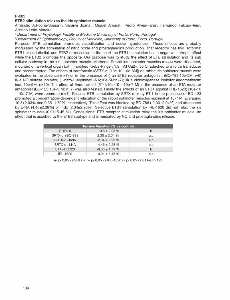
104
P-083ETB2 stimulation relaxes the iris sphincter muscle.Amândio A.Rocha-Sousa1,2, Saraiva Joana1, Miguel Amaral1, Pedro Alves-Faria2, Fernando Falcão-Reis2, Adelino Leite-Moreira1
1 Department of Physiology, Faculty of Medicine University of Porto, Porto, Portugal2Department of Ophthalmology, Faculty of Medicine, University of Porto, Porto, PortugalPurpose: ET-B stimulation promotes vasodilatation and ocular hypotension. Those effects are probably modulated by the stimulation of nitric oxide and prostaglandins production. That receptor has two isoforms: ETB1 or endothelial, and ETB2 or muscular. In the heart the ETB1 stimulation has a negative inotropic effect while the ETB2 promotes the opposite. Our purpose was to study the effect of ETB stimulation and its sub cellular pathway in the iris sphincter muscle. Methods: Rabbit iris sphincter muscles (n=44) were dissected, mounted on a vertical organ bath (modifi ed Krebs-Ringer; 1.8 mM Ca2+; 35 C) attached to a force transducer and precontracted. The effects of sarafotoxin (SRTX-c; [10e-10-10e-6M]) on rabbit iris sphincter muscle were evaluated in the absence (n=7) or in the presence of i) an ETB2 receptor antagonist, (BQ-788;10e-5M;n=6) ii) a NO sintase inhibitor, (L-nitro-L-arginine;L-NA;10e-5M;n=7); iii) a ciclooxigenase inhibitor (indomethacin; indo;10e-5M; n=10). The effect of Endothelin-1 (ET-1;10e-10 - 10e-7 M) in the presence of an ETA receptor antagonist (BQ-123;10e-5 M; n=7) was also tested. Finally the effects of an ETB1 agonist (IRL-1620; [10e-10 - 10e-7 M) were recorded (n=7). Results: ETB stimulation by SRTX-c or by ET-1 in the presence of BQ-123 promoted a concentration-dependent relaxation of the rabbit sphincter muscles maximal at 10-7 M, averaging 10.8±2.03% and 9.35±1.79%, respectively. This effect was blocked by BQ-788 (-2,30±2.04%) and attenuated by L-NA (4.46±2.28%) or Indo (2.34±2.93%). Selective ETB1 stimulation by IRL-1620 did not relax the iris sphincter muscle (0.91±5.45 %). Conclusions: ETB receptor stimulation relax the iris sphincter muscle, an effect that is ascribed to the ETB2 subtype and is mediated by NO and prostaglandins release.
Tension Variation (% vs control)SRTX-c -10,8 ± 2,03 % b
SRTX-c +BQ-788 2,30 ± 2,04 % a,c SRTX-c +Indo -2,34 ± 2,93 % a,c SRTX-c +LNA -4,46 ± 2,28 % a,c ET1 +BQ123 -9,35 ± 1,79 % b
IRL-1620 -0,91 ± 5,45 % a,c
a- p<0,05 vs SRTX-c b- p<0,05 vs IRL-1620 c- p<0,05 vs ET1+BQ-123
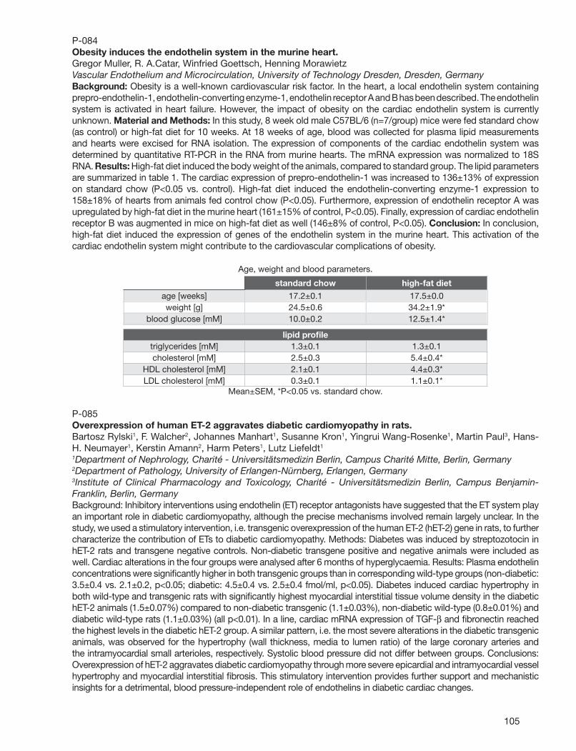
105
P-084Obesity induces the endothelin system in the murine heart.Gregor Muller, R. A.Catar, Winfried Goettsch, Henning MorawietzVascular Endothelium and Microcirculation, University of Technology Dresden, Dresden, GermanyBackground: Obesity is a well-known cardiovascular risk factor. In the heart, a local endothelin system containing prepro-endothelin-1, endothelin-converting enzyme-1, endothelin receptor A and B has been described. The endothelin system is activated in heart failure. However, the impact of obesity on the cardiac endothelin system is currently unknown. Material and Methods: In this study, 8 week old male C57BL/6 (n=7/group) mice were fed standard chow (as control) or high-fat diet for 10 weeks. At 18 weeks of age, blood was collected for plasma lipid measurements and hearts were excised for RNA isolation. The expression of components of the cardiac endothelin system was determined by quantitative RT-PCR in the RNA from murine hearts. The mRNA expression was normalized to 18S RNA. Results: High-fat diet induced the body weight of the animals, compared to standard group. The lipid parameters are summarized in table 1. The cardiac expression of prepro-endothelin-1 was increased to 136±13% of expression on standard chow (P<0.05 vs. control). High-fat diet induced the endothelin-converting enzyme-1 expression to 158±18% of hearts from animals fed control chow (P<0.05). Furthermore, expression of endothelin receptor A was upregulated by high-fat diet in the murine heart (161±15% of control, P<0.05). Finally, expression of cardiac endothelin receptor B was augmented in mice on high-fat diet as well (146±8% of control, P<0.05). Conclusion: In conclusion, high-fat diet induced the expression of genes of the endothelin system in the murine heart. This activation of the cardiac endothelin system might contribute to the cardiovascular complications of obesity.
Age, weight and blood parameters.
standard chow high-fat diet
age [weeks] 17.2±0.1 17.5±0.0 weight [g] 24.5±0.6 34.2±1.9*
blood glucose [mM] 10.0±0.2 12.5±1.4*
lipid profi le triglycerides [mM] 1.3±0.1 1.3±0.1 cholesterol [mM] 2.5±0.3 5.4±0.4*
HDL cholesterol [mM] 2.1±0.1 4.4±0.3* LDL cholesterol [mM] 0.3±0.1 1.1±0.1*
Mean±SEM, *P<0.05 vs. standard chow.
P-085Overexpression of human ET-2 aggravates diabetic cardiomyopathy in rats.Bartosz Rylski1, F. Walcher2, Johannes Manhart1, Susanne Kron1, Yingrui Wang-Rosenke1, Martin Paul3, Hans-H. Neumayer1, Kerstin Amann2, Harm Peters1, Lutz Liefeldt1
1Department of Nephrology, Charité - Universitätsmedizin Berlin, Campus Charité Mitte, Berlin, Germany2Department of Pathology, University of Erlangen-Nürnberg, Erlangen, Germany3Institute of Clinical Pharmacology and Toxicology, Charité - Universitätsmedizin Berlin, Campus Benjamin-Franklin, Berlin, GermanyBackground: Inhibitory interventions using endothelin (ET) receptor antagonists have suggested that the ET system play an important role in diabetic cardiomyopathy, although the precise mechanisms involved remain largely unclear. In the study, we used a stimulatory intervention, i.e. transgenic overexpression of the human ET-2 (hET-2) gene in rats, to further characterize the contribution of ETs to diabetic cardiomyopathy. Methods: Diabetes was induced by streptozotocin in hET-2 rats and transgene negative controls. Non-diabetic transgene positive and negative animals were included as well. Cardiac alterations in the four groups were analysed after 6 months of hyperglycaemia. Results: Plasma endothelin concentrations were signifi cantly higher in both transgenic groups than in corresponding wild-type groups (non-diabetic: 3.5±0.4 vs. 2.1±0.2, p<0.05; diabetic: 4.5±0.4 vs. 2.5±0.4 fmol/ml, p<0.05). Diabetes induced cardiac hypertrophy in both wild-type and transgenic rats with signifi cantly highest myocardial interstitial tissue volume density in the diabetic hET-2 animals (1.5±0.07%) compared to non-diabetic transgenic (1.1±0.03%), non-diabetic wild-type (0.8±0.01%) and diabetic wild-type rats (1.1±0.03%) (all p<0.01). In a line, cardiac mRNA expression of TGF-β and fi bronectin reached the highest levels in the diabetic hET-2 group. A similar pattern, i.e. the most severe alterations in the diabetic transgenic animals, was observed for the hypertrophy (wall thickness, media to lumen ratio) of the large coronary arteries and the intramyocardial small arterioles, respectively. Systolic blood pressure did not differ between groups. Conclusions: Overexpression of hET-2 aggravates diabetic cardiomyopathy through more severe epicardial and intramyocardial vessel hypertrophy and myocardial interstitial fi brosis. This stimulatory intervention provides further support and mechanistic insights for a detrimental, blood pressure-independent role of endothelins in diabetic cardiac changes.
106
P-086Activation of renal medullary ETB receptor induces diuresis and natriuresis via nitric oxide synthase 1, cGMP and protein kinase G pathways.Daisuke Nakano, Jennifer S.Pollock, David M.PollockVascular Biology Center, Medical College of Georgia, Augusta, GAThe renal medulla is one the major sites of ET-1 synthesis and receptor expression in the body. Considerable in vitro evidence indicates that activation of the ETB receptor inhibits tubular reabsorption of water and electrolytes. The aim of this study was to examine whether renal medullary infusion of the ETB receptor agonist, sarafotoxin S6c, induces a diuretic and natriuretic response in vivo by activation of nitric oxide synthase (NOS) 1, cGMP production and protein kinase (PK) G in normotensive rats. S6c and inhibitors were infused into the left renal medullary interstitium of inactin-anesthetized Sprague-Dawley rats. Intramedullary infusion of S6c (0.45 µg/kg/h; n=8) markedly increased the urine fl ow (UV; 4.1 ± 0.2 µl/min in saline controls and 9.8 ± 0.3 µl/min in S6c infused rats; p<0.01) and urinary sodium excretion (UNaV; 0.44 ± 0.06 and 1.41 ± 0.12 µmol/min in saline and S6c infused rats, respectively, p<0.01). S6c increased renal medullary cGMP content (9.4 ± 0.6 vs. 4.0 ± 0.3 pmol/mg protein in controls; p<0.01). There were no changes in arterial blood pressure in any group. Pretreatment with NG-propyl-L-arginine (NPA; 10 µg/kg/h, n=6), a selective inhibitor of NOS1, suppressed S6c-induced increases in UV and UNaV (6.2 ± 0.3 µl/min and 0.75 ± 0.05 µmol/min, respectively; p<0.01) and prevented the increase in medullary cGMP content (3.3 ± 0.1 pmol/mg protein; p<0.01). Moreover, Rp-8-Br-PET-cGMPS (10 µg/kg/h; n=6), a PKG inhibitor, signifi cantly inhibited S6c-induced increases in UV and UNaV (6.6 ± 0.3 µl/min and 0.82 ± 0.08 µmol/min, respectively; p<0.01). Neither NPA nor Rp-8-Br-PET-cGMPS affected UV or UNaV when infused in the absence of S6c. These results demonstrate that renal medullary ETB receptor activation in vivo induces diuretic and natriuretic responses through the NOS1, cGMP and PKG pathways.
P-087Contralateral nociceptive sensitization following the subcutaneous injection of ET-1: Evidence for central sensitization.Gary Strichartz1, Alla Khodorova1, Philip Albrecht2, Frank Rice2
1Pain Research Center, Brigham and Women’s Hospital, Boston, MA2Center for Neuropharmacology and Neuroscience, Albany Medical College, Albany, NYSubcutaneous injection of endothelin-1 (ET-1) into the rat’s footpad induces ipsilateral thermal and mechanical sensitization. The uninjected, contralateral hindpaw also has altered sensitivity. At 24h after the initial injection (200 uM), contralateral tactile sensitivity (paw withdrawal threshold to von Frey hairs) is increased, although sensitivity to noxious heat is unchanged. Contralateral injection of the same ET-1 dose at this time causes an almost 2-fold greater fl inching response, as is also true for the second phase of the formalin (0.5%) response. Block of the ipsilateral sciatic nerve by local anesthetics prevents contralateral sensitization, indicating the importance of afferent transmission for contralateral regulation, and implicating central sensitization, e.g. involving spinal cord circuits. Immunocytochemistry of the contralateral paw after ipsilateral ET-1 reveals elevations of glutamate and CGRP, a potent pain-sensitizing neuropeptide, in epidermal keratinocytes at 24 h, well after these compounds’ elevations in the ipsilateral paw have subsided towards baseline levels. The polymodal transducing receptor, TRPV-1, also is elevated in the contralateral paw, consistent with the known role of this molecule in maintaining tactile allodynia in cutaneous tissue. Overall, these fi ndings show that local direct actions of ET-1 act through afferent neurons to produce a much broader sensitization of tissues spread over the body’s surface.
107
P-088Palosuran, an oral U-II antagonist, prevents the cardiac effects of high fructose diet in rats.Alessandro Cosenzi, Elena Bernobich, Silvia Tomasella, Irene Gilotta, Polonca Kogoj, Giuseppe BelliniDepartment of Clinical Medicine and Neurology, University of Trieste, Trieste, ItalyPrevious studies have demonstrated that Urotensin II (U-II) might be involved in the development of cardiac hypertrophy and fi brosis. Palosuran is an oral competitive antagonist of U-II receptors. Rats fed a high fructose diet (HFD) develop hyperinsulinism with cardiac changes comparable with those observed in diabetic rats. The aim of this study was to evaluate whether chronic palosuran administration could prevent the cardiac changes induced by HFD in rats. Methods . Forty male Wistar rats were divided into 4 groups: group 1 received standard diet (SD); group 2 received HFD (= 60% of calories from fructose); group 3 received HFD + palosuran 300mg/kg daily mixed with food; group 4 received SD + palosuran. Blood pressure was measured weekly throughout the study by tail cuff. At the end of the study, after one month, all animals were sacrifi ced and their hearts immediately removed, weighed and fi xed. Immunohistochemistry with the alkaline phosphatase method was performed to evaluate cardiac deposits of collagen III. The amount of collagen III deposits was graded by 4 independent blind observers in a semiquantitative manner. The scores assigned to each group were compared by means of one-way analysis of variance and the LSD Fisher’s method. Results . Blood pressure and serum glucose were similar in all groups at the beginning and at the end of the study. Rats fed HFD had a signifi cantly higher cardiac mass in comparison with rats fed standard diet and this effect was prevented by palosuran (p<0.05). Collagen III deposition was signifi cantly higher in the heart of animals fed HFD; this effect was completely prevented by palosuran (p<0.02); Conclusions . The results of this study demonstrate for the fi rst time that palosuran, chronically administered, is effective in preventing the increase in heart weight and the cardiac deposition of collagen III induced by HFD in the rats. This effect was not a consequence of a decrease in arterial blood pressure and suggests further perspectives for this newborn class of drugs.
P-089The Effect of KMUVS-1 in Cerebrovasospasm after SAH via Inhibition of ET-1 Production.Jwu-Lai Yeh1, Chun-Jung Huang1, Yang-Chang Wu3, Jen-Hao Hsu3, Shu-Chuan Wu2, Kuang-I Cheng5,6, Chun-Po Yen2,5, Bin-Nan Wu1, Aij-Lie Kwan2,5
1Graduate Institute of Pharmacology, Kaohsiung Medical University, Kaohsiung, Taiwan2Department of Neurosurgery, Kaohsiung Medical University Hospital, Kaohsiung, Taiwan3Graduate Institute of Natural Products, Kaohsiung Medical University, Kaohsiung, Taiwan4Department of Physiology, Kaohsiung Medical University, Kaohsiung, Taiwan5Graduate Institute, Faculty of Medicine, College of Medicine, Kaohsiung Medical University, Kaohsiung, Taiwan6Department of Anesthesiology, Kaohsiung Medical University Hospital, Kaohsiung, TaiwanEndothelin-1 (ET-1), a potent endogenous vasoconstrictor, played an important role to cause cerebral vasospasm after subarachnoid hemorrhage (SAH). KMUVS-1, a Chinese medicinal formula containing Salvia miltiorrhiza radix , Carthamus tinctorius, Anredera cordifi lia moq., Ligusticum chuanxiong, Carica papaya, Pheretima aspergillum, Glycyrrhizae radix preparata, Taxillus chinensis, Prunus persica, Achyranthus bidentata, Spatholobus suberectus, Caesalpinia sappan and Paeonia lactifl ora, has individually been show vasodilatation effects. In others study has demonstrated the Salvia miltiorrhiza radix (Danshen) can inhibits ET-1 production in human umbilical vein endothelial cells (HUVECs). In our previous study, the Chinese medicinal formula- KMUVS-1 had vasodilatation effect and it attenuated cerebral vasospasm effectively after experimental SAH, and KMUVS-1 (1 mg/kg, 500 mg/kg, 1000 mg/kg) reduced average luminal area of cross sections and morphological injury in basilar arteries were signifi cantly. In this study we design two-hemorrhage model, SAH was induced in the Sprague-Dawley rat basilar artery by injection 0.3 mL of autologous blood into cisternal magna, following the second hemorrhage at 48 hours later. Oral treatment with KMUVS-1 at dosages of 0.5g/kg , twice daily for 7 days began from one hour after the fi rst SAH. ET-1 expression in different tissue, plasma and cerebral spinal fl uid (CSF) were assessed by ELISA. In this study, we have seen KMUVS-1 ingredients in CSF and plasma. These result show KMUVS-1 can through Blood Brain Barrier in experimental SAH model. We suggest the effect of KMUVS-1 may play important role in the inhibiting ET-1 production related to prevent vasospasm.
108
P-090Increased expression of urotensin II-related peptide in the kidney of rats with hypertension or chronic renal failure.Kazuhiro Takahashi1,2, Nobuyoshi Mori3, Takuo Hirose4, Takashi Nakayama4, Osamu Ito3, Masayuki Kanazawa3, Masahiro Kohzuki3, Yutaka Imai4,2, Kazuhito Totsune4,2
1Department of Analytical Medical Technology, Tohoku University School of Health Sciences, Sendai, Japan2Tohoku University 21st COE Program Comprehensive Research and Education Center for Planning of Drug Development and Clinical Evaluation (CRESCENDO), Sendai, Japan3Department of Internal Medicine and Rehabilitation Science, Tohoku University School of Medicine, Sendai, Japan4Department of Clinical Pharmacology and Therapeutics, Tohoku University Graduate School of Pharmaceutical Sciences and Medicine, Sendai, JapanUrotensin II (UII) is a potent vasoconstrictor peptide, whereas it acts as a vasodilator in some vascular vessels. UII is more potent than endothelin-1 in its vasoconstrictor property. Urotensin II-related peptide (URP) is a novel vasoactive peptide that shares urotensin II receptor (UT-R) with UII. Expression of endothelin-1 is increased in the kidney tissues with chronic renal failure (CRF). There has been, however, no report on URP expression in CRF kidneys. In order to clarify the pathophysiological roles of URP in hypertension and chronic renal failure, expression of URP and UT-R was studied in the kidney obtained from hypertensive rats and CRF rats by quantitative RT-PCR and immunocytochemistry. Kidney tissues from spontaneous hypertensive rats (SHR) and Wister-Kyoto rats (WKY) were dissected into renal cortex, inner medulla and outer medulla. The highest expression levels of URP mRNA were found in the inner medulla among these three portions in both SHR and WKY. URP mRNA expression levels in the inner medulla were signifi cantly higher in SHR than in WKY (about 2.7-fold), but not in other portions. Rat model of CRF was prepared by 5/6 nephrectomy in WKY. Expression levels of URP and UT-R mRNAs were signifi cantly elevated in the remnant kidney of the rats with renal mass ablation at day 56 after nephrectomy, but not at day 14, when compared with sham-operated rats (about 8.3-fold and 12.4-fold, respectively). Immunocytochemistry showed that URP immunostaining was found mainly in the renal tubules, particularly distal tubules, and vascular smooth muscle and endothelial cells. UT-R immunoreactivity was localized in the renal tubules and vascular endothelial cells. These fi ndings suggest that the expression of URP and UT-R is enhanced in hypertension and CRF, and has important pathophysiological roles in these diseases.
P-091Clonidine and endothelin antagonist sulfi soxazole combination produces potent analgesia in mice.Anil Gulati1, George Matwyshyn1, Eileen Deano2, Bhagya L.Puppala2
1Chicago College of Pharmacy, Midwestern University, Downers Grove, IL2Department of Pediatrics and Neonatology, Advocate Lutheran General Children’s Hospital, Park Ridge, ILOpioids are the most commonly used analgesics for the clinical management of acute and chronic pain. There are various side effects associated with the long-term use of opioids including the development of tolerance, which results in inadequate pain relief. The present study was conducted for the following reasons: (a) clonidine has analgesic effect and potentiates morphine analgesia; (b) ETA receptor antagonists potentiate morphine analgesia, (c) sulfi soxazole is an ETA receptor antagonist; and (d) clonidine and ETA receptor antagonists have been shown to have cardiovascular interactions. However, no study has been performed to our knowledge to study the interaction of clonidine and ETA receptor antagonists on analgesia. The present study was performed to determine the effect of (1) clonidine on morphine analgesia, (2) sulfi soxazole on morphine analgesia, and (3) combined clonidine and sulfi soxazole on morphine analgesia. Male Swiss Webster mice weighing 25 to 30 g were used and antinociceptive response to morphine was determined by tail-fl ick latency method. Clonidine produced analgesia and an AUC of 68.4 ± 12.3 was observed which was signifi cantly higher than control. Clonidine pretreatment signifi cantly (42%) enhanced the antinociception produced by morphine. Sulfi soxazole (56.0 ± 9.1) produced an increase in AUC compared to control. Sulfi soxazole pretreatment produced only 14% potentiation of antinociception produced by morphine. However, sulfi soxazole (1000 mg/kg, oral) in combination with clonidine (2 mg/kg, i.p.) produced a signifi cant analgesia and the AUC observed in this group (119.1 ± 8.9) was equal to that of a high dose of 8 mg/kg of morphine. Clonidine and sulfi soxazole when administered together produced more than 167% increase in AUC compared either clonidine or sulfi soxazole administered alone. This is the fi rst report of the observation that sulfi soxazole and clonidine augment analgesia. The augmentation is so marked that the analgesia induced by combined use of clonidine and sulfi soxazole was comparable to a high dose of morphine. (This study was funded by EndogenX. Inc., Los Gatos, CA).
109
P-09217β-estradiol prevents cerebral vasospasm and endothelin-1 expression in the brain stem after subarachnoid hemorrhage.Chih-Lung Lin1,2, A.s. Lieu1,2, K.s. Lee1,2, YuFeng Su1,2, ShenLong Howng1,2, T.f. Chan2,3, K.p. Chang2,4, AijLie Kwan1,2, Zk Dai1,5
1Neurosurgery, Kaohsiung Medical University, Kaohsiung, Taiwan2Faculty of Medicine, Graduate Institute of Medicine, College of Medicine, Kaohsiung Medical University, Kaohsiung, Taiwan3Obstetrics and Gynecology, Kaohsiung Medical University Hospital, Kaohsiung, Taiwan4Plastic and Reconstructive Surgery, Kaohsiung Medical University Hospital, Kaohsiung, Taiwan5Division of Cardiology and Pulmonology, Department of Pediatrics, Kaohsiung Medical University Hospital, Kaohsiung, TaiwanBackground. Although cerebral vasospasm after aneurysmal subarachnoid hemorrhage (SAH) has been recognized for over half of a century, it remains a major complication in patients suffering from SAH. As the problem of no effective treatment for cerebral vasospasm still exists, the pathophysiological mechanism contributing to arterial dysfunction needs intensive study. A burgeoning body of evidence suggests that endothelin (ET) may be critical in the pathophysiology of cerebral vasospasm after SAH. This study is designed to evaluate the infl uence of 17β-estradiol (E2) on the expression of ET-1 in the brain stem and cerebrovasospasm after SAH in rat. Method. Experimental SAH was induced in Sprague-Dawley male rats by injecting 0.3mL autogenous blood into the cisterna magna on day 0 and day 2. A 30-mm Silastic® tube fi lled with E2 in corn oil (0.3 mg/ml) was subcutaneously implanted in rats just before SAH induction. The degree of vasospasm was determined by averaging the cross sectional areas of basilar artery 7 days after fi rst SAH. Expressions of ET-1 in the brain stem were also evaluated. Results. E2-treatment signifi cantly attenuated SAH-induced vasospasm. The expressions of ET-1 were signifi cantly increased in SAH only and SAH plus vehicle groups and were not signifi cantly different in SAH plus E2 group when compared to the controls. There was a signifi cant correlation between the cross sectional areas of basilar artery and the expression of ET-1 (P<0.001). Conclusion. Our fi nding further confi rmed the anti-spastic effect of E2 after SAH. The mechanism of E2 in the attenuating SAH-induced vasospasm may be partially by decreasing ET-1 production.
P-093Potential of endothelin antagonist in experimental model of cerebral stroke.Yogendra K.Gupta1, Seema Briyal1, Anil Gulati21Department of Pharmacology, All India Institute of Medical Sciences, New Delhi, India2Department of Biopharmaceutical Sciences, University of Illinois at Chicago, Chicago, IL 60612, ILThe effect of endothelin antagonist was studied after hypoxia/ischemia in vitro by oxygen glucose deprivation and in vivo by middle cerebral artery occlusion (MCAo). Percent cell viability was assessed using MTT assay in vitro. The parameters for assessing ischemic damage in vivo included diffusion-weighted imaging (DWI), neurological defi cit, motor performance tests in addition to oxidative stress parameters: malondialdehyde (MDA), reduced glutathione (GSH) and superoxide dismutase (SOD) In vitro, the percent cell viability in hypoxic cells was reduced as compared to the control. Incubation with endothelin antagonist (0.01, 0.1 and 1 µg/µl) after reperfusion caused an increase in the cell viability in a time dependent manner. In in vivo study, male Wistar rats were pretreated with endothelin antagonist for 7 days and thereafter subjected to focal ischemia by occlusion of MCA using intraluminal thread for two hours. 30 min after reperfusion the animals were subjected to diffusion-weighted imaging (DWI) for assessment of protective effect. Twenty-four hours later the motor performance was tested and subsequently the animals were sacrifi ced for estimation of markers of oxidative stress; malondialdehyde (MDA), glutathione (GSH) and superoxide dismutase (SOD). In the endothelin antagonist pretreated group, percent hemispheric lesion area (% HLA) in DWI was signifi cantly attenuated 17.5±0.5 % as compared to control group 61.2±5.9 %. Signifi cant motor impairment, with signifi cant elevated levels of MDA, decrease in GSH and SOD were observed in the vehicle treated MCA occluded rats. Pretreatment prevented the motor impairment and signifi cantly reversed the changes in markers of oxidative stress (MDA, GSH and SOD). In addition to well-known vasodilatory effect, the results indicate that endothelin antagonist protects neuronal damage caused by hypoxia in vitro and focal cerebral ischemia in vivo, and can be attributed to its antioxidant activity. These effects can contribute to the protection afforded by TAK-044 in the present study.

110
P-094Effects of TNF-alpha blocking peptide on Endothelin-1 levels in lungs in endotoxemic rat model.Subrina Jesmin1,3, Atsushi Sawamura1, Sohel Zaedi1,3, Shamsul Haque Prodhan3, Naoto Yamaguchi3, Katsutoshi Goto2, Satoshi Gando1
1Department of Anesthesiology and Critical Care Medicine, Hokkaido University, Sapporo, Japan2Department of Pharmacology, University of Tsukuba, Tsukuba, Japan3Center of Medical Science, Ibaraki Prefectural University, Ami, JapanEndothelin (ET)-1, a potent vasoconstrictor, is implicated in the pathogenesis of a number of diseases. Some evidences suggest that circulating ET-1 is elevated in sepsis. The present study investigated whether ET system (ET-1, ETA and ETB receptors) has a potential role in the acute lung injury of sepsis in a time-dependent manner and whether the blockade of TNF-α has a regulatory role on the ET-1 level in septic lung. Male Wistar rats at 8 weeks of age were administered with either saline or lipopolysaccharide (LPS) . The rats were killed with ether at different time points (1, 3, 6 and 10 hours) and lungs were either frozen or preserved in formalin. Blood gas analysis was done immediately after opening the chest. The features of acute lung injury were observed at 1 hour after LPS administration, which gradually became severe with time. Systolic and diastolic pressures were drastically lower just 1 hour after LPS administration. Pulmonary TNF-α level was signifi cantly increased at various time points after LPS administration. A time-dependent increase of ET-1 expression was observed in LPS administered lungs compared to control lungs and ET-1 level was peaked at 6 hours (3-fold) after induction of endotoxemia by LPS while the expression of preproET-1 mRNA was peaked in lung tissue at 1 hour after LPS administration. Immunoblot analysis and Real-Time PCR experiments demonstrated a time-dependent gradual increase of ETA receptor after LPS administration while opposite result was found in ETB receptor expression. ETB receptor with vasodilating property, was remarkably downregulated in septic lung in a time-related manner. We conclude that time-dependent increase of ET-1, ETA receptor with the downregulation of ETB receptor may play a role in the pathogenesis of acute lung injury in endotoxemia. In the second part of this study, we treated the LPS-administered rats with TNF-α blocking peptide for three hours and found that pulmonary ET-1 level was signifi cantly suppressed by the blockage of TNF-α. Thus, TNF-α-mediated ET-1 upregulation may be a crucial pathological response to cytokine induction following an infl ammatory response.
P-095Upregulation of functional ETA receptor and downregulation of functional ETB receptor in cancer and stromal cells within colorectal cancer.Moinuddin M. Hoosein1, Michael R.Dashwood2, Irving Taylor1, Marilena Loizidou1
1Department of Surgery, Royal Free and University College Medical School, UCL, London, United Kingdom2Department of Clinical Biochemistry, Royal Free and University College Medical School, London, United KingdomBackground - The vasoactive peptide endothelin-1 (ET-1) acts via two endothelin receptor subtypes, ETA (ETAR) and ETB (ETBR). ET-1 and ETAR are overexpressed in colorectal cancer tissues. In vitro, ET-1 acting via ETAR, is a mitogen for colorectal cancer cells. To identify other potential stimulatory loops, we investigated the distribution and cell specifi c localisation of both ETAR and ETBR in tissue sections from patients with colorectal cancer. Methods - Frozen sections from specimens of colorectal cancer (n=9) and normal colon (n=9) were cut and subjected to either (i) autoradiography (+/-densitometry) or (ii) a combination of cell type specifi c immunohistochemistry, using antibodies against fi broblasts (AS02), endothelial cells (CD31) or nerve fi bres (NF200) and in vitro receptor microautoradiography, using ETAR and ETBR specifi c radioligands. Results - ETARs were upregulated in all cell types, apart from nerve, in cancer compared to normal colon [mean (SD): 206(+/-27) v. 129.2(+/-35.5); cancer v. normal; 60% increase]. Specifi cally, ETAR binding was highest in cancer-associated blood vessels and fi broblasts and to a lesser extent in epithelial cancer cells. In contrast, ETBRs were the predominant receptors in normal colon [122.4(+/-24.5) v. 207(+/-35.5); cancer v. normal; 40% decrease] and were markedly downregulated in cancer-associated blood vessels, fi broblasts, and to a lesser extent in epithelial cells. Nerve co-localisation was demonstrated, but remained unchanged for all tissues. Conclusion - The shift in ET receptor binding observed in epithelial cancer cells and cancer associated fi broblasts and endothelial cells may favour ET-1 signals contributing to colorectal cancer growth and neovascularisation.

111
P-096Toxicokinetic Evaluation of IRL-1620 in a 4-Week Toxicology Study in Rats.Guru Reddy1, Jennifer Drisko2, Jeff Hoffman3, Henry Pieniasak4, Prasad Kolli1, Dorla Mirejovski1, Luigi Lenaz1
1Spectrum Pharmaceuticals, Inc., Irvine, CA2Bioanalytical Systems, Inc., Mount Vernon, IN3MicroConstants, Inc, San Diego, CA4HPP Consulting, Darlington, MDBackground: IRL-1620, a selective ETB receptor agonist, has been reported to selectively enhance tumor perfusion and potentiate the therapeutic potential of anti-cancer agents in breast and prostate tumor bearing rats1,2. We conducted a study to evaluate the toxicity and systemic exposure when IRL-1620 is administered as a single intravenous bolus dose, once a week for four weeks in rats. Toxicokinetic evaluation of IRL-1620 will be presented here. Study Design: Toxicokinetics were determined in three satellite groups of 12 male and 12 female rats each that received a slow IV bolus dose of SPI-1620 once weekly for four weeks at doses of 5, 10 and 15 ug/kg. Blood samples were obtained pre-dose and over a 2 hour period post-dose. IRL-1620 in plasma was assayed by LC-MS/MS method and non-compartmental toxicokinetics were determined using WinNonlin. Results: Dose related increases in Cmax and AUCT were observed. Cmax values were 41.2, 137 and 225 ng/mL in males and 43.0, 126 and 218 ng/mL in females following nominal IRL-1620 doses of 5.0, 10.0 and 15.0 ug/kg respectively. Corresponding AUC values were 374, 1150 and 2090 ng.min/mL in males and 359, 1150 and 2050 ng.min/mL in females. The terminal disposition T1/2 was similar across doses and gender and ranged from 4.7 to 7.9 minutes. No apparent gender differences in toxicokinetics of SPI-1620 were seen. Conclusion: It is concluded that in rats there is dose linearity, using both Cmax and AUC analysis. Blood clearance is rapid as shown by CL and T1/2.
P-097Effect of IRL-1620 on Respiration Rate and Tidal Volume in Sprague Dawley Rats.Guru Reddy1, Melinda Broward2, Luigi Lenaz1
1Spectrum Pharmaceuticals, Inc., Irvine, CA2Aptuit, Inc., Kansas City, MOBackground: IRL-1620, a selective ETB receptor agonist, has been reported to selectively enhance tumor perfusion and potentiate the therapeutic potential of anti-cancer agents in breast and prostate tumor bearing rats1,2. In the published studies 3 nmol/kg (5.5 ug/kg) of IRL-1620 was determined as the optimal dose for therapeutic effects in rats. The purpose of the present study was to assess the effects of SPI-1620 at intravenous doses of 5,10 and 20 ug/kg on respiration rates and tidal volumes in rats. Study Design: Male Sprague Dawley rats (8 rats per group) were assigned to each treatment and were administered either a single intravenous dose of 5, 10, or 20 µg/kg SPI-1620, vehicle, or 20 mg/kg morphine (reference substance). For each recording session, the animals were restrained in body enclosed plethysmography chambers designed to isolate the head from body respiratory movements. Respiration rate and tidal volume were evaluated. The predose data were acquired approximately 15-20 min after entry into the chamber (a specifi c marker was placed at the relevant time point). The rats were then removed from the chambers in order to be dosed with vehicle, test article, or reference substance. The rats were immediately returned to the chambers after dosing and respiratory parameters acquired. Specifi c digital markers were placed at 5, 30, and 60 min postdose. On completion of the 60 min postdose data acquisition, the animals were sacrifi ced. Results: There were no statistically signifi cant changes in the mean respiration rate of rats treated intravenously with 5 µg/kg SPI-1620 when compared to the corresponding vehicle group (5, 30, and 60 min post-dose). Statistically signifi cant increases were observed in mean respiration rates at the 10 and 20 µg/kg SPI-1620 treatment levels compared to the corresponding vehicle group. Signifi cant decreases were observed in mean tidal volumes at all SPI-1620 treatment levels of 5, 10, and 20 µg/kg. Conclusion: It is concluded that at a therapeutically relevant dose of 5 ug/kg, although the tidal volume was decreased, IRL-1620 did not have any effect on respiration rate in rats.

112
P-098Doxorubicin suppresses ET-1 mRNA expression in endothelial cells.Katalin Keltai1, László Cervenak1, Veronika Makó1, Zoltán Doleschall2, András Zsáry1
13rd Dept of Medicine, Semmelweis University, Budapest, Hungary2Dept of Pathogenetics, National Institute of Oncology, Budapest, HungaryDoxorubicin (DXR) is a widely used cytostatic agent, whose use is limited by its toxic cardiovascular effects. Previously, we reported signifi cantly decreased plasma endothelin-1 (ET-1) levels in DXR treated lymphoma patients. Endothelial cells are known to be the major source of plasma ET-1. The aim of our present study was to investigate whether DXR directly modulates ET-1 production of endothelial cells. We used in vitro cultured human umbilical vein endothelial cells (HUVEC) as a model. We previously described that ET-1 protein levels correlated with mRNA expression, thus ET-1 production was assessed at the mRNA level by SuperArray (SA, GEArray Q Series, Human Cardiovascular Diseases I: HS037 kit) and LightCycler (LC) technologies. DXR toxicity was determined by fl uorescence based microplate assay. HUVECs were treated with DXR in different doses for 6 and 24 hours. Half maximal cytotoxic concentration (LD50) was calculated and DXR was subsequently used in doses less than LD50. In preliminary analyses we determined the maximal non-toxic concentration (1000 ng/ml) and optimal treatment period (6 hours) of DXR on ET-1 mRNA expression by LC. We optimized cell culture conditions for SA. HUVECs were treated with TNFalpha or left untreated for 6 hours and mRNA expression pattern was compared. SA system was validated by the well known TNFalpha induced signifi cant MCP-1, IL-8, ICAM-1, VCAM-1 and E-Selectin mRNA expression. HUVECs were treated with 1000 ng/ml DXR under the same conditions. Out of the 96 representative cardiovascular genes of the array only ET-1 gene expression changed signifi cantly. ET-1 mRNA expression was 10.9% of the untreated control (p=0.0049). This result was confi rmed by LC real-time PCR (24.1% of control, p=0.0022). Above results are in concordance with our previous in vivo fi ndings that both plasma ET-1 protein level in lymphoma patients and in vitro endothelial ET-1 mRNA level were signifi cantly suppressed by DXR. Decreased ET-1 mRNA level was not the consequence of a general mRNA suppression of DXR as the expression of the other 95 genes did not change signifi cantly. This suggests that ET-1 may have causative role in the pathogenesis of DXR induced cardiotoxicity.
P-099ETB receptor antagonists inhibit cell proliferation in human glioblastoma cell lines.Mayra Paolillo, Cristina Lanni, Carla D.Boidi, Marika A.Russo, Sergio SchinelliDipartimento di Farmacologia Sperimentale ed Applicata, Università degli Studi di Pavia, Pavia, ItalyThe proliferative and antiapoptotic actions of endothelin (ET)-1 in cancer cells have been documented and specifi c ET receptors antagonists have been exploited as potential anticancer drugs. Either ETA or ETB receptors appear to be involved in the observed effects on cancer cell proliferation in different cells types. In previous studies we have found that glioblastoma cell lines express ETB and ETA receptors and release ET-1 in the culture medium. In this study we have investigated in 1321-N1 and U87 glioblastoma cell lines the effects of the specifi c ETB receptors antagonists BQ788 and A192621 on the serum-induced cell proliferation. BrdU incorporation assays were performed and we found that the two ETB receptor antagonist BQ788 (10 µM, 48h) and A192621 (10 µM, 48h) inhibited cell proliferation (1321-N1: -24/12% for BQ788 treatments and -19,96 for A192126 treatments. U87: -16,22% for BQ788 treatments and -27,11% for A192126 treatments). Previous cell viability experiments showed that the ETA receptor antagonsit BQ123 had no effect in both cell lines so we did not use this compound in BrdU experiments. Flow cytometry analysis (FACS) revealed that the same BQ treatment caused an increase in the number of cells arrested in the G1 phase and a decrease in ciclin D1 expression while the A192621 treatment causes mainly an increase in the number of cells arrested in the G2/M phase. The downstream signaling pathways affected by ETB receptor antagonists were studied by western blot analysis using phosphospecifi c antibodies. The stimulation of serum-starved 1321-N1 and U87 cells by ET-1 (100 nM, 2 min) induces a transient phosphorylation of both ERK1/2 and p38MAPK kinases that was blocked in the presence of 10µM BQ788. In conclusion, we suggest that the observed inhibition of cell proliferation in 1321-N1 and U87 cells induced by ETB receptors antagonists might be in part explained by interferences with downstream signaling pathways involved in cell proliferation such as the ERK1/2-dependent and p38MAPK-dependent pathways. Additional experiments are warranted to establish whether a longer treatment of glioma cell lines with ETB receptor antagonists may trigger apoptotic mechanisms.
113
P-100Improvement in the uptake and effi cacy of chemotherapeutic agents by IRL-1620 in prostate tumor rats.Anil Gulati1, N. V.Rajeshkumar2, George Matwyshyn1
1Chicago College of Pharmacy, Midwestern University, Downers Grove, IL2Biopharmaceutical Sciences, University of Illinois at Chicago, Chicago, ILIRL-1620, an endothelin B receptor agonist, enhanced the tumor delivery and effi cacy of paclitaxel in a breast tumor model, but its effect in prostate cancer is not known. Studies have provided clear evidence of the involvement of ET in prostate cancer. However, most of the studies have targeted the prostate cancer cells and not the blood vessels. We thought of applying our approach of using an ETB receptor agonist IRL-1620 to stimulate prostate tumor blood vessels so that the delivery of anticancer drugs can be increased to the prostate tumor tissue. The present study was conducted to evaluate the effect of IRL-1620 on tumor perfusion, uptake of [14C]-doxorubicin in the tumor and effi cacy of doxorubicin (DOX), and 5-fl urouracil (5-FU) in a rat prostate tumor model. Copenhagen rats were inoculated with 10,000 JHU-4 (Mat-Lu) cells and experiment were initiated once the tumors reached a size of 200 mm3. Rat prostate tumor perfusion was measured using a Perifl ux PF2b 4000 Laser Doppler fl owmeter; biodistribution of [14C]-doxorubicin in tumor and organs; and effi cacy studies with doxorubicin and 5-fl urouracil were conducted. The results of the present study demonstrate that administration of IRL-1620 (3 and 6 nmol/kg) produced a signifi cant increase in prostate tumor perfusion compared to vehicle treated rats. The lower dose of IRL-1620 (1 nmol/kg) used in the present study was found to be ineffective in increasing tumor perfusion. A higher dose of 3 nmol/kg of IRL-1620, however, produced a maximal increase in tumor perfusion (102.8%), which was more than that of IRL-1620 6 nmol/kg (79.1%). Therefore, 3 nmol/kg dose of IRL-1620 was selected for drug delivery and effi cacy studies. Administration of IRL-1620 (3 nmol/kg, i.v) signifi cantly increased (115%) tumor uptake of [14C]-doxorubicin compared to vehicle treated rats. Results of the effi cacy study demonstrate that IRL-1620 administration 15 min prior to DOX (5 mg/kg) or 5-FU (50 mg/kg) on every third day for a total of four doses signifi cantly reduced tumor volume compared to vehicle treated rats. In conclusion, IRL-1620 signifi cantly enhanced the uptake and effi cacy of anticancer agents in prostate cancer.
P-101The ET-1 metabolising proteases, ECE-1 and NEP, in human cancers.Yue Hong, Badar Usmani, Anthony TurnerInstitute of Molecular and Cellular Biology, University of Leeds, Leeds, United KingdomEndothelin-1 exhibits a potent mitogenic effect and has been implicated in the progression of various cancers and is considered to provide a driving force for tumour survival and growth. Bioactive ET-1 is synthesized from its bio-inactive precursor, big ET, by the membrane metalloprotease endothelin-1 converting enzyme (ECE-1) and subsequently inactivated by the ECE-1 homologue, neprilysin (NEP). Elevated ECE-1 coupled with decreased NEP expression frequently occurs in numerous cancer cells and nearly all malignant prostate cancer (PC) cell lines in our study. This imbalanced expression of both enzymes was also shown to strongly associate with the severity of cancer, which highlights the counter-balancing physiological roles of ECE-1 and NEP in tumour progression. However, the transcriptional regulatory mechanism(s) of these enzymes involved in both normal and cancer cells is still not well understood. To address this question, we are exploring potential regulators by focusing on three main candidate groups: amyloid precursor protein intracellular domain (AICD), catechins of green tea and steroid hormones (androgen (DHT) and oestrogen (17β-estradiol)). Since a decreased ratio of ECE-1/ NEP may result in reduced cancer cell malignancy, we have developed a HVS-based ECE-1 targeting siRNA delivery system. This virus delivering siRNA readily enters prostate primary cells which are normally resistant to transfection and provide a long-term effect on ECE-1 expression levels. The consequent effects on PC cell viability and invasive ability have been examined.
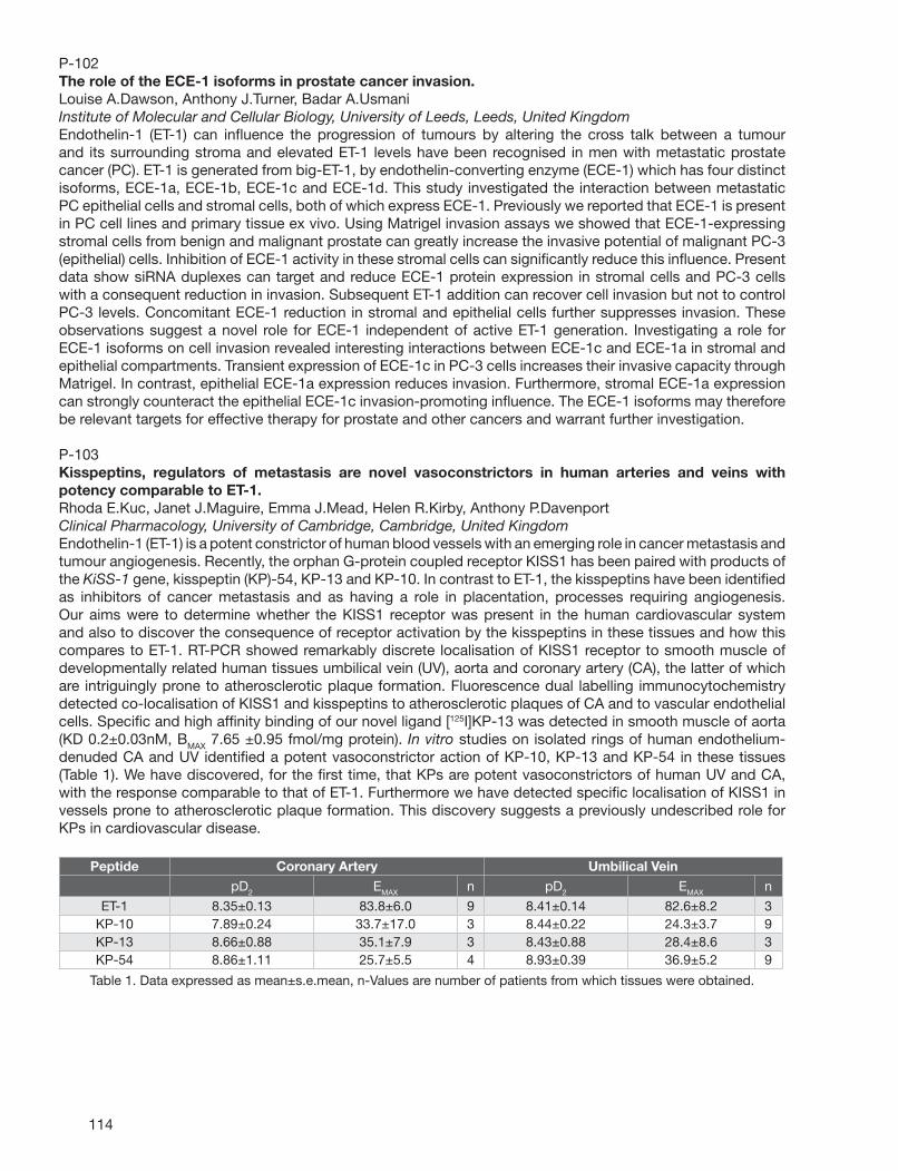
114
P-102The role of the ECE-1 isoforms in prostate cancer invasion.Louise A.Dawson, Anthony J.Turner, Badar A.UsmaniInstitute of Molecular and Cellular Biology, University of Leeds, Leeds, United KingdomEndothelin-1 (ET-1) can infl uence the progression of tumours by altering the cross talk between a tumour and its surrounding stroma and elevated ET-1 levels have been recognised in men with metastatic prostate cancer (PC). ET-1 is generated from big-ET-1, by endothelin-converting enzyme (ECE-1) which has four distinct isoforms, ECE-1a, ECE-1b, ECE-1c and ECE-1d. This study investigated the interaction between metastatic PC epithelial cells and stromal cells, both of which express ECE-1. Previously we reported that ECE-1 is present in PC cell lines and primary tissue ex vivo. Using Matrigel invasion assays we showed that ECE-1-expressing stromal cells from benign and malignant prostate can greatly increase the invasive potential of malignant PC-3 (epithelial) cells. Inhibition of ECE-1 activity in these stromal cells can signifi cantly reduce this infl uence. Present data show siRNA duplexes can target and reduce ECE-1 protein expression in stromal cells and PC-3 cells with a consequent reduction in invasion. Subsequent ET-1 addition can recover cell invasion but not to control PC-3 levels. Concomitant ECE-1 reduction in stromal and epithelial cells further suppresses invasion. These observations suggest a novel role for ECE-1 independent of active ET-1 generation. Investigating a role for ECE-1 isoforms on cell invasion revealed interesting interactions between ECE-1c and ECE-1a in stromal and epithelial compartments. Transient expression of ECE-1c in PC-3 cells increases their invasive capacity through Matrigel. In contrast, epithelial ECE-1a expression reduces invasion. Furthermore, stromal ECE-1a expression can strongly counteract the epithelial ECE-1c invasion-promoting infl uence. The ECE-1 isoforms may therefore be relevant targets for effective therapy for prostate and other cancers and warrant further investigation.
P-103Kisspeptins, regulators of metastasis are novel vasoconstrictors in human arteries and veins with potency comparable to ET-1.Rhoda E.Kuc, Janet J.Maguire, Emma J.Mead, Helen R.Kirby, Anthony P.DavenportClinical Pharmacology, University of Cambridge, Cambridge, United KingdomEndothelin-1 (ET-1) is a potent constrictor of human blood vessels with an emerging role in cancer metastasis and tumour angiogenesis. Recently, the orphan G-protein coupled receptor KISS1 has been paired with products of the KiSS-1 gene, kisspeptin (KP)-54, KP-13 and KP-10. In contrast to ET-1, the kisspeptins have been identifi ed as inhibitors of cancer metastasis and as having a role in placentation, processes requiring angiogenesis. Our aims were to determine whether the KISS1 receptor was present in the human cardiovascular system and also to discover the consequence of receptor activation by the kisspeptins in these tissues and how this compares to ET-1. RT-PCR showed remarkably discrete localisation of KISS1 receptor to smooth muscle of developmentally related human tissues umbilical vein (UV), aorta and coronary artery (CA), the latter of which are intriguingly prone to atherosclerotic plaque formation. Fluorescence dual labelling immunocytochemistry detected co-localisation of KISS1 and kisspeptins to atherosclerotic plaques of CA and to vascular endothelial cells. Specifi c and high affi nity binding of our novel ligand [125I]KP-13 was detected in smooth muscle of aorta (KD 0.2±0.03nM, BMAX 7.65 ±0.95 fmol/mg protein). In vitro studies on isolated rings of human endothelium-denuded CA and UV identifi ed a potent vasoconstrictor action of KP-10, KP-13 and KP-54 in these tissues (Table 1). We have discovered, for the fi rst time, that KPs are potent vasoconstrictors of human UV and CA, with the response comparable to that of ET-1. Furthermore we have detected specifi c localisation of KISS1 in vessels prone to atherosclerotic plaque formation. This discovery suggests a previously undescribed role for KPs in cardiovascular disease.
Peptide Coronary Artery Umbilical Vein
pD2 EMAX n pD2 EMAX n
ET-1 8.35±0.13 83.8±6.0 9 8.41±0.14 82.6±8.2 3 KP-10 7.89±0.24 33.7±17.0 3 8.44±0.22 24.3±3.7 9 KP-13 8.66±0.88 35.1±7.9 3 8.43±0.88 28.4±8.6 3 KP-54 8.86±1.11 25.7±5.5 4 8.93±0.39 36.9±5.2 9
Table 1. Data expressed as mean±s.e.mean, n-Values are number of patients from which tissues were obtained.
115
P-104Expression of ET-1 in human adrenal tumors and attached non-neoplastic adrenal tissues.Ryo Morimoto1, Fumitoshi Satoh1, Osamu Murakami1, Kazuhito Totsune2, Yoichi Arai3, Takashi Suzuki4, Hironobu Sasano5, Sadayoshi Ito1, Kazuhiro Takahashi61Department of Nephrology, Endocrinology, and Vascular Medicine, Tohoku University Graduate School of Medicine, Sendai, Miyagi, Japan2Department of Clinical Pharmacology and Therapeutics, Tohoku University Graduate School of Medicine, Sendai, Miyagi, Japan3Department of Urology, Tohoku University Graduate School of Medicine, Sendai, Miyagi, Japan4Department of Pathology, Tohoku University School of Health Sciences, Sendai, Miyagi, Japan5Department of Pathology, Tohoku University Graduate School of Medicine, Sendai, Miyagi, Japan6Department of Analytical Medical Technology, Tohoku University School of Health Sciences, Sendai, Miyagi, Japan[Introduction] Endothelin-1 (ET-1) is a potent vasoconstrictor peptide fi rst identifi ed from vascular endothelial cells. In addition to its vascular actions, ET-1 has been shown to have mitogenic effects on tumor growth and some regulatory effects on adrenal steroidogenesis. We have previously shown that ET-1 is produced and secreted by cultured human adrenocortical carcinoma cells (SW-13 cells). The expression of ET-1 in human adrenal tumors has not been studied by immunohistochemistry. In the present study, we examined expression of ET-1 in human adrenal tumors and attached non-neoplastic adrenal tissues by immunohistochemistry. [Methods] Materials: Tissues of human adrenal tumors were obtained at surgical adrenalectomy from 43 patients with adrenal tumors. Antiserum: The antiserum against human ET-1 (BP6) was a gift from Professor S. R. Bloom and Professor M. A. Ghatei (Hammersmith Hospital, London, UK). Immunohistochemistry: Immunostaining was performed by ABC method using Vector ABC kit (Vector Laboratories Inc., USA) with formalin-fi xed, paraffi n-embedded sections. This study was approved by the Ethics Committee of Tohoku University School of Medicine. [Results] ET-1 was immunolocalized in tumor cells of all adrenal tumors examined of both cortical and medullary origins; 8 cases of cortisol-producing adenomas, 8 cases of aldosterone-producing adenomas, 2 cases of non-functioning adenomas, 17 cases of adrenocortical carcinomas, and 8 cases of pheochromocytomas. In attached adrenals, immunoreactivity for ET-1 was detected in medulla, with much weaker immunoreactivity found in cortex. The adrenocortical tumor cells were more intensely immunostained for ET-1 than the attached non-neoplastic adrenal cortex, suggesting that expression of ET-1 was up-regulated in neoplastic adrenocortical tissues. There was no apparent difference in the degree of ET-1 immunoreactivity between benign adrenocortical adenomas and adrenocortical carcinomas. [Conclusion] The present study showed that ET-1 was expressed in the adrenal tumors and attached non-neoplastic adrenal tissues, and suggested a possible role of ET-1 in tumor growth and/or secretory activities of these tumors.
P-105ET-1 production by mononuclear cells in portal hypertensive patients.Abeya Lotfi , Lobna Ghanem, Mootaz Hassan, Ahmed Hazem, Sobhi Keshta, Soheir Mansy, Mohamed MagdyTheodor Bilharz Research Institute, Cairo, EgyptIn cirrhotic patients, it has been established that endothelin-1 (ET-1) vasoactive peptide is an important causative agent of portal hypertension increasing resistance to portal blood fl ow. The site of synthesis of the peptide has been localized to two major sites: the liver and spleen. This study aimed to analyse the dynamics of synthesis of ET-1 across the liver and spleen in cirrhotic patients with portal hypertension. As mononuclear cells (MNCs) were found to be capable of ET-1 synthesis in patients with subarachnoid hemorrhage in a previous study, this role was investigated in our cirrhotic patients. In situ hybridization was performed to measure ET-1 mRNA levels in the liver and splenic tissues of six splenectomized patients with cirrhosis and portal hypertension. Also levels were compared in peripheral blood MNCs derived from splenic (SPV), superior mesenteric (SMV) and systemic veins (SV). It was found that the percentage of MNCs expressing ET-1 mRNA were signifi cantly higher in SPV and SMV than in SV. In the spleen, ET-1 mRNA was localized to two types of cells: MNCs in both the marginal zone of the white pulp and the splenic cords of the red pulp as well as endothelial cells of splenic sinuses and blood vessels. In the liver, positive signals were concentrated in the portal tract(periportal hepatocytes, perivascular infl ammatory cells, endothelial lining of blood vessels and duct epithelium).In conclusion, from the results of this work, it is suggested that ET-1-formig cells are not static but tend to circulate as MNCs along a certain pathway from the spleen and gut, streaming down through the SPV and SMV, respectively, to the infl ammatory periportal areas in the liver. This ensures the release and concentration of ET at its site of action in the liver. These fi ndings may have important therapeutic implications as anti-infl ammatory strategies and splenectomy could help to get rid of the main source of synthesis of ET-1 and thus prevent vasoconstrictive complications and portal hypertension.

116
P-106Increased expression of endothelin-converting enzyme (ECE)-1d isoform is associated with chronic enteroviral myocarditis in mice.Hans-Dieter Orzechowski1, Steffi Lindenau1, Sandra Boche1, Martina Sauter2, Karin Klingel2, Martin Paul11Clinical Pharmacology and Toxicology, Charité, Berlin, Germany2Molecular Pathology, Eberhard Karls University, Tübingen, GermanyWe compared expression of cardiac ECE-1 in murine models of enteroviral myocarditis to analyze whether different severity of histological disease observed in these models is associated with different ECE-1 expression. To analyze the expression of ECE-1 in greater detail, we have established a quantitative assay for the analysis of its isoforms. C57BL/6 (BL/6) and A.BY/SnJ (ABY) mice were infected i.p. with 10e5 coxsackievirus B3 (CVB3, Nancy strain) to generate disease models of acute myocarditis which resolves thereafter (BL/6) or which develops into chronic myocarditis (ABY). Hearts were removed for RNA extraction at days 4, 8, 12, 28 and 84 after CVB3 infection, representing different stages of myocarditis. Expression of ECE-1 isoforms (a, b, c, and d) and of PPET-1 was analyzed by real-time PCR. Signals were normalized to 18S RNA and expression was calculated using the ddCt method. Histological damage of each sample was scored on HE-stained paraffi n sections from grade 0 (no infi ltrations or lesions) to grade 4 (extensive necrosis and large infi ltrations). Histological damage of ABY hearts (grade 4 from day 8 on) was more severe compared to BL/6 hearts (maximum grade 3). Compared to basal levels, ECE-1d mRNA expression was persistently and signifi cantly (p<0.05) increased in ABY mice (up to 4.7-fold) beginning with day 8. In contrast, we did not detect any signifi cant changes of ECE-1d mRNA in BL/6 hearts. ECE-1b mRNA was about 2-fold increased in ABY, but not in BL/6 hearts, beginning with day 4, but the differences were statistically not signifi cant. ECE-1c mRNA was non-signifi cantly increased only at day 28 in ABY (3.3-fold) and in BL/6 hearts (1.9-fold). ECE-1a mRNA remained unchanged in both models. Analysis of PPET-1 mRNA expression showed a persisting, about 2-fold increase in ABY hearts only. We found that ongoing activation of the ET system (ECE-1d, PPET-1) is associated with more severe cardiac damage in the chronic model of murine enteroviral myocarditis. Whether this fi nding refl ects invasion of infl ammatory cells or ET system upregulation in resident cardiac cells remains to be investigated.
P-107Endothelin in gut radiation damage: a therapeutic target to prevent intestinal fi brosis?Nicolas Jullien, Fabien Milliat, Marc Benderitter, Agnes FrancoisIRSN/DRPH/SRBE/LRPAT, Fontenay aux Roses, FranceRadiation exposure of the digestive tract, in accidental or therapeutic confi guration, may lead to lesions of the intestinal wall that may be very incapacitating and, in some cases, may engage the vital prognosis of patients. Indeed, about half of cancer patients are treated by radiotherapy (1.5 million of patients per year in Europe). Eighty percent of them have acute functional troubles and digestive pains and nearly fi ve to ten percent show late radiation-induced fi brosis, which constitute a strong limitation of radiotherapy. The continuum of pathological effects leads a widespread fi brosis through all the layers of the intestinal wall. This refl ects very complex series of reactions and the production/activation of many pro-mitotic, pro-infl ammatory and pro-fi brotic mediators or different tissue specifi c actors of the reparation system. Among these factors, endothelins, which appear now as major mediators of fi brosis in numerous organs, may play a predominant role in the apparition of radiation-induced intestinal pathologies. The aim of our study is to examine the sequential changes in ET levels and localization during the development of radiation-induced colo-rectal fi brosis and to evaluate the therapeutic potential of strategies based on ET receptor blockade. Experiments are ongoing on a pre-clinical model of colo-rectal radiation injury in the rat. Tissue damage is characterized by severe acute infl ammation, progressive extracellular matrix deposition and tissue transmural fi brosis, corresponding to normal tissue toxicity in humans. First results show a two-fold increase of the ET precursor expression in the external muscular layer of the distal colon and a fi ve to seven-fold increase of the two ET receptors subtypes expression in the mucosa, one week after irradiation. These data strongly suggest that ET may be implicated in the process of normal tissue damage. In vivo experiments are ongoing to evaluate the expression of ET system actors in the late phase after irradiation and the therapeutic potential of ET system blockade by administration of ET receptor antagonists in the same model.
117
P-108The effects of a dual endothelin receptor antagonist, bosentan, on metabolic parameters in the patients with pulmonary hypertension.Naoko Iwasa, Kazuhiko Nakayama, Kazuya Miyagawa, Hidemi Nonaka, Noriaki EmotoDivision of Cardiovascular Medicine, Department of Internal Medicine, Kobe University Graduate School of Medicine, Kobe, JapanDiet-induced obesity is the primary determinant of the current epidemic of metabolic syndrome. Increased plasma endothelin (ET)-1 levels have been reported in the patients with obesity and insulin resistance, suggesting that ET-1 participates in the pathogenesis of obesity and insulin resistance. Previously, we reported that the targeted disruption of ET-1 in vascular endothelial cell attenuated high fat diet (HFD)-induced obesity, hypertension and insulin resistance. To elucidate the effects of the inhibition of endothelin signaling in human, we investigated the change of metabolic parameters in the patients treated with bosentan, a dual endothelin receptor antagonist, for pulmonary hypertension (PH). We followed 12 patients (6 males and 6 females; 2 idiopathic pulmonary artery hypertension, 5 PH due to connective tissue disease, 1 PH due to congenital heart disease and 4 chronic thromboembolic pulmonary hypertension) who received bosentan (initial dose, 32.25 mg or 62.5 mg once daily, titrated to 125 mg or 250 mg twice daily) for 6 months. We examined body mass index (BMI), total cholesterol (TC), HDL-C, LDL-C, triglycerides (TG), fasting glucose and Hb-A1c at 3 and 6 months. There was no signifi cant difference in BMI, TC, LDL-C and TG between before and after bosentan treatment. However, HDL-C were increased signifi cantly during bosentan treatment (baseline 53.8±4.7, 3 months 63.5±4.5, 6 months 69.1±3.5 mg/dl, p<0.05). Fasting glucose levels were slightly but signifi cantly increased after 3 months of bosentan treatment (baseline 85.1±4.2, 3 months 99.6±5.7, p<0.05), but HbA1c was unchanged during the treatment. These observations suggest that bosentan may affect the metabolic parameters, however long-term follow-up will be necessary to evaluate the contribution of bosentan in metabolic syndrome.
P-109ETA antagonist TBC3214 does not affect bone formation during orthodontic tooth movement in rats.Špela Sprogar1, Martina Drevensek2, Andrej Coer3, Gorazd Drevensek1
1Institute of Pharmacology and Experimental Toxicology, University of Ljubljana, Faculty of Medicine, Ljubljana, Slovenia2Department of Orthodontics, University of Ljubljana, Faculty of Medicine, Ljubljana, Slovenia3Institute of Histology and Embryology, University of Ljubljana, Faculty of Medicine, Ljubljana, SloveniaAfter a force is applied to a tooth, areas of pressure and tension appear in the periodontal ligament (PDL). On the pressure side the alveolar bone in resorbed by osteoclasts and on the tension side new bone is formed by osteoblasts. Many chemical messengers are involved in this processes, among them endotelin-1 (ET-1). ET-1 stimulates bone formation, but it’s infl uences on bone resorption are controversial. The aim of the study was to determine the role of TBC3214 a highly selective ETA antagonist on bone formation and/or bone resorption during orthodontic tooth movement. The study was performed on 30 male Wistar rats (310-335g, 12 weeks old) divided into 3 groups. Group I (n=10): a superelastic closed coil spring (F=25cN) was placed between the upper left fi rst molar and the upper left incisor and animals were daily treated with TBC3214 i.p. (15mg/kg). Group II (n=10): coil spring as in Group I was used and animals were daily treated with a placebo. Group III (n=10): no appliance, only a placebo daily. After 42 days the animals were sacrifi ced and tissue samples of the alveolar bone, three molars and PDL were taken and fi xed in formalin, decalcifi ed and embedded in paraffi n. Sections of 5 microm thickness were stained with haematoxylin and eosin and stereologicaly analysed using cycloid stereological grid with light microscope at 200 magnifi cation and the bone density was determined. The percentages of bone surface covered with active osteoblasts were determined as well. The stereologic analysis showed that in Group I the amount of bone vs. other tissues (connective tissue, tooth) was signifi cantly higher compared to Group II (p<0,05). The amount of bone vs. other tissues was signifi cantly larger in Groups I and II, which were applied a closed coil spring compared to Group III, without the closed coil spring (p< 0,001). The stereologic analysis showed no signifi cant difference in the number of osteoblasts between Groups I and II and between Groups I, II and III. Considering our results TBC3214, an ETA antagonist, does not affect bone formation, but increases bone resorption during orthodontic tooth movement in rats.
118
P-110Synthesis of 1,3,6-Trisubstituted-2-Carboxy-Quinol-4-ones as Selective ETA Antagonists and Their Role in Controlling Preterm Labor in a Mouse Model.Hardik J.Patel, Wei Wang, Ralph Stephani, Sandra ReznikDepartment of Pharmaceutical Sciences, St. John’s University, Jamaica, NYPrevious investigations have implicated endothelin-1 (ET-1) in the pathogenesis of infection-associated preterm labor (PTL). ET-1 increases myometrial smooth muscle tone in vitro . Also it was showed that preterm labor (PTL) is controlled with the endothelin converting enzyme (ECE-1) inhibitor phosphoramidon. We now show that similar inhibition is achieved with BQ-123, a peptide ETA selective antagonist. We also have treated a mouse model with ECE-1 RNAi with similar results. Thus a series of 6-alkoxy substituted-3-carboxybenzyl-N-benzyl-quinol-4-ones were designed and synthesized as potential ETA selective inhibitors. These compounds were tested for their selective ETA inhibition and may serve as the pharmacological tools to determine the role of ETA in preterm labor (PTL). Results of these latest tests will be discussed in terms of ED50’s and structure activity relationships. Methods: in vivo experiments: C57Bl/6 E15.5 timed pregnant mice were injected with lipopolysaccharide (LPS) at t=0 and divided into four groups. Group 1 mice (n=7) received an intraperitoneal injection of BQ-123 or test compounds at 6.7 mg/kg at t=12 hours. Group 2 mice (n=7) received an intraperitoneal injection of BQ-123 or test compounds at 3.3 mg/kg at t=12 hours. Group 3 mice (n=6) were treated with ECE-1 RNAi (target sequence NNCUUCCACAGCCCCCGGAGU) at t=-30 hours, t=-12 hours and t=-2 hours. Group 4 mice (n=10) received a sham injection of phosphate buffered saline at t=12 hours. Mice were continually monitored from t=12 hours to t=24 hours and time of delivery of the fi rst pup as well as the number of pups delivered was recorded.
P-111Human Metabolism and Plasma Protein-Binding Properties of Ambrisentan.J. Craig Hartman, Richard J.GorczynskiR&D, Gilead Colorado, Westminster, COIntroduction: The ETA-selective endothelin receptor antagonist ambrisentan is currently under review by the FDA and EMEA for the treatment of pulmonary arterial hypertension. The following in vitro results describe the hepatic metabolism and inhibitory potential, and the plasma protein-binding properties of ambrisentan. Methods: Phase I metabolism of ambrisentan was studied in human liver microsomes and phase II was examined in human hepatocytes. Inhibitory effects of ambrisentan were tested on cDNA derived CYPs 1A2, 2A6, 2B6, 2C8, 2C9, 2C19, 2D6, 2E1, 3A4, and UGTs 1A1, 1A6, 1A9, 2B7 using model substrates. Human plasma (pooled) protein-binding effects were studied using the equilibrium dialysis method. Results: Phase I metabolism was low (3-4%, turnover at 24 hr). The predominant metabolites were glucuronides formed by UGTs 1A9 and 2B7 (21%, 24 hr). Ambrisentan inhibited these hepatic enzymes: CYPs 2A6 (19%, at 300µM) and 2C8 (20%, 300 µM). The following UGTs were inhibited at 300 µM: 1A1 (30%), 1A6 (10%), 1A9 (23%), 2B7 (10%). Ambrisentan is 98.8 % bound to human plasma proteins, and at co-incubation concentrations of 10 and 20 µg/mL for each agent there was no protein binding competition between ambrisentan (remained 98.8% bound) and warfarin (98.7%). Conclusions: In vitro, ambrisentan was minimally metabolized by human phase I hepatic enzymes, and was predominantly glucuronidated by phase II UDP-glucuronosyltransferase activity. At concentrations representing the maximal therapeutic range achieved in man, ambrisentan did not inhibit phase I or II hepatic metabolizing enzymes. Ambrisentan is highly protein bound to human plasma, however co-incubation with another substantially protein bound concomitant medication, warfarin, revealed no protein displacement activity for either agent. Ambrisentan is predominantly metabolized by glucuronidation and to a lesser extent by oxidation via CYP pathways suggesting a low potential for drug-drug interactions.
119
P-112Effects of Ambrisentan, Darusentan, Bosentan, and Sitaxsentan on Human Hepatic Uptake and Effl ux Transporters.J. Craig Hartman1, Richard J.Gorczynski1, Robert L.St. Claire III2, Kirsten R.Wille2, Cassandra H.Perry2, Kenneth R.Brouwer2
1R&D, Gilead Colorado, Westminster, CO2Qualyst Inc., Raleigh, NCFour endothelin receptor antagonists (ERAs) were examined for substrate activity and potential inhibition of selected hepatic uptake and effl ux transporters using sandwich cultured human hepatocytes. ERA substrate activity for respective transporters () was evaluated using ritonavir (OATP, BSEP), bromosulphalein (OATP), Na+ depleted (NTCP), and erythromycin (Pgp). ERA transporter () inhibition was examined using substrates E2-17betaG (OATP), taurocholate uptake (NTCP), taurocholate effl ux (BSEP), and DPDPE (MRP2). Each ERA was tested for substrate activity at 2uM and inhibition at 2, 20, 100uM. Sitaxsentan and bosentan demonstrated greater hepatic uptake (5-30 fold) than that of ambrisentan and darusentan. Decreases in infl ux of all 4 ERAs with co-administration of ritonavir, bromosulfalein, or probenecid suggested that OATPs contribute to uptake. Darusentan infl ux was the least altered by those agents (84-100% of control), whereas bosentan was the most (32-58%). The absence of Na+, reduced uptake of ERAs (bosentan>darusentan >sitaxsentan >ambrisentan) demonstrated the relative contribution of NTCP to infl ux, Ritonavir did not change the biliary excretion index (BEI) of ambrisentan, suggesting no effect on BSEP mediated effl ux. Darusentan and bosentan showed increased BEI with ritonavir, whereas parent sitaxsentan was not excreted. Erythromycin did not change the BEI for ambrisentan, but reduced effl ux for darusentan (64% of control) and bosentan (72%), demonstrating that Pgp contributes to the excretion of the latter drugs. No concentrations of ambrisentan nor darusentan signifi cantly reduced OATP, NTCP, BSEP, or MRP2 transport. Bosentan (100uM; 33% of control) and sitaxsentan (20 and 100uM; 19 and 2%) signifi cantly attenuated NTCP transport. Only sitaxsentan (100uM) signifi cantly decreased OATP transport to 52% of control. BSEP transport was reduced by 100 uM bosentan (78% of control) and sitaxsentan (85%). Neither bosentan nor sitaxsentan signifi cantly altered MRP2 transport. These results indicate that ERAs are hepatic transport substrates, and suggest that bosentan and sitaxsentan, but not ambrisentan and darusentan inhibit human hepatic transport.
P-113Plasma ET-1 as a biochemical marker of endothelial dysfunction in endocrine diseases with increased cardiovascular risk. G.Kirilov1, P.Kumanov1, S. Zacharieva1, A. S. Alexandrov2
1Clinical Center of Endocrinology, Sofi a, Bulgaria 2Institute of Biophysics, BAS, Sofi a.Background: The aim of the present study was to summarize our results of plasma endothelin-1 (ET-1) level determined in some endocrine diseases like diabetes, hyper- and hypothyroidism, Cushing’s disease, acromegaly and male hypogonadism in which the existence of endothelial dysfunction and increased cardiovascular risk is proved. Patients and Results: In patients with type 2 diabetes (n=34) plasma ET-1 (0.9±0.17 pmol/l) correlated with the degree of vascular complications and reached the highest level in hemodialysis diabetics (3.4± 0.38 pmol/l). ET-1 was signifi cantly elevated in patients with hyperthyroidism (0.78±0.11 pmol/l, n=18) and decreased to normal range after thyrostatic treatment (0.5± 0.1 pmol/l). However, in hypothyroid subjects (n=20) ET-1 did not differ from controls (0.49± 0.12 vs 0.46 ±0.2 pmol/l). ET-1 level in patients with Cushing’s disease was 3-fold higher (1.6± 0.8 pmol/l, n=13) than in healthy subjects, and decreased signifi cantly after disease remission (0.73±0.53 pmol/l). Hypersomatotropism in acromegaly (n=28) leads to signifi cant elevation of ET-1 (1.24±0.2 pmol/l). After treatment and normalization of IGF-1, ET-1 showed the normal concentration (0.39±0.1 pmol/l). The low testosterone level in male hypogonadism (n=33) caused higher plasma ET-1 (0.95±0.53 pmol/l). Patients with hypergonadotrophic hypogonadism had signifi cantly higher ET-1 in comparison to hypogonadotrophic hypogonadism (1.05± 0.57 pmol/l vs 0.89 ±0.53 pmol/l). The testosterone replacement therapy did not lead to signifi cant changes of ET-1 level. In all groups no correlation was observed, however, between the concentrations of ET-1, blood pressure, lipid status and plasma homocystein. Conclusions: Our results clearly demonstrate that hyperthyroidism, hypercortisolism, hypersomatotropism and hypogonadism lead to activation of endothelin system. Elevated plasma ET-1 levels probably play a role in the pathogenesis of accelerated and early atherosclerosis development in this disorders.
120
P-114ETA antagonist TBC3214 decreases orthodontic tooth movement in rats.Špela Sprogar1, Martina Drevensek2, Tomaz Vaupotic3, Gorazd Drevensek1
1Institute of Pharmacology and Experimental Toxicology, University of Ljubljana, Faculty of Medicine, Ljubljana, Slovenia2Department of Orthodontics, University of Ljubljana, Faculty of Medicine, Ljubljana, Slovenia3Institute of Biochemistry, University of Ljubljana, Faculty of Medicine, Ljubljana, SloveniaThe basis of orthodontic tooth movement is force application to a tooth. It causes the release of many mediators and in consequence tooth movement. One of the mediators is endothelin-1 (ET-1). It’s concentration in the periodontal ligament (PDL) doubles after a 3-hour axial loading of a tooth. We showed that tezosentan, an ETA/ETB antagonist decreased the late phase of orthodontic tooth movement in rats. The aim of the study was to determine the role of ET-1 and expression levels of ETA and ETB in orthodontic tooth movement in rats. 40 male Wistar rats (300-330g) were divided into 3 groups. Group I (n=10): a superelastic closed coil spring (F=25cN) was placed between the upper left fi rst molar and the upper left incisor and the animals were daily treated with TBC3214, a highly selective ETA antagonist (15mg/kg). Group II (n=10): coil spring as in Group I was used and the animals were daily treated with a placebo. Group III (n=20): no appliance, only a placebo daily, than the animals were sacrifi ced on days 0, 14, 28 and 42. Tissue samples of the alveolar bone, three molars with their PDL were taken. Total RNA was isolated using TRIzol reagent. The ETA and ETB expression levels were assessed by means of relative RT-PCR using subtype-specifi c primers. The relative expression levels of ETA and ETB mRNA were normalized against GAPDH mRNA as a control. In Group I tooth movement was signifi cantly smaller (p<0,001) on days 32, 37 and 40 when compared to Group II. The basal mRNA levels of ETA and ETB on day 0 were comparable. During the whole experiment, the expression level of ETB was 1.5-1.8-fold higher than of ETA (p < 0.05). A 2 fold induction of ETA mRNA level compared to day 0 was observed on day 42 (p < 0.05). After 14 and 28 days a 2.5-fold and 1.7-fold induction of ETB mRNA level, respectively, compared to day 0 was observed (p < 0.05) and was further induced up to 3.3-fold after 42 days compared to day 0 (p < 0.001). Considering the effects of tezosentan and TBC3214 on orthodontic tooth movement and the expression levels of ETA and ETB receptors, we conclude that ET-1 acts predominantly on ETA receptors in the late phase of orthodontic tooth movement in rats.
P-115ETA receptor promotes β-catenin signaling pathway through β-Arrestin-1 in human ovarian carcinoma.Laura Rosanò1, Roberta Cianfrocca1, Stefano Masi1, Francesca Spinella1, Valeriana Di Castro1, Maria Rita Nicotra3, Pier Giorgio Natali2, Anna Bagnato1
1Laboratory of Molecular Pathology and Ultrastructure, Regina Elena Cancer Institute, Rome, Italy2Laboratory of Immunology, Regina Elena Cancer Institute, Rome, Italy3Molecular Biology and Pathology Institute, National Research Council, Rome, ItalyThe endothelin A receptor (ETAR)/endothelin-1 (ET-1) axis has a key role in the pathophysiology of ovarian carcinoma. Among the different intracellular pathways activated by ET-1, the epidermal growth factor receptor (EGFR) transactivation is a downstream signaling in driving ovarian cancer progression. Recently, it has become evident that β-arrestin-1 promotes signal transduction by G-protein-coupled receptors acting as multifunctional adaptor protein. Here, we examined the functional role of β-arrestin-1 in the cross-communication between ETAR and EGFR to regulate cell-cell adhesion complex and β-catenin signaling in ovarian cancer. We reported that, in HEY and OVCA 433 ovarian cancer cells, ET-1 induced the cytosol-to-membrane translocation of β-arrestin-1 and the dephosphorylation on serine-412, leading to a membrane ETAR/β-arrestin-1/Src signaling complex (“signalplex”) formation. By transfection with FLAG-tagged WT- or mutant S412D-β-arrestin-1, we demonstrated that this signalplex was crucial for EGFR transactivation and downstream signaling, such as β-catenin tyrosine phosphorylation, and loss of β-catenin/E-cadherin interactions. Moreover, ET-1 induced signalplex-mediated β-catenin nuclear translocation and its transactivating ability for TCF/LEF transcription factor. At functional level, these events correlated with enhanced cell invasion, indicating that ETAR/β-arrestin-1/Src complex and subsequent EGFR transactivation may also account for the cell aggressive behavior. These effects were prevented by both ETAR antagonists and ETAR siRNA, thus validating ETAR as the receptor involved. In human ovarian carcinoma xenografts, ETAR blockade by selective ETAR antagonist, ZD4054, signifi cantly inhibited tumor growth, peritoneal dissemination and expression of EMT effectors. The in vitro results provided the basis to test combined targeting of ETAR by ZD4054, and EGFR, by the EGFR inhibitor gefi tinib (IRESSA). The coadministration of ZD4054 enhanced the effi cacy of gefi tinib leading to partial (82%) or complete tumor regression on HEY xenografts, indicating new opportunities that confer improved therapeutic effi cacy in ovarian cancer patients.

121
P-116Anti-invasive activity of the specifi c ETA receptor antagonist, ZD4054, in A673 rhabdomyosarcoma cells.Jim Growcott1, Mark Hickinson1, Jon Curwen2, Beverley Isherwood3, Neil Carragher3, Nicola Curtis1
1Discovery Medicine, AstraZeneca, Alderley Park, Macclesfi eld, United Kingdom2Cancer and Infection Bioscience, AstraZeneca, Alderley Park, Macclesfi eld, United Kingdom3Advanced Science and Technology Laboratory, AstraZeneca R&D Charnwood, Loughborough, United KingdomZD4054, N-(3-Methoxy-5-methylpyrazin-2-yl)-2-[4-(1,3,4-oxadiazol-2-yl)phenyl]pyridine-3-sulfonamide, is a specifi c ETAR antagonist,1 which demonstrates a variety of pre-clinical anti-cancer biology in vitro and in vivo.2-5 Moreover, endothelin-1 (ET-1) appears to drive tumour cell invasion and migration.6 We have extended this thinking by investigating the ability of ZD4054 to modulate in vitro tumour cell migration and invasion using the A673 rhabdomyosarcoma tumour cell line. In addition we have investigated cellular morphology and related signalling associated with the invasive phenotype. As determined by actin re-organisation and increased phosphorylation of paxillintyr31 and FAKtyr397,tyr861, ET-1 (1 nM, 10 minutes) induced the formation of stress fi bres in A673 cells suggestive of an invasive phenotype. These effects were blocked by pre-incubation with either ZD4054 (0.5 µM and 0.01-10 µM, respectively), or the ETAR-specifi c antagonist, BQ123 (0.1 µM), but not by the ETBR-specifi c antagonist, BQ788 (0.1 µM). These observations demonstrate that these effects are ETAR-mediated. Furthermore, in 3D transwell assays, ET-1 (100 nM, 72 hours) induced A673 cell invasion through MatrigelTM. This effect was ablated following prior incubation with ZD4054 (1-30 µM) prior to ET-1 treatment whereas the ETB-agonist, safarotoxin, (1-10,000 nM) had negligible activity, implying that the invasive phenotype in this model system is ETAR-mediated. In conclusion, ZD4054 inhibited cell invasion by blocking the action of ET-1 at ETA-Rs. Tumours containing a major cell migratory/invasion component should be responsive to ZD4054, which is currently in clinical development in hormone-resistant prostate cancer. References 1. Morris C et al. Br J Cancer 2005;92:2148-2152. 2. Curtis N et al. Eur J Cancer Suppl 2004;2(8):abst 78. 3. Rosano L et al. Proc Am Assoc Cancer Res 2005;46:abst 5830. 4. Curtis et al. Proc Am Assoc Cancer Res 2005;46:abst 1512. 5. Williams E et al. Eur J Cancer Suppl 2006;4(12):15. 6. Rosano L et al. Exp Biol Med 2006;231:1128-1131.
P-117Shigatoxin-2 reduces nephrin gene expression via ET-1/ETA receptor mechanism in cultured podocytes.Simona Buelli1, Lorena Longaretti1, Marina Morigi1, Elena Gagliardini1, Daniela Corna1, Carla Zoja1, Giuseppe Remuzzi1,2, Ariela Benigni11Department of Molecular Medicine, Mario Negri Institute for Pharmacological Research, Bergamo, Italy2Unit of Nephrology and Dialysis, Azienda Ospedaliera, Bergamo, ItalyShigatoxin(Stx) is the offending agent of post-diarrheal Hemolytic Uremic Syndrome (HUS), characterized by glomerular ischemic changes and microvascular thrombosis that contribute to acute kidney injury. We have previously shown that podocyte is a functionally relevant target of Stx-2 that, via up-regulation of endothelin(ET)-1 gene and protein, promotes cytoskeletal changes and glomerular permeability dysfunction in an autocrine manner. Here we studied whether in differentiated podocytes Stx-2 altered the expression of nephrin, key component of the slit diaphragm, through an ET-1-dependent pathway. We used murine podocytes cultured for 24 hours before and during the experiments with a medium containing 1,25-dihydroxyvitamin D3, all-trans-retinoic acid and dexamethasone (Takano Y, Am J Physiol Renal Physiol 2007) that increased nephrin expression up to 200 fold over normal cultured cells. Stx-2 signifi cantly reduced (p<0.05) nephrin mRNA levels in a dose-dependent manner in respect to control cells (Stx-2 50pM: 0.66±0.04, Stx-2 1nM: 0.32±0.06-fold vs control: 1). Blockade of ETA receptor with LU302146 prevented Stx-2-dependent decline of nephrin expression, indicating the involvement of ET-1 via ETA receptor (Stx-2 50pM+LU: 0.97±0.17, Stx-2 1nM+LU: 0.96±0.18-fold vs control: 1; p<0.05 vs corresponding stimulus). Consistent with Stx-2 effect, ET-1 (100nM) added to podocytes decreased by 58% (p<0.05) nephrin mRNA levels. Inhibition of Rho kinase -crucial for the formation of cytoskeletal stress fi bers- partially recovered nephrin mRNA levels in respect to Stx-2-treated cells. In previous experiments injection of Stx-2 in mice caused podocyte changes including focal foot process effacement and increased expression of ET-1. In these animals here we found a 30% reduction of nephrin protein expression in respect to controls. In summary, our data show that ET-1 produced by podocytes in response to Stx, is an important mediator of Stx-induced reduction of nephrin expression, and might play a pivotal role in dysfunction of the fi ltration barrier in HUS.
122
P-118Determination of Endothelin Receptor Antagonist Affi nities and Selectivities in Human Cardiac Membranes.Scott Greene3, Richard A.Bundey2, Karin Nunley3, J. Craig Hartman1, Lawrence S.Melvin1, Richard J.Gorczynski1, Paul A.Insel2, Michael R.Bristow3, Kelly R.Pitts1
1R&D, Gilead Colorado, Westminster, CO2Department of Pharmacology, UCSD School of Medicine, San Diego, CA3Cardiovascular Institute, University of Colorado Health Science Center, Denver, COEndothelin is a potent vasoactive peptide capable of exerting pleiotropic effects mediated by two endothelin (ET) receptors, ETA and ETB. Such effects can be attenuated at the level of ligand binding owing to several small molecule-based endothelin receptor antagonists (ERAs) either currently in the clinic or in development. Using isolated human cardiac membranes containing a mixed population of ETA and ETB receptors, we determined the affi nities and selectivities of four ERAs - the propanoic acid class ERAs S-ambrisentan and S-darusentan (ETA receptor-selective), and the sulfonamide class ERAs bosentan (a “mixed” receptor antagonist) and sitaxsentan (ETA receptor-selective). Saturation experiments showed that the time to steady state is longer for S-ambrisentan (4hr) than sitaxsentan (2hr). [I125]-ET-1 competition studies revealed that, while each ERA exhibits some ETA receptor selectivity, S-ambrisentan displays the greatest selectivity of those ERAs tested (S-ambrisentan>S-darusentan=sitaxsentan>bosentan), with an ETB:ETA Ki selectivity ratio of ~4700. Similar competition studies using membranes isolated from CHO cells expressing signaling-competent human ETA or ETB receptors confi rmed this selectivity (S-ambrisentan>sitaxsentan>S-darusentan>bosentan), with an S-ambrisentan ETB:ETA Ki selectivity ratio of ~4300. Since S-ambrisentan was found to have the greatest selectivity, we tested whether this characteristic was specifi c to S-ambrisentan by generating its optical enantiomer R-ambrisentan and an in vivo metabolite 4-hydroxymethyl ambrisentan (4-HMamb). R-ambrisentan exhibits one-site competition kinetics in cardiac membranes with a single Ki that is ~10-fold greater compared to the low affi nity (i.e., ETB receptor) Ki for S-ambrisentan, suggesting that R-ambrisentan exhibits less affi nity for ET receptors. However, 4-HMamb displays ETA receptor selectivity in cardiac membranes, but exhibits ETA and ETB Ki values ~100-fold greater compared to those of S-ambrisentan. Together these data indicate that S-ambrisentan is a potent ETA receptor-selective antagonist in human cardiac membranes, and that this activity is attributable to its S enantiomer.
P-119Neonatal Rat Survival and Growth Following Maternally Administered Endothelin Receptor Antagonism.Larry G.Thaete1,2, Sylvia Synowiec1, Mark G.Neerhof1,2
1Obstetrics & Gynecology, Evanston Northwestern Healthcare, Evanston, IL2Obstetrics & Gynecology, Northwestern University Feinberg School of Medicine, Chicago, ILEndothelin receptor A (ETA) antagonism improves placental perfusion and normalizes fetal growth in several rat models of fetal growth restriction. However, direct administration of ETA antagonists to newborn rats has been consistently associated with neonatal demise, raising concerns about the safety of their use late in pregnancy. Perinatal exposure to ETA antagonists (maternal administration in late gestation) and its impact on rat pup survival and growth has not been investigated. Objective: To determine the impact of a maternally administered ETA antagonist on pregnancy outcome, survival and growth of rat pups. Methods: Timed pregnant Sprague-Dawley rats were treated with FR139317 (12 mg/kg/day; ETA antagonist) or vehicle, by subcutaneous osmotic pump connected to an intravenous catheter, from gestational day 14 (term=22 days) through parturition. All fi ve pregnant rats in each group were allowed to deliver spontaneously and to nurse their pups through postpartum day 7. Viability of newborns, litter sizes, and pup weights were recorded at birth and at day 7. Results are presented as means ± SE. Results: See table below. Conclusions: Maternal administration of an ETA antagonist from gestational day 14 through parturition has no adverse impact on survival and growth of neonatal rat pups.
Pregnancy Outcome, Survival and Growth of Rat Pups with Maternal ETA Antagonism
Group Gest Age at Birth (days)
Litter Size (# pups)
Live Pups at Delivery
Live Pups at 7 Days
Birth Weight (g)
Weight at 7 Days (g)
% Stillbirth %
Neonatal Loss
Vehicle 22.1±0.30 12.4±0.42 11.6±0.53 11.3±0.76 5.3±0.15 12.9±0.66 6.0±3.0a 6.7±5.4b ETA Antag. 22.4±0.24 12.8±0.80 11.6±0.93 11.6±0.93 5.6±0.19 13.5±0.80 7.7±3.9a 0.0±0.0b
There were no statistically signifi cant differences in any of these parameters between groups. aVehicle group: One stillbirth in each of two rats and three stillbirths in one rat. ETA antagonist group: One stillbirth in one rat and three stillbirths in one rat. bVehicle group: Four neonatal losses in one rat and one loss in another. ETA antagonist group: No neonatal losses.
123
P-120Oxygen Saturation in Neonatal Rats Following Maternally Administered Endothelin Receptor Antagonism.Larry G.Thaete1,2, Sylvia Synowiec1, Khan Saira1, Mark G.Neerhof1,2
1Obstetrics & Gynecology, Evanston Northwestern Healthcare, Evanston, IL2Obstetrics & Gynecology, Northwestern University Feinberg School of Medicine, Chicago, ILEndothelin receptor A (ETA) antagonism has been shown to normalize placental perfusion and fetal growth in several rat models of fetal growth restriction. However, direct administration of ETA antagonists to newborn rats within 3 hours of delivery has been consistently associated with neonatal demise due to failure of the ductus arteriosus to close, raising concerns about the safety of their use late in pregnancy. Perinatal exposure to ETA antagonists (maternal administration in late gestation) and its impact on rat pup survival and oxygen saturation has not been investigated. Objective: To determine the impact of a maternally administered ETA antagonist on oxygen saturation in newborn and 7-day-old rat pups. Methods: Timed pregnant Sprague-Dawley rats were treated with FR139317 (12 mg/kg/day; ETA antagonist) or 4.2% NaHCO3 vehicle, by subcutaneous osmotic pump connected to an intravenous catheter, from gestational day 14 (term=22 days) through parturition. All fi ve pregnant rats in each group delivered spontaneously and nursed their pups through postpartum day 7. Oxygen saturation of each rat pup was measured by pulse oximeter on postpartum days 1 and 7. Results are presented as means ± SE. Results: See table below. Conclusion: Maternal administration of an ETA antagonist from gestational day 14 through parturition, at a dose suffi cient to ameliorate fetal growth restriction, has no adverse impact on oxygen saturation in neonatal rat pups.
Oxygen Saturation (%O2) in Neonatal Rats following Maternal ETA AntagonismGroup Newborn 7-day NeonateVehicle 92.6 ± 0.9 94.4 ± 0.8
ETA Antagonist 94.7 ± 0.8 91.6 ± 1.1
There were no statistically signifi cant differences between the treatment groups.
P-121Effects of protease activated receptor-2 (PAR2) on the upregulated levels of ET-1 and TNF-α in acute liver injury in a rat model of endotoxemia.Subrina Jesmin1,3, Atsushi Sawamura1, Sohel Zaedi1,3, Shamsul Haque Prodhan3, Naoto Yamaguchi3, Katsutoshi Goto2, Satoshi Gando1
1Department of Anesthesiology and Critical Care Medicine, Hokkaido University, Sapporo, Japan2Department of Pharmacology, University of Tsukuba, Tsukuba, Japan3Center of Medical Science, Ibaraki Prefectural University, Ami, JapanSeptic shock is associated with the development of progressive damage in multiple organs, and is the leading cause of patient mortality in intensive care units. The liver can be injured and its functions altered by activation of coagulation, vasoactive-peptide and infl ammatory processes in sepsis. Endothelin (ET)-1, a potent vasoconstrictor, is implicated in the pathogenesis of a number of diseases including sepsis. Here, we examined the time-dependent alterations of ET-1, NO and infl ammatory cytokine, such as TNF-α in liver tissue in a septic rat model. Normal male Wistar rats at age 8 wks were administered with lipopolysaccharide (LPS: 15 mg/kg) and then sacrifi ced at different time points (1h, 3h, 6h and 10h). The classical features of acute liver injury, such as infi ltration of infl ammatory cells, hepatocytic necrosis, were seen in LPS administered rats. Furthermore, plasma bilirubin, AST and ALT levels were also signifi cantly changed. A 28-fold increase in ET-1 level was observed in liver tissue at 10 h after LPS administration, while a peak increase of 14-fold ET-1 mRNA level was seen 1 hour after LPS administration in liver tissue. Levels of hepatic TNF-α peaked (4.5-fold) at 1 hour of sepsis. Endotoxemia often triggers exuberant infl ammatory responses and activation of the coagulation cascade, and interactions between infl ammation and coagulation may be important in this setting. Protease-activated receptors (PARs) connect coagulation proteases to cellular responses and represent one mechanism by which coagulation might affect infl ammation. Of the 4 mammalian PARs, PAR1, PAR3, and PAR4 are activated by thrombin, and PAR2 can be activated by coagulation proteases VIIa and Xa but not thrombin. Interestingly, PAR2 blocking peptide (BP) improved liver injury, an effect that was associated with suppression of TNF-α elevation, and normalization of ET-1. The present study reveals a distinct chronological expression of ET-1 in LPS-mediated liver injury and shows that blockade of PAR2 may play a crucial role in treating liver injury, via normalization of infl ammation, coagulation and vaso-active peptide.

P-122The specifi c ETA receptor antagonist ZD4054 reduces tumour-induced angiogenesis in a preclinical model.J. Curwen1, G. Hughes1, J. Growcott2
1Cancer & Infection Bioscience, AstraZeneca, Alderley Park, Macclesfi eld, United Kingdom2Discovery Medicine, AstraZeneca, Alderley Park, Macclesfi eld, United KingdomThe endothelin A receptor (ETAR) has been implicated in pathological angiogenesis by increasing the levels of angiogenic factors such as VEGF1 and by direct action on endothelial cells to induce motility.2 Clinical measures support a correlation between the endothelin axis, VEGF expression and disease outcome.3 We used an intradermal model of early tumour development to measure the ability of ZD4054 (N-(3-Methoxy-5-methylpyrazin-2-yl)-2-(4-[1,3,4-oxadiazol-2-yl]phenyl)pyridine-3-sulfonamide), a specifi c ETAR antagonist, to inhibit pathological angiogenesis in vivo. Human tumour cells were inoculated via intradermal injection into the abdominal area of athymic mice (Alderley Park nu/nu strain). Control animals were inoculated with an equal volume of acellular tissue culture media. ZD4054 or vehicle was administered daily for 5 days before the animals were euthanized. To estimate blood vessel count, a 1 cm2 area of skin was taken, with the developing tumour at the centre and the number of blood vessel bifurcations within the fi eld counted as a surrogate metric of total vessel count. ZD4054 produced a consistent and highly statistically signifi cant decrease in vessel count around the tumours (table). Compared with control tumours, treated tumours showed obvious areas of necrosis and vasodilation of vessels in the skin. These data show that in an in vivo tumour setting, inhibition of ETA with ZD4054 produced a modest anti-angiogenic outcome and that other aspects of tumour development appeared to be normalized. References 1. Bagnato et al. Endocr Relat Cancer 2005;12:761-772. 2. Bagnato & Spinella. Trends Endocrinol Metab 2003;14:44-50. 3. Wulfi ng et al. Clin Cancer Res 2004;10:2393-2400.
Cell line Tumour type ZD4054 dose (mg/kg) Percent reduction in blood vessel count (compared with control) LOVO Colon 50 20 (P=0.001) LOVO Colon 50 25 28 (P<0.001) 28 (P<0.001) DU145 Prostate 50 25 30 (P<0.05) 38 (P<0.001)
PC3 Prostate 50 25 24 (P=0.009) 15 (P=0.08)

Index

127
AUTHOR INDEX
AAbassi, Zaid O-15, P-062, P-064Abraham, D. O-20Abraham, David O-49, O-57Abraham, David J. O-32Adiarto, Suko P-031, O-16, O-36, O-59Adur, Javier P-014Agelaki, Maria G. P-021Ahmarani, Lena O-07Aiello, Ernesto A. P-045Aivars, Juris I. P-067Albertoni Borghese, Maria F. P-066Albrecht, Philip P-087, O-44Alexanian, Anna O-11Alves-Faria, Pedro P-083Amadei, Barbara O-53Amann, Kerstin P-085Amaral, Miguel P-083Anggrahini, Dyah W. P-031, O-59Anggrahini, Dyah Wulan O-16, O-36Angst, Eliane O-48Ansar, Saema P-071, O-40Arai, Yoichi P-104Armour, Danielle P-035Avedanian, Levon O-06
BBacker, Angela O-25, P-040Bagnall, Alan J. P-016Bagnato, Anna P-025, O-52, P-115Bai, Jie P-020Baltogiannis, Giannis G. P-021Bankers, Christine P-050Bartholdy, Roland O-28Barton, Matthias P-026, P-027, P-030, O-48Bauer, C. P-013Bauer, Christian P-011, O-29Baylis, Chris O-01Baynach, Amy O-12Bedford, Chris O-19Bellini, Giuseppe P-061, P-088Belloni, Anna S. O-39Benderitter, Marc P-107Benigni, Ariela O-35, O-53, O-54, P-117Bennett, Martin R. O-17Bento, Allisson F. O-62Berden, Jernej P-022Beresewicz, Andrzej P-002Bernobich, Elena P-061, P-088Bestmann, Lukas P-030Beuzard, Yves O-55

128
Bhattacharya, Indranil P-026, P-027Bishara, Bishara O-15, P-064Bkaily, Ghassan O-06, P-038Bkaily Jr, Ghassan B. O-06Black, C. O-20Black, Carol O-57Black, Carol M. O-32Boche, Sandra P-106Bodil, Petersen O-28Boemke, Willehad P-010, P-037, O-29Boesen, Erika I. P-070Boidi, Carla D. P-099Bonnin, Philippe O-55Bormane, Inga P-067Bou-Gharios, George O-32Bramall, Alexa O-46Bramall, Alexa N. O-31Bridges, Phillip O-10Brioschi, Simona O-53Bristow, Michael R. P-118Briyal, Seema P-093Brochu, Isabelle P-038Brouwer, Kenneth R. P-112Broward, Melinda P-097Buelli, Simona P-117Bundey, Richard A. P-118Bungert, Jorg O-01Busch, Thilo O-28, O-29Böhm, Felix O-21, O-26Bütikofer, Yanik O-48
CCain, Brian O-01Calderone, Angelino P-049, P-052Caldiz, Claudia P-018Calixto, Joao B. P-075, O-62Camilión de Hurtado, María C. P-018Campi, Barbara O-45Candinas, Daniel O-48Caprara, Valentina O-52Caprioli, Jessica O-53Carneiro, Fernando S. P-032, P-047Carpenter, Todd P-050Carragher, Neil P-116Carrara, Fabiola O-35Caruso, Arnaldo P-025Casco, Victor P-014Castañares, Cristina O-03Castañeda, Mauricio M. O-42Catar, R. A. P-084Cattaneo, Dario O-54Cervenak, László P-098Chahdi, Ahmed P-005Chai, Chee-Yin P-042, P-053, P-057, P-058
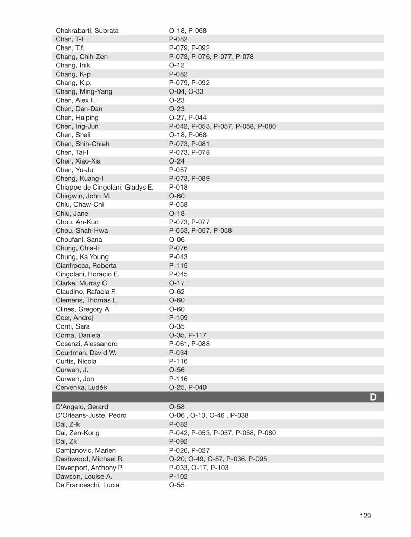
129
Chakrabarti, Subrata O-18, P-068Chan, T-f P-082Chan, T.f. P-079, P-092Chang, Chih-Zen P-073, P-076, P-077, P-078Chang, Inik O-12Chang, K-p P-082Chang, K.p. P-079, P-092Chang, Ming-Yang O-04, O-33Chen, Alex F. O-23Chen, Dan-Dan O-23Chen, Haiping O-27, P-044Chen, Ing-Jun P-042, P-053, P-057, P-058, P-080Chen, Shali O-18, P-068Chen, Shih-Chieh P-073, P-081Chen, Tai-I P-073, P-078Chen, Xiao-Xia O-24Chen, Yu-Ju P-057Cheng, Kuang-I P-073, P-089Chiappe de Cingolani, Gladys E. P-018Chirgwin, John M. O-60Chiu, Chaw-Chi P-058Chiu, Jane O-18Chou, An-Kuo P-073, P-077Chou, Shah-Hwa P-053, P-057, P-058Choufani, Sana O-06Chung, Chia-li P-076Chung, Ka Young P-043Cianfrocca, Roberta P-115Cingolani, Horacio E. P-045Clarke, Murray C. O-17Claudino, Rafaela F. O-62Clemens, Thomas L. O-60Clines, Gregory A. O-60Coer, Andrej P-109Conti, Sara O-35Corna, Daniela O-35, P-117Cosenzi, Alessandro P-061, P-088Courtman, David W. P-034Curtis, Nicola P-116Curwen, J. O-56Curwen, Jon P-116Cervenka, Ludek O-25, P-040
DD'Angelo, Gerard O-58D'Orléans-Juste, Pedro O-06 , O-13, O-46 , P-038Dai, Z-k P-082Dai, Zen-Kong P-042, P-053, P-057, P-058, P-080Dai, Zk P-092Damjanovic, Marlen P-026, P-027Dashwood, Michael R. O-20, O-49, O-57, P-036, P-095Davenport, Anthony P. P-033, O-17, P-103Dawson, Louise A. P-102De Franceschi, Lucia O-55
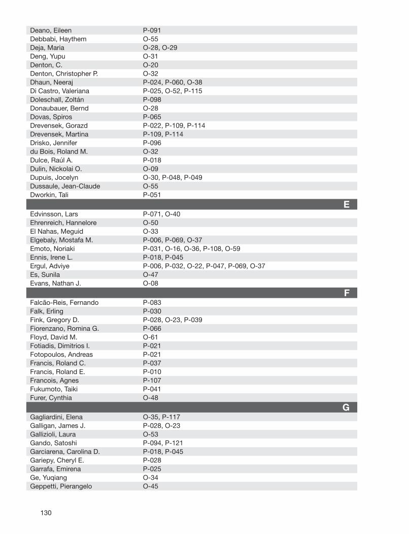
130
Deano, Eileen P-091Debbabi, Haythem O-55Deja, Maria O-28, O-29Deng, Yupu O-31Denton, C. O-20Denton, Christopher P. O-32Dhaun, Neeraj P-024, P-060, O-38Di Castro, Valeriana P-025, O-52, P-115Doleschall, Zoltán P-098Donaubauer, Bernd O-28Dovas, Spiros P-065Drevensek, Gorazd P-022, P-109, P-114Drevensek, Martina P-109, P-114Drisko, Jennifer P-096du Bois, Roland M. O-32Dulce, Raúl A. P-018Dulin, Nickolai O. O-09Dupuis, Jocelyn O-30, P-048, P-049Dussaule, Jean-Claude O-55Dworkin, Tali P-051
EEdvinsson, Lars P-071, O-40Ehrenreich, Hannelore O-50El Nahas, Meguid O-33Elgebaly, Mostafa M. P-006, P-069, O-37Emoto, Noriaki P-031, O-16, O-36, P-108, O-59Ennis, Irene L. P-018, P-045Ergul, Adviye P-006, P-032, O-22, P-047, P-069, O-37Es, Sunila O-47Evans, Nathan J. O-08
FFalcão-Reis, Fernando P-083Falk, Erling P-030Fink, Gregory D. P-028, O-23, P-039Fiorenzano, Romina G. P-066Floyd, David M. O-61Fotiadis, Dimitrios I. P-021Fotopoulos, Andreas P-021Francis, Roland C. P-037Francis, Roland E. P-010Francois, Agnes P-107Fukumoto, Taiki P-041Furer, Cynthia O-48
GGagliardini, Elena O-35, P-117Galligan, James J. P-028, O-23Gallizioli, Laura O-53Gando, Satoshi P-094, P-121Garciarena, Carolina D. P-018, P-045Gariepy, Cheryl E. P-028Garrafa, Emirena P-025Ge, Yuqiang O-34Geppetti, Pierangelo O-45

131
Germain, Stéphane O-55Ghanem, Lobna P-105Giacchetti, Gilberta O-53Giachini, Fernanda P-032Giachini, Fernanda R. P-047Giannopoulou, Myrto P-065Gilotta, Irene P-061, P-088Giovinazzo, Michele P-051, P-056Girot, Robert O-55Goddard, Jane P-024, P-060, O-38Godes, Michael P-003, P-008, P-012, P-017, O-14, P-063Goettsch, Winfried P-004, P-029, P-084Gomazcov, Oleg P-055Gorczynski, Richard J. P-111, P-112, P-118Goto, Katsutoshi P-094, P-121Gourgoulianis, Konstantinos P-065Gourineni, Nandu P-039Greene, Scott P-118Grigoriadis, Anita O-49Groselj, Blaz P-022Growcott, J. O-56Growcott, Jim P-116Guise, Theresa A. O-60Gulati, Anil P-091, P-093, O-47, P-100Gumz, Michelle O-01Gupta, Yogendra K. P-093
HHaas, Elvira P-030, O-48Hadoke, Patrick W. P-016Han, Robin N. O-31Harris, Alex K. P-006, P-069Hartman, J. Craig P-111, P-112, P-118Hassan, Mootaz P-105Hatzoglou, Chryssi P-065Haylor, John L. O-33Hazem, Ahmed P-105Hecker, Markus P-004Heiden, Susi P-003, P-008, P-012, P-017, P-063Herden, Uta O-48Herzfeld, Sophia P-011Heyman, Samuel N P-062Hickinson, Mark P-116Hilgers, Rob H. P-019Hirose, Takuo P-090Hocher, B. P-013Hocher, Berthold P-003, P-008, P-011, P-012, P-017, O-14, O-29, P-063Hoehne, Claudia P-010Hoffman, Aaron O-15, P-064Hoffman, Jeff P-096Hofmann-Lehmann, Regina P-030Hogarth, Kyle O-09Hong, Yue P-101Hoosein, Moinuddin M. P-095

132
Howng, Shen-Long P-077, P-082Howng, Sheng-Long P-076, P-078Howng, ShenLong P-079, P-092Hsu, Jen-Hao P-089Hsu, Jong-Hau P-042Huang, Chun-Jung P-089Huang, Shin-Yin P-081Hughes, G. O-56Husková, Zuzana P-040Hutchinson, Jim R. O-22Hutchinson, Jimmie P-069Hwang, Yen-Fen P-078Höhne, Claudia P-037
IImai, Yutaka P-090Insel, Paul A. P-118Iribarne, María O-42Isherwood, Beverley P-116Ito, Osamu P-090Ito, Sadayoshi P-104Iwasa, Naoko P-031, O-16, O-36, P-108, O-59
JJacques, Danielle O-06, O-07Jeng, Arco P-073, P-081Jeng, Arco Y P-042, P-053, P-057, P-058Jensen, Jens O-26Jesmin, Subrina P-094, P-121Joana, Saraiva P-083Jocelyn, Dupuis P-052Jochum, Wolfram P-030Johnston, Neil P-060, O-38Jules, Farah O-07Jullien, Nicolas P-107Jurka, Antra P-067
KKa, Yuki O-02Kaisers, Udo O-28, O-29Kaisers, Udo X. P-010, P-037Kalk, Philipp P-003, P-008, P-011, P-012, P-013, P-017, O-14, O-29, P-063Kanazawa, Masayuki P-090Kanematsu, Miwa O-02Karram, Tony O-15, P-064Kassell, Neal P-077, P-078Kassner, Nadine P-009Kekesi, Violetta P-046Keltai, Katalin P-098Kennedy, Laura O-57Kennedy, Laura E. O-32Keshta, Sobhi P-105Khamaisi, Mogher P-062Khatib, Samer P-064Khodorova, Alla P-087, O-44Kirby, Helen R. P-103
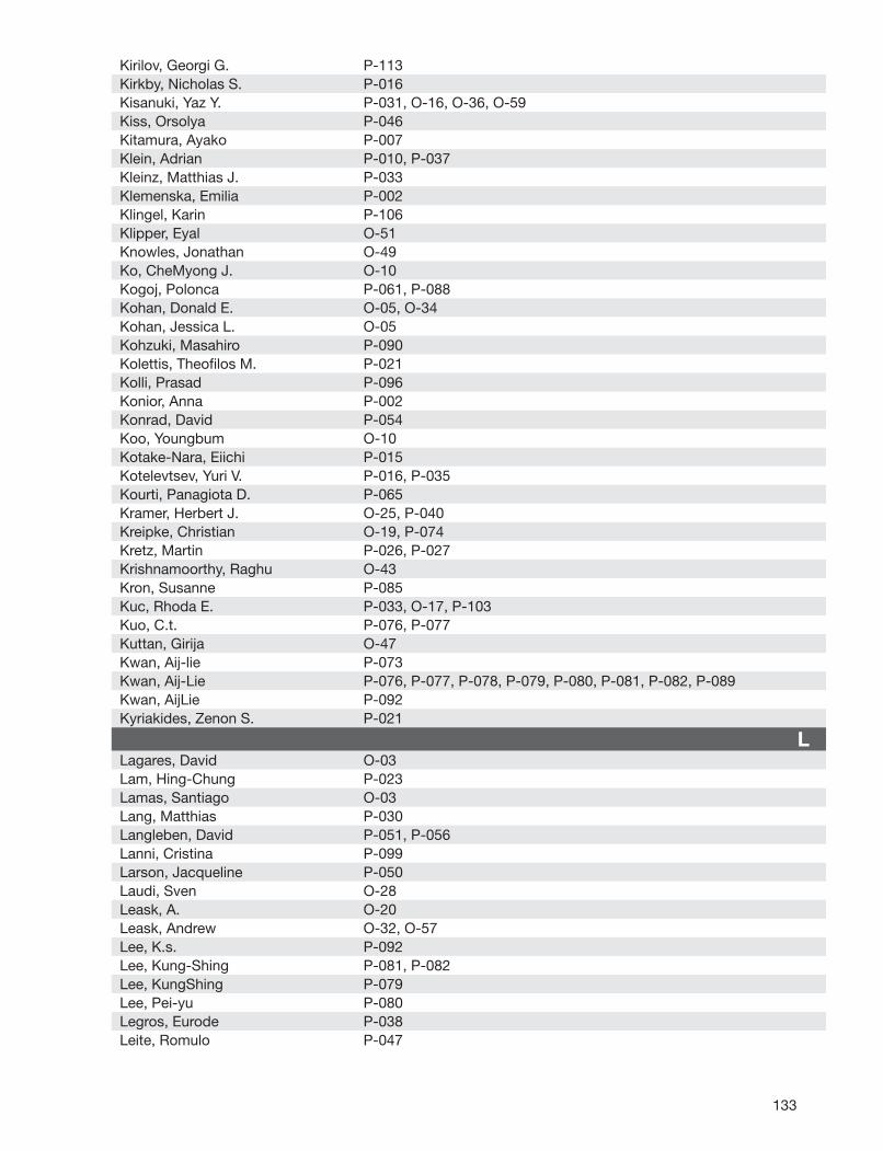
133
Kirilov, Georgi G. P-113Kirkby, Nicholas S. P-016Kisanuki, Yaz Y. P-031, O-16, O-36, O-59Kiss, Orsolya P-046Kitamura, Ayako P-007Klein, Adrian P-010, P-037Kleinz, Matthias J. P-033Klemenska, Emilia P-002Klingel, Karin P-106Klipper, Eyal O-51Knowles, Jonathan O-49Ko, CheMyong J. O-10Kogoj, Polonca P-061, P-088Kohan, Donald E. O-05, O-34Kohan, Jessica L. O-05Kohzuki, Masahiro P-090Kolettis, Theofi los M. P-021Kolli, Prasad P-096Konior, Anna P-002Konrad, David P-054Koo, Youngbum O-10Kotake-Nara, Eiichi P-015Kotelevtsev, Yuri V. P-016, P-035Kourti, Panagiota D. P-065Kramer, Herbert J. O-25, P-040Kreipke, Christian O-19, P-074Kretz, Martin P-026, P-027Krishnamoorthy, Raghu O-43Kron, Susanne P-085Kuc, Rhoda E. P-033, O-17, P-103Kuo, C.t. P-076, P-077Kuttan, Girija O-47Kwan, Aij-lie P-073Kwan, Aij-Lie P-076, P-077, P-078, P-079, P-080, P-081, P-082, P-089Kwan, AijLie P-092Kyriakides, Zenon S. P-021
LLagares, David O-03Lam, Hing-Chung P-023Lamas, Santiago O-03Lang, Matthias P-030Langleben, David P-051, P-056Lanni, Cristina P-099Larson, Jacqueline P-050Laudi, Sven O-28Leask, A. O-20Leask, Andrew O-32, O-57Lee, K.s. P-092Lee, Kung-Shing P-081, P-082Lee, KungShing P-079Lee, Pei-yu P-080Legros, Eurode P-038Leite, Romulo P-047
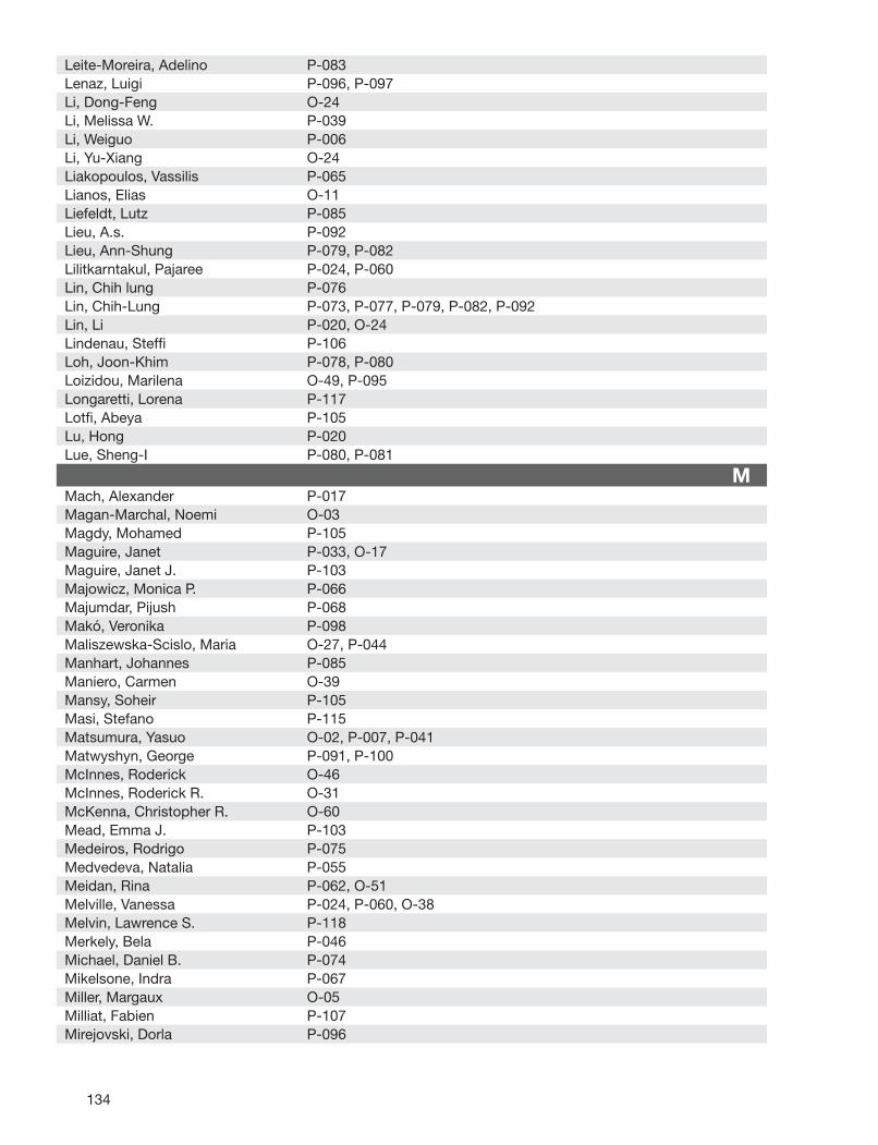
134
Leite-Moreira, Adelino P-083Lenaz, Luigi P-096, P-097Li, Dong-Feng O-24Li, Melissa W. P-039Li, Weiguo P-006Li, Yu-Xiang O-24Liakopoulos, Vassilis P-065Lianos, Elias O-11Liefeldt, Lutz P-085Lieu, A.s. P-092Lieu, Ann-Shung P-079, P-082Lilitkarntakul, Pajaree P-024, P-060Lin, Chih lung P-076Lin, Chih-Lung P-073, P-077, P-079, P-082, P-092Lin, Li P-020, O-24Lindenau, Steffi P-106Loh, Joon-Khim P-078, P-080Loizidou, Marilena O-49, P-095Longaretti, Lorena P-117Lotfi , Abeya P-105Lu, Hong P-020Lue, Sheng-I P-080, P-081
MMach, Alexander P-017Magan-Marchal, Noemi O-03Magdy, Mohamed P-105Maguire, Janet P-033, O-17Maguire, Janet J. P-103Majowicz, Monica P. P-066Majumdar, Pijush P-068Makó, Veronika P-098Maliszewska-Scislo, Maria O-27, P-044Manhart, Johannes P-085Maniero, Carmen O-39Mansy, Soheir P-105Masi, Stefano P-115Matsumura, Yasuo O-02, P-007, P-041Matwyshyn, George P-091, P-100McInnes, Roderick O-46McInnes, Roderick R. O-31McKenna, Christopher R. O-60Mead, Emma J. P-103Medeiros, Rodrigo P-075Medvedeva, Natalia P-055Meidan, Rina P-062, O-51Melville, Vanessa P-024, P-060, O-38Melvin, Lawrence S. P-118Merkely, Bela P-046Michael, Daniel B. P-074Mikelsone, Indra P-067Miller, Margaux O-05Milliat, Fabien P-107Mirejovski, Dorla P-096
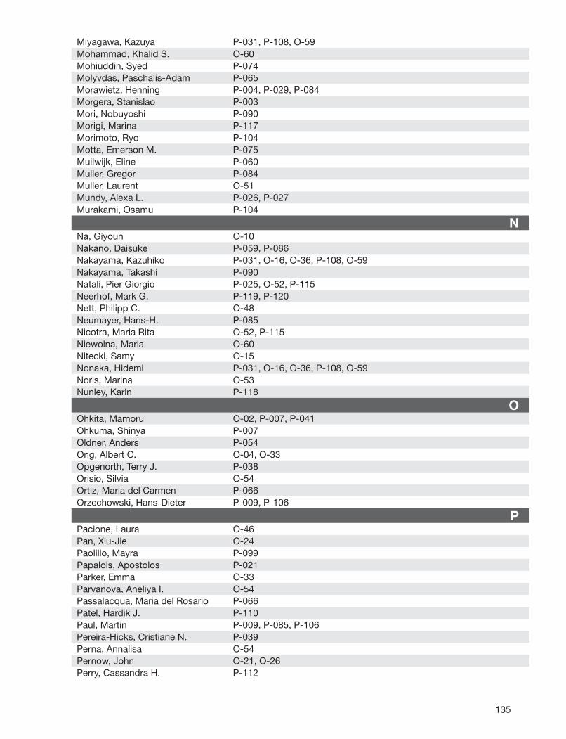
135
Miyagawa, Kazuya P-031, P-108, O-59Mohammad, Khalid S. O-60Mohiuddin, Syed P-074Molyvdas, Paschalis-Adam P-065Morawietz, Henning P-004, P-029, P-084Morgera, Stanislao P-003Mori, Nobuyoshi P-090Morigi, Marina P-117Morimoto, Ryo P-104Motta, Emerson M. P-075Muilwijk, Eline P-060Muller, Gregor P-084Muller, Laurent O-51Mundy, Alexa L. P-026, P-027Murakami, Osamu P-104
NNa, Giyoun O-10Nakano, Daisuke P-059, P-086Nakayama, Kazuhiko P-031, O-16, O-36, P-108, O-59Nakayama, Takashi P-090Natali, Pier Giorgio P-025, O-52, P-115Neerhof, Mark G. P-119, P-120Nett, Philipp C. O-48Neumayer, Hans-H. P-085Nicotra, Maria Rita O-52, P-115Niewolna, Maria O-60Nitecki, Samy O-15Nonaka, Hidemi P-031, O-16, O-36, P-108, O-59Noris, Marina O-53Nunley, Karin P-118
OOhkita, Mamoru O-02, P-007, P-041Ohkuma, Shinya P-007Oldner, Anders P-054Ong, Albert C. O-04, O-33Opgenorth, Terry J. P-038Orisio, Silvia O-54Ortiz, Maria del Carmen P-066Orzechowski, Hans-Dieter P-009, P-106
PPacione, Laura O-46Pan, Xiu-Jie O-24Paolillo, Mayra P-099Papalois, Apostolos P-021Parker, Emma O-33Parvanova, Aneliya I. O-54Passalacqua, Maria del Rosario P-066Patel, Hardik J. P-110Paul, Martin P-009, P-085, P-106Pereira-Hicks, Cristiane N. P-039Perna, Annalisa O-54Pernow, John O-21, O-26Perry, Cassandra H. P-112
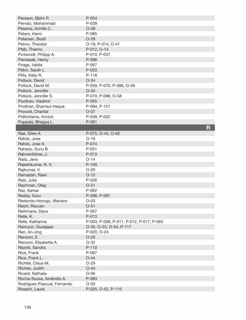
136
Persson, Björn P. P-054Pervaiz, Mohammad P-039Pessina, Achille C. O-39Peters, Harm P-085Petersen, Bodil O-29Petrov, Theodor O-19, P-074, O-41Pfab, Thiemo P-012, O-14Pickerodt, Philipp A. P-010, P-037Pieniasak, Henry P-096Pirags, Valdis P-067Pitkin, Sarah L. P-033Pitts, Kelly R. P-118Pollock, David O-34Pollock, David M. P-059, P-070, P-086, O-58Pollock, Jennifer O-34Pollock, Jennifer S. P-070, P-086, O-58Pozdnev, Vladimir P-055Prodhan, Shamsul Haque P-094, P-121Provost, Chantal O-07Préfontaine, Annick P-049, P-052Puppala, Bhagya L. P-091
RRae, Giles A. P-075, O-45, O-62Rafols, Jose O-19Rafols, Jose A. P-074Raharjo, Sunu B. P-031Rahnenführer, J. P-013Raila, Jens O-14Rajeshkumar, N. V. P-100Rajkumar, V. O-20Ramadan, Rawi O-15Rasi, Julia P-026Rayhman, Oleg O-51Raz, Itamar P-062Reddy, Guru P-096, P-097Redondo-Horcajo, Mariano O-03Reich, Reuven O-51Reihmane, Dace P-067Relle, K. P-013Relle, Katharina P-003, P-008, P-011, P-012, P-017, P-063Remuzzi, Giuseppe O-35, O-53, O-54, P-117Ren, An-Jing P-020, O-24Renzoni, E. O-20Renzoni, Elisabetta A. O-32Reznik, Sandra P-110Rice, Frank P-087Rice, Frank L. O-44Richter, Claus-M. O-29Richter, Judith O-44Rivard, Nathalie O-06Rocha-Sousa, Amândio A. P-083Rodriguez-Pascual, Fernando O-03Rosanò, Laura P-025, O-52, P-115
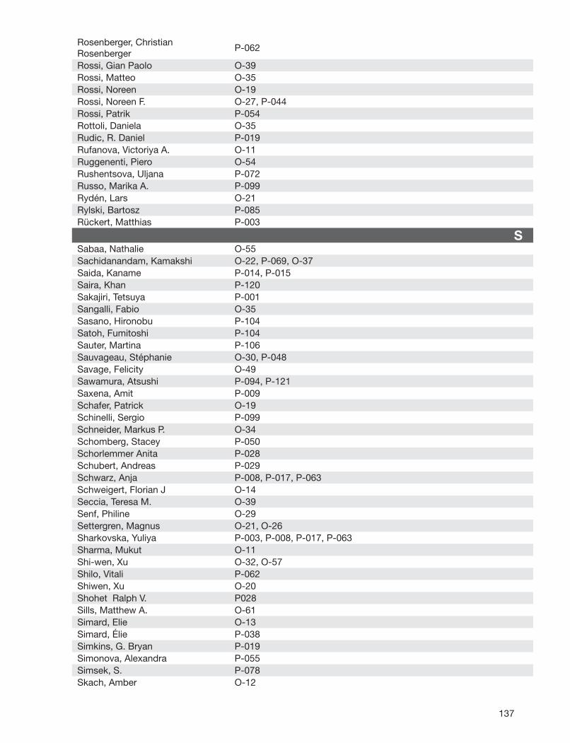
137
Rosenberger, Christian Rosenberger
P-062
Rossi, Gian Paolo O-39Rossi, Matteo O-35Rossi, Noreen O-19Rossi, Noreen F. O-27, P-044Rossi, Patrik P-054Rottoli, Daniela O-35Rudic, R. Daniel P-019Rufanova, Victoriya A. O-11Ruggenenti, Piero O-54Rushentsova, Uljana P-072Russo, Marika A. P-099Rydén, Lars O-21Rylski, Bartosz P-085Rückert, Matthias P-003
SSabaa, Nathalie O-55Sachidanandam, Kamakshi O-22, P-069, O-37Saida, Kaname P-014, P-015Saira, Khan P-120Sakajiri, Tetsuya P-001Sangalli, Fabio O-35Sasano, Hironobu P-104Satoh, Fumitoshi P-104Sauter, Martina P-106Sauvageau, Stéphanie O-30, P-048Savage, Felicity O-49Sawamura, Atsushi P-094, P-121Saxena, Amit P-009Schafer, Patrick O-19Schinelli, Sergio P-099Schneider, Markus P. O-34Schomberg, Stacey P-050Schorlemmer Anita P-028Schubert, Andreas P-029Schwarz, Anja P-008, P-017, P-063Schweigert, Florian J O-14Seccia, Teresa M. O-39Senf, Philine O-29Settergren, Magnus O-21, O-26Sharkovska, Yuliya P-003, P-008, P-017, P-063Sharma, Mukut O-11Shi-wen, Xu O-32, O-57Shilo, Vitali P-062Shiwen, Xu O-20Shohet Ralph V. P028Sills, Matthew A. O-61Simard, Elie O-13Simard, Élie P-038Simkins, G. Bryan P-019Simonova, Alexandra P-055Simsek, S. P-078Skach, Amber O-12
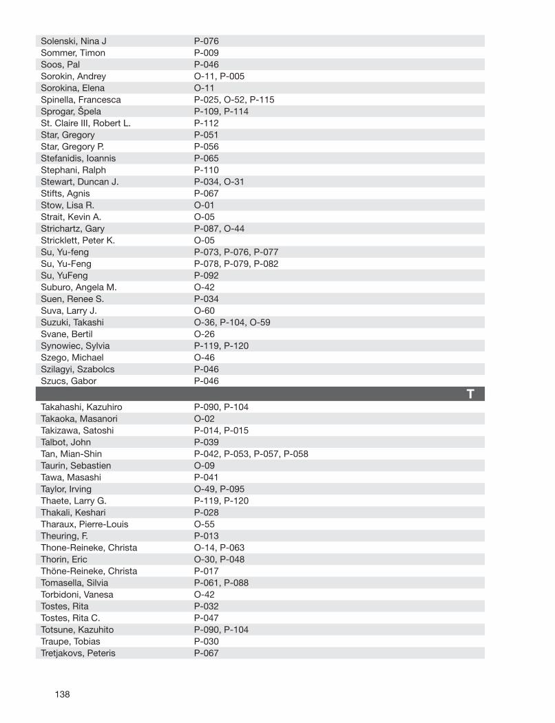
138
Solenski, Nina J P-076Sommer, Timon P-009Soos, Pal P-046Sorokin, Andrey O-11, P-005Sorokina, Elena O-11Spinella, Francesca P-025, O-52, P-115Sprogar, Špela P-109, P-114St. Claire III, Robert L. P-112Star, Gregory P-051Star, Gregory P. P-056Stefanidis, Ioannis P-065Stephani, Ralph P-110Stewart, Duncan J. P-034, O-31Stifts, Agnis P-067Stow, Lisa R. O-01Strait, Kevin A. O-05Strichartz, Gary P-087, O-44Stricklett, Peter K. O-05Su, Yu-feng P-073, P-076, P-077Su, Yu-Feng P-078, P-079, P-082Su, YuFeng P-092Suburo, Angela M. O-42Suen, Renee S. P-034Suva, Larry J. O-60Suzuki, Takashi O-36, P-104, O-59Svane, Bertil O-26Synowiec, Sylvia P-119, P-120Szego, Michael O-46Szilagyi, Szabolcs P-046Szucs, Gabor P-046
TTakahashi, Kazuhiro P-090, P-104Takaoka, Masanori O-02Takizawa, Satoshi P-014, P-015Talbot, John P-039Tan, Mian-Shin P-042, P-053, P-057, P-058Taurin, Sebastien O-09Tawa, Masashi P-041Taylor, Irving O-49, P-095Thaete, Larry G. P-119, P-120Thakali, Keshari P-028Tharaux, Pierre-Louis O-55Theuring, F. P-013Thone-Reineke, Christa O-14, P-063Thorin, Eric O-30, P-048Thöne-Reineke, Christa P-017Tomasella, Silvia P-061, P-088Torbidoni, Vanesa O-42Tostes, Rita P-032Tostes, Rita C. P-047Totsune, Kazuhito P-090, P-104Traupe, Tobias P-030Tretjakovs, Peteris P-067
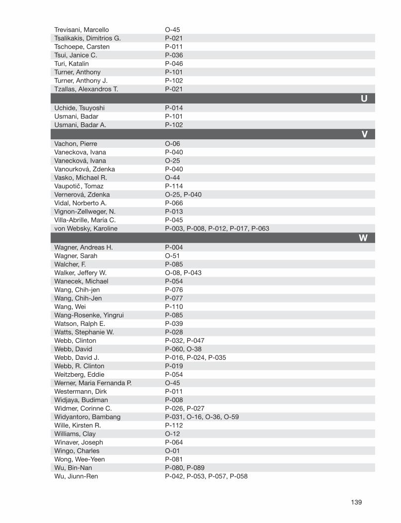
139
Trevisani, Marcello O-45Tsalikakis, Dimitrios G. P-021Tschoepe, Carsten P-011Tsui, Janice C. P-036Turi, Katalin P-046Turner, Anthony P-101Turner, Anthony J. P-102Tzallas, Alexandros T. P-021
UUchide, Tsuyoshi P-014Usmani, Badar P-101Usmani, Badar A. P-102
VVachon, Pierre O-06Vaneckova, Ivana P-040Vanecková, Ivana O-25Vanourková, Zdenka P-040Vasko, Michael R. O-44Vaupotic, Tomaz P-114Vernerová, Zdenka O-25, P-040Vidal, Norberto A. P-066Vignon-Zellweger, N. P-013Villa-Abrille, María C. P-045von Websky, Karoline P-003, P-008, P-012, P-017, P-063
WWagner, Andreas H. P-004Wagner, Sarah O-51Walcher, F. P-085Walker, Jeffery W. O-08, P-043Wanecek, Michael P-054Wang, Chih-jen P-076Wang, Chih-Jen P-077Wang, Wei P-110Wang-Rosenke, Yingrui P-085Watson, Ralph E. P-039Watts, Stephanie W. P-028Webb, Clinton P-032, P-047Webb, David P-060, O-38Webb, David J. P-016, P-024, P-035Webb, R. Clinton P-019Weitzberg, Eddie P-054Werner, Maria Fernanda P. O-45Westermann, Dirk P-011Widjaya, Budiman P-008Widmer, Corinne C. P-026, P-027Widyantoro, Bambang P-031, O-16, O-36, O-59Wille, Kirsten R. P-112Williams, Clay O-12Winaver, Joseph P-064Wingo, Charles O-01Wong, Wee-Yeen P-081Wu, Bin-Nan P-080, P-089Wu, Jiunn-Ren P-042, P-053, P-057, P-058
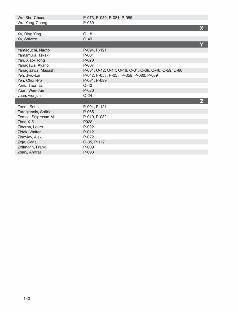
140
Wu, Shu-Chuan P-073, P-080, P-081, P-089Wu, Yang-Chang P-089
XXu, Bing Ying O-18Xu, Shiwen O-49
YYamaguchi, Naoto P-094, P-121Yamamura, Takaki P-001Yan, Xiao-Hong P-020Yanagawa, Ayano P-007Yanagisawa, Masashi P-031, O-12, O-14, O-16, O-31, O-36, O-46, O-59, O-60Yeh, Jwu-Lai P-042, P-053, P-057, P-058, P-080, P-089Yen, Chun-Po P-081, P-089Yorio, Thomas O-43Yuan, Wen-Jun P-020yuan, wenjun O-24
ZZaedi, Sohel P-094, P-121Zarogiannis, Sotirios P-065Zemse, Saiprasad M. P-019, P-032Zhao X-S. P028Ziberna, Lovro P-022Zidek, Walter P-012Zinoviev, Alex P-072Zoja, Carla O-35, P-117Zollmann, Frank P-009Zsáry, András P-098