BEST CLINICAL PRACTICES AND CHALLENGES
92
BEST CLINICAL PRACTICES AND CHALLENGES ATYPICAL WOUNDS
-
Upload
khangminh22 -
Category
Documents
-
view
0 -
download
0
Transcript of BEST CLINICAL PRACTICES AND CHALLENGES

BEST CLINICAL PRACTICES AND CHALLENGES
ATYPICAL WOUNDS
AT
YP I CA L W OUND
S

S 2 J O U R N A L O F WO U N D C A R E VO L 2 8 N O 6 E W M A D O C U M E N T 2 0 1 9
© EWMA 2019
All rights reserved. No reproduction, transmission or copying of this publication is allowed without written permission. No part of this publication may be reproduced, stored in a retrieval system, or transmitted in any form or by any means, mechanical, electronic, photocopying, recording, or otherwise, without the prior written permission of the European Wound Management Association (EWMA) or in accordance with the relevant copyright legislation.
Although the editor, MA Healthcare Ltd. and EWMA have taken great care to ensure accuracy, neither MA Healthcare Ltd. nor EWMA will be liable for any errors of omission or inaccuracies in this publication.
Published on behalf of EWMA by MA Healthcare Ltd.Editor: Rachel Webb Managing Director: Anthony Kerr Published by: MA Healthcare Ltd, St Jude’s Church, Dulwich Road, London, SE24 0PB, UKTel: +44 (0)20 7738 5454 Email: [email protected] Web: www.markallengroup.com
Kirsi Isoherranen (Editor), MD, PhD, Helsinki University Central Hospital and Helsinki University, Wound Healing Centre and Dermatology Clinic, Helsinki, Finland ([email protected])
Julie Jordan O’Brien (Co-editor), RNP, MSc Nursing, Advanced Nurse Practitioner Plastic Surgery, Beaumont Hospital, Dublin, Ireland ([email protected])
Judith Barker, Nurse Practitioner – Wound Management, Rehabilitation, Aged and Community Care., Adjunct Associate Professor, University of Canberra, Canberra, Australia ([email protected])
Joachim Dissemond (JD), Professor, MD, University Hospital of Essen, Department of Dermatology, Venerology and Allergology, Hufelandstraße 55, Essen, Germany ([email protected])
Jürg Hafner, Professor, MD, Department of Dermatology, University Hospital of Zurich, Gloriastrasse 31, Zurich, Switzerland ([email protected])
Gregor B. E. Jemec (GJ), Professor, MD, Department of Dermatology, Zealand University Hospital, Roskilde, Denmark ([email protected])
Jivko Kamarachev (JK), MD, PHD, Department of Dermatology, University Hospital of Zurich, Gloriastrasse 31, Zurich, Switzerland ([email protected])
Severin Läuchli, MD, PHD, Department of Dermatology, University Hospital of Zurich, Gloriastrasse 31, Zurich, Switzerland ([email protected])
Elena Conde Montero, MD, PHD, Hospital Universitario Infanta Leonor, Dept. of Dermatology, Madrid, Spain ([email protected])
Stephan Nobbe (SN), MD, Department of Dermatology, University Hospital of Zurich, Gloriastrasse 31, Zurich, Switzerland Department of Dermatology, Cantonal Hospital of Frauenfeld, Switzerland ([email protected])
Cord Sunderkötter (CS), Professor, MD and Chair, Department of Dermatology and Venerology, University and University Hospital of Halle, Ernst-Grube-Strasse 40, Halle, Germany ([email protected])
Mar Llamas Velasco, MD, PhD, Department of Dermatology, Hospital Universitario De La Princesa, Madrid, Spain. ([email protected])
The document is supported by an unrestricted educational grant from PolyMem and Essity.
Corresponding author:
Editor: Kirsi Isoherranen, [email protected]
Wounds Australia: www.woundsaustralia.com.au
Editorial support and coordination: EWMA Secretariat, Jan Kristensen: [email protected]
This article should be referenced as: Isoherranen K, Jordan O’Brien J, Barker J et al. EWMA document; Atypical wounds. Best clinical practice and challenges

J O U R N A L O F WO U N D C A R E VO L 2 8 N O 6 E W M A D O C U M E N T 2 0 1 9 S 3
Contents
1. Introduction 4
2. Pyoderma gangrenosum 6
3. Vasculitides as causes of wounds 11
4. Occlusive vasculopathy 21
5. Martorell HYTILU and calciphylaxis: skin infarction and acral gangrene from ischaemic
arteriolosclerosis 26
6. Hidradenitis suppurativa 36
7. Malignant wounds 39
8. Artefactal ulcers 45
9. Ecthyma and ecthyma gangrenosum 47
10. Other types of atypical wounds 51
11. Histology of atypical wounds 55
12. Practical aspects of diagnosing and treating atypical wounds 61
13. Topical treatment for atypical wounds 66
14. The patient perspective 70
15. Health economy and organisation 74
16. Conclusions and future perspectives 77
17. References 79
18. Glossary 89
S 4 J O U R N A L O F WO U N D C A R E VO L 2 8 N O 6 E W M A D O C U M E N T 2 0 1 9
1. Introduction
Atypical wounds comprise approximately
20% of all chronic wounds.3–5 With an aging
population and increase in comorbidities
these numbers are expected to rise. Atypical wounds
are considered as wounds that do not fall into a
typical wound category, i.e. venous, arterial, mixed,
pressure ulcer (PU) or diabetic foot ulcers (DFU).6–8
They are a broad spectrum of conditions or diseases
caused by inflammation, infection, malignancy,
chronic illnesses or genetic disorders.
An atypical wound can be suspected if the wound
has an abnormal presentation or location, pain
out of proportion of the size of the wound and
does not heal within four to 12 weeks with a good
treatment plan.7–9 Unfortunately, the diagnostic
delay can be considerable which leads to higher
mortality.10–13
Therefore, it is important that every health
professional treating these wounds is familiar
with this entity or at least has the knowledge to
suspect an atypical wound and when to refer the
patient to an expert. A multidisciplinary team
(MDT) of professionals, optimally consisting of
a dermatologist, vascular and plastic surgeon,
rheumatologist, diabetologist, nephrologist,
infectious disease specialist, psychiatrist,
tissue viability nurse, podiatrist, nephrologist,
psychologist, nutritionist, physiotherapist, podiatrist
and a social care worker, is needed to manage this
group of patients. However, it is mainly community
health professionals who manages these patients
on a day-to-day basis. Early diagnosis and referral
to dermatologists is important as they are experts
in diagnosing and treating these wounds.3,7,13
After exact diagnosis, a holistic assessment and
interdisciplinary plan of care is essential for cost-
effective management and to prevent recurrence.
From the patient perspective, atypical wounds can
be very painful and have prolonged healing times
which lead to impaired quality of life (QoL).14 Many
patients suffer daily both physically with odour,
exudate, pain, reduced mobility and psychologically
with negative emotions, loneliness and depression.15
Mortality rates are higher not only due to their
comorbidities but also lower socio-economic
circumstances leading to higher rates of suicide.16
Still, there is a dearth of literature reporting QoL
or health economy in this patient population.
In response to this lack of uniform data, the
European Wound Management Association
(EWMA) has established a working group to
gather the best available knowledge on atypical
wounds. This document is targeted at increasing
awareness of the clinical picture, diagnosis
and treatment of these wounds among health
professionals and to provide practical advice
on some of the challenges that typically arise,
e.g. delay in diagnosis for the inflammatory
and vasculopathy wounds (such as pyoderma
gangrenosum (PG), an inflammatory neutrophilic
disorder and cutaneous vasculitis). We hope
that a systematic approach will improve care
and QoL for this patient group. Lastly, it is also
hoped that this document will act as a catalyst
in the management of atypical wounds and
fill the void that currently exists in the clinical
decision making.
This document focuses on atypical wounds caused
by inflammation, malignancy and chronic illnesses.
Genetic disorders for example epidermolysis bullosa
(EB) will not be covered. We also want to highlight
that this is an educational and state of the art
document. The level of evidence is not given but it
is a literature review of available evidence.

J O U R N A L O F WO U N D C A R E VO L 2 8 N O 6 E W M A D O C U M E N T 2 0 1 9 S 5
The aim of the document is to:
• Present the diagnostic criteria, comorbidities and
diagnostic tools for wounds defined as atypical,
including practical hints for health professionals.
• Present the best available documented current
treatment options. High-quality evidence
is sparse, but there are retrospective and
observational studies as well as some randomised
prospective studies.
• Present some newer treatment options for
atypical wounds.
• Reduce the diagnostic delay with these wounds by
providing up to date evidence based literature on
atypical wounds and an algorithm to aid clinicians
in assessing these wounds in a systematic way
(see algorithm in Chapter 12 Practical aspects of
diagnosing and treating atypical wounds).

S 6 J O U R N A L O F WO U N D C A R E VO L 2 8 N O 6 E W M A D O C U M E N T 2 0 1 9
2. Pyoderma gangrenosum
PG is a neutrophilic dermatosis as a part
of a complex systemic auto inflammatory
process. The literature indicates an
incidence of 0.3–1.0/100,000 inhabitants. Thus,
the PG is one of the so-called orphan diseases.17
The gender distribution is described with about 3:1
more common in women. The disease can occur at
any age. However, patients are frequently affected
after the age of 50 years.18
AetiologyThe exact aetiology of PG is unknown. The
presence of abnormal neutrophils and T-cells lead
to immune dysregulation with increased levels of
inflammatory mediators. It has been reported that
healed sites of previous ulceration are refractory
to local relapse. Therefore it is discussed that
the T cell activity obligatory targets follicular
adnexal structures leading to the destruction of
pilosebaceous units.19 A genetic predisposition with
mutations in the PEST (proline, gluta-mate, serine
and threonine-rich) family of protein tyrosine
phosphatases (PTP) has been described.20
The current understanding of pathophysiology
is that PG is not only an isolated skin disease,
but a cutaneous manifestation of a generalised
inflammatory response. This is also clear when one
considers that there are many associations with
other inflammatory diseases. PG has also been
repeatedly described as part of some rare syndromes
often associated with hidradenitis suppurativa
(Table 1).21
ComorbiditiesIn addition to the long-known diseases associated
with PG such as chronic inflammatory bowel
diseases (IBD) and inflammatory rheumatological
diseases (IRD), associations with (haematological)
neoplasia have also been reported. In addition, there
is increasing evidence that PG may be associated
with aspects of the metabolic syndrome.17 Therefore
it is not surprising that there is an estimated 3-fold
increase in the mortality rate amongst patients with
PG12 compared with the normal population.
Methods of diagnosis in patients with PG should
therefore be individually adapted and consider
various aspects of comorbidities, in particular
the potentially paraneoplastic aspects.22 An
interdisciplinary approach is therefore essential.
Clinical presentationThe first skin lesions of PG are erythematous
papules, nodules, or pustules, which mostly develop
into deep and very painful ulcerations within a
few days (Fig 1 and 2). Many patients describe
Table 1. Autoinflammatoric syndromes which are related to pyoderma gangrenosum (PG)
PAPA syndrome. Acronym for PG, pyogenic arthritis, and acne
PASH syndrome. Acronym for PG, pyogenic arthritis, and hidradenitis suppurativa
PA-PASH syndrome. Acronym for PG, pyogenic arthritis, hidradenitis suppurativa, and acne

J O U R N A L O F WO U N D C A R E VO L 2 8 N O 6 E W M A D O C U M E N T 2 0 1 9 S 7
the appearance of a PG after a minimal trauma.
This is referred as a pathergy phenomenon. In
the early inflammatory phase, the livid colour of
the surrounding erythema is very typical.23 After
the occurrence of ulcerations, the wound edges
are then usually dark-red to livid in colour and
with associated undermining. Some pustules can
also often be found in the periwound area. Here
it is important to distinguish these primarily
sterile pustules from pustules as part of a bacterial
superinfection, which is a frequent misdiagnosis.
A PG can occur on any part of the body. However,
about 70% of all PG manifest on the lower legs.24
Another predilection site is the skin around
a stoma.25 The resulting scars are described
as cribriform (Fig 3).
PG can occur also postoperatively at the site of
surgery, and typically it is first misdiagnosed as
a surgical site infection. Post-surgical pyoderma
gangrenosum (PSPG) has been reported most
commonly after breast surgery; followed by
cardiothoracic, abdominal and obstetric surgeries.26
Negative wound swabs, no response to antibiotic
treatment and worsening of the lesions following
debridement and surgery are important clues
for this potentially fatal condition.10,27 As with
non-surgical PG, the risk factors for PSPG include
previous history of PG, inflammatory bowel disease,
hematologic disorders, rheumatoid arthritis and a
first degree relative with PG.26
There are increasing scientific reports of visceral
manifestations of PG. This extra-cutaneous
Fig 1. A young female patient with an early presentation in our clinic with a sterile pustule (picture 1, and small insert)). A biopsy was performed. Picture by Joachim Dissemond.
Fig 2. The patient came back two days later with a growing, painful PG. Another superficial wound, which ulcerated spontaneously, can be seen above. Picture by Joachim Dissemond.
S 8 J O U R N A L O F WO U N D C A R E VO L 2 8 N O 6 E W M A D O C U M E N T 2 0 1 9
infiltration can potentially occur in all organ
systems. The most commonly reported visceral
manifestation in PG is in the lungs, followed by
liver, spleen, and bone. In common, these patients
have aseptic abscesses.28
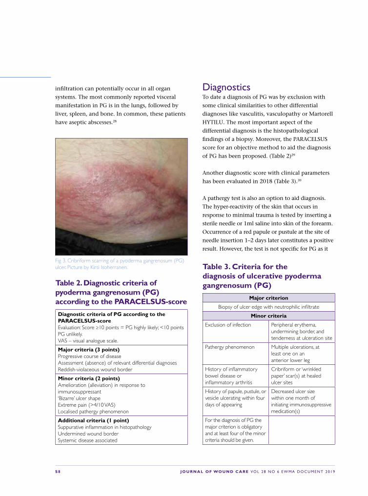
Fig 3. Cribriform scarring of a pyoderma gangrenosum (PG) ulcer. Picture by Kirsi Isoherranen.
Table 2. Diagnostic criteria of pyoderma gangrenosum (PG) according to the PARACELSUS-score
Diagnostic criteria of PG according to the PARACELSUS-scoreEvaluation: Score ≥10 points = PG highly likely; <10 points PG unlikely.VAS – visual analogue scale.
Major criteria (3 points) Progressive course of diseaseAssessment (absence) of relevant differential diagnosesReddish-violaceous wound border
Minor criteria (2 points) Amelioration (alleviation) in response to immunosuppressant‘Bizarre’ ulcer shapeExtreme pain (>4/10 VAS)Localised pathergy phenomenon
Additional criteria (1 point)Suppurative inflammation in histopathologyUndermined wound borderSystemic disease associated
Table 3. Criteria for the diagnosis of ulcerative pyoderma gangrenosum (PG)
Major criterion
Biopsy of ulcer edge with neutrophilic infiltrate
Minor criteria
Exclusion of infection Peripheral erythema, undermining border, and tenderness at ulceration site
Pathergy phenomenon Multiple ulcerations, at least one on an anterior lower leg
History of inflammatory bowel disease or inflammatory arthritis
Cribriform or ‘wrinkled paper’ scar(s) at healed ulcer sites
History of papule, pustule, or vesicle ulcerating within four days of appearing
Decreased ulcer size within one month of initiating immunosuppressive medication(s)
For the diagnosis of PG the major criterion is obligatory and at least four of the minor criteria should be given.
DiagnosticsTo date a diagnosis of PG was by exclusion with
some clinical similarities to other differential
diagnoses like vasculitis, vasculopathy or Martorell
HYTILU. The most important aspect of the
differential diagnosis is the histopathological
findings of a biopsy. Moreover, the PARACELSUS
score for an objective method to aid the diagnosis
of PG has been proposed. (Table 2)29
Another diagnostic score with clinical parameters
has been evaluated in 2018 (Table 3).30
A pathergy test is also an option to aid diagnosis.
The hyper-reactivity of the skin that occurs in
response to minimal trauma is tested by inserting a
sterile needle or 1ml saline into skin of the forearm.
Occurrence of a red papule or pustule at the site of
needle insertion 1–2 days later constitutes a positive
result. However, the test is not specific for PG as it

J O U R N A L O F WO U N D C A R E VO L 2 8 N O 6 E W M A D O C U M E N T 2 0 1 9 S 9
can also be positive in other neutrophilic diseases
such as Behçet’s disease.31
TherapyThe successful treatment of PG is based on various
immunomodulatory and/or immunosup-pressant
therapies. For the selection of the appropriate
individual strategy the severity as well as the acuity
has to be considered.
Local therapyFor topical therapy, highly potent glucocorticoids
such as preparations with clobetasol are usually
applied. Especially for long-term treatments
a 0.1% tacrolimus ointment is an effective
alternative. The ointments should be applied
as early as possible on new skin changes and
also on the wound bed.32 Intra- or periwound
triamcinolone can be used for an injection
therapy. The sole local therapy is usually
insufficient for pronounced courses of a PG, but
should be used as a supportive procedure.
Systemic therapyGlucocorticoids are currently the only approved
systemic treatment and the therapy of first choice.
In addition, systemic therapy with cyclosporine has
good scientific evidence. Therefore cyclosporine
can be used either alone or in combination with
glucocorticoids. In particular, in patients with IBD
or IRD, therapy with a TNF-α inhibitors are a good
alternative. In this constellation they are not in
an off-label use. Systemic antibiotics should only
be given if signs of systemic bacterial infection are
present. Moreover, it is very important to consider
the use of analgesia as part the multidisciplinary
therapeutic approach.
In addition, there are numerous alternative
systemic therapies, most of which have
been described in case series or retrospective
uncontrolled cohort studies (Table 4).
EvidenceIn a current review about systemic treatments for
PG 41 studies with 704 participants were found.33
Currently there are only two randomised controlled
trials (RCT) which include the STOP-GAP study
which showed in 121 patients that prednisolone
and cyclosporine were similar in their effectiveness,
with 15–20% complete healing after six weeks and
47% after six months.34 The other RCT reported in
30 patients that infliximab was superior to placebo
with a healing rate of 21% after six weeks.35
Wound therapyLocal wound treatment should also be performed
concomitantly in all patients. This should
be based on moist wound healing principles
and the patients presenting symptoms.36 It
is important that wound dressings can be
removed atraumatically. A mechanical or surgical
debridement has a potential risk of induction of a
pathergy phenomenon. However, these physical
treatments may be carried out during controlled
inflammation by systemic immunosuppression.37
Otherwise, atraumatic alternatives such as autolytic
hydrogels, preparations containing proteolytic
enzymes or biosurgery are possible.38
Since the patients are usually given systemic
immunosuppressive therapy, there is an increased
Table 4. Therapeutic alternatives published in recent years for systemic treatment in patients with pyoderma gangrenosum (PG)
• Traditional systemic agents• Azathioprine• Dapsone• Mycophenolate Mofetil• Prednisolon• Cyclosporin• Methotrexate
• Biologic agents• Adalimumab• Anakinra• Canakinumab• Etanercept• Ruxolitinib• Secukinumab• Ustekinumab
Intravenous Immunoglobulin (IVIG)

S 1 0 J O U R N A L O F WO U N D C A R E VO L 2 8 N O 6 E W M A D O C U M E N T 2 0 1 9
risk of infection. Therefore, antimicrobial treatments
should always be included as part of the standard
approach. Hyperbaric oxygen therapy (HBOT) may
be considered as an additional option as it can
support wound healing39. If there are no clinical
signs of inflammation in the course of successful
therapy, negative pressure woudn therapy (NPWT)
and/or split-thickness skin grafting (STSG) may be
considered40.
Currently, there are no scientific studies on the
importance of compression therapy in patients
with PG. However, clinical experience indicates
that all patients with lower extremity wounds and
edema benefit from compression therapy. This
compression therapy may be painful in patients
with inflammatory PG. Here treatment can then
be started with lower pressures of 20mmHg.41
PrognosisSpeed of healing and resolution of inflammation
have been shown to be good predictors for
healing of PG.42 The tendency to develop a new
PG can potentially persist for many years.
Conclusion and recommendationPG is a rarely diagnosed disease that results
in very painful and often hard-to-heal
wounds. Most patients require a combined
topical and systemic immunosuppressive or
immunomodulatory therapy in combination
with wound therapy.After exclusion of relevant
differential diagnoses a multidisciplinary
diagnostic and therapeutic concept is
definitely recommended.

J O U R N A L O F WO U N D C A R E VO L 2 8 N O 6 E W M A D O C U M E N T 2 0 1 9 S 1 1
3. Vasculitides as causes of wounds
Vasculitis is inflammation during which
the primary event is the destruction of the
walls of blood vessels.
It causes wounds by ischaemic necrosis due
to vessel damage and ensuing ulceration.
Subsequently these wounds often present
disturbed healing because of a deficit of intact or
functional vessels.
A common way to categorise vasculitides is by
size into large vessel vasculitis (affecting mainly
aorta, large and medium size arteries), medium
vessel vasculitis (affecting mainly medium and
small arteries) and small vessel vasculitis (affecting
mainly small arteries, arterioles, capillaries, venules).
The skin harbours small blood vessels, albeit of
different sizes (small arteries, arterioles, capillaries,
postcapillary venules, and small veins). The arteries
and veins in the panniculus and its septae are
considered medium vessels in the Chapel Hill
Consensus Conference (CHCC) system.43
Vasculitis of small vessels often and rapidly leads
to secondary thrombotic occlusion of vessels.
Therefore, it is not always easy to differentiate
vasculopathy from small vessel vasculitis in
single biopsies.
Vasculopathy is an occlusive, initially non-
inflammatory disease affecting small vessels in
the skin, caused either by a systemic or local
vascular coagulopathy, by embolisation, by platelet
plugging or by cold-related gelling of molecules,
such as cryoglobulins.
There are entities that may include both
vasculitis and vasculopathy, sometimes occurring
simultaneously (eg in cryoglobulinemic vasculitis,
vasculitis in monoclonal gammopathy).44–46
In a recently published interdisciplinary
consensus on nomenclature of cutaneous
vasculitis,47 vasculitis in the skin has been
distinguished in i) a cutaneous component of a
systemic vasculitis (eg cutaneous manifestations
of PAN), ii) a skin-limited or skin-dominant
expression or variant of a systemic vasculitis (eg
cutaneous PAN or cutaneous arteritis), or iii) a
single organ vasculitis (SOV) of the skin that
differs with regard to clinical, laboratory, and
pathological features from recognised systemic
vasculitides (eg nodular vasculitis; Table 5)).
This chapter will focus on those vasculitides that
cause ulcers or chronic wounds:
Medium vessel vasculitides
• Polyarteritis nodosa cutanea (cutaneous arteritis)
Small vessel vasculitides
• Immune complex vasculitides (including
cryoglobulinemic vasculitis)
• Rheumatoid vasculitis and vasculitides in
lupus erythematosus (LE) or other collagenous
autoimmune diseases
• ANCA associated vasculitides
S 1 2 J O U R N A L O F WO U N D C A R E VO L 2 8 N O 6 E W M A D O C U M E N T 2 0 1 9
• Nodular vasculitis.
The aetiology differs markedly between these
vasculitides as does therapy.
Medium vessel vasculitidesPolyarteriitis nodosa cutanea (cutaneous
arteritis) is a chronic medium vessel arteritis
which affects small arteries and arterioles in
the panniculus extending to arterioles at the
dermosubcutaneous junction,48 with or without
arteritis in adjacent skeletal muscle and peripheral
nerves (mononeuritis multiplex). It does not
directly involve venous vessels.
AetiologyThe frequent localisation on the lower legs
or rarely at other sites of chronic oedema,
indicate a contribution of the humoral immune
response, coinciding with slowed clearing
of so far unknown antigens or antibodies.
Supposedly, there is a reaction to an infection
such as hepatitis B, hepatitis C or streptococci or
alternatively a drug. A recently described variant
is associated with mutation of the CECR1-gene
and deficiency of adenosine deaminase 2 (ADA2).
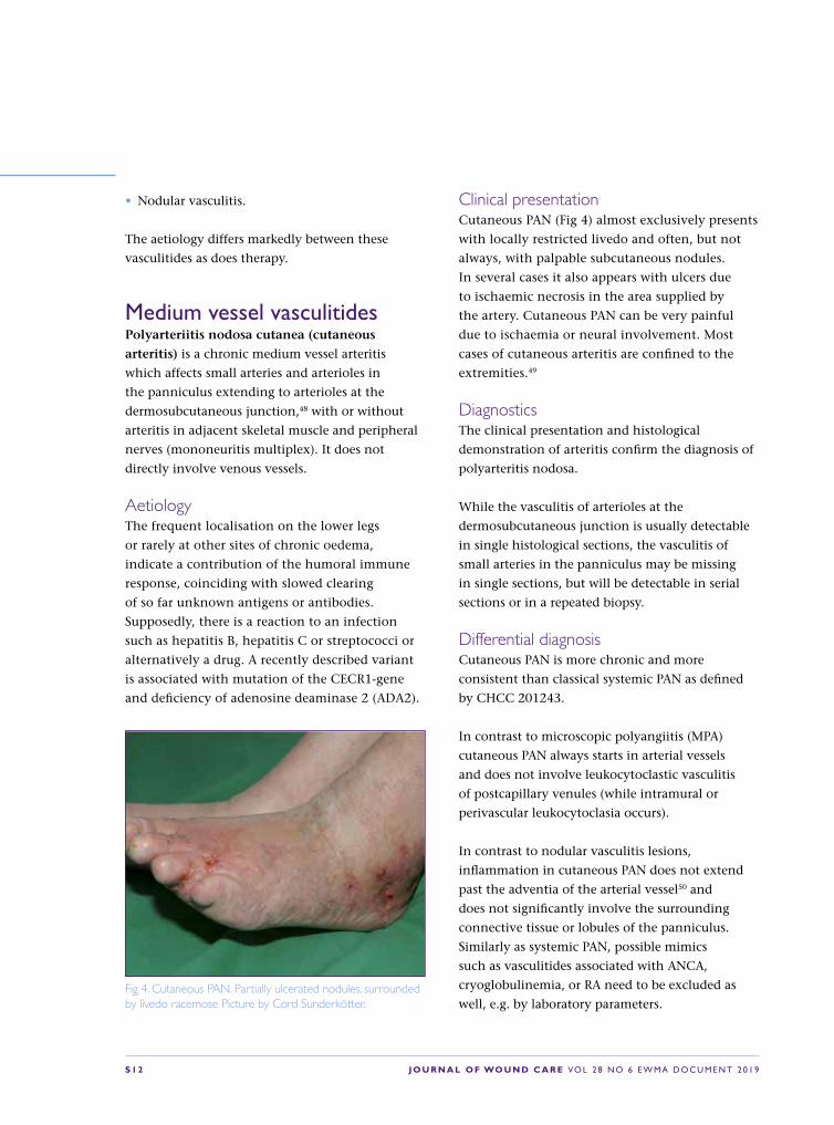
Clinical presentationCutaneous PAN (Fig 4) almost exclusively presents
with locally restricted livedo and often, but not
always, with palpable subcutaneous nodules.
In several cases it also appears with ulcers due
to ischaemic necrosis in the area supplied by
the artery. Cutaneous PAN can be very painful
due to ischaemia or neural involvement. Most
cases of cutaneous arteritis are confined to the
extremities.49
Diagnostics The clinical presentation and histological
demonstration of arteritis confirm the diagnosis of
polyarteritis nodosa.
While the vasculitis of arterioles at the
dermosubcutaneous junction is usually detectable
in single histological sections, the vasculitis of
small arteries in the panniculus may be missing
in single sections, but will be detectable in serial
sections or in a repeated biopsy.
Differential diagnosisCutaneous PAN is more chronic and more
consistent than classical systemic PAN as defined
by CHCC 201243.
In contrast to microscopic polyangiitis (MPA)
cutaneous PAN always starts in arterial vessels
and does not involve leukocytoclastic vasculitis
of postcapillary venules (while intramural or
perivascular leukocytoclasia occurs).
In contrast to nodular vasculitis lesions,
inflammation in cutaneous PAN does not extend
past the adventia of the arterial vessel50 and
does not significantly involve the surrounding
connective tissue or lobules of the panniculus.
Similarly as systemic PAN, possible mimics
such as vasculitides associated with ANCA,
cryoglobulinemia, or RA need to be excluded as
well, e.g. by laboratory parameters.Fig 4. Cutaneous PAN. Partially ulcerated nodules, surrounded by livedo racemose Picture by Cord Sunderkötter.

J O U R N A L O F WO U N D C A R E VO L 2 8 N O 6 E W M A D O C U M E N T 2 0 1 9 S 1 3
TherapyAny potentially eliciting drug should be
discontinued (eg minocycline). In case of
hepatitis or strep throat anti-infective therapy
is warranted. There are no controlled trials, so
treatment is based on experience, case series or
reviews.51 Management of pain by conventional
pain medication is important. Compression
therapy should also be performed and seems to
be well tolerated. Local wound therapy should be
performed as described above.
There is no strong evidence for drug therapy,
but dapsone often is effective. If not, colchicine
or methotrexate should be tried. Due to overlap
with coagulopathic events low molecular weight
heparin can be tried in antithrombotic dosages as
used for thrombosis. In severe cases with enlarging
ulcers, systemic glucocorticoids (0.4–0.5mg/
kg b.w.) may be administered, followed by
initiation of steroid sparing methotrexate (initially
25–30mg/week, then 10–15mg/week). Successful
use of hydroxychloroquine, azathioprine,
pentoxiphylline, mycophenolate mofetile or
high-dose immunoglobulins has been reported in
individual cases.
Systemic or classical PAN This systemic vasculitis is much rarer than
originally thought. Aetiology is still an enigma, but
it has been associated with hepatitis B.
Small vessel vasculitides Small vessel vasculitides can be roughly divided
into: Immune-complex vasculitides (IgA and IgG/
IgM vasculitis, cryoglobulinemic vasculitis, serum
sickness) and ANCA-associated vasculitides (AAV).
Immune‐complex vasculitides (IgA and IgG/IgM
vasculitis, cryoglobulinemic vasculitis, serum
sickness) affects approximately 1–9/100 000
people worldwide.
Among the immune complex vasculitides, systemic
forms and variants that are limited to the skin are
both common.
AetiologyIn IgA vasculitis (IgAV) or Henoch Schoenlein
Purpura deposition of poorly O-galactosylated IgA1
at glomerular capillary walls and the mesangium
is one decisive feature. A number of review are
available on the topic.52–54
In some patients with systemic IgAV the serum
levels are increased and they may form complexes
with IgG and IgA antibodies. Other antigens may
possibly bind to small vessels, activate or interact
with endothelial cells and attract neutrophils,
which become activated. This interaction results in
vessel damage by respiratory burst, degranulation,
and release of cytotoxic products.55,56
Involvement of skin in cryoglobulinemia is due
to two major mechanisms of tissue damage: i)
leukocytoclastic immune complex vasculitis
(type II or type III cryoglobulins; primary
pathomechanisms unrelated to cold), and/or
ii) occlusion of cutaneous vessels by gelling or
precipitation of type I cryoglobulins (vasculopathy)
in cold-exposed skin areas (all small blood
vessels of the upper or deep dermis, as well as the
capillaries of the fat lobule may be involved).57
Both features may occur simultaneously and
are involved at varying degrees,45 for example
deposition of immune complexes made of
cryoglobulins type II and III slow blood flow and
facilitate additional gelling of cryoglobulins.
Clinical presentationThe different forms of systemic immune complex
vasculitides, as defined by the CHCC 2012, all
present distinctive cutaneous features that are
also seen in the respective skin-limited variants:
round and sometimes retiform palpable purpura
with predilection for the lower limbs is a constant

S 1 4 J O U R N A L O F WO U N D C A R E VO L 2 8 N O 6 E W M A D O C U M E N T 2 0 1 9
feature of IgA- or IgM/IgG-immune complex
vasculitides (Fig 5), as is the inducibility of lesions
by vasodilatory stimuli during acute phases.
Ulcerations, preceded by haemorrhagic vesicles,
may occur in severe forms.
Occlusive damage of vessels beyond postcapillary
venules and probably also simultaneous damage to
several postcapillary venules in one area can lead
to bullae, erosions or (ischaemic) ulcers. Extensive
involvement of vessels beyond postcapillary
venules, loss of preponderance and predilection of
lesions for lower legs is encountered in cases with
additional pathophysiological mechanisms, such
as in cryoglobulinemia, rheumatoid vasculitis, and
SLE accompanied by immune complex vasculitis.
In cryoglobulinemia, leukocytoclastic vasculitis is
most frequently seen in type II, less often in type
III-mixed cryoglobulinemia (Fig 6).
Vasculopathy (with gelling or cryo-precipitation)
with occlusion of vessels is more frequent in
type I monoclonal cryoglobulinemia (often high
titres of cryoglobulins) and manifests clinically
as necrosis in cold-exposed, acral areas (hands,
feet, lips, ears, and nose). Sometimes it is
accompanied by livedo due to partial obstruction
of blood flow.
Cutaneous IgM- or IgG immune complex vasculitis is
a term not used in the original CHCC 201247. It refers
to a leukocytoclastic vasculitis of mainly post-capillary
venules which in the skin is indistinguishable from
IgA vasculitis both histologically and clinically
(palpable purpura with predilection for legs,
elicitable by vasodilation). However, on (repeated)
immunofluorescence tests, it is not dominant for IgA,
but instead, especially in early or histamine-induced
lesions,58,59 it shows vascular deposits of IgM and/
or IgG. This category had only been described in
reviews, while in studies it has been subsumed in the
group of IgA-negative cutaneous immune complex
vasculitides, often referred to as leukocytoclastic
angiitis or hypersensitivity vasculitis.60,61 It probably
only rarely causes ulcers.
Diagnostics The characteristic clinical presentation and
detection of IgA or IgG or IgM around vessels
by immunofluorescence are usually sufficient
for diagnosis. Histological demonstration of
leukocytoclastic vasculitis mainly of postcapillary
venules may add certainty when required. The
Fig 5. Immune complex vasculitis on lower leg featuring palpable and retiform purpura and incipient erosions. Picture by Cord Sunderkötter.
Fig 6. Livedo and retiform purpura and ulcer in cryoglobulinemic vasculitis. Picture by Cord Sunderkötter.

J O U R N A L O F WO U N D C A R E VO L 2 8 N O 6 E W M A D O C U M E N T 2 0 1 9 S 1 5
demonstration of leukocytoclastic immune
complex vasculitis in a biopsy specimen requires
a selection of a lesion between 24–48 hours old
in an untreated patient. Immunofluorescence for
demonstration of immunoreactants in dermal
blood vessels requires a selection of a younger
lesion as degrading enzymes released by leukocytes
or other factors may alter the initial findings.
The ensuing diagnostic procedure is meant to
assess extension of vasculitis (renal) and possible
causes (see articles on vasculitis).
Differential diagnosisExclusion of other small vessel vasculitides.
TherapyThere are no controlled clinical trials on therapies
of most immune complex vasculitides with perhaps
the exception of cryoglobulinemic vasculitis,45,46 so
the suggested procedures or treatments are based on
case series, experience or reviews.51
A potentially eliciting drug should be discontinued
(eg NSAID, sulphonamides, penicillin, cephaclor,
diuretics, allopurinol, quinolones, hydralazine,
methotrexate and contraceptives). In cases of
systemic IgA-vasculitis, we recommend routine
tumour screening.
Compression therapy of lower legs is a partially
causative therapy as it diminishes deposition of
immune complexes. Antihistamines in regular doses
may be helpful and can be used for long periods.
It is often a self-limited disease, which does not
require further measures and no glucocorticoids.
However, especially with reference to chronic
wounds, one exception is important: glucocorticoids
are indicated whenever bullae or incipient necrosis
occurs, as they will promptly reduce vasculitis and
prevent ulcers, which are prone to heal slowly due
to destroyed blood vessels.51,62
Treatment in case of renal involvement is described
in articles on vasculitis.
Rheumatoid vasculitis and vasculitides in LE or
other collagenous autoimmune diseases
Rheumatoid vasculitis or vasculitides in LE are
allocated to the category ‘Vasculitis Associated
with Systemic Disease’ due to their involvement of
different vessels, even though they are related to
immune complex vasculitides.
They are prone to cause ulcers. Rheumatoid
vasculitis occurs in patients with a positive test
for rheumatoid factor, long standing disease
and often with erosive rheumatoid arthritis.
In the skin, it ranges from (often IgG/IgM-positive,
but also IgA positive) LCV of post-capillary
venules63 to arteritis at the dermo-subcutaneous
junction or in the panniculus.64 A more frequent
involvement of vessels larger than postcapillary
venules distinguishes it from genuine IgA or
IgG/IgM vasculitis. Therefore, it results in a
more varied clinical presentation, including
cutaneous ulcers, digital gangrene or nail fold
infarction. Involvement of the vasa nervorum
may be the cause for neuropathy. Rheumatoid
vasculitis affecting muscular arteries (MVV) is
distinguished from cutaneous or systemic PAN
by the additional involvement of postcapillary
venules as well as by the presence of rheumatoid
factor and arthritis.
A similarly heterogeneous presentation of vasculitides
may occur in LE (mostly cutaneous SVV, or MVV
often involving peripheral nerves)65, and more rarely
in dermatomyositis or systemic sclerosis. Cutaneous
vasculitis in LE may also present as immune complex
vasculitis, both of which are restricted primarily to
postcapillary venules.
Therapy of these vasculitides occurs as part of the
treatment of the underlying systemic disease.

S 1 6 J O U R N A L O F WO U N D C A R E VO L 2 8 N O 6 E W M A D O C U M E N T 2 0 1 9
ANCA-associated vasculitides (AAV)The ANCA-associated vasculitides (AAV) are
a multisystem autoimmune diseases, more
common in older people and in men.66 Although
rare, they will increase concomitant with the
increasing average age of the population. They
are a chronic disease needing long-term immuno-
suppressive therapy.66
AetiologyAVV is associated with ANCA specific for
myeloperoxidase (MPO-ANCA) or proteinase
3 (PR3-ANCA). Based on pathologic and
clinical features, ANCA-associated vasculitis is
subdivided into microscopic polyangiitis (MPA),
granulomatosis with polyangiitis (Wegener) (GPA),
and eosino-philic granulomatosis with polyangiitis
(Churg–Strauss) (EGPA (1)).
Presence of MPO-ANCA or PR3-ANCA is a defining
feature. There is evidence both from clinical
observations (rough correlation of ANCA titers
with response to therapy, the use of antibody
depleting therapies, animal models, and in vitro
experiments with human neutrophils (PMN))
that both autoantibodies are pathogenic or at
least one major pathophysiological factor. In vitro,
both PR3-ANCA and MPO-ANCA IgG activate
human neutrophils (PMN) to generate respiratory
burst, degranulation, to release factors to
activate alternative pathway of complement and
neutrophil extracellular traps (NETs).67
Clinical presentationThe cutaneous component of systemic AAV may
present with different morphology, but all of them
may result in ulceration and chronic wounds: a)
leukocytoclastic vasculitis of postcapillary venules,
sometimes extending into arterioles or small veins,
clinically manifesting as haemorrhagic papules or
macules, sometimes nodules and ulcers, and/or b)
vasculitis of small arteries or arterioles clinically
manifesting as inflammatory retiform purpura or
livedo with early lesions exhibiting prominent
erythema and sometimes ulcerating nodules68 and
sometimes as digital infarcts.
The simultaneous presence of haemorrhagic
papules as well as livedo or nodules is highly
suspicious of an ANCA-associated vasculitis and
not characteristic for immune complex vasculitis
such as IgA vasculitis (HSP). It should prompt
determination of ANCA in serum.
Diagnostics Prompt diagnosis and initiation of appropriate
immunosuppressive therapy is essential for
optimum patient and outcomes.
It is important for clinicians taking care of
wounds to suspect AAV when the heterogeneous
clinical picture is associated with deteriorating
condition of the patient and other symptoms
such as sinusitis and bloody discharge,
haemoptysis, proteinuria and haematuria
or history of asthma or allergic rhinitis.
Then PR3-ANCA or MPO-ANCA should be
determined by ELISA and associated with an
interdisciplinary procedure.
TherapyANCA-associated vasculitides are the only group of
vasculitides mentioned here, which have been the
subject of large clinical studies and therefore also
of evidence-based recommendations regarding
management. Since they are comparatively rare
causes for atypical wounds, a detailed description
of their therapy is out of scope of this chapter.
Systemic therapy should be performed by or
together with a specialist. If possible, enrolment
in clinical studies or registries should be chosen
or offered. Rituximab has become a good
alternative to the well-studied immunosuppressive
chemotherapy, usually consisting of an aggressive

J O U R N A L O F WO U N D C A R E VO L 2 8 N O 6 E W M A D O C U M E N T 2 0 1 9 S 1 7
induction therapy aiming at remission, followed
by maintenance therapy.66,69,70
Nodular vasculitis (erythema induratum of Bazin) Nodular vasculitis (erythema induratum of Bazin)
is a lobular panniculitis, often associated with
vasculitis of vessels in the panniculus. In presence
of tuberculosis (usually with hyperergic reaction)
it has been referred to as erythema induratum of
Bazin71. It is a single organ vasculitis limited to the
skin without systemic involvement.
AetiologyIt is probably a hyperergic (Typ IV) reaction to
infectious agents such as mycobacteria tuberculosis,
Hepatitis B/C, nocardia, or to rheumatoid arthritis,
SLE or inflammatory bowel disease. It appears also
dependent on certain constitutive traits, as it usually
occurs in women with lipoedema, acrocyanosis and
livedo reticularis.
Clinical presentationClinical features include tender erythematous
subcutaneous plaques and nodules on the posterior
aspects of calves, sometimes tender, mostly in
middle-aged women with lipoedema (column-like
lower legs), livedo reticularis or erythrocyanosis
surrounding follicular pores (Fig 7). In the course
of disease, most, but not all, nodules ulcerate.
Diagnostics Diagnosis is made on clinical suspicion and
ensuing histological confirmation.
There is always lobular panniculitis and often
vasculitis. Affected vessels in order of frequency
are: (1) small venules of fat lobules, (2) both
veins of connective tissue septae and venules of
fat lobules, (3) only veins of connective tissue
septae, (4) veins and arteries of connective tissue
septae and venules of fat lobules, (5) veins and
arteries of connective tissue septae. In some
cases, vasculitis could not be demonstrated
within serial sections throughout the specimen.71
Nodular vasculitis with lobular panniculitis can be
associated with coagulative and caseous necrosis,
and (extravascular) granulomatous inflammation.
According to the stage of disease, vasculitis is
associated with neutrophilic, granulomatous or
lymphocytic inflammation.50
Differential diagnosisThis lobular panniculitis distinguishes it from
cutaneous arteritis, and the primary localization
of vasculitis and panniculitis distinguishes it from
GPA and EGPA.
TherapyIn presence of tuberculosis tuberculostatic therapy
(combination of three drugs for nine months). In
other cases wound care, symptomatic treatment
with cool dressings, compression, potassium
jodatum, NSAID, systemic glucocorticoids,
clofazimine, or colchicine.
Fig 7. Nodular vasculitis (erythema induratum of Bazin). Picture by Kirsi Isoherranen.

S 1 8 J O U R N A L O F WO U N D C A R E VO L 2 8 N O 6 E W M A D O C U M E N T 2 0 1 9
Table 5. Systemic and cutaneous variants of medium-vessel and small vessel vasculitides with relevance for atypical wounds (adapted from47)
Systemic and cutaneous variants of medium-vessel and small vessel vasculitides with relevance for atypical wounds
CHCC2012 name Abbreviated CHCC 2012 definition
Cutaneous component of systemic vasculitis
Skin-limited or skin-dominant vasculitis
Cutaneous presentations
Medium-vessel vasculitis = vasculitis which predominantly affects medium arteries defined as the main visceral arteries and their branches
Polyarteritis nodosa
Necrotizing arteritis of medium or small arteries without glomerulonephritis and not associated with ANCA.
Acute arteritis of small arteries or arterioles, manifesting as digital arteritis, purpura, and/or skin nodules with ulcers.
Cutaneous arteritis (cutaneous PAN) Arteritis affecting small arteries in the panniculus extending to arterioles at the dermo-subcutaneous junction, but not to postcapillary venules.
Locally restricted livedo and often, but not always, palpable subcutaneous, often ulcerating nodules
Small vessel vasculitis = vasculitis predominantly affecting small vessels, defined as small intraparenchymal arteries, arterioles, capillaries, and (postcapillary) venules
ANCA-associated vasculitis (Microscopic polyangiitis, Granulomatosis with polyangiitis (Wegener’s), Eosinophilic granulomatosiswith polyangiitis (Churg-Strauss)
Necrotizing vasculitis affecting small vessels (i.e., capillaries, venules, arterioles, and small arteries), associated with ANCA.
Vasculitis of cutaneous postcapillary venules small veins, arterioles, and small arteries, associated with ANCA.
ANCA-associated, vasculitis limited to the skin
Palpable round and inflammatory retiform purpura, and/or b) livedo and/or c) haemorrhagic nodules and/or d) e.g. hyperplastic gingivitis (no vasculitis), especially in GPA
Immune complex vasculitides (systemic and skin-limited forms)
IgA vasculitis (Henoch-Schoenlein)
Vasculitis, with IgA1-dominant immune deposits, affecting small vessels (predominantly capillaries, venules, or arterioles).
Leukocytoclastic IgA1- dominant vasculitis of mostly postcapillary venules and also veins or arterioles in the skin, with vascular IgA deposits
Skin-limited IgA- vasculitis
Cutaneous IgA-dominant vasculitis without systemic vasculitis.
Palpable round and inflammatory retiform purpura with predilection for the legs; in severe forms haemorrhagic blisters and ulcers
Cryoglobulinemic vasculitis
Vasculitis with cryoglobulin immune deposits affecting small vessels and associated with serum cryoglobulins.
Leukocytoclastic vasculitis of small vessels and associated with serum cryoglobulins (usually type II and III)
Skin-limited cryoglobulinemic vasculitis
Cryoglobulinemic vasculitis of the skin without systemic vasculitis.
Palpable round and inflammatory retiform purpura; livedo, non-inflammatory retiform purpura and haemorrhagic necrosis when cryoglobulins (e.g. type I) show gelling in cold-exposed areas

J O U R N A L O F WO U N D C A R E VO L 2 8 N O 6 E W M A D O C U M E N T 2 0 1 9 S 1 9
Table 5. Systemic and cutaneous variants of medium-vessel and small vessel vasculitides with relevance for atypical wounds (adopted from47) cont
Systemic and cutaneous variants of medium-vessel and small vessel vasculitides with relevance for atypical wounds
CHCC2012 name Abbreviated CHCC 2012 definition
Cutaneous component of systemic vasculitis
Skin-limited or skin-dominant vasculitis
Cutaneous presentations
Systemic and cutaneous variants of vasculitis associated with probable aetiology
Vasculitis associated with systemic disease
Vasculitis associated with a systemic disease (e.g. rheumatoid vasculitis, LE)
Cutaneous vasculitis as a component of systemic vasculitis
Cutaneous vasculitis without systemic vasculitis
Palpable round and inflammatory retiform purpura, livedo and hemorrhagic necrosis, no clear predilection for legs
Single-organ vasculitis of the skin = vasculitis in a single organ (skin) that has no features that indicate that it is a limited expression of a systemic vasculitis.
Cutaneous IgM / IgG immune complex vasculitis
Vasculitis, with IgM and/or IgG dominant or co-dominant immune deposits, affecting small vessels
None (not described yet)
Cutaneous IgM- or IgG-dominant/co-dominant leukocytoclastic vasculitis of mostly postcapillary venules, without systemic involvement or cryoglobulins.
Similar as in IgA vasculitis
Nodular cutaneous vasculitis (erythema induratum of Bazin)
None Vasculitis with lobular panniculitis of mostly small blood vessels of the fat lobule
Erythematous subcutaneous plaques and nodules on the posterior aspects of calves (sometimes ulcerated), livedo reticularis or erythrocyanosis surrounding follicular pores
Conclusions and recommendationsVasculitides cause atypical wounds by ischaemic
necrosis due to vessel damage and ensuing
ulceration. Those relevant for atypical wounds are
mostly small vessel vasculitides
The clinical signs provide good indications as to
the respective form of vasculitis:
• Numerous lesions of palpable round or retiform
purpura on lower legs (or dependent parts of
the body) is almost pathognomonic for immune
(complex) vasculitis such as IgA- or IgG/IgM
– vasculitis
• Single painful, often ulcerated nodules surrounded
by livedo (sometimes only a small zone around the
ulcer) are typical for cutaneous PAN

S 2 0 J O U R N A L O F WO U N D C A R E VO L 2 8 N O 6 E W M A D O C U M E N T 2 0 1 9
• A more heterogeneous picture with a smaller
number of haemorrhagic macules palpable, round
or retiform purpura, ulcerating nodules and
without a clear predilection for lower legs, but
together with other systemic symptoms should
arouse suspicion of ANCA-associated vasculitis
• Erythematous ulcerated nodules on the
posterior aspects of calves, mostly in middle-
aged women with lipoedema (column-like
lower legs), sometimes with livedo reticularis or
erythrocyanosis surrounding follicular pores is
typical for nodular vasculitis
• Haemorrhagic necrosis in cold exposed areas in
patients with cryoglobulins (mostly monoclonal
type I, but also in type II or III) is typical rather
for occluding vasculopathy by cryoglobulins
cryogelling in vivo than for vasculitis due to
immune-complex forming cryoglobulins
• Treatment of wounds must include treatment
of vasculitis. However, systemic treatment
decisions are not always easy as good clinical
studies are few. Treatment consists of searching
and possibly abolishing or alleviating
potential causes.
J O U R N A L O F WO U N D C A R E VO L 2 8 N O 6 E W M A D O C U M E N T 2 0 1 9 S 2 1
4. Occlusive vasculopathy
O cclusive vasculopathy may be defined
as a blood vessels disorder mainly
characterised by a diminishment or even
complete occlusion of the vessel lumina.
Clinically the most characteristic finding in
occlusive non-vasculitic vasculopathic disorders
also known as occlusive vasculopathy is retiform
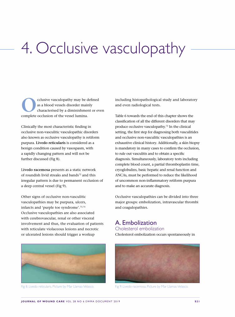
purpura. Livedo reticularis is considered as a
benign condition caused by vasospasm, with
a rapidly changing pattern and will not be
further discussed (Fig 8).
Livedo racemosa presents as a static network
of roundish livid streaks and bands72 and this
irregular pattern is due to permanent occlusion of
a deep central vessel (Fig 9).
Other signs of occlusive non-vasculitic
vasculopathies may be purpura, ulcers,
infarcts and ‘purple toe syndrome’.73,74
Occlusive vasculopathies are also associated
with cerebrovascular, renal or other visceral
involvement and thus, the evaluation of patients
with reticulate violaceous lesions and necrotic
or ulcerated lesions should trigger a workup
including histopathological study and laboratory
and even radiological tests.
Table 6 towards the end of this chapter shows the
classification of all the different disorders that may
produce occlusive vasculopathy.72 In the clinical
setting, the first step for diagnosing both vasculitides
and occlusive non-vasculitic vasculopathies is an
exhaustive clinical history. Additionally, a skin biopsy
is mandatory in many cases to confirm the occlusion,
to rule out vasculitis and to obtain a specific
diagnosis. Simultaneously, laboratory tests including
complete blood count, a partial thromboplastin time,
cryoglobulins, basic hepatic and renal function and
ANCAs, must be performed to reduce the likelihood
of uncommon non-inflammatory retiform purpura
and to make an accurate diagnosis.
Occlusive vasculopathies can be divided into three
major groups: embolization, intravascular thrombi
and coagulopathies.
A. EmbolizationCholesterol embolizationCholesterol embolization occurs spontaneously in
Fig 8. Livedo reticularis. Picture by Mar Llamas-Velasco. Fig 9. Livedo racemosa. Picture by Mar Llamas-Velasco.

S 2 2 J O U R N A L O F WO U N D C A R E VO L 2 8 N O 6 E W M A D O C U M E N T 2 0 1 9
elderly patients with severe atherosclerotic disease,
as can be found in 4.4% of autopsies. Higher
risk factors include catheterisation, pro-longed
anticoagulation or acute thrombolytic therapy75.
Skin biopsy can be diagnostic showing elongated,
needle-shaped clefts within the lumina of small
arterioles, known as cholesterol clefts. A typical
clinical picture is a brisk appearance of acute pain,
livedo racemosa, retiform purpura, peripheral
gangrene, cyanosis or ulceration on distal parts
of the extremities. Birefringent crystals at the
bifurcation of retinal arteries named Hollenhorst
plaques, may be demonstrated by fundoscopic
examination.73 Treatment is mostly supportive,
treating associated hypertension and underlying
heart failure as well with renal replacement therapy
when needed.76
Skin metastasisIntravascular cutaneous metastases are characterised
by the presence of neoplastic cells that can involve
the skin while mainly located within the lymphatic
vessels, so-called ‘inflammatory metastatic
carcinoma’, or within the blood vessels, named as
‘telangiectatic metastatic carcinoma’ (Fig 10).77
Diagnosis should be confirmed by
immunohistochemical studies to define the most
plausible primary origin and complementary
work up is mandatory to find the primary
tumour if unknown or to rule out metastatic
disease of another organ. Not all metastases go
inside the vessels and they can be extravascular,
mimicking pericytes and thus making diagnosis
even more challenging.78,79 Treatment should be
mostly directed to the primary tumour although
skin metastasis can be also treated with surgical
excision, cryotherapy or with electrochemotherapy.
Infective endocarditis Endocarditis can present with septic or non-
septic embolisms. Non-bacterial thrombotic
endocarditis results in focal deposits of fibrin and
other blood elements on heart valves that may
detach and occlude a smaller vessel (Libman-Sacks
endocarditis). Typical lesions in septic endocarditis
are Osler nodules, transient painful nodular lesions
located on the ventral aspect of fingers and toes;
and Janeway lesions, which are multiple slightly
infiltrated, haemorrhagic macules, both lesions are
secondary to septic emboli.80,81 Subungual splinter
haemorrhages are quite unspecific if no additional
signs or symptoms of infection are found.
Histopathology in these infections show a wide
range of variation from thrombotic process without
vasculitis to leukocytoclastic vasculitis, with or
without thrombi, as in other forms of septic
vasculitis. Septic vasculitis differs from idiopathic
leukocytoclastic vasculitis by prominent intraluminal
thrombi in association with minimal leukocytoclasia
and, despite presenting with vascular occlusion, will
not be reviewed in this chapter even when it may
appear associated with infective endocarditis or with
any other systemic infections.82,83
Fig 10. Stage IV breast cancer metastasis. Picture by Mar Llamas-Velasco.

J O U R N A L O F WO U N D C A R E VO L 2 8 N O 6 E W M A D O C U M E N T 2 0 1 9 S 2 3
Empiric antibiotic treatment in late prosthetic
valves or community-acquired native valves is a
combination of ampicillin/flucloxacillin or oxacillin
plus gentamycin. In early prosthetic valves or
nosocomial ones, vancomycin plus gentamycin
and rifampicin is preferred, an empiric treatment
that must be changed when microorganism
susceptibility pattern is later known.83
B. Intravascular ThrombiHeparin-induced skin necrosisHeparin-induced skin necrosis presents as purpuric
plaques with or without necrosis at the site of
heparin injection 5 –14 days after beginning
treatment. It also affects distant sites while
platelet number is characteristically reduced.84
It is caused by the appearance of an antibody,
which binds complexes formed by heparin with
protein factor 4 that is located in the platelets
surface. Histopathology shows a non-inflammatory
occlusion by an eosinophilic thrombi composed of
a platelet plug known as a ‘white clot’, involving
both arterial and venous dermal vessels in the
superficial and deep plexus.84
Other diseases such as thrombocytosis,
polycythaemia vera and myelodysplastic may
present similar clinicopathological picture and
thus, the patient must be referred to haematology
department if blood test suggests these type
of diseases.
CryoglobulinemiasCryoglobulinemias are classified as monoclonal,
mixed or polyclonal. Only type I cryoglobulinemia
manifests with an occlusive vasculopathy. Acral
sites are more frequently involved (Fig 11).
Histopathology is diagnostic, as the lumina of small
blood vessels appear filled by a homogeneous, PAS
positive, eosinophilic material. Although up to
20% of the cases are idiopathic, it can be associated
to haematological diseases such as multiple
myeloma, Waldeström’s macro-globulinemia,
leukaemia or lymphoma. The treatment of the
B-cell lymphoproliferative disorder as well as
the avoidance of cold temperatures improves
the cryoglobulinemic vasculitis although other
additional treatments may be used such as
rituximab, plasma exchange, corticosteroids or
iloprost and some authors have proposed the use
of bortezomib.85,86 Crystalglobulinemia, rarely
reported in the skin, is considered as a severe
type of cryoglobulinemia usually appearing along
with a rapidly progressive renal failure, peripheral
neuropathy and polyarthropathy.72,87
C. CoagulopathiesSystemic coagulopathiesCoumarin/Warfarin-induced skin necrosis affects
patients with a congenital or acquired protein
C and S deficiency when they need warfarin as
it is a vitamin Kantagonist73. First skin lesions
appear 3–5 days after the treatment as purpuric
maculopapular or urticarial lesions mostly affecting
fatty areas. Concomitant use of heparin before
starting warfarin treatment and during the first
five days of warfarin treatment may prevent the
disease88. Treatment with vitamin K and fresh-
frozen plasma may reverse the effects.89
Fig 11. Cryoglobulinemia. Picture by Mar Llamas-Velasco.

S 2 4 J O U R N A L O F WO U N D C A R E VO L 2 8 N O 6 E W M A D O C U M E N T 2 0 1 9
Hypercoagulable states, that are classified into
primary (such as factor V Leiden mutation,
pro-thrombin G20210A mutation and
hyperhomocysteinemia as characteristic examples)
and secondary states such as immobilisation after
surgery, cancer or sepsis, are included within the
wide list of hypercoagulability states that may
produce thrombosis and a vascular occlusion thus
leading to necrosis and ulceration in the skin.90
Primary states control often requires consultation
to haematologist and secondary states to control
the predisposing factors.
Purpura fulminans (PF) is a dermatologic
emergency where non-inflammatory, retiform,
confluent purpura evolves rapidly into necrotic
lesions and underlying conditions may include
severe systemic coagulopathies, drugs intake,
mostly cocaine adulterated with levamisole and
disseminated infections82,91,92. Supportive treatment
(blood transfusion, heparinisation, anti-Xa agents,
synthetic protease inhibitors, natural protease
inhibitor, and fibrinolytic treatment) as well as
treating the underlying causes; with antibiotics
combined with surgical drainage when infection is
underlying or; anticancer drugs with surgery when
there is a malignant disease are needed.
Livedoid vasculopathy (LV)LV is also known as PURPLE (painful purpuric
ulcers with reticular pattern of the lower
extremities).93 LV often is associated with several
hypercoagulability states but the pathogenesis
is not completely understood.90 Characteristic
clinical presentation is painful, punched-out
ulcers that heal leaving white stellate scars
(atrophie blanche) with peripheral telangiectasia
appearing mostly on distal lower extremities in
females (Fig 12).
As LV may coexist with cutaneous polyarteritis
that may also clinically mimic LV,94 skin biopsy is
mandatory to rule out the segmental involvement
of a muscular vessel.95 Many therapies have
been tried from antiaggregants, analgesics, anti-
inflammatory drugs, pentoxifylline, hyperbaric
oxygen therapy (HBOT) to systemic anticoagulants
and even fibrinolytic therapy and vasoactive
drugs.96,97 Rivaroxaban 10–20mg daily seems a
promising drug.98,99
ConclusionClinicopathological evaluation of reticulated
violaceous lesions that may present with skin
ulcers, will lead to an accurate diagnosis of
occlusive vasculopathy in order to establish risk of
other visceral involvement and to properly choose
a first line empirical treatment.
Skin biopsy and laboratory tests including
complete blood count, a coagulation study,
cryoglobulins, basic hepatic and renal function as
well as ANCAs should be done.
Treatments can be classified as the ones focused
in the control of the predisposing factors but
anticoagulants, antiaggregants, vasoactive drugs
and even plasma exchange or anti-inflammatory
treatments or antibiotic agents may be needed
depending on the aetiology of the disease.Fig 12. Livedoid vasculopathy. Picture by Mar Llamas-Velasco.

J O U R N A L O F WO U N D C A R E VO L 2 8 N O 6 E W M A D O C U M E N T 2 0 1 9 S 2 5
Table 6. Classification of occlusive vasculopathies. Based on previously published table72
Types Clinicopathological entities
Embolization Endogenous material Cholesterol emboli, oxalate emboli, crystal globulin vasculopathy
Tumours Atrial myxoma, angiosarcoma, intravascular lymphoma, crystal globulin vasculopathy, intravascular metastasis
Abnormal benign cells Intralymphatic histiocytosis, reactive angioendotheliomatosis
Foreign components Infective: Infective endocarditisForeign material: drug-eluting beds
Thrombi Platelet plugging Heparin-induced skin necrosisThrombocytosis secondary to myeloproliferative disordersParoxysmal nocturnal haemoglobinuriaThrombotic thrombocytopenic purpura
Cold-related agglutination CryoglobulinemiaCryofibrinogenemiaCold agglutinins
Infectious microorganism invading vessels
Ecthyma gangrenosumOpportunistic fungi, for example aspergillosisDisseminated strongyloidiasisLucio´s phenomenon of leprosy
Coagulopathies Systemic coagulopathies Defects of C and S proteinsCoumarin-induced skin necrosisDisseminated intravascular coagulationAntiphospholipid antibody/ lupus anticoagulant syndrome
Vascular coagulopathies Sneddon’s syndromeLivedoid vasculopathyDegos disease
Red cell occlusion syndromes Stress reticulocyte adhesion
Miscellaneous Calciphylaxis Hydroxyurea-induced ulcers (see picture in Ch. 10 Other types of atypical wounds)Occlusive vasculopathy induced by cocaine-levamisole

S 2 6 J O U R N A L O F WO U N D C A R E VO L 2 8 N O 6 E W M A D O C U M E N T 2 0 1 9
Martorell hypertensive ischemic leg
ulcer (HYTILU) and calciphylaxis,
also known as calcific uremic
arteriolopathy (CUA) share a common clinical
pattern and histological hallmarks: Skin
infarction and acral gangrene caused by ischaemic
arteriolosclerosis (Fig 13).
Arterioles show a hypertrophic media with or
without a miniaturised form of Mönckeberg
medial calcinosis and with or without intima
hyperplasia, which result in a narrow vessel
lumen (Fig14).100
Clinically both entities share the same distribution
of skin infarctions, which can be located distally
(characteristic laterodorsal location including
the Achilles tendon) or proximally and centrally
(medial thighs, abdominal apron, breasts, lateral
upper arms). Acral gangrene of fingers, toes or
penis occurs exclusively in calciphylaxis.101–107
‘Classical’ calciphylaxis occurs in patients with
end-stage renal disease (ESRD)104 and rarely in
patients without ESRD who exhibit morbid obesity
with essential hypertension and diabetes type
2100.108–110 Martorell HYTILU111 occurs in patients
with usually long-standing and well-controlled
essential hypertension. Approximately 60% of
Martorell HYTILU patients also have diabetes
type 2.101,103,106,112 By definition, Martorell HYTILU
patients have no ESRD; otherwise they qualify for
the distal form of ‘classical’ calciphylaxis.100
Both Martorell HYTILU and calciphylaxis can
be lethal. The lethality of Martorell HYTILU
may reach 10%, particularly when patients
are unnecessarily exposed to high-dose
immunosuppression for erroneous diagnosis of
pyoderma gangrenosum.103 The one-year-mortality
rate of central forms of calciphylaxis has been
reported around 40–50% in patients with ESRD
and 25% in patients without ESRD.104,113
In many countries the awareness of Martorell
HYTILU and calciphylaxis is still low. Both entities
are hardly mentioned in medical textbooks
or lectures, except in France, where Martorell
HYTILU enjoys high awareness under the term
angiodermite nécrotique.101,106,114–117
5. Martorell HYTILU and calciphylaxis: skin infarction and acral gangrene from ischaemic arteriolosclerosis

J O U R N A L O F WO U N D C A R E VO L 2 8 N O 6 E W M A D O C U M E N T 2 0 1 9 S 2 7
Calciphylaxis in end-stage kidney disease or after successful kidney transplant
Calciphylaxis in normal reneal function and normal parathyroid function
Dist
all p
atte
rn (
belo
w k
nee,
in
calc
iphy
laxi
s fin
gers
, toe
s, pe
nis
Prox
imal
pat
tern
(th
ighs
, ab
dom
inal
apr
on, t
runk
)
Calciphylaxis, distal pattern Martorell HYTILU (hypertension)
Calciphylaxis, proximal patternEutrophication
in morbid obesity
Fig 13. The four clinical patterns of Martorell hypertensive ischemic leg ulcer, calciphylaxis in normal renal and parathyroid function, and calciphylaxis, peripheral and central pattern. Hafner J. Calciphylaxis and martorell hypertensive ischemic leg ulcer: same pattern - one pathophysiology. Dermatology 2016; 232(5):523–533, reproduced with the permission of S. Karger AG, Basel, Switzerland.
PathophysiologyPathophysiology of calciphylaxis in end-stage renal disease (ESRD)Calciphylaxis occurs in approximately 35/10,000
ESRD patients on dialysis.104 The typical period
of onset is 35–105 months after initiation of
dialysis.104 Calciphylaxis is associated with the
following risk factors:118,119 Diabetes, obesity,
female sex, dependence on dialysis, calcium and
phosphate disturbances, overuse of vitamin D
and calcium substitutes and/or calcium-based
phosphate binders and vitamin K depletion (from
malnutrition or malabsorption) or medication with
vitamin K antagonists (hazard ratio 3-13!).120,121
Matrix Gla protein (MGP) is a vitamin
K-dependent strong protector from extraskeletal
calcification121–123 which simultaneously inhibits
the procalcifying proteins BMP 2+4. 120,124 MGP and
fetuin A deficiency are regarded essential risk factors

S 2 8 J O U R N A L O F WO U N D C A R E VO L 2 8 N O 6 E W M A D O C U M E N T 2 0 1 9
of calciphylaxis. Vitamin K antagonists (warfarin,
phenprocoumon, acenocoumarol) therefore increase
the risk of developing calciphylaxis.120,121
Subcutaneous obliterating arteriolosclerosis (vessel
diameter: 100–600mm) with medial hyper-trophy,
with or without Mönckeberg medial calcification
and with or without endothelial hyperplasia
(Fig 15) are the immediate cause of skin infarction
and acral gangrene.103,112,125,126
Pathophysiology of calciphylaxis without ESRD (‘eutrophication’)Calciphylaxis without ESRD exclusively occurs in
patients with morbid obesity who commonly have
type 2 diabetes and essential hypertension.108–110
Nephrologists regard calciphylaxis without ESRD as
a subform of calciphylaxis, whereas we suggested
the hypothesis that it represents the proximal
variant of Martorell HYTILU.100
Pathophysiology of Martorell HYTILUMartorell HYTILU is characterised by skin infarctions
in a typical laterodorsal location of the legs and over
the Achilles tendon (Fig 16).101,103,105,106,112,114–117
Fig 14. The four identical histological patterns. Hafner J. Calciphylaxis and martorell hypertensive ischemic leg ulcer: same pattern - one pathophysiology. Dermatology 2016; 232(5):523–533, reproduced with the permission of S. Karger AG, Basel, Switzerland
Calciphylaxis in end-stage kidney disease or after successful kidney transplant
Calciphylaxis in normal reneal function and normal parathyroid function
Dist
all p
atte
rn (
belo
w k
nee,
in
calc
iphy
laxi
s fin
gers
, toe
s, pe
nis
Prox
imal
pat
tern
(th
ighs
, ab
dom
inal
apr
on, t
runk
)
Calciphylaxis, distal pattern Martorell HYTILU (hypertension)
Calciphylaxis, proximal pattern Eutrophication in morbid obesity
Fig 15. Subcutaneous arteriolosclerosis, with and without medial calcification, adjacent dermal necrosis. Picture by Jürg Hafner.

J O U R N A L O F WO U N D C A R E VO L 2 8 N O 6 E W M A D O C U M E N T 2 0 1 9 S 2 9
They are associated with essential hypertension
(usually long-standing and well-controlled) (100%),
diabetes type 2 (60%) and perhaps also with vitamin
K antagonist medication. In 50% of patients
Martorell HYTILU is accompanied by common
peripheral arterial disease (PAD), but 50% of patients
show normal peripheral arteries of the legs.103
Common characteristics of Martorell HYTILU and calciphylaxisCommon atherosclerosis results from inflammation
and remodelling of the arterial vessel wall.
Hypertension, diabetes, dyslipidaemia and smoking
are the four major cardiovascular risk factors.
We suggested the hypothesis that arteriolosclerosis
in Martorell HYTILU and calciphylaxis without
ESRD, and to some extent ‘classical’ calciphylaxis in
ESRD, too, share the same risk factors (Table 7).100
(A) Hypertension (HT) is always present in
Martorell HYTILU (primary HT),101,103,105,106,112,114–117
in calciphylaxis without ESRD108–110 and in most
instances of ‘classical’ calciphylaxis in ESRD (renal
hypertension).
(B) Diabetes can be found in 60% of patients
with Martorell HYTILU, in 100% of patients with
calciphylaxis without ESRD),108–110 and is the most
frequent cause of ESRD.
(C) Vitamin K antagonists are an established risk
factor of ‘classical’ calciphylaxis in ESRD.120,123 It is
plausible to hypothesise that vitamin K antagonists
may be a risk factor of Martorell HYTILU and
calciphylaxis without ESRD, too, however, this has
not yet been scientifically proven.
(D) Raised calcium and phosphate levels and
disturbed bone mineralisation are regarded
major risk factors of ‘classical’ calciphylaxis
in ESRD.118,119,126–134
Fig 16. A 66-year-old-male, Martorell hypertensive ischemic leg ulcer. Laterodorsal skin infarction, involving the area over the Achilles tendon. Picture by Jürg Hafner.
Table 7. Cardiovascular risk factors of arteriolosclerosis
Normal renal function Chronic renal insufficiency
Distal pattern of necrosis
Martorell hypertensive ischemic leg ulcerArterial hypertension (primary, essential hypertension)Diabetes mellitus (type 2)Vitamin K antagonists (fetuin A inactivation)
Calciphylaxis, distal patternRenal hypertension (secondary, renal hypertension)Diabetes mellitus (type 1 or type 2)Vitamin K antagonists (fetuin A inactivation)2° or 3° Hyperparathyroidism
Proximal pattern of necrosis
Calciphylaxis in normal renal and parathyroid functionMorbid obesity with:• Arterial hypertension (primary, essential hypertension)• Diabetes mellitus type 2• Vitamin K antagonists (fetuin A inactivation)
Calciphylaxis, proximal patternRenal hypertension (secondary, renal hypertension)Diabetes mellitus (type 1 or type 2)Vitamin K antagonists (fetuin A inactivation)2° or 3° Hyperparathyroidism

S 3 0 J O U R N A L O F WO U N D C A R E VO L 2 8 N O 6 E W M A D O C U M E N T 2 0 1 9
Clinical presentation and differential diagnosisSkin infarction typically begins as dusky
discoloration, violaceous spot or plaque that
becomes overtly necrotic within a few days.104
The necrotic area is irregular and polycyclic. A
livedo pattern is often found around the necrosis,
and the wound border is typically inflamed and
undermined. Most patients experience excruciating
pain which is difficult to control with common
analgesia, including opioids.103,104 Acral gangrene of
fingers, toes and penis can occur during ‘classical’
calciphylaxis in ESRD.102,104 The location of necrosis
mostly follows a typical pattern: Laterodorsal legs
including the Achilles tendon,105 as well as the
medial thighs, abdominal fat apron, female breasts,
and lateral upper arms (Table 8).103
Difficulty of distinguishing/confounding Martorell HYTILU from PG or necrotising cutaneous vasculitisMartorell HYTILU can easily be overlooked or
interpreted as PG or necrotising vasculitis by
physicians who are unaware of the entity. In a
series of 31 patients we described in 2010, half
of the patients erroneously received high-dose
immunosuppression for supposed PG.103
The clinical aspect of the wound in Martorell
HYTILU and calciphylaxis, respectively, are indeed
difficult to distinguish. Both entities develop
progressive, polycyclic skin necrosis with a very
painful, violaceous and undermined border:
Martorell HYTILU, which is based on
arteriolosclerotic skin infarction, tends to
result in a ‘black’ skin necrosis involving all
skin layers, sparing the fascia. Patient history
includes longstanding and well-controlled
primary hypertension and in 60% of patients
type 2 diabetes. Malignancies, particularly
hematoproliferative disorders and myeloma, and
inflammatory bowel disease are not associated
with Martorell HYTILU. Martorell HYTILU does
not respond to systemic glucocorticosteroids or
other systemic immunosuppressives, except that
the tissue inflammation around the skin ulcer may
clinically appear attenuated.
PG, which is based on uncontrolled neutrophil
activation, initially results in a more superficial
necrotic ‘liquefaction’ of the skin necrosis
that responds to and heals under systemic
glucocorticosteroids. Skin necrosis in PG is often
not ‘black’, and commonly stops at the deeper
dermis. Hematoproliferative disorders are found
associated in 20% and inflammatory bowel disease
in 10% patients. The majority of PG are located
at the legs, and hypertension and diabetes are
associated with PG, as well.12,17,135
Table 8. Patterns of location
Normal renal function Chronic renal insufficiency
Distal pattern of necrosis (skin infarction)
Martorell hypertensive ischemic leg ulcerLaterodorsal legAchilles tendon
Calciphylaxis, distal patternLaterodorsal legAchilles tendonFinger, toe, penis necrosis or gangrene
Proximal pattern of necrosis (skin infarction)
Calciphylaxis in normal renal and parathyroid functionMedial thighAbdominal apronFemale breastsLateral upper arms
Calciphylaxis, proximal patternMedial thighAbdominal apronFemale breastsLateral upper arms

J O U R N A L O F WO U N D C A R E VO L 2 8 N O 6 E W M A D O C U M E N T 2 0 1 9 S 3 1
Necrotising vasculitis commonly shows bilateral
multiple, symmetric round and deep skin ulcers at
the legs, buttocks, lower back, and less frequently
arms and shoulders. Palpable purpura is present
during the initial phase of disease. Infections are
the most common trigger, and organ involvement,
particularly glomerulonephritis, have to be
searched for and treated as indicated.47,136
Ecthyma gangrenosum (EG) can be seen
in immunocompetent patients with skin
infections with Streptococci spp. or Pseudomonas
spp., (eg after hot tub baths), as well as in
immunosuppressed patients with skin infections
with unusual strains, (eg atypical mycobacteria).
EG can bear close resemblance with PG or
vasculitis skin ulcers, and needs to be actively
considered in the mentioned settings.137
Examination, and indication of an appropriate skin biopsy(A) All patients with a suspicion of Martorell HYTILU
or calciphylaxis should have a screening examination
to exclude PAD. Approximately 50% of patients with
Martorell HYTILU have PAD, which usually is treated
by angioplasty (PTA). Restoration of arterial inflow by
PTA103 improves chance of healing.
(B) Microbiology using a swab or other culture
technique (eg PCR or biopsy) is important regarding
management of critical colonisation and/or
infection and perioperative antibiotic prophylaxis
during debridement and split skin transplantation.
(C) The necessity of performing skin biopsy to
confirm subcutaneous arteriolosclerosis as under-
lying pathology is controversial. The French
school suggests to base a diagnosis of Martorell
HYTILU on the clinical pattern alone, provided
that the clinical picture is characteristic106,117.
They argue that particularly in small skin ulcers a
biopsy may iatrogenically worsen the condition,
and that subcutaneous arteriolosclerosis is a non-
specific histopathologic finding, especially in the
elderly population. It is recommended to perform
a diagnostic skin biopsy in all cases or Martorell
HYTILU where PG and/or necrotising vasculitis or
where further pathologies have to be ruled out, or
when major wound surgery is required anyway.
It is important not to perform a small punch
biopsy, because histopathology of small samples
can be misleading and show dense neutrophil
infiltrates in the necrotic superficial dermis in
all suspected differential diagnoses, leading
to erroneous ‘confirmation’ of ‘PG’. Instead,
a 3–6cm long and 4–5mm wide spindle shape
biopsy extending from healthy skin into the
necrotic area and including all three skin layers
down to fascia is collected in local anaesthesia.103
These narrow and long biopsy wounds can be
closed with absorbable suture material (Fig 17).
Fig 17. Spindle shape biopsy including subcutane-ous fat, 6 cm long and 6 mm wide. Picture by Jürg Hafner.
Fig 18. Spindle shape biopsy including subcutane-ous fat, 6 cm long and 6 mm wide. Picture by Jürg Hafner.
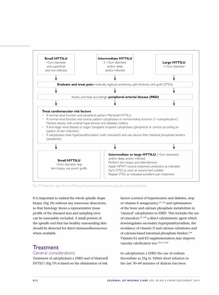
S 3 2 J O U R N A L O F WO U N D C A R E VO L 2 8 N O 6 E W M A D O C U M E N T 2 0 1 9
It is important to embed the whole spindle shape
biopsy (Fig 18) without any transverse dissections,
so that histology shows a representative tissue
profile of the diseased area and sampling error
can be reasonably excluded. A small portion of
the spindle end that has healthy surrounding skin
should be dissected for direct immunofluorescence
where available.
TreatmentGeneral considerationsTreatment of calciphylaxis ± ESRD and of Martorell
HYTILU (Fig 19) is based on the elimination of risk
Small HYTILU<5cm diameterand superficial
and non-infected
Intermediate HYTILU5–15cm diameter
and/or deepand/or infected
Large HYTILU>15cm diameter
Evaluate and treat pain: medically, regional anesthesia, split-thickness skin graft (STSG)
Assess and treat accordingly peripheral arterial disease (PAD)
Treat cardiovascular risk factors• If normal renal function and peripheral pattern: Martorell HYTILU• If normal renal function and central pattern: calciphylaxis in normal kidney function (= ‘eutrophication’).
Morbid obesity with arterial hypertension and diabetes mellitus• If end-stage renal disease or organ transplant recipient: calciphylaxis (peripheral or central, according to
pattern of skin infarction)• If calciphylaxis: treat hyperparathyroidism (with cinecalcet) and use calcium-free intestinal phosphate binders
(sevelamer)
Small HYTILU<5cm diameter skip
skin biopsy use punch grafts
Intermediate or large HYTILU (>5cm diameter) and/or deep, and/or infectedPerform skin biopsy and debridementApply NPWT wound treatment; antibiotics as indicatedEarly STSG as soon as wound bed suitableRepeat STSG as indicated; excellent pain treatment
Fig 19. Treatment algorithm of Martorell hypertensive ischemic leg ulcer and calciphylaxis
factors (control of hypertension and diabetes, stop
of vitamin K antagonists),121,138 and optimisation
of the bone and calcium phosphate metabolism in
‘classical’ calciphylaxis in ESRD. This includes the use
of cinecalcet,127,139 a direct calcimimetic agent which
downregulates secondary hyperparathyroidism, the
avoidance of vitamin D and calcium substitutes and
of calcium-based intestinal phosphate binders.104
Vitamin K1 and K2 supplementation may improve
vascular calcification too.104,115,140
In calciphylaxis ± ESRD the use of sodium
thiosulfate as 25g in 100ml short infusion in
the last 30–60 minutes of dialysis has been

J O U R N A L O F WO U N D C A R E VO L 2 8 N O 6 E W M A D O C U M E N T 2 0 1 9 S 3 3
proven beneficial in larger case series. RCTs are
underway investigating treatment outcomes in
calciphylaxis.104,141 In extensive cases of Martorell
HYTILU use sodium thiosulfate (10g/100ml
solution, 3x weekly) as well. This recommendation,
however, is not based on scientific data. Sodium
thiosulfate has antioxidant and vasodilatory
properties and inhibits adipocyte calcification
and calcification of vascular smooth-muscle
cells.142,143Bisphonates have shown efficacy in case
series of ‘classical’ calciphylaxis in ESRD.133
Patients with small forms of Martorell HYTILU
benefit from minor debridement followed by
immediate punch grafting.101,114,144,145
Wound surgeryIn patients with extensive forms of Martorell
HYTILU, several case series have shown more
rapid recovery when wound surgery, NPWT and
early skin grafts (punch grafts (Fig 20) or split-
thickness skin grafts (Fig 21 and 22) have been
used.103,112,117,144,146 These patients mostly require
antibiotic treatment to avoid local infections and
sepsis.103 The role of surgery in patients with central
calciphylaxis ± ESRD is not established and has to be
determined depending on the individual situations.
It is important to familiarise physicians and wound
experts with the clinical appearance and pathology
of Martorell HYTILU and calciphylaxis in order to
offer these extremely suffering patients an optimal,
straight-forward treatment.
Conclusions and recommendationsSimilarity of the clinical patterns of skin necrosis
and the histological pattern of ischaemic
subcutaneous arteriolosclerosis suggest a
common pathophysiology of (A) Martorell
HYTILU, (B) distal calciphylaxis in ESRD,
(C) central calciphylaxis in ESRD, and (D)
Fig 20. Martorell hypertensive ischemic leg ulcer before punch grafting (a). Same wound, day 14 after punch graft; reduction of pain (VAS 9-10 to 2-3)(b). Same wound, day 22 after punch graft (c). Same wound healed, day 58 after punch graft (d). All pictures by Elena Conde Montero
b
c
d
TOO LOW-RES TO USE NEED HI-RES SUPPLYING
a

S 3 4 J O U R N A L O F WO U N D C A R E VO L 2 8 N O 6 E W M A D O C U M E N T 2 0 1 9
c
a
b
Fig 21. Martorell HYTILU laterodorsal skin infarction, large area of livedo (a). NN, m, Martorell HYTILU Inpatient treatment with necrosectomy and NPWT (b). NN, m, Martorell hypertensive ischaemic leg ulcer. Initial take of split-thickness skin graft, at week three postoperatively (c). All pictures by Jürg Hafner.
Fig 22. Martorell hypertensive ischaemic leg ulcer. Further course after a one stage of split-thickness skin graft, five months. Pictures by Jürg Hafner.
(central) calciphylaxis in normal renal function
(‘eutrophication’ in morbid obesity). These four
entities share a common histopathology (stenotic
arteriolosclerosis ± miniaturised Mönckeberg
arteriolosclerosis, ± intimal hyperplasia) and
clinical pattern (ischaemic skin infarction).
These features suggest a partly common
pathophysiology (risk factors such as
hypertension, diabetes, vitamin K antagonist
medication, and disturbed bone and calcium
phosphate metabolism in ‘classical’ calciphylaxis
in ESRD lead to stenotic arteriolosclerosis and
eventually to skin infarctions and acral gangrene).
Martorell HYTILU must not be confounded with PG:
The therapeutic approach is completely different.
Patients with Martorell HYTILU require wound
surgery and early skin grafts. Immunosuppression for
erroneously diagnosed PG can become deleterious.
As in any chronic wound of the lower extremities,
PAD should be ruled out and treated accordingly
(eg with PTA), if present.
Confirmation of diagnosis with a long (3–6cm)
and narrow (4–5mm) spindle shape biopsy may
be unnecessary in patients with small forms of
clinically typical Martorell HYTILU, but is highly
recommended in all cases of diagnostic doubt or
when major wound surgery is required.
Small skin ulcers (<5cm diameter) of Martorell
HYTILU or calciphylaxis, respectively, can heal
under conservative measures alone or with minor

J O U R N A L O F WO U N D C A R E VO L 2 8 N O 6 E W M A D O C U M E N T 2 0 1 9 S 3 5
debridement and punch grafts. Intermediate
skin ulcers (<10cm diameter) mostly require
necrosectomy, negative pressure wound treatment
and split skin grafts. Patients with large (>10cm
diameter) or extensive skin ulcers may benefit from
intravenous sodium thiosulfate (10g/100ml short
infusion, 3x per week).
Skin grafts should be applied precociously, even
when the recipient wound bed does not expose
suitable granulation. Antibiotic therapy is often
required since many skin ulcers from Martorell
HYTILU or calciphylaxis, respectively, become
critically colonised or overtly infected in the course
of necrosis progression.
Skin grafts can dramatically decrease the
excruciating pain that many patients with
Martorell HYTILU or calciphylaxis, respectively,
have to suffer. In most instances they are the best
‘pain killers’.
The take of skin grafts is often optimal in the
centre, where it adheres to exposed vital fascia,
and hampered along the margins, where it should
connect with necrobiotic subcutis. The polycyclic
skin infarctions can become progressive along
the borders and often require serial skin grafts
until some smaller and more superficial wound
remnants can be left to secondary wound healing.
Future perspectivesThe sensitivity and specificity of the histopathologic
finding of subcutaneous arteriolosclerosis needs to
be investigated in a larger prospective controlled
study. Arteriolosclerosis may be a systemic
condition. The question of organ involvement
should be further examined. Future research
should focus on the pathophysiology, risk factors
and protective mechanisms of hypertensive, but
also diabetic, and renal arteriolosclerosis with and
without calcification, and on the role of the striking
inflammation–primary or secondary — that can be
observed in and around the skin infarctions caused
by subcutaneous arteriolosclerosis. The empirically
recommended treatment measures need to be
confirmed by randomised controlled clinical studies.

S 3 6 J O U R N A L O F WO U N D C A R E VO L 2 8 N O 6 E W M A D O C U M E N T 2 0 1 9
6. Hidradenitis suppurativa
The disease was described for the first
time by Velpeau in 1839.147 Hidradenitis
suppurativa (HS) is an inflammatory
disease which primarily affects the hair follicles of
apocrine gland bearing skin, in other words. the
perigenital skin and axillae, with recurrent lesions.
The lesions consist of inflamed or non-inflamed
nodules, abscesses and draining or non-draining
tunnels (Fig 23). In more progressed stages of the
disease scarring is prominent.148,149
The lesions cause pain, discomfort and
disfigurement. Occasionally HS appears in a
syndromic form with acne, arthritis and PG in
the PASH or PA-PASH syndromes.150 Recently,
substantial comorbidities have been linked to
the disease, associated with an increased overall
mortality including completed suicides.151,152 This
suggests that HS has a systemic effect and may be
seen as a more general inflammatory disease.148
The prevalence of the disease is reported from
0.05–4% of the population and the estimates
appear to depend not only on the population
sampled but also on the methodology used.153
Registry-based studies generally report lower
prevalence than self-reported data or physical
examination suggesting selection bias. This is
further supported by the observation that patients
globally experience a clinically significant delay
in diagnosis.154 HS appears to exert a major life
course influence on patients as a group, as it
appears associated with poorer socioeconomic
characteristics such as education, income and
social class.155,156
The concept of HS as a disease of wound healingChronic lesions of HS are characterised by a mix
of inflammation and tissue damage in the shape
of scars, tunnels and occasionally hypertrophic
granulation tissue extruding from the lesions and
prompting the hypothesis that HS may be a disease
of wound healing.157
The aetiology of the HS remains largely unknown,
but it is speculated that an aberrant immune
response to changes in the follicular microbiota
may play a role. The pathogenesis involves the
release of pro-inflammatory cytokines such as
TNF and IL-17, as well as anti-inflammatory
cytokines such as IL-10.158,159 Morphologically,
the pathogenesis involves initial preclinical
lesions consisting of a perifollicular lymphocytic
infiltrate surrounding biofilm-depleted Fig 23. Severe case of hidradenitis suppurativa. Picture by Gregor Jemec.

J O U R N A L O F WO U N D C A R E VO L 2 8 N O 6 E W M A D O C U M E N T 2 0 1 9 S 3 7
follicles.160,161 In later stages of the disease where
chronic lesions predominate, a shift towards
an infiltrate consisting of neutrophils and
mast cells is seen surrounding chronic tunnels
containing abundant biofilm.162,163 At this stage
metalloproteinase (MMP)-2 is also expressed in
the lesions indicating cell-migration associated
with wound healing.164
Use of dressings in management of HSTreatment of HS should be multimodal and consists
of adjuvant therapy (lifestyle interventions),
medical and surgical treatment, ideally provided
in parallel rather than sequentially.165 Life style
interventions include information about healthy
diet, weight loss, exercise and smoking cessation.
Dressings provide an important adjuvant therapy in
the management of HS.166,167
The aim of medical treatment in HS is to reduce
disease progression and inflammation, thereby
reducing pain and suppuration over time.
Studies have shown that chronic or recurrent
and unpredictable suppuration from lesions is
a major contributor to the significantly reduced
Health Related Quality of Life (HRQoL) of HS.168,169
This imvolves two components: smell and
seeping. Although not always clinically evident,
fear of malodourous discharge from lesions is
an important factor in the reduced HRQoL of
the patients. In addition patients often worry
about discharge seeping through clothing and
leaving visible marks not only on the patients
but also on chair and other furniture. Both these
fears are in principle manageable by the use of
appropriate bandaging, the challenge being that
most bandages are developed for use on plane or
convex surfaces, whereas HS predominantly affects
concave surfaces. In addition the absorptive ability,
anti-odorous ability and wearer comfort play a
role. Soft and absorbent dressings with a silicone-
based adhesive to minimise trauma to the skin
are generally useful, although the actual design
and price of the products restrict their use. The
design generally requires very careful placing of the
dressing, which may be difficult in view of the sites
involved, and the price often prohibitive. Many
patients therefore resort to using sanitary pads,
rags or kitchen towels.
Topical medical treatment is most suitable
for either very mild Hurley stage 1 disease or
single lesions. For mild disease an early trial
has suggested that topical clindamycin may
be beneficial, while single lesions have been
successfully treated with 0.15% resorcinol.165
Systemic medical treatment is primarily using
antibiotics and/or immunosupressants. The
antibiotics recommended by current guidelines
are primarily tetracycline-type and rifampicin,
and it is thought that the main effect is
immunomodulatory rather than antibacterial.
Evidence grade is however comparatively low
for antibiotic therapy of HS. Better evidence is
available for frank immunosuppressive therapy
using biologics, and particular TNF blockers
(adalimumab and infliximab), but also IL-1
inhibitors (anakinra) and IL-12/23 inhibitors.165

S 3 8 J O U R N A L O F WO U N D C A R E VO L 2 8 N O 6 E W M A D O C U M E N T 2 0 1 9
Ideally the symptoms can be controlled by medical
treatment, but because HS is associated with tissue
damage, residual chronic lesions may require surgical
intervention. The aim of surgical treatment in HS
is the reduction and amelioration of existing tissue
damage, as exemplified by excision of chronic scarred
lesions.165 Dressings are therefore also important to
the management of treatment related wounds.
The least invasive surgical procedure is the so-called
de-roofing of tunnels, which involved literally
removing the roof of HS tunnels.170 The procedure
exposes the immature epithelial lining of chronic
HS tunnels to the surrounding environment and
therefore only requires short-term dressing using
a mildly absorbing non-adherent dressing that
provides a local environment conducive to the
maturation of the epithelium.
In contrast, more scarred lesions require full
thickness excision of skin. Primary closure of
wounds is often possible for smaller lesions, but
this is often associated with higher recurrence
rates.165 Larger lesions may be managed by the
use of NPWT and skin grafts or flaps, but are
also frequently left open for secondary intention
healing. Similarly, lesions treated by CO2 laser
ablation are left for secondary intention healing.
Studies suggest that this ‘non-surgical’ approach
is well accepted by HS patients and generally
a safe and effective technique.171,172 Healing
times are however long and may be modified by
late skin grafting after either partial closure of
large defects or after wound preparation using
negative pressure.173 General guidelines on the
management of large tissue defects apply to these
postoperative wounds.
Conclusion and recommendationsWound care forms a mainstay of HS management,
both in the context of adjuvant therapy dealing
with chronic disease-related problems such
as drainage or hypergranulation, as well as
following surgery. The use of dressings to manage
spontaneous drainage from lesions if particularly
helpful to patients as it provides empowerment
and offers realistic possibilities of self-help. In
contrast, postoperative wound care initially
falls more within the realm of traditional health
care using established paradigms and treatment
algorithms. In either case, there is however a
challenge to find suitable dressing materials, as
the disease occurs in concave areas of the body
while most bandages are designed for application
on plane or convex areas. In the context of
wound dressings, the HS patients therefore
regularly experience an unmet treatment need. It
is recommended that more appropriate geometric
design of existing dressings is developed to suit this
underserved group of patients.

J O U R N A L O F WO U N D C A R E VO L 2 8 N O 6 E W M A D O C U M E N T 2 0 1 9 S 3 9
7. Malignant wounds
The prevalence of malignant wounds in
chronic leg ulcers is around 2–4%.174–177
This number varies according to the
different types of medical facilities and seems to be
highest in tertiary dermatological leg ulcer centres.
One prospective multicentre study in which all
included patients with non-healing chronic leg
ulcers were biopsied, showed a prevalence as
high as 10.4%.178 Many skin cancers presenting as
chronic leg ulcers might be unrecognised. Thus it
is important to identify these wounds as neoplastic
since the treatment is different compared with
vascular wounds and a cure of these wounds can
usually not be achieved by conservative treatment.
Aetiology and pathogenesisMalignant wounds can be subdivided into primary
ulcerating skin tumours arising in normal skin and
secondary ulcerating skin tumours that develop
in chronic leg ulcers, burn scars or osteomyelitis
sinuses. Primary malignant wounds are much
more common than secondary malignant wounds.
The group of primary malignant wounds on the
leg comprises especially basal cell carcinomas
(BCC) and squamous cell carcinomas (SCC).
Rarely, other tumour entities like malignant
melanoma, malignant vascular tumours, cutaneous
lymphomas, Merkel cell carcinoma (primary
neuroendocrine carcinoma of the skin) sarcomas,
adnexal tumours and skin metastases are found as
a cause of chronic leg ulcers (Table 9).
BCCs and SCCs are summarised under the term
non-melanoma skin cancer (NMSC). Together,
they represent the most frequently observed
malignancy in Caucasians.179–181 NMSC occurs
worldwide in all races. People with fair skin and
red hair (Fitzpatrick skin type 1) have the highest
prevalence rates. Besides genetic predisposition
and age, the lifetime cumulative UV-light
exposure and longstanding immunosuppression
are important risk factors.182–185 NMSCs are
located on the head and neck region in 60–80%
of cases and develop on the lower extremities
infrequently.186,187 It has long been discussed if
chronic venous insufficiency is a risk factor for
the development of NMSC, especially for BCCs,
but it is not clear if there is a true association
or a coincidence.177,188–190 Concerning the
frequency and types of skin tumours found in
leg ulcers, data are contradictory. Depending on
the published series, either BCC or SCC were
predominant.175,177,178
Table 9. Aetiologies for malignant ulcers
Aetiologies for malignant ulcers
Frequent aetiologies for malignant ulcers
Basal cell carcinomas BCCSquamous cell carcinomas (SCC)
Less frequent aetiologies for malignant ulcers
Malignant melanomaMalignant vascular tumours (eg Kaposi sarcoma, angiosarcoma)Cutaneous lymphomasMerkel cell carcinomaMalignant adnexal tumoursSarcomasCutaneous metastasesSecondary ulcerating skin tumours (Marjolin ulcers)

S 4 0 J O U R N A L O F WO U N D C A R E VO L 2 8 N O 6 E W M A D O C U M E N T 2 0 1 9
Secondary ulcerating skin tumours are very
uncommon, especially in chronic leg ulcers. In
a large Swedish retrospective analysis of 10,913
venous leg ulcer (VLU) patients, only 17 patients
(0.15%) were detected to have developed a
transformation into a secondary malignant ulcer.191
More commonly, secondary malignant ulcers are
found in burn scars and osteomyelitis sinuses.192
All of these ulcers are called Marjolin ulcers, named
after the French surgeon Jean Nicolas Marjolin
who described the malignant transformation
in long lasting wounds and scars in the 19th
century (Fig 24).193
The latency period of wounds or scars before the
development of a malignancy is usually more
than 10 years.191,194 Series with latency periods of
25–27 years have been described.191,195,196 These
secondary tumours are most frequently SCCs
but other tumours including BCCs, developing
on chronic leg ulcers have been described as
well.188,190,194,196,197 There are a number of hypotheses
on the pathogenesis of SCCs and BCCs that
develop in chronic wounds, including the chronic
inflammation of chronic venous insufficiency.
However, a clear differentiation between primary and
secondary malignant wounds is difficult and cannot
always be achieved, unless the ulcer development
and course have been precisely documented
including repeated biopsies that show a change from
a benign wound to a malignant wound.189
Clinical presentationThe clinical presentation of malignant wounds
differs widely (Fig 25). In fact, a malignant wound
can look like any other cause of chronic leg
ulcers, making it difficult to establish the correct
clinical diagnosis. The following factors and signs
should raise the suspicion for a malignant wound:
atypical ulcer location that is incompatible with
the typical locations of the common vascular
wounds and atypical ulcer appearance with
excessive granulation tissue of the wound bed
and especially the wound edges178. Less specific
signs are irregular wound borders, wound odour,
increased wound pain and bleeding of the wound.
Furthermore, a malignant wound and especially a
secondary malignant wound should be expected
Fig 24. A 74-year-old woman with a previous history of venous leg ulcer. She was diagnosed a squamous cell carcinoma appearing as an ulcer in the left leg. Picture by Kirsi Isoherranen
Table 10. Clinical hints for suspecting malignant wounds
Clinical hints for suspecting malignant wounds
Common Excessive granulation tissue of the wound bed and the wound edgesAtypical ulcer locationChange in appearance of a long lasting ulcer (enlarging size, hypertrophic wound edges)
Less specific Irregular wound bordersWound odourIncreasing wound painBleeding fragile tissue

J O U R N A L O F WO U N D C A R E VO L 2 8 N O 6 E W M A D O C U M E N T 2 0 1 9 S 4 1
if there is a change in appearance of a long lasting
ulcer (Table 10).196
Some malignant wounds show none of these
criteria. Generally all malignant wounds have
one aspect in common: they do not heal with
standard treatment that is commonly used for
chronic (venous) wounds, such as compression
therapy and wound dressings or off-loading for
neuropathic ulcers. Clinicians should always
be suspicious and consider the possibility of
malignancy if there is a delay in wound healing
despite appropriate treatment. If this is the
case or if one or more of the above-mentioned
criteria exist, further diagnostics should be
initiated and the patient should be referred for
specialist advice.
Diagnostics and stagingThe most important step for diagnosing a malignant
wound is a biopsy for histological analysis.
A wound biopsy can be performed either as a
narrow and deep wedge-shaped excision or as a
5–6mm punch biopsy and is usually done under
local anaesthesia. It is generally a safe procedure,
and the subsequent wound usually heal within
a few weeks with no delay in wound healing.198
Performing at least two biopsies on different areas of
the wound (wound edge and wound bed) to avoid
a b c d
e f g
Fig 25. A 92-year-old female with ulcerating basal cell carcinoma on the medial ankle (a), close-up (b). A 91-year-old male with ulcerating squamous cell carcinoma on the anterior lower leg (c), close-up (d). A 81-year-old female with ulcerating amelanotic malignant melanoma on the anterolateral lower leg (e), close-up (f). An 83-year-old male patient with ulcerating malignant melanoma on the heel (g). All pictures by Stephan Nobbe

S 4 2 J O U R N A L O F WO U N D C A R E VO L 2 8 N O 6 E W M A D O C U M E N T 2 0 1 9
the risk of false-negative single biopsies is highly
recommended.177,178 Particularly the wound edge,
including epithelium should not be missed in the
biopsy because on occasion malignant histological
changes are only found in the wound edge of the
ulcer.199 Repeated biopsies should be considered in
case of previous negative biopsies but unchanged
suspicious appearance of the wound. This is
Fig 26. A 59-year-old female patient with squamous cell carcinoma secondary to a chronic vascular ulcer on the lateral ankle (a). Excision of tumour with 1.5cm safety margin (b). Follow-up 1 week after transplantation with meshed split-thickness skin graft (STSG) from the lateral thigh (c). Follow-up three weeks after STSG (d) Donor site on the lateral thigh three weeks after harvesting of STSG (e). Follow-up two month after STSG (f). All pictures courtesy of Jürg Hafner.
a b
c d
e f

J O U R N A L O F WO U N D C A R E VO L 2 8 N O 6 E W M A D O C U M E N T 2 0 1 9 S 4 3
especially important when the development of
secondary malignant wounds cannot be ruled out.
When the diagnosis of a malignant wound is
established, staging examinations to detect potential
lymph node or organ involvement should be
considered. The extent of the staging depends
on the tumour entity, tumour size and location,
histologic features and patient characteristics
(eg immunosuppressive medications). For
BCCs confined to the skin, a thorough clinical
examination is most important, and radiologic
imaging is usually not necessary. Radiologic imaging
can be useful to detect the extent of tissue invasion
(computed tomography for bone assessment and
magnetic resonance imaging for soft tissue and nerve
examination) and to detect lymph node metastases
(ultrasonography or computed tomography). For
high-risk SCCs (tumour diameter >2cm, tumour
thickness >4mm, poor histological tumour
differentiation, perineural infiltration, Marjolin
ulcer), the examination with ultrasonography or
computed tomography of the draining lymph node
basins is recommended.184,200,201
Treatment and follow-upTreatment for localised malignant wounds is
primarily surgical. Micrographic surgery reduces
the risk of tumour recurrence and can usually be
performed as classic Mohs surgery with histologic
control of the entire excision margin in frozen
sections. The specimen has to be embedded
with a special technique in order to visualise
the entire resection margin and the histologic
slides are read by the dermato-surgeon. For larger
tumours and if Mohs surgery is not available,
histologic margin control can be performed
on paraffin sections and the defect closure
delayed by a few days. Aggressive tumours, (eg
secondary malignant tumours with histologically
undifferentiated appearance) should be excised
with a safety margin of several cms and in
case of bone involvement, these tumours even
require amputation.202
For defect closure on the lower leg, STSG from
the lateral thigh to the wound is often performed
because the skin in region around the ankle is tight
and difficult to mobilise (Fig 26).
This technique is relatively simple and safe and
can be performed in larger defects. Furthermore
and importantly, a tumour relapse is usually
detected earlier in clinical follow-up examinations
in wounds closed by skin grafts compared with
flap reconstructions. However, skin grafts on
the lower leg tend to heal slowly especially if
there is concomitant venous or arterial disease,
and typically result in depressed, atrophic or
hypopigmented scars. Alternative techniques
for surgical wound closure are local skin
flaps for wounds <4cm in diameter203–206 and
fasciocutaneous flaps207,208 or axial flaps208,209 for
larger wounds.
The use of radiotherapy for treatment can be an
option for some tumour entities and especially as
for palliative treatment. It has to be mentioned
that poor wound healing after radiotherapy
of malignant skin lesions on the lower leg was
described in one series in a third of patients and
was related to old age of patients, the diameter of
the radiotherapy field and the dose and energy
of radiotherapy used.210 Alternative treatment
modalities for malignant tumours on the lower
leg are, in selected cases and depending on the
entity and the tumour-stage, electrochemotherapy,
cryotherapy, intralesional interferon,
photodynamic therapy or topical chemotherapy.
In locally advanced and surgically non-resectable
BCCs, radiotherapy or systemic therapy with
a hedgehog-inhibitor (vismodegib) can be a
therapeutic option. For advanced and metastasised
SCCs, radiotherapy and systemic therapies

S 4 4 J O U R N A L O F WO U N D C A R E VO L 2 8 N O 6 E W M A D O C U M E N T 2 0 1 9
(chemotherapeutic agents, targeted therapies, and
immunotherapies) are available.
All skin tumour patients should receive follow-up
consultations to detect recurrences or further skin
tumours at different sites. If recurrences of SCCs
occur, they appear in >75% within two years after
the initial diagnosis.211,212 Patients with BCCs and
low-risk SCCs should be seen every six months
during the first two years, followed by annual
consultations for clinical skin and lymph node
examinations. In case of high-risk SCC, follow-up
visits may be considered every three months in
combination with repeated imaging (ultrasonic or
computed tomography examinations of the lymph
nodes, MRI in cases of nerve invasion) every six
months for two years.184
PrognosisThe prognosis of malignant wounds depends on
the underlying tumour entity and if the wound
was a primary or secondary tumour. Primary basal
cell carcinomas and squamous cell carcinomas are
usually cured after complete excision,177 if they
are recognised as tumours early and are treated
accordingly. Ulcerating melanomas and high-risk
SCCs, e.g. secondary malignant wounds (Marjolin
ulcers) have a less favourable prognosis. They tend
to be more aggressive and have a relevant metastatic
rate. The mortality rate of patients with a Marjolin
ulcer was described as high as 37% in one study.196
ConclusionsUnfortunately malignant wounds are not
uncommon and most health professionals
who treat patients with chronic leg ulcers will
experience patients with these types of wounds. In
most cases malignant wounds on the leg are caused
by ulcerating skin tumours that arise on non-
ulcerated skin, especially BCCs and SCCs. A correct
diagnosis can be achieved by performing one or
several wound biopsies for histological analysis.
Treatment for malignant wounds is usually surgical
with complete excision of the tumour that leads to
complete healing in most cases.
RecommendationsWound care professionals should be suspicious
and think of a malignant wound if the clinical
appearance or the location of the wound is
atypical or if there is insufficient healing tendency
despite assumed appropriate treatment. If this is
the case, wound biopsies (see Ch. 11 Histology of
atypical wounds) should always be performed for
further diagnostics.

J O U R N A L O F WO U N D C A R E VO L 2 8 N O 6 E W M A D O C U M E N T 2 0 1 9 S 4 5
8. Artefactal ulcers
Factitious ulcers are a form of dermatitis
artefacta and they can represent a real
challenge for the clinician in terms of
diagnostics and treatment. Dermatitis artefacta
can be defined as the deliberate and conscious
production of self-inflicted lesions, such as
ulcers, to satisfy an unconscious psychological or
emotional need.213 This disorder is predominantly
seen in women with underlying psychiatric
diagnoses and is exacerbated frequently during
periods of increased stress. Usually the patient
does not admit self-inflicting the lesions and thus
misdiagnosis with other diseases is frequent.214 As
there is a lack of specific diagnostic characteristics,
it is usually a diagnosis of exclusion.
Incidence and aetiologyDermatitis artefacta may occur at any age, with
the highest incidence being in the late teens or
early twenties and with a female-to-male ratios
varying from 3:1 to 20:1.215 Factitious ulcers appear
in mysterious ways and usually the patient denies
all responsibilities for them. The ulcers often
reflect deeply buried psychological distresses.216
The patient’s initial goal is to satisfy an internal
emotional need — the need to be taken care
of and to draw family members’ attention and
hospital admission.217,218 A common feature is also
a close connection of either the patient or a family
member with health-care field215. There may be
also a history of physical or sexual abuse.219 The
majority of patients with long-standing dermatitis
artefacta have border-line personality disorders. It
is also important to note that chronic dermatitis
artefacta is a disorder of body image and may be
associated with eating disorders.220 Munchausen’s
syndrome-by-proxy is similar, except the mother
is the patient while the child who presents with
lesions is too afraid to speak up215.
Clinical presentationThe clinical morphology of factitious ulcers is
variably depending on the method of injury, but
typically they display geometric or other unusual
patterns, with sharply demarcated edges and spare
anatomic areas that are difficult to reach (Fig 27 and
Fig 27. An artefactal ulcer in the abdomen region. Picture by Kirsi Isoherranen.
Fig 28. An artefactal ulcer in the face region. Picture by Kirsi Isoherranen.

S 4 6 J O U R N A L O F WO U N D C A R E VO L 2 8 N O 6 E W M A D O C U M E N T 2 0 1 9
28).221 Common sites are the face, upper trunk and
extremities. The lesions appear fully formed, all at the
same stage of development. This lack of evolutionary
changes has been termed the ‘hollow’ history and is
characteristic for dermatitis artefacta.222
DiagnosticsFactitious ulcers are usually a diagnosis by
exclusion. First of all, it is important to rule out
the most common aetiologies of ulcers: arterial,
venous, mixed, pressure or diabetic ulcers.
Secondly, other atypical wounds, such as vasculitis
and PG, are ruled out and usually a biopsy is
needed. Unfortunately, the microscopic findings in
suspected factitious ulcers are often non-specific,
but there are reports indicating that the presence
of multinucleated keratinocytes often comprised
of more than five nuclei.223–226 The clinician may
also suspect factitious ulcers by the strange history,
presentation and morphology.
TreatmentStandard wound management, such as gentle
debridement and wound dressings all have their
place in the treatment. Occlusive dressings have
been used to prevent further self-mutilation,
however their use is unlikely to prevent the
patient transferring their activity to another area
of the skin.215 The most important treatment
is psychiatric and psychological therapy. If
a sufficiently supportive empathetic non-
confrontational environment can be established,
a psychiatric referral may be possible. Ideally, a
joint consultation with a dermatologist and a
psychiatrist should be organised which enables
the psychiatric evaluation to proceed gradually.
Psychological treatment aims to strengthen the
patient’s ability to recognise hidden stressors
leading to harmful behaviour and to provide tools
to avoid it. Cognitive behavioural therapy and
psychotherapy can be helpful if the patient is
willing enough to receive help.227
In case the patient is strongly opposed to being
referred for a psychological/psychiatric assessment,
it is recommended that the health professional
prescribes a psychotropic drug such as selective
serotonin reuptake inhibitor (SSRI), buspirone or
an atypical antipsychotic, such as risperidone or
olanzapine.215 It is also recommended to see the
patient at frequent intervals and not to discharge
the patient.228 Preferably, after starting the
medication, the patient would agree psychiatric
referral even if first denied.
Conclusions and recommendationsPatients with factitious ulcers can have personality
disorders which make the consultation challenging.
It is recommended to avoid confrontation. The
best option is to create a trusting relationship and
providing support so that the patient feels secure
and considers the relationship to be helpful and
not to threaten her/his self-esteem. The timing
of discussing the diagnosis with the patient is
extremely important. It is recommended that it
would not be too soon in the first appointments,
but also avoiding the topic too long is suboptimal.
If the patient denies psychiatric referral, it is
recommended that the dermatologist/other
specialist prescribes psychotropic drugs.

J O U R N A L O F WO U N D C A R E VO L 2 8 N O 6 E W M A D O C U M E N T 2 0 1 9 S 4 7
9. Ecthyma and ecthyma gangrenosum
Ecthyma is a cutaneous bacterial infection
that extends into the dermis and causes
skin ulceration. Consequently, it heals with
scarring and it is widely considered a deeper type of
impetigo.229 The term ecthyma gangrenosum (EG) is
generally accepted to be reserved for the cutaneous
features of Pseudomonas aeruginosa bacteraemia in
immunocompromised patients.230 However, other
bacterial and fungal aetiologies have been detected
underlying the same clinical presentation,231 both
in immunocompromised and healthy patients.
Moreover, cases associated and non-associated with
bacteraemia have been described.232 Consequently,
after an exhaustive search of cases in the literature, it
has been suggested that there is no need to separate
Pseudomonas aeruginosa and non-Pseudomonas
aeruginos EG cases.137,233
Considering that different diseases may mimic
ecthyma and EG (the so-called ecthyma-like
lesions), an extensive anamnesis, clinical
examination and further microbiological
and histological exams may be needed
for diagnosis.229,234
Aetiology and pathophysiologyEcthyma is an ulcerative infectious disease and
may be caused by different bacteria, mainly
group A beta-haemolytic streptococci, such as
Streptococcus pyogenes. Staphylococcus aureus is
often concurrently isolated from purulent exudate
on the ulcer and can also be the only involved
bacteria. As ecthyma normally arises over skin
disruptions, risk factors include arthropod bites
or stings, poor hygiene, malnutrition and tropical
climate. Ecthyma is not a rare skin and soft tissue
infection among international travellers and, in
these cases, is commonly associated with multiple
lesions and a history of arthropod bite.229
EG traditionally referred to a pathognomonic
cutaneous manifestation of Pseudomonas
aeruginosa bacteraemia that mainly affected
immunocompromised patients, especially with
neutropenia, underlying malignant disease
(leukaemia, lymphoma) or organ transplant.230
However, during the last 40 years, several authors
have suggested that other Pseudomonas spp. and
various bacteria, such as Aeromonas hydrophila,
Escherichia coli, Klebsiella pneumonia, Morgagnella
Morgagni, Stenotrophomonas Maltophilia, atypical
mycobacteria as well as some fungi, such as
Candida or Fusarium spp., could be the causal
agents.235,236 Different species can be also found
in the same lesion. Moreover, as EG may occur in
immunocompetent and healthy patients, with no
associated bacteraemia, it has been suggested not to
be considered just as a skin manifestation of a severe
systemic infection (microbial seeding though blood
into the skin), but also a primary skin lesion in the
site of entry or inoculation of the organism into
the skin.137,233,235,236 In a review on 167 patients with
EG, Pseudomonas aeruginosa was detected in 73.65%
of cases, of them, only 58.5% cases with sepsis.233
In children, it has been reported to occur in the
perineal area of healthy infants after antibiotic
therapy in association with maceration of the
diaper area.237 However, it is important to rule out
predisposing risk factors in new-born infants.238

S 4 8 J O U R N A L O F WO U N D C A R E VO L 2 8 N O 6 E W M A D O C U M E N T 2 0 1 9
Regarding the physiopathological mechanism,
microbial invasion of skin vessels results in
secondary thrombosis of the arterioles, tissue
oedema and epidermal separation from the dermis,
which causes the typical bullous purpura and
subsequent necrotic ulcer.137
Clinical presentationEcthyma normally begins as a vesicle or pustule
with grey-yellowish exudate that rapidly
becomes a shallow punched-out ulcer with
erythematoviolaceous base and necrotic crust
(Fig 29). The lesions can be multiple and they are
typically located on the lower extremities. Healing
of these lesions implies residual scarring (Fig 30).
EG commonly presents with erythematous or
purpuric macules that rapidly evolve to vesicles or
nodules and subsequent ulcers with black eschar
formation with a surrounding erythematous
halo (Fig 31–34). Typically, initial vesicles appear
on an oedematous background and coalesce to
form large bullae. These bullae finally leave an
ulcerated centre. This lesion progression may
occur in hours.
Fig 31. Palpable purpura on the groin in an immunosuppressed patient. Picture by Elena Conde Montero
Fig 32. Rapid progression of the purpuric lesions into ulcers. Sloughy wound beds can be seen after surgical debridement of the black eschar. Picture by Elena Conde Montero
Fig 29. Necrotic crust with erythematous halo on the leg. Picture by Elena Conde Montero.
Fig 30. Post-ecthyma scar on the wrist. Picture by Elena Conde Montero.
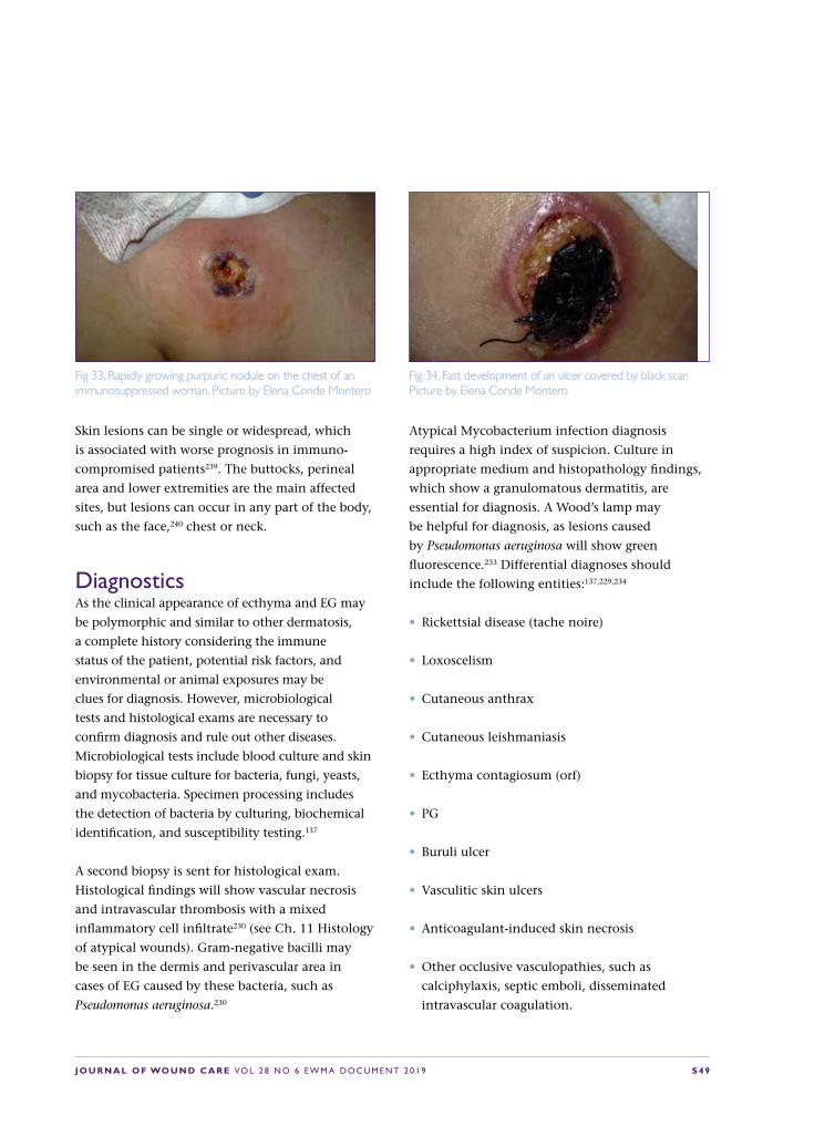
J O U R N A L O F WO U N D C A R E VO L 2 8 N O 6 E W M A D O C U M E N T 2 0 1 9 S 4 9
Skin lesions can be single or widespread, which
is associated with worse prognosis in immuno-
compromised patients239. The buttocks, perineal
area and lower extremities are the main affected
sites, but lesions can occur in any part of the body,
such as the face,240 chest or neck.
DiagnosticsAs the clinical appearance of ecthyma and EG may
be polymorphic and similar to other dermatosis,
a complete history considering the immune
status of the patient, potential risk factors, and
environmental or animal exposures may be
clues for diagnosis. However, microbiological
tests and histological exams are necessary to
confirm diagnosis and rule out other diseases.
Microbiological tests include blood culture and skin
biopsy for tissue culture for bacteria, fungi, yeasts,
and mycobacteria. Specimen processing includes
the detection of bacteria by culturing, biochemical
identification, and susceptibility testing.137
A second biopsy is sent for histological exam.
Histological findings will show vascular necrosis
and intravascular thrombosis with a mixed
inflammatory cell infiltrate230 (see Ch. 11 Histology
of atypical wounds). Gram-negative bacilli may
be seen in the dermis and perivascular area in
cases of EG caused by these bacteria, such as
Pseudomonas aeruginosa.230
Atypical Mycobacterium infection diagnosis
requires a high index of suspicion. Culture in
appropriate medium and histopathology findings,
which show a granulomatous dermatitis, are
essential for diagnosis. A Wood’s lamp may
be helpful for diagnosis, as lesions caused
by Pseudomonas aeruginosa will show green
fluorescence.233 Differential diagnoses should
include the following entities:137,229,234
• Rickettsial disease (tache noire)
• Loxoscelism
• Cutaneous anthrax
• Cutaneous leishmaniasis
• Ecthyma contagiosum (orf)
• PG
• Buruli ulcer
• Vasculitic skin ulcers
• Anticoagulant-induced skin necrosis
• Other occlusive vasculopathies, such as
calciphylaxis, septic emboli, disseminated
intravascular coagulation.
Fig 33. Rapidly growing purpuric nodule on the chest of an immunosuppressed woman. Picture by Elena Conde Montero
Fig 34. Fast development of an ulcer covered by black scar. Picture by Elena Conde Montero

S 5 0 J O U R N A L O F WO U N D C A R E VO L 2 8 N O 6 E W M A D O C U M E N T 2 0 1 9
TreatmentConsidering the broad spectrum of aetiologies in
ecthyma and EG patients, we should differentiate
between the specific microbiological treatment and
the general topical measures for wound healing in
all cases.
Therapy for patients with limited ecthyma lesions
may just consist of topical antibiotics. However,
for cases with wider involvement, oral antibiotics
are recommended.
In the case of clinical suspicion of EG, mainly in
immunocompromised patients, empiric anti-
pseudomonal antibiotics after sending a sample
for culture are highly recommended.137,233 A
combination of an aminoglycoside and an anti-
pseudomonal β-lactam antibiotics is commonly
recommended for treatment of EG, regardless of
the presence of bacteriemia.236
Some of the prognostic factors of the disease,
mainly in severe cases, are the time of onset of
antibiotic therapy and the patient’s response
to therapy.229,236
The microbiological results will indicate
the appropriate treatment, which may
be antibiotic and/ or antifungal therapy.
Intravenous anti-pseudomonal antibiotics are
necessary for treatment for pseudomonal EG.
In immunocompromised patients with non-
pseudomonal EG, intravenous microbiological
treatment should also be considered.
Once the aetiological treatment of EG has been
established, the necrotising soft-tissue lesion
must be locally managed. Necrotic tissue should
be removed. In small lesions, both autolytic and
bedside surgical debridement can be satisfactory.
However, in large necrotic ulcers, aggressive
surgical debridement followed by skin grafting may
be necessary. The surgical approach to Pseudomonas
and non-Pseudomonas cases is similar.137
Conclusions and recommendationsThe term EG is not limited to the cutaneous
features of Pseudomonas aeruginosa bacteraemia
in immunocompromised patients. Consequently,
healthy patients, absence of bacteraemia or non-
pseudomonal findings should not be considered
exclusion criteria for EG diagnosis.
Clinical features of EG are non-specific and other
ecthyma-like lesions should be considered in
differential diagnosis; such as arthropod bites,
anticoagulant-induced skin necrosis, PG, vasculitis
or occlusive vasculopathies.
Complete anamnesis and a skin biopsy for
microbiological and histological exams are
essential to allow early recognition of EG.
Considering that prompt treatment is
necessary to reduce mortality, especially in
immunocompromised patients, early empiric
anti-pseudomonal antibiotics and debridement of
necrotic tissue are the key therapeutic strategy.

J O U R N A L O F WO U N D C A R E VO L 2 8 N O 6 E W M A D O C U M E N T 2 0 1 9 S 5 1
10. Other types of atypical wounds
In addition to the atypical wounds covered in
other chapters, there are a number of atypical
wounds which challenge the clinician in
terms of recognition, diagnosis, management and
treatment.7 In this chapter we describe atypical
wounds associated with inflammatory, medication-
related and infectious processes.
Inflammatory woundsUlcerative lichen planusLichen planus is an inflammatory skin disease.
The classical forms of lichen planus (purple,
pruritic, polygonal, and papules/plaques) are easily
recognised, but there are subtypes that are more
uncommon.7,241 These subtypes include ulcerative
lichen planus (ULP), which affects typically adult
women. Typical features include hypertrophic,
hyperkeratotic, bullous and scaly lesions on the
plantar surface of the feet. Also mucosal involvement
may occur241. Along with skin manifestations
there can be absence of toe nails and scarring.242
Diagnosis is confirmed by biopsy, also direct
immunofluorescence specimen is recommended in
order to exclude LP pemphigoids. Serologic testing
for hepatitis is also recommended, as hepatitis C
has been associated with ULP.7 Treatment results
can be unsatisfactory. Systemic treatments include
retinoids and cyclosporine and local therapy options
include UVA-1, corticosteroids and tacrolimus.7,241,242
In severe cases surgical excision and grafting can be
considered.241 It is also important to know that this
chronic inflammatory disease may predispose to
SCC, and therefore serial biopsies are recommended
on ulcers resistant to treatment.7,243
Ulcerative necrobiosis lipoidica Necrobiosis lipoidica is an inflammatory disorder
of the subcutaneous tissue. Traditionally it has
been linked with diabetes and approximately
50–80 % of patients with necrobiosis lipoidica
have diabetes.244 However, it is important to
keep in mind that not all of necrobiosis lipoidica
patients have diabetes or will have it during
follow-up.245 Necrobiosis lipoidica typically appears
as yellowish-brown, shiny annular lesions in the
pretibial region of the legs in young to middle-aged
women. In approximately 35% of the cases, these
lesions ulcerate246 (Fig 35) and the ulcers are in
most cases hard-to-heal. Treatment options include
topical, intralesional and systemic corticosteroids;
tacrolimus, cyclosporine, hydroxychloroquine,
clofazimine, phototherapy, pentoxifylline
as well as biologic agents as etanercept and
infliximab.7,244 In resistant cases skin grafting is
an option.244 Even if there is no evidence about
Fig 35. Ulcerating necrobiosis lipoidica. Picture by Kirsi Isoherranen.

S 5 2 J O U R N A L O F WO U N D C A R E VO L 2 8 N O 6 E W M A D O C U M E N T 2 0 1 9
the benefits of compression therapy in ulcerative
necrobiosis lipoidica,247 clinical experience strongly
recommends it; also in terms of prevention. As
with ULP, there are also reports of SCC developing
in necrobiosis lipoidica.248
Ulcerative panniculitisPanniculitis is a general term for an inflammatory
disorder of the subcutaneous adipose tissue.
Clinically it presents as tender, sometimes
ulcerating, subcutaneous nodules in the lower
extremities. Different forms of panniculitis are
nodular vasculitis (see Ch. 3 Vasculitides as causes
of wounds), erythema nodosum, pancreatic
panniculitis and α-Antitrypsin deficiency.
Especially in pancreatic panniculitis there can be
systemic symptoms such as fever, abdominal pain,
arthritis, ascites and pleural effusions.3 Treatment
depends on the specific form of panniculitis.
Ulcerative sarcoidosisSarcoidosis is an inflammatory, granulomatous
inflammatory disorder affecting lungs, skin
and heart. Cutaneous involvement occurs in
approximately 25% of patients and of these
lesions, approximately 5% ulcerate.249 The typical
patient is a young adult black woman and lesions
appear predominantly in the pretibial area of the
legs. Treatment can be challenging; methotrexate,
corticosteroids, anti-malarials, mycophenolate
mofetil and thalidomide have been listed as
treatment options.7,250 There are also reports of
response to adalimumab.251
Other inflammatory atypical wounds Patients with rheumatoid arthritis (RA) have
an increased risk of leg ulceration.252 This patient
group is challenging, as wounds are typically
multi-aetiological. In addition of having a
venous, arterial, diabetic or pressure origin an
inflammatory component is often seen.7 RA is a
well-known comorbidity in PG and vasculitis ulcers
(see Ch. 5: Pyoderma gangrenosum and Ch. 6:
Vasculitides as causes of wounds) and presumably
vasculitic ulcers have been reported also in Felty’s
syndrome referring to the combination of RA,
neutropenia and splenomegaly.253
Tophaceous gout is also known to associate with
ulcers, most of which occur on the foot region.254
Autoimmune blistering diseases, such as pemphigoid
and pemphigus, can also create non-healing ulcers
especially in the lower leg region. Digital ulcers
associated with systemic sclerosis may act as red
flag for increased mortality,255 but this entity will not
be covered in detail in our document.
Iatrogenic ulcerationsHydroxyurea is a cytotoxic agent used mostly in
the treatment of myelodysplastic disorders. It has
been reported that it cause ulceration in nearly
10% of patients receiving continuous treatment.256
Fig 36. An ulcer caused by hydroxyurea (a). The same ulcer two months after cessation of hydroxyurea (b) and one month later the ulcer was completely healed. All pictures by Kirsi Isoherranen.
a
b

J O U R N A L O F WO U N D C A R E VO L 2 8 N O 6 E W M A D O C U M E N T 2 0 1 9 S 5 3
Ulcers typically occur after some years after
initiating therapy and present as small, well-
defined, bilateral and painful ulcers around the
ankles (Fig 36).257 Withdrawal of the drug generally
heals these ulcers, but advanced therapies can be
used to accelerate the healing.258
Methotrexate therapy has been associated with a
variety of cutaneous adverse effects, and cutaneous
ulceration and severe epidermal necrosis are
also rare side effects.259,260 Ulcerations are typical
for psoriasis patients, with type I and type II
ulcerations. Type I ulcerations occur in psoriatic
plaques, whereas type II ulcerations occur in skin of
patients with psoriasis with some other pathology,
like venous insufficiency. The medication should be
then discontinued or dose-adjusted.7
Both heparin and coumarin medications can
cause necrosis (see Ch 4. Occlusive vasculopathy).
These adverse reactions can be fatal and
therefore should be diagnosed promptly.234
Typically they appear some days after starting
the therapy, but there are also reports of delayed
onset.261 Diagnosis is clinical, and the lesions
start as erythematous plaques and evolve into
necrotic and haemorrhagic blisters or ulcers.
Diagnosis prompts immediate discontinuation of
coumarin or heparin, and the use of supportive
fresh-frozen plasma or vitamin K in case of
warfarin necrosis.234
Nicorandil, which is a commonly prescribed
medication for angina, has also been reported to
cause oral and anal ulceration and stopping of the
medication led to healing of the ulcers.262,263
Infectious ulcerationsThere are a number of microorganisms that can
cause ulcers. The amount of these ulcers is likely
to rise in Western countries due to travelling and
immigration. Treatment of these infectious ulcers
should be planned in cooperation with an internist
specialised in infectious diseases.
Cutaneous leishmaniasis presents as an ulcer
that can heal by itself over 3–18 months, but
can also lead to severe scarring and up to 10% of
cutaneous leishmaniasis cases progress to more
severe manifestations (Fig 37).264
In Acanthamoeba, ulcerated nodules can be the
first sign of disseminated disease in immuno-
compromised patients.7,265 Amebiasis is a protozoal
infection that can cause clinically evident
disease after months or years following exposure.
Cutaneous ulcerations due to amebiasis occur
typically perianally.7,266 Mycobacteria are also
capable of causing a wide range of cutaneous
manifestations, and Buruli ulcer is caused by
Mycobacterium ulcerans and other related slowly
growing mycobacteria. Buruli ulcer occurs mainly
in the lower extremities and presents first as a
painless nodule or a large indurated plaque that
evolves in a period of approximately four weeks
into an ulcer with undermined borders.267
MiscellaneousAge-related cutaneous fragility (dermatoporosis)
Fig 37. A 16-year-old refugee with an ulcer in the malleolar region for 6 months. PCR sampling revealed Leishmania tropica. Picture by Kirsi Isoherranen.
S 5 4 J O U R N A L O F WO U N D C A R E VO L 2 8 N O 6 E W M A D O C U M E N T 2 0 1 9
is associated with deep dissecting haematomas
that occur after minimal trauma. Management
involves surgical evacuation of the haematoma
in order to avoid larger ischaemic necrosis of the
deeper subcutaneous tissues.7 Trigeminal trophic
syndrome results from injury to the trigeminal nerve
and the resulting dysaesthesia leads to self-mutilating
behaviour causing atypical facial ulcers.268 Leg ulcers
are also a chronic and severe complication of sickle
cell disease (SCD). In a prospective, observational
cohort study it was demonstrated that healing
of SCD ulcers was independently associated with
wound area and duration, rather than with SCD
biological or clinical characteristics.269

J O U R N A L O F WO U N D C A R E VO L 2 8 N O 6 E W M A D O C U M E N T 2 0 1 9 S 5 5
11. Histology of atypical wounds
To provide optimal treatment it is essential
to follow a diagnostic synthesis process to
reach the correct assessment. In order to
do this diagnostic techniques aiming at a specific
diagnosis should be employed. Histological
diagnosis plays an important role in the armament
of these diagnostic techniques. However, while not
every chronic wound has to be biopsied, a biopsy
should be considered if the appearance of the ulcer
is atypical or if there is deterioration or failure to
progress after 12 weeks of active treatment.270
Biopsy technique‘A biopsy should be considered in every chronic
ulcer which has an atypical appearance, or fails
to respond to adequate topical therapy after four
to 12 weeks.’
Choosing the site for biopsy is an essential
decision. The biopsy should be taken from the
wound edge, since histology from the wound bed
will mostly show reparative and secondary findings
and will not provide any useful information as
to the aetiology of the wound.198,271 The biopsy
should include epidermis, dermis and sufficient
subcutaneous tissue (Fig 38). It is difficult to
obtain this by the punch technique, which
produces relatively superficial specimens so a
narrow spindle scalpel biopsy is the preferred
technique. This method also has the advantage
that the defect edges can be designed and sewn
under less pressure for a better healing result. A
specimen yielded by a scalpel biopsy provides
sufficient material for an adequate histologic
examination and can be used for additional
investigations (eg direct immunofluorescence if
vasculitis or autoimmune bullous disease is being
suspected or microbiological tissue cultures in
the cases of infection). In many cases, it is also
useful to consult dermato-pathologist for the
right biopsy site.
Fig 38. Biopsy technique, general basis (a). Biopsy technique, specific for purpuric/necrotic lesions (b) Drawings by Elena Conde Montero
a b

S 5 6 J O U R N A L O F WO U N D C A R E VO L 2 8 N O 6 E W M A D O C U M E N T 2 0 1 9
Each specimen for routine histology should be placed
into separate bottles of 10% formalin solution,
while specimens for direct immunofluorescence or
microbiology should be placed in saline solution if
the specimen can reach the lab within 24 hours or
otherwise in a special transport medium. Specific
attention to labelling with the name of the patient,
date, and the site of each specimen is essential. The
pathology requisition form should contain a brief
and relevant clinical history along with a description
of the wound or lesion. Most specimens are
processed for haematoxylineosin staining to examine
the morphology and cellular detail of the specimen.
For some differential diagnoses, the pathologist will
order additional special stains. In the following the
histological findings in different types of atypical
wounds are discussed.272,273
VasculitisVasculitis is defined as inflammation directed at
vessels, which compromises or destroys the vessel
wall leading to haemorrhagic and/or ischaemic
events.43 A skin biopsy is the gold standard for the
diagnosis of cutaneous vasculitis. For the highest
yield, biopsy specimens for haematoxylineosin
staining should be taken from an established
purpuric lesion (eg <72 hours old). For direct
immunofluorescence biopsy specimens, an acute
lesion (<24 hours old) provides the highest yield.274
Secondary vasculitis due to connective tissue
disorder (CTD) occurs frequently in systemic lupus
erythematosus (SLE), rheumatoid arthritis (RA)
and Sjögren’s syndrome and less commonly in
dermatomyositis, scleroderma and polychondritis.
In general, CTD vasculitis shows more widespread
organ involvement. A skin biopsy is likely to show a
mixed, mostly small and, less commonly, muscular
vessel neutrophilic vasculitis with lesions that can
resemble either typical CLA or polyarteritis nodosa
(PAN); the coexistence of small and muscular vessel
vasculitis in the same biopsy specimen or different
lesional skin is a characteristic feature of CTD
vasculitis. Extravascular histologies can provide a clue
to diagnosis of CTD vasculitis, such as the presence
of interface dermatitis (eg SLE and dermatomyositis),
dermal or subcutaneous sclerosis in scleroderma.275
Livedoid vasculopathyLivedoid vasculopathy ulcers occur typically
bilaterally in the malleolar region and are very
painful. In early and ulcerative lesions an increased
number of dermal vessels with intraluminal fibrin-
plugs and fibrinoid vessel wall necrosis are present
(Fig 39). There is usually a mild inflammatory
infiltrate but no sign of destruction of the vessel
walls, so that this condition should be considered
as a pseudo-vasculitis or a vasculopathy. A typical
sign is also porcelain white areas known as
‘atrophie blanche’ and signifies that the epidermis
is atrophic and the dermis shows a scleroderma like
hyalinisation.276
Cutaneous polyarteritis nodosa (CPAN)CPAN should be suspected in patients presenting
with tender nodules, livedo vasculopathy, livedo
racemosa, ulcers, acral gangrene and/or neuropathy.
A deep incisional biopsy, down to the fascia,
is likely to demonstrate neutrophilic muscular
vessel vasculitis at arterial branch points, located
either at the dermalsubcutis junction or within
the subcutis. Recently, two entities have been
described that show clinical features of CPAN, but
a lymphocytic arteritis on biopsy: macular arteritis
and lymphocytic thrombophilic arteritis. These
variants of vasculitis may simply represent latent or
late evolutionary stages of CPAN.275
Pyoderma gangrenosumPG ulcers have very variable clinical presentations,
but typically show pain out of proportion and
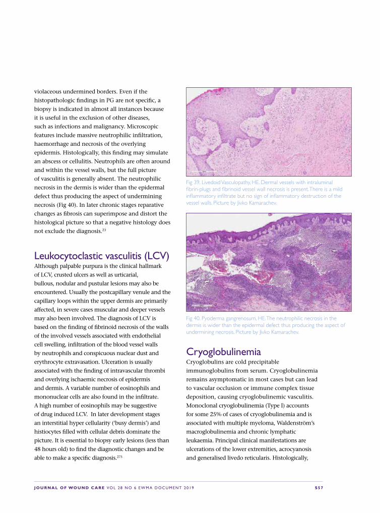
J O U R N A L O F WO U N D C A R E VO L 2 8 N O 6 E W M A D O C U M E N T 2 0 1 9 S 5 7
violaceous undermined borders. Even if the
histopathologic findings in PG are not specific, a
biopsy is indicated in almost all instances because
it is useful in the exclusion of other diseases,
such as infections and malignancy. Microscopic
features include massive neutrophilic infiltration,
haemorrhage and necrosis of the overlying
epidermis. Histologically, this finding may simulate
an abscess or cellulitis. Neutrophils are often around
and within the vessel walls, but the full picture
of vasculitis is generally absent. The neutrophilic
necrosis in the dermis is wider than the epidermal
defect thus producing the aspect of undermining
necrosis (Fig 40). In later chronic stages reparative
changes as fibrosis can superimpose and distort the
histological picture so that a negative histology does
not exclude the diagnosis.23
Leukocytoclastic vasculitis (LCV)Although palpable purpura is the clinical hallmark
of LCV, crusted ulcers as well as urticarial,
bullous, nodular and pustular lesions may also be
encountered. Usually the postcapillary venule and the
capillary loops within the upper dermis are primarily
affected, in severe cases muscular and deeper vessels
may also been involved. The diagnosis of LCV is
based on the finding of fibrinoid necrosis of the walls
of the involved vessels associated with endothelial
cell swelling, infiltration of the blood vessel walls
by neutrophils and conspicuous nuclear dust and
erythrocyte extravasation. Ulceration is usually
associated with the finding of intravascular thrombi
and overlying ischaemic necrosis of epidermis
and dermis. A variable number of eosinophils and
mononuclear cells are also found in the infiltrate.
A high number of eosinophils may be suggestive
of drug induced LCV. In later development stages
an interstitial hyper cellularity (‘busy dermis’) and
histiocytes filled with cellular debris dominate the
picture. It is essential to biopsy early lesions (less than
48 hours old) to find the diagnostic changes and be
able to make a specific diagnosis.275
CryoglobulinemiaCryoglobulins are cold precipitable
immunoglobulins from serum. Cryoglobulinemia
remains asymptomatic in most cases but can lead
to vascular occlusion or immune complex tissue
deposition, causing cryoglobulinemic vasculitis.
Monoclonal cryoglobulinemia (Type I) accounts
for some 25% of cases of cryoglobulinemia and is
associated with multiple myeloma, Waldenström’s
macroglobulinemia and chronic lymphatic
leukaemia. Principal clinical manifestations are
ulcerations of the lower extremities, acrocyanosis
and generalised livedo reticularis. Histologically,
Fig 39. Livedoid Vasculopathy, HE. Dermal vessels with intraluminal fibrin-plugs and fibrinoid vessel wall necrosis is present. There is a mild inflammatory infiltrate but no sign of inflammatory destruction of the vessel walls. Picture by Jivko Kamarachev.
Fig 40. Pyoderma gangrenosum, HE. The neutrophilic necrosis in the dermis is wider than the epidermal defect thus producing the aspect of undermining necrosis. Picture by Jivko Kamarachev.

S 5 8 J O U R N A L O F WO U N D C A R E VO L 2 8 N O 6 E W M A D O C U M E N T 2 0 1 9
small vessels in the upper dermis are filled with
homogeneous, eosinophilic, periodic acid-Schiff
staining (PAS) positive material beneath the
ulcerations and there is no vasculitis (Fig 41). In
polyclonal and mixed cryoglobulinemias, which
are most often associated with a hepatitis C virus
infection, the clinical and histological picture of an
acute leukocytoclastic vasculitis develops.277
Martorell HYTILUDiagnostic Martorell HYTILU, is an obliterating
subcutaneous arteriolosclerosis with medial
hypertrophy and medial calcinosis (Fig 42). Since
these changes have a focal character as in PAN it is
essential to have a sufficiently large and deep biopsy
and to perform multiple serial cuts. Best results are
obtained with a 5mm narrow but 3–4cm long and
sufficiently deep excisional biopsy, which should be
embedded longitudinally to provide a large surface
for inspection microscopically.278
Cholesterol embolismCholesterol emboli originate from atherosclerotic
plaques and can be dislodged in the course of
endovascular procedures or anticoagulation.
Predominantly the lower limbs are involved and
skin lesions include purpura, livedo reticularis, ulcers
and gangrene. Microscopically, clefts indicating the
sites of cholesterol depositions are found in arterioles
and small arteries in the dermis and subcutis. Fibrin
thrombi often surround the cholesterol crystals.
There may be extensive surrounding necrosis of
epidermal and dermal structures. Due to the focal
character of the embolic event, several and serial cuts
are necessary to make the diagnosis.73
CalciphylaxisCalciphylaxis is a life-threatening condition
affecting predominantly patients with chronic
renal failure on dialysis with abnormal calcium/
phosphorous metabolism and a secondary
or tertiary hyperparathyroidism. Clinically
erythematous indurated plaques predominantly
on the lower limbs with a tendency to ulcerate are
found. These ulcers heal poorly and are very painful.
The histological hallmark is the calcification of
small to medium-sized arteries and arterioles in the
dermis and subcutis. In pronounced cases, diffuse
interstitial calcification may be seen. Haemorrhage
and necrosis may result279.
Necrobiosis lipoidicaNecrobiosis lipoidica (NL) affects 0.3%–1.2% of
Fig 41. Cryoglobulinemia, HE: Small vessels in the upper dermis are filled with homogeneous, eosinophilic material. Picture by Jivko Kamarachev
Fig 42. Martorell HYTILU, HE. Focal obliterating subcutaneous arteriolosclerosis with medial hypertrophy and calcinosis. Picture by Jivko Kamarachev

J O U R N A L O F WO U N D C A R E VO L 2 8 N O 6 E W M A D O C U M E N T 2 0 1 9 S 5 9
diabetic patients.280 NL is characterised by single
or multiple asymptomatic red to yellow shiny
plaques that gradually enlarge and within which,
due to atrophy, dermal blood vessels are seen on
the surface. Ulceration occurs in about 15% patients
with NL usually following minor trauma.281
The histopathological changes in NL involve the
whole dermis. Areas of necrobiosis are usually
more extensive and less well defined than in
granuloma annulare. Between the necrobiotic
areas granulomatous inflammatory infiltrates is
present consisting of histiocytes, lymphocytes and
variable numbers of multinucleate Langerhans
or foreign body giant cells. In old atrophic
lesions and in the centre of plaques, there is little
necrobiosis and much dermal fibrosis. The lack of
pronounced depositions of mucin as detected by
the Alcian blue stain helps to differentiate NL from
granuloma annulare. Vascular changes ranging from
endothelial swelling through wall thickening and
fibrosis to overt lymphocytic vasculitis are more
prominent in NL than in granuloma annulare.282
Infectious diseasesA variety of infections can cause chronic leg
ulcers including: EG, tropical ulcer, atypical
mycobacteria, leprosy (neuropathic ulceration),
cutaneous tuberculosis, deep fungal infection
and leishmaniasis.
Essential for the diagnosing of infectious ulcers
are bacterial and mycological swabs and cultures.
Histology is often useful providing hints of infectious
origin or even a specific diagnosis if the infectious
agent can be identified with a specific stain as is the
case the PAS-stain identifying fungi, Gram-staining
for bacteria, Giemsa-Stain identifying Leishmania and
Ziehl-Nielsen stain which may be useful to identify
mycobacteria. Histological findings suggesting
an infectious ulcer include hyperplasia of the
epidermis which may reach pseudo-epitheliomatous
dimensions, a mixed inflammatory infiltrate
with foci of dense neutrophilic granulocytes and
formation of granulomas with or without caseation
and admixtures of numerous lymphocytes, plasma
cells and some eosinophils.
EG is typically associated with Pseudomonas
aeruginosa septicaemia but similar lesions have been
observed in patients with other bacterial, fungal
and viral infections. The lesions of EG result from
bacterial invasion of the walls of arteries and veins
in the skin and subcutaneous tissue. Impaired
immunity through the known predisposing factors
leads to increased susceptibility to infections.
Histopathology of EG lesions shows vascular
necrosis and intravascular thrombosis with a scarce
mixed inflammatory infiltrate and with basophilic
deposits suggestive of bacterial colonies surrounding
the affected vessels. In sections stained with Gram
stain, Gram-negative rods are identified. Blisters,
haemorrhage, oedema and necrosis are seen in
the surrounding dermis. However the histological
findings are highly variable dependent upon
the evolutionary stage of the lesion, which may
be affected by temporal and therapeutic factors.
Correlation with the clinical picture, and the results
of the tissue culture, and blood culture is essential.
TumoursSCC can arise de novo on the lower leg or as a
complication of chronic leg ulcers. SCC arising
within a leg ulcer has a far worse prognosis than
actinically induced SCC arising de novo on the
lower leg. This is often compounded by a delay in
diagnosis.283 Chronic ulcers that are unresponsive
to treatment, with new ulcer extensions or new
growths within or at the ulcer margins, should be
carefully examined for signs of SCC and biopsied if
indicated. If a poorly or moderately differentiated
SCC is found, early and aggressive surgical
intervention is essential.

S 6 0 J O U R N A L O F WO U N D C A R E VO L 2 8 N O 6 E W M A D O C U M E N T 2 0 1 9
BCCs are also found as ulcerating forms on
the lower leg. Histology is typical presenting a
basophilic epithelial tumour connected to the
epidermis, consistent of tumour cell strands and
complexes with a palisading arrangement of the
tumour cells in the periphery and with a well-
developed fibrous stroma with typical retraction
artefacts between tumour and stroma.
The lower legs are a predilection site for Kaposi
sarcoma in which an atypical vascular proliferation
is found in the reticular dermis, with prominent
erythrocyte extravasation and numerous plasma
cells in the tumour stroma. Immunohistochemistry
demonstrating human herpes virus 8 (HHV8) in the
atypical endothelia supports strongly the diagnosis
of Kaposi sarcoma.
The lower leg is also a predilection site for the
high grade diffuse large B cell lymphoma of the
lower leg, an aggressive entity which may present
as a quickly growing ulcerated tumour with an
aggressive course and fatal outcome. Histology
showed dense sheets of large CD20+, bcl2+,
MUM1+ B-cell blasts in the dermis and subcutis.284
Even melanoma of the acral lentiginous type can
be disguised as a DFU and only histology can
provide an adequate diagnosis.285
DrugsAlthough anecdotic case reports of ulcers arising
in association with the intake of a plethora
of medications exist in the literature the best
documented associations in large series concern
hydroxyurea and coumarin.
In one study of 41 cases of leg wounds due to
therapy with hydroxyurea, biopsy specimens were
taken from 11 patients. Histopathologic findings
showed epidermal atrophy, dermal fibrosis, and
scar tissue without vascular lesion. In one case a
few small capillaries were occluded, as in livedoid
vasculitis, but direct immunofluorescence staining
findings were negative. In another case there were
fibrinoid deposits in the dermis with extravasation
of red blood cells and leuko-cytoclastic vasculitis.286
In coumarin necrosis, sections show variable degrees
of epidermal and dermal necrosis. There are extensive
intravascular thrombi within capillaries and venules.
There remaining patent vessels are dilated.88,287
OthersAcute exogenous noxious substances (physical,
chemical, including as a manifestation of auto-
aggression) can lead to acute toxic changes in
the skin which may have typical histological
presentation — pale necrosis of the epidermis
and sometimes of the dermis with only scarce
neutrophil granulocytes infiltrate in the
surrounding viable tissue and no signs of vascular
damage or inflammatory process which could
account for the necrosis.
Conclusions and recommendationsHistology is an important diagnostic tool in atypical
wounds. A chronic wound should be biopsied if
atypical in appearance or failing to respond to
adequate therapy after a maximum of 12 weeks. The
biopsy should be taken from the wound edge. A deep
narrow but sufficiently long and deep spindle biopsy
comprising epidermis, dermis and subcutaneous fat
provides optimal material for histological diagnosis.
Providing the pathologist with a concise clinical
history and description increases the chances of
obtaining a specific diagnosis.

J O U R N A L O F WO U N D C A R E VO L 2 8 N O 6 E W M A D O C U M E N T 2 0 1 9 S 6 1
12. Practical aspects of diagnosing and treating atypical wounds
S uccessful wound management relies heavily
on the rapid and precise diagnosis of the
aetiology of the wound.8 Even evidence-
based wound management is destined to fail
without the exact diagnosis. The most common
chronic wound aetiologies are venous, arterial,
mixed venous and arterial leg ulcers, DFUs
and PUs.6,288 If the wound does not fit within
these typical presentations, an atypical cause
of wound should be suspected.6,7 Prompt and
exact diagnosis is essential also as many of the
atypical wounds need specific treatment differing
from the traditional ones and an incorrect
diagnosis can lead to devastating outcomes and
increased mortality.10,11,100
DiagnosisFor diagnosing atypical wounds it is very
important to take a thorough patient history
including a neurovascular assessment, which can
help to provide important information in order to
reach a diagnosis (Table 11).
The appearance of the wound and the surrounding
skin is also important. Hypergranulation, pathergy,
violaceous and undermined borders, necrosis,
atypical location, severe pain (out of proportion
which is normally associated with wounds),
livedo racemosa and reticularis and rapidly
progressive ulceration are signs of an atypical cause
(Fig 46).6,7,234 Wound assessment is a continuous
process, therefore it is important to establish
whether the wound is decreasing in size with
signs of healing by proper treatment (compression
therapy, offloading, optimal local therapy).
If a wound is initially classified as venous or
neuropathic but subsequently does not show signs
Table 11. Patient history related to the risk of atypical wound occurrence
Inflammatory bowel disease, rheumatoid arthritis, hematologic malignancy?
PG?
Uremic disease, dialysis? Calciphylaxis?
Hypertension, obesity, type II diabetes?
Martorell HYTILU? Hidradenitis suppurativa (HS)
Rheumatoid arthritis, connective tissue disease?
Vasculitides?
Vitamin K antagonist medication?
Calciphylaxis, Martorell HYTILU, Coumarin necrosis?
Hydroxyurea medication? Hydroxyurea-induced ulcers?
Travel history, refugee? Leishmaniasis? Tuberculosis?
Older age, immunosuppression, long persistence?
Malignancy?
Personality disorder? Artefactal ulcers?
Recurrent lesions in the axillae, under the female breast, ano-genital region or groin?
HS?

S 6 2 J O U R N A L O F WO U N D C A R E VO L 2 8 N O 6 E W M A D O C U M E N T 2 0 1 9
of healing during 4–12 weeks, at least one biopsy
should be taken (see Ch. 11 Histology of atypical
wounds).9 In case of suspected infectious ulcers,
a wide biopsy for tissue cultures for atypical
bacterial, mycobacterial and fungal infections
should be taken.6
In order to obtain a reliable specimen, the biopsy
should be taken with a scalpel from the wound
edge. When suspecting Martorell HYTILU and
ulcers associated with calciphylaxis punch
biopsies do not usually obtain a specimen deep
enough therefore a wide biopsy is preferable
(see Ch.5 Martorell HYTILU and calciphylaxis).
Punch biopsies can be used if malignancy is
suspected, but in case of diagnosing other atypical
wound aetiologies, they usually do not obtain a
specimen deep enough. In case of vasculitides,
in addition to the biopsy specimen, a specimen
for direct immuno fluorescence (DIF) should be
taken. For this, an acute lesion (<24 hours old)
provides the highest yield.274,289 Preferably the
histopathological and DIF specimens should
be analysed by a dermatopathologist who is
experienced in atypical wounds. However, it is
important to remember that a negative histology
does not exclude an atypical wound; also a
clinical assessment is needed and the patient
should be referred to a dermatologist.7
In addition to an atypical wound on the leg, patients
may also have concurrent arterial and/or venous
insufficiency. Therefore, careful arterial evaluation
(pulse palpation, measurement of the ankle-brachial
pressure index (ABPI)) is needed for every patient.
If the patient has signs of venous insufficiency,
a duplex ultrasound of lower extremities should
be performed. Exclusion of neuropathy using
microfilament sensory testing is also crucial. In
addition to these tests and the biopsy specimen,
measurement of blood pressure and further
laboratory testing is often needed. This includes
complete blood cell count, metabolic profile,
urinalysis and possible stool guaiac test. If vasculitis
or vasculopathy are considered, then testing
should include also anti-nuclear antibodies (ANA),
anti-neutrophilic cytoplasmic antibodies (ANCA),
rheumatoid factor, anticyclic citrullinated peptide,
serum protein electrophoresis, hepatitis profile,
cryoglobulins, lupus anticoagulant, antiphospholipid
and anticardiolipin antibodies, protein C, protein
S, factor V Leiden and antithrombin III levels,
cryofibrinogens and coagulation.6,290
TreatmentOnce a diagnosis has been established an
interdisciplinary approach should be adopted in
order to reach a successful outcome. This might
include one or more of the following health
professionals (eg dermatologist, vascular and plastic
surgeon, nephrologist, rheumatologist, tissue
viability nurse, podiatrist).288 The exact nature of the
treatment relies on the specific type of an atypical
wound and will be presented in the individual
chapters of this document. Where patients with a
wound are being treated by immunosuppressive
agents, NPWT and skin grafting can also be
considered but only after the inflammation has been
reduced by immunosuppressive therapy (Fig 43).291
Fig 43. Thin split-thickness skin graft. Drawing by Elena Conde Montero

J O U R N A L O F WO U N D C A R E VO L 2 8 N O 6 E W M A D O C U M E N T 2 0 1 9 S 6 3
The use of local corticosteroids in PG is widely
accepted. However, topical corticosteroids may also
help to reduce excessive inflammation that impairs
wound healing in other atypical wounds.7,288,292
Local application of corticosteroids to the wound
bed are well known to reduce hypergranulation.
However, they have also other benefits, such as
limiting the spread of necrosis in Martorell HYTILU
and other necrotising vasculitides293 (see Ch. 13
Topical treatment for atypical wounds).
As the immunosuppressive agents can cause delayed
wound healing, advanced therapies,294 epidermal
grafting295 and surgical procedures are important also
in terms of reducing healing time. It is also possible
to perform punch grafting, which is a traditional
method to obtain thin split-thickness skin grafts
containing epidermis and papillary dermis (Fig 44).
Grafts are obtained under a local anaesthetic
with a punch, curette or surgical blade and are
placed directly on the wound bed. The donor
site is normally the thigh, which subsequently
heals by secondary intention. This procedure can
be performed in an outpatient basis144,296,297 with
100% graft uptake (Fig 45). However, there may
be occasions where grafts do not adhere but it
is still useful as they release growth factors, cells
that promote wound epithelialization and reduce
pain.144,296,297 Local pressure and immobilisation
during the first 3–4 days after the procedure is
essential for graft taking.
The first dressing change should take place
5–7 days after the surgical procedure. During
subsequent dressing changes it is essential to
avoid excessive cleansing in order to avoid
altering the healing environment.144
Compression therapy (as provided by bandages,
hosiery or compression wraps) can be beneficial
for the majority of leg ulcers with oedema, even
if the cause of the wound is not venous. After
exclusion of advanced peripheral arterial occlusive
insufficiency arterial, compression therapy should
be used in all patients with lower leg ulcers.298
However, compression therapy may be painful in
patients with inflammatory PG or vasculitides,
therefore lower pressures of 20mmHg should be
used initially41 (see Ch. 2 Pyoderma gangrenosum).
Fig 44. Grafts obtained with a curette (a). Grafts obtained with a punch and surgical blade (b). Point-like bleeding is observed after graft excision (c and d). Pictures by Elena Conde Montero
a
b
c
d
a b
c d
Fig 45. Chronic ulcer secondary to vasculitis (a). Grafts obtained with surgical blade placed on the wound (b). Clinical aspect 3 weeks later (c) Complete epithelialisation seven weeks after the procedure (d). Pictures by Elena Conde Montero

S 6 4 J O U R N A L O F WO U N D C A R E VO L 2 8 N O 6 E W M A D O C U M E N T 2 0 1 9
Fig 46. Algorithm for diagnosing atypical wounds
Detailed patient history and physical examination
The wound cannot be classified as typical (venous, arterial, mixed venous and arterial leg ulcer, diabetic foot ulcer or pressure ulcer) and/or shows atypical features OR does not heel with appropriate treatment within 4 to 12 weeks
Biopsy specimen and if needed, direct immunofluorescence specimen Laboratory tests (see also Ch. 12 Practical aspects of diagnosing and treating atypical wounds
Inflammatory bowel disease, rheumatoid arthritis,
hematologic malignancy?
Reddish-violaceous and undermined wound borders, pustules?
Pathergy?
Severe pain?
Rheumatoid arthritis, connective tissue disease?
Palpable purpura, livedo racemosa, subcutaneous nodules?
Wounds with necrosis and violaceous borders?
Lesions with a predilection for lower legs bilaterally, multifocal lesions?
Pathergy?
Hypergranulation?
Atypical pigmentation?
Not healing with standard treatment?
Atypical location?
Pyoderma gangrenosum? Vasculitides? Malignant wound?
Hypertension, type II diabetes, obesity, vitamin K antagonist?
Necrotic, painful ulcers in the lateral lower leg or over Achilles tendon?
Uremic disease, hyperparathyroidism, vitamin K antagonist medication?
Painful, necrotic, rapidly progressing wounds?
Martorell HYTILU? Calciphylaxis?
Lesions in the peri-genital area and axillae?
Inflamed or non-inflamed nodules, abscesses and draining or non-draining tunnels? Scarring? Smoking?
Immunosuppressed patient?
Travel history, younger age, drug abuse?
Vesicle or pustule with grey-yellowish exudate?
Erythematous or purpuric macules that rapidly evolve to vesicles or nodules and subsequent ulcers with black eschar formation with a surrounding erythematous halo?
Vesicles appearing on an edematous background and coalescing to form large bullaes?
Multiple lesions?
HS?
Personality disorder?
Geometric or sharply demarcated ulcers?
Artefactal ulcer? Ecthyma gangrenosum?

J O U R N A L O F WO U N D C A R E VO L 2 8 N O 6 E W M A D O C U M E N T 2 0 1 9 S 6 5
The local therapy of atypical wounds is described
in detail in the chapter ‘Local treatment
of atypical wounds’. In the first phase of
treatment, it is generally recommended to use
gentle, non-adherent dressings to avoid the
pathergy phenomenon.
ConclusionIn cases where the wound does not fit into the
typical aetiologies, it is important to stop and
think whether an atypical wound is possible
Fig 46). The best approach is to consult an expert
in atypical wounds, such as a dermatologist. The
treatment relies on an interdisciplinary approach.
S 6 6 J O U R N A L O F WO U N D C A R E VO L 2 8 N O 6 E W M A D O C U M E N T 2 0 1 9
13. Topical treatment for atypical wounds
When treating atypical wounds, it
is necessary to ensure a holistic
assessment and appropriate clinical
examination of the patient to ensure an accurate
diagnosis can be made before any treatment regime
is initiated. Determining the aetiology of the
wound can be challenging as many of them mimic
other wounds so further investigations including a
biopsy sample may be needed.299
Assessment The patient, the wound and the environment
must be assessed to ensure a successful outcome.
The patient needs support in their environment
to ensure dressing changes are done in a timely
way either in the community or the clinic using
an aseptic technique to prevent further infections
or trauma. The patient’s lifestyle needs to be
addressed including diet, smoking cessation and
exercise to promote wound healing. Assessment
of the patient’s perfusion, pain levels and factors
that influence healing including comorbidities
need to be incorporated in to the plan of care
before any dressing choice is made.300 The wound
assessment itself includes size, depth, tissue,
exudate, odour, and periwound skin. If infection
is suspected a wound specimen is needed for
culture and sensitivity to ensure appropriate
antibiotics are used. Equipment such as pressure
redistribution devices aim to reduce shearing
forces and address the microclimate to preserve
skin.301 A collaborative approach between patients,
relatives, carers, health professionals, industry
and the organisation will ensure high-quality care
delivery for patients.
Preparation is needed to establish a plan of care.
Availability of resources may vary from country
to country. Listening to the patients’ needs with
empathy will increase adherence and gain trust.
The ‘goal of the week’ or treatment rationale
should be agreed by the team and initiated with the
appropriate treatment and dressing regime. Weekly
assessment and measurements of the wound need
to be recorded and if no improvement is seen after
four weeks a specialist referral is required.302
Cleansing Cleansing wounds has been debated in the
literature and currently there is a dearth of
literature available on which solution is best
for reducing infection and promoting wound
healing.303 However, Hodgkinson & Nay suggest
it is essential for effective skin care to remove dry
scales, assess the leg fully and most importantly
it makes patients feel better.304 The British
Association of Dermatology (BAD) guidelines
2014 recommend that patients ‘Should be
encouraged to wash their legs in warm potable tap
water when bandages are changed’.305 Legs should
be washed using a soap substitute for example
hydromol.305 The limb should be thoroughly dried
paying attention to the skin between the toes.306
Dried exudate and devitalised skin adhering to the
wound and surrounding skin will only promote
the production of bacteria so removing it using a
good skin care regime is recommended. Solutions
such as hydrogen peroxide, iodine, potassium
permanganate, chlorhexidine, saline and water
are used for cleansing and topical treatments of
atypical wounds to reduce bacterial bioburden

J O U R N A L O F WO U N D C A R E VO L 2 8 N O 6 E W M A D O C U M E N T 2 0 1 9 S 6 7
and biofilm in wounds, but more robust research
is required to definitively assist clinicians in their
choice of cleansing agent.307 Recognising signs
of infection and managing it is imperative for
patients with atypical wounds.308 Lastly, using
an appropriate emollient afterwards will prevent
cracks in skin and dry scales building up.309
Topical steroids Chronic wounds, particularly atypical wounds,
are commonly characterised by an excessive
inflammatory response, with a greater number
of inflammatory cells such as neutrophils,
lymphocytes and macrophages.310 This results in
the wound getting stuck in a chronic inflammatory
phase with high levels of cytokines, MMPs and
reactive oxygen species. This results in excessive
degradation of the ECM and progression of wound
healing.311 Therefore, the use of topical steroids
maybe useful in treating these wounds. A study by
Taheri et al. suggests that topical corticosteroids
can be used to suppress inflammation and
fibroblast activation in the treatment of burns to
reduce the incidence of hypertrophic scarring and
keloids.312 Also, case series exist of the successful
use of topical steroids in the treatment of excessive
hypergranulation.313 However, they are not
commonly used in general wound care practice.
Topical steroids can be used as a single steroid
topical agent or in combination with antimicrobial
and antifungal agents in wounds displaying
excessive and abnormal inflammatory features
despite good clinical wound care practice.292
Topical steroids should be prescribed by health
professionals with prescriptive authority only and
used in compliance with local policy and medicinal
regulatory body. A combined topical agent is often
first choice as single steroid agents typically increase
the risk of infection and thins the lining of skin.314
A study of chronic wounds including atypical
wounds such as (vasculitis, PG, mixed vessel
disease ulcers, rheumatoid ulcers) demonstrated
that using a combined topical steroid, antifungal
and antibiotic improved healing rates and reduced
exudate and pain symptoms for patients.292 The
study recommends that topical steroid treatment
should be considered when abnormal inflammation
is present in chronic wounds but only when the
correct diagnosis is made and where the patient can
be monitored carefully.
Dressings The health professional carrying out the dressing
must be trained skilled and competent. Using a
framework such as ‘TIME’ can aid the selection of
appropriate dressings.315
T=Tissue: good tissue perfusion is essential for
wound healing. The presence of necrosis or
devitalised tissue hinders this and therefore needs
to be removed by an appropriate method. Some
atypical wounds may need regular debridement.
However, some atypical wounds cannot be debrided
for example active debridement can be deleterious
in the active inflammatory phase of vasculitic
ulcers and pyoderma gangrenosum. But when the
immunosuppressive treatment has reduced the

S 6 8 J O U R N A L O F WO U N D C A R E VO L 2 8 N O 6 E W M A D O C U M E N T 2 0 1 9
inflammation, then topical treatment to debride
the wound can be carried out. A full overview of
approaches to debridement can be found in the
‘EWMA Document: debridement: an updated overview
and clarification of the principle role of debridement.’38
I=Infection: establishing the cause of infection
is necessary to ensure appropriate use of
antimicrobials. In atypical wounds, the presence
of bacteria delay healing therefore reducing the
wound bioburden should reduce adverse effects. In
cases of infections, antibiotics may be indicated,
but holistic assessment of the patient is needed
first to ensure correct diagnosis is made. Many
patients with PG have completed multiple courses
of antibiotics before being diagnosed. The use
of antiseptics may prevent the development
of infections that will minimise antibiotic
use, as well as promoting quicker healing. The
use of topical antimicrobial therapy may be
especially helpful for example, the eradication
of bacteria before grafting for patients with
atypical wounds.307,316 A full overview about
the EWMA position regarding antimicrobials
and non-healing wounds can be found in the
‘EWMA document: Antimicrobials and non-healing
wounds. Evidence, controversies and suggestions’317
as well as by following the EWMA Antimicrobial
Stewardship campaign which is being developed
in partnership with the British Society for
Antimicrobial Chemotherapy (BSAC).317
M=Moisture: exudate facilitates the inflammatory
response and aids autolytic debridement and
epithelisation. Wound exudate should be assessed
at dressing change noting colour, amount, high
viscosity, odour and consistency. However, too
much moisture can lead to maceration and damage
surrounding skin.318
E=Epithelial cells: will migrate across the wound by
active cell division. The wound edge needs to be
healthy for wound closure to occur.
Dressing selection should be based on the
condition of the wound bed and the nature/tissue
types present. Key considerations should include:
• Promote a moist wound healing environment
• Management of tissue types present, necrosis,
slough, granulation
• Management of exudate
• Management of infection
• Promote haemostasis if bleeding present
• Management of odour
• Protection of the periwound environment
• Atraumatic dressing change
• Comfortable and aesthetically acceptable
to patients.
Pain Pain is a common feature of atypical wounds.
Therefore, a validated assessment tool should be
used to establish an individual’s level of pain and
ensure analgesia is given before dressing change
as many patients with atypical wounds suffer
unnecessarily before, during and after dressing
changes if pain is not managed well.
Avoiding adhesive dressings such as gauze to
minimise pain or trauma to surrounding tissues
is essential.319 Consider the wear time, moisture
balance levels to ensure comfort for patients.
Wounds as associated with calciphylaxis and PG
can be particularly painful. Non-adherent, sensitive
dressings have been found to be practical in clinical
use, as PG and vasculitis wounds have the pathergy
phenomenon, ie they worsen with minor trauma.

J O U R N A L O F WO U N D C A R E VO L 2 8 N O 6 E W M A D O C U M E N T 2 0 1 9 S 6 9
The use of an easy to apply multifunctional dressing
such as polymeric membrane dressings capable of
stimulating the healing process, providing pain
relief and continuous wound bed cleansing, can
simplify dressing selection and involve patients in
their care.320
Dressings There are many dressings available but agreeing a
‘goal of the week’ can help to aid in the selection
for clinicians. If an allergy to the dressing is
suspected or if evidence of contact dermatitis patch
testing is required as soon as possible.
Categories of dressings include:
• Passive simple dressings used for protection
such as surgical pads useful for absorbing
minimal exudate
• Active dressings, which interact with the wound
bed, such as for example polymeric membrane
dressings are useful in gently softening and
debriding hard eschar in wounds associated
with calciphylaxis
• Advanced therapies interact and advance
wound healing such as larvae therapy for
example are useful in managing complex
challenging wounds.
Dressing types include the following:
• Alginates
• Antimicrobials
• Foams
• Transparent Films
• Hydrocolloids
• Gelling fibre
• Hydrogels
• Hydropolymers
• Non-adherents
• NPWT
• Polymeric membrane dressings
• Super absorbers.
NB: Please refer to your local dressing formulary
regarding sizes, cost, indications etc. Specifically
regarding NPWT please refer to the ‘EWMA
Document: Negative Pressure Wound Therapy’.321

S 7 0 J O U R N A L O F WO U N D C A R E VO L 2 8 N O 6 E W M A D O C U M E N T 2 0 1 9
14. The patient perspective
T he negative impact of wounds is well
recognised in the literature with many
patients suffering with anxiety and
depression. Many patients with atypical wounds
suffer daily both physically with odour, exudate,
pain, reduced mobility and psychologically
with negative emotions, loneliness and
depression.14 Mortality rates are higher for
patients with atypical wounds not only due to
their comorbidities and lower socio-economic
circumstances leading to higher rates of suicide
but for many there can be a delay in diagnosis
prolonging their pain and suffering.322 Stress
has also been shown to impact negatively on
wounds by reducing healing rates.15 There is a
dearth of literature written about the impact of
atypical wounds, despite these wounds being a
chronic condition. Therefore, a literature review
of the patient perspective of chronic and atypical
wounds was included in the search.
Autonomy Understanding wound care from a patient’s
perspective is the hallmark of a successful
practitioner.323 Sleep deprivation, pain or loss of
independence can make the patient appear to
be difficult to clinicians. But the reality is when
they suffer in this way in addition to wound pain
they often feel they are not listened to. Loss of
autonomy means a lack of control and this can be
frustrating for many patients who want to have
their independence. Proper symptom management
can improve a patient’s QoL. When they are
involved in their care and an agreed plan is in
place adherence is more likely to occur leading to
success of wound healing.
The incidence and prevalence of chronic wounds
including atypical wounds is likely to increase.
With an aging population and increases in the
prevalence of obesity, diabetes and lower extremity
arterial disease many patients have several co-
morbidities resulting in hard to heal wounds.324
Therefore, encouraging patients to be independent
and engaged with their wound care is needed.
Avoiding hospital admissions and managing in
their homes is encouraged to improve QoL and
reduce the cost to health services. Some patients
need to work and cannot afford time off to attend
clinics where as others, perhaps the elderly, see the
clinic as an opportunity to engage with others.
Patient involvement Patient involvement has long been recognised as
a key to success in the management of chronic
atypical wounds.325 Ensuring dignity and respect
is essential in the delivery of high-quality care.
However, this can lead to conflict when patients
dispute with health professionals regarding what
they want and what they get.326 Many of these
patients have been treated with many products
and medications before an accurate diagnosis is
made.103 Communication and understanding of
these complex wounds is essential to empower
patients and to ensure the correct diagnosis is
obtained and patient engagement is achieved.
From this perspective it is possible to identify
J O U R N A L O F WO U N D C A R E VO L 2 8 N O 6 E W M A D O C U M E N T 2 0 1 9 S 7 1
wellbeing as more of a priority for the patient
than wound healing.
Education Education is important for health professionals
and patients. Health-care staff need to improve
their knowledge base and their ability to teach.
Informing the patient in a language they can
understand and internalise is important for their
understanding. This message needs to be delivered
at an appropriate level of contextualisation of
the individual to ensure their efforts in health
promotion are engaged, for example smoking
cessation is more effective when the smoker is
engaged. Flexibility of the content, context and
delivery methods will aid in the full impact of
patient understanding and engagement.327 For
example, some like videos and some prefer leaflets.
Elderly people may not be comfortable with
information technology.328
Patient expectations It is equally important to identify the expectations
of the patient when prioritising treatment. For
example, patients need to understand that a
biopsy is only a diagnostic and not a treatment
for skin cancer.103 Therefore, a contract or agreed
plan of care should be implemented following a
thorough assessment with the patient to ensure
both clinicians and clients can reach mutual goals.
Informed decision-making by way of appropriate
information and tools allow us to gain consent
in a way that allows the patient to take charge of
their situation. Reassessment and actions recorded
ensure progress is being made and appropriate
referrals are made in a timely fashion when
required. Communication between members of
the MDT is accurate when recorded and prevents
replication of requests especially when all members
of the team use electronic patient record.329
Patient capacity Allowing time to express their concerns can be
challenging in a busy clinic but a chance to be
involved and take ownership should be offered
to the patient.330 Wounds International argue,
health care professionals have a duty of care to
act as advocates with the patient’s best interest by
ensuring the patients care is a priority and where
possible to involve patients in shared decision
making at all stages even when wound healing is
not possible.324 However, patient capacity needs to
be considered when health professionals are making
a clinical judgement with the patient’s best interest
at the forefront. Sometimes we do not always agree
with the patient decision and sometimes they delay
surgery when recommended but if fully mentally
competent then that’s their choice and we both
must live with the consequences. Multidisciplinary
meetings can be useful in this scenario where other
health professionals can share their thoughts and
experiences in dealing with these patients.
Patients are no longer passive in their care and
this has been facilitated by having internet
access and being empowered through national
frameworks. The UK Department of Health in

S 7 2 J O U R N A L O F WO U N D C A R E VO L 2 8 N O 6 E W M A D O C U M E N T 2 0 1 9
2011 implemented a strategy of no decision
about me without me which demonstrates value
in the management of chronic care.331,332 Patient
outcomes are measured by levels of satisfaction
and their experience as well as safety of treatments.
However, a study by Green et al.333 demonstrated
that 38% of patients did not raise their concerns.
Indeed 8% who did were not acknowledged
or disregarded by their community nurse. If
shared decision making is the future of wound
care, then we need to examine our consultation
skills. However, increases in qualitative research
demonstrates patients want more choice over their
care dressings and other treatments.334 We need to
work with our patients if we are to get the best out
of this resource largely ignored in the past.
Digital imagingA study explored the patient perspective of using
digital imaging to monitor the progress of their
wound. This process demonstrated that patients
were open to the idea and they felt it was beneficial
not only for their own progress but also felt it was
a positive teaching tool for health professionals.
Participants felt that when used in conjunction
with wound measurements and description elicited
an accurate record of their wound. The study also
showed that most patients did not care who took
the photo a doctor, nurse or photographer nor
were they concerned whether it was a hospital
or personal device. However, due to introduction
of General Data Protection Regulation, this has
changed. Having a secure accurate measurement of
the wound is essential for record management.14
Quality of life Patients with atypical wounds need psychological
support and assistance from several members of
multidisciplinary team not only delivering wound
care but, also in accessing and engaging in the
resources needed for them to cope day-to-day in
basic needs and companionship to become valued
members of society again.329 A study demonstrated
that social support mechanisms helped patients
to cope better with their treatment and have a
better QoL.335
A phenomenological Portuguese study in 2017
involving ten patients living with chronic leg
ulcers reported that they felt limited, depressed,
anxious and isolated. They were embarrassed
about the odour, leakage of the wound in bed
with their partner and many felt they were
isolated from social events. They found comfort
in their religious belief which helped them to
cope. Understanding the lived experience of
patients with atypical wounds by using validated
HR QOL tools allows us to develop solutions
tailored to patient needs.334,336
Conclusion Research, case-controlled studies and audits
help us to understand the patient perspective of
these complex wounds and plan our resources
where needed most. Data from research allows
for standardisation and consistency in decision
pathways. Care pathways allow patients to
understand the process and to gain trust in the
health professional. Reassurance is very often
what they need and an understanding of the
rationale for their treatment. Appropriate and
timely access to care is also a key performance
indicator. We have a duty to ensure the safe
delivery of quality care by reducing risks such
as surgical site infection (SSI) and abiding by
international evidence-based guidelines. Moore
et al. emphasise that a good multidisciplinary
approach is paramount for good wound care
management.288 Another article points out that
patient held records gives ownership to patients
allowing for continuity of care and shared care if
they are responsible and can locate the notes or
remember to bring them with them to clinic.337

J O U R N A L O F WO U N D C A R E VO L 2 8 N O 6 E W M A D O C U M E N T 2 0 1 9 S 7 3
An electronic card that patients could carry may
be of benefit but not for all patients. Patient
related outcome measurements (PROMS) are a
good way to ascertain if patient needs are met.
A pilot study demonstrated this method using
an online questionnaire filled in by patients
with a tablet or online computer giving valuable
and reliable data on the patient experience of
arthroscopy and an option to seek a follow-
up appointment only when needed reducing
unnecessary routine appointments.338 It is this
increased level of understanding that leads to
informed choice, which leads to better care with
finite resources from the patient perspective,
which is what we want.
RecommendationsThere is still very little evidence available to health
professional to deal with the complexity of what
it is like to live with atypical wounds for patients.
However, if we are to meet the patient’s needs and
work corroboratively outcomes will be better. There is
an opportunity for health professional to learn from
both experience and available literature to improve
pathways and ensure correct diagnosis is made so
that correct management is delivered. Improvements
in education regarding atypical wounds is necessary
for both clinicians and patients. Lastly, further robust
research studies are needed to measure outcomes
such as quality of life and pain management
strategies not just wound healing outcomes.

S 7 4 J O U R N A L O F WO U N D C A R E VO L 2 8 N O 6 E W M A D O C U M E N T 2 0 1 9
15. Health economy and organisation
The cost of management of atypical
wounds is largely unknown due to the
varied aetiologies and because data is
often incorporated in chronic wound and lower
leg ulcer studies. However, both financial and
human costs can be considerable in the case
of atypical wounds as there is often a delay in
diagnosis and patient comorbidities and chronic
inflammation cause stalled wound healing. Due
to the rarity there is an absence of high-quality
evidence in the management; studies often include
a range of low-quality evidence-based topical and
systemic treatment options and studies are often
problematic as recruitment is difficult. Conditions
such as PG are clinically diagnosed after excluding
other diagnoses, because usually there is no specific
diagnostic tests or histological findings,339 so there
can be a delay in the commencement of treatment,
causing further financial and human cost.
A comprehensive introduction to the elements
and methodologies of wound care related health
economic studies is beyond the scope of this
document but can be found in the ‘EWMA Out-
comes in controlled and comparative studies of
non-healing wounds’ document.340
Cost to the health economy An estimated cost of atypical wounds to health
economies is limited. The cost of chronic wounds
in general is well documented341 and is expected
to escalate with an ageing population as well as
increasing prevalence of obesity and diabetes and
other autoimmune diseases, which will further
increase costs to patients and health care resources
in the coming decades.
There are over 400,000 Australians living with
a chronic wound.342 A third of the wounds are
present for at least six months and one in five for
a year or more.342 In Australia, AUD$3 billion per
year is spent on chronic wounds, an estimated
2% of the national health-care expenditure.342
Similar results are seen in other Western counties.
In the UK treatment of chronic wounds accounts
for 3% of total health-care expenditure,343 and a
study from Wales using National Health Service
(NHS) data concluded that chronic wounds have
a prevalence of 6% and consume at least 5.5%
of NHS expenditure.342 345 Approximately 2.2
million patients have a chronic wound344 with
the annual NHS cost of managing these wounds
and associated comorbidities being £5.3 billion.341
Across Europe, 2–4% of health-care expenditure is
spent on wounds and this figure is on the rise.343
The average cost of treating a wound ranges from
€6,000–€10,000 per year.343 In addition to the
initial cost of a wound to healing, it is common
for wounds to recur resulting in ongoing costs for
many years.
Cost to organisationsChronic wounds impose huge costs on health
services and organisations. Information about
cost is useful for raising the profile of the problem
among decision makers and also provides
estimates of the savings if the problem is reduced.
Data on the treatment cost of wounds for

J O U R N A L O F WO U N D C A R E VO L 2 8 N O 6 E W M A D O C U M E N T 2 0 1 9 S 7 5
organisations are inconsistent and vary depending
on disease process methodology and evaluation
method. The lack of evidence impedes health-care
decisions, policy development and improvement
in resource allocation and optimisation of the
cost-benefit ratio.
The STOP GAP trial, a multicentre randomised
control trial was conducted to provide better
evidence to guide clinicians to improve treatment
decisions for patients with PG.339 As part of this
study, Mason et al. compared the cost-effectiveness
of cyclosporin and prednisolone as first line
treatment for patients with PG as a preferred use
of NHS resources.339 For patients with small lesions
<20cm2 neither treatment was more cost-effective
than the other.339 However, cyclosporin treatment
was found to be a cost effective option in patients
with large lesions (>20cm2) and there was a
reported improvement in QoL.339 Decisions about
treatment will continue to be informed by patient
preference and drug adverse reactions; however
the study suggested NHS resources would be better
directed to cyclosporin than prednisolone in terms
of cost-effectiveness and improved QoL outcomes
for patients who have large wounds and is likely to
reflect routine care in this setting.339
This economic evaluation is the first published
cost-effectiveness analysis relating to PG and it
opens up opportunities for further research in
this area.339
There is often a delay in diagnosis also for patients
with HS. Health resource use, including direct
and indirect costs for treating HS is limited. This
chronic, progressive, painful and debilitating
skin condition affects a younger population
often requiring ongoing lifelong treatment and
wound dressings.
Studies have demonstrated the increase in use of
emergency department and inpatient admissions
for patients with HS.343 A retrospective cohort
study conducted in the UK revealed the high
burden of frequent outpatient, emergency and
inpatient attendances, with the majority of
hospital admissions under the care of surgical
specialties.345 Using Hospital Episode Statics data
from 1 April 2007 — 31st December 2013, 11,359
patients were studied. There were 65,544 inpatient
admissions; 43,773 emergency attendances and
303,204 outpatient appointments.346 Based on the
mean number of admissions per patient per year,
the mean hospital resource use cost for a patient
with HS was £2,027 per patient per year.346
As the majority of patients in the study were of
working age, the socioeconomic impact of time
lost from work associated with frequent hospital
visits is significant and likely to be consistent with
a reported loss of 2.7 work days per year.347 In a
survey of employed patients with HS in the US, 58%
reported that the disease caused work absence, with
a mean absence of around 34 day’s annually.347
Skin cancers associated with non-healing wounds
of the lower leg may result from malignant
transformation as seen in a Marjolin ulcer or
may arise and mimic a leg ulcer and be treated as

S 7 6 J O U R N A L O F WO U N D C A R E VO L 2 8 N O 6 E W M A D O C U M E N T 2 0 1 9
vascular in origin, delaying the correct diagnosis
and appropriate treatment.178 The two main forms
of skin cancers are SCC and BCC, the latter being
the most common. These NMSCs are the most
common cause of cancer and one of the few
malignancies of increasing incidence especially
in Caucasian populations.348 The incidence of
melanomas is also increasing and together with
the rarer Merkel cell carcinoma, both tumours are
aggressive depending on the stage of diagnosis
and costly to treat with options such as surgery,
chemotherapy, immunotherapy, radiotherapy and
ongoing monitoring for recurrence.
In Australia, general practitioners see over one
million patient consultations per year for skin
cancers and the cost to the economy in medication,
clinical and procedural treatments, pathology and
diagnostic tests is the highest cost to the health
system of all cancers.349 The majority of skin cancers
are caused by exposure to the sun and the incidence
is one of the highest in the world, two to three
times the rate in Canada, the US and the UK.349 In
2010 it was calculated that the total cost of NMSC
(diagnosis, treatment and pathology) was $AUD 511
million and was estimated to increase to over $700
million in the coming years.349
Clinical practice guidelines are increasing in the
field of wound care and dermatology, but gaps
exist in demonstrating their cost-effectiveness
for organisations. A study found that guideline-
based care may be cost-saving or cost-effective
in most circumstances.350 The quality and
usefulness of reviewed studies for decision
making varied, however they concluded that with
better information and higher-quality economic
evaluations decision makers will have increased
confidence to promote guideline-based care.350
With this in mind, a Danish study has advocated
that by adopting a national strategy based on best
practice guidelines, it may be possible to reduce the
costs of wound management by 30%.351
A delay in diagnosis of an atypical wound can
occur as a result of limited access to specialist
treatment and varying levels of expertise, skill
set and knowledge. Significant savings have been
demonstrated in some studies by the adoption
of evidence-based care, such as that provided by
the community and outpatient specialist wound
clinics.325 Education, training and credentialing
of health professionals in the management of
chronic wounds is an indirect expense that must
be factored into organisational budgets when
evaluating costs and providing sufficient resources
and cost-effective wound care.325
Implementing best practice guidelines developed
by professional bodies, such as EWMA, and
ensuring that those involved in the provision
of wound care are educated appropriately, could
have a significant impact on the financial burden
of wound care. Potentially, the greatest efficiency
gains can be achieved through effective training
and education of health professionals; involved in
wound care. A structured programme of training
and providing credentials for health professionals
involved in wound care, supported by best practice
guideline and protocols, has real potential to
reduce the variation in practice, reduce costs and
improve patient outcomes.352–354
Cost to the patient Finally, it’s important to remember the role of
the patient in the cost of managing their wound.
A total estimate of the financial cost to the
patient is unknown. The burden of these wounds
and the direct impact on the patient and their
family is often underestimated. Atypical wounds
are often a symptom or complication of other
comorbidities, affecting all age groups and can be
life-long. Treatment pathways are often confusing
and complex with multiple specialities managing
the wound, resulting in cost of additional travel,
absence from work, numerous diagnostic tests and

J O U R N A L O F WO U N D C A R E VO L 2 8 N O 6 E W M A D O C U M E N T 2 0 1 9 S 7 7
payment to specialists. Chronic wounds severely
reduce QoL and patient wellbeing, capacity to
work and increase social isolation.
Depending on the financial support and access to
wound care services and wound products, they can
also impose substantial personal costs to the patient.
The trend towards delivery of wound care in the
community has transferred the care costs from the
health system to the patient in countries that do
not reimburse community-based care expenses.
Costs for the patient such as travel expenses for
domiciliary visits, wound dressings and compression
bandages can be significant and may comprise
their care decisions. Furthermore, these additional
factors confound direct comparisons of health-care
expenditure on wound management across health-
care systems, and over time.

S 7 8 J O U R N A L O F WO U N D C A R E VO L 2 8 N O 6 E W M A D O C U M E N T 2 0 1 9
16. Conclusions and future perspectives
A typical wounds are estimated to
comprise about 20% of all chronic
wounds.3–5 There is a high demand
on prospective multicentre clinical trials with
well-defined outcomes. Following an extensive
literature search clinical trials in the field of
atypical wounds are scarce, with many reports’
retrospective or case studies. In case of PG,
there are two prospective randomised trials;
one compared infliximab with placebo during
an observation period of two weeks, and due to
the superiority of infliximab in terms of clinical
improvement (46% versus 6%), after these two
weeks the non-responders in placebo group
received also infliximab.35
The other compared in a multicentre trial
with 112 participants the efficacy of cyclosporin
and prednisolone. Cyclosporin and prednisolon
did not differ across a range of objectives and
patient reported outcomes.34 RCTs in the case
of calciphylaxis are also under way. However
future trials to compare new treatments, such
as biologic drugs, head-to-head with prednisolone
or cyclo-sporin are needed.253 While there is
research currently in progress this author
group recommend future projects incorporating
atypical wounds should include the following:
1. In future trials recommended outcomes would
be speed of healing time (for instance at
six weeks), time to healing, patient reported
pain, health related quality of life, adverse
reactions to study drugs and complications
such as infection, cellulitis, sepsis and death.34
2. The exact pathophysiological mechanisms of
many of these inflammatory wounds are still
unknown. To tailor new targeted therapies in the
pipeline both for drug and local therapies we need
more precise understanding of the inflammatory
and occlusive mechanisms of these wounds.
For instance, we still lack a treatment that could
specifically reduce subcutaneous arteriolosclerosis.
3. There is evidence of a positive outcome to
early skin grafting in ulcers in the context of
arteriolopathy, with a breakdown in the vicious
cycle necrosis-inflammation and consequent pain
reduction and epithelialisation promotion.146 In
contrast to the traditional concept of waiting for
a suitable wound bed is present before grafting,
healing stimulation may be observed without
previous surgical, debridement, even if the wound
bed presents with slough.144 Consequently, it
would be interesting to study the exact mediators
involved in this phenomenon.
4. Since any non-healing wound, regardless of
its aetiology, is an inflammatory lesion, the
benefits of traditional treatments, such as topical
corticosteroids and tacrolimus, should be studied
further as new anti-inflammatory treatments
are urgently needed. An imbalance between
pro-inflammatory and anti-inflammatory
macrophage phenotypes play an important
role, so these cells should be the target of
new treatment strategies.355 Considering the
potential anti-inflammatory effect of skin
grafting in wound healing, it should be studied
how keratinocytes and molecules typically

J O U R N A L O F WO U N D C A R E VO L 2 8 N O 6 E W M A D O C U M E N T 2 0 1 9 S 7 9
involved in the final phase of epithelialisation
may regulate the microenvironment of an
abnormally prolonged inflammatory phase and
accelerate wound healing. Studies could analyse
the benefits of early punch grafting, which is a
simple and cost-effective technique to enhance
wound healing. Also, prospective studies on
NPWT and advanced therapies would provide
more strong evidence for treatment outcomes
and reimbursement.
5. Another challenge for the management of atypical
wounds is the lack of consensus and guidelines
for the standardisation of treatment and the lack
of economic studies. Moreover, most research
in the area of atypical wound management is
currently focused on wound outcomes during
the wound-healing phase without taking into
consideration the high rate of recurrences. It is
recommended that the time of recurrence for
ulcers, as well as their frequency, should also
be taken into consideration when examining
the effectiveness of treatments. Studies on the
diagnostic delays would be important both for a
patient’s and an organisational point of view. In
addition, cost efficacy is very seldom covered in
studies concerning atypical wounds. For clinicians
prompt diagnosis leads to better treatment results,
but for advocacy it is important to show these
facts in reliable figures.
6. Atypical wounds challenge the clinician both in
recognition, diagnosis and treatment. It is not
only important to broaden the knowledge of
health professionals of these wounds, but also
to establish specialised centres to treat the most
challenging atypical wounds patients. Organised
MDTs consisting of dermatologists, vascular and
plastic surgeons, rheumatologists, diabetologists,
infectious disease specialists, psychiatrists,
tissue viability nurses, podiatrist, nephrologist,
psychologists, nutritionists, physiotherapists and
social care workers should be included in care
pathways for these wounds.
Finally, we want again to emphasise the importance
of early suspicion and expert consultation with
these wounds. The ‘wait and see’– attitude can
lead to devastating outcomes. We hope the
algorithm helps in daily practice in the systematic
assessment of these wounds and that the document
will be disseminated and used among all health
professionals treating chronic wounds. We also
hope that the document stimulates practitioners
and scientists for a translational research approach
in the future. Lastly, we would like to acknowledge
all the feedback and comments we received from
professionals all over the World. We are sincerely
grateful for all your comments in order to ensure
that this document is of great quality and will
benefit both clinicians and patients.

S 8 0 J O U R N A L O F WO U N D C A R E VO L 2 8 N O 6 E W M A D O C U M E N T 2 0 1 9
17. References
1. Health Service Executive. Multidiscip. Team Health Serv. Exec. (2013)2. Wiecha J, Pollard T. The interdisciplinary eHealth team: chronic care
for the future. J Med Internet Res 2004; 6(3):e22. https://dx.doi.org/10.2196%2Fjmir.6.3.e22
3. Shanmugam VK, Angra D, Rahimi H, McNish S. Vasculitic and autoimmune wounds. J Vasc Surg Venous Lymphat Disord 2017; 5(2):280–292. https://dx.doi.org/10.1016/j.jvsv.2016.09.006
4. Shanmugam VK, Schilling A, Germinario A et al. Prevalence of immune disease in patients with wounds presenting to a tertiary wound healing centre. Int Wound J 2012; 9(4):403–411. https://dx.doi.org/10.1111/j.1742-481X.2011.00899.x
5. Körber A, Klode J, Al-Benna S et al. Etiology of chronic leg ulcers in 31,619 patients in Germany analyzed by an expert survey. JDDG: Journal der Deutschen Dermatologischen Gesellschaft 2011; 9(2):116–121. https://dx.doi.org/10.1111/j.1610-0387.2010.07535.x
6. Shelling ML, Federman DG, Kirsner RS. Clinical approach to atypical wounds with a new model for understanding hypertensive ulcers. Arch Dermatol 2010; 146(9):1026–1029. https://dx.doi.org/10.1001/archdermatol.2010.213
7. Hoffman MD. Atypical ulcers. Dermatol Ther 2013; 26(3):222–235. https://dx.doi.org/10.1111/dth.12048
8. Mooij MC, Huisman LC. Chronic leg ulcer : does a patient always get a correct diagnosis and adequate treatment? Phlebology 2016; 31(1_suppl Suppl):68–73. https://dx.doi.org/10.1177/0268355516632436
9. Alavi A, Sibbald RG, Phillips TJ et al. What’s new: Management of venous leg ulcers. J Am Acad Dermatol 2016; 74(4):627–640. https://dx.doi.org/10.1016/j.jaad.2014.10.048
10. Hradil E, Jeppsson C, Hamnerius N, Svensson Å. The diagnosis you wish you had never operated on: Pyoderma gangrenosum misdiagnosed as necrotizing fasciitis—a case report. Acta Orthop 2017; 88(2):231–233. https://dx.doi.org/10.1080/17453674.2016.1260403
11. Janowska A, Oranges T, Chiricozzi A et al. Synergism of Therapies After Postoperative Autograft Failure in a Patient With Melanoma of the Foot Misdiagnosed as a Pressure Ulcer. Wounds 2018; 30(4):E41–E43
12. Langan SM, Groves RW, Card TR, Gulliford MC. Incidence, mortality, and disease associations of pyoderma gangrenosum in the United Kingdom: a retrospective cohort study. J Invest Dermatol 2012; 132(9):2166–2170. https://dx.doi.org/10.1038/jid.2012.130
13. García-Lozano JA, Ocampo-Candiani J, Martínez-Cabriales SA, Garza-Rodríguez V. An Update on Calciphylaxis. Am J Clin Dermatol 2018; 19(4):599–608. https://dx.doi.org/10.1007/s40257-018-0361-x
14. Jones B, Lake A, Harper C. My wound and I: patients’ perspective. J Vis Commun Med 2016; 39(3-4):120–126. https://dx.doi.org/10.1080/17453054.2016.1246060
15. Solowiej K, Mason V, Upton D. Review of the relationship between stress and wound healing: part 1. J Wound Care 2009; 18(9):357–366. https://dx.doi.org/10.12968/jowc.2009.18.9.44302
16. Tiri H, Huilaja L, Jokelainen J et al. Women with Hidradenitis Suppurativa Have an Elevated Risk of Suicide. J Invest Dermatol 2018; 138(12):2672–2674. https://dx.doi.org/10.1016/j.jid.2018.06.171
17. Al Ghazal P, Herberger K, Schaller J et al. Associated factors and comorbidities in patients with pyoderma gangrenosum in Germany: a
retrospective multicentric analysis in 259 patients. Orphanet J Rare Dis 2013; 8(1):136. https://dx.doi.org/10.1186/1750-1172-8-136
18. Jockenhöfer F, Klode J, Kröger K et al. Patients with pyoderma gangrenosum - analyses of the German DRG data from 2012. Int Wound J 2016; 13(5):951–956. https://dx.doi.org/10.1111/iwj.12463
19. Wang EA, Steel A, Luxardi G et al. Classic Ulcerative Pyoderma Gangrenosum Is a T Cell-Mediated Disease Targeting Follicular Adnexal Structures: A Hypothesis Based on Molecular and Clinicopathologic Studies. Front Immunol 2018; 8:1980. https://dx.doi.org/10.3389/fimmu.2017.01980
20. Marzano AV, Borghi A, Meroni PL, Cugno M. Pyoderma gangrenosum and its syndromic forms: evidence for a link with autoinflammation. Br J Dermatol 2016; 175(5):882–891. https://dx.doi.org/10.1111/bjd.14691
21. Cugno M, Borghi A, Marzano AV. PAPA, PASH and PAPASH Syndromes: Pathophysiology, Presentation and Treatment. Am J Clin Dermatol 2017; 18(4):555–562. https://dx.doi.org/10.1007/s40257-017-0265-1
22. Cosgarea I, Lovric Z, Körber A, Dissemond J. Successful treatment of refractory pyoderma gangrenosum with ustekinumab only after excision of renal cell carcinoma. Int Wound J 2016; 13(5):1041–1042. https://dx.doi.org/10.1111/iwj.12377
23. Hadi A, Lebwohl M. Clinical features of pyoderma gangrenosum and current diagnostic trends. J Am Acad Dermatol 2011; 64(5):950–954.e2. https://dx.doi.org/10.1016/j.jaad.2010.01.049
24. Herberger K, Dissemond J, Hohaus K et al. Treatment of pyoderma gangrenosum: retrospective multicentre analysis of 121 patients. Br J Dermatol 2016; 175(5):1070–1072. https://dx.doi.org/10.1111/bjd.14619
25. Barbosa NS, Tolkachjov SN, el-Azhary RA et al. Clinical features, causes, treatments, and outcomes of peristomal pyoderma gangrenosum (PPG) in 44 patients: The Mayo Clinic experience, 1996 through 2013. J Am Acad Dermatol 2016; 75(5):931–939. https://dx.doi.org/10.1016/j.jaad.2016.05.044
26. Zuo KJ, Fung E, Tredget EE, Lin AN. A systematic review of post-surgical pyoderma gangrenosum: Identification of risk factors and proposed management strategy. J Plast Reconstr Aesthet Surg 2015; 68(3):295–303. https://dx.doi.org/10.1016/j.bjps.2014.12.036
27. Larcher L, Schwaiger K, Eisendle K et al. Aesthetic breast augmentation mastopexy followed by post-surgical pyoderma gangrenosum (pspg): clinic, treatment, and review of the literature. Aesthetic Plast Surg 2015; 39(4):506–513. https://dx.doi.org/10.1007/s00266-015-0499-3
28. André MF, Piette JC, Kémény JL et al.; French Study Group on Aseptic Abscesses. Aseptic Abscesses. Medicine (Baltimore) 2007; 86(3):145–161. https://dx.doi.org/10.1097/md.0b013e18064f9f3
29. Jockenhöfer F, Wollina U, Salva KA et al. The PARACELSUS score: a novel diagnostic tool for pyoderma gangrenosum. Br J Dermatol 2018; 10.1111/bjd.16401
30. Maverakis E, Ma C, Shinkai K et al. Diagnostic Criteria of Ulcerative Pyoderma Gangrenosum. JAMA Dermatol 2018; 154(4):461–466. https://dx.doi.org/10.1001/jamadermatol.2017.5980
31. Kutlubay Z, Tüzün Y, Wolf R. The pathergy test as a diagnostic tool. Skinmed 2017; 15(2):97–104

J O U R N A L O F WO U N D C A R E VO L 2 8 N O 6 E W M A D O C U M E N T 2 0 1 9 S 8 1
32. Al Ghazal P, Dissemond J. Therapy of pyoderma gangrenosum in Germany: results of a survey among wound experts. JDDG: Journal der Deutschen Dermatologischen Gesellschaft 2015; 13(4):317–324. https://dx.doi.org/10.1111/ddg.12585
33. Partridge AC, Bai JW, Rosen CF et al Effectiveness of systemic treatments for pyoderma gangrenosum: a systematic review of observational studies and clinical trials. Br J Dermatol 2018; 179(2):290–295. https://dx.doi.org/10.1111/bjd.16485
34. Ormerod AD, Thomas KS, Craig FE et al.Comparison of the two most commonly used treatments for pyoderma gangrenosum: results of the STOP GAP randomised controlled trial. BMJ 2015; 350 jun12 3:h2958. https://dx.doi.org/10.1136/bmj.h2958
35. Brooklyn TN, Dunnill MG, Shetty A et al. Infliximab for the treatment of pyoderma gangrenosum: a randomised, double blind, placebo controlled trial. Gut 2006; 55(4):505–509. https://dx.doi.org/10.1136/gut.2005.074815
36. Dissemond J, Assenheimer B, Engels P et al. M.O.I.S.T. - a concept for the topical treatment of chronic wounds. JDDG 2017; 15(4):443–445. https://dx.doi.org/10.1111/ddg.13215
37. Leiphart PA, Lam CC, Foulke GT. Suppression of pathergy in pyoderma gangrenosum with infliximab allowing for successful tendon debridement. JAAD Case Rep 2018; 4(1):98–100. https://dx.doi.org/10.1016/j.jdcr.2017.08.009
38. Strohal R, Dissemond J, Jordan O’Brien J et al. EWMA Document: Debridement: An updated overview and clarification of the principle role of debridement. J Wound Care 2013; 22(Sup1 Suppl):S1–S49. https://dx.doi.org/10.12968/jowc.2013.22.Sup1.S1
39. Seo HI, Lee HJ, Han KH. Hyperbaric oxygen therapy for pyoderma gangrenosum associated with ulcerative colitis. Intest Res 2018; 16(1):155–157. https://dx.doi.org/10.5217/ir.2018.16.1.155
40. Pichler M, Thuile T, Gatscher B et al. Systematic review of surgical treatment of pyoderma gangrenosum with negative pressure wound therapy or skin grafting. J Eur Acad Dermatol Venereol 2017; 31(2):e61–e67. https://dx.doi.org/10.1111/jdv.13727
41. Dissemond J, Storck M, Kröger K, Stücker M. [Indications and contraindications for modern compression therapy]. [Article in German]. Wien Med Wochenschr 2018; 168(9-10):228–235. https://dx.doi.org/10.1007/s10354-017-0605-z
42. Wilkes SR, Williams HC, Ormerod AD et al. Is speed of healing a good predictor of eventual healing of pyoderma gangrenosum? J Am Acad Dermatol 2016; 75(6):1216–1220.e2. https://dx.doi.org/10.1016/j.jaad.2016.07.049
43. Jennette JC, Falk RJ, Bacon PA et al. 2012 Revised International Chapel Hill Consensus Conference Nomenclature of Vasculitides. Arthritis Rheum 2013; 65(1):1–11. https://dx.doi.org/10.1002/art.37715
44. Linos K, Cribier B, Carlson J. Cutaneous cryoglobulinemic vasculitis. In: Dammacco F: HCV infection and cryoglobulinemia. Springer Verl, 2012
45. Ramos-Casals M, Stone JH, Cid MC, Bosch X. The cryoglobulinaemias. Lancet 2012; 379(9813):348–360. https://dx.doi.org/10.1016/S0140-6736(11)60242-0
46. Terrier B, Karras A, Kahn JE et al. The spectrum of type I cryoglobulinemia vasculitis: new insights based on 64 cases. Medicine (Baltimore) 2013; 92(2):61–68. https://dx.doi.org/10.1097/MD.0b013e318288925c
47. Sunderkötter CH et al. Nomenclature of Cutaneous Vasculitis: Dermatologic Addendum to the 2012 Revised International Chapel Hill Consensus Conference Nomenclature of Vasculitides. Arthritis Rheumatol. Hoboken NJ 70, 171–184 (2018)
48. Yus ES, Simón RS, Requena L. Vein, artery, or arteriole? A decisive question in hypodermal pathology. Am J Dermatopathol 2012; 34(2):229–232. https://dx.doi.org/10.1097/DAD.0b013e3182157ed1
49. Criado PR, Marques GF, Morita TC, de Carvalho JF. Epidemiological, clinical and laboratory profiles of cutaneous polyarteritis nodosa patients: Report of 22 cases and literature review. Autoimmun Rev 2016; 15(6):558–563. https://dx.doi.org/10.1016/j.autrev.2016.02.010
50. Carlson JA, Chen KR. Cutaneous vasculitis update: neutrophilic muscular vessel and eosinophilic, granulomatous, and lymphocytic vasculitis syndromes. Am J Dermatopathol 2007; 29(1):32–43. https://dx.doi.org/10.1097/01.dad.0000245198.80847.ff
51. Sunderkötter C, de Groot K. [Therapy of vasculitis and vasculopathies]. [Article in German]. Hautarzt 2008; 59(5):382–393. https://dx.doi.org/10.1007/s00105-008-1530-2
52. Yang YH, Yu HH, Chiang BL. The diagnosis and classification of Henoch–Schönlein purpura: An updated review. Autoimmun Rev 2014; 13(4-5):355–358. https://dx.doi.org/10.1016/j.autrev.2014.01.031
53. Heineke MH, Ballering AV, Jamin A et al. New insights in the pathogenesis of immunoglobulin A vasculitis (Henoch-Schönlein purpura). Autoimmun Rev 2017; 16(12):1246–1253. https://dx.doi.org/10.1016/j.autrev.2017.10.009
54. Sunderkötter C. Vasculitis of small blood vessels - some riddles about IgA and about the complexity of transmigration. Exp Dermatol 2009; 18(1):91–96. https://dx.doi.org/10.1111/j.1600-0625.2008.00791.x
55. Sunderkötter C, Seeliger S, Schönlau F et al. Different pathways leading to cutaneous leukocytoclastic vasculitis in mice. Exp Dermatol 2001 Dec;10(6):391–404. https://dx.doi.org/10.1034/j.1600-0625.2001.100602.x
56. Sindrilaru A, Seeliger S, Ehrchen JM et al. Site of blood vessel damage and relevance of CD18 in a murine model of immune complex-mediated vasculitis. J Invest Dermatol 2007; 127(2):447–454. https://dx.doi.org/10.1038/sj.jid.5700563
57. Requena L, Kutzner H, Angulo J, Renedo G. Generalized livedo reticularis associated with monoclonal cryoglobulinemia and multiple myeloma. J Cutan Pathol 2007; 34(2):198–202. https://dx.doi.org/10.1111/j.1600-0560.2006.00594.x
58. Braverman IM, Yen A. Demonstration of immune complexes in spontaneous and histamine-induced lesions and in normal skin of patients with leukocytoclastic angitis. J Invest Dermatol 1975; 64(2):105–112. https://dx.doi.org/10.1111/1523-1747.ep12510321
59. Gower RG, Mitchell Sams W Jr et al. Leukocytoclastic vasculitis: sequential appearance of immunoreactants and cellular changes in serial biopsies. J Invest Dermatol 1977; 69(5):477–484. https://dx.doi.org/10.1111/1523-1747.ep12511631
60. Arora A, Wetter DA, Gonzalez-Santiago TM et al. Incidence of leukocytoclastic vasculitis, 1996 to 2010: a population-based study in Olmsted County, Minnesota. Mayo Clin Proc 2014; 89(11):1515–1524. https://dx.doi.org/10.1016/j.mayocp.2014.04.015
61. García-Porrúa C, González-Gay MA. Comparative clinical and

S 8 2 J O U R N A L O F WO U N D C A R E VO L 2 8 N O 6 E W M A D O C U M E N T 2 0 1 9
epidemiological study of hypersensitivity vasculitis versus Henoch-Schönlein purpura in adults. Semin Arthritis Rheum 1999; 28(6):404–412. https://dx.doi.org/10.1016/S0049-0172(99)80006-7
62. Sunderkötter C, Bonsmann G, Sindrilaru A, Luger T. Management of leukocytoclastic vasculitis. J Dermatolog Treat 2005; 16(4):193–206. https://dx.doi.org/10.1080/09546630500277971
63. Schroeter AL, Conn DL, Jordon RE. Immunoglobulin and complement deposition in skin of rheumatoid arthritis and systemic lupus erythematosus patients. Ann Rheum Dis 1976; 35(4):321–326. https://dx.doi.org/10.1136/ard.35.4.321
64. Chen KR, Toyohara A, Suzuki A, Miyakawa S. Clinical and histopathological spectrum of cutaneous vasculitis in rheumatoid arthritis. Br J Dermatol 2002; 147(5):905–913. https://dx.doi.org/10.1046/j.1365-2133.2002.04933.x
65. Ramos-Casals M, Nardi N, Lagrutta M et al. Vasculitis in systemic lupus erythematosus: prevalence and clinical characteristics in 670 patients. Medicine (Baltimore) 2006; 85(2):95–104. https://dx.doi.org/10.1097/01.md.0000216817.35937.70
66. Yates M, Watts R. ANCA-associated vasculitis. Clin Med (Northfield Ill) 2017; 17(1):60–64. https://dx.doi.org/10.7861/clinmedicine.17-1-60
67. K, Krumbholz M, Schönermarck U, Back W et al. Netting neutrophils in autoimmune small-vessel vasculitis. Nat Med 2009; 15(6):623–625. https://dx.doi.org/10.1038/nm.1959
68. Chen KR. Skin involvement in ANCA-associated vasculitis. Clin Exp Nephrol 2013; 17(5):676–682. https://dx.doi.org/10.1007/s10157-012-0736-x
69. Geetha D, Jin Q, Scott J, Hruskova Z et al. Comparisons of Guidelines and Recommendations on Managing Antineutrophil Cytoplasmic Antibody–Associated Vasculitis. Kidney Int Rep 2018; 3(5):1039–1049. https://dx.doi.org/10.1016/j.ekir.2018.05.007
70. Schirmer JH, Aries PM, de Groot K et al. S1 guidelines Diagnostics and treatment of ANCA-associated vasculitis. Z Rheumatol 2017; 76(S3 Suppl 3):77–104. https://dx.doi.org/10.1007/s00393-017-0394-1
71. Segura S, Pujol RM, Trindade F, Requena L. Vasculitis in erythema induratum of Bazin: A histopathologic study of 101 biopsy specimens from 86 patients. J Am Acad Dermatol 2008; 59(5):839–851. https://dx.doi.org/10.1016/j.jaad.2008.07.030
72. Llamas-Velasco M, Alegría V, Santos-Briz Á et al. Occlusive Nonvasculitic Vasculopathy. Am J Dermatopathol 2017; 39(9):637–662. https://dx.doi.org/10.1097/DAD.0000000000000766
73. Carlson JA, Chen KR. Cutaneous Pseudovasculitis. Am J Dermatopathol 200729(1):44–55. https://dx.doi.org/10.1097/01.dad.0000245195.35106.3c
74. Ehrmann S. [A new vascular symptom of syphilis.] Wien Med Wochenschr1907; 57:777–782
75. Lie JT. Cholesterol atheromatous embolism. The great masquerader revisited. Pathol Annu 1992; 27(Pt 2):17–50
76. Ghanem F, Vodnala D, K Kalavakunta J et al. Cholesterol crystal embolization following plaque rupture: a systemic disease with unusual features. J Biomed Res 2017; 31(2):82–94
77. Marneros AG, Blanco F, Husain S et al Classification of cutaneous intravascular breast cancer metastases based on immunolabeling for blood and lymph vessels. J Am Acad Dermatol 2009; 60(4):633–638. https://dx.doi.org/10.1016/j.jaad.2008.11.008
78. Lugassy C, Zadran S, Bentolila LA et al. Angiotropism, pericytic mimicry and extravascular migratory metastasis in melanoma: an alternative to intravascular cancer dissemination. Cancer Microenviron 2014; 7(3):139–152. https://dx.doi.org/10.1007/s12307-014-0156-4
79. Alcaraz I, Cerroni L, Rütten A et al. Cutaneous metastases from internal malignancies: a clinicopathologic and immunohistochemical review. Am J Dermatopathol 2012; 34(4):347–393. https://dx.doi.org/10.1097/DAD.0b013e31823069cf
80. Vinson RP, Chung A, Elston DM, Keller RA. Septic microemboli in a Janeway lesion of bacterial endocarditis. J Am Acad Dermatol 1996; 35(6):984–985. https://dx.doi.org/10.1016/S0190-9622(96)90125-5
81. Alpert JS. Osler’s nodes and Janeway lesions are not the result of small-vessel vasculitis. Am J Med 2013; 126(10):843–844. https://dx.doi.org/10.1016/j.amjmed.2013.04.002
82 Delgado-Jiménez Y, Fraga J, Fernández-Herrera J, García-Diez A. [Septic vasculopathy] Actas Dermo-Sifiliograficas 2007; 98(Suppl 1):22–28
83. Habib G, Lancellotti P, Antunes MJ et al. 2015 ESC Guidelines for the management of infective endocarditis. Eur Heart J 2015; 36(44):3075–3128. https://dx.doi.org/10.1093/eurheartj/ehv319
84. Prechel M, Walenga J. Heparin-induced thrombocytopenia: an update. Semin Thromb Hemost 2012; 38(05):483–496. https://dx.doi.org/10.1055/s-0032-1306432
85. Cacoub P, Comarmond C, Domont F et al. Cryoglobulinemia Vasculitis. Am J Med 2015; 128(9):950–955. https://dx.doi.org/10.1016/j.amjmed.2015.02.017
86. Ramirez GA, Campochiaro C, Salmaggi C et al. Bortezomib in type I cryoglobulinemic vasculitis: are we acting too late? Intern Med 2015; 54(9):1119–1123. https://dx.doi.org/10.2169/internalmedicine.54.3668
87. Gupta V, El Ters M, Kashani K et al. Crystalglobulin-Induced Nephropathy. J Am Soc Nephrol 2015; 26(3):525–529. https://dx.doi.org/10.1681/ASN.2014050509
88. Nazarian RM, Van Cott EM, Zembowicz A, Duncan LM. Warfarin-induced skin necrosis. J Am Acad Dermatol 2009; 61(2):325–332. https://dx.doi.org/10.1016/j.jaad.2008.12.039
89. Fred HL. Skin Necrosis Induced by Coumarin Congeners. Tex Heart Inst J 2017; 44(4):233–236. https://dx.doi.org/10.14503/THIJ-17-6430
90. Thornsberry LA, LoSicco KI, English JC 3rd. The skin and hypercoagulable states. J Am Acad Dermatol 2013; 69(3):450–462. https://dx.doi.org/10.1016/j.jaad.2013.01.043
91. Watt SG, Winhoven S, Hay CR, Lucas GS. Purpura fulminans in paroxysmal nocturnal haemoglobinuria. Br J Haematol 2007; 137(4):271. https://dx.doi.org/10.1111/j.1365-2141.2007.06522.x
92. Chalmers E, Cooper P, Forman K et al. Purpura fulminans: recognition, diagnosis and management. Arch Dis Child 2011; 96(11):1066–1071. https://dx.doi.org/10.1136/adc.2010.199919
93. Milstone LM, Braverman IM. PURPLE (oops! Atrophie blanche) revisited. Arch Dermatol 1998; 134(12):1634–1634. https://dx.doi.org/10.1001/archderm.134.12.1634
94. Llamas-Velasco M, de Argila D, Fraga J, García-Diez A. Cutaneous polyarteritis nodosa with manifestations of livedoid vasculopathy. Actas Dermo-Sifiliográficas Engl. Ed. 102, 477–479 (2011).
95. Mimouni D, Ng PP, Rencic A et al. Cutaneous polyarteritis nodosa in patients presenting with atrophie blanche. Br J Dermatol 2003; 148(4):789–794. https://dx.doi.org/10.1046/j.1365-2133.2003.05176.x
96. Alavi A, Hafner J, Dutz JP et al. Livedoid vasculopathy: An in-depth analysis using a modified Delphi approach. J Am Acad Dermatol 2013; 69(6):1033–1042.e1. https://dx.doi.org/10.1016/j.jaad.2013.07.019
97. Vasudevan B, Neema S, Verma R. Livedoid vasculopathy: A review of pathogenesis and principles of management. Indian J Dermatol Venereol Leprol 2016; 82(5):478–488. https://dx.doi.org/10.4103/0378-6323.183635
98. Franco Marques G, Criado PR, Alves Batista Morita TC, Cajas García MS. The management of livedoid vasculopathy focused on direct oral anticoagulants (DOACs): four case reports successfully treated with rivaroxaban. Int J Dermatol 2018; 57(6):732–741. https://dx.doi.org/10.1111/ijd.13997
99. Micieli R, Alavi A. Treatment for Livedoid Vasculopathy. JAMA Dermatol 2018; 154(2):193–202. https://dx.doi.org/10.1001/jamadermatol.2017.4374
100. Hafner J. Calciphylaxis and Martorell hypertensive ischemic leg ulcer : same pattern - one pathophysiology. Dermatology 2016; 232(5):523–533. https://dx.doi.org/10.1159/000448245
101. Bertranou EG, Gonoraky SE, Otero AE. [Martorell hypertensive arteriolar ulcer : Outpatient outcome on 366 cases]. [Articles in French] Phlebologie 2001; 54:267–272

J O U R N A L O F WO U N D C A R E VO L 2 8 N O 6 E W M A D O C U M E N T 2 0 1 9 S 8 3
102. Hafner J, Keusch G, Wahl C, Burg G. Calciphylaxis: a syndrome of skin necrosis and acral gangrene in chronic renal failure. Vasa 1998; 27(3):137–143
103. Hafner J, Nobbe S, Partsch H et al. Martorell hypertensive ischemic leg ulcer : a model of ischemic subcutaneous arteriolosclerosis. Arch Dermatol 2010; 146(9):961–968. https://dx.doi.org/10.1001/archdermatol.2010.224
104. Nigwekar SU, Thadhani R, Brandenburg VM. Calciphylaxis. N Engl J Med 2018; 378(18):1704–1714. https://dx.doi.org/10.1056/NEJMra1505292
105. Schnier BR, Sheps SG, Juergens JL. Hypertensive ischemic ulcer. Am J Cardiol 1966; 17(4):560–565. https://dx.doi.org/10.1016/0002-9149(66)90247-5
106. Senet P, Beneton N, Debure C et al. [Hypertensive leg ulcers: epidemiological characteristics and prognostic factors for healing in a prospective cohort]. [Article in French] Ann Dermatol Venereol 2012; 139(5):346–349. https://dx.doi.org/10.1016/j.annder.2011.09.191
107. Strazzula L, Nigwekar SU, Steele D et al. Intralesional sodium thiosulfate for the treatment of calciphylaxis. JAMA Dermatol 2013; 149(8):946–949. https://dx.doi.org/10.1001/jamadermatol.2013.4565
108. Kalajian AH, Malhotra PS, Callen JP, Parker LP. Calciphylaxis with normal renal and parathyroid function: not as rare as previously believed. Arch Dermatol 2009; 145(4):451–458. https://dx.doi.org/10.1001/archdermatol.2008.602
109. Nigwekar SU, Wolf M, Sterns RH, Hix JK. Calciphylaxis from nonuremic causes: a systematic review. Clin J Am Soc Nephrol 2008; 3(4):1139–1143. https://dx.doi.org/10.2215/CJN.00530108
110. Ramsey-Stewart G. Eutrophication: spontaneous progressive dermatoliponecrosis. a fatal complication of gross morbid obesity. Obes Surg 1992; 2(3):263–264. https://dx.doi.org/10.1381/096089292765560141
111. Martorell F. [Supra-malleolar ulcers secondary to arteriolitis among women with uncontrolled hypertension]. Actas Reun Cientif Cuerpo Facul Inst Policlin Barc 1945;1(1):6–9
112. Vuerstaek JD, Reeder SW, Henquet CJ, Neumann HA. Arteriolosclerotic ulcer of Martorell. J Eur Acad Dermatol Venereol 2010; 24(8):867–874. https://dx.doi.org/10.1111/j.1468-3083.2009.03553.x
113. Santos PW, He J, Tuffaha A, Wetmore JB. Clinical characteristics and risk factors associated with mortality in calcific uremic arteriolopathy. Int Urol Nephrol 2017; 49(12):2247–2256. https://dx.doi.org/10.1007/s11255-017-1721-9
114. Lazareth I, Priollet P. [Necrotic angiodermatitis: treatment by early cutaneous grafts]. [Article in French] Ann Dermatol Venereol 1995; 122(9):575–578
115. Nicol P, Bernard P, Nguyen P, et al [Retrospective study of hypertensive leg ulcers at Reims University Hospital: Epidemiological, clinical, disease progression data, effects of vitamin K antagonists]. [Article in French] Ann Dermatol Venereol 2017; 144(1):37–44. https://dx.doi.org/10.1016/j.annder.2016.09.039
116. Priollet P, Juillet Y, Grill S, Phat VN, Housset E. [Necrotic angiodermatitis]. [Article in French] Ann Dermatol Venereol 1984; 111(1):55–61
117. Senet P, Vicaut E, Beneton N et al Topical treatment of hypertensive leg ulcers with platelet-derived growth factor-BB: a randomized controlled trial. Arch Dermatol 2011; 147(8):926–930. https://dx.doi.org/10.1001/archdermatol.2011.84
118. Brandenburg VM Kramann R, Rothe H et al. Calcific uraemic arteriolopathy (calciphylaxis): data from a large nationwide registry. Nephrol. Dial. Transplant. Off. Publ. Eur. Dial. Transpl. Assoc. -. Eur Ren Assoc 2017; 32:126–132. https://doi.org/10.1093/ndt/gfv438
119. Nigwekar SU, Zhao S, Wenger J et al. A Nationally Representative Study of Calcific Uremic Arteriolopathy Risk Factors. J Am Soc Nephrol 2016; 27(11):3421–3429. https://dx.doi.org/10.1681/ASN.2015091065
120. Griethe W, Schmitt R, Jurgensen JS et al. Bone morphogenic protein-4 expression in vascular lesions of calciphylaxis. J Nephrol 2003; 16(5):728–732
121. Nigwekar SU, Bloch DB, Nazarian RM et al. Vitamin K–Dependent Carboxylation of Matrix Gla Protein Influences the Risk of Calciphylaxis. J Am Soc Nephrol 2017; 28(6):1717–1722. https://dx.doi.org/10.1681/ASN.2016060651
122. Schäfer C, Heiss A, Schwarz A et al. The serum protein α2–Heremans-Schmid glycoprotein/fetuin-A is a systemically acting inhibitor of ectopic calcification. J Clin Invest 2003; 112(3):357–366. https://dx.doi.org/10.1172/JCI17202
123. Schurgers LJ, Uitto J, Reutelingsperger CP. Vitamin K-dependent carboxylation of matrix Gla-protein: a crucial switch to control ectopic mineralization. Trends Mol Med 2013; 19(4):217–226. https://dx.doi.org/10.1016/j.molmed.2012.12.008
124. Mikhaylova L, Malmquist J, Nurminskaya M. Regulation of in vitro vascular calcification by BMP4, VEGF and Wnt3a. Calcif Tissue Int 2007; 81(5):372–381. https://dx.doi.org/10.1007/s00223-007-9073-6
125. Farber EM, Hines EA Jr, Montgomery H, Craig WM. The arterioles of the skin in essential hypertension. J Invest Dermatol 1947; 9(6):285–298. https://dx.doi.org/10.1038/jid.1947.101 Medline
126. Hafner J, Keusch G, Wahl C et al. Uremic small-artery disease with medial calcification and intimal hyperplasia (so-called calciphylaxis): a complication of chronic renal failure and benefit from parathyroidectomy. J Am Acad Dermatol 1995; 33(6):954–962. https://dx.doi.org/10.1016/0190-9622(95)90286-4
127. Chertow GM, Block GA, Correa-Rotter R et al. Effect of cinacalcet on cardiovascular disease in patients undergoing dialysis. N Engl J Med 2012; 367(26):2482–2494. https://dx.doi.org/10.1056/NEJMoa1205624
128. Mawad HW, Sawaya BP, Sarin R, Malluche HH. Calcific uremic arteriolopathy in association with low turnover uremic bone disease. Clin Nephrol 1999; 52(3):160–166
129. Moe SM, Chen NX. Calciphylaxis and vascular calcification: a continuum of extra-skeletal osteogenesis. Pediatr Nephrol 2003; 18(10):969–975. https://dx.doi.org/10.1007/s00467-003-1276-0
130. Nigwekar SU, Jiramongkolchai P, Wunderer F et al. Increased bone morphogenetic protein signaling in the cutaneous vasculature of patients with calciphylaxis. Am J Nephrol 2017; 46(5):429–438. https://dx.doi.org/10.1159/000484418
131. Rothe H, Brandenburg V, Haun M et al. Ecto-5′ -Nucleotidase CD73 (NT5E), vitamin D receptor and FGF23 gene polymorphisms may play a role in the development of calcific uremic arteriolopathy in dialysis patients – data from the German Calciphylaxis Registry. PLoS One 2017; 12(2):e0172407. https://dx.doi.org/10.1371/journal.pone.0172407
132. Shroff R, Long DA, Shanahan C. Mechanistic insights into vascular calcification in CKD. J Am Soc Nephrol 2013; 24(2):179–189. https://dx.doi.org/10.1681/ASN.2011121191
133. Torregrosa JV, Sánchez-Escuredo A, Barros X et al Clinical management of calcific uremic arteriolopathy before and after therapeutic inclusion of bisphosphonates. Clin Nephrol 2015; 83(4):231–234
134. Yamada S, Giachelli CM. Vascular calcification in CKD-MBD: Roles for phosphate, FGF23, and Klotho. Bone 2017; 100:87–93. https://dx.doi.org/10.1016/j.bone.2016.11.012
135. Kolios AG, Hafner J, Luder C et al. Comparison of pyoderma gangrenosum and Martorell hypertensive ischaemic leg ulcer in a Swiss cohort. Br J Dermatol 2018; 178(2):e125–e126. https://dx.doi.org/10.1111/bjd.15901
136. Ratzinger G, Zelger BG, Carlson JA et al Vasculitic wheel - an algorithmic approach to cutaneous vasculitides. JDDG 2015; 13(11):1092–1117. https://dx.doi.org/10.1111/ddg.12859
137. Vaiman M, Lazarovitch T, Heller L, Lotan G. Ecthyma gangrenosum and ecthyma-like lesions: review article. Eur J Clin Microbiol Infect Dis 2015; 34(4):633–639. https://dx.doi.org/10.1007/s10096-014-2277-6

S 8 4 J O U R N A L O F WO U N D C A R E VO L 2 8 N O 6 E W M A D O C U M E N T 2 0 1 9
138. Galloway PA, El-Damanawi R, Bardsley V et al. Vitamin K antagonists predispose to calciphylaxis in patients with end-stage renal disease. Nephron 2015; 129(3):197–201. https://dx.doi.org/10.1159/000371449
139. Knapen MH, Braam LA, Drummen NE et al. Menaquinone-7 supplementation improves arterial stiffness in healthy postmenopausal women. Thromb Haemost 2015; 113(05):1135–1144. https://dx.doi.org/10.1160/TH14-08-0675
140. Brandenburg VM, Reinartz S, Kaesler N et al. Slower progress of aortic valve calcification with vitamin K supplementation. Circulation 2017; 135(21):2081–2083. https://dx.doi.org/10.1161/CIRCULATIONAHA.116.027011
141. Nigwekar SU, Brunelli SM, Meade D et al. Sodium thiosulfate therapy for calcific uremic arteriolopathy. Clin J Am Soc Nephrol 2013; 8(7):1162–1170. https://dx.doi.org/10.2215/CJN.09880912
142. Pillai IC, Li S, Romay M et al. Cardiac Fibroblasts Adopt Osteogenic Fates and Can Be Targeted to Attenuate Pathological Heart Calcification. Cell Stem Cell 2017; 20(2):218–232.e5. https://dx.doi.org/10.1016/j.stem.2016.10.005
143. Chen NX, O’Neill K, Akl NK, Moe SM. Adipocyte induced arterial calcification is prevented with sodium thiosulfate. Biochem Biophys Res Commun 2014; 449(1):151–156. https://dx.doi.org/10.1016/j.bbrc.2014.05.005
144. Conde Montero E, Guisado Muñoz S, Pérez Jerónimo L et al. Martorell Hypertensive Ischemic Ulcer Successfully Treated With Punch Skin Grafting. Wounds 2018; 30(2):E9–E12
145. Zitt E, König M, Vychytil A, Auinger M et al. Use of sodium thiosulphate in a multi-interventional setting for the treatment of calciphylaxis in dialysis patients. Nephrol Dial Transplant 2013; 28(5):1232–1240. https://dx.doi.org/10.1093/ndt/gfs548
146. Dagregorio G, Guillet G. A retrospective review of 20 hypertensive leg ulcers treated with mesh skin grafts. J Eur Acad Dermatol Venereol 2006; 20(2):166–169. https://dx.doi.org/10.1111/j.1468-3083.2006.01399.x
147 Velpeau, A. A. [Medical dictionary, or general directory of medical sciences considered under the theoretical and practical report]. Béchet Jeune 30, (1839)
148. Jemec GB. Hidradenitis Suppurativa. N Engl J Med 2012; 366(2):158–164. https://dx.doi.org/10.1056/NEJMcp1014163
149. Zouboulis CC, del Marmol V, Mrowietz U et al. Hidradenitis Suppurativa/Acne Inversa: Criteria for Diagnosis, Severity Assessment, Classification and Disease Evaluation. Dermatology 2015; 231(2):184–190. https://dx.doi.org/10.1159/000431175
150. Gasparic J, Theut Riis P, Jemec GB. Recognizing syndromic hidradenitis suppurativa: a review of the literature. J Eur Acad Dermatol Venereol 2017; 31(11):1809–1816. https://dx.doi.org/10.1111/jdv.14464
151. Thorlacius L, Cohen AD, Gislason GH et al. Increased Suicide Risk in Patients with Hidradenitis Suppurativa. J Invest Dermatol 2018; 138(1):52–57. https://dx.doi.org/10.1016/j.jid.2017.09.008
152. Egeberg A, Gislason GH, Hansen PR. Risk of Major Adverse Cardiovascular Events and All-Cause Mortality in Patients With Hidradenitis Suppurativa. JAMA Dermatol 2016; 152(4):429–434. https://dx.doi.org/10.1001/jamadermatol.2015.6264
153. Jemec GB, Kimball AB. Hidradenitis suppurativa: Epidemiology and scope of the problem. J Am Acad Dermatol 2015; 73(5 Suppl 1):S4–S7. https://dx.doi.org/10.1016/j.jaad.2015.07.052
154. Saunte DM, Boer J, Stratigos A et al. Diagnostic delay in hidradenitis suppurativa is a global problem. Br J Dermatol 2015; 173(6):1546–1549. https://dx.doi.org/10.1111/bjd.14038
155. Deckers IE, Janse IC, van der Zee HH et al. Hidradenitis suppurativa (HS) is associated with low socioeconomic status (SES): A cross-sectional reference study. J Am Acad Dermatol 2016; 75(4):755–759. https://dx.doi.org/10.1016/j.jaad.2016.04.067
156. Theut Riis P, Pedersen OB, Sigsgaard V et al. Prevalence of patients
with self-reported hidradenitis suppurativa in a cohort of Danish blood donors: a cross-sectional study. Br J Dermatol 2018; 10.1111/bjd.16998
157. Ralf Paus L, Kurzen H, Kurokawa I et al. What causes hidradenitis suppurativa? Exp Dermatol 2008; 17(5):455–472. https://dx.doi.org/10.1111/j.1600-0625.2008.00712.x
158. van der Zee HH, de Ruiter L, van den Broecke DG et al Elevated levels of tumour necrosis factor (TNF)-α, interleukin (IL)-1β and IL-10 in hidradenitis suppurativa skin: a rationale for targeting TNF-α and IL-1β. Br J Dermatol 2011; 164(6):1292–1298. https://dx.doi.org/10.1111/j.1365-2133.2011.10254.x
159. Moran B, Sweeney CM, Hughes R et al. Hidradenitis suppurativa is characterized by dysregulation of the th17:treg cell axis, which is corrected by anti-TNF therapy. J Invest Dermatol 2017; 137(11):2389–2395. https://dx.doi.org/10.1016/j.jid.2017.05.033
160. Boer J, Weltevreden EF. Hidradenitis suppurativa or acne inversa. A clinicopathological study of early lesions. Br J Dermatol 1996; 135(5):721–725. https://dx.doi.org/10.1111/j.1365-2133.1996.tb03880.x
161. Ring H, Bay L, Kallenbach K et al. Normal skin microbiota is altered in pre-clinical hidradenitis suppurativa. Acta Derm Venereol 2017; 97(2):208–213. https://dx.doi.org/10.2340/00015555-2503
162. Jemec GB, Hansen U. Histology of hidradenitis suppurativa. J Am Acad Dermatol 1996; 34(6):994–999. https://dx.doi.org/10.1016/S0190-9622(96)90277-7
163. Ring HC, Bay L, Nilsson M et al. Bacterial biofilm in chronic lesions of hidradenitis suppurativa. Br J Dermatol 2017; 176(4):993–1000. https://dx.doi.org/10.1111/bjd.15007
164. Mozeika E, Pilmane M, Nürnberg B, Jemec G. Tumour necrosis factor-alpha and matrix metalloproteinase-2 are expressed strongly in hidradenitis suppurativa. Acta Derm Venereol 2013; 93(3):301–304. https://dx.doi.org/10.2340/00015555-1492
165. Saunte DM, Jemec GB. Hidradenitis suppurativa. JAMA 2017; 318(20):2019–2032. https://dx.doi.org/10.1001/jama.2017.16691
166. Alavi A, Kirsner RS. Local wound care and topical management of hidradenitis suppurativa. J Am Acad Dermatol 2015; 73(5 Suppl 1):S55–S61. https://dx.doi.org/10.1016/j.jaad.2015.07.048
167. Kazemi A, Carnaggio K, Clark M et al Optimal wound care management in hidradenitis suppurativa. J Dermatolog Treat 2018; 29(2):165–167. https://dx.doi.org/10.1080/09546634.2017.1342759
168. Alavi A, Farzanfar D, Lee RK, Almutairi D. The Contribution of malodour in quality of life of patients with hidradenitis suppurativa. J Cutan Med Surg 2018; 22(2):166–174. https://dx.doi.org/10.1177/1203475417745826
169. Riis P, Vinding G, Ring H, Jemec G. Disutility in Patients with Hidradenitis Suppurativa: A cross-sectional study using EuroQoL-5D. Acta Derm Venereol 2016; 96(2):222–226. https://dx.doi.org/10.2340/00015555-2129
170. van der Zee HH, Prens EP, Boer J. Deroofing: A tissue-saving surgical technique for the treatment of mild to moderate hidradenitis suppurativa lesions. J Am Acad Dermatol 2010; 63(3):475–480. https://dx.doi.org/10.1016/j.jaad.2009.12.018
171. Morgan WP, Harding KG, Hughes LE. A comparison of skin grafting and healing by granulation, following axillary excision for hidradenitis suppurativa. Ann R Coll Surg Engl 1983; 65(4):235–236
172. Silverberg B, Smoot CE, Landa SJ, Parsons RW. Hidradenitis suppurativa. Plast Reconstr Surg 1987; 79(4):555–559. https://dx.doi.org/10.1097/00006534-198704000-00007
173. Elwood ET, Bolitho DG. Negative-pressure dressings in the treatment of hidradenitis suppurativa. Ann Plast Surg 2001; 46(1):49–51. https://dx.doi.org/10.1097/00000637-200101000-00010
174. Läuchli S, Bayard I, Hafner J et al. [Different duration of healing and frequency of hospitalization in ulcus cruris of various causes]. [Article in German] Hautarzt 2013; 64(12):917–922. https://dx.doi.org/10.1007/s00105-013-2671-5

J O U R N A L O F WO U N D C A R E VO L 2 8 N O 6 E W M A D O C U M E N T 2 0 1 9 S 8 5
175. Yang D, Morrison BD, Vandongen YK et al. Malignancy in chronic leg ulcers. Med J Aust 1996; 164(12):718–720
176. Colgan MP, Moore DJ, Shanik GD. Challenges in diagnosis. J Wound Care 1998; 7(4):200–202. https://dx.doi.org/10.12968/jowc.1998.7.4.200
177. Misciali C, Dika E, Fanti PA et al. Frequency of malignant neoplasms in 257 chronic leg ulcers. Dermatol Surg 2013; 39(6):849–854. https://dx.doi.org/10.1111/dsu.12168
178. Combemale P, Combemale P, Debure C et al. Malignancy and chronic leg ulcers: the value of systematic wound biopsies: a prospective, multicenter, cross-sectional study. Arch Dermatol 2012; 148(6):704–708. https://dx.doi.org/10.1001/archdermatol.2011.3362
179. Gilbody JS, Aitken J, Green A. What causes basal cell carcinoma to be the commonest cancer? Aust J Public Health 1994; 18(2):218–221. https://dx.doi.org/10.1111/j.1753-6405.1994.tb00231.x
180. Miller DL, Weinstock MA. Nonmelanoma skin cancer in the United States: Incidence. J Am Acad Dermatol 1994; 30(5):774–778. https://dx.doi.org/10.1016/S0190-9622(08)81509-5
181. Staples MP, Elwood M, Burton RC et al. Non-melanoma skin cancer in Australia: the 2002 national survey and trends since 1985. Med J Aust 2006; 184(1):6–10
182. Jensen P, Hansen S, Møller B et al. Skin cancer in kidney and heart transplant recipients and different long-term immunosuppressive therapy regimens. J Am Acad Dermatol 1999; 40(2):177–186. https://dx.doi.org/10.1016/S0190-9622(99)70185-4
183. Lindelöf B, Sigurgeirsson B, Gäbel H, Stern RS. Incidence of skin cancer in 5356 patients following organ transplantation. Br J Dermatol 2000; 143(3):513–519
184. Que SK, Zwald FO, Schmults CD. Cutaneous squamous cell carcinoma. J Am Acad Dermatol 2018; 78(2):237–247. https://dx.doi.org/10.1016/j.jaad.2017.08.059
185. Xiang F, Lucas R, Hales S, Neale R. Incidence of nonmelanoma skin cancer in relation to ambient UV radiation in white populations, 1978-2012: empirical relationships. JAMA Dermatol 2014; 150(10):1063–1071. https://dx.doi.org/10.1001/jamadermatol.2014.762
186. Aloi F, Tomasini C, Margiotta A, Pippione M. Chronic venous stasis: not a predisposing factor for basal cell carcinoma on the leg. A histopathological study. Dermatology 1994; 188(2):91–93. https://dx.doi.org/10.1159/000247108
187. Hannuksela-Svahn A, Pukkala E, Karvonen J. Basal cell skin carcinoma and other nonmelanoma skin cancers in Finland from 1956 through 1995. Arch Dermatol 1999; 135(7):781–786. https://dx.doi.org/10.1001/archderm.135.7.781
188. Black MM, Walkden VM. Basal cell carcinomatous changes on the lower leg: a possible association with chronic venous stasis. Histopathology 1983; 7(2):219–227. https://dx.doi.org/10.1111/j.1365-2559.1983.tb02237.x
189. Patel NP, Kim SH, Padberg FT Jr. Venous ulceration and basal cell carcinoma: coincident or synergistic? J Vasc Surg 2006 Jul;44(1):210–210. https://dx.doi.org/10.1016/j.jvs.2005.01.050 Medline
190. Harris B, Eaglstein WH, Falanga V. Basal cell carcinoma arising in venous ulcers and mimicking granulation tissue. J Dermatol Surg Oncol 1993; 19(2):150–152. https://dx.doi.org/10.1111/j.1524-4725.1993.tb03445.x
191. Baldursson B, Sigurgeirsson B, Lindelöf B. Venous leg ulcers and squamous cell carcinoma a large-scale epidemiological study. Br J Dermatol 1995; 133(4):571–574. https://dx.doi.org/10.1111/j.1365-2133.1995.tb02707.x
192. Kaplan RP. Cancer complicating chronic ulcerative and scarifying mucocutaneous disorders. Adv Dermatol 1987; 2:19–46 Medline.
193. Da Costa JC 3rd. III. Carcinomatous Changes in an Area of Chronic Ulceration, or Marjolin’s Ulcer. Ann Surg 1903; 37(4):496–502 .
194. Reich-Schupke S, Doerler M, Wollina U et al. Squamous cell carcinomas in chronic venous leg ulcers. Data of the German Marjolin
Registry and review. JDDG 2015; 13(10):1006–1013. https://dx.doi.org/10.1111/ddg.12649
195. Baldursson B, Sigurgeirsson B, Lindelöf B. Leg ulcers and squamous cell carcinoma. An epidemiological study and a review of the literature. Acta Derm Venereol 1993; 73(3):171–174
196. Combemale P, Bousquet M, Kanitakis J et al. Malignant transformation of leg ulcers: a retrospective study of 85 cases. J Eur Acad Dermatol Venereol 2007; 21(7):935–941. https://dx.doi.org/10.1111/j.1468-3083.2006.02118.x
197. Onesti MG, Fino P, Fioramonti P, et al. Ten years of experience in chronic ulcers and malignant transformation. Int Wound J 2015; 12(4):447–450. https://dx.doi.org/10.1111/iwj.12134
198. Panuncialman J, Hammerman S, Carson P, Falanga V. Wound edge biopsy sites in chronic wounds heal rapidly and do not result in delayed overall healing of the wounds. Wound Repair Regen 2010; 18(1):21–25. https://dx.doi.org/10.1111/j.1524-475X.2009.00559.x
199. Enoch S, Miller DR, Price PE, Harding KG. Early diagnosis is vital in the management of squamous cell carcinomas associated with chronic non healing ulcers: a case series and review of the literature. Int Wound J 2004; 1(3):165–175. https://dx.doi.org/10.1111/j.1742-4801.2004.00056.x
200. Bonerandi JJ, Beauvillain C, Caquant L et al.; French Dermatology Recommendations Association (aRED). Guidelines for the diagnosis and treatment of cutaneous squamous cell carcinoma and precursor lesions. J Eur Acad Dermatol Venereol 2011; 25 Suppl 5:1–51. https://dx.doi.org/10.1111/j.1468-3083.2011.04296.x
201. Breuninger H, Bootz F, Hauschild A, et al. Short German guidelines: Squamous cell carcinoma. J Dtsch Dermatol Ges 2008; 6 Suppl 1:S5–S8. https://dx.doi.org/10.1111/j.1610-0387.2008.06706.x
202. Trent JT, Kirsner RS. Wounds and Malignancy. Adv Skin Wound Care 2003; 16(1):31–34. https://dx.doi.org/10.1097/00129334-200301000-00014
203. Cecchi R, Bartoli L, Brunetti L. Double helix flaps for lower leg defects: Report of 4 cases. J Cutan Aesthet Surg 2013; 6(3):164–165. https://dx.doi.org/10.4103/0974-2077.118425
204. Cecchi R, Bartoli L, Brunetti L, Troiano G. Reconstruction of a large leg defect with a combined hatchet flap technique. Dermatol Online J 2017; 23(4):
205. Dixon AJ, Dixon MP. Reducing opposed multilobed flap repair, a new technique for managing medium-sized low-leg defects following skin cancer surgery. Dermatol Surg 2004; 30(11):1406–1411
206. Verdolini R, Dhoat S, Bugatti L, Filosa G. Opposed bilateral transposition flap: a simple and effective way to close large defects, especially of the limbs. J Eur Acad Dermatol Venereol 2008; 22(5):601–605. https://dx.doi.org/10.1111/j.1468-3083.2008.02696.x
207. Martinez JC, Cook JL, Otley C. The keystone fasciocutaneous flap in the reconstruction of lower extremity wounds. Dermatol Surg 2012; 38(3):484–489. https://dx.doi.org/10.1111/j.1524-4725.2011.02239.x
208. Cho EH, Shammas RL, Carney MJ et al. Muscle versus Fasciocutaneous Free Flaps in Lower Extremity Traumatic Reconstruction. Plast Reconstr Surg 2018; 141(1):191–199. https://dx.doi.org/10.1097/PRS.0000000000003927
209. Cheng L, Yang X, Chen T, Li Z. Peroneal artery perforator flap for the treatment of chronic lower extremity wounds. J Orthop Surg Res 2017; 12(1):170. https://dx.doi.org/10.1186/s13018-017-0675-z
210. Cox NH, Dyson P. Wound healing on the lower leg after radiotherapy or cryotherapy of Bowen’s disease and other malignant skin lesions. Br J Dermatol 1995; 133(1):60–65. https://dx.doi.org/10.1111/j.1365-2133.1995.tb02493.x
211. Karia PS, Jambusaria-Pahlajani A, Harrington DP et al. Evaluation of American Joint Committee on Cancer, International Union Against Cancer, and Brigham and Women’s Hospital Tumor Staging for Cutaneous Squamous Cell Carcinoma. J Clin Oncol 2014; 32(4):327–334. https://dx.doi.org/10.1200/JCO.2012.48.5326

S 8 6 J O U R N A L O F WO U N D C A R E VO L 2 8 N O 6 E W M A D O C U M E N T 2 0 1 9
212. Schmults CD, Karia PS, Carter JB et al. Factors predictive of recurrence and death from cutaneous squamous cell carcinoma: a 10-year, single-institution cohort study. JAMA Dermatol 2013; 149(5):541–547. https://dx.doi.org/10.1001/jamadermatol.2013.2139
213. Rodríguez Pichardo A, García Bravo B. Dermatitis artefacta: a review. Actas Dermo-Sifiliográficas (English Edition) 2013; 104(10):854–866. https://dx.doi.org/10.1016/j.adengl.2012.10.018
214. Conde Montero E, Sánchez-Albisua B, Guisado S et al. Factitious Ulcer Misdiagnosed as Pyoderma Gangrenosum. Wounds 2016; 28(2):63–67
215. Koblenzer CS. Dermatitis Artefacta. Am J Clin Dermatol 2000; 1(1):47–55. https://dx.doi.org/10.2165/00128071-200001010-00005
216. Chatterjee SS, Mitra S. Dermatitis Artefacta Mimicking Borderline Personality Disorder: Sometimes, Skin Could Be Misleading. Clin Psychopharmacol Neurosci 2016; 14(3):311–313. https://dx.doi.org/10.9758/cpn.2016.14.3.311
217. Gawn RA, Kauffmann EA. Munchausen Syndrome. BMJ 1955; 2(4947):1068. https://dx.doi.org/10.1136/bmj.2.4947.1068
218. Edlich RF, Tafel JA, Smith JF, Morgan RF. Factitious skin wounds. Compr Ther 1987; 13(8):57–61
219. Plassmann R. The biography of the factitious-disorder patient. Psychother Psychosom 1994; 62(1-2):123–128. https://dx.doi.org/10.1159/000288912
220. Farber SK. Self-Medication, Traumatic Reenactment, and Somatic Expression in Bulimic and Self-Mutilating Behavior. Clin Soc Work J 1997; 25(1):87–106. https://dx.doi.org/10.1023/A:1025785911606
221. Gattu S, Rashid RM, Khachemoune A. Self-induced skin lesions: a review of dermatitis artefacta. Cutis 2009; 84(5):247–251
222. Krener P. Factitious disorders and the psychosomatic continuum in children. Curr Opin Pediatr 1994; 6(4):418–422. https://dx.doi.org/10.1097/00008480-199408000-00011
223. Gutierrez D, Schowalter MK, Piliang MP, Fernandez AP. Epidermal multinucleated keratinocytes: a histopathologic clue to dermatitis artefacta. J Cutan Pathol 2016; 43(10):880–883. https://dx.doi.org/10.1111/cup.12744
224. Winters C, Machan M, Liu D, Fraga GR. Multinucleated giant cells in factitial dermatitis. J Cutan Pathol 2013;40(2):205–208. https://dx.doi.org/10.1111/cup.12080 Medline
225. Sweeney SA, Sulit DJ, Adams EG et al Grape Cells (Multinucleated Keratinocytes) in Noninfectious Dermatoses. Am J Dermatopathol 2015; 37(12):e143–e146. https://dx.doi.org/10.1097/DAD.0000000000000330
226. Azurdia RM, Guerin DM, Sharpe GR. Recurrent bullous dermatitis artefacta mimicking immunobullous disease. Br J Dermatol 2000; 143(1):229–230. https://dx.doi.org/10.1046/j.1365-2133.2000.03642.x
227. Mohandas P, Bewley A, Taylor R. Dermatitis artefacta and artefactual skin disease: the need for a psychodermatology multidisciplinary team to treat a difficult condition. Br J Dermatol 2013; 169(3):600–606. https://dx.doi.org/10.1111/bjd.12416
228. Nielsen K, Jeppesen M, Simmelsgaard L et al. Self-inflicted skin diseases. A retrospective analysis of 57 patients with dermatitis artefacta seen in a dermatology department. Acta Derm Venereol 2005; 85(6):512–515
229. Orbuch DE, Kim RH, Cohen DE. Ecthyma: a potential mimicker of zoonotic infections in a returning traveler. Int J Infect Dis 2014; 29:178–180. https://dx.doi.org/10.1016/j.ijid.2014.08.014
230. Greene SL, Daniel Su WP, Muller SA. Ecthyma gangrenosum: Report of clinical, histopathologic, and bacteriologic aspects of eight cases. J Am Acad Dermatol 1984; 11(5):781–787. https://dx.doi.org/10.1016/S0190-9622(84)80453-3
231. Reich HL, Williams Fadeyi D, Naik NS et al. Nonpseudomonal ecthyma gangrenosum. J Am Acad Dermatol 2004; 50(5 Suppl):114–117. https://dx.doi.org/10.1016/j.jaad.2003.09.019
232. Huminer D, Siegman-Igra Y, Morduchowicz G, Pitlik SD. Ecthyma
gangrenosum without bacteremia. Report of six cases and review of the literature. Arch Intern Med 1987; 147(2):299–301. https://dx.doi.org/10.1001/archinte.1987.00370020117047
233. Vaiman M, Lasarovitch T, Heller L, Lotan G. Ecthyma gangrenosum versus ecthyma-like lesions: should we separate these conditions? Acta Dermatovenerol Alp Pannonica Adriat 2015; 24(4):69–72
234. Karimi K, Odhav A, Kollipara R et al. Acute Cutaneous Necrosis: A Guide to Early Diagnosis and Treatment. J Cutan Med Surg 2017; 21(5):425–437. https://dx.doi.org/10.1177/1203475417708164
235. Soria A, Francès C, Guihot A et al. [Etiology of ecthyma gangrenosum (four cases)]. Ann Dermatol Venereol 2010 Jun;137(6-7):472–476. https://dx.doi.org/10.1016/j.annder.2010.04.013
236. Son YM, Na SY, Lee HY et al. Ecthyma Gangrenosum: A Rare Cutaneous Manifestation Caused by Stenotrophomonas maltophilia in a Leukemic Patient. Ann Dermatol 2009; 21(4):389–392. https://dx.doi.org/10.5021/ad.2009.21.4.389
237. Boisseau AM, Sarlangue J, Perel Y et al. Perineal ecthyma gangrenosum in infancy and early childhood: Septicemic and nonsepticemic forms. J Am Acad Dermatol 1992; 27(3):415–418. https://dx.doi.org/10.1016/0190-9622(92)70210-7
238. Zomorrodi A, Wald ER. Ecthyma gangrenosum: considerations in a previously healthy child. Pediatr Infect Dis J 2002; 21(12):1161–1164. https://dx.doi.org/10.1097/00006454-200212000-00016
239. O’Sullivan GM, Worsnop F, Natkunarajah J. Ecthyma gangrenosum, an important cutaneous infection to recognize in the immunosuppressed patient. Clin Exp Dermatol 2018; 43(1):67–69. https://dx.doi.org/10.1111/ced.13253
240. Blumenthal NC, Sood UR, Aronson PJ, Hashimoto K. Facial ulcerations in an immunocompromised patient. Ecthyma gangrenosum. Arch Dermatol 1990; 126(4):529. https://dx.doi.org/10.1001/archderm.126.4.529
241. Weston G, Payette M. Update on lichen planus and its clinical variants. Int J Womens Dermatol 2015 Aug;1(3):140–149. https://dx.doi.org/10.1016/j.ijwd.2015.04.001 Medline
242. Sălăvăstru C, Tiplica GS. Therapeutic hotline: ulcerative lichen planus - treatment challenges. Dermatol Ther 2010; 23(2):203–205. https://dx.doi.org/10.1111/j.1529-8019.2010.01316.x
243. Desroche T, Moawad S, Cuny JF et al. Bilateral squamous cell carcinoma arising from cutaneous ulcerative lichen planus of the extremities: two case reports. Eur J Dermatol 2018; 28(3):391–392
244. Feily A, Mehraban S. Treatment modalities of necrobiosis lipoidica: a concise systematic review. Dermatol Rep 2015; 7(2):5749. https://dx.doi.org/10.4081/dr.2015.5749
245. O’Toole EA, Kennedy U, Nolan JJ et al. Necrobiosis lipoidica: only a minority of patients have diabetes mellitus. Br J Dermatol 1999; 140(2):283–286. https://dx.doi.org/10.1046/j.1365-2133.1999.02663.x
246. Nelzén O, Bergqvist D, Lindhagen A. Venous and non-venous leg ulcers: Clinical history and appearance in a population study. Br J Surg 1994; 81(2):182–187. https://dx.doi.org/10.1002/bjs.1800810206
247. Nakajima T, Tanemura A, Inui S, Katayama I. Venous insufficiency in patients with necrobiosis lipoidica. J Dermatol 2009; 36(3):166–169. https://dx.doi.org/10.1111/j.1346-8138.2009.00612.x
248. Santos-Juanes J, Galache C, Curto JR et al Squamous cell carcinoma arising in long-standing necrobiosis lipoidica. J Eur Acad Dermatol Venereol 2004; 18(2):199–200. https://dx.doi.org/10.1111/j.1468-3083.2004.00444.x
249. Yoo SS, Mimouni D, Nikolskaia OV et al Clinicopathologic features of ulcerative-atrophic sarcoidosis. Int J Dermatol 2004; 43(2):108–112. https://dx.doi.org/10.1111/j.1365-4632.2004.01896.x
250. Albertini JG, Tyler W, Miller OF 3rd. Ulcerative Sarcoidosis. Case report and review of the literature. Arch Dermatol 1997; 133(2):215–219. https://dx.doi.org/10.1001/archderm.1997.03890380087013
251. Philips MA, Lynch J, Azmi FH. Ulcerative cutaneous sarcoidosis responding to adalimumab. J Am Acad Dermatol 2005; 53(5):917. https://dx.doi.org/10.1016/j.jaad.2005.02.023

J O U R N A L O F WO U N D C A R E VO L 2 8 N O 6 E W M A D O C U M E N T 2 0 1 9 S 8 7
252. Thurtle OA, Cawley MI. The frequency of leg ulceration in rheumatoid arthritis: a survey. J Rheumatol 1983; 10(3):507–509
253. Mekkes JR, Loots MA, Van Der Wal AC, Bos JD. Causes, investigation and treatment of leg ulceration. Br J Dermatol 2003;1 48(3):388–401. https://dx.doi.org/10.1046/j.1365-2133.2003.05222.x
254. Lam G, Ross FL, Chiu ES. Nonhealing Ulcers in Patients with Tophaceous Gout. Adv Skin Wound Care 2017; 30(5):230–237. https://dx.doi.org/10.1097/01.ASW.0000515456.65405.56
255. Meunier P, Dequidt L, Barnetche T et al. Increased risk of mortality in systemic sclerosis-associated digital ulcers: a systematic review and meta-analysis. J. Eur. Acad. Dermatol. Venereol. 2018; 33(2):405–409. https://dx.doi.org/10.1111/jdv.15114
256. Montefusco E, Alimena G, Gastaldi R et al. Unusual dermatologic toxicity of long-term therapy with hydroxyurea in chronic myelogenous leukemia. Tumori Journal 1986; 72(3):317–321. https://dx.doi.org/10.1177/030089168607200314
257. Quattrone F, Dini V, Barbanera S et al. Cutaneous ulcers associated with hydroxyurea therapy. J Tissue Viability 2013; 22(4):112–121. https://dx.doi.org/10.1016/j.jtv.2013.08.002
258. Flores F, Eaglstein WA, Kirsner RS. Hydroxyurea-induced leg ulcers treated with Apligraf. Ann Intern Med 2000; 132(5):417–418. https://dx.doi.org/10.7326/0003-4819-132-5-200003070-00017
259. Chen TJ, Chung WH, Chen CB et al. Methotrexate-induced epidermal necrosis: A case series of 24 patients. J Am Acad Dermatol 2017; 77(2):247–255.e2. https://dx.doi.org/10.1016/j.jaad.2017.02.021
260. Mönch S, Zimmo B, El Helou T, Colmegna I. Methotrexate-Induced Cutaneous Erosions. Arthritis Rheumatol 2016; 68(1):254. https://dx.doi.org/10.1002/art.39409
261. Jörg I, Fenyvesi T, Harenberg J. Anticoagulant-related skin reactions. Expert Opin Drug Saf 2002; 1(3):287–294. https://dx.doi.org/10.1517/14740338.1.3.287
262. Watson A, Ozairi OA, Fraser A et al. Nicorandil associated anal ulceration. Lancet 2002; 360(9332):546–547. https://dx.doi.org/10.1016/S0140-6736(02)09710-6
263. Reichert S, Antunes A, Trechot P. Major apthous stomatitis induced by nicorandil. Eur J Dermatol 1997; 7:1321–133.
264. Burza S, Croft SL, Boelaert M. Leishmaniasis. Lancet 2018; 392(10151):951–970. https://dx.doi.org/10.1016/S0140-6736(18)31204-2
265. Steinberg JP, Galindo RL, Kraus ES, Ghanem KG. Disseminated acanthamebiasis in a renal transplant recipient with osteomyelitis and cutaneous lesions: case report and literature review. Clin Infect Dis 2002; 35(5):e43–e49. https://dx.doi.org/10.1086/341973
266. Verma GK, Sharma NL, Shanker V et al. Amoebiasis cutis: Clinical suspicion is the key to early diagnosis. Australas J Dermatol 2010; 51(1):52–55. https://dx.doi.org/10.1111/j.1440-0960.2009.00594.x
267. Franco-Paredes C, Marcos LA, Henao-Martínez AF et al. Cutaneous Mycobacterial Infections. Clin Microbiol Rev 2018; 32(1):e00069-18. https://dx.doi.org/10.1128/CMR.00069-18
268. Kentley J, Marshall C, Gkini M et al. Atypical Trigeminal Trophic Syndrome: An Unusual Cause of Facial Ulceration. Acta Derm Venereol 2017; 97(8):971–972. https://dx.doi.org/10.2340/00015555-2675
269. Senet P, Blas-Chatelain C, Levy P et al. Factors predictive of leg-ulcer healing in sickle cell disease: a multicentre, prospective cohort study. Br J Dermatol 2017; 177(1):206–211. https://dx.doi.org/10.1111/bjd.15241
270. Morton LM, Phillips TJ. Wound healing and treating wounds. J Am Acad Dermatol 2016; 74(4):589–605. https://dx.doi.org/10.1016/j.jaad.2015.08.068
271. Alavi A, Niakosari F, Sibbald RG. When and how to perform a biopsy on a chronic wound. Adv Skin Wound Care 2010; 23(3):132–140. https://dx.doi.org/10.1097/01.ASW.0000363515.09394.66
272. Weedon D, Patterson JW. Weedon’s skin pathology, 4th edn. Elsevier, 2015
273. Misciali C, Dika E, Baraldi C et al. Vascular leg ulcers: histopathologic study of 293 patients. Am J Dermatopathol 2014; 36(12):977–983. https://dx.doi.org/10.1097/DAD.0000000000000141
274. Elston DM, Stratman EJ, Miller SJ. Skin biopsy. J Am Acad Dermatol 2016; 74(1):1–16. https://dx.doi.org/10.1016/j.jaad.2015.06.033
275. Carlson JA. The histological assessment of cutaneous vasculitis. Histopathology 2010; 56(1):3–23. https://dx.doi.org/10.1111/j.1365-2559.2009.03443.x
276. Calonje JE, Brenn T, Lazar A, McKee P. McKee’s pathology of the skin, 4th edn. Saunders, 2012
277. Kazandjieva J, Antonov D, Kamarashev J, Tsankov N. Acrally distributed dermatoses: Vascular dermatoses (purpura and vasculitis). Clin Dermatol 2017; 35(1):68–80. https://dx.doi.org/10.1016/j.clindermatol.2016.09.013
278. Hafner J, Nobbe S, Partsch H et al. Martorell hypertensive ischaemic leg ulcer. A model of ischemic subcutaneous arteriosclerosis. Arch Demrtaol 2010; 146(9):961–968
279. Chen TY, Lehman JS, Gibson LE et al. Histopathology of Calciphylaxis. Am J Dermatopathol 2017; 39(11):795–802. https://dx.doi.org/10.1097/DAD.0000000000000824
280. Ahmed I, Goldstein B. Diabetes mellitus. Clin Dermatol 2006 Jul;24(4):237–246. https://dx.doi.org/10.1016/j.clindermatol.2006.04.009 Medline
281. Markey AC, Tidman MJ, Rowe PH et al Aggressive ulcerative necrobiosis lipoidica associated with venous insufficiency, giant-cell phlebitis and arteritis. Clin Exp Dermatol 1988; 13(3):183–186. https://dx.doi.org/10.1111/j.1365-2230.1988.tb01966.x
282. Wick MR. Granulomatous & histiocytic dermatitides. Semin Diagn Pathol 2017; 34(3):301–311. https://dx.doi.org/10.1053/j.semdp.2016.12.003
283. Baldursson BT, Hedblad MA, Beitner H, Lindelöf B. Squamous cell carcinoma complicating chronic venous leg ulceration: a study of the histopathology, course and survival in 25 patients. Br J Dermatol 1999; 140(6):1148–1152. https://dx.doi.org/10.1046/j.1365-2133.1999.02879.x
284. Wilcox RA. Cutaneous B-cell lymphomas: 2016 update on diagnosis, risk-stratification, and management. Am J Hematol 2016; 91(10):1052–1055. https://dx.doi.org/10.1002/ajh.24462
285. Mansur AT, Demirci GT, Ozel O et al. Acral melanoma with satellitosis, disguised as a longstanding diabetic ulcer : a great mimicry. Int Wound J 2016; 13(5):1006–1008. https://dx.doi.org/10.1111/iwj.12481
286. Sirieix ME, Debure C, Baudot N et al. Leg ulcers and hydroxyurea: forty-one cases. Arch Dermatol 1999; 135(7):818–820. https://dx.doi.org/10.1001/archderm.135.7.818
287. Chan YC, Valenti D, Mansfield AO, Stansby G. Warfarin induced skin necrosis. Br J Surg 2000; 87(3):266–272. https://dx.doi.org/10.1046/j.1365-2168.2000.01352.x
288. Moore Z, Butcher G, Corbett LQ et al. AAWC, AWMA, EWMA position paper: managing wounds as a team. J Wound Care 2014; 23(5 Suppl):S1–S38
289. Goeser MR, Laniosz V, Wetter DA. A practical approach to the diagnosis, evaluation, and management of cutaneous small-vessel vasculitis. Am J Clin Dermatol 2014; 15(4):299–306. https://dx.doi.org/10.1007/s40257-014-0076-6
290. Sharma AM, Singh S, Lewis JE. Diagnostic approach in patients with suspected vasculitis. Tech Vasc Interv Radiol 2014; 17(4):226–233. https://dx.doi.org/10.1053/j.tvir.2014.11.002
291. Pichler M, Larcher L, Holzer M et al. Surgical treatment of pyoderma gangrenosum with negative pressure wound therapy and split thickness skin grafting under adequate immunosuppression is a valuable treatment option: Case series of 15 patients. J Am Acad Dermatol 2016; 74(4):760–765. https://dx.doi.org/10.1016/j.jaad.2015.09.009

S 8 8 J O U R N A L O F WO U N D C A R E VO L 2 8 N O 6 E W M A D O C U M E N T 2 0 1 9
292. Bosanquet DC, Rangaraj A, Richards AJ et al Topical steroids for chronic wounds displaying abnormal inflammation. Ann R Coll Surg Engl 2013; 95(4):291–296. https://dx.doi.org/10.1308/003588413X13629960045634
293. Carre D, Dompmartin A, Gislard A et al. [Benefit of topical steroids in necrotic angiodermatitis]. [In French] Ann Dermatol Venereol 2003; 130(5):547–548
294 Piaggesi A, Läuchli S, Bassetto F et al. EWMA document: advanced therapies in wound management: cell and tissue based therapies, physical and bio-physical therapies smart and IT based technologies. J Wound Care 2018; 27(6); Suppl 6
295. Richmond NA, Lamel SA, Braun LR et al. Epidermal grafting using a novel suction blister-harvesting system for the treatment of pyoderma gangrenosum. JAMA Dermatol 2014; 150(9):999–1000. https://dx.doi.org/10.1001/jamadermatol.2014.1431
296. Nordström A, Hansson C. Punch-grafting to enhance healing and to reduce pain in complicated leg and foot ulcers. Acta Derm Venereol 2008; 88(4):389–391
297. Fourgeaud C, Mouloise G, Michon-Pasturel U et al. Interest of punch skin grafting for the treatment of painful ulcers. J Mal Vasc 2016; 41(5):329–334. https://dx.doi.org/10.1016/j.jmv.2016.08.002
298. Partsch H, Mortimer P. Compression for leg wounds. Br J Dermatol 2015; 173(2):359–369. https://dx.doi.org/10.1111/bjd.13851
299. Simmons J. Getting ready for wound certification. J Wound Ostomy Continence Nurs 2018; 45(5):474–476. https://dx.doi.org/10.1097/WON.0000000000000464
300. Pieper, B. Atypical Lower extremity wounds. Doughty DBMc Nichol LL Core Curric. Wound Manag. Philiadelphia 515–529 (2016).
301. Agarwal A, Cardones AR. Wounds caused by dermatologic conditions. Doughty DB Mc Nichol LL. Core Curriculum: Wound Management, Philadelphia, 2016: 573-585
302. Sussman G. Ulcer dressings and management. Aust Fam Physician 2014; 43(9):588–592
303. Huang CY, Choong MY, Li TS. Cleansing of wounds by tap water? An evidence-based systemic analysis. Int Wound J 2015; 12(4):493–494. https://dx.doi.org/10.1111/iwj.12113
304. Hodgkinson B, Nay R. Effectiveness of topical skin care provided in aged care facilities. Int J Evid Based Healthc 2005; 3(4):65–101
305. Venous Leg Ulcers. (2014).306. Nix D, Haugen V. Prevention and management of incontinence-
associated dermatitis. Drugs Aging 2010; 27(6):491–496. https://dx.doi.org/10.2165/11315950-000000000-00000
307 Cooper, R. A review of the evidence of the use of topical antimicrobial agents in wound care. World Wound 11, (2005).
308. White R. An interview with Richard White. Br J Community Nurs 2014; 19(Sup6 Suppl):S54–S55. https://dx.doi.org/10.12968/bjcn.2014.19.Sup6.S54
309. White R, Lindsey E, Fletcher J et al. Leg ulcer care: Should we be washing the legs and taking time for effective skin care? Wounds UK 2016; 12:12–16.
310. Harding KG, Morris HL, Patel GK. Science, medicine, and the future: Healing chronic wounds. BMJ 2002; 324(7330):160–163. https://dx.doi.org/10.1136/bmj.324.7330.160
311. Schultz GS, Sibbald RG, Falanga V et al. Wound bed preparation: a systematic approach to wound management. Wound Repair Regen 2003; 11(s1 Suppl 1):S1–S28. https://dx.doi.org/10.1046/j.1524-475X.11.s2.1.x
312. Taheri A, Mansoori P, Al-Dabagh A, Feldman SR. Are corticosteroids effective for prevention of scar formation after second-degree skin burn? J Dermatolog Treat 2014; 25(4):360–362. https://dx.doi.org/10.3109/09546634.2013.806768
313. Hofman D, Moore K, Cooper R et al. Use of topical corticosteroids on chronic leg ulcers. J Wound Care 2007; 16(5):227–230. https://dx.doi.org/10.12968/jowc.2007.16.5.27047
314. Wicke C, Halliday B, Allen D et al. Effects of steroids and retinoids on wound healing. Arch Surg 2000; 135(11):1265–1270. https://dx.doi.org/10.1001/archsurg.135.11.1265
315. Leaper DJ, Schultz G, Carville K et al. Extending the TIME concept: what have we learned in the past 10 years? Int Wound J 2012; 9 Suppl 2:1–19. https://dx.doi.org/10.1111/j.1742-481X.2012.01097.x
316 317. Cooper R, Kirketerp-Møller K. Non-antibiotic antimicrobial interventions and antimicrobial stewardship in wound care. J Wound Care 2018; 27(6):355–377. https://dx.doi.org/10.12968/jowc.2018.27.6.355
317. Gottrup F, Apelqvist J, Bjarnsholt T et al. EWMA document: Antimicrobials and non-healing wounds. Evidence, controversies and suggestions. J Wound Care 2013; 22(Sup5 Suppl):S1–S89. https://dx.doi.org/10.12968/jowc.2013.22.Sup5.S1 Medline
318. . Romanelli M, Weir D. Exudate management made easy. Wounds UK 1, (2010).
319. . Mudge E, Orsted H. Wound infection and pain management. Made Easy. Wounds Int. 2010;1:1–6.
320. . Agathangelou C, Fletcher J. HariKrishna, R. N et al. PolyMem dressings Made Easy. Wounds UK 1–4 (2015).
321. . Apelqvist J, Willy C, Fagerdahl AM et al. EWMA Document: Negative Pressure Wound Therapy. J Wound Care 2017; 26 Sup3:S1–S154. https://dx.doi.org/10.12968/jowc.2017.26.Sup3.S1
322. . Deufert D, Graml R. Disease-specific, health-related quality of life (HRQoL) of people with chronic wounds—A descriptive cross-sectional study using the Wound-QoL. Wound Medicine 2017; 16:29–33. https://dx.doi.org/10.1016/j.wndm.2017.01.006
323. . Moch SD. Personal knowing: evolving research and practice. Sch Inq Nurs Pract 1990;4(2):155–165
324. International consensus. Making the case for cost-effective wound management. Wounds Int. (2013).
325 International Best Practice Statement: Optimising patient involvement in wound management. Wounds International, 2016
326. Weenig RH, Davis MD, Dahl PR, Su WP. Skin ulcers misdiagnosed as pyoderma gangrenosum. N Engl J Med 2002; 347(18):1412–1418. https://dx.doi.org/10.1056/NEJMoa013383
327. Van Hecke A, Beeckman D, Grypdonck M et al. Knowledge deficits and information-seeking behavior in leg ulcer patients: an exploratory qualitative study. J Wound Ostomy Continence Nurs 2013;40(4):381–387. https://dx.doi.org/10.1097/WON.0b013e31829a2f4d
328. Hammar T, Nilsson AL, Hovstadius B. Patients’ views on electronic patient information leaflets. Pharmacy Practice 2016; 14(2):702. https://dx.doi.org/ 10.18549/PharmPract.2016.02.702
329. Bentley J. Assess, negotiate, treat: community prescribing for chronic wounds. Br J Community Nurs 2001; 6(6):302–312. https://dx.doi.org/10.12968/bjcn.2001.6.6.7076
330. Bethell E, Cadogan J, Charles DH et al. Issues in wound care: empowering patients - developing a framework for practitioners. J Wound Care 2006; 15(2):62–63. https://dx.doi.org/10.12968/jowc.2006.15.2.26889
331. Elzen H, Slaets JP, Snijders TA, Steverink N. Evaluation of the chronic disease self-management program (CDSMP) among chronically ill older people in the Netherlands. Soc Sci Med 2007; 64(9):1832–1841. https://dx.doi.org/10.1016/j.socscimed.2007.02.008
332. Kulzer B, Hermanns N, Reinecker H, Haak T. Effects of self-management training in Type 2 diabetes: a randomized, prospective trial. Diabet Med 2007; 24(4):415–423. https://dx.doi.org/10.1111/j.1464-5491.2007.02089.x
333. Green J, Jester R, McKinley R, Pooler A. Nurse–patient consultations in primary care: do patients disclose their concerns? J Wound Care 2013; 22(10):534–539. https://dx.doi.org/10.12968/jowc.2013.22.10.534
334. De Souza Leal T, De Oliveira B, Santos Bomfim E et al. Perception of people with chronic wound. J Nurs Recife 2017; 11(3):1156–1162

J O U R N A L O F WO U N D C A R E VO L 2 8 N O 6 E W M A D O C U M E N T 2 0 1 9 S 8 9
335. Upton D, Upton P, Alexander R. Contribution of the Leg Club model of care to the well-being of people living with chronic wounds. J Wound Care 2015; 24(9):397–405. https://dx.doi.org/10.12968/jowc.2015.24.9.397
336. Augustin M, Herberger K, Rustenbach SJ, Schäfer I, Zschocke I, Blome C. Quality of life evaluation in wounds: validation of the Freiburg Life Quality Assessment-wound module, a disease-specific instrument. Int Wound J 2010; 7(6):493–501. https://dx.doi.org/10.1111/j.1742-481X.2010.00732.x
337. McMath E, Harvey C. Complex wounds: a partnership approach to patient documentation. Br J Nurs 2004; 13 Sup2:S12–S16. https://dx.doi.org/10.12968/bjon.2004.13.Sup2.13234
338. Wilson J, Arshad F, Nnamoko N et al. Patient-reported outcome measures: an on-line system empowering patient choice. J Am Med Inform Assoc 2014; 21(4):725–729. https://dx.doi.org/10.1136/amiajnl-2012-001183
339. Mason JM, Thomas KS, Ormerod AD et al. Ciclosporin compared with prednisolone therapy for patients with pyoderma gangrenosum: cost-effectiveness analysis of the STOP GAP trial. Br J Dermatol 2017; 177(6):1527–1536. https://dx.doi.org/10.1111/bjd.15561
340. Gottrup F, Apelqvist J, Price P et al .Outcomes in controlled and comparative studies on non-healing wounds: recommendations to improve the quality of evidence in wound management. J Wound Care 2010; 19(6):237–268. https://dx.doi.org/10.12968/jowc.2010.19.6.48471
341. Guest JF, Ayoub N, McIlwraith T et al. Health economic burden that wounds impose on the National Health Service in the UK. BMJ Open 2015; 5(12):e009283. https://dx.doi.org/10.1136/bmjopen-2015-009283
342. Graves N, Zheng H. Modelling the direct health care costs of chronic wounds in Australia. Wound Pract Res 2014; 22:20.
343. Posnett J, Gottrup F, Lundgren H, Saal G. The resource impact of wounds on health-care providers in Europe. J Wound Care 2009; 18(4):154–161. https://dx.doi.org/10.12968/jowc.2009.18.4.41607
344. Phillips CJ, Humphreys I, Fletcher J et al. Estimating the costs associated with the management of patients with chronic wounds using linked routine data. Int Wound J 2016; 13(6):1193–1197. https://dx.doi.org/10.1111/iwj.12443
345. Khalsa A, Liu G, Kirby JS. Increased utilization of emergency department and inpatient care by patients with hidradenitis suppurativa. J Am Acad Dermatol 2015; 73(4):609–614. https://dx.doi.org/10.1016/j.jaad.2015.06.053
346. Desai N, Shah P. High burden of hospital resource utilization in patients with hidradenitis suppurativa in England: a retrospective cohort study using hospital episode statistics. Br J Dermatol 2017; 176(4):1048–1055. https://dx.doi.org/10.1111/bjd.14976
347 Matusiak Ł., Bieniek A Szepietowski JC. Hidradenitis suppurativa markedly decreases quality of life and professional activity. J. Am. Acad. Dermatol. 62, 706–708, 708.e1 (2010).
348. Cancer Council. Skin Cancer. https://www.cancer.org.au/about-cancer/types-of-cancer/skin-cancer.html. (accessed 15 May 2019)
349 Cancer Council. Skincancer incidence and mortality. https://tinyurl.com/y6efalcr. (accessed 15 May 2019)
350. Graves N, Finlayson K, Gibb M et al. Modelling the economic benefits of gold standard care for chronic wounds in a community setting. Wound Pract Res 2014; 22:163.
351. Hjort A, Gottrup F. Cost of wound treatment to increase significantly in Denmark over the next decade. J Wound Care 2010; 19(5):173–184. https://dx.doi.org/10.12968/jowc.2010.19.5.48046
352. Norman G, Atkinson RA, Smith TA et al. Intracavity lavage and wound irrigation for prevention of surgical site infection. Cochrane Database Syst Rev 2017; 10:CD012234. https://dx.doi.org/10.1002/14651858.CD012234.pub2
353. Rybak Z, Franks PJ, Krasowski G et al. Strategy for the treatment of chronic leg wounds: a new model in Poland. Int Angiol 2012; 31(6):550–556
354. Edwards H, Finlayson K, Courtney M et al. Health service pathways for patients with chronic leg ulcers: identifying effective pathways for facilitation of evidence based wound care. BMC Health Serv Res 2013; 13(1):86. https://dx.doi.org/10.1186/1472-6963-13-86
355. Snyder RJ, Lantis J, Kirsner RS et al Macrophages: A review of their role in wound healing and their therapeutic use. Wound Repair Regen 2016; 24(4):613–629. https://dx.doi.org/10.1111/wrr.12444

S 9 0 J O U R N A L O F WO U N D C A R E VO L 2 8 N O 6 E W M A D O C U M E N T 2 0 1 9
18. Glossary
DefinitionsBiologic treatment: a type of treatment that
uses substances made from living organisms to
treat disease. Types of biological therapy include
immunotherapy (such as vaccines, cytokines,
and some antibodies), gene therapy, and some
targeted therapies.
Cribriform: scars perforated like a sieve
Immunomodulatory therapy: treatment
that modulates the activity of the body’s
immune system.
Immunosuppressive therapy: treatment that
lowers the activity of the body’s immune system
Multidisciplinary team: a multidisciplinary
team is a group of health care workers who are
members of different disciplines each providing
specific services to the patient. The team members
independently treat various issues a patient
may have, focusing on the issues in which they
specialise1.
Interdisciplinary team: An interdisciplinary
clinical team is a consistent grouping of people
from relevant clinical disciplines, ideally inclusive
of the patient, whose interactions are guided by
specific team functions and processes to achieve
team-defined favourable patient outcomes.2
Pathergy: A skin condition in which a minor
trauma leads to the development of specific
skin lesions.
Purpura: Red to dark purple spots in the skin that
do not disappear on compression. Associated with
vessel damage in the dermis.
Systemic treatment: Treatment using substances
that travel through the bloodstream, reaching and
affecting cells all over the body.
AbbreviationsAAV: ANCA-associated vasculitides
ADA2: Adenosine deaminase2
ABPI: Ankle-brachial pressure index
ANA: Antinuclear antibodies
ANCA: Antineutrophilic cytoplasmic antibodies
BAD: British Association of Dermatology
BCC: Basal cell carcinomas
BMP: Bone morphogenetic proteins
BSAC: British Society for Antimicrobial
Chemotherapy
CHCC: Chapel Hill Consensus Conference
Nomenclature of Vasculitides
CPAN: Cutaneous polyarteritis nodosa
CTD: Connective tissue disorder

J O U R N A L O F WO U N D C A R E VO L 2 8 N O 6 E W M A D O C U M E N T 2 0 1 9 S 9 1
CUA: Calcific uremic arteriolopathy
DIF: Direct immunofluorescence
EB: Epidermolysis bullosa
EG: Ecthyma gangrenosum
EGPA: Eosinophilic granulomatosis with polyangiitis
ELISA: Enzyme-linked immunosorbent assay
ESRD: End-stage renal disease
EWMA: European Wound Management Association
GPA: Granulomatosis with polyangiitis
HE: Haematoxylin and eosin stain
HSP: Henoch-Schönlein purpura
HHV8: Human herpes virus 8
HRQoL: Health Related Quality of Life
HS: Hidradenitis suppurativa
HT: Hypertension
HYTILU: Hypertensive ischemic leg ulcer
IBD: Inflammatory bowel diseases
IgA/G/M: Immunoglobulin A/G/M
IgAV: IgA vasculitis
IL-1/12/17: Interleukin
IRD: Inflammatory rheumatological diseases
LCV: Leukocytoclastic vasculitis
LE: Lupus erythematosus
LP: Lichen planus
LU: Leg ulcer
LV: Livedoid vasculopathy
MGP: Matrix Gla protein
MMP: Matrix metalloproteinase
MPA: Microscopic polyangiitis
MPO-ANCA: Myeloperoxidase ANCA
MRI: Magnetic resonance imaging
MVV: Medium vessel vasculitis
NETs: Neutrophil extracellular traps
NHS: National health service
NL: Necrobiosis lipoidica
NMSC: Non-melanoma skin cancer

S 9 2 J O U R N A L O F WO U N D C A R E VO L 2 8 N O 6 E W M A D O C U M E N T 2 0 1 9
NPWT: Negative pressure wound therapy
NSAID: Non-Steroidal Anti-Inflammatory Drug
PAD: Peripheral arterial disease
PAN: Polyarteritis nodosa
PAPA: PG, Pyogenic arthritis, and Acne
PAS: Periodic acid–Schiff staining
PASH: PG, Pyogenic arthritis and Hidradenitis
suppurativa
PA-PASH: PG, Pyogenic arthritis, Hidradenitis
suppurativa, and Acne
PCR: Polymerase chain reaction
PEST: Proline, glutamate, serine and threonine
PF: Purpura fulminans
PMN: Polymorphonuclear leukocytes
PR3-ANCA: Proteinase 3 ANCA
PROMS: Patient related outcome measurements
PSPG: Postsurgical pyoderma gangrenosum
PTA: Percutaneous transluminal angioplasty
PTP: Protein tyrosine phosphatases
PURPLE: Painful Purpuric Ulcers with Reticular
Pattern of the Lower Extremities
RA: Rheumatoid arthritis
RCT: Randomised controlled trials
SCC: Squamous cell carcinomas
SCD: Sickle cell disease
SLE: Systemic lupus erythematosus
SOV: Single organ vasculitis
SSI: Surgical site infection
SSRI: Serotonin reuptake inhibitor
SVV: Small vessel vasculitis
TNF-α: Tumour necrosis factor alpha
ULP: Ulcerative lichen planus
UVA1: Ultraviolet A1