Beneficial Effects of HIV Peptidase Inhibitors on Fonsecaea pedrosoi: Promising Compounds to Arrest...
12
Beneficial Effects of HIV Peptidase Inhibitors on Fonsecaea pedrosoi: Promising Compounds to Arrest Key Fungal Biological Processes and Virulence Vanila F. Palmeira 1,2 , Lucimar F. Kneipp 3 , Sonia Rozental 4 , Celuta S. Alviano 2 , Andre ´ L. S. Santos 1 * 1 Laborato ´ rio de Estudos Integrados em Bioquı ´mica Microbiana, Departamento de Microbiologia Geral, Instituto de Microbiologia Prof. Paulo de Go ´ es (IMPPG), Centro de Cie ˆ ncias da Sau ´ de (CCS), Universidade Federal do Rio de Janeiro (UFRJ), Rio de Janeiro, Brazil, 2 Laborato ´ rio de Estrutura de Microrganismos, Departamento de Microbiologia Geral, tuto de Microbiologia Prof. Paulo de Go ´ es (IMPPG), Centro de Cie ˆ ncias da Sau ´ de (CCS), Universidade Federal do Rio de Janeiro (UFRJ), Rio de Janeiro, Brazil, 3 Laborato ´ rio de Taxonomia, Bioquı ´mica e Bioprospecc ¸a ˜o de Fungos, Instituto Oswaldo Cruz, Fundac ¸a ˜o Oswaldo Cruz, Rio de Janeiro, Brazil, 4 Laborato ´ rio de Biologia Celular de Fungos, Instituto de Biofı ´sica Carlos Chagas Filho (IBCCF), Centro de Cie ˆ ncias da Sau ´ de (CCS), Universidade Federal do Rio de Janeiro (UFRJ), Rio de Janeiro, Brazil Abstract Background: Fonsecaea pedrosoi is the principal etiologic agent of chromoblastomycosis, a fungal disease whose pathogenic events are poorly understood. Current therapy for chromoblastomycosis is suboptimal due to toxicity of the available therapeutic agents and the emergence of drug resistance. Compounding these problems is the fact that endemic countries and regions are economically poor. Purpose and Principal Findings: In the present work, we have investigated the effect of human immunodeficiency virus (HIV) peptidase inhibitors (PIs) on the F. pedrosoi conidial secreted peptidase, growth, ultrastructure and interaction with different mammalian cells. All the PIs impaired the acidic conidial-derived peptidase activity in a dose-dependent fashion, in which nelfinavir produced the best inhibitory effect. F. pedrosoi growth was also significantly reduced upon exposure to PIs, especially nelfinavir and saquinavir. PIs treatment caused profound changes in the conidial ultrastructure as shown by transmission electron microscopy, including invaginations in the cytoplasmic membrane, disorder and detachment of the cell wall, enlargement of fungi cytoplasmic vacuoles, and abnormal cell division. The synergistic action on growth ability between nelfinavir and amphotericin B, when both were used at sub-inhibitory concentrations, was also observed. PIs reduced the adhesion and endocytic indexes during the interaction between conidia and epithelial cells (CHO), fibroblasts or macrophages, in a cell type-dependent manner. Moreover, PIs interfered with the conidia into mycelia transformation when in contact with CHO and with the susceptibility killing by macrophage cells. Conclusions/Significance: Overall, by providing the first evidence that HIV PIs directly affects F. pedrosoi development and virulence, these data add new insights on the wide-spectrum efficacy of HIV PIs, further arguing for the potential chemotherapeutic targets for aspartyl-type peptidase produced by this human pathogen. Citation: Palmeira VF, Kneipp LF, Rozental S, Alviano CS, Santos ALS (2008) Beneficial Effects of HIV Peptidase Inhibitors on Fonsecaea pedrosoi: Promising Compounds to Arrest Key Fungal Biological Processes and Virulence. PLoS ONE 3(10): e3382. doi:10.1371/journal.pone.0003382 Editor: Kirsten Nielsen, University of Minnesota, United States of America Received July 11, 2008; Accepted September 16, 2008; Published October 13, 2008 Copyright: ß 2008 Palmeira et al. This is an open-access article distributed under the terms of the Creative Commons Attribution License, which permits unrestricted use, distribution, and reproduction in any medium, provided the original author and source are credited. Funding: This study was supported by the Brazilian agencies: MCT/CNPq (Conselho Nacional de Desenvolvimento Cientı ´fico e Tecnolo ´ gico), FAPERJ (Fundac ¸a ˜o de Amparo a ` Pesquisa do Estado do Rio de Janeiro), FUJB (Fundac ¸a ˜ o Universita ´ria Jose ´ Bonifa ´ cio) and CAPES (Coordenac ¸a ˜o de Aperfeic ¸oamento de Pessoal de Nı ´vel Superior). Competing Interests: The authors have declared that no competing interests exist. * E-mail: [email protected] Introduction Chromoblastomycosis is a chronic, suppurative and progressive mycosis of the skin and subcutaneous tissues [1]. Fonsecaea pedrosoi, a polymorphic pathogenic fungus, is considered its most frequent etiologic agent that occurs worldwide but is more commonly found in tropical and subtropical regions of America and Africa, and causes a typical granulomatous inflammatory response, whose degree reflects the immune status of the host. Chromoblastomy- cosis occurs mainly in farmers and rural workers. It usually affects the upper and the lower limbs, where F. pedrosoi gain entrance through the skin by traumatic implantation of conidia and fragments of mycelial cells [1,2,3]. Lesions can vary in appearance from flat plaques to warty lesions, and grow slowly into clusters of large hyperkeratotic verrucous plaques with central scarring, ulceration and cystic areas. Limbs can become deformed with secondary lymph edema and keratin necrosis can also result [1]. Other complications of chromoblastomycosis include carcinoma- tous degeneration, elephantiasis of the affected limbs and secondary bacterial infection [4]. Chromoblastomycosis lesions are recalcitrant and extremely difficult to eradicate. Chemotherapy, surgical excision and/or cryosurgery have been used throughout the years [5,6], but an effective treatment for chromoblastomycosis has not yet been established. Treatment of the mycosis caused by this agent is unrewarding not only because of the scarcity of effective antifungal PLoS ONE | www.plosone.org 1 October 2008 | Volume 3 | Issue 10 | e3382
Transcript of Beneficial Effects of HIV Peptidase Inhibitors on Fonsecaea pedrosoi: Promising Compounds to Arrest...
Beneficial Effects of HIV Peptidase Inhibitors onFonsecaea pedrosoi: Promising Compounds to ArrestKey Fungal Biological Processes and VirulenceVanila F. Palmeira1,2, Lucimar F. Kneipp3, Sonia Rozental4, Celuta S. Alviano2, Andre L. S. Santos1*
1 Laboratorio de Estudos Integrados em Bioquımica Microbiana, Departamento de Microbiologia Geral, Instituto de Microbiologia Prof. Paulo de Goes (IMPPG), Centro de
Ciencias da Saude (CCS), Universidade Federal do Rio de Janeiro (UFRJ), Rio de Janeiro, Brazil, 2 Laboratorio de Estrutura de Microrganismos, Departamento de
Microbiologia Geral, tuto de Microbiologia Prof. Paulo de Goes (IMPPG), Centro de Ciencias da Saude (CCS), Universidade Federal do Rio de Janeiro (UFRJ), Rio de Janeiro,
Brazil, 3 Laboratorio de Taxonomia, Bioquımica e Bioprospeccao de Fungos, Instituto Oswaldo Cruz, Fundacao Oswaldo Cruz, Rio de Janeiro, Brazil, 4 Laboratorio de
Biologia Celular de Fungos, Instituto de Biofısica Carlos Chagas Filho (IBCCF), Centro de Ciencias da Saude (CCS), Universidade Federal do Rio de Janeiro (UFRJ), Rio de
Janeiro, Brazil
Abstract
Background: Fonsecaea pedrosoi is the principal etiologic agent of chromoblastomycosis, a fungal disease whosepathogenic events are poorly understood. Current therapy for chromoblastomycosis is suboptimal due to toxicity of theavailable therapeutic agents and the emergence of drug resistance. Compounding these problems is the fact that endemiccountries and regions are economically poor.
Purpose and Principal Findings: In the present work, we have investigated the effect of human immunodeficiency virus(HIV) peptidase inhibitors (PIs) on the F. pedrosoi conidial secreted peptidase, growth, ultrastructure and interaction withdifferent mammalian cells. All the PIs impaired the acidic conidial-derived peptidase activity in a dose-dependent fashion, inwhich nelfinavir produced the best inhibitory effect. F. pedrosoi growth was also significantly reduced upon exposure to PIs,especially nelfinavir and saquinavir. PIs treatment caused profound changes in the conidial ultrastructure as shown bytransmission electron microscopy, including invaginations in the cytoplasmic membrane, disorder and detachment of thecell wall, enlargement of fungi cytoplasmic vacuoles, and abnormal cell division. The synergistic action on growth abilitybetween nelfinavir and amphotericin B, when both were used at sub-inhibitory concentrations, was also observed. PIsreduced the adhesion and endocytic indexes during the interaction between conidia and epithelial cells (CHO), fibroblastsor macrophages, in a cell type-dependent manner. Moreover, PIs interfered with the conidia into mycelia transformationwhen in contact with CHO and with the susceptibility killing by macrophage cells.
Conclusions/Significance: Overall, by providing the first evidence that HIV PIs directly affects F. pedrosoi development andvirulence, these data add new insights on the wide-spectrum efficacy of HIV PIs, further arguing for the potentialchemotherapeutic targets for aspartyl-type peptidase produced by this human pathogen.
Citation: Palmeira VF, Kneipp LF, Rozental S, Alviano CS, Santos ALS (2008) Beneficial Effects of HIV Peptidase Inhibitors on Fonsecaea pedrosoi: PromisingCompounds to Arrest Key Fungal Biological Processes and Virulence. PLoS ONE 3(10): e3382. doi:10.1371/journal.pone.0003382
Editor: Kirsten Nielsen, University of Minnesota, United States of America
Received July 11, 2008; Accepted September 16, 2008; Published October 13, 2008
Copyright: � 2008 Palmeira et al. This is an open-access article distributed under the terms of the Creative Commons Attribution License, which permitsunrestricted use, distribution, and reproduction in any medium, provided the original author and source are credited.
Funding: This study was supported by the Brazilian agencies: MCT/CNPq (Conselho Nacional de Desenvolvimento Cientıfico e Tecnologico), FAPERJ (Fundacaode Amparo a Pesquisa do Estado do Rio de Janeiro), FUJB (Fundacao Universitaria Jose Bonifacio) and CAPES (Coordenacao de Aperfeicoamento de Pessoal deNıvel Superior).
Competing Interests: The authors have declared that no competing interests exist.
* E-mail: [email protected]
Introduction
Chromoblastomycosis is a chronic, suppurative and progressive
mycosis of the skin and subcutaneous tissues [1]. Fonsecaea pedrosoi,
a polymorphic pathogenic fungus, is considered its most frequent
etiologic agent that occurs worldwide but is more commonly found
in tropical and subtropical regions of America and Africa, and
causes a typical granulomatous inflammatory response, whose
degree reflects the immune status of the host. Chromoblastomy-
cosis occurs mainly in farmers and rural workers. It usually affects
the upper and the lower limbs, where F. pedrosoi gain entrance
through the skin by traumatic implantation of conidia and
fragments of mycelial cells [1,2,3]. Lesions can vary in appearance
from flat plaques to warty lesions, and grow slowly into clusters of
large hyperkeratotic verrucous plaques with central scarring,
ulceration and cystic areas. Limbs can become deformed with
secondary lymph edema and keratin necrosis can also result [1].
Other complications of chromoblastomycosis include carcinoma-
tous degeneration, elephantiasis of the affected limbs and
secondary bacterial infection [4].
Chromoblastomycosis lesions are recalcitrant and extremely
difficult to eradicate. Chemotherapy, surgical excision and/or
cryosurgery have been used throughout the years [5,6], but an
effective treatment for chromoblastomycosis has not yet been
established. Treatment of the mycosis caused by this agent is
unrewarding not only because of the scarcity of effective antifungal
PLoS ONE | www.plosone.org 1 October 2008 | Volume 3 | Issue 10 | e3382
agents but also due to the need for prolonged periods of treatment,
which in some reports has required extended therapeutic regimens
of up to 2 years to obtain a mycological cure [7]. Such treatment is
not very effective, producing relapses during therapy and lack of
tolerance of antifungal drugs. The disease is usually insidious and
the lesions increase slowly but progressively, not responding to the
usual treatments and quite often reappearing. Therefore, more
targeted use of antifungal agents is therefore required to help
minimize future incidences of infection.
In recent years, the introduction of anti-retroviral drugs,
particularly aspartyl peptidase inhibitors (PIs) used in the
chemotherapy of the human immunodeficiency virus (HIV), into
medical practice had led to a marked improvement in the life
expectancy of AIDS sufferers by the fall of HIV viremia and by
restoring the immune responses with an increase in the number of
CD4+ T lymphocytes [8,9,10,11]. In addition, the functional
upgrading of two critical components of innate antimicrobial
immunity, such as neutrophils and monocytes, may contribute to
the improved cell-mediated immune responses against opportu-
nistic infections in highly active anti-retroviral therapy (HAART)-
treated patients [9], such as candidiasis [12], cryptococcosis [13]
and microsporidiosis [14]. Nevertheless, experimental evidences
demonstrated that HIV aspartyl-type PIs also exerted a direct
inhibitory effect on AIDS-related opportunistic pathogens. In
particular, HIV PIs profoundly reduce Candida albicans in vitro
growth and experimental pathogenicity. The phenomenon is
associated with a direct effect of such drugs on the production of C.
albicans secreted aspartyl peptidases (Saps), which assist the fungus
to colonize and invade host tissues, and to evade the host’s
antimicrobial defense mechanisms [reviewed in 15].
Interestingly, F. pedrosoi conidial and mycelial cells extracellu-
larly released aspartyl-type peptidases, when cultured under
chemically defined conditions, which were capable of hydrolyzing
several proteinaceous substrates including human serum proteins
(e.g., albumin, immunoglobulin G and fibrinogen), extracellular
matrix components (e.g., laminin, fibronectin and type I collagen)
and sialylated glycoproteins (e.g. fetuin and mucin). Taken
together, these F. pedrosoi secreted peptidases are expected to
favor assimilation of nitrogen from different proteinaceous sources
and have been proposed as potential virulence attributes, which
help the fungal tissue dissemination and/or escape of host immune
response [16,17].
Peptidases participate in several physiological and pathological
processes in different cell types. In pathogenic fungi, this class of
hydrolytic enzymes directly acts in different steps of the
microorganism-host interplay, being considered as virulence factor
[15,18]. Considering all these facts together, we have conducted a
study to investigate the direct effect of four different HIV PIs
(indinavir, saquinavir, ritonavir and nelfinavir), commonly used in
HAART, on the F. pedrosoi conidial secreted aspartyl peptidase,
growth ability, ultrastructure and interaction of this human
pathogen with distinct animal cell lineages in vitro. The possible
synergistic effect between PI and antifungal compounds was also
available.
Methods
ChemicalsSaquinavir and nelfinavir were obtained from Hoffmann-La
Roche AG (Grenzach-Wyhlen, Germany), indinavir was from
Merck Sharp & Dohme GmbH (Haar, Germany) and ritonavir
from Abbot Park (Illinois, USA), which were dissolved in absolute
methanol to obtain a final concentration of 20 mM and stored at
220uC until use. Amphotericin B, itraconazole, bovine serum
albumin (BSA), propidium iodide, dimethylsulfoxide (DMSO), 3-
(4,5-dimethylthiazol-2-yl)-2,5-diphenyl tetrazolium bromide dye
(MTT), trans-epoxysuccinyl L-leucylamido-(4-guanidino) butane
(E-64), phenylmethylsulphonyl fluoride (PMSF), pepstatin A and
1,10-phenanthroline were purchased from Sigma Chemical Co.
(St Louis, USA). Media constituents, reagents used in electropho-
resis and buffer components were purchased from Amersham Life
Science (Little Chalfont, UK). All other reagents were analytical
grade.
Microorganisms and growth conditionA pathogenic strain of Fonsecaea pedrosoi (5VLP), isolated from a
human patient with chromoblastomycosis [19], was used in all
parts of the present work. This F. pedrosoi strain is being
investigated by our research group in order to understand its
biological, biochemical and infectious properties [reviewed in 3].
Stock cultures were maintained on Sabouraud dextrose agar under
mineral oil and kept at 4uC. Transfers were made at 6-month
intervals. For conidium formation [20], cultures were incubated
for 5 days under constant agitation (200 rpm) at room temperature
in 250-ml Erlenmeyer flask containing 100 ml of Czapek-Dox
chemically defined medium containing (g/l): sucrose, 30; NaNO3,
2; K2HPO4, 1; MgSO4.7H2O, 0.5; KCl, 0.5; FeSO4.7H2O, 0.01;
pH 5.5. Additionally, two other clinical isolates (designated as
11428 and Mage strains) were used to demonstrate that the
secretion of peptidase activity is a common feature of F. pedrosoi
species, instead of being strain-specific.
Evaluation of conidia growth and viabilityCellular growth was estimated by counting the conidial cells in a
Neubauer chamber. The viability of conidia was assessed by
colony-forming unit (CFU) measurements or propidium iodide
staining. In this last methodology, cell death due to a loss in cell
membrane integrity of drug-treatment F. pedrosoi was assessed by
flow cytometry measuring the level of propidium iodide uptake
into damage conidia.
Cell-free culture supernatant and protein contentThe cultures were centrifuged (4000 g, 10 minutes, 4uC) and the
supernatants were filtered in a 0.22-mm membrane (Millipore).
The cell-free culture supernatants were concentrated 100-fold in a
10,000 molecular weight cut-off Amicon micropartition system
(Beverly, MA, USA) [16]. Protein concentration was determined
by the method described by Lowry et al. [21], using BSA as
standard. Samples containing 5 mg of released proteins were
added to 10 ml of sodium dodecyl sulfate-polyacrylamide gels
electrophoresis (SDS–PAGE) sample buffer (125 mM Tris,
pH 6.8, 4% SDS, 20% glycerol, 0.002% bromophenol blue)
supplemented with 5% b-mercaptoethanol, followed by heating at
100uC, for 5 minutes. Proteins were analyzed in 12% SDS–PAGE
using the method described by Laemmli [22]. Electrophoresis was
carried out at 4uC, at 120 V, and the gels were silver stained. The
molecular masses of sample polypeptides were calculated from
mobility of GIBCO BRL (Grand Island, NY, USA) molecular
mass standards.
Quantitative proteolytic activity assayThe extracellular proteolytic activity was measured spectropho-
tometrically using the substrate BSA, according to the method
described by Buroker-Kilgore and Wang [23]. Briefly, 20 ml of
concentrated supernatant (5 mg), BSA (0.5 mg/ml) and 10 mM
sodium citrate (pH 2.0–5.0) was added to a microcentrifuge tube
(350 ml) and incubated for 1 hour at 37uC. After incubation, three
HIV-PIs on F. pedrosoi
PLoS ONE | www.plosone.org 2 October 2008 | Volume 3 | Issue 10 | e3382
aliquots (100 ml each) of the reaction mixture were transferred to
wells on a microtiter plate containing 50 ml of water and 100 ml of
a Coomassie solution (0.025% Coomassie brilliant blue G-250,
11.75% ethanol and 21.25% phosphoric acid). A control, where
the substrate was added just after the reactions were stopped, was
used as blank. After 10 min to allow dye binding, the plate was
read on a Molecular Devices Thermomax microplate reader at an
absorbance of 595 nm. One unit of enzyme activity was defined as
the amount of enzyme that caused an increase of 0.001 in
absorbance unit, under standard assay conditions [16,17].
Alternatively, the concentrated supernatant was pre-incubated
for 20 minutes at 37uC in the presence or absence of the following
PIs: PMSF (1 mM), E-64 (1 mM), 1,10-phenanthroline (1 mM),
pepstatin A (1, 5, 10 and 20 mM), indinavir, saquinavir, ritonavir
and nelfinavir (50, 100 and 200 mM). In this last case, the results
were expressed as relative percentage of activity with PIs
subtracted from the activity without inhibitors. Besides, the
reaction mixtures were applied on SDS-PAGE to demonstrate
the fragments correspondent to the BSA hydrolysis; in this
situation, the gels were stained with Coomassie blue R-250
solution [17].
Effect of aspartyl PIs on the fungal developmentThe culture of F. pedrosoi conidia was centrifuged at 4000 g for
10 minutes at 4uC. The cells were washed three times in
phosphate-buffered saline (PBS, pH 7.2) and then resuspended
(1.06107 cells/ml) in Czapek liquid broth. Suspensions containing
1.06103 cells/ml were added to the wells of a 24-well plate,
followed by supplementation with aspartyl PIs at different
concentrations: pepstatin A (1 and 10 mM), indinavir, saquinavir,
ritonavir and nelfinavir (50, 100 and 200 mM). These mixtures
were incubated for 1 hour at room temperature under constant
agitation. Cells were then harvested by centrifugation, washed
three times with PBS and re-inoculated in solid Czapek medium
without drugs, to measure the CFU. All PIs were dissolved in
absolute methanol. The solvent has also been tested for their
possible ability to inhibit cellular growth, presenting negative
results. A control was also made replacing PIs to citrate buffer
[16]. In some experiments, the synergy susceptibility testing for
aspartyl PIs and antifungal drugs (itraconazole or amphotericin B)
were also evaluated, following the same above protocol. In this set
of experiments, the conidia of F. pedrosoi were treated with both PIs
(at 25 mM) and antifungal compounds (itraconazole at 0.3 mg/ml
and amphotericin B at 3 mg/ml), alone or in combination, at sub-
inhibitory concentration.
Mammalian cellsCHO cells, provided by Dr. Pamela Stanley (Department of
Cell Biology, Albert Einstein College of Medicine, NY, USA),
RAW264.7 mouse macrophages was obtained from ATCC (code
number TIB-71) and 3T3-L1 mouse fibroblasts were purchased
from a Rio de Janeiro cell culture collection (BCR, registration
number CR089). The mammalian cells were grown at 37uC with
5% CO2 in 25 cm2 culture flasks containing DMEM medium
(Gibco), added with 10% fetal bovine serum (FBS) (Gibco). The
pH was maintained at 7.2 by the addition of HEPES (3 g/l) and
NaHCO3 (0.2 g/l) to the culture medium. The initial inoculum
was 5.06104 cells/ml; cultures were subcultured every 2 days and
the cells maintained in exponential growth phase.
Fungus-host cells interactionAnimal cells were plated onto 24-well multidishes at a density of
1.06105 cells per well [24]. They were then incubated at 37uC for
24 hour in the presence of DMEM medium supplemented with
10% FBS. Before interaction with animal cells, conidia of F.
pedrosoi (1.06106 cells) were incubated for 1 hour at room
temperature in PBS (control) or in the same solution containing
different concentrations (50, 100 and 200 mM) of HIV aspartyl-
type PIs. Conidia were then washed twice with PBS and finally
rinsed in DMEM. The fungal viability after these treatments
presented more than 98% of survival. Fungal cells were suspended
in the same medium to a ratio of 10 F. pedrosoi conidia per animal
cell on monolayers. After the addition of F. pedrosoi, the cells were
incubated at 37uC for 1 hour (macrophages) [25,26] and 2 hours
(fibroblast and CHO cells) [24], washed three times in PBS to
remove non-adherent conidia, fixed in Bouin’s solution and
stained with Giemsa. The percentage of infected cells was
determined by randomly counting a minimum of 300 cells on
each triplicate coverslip and experiments were repeated at least 3
times. The adhesion index was calculated by multiplying the mean
number of attached fungi per animal cell by the percentage of
infected cells, observed by microscopic examination using an
immersion objective in a Zeiss Axioplan 2 microscopy (Germany).
Endocytic index was determined by the same way, but using the
mean number of ingested fungi [24]. By means of optical and
transmission electron microscopy (see below), representative
images of the adhesion and endocytic processes were shown.
Transmission electron microscopyAfter the interaction of F. pedrosoi conidia-mammalian cells, the
cultures were fixed for 1 hour, at room temperature, with a
solution containing 2.5% glutaraldehyde, 4% paraformaldehyde,
10 mM CaCl2 in 0.1 M cacodylate buffer, pH 7.2, for 1 hour at
25uC. The cells were gently scraped off with a rubber policeman,
washed with the same buffer and post-fixed for 1 hour in a
solution containing 1% OsO4 and 0.8% potassium ferricyanide in
cacodylate buffer. The cells were then rinsed, dehydrated in a
graded series of acetone and embedded in Spurr resin. Ultrathin
sections were obtained, stained with uranyl acetate and lead
citrate, and examined in a JEOL 1200 EX transmission electron
microscope [24]. Alternatively, F. pedrosoi conidia (1.06103 cells)
pre-treated or not with the HIV aspartyl PIs at 100 mM for 1 hour
were also processed to transmission electron microscopy, with the
purpose of evidencing some ultrastructural alteration.
Killing F. pedrosoi conidia by macrophagesFirst, the effect of the HIV aspartyl PIs on the macrophage
(RAW264.7 lineage) viability was tested. In this sense, macro-
phages (1.06105 cells) were incubated in a 96-well cell culture
plate for 24 hours in the absence or in the presence of aspartyl PIs
at different concentration varying from 3.125 to 50 mM.
Subsequently, the viability of macrophage cells was measured by
MTT reduction assay. Briefly, each well was incubated with
0.5 mg/ml MTT for 3 hours at 37uC. Finally, cells were lysed by
adding 0.6% acidic acid and 10% SDS in DMSO, to release and
dissolve the formed formazan salt. The amount of incorporated
formazan was determined spectrophotometrically at 490 nm.
Second, in order to investigate the killing properties of RAW cells
against conidia of F. pedrosoi, viable fungal cells were washed in
PBS and allowed to interact with macrophages at a fungi-to-host
cell ratio of 10:1 during 1 hour at 37uC [25]. Nonassociated fungi
were then removed by exhaustive washing in PBS and mammalian
cells were incubated in DMEM medium for additional 24 hours in
the absence or in the presence of HIV aspartyl PIs at 6.25 mM.
Subsequently, the adhered macrophages were washed in PBS,
lysed with sterile cold water and the resulting suspension was
plated onto Czapek plates. After 7 days, the number of CFU was
determined.
HIV-PIs on F. pedrosoi
PLoS ONE | www.plosone.org 3 October 2008 | Volume 3 | Issue 10 | e3382
Statistical analysisAll experiments were repeated at least three times. All systems
were performed in triplicate, and representative images of these
experiments are shown. The data were analyzed statistically using
Student’s t test using EPI-INFO 6.04 (Database and Statistics
Program for Public Health) computer software. P values of 0.05 or
less were considered statistically significant.
Results
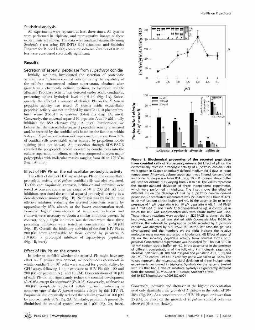
Secretion of aspartyl peptidase from F. pedrosoi conidiaInitially, we have investigated the secretion of proteolytic
activity from F. pedrosoi conidial cells by testing the capability of
the cell-free concentrated culture supernatant, obtained after
growth in a chemically defined medium, to hydrolyze soluble
albumin. Peptidase activity was detected under acidic conditions,
presenting highest hydrolysis level at pH 4.0 (Fig. 1A). Subse-
quently, the effect of a number of classical PIs on the F. pedrosoi
peptidase activity was tested. F. pedrosoi acidic extracellular
peptidase activity was not inhibited by metallo (1,10-phenanthro-
line), serine (PMSF), or cysteine (E-64) PIs (Fig. 1A, inset).
Conversely, the universal aspartyl PI pepstatin A at 10 mM totally
inhibited the BSA cleavage (Fig. 1A, inset). Furthermore, we
believe that the extracellular aspartyl peptidase activity is released
and/or secreted by the conidial cells based on the fact that, within
5 days of F. pedrosoi cultivation in Czapek medium, more than 99%
of conidial cells were viable when assessed by propidium iodide
staining (data not shown). An inspection through SDS-PAGE
revealed the polypeptide profile secreted by conidial cells into the
culture supernatant medium, which was composed of seven major
polypeptides with molecular masses ranging from 50 to 120 kDa
(Fig. 1A, inset).
Effect of HIV PIs on the extracellular proteolytic activityThe effect of distinct HIV aspartyl-type PIs on the extracellular
proteolytic activity of F. pedrosoi conidial cells was also evaluated.
To this end, saquinavir, ritonavir, nelfinavir and indinavir were
tested at concentrations in the range of 50 to 200 mM. All four
inhibitors restrained the conidial aspartyl proteolytic activity in a
dose-dependent manner (Fig. 1B). Nelfinavir was by far the most
effective inhibitor, reducing the secreted proteolytic activity by
approximately 85% already at the lowest concentration used.
Four-fold higher concentration of saquinavir, indinavir and
ritonavir were necessary to obtain a similar inhibition pattern. In
contrast, only a slight inhibition was detected when these three
preceding inhibitors were tested at concentration of 50 mM
(Fig. 1B). Overall, the inhibitory activities of the four HIV PIs at
200 mM were comparable to those exerted by pepstatin A
(10 mM), a prototypal inhibitor of aspartyl-type peptidases
(Fig. 1B, inset).
Effect of HIV PIs on the growthIn order to establish whether the aspartyl PIs might have any
effect on F. pedrosoi development, we performed experiments in
which conidia (1.06103 cells) were assayed for growth ability, by
CFU assay, following 1 hour exposure to HIV PIs (50, 100 and
200 mM) or pepstatin A (1 and 10 mM). Concentrations of 50 mM
of each PIs did not significantly reduce the conidial development
(P.0.05), except for saquinavir (P,0.05). Conversely, nelfinavir at
100 mM completely abolished cellular growth, indicating a
complete cure of the F. pedrosoi conidia culture by this HIV PI.
Saquinavir also drastically reduced the cellular growth at 100 mM
by approximately 90% (Fig. 2A). Similarly, pepstatin A powerfully
diminished the conidial growth even at 1 mM (Fig. 2A, inset).
Conversely, indinavir and ritonavir at the highest concentration
used only diminished the growth of F. pedrosoi in the order of 20–
30% (Fig. 2A). At a concentration of HIV PIs equal or lower than
25 mM, no effect on the growth of F. pedrosoi conidial cells was
observed (data not shown).
Figure 1. Biochemical properties of the secreted peptidasefrom conidial cells of Fonsecaea pedrosoi. (A) Effect of pH on theextracellularly released proteolytic activity of F. pedrosoi conidia. Cellswere grown in Czapek chemically defined medium for 5 days at roomtemperature. Afterward, culture supernatant was filtered, concentratedand tested to degrade soluble BSA using 10 mM sodium citrate bufferadjusted for distinct pH’s varying from 2.0 to 5.0. The values representthe mean6standard deviation of three independent experiments,which were performed in triplicate. The inset shows the effect ofdifferent PIs on the cleavage of BSA by F. pedrosoi conidial-derivedpeptidase. Concentrated supernatant was incubated for 1 hour at 37uCin 10 mM sodium citrate buffer, pH 4.0, in the absence (b) or in thepresence of 1 mM pepstatin A (c), 10 mM pepstatin A (d), 1 mM PMSF(e), 1 mM E-64 (f) and 1 mM 1,10-phenanthroline (g). A control (a) inwhich the BSA was supplemented only with citrate buffer was used.These mixture reactions were applied on SDS-PAGE to detect the BSAhydrolysis, and the gel was stained with Coomassie blue R-250. Inaddition, the extracellular polypeptide profile secreted by F. pedrosoiconidia was analyzed by SDS–PAGE (h). In this last case, the gel wassilver-stained and the numbers on the right indicate the relativemolecular mass markers expressed in kilodaltons. (B) Effect of aspartylPIs on the secretory peptidase activity from conidial forms of F.pedrosoi. Concentrated supernatant was incubated for 1 hour at 37uC in10 mM sodium citrate buffer, pH 4.0, in the absence or in the presenceof different concentrations of the following PIs: indinavir, saquinavir,ritonavir, nelfinavir (50, 100 and 200 mM) and pepstatin A (1, 5, 10 and20 mM). The control (39.361.7 arbitrary units) was taken as 100%. Thevalues represent the mean6standard deviation of three independentexperiments performed in triplicate. Symbols denote systems treatedwith PIs that had a rate of substrate hydrolysis significantly differentfrom the control (¤, P,0.05; w, P,0.001; Student’s t test).doi:10.1371/journal.pone.0003382.g001
HIV-PIs on F. pedrosoi
PLoS ONE | www.plosone.org 4 October 2008 | Volume 3 | Issue 10 | e3382
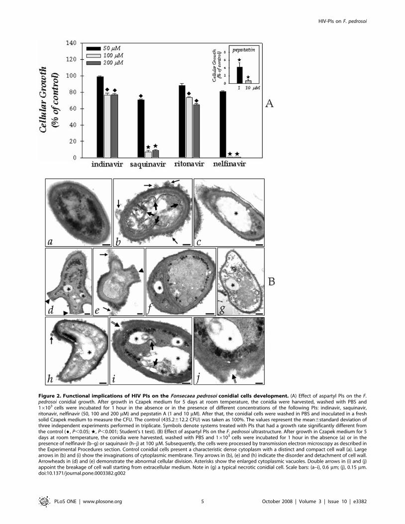
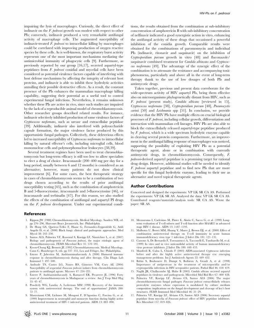
Figure 2. Functional implications of HIV PIs on the Fonsecaea pedrosoi conidial cells development. (A) Effect of aspartyl PIs on the F.pedrosoi conidial growth. After growth in Czapek medium for 5 days at room temperature, the conidia were harvested, washed with PBS and16103 cells were incubated for 1 hour in the absence or in the presence of different concentrations of the following PIs: indinavir, saquinavir,ritonavir, nelfinavir (50, 100 and 200 mM) and pepstatin A (1 and 10 mM). After that, the conidial cells were washed in PBS and inoculated in a freshsolid Czapek medium to measure the CFU. The control (435.2612.2 CFU) was taken as 100%. The values represent the mean6standard deviation ofthree independent experiments performed in triplicate. Symbols denote systems treated with PIs that had a growth rate significantly different fromthe control (¤, P,0.05; w, P,0.001; Student’s t test). (B) Effect of aspartyl PIs on the F. pedrosoi ultrastructure. After growth in Czapek medium for 5days at room temperature, the conidia were harvested, washed with PBS and 16103 cells were incubated for 1 hour in the absence (a) or in thepresence of nelfinavir (b–g) or saquinavir (h–j) at 100 mM. Subsequently, the cells were processed by transmission electron microscopy as described inthe Experimental Procedures section. Control conidial cells present a characteristic dense cytoplasm with a distinct and compact cell wall (a). Largearrows in (b) and (i) show the invaginations of cytoplasmic membrane. Tiny arrows in (b), (e) and (h) indicate the disorder and detachment of cell wall.Arrowheads in (d) and (e) demonstrate the abnormal cellular division. Asterisks show the enlarged cytoplasmic vacuoles. Double arrows in (i) and (j)appoint the breakage of cell wall starting from extracellular medium. Note in (g) a typical necrotic conidial cell. Scale bars: (a–i), 0.6 mm; (j), 0.15 mm.doi:10.1371/journal.pone.0003382.g002
HIV-PIs on F. pedrosoi
PLoS ONE | www.plosone.org 5 October 2008 | Volume 3 | Issue 10 | e3382
Effect of HIV PIs on the ultrastructureBased on the efficacy of the HIV aspartyl PIs in diminishing the
growth of F. pedrosoi, particularly nelfinavir and saquinavir, we next
investigated the effect of both inhibitors on the F. pedrosoi
morphological level (Fig. 2B). In this context, by means of
transmission electron microscopy, the normal morphology of
control conidium (Fig. 2B, panel a) was compared with the
ultrastructure of PIs-treated cells (Fig. 2B, panels b–j). After 1 hour
of treatment, both nelfinavir and saquinavir at 100 mM induced
several morphological alterations, including: invaginations in the
cytoplasmic membrane and withdrawal of the cytoplasmic
membrane from within the cell wall (large arrows in Fig. 2B,
insets b and i), disorder and detachment of the cell wall (tiny
arrows in Fig. 2B, panels b, e, and h), rupture of internal organelles
(Fig. 2B, panel g), and the presence of large and irregular
cytoplasmic vacuoles (asterisks in Fig. 2B), some of them
containing small vesicles (Fig. 2B, panels i and j). In some cells,
the enlargement of intracellular vacuoles was so intense that they
occupied almost the entire cytoplasm area (Fig. 2B, panel c).
Furthermore, abnormal division disfiguring regular conidia
morphology could be observed in cells treated with nelfinavir
(arrowheads in Fig. 2B, panels d–f), while the saquinavir treatment
seams to induce breakage of cell wall starting from extracellular
environmental (double arrows in Fig. 2B, panels i and j). Ritonavir
and indinavir also promoted some of these aberrant alterations;
however, in a lower proportion when compared to nelfinavir and
saquinavir treatment (data not shown). In contrast, non-treated
conidial cells presented a normal morphology with a characteristic
dense and dark cytoplasm and a distinct cell wall (Fig. 2B, panel a).
Effect of HIV PIs on the interaction of conidia-animal cellsLight microscopy of Giemsa-stained preparations, revealed that
conidia of F. pedrosoi attached to (Fig. 3A, a1), and were ingested
(Fig. 3B, b1) by Chinese hamster ovary cells (CHO) after 2 hours
of fungus-mammalian cell contact as previously experimented
[24]. Transmission electron microscopy analysis showed that the
fungus adhered to cell surface by a discreet contact between its cell
wall and the cytoplasmic membrane of the vertebrate cell (Fig. 3A,
a2). After ingestion, conidial cell could be clearly observed within a
membrane cytoplasmic vacuole by using both light and electron
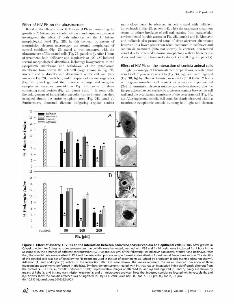
Figure 3. Effect of aspartyl HIV PIs on the interaction between Fonsecaea pedrosoi conidia and epithelial cells (CHO). After growth inCzapek medium for 5 days at room temperature, the conidia were harvested, washed with PBS and 16106 cells were incubated for 1 hour in theabsence or in the presence of different concentrations (50, 100 and 200 mM) of the following PIs: indinavir, saquinavir, ritonavir and nelfinavir. Afterthat, the conidial cells were washed in PBS and the interaction process was performed as described in Experimental Procedures section. The viabilityof the conidial cells was not affected by the PIs treatment used in this set of experiments as judged by propidium iodide staining (data not shown).Adhesion (A) and endocytic (B) indices of the interaction after 2 h were shown. The values represent the mean6standard deviation of threeindependent experiments performed in triplicate. Symbols denote systems treated with PIs that had an interaction index significantly different fromthe control (¤, P,0.05; w, P,0.001; Student’s t test). Representative images of attached (a1 and a2) and ingested (b1 and b2) fungi are shown bymeans of light (a1 and b1) and transmission electron (a2 and b2) microscopy analyses. Note that ingested conidia are located within vacuole (b1 andb2). Arrows show the conidia attached (a1) or ingested (b1) by CHO cells. Scale bars: (a1 and b1), 10 mm; (a2 and b2), 1 mm.doi:10.1371/journal.pone.0003382.g003
HIV-PIs on F. pedrosoi
PLoS ONE | www.plosone.org 6 October 2008 | Volume 3 | Issue 10 | e3382
microscopy (Fig. 3B, b1–b2). Then, we evaluated the influence of
HIV PIs on this interaction process. In this sense, conidia
(1.06106 cells) were pre-treated with each aspartyl PI (50, 100
and 200 mM) for 1 hour before interaction. In this cellular density,
the conidia maintained their viability after the treatment for 1 hour
with each PI in all the studied concentration as judged by their
morphology and propidium iodine staining (in which more than
99% of the conidia were viable). Similar results were obtained by
our group when different cell numbers were treated with pepstatin
A [16]. In the present study, we showed that saquinavir was the
most potent inhibitor of both adhesion and endocytosis processes
between F. pedrosoi conidia and CHO cells, showing a clear dose-
dependent inhibition profile, where the inhibition increased from 60
to 85% (adhesion index) and 70 to 97% (endocytic index) as
saquinavir concentration rose from 50 to 200 mM (Fig. 3).
Curiously, nelfinavir, which most effectively inhibited both secreted
aspartyl peptidase and cellular growth, only significantly reduced
the adherence and endocytosis of F. pedrosoi to 55% of the controls at
highest concentration. Ritonavir and indinavir both at 200 mM
diminished the interaction process in a similar way (Fig. 3).
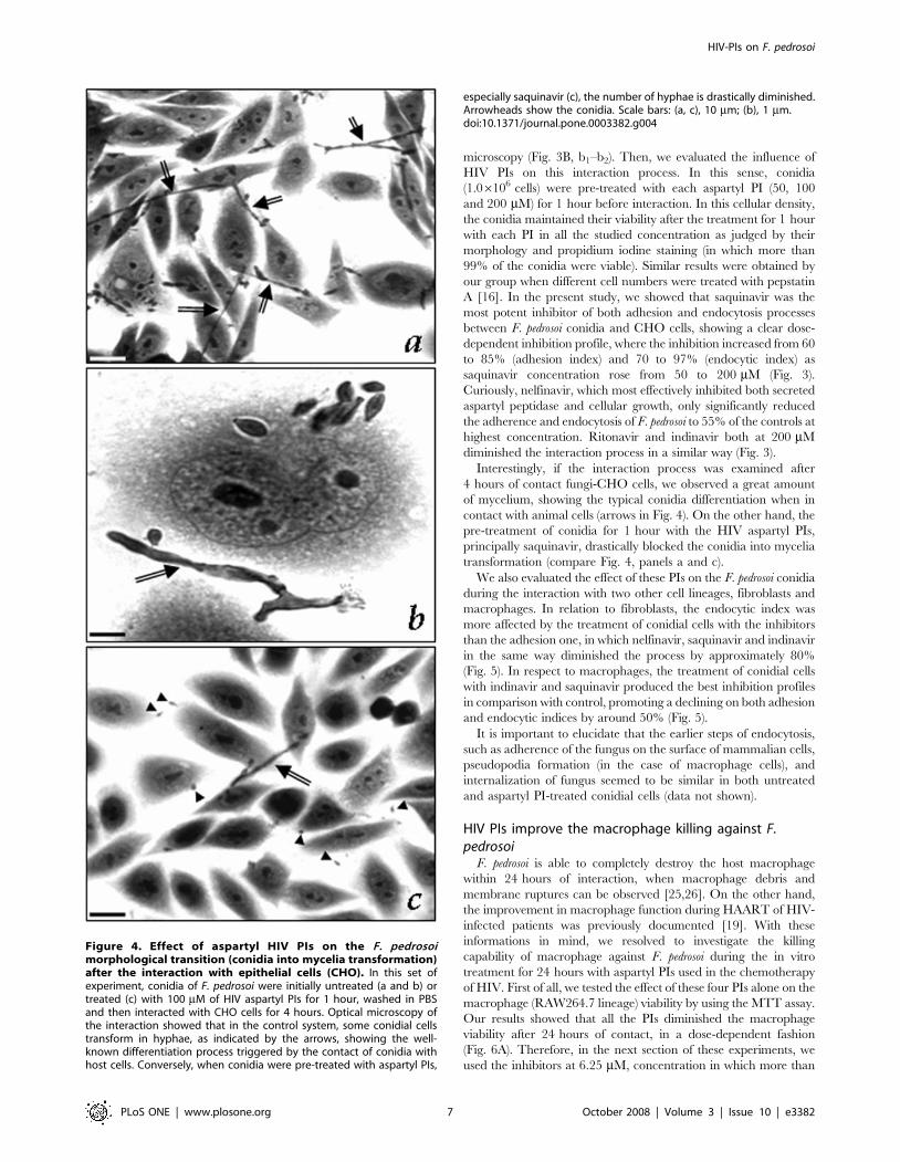
Interestingly, if the interaction process was examined after
4 hours of contact fungi-CHO cells, we observed a great amount
of mycelium, showing the typical conidia differentiation when in
contact with animal cells (arrows in Fig. 4). On the other hand, the
pre-treatment of conidia for 1 hour with the HIV aspartyl PIs,
principally saquinavir, drastically blocked the conidia into mycelia
transformation (compare Fig. 4, panels a and c).
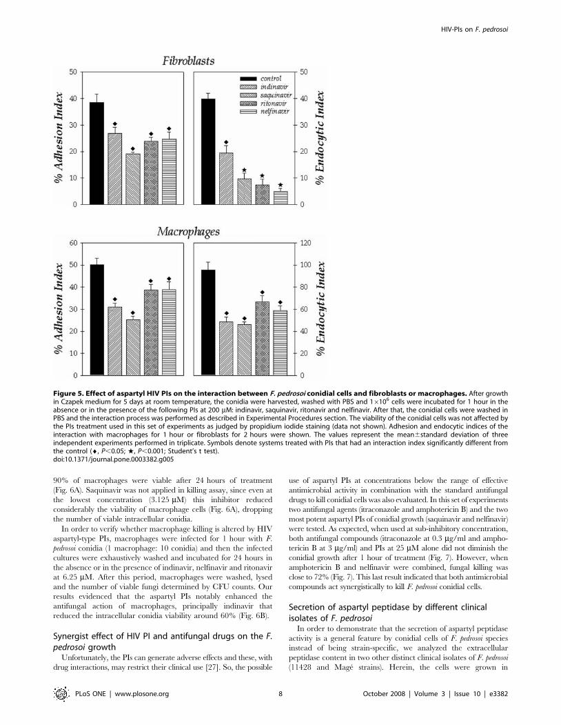
We also evaluated the effect of these PIs on the F. pedrosoi conidia
during the interaction with two other cell lineages, fibroblasts and
macrophages. In relation to fibroblasts, the endocytic index was
more affected by the treatment of conidial cells with the inhibitors
than the adhesion one, in which nelfinavir, saquinavir and indinavir
in the same way diminished the process by approximately 80%
(Fig. 5). In respect to macrophages, the treatment of conidial cells
with indinavir and saquinavir produced the best inhibition profiles
in comparison with control, promoting a declining on both adhesion
and endocytic indices by around 50% (Fig. 5).
It is important to elucidate that the earlier steps of endocytosis,
such as adherence of the fungus on the surface of mammalian cells,
pseudopodia formation (in the case of macrophage cells), and
internalization of fungus seemed to be similar in both untreated
and aspartyl PI-treated conidial cells (data not shown).
HIV PIs improve the macrophage killing against F.pedrosoi
F. pedrosoi is able to completely destroy the host macrophage
within 24 hours of interaction, when macrophage debris and
membrane ruptures can be observed [25,26]. On the other hand,
the improvement in macrophage function during HAART of HIV-
infected patients was previously documented [19]. With these
informations in mind, we resolved to investigate the killing
capability of macrophage against F. pedrosoi during the in vitro
treatment for 24 hours with aspartyl PIs used in the chemotherapy
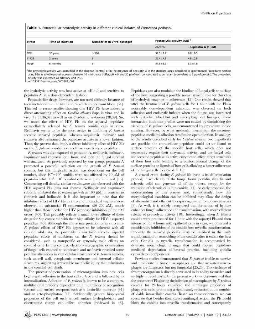
of HIV. First of all, we tested the effect of these four PIs alone on the
macrophage (RAW264.7 lineage) viability by using the MTT assay.
Our results showed that all the PIs diminished the macrophage
viability after 24 hours of contact, in a dose-dependent fashion
(Fig. 6A). Therefore, in the next section of these experiments, we
used the inhibitors at 6.25 mM, concentration in which more than
Figure 4. Effect of aspartyl HIV PIs on the F. pedrosoimorphological transition (conidia into mycelia transformation)after the interaction with epithelial cells (CHO). In this set ofexperiment, conidia of F. pedrosoi were initially untreated (a and b) ortreated (c) with 100 mM of HIV aspartyl PIs for 1 hour, washed in PBSand then interacted with CHO cells for 4 hours. Optical microscopy ofthe interaction showed that in the control system, some conidial cellstransform in hyphae, as indicated by the arrows, showing the well-known differentiation process triggered by the contact of conidia withhost cells. Conversely, when conidia were pre-treated with aspartyl PIs,
especially saquinavir (c), the number of hyphae is drastically diminished.Arrowheads show the conidia. Scale bars: (a, c), 10 mm; (b), 1 mm.doi:10.1371/journal.pone.0003382.g004
HIV-PIs on F. pedrosoi
PLoS ONE | www.plosone.org 7 October 2008 | Volume 3 | Issue 10 | e3382
90% of macrophages were viable after 24 hours of treatment
(Fig. 6A). Saquinavir was not applied in killing assay, since even at
the lowest concentration (3.125 mM) this inhibitor reduced
considerably the viability of macrophage cells (Fig. 6A), dropping
the number of viable intracellular conidia.
In order to verify whether macrophage killing is altered by HIV
aspartyl-type PIs, macrophages were infected for 1 hour with F.
pedrosoi conidia (1 macrophage: 10 conidia) and then the infected
cultures were exhaustively washed and incubated for 24 hours in
the absence or in the presence of indinavir, nelfinavir and ritonavir
at 6.25 mM. After this period, macrophages were washed, lysed
and the number of viable fungi determined by CFU counts. Our
results evidenced that the aspartyl PIs notably enhanced the
antifungal action of macrophages, principally indinavir that
reduced the intracellular conidia viability around 60% (Fig. 6B).
Synergist effect of HIV PI and antifungal drugs on the F.pedrosoi growth
Unfortunately, the PIs can generate adverse effects and these, with
drug interactions, may restrict their clinical use [27]. So, the possible
use of aspartyl PIs at concentrations below the range of effective
antimicrobial activity in combination with the standard antifungal
drugs to kill conidial cells was also evaluated. In this set of experiments
two antifungal agents (itraconazole and amphotericin B) and the two
most potent aspartyl PIs of conidial growth (saquinavir and nelfinavir)
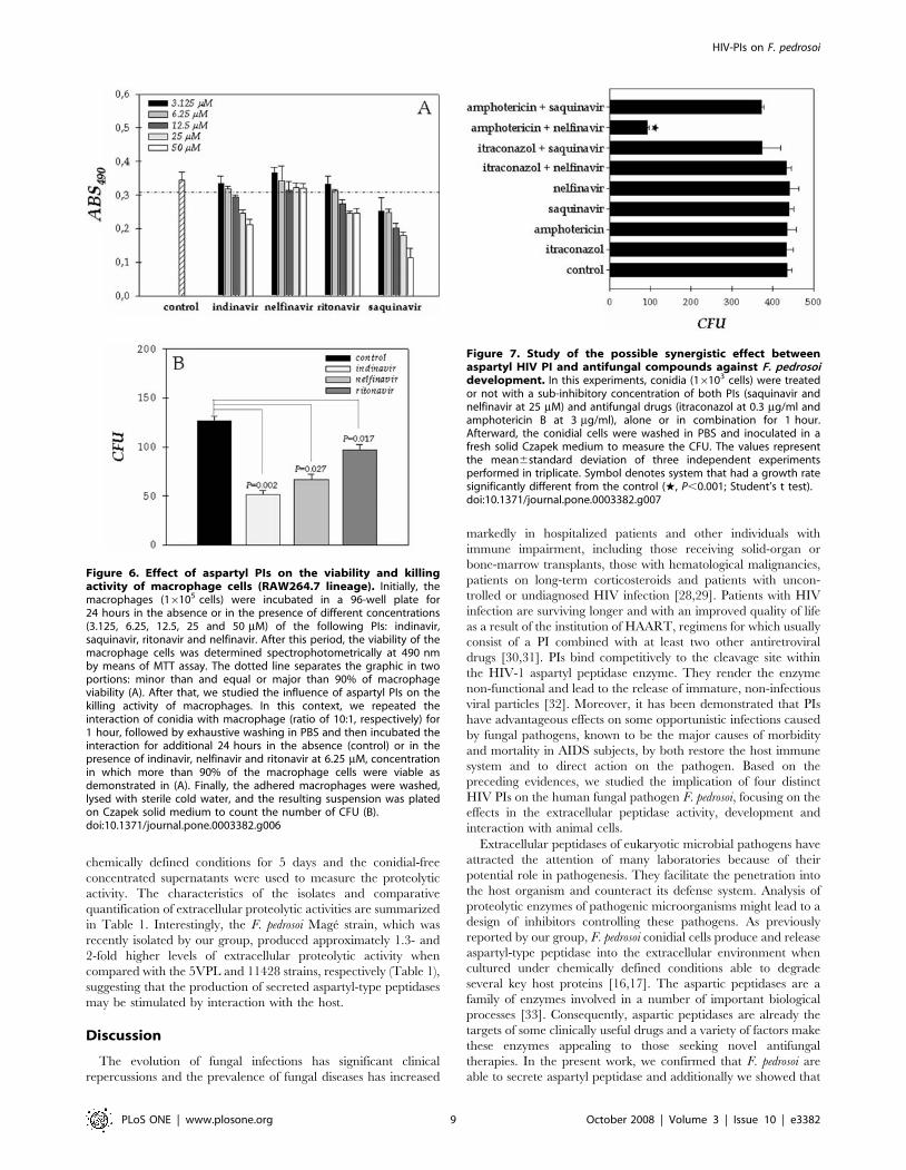
were tested. As expected, when used at sub-inhibitory concentration,
both antifungal compounds (itraconazole at 0.3 mg/ml and ampho-
tericin B at 3 mg/ml) and PIs at 25 mM alone did not diminish the
conidial growth after 1 hour of treatment (Fig. 7). However, when
amphotericin B and nelfinavir were combined, fungal killing was
close to 72% (Fig. 7). This last result indicated that both antimicrobial
compounds act synergistically to kill F. pedrosoi conidial cells.
Secretion of aspartyl peptidase by different clinicalisolates of F. pedrosoi
In order to demonstrate that the secretion of aspartyl peptidase
activity is a general feature by conidial cells of F. pedrosoi species
instead of being strain-specific, we analyzed the extracellular
peptidase content in two other distinct clinical isolates of F. pedrosoi
(11428 and Mage strains). Herein, the cells were grown in
Figure 5. Effect of aspartyl HIV PIs on the interaction between F. pedrosoi conidial cells and fibroblasts or macrophages. After growthin Czapek medium for 5 days at room temperature, the conidia were harvested, washed with PBS and 16106 cells were incubated for 1 hour in theabsence or in the presence of the following PIs at 200 mM: indinavir, saquinavir, ritonavir and nelfinavir. After that, the conidial cells were washed inPBS and the interaction process was performed as described in Experimental Procedures section. The viability of the conidial cells was not affected bythe PIs treatment used in this set of experiments as judged by propidium iodide staining (data not shown). Adhesion and endocytic indices of theinteraction with macrophages for 1 hour or fibroblasts for 2 hours were shown. The values represent the mean6standard deviation of threeindependent experiments performed in triplicate. Symbols denote systems treated with PIs that had an interaction index significantly different fromthe control (¤, P,0.05; w, P,0.001; Student’s t test).doi:10.1371/journal.pone.0003382.g005
HIV-PIs on F. pedrosoi
PLoS ONE | www.plosone.org 8 October 2008 | Volume 3 | Issue 10 | e3382
chemically defined conditions for 5 days and the conidial-free
concentrated supernatants were used to measure the proteolytic
activity. The characteristics of the isolates and comparative
quantification of extracellular proteolytic activities are summarized
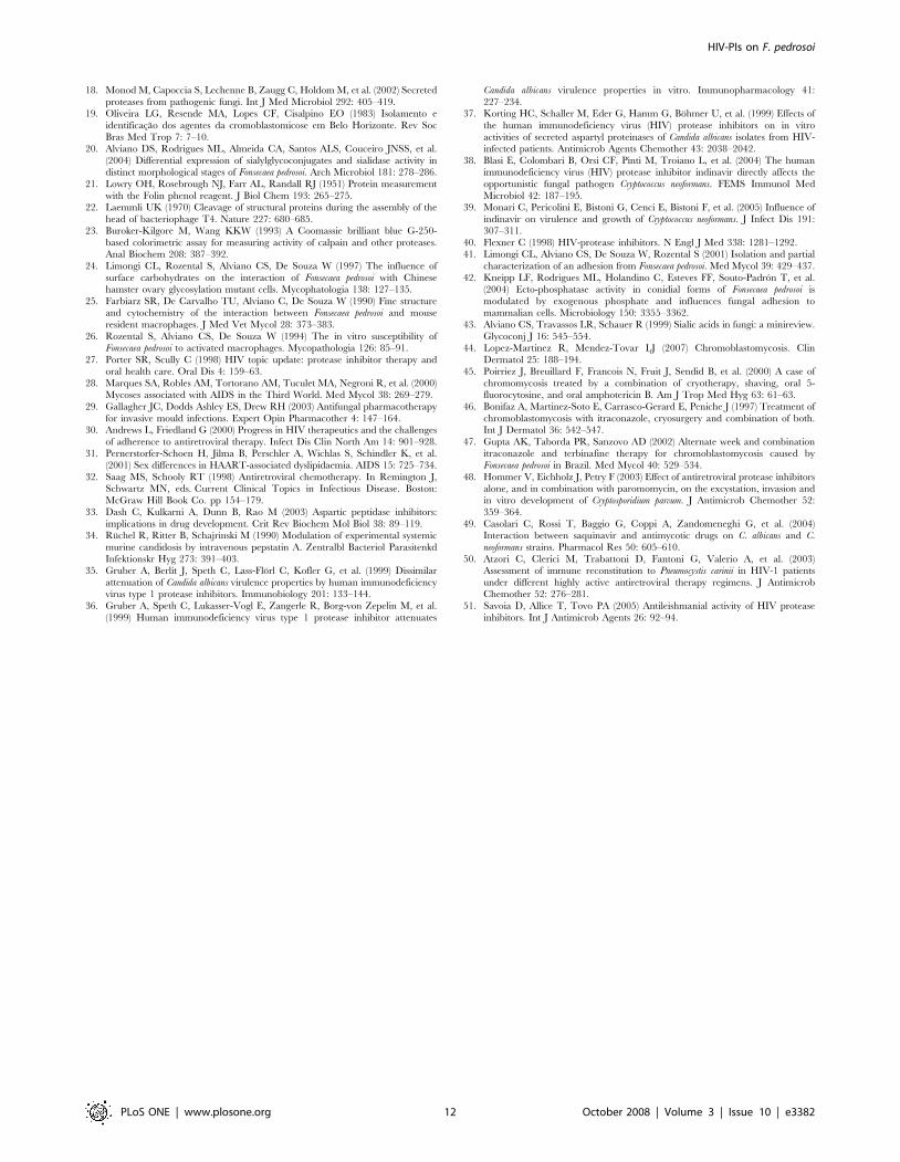
in Table 1. Interestingly, the F. pedrosoi Mage strain, which was
recently isolated by our group, produced approximately 1.3- and
2-fold higher levels of extracellular proteolytic activity when
compared with the 5VPL and 11428 strains, respectively (Table 1),
suggesting that the production of secreted aspartyl-type peptidases
may be stimulated by interaction with the host.
Discussion
The evolution of fungal infections has significant clinical
repercussions and the prevalence of fungal diseases has increased
markedly in hospitalized patients and other individuals with
immune impairment, including those receiving solid-organ or
bone-marrow transplants, those with hematological malignancies,
patients on long-term corticosteroids and patients with uncon-
trolled or undiagnosed HIV infection [28,29]. Patients with HIV
infection are surviving longer and with an improved quality of life
as a result of the institution of HAART, regimens for which usually
consist of a PI combined with at least two other antiretroviral
drugs [30,31]. PIs bind competitively to the cleavage site within
the HIV-1 aspartyl peptidase enzyme. They render the enzyme
non-functional and lead to the release of immature, non-infectious
viral particles [32]. Moreover, it has been demonstrated that PIs
have advantageous effects on some opportunistic infections caused
by fungal pathogens, known to be the major causes of morbidity
and mortality in AIDS subjects, by both restore the host immune
system and to direct action on the pathogen. Based on the
preceding evidences, we studied the implication of four distinct
HIV PIs on the human fungal pathogen F. pedrosoi, focusing on the
effects in the extracellular peptidase activity, development and
interaction with animal cells.
Extracellular peptidases of eukaryotic microbial pathogens have
attracted the attention of many laboratories because of their
potential role in pathogenesis. They facilitate the penetration into
the host organism and counteract its defense system. Analysis of
proteolytic enzymes of pathogenic microorganisms might lead to a
design of inhibitors controlling these pathogens. As previously
reported by our group, F. pedrosoi conidial cells produce and release
aspartyl-type peptidase into the extracellular environment when
cultured under chemically defined conditions able to degrade
several key host proteins [16,17]. The aspartic peptidases are a
family of enzymes involved in a number of important biological
processes [33]. Consequently, aspartic peptidases are already the
targets of some clinically useful drugs and a variety of factors make
these enzymes appealing to those seeking novel antifungal
therapies. In the present work, we confirmed that F. pedrosoi are
able to secrete aspartyl peptidase and additionally we showed that
Figure 6. Effect of aspartyl PIs on the viability and killingactivity of macrophage cells (RAW264.7 lineage). Initially, themacrophages (16105 cells) were incubated in a 96-well plate for24 hours in the absence or in the presence of different concentrations(3.125, 6.25, 12.5, 25 and 50 mM) of the following PIs: indinavir,saquinavir, ritonavir and nelfinavir. After this period, the viability of themacrophage cells was determined spectrophotometrically at 490 nmby means of MTT assay. The dotted line separates the graphic in twoportions: minor than and equal or major than 90% of macrophageviability (A). After that, we studied the influence of aspartyl PIs on thekilling activity of macrophages. In this context, we repeated theinteraction of conidia with macrophage (ratio of 10:1, respectively) for1 hour, followed by exhaustive washing in PBS and then incubated theinteraction for additional 24 hours in the absence (control) or in thepresence of indinavir, nelfinavir and ritonavir at 6.25 mM, concentrationin which more than 90% of the macrophage cells were viable asdemonstrated in (A). Finally, the adhered macrophages were washed,lysed with sterile cold water, and the resulting suspension was platedon Czapek solid medium to count the number of CFU (B).doi:10.1371/journal.pone.0003382.g006
Figure 7. Study of the possible synergistic effect betweenaspartyl HIV PI and antifungal compounds against F. pedrosoidevelopment. In this experiments, conidia (16103 cells) were treatedor not with a sub-inhibitory concentration of both PIs (saquinavir andnelfinavir at 25 mM) and antifungal drugs (itraconazol at 0.3 mg/ml andamphotericin B at 3 mg/ml), alone or in combination for 1 hour.Afterward, the conidial cells were washed in PBS and inoculated in afresh solid Czapek medium to measure the CFU. The values representthe mean6standard deviation of three independent experimentsperformed in triplicate. Symbol denotes system that had a growth ratesignificantly different from the control (w, P,0.001; Student’s t test).doi:10.1371/journal.pone.0003382.g007
HIV-PIs on F. pedrosoi
PLoS ONE | www.plosone.org 9 October 2008 | Volume 3 | Issue 10 | e3382
the hydrolytic activity was best active at pH 4.0 and sensitive to
pepstatin A, in a dose-dependent fashion.
Pepstatin-like drugs, however, are not used clinically because of
their metabolism in the liver and rapid clearance from blood [34].
This led to recent studies showing that HIV PIs have indeed a
direct attenuating effect on Candida albicans Saps in vitro and in
vivo [12,35,36,37] as well as on Cryptococcus neoformans [38,39]. So,
we tested the effect of HIV PIs on the aspartyl peptidase
extracellularly released by F. pedrosoi conidia cells in vitro.
Nelfinavir seems to be the most active in inhibiting F. pedrosoi
secreted aspartyl peptidase, whereas saquinavir, indinavir and
ritonavir also restrained the peptidase activity in a lower fashion.
Thus, the present data imply a direct inhibitory effect of HIV PIs
on the F. pedrosoi conidial extracellular aspartyl-type peptidase.
F. pedrosoi was also exposed to pepstatin A, nelfinavir, indinavir,
saquinavir and ritonavir for 1 hour, and then the fungal survival
was analyzed. As previously reported by our group, pepstatin A
promoted a powerful reduction on the growth of F. pedrosoi
conidia, but this fungicidal action was dependent on the cell
number, since 1062104 conidia were not affected by 10 mM of
pepstatin while 1022103 conidial cells were fully inhibited [16].
Concerning cell density, similar results were also observed with the
HIV aspartyl PIs (data not shown). Nelfinavir and saquinavir
robustly inhibited the F. pedrosoi growth at 100 mM, in contrast to
ritonavir and indinavir. It is also important to note that the
inhibitory effect of HIV PIs in vitro and in candidal vaginitis were
observed at substantial PI concentrations (50–200 mM), much
higher than those needed for HIV peptidase inhibition (of the nM
order) [40]. This probably reflects a much lower affinity of these
drugs for Sap compared with their high affinity for HIV-1 aspartyl
peptidase [40]. Although the above interpretation of in vitro anti-
F. pedrosoi effects of HIV PIs appears to be coherent with all
experimental data, the possibility of unrelated secreted aspartyl
peptidase effects of inhibitors on the F. pedrosoi should be
considered, such as nonspecific or generally toxic effects on
conidial cells. In this context, electron-micrographic examination
of fungal cells exposed to saquinavir and nelfinavir revealed some
peculiar alterations in vital cellular structures of F. pedrosoi conidia,
such as cell wall, cytoplasmic membrane and internal cellular
structures, suggesting irreversible metabolic injury that culminates
in the conidial cell death.
The process of penetration of microorganism into host cells
begins with adhesion to the host cell surface and is followed by its
internalization. Adhesion of F. pedrosoi is known to be a complex,
multifactorial property dependent on a multiplicity of recognition
systems and surface receptors such as a lectin-like molecule [41]
and an ecto-phosphatase [42]. Additionally, general biophysical
properties of the cell such as cell surface hydrophobicity and
electrostatic charge can affect adhesion [reviewed in 43].
Peptidases can also modulate the binding of fungal cells to surface
of the host, suggesting a possible non-enzymatic role for this class
of hydrolytic enzymes in adherence [15]. Our results showed that
after the treatment of F. pedrosoi cells for 1 hour with the PIs a
noticeably dose-dependent inhibition was observed on both
adhesion and endocytic indexes when the fungus was interacted
with epithelial, fibroblast and macrophage cell lineages. These
interaction inhibition profiles were not caused by diminishing the
viability of F. pedrosoi cells, as demonstrated by propidium iodide
staining. However, by what molecular mechanism the secretory
peptidase mediates adhesion remains on open question. In analogy
to the results described early for Candida albicans, two hypotheses
are possible: the extracellular peptidase could act as ligand to
surface proteins of the specific host cells, which does not
necessarily require their enzymatic activity, and the fungal cells
use secreted peptidase as active enzymes to affect target structures
of their host cells, leading to a conformational change of the
surface proteins or ligands of host cells allowing a better adherence
of the fungal cells [reviewed in 15].
A crucial event during F. pedrosoi life cycle is its differentiation
process, in which any of the fungal forms (conidia, mycelia and
sclerotic cells) can generate all of the others, except for the
transition of sclerotic cells into conidia [44]. As early proposed, the
understanding of this process and, consequently, how this
morphological transition can be inhibited may allow the design
of alternative and efficient therapies against chromoblastomycosis
[3]. As well, it is widely recognized that formation of hyphae
enhances fungal adherence and tissue invasion, with more efficient
release of proteolytic activity [18]. Interestingly, when F. pedrosoi
conidia were pre-treated for 1 hour with the aspartyl PIs and then
interacted for 4 hours with epithelial cells in vitro, we observed a
considerably inhibition of the conidia into mycelia transformation.
Probably the aspartyl peptidase may be involved in the early
transformation or remodeling of the conidia after it enters the host
cells. Conidia to mycelia transformation is accompanied by
dramatic morphologic changes that could require peptidase-
mediated degradation of several proteins, including conidia
cytoskeleton components.
Previous studies demonstrated that F. pedrosoi is able to survive
and proliferate in tissue macrophages and that activated macro-
phages are fungistatic but not fungicidal [25,26]. The virulence of
this microorganism is directly correlated to its ability to survive and
multiply intracellularly. In the present work, we demonstrated that
the presence of PIs during the infection of macrophages by F. pedrosoi
conidia for 24 hours enhanced the antifungal properties of
phagocytic cells, promoting a significantly reduction in the number
of viable intracellular conidia. Based on these evidences, we can
speculate that besides their direct antifungal action, the PIs could
block the conidia into mycelia transformation and consequently
Table 1. Extracellular proteolytic activity in different clinical isolates of Fonsecaea pedrosoi.
Strain Time of isolation Number of in vitro passages Proteolytic activity (AU) a
control +pepstatin A (1 mM)
5VPL 30 years .500 39.361.7 0.860.5
11428 2 years 8 26.464.8 4.862.8
Mage 6 months 6 51.865.3 5.361.8
aThe proteolytic activity was quantified in the absence (control) or in the presence of pepstatin A in the standard assay described in Experimental Procedures sectionusing BSA as soluble proteinaceous substrate, 10 mM citrate buffer, pH 4.0, and 20 ml of each concentrated supernatant (equivalent to 5 mg of protein). The proteolyticactivity was expressed as arbitrary unit (AU).
doi:10.1371/journal.pone.0003382.t001
HIV-PIs on F. pedrosoi
PLoS ONE | www.plosone.org 10 October 2008 | Volume 3 | Issue 10 | e3382
impairing the lysis of macrophages. Curiously, the direct effect of
indinavir on the F. pedrosoi growth was modest with respect to other
PIs; conversely, indinavir produced a very remarkable antifungal
activity of macrophage cells. The augmented susceptibility of
indinavir-treated F. pedrosoi to intracellular killing by macrophages
could be correlated with improving production of oxygen reactive
species by these cells. As is well-known, the respiratory burst activity
represents one of the most important mechanisms mediating the
antimicrobial immunity of phagocytic cells [9]. Furthermore, as
previously reported by our group [16,17], secreted aspartyl-type
peptidases from F. pedrosoi conidial and mycelial cells have been
considered as potential virulence factors capable of interfering with
host defense mechanisms by affecting the integrity of relevant host
proteins, and indinavir is able to inhibit these hydrolytic activities
annulling their possible destructive effects. As a result, the constant
presence of the PIs enhances the mammalian macrophage killing
capability, suggesting their potential as a useful tool to control
experimental fungal infections. Nevertheless, it remains unknown
whether these PIs are active in vivo, since such studies are impaired
by the lack of a reproducible animal model of chromoblastomycosis.
Other researcher groups described related results. For instance,
indinavir selectively inhibited production of some virulence factors of
Cryptococcus neoformans, such as urease and extracellular peptidase
[39]. Additionally, indinavir also interfered with polysaccharide
capsule formation, the major virulence factor produced by this
opportunistic fungal pathogen. Collectively, these deleterious effects
led to increased susceptibility of Cryptococcus neoformans to intracellular
killing by natural effector’s cells, including microglial cells, blood
mononuclear cells and polymorphonuclear leukocytes [38,39].
Several treatment regimes have been used to treat chromoblas-
tomycosis but long-term efficacy is still too low to allow specialists
to elect a drug of choice. Itraconazole (200–400 mg per day for a
long period, usually between 6 and 12 months) is considered as an
alternative; however, many patients do not show clinical
improvement [6]. For some cases, the best therapeutic strategy
in cases of chromoblastomycosis seems to be a combination of two
drugs chosen according to the results of prior antifungal
susceptibility testing [45], such as the combination of amphotericin
B and 5-fluorocytosine, itraconazole and 5-fluorocytosine [46], or
itraconazole and terbinafin [47]. For this reason, we also studied
the effects of the combination of antifungal and aspartyl PI drugs
on the F. pedrosoi development. Under our experimental condi-
tions, the results obtained from the combination at sub-inhibitory
concentration of amphotericin B with sub-inhibitory concentration
of nelfinavir indicated a good synergistic action in vitro, enhancing
the antifungal activity of these drugs that occasioned a powerful
inhibition of the conidia growth. Comparable results were
obtained for the combinations of paromomycin and individual
PIs (indinavir, ritonavir and saquinavir) on the inhibition of
Cryptosporidium parvum growth in vitro [48] and fluconazole/
saquinavir combined treatment for Candida albincans and Cryptococ-
cus neoformans [49]. The advantage of the synergic effect of the
combination is to attenuate the resistance and accompanying toxic
phenomena, particularly and above all in the event of long-term
therapy thank to the use of low dosages of both PIs and
antimycotic drugs.
Taken together, previous and present data corroborate for the
wide-spectrum activity of HIV aspartyl PIs, being them effective
also on microorganisms phylogenetically distant from HIV, such as
F. pedrosoi (present study), Candida albicans [reviewed in 15],
Cryptococcus neoformans [38], Cryptosporidium parvum [48], Pneumocystis
carinii [50] and Leishmania spp [51]. In conclusion, we provide
evidence that the HIV PIs have multiple effects on crucial biological
processes of F. pedrosoi, including cellular growth, differentiation and
interaction with mammalian cell lineages. HIV PIs are also able to
block the extracellularly released aspartyl-type peptidase produced
by F. pedrosoi, which is a wide spectrum hydrolytic enzyme capable
in cleaving several protein components. Furthermore, an improve-
ment in the antifungal killing response of macrophages was noticed,
supporting the possibility of exploiting HIV PIs as a potential
therapeutic agent, alone or in combination with currently
antimycotic drugs, in chromoblastomycosis. Consequently, F.
pedrosoi-derived aspartyl peptidase is a promising target for rational
drug design. However, additional studies will be needed to identify
F. pedrosoi aspartyl peptidase and to find new PIs that are more
specific for this fungal hydrolytic enzyme, leading to their use as
alternative and novel topical therapeutic agents.
Author Contributions
Conceived and designed the experiments: VP LK SR CA AS. Performed
the experiments: VP LK SR AS. Analyzed the data: VP LK SR CA AS.
Contributed reagents/materials/analysis tools: SR CA AS. Wrote the
paper: SR AS.
References
1. Rippon JW (1988) Chromoblastomycosis. Medical Mycology. Sauders WB, ed.
pp 276–296, Harcourt Brace Jovanovich, Inc: Philadelphia.
2. De Hoog GS, Queiroz-Telles F, Haase G, Fernandez-Zeppenfeldt G, Attili
Angelis D, et al. (2000) Black fungi: clinical and pathogenic approaches. Med
Mycol 38: 243–250.
3. Santos ALS, Palmeira VF, Rozental S, Kneipp LF, Nimrichter L, et al. (2007)
Biology and pathogenesis of Fonsecaea pedrosoi, the major etiologic agent of
chromoblastomycosis. FEMS Microbiol Rev 31: 570–591.
4. Kwon-Chung KJ, Bennett JE (1992) Chromoblastomycosis. Medical Mycology.
Cann C, Hunsberger S, eds. pp 337–355, Lea and Febiger, Inc: Philadelphia.
5. Esterre P, Jahevitra M, Andriantsimahavandy A (2000) Humoral immune
response in chromoblastomycosis during and after therapy. Clin Diagn Lab
Immunol 7: 497–500.
6. Andrade TS, Castro LG, Nunes RS, Gimenes VM, Cury AE (2004)
Susceptibility of sequential Fonsecaea pedrosoi isolates from chromoblastomycosis
patients to antifungal agents. Mycoses 47: 216–221.
7. Esterre P, Andriantsimhavandy A, Ramarcel ER, Pecarrere JL (1996) Forty
years of chromoblastomycosis in Madagascar: a review. Am J Trop Med Hyg
55: 45–47.
8. Powderly WG, Landay A, Lederman MM (1998) Recovery of the immune
system with antiretroviral therapy. The end of opportunism? JAMA 280:
72–77.
9. Mastroianni CM, Lichtner M, Mengoni F, D’Agostino C, Forcina G, et al.
(1999) Improvement in neutrophil and monocyte function during highly active
antiretroviral treatment of HIV-1 infected patients. AIDS 13: 883–890.
10. Mezzaroma I, Carlesimo M, Pinter E, Alario C, Sacco G, et al. (1999) Long-
term evaluation of T-cell subsets and T-cell function after HAART in advanced
stage HIV-1 disease. AIDS 13: 1187–1193.
11. Malhotra U, Berrei MM, Huang Y, Maree J, Brown DJ, et al. (2000) Effect of
combination antiretroviral therapy on T-cell immunity in acute human
immunodeficiency virus type 1 infection. J Infect Dis 181: 121–131.
12. Cassone A, De Bernardis F, Torosantucci A, Tacconelli E, Tumbarello M, et al.
(1999) In vitro and in vivo anticandidal activity of human immunodeficiency
virus protease inhibitors. J Infect Dis 180: 448–453.
13. Manfredi R, Calza L, Chiodo F (2003) AIDS-associated Cryptococcus infection
before and after the highly active antiretroviral therapy era: emerging
management problems. Int J Antimicrob Agents 22: 449–452.
14. Bobin S, Bouhouere D, Dumpt S, Boibieux A, Grault A, et al. (1998)
Importance of antiprotease in the treatment of microsporidia and/or
cryptosporidia infections in HIV-seropositive patients. Pathol Biol 46: 418–419.
15. Naglik JR, Challacombe SJ, Hube B (2003) Candida albicans secreted aspartyl
peptidases in virulence and pathogenesis. Microbiol Mol Biol Rev 67: 400–428.
16. Palmeira VF, Kneipp LF, Alviano CS, Santos ALS (2006) The major
chromoblastomycosis fungal pathogen Fonsecaea pedrosoi extracellularly releases
proteolytic enzymes whose expression is modulated by culture medium
composition: implications on the fungal development and cleavage of key’s host
structures. FEMS Immunol Med Microbiol 46: 21–29.
17. Palmeira VF, Kneipp LF, Alviano CS, Santos ALS (2006) Secretory aspartyl
peptidase from mycelia of Fonsecaea pedrosoi: effect of HIV peptidase inhibitors.
Res Microbiol 157: 819–826.
HIV-PIs on F. pedrosoi
PLoS ONE | www.plosone.org 11 October 2008 | Volume 3 | Issue 10 | e3382
18. Monod M, Capoccia S, Lechenne B, Zaugg C, Holdom M, et al. (2002) Secreted
proteases from pathogenic fungi. Int J Med Microbiol 292: 405–419.19. Oliveira LG, Resende MA, Lopes CF, Cisalpino EO (1983) Isolamento e
identificacao dos agentes da cromoblastomicose em Belo Horizonte. Rev Soc
Bras Med Trop 7: 7–10.20. Alviano DS, Rodrigues ML, Almeida CA, Santos ALS, Couceiro JNSS, et al.
(2004) Differential expression of sialylglycoconjugates and sialidase activity indistinct morphological stages of Fonsecaea pedrosoi. Arch Microbiol 181: 278–286.
21. Lowry OH, Rosebrough NJ, Farr AL, Randall RJ (1951) Protein measurement
with the Folin phenol reagent. J Biol Chem 193: 265–275.22. Laemmli UK (1970) Cleavage of structural proteins during the assembly of the
head of bacteriophage T4. Nature 227: 680–685.23. Buroker-Kilgore M, Wang KKW (1993) A Coomassie brilliant blue G-250-
based colorimetric assay for measuring activity of calpain and other proteases.Anal Biochem 208: 387–392.
24. Limongi CL, Rozental S, Alviano CS, De Souza W (1997) The influence of
surface carbohydrates on the interaction of Fonsecaea pedrosoi with Chinesehamster ovary glycosylation mutant cells. Mycophatologia 138: 127–135.
25. Farbiarz SR, De Carvalho TU, Alviano C, De Souza W (1990) Fine structureand cytochemistry of the interaction between Fonsecaea pedrosoi and mouse
resident macrophages. J Med Vet Mycol 28: 373–383.
26. Rozental S, Alviano CS, De Souza W (1994) The in vitro susceptibility ofFonsecaea pedrosoi to activated macrophages. Mycopathologia 126: 85–91.
27. Porter SR, Scully C (1998) HIV topic update: protease inhibitor therapy andoral health care. Oral Dis 4: 159–63.
28. Marques SA, Robles AM, Tortorano AM, Tuculet MA, Negroni R, et al. (2000)Mycoses associated with AIDS in the Third World. Med Mycol 38: 269–279.
29. Gallagher JC, Dodds Ashley ES, Drew RH (2003) Antifungal pharmacotherapy
for invasive mould infections. Expert Opin Pharmacother 4: 147–164.30. Andrews L, Friedland G (2000) Progress in HIV therapeutics and the challenges
of adherence to antiretroviral therapy. Infect Dis Clin North Am 14: 901–928.31. Pernerstorfer-Schoen H, Jilma B, Perschler A, Wichlas S, Schindler K, et al.
(2001) Sex differences in HAART-associated dyslipidaemia. AIDS 15: 725–734.
32. Saag MS, Schooly RT (1998) Antiretroviral chemotherapy. In Remington J,Schwartz MN, eds. Current Clinical Topics in Infectious Disease. Boston:
McGraw Hill Book Co. pp 154–179.33. Dash C, Kulkarni A, Dunn B, Rao M (2003) Aspartic peptidase inhibitors:
implications in drug development. Crit Rev Biochem Mol Biol 38: 89–119.34. Ruchel R, Ritter B, Schajrinski M (1990) Modulation of experimental systemic
murine candidosis by intravenous pepstatin A. Zentralbl Bacteriol Parasitenkd
Infektionskr Hyg 273: 391–403.35. Gruber A, Berlit J, Speth C, Lass-Florl C, Kofler G, et al. (1999) Dissimilar
attenuation of Candida albicans virulence properties by human immunodeficiencyvirus type 1 protease inhibitors. Immunobiology 201: 133–144.
36. Gruber A, Speth C, Lukasser-Vogl E, Zangerle R, Borg-von Zepelin M, et al.
(1999) Human immunodeficiency virus type 1 protease inhibitor attenuates
Candida albicans virulence properties in vitro. Immunopharmacology 41:
227–234.37. Korting HC, Schaller M, Eder G, Hamm G, Bohmer U, et al. (1999) Effects of
the human immunodeficiency virus (HIV) protease inhibitors on in vitro
activities of secreted aspartyl proteinases of Candida albicans isolates from HIV-infected patients. Antimicrob Agents Chemother 43: 2038–2042.
38. Blasi E, Colombari B, Orsi CF, Pinti M, Troiano L, et al. (2004) The humanimmunodeficiency virus (HIV) protease inhibitor indinavir directly affects the
opportunistic fungal pathogen Cryptococcus neoformans. FEMS Immunol Med
Microbiol 42: 187–195.39. Monari C, Pericolini E, Bistoni G, Cenci E, Bistoni F, et al. (2005) Influence of
indinavir on virulence and growth of Cryptococcus neoformans. J Infect Dis 191:307–311.
40. Flexner C (1998) HIV-protease inhibitors. N Engl J Med 338: 1281–1292.41. Limongi CL, Alviano CS, De Souza W, Rozental S (2001) Isolation and partial
characterization of an adhesion from Fonsecaea pedrosoi. Med Mycol 39: 429–437.
42. Kneipp LF, Rodrigues ML, Holandino C, Esteves FF, Souto-Padron T, et al.(2004) Ecto-phosphatase activity in conidial forms of Fonsecaea pedrosoi is
modulated by exogenous phosphate and influences fungal adhesion tomammalian cells. Microbiology 150: 3355–3362.
43. Alviano CS, Travassos LR, Schauer R (1999) Sialic acids in fungi: a minireview.
Glycoconj J 16: 545–554.44. Lopez-Martinez R, Mendez-Tovar LJ (2007) Chromoblastomycosis. Clin
Dermatol 25: 188–194.45. Poirriez J, Breuillard F, Francois N, Fruit J, Sendid B, et al. (2000) A case of
chromomycosis treated by a combination of cryotherapy, shaving, oral 5-fluorocytosine, and oral amphotericin B. Am J Trop Med Hyg 63: 61–63.
46. Bonifaz A, Martinez-Soto E, Carrasco-Gerard E, Peniche J (1997) Treatment of
chromoblastomycosis with itraconazole, cryosurgery and combination of both.Int J Dermatol 36: 542–547.
47. Gupta AK, Taborda PR, Sanzovo AD (2002) Alternate week and combinationitraconazole and terbinafine therapy for chromoblastomycosis caused by
Fonsecaea pedrosoi in Brazil. Med Mycol 40: 529–534.
48. Hommer V, Eichholz J, Petry F (2003) Effect of antiretroviral protease inhibitorsalone, and in combination with paromomycin, on the excystation, invasion and
in vitro development of Cryptosporidium parvum. J Antimicrob Chemother 52:359–364.
49. Casolari C, Rossi T, Baggio G, Coppi A, Zandomeneghi G, et al. (2004)Interaction between saquinavir and antimycotic drugs on C. albicans and C.
neoformans strains. Pharmacol Res 50: 605–610.
50. Atzori C, Clerici M, Trabattoni D, Fantoni G, Valerio A, et al. (2003)Assessment of immune reconstitution to Pneumocystis carinii in HIV-1 patients
under different highly active antiretroviral therapy regimens. J AntimicrobChemother 52: 276–281.
51. Savoia D, Allice T, Tovo PA (2005) Antileishmanial activity of HIV protease
inhibitors. Int J Antimicrob Agents 26: 92–94.
HIV-PIs on F. pedrosoi
PLoS ONE | www.plosone.org 12 October 2008 | Volume 3 | Issue 10 | e3382