Belgian Journal of Paediatrics - BVK / SBP
88
Theme: Rehabilitation Current perspectives in child rehabilitation Adolescents with cerebral palsy on the road to adulthood Duchenne Muscular Dystrophy: a neurocognitive and behavioral perspective Early detection of Autism Spectrum Disorders by primary care physicians: a report on the experience of French-speaking Belgium Integration of exergaming in pediatric rehabilitation Physical activity and sports in children with disabilities in Flanders Plasticity of executive functions after traumatic brain injury in adolescents Rehabilitation in pediatric oncology Rehabilitation in spinal muscular atrophy – a challenge for the future Upper limb rehabilitation in children with unilateral cerebral palsy Case report Acute submandibular sialadenitis: a possible presentation of COVID-19 in children Cystic fibrosis and trisomy 21, two co-existing genetic syndromes in a newborn: a case report and a review of the literature Barriers to paediatric pain management as viewed by doctors in the region of Thiès, Senegal: first results Shiga toxin-producing Escherichia coli outbreak in a childcare facility Mycoplasma Respiratory Infection Mimicking COVID-19 Endovascular management of a stroke in a 9-year-old child with neurofibromatosis type 1 and a common carotid artery occlusion A girl with a delirium due to an unexpected culprit: a case report Prolonged fever, splenomegaly and pancytopenia in a 4-year-old child: don’t forget Leishmania Transient Headache and Neurological Deficits with cerebrospinal fluid Lymphocytosis (HaNDL-syndrome) with an acute confusional state and papilledema in a 10-year old girl: a case report. Surgical treatment for infantile spasms (West syndrome): a case report Cerebral and coronary vasculitis following meningococcal meningitis: an incomplete form of Kawasaki disease. A case report Carbon monoxide intoxication due to waterpipe smoking as cause of a seizure in an adolescent: a case report. BELGISCHE VERENIGING VOOR KINDERGENEESKUNDE SOCIÉTÉ BELGE DE PÉDIATRIE 2021 - Volume 23 - number 1 - March V.U./E.R. S. C. Chantrain (CHC-Liège), M. Raes (KUL) UZ Leuven, Herestraat 49, 3000 Leuven E-mail: [email protected] QUARTERLY ISSN 2466-8907 (printed version) ISSN 2566-1558 (digital version) Belgian Journal of Paediatrics Publication of the Belgian Society of Paediatrics Belgische Vereniging voor Kindergeneeskunde Société Belge de Pédiatrie
-
Upload
khangminh22 -
Category
Documents
-
view
3 -
download
0
Transcript of Belgian Journal of Paediatrics - BVK / SBP

Theme: Rehabilitation Current perspectives in child rehabilitation
Adolescents with cerebral palsy on the road to adulthood
Duchenne Muscular Dystrophy: a neurocognitive and behavioral perspective
Early detection of Autism Spectrum Disorders by primary care physicians: a report on the experience of French-speaking Belgium
Integration of exergaming in pediatric rehabilitation
Physical activity and sports in children with disabilities in Flanders
Plasticity of executive functions after traumatic brain injury in adolescents
Rehabilitation in pediatric oncology
Rehabilitation in spinal muscular atrophy – a challenge for the future
Upper limb rehabilitation in children with unilateral cerebral palsy
Case report Acute submandibular sialadenitis: a possible presentation of
COVID-19 in children
Cystic fibrosis and trisomy 21, two co-existing genetic syndromes in a newborn: a case report and a review of the literature
Barriers to paediatric pain management as viewed by doctors in the region of Thiès, Senegal: first results
Shiga toxin-producing Escherichia coli outbreak in a childcare facility
Mycoplasma Respiratory Infection Mimicking COVID-19
Endovascular management of a stroke in a 9-year-old child with neurofibromatosis type 1 and a common carotid artery occlusion
A girl with a delirium due to an unexpected culprit: a case report
Prolonged fever, splenomegaly and pancytopenia in a 4-year-old child: don’t forget Leishmania
Transient Headache and Neurological Deficits with cerebrospinal fluid Lymphocytosis (HaNDL-syndrome) with an acute confusional state and papilledema in a 10-year old girl: a case report.
Surgical treatment for infantile spasms (West syndrome): a case report
Cerebral and coronary vasculitis following meningococcal meningitis: an incomplete form of Kawasaki disease. A case report
Carbon monoxide intoxication due to waterpipe smoking as cause of a seizure in an adolescent: a case report.
BELGISCHE VERENIGING VOOR KINDERGENEESKUNDE SOCIÉTÉ BELGE DE PÉDIATRIE
2021 - Volume 23 - number 1 - March
V.U./E.R. S. C. Chantrain (CHC-Liège), M. Raes (KUL)
UZ Leuven, Herestraat 49, 3000 Leuven
E-mail: [email protected]
QUARTERLYISSN 2466-8907 (printed version)ISSN 2566-1558 (digital version)
Belgian Journalof Paediatrics
BJPPublication of the Belgian Society of Paediatrics
Belgische Vereniging voor KindergeneeskundeSociété Belge de Pédiatrie


3
Contents• Editorial 5• Theme: Rehabilitation Editorial: Current perspectives in child rehabilitation
E. Ortibus, B. Dan 7 Adolescents with cerebral palsy on the road to adulthood
M. Moens, D. Cautreels, B. Ceulemans 9 Duchenne Muscular Dystrophy: a neurocognitive and behavioral perspective
S. Geuens, N. Goemans, L. De Waele 13 Early detection of Autism Spectrum Disorders by primary care physicians: a report on the experience
of French-speaking Belgium P. Defresne, E. Cappe, E. Willaye 18
Integration of exergaming in pediatric rehabilitation B. Bonnechère 23
Physical activity and sports in children with disabilities in Flanders P. Van de Walle, R. Van Assche, J. Van Assche, C. Vanroy, F. Lenaerts, S. Verheyen, G. Molenaers, A. Bosmans 27
Plasticity of executive functions after traumatic brain injury in adolescents C. Vander Linden, H. Verhelst, K. Deblaere, K. Caeyenberghs, G. Vingerhoets 31
Rehabilitation in pediatric oncology Sleurs C., Lemiere J., Vercruysse G., Jacobs S., Verschueren S., Uyttebroeck A. 35
Rehabilitation in spinal muscular atrophy – a challenge for the future M. den Brave, F. Dal Farra, V. Jousten, L. Fraulin, S. Bethlen, L. Servais 39
Upper limb rehabilitation in children with unilateral cerebral palsy G. Dequeker, L. Mailleux 44
• Case report Acute submandibular sialadenitis: a possible presentation of COVID-19 in children
B. Boon, D. Ophoff, A. Delmotte, F. De Meulder, T. Jonckheer, K. Poschet 49 Cystic fibrosis and trisomy 21, two co-existing genetic syndromes in a newborn:
a case report and a review of the literature N. Vanden Eynde, V. Demeulemeester, E. Dierckx, K. Plaskie 51
Barriers to paediatric pain management as viewed by doctors in the region of Thiès, Senegal: first results F. Krechting, A. Magib Cissé, K. Van De Maele, J. van der Werff ten Bosch 55
Shiga toxin-producing Escherichia coli outbreak in a childcare facility A. Revercez, R. Mahieu, S. Clément de Cléty, E. Rebuffat, L. De Viron, F. Crombé, D. Piérard, T. Goetghebuer 59
Mycoplasma Respiratory Infection Mimicking COVID-19 E. Exelmans, M. Proesmans, F. Vermeulen, S. Van Lierde a 61
Endovascular management of a stroke in a 9-year-old child with neurofibromatosis type 1 and a common carotid artery occlusion Y. Bruyère, C. Fricx, N. Ligot, B. Mine, P. David, F. Vermeulen, F. Christiaens 64
A girl with a delirium due to an unexpected culprit: a case report E. Janssens, Y. Eelen, T. Schepens, J. De Dooy, E. Duval 67
Prolonged fever, splenomegaly and pancytopenia in a 4-year-old child: don’t forget Leishmania K. S. Addi, F. Ulgiati, S. Blumental, B. Adams, P. R. Smeesters, C. Mignon 69
Transient Headache and Neurological Deficits with cerebrospinal fluid Lymphocytosis (HaNDL-syndrome) with an acute confusional state and papilledema in a 10-year old girl: a case report. S. Vermaning, A. Jansen, T. Vanderhasselt, L. Régal, T. Wassenberg 72
Surgical treatment for infantile spasms (West syndrome): a case report J. Van Gaver, C. Sculier, L. Lebrun, M. L. Racu, I. Salmon, O. Dewitte, G. Boitsios, P. Van Bogaert, A. Aeby 76
Cerebral and coronary vasculitis following meningococcal meningitis: an incomplete form of Kawasaki disease. A case report M.-J. Debuf, G. Mets, W. Decaluwe 79
Carbon monoxide intoxication due to waterpipe smoking as cause of a seizure in an adolescent: a case report. I. Van Ussel, D. Klink 82
• Editorial Policy 84
Founding editorsL. Corbeel, W. Proesmans
Chief EditorsC. Chantrain, M. Raes
Associate EditorsC. Barrea, S. Cadranel, O. Danhaive, I. Decuyper, S. De Rechter, N. Francotte, L. Hoste, L. Panneel, A. Rochtus, Y. Vandenplas, K. van Hoeve, A. Vuckovic, M. Wojciechowski
SecretariatN. Meignen
UniversitiesG. Buyse (UZ Leuven), MC Seghaye (ULG),P. Smeesters (ULB), S. Van Daele (UZ Gent)I. Gies (VUB), S. Moniotte (UCL), S. Verhulst (UZA)
BVK-SBP Executive CommitteeM. Raes, President F. Smets, Vice-president G. Buyse, Secretary P. Smeesters, Secretary D. Dewolf, Treasurer A. Malfroot, Past-president D. Van Gysel, International societies
Associations
A. Bael (VVK)P. Philippet (GBPF)
Belgian Academy of PaediatricsA. De Guchtenaere, President S. Moniotte, vice-presidentT. Jonckheer, SecretaryP. Philippet, treasurer
Editorial Board

BELGISCHE VERENIGING VOOR KINDERGENEESKUNDE
SOCIETE BELGE DE PEDIATRIE
BELGISCHE VERENIGING VOOR KINDERGENEESKUNDE
SOCIETE BELGE DE PEDIATRIE
SBP_Partners_2103.indd 1SBP_Partners_2103.indd 1 29/03/2021 17:5229/03/2021 17:52

5
Uw vragen of commentaarVos questions ou commentaires
Comité de rédaction - Redactieraad M. Raes - Chr Chantrain
Gasthuisberg - Kindergeneeskunde
Herestraat 49 - 3000 Leuven E-mail [email protected]
BELGISCHE VERENIGING
VOOR KINDERGENEESKUNDE
SOCIÉTÉ BELGE DE PÉDIATRIE
Editorial
Dear colleaguesDear friends
It’s springtime !
We are writing this editorial as the sun shines back in the sky of April. All around us, nature is awakening. Gradually, this weather and the widening vaccination campaign bring us some hope and perspective of returning safely to normal life.
But the situation remains delicate. We still need caution and attention. More than ever, as healthcare professionals, we need to listen to the challenges of our patients, of children and adolescents and of their families. Over the last few months, we have perceived the growing physical, psychological and social distress caused by the COVID pandemia and the restrictions it has imposed. We have to look for powerful and sustainable measures to help and to support all those who suffered from this pandemic. We will need to adapt, to be creative and persistent. This power of adaptation, this capacity of to bounce back are the strengths of children. Part of our work is to accompany and build on this resilience. This is the main focus of this BJP issue. Els Ortibus (KUL) and Bernard Dan (ULB) have coordinated the theme articles devoted to “Current perspective in child rehabilitation”. The outstanding contributions they put together, reflect the dynamism of Belgian players in the field. Several articles summarize the main and latest guiding principles of pediatric rehabilitation and also explain how this can be practically applied in a range of pathologies from autistic spectrum disorder to neuromuscular disorders, oncology, cerebral palsy, and traumatic brain injury. We thank our guest editors and all the authors for their very dedicated commitment. As initiated last year, we also asked Serge Ernst and his famous cartoon character “Boule à Zéro” to illustrate the theme of this issue. We hope you enjoy it and better understand the concept of exergaming described by Bruno Bonnechère.
Adaptation and evolution were also addressed by the 2021 congress of the Society of Paediatrics. For the second year in a row, the organization of this major annual scientific meeting has been shaken up by the SARS-COV2. The 49th meeting of the Belgische Vereniging voor KIndergeneeskunde / Société Belge de Pédiatrie was organized by ULB-HUDERF and UGhent and took place virtually. Multidisciplinary specialists of children (doctors, nurses, psychologists, paramedics and even family of patients) discussed “the Changing Face of Paediatrics”. The organization was a big succes with very interesting sessions, emotive parents testimonials, contributions from our young colleagues, more than 270 abstracts and over 500 attendees each day .We warmly thank the organizers and all the participants for their enthusiasm and efforts.
In addition to these theme articles, we are also very pleased to publish clinical cases and original works about many diverse subjects, mostly submitted by our younger colleagues and trainees. Several cases of infectious diseases are reported: submandibular sialadenitis in a SARS-COV2 positive patient, haemolytic uremic syndrome associated to Shiga toxin-producing Eschericia coli, mycoplasma respiratory infection mimicking COVID-19, leishmania, cerebral and coronary vasculitis following meningococcal meningitis. Other neurological manifestations are described in various conditions: stroke in a child with neurofibromatosis-1, delirium due to an unexpected intoxication, transient headache and neurological deficit with cerebrospinal fluid lymphocytosis , infantile spasms and seizure after carbon monoxide intoxication due to waterpipe smoking. Nathalie Van den Eynde and colleagues also report how cystic fibrosis and trisomy 21 was diagnosed in a newborn. In a more international study, Floortje Krechting and colleagues investigate the potential barriers to paediatric pain management in Senegal.
Due to an oversupply of submissions we were obliged to postpone the publication of some manuscripts and also regular contributions e.g. Made in Belgium and The Paediatric Cochrane Corner to the next issue. Hereby we want to apologize to all the authors and thank them for their understanding and their patience
On behalf of the entire editorial board, we wish you a resourcing reading and a colorful and bright spring and summer!
Warm regards,
Christophe Chantrain and Marc Raes, editors-in-chief

Prix public Belgique 86,52€Prix public Luxembourg 84,07€
RÉSUMÉ ABRÉGÉ DES CARACTÉRISTIQUES DU PRODUIT Veuillez vous référer au Résumé des Caractéristiques du Produit pour une information complète concernant l’usage de ce médicament. DÉNOMINATION DU MÉDICAMENT Bexsero suspension injectable en seringue préremplie Vaccin méningococcique groupe B (ADNr, composant, adsorbé) EU/1/12/812/001 EU/1/12/812/002, EU/1/12/812/003, EU/1/12/812/004 Classe pharmacothérapeutique : vaccins méningococciques, Code ATC : J07AH09 COMPOSITION QUALITATIVE ET QUANTITATIVE Une dose (0,5 ml) contient : Protéine de fusion recombinante NHBA de Neisseria meningitidis groupe B 1, 2, 3 50 microgrammes Protéine recombinante NadA de Neisseria meningitidis groupe B 1, 2, 3 50 microgrammes Protéine de fusion recombinante fHbp de Neisseria meningitidis groupe B 1, 2, 3 50 microgrammes Vésicules de membrane externe (OMV) de Neisseria meningitidis groupe B, souche NZ98/254 mesurée en tant que proportion de l’ensemble des protéines contenant l’antigène PorA P1.4 2 25 microgrammes 1 produite dans des cellules d’E. coli par la technique de l’ADN recombinant 2 adsorbée sur hydroxyde d’aluminium (0,5 mg Al³+) 3 NHBA (antigène de liaison à l’héparine de Neisseria), NadA (adhésine A de Neisseria), fHbp (protéine de liaison du facteur H) Indications thérapeutiques Bexsero est indiqué pour l’immunisation active des sujets à partir de l’âge de 2 mois contre l’infection invasive méningococcique causée par Neisseria meningitidis de groupe B. L’impact de l’infection invasive à différentes tranches d’âge ainsi que la variabilité épidémiologique des antigènes des souches du groupe B dans différentes zones géographiques doivent être pris en compte lors de la vaccination. Voir rubrique 5.1 du RCP complet pour plus d’informations sur la protection contre les souches spécifiques au groupe B. Ce vaccin doit être utilisé conformément aux recommandations officielles. Posologie et mode d’administration Posologie Tableau 1. Résumé de la posologie
Age lors de la première dose Primovaccination Intervalles entre les doses de
primovaccination Rappel
Nourrissons de 2 à 5 moisa Trois doses de 0,5 ml chacune 1 mois minimumOui, une dose entre l’âge de 12 et 15 mois avec un intervalle d’au moins 6 mois entre la primovaccination et la dose de rappel b, c
Deux doses de 0,5 ml chacune 2 mois minimumNourrissons de 6 à 11 mois Deux doses de 0,5 ml chacune 2 mois minimum Oui, une dose au cours de la deuxième année avec un intervalle d’au moins 2 mois entre la primovaccination et la dose de rappel c
Enfants de 12 à 23 mois Deux doses de 0,5 ml chacune 2 mois minimum Oui, une dose avec un intervalle de 12 à 23 mois entre la primovaccination et la dose de rappel c
Enfants de 2 à 10 ansDeux doses de 0,5 ml chacune 1 mois minimum Selon les recommandations officielles, une dose de rappel peut être envisagée chez les sujets présentant un risque continu d’exposition
à infection méningococciquedAdolescents (à partir de 11 ans) et adultes*a La première dose ne doit pas être administrée avant l’âge de 2 mois. La sécurité et l’efficacité de Bexsero chez les nourrissons de moins de 8 semaines n’ont pas encore été établies. Aucune donnée n’est disponible. b En cas de retard, la dose de rappel ne doit pas être administrée audelà de l’âge de 24 mois. c Voir rubrique 5.1 du RCP complet La nécessité et le moment d’administration d’une dose de rappel n’ont pas encore été déterminés. d Voir rubrique 5.1 du RCP complet * Il n’existe aucune donnée chez les adultes de plus de 50 ans. Mode d’administration Le vaccin est administré par une injection intramusculaire profonde, de préférence dans la face antérolatérale de la cuisse chez le nourrisson ou dans la région du muscle deltoïde du haut du bras chez les sujets plus âgés. Des sites d’injection distincts doivent être utilisés si plusieurs vaccins sont administrés simultanément. Le vaccin ne doit pas être injecté par voie intraveineuse, souscutanée ni intradermique et ne doit pas être mélangé avec d’autres vaccins dans la même seringue. Pour les instructions concernant la manipulation du vaccin avant administration, voir la rubrique 6.6 du RCP complet. Contreindications Hypersensibilité aux substances actives ou à l’un des excipients mentionnés à la rubrique 6.1 du RCP complet. Mises en garde spéciales et précautions d’emploi Comme pour les autres vaccins l’administration de Bexsero doit être reportée chez des sujets souffrant de maladie fébrile sévère aiguë. Toutefois, la présence d’une infection mineure, telle qu’un rhume, ne doit pas entraîner le report de la vaccination. Ne pas injecter par voie intravasculaire. Comme pour tout vaccin injectable, un traitement médical approprié et une surveillance adéquate doivent toujours être disponibles en cas de réaction anaphylactique consécutive à l’administration du vaccin. Des réactions en rapport avec l’anxiété, y compris des réactions vasovagales (syncope), de l’hyperventilation ou des réactions en rapport avec le stress peuvent survenir lors de la vaccination comme réaction psychogène à l’injection avec une aiguille (voir rubrique « Effets indésirables »). Il est important que des mesures soient mises en place afin d’éviter toute blessure en cas d’évanouissement. Ce vaccin ne doit pas être administré aux patients ayant une thrombocytopénie ou tout autre trouble de la coagulation qui serait une contreindication à une injection par voie intramusculaire, à moins que le bénéfice potentiel ne soit clairement supérieur aux risques inhérents à l’administration. Comme tout vaccin, la vaccination par Bexsero peut ne pas protéger tous les sujets vaccinés. Il n’est pas attendu que Bexsero assure une protection contre la totalité des souches de méningocoque B en circulation. Comme pour de nombreux vaccins, les professionnels de santé doivent savoir qu’une élévation de la température corporelle peut survenir suite à la vaccination des nourrissons et des enfants (de moins de 2 ans). L’administration d’antipyrétiques à titre prophylactique pendant et juste après la vaccination peut réduire l’incidence et la sévérité des réactions fébriles postvaccinales. Un traitement antipyrétique doit être mis en place conformément aux recommandations locales chez les nourrissons et les enfants (de moins de 2 ans). Les personnes dont la réponse immunitaire est altérée soit par la prise d’un traitement immunosuppresseur, une anomalie génétique ou par d’autres causes, peuvent avoir une réponse en anticorps réduite après vaccination. Des données d’immunogénicité sont disponibles chez les patients présentant un déficit en complément, une asplénie ou une dysfonction splénique. Les personnes ayant des déficits hétréditaires du complément (par exemple les déficits en C3 ou C5) et les personnes recevant un traitement inhibiteur de l’activation de la fraction terminale du complément (par exemple, l’éculizumab) ont un risque accru de maladie invasive due à Neisseria meningitidis du groupe B, même après avoir développé des anticorps après vaccination par Bexsero. Il n’existe aucune donnée sur l’utilisation de Bexsero chez les sujets de plus de 50 ans et il existe des données limitées chez les patients atteints de maladies chroniques. Le risque potentiel d’apnée et la nécessité d’une surveillance respiratoire pendant 48 à 72 heures doivent soigneusement être pris en compte lors de l’administration des doses de primovaccination chez des grands prématurés (nés à 28 semaines de grossesse ou moins), en particulier chez ceux ayant des antécédents d’immaturité respiratoire. En raison du bénéfice élevé de la vaccination chez ces nourrissons, l’administration ne doit pas être suspendue ou reportée. Le capuchon de la seringue peut contenir du latex de caoutchouc naturel. Bien que le risque de développer des réactions allergiques soit très faible, les professionnels de santé doivent évaluer le rapport bénéfices/risques avant d’administrer ce vaccin à des sujets présentant des antécédents connus d’hypersensibilité au latex. La kanamycine est utilisée au début du procédé de fabrication et est éliminée au cours des étapes ultérieures de la fabrication. Les taux de kanamycine éventuellement détectables dans le vaccin final sont inférieurs à 0,01 microgramme par dose. L’innocuité de Bexsero chez les sujets sensibles à la kanamycine n’a pas été établie. Traçabilité Afin d’améliorer la traçabilité des médicaments biologiques, le nom et le numéro de lot du produit administré doivent être clairement enregistrés. EFFETS INDÉSIRABLES Résumé du profil de sécurité La sécurité de Bexsero a été évaluée lors de 17 études, dont 10 essais cliniques randomisés contrôlés portant sur 10565 sujets (âgés de 2 mois minimum) ayant reçu au moins une dose de Bexsero. Parmi les sujets vaccinés par Bexsero, 6837 étaient des nourrissons et des enfants (de moins de 2 ans), 1051 étaient des enfants (entre 2 et 10 ans) et 2677 étaient des adolescents et des adultes. Parmi les nourrissons ayant reçu les doses de primovaccination de Bexsero, 3285 ont reçu une dose de rappel au cours de leur deuxième année de vie.. Chez les nourrissons et les enfants (de moins de 2 ans), les réactions indésirables locales et systémiques les plus fréquemment observées lors des essais cliniques étaient : sensibilité et érythème au site d’injection, fièvre et irritabilité. Dans les études cliniques menées chez les nourrissons vaccinés à 2, 4 et 6 mois, la fièvre (≥ 38 °C) était rapportée chez 69% à 79 % des sujets lorsque Bexsero était coadministré avec des vaccins de routine (contenant les antigènes suivants : pneumococcique heptavalent conjugué, diphtérie, tétanos, coqueluche acellulaire, hépatite B, poliomyélite inactivée et Haemophilus influenzae de type b), contre 44% à 59 % des sujets recevant les vaccins de routine seuls. Une utilisation plus fréquente d’antipyrétiques était également rapportée chez les nourrissons vaccinés par Bexsero et des vaccins de routine. Lorsque Bexsero était administré seul, la fréquence de la fièvre était similaire à celle associée aux vaccins de routine administrés aux nourrissons pendant les essais cliniques. Les cas de fièvre suivaient généralement un schéma prévisible, se résolvant généralement le lendemain de la vaccination. Chez les adolescents et les adultes, les réactions indésirables locales et systémiques les plus fréquemment observées étaient : douleur au point d’injection, malaise et céphalée. Aucune augmentation de l’incidence ou de la sévérité des réactions indésirables n’a été constatée avec les doses successives du schéma de vaccination. Liste tabulée des effets indésirables Les effets indésirables (consécutifs à la primovaccination ou à la dose de rappel) considérés comme étant au moins probablement liés à la vaccination ont été classés par fréquence. Les fréquences sont définies comme suit : Très fréquent : (≥ 1/10) Fréquent : (≥ 1/100 à < 1/10) Peu fréquent : (≥ 1/1 000 à < 1/100) Rare : (≥ 1/10 000 à < 1/1 000) Très rare : (< 1/10 000) Fréquence indéterminée : (ne peut être estimée sur la base des données disponibles) Dans chaque groupe de fréquence, les effets indésirables sont présentés par ordre de sévérité décroissante. Outre les événements rapportés lors des essais cliniques, les réactions spontanées rapportées dans le monde pour Bexsero depuis sa commercialisation sont décrites dans la liste ci dessous. Comme ces réactions ont été rapportées volontairement à partir d’une population de taille inconnue, il n’est pas toujours possible d’estimer de façon fiable leur fréquence. Ces réactions sont, en conséquence, listées avec une fréquence indéterminée. Nourrissons et enfants (jusqu’à l’âge de 10 ans) Affections du système immunitaire Fréquence indéterminée : réactions allergiques (y compris réactions anaphylactiques) Troubles du métabolisme et de la nutrition Très fréquent : troubles alimentaires Affections du système nerveux Très fréquent : somnolence, pleurs inhabituels, céphalée Peu fréquent : convulsions (y compris convulsions fébriles) Fréquence indéterminée : épisode d’hypotonie-hyporéactivité, irritation des méninges (des signes d’irritation des méninges, tels qu’une raideur de la nuque ou une photophobie, ont été rapportés sporadiquement peu de temps après la vaccination. Ces symptômes ont été de nature légère et transitoire) Affections vasculaires Peu fréquent : pâleur (rare après le rappel) Rare : syndrome de Kawasaki Affections gastrointestinales Très fréquent : diarrhée, vomissements (peu fréquents après le rappel) Affections de la peau et du tissu souscutané Très fréquent : rash (enfants âgés de 12 à 23 mois) (peu fréquent après le rappel) Fréquent : rash (nourrissons et enfants âgés de 2 à 10 ans) Peu fréquent : eczéma Rare : urticaire Affections musculosquelettiques et systémiques Très fréquent : arthralgies Troubles généraux et anomalies au site d’administration Très fréquent : fièvre (≥ 38 °C), sensibilité au niveau du site d’injection (y compris sensibilité sévère au site d’injection définie par des pleurs lors d’un mouvement du membre ayant reçu l’injection), érythème au site d’injection, gonflement du site d’injection, induration au site d’injection, irritabilité Peu fréquent : fièvre (≥ 40 °C) Fréquence indéterminée : réactions au site d’injection (incluant un gonflement étendu du membre vacciné, vésicules au point d’injection ou autour du site d’injection et nodule au site d’injection pouvant persister pendant plus d’un mois) Adolescents (à partir de 11 ans) et adultes Affections du système immunitaire Fréquence indéterminée : réactions allergiques (y compris réactions anaphylactiques) Affections du système nerveux Très fréquent : céphalée Fréquence indéterminée : syncope ou réaction vasovagale à l’injection, irritation des méninges (des signes d’irritation des méninges, tels qu’une raideur de la nuque ou une photophobie, ont été rapportés sporadiquement peu de temps après la vaccination. Ces symptômes ont été de nature légère et transitoire) Affections gastrointestinales Très fréquent : nausées Affections de la peau et du tissu sous-cutané Fréquence indéterminée : rash Affections musculosquelettiques et systémiques Très fréquent : myalgies, arthralgies Troubles généraux et anomalies au site d’administration Très fréquent : douleur au point d’injection (y compris douleur sévère au point d’injection définie par une incapacité à mener à bien des activités quotidiennes normales), gonflement du site d’injection, induration au point d’injection, érythème au site d’injection, malaise Fréquence indéterminée : fièvre, réactions au site d’injection (incluant gonflement étendu du membre vacciné, vésicules au point d’injection ou autour du site d’injection et nodule au site d’injection pouvant persister plus d’un mois) Déclaration des effets indésirables suspectés La déclaration des effets indésirables suspectés après autorisation du médicament est importante. Elle permet une surveillance continue du rapport bénéfice/risque du médicament. Les professionnels de santé déclarent tout effet indésirable suspecté via le système national de déclaration : Belgique Agence fédérale des médicaments et des produits de santé Division Vigilance Boîte Postale 97 B-1000 Bruxelles Madou Site internet: www.afmps.be e-mail: [email protected] Luxembourg Centre Régional de Pharmacovigilance de Nancy Bâtiment de Biologie Moléculaire et de Biopathologie (BBB) CHRU de Nancy – Hôpitaux de Brabois Rue du Morvan 54 511 VANDOEUVRE LES NANCY CEDEX Tél : (+33) 3 83 65 60 85 / 87 Fax : (+33) 3 83 65 61 33 E-mail : [email protected] ou Direction de la Santé Division de la Pharmacie et des Médicaments Allée Marconi - Villa Louvigny L-2120 Luxembourg Tél. : (+352) 2478 5592 Fax : (+352) 2479 5615 E-mail : [email protected] Link pour le formulaire : http://www.sante.public.lu/fr/politique-sante/ministere-sante/direction-sante/div-pharmacie-medicaments/index.html TITULAIRE DE L’AUTORISATION DE MISE SUR LE MARCHÉ GSK Vaccines S.r.l., Via Fiorentina 1, 53100 Sienne, Italie DATE D’APPROBATION DU TEXTE 02/07/2020 (v11) MODE DE DELIVRANCE Sur prescription médicale.1. Medini D, Stella M, Wassil J, Vaccine 2015; 33; 2629-2636. 2. Bexsero SMPC.PM-BE-BEX-ADVT-210002 - Mars 2021 - E.R.: GlaxoSmithKline Pharmaceuticals s.a., av Pascal 2-4-6, 1300 Wavre
Le premier vaccin contre le méningocoque de sérogroupe B.1
Le seul indiqué dès l’âge de 2 mois.1,2
pour les nourrissons à partir de 2 mois.1
2021-073_GSKBEX_Ad Update_FR_340x260_BAT.indd 12021-073_GSKBEX_Ad Update_FR_340x260_BAT.indd 1 17/03/2021 13:2217/03/2021 13:22

7
Theme
Rehabilitation
Editorial
Current perspectives in child rehabilitationEls Ortibus a, Bernard Dan b
a KU Leuven, Dept of Development and Regeneration; University Hospitals Leuven, Division of child neurology, and Centre for Developmental Disabilities, Leuven
b Université libre de Bruxelles (ULB), Brussels, and Inkendaal Rehabilitation Hospital, Vlezenbeek
Rehabilitation is defined, according to the World Health Organization (WHO), as “a set of interventions designed to optimize functioning and reduce disability in individuals with health conditions in interaction with their environment” (https://www.who.int/news-room/fact-sheets/detail/rehabilitation). The need for rehabilitation has long been thought to concern only a little proportion of the population. However, a very recent, thorough study of global needs for rehabilitation found that 2.41 billion individuals worldwide would benefit from rehabilitation (1). Among children below age 15 years, sensory impairments (including visual impairments), mental health disorders (including autism spectrum disorder), musculoskeletal disorders, and cerebral palsy (CP) accounted for 91% of the 162.3 million prevalent cases. Despite those numbers, rehabilitation in many countries has never been prioritized and this report is an urgent pledge to health policy makers. In high-income countries, life expectancy and quality of life of individuals with CP, for example, has come increasingly closer to that of the general population, and rehabilitation interventions could be beneficial throughout the life span (2,3).
For people with childhood-onset disabilities and in particular those with neurodevelopmental disorders, rehabilitation is even more complex, as it addresses needs of individuals who have not previously acquired skills for independence, and their skill acquisition takes place along alternative developmental trajectories. Therefore, some authors prefer the term ‘habilitation’ in this context’ (4).
This sets the tone for this important issue of the Belgian Journal of Pediatrics. It was a pleasure to act as guest editors, bringing together many outstanding contributions, sharing proof not only of high-level clinical science approaches, but also of great empathy towards children and their family, as the topic is so close to our hearts.
Rehabilitation for children with disabilities is organized following a holistic approach within the framework of the International Classification of Functioning, Disability and Health for Children and Youth (ICF-CY) suggested by the WHO. This incorporates the so-called favorite F-words suggested for childhood disability by Rosenbaum and Gorter (5). These authors draw a parallel between those 5 F-words with dimensions described in the ICF-CY: Fitness corresponds to Body Structure and Function; Function to Activity; Friends with Participation; Family with Environmental Factors; and Fun with Personal Factors. In addition, they have included Future to emphasize the lifelong perspective. All these dimensions are notably present in the contributions in this issue, expanding health care beyond the sole biomedical approach to a vision in which empowerment of the child with a disability no longer reduces them to that disability, but on the contrary, makes them a more independent individual (6). Societal attitudes towards issues relating to impairment still need to change for the better, however we have advanced in empowerment of disabled children, for example by involving them better in designing interventions and engaging them in the development of our research (7,8).
All contributions reflect the dynamism of the players in this field, eager to report on the latest findings and guiding principles on a range of topics from autistic spectrum disorder (ASD) to neuromuscular disorders, oncology, cerebral palsy (CP), and traumatic brain injury. The reader might notice that a few gaps remain to be filled. Several of the contributions to this themed issue report on best possible evidence, as in clinical research, high quality randomized controlled trials with large homogeneous samples are often impossible to perform. This is why even collaborators on Cochrane Reviews are moving from best evidence to the best possible evidence that can be trusted and is sufficiently informative to guide our practice and decision making (9). This might prove to be an important motivation for addressing the current lack of reimbursement of certain therapy models, such as the camp models for upper limb rehabilitation in CP as reported in this issue by De Queker and Mailleux.
Pediatric rehabilitation has several guiding principles among which the nature of recovery and reorganization mechanisms in children (10). In infancy, neurons and neural networks have the capacity to change their connections and behavior in response to experience. Consequently, early intervention is advocated when the neuroplasticity of the infant’s brain is at its highest (11). In this issue, Defresne et al nicely describe the strategy for early diagnosis of ASD in children with a two-visits approach and the additional use of a questionnaire. The strategy is focused on three groups of children, those whose parents already have a concern, those in which primary caregivers are worried and thirdly the siblings of children with an ASD diagnosis; Implementing a systematic visit policy, the authors noted a threefold increase of referrals to their center. Unfortunately, although resources and services for parents in Belgium are available, long waiting lists prevent a timely start of intervention for their children. Alongside awareness raising for early detection with cost effective screening programs, rehabilitation settings should organize themselves to persuade the government to invest more in children’s future.
The era of SARS-CoV-2 has taught us some creativity and skills in telemedicine, not only to provide medical assistance outside the traditional face-to-face approach, but also for rehabilitation purposes (12). Telerehabilitation seems to be an effective, flexible, and individualized intervention, making significant saving on costs. In Italy, for example, patients reported a high level of satisfaction, reinforcing the hypothesis that the rehabilitative services at a distance is a feasible alternative to routine care (13). Personal experience however has shown that serial casting had to be performed more frequently for stretching of gastrocnemius muscles in children with CP after a period of “home”- rehabilitation. Nevertheless, Dequeker and Mailleux, in their contribution, report on the feasibility of early home intervention programs for toddlers with a unilateral lesion and present a very nice overview of the current evidence for rehabilitation strategies in this group. For over more than 10 years, modified constraint induced movement therapy has been advocated for and new evidence is emerging that this approach, in combination with

8
bimanual treatment, is the gateway to better outcome. Moreover, recent neuro
imaging work nicely shows how brain (re)wiring after an early brain insult, can
possibly dictate the individualized treatment strategies. Lastly, studies, not only
in CP, have also been trying to identify the right dosage regimen, proposing
more weeks of therapy but at a reduced intensity (14,15). Assisting Hand
Assessment, Jebsen Taylor Test of Hand Function and Canadian Occupational
Performance Measure which were administered at baseline, three and 26
weeks. Mixed linear modelling was used to compare between dose (e.g. \”full
dose\” to \”half dose\” of either mCIMT or bimanual therapy.
Research of Vander Linden, in this issue, also indicates that executive skills
of adolescents in the chronic phase of traumatic brain injury improved after
an 8-week home-based computerized cognitive intervention. Interestingly, her
group also found that the benefit of serious game training was rather poor
in adolescents with diffuse axonal damage in the basal ganglia. This again
stresses the need for thorough behavioral but also neuroimaging evaluation
before embarking on any therapy.
As one form of technology for rehabilitation, serious gaming has gained
popularity worldwide, and already has a history in Belgium as well (16). Although
serious, rehabilitation should be fun and this is exactly where serious gaming
has an added value, as it responds to the lack of motivation which often pops
up in children having frequent therapy. In their contribution on exergaming,
Bonnechère overviews the introduction of gaming in the rehabilitation field,
and the expansion of their use in several conditions such as CP and ASD.
For children with CP, the use of Brain Computer Interfaces for gameplay is an
emerging field of research and assistive technologies for children who lack
communication abilities are increasingly well known (17,18).
Insight in the child’s behavioral and cognitive phenotype is equally import
for individually tailored intervention. One example of genotype-phenotype
importance for therapy guidance, is the exon skipping potential in Duchenne
muscular dystrophy (19). Geuens et al, in their contribution, elegantly summarize
the behavioral and psychiatric comorbidities in boys with Duchenne muscular
dystrophy and stress the need for further unraveling the brain structure –
function relation in this condition. In another contribution on neuromuscular
disorders, den Brave et al overview the management of spinal muscular
atrophy, stressing multidisciplinary approach and touching on emerging
disease-modifying treatments and new technologies.
Having a disability should however not stand in the way of having friends and fun,
which is what Uyttebroeck and Van de Walle et al stress in their contributions.
Promoting physical fitness in children with a disability is one thing, however, we
should also actively try to understand the barriers. Over the last years, some
nice examples of organized physical activity and sports have found their way
in Belgium thanks to Van de Walle’s team. Uyttebroeck et al report on similar
issues in survivors of childhood cancer, promoting (re)habilitation from the early
start of treatment. Equally important in pediatric rehabilitation is the principle
of family-centered care and the added value of having friends (10). Practical
management decisions must be endorsed by the family from young age onward
up until adolescence and the children themselves should have an active part in
decision making (6).
Lastly, transition from adolescence to adulthood (see Moens et al) deserves an
extra contribution in this journal of pediatrics, as it has become clear that many
issues remain. Recently, The Child Neurology Foundation has published open
source, practical guides designed to facilitate this process (20).
In sum, the rehabilitation process requires a coordinated transdisciplinary
team working to provide integrated evaluations and (best) evidence based
interventions. We should foster the colleagues embarking on this important
field in pediatrics and stimulate sound research to answer the many questions
that remain, taking the 5 factors into account.
1. Cieza A, Causey K, Kamenov K, Hanson SW, Chatterji S, Vos T. Global estimates of the need for rehabilitation based on the Global Burden of Disease study 2019: a systematic analysis for the Global Burden of Disease Study 2019. Lancet [Internet]. 2020 Dec 19 [cited 2021 Feb 17];396(10267):2006–17. Available from: https://pubmed.ncbi.nlm.nih.gov/33275908/
2. Brooks JC, Strauss DJ, Shavelle RM, Tran LM, Rosenbloom L, Wu YW. Recent trends in cerebral palsy survival. Part II: Individual survival prognosis. Dev Med Child Neurol [Internet]. 2014 Nov 1 [cited 2021 Feb 20];56(11):1065–71. Available from: https://pubmed.ncbi.nlm.nih.gov/25041081/
3. Colver A, Rapp M, Eisemann N, Ehlinger V, Thyen U, Dickinson HO, et al. Self-reported quality of life of adolescents with cerebral palsy: A cross-sectional and longitudinal analysis. Lancet [Internet]. 2015 Feb 21 [cited 2021 Feb 20];385(9969):705–16. Available from: https://pubmed.ncbi.nlm.nih.gov/25301503/
4. Hayton J, Dimitriou D. VISION REHABILITATION INTERNATIONAL 1 What’s in a word? Distinguishing between Habilitation and Re-habilitation. 2019;
5. Rosenbaum P, Gorter JW. The ‘F‐words’ in childhood disability: I swear this is how we should think! Child Care Health Dev [Internet]. 2012 Jul 1 [cited 2021 Feb 6];38(4):457–63. Available from: https://onlinelibrary.wiley.com/doi/10.1111/j.1365-2214.2011.01338.x
6. Dan B. Disability and empowerment. Vol. 62, Developmental Medicine and Child Neurology. Blackwell Publishing Ltd; 2020. p. 536.
7. Shakespeare T. Participation as human right and health benefit for young people with physical disabilities [Internet]. Vol. 62, Developmental Medicine and Child Neurology. Blackwell Publishing Ltd; 2020 [cited 2021 Feb 20]. p. 548–9. Available from: https://pubmed.ncbi.nlm.nih.gov/31837012/
8. Gross PH, Bailes AF, Horn SD, Hurvitz EA, Kean J, Shusterman M. Setting a patient-centered research agenda for cerebral palsy: a participatory action research initiative. Dev Med Child Neurol [Internet]. 2018 Dec 1 [cited 2021 Feb 20];60(12):1278–84. Available from: https://pubmed.ncbi.nlm.nih.gov/30132826/
9. Negrini S, Arienti C, Kiekens C. Cochrane Rehabilitation and the future of systematic reviews in developmental rehabilitation. Dev Med Child Neurol [Internet]. 2019 Nov 2 [cited 2021 Feb 6];61(11):1241–1241. Available from: https://onlinelibrary.wiley.com/doi/abs/10.1111/dmcn.14337
10. Noetzel MJ, Dosenbach NUF. Pediatric Neurorehabilitation Medicine. In: Swaiman’s Pediatric Neurology: Principles and Practice: Sixth Edition. Elsevier Inc.; 2017. p. 1248–55.
11. Miguel PM, Pereira LO, Silveira PP, Meaney MJ. Early environmental influences on the development of children’s brain structure and function [Internet]. Vol. 61, Developmental Medicine and Child Neurology. Blackwell Publishing Ltd; 2019 [cited 2021 Feb 20]. p. 1127–33. Available from: https://onlinelibrary.wiley.com/doi/full/10.1111/dmcn.14182
12. Ben-Pazi H, Beni-Adani L, Lamdan R. Accelerating Telemedicine for Cerebral Palsy During the COVID-19 Pandemic and Beyond [Internet]. Vol. 11, Frontiers in Neurology. Frontiers Media S.A.; 2020 [cited 2021 Feb 20]. Available from: https://pubmed.ncbi.nlm.nih.gov/32670193/
13. Fazzi E, Galli J. New clinical needs and strategies for care in children with neurodisability during COVID-19 [Internet]. Vol. 62, Developmental Medicine and Child Neurology. Blackwell Publishing Ltd; 2020 [cited 2021 Feb 20]. p. 879–80. Available from: https://pubmed.ncbi.nlm.nih.gov/32358977/
14. Sakzewski L, Provan K, Ziviani J, Boyd RN. Comparison of dosage of intensive upper limb therapy for children with unilateral cerebral palsy: How big should the therapy pill be? Res Dev Disabil. 2015 Feb 1;37:9–16.
15. Dumas HM, Fragala-Pinkham MA, Rosen EL, Folmar E. Physical therapy dosing: Frequency and type of intervention in pediatric postacute hospital care. Pediatr Phys Ther. 2017;29(1):47–53.
16. Van Sint Jan S, Wermenbol V, Van Bogaert P, Desloovere K, Degelaen M, Dan B, et al. A technological platform for cerebral palsy - The ICT4Rehab project. Medecine/Sciences. 2013;29(5).
17. Kinney-Lang E, Kelly D, Floreani ED, Jadavji Z, Rowley D, Zewdie ET, et al. Advancing Brain-Computer Interface Applications for Severely Disabled Children Through a Multidisciplinary National Network: Summary of the Inaugural Pediatric BCI Canada Meeting. Front Hum Neurosci [Internet]. 2020 Dec 3 [cited 2020 Dec 24];14:530. Available from: https://www.frontiersin.org/articles/10.3389/fnhum.2020.593883/full
18. Pontikas CM, Tsoukalas E, Serdari A. A map of assistive technology educative instruments in neurodevelopmental disorders [Internet]. Disability and Rehabilitation: Assistive Technology. Taylor and Francis Ltd.; 2020 [cited 2021 Feb 20]. Available from: https://pubmed.ncbi.nlm.nih.gov/33125855/
19. Lim KRQ, Nguyen Q, Yokota T. Genotype-Phenotype Correlations in Duchenne and Becker Muscular Dystrophy Patients from the Canadian Neuromuscular Disease Registry. J Pers Med [Internet]. 2020 Nov 23 [cited 2021 Feb 20];10(4):1–18. Available from: http://www.ncbi.nlm.nih.gov/pubmed/33238405
20. Brown LW. Practical Tools to Monitor and Evaluate Transition. Vol. 36, Seminars in Pediatric Neurology. W.B. Saunders; 2020.
REFERENCES: Theme

9
Adolescents with cerebral palsy on the road to adulthoodMarian Moens a,b, Dries Cautreels b, Berten Ceulemans a
a Cerebral palsy reference centre Antwerp (CePRA) b Heder, institution for children and adults with a disability, Antwerp
Theme
AbstractTransition to adulthood is for adolescents with cerebral palsy even more challenging than for their abled peers. Empowering them gradually as soon as possible by involving them in decisions for issues that affect them, will give them self-esteem and will help them in their evolution to maximal autonomy. It’s important to refer children with medical comorbidities in time to adult subspecialists and the general practitioner should be involved to keep an overview. Adolescents with cerebral palsy encounter quite a few obstacles on the way to participation. A counsellor that is familiar with all the possibilities in the “market of care and support” could be an added value to guide parents and children through this period and later on in adult life. Factors that have a negative influence on the quality of life of adults with cerebral palsy are pain, poor physical fitness, non-addressed psychological issues as a child, parental stress and the ability to develop and maintain peer relationships. One should take this into account when dealing with adolescents : prevention and early treatment of pain, leisure time activities with peers (abled and disabled) with physical activity should be encouraged, the use of new technologies could be a strong motivator for movement and therapy, emotional and psychological problems in childhood should be addressed, parents should be supported. A lot of practical obstacles to participation still need to be addressed by society but real inclusion demands a shift in attitude of society towards persons with a disability, embracing diversity.
KeywordsCerebral palsy, adolescents, transition
The time that cerebral palsy was a paediatric condition has long gone. It is now in most countries the most common cause of life time physical disability (1). The survival rate of the more affected individuals has improved dramatically the last decades and the number of individuals that need adult care and follow up by specialists has equally increased. About 98% of children aged 4 to 14 years survive to age 20, and of those who survive 20 years, 86% survive to age 50 (comparing to 96% in the general population) (2).
Since cerebral palsy is a very heterogeneous condition, needing personalized treatment, the transition into adulthood also needs to be personalized and well prepared (3). Transition is not an event but a process (4). The goal of a well prepared transition is maximal participation with a good quality of life (5).
This article highlights the main topics to consider during the transition into adulthood and points out possible pitfalls.
From supported and shared decision making towards maximal autonomyStarting point in the approach of adolescents with cerebral palsy is the way of empowering them towards autonomous decision making in adult life (2,6). The decree on the legal position of minors in Belgium states that every child from the age of 12 years should be involved in decisions concerning all issues that affect them. This decree is based on the WHO declaration of children’s rights. It’s important that parents as well as caregivers listen to the needs and concerns of the adolescents, taking into account their intellectual ability and realizing the necessary support to speak out for themselves, before starting any kind of care or treatment and that they always try to see decisions from the perspective of the adolescent. In this way the adolescent can gradually learn what the impact of specific decisions is on their daily life, function and participation and can learn to weigh the pros and cons of their decisions (6). It will certainly also motivate them to follow the sometimes very strict protocols of care and treatment they require. For caregivers and especially parents it is an exercise of “letting go” , a process that every parent has to go through but that‘s even more challenging when facing a child with special needs (2).
Healthcare transition (HCT) : towards a “spoke and wheel” approach in adulthood?In Belgium, there are five centres of reference for children and adults with cerebral palsy. They go there for follow up and treatment generally two times a year. A multidisciplinary rehab team of paediatric neurologists, orthopaedic surgeons, physiatrists, physiotherapists, occupational therapists, speech therapists, psychologists and social workers can see the children on a regular basis in these centres. Especially children from the higher gross motor function classification system (GMF-CS) levels 4 and 5 can have serious associated health conditions that also need regular referral to other specialists such as ophthalmologists, pneumologists, gastroenterologists, otorhinolagyngologists, psychiatrists.
As children grow into adolescence and adult life, we see that the need for follow up by the reference centres diminishes, mostly because the motor and communication abilities stabilize and because adults, in our experience, don’t feel the need as such any more to attend these intense consultations. Many of the health problems also tend to stabilize but still need regular follow up by specialists. During adolescence it is crucial to refer the adolescents timely to the appropriate adult health care specialists including specialists in the field of mental health care (7). This is often challenging because sub specialisms in adult medicine are often not familiar with the specific conditions of individuals with cerebral palsy and the time consuming consultations of people with sometimes severe motor disability and speech problems make that few specialists are available or even accessible (6,8). Nevertheless the reference centre is responsible for the fluent transition to the adult subspecialists and as stated in the literature, meeting the adult subspecialists is crucial before transition is finished (4). Some authors advocate multidisciplinary teams for adults too, but in our opinion, if so, one should in anyway consider another constellation of these teams in adulthood, given the different needs they have, as will be pointed out later (8). Instead of setting up a multidisciplinary team for adults in hospital, we rather think that in this health care transition process, the general practitioner should play a central role. He should be the one that can later in life overview the health condition of the individual with cerebral palsy and refer to the chosen subspecialists when needed. To be able to do that he should timely be involved in the transition period by the centre of reference. He should be the axe of a spoke and wheel approach (2). In this function he could also connect with other professional and personal caregivers around the adolescent or adult with cerebral palsy and fulfil the holistic role that the paediatrician plays in childhood.
Introduction

10
From multidisciplinary rehabilitation towards fun and fitnessRehabilitation of children with cerebral palsy aims at maximal participation. During growth and development physiotherapy (PT), occupational therapy (OT), speech therapy (ST) and feeding therapy can be given according to the individual needs of the child and be adjusted with each milestone the child reaches. Surgery, medication, splinting, tools and technologies further support the given treatments (9). The International Classification of Functioning, Disability and Health for Children and Youth (ICF-CY) frame of the WHO is the guiding framework throughout childhood to reach the goal of participation (fig 1) (10,11) . According to the study of Majnemer et al., over half of the children with cerebral palsy attend normal schools ( 53,2% of children, 57,5 % of adolescents) (12). These figures seem comparable with the situation in Belgium although no exact numbers are available. In the same study 85% of the children receive any kind of therapy, in adolescence 68%. The higher affected the children are, the more services of rehabilitation they receive. In special schools where there are more children with
more motor limitations, lower IQ and more activity limitations, children are much more likely to receive multidisciplinary therapy, what we see in Belgium too. In our institution, linked to a special school for children with physical disability, from the children to age 6, 100% receive multidisciplinary therapy, from age 6 to 13, 95% and after the age of 13 the percentage drops to 40% of the adolescents who receive monodisciplinary therapy, mostly physiotherapy (table 1). So in adolescence, therapy intensity and frequency drops due to stabilisation of function. However keeping adolescents motivated for therapy and especially movement is a challenge, as also often is the case for their abled peers. Technologies like robotics, virtual reality training and gaming should be considered to keep them motivated, leisure time physical activity should be encouraged too (13-17). This is especially important considering that pain, fatigue and physical fitness are the main factors that have a negative influence on the quality of life of adults with cerebral palsy (18-24). The 6f frame of Rosenbaum (fig 2)should be kept in mind to encourage adolescents to keep moving to be physical fit, to have fun together with friends so they can benefit from it in the future (25-27).
Figure 1: WHO ICF-CY framework
Figure 2: 6f ICF framework (Rosenbaum)

11
Towards participation and quality of lifeThe approach to people with a disability has shifted from a policy of institutionalisation in the previous century to a policy of (re)integration and inclusion of people with a disability in society : efforts are made with mixed success to send children with a disability as long as possible to regular schools. Institutions for children with a disability have developed a more diverse offer including short trajectories of support, short stays for caregiver respite, smaller units and ambulatory care and treatment. This is on the way to inclusion. On the other hand we have to be careful with what kind of inclusion we aim for. Inclusion like in : “everything the same as everyone else, together with the others, in the same way as the others” seems intuitively the best of worlds. This can however lead to a denial of the problem. Behaviour has to be the same as the abled peers, they have to look the same as their friends and the idea is stressed they are just like everyone else. That could isolate them from other children with a disability and give them the feeling of being a lonesome outsider in the world. Children, and certainly adolescents, need role models, need to meet people with the same problems, feel the bonding or simply be the best at something (28). That’s what we observe in our institution too, when we organize camps for children that go to regular schools (for instance the camps for upper limb training for children with unilateral cerebral palsy). The recognition of other children having the same problems/splints is reported by parents and children as even more beneficial than the training effects for which these camps are organized for. We observe the same effect when children after a long and difficult trajectory in regular schools, start special education. The feeling of relief of not having to struggle anymore to fit in of to fulfil expectations is sometimes strikingly. Inclusion as in the definition of Cobigo et al. which includes a factor of reciprocity, social role models and being accepted for who one is, is more than just participation (29). It makes that the responsibility of inclusion lies not only with the person with the disability but it embraces diversity in society as an added value instead of aiming for equality (30).
Inclusion and maximal participation according to the ability of the adolescent is still the aim (5). The dreams and aspirations of adolescents on the threshold of adulthood are no other than the dreams of their abled peers. They want to earn their own money with a meaningful job, have an intimate relationship, friends and family, experience a pleasant leisure time, live independently in a nice home without financial troubles. Yet, after a childhood of working hard to achieve maximal function in mobility , activities of daily life, communication and education they face quite a few obstacles (2,32,33).
For a lot of adolescents with cerebral palsy, one of the first disappointments when they finish school is that they can’t get a drivers licence. They remain dependent on public transportation, and when they are wheelchair dependent, they always have to plan trips long in advance, and even then cannot always get on every tram/train or bus and still face inaccessibility of some train stations
Accessibility of public buildings for people with a disability is obligatory by law, yet a lot of older buildings and even new constructions are still not fit for entering with a wheelchair
Studies reveal that people with cerebral palsy have more difficulties to find a suitable job and when they eventually get a job, often have a job well under the level of their education of capabilities. We also know that even in sheltered working spaces, meant for people with disabilities, people with motor disabilities and movement disorders often cannot keep up with the pace that is needed for
production. They often end up at volunteer jobs (34,35). For more affected people with cerebral palsy, going to a day care centre is a possibility to meet with peers, do all kinds of activities and give respite to their caregivers.
The administrative burden on people with a disability is well known. Endless and repetitive filling in of papers to prove over and over again that they have a motor disability is very frustrating and time consuming. In Belgium the reference centres and private therapy are incorporated in the federal health system, whereas care, multidisciplinary therapy and educational support are regional. In Flanders adults have a personalized financial budget for care since 2017, many adults are still on a waiting list to acquire this budget. Administration to get this budget is an exhausting search for adolescents and their parents on the way to adulthood. Soon this personalized budget will be introduced for children too. Question is whether or not it will make the administrative burden for parents even more complicated. A budget for children will have to be tailored and adjusted constantly during growth and development to the needs of the child. Especially for the more affected children who need multidisciplinary therapy, the risk of gradually wiping out specialisation for therapy exists because these budgets are based on general scores of care and not specific for specific conditions nor for specific care givers or therapists. Almost all political parties in Flanders see this personalized budget as a symbol of autonomy and inclusion, although it’s merely a possible aid to get there, and real inclusion is not accomplished by giving disabled persons a budget. There is very little literature about the long term effects of these personal budgets, but experts warn that they will bring more responsibility concerning the outcome to parents, likely along with a feeling of guilt induced by themselves or the outside world when things don’t go well with the disabled person
Living independently is not achievable for every person with cerebral palsy, depending on the intellectual capacity and the level of motor impairment (19). There is a tendency that small groups of parents join together to realise a living unit for their disabled children. It’s a challenge to make that not only the parents, but also their children have a good match with each other and that enough professional support and equipment is available for sometimes severely disabled people. For adolescents with cerebral palsy, often raised in a protective environment, and their parents, it’s not always easy to make the assessment whether or not they can manage to live independently. Projects of “training homes” are a very useful way to learn them gradually how to organize independent living.
Many of the above mentioned obstacles should be addressed by politicians and regulation. Other obstacles come together with the nature of some of the comorbidities of people with cerebral palsy like cognitive impairment, problems with executive functions, behavioural problems, communication disorders, emotional vulnerability or attachment disorders.
A mentor or navigator could be appointed to guide youngsters and their parents around some of the obstacles and to give information (36). This could be a social worker, a specialized counsellor or another caregiver familiar with “the market of care”.
Despite all these obstacles, the study of Colver et al., along with other studies on quality of life, reveals that self-reported quality of life of adolescents with cerebral palsy is very similar to that of their abled peers, except for a few factors that can have a negative influence on their well- being : pain (as mentioned before), parenting stress and non-addressed psychological problems as a child (3,33). The most important negative factor however is the ability to develop and maintain peer relationships (24,37). It will certainly help to encourage children and adolescents to join leisure time activities and to go to school together with abled and disabled children (30,38). Developing peer relationships later in life is a difficult issue to address because it has to do with the way society looks at people who are disabled and the way it tends to identify and categorise people by their handicap instead of looking at them as a person with the same aspirations and diversity in personality as everyone else. Only a change of this attitude, embracing diversity, can lead to real inclusion.
ConclusionDuring the last decades many efforts have been made to set up multidisciplinary treatment, care and follow up for children with cerebral palsy. During the transition period the needs of adolescents with cerebral palsy gradually shift towards of those of adults, being more focused on participation and quality of life. Already in childhood, and certainly in adolescence, there has to be a proactive policy, addressing issues that can lead to diminished quality of life as an adult.
Table 1. amount and percentage of children in Heder following multidisciplinary of monodisciplinary therapy.
Numbers (%)
age #CP #CP/mono disc. #CP/multi disc.
< 6 y 14 (8%) 0 (0%) 14 (100%)
6-13 y 68 (41%) 4 (6%) 64 (94%)
> 13 y 85 (51%) 51 (60%) 34 (40%)
167 (100%)
#CP : number of children or youngsters with cerebral palsy, #CP/mono disc : number or children or youngsters getting monodisciplinary therapy, #CP/multi disc : number or children of youngsters getting multidisciplinary therapy
12
Transition is not a moment but a period in time and should be tailored together with the adolescent, based on his proper needs. Lots of obstacles for maximal participation can and should be addressed by society, but for real inclusion there also needs to be a shift in attitude towards people with a disability, embracing diversity.
The authors have no conflict of interest to declare.
1. Graham H, Rosenbaum P, Paneth N, Dan B, Lin JP, Damiano D, et al. Cerebral palsy. Nat Rev Dis Primers 2016;2:15082.
2. Berens J, Wozow C, Peacock C. Transition into adult care. Phys Med Rehabil Clin N Am. 2020; 31: 159-170.
3. Colver A, Rapp M, Eisermann N, Ehlinger V, Thyen U, O Dickinson H, et al. Self-reported quality of life of adolescents with cerebral palsy: a cross-sectional and longitudinal analysis. Lancet 2015;385(9969):705-16.
4. Colver A, Rapley T, Parr J, McConachieH, Dovey-Pearce G, Le Couteur A, et al. Facilitating the transition of young people with long-term conditions through health services from childhood to adulthood: the Transition research programme. Southampton (UK): NIHR Journals Library; 2019 May.
5. Colver A, Fairhurst C, Pharoah PO. Cerebral palsy. Lancet. 2014;383:1240-49.
6. Chabrol B, Milh M. Transition from paediatric to adult care in adolescents with neurological diseases and handicap Rev Neurol (Paris). 2020;176:37-42.
7. Binks J, Barden W, Burke T, Young N. What do we really know about the transition to adult-centered health care? A focus on cerebral palsy and spina bifida. Arch Phys Med Rehabil 2007;88:1064-73.
8. Bolger B, Vargus-Adams J, McMahon M. Transition of Care in Adolescents With Cerebral Palsy: A Survey of Current Practices. PM R 2017;9:258-264.
9. Novak I, Morgan C, Fahey M, Finch-Edmondson M, Galea C, Hines A, et al. State of the Evidence Traffic Lights 2019: Systematic Review of Interventions for Preventing and Treating Children with Cerebral Palsy. Curr Neurol Neurosci Rep 2020;20:3
10. WHO, editor. International classification of functioning, disability and health: children and youth version: ICF-CY. Geneva: World Health Organization; 2007.
11. Nguyen T, Stewart D, Rosenbaum P, Baptiste S, Kraus de Camargo O, Gorter JW. Using the ICF in transition and practice? Lessons from a scoping review. Res Dev Disabil. 2018;72:225-239.
12. Majnemer A, Shikako-Thomas K, Lach L, Shevell M, Law M, Schmitz N, et al. Rehabilitation service utilization in children and youth with cerebral palsy. Child Care Health Dev. 2014;40:275-82.
13. Hickman R, Popescu L, Manzanares R, Morris B, Lee SP, Dufek JS. Use of active video gaming in children with neuromotor dysfunction: a systematic review. Dev Med Child Neurol. 2017;59:903–911.
14. Lopes S, Magalhaes P, Pereira A, Martins J, Magalhaes C, Chaleta E, et al. Games used with serious purposes: a systematic review of interventions in patients with cerebral palsy. Front Psychol. 2018;9:1712.
15. Lai B, Lee E, Kim Y, Matthews C, Swanson-Kimani C , Davis D, et al. Leisure-time physical activity interventions for children and adults with cerebral palsy: a scoping review. Dev Med Child Neurol 2021;63:162-171.
16. Reedman S, Boyd RN, Sakzewski L. The efficacy of interventions to increase physical activity participation of children with cerebral palsy: a systematic review and meta-analysis. Dev Med Child Neurol. 2017;59(10):1011–18.
17. Bloemen M, Van Wely L, Mollema J, Dallmeijer A, de Groot J. Evidence for increasing physical activity in children with physical disabilities: a systematic review. Dev Med Child Neurol. 2017;59:1004–10.
18. Van der Slot WMA, Benner JL, Brunton L, Engel JM, Gallien P, Hilberink SR, et al. Pain in adults with cerebral palsy : a systematic review and meta-analysis of individual participant data. Phys Rehabil Med. 2020:S1877-0657(20)30034-8.
19. Van Gorp M, Hilberink SR, Noten S, Benner JL, Stam HJ, van der Slot WMA, et al. Epidemiology of cerebral palsy in adulthood: a systematic review and meta-analysis of the most frequently studied outcomes. Arch Phys Med Rehabil. 2020;101:1041-1052.
20. Rosenbaum PL, Livingston MH, Palisano RJ, Galuppi BE, Russell DJ. Quality of life and health-related quality of life of adolescents with cerebral palsy. Dev Med Child Neurol. 2007;49:516–21.
21. Young NL, Rochon TG, McCormick A, Law M, Wedge JH, Fehlings D. The health and quality of life outcomes among youth and young adults with cerebral palsy. Arch Phys Med Rehabil. 2010;91:143–48.
22. Parkinson KN, Dickinson HO, Arnaud C. Pain in young people aged 13 to 17 years with cerebral palsy: cross-sectional, multi centre European study. Arch Dis Child. 2013;98:434–40.
23. Hombergen S, Huisstede B, Streur M, Stam H, Slaman J, Bussmann J, et al. Impact of cerebral palsy on health-related physical fitness in adults. Arch Phys Med Rehabil 2012;93:871-81.
24. Lindsay S. Child and youth experiences and perspectives of cerebral palsy: a qualitative systematic review. Child Care Health Dev 2016;42:153-75.
25. Rosenbaum, P. & Gorter, J. W. The ‘F-words’ in childhood disability: I swear this is how we should think! Child Care Health Dev. 2012;38 457–63.
26. Gorter J. Rehabilitative therapies for the child with cerebral palsy: focus on family, function and fitness. Minerva Pediatr. 2009;61:425-40.
27. Graham D, Paget S, Wimalasundera N. Current thinking in the health care management of children with cerebral palsy, Med J Aust. 2019;210:129-35.
28. De Wachter D, Keirse M. Goed leven met kwetsbaarheid en beperking. Leuven: Lannoo-Campus; 2020.
29. Cobigo V, Brown R, Lachapelle Y, Lysaght R, Martin L, Ouellette-Kuntz H, et al. Social Inclusion: A Proposed Framework to Inform Policy and Service Outcomes Evaluation. Inclusion 2016;4,226-38.
30. Hewitt-Taylor J. Children who have complex health needs: parents' experiences of their child’s education. Child Care Health Dev 2009;35:521-26.
31. Cussen A, Howie L, Imms C. Looking to the future: adolescents with cerebral palsy talk about their aspirations—a narrative study. Disabil Rehabil. 2012;34:2103–110.
32. Van Gorp M, Van Wely L, Dallmeijer A, De Groot V, Ketelaer M, Roebroeck M. Long-term course of difficulty in participation of individuals with cerebral palsy aged 16 to 34 years: a prospect cohort study. Dev Med Child Neurol. 2019;61:194–203.
33. Roebroeck M, Jahnsen R, Carona C, Kent R, Chamberlain M. Adult outcomes and lifespan issues for people with childhood-onset physical disability. Dev Med Child Neurol 2009;51:670-78.
34. Michelsen S. Employment as a measure of participation in adults with cerebral palsy. Dev Med Child Neurol 2017;59:678-79.
35. Benner J, Hilberink S, Veenis T, Van der Slot W, Roebroeck M. Course of employment in adults with cerebral palsy over a 14-year period. Dev Med Child Neurol 2017;59:762-68.
36. Freeman M, Stewart D, Cunningham C, Gorter J. Information needs of young people with cerebral palsy and their families during the transition to adulthood: a scoping review. J Transition Med. 2019;doi: 10.1515/jtm-2018-0003.
37. Wiegerink DJ, Stam HJ, Gorter JW, Cohen-Kettenis PT, Roebroeck ME.Development of romantic relationships and sexual activity in young adults with cerebral palsy: a longitudinal study. Arch Phys Med Rehabil 2010;91:1423–28.
38. Michelsen S, Flachs E, Damsgaard M. European study of frequency of participation of adolescents with and without cerebral palsy. Eur J Paediatr Neurol. 2014;18:282–94.
REFERENCES:
Theme

13
Duchenne Muscular Dystrophy: a neurocognitive and behavioral perspectiveSam Geuens a,b, Nathalie Goemans a,b, Liesbeth De Waele a,b
a Department of Child Neurology, NMRC Children, University Hospitals Leuven, Leuven, Belgium b Department of Development and Regeneration, Biomedical Sciences Group, KU Leuven, Leuven, Belgium
Theme
AbstractDuchenne muscular dystrophy is a progressive neuromuscular disorder associated with neurocognitive and behavioral difficulties. In the past, the focus of research and clinical care was mainly on the devastating physical consequences of this disease. Recently, more attention goes to the neurocognitive and behavioral aspects that affect boys with Duchenne muscular dystrophy and their families. Boys with Duchenne muscular dystrophy are more vulnerable for cognitive deficits, learning difficulties and behavioral problems. The combination with their physical problems can be a heavy burden for these patients and their families. Research tries to reveal the complex mechanisms between genotype, dystrophin deficiency, brain structure and functioning, and behavior and cognition. Meanwhile, clinical care should focus on early detection and intervention. This paper reflects about what we know thus far about these aspects, which research is ongoing and how these issues can be handled in clinical care.
KeywordsDuchenne Muscular Dystrophy – cognition – behavior – brain – clinical care
Duchenne Muscular Dystrophy (DMD) is a progressive neuromuscular disorder caused by mutations in the dystrophin gene on the X-chromosome. With an estimated incidence of 1 in 3500-6000 live born boys, it is one of the more common neuromuscular diseases, however still rare. Due to the X-linked inheritance, mainly boys are affected. Female carriers can develop relatively mild muscular symptoms and they have a risk for cardiomyopathy, but female cases with a severe phenotype are rare (1).
The dystrophin gene is one of the biggest genes in humans and it is responsible for the expression of different isoforms of the protein dystrophin. A mutation in this gene causes a disruption in the production of one or more of those isoforms, depending on the location of the mutation (2). The full-length isoform of dystrophin is expressed in muscles and other isoforms are expressed throughout different body tissues, like the kidneys, the retina and the brain (3). In muscles, dystrophin has key functions in maintaining cell membrane stability and cell functioning. Without dystrophin, muscle tissue gets progressively damaged, leading to a gradual loss of motor function (2).
Boys with DMD typically are referred to a specialist before the age of four, often with a history of delayed motor or global development. The disease is characterized by progressive muscle weakness resulting in a typical clinical evolution of motor decline. After the first symptoms of delayed motor development and/or muscle weakness, young boys with DMD develop a typical waddling gait pattern. Most DMD boys lose ambulation around the age of 13 years. In the following years total wheelchair dependence, progressive muscle weakness of the upper limbs, respiratory muscle weakness and cardiomyopathy will follow. Due to the muscle weakness and motor difficulties secondary orthopedic complications (e.g., scoliosis, joint contractures, …) will develop. DMD patients have a reduced life expectancy with an early death around 30 to 40 years most often due to cardiorespiratory failure (2).
DMD should be taken in consideration in young boys with motor difficulties or a global developmental delay and raised creatine kinase (CK) levels (more than 10 times the upper limit of normal). Nowadays, genetic confirmation finalizes the diagnosis, avoiding the need for a muscle biopsy to demonstrate dystrophin deficiency (2).
Currently, DMD cannot be cured and treatment exists merely of symptomatic
management. A multidisciplinary approach is essential to support children and adults with DMD and their families. Corticosteroids have been proven effective in slowing down the progression of the disease, altering the typical clinical evolution and increasing life expectancy. Best clinical practice guidelines were developed and published in 2010 and frequently updated since (4).
Due to the enormous impact on physical functioning, DMD has long be considered as an exclusively muscular disease. However, even in the first descriptions of this disease by Edward Meyron in 1851 and Duchenne de Boulogne in the late 19th century, intellectual comorbidities were mentioned (5). For decades, this aspect of DMD was somehow neglected in clinical care and in research. Only few papers explored cognitive and behavioral functioning of DMD boys in the second half of the 20th century and only in the late ’90s more structural research was performed. A lot of explorative research has been done in the first two decennia of the current century. With improving therapeutic options and increasing survival rates, we are now aware that the cognitive and behavioral phenotype of DMD is very important. However, little is known about the pathophysiological mechanisms and the impact of dystrophin deficiency on the brain. This paper gives an overview of what is known about the cognitive and behavioral phenotype in DMD, where to focus on in clinical care and the research currently ongoing regarding this topic.
Cognition & learningIn the first reports about DMD, boys were described as having a “dull intellect” and “difficult speech” (6). Intelligence is one of the most intensively researched cognitive domains in boys with DMD. Dozens of studies have investigated the intelligence profile of this population. The consensus is that boys with DMD have a higher risk of a lower full scale intelligence quotient (FSIQ), than healthy peers (7–9). Moreover, most studies support the findings that boys in the DMD population have a normally distributed FSIQ with a mean of 85, which is one standard deviation under the normative population FSIQ (8). This FSIQ seems stable throughout life, however studies about intelligence in adult men with DMD are scarce. Most evidence supports the idea that the FSIQ does not correlate with motor functioning or severity of the physical symptoms (8). More specifically, boys with DMD tend to have a significant discrepancy between their verbal intelligence quotient (VIQ) and their performance intelligence quotient (PIQ) with lower scores on the verbal tasks (10). As most studies have been conducted with
Introduction

14
relatively small samples sizes using multiple different instruments, it is difficult to generalize the results. Furthermore, most researchers are convinced that there is a direct effect of dystrophin mutations on intellectual functioning. Moreover, some studies found a significant correlation between gene mutation site and FSIQ (9,11). The hypothesis is that the more the mutation is located at the end of the gene, the more production of protein dystrophin isoforms is disrupted (also those isoforms expressed in the brain) and the more cognitive impact there is. However, the whole image is not that straightforward, as discussed below.
Intelligence is a very broad cognitive concept and some studies have investigated more specific cognitive domains in DMD boys. These findings are somewhat inconsistent and reflect the great heterogeneity in cognitive and neuropsychological functioning in DMD patients. Nevertheless, it is clear that they are more vulnerable to neuropsychological deficits and should be monitored closely. One of the most constant findings is problems with short term auditive memory and verbal working memory (12,13). Especially this last feature is thoroughly described in literature and this is of great clinical relevance, as this is a capacity which is widely used in daily life. For example, usually in a classroom auditive instructions by the teacher are supposed to be processed by the pupils while retrieving new information from further instructions. In general, boys with DMD will perform badly in such situations and teachers have to be advised to deliver information in short bits or use visual cues to support them. Another frequently reported difficulty is automatization of new information and procedures (14–16). Boys with DMD seem to need more repetition, explanation and explicitation in order to learn new skills, compared to their peers. Additionally, some studies found difficulties in implicit learning, information processing and executive functioning (17,18). Combined, these problems seem to lead to deficits in school accomplishments, like reading and mathematics as well as in less obvious social behaviors. Indeed, boys with DMD are at high risk for reading and other learning disorders. Reading problems have been reported in up to 30% of DMD boys, and difficulties with maths in up to 10% (19). A good follow up, early detection and interventions and an open communication with all stakeholders (parents, teachers, student counseling, DMD experts, …) are crucial to optimize school functioning of boys with DMD and give them a fair chance of regular education (see below for more).
Finally, boys with DMD seem to be at risk for a delayed language development and language problems later on in life (20). More specifically, some studies reported speech delay (33%), significant lower expressive and receptive language capacities and deficits in verbal working memory performance (21,22). In clinical care it is striking that many boys and young adults with DMD have difficulties with verbal communication and rely on their parents to express their experiences. Generally, they experience difficulties with expressing their emotions, wishes and thoughts through verbal communication. However, it remains unclear if this is a
matter of language performance or rather a matter of communication skills and feeling comfortable to speak in unfamiliar circumstances. Currently, this group is investigating language performance and the connectivity of language networks in the brain in DMD patients in UZ Leuven.
Behavioral difficultiesNot only cognition, learning and language are affected in DMD, but also behavioral problems have been documented frequently, as well in clinical care as in scientific literature. However, the use of different methodologies and even more different instruments makes it difficult to draw general conclusions and most studies report only small samples without control groups. Despite these considerations, the general conclusion is that boys with DMD are more at risk for psychopathologies like Attention Deficit and Hyperactivity Disorder (ADHD), Autism Spectrum Disorder (ASD), Obsessive Compulsive Disorder (OCD) and anxiety and depressive disorders (7,23). Estimations of the prevalence of ADHD within the DMD population differ from 11 to 30%, being higher than prevalence rates in the normal population (7,9,24). The same conclusions, however with lower percentages, can be drawn about the prevalence of ASD (2-21%) and OCD (5-14%) in DMD patients (23,25). Whereas studies have investigated these conditions separately, clinical reality is more complex, with many boys (over 30%) being diagnosed with more than one psychiatric diagnosis (9). This of course has a major impact on the boys themselves and their families. From a clinical point of view, behaviors reported in boys with DMD can be classified in four different but overlapping clusters as we illustrate in figure 1. Common behavioral characteristics seen in DMD are rigid thinking, asking many questions, looking for predictability and control, obsessions, difficulties with being alone, lower social skills, anger tantrums, anxiety issues and difficulties falling asleep. They can appear simultaneously and vary between different age periods, with changing intensity. Regular screening, early detection and early intervention to prevent severe problems are key in clinical care (see below). The burden of those behavioral aspects can be very high for a family and during some age periods even more manifest than the physical problems. Awareness of these aspects is not only required in the family setting, but also in other social contexts, like school and leisure activities, especially because children and adolescents with DMD tend to have more difficulties with acquiring and maintaining social relationships caused by a combination of above behavioral vulnerabilities and a higher threshold for social participation due to physical limitations.
In clinics, it is important to screen for these problems on a regular basis during history taking and by observing. The behavioral issues in DMD are not easily classified based on existing behavioral classification models, like the DSM-V, as there are many comorbidities and there is overlap between these behaviors.
Figure 1: Visual representation of different, but overlaying clusters of behaviors in DMD

15
Referral to a specialized child psychiatric setting should be done carefully and with good documentation of what we know about DMD and behavioral problems thus far.
Brain involvement in DMDThe cognitive and behavioral complications in DMD cannot be explained as a consequence of limited motor functioning as these specific behaviors are not described in other neuromuscular pathologies similar to DMD (26). In this complexity, they seem to be inherent to and specific for DMD. Researchers have been studying the link between dystrophin mutations and neurocognition and –psychology in DMD patients as some dystrophin isoforms are expressed in the brain (9). Different isoforms are expressed in different brain regions through different developmental stages. Dp140, for example, is expressed in the cerebral cortex only in fetal life stages, whereas in the cerebellum it is expressed after birth as well (3). Which role the different dystrophin isoforms exactly play in the brain remains unknown thus far. Different brain imaging techniques have been used to study brain structure, connectivity, blood flow and metabolism in DMD patients, however so far, in an exploratory manner. However, this kind of research is scarce in this population and has been done only with exploratory aims. Boys with DMD seem to have smaller total brain volumes and smaller grey matter volumes, measured with T1-weighted magnetic resonance imaging (27). Diffusion weighted imaging revealed smaller fractional anisotropy and higher mean diffusivity in white matter in boys with DMD on a whole brain level, possibly indicating small changes in white matter tracts in DMD patients (27). Other brain imaging modalities support the hypothesis that DMD is also influencing brain function. A perfusion study showed 17% reduction in cerebral blood flow in the DMD group (28). While the specific function of dystrophin and the impact of a reduced expression of this protein in muscles is well understood, this is absolutely not the case in the brain. Muscle pathology and its complications (cardiac and respiratory, orthopedic,…) can be treated, but the brain-related pathology is still mainly neglected. Nevertheless, over the last decade there was a growing interest to investigate the brain-related pathology in DMD. Not only in humans, but also in dystrophin deficient mdx mice the effects of dystrophin mutations on brain development, structure and functioning are being investigated (29). The international multicenter Brain Involvement iN Dystrophinopathies (BIND) study started in January 2020 and focusses on this topic (https://cordis.europa.eu/project/id/847826). Our group currently conducts a brain imaging study with primary focus on the language tracts.
Corticosteroid treatmentCurrently, glucocorticosteroid (GCS) treatment is the golden standard for DMD, being initiated already early in the ambulant stage of the disease. GCS have been proven to increase muscular strength and slow down the progression of the disease, delaying the moment of loss of ambulation and the progression of orthopedic, cardiac and respiratory complications (2). Two different GCS compounds are widely used in DMD treatment, namely prednisolone and deflazacort. Both have been proven effective, but each show a slightly different pattern of efficiency. Research has mainly focused on the physical effects of chronic GCS use and some studies have compared these two compounds. Cushingoid features, weight gain, growth inhibition, osteoporosis, cataract, … are well-known side effects (2). However, it is well known that chronic GCS treatment also has significant psychological side effects. Less understood is the influence on behavior and wellbeing. Use of high doses of GCS for long periods can induce behavioral problems. A study conducted by our group in cooperation with the Leiden University Medical Center (LUMC) aims to investigate these behavioral and psychological side effects of chronic GCS treatment. The DMD population in Leiden is traditionally treated with prednisolone, while the standard of care in UZ Leuven exists since more than 30 years of daily deflazacort treatment. The aims of this study are 1) to investigate physical changes due to GCS treatment and their impact on self-perception, body image and illness perception, 2) to explore if differences can be detected in neuropsychological and behavioral functioning between both GCS regimes, and 3) to evaluate the impact of chronic GCS use on brain structure and functioning. Indeed, scientific evidence in other pathologies suggests that GCS cross the blood-brain barrier, possibly having consequences for brain development, such as cortical atrophy (30). More
insight into the consequences of starting chronic GCS treatment in early childhood on outcome in adulthood is urgently needed.
Clinical approach The above overview of neurocognitive and behavioral complications in patients with DMD is only an indication of the complexity families with DMD are confronted with. Besides the already existing evidence, there are many aspects of DMD that have not been studied yet: the great heterogeneity of these deficits in the DMD population, the burden on the family, the impact of having to cope with progressive loss of physical functioning and a set future of premature death, difficult participation in society, problems with functioning at school, … The clinical complexity of DMD requires a multidisciplinary approach, management and longitudinal follow-up (4). The progressive nature of this disease requires constant adaptation, on a physical level as well as in educational, social and familial functioning. Screening for possible difficulties is key in order to prevent more severe problems in the future. Screening should include regular standardized neuropsychological testing as well as questions about behavior, sleep, school and social functioning, and family burden. Prevention consists of early initiation of parent counseling, advise for schools, multidisciplinary therapies, psychoeducation about DMD of the boys themselves, … In some cases, psychopharmacological interventions can be necessary to relief the burden on the families and help the boys. Relatively low doses of fluoxetine have been suggested being effective in boys who suffer from anxiety and OCD-like behaviors, and methylphenidate seems to be effective for boys with attention and concentration problems
(29,31). Melatonin supplementation can be helpful to support falling asleep.
Conclusion and future perspectivesIn conclusion, DMD is a complex disease and many aspects of the neurocognitive and behavioral problems are still poorly understood. Currently, a cure is not available, but promising therapies such as exon skipping and gene therapy are being tested in clinical trials (32). However, these therapies target the skeletal (and cardiac) muscles, but not the brain-related comorbidities. Filling the research gap about neurocognitive and behavioral aspects in DMD may pave the road towards treatments also targeting those important aspects of this complex disorder. In the meanwhile, clinical awareness of neurocognitive and behavioral vulnerabilities in boys with DMD is extremely important. Raising this awareness in healthcare professionals, schools, parents of boys with DMD and society in general is also one of the priorities on the agenda of the World Duchenne Organization (www.worldduchenne.org) and the Duchenne Parent Project (www.DPPbelgium.be & www.duchenne.nl). These patient driven organizations fund scientific research, facilitate networking between patients and professionals, are an advocacy for patient’s rights and create more awareness about Duchenne muscular dystrophy in the general population. In 2020, the main topic on the World Duchenne Awareness Day (7th September) was Duchenne and the brain. Indeed, networking, creating awareness and working together are important strategies to optimize the overall care for boys with DMD and their families. The little evidence and expertise about cognitive and behavioral issues in boys with DMD should be shared as much as possible in order to learn faster, develop interventions and ultimately treating DMD as a whole, including the brain.
The authors have no conflict of interest to declare

16
1. Emery AE. Population frequencies of inherited neuromuscular diseases-a world survey. Neuromuscul Disord. 1991;1:19–29.
2. Deconinck N, Goemans N, Editors. Neuromuscular Disorders in Children: A Multidisciplinary Approach to Management. London: Mac Keith Press; 2019. 166–186 p.
3. Doorenweerd N, Mahfouz A, van Putten M, Kaliyaperumal R, T’ Hoen PAC, Hendriksen JGM, et al. Timing and localization of human dystrophin isoform expression provide insights into the cognitive phenotype of Duchenne muscular dystrophy. Sci Rep. 2017;7:12575.
4. Birnkrant DJ, Bushby K, Bann CM, Apkon SD, Blackwell A, Colvin MK, et al. Diagnosis and management of Duchenne muscular dystrophy, part 3: primary care, emergency management, psychosocial care, and transitions of care across the lifespan. Lancet Neurol. 2018;17:445–55.
5. Emery AE. the muscular dystrophies. Lancet. 2002;359:687–95.
6. Emery AE, Muntoni F, Quinlivan R, Editors. Duchenne Muscular Dystrophy. 4th ed. Oxford: Oxford University Press; 2015.
7. Banihani R, Smile S, Yoon G, Dupuis A, Mosleh M, Snider A, et al. Cognitive and neurobehavioral profile in boys with duchenne muscular dystrophy. J Child Neurol. 2015;30:1472–82.
8. Snow WM, Anderson JE, Jakobson LS. Neuropsychological and neurobehavioral functioning in Duchenne muscular dystrophy: a review. Neurosci Biobehav Rev. 2013;37:743–52.
9. Ricotti V, Mandy WPL, Scoto M, Pane M, Deconinck N, Messina S, et al. Neurodevelopmental, emotional , and behavioural problems in Duchenne muscular dystrophy in relation to underlying dystrophin gene mutations. 2015;71:77–84.
10. Perumal AR, Rajeswaran J, Nalini A, Perumal AR, Rajeswaran J. Neuropsychological profile of duchenne muscular dystrophy. Appl Neuropsychol Child. 2015;2965:49–57.
11. Wingeier K, Giger E, Strozzi S, Kreis R, Joncourt F, Conrad B, et al. Neuropsychological impairments and the impact of dystrophin mutations on general cognitive functioning of patients with Duchenne muscular dystrophy. J Clin Neurosci Off J Neurosurg Soc Australas. 2011;18:90–5.
12. Perumal AR, Rajeswaran J, Nalini A. Neuropsychological Profile of Duchenne Muscular Dystrophy. Appl Neuropsychol Child. 2013;2965:37–41.
13. Leaffer EB, Fee RJ, Hinton VJ. Digit Span Performance in Children with Dystrophinopathy: A Verbal Span or Working Memory Contribution? J Int Neuropsychol Soc. 2016 Aug;22:777–84.
14. Poysky J. Behavior patterns in Duchenne muscular dystrophy: report on the Parent Project Muscular Dystrophy behavior workshop 8-9 of December 2006, Philadelphia, USA. Neuromuscul Disord. 2007;17:986–94.
15. Cyrulnik SE, Hinton VJ. Duchenne muscular dystrophy: a cerebellar disorder? Neurosci Biobehav Rev. 2008;32:486–96.
16. Astrea G, Pecini C, Gasperini F, Brisca G, Scutifero M, Bruno C, et al. Reading impairment in Duchenne muscular dystrophy: A pilot study to investigate similarities and differences with developmental dyslexia. Res Dev Disabil. 2015;45–46:168–77.
17. Vicari S, Piccini G, Mercuri E, Battini R, Chieffo D, Bulgheroni S, et al. Implicit learning deficit in children with Duchenne muscular dystrophy: Evidence for a cerebellar cognitive impairment? PLoS One. 2018;13:e0191164.
18. Hellebrekers DMJ, Doorenweerd N, Sweere DJJ, van Kuijk SMJ, Aartsma-Rus AM, Klinkenberg S, et al. Longitudinal follow-up of verbal span and processing speed in Duchenne muscular dystrophy. Eur J Paediatr Neurol. 2020;25:120–6.
19. Thangarajh M, Hendriksen J, McDermott MP, Martens W, Hart KA, Griggs RC. Relationships between DMD mutations and neurodevelopment in dystrophinopathy. Neurology. 2019;93:E1597–604.
20. Cyrulnik SE, Fee RJ, De Vivo DC, Goldstein E, Hinton VJ. Delayed developmental language milestones in children with Duchenne’s muscular dystrophy. J Pediatr. 2007;150:474–8.
21. Chieffo D, Brogna C, Berardinelli A, D’Angelo G, Mallardi M, D’Amico A, et al. Early neurodevelopmental assessment in Duchenne muscular dystrophy. Neuromuscul Disord. 2015;10:1–7.
22. Thangarajh M, Spurney CF, Gordish-Dressman H, Clemens PR, Hoffman EP, McDonald CM, et al. Neurodevelopmental Needs in Young Boys with Duchenne Muscular Dystrophy (DMD): Observations from the Cooperative International Neuromuscular Research Group (CINRG) DMD Natural History Study (DNHS). PLoS Curr. 2018;10:ecurrents.md.4cdeb6970e54034db2bc3dfa54b4d987.
23. Hendriksen JGM, Vles JSH. Neuropsychiatric disorders in males with duchenne muscular dystrophy: frequency rate of attention-deficit hyperactivity disorder (ADHD), autism spectrum disorder, and obsessive--compulsive disorder. J Child Neurol. 2008;23:477–81.
24. Pane M, Scalise R, Berardinelli A, D’Angelo G, Ricotti V, Alfieri P, et al. Early neurodevelopmental assessment in Duchenne muscular dystrophy. Neuromuscul Disord. 2013 Jun;23:451–5.
25. Fujino H, Saito T, Matsumura T, Shibata S, Iwata Y, Fujimura H, et al. Autism spectrum disorders are prevalent among patients with dystrophinopathies. Neurol Sci. 2018;39:1279–82.
26. Billard C, Gillet P, Barthez M, Hommet C, Bertrand P. Reading ability and processing in Duchenne muscular dystrophy and spinal muscular atrophy. Ann Rheum Dis. 1998;56:641-8.
27. Doorenweerd N, Straathof CS, Dumas EM, Spitali P, Ginjaar IB, Wokke BH, et al. Reduced cerebral gray matter and altered white matter in boys with Duchenne muscular dystrophy. Ann Neurol. 2014;76:403–11.
28. Doorenweerd N, Dumas EM, Ghariq E, Schmid S, Straathof CSM, Roest AAW, et al. Decreased cerebral perfusion in Duchenne muscular dystrophy patients. Neuromuscul Disord. 2017;27:29–37.
29. Hendriksen JGM, Thangarajh M, Kan HE, Muntoni F, Aoki DY, Collin DP, et al. 249th ENMC International Workshop: The role of brain dystrophin in muscular dystrophy: Implications for clinical care and translational research, Hoofddorp, The Netherlands, November 29th–December 1st 2019. Neuromuscul Disord. 2020;30:782–94.
30. Nguyen DM, Yassa MA, Tustison NJ, Roberts JM, Kulikova A, Nakamura A, et al. The Relationship Between Cumulative Exogenous Corticosteroid Exposure and Volumes of Hippocampal Subfields and Surrounding Structures. J Clin Psychopharmacol. 2019;39:653–7.
31. Lionarons JM, Hellebrekers DMJ, Klinkenberg S, Faber CG, Vles JSH, Hendriksen JGM. Methylphenidate use in males with Duchenne muscular dystrophy and a comorbid attention-deficit hyperactivity disorder. Eur J Paediatr Neurol. 2019;23:152-7.
32. Łoboda A, Dulak J. Muscle and cardiac therapeutic strategies for Duchenne muscular dystrophy: past, present, and future. Pharmacol Rep. 2020;72:1227–63.
REFERENCES:

Inschrijvingen via internet : www.fondation-mustela.be
Voor bijkomende informatie :Tel :
OPROEP TOT PROJECTEN 2021PRIJS VOORSOCIALE PEDIATRIE
Avec les professionnels de santé, pour la petite enfance
02 467 61 01
Doelstelling :Het realiseren van een actie ter ondersteuning
van het welzijn van het kind,zijn familie en zijn omgeving.
Het financieel steunen en hetopstarten van een project geïnitieerddoor een team van kinderartsen(ziekenhuis of groepspraktijk).
Uiterste datum voor het indienen van je kandidatuur : 30/08/2021
DOTATIE : 8000€
BELGISCHE VERENIGINGVOOR KINDERGENEESKUNDE
VLAAMSE VERENIGING VOORKINDERGENEESKUNDE

18
Early detection of Autism Spectrum Disorders by primary care physicians: a report on the experience of French-speaking BelgiumPierre Defresne a, Emilie Cappe b, Eric Willaye a
a Centre de Référence pour les Troubles du Spectre de l’Autisme, Fondation SUSA - University of Mons, Mons - BELGIUM b Institut de Psychologie, LPPS - University of Paris, Boulogne-Billancourt – FRANCE
Theme
AbstractObjective To report on the strategy of early detection of Autism Spectrum Disorder in French-speaking Belgium and to discuss the lessons learned from that experience concerning Autism Spectrum Disorder screening programs, parents’ expectations and well-adjusted health care policies. Methods The program relies on the existing primary health care network, a “two-visits approach” and the association of the M-CHAT-R/F questionnaire with a brief clinical assessment. We analyzed the evolution of the referral of young children between 2016 and 2019 and we interviewed the parents of 101 infants and toddlers detected during the course of the year 2019 about the support which they receive and their unmet expectations. Results Between 2016 and 2019, we noticed a three-fold increase in requests concerning children under 3 years old in our Center. Unfortunately, in the months following early detection of Autism Spectrum Disorder, most French-speaking Belgian families remain without sufficient support and complain of various unmet expectations. InterpretationA strategy of early Autism Spectrum Disorder identification aiming at improving the skills of the primary care practitioners and the network already in charge of the developmental follow-up of children may be a valuable and cost-effective approach. However, along with the improvement of early identification, providing accurate support and effective intervention is crucial for children and families.
KeywordsAutism spectrum disorders; early identification; primary health care
Autism Spectrum Disorder (ASD) is a neurodevelopmental disorder characterized by deficits in social communication associated with restrictive and repetitive patterns of behaviors, and interests or activities which have significant consequences in daily life (1). Even if some studies have shown that early signs may be detected before the age of one year most parents notice abnormalities during the course of the first two years of life (2-4). Early identification is the first step for families towards understanding their child, obtaining information, starting a diagnostic process and accessing services and professionals. This is therefore a major concern for both parents and professionals (5). In a recent European survey, families who reported having these disorders detected at an early age expressed greater satisfaction (6). Reducing the delay in detection and diagnosis also reduces the delay before early intervention which can improve the long-term outcome, at least for some children (7,8). For all these reasons, there are worldwide attempts to lower the age of detection and the age of intervention for ASD children. In 2014, Garcia-Primo et al. published an overview of ASD screening studies and ongoing programs in Europe which showed that, even if many countries have studied different screening procedures, in most of them, it is still not part of a current practice (9). In 2019, very few European families (3,1%) reported participation in ASD-specific screening programs (6). Many important questions remain about early and systematic screening, considering uncertainties on the feasibility, reliability, costs and risks for example causing unnecessary anxiety for parents of false-positive children (10). In 2016, while the American Academy of Pediatrics (AAP) approved systematic screening, the U.S. Preventive Services Task Force concluded that there was insufficient evidence to recommend universal toddler screening for ASD (11). In 2019, the Canadian Pediatric Society also considered that there was insufficient evidence for a generalized and systematic screening for ASD (12). After children at risk of ASD have been detected, there are also uncertainties about the right intervention to propose to the right child (13-15). Nevertheless, parents ask for answers to
their questions and wait for daily support and specialized interventions (16-18). This is the main challenge that clinicians, researchers and public authorities have to face.
A few years ago, at the request of the governments of Wallonia and Brussels, the “Fondation SUSA” developed a program of “Early Identification of ASD and all the Communication and Social Interaction Disorders” in French-speaking Belgium. In 2016, we published an algorithm to improve detection in the Wallonia and Brussels regions (19). Rather than propose systematic screening, this program called “STARTER” relies on primary-care physicians and the clinical examination in a primary care setting. It proposes a “two-visits approach”. The goal of this paper is to report on this strategy, to discuss its effects on early detection practice, to characterize the so-detected children and their families, and to precise their expectations, in order to learn from this experience about programs to develop in the future.
Materials and MethodsDescription of the “STARTER” program.
The algorithm is described on figure 1. There are three gateways to enter the program, which correspond to three sub-groups of children. The first group includes the first-degree relatives of an older ASD child, who present a well-known higher likelihood of ASD. The second group is composed of children with an abnormal or atypical development noticed by a primary care provider (pediatrician, general practitioner or any professional involved in the preventive health care network). The third group is the group of children whose parents report developmental or behavioral concerns. The program states three specific moments. Firstly, we encourage primary care physicians to learn and use a short list of “red flags” which draw their attention when visiting every child aged between 14 and 18 months (Table I). These clinical signs are derived from early
Introduction

19
signs described in familial videotapes, the items of the CHAT, the M-CHAT and the AOSI (20-23). As abnormal clinical signs are noticed, because of the challenge to distinguishing between a variant of development and a significant delay, and because of the poor reliability of a brief assessment especially while the child may potentially be ill or crying, we advise the primary care physician to organize a second appointment a few weeks later (24). This second appointment should be dedicated to a brief developmental assessment and the use of the M-CHAT-R/F, a specific questionnaire for the detection of ASD (22). If the previous concerns have been confirmed at the time of this second assessment, including a M-CHAT-R/F score ≥ 3, we encourage the referral of the child to a specialized team for a more complete assessment and a diagnostic process. Finally, we collaborate with school medical services for the identification of suspect children at the time they enter nursery school. For all of these “so-detected children”, because of important waiting lists for most specialized services, we propose to the parents a few sessions of parent-coaching. From 2016 to 2019, we communicated these recommendations at numerous meetings with pediatricians and instructed them by using videos illustrating typical and atypical toddlers’ behaviors or developmental milestones. We also contributed to the teaching program of the nurses in charge of the follow-up of infants and toddlers in the preventive health care network, and we created a program of e-learning on a digital platform for primary care physicians.
Procedure
All the children under 3 years referred to the “Fondation SUSA” during the year 2019 because of a suspicion of ASD or because of a sibling with ASD were registered. Socio-demographic data was collected. If not available, the M-CHAT-R/F questionnaire including the follow-up part was completed by a psychologist from our team for all the children between 16 and 36 months. Children with a M-CHAT-R/F score ≥ 3 were sent to services providers in the community. A next appointment was scheduled 2-3 months later to provide educational advises and to ensure that the family obtained effective support. At this time, the psychologist interviewed the parents to find out what support they received and which of their expectations were not met. Parents who did not attend this visit were contacted by phone.
Measures
Sociodemographic questionnaire
To assess common socio-demographic information, we used a questionnaire specifically designed for this study. The following data was registered: sex, age at the time of the first visit, origin of referral, ASD siblings, languages spoken at home.
Screening of ASD
To assess the risk of ASD, we used the M-CHAT-R/F questionnaire which encompasses 20 yes/no questions for the parents. Children who score < 3 are in the low-risk range and only need to be rescreened if they are younger than 24 months. If children score ≥ 3, parents are asked structured follow-up questions to obtain additional information and examples of at-risk behaviors. Children whose total score was ≥ 3 initially and ≥ 2 after follow-up have a 47.5% risk of being diagnosed with autism spectrum disorder and a 94.6% risk of any developmental delay or concern. Children who score in the high-risk range may bypass the follow-up (22).
Figure 1: Algorithm of the “STARTER” early identification program
Table 1. Red flags
· Social or communicative regression
· Parental concerns on social and communication development
· Autism Spectrum Disorder in siblings
· Poor visual contact
· Absence of response to the name
· Lack of joint attention
· Lack of pointing, showing objects
· Lack of imitation
· Lack of pretend to play
· Stereotyped behaviors or gesture
· Absence of babbling, pointing and social gesture at 12 months
· Absence of words at 18 months
· Absence of non-echolalic words association at 24 months

20
Support expectations questionnaire
Based on the characteristics of our population of toddlers presenting a suspicion of ASD and on our focus on the expectations of support, we created our own simplified questionnaire. We proposed to the parents a pre-determined list of support services and professionals, inspired by the literature and our experience of locally available resources (Table II) (8,15,25). We asked them to answer two questions: (1) “At this time, what kind of support services from that list are you expecting for? Select one or more items”. (2) “Rank your selected items from 5 for your highest priority up to 1 for your lowest”. We summed up all the scores to establish a global ranking of the parents’ expectations.
Table 2 . Parents’ expectations
N = 51 (%)
“First priority”
• Professionalized intervention 27 (53) 15 (29,5)
• Daycare setting or school 17 (33,5) 8 (15,5)
• Parent coaching about:
- communication 44 (86) 21 (41)
- behavior 16 (31,5) 3 (6)
- feeding problems 18 (35) 3 (6)
- sleep disorders 5 (10) 0 (0)
- daily skills 7 (14) 1 (2)
• Administrative and financial support 3 (6) 0 (0)
• Emotional support for family members 1 (2) 0 (0)
ResultsBetween 2019 January the 1st and December the 31st, 101 families made a contact with our Reference Center concerning a suspicion of autism for a child aged under three years. The sex ratio was 3,2 (77% males - 24 % female) as is usual (1). Fifty-nine children were referred by a medical practitioner, either a family doctor, a pediatrician or a physician working in the preventive health care network. Twenty children were followed because of an older ASD sibling. Twenty-two were referred by another professional (psychologist, teacher or social worker at the nursery, primary care provider).
At the time of the first contact with our Center, the majority of all the children (75%) were aged between 16 and 36 months, the age at which our program was supposed to help identification by using “red flag” signs. The remaining 26 children were younger than 16 months. Sixteen of them were followed on a systematic basis because of an older ASD sibling, and 10 presented developmental difficulties that raised concerns in parents.
Among the 101 families, 35 were multicultural and were talking more than one language at home.
Among the 75 families of children older than 16 months, 70 completed the M-CHAT-R/F, 3 dropped out and 2 questionnaires were not administrated. Sixty M-CHAT-R/F on 70 (86%) scored ≥ 3, which constitutes a moderate to severe risk for ASD or another developmental disorder. When we questioned the parents about the support they obtained between 2 and 3 months after the M-CHAT-R/F had been completed, the majority (68%) declared one hour or less a week, whatever the support (psychologist, speech therapist, physiotherapy, home-based support). None received more than 4 hours support a week. The mean time of support was about one hour a week [mean=1.11; SD=1.21]. Of the 60 families of M-CHAT-R/F positive children, 9 (15%) expressed no need for help because they weren’t worried anymore or they wanted to let their child grow up before intervention. 51 families were questioned about their expectations for support and intervention (Table II). For 41%, the first priority
is about a support to improve their child’s communication and to help them understand him. This is a request from 86% of the parents. A majority (53%) expects to find a professional qualified to provide individual care to the child. This is the priority for 29,5% of parents. One third of parents expect a daycare setting or a nursery school (33,5%), coaching for behavioral issues (31,5%) and feeding problems (35%). The ranking of the higher-priority expectations confirmed a priority given to parent-coaching for communication, qualified professionals for individual care, daycare setting and coaching for feeding and behavioral problems (Figure 2).
DiscussionIn terms of public health policy, a program of early identification is recommended on the following conditions: (1) a reliable approach, with good predictive properties, (2) large, easy and cost-effective implementation, (3) a demonstrated benefit of early intervention, with as few side-effects as possible (4) an available and affordable intervention for all the detected children who deserve it. All over the world, each country attempts to respond to that challenge, within its own social, cultural and financial conditions. In French-speaking Belgium, we propose a strategy to improve the early detection of ASD, relying on the existing primary care medical network, a “two-visits approach” and the association of the M-CHAT-R/F questionnaire with a brief clinical assessment. At the age of 14 months, most babies are seen in routine medical visits for preventive or curative care and clinical signs are reliable so that the ASD identification becomes stable (26). However, regular appointments usually do not last more than a few minutes and the conditions for an effective developmental assessment may not be met. Children may not demonstrate characteristic symptoms in a brief observation (24). The algorithm of the STARTER program proposes therefore that a next appointment should take place a few weeks after “red flags” have been waved. In between, parents can draw their attention to some developmental milestones or behavioral targets, and the developmental trajectory of the child can be taken into account. During this second visit especially dedicated to the developmental assessment, parents are invited to fill-in the M-CHAT-R/F questionnaire and the practitioner to develop activities to assess social interaction, according to the “observation items” of the CHAT (21). The M-CHAT-R/F is the most studied and widely used tool for screening ASD. It was revised and completed in 2014 to improve specificity while maintaining a high sensitivity (22). With a score ≥ 3, 47,5 % of children are diagnosed with ASD, and 94,6 % of screen-positive cases present developmental delay or concerns. In the guidelines published in 2020 by the AAP, primary care providers are tasked with identifying all children who would benefit from early intervention, not only children at risk of ASD (27). It is important to identify all developmental delays in children with referral for appropriate diagnostic evaluation and intervention. The AAP recommends screening all children for symptoms of ASD through a combination of developmental surveillance at all visits and standardized autism-specific screening tests at 18 and 24 months of age in their primary-care visits (27). In our program, the “two-visits approach” and the combination of a validated questionnaire with a brief clinical assessment may improve the efficacy of the identification process (24). The STARTER program involves five steps : (1) identification of “red flag” clinical signs from 14 months ; (2) “development appointment” a few weeks later; (3) parental questionnaire M-CHAT-R/F; (4) simple activities for eliciting and assessing social interactions; (5) if confirmed, specialized service for a more complete assessment and diagnosis. Two years after our first publication, the French Health Authority published guidelines for early detection of ASD children that are quite similar to ours (28).
In the years following the publication of the STARTER program and its guidelines, we recorded a significant increase in requests concerning children under three years to our Reference Centre - from 36 children in 2016 to 101 children in 2019 - confirming that our methodology was effective and contributed to better detection. Unfortunately, these data only concern our Centre, as the current system of data collection doesn’t allow us to determine if the same observations were made in other Centers and whether this increase can be partially attributable to the program or to other factors. Nevertheless, the efficiency and the cost-effectiveness of this strategy can be considered to be excellent because the program relies on an existing health care network and requires neither the creation of additional services nor the investment of new
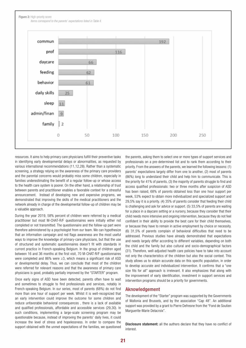
21
resources. It aims to help primary care physicians fulfill their preventive tasks in identifying early developmental delays or abnormalities, as requested by various international recommendations (11,12,28). Rather than a systematic screening, a strategy relying on the awareness of the primary care providers and the parental concerns would probably miss some children, especially in families underestimating the benefit of a regular follow-up or whose access to the health care system is poorer. On the other hand, a relationship of trust between parents and practitioner enables a favorable context for a stressful announcement. Instead of developing new and expensive programs, we demonstrated that improving the skills of the medical practitioners and the network already in charge of the developmental follow-up of children may be a valuable approach.
During the year 2019, 58% percent of children were referred by a medical practitioner but most M-CHAT-R/F questionnaires were initially either not completed or not transmitted. The questionnaire and the follow-up part were therefore administered by a psychologist from our team. We can hypothesize that an information campaign and red flags awareness are the most useful ways to improve the knowledge of primary-care physicians, but that the use of structured and systematic questionnaires doesn’t fit with standards in current practice in French-speaking Belgium. In the group of children aged between 16 and 36 months at the first visit, 70 M-CHAT-R/F questionnaires were completed and 86% were ≥3, which means a significant risk of ASD or developmental delay. Thus, we can conclude that most of the children were referred for relevant reasons and that the awareness of primary care physicians is good, probably partially improved by the “STARTER” program.
Once early signs of ASD have been detected, parents often have to wait and sometimes to struggle to find professionals and services, notably in French-speaking Belgium. In our series, most of parents (68%) do not find more than one hour of support per week. Whilst it is well-recognized that an early intervention could improve the outcome for some children and reduce unfavorable behavioral consequences , there is a lack of available and qualified professionals, affordable and accessible services (29,30). In such conditions, implementing a large-scale screening program may be questionable because, instead of improving the parents’ daily lives, it could increase the level of stress and hopelessness. In order to compare the support obtained with the unmet expectations of the families, we questioned
the parents, asking them to select one or more types of support services and professionals on a pre-determined list and to rank them according to their priority. From the answers of the parents, we learned the following lessons: (1) parents’ expectations largely differ from one to another, (2) most of parents (86%) long to understand their child and help him to communicate. This is the priority for 41% of parents, (3) the majority of parents struggle to find and access qualified professionals: two or three months after suspicion of ASD has been raised, 68% of parents obtained less than one hour support per week, 53% expect to obtain more individualized and specialized support and 29,5% say it is a priority. (4) 35% of parents consider that feeding their child is challenging and ask for advice or support. (5) 33,5% of parents are waiting for a place in a daycare setting or a nursery, because they consider that their child needs more intensive and ongoing intervention, because they do not feel confident in their ability to provide the best care for their child themselves, or because they have to remain in active employment by choice or necessity. (6) 31,5% of parents complain of behavioral difficulties that need to be addressed. Previous studies have already demonstrated that expectations and needs largely differ according to different variables, depending on both the child and the family but also cultural and socio-demographical factors (31). Therefore, well-adjusted health care policies have to take into account not only the characteristics of the children but also the social context. This study allows us to obtain accurate data on this specific population, in order to develop accurate and individualized intervention. It confirms that a “one size fits for all” approach is irrelevant. It also emphasizes that along with the improvement of early identification, investment in support services and intervention programs should be a priority for governments.
AknowledgementThe development of the “Starter” program was supported by the Governments of Wallonia and Brussels, and by the association “Cap 48”. An additional support was provided by a grant to Pierre Defresne from the “Fond de Soutien Marguerite-Marie Delacroix”.
Disclosure statement: all the authors declare that they have no conflict of interest.
Figure 2: High-priority score Items correspond to the parents’ expectations listed in Table II.

22
ThemeREFERENCES:
1. American Psychiatric Association. Diagnostic and statistical manual of mental disorders (DSM-5). Behavioral Reviews 2013; 33: 1204-1214.
2. Werner E, Dawson G, Osterling J, Dinno N. Brief report: Recognition of autism spectrum disorder before one year of age: a retrospective study based on home videotapes. J Autism Dev Disord 2000; 30: 157–162.
3. Ozonoff S, Iosif AM, Baguio F, Cook IC, Hill MM, Hutman T,et al. A prospective study of the emergence of early behavioral signs of autism. J Am Acad Child Adolesc Psychiatry 2010; 49: 256–66.
4. Chawarska K, Paul R, Klin A, Hannigen S, Dichtel LE, Volkmar F. Parental recognition of developmental problems in toddlers with autism spectrum disorders. J Autism Dev Disord 2007; 37: 62–72.
5. Klintwall L, Eikeseth S. Early and intensive behavioral intervention (EIBI) in autism. In J. Anderson, S. Patel, V. B. Preedy, & C. R. Martin, editors. Comprehensive Guide to Autism. Springer, New York, 2014: 117-137.
6. Bejarano-Martín Á, Canal-Bedia R, Magán-Maganto M, Fernández-Álvarez C, Cilleros-Martín MV, Sánchez-Gómez MC, et al. Early Detection, Diagnosis and Intervention Services for Young Children with Autism Spectrum Disorder in the European Union (ASDEU): Family and Professional Perspectives. J Autism Dev Disord 2020; 50: 3380-3394
7. French L, Kennedy EMM. Annual Research Review: Early intervention for infants and young children with, or at-risk of, autism spectrum disorder: a systematic review. J Child Psychol Psychiatry 2018; 59: 444–456.
8. Landa RJ. Efficacy of early interventions for infants and young children with, and at risk for, autism spectrum disorders. Int Rev Psychiatry 2018; 30: 25–39.
9. García-Primo P, Hellendoorn A, Charman T, Roeyers H, Dereu M, Roge B, et al. Screening for autism spectrum disorders: state of the art in Europe. Eur Child Adolesc Psychiatry 2014 ; 23: 1005–1021.
10. Ozonoff S. Editorial: Early detection of mental health and neurodevelopmental disorders: the ethical challenges of a field in its infancy. J Child Psychol Psychiatry 2015; 56: 933–935.
11. Siu AL, US Preventive Services Task Force (USPSTF), Bibbins-Domingo K, Grossman DC, Baumann LC, Davidson KW, et al. Screening for Autism Spectrum Disorder in Young Children: US Preventive Services Task Force Recommendation Statement. JAMA 2016; 315: 691–696.
12. Zwaigenbaum L, Brian JA, Ip A. Early detection for autism spectrum disorder in young children. Paediatr Child Health 2019; 24: 424–443.
13. Nevill RE, Lecavalier L, Stratis EA. Meta-analysis of parent-mediated interventions for young children with autism spectrum disorder. Autism 2018; 22: 84–98.
14. Reichow B, Hume K, Barton EE, Boyd BA. Early intensive behavioral intervention (EIBI) for young children with autism spectrum disorders (ASD). Cochrane Database Syst Rev 2018; 5: CD009260.
15. Sandbank M, Bottema-Beutel K, Crowley S, Cassidy M, Dunham K, Feldman JI, et al. Project AIM: Autism intervention meta-analysis for studies of young children. Psychol Bull 2020; 146: 1–29.
16. Cappe E, Poirier N. Les besoins exprimés par les parents d’enfants ayant un TSA : une étude exploratoire franco-québécoise. Annales Médico-Psychologiques, revue Psychiatrique. 2016; 174: 639-643.
17. Derguy C, Michel G, M’bailara K, Roux S, Bouvard M. Assessing needs in parents of children with autism spectrum disorder: A crucial preliminary step to target relevant issues for support programs. Journal of Intellectual and Developmental Disability 2015; 40: 156–166.
18. Divan G, Vajaratkar V, Desai MU, Strik-Lievers L, Patel V. Challenges, coping strategies, and unmet needs of families with a child with autism spectrum disorder in Goa, India. Autism Research 2012; 5: 190–200.
19. Defresne P, Bouchez M-H, Delmotte J, Willaye E. START, un programme de « Repérage des troubles précoces de la communication et des interactions sociales », comme alternative au dépistage systématique des troubles du spectre de l’autisme. ANAE 2016; 144: 511-517.
20. Adrien JL, Barthélémy C, Perrot A, Roux S, Lenoir P, Hameury L, et al. Validity and reliability of the infant behavioral summarized evaluation (IBSE): a rating scale for the assessment of young children with autism and developmental disorders. J Autism Dev Disord 1992; 22: 375–394.
21. Baron-Cohen S, Wheelwright S, Cox A, Baird G, Charman T, Swettenham J, et al. Early identification of autism by the CHecklist for Autism in Toddlers (CHAT). J R Soc Med 2000; 93: 521–525.
22. Robins DL, Casagrande K, Barton M, Chen CM, Dumont-Mathieu T, Fein D. Validation of the modified checklist for Autism in toddlers, revised with follow-up (M-CHAT-R/F). Pediatrics 2014; 133: 37–45.
23. Bryson SE, Zwaigenbaum L, McDermott C, Rombough V, Brian J. The Autism Observation Scale for Infants: scale development and reliability data. J Autism Dev Disord 2008; 38: 731–738.
24. Gabrielsen TP, Farley M, Speer L, Villalobos M, Baker CN, Miller J. Identifying autism in a brief observation. Pediatrics 2015; 135: e330–e338.
25. Estes A, Swain DM, MacDuffie KE. The effects of early autism intervention on parents and family adaptive functioning. Pediatr Med 2019; 2: 21.
26. Pierce K, Gazestani VH, Bacon E, Barnes CC, Cha D, Nalabolu S, et al. Evaluation of the diagnostic stability of the early autism spectrum disorder phenotype in the general population starting at 12 months. JAMA Pediatr 2019; 173: 578–587.
27. Hyman SL, Levy SE, Myers SM. Council on children with disabilities, section on developmental and behavioral pediatrics. Identification, evaluation, and management of children with autism spectrum disorder. Pediatrics 2020; 145: e20193447.
28. Haute Autorité de la Santé. Trouble du Spectre de l’Autisme. Signes d’alerte, repérage, diagnostic et évaluation chez l’enfant et l’adolescent. Recommandations pour la pratique clinique [Internet]. 2018 [cited 2020 September 30]. Available from : https://www.has-sante.fr/jcms/c_468812/fr/trouble-du-spectre-de-l-autisme-signes-d-alerte-reperage-diagnostic-et-evaluation-chez-l-enfant-et-l-adolescent.
29. Siller M, Hutman T, Sigman M. A parent-mediated intervention to increase responsive parental behaviors and child communication in children with ASD: A randomized clinical trial. J Autism Dev Disord 2013; 43: 540–555.
30. Estes A, Munson J, Rogers S, Greenson J, Winter J, Dawson G. Long-term outcomes of early-intervention in 6-year-old children with autism spectrum disorders. J Am Acad Child Adolesc Psychiatry 2015; 54(7): 580-7
31. Hodgetts S, Zwaigenbaum L, Nicholas D. Profile and predictors of service needs for families of children with autism spectrum disorders. Autism 2015; 19: 673–683.

23
Integration of exergaming in pediatric rehabilitationBruno Bonnechère
REVAL Rehabilitation Research Center – Faculty of Rehabilitation Sciences, Hasselt University, Diepenbeek, Belgium
Theme
AbstractThe development and generalization of modern technology have totally modified our world and habits. Concerning rehabilitation, the situation is a bit different and the integration of technology seems to be a bit slower than in other fields. The development of video games, and in particular active video games, offers new perspectives and pediatric rehabilitation. In this paper, we present the different technologies currently tested and developed in various rehabilitation contexts ranging from neurological rehabilitation (children with cerebral palsy) to pain management but also diseases such as autism spectrum disorders. Finally, we discuss the current and future challenges related to the integration of exergames in pediatric rehabilitation and propose some solutions to promote the development and implementation of these solutions on a large scale in clinics.
KeywordsPediatric rehabilitation; exergames; video games; new technology; motivation
Currently, informatics and technology are everywhere, or almost everywhere, in our lives. In the last decade, not only technology has developed drastically but two other elements are also of importance for the development and implementation of innovative solutions, at a large scale, in the healthcare sector: the democratization of the technology and the miniaturization of the devices. The development of new technology offers, therefore, a lot of new opportunities in rehabilitation for both the diagnosis, the assessment, and the treatment.
In this paper, we are going to focus on the use and development of exergames in pediatric rehabilitation but the readers should be aware that the use of technology is much broader than this particular set of applications.
Before discussing more in detail the exergames let’s briefly discussed some key potential advantages of the integration of new technology in rehabilitation.
The first one is related to the lack of access to physical rehabilitation centers. Lack of access to healthcare facilities or healthcare professionals has been pointed out by the WHO as one of the major obstacles to the rehabilitation process not only in low-income countries but also in high-income countries (1). Recently the problem of accessibility and physical interaction with clinicians has been brought to the forefront in the context of the COVID-19 crisis (2). The use of technology could help to partially solve the issue related to the accessibility of care with the development and generalization of telerehabilitation and teleconsultation (3).
Another important point, especially for pediatric rehabilitation, is the motivation of the patients. The motivation of the patients is the main key to an as successful as possible rehabilitation treatment (4). The main challenge is therefore to keep patients motivated enough despite the feeling he/she could have of ‘inefficiency’, ‘lack of progress’, ‘tiredness’, etc. Such problems are even more present with teenagers during the puberty identity crisis. By definition games are fun and induced distraction thus the patients are more motivated by this kind of exercise (at least at the beginning of the treatment we will discuss this point later on) (5).
A last potential advantage is that, in most cases, the exercises and the motions performed by the patients can be recorded. This information can be used later on to provide real-time feedback to the patients to correct him when he is performing the exercises, or to remind him to do the exercises or how to do them properly (6,7). This information can also be used by the clinicians to follow the evolution of the patients and, if needed, adapt the rehabilitation program or the difficulty of the exercises to the real need and specificity of the patients (8,9). By doing so the treatment is always adapted to the patients and thus the quality of the care is increased.
There are a lot of different terms used to describe the development and use of games in rehabilitation (both motor and cognitive aspects). Unfortunately, these terms are often misused which could lead to a certain confusion for the clinicians. Therefore before discussing the clinical aspect of this new domain it is important to define the different terms used in the literature.
Clinical applicationsDifferent research directions (e.g., motor rehabilitation, cognitive rehabilitation, pain management) are currently under development or already in clinical use. We will present here the results and level of evidence for some of the most important, or most promising field, of pediatric rehabilitation. We try to give the best possible overview of the possibilities offered by the addition of games in the rehabilitation but could not present all the different studies. First, we will present applications where the games are used to promote motor functions: for neurologic rehabilitation and solution to promote physical activity levels. Then we will discuss other fields of applications where the games are more used to stimulate cognitive functions.
Cerebral palsy
For the sake of this presentation, we focus on children with cerebral palsy but the principle can be easily transferred to other neurologic or orthopedic pathologies since most of the solutions are focusing on balance and mobility issues.
Cerebral palsy is a group of movement disorders that appears in early childhood. It is due to brain lesions occurring during pregnancy, childbirth, or during the first year of the children. These lesions, and therefore the movement disorders, are irreversible because they occurred in immature brain tissues. Patients suffering from cerebral palsy present various motor and balance troubles.
In a systematic review about the use of commercial video games in rehabilitation, we found that the integration of this kind of game in the treatment of children with cerebral palsy was quite popular (after balance issue related to aging and stroke) (10). Other important findings of this review are that the exergames increase patients’ motivation and that the patients are performing more repetitions of the exercises than during conventional rehabilitation sessions. The latter is particularly important since it is well known that the number of repetitions is a key factor in rehabilitation (11).
The results of 19 RCTs on the efficacy of exergames on upper extremity function, ambulation, and postural control were summarized in a meta-analysis (12). 12 of the 19 studies were done using commercial solutions, among them the great majority were done using the Nintendo Wii (8/12).
Introduction Definitions

24
13 RCT studies were included measuring arm function. Across all studies, there was a strong effect (d = 0.835; 95% CI = 0.388–1.282) in favor of the intervention. Interestingly the authors observed a large difference in the effect between exergames specially developed for the rehabilitation and commercial systems (d = 2.162 for specific systems; d = 0.491 for commercially available systems). Concerning the ambulation, 8 studies were included. The overall effect size was 0.755 (95% CI = 0.348–1.161), indicating a medium to large effect size, due to the small sample size the authors were not able to perform subgroups analysis to compare specific and commercial solutions. Finally, for the postural control, 10 studies were included. The overall effect size was 1.003 (95% CI = 0.503–1.502), indicating a large effect size.
Physical activity
Overweight and obesity issues are a huge and growing healthcare challenge worldwide and this problem is becoming more and more precocious. The ‘traditional’ video games (i.e., video games controlled with a controller when the player is sited), also called passive video games, are known to increase the level of sedentariness and promote snacking and junk food (13). To try to change this negative image and promote physical activity games companies developed systems (mainly Nintendo Wii™ and Microsoft Xbox Kinect™) where the players have to move to control the games: the active video games.
The first question is to determine whether or not active video games induce an increase in energy expenditure and if yes what is the level of physical activity reached during this kind of training. Many studies have been conducted in this field. The level of energy expenditure reached is estimated to be between 2.7 and 5.4 metabolic equivalents (i.e. moderate-intensity level of physical activity) when children and teenagers playing active games (14). A study compared the levels of energy expenditure in an obese population and in a control group, surprisingly the author found that obese children lose fewer calories than the control group (15). Although the levels of energy expenditure reached are relatively low the next question is to determine if active video games could still be successfully integrated into the management of obesity and overweight. A large study was conducted with 171 obese students included. They were separated into 2 groups: 63 people were included in the group playing video games and the other 108 in the control group. After nine weeks of training, the level of physical activity was higher in the intervention group and patients had lost between 1.5 and 2kg after this intervention compared to the control group (16).
There are also plenty of different solutions developed to provide dietetic information, records the food consumed, and modify eating behavior (17).
Autism spectrum disorders
Autism spectrum disorders are developmental conditions that involve difficulties in social and non-verbal interaction, speech, and restricted or repetitive behavior. Exergames could also be used with patients with autism spectrum disorders in order to reduce fear, anxiety level, and to increase social skills (18). The development of specific solutions for patients with autism spectrum disorders is a growing field. The idea is to immerse the patients in a virtual environment where they can learn to collaborate and interact with other players without fearing the interactions (19). Another potential advantage for the clinicians is that when the patients are immersed in the games they are paying less attention to their real environment and the clinicians can touch them to promote proprioception and acceptance of others (20). The last important area of research in the development of diagnosis tools, autism may be difficult to diagnose, especially in young children. The way the children are playing and using tablets may be used to distinguish affected from not affected children and to assess the severity of the disease (21).
Pain management
Pain is one of the most frequent symptoms in rehabilitation, and also one of the most complex to manage. Several approaches can be used to integrate exergames in pain management programs.
Pain, especially chronic pain, remains a relatively poor and poorly understood process. Therefore patients may have a lot of questions related to it. Healthcare professionals are not always present and available to respond. Thus have been developed (explanatory videos, texts, videos) to answer the questions of the patients and help them. These virtual systems have proven their efficacy in the treatment of chronic pain (22). Therefore, such kind of tools appears to be a
feasible solution to increase the dialogue on pain management between patient and therapist (23). However, currently, most of the studies using such kinds of interventions are done with adults, not with children.
A second approach is to create a distraction phenomenon in patients when performing painful procedures. The challenge for developers is to create games in which the patients won’t move but will be immersed enough in order to not feel pain. Such solutions have been developed for young children going to the dentist. There was a significant decrease in pain perception and anxiety with the use of virtual reality glasses and games during dental treatment (24).
Another particularly painful condition is the treatment of severe burns. Virtual reality and serious games can be used to reduce pain during wound cleaning. The results seem to be maintained over time: 3 or 4 days after the intervention the results are maintained and the pain is still decreased during care (25).
Adherence to medical treatment
We have seen that for pain management solutions have been developed to inform the patients. We know that treatment adherence is better when the patients are well informed about their pathologies and treatment. This is particularly important in pediatric rehabilitation.
One salient aspect of the exergames is that pieces of advice and medical information can be added to games. The first example of such kind of games was a game for diabetic children developed in collaboration with gaming and pharmaceutical companies. A study shows that compared to a control group, children who play the games managed better their diabetes: the number of children who have to go to the hospital due to a glucose crisis decreased by 77% in the intervention group compared to the control (26). Another very popular game that had a significant impact on health-related behavior is ‘’ game. This game was created for children with cancer to teach them how to deal with cancer treatment (mainly chemotherapy) to maximize treatment adherence. In this game, patients must shoot cancer cells to fight the infections and manage clinical signs and adverse effects (e.g., constipation, nausea, etc.). A clinical trial has been done to compare the effect of the game compared to a commercial game in adolescents and young adults who were undergoing cancer therapy. Results showed that patients playing ‘ significantly improved treatment adherence, indicators of cancer-related self-efficacy, and knowledge about the disease (27).
Challenges to solveAlthough exergames are being tested for several years in clinics there are still a lot of problems to solve before they can be broadly used in daily clinics. Some of these issues are related to the technology (i.e., price, accessibility, ease of use), but most of them concern the clinicians and the patients: people must still be convinced that exergames are serious tools for rehabilitation and not just fun. Large scale RCTs are needed to determine whether or not exergames can be used in pediatric rehabilitation and to what extent. One of the limitations of the current studies is the relatively small sample size and an important heterogeneity in the patients but also in the tests used to evaluate the intervention, making the comparison between studies more complicated.
Besides the validation of the new interventions, other questions should be answered.
The first one is the long term motivation and/or long term efficacy of such kind of intervention. Most of the studies are relatively short (max 3 months of interventions) and the evaluations are performed after the intervention. There is not much information about the persistence of the benefit. Concerning the motivation and the treatment adherence, it has been demonstrated that in the beginning when you introduce novelty in the treatment the patients are motivated but this effect fades over time (28).
Another important point is to evaluate the mechanism of action of the exergames. This should be used to increase the quality of the rehabilitation and also to convince the most skeptical. A recent scoping review focusing on children with cerebral palsy suggested some possible mechanisms of action. For the motor skills, exergames enhanced problem-solving and cognitive engagement during play and increased motivation and neuroplasticity changes; exergames also created repetitive task-oriented and task-specific practices in an ecologically valid virtual environment that was similar to the real world and provided the flexibility of adjusting task difficulties, visual and/

25
or auditory feedback, and the potential of social play and interaction; and finally exergames offered social support from parents, peers, or therapists (29).
The safety of the intervention also needs to be further assessed, especially in the context of telerehabilitation where the patients have to do the exercises by themselves.
The last point is the sustainability of the system. We have seen that for the rehabilitation of children with cerebral palsy and weight management, most of the systems were commercial active video games. Despite the popularity in the medical community, the Wii system was discontinued in 2013 and the Kinect in 2017. In the meantime, Nintendo executives announced that they will not incorporate any active video games in their next console and games and only focus on passive video games (30). Does this mean the end of exergames? Hopefully not, there are already specific solutions that do not rely on technologies from the gaming industry. However, those solutions are more difficult to find and more expensive because of factors like the size of the market. It is therefore important to find solutions to guarantee patients a long-term sustainable use of these new tools.
ConclusionThe current gaming technology is, currently, not fully adapted to the clinics and further development and improvement are still needed. However, the existing solutions – commercial or specially developed – already offer interesting new perspectives in rehabilitation. The use of exergames increases the motivation of the patients. Therefore the patients are performing a higher number of repetitions of the exercises which has a direct positive impact on the rehabilitation. In addition to this direct aspect, the advantage of the games is that they also stimulate different cognitive functions and distract the patient which allows for other types of benefits during revalidation. Although the aspects are not fully understood and therefore the benefits of these are not yet fully implemented in rehabilitation these are very interesting perspectives for various pathologies.
Conflict of interest
No potential competing interest was reported by the author.
REFERENCES:
1. World Health Organization. World Health Organization (WHO) and World Bank World Report on Disability. 2011.
2. Demers M, Martinie O, Winstein C, Robert MT. Active Video Games and Low-Cost Virtual Reality: An Ideal Therapeutic Modality for Children With Physical Disabilities During a Global Pandemic. Front Neurol. 2020;11:601898.
3. Charvet LE, Yang J, Shaw MT, Sherman K, Haider L, Xu J, et al. Cognitive function in multiple sclerosis improves with telerehabilitation: Results from a randomized controlled trial. PLoS One. 2017;12(5):e0177177.
4. Reedman SE, Boyd RN, Ziviani J, Elliott C, Ware RS, Sakzewski L. Participation predictors for leisure‐time physical activity intervention in children with cerebral palsy. Dev Med Child Neurol. 2021 [in Press]
5. Ramírez-Granizo IA, Ubago-Jiménez JL, González-Valero G, Puertas-Molero P, San Román-Mata S. The Effect of Physical Activity and the Use of Active Video Games: Exergames in Children and Adolescents: A Systematic Review. IJERPH. 2020;17(12):4243.
6. Bonnechère B, Omelina L, Jansen B, Van Sint Jan S. Balance improvement after physical therapy training using specially developed serious games for cerebral palsy children: preliminary results. Disabil Rehabil. 2017;39(4):403–6.
7. Bonnechère B. Serious Games in Physical Rehabilitation: From Theory to Practice. Springer International Publishing; 2018
8. Bonnechère B, Jansen B, Haack I, Omelina L, Feipel V, Van Sint Jan S, et al. Automated functional upper limb evaluation of patients with Friedreich ataxia using serious games rehabilitation exercises. J Neuroeng Rehabil. 2018;15(1):87.
9. Bonnechère B, Sholukha V, Omelina L, Van Sint Jan S, Jansen B. 3D Analysis of Upper Limbs Motion during Rehabilitation Exercises Using the KinectTM Sensor: Development, Laboratory Validation and Clinical Application. Sensors. 2018;18(7):2216.
10. Bonnechère B, Jansen B, Omelina L, Van Sint Jan S. The use of commercial video games in rehabilitation: a systematic review. Int J Rehabil Res. 2016;39(4):277–90.
11. Langhorne P, Bernhardt J, Kwakkel G. Stroke rehabilitation. The Lancet. 2011;377(9778):1693–702.
12. Chen Y, Fanchiang HD, Howard A. Effectiveness of Virtual Reality in Children With Cerebral Palsy: A Systematic Review and Meta-Analysis of Randomized Controlled Trials. Phys Ther. 2018;98(1):63–77.
13. Taylor MJD, McCormick D, Shawis T, Impson R, Griffin M. Activity-promoting gaming systems in exercise and rehabilitation. JRRD. 2011;48(10):1171.
14. Comeras-Chueca C, Villalba-Heredia L, Pérez-Llera M, Lozano-Berges G, Marín-Puyalto J, Vicente-Rodríguez G, et al. Assessment of Active Video Games’ Energy Expenditure in Children with Overweight and Obesity and Differences by Gender. Int J Environ Res Public Health. 2020;17(18).
15. O’Donovan C, Roche EF, Hussey J. The energy cost of playing active video games in children with obesity and children of a healthy weight. Pediatr Obes. 2014;9(4):310–7.
16. Johnston JD, Massey AP, Marker-Hoffman RL. Using an alternate reality game to increase physical activity and decrease obesity risk of college students. J Diabetes Sci Technol. 2012;6(4):828–38.
17. Froome HM, Townson C, Rhodes S, Franco-Arellano B, LeSage A, Savaglio R, et al. The Effectiveness of the Foodbot Factory Mobile Serious Game on Increasing Nutrition Knowledge in Children. Nutrients. 2020;12(11):3413.
18. Maskey M, Lowry J, Rodgers J, McConachie H, Parr JR. Reducing Specific Phobia/Fear in Young People with Autism Spectrum Disorders (ASDs) through a Virtual Reality Environment Intervention. van der Feltz-Cornelis C, editor. PLoS ONE. 2014;9(7):e100374.
19. Terlouw G, van ’t Veer JT, Prins JT, Kuipers DA, Pierie J-PEN. Design of a Digital Comic Creator (It’s Me) to Facilitate Social Skills Training for Children With Autism Spectrum Disorder: Design Research Approach. JMIR Ment Health. 2020;7(7):e17260.
20. Tang JSY, Falkmer M, Chen NTM, Bölte S, Girdler S. Designing a Serious Game for Youth with ASD: Perspectives from End-Users and Professionals. J Autism Dev Disord. 2019;49(3):978–95.
21. Millar L, McConnachie A, Minnis H, Wilson P, Thompson L, Anzulewicz A, et al. Phase 3 diagnostic evaluation of a smart tablet serious game to identify autism in 760 children 3-5 years old in Sweden and the United Kingdom. BMJ Open. 2019;9(7):e026226.
22. Rantala A, Pikkarainen M, Miettunen J, He H-G, Pölkki T. The effectiveness of web-based mobile health interventions in paediatric outpatient surgery: A systematic review and meta-analysis of randomized controlled trials. J Adv Nurs. 2020;76(8):1949-1960
23. Yap KY-L, Koh DWH, Lee VSJ, Wong LL. Use of virtual reality in the supportive care management of paediatric patients with cancer. Lancet Child Adolesc Health. 2020;4(12):899–908.
24. Gates M, Hartling L, Shulhan-Kilroy J, MacGregor T, Guitard S, Wingert A, et al. Digital Technology Distraction for Acute Pain in Children: A Meta-analysis. Pediatrics. 2020;145(2):e20191139
25. Faber AW, Patterson DR, Bremer M. Repeated use of immersive virtual reality therapy to control pain during wound dressing changes in pediatric and adult burn patients. J Burn Care Res. 2013;34(5):563–8.
26. Brown SJ, Lieberman DA, Germeny BA, Fan YC, Wilson DM, Pasta DJ. Educational video game for juvenile diabetes: results of a controlled trial. Med Inform (Lond). 1997;22(1):77–89.
27. Kato PM, Cole SW, Bradlyn AS, Pollock BH. A video game improves behavioral outcomes in adolescents and young adults with cancer: a randomized trial. Pediatrics. 2008;122(2):e305-317.
28. Joelsson M, Lundqvist S, Larsson MEH. Tailored physical activity on prescription with follow-ups improved motivation and physical activity levels. A qualitative study of a 5-year Swedish primary care intervention. Scand J Prim Health Care. 2020;38(4):399–410.
29. Levac D, Rivard L, Missiuna C. Defining the active ingredients of interactive computer play interventions for children with neuromotor impairments: a scoping review. Res Dev Disabil. 2012;33(1):214–23.
30. Bonnechère B, Omelina L, Kostkova K, Van Sint Jan S, Jansen B. The end of active video games and the consequences for rehabilitation. Physiother Res Int. 2018 Oct;23(4):e1752.

26
Theme

27
Physical activity and sports in children with disabilities in FlandersPatricia Van de Walle a,b, Ronny Van Assche a, Jens Van Assche a, Christel Vanroy b, Frederik Lenaerts c, Stefan Verheyen d, Guy Molenaers c,e, Annik Bosmans a
a Heder, Institution for children and adults with a disability, Antwerp b Research Group MOVANT, Department of Rehabilitation Sciences and Physiotherapy (REVAKI), University of Antwerp c Fysische Geneeskunde en Revalidatie, Sports, UZ Pellenberg, Leuven d Grassroots, Voetbal Vlaanderen e Department of Paediatric Orthopaedics, UZ Pellenberg, Leuven
[email protected] ; [email protected]
Theme
AbstractThe paper describes physical activity and discusses benefits and opportunities but also obstacles of physical activity and sports in children with disabilities.
Keywordsphysical activity, child, motor disorder, disability
It is well known that physical activity (PA) is an important factor to improve fitness and health in all people with and without disabilities. Children with disabilities, however, are known to participate less in PA than their typically developing peers (1, 2). PA is necessary for an optimal physical, emotional and psychosocial development, but is not easy to obtain for children with disabilities. In 2015 Verschuren et al. published recommendations on PA and avoidance of sedentary behavior for people with cerebral palsy (CP) based on the previous WHO guidelines (3). The recent WHO guidelines now include recommendations on PA in children, adolescents (at least 60 minutes per day of moderate to vigorous activity) and adults (150-300 minutes/week moderate to vigorous aerobic activity, including muscle and bone strengthening exercise at least 3 times/week) living with disabilities (4).
In order to promote PA in children with disabilities, understanding barriers and facilitators is important. Reasons for the lower level of PA are complex and multifactorial (5). They can be found in the , their and the (6). It is also important to realize that health related benefits of PA can be attained during . Promotion of PA should thus not only focus on organized sports and activities, but also on occupational, transportation related and leisure activities in e.g. a school or familial context. The Physical Activity for People with Disabilities conceptual model is an integrated model of physical activity behavior and explores its relation with functioning and disability. This model can help health professionals to understand the facilitators and barriers in individuals with disabilities (7).
The goal of this study was is to describe habitual PA, investigate motivation and barriers and discuss local examples to enhance habitual PA in children with CP in specific and people with neuromotor disabilities in general. We present the results of two local studies, a first study describing habitual PA in 6-9 year old children with CP and a second study exploring motivation and barriers to join organized PA in children and adults in Antwerp. Finally we share our local experience of organizing PA and sports, specifically for children and adolescents with motor disabilities, based on three examples: a sports club, a sport-specific organization and an advice center for individual PA.
MethodsExploring physical activity in children with cerebral palsy in Flanders
For this clinical trial, all ambulatory children with CP (Gross Motor Functioning Classification Scale (GMFCS) I-III ), attending either the CP reference center of Antwerp (CePra) or UZ Leuven, born in 2009 or 2010 were identified (8, 9). They were asked to fill in two questionnaires (Children Assessment and Performance Scale (CAPE) and Activities Scale for Kids-Performance (ASKp)) and a coded diary on PA for seven consecutive days.
The ASKp is a 30-item questionnaire with seven sub-domains measuring what the child did in the previous week at the levels of personal care, dressing, other skills, locomotion, play and transfers and thus indicates a child’s physical functioning (10, 11). The CAPE is a 55-item questionnaire and gives information about five dimensions of participation in the last four months: diversity, intensity, enjoyment, with whom and where (12).
The coded activity diary was used for determining the activity levels (13). It was completed for seven consecutive days (five schooldays and two weekend days). Activities during twenty-four hours were converted to metabolic equivalents values and multiplied by 30 min (METs*minutes) (14). MET values can be divided in four activity levels: corresponding to sedentary (< 1 METs), light (>1 - < 3 METs), moderate (3-6 METs) and vigorous activity (> 6METs).
Exploring motivation and barriers in sports participation in people with disabilities in Antwerp
All participants (or their parents) of two local sports clubs for people with disabilities (Spinnaker and Wapper), were asked about their motivation and barriers to participate in sports activities.
Participants were asked if one or more motivational aspects (global health, improved cardiorespiratory fitness, social contacts, performance, leisure or others) and one or more barriers (distance (to far), transport, financial aspects, health, lack of material, sports club or (different) sports, familial reasons or others) were applicable.
Description of local initiatives to enhance PA in people with disabilities
Local initiatives at different levels will be discussed:
· a sports club offering different sports for children and adults with motor disabilities
· a sports organization promoting a single sport for the specific target popu-lation of children with neuromotor disorders
· an advice center for mobility and PA through cycling for children and adults
ResultsExploring physical activity in children with cerebral palsy in Flanders
Of 130 children born in 2009 or 2010 attending the two CP reference cen-ters, 110 children were reached and 35 children (GMFCS I: n=22; GMFCS II: n=7; GMFCS III: n=6) returned complete questionnaires and diaries on PA. Of those 35 children all children received physiotherapy and 85% par-ticipated in sports (17/22 in GMFCS I, 6/7 in GMFCS II and 4/6 in GMFCS III).
Introduction

28
ASKp scores were near normal in GMFCS I but were decreased in most chil-dren with GMFCS II-III (Figure 1A). Mean participation time in sport activities was 147 minutes [0 - 420] with higher values for GMFCS I-II (GMFCS I; 146 minutes [0-420], GMFCS II: 165 minutes [0-420] compared to GMFCS III (70 minutes [0-120]) per week with joy in all GMFCS-levels (Figure 1B). Fig-ure 2 gives the individual amounts of moderate and vigorous activity during school, semi-school (Wednesday) and weekend days from the coded diary.
Exploring motivation and barriers in sports participation in people with disabilities in Antwerp
With 97/128 returned questionnaires for Spinnaker and 44/86 for Wapper there was a response rate of respectively 75% and 51%.
Most cited motivation to participate in sport activities is leisure in all age groups (63% in children, 81% in adolescents and 69% in adults, see figure 3). More than half of the participants had two or more reasons to participate. In the 65/141 participants with only one reason, leisure was most cited, 3 times more than the second most cited cardiorespiratory fitness. If more than one reason was cited, leisure was still most cited, 19% more than social contact and almost twice as much (80% more) as cardiorespiratory fitness.
All children (or their parents) experienced at least one barrier to participate in sports activities. In adults 46% did not experience barriers at all (see figure 4). If only one single reason was cited, this was always transport. Other reasons not to participate were diverse, including ‘I don’t know where to look’, ‘want to, but did not do it yet’, ‘not possible yet in combination with school’, not all clubs fit my need’, ‘not offered in my region’, ‘I don’t know which sport I can do with my handicap’.
Local initiatives to enhances PA in children with disabilities
Sports club
started already in 1976 as a sports club with a more recreational character as part of the former Sint-Jozefinstituut, now Heder, a special school and rehabilitation center for children and youth with physical or mental disorders in Antwerp. In the early ’90s the offer became limited to people with physical disabilities, as this target group experienced most barriers to be physical active. Furthermore the offered sports were from that point on limited to sports with an ‘official’ character, meaning that there were opportunities for competition and transfer to regular circuits. The sports offered at Spinnaker have a clear link with the children attending school at Heder. The main goal of this sports club is to get the children acquainted with sports and PA and its values and benefits so they can take that positive experience to carry on in their future adult lives.
Sports organization
In 2015 a first CP soccer team for children restarted out of the need for an offer for children with neuromotor disorders. After the Paralympic medals (1984 and 1988) and diverse adult championships till 1994, CP-football in general and for children
Figure 1: Subjective measure of habitual physical activity in GMFCS level I-III
A: Total score on the Activity Scale for Kids performance (AsKp) (red line, mean value of typical
developing children, maximum= 100)
B: score Children Assessment and Performance Scale (CAPE) subscore joy
Figure 2: Mean habitual physical activity during schooldays (A), semi-schooldays (B) and weekend days (C) for individual patients as obtained by a coded diary sorted by GMFCS level.
Moderate activity in grey, vigorous activity in yellow. GMFCS = Gross Motor Function Classification System, MET = metabolic equivalent of tasks, MTSdayMO = Metabolic equivalent of tasks per day Moderate, MTSdayVI Metabolic equivalent of tasks per day vigorous.

29
in particular had become somewhat forgotten in Belgium. Children with neuromotor disorders were therefore directed to a more general offer for children and adults with different kinds of limitations (G-sports). However for a child with motor impairments it is difficult to compete with children with mental disorders and normal motor capacities and they often gave up soccer due to a lack of enjoyment in general G-sport. Not long after the first team, different regular soccer clubs arose, at first for children with CP (in analogy with International Federation of CP (IFCP) football). Nowadays, about ten teams offer soccer especially for children with neuromotor disorders within the Belgian Football Association (VoetbalVlaanderen) under the umbrella of CP+ soccer, the ‘+’ indicating that children with other neuromotor disorders are also welcome. The sports organizations, in close collaboration with Parantee-Psylos who supports clubs and federations in Flanders, plays an important role in promotion and enhances initiatives that brings different clubs together, allowing this initiatives to grow.
Advice center
Not all children choose to participate in organized sports and the health benefits of PA also exist when exercising in other contexts. Furthermore cycling is an ideal way to combine transport and habitual PA. Therefore initiatives to enhance PA outside an organized sports context are of great value. In 2010, a local center for individual advice on cycling was developed by UZ Pellenberg with support of the Province Vlaams-Brabant and VZW Move to Improve , a foundation with the goal to optimize care for children with neuromotor (movement) difficulties in order to find sustainable solutions to help those children to move more and better, based on movement evaluation and scientific research. Children (and adults) who experience difficulties with learning to or riding a bike due to their impairment can find objective and independent information and council at the Fietsadviescentrum at the UZ Pellenberg. A large part of the advices
Figure 3: Number of participants per motivational aspect for A: children (≤13 years - light grey), B: adolescents (14-18 year - dark grey), C: adults (>18 year - black). D: percentage of participants per group
that cited a specific reason (x-axis).
CRF= cardiorespiratory fitness
Figure 4: Number of participants for A: children (≤13 years - light grey), B: adolescents (14-18 year - dark grey), C: adults (>18 year - black) and D: percentage of participants citing a barrier per group and
per perceived barrier.

30
(30%) is given to children between 6 and 12 years. A special fitted bike is not always necessary, often small adaptations to a normal bike are sufficient to get the children mobile, active and in charge of their own mobility.
DiscussionThe goal of this study was to describe PA, investigate motivation and barriers and discuss local examples to enhance PA in children with CP in specific and people with neuromotor disabilities in general.
Results show that it is possible for ambulant children with CP to reach the recommendations on PA by the WHO, regardless of GMFCS level. The main motivation to participate in sports activities was a meaningful leisure activity, regardless of age. Almost half of the adults could participate without experiencing barriers.
However, in the first study, only 35/130 children aged between 6-9 years participated, partially to the time-consuming questionnaires and diaries, so extrapolation to the entire population of children with CP remains difficult. In order to have an idea of the habitual PA in the individual child with neuromotor disabilities, PA can be monitored in clinical practice with commercial activity trackers.
The second study confirmed that motivation and barriers can be multidimensional, with more than half of the participants having more than one single motivation (5, 6). Most of the participants with a singular reason were motivated to participate in sports as part of meaningful leisure activities, before personal health or cardiorespiratory fitness. In the selected population, all children, adolescents and adults had already found their way to organized sports. Still, half of the adults reported that transport and distance to a club remains a barrier. For the children, these barriers were almost not existing, as they participated at the school they were attending (Spinnaker being located at Heder).
As confirmed by our results, organizing sports activities locally and nearby can help to overcome barriers. The goal of is to get the children acquainted with different kinds of sports and PA in order to give them positive experiences they can carry on to adolescence and adulthood. Our experience in CP+ soccer also highlights the importance of local clubs. Although at the beginning several parents were willing to drive up to an hour to give their child the opportunity to experience enjoyment on a soccer field every weekend, as soon as new clubs arose, they changed to a club nearby. CP+ soccer arose out of the need to have an offer especially for children with neuromotor disorders, as inclusion in the existing G-teams (for people with mental disorders and good motor capacities) was difficult. VoetbalVlaanderen and Parantee-Psylos have done a great effort to enhance soccer for this specific target population, enabling a quick growth of number of clubs and thus also number of children participating in soccer regularly. Their enhancement is imperative as children want to do more than just train with their teammates in one club. In order to stay in the game, they also want the experience of real matches, tournaments and opponents, in one word real inclusion into the world of soccer. As clubs and trainers almost always are volunteers, support on the level of sport-specific or general organizations is imperative, so that they can focus on the sportive aspect and the participants.
Although all children (and adults) should be physical active on a regular basis and avoid sedentary behavior, not all children with neuromotor disorders, just as typical developing children, are prone to (team) sports (4). It is therefore important to promote not only sports but habitual PA in general. The cycling advice center is an excellent example of how habitual PA in children with neuromotor disorders can be enhanced. Although the advice center does not focus on children in particular, 30% of the advices are given to children in primary school. At this age they become too old to be transported in a buggy and moving around autonomously becomes important. The advice center had financial support from VZW Move to Improve to be able to acquire test models and is still funded by SportVlaanderen. Without these financial injections it would not be possible to give the individual advice needed, as every patient experiences different barriers.
For all three initiatives we still experience that it is difficult for parents to find their way to them. Often they are positively surprised when hearing of the existing offers. In our experience this is not due to a lack of promotion from the different sports organizations. Often parents are reluctant because they either underestimate the capacities of their child or they are not able to match them with the (for them too abstract) offer from different sports clubs. So-called mouth to mouth information from different clinical professionals or parent (organization)s is often the necessary boost to join organized sports. Establishing a visible collaboration between organizations with an offer for children with disabilities and specific reference centers for children with disabilities
can help to promote habitual PA and sport to those children who did not find their way yet. Enhancing the link between existing initiatives such as the online search tool from Parantee-Psylos and clinical care can help to further improve to the level of individual council, which is key as a child with a motor disorder and normal mental capacities needs a different approach than an adult with mere mental disabilities. Furthermore establishing an interest and obviousness of the need to be physical active and avoid sedentary behavior in the young child with disabilities will help them to keep being active as an adult with physical (less pain, less fatigue, better cardiorespiratory fitness,…) and psycho-social benefits (inclusion, meaningful leisure,…) of improved quality of life.
ConclusionIn conclusion, although barriers exist, all children with neuromotor disorders should be able to find their way to and participate in habitual PA and organized sports. Clinicians can play an important role to help parents understand that a neuromotor disorders is no reason not to be physical active, as habitual PA not only enhances physical health but also inclusion and social well-being. Sports clubs, organizations and advice centers should become anchored not only in the care of children with motor disabilities, but also in their local society so that inclusion in PA for people with disabilities becomes evident and no longer an exception for intrinsically motivated children and parents.
AcknowledgementsThe authors like to acknowledge Parantee-Psylos and especially Elien Moerman, Silke Van Hoof and Bas Van Dycke for their reluctant efforts to guide teams, clubs and organizations to improve their offer for children with motor disabilities.
The study: was funded by La Fondation Paralysie Cérébrale/La Fondation Motrice, Paris, France.
Conflict of interest: the authors have no conflict of interest to declare.
REFERENCES:
1. Carlon SL, Taylor NF, Dodd KJ, Shields N. Differences in habitual physical activity levels of young people with cerebral palsy and their typically developing peers: a systematic review. Disabil Rehabil. 2013;35(8):647-55.
2. Frey GC, Stanish HI, Temple VA. Physical activity of youth with intellectual disability: review and research agenda. Adapt Phys Activ Q. 2008;25(2):95-117.
3. Verschuren O, Peterson MD, Balemans AC, Hurvitz EA. Exercise and physical activity recommendations for people with cerebral palsy. Dev Med Child Neurol. 2016.
4. WHO guidelines on physical activity and sedentary behaviour. Geneva: World Health Organization2020.
5. Heah T, Case T, McGuire B, Law M. Successful participation: the lived experience among children with disabilities. Can J Occup Ther. 2007;74(1):38-47.
6. Shields N, Synnot A. Perceived barriers and facilitators to participation in physical activity for children with disability: a qualitative study. BMC Pediatr. 2016;16:9.
7. van der Ploeg HP, van der Beek AJ, van der Woude LH, van Mechelen W. Physical activity for people with a disability: a conceptual model. Sports Med. 2004;34(10):639-49.
8. Palisano RJ, Rosenbaum P, Bartlett D, Livingston MH. Content validity of the expanded and revised Gross Motor Function Classification System. Dev Med Child Neurol. 2008;50(10):744-50.
9. Rosenbaum PL, Palisano RJ, Bartlett DJ, Galuppi BE, Russell DJ. Development of the Gross Motor Function Classification System for cerebral palsy. Dev Med Child Neurol. 2008;50(4):249-53.
10. Bult MK, Verschuren O, Gorter JW, Jongmans MJ, Piskur B, Ketelaar M. Cross-cultural validation and psychometric evaluation of the Dutch language version of the Children's Assessment of Participation and Enjoyment (CAPE) in children with and without physical disabilities. Clin Rehabil. 2010;24(9):843-53.
11. King GA, Law M, King S, Hurley P, Hanna S, Kertoy M, et al. Measuring children's participation in recreation and leisure activities: construct validation of the CAPE and PAC. Child: care, health and development. 2007;33(1):28-39.
12. Young NL, Williams JI, Yoshida KK, Wright JG. Measurement properties of the activities scale for kids. Journal of clinical epidemiology. 2000;53(2):125-37.
13. Vanroy C, Vanlandewijck Y, Cras P, Feys H, Truijen S, Michielsen M, et al. Is a coded physical activity diary valid for assessing physical activity level and energy expenditure in stroke patients? PLoS One. 2014;9(6):e98735.
14. Loconsole D, Metallo A, De Robertis AL, Morea A, Quarto M, Chironna M. Seroprevalence of Dengue Virus, West Nile Virus, Chikungunya Virus, and Zika Virus in International Travelers Attending a Travel and Migration Center in 2015-2017, Southern Italy. Vector Borne Zoonotic Dis. 2018;18(6):331-4.
Theme

31
Plasticity of executive functions after traumatic brain injury in adolescentsCatharine Vander Linden a, Helena Verhelst b, Karel Deblaere c, Karen Caeyenberghs d, Guy Vingerhoets b
a Ghent University Hospital, Child Rehabilitation Centre K7 b Ghent University, Department of Experimental Psychology, Faculty of Psychology and Educational Sciences c Ghent University Hospital, Department of Neuroradiology d Australian Catholic University, Mary McKillop Institute for Health Research, Melbourne, Australia
Theme
AbstractChildren with a moderate to severe traumatic brain injury do recover quite well when it comes to talking and walking. They frequently may appear to make a full physical recovery and at the end of the rehabilitation period do often perform within the average range in various physical and standardized neuropsychological assessments. However, regardless of their performance on standardized tests, everyday functioning at home or in school remains generally poor. ‘The hidden disability’ such as difficulties in executive function and sometimes an ‘unusual’ behavior jeopardizes future socio-economic integration and can be deeply distressing for parents and siblings. Rehabilitation is essential to foster reorganization and further maturation of the child’s brain, not only immediately post-injury but alongside the ongoing acquisition of higher cognitive skills. A computerized training program can provide a therapeutic manipulation of functional and structural neuroplasticity in the traumatized developing brain to enhance recovery and continuing maturation of executive function.
KeywordsBrain plasticity, pediatric traumatic brain injury, executive function, rehabilitation, cognitive training
Pediatric traumatic brain injury (TBI) is one of the most common causes of acquired cognitive and behavior disabilities in childhood (1). The worldwide annual incidence of pediatric TBI varies greatly by country, with most reporting a range between 47 and 280 per 100.000 children (1). A bimodal age distribution is described, with young children (<5 years) and adolescents (11-17 years) more commonly injured (1). In the last decades more children survive traumatic brain injury due to the emerging subspecialty of pediatric neurocritical care and the use of advanced neuromonitoring approaches in the acute phase of pediatric TBI. However, many of these young survivors experience lifelong neurobehavioral impairments and face deficits in day-to-day function at home and in school (2). Adults with a history of pediatric TBI are generally confronted with a delayed achievement - or even failure to reach - important milestones such as employment, independent living and engagement in valuable relationships. In addition, some papers showed evidence that a history of TBI during childhood or adolescence increases the risk of hazardous alcohol or substance use and criminality in adulthood (3). Each child is unique in the way it responds to a brain injury and the substantial difference with adults is that the injury happens when “the brain has unfinished business”. In a setting of developmental physical, cognitive and social growth, the level of function continuously changes over time. Some post-injury clinical effects may be initially subtle and often do not become apparent until the impaired area of the brain fully matures (4). Moreover, as children with TBI grow older, higher cognitive challenges (e.g. executive functions) and social expectations for behavior regulation are requested. Giza et al 2009 entitled the emerging deficits with age as: “children with TBI grow into their lesion” (5).Fortunately, besides being specifically vulnerable, a developing brain may have advantages in reorganization or compensation after injury compared to the adult counterpart. Increased neurogenesis, myelination and synaptogenesis, particularly during time frames of brain-developmental “growth spurt”, may offer augmented opportunities to interfere with rehabilitation programs. The rate of functional recovery across development and the presence of sensitive windows (growth spurts) during development, are important determinants in the as-close-as-possible age-matched outcome of a pediatric TBI.
TBI is clinically correlated with a spectrum of neurological deficits, however one of the main causes of poor socio-academic outcome from a pediatric TBI is sustained executive dysfunctioning (6). Executive function is an umbrella term that encompasses a set of core cognitive skills (attention, response inhibition, working memory and cognitive flexibility) and higher executive functions (such as strategy development, planning and problem solving) (7). Executive function is genetically determined in origin and has a protracted developmental trajectory from childhood into early adulthood with increased sensitivity to environmental influences and experience (8). Furthermore, this developmental course is characterized by “growth spurts in executive function” which occur from birth to 2 years, 7 to 9 years and again in adolescence from 12 to 16 years (9). These time frames involve peak periods of structural changes in cortical – subcortical grey matter and white matter brain networks, particularly vulnerable for a traumatic impact or adverse socio-economic circumstances however specifically sensitive for environmental influences with enhanced opportunities for rehabilitation intervention (10). Emerging executive skills throughout childhood is crucial to achieve greater autonomy and increasingly flexible and adaptive behavior, which is essential in scholastic achievement and social development (11).
Pathophysiology of traumatic brain injury Cortical injuryTraumatic brain injury causes both focal and diffuse damage of the brain. Given the shape of the skull and how the brain is held in situ, focal lesions are most frequently seen in the frontal and temporal cortex (12). Focal contusion of the cortex may directly injure neurons, initiating an adverse metabolic cascade that ultimately leads to apoptotic cell death, which from a magnetic resonance imaging perspective may be expressed as cortical gray matter loss (13). This pathophysiologic process has an important impact on the maturation of the cortex during childhood into adulthood. As we know from previous literature, typical maturation of the brain is characterized by a well described prepubertal expansion of the cortical grey matter (based on neurogenesis, glial cell proliferation, dendritic spine motility and synaptogenesis) followed by postpubertal sustained
Introduction The importance of “Executive Function”

32
loss, reflecting pruning and dendritic abbreviation to generate more adequate specific synaptic transmission (14). Since cortical maturation of the prefrontal and temporal cortex is correlated with the development of higher executive function and since these cortices are most frequently injured due to the morphology of the skull, the expectation for higher neurocognitive problems (e.g. executive function) following traumatic brain injury is high (15).
White matter injury While grey matter volumes of the prefrontal cortex are significantly correlated with cognitive measures of executive function, they do not independently predict executive intelligence. In contrast, prefrontal cortical activation is supported by a large distributed network of cortical-subcortical structures and white matter circuits (16). Many neuroimaging studies have highlighted the additional but important role of the parietal, temporal, occipital, cerebellar cortex, and also the basal ganglia and thalamus have been increasingly recognized as essential nodes in higher executive circuits (17). There is a strong cohesion between these cortical-subcortical brain regions underlying executive function, moving and changing together in a flexible broad executive network (18). Unfortunately, this vulnerable dynamic organization can be seriously disturbed by a TBI. Rotational acceleration-deceleration forces at the time of injury, often seen in traffic or sports accidents, cause widespread axonal shearing and tearing, called diffuse axonal injury (DAI) (19). Axonal injury results in a degree of de-afferentation or de-efferentation of diverse brain regions, which is clinically correlated with a spectrum of neurological deficits ranging from comatose state to minor neurocognitive impairment (20). Recent research indicated that the anatomical distribution of DAI may have more implications for executive functioning than the total amount of DAI (21). When DAI is found in the corpus callosum and the deep brain nuclei, the neurocognitive impairment is significantly more prominent (21).
Rehabilitation of “the hidden disability” in pediatric traumatic brain injuryIn every day clinical practice, it is fascinating to watch a child with an acute traumatic brain injury, progress from coma through low-level states to a functional condition. This recovery process likely follows a rather characteristic sequence and there is a temptation to imagine an intrinsic program of functional recovery (from basic behaviors -partly reflexive in origin- to obeying a command, learning new information and social interaction). The brain is malleable, but adaptive plasticity (based on genetic and environmental factors) is not enough to ensure recovery. An interesting prospective, longitudinal study of Abdullah et al. (2005) showed poor spontaneously improvement in cognitive functions during the first year following mild to severe traumatic brain injury in children, who were deprived from professional rehabilitation due to a lack of facilities in a rural area of Malaysia (22). Brain-plasticity in children with traumatic brain injury requires guidance and encouragement, and rehabilitation is an important driving force in this process. Many previous papers advocate that during the multidisciplinary rehabilitation process, children with TBI make most progress in the physical areas of functioning and the least process in cognitive-social skills (23). As a consequence, clinicians or pediatricians may fall into the trap of a good looking child with traumatic brain injury and don’t recognize the serious “hidden disability” of executive dysfunctioning.
Cognitive training in pediatric TBI Many pages have been written about the efficacy of cognitive interventions in acute pediatric TBI and after several months of intensive cognitive training, children with TBI often perform within the low average range of standardized neuropsychological assessments. However, months and years post-rehabilitation, as requirements for new skills increase with age and executive development is jeopardized due to TBI, usually these children gradually fall behind in cognitive/executive skills compared to their peers (24). Therefore, cognitive rehabilitation is needed alongside the ongoing acquisition of higher cognitive skills (23). It is clear that recovery from pediatric TBI may go on for many years and it is impossible to state whether a child or adolescent with TBI has been “fully recovered” until the impact on final adult daily function is clarified.
A major limitation of cognitive remediation in the chronic stage of pediatric TBI is the intensity and duration of intervention programs, which is rather unrealistic in a clinical setting where services need to be economical and efficient. Furthermore, as children go to school and their parents go to work, only a home-based cognitive training program seems feasible and cost-effective. Increasing sophistication of computer technology provides new opportunities in home-based individualized tailored brain training to improve cognitive functions. Our research group developed a novel homebased iPad application “BrainGames” which contains in total 8 different games to train multiple executive components including verbal- and visuospatial working memory, attention, cognitive flexibility, response inhibition, planning, updating, and processing speed (25). (Figure 1 and see appendix for more details regarding the content of the games.)
The games are adaptive with tasks increasing in difficulty and complexity as the child’s performance improves. The ultimate purpose of this computerized cognitive training is trying to attain generalization of gains into every day “real world” activities.
In a recent controlled study, 16 adolescents (mean age 15y8m SD 1y7m) in the chronic stage of TBI (mean time post injury 2y4m SD 1y2w) with impaired executive function were enrolled in an intensive 8-week (5 days per week, 40 min per session) cognitive training program with Braingames (26). Within a week and 6 months after the cognitive training, the adolescents underwent the same neurocognitive test battery as pre-intervention. Analyses indicated this cognitive training program was successful, in that it showed immediately post-intervention significant improvements in executive performance, which remained after 6 months. Moreover, a generalization of gains to untrained executive tasks in daily living was observed, reflecting in less executive dysfunction measured by the BRIEF (Behavior Rating Inventory of Executive Function). After a period of 6 months post-intervention, daily executive function of the participants with TBI did not differ significantly anymore with the healthy control group (in contrast to pre-intervention). With this long-term benefit (> 6 months) we may suggest that a boost of executive training in the chronic stage of TBI during adolescence, could perhaps redirect the disturbed developmental trajectory of executive function. Mechanisms underlying training effects for cognitive skills are largely unknown, but cortical-subcortical communication between prefrontal cortex and deep brain nuclei (striatum and thalamus), and interhemispheric transfer of information through the corpus callosum are critical to achieve higher levels of cognitive functioning (27). In this study we observed that adolescents with DAI in the deep brain nuclei showed indeed significant lower benefit from the cognitive training on daily executive function (BRIEF) compared to TBI-adolescents without DAI in the deep brain nuclei. Furthermore, we noticed that adolescents with DAI in the corpus callosum showed no significant improvement in the BRIEF in contrast to TBI-adolescents without DAI in corpus callosum. Unexpectedly, we could not find a significant difference in training benefit in the presence or absence of prefrontal cortical encephalomalacia. This finding was surprising given the prominent role of the prefrontal cortex in executive functioning (16).
Grey matter correlates of cognitive training. The integrity of grey matter in the prefrontal cortex and deep brain nuclei plays a crucial role in the level of executive functioning of the child with TBI (15). An important question is, are we able to “reconstruct” these injured brain-regions by cognitive training, and to what extent could training related (sub)cortical plasticity contribute to improvement of executive function? Several neuroimaging studies in healthy adults associated improvements in cognitive skills with measurable plastic alterations in brain grey matter after cognitive intervention. In these papers, a training induced expansion–renormalization model is described, including an expansion of grey matter in regions related to the trained function during and shortly after training, followed by a renormalization within a couple of weeks. Renormalization of grey matter indicates a remodeling of activity in efficient neuronal circuits with pruning and dendritic abbreviation, contributing to improved functional performance (28). This reported experience-dependent brain plasticity provides belief to the notion that cognitive training in children or adolescents with TBI targeting executive impairments, may also lead to adaptive changes in brain architecture. In our recent study with 16 adolescents in the chronic stage of TBI, we tried to find correlations between training related cognitive improvements and longitudinal structural grey matter changes in regions of interest (29).

33
Nine bilateral cortical/subcortical grey matter regions of interest (ROI) from the Desikan-Killiany parcellation atlas were selected (superior frontal, caudal middle frontal, rostral middle frontal, superior parietal, inferior parietal, anterior cingulate, caudate nucleus, putamen and thalamus) and 3 control regions (primary visual, primary auditory and primary somatosensory cortex) in which no cognitive training related changes were expected. After 8 weeks of intensive cognitive training with BrainGames, we could not observe statistical significant expansions in the mean grey matter volume of the 9 ROI’s in our study population with TBI between onset and post-intervention, however comparing with the 3 control regions we did find a significant difference in change of grey matter volume over time post-intervention (29) Unfortunately, we were not able to identify significant correlations between structural and functional post-intervention variations.
Conclusion Pediatric traumatic brain injury (TBI) is widespread and leads to important disability in thousands of children around the world each year. Overall, the incidence of pediatric TBI is likely to increase in absolute terms in the future because of the advances in pediatric neurocritical care. Children recovering from traumatic brain injury often have an ‘invisibly injured’ profile of a young person who talks, walks and has independence in learned activities of daily living, but who remains very incapacitated by new executive skill weaknesses. This persisting deficit impacts on the child’s capacity to interact with the environment effectively and to harness experience in an adaptive fashion, resulting in problems in academic and social skill acquisition with increasing gaps between injured children and their peers as they move through adolescence into adulthood.
Figure 1. Screenshot of BrainGames on the iPad.
Eight games of “Braingames”:
1. The vault: verbal working memory.The player is a thief about to heist the contents of a vault. The player’s accomplice communicates the digit combination verbally over the phone. The player is asked to remember auditory presented sequences of numbers. The response sequences of the player must be an exact match to the sequences as they were presented.
2. Stingrays: inhibitory control and selective attention Stingrays swim in schools, but some stingrays will not swim in the same direction. The player is a deep sea diver, who keeps a close eye on them.Five stingrays are shown with the four outer stingrays pointing in the same direction and the central stingray either in the same (congruent) or in a different (incongruent) direction. The player has to indicate the direction of the middle stingray as fast as possible.
3. Moles in the garden: visuospatial working memory There is a serious mole plague in the garden. The player is a gardener asked to catch the moles using traps. The player is asked to remember visuospatial presented sequences of moles in a 4 · 4 grid. The response sequences of the player must be an exact match to the sequences as they were presented.
4. Fishing: vigilance and alertness The player is enjoying a lovely day while fishing on the lake.Whenever the fishing float moves (which occurs infrequently), the player has to collect the fish quickly.
5. Pictures: monitoring, updating, working memory The player is reorganizing his photo albums and some of the pictures have to be removed.Pictures are shown one by one and the player has to remove the picture if the picture is the same picture as n trials before (n = 1, 2, or 3).
6. Shooting monsters: selective attention, inhibitory control, cognitive flexibility The player is a space explorer, who just landed on a planet. He has to be careful; some of the inhabitants are not that friendly.Monsters are shown successively on the screen at different locations and the player has to shoot down target monsters and ignore distractor monsters.
7. Caterpillars: visuospatial monitoring, updating, working memory It is caterpillar season and the player, who is a biologist, has to observe the caterpillars in a certain manner.Caterpillars crawl across the screen one by one. After this presentation, the player has to indicate which two/three/four caterpillar(s) crawled last (instructions vary according to the level)
8. Ice cream parlor: divided attention and multitasking As a staff member of the ice cream parlor, the player is working hard on this busy day to keep the customers happy.The player has to prepare ice cream cones according to the orders he receives (comprising a cone type, flavor, and topping). He has to keep track of multiple ice cream stations at the same time and needs to react quickly so the ice cream does not melt.
Appendix: “BrainGames”Copy from our pilot-study: How to Train an Injured Brain? A Pilot Feasibility Study of Home-Based Computerized Cognitive Training. Helena Verhelst, Catharine Vander Linden, Guy Vingerhoets, Karen Caeyenberghs. Games Health Journal 2017, 6, 28-38.

34
Our research indicated that adolescents in the chronic phase of TBI improved in daily executive functioning after an 8-week home-based computerized cognitive intervention, with a long-term (6 months) effect and approximation to the executive skills of typically developing peers. Simultaneously, we observed alterations in grey matter volumes of regions of interest, acting over time significantly different from control regions. Based on the long-term improvement of executive function, we could assume this persisting training-benefit is established by a positive redirection of the disturbed ongoing executive development in the chronic stage of TBI. Therefore we advocate post-rehabilitation repeating cognitive training programs alongside the neurocognitive development in children and adolescents, in order to keep them on track with their peers and provide them the optimal chances in academic, social and economic objectives. The frequency of these “cognitive boosts” has to be subject of further research, but starting with the key transition stages such as moving to the first, second or third grade at school could be an interesting idea. Obviously, studies measuring the effect of repeating cognitive training will need a very long term follow up to confirm whether these survivors of pediatric TBI “catch up” with their peers in terms of developmental gains and academic outcome.
Interestingly, in our cognitive-executive training study we were able to identify a subgroup of adolescents with TBI, who didn’t perform so well pre- and post-intervention. Although previous literature reported that the success of executive function training has been most pronounced in children with the poorest executive performance, we suggest the opposite in adolescents with TBI (7). We indicated that adolescents with DAI in the deep brain nuclei and/or corpus callosum not only have the poorest outcomes in executive functioning, but also have the poorest training benefit from a computerized cognitive training program. In this particular group of adolescents with DAI, we have to take into account that executive benefit from repeating training programs as suggested above, might be insufficient.
We believe that the field of pediatric rehabilitation by neuro-modulation can be propelled by being mindful of the mounting knowledge of critical and sensitive periods in brain maturation. These promising windows with enhanced plasticity are unique and may offer a tremendous opportunity to intervene in the disrupted development. The assumption that adolescence is such a remarkable period of enhanced susceptibility for training effects, has already been demonstrated in prior research, and indeed in our study we measured convincing positive long-term effects of a cognitive intervention targeting executive function. However, more research in TBI-children of different developmental ages is needed to strengthen this belief of advantage. It is foreseeable in the near future that we can identify when and which traumatic lesions in the developing brain are more or less sensitive to therapeutic interventions. A deeper understanding of developmental neuroplasticity will change our rehabilitation approach for pediatric TBI into time-sensitive and lesion-specific based interventions covering the concept of “precision” pediatric neuro-modulation.
Acknowledgements The authors would like to thank everyone who participated in this thesis:All the adolescents with TBI and healthy controls.Promotors and co-promotors: prof. dr. MD Karel Deblaere, prof. dr. MSc Guy Vingerhoets, prof. dr. MSc Karen Caeyenberghs
Conflict of interestThere are no conflicts of interest in this study
REFERENCES:
1. Thurman DJ. The Epidemiology of Traumatic Brain Injury in Children and Youths: A Review of Research Since 1990. J Child Neurol. 2016;31(1):20-7.
2. Anderson V, Brown S, Newitt H, Hoile H. Long-term outcome from childhood traumatic brain injury: intellectual ability, personality, and quality of life. Neuropsychology. 2011;25(2):176-84.
3. Weil ZM, Karelina K. Traumatic Brain Injuries during Development: Implications for Alcohol Abuse. Frontiers in behavioral neuroscience. 2017;11:135.
4. Anderson V, Catroppa C, Morse S, Haritou F, Rosenfeld J. Functional plasticity or vulnerability after early brain injury? Pediatrics. 2005;116(6):1374-82.
5. Giza CC, Kolb B, Harris NG, Asarnow RF, Prins ML. Hitting a moving target: Basic mechanisms of recovery from acquired developmental brain injury. Dev Neurorehabil. 2009;12(5):255-68.
6. Lambregts SAM, Smetsers JEM, Verhoeven I, de Kloet AJ, van de Port IGL, Ribbers GM, et al. Cognitive function and participation in children and youth with mild traumatic brain injury two years after injury. Brain Inj. 2018;32(2):230-41.
7. Diamond A. Want to Optimize Executive Functions and Academic Outcomes?: Simple, Just Nourish the Human Spirit. Minnesota symposia on child psychology (Series). 2014;37:205-32.
8. Friedman NP, Miyake A. Unity and diversity of executive functions: Individual differences as a window on cognitive structure. Cortex. 2017;86:186-204.
9. Lee K, Bull R, Ho RM. Developmental changes in executive functioning. Child development. 2013;84(6):1933-53.
10. Hsu NS, Novick JM, Jaeggi SM. The development and malleability of executive control abilities. Front Behav Neurosci. 2014;8:221.
11. Hughes C, Ensor R. Individual differences in growth in executive function across the transition to school predict externalizing and internalizing behaviors and self-perceived academic success at 6 years of age. Journal of Experimental Child Psychology. 2011;108(3):663-76.
12. Bigler ED, Abildskov TJ, Petrie J, Farrer TJ, Dennis M, Simic N, et al. Heterogeneity of brain lesions in pediatric traumatic brain injury. Neuropsychology. 2013;27(4):438-51.
13. Povlishock JT, Katz DI. Update of neuropathology and neurological recovery after traumatic brain injury. J Head Trauma Rehabil. 2005;20(1):76-94.
14. Vijayakumar N, Allen NB, Youssef G, Dennison M, Yucel M, Simmons JG, et al. Brain development during adolescence: A mixed-longitudinal investigation of cortical thickness, surface area, and volume. Hum Brain Mapp. 2016;37(6):2027-38.
15. Vander Linden C, Verhelst H, Verleysen G, Caeyenberghs K, Deblaere K, Vingerhoets G. Prefrontal and temporal cortical thickness in adolescents with traumatic brain injury. Developmental medicine and child neurology. 2019;61(6):672-9.
16. Bettcher BM, Mungas D, Patel N, Elofson J, Dutt S, Wynn M, et al. Neuroanatomical substrates of executive functions: Beyond prefrontal structures. Neuropsychologia. 2016;85(Supplement C):100-9.
17. De Simoni S, Jenkins PO, Bourke NJ, Fleminger JJ, Hellyer PJ, Jolly AE, et al. Altered caudate connectivity is associated with executive dysfunction after traumatic brain injury. Brain. 2018;141(1):148-64.
18. Ponsoda V, Martinez K, Pineda-Pardo JA, Abad FJ, Olea J, Roman FJ, et al. Structural brain connectivity and cognitive ability differences: A multivariate distance matrix regression analysis. Hum Brain Mapp. 2017;38(2):803-16.
19. Davceva N, Basheska N, Balazic J. Diffuse Axonal Injury-A Distinct Clinicopathological Entity in Closed Head Injuries. Am J Forensic Med Pathol. 2015;36(3):127-33.
20. Shively SB, Edgerton SL, Iacono D, Purohit DP, Qu BX, Haroutunian V, et al. Localized cortical chronic traumatic encephalopathy pathology after single, severe axonal injury in human brain. Acta Neuropathol. 2017;133(3):353-66.
21. Vander Linden C, Verhelst H, Genbrugge E, Deschepper E, Caeyenberghs K, Vingerhoets G, et al. Is diffuse axonal injury on susceptibility weighted imaging a biomarker for executive functioning in adolescents with traumatic brain injury? European journal of paediatric neurology : EJPN : official journal of the European Paediatric Neurology Society. 2019;23(3):525-36.
22. Abdullah JM, Awang N, Ghazali MM, Kumaraswamy N, Abdullah MR. Persistence of cognitive deficits following paediatric head injury without professional rehabilitation in rural East Coast Malaysia. Asian journal of surgery. 2005;28(3):163-7.
23. Gracey F, Adlam A, Humphrey A, McCollum D, Bateman A. Holistic rehabilitation in the developmental context: promises and problems of the adult model applied to children and adolescents with acquired brain injury. Developmental Medicine & Child Neurology. 2010;52:11.
24. Ryan NP, van Bijnen L, Catroppa C, Beauchamp MH, Crossley L, Hearps S, et al. Longitudinal outcome and recovery of social problems after pediatric traumatic brain injury (TBI): Contribution of brain insult and family environment. Int J Dev Neurosci. 2016;49:23-30.
25. Verhelst H, Vander Linden C, Vingerhoets G, Caeyenberghs K. How to Train an Injured Brain? A Pilot Feasibility Study of Home-Based Computerized Cognitive Training. Games Health J. 2017;6(1):28-38.
26. Vander Linden C, Verhelst H, Deschepper E, Vingerhoets G, Deblaere K, Caeyenberghs K. Cognitive training benefit depends on brain injury location in adolescents with traumatic brain injury: a pilot study. Eur J Phys Rehabil Med. 2018.
27. Moen KG, Brezova V, Skandsen T, Haberg AK, Folvik M, Vik A. Traumatic axonal injury: the prognostic value of lesion load in corpus callosum, brain stem, and thalamus in different magnetic resonance imaging sequences. J Neurotrauma. 2014;31(17):1486-96.
28. Holtmaat A, Svoboda K. Experience-dependent structural synaptic plasticity in the mammalian brain. Nat Rev Neurosci. 2009;10(9):647-58.
29. Vander Linden C, Verhelst H, Deschepper E, Vingerhoets G, Deblaere K, Caeyenberghs K. Exploration of gray matter correlates of cognitive training benefit in adolescents with chronic traumatic brain injury. NeuroImage Clinical. 2019;23:101827.
Theme

35
Rehabilitation in pediatric oncology Sleurs Charlotte a,b, Lemiere Jurgen a, Vercruysse Gertrui a, Jacobs Sandra a,b, Verschueren Sabine c, Uyttebroeck Anne a,b
a Department of Pediatric Hematology and Oncology, University Hospitals Leuven, Belgium b Department of Oncology, KU Leuven, Belgium c Department of Rehabilitation Sciences, KU Leuven, Belgium
Theme
Highlights• Improving treatments in childhood cancer leads to an increasing number of survivors
• There is a need to reduce the risks of late effects due to cancer and therapy
• Physical and neuropsychological rehabilitation needs to start from diagnosis onwards
Keywordschildhood cancer, long-term sequelae, rehabilitation, quality of life
The childhood cancer survivor population exponentially expands due to increasing survival rates (1). Current multimodal therapies result in five-year survival rates exceeding 80 % in Western European countries. Hence, possible late effects of the treatment increasingly require attention and have shifted the focus of research towards survivorship studies.
Survivors can be at risk of multiple physical as well as cognitive and mental health difficulties, which can be attributed to either the disease itself or its treatment. Such problems can subsequently lead to social or socioeconomic problems, and can ultimately largely affect their well-being and quality of life. In this narrative review article, we provide an overview of possible sequelae after childhood cancer and their challenges in daily life. We describe physical and psychological side effects, and possible impact later in life. For each domain, we also provide some guidelines and potential interventions. Throughout the review, we will follow the model provided by international classification of Functioning Disability and Health (ICF).
Health conditions Cancer therapeutic regimens, such as chemotherapy, radiotherapy, surgery, stem cell transplantation, and more recently immunotherapy, targeted therapy are critical for achieving survival, but may have adverse effects on physical and psychosocial health later in life, e.g. the risk of subsequent neoplasms and organ dysfunction, fatigue and school problems. Systemic treatments such as chemotherapy could lead to acute and late effects including cardiovascular, endocrine, reproduction, renal and hepatic toxicity, hearing loss, which can have a large impact on daily life of survivors. Studies indicate childhood cancer patients to be more sedentary than healthy peers, there is an increased risk of developing inactivity-related diseases such as osteoporosis, diabetes, hypertension, and coronary artery disease (2). Of childhood cancer survivors, 50% reported being obese, 61% having dyslipidemia and 23% being hypertensive, which also lead to a higher risk of cardiovascular problems (3).
Body structure and function Physical and motor functioning
The disease process and the therapy have a pronounced impact on the physical and motor functioning and these should be monitored during and after treatment.
A decrease in cardiorespiratory function can be present after each cancer treatment and is highly prevalent with exercise intolerance (peak oxygen uptake of <85% from maximal cardiopulmonary exercise testing) being present in 65% of survivors of childhood cancer (4).
A significant decrease in strength of all muscles compared to age and gender matched controls is reported at the beginning of cancer treatment and even 3 years after treatment (5). Moderate-to-severe bone mineral density (BMD) deficits were reported in 68% of childhood cancer survivors presented, and 46% had severe deficits (6). In case of leukemia, this decrease in BMD can directly be attributed to leukemic processes. Furthermore, in bone or soft tissue sarcoma patients, a prosthesis can complicate physical functioning. Finally, patients with tumors in the central nervous system, can present motor problems related to damage of the spinal or cranial nerves, or to cerebral damage in motor-related brain areas (e.g. primary and secondary motor area, cerebellum). Proper rehabilitation aimed at reaching healthy levels of physical capacity, is important and this not just for general health and ability to participate in physical activities, but also for social reintegration and better quality of life.
Fatigue & Pain
Both short- and long-term effects of childhood cancer treatments on the energy level of patients are evidenced. These studies increasingly demonstrate difficulties in sleep and altered sleep patterns, increased daytime sleepiness, resulting in chronic fatigue (7,8). Moreover, fatigue is significantly associated with experienced levels of pain, so they appear to be interdependent (9). Patients appear to be at higher risk than controls of experiencing chronic pain, which ranges between 4.3% and 75% of survivors across studies. Given that worse sleep could be associated with distress and decreased physical activity, these patients might benefit from stress reduction exercises or physical exercise, respectively (10).
Accelerated ageing & Neurocognitive functioning
Cancer treatments including cranial radiotherapy and systemic chemotherapy, have both been associated with acute neurotoxicity and neural damage. Hence the so-called concepts “chemobrain” and “accelerated ageing” have been posed, referring to the induced damage at cerebral level and the associated decreased neurocognitive outcomes of childhood cancer patients (11).
First, regarding non-CNS cancers, most evidence on neurocognitive decline exists for leukemia patients, since these patients received cranial radiotherapy until the 80’s, and are treated with CNS-directed chemotherapy nowadays. Our previous imaging studies have demonstrated microstructural neurological changes in long-term leukemia as well as sarcoma survivors after high-dose intravenous chemotherapy which mostly correlated with subtle decreases in processing speed (12,13). So, regarding chemotherapy-induced neural changes, the neurobehavioral outcomes seem relatively stable, with only subtle changes in attention. Consequently, most of these patients do not experience observable
Introduction
36
problems or do not demonstrate clearly decreased performance at school. Of course, the cancer treatment can still challenge them in other ways to reintegrate at school again.
Second, pediatric brain tumors are the second most common type of pediatric cancers (after leukemia). These children are at high risk to experience neurocognitive problems. It is estimated that 40%, up to 100%, of survivors, depending on specific sample and type of cognitive functions assessed, are developing one or more neurocognitive problems (14). These problems are often associated with worse academic or professional functioning. Multiple factors are found to play a role in the development of neurocognitive deficits: size and location of tumor, surgical intervention, chemotherapy and radiotherapy or a combination.
Activities: rehabilitation approaches at the level of functioning and activitiesPhysical rehabilitation
As survivors often experience lower cardiovascular fitness, muscle weakness and osteopenia, this in combination with decreased physical activity and a sedentary lifestyle may also lead to early development of diseases such as hypertension, diabetes, osteoporosis. For these patients, physical activity is important and is suggested to elevate strength, cardiovascular functioning, health-related quality of life (HRQoL), which is even more important for inactive patients who are at higher risk of secondary cancers and relapse (15).
In adult cancer, evidence is available showing that specific doses of physical activity (aerobic and or resistance training) can improve many cancer-related side effects, including physical function, quality of life (QoL), anxiety, depression, and fatigue (Campbell 2019). In children however, similar literature remains scarce. A few systematic reviews on the effects of physical exercise intervention in childhood cancer survivors conclude that most likely there is a beneficial effect of activity on cardiovascular fitness, on strength, and on bone marrow density, with no clear evidence yet on fatigue and QoL (3). The current evidence suggests that mild-moderate intensity exercise is safe and feasible and an active lifestyle throughout the paediatric cancer experience should be promoted (16). Klika et al. suggest hereby a 3 phase exercise rehabilitation plan: Phase 1: in-hospital exercises aimed at being fun and moving from sedentary behaviour to any movement and emphasis on motor skill acquisition where physical therapists work together with the medical oncology team (3). Phase 2: transition to home-based training (under parental supervision): aimed at return to normal activities and sport and thus a shift towards aerobic, strength, flexibility and skill refinement. Phase 3: home-based and independent training. Behavioral change towards a life-long active lifestyle is required, with techniques such as goal setting, self-monitoring, problem solving and feedback, modelling (17). To give an example, in UZ Leuven, a group exercise program is implemented on top of the individual physical therapy sessions during the intensive chemotherapy, called the KIKFIT program. This program helps motivating children to be involved in group sessions and to be physically active (phase 1).
However, exercise guidelines for children with cancer remain inconclusive and the optimal physical training program is not known yet. Currently we perform studies to determine what the suitable “dose” (=type, intensity, duration and frequency) of exercise is in this group to obtain benefits on specific side effects following treatment. For example, a child with major strength decrease following treatment will benefit most likely from resistance training, while a child who is at risk for osteopenia after treatment, will need exercises aimed at loading the bone and stimulate bone formation at a to be defined intensity. A child hindered by fatigue might benefit more from short bouts of aerobic training at a light intensity (18). Consequently, we need to move away from a one-size-fits-all program design to an exercise program tailored to the specific physical and psychosocial profile of each survivor and thus approach exercise oncology as precision medicine.
Concluding, physical rehabilitation/exercise referrals should be a standard practice for all children with cancer. To succeed in this mission in oncology, communication of clinicians, health care providers and the patient’s parents appear to be key, and they should assess physical activity at regular intervals, advise and educate patients and their parents on the message that moving matters and that the parents should show this as example, modelling behaviour, and refer them to appropriate exercise programs (22).
Psychosocial support
Besides support for physical recovery, patients are strongly challenged mentally throughout their cancer disease. They can be at risk for developing internalizing or externalizing problems, for which professional psychosocial support by a psychologist is highly recommended (19). There is consensus that personalized psychological care should be an essential part during and after the treatment for every child and family. The Pediatric Medical Traumatic Stress model of Kazak provides a clinical framework to establish psychosocial interventions, including three levels of psychosocial risk profiles: Universal, Targeted and Clinical (20). The largest group consists of ‘Universal’ families. These families are found to be adaptive and resilient when being confronted with health-related stressors. The ‘Targeted’ families are prone to psychosocial difficulties as a result of acute distress or pre-existing risk factors. The smallest group is the ‘Clinical’ group. These families have multiple psychosocial risk factors. The Psychosocial Assessment Tool (PAT) is a brief, parent report screener and can be used to identify these risk profiles (20). A Flemish version is currently investigated in some centers and the first results are in line with the general distribution of these groups. Based on the individual risk profile, appropriate interventions can be installed during and after the treatment. A recent meta-analysis demonstrated that a wide variety of psychosocial interventions, such as individual counselling of the families, targeted interventions of anxiety and depression within families, group sessions for parents or patients, acceptance and commitment therapy, can be effective in pediatric cancer survivors to reduce the psychological burden and to improve social skills (21).
Neurological rehabilitation
Ideally, prevention of neurological problems by reducing treatment exposure, while maintaining the best prognosis, should be the ultimate goal. Especially, children with brain tumors are at highest risk. In 2015, a set of standards of psychosocial care for children with cancer was published, including monitoring of neurocognitive functioning of children with a pediatric brain tumor during and after treatment (22). A wide variety of neurocognitive problems can occur: overall intellectual decline, slow processing speed, attentional, memory and visuomotor difficulties, executive dysfunctioning, but also learning problems. Since 2010, a neurocognitive follow-up program in UZ Leuven has been established for all these children. Irrespective of the treatment, all children with a pediatric brain tumor are invited to perform a comprehensive neurocognitive battery shortly after diagnosis, which is repeated every two years. A longitudinal follow-up of children treated for a pediatric brain tumor is crucial because the effects of the tumor and its treatments take place in a developing brain. It is well established that children treated with radiotherapy are at high risk to develop neurocognitive problems months after the treatment. Several studies demonstrated growing discrepancies between survivors and controls in intellectual functioning, processing speed, attention and working memory over time (23). Age at diagnosis, amount of irradiation and use of chemotherapy are other important risk factors that can increase the degree of impairment (23–25). A comprehensive neurocognitive assessment is time-consuming, but should be considered in standard care, as supported by several international initiatives, e.g. The workgroup Quality of Survival of the SIOPE-Brain Tumor and International Guideline Harmonization Group (www.ighg.com) (26–28). Our experience of the neurocognitive follow-up in UZ Leuven is positive, as it helps to understand the problems that many survivors experience in daily and school life. Such a comprehensive neurocognitive assessment of the impact of the disease and its treatment is a crucial step to understand the problems, provide psycho-education and to detect the needs for support and rehabilitation. Psychoeducation is a crucial element in the long-term follow-up. Not only the child and parents, but also schools need to be involved, during (if available also hospital schools) and after treatment. The strength/weakness profile of neurocognitive functioning provides directions for 1) specific interventions at home or school and 2) referral to specialized rehabilitation services.
Regarding interventions, first some pharmacological treatments exist that could be used for cognitive improvements, including methylphenidate (MPH), a mixed dopaminergic-noradrenergic agonist which is often used with a good response rate of 75-80% in children with ADHD. A large multisite and multiphase trial with MPH demonstrated beneficial effects in attention regulation and social and academic competence in children with a pediatric brain tumor or acute lymphoblastic leukemia (29). Currently, the use of MPH in the clinical setting should be monitored very carefully for the occurrence of side effects (e.g. weight loss), and not all survivors and parents are willing to use it. Contra-indications are

37
uncontrolled seizures and uncorrected hypothyroidism. Several other drugs could also have potential beneficial effects (e.g. Modafinil, Donezepil, Metformin), but are still to be investigated.
Second, cognitive remediation and computerized cognitive training are other non-pharmacological avenues to target deficient functions, which also impact activities in daily living. It is found that cognitive remediation can positively impact metacognitive and academic skills, but has little impact on improving the deficient functions. Also, the beneficial effects are modest (with effect sizes ranging from 0.1-0.5). The labor- and time-intensiveness complicate implementation in daily life. Computerized cognitive training can additionally have a positive impact on the specific functions that are trained, but the low ecological validity and generalizability are a significant limitation. Currently, rehabilitation trajectories focus on training of activities in daily living and participation. Additional, training of strategies to deal with and to compensate for cognitive problems are common part of the rehabilitation process. Parents, other caregivers, teachers should be involved, who need to be a coach by providing instructions, giving cues to stay attentive, eliminate distractors, provide positive feedback.
Personal factors Demographic factors
Not all patients develop similar daily life complaints. Besides physical and clinical risk factors as described in section 3, also pre-existing patient- and family-related factors play an important role in coping with the situation, and long-term physical and psychological quality of life. In addition, different childhood cancers and their treatment can differently interact with physiological functioning of the body, brain and psychological processes of the child, depending on their age, gender and socioeconomic status. More specifically, for some processes, younger age can be a risk factor. For instance, lower levels of growth hormone at younger ages is known to result in a shorter adult height. Also neurotoxic processes could affect brain development to a larger extent in younger children (which is associated with the “Growing into deficit” concept). On the other hand, older patients might be more at risk for some mental health issues and difficulties in socializing or reintegration at school (30). However, the directions of these risk factors still need more validation in future studies.
Socio-emotional functioning & resilience (e.g. post-traumatic stress/growth)
Some patients are specifically vulnerable to be challenged emotionally, if there are already pre-existing emotional and social difficulties within the family of the patient. By contrast, resilience and post-traumatic growth has also been observed in patients who were longer out of treatment, who had more social support, and who showed optimism during treatment (31). In addition, a more positive experience of the illness severity predicted higher chances of posttraumatic growth (32). Such positive factors as well as personal growth can explain why some studies demonstrate improved HRQoL in survivors many years after treatment.
Motivation and adherence
Successful adherence to follow-up is very important to monitor a healthy lifestyle, risk factors for relapse as well as long-term mental well-being. Whether childhood cancer patients adhere to their follow-up and transition to adult care, depends on their own self-management skills, education, empowerment, social environment, personal feelings and emotions, the clinical settings, financial issues and insurance and good communication (33). In other words, patients ideally receive sufficient information and empowerment, not only by the clinical setting, but also strongly supported by their families.
Environmental factors
Family, social & school support
As previously mentioned, adherence to treatments and empowerment strongly relies on social support by family members and friends (33). Adherence is of course important for the long-term physical functioning of the patient, while the empowerment of the patient is very predictive for their long-term mental functioning. Also after treatment, fulfilling the patient’s wish of having a “normal life” as much as possible, requires sufficient social support and understanding of the environment. Additional psychosocial programs (e.g. yoga, psychoeducation, sport activities,…) could also help with the reintegration process (21).
Inpatient / survivorship (time since treatment)
Although long-term survivors more often live more independently than their siblings, some subpopulations are at increased risk to be able to live independently (34). More specifically, cranial radiation, use of psychopharmaceutics (antiepileptics, psychostimulants), attention problems, poor physical functioning, depression and racial/ethnic minority status are significant predictors of a smaller chance to be able to live independently.
Finances
As childhood cancer treatment have high financial costs, the economic burden to the family should ideally be limited. In Europe, insurance systems for standard care are mostly sufficient. However, for rare diseases only expensive experimental treatments are an option, which are not covered by their insurance. Besides these short-term costs, also financial outcomes of survivors highly depend on their employment opportunities. Given the abovementioned sequelae, their employment rates and socioeconomic status can be decreased, which are highly associated with task efficiency, somatization and depression (35,36). Hence, early neurorehabilitation is mostly important for neuro-oncological patients, suffering from neural damage, in order to maximize their job opportunities later in life. Additionally, physically impaired patients, e.g. due to a prosthesis, should start rehabilitation as early as possible to maximize their chances of obtaining a career which requires physical performance.
Participation After cancer treatment, patients are confronted with new challenges in their lives. Physical, neurological and mental difficulties can arise, which could complicate their reintegration in society. It is very important to soon engage again in their hobbies and sport activities. This engagement is not only important to re-establish a healthy lifestyle and to repair physical functioning, also for their social engagement and support. Such engagement highly depends on their bodily functioning (e.g. organ toxicity), physical capacity, levels of fatigue, neurocognitive and mental functioning, as well as personal and environmental factors. Specific attention should be provided for neurocognitive impaired or socially isolated patients, to motivate them for stimulating activities as well as to support them in accepting the existing complications.
Conclusion: long-term quality of life
Childhood cancer survivors might have to deal with long-term consequences of their treatment with impact, with large variability between survivors, on their health and physical function, social and emotional well-being and cognitive functioning. Therefore, these patients are in need of adapted follow-up care and specific rehabilitation approaches, focused on their individual physical, psychosocial and cognitive problems and aimed at improving their functioning, activities and participation .
As physical activity has beneficial effects on the musculoskeletal and cardiovascular system as well as on psychosocial variables such as anxiety and depression in childhood cancer patients, motivating them to be (lifelong) physically active should be standard practice. In addition, abovementioned psychosocial and neurocognitive interventions, as well as the protective factors can help improve anxiety for relapse or reintegration, depressive mood, fatigue, pain and quality of life. Early physical and psychological interventions could therefore help in increasing benefit finding and personal growth in order to improve the patients’ daily life quality. All of these interventions increasingly receive attention for international guideline constructions and will be integrated in a new EU-funded PanCareFollowUp project (www.pancarefollowup.eu) initiated in four clinics in Belgium, Czech Republic, Italy and Sweden with the objective to standardize survivorship care.
Conflict of interest: the authors have no conflict of interest to declare.

38
REFERENCES:
1. Erdmann F, Frederiksen LE, Bonaventure A, Mader L, Hasle H, Robison LL, et al. Childhood cancer: Survival, treatment modalities, late effects and improvements over time. Cancer Epidemiol. 2020 May 24:1010733. doi:10.1016/jcanep.2020.101733. Epub ahed of print.
2. Ness KK, Leisenring WM, Huang S, Hudson MM, Gurney JG, Whelan K, et al. Predictors of inactive lifestyle among adult survivors of childhood cancer: a report from the Childhood Cancer Survivor Study. Cancer. 2009;115(9):1984-94.
3. Klika R, Tamburini A, Galanti G, Mascherini G, Stefani L. The role of exercise in pediatric and adolescent cancers: A review of assessments and suggestions for clinical implementation. J Funct Morphol and Kinesiol. 2018:3(1):7.
4. Ness KK, Plana JC, Joshi VM, Luepker R V., Durand JB, Green DM, et al. Exercise intolerance, mortality, and organ system impairment in adult survivors of childhood cancer. J Clin Oncol. 2020;38(1):29-42.
5. Hartman A, Van Den Bos C, Stijnen T, Pieters R. Decrease in peripheral muscle strength and ankle dorsiflexion as long-term side effects of treatment for childhood cancer. Pediatr Blood Cancer. 2008;50(4):833-37.
6. Wilson CL, Ness KK. Bone mineral density deficits and fractures in survivors of childhood cancer. Curr Osteoporos Rep. 2013;11(4):329-37.
7. Daniel LC, van Litsenburg RRL, Rogers VE, Zhou ES, Ellis SJ, Wakefield CE, et al. A call to action for expanded sleep research in pediatric oncology: A position paper on behalf of the International Psycho-Oncology Society Pediatrics Special Interest Group. Psychooncology. 2020;29(3):465-74.
8. Christen S, Roser K, Mulder RL, Ilic A, Lie HC, Loonen JJ, et al. Recommendations for the surveillance of cancer-related fatigue in childhood, adolescent, and young adult cancer survivors: a report from the International Late Effects of Childhood Cancer Guideline Harmonization Group. J Cancer Surviv. 2020;14(6):923-38.
9. Schulte FSM, Patton M, Alberts NM, Kunin-Batson A, Olson-Bullis BA, Forbes C, et al. Pain in long-term survivors of childhood cancer: A systematic review of the current state of knowledge and a call to action from the Children’s Oncology Group. Cancer. 2020;127(1):35-44.
10. Van Dijk-Lokkart EM, Steur LMH, Braam KI, Veening MA, Huisman J, Takken T, et al. Longitudinal development of cancer-related fatigue and physical activity in childhood cancer patients. Pediatr Blood Cancer. 2019;66(12):e27949.
11. Krull KR, Hardy KK, Kahalley LS, Schuitema I, Kesler SR. Neurocognitive Outcomes and Interventions in Long-Term Survivors of Childhood Cancer. J Clin Oncol. 2018;36(21):2181-89.
12. Schuitema I, Deprez S, Van Hecke W, Daams M, Uyttebroeck A, Sunaert S, et al. Accelerated aging, decreased white matter integrity, and associated neuropsychological dysfunction 25 years after pediatric lymphoid malignancies. J Clin Oncol. 2013;31(27):3378–88.
13. Sleurs C, Lemiere J, Christiaens D, Billiet T, Peeters R, Sunaert S, et al. Advanced MR diffusion imaging and chemotherapy-related changes in cerebral white matter microstructure of survivors of childhood bone and soft tissue sarcoma ? Hum Barin Mapp 2018;39(8):3375-87.
14. Castellino SM, Ullrich NJ, Whelen MJ, Lange BJ. Developing interventions for cancer-related cognitive dysfunction in childhood cancer survivors. J Natl Cancer Inst. 2014;106(8):dju186.
15. Viña CC, Wurz AJ, Nicole Culos-Reed S. Promoting physical activity in pediatric oncology. Where do we go from here? Front Oncol. 2013; 3:173 doi:10.2289/fonc2013.00173.
16. Baumann FT, Bloch W, Beulertz J. Clinical exercise interventions in pediatric oncology: A systematic review. Pediatr Res. 2013;74(4):366-74.
17. Schmitz KH, Campbell AM, Stuiver MM, Pinto BM, Schwartz AL, Morris GS, et al. Exercise is medicine in oncology: Engaging clinicians to help patients move through cancer. CA Cancer J Clin. 2019;69(6):468-84.
18. Schmitz KH, Stout NL, Maitin-Shepard M, Campbell A, Schwartz AL, Grimmett C, et al. Moving through cancer: Setting the agenda to make exercise standard in oncology practice. Cancer. 2020;127(3):476-84.
19. Brinkman TM, Li C, Vannatta K, Marchak JG, Lai JS, Prasad PK, et al. Behavioral, social, and emotional symptom comorbidities and profiles in adolescent survivors of childhood cancer: A report from the childhood cancer survivor study. J Clin Oncol. 2016;34(28):3417-25.
20. Kazak AE, Schneider S, Didonato S, Pai ALH. Family psychosocial risk screening guided by the Pediatric Psychosocial Preventative Health Model (PPPHM) using the Psychosocial Assessment Tool (PAT). Acta Oncol. 2015;54(5):574-80.
21. Peikert ML, Inhestern L, Bergelt C. Psychosocial interventions for rehabilitation and reintegration into daily life of pediatric cancer survivors and their families: A systematic review. PLoS ONE. 2018;13(4):e0196151.
22. Jones B, Currin-Mcculloch J, Pelletier W, Sardi-Brown V, Brown P, Wiener L. Psychosocial standards of care for children with cancer and their families: A national survey of pediatric oncology social workers. Soc Work Health Care. 2018;57(4):221-49.
23. Palmer SL, Armstrong C, Onar-Thomas A, Wu S, Wallace D, Bonner MJ, et al. Processing speed, attention, and working memory after treatment for medulloblastoma: an international, prospective, and longitudinal study. J Clin Oncol. 2013;31(28):3494–500.
24. Hanzlik E, Woodrome SE, Abdel-Baki M, Geller TJ, Elbabaa SK. A systematic review of neuropsychological outcomes following posterior fossa tumor surgery in children. Childs Nerv Syst. 2015;31(10):1869–75.
25. de Ruiter MA, van Mourik R, Schouten-van Meeteren AYN, Grootenhuis MA, Oosterlaan J. Neurocognitive consequences of a paediatric brain tumour and its treatment: a meta-analysis. Dev Med Child Neurol. 2013;55(5):408–17.
26. Limond JA, Bull KS, Calaminus G, Kennedy CR, Spoudeas HA, Chevignard MP. Quality of survival assessment in European childhood brain tumour trials, for children aged 5 years and over. Eur J Paediatr Neurol. 2015;19(2):202–10.
27. Limond J, Thomas S, Bull KS, Calaminus G, Lemiere J, Traunwieser T, et al. Quality of survival assessment in European childhood brain tumour trials, for children below the age of 5 years. Eur J Paediatr Neurol. 2020; 25:59-67.
28. Jacola LM, Partanen M, Lemiere J, Hudson MM TS. Assessment and monitoring of cognitive function in pediatric cancer. J Clin Oncol. 2021;Accepted.
29. Conklin HM, Helton S, Ashford J, Mulhern RK, Reddick WE, Brown R, et al. Predicting methylphenidate response in long-term survivors of childhood cancer: a randomized, double-blind, placebo-controlled, crossover trial. J Pediatr Psychol. 2010;35(2):144–55.
30. Chan SF, Hoag JA, Karst JS, Bingen KM. Social adjustment of adolescent cancer patients transitioning off active treatment: A short-term prospective mixed methods study. Pediatr Blood Cancer. 2019;66(3):e27530.
31. Turner JK, Hutchinson A, Wilson C. Correlates of post-traumatic growth following childhood and adolescent cancer: A systematic review and meta-analysis. Psychooncology. 2018;27(4):1100-109.
32. Barakat LP, Alderfer MA, Kazak AE. Posttraumatic growth in adolescent survivors of cancer and their mothers and fathers. J Pediatr Psychol. 2006;31(4):413-19.
33. Freyer DR. Transition of care for young adult survivors of childhood and adolescent cancer: Rationale and approaches. J Clin Oncol. 2010;28(32):4810-18.
34. Kunin-Batson A, Kadan-Lottick N, Zhu L, Cox C, Bordes-Edgar V, Srivastava DK, et al. Predictors of independent living status in adult survivors of childhood cancer: A report from the Childhood Cancer Survivor Study. Pediatr Blood Cancer. 2011;15(7):1197-203.
35. Guy GP, Berkowitz Z, Ekwueme DU, Rim SH, Yabroff KR. Annual economic burden of productivity losses among adult survivors of childhood cancers. Pediatrics. 2016;138(Suppl 1):S15-S21.
36. Prasad PK, Hardy KK, Zhang N, Edelstein K, Srivastava D, Zeltzer L, et al. Psychosocial and Neurocognitive Outcomes in Adult Survivors of Adolescent and Early Young Adult Cancer: A Report From the Childhood Cancer Survivor Study. J Clin Oncol. 2015;33(23):2545–52.
Theme

39
Rehabilitation in spinal muscular atrophy – a challenge for the future Meike den Brave a, Fabian Dal Farra b, Virginie Jousten b, Laurence Fraulin b, Sarah Bethlen a, Laurent Servais c
a University of Liège, Neuromuscular Reference Centre, Department of Rehabilitation Medicine, Liege, Belgium
b University of Liège, Neuromuscular Reference Centre, Liege, Belgium. c University of Liège, Neuromuscular Reference Centre, Department of Pediatrics, Liege, Belgium MDUK Neuromuscular Centre, Department of Paediatrics, University of Oxford, Oxford, UK.
Theme
KeywordsSpinal Muscular Atrophy, Guidelines, Care, Physiotherapy, Treatments
Introduction
Spinal muscular atrophies refer to a large group of genetic diseases of the lower motoneuron.
The most common form, which affects 1 in 10,000 individuals, is caused by a homozygous loss of function mutation of the Survival Motor Neuron () gene, which leads to muscle atrophy and weakness by premature death of motoneurons of the anterior horn. In 95% of cases, this mutation is a homogenous deletion of exon 7 of SMN1.
The first standards of care were established in 2007, and a new version has recently been published (1, 2, 3). The approval of new medications since 2018 and the presentation of new phenotypes, including patients treated before the onset of symptoms, both pose new challenges for rehabilitation.
Guidelines and managementThe care of spinal muscular atrophy (SMA) patients must be planned and organised by reference centres, within structures capable of meeting all the needs for providing a standard of care and for the management of acute conditions related to the disease (1,2).
A neuromuscular reference centre brings together a multidisciplinary team of medical and paramedical experts in the field of neuromuscular diseases. In addition to the neurologist or neuropaediatrician, the team consists of specialists in physical and rehabilitation medicine, an orthopaedic surgeon, a cardiologist and pulmonologist, a physiotherapist, occupational therapist, speech therapist, psychologist and neuropsychologist, coordinating nurse, social worker and dietician, supported by a team of secretaries.
Belgium has 7 neuromuscular reference centres spread over 9 sites, located in Liège, Brussels (4 centres), Ghent, Edegem, Vlezenbeek and Leuven.
Until recently SMA patients only received supportive treatments, including non-invasive ventilation and nutritional support, which in recent decades have translated into an increase in life expectancy (3-5).
Patients’ rehabilitation has for years focused on the functional ability to reduce impairments and preventing or treating complications. Good positioning, stretching, passive, assisted or active muscle work, respiratory physiotherapy and verticalisation have become individual key points in the management of SMA patients (6).
There are a wide range of phenotypes classified into clinical groups grounded on their current motor ability (non-sitter, sitter, walker) in the new standard of care or on their previous maximal achievement of motor function (type 1, 2, 3 or 4). Recommendations for physiotherapy are commonly expressed according to the patient’s motor ability:
Non-sitters
For non-sitters, the goals of rehabilitation are improving motor functions, limiting deficits and improving the tolerance of the different positions (Figure 1).
To achieve this goal, stretching and mobilisation, technical postural or
communication aids, as well as respiratory physiotherapy are necessary. The
latter is particularly important in maintaining bronchial clearance and improving
alveolar recruitment.
In patients needing cough assistance, postural drainage as well as the use of
intrapulmonary percussion with elastic belts can be offered. This method allows
for the increase of ventilation parameters up to 30-40cmH2O. To further ensure
upper airways clearance, nasobuccal aspirations should be performed on a
regular basis by trained caregivers. Regular capnometry is needed to decide
the onset of a non-invasive ventilation (NIV).
The participation of caregivers in the daily respiratory management is crucially
important, and initial training as well as regular refreshment should be
implemented.
Sitters
For this clinical group, the main objective will be to gain and to maintain
autonomy through the installation of electronic chairs and seat corsets
(Figure 2). The same principle of rehabilitation and nutrition, this time with
verticalisation, will be applied. Particular attention will be paid to the spine and
the high incidence of deviation, but the amplitude of all joints must be checked
on a regular basis and stretched accordingly (5).
The management of the respiratory aspect will be similar to that of non-sitters.
Figure 1. Schematic evolution of type 1 SMA patients before the availability of disease-modifying treatments.
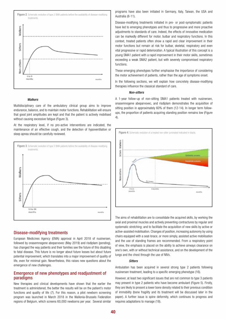
40
Walkers
Multidisciplinary care of the ambulatory clinical group aims to improve endurance, balance, and to maintain motor functions. Rehabilitation will ensure that good joint amplitudes are kept and that the patient is actively mobilised without causing excessive fatigue (Figure 3).
At the respiratory level, if no pro-active interventions are indicated, the maintenance of an effective cough, and the detection of hypoventilation or sleep apnea should be carefully reviewed.
Disease-modifying treatmentsEuropean Medicines Agency (EMA) approval in April 2018 of nusinersen, followed by onasemnogene abeparvovec (May 2019) and risdiplam (pending), has changed the way patients and their families see the future of this disabling to fatal disease. This future is no longer about future losses but about future potential improvement, which translates into a major improvement of quality of life, even for minimal gain. Nevertheless, this raises new questions about the emergence of new challenges.
Emergence of new phenotypes and readjustment of paradigmsNew therapies and clinical developments have shown that the earlier the treatment is administered, the better the results will be on the patient’s motor functions and quality of life (7). For this reason, a pilot newborn screening program was launched in March 2018 in the Wallonia-Brussels Federation regions of Belgium, which screens 60,000 newborns per year. Several similar
programs have also been initiated in Germany, Italy, Taiwan, the USA and Australia (8-11).
Disease-modifying treatments initiated in pre- or post-symptomatic patients have led to emerging phenotypes and thus to progressive and more proactive adjustments to standards of care. Indeed, the effects of innovative medication can be markedly different for motor, bulbar and respiratory functions. In this context, treated patients often show a rapid and clear improvement in their motor functions but remain at risk for bulbar, skeletal, respiratory and even vital progressive or rapid deterioration. A typical illustration of this concept is a young SMA1 patient with a rapid improvement in their motor skills, sometimes exceeding a weak SMA2 patient, but with severely compromised respiratory functions.
These emerging phenotypes further emphasise the importance of considering the motor achievement of patients, rather than the age of symptoms onset.
In the following sections, we will explain how concretely disease-modifying therapies influence the classical standard of care.
Non-sitters
A 1-year follow-up of non-sitting SMA1 patients treated with nusinersen, onasemnogene abeparvovec, and risdiplam demonstrates the acquisition of sitting position in approximately 60% of them (12-14). In longer term follow-ups, the proportion of patients acquiring standing position remains low (Figure 4).
The aims of rehabilitation are to consolidate the acquired skills, by working the axial and proximal muscles and actively preventing contractures by regular and systematic stretching; and to facilitate the acquisition of new skills by active or active-assisted mobilisation. Changes of position, increasing autonomy by using chairs equipped with a seat-brace, or more simply, assisted active mobilisation and the use of standing frames are recommended. From a respiratory point of view, the emphasis is placed on the ability to achieve airways clearance on one’s own, with or without technical assistance, and on the development of the lungs and the chest through the use of NIVs.
Sitters
Ambulation has been acquired in several strong type 2 patients following nusinersen treatment, leading to a specific emerging phenotype (16).
However, at least two significant issues that are not common to type 3 patients may present in type 2 patients who have become ambulant (Figure 5). Firstly, they are likely to present a lower bone density related to their previous condition of immobility (bone fragility and its treatment will be discussed later in the paper). A further issue is spine deformity, which continues to progress and requires adaptations to manage (18).
Figure 2. Schematic evolution of type 2 SMA patients before the availability of disease-modifying treatments.
Figure 4. Schematic evolution of a treated non-sitter (untreated indicated in black).
Figure 3. Schematic evolution of type 3 SMA patients before the availability of disease-modifying treatments.
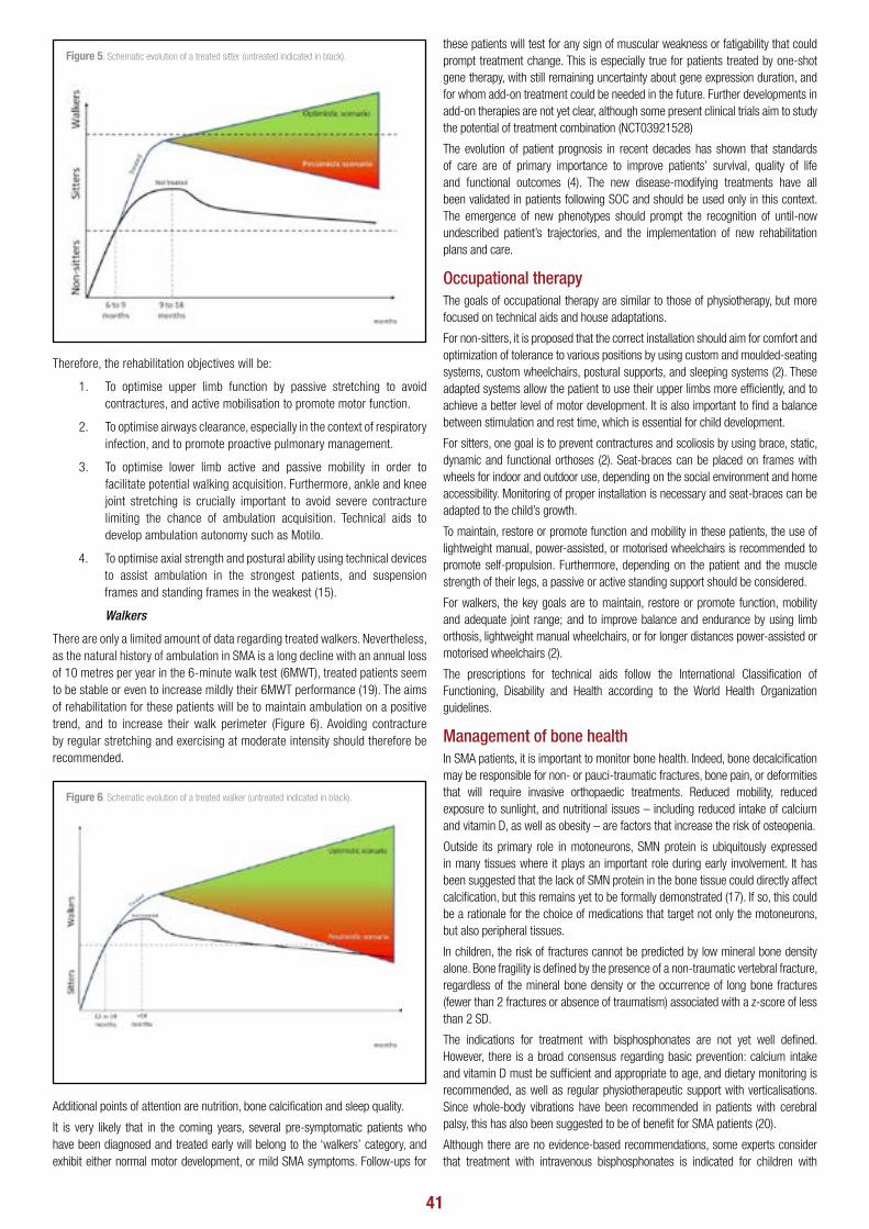
41
Therefore, the rehabilitation objectives will be:
1. To optimise upper limb function by passive stretching to avoid contractures, and active mobilisation to promote motor function.
2. To optimise airways clearance, especially in the context of respiratory infection, and to promote proactive pulmonary management.
3. To optimise lower limb active and passive mobility in order to facilitate potential walking acquisition. Furthermore, ankle and knee joint stretching is crucially important to avoid severe contracture limiting the chance of ambulation acquisition. Technical aids to develop ambulation autonomy such as Motilo.
4. To optimise axial strength and postural ability using technical devices to assist ambulation in the strongest patients, and suspension frames and standing frames in the weakest (15).
Walkers
There are only a limited amount of data regarding treated walkers. Nevertheless, as the natural history of ambulation in SMA is a long decline with an annual loss of 10 metres per year in the 6-minute walk test (6MWT), treated patients seem to be stable or even to increase mildly their 6MWT performance (19). The aims of rehabilitation for these patients will be to maintain ambulation on a positive trend, and to increase their walk perimeter (Figure 6). Avoiding contracture by regular stretching and exercising at moderate intensity should therefore be recommended.
Additional points of attention are nutrition, bone calcification and sleep quality.
It is very likely that in the coming years, several pre-symptomatic patients who have been diagnosed and treated early will belong to the ‘walkers’ category, and exhibit either normal motor development, or mild SMA symptoms. Follow-ups for
these patients will test for any sign of muscular weakness or fatigability that could prompt treatment change. This is especially true for patients treated by one-shot gene therapy, with still remaining uncertainty about gene expression duration, and for whom add-on treatment could be needed in the future. Further developments in add-on therapies are not yet clear, although some present clinical trials aim to study the potential of treatment combination (NCT03921528)
The evolution of patient prognosis in recent decades has shown that standards of care are of primary importance to improve patients’ survival, quality of life and functional outcomes (4). The new disease-modifying treatments have all been validated in patients following SOC and should be used only in this context. The emergence of new phenotypes should prompt the recognition of until-now undescribed patient’s trajectories, and the implementation of new rehabilitation plans and care.
Occupational therapyThe goals of occupational therapy are similar to those of physiotherapy, but more focused on technical aids and house adaptations.
For non-sitters, it is proposed that the correct installation should aim for comfort and optimization of tolerance to various positions by using custom and moulded-seating systems, custom wheelchairs, postural supports, and sleeping systems (2). These adapted systems allow the patient to use their upper limbs more efficiently, and to achieve a better level of motor development. It is also important to find a balance between stimulation and rest time, which is essential for child development.
For sitters, one goal is to prevent contractures and scoliosis by using brace, static, dynamic and functional orthoses (2). Seat-braces can be placed on frames with wheels for indoor and outdoor use, depending on the social environment and home accessibility. Monitoring of proper installation is necessary and seat-braces can be adapted to the child’s growth.
To maintain, restore or promote function and mobility in these patients, the use of lightweight manual, power-assisted, or motorised wheelchairs is recommended to promote self-propulsion. Furthermore, depending on the patient and the muscle strength of their legs, a passive or active standing support should be considered.
For walkers, the key goals are to maintain, restore or promote function, mobility and adequate joint range; and to improve balance and endurance by using limb orthosis, lightweight manual wheelchairs, or for longer distances power-assisted or motorised wheelchairs (2).
The prescriptions for technical aids follow the International Classification of Functioning, Disability and Health according to the World Health Organization guidelines.
Management of bone healthIn SMA patients, it is important to monitor bone health. Indeed, bone decalcification may be responsible for non- or pauci-traumatic fractures, bone pain, or deformities that will require invasive orthopaedic treatments. Reduced mobility, reduced exposure to sunlight, and nutritional issues – including reduced intake of calcium and vitamin D, as well as obesity – are factors that increase the risk of osteopenia.
Outside its primary role in motoneurons, SMN protein is ubiquitously expressed in many tissues where it plays an important role during early involvement. It has been suggested that the lack of SMN protein in the bone tissue could directly affect calcification, but this remains yet to be formally demonstrated (17). If so, this could be a rationale for the choice of medications that target not only the motoneurons, but also peripheral tissues.
In children, the risk of fractures cannot be predicted by low mineral bone density alone. Bone fragility is defined by the presence of a non-traumatic vertebral fracture, regardless of the mineral bone density or the occurrence of long bone fractures (fewer than 2 fractures or absence of traumatism) associated with a z-score of less than 2 SD.
The indications for treatment with bisphosphonates are not yet well defined. However, there is a broad consensus regarding basic prevention: calcium intake and vitamin D must be sufficient and appropriate to age, and dietary monitoring is recommended, as well as regular physiotherapeutic support with verticalisations. Since whole-body vibrations have been recommended in patients with cerebral palsy, this has also been suggested to be of benefit for SMA patients (20).
Although there are no evidence-based recommendations, some experts consider that treatment with intravenous bisphosphonates is indicated for children with
Figure 5. Schematic evolution of a treated sitter (untreated indicated in black).
Figure 6. Schematic evolution of a treated walker (untreated indicated in black).

42
SMA having at least two of the following criteria: bone density less than - 2 SD, two or more multiple fractures due to minor trauma, and chronic bone pain (21). Maintaining the treatment over time should be evaluated regularly by performing bone densitometry and blood and urine phospho-calcium balances (21).
A proactive approach is very important considering that the proportion of walking patients will increase, and that patients with major bone fragility may start to walk and be at risk of falling.
Orthopaedic treatmentsSince the emergence of new treatments, the vital and functional prognosis of SMA patients has improved, especially in SMA1 patients. Previously, orthopaedic management was more conservative with regard to managing congenital dislocation of the hips, tendon contractures, bone deformities, scoliosis and thoracic kyphosis.
Because an incidence as high as 60 to 90% of scoliosis deformities occur in childhood, management must be systematic. A clinical examination and radiography of the spine should be performed every 6 months until the patient is fully grown, then once every following year.
Treatment with braces from 20° of Cobb angulation is recommended to slow down progression, allow stable sitting positions, and facilitate respiratory function (2). Nevertheless, bracing is not effective to stop progression of scoliosis and should be discussed in regards with patient’s vital capacity (22).
A surgical intervention is suggested if scoliosis is more severe than 50° or if progression is more than 10° per year.
In early-onset scoliosis, occurring before the age of 10 in skeletally immature patients (which is the case in most sitters), the use of instruments allowing for continued spine growth should be considered. Growing rods are the standard and most well-documented surgical technique.
To decrease the need for repeated surgery every 6 months and complications due to invasive procedures and exposure to general anaesthesia, magnetically-controlled growing rods have recently been implemented as an alternative to traditional growing rods. However, complications such as metallosis and a significant rate of implant failure requiring unplanned revision surgery have been reported (2, 23).
Good knowledge of the natural history of progression is essential to ensure optimal timing of therapeutic interventions (24). Because of the limited survival of non-sitters before the development of innovative therapies, the management of scoliosis was more conservative; even more so in patients with highly compromised respiration, or with severe bulbar issues. Braces could be proposed in respiratory stable patients. A more pro-active management is proposed with increasing life expectancy in SMA1 patients.
Spine deformity in SMA 2 patients who acquire ambulation is progressive; this should prompt surgical treatment, as is classically proposed to SMA2 patients. However, in ambulant patients with a limited walking perimeter, spinal surgery is likely to lead to loss of ambulation, and the indication is therefore much less obvious than in sitters. Proactive management of spine deformity with a conservative approach (such as a brace) should thus be proposed.
Due to new innovations in treatment, rehabilitative care will also need to adapt and follow this innovative trend. Besides the classic rehabilitative treatments which have proven their efficiency, there are various re-educative tools such as exoskeletons and virtual reality which have emerged in recent years.
Lower limb exoskeletonsRobotic lower limb exoskeletons have emerged in the last several years as a potent rehabilitation tool, mainly used by patients with spinal cord injury and injury to the central nervous system.
Their use is based on the principles of neuroplasticity and motor learning maximising afferent input from peripheral joints and providing task‐specific stimulation to the central nervous system) and the beneficial effects of verticalisation and mobilisation (24).
The benefits proposed in relation to spinal cord injury include the strengthening of impaired muscles, increased walking speed and efficiency, quality of life, and a decrease in spasticity and pain. Positive changes in the cardiovascular system and metabolism, bowel, and bones have also been proposed; however, only a few patients were included in these studies (25).
Another meta-analysis studying the effects of exoskeleton use in spinal cord injury
has shown a significant positive effect on ASIA lower extremity muscle score (LEMS), and the results of both the 6-minute walk test (6MWT) and 10-minute walk test (10MWT) (26).
These results are promising for patients with acquired injuries, although exoskeletons presently have prohibitive costs, limited accessibility, and require a high level of training for both supervisor and patient before they can be safely and independently used (26).
However, the usefulness of exoskeletons with regard to neuromuscular diseases such as SMA is still unproven There is presently no literature on this subject, with the exception of one case report studying supported treadmill therapy in 3 patients with limb-girdle muscular dystrophy (LGMD) (27).
ConclusionsInnovative therapeutic approaches for the treatment of spinal muscular atrophy have in recent years modified the prognosis for patients with this disease, increasing life expectancy and motor development.
The standard of care in spinal muscular atrophy was renewed and adapted in 2018, with the goal of minimising the consequences of the disease and to maintain, restore, and promote function and mobility. Nevertheless, as new phenotypes are still appearing due to the approval of new medications released after 2018, newborn screening, and pre symptomatic treatments, the standard of care will need to be more frequently adapted going forward.
A multidisciplinary approach is therefore essential and as vitally important as a proactive attitude in both medical and paramedical teams.
Conflict of interest statement
The authors report no conflicts of interest.
REFERENCES:
1. Wang C. H., Finkel R. S., Bertini E. S., Schroth M., Simonds A., Wong B., et al. Consensus statement for standard of care in spinal muscular atrophy. J. Child Neurol. Aut. 2007;22(8):1027-49.
2. Mercuri E., Finkel R. S., Muntoni F., Wirth B., Montes J., Main M., et al. Diagnosis and management of spinal muscular atrophy: Part 1: Recommendations for diagnosis, rehabilitation, orthopedic and nutritional care. J Neuromuscul. Disord. 2018;(28):(103-115).
3. Finkel R. S., Mercuri E., Meyer O. H., Simonds A. K., Schroth M. K., Graham R. J., et al. Diagnosis and management of spinal muscular atrophy: Part 2: Pulmonary and acute care; medications, supplements and immunizations; other organ systems; and ethics. J. Neuromuscul. Disord. 2018;28(3):197207.
4. Oskoui M., Levy G., Garland C. J., Gray J. M., O’Hagen J., De Vivo D. C., et al. The changing natural history of spinal muscular atrophy type 1. Neurology. 13 Nov. 2007;69(20):19316.
5. Oskoui M., Kaufmann P. Spinal muscular atrophy. Neurotherapeutics. Oct. 2008;5(4):499506.
6. Schroth M. K. Special considerations in the respiratory management of spinal muscular atrophy. Pediatrics. May 2009;123 Suppl 4:S245-249.
7. Dangouloff T., Servais L. Clinical Evidence Supporting Early Treatment Of Patients With Spinal Muscular Atrophy: Current Perspectives. Ther. Clin. Risk Manag. 2019;15:115361.
8. Dangouloff T., Burghes A., Tizzano E. F., Servais L., Burghes A., et al. 244th ENMC International Workshop: Newborn screening in spinal muscular atrophy. 10-12 May 2019, Hoofdorp, The Netherlands. J Neuromuscul. Disord. Jan 2020;30(1):93103.
9. Vill K., Kölbel H., Schwartz O., Blaschek A., Olgemöller B., Harms E., et al. One Year of Newborn Screening for SMA - Results of a German Pilot Project. J. Neuromuscul. Disord. 2019;6(4):50315.
10. Kay D. M., Stevens C. F., Parker A., Saavedra-Matiz C. A., Sack V., Chung W. K., et al. Implementation of population-based newborn screening reveals low incidence of spinal muscular atrophy. Genet. Med. Aut. 2020;22(8):1296302.
11. Kariyawasam D. S. T., Russell J. S., Wiley V., Alexander I. E., Farrar M. A. The implementation of newborn screening for spinal muscular atrophy: the Australian experience. Genet. Med. 2020;22(3):55765.
12. Aragon-Gawinska K., Daron A., Ulinici A., Vanden Brande L., Seferian A., Gidaro T., et al. Sitting in patients with spinal muscular atrophy type 1 treated with nusinersen. Dev. Med. Child Neurol. 2020;62(3):3104.
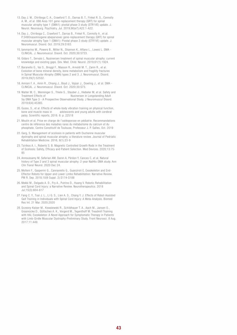
43
13. Day J. W., Chiriboga C. A., Crawford T. O., Darras B. T., Finkel R. S., Connolly A. M., et al. 066 Avxs-101 gene-replacement therapy (GRT) for spinal muscular atrophy type 1 (SMA1): pivotal phase 3 study (STR1VE) update. J. Neurol. Neurosurg. Psychiatry. Jul. 2019;90(e7):A22.1-A22.
14. Day J., Chiriboga C., Crawford T., Darras B., Finkel R., Connolly A., et al. P.349Onasemnogene abeparvovec gene-replacement therapy (GRT) for spinal muscular atrophy Type 1 (SMA1): Pivotal phase 3 study (STR1VE) update. J. Neuromuscul. Disord. Oct. 2019;29:S183.
15. Iammarino M., Powers B., Miller N., Shannon K., Alfano L., Lowes L. SMA - CLINICAL. J. Neuromuscul. Disord. Oct. 2020;30:S723.
16. Gidaro T., Servais L. Nusinersen treatment of spinal muscular atrophy: current knowledge and existing gaps. Dev. Med. Child. Neurol. 2019;61(1):1924.
17. Baranello G., Vai S., Broggi F., Masson R., Arnoldi M. T., Zanin R., et al. Evolution of bone mineral density, bone metabolism and fragility fractures in Spinal Muscular Atrophy (SMA) types 2 and 3. J. Neuromuscul. Disord. 2019;29(7):52532.
18. Amrani F. A., Amin R., Chiang J., Boyd J., Vajsar J., Dowling J., et al. SMA - CLINICAL. J. Neuromuscul. Disord. Oct. 2020;30:S73.
19. Walter M. C., Wenninger S., Thiele S., Stauber J., Hiebeler M, et al. Safety and Treatment Effects of Nusinersen in Longstanding Adult 5q-SMA Type 3 - A Prospective Observational Study. J Neuromuscul Disord. 2019;6(4):45365.
20. Gusso, S., et al. Effects of whole-body vibration training on physical function, bone and muscle mass in adolescents and young adults with cerebral palsy. Scientific reports, 2016. 6: p. 22518
21. Moulin et al. Prise en charge de l’ostéoporose en pédiatrie. Recommandations centre de référence des maladies rares du métabolisme du calcium et du phosphate, Centre Constitutif de Toulouse, Professeur J. P. Salles, Oct. 2019
22. Garg, S. Management of scoliosis in patients with Duchenne muscular dystrophy and spinal muscular atrophy: a literature review. Journal of Pedriatic Rehabilitation Medicine. 2016, 9(1):23-9
23. Tsirikos A. I., Roberts S. B. Magnetic Controlled Growth Rods in the Treatment of Scoliosis: Safety, Efficacy and Patient Selection. Med Devices. 2020;13:75-85
24. Annoussamy M, Seferian AM, Daron A, Péréon Y, Cances C, et al, Natural history of Type 2 and 3 spinal muscular atrophy: 2-year NatHis-SMA study. Ann Clin Transl Neurol. 2020 Dec 24.
25. Molteni F., Gasperini G., Cannaviello G., Guanziroli E. Exoskeleton and End-Effector Robots for Upper and Lower Limbs Rehabilitation: Narrative Review. PM R. Sep. 2018;10(9 Suppl. 2):S174-S188
26. Mekki M., Delgado A. D., Fry A., Putrino D., Huang V. Robotic Rehabilitation and Spinal Cord Injury: a Narrative Review. Neurotherapeutics. 2018 Jul;15(3):604-617
27. Fang C. Y., Tsai J. L., Li G. S., Lien A. S., Chang Y. J. Effects of Robot-Assisted Gait Training in Individuals with Spinal Cord Injury: A Meta-Analysis. Biomed Res Int. 21 Mar. 2020;2020
28. Sczesny-Kaiser M., Kowalewski R., Schildhauer T. A., Aach M., Jansen O., Grasmücke D., Güttsches A. K., Vorgerd M., Tegenthoff M. Treadmill Training with HAL Exoskeleton: A Novel Approach for Symptomatic Therapy in Patients with Limb-Girdle Muscular Dystrophy-Preliminary Study. Front Neurosci. 8 Aug. 2017;11:449.

44
Upper limb rehabilitation in children with unilateral cerebral palsy Griet Dequeker a,*, Lisa Mailleux b,c,*
a University Hospitals Leuven, Campus Gasthuisberg, Cerebral Palsy Reference Centre, Leuven, Belgium b Centre For Developmental Disabilities, Leuven, Belgium c KU Leuven - University of Leuven, Department of Rehabilitation Sciences, Leuven, Belgium
* both authors contributed equally
Theme
AbstractChildren with unilateral cerebral palsy experience difficulties with unimanual and bimanual functions, impeding self-care independence in daily life. Hence, research aimed at improving upper limb function has increased tremendously throughout the last decade. In this manuscript, we will provide an overview of all evidence-based, non-surgical therapy models intended to improve upper limb function in children with unilateral cerebral palsy, described according to the International Classification of Functioning, Disability and Health framework. The strongest level of evidence refers to activity-based interventions like constraint-induced movement therapy, bimanual training and goal-directed training. Interventions targeting body structures or functions, such as muscle strengthening, taping, splinting or casting, are less well investigated and should at this point be considered as assistive interventions with therapy goals on the level of body structure and function. Limited evidence exists on the efficacy of participation-based interventions. Finally, environmental factors can further shape the therapy model by providing the therapy in the home setting or a camp environment, while personal factors may influence the response to treatment.
Keywordsunilateral cerebral palsy, upper limb, rehabilitation, intervention, therapy
The upper limb plays a crucial role in acquiring self-care independence in daily life enabling participation in home, school and leisure activities. The performance of such activities requires the skilled use of both hands together. Throughout daily life, the variety of upper limb functions is tremendous, with reaching and grasping being the first developmental milestone. Already during the first year of life, reaching and grasping develops into a skilful movement. As a matured motor activity, reaching and grasping looks fairly easy, yet it involves a complex neural action to control and coordinate the numerous degrees of freedom. Hence, a lesion during early brain development, such as in cerebral palsy (CP), may disrupt the fine-tuned coordination of upper limb movements, compromising the performance of daily life activities.
CP is a major cause of paediatric disability and is defined as “a group of permanent disorders of the development of movement and posture, causing activity limitation, that are attributed to non-progressive disturbances that occurred in the developing foetal or infant brain. The motor disorders of CP are often accompanied by disturbances of sensation, perception, cognition, communication and behaviour, by epilepsy, and by secondary musculoskeletal problems” (1). The upper limb is most often investigated in children with spastic unilateral CP (uCP), in whom sensorimotor impairments are predominantly present at one side of the body. Studies have shown that particularly distal muscle weakness and somatosensory dysfunction negatively affects upper limb activity and self-care independence (2,3). This may cause the child to end up in a vicious circle. Increased muscle weakness and somatosensory dysfunction can contribute to a reduced hand use in daily life, further preventing the spontaneous daily stimulation of muscle strength and somatosensory input (2,3). Moreover, a recent five-year longitudinal study reported that from the age of nine years onwards, the spontaneous use of the impaired side in bimanual tasks decreases (4). Together these findings underline the importance of motor interventions to improve and retain function, even before the age of one year.
In the end, the ultimate goal of each clinician is to enhance the child’s functional potential for which adequate treatment planning is imperative. Hence, in this manuscript, we will provide an overview of all evidence-based therapy models aimed at improving upper limb function in children with CP, described according to the ICF-framework (International Classification of Functioning, Disability and Health) (5). We will focus on the non-surgical interventions, which are recommended by Novak et al. in their traffic lights paper with a green light referring to strong positive evidence and an orange light to weak positive evidence (see Figure 1) (5). Where applicable, personal recommendations based on clinical experience and as performed at the University Hospitals Leuven, were added.
Interventions at the body structure and function levelIt is recommended to perform muscle strengthening, taping, splinting and casting as an assistive intervention combined with task-specific motor training (5).
For muscle strengthening, there is strong evidence that it is effective in improving muscle strength (green light) (5). Moreover, practicing strength during functional tasks may result in a better transfer towards manual activities compared to practicing strength in non-functional positions (orange light) (6). The National Strength and Conditioning Association guidelines recommends for a muscle strengthening program in children, a duration of 12 weeks with a frequency of three times a week at 8–12 repetition maximum (6).
Recently, taping gains in popularity as an assistive intervention due to its low costs and easy application (7). There are two main kinds of tape. Kinesio tape, a flexible/elastic type of tape and the athletic tape which is a more rigid/inelastic tape that is more effective in limiting joint movement (7). In children with CP, taping is often used in the management of spasticity/hypotonia, facilitation of muscle function and joint stabilization (7). In combination with a task-specific motor training, taping may augment the treatment effects (orange light) (5). Though specifically for the upper limb, immediate effects are usually not visible, indicating that the taping needs to be applied long
Introduction

45
enough (7). Moreover, taping has additional benefits with respect to comfort and cosmesis compared to traditional orthotics (5).
Hand splinting is considered as standard practice in children with uCP. Though, less than 10 randomized controlled trials have been published on the efficacy of hand splinting. According to a meta-analysis, the use of non-functional hand splinting (i.e. splints worn at night) has a small beneficial effect on upper limb function in combination with therapy compared to therapy alone (orange light) (8). However, these benefits were diminished already two to three months after splint wearing was stopped. Moreover, the combined therapy that was used across these studies strongly differed (i.e. botulinum injections, neurodevelopmental therapy, goal-directed training), which makes it hard to draw clear conclusions. Only two randomized controlled trials investigated the efficacy of functional hand splinting (i.e. worn during the performance of functional tasks), but with conflicting results (9,10). According to our point of view, the main benefit of functional hand splinting is the stabilization of the wrist and carpometacarpal joint of the thumb in a functional position which facilitates a digital grip. Though for children with intact somatosensory function, the main disadvantage of hand splinting is that it restricts tactile input.
Upper limb casting may aid in the prevention of contractures (orange light) (5). In combination with botulinum toxin injections, the effects of casting can be enhanced (green light), which is also better tolerated by the children than applying casting without botulinum toxin injections (5). However, careful consideration is needed because casting can cause altered proprioception and secondary muscle weakness in particular with botulinum toxin injections (5). Hence, when the cast is removed, active and/or goal-directed motor interventions are crucial to regain these functions (5). Severe contractures cannot be treated with casting alone, and usually require orthopaedic surgery (5).
Interventions at the activity levelThe strongest level of evidence for efficacious therapy models for the upper limb is situated on the level of activity (5). Novak et al. recommend to perform constraint-induced movement therapy, bimanual therapy, goal directed training, action observation training and virtual reality training (5). All these interventions are based on the theory of motor learning. Important motor learning principles are problem-solving, repetitive practice and structured feedback. Real-life tasks are practiced through self-generated movements
at a high intensity and within a motivational setting, often aimed at achieving goals set by the child (or parent if necessary) (5). The most popular evidence-based therapy models are constraint-induced movement therapy (CIMT) and bimanual therapy.
The application of CIMT in children with CP was derived from promising results in stroke rehabilitation. CIMT involves restraining the less-impaired upper limb allowing motor stimulation of the impaired upper limb. However, the original model requested wearing the constraint during 90% of waking hours for at least two weeks, while providing upper limb therapy for at least three hours per day. In children, usually a more child-friendly approach is used (i.e. modified CIMT), which includes variation in type of restraint, the hours worn per day and duration in weeks (11). In literature, frequency varies from one hour per day to 24 hours per day, with a duration of two to ten weeks (11). Forced-use therapy is the restraint of the less-impaired upper limb without additional treatment for the impaired upper limb. The restraint can be a splint, a cast, a sling or a glove. In the University Hospitals Leuven, a sling or glove are used (see Figure 2), which have the advantage to be removed, limiting frustration outside the structured therapy moments. In general, we advise a sling in children younger than four years of age and in children with less functional abilities (i.e. children without an active grasp ability). A sling keeps the arm against the trunk preventing the child to use its less-impaired upper limb in any way for task execution. Working with a sling is preferably done in a sitting position since it can have a negative impact on balance and prevents a protective reaction in case of falling. A glove is used in children older than four-years of age and/or in children with better functional abilities. Nevertheless, this is not a strict guideline. It remains important to look at the individual child together with the parents and the therapist to consider whether a sling or a glove would be the best option. For example, in a six-year old child, also having ADHD, a sling might offer better results.
So far, CIMT is the most studied therapy model for the upper limb in children with uCP, in which strong evidence point towards clinical meaningful and sustained benefits (green light) (5,11). (Modified) CIMT can counteract the process of developmental disregard of the impaired upper limb (11). Children with uCP may disregard their preserved capacity as they may learn from an early age that using only the less-impaired upper limb or applying other strategies might be easier to complete daily life tasks. This may cause the child to end up in a vicious circle, which then can be interrupted by applying
Figure 1. Overview of interventions recommended by Novak et al.5 according to the ICF-model.

46
CIMT principles. The main disadvantage is that only unimanual functions can be practiced with CIMT, while in daily life most activities are bimanual.
Subsequently, intensive bimanual therapy models arose, often referred to as Hand-Arm Bimanual Intensive Therapy (HABIT) or Bimanual Intensive Therapy (BIT). These therapy models retained the intensive structured practice but instead of using a restraint they encourage the use of the impaired upper limb while performing bimanual activities. Although the amount of evidence is less abundant compared to CIMT, strong evidence points towards the efficacy of bimanual therapy to improve upper limb function (green light) (5). As such it is highly recommended to imply bimanual therapy models within upper limb therapy. Another therapy model added a lower limb component during HABIT (i.e. HABIT-ILE). This did not seem to attenuate the improvements of the upper limb (orange light) (5,12).
According to one systematic review, it could not be concluded whether CIMT or HABIT is superior (14). Though another systematic review acknowledged the task specificity of training, suggesting that CIMT results in better unimanual improvements, while bimanual training leads to better bimanual function (15). Hence, we often recommend a hybrid therapy model combining CIMT with bimanual training.
Both modified CIMT and bimanual therapy can also be applied in infancy. Such early intervention models are hypothesized to result in better motor outcomes compared to the same treatment at a later age due to the increased neuroplasticity of the young infant brain (13). Currently, there is mounting evidence pointing towards the efficacy of early interventions for improving upper limb function (orange light), including the absence of adverse events such as a negative impact of the restraint on the function of the dominant hand or on the child’s gross motor development (5).
Goal-directed training addresses goals that children and their parents identified as important in their daily life. The training process consists of goal selection, task analysis, intervention and evaluation (16). This individualized approach has been shown to be efficacious for improving hand function in children with uCP with a range of physical and cognitive abilities (green light) (5,16).
In virtual reality training, video games are used to improve upper limb function. However, virtual reality should be considered as an assistive intervention, that when combined with task-specific motor training may enhance the positive effects (orange light) (5). Video games have the opportunity to create repetition, gradation and feedback in the game. The main advantage of virtual reality is the positive impact on the child’s motivation. Moreover, video games could easily be implemented in the home setting to maintain rehabilitation benefits (17).
Finally, action observation training (AOT) is a novel approach aimed at stimulating the mirror neuron system. It is based on the principle that during the observation of a motor task, the same neurons are active as during the actual performance of that task (18). Hence, within AOT the child first observes a meaningful action repeatedly followed by the actual execution of the same task. AOT can include both unimanual as bimanual tasks, as long as the task itself is goal-directed. Although this is a fairly new method, there is strong evidence that AOT is effective for improving upper limb function (green light) (5).
Interventions at the participation levelMost interventions hope to further result in an improvement on participation level, yet there is only limited evidence available on the effect on participation outcomes. A participation-based intervention must address all personal, contextual and environmental factors. To the best of our knowledge, there is one intervention study that was specifically designed to target participation (19). This randomized controlled trial resulted in an increased perceived performance of leisure-time physical activity goals in children with CP. However, the actual level of physical activity only seemed to increase in children who already did not meet the physical activity guidelines (19). Although more research on this area is needed, adequate communication among the different stakeholders seems crucial in order to reduce barriers for participating in home, school and leisure activities.
Environmental factorsEnvironmental enrichment is a way to stimulate upper limb function already very early in infancy. The goal is to set up a motor enriched play environment at home to encourage self-initiated movements, exploration and task success. Hence, a variety of objects to stimulate grasp and reach behaviours is necessary. Toys must be selected carefully to match the desired motor task taking into account the motor and cognitive level of the child. Strong evidence reports that environmental enrichment improves upper limb motor function (green light) (5).
Moreover, environmental factors can further define the therapy model. The therapy can be provided in a private practice, at home, in a rehabilitation centre or in a camp environment.
From a family-centred perspective, the home environment is an interesting and natural place to stimulate the child in using his/her impaired upper limb with the main advantage of generalizing skills in daily life. However, also in this case, the therapist plays a crucial role in achieving success by guiding and supporting the parents to enhance their competency. Parents, grandparents and even caregivers in the nursery can be coached on how they can implement the therapy concept in the daily life of the child. Regular contact with the guiding therapist is crucial for support and motivation, while a manual with tips and tricks can provide additional guidance. Almost all therapy concepts can be implemented in a home programme. Home-programs are recommended for improving upper limb function (green light), and have also been shown to be feasible to perform (5,20).
Another environment for therapy delivery can be the rehabilitation unit of an hospital, where the child can receive ambulant therapy or can even be hospitalized for a short period of time (e.g. 2-3 weeks as performed in the University Hospitals Leuven) (11). A hospitalisation allows a boost of intensive rehabilitation, also providing multidisciplinary therapy and guidance.
Finally, camps have the advantage of being a group intervention usually including children of similar ages and motor difficulties. Hence, it has a more recreative feeling as well as an important social impact. These camps have shown its efficacy in research and, seeing its overall benefits, have been clinically implemented in Belgium (11).
Figure 2. Example of a sling (A) and a glove (B).

47
Personal factorsDespite the proven effectiveness of activity-based therapy models, it has been reported that some children might benefit more than others (21). There is vast amount of variability in clinical characteristics amongst children with uCP that might contribute to the individualised treatment response. Factors like age, baseline motor function, brain lesions characteristics, cognitive ability, motivation… differ from child to child. In literature, there is most evidence that children with lower baseline function profit more from intensive therapy models like modified CIMT and bimanual training, and that modified CIMT seems more beneficial for children with a poor somatosensory function (22,23). With respect to age, literature tends more towards a non-relevant impact, suggesting that children from all ages benefit from intensive activity-based therapy models (22,23). However, from eight years onwards, the focus should be much more on goal directed therapy (22). In addition, treatment response seems independent of underlying brain lesion characteristics (23,24).
ConclusionThe strongest level of evidence to improve motor function of the upper limb is at the level of activity involving CIMT, bimanual therapy, goal-directed training and AOT. It has been suggested that it does not matter ‘what’ is done, but that the ‘intensity’ of the therapy model is more important as well as that repeating intensive therapy periods might be needed to maintain gains in function (21). Existing evidence has proposed following key ingredients of activity-based interventions: collaborative goal setting, whole-task practice (or part-task practice for building skills for whole-task practice), context (practice within real-life environments), increasingly challenging tasks, feedback, motivation and sufficient dosage (25). A minimum of 40 hours of practice is recommended to improve motor ability, while for improving individual goals, 14 to 25 hours of therapy might already be sufficient (25,26). Moreover, the type of the therapy model must be chosen and individualized based on the child itself (i.e. age, cognitive abilities, level of motor function, behavioural problems,…), whereby it is important to include potential goals the child and/or its parents might have.
The authors have no conflict of interest to declare.
REFERENCES:
1. Rosenbaum P, Paneth N, Leviton A, Goldstein M, Bax M, Damiano D, et al. A report: the definition and classification of cerebral palsy April 2006. Dev Med Child Neurol. 2007;(109):8–14.
2. Russo RN, Skuza PP, Sandelance M, Flett P. Upper limb impairments, process skills, and outcome in children with unilateral cerebral palsy. Dev Med Child Neurol. 2019;61(9):1080–6.
3. Klingels K, Demeyere I, Jaspers E, De Cock P, Molenaers G, Boyd R, et al. Upper limb impairments and their impact on activity measures in children with unilateral cerebral palsy. Eur J Paediatr Neurol. 2012;16(5):475–84.
4. Klingels K, Meyer S, Mailleux L, Simon-Martinez C, Hoskens J, Monbaliu E, et al. Time course of upper limb function in children with unilateral cerebral palsy: A five-year follow-up study. Neural Plast. 2018;(2831342).
5. Novak I, Morgan C, Fahey M, Finch-Edmondson M, Galea C, Hines A, et al. State of the Evidence Traffic Lights 2019: Systematic Review of Interventions for Preventing and Treating Children with Cerebral Palsy. Curr Neurol Neurosci Rep. 2020;20(2):3.
6. Rameckers EAA, Janssen-Potten YJM, Essers IMM, Smeets RJEM. Efficacy of upper limb strengthening in children with Cerebral Palsy: A critical review. Res Dev Disabil [Internet]. 2015;36:87–101. Available from: http://dx.doi.org/10.1016/j.ridd.2014.09.024
7. Güçhan Z, Mutlu A. The effectiveness of taping on children with cerebral palsy: a systematic review. Dev Med Child Neurol. 2017;59(1):26–30.
8. Jackman M, Novak I, Lannin N. Effectiveness of hand splints in children with cerebral palsy: A systematic review with meta-analysis. Dev Med Child Neurol. 2014;56(2):138–47.
9. Elliott C, Reid S, Alderson J, Elliott B. Lycra arm splints in conjunction with goal-directed training can improve movement in children with cerebral palsy. NeuroRehabilitation. 2011;28(1):47–54.
10. Jackman M, Novak I, Lannin N, Galea C. Immediate effect of a functional wrist orthosis for children with cerebral palsy or brain injury: A randomized controlled trial. J Hand Ther. 2019;32(1):10–6.
11. Hoare BJ, Wallen MA, Thorley MN, Jackman ML, Carey LM, Imms C. Constraint-induced movement therapy in children with unilateral cerebral palsy (Review). Cochrane Database Syst Rev. 2019;2019(4):Art. No.: CD004149.
12. Saussez G, Brandão MB, Gordon AM, Bleyenheuft Y. Including a lower-extremity component during Hand-Arm Bimanual Intensive Training does not attenuate improvements of the upper extremities: A retrospective study of randomized trials. Front Neurol. 2017;8(SEP):1–10.
13. Friel K, Chakrabarty S, Kuo H, Martin J. Using Motor Behavior during an Early Critical Period to Restore Skilled Limb Movement after Damage to the Corticospinal System during Development. J Neurosci. 2012;32(27):9265–76.
14. Tervahauta MH, Girolami GL, Øberg GK. Efficacy of constraint-induced movement therapy compared with bimanual intensive training in children with unilateral cerebral palsy: a systematic review. Clin Rehabil. 2017 Nov;31(11):1445–56.
15. Dong VA-Q, Tung IH-H, Siu HW-Y, Fong KN-K. Studies comparing the efficacy of constraint-induced movement therapy and bimanual training in children with unilateral cerebral palsy: A systematic review. Dev Neurorehabil. 2013 Apr 1;16(2):133–43.
16. Mastos M, Miller K, Eliasson A-C, Imms C. Goal-directed training: Linking theories of treatment to clinical practice for improved functional activities in daily life. Clin Rehabil. 2007;21(1):47–5(1):47–55.
17. Bonnechère B, Jansen B, Omelina L, Degelaen M, Wermenbol V, Rooze M, et al. Can serious games be incorporated with conventional treatment of children with cerebral palsy? A review. Res Dev Disabil. 2014;35(8):1899–913.
18. Rizzolatti G, Craighero L. The mirror-neuron system. Annu Rev Neurosci. 2004;27:169–92.
19. Reedman SE, Boyd RN, Trost SG, Elliott C, Sakzewski L. Efficacy of Participation-Focused Therapy on Performance of Physical Activity Participation Goals and Habitual Physical Activity in Children With Cerebral Palsy: A Randomized Controlled Trial. Arch Phys Med Rehabil. 2019;100(4):676–86.
20. Beckers LWME, Geijen MME, Kleijnen J, Rameckers EAA, Schnackers MLAP, Smeets RJEM, et al. Feasibility and effectiveness of home-based therapy programmes for children with cerebral palsy: A systematic review. BMJ Open. 2020;10(10).
21. Sakzewski L, Gordon A, Eliasson AC. The state of the evidence for intensive upper limb therapy approaches for children with unilateral cerebral palsy. J Child Neurol. 2014;29(8):1077–90.
22. Sakzewski L, Ziviani J, Boyd RN. Best responders after intensive upper-limb training for children with unilateral cerebral palsy. Arch Phys Med Rehabil. 2011;92(4):578–84.
23. Simon-Martinez C, Mailleux L, Hoskens J, Ortibus E, Jaspers E, Wenderoth N, et al. Randomized controlled trial combining constraint-induced movement therapy and action-observation training in unilateral cerebral palsy: clinical effects and influencing factors of treatment response. Ther Adv Neurol Disord. 2020;13:1756286419898065.
24. Islam M, Nordstrand L, Holmström L, Kits A, Forssberg H, Eliasson AC. Is outcome of constraint-induced movement therapy in unilateral cerebral palsy dependent on corticomotor projection pattern and brain lesion characteristics? Dev Med Child Neurol. 2014;56(3):252–8.
25. Jackman M, Lannin N, Galea C, Sakzewski L, Miller L, Novak I. What is the threshold dose of upper limb training for children with cerebral palsy to improve function? A systematic review. Aust Occup Ther J. 2020;67(3):269–80.
26. Sakzewski L, Ziviani J, Boyd RN. Efficacy of upper limb therapies for unilateral cerebral palsy: A meta-analysis. Pediatrics. 2014;133(1).

Case Report
SOCIÉTÉ BELGE DE PÉDIATRIE
Inscriptions en ligne sur le site Internet :www.fondation-mustela.be
Pour tout renseignement complémentaire :Tél :
APPEL à PROJETS 2021PRIX DE PÉDIATRIESOCIALE
Avec les professionnels de santé, pour la petite enfance
02 467 61 01
Objectif :réaliser une action de promotion de
la bientraitance de l’enfant, et de sa famille,dans son environnement.
Pour fi nancer un projet initié parune équipe pédiatrique de terrain(hospitalière ou communautaire)et permettre sa mise en place.
Date limite dépôt candidature : 30/08/2021
VLAAMSE VERENIGING VOORKINDERGENEESKUNDE

49
Acute submandibular sialadenitis: a possible presentation of COVID-19 in children Bram Boon, Daphnée Ophoff, Alexandra Delmotte, Frederic De Meulder, Tyl Jonckheer, Kathleen Poschet
Department of Paediatrics, Gasthuiszusters Antwerpen, Sint-Vincentius Ziekenhuis, Antwerp, Belgium
Case Report
AbstractThis case report describes a paediatric patient presenting with acute unilateral submandibular sialadenitis during the 2019/2020 SARS-CoV-2 pandemic. The patient's history, clinical examination and investigatory work-up favoured a viral aetiology over bacterial infections or sialolithiasis. Laboratory tests for common viral aetiologies of sialadenitis were negative. SARS-CoV-2 PCR testing was positive. Indirect evidence supports the hypothesis that SARS-CoV-2 can cause acute sialadenitis. Caregivers could consider the possibility of SARS-CoV-2 infection in an afebrile child with acute sialadenitis presenting during the current pandemic.
KeywordsSialadenitis; Coronavirus; COVID-19
Saliva has various functions, including lubrication, taste, digestion, tooth protection, antibacterial properties. It is produced by the salivary glands and transported into the mouth by the salivary ducts. Viral infections are the most common acute salivary gland disorders in children (1).
Our patient presented in a Belgian hospital during the 2019-2020 severe acute respiratory syndrome coronavirus 2 (SARS-CoV-2) pandemic. SARS-CoV-2 is a human pathogenic betacoronavirus causing coronavirus-disease 2019 (COVID-19). There is a wide array of possible clinical presentations, ranging from asymptomatic carriers to severe bilateral pneumonia with acute respiratory distress syndrome to isolated anosmia. In general, children appear to run a milder clinical course.
A 10-year-old boy presented at the emergency department with an acute onset painful swelling of the right submandibular area. Moreover, he was feeling markedly unwell for the last two days. He reported difficulties eating and speaking. He did not have any tooth pain. He reported a recent history of having high fever (>39°C), a dry cough and breathing difficulties during the previous week, which spontaneously resolved. During the last four days, respiratory symptoms and fever were absent. The patient received all of his recommended vaccines, had no history of similar episodes and had no significant medical history. Familial history revealed that both parents had been sick recently, both reporting dry cough and fever. The mother had a negative polymerase chain reaction (PCR) test for SARS-CoV-2. Clinical examination showed an ill-defined area of swelling of the right submandibular region (see Figure 1). The area was slightly red, warm and tender on palpation. The
Introduction Case report
Figure 1. Clinical picture showing residual swelling of the right submandibular area, taken two days after initial presentation.

50
patient displayed trismus; he was unable to open his mouth more than 2 cm. He was visibly dehydrated with dry mouth mucosa and a prolonged capillary refill time. Oral examination revealed no signs of tooth decay and normal appearance of the salivary duct orifices. Lung auscultation was normal. Laboratory investigations showed slightly elevated inflammatory markers, with C-reactive protein (CRP) levels of 26 mg/L (<1 mg/L), leukocytosis of 11 x 109/L (4-10 x 109/L) with mild neutrophilia of 8,54 x 10⁹/L (2-8 x 10⁹/L) and lymphopenia of 1,42 x 10⁹/L (3-9 x 10⁹/L). Ultrasound revealed inflammation of the right submandibular salivary gland and reactive swollen cervical lymph nodes, without sialolithiasis or signs of abcedation. The patient was admitted for intravenous fluid resuscitation, pain medication and intravenous antibiotics (amoxicillin/clavulanic acid). Serologic testing for cytomegalovirus (CMV), Epstein-Barr virus (EBV) and mumps virus was negative. PCR testing for SARS-CoV-2 on nasopharyngeal swab was positive. During the course of hospitalisation, the patient did not display symptoms or signs suggestive of pneumonia caused by SARS-CoV-2. The swelling decreased gradually over the following days.
DiscussionOur patient presented with unilateral acute submandibular sialadenitis. In children, sialadenitis is most frequently caused by viral infections. Bacterial sialadenitis and sialolithiasis were also considered in the differential diagnosis. The absence of fever, the absence of pus on clinical examination and only slightly elevated inflammatory parameters on blood tests rendered bacterial infection unlikely. Nevertheless, our patient received intravenous antibiotic treatment to account for the possibility of a bacterial infection. Sialolithiasis is a rare cause of sialadenitis in children. Ultrasound is regarded as the first imaging modality of choice for detecting sialoliths and detects up to 90% of sialoliths greater than 2 mm (2). Ultrasound imaging did not show any sialoliths in our patient, which rendered sialolithiasis unlikely. Testing for viral organisms known to frequently cause sialadenitis (including mumps virus, EBV and CMV) was negative. A full respiratory screening panel was not performed. PCR testing for SARS-CoV-2 was performed because of the current global pandemic and was positive.
Various viruses have been reported to cause sialadenitis. In the pre-vaccine era, the mumps virus was the most frequent cause of sialadenitis in children. Since the start of widespread vaccinations against the mumps virus, various studies investigated other potential viral aetiologies of sialadenitis using PCR-based analysis or serologic testing. The most frequently found viral aetiologies of mumps negative sialadenitis are EBV, influenza, parainfluenza and human herpesviruses. Barrabeig et al. performed the only study that included human coronaviruses (coronavirus 229E and coronavirus OC43) in their PCR-based analysis (3). They investigated 101 suspected mumps cases with negative PCR results for the mumps virus and reported no positive results for these two coronaviruses in their study population (3).
A recent study in Medical Hypotheses by Wang et. al investigated whether SARS-CoV-2 has the potential to cause acute (and/or chronic) sialadenitis (4). SARS-CoV-2 invades host cells by binding to the angiotensin-converting enzyme 2 (ACE2) receptor. This receptor is present in salivary gland epithelium, with a mean expression level of the ACE2 gene being even higher in salivary gland tissue than in lung tissue (5). The virus can indeed be detected in salivary specimens, with a high viral load in saliva, comparable to those in sputum and throath swabs (6). Wang et. al hypothesised that SARS-CoV-2 can bind to ACE2 receptors on salivary gland epithelium, fuse with them, replicate and induce cell lysis causing acute sialadenitis (4). Moreover, excessive immunoreaction may further damage the salivary glands, similar to tissue damage seen in other organs with ACE2 receptor expression. Secondary fibrosis could later lead to chronic sialadenitis (4). They concluded that the indirect evidence indicates a high probability that SARS-CoV-2 does have the potential to cause acute (and chronic) sialadenitis (4). There are, however, no studies that systematically investigated SARS-CoV-2 infection in patients presenting with acute sialadenitis during the current pandemic. So far, there are only three reported cases of acute sialadenitis caused by SARS-CoV-2 (7, 8). They include two adult patients with unilateral parotitis and one adult patient with combined bilateral submandibular sialadenitis and parotitis.
ConclusionThis article is the first case report on acute submandibular sialadenitis in a SARS-CoV-2 positive paediatric patient. Indirect evidence supports the hypothesis that SARS-CoV-2 can cause acute sialadenitis. Caregivers could consider the possibility of SARS-CoV-2 infection in an afebrile child with acute sialadenitis presenting during the current pandemic. More evidence is needed to establish a definite link between SARS-CoV-2 infection and acute sialadenitis.
The authors declare that there is no conflict of interest.
REFERENCES:
1. Francis CL, Larsen CG. Pediatric sialadenitis. Otolaryngol Clin North Am. 2014;47(5):763-78.
2. Ugga L, Ravanelli M, Pallottino AA, Farina D, Maroldi R. Diagnostic work-up in obstructive and inflammatory salivary gland disorders. Acta Otorhinolaryngol Ital. 2017;37(2):83-93.
3. Barrabeig I, Costa J, Rovira A, Marcos MA, Isanta R, López-Adalid R, et al. Viral etiology of mumps-like illnesses in suspected mumps cases reported in Catalonia, Spain. Hum Vaccin Immunother. 2015;11(1):282-7.
4. Wang C, Wu H, Ding X, Ji H, Jiao P, Song H, et al. Does infection of 2019 novel coronavirus cause acute and/or chronic sialadenitis? Med Hypotheses. 2020;140:109789.
5. Xu J, Li Y, Gan F, Du Y, Yao Y. Salivary Glands: Potential Reservoirs for COVID-19 Asymptomatic Infection. J Dent Res. 2020;99(8).
6. Zhu J, Guo J, Xu Y, Chen X. Viral dynamics of SARS-CoV-2 in saliva from infected patients. J Infect. 2020;81(3):e48-e50.
7. Capaccio P, Pignataro L, Corbellino M, Popescu-Dutruit S, Torretta S. Acute parotitis: a possible precocious clinical manifestation of SARS-CoV-2 infection? Otolaryngol Head Neck Surg. 2020;163(1):182-3.
8. Chern A, Famuyide AO, Moonis G, Lalwani AK. Sialadenitis: A Possible Early Manifestation of COVID-19. Laryngoscope. 2020:10.1002/lary.29083.
Case Report
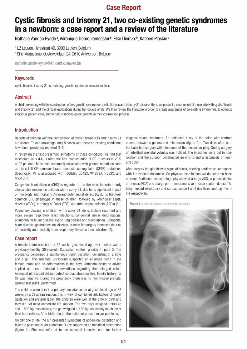
51
Cystic fibrosis and trisomy 21, two co-existing genetic syndromes in a newborn: a case report and a review of the literature Nathalie Vanden Eynde a, Véronique Demeulemeester b, Elke Dierckx b, Katleen Plaskie b
a UZ Leuven, Herestraat 49, 3000 Leuven, Belgium b Sint -Augustinus, Oosterveldlaan 24, 2610 Antwerpen, Belgium
Case Report
Reports of children with the combination of cystic fibrosis (CF) and trisomy 21 are scarce. To our knowledge, only 8 cases with these co-existing conditions have been previously reported (1-6).
In reviewing the first presenting symptoms of these conditions, we find that meconium ileus (MI) is often the first manifestation of CF. It occurs in 20% of CF patients. MI is most commonly associated with genetic mutations such as class I-III CF transmembrane conductance regulator (CFTR) mutations. Specifically, MI is associated with F508del, G542X, W1282X, R553X, and G551D (7).
Congenital heart disease (CHD) is regarded to be the most important early clinical phenomenon in children with trisomy 21, due to its significant impact on morbidity and mortality. Atrioventricular septal defect (AVSD) is the most common CHD phenotype in these children, followed by ventricular septal defects (VSDs), tetralogy of Fallot (TOF), and atrial septal defects (ASDs) (8).
Pulmonary disease in children with trisomy 21 alone, include recurrent and more severe respiratory tract infections, congenital airway deformations, pulmonary vascular disease, cystic lung disease and sleep apnea. Congenital heart disease, gastrointestinal disease, or need for surgery increases the risk of morbidity and mortality from respiratory illness in these children (9).
Case reportA female infant was born at 33 weeks gestational age. Her mother was a previously healthy 36-year-old Caucasian mother, gravida 4, para 3. The pregnancy concerned a spontaneous triplet gestation, consisting of 2 boys and a girl. The antenatal ultrasound suspected an enlarged colon in the female infant and no deformations in the boys. Antenatal obstetric advice implied no direct perinatal interventions regarding the enlarged colon. Antenatal ultrasound did not detect cardiac abnormalities. Family history for CF was negative. During the pregnancy, there was no noninvasive prenatal genetic test (NIPT) performed.
The children were born in a primary neonatal center at gestational age of 33 weeks by a Cesarean section, this in view of combined risk factors of triplet gestation and preterm labor. The children were well at the time of birth and they did not need immediate life support. The two boys weighed 1.900 kg and 1.890 kg respectively; the girl weighed 1.490 kg, noticeably much lower than her brothers. After birth, the brothers did not present major problems.
On day one of life, the girl presented symptoms of abdominal distention and failed to pass stools. An abdominal X-ray suggested an intestinal obstruction (figure 1). She was referred to our neonatal intensive care for further
diagnostics and treatment. An additional X-ray of the colon with contrast enema showed a generalized microcolon (figure 2). Two days after birth the baby had surgery with clearance of the meconium plug. During surgery, an intestinal prenatal volvulus was noticed. The intestines were put in non-rotation and the surgeon constructed an end-to-end anastomosis of ileum and colon.
After surgery the girl showed signs of shock, needing cardiovascular support with intravenous dopamine. On physical examination we detected no heart murmur. Additional echocardiography showed a large ASD, a patent ductus arteriosus (PDA) and a large peri-membranous ventricular septum defect. The baby needed respiratory and cardiac support until day three and day five of life respectively.
Introduction
AbstractA child presenting with the combination of two genetic syndromes, cystic fibrosis and trisomy 21, is rare. Here, we present a case report of a neonate with cystic fibrosis and trisomy 21 and the clinical implications during her course of life. We then review the literature in order to create awareness of co-existing syndromes, to optimize individual patient care, and to help clinicians guide parents in their counselling process.
Keywordscystic fibrosis, trisomy 21, co-existing, genetic syndrome, meconium ileus
Figure 1. Abdominal distention, lower intestinal obstruction
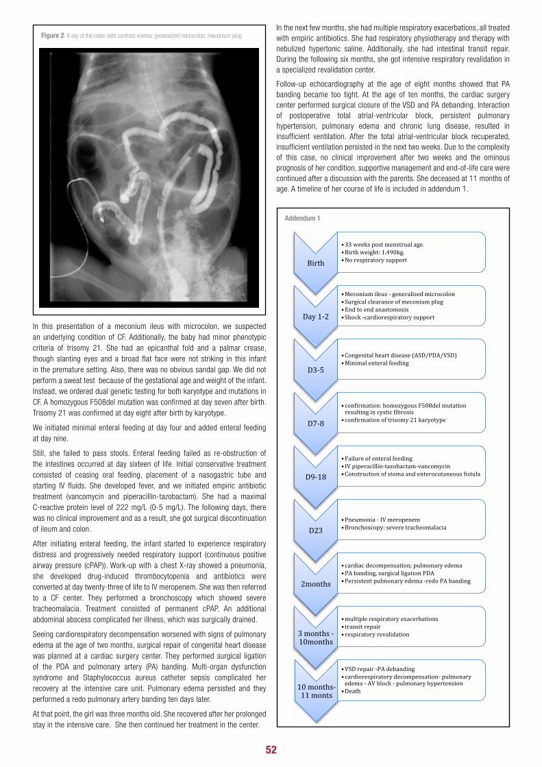
52 6
Addendum 1
Birth
•33weekspostmenstrualage.•Birthweight:1.490kg.•Norespiratorysupport
Day1-2
•Meconiumileus- generalisedmicrocolon•Surgicalclearanceofmeconiumplug•Endtoendanastomosis•Shock-cardiorespiratorysupport
D3-5
•Congenitalheartdisease(ASD/PDA/VSD)•Minimalenteralfeeding
D7-8
•confirmation:homozygousF508delmutationresultingincysticfibrosis
•confirmationoftrisomy21karyotype
D9-18
•Failureofenteralfeeding• IVpiperacillin-tazobactam-vancomycin•Constructionofstomaandenterocutaneousfistula
D23•Pneumonia- IVmeropenem•Bronchoscopy:severetracheomalacia
2months
•cardiacdecompensation;pulmonaryedema•PAbanding,surgicalligationPDA•Persistentpulmonaryedema-redoPAbanding
3months-10months
•multiplerespiratoryexacerbations• transitrepair•respiratoryrevalidation
10months-11monts
•VSDrepair-PAdebanding•cardiorespiratorydecompensation- pulmonaryedema- AVblock- pulmonaryhypertension
•Death
In this presentation of a meconium ileus with microcolon, we suspected an underlying condition of CF. Additionally, the baby had minor phenotypic criteria of trisomy 21. She had an epicanthal fold and a palmar crease, though slanting eyes and a broad flat face were not striking in this infant in the premature setting. Also, there was no obvious sandal gap. We did not perform a sweat test because of the gestational age and weight of the infant. Instead, we ordered dual genetic testing for both karyotype and mutations in CF. A homozygous F508del mutation was confirmed at day seven after birth. Trisomy 21 was confirmed at day eight after birth by karyotype.
We initiated minimal enteral feeding at day four and added enteral feeding at day nine.
Still, she failed to pass stools. Enteral feeding failed as re-obstruction of the intestines occurred at day sixteen of life. Initial conservative treatment consisted of ceasing oral feeding, placement of a nasogastric tube and starting IV fluids. She developed fever, and we initiated empiric antibiotic treatment (vancomycin and piperacillin-tazobactam). She had a maximal C-reactive protein level of 222 mg/L (0-5 mg/L). The following days, there was no clinical improvement and as a result, she got surgical discontinuation of ileum and colon.
After initiating enteral feeding, the infant started to experience respiratory distress and progressively needed respiratory support (continuous positive airway pressure (cPAP)). Work-up with a chest X-ray showed a pneumonia, she developed drug-induced thrombocytopenia and antibiotics were converted at day twenty-three of life to IV meropenem. She was then referred to a CF center. They performed a bronchoscopy which showed severe tracheomalacia. Treatment consisted of permanent cPAP. An additional abdominal abscess complicated her illness, which was surgically drained.
Seeing cardiorespiratory decompensation worsened with signs of pulmonary edema at the age of two months, surgical repair of congenital heart disease was planned at a cardiac surgery center. They performed surgical ligation of the PDA and pulmonary artery (PA) banding. Multi-organ dysfunction syndrome and Staphylococcus aureus catheter sepsis complicated her recovery at the intensive care unit. Pulmonary edema persisted and they performed a redo pulmonary artery banding ten days later.
At that point, the girl was three months old. She recovered after her prolonged stay in the intensive care. She then continued her treatment in the center.
In the next few months, she had multiple respiratory exacerbations, all treated with empiric antibiotics. She had respiratory physiotherapy and therapy with nebulized hypertonic saline. Additionally, she had intestinal transit repair. During the following six months, she got intensive respiratory revalidation in a specialized revalidation center.
Follow-up echocardiography at the age of eight months showed that PA banding became too tight. At the age of ten months, the cardiac surgery center performed surgical closure of the VSD and PA debanding. Interaction of postoperative total atrial-ventricular block, persistent pulmonary hypertension, pulmonary edema and chronic lung disease, resulted in insufficient ventilation. After the total atrial-ventricular block recuperated, insufficient ventilation persisted in the next two weeks. Due to the complexity of this case, no clinical improvement after two weeks and the ominous prognosis of her condition, supportive management and end-of-life care were continued after a discussion with the parents. She deceased at 11 months of age. A timeline of her course of life is included in addendum 1.
Figure 2. X-ray of the colon with contrast enema: generalized microcolon, meconium plug
Addendum 1.

53
DiscussionReports of co-existing CF and trisomy 21 are scarce. Incidence in Belgium of CF is 1:3500 live births and of trisomy 21 is 1:1400 live births (10,11). The risk of co-occurrence in Belgium is 1:4.900.000. The birth rate in Belgium is approximately 117.000 newborns per year (data of 2018) (12). This means a similar new case might occur every 41 years.
To our knowledge, no other report of a Belgian child is previously made. The prognosis for these children, with exception of one case report, is poor and they did not survive infancy or childhood (1-6).
Children with trisomy 21 alone have a vulnerable pulmonary vasculature that may manifest clinically as pulmonary hypertension, pulmonary edema or pulmonary hemorrhage. They develop more acute pulmonary edema , an indication of the fragility of pulmonary capillary integrity. Diffuse parenchymal lung disease manifests in these children as chronic radiographic changes associated with persistent findings such as dyspnea, cough, wheezing, crackles, or hypoxia. They have an increased risk of respiratory tract infections and are relatively more likely than children without trisomy 21 to have a severe course and even death from respiratory causes. In the subgroup of patients with trisomy 21 who had surgery, risk of respiratory infection was approximately three times higher than for children with trisomy 21 who did not have surgery. Additional congenital heart disease or gastrointestinal disease was a risk factor for admission for respiratory illness independent of undergoing surgery. Pneumonia was the single most common respiratory disorder, accounting for 43% of admissions to the intensive care unit. The pathophysiology underlying the increased risk for respiratory disease in children with trisomy 21 remains unclear, but a variety of immune defects have been identified (9).
Lung disease in children with CF is responsible for the vast majority of morbidity and mortality. Until recently, the standard of care in CF treatment focused on preventing and treating complications of the disease; now, novel treatment strategies targeting the ion channel abnormality directly are becoming available.
The interaction of the pathophysiology of lung disease in trisomy 21 with the pathophysiology in CF has not been studied, since the literature on this topic consists only of case reports. Still, it is not unlikely that novel treatment strategies in CF might improve the outcome in combined genetic syndromes as well.
ConclusionOur case is the first case of a neonate with the co-existence of CF and trisomy 21 as a part of a triplet pregnancy, consisting of otherwise two healthy boys. This report highlights the possible co-existence of two genetic diagnoses in a newborn. The severity of co-morbidities in either of these conditions and the interplay between them might predict the possible outcome. Although more prospective and even prenatal research is needed, our case report underlines the impact of the diagnosis in the neonatal course of life. Therefore clinicians should be aware of the possibility of this co-existence of syndromes.
Conflicts of interest
Authors declare no conflict of interest.
Human research statement
Parental oral informed consent is obtained.
REFERENCES:
1. Akinloye OW, Truong W, Giacomantonio M, Mateos D and El-Naggar W. Coexcistence of meconium ileus with duodenal atresia and trisomy 21 in a newborn: a case report. Journal of perinatology. 2014;34:875-876.
2. Guy E.L, Peckham D. G, Brownlee K. G, Conway S. P, Lee T.W.R. Cystic fibrosis co-existing with trisomy 21. Journal of Cystic Fibrosis. 2010;9:330-331.
3. Kruger C, Barmeier H, Sailer R, Harms D. Cystic fibrosis in Down’s syndrome – diagnostic pitfalls and implications for the clinician. Arch Dis Child. 1998;78:194.
4. Milunsky A. Cystic fibrosis and Down’s syndrome. Pediatrics. 1968;42:501-504.
5. Saglani S. and Bush A.Cystic fibrosis and Down’s syndrome: not always a poor prognosis. Pediatr. Pulmonol. 2001;31:321-322.
6. Vetrella M, Barthelmai W and Matsuda H. Down’s syndrome and cystic fibrosis. Pediatrics 1969;43:905-906.
7. Sathe M and Houwen R. Meconium ileus in Cystic Fibrosis. Journal of Cystic Fibrosis. 2017;16:S32–S39.
8. Pfitzer C, Helm P. C, Rosenthal L-M, Berger F, Bauer U. M. M, Schmitt K. RL. Dynamics in prevalence of Down syndrome in children with congenital heart disease. Eur J Pediatr. 2018;177:107-115.
9. McDowell KM, Craven DI. Pulmonary Complications of Down Syndrome during Childhood. The Journal of Pediatrics 2011;158:319-325.
10. Bevolkingsonderzoek aangeboren aandoeningen [Internet]. Vlaanderen: Mucoviscidose informatie voor professionelen; [cited 2019 Feb 20]. Available from: https://www.aangeborenaandoeningen.be/mucoviscidose-informatie-voor-professionelen
11. UZA [Internet]. Antwerp: Down syndrome; [cited 2019 Feb 20]. Available from: https://www.uza.be/downsyndroom
12. Statbel [Internet]. Brussels: Aantal geboorten blijft verder dalen in 2018; [cited 2019 Feb 20]. Available from: https://statbel.fgov.be/nl/nieuws/aantal-geboorten-blijft-verder-dalen-2018

54
Research Article

55
Barriers to paediatric pain management as viewed by doctors in the region of Thiès, Senegal: first results Floortje Krechting a, Abdoul Magib Cissé b, Karolien Van De Maele a, Jutte van der Werff ten Bosch c
a Department of Paediatrics, Universitair Ziekenhuis Brussel, Brussels, Belgium b Department of Paediatrics, EPS MBour, Mbour, Senegal c Department of Paediatric Oncology, Universitair Ziekenhuis Brussel, Brussels, Belgium
Research Article
Despite the known negative consequences associated with inadequate pain management, effective pain treatment remains largely unavailable to children in the developing world.
There is considerable evidence that effective pain management has the potential to reduce both morbidity and mortality, lower anxiety and stress of both the child and the families, facilitate recovery and reduce the cost of healthcare (1,2). Alongside these practical advantages, access to pain management is considered by the International Association for the Study of Pain (IASP) to be a fundamental human right (Declaration of Montreal) (3). A joint statement was issued by the American Academy of Pediatrics (AAP) and the American Pain Society (APS) in 2001 to underscore the importance of paediatricians assuming a leadership and advocacy role to ensure the humane and competent treatment of pain and suffering in all infants, children, and adolescents (4). However, in spite of the widespread evidence and knowledge of the importance of pain treatment, paediatric pain is largely underrecognised and undertreated all over the world (5). The AAP states that this is due to, amongst other things, the myth that children do not feel pain the way adults do, the misunderstanding of how to quantify a subjective experience, the lack of knowledge of pain treatment and fears of adverse effects of analgesics (4).
On top of these difficulties, doctors in the developing world face a multitude of challenges, many of which they cannot control directly. For instance, Africa bears 24% of the global burden of disease, and yet possesses only 3% of the world’s healthcare force (6). Case in point, Senegal has roughly one doctor per 14,500 inhabitants whereas Belgium has one doctor per 326 inhabitants (7). The result is a great global inequality of pain treatment due to a variety of issues. With respect to paediatric pain management in sub-Saharan Africa, the challenges have been described by Albertyn et al (8). Firstly, there are access barriers which result in a lack of analgesic medicine, information and education. Secondly, there are attitudinal barriers, where cultural
differences impact the management of paediatric pain. And, thirdly, there are legal barriers imposed by regulations that have been put in place due to fear of drug dependence and abuse.
Little is known about the specific barriers for paediatric pain management in Senegal. While we know that access to health care and a shortage of health-care workers are major barriers to providing pain treatment to children, this study focused on children who have already been hospitalised because within that context there are factors that can be influenced in the short term to improve paediatric pain treatment. As physicians are responsible for providing pain management, their view on the matter is highly important.
The aim of this study was to identify and assess barriers to effective pain management in children, as viewed by doctors working in hospitals in the region of Thiès, Senegal. We developed a questionnaire which evaluated the doctors’ attitude towards pain and treatment and their access to drugs, protocols and pain scales.
MethodsThis study was designed as a cross-sectional, observational study with a single questionnaire.
The study was conducted in the region of Thiès in western Senegal between May 2018 and July 2018 in four different hospitals in this region: EPS1 (Établissement Public de Santé) Mbour, CHU (Centre Hospitalier Universitaire) Diamniadio, CHR (Centre Hospitalier Regional) Thiès and HSJD (Hôpital Saint Jean de Dieu) Thiès. The questionnaire was specifically designed for the study, based on previously published questionnaires.
The semi-structured questionnaire consisted of 29 items (addendum). The questions were divided in six sections: 1) evaluation of pain, 2) attitude, 3) treatment options, 4) accessibility of medication, 5) knowledge, 6) quality improvement.
Introduction
AbstractBackground: Effective paediatric pain treatment has been proven to be highly beneficial, both in the short and long term, but remains largely unavailable in developing countries. Given that little is known about what doctors in Senegal see as barriers to paediatric pain management, this information can help to improve current practice in Senegal. In light of the diverse background of patients and parents in Belgium, it can also provide valuable insights to Belgian paediatricians. Objective: The aim of this study was to identify and assess barriers to effective pain management in children, as viewed by doctors working in Senegal.Methods: We used an observational, cross-sectional study design with a single questionnaire. Questionnaires were handed out to 65 doctors. The study was conducted in 2018 in the region of Thiès, Senegal.Results: The response rate was 86%. Respondents found pain evaluation to be more difficult in younger children. Only 15% of doctors always have access to opioids and only 22% have access to a protocol for paediatric pain management. A majority (60%) has not received training in paediatric pain management in the past 5 years. Strikingly, 30% think that opioids should be reserved for children with cancer or for palliative care. Conclusion: The main barriers to effective pain management are access barriers to medication, with low access to opioids. Furthermore, the physicians consider access barriers to protocols and training to be an another important factor. Lack of the latter may explain why we see that some misconceptions regarding pain still exist.
KeywordsPain management, Pediatrics, Analgesics, Opioids, Senegal

56
The study included medical doctors who worked with children in one of the four hospitals. Medical students in their last year of medical training were also included if they worked at a ward where children were treated at the moment of inclusion. This was done because the organisation of healthcare in Senegal is such that these students treat patients independently. Participants were given a questionnaire with information about the purpose of the study and their consent to participate was obtained. Respondents were informed that their contributions would remain confidential and anonymous.
The questionnaires were administered by the researchers and entered and analysed using Statistical Package for Social Sciences (SPSS, version 26.0).
Descriptive analyses were used to describe study population characteristics. Data were described as frequency and/or percentage or as median and range. The association between variables was analysed using the chi-square test and Spearman’s correlation coefficients. A P-value below 0.05 was considered statistically significant.
ResultsCharacteristics of participants (table 1)
Sixty-five questionnaires were handed out, of which 56 were returned and included in the analysis, corresponding to a response rate of 86%. The median age of all respondents was 32 years (range between 25 and 54 years old). Over half of the participants had between two and ten years of experience working with children, with a median experience of 5 years (range 0.5 to 20 years).
Table 1 Characteristics of participants
Frequency (N= 56) Percent (%)
Sex
Male 34 63%
Female 20 37%
Age
25-35 31 56.4%
35-45 17 30.9%
45-55 7 12.7%
Position
Resident pediatrician 12 21.8%
Pediatrician 14 25.5%
Emergency physician 4 7.3%
Surgeon 5 9.1%
Pediatric Surgeon 2 3.6%
Anesthetist 2 3.6%
Other 16 29.1%
Of which students 13 23.6%
Hospital
CHU Diamniadio 26 46.4%
CHR HSJD 8 14.3%
CHR Thiès 8 14.3%
EPS1 Mbour 14 25.0%
Years of experience in pediatrics
0-5 24 48%
5-10 15 30%
>10 11 22%
Formation in pediatric pain management in the last 5 years
Yes 22 39.3%
No 34 60.7%
Pain evaluation
The first section of the questionnaire addressed the evaluation of pain (table 2). Almost every professional used crying or facial expression (54/56) and discomfort or agitation (51/56) to evaluate pain. Eighty-five percent (48/56) listened to verbal complaints from patients and 64% (36/56) asked parents or caregivers if their child was experiencing pain. Forty-one percent (23/56) of the respondents used pain scales.
The majority of doctors did not feel comfortable or felt only somewhat comfortable evaluating pain in younger children. The older the patient, the more comfortable the doctor felt with evaluating pain (Figure 1).
Table 2 How is pain evaluated by doctors
Frequency (N = 56) Percent (%)
Crying or facial expressions 54 96
Discomfort or agitation 51 91
Verbal complaints of patients 48 85
By asking caregivers 36 64
Pain scales 23 41
Barriers to pain treatment
The main barriers were access barriers to medication, protocols and training. When asked, as an open-ended question, what they saw as the barriers to good pain treatment, most of the respondents cited the inaccessibility of medication (20/56) and the absence of protocols and training (25/56) as primary barriers. Some wrote that the financial situation of families makes it impossible for parents to buy the prescribed analgesics (7/56). Five of the 56 respondents also stated that their fear of the secondary effects of opioids was a barrier to them in providing good pain treatment.
Access barriers
While paracetamol and ibuprofen are widely available, there is poor access to opioids (table 3). Paracetamol was used by 100% of doctors and ibuprofen by 93%. Fifteen percent of the respondents always have access to opioids, while 46% of doctors either do not have access to opioids or only occasionally have access to them.
When the respondents were asked about the use of non-pharmacological treatment, 54% of them reported never using non-pharmacological treatments such as distraction, relaxation or massage for the treatment of pain.
Pain scales and protocols are only available to a minority of doctors. Although 41% of respondents reported using pain scales in the evaluation of pain, only 21% have a pain scale available at their department. Furthermore, only 22% of participants claimed to have a protocol for paediatric pain management available in the hospital where they work.
Figure 1
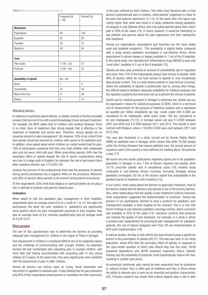
57
Table 3 What medication and tools are used to treat pain
Frequency (N = 56)
Percent (%)
Medication
Paracetamol 56 100
Ibuprofen 52 93
Tramadol 42 75
Morphine 28 50
Tools
Pain scales 11 (N = 53) 21
Protocols 12 (N = 54) 22
Availability of opioids (N = 54)
Never 1 2
Occasionally 24 44
Most of the time 21 39
Always 8 15
Attitudinal barriers
In response to questions about attitude, a notable minority of doctors provided answers that are not in line with current knowledge of pain and pain treatment. For example, the WHO states that for children with medical illnesses ‘there is no other class of medicines than strong opioids that is effective in the treatment of moderate and severe pain. Therefore, strong opioids are an essential element in pain management’ (9). However, 30% of our cohort think that opioids should be reserved for children with cancer or for palliative care. In addition, when asked about which children our cohort would treat for pain, 18% of participants answered that they only treat children with unbearable pain and not every child with pain. When prescribing opioids, 64% fear the secondary effect of opioids despite the risk of severe complications being very low. In a large audit in England, for example, the risk of permanent harm after morphine infusion was 1/10,000 (10).
Seventy-two percent of the professionals think that the presence of parents during painful procedures has a negative effect on the procedure. Moreover, only 26% of doctors allow parents to be present during painful procedures.
Of the respondents, 82% think that religious or spiritual beliefs do not play a role in attitude of patients and parents towards pain.
Evaluation
When asked to rate the paediatric pain management in their hospitals, respondents gave an average score of 6 on a scale of 1 to 10. The older the participants, the lower the rank: residents in paediatrics are significantly more positive about the pain management practiced in their hospital: they give an average mark of 6.6, whereas paediatricians give an average mark of 4.9 (P 0.01).
DiscussionThe aim of this questionnaire was to determine the barriers to providing adequate pain management for children in the region of Thiès in Senegal.
Pain assessment in children is considered difficult due to its subjective nature and the challenge of communicating with younger children. As expected, doctors felt less comfortable with evaluating pain in younger children, with more than half feeling uncomfortable with evaluating pain in very young children (0-2 years). At the same time, they were significantly more confident with the assessment of pain in older children.
Almost all doctors use clinical signs as crying, facial expression and discomfort or agitation to evaluate pain. It was striking that for pain evaluation only 64% of the respondents asked parents or caretakers for their impression
of the pain suffered by their children. This while most literature tells us that doctors underestimate pain in children, while parents’ judgement is closer to the pain that patients experience (11,12). At the same time, this figure was clearly higher than what was found in a study conducted among paediatric oncologists in sub-Saharan Africa, who only asked parents about their child’s pain in 38% of the cases (13). In future research, it would be interesting to ask patients and parents about the pain experience and their satisfaction with treatment.
Among our respondents, paracetamol and ibuprofen are the most widely used and available analgesics. This availability is slightly better compared with a study among paediatric oncologists in sub-Saharan Africa, where paracetamol is almost always or always available in 7 out of the 8 hospitals. In this same study, non-steroidal anti-inflammatory drugs (NSAID’s) were only ‘most often’ available in 5 out of the 8 hospitals (13).
Opioids are less used, primarily on account of unavailability due to regulation and costs. Only 15% of the respondents always have access to opioids, while 46% of doctors either do not have access to opioids or only occasionally have access to them. This is a well-known problem in most African countries, where the availability of opioids is problematic due to, among other things, the difficult balance between adequate availability for medical purposes and the regulatory systems that have been set up to prevent the misuse of opioids.
Opioid use for medical purposes in Senegal is extremely low. Opioid use can be expressed in doses for statistical purposes (S-DDD), which is a technical unit of measurement for the purpose of statistical analysis and is expressed as quantity per million inhabitants per day. Levels under 200 S-DDD are considered to be inadequate, while levels under 100 are considered to be very inadequate (14,15). In Senegal opioid use was 3 S-DDD between 2001 and 2003 and 4 S-DDD between 2011 and 2013. This stands in stark contrast with Belgium, where >10,000 S-DDD was used between 2011 and 2013 (16).
This was also illustrated in a study carried out by Human Rights Watch (HRW) on palliative care in Senegal, which shows that while 70,000 people suffer life-limiting illnesses that require palliative care, the annual amount of morphine used in the country is only sufficient for treating about 194 patients a year (17).
We found only two earlier publications regarding opioid use in the paediatric population in Senegal. In one, 7.8% of doctors regularly use opioids, while 70.1% prescribe opioids only in exceptional cases (18). Another study conducted in sub-Saharan African countries (including Senegal) among paediatric oncologists, 62.5% of the doctors stated that unavailability is the greatest barrier to treatment with morphine (13).
In our cohort, when asked about the barriers to good pain treatment, most of the doctors stated that the absence of protocols is one of the primary barriers. Also, when asked about how the quality of pain treatment could be improved, most respondents suggested the implementation of protocols. Twenty-two percent of our participants claimed to have a protocol for paediatric pain management available in their hospital at the moment. This is in line with former findings in sub-Saharan paediatric-oncology centres, where a protocol was available in 25% of the cases (13). Literature confirms that protocols can improve the quality of pain treatment. For example, in a study in which a protocol was implemented for procedural pain management for paediatric patients, the use of topical analgesia went from 2% pre-implementation to 92% post-implementation (19).
In various studies, the fear of side effects has been found to pose a significant barrier to the prescription of opioids (20,21). This was also seen in our study population, where 64% fear the secondary effect of opioids. In response to the open-ended question of which side effects they fear the most, 18/56 answered dependency and 26/56 answered respiratory failure. Specific training and the availability of protocols could hypothetically reduce this fear, resulting in a better pain policy.
As previously mentioned, pain cannot be seen separately from its situational or cultural context. Pain is often part of traditions and rites in Africa where the ability to tolerate pain is seen as an essential and positive characteristic (22). Previous research has shown that cultural aspects play a role in pain

58
management, and we saw indirect evidence of this in the answers provided by our cohort who reported that they only treat pain when it becomes unbearable (18%) or who find that opioids should only be prescribed when children are undergoing cancer treatment or palliative care (30%) (13,23).
As many Belgian paediatricians work with a broad spectrum of patients and parents of different nationalities, insights from this study could be used to improve daily practice when it comes to understanding parents who are less familiar with non-pharmacological approaches, hesitant towards opioids or less spontaneous in reporting their children’s pain to care providers. As one-third of doctors in this study think that opioids should be reserved for a palliative setting, this may implicate for the Belgian context that, for example, parents of (West) African descent may require a more detailed explanation when their children are prescribed opioids during hospitalisation. The insights can also inspire us to reconsider the focus we place on our practice of pain assessment and pain management.
Our study aimed to investigate the barriers to providing adequate pain management in Senegal. To our knowledge, it is the first study conducted amongst doctors in Senegal to elucidate the barriers to pain treatment in the general paediatric population. Despite the small number of physicians surveyed (65), the response rate was high (56). This study can serve as a point of departure for an audit on pain treatment, as it shows the current status of physicians’ attitude toward pain management.
However, some limitations should be acknowledged. In addition to the small sample size, this was also the first time that the questionnaire was used, given that it was developed on behalf of this study. Furthermore, the data was gathered by means of self-reporting rather than observation, with the possibility of the former leading to socially desirable answers.
Future research could expand the scope to include both the patients’ and parents’ view of pain practice, as well as the nurses’ attitude towards and knowledge of pain. Conducting a pain management assessment before and after the introduction of a protocol and proper training could also be valuable.
Additionally, misconceptions about pain and pain treatment could be averted by incorporating training about paediatric pain management into medical curricula and making an effort to implement national pain management protocols. On a larger scale, efforts should continue to encourage Senegalese policymakers to support and augment the health-care workforce and increase the availability of health care and essential medications.
ConclusionThis study was designed to identify and assess barriers to effective pain management in children, as viewed by doctors working in hospitals in the region of Thiès, Senegal. The main barriers to effective pain management are access barriers to medication, with low access to opioids due to legislation. Furthermore, physicians consider access barriers to protocols and training to be an another important factor. Lack of the latter may explain why we see that some misconceptions around pain and pain treatment still exist. Lastly, indirect evidence suggests that attitudinal barriers may exist, with a strikingly high number of physicians afraid to prescribe opioids due to their potential side effects, despite WHO recommendations for their use in pain treatment.
The authors have no conflict of interest to declare
REFERENCES:
1. The Development of a ‘Pain Stewardship Program’ in a Tertiary Childrens Hospital. Hosp Pediatr. 2016;6(9):520–8.
2. Weisman SJ, Bernstein B, Schechter NL. Consequences of Inadequate Analgesia During Painful Procedures in Children. Arch Pediatr Adolesc Med [Internet]. 1998 Feb 1 [cited 2020 Mar 31];152(2). Available from: http://archpedi.jamanetwork.com/article.aspx?doi=10.1001/archpedi.152.2.147
3. International Association for the Study of Pain. Declaration of Montréal. [Internet]. 2018 [cited 2019 Apr 4]. Available from: https://www.iasp-pain.org/DeclarationofMontreal
4. Committee on Psychosocial Aspects of Child and Family Health, Task Force on Pain in Infants, Children, and Adolescents. The Assessment and Management of Acute Pain in Infants, Children, and Adolescents. Pediatrics. 2001;108(3):793–7.
5. Birnie KA, Chambers CT, Fernandez CV, Forgeron PA, Latimer MA, McGrath PJ, et al. Hospitalized Children Continue to Report Undertreated and Preventable Pain. Pain Res Manag. 2014;19(4):198–204.
6. World Health Organization. World health report. Overview, why the workforce is important. [Internet]. 2006. [Cited 2020 Feb 20] Available at: https://www.who.int/whr/2006/overview/en/.
7. World Health Organization. Global health observatory (GHO) data. Density of physicians (total number per 1000 population, latest available year). [Internet]. 2019. [Cited 2020 Feb 20]. Available at: https://www.who.int/gho/health_workforce/physicians_density/en/.
8. Albertyn R, Rode H, Millar AJW, Thomas J. Challenges associated with paediatric pain management in Sub Saharan Africa. Int J Surg. 2009;7(2):91–3.
9. WHO guidelines on the pharmacological treatment of persisting pain in children with medical illnesses. Geneva: World Health Organization; 2012. 166 p.
10. Howard RF. Audits of postoperative analgesia: what have we learned and what should we do now? Pediatr Anesth. 2010;20(2):117–8.
11. Brudvik C, Moutte S-D, Baste V, Morken T. A comparison of pain assessment by physicians, parents and children in an outpatient setting. Emerg Med J. 2017;34(3):138–44.
12. Pillai Riddell RR, Craig KD. Judgments of Infant Pain: The Impact of Caregiver Identity and Infant Age. J Pediatr Psychol. 2006;32(5):501–11.
13. Yao A, Coze C, Traoré F, André N, Moreira C, Pondy A, et al. Prise en charge de la douleur de l’enfant atteint de cancer en Afrique : état des lieux au sein du Groupe franco-africain d’oncologie pédiatrique. Arch Pédiatrie. 2013;20(3):257–64.
14. Nchako E, Bussell S, Nesbeth C, Odoh C. Barriers to the availability and accessibility of controlled medicines for chronic pain in Africa. Int Health. 2018;10(2):71–7.
15. International Narcotics Control Board. Report of the International Narcotics Control Board on the Availability of Internationally Controlled Drugs: Ensuring Adequate Access for Medical and Scientific Purposes. [Internet]. 2010. [Cited 2020 March 12]. Available at: https://www.unodc.org/documents/lpo-brazil/noticias/2011/03-marco/Jife/Report_of_the_Board_on_the_availability_of_controlled_substances.pdf.
16. Berterame S, Erthal J, Thomas J, Fellner S, Vosse B, Clare P, et al. Use of and barriers to access to opioid analgesics: a worldwide, regional, and national study. Lancet Lond Engl. 2016;387(10028):1644–56.
17. Chung A. Abandoned in Agony. Cancer and the Struggle for Pain Treatment in Senegal. [Internet]. 2013. [Cited 2020 March 4]. Available from: https://www.hrw.org/report/2013/10/24/abandoned-agony/cancer-and-struggle-pain-treatment-senegal
18. Pouye A, Dia D, Sow PS, Yacouba H, Ndour CT, Soumare M, et al. [Prescription of morphinics in the management of pain in Dakar teaching hospitals]. Bull Soc Pathol Exot 1990. 2005;98(2):118–20.
19. Cregin R, Rappaport AS, Montagnino G, Sabogal G, Moreau H, Abularrage JJ. Improving pain management for pediatric patients undergoing nonurgent painful procedures. Am J Health-Syst Pharm AJHP Off J Am Soc Health-Syst Pharm. 2008;65(8):723–7.
20. Wolfert MZ, Gilson AM, Dahl JL, Cleary JF. Opioid Analgesics for Pain Control: Wisconsin Physicians’ Knowledge, Beliefs, Attitudes, and Prescribing Practices. Pain Med. 2010;11(3):425–34.
21. Suleiman ZA, Wahab KW, Kolawole IK. Opioid prescribing habits of physicians in Kwara State, Nigeria. Ghana Med J. 2016;50(2):63.
22. Kéita M-M. Le vécu de la douleur par les populations africaines. Arch Pédiatrie. 2009;16(6):775–6.
23. Rampanjato RM, Florence M, Patrick NC, Finucane BT. Factors influencing pain management by nurses in emergency departments in Central Africa. Emerg Med J EMJ. 2007;24(7):475–6.
Case Report
59
Shiga toxin-producing Escherichia coli outbreak in a childcare facility Audrey Revercez a, Romain Mahieu b, Stéphan Clément de Cléty c, Elisabeth Rebuffat a, Louise De Viron b, Florence Crombé d, Denis Piérard d, Tessa Goetghebuer a,e
a Paediatric Department, CHU Saint-Pierre, Université Libre de Bruxelles b Infection Prevention and Control, Common Community Commission (COCOM) c Paediatric Intensive Care Unit (PICU), Cliniques universitaires Saint-Luc, Université catholique de Louvain d National Reference Centre for Shiga toxin-producing Escherichia coli (NRC STEC), Department of Microbiology and Infection Control, Universitair Ziekenhuis Brussel e Office de la Naissance et de l’Enfance (ONE), Belgium
Case Report
Haemolytic uremic syndrome (HUS) is a clinical syndrome characterized by acute renal failure (ARF) associated with anaemia and thrombocytopenia. It is the most common cause of acute kidney injury in children. Approximately 25% of STEC-HUS patients develop neurologic symptoms and is an important contributor to the morbidity of the disease (1). HUS mortality rate is 2.5-7% (2). A Belgian study reports a 2.5% lethality (3).
In 90-95% of cases, the syndrome is consecutive to an episode of diarrhoea of 2 to 14 days due to Shiga toxin-producing Escherichia coli strains (STEC) (4). STEC are classified based on their serogroup (“O”-antigen) and their serotype (”H”-antigen). Most STEC infections are caused by STEC O157:H7(1). Among STEC, some genes are correlated with an increased risk of developing HUS (Stx2a, Stx2d, or Eae) (5,6). The duration of carriage of serogroup O157 is generally ranging from 5 to 29 days but it can reach 124 days (1). It is estimated that up to 10% of patients with STEC infection may develop HUS, and up to 20% in some epidemics (4).Human contamination occurs through consumption or handling of contaminated food. Human-to-human transmission of STEC has been described in family settings and in children’s communities (7). This mode of transmission by the fecal-oral route is important with a transmission rate of up to 20%.(4)HUS surveillance is important to detect outbreaks, STEC strains associated with severe outcomes, and new strains emergence as well as to try to identify contamination source and remove contaminated products. HUS caused by STEC strains is a mandatory notifiable disease in our country.
We report here two cases of HUS admitted to two hospitals Brussels and our subsequent epidemiologic research in the day-care centre.
Case reports and subsequent study The first case, a two-year-old boy, was admitted to an emergency room for seizures, diarrhoea and fever. Blood sample revealed the characteristic HUS triad. E. Coli O157:H7 (Stx1+, Stx2a+, Eae+) was detected by PCR in the stool (National Reference Centre (NRC) for STEC). The child was transferred to a paediatric intensive care unit (PICU) for status epilepticus. He required mechanical ventilation for 12 days and benefited from peritoneal dialysis followed
by continuous veno-venous hemofiltration for a total of 6 days. (Peritoneal dialysis did not allow adequate fluid and metabolic control). He was hospitalized for 4 weeks and recovered completely.
The childcare facility doctor notified the case to the Infection Prevention and Control department (COCOM) and sent an information message to all parents. They were invited to contact their physician if their child presented signs of gastroenteritis and to tell him or her that a HUS has been diagnosed in the childcare facility.
Five days after admission of the first case, a 2-year-old girl was admitted to the emergency room of another hospital after 5 days of fever and vomiting but with no history of diarrhoea. Her mother brought the letter received from the childcare facility. Blood tests were characteristic for HUS. The PCR-STEC was negative in both blood and stools samples. The patient was transferred to a PICU but did not require dialysis. She was discharged and well after 2 weeks.
Because a third case of uncomplicated diarrhoea was reported by the childcare facility, it was decided to screen all children. The decision to perform systematic screening was taken by the COCOM, the childcare managers and physicians, and the NRC for STEC. The screening was initiated for the whole childcare community. All 77 children and 27 adults staff members were screened. Children were divided into three groups according to their ages and the organization of the day-nursery.
The carrying incidence was 9% in the whole childcare facility (7/77). The same STEC was indeed identified in seven children: 1 with HUS, 5 with mild gastroenteritis, 1 asymptomatic. These 7 children and the second HUS case, which was STEC negative, belonged to the same group of 29 children. The incidence of carrier status was high in this group (8/29; 28%). One sibling of a child from the day-care was also positive. No cases were identified among staff members.
To avoid further contamination, these children were banned from the nursery until two negative cultures were obtained from samples taken 24 hours apart. Appropriate hygiene measures led to a full recovery after two months (Table 2). No antibiotic has been administered (Table 1).Origin of the index case remained unknown despite epidemiological and field investigations.
Introduction
AbstractHaemolytic uremic syndrome is a rare disease typically associated with Shiga toxin-producing Escherichia coli. We describe two cases of haemolytic uremic syndrome that occurred in the same childcare facility.We collaborated with the National Reference Centre for Shiga toxin producing Escherichia coli to screen all 77 children and 27 adults present at the childcare facility. Shiga toxin Escherichia coli was detected in 7 children in the childcare facility (9%) and 1 symptomatic sibling. Only one section was touched, 8 out of 29 children from the “big” section (28%) were affected (haemolytic uremic syndrome and/or carriers). The high prevalence of Shiga toxin Escherichia coli carrying in childcare facilities supports the need for early hygiene measures and information to parents.
KeywordsHaemolytic uremic syndrome, Shiga toxin-producing Escherichia coli, STEC, childcare

60
Table 1 Comprehensive screening
SectionsTotal number
Asymptomatic carrier
Symptomatic subject
HUSLaboratory confirmed cases
Group of «small» children
22 0 0 0 0
Group of « middle » children
26 0 0 0 0
Group of « big» children
29 2 4 2 7
Staff members 27 0 0 0 0
Sibling not admitted to the day-care centre
2 0 2 0 1
HUS: Haemolytic uremic syndrome.
Discussion Our epidemiologic study shows that a quarter of the children belonging to the same group in the childcare facility carried a STEC, sometimes for few weeks. Such incidence is rarely documented and justifies caution in high-risk groups.Diagnosis and reporting of STEC infections are indeed of particular importance for the rapid detection of epidemics and implementation of adequate measures (6). As soon as a healthcare provider reports a HUS case, several control measures are taken by the infectious disease surveillance unit. These measures include: informing the concerned community and parents (in the form of a letter), strengthening hygiene measures, stool sampling for STEC detection, epidemiological investigation and if a source of contamination is suspected, further work-up to the Federal Agency for the Safety of the Food Chain (AFSCA) (6).The identification of STEC carrying is made from stool sample or rectal smear. In the event of HUS or STEC outbreak, must be systematically sent to the NRC. Diagnosis is based on strain culture and serotyping, PCR amplification of virulence genes in stool, immunological tests and serologies (search for antibodies against LPS (lipopolysaccharides) of E. coli O-serogroups). It is not always easy to highlight STEC due to its low concentration in stool and its rapid elimination from the intestine, especially during active HUS (4). The likelihood of identifying Shiga toxin decreases dramatically over the course of the disease. Therefore stool collection should be carried out at the latest 4 to 6 days after the onset of digestive prodromes (1).
Treatment of STEC infection is usually symptomatic. Antibiotics use is controversial because in most cases bacteria lysis can release more toxins (8,9)that provided data from patients (1). In France, azithromycin is recommended, particularly in asymptomatic carriers. High quality data however are lacking (1,6).
Zhang et al suggested that in mice, azithromycin has a strong effect on Stx production by STEC and on the Stx- induced inflammatory host response and prevents death. Azithromycin may have a beneficial effect on STEC-associated disease. However further studies are required before strong recommendations (10) produces Stx from phage, and causes the development of hemolytic-uremic syndrome via Stx-induced inflammatory cytokine production. Azithromycin exhibited strong in vitro activity against STEC without inducing Stx-converting phage, in marked contrast to norfloxacin. Azithromycin decreased the tumor necrosis factor alpha (TNF-α).
Complement activation plays an important role in the pathogenesis of atypical HUS. Eculizumab is an anti-C5-convertase monoclonal antibody. It is safe and effective for the treatment of atypical HUS (11) thrombocytopenia and AKI. In ~ 90% of cases, HUS is a consequence of infection with Shiga toxin-producing E. coli (STEC. There is no controlled data investigating the use of eculizumab in STEC-HUS but many studies have shown clinical improvement in cases of STEC-HUS with neurological involvement (11,12) thrombocytopenia and AKI. In ~ 90% of cases, HUS is a consequence of infection with Shiga toxin-producing E. coli (STEC).
Two double-blind, placebo-controlled trials are underway in France and the UK to provide evidence to guide the use of this treatment in STEC-HUS(12).
Conclusion The incidence of STEC outbreaks in childcare facilities is rarely documented. Since it could be high, particularly in children spending few hours together each day such as in our study, preventive and control measures should be implemented as soon as possible, in order to lead to rapid identification of children with HUS symptoms. Rapid and close collaborations between the public health authority, National Reference Center, childcare staff and parents insured efficient disease control.
Conflict of interest statement
The authors of this case report declare that they have no conflict of interest.
Table 2 Temporal description of STEC and/or HUS affected children
Children 1 2 3 4 5 6 7 8 9
STEC-PCR results
10-11-19positive
13-11-19positive
15-11-19negative
26-11-19positive
29-11-19positive
02-12-19positive
02-12-19positive
02-12-19negative
06-12-19positive
02-12-19positive
29-11-19positive
29-11-19negative
03-12-19positive
20-12-19negative
07-12-19positive
06-12-19negative
12-12-19positive
09-12-19negative
13-12-19positive
10-12-19negative
05-12-19negative
10-01-20negative
16-12-10positive
09-12-19negative
17-12-19positive
10-12-19negative
12-12-19negative
29-12-19negative
20-12-19positive
31-12-19negative
07-01-20negative
10-01-20negative
Symptoms HUS enteritis HUS enteritis enteritis asymptomatic enteritis enteritis asymptomatic
STEC: Shiga toxin-producing Escherichia coli, HUS: Haemolytic uremic syndrome. Appearance in the order of positive stool cultures.
REFERENCES:
1. De Rauw K, Buyl R, Jacquinet S, Piérard D. Risk determinants for the development of typical haemolytic uremic syndrome in Belgium and proposition of a new virulence typing algorithm for Shiga toxin-producing Escherichia coli. Epidemiol Infect. 2019;147:e6, 1-5.
2. Jacquinet S, De Rauw K, Pierard D, Godefroid N, Collard L, Van Hoeck K, et al. Haemolytic uremic syndrome surveillance in children less than 15 years in Belgium, 2009–2015. Arch Public Health. 2018;76:41.
3. Mariani-Kurkdjian P, Bingen É. Physiopathologie et virulence des Escherichia coli producteurs de Shiga-toxines. Réanimation 2012;21:268-279.
4. Joseph A, Cointe A, Mariani Kurkdjian P, Rafat C, Hertig A. Shiga Toxin-Associated Hemolytic Uremic Syndrome: A Narrative Review. Toxins (Basel). 2020;12:67.
5. Law D. Virulence factors of Escherichia coli O157 and other Shiga toxin-producing E. coli. Journal of Applied Microbiol. 2000;88 :729-745.
6. Sciensano, AViQ [Internet]. Fiche informative MATRA « Escherichia Coli producteur de Shigatoxines»; 2017 [cited 2020 Sep 29]. Available from: https://www.wiv-isp.be/matra/Fiches/EHEC.pdf.
7. Al-Jader L., Salmon RL., Walker AM., Williams HM., Willshaw GA., and Cheasty T. Outbreak of Escherichia coli O157 in a nursery: Lessons for prevention. Arch Dis Child 1999; 81:60-63.
8. Freedman SB, Xie J, Neufeld MS, Hamilton WL, Hartling L, Tarr PI. Shiga Toxin–Producing Escherichia coli Infection, Antibiotics, and Risk of Developing Hemolytic Uremic Syndrome: A Meta-analysis. Clin Infect Dis. 2016;62:1251-1258.
9. Tarr GAM, Oltean HN, Phipps AI, Rabinowitz P, Tarr PI. Strength of the association between antibiotic use and hemolytic uremic syndrome following Escherichia coli O157:H7 infection varies with case definition. Int J Med Microbiol. 2018;308:921-926.
10. Ohara T, Kojio S, Taneike I, Nakagawa S, Gondaira F, Tamura Y, et al. Effects of Azithromycin on Shiga Toxin Production by Escherichia coli and Subsequent Host Inflammatory Response. Antimicrob Agents Chemother. 2002;46:3478-3483.
11. Walsh P. R., Johnson, S. Eculizumab in the treatment of Shiga toxin haemolytic uraemic syndrome. Pediatr Nephrol. 2019; 34: 1485–1492.
12. Mahat U, Matar RB, Rotz SJ. Use of complement monoclonal antibody eculizumab in Shiga toxin producing Escherichia coli associated hemolytic uremic syndrome: A review of current evidence. Pediatr Blood Cancer. 2019;66:27913.
Case Report

61
Mycoplasma Respiratory Infection Mimicking COVID-19 Elise Exelmans a,b, Marijke Proesmans b, Francois Vermeulen b, Stefaan Van Lierde a
a Regional Hospital Tienen, Belgium b University Hospital Leuven, Belgium
Case Report
Since December 2019 there has been a rapid spread of the new coronavirus SARS-CoV-2, first within China, but then quickly to the rest of the world. The first epidemiological studies in the Chinese population show that children represent only 2.2% of the total number of infections and that infection is generally less severe with a negligible mortality (1,2-4). Subsequent reports from Europe confirm the low burden of SARS-CoV-2 in children, with very few admissions to intensive care, even in children with immunosuppression (5-8). Whether this is from shielding by school closures or by a reduced susceptibility to infection remains unclear (9).
The gold standard diagnostic test for SARS-CoV-2 infection is a polymerase chain reaction (PCR) on a respiratory sample, in most cases a nasopharyngeal swab. CT scan of the lungs has been put forward as an alternative way of diagnosing COVID-19 because of bronchoalveolar lavage (BAL) proven SARS-CoV-2 infection in swab negative adults (10,11). An acute respiratory infection with compatible chest CT and negative PCR is accepted as case definition of SARS-CoV-2 infection in several guidelines and many adult cases of were diagnosed based on CT findings (12).
We report a case of a child with Down syndrome presenting with a severe respiratory infection. According to adult guidelines, he was diagnosed with COVID-19 based on the highly suggestive findings on chest CT scan. SARS-CoV-2 PCR tests on nasopharyngeal swabs were repeatedly negative, and Mycoplasma pneumoniae was eventually shown to be the cause of the respiratory infection.
Case PresentationOn March 30th 2020, a 10-year old boy with trisomy 21 presented at the emergency department of our regional hospital with a two weeks’ history of gradually worsening dyspnea and cough, despite treatment with oral amoxicillin and inhaled bronchodilators. The child had been treated in an intensive care setting for a respiratory infection at the age of 6 weeks and had a history of viral induced wheezing and immunodeficiency (lymphocytopenia and hypogammaglobulinemia) for which he received intravenous immunoglobulins from 2010 till 2017 through an implantable venous access device (Port-a-Cath), discontinued after recovery of peripheral blood lymphocytosis and serum immunoglobulin levels. No frequent or severe infections were reported during the last 3 years. The boy had no cardiac malformation. He suffered from psychomotor retardation and hypotonia, compatible with trisomy 21.
At presentation the patient was afebrile and somewhat apathic. Heart rate was 110 bpm, blood pressure 124/88 mm Hg, pulse oxygen saturation in ambient air was 78%. He was dyspneic with a respiratory rate of 64/min, chest retractions and use of accessory respiratory muscles. Poor air entry, diffuse fine crackles and wheezing were noted on chest auscultation. Heart sounds were normal. Liver was palpated at 6 cm below the right costal margin.
Arterial blood gases showed mild hypercapnia and mild hypoxemia. Oxygen by face mask and inhaled bronchodilators resulted in slight temporary improvement in oxygen saturation. Prednisolone IV 1 mg/kg was administered given the history of hyperreactive airways and wheezing. A third-generation cephalosporin was started.
Laboratory testing (Table 1) showed increased inflammatory markers, normal levels of immunoglobulins, raised aminotransferases, troponins and D-dimers. M. pneumoniae IgM was positive. A nasopharyngeal swab to test for COVID-19 was collected.
A bedside chest X-ray showed a limited confluent density in the right lung base. Given the discrepancy between severe hypoxemia and mild X-ray changes, a CT scan of the lungs was made before transfer to a tertiary care center with pediatric intensive care facilities, according to the current local ‘COVID-guidelines’. The chest CT showed ground glass opacities in all lobes and was scored as very suggestive of an infection with the SARS-CoV-2 according to a standardized local protocol designed for the COVID epidemic based on recent literature (Figure 1) (13).
On arrival at the tertiary center, a cardiac ultrasound confirmed mild right ventricular dysfunction, pulmonary hypertension and a small pericardial effusion. Diuretics were started in combination with low molecular weight heparin (LWMH) at prophylactic dose. High flow nasal oxygen (HFNO) was delivered with a maximal FiO2
of 1.0. Azithromycin was added to cefotaxime. Given the high oxygen requirements and increase in pCO
2 to 75 mmHg on
venous blood gases, the child was transferred to the intensive care COVID unit. Supportive care was continued, with resolution of the hypercapnia and right heart failure after 48 hours and weaning of HFNO after 72 hours upon which the patient could be transferred to a pediatric infectious disease ward.
Four SARS-CoV-2 PCR tests on nasopharyngeal swabs over 48 hours were negative. The semi-quantitative PCR for M. pneumoniae obtained from the nasopharyngeal swab was strongly positive, in line with the positive serum
Introduction
AbstractIn the current pandemic with SARS-CoV-2, children can present with severe acute respiratory infection, qualifying as COVID-19 according to published guidelines. However other etiologies should not be overlooked. We present a case of a 10-year old boy with Down syndrome, who presented with fever and respiratory failure. A CT scan of the lungs showed lesions very suggestive of COVID-19. He was admitted to an intensive care unit because of deep hypoxemia requiring high flow nasal oxygen. Polymerase chain reaction on nasopharyngeal swabs was repeatedly negative for SARS-CoV-2 but positive for Mycoplasma pneumoniae. This case report illustrates possible diagnostic pitfalls when treating children in this pandemic.
KeywordsSARS-CoV-2; pediatric infectious disease; acute respiratory failure

62
IgM for M. pneumoniae. A respiratory panel (containing 29 pathogens) confirmed a positive PCR for M. pneumoniae as well as herpes-simplex virus 1. Serology (Abbott semi-quantitative antibody test, IgG) for SARS-CoV-2 was negative 2 weeks after the onset of symptoms.
At day 11, low grade fever and need for supplemental oxygen persisted. A CT pulmonary angiography confirmed pulmonary embolism that was suspected because of the presence of an implantable IV access, pulmonary hypertension, and elevated D-dimers. LMWH was started at therapeutic doses. A repeat SARS-CoV-2 PCR was strongly positive (Cycle threshold (Ct) value 16,8). At that point serology for SARS-CoV-2 remained negative, demonstrating a probable nosocomial infection. Oxygen therapy could be stopped on day 16 and laboratory tests returned to normal. One week after the positive PCR, the serology also became positive. The patient was discharged from the hospital on day 18. On a follow-up visit 6 weeks later SARS-Cov-2 IgG were still positive. The immunological screening completed with a pneumococcal antibody response was normal.
DiscussionThis patient with Down syndrome and a past medical history of ill-defined immunodeficiency presented with a severe respiratory infection leading tot respiratory failure requiring transfer to intensive care. The disease was characterized by fever, bronchial obstruction, profound hypoxemia and multifocal interstitial pneumonia on CT scan. The patient fulfilled the case definition for COVID-19, despite the negative PCR for SARS-CoV-2, and was cohorted accordingly. The impaired coagulation and elevated D-dimers contributed to the initial diagnosis. Research in adult population shows that severe COVID-19 is associated with a higher incidence of thromboembolic events (14). Other possible pathogens were also considered and treated, such as bacterial pneumonia with cefotaxime and atypical pneumonia with azithromycin. Pneumocystis carinii was considered unlikely given the normal lymphocytosis. Definitive results with a positive M. pneumoniae PCR and IgM, repeatedly negative SARS-CoV-2 PCR and negative serology 2 weeks into the respiratory symptoms make M. pneumoniae the most likely etiologic agent of the respiratory infection in this child. SARS-CoV-2 PCR on a nasopharyngeal swab can produce a false negative result, especially in the later stages of the disease where the virus still can be found in the lungs (15). However, we
found evidence of an acute infection with M. pneumoniae: PCR and IgM were both positive. Moreover, the patient did not have lymphocytopenia which is less likely for a severe COVID-19 and he had bronchial obstruction, which is not frequently reported in COVID-19 (3,4). Improvement was dramatic after the initiation of macrolide antibiotic therapy. No bronchoalveolar lavage was performed in the acute setting because of the risk of the procedure, nor later in the light of the diagnosis of M. pneumoniae infection with improvement with macrolides. SARS-CoV-2 serology turned out to be negative.
Interstitial pneumonia with severe hypoxemia is a rare but known presentation of atypical pneumonia caused by M. pneumoniae. Trisomy 21 is associated with frequent and more severe infections, with some degree of immunodeficiency, as well as with pulmonary hypertension during lung infections (16-18). Our patient has had a previous episode of severe respiratory infection and a documented, albeit transient period of lymphocytopenia and hypogammaglobulinemia. Acute or chronic pulmonary embolism likely contributed to the severity of the initial presentation.
This case illustrates two pitfalls that can arise during this COVID-19 pandemic. First, the continuous flow of information and profound changes in the organization of care, both highly focused on COVID-19, create a so-called ‘availability heuristics’ causing clinicians to think first about COVID-19, rather
Figure 1. Chest CT images (A,B) showing peripheral ground glass opacities Table 1 Laboratory data on admission*
Variable Value Reference Range
Hemoglobin (g/dl) 12.2 (11.5-15.5)
White cells (per mm2) 18,610 (4,500-13,500)
Absolute neutrophil count (per mm2) 12,700 (2.00-7.50)
Absolute ymphocyte count (per mm2) 3,160 (1.50-4.00)
Platelet count (per mm2) 133,000 (150,000-400,000)
C-Reactive Protein (mg/liter) 105.1 (<5.0)
Prothrombin time (sec) 20.6 (10-14.1)
Activated partial thromboplastin time (sec) 27.2 (24.6-38.4)
Prothrombin time international normalized ratio 1.76 (1.00-1.20)
Fibrinogen (mg/dl) 328 (276-471)
D-dimers (ng/ml) 85 612 (69-580)
Blood urea nitrogen (mg/dl) 41.5 (10.8-38.4)
Creatitine (mg/dl) 0.54 (0.26-0.77)
Troponine I (ng/ml) 26.9 (<17.5)
Brain natriuretic peptide (pg/ml) 1180 (0-99)
Aspartate aminotransferase (U/liter) 998 (<50)
Alanine aminotransferase (U/liter) 1613 (<50)
Lactate dehydrogenase (U/liter) 1174 (110-295)
Mycoplasma pneumoniae IgM positive
* To convert the values for urea nitrogen to millimoles per liter, multiply by 0.357. To convert the values of creatinine to micromoles per liter, multiply by 88.4.
63
than about other diseases. The epidemiology of COVID-19 is dramatically different in children and adults, with discordances in disease sensitivity, contagiousness, clinical picture and prognosis. Severe pulmonary disease in children does hardly occur. In the largest pediatric cohort with COVID-19 reported so far, the disease was more severe in children whose diagnosis of SARS-CoV-2 infection was not PCR confirmed, pointing out to the role of other (co) infections in severe cases and at least in some confirmed cases (2). One small case series reports coinfections with influenza, Mycoplasma, RSV or CMV in 8/20 children (19). Nevertheless, individuals with Down syndrome may have a higher risk for more severe COVID-19 due to the anatomic, immunologic and metabolic comorbidities associated with trisomy 21 (20-22). They have an increased occurrence of autoimmunity and certain antibodies (e.g. anti-type 1 interferon) have already been shown to be a significant risk factor for severe COVID-19 in adults (22). In observational studies hospitalized patients with Down syndrome and COVID-19 are younger and have a more severe disease than matched non-Down syndrome controls; so caution is still advised in this patient population (23).
Secondly, this case highlights an important specificity of the diagnostic pathway for COVID-19 in children. In several reports and guidelines, chest CT is proposed as a screening tool for the diagnosis of COVID-19 infection, arguing a better sensitivity than the SARS-Cov-2 PCR. It also provides quicker results. Preliminary evaluations estimate its sensitivity at 97% using the RT-PCR as reference, but with a specificity of only 25% (24). In children, ground glass opacities are reported as the most common abnormality on chest CT with an incidence between 33 and 60% in hospitalized children (3,4,25). Chest CT expose children to ionizing radiations, and little information is available about what to expect in children with COVID-19. In this patient, the suggestive imaging results led to the diagnosis of COVID-19 even before the PCR results were known. The patient was therefore admitted to a COVID intensive care unit, which eventually led to a probable nosocomial infection. However, given the very low probability of severe COVID-19 in children, confirmation with a very nonspecific diagnostic test such as chest CT is in general not warranted, as its positive predictive value will be very low in a disease with a low prevalence. Even with a highly suggestive CT scan, alternative diagnoses remain more likely than COVID-19 in children with severe pulmonary infections. Of course, caution is necessary in children with a higher risk for a more serious disease course. On the contrary, a negative PCR, which is a very sensitive test, has a good negative predictive value if well obtained.
Overlooking an alternative diagnosis in COVID-19 times, based on current guidelines (lung CT-scan) is a real pitfall in some pediatric patients, especially if the alternative is treatable. This is even more so when difficult ethical decisions have to be taken as to whom further intensive therapy can be offered.
Conflict of Interest:
The authors have no conflicts of interest to disclose.
REFERENCES:
1. Wu Z, McGoogan JM. Characteristics of and Important Lessons From the Coronavirus Disease 2019 (COVID-19) Outbreak in China: Summary of a Report of 72 314 Cases From the Chinese Center for Disease Control and Prevention. JAMA. 2020;323(13):1239-1242.
2. Dong Y, Mo X, Hu Y, Qi X, Jiang F, Jiang Z, et al. Epidemiology of COVID-19 Among Children in China. Pediatrics. 2020;145(6):e20200702.
3. Lu X, Zhang L, Du H, Zhang J, Li YY, Qu J, et al. SARS-CoV-2 Infection in Children. N Engl J Med. 2020;382(17):1663-1665.
4. Qiu H, Wu J, Hong L, Luo Y, Song Q, Chen D. Clinical and epidemiological features of 36 children with coronavirus disease 2019 (COVID-19) in Zhejiang, China: an observational cohort study. Lancet Infect Dis. 2020;20(6):689-696.
5. D'Antiga L. Coronaviruses and immunosuppressed patients. The facts during the third epidemic. Liver Transpl. 2020;26(6):832-834.
6. Götzinger F, Santiago-García B, Noguera-Julián A, Lanaspa M, Lancella L, Calò Carducci, FI, et al. COVID-19 in children and adolescents in Europe: a multinational, multicentre cohort study. Lancet Child Adolesc Health. 2020;4(9):653-661.
7. Cornelissen L, Litzroth A, Montourcy M, De Rouck M, Wyndham-Thomas C, Klamer S, Van Beckhoven D. COVID-19-infectie bij kinderen in België: resultaten van labosurveillance, schoolgegevens en ziekenhuis surveillance tot en met 28 juni 2020. Brussel, België: Sciensano; 2020. Wettelijk depotnummer: D/2020/14.440/68
8. Wann OV, Holden KA, Turtle L, et al. Clinical characteristics of children and young people admitted to hospital with covid-19 in United Kingdom: prospective multicentre observational cohort study. BMJ. 2020;370:m3249. Published 2020 Aug 27.
9. Munro APS, Faust SN. COVID-19 in children: current evidence and key questions. Curr Opin Infect Dis. 2020;33(6):540-547.
10. Wang W, Xu Y, Gao R, Lu R, Han K, Wu G, et al. Detection of SARS-CoV-2 in Different Types of Clinical Specimens. JAMA. JAMA. 2020;323(18):1843-1844.
11. Lei P, Fan B, Wang P. Differential Diagnosis for Coronavirus Disease (COVID-19): Beyond Radiologic Features. American Journal of Roentgenology. 2020;215(1):W19.
12. Scientific Institute of Public Health (WIV-ISP) (Belgium). Gevalsdefinitie, indicaties voor testen en verplichte melding van COVID-19. Sciensano [internet]. 2020 april 1. [cited 2020 april 5]; Available from https://www.sciensano.be/en
13. Pan Y, Guan H, Zhou S, et al. Initial CT findings and temporal changes in patients with the novel coronavirus pneumonia (2019-nCoV): a study of 63 patients in Wuhan, China. Eur Radiol. 2020;30(6):3306-3309.
14. Bikdeli B, Madhavan MV, Jimenez D, Chuich T, Dreyfus I, Driggin E, et al. COVID-19 and Thrombotic or Thromboembolic Disease: Implications for Prevention, Antithrombotic Therapy, and Follow-Up: JACC State-of-the-Art Review. J Am Coll Cardiol. 2020;75(23):2950-2973.
15. Yang Y, Yang M, Shen C, Wang F, Yuan J, Li J, et al. Evaluating the accuracy of different respiratory specimens in the laboratory diagnosis and monitoring the viral shedding of 2019-nCoV infections. MedRxiv. 2020 feb 17.
16. Orlicek SL, Walker MS, Kuhls TL. Severe Mycoplasma Pneumonia in Young Children with Down Syndrome. Clin Pediatr (Phila). 1992;31(7):409-412.
17. Ram G, Chinen J. Infections and immunodeficiency in Down syndrome. Clinical & Experimental Immunology. 2011;164(1):9-16.
18. Bush D, Galambos C, Ivy DD, Abman SH, Wolter-Warmerdam K, Hickey F. Clinical Characteristics and Risk Factors for Developing Pulmonary Hypertension in Children with Down Syndrome. The Journal of Pediatrics. 2018;202:212-219.e2.
19. Xia W, Shao J, Guo Y, Peng X, Li Z, Hu D. Clinical and CT features in pediatric patients with COVID‐19 infection: Different points from adults. Pediatr Pulmonol. 2020;55(5):1169-1174.
20. Newman AM, Jhaveri R, Patel AB, Tan TQ, Toia JM, Arshad M. Trisomy 21 and Coronavirus Disease 2019 in Pediatric Patients [published online ahead of print, 2020 Aug 27]. J Pediatr. 2020;S0022-3476(20)31103-3.
21. Meyts I, Bucciol G, Quinti I, Neven B, Fischer A, Seoane E, et al. Coronavirus Disease 2019 in patients with inborn errors of immunity: an international study [published online ahead of print, 2020 Sep 24]. J Allergy Clin Immunol. 2020;S0091-6749(20)31320-8.
22. Dard R, Janel N, Vialard F. COVID-19 and Down's syndrome: are we heading for a disaster?. Eur J Hum Genet. 2020;28(11):1477-1478. doi:10.1038/s41431-020-0696-7
23. Bastard P, Rosen LB, Zhang Q, Michailidis E, Hoffmann HH, Zhang Y, et al. Autoantibodies against type I IFNs in patients with life-threatening COVID-19. Science. 2020;370(6515):eabd4585.
24. Malle L, Gao C, Hur C, ruong HQ, Bouvier NM, Percha B, et al. Individuals with Down syndrome hospitalized with COVID-19 have more severe disease [published online ahead of print, 2020 Oct 16]. Genet Med. 2020;10.1038/s41436-020-01004-w.
25. Ai T, Yang Z, Hou H, Zhan C, Chen C, Lv W, et al. Correlation of Chest CT and RT-PCR Testing in Coronavirus Disease 2019 (COVID-19) in China: A Report of 1014 Cases. Radiology. 2020;296(2):E32-E40.
26. Feng K, Yun YX, Wang XF, Yang GD, Zheng YJ, Lin CM, et al. Analysis of CT features of 15 Children with 2019 novel coronavirus infection. Zhonghua Er Ke Za Zhi. 2020;58(0):E007.

64
Endovascular management of a stroke in a 9-year-old child with neurofibromatosis type 1 and a common carotid artery occlusion Yveline Bruyère a, Christophe Fricx a, Noémie Ligot b, Benjamin Mine c, Philippe David d, Françoise Vermeulen a, Florence Christiaens e
a Department of Pediatrics, Hôpital Erasme, Université Libre de Bruxelles (ULB), Brussels, Belgium. b Department of Neurology, Hôpital Erasme, Université Libre de Bruxelles (ULB), Brussels, Belgium. c Department of Neurointervention, Hôpital Erasme, Université Libre de Bruxelles (ULB), Brussels, Belgium. d Department of Neuroradiology, Hôpital Erasme, Université Libre de Bruxelles (ULB), Brussels, Belgium. e Department of Neuropediatrics, Hôpital Erasme, Université Libre de Bruxelles (ULB), Brussels, Belgium.
Case Report
AbstractStrokes are being more frequently recognized as a cause of morbidity and mortality in the pediatric population. Thrombotic strokes are rare in childhood and most of them are associated with non-atherosclerotic cerebral arteriopathies, which can be induced by genetic association like neurofibromatosis type 1. This case report illustrates this pathology, associated to an unusual vasculopathy of the common carotid artery, and reports the optimal management of a thrombotic stroke in an official European Stroke Center.
Keywordsstroke, neurofibromatosis type 1, thrombectomy, common carotid occlusion, arteriopathy
Strokes are being more frequently recognized as a cause of morbidity and mortality in children. The annual incidence for arterial ischemic stroke (AIS) is about 2,4 per 100.000 children (1). The mortality rate is 4% but the morbidity is 50%, and this is varying by age, gender and ethnicity (1,2).
Due to an optimal management with effective surgical revascularization or thrombolysis, early diagnosis is important to avoid long-term impairments (2).
In contrast to adults, risk factors for pediatric AIS are less well understood (1). Traditional pediatric stroke risks are congenital heart disease, sickle cell disease, radiotherapy, trauma, hypercoagulable states, infections and post-infectious inflammatory disorders like post-varicella arteriopathy (1).
In many pediatric stroke cases, pathogenesis is multifactorial (3). Recent studies (2-4) show that most of AISs are associated with non-atherosclerotic cerebral arteriopathies, which is an important predictor. Recurrent events will happen in about 21% of these patients (2-4). Pediatric cerebral arteriopathy has two important causes: environmental and genetic associations (4).
One of the genetic associations is neurofibromatosis type 1 (NF1). NF1 has an autosomal dominant inheritance pattern linked to germline mutations in the NF1 gene encoding neurofibromin (5). This gene regulates the RAS signaling pathway, that controls cell proliferation, which corresponds to a tumor-suppressor gene (6). The diagnosis of NF1 is based on clinical criterias, involving: i) dermatologic, ii) ophthalmologic and iii) radiologic specificities (5). This point shows that NF1 is a multisystem disorder, implying that this has a large impact on a child’s health.
Important complications of NF1 are vascular manifestations. Patients with NF1 can develop congenital heart disease (2%), aneurysms, arteriovenous fistulae and peripheral and cerebral arteriopathy (2.5% to 6%) (6-8). This vasculopathy is probably caused by the loss of neurofibromin function, complicated by proliferation of smooth muscles (7,8). In addition, neurofibromin serves to maintain the integrity of endothelial cell layer. Unopposed proliferation of the vascular smooth muscle cells can be caused by alteration of its integrity (7).
This vasculopathy is often asymptomatic, but can have major consequences.
The most common sites are the kidneys and the brain (9). In children with NF1, renal artery stenosis is the most frequent site of symptomatic vasculopathy and an important cause of hypertension (6,7).
On the other hand, in children cerebral arteriopathy can have dramatic end result. The most frequent are stenotic lesions, particularly of the intracranial internal carotid (ICA), middle cerebral (MCA), or anterior cerebral (ACA) arteries (6). These stenotic lesions can be progressive and lead to an increased risk for ischemic and hemorrhagic stroke (<1%) and focal neurologic signs (6,9).
Description of the Case A 9-year-old boy was admitted to the emergency department (ED) due to a right-sided hemiplegia. He woke-up with these complains and he had no fever or headaches. He is known to have NF1, for which he had been genetically tested that showed a mutation of NF1 (c.8107del). Further, he has a delay in language development associated with NF1. He was not known with cerebral pathology, only with choroidal nodules and typical hyperintense lesions (unidentified bright objects, UBO) on brain magnetic resonance imaging (MRI).
Clinical examination revealed right-sided hemiplegia, a right Babinski sign, and a right inferior facial palsy associated with dysarthria. He had more than six café-au-lait spots (bigger than 0.5 cm) and axillary freckling. At admission, the cardiovascular parameters were normal (cardiac rhythm 87 bpm, arterial pressure 114/71 mmHg, oxygen saturation 99%).
Computerized tomography (CT) and MR- angiographies showed a proximal left MCA occlusion (M1 segment) (figure 1) due to a thrombus, as well as common carotid artery (CCA) occlusion (figure 2). There was a delay from onset exceeding 4.5 hours, so intravenous lysis was not administrated (table). After a multidisciplinary discussion involving pediatricians, neurologists and interventional neuroradiologists, a mechanical thrombectomy (by right femoral artery puncture) was performed without complications. Access to the left MCA was obtained through the posterior circulation by the left posterior communicating artery. Thromboaspiration and thrombectomy using
Introduction

65
a stentriever were performed resulting in a partial recanalization (mTICI2a) (figure 3). We started antiplatelet therapy with acetylsalicylic acid (2.4 mg/kg/day) and clopidogrel (1 mg/kg/day). The clinical evolution was quickly favorable. Within 48 hours, a partial recovery of the strength of the right hemibody (4/5) and a minimal dysarthria was seen. After 5 days Clopidogrel was stopped.
One day later, the patient presented a recurrent transient right-sided hemiplegia lasting for 3 hours. MRI confirmed the partial reocclusion of the left MCA but it didn’t show new ischemic injury and clopidogrel was restarted again. Transcranial doppler showed a partial spontaneous re-sealing of the left MCA.
The final diagnosis is a left MCA thrombotic (ischemic) stroke related to an occlusion of left CCA, followed by a transient ischemic attack related to a MCA stenosis post mechanical thrombectomy. At discharge, the patient only kept a mild right hemiparesis. The entire stroke-examinations assessment has been completed: there was no argument for an infection, no recent history of varicella infection, echocardiography was normal and coagulation check-up showed no thrombophilia. The boy had no hemoglobinopathy or lipid abnormalities and immunological tests were normal. He presented no lifestyle risk factors and his family medical history of stroke was negative.
Six weeks later, MRI still shows a left CCA occlusion, a partial stenosis (50%) of the second half of the left MCA and partial permeability of branches of left MCA division, without new ischemic injury. During the clinical follow-up, we have seen that the patient is autonomous despite a mild right residual hemiparesis and right inferior facial palsy, without dysarthria. He has resumed his schooling and is undergoing weekly physiotherapy treatment.
DiscussionWe presented a case of a young boy with cerebral arteriopathy associated to NF1. This pathology has been illustrated in several studies. The prevalence varies from 4,8% in a study made by Ghosh et al. on 398 children, up to 6% in a study of Rea et al. on 419 children (7,8). Some associations have been observed: for example, arteriopathy was more common in patients with NF1 with optic gliomas (47% to 52%), who had no history of intracranial radiotherapy (7,8). The most common arteriopathy was moya-moya syndrome (MMS) (from 47% up to 76%), while distal ICA is the most commonly affected artery (7,8). Clinical presentation varies. In the first study, half of the cases were asymptomatic at presentation and none had focal neurologic deficits or complications attributable to their vasculopathy (7). Neuroimaging was indicated for headache, seizures, brain tumor or screening (7). In opposite, 47% had focal neurologic deficits in the second study (8).
The study of Rea et al. showed that during follow-up (mean of 7 years), 35% had progressive arteriopathy (progressive vessel stenosis, new infarct or MMS) requiring revascularization surgery (8).
Compared to those results, our patient had a symptomatic vasculopathy, presenting like a sylvian left stroke and a left CCA occlusion, with no suggestive image of MMS on the arteriography. Vasculopathy of the CCA is an unusual arteriopathy and is rarely described, as opposed to the distal ICA arteriopathy. We only found one other case report reporting an occlusion of CCA linked to NF1, fortuitously discovered during orthognathic surgery for right hemifacial hypoplasia (10). Occlusion of CCA is mostly due to atherosclerosis and thus affecting old patients suffering from comorbidities. Other etiologies are arteritis, radiotherapy exposure, trauma, thrombophilia and cardiac embolism, which have been ruled out in the check-up at the admission of our patient. Dissection, aneurysm, vasculitic involvement or fibromuscular dysplasia have been excluded by MRI of the vascular walls (figure 2). This CCA occlusion is thus probably associated to NF1, by physiopathology of neurofibromin described before.
Children with NFI have a risk of developing cerebral arteriopathy. Some authors recommend to perform regular brain MRAs and a close follow up for progression of the vasculopathy (3, 7, 8). Those recommendations may improve the management of those patients.
Our patient was treated in an official European Stroke Center, so that immediate mechanical thrombectomy could be performed. Although he is
Figure 1. a. Axial diffusion-weighted magnetic resonance imaging apparent diffusion coefficient (ADC)-MAP:
hypointensity in paraventricular frontal white matter due to restricted diffusion : acute ischemic stroke.
b. Axial T1-weighted magnetic resonance imaging after gadolinium injection: occlusion on the proximal M1 segment of the left middle cerebral artery (arrow).
Figure 3. a. Pre-thrombectomy digitalized subtraction angiography (DSA): anteroposterior view of the right
vertebral artery opacification. The left supraclinoid internal carotid artery (ICA) is opacified through the posterior communicating artery (arrowhead). The arrow indicates the proximal stop on the M1 segment of the left MCA.
b. Post-thrombectomy DSA: oblique view of the left vertebral artery opacification. Partial recanalization of the left MCA territory has been achieved. The M1 (arrow) and M2 segments (arrowhead) are now opacified. The petrous ICA is indirectly opacified through opening of cervical collaterals (double arrow).
Figure 2. Maximal intensity projection (MIP) frontal view obtained from a Phase Contrast Angiography (PCA) acquisition: absence of signal due to an occlusion of the proximal part of the common carotid artery (CCA) (arrow 1). Segmental stenosis of the CCA (arrow 2) and upper permeability of the CCA (arrow 3) due to retrograde flow via the Willis circle.

66
not eligible for thrombolysis, in this case, it showed a positive result and evolution. There is lacking data about thrombectomy in children, but in recently published childhood stroke guidelines, treatment with reperfusion therapies is allowed within the recommended time windows for adults (2,3). Some selection criterias are proposed by the American heart association/American Stroke association : i) persistent disabling neurological deficits, ii) radiographically confirmed cerebral large occlusion, iii) older children ( because of size-based limitations about catheter size, use of contrast and radiation exposure but there is no evidence to determine an age-limitation), iv) implication of neurologists with expertise in the treatment of children with stroke, and v) intervention performed by an experimented endovascular surgeon (3). Our patient completed those criterias, and his recanalization therapy has been successful.
Comparing to adults, children are not often exposed to reperfusion therapies, because of diagnostic delays. This is mostly due to clinical misdiagnosis of stroke linked to initial investigation with CT, which only has a sensitivity of 16 to 50% (2). New guidelines recommend thus MRI as the initial imaging modality to shorter the time to diagnosis. If MRI is not available, angio-CT is a good alternative. Although radiation and contrast exposure are better to be avoided. Recent studies, however, have demonstrated the feasibility of obtaining rapid MRI in the ED in children (2).
Finally, antithrombotic therapies are important for stroke prevention, particularly for children with arteriopathy and high recurrence risk (3). There are also few pediatric studies on this subject but a majority recommend the use of acetylsalicylic acid (3-5 mg/kg) (3). While adult recommendations favor a dual antiplatelet therapy (acetylsalicylic acid and clopidogrel) in case of TIA or minor ischemic stroke. We have chosen the adult approach after interdisciplinary discussion. This preventive medication has to be continued for a minimum of two years (3).
Conclusion The particularity of this case report is that our patient suffers from AIS linked to the occlusion of the CCA, probably due to NF1, which is rarely described. In addition, he has been treated with immediate mechanical thrombectomy,
which is uncommon in children because of delayed diagnostics, and showed a good clinical evolution.
Furthermore, he had access to MRI in emergency, which is a major challenge, particularly in younger children requiring sedation or anesthesia.
We therefore wanted to emphasize the need for rapid and effective management of these patients, for particularly referral centers with experimented neurointerventionalists.
Conflicts of Interest
The authors declare there is any conflict of interest for any of the authors.
Table. Guidelines for medical care of a suspected stroke in Erasme Hospital (adults guidelines): The patient of our case report is not a candidate to thrombolysis because there was a delay from onset of symptoms exceeding 4.5-9 hours. Computerized tomography and MR- angiographies showed a proximal left MCA occlusion (M1 segment) due to a thrombus and NIHSS was bigger than 1, so a mechanical thrombectomy has been performed.
Table : Guidelines for medical care of a suspected stroke in Erasme Hospital (adults guidelines): The patient of our case report is not a candidate to thrombolysis because there was a delay from onset of symptoms exceeding 4.5-9 hours. Computerized tomography and MR- angiographies showed a proximal left MCA occlusion (M1 segment) due to a thrombus and NIHSS was bigger than 1, so a mechanical thrombectomy has been performed.
CT: computerized tomography; ICA: internal carotid artery; NIHSS: National Institutes of Health Stroke; mRS: modified Rankin Scale ; INR: international Normalized Ratio ; APTT: activated partial thromboplastin time.
REFERENCES:
1. Numis AL, Fox CK. Arterial Ischemic Stroke in Children: Risk Factors and Etiologies. Curr Neurol Neurosci Rep. 2014; 14(1): 422.
2. Mackay MT, Steinlin M, Recent developments and new frontiers in childhood arterial ischemic stroke. Int J Stroke. 2019;14(1):32-43.
3. Ferriero DM, Fullerton HJ, Bernard TJ, Billinghurst L, Daniels SR, DeBaun MR, et al. American Heart Association Stroke Council and Council on Cardiovascular and Stroke Nursing. Management of Stroke in Neonates and Children: A Scientific Statement From the American Heart Association/American Stroke Association. Stroke. 2019;50(3):e51-e96.
4. McCrea N, Fullerton HJ, Ganesan V. Genetic and Environmental Associations With Pediatric Cerebral Arteriopathy. Stroke. 2019;50(2):257-265.
5. Gutmann DH, Ferner RE, Listernick RH, Korf BR, Wolters PL, Johnson KJ. Neurofibromatosis type 1. Nat Rev Dis Primers 2017;3:17004.
6. Miller DT, Freedenberg D, Schorry E, Ullrich NJ, Viskochil D, Korf BR. Health Supervision for Children With Neurofibromatosis Type 1. Pediatrics. 2019;143(5): e20190660.
7. Ghosh PS, Rothner AD, Emch TM, Friedman NR, Moodley M. Cerebral vasculopathy in children with neurofibromatosis type 1. J Child Neurol. 2013;28(1):95-101.
8. Rea D, Brandsema JF, Armstrong D, Parkin P, deVeber G, MacGregor D et al. Cerebral arteriopathy in children with neurofibromatosis type 1. Pediatrics. 2009;124(3):e476-83.
9. Tonsgard JH. Clinical manifestations and management of neurofibromatosis type 1. Semin Pediatr Neurol. 2006;13(1):2-7.
10. Molins G, Valls A, Silva L, Blasco J, Hernandez-Alvaro F. Neurofibromatosis type 1 and right mandibular hypoplasia: unusual diagnosis of occlusion of the left common carotid artery. J Clin Anesth. 2017;42:98–99.
CT: computerized tomography; ICA: internal carotid artery; NIHSS: National Institutes of Health Stroke; mRS: modified Rankin Scale ; INR: international Normalized Ratio ; APTT: activated partial thromboplastin time.
Case Report

67
A girl with a delirium due to an unexpected culprit: a case report Eva Janssens*, Yeleni Eelen*, Tom Schepens, Jozef De Dooy, Els Duval
Department of Paediatric Critical Care Medicine, Antwerp University Hospital * Both authors have contributed equally
Case Report
A delirium is defined as a sudden central nervous system (CNS) dysfunction, impairing behaviour, cognition and awareness in a previously healthy child (1-3). This can be triggered by a wide range of causes and should be considered as a medical emergency because of the potential severe nature of the underlying pathological processes (2). This case describes a girl who presented with severe confusion and agitation, later found to be triggered by a diphenhydramine intoxication. Diphenhydramine is a H
1-receptor antagonist that can be used as
anti-allergic medication, however, an overdose can result in severe neurological symptoms and even death (4).
Case presentationAn 11-year-old girl presented with acute confusion in our emergency department. Her parents explained that she came home earlier that day after buying a snack from a fast-food restaurant. She felt tired and slept for a few hours but did not exhibit any other symptoms at this point. Upon awakening, she was extremely confused and had to vomit. She was disoriented: she could not recall what she did recently or where she was, nor did she recognise her parents. She was shivering and had visual hallucinations.
The girl had no prior medical history and did not take any medication. She was born to consanguineous parents (first cousins), her mother suffers from diabetes and her father has hypertension. There is familial atopy with both her sister and father having mild allergies. Her other three siblings are healthy. Additional history taken regarding possible contact with drugs or medication revealed that losartan, hydrochlorothiazide and metformin were present in the household. She had no contact with any possible drug users.
On physical examination, a markedly confused and agitated girl was seen. She was nonfebrile (36.3°C), hypertensive (153/78 mmHg) and tachycardic (147 beats/min). Heart, lung and abdominal examination were unremarkable. On neurological examination, she was found to have a tremor, an ataxic gait, generalised ataxia and dyspraxia: she grasped aimlessly at her surroundings. Her pupils were normal-sized, equal and reactive to light. Peripheral reflexes were difficult to trigger, Babinski was bilaterally indifferent. No signs of meningeal irritation were seen. The patient was disoriented and was unable to understand and thus complete simple tasks.
Baseline haematological and biochemical investigations were within normal limits (normal blood count, negative CRP, normal electrolytes, glycaemia 78 mg/dL, normal kidney and liver function with slightly elevated LDH of 328 U/L, normal ammonia). Venous blood gas was normal besides a mildly increased lactate of 3.3 mmol/L. Carboxyhaemoglobin was negative. Blood samples for autoimmune encephalitis screening and culture were taken. After initial work-up, intoxication, encephalitis (either infectious or autoimmune) and intracranial space-occupying lesions needed to be excluded as a cause.
The electrocardiogram demonstrated a normal sinus rhythm. Fundoscopy to detect signs of increased intracranial pressure was unremarkable. A computed tomography scan (non-contrast CT-scan) of the brain revealed no signs of intracranial bleeding or space-occupying lesions, encephalitis could not be excluded at this moment. Urine toxicology (standard screening and gamma-hydroxybutyric acid) came back positive for methadone and antidepressants. Although intoxication was the most likely cause, this was an unexpected finding as the family denied the availability of antidepressants or drugs.
Profound agitation and aggression necessitated sedation with propofol, dexmedetomidine, remifentanil and midazolam and she was admitted to our paediatric intensive care unit (PICU) for further monitoring and treatment. An attempt for lumbar puncture failed. Antibiotic and antiviral coverage for CNS infection was considered but not administered since the patient was afebrile and there were no infectious signs in the blood.
Sedatives were gradually weaned over the course of 12 hours. After cessation, a progressive clearance of the neurological symptoms was seen and within a few hours, she was fully recovered. An electro-encephalogram was done after cessation and revealed no signs of epileptic activity. The girl explained that after coming home from the restaurant she had an itchy nose and ingested several tablets of her sister’s allergy medication without informing her parents. Her parents did not mention the presence of antihistamines at home since they regarded these as safe. Diphenhydramine is not available for children in Belgium, however, her father purchased it in the United States (US) where it is an over-the-counter anti allergic drug. Additional toxicological testing was positive for diphenhydramine.
The girl was transferred to our paediatric department for further observation, after work-up there were no arguments for further risk of autointoxication and the home environment was considered safe for discharge. An outpatient follow-up one week later revealed a full recovery, there was no indication for additional imaging or follow-up.
DiscussionA delirium or acute confusional state (ACS) refers to a sudden impairment of cognitive function in a previously healthy child (1-3). It should be considered as a medical emergency because of its possible serious underlying pathological processes (2). The overall incidence of ACS in children is unknown, however, it is a relative common presentation in the emergency department (2). A detailed history, physical examination as well as laboratory testing may provide clues for the underlying cause (Table 1) (2, 5). In this case an overdose of diphenhydramine caused acute confusion, however, diagnosis was delayed due to cross reactivity and incomplete history. The causative agent remained unclear until the patient was alert and mentioned taking antihistamines without telling her parents, after which further toxicological testing confirmed the diagnosis.
Introduction: what is unique about this case
AbstractCase presentation: This case describes a girl who presented with severe confusion and agitation, later found to be caused by a diphenhydramine intoxication. Discussion: Diphenhydramine is an H1-receptor antagonist commonly used as an anti-allergic drug; however, an overdose can result in severe anticholinergic and neurological symptoms.Conclusion: The differential diagnosis of delirium in a child includes a broad range of aetiologies. Antihistamines are usually considered safe and are readily available, however, diphenhydramine’s possible serious side effects warrant careful use.
Keywordsintoxication, confusion, paediatric, antihistamines, diphenhydramine

68
Table 1 Differential diagnosis acute confusional state in childhood, adapted from M. Prasad, A. Seal
and S. R. Mordekar: Fifteen-minute consultation: Approach to the child with an acute confusional state.
Arch. Dis. Child. Educ. Pract. Ed. 2017; 102:72-77 (2)
Endocrine Hypo- and hyperglycaemiaDiabetic keto-acidosisAdrenal cortex insufficiencyHypoparathyroidismHypo- and hyperthyroidism
Infectious Meningitis, encephalitis, brain abscessPara infectious (Salmonella)Septicaemia
Inflammatory Autoimmune CNS (Acute Disseminated Encephalomyelitis, anti-NMDA, Hashimoto)
Metabolic Acidosis/alkalosisHyponatremiaHyperammonaemiaHepatic encephalopathyUremic encephalopathy
Neoplastic CNS neoplasms
Neurological Postictal stateFocal seizures with impairment of consciousnessNon-conclusive status epilepticusBasilar migraine, hemiplegic migraine, acute confusional migraineHydrocephalus
Psychogenic ConversionPsychosisDelirium
Toxic Drug abuse/accidentsAlcoholMedication side effectWithdrawalHeavy metal intoxication
Traumatic Post-concussionIntracranial bleeding
Vascular Arteriovenous malformation-bleedCerebral vasculitisHypertensive encephalopathyHypo- and hypertension, hypovolemia
Diphenhydramine is a frequently used antihistamine, which is available over the counter in several countries. It is a H
1-receptor antagonist that competes with
histamine for H1-receptor sites on effector cells in the gastrointestinal tract, blood
vessels and respiratory tract (4). Diphenhydramine can bind to central H1-receptors
in the frontal cortex, temporal cortex, hippocampus and pons potentially resulting in significant sedation (4). Peak serum levels are reached two to three hours after oral ingestion. In children, the half-life is five hours on average but ranges between four to seven hours (4). Diphenhydramine is known to produce false positive results in urine detection of methadone and antidepressants due to cross reactivity, which was also seen in our patient (6, 7).
Diphenhydramine is indicated for a wide array of symptoms, including nausea or vomiting, allergic reactions ranging from rhinitis to anaphylaxis, and insomnia (4). In animal studies diphenhydramine was found to stimulate the mesolimbic reward pathway by stimulating dopamine transmission in the nucleus accumbens (8). Unfortunately, these behavioural effects give rise to frequent abuse (4, 8). It is even listed in the top 15 of drugs causing overdose deaths in the US, most of which occur in children age six and younger (4, 9).
Diphenhydramine intoxication can be severe and even fatal due to anticholinergic effects and CNS depression (4, 9). Anticholinergic symptoms are mostly seen in mild to moderate intoxication and include xerostomia, mydriasis, urinary retention, confusion and decreased bowel movements (4, 8, 9). Although used as an anti-emetic drug, high doses can cause vomiting (10). With increasing severity of toxicity additional CNS symptoms are seen, such as severe delirium, seizures, psychosis, dystonia, hallucinations and coma (4, 8, 9). Cardiovascular effects include tachycardia, hypo- and hypertension and cardiac arrhythmias (i.e., QT prolongation and QRS prolongation) (4, 9). Rarely, rhabdomyolysis and renal failure is reported in patients with prolonged agitation, coma, or seizures (4). In this case, our patient presented with tachycardia, hypertension, vomiting, hallucinations, tremor/dystonia, symptoms of delirium (disorientation and agitation) which are all attributable to diphenhydramine (4, 9). Simultaneous use of dexmedetomidine, remifentanil and midazolam with diphenhydramine can worsen the central effects of the latter including delirium, however, these agents were necessary to adequately sedate the patient who was at that point severely agitated and at risk for self-harm (11).
If the patient presents within one hour of ingestion of diphenhydramine, decontamination with activated charcoal may be considered (4). In few cases a
trial with the parasympathomimetic alkaloid physostigmine was done to reverse the anticholinergic effects, however, this was only successful in half the cases (9). There is no other antidote known to reverse the effects of diphenhydramine toxicity, so the mainstay of therapy remains supportive (4).
Diphenhydramine is available in Belgium as a sleep aid (Nutasium) and for nausea, vomiting and motion sickness (R Calm) (12). However, its use is not recommended by the Belgian Centre for Pharmacotherapeutic Information (BCFi/CBiP) given the risk of abuse (for recreational purposes) and addiction, especially in adolescents and young adults (12, 13).
The use of antihistamines in paediatrics is indicated for allergy symptoms including rhinitis, asthma, urticaria, atopic dermatitis and acute allergic reactions (14).
The use of second-generation antihistamines for allergy in children is preferred to that of first-generation products due to the low incidence of central side-effects (14,17). In contrast, intoxication with first generation antihistamines (e.g., diphenhydramine) can cause lethargy and anticholinergic like symptoms including flushing, hallucinations, seizures, hypertension and fever (14-17). Severe intoxication can even be fatal due to refractory seizures and cardiopulmonary arrest (14). Second or third generation antihistamines (e.g., cetirizine and desloratadine) have a better safety profile (15,16). A French study concluded that only 9% of children with an overdose of H2
-antihistamines were symptomatic and none exhibited severe symptoms (16).
ConclusionThe differential diagnosis of delirium in a child includes a broad range of aetiologies. Diphenhydramine was found to be the unexpected culprit in this case. Although antihistamines are usually considered safe and are readily available, the serious side effects of diphenhydramine warrant careful use.
The authors have no conflict of interest to declare.
REFERENCES:
1. Malas N, Brahmbhatt K, McDermott C, Smith A, Ortiz-Aguayo R, Turkel S. Pediatric Delirium: Evaluation, Management, and Special Considerations. Curr Psychiatry Rep. 2017;19(9):65.
2. Prasad M, Seal A, Mordekar SR. Fifteen-minute consultation: Approach to the child with an acute confusional state. Arch Dis Child Educ Pract Ed. 2017;102(2):72-7.
3. Patel AK, Bell MJ, Traube C. Delirium in Pediatric Critical Care. Pediatr Clin North Am. 2017;64(5):1117-32.
4. Huynh DA, Abbas M, Dabaja A. Diphenhydramine Toxicity [Internet]. Treasure Island Florida: StatPearls Publishing LLC; 2020 [cited 2020 Aug 31]. Available from: https://www.statpearls.com .
5. Guideline Dvelopment Group. The management of children and young people with an acute decrease in conscious level. A nationally developed evidence-based Guideline for practitioners. 2015 update Revised 2019. [Internet]. London: The Royal College of Paediatrics and Child Health; 2019 [cited 2020 Aug 31]. Available from: https://www.rcpch.ac.uk/sites/default/files/2019-04/decon_guideline_revised_2019_08.04.19.pdf.
6. Rogers SC, Pruitt CW, Crouch DJ, Caravati EM. Rapid urine drug screens: diphenhydramine and methadone cross-reactivity. Pediatr Emerg Care. 2010;26(9):665-66.
7. Poklis A, Poklis JL, Tarnai LD, Backer RC. Evaluation of the Triage PPY on-site testing device for the detection of dextropropoxyphene in urine. J Anal Toxicol. 2004;28(6):485-88.
8. Gracious B, Abe N, Sundberg J. The importance of taking a history of over-the-counter medication use: a brief review and case illustration of "PRN" antihistamine dependence in a hospitalized adolescent. J Child Adolesc Psychopharmacol. 2010;20(6):521-24.
9. Palmer RB, Reynolds KM, Banner W, Bond GR, Kauffman RE, Paul IM, et al. Adverse events associated with diphenhydramine in children, 2008-2015. Clin Toxicol (Phila). 2020;58(2):99-106.
10. Drugs.com [Internet]. DiphenhydrAMINE (Systemic) 2020 [updated Apr 20, 2020] - [cited 2020 Aug 31]. Available from: https://www.drugs.com/ppa/diphenhydramine-systemic.html - moreResources.
11. Drugs.com [Internet]. Drug Interactions Checker. 2020 - [cited 2020 Nov 15]. Available from: https://www.drugs.com/drug_interactions.html.
12. Gecommentarieerd Geneesmiddelen Repertorium 12.4.1.2. Sederende antihistaminica [internet]. Brussel: BCFi, Federaal Agentschap voor Geneesmiddelen en Gezondheidsproducten; 2020 [cited 2020 Aug 31]. Available from: https://www.bcfi.be/nl/keywords/difenhydramine?type=substance.
13. Federaal Agentschap voor Geneesmiddelen en Gezondheidsproducten. Vig-news Farmacovigilantie [Internet]. Brussel: FAGG; 2016 [cited 2020 Aug 31]. Available from: https://www.afmps.be/sites/default/files/vignews_nl_clean.pdf.
14. Anagnostou K, Swan K.E, Brough H. The use of antihistamines in children, Paediatr Child Health. 2016;26(7):310-13.
15. Fitzsimons R, van der Poel LA, Thornhill W, du Toit G, Shah N, Brough HA. Antihistamine use in children. Arch Dis Child Educ Pract Ed. 2015;100(3):122-31.
16. Verdu E, Blanc-Brisset I, Meyer G, Le Roux G, Bruneau C, Deguigne M. Second-generation antihistamines: a study of poisoning in children. Clin Toxicol (Phila). 2020;58(4):275-83.
17. De Bruyne P, Christiaens T, Boussery K, Mehuys E, Van Winckel M. Are antihistamines effective in children? A review of the evidence. Arch Dis Child. 2017;102(1):56-60.
Case Report

69
Prolonged fever, splenomegaly and pancytopenia in a 4-year-old child: don’t forget LeishmaniaKawtar Ssoussi Addi MD a, Fiorenza Ulgiati b, Sophie Blumental a, Brigitte Adams b, Pierre R. Smeesters a,c, Céline Mignon a
a Infectious Disease Unit, Hôpital Universitaire des Enfants Reine Fabiola, Université Libre de Bruxelles, Brussels, Belgium. b Pediatric Nephrology Department, Hôpital Universitaire des Enfants Reine Fabiola, Université Libre de Bruxelles, Brussels, Belgium. C Laboratoire de Bactériologie Moléculaire, Université libre de Bruxelles, Brussels, Belgium.
Case Report
Visceral leishmaniasis (VL) also called Kala Azar (Hindi for black fever) is a systemic protozoan infection transmitted by sandflies. It is usually caused by Leishmania donovani in Asia and sub-Saharan Africa and Leishmania infantum in the Mediterranean region, the Middle East, Central Asia, South America, and Central America (1,2). Another clinical manifestation of leishmaniasis is a cutaneous or mucocutaneous infection caused by different leishmanial species (for example Leishmania tropica, mexicana, major) (2).While the infection is often asymptomatic, the most common clinical manifestation of visceral leishmaniasis is fever associated with abdominal pain due to hepatosplenomegaly (3). Disease severity varies with the patient immune status: immunodeficiencies are risk factors for more severe disease with atypical localizations. Systemic treatment is always indicated in symptomatic visceral leishmaniasis. The selection of the appropriate treatment depends on two main factors: the geographical region where the infection is contracted and the immune status of the host. Immunocompromised patients need higher total doses of antimicrobials (2,3). We report the case of a 4-year-old boy who came back from Morocco with fever for two months with painful and distended abdomen.
Case reportDuring a long stay in Morocco in August 2019, a previously healthy four-year-old boy born in Morocco and living in Belgium since 2018 (with multiple journeys in Morocco between 2018 and August 2019), presented with fever, abdominal pain and abdominal distension. Fever was irregular (with 24 to 48 hours of fever followed by 24 hours of apyrexia). A weight loss was described by the patient’s mother at that time but was not measured. The patient sought medical advices in various outpatient clinics in Morocco during this period. A hepatosplenomegaly was clinically observed. A first blood test was done by a general practitioner in Zaio (Morocco) on August 11 and showed a moderate pancytopenia and inflammatory syndrome: hemoglobin: 9,2 g/dl, platelets : 127000/mm³ white blood cells : 2600/mm³, CRP (C-reactive protein) : 4,4 mg/L, ESR (erythrocyte sedimentation rate) : 44mm/h (Figure 1). Hemoglobin electrophoresis was normal and hepatitis A, B and C serologies were negative. Abdominal ultrasound and computerized tomography (CT) confirmed the hepatosplenomegaly. One month later, the parents asked for a second advice in a public hospital in Morocco because of persistent fever and abdominal distention. A second blood test confirmed the pancytopenia (Figure 1). A bone marrow aspiration was normal: rich and polymorphous
marrow without signs of malignancy. The patient came back in Belgium in October 2019. He was referred to our tertiary children’s hospital to complete the evaluation. At admission (Day 0, two months after the onset of symptoms), he had fever (38.8°C), a massive splenomegaly (10 cm below the costal margin) and hepatomegaly (3 cm below the costal margin), cardiac murmur, pallor and cervical lymph node, with no other clinical manifestations. He had no drug exposure. There was no history of travel besides the stay in Morocco. The blood test confirmed the pancytopenia without lymphoblasts. He had no inflammatory syndrome. Ferritin and triglycerides were not significantly elevated. Immunoglobulin dosages were within the normal range for the age except for an isolated low level of IgA (0,02 g/L- normal value for the age: 0,33- 2,35 g/L (4)). Antinuclear antibodies screening (ANA) revealed the presence
Introduction
AbstractWe report the case of a four-year-old boy returning from Morocco admitted to our hospital with a two-months history of fever and abdominal distension. He had a splenomegaly and a pancytopenia. Anti-leishmania antibodies were found by indirect immunofluorescence test on patient’s blood. The presence of Leishmania infantum DNA in bone marrow was confirmed by polymerase chain reaction establishing the diagnosis of visceral leishmaniasis due to Leishmania infantum. The child was treated with liposomal amphotericin B. Visceral leishmaniasis should be considered in the differential diagnosis of children with persistent fever, hepatosplenomegaly and pancytopenia with travel history to endemic areas.
KeywordsVisceral Leishmaniasis, pediatrics, Leishmania Infantum, splenomegaly, pancytopenia
Figure 1. Clinical and biological evolution during treatment
D0 stands for day of admission in our tertiary hospital. Treatment with intravenous liposomal amphotericin B (Ambisome®) was started on day 10. He received a total dose of 21 mg/kg: 3 mg/kg/day for 5 days (between D10-D15) and then 2 more doses on day 14 (D24) and day 21 (D31). Fever resolved before the treatment at day four after admission, spleen size decreased after 2 weeks of treatment together with normalization of complete blood count

70
of a low titer (1/80), bacterial and viral serologies performed at admission Cytomegalovirus (CMV), Epstein Barr Virus (EBV), Human immunodeficiency virus (HIV), Parvovirus, Bartonella, Borellia, Brucella) were all negative. A bone marrow aspiration was performed on day 1 of hospitalization to rule out acute leukemia or hemophagocytic lymphohistiocytosis. It revealed a hypocellular marrow without evidence of malignancy or hemophagocytosis. No parasites were observed but a Leishmania species serology by indirect immunofluorescence test (IFA) and enzyme linked immunosorbent assays (ELISA) revealed a high anti-leishmanial antibody titer (1/2560). A second bone marrow aspiration was performed and Leishmania infantum DNA was detected by polymerase chain reaction (PCR), which confirmed the diagnosis of visceral leishmaniasis due to Leishmania infantum. The child was treated with intravenous liposomal amphotericin B. He received a total dose of 21 mg/kg: 3 mg/kg/day on day 1 to 5, day 14 and day 21. Fever had already resolved 6 days before treatment, spleen size gradually decreased after treatment and complete blood count normalized within 2 weeks (figure 1). We did not observe any renal or metabolic toxicity due to liposomal amphotericin B. We are not aware of any relapse but the patient interrupted his follow up after 3 months.
DiscussionVisceral leishmaniasis is a vector borne disease. Leishmania infantum is usually found in the Mediterranean Basin, the Middle East, Central Asia, South and Central America (1). In endemic regions the infection can be asymptomatic. The seroprevalence ranges from 7% to 63% in endemic areas. The global prevalence in 2017 was between 50 000 and 90 000 new cases (2). The incubation period for Visceral Leishmaniasis ranges from 2 to 6 months and sometimes up to several years (2).
The disease results from dissemination of the parasite through the reticuloendothelial system. The triad including prolonged fever, hepatosplenomegaly and pancytopenia after a stay in an endemic region must raise high suspicion for visceral leishmaniasis. The most frequently involved organs are bone marrow, spleen and liver. The clinical presentation depends of the involved organs. History of prolonged fever, weight loss and abdominal discomfort due to splenomegaly are the most commonly reported symptoms. Hepatomegaly is less often observed. In rare cases patients have lymphadenopathies (2,3). Pancytopenia is usually present reflecting bone marrow suppression and splenic sequestration (5). Immunocompromised patients, particularly HIV co infected patients usually present a more severe disease sometimes with atypical localizations such as the intestinal or respiratory tract (1). Morbidity and mortality depend on the involved organs. The most common complications are bacterial coinfections or sepsis due to leucopenia, and hemorrhage due to thrombocytopenia and/or hepatic dysfunction. Without treatment the mortality rate of symptomatic visceral leishmaniasis is high (1,3,5).
Multiple diagnostic approaches exist (Table 1). The gold standard for the diagnosis is visualization of the amastigote in affected tissues either by microscopy, histopathology or in vitro culture (1,3,5). The specificity and sensitivity of direct examination depend of the tissues and is well described in different studies: Van Griensven et al. described a sensitivity above 90% in the spleen, 52-85% in bone marrow and 52-58% in lymph nodes (3). Sundar et al. and Costa et al. observed a sensitivity in the bone marrow of about 60-85% (6,7). Parasites can sometimes be retrieved from blood samples in HIV co infected patients because of a higher parasitemia. Splenic aspiration has the highest sensitivity (93- 99 %) but a high risk of life-threatening complications (8). Bone marrow aspiration or biopsy is usually preferred (9). Culture allows greater sensitivity but because it is time consuming and expensive this method is rather used in research labs than in clinical practice (5,9).
Parasite DNA can also be detected by polymerase chain reaction (PCR) in bone marrow and peripheral blood. Molecular testing is the most sensitive method to confirm the diagnosis. As recently reviewed, sensitivity and specificity ranges between 82,6 and 100% and 92 and 100% respectively (9-11). It also allows species identification and diagnosis of the infection in asymptomatic individuals or in patients with atypical clinical presentations such as HIV co-infected patients (2,3,5,11).
Table 1 Advantages and disadvantages of different diagnostic methods in visceral
leishmaniasis (3,5-11)
Advantages Disadvantages
Cultures / direct
examination
• Visualization of the parasite in the tissue allows definitive diagnosis, high specificity
• sensitivity is relatively low and varies with sampled tissue
• Do not allow for spe-cies identification
• Culture can take up to several weeks
• Invasive
• Culture is expensive and require specific expertise
Serologic Testing • Rapid result
• Less invasive
• Limited cost
• No difference between current from previous infection
• Do not assess the response to treatment
• Variable sensitivity and specificity (cross reactivity with other parasitic infections)
• Lower sensitivity in immunocompromised patient
• Do not allow for an early diagnosis (delay between the infection and the immune response)
Molecular Testing
by PCR
• Detection of the infection even in asymptomatic im-munocompromised patients
• Rapid result (<24h)
• Do not need viable parasites
• Allows species identification
• Assess the re-sponse to treatment
• High sensitivity and specificity
• Access to the technic in low income setting
Advantages and disadvantages of different diagnostic methods. The IDSA recommends using a multiple diagnostic approach starting with histopathology and molecular methods, preferably on bone marrow samples rather than on spleen samples. If not feasible or negative with a high suspicion, serologies can be useful.
If these methods fail to identify parasites, serologies could be useful. The diagnosis by serological testing is based on the immune response. A range of serological methods exist with variable sensitivity and specificity: enzyme-linked immunosorbent assay (ELISA), indirect immunofluorescence antibody (IFA), immunoblotting, direct agglutination test, strip test. A positive serological test does not definitely confirm a diagnosis of active visceral leishmaniasis since antibodies can persist for years after an infection. This is the reason why serological testing cannot be used to assess the response to treatment (2,3,5,9,10). The sensitivity is lower in immunocompromised patients (5,10). IDSA guidelines (Infectious Diseases Society of America) recommend serological testing if microscopic examination, culture and molecular tests cannot be realized or have negative results despite a high clinical suspicion of visceral leishmaniasis (5).
In our case, molecular testing was not performed on the first two bone marrow samples. Diagnosis could not be confirmed by direct examination alone but visceral leishmaniasis was highly suspected due to typical clinical symptoms and a positive serology. This suspicion led us to take a third bone marrow sample to confirm the diagnosis by PCR. A molecular test on the first bone marrow aspiration would probably have allowed a faster diagnosis.

71
Symptomatic patients with visceral leishmaniasis should be treated. Pentavalent antimonial compounds have been the first line treatment during the last 7 decades and are still widely used. The main concerns which led to a change of care were their toxicity (cardiac, hepatotoxicity, nephrotoxicity and pancreatitis) and the apparition of therapeutic failure due to resistance in certain area in India (3). Intravenous liposomal amphotericin B (total dose (18)-21 mg/kg: 3mg/kg/day on days 1-5, 14 and 21) is currently the recommended treatment for immunocompetent patients with visceral leishmaniasis due to Leishmania infantum or donovani, (3,5). Immunocompromised patients require a higher dose of liposomal amphotericin B (total dose 40 mg/kg) combined with a longer treatment duration because of a high risk of relapse (5). The most frequent side effects of liposomal amphotericin B are fever, flushing, nausea and headache that may occur 1 to 2 hours after the infusion is started. Close monitoring is necessary during treatment. In some cases, acute renal failure or hypokalemia occur. Preexisting renal failure or ionic disorder should be excluded before treatment administration (5). Mortality is high without treatment (10-20%) and death often results from hemorrhagic or infectious complications (12). Clinical and hematological evolutions are used to assess the treatment effectiveness because it correlates with parasitological clearance. Fever should resolve in less than one week after treatment. Organomegaly can take up to 10 days to decrease and up to 3 to 6 months to resolve completely. Leukopenia and thrombopenia usually normalize within 1 month and anemia within 6 to 12 months (5). Patients should have a long term follow up since relapses can occur up to 6 to 12 months after treatment. Post kala-azar dermal leishmaniasis (PKDL) has also been observed up to 12 months after visceral leishmaniasis. Clinical manifestations of PKDL are papules, nodules or hypopigmented lesions around the nose and mouth. It occurs more frequently post Leishmania donovani infections. The diagnosis can be confirmed with a skin biopsy showing amastigote infiltrated lesions (1-3,5).
There is no vaccine or prophylaxis to prevent the infection. For people travelling to endemic areas preventive measures are important and include reduced contact with sandflies, use of bed nets and insecticide sprays (3,6).
Conclusion Visceral leishmaniasis is an infrequent diagnosis in Belgium but it should be considered in the differential diagnosis of children with persistent fever, hepatosplenomegaly and pancytopenia with travel history to endemic areas. Definitive diagnosis requires demonstration of parasites in affected organs. Serological tests have a high sensitivity but are not stage specific and remain positive for months or years after treatment. Molecular methods have remarkable sensitivity and specificity and allow species identification (2,3,5-9). Combination of these multiple diagnostic tools is suggested for accurate diagnosis (5). The IDSA recommends using a multiple diagnostic approach starting with histopathology and molecular methods. Spleen biopsy is the gold standard but because of a high risk of complications, bone marrow aspiration or biopsy is preferred. If not feasible or negative with a high suspicion, serologies can be useful (5-8).
REFERENCES:
1. Faucher B, Piarroux R. Actualités sur les leishmanioses viscérales. La Revue de Médecine Interne. 2011;32(9):544-51.
2. Burza S, Croft SL, Boelaert M. Leishmaniasis. The Lancet. 2018;392(10151):951-70.
3. Van Griensven J, Diro E. Visceral Leishmaniasis. Infectious Disease Clinics of North America. 2012;26(2):309-22.
4. Lockitch G, Halstead AC, Quigley G, MacCallum C. Age- and sex-specific pediatric reference intervals: study design and methods illustrated by measurement of serum proteins with the Behring LN Nephelometer. Clinical Chemistry. 1988;34(8):1618 21.
5. Aronson N, Herwaldt BL, Libman M, Pearson R, Lopez-Velez R, Weina P, et al. Diagnosis and Treatment of Leishmaniasis: Clinical Practice Guidelines by the Infectious Diseases Society of America (IDSA) and the American Society of Tropical Medicine and Hygiene (ASTMH). Clinical Infectious Diseases. 2016;63(12):e202-64.
6. Sundar S, Rai M. Laboratory Diagnosis of Visceral Leishmaniasis. Clinical and Vaccine Immunology 2002;9(5):951-8.
7. Costa CHN, Stewart JM, Da Silva MRB. Sensitivity of bone marrow aspirates in the diagnosis of visceral leishmaniasis. The American Journal of Tropical Medicine and Hygiene 2005;72(6):811-4.
8. Srividya G, Kulshrestha A, Singh R, Salotra P. Diagnosis of visceral leishmaniasis developments over the last decade. Parasitol Res. 2012; 110(3):1065-78.
9. Shivani T, Jyoti J, Sukhbir K. Leishmaniasis diagnosis : an update on the use of parasitological immunological and molecular methods. Journal of parasitology disease. 2020; 44 (2):253-272.
10. Srivastava P, Dayama A, Mehrotra S, Sundar S. Diagnosis of visceral leishmaniasis. Transactions of the Royal Society of Tropical Medicine and Hygiene. 2011;105(1):1-6.
11. Van der Auwera G, Maes I, De Doncker S, Ravel C, Cnops L, Van Esbroeck M, et al. Heat-shock protein 70 gene sequencing for Leishmania species typing in European tropical infectious disease clinics. Eurosurveillance. 2013;18(30):20543.
12. Alvar J, Vélez ID, Bern C, Herrero M, Desjeux P, Cano J, et al. Leishmaniasis Worldwide and Global Estimates of Its Incidence. Kirk M, éditeur. PLoS ONE. 2012;7(5):e35671.

72
Transient Headache and Neurological Deficits with cerebrospinal fluid Lymphocytosis (HaNDL-syndrome) with an acute confusional state and papilledema in a 10-year old girl: a case report. Sietske Vermaning a, Anna Jansen b, Tim Vanderhasselt c, Luc Régal b, Tessa Wassenberg b
a Department of Paediatrics, UZ Brussel, Vrije Universiteit Brussel, Belgiumb Paediatric Neurology Unit, Department of Paediatrics, UZ Brussel, Vrije Universiteit Brussel, Belgiumc Department of Paediatric Radiology, UZ Brussel, Vrije Universiteit Brussel, Belgium
Case Report
AbstractThe syndrome of transient Headache and Neurological Deficits with cerebrospinal fluid Lymphocytosis (HaNDL-syndrome) is a rare entity of unknown aetiology. We present a 10-year-old girl, with three episodes of HaNDL-syndrome, each characterized by slightly different neurological symptoms and signs. As HaNDL is a rare syndrome, a standardized therapeutic strategy has not been established. Treatment mainly consists of supportive therapy and in case of papilledema acetazolamide must be considered. We suspect that the diagnosis is often missed or mistaken for other neurological disorders.
KeywordsHeachache, HaNDL syndrome, Pseudomigraine with pleocytosis, Cerebrospinal fluid lymphocytosis,
The syndrome of transient Headache and Neurological Deficits with cerebrospinal fluid Lymphocytosis (HaNDL-syndrome) is a rare entity mainly occurring in adults. However, it has been described in children as well (1,2). First described in 1981, it was initially referred to as migrainous syndrome with cerebrospinal fluid pleocytosis or pseudo-migraine with temporary neurological symptoms and lymphocytic pleocytosis (PMP-syndrome) (3). In 2018, the Classification Committee of The International Headache Society classified HaNDL-syndrome as a headache attributed to non-infectious inflammatory intracranial disease (4). HaNDL-syndrome is a self-limiting disease of unknown aetiology, which may relapse several times over a 3-month period. Diagnosis is made based on diagnostic criteria, which are listed in table 1.
We present a 10-year-old girl, with three episodes of HaNDL-syndrome, each characterized by slightly different neurological symptoms and signs. Recognition of this rare syndrome is important, because of its self-limiting character and favourable prognosis.
A 10-year old, previously well girl was admitted to our paediatric intensive care department. No history of recent febrile illness or recent vacation in (sub) tropical countries, and no chronic medication use. There were neither risk factors nor positive family history for strokes or migraines.
For one week, she had been complaining of bilateral frontal headaches and persistent vomiting, followed by a sudden change in consciousness. On presentation, she had stable vital signs and normal temperature. Neurological examination showed an apathetic girl with prosopagnosia and truncal ataxia, she was unable to sit and stand unaided. There were neither signs of lateralisation nor meningeal signs. Inflammatory parameters were negative in blood. Lumbar puncture revealed a pleocytosis (210/µl) with 94.3% lymphocytes. Empirical treatment with ceftriaxone and acyclovir was started intravenously and was stopped after 72 hours because of negative bacterial cultures and negative polymerase chain reaction (PCR)–based testing of common viruses and bacteria, including Herpes Simplex Virus. Borrelia and mycoplasma serology was
Introduction Case report (Figure 1, table 2)
A Episodes of migraine-like headache fulfilling criteria B and C.
B Both of the following:
1. Accompanied or shortly preceded by the onset of at least one of the following transient neurological deficits lasting >4 hours.
a) hemiparaesthesiab) dysphasiac) hemiparesis
2. Associated with cerebrospinal fluid (CSF) lymphocytic pleocytosis (>15 white cells per µl), with negative aetiological studies.
C. Evidence of causation demonstrated by either or both of the following:
1. Headache and transient neurological deficits have developed or significantly worsened in temporal relation to the onset or worsening of the CSF lymphocytic pleocytosis, or led to its discovery.
2. Headache and transient neurological deficits have significantly improved in parallel with improvement in the CSF lymphocytic pleocytosis.
D. Not better accounted for by another ICHD-3 diagnosis.
Table 1 Diagnostic criteria of HaNDL-syndrome according to the ICHD-3. (4)

73
negative. Anti-neuronal antibodies in cerebrospinal fluid (CSF) and serum were negative. Neuroimaging with computerized tomography (CT) scan and brain magnetic resonance imaging (MRI) showed no significant abnormalities. The electroencephalogram showed background slowing and slow-wave activities, suggestive for encephalopathy. Fundoscopy was normal. During the first hours of admission, she showed a progressive improvement, being more alert and responsive to questions and assignments. The first working hypothesis included cerebellitis or encephalitis of unknown origin. Electroencephalogram normalized during admission. She received supportive treatment with paracetamol, ibuprofen, and ondansetron. She fully recovered and was discharged after 5 days.
She was re-admitted 5 days after discharge, suffering this time from unilateral right-sided headache, continuous vomiting, unilateral left-sided numbness with paraesthesia and visual hallucinations. Neurological examination showed a somnolent but arousable child with brisk reflexes with a bilateral clonus on patellar reflex testing and pronation and lowering of the left forearm at the Barré test. Blood results showed no sign of infection and antinuclear antibody, antineutrophil cytoplasmic antibodies, sedimentation rate and rheumatoid factor were negative. Lumbar puncture showed pleocytosis (131/µl) with 93% lymphocytosis. Due to a technical difficult lumbar puncture, it was not possible to perform a pressure measurement. Cultures and PCR–based testing in CSF remained negative. Anti-neuronal antibodies in CSF and blood were negative. Electroencephalogram revealed an asymmetrical encephalopathic background pattern with slowing over the right cerebral hemisphere.
Arterial spin labelling (ASL) MRI brain perfusion revealed asymmetrical, lower perfusion on the right cerebral hemisphere, as illustrated in figure 2.
Bilateral mild papilledema was found on fundoscopy. Echocardiography was normal. Due to the recurrence of neurological symptoms and cerebrospinal fluid lymphocytic pleocytosis, HaNDL syndrome was put forward as differential diagnosis. Neither antibiotics nor antiviral treatment was started. She was hospitalized for 5 days with the same supportive treatment.
She had a third episode, 2 weeks after her second hospitalisation, starting with headache and vomiting, followed by aphasia. Parents described a short period of unilateral facial paralysis and opisthotonos. She was somnolent
Figure 1. Timeline during HaNDL syndrome including follow-up. T=0 Onset of symptoms. Timeline is plotted with an interval of 1 month.
* Antineuronal antibodies included anti-Hu, anti-Ri, anti-Yo, anti-Ma2/TA, anti-CV2, anti-Tintin, anti-recoverin, anti-Sox1, anti-Zic-4, anti-GAD, anti-amphiphysin, anti-MAG, anti-AQP4 and anti-NMDA
** Antineuronal antibodies included anti-NMDA, anti-AMPA1/2, anti-CASPR2, anti-LGI1, anti-GABA-b, and anti-DPPX
Table 2 Performed tests to exclude infectious/inflammatory/autoimmune diseases, all tests turned out to be negative .
Analysis performed and found all negative
Cerebrospinal fluid Enterovirus, Herpes simplex virus type 1/2 (HSV), Varicella zoster virus (VZV),
Human herpesvirus 6 (HHV-6), Cytomegalovirus (CMV), Human Parechovirus (HPeV), Escherichia coli K1, Epstein-Barr-virus (EBV), Mycoplasma pneumoniae,
Haemophilus influenzae, Listeria monocytogenes, Neisseria meningitidis, Streptococcus agalactiae, Streptococcus pneumoniae, Cryptococcus neoformans/gatti.
Serum Paramyxovirus (parotitis epidemica) IgM, Rubella IgM, Adenovirus IgA, Human immunodeficiency virus type 1/2 (HIV), Varicella zoster virus (VZV), Herpes simplex virus type 1/2 (HSV), Borrelia burgdorferi IgG / IgM, Treponema pallidum,
Mycoplasma pneumoniae
Cytomegalovirus (CMV) IgG / IgM and Epstein-Barr-virus (EBV) IgG / IgM:
Both immune and no current nor recent infection
Nasopharyngeal swab Influenza A/B, Respiratory syncytial virus (RSV), Human parainfluenza viruses 1/2/3/4 (HPIV), Human metapneumovirus (hMPV), Chlamydia pneumoniae, Mycoplasma pneumoniae, Bocaparvovirus, Rhinovirus, Enterovirus, Adenovirus,
Bordetella pertussis, Bordetella parapertussis Coronavirus type 229E / HKU1 / NL63 /AC43
Other serum tests Antineutrophil cytoplasmic antibody (ANCA), Antinuclear antibody (ANA), Rheumatoid factor (RF)

74
but arousable. Neurological examination showed brisk reflexes without clonus, disorientation in time and comprehension difficulties. A blood test showed mild leucocytosis with neutrophilia and negative C-reactive protein. Cerebral MRI was normal and fundoscopy showed mild bilateral papilledema with minor peripapillary haemorrhage in the left eye. Lumbar puncture was not performed. Supportive treatment was given together with acetazolamide (30 mg/kg/day, reduced to 15 mg/kg/d because of metabolic acidosis). She could be discharged after 4 days. There was a significant improvement in papilledema with acetazolamide treatment, which could be progressively tapered. At 6-month and 9-month follow-up, she did not show any recurrence of symptoms and was still taking a low dose of acetazolamide (5mg/kg/day). One year after initial presentation, she showed no relapses, papilledema had disappeared and acetazolamide was stopped.
DiscussionOur patient fulfils the diagnostic criteria of HaNDL syndrome according to third edition of the International Classification of Headache Disorders (ICHD-3) criteria, Table 1. HaNDL is a diagnosis of exclusion. Diagnosis of stroke, (hemiplegic) migraine, structural brain lesions, (mollaret) meningitis, (auto-immune) encephalitis, seizures, neuroborreliosis, neurosyphilis, neurobrucellosis, mycoplasma, granulomatous and neoplastic arachnoiditis and central nervous system vasculitis should be excluded (1,4). Episodes can last from 4 hours, up to 3 days. The transient neurological deficits can occur during or after headache onset. The syndrome resolves spontaneously within 3 months. Some patients (25%) have relapses, up to 20 episodes, during these 3 months (4,5). It is a self-limiting and benign syndrome. Between episodes, patients are asymptomatic. Intracranial hypertension with papilledema is described in several paediatric HaNDL cases, for which sometimes acetazolamide was initiated (1,2,4,9). HanDL syndrome is
different from idiopathic intracranial hypertension in which no pleiocytosis is present, neurological examination is normal except for possible cranial nerve abnormalities, and clinical course is generally not benign.
The precise aetiology of HaNDL syndrome is not fully understood. The first hypothesis is based on a post-infectious and /or inflammatory mechanism as it is frequently associated (up to 33%) with a viral syndrome prior to signs of HaNDL syndrome (5,9). Infectious origins are systematically looked for but are almost always negative. There are some cases described where an infectious agent (HIV, CMV, Borrelia lusitaniae, HHV-6 and echovirus) was identified associated with or mimicking HaNDL syndrome (6,8,9).
Inflammation can be triggered by a viral infection, possibly creating a cortical spreading depression-like mechanism, which might cause the neurological symptoms and characteristics on EEG and cerebral perfusion MRI (9,11).
Others consider an auto-immune hypothesis, supported by a recent article, reporting antibodies against antibodies to a subunit of the T-type voltage-gated calcium channel CACNA1A in 2 patients with HaNDL syndrome (7).
The third hypothesis considers HaNDL-syndrome to be an atypical type of migraine, with longer symptom duration than classical migraine attacks and atypical aura (5). HaNDL syndrome shares some clinical features with hemiplegic migraine, including the duration of the attack and the possibility of hemiparesis. Were familial hemiplegic migraine can be linked with pathogenic variants in the CACNA1A gene, no variants were found in several patients with HaNDL syndrome (1). Most patients with HaNDL-syndrome do not have a personal or family history of migraine. Some patients suffer from migraine after HaNDL syndrome, favouring the migraine hypothesis (2). However, signs of intracranial hypertension and CSF abnormalities are not commonly associated with migraine, although not routinely investigated (9,10).
Figure 2. Arterial spin labelling MRI perfusion. A. Flair MRI B. Arterial spin labelling at the same level showing decreased perfusion over in the right cerebral hemisphere.
Red and yellow colour means higher intensity, whereas blue means a low intensity.No other abnormalities were seen on brain MRI

75
Several publications describe alternations in cerebral blood flow, as can be seen in migraine (11). Decreased blood flow is only seen during the acute phase (11). Most frequent EEG findings during the acute phase are slow delta or theta waves range (1,11). Our patient showed left-sided paraesthesia, right-sided decreased perfusion on cerebral MRI and right-sided background slowing on EEG during the second episode.
HaNDL syndrome is a rare entity in adults and sporadically (15%) occurs in children. To date, 30 children with signs compatible with HaNDL syndrome have been reported in the literature (9). In adults, peak age incidence is between 30-50 years. The youngest child reported was five years old. In adults with HaNDL syndrome there is no gender predominance, but in children seems to be a female predominance. HaNDL syndrome has a heterogenous aspect at (first) presentation, which makes it difficult to distinguish from other diagnoses. When relapses occur, a diagnosis of HaNDL syndrome becomes more likely. Neurological manifestations are sensory (78%), aphasia (66%), motor deficits (56%) and aura (18%), besides nausea/vomiting, weakness and decreased vision (5). In adults altered consciousness is rare, but in children it seems to be one of the possible clinical signs (2,5).
Given the benign character of HaNDL with its self-limiting nature, treatment is mostly supportive (1). While awaiting negative blood and CSF cultures and PCR results, antibiotic and antiviral treatment should be considered (2,3). In one case report (25-year-old patient), methylprednisolone was given, after which no more relapses occurred and the elevated intracranial pressure normalized. The positive effect of steroids in that case report supports the (post) infectious/auto-immune aetiology hypothesis (10). Patient education and reassurance about this syndrome is crucial during treatment and follow-up. Treatment consists of perfusion if necessary, antiemetics and pain-relieving medication. For patients with a clear diagnosis of HaNDL syndrome who present with a new episode within three months after onset, it may be reasonable to limit investigations including lumbar puncture. Fundoscopy can be valuable, as seen in our patient who developed papilledema resulting in a small peripapillary haemorrhage (4). Acetazolamide treatment should be considered, as raised intracranial pressure could give permanent visual sequelae when left untreated.
ConclusionThe syndrome of transient headache and neurological deficits with cerebrospinal fluid lymphocytosis or HaNDL syndrome is a rare entity in children. It should be considered in children presenting with altered consciousness with lymphocytic pleocytosis and headache. It is a clinical diagnosis and the clinical picture can have different presentations in different patients. We suspect that the diagnosis is often missed or mistaken for other neurological disorders. The prognosis is favourable and considered benign, but one should remain aware of possible visual sequelae due to increased intracranial pressure.
Patient perspective Given the unpredictable nature of this syndrome (unknown number of relapses, different neurological deficits each time), this created enormous psychological pressure and stress on parents and patient. The fact that it is a diagnosis of exclusion and waiting for some results could take several days, always raised the question whether all other diagnoses had been ruled out. After these three months, parents and school noticed that she was much more emotional and sensitive, cried easily and had mild concentration problems at school. Therefore, psychological follow-up was planned.
Informed consentThe patient and his family provided verbal consent to publish, and identifying information was excluded from the manuscript.
Disclosure of potential conflicts of interest: The author(s) declare no potential conflicts of interest with respect to the research, authorship, and/or publication of this article.
REFERENCES:
1. Uptodate.com [internet]. Lay CL, Sun-Edelstein C. Syndrome of transient headache and neurological deficits with cerebrospinal fluid lymphocytosis (HaNDL), Last updated 2019. Available from: https://www.uptodate.com.
2. Moavero R, Papetti L, Tarantino S, Battan B, Salfa I, Deodati, et al. Syndrome of Transient Headache and Neurologic Deficits With Cerebrospinal Fluid Lymphocitosis Should Be Considered in Children Presenting With Acute Confusional State. Headache. 2017; 58(3), 438–442.
3. Bartleson JD, Swanson JW, Whisnant JP. A migrainous syndrome with cerebrospinal fluid pleocytosis. Neurology. 1981(10); 31:1257-62
4. Headache Classification Committee of the International Headache Society (IHS) The International Classification of Headache Disorders, 3rd edition. Cephalalgia. 2018; 38(1), 1–211.
5. Armiento R, Kornberg AJ. Altered conscious state as a presentation of the syndrome of transient headache and neurological deficits with cerebrospinal fluid lymphocytosis (HaNDL syndrome) in a paediatric patient. J Paediatr Child Health. 2016; 52(7), 774–76.
6. Berthold O, Theophil M, von Moers A. HaNDL Syndrome with Fever in a 12-Year-Old Boy - A Case Report. Headache. 2018; 58(4), 597–98.
7. Kürtüncü M, Kaya D, Zuliani L, Erdag E, Içöz S, Ugurel E, et al. CACNA1H antibodies associated with headache with neurological deficits and cerebrospinal fluid lymphocytosis (HaNDL). Cephalalgia. 2013; 33(2):123-29.
8. Vieira JP, Brito MJ, de Carvalho IL. Borrelia lusitaniae Infection Mimicking Headache, Neurologic Deficits, and Cerebrospinal Fluid Lymphocytosis. J Child Neurol. 2019;34(12):748-50.
9. Armstrong-Javors A, Krishnamoorthy K. HaNDL Syndrome: Case Report and Literature Review. J Child Neurol. 2019;34(3):161-167.
10. Zhao L, Wang R, Fang H, Song B, Liang D, Xu Y. Chorea and the effectiveness of steroids in a patient with the syndrome of transient headache with neurologic deficits and cerebrospinal fluid lymphocytosis: a case report. J Pain Res. 2019;12:2247-50.
11. Fernández-Rodríguez P, Lojo-Ramírez JA, Medina Rodríguez M, Jiménez-Hoyuela García JM, García-Solís D. Differential diagnosis of HaNDL syndrome in a case report of a pediatric patient: The role of SPECT with 99mTc-HMPAO. eNeurologicalSci. 2020;19:100240.

76
Surgical treatment for infantile spasms (West syndrome): a case report Justine Van Gaver a, Claudine Sculier b, Laetitia Lebrun c, Marie Lucie Racu c, Isabelle Salmon c, Olivier Dewitte d, Grammatina Boitsios e, Patrick Van Bogaert f, Alec Aeby g
a Department of Pediatrics, Hôpital Universitaire des Enfants Reine Fabiola, Université Libre de Bruxelles, Brussels, Belgium b Department of Neurology, Erasme Hospital, Université Libre de Bruxelles, Brussels, Belgium. c Department of Pathology, Erasme Hospital, Université Libre de Bruxelles, Brussels, Belgium. d Department of Neurosurgery, Erasme Hospital, Université Libre de Bruxelles, Brussels, Belgium. e Department of Radiology and medical imaging, Hôpital Universitaire des Enfants Reine Fabiola, Université Libre de Bruxelles, Brussels, Belgium. f Department of Pediatric Neurology, CHU d'Angers and Laboratoire Angevin de Recherche en Ingénierie des Systèmes (LARIS), Université d'Angers, France. g Department of Pediatric Neurology, Hôpital Universitaire des Enfants Reine Fabiola, Université Libre de Bruxelles, Brussels, Belgium.
Case Report
AbstractWe report the case of a 10-month-old boy with developmental regression parallel to apparition of symmetrical flexion spasms and hypsarrhythmia, characteristic of West syndrome. Brain magnetic resonance imaging showed an expansive entorhinal and right parahippocampal lesion. A right temporal lobectomy was performed. Pathological examination showed the presence of a low-grade oligodendrocyte-like glioneuronal tumor with a BRAF V600E genetic variant.
Our case highlights the role of the BRAF V600E genetic variant in the development of this refractory epileptic syndrome. In addition, it shows that surgery offers a potentially curative treatment for epilepsy in the subgroup of children with a focal brain lesion, particularly if surgical treatment is performed early.
KeywordsInfantile spasms, West syndrome, pediatric brain tumor, BRAFV600E
West syndrome is an epileptic encephalopathy specific to infants characterized by a triad of spasm clusters, hypsarrhythmia pattern on electroencephalogram (EEG) and developmental delay or regression. About 58% of patients present an identifiable etiology, including non-chromosomal brain malformations (53%), perinatal vascular, infectious or toxic injuries (25%), genetic abnormalities (17%) and other (5%) (1).
West syndrome is rarely caused by brain tumors, but their recognition is essential because the long-term epileptic and oncological prognosis is better with early surgical treatment (1-5).
Clinical caseA male infant was born at term from non-consanguineous parents, with a normal antenatal history and fetal ultrasounds. Neonatal adaptation was normal and developmental milestones were achieved. Family history was unremarkable. At the age of 10 months, the patient suddenly presented several breaks in eye contact with nodding flexion spasms, symmetric extension spasms of the four limbs and ocular revulsions, followed by crying. The neurological examination revealed weak eye contact, transient social smile and irritability. When pulled to a sitting position, axial hypotonia with poor head control and loss of sitting position was noted. Continuous video-EEG confirmed the diagnosis of West syndrome supported by interictal hypsarrhythmia during wakefulness and sleep, and several symmetrical spasm clusters. There was initially no lateralizing element on the EEG. Treatment by vigabatrin (up to 150 mg/kg/d) was initiated, leading to the disappearance of the spasms but persistence of the hypsarrhythmia on EEG after 12 days. Further treatment by adrenocorticotropic hormone (ACTH 6 UI/kg/2d) allowed improvement of the hypsarrhythmia after 10 days but lateralized interictal epileptiform discharges were then recorded during sleep, with right high voltage spike-and-waves. At that point, psychomotor abilities of the child started to improve.
The diagnosis of tuberous sclerosis complex was evoked but unlikely as he had no hypomelanotic macules. The urinary organic acid profile was
normal. The brain magnetic resonance imaging (MRI) showed an expansive cortico-subcortical entorhinal and right parahippocampal lesion (Figure 1) associated with T2-weighted magnetic susceptibility phenomena, suggesting the presence of right calcifications, without contrast uptake. The methionine positron emission tomography scan revealed a hypermetabolic lesion.
Given the severe clinical presentation of epileptic encephalopathy, the association of right epileptic activities with a corresponding right temporal lesion justified a right temporal lobectomy at the age of 12 months, after multidisciplinary discussion.
The histological analysis of the sample found a low-grade oligodendrocyte-like lesion presenting “branched” vascularization and calcifications (Figure 2). The molecular assessment, based on a Next Generation Sequencing (NGS) analysis of a “Cancer panel”, revealed the presence of a BRAFV600E mutation in the tumor cells. Follow-up via brain MRI confirmed the complete resection of the tumor with no recurrence over a period of three years.
At the age of four, the child has a normal motor and language development and is seizure-free without any antiepileptic drug.
DiscussionWe report a case of West syndrome revealing a right mesiotemporal oligodendrocyte-like tumor, which was successfully treated by resective surgery. The child is seizure-free since then and could recover a normal development, illustrating the efficacy of surgical treatment in West syndrome caused by a single brain lesion.
The concept of epileptic encephalopathy states that the epileptic activity in itself can contribute to cognitive and behavioral disorders beyond what might be expected from the underlying pathology (6). Therefore cognitive disabilities are likely to get worse over time. This concept implies that antiepileptic drugs should eliminate not only seizures but also interictal epileptic activity on EEG, i.e. hypsarrhythmia in the case of West syndrome. Our case illustrates
Introduction

77
this notion. As soon as the flexion spasms and hypsarrhythmia appeared, the patient showed developmental regression. The psychomotor skills were not improved by vigabatrin, which was efficient to treat the seizures but not the hypsarrhythmia. Interestingly, the child dramatically improved his performances with resolution of hypsarrhythmia after ACTH treatment and the resection of the brain tumor. His neurological development is now normal.
Several divergent opinions have evolved over the last decades regarding the treatment of infantile spasms. ACTH is used for the short-term treatment of infantile spasms but there is insufficient evidence to recommend the optimum dosage and duration of treatment (2).
Vigabatrin is possibly effective with a response rate of 30% and may be the treatment of choice in tuberous sclerosis with more than 50% patients seizure-free (2). A recent international multicentric, randomized trial found that combination therapy with hormonal therapy (ACTH or prednisone) and vigabatrin is significantly more effective than hormonal therapy alone to treat spasms (2).
Moreover, in well-selected cases, a surgical treatment should also be considered. Obviously, the risk-benefit ratio favors an aggressive treatment (hormonal therapy or surgery) for the purposes of eliminating interictal epileptic activity and spams and therefore improving developmental outcome.
During the first three years of life, epilepsy has an incidence of 0.2% (7). Early childhood epilepsy has many different etiologies, with the thread of negative and persistent repercussions on health and quality of life. Almost 40% of children with epilepsy onset before three years present an abnormality - either a specific diagnosis (such as a cerebral malformation, tumor, metabolic disease, genetic disease, clinical dysmorphic syndrome) or a developmental
delay of unknown etiology. This fact corroborates the necessity of performing a brain MRI in case of early-life epilepsy. Some of those children could develop a severe form of epilepsy such as West syndrome (7).
In 2018, a prospective observational study was conducted on 509 patients with epilepsy starting before the age of one year (8). They were divided into two groups of about 250 patients—those with infantile spasms (initially or within the first year), and those presenting another type of early onset epilepsy. The age of epilepsy onset was more widely distributed and occurred earlier in patients with early onset epilepsy without infantile spasms than in those with spasms (median of four versus six months) (8).The genetic analyses of 92 patients with infantile spasms revealed 50 known pathogenic variants (8). These genes were gathered according to the common biological pathways, the molecular functions they govern, and the cellular compartments where they are expressed. This revealed significant differences between the two groups. In the group of patients affected by infantile spasms, mutations of genes involved in cell body function, such as the Golgi apparatus and endoplasmic reticulum, were found, while the other group showed genetic abnormalities expressed in the axons, dendrites, nodes of Ranvier and synapses. Three molecular functions were overrepresented in the patients affected by infantile spasms, namely protein-protein interactions, the formation of molecular complexes, and the phosphorylation of proteins by protein kinases. The BRAFV600E genetic variant expressed by the tumor cells of our patient belongs to the last category of protein kinases.
In most patients, West syndrome appears before the age of one year, with peak incidence at six months of age. Irrespective of the etiology, the site of the cortical lesions influences the age of infantile spasms onset, depending on the cerebral lobe affected. Interestingly, the occurrence of this
Figure 1. Cranial MRIMRI examination without administration of contrast agent (from left to right) using an axial and coronal plane with T1 weighted image sequence, a coronal plane with T2* weighted image sequence, axial FLAIR, coronal and axial planes with T2 weighted image sequence, and an axial plane with TRACE and ADC MAP-type diffusion imaging.Hypersignal lesion to the cortex in T1 weighted image sequence centered on the right internal tem-poral cortex ; having a hyposignal appearance in T2* weighted image sequence, suggesting either hemosiderin deposition or a calcic content, hyposignal in appearance in FLAIR image sequence; characterized by a hyposignal appearance in T2 weighted image sequence surrounded by a dis-crete hypersignal area of perilesional edema; without restriction of diffusion coefficients in diffusion imaging.

78
encephalopathy follows the normal maturation timeline of the central nervous system. Thus, the lesions in the occipital lobes are symptomatic earlier than the parietal/temporal lobes, which are symptomatic earlier than the frontal lobes. Most isolated cortical lesions are found in the temporal and parietal lobes, which coincide with the peak incidence of six months (9).
In addition, irrespective of the cause, patients born prematurely will develop spasms later and proportional to their prematurity, which strongly supports the hypothesis that this syndrome arises at a specific stage of brain development (8).
It appears then that in order to develop West syndrome rather than another type of epilepsy, different factors must come into play, such as the stage of cerebral maturation as well as specific metabolic pathways that are altered concurrently.
Some patients with West syndrome are candidates for a surgical tailored resection, when a focal epileptic onset zone is demonstrated. Those patients show comparable rates of epilepsy control than patients affected by other types of refractory epilepsy, with approximately 69% of patients seizure-free at six months and 50% at five years (10). Indeed underlying etiologies are similar, with 70% of brain malformations (cortical dysplasia, hemimegalencephaly, tuberous sclerosis), 13% of ischemia and rare cases of temporal low grade tumors (10). Nevertheless, the persistence of preoperative hypsarrhythmia is associated with poor outcome of mortality and cognitive development (10). Surgical treatment for infantile spasms with focal onset offers better control of epilepsy when performed earlier (<36 months) rather than later (>50 months) (4). The ideal surgical candidate carries a single MRI lesion or a single region of abnormal glucose metabolism concordant with the epileptic focus on EEG and clinical signs of lateralization during seizures or spasms (5).
ConclusionWest syndrome is a severe epileptic encephalopathy reputed to be refractory with a poor developmental outcome. In the case of West syndrome caused by a single brain lesion, early resective surgery offers a potentially curative treatment of epilepsy followed by a normal development outcome.
Figure 2. Anatomopathological image illustrating the «oligodendrocyte-like » part.Highly-vascular tumor proliferation in the form of a fine capillary network, associated with the presence of calcifications, consisting of oligodendrocyte-like cells with consistent nuclei or of cells with more fusiform, ovoid nuclei.
REFERENCES:
1. Osborne JP, Edwards SW, Dietrich Alber F, Hancock E, Johnson AL, Kennedy CR et al. The underlying etiology of infantile spasms (West syndrome): information from the International Collaborative Infantile Spasms Study (ICISS). Epilepsia. 2019;60(9)1861-69.
2. Aeby A. Infantile and early childhood epileptic syndromes. In: Salih M editor. Clinical Child Neurology. Springer Nature Switzerland AG; 2020. P. 831-61.
3. Yum MS, Ko TS, Lee JK, Hong S, Kim DS, Kim J. Surgical treatment for localization-related infantile spasms: Excellent long-term outcomes. Clin Neurol Neurosurg. 2011;113(3):213-7.
4. Chipaux M, Dorfmüller G, Fohlen M, Dorison N, Metten MA, Delalande O et al. Refractory spasms of focal onset-A potentially curable disease that should lead to rapid surgical evaluation. Seizure. 2017;51:163-170.
5. Chugani HT, Asano E, Sood S. Infantile spasms : Who are the ideal surgical candidates ? Epilepsia 2010;51(suppl 1):94-6.
6. Berg AT, Berkovic SF, Brodie MJ, Buchhalter J, Cross H, van Emde Boas W et al. Revised terminology and concepts for organizations of seizures and epilepsies : report of the ILAE Commission on Classification and Terminology, 2005-2009. Epilepsia. 2010;51(4):676-85.
7. Berg AT, Coryell J, Saneto RP, Grinspan ZM, Alexander JJ, Kekis M et al. Early-life epilepsies and the emerging role of genetic testing. Jama Pediatr 2017;171(9):863-871.
8. Berg AT, Chakravorty S, Koh S, Grinspan ZM, Shellhaas RA, Saneto RP. Why West? Comparisons of clinical, genetic and molecular features of infants with and without spasms. PLoS One. 2018;13(3):e0193599.
9. Koo B, Hwang P. Localization of focal cortical lesions influences age of onset of infantile spasms. Epilepsia 1996;37(11):1068-71.
10. Jonas R, Asarnow RF, LoPresti C, Yudovin S, Koh S, Wu JY et al. Surgery for symptomatic infant-onset epileptic encephalopathy with and without infantile spasms. Neurology 2005;64(4):746-50.
Case Report

79
Figure 1. MRI axial contrast enhanced T1-weighted black-blood image. The enhancement of the dura mater (white arrow) and the leptomeninges (grey arrow) on the surface of the brain and deep in the sulci can be depicted. The enhancing walls of the vessels are best seen in the frontal region where the vessels are surrounded by CSF (black arrows). This can be visualised because the flowing blood remains black on these images even after contrast administration, hence the name black-blood.
Cerebral and coronary vasculitis following meningococcal meningitis: an incomplete form of Kawasaki disease. A case report Marie-Julie Debuf a, Gilles Mets b, Wim Decaluwe c
a Department of Pediatrics, Cliniques Universitaires Saint-Luc, Brussels, Belgium. b Department of Pediatrics, University Hospital Ghent, Ghent, Belgium. c Department of Pediatrics and Neonatology, AZ Sint-Jan Brugge-Oostende, Bruges, Belgium.
Case Report
AbstractKawasaki disease is the most common cause of acquired heart disease in developed countries. An exaggerated degree of immune activation caused by bacterial or viral protein toxins is considered the basis of this pathology. Early diagnosis can be challenging as 20 % of Kawasaki disease are incomplete forms, but imperative as early treatment reduces the development of coronary aneurysms.
We present the case of a 7-month-old-child with confirmed meningococcal B meningitis. Despite adequate antibiotherapy fever persisted for more than 5 days. Echocardiographic examination and laboratory findings were suggestive of incomplete Kawasaki disease. Moreover, magnetic resonance imaging of the brain showed cerebral vasculitis. The fever subsided after 2 doses of immunoglobulins and a course of glucocorticosteroids.
Our case demonstrates that, even with a confirmed bacterial diagnosis, Kawasaki disease should be considered in the differential diagnosis when fever persists. Our case also demonstrates that procalcitonin may be helpful to differentiate an uncontrolled infection from inflammation.
Keywordsmeningococcal meningitis, vasculitis, Kawasaki, procalcitonin
A previously healthy 7-month-old Caucasian girl was admitted to the hospital with fever (temperature maximum of 40.7 °C), with onset on the preceding day, lethargy and vomiting. Blood analysis showed high c-reactive protein (3162 nmol/L, [< 47.62 nmol/L]) and procalcitonin (53.23 µg/L, [< 0.1 µg/L]). The white blood cells count (3.68 109/L, [< 0.005 109/L]) and protein (267 g/L, [< 40 g/L]) in her cerebrospinal fluid were elevated. Her chest x-ray was normal and urine sample clear. Intravenous antibiotic treatment (ceftriaxone 100 mg/kg/day) was started immediately. A broad qualitative multiplexed nucleic acid-based in vitro diagnostic test performed on cerebrospinal fluid identified the presence of Neisseria meningitidis. A positive cerebrospinal fluid culture confirmed the diagnosis of meningococcal meningitis. The serogroup B was known a few days later. Antibiotic susceptibility testing showed sensitivity to Ceftriaxone. Blood cultures remained negative and hemodynamically she remained stable.
Her temperature remained above 38.5 °C after 96 hours of antibiotic
therapy, despite a declining c-reactive protein (1905 nmol/L). Brain magnetic
resonance imaging (MRI) was performed to exclude purulent complications,
which were absent. There were signs of meningeal enhancement and
vasculitis (Figure 1).
On day seven, persistent fever, anaemia (haemoglobin 90 g/L, [111-141
g/L]), leucocytosis (17.5 109/L, [8-12 109/L]), and hypoalbuminemia (316
µmol/L, [571.8-812.5 µmol/L]) were noted with a rising c-reactive protein
(2762 nmol/L). Her procalcitonin had however decreased to 4.07 µg/L
(<10% of initial value), indicating that the initial bacterial infection was well
controlled and suggesting that the persisting fever and increasing c-reactive
protein were likely of inflammatory origin. She showed no clinical feature of
classic Kawasaki disease (KD).
Case Report

80
An echocardiography performed on day eight showed perivascular inflammation and vascular wall thickening of the coronary arteries and aortic root without aneurysm formation (Z-score for the right coronary artery +1.71 standard deviation and for the left anterior descending coronary +1.67 standard deviation) (Figure 2). Even without any clinical sign of KD, as these ultrasound findings resembled the perivascular inflammation seen in KD and our patient fitted the criteria for incomplete KD with persistent fever, high c-reactive protein, anaemia, leucocytosis and hypoalbuminemia, she was treated with a single dose of intravenous immunoglobulins (2 g/kg) (1). From day nine, a thrombocytosis > 450 109/L was also noted which fulfils another diagnostic criterion (1).
On day ten her c-reactive protein and procalcitonin had declined but her fever persisted. Echocardiographic re-evaluation was similar to day eight, the suspicion of immunoglobulin resistance was raised. A second dose of intravenous immunoglobulins (2 g/kg) together with a regimen of intravenous glucocorticosteroids according to the RAISE-study protocol was given (2). On day eleven, her fever subsided and c-reactive protein diminished to 1533 nmol/L (the evolution of fever and blood parameters is shown in figure 3). Despite the strong suspicion of KD raised up from day eight, we have not interrupted the antibiotic treatment before day fourteen. Cardiac follow-up showed normalization of the echocardiography over the next months.
DiscussionKD is a necrotizing multisystemic vasculitis which primarily affects children under 5 years of age. The aetiology is still unknown. In developed countries, it is the most common cause of acquired heart disease. An exaggerated immune activation caused by bacterial or viral protein toxins acting as superantigens is currently considered the pathogenetic basis of KD (3). Meningococcal toxins may act in this way. To our knowledge, only three other similar cases have been reported in the literature (4-6).
Early diagnosis of KD is of utmost importance, given the benefit of early administration of intravenous immunoglobulins in preventing coronary artery aneurysm formation. The incidence can be reduced to 4 % (compared to 25 %) by timely treatment (1). Twenty percent of KD are however “atypical” or “incomplete” forms, which compromises early diagnosis. This should be suspected in any child with an unexplained prolonged fever of more than five days with less than four of the principal clinical findings of typical KD. Biochemical markers and echocardiographic abnormalities help in establishing this diagnosis. McCrindle et al., in their article for the American Heart Association, published an algorithm as an aid for early diagnosis (1).
Laboratory parameters are unfortunately non-specific for this diagnosis. In our patient, due to persistent fever, we were primarily concerned about an insufficiently targeted anti-infective treatment, although 4 laboratory findings
suggestive of incomplete KD were present. In differentiating between infection and another origin of inflammation, procalcitonin was helpful. This protein is synthesized in response to a bacterial, fungal or certain parasitic infection : the measured blood levels are significantly higher in bacterial infections than during viral infections or inflammatory diseases (7). In adult studies, it has been shown that this measure can support the clinical decision to initiate, prolong or discontinue antibiotherapy (8). In our patient, procalcitonin was 53.23 µg/L at admission and had dropped to 4.07 µg/L after six days of Ceftriaxone, suggesting an excellent response to antibiotic therapy and decreasing the likelihood of resistance to antibiotherapy. Indeed, a study carried out in a paediatric population showed significant higher values of procalcitonin among patients with confirmed bacterial infection compared to patients with low suspicion of bacterial infection (9). However, there was high variability in procalcitonin values, making it difficult to interpret (9). What’s more, neither single nor serial procalcitonin measurements were able to predict the presence or absence of confirmed bacterial infection with enough certainty to recommend initiating or withholding antibiotics (9). So, this laboratory finding must be carefully interpreted within the particular clinical context and taking into account other available analyses and images.
Imaging is helpful in establishing the diagnosis of (incomplete) KD. Echocardiography showing vasculitis with coronary dilation is considered diagnostic – diagnosis of KD can be established when this coronary artery anomaly is detected on echocardiography even without any clinical classic feature (1). In our patient, echocardiography showed perivascular inflammation and vascular wall thickening without enlargement. This does not fulfil echocardiographic incomplete KD criteria (1). However, this measure lacks sensitivity and dilation is generally not identifiable at the beginning of the disease. The hyperechogenicity of the aortic root and coronary walls was suggestive of vasculitis as the coronary abnormalities during KD are known to follow a progressive pathophysiological process (1).
In addition to these echocardiographic signs, brain MRI showed also vasculitis. KD is a systemic disease that involves many organs, including the brain. Central nervous system involvement resulting from KD is found in up to 30 % of cases (10).
Conclusion KD is the most common cause of acquired heart disease in developed countries. An exaggerated degree of immune activation caused by bacterial or viral protein toxins is considered the basis of this pathology. Meningococcal toxins may also act in this way. Early diagnosis can be a challenge, as 20 % of KD are incomplete forms, but imperative as early treatment reduces the development of coronary aneurysms. Unfortunately, laboratory parameters are non-specific. In a child with persistent fever and high c-reactive protein,
Figure 2. echocardiography showing perivascular inflammation and vascular wall thickening of the coronary arteries. View “a” shows the right coronary artery and view “b” the left anterior descending coronary artery.

81
we support that measuring procalcitonin may be helpful to differentiate an uncontrolled infection (procalcitonin will remain high) from an inflammatory origin such as KD (procalcitonin will decrease). Even if its clinical relevance among children is still debate, we think that this finding could be useful in some specific situation like this one. In combination with cardiac imaging – echocardiography showing vasculitis with coronary dilation is diagnostic – cerebral MRI can also help diagnose KD as the central nervous system is involved in up to 30% of cases.
The core message is that, even with a confirmed bacterial diagnosis, Kawasaki disease should be considered in the differential diagnosis when fever persists.
Conflicts of interest statement
The authors of this case report declare that they have no conflict of interest. They do not have any affiliations with or involvement in any organization or entity with any financial or non-financial interest in the subject matter or materials discussed in this case report.
Figure 3. evolution of maximal temperature (Max T., °C), PCT (procalcitonin) (µg/L), CRP (C-reactive protein) (nmol/L) and PLT (platelets) (.109/L).
REFERENCES:
1. McCrindle BW, Rowley AH, Newburger JW, Burns JC, Bolger AF, Gewitz M, et al. Diagnosis, treatment, and long-term management of Kawasaki disease: a scientific statement for health professionals from the American Heart Association. Circulation 2017;135(17):927-99.
2. Kobayashi T, Saji T, Otani T, Takeuchi K, Nakamura T, Arakawa H, et al. Efficacy of immunoglobulin plus prednisolone for prevention of coronary artery abnormalities in severe Kawasaki disease (RAISE study): a randomised, open-label, blinded-endpoints trial. Lancet 2012;379(9826):1613-20.
3. Leung DY, Giorno RC, Kazemi LV, Flynn PA, Busse JB. Evidence for superantigen involvement in cardiovascular injury due to Kawasaki syndrome. J Immunol 1995;155(10):5018-21.
4. Ramanan AV, Baildam EM. Kawasaki disease following meningococcal septicaemia. Arch Dis Child 2002;87(2):170-1.
5. Ford SR, Rao A, Kochilas L. Giant Coronary Artery Aneurysm Formation Following Meningococcal Septicaemia. Pediatr Cardiol 2007;28(4):300-2.
6. Donà D, Gnoato E, Giaquinto C, Moretti C. An Unexpected Fever Post Serogroup B Meningococcal Sepsis. Pediatr Rep 2016;8(4): 6613.
7. Hyuck L. Procalcitonin as a biomarker of infectious diseases. Korean J Intern Med 2013;28(3):285-91.
8. Matthaiou DK, Ntani G, Kontogiorgi M, Poulakou G, Armaganidis A, Dimopoulos G. An ESICM systematic review and meta-analysis of procalcitonin-guided antibiotic therapy algorithms in adult critically ill patients. Intensive Care Med 2012;38(6):940-49.
9. Mandell IM, Aghamohammadi S, Deakers T, Khemani RG. Procalcitonin to Detect Suspected Bacterial Infections in the PICU. Pediatr Crit Care Med 2016;17(1):e4-12.
10. Abdel Razek AA, Alvarez H, Bagg S, Refaat S, Castillo M. Imaging spectrum of CNS vasculitis. Radiographics 2014;34(4):873-94.

82
Carbon monoxide intoxication due to waterpipe smoking as cause of a seizure in an adolescent: a case report. Isabelle Van Ussel a,b, Daniel Klink a
a ZNA Koningin Paola Kinderziekenhuis, Antwerp, Belgium. Department of Paediatrics b University of Antwerp, Antwerp University Hospital, Department of Paediatrics
Case Report
AbstractWaterpipe smoking has become increasingly popular in Western Europe, particularly among young people. Since the smoke passes through water, it is commonly perceived as less harmful than cigarette smoking. We report a case of a 13-year-old boy who presented himself with a tonic epileptic insult after waterpipe smoking. Especially in young adults presenting with an atypical presentation, clinicians should think of the possibility of a carbon monoxide intoxication. A fortiori, more attention is needed to carbon monoxide intoxication because it can have a lifetime of effects in young adults.
KeywordsWaterpipe, Hookah, Carbon monoxide intoxication, Case report
A 13-year-old boy presented himself around midnight at the emergency department with the suspicion of a first seizure. The boy was accompanied by his parents and they reported an episode of dizziness during a few seconds and altered sensations in his feet. Subsequently, he fell and had a tonic episode during approximately 3 minutes with urine incontinence. The parents tried to open his mouth and pull his tongue out. Clonic movements were not observed. The parents phoned the Emergency Medical Services (EMS). Initial history was unremarkable except for taking two sips of a waterpipe on a terrace with a friend. Other drug use was denied.
Physical assessment by the EMS demonstrated maximal Glasgow Coma Score [15] with no signs of post-ictal phase and normal blood pressure. Additional investigations excluded hypoglycaemia, electrocardiographic findings showed a sinus tachycardia.
During the initial presentation at the emergency department, his single complaint was headache. Physical examination demonstrated an oxygen saturation of 100%, a temperature of 37.3°C and a slight tachycardia of 120 beats/minute. Neurological assessment was normal with symmetric pupillary light reflexes and normal function of other cranial nerves. The deep tendon reflexes were difficult to elicit but pathological reflexes were absent. Extrapyramidal signs were not observed.
Blood gas analysis revealed a carboxyhaemoglobin (COHb) level of 25.3% (normal 0.5-1.5%) which confirmed severe carbon monoxide (CO) intoxication. Additional blood analysis showed a normal complete blood count, glycaemia, kidney function and liver function. There were also no signs of infection. Lactate was elevated, 3.5mmol/liter. Oxygen therapy at atmospheric pressure was started immediately and the patient was transferred to a specialized centre for hyperbaric oxygen therapy (HBOT). After one session of HBOT in a pressure chamber, he was discharged. Upon follow-up 2 days after, he had fully recovered.
When the patient was confronted with the results, he admitted - when his parents were not present - that he had smoked waterpipe for two hours using four charcoals instead of the usual one on the waterpipe.
Discussion and review of literature Waterpipe smoking has become increasingly popular in Western Europe, particularly among young people (1-6). Adolescents often present at the emergency department with clinical signs of intoxication but waterpipe
smoking may be overlooked as a cause. Waterpipe smoking can pose serious health problems with potentially very dangerous sequelae, such as CO intoxication (7). Emergency health care providers should be aware of this.
However little data is available on prevalence and incidence of complications. Here, we review the reported cases of CO intoxication due to waterpipe in children/adolescents.
Waterpipe smoking, also known as hookah, shisha, goza, hubble-bubble, argeela and narghile, is a traditional method of tobacco use (1-6). It began in India and spread geographically to Iran and the Mediterranean region (Arabs and Turks). The last years it has become increasingly popular in (Western-)Europe and (North-)America, especially in adolescents (2,6,8). A possible explanation is migration, youth subculture and the belief that it is less addictive and less harmful than cigarette smoking (4,6). The latter is probably due to the fact that the smoke passes through water. This induces the misperception of a filtering effect (5). Alternatively, the introduction of aromatized non-tobacco products with sweet and fruity flavours can also create the impression that waterpipe smoking is less harmful than cigarette tobacco (1,4). While nicotine-free herb blends, which can be used as an alternative, contain less nicotine, the charcoal combustion is still needed, and the quantities of other toxic substances are equal or more (6). Indeed, an abundance of toxic substances such as tar, polycyclic aromatic hydrocarbons, volatile aldehydes and heavy metals have been reported in narghile smokers and the aerosol of narghile smoke (2,6). Additionally, compared to smoking a single cigarette, waterpipe smoke contains 30 times the carcinogenic polycyclic aromatic hydrocarbons, 40 times the tar and twice the amount of nicotine (9).
CO intoxication
When compared to cigarette smoking, the increased CO exposure in waterpipe smoking is potentially more harmful. Several factors contribute to the increased CO exposure. First, the amount of smoke inhaled. A single waterpipe session results in 90L of smoke versus 0.5L of smoke with cigarette smoking (5). Secondly, the CO concentration can be up to 30-fold higher in waterpipe smoking (3,8). Furthermore, to inhale the same amount of nicotine and to get the nicotine satisfaction, exposure to a higher CO concentration is necessary in shisha (10). A single session can expose the person to an equivalent of consuming 100 or more cigarettes in one session (1,2,5). At room temperature, CO is a colourless, tasteless, odourless and
Case

83
non-irritating gas (5,6,10-12). Because of the less irritating nature of the moisturized smoke, it can be smoked for several hours at a time and it can be inhaled more deeply (3). The larger volumes of inhalation, the longer duration of one session, and the use of charcoal to burn tobacco contribute to a higher absorption of CO when compared to cigarette smoking (3). It is independent of the use of non-tobacco flavours because the CO intoxication originates mainly from the incomplete combustion of charcoal (hydrocarbons), used to heat the tobacco (1,5,6,8,11,12).
Pathophysiology
The binding affinity of haemoglobin for CO is 200-300 times higher than that for oxygen (5,10,12). Due to the formation of COHb, there is a left shift of the oxygen dissociation curve and the oxygen delivery to other organs is affected, even though normal arterial oxygen partial pressure (PaO
2) is present (6,7,10).
Consequently, CO impairs not only the oxygen uptake due to its competition for haemoglobin, but also the oxygen delivery capacity (6,7,12). This may lead to severe cardiovascular and metabolic manifestations like myocardial ischemia, ventricular arrythmias, pulmonary oedema and profound lactate acidosis (10,11).
Acute symptomatology
Diagnosing a CO intoxication can be challenging due to the variability and non-specific nature of the symptoms and may be related to the physical development stage of the patient (3,5,11). Indeed, Kurt et al. investigated the difference in presenting symptoms according to age and found that nausea and vomiting are the most common presenting symptoms, but also headache, syncope (transient loss of consciousness), dizziness, fatigue and confusion are commonly presented (5,6,11). In the adolescent group, the neurologic symptoms, like nausea and vomiting, headache, syncope, dizziness and seizure, occurred most frequently (11).
CO intoxication causes both, immediate and delayed neurological symptoms (6). It can result in seizures, syncope and coma (11).
The severity of symptoms is thought to be more likely related to the duration of exposure than to COHb-levels (6,7,11). However, Kurt et al. showed that a COHb level above 25% was correlated with more severe symptoms, a lower Glasgow Coma Score, higher hospitalization rate and longer duration of hospital stay, when compared to levels between 5-25% (11).
Diagnostic pitfalls
As our case demonstrates, when adolescents present with non-specific symptoms which cannot easily be categorized, one should consider CO intoxication (5,8). The photometric absorption of COHb is the same as oxyhaemoglobin and therefore pulse oximetry cannot discriminate between oxyhaemoglobin and COHb and thus will read normal oxygen-saturation (5-7,12). A blood gas analysis is the fastest way to confirm CO intoxication (5,6,8).
Treatment
Treatment of CO intoxication due to waterpipe smoking does not differ from treatment for another cause (1). Depending on available facilities, high dose oxygen therapy can be given through a non-rebreathing mask or HBOT (5,6,8,10). The latter two will reduce the CO-elimination half-life from 320 minutes in ambient air to 74 minutes and 20 minutes, respectively (1,7,12). It is thought that HBOT will decrease the delayed neurologic injury (3). An additionally benefit of HBOT is the inhibition of leucocyte-mediated inflammatory changes and oxidative stress in the brain (7). Expert opinion recommends treatment by HBOT for all patients having undergone severe CO intoxication involving loss of consciousness, cardiac ischemia alterations, neurologic deficits, metabolic acidosis, or COHb-levels above 25% (1,5,12).
Patients can experience symptoms in the days/months after the intoxication because CO can cause inflammation through different pathways that contribute to a systemic inflammatory response syndrome and delayed neurologic sequelae (7,8,11,12).
Long term consequences
On the long term, it can cause neurological and neuropsychological sequelae such as memory loss, impaired concentration, mood disorders, movement
disorders (gait and balance), affective disorders (depression, anxiety), personality changes, and various other symptoms (6,12).
ConclusionContrary to common belief, waterpipe smoking can be potentially harmful (3,5). Since adolescents and young adults are not likely to link their symptoms to previous waterpipe smoke exposure, clinicians should be vigilant of CO intoxication when this group presents itself atypically, such as unexplained confusion or non-specific neurological symptoms. Mostly because CO intoxication can have (long-term) adverse effects.
The authors have no conflict of interest to declare.
REFERENCES:
1. de Suremain N, Ngo J, Loschi S, Haegy-Doehring I, Aroulandom J, Carbajal R. Carbon monoxide poisoning from waterpipe (narghile) smoking in a child. Arch Pediatr. 2019;26(1):44-7.
2. Eichhorn L, Michaelis D, Kemmerer M, Juttner B, Tetzlaff K. Carbon monoxide poisoning from waterpipe smoking: a retrospective cohort study. Clin Toxicol (Phila). 2018;56(4):264-72.
3. La Fauci G, Weiser G, Steiner IP, Shavit I. Carbon monoxide poisoning in narghile (water pipe) tobacco smokers. CJEM. 2012;14(1):57-9.
4. Smith-Simone S, Maziak W, Ward KD, Eissenberg T. Waterpipe tobacco smoking: knowledge, attitudes, beliefs, and behavior in two U.S. samples. Nicotine Tob Res. 2008;10(2):393-8.
5. Veen M. Carbon Monoxide Poisoning Caused by Water Pipe Smoking: A Case Series. J Emerg Med. 2016;51(3):e41-4.
6. von Rappard J, Schonenberger M, Barlocher L. Carbon monoxide poisoning following use of a water pipe/hookah. Dtsch Arztebl Int. 2014;111(40):674-9.
7. Verweij BGF, Rood PPM, Schuit SCE, Bouwhuis MG. Waterpipe smoking: not as innocent as it may seem. Neth J Med. 2019;77(4):156-9.
8. Bas W.J. Bens JCtMeJJML. Koolmonoxidevergiftiging na roken van waterpijp. Nederlands tijdschrift voor geneeskunde. 2013;157:1-3.
9. Barnett TE, Lorenzo FE, Soule EK. Hookah Smoking Outcome Expectations Among Young Adults. Subst Use Misuse. 2017;52(1):63-70.
10. Maalem R, Alali A, Alqahtani S. Tobacco hookah smoking-induced carbon monoxide poisoning: A case report of non-ambient exposure. Clin Case Rep. 2019;7(6):1178-80.
11. Kurt F, Bektas O, Kalkan G, Oncel MY, Yakut HI, Kocabas CN. Does age affect presenting symptoms in children with carbon monoxide poisoning? Pediatr Emerg Care. 2013;29(8):916-21.
12. Rose JJ, Wang L, Xu Q, McTiernan CF, Shiva S, Tejero J, et al. Carbon Monoxide Poisoning: Pathogenesis, Management, and Future Directions of Therapy. Am J Respir Crit Care Med. 2017;195(5):596-606.

84
Aims and scopeThe Belgian Journal of Paediatrics (BJP) is the Official Journal of the Belgian Society of Paediatrics.
The Belgian Journal of Paediatrics publishes peer reviewed original articles, review articles, short communications and case reports on all aspects of paediatrics. In addition, all official reports of the Belgian Academy of Paediatrics are published in the BJP.
The BJP aims to connect all Belgian paediatricians with stimulating, scientifically sound, peer-reviewed articles.
The Journal is published quarterly. The printed version is sent to all Belgian paediatricians and paediatricians in training, the electronic version is only accessible to the members of the Belgian Society of Paediatrics.
EditorsEditors-in-Chief: M. Raes, C. ChantrainAssociate editors: C. Barrea, S. Cadranel, O. Danhaive, I. Decuyper, S. Derechter,
N. Francotte, L. Hoste, L. Panneel, A. Rochtus, Y. Vandenplas, K. van Hoeve, A. Vuckovic, M. Wojciechowski
Editorial office: N. Meignen, UZ Leuven, Herestraat 49, 3000 LeuvenPublisher: Vivactis,Gustave Demey Avenue 57, B-1160 Auderghem, Belgium.Owner: Belgische Vereniging voor Kindergeneeskunde –
Société Belge de Pédiatrie
Editorial principlesEditorship: All papers at Belgian Journal of Paediatrics are previewed by a member of the editorial team. Those papers that are of sufficient novelty and impact for publication are forwarded for peer review. Other papers are returned without review after decision of an editor-in-chief. If one of the editors-inchief or associate editors has a conflict of interest with a submitted manuscript or with the authors, he or she will abstain from the editorial board decision process.
Invited Editors: Invited Editors are appointed by the Editors to coordinate the compilation of a special chapter of the Journal dedicated to a particular subject. They choose the topics of the chapter, they contact the authors with expertise in the field and watch for the deadlines for review. In addition, they write an editorial letter for the special chapter.
Manuscript submission: Guidelines for preparing and submitting manuscripts are described in the section ‘Instructions for authors’. No publication fee is charged. The editors will ensure the confidentiality of the author’s work.
Authorship criteria: Authors should meet the criteria for authorship according to the "Uniform Requirements for Manuscripts Submitted to Biomedical Journals: Writing and Editing for Biomedical Publication" available at www.icmje.org. Only persons that have substantially contributed to all of the following are considered as authors: conception and design, acquisition, analysis and interpretation of data; drafting the article or revising it critically; final approval of the version published. The first and, if different, the corresponding author should declare on the title page that these criteria have been satisfied. Persons who have contributed to the study or manuscript but who do not fulfil the criteria for authorship have to be listed under a heading ‘acknowledgments’. Financial and material support should also be acknowledged. Any change in authors after initial submission must be approved by all authors and must be explained to the Editor. The Editor may contact any of the authors and / or contributors to verify whether they agree to any change. The authors are fully responsible for the propositions and statements in their article. Although the Editors try to recognize and reject misconduct to the best of their ability, neither the Editorial Board of the Belgian Journal of Paediatrics, nor the Executive Board of the Belgian Paediatric Society are responsible for malpractice by authors.
Misconduct: Reviewers of the manuscripts and readers of the journal are encouraged to report malpractice. Whenever misconduct is effectively established, the Editors will notify the author's institution and inform the readers of the Journal.
Copyright: By accepting publication in the Belgian Journal of Paediatrics authors automatically transfer copyright to the journal.
Ethical standards: Human subjects research requires ethics committee approval. This should be documented in the ‘methods’ section of the paper. No information possibly identifying patients should be included in the paper, unless the information
is essential for scientific purposes and a written informed consent for publication was obtained. This should be added as a separate page to the manuscript. Even when consent was given, identifying details should be omitted if not essential. Special attention should be given to patient’s images, names, initials, hospital numbers. The registration number and the site of registry of clinical trials should be provided on the title page. When reporting experiments on animals, authors should indicate whether the institutional rules and/or national legislation for the care and use of laboratory animals was respected.
Negative studies: The Belgian Journal of Paediatrics agrees with the International Committee of Medical Journal Editors statement regarding the obligation to publish negative studies.
Duplicate or prior publication: Only original manuscripts that have not been published before (except in the form of an abstract or as part of a published lecture or a thesis) can be accepted. Reproduction of material from other sources: any written or illustrative material that has been or will be published elsewhere must be duly acknowledged and accompanied by the written consent of the copyright holder.
Publication embargo: Every submission accepted for publication in the Belgian Journal of Paediatrics is under embargo until it is published. This means that until then it may not be disclosed to third parties. However, an abstract presentation of data contained in a manuscript at a scientific meeting is acceptable, as is publication of abstracts in print and online conference proceedings, but authors should not distribute copies of the original manuscript.
Peer review: All received papers will be peer reviewed by at least 2 external reviewers, solicited by the Editors and not belonging to the same institution as the authors. Following the review process, a decision will be made to accept as such or with minor or major revisions, or to reject. In case of controversy or strong disagreement regarding the merits of the work, an additional review may be solicited or one of the journal’s editors might give an evaluation. The reviewers’ names will be blinded to the authors. Manuscripts will be submitted again to the reviewers after revision by the authors. The editors are responsible for the final decision to accept or reject a manuscript. Authors will be notified about the decision and, if the manuscript is accepted, the timing of publication. Roles and responsibilities of peer reviewers are described in the section ‘Instructions for peer reviewers’.
Advertising: Advertisers are not allowed to influence or modify the content of accepted articles before publication. Advertisement of products like alcohol, tobacco or products known to be harmful for children’s health are not allowed in the journal. Editors have the final authority to accept advertisements in each published issue of the Journal. Each advertisement is clearly identified as such and is not inserted in the flow of an article. The Belgian Society of Paediatrics oversees the advertising policy of the Journal. The Editors are not responsible for the advertising on linked sites of the electronic version of the journal.
Complaints: Complaints regarding Editorial decisions have to be addressed to the Editorial Office ([email protected]). All complaints will be analysed by the Editorial Team and a detailed answer will be provided.
Instructions for authorsSubmission information
The Belgian Journal of Paediatrics publishes the following types of manuscripts:
Research Articles: Research articles are papers reporting the results of original research (clinical study, clinical trial, meta-analysis). Articles are limited to 250 words for the Abstract, 500 words for the Introduction, 1500 words for the Discussion and overall 4500 words, 30 references and eight figures or tables. Note that BJP does not permit supplementary material, hence all of the methods and results must be described in the body of the paper. We ask authors to aim for accuracy, clarity and brevity and to not describe results in detail that are clearly shown in a table or figure. We encourage the use of the EQUATOR reporting guidelines (https://www.equator-network.org). For clinical trials and clinical studies the number and place of approval by an ethical committee has to be mentioned in the methodology section, as well as the registration number and the site of registry for clinical trials.
Review Articles: Review articles are broadly based and are meant to cover an
Editorial Policy
84

8585
important field in an authoritative way. Reviews should include an abstract of no more than 250 words and have a mean text range between 1500-4000 words, with up to 30 references.
- Systematic Review: A PRISMA style flow diagram has to be included (http://prismastatement.org/PRISMAStatement/FlowDiagram.aspx).
- Narrative Review: A narrative review gives an update on the current understanding of the pathophysiology, diagnosis and treatment of a disease. A narrative review may be illustrated by one or more case descriptions.
Case Reports: Case reports are limited to an abstract of 100 words, main text of 1500 words, three tables and/or figures, and 10 references. Authors are encouraged to follow the CARE Case Report Guidelines (https://www.care-statement.org).
Short Communications: Short Communications are limited to an abstract of 100 words, main text of 1500 words, 1 table and/or 1 figure, and 10 references.
- Brief communication: Contains reports of original research. Can include any of the study types listed under Research Articles.
- Made in Belgium: Summary of a PhD thesis defended in Belgium. The title of the PhD thesis must be followed by a subtitle “PhD thesis presented on [date] at [university or high school], [city], Belgium. The author is the PhD student. Promotors and co-promoters are listed under the author.
- Focus on symptoms: A short schematic or algorithmic approach to symptoms with which a clinician is regularly confronted. For this article type, no abstract is requested.
Correspondence to the Editor: Correspondence to the Editor should be limited to 400 words and may include one table or figure, if essential, and five or fewer references. Two types of correspondence are accepted:
- Letters to the Editor addressing previously published articles.
- Personal opinion: can address any important topic in paediatric medicine, public health, research, discovery, prevention, ethics and commentaries on all aspects of paediatrics.
Reviews of books: Book reviews related to paediatrics can be submitted by authors who want to share their experience with the readers of the journal. Book reviews should not exceed 500 words. The editors take the decision whether or not to publish.
Submission information
Manuscripts must be submitted in Word (single-spaced) and sent by e-mail to the editor at [email protected].
Authors should meet the criteria for authorship according to the "Uniform Requirements for Manuscripts Submitted to Biomedical Journals: Writing and Editing for Biomedical Publication" available at www.icmje.org. Each person listed as an author is expected to have participated in the manuscript to a significant extent. Although the editors and reviewers make every effort to ensure the validity of published manuscripts, the final responsibility rests with the authors, not with the Journal, its editors, or the publisher.
Language: Manuscripts must be submitted in English. The chosen English spelling, UK or US spelling, must be used consistently throughout the article. It is recommended that authors, who are not very familiar with English, get assistance writing the article.
Title page: All submissions should be accompanied by a separate title page providing all the following information:
a) Title of the manuscript
b) Author full names and affiliations, no academic degrees. Please clearly indicate, in order, the given name(s) and family name(s) of each author and check that all names are accurately spelled. Indicate all affiliations with a lower-case superscript letter immediately after the author's name and prior to the appropriate address.
c) Corresponding author contact information: name and address for correspondence, telephone number, e-mail address.
d) Up to five keywords in English
e) Number of words in the Abstract, Introduction, Discussion, and body of the manuscript.
f) Number of figures and tables.
g) The contribution of each author (see authorship criteria)
h) In case of clinical trial, the registration number and the site of registry should be provided
i) A statement that the manuscript has not been and will not be submitted to any other journal while it is taken into consideration by the Belgian Journal of Paediatrics
j) The authors may suggest the names and mail addresses of 2 to 4 possible peer reviewers Information that may allow identification of patients: no information possibly identifying patients should be included in the paper, unless the information is essential for scientific purposes. A signed release from the patient or legal guardian authorizing publication should be added as a separate page to the manuscript.
Abstracts: Abstracts should not contain abbreviations or references. Abstracts for Research articles must be limited to 250 words and must be structured into subsections for: Objective, Methods, Results, Interpretation. Abstracts for Review articles are limited to 250 words and for Case reports or Short Communications to 100 words and should not include subsections.
Abbreviations: Always spell out abbreviations at first mention and place the acronym or abbreviation in parentheses immediately after. Abbreviations should be limited to terms that are both long and frequently repeated in the text. Try to avoid using more than six abbreviations in a paper, otherwise the text appears to be written in code.
Text: Organise the manuscript according to the instructions in the article type section. Sections must appear in the following order: Abstract, Introduction, Materials and Methods, Results, and Discussion, Conclusion, Acknowledgements, Author Contributions, Conflicts of Interest, References and Figure/Table Legends. Acknowledgements should include individuals who have contributed to the work (provided materials, technical assistance, etc.), but do not fulfil the criteria for authorship; all such individuals should agree to being included in this way before the manuscript is submitted. The Acknowledgements should also include sources of financial support for the work.
Data Analysis: Description of data analysis should provide the specific methods used, their rationale, the underlying assumptions, whether data met those assumptions, and how any missing data were handled Units of measurement and laboratory values: Follow internationally accepted rules and conventions: use the international system of units (SI). If other units are mentioned, please give their equivalent in SI. If applicable, normal values should be given in brackets when the value is first stated.
Drugs and other products: non-proprietary names of drugs or other products should be used, unless a specific trade name is essential for discussion.
Eponyms and acronyms: Eponyms should be used in their non-possessive form (e.g. Down syndrome and not Down’s syndrome). Acronyms should be avoided, if this is not possible they should be fully explained when first used.
Tables: Tables should be printable in a single page in portrait orientation. They should be typed in the same font as the rest of the paper, as text tables (rather than as figures). Tables should be numbered in order of appearance in the text. Tables and their legends should appear after the References and Figure Legends, as part of the main document of the paper. Screen captured tables are not allowed.
Figures: All figures must be submitted in JPEG format. Do not submit your figures embedded in a Microsoft Word or Adobe PDF document (i.e., as a .DOC or a .PDF file). The resolution must be at least 600 dpi. Figures should be cited in order of appearance. Each figure must have a legend. Figure legends should appear after the References, as part of the main document of the paper.
Please do not include extra text (including keys and headings) in the artwork, spell out keys and headings in the figure legend instead. Photographs of recognizable persons should be accompanied by a signed release from the patient or legal guardian authorizing publication, as described above. Masking eyes to hide identity is not sufficient.
Patient privacy, informed consent and ethical standards: If the work involves the use of human subjects, the author should ensure that the work has been

86
carried out in accordance with The Code of Ethics of the World Medical Association (Declaration of Helsinki) for experiments involving humans.
Authors should include a statement in the manuscript that informed consent was obtained for experimentation with human subjects. For clinical trials and clinical studies the number and place of approval by an ethical committee has to be mentioned in the methodology section, as well as the registration number and the site of registry for clinical trials. The privacy rights of human subjects must always be observed. Race / ethnicity, gender or religion should only be mentioned if relevant to the content or purpose of the article.
Animal rights: All animal experiments should comply with the ARRIVE guidelines and should be carried out in accordance with EU Directive 2010/63/EU for animal experiments, or the National Institutes of Health guide for the care and use of Laboratory animals (NIH Publications No. 8023, revised 1978) and the authors should clearly indicate in the manuscript that such guidelines have been followed. The sex of animals must be indicated, and where appropriate, the influence (or association) of sex on the results of the study.
References: Arrange references in order of first appearance in the text. The references must be formatted according to Vancouver style.
Reference numbers in the text must be put at the end of the sentence, between brackets and inside the punctuation. Separate by a comma if more than one reference is cited, for example (1,5,8). For sequences of consecutive numbers, the first and last number of the sequence should be separated by a hyphen, for example (1-4). Only published papers or papers in press should be included in the reference list. Personal communications or unpublished data must be cited in parentheses in the text with the author’s names, the source and year.
- The reference list, numbered in the order of mention in the text, must appear at the end of the manuscript. [Authors]. [Title of the Article]. [Name of the Journal] [Publication date], [Volume number] [(Issue number)]: [Starting page]-[End page]. According to the Uniform Requirements the first six authors are named, followed by at al. if there’s more than six. Authors are referenced as their surname followed by initials, the two separated by a comma. Names of journals are preferably abbreviated if such standard abbreviation exists. If in a journal a volume page numbering goes uninterrupted, the number of the issue may be omitted.
Examples:
Gonzalez-Aguero A, Vicente-Rodriguez G, Gomez-Cabello A, Ara I, Moreno LA, Casajus JA. A combined training intervention programme increases lean mass in youths with Down syndrome. Res Dev Disabil. 2011;32(6):2383-8.
Bervoets L, Van Noten C, Van Roosbroeck S, Hansen D, Van Hoorenbeeck K, Verheyen E, et al. Reliability and Validity of the Dutch Physical Activity Questionnaires for Children (PAQ-C) and Adolescents (PAQ-A). Arch Public Health. 2014;72(1):47.
For a chapter in a book: list [Authors].[Title (of chapter]). In: [Editors]. [Title (of book].[Place of publication]: [Publisher], [year]. [Start and end page].
Example:
Meltzer PS, Kallioniemi A, Trent JM. Chromosome alterations in human solid tumors. In: Vogelstein B, Kinzler KW, editors. The genetic basis of human cancer. New York: McGraw-Hill; 2002. p. 93-113.
More examples of other published and unpublished material can be found on the website of the U.S. National Library of Medicine: https://www.nlm.nih.gov/bsd/uniform_requirements.html.
Refer to the List of Journals Indexed in Index Medicus for abbreviations of journal names, or access the list at http://www.nlm.nih.gov/archive/20130415/tsd/serials/lji.html.
Disclosure of potential conflicts of interest: The corresponding author should disclose any conflict of interest for any of the authors. The disclosure declaration must be written in a separate paragraph after the conclusion and before the references
After submission
Manuscripts must comply with the guidelines described in the instructions for authors. After submission, the manuscripts are first reviewed editorially. Manuscripts not prepared according to the instructions for authors will be returned to the author(s) before starting the review process.
All manuscripts considered for publication undergo peer review. The editors assign a least 2 external reviewers. The reviewers’ names are blinded to the authors. Reviewers are requested to maintain the confidentiality of the review process: not sharing, discussing with third parties, or disclosing information from the reviewed paper.When resubmitting a manuscript after review the authors should indicate clearly their responses to the reviewers’ comments. A document in which the reviewers’ comments are answered point by point should be provided with the revised manuscript and include a copy of the original manuscript with track changes displaying the changes made.
After acceptance
Corresponding authors will receive electronic page proofs to check the copy-edited and typeset article before publication. Portable document format (PDF) files of the typeset pages and support documents will be sent to the corresponding author by e-mail. It is the author’s responsibility to ensure that there are no errors in the proofs. Changes that have been made to conform to journal style will stand if they do not alter the authors' meaning. Only the most critical changes to the accuracy of the content will be made. The publisher reserves the right to deny any changes that do not affect the accuracy of the content. Proofs must be checked carefully, and corrections returned within 1 week of reception.
Publication embargo: Publication embargo as described in the editorial policy section applies until effective publication of an accepted manuscript.
Corrections: Requests to publish corrections should be sent to the editorial office. Corrections are reviewed by the editors and published in the next journal issue.Copyright: By accepting publication in the Belgian Journal of Paediatrics authors automatically transfer copyright to the journal. Authors will receive a PDF file for personal use only.
Reprints: Reprints are available for members of the Belgische Vereniging voor Kindergeneeskunde – Société Belge de Pédiatrie from the website of the society at http://bvk-sbp.be.
Instructions for peer reviewersReview of a submitted manuscript by at least 2 external reviewers is solicited by the editors. The reviewers’ names will be blinded to the authors.Reviewers should only agree if they feel qualified to review a manuscript and are able to return the review within a reasonable time-frame of maximum 3 weeks. If they cannot review, it is helpful to make suggestions for alternative reviewers.Reviewers must refuse to review a manuscript in case of any potentially conflicting or competing interest.Reviewers are requested to maintain confidentiality about the manuscripts and the information they contain.Reviewers must provide a fair, honest, and unbiased assessment of the strengths and weaknesses of the manuscript. Comments to the authors will be passed in full to authors. The reviewers can also provide additional confidential comments to the editors, which will not be passed to the authors.If the reviewer has concerns about misconducted during the elaboration or submission of the manuscript he must notify the editor. This also applies to the case where the reviewer notices important similarities between the manuscript and a published article.
Instructions for invited editorsEach year, a number of issues address a special chapter dedicated to a particular subject. Two guest editors, a Dutch-speaking and a French-speaking, are responsible for the content of these chapters.
A number of 6 manuscripts per chapter is expected. If more than 6 articles are needed to elaborate the topic of the chapter correctly, the editors can decide to spread the chapter over 2 issues.
The tasks of the invited editors are:
- To make choice of topics- To invite authors- To supervise the manuscripts in terms of content- To watch over the deadline for publication- To write an editorial introducing the chapter
Editorial review and solicitation of peer reviewers will be done by the editorial team of the BJP

Publieksprijs België 86,52€
VERKORTE SAMENVATTING VAN DE PRODUCTKENMERKEN Gelieve de Samenvatting van de Productkenmerken te raadplegen voor de volledige informatie over het gebruik van dit geneesmiddel. NAAM VAN HET GENEESMIDDEL Bexsero suspensie voor injectie in voorgevulde spuit Meningokokken groep Bvaccin (rDNA, component, geadsorbeerd) EU/1/12/812/001, EU/1/12/812/002, EU/1/12/812/003, EU/1/12/812/004 Farmacotherapeutische categorie: meningokokkenvaccins, ATCcode: J07AH09 KWALITATIEVE EN KWANTITATIEVE SAMENSTELLING Een dosis (0,5 ml) bevat: Recombinant Neisseria meningitidis groep B NHBAfusieeiwit 1, 2, 3 50 microgram Recombinant Neisseria meningitidis groep B NadAeiwit 1, 2, 3 50 microgram Recombinant Neisseria meningitidis groep B fHbpfusieeiwit 1, 2, 3 50 microgram Buitenmembraanvesikels (BMV) van Neisseria meningitidis groep Bstam NZ98/254, gemeten als hoeveelheid totaal eiwit dat PorA P1.4 bevat 2 25 microgram 1 Geproduceerd in E. colicellen door recombinantDNAtechnologie 2 Geadsorbeerd aan aluminiumhydroxide (0,5 mg Al³+) 3 NHBA (Neisseria heparinebindend antigeen), NadA (Neisseriaadhesine A), fHbp (factor Hbindend eiwit) Therapeutische indicaties Bexsero is geïndiceerd voor de actieve immunisatie van personen van 2 maanden en ouder tegen invasieve meningokokkenziekte veroorzaakt door Neisseria meningitidis groep B. Bij het vaccineren moet rekening worden gehouden met het effect van invasieve ziekte bij verschillende leeftijdsgroepen, evenals met de variabiliteit van de epidemiologie van antigenen voor groep Bstammen in verschillende geografische gebieden. Zie rubriek 5.1 van de volledige SPK voor informatie over bescherming tegen specifieke groep Bstammen. Dit vaccin dient te worden gebruikt in overeenstemming met officiële aanbevelingen. Dosering en wijze van toediening Dosering Tabel 1. Samenvatting van de dosering
Leeftijd bij eerste dosis Primaire immunisatie Intervallen tussen primaire doses Booster
Zuigelingen van 2 tot en met 5 maanden a Drie doses, elk van 0,5 ml Niet minder dan 1 maandJa, één dosis tussen 12 en 15 maanden oud met een interval van ten minste 6 maanden tussen de primaire serie en de boosterdosisb, c
Twee doses, elk van 0,5 ml Niet minder dan 2 maandenZuigelingen van 6 tot en met 11 maanden Twee doses, elk van 0,5 ml Niet minder dan 2 maanden Ja, één dosis in het tweede levensjaar met een interval van minimaal 2 maanden tussen de primaire serie en de boosterdosis c
Kinderen van 12 tot en met 23 maanden Twee doses, elk van 0,5 ml Niet minder dan 2 maanden Ja, één dosis met een interval van 12 tot en met 23 maanden tussen de primaire serie en de boosterdosis c
Kinderen van 2 tot en met 10 jaarTwee doses, elk van 0,5 ml Niet minder dan 1 maand Een boosterdosis dient overwogen te worden bij personen met een blijvend risico op blootstelling aan meningokokkenziekte,
op basis van officiële aanbevelingen dAdolescenten (11 jaar of ouder) en volwassenen*
a De eerste dosis moet niet worden gegeven op de leeftijd jonger dan 2 maanden. De veiligheid en werkzaamheid van Bexsero bij zuigelingen jonger dan 8 weken zijn nog niet vastgesteld. Er zijn geen gegevens beschikbaar. b In geval van uitstel mag de booster niet later dan op een leeftijd van 24 maanden worden gegeven. c Zie rubriek 5.1 van de volledige SPK. De noodzaak voor en tijdsplanning van een boosterdosis na dit vaccinatieschema is niet vastgesteld d Zie rubriek 5.1 van de volledige SPK. * Gegevens over volwassenen ouder dan 50 jaar ontbreken. Wijze van toediening Het vaccin wordt toegediend via een diepe intramusculaire injectie, bij voorkeur in het anterolaterale gedeelte van de dij bij zuigelingen, of in de streek van de deltaspier van de bovenarm bij oudere personen. Als meer dan één vaccin tegelijk wordt toegediend, moeten afzonderlijke injectieplaatsen worden gebruikt. Het vaccin mag niet intraveneus, subcutaan of intradermaal worden toegediend, en mag niet worden gemengd met andere vaccins in dezelfde spuit. Voor instructies over het hanteren van het vaccin voorafgaand aan toediening, zie rubriek 6.6 van de volledige SPK. Contraindicaties Overgevoeligheid voor de werkzame stof(fen) of voor een van de in rubriek 6.1 van de volledige SPK vermelde hulpstof(fen). Bijzondere waarschuwingen en voorzorgen bij gebruik Zoals dat voor alle vaccins geldt, dient ook toediening van Bexsero te worden uitgesteld bij personen die lijden aan een acute, ernstige, met koorts gepaard gaande ziekte. De aanwezigheid van een lichte infectie, zoals verkoudheid, mag echter niet leiden tot uitstel van vaccinatie. Niet intravasculair injecteren. Zoals dat voor alle injecteerbare vaccins geldt, dienen passende medische behandeling en toezicht altijd direct beschikbaar te zijn voor het geval zich na toediening van het vaccin een anafylactische reactie voordoet. Reacties die verband houden met angst, waaronder vasovagale reacties (syncope), hyperventilatie of stressgerelateerde reacties, kunnen in relatie met vaccinatie voorkomen als psychogene reactie op de naaldinjectie (zie rubriek “Bijwerkingen”). Het is belangrijk dat er passende procedures zijn om letsel als gevolg van flauwvallen te voorkomen. Dit vaccin mag niet worden toegediend aan personen met trombocytopenie of een bloedstollingsstoornis die een contraindicatie voor intramusculaire injectie vormt, tenzij het mogelijke voordeel duidelijk opweegt tegen het risico van toediening. Zoals dat voor alle vaccins geldt, beschermt vaccinatie met Bexsero mogelijk niet alle gevaccineerden. Bexsero wordt niet geacht bescherming te bieden tegen alle circulerende meningokokken Bstammen. Zoals dat voor veel vaccins geldt, moet het medisch personeel zich ervan bewust zijn dat een temperatuursstijging kan optreden na vaccinatie van zuigelingen en kinderen (jonger dan 2 jaar). Profylactische toediening van antipyretica gelijktijdig met en meteen na vaccinatie kan de incidentie en intensiteit van koortsreacties na vaccinatie verminderen. Antipyretische medicatie dient te worden gestart volgens de lokale richtlijnen bij zuigelingen en kinderen (jonger dan 2 jaar). Personen met een immunodeficiëntie, door het gebruik van immunosupressieve therapie, een genetische stoornis, of door een andere oorzaak, kunnen een verlaagde antilichaamrespons hebben bij actieve immunisatie. Immunogeniciteitsgegevens zijn beschikbaar van personen met complementdeficiëntie, asplenie of miltdisfuncties. Personen met familiale complementdeficiënties (bijvoorbeeld C3- of C5-deficiënties) en personen die behandelingen ondergaan die de terminale complementactivatie remmen (bijvoorbeeld eculizumab) hebben een hoger risico op een invasieve ziekte veroorzaakt door Neisseria meningitidis groep B, zelfs als deze personen antilichamen ontwikkelen na vaccinatie met Bexsero. Er zijn geen gegevens over het gebruik van Bexsero bij personen ouder dan 50 jaar en beperkte gegevens bij patiënten met chronische medische aandoeningen. Wanneer de primaire immunisatieserie aan zeer premature zuigelingen (geboren na ≤ 28 weken zwangerschap) wordt toegediend, moet rekening worden gehouden met een potentieel risico op apneu en de noodzaak van controle van de ademhaling gedurende 4872 uur, vooral bij zuigelingen met een voorgeschiedenis van onvolgroeide longen. Aangezien het voordeel van vaccinatie groot is bij deze groep zuigelingen, moet vaccinatie niet worden onthouden of uitgesteld. De dop van de injectiespuit bevat mogelijk natuurlijk rubber (latex). Hoewel het risico op het ontwikkelen van allergische reacties zeer klein is, moet het medisch personeel de voor en nadelen goed afwegen voordat dit vaccin wordt toegediend aan personen met een bekende voorgeschiedenis van overgevoeligheid voor latex. Kanamycine wordt aan het begin van het productieproces gebruikt en wordt in latere productiestadia verwijderd. Indien aanwezig, bedraagt het kanamycineniveau in het uiteindelijke vaccin minder dan 0,01 microgram per dosis. Veilig gebruik van Bexsero bij personen die gevoelig zijn voor kanamycine is niet vastgesteld. Terugvinden herkomst Om het terugvinden van de herkomst van biologicals te verbeteren moeten de naam en het batchnummer van het toegediende product goed geregistreerd worden. Overzicht van het veiligheidsprofiel De veiligheid van Bexsero is geëvalueerd in 17 onderzoeken, inclusief 10 gerandomiseerde gecontroleerde klinische studies met 10.565 proefpersonen (vanaf de leeftijd van 2 maanden) die minimaal één dosis Bexsero toegediend kregen. Van de personen die Bexsero toegediend kregen, waren 6.837 zuigelingen en kinderen (jonger dan 2 jaar), 1.051 kinderen (van 2 tot 10 jaar) en 2.677 adolescenten en volwassenen. Van de proefpersonen die de primaire immunisatieserie voor zuigelingen van Bexsero toegediend kregen, kregen 3.285 een boosterdosis in het tweede levensjaar. De meest voorkomende lokale en systemische bijwerkingen bij zuigelingen en kinderen (jonger dan 2 jaar) die in klinische studies zijn waargenomen, waren gevoeligheid en erytheem op de injectieplaats, koorts en prikkelbaarheid. In klinische onderzoeken bij zuigelingen gevaccineerd op de leeftijd van 2, 4 en 6 maanden, is bij 69% tot 79% van de proefpersonen melding gemaakt van koorts (≥ 38°C) wanneer Bexsero gelijktijdig werd toegediend met standaardvaccins (die de volgende antigenen bevatten: 7valent pneumokokkenconjugaat, difterie, tetanus, acellulair pertussis, hepatitis B, geïnactiveerde poliomyelitis en Haemophilus influenzae type b) in vergelijking met 44% tot 59% van de proefpersonen die alleen de standaardvaccins kregen toegediend. Bij zuigelingen die Bexsero en standaardvaccins toegediend kregen, is ook vaker melding gemaakt van het gebruik van antipyretica. Wanneer alleen Bexsero werd toegediend, kwam koorts bij zuigelingen even vaak voor als bij standaardzuigelingenvaccins die tijdens klinische studies werden toegediend. Eventuele koorts volgde in het algemeen een voorspelbaar patroon, waarbij de meeste koortsgevallen de dag na de vaccinatie over waren. De meest voorkomende lokale en systemische bijwerkingen waargenomen bij adolescenten en volwassenen waren pijn op de injectieplaats, malaise en hoofdpijn. Er is geen toename waargenomen in de incidentie of ernst van bijwerkingen bij opeenvolgende doses in de vaccinatiereeks. Tabel met bijwerkingen Bijwerkingen (na primaire immunisatie of boosterdosis) die ten minste als mogelijk gerelateerd aan de vaccinatie kunnen worden beschouwd, zijn naar frequentie ingedeeld. De frequentie is als volgt geclassificeerd: Zeer vaak: (≥1/10) Vaak: (≥1/100, <1/10) Soms: (≥1/1.000, <1/100) Zelden: (≥1/10.000, <1/1.000) Zeer zelden: (<1/10.000) Niet bekend: (kan met de beschikbare gegevens niet worden bepaald) De bijwerkingen worden binnen elke frequentiegroep gerangschikt in aflopende volgorde van ernst. Naast de meldingen uit klinische onderzoeken, zijn ook de wereldwijd ontvangen vrijwillige meldingen over bijwerkingen van Bexsero sinds de introductie op de markt in de volgende lijst opgenomen. Aangezien deze bijwerkingen vrijwillig zijn gemeld door een populatie van onbekende omvang, is het niet altijd mogelijk om een betrouwbare schatting van de frequentie te geven en worden ze daarom hier vermeld met de frequentie Niet bekend. Zuigelingen en kinderen (tot en met 10 jaar) Immuunsysteemaandoeningen Niet bekend: allergische reacties (waaronder anafylactische reacties) Voedings en stofwisselingsstoornissen Zeer vaak: eetstoornissen Zenuwstelselaandoeningen Zeer vaak: slaperigheid, ongewoon huilen, hoofdpijn Soms: insulten (inclusief febriele insulten) Niet bekend: hypotoon-hyporesponsieve episode, meningeale prikkeling (tekenen van meningeale prikkeling zoals stijfheid van de nek of fotofobie zijn kort na de vaccinatie sporadisch gemeld. Deze symptomen waren mild en van voorbijgaande aard). Bloedvataandoeningen Soms: bleekheid (zelden na booster) Zelden: ziekte van Kawasaki Maagdarmstelselaandoeningen Zeer vaak: diarree, braken (soms na booster) Huid en onderhuidaandoeningen Zeer vaak: huiduitslag (kinderen van 12 tot en met 23 maanden) (soms na booster) Vaak: huiduitslag (zuigelingen en kinderen van 2 tot en met 10 jaar) Soms: eczeem Zelden: urticaria Skeletspierstelsel en bindweefselaandoeningen Zeer vaak: artralgie Algemene aandoeningen en toedieningsplaatsstoornissen Zeer vaak: koorts (≥38°C), gevoeligheid op de injectieplaats (inclusief ernstige gevoeligheid op de injectieplaats, gedefinieerd als huilen wanneer de geïnjecteerde ledemaat wordt bewogen), erytheem op de injectieplaats, zwelling op de injectieplaats, verharding op de injectieplaats, prikkelbaarheid Soms: koorts (≥40°C) Niet bekend: injectieplaatsreacties (inclusief uitgebreide zwelling van de gevaccineerde ledemaat, blaren op of rondom de injectieplaats en een nodus op de injectieplaats die meer dan een maand kan aanhouden) Adolescenten (van 11 jaar en ouder) en volwassenen Immuunsysteemaandoeningen Niet bekend: allergische reacties (waaronder anafylactische reacties) Zenuwstelselaandoeningen Zeer vaak: hoofdpijn Niet bekend: syncope of vasovagale reacties op een injectie, meningeale prikkeling (tekenen van meningeale prikkeling zoals stijfheid van de nek of fotofobie zijn kort na de vaccinatie sporadisch gemeld. Deze symptomen waren mild en van voorbijgaande aard). Maagdarmstelselaandoeningen Zeer vaak: misselijkheid Huid en onderhuidaandoeningen Niet bekend: huiduitslag Skeletspierstelsel en bindweefselaandoeningen Zeer vaak: myalgie, artralgie Algemene aandoeningen en toedieningsplaatsstoornissen Zeer vaak: pijn op de injectieplaats (inclusief ernstige pijn op de injectieplaats, gedefinieerd als niet in staat normale dagelijkse activiteiten uit te voeren), zwelling op de injectieplaats, verharding op de injectieplaats, erytheem op de injectieplaats, malaise Niet bekend: koorts, injectieplaatsreacties (inclusief uitgebreide zwelling van de gevaccineerde ledemaat, blaren op of rondom de injectieplaats en een nodus op de injectieplaats die meer dan een maand kan aanhouden) Melding van vermoedelijke bijwerkingen Het is belangrijk om na toelating van het geneesmiddel vermoedelijke bijwerkingen te melden. Op deze wijze kan de verhouding tussen voordelen en risico’s van het geneesmiddel voortdurend worden gevolgd. Beroepsbeoefenaren in de gezondheidszorg wordt verzocht alle vermoedelijke bijwerkingen te melden via het nationale meldsysteem: België Federaal agentschap voor geneesmiddelen en gezondheidsproducten Afdeling Vigilantie Postbus 97 B-1000 Brussel Madou Website: www.fagg.be e-mail: [email protected] Luxemburg Centre Régional de Pharmacovigilance de Nancy Bâtiment de Biologie Moléculaire et de Biopathologie (BBB) CHRU de Nancy – Hôpitaux de Brabois Rue du Morvan 54 511 VANDOEUVRE LES NANCY CEDEX Tél. : (+33) 3 83 65 60 85 / 87 Fax : (+33) 3 83 65 61 33 E-mail : [email protected] ou Direction de la Santé Division de la Pharmacie et des Médicaments Allée Marconi - Villa Louvigny L-2120 Luxembourg Tél. : (+352) 2478 5592 Fax : (+352) 2479 5615 E-mail : [email protected] Link pour le formulaire : http://www.sante.public.lu/fr/politique-sante/ministere-sante/direction-sante/div-pharmacie-medicaments/index.html HOUDER VAN DE VERGUNNING VOOR HET IN DE HANDEL BRENGEN GSK Vaccines S.r.l, Via Fiorentina 1, 53100 Siena, Italië DATUM VAN DE GOEDKEURING VAN DE TEKST 07/2020 (V11) AFLEVERINGSWIJZE Op medisch voorschrift.
1. Medini D, Stella M, Wassil J, Vaccine 2015; 33; 2629-2636. 2. Bexsero SMPC. PM-BE-BEX-ADVT-210001 - Maart 2021 - V.U.: GlaxoSmithKline Pharmaceuticals s.a., av Pascal 2-4-6, 1300 Waver
Vaccin tegen meningokokken van groep B(rDNA, component, geadsorbeerd)
Het eerste vaccin tegen meningokokken van serogroep B.1
Het enige geïndiceerd vanaf 2 maanden.1,2
voor zuigelingen vanaf de leeftijd van 2 maanden.1
2021-073_GSKBEX_Ad Update_NL_340x260_BAT.indd 12021-073_GSKBEX_Ad Update_NL_340x260_BAT.indd 1 17/03/2021 13:2317/03/2021 13:23