Pleistocene fossil Alcini (Cervidae, Mammalia) from Lombardy and Emilia Romagna (North Italy)
This article was downloaded by: [American Museum of Natural History]On: 16 June 2014, At: 16:51Publisher: Taylor & FrancisInforma Ltd Registered in England and Wales Registered Number: 1072954 Registered office: Mortimer House,37-41 Mortimer Street, London W1T 3JH, UK
Journal of Systematic PalaeontologyPublication details, including instructions for authors and subscription information:http://www.tandfonline.com/loi/tjsp20
Australohyaena antiqua (Mammalia, Metatheria,Sparassodonta), a large predator from the LateOligocene of PatagoniaAnalía M. Forasiepia, M. Judith Babotb & Natalia Zimiczc
a CONICET, IANIGLA, CCT-Mendoza, Av. Ruiz Leal s/n, 5500, Mendoza, Mendoza Province,Argentinab Fundación Miguel Lillo, Miguel Lillo 251, 4000, San Miguel de Tucumán, Tucumán Province,Argentinac CONICET, Cátedra de Geología Argentina y Sudamericana, Facultad de Ciencias Naturales,Universidad Nacional de Salta, Avenida Bolivia 5500, 4400, Salta Province, ArgentinaPublished online: 13 Jun 2014.
To cite this article: Analía M. Forasiepi, M. Judith Babot & Natalia Zimicz (2014): Australohyaena antiqua (Mammalia,Metatheria, Sparassodonta), a large predator from the Late Oligocene of Patagonia, Journal of Systematic Palaeontology,DOI: 10.1080/14772019.2014.926403
To link to this article: http://dx.doi.org/10.1080/14772019.2014.926403
PLEASE SCROLL DOWN FOR ARTICLE
Taylor & Francis makes every effort to ensure the accuracy of all the information (the “Content”) containedin the publications on our platform. However, Taylor & Francis, our agents, and our licensors make norepresentations or warranties whatsoever as to the accuracy, completeness, or suitability for any purpose of theContent. Any opinions and views expressed in this publication are the opinions and views of the authors, andare not the views of or endorsed by Taylor & Francis. The accuracy of the Content should not be relied upon andshould be independently verified with primary sources of information. Taylor and Francis shall not be liable forany losses, actions, claims, proceedings, demands, costs, expenses, damages, and other liabilities whatsoeveror howsoever caused arising directly or indirectly in connection with, in relation to or arising out of the use ofthe Content.
This article may be used for research, teaching, and private study purposes. Any substantial or systematicreproduction, redistribution, reselling, loan, sub-licensing, systematic supply, or distribution in anyform to anyone is expressly forbidden. Terms & Conditions of access and use can be found at http://www.tandfonline.com/page/terms-and-conditions
Australohyaena antiqua (Mammalia, Metatheria, Sparassodonta), a large predatorfrom the Late Oligocene of Patagonia
Anal�ıa M. Forasiepia*, M. Judith Babotb and Natalia Zimiczc
aCONICET, IANIGLA, CCT-Mendoza, Av. Ruiz Leal s 6 n, 5500, Mendoza, Mendoza Province, Argentina; bFundaci�on Miguel Lillo,Miguel Lillo 251, 4000, San Miguel de Tucum�an, Tucum�an Province, Argentina; cCONICET, C�atedra de Geolog�ıa Argentina y
Sudamericana, Facultad de Ciencias Naturales, Universidad Nacional de Salta, Avenida Bolivia 5500, 4400, Salta Province, Argentina
(Received 14 November 2013; accepted 24 March 2014)
An almost complete skull of Australohyaena antiqua (Ameghino), from the Late Oligocene (Deseadan SALMA) of CabezaBlanca, Chubut Province, Argentina is described and analysed. For more than a century, this species was represented byisolated teeth. The genus Australohyaena gen. nov. is proposed based on a phylogenetic reconstruction that demonstratesthat A. antiqua is a Borhyaenidae (Mammalia, Sparassodonta), grouped with Arctodictis and Borhyaena, but not withPharsophorus lacerans, the genus to which antiqua was formerly assigned. A. antiqua is recognized by several features onthe skull, dentary and dentition. In addition, a short snout, large canines, deep jaw, reduced protocone and taloniddetermine A. antiqua as hypercarnivorous. A vaulted skull, well-developed temporal fossa and little difference on the jawdepth at p3 and m4, are suggestive of bone-cracker specializations. A. antiqua is within the largest Deseadan sparassodontswith a body mass of about 70 kg. Homoplasies are detected within borhyaenoids on lower molar cusps. The metaconid islost within Sparassodonta, although Pharsophorus and borhyaenids retained the metaconid on m2�m4 or m2�m3.
http://zoobank.org/urn:lsid:zoobank.org:pub:EDB0575A-C1D9-4C17-B6EB-3D761D1D7DB3
Keywords: anatomy; phylogeny; palaeoecology; stem Marsupialia; Cenozoic; South America
Introduction
The Sparassodonta, a group of carnivorous metatherians,
was a component of the native South American fauna.
The group is recorded from the Palaeocene to the Plio-
cene. The Oligocene witnessed the start of the radiation of
the lineages that predominated during the Neogene (Goin
et al. 2010) and marked the decline of the largest members
of the group, the Proborhyaenidae, which became extinct
by the Deseadan age (Bond & Pascual 1983; Babot 2005).
The overall diversity of the Sparassodonta reached a peak
of eight known species in the Oligocene, represented by
hypercarnivores of all sizes (Zimicz 2012; Prevosti et al.
2013). Deseadan sparassodonts have been recovered from
several localities spread over different latitudes of South
America. The north of the continent produced sparasso-
donts from Trememb�e, Brazil (Borhyaenidae indet.; Soria& Alvarenga 1989) and Salla, Bolivia (Fredszalaya hunt-
eri, Notogale mitis, Paraborhyaena boliviana, Pharso-
phorus lacerans and Sallacyon hoffstetteri; Villarroel &
Marshall 1982; Petter & Hoffstetter 1983; Shockey &
Anaya 2008). Southern South America provided two non-
Patagonian sites. From Paso del Cuello (Uruguay), Mones
& Ubilla (1978) described Proborhyaena cf. P. gigantea;
and from Quebrada Fiera (Mendoza, Argentina), Bond &
Pascual (1983) described Proborhyaena gigantea and
Cerde~no (2011) mentioned the occurrence of Pharsopho-
rus sp. In Patagonia, Deseadan sparassodonts were mainly
collected from Cabeza Blanca and Rinconada de los
L�opez (including Scarritt Pocket). The Cabeza Blanca
locality provided Pharsophorus lacerans, Pharsophorus
tenax, Notogale mitis, Notogale(?) tenuis and Probo-
rhyaena gigantea. This locality was also the source of the
specimen studied here. Rinconada de los L�opez yielded
cf. Pharsophorus sp. and Proborhyaena gigantea (Ame-
ghino 1897; Loomis 1914; Chaffee 1952; Marshall 1978;
Patterson & Marshall 1978). A few other Patagonian
localities such as Laguna La Bombilla, Laguna Payahil�e,La Flecha, and Pico Truncado (Santa Cruz Province) have
provided fragmentary remains.
In this contribution we analyse an almost complete skull
of a borhyaenid (UNPSJB PV 113) from the Cabeza Blanca
locality (Sarmiento Formation; Deseadan Age, Late Oligo-
cene). We provide a complete anatomical description of the
specimen, review the taxonomy of Pharsophorus(?) anti-
quus, give an emended diagnosis of the species, propose a
hypothesis of its phylogenetic relationships, and analyse
some palaeobiological aspects (diet and body mass).
*Corresponding author. Email: [email protected]
� The Trustees of the Natural History Museum, London 2014. All Rights Reserved.
Journal of Systematic Palaeontology, 2014
http://dx.doi.org/10.1080/14772019.2014.926403
Dow
nloa
ded
by [
Am
eric
an M
useu
m o
f N
atur
al H
isto
ry]
at 1
6:51
16
June
201
4
Geographical and stratigraphical locationThe locality of Cabeza Blanca is one of the richest sites
with a Deseadan fauna in Patagonia (Loomis 1914; Reg-
uero & Escribano 1996). First explored by Carlos Ame-
ghino between 1894 and 1896, Cabeza Blanca is located
5 km south-west of the Estancia Venter (45� 130 5500 S,67� 280 0700 W), east of the Chico River, Escalante Depart-
ment, Chubut Province, Argentina (Fig. 1A). The strati-
graphical sequence exposed at Cabeza Blanca is
composed of marine and continental sediments. The fos-
siliferous levels where the mammals come from have
been referred to the Sarmiento Formation. At Cabeza
Blanca, the Sarmiento Formation has been subdivided
into two. The basal levels are composed of white to grey
claystones and have Casamayoran mammals (Fig. 1B).
The overlying levels are variable in grain size and include
green, grey and white sandstones and claystones, with tuff
content. Fossil vertebrates have been discovered through-
out this sequence and are assigned to the Deseadan Age
(Reguero & Escribano 1996). Specimen UNPSJB PV 113
comes from the fossiliferous middle levels of the Desea-
dan Sarmiento Formation (Abril Monica, pers. comm.;
Fig. 1B). The absolute date of the Deseadan SALMA was
estimated to be between 30�23 Ma (Dunn et al. 2012).
Material and methods
The nomenclature for the descriptions follows veterinary
text books (e.g. Schaller 1992) and publications on meta-
therians (de Muizon 1998; Wible 2003). The hypotym-
panic sinus is “the part of the tympanic cavity that
contains none of its principal elements, including the audi-
tory ossicles and the fenestrae in the periotic” (Van der
Klaauw 1931, p. 19). Following de Muizon (1998), the
hypotympanic sinus in the Sparassodonta can be formed
by: (a) the expansion of the alisphenoid, squamosal and
petrosal, or some of these bones, assuming that the spaces
are homologous even when different bones surround it; or
(b) the anteroventral expansion of the alisphenoid tym-
panic process (de Muizon 1998). The nomenclature for
the dentition follows Hershkovitz (1982), Marshall (1978)
and de Muizon (1998). The orientation of the teeth fol-
lows the terminology of Smith & Dodson (2003).
The general anatomy of UNPSJB PV 113 is similar to
other sparassodonts (e.g. Borhyaena, Arctodictis, Callis-
toe; Sinclair 1906; Babot 2005; Forasiepi 2009). Bone
contacts and sutures are mentioned in the description only
if differences exist with the compared taxa, listed in the
Supplemental Material.
The phylogenetic relationships between sparassodont
species has been tested through a parsimony analysis con-
ducted with the program TNT 1.1 (Goloboff et al. 2008a,
b), under equal and implied weighted characters. Because
uncertainties exist in the homologies of the talonid cusps
in Borhyaenidae, the primary homologies (the conjectural
homologies based on similarity) were tested following de
Pinna (1991) and recent examples with this methodologi-
cal approach (O’Meara & Thompson 2014). The second-
ary homologies (primary homologies that have been
evaluated against the framework of a general pattern; de
Pinna 1991) have been tested by congruence, coding as
uncertainties (?) conflicting characters and constructing
new phylogenetic trees.
Linear measurements, morphometric indexes and body
mass were calculated with the aim of characterizing the
ecomorph space (Werdelin 1989; Van Valkenburgh 2007)
of Australohyaena antiqua. Measurements are in milli-
metres and are provided in the Supplemental Material.
Institutional abbreviationsFMNH: Field Museum of Natural History, Chicago,
USA; MACN A: Museo Argentino de Ciencias Naturales
‘Bernardino Rivadavia’, Ameghino Collection, Buenos
Aires, Argentina; PVL: Paleontolog�ıa de Vertebrados
Lillo, Instituto Miguel Lillo, Tucum�an, Argentina;
UNPSJB PV: Universidad Nacional de La Patagonia
‘San Juan Bosco’, Paleontolog�ıa de Vertebrados, Como-
doro Rivadavia, Argentina; UF: University of Florida,
Vertebrate Paleontology Division, Florida Museum of
Natural History, Gainesville, FL, USA; YPM PU: Yale
Peabody Museum, collection of Princeton University,
New Haven, CT, USA.
Anatomical abbreviationsCapital and lower case letters refer to upper and lower
teeth, respectively: C6 c, canine; I 6 i, incisor; M 6 m,
molar; P6 p, premolar.
Systematic palaeontology
SubclassMetatheria Huxley 1880
Order Sparassodonta Ameghino 1894
Family Borhyaenidae Ameghino 1894
Genus Australohyaena gen. nov.
1894 ?Borhyaena antiqua Ameghino: 655.
1897 Proborhyaena antiqua Ameghino: 502.
1914 Proborhyaena antiqua Ameghino; Loomis: 219.
1978 Pharsophorus(?) antiquus (Ameghino); Marshall: 36,
fig. 7, table 3.
Type species. Australohyaena antiqua (Ameghino
1894), MACN A 52-532, a nearly complete isolated right
upper canine.
Diagnosis. As for the type and only known species.
Derivation of name. Australo- refers to south, given the
endemic South American distribution of sparassodonts;
2 A. M. Forasiepi et al.
Dow
nloa
ded
by [
Am
eric
an M
useu
m o
f N
atur
al H
isto
ry]
at 1
6:51
16
June
201
4
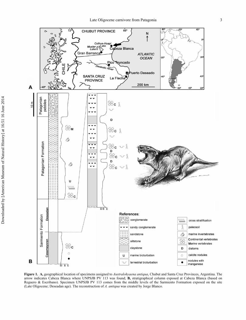
Figure 1. A, geographical location of specimens assigned to Australohyaena antiqua, Chubut and Santa Cruz Provinces, Argentina. Thearrow indicates Cabeza Blanca where UNPSJB PV 113 was found; B, stratigraphical column exposed at Cabeza Blanca (based onReguero & Escribano). Specimen UNPSJB PV 113 comes from the middle levels of the Sarmiento Formation exposed on the site(Late Oligocene; Deseadan age). The reconstruction of A. antiqua was created by Jorge Blanco.
Late Oligocene carnivore from Patagonia 3
Dow
nloa
ded
by [
Am
eric
an M
useu
m o
f N
atur
al H
isto
ry]
at 1
6:51
16
June
201
4
-hyaena refers to the massive aspect of the skull, similar to
living African hyaenids; also a common ending for large
sized sparassodonts.
Australohyaena antiqua (Ameghino, 1894)
(Figs 2�6)
Diagnosis. Large Borhyaenidae characterized by a mas-
sive skull, short rostrum and wide palate. It differs from
other borhyaenids (Borhyaena and Arctodictis) by having
nasals extended beyond the level of the postorbital pro-
cess, jugal extended more anterior than the lacrimal, post-
orbital process poorly developed and formed by the nasal
and frontal, distinct preglenoid process of the squamosal,
squamous part of the squamosal pierced by large foramina
for emissary veins, dentary with coronoid process
markedly inclined (anterior coronoid crest at 132� from
the alveolar line); abrupt change in size between p1�p2
and p3; upper molars with broader stylar shelf on M3 and
ectocingulum extending backwards up to the level of the
metacone, M4 with three roots; lower molars with better
developed talonid and distinct metaconid on m2�m4 sep-
arated from the talonid.
Referred specimens. MACN A 52-384, isolated left m3;
FMNH P13633, crown of right lower canine; FMNH
P13800, isolated right M3; UNPSJB PV 113, rostrum,
basicranium, both dentaries, and dentition, except left i2,
right i4 and right p1.
Occurrence. The type has no specific provenance. Mar-
shall (1978) mentioned that MACN A 52-532 comes from
‘capas de Pyrotherium’, Chubut Province; although in the
same year Patterson & Marshall (1978) tentatively consid-
ered the occurrence from La Flecha, Santa Cruz Province.
These data disagree with those in a revised catalogue of
the MACN, where the locality of Punta Nava is tentatively
suggested. MACN A 52-384 is labelled ‘Colhuapi
Pyroth’, which means the Deseadan horizon at Gran Bar-
ranca, Sarmiento Formation, Chubut Province (Patterson
& Marshall 1978); FMNH P13633, La Flecha locality,
Santa Cruz Province, Sarmiento Formation; FMNH
P13800, Pico Truncado locality, Santa Cruz Province,
Sarmiento Formation; UNPSJB PV 113, Cabeza Blanca
locality, Chubut Province, Sarmiento Formation. Desea-
dan SALMA, Late Oligocene (Fig. 1A, B).
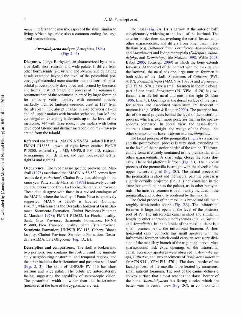
Description and comparisons. The skull is broken into
two portions; one contains the rostrum and the immedi-
ately neighbouring postorbital and temporal regions, and
the other includes the basicranium and posterior skull roof
(Figs 2, 3). The skull of UNPSJB PV 113 has short
rostrum and wide palate. The orbits are anterolaterally
facing, suggesting the capability of stereoscopic vision.
The postorbital width is wider than the basicranium
(measured at the base of the zygomatic arches).
The nasal (Fig. 2A, B) is narrow at the anterior half,
conspicuously widening at the level of the lacrimal. The
anterior border does not overhang the narial fossae, as in
other sparassodonts, and differs from other basal meta-
therians (e.g. Deltatheridium, Peradectes, Andinodelphys
and Mayulestes) and living marsupials (Didelphis, Mono-
delphys and Dromiciops) (de Muizon 1998; Wible 2003;
Babot 2005; Forasiepi 2009) in which the bone extends
forwards. At the level of the contact with the maxilla and
the lacrimal, the nasal has one large nutrient foramen at
both sides of the skull. Specimens of Callistoe (PVL
4187), Arminiheringia (MACN A 10970) and Borhyaena
(PU YPM 15701) have a small foramen in the mid-dorsal
part of one nasal. Borhyaena (PU YPM 15120) has two
foramina in the left nasal and one in the right (Sinclair
1906, lam. 43). Openings in the dorsal surface of the nasal
for nerves and associated vasculature are frequent in
mammals (e.g. Wible & Rougier 2000). The posterior bor-
der of the nasal projects behind the level of the postorbital
process, which is even more posterior than in the sparas-
sodonts compared. In dorsal view, the nasal�frontal
suture is almost straight; the wedge of the frontal that
other sparassodonts have is absent in Australohyaena.
The facial process of the premaxilla (Fig. 2A) is narrow
and the posterodorsal process is very short, extending up
to the level of the posterior border of the canine. The para-
canine fossa is entirely contained in the premaxilla, as in
other sparassodonts. A sharp edge closes the fossa dor-
sally. The narial platform is broad (Fig. 2B). The alveolar
process of the premaxilla is straight with the alveoli of the
upper incisors aligned (Fig. 2C). The palatal process of
the premaxilla is short and the medial palatine process is
slightly dorsally projected (i.e. it is not contained in the
same horizontal plane as the palate), as in other borhyae-
nids. The incisive foramen is oval, mostly included in the
premaxilla, and posteriorly bordered by the maxilla.
The facial process of the maxilla is broad and tall, with
roughly semicircular shape (Fig. 2A). The infraorbital
foramen is large and opens at the level of the posterior
root of P3. The infraorbital canal is short and similar in
length to other short-snout borhyaenids (e.g. Borhyaena
and Arctodictis). In the left side of the maxilla, there is a
small foramen below the infraorbital foramen. A short
horizontal canal connects this small aperture with the
infraorbital foramen which could carry an accessory divi-
sion of the maxillary branch of the trigeminal nerve. Most
sparassodonts lack extra openings of the infraorbital
canal; accessory apertures were observed in Arminiherin-
gia, Callistoe, and two specimens of Borhyaena tuberata
(MACN 9341, YPM PU 15701). The dorsal border of the
facial process of the maxilla is perforated by numerous,
small nutrient foramina. The root of the canine defines a
convex surface that almost reaches the dorsal border of
the bone. Australohyaena has flaring cheeks, which are
better seen in ventral view (Fig. 2C), in common with
4 A. M. Forasiepi et al.
Dow
nloa
ded
by [
Am
eric
an M
useu
m o
f N
atur
al H
isto
ry]
at 1
6:51
16
June
201
4
Figure 2. Australohyaena antiqua, UNPSJB PV 113, Late Oligocene, Deseadan, Cabeza Blanca, Chubut, Argentina; fragment of thesnout in: A, lateral; B, dorsal; and C, ventral views. Abbreviations: Al, alisphenoid; ef, ethmoidal foramen; fap, facial process of pre-maxilla; Fr, frontal; iof, infraorbital foramen; ioc, infraorbital canal; inf, incisive foramen; Ju, jugal; La, lacrimal; mpf, minor palatineforamen; mpp, medial palatine process; mpps, medial postpalatine spine; mt, maxillary tuberosity; Mx, maxilla; Na, nasal; nf, nutrientforamen; npl, narial platform; opl, orbital platform; p, pit for reception of protoconid; Pal, palatine; palf, palatine foramen; parf, paraca-nine fossa; Pmx, premaxilla; spf, sphenopalatine foramen; tl, temporal line. Scale bar: 5 cm.
Late Oligocene carnivore from Patagonia 5
Dow
nloa
ded
by [
Am
eric
an M
useu
m o
f N
atur
al H
isto
ry]
at 1
6:51
16
June
201
4
other sparassodonts (e.g. Sipalocyon, Arminiheringia and
Borhyaena). The palatal process of the maxilla (Fig. 2C)
is short and wide. The major palatine foramen is absent;
several foramina disperse on the palatal surface and would
carry branches of the major palatine nerve and artery, as
probably occurred in other sparassodonts (Babot 2005;
Forasiepi 2009). There are three major pairs of foramina:
the first is the largest and is situated at the level of the pos-
terior border of the canine, the second is at the level of P2,
and the third is at the level of the anterior root of P3. The
exact position and size of the apertures vary between the
right and left maxilla. There is another foramen in the left
palate at the level of the posterior root of M3. Additional
small foramina are dispersed over the palatal surface. The
minor palatine foramen is partially broken, but deduced
from the bases it was small, with the ventral bridge
formed by the palatine and the maxilla, again, common to
most other sparassodonts (de Muizon 1998; Babot 2005;
Forasiepi 2009). There is a deep dental pit between M3
and M4 that houses the protoconid of the m4 in occlusion.
A shallow pit exists between M2 and M3. The orbital plat-
form (Fig. 2B) is triangular, with shallow grooves and
small alveolar foramina (Fig. 2B).
The zygomatic arch is broken on both sides of the skull
(Fig. 2A, B), preserving the anterior base formed by the
maxilla and the jugal (the latter present only in the left
side of the skull) and a small portion of the posterior base
formed by the squamosal. The suture between the maxilla
and the jugal is oblique and at a shallower angle to
Borhyaena and Arctodictis in which it is nearly vertical
(Forasiepi 2009). The jugal extends more anteriorly than
the lacrimal: when tracing a perpendicular line, the jugal
extends up to the level of M2, whereas the lacrimal does
up to the level of M3.
In ventral view (Fig. 2C), the horizontal process of the
palatine exposes on the third posterior portion of the pal-
ate. It extends anteriorly up to the level of the anterior root
of M2, describing an opened ‘W’, and posteriorly behind
the posterior border of M4. The ventral border of the
choana is thick and biconcave, with a blunt postpalatine
spine. In common with other sparassodonts, the palatine
torus, which is present in basal metatherians (Mayulestes,
Pucaldelphys and Andinodelphys; de Muizon 1998, 1999)
and living marsupials (Monodelphys, Dromiciops and
Dasyurus; de Muizon 1999; Wible 2003; Forasiepi 2009),
is absent in Australohyaena. There is a small contribution
of the palatine to the medial wall of the orbit and floor of
the infraorbital canal by means of the perpendicular pro-
cess (Fig. 2A). The sphenopalatine foramen is oval and
located at the joint between the floor and wall of the orbit.
On the right palatine, posterior to the sphenopalatine fora-
men and at the level of its dorsal border, there is a small
aperture that probably corresponds to a nutrient foramen.
The facial process of the lacrimal is crescent-shaped
and almost restricted by the orbital rim. The lacrimal
tubercle is damaged, but its base suggests that it was
prominent, similar to other borhyaenids (Babot et al.
2002; Babot 2005; Forasiepi 2009). The lacrimal foramen
is single and opens inside the orbit. The contribution of
the lacrimal to the orbital wall is larger than in other
borhyaenids (e.g. Arctodictis). The orbital surface is kid-
ney-shaped with a frontal wedge interposed. In other spar-
assodonts, the posterior border of the lacrimal is mostly
concave.
In dorsal view (Fig. 2B), the frontal is slightly convex.
The postorbital process is blunt and poorly developed in
comparison to other sparassodonts, and involves the nasal
and frontal, unlike other sparassodonts, where it involves
only the frontal (Sipalocyon, Cladosictis, Prothylacynus,
Callistoe, Arctodictis). The temporal lines are short and
restricted to the area near the midline of the skull. The
two temporal lines join to form the sagittal crest, which is
weak at its anterior base. In lateral view (Fig. 2A) the
sphenorbital fissure is large and posteriorly bordered by
the alisphenoid. The ethmoidal foramen is small, anterior
and dorsal to the sphenorbital fissure. The orbitotemporal
crest is weak and only evident in the anterodorsal neigh-
bouring area of the sphenorbital fissure, running oblique
towards the postorbital process. In other sparassodonts,
the crest is sharper and the division between the orbit and
the temporal fossa is more evident.
The fragment that contains the basicranium consists of
part of the parietal and 6 or interparietal, alisphenoid, squa-mosal, basisphenoid, occipital bones, and a fragment of
the right petrosal. In dorsal view (Fig. 3A) the skull roof
is formed by one bone, which could be the parietal, the
interparietal, or both. In some other sparassodonts (e.g.
Sipalocyon and Cladosictis) these two bones are separated
by a suture, but in other taxa (e.g. borhyaenids, Callistoe)
only one bone is observed that could represent the fusion
of the parietal with the interparietal (de Muizon 1999;
Forasiepi 2009). This condition is found in several mam-
mals (Koyabu et al. 2012). In UNPSJB PV 113, the entire
bone surface has deep rugosities that suggest the attach-
ment of a strong temporal musculature. Although the sag-
ittal crest is broken at its base, the robust section of the
remains is in accordance with well-developed temporal
musculature.
The squamosal is partially preserved in UNPSJB PV
113. The squamous part is dorsally extended, reaching
almost to the skull roof (Fig. 3A). There are two large
foramina and an extra smaller one on the right side of the
skull. These apertures are referred as emissary foramina,
interpreted as associated with the venous drainage from
the temporal fossa (Hiatt & Gartner 1987). Similar open-
ings are present in Paraborhyaena and Thylacosmilus
(Petter & Hoffstetter 1983; Babot 2005). There are, in
addition, weak grooves that radiate from the foramina that
probably are the impression of the soft tissues that these
apertures conveyed. On the right side of the skull and
6 A. M. Forasiepi et al.
Dow
nloa
ded
by [
Am
eric
an M
useu
m o
f N
atur
al H
isto
ry]
at 1
6:51
16
June
201
4
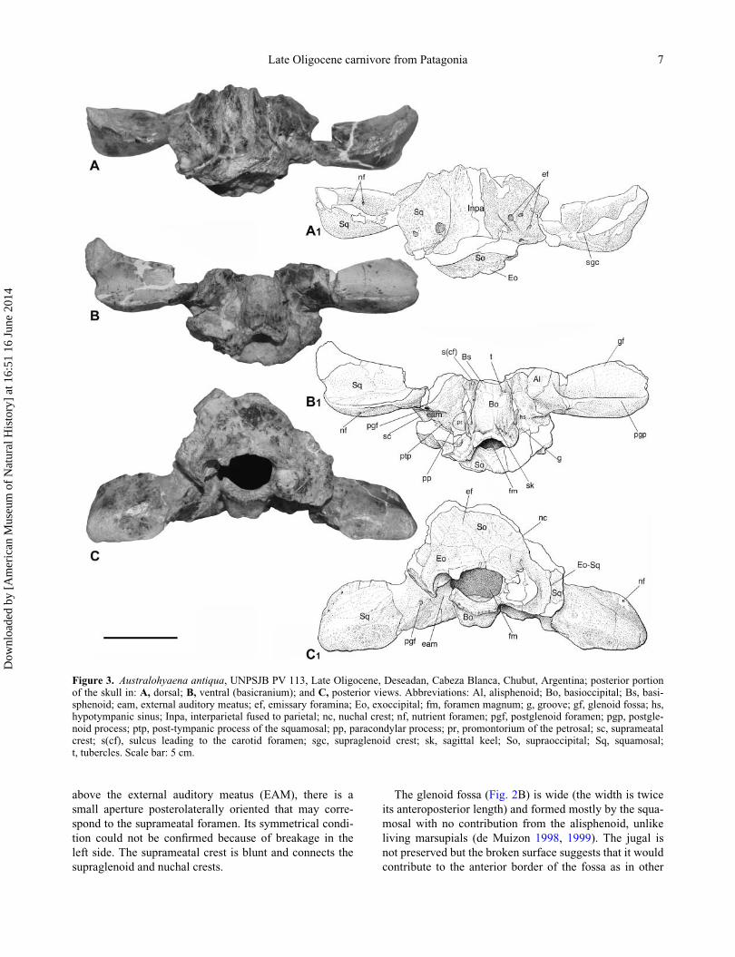
above the external auditory meatus (EAM), there is a
small aperture posterolaterally oriented that may corre-
spond to the suprameatal foramen. Its symmetrical condi-
tion could not be confirmed because of breakage in the
left side. The suprameatal crest is blunt and connects the
supraglenoid and nuchal crests.
The glenoid fossa (Fig. 2B) is wide (the width is twice
its anteroposterior length) and formed mostly by the squa-
mosal with no contribution from the alisphenoid, unlike
living marsupials (de Muizon 1998, 1999). The jugal is
not preserved but the broken surface suggests that it would
contribute to the anterior border of the fossa as in other
Figure 3. Australohyaena antiqua, UNPSJB PV 113, Late Oligocene, Deseadan, Cabeza Blanca, Chubut, Argentina; posterior portionof the skull in: A, dorsal; B, ventral (basicranium); and C, posterior views. Abbreviations: Al, alisphenoid; Bo, basioccipital; Bs, basi-sphenoid; eam, external auditory meatus; ef, emissary foramina; Eo, exoccipital; fm, foramen magnum; g, groove; gf, glenoid fossa; hs,hypotympanic sinus; Inpa, interparietal fused to parietal; nc, nuchal crest; nf, nutrient foramen; pgf, postglenoid foramen; pgp, postgle-noid process; ptp, post-tympanic process of the squamosal; pp, paracondylar process; pr, promontorium of the petrosal; sc, suprameatalcrest; s(cf), sulcus leading to the carotid foramen; sgc, supraglenoid crest; sk, sagittal keel; So, supraoccipital; Sq, squamosal;t, tubercles. Scale bar: 5 cm.
Late Oligocene carnivore from Patagonia 7
Dow
nloa
ded
by [
Am
eric
an M
useu
m o
f N
atur
al H
isto
ry]
at 1
6:51
16
June
201
4
sparassodonts. The glenoid fossa is limited by the pregle-
noid and postglenoid processes of the squamosal anteri-
orly and posteriorly, respectively. Dissimilar from other
sparassodonts (e.g. Borhyaena, Arctodictis), the pregle-
noid process of the squamosal is tall, only slightly lower
than the postglenoid process. The postglenoid process is
half the length of its width, in common with other
borhyaenids (e.g. Cladosictis, Prothylacynus, Borhyaena,
Fredszalaya and Callistoe). In dorsal view and dorsal to
the glenoid fossa, the surface of the squamosal has three
principal depressions that suggest the attachment of the
strong temporal musculature, as in Callistoe and Parabo-
rhyaena (Babot 2005). On the left side of the skull, there
are two small apertures that communicate with each other
by a superficial canal and probably are accessory aper-
tures related to the venous drainage of the temporal mus-
culature. In posterior view (Fig. 3C), a small foramen
opens at the lateral angle of the postglenoid process. Simi-
lar openings were observed in the squamosal of living
marsupials (unnamed foramen in Wible 2003).
The EAM is deep, wide, and enclosed between the post-
glenoid and post-tympanic processes. The anterior wall of
the meatus is pierced by a large postglenoid foramen,
located at the level of the medial border of the glenoid
fossa. This position is common in most sparassodonts
with the exception of some taxa (e.g. Lycopsis and Honda-
delphys) in which it occupies a more lateral position. The
post-tympanic process of the squamosal is robust and rises
together with the paracondylar process of the exoccipital.
The hypotympanic sinus is a broad cavity excavated in
the alisphenoid, squamosal and petrosal (Fig. 3B). The
petrosal position in the basicranium is almost vertical,
with the main axis only slightly inclined in the anteroven-
tral�posterodorsal direction. This is also the condition in
Paraborhyaena and Callistoe (Petter & Hoffstetter 1983;
de Muizon 1999; Babot et al. 2002). The general aspect of
the petrosal is elongated, more in common with the speci-
men AMNH 29591 referred as cf. Pharsophorus sp. than
with Borhyaena and Arctodictis, which are shorter and
more compact. The promontorium is bulbous, even more
than in cf. Pharsophorus. There is a low crest-like rostral
tympanic process pointing forward, similar to all known
sparassodonts, instead of the vertical process described
for living didelphids (Wible 2003; Ladev�eze & de Muizon
2010). In cerebellar view, the internal acoustic meatus is
slightly more oval than in cf. Pharsophorus, and the sub-
arcuate fossa is slightly shallower than in the aforemen-
tioned specimen.
A small portion of the alisphenoid contributing to the
ear region is conserved in the specimen UNPSJB PV 113.
The suture with the squamosal is nearly vertically and
slightly medially oblique to the hypotympanic sinus. The
foramen ovale is represented by a notch at the anterior
border of the hypotympanic sinus (the posterior border is
incomplete), described in some large size borhyaenoids
(e.g. Fredszalaya, Callistoe, Borhyaena and Arctodictis).
There is no evidence of an alisphenoid tympanic process,
neither of the transverse foramen, assuming that both
structures were absent in Australohyaena, again similar to
some borhyaenoids. The paracondylar process of the
exoccipital is slightly anteriorly inclined. There is an
accessory sinus developed directly posterior to the petro-
sal (Fig. 3C).
The anterior border of the exoccipital, medial to the
paracondylar process, has a small notch through which
the jugular vein and accompanying nerves and vessels
would pass. The jugular foramen primarily develops
between the exoccipital and the petrosal (Wible 2003).
According to the position of the petrosal, the primary jug-
ular foramen would open deep inside the basicranium.
The notch seen in ventral view is the outer expression of
the aperture, similar to Arctodictis, Borhyaena (Forasiepi
2009) and Callistoe (Babot 2005).
The basioccipital is nearly rhomboidal (Fig. 3B). There
is a low sagittal keel which separates two shallow fossae,
and anteriorly a pair of tubercles, probably related to the
attachment of the muscles rectus capitis ventralis and lon-
gus capitis (Evans & Christensen 1979). In lateral view,
the borders of the basioccipital are thick and have a deep
sulcus that runs almost horizontally. The sulcus leads to
the carotid foramen (e.g. Callistoe and Borhyaena), but
this foramen is not preserved in UNPSJB PV 113.
The occiput (Fig. 3C) is formed by the supraoccipital,
exoccipital and squamosal. The mastoid portion of the
petrosal is excluded from the occiput, in common with
other sparassodonts. There are several small foramina dis-
persed over the posterior surface of the supraoccipital,
with the largest located close to the sagittal plane, which
probably carried emissary veins, connecting with the
intracranial sinuses. The foramen magnum is bordered by
the exoccipital in posterior view and by the basioccipital
in ventral view, as in other sparassodonts (e.g. Prothylacy-
nus, Paraborhyaena and Callistoe). The nuchal crest is
damaged, but the preserved base suggests that it was thick
and robust. The occipital condyles in UNPSJB PV 113 are
not preserved, nor are the foramina associated with them
(i.e. hypoglossal foramina).
Endocranium. The contribution of the parie-
tal�interparietal bones to the posterior skull roof is signif-
icant in the internal part of the skull. The lateral walls of
the endocranium are formed by the squamosal, exoccipi-
tal, and a small contribution from the petrosal. The floor is
formed by the basioccipital. There is a deep cavity on the
posterior sagittal plane, where the vermis of the cerebel-
lum would be housed, similar to Arctodictis, Borhyaena
and Paraborhyaena (Quiroga 1978; Petter & Hoffstetter
1983; Forasiepi 2009). Lateral to this depression there is a
smaller and shallower concavity on each side of the skull,
where the cerebellar hemispheres were housed. The
8 A. M. Forasiepi et al.
Dow
nloa
ded
by [
Am
eric
an M
useu
m o
f N
atur
al H
isto
ry]
at 1
6:51
16
June
201
4
difference in size between the vermis and the cerebellar
hemispheres is clearer in UNPSJB PV 113 than in
Borhyaena, Arctodictis, and apparently Paraborhyaena.
In Borhyaena these depressions are similar in size (Quir-
oga 1978; Forasiepi 2009). Judging by the size of these
fossae and the fossa subarcuata, the cerebellum of
Australohyaena seems to have been larger than the crown
Marsupialia, in common with some basal metatherians
(i.e. Pucaldelphys) (Macrini et al. 2007). A V-shaped
groove emerges from the front of the depression for the
vermis that would correspond to the most posterior
impression of the sagittal sinus and its division into the
transverse sinuses. In the right lateral side of the skull, the
subsquamosal foramen opens close to the ventral end of
the transverse sinus and close to the anterior border of the
petrosal.
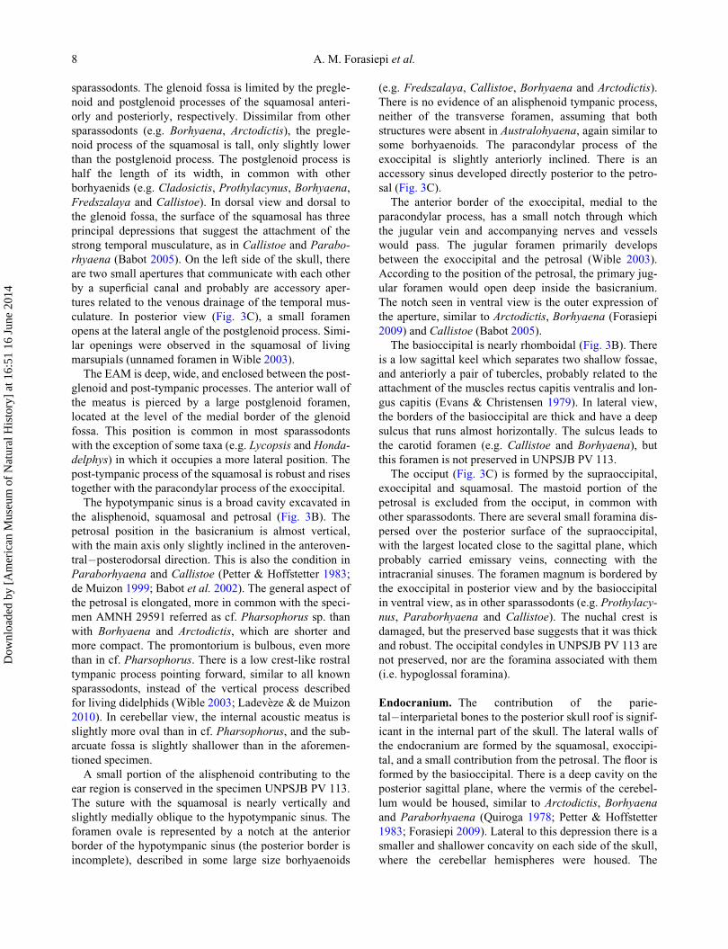
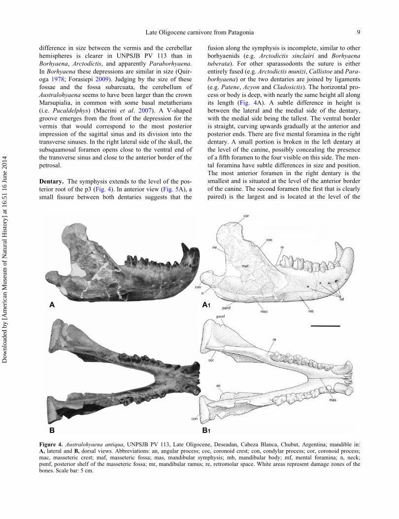
Dentary. The symphysis extends to the level of the pos-
terior root of the p3 (Fig. 4). In anterior view (Fig. 5A), a
small fissure between both dentaries suggests that the
fusion along the symphysis is incomplete, similar to other
borhyaenids (e.g. Arctodictis sinclairi and Borhyaena
tuberata). For other sparassodonts the suture is either
entirely fused (e.g. Arctodictis munizi, Callistoe and Para-
borhyaena) or the two dentaries are joined by ligaments
(e.g. Patene, Acyon and Cladosictis). The horizontal pro-
cess or body is deep, with nearly the same height all along
its length (Fig. 4A). A subtle difference in height is
between the lateral and the medial side of the dentary,
with the medial side being the tallest. The ventral border
is straight, curving upwards gradually at the anterior and
posterior ends. There are five mental foramina in the right
dentary. A small portion is broken in the left dentary at
the level of the canine, possibly concealing the presence
of a fifth foramen to the four visible on this side. The men-
tal foramina have subtle differences in size and position.
The most anterior foramen in the right dentary is the
smallest and is situated at the level of the anterior border
of the canine. The second foramen (the first that is clearly
paired) is the largest and is located at the level of the
Figure 4. Australohyaena antiqua, UNPSJB PV 113, Late Oligocene, Deseadan, Cabeza Blanca, Chubut, Argentina; mandible in:A, lateral and B, dorsal views. Abbreviations: an, angular process; coc, coronoid crest; con, condylar process; cor, coronoid process;mac, masseteric crest; maf, masseteric fossa; mas, mandibular symphysis; mb, mandibular body; mf, mental foramina; n, neck;psmf, posterior shelf of the masseteric fossa; mr, mandibular ramus; re, retromolar space. White areas represent damage zones of thebones. Scale bar: 5 cm.
Late Oligocene carnivore from Patagonia 9
Dow
nloa
ded
by [
Am
eric
an M
useu
m o
f N
atur
al H
isto
ry]
at 1
6:51
16
June
201
4
anterior root of p2. The second pair of mental foramina is
slightly smaller and almost similar to the remaining open-
ings, and is located at the level of the posterior root of p2
in the left dentary and at the junction of p2�p3 on the
right side. The third pair is located at the junction of
p3�m1, and the fourth pair is below the m2. In addition,
in anterior view, there are two pairs of other foramina,
symmetrically located. One pair is set close to the alveolar
border of the incisors (Fig. 5A), and the second pair set
more ventral, in the same vertical line as the first pair. The
retromolar space is nearly as long as the m1. The coronoid
process is high, inclined posteriorly, even more than in
other large sized sparassodonts (e.g. Arctodictis,
Borhyaena and Thylacosmilus). The masseteric fossa is
deep and delimited by protruding coronoid and masseteric
crests. The condyle is cylindrical and is located below the
level of the alveolar border, as in other sparassodonts. The
neck distinguishes as the constriction below the condyle.
The angular process projects ventromedially and forms a
rough triangular shelf (Fig. 4B), which corresponds to the
‘intermediate’ state described by S�anchez-Villagra &
Smith (1997) and characterizes large metatherians, includ-
ing sparassodonts. The mandibular foramen is large and
opens at the middle of the anteroposterior length of the
coronoid process.
Dentition. The dental formula is 3 6 3; 1 6 1; 3 6 3; 4 6 4.The three upper and three lower incisors in Australo-
hyaena are hypothesized to be homologous to I2�4 6i2�4 of other metatherians (Forasiepi 2009). The upper
incisors are crowded and set in a straight line. This condi-
tion is also present in most sparassodonts (e.g. Cladosic-
tis, Prothylacynus, Lycopsis, Borhyaena and Arctodictis).
The incisors increase in size to I4. The I4 is styliform and
slightly laterally curved. It consists of a main cusp lat-
erally followed by a blunt cingulum. The roots of all inci-
sors are stout, notably in the I4, which is bulbous and set
outside the alveolus.
The upper canine is robust and oval in cross section.
The distribution of the enamel in the crown is irregular,
covering more the lateral than the medial surface of the
tooth. The root is surrounded by longitudinal grooves,
mostly distributed on the labial surface. In the lingual
side, there is one principal groove close to the mesial bor-
der of the tooth, which is deeper than the others. The pres-
ence of striations and grooves in the canines is common in
large sparassodonts (e.g. Arctodictis, Borhyaena, Callis-
toe, Paraborhyaena and Proborhyaena). A breakage in
the left side of the maxilla reveals that the canine is deeply
inserted in its alveolus and probably reaches the dorsal
border of the maxilla, as suggested by the convex surface
defined on the maxilla.
The upper premolars are robust with bulbous roots
exposed from their alveoli (Fig. 6A). The P1 is slightly
smaller than the P2, and both are conspicuously smaller
than the P3. The P1 is set oblique in the maxilla; the P2 is
parallel to the anteroposterior axis of the palate, and the
P3 set oblique but opposite to the first premolar. All pre-
molars consist of a main cusp followed by a distal cingu-
lum or heel. The P1�P2 have a short and weakly defined
mesiolingual cingulum. The principal cusp is asymmetri-
cal in the P1 and symmetrical in the P2�P3. There is a
distal cusp in all premolars, larger in P3. The P3 is the
most robust and the largest premolar. The morphology
and the robustness of the P3 of Australohyaena resemble
that of Borhyaena and Arctodictis more than any other
sparassodont (Marshall 1978). The upper molars increase
in size from the M1 to M3, with M4 being the smallest
(Fig. 6A). All molars have three roots, including the M4.
The crowns have strong wear facets obscuring their mor-
phology, in particular in the M1�M2. In the M2, there is
a narrow stylar shelf and an ectocingulum that extends
backwards up to the level of the metacone. This last fea-
ture is also present in other sparassodonts such as
Borhyaena, Arctodictis, Callistoe and Proborhyaena. In
the M3, the metacone is the dominant cusp and occupies
nearly a central position in the tooth. The paracone is coa-
lescent with the metacone (there is no centrocrista). The
protocone is very small and occupies a basal position in
the crown. The stylar shelf is broad and has a similar
width at the mesial and the distal part of the tooth. The
ectocingulum is long, extending up to the level of the
metacone. The ectoflexus is very shallow. The postmeta-
crista is twice longer than the preparacrista. The M4 has a
tall paracone and a small metacone that is coalescent with
the paracone. Other few sparassodonts (Patene, Honda-
delphys and Sallacyon) exhibit a small metacone. The pro-
tocone is even smaller than in previous molars. The
preparacrista is longer than in the M3 and the postmeta-
crista is absent. There is no stylar shelf.
The three lower incisors are tightly packed between the
canines and arranged in a straight line, in the anterior part
of the dentaries (Fig. 5). All the elements are roughly
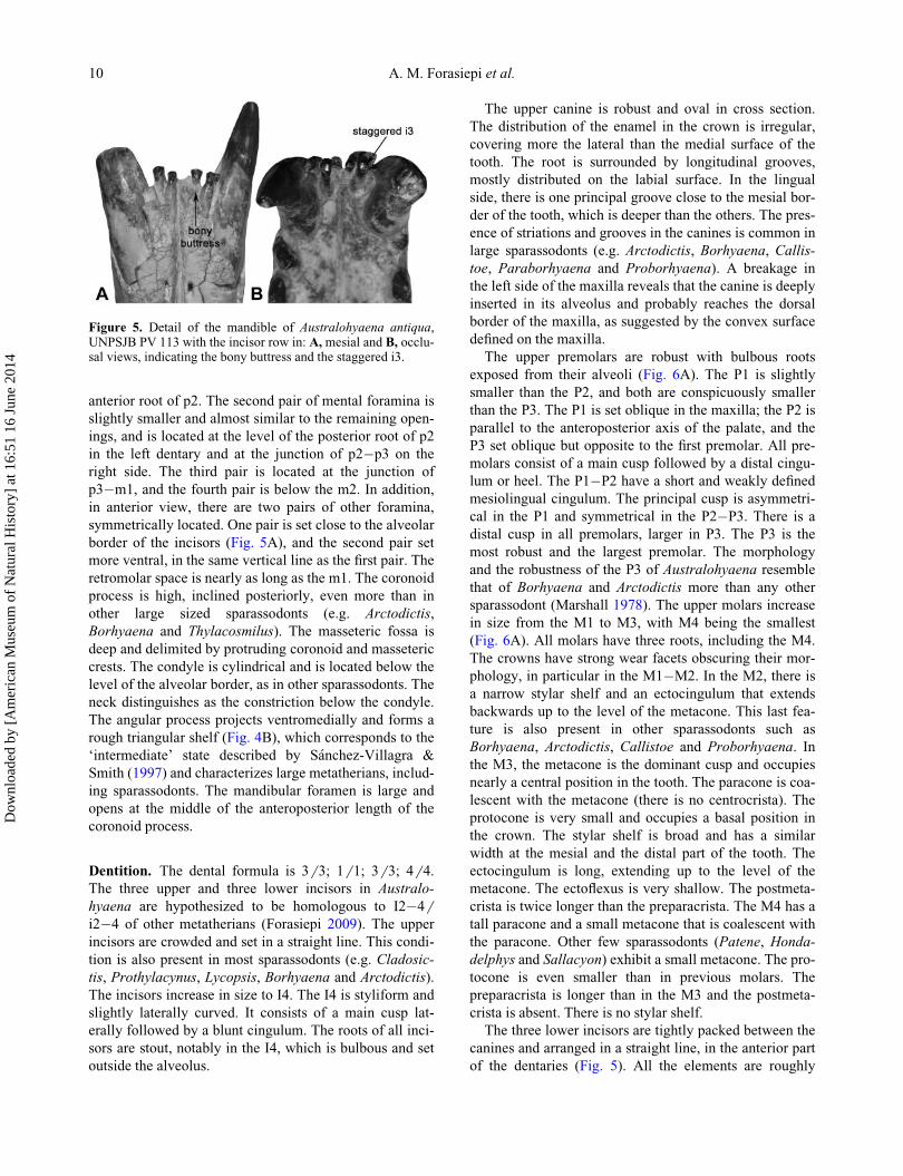
Figure 5. Detail of the mandible of Australohyaena antiqua,UNPSJB PV 113 with the incisor row in: A,mesial and B, occlu-sal views, indicating the bony buttress and the staggered i3.
10 A. M. Forasiepi et al.
Dow
nloa
ded
by [
Am
eric
an M
useu
m o
f N
atur
al H
isto
ry]
at 1
6:51
16
June
201
4
similar in size, with the i2 the smallest. In anterior view,
the crown is triangular and narrower at the base. The i3 is
staggered, in common with almost all other metatherians
(Hershkovitz 1982, 1995), with a bony buttress (Fig. 5).
The lower canines are robust and oval in section with a
bulbous root. The distribution of the enamel is also irregu-
lar, following the pattern of the upper canine. The canine
is almost vertical and resembles many other borhyaenids,
unlike Arminiheringia in which it is procumbent (Simpson
1948; Babot et al. 2002). The root has numerous vertical
grooves, similar to the upper canine, with the deepest
being in the lingual view.
The postcanine row is set slightly laterally oblique,
meaning that when the jaw is seen in dorsal view, the teeth
are leaning labially (Fig. 4B). The premolars are similar in
robustness to the upper premolars and similarly oriented
with the p1 oblique, the p2 parallel to the dentary axis,
and the p3 oblique but opposite to the p1 (Fig. 6B). The
p1 is slightly smaller than the p2, and the p3 is the largest.
The difference in size between the p1�p2 and the p3 is
more evident in the upper premolars. This change in size
is more gradual in Borhyaena and Arctodictis. The crowns
of premolars consist of a principal cusp followed by a
large distal cingulum or heel. The principal cusp is asym-
metrical in the p1�p2 and symmetrical in the p3. There is
a cusp associated to the distal heel in all premolars that is
large and bulbous in the p3. In lateral view, the p3 is
slightly obliquely, distally inclined. This trait was
classically considered a diagnostic feature of the genus
Pharsophorus (Marshall 1978), although it is also present
in Plesiofelis, Borhyaena macrodonta and Arctodictis
sinclairi. The lower molars increase rapidly in size from
the m1 to the m4 and are disposed slightly imbricately
between each other (Fig. 6B). The roots are strong and
parallel, exposed outside their alveoli. The molars consist
of a tall protoconid followed by a paraconid mesially and
a small distal cusp (hypoconid in m1 and metaconid in
m2�m4; see Discussion). In the m1, the three cusps are
aligned, whereas in the posterior elements, the three cusps
form an angle that closes towards the m4. The paraconid
is lower than the protoconid and this difference in height
increases to the back. The precingulid is small in all pre-
molars. The hypoconulid notch is shallow in the m1 and
becomes deeper towards the m4. The talonid is vestigial.
The hypoconulid is small and occupies a median position.
The entocristid is short. The postcingulid is wide, extend-
ing more over the labial than over the lingual side of the
tooth. This condition is very similar to that seen in
borhyaenids such as Borhyaena and Arctodictis.
Discussion
Systematics
Taxonomy. Ameghino in 1894 (p. 655) made a provi-
sional assignation of an isolated canine (the holotype,
Figure 6. Dentition of Australohyaena antiqua. A, B, UNPSJB PV 113; A, left and right upper postcanines in lingual and occlusalviews; B, left lower postcanines in labial, lingual and occlusal views; white areas represent worn zones of the teeth; C, holotype, MACNA 52-532, a nearly complete isolated right upper canine; D, FMNH P13633, right lower canine; E, FMNH P13800, isolated right M3 inlingual and occlusal views; F,MACN A 52-384, isolated left m3 in occlusal, labial and lingual views. Scale bar: 2 cm.
Late Oligocene carnivore from Patagonia 11
Dow
nloa
ded
by [
Am
eric
an M
useu
m o
f N
atur
al H
isto
ry]
at 1
6:51
16
June
201
4

MACN A 52-532; Fig. 6C) to the species ?Borhyaena
antiqua. This material was in a faunistic association nor-
mally not connected to the species of Borhyaena, raising
uncertainties despite strong similarities with the canine of
Borhyaena. Later, Ameghino (1897) recognized the genus
Proborhyaena and proposed the combination P. antiqua
for that isolated piece.
In 1978, Marshall tentatively added into the hypodigm
of antiqua the specimens FMNH 13366, a lower canine;
FMNH 13800, an isolated M3; and MACN A 52-384, an
isolated m3 (Fig. 6D�F). At the same time, Marshall sug-
gested that the species was not Proborhyaena but a
borhyaenid, probably Pharsophorus, combining the
namePharsophorus(?) antiquus.
The lower canine FMNH 13633 (Fig. 6D) has a deep
vertical medial sulcus and extra shallower grooves at the
bases, as in UNPSJB PV 113. The M3 FMNH 13800
(Fig. 6E) has the same size as the M3 of UNPSJB PV 113
and the crown is less worn. The stylar shelf is wide as in
UNPSJB PV 113. The lingual margin has a long ectocing-
ulum and a discontinuous crest with tiny cusps. The proto-
cone is small and there are sharp parallel crests
descending from the paracone and metacone to the proto-
cone. Those features are present in UNPSJB PV 113 but
obscured by wear. The m3 MACN A 52-384 (Fig. 6F) is
slightly smaller than the m3 of UNPSJB PV 113. Both
have a similar morphology, differing in the position of the
metaconid (more lingual in MACN A 52-384) and in the
morphology of the talonid (larger and more protruded in
MACN A 52-384). We consider that these differences are
intraspecific variation. Considering UNPSJB PV 113, we
support the view of Marshall (1978) in recognizing the
specimens FMNH 13366, FMNH 13800 and MACN A
52-384 as part of the hypodigm of the species initially
described by Ameghino (1894) and here determined as
Australohyaena antiqua.
Phylogeny. A cladistic analysis was performed to test the
phylogenetic position of Australohyaena antiqua within
Sparassodonta. The data matrix is based on Forasiepi
(2009) and includes the modifications and additions
recently proposed by Engelman & Croft (2014). New
changes in the scoring were included and are indicated in
the Supplemental Material.
The matrix has 307 morphological characters (107 cra-
nial, 80 dental and 120 postcranial characters) that were
scored for 19 sparassodonts and 20 species in the out-
group, including representatives of the crown group Mar-
supialia and stem marsupials (Supplemental Material).
Forty-nine characters describing a logic sequence were
ordered following Engelman & Croft (2014).
The data matrix was analysed using maximum parsi-
mony (MP) with equally weighted characters and under
implied weighted characters with the computer program
TNT 1.1 (Goloboff et al. 2008a, b). The implied weight
procedure uses evidence on homoplasy to estimate char-
acter reliability. The trees are constructed based on maxi-
mum reliable characters and have a better total fit
(Goloboff 1993; Goloboff et al. 2008a).
Thylacinus cynocephalus was excluded because this
taxon was grouped in all the trees with the Sparassodonta
(see also Forasiepi 2009 and Engelman & Croft 2014).
Thylacinus is a Dasyuromorphia as supported by molecu-
lar analysis (e.g. Thomas et al. 1989; Krajewski et al.
1992, 1997) and the anatomy of the tarsal bones (Szalay
1982, 1994). The inclusion of Thylacinus within sparasso-
donts in our tree is based on homoplasies.
The equally weighted MP analysis was conducted by
searching Wagner trees with 500 random addition sequen-
ces, followed by tree bisection reconnection (TBR), and
saving 10 trees per round. The analysis resulted in five
most parsimonious trees of 1022 steps (consistency index
[CI] D 0.373, retention index [RI] D 0.676). The strict
consensus and Bremer index of the nodes are shown in
Fig. 7A. The list of synapomorphies is in the Supplemen-
tal Material. The consensus tree has two major divisions
for the Sparassodonta, the Hathliacynidae and Borhyae-
noidea, and stem taxa. The internal arrangement of hath-
liacynids is different among the five resulting trees and
this is shown as a polytomy in the consensus. In addition,
no agreement is found by comparing the results of differ-
ent authors (e.g. de Muizon 1999; Babot et al. 2002;
Babot 2005; Forasiepi et al. 2006; Forasiepi 2009;
Engelman & Croft 2014), suggesting that the hathliacynid
relationships deserves a particular analysis. Borhyaenidae
(including Australohyaena antiqua) and Thylacosmilidae
are represented in our analysis as monophyletic groups
(Fig. 7A). Conversely, Proborhyanidae is not shown as
monophyletic, with Callistoe and Paraborhyaena placed
as successive stem taxa of Thylacosmilidae. The recogni-
tion of Proborhyaenidae as a monophyletic group is still a
controversial issue. Some recent studies have recorded
them as monophyletic (e.g. Babot et al. 2002; Engelman
& Croft 2014), although the most exhaustive analysis that
included several species of this group recorded them as
paraphyletic (Babot 2005). Pharsophorus lacerans is
placed as the sister taxon of the monophyletic group
formed by Borhyaenidae, Thylacosmilidae, and the para-
phyletic Proborhyaenidae.
The analysis under implied weights (K D 3) provided
one tree with fit D 110.16 (Fig. 7B). The principal differ-
ence in the sparassodont relationships comparing the trees
under equal and implied weighted characters is the
arrangement and position of proborhyaenids. Under
implied weights, Callistoe and Paraborhyaena are sister
taxa, supporting the monophyly of Proborhyaenidae. Pro-
borhyaenidae is placed as the sister taxon of Borhyaenidae
and Thylacosmilidae, similar to the arrangement obtained
by Engelman & Croft (2014) with MP under equal
weighted characters. By constraining this arrangement of
12 A. M. Forasiepi et al.
Dow
nloa
ded
by [
Am
eric
an M
useu
m o
f N
atur
al H
isto
ry]
at 1
6:51
16
June
201
4
the groups in our five MP trees with equal weighted char-
acters, only a single extra step (length 1023) is required to
obtain Proborhyaenidae as monophyletic and basal to
Borhyaenidae and Thylacosmilidae. As previously
observed, the monophyletic nature of the Proborhyaenidae
requires clarification in future analyses.
Because the homologies of the lower molar talonid
cusps within Borhyaenidae has been subject of different
interpretations (Marshall 1978; Goin et al. 2007; Forasiepi
2009), a second analysis was performed re-coding as
uncertain (?) the features related to the entoconid, hypoco-
nulid, hypoconid, metaconid and associated crests for the
m1�m4 in Borhyaenidae. The characters involved in the
re-coding were 169, 176�180, 182, 183 and 184 in
Borhyaena tuberata, Arctodictis munizi, Arctodictis sin-
clairi and Australohyaena antiqua. The MP analysis
under equally weighted characters resulted in five most
parsimonious trees of 1021 steps (CI D 0.373; RI D0.674). These trees have the same topologies as those
originally obtained with the characters coded for
Borhyaenidae. The strict consensus has the same topol-
ogy, except Borhyaenidae has a support of 1 instead of 2
as in the original results (Supplemental Material). In addi-
tion, the analysis under implied weights (K D 3) provided
one tree with fit D 110.01 that has the same topology as
the tree obtained in the original analysis. The re-coding of
those lower molar characters as uncertain has not influ-
enced the topology of the resultant tree. Our primary
homology can be supported.
Considering the analyses, the metaconid in the Sparas-
sodonta was lost in the common ancestor of hathliacynids
and borhyaenoids. This cusp is registered again in the
m2�m4 in Pharsophorus lacerans and Borhyaenidae
(character length: 3). In Arctodictis and some specimens
of Borhyaena the m4 metaconid is lost (see Discussion).
In addition, sparassodonts have a small or non-existence
entoconid, a reduction of the talonid dimension and asso-
ciated structures. This is particularly stressed in borhyae-
nids, thylacosmilids and proborhyaenids. The alternative
view of considering the posterior trigonid cusp of
Figure 7. Phylogenetic trees. A, strict consensus cladogram of five trees obtained with equally weighted characters (1022 steps, CI D0.373, RI D 0.676); numbers at nodes indicate Bremer support values; B, single tree obtained with implied weighted characters (K3; fitD 110.16).
Late Oligocene carnivore from Patagonia 13
Dow
nloa
ded
by [
Am
eric
an M
useu
m o
f N
atur
al H
isto
ry]
at 1
6:51
16
June
201
4
borhyaenids homologous to an enlarged entoconid seems
unlikely in the context of their molar morphology (see
Discussion).
In all the analyses, Australohyaena antiqua was
grouped with Borhyaena and Arctodictis within Borhyae-
nidae. The monophyletic Borhyaenidae was recorded in
all the phylogenetic trees. Australohyaena antiqua is
clearly separated from Pharsophorus lacerans and the lat-
ter is located basal to borhyaenids, thylacosmilids and
proborhyaenids (Fig. 7A, B). Our interpretation of the tax-
onomy, i.e. the recognition of a new genus for antiqua to
distinguish this taxon from Pharsophorus lacerans with
different generic names, is based on the phylogenetic
analysis. Finally, in all our trees UF 27881, recently
described by Engelman & Croft (2014), appears as a
Borhyaenoidea and this arrangement differs from the
analysis recently presented which locates this taxon as a
plesion Sparassodonta. The support we obtained for the
node that includes UF 27881 and other parts of the tree is
low (Fig. 7A). Alternative arrangements are possible with
recoding characters and adding new taxa.
PalaeoecologyIn living mammals, the niche of carnivory is subdivided
into three categories (hypercarnivores, mesocarnivores
and hypocarnivores) according to the proportion of meat
included in the diet (Van Valkenburgh 1988, 1989).
The hypercarnivorous diet consists of more than 70% of
vertebrate material (Van Valkenburgh 1999) and taxa are
arranged in three ecomorphs. The cat-like type has a short
snout, larger and vertical incisors, elongated and laterally
compressed canines, shearing premolars, carnassial
molars without talonids and reduced post-carnassial denti-
tion (Van Valkenburgh 2007). The jaw is deeper at the
level of the carnassials. The hyaena-like type or bone-
cracker has a short snout, robust sagittal crest, and vaulted
skull (Werdelin 1989). The premolars are robust, conical,
and located where the bite force is maximal (Werdelin
1987, 1989). As a consequence, the jaw has a similar
depth below the premolars and carnassials (Werdelin
1987). The wolf-like type or bone-crusher shows a long
and wide snout, narrow premolars, and talonids in the car-
nassials with one cusp which is blade-like, forming a
‘trenchant-heel’ (Van Valkenburgh 2007). Contrary to the
highly specialized hypercarnivores, the mesocarnivores
and hypocarnivores have more uniform morphology. The
mesocarnivore diet consists of between 50% and 70% of
vertebrate material (Van Valkenburgh 1999). The snout is
larger than in hypercarnivores, the jaws are shallower,
and the carnassial lower molars have talonids with two
cusps (Van Valkenburgh 1999). Low grinding activity
occurs in the carnassials, giving great versatility in the
item consumption. Hypocarnivores, or omnivores, have a
diet that consists of about 30% of vertebrate material and
the remainder is mainly composed of invertebrates and
fruits (Van Valkenburgh 1999, 2007). Their skull and den-
tition is more generalized. They have well-developed post
carnassial molars with broad and cuspated talonids, and
the trigonids have shorter and blunt crests (Van Valken-
burgh 2007). The palaeobiological aspects (body mass
and feeding preferences) of Australohyaena antiqua are
examined in the light of these definitions using morpho-
metric variables.
Body mass. Equations based on tooth measurements
(Gordon 2003) and craniodental measurements (Myers
2001) were used to estimate the body mass of UNPSJB
PV 113 (Table 1). For dental measurements, the best pre-
dictor variables are the length of the second upper molar
(M2L) and the length of the third lower molar (m3L)
(Zimicz 2012). The craniodental variables selected are:
the lower and upper molar occlusal row length (LOMRL
and UOMRL); the total jaw length (TJL), measured from
the base of the first incisor to the posterior border of the
dentary (i.e. condyle); and the posterior jaw length (PJL),
measured from the posterior border of m4 to the posterior
border of the condyle.
The estimation of 67 kg based on m3L for the body
mass of UNPSJB PV 113 is selected because it presents
low values of %PE (12%) and good adjusted R2 (0.95).
The lowest value of %PE (7%) corresponds to the estima-
tion based on M2L (44 kg). This value is probably an
underestimate taking into account that the skull length of
UNPSJB PV 113 is 50% longer than Puma concolor, one
of the largest extant South American carnivores, whose
average body mass is 60 kg (Silva & Downing 1995). The
equations based on craniodental variables have the lowest
values of %SEE, but the resulting body mass is probably
Table 1. Body mass estimation of Australohyaena antiqua (UNPSJB PV 113).
Regression Variable %SEE %PE Adjusted R2 Smearing estimate Body mass (kg)
Log y D �1.098 C 3.35 (log x) UMORL 19 14 0.996 1.2 1241.35
Log y D �1.225 C 3.34 (log x) LMORL 20 16 0.995 1.6 1603.52
Log y D �2.722 C 3.207 (log x) TJL 26 18 0.991 2.3 175.28
Ln y D 1.89 C 3.14 (ln x) M2L 38 7 0.95 1.16 44.13
Ln y D 1.76 C 3.17 (ln x) m3L 39 12 0.95 1.11 67.08
14 A. M. Forasiepi et al.
Dow
nloa
ded
by [
Am
eric
an M
useu
m o
f N
atur
al H
isto
ry]
at 1
6:51
16
June
201
4
overestimated (Table 1) because the specimen UNPSJB
PV 113 is an outlier in the range values of the independent
variables of the equations of Myers (2001).
Diet. Tooth measurements. Five morphometric indexes
were calculated for UNPSJB PV 113. The relative grind-
ing area (RGA) (Van Valkenburgh 1991) is the ratio
between the square root of the talonid area and the length
of the trigonid of the carnassials, and measures the grind-
ing capacity of the molars. The critical values are: RGA
< 0.5 D hypercarnivores; RGA 0.5�0.8 D mesocarni-
vores, and RGA>0.8 D hypocarnivores (Zimicz 2012). In
the case of metatherians, the m4 is used to estimate the
RGA following Prevosti et al. (2013). The RGA for
UNPSJB PV 113 is 0.17 (Table 2), which supports hyper-
carnivorous habits.
The premolar shape (PS) (Van Valkenburgh 1991) is the
ratio between the maximum width and length of the largest
lower premolar. It measures the robustness of the premolar
and its capacity as a cracking or shearing tool. This index
discriminates hypercarnivorous bone-crackers with PS >
0.58 from the remaining carnivorous types with PS < 0.58
(Zimicz 2012). The PS for UNPSJB PV 113 is 0.65
(Table 2) and defines a bone-cracker morphotype.
The relative premolar size (RPS) (Van Valkenburgh
1991) measures the robustness of the premolar consider-
ing the body mass. The index is defined as the ratio
between the maximum width of the largest lower premo-
lar and the cubic root of the estimated body mass. Similar
to PS, this index discriminates hypercarnivorous bone-
crackers with RPS > 2.6 from the remaining carnivorous
types with RPS < 2.6 (Zimicz 2012). The RPS for
UNPSJB PV 113 is 2.32 and groups the specimen with
the cat-like species.
The relative premolar length (RPL) (Van Valkenburgh
1991) measures the size of the largest premolar and the car-
nassial molar. The index is defined as the length of the
largest lower premolar over the length of the carnassial
molar and discriminates between hypercarnivores with
RPL > 0.7 and mesocarnivores and hypocarnivores with
RPL < 0.7 (Zimicz 2012). Contrary to the other indexes,
the RPL value obtained for UNPSJB PV 113 (0.59; Table 2)
suggests mesocarnivorous�hypocarnivorous habits.
The trigonid length of the carnassial molar (TLC) (Van
Valkenburgh 1991) estimates the shearing capacity of the
carnassials and is the length of the trigonid over the total
length of the carnassial molar. The critical values discrim-
inate: hypercarnivorous cat-like with TLC > 0.9;
hypercarnivorous bone-crackers with 0.8 < TLC < 0.9;
mesocarnivores with 0.7 < TLC < 0.8; and hypocarni-
vores with TLC < 0.7 (Zimicz 2012). The TLC for
UNPSJB PV 113 of 0.91 (Table 2) suggests hypercarnivo-
rous cat-like habits, although very close to the boundary
with bone-cracker forms.
Skull measurements. We used the method devel-
oped by Werdelin (1989) to infer the diet on the basis of
the skull morphology. An arc was traced in the lateral
view of the skull and jaw of UNPSJB PV 113, with the
upper and lower molars in occlusion (Fig. 8A). The centre
of the circle was placed in the point of rotation of the
lower jaw (condyle). The radius is equal to the distance
between the condyle and the apex of the cracking tooth
(p3). The arc passes between the infraorbital foramen and
the anteroventral surface of the orbit continuing up to the
skull roof (Werdelin 1989). The arc in UNPSJB PV 113
was almost tangential to the skull roof (Fig. 8A), with an
angle of 14�. This is similar to bone-crackers with vaulted
skulls, such as hyaenas (Crocuta crocuta) and the fossil
bone-cracker canid Osteoborus cyonoides (Werdelin
1989). The arc in C. crocuta and Hyaena brunnea contacts
Table 2. Morphometric indexes used for diet estimation of Australohyaena antiqua (UNPSJB PV 113).
Index 6 variable RGA PS RPS RPL TLC JDM2L(mm)
m3L(mm)
UOMRL(mm)
LOMRL(mm)
TJL(mm)
Talonidtype
Value 0.17 0.65 2.32 0.59 0.91 64.8 16.7 18.7 132.8 145.7 235 I
Figure 8. Skull and jaw measurements of Australohyaena anti-qua. A, lateral view of the anterior portion of the skull showingits vaulted appearance; B, jaw geometry after Werdelin (1989);C, lateral view of the right dentary. Abbreviations: iof, infraorbi-tal foramen; fs, fused symphysis; LC, left condyle; mbfr,maximum bite force region; A, position of the resultant force atthe cracking tooth; B, position of the resultant force at thecarnassials; R, position of the resultant bite force; RC, rightcondyle; JD, jaw depth.
Late Oligocene carnivore from Patagonia 15
Dow
nloa
ded
by [
Am
eric
an M
useu
m o
f N
atur
al H
isto
ry]
at 1
6:51
16
June
201
4
the skull roof at 10� and 15�, respectively (N. Zimicz,
pers. obs.). The vaulted skull has the function to withstand
the compressive forces generated during the bite at the
level of premolars (Buckland-Wright 1978). In the cat-
like and wolf-like hypercarnivores and mesocarnivores,
the arc passes above the skull roof in an obtuse angle
(Werdelin 1989). Panthera species have contact angles
varying from 17� (P. leo) to 28� (P. pardus). Panthera
tigris and P. onca have angles between 25� and 26�
respectively (N. Zimicz, pers. obs.).
Jaw measurements. We used the geometry of the
lower jaw of UNPSJB PV 113 (Fig. 8B) to test the diet
estimations (Greaves 1983, 1985; Werdelin 1987, 1989).
Bone-crackers concentrate the bite force at the level of
the cracking premolars while other carnivores produce the
optimal level of force at the carnassials. The method con-
sists of a series of lines connecting functional points of
the mandibles in occlusal view (Fig. 8B). RC and LC are
the midpoints of the right and left condyles respectively;
A represents the anteriormost position of the jaw muscle
resultant, and B is the point at which a line connecting the
last molar with the opposite condyle crosses the midline
of the jaw (Fig. 8B). The area between the lines RC-A and
RC-B is the zone of maximum muscle force and R is the
expected muscle resultant position at the carnassials. In
UNPSJB PV 113 the maximum force zone is between the
posterior root of p2 and m4 (Fig. 8B), and p3 lies entirely
on the zone of the jaw where the bite force is maximal, as
occurs in extant hyaenas (Werdelin 1989). The use of p3
as a cracking tooth is also supported in the UNPSJB PV
113 by the extension of the mandibular symphysis to the
anterior root of m1. The p3 is included totally over the
fused region of the symphysis, suggesting that heavy
loads can be supported by this area (Fig. 8B).
Bone-crackers have deeper dentaries and smaller differ-
ences in the jaw depth at the cracking premolar and car-
nassial molar than other carnivores (Palmqvist et al.
2011), as is the case in UNPSJB PV 113 (Fig. 8C). The
dentary is 3.5 mm higher at the level of m4 than at the
level of p3. The difference is slightly more stressed at the
lingual side with 7.5 mm difference (measurements in
Supplemental Material). Considering the jaw depth as a
function of the body mass (Fig. 9) and the estimation at
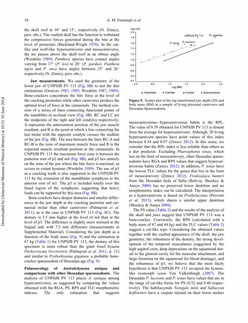
67 kg (Table 1) for UNPSJB PV 113, the dentary of this
specimen is more robust than the giant fossil hyaena
Pachycrocuta brevirostris (Palmqvist et al. 2011, p. 11)
and similar to Proborhyaena gigantea, a probable bone-
cracker sparassodont of Deseadan age (Fig. 9).
Palaeoecology of Australohyaena antiqua and
comparisons with other Deseadan sparassodonts. The
analysis of UNPSJB PV 113 places A. antiqua among
hypercarnivores, as suggested by comparing the values
obtained with the RGA, PS, RPS and TLC morphometric
indexes. The only estimation that suggests
mesocarnivorous�hypocarnivorous habits is the RPL.
The value of 0.59 obtained for UNPSJB PV 113 is distant
from the average for hypercarnivores. Although, 20 living
hypercarnivore species have point values of this index
between 0.54 and 0.97 (Zimicz 2012). In this sense, we
consider that the RPL index is less reliable than others as
a diet predictor. Excluding Pharsophorus tenax, which
lies on the limit of mesocarnivory, other Deseadan sparas-
sodonts have RGA and RPS values that suggest hypercar-
nivorous habits (Zimicz 2012). In addition, P. tenax have
the lowest TLC values for the genus that lies in the limit
of mesocarnivory (Zimicz 2012). Fredszalaya hunteri
from the Deseadan beds of Salla (Bolivia; Shockey &
Anaya 2008) has no preserved lower dentition and no
morphometric index can be calculated. The interpretation
as a hypercarnivore is based on Prothylacynus (Prevosti
et al. 2013), which shares a similar upper dentition
(Shockey & Anaya 2008).
The PS value (Table 2) and the results of the analysis of
the skull and jaws suggest that UNPSJB PV 113 was a
bone-cracker. Conversely, the RPS (calculated with a
body mass of 67 and 44 kg) and the TLC values (Table 2)
suggest a cat-like type. Considering the obtained values
together with the vaulted appearance of the skull, the jaw
geometry, the robustness of the dentary, the strong devel-
opment of the temporal musculature (suggested by the
high sagittal crest, deep depressions on the squamosal dor-
sal to the glenoid cavity for the muscular attachment, and
large foramina on the squamosal for blood drainage), and
the robustness of p3, we believe that the most likely
hypothesis is that UNPSJB PV 113 occupied the hyaena-
like ecomorph sensu Van Valkenburgh (2007). The
Deseadan P. lacerans and P. tenax have values that are in
the range of cat-like forms for PS (0.52 and 0.48 respec-
tively). The hathliacynids Notogale mitis and Sallacyon
hoffstetteri have a cuspate talonid on their lower molars
Figure 9. Scatter plot of the log transformed jaw depth (JD) andbody mass (BM) in a sample of living placental carnivores andDeseadan Sparassodonta.
16 A. M. Forasiepi et al.
Dow
nloa
ded
by [
Am
eric
an M
useu
m o
f N
atur
al H
isto
ry]
at 1
6:51
16
June
201
4
similar to the trenchant heel of wolf-like hypercarnivores
(Zimicz 2012). Proborhyaena gigantea has several fea-
tures of bone-crackers (deep jaw depth, fused symphysis
extending to the p3�m1 joint, and robust cracking premo-
lar with PS D 0.6; Zimicz 2012). The palaeoecological
restoration of Paraborhyaena boliviana is incomplete
because the dentition is eroded for estimating the morpho-
metric indexes. However some morphological traits, such
as the fused symphysis extending to the anterior root of
m1, and the massive aspect of their lower third premolar,
suggest that it was another bone-cracker (Table 3).
The estimated body mass of UNPSJB PV 113 of 67 kg
lies in the range of values of Arctodictis munizi from the
younger Santacrucian beds (Ercoli & Prevosti 2011;
Ercoli et al. 2013). Dental variables are considered poor
estimators of body mass compared to postcranial variables
(Damuth & MacFadden 1990), and the sparassodonts are
no exception (Prevosti et al. 2013). However, the scarcity
of postcranial elements requires dependence on the dental
indexes for the group. Deseadan sparassodonts range from
small to large sizes (Table 3). Sallacyon hoffstetteri and
Notogale mitis were the smallest Deseadan sparassodonts
at about 1 to 3 kg (Zimicz 2012; Prevosti et al. 2013). The
largest was Proborhyaena gigantea at about 100 kg (93 or
153 kg according to Zimicz 2012 and Prevosti et al. 2013,
respectively).
Homology of the lower tooth cusps in
BorhyaenidaeThe occurrence of the metaconid in the lower molars in
Sparassodonta has been discussed for the species of
Borhyaenidae. Marshall (1978) considered that the meta-
conid is present in basal forms, but it is lost in post-Desea-
dan borhyaenids. This idea was questioned by Goin et al.
(2007), who considered that either the cusp posterior to
the protoconid is homologous to the entoconid or the ento-
conid is fused to the metaconid. We support here the inter-
pretation of the posterior cusp being homologous to the
metaconid for the m2�m4 or m2�m3 of Pharsophorus,
Borhyaena, Australohyaena and Arctodictis (see Phylog-
eny section).
Basal sparassodonts such as Patene or Nemolestes show
a primitive molar pattern with complete trigonid and talo-
nid cusps (Fig. 10A). The trigonid is tricuspate, with a
reduced metaconid located at the distolingual corner of
the trigonid. The talonid is wide (as wide as the trigonid),
with the hypoconid, entoconid, hypoconulid and associ-
ated crests. The postcingulid descends from the hypoco-
nulid and surrounds labially the base of the hypoconid.
Pharsophorus lacerans has a more specialized carnivo-
rous dentition. The m1 of Pharsophorus (Figs 10B, 11D)
lacks a metaconid and the trigonid is formed by the para-
conid and protoconid aligned with the mesiodistal axis of
the tooth. The talonid is short, narrower than the base
of the protoconid, basined, and cuspate. The tallest cusp
of the talonid is the hypoconid placed nearly on the sagit-
tal axis of the tooth. The entoconid is small. The hypoco-
nulid is minute, located in a median position in the distal
border of the talonid. The postcingulid descends from the
hypoconulid to the distolabial border of the crown. This
tooth morphology is similar to that of the lower molars of
hathliacynids, Pseudothylacynus and Prothylacynus
(Fig. 11A�C) in that all molars lack a metaconid, possess
a basined three-cuspate talonid, and a well-developed
postcingulid. Unlike the m1, the trigonid of the m2 in
Pharsophorus (Figs 10B, C, 11D) has a small metaconid
located on the distolingual side of the protoconid, resem-
bling basal sparassodonts. The metaconid is a conical
cusp, almost circular in cross section. The postprotocristid
descends from the tip of the protoconid to the metaconid;
there is a carnassial notch between both cusps. The talonid
is slightly wider than long and basined. The surface of the
talonid is lingually inclined (the base of the hypoconid is
lower than the base of the entoconid). The hypoconid is
considerably lower than in the m1. There is a crest that
surrounds the distal border of the talonid connecting the
entoconid and the hypoconid. This crest should include
the hypoconulid, but the cusp is not evident in this tooth.
The entoconid is small, labiolingually compressed,
Table 3. Palaeoecological reconstruction of Deseadan sparassodonts. Palaeobiological inferences were taken from Zimicz (2012) andPrevosti et al. (2013). Palaeoecology of Australohyaena antiqua is discussed in the text. Body mass calculation of Fredszalaya hunteriis in the Supplemental Material.
Taxon Diet Body mass
Notogale mitis Hypercarnivores � wolf-like Small size (»1 to 3 kg)
Sallacyon hoffstetteri Hypercarnivores � wolf-like Small size (»1 to 3 kg)
Fredszalaya hunteri Hypercarnivores Medium size (»10 kg)
Pharsophorus tenax Limit to mesocarnivory Large size (»15 to 20 kg)
Pharsophorus lacerans Hypercarnivores � cat-like Large size (» 30 kg)
Australohyaena antiqua Hypercarnivores � bone-cracker Large size (67 kg)
Proborhyaena gigantea Hypercarnivores � bone-cracker Large size (» 90 to 100 kg)
Paraborhyaena boliviana Hypercarnivores � bone-cracker Large size (» 90 kg)
Late Oligocene carnivore from Patagonia 17
Dow
nloa
ded
by [
Am
eric
an M
useu
m o
f N
atur
al H
isto
ry]
at 1
6:51
16
June
201
4
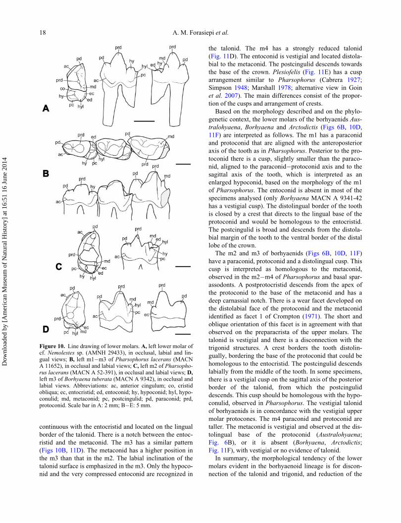
continuous with the entocristid and located on the lingual
border of the talonid. There is a notch between the entoc-
ristid and the metaconid. The m3 has a similar pattern
(Figs 10B, 11D). The metaconid has a higher position in
the m3 than that in the m2. The labial inclination of the
talonid surface is emphasized in the m3. Only the hypoco-
nid and the very compressed entoconid are recognized in
the talonid. The m4 has a strongly reduced talonid
(Fig. 11D). The entoconid is vestigial and located distola-
bial to the metaconid. The postcingulid descends towards
the base of the crown. Plesiofelis (Fig. 11E) has a cusp
arrangement similar to Pharsophorus (Cabrera 1927;
Simpson 1948; Marshall 1978; alternative view in Goin
et al. 2007). The main differences consist of the propor-
tion of the cusps and arrangement of crests.
Based on the morphology described and on the phylo-
genetic context, the lower molars of the borhyaenids Aus-
tralohyaena, Borhyaena and Arctodictis (Figs 6B, 10D,
11F) are interpreted as follows. The m1 has a paraconid
and protoconid that are aligned with the anteroposterior
axis of the tooth as in Pharsophorus. Posterior to the pro-
toconid there is a cusp, slightly smaller than the paraco-
nid, aligned to the paraconid�protoconid axis and to the
sagittal axis of the tooth, which is interpreted as an
enlarged hypoconid, based on the morphology of the m1
of Pharsophorus. The entoconid is absent in most of the
specimens analysed (only Borhyaena MACN A 9341-42
has a vestigial cusp). The distolingual border of the tooth
is closed by a crest that directs to the lingual base of the
protoconid and would be homologous to the entocristid.
The postcingulid is broad and descends from the distola-
bial margin of the tooth to the ventral border of the distal
lobe of the crown.
The m2 and m3 of borhyaenids (Figs 6B, 10D, 11F)
have a paraconid, protoconid and a distolingual cusp. This
cusp is interpreted as homologous to the metaconid,
observed in the m2�m4 of Pharsophorus and basal spar-
assodonts. A postprotocristid descends from the apex of
the protoconid to the base of the metaconid and has a
deep carnassial notch. There is a wear facet developed on
the distolabial face of the protoconid and the metaconid
identified as facet 1 of Crompton (1971). The short and
oblique orientation of this facet is in agreement with that
observed on the preparacrista of the upper molars. The
talonid is vestigial and there is a disconnection with the
trigonid structures. A crest borders the tooth distolin-
gually, bordering the base of the protoconid that could be
homologous to the entocristid. The postcingulid descends
labially from the middle of the tooth. In some specimens,
there is a vestigial cusp on the sagittal axis of the posterior
border of the talonid, from which the postcingulid
descends. This cusp should be homologous with the hypo-
conulid, observed in Pharsophorus. The vestigial talonid
of borhyaenids is in concordance with the vestigial upper
molar protocones. The m4 paraconid and protoconid are
taller. The metaconid is vestigial and observed at the dis-
tolingual base of the protoconid (Australohyaena;
Fig. 6B), or it is absent (Borhyaena, Arctodictis;
Fig. 11F), with vestigial or no evidence of talonid.
In summary, the morphological tendency of the lower
molars evident in the borhyaenoid lineage is for discon-
nection of the talonid and trigonid, and reduction of the
Figure 10. Line drawing of lower molars. A, left lower molar ofcf. Nemolestes sp. (AMNH 29433), in occlusal, labial and lin-gual views; B, left m1�m3 of Pharsophorus lacerans (MACNA 11652), in occlusal and labial views; C, left m2 of Pharsopho-rus lacerans (MACN A 52-391), in occlusal and labial views; D,left m3 of Borhyaena tuberata (MACN A 9342), in occlusal andlabial views. Abbreviations: ac, anterior cingulum; co, cristidobliqua; ec, entocristid; ed, entoconid; hy, hypoconid; hyl, hypo-conulid; md, metaconid; pc, postcingulid; pd, paraconid; prd,protoconid. Scale bar in A: 2 mm; B�E: 5 mm.
18 A. M. Forasiepi et al.
Dow
nloa
ded
by [
Am
eric
an M
useu
m o
f N
atur
al H
isto
ry]
at 1
6:51
16
June
201
4
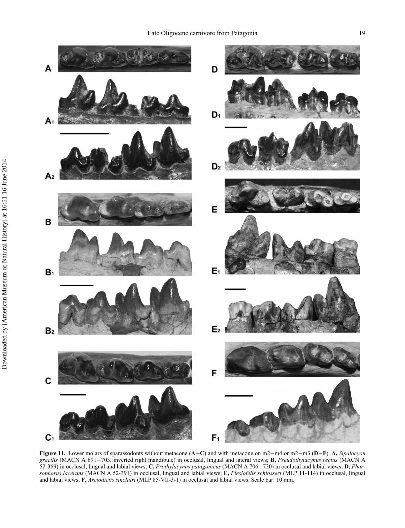
Figure 11. Lower molars of sparassodonts without metacone (A�C) and with metacone on m2�m4 or m2�m3 (D�F). A, Sipalocyongracilis (MACN A 691�703, inverted right mandibule) in occlusal, lingual and lateral views; B, Pseudothylacynus rectus (MACN A52-369) in occlusal, lingual and labial views; C, Prothylacynus patagonicus (MACN A 706�720) in occlusal and labial views; D, Phar-sophorus lacerans (MACN A 52-391) in occlusal, lingual and labial views; E, Plesiofelis schlosseri (MLP 11-114) in occlusal, lingualand labial views; F, Arctodictis sinclairi (MLP 85-VII-3-1) in occlusal and labial views. Scale bar: 10 mm.
Late Oligocene carnivore from Patagonia 19
Dow
nloa
ded
by [
Am
eric
an M
useu
m o
f N
atur
al H
isto
ry]
at 1
6:51
16
June
201
4
talonid in favour of the development of the trigonid. Func-
tionally, the shearing areas of the tooth increase in size,
such as the paracristid and metacristid in the lower and
the postmetacrista and preparacrista in the upper molars.
Grinding areas are vestigial to absent, such as the talonid
in the lower and protocone in the upper molars.
If the interpretations about the homologies supported in
this contribution are correct, there is a clear difference in
tooth patterns between m1 and m2�m4 in the Borhyaeni-
dae. In the phylogenetic context, the presence of the meta-
conid is a plesiomorphic trait, while its absence is the
derived state. The metaconid is present in all the molars in
plesion metatherians and most marsupials. The Sparasso-
donta hathliacynid and some basal borhyaenoids (e.g.
Lycopsis and Prothylacynus) lack the metaconid in the
entire molar series. Borhyaenidae has the metaconid on
all molars except m1, which suggests that among the
lower molars m1 is an evolutionary plastic tooth for
acquiring major morphological change. A similar discrep-
ancy in the morphology of the first molar compared to the
posterior elements has been reported in marsupials. For
example, Sarcophilus lacks the paraconid on the m1 but
this cusp is present in the posterior molars and shifts pos-
teriorly the premolariform�molariform boundary (Archer
1976). If the seven postcanine teeth of eutherians and
metatherians are homologous and the first molar of meta-
therians is homologous to the deciduous premolar
(dP4 6 dp4; Luckett 1993; O’Leary et al. 2013) of Euthe-
ria, then this would result in metatherians presenting dif-
ferent morphological constraints between first and
posterior molars.
Tooth morphology, the presence, absence, arrangement
and sizes of cusps are variables in mammals. In developing
teeth, the enamel knots act as epithelial signalling centres
to control cusp positions and are the first signs of cusp pat-
terns that distinguish species (Kassai et al. 2005; Jernvall
& Thesleff 2012). The gene system regulating the tooth
development is highly complex, but the network organiza-
tion requires a few small changes and simple activa-
tor�inhibitor feedback loops to generate different crown
types and cusp patterns (Kassai et al. 2005; Salazar-Ciudad
& Jernvall 2002, 2010). Homoplasies are frequent in mam-
mals and this makes interpretation of homologies and the
character evolution a controversial issue.
Conclusions
A new taxonomic combination is proposed for Australo-
hyaena antiqua (Ameghino) on the basis of its phyloge-
netic affinities, which places it as member of the
Borhyaenidae (Mammalia, Metatheria, Sparassodonta),
distant from Pharsophorus lacerans, the genus to which it
was formerly referred. A unique combination of features
clearly differentiates A. antiqua from other sparassodonts.
An emended diagnosis is provided considering characters
from the skull, dentary and dentition.
The craniodental characteristics of Australohyaena
antiqua suggest that it was a hypercarnivore predator.
Several features (vaulted skull, well-developed temporal
fossa and consequently temporal musculature, and high
jaw at the carnassial and p3) are suggestive of bone-
cracker capabilities.
The body mass estimation provides a value of about
70 kg and places A. antiqua within the largest Deseadan
sparassodonts.
We support the interpretation that the cusp posterior to
the protoconid is homologous to the hypoconid in the m1
but the cusp posterior to the protoconid in m2�m4 is
homologous to the metaconid in the posterior molars in
Borhyaenidae.
Acknowledgements
We gratefully thank M�onica Abril (UNPSJB) for permit-
ting access to the specimen of Australohyaena antiqua and
her courteous support during the study of the material. We
thank M. Reguero (MLP) and A. Kramarz (MACN), for
permitting access to the collections under their care; Enri-
que Guanuco (Fundaci�on Miguel Lillo) for the drawings,
G. W. Rougier for his guidance on the study of mammal
anatomy and S. D. Kay who kindly assisted by reviewing
the English in early versions of the manuscript. The anony-
mous reviewers and editors provided useful suggestions.
This is a contribution to the project financially supported
by PICT 2010-1805, PICT 2011-0309, PIP 1723 CONI-
CET, and IPA-P.1, Fundaci�on Miguel Lillo. We acknowl-
edge having used the Willi Hennig Society edition of TNT.
Supplemental data
Supplemental material for this article can be accessed
here: http://dx.doi.org/10.1080/14772019.2014.926403
References
Ameghino, F. 1894. Enum�eration synoptique des esp�eces demammif�eres fossiles des formations �eoc�enes de Patagonie.Bolet�ın de la Academia Nacional de Ciencias de C�ordoba,13, 259�452.
Ameghino, F. 1897. Mammif�eres Cr�etac�es de l’Argentine.Deuxi�eme contribution �a la connaissance de la faune mam-malogique des conches �a Pyrotherium. Bolet�ın del InstitutoGeogr�afico Argentino, 18, 406�521.
Archer, M. 1976. The dasyurid dentition and its relationships tothat of didelphids, thylacinids, borhyaenids (Marsupicarni-vora) and peramelids (Peramelina: Marsupialia). AustralianJournal of Zoology, Supplementary Series, 39, 1�34.
20 A. M. Forasiepi et al.
Dow
nloa
ded
by [
Am
eric
an M
useu
m o
f N
atur
al H
isto
ry]
at 1
6:51
16
June
201
4
Babot, M. J. 2005. Los Borhyaenoidea (Mammalia, Metatheria)del Terciario inferior del Noroeste argentino. Aspectos filo-gen�eticos, paleobiol�ogicos y bioestratigr�aficos. UnpublishedPhD thesis, Universidad Nacional de Tucum�an, 454 pp.
Babot, M. J., Powell, J. E. & Muizon, C de. 2002. Callistoevincei, a new Proborhyaenidae (Borhyaenoidea, Metatheria,Mammalia) from the early Eocene of Argentina. Geobios,35, 615�629.
Bond, M. & Pascual, R. 1983. Nuevos y elocuentes restos cranea-nos de Proborhyaena gigantea Ameghino, 1897 (Marsupialia,Borhyaenidae, Proborhyaeninae) de la Edad Deseadense. Unejemplo de coevoluci�on. Ameghiniana, 20, 47�60.
Buckland-Wright, J. C. 1978. Bone structure and the patternsof force transmission in the cat skull (Felis catus). Journalof Morphology, 155, 35�62.
Cabrera, A. 1927. Datos para el conocimiento de los dasyuroi-deos f�osiles argentinos. Revista del Museo de La Plata, 30,271�315.
Cerde~no, E. 2011. Quebrada Fiera (Mendoza), an importantpaleobiogeographic center in the South American late Oligo-cene. Estudios Geol�ogicos, 67, 375�384.
Chaffee, R. G. 1952. The Deseadan vertebrate fauna of the Scar-ritt Pocket, Patagonia. Bulletin of the American Museum ofNatural History, 98, 507�562.
Crompton, A. W. 1971. The origin of the tribosphenic molar.Zoological Journal of the Linnean Society, 50, Supplement1, 65�87.
Damuth, J. & Macfadden, B. 1990. Body size and its estima-tion. Pp. 1�10 in J. Damuth & B. J. Macfadden (eds) Bodysize in mammalian paleobiology. Estimation and biologicalimplications. Cambridge University Press, Cambridge.
Dunn, R. E., Madden, R. H., Kohn, M. J., Schmitz, M. D.,Str€omberg, C. A. E., Carlini, A. A., R�e, G. H. & Crowley,J. 2012. A new chronology for middle Eocene�early Mio-cene South American Land Mammal Ages. Bulletin of theGeological Society of America, 125, 539�555.
Engelman, R. K. & Croft, D. A. 2014. A new species of small-bodied sparassodont (Mammalia: Metatheria) from the mid-dle Miocene locality of Quebrada Honda, Bolivia. Journalof Vertebrate Paleontology, 34, 672�688.
Ercoli, M. D. & Prevosti, F. J. 2011. Estimaci�on de masa de lasespecies de Sparassodonta (Mammalia, Metatheria) de laedad santacrucense (Mioceno temprano) a partir del tama~nodel centroide de los elementos apendiculares: inferenciaspaleoecol�ogicas. Ameghiniana, 48, 462�479.
Ercoli, M. D., Prevosti, F. J. & Forasiepi, A. M. 2013. Thestructure of the mammalian predator guild in the Santa CruzFormation (late early Miocene), Patagonia, Argentina. Jour-nal of Mammalian Evolution, DOI:10.1007/s10914-013-9243-4.
Evans, H. E. & Christensen, G. C. 1979. Anatomy of the dog.Saunders Publisher, Philadelphia, 941 pp.
Forasiepi, A. M. 2009. Osteology of Arctodictis sinclairi (Mam-malia, Metatheria, Sparassodonta) and phylogeny of Cenozoicmetatherian carnivores from South America.Monograf�ıas delMuseo Argentino de Ciencias Naturales n.s., 6, 1�174.
Forasiepi, A. M., S�anchez-Villagra, M. R., Goin, F. J., Takai,M., Shigehara, N. & Kay, R. F. 2006. A new species ofHathliacynidae (Metatheria, Sparassodonta) from the MiddleMiocene of Quebrada Honda, Bolivia. Journal of VertebratePaleontology, 26, 670�684.
Goin, F. J., S�anchez-Villagra, M. R., Abello, A. & Kay, R. F.2007. A new generalized paucituberculatan marsupial fromthe Oligocene of Bolivia and the origin of ‘shrew-like’ opos-sums. Palaeontology, 50, 1267�1276.
Goin, F. J., Abello, M. A. & Chornogubsky, L. 2010. MiddleTertiary marsupials from central Patagonia (early Oligoceneof Gran Barranca): understanding South America’s GrandeCoupure. Pp. 69�105 in R. H. Madden, A. A. Carlini, M. G.Vucetich & R. F. Kay (eds) The paleontology of Gran Bar-ranca: evolution and environmental change through theMiddle Cenozoic of Patagonia. Cambridge University Press,Cambridge.
Goloboff, P. A. 1993. Estimating character weight during treesearch. Cladistics, 9, 83�91.
Goloboff, P., Carpenter, J. M., Arias, S. J. &Miranda Esqui-vel, D. R. 2008a. Weighting against homoplasy improvesphylogenetic analysis of morphological data sets. Cladistics,24, 1�16.
Goloboff, P. A., Farris, J. S.&Nixon, K. C. 2008b. T.N.T., a freeprogram for phylogenetic analysis. Cladistics, 24, 774�786.
Gordon, C. L. 2003. A first look at estimating body size in den-tally conservative marsupials. Journal of Mammalian Evolu-tion, 10, 1�21.
Greaves, W. S. 1983. A functional analysis of carnassial biting.Biological Journal of the Linnean Society, 20, 353�363.
Greaves, W. S. 1985. The generalized carnivore jaw. ZoologicalJournal of the Linnean Society, 85, 267�274.
Hershkovitz, P. 1982. The staggered marsupial lower third inci-sor (i3). Geobios, M�emoire Sp�ecial, 6, 191�200.
Hershkovitz, P. 1995. The staggered marsupial third lower inci-sor: hallmark of Cohort Didelphimorphia, and description ofa new genus and species with staggered i3 from the Albian(Lower Cretaceous) of Texas. Bonner Zoologische Beitr€age,45, 153�169.
Hiatt, J. L. & Gartner, L. P. 1987. Textbook of head and neckanatomy. 3rd edition. Lippincott Williams and Wilkins,Philadelphia, 352 pp.
Huxley, J. S. 1880. On the application of the law of evolution tothe arrangement of the Vertebrata and more particular to theMammalia. Proceedings of the Zoological Society ofLondon, 43, 649�662.
Jernvall, J. & Thesleff, I. 2012. Tooth shape formation andtooth renewal: evolving with the same signals. Development,139, 3487�3497.
Kassai, Y., Munne, P., Hotta, Y., Penttil€a, E., Kavanagh, K.,Ohbayashi, N., Takada, S., Thesleff, I., Jernvall, J. &Itoh, N. 2005. Regulation of mammalian tooth cusp pattern-ing by ectodin. Science, 309, 2067�2070.
Koyabu, D.,Maier, W. & S�anchez-Villagra, M. R. 2012. Pale-ontological and developmental evidence resolve the homol-ogy and dual embryonic origin of a mammalian skull bone,the interparietal. Proceedings of the National Academy ofSciences, 109, 14075�14080.
Krajewski, C., Driskell, A. C., Baverstock, P. R. & Braun, M.J. 1992. Phylogenetic relationships of the thylacine (Mam-malia: Thylacinidae) among dasyuroid marsupials: evidencefrom cytochrome b DNA sequences. Proceedings of theRoyal Society of London, Series B, 250, 19�27.
Krajewski, C., Buckley, L.&Westerman, M. 1997. DNA phy-logeny of the marsupial wolf resolved. Proceedings of theRoyal Society of London, Series B, 264, 911�917.
Ladev�eze, S. & Muizon, C. de. 2010. Evidence of early evolu-tion of Australidelphia (Metatheria, Mammalia) in SouthAmerica: phylogenetic relationships of the metatheriansfrom the late Palaeocene of Itabora�ı (Brazil) based on teethand petrosal bones. Zoological Journal of the Linnean Soci-ety, 159, 746�784.
Loomis, F. B. 1914. The Deseado Formation of Patagonia.Amherst College, New Haven, 232 pp.
Late Oligocene carnivore from Patagonia 21
Dow
nloa
ded
by [
Am
eric
an M
useu
m o
f N
atur
al H
isto
ry]
at 1
6:51
16
June
201
4
Luckett, W. P. 1993. Ontogenetic staging of the mammaliandentition, and its value for assessment of homology and het-erochrony. Journal of Mammalian Evolution, 1, 269�282.
Macrini, T. E., Muizon C. de, Cifelli, R. L. & Rowe, T. 2007.Digital cranial endocast of Pucadelphys andinus, a Paleo-cene metatherian. Journal of Vertebrate Paleontology, 27,99�107.
Marshall, L. G. 1978. Evolution of the Borhyaenidae, extinctSouth American predaceous marsupials. University ofCalifornia Publications in Geological Sciences, 117, 1�89.
Mones, A. & Ubilla, M. 1978. La edad Deseadense (Oligocenoinf.) de la Formaci�on Fray Bentos y su contenido paleon-tol�ogico con especial referencia a la presencia de Probo-rhyaena cf. gigantea Ameghino (Marsupialia:Borhyaenidae) en el Uruguay. Nota preliminar. Comunica-ciones Paleontol�ogicas del Museo de Historia Natural deMontevideo, 1, 151�158.
Muizon, C. de. 1998. Mayulestes ferox, a borhyaenoid(Metatheria, Mammalia) from the early Paleocene ofBolivia. Phylogenetic and palaeobiologic implications.Geodiversitas, 20, 19�142.
Muizon, C. de. 1999. Marsupial skulls from the Deseadan (lateOligocene) of Bolivia and phylogenetic analysis of theBorhyaenoidea (Marsupialia, Mammalia). Geobios, 32,483�509.
Myers, T. J. 2001. Prediction of marsupial body mass. Austra-lian Journal of Zoology, 49, 99�118.
O’Leary, M. A., Bloch, J. I., Flynn, J. J., Gaudin, T. J., Gial-lombardo, A, Giannini, N. P., Goldberg, S. L., Kraatz, B.P., Luo, Z.-X.,Meng, J., Ni, X., Novacek, M. J., Perini, F.A., Randall, Z. S., Rougier, G. W., Sargis, E. J., Silcox,M. T., Simmons, N. B., Spaulding, M., Velazco, P. M.,Weksler, M., Wible, J. R. & Cirranello, A. L. 2013. Theplacental mammal ancestor and the post�K-Pg radiation ofplacentals. Science, 339, 662�667.
O’Meara, R. N. & Thompson, R. S. 2014. Were there MioceneMeridiolestidans? Assessing the phylogenetic placement ofNecrolestes patagonensis and the presence of a 40 millionyear Meridiolestidan ghost lineage. Journal of MammalianEvolution, DOI: 10.1007/s10914-013-9252-3.
Palmqvist, P., Mart�ınez-Navarro, B., P�erez-Claros, J. A.,Torregrosa, V., Figuerido, B., Jim�enez-Arenas, J. M.,Patrocinio Espigares, M., Ros-Montoya, S. Y& De Renzi,M. 2011. The giant hyena Pachycrocuta brevirostris:modeling the bone-cracking behavior of an extinct carni-vore. Quaternary International, 12, 1�19.
Patterson, B.&Marshall, L. G. 1978. The Deseadan, early Oli-gocene, Marsupialia of South America. Fieldiana Geology,41, 37�100.
Petter, G. & Hoffstetter, R. 1983. Les marsupiaux duD�es�eadien (Oligoc�ene Inf�erieur) de Salla (Bolivie). Annalesde Pal�eontologie (Vert.-Invert.), 69, 175�234.
Pinna, M. G. G. de. 1991. Concepts and tests of homology inthe cladistics paradigm. Cladistics, 7, 367�394.
Prevosti, F. J., Forasiepi, A. M. & Zimicz, A. N. 2013. Theevolution of the Cenozoic terrestrial mammalian predatorguild in South America: competition or replacement? Jour-nal of Mammalian Evolution, 20, 3�21.
Quiroga, J. C. 1978. El enc�efalo de Borhyaena fera (Mammalia,Marsupialia). Estudio preliminar sobre las �areas corticalesde dos marsupiales extinguidos. Publicaciones del MuseoMunicipal de Ciencias Naturales de Mar del Plata “LorenzoScaglia”, 2, 191�197.
Reguero, M. A. & Escribano, V. 1996. Trachytherus spegazzi-nianus Ameghino, 1889 (Notoungulata: Mesotheriidae) de
la edad Deseadense Deseadense (Oligoceno superi-or�Mioceno inferior) de Argentina y Bolivia. NaturaliaPatag�onica, Revista de la Universidad Nacional de la Pata-gonia “San Juan Bosco”, 4, 43�71.
Salazar-Ciudad, I. & Jernvall, J. 2002. A gene network modelaccounting for development and evolution of mammalianteeth. Proceedings of the National Academy of SciencesUSA, 99, 8116�8120.
Salazar-Ciudad, I. & Jernvall, J. 2010. A computational modelof teeth and the developmental origins of morphological var-iation. Nature, 464, 583�586.
S�anchez-Villagra, M. R. & Smith, K. K. 1997. Diversity andevolution of the marsupial mandibular angular process.Journal of Mammalian Evolution, 4, 119�144.
Schaller, O. 1992. Illustrated veterinary anatomical nomencla-ture. Ferdinand Enke Verlag, Stuttgart, 575 pp.
Shockey, B. C.& Anaya, F. 2008. Postcranial osteology of mam-mals from Salla, Bolivia (late Oligocene): form, function, andphylogenetic implications. Pp. 135�157 in E. J. Sargis &M. Dagosto (eds) Mammalian Evolutionary Morphology.A Tribute to Frederick S. Szalay. Springer, Netherlands.
Silva, M. & Downing, J. A. 1995. CRC handbook of mamma-lian body masses. CRC Press, Florida, USA, 359 pp.
Simpson, G. G. 1948. The beginning of the age of mammals inSouth America. Part I. Bulletin of the American Museum ofNatural History, 91, 1�232.
Sinclair, W. J. 1906. Mammalia of the Santa Cruz beds: Marsu-pialia. Reports of the Princeton University, Expedition toPatagonia, 4, 333�460.
Smith, J. B. & Dodson, P. 2003. A proposal for a standard ter-minology of anatomical notation and orientation in fossilvertebrate dentitions. Journal of Vertebrate Paleontology,23, 1�12.
Soria, M. F. & Alvarenga, H. M. F. 1989. Nuevos restos demam�ıferos de la cuenca de Taubat�e, Estado de Sao Paulo,Brasil. Anais de la Academia Brasileira de Ciencias, 61,157�175.
Szalay, F. S. 1982. Phylogenetic relationships of the marsupials.Geobios, M�emoire Special, 6, 177�190.
Szalay, F. S. 1994. Evolutionary history of the marsupials andan analysis of osteological characters. Cambridge Univer-sity Press, Cambridge, 481 pp.
Thomas, R. H., Schaffner W., Wilson, A. C. & P€a€abo, S.1989. DNA phylogeny of the extinct marsupial wolf. Nature,340, 465�467.
Van der Klaauw, C. J. 1931. The auditory bulla in some fossilmammals with a general introduction to this region of theskull. Bulletin of the American Museum of Natural History,62, 1�352.
Van Valkenburgh, B. 1988. Trophic diversity in past and pres-ent guilds of large predatory mammals. Paleobiology, 14,155�173.
Van Valkenburgh, B. 1989. Carnivore dental adaptations anddiet: a study of trophic diversity within guilds. Pp. 410�436in J. L. Gittleman (ed.) Carnivore behaviour, ecology, andevolution. Cornell University Press, Ithaca.
Van Valkenburgh, B. 1991. Iterative evolution of hypercarnivoryin canids (Mammalia: Carnivora): evolutionary interactionsamong sympatric predators. Paleobiology, 17, 340�362.
Van Valkenburgh, B. 1999. Major patterns in the history of car-nivorous mammals. Annual Review of Earth and PlanetarySciences, 27, 463�493.
Van Valkenburgh, B. 2007. D�ej�a vu: the evolution of feedingmorphologies in the Carnivora. Integrative and ComparativeBiology, 47, 147�163.
22 A. M. Forasiepi et al.
Dow
nloa
ded
by [
Am
eric
an M
useu
m o
f N
atur
al H
isto
ry]
at 1
6:51
16
June
201
4
Villarroel, C. & Marshall, L. G. 1982. Geology of the Desea-dan (early Oligocene) age estratos Salla in the Salla-LuribayBasin, Bolivia with description of new Marsupialia.Geobios, M�emoire Sp�ecial, 6, 201�211.
Werdelin, L. 1987. Jaw geometry and molar morphology inmarsupial carnivores: analysis of a constraint and itsmacroevolutionary consequences. Paleobiology, 13,342�350.
Werdelin, L. 1989. Constraint and adaptation in the bone-crack-ing canid Osteoborus (Mammalia: Canidae). Paleobiology,15, 387�401.
Wible, J. R. 2003. On the cranial osteology of the short-tailedopossum Monodelphis brevicaudata (Didelphidae, Marsu-pialia). Annals of Carnegie Museum, 72, 137�202.
Wible, J. R. & Rougier, G. W. 2000. The cranial anatomy ofKryptobaatar dashzevegi (Mammalia, Multituberculata),and its bearing on the evolution of mammalian characters.Bulletin of the American Museum of Natural History, 247,1�124.
Zimicz, A. N. 2012. Ecomorfolog�ıa de los marsupialespale�ogenos de Am�erica del Sur. Unpublished PhD thesis,Universidad Nacional de La Plata, 454 pp.
Late Oligocene carnivore from Patagonia 23
Dow
nloa
ded
by [
Am
eric
an M
useu
m o
f N
atur
al H
isto
ry]
at 1
6:51
16
June
201
4
Copyright © 2022 FDOKUMEN