Auditory Priming within and across Modalities: Evidence from Positron Emission Tomography
14
Auditory Priming within and across Modalities: Evidence from Positron Emission Tomography (Article begins on next page) Citation Badgaiyan, Rajendra D., Daniel L. Schacter, and Nathaniel M. Alpert. 1999. Auditory priming within and across modalities: Evidence from positron emission tomography. Journal of Cognitive Neuroscience 11(4): 337-348. Published Version doi:10.1162/089892999563463 Accessed July 14, 2011 2:42:41 AM EDT Citable Link http://nrs.harvard.edu/urn-3:HUL.InstRepos:3627268 Terms of Use This article was downloaded from Harvard University's DASH repository, and is made available under the terms and conditions applicable to Other Posted Material, as set forth at http://nrs.harvard.edu/urn-3:HUL.InstRepos:dash.current.terms-of- use#LAA
-
Upload
hms-harvard -
Category
Documents
-
view
0 -
download
0
Transcript of Auditory Priming within and across Modalities: Evidence from Positron Emission Tomography

Auditory Priming within and across Modalities: Evidence fromPositron Emission Tomography
(Article begins on next page)
Citation Badgaiyan, Rajendra D., Daniel L. Schacter, and Nathaniel M.Alpert. 1999. Auditory priming within and across modalities:Evidence from positron emission tomography. Journal ofCognitive Neuroscience 11(4): 337-348.
Published Version doi:10.1162/089892999563463
Accessed July 14, 2011 2:42:41 AM EDT
Citable Link http://nrs.harvard.edu/urn-3:HUL.InstRepos:3627268
Terms of Use This article was downloaded from Harvard University's DASHrepository, and is made available under the terms and conditionsapplicable to Other Posted Material, as set forth athttp://nrs.harvard.edu/urn-3:HUL.InstRepos:dash.current.terms-of-use#LAA

Auditory Priming within and acrossModalities: Evidence from Positron EmissionTomography
Rajendra D. BadgaiyanHarvard University and Massachusetts General Hospital
Daniel L. SchacterHarvard University
Nathaniel M. AlpertMassachusetts General Hospital
Abstract
� Previous neuroimaging studies of perceptual priming havereported priming-related decreases in the extrastriate cortex.However, because these experiments have used visual stimuli,it is unclear whether the observed decreases are associatedspeciªcally with some aspect of visual perceptual processingor with more general aspects of priming. We studied within-and cross-modality priming using an auditory word stem com-pletion paradigm. Positron emission tomography (PET) imageswere obtained during stem completion and a ªxation task.Within-modality auditory priming was associated with blood
ºow decreases in the extrastriate cortex (bilateral), medial/right anterior prefrontal cortex, right angular gyrus, and pre-cuneus. In cross-modality priming, the study list was presentedvisually, and subjects completed auditory word stems. Cross-modality priming was associated with trends for blood ºowdecreases in the left angular gyrus and increases in the me-dial/right anterior prefrontal cortex. Results thus indicate thatreduced activity in the extrastriate cortex accompanies within-modality priming in both visual and auditory modalities. �
INTRODUCTION
Priming refers to a change in the ability to identify orproduce an object or word as a result of a speciªc priorencounter with the item (Tulving & Schacter, 1990).Priming does not require conscious or explicit recollec-tion of a prior encounter with an object and is thusconsidered a form of nonconscious, nondeclarative, orimplicit memory (cf. Jacoby, Toth, & Yonelinas, 1993;Schacter, Chiu, & Ochsner, 1993; Squire, 1994).
A variety of recent studies have examined primingusing functional neuroimaging techniques such as posi-tron emission tomography (PET) and functional mag-netic resonance imaging (fMRI). Such studies haveconsistently shown that priming is accompanied by re-duced activity in a variety of cortical regions (for review,see Schacter & Buckner, 1998; Wiggs & Martin, 1998).One of the most consistent ªndings has been reportedin studies using a word stem completion paradigm:
after studying a list of words, subjects are later shownthree-letter word stems and are asked to provide the ªrstword that comes to mind. Priming occurs when partici-pants produce more designated target items to stems of
© 1999 Massachusetts Institute of Technology Journal of Cognitive Neuroscience 11:4, pp. 337–348
previously studied words than to stems of nonstudiedwords. Several studies have reported reduced activity inregions of the extrastriate visual cortex, most notablyBrodmann’s area (BA) 19, during primed stem comple-tion performance compared to unprimed stem comple-tion performance (Backman et al., 1997; Buckner et al.,1995; Schacter et al., 1996; Squire et al., 1992; for relateddata with event-related potentials, see Badgaiyan & Pos-ner, 1996; 1997). Priming-related reductions in this re-gion have also been observed for other priming tasksinvolving both words and objects (e.g., Blaxton et al.,1996; Buckner et al., 1998).
The results of previous studies support the idea thatpriming-related blood ºow decreases in the extrastriatecortex reºect modality-speciªc reductions in visual op-erations: encoding of visual features of target items dur-ing the study phase may facilitate later visual processingof test stems. However, prior neuroimaging studies ofperceptual priming have used visual stimuli (for a relatedstudy of item repetition effects using auditory stimuli,see Buckner, Koutstaal, Schacter, & Rosen, 1999). It istherefore unclear whether priming-related decreases inthe extrastriate cortex are associated speciªcally with

some aspect of visual perceptual processing or withmore general aspects of priming. To illuminate the mat-ter, it would be desirable to use neuroimaging tech-niques to investigate forms of priming that do notrequire visual processing. Auditory priming represents apromising target for such investigations.
Previous behavioral studies have reported reliablepriming effects in auditory stem completion tasks, whereparticipants hear the ªrst syllable of a word and respondwith the ªrst word that comes to mind (Bassili, Smith, &MacLeod, 1989; Church & Schacter, 1994; Schacter &Church, 1992). Such studies have also shown that audi-tory stem completion priming is greater when the studyand test items are presented in the same modality(within-modality priming) than in different modalities(cross-modality priming; Bassili et al., 1989; McClelland &Pring, 1991).
To help extend the domain of priming and neuroimag-ing research beyond the visual modality, and to providea broader basis for interpreting previous observations ofpriming-related reductions in the extrastriate cortex, weperformed two PET studies of auditory priming. Experi-ment 1 examined within-modality auditory priming. Ifprevious observations of priming-related decreases inthe extrastriate cortex reºect modality-speciªc visualprocessing, no such reductions should be observed dur-ing auditory priming. If, on the other hand, these de-creases are associated with more general primingprocesses that are not speciªc to the visual modality,priming-related extrastriate decreases may be observedduring auditory priming. In addition, in view of priorsuggestions that auditory priming may depend on corti-cal regions subserving auditory processing (e.g., Schac-ter, 1994), priming-related decreases in the auditorycortex would also be expected.
Experiment 2 included a cross-modality priming con-dition. If priming-related decreases in the extrastriatecortex reºect very general changes, they may also beobserved during cross-modality priming. However,previous evidence from both cognitive studies (e.g.,Kirsner, Dunn, & Standen, 1989; Jacoby et al., 1993;Richardson-Klavehn & Gardiner, 1996) and neuropsy-chological investigations (e.g., Curran, Schacter, & Galluc-cio, 1999) suggests that different mechanisms underliewithin- and cross-modality priming on the stem comple-tion task. More speciªcally, it has been suggested thatcross-modality priming is mediated by (1) some form ofabstract lexical representation involved in phonologicalinput or output processing (e.g., Curran et al., 1999;Kirsner et al., 1989; Weldon, 1991) and (2) aspects ofexplicit retrieval (Jacoby et al., 1993; Richardson-Klavehn& Gardiner, 1996). Although these issues have not beenexamined previously in neuroimaging studies, we wereparticularly interested in the possibility that regions pre-viously implicated in phonological processing and ex-plicit retrieval would be associated with cross-modalitypriming.
In Experiment 2, subjects completed auditory stemsfollowing auditory study of some words and visual studyof others. We used experimental conditions that pro-duced similar levels of within- and cross-modality prim-ing (see Methods), thereby allowing a direct comparisonbetween the two forms of priming that is not con-founded by overall differences in the amount of primingobserved. To facilitate comparison across experiments,we describe Methods and Results for the two experi-ments together.
RESULTS
Experiment 1
Analysis of behavioral data revealed a signiªcant primingeffect. Collapsed across test blocks, subjects completed49.2% of primed stems using studied words, comparedto 16.4% in the baseline condition (t (7) = 12.42, p <0.0001). The levels of priming (as indicated by primingscores obtained by subtracting baseline stem completionrates from primed stem completion rates) showed noconsistent trend across study-test blocks. Compared tothe ªrst study-test block, priming scores did not differsigniªcantly in the second (t (7) = 1.83; p > 0.11) or third(t (7) = 1.07; p > 0.32) study-test blocks (Table 1).
The fact that similar amounts of priming were ob-served across study-test blocks suggests that the subjectsdid not engage in intentional or explicit retrieval strate-gies during the stem completion task. Such a strategywould have inºated priming scores in the second andthird blocks.
Compared to the ªxation control, baseline auditorystem completion performance produced rCBF increasesin a network of left-hemisphere regions: the primary
Table 1. Behavioral Responses for Within- andCross-Modality Priming Conditions in Different Study-TestBlocks
Block 1 Block 2 Block 3
Experiment 1
Within-modality priming Priming score (%) 32.0 36.5 29.8 Response latency (msec) 1931 1899 1860
Experiment 2
Within-modality priming Priming score (%) 34.9 33.1 — Response latency (msec) 1866 1829 —
Cross-modality priming Priming score (%) 28.3 29.9 — Response latency (msec) 1862 1784 —
Priming score = percentage of correct stem completions for primedwords minus percent of correct completions for baseline words.
338 Journal of Cognitive Neuroscience Volume 11, Number 4
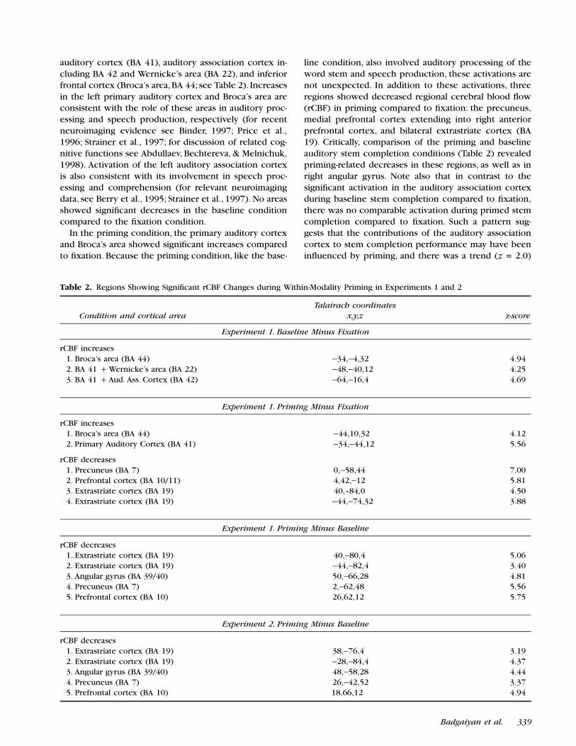
auditory cortex (BA 41), auditory association cortex in-cluding BA 42 and Wernicke’s area (BA 22), and inferiorfrontal cortex (Broca’s area, BA 44; see Table 2). Increasesin the left primary auditory cortex and Broca’s area areconsistent with the role of these areas in auditory proc-essing and speech production, respectively (for recentneuroimaging evidence see Binder, 1997; Price et al.,1996; Strainer et al., 1997; for discussion of related cog-nitive functions see Abdullaev, Bechtereva, & Melnichuk,1998). Activation of the left auditory association cortexis also consistent with its involvement in speech proc-essing and comprehension (for relevant neuroimagingdata, see Berry et al., 1995; Strainer et al., 1997). No areasshowed signiªcant decreases in the baseline conditioncompared to the ªxation condition.
In the priming condition, the primary auditory cortexand Broca’s area showed signiªcant increases comparedto ªxation. Because the priming condition, like the base-
line condition, also involved auditory processing of theword stem and speech production, these activations arenot unexpected. In addition to these activations, threeregions showed decreased regional cerebral blood ºow(rCBF) in priming compared to ªxation: the precuneus,medial prefrontal cortex extending into right anteriorprefrontal cortex, and bilateral extrastriate cortex (BA19). Critically, comparison of the priming and baselineauditory stem completion conditions (Table 2) revealedpriming-related decreases in these regions, as well as inright angular gyrus. Note also that in contrast to thesigniªcant activation in the auditory association cortexduring baseline stem completion compared to ªxation,there was no comparable activation during primed stemcompletion compared to ªxation. Such a pattern sug-gests that the contributions of the auditory associationcortex to stem completion performance may have beeninºuenced by priming, and there was a trend (z = 2.0)
Table 2. Regions Showing Signiªcant rCBF Changes during Within-Modality Priming in Experiments 1 and 2
Condition and cortical area
Talairach coordinates
x,y,z z-score
Experiment 1. Baseline Minus Fixation
rCBF increases 1. Broca’s area (BA 44) −34,−4,32 4.94 2. BA 41 + Wernicke’s area (BA 22) −48,−40,12 4.25 3. BA 41 + Aud. Ass. Cortex (BA 42) −64,−16,4 4.69
Experiment 1. Priming Minus Fixation
rCBF increases 1. Broca’s area (BA 44) −44,10,32 4.12 2. Primary Auditory Cortex (BA 41) −34,−44,12 5.56
rCBF decreases 1. Precuneus (BA 7) 0,−58,44 7.00 2. Prefrontal cortex (BA 10/11) 4,42,−12 5.81 3. Extrastriate cortex (BA 19) 40,–84,0 4.50 4. Extrastriate cortex (BA 19) −44,−74,32 3.88
Experiment 1. Priming Minus Baseline
rCBF decreases 1. Extrastriate cortex (BA 19) 40,−80,4 5.06 2. Extrastriate cortex (BA 19) −44,−82,4 3.40 3. Angular gyrus (BA 39/40) 50,−66,28 4.81 4. Precuneus (BA 7) 2,−62,48 5.56 5. Prefrontal cortex (BA 10) 26,62,12 5.75
Experiment 2. Priming Minus Baseline
rCBF decreases 1. Extrastriate cortex (BA 19) 38,−76,4 3.19 2. Extrastriate cortex (BA 19) −28,−84,4 4.37 3. Angular gyrus (BA 39/40) 48,−58,28 4.44 4. Precuneus (BA 7) 26,−42,52 3.37 5. Prefrontal cortex (BA 10) 18,66,12 4.94
Badgaiyan et al. 339

for reduced rCBF in primed compared to unprimed stemcompletion performance. Nonetheless, the failure to ob-serve a signiªcant decrease in the direct comparisonbetween primed and baseline stem completion perfor-mance indicates that these ªndings must be viewedcautiously.
Finally, there were no signiªcant rCBF increases in theprimed stem completion condition compared to thebaseline stem completion condition.
Experiment 2
In the within-modality priming condition, subjects com-pleted 54.3% of stems using studied words, compared to20.3% in the baseline condition (t (7) = 15.4; p < 0.0001).Further, response latencies were signiªcantly shorter forprimed words (1848 ± 78 msec) compared to baselinewords (2073 ± 74 msec; t (7) = 3.58; p < 0.009). In thecross-modality condition, subjects completed 48.1% ofstems using studied words, compared to 19.1% in thebaseline condition (t (7) = 17.38; p < 0.0001). Meanresponse latency in the priming condition (1823 ± 69msec) was signiªcantly less than that in the baseline(1995 ± 51 msec; t (7) = 2.67; p < 0.03). There were noeffects of test block on proportions of stems completedin either within-modality (t (7) = 0.22; p > 0.83) orcross-modality (t (7) = 1.48; p > 0.18) conditions. Al-though there was a slight trend for more priming in thewithin-modality than the cross-modality condition, thepercentage of stems completed with target words didnot differ signiªcantly between the two conditions(t (7) = 0.27; p > 0.79).
Changes in rCBF were examined separately for within-and cross-modality priming conditions. We hypothesizedthat within-modality priming would be accompanied byrCBF decreases in the same anatomic regions observedin Experiment 1. Consistent with this hypothesis, rCBF
changes were essentially identical to those observed inExperiment 1 (Table 2; Figure 1A and B). Each of thecortical regions that showed rCBF decreases in theprimed stem completion condition compared to thebaseline stem completion condition of Experiment 1also showed reduced activity in the corresponding com-parison of the present experiment: the extrastriate cor-tex bilaterally, right angular gyrus, precuneus andmedial/right anterior prefrontal cortex.
In cross-modality priming, no areas showed increasesor decreases in the priming condition compared to thebaseline condition that met our statistical threshold fornonplanned comparisons (z = 4.2; see Methods). Therewere, however, several rCBF differences between thepriming and baseline conditions that were at or near ourthreshold for planned comparisons (z = 3.09) and thatare consistent with the hypotheses discussed in theIntroduction that changes in phonological processingand explicit retrieval, respectively, play a role in cross-modal priming. We thus tentatively characterize theseªndings (Figures 2 and 3) as suggestive trends that meritfurther exploration in future research. First, the cross-modality priming condition was associated with an rCBFincrease encompassing medial and right anterior pre-frontal cortex (BA 10). As illustrated in the two slicesshown in Figure 2, the maximum activation occurred inthe medial prefrontal cortex (0, 48, 20; z = 3.31); themaximum activation in the right anterior prefrontal cor-tex was at 36, 60, 8; z = 3.06. Previous neuroimagingstudies have implicated the right anterior prefrontal cor-tex in aspects of explicit retrieval (e.g., Tulving et al.,1994). We also observed a trend for decreased rCBF inthe left angular gyrus during cross-modality primingcompared to baseline (BA 39/40, −40, −46, 28; z = 3.75;see Figure 3), a region that has been implicated pre-viously in aspects of phonological processing (e.g.,Paulesu, Frith, & Frackowiak, 1993). Direct comparison
Figure 1. Within-modalityauditory priming: Statisticalparametric maps (SPM) show-ing signiªcant rCBF decreasesin the extrastriate (BA 19) andmedial prefrontal (BA 10) ar-eas during within-modalityauditory priming as comparedto the stem completion base-line in Experiment 1 (A) andExperiment 2 (B). The medialprefrontal activation extendedto the right anterior prefrontalcortex in both experiments.The maps are superimposedover averaged structural MRIimages that were transformedto Talairach space. Atlas coordi-nates are provided in Table 2.
340 Journal of Cognitive Neuroscience Volume 11, Number 4

of the PET data obtained during within- and cross-modality priming also yielded ªndings that are best char-acterized as suggestive. There were trends for within-modality > cross-modality rCBF differences in the leftangular gyrus (−40, −46, 28; z = 3.18) and trends forcross-modality > within-modality priming in precuneus(10, −62, 44; z = 2.88) and in the right prefrontal (16,60,16; z = 2.94) and left extrastriate (−28, −70, 20; z =2.81) cortices.
DISCUSSION
Analysis of the PET data revealed that for within-modal-ity priming, there was reduced rCBF in the extrastriatearea (BA 19) during primed auditory stem completioncompared to baseline auditory stem completion (Experi-ments 1 and 2). Findings concerning cross-modalitypriming were less clear-cut, but the data are consistentwith the possibility that cortical areas associated withcross-modality priming may be distinct from those asso-ciated with within-modality priming (Experiment 2).
The fact that we observed decreased rCBF in theextrastriate cortex during within-modality auditory prim-ing in both Experiments 1 and 2 indicates that thisªnding is reliable and reproducible. The extrastriate re-gion showing priming-related decreases in the presentstudy is generally similar to that reported previously inwithin-modality visual stem completion priming experi-ments (Backman et al., 1997; Buckner et al., 1995; Schac-ter et al., 1996). It is also worth noting that Buckner etal. observed priming-related decreases in the extrastriatecortex during within-modality visual priming both whenthe type case of target items (upper or lower) wasidentical at study and test (same case condition) andwhen it differed (different case condition). The fact thatBuckner et al. (1995) observed rCBF decrease in theextrastriate cortex in the different case condition sug-
gests that this decrease probably does not reºect prim-ing of the speciªc visual features of previously studiedtarget words. Our ªndings are thus consistent with thoseof Buckner et al. However, because within-modality audi-tory priming does not require any visual perceptualprocessing, whereas previous discussions of priming-related blood ºow reductions in the extrastriate cortexhave emphasized modality-speciªc perceptual processes(Buckner et al., 1995; Schacter et al., 1996; Squire et al.,1992), our ªndings cast new light on the mechanisminvolved. We consider three potential explanations toaccount for the rCBF decrease in the extrastriate cortex
Figure 2. Cross-modalitypriming: SPM at two slice loca-tions showing increasedblood ºow in the prefrontalcortex (BA 10) during cross-modality auditory priming (ascompared to stem completionbaseline). Maximal rCBF in-creases were observed in themedial prefrontal area (A; z =3.31) and in the right anteriorprefrontal area (B; z = 3.06).The maps are superimposedover averaged structural MRIimages that were transformedto Talairach space.
Figure 3. Cross-modality priming: SPM showing rCBF decrease (z =3.75) in the left angular gyrus (BA 39/40) during cross-modalitypriming as compared to the baseline. The maps are superimposedover averaged structural MRI images that were transformed to Talai-rach space.
Badgaiyan et al. 341

during auditory priming: (1) visual imagery or relatedprocesses played a role in auditory stem completionpriming, (2) the decreases may reºect a kind of cross-modal suppression effect, and (3) a part of the extrastri-ate cortex may support nonvisual functions.
One possible account of our results involves the useof visual imagery. Neuroimaging studies have consis-tently reported increased rCBF in the extrastriate cortex(and several other cortical regions) in tasks that requirevisual imagery (e.g., Cabeza et al., 1997; Cohen et al.,1996; D’Esposito et al., 1997; Fletcher et al., 1995; Roland,Eriksson, Stone-Elander, & Widen, 1987). Perhaps subjectscreated visual images of auditorily presented words dur-ing the study task and retrieved those images duringprimed auditory stem completion performance. How-ever, by this account we should have observed increasedrCBF—not decreased rCBF—in the extrastriate cortexduring auditory priming. Moreover, when subjects wereexplicitly shown visual words during the study phase inExperiment 2, we failed to observe subsequent rCBFchanges in the extrastriate cortex during cross-modalityauditory priming. Thus, use of visual imagery during thestudy task and priming test does not offer a promisingor even a plausible account of our results.
An alternative version of an imagery hypothesis wouldhold that subjects created visual images of the targetwords during baseline auditory stem completion perfor-mance. During primed auditory stem completion perfor-mance, however, participants may have been able toprocess auditory information more easily than in thebaseline condition and were thus less likely to engage invisualization of target stems or words, thereby resultingin extrastriate rCBF decreases. By this view, the extrastri-ate cortex should have been associated with signiªcantrCBF increases during baseline stem completion perfor-mance compared to the ªxation control (possiblyreºecting the occurrence of visual imagery). However,no such increases were observed. A related idea is thatauditory processing may have activated orthographicfeatures of target items during baseline stem completionperformance (e.g., Tanenhaus, Flanagan, & Seidenberg,1980). If the extrastriate cortex is involved with ortho-graphic processing and representation (e.g., Petersen,Fox, Snyder, & Raichle, 1990), the observed rCBF de-creases may reºect corresponding decreases in ortho-graphic processing after auditory priming. Though anattractive idea, as with the visual imagery hypothesis, thisnotion would lead us to expect increased extrastriateactivation in the baseline stem completion task relativeto ªxation.
A second hypothesis is that extrastriate decreases mayreºect a suppression of visual processing during audi-tory task performance. For example, Buckner, Raichle,Miezin, and Petersen (1996) reported rCBF decreases inthe extrastriate cortex during an auditory paired-associ-ate recall task compared to a ªxation control. In this task,
subjects were presented with a cue word auditorily andwere asked to recall the second member of the pair; thetwo words had been presented together auditorily priorto scanning. Buckner et al. suggested that the extrastriatedecreases might have occurred because attention toauditory stimuli suppressed activity in visual areas (seeHaxby et al., 1994, for discussion of a similar cross-modalsuppression effect in the auditory cortex during visualattention).
We cannot rule out the possibility that cross-modalsuppression is involved in the extrastriate decrease weobserved. Nonetheless, it seems an unlikely explanationbecause we did not observe signiªcant extrastriate rCBFdecreases in the baseline auditory stem completion con-dition compared to ªxation (nor did we observe anytrends for such decreases, even when the signiªcancelevel was set at z = 1). If the observed decrease resultedfrom suppression of visual regions as a consequence ofattention to auditory stimuli, the effect should have beenobserved in the baseline condition as well as in thepriming condition. More generally, as Buckner et al.(1999) note, it is important to distinguish between re-gions that show decreased activity compared to a ªxa-tion control (for review, see Shulman et al., 1997) anddecreases as a result of priming that reºect reducedactivity in a region that shows increased activity duringbaseline task performance. The extrastriate decrease re-ported here is both similar to and different from each ofthese types of decreases. On the one hand, the extrastri-ate decrease was not observed during baseline stemcompletion performance relative to ªxation (thus distin-guishing it from the general kinds of decreases reviewedby Shulman et al. that occur in relation to ªxation).However, it was observed in the comparison of primedand unprimed stem completion performance (similar toprevious priming studies) and in the comparison ofprimed stem completion and the ªxation condition. Butwhereas some previous reports of priming-related ex-trastriate decreases during visual stem completion haveshown increased activity in this region during baselinestem completion performance compared to ªxation(e.g., Buckner et al., 1995), we did not observe such anoutcome (nor did Backman et al., 1997).
In the present study, reduced rCBF during primingboth in comparison with the ªxation condition and withthe unprimed stem completion baseline suggests thatthe reduction is associated with priming and is not aresult of increased rCBF in the unprimed stem comple-tion baseline condition. Increased rCBF both in the ªxa-tion and the stem completion baseline condition couldalso produce this result, but this is unlikely because thepriming task (as well as the stem completion baselinetask) included the ªxation condition (that is, subjectsalways looked at a ªxation cross; this strategy helped usreduce artifacts due to eye movements, and its effectsshould have subtracted out in the various contrasts we
342 Journal of Cognitive Neuroscience Volume 11, Number 4

used in this experiment). Therefore, a “ªxation-relatedincrease” should have yielded increased rCBF in thepriming condition (rather than decreased rCBF). Clearly,however, further studies are necessary for better under-standing of the signiªcance of this reduction.
A third possible interpretation of the priming-relatedextrastriate decrease is that parts of the extrastriate cor-tex may support nonvisual functions. Neuroanatomicaland electrophysiological data suggest that the extrastri-ate cortex in humans and other primates includes dis-tinct areas of specialization (DeYoe et al., 1996; Rodman& Moore, 1997; Zilles & Clarke, 1997). A region of theextrastriate cortex (V3A) that corresponds roughly tothe superolateral part of BA 19 in the human braindiffers from the rest of the extrastriate cortex in itsconnectivity, neurohumoral proªle, and cytoarchitecture(for detailed discussion, see Zilles & Clarke). This regionis extensively connected to the subcortical and corticalareas outside the occipital visual cortex and is not de-pendent on input from the primary visual cortex foractivation. Indeed, a recent fMRI experiment has demon-strated activation in the extrastriate area in response toauditory stimuli (Bookheimer et al., 1998). Interestingly,Talairach coordinates of the V3A region determined byfMRI (Tootell et al., 1997) agree with the coordinates ofthe extrastriate rCBF decreases we observed in primingcondition. It, however, remains to be determinedwhether (and how) possible nonvisual functions of re-gions within the extrastriate cortex are related to thepriming-related rCBF reductions reported here, but thepossible link seems worth considering and exploring.
As indicated by the foregoing discussion, the exactrole that the extrastriate cortex plays in visual and audi-tory stem completion priming remains to be speciªed.In general, however, our ªndings support the conclusionthat priming-related decreases in the extrastriate cortexreºect processes common to visual and auditory modali-ties rather than modality-speciªc visual processes. Thisconclusion is generally consistent with ªndings from arecent fMRI study by Buckner et al. (1999), who used arepeated word generation paradigm to examine primingeffects in visual and auditory word stem completion. Intheir study, subjects were repeatedly presented witheither visual stems or with auditory stems (in separateexperiments) and generated appropriate completions.Completion latencies were reduced with repetition ofthe identical stems but returned to baseline levels whennovel stems were presented, thus indicating that someform of item-speciªc priming occurred. Importantly,Buckner et al. observed repetition-related activation re-ductions during both visual and auditory stem comple-tion performance in an area of the inferior temporalcortex (localized to x = −43, y = −52, z = −12) near theventral visual processing stream, anterior to the extras-triate regions discussed here. They suggested that thisregion is involved in amodal lexical/conceptual proc-
esses that occur during both visual and auditory stemcompletion priming. Thus, the general conclusions fromthe Buckner et al. study are consistent with, and comple-mentary to, the hypotheses that we have suggested.
One difference between the two studies is that theinferior temporal region that showed repetition-relatedreductions in the Buckner et al. (1999) study alsoshowed signiªcant activation compared to a ªxationbaseline, whereas, as noted earlier, the extrastriate regionwe have focused on was not activated signiªcantly dur-ing the stem completion baseline compared to ªxation.Differences in experimental paradigms (or possibly im-aging modalities) may account for the contrasting pat-terns.
Although the possibility that priming-related extrastri-ate decreases reºect amodal processes is intriguing, thefact that we observed no extrastriate rCBF decreasesduring cross-modality priming—when priming dependsexclusively on amodal processes—indicates that thestory may be more complex (and also indicates that suchdecreases do not occur ubiquitously in all forms ofpriming). Perhaps priming-related extrastriate decreasesare speciªc to conditions in which within-modality per-ceptual processes that were carried out during encoding(visual or auditory) are reinstated during a priming test.To say more at the present time would go beyond theavailable data. We believe that the pattern of resultsreported here may provide important clues concerningthe nature and basis of priming, but it is evident thatfurther studies will be required to provide a ªrmer basisfor theoretical interpretation of our data.
Within-modality auditory priming was also associatedwith rCBF decreases in the right angular gyrus. Thisdecrease may be associated with some aspect of lexicalprocessing. Although the function of the right angulargyrus is not well understood, its activation has beenreported in episodic memory tasks involving consciousrecollection of words and sentences (Horwitz, Rumsey,& Donohue, 1998; Tulving et al., 1994). Other areas thatshowed consistent decreases during within-modalityauditory priming are the medial prefrontal cortex ex-tending into the right anterior prefrontal cortex, and ananterior part of the precuneus (medial parietal cortex).Buckner et al. (1996) reported that both of these regionsexhibited decreased activity during paired associate re-call compared to a ªxation control. It is possible that thedecreases reported by Buckner et al. are related to ourobservations, but further work will be necessary to settlethe matter.
Perhaps surprisingly, we did not ªnd evidence forpriming-related decreases in the primary auditory cortexor in the auditory association cortex. As noted earlier,however, in Experiment 1, we did ªnd some evidencesuggestive of priming-related decreases in the auditoryassociation cortex. During the baseline stem completiontasks, there was signiªcant activation in the left primary
Badgaiyan et al. 343
auditory cortex (BA 41) extending into auditory associa-tion cortex (BA 22 and 42) compared to ªxation (Table2). In primed stem completion, the primary auditorycortex activation was again observed, but the activationdid not extend into the auditory association cortex. How-ever, because the direct contrast between primed andbaseline stem completion performance did not revealsigniªcant changes in the auditory association cortex,further studies will be necessary to determine whetherthis region shows reliable priming-related decreases onauditory stem completion or similar tasks.
In the cross-modality condition, none of the areas thatshowed rCBF decreases during within-modality primingshowed reduced activity; the only decrease observed inthis condition was a trend in the left posterior parietalcortex (BA 39/40; angular gyrus). There were also trendsfor increases observed in the medial and right anteriorprefrontal cortex. Although we must be cautious wheninterpreting these trends, they ªt well with previouscognitive studies that have highlighted differences be-tween within- and cross-modality priming. One class ofexplanations has focused on the idea that cross-modalpriming is mediated by some form of abstract lexicalrepresentation involved in phonological input or outputprocessing (e.g., Curran et al., 1999; Kirsner et al., 1989;Weldon, 1991). Previous neuroimaging studies have sug-gested that regions of the left posterior parietal cortex(BA 39/40), in the vicinity of the region that showed adecrease during cross-modal priming, are involved inphonological storage processes (Awh et al., 1996;Paulesu, Frith, & Frackowiak, 1993). This conclusion isalso supported by neuropsychological studies indicatingthat lesions to the left posterior parietal cortex are fre-quently observed in patients characterized by phonologi-cal storage deªcits (Vallar & Shallice, 1990). Thus, thetrend we observed in the left posterior parietal cortexduring cross-modal priming could reºect a priming-related reduction in some aspect of phonological orlexical processing.
Activation in the right anterior prefrontal region (BA)has been observed frequently in studies of episodic orexplicit memory retrieval (Tulving et al., 1994) and hasbeen previously associated with various aspects of ex-plicit retrieval, including retrieval “mode” (Nyberg et al.,1995), retrieval effort (Schacter et al., 1996), and post-retrieval monitoring (Rugg, Fletcher, Frith, Frackowiak, &Dolan, 1996; Schacter, Buckner, Koutstaal, Dale, & Rosen,1997). Two lines of evidence from behavioral studieshave illuminated the role of explicit retrieval in cross-modality stem completion priming. Neuropsychologicalstudies have demonstrated that amnesic patients shownormal cross-modality priming on the stem completiontask (Carlesimo, 1994; Graf, Shimamura, & Squire, 1985),thereby indicating that explicit retrieval is not necessaryfor cross-modal priming to occur. Nonetheless, usingprocess dissociation procedures previously developedby Jacoby (1991) for estimating nonconscious and con-
sciously controlled contributions to retrieval, Jacoby etal. (1993) provided evidence from college students thatwhereas within-modality priming depends on noncon-scious or implicit retrieval processes, cross-modalitypriming involves conscious recollection.
In a comparison of within- and cross-modality visualstem completion priming, Richardson-Klavehn andGardiner (1996) further reªned this notion by distin-guishing different aspects of conscious recollection, onedimension involving intentional/unintentional retrievaland another involving awareness/unawareness of thestudy episode (Schacter, 1987). They provided evidencethat cross-modality priming, like within-modality prim-ing, involves unintentional retrieval of previously studieditems. Richardson-Klavehn and Gardiner supported thisidea by showing that a deep versus shallow encodingmanipulation, which signiªcantly affected explicit recall,had no effect on cross-modality (and within-modality)priming (see also Craik, Moscovitch, & McDowd, 1994).Had subjects been engaging in intentional retrieval dur-ing cross-modality priming, they should have exhibitedmore priming after deep than shallow encoding (forelaboration of this logic, see Schacter, Bowers, & Booker,1989). However, Richardson-Klavehn and Gardiner alsopresented evidence based on Jacoby’s (1991) processdissociation procedure that cross-modality priming, un-like within-modality priming, entails conscious aware-ness of the prior presentation of the studiedwords—what has been termed “involuntary consciousmemory” or “involuntary explicit memory” (Schacter,1987; Schacter et al., 1989).
If the trend for right anterior prefrontal activation inExperiment 2 reºects “involuntary explicit memory,”one might expect that during cross-modal stem comple-tion testing subjects were more likely to become awarethat test items came from the study list than duringwithin-modality stem completion testing. Informal as-sessments of subjects during postscan debrieªng re-vealed that nearly all subjects (87.5%) in both within-and cross-modality conditions were aware of the factthat some of the stems came from the study list. How-ever, no subjects reported that they intentionally at-tempted to recall studied words in either condition.Future studies will thus be required to determinewhether the trends for prefrontal activation observed inExperiment 2 reºect the kinds of processes observed inthe foregoing cognitive studies of cross-modality prim-ing. Moreover, because we have examined cross-modalitypriming of auditory stem completion after visual study,whereas the foregoing cognitive studies examined cross-modality priming of visual stem completion tasks afterauditory study, it will be important to determinewhether the trends observed in the present experimentare obtained in visual stem completion tasks that arepreceded by auditory study. Initial results are consistentwith this possibility (Schacter, Badgaiyan, & Alpert, 1999).
In summary, the main ªnding from our experiments is
344 Journal of Cognitive Neuroscience Volume 11, Number 4
that blood ºow decreases in the extrastriate cortex ob-served previously during within-modality visual primingare also observed during within-modality auditory prim-ing. These ªndings raise the possibility that extrastriateblood ºow decreases reºect changes in more generalpriming processes than was previously suspected.
METHODS
The experimental protocol was approved by the institu-tional review boards of Harvard University and Massa-chusetts General Hospital. The experiments wereconducted with young native English speaking volun-teers who were right handed as assessed by Edinburghhandedness inventory (Raczkowski, Kalat, & Nebes,1974). All subjects had normal or corrected to normalvision and hearing. In a prescan interview, they werescreened to rule out current medical conditions, historyof neurological or psychiatric disturbances, prolongeduse of a prescription or recreational drugs, claustropho-bia, and signiªcant prior radiation exposure. They wereadvised to remain alcohol free for at least 24 hr prior tothe scan.
Experiment 1 was conducted with eight young volun-teers (three females, ªve males, 20–28 years; mean age =25.2 years) who consented to participate for monetarycompensation.
In the study phase, a list of 60 words (30 target wordsand 30 ªller words, presented in random order), re-corded in a single female voice, was presented overheadphones (2 sec/word). Subjects were instructed torate the clarity of enunciation for each word on a 1 to3 scale using a numeric keypad and responding with theright hand.
Approximately 2 min after presentation of the (non-scanned) study list, participants were scanned duringseparate blocks in which they provided completions toauditory word stems that could be completed with pre-viously studied words (priming scan) or could not becompleted with previously studied words (baselinescan). Thirty auditory word stems were presented(3 sec/stem; in the same female voice used for the studywords) over headphones, and subjects were asked tospeak aloud the ªrst word that came to mind beginningwith each stem. Participants were told to avoid propernouns and were assured that there were no right orwrong answers. The ªrst nine stems in each block werederived from both studied and nonstudied words. Theªnal 21 stems in priming scans all came from the studiedlist, whereas in baseline scans none of the ªnal 21 stemscould be completed with a studied word. PET scanswere obtained during completion of the ªnal 20 stems.The study-test blocks were repeated so that we obtainedthree priming blocks and three baseline blocks. Eachblock comprised a different set of words and stems.
The experiment also included a ªxation condition inwhich subjects were instructed to ªxate on a crosshair.
There were two ªxation blocks, one prior to the ªrststudy list and the other after the ªnal stem completiontest. Each subject thus underwent eight scans, three inthe priming, three in the baseline, and two in ªxationcondition. During the stem completion test, responseswere recorded on a tape recorder, and response laten-cies were recorded with a microphone connected to aPsyScope button box (Macwhinney, Cohen, & Provost,1997).
Target materials consisted of a list of 200 commonEnglish words, each having a unique ªrst syllable. Se-lected words were divided into two lists (A and B),which were balanced according to the word frequency(range 1 to 100; Kucera & Francis, 1967), number ofsyllables (range 2 to 4), and number of possible comple-tions. The ªrst syllable of each word was used as anauditory word stem, and each stem had at least sixpossible completions. The American Heritage College
Dictionary (Costello, 1997) was used to identify sylla-bles and determine the number of possible completions.For half of the subjects, list A was used for study lists andlist B for baseline lists; for the other half, the assignmentof lists to conditions was reversed. Lists A and B werefurther divided into three sublists that were counterbal-anced across subjects, scan sequences, and priming orbaseline conditions in such a way that each sublist oc-curred equally often in the priming and baseline condi-tion and in each scan sequence. Further, the sequenceof priming and baseline conditions was counterbalancedacross subjects so that each condition occurred equallyoften at each scan sequence. During the stem comple-tion task, subjects were asked to focus on a crosshairthat was always displayed at the center of a computermonitor placed directly in front of the subject.
Experiment 2 was conducted with eight volunteerswhose age (19 to 30 years, mean 22.75 years) andgender (three females/ªve males) were approximatelymatched to those of the subjects who participated inExperiment 1.
The experiment included both within-modality andcross-modality priming conditions. The within-modalitycondition was nearly identical to Experiment 1, withonly two differences: (1) there were only two (insteadof three) study-test blocks, and (2) during the studyphase, subjects made pleasantness judgments for eachstudy word, thus allowing us to use the same encodingtask for auditory and visual study items. In the cross-modality condition, study words were presented visuallyat the center of a computer monitor, whereas in thewithin-modality condition, words were presented audi-torily as described for Experiment 1. Cross-modalitypriming on the stem completion task is typically smallerthan within-modality priming (Graf et al., 1985; Roediger& Blaxton, 1987; Schacter & Graf, 1989). Therefore, weattempted to create conditions that would reduce oreliminate possible differences in the overall magnitudeof priming that could confound interpretation of any
Badgaiyan et al. 345

observed differences in PET activations for the twoforms of priming.
Based on the results of behavioral pilot studies, wereduced the length of the study list (in each study block)from 60 words (30 study and 30 ªller) for the within-modality condition to 45 words (30 study and 15 ªller)for the cross-modality condition. Stem completionblocks were constructed in the same manner in thewithin- and cross-modality conditions, as described forExperiment 1.
The words used in this experiment were selectedusing the criteria described for Experiment 1 and werecounterbalanced across subjects in such a way that eachword was used equally often in priming and baselineconditions and in within- and cross-modality conditions.The sequence of scans was also counterbalanced acrosssubjects so that equal numbers of within- and cross-modality conditions, as well as priming and baselineconditions, occurred in each scan sequence. Each sub-ject underwent eight scans, two in each of the four mainconditions (i.e., within-modality priming and baseline,cross-modality priming and baseline).
PET Scan
During the priming, baseline, and ªxation conditionshead scans were obtained using a General ElectricScanditronix (Uppsala) model PC4096 15-slice wholebody tomograph. An individually molded plastic facemask was used to minimize head motion during theexperiment. At time zero, the stem completion or theªxation task was started along with the PET camera andcontinued for 90 sec. At 30 sec, radioactive tracer inha-lation (15O labeled carbon dioxide) and emission dataacquisition began. Tracer inhalation and data acquisitionlasted for 60 sec. A washout period of approximately 10min was allowed between successive scans. Details ofthe PET facility and procedures are similar to thosedescribed earlier (Kosslyn et al., 1994).
Data Analysis
After image reconstruction, the scans from each subjectwere treated as follows: A correction was computed toaccount for head movement (rigid body translation androtation) using a least squares ªtting technique (Alpert,Berdichevsky, Levin, Morris, & Fischman, 1996). Themean over all conditions was formed and used as inputto determine the transformation to the standard coordi-nate system of Talairach and Tournoux (1988). This trans-formation was computed by deforming the 10-mmparasagittal brain-surface contour to match the contourof a reference brain (Alpert, Berdichevsky, Weiss, Tang,Rauch, 1993). Following spatial normalization, scanswere ªltered with a two-dimensional Gaussian ªlter, fullwidth at half maximum set to 20 mm. Statistical analysisfollowed the theory of statistical parametric mapping
(Friston, Frith, Liddle, & Frackowiak, 1991; Friston et al.,1995; Worsley, Evans, Marrett, & Neelin, 1992). Data wereanalyzed with SPM95 (from the Wellcome Dept. of Cog-nitive Neurology, London, UK). The PET data at eachvoxel were normalized by the global mean and ªt to alinear statistical model with cognitive state (i.e., scancondition) considered as a main effect and subjects as ablock effect. Hypothesis testing was performed using themethod of planned contrasts at each voxel. We setthresholds for signiªcance according to the theory ofGaussian ªelds (see Friston et al., 1991; 1995; Worsley etal., 1992). When no localizing hypothesis or prior experi-mental data were available, a threshold of z = 4.2 wasconsidered signiªcant. When we had a priori hypothesesthat localized the putative activation to a speciªcanatomic region, we considered a threshold of z = 3.09to be signiªcant. This threshold (z = 3.09) constitutes acompromise between a low threshold uncorrected formultiple comparisons (z = 1.96) and a higher threshold(z = 4.2) suggested by the theory of Gaussian ªelds forunplanned comparisons (see Worsley et al., 1996, fordiscussion of the basis of a lower statistical threshold fortesting anatomically localized hypotheses).
Acknowledgment
This research was supported by the National Institutes ofHealth grant number MH57915-02 and Human Frontiers Sci-ence Program Grant number RG0126.
Reprint requests should be sent to Rajendra D. Badgaiyan,Harvard University, William James Hall, Rm. 875, 33 KirklandStreet, Cambridge, MA 02138, or via e-mail: [email protected].
REFERENCES
Abdullaev, Y. G., Bechtereva, N. P., & Melnichuk, K. V. (1998).Neuronal activity of human caudate nucleus and prefron-tal cortex in cognitive tasks. Behavioral Brain Research,
97, 159–177.Alpert, N. M., Berdichevsky, D., Levin, Z., Morris, E. D., &
Fischman, A. J. (1996). Improved methods for image regis-tration. Neuroimage, 3, 10–18.
Alpert, N. M., Berdichevsky, D., Weiss, S., Tang, J., & Rauch,S. L. (1993). Stereotactic transformation of PET scans bynon linear least squares. In K. Uemura, T. Jones, N. A. Las-sen, & I. Kanno (Eds.), Quantiªcation of brain function:
Tracer kinetics and image analysis in PET (pp. 459–463). Amsterdam: Elsevier.
Awh, E., Jonides, J., Smith, E. E., Schumacher, E. H., Koeppe,R. A., & Katz, S. (1996). Dissociation of storage and re-hearsal in verbal working memory: Evidence from posi-tron emission tomography. Psychological Science, 7, 25–31.
Backman, L., Almkvist, O., Andersson, J., Nordberg, A., Win-blad, B., Reineck, R., & Langstrom, B. (1997). Brain activa-tion in young and older adults during implicit and explicitretrieval. Journal of Cognitive Neuroscience, 9, 378–391.
Badgaiyan, R. D., & Posner, M. I. (1996). Priming reduces in-put activity in right posterior cortex during stem comple-tion. NeuroReport, 7, 2975–2978.
Badgaiyan, R. D., & Posner, M. I. (1997). Time course of corti-
346 Journal of Cognitive Neuroscience Volume 11, Number 4
cal activations in implicit and explicit recall. Journal of
Neuroscience, 17, 4904–4913.Bassili, J. N., Smith, M. C., & MacLeod, C. M. (1989). Auditory
and visual word-stem completion: Separating data-drivenand conceptually driven processes. Quarterly Journal ofExperimental Psychology, 41A, 439–459.
Berry, I., Demonet, J. F., Warach, S., Viallard, G., Boulanouar, K.,Franconi, J. M., Marc-Vergnes, J. P., Edelman, R., & Manelfe,C. (1995). Activation of association auditory cortex demon-strated with functional MRI. Neuroimage, 2, 215–219.
Binder, J. (1997). Functional magnetic resonance imaging. Lan-guage mapping. Neurosurgery Clinics of North America,
8, 383–392.Blaxton, T. A., Bookheimer, S. Y., Zefªro, T. A., Figlozzi, C. M.,
Gaillard, W. D., & Theodore, W. H. (1996). Functional map-ping of human memory using PET: Comparisons of con-ceptual and perceptual tasks. Canadian Journal of
Experimental Psychology, 50, 42–56.Bookheimer, S. Y., Zefªro, T. A., Blaxton, T. A., Gaillard, W. D.,
Malow, B., & Theodore, W. H. (1998). Regional cerebralblood ºow during auditory responsive naming: Evidencefor cross-modality neural activation. NeuroReport, 13,
2409–2413.Buckner, R. L, Goodman, J., Burock, M., Rotte, M., Koutstaal,
W., Schacter, D. L., Rosen, B., & Dale, A. (1998). Functional-anatomic correlates of object priming in humans revealedby rapid presentation event-related fMRI. Neuron, 20, 285–296.
Buckner, R. L., Koutstaal, W., Schacter, D. L., & Rosen, B. R.(1999). Functional-anatomic evidence for amodal mecha-nisms underlying human repetition priming. Submitted forpublication.
Buckner, R. L., Petersen, S. E., Ojemann, J. G., Miezin, F. M.,Squire, L. R., & Raichle, M. E. (1995). Functional anatomi-cal studies of explicit and implicit memory retrieval tasks.Journal of Neuroscience, 15, 12–29.
Buckner, R. L., Raichle, M. E., Miezin, F. M., & Petersen, S. E.(1996). Functional anatomic studies of memory retrievalfor auditory words and visual picture. Journal of Neurosci-
ence, 16, 6219–6235.Cabeza, R., Mangels, J., Nyberg, L., Habib, R., Houle, S., McIn-
tosh, A. R., & Tulving, E. (1997). Brain regions differentiallyinvolved in remembering what and when: A PET study.Neuron, 19, 863–870.
Carlesimo, G. A. (1994). Perceptual and conceptual primingin amnesic and alcoholic patients. Neuropsychologia, 32,
903–921.Church, B., & Schacter, D. L. (1994). Perceptual speciªcity of
auditory priming: Implicit memory for voice intonationand fundamental frequency. Journal of Experimental Psy-
chology: Learning, Memory, and Cognition, 20, 521–533.Cohen, M. S., Kosslyn, S. M., Breiter, H. C., DiGirolamo, G. J.,
Thompson, W. L., Anderson, A. K., Brookheimer, S. Y.,Rosen, B. R., & Belliveau, J. W. (1996). Changes in corticalactivity during mental rotation. A mapping study usingfunctional MRI. Brain, 119, 89–100.
Costello, R. B. (Ed.). (1997). The American Heritage College
Dictionary (3rd ed.). Boston, MA: Houghton Mifºin Com-pany.
Craik, F. I. M., Moscovitch, M., & McDowd, J. M. (1994). Con-tributions of surface and conceptual information to perfor-mance on implicit and explicit memory tasks. Journal of
Experimental Psychology: Learning, Memory, and Cogni-
tion, 20, 864–875.Curran, T., Schacter, D. L., & Galluccio, L. (1999). Cross-modal
priming and explicit memory in patients with verbal pro-duction deªcits. Brain and Cognition, 39, 133–146.
D’Esposito, M., Detre, J. A., Aguirre, G. K., Stallcup, M., Alsop,
D. C., Tippet, L. J., & Farah, M. J. (1997). A functional MRIstudy of mental image generation. Neuropsychologia, 35,
725–30.DeYoe, E. A., Carman, G. J., Bandettini, P., Glickman, S., Wieser,
J., Cox, R., Miller, D., & Neitz, J. (1996). Mapping striate andextrastriate visual areas in human cerebral cortex. Proceed-
ings of the National Academy of Sciences USA, 19; 93,
2382–2386.Fletcher, P. C., Frith, C. D., Baker, S. C., Shallice, T., Frack-
owiak, R. S., & Dolan, R. J. (1995). The mind’s eye—pre-cuneus activation in memory-related imagery.Neuroimage, 2, 195–200.
Friston, K. J., Frith, C. D., Liddle, P. F., & Frackowiak, R. S. J.(1991) Comparing functional (PET) images: The assess-ment of signiªcant change. Journal of Cerebral Blood
Flow and Metabolism 11, 690–699.Friston, K. J., Holmes, A. P., Worsley, K. J., Poline, J. B., Frith,
C. D., and Frackowiak, R. S. J.(1995) Statistical parametricmaps in functional imaging: A general approach. Human
Brain Mapping 2, 189–210.Graf, P., Shimamura, A. P., & Squire, L. R. (1985). Priming
across modalities and priming across category levels; ex-tending the domain of preserved functioning in amnesia.Journal of Experimental Psychology: Learning, Memory,
and Cognition, 16, 164–178.Haxby, J. V., Horwitz, B., Ungerleider, L. G., Maisog, J. M.,
Pietrini, P., & Grady, C. L. (1994). The functional organiza-tion of human extrastriate cortex: A PET-rCBF study of se-lective attention to faces and locations. Journal ofNeuroscience, 14, 6336–6353.
Horwitz, B., Rumsey, J. M., & Donohue, B. C. (1998). Func-tional connectivity of the angular gyrus in normal readingand dyslexia. Proceedings of the National Academy of Sci-
ences USA, 95, 8939–8944.Jacoby, L. L. (1991). A process dissociation framework: Sepa-
rating automatic from intentional uses of memory. Journal
of Memory and Language, 30, 513–541.Jacoby, L. L., Toth, J. P., & Yonelinas, A. P. (1993). Separating
conscious and unconscious inºuences of memory: Measur-ing recollection. Journal of Experimental Psychology:General, 122, 139–154.
Kirsner, K., Dunn, J. C., & Standen, P. (1989). Domain-speciªcresources in word recognition. In S. Lewandowsky, J. C.Dunn, & K. Kirsner (Eds.), Implicit memory: Theoretical is-
sues (pp. 99–122). Hillsdale, NJ: Erlbaum.Kosslyn, S. M., Alpert, N. M., Thompson, W. L., Chabris, C. F.,
Rauch, S. L., & Anderson, A. K. (1994). Identifying objectsseen from different viewpoints. A PET investigation. Brain,
117, 1055–1071.Kucera, H., & Francis, W. N. (1967). Computational analysis
of present-day American English. Providence, RI: BrownUniversity Press.
Macwhinney, B., Cohen, J., & Provost, J. (1997). The PsyScopeexperiment-building system. Spatial Vision, 11, 99–101.
McClelland, A. G. R., & Pring, L. (1991). An investigation ofcross-modality effects in implicit and explicit memory.Quarterly Journal of Experimental Psychology, 43A, 19–33.
Nyberg, L., Tulving, E., Habib, R., Nilsson, L. G., Kapur, S.,Houle, S., Cabeza, R., & McIntosh, A. R. (1995). Functionalbrain maps of retrieval mode and recovery of episodic in-formation. NeuroReport, 7, 249–252.
Paulesu, E., Frith, C. D., & Frackowiak, R. S. (1993). The neu-ral correlates of the verbal component of working mem-ory. Nature, 362, 342–345.
Petersen, S. E., Fox, P. T., Snyder, A. Z., & Raichle, M. E. (1990).Activation of extrastriate and frontal cortical areas by vis-ual words and word-like stimuli. Science, 249, 1041–1044.
Badgaiyan et al. 347

Price, C. J., Wise, R. J., Warburton, E. A., Moore, C. J., Howard,D., Patterson, K., Frackowiak, R. S., & Friston, K. J. (1996).Hearing and saying. The functional neuro-anatomy of audi-tory word processing. Brain, 119, 919–931.
Raczkowski, D., Kalat, J. W., & Nebes, R. (1974). Reliabilityand validity of some right handed questionnaire items.Neuropsychology, 6, 43–47.
Richardson-Klavehn, A., & Gardiner, J. M. (1996). Cross-modal-ity priming in stem completion reºects conscious mem-ory, but not voluntary memory. Psychonomic Bulletin and
Review, 3, 238–244.Rodman, H. R., & Moore, T. (1997). Development and plastic-
ity of extrastriate visual cortex in monkeys. Cerebral Cor-
tex, 12, 639–672.Roediger, H. L., & Blaxton, T. A. (1987). Effects of varying mo-
dality, surface features, and retention interval on primingin word fragment completion. Memory and Cognition,
15, 379–388.Roland, P. E., Eriksson, L., Stone-Elander, S., & Widen, L.
(1987). Does mental activity change the oxidative metabo-lism of the brain? Journal of Neuroscience, 7, 2373–2389.
Rugg, M. D., Fletcher, P. C., Frith, C. D., Frackowiak, R. S., &Dolan, R. J. (1996). Differential activation of the prefrontalcortex in successful and unsuccessful memory retrieval.Brain, 119, 2073–2083.
Schacter, D. L. (1987). Implicit memory: History and currentstatus. Journal of Experimental Psychology: Learning,
Memory, and Cognition, 13, 501–518.Schacter, D. L. (1994). Priming and multiple memory systems:
Perceptual mechanisms of implicit memory. In D. L. Schac-ter & E. Tulving (Eds.), Memory systems 1994 (pp.233–268). Cambridge, MA: MIT Press.
Schacter, D. L., Alpert, N. M., Savage, C. R., Rauch, S. L., & Al-bert, M. S. (1996). Conscious recollection and the humanhippocampal formation: Evidence from positron emissiontomography. Proceedings of the National Academy of Sci-
ences USA, 93, 321–325.Schacter, D. L., Badgaiyan, R. D., & Alpert, N. M. (1999). Visual
stem completion priming within and across modalities: APET study. NeuroReport 10, 2061–2065.
Schacter, D. L., Bowers, J., & Booker, J. (1989). Intention,awareness, and implicit memory: The retrieval intentional-ity criterion. In S. Lewandowsky, J. C. Dunn, & K. Kirsner(Eds.), Implicit memory: Theoretical issues (pp. 47–65).Hillsdale, NJ: Erlbaum.
Schacter, D. L., & Buckner, R. L. (1998). Priming and thebrain. Neuron, 20, 185–195.
Schacter, D. L., Buckner, R. L., Koutstaal, W., Dale, A. M., &Rosen, B. R. (1997). Late onset of anterior prefrontal activ-ity during true and false recognition: An event-related fMRIstudy. Neuroimage, 6, 259–269.
Schacter, D. L., Chiu, C. Y. P., & Ochsner, K. N. (1993). Im-plicit memory: A selective review. Annual Review of Neu-
roscience, 16, 159–182.Schacter, D. L., & Church, B. A. (1992). Auditory priming: Im-
plicit and explicit memory for words and voices. Journal
of Experimental Psychology: Learning, Memory, and Cog-
nition, 18, 915–930.Schacter, D. L., & Graf, P. (1989). Modality speciªcity of im-
plicit memory for new associations. Journal of Experimen-
tal Psychology: Learning, Memory, and Cognition., 15, 3–12.
Shulman G. L., Fiez J. A., Corbetta M., Buckner R. L., MiezinF. M., Raichle M. E., & Petersen, S. E. (1997). Commonblood ºow changes across visual tasks. II. Decreases incerebral cortex. Journal of Cognitive Neuroscience, 9,
648–663.Squire, L. R., Ojemann, J. G., Miezin, F. M., Petersen, S. E.,
Videen, T. O., & Raichle, M. E. (1992). Activation of the hip-pocampus in normal humans: A functional anatomicalstudy of memory. Proceedings of National Academy of
Sciences, USA, 89, 1837–1841.Squire, L. R. (1994). Declarative and nondeclarative memory:
Multiple brain systems supporting learning and memory.In D. L. Schacter & E. Tulving (Eds.), Memory Systems
1994 (pp. 203–231). Cambridge, MA: MIT Press.Strainer, J. C., Ulmer, J. L., Yetkin, F. Z., Haughton, V. M.,
Daniels, D. L., & Millen, S. J. (1997). Functional MR of theprimary auditory cortex: An analysis of pure tone activa-tion and tone discrimination. American Journal of
Neuroradiology, 18, 601–610.Talairach, J., & Tournoux, P. (1988). Co-planar stereotaxic at-
las of the human brain. New York: Thieme.Tanenhaus, M. K., Flanagan, H. P., & Seidenberg, M. S. (1980).
Orthographic and phonological activation in auditory andvisual word recognition. Memory and Cognition, 8, 513–520.
Tootell, R. B., Mendola, J. D., Hadjikhani, N. K., Ledden, P. J.,Liu, A. K., Reppas, J. B., Sereno, M. I., & Dale, A. M. (1997).Functional analysis of V3A and related areas in human vis-ual cortex. Journal of Neuroscience, 17, 7060–7078.
Tulving, E., Kapur, S., Moscovitch, M., Craik, F. I., Habib, R., &Houle, S. (1994). Neuroanatomical correlates of retrieval inepisodic memory: Auditory sentence recognition. Proceed-
ings of National Academy of Sciences, USA, 91, 2012–2015.
Tulving, E., & Schacter, D. L. (1990). Priming and humanmemory systems. Science, 247, 301–306.
Vallar, G., & Shallice, T. (Eds.) (1990). Neuropsychological im-pairments of short-term memory. New York: CambridgeUniversity Press.
Weldon, M. S. (1991). Mechanisms underlying priming on per-ceptual tests. Journal of Experimental Psychology: Learn-
ing, Memory, and Cognition, 17, 526–541.Wiggs, C. L., & Martin, A. (1998). Properties and mechanisms
of perceptual priming. Current Opinion in Neurobiology,
8, 227–233.Worsley, K. J., Evans, A. C., Marrett, S., & Neelin, P. (1992) A
three-dimensional statistical analysis for rCBF activationstudies in human brain. Journal of Cerebral Blood Flow
and Metabolism, 12, 900–918.Worsley, K. J., Marrett, S., Neelin, P., Vandal, A. C., Friston,
K. J., & Evans, A. C. (1996). A uniªed statistical approachfor determining signiªcant signals in images of cerebral ac-tivation. Human Brain Mapping, 4, 58–73.
Zilles, K., & Clarke, S. (1997). Architecture, connectivity, andtransmitter receptors of human extrastriate visual cortex.Cerebral Cortex, 12, 673–742.
348 Journal of Cognitive Neuroscience Volume 11, Number 4
This article has been cited by:
1. Noa Tal, Amir Amedi. 2009. Multisensory visual–tactile object related network in humans: insights gained using a novelcrossmodal adaptation approach. Experimental Brain Research 198:2-3, 165-182. [CrossRef]
2. Samuel T. Moulton, Stephen M. Kosslyn. 2008. Using Neuroimaging to Resolve the Psi DebateUsing Neuroimaging to Resolvethe Psi Debate. Journal of Cognitive Neuroscience 20:1, 182-192. [Abstract] [PDF] [PDF Plus]
3. Samuel T. Moulton, Stephen M. Kosslyn. 2008. Using Neuroimaging to Resolve the Psi Debate. 20:1, 182. [CrossRef]4. Pierre Gagnepain, Karine Lebreton, Francis Eustache. 2007. À la recherche d’une mémoire perceptive pour la forme auditive des
mots : apport des études sur l’amorçage perceptif. L’Année psychologique 106:04, 543. [CrossRef]5. Uri Hasson, Howard C. Nusbaum, Steven L. Small. 2006. Repetition Suppression for Spoken Sentences and the Effect of Task
DemandsRepetition Suppression for Spoken Sentences and the Effect of Task Demands. Journal of Cognitive Neuroscience 18:12,2013-2029. [Abstract] [PDF] [PDF Plus]
6. Ingo G. Meister, Jürgen Weidemann, Henrik Foltys, Henning Brand, Klaus Willmes, Timo Krings, Armin Thron, RudolfTöpper, Babak Boroojerdi. 2005. The neural correlate of very-long-term picture priming. European Journal of Neuroscience 21:4,1101-1106. [CrossRef]
7. Daniel L. Schacter, Ian G. Dobbins, David M. Schnyer. 2004. Specificity of priming: a cognitive neuroscience perspective. NatureReviews Neuroscience 5:11, 853-862. [CrossRef]
8. Luigi Maccotta, Randy L. Buckner. 2004. Evidence for Neural Effects of Repetition that Directly Correlate with BehavioralPrimingEvidence for Neural Effects of Repetition that Directly Correlate with Behavioral Priming. Journal of Cognitive Neuroscience16:9, 1625-1632. [Abstract] [PDF] [PDF Plus]
9. Dafna Bergerbest , Dara G. Ghahremani , John D. E. Gabrieli . 2004. Neural Correlates of Auditory Repetition Priming: ReducedfMRI Activation in the Auditory CortexNeural Correlates of Auditory Repetition Priming: Reduced fMRI Activation in theAuditory Cortex. Journal of Cognitive Neuroscience 16:6, 966-977. [Abstract] [PDF] [PDF Plus]
10. Paul J. Laurienti, Mark T. Wallace, Joseph A. Maldjian, Christina M. Susi, Barry E. Stein, Jonathan H. Burdette. 2003.Cross-modal sensory processing in the anterior cingulate and medial prefrontal cortices. Human Brain Mapping 19:4, 213-223.[CrossRef]
11. E. Darcy Burgund, Chad J. Marsolek, Monica Luciana. 2003. Serotonin levels influence patterns of repetition priming.Neuropsychology 17:1, 161-170. [CrossRef]
12. Rajendra D. Badgaiyan, Daniel L. Schacter, Nathaniel M. Alpert. 2003. Priming of new associations: a PET study. 14:18, 2475.[CrossRef]
13. Frank Jessen, Christoph Manka, Lukas Scheef, Dirk-Oliver Granath, Hans H. Schild, Reinhard Heun. 2003. Novelty detectionand repetition suppression in a passive picture viewing task: A possible approach for the evaluation of neuropsychiatric disorders.Human Brain Mapping 17:4, 230-236. [CrossRef]
14. Yal�in Abdullaev, Barbara L. Kennedy, Allan Tasman. 2002. Changes in neural circuitry of language before and after treatmentof major depression. Human Brain Mapping 17:3, 156-167. [CrossRef]
15. H??l??ne Gros, Kader Boulanouar, G??rard Viallard, Emmanuelle Cassol, Pierre Celsis. 2001. Event-Related Functional MagneticResonance Imaging Study of the Extrastriate Cortex Response to a Categorically Ambiguous Stimulus Primed by Letters andFamiliar Geometric Figures. Journal of Cerebral Blood Flow & Metabolism 1330-1341. [CrossRef]
16. Karine Lebreton, Béatrice Desgranges, Brigitte Landeau, Jean-Claude Baron, Francis Eustache. 2001. Visual Priming Within andAcross Symbolic Format Using a Tachistoscopic Picture Identification Task: A PET StudyVisual Priming Within and AcrossSymbolic Format Using a Tachistoscopic Picture Identification Task: A PET Study. Journal of Cognitive Neuroscience 13:5,670-686. [Abstract] [PDF] [PDF Plus]
17. Lars Nyberg. 2000. Perceptual priming and extrastriate cortex: Consensus and controversy. Human Brain Mapping 10:4, 195-196.[CrossRef]
18. Rajendra D. Badgaiyan. 2000. Neuroanatomical organization of perceptual memory: An fMRI study of picture priming. HumanBrain Mapping 10:4, 197-203. [CrossRef]
19. Rajendra D. Badgaiyan. 2000. Executive control, willed actions, and nonconscious processing. 9:1, 38. [CrossRef]