Auditory edge detection: a neural model for physiological and psychoacoustical responses to...
21
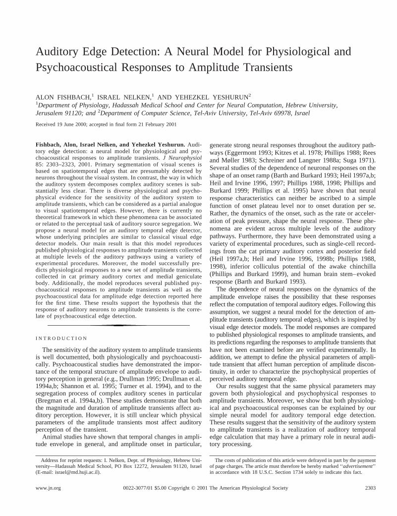
Auditory Edge Detection: A Neural Model for Physiological and Psychoacoustical Responses to Amplitude Transients ALON FISHBACH, 1 ISRAEL NELKEN, 1 AND YEHEZKEL YESHURUN 2 1 Department of Physiology, Hadassah Medical School and Center for Neural Computation, Hebrew University, Jerusalem 91120; and 2 Department of Computer Science, Tel-Aviv University, Tel-Aviv 69978, Israel Received 19 June 2000; accepted in final form 21 February 2001 Fishbach, Alon, Israel Nelken, and Yehezkel Yeshurun. Audi- tory edge detection: a neural model for physiological and psy- choacoustical responses to amplitude transients. J Neurophysiol 85: 2303–2323, 2001. Primary segmentation of visual scenes is based on spatiotemporal edges that are presumably detected by neurons throughout the visual system. In contrast, the way in which the auditory system decomposes complex auditory scenes is sub- stantially less clear. There is diverse physiological and psycho- physical evidence for the sensitivity of the auditory system to amplitude transients, which can be considered as a partial analogue to visual spatiotemporal edges. However, there is currently no theoretical framework in which these phenomena can be associated or related to the perceptual task of auditory source segregation. We propose a neural model for an auditory temporal edge detector, whose underlying principles are similar to classical visual edge detector models. Our main result is that this model reproduces published physiological responses to amplitude transients collected at multiple levels of the auditory pathways using a variety of experimental procedures. Moreover, the model successfully pre- dicts physiological responses to a new set of amplitude transients, collected in cat primary auditory cortex and medial geniculate body. Additionally, the model reproduces several published psy- choacoustical responses to amplitude transients as well as the psychoacoustical data for amplitude edge detection reported here for the first time. These results support the hypothesis that the response of auditory neurons to amplitude transients is the corre- late of psychoacoustical edge detection. INTRODUCTION The sensitivity of the auditory system to amplitude transients is well documented, both physiologically and psychoacousti- cally. Psychoacoustical studies have demonstrated the impor- tance of the temporal structure of amplitude envelope to audi- tory perception in general (e.g., Drullman 1995; Drullman et al. 1994a,b; Shannon et al. 1995; Turner et al. 1994), and to the segregation process of complex auditory scenes in particular (Bregman et al. 1994a,b). These studies demonstrate that both the magnitude and duration of amplitude transients affect au- ditory perception. However, it is still unclear which physical parameters of the amplitude transients most affect auditory perception of the transient. Animal studies have shown that temporal changes in ampli- tude envelope in general, and amplitude onset in particular, generate strong neural responses throughout the auditory path- ways (Eggermont 1993; Kitzes et al. 1978; Phillips 1988; Rees and Møller 1983; Schreiner and Langner 1988a; Suga 1971). Several studies of the dependence of neuronal responses on the shape of an onset ramp (Barth and Burkard 1993; Heil 1997a,b; Heil and Irvine 1996, 1997; Phillips 1988, 1998; Phillips and Burkard 1999; Phillips et al. 1995) have shown that neural response characteristics can neither be ascribed to a simple function of onset plateau level nor to onset duration per se. Rather, the dynamics of the onset, such as the rate or acceler- ation of peak pressure, shape the neural response. These phe- nomena are evident across multiple levels of the auditory pathways. Furthermore, they have been demonstrated using a variety of experimental procedures, such as single-cell record- ings from the cat primary auditory cortex and posterior field (Heil 1997a,b; Heil and Irvine 1996, 1998b; Phillips 1988, 1998), inferior colliculus potential of the awake chinchilla (Phillips and Burkard 1999), and human brain stem– evoked response (Barth and Burkard 1993). The dependence of neural responses on the dynamics of the amplitude envelope raises the possibility that these responses reflect the computation of temporal auditory edges. Following this assumption, we suggest a neural model for the detection of am- plitude transients (auditory temporal edges), which is inspired by visual edge detector models. The model responses are compared to published physiological responses to amplitude transients, and its predictions regarding the responses to amplitude transients that have not been examined before are verified experimentally. In addition, we attempt to define the physical parameters of ampli- tude transient that affect human perception of amplitude discon- tinuity, in order to characterize the psychophysical properties of perceived auditory temporal edge. Our results suggest that the same physical parameters may govern both physiological and psychophysical responses to amplitude transients. Moreover, we show that both physiolog- ical and psychoacoustical responses can be explained by our simple neural model for auditory temporal edge detection. These results suggest that the sensitivity of the auditory system to amplitude transients is a realization of auditory temporal edge calculation that may have a primary role in neural audi- tory processing. Address for reprint requests: I. Nelken, Dept. of Physiology, Hebrew Uni- versity—Hadassah Medical School, PO Box 12272, Jerusalem 91120, Israel (E-mail: [email protected]). The costs of publication of this article were defrayed in part by the payment of page charges. The article must therefore be hereby marked ‘‘advertisement’’ in accordance with 18 U.S.C. Section 1734 solely to indicate this fact. 2303 0022-3077/01 $5.00 Copyright © 2001 The American Physiological Society www.jn.org
-
Upload
independent -
Category
Documents
-
view
2 -
download
0
Transcript of Auditory edge detection: a neural model for physiological and psychoacoustical responses to...
Auditory Edge Detection: A Neural Model for Physiological andPsychoacoustical Responses to Amplitude Transients
ALON FISHBACH,1 ISRAEL NELKEN,1 AND YEHEZKEL YESHURUN2
1Department of Physiology, Hadassah Medical School and Center for Neural Computation, Hebrew University,Jerusalem 91120; and2Department of Computer Science, Tel-Aviv University, Tel-Aviv 69978, Israel
Received 19 June 2000; accepted in final form 21 February 2001
Fishbach, Alon, Israel Nelken, and Yehezkel Yeshurun.Audi-tory edge detection: a neural model for physiological and psy-choacoustical responses to amplitude transients.J Neurophysiol85: 2303–2323, 2001. Primary segmentation of visual scenes isbased on spatiotemporal edges that are presumably detected byneurons throughout the visual system. In contrast, the way in whichthe auditory system decomposes complex auditory scenes is sub-stantially less clear. There is diverse physiological and psycho-physical evidence for the sensitivity of the auditory system toamplitude transients, which can be considered as a partial analogueto visual spatiotemporal edges. However, there is currently notheoretical framework in which these phenomena can be associatedor related to the perceptual task of auditory source segregation. Wepropose a neural model for an auditory temporal edge detector,whose underlying principles are similar to classical visual edgedetector models. Our main result is that this model reproducespublished physiological responses to amplitude transients collectedat multiple levels of the auditory pathways using a variety ofexperimental procedures. Moreover, the model successfully pre-dicts physiological responses to a new set of amplitude transients,collected in cat primary auditory cortex and medial geniculatebody. Additionally, the model reproduces several published psy-choacoustical responses to amplitude transients as well as thepsychoacoustical data for amplitude edge detection reported herefor the first time. These results support the hypothesis that theresponse of auditory neurons to amplitude transients is the corre-late of psychoacoustical edge detection.
I N T R O D U C T I O N
The sensitivity of the auditory system to amplitude transientsis well documented, both physiologically and psychoacousti-cally. Psychoacoustical studies have demonstrated the impor-tance of the temporal structure of amplitude envelope to audi-tory perception in general (e.g., Drullman 1995; Drullman et al.1994a,b; Shannon et al. 1995; Turner et al. 1994), and to thesegregation process of complex auditory scenes in particular(Bregman et al. 1994a,b). These studies demonstrate that boththe magnitude and duration of amplitude transients affect au-ditory perception. However, it is still unclear which physicalparameters of the amplitude transients most affect auditoryperception of the transient.
Animal studies have shown that temporal changes in ampli-tude envelope in general, and amplitude onset in particular,
generate strong neural responses throughout the auditory path-ways (Eggermont 1993; Kitzes et al. 1978; Phillips 1988; Reesand Møller 1983; Schreiner and Langner 1988a; Suga 1971).Several studies of the dependence of neuronal responses on theshape of an onset ramp (Barth and Burkard 1993; Heil 1997a,b;Heil and Irvine 1996, 1997; Phillips 1988, 1998; Phillips andBurkard 1999; Phillips et al. 1995) have shown that neuralresponse characteristics can neither be ascribed to a simplefunction of onset plateau level nor to onset duration per se.Rather, the dynamics of the onset, such as the rate or acceler-ation of peak pressure, shape the neural response. These phe-nomena are evident across multiple levels of the auditorypathways. Furthermore, they have been demonstrated using avariety of experimental procedures, such as single-cell record-ings from the cat primary auditory cortex and posterior field(Heil 1997a,b; Heil and Irvine 1996, 1998b; Phillips 1988,1998), inferior colliculus potential of the awake chinchilla(Phillips and Burkard 1999), and human brain stem–evokedresponse (Barth and Burkard 1993).
The dependence of neural responses on the dynamics of theamplitude envelope raises the possibility that these responsesreflect the computation of temporal auditory edges. Following thisassumption, we suggest a neural model for the detection of am-plitude transients (auditory temporal edges), which is inspired byvisual edge detector models. The model responses are comparedto published physiological responses to amplitude transients, andits predictions regarding the responses to amplitude transients thathave not been examined before are verified experimentally. Inaddition, we attempt to define the physical parameters of ampli-tude transient that affect human perception of amplitude discon-tinuity, in order to characterize the psychophysical properties ofperceived auditory temporal edge.
Our results suggest that the same physical parameters maygovern both physiological and psychophysical responses toamplitude transients. Moreover, we show that both physiolog-ical and psychoacoustical responses can be explained by oursimple neural model for auditory temporal edge detection.These results suggest that the sensitivity of the auditory systemto amplitude transients is a realization of auditory temporaledge calculation that may have a primary role in neural audi-tory processing.
Address for reprint requests: I. Nelken, Dept. of Physiology, Hebrew Uni-versity—Hadassah Medical School, PO Box 12272, Jerusalem 91120, Israel(E-mail: [email protected]).
The costs of publication of this article were defrayed in part by the paymentof page charges. The article must therefore be hereby marked ‘‘advertisement’’in accordance with 18 U.S.C. Section 1734 solely to indicate this fact.
23030022-3077/01 $5.00 Copyright © 2001 The American Physiological Societywww.jn.org

M E T H O D S
Neural model principles
In line with the auditory-visual edge detection analogy, we adapteda model of visual edge detection to the auditory modality. Thefundamental principle of the operation of visual edge detector is thecalculation of a local brightness gradient. This is accomplished bydifferentiating the brightness function along some spatial direction ordirections, using a combination of inhibitory and excitatory connec-tions. The spatial organization of these connections in terms of theretinal image induces a receptive field that might be functionallydescribed as an edge detector. Although there are recent and moreelaborated visual receptive fields models, the simplest edge detectingreceptive field model (Marr 1982; Rodieck 1965), which has anon-center off-surround (or vice versa) response pattern, suffices forour purpose. This receptive field describes the responses of edgedetector neurons that can be found mostly in sub-cortical visualcenters. The spatial properties of an idealized receptive field can beapproximated by the second derivative of a gaussian or a difference oftwo gaussians (DOG), one wider than the other.
To adapt such a mechanism to auditory temporal edge detection, wehypothesize the existence of a temporal delay dimension, analogous to
the visual spatial dimensions. The stimulus is progressively delayedalong this delay dimension. Information related to the temporal dy-namics of the amplitude envelope (e.g., its rate of change) can bemade explicit by differentiating the stimulus along this dimension, asthe visual brightness gradient is made explicit by differentiating thestimulus along a spatial dimension.
We construct the delay dimension by using the well-known tem-poral characteristics of a standard version of the integrate-and-firemodel (I&F). Our I&F makes use of a kernel function in the form
K~x! 51
tm2 xe2x/tm x $ 0 (1)
The kernel function, when convolved with the neuron’s presynap-tic input, determines its postsynaptic potential (Gerstner 1999a).tm
is the membrane time constant that may range from 3 to 25 ms(McCormick et al. 1985). Highertm values induce greater delay inthe neuron’s response (Agmon-Snir and Segev 1993). Inducing areceptive field in the delay dimension can be done by connectingthe neurons with increasingtms to an edge detector neuron usinginhibitory and excitatory connections with various efficacies thatreflect the receptive field shape. Differentiation of the stimuli is
FIG. 1. Schematic diagram of the model. See detailed explanation inMETHODS.
2304 A. FISHBACH, I. NELKEN, AND Y. YESHURUN

obtained by using a receptive field shape of a first-order derivativeof a gaussian.
Figure 1 presents a schematic diagram of the model and the flow ofdata along the different model components. Each model component isannotated with an approximate expression for its operation on itsinput. These formulations will be used in the analysis of the model.Exact implementation details are given inAPPENDIX A. The inputstested consisted of tone bursts shaped withON and OFF ramps ofvarious shapes. An example of a tone burst with linear ramps isdisplayed in Fig. 1A.
NEURAL REPRESENTATION. The neural representation (Fig. 1B) isroughly the expected peripheral representation of sound by the innerhair cell DC potential. This representation is generated using a simplepreprocessing that includes demodulation to extract the temporalenvelope, non-linear compression and low-pass filtering. In our anal-ysis we formulate the demodulation and the non-linear compressionusing the amplitude envelope of the input converted to dB SPL scale(the constants in Fig. 1B are set toA 5 20/ln (10) andP0 5 2 z 1025
Pa). The form of the argument to the log transformation eases theanalysis for near-zero values oft and has negligible effect fort @ 0.The low-pass filtering is formulated by convolving the log-envelopewith an alpha kernel function (Eq. 1) with a time constant oft1, whichis in the millisecond range (Hewitt and Meddis 1990; Smith 1988).This preprocessing stage can be replaced by a more realistic inner haircell model (which produces simulation of auditory nerve firing prob-abilities) (Hewitt and Meddis 1990; as implemented by Slaney 1998)without any qualitative change in the response characteristics of themodel.
DELAY LAYER. The preprocessed input is fed to the delay layer ofthe model, which consists of standard integrate & fire (I&F) neuronswith ascending membrane time decay constants. Each unitU(t, t2) inthe delay layer represents a population of neurons with identicalcharacteristics. The population response is modeled as an analoguevariable, by convolving the neuronal representationN(t), with a kernel(Gerstner 1999b) whose time constant ist2. I&F kernel functions andmembrane time-constant values are shown for several units (Fig. 1C).The membrane potential of each neuron in the delay layer is thensaturated using a sigmoidal function
S~x! 5 Fmax$2/@1 1 e2~x/C!# 2 1% (2)
where Fmax is the maximal instantaneous output firing rate (225spikes/s) andC is a scaling factor, which determines the dynamicrange of the transformation. In Fig. 3, the outputs of the delay layerneurons (including various amounts of saturation) are shown forstimuli, similar to the stimulus presented in Fig. 1.
RECEPTIVE FIELD. The delay layer neurons are connected to an edgedetector neuron using inhibitory and excitatory connections withvarious efficacies (Fig. 1D) that reflect the receptive field shape,which is a first derivative of a gaussian. The output of the receptivefield, R(t), is shown in Fig. 3 for stimuli similar to the stimuluspresented in Fig. 1 and is approximately a smoothed first derivative ofthe outputs of the delay neurons along thet2 dimension.
EDGE DETECTOR NEURON. The edge detector neuron (Fig. 1E) is asingle I&F neuron with a membrane time constantt3. The output ofthe edge detector neuron is also the output of the model. In thenumerical implementation of the model, a noisy integration was used(Gerstner 1999a). For the analytical treatment presented here, themembrane potential of the edge detection neuron,M(t), is modeled asa low-pass filter operating on the output of the receptive field operator,R(t).
PARAMETERS OF THE MODEL. The responses of the model areadjusted to fit the response of a specific neuron by adjusting twoparameters. The first parameter isC, the scaling factor of the delaylayer saturation transformation (Eq. 2), and the second parameter ist3,the membrane time constant of the edge detector neuron. In addition,
the threshold of the edge detector neuron was varied. However, thethreshold was not manipulated independently; instead, its value wasalways set to best approximate the threshold of the neuron that wasfitted. There are six additional fixed parameters of the model; three ofthem are parameters of the I&F model. These parameters and theirvalues are listed in full inAPPENDIX A. Their specific values have onlyminor or redundant effect on the responses of the model. For example,changing the value oft1 or the range oft2 that are used in the delaylayer can be in large extent be compensated by adjusting the valueof t3.
Physiological methodsANIMALS AND PREPARATION. Neurons have been recorded in pri-mary auditory cortex (AI) and medial geniculate body (MGB) of twohalothane-anesthetized adult cats. The methods have been describedin details elsewhere (Nelken et al. 1999). In short, the cats werepremedicated with xylazine (0.1 ml im), and anesthesia was inducedby ketamine (30 mg/kg im). The radial vein, the femoral artery, andthe trachea were cannulated. Blood pressure and CO2 levels in thetrachea were continuously monitored. The cat was respirated with amixture of O2/N2O (30%/70%) and halothane (0.2–1.5%, as needed).Halothane level was set so that arterial blood pressure was keptaround 100 mmHg on the average. Under these conditions, the catusually could be respirated without the use of muscle relaxants. Incase muscle relaxants were required, the depth of anesthesia wasevaluated by testing paw withdrawal reflexes before administeringlow levels (pancuronium bromide, 0.05–0.1 mg iv, typically onceevery 2–3 h). Lactated ringer was continuously given through thevenous catheter (10 ml/h). Every 8–12 h a chemical analysis ofarterial blood was performed. When the cat developed acidosis, bi-carbonate was given (typically 5 ml iv, every 8 h).
AI was accessed using standard methods. To reach the MGB,electrodes were introduced at the appropriate stereotactic coordinates.Physiological characteristics of the neuronal activity were used toposition the electrode at the ventral division of the MGB. The elec-trodes were stained with DiI, and the localization was verified after theexperiments using histological reconstruction of the electrode tracks.The animal protocol was approved by the local animal care commit-tee.
DATA ACQUISITION. Glass-coated tungsten electrodes (locallymade) were used for recording neuronal activity. The activity from theelectrodes was amplified (MCP8 Plus, Alpha-Omega), and spikeswere detected on-line by a spike sorter (MSD, Alpha-Omega). Thetimes of the spikes were recorded (ET1, TDT) and written into a filefor off-line analysis.
ACOUSTIC STIMULATION. Stimuli were generated digitally con-verted to analog waveforms and attenuated using TDT equipment. Allstimuli were tone bursts, 230 ms long including the symmetrical onsetand offset ramps. Six types of onset/offset window shapes were used,cos2 (t), cos4 (t), t, t2, t4, and squared exponential. By denoting theplateau peak pressure in Pascal units withP, and the onset rise timein milliseconds withD, the peak pressure (in Pa) during the onset isgiven by
El~t! 5 PS t
DDn
for 0 # t # D n 5 $1, 2, 4% (3)
for the t, t2, andt4 windows, and is given by
Ec~t! 5 P cosn S tp
2D1
p
2D for 0 # t # D n 5 $2, 4% (4)
for the cos2 (t) and cos4 (t) windows. For the squared exponentialwindow, the peak level (in dB instead of in Pa) is given byEq. 3withn 5 2, except thatP is given in dB. To accommodate the peakpressure close to 0 Pa (at the beginning of the onset and the end of the
2305NEURAL MODEL FOR AMPLITUDE TRANSIENT DETECTION

offset), where the dB scale is singular, a short linear ramp was used upto peak sound levels of about 0 dB SPL.
Onset window shapes were generated either using an electronicswitch (SW2, TDT) or in the digital domain (for the squared expo-nential windows). The sound was presented to the animal throughelectrostatic earphones (Sokolich) whose frequency response variedby less than 10 dB in the frequency range used here. In situ calibrationof the earphones was performed in each ear.
For the data presented here, neurons were presented with tonebursts at their best frequency. Tone levels were chosen from about 10dB below neuronal threshold and up to about 100 dB SPL, in 10 dBsteps. Tone rise times covered the range of 1.7–100 ms and weremeasured between 10 and 90% amplitude points when generatedusing the electronic switch, or between 0 and 100% amplitude pointswhen generated in the digital domain. Data were taken in blocks,within which the window shape was kept constant, but the tone levelvaried randomly under the constraint that each level was presented 20times. Stimuli were presented at a rate of 1/s. After a block wasfinished, another window shape (or a different rise time) was selected,and the process was repeated. In total, 19 neurons in AI and 9 neuronsin MGB were tested with these stimuli. Of these, data from 11 neuronsin AI and 4 neurons in MGB, whose responses were strong and stableduring the recording session, were analyzed for this paper.
Psychoacoustical methods
The main goal of our psychoacoustical experiments was to testwhether the perception of amplitude changes is determined by thegradient of the change, or by some other combination of its durationand magnitude. A secondary goal was to rule out the possibility thatthe sensitivity of the auditory system to amplitude changes is due toa spectral splatter that may be induced by the sudden amplitudechange. In order to accomplish these goals we used a direct measureof the way in which the amplitude change is being perceived, ratherthan measuring amplitude change effect on higher perceptual tasks.This enabled us to isolate the perception of the amplitude transientfrom the context of more elaborate auditory phenomena such asauditory source segregation, in order to avoid high-level cognitiveinfluences. Two sets of experiments were conducted; the first mea-sured the discontinuity perception of ramped sinusoids (experiment 1),while the second measured the perception of ramped noise bursts(experiment 2).
PARTICIPANTS. All participants were normal hearing volunteeradults, who participated with full informed consent. Data forexperi-ment 1were obtained from 10 participants. All except for one, who isone of the authors (YY), had no previous listening experience inpsychoacoustical experiments. Data forexperiment 2were obtainedfrom five participants. None had participated inexperiment 1,andnone had previous listening experience in psychoacoustic experi-ments.
STIMULI. Experiment 1stimuli are pure tones with an amplitudeenvelope as illustrated in Fig. 2 (solid line). Onset and offset times are150 ms, and both plateau amplitude periods are 1 s. The first plateaulevel (A1), the amplitude ramp size (DA) and duration (DT), and thefrequency of the tone were manipulated. The values used appear inTable 1. The set of stimuli is a full combination of the variable’svalues, thus forming a set of 224 unique stimuli, each of which waspresented once. The stimuli were generated digitally and played overa Silicon-Graphics Indigo workstation at sampling rate of 16,000 Hzat 16-bit resolution.
The stimuli used inexperiment 2were prepared by Olsen (1994).All stimuli were broadband noise bursts, 700 ms in duration, 0–22kHz bandwidth, uniform random, digitally generated using a PCcomputer and signal processing software (Signal, Engineering De-sign). The amplitude envelope of the noise burst was shaped by
multiplying the signal with a trapezoidal function, which is illustratedin Fig. 2 (dashed line) and contained 96-ms onset/offset time and 104ms of plateau level before and after the pedestal. The values of thevariables used in this experiment can be found in Table 2. The set ofstimuli is a full combination of the variable’s values, thus forming aset of 36 unique stimuli, each of which was presented 5 times.Stimulus levels for both experiments were calibrated using General-Audio 1562-Z audiometer calibration set.
PROCEDURE. An identical procedure was used in both experiments.The stimuli were presented binaurally through Yamaha HP-2 ear-phones to the participants who were seated in a soundproof room. Thepsychophysical task was to judge whether the transition between thetwo plateau amplitude levels was a continuous or discontinuous one.The participants were asked to indicate their choice for each of thestimuli using a two-alternative forced choice procedure. A randomtraining subset of 40 trials was presented to the listeners, followed bythe entire set presented in random order. The listeners were unawareof the fact that the first trials were training trials. Participants hadunlimited time to respond after each trial and were presented with thenext trial 2 s after their response.
R E S U L T S
Neural model: general observations
The model was capable of reproducing all the physiologicalcharacteristics of onset responses in AI neurons. In particular,the model was capable to produce the shortening of latencieswith increase in tone level, and was capable of generating bothmonotonic and non-monotonic rate-level functions.
Figure 3 illustrates the way the model responds to ampli-tude transients and the effect of the delay layer’s saturationon the timing and strength of the responses. The log-com-pressed envelopes of linearly shaped 30-dB SPL and 90-dBSPL tone bursts are shown in Fig. 3A. The response of themodel components to these stimuli is considered in twodifferent saturation conditions. A model with a highly sat-urated delay layer, which yields non-monotonic responses,is described in Fig. 3,B, D, andF, while a model with onlyweakly saturated delay layer is described in Fig. 3,C, E,andG. For clarity, we consider a simplified delay layer thatconsists of only two neurons with time constants of 3 and 6ms. Figure 3,B andC, demonstrates the different delays ofthe stimulus envelope that are being induced by the twoneurons. The outputs of the two neurons are subtracted byconnecting them to the edge detector neuron with weights of
FIG. 2. Illustration of the amplitude envelopes of the stimuli that were usedin experiments 1(solid line) and2 (dashed line). In both experimentsDA, DT,andA1 were manipulated. Note that the time scales for the 2 experiments aredifferent. The total duration of the tone stimuli used inexperiment 1is2,3001 DT ms, while the duration of the noise bursts used inexperiment 2is700 ms.
2306 A. FISHBACH, I. NELKEN, AND Y. YESHURUN

equal magnitude and opposite signs (Fig. 3,D andE). Theprominent effect of the amount of saturation on the modelresponses emerges at this stage. For example, the totalcurrent (the integrated presynaptic input) that is being in-jected to the edge detector neuron in the highly saturatedmodel (Fig. 3D) is higher in response to the 30-dB tone thanto the 90-dB tone (155 vs. 89.7 in arbitrary units, respec-tively). In the weakly saturated model (Fig. 3E), the inte-grated presynaptic input is lower in response to the 30-dBtone that the 90-dB tone (81.9 vs. 246.8, respectively). Thenon-monotonicity of the highly saturated model is enhancedby the low-pass properties of the membrane potential of theedge detector neuron (Fig. 3F). The effect of the delaylayer’s saturation on the non-monotonicity of the model isbeing mathematically analyzed inAPPENDIX B. Another effectof the saturation is decreasing the first-spike latency andshortening the period of neural activity. For the purpose ofmathematical treatment, it can be reasonably assumed that aneuron starts to fire when its membrane potential hits a fixedthreshold and that its spike count is proportional to the areaenclosed by this threshold and the neuron’s membrane po-tential (Fig. 3G).
Evaluation of the neural model: single-neuron data
We evaluated the adequacy of the model to match reportedneural response to sound bursts by feeding the model with theamplitude envelope of the stimuli and comparing several as-pects of the model output with those of the reported responses.The properties of the output examined were the first spikelatency of the response, the response strength measured by thenumber of spikes that followed a stimulus, and the relation-ships between the two.
LATENCY. Heil and his co-workers (Heil 1997a; Heil andIrvine 1996) studied the latency of primary auditory cortexneurons (AI) as a function of the shape, amplitude, and dura-tion of the rise time of a best frequency tone. Two kinds ofonset envelope functions were used, linear and cosine-squared.The peak amplitude during a linear onset is described by apower function as described inEq. 3 with n 5 1. The peakamplitude during a cosine-squared onset is described byEq. 4,with n 5 2.
As was stated earlier, the main finding of Heil and hisco-workers is that mean latency is not solely a function of oneparameter of the onset envelope, but rather a function of thedynamics of the envelope. The latency of response appears tobe a function of the rate of rise of the onset when a linearshaped onset is used, and a function of maximal acceleration ofthe envelope for cosine-squared onsets. Moreover, Heil pro-posed a functional expression for the relationships between theresponse latency and rate of rise (for linear onsets) or maximal
acceleration of peak pressure (for cosine-squared onsets). Thefunction for the linear case is given by
Ll 5 Lmin 1 Al p Flog SP
DD1 SG24
(5)
whereAl is a global scaling factor, andLmin andS are neuronspecific parameters that determine the minimal latency of theneuron and its sensitivity to onset rate of rise, respectively.
The function for cosine squared onsets is given by
Lc 5 Lmin 1 Ac p Flog Sp2P
2D2D1 SG24
(6)
Note that the termp2P/2D2 stands for the maximal accelera-tion of the envelope, which occurs at the beginning of theonset. Heil fit global scaling factorsAl andAc over the entireneural population that was recorded and set them to 1,277 and12,719 ms, respectively.
We fitted the model parameters to match the responses of 13AI neurons for which both latency and spike-count data arefully reported by Heil (1997a,b). For all of these neurons wefound that the model reproduced the latency phenomena thatwere measured by Heil. The latency data for two of theseneurons is shown in Fig. 4.
Figure 4,A and B, shows the experimental vs. simulatediso-rise-time curves of first-spike latency as a function ofamplitude peak pressure of a cosine-squared onset. Figure 4,Cand D, demonstrates that plotting both the experimental andsimulated latency as a function of maximal acceleration of thecosine-squared onset brings the iso-rise-time curves to closecongruence along a single curve that can be fitted byEq. 6.Figure 4,E andF, shows the congruence of the iso-rise-timecurves as a function of rate of linear rise onset.
Phillips (1998) and Heil and Irvine (1998b) reported theresponses of single neurons in the cat primary auditory cortexand the posterior field to characteristic frequency (CF) toneswith cosine-squared–shaped onsets. These data confirmed theinitial observations of Heil and his co-workers in AI andextended them to a secondary cortical field. Figure 5,A andC,replots the first-spike latency of two neurons from the posteriorfield as reported by Phillips, and Fig. 5,B andD, plots the fitof the model to this data. Plotting Phillips’ latency data as afunction of maximal acceleration of the cosine-squared onsetdemonstrates again the close congruence of the latency dataalong a single curve, which can be fitted byEq. 6.
FIXED-THRESHOLD MODEL DOES NOT FIT THE DATA. A possibleexplanation for the latency phenomena is that the neuron firstspike occurs when the input stimuli level hits a fixed threshold(Kitzes et al. 1978; Phillips 1988; Suga 1971). Indeed, it is easyto show that such a simple model predicts a reciprocal relationbetween the first-spike latency of a neuron and the rate (P/D)of linear onsets and maximum acceleration (p2P/2D2) of co-sine-squared onsets. While these predictions roughly approxi-mate the experimental results, the later systematically deviatefrom the predictions. On these grounds, Heil and Irvine (1996)
TABLE 1. Values of variables used in experiment 1
DT, ms 6 12 18 24 40 96 120DA, dB 6 12 18 24f, Hz 250 400 650 950A1, dB SPL 60 73
TABLE 2. Values of variables used in experiment 2
DT, ms 3 6 12 24 40 96DA, dB 12 18A1, dB SPL 59 65 71
2307NEURAL MODEL FOR AMPLITUDE TRANSIENT DETECTION

argue against the simple threshold model. Their claims can besummarized by two main points that are illustrated in Fig. 6.
First, the threshold model predicts that the first-spike latencyshould be a linear function of rise time (see dashed lines in Fig.6A). The experimental data of Heil and Irvine (1996; Heil
1997a) show a systematically deviation from this prediction.Notably, the relation between the latency and the rise time iscompressive, which rules out the possibility that adaptive pro-cesses are the cause for this deviation.
The second argument relates to the slopes of the quasi-linear
FIG. 3. The output of several of the modelcomponents as response to sound bursts of 2amplitude levels (A). Two model settings areshown, the 1st includes highly saturated delaylayer (B, D, andF), while the 2nd is only mod-erately saturated (C, E,andG). The figure dem-onstrates how the input is being progressivelydelayed along a simplified delay layer (B andC)and differentiated using the receptive field (Dand E) that is formed by the connections fromthe delay layer neurons to the edge detectorneuron (F andG). For mathematical analysis ofthe model we define the 1st-spike latency of themodel as the time from stimulus onset to the 1sttime the edge detector membrane potential hits afixed threshold level (L30 and L90 in G). Thespike count is assumed to be proportional to thearea enclosed by the membrane potential and thethreshold level (striped area inG).
2308 A. FISHBACH, I. NELKEN, AND Y. YESHURUN

iso-step-size curves, which should decrease, according to thefixed threshold model, as the inverse ratio of the step sizes.Heil and Irvine demonstrate that the slopes of the curvesdecrease by a factor that is smaller than expected. Similardeviation from the threshold model predictions have beenobserved in the first-spike latency of cortical neurons as re-sponse to cosine-squared onsets (see Heil 1998 for reanalysisof the data of Phillips 1998); in the response latency of inferiorcolliculus potential in unanesthetized chinchillas to cosine-squared onsets (Phillips and Burkard 1999) and in the responselatency of evoked cortical potentials in humans as response tolinear onsets (Onishi and Davis 1968). Since our model repro-duces very accurately the reported latency phenomena, it alsoshows deviations from the predictions of the fixed thresholdmodel (Fig. 6B).MATHEMATICAL ANALYSIS OF LATENCY PHENOMENA. In ouranalysis we use the formulations given in Fig. 1, and weassume that the amplitude envelope of the input stimulus,E(t),
can be approximated during the onset (fort # D) by a powerfunction such as described inEq. 3 for any n . 0. Forsimplicity sake we will restrict our analysis tot # D; thisassumption is equivalent to the statement that the first spikeoccurred during the onset ramp (after taking into accountconstant latency components that are independent of the soundlevel).
As illustrated in Fig. 3G, we assume that the edge detectorneuron starts firing when its membrane potential,M(t), hits afixed threshold level,T. Thus the time of the first spike,t*,satisfies the condition:M(t*) 5 T. Although t* can be calcu-lated numerically using the implicit functional formM(t*) 5 T(as it is actually done in the process of fitting the model freeparameters to match the experimental data), we are unable toextract an explicit expression fort* that can replace Heil’sfunctional forms (Eqs. 5and 6). However, the implicit func-tional form is useful in order to prove several characteristics ofexperimental and simulated latency phenomena, and to predict
FIG. 4. Experimental (replotted from Heil1997a) vs. model simulated data for 1st-spike la-tency of the onset response.A andB: the latencyas a function of the amplitude level.C andD: thelatency as a function of maximal acceleration ofthe cosine-squared onset and the curve fitted byEq. 6.Our fit for the neuron inC yieldedS5 4.53andLmin 5 10.87 ms, and for the simulated data inD the best fit yieldedS5 5.12 andLmin 5 8.1 ms.E and F: the latency as a function of the rate oflinearly shaped onset and the curve fitted byEq. 5.The fit for the neuron inE yielded S 5 4.9 andLmin 5 11.55 ms and for the model (F) S 5 5.09andLmin 5 6.7 ms. Neuron identity, neuron char-acteristic frequency (CF), and the model parame-ters that were used are shown above each plot. Thedifference between experimental and simulatedLmin values reflects constant delays (acoustic, co-chlear, and neural delays), which are not includedin the model. ModelS values are consistentlysomewhat higher than those estimated from thedata, as explained inDISCUSSION.
2309NEURAL MODEL FOR AMPLITUDE TRANSIENT DETECTION

latency behavior as response to stimuli that were not examinedexperimentally.
SinceM(t) includes only non-linear compression and lineartime-invariant filtering ofE(t), it is clear thatt*, as a functionof P and D, is being determined uniquely by the termP/Dn.This explains Heil’s findings regarding the latency being afunction of the rate of linear onsets (n 5 1) while being afunction of the maximum acceleration of cosine-squared onset(n 5 2, up to a 1st-order approximation). In addition, thisconclusion predicts that for a large family of functions that canbe approximated by a power function, the first-spike latencyfor tone bursts that are shaped using these functions should bedetermined by the termP/Dn. Moreover, we predict that forexponential power functions, such that the envelope is a powerfunction whenP is given in dB units,t* is determined by theterm P/Dn, whenP is given in dB units.
Note that the analysis in the previous paragraphs is limitedto t* # D (1st spike generation occurring during the onset
ramp). For near-threshold levels ofP, t* may exceedD, whichresults in longer latencies than predicted. This presumably isthe cause of the departures from the invariant relationshipbetween first spike latency andP/Dn at low levels ofP in bothexperimental and simulated data (e.g., Figs. 4C and 5).
Another phenomenon that can be explained by the im-plicit form of t* is of Heil and Irvine (1996) regarding thedeviations of the latency from the predictions of a fixedthreshold model. InAPPENDIX B we explore the dependenceof t* on the duration of the onset,D, and prove the com-pressive nature oft*(D) as evident in both experimental andsimulated data (Fig. 6).
It should be noted that the latency of any fixed-thresholdsystem, which includes only monotonic transformations andlinear time-invariant filtering ofE(t), as a function ofP, D, andn, is being uniquely determined by the termP/Dn. This obser-vation can account for the latency phenomena of auditorynerve fibers, reported by Heil and Irvine (1997).
FIG. 5. Latency data from the cat posterior field(replotted from Phillips 1998) vs. model simulateddata. The latency is plotted as a function of maxi-mum acceleration of peak pressure and is fittedusing Heil’s functional form. Our estimatedSvaluefor neuron 93K010.24(A) is 3.99, and the estimatedvalue for the corresponding simulated latency (B) is4.62. EstimatedSfor neuron 93K013.12(C) is 4.42and for the corresponding model setting (D) theestimation is 4.57.
FIG. 6. A: mean 1st-spike latency of a neuronfrom cat primary auditory cortex (solid lines) as afunction of rise time of a CF tone of 22 kHz (replottedfrom Heil and Irvine 1996). The dashed lines plot thebest fit of the data according to a fixed-thresholdmodel. Note that the latency is not a linear function ofrise time and that the slope ratio of any two quasi-linear iso-step-size curves does not match the inverseratio of the step sizes. The model reproduces thesephenomena (B). Note that the latency axis is trans-lated with respect toA for greater clarity.
2310 A. FISHBACH, I. NELKEN, AND Y. YESHURUN

COMPARING MODEL PREDICTIONS WITH LATENCY RESULTS OF
PHYSIOLOGICAL EXPERIMENTS. Figure 7 shows the latency dataof one AI unit in response to three types of onset windows,linear (Fig. 7A), cosine squared (Fig. 7C), and squared expo-nential (Fig. 7E). The latency for each window is plotted as afunction of the predicted invariant measure, and the alignmentof the latency data along a single curve for each rise functionvalidates our predictions. Numerical simulations reproducethese phenomena (Fig. 7,B, D, andF). Figure 8A shows thelatency of a MGB neuron in response to four types of ampli-tude rise function, cos2 (t), cos4 (t), t2, and t4. The latency ofeach rise function is plotted as a function of the predictedinvariant measure, which isP/Dn for the tn rise functions andpnP/2nDn for the cosn (t) rise functions {Taylor’s series ap-proximation of cosn [(p/2)t 1 (p/2)] is (pn/2n)tn 1 o(tn12) forevenn}. This way the latency data collected with thet2 and thecos2 (t) rise function aligns along a single curve, and thelatency data collected with thet4 and the cos4 (t) rise function
aligns along another curve. The model predictions also hold forthe responses of a neuron in primary auditory cortex (Fig. 8C)and are being reproduced by the numerical simulations of themodel (Fig. 8,B andD).
SPIKE COUNT. Neurons in AI of anesthetized cat show a lowspontaneous rate of fire, and their typical response to soundbursts is a single spike or a short burst of a few spikesimmediately following the onset of the stimulus (e.g., Heil1997b). Examining the spike count as a function of plateaupeak pressure alone reveals a non-monotonic pattern that isshared by many AI neurons to various degrees (e.g., Heil1997b; Heil and Irvine 1998a; Phillips 1988; Schreiner andMendelson 1990). Furthermore, the non-monotonicity isenhanced at the shorter rise times. Figure 9 demonstrates thetypical response patterns of two types of neurons, as replot-ted from Heil’s (1997b) data. Figure 9A shows a highlynon-monotonic neuron, whereas Fig. 9C shows a more
FIG. 7. First-spike latency of a single unit of acat [primary auditory cortex (AI)] as response to 3rise functions, linear (A, experimental;B, simu-lated), cosine squared (C, experimental;D, simu-lated), and squared exponential (E, experimental;F, simulated). Latency is plotted as a function ofthe predicted invariant measure of each rise func-tion.
2311NEURAL MODEL FOR AMPLITUDE TRANSIENT DETECTION

monotonic neuron. The spike-count data are plotted as afunction of plateau peak pressure and are organized alongiso-rise-time curves. The model reproduces these phenom-ena over a wide range of degrees of monotonicity. Figure 9,B and D, demonstrates a good correspondence between
experimental and simulated results. The correspondence isapparent for curve shapes as well as for order of displace-ment of the iso-rise-time curves, although the displacementof the model curves along the abscissa are much larger thanthose of the neural curves.
FIG. 8. First-spike latency measured usingcos2 (t), cos4 (t), t2, andt4 rise functions froma single unit of a cat medial geniculate body(MGB; A andB, simulated) and AI (C andD,simulated). Latency data are plotted as a func-tion of the predicted invariant measure [P/Dn
for the tn rise functions andpnP/2nDn for thecosn (t) rise functions].
FIG. 9. Experimental vs. simulated spike-count data. Iso-rise-time curves of spike countsare plotted as a function of amplitude level of acosine-squared onset, for a non-monotonicneuron (A, experimental;B, simulated), and amonotonic neuron (C, experimental;D, simu-lated). Experimental data are replotted fromHeil (1997b). Simulated data for both neuronswere obtained using the same sets of parame-ters that were used to match their latency data(see Fig. 4).
2312 A. FISHBACH, I. NELKEN, AND Y. YESHURUN
The monotonicity of the model can be controlled by chang-ing the value of the two adjustable parameters, as alreadyillustrated in Fig. 3. Increasing the dynamic range of the delaylayer’s saturation and decreasing the membrane time constantincrease the monotonicity of the neuron. The relation betweenthe degree of the saturation and the monotonicity of the neuronspike count is being formally proved inAPPENDIX B. It isnoteworthy that raising the sigmoidal scaling factor of thesaturation transformation both raises the threshold and in-creases the monotonicity of the neuron. This relation betweenthe threshold and monotonicity of the neuron is consistent withpreviously reported findings (Heil et al. 1994; Sutter andSchreiner 1995).
Heil (1997b) found an interesting relationship between thespike count and the latency of the response. This relation linksthe dynamics of the onset and the number of spikes that followit. Heil demonstrated that plotting the spike count as a functionof the stimuli’s peak pressure at the moment of first-spikegeneration brings the iso-rise-time curves to close congruence.The moment of first-spike generation is defined as the meanlatency (for the given rise time and plateau peak pressure)minus the minimal latency of the neuron (as defined by thetermLmin in Eqs. 5and6). The congruence of the iso-rise-timecurves holds for both linear and cosine-squared onsets and forboth monotonic and non-monotonic neurons. Figure 10 dem-onstrates this phenomenon using the data of Heil (1997b),Phillips (1998), and the original data reported here, and showsthat the model reproduces this phenomenon for variety sets ofmodel parameters. InAPPENDIX B we analyze this special rela-tionship between the latency and the spike count of the modeland of auditory cortical neurons.
Evaluation of the neural model: evoked auditory brain stemresponses
The ability of the model to match reported evoked auditorybrain stem responses in humans (Barth and Burkard 1993) andinferior colliculus potential (ICP) in the awake chinchilla (Phil-lips and Burkard 1999) in response to sound bursts, was testedby feeding the model with the amplitude envelope of thestimuli and comparing the model output to the reported re-sponses. The membrane potential of our modeled edge detectorneuron (Fig. 1E) was used as an estimate of the combinedactivity of a large population of brain stem neurons (Gerstner1999b). The model activity was then differentiated to mimicthe analogue highpass filter (with a slope of 6 dB/oct) used inthese experiments.
Figure 11A shows a typical measure of the inferior colliculuspotential in response to a tone burst as replotted from Barth andBurkard (1993). Figure 11B shows the differentiated mem-brane potential of the edge detector neuron of the model. Thisfigure also illustrates the definitions of the latency and ampli-tude of the response. The two adjustable parameters that shapethe model’s response to amplitude transients (C andt3) wereadjusted to fit the latency and amplitude of the experimentalresponses.
In contrast to the stimuli that were used in single-cell re-cordings, whose total durations were 50–100 ms (Phillips1998) or 400 ms (Heil 1997a,b), the stimuli that were used byBarth and Burkard (1993) and by Phillips and Burkard (1999)
were much shorter and included plateau-level durations of 2–5ms. For the model to accurately reproduce the experimentalresponses to these very short bursts, we had to reduce the timeconstant of the delay layer units from a range of 3–5 ms to arange of 0.5–1 ms, since higher time constants oversmoothedthe envelope. The problem of using very short time constantswhen modeling mammalian inferior colliculus neurons has alsobeen encountered in other modeling studies (Hewitt and Med-dis 1994).
LATENCY. Phillips and Burkard (1999) measured the latencyof the ICP in the awake chinchilla in response to cosine-squared onsets of various rise times and amplitude levels.Although Phillips and Burkard reported that there were strongsimilarities between the latency behavior of the ICP and that ofcortical single cells, they did not use Heil’s functional expres-sion (seeEq. 6) to match the latency data according to themaximum acceleration of the onset envelope. Figure 12Ashows that replotting the ICP latency data as a function ofmaximum acceleration of the envelope brings the iso-rise-timecurves to converge along a single curve that can be fitted usingEq. 6 and by using the same value of the constant parameter(Ac) that was used by Heil (1997a). Figure 12B shows that themodel reproduces the ICP latency data.
Barth and Burkard (1993) measured the latency of wave Vof brain stem auditory evoked responses (BAER) in responseto linear shaped onsets. Although Barth and Burkard reportedthat both the onset rise time and amplitude affect the responselatency, they did not analyze the latency as a function of theenvelope rate of change. Figure 12C shows that replotting theBAER latency as a function of the envelope rate brings theiso-rise-time curves to close congruence. Using Heil’s func-tional form (Eq. 5) and the same value of Heil’s constant (Al)to match this curve yields a moderate fit. The model latencydata is shown in Fig. 12D.
RESPONSE AMPLITUDE. The effect of onset rise time and am-plitude level on the ICP and on wave V of BAER responseamplitude are similar to their effect on the spike count ofmonotonic cortical single cells. The response amplitude in-creased with ascending amplitude levels and with descendingonset rise times. Figure 13 replots Phillips and Burkard’s(1999) ICP amplitude response (Fig. 13A) and Barth andBurkard’s (1993) BAER wave V response amplitude (Fig.13C); both are plotted as a function of the plateau peak level.The simulated response amplitudes are presented in Fig. 13,Band D, and are scaled in order to match the experimentalmeasurements.
Results of the psychoacoustic experiments
The results of the two experiments were analyzed using astepwise logistic regression. The dependent variable was set tobe the probability of eliciting a discontinuous response, and theindependent variables included the stimuli parameters used ineach experiment (as detailed in Tables 1 and 2, respectively).In addition, motivated by our model, we added to the two setsof independent variables: the logarithm of the normalized rateof change of the ramp peak pressure re the base peak pressure
2313NEURAL MODEL FOR AMPLITUDE TRANSIENT DETECTION

FIG. 10. Experimental and simulated spike-count iso-rise-time curves are closely alignedwhen plotted as a function of stimulus peak pres-sure at 1st-spike generation. Experimental data ofHeil (1997a,b) (A, C,andE) and of Phillips (1998)(G) are recorded from single units of the cat AI.Original data from a single unit of the cat MGBare shown inI. Note that the model (B, D, F, H,andJ) reproduces this phenomenon over a broadrange of parameters.
2314 A. FISHBACH, I. NELKEN, AND Y. YESHURUN

(this is the invariant measure for the stimuli used here, see nextsection).
EXPERIMENT 1. The regression results show that the variablethat accounts for most of the variance is the normalized rate ofthe ramp peak pressure [F(1,2238)5 1,948.6,P , 10215]. Othersignificant variables are the duration of the change, [F(1,2237)560.8, P , 10212]; and the tone frequency [F(1,2236) 5 32.5,P , 1027].
EXPERIMENT 2. The results of the second experiment alsofound the normalized rate of peak pressure to be the variablethat accounts for most of the variance [F(1,898) 5 509.6,P ,10215]. Other significant variables were the first plateau am-plitude level, [F(1,897) 5 29.2,P , 1027]; the step amplitude[F(1,896)5 11.5,P , 0.0007] and the step duration [F(1,895)511.07,P , 0.0009].
Mean results across all participants for the two experimentsare plotted in Fig. 14A. As expected from the regressionanalysis, it is evident that plotting the probability data as afunction of the rate of peak pressure causes the data to alignalong a typical psychometric function.
Evaluation of the neural model: psychoacoustic data
In the following section we will compare the model re-sponses with the results of three psychoacoustical experiments.These experiments include the experiment reported above thattested the perception of amplitude discontinuity; an experimentthat tested the effect of amplitude transients on auditory seg-regation (Bregman et al. 1994b); and a forward masking ex-periment (Turner et al. 1994) that tested the effect of the proberise time on the degree of masking. Although these experi-ments investigate different auditory phenomena, we demon-strate that by identifying the psychoacoustical measures withthe responses of the neural model to the amplitude transientspresented in the experiments, the model is able to reasonablyreproduce the psychoacoustical results.
In two of these experiments (Bregman et al. 1994b and theexperiment reported here) the stimuli contained an amplituderamp rising above a pedestal. The invariant measure for thesestimuli is not the rate of rise of the amplitude ramp per se, butrather the normalized rate of rise re the pedestal,P*/Dn, whereP* is the plateau peak pressure of the ramp normalized by the
FIG. 11. A: a typical wave-V brain stem auditoryevoked response (BAER) as response to a 60-dB nHL1.25-ms rise-time noise burst replotted from Barth andBurkard (1993).B: a differentiated membrane poten-tial of the model edge detector neuron as a response tothe same stimulus without the addition of noise. Theresponse latency is measured with respect to the peakof the BAER, and the response amplitude is measuredfrom the peak to the following trough, as illustratedin A.
FIG. 12. A: replotting inferior colliculus potential(ICP) response latencies (Phillips and Burkard 1999) asa function of maximum acceleration of cosine-squaredonsets yields a good alignment of the latency data alonga curve that can be fitted by Heil’s (1997a) functionalform (Eq. 6). Our fit for the experimental ICP data yieldsS 5 5.65 andLmin 5 3.46. The model reproduces theseresults (B), with a fit of S 5 5.97 andLmin 5 1.18.C:replotting wave-V latencies (Barth and Burkard 1993) asa function of the rate of linear onsets reveals goodalignment along a curve that only moderately fits Heil’sfunctional form (Eq. 5) with S5 5.65 andLmin 5 6.43.The model matches the experimental results (D) but isbetter fitted by the functional form (S5 6.17 andLmin 52.27). Note that both Barth and Burkard (1993) andPhillips and Burkard (1999) used 0-ms rise time onsets.To allow a valid calculation of the envelope maximumacceleration and rate of change for these stimuli, wereplaced the zero rise time by a 0.185-ms value. Thisvalue was found to best match the fitted curves for boththe ICP and the BAER latency data.
2315NEURAL MODEL FOR AMPLITUDE TRANSIENT DETECTION

ratio between the pedestal peak pressure andP0 (see Fig. 1B).Intuitively, this follows from the fact that the essential opera-tion of the model is differentiating the log-compressed ampli-tude envelope. Therefore the output of the receptive field (Fig.1D) is not changed by multiplying the input stimuli by aconstant factor. In consequence, the response to a ramp rising
above a pedestal is identical to the response to the onset of asound with the same size in dB reP0 and with the same shape.Note that we arbitrarily set the value ofP0 to 0 dB SPL forsimplicity sake. Using differentP0 values can be compensatedby adjusting the threshold value of the edge detector neuron.P0value is significant only when fitting the model responses with
FIG. 13. Response amplitude of ICP (A) replot-ted from Phillips and Burkard (1999), and responseamplitude of wave-V BAER (B) replotted fromBarth and Burkard (1993) as a function of plateaupeak pressure. Note the resemblance between the 2experimental findings in response to stimuli ofcomparable parameters and between the experi-mental and simulated data (B andD).
FIG. 14. A: mean results across all participantsfor experiment 1(solid lines) andexperiment 2(dashed lines). The probability for the amplituderamp to be perceived as a discontinuous change isplotted as a function of the normalized rate of theramp peak pressure re the pedestal (see text). Thisproduces a good congruence of the data along atypical psychometric curve. A plot of the simula-tion results (B) shows a good fit with the psy-choacoustic data.C: a replot of the discriminationscore from Bregman et al. (1994b) as a functionof the normalized rate of change of the incre-mented partials. The model matches the data onlymoderately (D).
2316 A. FISHBACH, I. NELKEN, AND Y. YESHURUN

the responses of a specific neuron to both the onset of a soundand to a ramp rising above a pedestal. In these casesP0 may beadjusted to best fit the neural responses to both types of stimuli.
PERCEPTION OF AMPLITUDE DISCONTINUITY. To compare ourpsychophysical results and the model predictions, a function ofthe neural response compatible with the dichotic nature of thepsychophysical responses is required. As mentioned earlier, themodeled neurons have low spontaneous activity, and theirresponses to sound bursts consist of a short burst of 1–3 spikes.Therefore it seemed plausible to define a response to a stimulusas one or more spikes, and to identify the probability ofresponse as the probability that a participant would report adiscontinuous amplitude change in the psychophysical exper-iment. This measure did in fact yield a good match between thesimulated (Fig. 14B) and the experimental results.
EFFECT OF AMPLITUDE TRANSIENTS ON AUDITORY SEGREGATION.
One of the few studies that tested the effect of both the durationand magnitude of amplitude changes on auditory segregationtasks has been reported in Bregman et al. (1994b). Theypresented a 3.5-s long complex tone consisting of five harmon-ics of 500 Hz. The amplitudes of an adjacent pair of the threemiddle frequencies (1,000, 1,500, and 2,000 Hz) were incre-mented in succession in random order. A sufficiently largeamplitude increment caused the partials to be segregated fromthe complex tone, and to be perceived as separate tones. Tomeasure the degree of segregation, the participants had tojudge whether the perceived pitch pattern, caused by the seg-regated partials, went up or down. Three levels of incrementswere used (1, 3, and 6 dB) and six increment durations (30, 90,270, 730, 910, and 970), resulting in a total of 18 experimentalconditions. The overall amplitude level of the complex tone inits steady state was 65 dB SPL. Bregman et al. reported thatboth the amplitude increment level and the increment durationhad a significant effect on the participants’ performance.Longer increment duration resulted in poorer discriminationperformance, while larger increment levels led to better dis-crimination. These results suggest that the gradient of theincrement had a dominant effect on discrimination perfor-mance. However, Bregman et al. did not include the gradient ofthe increment in their statistical analysis, and therefore it isimpossible to determine the exact influence of the amplitudegradient of a tone on the ability to segregate it from a mixtureof tones. When the results of Bregman et al. are replotted as afunction of the normalized rate of peak pressure of the ampli-tude increment, the data fall along a single curve (Fig. 14C).
Since Bregman et al. used a continuous measure rangingfrom 0 to 5, we used the spike count of the model as thesimulated measure while using a linear transformation of thespike count data that resulted in the best fit to the psychoacous-tical results. Figure 14D shows that the model’s ability toapproximate the experimental results of Bregman et al. is onlymoderate. Formally, the model responses do not align on asingle curve because of the use of extremely shallow ramps inthis experiment (see Fig. B1 and the accompanying discussionin APPENDIX B). Interestingly, the experimental data of Bregmanet al. (1994b) are in fact invariant with respect to the normal-ized rate of rise of the ramp, implying that the rate of rise is thebehaviorally relevant variable even under these extreme con-ditions.
EFFECT OF AMPLITUDE TRANSIENTS ON RELEASE FROM
FORWARD MASKING. In forward masking, the masker (whichcan be a tone or a noise burst) masks a target tone that appearsjust after the masker ends. The degree of masking depends onmany factors such as masker level, bandwidth, duration, andthe inter-stimulus interval. Turner et al. (1994) studied theeffect of the target tone rise time and duration on forwardmasking levels. They used two types of target tones, one witha total duration of 25 ms including 2-ms cosine-squared rise/fall ramps, and the second with a total duration of 22 msincluding 10-ms cosine-squared rise/fall ramps. Growth ofmasking (GOM) functions were measured using noise maskersat levels of 10–90 dB SPL. Their results show that targets with10-ms rise time were masked more than targets of 2-ms risetime. In addition, Turner et al. showed that in contrast with thepsychoacoustical results, there was no significant effect of thetarget rise time on the amount of masking that was measured insingle auditory-nerve fibers of the chinchilla. This suggeststhat, although some forward masking effects are apparent at thelevel of the auditory periphery, the effect of target rise timemay involve higher auditory centers.
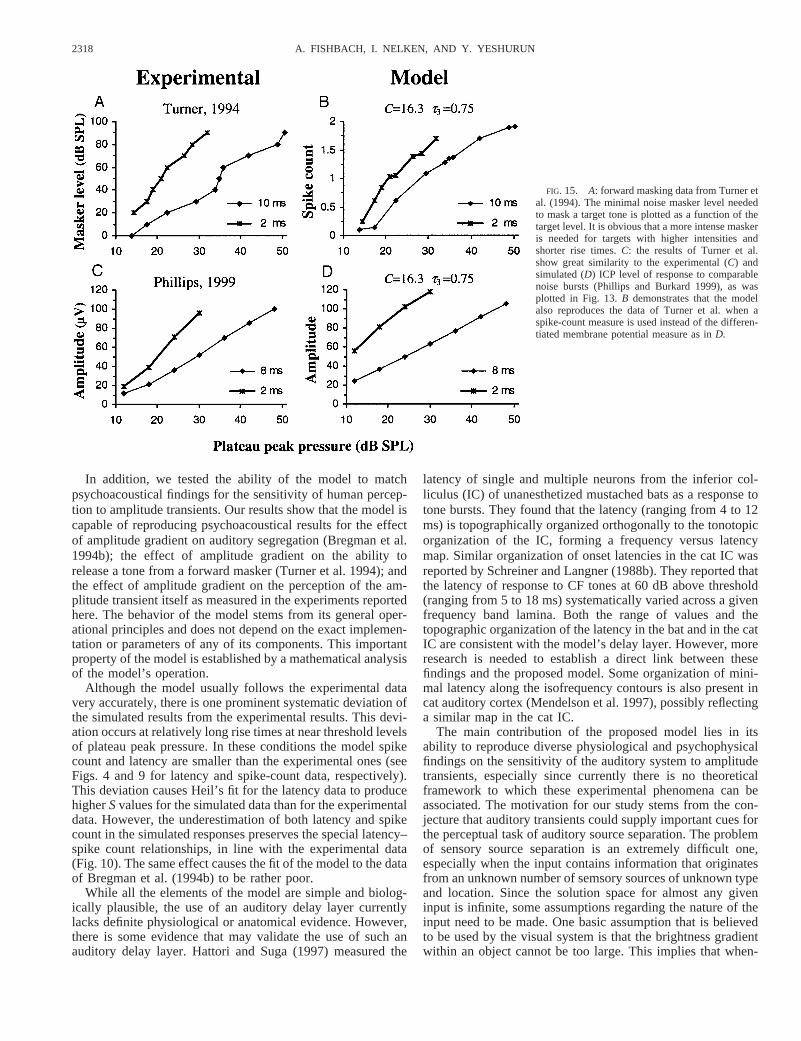
To put these results in the context of our model, we inter-preted the forward masking paradigm as a method of assessingthe strength of response produced by the target tone; the higherthe response produced by the target, the louder the masker thatis needed to mask it. Therefore we interpreted the minimalmasker level needed to mask a target tone as a measure of theresponse produced by the target. This measure is being com-pared with the strength of response produced by the neuralmodel as response to the target tone alone. Figure 15A replotsthe masker level as a function of the target level for the tworise-time targets as calculated from the data of Turner et al.Figure 15B demonstrates that these results are reproduced bythe spiking responses of the edge detector neuron in the model.In addition, the data of Turner et al. remarkably resemble theICP amplitude data of Phillips and Burkard (1999). Figure 15Creplots Phillips and Burkard’s (1999) ICP responses at com-parable parameter values, and the corresponding model re-sponses (as already shown in Fig. 13) are plotted in Fig. 15D.Thus the psychophysical data of Turner et al. can also beinterpreted by this version of the model.
D I S C U S S I O N
In the present study we describe a neural model for auditorytemporal edge detection. The core of the model is in theformation of an auditory delay dimension. Sensitivity to am-plitude edges is achieved by differentiating the stimulus alongthis dimension. We demonstrate the ability of the model toreproduce both the latency and magnitude of responses tosound bursts, as recorded from single units of the cat primaryauditory cortex and posterior field (Heil 1997a,b; Heil andIrvine 1996; Phillips 1988, 1998), inferior colliculus potentialof awake chinchilla (Phillips and Burkard 1999), and wave Vof human brain stem–evoked response (Barth and Burkard1993). Moreover, we predict the response of cortical neurons toa general family of sound bursts whose onset envelope is apower function or the exponent of a power function. Wesuccessfully verified these predictions for several of thesestimuli by recording from single units of the cat primaryauditory cortex and MGB.
2317NEURAL MODEL FOR AMPLITUDE TRANSIENT DETECTION
In addition, we tested the ability of the model to matchpsychoacoustical findings for the sensitivity of human percep-tion to amplitude transients. Our results show that the model iscapable of reproducing psychoacoustical results for the effectof amplitude gradient on auditory segregation (Bregman et al.1994b); the effect of amplitude gradient on the ability torelease a tone from a forward masker (Turner et al. 1994); andthe effect of amplitude gradient on the perception of the am-plitude transient itself as measured in the experiments reportedhere. The behavior of the model stems from its general oper-ational principles and does not depend on the exact implemen-tation or parameters of any of its components. This importantproperty of the model is established by a mathematical analysisof the model’s operation.
Although the model usually follows the experimental datavery accurately, there is one prominent systematic deviation ofthe simulated results from the experimental results. This devi-ation occurs at relatively long rise times at near threshold levelsof plateau peak pressure. In these conditions the model spikecount and latency are smaller than the experimental ones (seeFigs. 4 and 9 for latency and spike-count data, respectively).This deviation causes Heil’s fit for the latency data to producehigherSvalues for the simulated data than for the experimentaldata. However, the underestimation of both latency and spikecount in the simulated responses preserves the special latency–spike count relationships, in line with the experimental data(Fig. 10). The same effect causes the fit of the model to the dataof Bregman et al. (1994b) to be rather poor.
While all the elements of the model are simple and biolog-ically plausible, the use of an auditory delay layer currentlylacks definite physiological or anatomical evidence. However,there is some evidence that may validate the use of such anauditory delay layer. Hattori and Suga (1997) measured the
latency of single and multiple neurons from the inferior col-liculus (IC) of unanesthetized mustached bats as a response totone bursts. They found that the latency (ranging from 4 to 12ms) is topographically organized orthogonally to the tonotopicorganization of the IC, forming a frequency versus latencymap. Similar organization of onset latencies in the cat IC wasreported by Schreiner and Langner (1988b). They reported thatthe latency of response to CF tones at 60 dB above threshold(ranging from 5 to 18 ms) systematically varied across a givenfrequency band lamina. Both the range of values and thetopographic organization of the latency in the bat and in the catIC are consistent with the model’s delay layer. However, moreresearch is needed to establish a direct link between thesefindings and the proposed model. Some organization of mini-mal latency along the isofrequency contours is also present incat auditory cortex (Mendelson et al. 1997), possibly reflectinga similar map in the cat IC.
The main contribution of the proposed model lies in itsability to reproduce diverse physiological and psychophysicalfindings on the sensitivity of the auditory system to amplitudetransients, especially since currently there is no theoreticalframework to which these experimental phenomena can beassociated. The motivation for our study stems from the con-jecture that auditory transients could supply important cues forthe perceptual task of auditory source separation. The problemof sensory source separation is an extremely difficult one,especially when the input contains information that originatesfrom an unknown number of semsory sources of unknown typeand location. Since the solution space for almost any giveninput is infinite, some assumptions regarding the nature of theinput need to be made. One basic assumption that is believedto be used by the visual system is that the brightness gradientwithin an object cannot be too large. This implies that when-
FIG. 15. A: forward masking data from Turner etal. (1994). The minimal noise masker level neededto mask a target tone is plotted as a function of thetarget level. It is obvious that a more intense maskeris needed for targets with higher intensities andshorter rise times.C: the results of Turner et al.show great similarity to the experimental (C) andsimulated (D) ICP level of response to comparablenoise bursts (Phillips and Burkard 1999), as wasplotted in Fig. 13.B demonstrates that the modelalso reproduces the data of Turner et al. when aspike-count measure is used instead of the differen-tiated membrane potential measure as inD.
2318 A. FISHBACH, I. NELKEN, AND Y. YESHURUN
ever a sudden brightness change (visual edge) is observed, it isinterpreted as a border between adjacent objects. The existenceof neurons in the visual system that are sensitive to brightnessedges supports the conjecture that the visual system uses localgradient constraints when interpreting visual images.
This visual example of a priori constraints that reduce thesolution space for the source separation problem led us to maketwo assumptions that underlie the work presented here. First,we assume that the local gradient constraint can be applied tothe perception process of acoustic signals. Second, we assumethat local gradients of acoustic properties can be computedusing neural circuitry that is similar to the one that is used tocompute local gradients of visual properties in sub-corticalvisual centers. These assumptions lead to two expectations.
First, we would expect to find units of the auditory systemthat are sensitive to the gradient of the stimulus amplitude.Indeed, as reviewed earlier, examination of the responses ofmany cortical and sub-cortical neurons to amplitude transientssuggests that the neural response is sensitive to the derivativeof the stimulus intensity over time and therefore their responsemay be interpreted as reflecting a temporal edge detectioncomputation.
Second, we would expect to find that amplitude gradientsaffect auditory perception in general and auditory source seg-regation phenomena in particular. Although many studies dem-onstrate the importance of amplitude transients to speech in-telligibility (Drullman et al. 1994a,b; Shannon et al. 1995) andto the segregation process of a sinusoidal component from abackground of other sinusoidal tones (Bregman et al. 1994a),the importance of the amplitude gradient cannot be directlydeduced from these observations. Only few psychophysicalstudies (Bregman et al. 1994b; Turner et al. 1994) have ex-plicitly manipulated both the duration and the size of theamplitude change simultaneously, making it possible to isolatethe effect of the amplitude gradient on auditory perception. Aswe have demonstrated earlier, the results of these studies areconsistent with the assumption that auditory perception issensitive to the gradient of amplitude transients and that alarger gradient enables easier separation of auditory compo-nents.
An alternative explanation for these physiological and psy-choacoustical phenomena is that they reflect the sensitivity ofthe auditory system to the frequency splatter that may becaused by an amplitude transient, rather then by the transientper se. However, this explanation is rendered implausible bymany experiments that demonstrate the effect of amplitudetransients using broad-band noise bursts (e.g., Barth andBurkard 1993; Phillips and Burkard 1999; Turner et al. 1994;and the psychoacoustical experiments reported here).
These physiological and psychophysical findings supportour assumption that the local gradient constraint may be ap-plied to the perception process of acoustic signals. Theseobservations, and the assumption regarding the possible simi-larity between neural mechanisms that perform visual andauditory edge calculations, led us to suggest the proposedmodel whose underlying principles are inspired by classicalmodels for visual edge detection neurons.
The ability of the model to account for numerous disparateexperimental findings suggests that the sensitivity of the audi-tory system to amplitude transients is a realization of auditorytemporal edge calculation, and that this computation has a
primary role in neural auditory processing in general and inauditory source separation in particular.
A P P E N D I X A
This section lists the mathematical equations and parameters of themodel.
Neural representation
The amplitude envelope,E(t), of the input stimulus is logarithmi-cally compressed and low-pass filtered. When expressed in dB SPLunits, the neural representation is
N~t! 51
t 12E
0
t
E~x!~t 2 x!e2~t2x!/t1dx (A1)
wheret1 is set to 1 ms.
Delay layer
The operation of each unitU(t, hi) of the delay layer on its input,N(t) is given by
U~t, hi! 51
hi2E
0
t
N~x!~t 2 x!e2~t2x!/hidx (A2)
In our simulations we used 10 units withhi values equally spacedbetween 3 and 5 ms. The output of the units is saturated using thefollowing sigmoidal transformation
U~t, hi! 5 Fmax$2/@1 1 e2U~t,hi!/C# 2 1% (A3)
whereFmax is set to 225 spikes/s andC is a scaling parameter that isused to adjust the degree of the spike-count monotonicity.
Receptive field and edge detection neuron
The delay layer units are connected to a single neuron. The neu-ron’s input I(t) is given by
I~t! 5 Oi
WiU~t, hi! (A4)
where Wi 5 {0.0285, 0.1637, 0.5240, 0.8547, 0.4697,20.4697,20.8547,20.5240,20.1637,20.0285}. The neuron is modeled as asimple leaky integrator with a voltage threshold (T), with an absoluterefractoriness perioddabs5 1 ms, and a refractoriness function
w~t! 5 2Te2~t2dabs!/lS~t 2 dabs! 2 KS~t!S~dabs2 t! (A5)
with a constantK3 `, l 5 1.5 ms and whereS(t) is the positive stepfunction (Gerstner 1999a). The membrane potentialM(t) of the neuronis given by
M~t! 51
t 32E
0
t
@I~x! 1 j~x!#~t 2 x!e2~t2x!/t3dx 1 Ofi[$f1, . . . ,fn%
w~t 2 fi! (A6)
where {f1, . . . , fn} are the set of firing times of the neuron, themembrane time constantt3 is a parameter andj(x) is a randomgaussian noise with a zero mean and a standard deviations 5 0.2T.
A P P E N D I X B
Approximate expressions for the model components
In the following we derive approximate expressions for the oper-ation of each of the model components on its input, as annotated inFig. 1. These expressions will be used throughout the appendix for
2319NEURAL MODEL FOR AMPLITUDE TRANSIENT DETECTION

analyzing some properties of the model. In our analysis we assumethat the amplitude envelope of the input stimulus onset,E(t), can beapproximated by a power function
E~t! 5 PS t
DDn
for 0 # t # D (B1)
wheren . 0, P denotes the plateau peak pressure in Pascal units andD denotes the onset rise time in milliseconds. For now the analysiswill be restricted tot # D. The implications of this restriction for theanalysis of the first-spike latency phenomena have been discussed inRESULTS, and the implications for the analysis of the spike-countphenomena will be discussed in the following. The neural represen-tation of the auditory input is achieved by low-pass filtering of thestimulus envelope in dB SPL units
N~t! 5A
t 12E
0
t
ln F1 1P
P0Sx
DDnG~t 2 x!e2~t2x!/t1dx (B2)
whereA 5 20/ln (10) andP0 5 2e 2 5 Pa. The form of the argumentto the log transformation eases the analysis for near-zero values oft,and has negligible effect fort @ 0.
The convolution integrals appearing at three levels of the model(neural representation, delay layer, and edge detector neuron) do nothave closed analytical form. In the following, these integrals areapproximated as follows
1
t2 E0
t
F~x!~t 2 x!e2~t2x!/tdx >1
t2 F~t!E0
t
~t 2 x!e2~t2x!/tdx (B3)
This approximation is valid whenF(x) is monotonic increasing, as itis in all three cases, and whent is small enough so thatF(x) variesslowly on an interval comparable tot around timet. These claims willbe proved at the end of this section.
Using this approximation for the neural representation gives rise tothe following expression
N~t! >A
t 12 ln F1 1
P
P0S t
DDnG E
0
t
~t 2 x!e2~t2x!/t1dx
5 A ln F1 1P
P0S t
DDnGF1 2 e2t/t1S1 1
t
t1DG (B4)
The output of each unit of the delay layer is given by
U~t, t2! 51
t 22E
0
t
N~x!~t 2 x!e2~t2x!/t2dx
substituting the approximation ofN(x) according toEq. B4yields
U~t, t2! >A
t 22E
0
t
ln F1 1P
P0Sx
DDnGF1 2 e2x/t1S1 1
x
t1DG~t 2 x!e2~t2x!/t2dx
which is approximated by
U~t, t2! > A ln F1 1P
P0S t
DDnGH1 2 e2t/t2Ft 2
2~t2 2 3t1!
~t2 2 t1!3 1 t
t2
~t2 2 t1!2G
1 e2t/t1Ft 12~t1 2 3t2!
~t2 2 t1!3 1 t
t1
~t2 2 t1!2GJ (B5)
Biological constraints require thatt2 @ t1 5 1 ms. In this caseEq. B5can be further simplified to
U~t, t2! > A ln F1 1P
P0S t
DDnGF1 2 e2t/t2S1 1
t
t2DG (B6)
The operation of the receptive filed is approximated by the negative ofthe first-order derivative ofU(t, t2) with respect tot2 at the neigh-borhood of some fixed value oft2. For the analytical treatment, thesigmoidal saturation following the delay layer will be neglected (theeffects of saturation will be analyzed below). The degree of non-monotonicity of the model depends on this transformation. Thus, onlymonotonic neurons will be further analyzed using these approxima-tions. Using these assumptions, it follows that
R~t! 5dU~t, t2!
dt2
>A
t23 ln F1 1
P
P0S t
DDnGt2e2t/t2 (B7)
The membrane potential of the edge detector neuron is therefore givenby
M~t! 5A
t 23t 3
2E0
t
ln F1 1P
P0Sx
DDnGx2e2x/t2~t 2 x!e2~t2x!/t3dx
Given the realistic biological values fort2 andt3 time constants wecan assume thatt2 5 t3 and by using the same kind of approximationused inEq. B4we get the following simplification
M~t! >A
12t35 ln F1 1
P
P0S t
DDnGt4e2t/t3 (B8)
M(t) will be used to investigate the dependence of the first-spikelatency and spike count of the model on the parameters (P, D, andn)of the input stimulus.
To justify the approximation described inEq. B3,we defineG(t)andG(t) as follows
G~t! 51
t 2E0
t
F~x!~t 2 x!e2~t2x!/tdx > G~t! 51
t 2 F~t!E0
t
~t 2 x!e2~t2x!/tdx (B9)
where
F~x! 5 A ln F1 1P
P0Sx
DDnG
and investigate the relative error [G(t) 2 G(t)]/G(t). Since
G~t! 2 G~t! 5E0
t0
@F~t0! 2 F~x!#~t 2 x!e2~t2x!/tdx
1Et0
t
@F~t! 2 F~x!#~t 2 x!e2~t2x!/tdx 0 # t0 # t
and since (t 2 x)e2(t2x)/t $ 0 for 0 # x # t, andF(x) is positive andmonotonically ascending, it holds that
G~t! 2 G~t! # @F~t0! 2 F~0!#E0
t0
~t 2 x!e2~t2x!/tdx
1 @F~t! 2 F~t0!#Et0
t
~t 2 x!e2~t2x!/tdx
and therefore
G~t! 2 G~t!
G~t!# L~t0, t! 5
1 2 E~t 2 t0!
1 2 E~t!2 FF~t0!
F~t!G 1 2 2E~t 2 t0! 1 E~t!
1 2 E~t!
whereE~t! 5 e2t/tS1 1t
tD
It is easy to see that for a fixed value oft, there ist0 such thatL(t0, t) #L(t0, t) for every 0# t0 , t. Although we are unable to show a closedanalytic form for t0, there are some observations that can be maderegardingL(t0, t). If F(x) is a constant function such thatF(t0)/F(t) 5
2320 A. FISHBACH, I. NELKEN, AND Y. YESHURUN

1 for everyt0 , t thent0 5 0 andL(t0, t) 5 0. Given the functionF(x)that is being used inEq. B9,it holds that
limP3`
F~t0!
F~t!5 1 for every 0, t0 , t # Df lim
P3`L~t0, t! 5 0
and
limn30
F~t0!
F~t!5 1 for every 0, t0 , t # Df lim
n30L~t0, t! 5 0
On the other hand, limD30 F~t0!/F~t! and limD3` F~t0!/F~t! for every0 , t0 , t # D depend on the exact form ofF. In the special case thatD 3 ` or t 3 0, thent0/t, t/t 3 `, and it holds that
limt0/t,t/t3`
L~t0, t! 5 1 2F~t0!
F~t!f t0 5 tf lim
t0/t,t/t3`L~t0, t! 5 0
and whenD 3 0 or t 3 `, thent0/t, t/t 3 0, and it holds that
limt0/t,t/t30
L~t0, t! 5~a 2 1!2
a2 2~a 2 2!2 2 2
a2
F~t0!
F~t!wherea 5
t
t0
which is always positive.These observations show that the quality of the fit becomes better
as P and D becomes bigger andn and t becomes smaller. Forexample, ifF(t) varies slowly enough such thatF(t)/F(t) . 0.95 fort , t # D then
G~t! 2 G~t!
G~t!# 0.157 fort . 4t, n 5 1
Proof for the compressive nature of the model’s latency as afunction of the rise time
As illustrated in Fig. 3G, it is assumed that the edge detector neuronstarts firing when its membrane potential hits a fixed threshold level,T. Thus the time of the first spike,t*, satisfies the condition:M(t*) 5T, whereM(x) is the edge detector membrane potential approximatedby Eq. B8.To prove the compressive nature oft*(D), we will showthat limD30 ~d/dD!t* ~D! 5 ` and limD3` t* ~D! , `. SincelimD30 t* ~D! 5 0 andt*(D) . 0 for everyD, the compressive natureof t*(D) is proven.
To prove that limD30 ~d/dD!t* ~D! 5 `, we will differentiate bothsides of the following approximation with respect toD
d
dD@M~t* !# >
d
dD H A
12t35 ln F1 1
P
P0St*
DDnGt* 4e2t*/t3J5
d
dDT 5 0
To extract the following expression for (d/dD)t*
d
dDt* 5
t3n@eg~t* ! 2 1#1/n
P
P0H@t3n 1 4t3g~t* ! 2 t*g~t* !# 1
g~t* !
eg~t* ! 2 1~4t3 2 t* !Jwhere g~t* ! 5
12Tt35
At* 4e2t*/t3
Since limD30 t* ~D! 5 0 and limt*30 g~t* ! 5 ` it holds that
limD,t*30
d
dDt* 5 lim
D,t*30
t3n1
g~t* !@eg~t* ! 2 1#1/n
P
P0HF t3n
g~t* !1 4t3 2 t*G1
1
eg~t* ! 2 1~4t3 2 t* !J5 `
In order to prove that limD3` t* ~D! , ` the following function isdefined
Z~D! 5 t such that M~t! 5 maxt.0
$M~t!%
Z(D) is the time-to-peak of the membrane potential. It will be shownbelow that
limD3`
Z~D! 5 ~4 1 n!t3 (B10)
From the definition ofZ(D) it holds that
limD3`
t* ~D! # limD3`
Z~D! 5 ~4 1 n!t3
To proveEq. B10,we solve the following equation
limD3`
d
dtM~t! 5 0f lim
D3`Ht3n
P
P0
tn13 1 Dn ln F1 1P
P0S t
DDnG~4t3t
3 2 t4!J5 0
Dividing both sides of the equation byDn yields
P
P0
tn13@t3~n 1 4!# 2P
P0
tn14 5 0f H t 5 0t 5 t3~n 1 4!
The non-trivial solution of the equation provesEq. B10.Note thatthese are asymptotic results; numerical analysis ofM(t) demonstratesthat for finite values ofD, the derivative oft*(D) with respect toD isnot proportional toÎn 1/P, in agreement with the experimental findingsof Heil and Irvine (1996) and of Phillips (1998).
Proof for the effect of the saturation on the monotonicity ofthe model
To ease the analysis we consider the monotonicity of the pre-synaptic input of the edge detector neuron,R(t) (Fig. 3, D andE),instead of the monotonicity of the neuron membrane potential,M(t) (Fig. 3, F andG). We show that for small enough value ofCthe total current that is being injected to the edge detector neuronis a decreasing function ofP, i.e., d/dP *0
` R(t)dt , 0. Assumingthat the derivatives ofU(t, t2) with respect toP and t2 arecontinuous, it holds that
d
dPE0
`
R~t!dt 5 2d
dPE0
` d
dt2
U~t, t2!dt 5 2E0
` d2
dPdt2F1 2 e2U~t, t2!/C
1 1 e2U~t,t2!/CGdt
(B11)
Using the derivative chain rule we can further simplifyEq. B11
d
dPE0
`
R~t!dt >E0
`
2 S d2
dPdt2
UDHdt
where H 52e2U/C
C2~1 1 e2U/C!2 SC 2 U1 2 e2U/C
1 1 e2U/CD (B12)
Note that for simplifyingH we used the approximation forU(t, t2)(Eq. B6), which justifies the following approximation
S d
dPUDS d
dt2
UD > S d2
dPdt2
UDU
Since2d2U/dPdt2 is positive and bounded for 0# t , `, U does notdepend onC, limC30 H~U! 5 0, and limt3` H~U! 5 0 we can use thefollowing approximation
E0
` d2U
dPdt2
Hdt >E0
t1 d2U
dPdt2
Hdt such thatU~t1! 5 kC
where the degree of approximation is determined by the parameterk,independently ofC. By using small enough value ofC and thereforesmall enough value oft1, we can use the approximations
U~t! >AP
2t 22P0D
n t n12 andd
dtU~t! >
~n 1 2!~AP!1/~n12!
~2t 22P0D
n!1/~n12! U @121/~n12!#~t!
2321NEURAL MODEL FOR AMPLITUDE TRANSIENT DETECTION

to transformEq. B12as follows
E0
t1
2d2U~t!
dPdt2
H@U~t!#dt 52
Pt2E
0
t1
U~t!H@U~t!#dt
5 gE0
t1
U1/~n12!~t!H@U~t!#d
dtU~t!dt 5 gE
0
kC
U1/~n12!H~U!dU (B13)
where
g 52~2t 2
2P0Dn!1/~n12!
Pt2~n 1 2!~AP!1/~n12! . 0
Since
H~U! . 0 0, U , u0 u0 > 1.54C
H~U! , 0 u0 , U
and
E0
u0
H~U!dU 1Eu0
`
H~U!dU 5 0
and sinceU1/(n12) is monotonically increasing, it holds that
d
dPE0
`
R~t!dt > gE0
kC
U1/~n12!H~U!dU > gE0
`
U1/~n12!H~U!dU , 0 (B14)
Analysis of the spike count as a function of the inputparameters
Two assumptions are made to analyze the model predictions re-garding spike counts, as illustrated in Fig. 3G. First, it is assumed thatthe neuron fires as long as its membrane potential is above the fixedthreshold level. Second, it is assumed that the firing rate is linearlyproportional to the level of the membrane potential above the thresh-old. Formally, letM(t) denote the membrane potential of the edgedetector neuron andT denote the fixed threshold level, then the totalspike countS(P, D, n) is given by
S~P, D, n! 5EL1
L2
@M~t! 2 T#dt where M~L1! 5 M~L2! 5 T (B15)
This approach to approximating the spike counts is valid only fort ,D, since the expression forM(t) (Eq. B8) is valid only for t , D(where D denotes the onset rise time). Therefore, ifL2 . D, theapproximation of the spike counts is invalid. These limitations restrictfurther analysis to onsets of sufficiently long duration and large stepsize.
Under these assumptions, the spike count of the model in responseto a power-function onset is a function ofP/Dn, sinceL1, L2, andM(t)are all functions ofP/Dn.
The dependence of the spike count onP/Dn is consistent withexperimental (see Fig. B1A) and simulated findings. In particular,since the proof for this relationship is based on the approximationdescribed inEq. B3,it is expected that the relationship would not holdfor parameter values that yield poor approximations. For example,low P values yield poor approximation for these equations and resultin spike count that seems to be uncorrelated withP/Dn (e.g.,P # 20dB, thick lines in Fig. B1A). This fact is the main reason for the failureof the model to fit the data of Bregman et al. (1994b, see Fig. 14).
However, the above analysis does not explain the experimental andsimulated relationship between the spike count and the stimulus pressureat the moment of first-spike generation. For the parameter ranges inwhich the approximations hold, the first-spike latency is a monotonicdecreasing function ofP/Dn, the stimulus peak pressure at first-spike
latency is a decreasing function ofP, and therefore the dependence of thespike counts onP/Dn can be transformed into a dependence of the spikecounts on the stimulus peak pressure at first-spike latency. To explainwhy the spike counts are still approximate functions of the stimulus peakpressure at first-spike latency even when the approximations fail, notethat the most obvious departures from the approximations occur whenPis small. At these lower levels, the spike counts are no longer functionsof P/Dn. At these lower levels, the values ofP/Dn can vary over order ofmagnitudes. For example, whenn 5 2 andD covers a range of 4.2:170,P/Dn would cover, for the sameP, a range of 1:1,638 (see Fig. B1A). Onthe other hand, the sound peak pressure at the time of first-spike gener-ation varies much less withD (for example, in Fig. B1B it covers a rangeof only 1:1.075). Thus plotting spike counts as a function of the soundpeak pressure at the time of first-spike generation causes the spike-countcurves to better overlap also at these lower values ofP (e.g.,P # 20, thicklines in Fig. B1B), but is not an essential feature of the model.
M. Furst helped collect the psychoacoustical data. L. Ahdut, N. Ulanovsky,and G. Jacobson helped collect the electrophysiological data. We thank P. Heilfor helpful comments on an earlier version of this manuscript.
This research was supported by a grant from the Human Frontiers ScienceProgram.
Present address of A. Fishbach: Dept. of Biomedical Engineering, JohnsHopkins University, 505 Traylor Bldg., 720 Rutland Ave., Baltimore, MD21205.
REFERENCES
AGMON-SNIR H AND SEGEV I. Signal delay and input synchronization in passivedendritic structure.J Neurophysiol70: 2066–2085, 1993.
FIG. B1. Spike-count data from Heil (1997b) replotted as a function ofmaximum acceleration of peak pressure (A) and as a function of stimulus levelat the time of 1st-spike generation (B). Note that the data are organized alongiso-plateau-level curves and not along iso-rise-time curves. This figure dem-onstrates that the spike-count data for stimuli above 20 dB SPL converge alonga single curve also when presented as a function of maximum acceleration (A).See more details inAPPENDIX B.
2322 A. FISHBACH, I. NELKEN, AND Y. YESHURUN
BARTH CD AND BURKARD R. Effects of noise bursts rise time and level on thehuman brainstem auditory evoked response.Audiology32: 225–233, 1993.
BREGMAN AS, AHAD PA, AND KIM J. Resetting the pitch-analysis system. 2.Role of sudden onsets and offsets in the perception of individual compo-nents in a cluster of overlapping tones.J Acoust Soc Am96: 2694–2703,1994a.
BREGMAN AS, AHAD PA, KIM J, AND MELNERICH L. Resetting the pitch-analysis system. 1. Effects of rise times of tones in noise backgrounds or ofharmonics in a complex tone.Percept Psychophys56: 155–162, 1994b.
DRULLMAN R. Temporal envelope and fine structure cues for speech intelligi-bility. J Acoust Soc Am97: 585–592, 1995.
DRULLMAN R, FESTENHM, AND PLOMP R. Effect of temporal envelope smear-ing on speech reception.J Acoust Soc Am95: 1053–1064, 1994a.
DRULLMAN R, FESTEN HM, AND PLOMP R. Effect of reducing slow temporalmodulations on speech reception.J Acoust Soc Am95: 2670–2680, 1994b.
EGGERMONT JJ. Differential effects of age on click-rate and amplitude-modu-lation-frequency coding in cat primary auditory cortex.Hear Res65: 175–192, 1993.
GERSTNERW. Spiking neurons. In:Pulsed Neural Networks,edited by MaassW and Bishop CM. Cambridge, MA: MIT Press, 1999a.
GERSTNER W. Population of spiking neurons. In:Pulsed Neural Networks,edited by Maass W and Bishop CM. Cambridge, MA: MIT Press, 1999b.
HATTORI T AND SUGA N. The inferior colliculus of the mustached bat has thefrequency-vs-latency coordinates.J Comp Physiol [A]180: 271–284, 1997.
HEIL P. Auditory onset responses revisited. I. First-spike timing.J Neuro-physiol77: 2616–2641, 1997a.
HEIL P. Auditory onset responses revisited. II. Response strength.J Neuro-physiol77: 2642–2660, 1997b.
HEIL P. Further observations on the threshold model of latency for auditoryneurons.Behav Brain Res95: 233–236, 1998.
HEIL P AND IRVINE DRF. On determinants of first-spike latency in auditorycortex.Neuroreport7: 3073–3076, 1996.
HEIL P AND IRVINE DRF. First-spike timing of auditory-nerve fibers andcomparison with auditory cortex.J Neurophysiol78: 2438–2454, 1997.
HEIL P AND IRVINE DRF. Functional specialization in auditory cortex: responseto frequency-modulated stimuli in the cat’s posterior auditory field.J Neu-rophysiol79: 3041–3059, 1998a.
HEIL P AND IRVINE DRF. The posterior field P of cat auditory cortex: coding ofenvelope transients.Cereb Cortex8: 125–141, 1998b.
HEIL P, RAJAN R, AND IRVINE DRF. Topographic representation of toneintensity along the isofrequency axis of cat primary auditory cortex.HearRes76: 188–202, 1994.
HEWITT MJ AND MEDDIS R. Implementation details of a computation model ofthe inner hair-cell/auditory-nerve synapse.J Acoust Soc Am87: 1813–1816,1990.
HEWITT MJ AND MEDDIS R. A computer-model of amplitude modulationsensitivity of single units in the inferior colliculus.J Acoust Soc Am95:2145–2159, 1994.
KITZES LM, GIBSON MM, ROSEJE,AND HIND JE. Initial discharge latency andthreshold considerations for some neurons in cochlear nucleus complex ofthe cat.J Neurophysiol41: 1165–1182, 1978.
MARR D. Vision: A Computational Investigation Into the Human Representa-tion and Processing of Visual Information.San-Francisco, CA: Freeman,1982.
MCCORMICK DA, CONNORS BW, LIGHTHALL JW, AND PRINCE DA. Compara-tive electrophysiology of pyramidal and sparsely spiny stellate neurons ofthe neocortex.J Neurophysiol54: 782–806, 1985.
MENDELSONJR, SCHREINERCE,AND SUTTER KL. Functional topography of catprimary auditory cortex: response latencies.J Comp Physiol [A] 181:615–633, 1997.
NELKEN I, ROTMAN Y, AND BAR-YOSEF O. Responses of auditory cortexneurons to structural features of natural sounds.Nature397: 154–157, 1999.
OLSEN JF. Sensitivity of medial geniculate neurons in the squirrel monkey torate of rise.Soc Neurosci Abstr20: 321, 1994.
ONISHI S AND DAVIS H. Effects of duration and rise time of tone bursts onevoked V potentials.J Acoust Soc Am44: 582–591, 1968.
PHILLIPS DP. Effect of tone-pulse rise time on rate-level functions of catauditory cortex neurons: excitatory and inhibitory processes shaping re-sponses to tone onset.J Neurophysiol59: 1524–1539, 1988.
PHILLIPS DP. Factors shaping the response latencies of neurons in the cat’sauditory cortex.Behav Brain Res93: 33–41, 1998.
PHILLIPS DP AND BURKARD R. Response magnitude and timing of auditoryresponse initiation in the inferior colliculus of the awake chinchilla.J AcoustSoc Am105: 2731–2737, 1999.
PHILLIPS DP, SEMPLE MN, AND KITZES LM. Factors shaping the tone levelsensitivity of single neurons in posterior field of cat auditory cortex.J Neu-rophysiol73: 674–686, 1995.
REESA AND MØLLER AR. Responses of neurons in the inferior colliculus of therat to AM and FM tones.Hear Res10: 301–330, 1983.
RODIECK RW. Quantitative analysis of cat retinal ganglion cell response tovisual stimuli.Vision Res5: 583–601, 1965.
SACHS MB AND ABBAS PJ. Rate versus level functions for auditory nerve fibersin cats: tone burst stimuli.J Acoust Soc Am56: 1835–1847, 1974.
SCHREINER CE AND LANGNER G. Central coding of temporal patterns. In:Auditory Function, Neurobiological Bases of Hearing,edited by EdelmanG, Gall W, and Cowan W. New York: Wiley, 1988a.
SCHREINERCE AND LANGNER G. Periodicity coding in the inferior colliculus ofthe cat. II. Topographical organization.J Neurophysiol60: 1823–1840,1988b.
SCHREINER CE AND MENDELSON JR. Functional topography of cat primaryauditory cortex: distribution of integrated excitation.J Neurophysiol64:1442–1459, 1990.
SHANNON RV, ZENG FG, KAMATH V, WYGONSKI J, AND EKELID M. Speechrecognition with primarily temporal cues.Science270: 303–304, 1995.
SLANEY M. Auditory Toolbox Version 2.Interval Research Corporation, 1998.(Tech. Rep. No. 1998-010)
SMITH RL. Encoding of sound intensity by auditory neurons. In:AuditoryFunction, Neurobiological Bases of Hearing,edited by Edelman G, Gall W,and Cowan W. New York: Wiley, 1988.
SUGA N. Responses of inferior collicular neurons of bats to tone bursts withdifferent rise times.J Physiol (Lond)217: 159–177, 1971.
SUTTER ML AND SCHREINERCE. Topography of intensity tuning in cat primaryauditory cortex—single-neuron versus multiple-neuron recordings.J Neu-rophysiol73: 190–204, 1995.
TURNER CW, RELKIN EM, AND DOUCET J. Psychophysical and physiologicalforward masking studies: probe duration and rise-time effects.J Acoust SocAm 96: 795–800, 1994.
2323NEURAL MODEL FOR AMPLITUDE TRANSIENT DETECTION