Attenuation of skeletal muscle and strength in the elderly: The Health ABC Study
10
90:2157-2165, 2001. J Appl Physiol Scherzinger, Tamara B. Harris, Elizabeth Stamm and Anne B. Newman Bret H. Goodpaster, Catherine L. Carlson, Marjolein Visser, David E. Kelley, Ann elderly: The Health ABC Study Attenuation of skeletal muscle and strength in the You might find this additional info useful... 28 articles, 17 of which can be accessed free at: This article cites /content/90/6/2157.full.html#ref-list-1 98 other HighWire hosted articles, the first 5 are: This article has been cited by [PDF] [Full Text] [Abstract] , May , 2014; 5 (3): 260-267. Adv Nutr Sandra M. L. Ribeiro and Joseph J. Kehayias Sarcopenia and the Analysis of Body Composition [PDF] [Abstract] , July , 2014; 47 (6): 178-184. Measurement and Control JA Casajus, A Matute-Llorente, H Herrero and A González-Agüero Body Composition in Spanish Soccer Referees [PDF] [Full Text] [Abstract] , July 29, 2014; . CJASN Olof Heimbürger, Tommy Cederholm, Peter Stenvinkel and Juan Jesús Carrero Naohito Isoyama, Abdul Rashid Qureshi, Carla Maria Avesani, Bengt Lindholm, Peter Bàràny, Patients Comparative Associations of Muscle Mass and Muscle Strength with Mortality in Dialysis [PDF] [Full Text] [Abstract] , September 15, 2014; 592 (18): 4083-4096. J Physiol Russell S. Richardson and Lisa A. Lesniewski Bramwell, Ryley A. Enz, R. Garrett Morgan, Kelly D. Reihl, Sugata Hazra, Ashley E. Walker, Anthony J. Donato, Grant D. Henson, Corey R. Hart, Gwenael Layec, Joel D. Trinity, R. Colton evidence of significant multisystem dysfunction The impact of ageing on adipose structure, function and vasculature in the B6D2F1 mouse: including high resolution figures, can be found at: Updated information and services /content/90/6/2157.full.html can be found at: Journal of Applied Physiology about Additional material and information http://www.the-aps.org/publications/jappl This information is current as of October 5, 2014. ISSN: 0363-6143, ESSN: 1522-1563. Visit our website at http://www.the-aps.org/. Physiological Society, 9650 Rockville Pike, Bethesda MD 20814-3991. Copyright © 2001 by the American Physiological Society. those papers emphasizing adaptive and integrative mechanisms. It is published 12 times a year (monthly) by the American publishes original papers that deal with diverse areas of research in applied physiology, especially Journal of Applied Physiology on October 5, 2014 Downloaded from on October 5, 2014 Downloaded from
-
Upload
independent -
Category
Documents
-
view
3 -
download
0
Transcript of Attenuation of skeletal muscle and strength in the elderly: The Health ABC Study
90:2157-2165, 2001.J Appl PhysiolScherzinger, Tamara B. Harris, Elizabeth Stamm and Anne B. NewmanBret H. Goodpaster, Catherine L. Carlson, Marjolein Visser, David E. Kelley, Annelderly: The Health ABC StudyAttenuation of skeletal muscle and strength in the
You might find this additional info useful...
28 articles, 17 of which can be accessed free at:This article cites /content/90/6/2157.full.html#ref-list-1
98 other HighWire hosted articles, the first 5 are:This article has been cited by
[PDF] [Full Text] [Abstract]
, May , 2014; 5 (3): 260-267.Adv NutrSandra M. L. Ribeiro and Joseph J. KehayiasSarcopenia and the Analysis of Body Composition
[PDF] [Abstract], July , 2014; 47 (6): 178-184.Measurement and Control
JA Casajus, A Matute-Llorente, H Herrero and A González-AgüeroBody Composition in Spanish Soccer Referees
[PDF] [Full Text] [Abstract], July 29, 2014; .CJASN
Olof Heimbürger, Tommy Cederholm, Peter Stenvinkel and Juan Jesús CarreroNaohito Isoyama, Abdul Rashid Qureshi, Carla Maria Avesani, Bengt Lindholm, Peter Bàràny,PatientsComparative Associations of Muscle Mass and Muscle Strength with Mortality in Dialysis
[PDF] [Full Text] [Abstract], September 15, 2014; 592 (18): 4083-4096.J Physiol
Russell S. Richardson and Lisa A. LesniewskiBramwell, Ryley A. Enz, R. Garrett Morgan, Kelly D. Reihl, Sugata Hazra, Ashley E. Walker, Anthony J. Donato, Grant D. Henson, Corey R. Hart, Gwenael Layec, Joel D. Trinity, R. Coltonevidence of significant multisystem dysfunctionThe impact of ageing on adipose structure, function and vasculature in the B6D2F1 mouse:
including high resolution figures, can be found at:Updated information and services /content/90/6/2157.full.html
can be found at:Journal of Applied Physiologyabout Additional material and information http://www.the-aps.org/publications/jappl
This information is current as of October 5, 2014.
ISSN: 0363-6143, ESSN: 1522-1563. Visit our website at http://www.the-aps.org/.Physiological Society, 9650 Rockville Pike, Bethesda MD 20814-3991. Copyright © 2001 by the American Physiological Society.those papers emphasizing adaptive and integrative mechanisms. It is published 12 times a year (monthly) by the American
publishes original papers that deal with diverse areas of research in applied physiology, especiallyJournal of Applied Physiology
on October 5, 2014
Dow
nloaded from on O
ctober 5, 2014D
ownloaded from
Attenuation of skeletal muscle and strengthin the elderly: The Health ABC Study
BRET H. GOODPASTER,1 CATHERINE L. CARLSON,1 MARJOLEIN VISSER,2,4
DAVID E. KELLEY,1 ANN SCHERZINGER,3 TAMARA B. HARRIS,2
ELIZABETH STAMM,3 AND ANNE B. NEWMAN1
1Department of Medicine, University of Pittsburgh, Pittsburgh, Pennsylvania 15261;2National Institute on Aging, Bethesda, Maryland 20892; 3Department of Radiology,University of Colorado Health Sciences Center, Denver, Colorado 80261; and 4Institute for Researchin Extramural Medicine, Vrije University, Amsterdam, The NetherlandsReceived 5 December 2000; accepted in final form 11 January 2001
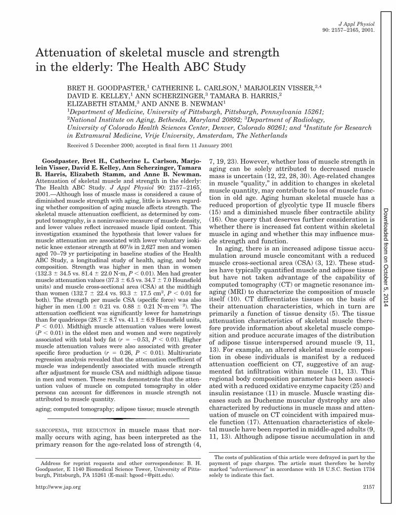
Goodpaster, Bret H., Catherine L. Carlson, Marjo-lein Visser, David E. Kelley, Ann Scherzinger, TamaraB. Harris, Elizabeth Stamm, and Anne B. Newman.Attenuation of skeletal muscle and strength in the elderly:The Health ABC Study. J Appl Physiol 90: 2157–2165,2001.—Although loss of muscle mass is considered a cause ofdiminished muscle strength with aging, little is known regard-ing whether composition of aging muscle affects strength. Theskeletal muscle attenuation coefficient, as determined by com-puted tomography, is a noninvasive measure of muscle density,and lower values reflect increased muscle lipid content. Thisinvestigation examined the hypothesis that lower values formuscle attenuation are associated with lower voluntary isoki-netic knee extensor strength at 60°/s in 2,627 men and womenaged 70–79 yr participating in baseline studies of the HealthABC Study, a longitudinal study of health, aging, and bodycomposition. Strength was higher in men than in women(132.3 6 34.5 vs. 81.4 6 22.0 Nzm, P , 0.01). Men had greatermuscle attenuation values (37.3 6 6.5 vs. 34.7 6 7.0 Hounsfieldunits) and muscle cross-sectional area (CSA) at the midthighthan women (132.7 6 22.4 vs. 93.3 6 17.5 cm2, P , 0.01 forboth). The strength per muscle CSA (specific force) was alsohigher in men (1.00 6 0.21 vs. 0.88 6 0.21 Nzmzcm22). Theattenuation coefficient was significantly lower for hamstringsthan for quadriceps (28.7 6 8.7 vs. 41.1 6 6.9 Hounsfield units,P , 0.01). Midthigh muscle attenuation values were lowest(P , 0.01) in the eldest men and women and were negativelyassociated with total body fat (r 5 20.53, P , 0.01). Highermuscle attenuation values were also associated with greaterspecific force production (r 5 0.26, P , 0.01). Multivariateregression analysis revealed that the attenuation coefficient ofmuscle was independently associated with muscle strengthafter adjustment for muscle CSA and midthigh adipose tissuein men and women. These results demonstrate that the atten-uation values of muscle on computed tomography in olderpersons can account for differences in muscle strength notattributed to muscle quantity.
aging; computed tomography; adipose tissue; muscle strength
SARCOPENIA, THE REDUCTION in muscle mass that nor-mally occurs with aging, has been interpreted as theprimary reason for the age-related loss of strength (4,
7, 19, 23). However, whether loss of muscle strength inaging can be solely attributed to decreased musclemass is uncertain (12, 22, 28, 30). Age-related changesin muscle “quality,” in addition to changes in skeletalmuscle quantity, may contribute to loss of muscle func-tion in old age. Aging human skeletal muscle has areduced proportion of glycolytic type II muscle fibers(15) and a diminished muscle fiber contractile ability(16). One query that deserves further consideration iswhether there is increased fat content within skeletalmuscle in aging and whether this may influence mus-cle strength and function.
In aging, there is an increased adipose tissue accu-mulation around muscle concomitant with a reducedmuscle cross-sectional area (CSA) (3, 12). These stud-ies have typically quantified muscle and adipose tissuebut have not taken advantage of the capability ofcomputed tomography (CT) or magnetic resonance im-aging (MRI) to characterize the composition of muscleitself (10). CT differentiates tissues on the basis oftheir attenuation characteristics, which in turn areprimarily a function of tissue density (5). The tissueattenuation characteristics of skeletal muscle there-fore provide information about skeletal muscle compo-sition and produce accurate images of the distributionof adipose tissue interspersed around muscle (9, 11,13). For example, an altered skeletal muscle composi-tion in obese individuals is manifest by a reducedattenuation coefficient on CT, suggestive of an aug-mented fat infiltration within muscle (11, 13). Thisregional body composition parameter has been associ-ated with a reduced oxidative enzyme capacity (25) andinsulin resistance (11) in muscle. Muscle wasting dis-eases such as Duchenne muscular dystrophy are alsocharacterized by reductions in muscle mass and atten-uation of muscle on CT coincident with impaired mus-cle function (17). Attenuation characteristics of skele-tal muscle have been reported in middle-aged adults (9,11, 13). Although adipose tissue accumulation in and
Address for reprint requests and other correspondence: B. H.Goodpaster, E 1140 Biomedical Science Tower, University of Pitts-burgh, Pittsburgh, PA 15261 (E-mail: [email protected]).
The costs of publication of this article were defrayed in part by thepayment of page charges. The article must therefore be herebymarked ‘‘advertisement’’ in accordance with 18 U.S.C. Section 1734solely to indicate this fact.
J Appl Physiol90: 2157–2165, 2001.
http://www.jap.org 2157
on October 5, 2014
Dow
nloaded from

around muscle has been associated with aging, theattenuation characteristics of aging muscle have notbeen well described. In addition, data regarding theassociation between the attenuation characteristics ofmuscle on CT and muscle strength in the elderly arenot available. The purpose of the present study was totest the hypothesis that skeletal muscle composition,assessed by the attenuation characteristics or densityof muscle on CT, is associated with muscle strength inelderly individuals. To test this hypothesis, mean CTattenuation values of skeletal muscle were determinedin the Health, Aging, and Body Composition (ABC)Study, along with measures of isokinetic strength.
METHODS
Participants in the study were recruited for baseline stud-ies of the Health ABC Study, a longitudinal study of 3,075nondisabled men and women aged 70–79 yr residing inPittsburgh, PA, and Memphis, TN. The population was48.5% male and 58.3% white and had a mean age of 73.6 yr.Participants were recruited primarily from a random sampleof Medicare-eligible adults .65 yr of age from a list providedby the Health Care Financing Administration. Persons wereineligible if they reported difficulty getting around withoutassisted devices, reported difficulty in performing basic ac-tivities of daily living, reported difficulty walking one-quartermile or climbing 10 steps without resting, reported life-threatening cancers, were not planning to remain in thestudy area for $3 yr, or were participating in any researchstudy involving medications or modification of eating or ex-ercise habits. Exclusions from strength testing were a diag-nosis of cerebral aneurysms or bleeding, stroke, hypertension(blood pressure $ 200/110 mmHg), severe bilateral kneepain, or prior bilateral knee replacement. Thus 2,627 menand women from the entire Health ABC Study cohort wereincluded in the study. The study was approved by the Uni-versities of Pittsburgh and Tennessee, and written, informedconsent was obtained from each volunteer.
Age of participants was determined to the nearest year.Standing height and weight were obtained from each volun-teer, and a body mass index (BMI) was calculated as weight(kg)/height (m2). Total body fat was determined using dual-energy X-ray absorptiometry (model QDR 4500, Hologic,Waltham, MA).
CT of the midthigh. Axial CT scans at the midthigh levelwere obtained on each participant during their first exami-nation of the Health ABC Study protocol. CT images wereacquired in Pittsburgh (9800 Advantage, General Electric,Milwaukee, WI) or Memphis (Somatom Plus 4, Siemens,Erlangen, Germany, or PQ 2000S, Marconi Medical Systems,Cleveland, OH). Patients were imaged in the supine positionwith the arms above the head and toes directed toward thetop of the gantry, with legs extended flat on the table. Ananterior-posterior scout scan of the entire femur was used tolocalize the midthigh position. The femoral length was mea-sured in cranial-caudal dimension, and the scan position wasdetermined as the midpoint of the distance between themedial edge of the greater trochanter and the intercondyloidfossa. This measurement was done on the same leg that wasused for isokinetic strength testing, usually the right. Asingle, 10-mm-thick axial image was obtained at the femoralmidpoint, with care taken that the entire circumference ofboth thighs was included in the field of view. The scanningparameters for this image were 120 kVp and 200–250 mA.Images were then network transferred (TCP/IP protocols) to
a SUN workstation (SPARCstation II, Sun Microsystems,Mountain View, CA) for review. A quality review was per-formed on each subject’s images to ensure that all imageswere present, that the proper scan techniques were used, andthat the image was of appropriate quality for analysis.
Skeletal muscle and adipose tissue areas of the thigh werecalculated from the axial CT images using proprietary IDLdevelopment software (RSI Systems, Boulder, CO). Muscleand adipose tissue areas were calculated by multiplying thearea of a given pixel as extracted from the image header.The mean attenuation coefficient values of muscle withinthe regions outlined on the images were determined by av-eraging the CT number (pixel intensity) in Hounsfield units.The methodological variability of this measure is quite small(8). Skeletal muscle and adipose tissue areas were distin-guished by a bimodal image histogram resulting from thedistribution of CT numbers in adipose tissue and muscle (24).These peaks are readily separable (10), and the areas ofadipose tissue and muscle in the entire image were deter-mined by the areas under their respective peaks of thehistogram.
Intermuscular adipose tissue was distinguished from thesubcutaneous adipose tissue by manual drawing of a linealong the deep fascial plane surrounding the thigh muscles.Once the adipose tissue was segmented from the images, theindividual muscles were identified. Muscle borders that werenot already defined by adipose tissue were outlined manu-ally, with care taken that no pixels for bone were included inthe muscle area. Quadriceps muscles were separated fromhamstring muscles with manual tracing (Fig. 1).
Isokinetic strength testing. Isokinetic strength of the kneeextensors was determined at 60°/s with a dynamometer(model 125 AP, Kin-Com, Chattanooga, TN). Before strengthtesting, participants warmed-up by performing a long-dis-tance corridor walk as another part of the Health ABC Study
Fig. 1. Typical computed tomography (CT) image depicting musclearea (gray), subcutaneous adipose tissue (large arrow), and inter-muscular thigh adipose tissue (small arrows). Quadriceps (Q) andhamstring (H) muscle groups were distinguished by manual tracing.
2158 SKELETAL MUSCLE COMPOSITION AND STRENGTH IN THE ELDERLY
on October 5, 2014
Dow
nloaded from

protocol. The right leg was tested unless it was injured orweaker by self-report or restricted in motion. After instruc-tion on the procedure, the participant was positioned so thatthe lateral femoral epicondyle of the knee joint was alignedwith the rotational axis of the dynamometer. The partici-pant’s limb was weighed for gravity correction, and start-stopangles were set at 90° and 30°. Two practice trials wereperformed at 50% effort to familiarize the participant withthe procedure and to provide a warm-up period. At leastthree maximal efforts were performed by each participant.Beginning with the first maximal effort, the torque produc-tion over the entire range of motion was plotted, and the plotof each subsequent effort was overlaid on the previous effortsuntil three similar curves were obtained. Participants werenot asked to perform more than six trials. Maximal torqueproduction was recorded as the mean peak torque productionfrom three similar trials. A methodological consideration waswhether maximal voluntary effort during isokinetic strengthtesting represents the true contractile ability of muscle. Elec-trical stimulation superimposed on maximal voluntary con-traction has been demonstrated to produce no additionaltorque in elderly men and women (14, 28), suggesting thatthe maximal voluntary effort is a valid measure of strength.
Statistical analysis. ANOVA was used to compare meanmuscle and adipose tissue areas, the mean muscle attenua-tion values, and strength with respect to gender, race, andage. The association between thigh composition and isoki-netic knee extensor strength and specific torque (strength/CSA) was determined using simple linear regression analy-sis. Gender-specific forced multiple linear regression modelswere fit to determine the independent associations of thighmuscle area and muscle attenuation with maximal isokineticstrength, controlling for age, race, height, and thigh adiposetissue. Gender-specific forced multiple linear regression mod-els were also fit to determine the independent association ofthigh muscle attenuation values with specific torque, control-ling for age, height, and thigh adipose tissue. Gender-specificmodels were used in the analysis, because there was littleoverlap between strength and lean mass in men and women.Because there were no significant race interactions, blacksand whites were collapsed for presentation. All analyses wereperformed using SAS 6.12 for Windows (SAS Institute,Cary, NC).
RESULTS
Subject characteristics. Subject characteristics arepresented in Table 1. The study population was 60%white and 40% black, divided approximately equallyamong men and women between 70 and 80 yr of age.Because of exclusion criteria for strength testing, 2,627subjects out of the entire Health ABC Study cohort
were included in the study. However, the demograph-ics of the present study participants were similar tothose of the entire Health ABC Study cohort. The ageof the cohort was well matched according to gender andrace. As expected, men were taller and heavier thanwomen, but women had more total body fat than men.Black women had a greater BMI and a higher propor-tion of body fat than white women, but white men hadmore body fat than black men.
Muscle attenuation values and CSA by CT. Skeletalmuscle attenuation values in this cohort are presentedin Table 2. The mean attenuation value for muscle andmuscle CSA represent the entire midthigh of the re-spective leg, i.e., right or left, that was used for isoki-netic strength testing. The mean skeletal muscle at-tenuation values were lower in these elderly womenthan in men (P , 0.01). Black women had lower meanattenuation values in both muscle groups than whitewomen, but white men had slightly lower mean atten-uation values in hamstrings than black men (P , 0.01).CSA of the midthigh was greater in men than inwomen (Table 2), and this was true for quadriceps andhamstrings (P , 0.01). The mean CT attenuation val-ues and the CSA were lower for hamstrings than forquadriceps across all groups (P , 0.01).
Values for the attenuation of muscle were decreasedacross increasing quartiles of BMI (Fig. 2) and werenegatively associated with BMI in men (r 5 20.44, P ,0.01) and women (r 5 20.43, P , 0.01). Similarly, theattenuation value for muscle had negative associationswith total body fat (r 5 20.53, P , 0.01) and totalpercent fat (r 5 20.49, P , 0.01) in this cohort. How-ever, thigh muscle CSA was higher in men and womenin the higher-BMI quartiles, and BMI was positivelyassociated with muscle CSA in men (r 5 0.61, P , 0.01)and women (r 5 0.60, P , 0.01). Thus increased bodyfatness was associated with larger thigh muscle butwith muscle of different composition than in leanersubjects. Figure 2 also illustrates that the attenuationvalue for muscle decreased as a function of increasingage, so that the oldest men and women had the lowestmuscle density. In addition, muscle CSA was alsolower with increasing age in these elderly men andwomen (data not shown). Thus, although advancingage and increased obesity are associated with reducedmuscle density in older, healthy adults, the respectiveeffects on muscle size are opposite.
Table 1. Descriptive characteristics of participants with strength and CT measurements completed
White Men(n 5 833)
Black Men(n 5 458)
All Men(n 5 1,285)
White Women(n 5 749)
Black Women(n 5 602)
All Women(n 5 1,342)
Entire Cohort(n 5 2,627)
Age, yr 73.962.9 73.462.8 73.762.9 73.562.8 73.362.9 73.462.8 73.662.9Height, m 1.760.1 1.760.1 1.760.1 1.660.1 1.660.1 1.660.1 1.760.1Weight, kg 81.3612.3 81.7614.5 81.4613.2 66.1612.2 75.4615.6 70.3614.5 75.7614.9BMI, kg/m2 27.063.7 27.264.3 27.164.0 26.064.5 29.565.8 27.665.4 27.364.8Total body fat, kg 21.766.7 20.367.3 21.267.0 25.067.7 29.569.8 27.069.0 24.268.6
Values are means 6 SD. BMI, body mass index; CT, computed tomography. P values for specific comparisons are as follows: genderdifferences: age, 0.0115; height, 0.0001; weight, 0.0001; BMI, 0.0049; total body fat, 0.0001; men race differences: age, 0.0057; height, 0.4409;weight, 0.6274; BMI, 0.3925; total body fat, 0.0003; women race differences: age, 0.1477; height, 0.4925; weight, 0.0001; BMI, 0.0001; totalbody fat, 0.0001.
2159SKELETAL MUSCLE COMPOSITION AND STRENGTH IN THE ELDERLY
on October 5, 2014
Dow
nloaded from

Midthigh adipose tissue content. Adipose tissue areawithin the midthigh was divided into subcutaneousand intermuscular depots, inasmuch as this later com-partment better describes the infiltration of adiposetissue around muscle (Fig. 1). Absolute amounts ofintermuscular adipose tissue were much smaller thansubcutaneous adipose tissue areas in this cohort (Table2), representing only 11.7% of the total thigh adiposetissue area. Women and men had similar amounts ofintermuscular thigh adipose tissue, but women hadsubstantially more subcutaneous thigh adipose tissuethan men, so that the proportion of thigh intermuscu-lar adipose tissue relative to the total thigh adiposetissue was lower in women than in men (8.6 vs. 17.4%,P , 0.01). Black men and women had higher absoluteareas of intermuscular and subcutaneous thigh adi-pose tissue than white men and women, but there wereno racial differences in the relative amount of inter-muscular adipose tissue depots (12.2 and 11.2% forblack and white participants, respectively).
The amount of intermuscular thigh adipose tissuewas higher in obesity (BMI) in these elderly men (r 50.56) and women (r 5 0.61, both P , 0.01). Similarassociations were observed between the amount of sub-cutaneous thigh adipose tissue and BMI in men andwomen (r 5 0.69 and 0.73, both P , 0.01, respectively).Moreover, lower mean muscle attenuation values wererelated to more intermuscular (r 5 20.55) and subcu-taneous (r 5 20.42) thigh adipose tissue (both P ,0.01). Neither of these adipose tissue depots was asso-ciated with age within these elderly participants.
Association between attenuation of muscle andstrength. Isokinetic strength was greater (P , 0.01) inmen than in women (Table 3); black women had higherabsolute strength than white women, but there wereno racial differences in strength in men. There was a19% decrease in absolute strength in men and womenacross this age range. There was a strong associationbetween strength and the CSA of muscle in men (r 50.55, P , 0.01) and women (r 5 0.48, P , 0.01); thusmuscle CSA accounted for ;25% of the variance inmuscle strength independently of gender. To accountfor the effect of muscle size, strength per unit of CSA
Table 2. Midthigh composition characteristics
White Men Black Men All Men White Black Women All Women Entire Cohort
SM area, cm2 128.5619.5 140.2625.2 132.7622.4 86.0614.1 102.5617.0 93.3617.5 112.6628.1Quadriceps 60.669.4 65.2612.2 62.2610.7 40.067.0 46.468.4 42.968.3 52.3613.6Hamstrings 30.065.7 33.866.8 31.466.3 21.364.1 25.764.5 23.364.8 27.266.9
SM attenuation, HU 37.566.4 37.066.6 37.366.5 34.767.0 32.766.9 33.867.0 35.567.0Quadriceps 43.665.8 42.966.6 43.466.1 39.866.9 37.766.8 38.966.9 41.166.9Hamstrings 30.068.4 31.368.5 30.568.4 27.468.7 26.768.7 27.168.7 28.768.7
AT area, cm2
Intermuscular 9.365.9 11.268.9 10.067.2 8.764.4 12.566.9 10.466.0 10.266.6Subcutaneous 46.3619.7 49.3621.2 47.4620.3 96.3638.6 120.4652.9 107.0647.1 77.9647.1
Values are means 6 SD. SM, skeletal muscle; HU, Hounsfield units; AT, adipose tissue. Attenuation refers to the mean attenuationcoefficient of muscle. P values for specific comparisons are as follows: gender differences: SM area, 0.0001; SM attenuation, 0.0001;quadriceps area, 0.0001; hamstring area, 0.0001; quadriceps attenuation, 0.0001; hamstring attenuation, 0.0001; intermuscular AT, 0.0760;subcutaneous AT, 0.0001; men race differences: SM area, 0.0001; SM attenuation, 0.1661; quadriceps area, 0.0001; hamstring area, 0.0001;quadriceps attenuation, 0.0668; hamstring attenuation, 0.0076; intermuscular AT, 0.0001; subcutaneous AT, 0.0114; women race differences:SM area, 0.0001; SM attenuation, 0.0001; quadriceps area, 0.0001; hamstring area, 0.0001; quadriceps attenuation, 0.0001; hamstringattenuation, 0.1260; intermuscular AT, 0.0001; subcutaneous AT, 0.0001.
Fig. 2. Mean midthigh muscle attenuation values determined by CTin men and women were lower across increasing body mass index(BMI) quartiles (A) and increasing age (B). HU, Hounsfield units.Differences in mean attenuation coefficients with respect to BMI andage were analyzed using one-way ANOVA. Error bars, SE.
2160 SKELETAL MUSCLE COMPOSITION AND STRENGTH IN THE ELDERLY
on October 5, 2014
Dow
nloaded from

was calculated, and this value was termed specificforce.
Men were able to generate higher (P , 0.01) specificforce than women (Table 3). Specific force in black men
and women was reduced compared with that in whitemen and women. Higher values for the attenuation ofmuscle were associated with greater torque as well ashigher specific force production (Fig. 3). These associ-
Fig. 3. Associations between midthigh attenuation values and torque (A) and specific torque (B) in all subjects (n 52,627). Correlation coefficients determined using simple linear regression were 0.20 for attenuation value vs.torque and 0.26 for attenuation value vs. specific torque (P , 0.01 for both).
Table 3. Isokinetic strength
White Men Black Men All Men White Women Black Women All Women Entire Cohort
Maximal torque, N zm 130.6633.1 134.4637.6 132.3634.5 78.3620.0 85.1624.0 81.4622.0 106.0638.5Specific torque, torque/cm2 1.0260.20 0.9760.22 1.0060.21 0.9160.20 0.8460.21 0.8860.21 0.9460.22
Values are means 6 SD. See Table 2 footnote for abbreviations. Specific torque, torque per unit cross-sectional muscle area. P values forspecific comparisons are as follows: gender differences: torque, 0.0001; specific torque, 0.0001; men race differences: torque, 0.0586; specifictorque, 0.0001; women race differences: torque, 0.0001; specific torque, 0.0001.
2161SKELETAL MUSCLE COMPOSITION AND STRENGTH IN THE ELDERLY
on October 5, 2014
Dow
nloaded from

ations were slightly better (P , 0.01) in quadricepsthan in hamstrings (r 5 0.27 and 0.22, P , 0.01 forquadriceps and hamstrings vs. specific force, respec-tively). Specific force was significantly increased (P ,0.01) across quartiles of muscle attenuation in men andwomen (Fig. 4), indicating that men and women withthe highest values for muscle attenuation also had thehighest force per CSA. Although maximal torque wasslightly increased (P 5 0.014) across muscle attenua-tion quartiles in women, this was not true for men.Neither intermuscular nor subcutaneous adipose tis-sue depots were associated with maximal torque orspecific force production.
Multivariate regression analyses in men and womenrevealed that the attenuation values for muscle wereindependently associated with maximal torque afteradjustment for muscle size (CSA), height, weight, age,and race (Table 4), with this model accounting for 36and 32% of the total variance in strength for men andwomen, respectively. In addition, attenuation of mus-cle was associated with a measure of muscle quality
(specific force) independently of age, race, and otherdepots of adipose tissue within the thigh (Table 5). Thismodel accounted for 9 and 11% of the total variance inspecific force in men and women, respectively.
DISCUSSION
The loss of muscle mass, termed sarcopenia, withaging has been well described (for review see Ref. 6),but less is known about the changes in the compositionor quality of muscle in elderly persons. This studyrepresents the first large-scale, detailed description ofthe composition of skeletal muscle and its associationwith strength in the elderly and thus provides novelinformation concerning the interrelationships amongmuscle quantity, quality, and strength in aging. Therewere new findings in this study: 1) the attenuationvalue, or density, of skeletal muscle decreases with ageand increases with the level of body fatness in men andwomen 70–80 yr of age, and 2) higher values for theattenuation of skeletal muscle are associated withgreater voluntary strength independent of the muscleCSA.
The attenuation of muscle was lower with increasingage in these older men and women, and this was truefor quadriceps and hamstring muscle groups. Attenu-ation values for muscle were also lower in associationwith increasing BMI and total body fatness, a resultconsistent with prior studies in which lower muscleattenuation values were observed in obese middle-agedmen and women (11). Mean muscle attenuation waslower in women than in men for quadriceps and ham-strings groups, and black women had lower muscledensity than white women. Because mean muscle at-tenuation values are a direct measure of reduced mus-cle density (5) and are associated with an increasedlipid accumulation within muscle (8), our results indi-cate that skeletal muscle lipid content was higher withincreasing age and overall adiposity and was higher inwomen. In support of this, Visser at al. (29) reportedthat the density of fat-free mass as a function of waterand mineral content is independent of age, obesity,gender, and race. Therefore, variations in muscle den-sity in the elderly and in obesity are likely due tochanges in muscle lipid content.
Fig. 4. Specific torque [maximal torque per unit muscle cross-sec-tional area (CSA)] of knee extensors was higher across increasingquartiles for muscle attenuation values in men and women (P ,0.01). Differences in specific torque with respect to quartiles ofmuscle attenuation values were analyzed using one-way ANOVA.Error bars, SE.
Table 4. Multivariate models accounting for variance in isokinetic strength in men and women
Variable
Men Women
Mean6SE P Partial R2 Mean6SE P Partial R2
Thigh muscle area, cm2 0.85760.039 0.0001 0.304 0.64160.035 0.0001 0.248Thigh MA, HU 0.45560.153 0.0029 0.016 0.55860.090 0.0001 0.030Age, yr 21.03460.279 0.0002 0.007 20.60860.184 0.0010 0.009Race 25.16661.688 0.0023 0.007 22.91961.158 0.0119 0.004Height, cm 72.547612.142 0.0001 0.017 57.30768.392 0.0001 0.023AT, cm2
Intermuscular AT 20.24260.140 0.0848 0.002 20.06060.112 0.5941 0.000001Subcutaneous AT 20.05660.044 0.2118 0.001 0.02960.012 0.0171 0.003
Strength values are expressed as units of torque (N zm). Partial variance (partial R2) is the explained variance for each specific parameterin the model. Total variance (R2) by the models was 0.355 for men, 0.315 for women, and 0.628 combined. MA, muscle attenuation for totalmid-thigh.
2162 SKELETAL MUSCLE COMPOSITION AND STRENGTH IN THE ELDERLY
on October 5, 2014
Dow
nloaded from

Muscle CSA was lower with age but higher in asso-ciation with obesity in this cohort. Because of the closeassociation between the loss of muscle mass and dimin-ished muscle function in aging, the precise determina-tion of skeletal muscle mass has been an importantconsideration in studies of aging and muscle strength.Several indirect methods have been used to quantifymuscle CSA in older persons, including anthropometry(2, 23) and manual outlining of muscle area on CTimages (27). CT (3) and MRI (7, 12) image analysissoftware has recently been used to obtain specific mea-sures of muscle CSA in the elderly on the basis of theability to precisely distinguish muscle from adiposetissue. Our results concur with these CT (3) and MRI(7, 12) studies demonstrating lower muscle CSA inolder individuals. The composition of muscle defined byits mean attenuation value was altered independentlyof these measurable changes in muscle quantity. Thisresult is in accord with prior exercise (26) or weightloss (10) intervention studies reporting changes inmuscle density independently of changes in muscleCSA.
Muscle CSA was positively associated with kneeextensor strength in these elderly men and women,accounting for ;25% of the variance in strength in menand women. Thus our results confirm those of priorstudies (4, 7, 19, 23) in demonstrating that lower mus-cle strength with age can be largely attributed to areduction of skeletal muscle. However, the differencein muscle strength between men and women could notbe explained solely by differences in muscle size, inas-much as the explained variance in strength increasedto ;50% for the entire cohort when gender was in-cluded as a covariate. On the basis of differences inattenuation characteristics between skeletal muscleand adipose tissue, we systematically separated mus-cle from subcutaneous adipose tissue and, in addition,determined the amount of adipose tissue interspersedbetween muscle, termed intermuscular adipose tissue.In this process, we were able to quantify muscle CSAexclusive of these different adipose tissue depots butnot necessarily the amount of lipid within muscle it-self. Subcutaneous and intermuscular thigh adiposetissue were increased with obesity in this older cohort,a result congruent with prior studies in middle-agedadults (10). However, the amount of adipose tissue inthe thigh was not associated with the strength or
specific force production by muscle. Thus lower muscleCSA, but not the accumulation of these fat depotsoutside muscle, impacts negatively on strength inthese older individuals.
Another important finding in the present study wasthat higher values for the attenuation of muscle den-sity were associated with greater absolute musclestrength after accounting for the quantity of muscle.Greater muscle attenuation values were also related tobetter muscle quality as defined by greater specificforce production by muscle, i.e., force production perunit muscle size (18, 20). Nearly half the explainedvariance in specific torque was due to the attenuationvalues of muscle. Metter and colleagues (18, 20), usingCSA or fat-free mass to measure the quantity of mus-cle, found that specific force production declines withage. Other investigations have also found an alteredmuscle quality in older persons, as evidenced by areduced proportion of type II fibers (15) and a reducedforce production by individual muscle fibers (16). Theage-related increase in the relative proportion of type Imuscle fibers, which are known to have a greater lipidcontent (1), may partly explain the age-associated de-crease in muscle density. Our results demonstrate thatlower muscle attenuation values are associated withlower muscle quality in the elderly. Quantification ofskeletal muscle attenuation characteristics may helpdefine the nonfat component of skeletal muscle,thereby accounting for additional variance in musclestrength.
In support of our findings of an association betweenaltered muscle density and strength, strength trainingin elderly women has been shown to increase the at-tenuation of muscle and strength (26). In studies per-formed in muscular dystrophy (17) and neurologicalpatients (21), whose muscle wasting closely resemblesthat of age-related sarcopenia, it was noted that anincreased fatty infiltration was associated with re-duced force generation in skeletal muscle. However,these studies were based on a small number of pa-tients, and only one of these studies quantified themuscle density (17). It is possible that factors otherthan lipid contained within muscle, such as connectivetissue, contribute to alterations in the attenuationcharacteristics of muscle. However, attenuation ofmuscle is correlated with direct measures of musclefiber lipid content determined by muscle biopsy (8).
Table 5. Multivariate models accounting for the variance in specific torque in men and women
Variable
Men Women
Mean6SE P Partial R2 Mean6SE P Partial R2
Thigh MA, HU 0.00460.001 0.0003 0.032 0.00660.001 0.0001 0.050Age, yr 20.00660.002 0.0013 0.007 20.00460.002 0.0558 0.004Race 20.04660.012 0.0001 0.014 20.06460.012 0.0001 0.023Height, cm 0.50160.087 0.0001 0.022 0.58960.090 0.0001 0.028AT, cm2
Intermuscular 20.00260.001 0.0510 0.004 20.00160.001 0.2270 0.001Subcutaneous 20.00160.0003 0.1585 0.001 0.000260.0001 0.1325 0.001
Specific torque is voluntary leg extensor strength per unit area (N zm zcm22). Partial variance (Partial R2) is the explained variance for eachspecific parameter in the model. Total variance (R2) by the models was 0.083 for men, 0.106 for women, and 0.162 combined.
2163SKELETAL MUSCLE COMPOSITION AND STRENGTH IN THE ELDERLY
on October 5, 2014
Dow
nloaded from
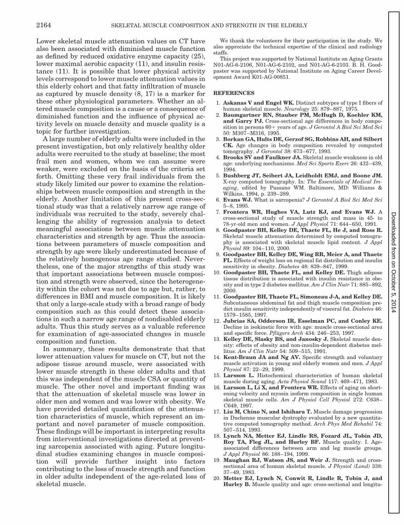
Lower skeletal muscle attenuation values on CT havealso been associated with diminished muscle functionas defined by reduced oxidative enzyme capacity (25),lower maximal aerobic capacity (11), and insulin resis-tance (11). It is possible that lower physical activitylevels correspond to lower muscle attenuation values inthis elderly cohort and that fatty infiltration of muscleas captured by muscle density (8, 17) is a marker forthese other physiological parameters. Whether an al-tered muscle composition is a cause or a consequence ofdiminished function and the influence of physical ac-tivity levels on muscle density and muscle quality is atopic for further investigation.
A large number of elderly adults were included in thepresent investigation, but only relatively healthy olderadults were recruited to the study at baseline; the mostfrail men and women, whom we can assume wereweaker, were excluded on the basis of the criteria setforth. Omitting these very frail individuals from thestudy likely limited our power to examine the relation-ships between muscle composition and strength in theelderly. Another limitation of this present cross-sec-tional study was that a relatively narrow age range ofindividuals was recruited to the study, severely chal-lenging the ability of regression analysis to detectmeaningful associations between muscle attenuationcharacteristics and strength by age. Thus the associa-tions between parameters of muscle composition andstrength by age were likely underestimated because ofthe relatively homogenous age range studied. Never-theless, one of the major strengths of this study wasthat important associations between muscle composi-tion and strength were observed, since the heterogene-ity within the cohort was not due to age but, rather, todifferences in BMI and muscle composition. It is likelythat only a large-scale study with a broad range of bodycomposition such as this could detect these associa-tions in such a narrow age range of nondisabled elderlyadults. Thus this study serves as a valuable referencefor examination of age-associated changes in musclecomposition and function.
In summary, these results demonstrate that thatlower attenuation values for muscle on CT, but not theadipose tissue around muscle, were associated withlower muscle strength in these older adults and thatthis was independent of the muscle CSA or quantity ofmuscle. The other novel and important finding wasthat the attenuation of skeletal muscle was lower inolder men and women and was lower with obesity. Wehave provided detailed quantification of the attenua-tion characteristics of muscle, which represent an im-portant and novel parameter of muscle composition.These findings will be important in interpreting resultsfrom interventional investigations directed at prevent-ing sarcopenia associated with aging. Future longitu-dinal studies examining changes in muscle composi-tion will provide further insight into factorscontributing to the loss of muscle strength and functionin older adults independent of the age-related loss ofskeletal muscle.
We thank the volunteers for their participation in the study. Wealso appreciate the technical expertise of the clinical and radiologystaffs.
This project was supported by National Institute on Aging GrantsN01-AG-6-2106, N01-AG-6-2102, and N01-AG-6-2103. B. H. Good-paster was supported by National Institute on Aging Career Devel-opment Award K01-AG-00851.
REFERENCES
1. Askanas V and Engel WK. Distinct subtypes of type I fibers ofhuman skeletal muscle. Neurology 25: 879–887, 1975.
2. Baumgartner RN, Stauber PM, McHugh D, Koehler KM,and Garry PJ. Cross-sectional age differences in body compo-sition in persons 601 years of age. J Gerontol A Biol Sci Med Sci50: M307–M316, 1995.
3. Borkan GA, Hults DE, Gerzof SG, Robbins AH, and SilbertCK. Age changes in body composition revealed by computedtomography. J Gerontol 38: 673–677, 1993.
4. Brooks SV and Faulkner JA. Skeletal muscle weakness in oldage: underlying mechanisms. Med Sci Sports Exerc 26: 432–439,1994.
5. Bushberg JT, Seibert JA, Leidholdt EMJ, and Boone JM.X-ray computed tomography. In: The Essentials of Medical Im-aging, edited by Passano WM. Baltimore, MD: Williams &Wilkins, 1994, p. 239–289.
6. Evans WJ. What is sarcopenia? J Gerontol A Biol Sci Med Sci5–8, 1995.
7. Frontera WR, Hughes VA, Lutz KJ, and Evans WJ. Across-sectional study of muscle strength and mass in 45- to78-yr-old men and women. J Appl Physiol 71: 644–650, 1991.
8. Goodpaster BH, Kelley DE, Thaete FL, He J, and Ross R.Skeletal muscle attenuation determined by computed tomogra-phy is associated with skeletal muscle lipid content. J ApplPhysiol 89: 104–110, 2000.
9. Goodpaster BH, Kelley DE, Wing RR, Meier A, and ThaeteFL. Effects of weight loss on regional fat distribution and insulinsensitivity in obesity. Diabetes 48: 839–847, 1999.
10. Goodpaster BH, Thaete FL, and Kelley DE. Thigh adiposetissue distribution is associated with insulin resistance in obe-sity and in type 2 diabetes mellitus. Am J Clin Nutr 71: 885–892,2000.
11. Goodpaster BH, Thaete FL, Simoneau J-A, and Kelley DE.Subcutaneous abdominal fat and thigh muscle composition pre-dict insulin sensitivity independently of visceral fat. Diabetes 46:1579–1585, 1997.
12. Jubrias SA, Odderson IR, Esselman PC, and Conley KE.Decline in isokinetic force with age: muscle cross-sectional areaand specific force. Pflugers Arch 434: 246–253, 1997.
13. Kelley DE, Slasky BS, and Janosky J. Skeletal muscle den-sity: effects of obesity and non-insulin-dependent diabetes mel-litus. Am J Clin Nutr 54: 509–515, 1991.
14. Kent-Braun JA and Ng AV. Specific strength and voluntarymuscle activation in young and elderly women and men. J ApplPhysiol 87: 22–29, 1999.
15. Larsson L. Histochemical characteristics of human skeletalmuscle during aging. Acta Physiol Scand 117: 469–471, 1983.
16. Larsson L, Li X, and Frontera WR. Effects of aging on short-ening velocity and myosin isoform composition in single humanskeletal muscle cells. Am J Physiol Cell Physiol 272: C638–C649, 1997.
17. Liu M, Chino N, and Ishihara T. Muscle damage progressionin Duchenne muscular dystrophy evaluated by a new quantita-tive computed tomography method. Arch Phys Med Rehabil 74:507–514, 1993.
18. Lynch NA, Metter EJ, Lindle RS, Fozard JL, Tobin JD,Roy TA, Fleg JL, and Hurley BF. Muscle quality. I. Age-associated differences between arm and leg muscle groups.J Appl Physiol 86: 188–194, 1999.
19. Maughan RJ, Watson JS, and Weir J. Strength and cross-sectional area of human skeletal muscle. J Physiol (Lond) 338:37–49, 1983.
20. Metter EJ, Lynch N, Conwit R, Lindle R, Tobin J, andHurley B. Muscle quality and age: cross-sectional and longitu-
2164 SKELETAL MUSCLE COMPOSITION AND STRENGTH IN THE ELDERLY
on October 5, 2014
Dow
nloaded from

dinal comparisons. J Gerontol A Biol Sci Med Sci 54: B207–B218, 1999.
21. Nordal HJ, Dietrichson P, Eldevik P, and Gronseth K. Fatinfiltration, atrophy and hypertrophy of skeletal muscles dem-onstrated by X-ray computed tomography in neurological pa-tients. Acta Neurol Scand 77: 115–122, 1988.
22. Overend TJ, Cunningham DA, Kramer JF, Lefcoe MS, andPaterson DH. Knee extensor and knee flexor strength: cross-sectional area ratios in young and elderly men. J Gerontol 47:M204–M210, 1992.
23. Reed RL, Pearlmutter L, Yochum K, Meredith KE, andMooradian AD. The relationship between muscle mass andmuscle strength in the elderly. J Am Geriatr Soc 39: 555–561,1991. [Comment. J Am Geriatr Soc 40: January 1992, p. 103.]
24. Seidell JC, Oosterlee A, Thijssen MA, Burema J, Deuren-berg P, Hautvast JG, and Ruijs JH. Assessment of intra-abdominal and subcutaneous abdominal fat: relation betweenanthropometry and computed tomography. Am J Clin Nutr 45:7–13, 1987.
25. Simoneau JA, Colberg SR, Thaete FL, and Kelley DE.Skeletal muscle glycolytic and oxidative enzyme capacities aredeterminants of insulin sensitivity and muscle composition inobese women. FASEB J 9: 273–278, 1995.
26. Sipila S and Suominen H. Effects of strength and endurancetraining on thigh and leg muscle mass and composition in elderlywomen. J Appl Physiol 78: 334–340, 1995.
27. Trappe SW, Costill DL, Goodpaster BH, and Pearson DR.Calf muscle strength in former elite distance runners. ScandJ Med Sci Sports 6: 205–210, 1996.
28. Vandervoort AA and McComas AJ. Contractile changes inopposing muscles of the human ankle joint with aging. J ApplPhysiol 61: 361–367, 1986.
29. Visser M, Gallagher D, Deurenberg P, Wang J, Pierson RNJr, and Heymsfield SB. Density of fat-free body mass: relation-ship with race, age, and level of body fatness. Am J PhysiolEndocrinol Metab 272: E781–E787, 1997.
30. Young A, Stokes M, and Crowe M. Size and strength of thequadriceps muscle of old and young women. Eur J Clin Invest 14:282–287, 1984.
2165SKELETAL MUSCLE COMPOSITION AND STRENGTH IN THE ELDERLY
on October 5, 2014
Dow
nloaded from