Athersley, Barnsley (SHW13): Osteological/Zooarchaeological report
12
1 SHW13 Osteological Report Anna Bloxam 2013 Introduction Faunal remains were identified in 11 test pits dug in Athersley, Barnsley. The remains were mostly highly fragmentary mixed deposits, but the articulated remains of a domestic dog (Canis familiaris) from a modern burial were also uncovered in context TP15. The mixed faunal remains will be described here first, with a detailed analysis and discussion of the dog burial following. Methodology Finds of bone, bagged by spit and context, were received by the author for analysis. Following cleaning, the faunal assemblage was assessed for identifiable fragments that could be assigned to a species (common names given). Many identifiable skeletal elements could not be securely assigned to any species due to their fragmentary state; these were assigned to categories by animal size. The categories used were ‘micro’ (small rodent sized), ‘small’ (cat or dog sized), ‘medium’ (sheep/goat or pig sized), and ‘large’ (cattle or horse sized). Individual fragments were counted to give the Number of Identified Specimens (NISP) and the Minimum Number of Individuals (MNI) was assessed by checking for duplications of skeletal elements or incompatible developmental stages within a species in each sample. Fragments that could not be assigned to any particular animal were not given a MNI figure. Fragments were weighed to an accuracy of 0.01g using digital scales to provide a measure of quantity. All fragments were assessed for evidence of taphonomic or pathological changes. Any modifications to the bone, such as evidence of butchery or burning, were recorded. The recorded data can be found listed by context in Appendix 1. The dog skeleton was assessed separately from the mixed faunal assemblage, as it represents an intentional deposition of a ‘known individual’ (known to have been called ‘Chippy’) rather than food remains or other domestic refuse. A skeletal inventory was first taken of the remains to assess the level of completeness and any patterning to the elements present. The sex of the individual was established due to the presence of a baculum, but was also assessed through observation of elements of cranial morphology including the basioccipital (following The & Trouth, 1976 and Trouth et al. 1977). The age of the individual was assessed through observation of dental wear and age-related degeneration. All cranial sutures and epiphyses were fully fused, indicating the individual was an adult.
Transcript of Athersley, Barnsley (SHW13): Osteological/Zooarchaeological report

1
SHW13 Osteological Report
Anna Bloxam 2013
Introduction
Faunal remains were identified in 11 test pits dug in Athersley, Barnsley. The remains were mostly
highly fragmentary mixed deposits, but the articulated remains of a domestic dog (Canis familiaris)
from a modern burial were also uncovered in context TP15. The mixed faunal remains will be
described here first, with a detailed analysis and discussion of the dog burial following.
Methodology
Finds of bone, bagged by spit and context, were received by the author for analysis. Following
cleaning, the faunal assemblage was assessed for identifiable fragments that could be assigned to a
species (common names given). Many identifiable skeletal elements could not be securely assigned
to any species due to their fragmentary state; these were assigned to categories by animal size. The
categories used were ‘micro’ (small rodent sized), ‘small’ (cat or dog sized), ‘medium’ (sheep/goat or
pig sized), and ‘large’ (cattle or horse sized). Individual fragments were counted to give the Number
of Identified Specimens (NISP) and the Minimum Number of Individuals (MNI) was assessed by
checking for duplications of skeletal elements or incompatible developmental stages within a species
in each sample. Fragments that could not be assigned to any particular animal were not given a MNI
figure. Fragments were weighed to an accuracy of 0.01g using digital scales to provide a measure of
quantity. All fragments were assessed for evidence of taphonomic or pathological changes. Any
modifications to the bone, such as evidence of butchery or burning, were recorded. The recorded
data can be found listed by context in Appendix 1.
The dog skeleton was assessed separately from the mixed faunal assemblage, as it represents an
intentional deposition of a ‘known individual’ (known to have been called ‘Chippy’) rather than food
remains or other domestic refuse. A skeletal inventory was first taken of the remains to assess the
level of completeness and any patterning to the elements present.
The sex of the individual was established due to the presence of a baculum, but was also assessed
through observation of elements of cranial morphology including the basioccipital (following The &
Trouth, 1976 and Trouth et al. 1977). The age of the individual was assessed through observation of
dental wear and age-related degeneration. All cranial sutures and epiphyses were fully fused,
indicating the individual was an adult.
2
The withers (shoulder) height of the individual was assessed following the methods of Harcourt
(1974). This paper was also followed for the measurements taken of the skeleton and in the
calculation of a number of indices to qualitatively describe the skull and limb bones; the
measurements and indices are detailed in Appendix 2. Measurements were taken using digital
sliding calipers to an accuracy of 0.1mm and, where elements were too large for this, using an
osteometric board to an accuracy of 1mm.
A variety of pathological changes were observed to the skeleton; these are described in terms of
their appearance and an assessment of their probable impact on the individual in life is made.
The Mixed Faunal Assemblage
Elements and Species Present
A variety of domestic and wild species were represented in small quantities within the sample,
including sheep/goat, cattle, probable deer, dog or cat, domestic bird, and rat, as well as various
unidentified small- and medium-sized mammals. Most test pits contained several bone fragments
belonging to multiple species. In no context was the MNI ever greater than 1. This suggests that the
remains are from small domestic contexts and the locations were not used as refuse sites over
extended periods of time. The skeletal elements identified came from across the body, with
dentition and fragments of long bone, phalanges, ribs and pelvis being identified. Mature and
immature individuals were identified, with all teeth identified being well worn. Fragments of rib
belonging to a large cat or small dog were identified in TP15, suggesting that Chippy may not have
been the only pet to be buried in that location.
Taphonomic changes
Much of the bone was highly fragmentary, with some being heavily weathered, making species
identification or assessment or surface features difficult.
Contexts TP5 and TP6 both contained fragments of burned bone, though in both cases not all the
bone present had been exposed to high temperatures, suggesting more than one deposition event
was represented in each. The burned bone in each context indicated variable temperature exposure,
with some fragments white and calcined and others charred black or red (figure 1). No charcoal or
ash was included with the bone samples for analysis, but the mixed and fragmentary nature of these
deposits and their exposure to varying temperatures is suggestive of built-up domestic cooking
refuse.

3
Evidence for butchery was found on several fragments across four contexts (Test pits 2, 6, 15, 26).
These were primarily cut-marks, though evidence of chopping was also observed (figure 2).
Figure 1 – burned fragments of bone from contexts TP5 (left) and TP6 (right).
Figure 2 – Left: Cut mark to the midshaft of a long bone, possibly deer humerus, from TP2. Right: Rib fragment belonging to a ‘large’ mammal with chop mark across vertebral end (TP26).
4
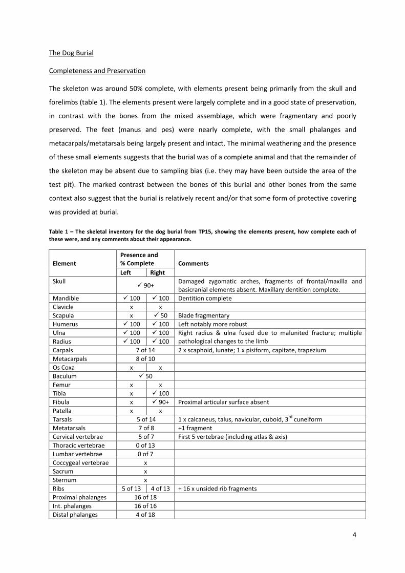
The Dog Burial
Completeness and Preservation
The skeleton was around 50% complete, with elements present being primarily from the skull and
forelimbs (table 1). The elements present were largely complete and in a good state of preservation,
in contrast with the bones from the mixed assemblage, which were fragmentary and poorly
preserved. The feet (manus and pes) were nearly complete, with the small phalanges and
metacarpals/metatarsals being largely present and intact. The minimal weathering and the presence
of these small elements suggests that the burial was of a complete animal and that the remainder of
the skeleton may be absent due to sampling bias (i.e. they may have been outside the area of the
test pit). The marked contrast between the bones of this burial and other bones from the same
context also suggest that the burial is relatively recent and/or that some form of protective covering
was provided at burial.
Table 1 – The skeletal inventory for the dog burial from TP15, showing the elements present, how complete each of these were, and any comments about their appearance.
Element
Presence and % Complete Comments
Left Right
Skull 90+
Damaged zygomatic arches, fragments of frontal/maxilla and basicranial elements absent. Maxillary dentition complete.
Mandible 100 100 Dentition complete
Clavicle x x
Scapula x 50 Blade fragmentary
Humerus 100 100 Left notably more robust
Ulna 100 100 Right radius & ulna fused due to malunited fracture; multiple pathological changes to the limb Radius 100 100
Carpals 7 of 14 2 x scaphoid, lunate; 1 x pisiform, capitate, trapezium
Metacarpals 8 of 10
Os Coxa x x
Baculum 50
Femur x x
Tibia x 100
Fibula x 90+ Proximal articular surface absent
Patella x x
Tarsals 5 of 14 1 x calcaneus, talus, navicular, cuboid, 3rd
cuneiform
Metatarsals 7 of 8 +1 fragment
Cervical vertebrae 5 of 7 First 5 vertebrae (including atlas & axis)
Thoracic vertebrae 0 of 13
Lumbar vertebrae 0 of 7
Coccygeal vertebrae x
Sacrum x
Sternum x
Ribs 5 of 13 4 of 13 + 16 x unsided rib fragments
Proximal phalanges 16 of 18
Int. phalanges 16 of 16
Distal phalanges 4 of 18

5
The maxillary and mandibular dentition were both complete, with an additional maxillary first
premolar being present (table 2). These teeth are very small and the presence of a second one
would have had little or no impact on the individual. All teeth were moderately worn, with the
pattern of wear being even across them, suggesting that the wear was due to ‘normal’ chewing
activity. Dental calculus was present on the buccal (cheek-facing) side of the right maxillary canine
and in very small amounts on the lingual (tongue-facing) side of the mandibular right canine and
second premolar and left first and second premolars (figure 3). No caries or abscesses were present.
Table 2 – The dental inventory of the dog burial from TP15. Top half: maxilla, Bottom half: mandible. A tick indicates the tooth was present in the jaw; all teeth were present.
Right Left
- M2 M1 P4 P3 P2 P1 C I3 I2 I1 I1 I2 I3 C P1 P2 P3 P4 M1 M2 -
M3 M2 M1 P4 P3 P2 P1 C I3 I2 I1 I1 I2 I3 C P1 P2 P3 P4 M1 M2 M3
Measurements and Indices
Measurements taken following Harcourt (1974) and indices calculated from these are shown in table
3. The Snout Index (SI) and Snout Width Index (SWI) were calculated to indicate snout shape; the
cranial index could not be calculated because the zygomatic arches were incomplete. The Mid-Shaft
Diameter Index (MSDI) was calculated for the humeri to indicate limb robusticity.
Figure 3 – Calculus deposit on the right maxillary canine of the dog from TP15
6
Table 3 – Measurements taken and indices calculated from these for the dog burial from TP15, following Harcourt (1974)
I II III IV IX X XI XII XV
Length (mm) 206.62 125.57 80.84 - 98.75 50.07 68.26 39.16 76.78
SI SWI MSDI
Index value 38.95 48.66 L: 11.22, R: 10.08
Pathology
A number of pathological changes were observed to the skeleton, the most severe being those
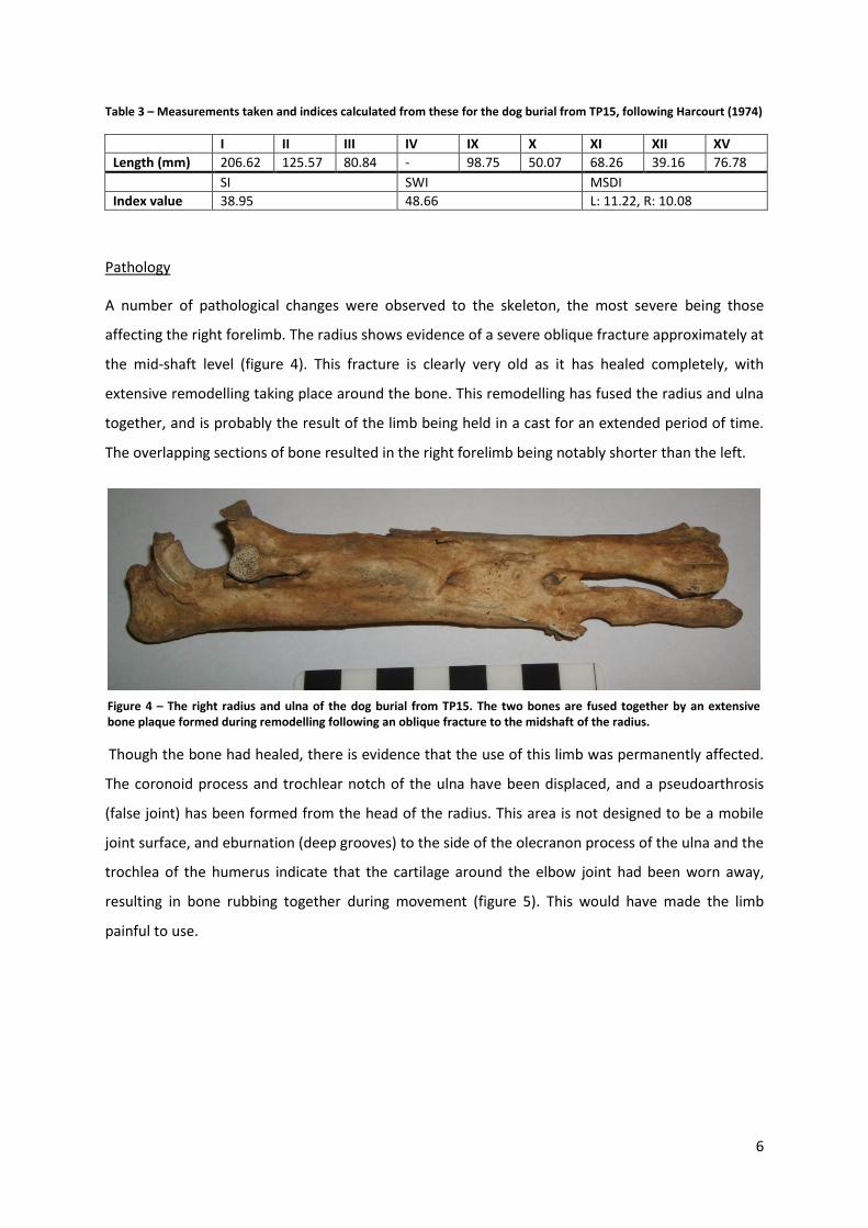
affecting the right forelimb. The radius shows evidence of a severe oblique fracture approximately at
the mid-shaft level (figure 4). This fracture is clearly very old as it has healed completely, with
extensive remodelling taking place around the bone. This remodelling has fused the radius and ulna
together, and is probably the result of the limb being held in a cast for an extended period of time.
The overlapping sections of bone resulted in the right forelimb being notably shorter than the left.
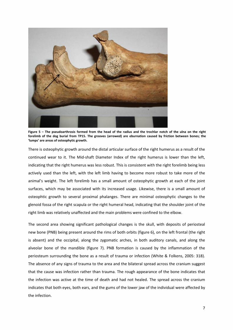
Though the bone had healed, there is evidence that the use of this limb was permanently affected.
The coronoid process and trochlear notch of the ulna have been displaced, and a pseudoarthrosis
(false joint) has been formed from the head of the radius. This area is not designed to be a mobile
joint surface, and eburnation (deep grooves) to the side of the olecranon process of the ulna and the
trochlea of the humerus indicate that the cartilage around the elbow joint had been worn away,
resulting in bone rubbing together during movement (figure 5). This would have made the limb
painful to use.
Figure 4 – The right radius and ulna of the dog burial from TP15. The two bones are fused together by an extensive bone plaque formed during remodelling following an oblique fracture to the midshaft of the radius.
7
There is osteophytic growth around the distal articular surface of the right humerus as a result of the
continued wear to it. The Mid-shaft Diameter Index of the right humerus is lower than the left,
indicating that the right humerus was less robust. This is consistent with the right forelimb being less
actively used than the left, with the left limb having to become more robust to take more of the
animal’s weight. The left forelimb has a small amount of osteophytic growth at each of the joint
surfaces, which may be associated with its increased usage. Likewise, there is a small amount of
osteophtic growth to several proximal phalanges. There are minimal osteophytic changes to the
glenoid fossa of the right scapula or the right humeral head, indicating that the shoulder joint of the
right limb was relatively unaffected and the main problems were confined to the elbow.
The second area showing significant pathological changes is the skull, with deposits of periosteal
new bone (PNB) being present around the rims of both orbits (figure 6), on the left frontal (the right
is absent) and the occipital, along the zygomatic arches, in both auditory canals, and along the
alveolar bone of the mandible (figure 7). PNB formation is caused by the inflammation of the
periosteum surrounding the bone as a result of trauma or infection (White & Folkens, 2005: 318).
The absence of any signs of trauma to the area and the bilateral spread across the cranium suggest
that the cause was infection rather than trauma. The rough appearance of the bone indicates that
the infection was active at the time of death and had not healed. The spread across the cranium
indicates that both eyes, both ears, and the gums of the lower jaw of the individual were affected by
the infection.
Figure 5 – The pseudoarthrosis formed from the head of the radius and the trochlar notch of the ulna on the right forelimb of the dog burial from TP15. The grooves (arrowed) are eburnation caused by friction between bones; the ‘lumps’ are areas of osteophytic growth.

8
Figure 6 – Periosteal new bone formation around the left orbit of the dog burial from TP15
Figure 7 – Periosteal new bone formation on the left mandible of the dog burial from TP15.
9
Age
All epiphyses were fully fused indicating the long bones had stopped growing, and cranial sutures
were fully fused, indicating the individual was fully mature at the time of death. The teeth are
moderately worn, with the cusps being blunted, but the crowns are still high (figure 8). Rates of wear
vary greatly between individuals depending on factors such as differences in diet and whether they
habitually chew other non-food items, making tooth wear an unreliable indicator of age. The lack of
any age-related degeneration (all degeneration appears to be secondary to trauma) suggests that
the individual was not very old at the time of death. As such, the individual is best described as
simply an ‘adult’.
Sex
The presence of a baculum (Os penis) indicates that the individual was male. The shape of the base
of the skull also suggests a male individual.
Stature
The height of a dog is assessed by estimating the height of its withers (shoulders) through measuring
the length of the long bones in its limbs. The measurements taken are shown in table 4. There is a
clear difference between the length of the left and right forelimbs, with the left radius being 15mm
longer than the right. If the individual’s other limb bones were healthy, it could be anticipated that
they would produce a similar height estimate to those from the healthy limbs here, being around
50cm. This is about the same height as a border collie or a springer spaniel (Kennel Club, 2013).
Table 4 – The length of long bones measured in mm and the height estimate each produces using the formulae of Harcourt (1974).
Humerus Radius Ulna
Humerus + Radius
Femur Tibia Femur +Tibia
R L R L R L R L R L R L R L
Length (mm)
154 151 143 158 173 183 297 308 - - 170 - - -
Height Estimate (cm)
50.2 49.1 47.4 52.2 48.7 51.5 48.6 50.3 - - 50.6 - - -
Figure 8 – Attritional wear to the mandibular dentition of the dog burial from TP15
10
References
Harcourt, R.A. 1974. The dog in prehistoric and early Britain. Journal of Archaeological Science 1:
151-75
Kennel Club Limited, The. 2013. Breed Information Centre (Online)
[Accessed 30/11/13]
[Available at: http://www.thekennelclub.org.uk/services/public/breed/Default.aspx]
The, T.L. and Trouth, C.O. 1976. Sexual dimorphism in the basilar part of the occipital bone of the
dog (Canis familiaris). Acta Anatomica 95: 565–71
Trouth, C.O., Winter, S., Gupta, K.C., Millis, R.M. and Holloway, J.A. 1977. Analysis of the sexual
dimorphism in the basioccipital portion of the dog’s skull. Acta Anatomica 98: 469–73
White, T.D. and Folkens, P.A. 2005. The Human Bone Manual. London: Elsevier
11
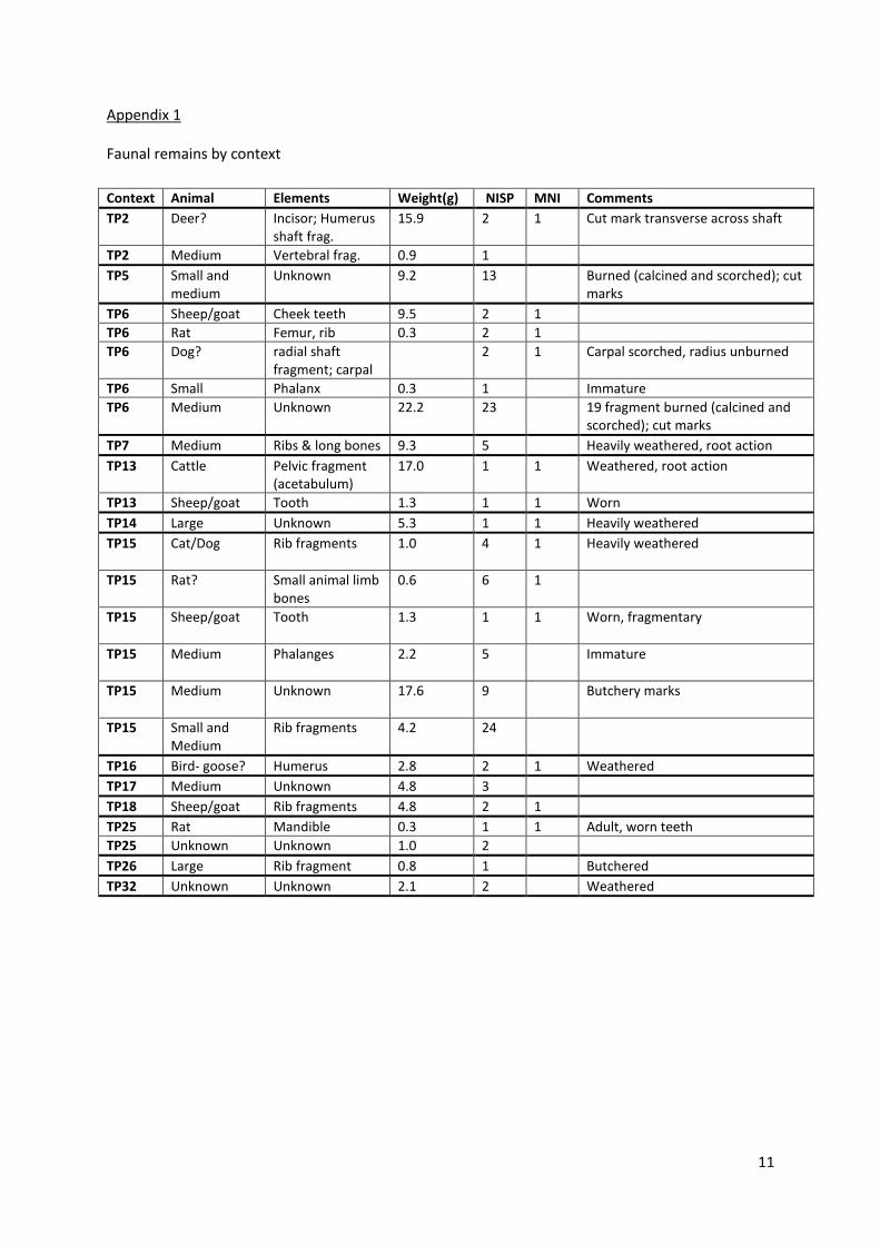
Appendix 1
Faunal remains by context
Context Animal Elements Weight(g) NISP MNI Comments
TP2 Deer? Incisor; Humerus shaft frag.
15.9 2 1 Cut mark transverse across shaft
TP2 Medium Vertebral frag. 0.9 1
TP5 Small and medium
Unknown 9.2 13 Burned (calcined and scorched); cut marks
TP6 Sheep/goat Cheek teeth 9.5 2 1
TP6 Rat Femur, rib 0.3 2 1
TP6 Dog? radial shaft fragment; carpal
2 1 Carpal scorched, radius unburned
TP6 Small Phalanx 0.3 1 Immature
TP6 Medium Unknown 22.2 23 19 fragment burned (calcined and scorched); cut marks
TP7 Medium Ribs & long bones 9.3 5 Heavily weathered, root action
TP13 Cattle Pelvic fragment (acetabulum)
17.0 1 1 Weathered, root action
TP13 Sheep/goat Tooth 1.3 1 1 Worn
TP14 Large Unknown 5.3 1 1 Heavily weathered
TP15 Cat/Dog Rib fragments 1.0 4 1 Heavily weathered
TP15 Rat? Small animal limb bones
0.6 6 1
TP15 Sheep/goat Tooth 1.3 1 1 Worn, fragmentary
TP15 Medium Phalanges 2.2 5 Immature
TP15 Medium Unknown 17.6 9 Butchery marks
TP15 Small and Medium
Rib fragments 4.2 24
TP16 Bird- goose? Humerus 2.8 2 1 Weathered
TP17 Medium Unknown 4.8 3
TP18 Sheep/goat Rib fragments 4.8 2 1
TP25 Rat Mandible 0.3 1 1 Adult, worn teeth
TP25 Unknown Unknown 1.0 2
TP26 Large Rib fragment 0.8 1 Butchered
TP32 Unknown Unknown 2.1 2 Weathered

12
Appendix 2
The measurements and indices of Harcourt (1974).
Measurement Definition
I The most posterior aspect of the occipital protuberance to the anterior margin of the medial incisor alveoli between the central incisors (alveolare).
II Occipital protuberance to junction of nasal and frontal bones (nasion)
III Nasion to alveolare.
IV Zygomatic width (maximum).
IX Palatal length.
X Palatal width between PM4 and Ml.
XI Maxillary cheek tooth row length.
XII Snout width across the outer margins of the alveoli of the canines.
XV Mandibular cheek tooth row length.
Index Calculation Indicates
Cephalic Index IV x 100 / I Width of the skull relative to its length
Snout Index III x 100 / I Length of the snout relative to that of the whole head
Snout Width Index XII x 100 / III Width of the muzzle relative to the length of the nose
Mid-Shaft Diameter Index MSD x 100 / Length ‘Stoutness’ of the bone


![Osteological sample profile [Bom Santo Cave (Lisbon) and the Middle Neolithic Societies of Southern Portugal]](https://static.fdokumen.com/doc/165x107/6319d548bc8291e22e0f4aa7/osteological-sample-profile-bom-santo-cave-lisbon-and-the-middle-neolithic-societies.jpg)