Association of nucleoid proteins with coding and non-coding segments of the Escherichia coli genome
11
Association of nucleoid proteins with coding and non-coding segments of the Escherichia coli genome David C. Grainger*, Douglas Hurd 1 , Martin D. Goldberg and Stephen J. W. Busby School of Biosciences, University of Birmingham, Edgbaston, Birmingham B15 2TT, UK and 1 Oxford Gene Technology, Begbroke Science Park, Sandy Lane, Yarnton, Oxford OX5 1PF, UK Received June 26, 2006; Revised July 11, 2006; Accepted July 12, 2006 ABSTRACT The Escherichia coli chromosome is condensed into an ill-defined structure known as the nucleoid. Nucleoid-associated DNA-binding proteins are involved in maintaining this structure and in medi- ating chromosome compaction. We have exploited chromatin immunoprecipitation and high-density microarrays to study the binding of three such pro- teins, FIS, H-NS and IHF, across the E.coli genome in vivo. Our results show that the distribution of these proteins is biased to intergenic parts of the genome, and that the binding profiles overlap. Hence some targets are associated with combina- tions of bound FIS, H-NS and IHF. In addition, many regions associated with FIS and H-NS are also associated with RNA polymerase. INTRODUCTION In eukaryotic organisms, DNA is compacted by histones, which organize DNA into nucleosomes that are further orga- nized into the higher order structures that form chromatin. Recently, the use of chromatin immunoprecipitation (ChIP), in combination with high-density microarrays (ChIP-chip), has allowed the distribution of some histones to be mapped across the Saccharomyces cerevisiae genome (1,2). These studies showed that, although histones associate with both coding and non-coding parts of the genome, they are depleted at promoters. Consistent with this, Nagy and co-workers (3) were able to fractionate the yeast genome on the basis of histone density. Eukaryotic chromatin is remodelled by tran- scription (4) and this remodelling can involve both the removal and the recruitment of histones (5,6). By comparison, the mechanisms used by prokaryotes to organize their chromosomal DNA are poorly understood. Bacterial DNA forms a highly condensed, yet undefined, structure called the nucleoid, that is thought to be ordered by supercoiling, macromolecular crowding, RNA and nucleoid-associated proteins, the prokaryotic equivalent of histones (7–9). Extensive studies with Escherichia coli have shown that many of these nucleoid-associated proteins com- pact chromosomal DNA (10–13) and can also function as transcription factors (14,15). Thus, factor for inversion stimu- lation (FIS), histone-like nucleoid structuring protein (H-NS) and the integration host factor (IHF) are nucleoid-associated proteins, whose individual subunits are present at 60 000, 20 000 and 12 000 copies, respectively, in rapidly growing E.coli cells (16). All three proteins bind to A:T rich DNA tar- gets and alter DNA topology. For H-NS, these DNA sites are highly degenerate but some DNA-binding specificity is displayed by FIS and IHF. Upon binding, FIS bends DNA by between 50 and 90 (17), whilst IHF induces a bend of 160 (18). H-NS is thought to oligomerize at intrinsically curved DNA sequences and, once bound, may induce further DNA bending (19). In addition to their role as chromosome shaping proteins, FIS, H-NS and IHF function as gene- specific transcription factors. For example, H-NS represses the transcription of many non-essential genes (20,21). Simi- larly, during rapid growth, FIS represses many promoters (22), though its best-characterized role is as an activator at the rrn operons (23). Little is known about the genome-wide distribution of the various nucleoid proteins. This is illustrated by the fact that only 63, 36 and 55 targets for FIS, H-NS and IHF respec- tively are listed in the Ecocyc database [www.ecocyc.org (24)]. There have been several attempts to study the distribu- tion of FIS, H-NS and IHF across bacterial genomes. Robison and co-workers (25) used bioinformatics to search the E.coli genome for DNA sequences that resemble the binding sites for FIS, H-NS and IHF. The study identified >10 000 binding sites for each factor and, although <10% of the E.coli genome is non-coding, >23% of the predicted targets for each protein were in non-coding DNA. Other attempts to study the distri- bution of nucleoid-associated proteins have relied on com- paring the transcriptomes of wild-type and mutant cells (20,22,26). The distribution of the protein under study is then inferred from changes in transcription resulting from a mutation in its gene. However, such approaches are unable to distinguish primary from secondary effects, and they *To whom correspondence should be addressed. Tel: +44 121 414 5435; Fax: +44 121 414 5925; Email: [email protected] Ó 2006 The Author(s). This is an Open Access article distributed under the terms of the Creative Commons Attribution Non-Commercial License (http://creativecommons.org/licenses/ by-nc/2.0/uk/) which permits unrestricted non-commercial use, distribution, and reproduction in any medium, provided the original work is properly cited. 4642–4652 Nucleic Acids Research, 2006, Vol. 34, No. 16 Published online 8 September 2006 doi:10.1093/nar/gkl542 by guest on September 26, 2013 http://nar.oxfordjournals.org/ Downloaded from by guest on September 26, 2013 http://nar.oxfordjournals.org/ Downloaded from by guest on September 26, 2013 http://nar.oxfordjournals.org/ Downloaded from by guest on September 26, 2013 http://nar.oxfordjournals.org/ Downloaded from by guest on September 26, 2013 http://nar.oxfordjournals.org/ Downloaded from by guest on September 26, 2013 http://nar.oxfordjournals.org/ Downloaded from by guest on September 26, 2013 http://nar.oxfordjournals.org/ Downloaded from by guest on September 26, 2013 http://nar.oxfordjournals.org/ Downloaded from by guest on September 26, 2013 http://nar.oxfordjournals.org/ Downloaded from by guest on September 26, 2013 http://nar.oxfordjournals.org/ Downloaded from by guest on September 26, 2013 http://nar.oxfordjournals.org/ Downloaded from
-
Upload
independent -
Category
Documents
-
view
1 -
download
0
Transcript of Association of nucleoid proteins with coding and non-coding segments of the Escherichia coli genome
Association of nucleoid proteins with coding andnon-coding segments of the Escherichia coligenomeDavid C. Grainger*, Douglas Hurd1, Martin D. Goldberg and Stephen J. W. Busby
School of Biosciences, University of Birmingham, Edgbaston, Birmingham B15 2TT, UK and1Oxford Gene Technology, Begbroke Science Park, Sandy Lane, Yarnton, Oxford OX5 1PF, UK
Received June 26, 2006; Revised July 11, 2006; Accepted July 12, 2006
ABSTRACT
The Escherichia coli chromosome is condensedinto an ill-defined structure known as the nucleoid.Nucleoid-associated DNA-binding proteins areinvolved in maintaining this structure and in medi-ating chromosome compaction. We have exploitedchromatin immunoprecipitation and high-densitymicroarrays to study the binding of three such pro-teins, FIS, H-NS and IHF, across the E.coli genomein vivo. Our results show that the distribution ofthese proteins is biased to intergenic parts of thegenome, and that the binding profiles overlap.Hence some targets are associated with combina-tions of bound FIS, H-NS and IHF. In addition, manyregions associated with FIS and H-NS are alsoassociated with RNA polymerase.
INTRODUCTION
In eukaryotic organisms, DNA is compacted by histones,which organize DNA into nucleosomes that are further orga-nized into the higher order structures that form chromatin.Recently, the use of chromatin immunoprecipitation (ChIP),in combination with high-density microarrays (ChIP-chip),has allowed the distribution of some histones to be mappedacross the Saccharomyces cerevisiae genome (1,2). Thesestudies showed that, although histones associate with bothcoding and non-coding parts of the genome, they are depletedat promoters. Consistent with this, Nagy and co-workers (3)were able to fractionate the yeast genome on the basis ofhistone density. Eukaryotic chromatin is remodelled by tran-scription (4) and this remodelling can involve both theremoval and the recruitment of histones (5,6).
By comparison, the mechanisms used by prokaryotes toorganize their chromosomal DNA are poorly understood.Bacterial DNA forms a highly condensed, yet undefined,structure called the nucleoid, that is thought to be orderedby supercoiling, macromolecular crowding, RNA and
nucleoid-associated proteins, the prokaryotic equivalent ofhistones (7–9). Extensive studies with Escherichia coli haveshown that many of these nucleoid-associated proteins com-pact chromosomal DNA (10–13) and can also function astranscription factors (14,15). Thus, factor for inversion stimu-lation (FIS), histone-like nucleoid structuring protein (H-NS)and the integration host factor (IHF) are nucleoid-associatedproteins, whose individual subunits are present at 60 000,20 000 and 12 000 copies, respectively, in rapidly growingE.coli cells (16). All three proteins bind to A:T rich DNA tar-gets and alter DNA topology. For H-NS, these DNA sites arehighly degenerate but some DNA-binding specificity isdisplayed by FIS and IHF. Upon binding, FIS bends DNAby between 50� and 90� (17), whilst IHF induces a bend of160� (18). H-NS is thought to oligomerize at intrinsicallycurved DNA sequences and, once bound, may induce furtherDNA bending (19). In addition to their role as chromosomeshaping proteins, FIS, H-NS and IHF function as gene-specific transcription factors. For example, H-NS repressesthe transcription of many non-essential genes (20,21). Simi-larly, during rapid growth, FIS represses many promoters(22), though its best-characterized role is as an activator atthe rrn operons (23).
Little is known about the genome-wide distribution of thevarious nucleoid proteins. This is illustrated by the fact thatonly 63, 36 and 55 targets for FIS, H-NS and IHF respec-tively are listed in the Ecocyc database [www.ecocyc.org(24)]. There have been several attempts to study the distribu-tion of FIS, H-NS and IHF across bacterial genomes. Robisonand co-workers (25) used bioinformatics to search the E.coligenome for DNA sequences that resemble the binding sitesfor FIS, H-NS and IHF. The study identified >10 000 bindingsites for each factor and, although <10% of the E.coli genomeis non-coding, >23% of the predicted targets for each proteinwere in non-coding DNA. Other attempts to study the distri-bution of nucleoid-associated proteins have relied on com-paring the transcriptomes of wild-type and mutant cells(20,22,26). The distribution of the protein under study isthen inferred from changes in transcription resulting from amutation in its gene. However, such approaches are unableto distinguish primary from secondary effects, and they
*To whom correspondence should be addressed. Tel: +44 121 414 5435; Fax: +44 121 414 5925; Email: [email protected]
� 2006 The Author(s).This is an Open Access article distributed under the terms of the Creative Commons Attribution Non-Commercial License (http://creativecommons.org/licenses/by-nc/2.0/uk/) which permits unrestricted non-commercial use, distribution, and reproduction in any medium, provided the original work is properly cited.
4642–4652 Nucleic Acids Research, 2006, Vol. 34, No. 16 Published online 8 September 2006doi:10.1093/nar/gkl542
by guest on September 26, 2013
http://nar.oxfordjournals.org/D
ownloaded from
by guest on Septem
ber 26, 2013http://nar.oxfordjournals.org/
Dow
nloaded from
by guest on September 26, 2013
http://nar.oxfordjournals.org/D
ownloaded from
by guest on Septem
ber 26, 2013http://nar.oxfordjournals.org/
Dow
nloaded from
by guest on September 26, 2013
http://nar.oxfordjournals.org/D
ownloaded from
by guest on Septem
ber 26, 2013http://nar.oxfordjournals.org/
Dow
nloaded from
by guest on September 26, 2013
http://nar.oxfordjournals.org/D
ownloaded from
by guest on Septem
ber 26, 2013http://nar.oxfordjournals.org/
Dow
nloaded from
by guest on September 26, 2013
http://nar.oxfordjournals.org/D
ownloaded from
by guest on Septem
ber 26, 2013http://nar.oxfordjournals.org/
Dow
nloaded from
by guest on September 26, 2013
http://nar.oxfordjournals.org/D
ownloaded from
yield no information about the distribution of nucleoid pro-teins between coding and non-coding DNA.
The aim of this study was to measure directly the bindingof FIS, H-NS and IHF across the entire E.coli genome using aChIP-chip analysis. We show that, in contrast to eukaryotichistone proteins, the binding of these proteins is biasedtowards non-coding parts of the genome. We identified over-lap in the DNA-binding profiles of FIS and H-NS, and manyregulatory regions bound with these two proteins were alsoassociated with RNA polymerase. Some binding sites fornucleoid proteins were found in coding DNA and, for FISand H-NS, some of these sites are in regions of the genomethat are highly transcribed.
MATERIALS AND METHODS
Bacterial growth conditions
Experiments were performed using E.coli strain MG1655grown to mid-log phase (OD650 0.3–0.4) at 37�C in M9minimal media supplemented with fructose. Our choice of aminimal and defined growth condition allowed us to monitorthe effect of adding specific reagents to E.coli cultures. Thus,for induction of the lac operon, 2 mM isopropyl-b-D-thiogalactopyranoside (IPTG) was added and cross-linkingwith formaldehyde was initiated at different time points.Salicylic acid was added to a final concentration of 5 mMand cross-linking was initiated after 40 min.
Chromatin immunoprecipitation
Bacterial cells were treated with formaldehyde, harvestedand lysed, and nucleoprotein was prepared as described byGrainger et al. (27). Immunoprecipitation was then per-formed using monoclonal antibodies against the b subunitof RNA polymerase (Neoclone, Madison, USA) and H-NS(donated by Jay Hinton) or rabbit polyclonal antibodiesagainst FIS [donated by Akira Ishihama, see Azam et al.(16)] and IHF (donated by Steve Goodman). Cross-reactivityof the anti-H-NS antibody with StpA was not observed(M. D. Goldberg, unpublished data). ImmunoprecipitatedDNA samples or total cell nucleoprotein samples were puri-fied and labelled with Cy5 or Cy3, without amplification, asdescribed previously (27).
PCR analysis of immunoprecipitated DNA
Quantitative PCR analyses were performed in real-time usingan Applied Biosystems 7700 sequence detector. Relativeoccupancy values shown in Figure 6 were calculated bydetermining the immunoprecipitation efficiency of the targetDNA sequence in samples from cultures either with or with-out IPTG. As internal controls, we used segments of therrl and mtlA genes. The fold enrichment of target sequences,relative to the control regions, is shown as occupancy unitsand error bars represent the standard deviation of the meanof three independent experiments. Primer pairs used in thisanalysis were lacZ up (50-aatcgccttgcagcac-30) and lacZdown (50-gccattcgccattca-30), lacY up (50-cgccgttttactctttttcg-30)and lacY down (50-gcaggaaacgccaataacat-30), lacA up (50-atatgt-gcgaaggcttaccg-30) and lacA down (50-aattgcggcctatatggatg-30),rrl up (50-ctacggtgctgaagcaacaa-30) and rrl down (50-cgaag-
ttacggcaccatttt-30), mtlA up (50-cgcgacagcaaacataagaa-30) andmtlA down (50-gttcggtaaccaccctgttg-30).
Microarray analysis of immunoprecipitated DNA
Microarrays (Oxford Gene Technology) were designed andproduced specifically to analyse DNA obtained from ChIPexperiments with E.coli MG1655 (27). Arrays consisted of21 321 60mer oligonucleotide probes that match MG1655sequences at intervals of �160 bp. Unamplified DNAobtained from immunoprecipitations and control MG1655genomic DNA was differentially labelled and hybridized tothe microarray as described previously (27). Values shownare the average of two independent experiments.
ChIP-chip data analysis
The Cy5/Cy3 intensity ratio was calculated for each spot andplotted against the corresponding position on the E.coliMG1655 chromosome. A cut-off was chosen and all probesthat had an intensity ratio greater than this value in bothexperiments were selected as a target. Adjacent target probeswere merged, the target position being defined by centre ofthe probe with the highest average intensity ratio. Targetswere judged to be in non-coding DNA if the peak centrefell within 200 bp of a non-coding DNA sequence. Leewayof 200 bp was allowed because, for some non-codingsequences, the nearest probe was at the 50 end of an adjacentgene and because peak locations are only accurate to�200 bp (27). Note that, in this study, we refer to DNAencoding stable RNA as ‘coding’ DNA and that the ribo-somal RNA operons were treated as a single entity. To esti-mate the FDR one dataset was randomized with respect to theprobe positions. The number of probes passing the cut-offwas then re-calculated. This process was repeated 50 timesto determine the average number of probes that passed thecut-off after randomization. When a stringent cut-off wasused the number of probes passing the cut-off post random-ization was �1% of the number that originally passed thecut-off.
To investigate the relationship between DNA sequence andthe binding of the various factors we ranked probes on themicroarray in order of their % A:T content. Probes with thesame A:T content were grouped and the average Cy5/Cy3ratio was calculated, using our ChIP-chip data for eitherFIS, H-NS, IHF or, as a control, RNA polymerase. The %A:T content of each group of probes was then plotted againstthe average signal intensity (Figure 3). Because groups ofprobes with a very high or very low % A:T content weresmall they could not be used to calculate a meaningful aver-age signal intensity they were excluded from the analysis(this applied to <1% of the probes on the microarray).
Phenol–chloroform fractionation of E.colinucleoprotein
Cells were grown to mid-log phase in minimal media andnucleoprotein was cross-linked with 1% formaldehyde.Cells were then washed three times with Tris-buffered salineand lysed by sonication, further sonication was used to frag-ment the genomic DNA. Cell debris was removed by cen-trifugation and the sample was split into two aliquots. Thefirst aliquot was retained as a control and the second was
Nucleic Acids Research, 2006, Vol. 34, No. 16 4643
treated with phenol–chloroform to remove DNA fragmentscross-linked to large amounts of protein. The control sampleand the phenol–chloroform treated sample were then decross-linked and processed as described for immunoprecipitationexperiments (27) before being labelled with Cy3 and Cy5,respectively, and hybridized to the microarray.
RESULTS
DNA fragments associated with FIS, H-NS, IHF andRNA polymerase in vivo
Previously, we analysed the distribution, in vivo, of RNApolymerase and different transcription factors bound to theE.coli chromosome (27). DNA targets were immunoprecipi-tated using antibodies directed against the different proteins.To analyse this DNA, we used microarrays of �22 000 60meroligonucleotides that correspond to evenly spaced sections ofthe E.coli genome. These arrays allowed us to measure bind-ing in both coding and non-coding sections of the E.coli chro-mosome. We reasoned that these arrays would be suitable forstudying the distribution of nucleoid-associated proteins.
Our first aim was to generate ChIP-chip data for FIS,H-NS, IHF and RNA polymerase under the same conditionsand thus create a ‘snapshot’ of their binding profiles. Thus,E.coli strain MG1655 was grown aerobically in M9 minimalmedia to an OD650 of 0.4. Cells were treated with formalde-hyde, lysed and their DNA was sonicated, yielding DNAfragments of �500–1000 bp. Antibodies directed againsteither FIS, H-NS, IHF or the b subunit of RNA polymerasewere then used to precipitate DNA fragments associatedwith each protein. After purification, immunoprecipitatedDNA fragments were labelled with Cy5. A control sample,labelled with Cy3, was generated from an aliquot of thetotal cross-linked nucleoprotein, reserved before immunopre-cipitation. The Cy5-labelled samples were then each mixedwith the control Cy3-labelled sample and hybridized to themicroarray. After washing and scanning, the Cy5/Cy3 signalintensity ratio was calculated for each probe. All experimentswere done in duplicate and replicates had a correlationco-efficient of >0.9.
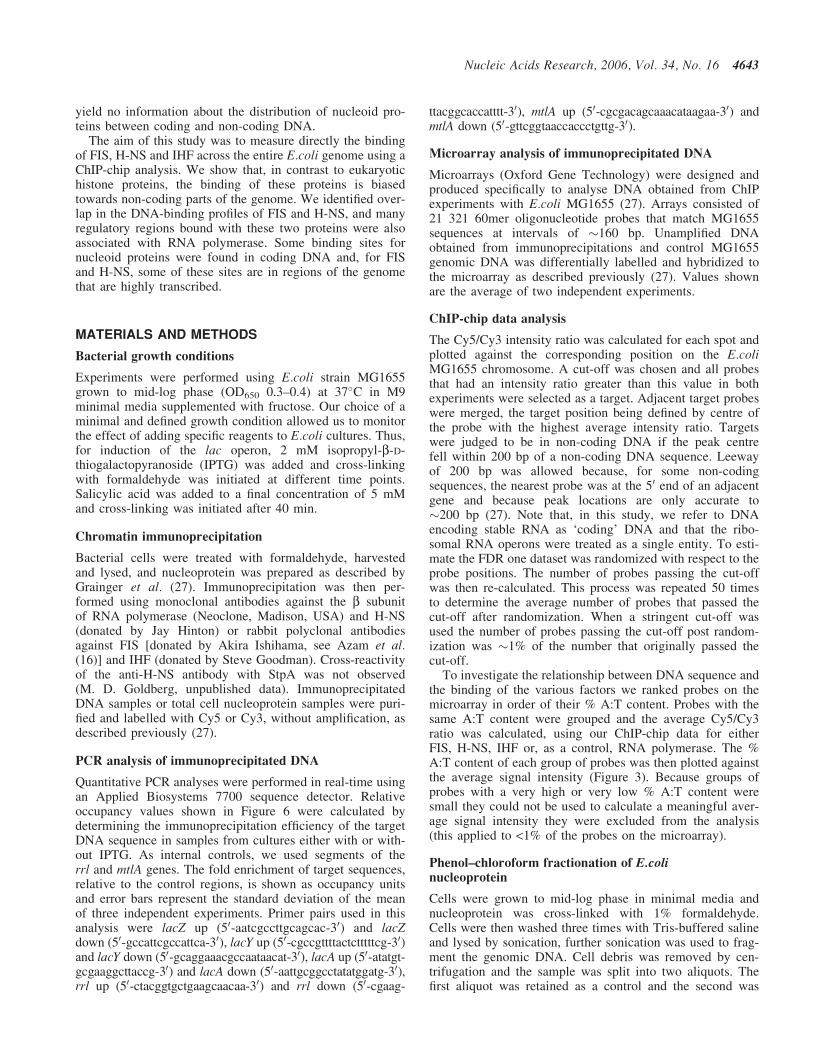
The complete dataset for each experiment is listed in Sup-plementary Table 1, which shows the Cy5/Cy3 ratio for eachprobe on the microarray as a function of its position on thegenome. Figure 1 shows an overview of the profiles forFIS, H-NS and IHF. It is clear that each of these proteinsbinds to many different targets across the E.coli chromosome,generating a wide range of signal intensities. Thus, first, wesearched for the targets for each protein that are listed inthe current Ecocyc database.
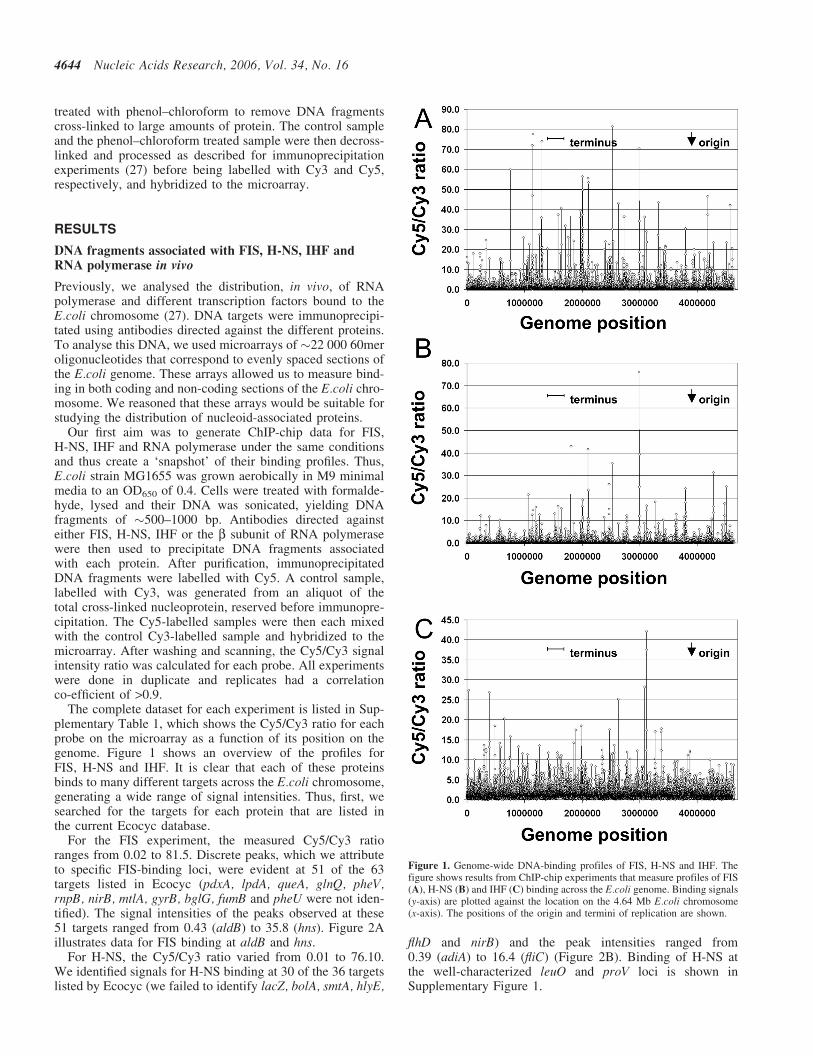
For the FIS experiment, the measured Cy5/Cy3 ratioranges from 0.02 to 81.5. Discrete peaks, which we attributeto specific FIS-binding loci, were evident at 51 of the 63targets listed in Ecocyc (pdxA, lpdA, queA, glnQ, pheV,rnpB, nirB, mtlA, gyrB, bglG, fumB and pheU were not iden-tified). The signal intensities of the peaks observed at these51 targets ranged from 0.43 (aldB) to 35.8 (hns). Figure 2Aillustrates data for FIS binding at aldB and hns.
For H-NS, the Cy5/Cy3 ratio varied from 0.01 to 76.10.We identified signals for H-NS binding at 30 of the 36 targetslisted by Ecocyc (we failed to identify lacZ, bolA, smtA, hlyE,
flhD and nirB) and the peak intensities ranged from0.39 (adiA) to 16.4 (fliC) (Figure 2B). Binding of H-NS atthe well-characterized leuO and proV loci is shown inSupplementary Figure 1.
Figure 1. Genome-wide DNA-binding profiles of FIS, H-NS and IHF. Thefigure shows results from ChIP-chip experiments that measure profiles of FIS(A), H-NS (B) and IHF (C) binding across the E.coli genome. Binding signals(y-axis) are plotted against the location on the 4.64 Mb E.coli chromosome(x-axis). The positions of the origin and termini of replication are shown.
4644 Nucleic Acids Research, 2006, Vol. 34, No. 16
For the experiment with IHF, the signal ranges from0.10 to 42.13. We observed peaks, signifying IHF binding,at 51 of the 55 targets listed by the Ecocyc database (wefailed to identify gcd, hemA, glcD and mtr). The peak inten-sities at these targets ranged from 1.8 (sucA) to 27.4 (carAB)(Figure 2C).
Our aim was to generate an unbiased list of targets for eachfactor and to measure their distribution between coding andnon-coding DNA. Using a stringent cut-off (selected to givea false discovery rate, FDR, of �1%) we identified 224, 100
and 135 targets for FIS, H-NS and IHF, respectively. Thesetargets are listed in Supplementary Tables 2–4. Althoughnon-coding sequences account for <10% of the E.coli gen-ome, �50% of the targets for each protein are in non-codingDNA. This distribution was not substantially changed when amore relaxed cut-off was set (corresponding to the value ofthe lowest scoring known target for each nucleoid protein).
FIS, H-NS and IHF are known to bind to A:T rich targets.Thus, with the data from each experiment, we calculated theaverage of the measured signal intensities for probes grouped
Figure 2. Association of FIS, H-NS and IHF with known target sites. The figure shows results from ChIP-chip experiments that measure profiles of FIS, H-NSand IHF binding across segments of the E.coli genome that are illustrated schematically below the profile. Binding signals (y-axis) are plotted against thegenomic location (x-axis) for two known targets for each factor. Data for FIS binding at adlB and hns are shown in (A), H-NS binding at adiA and fliC are shownin (B) and IHF binding at sucA and carA are shown in (C).
Nucleic Acids Research, 2006, Vol. 34, No. 16 4645
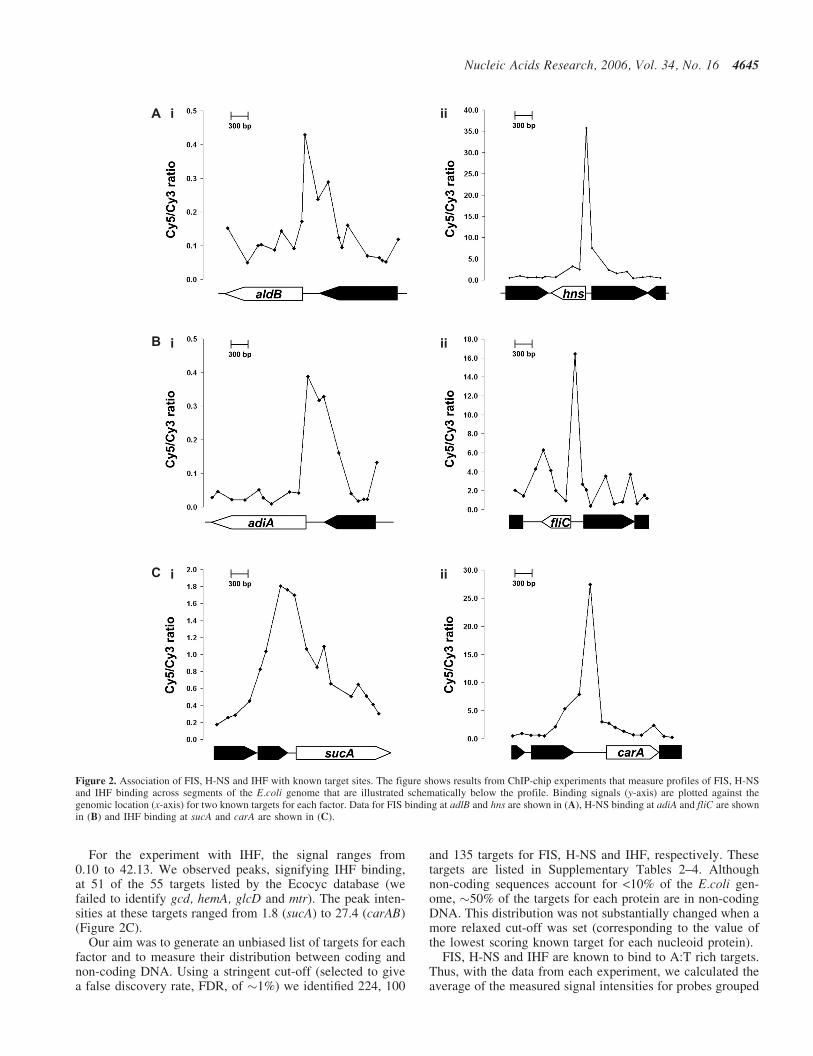
according to their % A:T content. Figure 3 shows a plot ofthe average binding signal for each protein as a function ofprobe A:T content. The graph shows that, as expected,there is an overall positive correlation between the A:T con-tent of probes and the binding of FIS, H-NS and IHF. As acontrol, data from an experiment in which we had measuredthe chromosome-wide distribution of RNA polymerase wasincluded. These data show that, in contrast to FIS, H-NSand IHF, there is no overall positive correlation betweenthe A:T content of probes and the binding of RNA poly-merase (Figure 3). Note that we had run the RNA polymeraseexperiment in order to compare the binding of RNApolymerase to the binding of FIS, H-NS and IHF (seebelow). This experiment gave the expected result, reportedby Grainger et al. (27), with the largest signals for RNA poly-merase binding located at genes encoding stable RNA andproteins essential for rapid growth (full data in Supplemen-tary Table 1).
Analysis of FIS, H-NS, IHF and RNA polymerasebinding in non-coding DNA
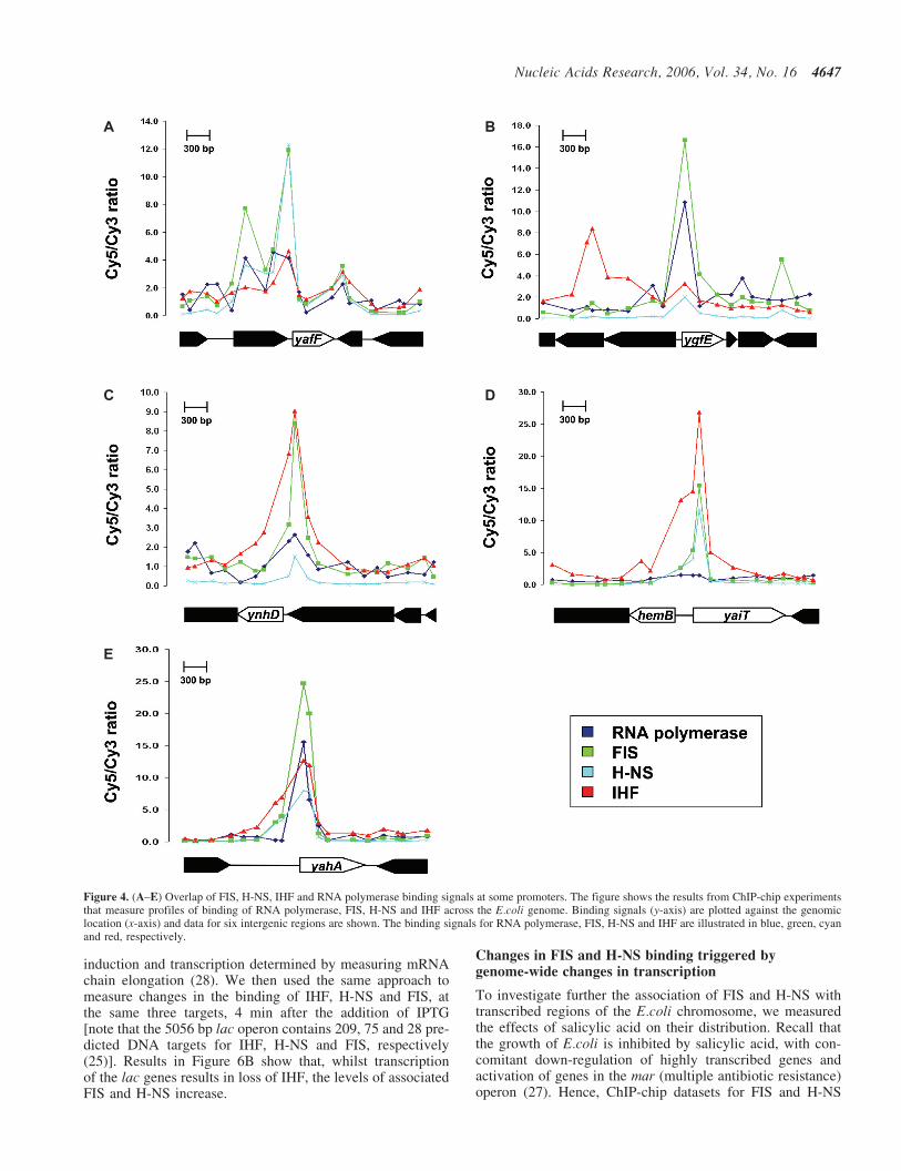
Although <10% of the E.coli genome is non-coding, ourresults show that �50% of the targets for FIS, H-NS andIHF are in these regions. To analyse further this binding,we aligned the ChIP-chip datasets for FIS, H-NS, IHF andRNA polymerase, removed data from probes that correspondto coding DNA, and scaled the datasets to have the sameaverage Cy5/Cy3 ratio. We then applied the same stringentcut-off (FRD �1%) to each dataset and counted the numberof probes that passed the cut-off for different combinations ofthe various factors studied. We observed a clear correlationbetween the binding of FIS and H-NS. Thus, �60% of thenon-coding DNA loci associated with FIS were also boundwith H-NS and vice versa (e.g. see Figure 4A). Additionally,�50% of the non-coding targets for FIS and/or H-NS werealso associated with RNA polymerase (Figure 4B). Overlapbetween the binding of IHF and FIS/H-NS was less frequent;only �30% of the targets for FIS or H-NS were also targets
for IHF (Figure 4C). A small number of targets passed thecut-off for FIS, H-NS and IHF (Figure 4D) and, in �50%of these cases, RNA polymerase was also found to be associ-ated (Figure 4E). Note that, at the regulatory regions of ygfE(Figure 4B) and yahA (Figure 4E), RNA polymerase is boundbut appears not to move into the coding part of the gene.
Analysis of FIS, H-NS and IHF binding in coding DNA
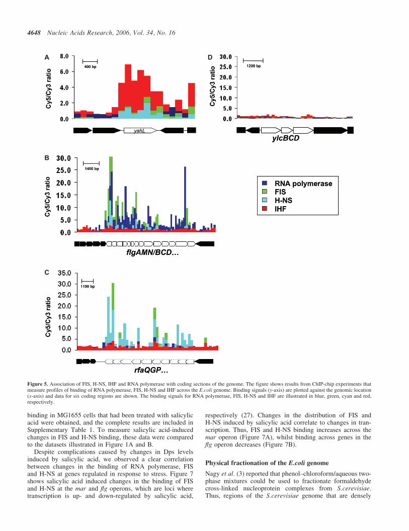
Datasets were aligned and analysed as in the previous section,except that results from probes corresponding to non-codingsections of the genome were discarded. Our expectation wasthat most binding targets would fall in transcriptionally silentparts of the genome and this was the case for IHF (e.g. seeFigure 5A). However, we noted that a significant proportionof probes that pass the cut-off for FIS or H-NS binding(39 and 31%, respectively) correspond to locations at whichRNA polymerase is bound. Thus, in contrast to IHF, FIS andH-NS may bind in both transcriptionally active (Figure 5B)and transcriptionally silent (Figure 5C) open reading frames.Note that the analysis also identified large sections of DNAthat do not appear to be associated with high levels of FIS,H-NS, IHF or RNA polymerase (Figure 5D). We suggestthat these regions may be associated with other factors.
To study directly the effect of transcription on the bindingof FIS, H-NS and IHF, we measured their association withthe lac operon genes, before and after induction with IPTG.To do this, antibodies directed against FIS, H-NS, IHF orthe RNA polymerase b subunit were used to immunoprecipi-tate nucleoprotein samples isolated from cells that had beentreated with formaldehyde, either before or after induction.Real-time PCR was used to quantify the relative amountsof different targets in the immunoprecipitates.
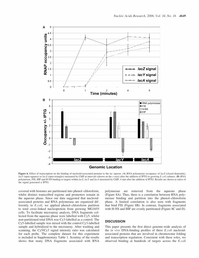
Figure 6A illustrates the time course of changes in RNApolymerase binding to targets in the lacZ, lacY and lacAgenes after addition of IPTG. This shows that RNA poly-merase is recruited to the lac operon within 60 s of induceraddition, and that RNA polymerase requires <2 min totranscribe the operon. This is consistent with rates of lac
Figure 3. Relationship between the A:T content of microarray probes and binding signal for RNA polymerase, FIS, H-NS and IHF. The figure shows averagedChIP-chip binding signals for RNA polymerase, FIS, H-NS and IHF at groups of microarray probes (y-axis) sorted by their A:T content (x-axis).
4646 Nucleic Acids Research, 2006, Vol. 34, No. 16
induction and transcription determined by measuring mRNAchain elongation (28). We then used the same approach tomeasure changes in the binding of IHF, H-NS and FIS, atthe same three targets, 4 min after the addition of IPTG[note that the 5056 bp lac operon contains 209, 75 and 28 pre-dicted DNA targets for IHF, H-NS and FIS, respectively(25)]. Results in Figure 6B show that, whilst transcriptionof the lac genes results in loss of IHF, the levels of associatedFIS and H-NS increase.
Changes in FIS and H-NS binding triggered bygenome-wide changes in transcription
To investigate further the association of FIS and H-NS withtranscribed regions of the E.coli chromosome, we measuredthe effects of salicylic acid on their distribution. Recall thatthe growth of E.coli is inhibited by salicylic acid, with con-comitant down-regulation of highly transcribed genes andactivation of genes in the mar (multiple antibiotic resistance)operon (27). Hence, ChIP-chip datasets for FIS and H-NS
Figure 4. (A–E) Overlap of FIS, H-NS, IHF and RNA polymerase binding signals at some promoters. The figure shows the results from ChIP-chip experimentsthat measure profiles of binding of RNA polymerase, FIS, H-NS and IHF across the E.coli genome. Binding signals (y-axis) are plotted against the genomiclocation (x-axis) and data for six intergenic regions are shown. The binding signals for RNA polymerase, FIS, H-NS and IHF are illustrated in blue, green, cyanand red, respectively.
Nucleic Acids Research, 2006, Vol. 34, No. 16 4647
binding in MG1655 cells that had been treated with salicylicacid were obtained, and the complete results are included inSupplementary Table 1. To measure salicylic acid-inducedchanges in FIS and H-NS binding, these data were comparedto the datasets illustrated in Figure 1A and B.
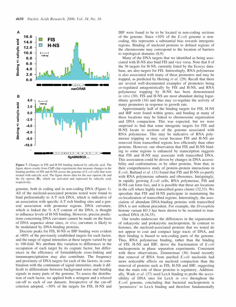
Despite complications caused by changes in Dps levelsinduced by salicylic acid, we observed a clear correlationbetween changes in the binding of RNA polymerase, FISand H-NS at genes regulated in response to stress. Figure 7shows salicylic acid induced changes in the binding of FISand H-NS at the mar and flg operons, which are loci wheretranscription is up- and down-regulated by salicylic acid,
respectively (27). Changes in the distribution of FIS andH-NS induced by salicylic acid correlate to changes in tran-scription. Thus, FIS and H-NS binding increases across themar operon (Figure 7A), whilst binding across genes in theflg operon decreases (Figure 7B).
Physical fractionation of the E.coli genome
Nagy et al. (3) reported that phenol–chloroform/aqueous two-phase mixtures could be used to fractionate formaldehydecross-linked nucleoprotein complexes from S.cerevisiae.Thus, regions of the S.cerevisiae genome that are densely
Figure 5. Association of FIS, H-NS, IHF and RNA polymerase with coding sections of the genome. The figure shows results from ChIP-chip experiments thatmeasure profiles of binding of RNA polymerase, FIS, H-NS and IHF across the E.coli genome. Binding signals (y-axis) are plotted against the genomic location(x-axis) and data for six coding regions are shown. The binding signals for RNA polymerase, FIS, H-NS and IHF are illustrated in blue, green, cyan and red,respectively.
4648 Nucleic Acids Research, 2006, Vol. 34, No. 16
covered with histones are partitioned into phenol–chloroform,whilst distinct transcribed regions and promoters remain inthe aqueous phase. Since our data suggested that nucleoid-associated proteins and RNA polymerase are organized dif-ferently in E.coli, we applied phenol–chloroform partitionto total cross-linked nucleoprotein from growing MG1655cells. To facilitate microarray analysis, DNA fragments col-lected from the aqueous phase were labelled with Cy5, whilstnon-partitioned total DNA was Cy3-labelled as a control. TheCy5-labelled sample was mixed with the control Cy3-labelledsample and hybridized to the microarray. After washing andscanning, the Cy5/Cy3 signal intensity ratio was calculatedfor each probe. The complete dataset for this experimentis included in Supplementary Table 1. Scrutiny of the resultsshows that many DNA fragments associated with RNA
polymerase are removed from the aqueous phase(Figure 8A). Thus, there is a correlation between RNA poly-merase binding and partition into the phenol–chloroformphase. A limited correlation is also seen with fragmentsthat bind FIS (Figure 8B). In contrast, fragments associatedwith H-NS and IHF are evenly partitioned (Figure 8C and D).
DISCUSSION
This paper presents the first direct genome-wide analysis ofthe in vivo DNA-binding profiles of three E.coli nucleoid-associated proteins that are involved in chromosome foldingand transcription regulation. Consistent with these roles, weobserved binding at hundreds of targets across the E.coli
Figure 6. Effect of transcription on the binding of nucleoid-associated proteins to the lac operon. (A) RNA polymerase occupancy of lacZ (closed diamonds),lacY (open squares) or lacA (open triangles) measured by ChIP at intervals (shown on the x-axis) after the addition of IPTG to growing E.coli cultures. (B) RNApolymerase, FIS, IHF and H-NS binding to targets within lacZ, lacY and lacA measured by ChIP, 4 min after the addition of IPTG. Results are shown as ratios ofthe signal generated ± IPTG.
Nucleic Acids Research, 2006, Vol. 34, No. 16 4649
genome, both in coding and in non-coding DNA (Figure 1).All of the nucleoid-associated proteins tested were found tobind preferentially to A:T rich DNA, which is indicative ofan association with specific A:T rich binding sites and a gen-eral association with promoter regions. DNA curvature,which is linked the % A:T content of the DNA, is thoughtto influence levels of H-NS binding. However, precise predic-tions concerning DNA curvature cannot be made on the basisof DNA sequence alone since, in vivo, curvature is likely tobe modulated by DNA-binding proteins.
Discrete peaks for FIS, H-NS or IHF binding were evidentat >80% of the previously established targets for each factor,and the range of signal intensities at these targets varied by upto 100-fold. We attribute this variation to differences in theoccupation of each target by its cognate factor, but differ-ences in the efficiency of formaldehyde cross-linking andimmunoprecipitation may also contribute. The frequencyand proximity of DNA targets for each of the factors, in com-bination with the continuum of signal intensities, made it dif-ficult to differentiate between background noise and bindingsignals in many parts of the genome. To assess the distribu-tion of each factor, we applied both a stringent and a relaxedcut-off to each of our datasets. Irrespective of the cut-offcriterion adopted, �50% of the targets for FIS, H-NS and
IHF were found to be to be located in non-coding sectionsof the genome. Since <10% of the E.coli genome is non-coding, this represents a substantial bias towards intergenicregions. Binding of nucleoid proteins to defined regions ofthe chromosome may correspond to the location of barriersto topological domains (8,9).
Many of the DNA targets that we identified as being asso-ciated with H-NS also bind FIS and vice versa. Note that 8 ofthe 36 targets for H-NS, currently listed by the Ecocyc data-base, are also targets for FIS. Interestingly, RNA polymeraseis also associated with many of these promoters and may betrapped, as predicted by Herring et al. (29). Recall that thereare several well-documented examples of promoters beingco-regulated antagonistically by FIS and H-NS, and RNApolymerase trapping by H-NS has been demonstratedin vitro (30). FIS and H-NS are most abundant during logar-ithmic growth (16) and thus may co-regulate the activity ofmany promoters in response to growth rate.
Approximately half of the binding targets for FIS, H-NSand IHF were found within genes, and binding at many ofthese locations may be linked to chromosome organizationand DNA compaction. This was expected, but we weresurprised to find that some intragenic targets for FIS andH-NS locate to sections of the genome associated withRNA polymerase. This may be indicative of RNA poly-merase trapping or may occur because FIS and H-NS areremoved from transcribed regions less efficiently than otherproteins. However, our observation that FIS and H-NS bind-ing to some regions is enhanced by transcription suggeststhat FIS and H-NS may associate with transcribed DNA.This association could be driven by changes in DNA accessi-bility and conformation, or by other proteins. Note that, intheir comprehensive study of protein–protein interactions inE.coli, Butland et al. (31) found that FIS and H-NS co-purifywith RNA polymerase subunits and ribosomes. Intriguingly,in rapidly growing E.coli cells, RNA polymerase, FIS andH-NS can form foci, and it is possible that these are locationsin the cell where highly transcribed genes cluster (32,33). Wespeculate that FIS and H-NS participate in maintaining theco-localization of transcribed regions. Interestingly, the asso-ciation of abundant DNA-binding proteins with transcribedDNA is not without precedent. For example, the Drosophilahistone variant H3.3 has been shown to be recruited to tran-scribed DNA (6,34,35).
Our results underscore the differences in the organizationof eukaryotic and prokaryotic nucleoprotein. In contrast tohistones, the nucleoid-associated proteins that we tested donot appear to coat and compact large tracts of DNA, andtheir binding is biased to non-coding parts of the genome.Thus, RNA polymerase binding, rather than the bindingof FIS, H-NS and IHF, drove the fractionation of E.colinucleoprotein in phase separation experiments. Consistentwith these observations, Zimmerman (36) found recentlythat removal of RNA from purified E.coli nucleoids hadmore noticeable effects on nucleoid compaction than theremoval of proteins such as FIS, H-NS and IHF, suggestingthat the main role of these proteins is regulatory. Addition-ally, Wade et al. (37) used LexA binding to probe the acces-sibility of DNA sites for LexA in vivo throughout theE.coli genome, concluding that bacterial nucleoprotein is‘permissive’ to LexA binding and therefore fundamentally
Figure 7. Changes in FIS and H-NS binding induced by salicylic acid. Thefigure shows results from ChIP-chip experiments that measure changes in thebinding profiles of FIS and H-NS across the genome of E.coli cells that weretreated with salicylic acid. The figure shows data for the mar operon (A) andthe flg operon (B), which are activated and repressed by salicylic acid,respectively.
4650 Nucleic Acids Research, 2006, Vol. 34, No. 16

different to the nucleoprotein of higher organisms. Furtherstudies of H-NS distribution in E.coli (T. Oshima, manuscriptin preparation), and similar studies of the S.typhimuriumnucleoid (38,39), should enhance our understanding of bacte-rial chromosome organization and the role of nucleoid-associated proteins.
SUPPLEMENTARY DATA
Supplementary Data are available at NAR Online.
ACKNOWLEDGEMENTS
We thank Jay Hinton, Steve Goodman and Akira Ishihama forsupplying reagents and Joseph Wade, Sacha Lucchini, JayHinton, Taku Oshima and Naotake Ogasawara for helpfuldiscussions. This work was supported by a Wellcome Trustprogramme grant. Funding to pay the Open Access publica-tion charges for this article was provided by our WellcomeTrust programme grant.
Conflict of interest statement. None declared.
REFERENCES
1. Sekinger,E.A., Moqtaderi,Z. and Struhl,K. (2005) Intrinsichistone–DNA interactions and low nucleosome density are important
for preferential accessibility of promoter regions in yeast. Mol. Cell,18, 735–448.
2. Yuan,G.C., Liu,Y.J., Dion,M.F., Slack,M.D., Wu,L.F., Altschuler,S.J.and Rando,O.J. (2005) Genome-scale identification of nucleosomepositions in S.cerevisiae. Science, 309, 626–630.
3. Nagy,P.L., Cleary,M.L., Brown,P.O. and Lieb,J.D. (2003)Genomewide demarcation of RNA polymerase II transcription unitsrevealed by physical fractionation of chromatin. Proc. Natl Acad. Sci.USA, 100, 6364–6369.
4. Eberharter,A. and Becker,P.B. (2002) Histone acetylation: a switchbetween repressive and permissive chromatin. EMBO Rep., 3, 224–229.
5. Schwabish,M.A. and Struhl,K. (2004) Evidence for evictionand rapid deposition of histones upon transcriptional elongation byRNA polymerase II. Mol. Cell. Biol., 24, 10111–10117.
6. Wirbelauer,C., Bell,O. and Schubeler,D. (2005) Variant histone H3.3 isdeposited at sites of nucleosomal displacement throughout transcribedgenes while active histone modifications show a promoter-proximalbias. Genes Dev., 19, 1761–1766.
7. Drlica,K. (1987) The nucleoid. In Neidhardt,F. (ed.), Escherichia coliand Salmonella typhimurium Cellular and Molecular Biology. ASMPress, Washington DC, Vol. 1, pp. 91–103.
8. Dame,R.T. (2005) The role of nucleoid-associated proteins in theorganization and compaction of bacterial chromatin. Mol. Microbiol.,56, 858–870.
9. Travers,A. and Muskhelishvili,G. (2005) Bacterial chromatin.Curr. Opin. Genet. Dev., 15, 507–514.
10. Spurio,R., Durrenberger,M., Falconi,M., La Teana,A., Pon,C.L. andGualerzi,C.O. (1992) Lethal overproduction of the Escherichia colinucleoid protein H-NS: ultramicroscopic and molecular autopsy.Mol. Gen. Genet., 231, 201–211.
11. Dame,R.T., Wyman,C. and Goosen,N. (2000) H-NS mediatedcompaction of DNA visualised by atomic force microscopy. NucleicAcids Res., 28, 3504–3510.
Figure 8. Fractionation of the E.coli genome. The figure illustrates the results of an experiment in which phenol–chloroform extraction was used to fractionateformaldehyde cross-linked E.coli nucleoprotein. A microarray was used to compare DNA present in the aqueous phase, following phenol–chloroform extraction,to a control ’input’ DNA sample. The panel shows the relationship between signal intensity at each probe and the binding of RNA polymerase (A), FIS (B), H-NS(C) or IHF (D) at that locus measured by ChIP-chip.
Nucleic Acids Research, 2006, Vol. 34, No. 16 4651
12. Ali,B.M., J, Amit,R., Braslavsky,I., Oppenheim,A.B., Giladi,O. andStavans,J. (2001) Compaction of single DNA molecules induced bybinding of integration host factor (IHF). Proc. Natl Acad. Sci. USA,98, 10658–10663.
13. Shoko,D., Yan,J., Johnson,R.C. and Marko,J.F. (2005) Low-force DNAcondensation and discontinuous high-force decondensation revealloop-stabilising function of the protein Fis. Phys. Rev. Lett., 95,208101.
14. Williams,R.M. and Rimsky,S. (1997) Molecular aspects of theE.coli nucleoid protein, H-NS: a central controller of gene regulatorynetworks. FEMS Microbiol. Lett., 156, 175–185.
15. Schneider,D.A., Ross,W. and Gourse,R.L. (2003) Control of rRNAexpression in Escherichia coli. Curr. Opin. Microbiol., 6, 151–156.
16. Azam,T.A., Iwata,I., Nishimura,A., Ueda,S. and Ishihama,A. (1999)Growth phase-dependent variation in protein composition of theEscherichia coli nucleoid. J. Bacteriol., 181, 6361–6370.
17. Pan,C.Q., Finkel,S.E., Cramton,S.E., Feng,J., Sigman,D.S. andJohnson,R.C. (1996) Variable structure of Fis–DNA complexesdetermined by flanking DNA–protein contacts. J. Mol. Biol.,264, 675–695.
18. Rice,P.A., Yang,S., Mizuuchi,K. and Nash,H.A. (1996) Crystalstructure of an IHF–DNA complex: a protein induced DNA U-turn.Cell, 87, 1295–1306.
19. Spurio,R., Falconi,M., Brandi,A., Pon,C.L. and Gualerzi,C.O. (1997)The oligomeric structure of nucleoid protein H-NS is necessary forrecognition of intrinsically curved DNA and for DNA bending. EMBOJ., 16, 1795–1805.
20. Hommais,F., Krin,E., Laurent-Winter,C., Soutourina,O., Malpertuy,A.,Le Caer,J.P., Danchin,A. and Bertin,P. (2001) Large-scale monitoringof pleiotropic regulation of gene expression by the prokaryoticnucleoid-associated protein, H-NS. Mol. Microbiol., 40, 20–36.
21. Dorman,C.J. (2004) H-NS: a universal regulator for a dynamic genome.Nature Rev. Microbiol., 2, 391–400.
22. Kelly,A., Goldberg,M.D., Carroll,R.K., Danino,V., Hinton,J.C. andDorman,C.J. (2004) A global role for Fis in the transcriptional controlof metabolism and type III secretion in Salmonella enterica serovarTyphimurium. Microbiology, 150, 2037–2053.
23. Paul,B.J., Ross,W., Gaal,T. and Gourse,R. (2004) rRNA transcriptionin Escherichia coli. Annu. Rev. Genet., 38, 794–770.
24. Keseler,I.M., Collado-Vides,J., Gama-Castro,S., Ingraham,J., Paley,S.,Paulsen,I.T., Peralta-Gil,M. and Karp,P.D. (2005) EcoCyc: acomprehensive database resource for Escherichia coli. Nucleic AcidsRes., 33, 334–337.
25. Robison,K., McGuire,A. and Church,G.A. (1998) Comprehensivelibrary of DNA-binding site matrices for 55 proteins applied to thecomplete Escherichia coli K-12 genome. J. Mol. Biol., 284, 241–254.
26. Mangan,M.W., Lucchini,S., Danino,V., Croinin,T.O., Hinton,J.C. andDorman,C.J. (2006) The integration host factor (IHF) integrates
stationary-phase and virulence gene expression in Salmonella entericaserovar Typhimurium. Mol. Microbiol., 59, 1831–1847.
27. Grainger,D.C., Hurd,D., Harrison,M., Holdstock,J. and Busby,S.J.W.(2005) Studies of the distribution of cAMP receptor protein and RNApolymerase along the Escherichia coli chromosome. Proc. Natl Acad.Sci. USA, 102, 17693–17698.
28. Vogel,U., Sorensen,M., Pedersen,S., Jensen,K.F. and Kilstrup,M.(1992) Decreasing transcription elongation rate in Escherichia coliexposed to amino acid starvation. Mol. Microbiol.,6, 2191–200.
29. Herring,C.D., Raffaelle,M., Allen,T.E., Kanin,E.I., Landick,R.,Ansari,A.Z. and Palsson,B.O. (2005) Immobilization of Escherichiacoli RNA polymerase and location of binding sites by use ofchromatin immunoprecipitation and microarrays. J. Bacteriol.,187, 6166–6174.
30. Dame,R.T., Wyman,C., Wurm,R., Wagner,R. and Goosen,N. (2002)Structural basis for H-NS-mediated trapping of RNA polymerase in theopen initiation complex at the rrnB P1. J. Biol. Chem., 277, 2146–50.
31. Butland,G., Peregrin-Alvarez,J.M., Li,J., Yang,W., Yang,X.,Canadien,V., Starostine,A., Richards,D., Beattie,B., Krogan,N. et al.(2005) Interaction network containing conserved and essential proteincomplexes in Escherichia coli. Nature, 433, 531–537.
32. Azam,T.A., Hiraga,S. and Ishihama,A. (2000) Two types oflocalisation of the DNA-binding proteins within the Escherichia colinucleoid. Genes Cells, 5, 613–626.
33. Cabrera,J.E. and Jin,D.J. (2003) The distribution of RNA polymerase inEscherichia coli is dynamic and sensitive to environmental cues.Mol. Microbiol., 50, 1493–1505.
34. Schwartz,B.E. and Ahmad,K. (2005) Transcriptional activation triggersdeposition and removal of the histone variant H3.3. Genes Dev.,19, 804–814.
35. Daury,L., Chailleux,C., Bonvallet,J. and Trouche,D. (2006) HistoneH3.3 deposition at E2F-regulated genes is linked to transcription.EMBO Rep., 7, 66–71.
36. Zimmerman,S.B. (2006) Cooperative transitions of isolatedEscherichia coli nucleoids: implications for the nucleoid as a cellularphase. J. Struct. Biol., 153, 160–175.
37. Wade,J.T., Reppas,N.B., Church,G.M. and Struhl,K. (2005) Genomicanalysis of LexA binding reveals the permissive nature of theEscherichia coli genome and identifies unconventional target sites.Genes Dev., 19, 2619–2630.
38. Navarre,W.W., Porwollik,S., Wang,Y., McClelland,M., Rosen,H.,Libby,S.J. and Fang,F.C. (2006) Selective silencing of foreign DNAwith low GC content by the H-NS protein in Salmonella. Science, 313,236–238.
39. Lucchini,S., Rowley,G., Goldberg,M.D., Hurd,D., Harrison,M. andHinton,J.C. (2006) H-NS mediates the silencing of laterally-acquiredgenes in bacteria. PloS Pathogens, in press.
4652 Nucleic Acids Research, 2006, Vol. 34, No. 16