Association of 11C-Methionine PET Uptake With Site of Failure After Concurrent Temozolomide and...
14
Association of 11 C-Methionine PET uptake with site of failure after concurrent temozolomide and radiation for primary glioblastoma multiforme Irwin H. Lee 1 , Morand Piert 2 , Diana Gomez-Hassan 3 , Larry Junck 4 , Lisa Rogers 4 , James Hayman 1 , Randall K Ten Haken 1 , Theodore S. Lawrence 1 , Yue Cao 1 , and Christina Tsien 1 1 Department of Radiation Oncology, University of Michigan 2 Department of Nuclear Medicine, University of Michigan 3 Department of Radiology, University of Michigan 4 Department of Neurology, University of Michigan Abstract Purpose—To determine if increased uptake on 11C-methionine-PET (MET-PET) imaging obtained prior to radiation therapy and temozolomide is associated with the site of subsequent failure in newly diagnosed glioblastoma multiforme (GBM). Methods—Patients with primary GBM were treated on a prospective trial with dose-escalated radiation and concurrent temozolomide. As part of the study, MET-PET was obtained prior to treatment but was not used for target volume definition. Using automated image registration, we assessed whether the area of increased MET-PET activity (PET-GTV) was fully encompassed within the high-dose region and compared the patterns of failure for those with and without adequate high- dose coverage of the PET-GTV. Results—Twenty-six patients were evaluated with a median follow-up of 15 months. Nineteen of 26 had appreciable (> 1 cm 3 ) volumes of increased MET-PET activity prior to treatment. Five of nineteen patients had PET-GTV that was not fully encompassed within the high-dose region, and all five patients had non-central failures. Among the 14 patients with adequately covered PET-GTV, only 2 had non-central treatment failures. Three of 14 patients had no evidence of recurrence with over 1 year after radiation therapy. Inadequate PET-GTV coverage was associated with increased risk of non-central failures. (p < 0.01). Conclusion—Pretreatment MET-PET appears to identify areas at highest risk for recurrence for patients with GBM. It would be reasonable to test a strategy of incorporating MET-PET into radiation treatment planning, particularly for identifying areas for conformal boost. Corresponding author: Christina Tsien, M.D, University of Michigan Medical Center, Dept. of Radiation Oncology, 1500 E. Medical Center Drive, Ann Arbor, Michigan 48109, Tel: 734-936-4288, Fax: 734-763-7370, Email: [email protected]. Presented at ASTRO 2007 Annual meeting (October 28–November 1, 2007, Los Angeles, CA, USA) Publisher's Disclaimer: This is a PDF file of an unedited manuscript that has been accepted for publication. As a service to our customers we are providing this early version of the manuscript. The manuscript will undergo copyediting, typesetting, and review of the resulting proof before it is published in its final citable form. Please note that during the production process errors may be discovered which could affect the content, and all legal disclaimers that apply to the journal pertain. NIH Public Access Author Manuscript Int J Radiat Oncol Biol Phys. Author manuscript; available in PMC 2010 February 1. Published in final edited form as: Int J Radiat Oncol Biol Phys. 2009 February 1; 73(2): 479–485. doi:10.1016/j.ijrobp.2008.04.050. NIH-PA Author Manuscript NIH-PA Author Manuscript NIH-PA Author Manuscript
-
Upload
independent -
Category
Documents
-
view
0 -
download
0
Transcript of Association of 11C-Methionine PET Uptake With Site of Failure After Concurrent Temozolomide and...

Association of 11C-Methionine PET uptake with site of failure afterconcurrent temozolomide and radiation for primary glioblastomamultiforme
Irwin H. Lee1, Morand Piert2, Diana Gomez-Hassan3, Larry Junck4, Lisa Rogers4, JamesHayman1, Randall K Ten Haken1, Theodore S. Lawrence1, Yue Cao1, and Christina Tsien1
1 Department of Radiation Oncology, University of Michigan
2 Department of Nuclear Medicine, University of Michigan
3 Department of Radiology, University of Michigan
4 Department of Neurology, University of Michigan
AbstractPurpose—To determine if increased uptake on 11C-methionine-PET (MET-PET) imagingobtained prior to radiation therapy and temozolomide is associated with the site of subsequent failurein newly diagnosed glioblastoma multiforme (GBM).
Methods—Patients with primary GBM were treated on a prospective trial with dose-escalatedradiation and concurrent temozolomide. As part of the study, MET-PET was obtained prior totreatment but was not used for target volume definition. Using automated image registration, weassessed whether the area of increased MET-PET activity (PET-GTV) was fully encompassed withinthe high-dose region and compared the patterns of failure for those with and without adequate high-dose coverage of the PET-GTV.
Results—Twenty-six patients were evaluated with a median follow-up of 15 months. Nineteen of26 had appreciable (> 1 cm3) volumes of increased MET-PET activity prior to treatment. Five ofnineteen patients had PET-GTV that was not fully encompassed within the high-dose region, and allfive patients had non-central failures. Among the 14 patients with adequately covered PET-GTV,only 2 had non-central treatment failures. Three of 14 patients had no evidence of recurrence withover 1 year after radiation therapy. Inadequate PET-GTV coverage was associated with increasedrisk of non-central failures. (p < 0.01).
Conclusion—Pretreatment MET-PET appears to identify areas at highest risk for recurrence forpatients with GBM. It would be reasonable to test a strategy of incorporating MET-PET into radiationtreatment planning, particularly for identifying areas for conformal boost.
Corresponding author: Christina Tsien, M.D, University of Michigan Medical Center, Dept. of Radiation Oncology, 1500 E. MedicalCenter Drive, Ann Arbor, Michigan 48109, Tel: 734-936-4288, Fax: 734-763-7370, Email: [email protected] at ASTRO 2007 Annual meeting (October 28–November 1, 2007, Los Angeles, CA, USA)Publisher's Disclaimer: This is a PDF file of an unedited manuscript that has been accepted for publication. As a service to our customerswe are providing this early version of the manuscript. The manuscript will undergo copyediting, typesetting, and review of the resultingproof before it is published in its final citable form. Please note that during the production process errors may be discovered which couldaffect the content, and all legal disclaimers that apply to the journal pertain.
NIH Public AccessAuthor ManuscriptInt J Radiat Oncol Biol Phys. Author manuscript; available in PMC 2010 February 1.
Published in final edited form as:Int J Radiat Oncol Biol Phys. 2009 February 1; 73(2): 479–485. doi:10.1016/j.ijrobp.2008.04.050.
NIH
-PA Author Manuscript
NIH
-PA Author Manuscript
NIH
-PA Author Manuscript

IntroductionGlioblastoma multiforme (GBM) is the most common primary malignancy of the adult centralnervous system and carries with it an exceptionally poor prognosis. Although radiation therapyhas been shown to prolong survival, patients treated with radiation therapy typically progresswithin the radiation field[1–3]. The addition of concurrent and adjuvant temozolomide (TMZ)to standard-dose radiation (60 Gy) improves survival, but most patients continue to developlocal failures[4]. Thus, further intensification of local therapy may be needed in conjunctionwith new chemotherapeutic agents like TMZ.
Prior dose-escalation studies with radiation alone suggest that patterns of failure can be alteredwith sufficiently high doses of radiation. Using the traditional dose of 60 Gy, approximately80% of failures occur “centrally,” i.e., within 2 cm of the gross tumor [3,5]. By comparison,in a single-institution study using accelerated proton boost to 90 cobalt gray equivalent (CGE),only 1/23 patients was found to have recurrent tumor within the 90 CGE volume [6]. In a seriesof patients treated with combined external beam radiation and interstitial brachytherapy boost,Cox proportional hazards analysis showed that increased dose was associated with a lowerlocal failure rate [7]. Similarly, in a series of patients treated with external beam radiationfollowed by a stereotactic radiosurgical (SRS) boost, the rate of failures within 2 cm of theSRS volume was only 38%[8]. Finally, a retrospective series of patients treated with high-doseconformal radiation compared to historical controls showed a relative decrease in theproportion of local failures[9]. Thus, there is evidence to suggest that high-dose conformalradiation may provide improved local control. However, this apparent increase in local controlis also associated with a higher rate of toxicity. Patients from both the brachytherapy and SRSboost trials had higher rates of symptomatic radiation necrosis in comparison to patients treatedto the current standard of 60 Gy.
The dose-escalation efforts described above have relied on standard contrast-enhanced MRIfor defining the extent of disease, and thus the high-dose radiation was delivered to the entirecontrast enhancing volume plus margin. The results of these studies suggest that treating largevolumes to high doses leads to unacceptable complication rates with symptomatic radiationnecrosis in 50% of patients or more. Therefore, better imaging techniques may be needed toidentify target volumes that are at the highest risk for recurrence, so the high-dose volumemight be reduced in size.
Metabolic imaging studies such 11C-methionine PET (MET-PET) may improve our ability toidentify target volumes at highest risk of local failure. MET-PET imaging demonstratesincreased metabolic activity due to increased amino-acid transport in glioma cells comparedto normal brain[10]. In comparison, FDG-PET for target definition is complicated by the highlevel of intrinsic glucose uptake in the brain[11]. There is evidence that MET-PET may beuseful for identifying residual tumor after resection and in identifying recurrent gliomas[12,13]. However, no prospective studies have assessed whether MET-PET prior to treatment maybe used to predict sites of subsequent treatment failures.
Therefore, we initiated a phase I/II radiation dose-escalation trial to determine the maximumtolerated dose (MTD) of RT when given with concurrent TMZ for newly diagnosed GBM. Wehypothesized that areas of increased MET-PET activity would be at higher risk for recurrence.To test this hypothesis, we prospectively acquired MET-PET scans to compare initial areas ofincreased methionine uptake with subsequent site of failure.
Lee et al. Page 2
Int J Radiat Oncol Biol Phys. Author manuscript; available in PMC 2010 February 1.
NIH
-PA Author Manuscript
NIH
-PA Author Manuscript
NIH
-PA Author Manuscript

Materials and MethodsStudy patients
This study was reviewed and approved by the University of Michigan’s Institutional ReviewBoard. From November 2003 through October 2006, thirty patients were enrolled on theprotocol. Eligibility criteria included: histologically-confirmed supratentorial WHO grade IVgliomas, age at least 18 years, Karnofsky performance status at least 70, minimal lifeexpectancy of 12 weeks, and adequate bone marrow, liver, and renal function. Exclusioncriteria included: multifocal or recurrent gliomas, involvement of the brainstem or posteriorfossa, CSF dissemination, severe concurrent disease, prior history of radiation therapy thatwould lead to overlap with new fields, or inability to undergo MRI. Treatment was requiredto be initiated within 5 weeks of surgical resection or biopsy.
TreatmentPatients were treated with concurrent TMZ (75 mg/m2 daily) and dose-escalated intensity-modulated radiation therapy (IMRT) using a simultaneous boost delivered in 30 treatmentsover 6 weeks. Treatment planning CT was obtained with the patient immobilized in anindividualized thermoplastic mask. MET-PET was obtained before treatment but not used todefine the radiation treatment volume. Post-operative MRI scans with contrast were co-registered to the treatment planning CT and all other imaging studies. Gross target volumes(GTV) were defined as the residual gross tumor or resection cavity, based on the contrast-enhancing T1 weighted images. GTVs were then expanded uniformly by 1.5 cm to form theclinical target volumes (CTV). The CTV and GTV were then expanded uniformly by 0.5 cmto generate planning target volumes, PTV1 and PTV2, respectively (Figure 1). Forward- orinverse-planned IMRT plans were generated to deliver 60 Gy to PTV1 and a higher dose (range:66–81 Gy) to PTV2, with at least 99.5% of the PTV encompassed by the 95% prescriptionisodose surface (IDS).
Follow-up and Failure DefinitionPatients were seen in follow-up at 1 month, then every 2–3 months for 1 year, and every 3–4months thereafter. Standard contrast-enhanced and non-enhanced MRI was obtained inconjunction with clinical follow-up. Progression was defined as >25% increase in greatestcross-sectional area of contrast-enhancement on MRI or new contrast-enhancing lesions onMRI beyond the initial tumor. MRI changes occurring 1–3 months following radiation therapythat were accompanied by neither clinical symptoms nor increased need for steroids werecarefully followed but not categorized as tumor progression due to the possibility of pseudo-progression[14]. Patients suspected of having disease progression generally underwent repeatMRI one month later for confirmation. Magnetic resonance spectroscopy (MRS) and MET-PET scans were obtained at the time of suspected tumor progression for further confirmation.If there were uncertainty in distinguishing tumor recurrence from radiation necrosis, imageguided re-excision or biopsy of the suspicious region was performed whenever possible.Patients who underwent re-excision or biopsy were considered to have treatment failure if therewere any histological evidence of residual or recurrent disease.
MET-PET ScanMET-PET was obtained on a Siemens ECAT EXACT HR+ whole body tomograph (axialresolution 4.1mm FWHM in the center of the field of view) [15]. Emission scan image dataobtained in 3D mode between 10 and 30 minutes following intravenous injection ofapproximately 740 MBq of MET were summed into a single frame and analyzed. Prior to METinjection, a transmission scan of 6 min duration (approximately 1.5 million counts) wasacquired using three Ge-68 rod sources. Emission data corrected for attenuation, scatter, and
Lee et al. Page 3
Int J Radiat Oncol Biol Phys. Author manuscript; available in PMC 2010 February 1.
NIH
-PA Author Manuscript
NIH
-PA Author Manuscript
NIH
-PA Author Manuscript

random coincidences were reconstructed iteratively using an All pass filter with 4 iterationsand 16 subsets (128 x 128 pixel matrix).
The MET uptake was normalized by the mean cerebellar uptake. PET-GTV was automaticallysegmented using a threshold of 1.5 times the mean cerebellar uptake[16]. Uptake in normaltissues outside normal brain including lacrimal and pituitary glands was excluded manually.
Image and Radiation Dose Co-registrationThe pre-treatment MET-PET scan and treatment planning CT scan were co-registered with thepre-treatment diagnostic MRI using functional imaging analysis tools (FIAT), a softwarepackage developed at the University of Michigan that includes a mutual information algorithm[17]. MRI at the time of tumor progression was also registered to the pre-treatment diagnosticMRI. Registration of the accumulated radiation dose plan with these imaging series wasaccomplished by applying (to the dose distribution) the same transformation that resulted fromregistration of the treatment planning CT. In each case,registration accuracy was verified usinga combination of methods, including split-screen, scrolling slice by slice in different planes,as well as comparing contour overlays. This ensured that accurate image registration wasobtained for different scans despite small changes in head position or image slice orientation.These methods are able to detect discrepancies in image registration with a magnitude of 1voxel or approximately 3–5mm.
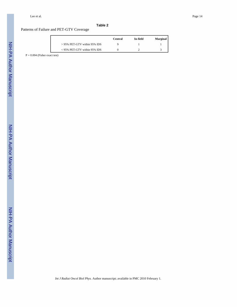
Analysis of Patterns of Failure and PET-GTV CoverageAll patients had repeat imaging at the time of treatment failure. Following image co-registration, a recurrence volume of interest (rVOI) was defined by a neuro-radiologist andradiation oncologist as the contrast-enhancing region on MRI at the time of tumor progression.Large central areas that were non-enhancing were excluded from the rVOI. The percentage ofrVOI enclosed by the 95% (high-dose prescription) isodose surface was determined in orderto classify the failure as: central (>95% covered), in-field (> 80–95%), marginal (20–80%), ordistant (<20%)[18]. Similarly, we determined the fraction of the PET-GTV encompassed bythe 95%-isodose surface with “adequate” coverage being defined as more than 95% of thePET-GTV volume encompassed within the 95%-isodose surface.
ResultsPatient Characteristics and Outcomes
At the time of this analysis (last updated September 10, 2007), 30 patients had been enrolledon study with a minimum follow-up of 1 year for those who had not progressed. Among these,two patients died from aplastic anemia, and 1 patient died of sepsis within 3 months ofcompleting radiation. These patients could not be evaluated with regard to treatment failureand were excluded from the patterns of failure analysis. In addition, one patient did not haveMET-PET prior to treatment and was also excluded from this analysis. Thus, a total of 26patients with pre-treatment MET-PET scans were analyzed for patterns of failure with a medianfollow-up of 15 months (Table 1). 13 patients had gross total resection (GTR), seven patientshad subtotal resection (STR), and six patients had stereotactic biopsy. Median progression freesurvival (PFS) from the date of enrollment for the analyzed population was 8 months (range:1.5–20 months).
Among the 26 patients analyzed, three patients remain alive with no clinical or radiologicalevidence of progression with more than one year of follow-up. Ten patients underwent re-resection or stereotactic biopsy to confirm tumor progression to determine the most appropriatetreatment course; one patient underwent autopsy. Among these eleven patients in whom tissuewas obtained after progression, pathologic review revealed one patient to have only radiation
Lee et al. Page 4
Int J Radiat Oncol Biol Phys. Author manuscript; available in PMC 2010 February 1.
NIH
-PA Author Manuscript
NIH
-PA Author Manuscript
NIH
-PA Author Manuscript

necrosis at time of re-resection and no evidence of recurrent tumor; the other 10 patients werefound to have some evidence of recurrent or residual GBM, including 3 cases with extensiveareas of radiation effect and vasculopathy in addition to the residual/recurrent disease. Twelvepatients were judged to have progression by clinical and radiological criteria without pathologicconfirmation; among these, 8 have expired (including 1 patient who progressed whileundergoing radiation therapy and received only 20 Gy) and 4 remain alive11–19 months afterprogression. Although the majority of patients who did not undergo re-resection died within 6months following documented tumor progression, four of these patients have responded tosecond-line chemotherapy and remain alive one year following clinical diagnosis of tumorprogression.
Methionine PET-GTV CharacteristicsThe median PET-GTV size with a lesion-to-cerebellum ratio of 1.5 or above was 5.5 cm3
(range: 0.3 – 43.8 cm3) and varied inversely with the reported extent of resection (Figure 2).Among the 13 patients reported to have GTR, seven patients had less than 1 cm3 of increasedPET activity. The maximum PET-GTV in the GTR group was 5.5 cm3. By contrast, the PET-GTV in patients who underwent stereotactic biopsy ranged from 12.1 to 43.8 cm3.
We began by assessing the relationship between the area of increased MET-PET uptake andthe contrast-enhancing region noted on pretreatment MRI. The size of the PET-GTV wastypically smaller than the contrast-enhancing volume on MRI (mean 11.5 vs 18.4 cm3, p <0.01, paired t-test). In the majority of the stereotactic biopsy cases, the region of increased PETactivity appeared similar to the contrast-enhancing lesion. However, there were cases in whichthe PET-GTV extended beyond the contrast-enhancing region (Figure 3A). In patients whounderwent surgical resection, MET-PET was particularly helpful in establishing the extent ofresidual tumor since post-operative changes could not always be distinguished from residualenhancement on MRI (Figure 3B).
MET-PET Activity and Patterns of FailureTo assess the potential utility of MET-PET in identifying regions at high risk for recurrence,we compared the patterns of failure for those patients whose PET-GTV was fully encompassedby the 95% IDS to those whose PET-GTV extended beyond the 95% IDS (Table 2). Wehypothesized that if MET-PET were useful in addition to MRI in detecting regions at highestrisk, then there would be significantly more non-central failure in patients whose PET-GTVwas fully covered. The seven patients with no appreciable MET-PET activity (i.e., PET-GTV< 1 cm3) were omitted from this analysis.
In five patients, the PET GTV was not fully encompassed by the 95% IDS, and all 5 of thesepatients recurred with non-central failures. In comparison, only two of 14 patients whose PET-GTV was fully encompassed by the 95% IDS had non-central failures. This includes 3 patientsthat have not yet failed with over 12 months of followup. Using the Fisher exact test, thisapparent association between suboptimal coverage of the PET-GTV and subsequent non-central failure was statistically significant (p < 0.01).
Review of the 5 cases in which the PET-GTV was inadequately covered revealed one case inwhich a distinct nodule was noted on the pre-treatment MET-PET which was not apparent oncontrast-enhanced MRI. This patient developed a recurrence one month after completingradiation treatment in the area of MET-PET uptake (Figure 4a). Minimum dose to the nodulewith increased PET uptake was 43 Gy. In the other four patients, the minimum dose to thePET-GTV was at least 58 Gy, the minimum dose to PTV1. In these remaining cases, therelationship between PET-GTV and subsequent site of failure was less obvious, but there was
Lee et al. Page 5
Int J Radiat Oncol Biol Phys. Author manuscript; available in PMC 2010 February 1.
NIH
-PA Author Manuscript
NIH
-PA Author Manuscript
NIH
-PA Author Manuscript

still overlap noted between the areas of initial increased PET uptake and the eventual area ofrecurrence on MRI (Figure 4b).
DiscussionIn this study, we have found that MET-PET provides potentially important additionalinformation over standard anatomic imaging with contrast-enhanced MRI. There was astatistically significant correlation between the presence of increased MET-PET uptake outsidethe high-dose region and subsequent non-central failure. This represents the first prospectivestudy in which pretreatment MET-PET imaging was compared with subsequent patterns offailure in GBM patients treated with radiation and TMZ. These findings suggest that it wouldbe reasonable to test a strategy of using MET-PET to identify focal areas for a conformalradiation boost.
The observation that regions of increased MET-PET activity and of contrast-enhancement onMRI are imperfectly correlated is consistent with the findings of a prior study in which patientswith primary GBM underwent MET-PET prior to undergoing radiation[13]. In that study,which was limited to patients who had undergone subtotal resection, 87% of patients werefound to have discrepancies between the Gd-enhancing volume and areas of increased PETactivity. However, that study did not include data on subsequent failures, so it was unclearwhether the MET-PET activity would be important in identifying regions at high risk forprogression.
In this limited dataset, we have found not only that MET-PET reveals areas of abnormalitythat are not apparent by contrast-enhancement on MRI but also that there is a correlationbetween the location of increased pre-radiation MET-PET activity and the subsequent site offailure. All patients who had suboptimal coverage of the PET-GTV had non-central failureswhereas only 2 of 14 patients with adequately covered PET-GTV had non-central failures. Byfocusing on whether the initial MET-PET activity fell entirely within the high-dose region andwhether the recurrences fell within the high-dose region, we have circumvented some of thechallenges associated with trying to localize the origin of failure more precisely. The correlationbetween the pattern of failure and the adequacy of coverage of the pretreatment MET-PETsupports the incorporation of MET-PET into radiation treatment planning, although additionaldata are clearly needed to corroborate our findings.
In addition to MET-PET, other modalities may also be useful in identifying regions at increasedrisk for progression. In particular, recently published studies have shown that 3D-magneticresonance spectroscopic imaging (MRSI) may serve as an adjunct to conventional MRI indelineating the extent of disease in high-grade gliomas and that combining standard MRI withMRSI volumes leads to improved coverage of areas that subsequently progress[19–21]. Othernovel MR techniques, including perfusion and diffusion-weighted MRI obtained prior to andduring treatment, have been demonstrated to be predictive of outcomes, but additional workis needed to determine if these techniques can specifically identify areas within the target thatare at risk for failure[22,23]. In the future, it seems likely that the complementary informationobtained with multiple imaging modalities will lead to the most efficient delineation of theappropriate target volume.
One limitation of this study is the small number of subjects enrolled. However, the purpose ofthe study was to evaluate prospectively whether MET-PET might be useful for treatmentplanning purposes, and the correlation we found between the location of increased PET activityand the subsequent pattern of failure in this preliminary dataset did achieve statisticalsignificance and provide justification for additional work on the use of MET-PET for treatmentplanning. Another potential criticism of our study is that only a subset of the patients in this
Lee et al. Page 6
Int J Radiat Oncol Biol Phys. Author manuscript; available in PMC 2010 February 1.
NIH
-PA Author Manuscript
NIH
-PA Author Manuscript
NIH
-PA Author Manuscript

study had histological confirmation of treatment failure. However, given that our policy wasto obtain tissue in cases with uncertainty and that 10 of 11 patients with additional tissue werefound to have evidence of recurrence, it seems likely that nearly all of the patients deemed tohave progression on clinical and imaging criteria would have had positive biopsies.
Based on the results of our study, we believe it would be reasonable to design a prospectivetrial testing whether selectively increasing the radiation dose to MET-PET avid regions couldimprove the outcome of treatment. By limiting the radiation boost volume, higher doses maybe safely achieved, so focal dose-escalation may be more successful than uniformly escalatingdose across the entire contrast-enhancing MRI volume. The addition of TMZ may be especiallyeffective as a radiation sensitizer if the radiation dose to normal brain can be limited by bettertargeting. If new, targeted chemotherapy agents lead to further improvements in control ofmicroscopic disease, radiation can be used primarily to control disease in limited regions thathave the highest risk of progression, i.e., where those agents are most likely to fail.
AcknowledgementsNIH 3PO1 CA 59827
References1. Aydin H, Sillenberg I, von Lieven H. Patterns of failure following CT-based 3-D irradiation for
malignant glioma. Strahlenther Onkol 2001;177(8):424–31. [PubMed: 11544905]2. Garden AS, Maor MH, Yung WK, et al. Outcome and patterns of failure following limited-volume
irradiation for malignant astrocytomas. Radiother Oncol 1991;20(2):99–110. [PubMed: 1851573]3. Wallner KE, Galicich JH, Krol G, et al. Patterns of failure following treatment for glioblastoma
multiforme and anaplastic astrocytoma. Int J Radiat Oncol Biol Phys 1989;16(6):1405–9. [PubMed:2542195]
4. Stupp R, Mason WP, van den Bent MJ, et al. Radiotherapy plus concomitant and adjuvanttemozolomide for glioblastoma. N Engl J Med 2005;352(10):987–96. [PubMed: 15758009]
5. Chang EL, Akyurek S, Avalos T, et al. Evaluation of peritumoral edema in the delineation ofradiotherapy clinical target volumes for glioblastoma. Int J Radiat Oncol Biol Phys 2007;68(1):144–50. [PubMed: 17306935]
6. Fitzek MM, Thornton AF, Rabinov JD, et al. Accelerated fractionated proton/photon irradiation to 90cobalt gray equivalent for glioblastoma multiforme: results of a phase II prospective trial. J Neurosurg1999;91(2):251–60. [PubMed: 10433313]
7. Sneed PK, Lamborn KR, Larson DA, et al. Demonstration of brachytherapy boost dose-responserelationships in glioblastoma multiforme. Int J Radiat Oncol Biol Phys 1996;35(1):37–44. [PubMed:8641924]
8. Shrieve DC, Alexander E 3rd, Black PM, et al. Treatment of patients with primary glioblastomamultiforme with standard postoperative radiotherapy and radiosurgical boost: prognostic factors andlong-term outcome. J Neurosurg 1999;90(1):72–7. [PubMed: 10413158]
9. Tanaka M, Ino Y, Nakagawa K, et al. High-dose conformal radiotherapy for supratentorial malignantglioma: a historical comparison. Lancet Oncol 2005;6(12):953–60. [PubMed: 16321763]
10. Jager PL, Vaalburg W, Pruim J, et al. Radiolabeled amino acids: basic aspects and clinical applicationsin oncology. J Nucl Med 2001;42(3):432–45. [PubMed: 11337520]
11. Grosu AL, Piert M, Weber WA, et al. Positron emission tomography for radiation treatment planning.Strahlenther Onkol 2005;181(8):483–99. [PubMed: 16044216]
12. Grosu AL, Weber WA, Franz M, et al. Reirradiation of recurrent high-grade gliomas using aminoacid PET (SPECT)/CT/MRI image fusion to determine gross tumor volume for stereotacticfractionated radiotherapy. Int J Radiat Oncol Biol Phys 2005;63(2):511–9. [PubMed: 16168843]
13. Grosu AL, Weber WA, Riedel E, et al. L-(methyl-11C) methionine positron emission tomographyfor target delineation in resected high-grade gliomas before radiotherapy. Int J Radiat Oncol BiolPhys 2005;63(1):64–74. [PubMed: 16111573]
Lee et al. Page 7
Int J Radiat Oncol Biol Phys. Author manuscript; available in PMC 2010 February 1.
NIH
-PA Author Manuscript
NIH
-PA Author Manuscript
NIH
-PA Author Manuscript

14. Taal W, Brandsma D, de Bruin HG, et al. The incidence of pseudo-progression in a cohort of malignantglioma patients treated with chemo-radiation with temozolomide. J Clin Oncol 2007;25(18S):2009.
15. Brix G, Zaers J, Adam LE, et al. Performance evaluation of a whole-body PET scanner using theNEMA protocol. National Electrical Manufacturers Association. J Nucl Med 1997;38(10):1614–23.[PubMed: 9379202]
16. Torii K, Tsuyuguchi N, Kawabe J, et al. Correlation of amino-acid uptake using methionine PET andhistological classifications in various gliomas. Ann Nucl Med 2005;19(8):677–83. [PubMed:16444993]
17. Cao Y. Development of Image Software Tools for Radiation Therapy Assessment. Medical Physics2005;32:2136.
18. Lee SW, Fraass BA, Marsh LH, et al. Patterns of failure following high-dose 3-D conformalradiotherapy for high-grade astrocytomas: a quantitative dosimetric study. Int J Radiat Oncol BiolPhys 1999;43(1):79–88. [PubMed: 9989517]
19. Park I, Tamai G, Lee MC, et al. Patterns of recurrence analysis in newly diagnosed glioblastomamultiforme after three-dimensional conformal radiation therapy with respect to pre-radiation therapymagnetic resonance spectroscopic findings. Int J Radiat Oncol Biol Phys. 2007
20. Pirzkall A, Li X, Oh J, et al. 3D MRSI for resected high-grade gliomas before RT: tumor extentaccording to metabolic activity in relation to MRI. Int J Radiat Oncol Biol Phys 2004;59(1):126–37.[PubMed: 15093908]
21. Pirzkall A, McKnight TR, Graves EE, et al. MR-spectroscopy guided target delineation for high-grade gliomas. Int J Radiat Oncol Biol Phys 2001;50(4):915–28. [PubMed: 11429219]
22. Cao Y, Nagesh V, Hamstra D, et al. The extent and severity of vascular leakage as evidence of tumoraggressiveness in high-grade gliomas. Cancer Res 2006;66(17):8912–7. [PubMed: 16951209]
23. Hamstra DA, Chenevert TL, Moffat BA, et al. Evaluation of the functional diffusion map as an earlybiomarker of time-to-progression and overall survival in high-grade glioma. Proc Natl Acad Sci US A 2005;102(46):16759–64. [PubMed: 16267128]
Lee et al. Page 8
Int J Radiat Oncol Biol Phys. Author manuscript; available in PMC 2010 February 1.
NIH
-PA Author Manuscript
NIH
-PA Author Manuscript
NIH
-PA Author Manuscript

FIGURE 1.Coronal slice of T1 post-gadolinum MRI with GTV outlined in green. PTV1, shown in blue,receives 60 Gy in 30 fractions. PTV2, shown in red, receives protocol dictated dose over thesame 30 fractions using a conformal simultaneous IMRT boost.
Lee et al. Page 9
Int J Radiat Oncol Biol Phys. Author manuscript; available in PMC 2010 February 1.
NIH
-PA Author Manuscript
NIH
-PA Author Manuscript
NIH
-PA Author Manuscript

FIGURE 2.MET-PET volume sorted by extent of surgery.
Lee et al. Page 10
Int J Radiat Oncol Biol Phys. Author manuscript; available in PMC 2010 February 1.
NIH
-PA Author Manuscript
NIH
-PA Author Manuscript
NIH
-PA Author Manuscript

FIGURE 3.FIGURE 3A. MET-PET (left) and Gd-enhanced T1-weighted MRI (right) after stereotacticbiopsy. PET-GTV (outlined in blue on MRI) extends beyond the contrast-enhancing lesion.FIGURE 3B. Post-operative Gd-enhanced T1-weighted MRI (left) demonstrates resectioncavity with no evidence of residual disease, but 11C Methionine PET scan (right) demonstratesan area of residual tumor that was not appreciated on the MRI.
Lee et al. Page 11
Int J Radiat Oncol Biol Phys. Author manuscript; available in PMC 2010 February 1.
NIH
-PA Author Manuscript
NIH
-PA Author Manuscript
NIH
-PA Author Manuscript

FIGURE 4.FIGURE 4A. A patient with a distinct nodule noted on 11C MET PET that was not fullyencompassed in the RT plan. Gadolinium-enhanced, T1-weighted MRI (left) shows noappreciable contrast enhancing lesion in the corresponding area of high uptake in the rightparietal region on pre-treatment MET-PET (center). MRI one month after completion oftherapy (right) shows a new contrast-enhancing nodule in the area of initial MET PET uptake(green). The subsequent clinical course confirmed disease progression.Figure 4B: Images from a representative patient with a PET-GTV extending beyond thecontrast-enhancing lesion on MRI. Pre-treatment gadolinium-enhanced, T1-weighted MRI(left) shows enhancement along the resection cavity. There is a focus of increased MET-PET(center) uptake directly above the resection cavity. Subsequent MRI including MRSpectroscopy and PET imaging performed 10 months following completion of therapy showstumor progression present above the resection cavity which overlaps with, but is not fullyencompassed by, the PET-GTV.
Lee et al. Page 12
Int J Radiat Oncol Biol Phys. Author manuscript; available in PMC 2010 February 1.
NIH
-PA Author Manuscript
NIH
-PA Author Manuscript
NIH
-PA Author Manuscript

NIH
-PA Author Manuscript
NIH
-PA Author Manuscript
NIH
-PA Author Manuscript
Lee et al. Page 13
Table 1Patient Characteristics (n = 26)
Age, median (range) 53 (20 – 75)
KPS
90–100 23
80 2
70 1
RPA classification[19]
III 11
IV 11
V 4
Radiation Prescription Dose
66 Gy 1
72 Gy 3
75 Gy 7*
78 Gy 7
81 Gy 8
Extent of Surgery
Gross total resection 13
Subtotal resection 7
Biopsy only 6*1 patient received only 20 Gy
Int J Radiat Oncol Biol Phys. Author manuscript; available in PMC 2010 February 1.
NIH
-PA Author Manuscript
NIH
-PA Author Manuscript
NIH
-PA Author Manuscript
Lee et al. Page 14
Table 2Patterns of Failure and PET-GTV Coverage
Central In-field Marginal
> 95% PET-GTV within 95% IDS 9 1 1
< 95% PET-GTV within 95% IDS 0 2 3
P = 0.004 (Fisher exact test)
Int J Radiat Oncol Biol Phys. Author manuscript; available in PMC 2010 February 1.


![Reference and target region modeling of [11C]-(R)-PK11195 brain studies](https://static.fdokumen.com/doc/165x107/633302fa576b626f850dabe0/reference-and-target-region-modeling-of-11c-r-pk11195-brain-studies.jpg)

![Eritema multiforme reaccional como manifestación atípica de lepra. Reporte de caso [Reactive erythema multiforme as atypical manifestation of leprosy. Case report]](https://static.fdokumen.com/doc/165x107/632459174d8439cb620d572d/eritema-multiforme-reaccional-como-manifestacion-atipica-de-lepra-reporte-de.jpg)
![Quantitative analysis of [11C]-erlotinib PET demonstrates specific binding for activating mutations of the EGFR kinase domain](https://static.fdokumen.com/doc/165x107/6345ca446cfb3d406409d7f9/quantitative-analysis-of-11c-erlotinib-pet-demonstrates-specific-binding-for-activating.jpg)
![Intracellular reactions affecting 2-amino-4-([11C]methylthio)butyric acid ([11C]methionine) response to carbon ion radiotherapy in C10 glioma cells](https://static.fdokumen.com/doc/165x107/6343c86b88adeae9b9061aee/intracellular-reactions-affecting-2-amino-4-11cmethylthiobutyric-acid-11cmethionine.jpg)