Assignment of PolyProline II Conformation and Analysis of Sequence – Structure Relationship
15
Assignment of PolyProline II Conformation and Analysis of Sequence – Structure Relationship Yohann Mansiaux 1,2,3. , Agnel Praveen Joseph 1,2,3. , Jean-Christophe Gelly 1,2,3 , Alexandre G. de Brevern 1,2,3 * 1 INSERM, UMR-S 665, Dynamique des Structures et Interactions des Macromole ´cules Biologiques (DSIMB), Paris, France, 2 Universite ´ Paris Diderot - Paris 7, Paris, France, 3 Institut National de la Transfusion Sanguine (INTS), Paris, France Abstract Background: Secondary structures are elements of great importance in structural biology, biochemistry and bioinformatics. They are broadly composed of two repetitive structures namely a-helices and b-sheets, apart from turns, and the rest is associated to coil. These repetitive secondary structures have specific and conserved biophysical and geometric properties. PolyProline II (PPII) helix is yet another interesting repetitive structure which is less frequent and not usually associated with stabilizing interactions. Recent studies have shown that PPII frequency is higher than expected, and they could have an important role in protein – protein interactions. Methodology/Principal Findings: A major factor that limits the study of PPII is that its assignment cannot be carried out with the most commonly used secondary structure assignment methods (SSAMs). The purpose of this work is to propose a PPII assignment methodology that can be defined in the frame of DSSP secondary structure assignment. Considering the ambiguity in PPII assignments by different methods, a consensus assignment strategy was utilized. To define the most consensual rule of PPII assignment, three SSAMs that can assign PPII, were compared and analyzed. The assignment rule was defined to have a maximum coverage of all assignments made by these SSAMs. Not many constraints were added to the assignment and only PPII helices of at least 2 residues length are defined. Conclusions/Significance: The simple rules designed in this study for characterizing PPII conformation, lead to the assignment of 5% of all amino as PPII. Sequence – structure relationships associated with PPII, defined by the different SSAMs, underline few striking differences. A specific study of amino acid preferences in their N and C-cap regions was carried out as their solvent accessibility and contact patterns. Thus the assignment of PPII can be coupled with DSSP and thus opens a simple way for further analysis in this field. Citation: Mansiaux Y, Joseph AP, Gelly J-C, de Brevern AG (2011) Assignment of PolyProline II Conformation and Analysis of Sequence – Structure Relationship. PLoS ONE 6(3): e18401. doi:10.1371/journal.pone.0018401 Editor: Markus Buehler, Massachusetts Institute of Technology, United States of America Received August 12, 2010; Accepted March 7, 2011; Published March 31, 2011 Copyright: ß 2011 Mansiaux et al. This is an open-access article distributed under the terms of the Creative Commons Attribution License, which permits unrestricted use, distribution, and reproduction in any medium, provided the original author and source are credited. Funding: This work was supported by grants from the Ministe ` re de la Recherche, Universite ´ Paris Diderot - Paris 7, National Institute for Blood Transfusion (INTS) and the Institute for Health and Medical Research (INSERM). APJ has a grant from CEFIPRA number 3903-E. AdB acknowledges CEFIPRA for collaborative grant (number 3903-E). The funders had no role in study design, data collection and analysis, decision to publish, or preparation of the manuscript. Competing Interests: The authors have declared that no competing interests exist. * E-mail: [email protected] . These authors contributed equally to this work. Introduction The three dimensional structures of proteins are of great help to understand the precise details of its biological function. Contrary to the earlier views, the first low resolution model of myoglobin exhibited high complexity and a definite lack of symmetry [1]. In spite of the global complexity, Pauling and Corey had proposed two types of regularities in the local backbone conformation [2,3]. The first one constitutes the a-helix conformation which was estimated to be stable and favorable on the basis of accurate geometrical parameters derived from small molecule crystal structures [4]. The second one is the b-sheet which was made of extended anti-parallel or parallel strands stabilized by backbone hydrogen bonds between them [5]. The high frequency of a- helices and b-sheets observed in experimentally determined structures [6] has led to the concept of ‘secondary structures’ which describes these local backbone regularities in the protein structure. Basically the secondary structure description is com- posed mainly of a-helix, b-strand and a state corresponding to other regions in the backbone, the coil. The structure descriptions are often limited to these three classes. With availability of a large number of experimentally determined protein structures, it is becoming obvious that other backbone conformations are also favored in proteins. The a-helices are not the only helical conformation and are often not linear [7,8], the b-sheets also show irregularities [9,10] and the coil is in fact, not strictly random. The class of b-turns is of particular interest. In the late 60 s, Venkatachalam discovered the existence of these small local folds that are characterized by the reversal of polypeptide chain and stabilized by a hydrogen bond between the first and the last residue [11]. These b-turns are part of a more general class, known as tight turns, which are characterized by precise dihedral angle values of their central residues and a short distance between the PLoS ONE | www.plosone.org 1 March 2011 | Volume 6 | Issue 3 | e18401
-
Upload
independent -
Category
Documents
-
view
1 -
download
0
Transcript of Assignment of PolyProline II Conformation and Analysis of Sequence – Structure Relationship

Assignment of PolyProline II Conformation and Analysisof Sequence – Structure RelationshipYohann Mansiaux1,2,3., Agnel Praveen Joseph1,2,3., Jean-Christophe Gelly1,2,3, Alexandre G. de
Brevern1,2,3*
1 INSERM, UMR-S 665, Dynamique des Structures et Interactions des Macromolecules Biologiques (DSIMB), Paris, France, 2 Universite Paris Diderot - Paris 7, Paris, France,
3 Institut National de la Transfusion Sanguine (INTS), Paris, France
Abstract
Background: Secondary structures are elements of great importance in structural biology, biochemistry and bioinformatics.They are broadly composed of two repetitive structures namely a-helices and b-sheets, apart from turns, and the rest isassociated to coil. These repetitive secondary structures have specific and conserved biophysical and geometric properties.PolyProline II (PPII) helix is yet another interesting repetitive structure which is less frequent and not usually associated withstabilizing interactions. Recent studies have shown that PPII frequency is higher than expected, and they could have animportant role in protein – protein interactions.
Methodology/Principal Findings: A major factor that limits the study of PPII is that its assignment cannot be carried outwith the most commonly used secondary structure assignment methods (SSAMs). The purpose of this work is to propose aPPII assignment methodology that can be defined in the frame of DSSP secondary structure assignment. Considering theambiguity in PPII assignments by different methods, a consensus assignment strategy was utilized. To define the mostconsensual rule of PPII assignment, three SSAMs that can assign PPII, were compared and analyzed. The assignment rulewas defined to have a maximum coverage of all assignments made by these SSAMs. Not many constraints were added tothe assignment and only PPII helices of at least 2 residues length are defined.
Conclusions/Significance: The simple rules designed in this study for characterizing PPII conformation, lead to theassignment of 5% of all amino as PPII. Sequence – structure relationships associated with PPII, defined by the differentSSAMs, underline few striking differences. A specific study of amino acid preferences in their N and C-cap regions wascarried out as their solvent accessibility and contact patterns. Thus the assignment of PPII can be coupled with DSSP andthus opens a simple way for further analysis in this field.
Citation: Mansiaux Y, Joseph AP, Gelly J-C, de Brevern AG (2011) Assignment of PolyProline II Conformation and Analysis of Sequence – StructureRelationship. PLoS ONE 6(3): e18401. doi:10.1371/journal.pone.0018401
Editor: Markus Buehler, Massachusetts Institute of Technology, United States of America
Received August 12, 2010; Accepted March 7, 2011; Published March 31, 2011
Copyright: � 2011 Mansiaux et al. This is an open-access article distributed under the terms of the Creative Commons Attribution License, which permitsunrestricted use, distribution, and reproduction in any medium, provided the original author and source are credited.
Funding: This work was supported by grants from the Ministere de la Recherche, Universite Paris Diderot - Paris 7, National Institute for Blood Transfusion (INTS)and the Institute for Health and Medical Research (INSERM). APJ has a grant from CEFIPRA number 3903-E. AdB acknowledges CEFIPRA for collaborative grant(number 3903-E). The funders had no role in study design, data collection and analysis, decision to publish, or preparation of the manuscript.
Competing Interests: The authors have declared that no competing interests exist.
* E-mail: [email protected]
. These authors contributed equally to this work.
Introduction
The three dimensional structures of proteins are of great help to
understand the precise details of its biological function. Contrary
to the earlier views, the first low resolution model of myoglobin
exhibited high complexity and a definite lack of symmetry [1]. In
spite of the global complexity, Pauling and Corey had proposed
two types of regularities in the local backbone conformation [2,3].
The first one constitutes the a-helix conformation which was
estimated to be stable and favorable on the basis of accurate
geometrical parameters derived from small molecule crystal
structures [4]. The second one is the b-sheet which was made of
extended anti-parallel or parallel strands stabilized by backbone
hydrogen bonds between them [5]. The high frequency of a-
helices and b-sheets observed in experimentally determined
structures [6] has led to the concept of ‘secondary structures’
which describes these local backbone regularities in the protein
structure. Basically the secondary structure description is com-
posed mainly of a-helix, b-strand and a state corresponding to
other regions in the backbone, the coil. The structure descriptions
are often limited to these three classes. With availability of a large
number of experimentally determined protein structures, it is
becoming obvious that other backbone conformations are also
favored in proteins. The a-helices are not the only helical
conformation and are often not linear [7,8], the b-sheets also
show irregularities [9,10] and the coil is in fact, not strictly
random.
The class of b-turns is of particular interest. In the late 60 s,
Venkatachalam discovered the existence of these small local folds
that are characterized by the reversal of polypeptide chain and
stabilized by a hydrogen bond between the first and the last
residue [11]. These b-turns are part of a more general class, known
as tight turns, which are characterized by precise dihedral angle
values of their central residues and a short distance between the
PLoS ONE | www.plosone.org 1 March 2011 | Volume 6 | Issue 3 | e18401

extremities [12]. The latter has been shown to play important
structural and functional roles [13].
Apart from turns, the other characterized secondary structure is
the PolyProline II (PPII) helix. The PPII helices correspond to a
unique local fold [14]. They were discovered more than 50 years
ago in fibrous proteins [15,16], as they contribute to coiled coil
supersecondary structures formation . Later they were also found
to occur in numerous globular proteins. PPII helix is a left-handed
helical structure with an overall shape resembling a triangular
prism [17,18]. With a helical pitch of 9.3 A/turn, each turn
constituting of 3 residues, it forms an extended helix. This
conformation is characterized by recurrent trans isomers of
peptide bonds and (w, y) values of 275u and +145u respectively,
the dihedral angles being a characteristic of b-strands As noted by
G. Rose, considering the hydrogen-bonded b-turns [11] and PPII
[16] along with the classical secondary structure result in the
assignment of 80% of the all amino acids to a regular backbone
conformation [19].This rises to 90%, in a more recent study where
the turns are defined in slightly different way [20].
Nonetheless, when compared to a-helices, b-sheets and turns,
analysis of PPII has not gained wide interest, mainly due to three
factors: (i) PPII has a low frequency of occurrence, (ii) PPII
conformation is not stabilized by a strong hydrogen bond pattern,
thus considered as an unstable conformation and, (iii) only a few
methods for PPII assignment are available and these methods use
different assignment parameters resulting in variable assignments.
Also, PPII helices are not assigned by the widely used Secondary
Structure Assignment Method (SSAM): DSSP [21].
Adzhubei and Sternberg in their first systematic search, found
96 PPII helices in a databank of 80 proteins [22]. They were
surprisingly common. Even if they are called polyproline for
historical reasons, they are not only composed of Proline
successions, some PPII helices have no Proline at all
[22,23,24,25,26] , e.g., short stretches of poly-glutamines were
found to form PPII conformation [27]. Hollingsworth, Berkholz
and Karplus recently proposed that its common name could be
changed to a more general form, i.e., ‘‘polypeptide-II’’. This would
maintain the familiar acronym, avoid the misleading association
with only Proline, and be consistent with the observation that it is a
prominent conformation in unfolded polypeptide chains [28].
These PPII helices are highly solvent-exposed and tend to have
high crystallographic temperature factors [22]. Moreover PPII are
not stabilized by salt bridges [29]. It has been suggested that PPII
helices could be stabilized by water mediated main chain hydrogen
bonds (in the absence of main chain-main chain H-bonds), as they
also tend to have a regular pattern of hydrogen bonds with water
[30]. Several studies suggest that peptide-solvent interaction is a
major determinant of PPII conformation [31,32,33]. However, the
preference for polyproline II conformation is also reported to be
independent of the degree of solvation [34]. Avbelj and Baldwin
noticed that solvation strongly affects preferences for different
backbone conformations. The dependence of backbone preference
on solvation might explain why Alanine favors PPII conformation
whereas Valine favors extended structure [35]. Stapley and Creamer
suggested that local side-chain to main-chain hydrogen bonds are also
important in stabilizing PPII helices [24]. Cubellis and co-workers
recently highlighted that PPII helices are stabilized by non-local
interactions [36]. PPII do not display strong sequence propensities in
contrast to the other extended conformations, such as b-strands [37].
The non-local stabilization of hydrogen-bond donors and acceptors
does, however, result in PPII conformations being well suited for
participating in protein-protein interactions. They are also suspected
to have a role in amyloid formation [38,39] and nucleic acid binding
[40]. Hence, a recent study shows its importance in Duchenne
muscular dystrophy [41]. Several studies have also focused on the
extremities of PPII. It has been proposed that PPII might interrupt in
the formation of b-sheet which is prone to aggregation [42], due to its
particular geometry with the neighboring amide bond [43]. They
could have a key role in the folding process [44]; the concentration of
residues in the PPII conformational space lowers the entropy of the
unfolded protein chain and thus facilitates folding (under appropriate
conditions) [45,46]. Recently, the number of studies on PPII
conformations has increased [19,36,47,48,49], especially in the field
of molecular dynamics [19,32,45,50].
Numerous approaches for secondary structure assignment that
rely on different descriptors, exist (see Table 1 of [51]). DSSP [21]
remains the most widely used SSAM. It identifies the secondary
structures on the basis of particular hydrogen bond patterns
detected from the protein geometry, with the help of an electrostatic
model. DSSP is used for assigning secondary structures for the
protein structures deposited in the Protein DataBank (PDB) [6,52].
Currently, only three publicly available SSAMs assign PPII,
namely XTLSSTR [53], PROSS [54] and SEGNO [55].
XTLSSTR uses all the backbone atoms to compute two angles
and three distances [53], which forms the basis of assignments.
Assignments made by PROSS and SEGNO are based solely on
backbone angles, mainly involving the Q and y dihedral angles.
The purpose of this work is to propose simple rules to assign
PPII based on a classical secondary structures assignment carried
out using DSSP. We have compared the assignment of the three
available PPII assignment methods and specifically analyzed the
distribution of PPII based on these assignments. Then, we propose
a rule to assign PPII within the coil assigned by DSSP; such that
there is a good concordance with the other assignments. Sequence
– structure relationships of these PPIIs are also analyzed to study
amino acid preferences and to ensure a good agreement with the
previous studies [56]. In the same way, a specific analysis of
sequence-structure relationships on capping regions was also
carried out. Residue accessibility and contacts in the PPII helices
were also studied as they are considered to be accessible for
different interactions. The behaviour of PPII has also been
analyzed at the light of a structural alphabet [56,57] named
Protein Blocks [58,59,60]. This gives a more accurate picture on
the local structures associated with PPII.
Results
Comparison of the different SSAMsA non-redundant databank of protein structures has been
extracted from the PDB [6]. The list of protein structures has been
obtained from PISCES database [61,62], which is generated based
on the following criteria : resolution less than 2.5 A, R factor
Table 1. Analysis of PPII properties.
f(PPII) all (%) in DSSP coil (%) av. len. in PPIIDSSP (%)
PROSS 10.10 6.70 1.35 65.9
XTLSSTR 6.80 4.18 2.63 56.0
SEGNO 3.97 2.58 2.58 45.6
mean 6.95 4.48
DSSP with PPIIDSSP 5.11 5.11 3.24 100.0
The PPII frequency of the 3 SSAMs (see Figure 1) and the novel PPIIDSSP aregiven with their average PPII length (av. len.) and the percentage of PPIIassigned by PPIIDSSP which are in common with the other SSAM assignments.doi:10.1371/journal.pone.0018401.t001
PolyProline II
PLoS ONE | www.plosone.org 2 March 2011 | Volume 6 | Issue 3 | e18401

below 0.2 and no proteins share more than 30% sequence identity.
As assessed earlier [51], SSAMs have only about 80% of consensus
between them. We have re-computed an agreement rate C3
between the four SSAM assignments used in this study (see Figure
S1). The results were in agreement with previous studies
[51,55,63,64]: the secondary structure assignment by XTLSSTR
had the lowest concordance with others, having C3 values of 77.4,
73.5 and 73.8% with DSSP, PROSS and SEGNO respectively
(mainly due to the highest frequency of assigned a-helices, as
already observed in [65]). The assignment made by SEGNO
remains closest to DSSP with a C3 of 88.9%. For PROSS, which
was not used in the earlier analysis, the C3 values are around 82%
(corresponding to classical values [51,66,67]). These results show
that the two closest SSAMs are DSSP and SEGNO while PROSS
is a little far away and XTLSSTR is even more distant.
PPIIFigure 1 summarizes most of the information on PPII
distribution (see also Table 1). The frequencies of classical
secondary structures (see Figure S2 and S3) and especially of
PPII are rather different for each SSAM. Frequencies of secondary
structures assigned by DSSP and SEGNO are quite similar (except
that SEGNO does not assign turns). Assignments made by PROSS
have relatively similar frequencies when compared to that of
DSSP, with a slightly lower frequency of turns [65]. XTLSSTR
assigns more coil and less repetitive structures when compared to
DSSP.
The percentage of PPII is quite significant, starting from 3.97%
for SEGNO, 6.80% for XTLSSTR and 10.10% for PROSS. A
ratio of 2.5 is thus observed between PPII assignments. However
1/3rd of the PPII assigned by each SSAM is not associated to
DSSP coil, but is shared between turns and b-strands. Considering
only the DSSP coil regions, the PPII frequencies range only from
2.58% for SEGNO to 6.70% for PROSS. As seen in the Venn
diagram, the consensus of PPII assignment is more limited, i.e.,
only 1.35% of the residues amino acids are assigned as PPII by all
the 3 SSAMs. It represents 52% of PPII assigned by SEGNO,
32% by XTLSSTR and 20% by PROSS. If only two SSAMs are
taken into account, the consensus in the region of DSSP coil goes
up to 3.25%. Thus the average frequency of PPII is about 6.95%,
Figure 1. PPII distribution. The Venn diagram gives the confusion between PPII assignments by the three SSAMs, namely PROSS, XTLSSTR andSEGNO. Inside different regions of the diagram is given two percentages x.xx(y.yy), the first percentage x.xx% correspond to the PPII frequencyobserved while y.yy gives the frequency in the DSSP coil alone. The secondary structure frequencies are given as a barplot in 4 states for DSSP, and 5states for the other three assignments. The correspondence of PPII residues with the assignments by DSSP is also given.doi:10.1371/journal.pone.0018401.g001
PolyProline II
PLoS ONE | www.plosone.org 3 March 2011 | Volume 6 | Issue 3 | e18401

and it is about 4.48% in the DSSP coil. Hence, no simple
consensus emerges from the analysis of the different PPII
assignments (see [51,63,65,67]).
Definition of the PPII assignment based on DSSPChoice of dihedrals. Seeking hints from literature [22] [37]
and the above analysis of previous methods [53,54,55], we propose
a simple rule for assigning PPII conformation for the residues in
the coil assigned by DSSP. For this purpose, we have used the
dihedral angles (.w+/2.e, y+/2e) to delineate the PPII space. The
canonical (.w, y) values of 275u and +145u have been selected as
the core of PPII region, and an e increasing by steps of 1u. Mean wvalue of PPII assigned by the SSAMs equals 75.6u while mean yvalue is slightly different from the canonical value, i.e., 136.9u.
The value of e is chosen such that equilibrium is reached
between the number of amino acids assigned as PPII by one of the
SSAMs and the residues not assigned as PPII in the DSSP coil.
The higher the e, the higher is the number of PPII assigned by one
of the three SSAMs and higher is the number of amino acids not
assigned as PPII by one or another. Figure 2a shows the
distribution of PPII assigned in DSSP coil and Figure 2b gives
the corresponding percentage in DSSP coil. With an e of 17u, we
have the highest percentage of amino acids assigned as PPII by
SEGNO, XTLSSTR and PROSS, within the DSSP coil.
Moreover, the corresponding percentage assigned (i.e., 4.9%) is
close to the average occurrence of PPII (see previous sections).
However, some PPII helices are only one residue long. It is mainly
due to the delimitation of DSSP coil (see Table 1).
Additional constraint. Taking the suggestions by different
groups into account [22,37,55], we have added a second rule to
the PPII assignment approach: at least two consecutive residues
with dihedral angles within the range (.w+/2e, .w+/2e), are
required to form PPII helix. After an iterative search, a new e of
29u was chosen for the assignment, which represents a PPII
frequency of 5.11%. The average length of the PPII assigned
within DSSP coil using this rule equals to 3.24 residues. The
PPII assignment based on DSSP (named hereafter as PPIIDSSP)
corresponds to 56.0%, 45.6% and 65.9% of PPII assignments by
XTLSSTR, SEGNO and PROSS respectively. This threshold ehas been selected by considering the following points: (i) the
percent of amino acids assigned as PPII by one of the three
SSAMs reached a maximum (of 83%), a further increase in edecreased this percentage, and (ii) it is a good compromise
between the average frequency of PPII observed (6.95%) and
the average frequency of PPII within DSSP coil (4.48%). Mean
w and mean y values of PPIIDSSP are 75.3uand 141.1urespectively. Hence, this definition of PPII assignment is
compared to the other PPII assignment methods and is
consistent with theoretical definition of PPII. Table 1
summarizes the main results. Figure 3 gives the distribution of
the different secondary structure states in the PPIIDSSP
assignments corresponding to the chosen epsilon while Figure 4
shows the same information for the values of epsilon ranging
from zero to 60u. While looking at XTLSSTR assignments
corresponding to PPIIDSSP residues, the PPII content is
generally high and mainly coil is found for low e values. For
SEGNO, which carries out least number of PPII assignments
(see Figure 1), nearly 20% of the assignments correspond to b-
strands. It must be noted that this SSAM does not assign any
turns. Finally, PROSS shares the maximum number of PPII, the
rest is mainly composed of b-strand and coil at a low
percentage. The assignments are characterized by absence of
helices and only a few turns were seen. Figure 4 shows the
interest in considering a second consecutive dihedral angle (see
Figure 2 to compare). Figure S4 gives the distribution of
distance between extremities of PPII for different PPII lengths.
A striking observation is the low standard deviation of the
distance. It ranges between 1 and 2 A depending on the length,
while the deviation is 2–3 times higher for the PPII helices
assigned by other SSAMs.
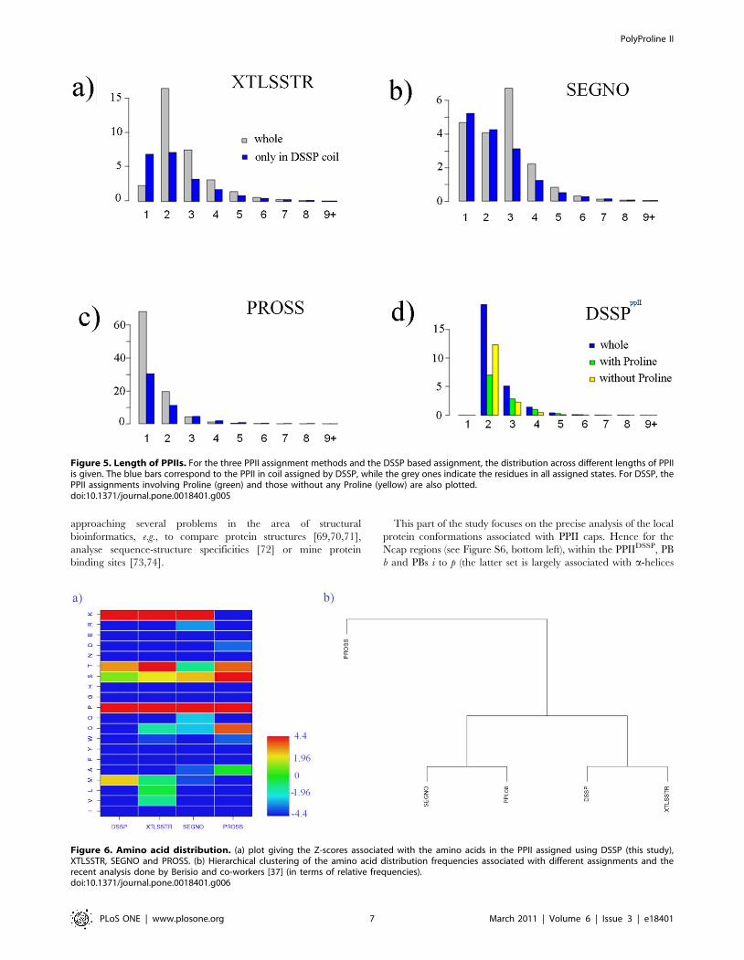
As seen in Figure 5, for PROSS, this assignment criterion results
in a small increase of the average length of PPII, from 1.35 to 1.63
amino acids (mainly due to a significant number of very short
helices outside the DSSP coil). The average length in the case of
SEGNO, decreases from 2.58 to 2.27 while for XTLSSTR, a
decrease from 2.63 to 2.32 was observed.
Analysis of the sequence-structure relationship. As
expected, the amino acid preference for PPIIs differs depending
on the SSAM used for assignment. However, the distribution of
amino acids remains clearly different from that seen in other
secondary structures (see Figure S5). Figure 6 summarizes the over
and under-representation of amino acids in the PPIIs assigned by
Figure 2. Deriving PPII assignment criterion: Choice of epsilon. (a) shows the percentage of residues found for e ranging from 0 to 60u. Thepercentage of amino acids associated to DSSP coil is shown in grey, PII assigned by PROSS is indicated in pink, assignment by XTLSSTR is in red andthat of SEGNO in orange. In blue is shown the percentage of residues considered as PPII at least by one of the three SSAMs and the green plotindicates percentage of common assignments. (b) The same information is given as the percentage of PPII residues (in regards to DSSP coil). In theinitial search, the epsilon chosen corresponds to 17u, it is indicated by a black line.doi:10.1371/journal.pone.0018401.g002
PolyProline II
PLoS ONE | www.plosone.org 4 March 2011 | Volume 6 | Issue 3 | e18401

different SSAMs (including our new approach). All of them have
about 3–5 over-represented amino acids. The most important one,
as expected, is Proline (P) with a Z-score greater than 100. Then
for all assignments, except PROSS (Z-score less than 210 and p
less than 1025), the other frequent amino acid is Lysine (K). The
third important amino acid is Serine (S) which is seen more or less
over-represented for the assignments by all the four SSAMs. It is
highly preferred with respect to PROSS assignment (Z-score.4.4,
p,1025), considerable over-representation is found with SEGNO
and XTLSSTR (Z-score.1.96, p,2.1023) and in the case of our
DSSP based assignment, occurrence frequency is similar to the
background (Z-score equals to 1.1). Another important amino acid
is Threonine (T), highly over-represented with XTLSSTR;
strongly over-representation is seen with PROSS and DSSP, but
slightly under-represented with SEGNO.
In some cases, the SSAM assignment results in assignment
specific amino acid preferences. Significant preference for
Methionine is found only with DSSP (Z-score equals to 2.44, i.e.
p,1023) while Cysteine (C) is seen strongly over-represented with
PROSS (Z-score equals to 3.73, i.e. p,1023). The hierarchical
clustering based on the relative amino acid distribution frequencies
of the four SSAMs and also the data from the work of Vitagliano’s
group [37], is shown on Figure 6b. DSSP and XTLSSTR show
similar characteristics as in the case of SEGNO and [37]. PROSS
Figure 3. PPIIs distribution with the length constraint. PPII residues assigned in DSSP coil represent 5.1% of all the residues. For these residuesthe corresponding assignments by XTLSSTR, SEGNO and PROSS, are given.doi:10.1371/journal.pone.0018401.g003
PolyProline II
PLoS ONE | www.plosone.org 5 March 2011 | Volume 6 | Issue 3 | e18401

gives the most distant distribution as it shares only 3 common
over-represented amino acids (P, T and S), and has a unique over-
representation of C.
Figure 7 further highlights the characteristics of this distribution,
giving the correlation of the relative preferences associated with
PPIIDSSP with that of the other three SSAMs and also with the
PPII analysis by Berisio et al. based on their assignment [37]. The
correlation coefficients (excluding the Proline frequencies) are 0.89
with PROSS, 0.87 with the analysis of Berisio and co-workers,
0.82 with XTLSSTR, and only 0.53 with SEGNO. Between
PROSS, XTLSSTR and the analysis of Berisio et al., correlation
coefficients range between 0.93 and 0.79. Including SEGNO
reduces this further to about 0.59. This underlines (i) that PPIIDSSP
is consistent with other PPII assignment methods and (ii) some
significant differences can be seen considering one or the other
assignment. The latter is important for the purpose of structure
prediction and analysis, as PPIIs are the repetitive structures with
the most contrasted residue distribution.
Analysis of amino acid preferences in PPII capping
regions. Capping residues of PPIIDSSP (i.e., amino acids before
and after PPIIDSSP) clearly have distinct amino acid preferences
when compared to that of PPIIDSSP associated preferences (cf.
previous paragraph). They are in fact close to coil and turn
associated distributions. Figure 8 shows the amino acid
distribution at the position just before the stretch assigned as
PPIIDSSP (N21) and the one after (C+1). N21 has a high preference
for Glycine (G) and Asparagine (N) and a considerable over-
representation of Histidine (H), Glutamine (Q) and Lysine (K) is
also seen. Histidine (H), Glutamine (Q) and Lysine (K) are also
overrepresented in PPII. Lysine (K) has a Z-score of 1.2 in the N21
and 7.0 within the PPII stretch. The distribution in the C+1 has
more in common with that of PPII, characterized by high over-
representations of G, P and V, strong over-representations of M
and T and a significant representation of S.
Capping amino acids of PPIIDSSP are not only coil or b-sheet
residues (see Figure S6). N21 residues are mainly turn associated
residues (with a frequency of 48.5% which is 2.4 times its expected
frequency) and coil (34.7%, 1.8 times expected). b-sheet represents
only 12.3% (0.55 times expected). For the C+1 residues, the
frequencies are more equilibrated, still showing a considerable over-
representation of turns (28.3%, 1.4 times its expected frequency).
Analysis of local structure features of PPII capping
regions. To obtain a more detailed picture of the local
structures associated with PPII, we have analyzed the capping
regions in terms of a structural alphabet [56,57,60], i.e., a set of
small local protein structures that can be used to approximate
precisely every part of a protein structure. Our structural alphabet,
namely Protein Blocks [58,59,68], is composed of 16 distinct
prototypes that are 5 residues long (see Methods section). It is the
most widely used structural alphabet and has been proved useful in
Figure 4. Secondary structure content in PPII assignments. The fig shows the percentage of residues of PPIIDSSP assigned as helix (red), b-turn(orange), coil (green), b-strand (blue) and PPII (purple) by (a) XTLSSTR, (b) SEGNO, and (c) PROSS, as a function of e. The chosen e (equals to 29u) isindicated by a black line.doi:10.1371/journal.pone.0018401.g004
PolyProline II
PLoS ONE | www.plosone.org 6 March 2011 | Volume 6 | Issue 3 | e18401
approaching several problems in the area of structural
bioinformatics, e.g., to compare protein structures [69,70,71],
analyse sequence-structure specificities [72] or mine protein
binding sites [73,74].
This part of the study focuses on the precise analysis of the local
protein conformations associated with PPII caps. Hence for the
Ncap regions (see Figure S6, bottom left), within the PPIIDSSP, PB
b and PBs i to p (the latter set is largely associated with a-helices
Figure 5. Length of PPIIs. For the three PPII assignment methods and the DSSP based assignment, the distribution across different lengths of PPIIis given. The blue bars correspond to the PPII in coil assigned by DSSP, while the grey ones indicate the residues in all assigned states. For DSSP, thePPII assignments involving Proline (green) and those without any Proline (yellow) are also plotted.doi:10.1371/journal.pone.0018401.g005
Figure 6. Amino acid distribution. (a) plot giving the Z-scores associated with the amino acids in the PPII assigned using DSSP (this study),XTLSSTR, SEGNO and PROSS. (b) Hierarchical clustering of the amino acid distribution frequencies associated with different assignments and therecent analysis done by Berisio and co-workers [37] (in terms of relative frequencies).doi:10.1371/journal.pone.0018401.g006
PolyProline II
PLoS ONE | www.plosone.org 7 March 2011 | Volume 6 | Issue 3 | e18401

[75]), are never seen. To study the PB specificities in the cap
regions, series of two PBs (di-PBs) were considered. Interestingly, 8
series of di-PBs correspond to 78% of all, seen in the Ncap regions
of PPII (see Figure 8). These can be grouped into three main
classes, the first one comprise 6 di-PBs and correspond to half of
the Ncap associated di-PBs, the most important of these is dd.
Many other strands related PBs are also found, which involves PBs
c and e. The second most important series is pa (18.7%), i.e., a series
characterizing transition of a-helix to b-strand. The third one
(8.2%) is ia which is largely associated with b-strand - b-strand
transition [75]. Hence, different neighbourhoods are observed.
The Ccap is more conserved with only 6 di-PBs corresponding
to 87% of the observations. Two main behaviours were observed.
A first cluster is associated with longer PPIIDSSPs, involving series
fk (25.8%), fb (11.5%) and hi (11.6%), while shorter PPIIDSSP are
still strongly linked to beta-like PBs with series dd (19.1%), cd
(13.8%) and df (6.0%). Thus, PPIIDSSP have a strong local
signature depending on the neighbourhood and more complex
than expected.
Analysis of structural properties. PPII are considered as
potential interacting regions, hence an analysis of their solvent
accessibility will be of broad interest. Relative accessibility of
different secondary structures is presented in Figure 9a. PPIIDSSP
is the second most accessible secondary structure, following the
turns and hence they are quite different from b-strands which are
the less accessible (on an average). For a relative accessibility
threshold of 25%, only 46.1% of PPIIDSSP are buried while in the
case of turns, b-strands, a-helices and coils, 35.8%, 72.2%, 55.4%
and 51.9% of residues are buried, respectively. Thus, PPIIDSSP is
more accessible than coils also. Interestingly, PPIIDSSP with
Proline are more accessible than PPIIDSSP without Proline (see
Figure 9b). Accessibility of Proline associated with PPIIDSSP is high
and does not really differ from the average accessibility of Proline.
The average numbers of contacts have been analyzed, using a
classical distance based approach, i.e., a contact is defined if a
distance between atoms is less than 8.0 A [76,77,78]. Unlike the
accessibility, PPIIDSSP is similar to b-strand in terms of the average
number of contacts. The turn and coil have lesser contacts and the
contacts are more in the case of a-helices. These results are in
accordance with previous studies on PPIIDSSP reflecting the
relevance of the rules used to define it.
A case of molecular modeling. Finally, to study the
dynamic behaviour of PPII (assigned using our approach), we
carried out a Molecular Dynamics simulation. Molecular
dynamics force field parameters seem to underestimate the
polyproline II and thus diminish their frequencies [79]. For this
purpose, we selected Saccharomyces cerevisiae pyruvate decarboxylase
(PDB code 2VK8 [80]) which has a high PPIIDSSP content, about
12% (see Figures 10a and 10b) and one of these PPII helices is
quite long (see Figures 10c and 10d). The simulation has been
carried out using GROMACS 4.0.5 [81,82,83,84] with the OPLS-
AA force field [85], details can be found in Figures S7 and S8.
Figure 11a gives the frequency of PPIIDSSP assignment for each
residue during the simulation. The majority of residues initially
associated to PPIIDSSP stays associated to this state; only 17% of
these residues have a frequency of PPIIDSSP less than 50%. Even,
some residues initially not associated to PPIIDSSP state, becomes
associated to this conformation during the course of the
Figure 7. Amino acid relative frequencies. Plot of the relative frequencies of amino acids associated with PPIIDSSP with that of the PPII assignedby other SSAMs (red: XTLSSTR, green: SEGNO, black: PROSS) and the distribution obtained from the analysis of Berisio and co-workers [37](blue).doi:10.1371/journal.pone.0018401.g007
PolyProline II
PLoS ONE | www.plosone.org 8 March 2011 | Volume 6 | Issue 3 | e18401

simulation. It is striking that the long PPII helix is in fact, 2
residues longer than initial. The evolution of the relative frequency
of PPIIDSSP during the simulation shows only a mean loss of 11%
of PPIIDSSP content. This value is not dependant on the time of
the simulation and more interestingly, is equivalent to the mean
loss of other repetitive structures (see Figure 11b). These results
suggest a better conservation of PPII than previously observed in
molecular dynamics simulations [33,86]. This can be explained by
the fact that the previous studies mainly focus on PPII fragments
and not the PPII content with a protein structure. This was also
highlighted in the work of Zagrovic and co-workers [79]. Indeed,
recent studies have shown the crucial impact of related force-fields
Figure 8. Sequence – structure relationship of PPIIDSSP. The central upper part presents the plot of Z-scores associated with the amino aciddistribution in PPIIDSSP and the capping regions (see Figure 3 for details). At its left and right the secondary structure distributions in these cappingregions are shown (assigned by DSSP). The lower half of the figure shows the transitions of Protein Blocks in the Ncap to PPIIDSSP core (left) and fromPPIIDSSP core to Ccap (right) . Only transitions with frequency more than 5% are shown.doi:10.1371/journal.pone.0018401.g008
Figure 9. PPIIDSSP accessibility. Plots showing the relative accessibility of the different secondary structures (left), and the relative accessibilityofPPIIDSSP (with and without proline residues) and that of all Prolines.doi:10.1371/journal.pone.0018401.g009
PolyProline II
PLoS ONE | www.plosone.org 9 March 2011 | Volume 6 | Issue 3 | e18401

on PPII conformation and the beta-strands contents, which also
seem to be associated. AMBER-03 significantly overweighs the
contribution of extended and PPII backbone configurations to the
conformational equilibrium while AMBER-99SB variant shows a
strong bias towards extended beta and PPII conformations [87].
Discussion
A long history of experimental analyses of peptides with PPII
conformation exists. This involved the study of chemical activities
under different conditions [45,88,89]. However, in the field of
structural bioinformatics, PPII has been a subject of only a limited
number of studies. The majority of studies on PPII concern
protein folding [90], while few have focused on model building
and sequence-structure relationships [91].
Pro-rich sequences are common recognition sites for protein–
protein interaction , e.g., the SH3 domain or the WW domain [92].
Hence protein – protein interaction involving PPII is also an
important area of interest [93,94,95]. We can note for instance, the
protein PflI which is a protein involved in flagellar positioning in
Caulobacter crescentus possess a PPII helix, implicated in interactions
[96]. Rath, Davidson and Deber concluded a crucial review on PPII
with these sentences: ‘‘An increasing amount of evidence suggests
that so-called ‘‘random-coil’’ polypeptides may not have completely
irregular structure, but are more accurately described largely as
PPII helices. This observation, along with the importance of PPII
structure in protein– protein recognition elements, implies that the
Polyproline II conformation should be regarded as equally
important to the folding and function of proteins as the classical
a-helix, b-sheet, and b-turn structures.’’ [97].
As observed in many studies, local structure assignment is not
trivial [56]. As these assignment methods are based on various
parameters and definitions for repetitive structures, they often give
different assignments [63,67,68,98]. PPII is an important local
protein structure, but not given enough significance, as noted by
Deber [97] and Rose [19]. Here we propose a simple strategy to
assign PPII on the basis of the most widely used SSAM, i.e., DSSP.
For this purpose, two major points must be considered. The first is
to define a rule that is largely coherent with the available PPII
assignment methods. The second is to assess the sequence –
structure relationship and check if the results are in agreement
with the literature. It would emphasize the quality of our PPII
assignment.
At first, we observe that confusions between the SSAMs (DSSP,
XTLSSTR, SEGNO and PROSS [21,53,54,55]) used in this
study correlate well within the classical C3 values of about 80%
[51,56,63,98].No strange behaviours (e.g., DEFINE [99] that has a
C3 value close to 60% [51]) were observed.
Figure 10. Saccharomyces cerevisiae pyruvate decarboxylase. (a) The structure involving four chains (PDB code 2VK8 [80]). (b) Chain Arepresented in cartoon, with a long loop associated with PPII conformation, shown in stick representation. The long loop with both backbone andsidechain atoms (c) and with only only backbone. The connecting non PPIIDSSP region is shown in green.doi:10.1371/journal.pone.0018401.g010
PolyProline II
PLoS ONE | www.plosone.org 10 March 2011 | Volume 6 | Issue 3 | e18401

Figure 11. Residues assigned as PPIIDSSP. Frequencies of PPIIDSSP assignments during course of MD simulation shown for (a) the whole proteinand (b) the PPII associated long loop (see Figure 10). The positions initially assigned as PPIIDSSP are shown in purple.doi:10.1371/journal.pone.0018401.g011
PolyProline II
PLoS ONE | www.plosone.org 11 March 2011 | Volume 6 | Issue 3 | e18401

The analysis of the relative frequency of PPII reveals a complex
issue. As mentioned earlier, a ratio of 2.5 is observed while comparing
the frequencies of occurrence of PPII, based on the assignments by
SEGNO and PROSS, with the occurrence frequencies ranging from
4.0 to 10.1%. The results of works done by other groups also highlight
this issue. Adzhubei and Sternberg found them more common than
expected [91]. Jha and co-workers even ascribed a singular
dominance to PPII based on their coil library [34]. However, the
work of Daggett’s group did not confirm this [100] while Berisio and
co-workers found an occurrence rate of 2% [37].
Another issue is the way turns are treated. In many works on the
analysis of secondary structures, assignment of turns has not been
considered. For instance, SEGNO assigns helices, sheets and PPII,
but not the turns. In a same way, Berisio and co-workers used
PROMOTIF to assign a- and 310-helices and b-strands, and then
assign PPII based on their own rules [37]. As turns are important
and cannot be neglected [65], we have decided to consider them
prior to the PPII assignment.
Using simple rules based on the choice of a set of (w, y) dihedral
angles for assigning PPIIDSSP, led to a good agreement with the
assignment made by other SSAMs. After different tests, we have
selected a range of +/229u around the canonical values of PPII.
This gives a PPII occurrence frequency of 5.1%, representing
about 1/3rd of the coil state and leading to an average length of 3.2
residues per PPIIDSSP helix. This assignment is coherent with
distribution of PPII based on the assignments made by other
SSAMs (see Table 1 for a summary).
Interestingly, our results show a good concordance with recent
studies and also the assignment done by the other SSAMs. It must
be noted that the agreement was poor when compared to the
earlier studies with fewer data [22,24,101], the correlation
coefficients were between 0.1 and 0.3. Moreover, the amino acid
preferences of PPII observed, is similar to that seen in the
assignments made by other SSAMs and published studies. This is
characterized by a strong over-representation of P (as expected),
K, and a considerable preference for M, T and S. Since long, there
has been evidence for the presence of PPII conformations in non-
proline polypeptides [26,102], we observe this predominantly in
the shorter helices (see Figure 2).
PPII helices have been implicated in protein–protein recognition
and folding. PPII conformation in stabilized in the unfolded
polypeptides [97] and polymers of proline in aqueous solution are
known to adopt this conformation as a result of steric interactions
between prolyl rings [23]. Several studies report that PPII helices are
more surface exposed than other repetitive structures [22,24], our
results agree entirely with these findings. Interestingly, its accessibility
is not as high as turns which are more accessible than any other
secondary structures, including coils. Analysis of capping regions of
PPII shows pertinent properties. Especially the Ncap could be
roughly characterized as a ‘turn’ without Proline. Recent studies have
also highlighted the difference in the type of interactions between
secondary structures. nRp* interactions favour contacts between a–
helix and PPII while dipole–dipole interactions are frequent between
b–sheet and PPII and long-range backbone H-bonds bridge a–helix
and b–sheet conformations [103,104].
In conclusion, it can be seen that though our approach is coarse
and simple, it presents considerable insights into the understanding
of PPII. The results are in good agreement with that of the earlier
studies on PPII. Moreover, the PPIIDSSP helices are longer than
PPII helices of the other SSAMs. Implementation of such an
approach is quite easy. However, one must note that an a posteriori
assignment is perhaps not the optimal assignment. The choice of
DSSP is mainly due to its popularity though other methods exists
which are quite efficient, e.g., STRIDE [105]. Assignment made by
STRIDE has 95% agreement with DSSP [51]. Satisfactorily, using
our approach, 96.7% of PPIISTRIDE is also assigned as PPIIDSSP.
Like SEGNO, the assignment rules could be adapted to give
different assignments for the core and extremities of PPII [36].
Nonetheless, our approach could assist in highlighting the
importance of PPII as a repetitive structure and widening the
extent of research carried out on PPII [97].
Materials and Methods
Data setsThe dataset of protein structures is taken from the PISCES
database [61,62] and represents 1,732,996 amino acids from 6,665
proteins chosen based on a pairwise sequence identity cutoff of 30%
with resolution less than 2.5 A and R factor below 0.2. It is available
at http://www.dsimb.inserm.fr/,debrevern/DOWN/DB/PPII.
Each chain is complete and does not have missing residues [51,68].
Secondary structure assignmentAssignment has been carried out with four different methods:
DSSP [21] (CMBI version 2000), XTLSSTR [53], PROSS [54]
(version September 2004) and SEGNO [55] (version 3.1). DSSP,
PROSS, XTLSSTR and SEGNO assign more than five
secondary structural states, thus we have reduced them as: a-helix
includes a, 3.10 and a- helices, the b-strand contains only the b-
sheet, the turn involves the turn assignments and bends (which are
assigned by DSSP), the PPII corresponds to the PolyProline II
assignments (not assigned by DSSP) and the coil includes the rest
of the assignments (b-bridges and coil). Default settings have been
used for all methods.
Protein Blocks descriptionProtein Blocks (PBs [60]) correspond to a set of 16 local
prototypes, labeled from a to p (see Figure 1 of [68]), of 5 residues
length, clustered based on w, y dihedral angles description. They
were obtained using an unsupervised classifier similar to Kohonen
Maps [106] and Hidden Markov Models [107]. The PBs m and d
can be roughly described as prototypes for central a-helix and
central b-strand, respectively. PBs a through c primarily represent
the N-cap region of b-strand while PBs e and f correspond to the
C-caps; PBs g through j are specific to coils, k and l correspond to
the N cap region of a-helix, and PBs n through p to that of C-caps.
This structural alphabet allows a reasonable approximation of
local protein 3D structures [59] with an average root mean square
deviation (rmsd) of 0.42 A [58]. PB [58] assignment was carried out
using an in-house program written in C (available at http://www.
dsimb.inserm.fr/,debrevern/DOWN/LECT/), it follows similar
rules to assignment done by PBE web server (http://bioinformatics.
univ-reunion.fr/PBE/) [71].
Agreement rateTo compare two distinct secondary structure assignment
methods, we used an agreement rate C3, which is the proportion
of residues associated to the same secondary structure state [63].
Note that SEGNO does not assign turns.
Z-scoreThe amino acid occurrences for each secondary structure have
been normalized to a Z-score [59,108,109,110]:
Z ni,j
� �~
nobsi,j {nth
i,jffiffiffiffiffiffinth
i,j
q
PolyProline II
PLoS ONE | www.plosone.org 12 March 2011 | Volume 6 | Issue 3 | e18401

where nobsi,j is the observed occurrence number of amino acid i in
position j for a given secondary structure and nthij the expected
number. The expected frequency is given by the product of the
occurrences in position j with the frequency of occurrence of
amino acid i in the entire databank. Positive Z-scores correspond
to overrepresented amino acids and respectively negative z-score
for underrepresented; threshold values of 4.42 and 1.96 were
chosen (probability less than 1025 and 5.1022 respectively) to
assess the significance.
Supporting Information
Figure S1 Agreement rate of SSAMs (reduced to three-states).
(DOC)
Figure S2 Secondary structure frequencies of the dif-ferent SSAMs.
(DOC)
Figure S3 Ramachandran maps. a) full databank, PPII
assigned by b) PROSS, c) XTLSSTR and d) SEGNO.
(DOC)
Figure S4 Distance between extremities of PPII as-signed through coil DSSP.
(DOC)
Figure S5 Clustering based on the amino acid distribu-tion in the assignments made by different SSAMs.(DOC)
Figure S6 PPII capping regions.(DOC)
Figure S7 Molecular dynamics of Saccharomyces cere-visiae pyruvate decarboxylase (PDB code 2VK8).(DOC)
Figure S8 Molecular dynamics of Saccharomyces cere-visiae pyruvate decarboxylase (PDB code 2VK8). [anima-
tion]
(DOC)
Acknowledgments
We would like to thank Aurelie Bornot for her help in molecular modeling
simulation. We also would like to thank all the developers of the different
SSAMs for their methodologies and providing them to the scientific
community.
Author Contributions
Conceived and designed the experiments: JCG AdB. Performed the
experiments: YM AdB. Analyzed the data: YM APJ AdB. Contributed
reagents/materials/analysis tools: YM APJ JCG AdB. Wrote the
manuscript: APJ JCG AdB.
References
1. Kendrew JC, Bodo G, Dintzis HM, Parrish RG, Wyckoff H, et al. (1958) A
three-dimensional model of the myoglobin molecule obtained by x-ray analysis.Nature 181: 662–666.
2. Pauling L, Corey RB (1950) Two Hydrogen-Bonded Spiral Configurations ofthe Polypeptide Chain. J Am Chem Soc 72: 5349.
3. Eisenberg D (2003) The discovery of the alpha-helix and beta-sheet, theprincipal structural features of proteins. Proc Natl Acad Sci U S A 100:
11207–11210.
4. Pauling L, Corey RB, Branson HR (1951) The structure of proteins; two
hydrogen-bonded helical configurations of the polypeptide chain. Proc NatlAcad Sci U S A 37: 205–211.
5. Pauling L, Corey RB (1951) The pleated sheet, a new layer configuration ofpolypeptide chains. Proc Natl Acad Sci U S A 37: 251–256.
6. Berman HM, Westbrook J, Feng Z, Gilliland G, Bhat TN, et al. (2000) The
Protein Data Bank. Nucleic Acids Res 28: 235–242.
7. Bansal M, Kumar S, Velavan R (2000) HELANAL: a program to characterize
helix geometry in proteins. J Biomol Struct Dyn 17: 811–819.
8. Martin J, Letellier G, Marin A, Taly JF, de Brevern AG, et al. (2005) Protein
secondary structure assignment revisited: a detailed analysis of differentassignment methods. BMC Struct Biol 5: 17.
9. Chan AW, Hutchinson EG, Harris D, Thornton JM (1993) Identification,classification, and analysis of beta-bulges in proteins. Protein Sci 2: 1574–1590.
10. Richardson JS, Getzoff ED, Richardson DC (1978) The beta bulge: a commonsmall unit of nonrepetitive protein structure. Proc Natl Acad Sci U S A 75:
2574–2578.
11. Venkatachalam CM (1968) Stereochemical criteria for polypeptides and
proteins. V. Conformation of a system of three linked peptide units.Biopolymers 6: 1425–1436.
12. Richardson JS (1981) The anatomy and taxonomy of protein structure. Adv
Protein Chem 34: 167–339.
13. Rose GD (1978) Prediction of chain turns in globular proteins on a
hydrophobic basis. Nature 272: 586–590.
14. Makowska J, Rodziewicz-Motowidlo S, Baginska K, Vila JA, Liwo A, et al.
(2006) Polyproline II conformation is one of many local conformational statesand is not an overall conformation of unfolded peptides and proteins. Proc Natl
Acad Sci U S A 103: 1744–1749.
15. Pauling L, Corey RB (1951) The structure of fibrous proteins of the collagen-
gelatin group. Proc Natl Acad Sci U S A 37: 272–281.
16. Cowan PM, McGavin S, North AC (1955) The polypeptide chain
configuration of collagen. Nature 176: 1062–1064.
17. Arnott S, Dover SD (1968) The structure of poly-L-proline II. Acta
Crystallogr B 24: 599–601.
18. Sasisekharan V (1959) Structure of poly-L-proline II. Acta Crystallogr 12:
897–903.
19. Fitzkee NC, Fleming PJ, Gong H, Panasik N, Jr., Street TO, et al. (2005) Are
proteins made from a limited parts list? Trends Biochem Sci 30: 73–80.
20. Perskie LL, Rose GD (2010) Physical-chemical determinants of coil
conformations in globular proteins. Protein Sci 19: 1127–1136.
21. Kabsch W, Sander C (1983) Dictionary of protein secondary structure: pattern
recognition of hydrogen-bonded and geometrical features. Biopolymers 22:
2577–2637.
22. Adzhubei AA, Sternberg MJ (1993) Left-handed polyproline II helices
commonly occur in globular proteins. J Mol Biol 229: 472–493.
23. Creamer TP (1998) Left-handed polyproline II helix formation is (very) locally
driven. Proteins 33: 218–226.
24. Stapley BJ, Creamer TP (1999) A survey of left-handed polyproline II helices.
Protein Sci 8: 587–595.
25. Creamer TP, Campbell MN (2002) Determinants of the polyproline II helix
from modeling studies. Adv Protein Chem 62: 263–282.
26. Chellgren BW, Creamer TP (2004) Short sequences of non-proline residues can
adopt the polyproline II helical conformation. Biochemistry 43: 5864–5869.
27. Chellgren BW, Miller AF, Creamer TP (2006) Evidence for polyproline II
helical structure in short polyglutamine tracts. J Mol Biol 361: 362–371.
28. Hollingsworth SA, Berkholz DS, Karplus PA (2009) On the occurrence of
linear groups in proteins. Protein Sci 18: 1321–1325.
29. Whittington SJ, Creamer TP (2003) Salt bridges do not stabilize polyproline II
helices. Biochemistry 42: 14690–14695.
30. Liu Z, Chen K, Ng A, Shi Z, Woody RW, et al. (2004) Solvent dependence of
PII conformation in model alanine peptides. J Am Chem Soc 126:
15141–15150.
31. Kentsis A, Mezei M, Gindin T, Osman R (2004) Unfolded state of polyalanine
is a segmented polyproline II helix. Proteins 55: 493–501.
32. Mezei M, Fleming PJ, Srinivasan R, Rose GD (2004) Polyproline II helix is the
preferred conformation for unfolded polyalanine in water. Proteins 55:
502–507.
33. Sreerama N, Woody RW (1999) Molecular dynamics simulations of
polypeptide conformations in water: A comparison of alpha, beta, and
poly(pro)II conformations. Proteins 36: 400–406.
34. Jha AK, Colubri A, Zaman MH, Koide S, Sosnick TR, et al. (2005) Helix,
sheet, and polyproline II frequencies and strong nearest neighbor effects in a
restricted coil library. Biochemistry 44: 9691–9702.
35. Avbelj F, Baldwin RL (2009) Origin of the change in solvation enthalpy of the
peptide group when neighboring peptide groups are added. Proc Natl Acad
Sci U S A 106: 3137–3141.
36. Cubellis MV, Caillez F, Blundell TL, Lovell SC (2005) Properties of
polyproline II, a secondary structure element implicated in protein-protein
interactions. Proteins 58: 880–892.
PolyProline II
PLoS ONE | www.plosone.org 13 March 2011 | Volume 6 | Issue 3 | e18401

37. Berisio R, Loguercio S, De Simone A, Zagari A, Vitagliano L (2006)Polyproline helices in protein structures: A statistical survey. Protein Pept Lett
13: 847–854.
38. Blanch EW, Morozova-Roche LA, Cochran DA, Doig AJ, Hecht L, et al.
(2000) Is polyproline II helix the killer conformation? A Raman optical activitystudy of the amyloidogenic prefibrillar intermediate of human lysozyme. J Mol
Biol 301: 553–563.
39. Eker F, Griebenow K, Schweitzer-Stenner R (2004) Abeta(1–28) fragment ofthe amyloid peptide predominantly adopts a polyproline II conformation in an
acidic solution. Biochemistry 43: 6893–6898.
40. Hicks JM, Hsu VL (2004) The extended left-handed helix: a simple nucleicacid-binding motif. Proteins 55: 330–338.
41. Banks GB, Judge LM, Allen JM, Chamberlain JS (2010) The polyproline site in
hinge 2 influences the functional capacity of truncated dystrophins. PLoSGenet 6: e1000958.
42. Darnell G, Orgel JP, Pahl R, Meredith SC (2007) Flanking polyproline
sequences inhibit beta-sheet structure in polyglutamine segments by inducingPPII-like helix structure. J Mol Biol 374: 688–704.
43. Kuemin M, Schweizer S, Ochsenfeld C, Wennemers H (2009) Effects of
terminal functional groups on the stability of the polyproline II structure: a
combined experimental and theoretical study. J Am Chem Soc 131:15474–15482.
44. Shi Z, Chen K, Liu Z, Kallenbach NR (2006) Conformation of the backbone in
unfolded proteins. Chem Rev 106: 1877–1897.
45. Shi Z, Olson CA, Rose GD, Baldwin RL, Kallenbach NR (2002) Polyproline II
structure in a sequence of seven alanine residues. Proc Natl Acad Sci U S A 99:
9190–9195.
46. Shi Z, Woody RW, Kallenbach NR (2002) Is polyproline II a major backbone
conformation in unfolded proteins? Adv Protein Chem 62: 163–240.
47. Vlasov PK, Kilosanidze GT, Ukrainskii DL, Kuz’min AV, Tumanian VG,et al. (2001) [Left-handed helix conformation of poly-L-proline II type in
globular proteins. Statistics of incidence and a role of sequence]. Biofizika 46:
573–576.
48. Kelly MA, Chellgren BW, Rucker AL, Troutman JM, Fried MG, et al. (2001)
Host-guest study of left-handed polyproline II helix formation. Biochemistry
40: 14376–14383.
49. Chen K, Liu Z, Zhou C, Shi Z, Kallenbach NR (2005) Neighbor effect on PPII
conformation in alanine peptides. J Am Chem Soc 127: 10146–10147.
50. Pappu RV, Rose GD (2002) A simple model for polyproline II structure inunfolded states of alanine-based peptides. Protein Sci 11: 2437–2455.
51. Tyagi M, Bornot A, Offmann B, de Brevern AG (2009) Analysis of loop
boundaries using different local structure assignment methods. Protein Sci 18:
1869–1881.
52. Bernstein FC, Koetzle TF, Williams GJ, Meyer EF, Jr., Brice MD, et al. (1977)
The Protein Data Bank: a computer-based archival file for macromolecular
structures. J Mol Biol 112: 535–542.
53. King SM, Johnson WC (1999) Assigning secondary structure from protein
coordinate data. Proteins 35: 313–320.
54. Srinivasan R, Rose GD (1999) A physical basis for protein secondary structure.Proc Natl Acad Sci U S A 96: 14258–14263.
55. Cubellis MV, Cailliez F, Lovell SC (2005) Secondary structure assignment that
accurately reflects physical and evolutionary characteristics. BMC Bioinfor-matics 6 Suppl 4: S8.
56. Offmann B, Tyagi M, de Brevern AG (2007) Local Protein Structures. Current
Bioinformatics 3: 165–202.
57. Joseph AP, Bornot A, de Brevern AG (2010) Local Structure Alphabets. In:
Rangwala H, Karypis G, eds. Protein Structure Prediction wiley. pp 75–106.
58. de Brevern AG (2005) New assessment of a structural alphabet. In Silico Biol 5:283–289.
59. de Brevern AG, Etchebest C, Hazout S (2000) Bayesian probabilistic approach
for predicting backbone structures in terms of protein blocks. Proteins 41:271–287.
60. Joseph AP, Agarwal G, Mahajan S, Gelly J-C, Swapna LS, et al. (2010) A short
survey on Protein Blocks. Biophysical Reviews 2: 137–145.
61. Wang G, Dunbrack RL, Jr. (2003) PISCES: a protein sequence culling server.
Bioinformatics 19: 1589–1591.
62. Wang G, Dunbrack RL, Jr. (2005) PISCES: recent improvements to a PDBsequence culling server. Nucleic Acids Res 33: W94–98.
63. Fourrier L, Benros C, de Brevern AG (2004) Use of a structural alphabet for
analysis of short loops connecting repetitive structures. BMC Bioinformatics 5:58.
64. Martin J, de Brevern AG, Camproux AC (2008) In silico local structure
approach: A case study on Outer Membrane Proteins. Proteins 71: 92–109.
65. Bornot A, de Brevern AG (2006) Protein beta-turn assignments. Bioinformation
1: 153–155.
66. Labesse G, Colloc’h N, Pothier J, Mornon JP (1997) P-SEA: a new efficientassignment of secondary structure from C alpha trace of proteins. Comput
Appl Biosci 13: 291–295.
67. Colloc’h N, Etchebest C, Thoreau E, Henrissat B, Mornon JP (1993)Comparison of three algorithms for the assignment of secondary structure in
proteins: the advantages of a consensus assignment. Protein Eng 6: 377–382.
68. Tyagi M, Bornot A, Offmann B, de Brevern AG (2009) Protein short loop
prediction in terms of a structural alphabet. Comput Biol Chem 33: 329–333.
69. Tyagi M, de Brevern AG, Srinivasan N, Offmann B (2008) Protein structure
mining using a structural alphabet. Proteins 71: 920–937.
70. Tyagi M, Gowri VS, Srinivasan N, de Brevern AG, Offmann B (2006) A
substitution matrix for structural alphabet based on structural alignment of
homologous proteins and its applications. Proteins 65: 32–39.
71. Tyagi M, Sharma P, Swamy CS, Cadet F, Srinivasan N, et al. (2006) ProteinBlock Expert (PBE): a web-based protein structure analysis server using a
structural alphabet. Nucleic Acids Res 34: W119–123.
72. de Brevern AG, Joseph AP, Valadie H (2011) Species Specific Amino Acid
Sequence - Protein Local Structure Relationships: an analysis in the light of a
structural alphabet. J Theor Biol, in press.
73. Dudev M, Lim C (2007) Discovering structural motifs using a structural
alphabet: application to magnesium-binding sites. BMC Bioinformatics 8: 106.
74. Wu CY, Chen YC, Lim C (2010) A structural-alphabet-based strategy forfinding structural motifs across protein families. Nucleic Acids Res 38: e150.
75. de Brevern AG, Valadie H, Hazout S, Etchebest C (2002) Extension of a local
backbone description using a structural alphabet: a new approach to the
sequence-structure relationship. Protein Sci 11: 2871–2886.
76. Faure G, Bornot A, de Brevern AG (2008) Protein contacts, inter-residue
interactions and side-chain modelling. Biochimie 90: 626–639.
77. Faure G, Bornot A, de Brevern AG (2009) Analysis of protein contacts into
Protein Units. Biochimie 91: 876–887.
78. Esque J, Oguey C, de Brevern AG (2011) Comparative Analysis of Threshold
and Tessellation Methods for Determining Protein Contacts. J Chem Inf
Model.
79. Zagrovic B, Lipfert J, Sorin EJ, Millett IS, van Gunsteren WF, et al. (2005)
Unusual compactness of a polyproline type II structure. Proc Natl AcadSci U S A 102: 11698–11703.
80. Kutter S, Weiss MS, Wille G, Golbik R, Spinka M, et al. (2009) Covalently
bound substrate at the regulatory site of yeast pyruvate decarboxylases triggers
allosteric enzyme activation. J Biol Chem 284: 12136–12144.
81. Lindahl E, Hess B, van der Spoel D (2001) GROMACS 3.0: A package for
molecular simulation and trajectory analysis. J Mol Mod 7: 306–317.
82. Hess B, Kutzner C, van der Spoel D, Lindahl E (2008) GROMACS 4:
Algorithms for highly efficient, load-balanced, and scalable molecularsimulation. J Chem Theor Comp 4: 435–447.
83. van der Spoel D, Lindahl E, Hess B, Groenhof G, Mark AE, et al. (2005)
GROMACS: Fast, Flexible and Free. J Comp Chem 26: 1701–1718.
84. Berendsen HJC, van der Spoel D, van Drunen R (2005) GROMACS: Amessage-passing parallel molecular dynamics implementation. Comp Phys
Comm 91: 43–56.
85. Jorgensen WL, Maxwell DS, Tirado-Rives J (1996) Development and Testing
of the OPLS All-Atom Force Field on Conformational Energetics and
Properties of Organic Liquids. J Am Chem Soc 118: 11225–11236.
86. Kameda T, Takada S (2006) Secondary structure provides a template for the
folding of nearby polypeptides. Proc Natl Acad Sci U S A 103: 17765–17770.
87. Thompson EJ, DePaul AJ, Patel SS, Sorin EJ (2010) Evaluating molecularmechanical potentials for helical peptides and proteins. PLoS One 5: e10056.
88. Shi Z, Chen K, Liu Z, Ng A, Bracken WC, et al. (2005) Polyproline II
propensities from GGXGG peptides reveal an anticorrelation with beta-sheet
scales. Proc Natl Acad Sci U S A 102: 17964–17968.
89. Horng JC, Raines RT (2006) Stereoelectronic effects on polyproline
conformation. Protein Sci 15: 74–83.
90. Jun S, Becker JS, Yonkunas M, Coalson R, Saxena S (2006) Unfolding of
alanine-based peptides using electron spin resonance distance measurements.
Biochemistry 45: 11666–11673.
91. Adzhubei AA, Sternberg MJ (1994) Conservation of polyproline II helices in
homologous proteins: implications for structure prediction by model building.
Protein Sci 3: 2395–2410.
92. Kay BK, Williamson MP, Sudol M (2000) The importance of being proline:the interaction of proline-rich motifs in signaling proteins with their cognate
domains. FASEB J 14: 231–241.
93. Peterson FC, Volkman BF (2009) Diversity of polyproline recognition by
EVH1 domains. Front Biosci 14: 833–846.
94. Polverini E, Rangaraj G, Libich DS, Boggs JM, Harauz G (2008) Binding of
the proline-rich segment of myelin basic protein to SH3 domains:
spectroscopic, microarray, and modeling studies of ligand conformation and
effects of posttranslational modifications. Biochemistry 47: 267–282.
95. Watanabe Y, Tsuboi H, Koyama M, Kubo M, Del Carpio CA, et al. (2006)
Molecular dynamics study on the ligand recognition by tandem SH3 domains
of p47phox, regulating NADPH oxidase activity. Comput Biol Chem 30:
303–312.
96. Obuchowski PL, Jacobs-Wagner C (2008) PflI, a protein involved in flagellarpositioning in Caulobacter crescentus. J Bacteriol 190: 1718–1729.
97. Rath A, Davidson AR, Deber CM (2005) The structure of ‘‘unstructured’’
regions in peptides and proteins: role of the polyproline II helix in protein
folding and recognition. Biopolymers 80: 179–185.
98. Martin J, Letellier G, Marin A, Taly J-F, de Brevern AG, et al. (2005) Protein
secondary structure assignment revisited: a detailed analysis of different
assignment methods. BMC Structural Biology 5: 17.
99. Richards FM, Kundrot CE (1988) Identification of structural motifs from
protein coordinate data: secondary structure and first-level supersecondarystructure. Proteins 3: 71–84.
PolyProline II
PLoS ONE | www.plosone.org 14 March 2011 | Volume 6 | Issue 3 | e18401

100. Beck DA, Alonso DO, Inoyama D, Daggett V (2008) The intrinsic
conformational propensities of the 20 naturally occurring amino acids andreflection of these propensities in proteins. Proc Natl Acad Sci U S A 105:
12259–12264.
101. Swindells MB, MacArthur MW, Thornton JM (1995) Intrinsic phi, psipropensities of amino acids, derived from the coil regions of known structures.
Nat Struct Biol 2: 596–603.102. Tiffany ML, Krimm S (1968) New chain conformations of poly(glutamic acid)
and polylysine. Biopolymers 6: 1379–1382.
103. Shi Z, Kallenbach NR (2011) Ramachandran redux. Proc Natl Acad Sci U S A108: 3–4.
104. Porter LL, Rose GD (2011) From the Cover: Redrawing the Ramachandranplot after inclusion of hydrogen-bonding constraints. Proc Natl Acad Sci U S A
108: 109–113.
105. Frishman D, Argos P (1995) Knowledge-based protein secondary structure
assignment. Proteins 23: 566–579.
106. Kohonen T (1982) Self-organized formation of topologically correct feature
maps. Biol Cybern 43: 59–69.
107. Rabiner LR (1989) A tutorial on hidden Markov models and selected
application in speech recognition. Proceedings of the IEEE 77: 257–286.
108. de Brevern AG, Benros C, Gautier R, Valadie H, Hazout S, et al. (2004) Local
backbone structure prediction of proteins. In Silico Biol 4: 381–386.
109. de Brevern AG, Hazout S (2003) ‘Hybrid protein model’ for optimally defining
3D protein structure fragments. Bioinformatics 19: 345–353.
110. Etchebest C, Benros C, Hazout S, de Brevern AG (2005) A structural alphabet
for local protein structures: improved prediction methods. Proteins 59:
810–827.
PolyProline II
PLoS ONE | www.plosone.org 15 March 2011 | Volume 6 | Issue 3 | e18401