Computer templates in chronic disease management: ethnographic case study in general practice
Upload
independentCategory
view
3download
0
Journal of Peptide ScienceJ. Peptide Sci. 5: 313–322 (1999)
Assembly of Binding Loops on Aromatic Templates asVCAM-1 Mimetics
FRANCESCO PERIa, DANIEL GRELLa, PASCAL DUMYa, YOSHIHIRO YOKOKAWAa, KARL WELZENBACHb,GABRIELE WEITZ-SCHMIDTb and MANFRED MUTTERa,*
a Institute of Organic Chemistry, University of Lausanne, BCH-Dorigny, Lausanne, Switzerlandb Novartis Pharma AG, Transplantation Research, Basel, Switzerland
Received 4 January 1999Accepted 20 January 1999
Abstract: The design and synthesis of cyclic mimetics of VCAM-1 protein that reproduce the integrin-bind-ing domain are presented. The unprotected peptide precursor 37–43, Thr-Gln-Ile-Asp-Ser-Pro-Leu, wasgrafted onto functional templates of type naphthalene, biphenyl and benzyl through the chemoselectiveformation of C- and N-terminal oximes resulting in a mixture of four isomeric forms due to syn–anti
isomerism of the oxime bonds. Some isomers could be monitored by HPLC and identified by NMR. Themolecule containing a naphthalene-derived template was found to inhibit the VCAM-1/VLA-4 interactionmore efficiently than previously reported for sulfur-bridged cyclic peptides containing similar sequences.The finding confirms the importance of incorporating conformational constraints between the terminal endsof the peptide loop 37–43 in the design of synthetic inhibitors of the VCAM-1/integrin interaction. Copyright© 1999 European Peptide Society and John Wiley & Sons, Ltd.
Keywords: VCAM-1 mimetics; template assembly; constrained cyclic peptides; chemoselective ligation; drugdesign
INTRODUCTION
The main goal in protein mimicry is to reproducethe receptor, sensory, and catalytic functions ofproteins in smaller molecules accessible by chemi-
cal synthetic methods or by recombinant tech-niques [1–5]. Proteins have been successfullyreduced in size by a combination of random muta-genesis and a selection method based on phagedisplay techniques as well as chemical synthesis[6–8]. These protein mimetics represent versatilemodels for the investigation of the complex struc-ture–function relationship in proteins. Moreover, ifa protein can be reduced to a size easily accessibleby chemical synthesis, a variety of non-natural ar-chitectures and non-proteinogenic amino acids canbe introduced.
In an alternative approach, small, conformation-ally stable protein domains have been used as scaf-folds for grafting binding loops of native proteinsand receptors [9–14]. Here, attached loops are ran-domly exchanged by mutagenesis to provide a li-brary of conformationally defined proteins.
The Template Assembled Synthetic Proteins(TASP) concept has been introduced [15] to over-come the main hurdle in protein de novo design, i.e.
Abbreviations: Alloc, allyloxycarbonyl; BCECF-AM, 2%,7%-bis-(2-car-boxyethyl)-5(and-6)-carboxyfluorescein-acetoxymethylester; Boc,tert-butoxycarbonyl; BSA, bovine serum albumin; Dap, a,b-L-di-aminopropionic acid; DCM, dichloromethane; DIPEA, diisopropyl-ethylamine; DMF, N,N-dimethylformamide; DMSO, di-methylsulfoxide;Fmoc,9-fluorenylmethoxycarbonyl; IgSF, immuno-globulin superfamily; tBu, tert-butyl; TIS, tris-isopropylsilane;Trt, trityl; PBS, phosphate buffered saline; PyBOP, benzotri-azole-yl-oxy-tris-pyrrolidino-phosphonium hexafluorophosphate;VCAM-1, vascular cell adhesion molecule-1; VLA-4, very-late anti-gen-4.
* Correspondence to: Institute of Organic Chemistry, University ofLausanne, BCH-Dorigny, CH-1015 Lausanne, Switzerland.E-mail: [email protected]
Contract/grant sponsor: Swiss National Science FoundationContract/grant sponsor: Novartis Pharma AG (Basel)
Copyright © 1999 European Peptide Society and John Wiley & Sons, Ltd.CCC 1075–2617/99/070313-10$17.50

PERI ET AL.314
the protein folding problem. In a recent extension ofthis approach, the functional part of a protein isdetached from the rest of the molecule and assem-bled on a topological template, mimicking the struc-tural framework of the native protein. Secondarystructure elements such as a-helices or loops havebeen assembled on regioselectively addressabletemplates [16–21].
Here, we describe the design, synthesis andbiostructural characterization of constructs con-taining an integrin-binding loop grafted onto aro-matic templates as functional mimetics of theVascular Cell Adhesion Molecule 1 (VCAM-1)protein.
MATERIALS AND METHODS
All protected amino acids were purchased from Cal-biochem-Novabiochem (Laufelfingen, Switzerland).Reagents and solvents were purchased from Fluka(Buchs, Switzerland) and used without further pu-rification. HPLC was performed on Waters equip-ment using a column packed with Vydac Nucleosil300 A, 5 mm C18 particles unless otherwise stated.The analytical column (250×4.6 mm) was operatedat 1 ml/min and the preparative column (250×21mm) at 18 ml/min, monitoring at 214 nm. Solvent Aconsisted of 0.09% TFA in water and solvent B of0.09% TFA in acetonitrile/water 9:1. Mass spectrawere obtained by electron spray ionization (ESI-MS)on a Finningan MAT SSQ 710C. NMR spectra wererecorded in H2O/D2O (9:1, v/v) at 400 MHz using aBruker ARX spectrometer at 300 K. Water signalsuppression was accomplished using the WATER-GATE pulse sequence. Two-dimensional experi-ments were typically acquired using 2K×512matrices over a 2000 Hz sweep width in both di-mensions. Quadrature detection in the indirect di-mension was achieved by using the TPPI procedure.Scalar connectivities were recovered from two-di-mensional double quantum filtration (DQF) COSYexperiments. Dipolar connectivities were obtainedeither through the conventional NOESY sequence orthe ROESY sequence with mixing times from 150 to200 ms. A randomization of the mixing length (95%) was introduced in the NOESY experiments inorder to minimize coherence transfer. The spin lockmixing interval of the ROESY sequence was appliedby coherent CW irradiation at gB2/2p=1 kHz. Ex-perimental data processing was performed usingthe Felix software package. The standard sinebellsquared routine was employed for apodization with
a shift range of 60–90° and zero filling in bothdimensions before two-dimensional transforma-tions were applied to end up with square matrices of2K×2K real point data.
Peptide Solid-Phase Synthesis
Peptides were synthesized manually following astandard Fmoc solid-phase synthetic protocol [22–24]. The resin was swollen in DCM for about 30 minbefore starting the synthesis. Commercial DMF wasdegassed for several hours with nitrogen. In thecoupling steps, N(a)-Fmoc amino acid (1.5 equiva-lents) activated in situ with PyBOP (1.5 equivalents)in the presence of DIPEA (3 equivalents) were used.The N(a)-Fmoc deprotection was carried out bytreatment with piperidine (20% v/v in DMF, 1×5min and 2×10 min) and assessed by UV analysis(at l=301 nm) of the effluents. The N(a)-Fmocamino acids were protected at the side chains func-tional groups as follows: tert-Butyl for Thr, Asp andSer and Trityl for Gln.
Peptide Sequence with Aminooxy Linkers
The sequence Thr(tBu)-Gln(Trt)-IleAsp(OtBu)-Ser(tBu)-Pro-Leu-Dap(N(a)-Alloc)- was assembledon Rink Amide MBHA resin (loading: 0.4 mmol/g)using the Fmoc strategy described above. The N(a)-Alloc-N(b)-Fmoc-diaminopropionic acid was linkedto the resin and its N(a)-amino group provided theC-terminal attachment site for the aminoxyacetylmoiety. After removal of the Alloc group (PhSiH3, 24equivalents; Pd(PPh3)4, 0.10 equivalents in DCMunder argon, 10 min [25]), two aminoxyacetyl moi-eties were linked at the a-amino groups of Thr andDap by reacting the peptide resin with N-Bocaminoxyacetyl hydroxysuccinimide (3 equivalents)in the presence of DIPEA (6 equivalents) in DMF (30min, room temperature [r.t.]). The cleavage from theresin and complete deprotection by treatment witha TFA/H2O/TIS mixture (95, 2.5, 2.5%, v/v), affordedpeptide IV.
Naphthalene Tetraethyl-diacetal(2,7-Di-[1%,1%-bis(ethoxy)ethyloxy]-naphthalene)
The 2,7-dihydroxynaphthalene (I) (Mr=160, 5mmol, 800 mg) was dissolved in dry DMF (20 ml);the bromoacetaldehyde diethylacetal (Mr=197, d=1.27, 10 equivalents, 7.8 ml) and the Cs2CO3 (Mr=325.8, 4 equivalents, 6.5 g) were added. Theheterogeneous solution was refluxed for 24 h at70°C under nitrogen and with vigorous stirring.
Copyright © 1999 European Peptide Society and John Wiley & Sons, Ltd. J. Peptide Sci. 5: 313–322 (1999)

VCAM-1 MIMETICS 315
After in vacuo evaporation of the solvent, theresidue was taken up with ethyl acetate (100 ml)and extracted with saturated NaHCO3 (two times,100 ml) and brine (two times, 150 ml). The organicphase was dried over Na2SO4 and evaporated todryness. The crude was purified by flash chro-matography on silica gel (eluent: hexane/ethyl ac-etate=7:3), recrystallization from diethyl ether gavethe diacetal as a white powder (52% yield). 1H-NMR(400 MHz, CDCl3): d (ppm)=1.28 (t, 12H, CH3,J=7.0 Hz), 3.65 (dq, 4H, –O–CH2–CH3, Jd=9.3Hz, Jq=7.0 Hz), 3.81 (dq, 4H, –O–CH2–CH3, Jd=9.3 Hz, Jq=7.0 Hz), 4.12 (d, 4H, Ar–O–CH2–, J=5.2 Hz), 4.90 (t, 2H, CH(OEt)2, J=5.2 Hz), 7.0 and7.6 (m, 6H, Harom) MS: m/z=392 (M), 347 (M-45),301 (M-91), 255 (M-137).
Dialdehyde II (2,7-Di-formylmethoxy-naphthalene)
A solution of the tetraethyl-diacetal (Mr=392, 5mmol, 1.96 g) in acetic acid and 1 N HCl (50 ml,50%, v/v) was stirred at r.t. for 30 min, then sol-vents were evaporated in vacuo, the residue wasredissolved in the same solvent mixture and stirredfor other 30 min at r.t.; reaction/solvent evaporationcycles were repeated until the tetraacetal was com-pletely hydrolysed to aldehyde as judged from TLC(eluent: chloroform/MeOH=9:1 v/v). Solvents werethen removed and the residue was taken up withethyl acetate (50 ml), washed with aqueous bicar-bonate, dried on sodium sulfate, filtered and con-centrated. Compound II was recovered as a whitepowder (1.16 g, 95% yield) and almost immediatelyreacted with the hydroxylamino-peptide IV. 1H-NMR (400 MHz, CDCl3): d (ppm)=4.67 (s, 4H, Ar–O–CH2), 7.0 and 7.6 (m, 6H, Harom). MS: m/z=244(M). Anal. Calc. for C14H12O4: C 68.8, H 4.9; foundC 67.9, H 5.3.
Biphenyl Tetraethyl-diacetal(2,2%-Di-[1¦,1¦-bis(ethoxy)ethoxy]
×-5,5%-dimethyl-biphenyl)
The 2,2%-dihydroxy-5,5%-dimethyl-biphenol was syn-thesized as previously reported [26] and trans-formed in the tetraethyl-diacetal as described forthe 2,7-dihydroxynaphthalene with a yield of 75%on purified product. 1H-NMR (400 MHz, CDCl3):d=1.10 (t, 12H, CH3, J=7.0 Hz), 2.30 (s, 6H, CH3–Ar), 3.40 (dq, 4H, –O–CH2–CH3, Jd=9.3 Hz, Jq=7.0 Hz), 3.59 (dq, 4H, –O–CH2–CH3, Jd=9.3 Hz,Jq=7.0 Hz), 3.89 (d, 4H, Ar–O–CH2–, J=5.2 Hz),4.58 (t, 2H, CH(OEt)2, J=5.2 Hz), 6.8 and 7.0 (m,6H, Harom). MS: m/z=446 (M), 309 (M-137).
Biphenyl Dialdehyde(2,2%-Di-formylmethoxy-5,5%-dimethyl-biphenyl)
The biphenyl tetraethyl-diacetal was hydrolysed tothe dialdehyde as described for II. 1H-NMR (400MHz, CDCl3): d=2.34 (s, 6H, CH3-aryle), 3.89 (s,4H, Ar–O–CH2), 6.8 e 7.0 (m, 6H, Harom). Mass:m/z=298 (M). Anal. Calc. for C18H18O4: C 72.5, H6.0; found C 73.1, H 5.7.
General Procedure for Coupling Peptide IV toTemplates
To a 0.5 mM solution of IV in aqueous acetate buffer(0.05 M, pH 4)/DMF (1:1, v/v), 1.1 equivalents ofdialdehyde was added; the solution was stirred atr.t., following the cyclization by analytical HPLC.The reaction was generally complete after 1 h andpure products were recovered by evaporating thesolvent in vacuo and by purifying by preparativeHPLC.
Compound I. After 1 h of cyclization at r.t. andsolvent evaporation, the pure compound 1 was pu-rified as a mixture of four isomers by preparativeHPLC (5–95% solvent B over 30 min). Product char-acterization by analytical HPLC (from 20 to 50% ofB in 30 min): Rt=13.0, 14.4, 15.3 and 16.0 min forthe four isomeric peaks. ESI-MS: m/z=626.2 (M+2/2), 1212.9 (M), 1235.1 (M+23[Na]).
Compound 2. Analytical HPLC (from 20 to 50% of Bin 30 min): Rt=20.0, 20.7, 20.9 min for the fourscarcely resolved peaks. ESI-MS: m/z=1266.5 (M).
Compound 3. The peptide was cyclized on the com-mercially available isophthalaldehyde following thesame procedure described for compounds 1 and 2.Analytical HPLC (from 20 to 50% of B in 30 min):Rt=11.5 min. ESI-MS: m/z=1102.9 (M).
Compound 4. A solution of peptide IV (Mr=1004,20 mg, 0.02 mmol) and benzaldehyde (Mr=106,d=1.05, 4 ml, 1 equivalent) in 10 ml of DMF/acetatebuffer pH 4 (50%, v/v) was stirred at r.t. for 1 h.After solvent evaporation, the residue was purifiedby HPLC (5–95% of B in 30 min). Analytical HPLC(from 20 to 50% of B in 30 min): Rt=20.0 min.ESI-MS: m/z=1180.1 (M).
Cell Adhesion Assays
Microtiter plates (Maxisorb, Nunc) were coated with3 mg/ml goat anti-mouse IgG-k chain antibody (SBA,Birmingham, USA) in 15 mM sodium bicarbonatebuffer (pH 8.7) at 4°C overnight. The coating solu-
Copyright © 1999 European Peptide Society and John Wiley & Sons, Ltd. J. Peptide Sci. 5: 313–322 (1999)
PERI ET AL.316
tion was removed and plates were blocked with 3%BSA (Fluka) in PBS at 37°C for 2 h. The plates werewashed once with 50 mM Tris–HCl (pH 7.4) contain-ing 150 mM NaCl, 5 mM glucose, 3 mM MnCl2 and0.5% BSA (binding buffer) followed by the additionof 0.1 mg/ml recombinant VCAM-1 mouse Ck fusionprotein in binding buffer. The VCAM-1 mouse Ck
fusion protein was produced as described for re-combinant E-selectin mouse Ck fusion protein [27].After incubation at 37°C for 2 h plates were washedthree times with binding buffer. Ramos cells (ATCC)were cultured in RPMI 1640 supplemented with10% fetal calf serum, 2 mM glutamine and non-es-sential amino acids (Gibco). Cell density was keptbelow l.3·l06 cells/ml. The cells were fluorescentlylabeled in binding buffer containing 10 mg/mlBCECF-AM (Molecular Probes) at 37°C for 45 minand washed once in binding buffer.
Peptides dissolved in water or DMSO were dilutedin binding buffer and added at twice their finaldesired concentration (50 ml/well). Then 1·105 la-beled Ramos cells in 50 ml binding buffer wereadded to each well. After an incubation at r.t. for 30min unbound cells were removed by aspiration us-ing a SIGMA 12-well harvester comb (M-2781). Thewells were washed two times with binding buffer.Bound cells were quantified by measuring fluores-cence using a Cytofluor 2350 (Millipore) at Ex/Em:485 nm/530 nm.
Computational Procedure for Molecular Dynamics
All modeling studies were performed with themolecular graphics package INSIGHT (version2.9.6; Molecular Simulations, 1993). Energy mini-mization and molecular dynamics simulations wereachieved on a Silicon Graphics IndigoII using theprogram CVFF/DISCOVER (version 2.9; MolecularSimulations, 1993). Minimization procedures con-sisted of 200 steps steepest descent, followed by aconjugate gradient minimization until the r.m.s.gradient was B0.01 kcal/mol A, . For all calculationsa distance-dependent dielectric term (o=4.00r) ap-proximating aqueous surrounding was chosen. Alow energy starting conformation of each moleculewas found, by connecting the template/linker to thepeptide sequence and performing a molecular dy-namics run of 100 ps at 600 K, whereas the coordi-nates of the loop were kept fixed. The structure withlowest energy was used as starting structure for theunrestrained dynamics simulation. The dynamicsruns were performed by using standard DISCOVERprotocols at a constant temperature of 300 K. After
10 ps initialization time the system was then sub-jected to a 500 ps simulation time. Structures weresaved every 5 ps and energy minimized. The averagestructures were generated by INSIGHT and oncemore energy minimized.
RESULTS AND DISCUSSION
Design of VCAM-1 Mimetics
The crystallization of the integrin-binding domain ofVCAM-1 [28], a protein belonging to the im-munoglobulin superfamily (IgSF) along with exten-sive mutagenesis studies [29,30] have provided adetailed understanding of the integrin-binding sites,revealing essential residues (e.g. Asp40). Most nota-bly, the CD-loop 37–43, Thr-Gln-Ile-Asp-Ser-Pro-Leu, plays a crucial role for the biological activity ofthe protein. The three-dimensional structure of theCD-loop is stabilized by three internal hydrogenbonds and additional hydrogen bonds to the EF-loop [31]. One of the internal hydrogen bonds isformed by the Thr37 carbonyl and the Asp40 aminogroup, inducing a b-turn on the corner of the CD-loop in which the important residue Asp40 is locatedat the i+3 position. Pro42 appears to be particularlyimportant, since its limited conformational freedombrings the Ca atoms of Thr37 and Leu43 within 7 A, .
The N-terminal portion of the CD-loop provides astructurally distinct motif for the IgSF/integrin classof adhesion interactions. This folding motif mayoccur more generally in integrin-binding molecules,as the related sequence motif Leu-Asp-Val has beenshown to be essential for very-late antigen-4 (VLA-4)mediated cell adhesion by the alternatively splicedtype-III connecting segment domain of fibronectin.The tripeptide segment Arg-Gly-Asp (RGD) plays arole in mediating binding for a range of moleculesthat interact with the non-leukocyte integrins. RGDcontaining peptides are potent competitors of theintegrin binding, one of these inhibiting the VCAM-1/VLA-4 interaction [32]. However, the conformationof the CD-loop in VCAM-1 shows little similarity tothe RGD structure as observed in various integrin-binding proteins, featuring a highly exposed, nega-tively charged side chain located in a flexible loopregion. Such side chains may contribute to adhe-sion by completing a divalent cation-binding site inthe integrin.
The binding loop (37–43) of the first VCAM-1domain is assembled onto aromatic templates,which substitute the loop supporting structural
Copyright © 1999 European Peptide Society and John Wiley & Sons, Ltd. J. Peptide Sci. 5: 313–322 (1999)

Plate 1 Evolution of the energy for naphthalene/linker system (a) and of RMSD values for peptide backbone (b)calculated for the four oxime isomers of compound 1 (see text).
Plate 2 Stick representation of averaged structures of 1 (template red) superimposed on to the backbone ribbon(yellow) of the X-ray structure. Hydrogen atoms are omitted for clarity.
(a) (b)
Copyright © 1999 European Peptide Society and John Wiley & Sons, Ltd. J. Peptide Sci. 5 (1999)
PSC 200 page 12/7/99 2:20 pm Page 1
VCAM-1 MIMETICS 317
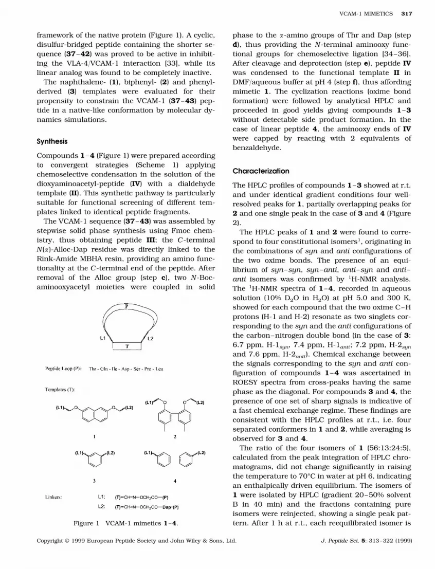
framework of the native protein (Figure 1). A cyclic,disulfur-bridged peptide containing the shorter se-quence (37–42) was proved to be active in inhibit-ing the VLA-4/VCAM-1 interaction [33], while itslinear analog was found to be completely inactive.
The naphthalene- (1), biphenyl- (2) and phenyl-derived (3) templates were evaluated for theirpropensity to constrain the VCAM-1 (37–43) pep-tide in a native-like conformation by molecular dy-namics simulations.
Synthesis
Compounds 1–4 (Figure 1) were prepared accordingto convergent strategies (Scheme 1) applyingchemoselective condensation in the solution of thedioxyaminoacetyl-peptide (IV) with a dialdehydetemplate (II). This synthetic pathway is particularlysuitable for functional screening of different tem-plates linked to identical peptide fragments.
The VCAM-1 sequence (37–43) was assembled bystepwise solid phase synthesis using Fmoc chem-istry, thus obtaining peptide III; the C-terminalN(a)-Alloc-Dap residue was directly linked to theRink-Amide MBHA resin, providing an amino func-tionality at the C-terminal end of the peptide. Afterremoval of the Alloc group (step c), two N-Boc-aminooxyacetyl moieties were coupled in solid
phase to the a-amino groups of Thr and Dap (stepd), thus providing the N-terminal aminooxy func-tional groups for chemoselective ligation [34–36].After cleavage and deprotection (step e), peptide IVwas condensed to the functional template II inDMF/aqueous buffer at pH 4 (step f), thus affordingmimetic 1. The cyclization reactions (oxime bondformation) were followed by analytical HPLC andproceeded in good yields giving compounds 1–3without detectable side product formation. In thecase of linear peptide 4, the aminooxy ends of IVwere capped by reacting with 2 equivalents ofbenzaldehyde.
Characterization
The HPLC profiles of compounds 1–3 showed at r.t.and under identical gradient conditions four well-resolved peaks for 1, partially overlapping peaks for2 and one single peak in the case of 3 and 4 (Figure2).
The HPLC peaks of 1 and 2 were found to corre-spond to four constitutional isomers1, originating inthe combinations of syn and anti configurations ofthe two oxime bonds. The presence of an equi-librium of syn–syn, syn–anti, anti–syn and anti–anti isomers was confirmed by 1H-NMR analysis.The 1H-NMR spectra of 1–4, recorded in aqueoussolution (10% D2O in H2O) at pH 5.0 and 300 K,showed for each compound that the two oxime C–Hprotons (H-1 and H-2) resonate as two singlets cor-responding to the syn and the anti configurations ofthe carbon–nitrogen double bond (in the case of 3:6.7 ppm, H-1syn, 7.4 ppm, H-1anti; 7.2 ppm, H-2syn
and 7.6 ppm, H-2anti). Chemical exchange betweenthe signals corresponding to the syn and anti con-figuration of compounds 1–4 was ascertained inROESY spectra from cross-peaks having the samephase as the diagonal. For compounds 3 and 4, thepresence of one set of sharp signals is indicative ofa fast chemical exchange regime. These findings areconsistent with the HPLC profiles at r.t., i.e. fourseparated conformers in 1 and 2, while averaging isobserved for 3 and 4.
The ratio of the four isomers of 1 (56:13:24:5),calculated from the peak integration of HPLC chro-matograms, did not change significantly in raisingthe temperature to 70°C in water at pH 6, indicatingan enthalpically driven equilibrium. The isomers of1 were isolated by HPLC (gradient 20–50% solventB in 40 min) and the fractions containing pureisomers were reinjected, showing a single peak pat-tern. After 1 h at r.t., each reequilibrated isomer isFigure 1 VCAM-1 mimetics 1–4.
Copyright © 1999 European Peptide Society and John Wiley & Sons, Ltd. J. Peptide Sci. 5: 313–322 (1999)

PERI ET AL.318
Scheme 1 Convergent strategy for the synthesis of VCAM-1 mimetic 1. (a) BrCH2CH(OEt)2 (10 equivalents), Cs2CO3 (4equivalents), DMF, 70°C, 24 h. (b) AcOH/HCl 1 N, 1 h, removal of EtOH in vacuo; II: 34% yield. (c) Solid-phase synthesisof peptide III on Rink amide resin using Fmoc strategy, then Alloc cleavage with Pd(PPh3)4 (0.10 equivalents), PhSiH3 (24equivalents), in DCM, 10 min, r.t. (d) Boc-NH-O-CH2-COOSu (3 equivalents), DIPEA (6 equivalents), DMF, 30 min, r.t. (e)TFA 95%, TIS 2.5%, H2O 2.5%, 1 h, r.t.; IV: 65% yield. (f) Peptide IV, 5·10−4 M in 50% DMF/acetate buffer (pH 4) (1:1, v/v)and dialdehyde II (1.1 equivalents), 1 h, r.t.; 1: 89% yield.
showing the identical HPLC profile composed of fourpeaks.
Compounds 1–4 were tested for inhibition ofVCAM-1/VLA-4 dependent adhesion by in vitrobinding assays. Only compound 1 was found to beactive at IC50:0.4 mM2. The binding curves of themimetics, compared to the one of the disulfur-bridged peptide c(Cys-Gln-Ile-Asp-Ser-Pro-Cys) [33]are shown in Figure 3. These experimental dataconfirm that the presence of the naphthalene tem-plate between the terminal residues of the loopenhances the activity of the synthetic inhibitor,thus encouraging the use of rigid templates in thedesign of these types of protein loop mimetics.
Molecular Dynamics Simulation
The impact of the constraint imposed by the tem-plate upon the backbone and side-chain conforma-tions was estimated by molecular dynamicssimulations. Using the X-ray structure of the CD-loop domain of VCAM-1 as the starting point for the
calculations3, the time-dependent evolution of theloop structure at r.t. was followed. As the R–O–N�Cconnectivity of the oxime bonds was not parameter-ized for any known force field, the values for theR–O–C�C group were taken as a first approxima-tion. All four isomers of the oxime bond in cycliccompounds 1–3 (syn–syn, syn–anti, anti–syn,anti–anti ) were treated separately in the simula-tions. In order to explore a broad region of theconformational space, a long simulation time (500ps) was chosen. After a period of 150 ps, the pep-tidic parts of molecules 1–3 were relaxed and didnot change its conformation significantly; structuralchanges were principally observed in the linker partof the molecules. This became obvious by compar-ing the fluctuation of the calculated energies for thetemplate/linker part with the RMSD values of thebackbone of the peptidic part, as shown in Plate 1for the four isomers of 1.
Four different minimized structures were ob-tained for each cyclic compound, corresponding tothe four combinations of the syn (E) and anti (Z)
Copyright © 1999 European Peptide Society and John Wiley & Sons, Ltd. J. Peptide Sci. 5: 313–322 (1999)

VCAM-1 MIMETICS 319
forms of the two oxime bonds as presented in Plate2 for compound 1. As a common feature of thesestructures4, the peptide part shows generally a goodsuperposition with the corresponding loop of thenative protein, whereas the template/linker partforms a compact hydrophobic core surrounded bythe loop.
From the series of VCAM-1 mimetics 1–3, prelim-inary conclusions on structure–function correla-tions can be drawn. The linear peptide 4 beingrather flexible in solution and lacking any spatialpre-organization of functional groups, turns out tobe completely inactive despite the presence of thepotentially active sequence. In mimetics 1–3, thetemplates reduce dramatically the conformationalspace of the molecules. The observed activity ofcompound 1 may originate from the rigidity of thenaphthalene-based spacer resulting in a fixed dis-tance between the C- and the N-terminal residuesof the loop. The average distance between the Na ofThr37 and CO carbon of Leu43 was found to be :10A, in the minimized structures of the syn–anti andanti–syn isomers of 1, and :11 A, for the syn–syn
and the anti–anti forms, respectively. These valuesare significantly larger compared to the nativeprotein loop (7.36 A, ), indicating that a redesign ofthe linker group could result in increased bindingaffinity. The dNH37–CO43
values for the minimizedstructures of 2 have a broad fluctuation range,varying from 5.5 A, for the syn–syn to 12.6 A, of theanti–syn isomer. Here, the overall mobility is proba-bly too high to ensure a conformational ‘lock-in’ ofthe peptide in an appropriate conformation. In com-pound 3, the distance between C- and N-terminalresidues of the loop is about 8.5 A, (averaged on thefour isomeric forms), being comparable to that ofthe native protein. Deviations of the distance dNH37–
CO43 values are all less than 10% during the simu-lations of 1–3.
CONCLUSIONS
The effect of aromatic templates upon the loopstructure of mimetics 1–4 (Figure 1) is studied byevaluating the in vitro activity in inhibiting the
Figure 2 HPLC profiles of compounds 1–4 at r.t. (gradient: 20–50% of B in 30 min); 1, Rt=13.0, 14.4, 15.3 and 16.0 min;2, Rt=20.0, 20.7, 20.9 min; 3, Rt=11.5 min; 4, Rt=20.0 min.
Copyright © 1999 European Peptide Society and John Wiley & Sons, Ltd. J. Peptide Sci. 5: 313–322 (1999)

PERI ET AL.320
Figure 3 Effect of synthetic VCAM-1 mimetics 1–4 and S–S bridged Cys-Gln-Ile-Asp-Ser-Pro-Cys (c[CQIDSPC]) on theadhesion of Ramos cells to recombinant VCAM-1. Ramos cells were allowed to adhere to immobilized VCAM-1 in thepresence of mimetics 1–4 as described. Each point represents the mean9S.D. of triplicates.
VCAM-1/VLA-4 (a4b1) specific recognition. A de-tailed understanding of the interaction betweenVLA-4 and VCAM-1 recognition sites [37–39] repre-sents an important starting point for the design anddevelopment of putative anti-inflammatory drugs.The biological activity of 1 makes this molecule aninteresting lead compound for structural optimiza-tion applying combinatorial approaches, althoughits activity cannot be assigned or attributed to onesingle isomer. Molecular dynamics simulations re-veal the effect of the template on the preferred loopconformations. Consequently further structuralmodifications of the templates represent a reason-able starting point in future structure–functionstudies.
The studies presented here show the potential ofthe template concept to stabilize exposed loops inproteins. Investigations for optimizing the bioactiv-ity of compound 1 applying combinatorial methodsare in progress.
Acknowledgements
We gratefully acknowledge the Swiss National Sci-ence Foundation and Novartis Pharma AG (Basel)for financial support.
NOTES
1. Compounds 1 and 2 were analysed with a LC/ESI-MSinstrument and for each compound the different peaksrevealed the same mass (Mr=1212.9 for 1 and Mr=1266.5 for 2). Compounds 3 and 4 showed a singlepeak at various gradients condition in the RP-HPLCanalysis.
2. Ramos cells were allowed to adhere to immobilizedVCAM-1 in the presence of peptides as described in the‘Materials and Methods’. In two different experiments(each one in triplicate), IC50 values of 0.3 and 0.54 mM
were found for compound 1.3. Coordinates of the X-ray structure of VCAM-1 were
obtained from the Brookhaven Crystallographic Data-base PDB code: 1vsc.
4. The same type of calculations were run for molecules 2and 3 giving a good superposition of the peptide partwith the native-protein loop for both molecules.
REFERENCES
1. Mutter M, Tuchscherer G. Non-native architectures inprotein design and mimicry. Cell Mol Life Sci 1997; 53:851–863.
2. Bryson JW, Betz SF, Lu HS, Suich DJ, Zhou HXX,O’Neil KT, DeGrado WF. Protein design—a hierarchi-cal approach. Science 1995; 270: 935–941.
Copyright © 1999 European Peptide Society and John Wiley & Sons, Ltd. J. Peptide Sci. 5: 313–322 (1999)

VCAM-1 MIMETICS 321
3. Baltzer L. Functionalization of designed foldedpolypeptides. Curr Opinion Struct Biol 1998; 8: 466–470.
4. Rau HK, Dejonge N, Haehnel W. Modular synthesis ofde novo designed metalloproteins for light-inducedelectron transfer. Proc Natl Acad Sci USA 1998; 95:11526–11531.
5. Tuchscherer G, Schleibler L, Dumy P, Mutter M.Protein design: on the threshold of functional proper-ties. Biopolymers 1998; 47: 63–73.
6. Braisted AC, Wells JA. Minimizing a binding domainfrom protein A. Proc Natl Acad Sci USA 1996; 93:5688–5692.
7. Cunningham BC, Wells JA. Minimized proteins. Curr
Opinion Struct Biol 1997; 7: 457–462.8. Struthers MD, Cheng RP, Imperiali B. Economy in
protein design: evolution of a metal-independent motifbased on the zinc finger domains. J Am Chem Soc
1996; 118: 3073–3081.9. Pessi A, Bianchi E, Crameri A, Venturini S, Tramon-
tano A, Sollazzo M. A designed metal-binding proteinwith a novel fold. Nature 1993; 362: 367–369.
10. Vita C, Vizzavona J, Drakopoulou E, Zinn-Justin S,Gilquin B, Menez A. Novel miniproteins engineered bythe transfer of active sites to small natural scaffolds.Biopolymers 1998; 47: 93–100.
11. Nygren PA, Uhlen M. Scaffolds for engineering novelbinding sites in proteins. Curr Opinion Struct Biol
1997; 7: 463–469.12. Vita C, Roumestand C, Toma F, Menez A. Scorpion
toxins as natural scaffolds for protein engineering.Proc Natl Acad Sci USA 1995; 92: 6404–6408.
13. Perez-Paya E, Houghten RA, Blondelle SE. Functional-ized protein-like structures from conformationally de-fined synthetic combinatorial libraries. J Biol Chem
1996; 271: 4120–4126.14. Houston ME, Wallace A, Bianchi E, Pessi A, Hodges
RS. Use of a conformationally restricted secondarystructural element to display peptide libraries: a twostranded alpha helical coiled-coil stabilized by lactambridges. J Mol Biol 1996; 262: 270–282.
15. Mutter M, Vuilleumier S. A chemical approach toprotein design: Template Assembled SyntheticProteins (TASP). Angew Chem Int Ed Engl 1989; 28:535–554.
16. Mutter M, Tuchscherer G, Miller C, Altmann KH,Carey RI, Wyss DF, Labhard AM, Rivier JE. Template-assembled synthetic proteins with four-helix-bundletopology. Total chemical synthesis and conformationalstudies. J Am Chem Soc 1992; 114: 1463–1470.
17. Mutter M, Dumy P, Garrouste P, Lehmann C, MathieuM, Peggion C, Peluso S, Razaname A, Tuchscherer G.Template assembled synthetic proteins (TASP) as func-tional mimetics of proteins. Angew Chem Int Ed Engl
1996; 35: 1482–1485.18. Sila U, Mutter M. Topological templates as tool in
molecular recognition and peptide mimicry: Synthesisof a TASK library. J Mol Recognition 1995; 8: 2934.
19. Bisang C, Weber C, Robinson JA. Protein-loop mimet-ics: a diketopiperazine-based template to stabilize loopconformations in cyclic peptides containing the NPNAand RGD motifs. Helv Chim Acta 1996; 79: 1825–1842.
20. Beeli R, Steger M, Linden A, Robinson JA. A tricyclictemplate derived from (2S,4R)-4-hydroxyproline for thesynthesis of protein loop mimetics. Helv Chim Acta
1996; 79: 2235–2248.21. Emery F, Bisang C, Favre M, Jiang L, Robinson JA. A
template for the solid-phase synthesis of conforma-tionally restricted protein loop mimetics. Chem Com-mun 1996; 2155–2156.
22. Fields GB, Noble RL. Solid phase peptide synthesisutilizing 9-fluorenylmethoxycarbonyl amino acids. Int
J Pept Protein Res 1990; 35: 161–214.23. Sheppard RC. Continuous flow methods in organic
synthesis. Chem Brit 1983; 402.24. Chang CD, Meienhofer J. Solid-phase peptide synthe-
sis using mild base cleavage of Na-fluorenylmethyloxy-carbonylamino acids, exemplified by a synthesis ofdihydrosomatostatin. Int J Pept Protein Res 1978; 11:246–249.
25. Thieret N, Alsina J, Giralt E, Guibe F, Albericio F. Useof Alloc-amino acids in solid-phase peptide synthesis-tandem deprotection-coupling reactions using neutralconditions. Tetrahedron Lett 1997; 41: 7275–7278.
26. Sartori G, Maggi R, Bigi F, Arienti A, Casnati G. Regio-chemical control in the oxidative coupling of metalphenolates: highly selective synthesis of symmetric,hydroxylated biaryls. Tetrahedron 1992; 48: 9483.
27. Weitz-Schmidt G, Stokmaier D, Scheel G, Nifant’ev NE,Tuzikov AB, Bovin NV. An E-selectin binding assaybased on a polyacrymide-like glycoconjugate. Anal
Biochem 1996; 238: 184–190.28. Jones EY, Harlos K, Bottomley MJ, Robinson RC,
Driscoll PC, Edwards RM, Clements JM, Dudgeon TJ,Stuart DI. Crystal structure of an integrin-bindingfragment of VCAM-1 at 1.8 Angstrom resolution. Na-ture 1995; 373: 539–544.
29. Vonderheide RH, Tedder TF, Springer TA, StauntonDE. Residues within a conserved amino acid motif ofdomains 1 and 4 of VCAM-1 are required for binding toVLA-4. J Cell Biol 1994; 125: 215–222.
30. Clements JM, Newham P, Shepherd M, Gilbert R, Dud-geon MJ, Needham LA, Edwards RM, Berry L, Brass A,Humphries MJ. Identification of a key integrin-bindingsequence in VCAM-1 homologous to the LDV activesite in fibronectin. J Cell Sci 1994; 107: 2127–2135.
31. De Fougerolles A, Springer TA. Ideas crystallized onimmunoglobulin superfamily-integrin interactions.Curr Biol 1995; 2: 639–643.
32. Cardarelli PM, Cobb RR, Nowlin DM, Scholz W, Gorc-san F, Moscinski M, Yasuhara M, Chiang SL, Lobl TJ.Cyclic RGD peptide inhibits a4b1 interaction with con-necting segment 1 and vascular cell adhesionmolecule. J Biol Chem 1994; 269: 18668–18673.
Copyright © 1999 European Peptide Society and John Wiley & Sons, Ltd. J. Peptide Sci. 5: 313–322 (1999)

PERI ET AL.322
33. Wang JH, Pepinsky RB, Stehle T, Liu JH, KarpusasM, Browning B, Osborn N. The crystal structure ofan N-terminal two-domain fragment of VCAM-1: acyclic peptide based on the domain 1 CD-loop caninhibit VCAM-1/a4 integrin interaction. Proc Natl
Acad Sci USA 1995; 92: 5714–5718.34. Rose K. Facile synthesis of homogeneous artificial
proteins. J Am Chem Soc 1994; 116: 30.35. Lu W, Qasim MA, Kent SBH. Comparative total
syntheses of turkey ovomucoid third domain byboth stepwise solid phase synthesis and nativechemical ligation. J Am Chem Soc 1996; 118: 8518–8523.
36. Tuchscherer G. Template assembled syntheticproteins: condensation of a multifunctional peptideto a topological template via chemoselective ligation.Tetrahedron Lett 1993; 34: 8419.
37. Hynes RO. Integrins: versatility, modulation and sig-naling in cell adhesion. Cell 1992; 69: 11–25.
38. Springer TA. Traffic signals for lymphocyte recircula-tion and leukocyte emigration: the multistepparadigm. Cell 1994; 76: 301–314.
39. Ruegg C, Postigo AA, Sikorski EE, Butcher EC,Pytela R, Erle DJ. Role of integrin alpha 4 beta 7/al-pha 4 beta P inlymphocyte adherence to fibronectinand VCAM-1 and in homotypic cell clustering. J CellBiol 1992; 117: 179–189.
Copyright © 1999 European Peptide Society and John Wiley & Sons, Ltd. J. Peptide Sci. 5: 313–322 (1999)
Copyright © 2022 FDOKUMEN