Artificial Intelligence Applications for Health Care; 1
68
-
Upload
khangminh22 -
Category
Documents
-
view
2 -
download
0
Transcript of Artificial Intelligence Applications for Health Care; 1


ARTIFICIAL INTELLIGENCE
APPLICATIONS FOR HEALTH CARE

ARTIFICIAL INTELLIGENCE
APPLICATIONS FOR HEALTH CARE
Edited by
Mitul Kumar Ahirwal, Narendra D. Londhe, and Anil Kumar

First edition published 2022 by CRC Press 6000 Broken Sound Parkway NW, Suite 300, Boca Raton, FL 33487-2742
and by CRC Press 4 Park Square, Milton Park, Abingdon, Oxon, OX14 4RN
CRC Press is an imprint of Taylor & Francis Group, LLC
© 2022 selection and editorial matter, Mitul Kumar Ahirwal, Narendra D. Londhe and Anil Kumar; individual chapters, the contributors
Reasonable efforts have been made to publish reliable data and information, but the author and publisher cannot assume responsibility for the validity of all materials or the consequences of their use. The authors and publishers have attempted to trace the copyright holders of all material reproduced in this publication and apologize to copyright holders if permission to publish in this form has not been obtained. If any copyright material has not been acknowledged please write and let us know so we may rectify in any future reprint.
Except as permitted under U.S. Copyright Law, no part of this book may be reprinted, reproduced, transmitted, or utilized in any form by any electronic, mechanical, or other means, now known or hereafter invented, including photocopying, microfilming, and recording, or in any information storage or retrieval system, without written permission from the publishers.
For permission to photocopy or use material electronically from this work, access www. copyright.com or contact the Copyright Clearance Center, Inc. (CCC), 222 Rosewood Drive, Danvers, MA 01923, 978-750-8400. For works that are not available on CCC please contact [email protected]
Trademark notice: Product or corporate names may be trademarks or registered trademarks and are used only for identification and explanation without intent to infringe.
Library of Congress Cataloging‑in‑Publication Data Names: Ahirwal, Mitul Kumar, editor. | Londhe, Narendra D., editor. | Kumar, Anil, 1980- editor. Title: Artificial intelligence applications for health care / edited by Mitul Kumar Ahirwal, Narendra D. Londhe, Anil Kumar. Description: First edition. | Boca Raton : Taylor and Francis, 2022. | Includes bibliographical references and index. Identifiers: LCCN 2021052132 | ISBN 9781032148465 (hardback) | ISBN 9781032148472 (paperback) | ISBN 9781003241409 (ebook) Subjects: LCSH: Artificial intelligence‐‐Medical applications. | Medical informatics. Classification: LCC R859.7.A78 A74 2022 | DDC 610.285‐‐dc23/eng/20220110 LC record available at https://lccn.loc.gov/2021052132
ISBN: 978-1-032-14846-5 (hbk) ISBN: 978-1-032-14847-2 (pbk) ISBN: 978-1-003-24140-9 (ebk)
DOI: 10.1201/9781003241409
Typeset in Times by MPS Limited, Dehradun

Dedication
This is dedicated to our parents and family members.

Contents Foreword ...................................................................................................................ix Preface.......................................................................................................................xi Acknowledgement ..................................................................................................xiii Editors Biographies .................................................................................................xv Contributors ...........................................................................................................xvii
Chapter 1 A Survey of Machine Learning in Healthcare ...................................1
S. Sathyanarayanan and Sanjay Chitnis
Chapter 2 A Review on Biomedical Signals with Fundamentals of Digital Signal Processing ..................................................................23
Mangesh Ramaji Kose, Mithilesh Atulkar, and Mitul Kumar Ahirwal
Chapter 3 Images in Radiology: Concepts of Image Acquisition and the Nature of Images.........................................................................49
Narendra Kuber Bodhey
Chapter 4 Fundamentals of Artificial Intelligence and Computational Intelligence Techniques with Their Applications in Healthcare Systems ...........................................................................73
Shubhi Sharma and Siddharth Sharma
Chapter 5 Machine Learning Approach with Data Normalization Technique for Early Stage Detection of Hypothyroidism ...............91
Madhusudan G. Lanjewar, Jivan S. Parab, and Ajesh K. Parate
Chapter 6 GPU-based Medical Image Segmentation: Brain MRI Analysis Using 3D Slicer................................................................109
Sajedul Talukder
Chapter 7 Preliminary Study of Retinal Lesions Classification on Retinal Fundus Images for the Diagnosis of Retinal Diseases ..................123
Jaskirat Kaur, Deepti Mittal, and Ramanpreet Kaur
vii

Chapter 8 Automatic Screening of COVID-19 Based on CT Scan Images Through Extreme Gradient Boosting.................................139
G. Madhu, G. Nagachandrika, A. Govardhan, and K. Lakshman Srikanth
Chapter 9 Investigations on Convolutional Neural Network in Classification of the Chest X-Ray Images for COVID-19 and Pneumonia ................................................................................163
Ganesh Laveti, P. Chaya Devi, and R. Goswami
Chapter 10 Improving the Detection of Abdominal and Mediastinal Lymph Nodes in CT Images Using Attention U-Net Based Deep Learning Model .....................................................................181
Hitesh Tekchandani, Shrish Verma, and Narendra D. Londhe
Chapter 11 Swarm Optimized Hybrid Layer Decomposition and Reconstruction Model for Multi-Modal Neurological Image Fusion ...................................................................................203
Manisha Das, Deep Gupta, Petia Radeva, and Ashwini M. Bakde
Chapter 12 Hybrid Seeker Optimization Algorithm-based Accurate Image Clustering for Automatic Psoriasis Lesion Detection....................227
Manoranjan Dash, Rajendra Sonawane, Narendra D. Londhe, and Subhojit Ghosh
Chapter 13 A COVID-19 Tracker for Medical Front-Liners ...........................241
Akshansh Jaiswal, Aman Shinde, Avani Pathak, Chaithra Bhat, Manali Kaswa, Rajeev Ramesh, Siddhant Singh, Vikramjit Banerjee, Prashant Gadakh, and Sandeep Patil
Chapter 14 Implementation of One Dimensional Convolutional Neural Network for ECG Classification on Python Platform .......257
Mitul Kumar Ahirwal, Ravi Singh, and Nikhil Agrawal
Chapter 15 Pneumonia Detection from X-Ray Images by Two Dimensional Convolutional Neural Network on Python Platform...............................................................................289
Mitul Kumar Ahirwal, Vipul Sharma, and Kuldeep Kumar Gautam
Index......................................................................................................................309
viii Contents

Foreword This book is edited by three authors having expertise in different domains. Dr. Mitul Kumar Ahirwal is working with Computer Science and Engineering department, MANIT Bhopal, India and has expertise in computational intelligence techniques and Python. Dr. Narendra D. Londhe is working with Electrical Engineering department, NIT Raipur, India and has expertise in biomedical signal/image processing. Dr. Anil Kumar is working with Electronics and Communications Engineering department, PDPM-IIITDM Jabalpur, India and has expertise in digital signal processing and digital filter design for biomedical signals.
This book is a very good choice for the readers who wish to continue their studies and research in interdisciplinary areas of artificial intelligence and biomedical signal/image processing.
ix

Preface At present, the health care industry is growing at a very fast rate. This is because of requirements and development of high precision and modern equipment/instruments as well as diagnosis methods. These requirements are fulfilled by research and studies conducted over different databases and subjects. The role of biomedical signals and images are very important in these studies. These research and studies also make use of artificial intelligence techniques. To understand and apply artificial intelligence techniques, sufficient skills are required. Hence, the objective of this book is to explore the applications of artificial intelligence techniques in the field of health care and provide a good understanding of technique used.
Artificial intelligence mainly includes the techniques that are iterative in nature. These techniques may help in optimization of solution or in learning/training for a predication and classification of model. Since the past decade, applications of these techniques are providing promising results in biomedical engineering and healthcare industry. Biomedical signal/image processing is one of the important fields that is currently playing a vital role in the healthcare industry.
This book has been edited by three experts from different domains, first editor is from computer science domain, second editor is from electrical engineering domain, and the third editor is from electronics & communication domain. Hence, the scope of book is wide and it covers paradigms of Computer Science, Electrical, and Electronics Engineering fields.
Therefore, this book will help readers working in the field of artificial intelligence and biomedical signal/image processing. Readers of artificial intelligence field will get the idea and basic implementation for applications of their expertise in biomedical signal/image processing field. On the other hand, readers of biomedical signal/image processing field will get familiar with the topics of artificial intelligence, machine learning, and optimization fields.
xi

Acknowledgement First of all we thank our parents for all the things that we have today. We would like to thanks our supervisors. We also express thanks to our family members for their support and sacrifices. Special Thanks to Dr. N.S. Raghuvanshi (Director, MANIT Bhopal), Dr. A.M. Rawani (Director, NIT Raipur), and Dr. Sanjeev Jain (Director, PDPM-IIITDM Jabalpur) for their encouragement and support. Thanks for all the authors/contributors for their valuable contribution in this book. Thanks for research labs/institutes for uploading their datasets for public usage in research and academics. Many thanks for CRC Press and Taylor & Francis Group for accepting the book proposal and their continuous support. At last, but not least, we thank almighty god for giving us strength to complete this book as a mission.
xiii

Editors Biographies Dr. Mitul K. Ahirwal received his B.E. in Computer Science & Engineering from SATI, Vidisha, (M.P) (Affiliated to RGPV, Bhopal, India) in 2009 and completed his M.Tech. in Computer Technology from National Institute of Technology, Raipur, India in 2011. He completed his Ph.D. in the Computer Science and Engineering Department, PDPM IIITDM, Jabalpur, India. Presently, he is Assistant Professor in the Department of Computer Science and Engineering at Maulana Azad National Institute of Technology, Bhopal, India. He has several national
and international publications in his credit. He is also working on several research projects funded by different agencies. His research area is biomedical signal processing, swarm optimization, brain–computer interface, and healthcare system. He has been involved as a reviewer with various reputed journals.
Institute webpage: http://www.manit.ac.in/content/dr-mitul-kumar-ahirwal Google Scholar: https://scholar.google.co.in/citations?user=fWgOgcIAAAAJ&hl=en
Dr. Narendra D. Londhe has completed his M.Tech and Ph.D. in the year of 2004 and 2011, respectively from Indian Institute of Technology, Roorkee, Uttarakhand, India. Currently he is working with National Institute of Technology, Raipur as an Associate Professor in Department of Electrical Engineering. His area of research includes image and signal processing, soft computing, biometrics, ultrasound imaging, IVUS imaging, brain–computer interface, pain assessment, and psoriasis severity detection. He has more than 40 publications in SCI indexed journals, more than 30
peered reviewed reputed national and international journal publications, and approximately 35 national and international reputed conferences. Moreover, he is member of many technical communities and societies such Senior member of IEEE, Member of IET, Life Member of The Institution of Engineers (India), Life Member of Ultrasonic Society of India, Life Member of Acoustical Society of India, Life Member of Biomedical Engineering Society of India, Life Member of Telemedicine Society of India, Life Member of Instrument Society of India, and Life Member of Institution of Engineers.
Institute webpage: http://www.nitrr.ac.in/viewdetails.php?q=ee.ndlondhe Google Scholar: https://scholar.google.co.in/citations?user=7bH2p9oAAAAJ&hl=en
xv

Dr. Anil Kumar has completed a B.E. from Army Institute of Technology (AIT), Pune, Pune University in Electronic & Telecommunication Engineering and M.Tech and Ph.D. from Indian Institute of Technology, Roorkee, India in 2002, 2006, and 2010, respectively. After doctoral work, he joined as an Assistant Professor in the Electronics & Communication Engineering Department, Indian Institute of Information Technology Design and Manufacturing, Jabalpur, India since 2009 to July 2016. His academic and research interest is in
designing Digital Filters & Multirate Filter Bank, Multirate Signal Processing, Biomedical Signal Processing, Image Processing, and Speech Processing.
Institute webpage: http://faculty.iiitdmj.ac.in/faculty/anilk Google Scholar: https://scholar.google.co.in/citations?user=Xqz4P2AAAAAJ&
hl=en
xvi Editors Biographies

Contributors Nikhil Agrawal Indian Institute of Information Technology Nagpur, India
Mitul Kumar Ahirwal Maulana Azad National Institute of
Technology Bhopal, India
Mithilesh Atulkar National Institute of Technology Raipur, India
Ashwini M. Bakde Department of Radio-Diagnosis All India Institute of Medical Sciences Nagpur, Maharashtra, India
Vikramjit Banerjee International Institute of Information
Technology Pune, India
Chaithra Bhat International Institute of Information
Technology Pune, India
Narendra Kuber Bodhey Department of Radiodiagnosis All India Institute of Medical Sciences Raipur, India
Sanjay Chitnis Dayananda Sagar University Bangalore, India
Manisha Das Department of Electronics and Communi-
cation Engineering Visvesvaraya National Institute of Technology Nagpur, Maharashtra, India
Manoranjan Dash Electrical Engineering Department National Institute of Technology Raipur, India
P. Chaya Devi Anil Neerukonda Institute of Technology
& Sciences, Visakhapatnam Andhra Pradesh, India
Prashant Gadakh International Institute of Information
Technology Pune, India
Kuldeep Kumar Gautam Maulana Azad National Institute of
Technology Bhopal, India
Subhojit Ghosh Electrical Engineering Department, National Institute of Technology Raipur, India
R. Goswami Anil Neerukonda Institute of Technology
& Sciences, Visakhapatnam Andhra Pradesh, India
A. Govardhan JNTUH College of Engineering Hyderabad, India
Deep Gupta Department of Electronics and Communi-
cation Engineering Visvesvaraya National Institute of Technology Nagpur, Maharashtra, India
xvii
Akshansh Jaiswal International Institute of Information
Technology Pune, India
Manali Kaswa International Institute of Information
Technology Pune, India
Jaskirat Kaur Chandigarh Group of Colleges Jhanjeri
Mohali Chandigarh Chandigarh, India
Ramanpreet Kaur Chandigarh Group of Colleges Jhanjeri
Mohali Chandigarh Chandigarh, India
Mangesh Ramaji Kose National Institute of Technology Raipur, India
Madhusudan G. Lanjewar School of Physical and Applied Sciences Goa University Goa, India
Ganesh Laveti Gayatri Vidya Parishad College of
Engineering for Women Visakhapatnam, Andhra Pradesh, India
Narendra D. Londhe Electrical Engineering Department National Institute of Technology Raipur, India
G. Madhu VNR Vignana Jyothi Institute of Enginee-
ring and Technology Hyderabad, India
Deepti Mittal Thapar Institute of Engineering and
Technology Patiala, Punjab, India
G. Nagachandrika VNR Vignana Jyothi Institute of Enginee-
ring and Technology Hyderabad, India
Jivan S. Parab School of Physical and Applied Sciences Goa University Goa, India
Rajesh K. Parate Department of Electronics S. K. Porwal College Kamptee, India
Avani Pathak International Institute of Information
Technology Pune, India
Sandeep Patil National Institute of Technology Silchar, India
Petia Radeva Department of Mathematics and Computer
Science Universitat de Barcelona and Computer Vision Center Barcelona, Spain
Rajeev Ramesh International Institute of Information
Technology Pune, India
S. Sathyanarayanan Sri Sathya Sai University for Human
Excellence Gulbarga, India
xviii Contributors

Shubhi Sharma University of Petroleum and Energy
Studies Uttrakhand, India
Siddharth Sharma Indian Institute of Technology Roorkee Uttrakhand, India
Vipul Sharma Maulana Azad National Institute of
Technology Bhopal, India
Aman Shinde International Institute of Information
Technology Pune, India
Ravi Singh Maulana Azad National Institute of
Technology Bhopal, India
Siddhant Singh International Institute of Information
Technology Pune, India
Rajendra Sonawane Psoriasis Clinic and Research Centre,
Psoriatreat Pune Maharashtra, India and National Institute of Technology Raipur, India
K. Lakshman Srikanth Legato Health Technologies LLP Bangalore, India and GAR Infobahn Telangana, India
Sajedul Talukder Southern Illinois University Carbondale, IL, USA
Hitesh Tekchandani National Institute of Technology Raipur, India
Shrish Verma National Institute of Technology Raipur, India
Contributors xix
1 A Survey of Machine Learning in Healthcare
S. Sathyanarayanan Sri Sathya Sai University for Human Excellence, Gulbarga, India
Sanjay Chitnis Dayananda Sagar University, Bangalore, India
CONTENTS
1.1 Introduction.......................................................................................................1 1.2 Artificial Intelligence........................................................................................2
1.2.1 Machine Learning...............................................................................3 1.2.1.1 Steps in Developing an ML System...................................3 1.2.1.2 Types of Machine Learning ................................................4
1.2.2 Deep Learning ....................................................................................6 1.2.3 The Major Types of DL.....................................................................8
1.3 Applications of ML in Healthcare...................................................................9 1.3.1 Cardiovascular Diseases...................................................................10 1.3.2 Medical Imaging...............................................................................10 1.3.3 Drug Discovery/Manufacturing .......................................................12 1.3.4 Electronic Health Records ...............................................................12 1.3.5 Clinical Decision Support System...................................................13 1.3.6 Surgical Robotics .............................................................................13 1.3.7 Precision Medicine ...........................................................................13 1.3.8 Population Health Management.......................................................14 1.3.9 mHealth and Smart Devices ............................................................14
1.3.10 AI for Tackling Pandemic ...............................................................14 1.4 ML Use Cases in Healthcare .........................................................................16 1.5 Limitations and Challenges in Adoption of AI in Healthcare .....................17 1.6 Conclusion ......................................................................................................18 Acknowledgements..................................................................................................19 References................................................................................................................19
1.1 INTRODUCTION
The advanced medical equipment used in secondary and tertiary healthcare centres is considerably expensive, and obtaining quality healthcare has become difficult and
DOI: 10.1201/9781003241409-1 1

unaffordable for majority of the population. Hence, the application of Machine Learning (ML) in healthcare domains to automate various procedures is expected to help accelerate diagnostic procedures, reduce cost and increase access to quality care for general population. ML can be used in the analysis of healthcare data for faster diagnosis. ML-enabled systems can also be used for prediction by analysing the vast amount of healthcare data to enable preventive measures in time. ML in biomedical technologies will help in the development of smaller, portable, easy-to- use, cloud-enabled and affordable devices, which can be used by the medical staff at primary healthcare centres; thereby, reducing the workload of doctors and elim-inating the need for advanced diagnostic equipment for most of the patients. This will also make expensive resources more easily available for people in need. Thus, ML in healthcare could provide greater access to quality care with minimal hindrances.
This chapter reviews some of the ongoing research in this area after a brief tutorial of ML and also briefly describes few ML-enabled devices already in use.
1.2 ARTIFICIAL INTELLIGENCE
Traditionally, computers had to be programmed by humans. This limited its application in areas with set rules and specific protocols. John McCarthy, an American computer scientist, defined AI as the science and engineering of making intelligent machines [1]. AI is used to describe systems that mimic “cognitive” functions that are associated with humans, such as problem-solving skills and learning [2]. Narrow AI systems are those that carry out specific tasks, for example the AI systems currently being used in healthcare domain. These systems are common, for example virtual assistants on smart phones and recommendation systems. Artificial General Intelligence (AGI) systems are capable of learning different tasks like humans. However, such systems do not exist as of now. The different types of AI systems based on the type of work they accomplish are briefly discussed here and depicted in Figure 1.1.
FIGURE 1.1 Types of AI.
2 Artificial Intelligence Applications for Health Care

Knowledge Representation and Reasoning (KR & R or KR): This is a type of AI wherein knowledge representation deals with representing information in the form of symbols and propositions. It makes use of reasoning as the method of deducing facts from the data, for example, computer-aided diagnosis.
Automated Planning and Scheduling: This is also called AI planning. It considers the time and resource constraints and generates a series of actions in sequence to achieve the objective.
Machine Learning: This gives computers the capability to learn from data without being programmed manually.
Natural Language Processing (NLP): This aims to build systems that can make sense out of the raw natural language data as input. An example is Siri in Apple devices.
Computer Vision: This enables computers to process image and video data and see, observe and make sense out of it. For example, computer vision helps in de-tecting patterns in various medical imaging data to help in diagnosing diseases.
Robotics: A robot is an autonomous system or a system with external control with the capability to perceive its environment, make decisions and perform actions in the real world. Robotics is a field that makes use of various branches of science and engineering to build autonomous robots.
Artificial General Intelligence: The goal of AI as a field is to move towards Artificial General Intelligence (AGI). A system having AGI implies that the system can match humans in intellectual capacity. The examples considered so far are examples of narrow AI.
1.2.1 MACHINE LEARNING
ML is a subfield of computer science that provides computers the ability to learn to perform a task on its own from experience without being explicitly programmed. Tom Mitchell defined learning precisely with the following definition [3]: “A computer program is said to learn from experience E with respect to some class of tasks T and performance measure P, if its performance at tasks in T, as measured by P, improves with experience E”.
Such systems learn from the data provided and give a prediction for new data. The conventional ML algorithms are dependent on the availability of data for ef-fectiveness. For example, a price prediction algorithm for houses may give a pre-diction for the house if the location, size, etc. are given. Each information included in the representation of the house for sale is known as a feature. The ML system learns how each of these features of the house correlates with various outcomes. The conventional programming systems give output based on the program and input data. The ML system gives a model as its output based on the input data and the various outcomes which is fed to it. This is illustrated in Figure 1.2.
1.2.1.1 Steps in Developing an ML System A project to develop an ML-enabled system involves multiple phases before it can be put to use.
Data Collection and Labelling: The first step in ML is the collection of data, including the detailed description of the data involved that will help in
A Survey of Machine Learning in Healthcare 3

understanding and identifying the type of data required for the ML model. The process of detecting and tagging data samples is called data labelling.
Pre-processing: After data is collected, it has to be pre-processed, which may in-volve deduplication, normalisation and error correction to make the data suitable for use.
Feature Engineering: A feature is an attribute or a measurable property shared by all the independent units of the data on which analysis or prediction is to be done [4].
For example, in property prices data, the area of the house, the number of rooms and the age of the property can be considered as features. The collection of data of all the houses sold may be considered as a dataset. If the dataset is represented in a tabular format with each row providing information about one house, features ap-pear as columns in the dataset. Feature engineering is the process of extracting features from raw data to increase the prediction capability or the accuracy of the ML model.
Selection of Algorithm: There are multiple algorithms for ML and selection of a suitable algorithm based on the input data format and the prediction expected of the model is an important component of an ML system.
Training: This is the next step in ML model in which input is given to the ML algorithm, which detects patterns on its own to build a model that will be used later for prediction.
Evaluation: The model is then evaluated with test data, that is, data that is not used for training, but whose output values are known. If the test result is not satisfactory, the previous step of training the model is repeated to get satisfactory results.
Prediction: The model thus evaluated and found to be satisfactory is then used to give a prediction for actual use cases after integrating it in a system.
Figure 1.3 shows the various steps in the production of an ML model.
1.2.1.2 Types of Machine Learning ML can be broadly categorised into four types – supervised, unsupervised, semi- supervised and reinforcement learning. Figure 1.4 depicts the types of ML and the most common application for which the type is used.
FIGURE 1.2 Difference between traditional programming system and machine learning system.
FIGURE 1.3 Steps in the production of an ML model.
(Source: https://www.pcictx.org/).
4 Artificial Intelligence Applications for Health Care

Supervised Learning: In this subset of ML, the input of a dataset, as well as the corresponding known output, is fed to the algorithm. The algorithm then builds a model by finding a function to map the set of input data to its known output by using a set of assumptions. The algorithm predicts the output for the known dataset, compares its predicted output to the known output and tries to minimise the error by changing the initial set of assumptions in an iterative process until the model attains sufficient accuracy in its prediction. The data used for the above process is called the training data.
When the output of a model is a category or a class and not a numeric value, the method is called a classification technique. In this technique, the model gives output by recognising patterns in the input dataset. When the output of a model is a continuous value, then the method is called the regression technique.
Unsupervised Learning: Unsupervised learning techniques are used to work with a dataset whose output values or labels are unknown. The algorithm is given only inputs. The model identifies hidden patterns and extracts insights from the input without supervision, which are then incorporated into its learning. Unsupervised learning uses two methods: clustering and dimensionality reduction. Analysing the data and grouping them based on the similarity of the characteristics into different clusters or classes is called clustering. The extraction of information from the data and leaving out less relevant features is called dimensionality re-duction. It is used when the number of features (or dimensions) of the input dataset is very large. The redundant features and features with minimal impact on the output of the model are removed to make the number of features more manageable.
Semi-supervised Learning: The cumbersome stage in supervised learning is data preparation. A semi-supervised learning method could be used to reduce the amount of work in the data preparation stage. First, a small amount of labelled data is used to partially train an ML model like in supervised learning and it is then used to label the large amount of unlabelled data. This procedure is called pseudo- labelling. The mix of the labelled and pseudo-labelled data is then used to train the model [5].
Reinforcement Learning: In Reinforcement Learning (RL), the software agent performs actions and learns from the rewards and penalties for its actions in an interactive environment and strives to maximise the cumulative rewards. Gaming
FIGURE 1.4 Types of machine learning.
A Survey of Machine Learning in Healthcare 5

and self-driving cars are examples where RL has been used. RL has also been used for detecting and treating sepsis.
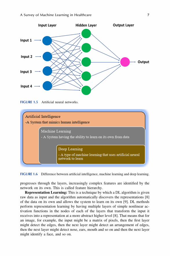
1.2.2 DEEP LEARNING
Artificial Neural Networks (ANN): A neuron is the fundamental component of the brain. A human brain contains around 100 billion neurons. Each neuron is con-nected to around 100,000 other neurons, enabling communication between them. If the signal received by a neuron is above a certain value called the threshold value, the neuron sends signals to the other neurons to which it is connected. The neurons are nonlinear in their behaviour and the transmitting of signals is controlled by the biochemistry [6].
ANN is inspired by the brain’s structure and its functioning. ANN is a computing system made up of a very large number of simple elements that are highly inter-connected and each of the elements process information by responding to inputs [7]. Dr. Robert Hecht-Nielsen, the inventor of one of the first neurocomputers, defined an artificial neural network as [7]
“…a computing system made up of a number of simple, highly interconnected pro-cessing elements, which process information by their dynamic state response to ex-ternal inputs.”
An ANN is made up of a series of layers. Each layer consists of an array of pro-cessing units called nodes or artificial neurons. The neurons receive input from the neurons in the preceding layer and give output to the neurons of the succeeding layer. The input to each neuron is a set of feature values multiplied by their cor-responding weights, which is then summed up to determine the weighted evidence. This value is processed by an activation function, a nonlinear function in the neuron, to determine the output. The activation functions are nonlinear. The various weights of the input are adjusted during training to get the accurate output. The first and the last layers are the input layer and the output layer, respectively. The middle layers are called “hidden” layers since they are not seen by the interfaces to the network and perform the computation. The hidden layer nodes consist of activation functions. A simple ANN with the input layer, hidden layer and the output layer is depicted below in Figure 1.5.
Deep learning (DL): DL is a subset of ML, built using ANN where the number of middle or hidden layers is more than one. The word “deep” is due to the number of the layers in the middle being more than one and hence DL is also called Deep Neural Networks (DNNs). The word “learning” is used because as the model processes more data the accuracy or its prediction power increases; therefore it is said to be learning from its experience. The relationship between AI, ML and DL is illustrated in Figure 1.6.
Feature Hierarchy: Raw data like images or videos are fed as input to a DL algorithm. As the raw data is processed, each layer extracts features from the raw data and passes it on to the next layer. The succeeding layers process the basic features from the preceding layers and identify more complex features. As the data
6 Artificial Intelligence Applications for Health Care
progresses through the layers, increasingly complex features are identified by the network on its own. This is called feature hierarchy.
Representation Learning: This is a technique by which a DL algorithm is given raw data as input and the algorithm automatically discovers the representations [8] of the data on its own and allows the system to learn on its own [9]. DL methods perform representation learning by having multiple layers of simple nonlinear ac-tivation functions in the nodes of each of the layers that transform the input it receives into a representation at a more abstract higher level [8]. That means that for an image, for example, the input might be a matrix of pixels, then the first layer might detect the edges, then the next layer might detect an arrangement of edges, then the next layer might detect nose, ears, mouth and so on and then the next layer might identify a face, and so on.
FIGURE 1.5 Artificial neural networks.
FIGURE 1.6 Difference between artificial intelligence, machine learning and deep learning.
A Survey of Machine Learning in Healthcare 7

Figure 1.7 illustrates the different approaches in DL compared to the approach in ML to solve a particular problem related to the prediction of disease by using medical images as the input data.
Advantages and Constraints of Deep Learning: The data received for input to a DL algorithm can be unstructured data or raw media like texts, images, audio and video recordings [10]. DL algorithms perform automatic feature extraction reducing the need for supervision and reducing the time taken in the data pre-processing stage. Also, unlike traditional ML methods, the performance of DL methods con-tinues to improve as the size of the input data increases.
DL algorithms need a huge amount of data to learn and take correct decisions. Since the DL algorithms perform a very large amount of processing, they need powerful systems, which is possible now due to the emergence of Graphics Processing Units (GPUs).
1.2.3 THE MAJOR TYPES OF DL
Feedforward neural networks, recurrent neural networks (RNN) and convolutional neural networks (CNN) are supervised ML algorithms. Generative adversarial networks (GAN) is an example of unsupervised ML algorithms.
Feedforward Neural Networks: In feedforward neural networks, the nodes are fully connected. Every node in a layer is connected to all the neurons in the next layers. Such structures are called multilayer network of neurons (MLN) [11]. The flow of information is from the input layer to the output layer through the hidden layers without any feedback [12].
Autoencoder: An autoencoder network has an input layer, an output layer and one or more hidden layers connecting them, but the output layer has the same number of units as the input layer [12]. Its purpose is to reconstruct its inputs. Therefore, autoencoders are unsupervised learning models used for dimensionality reduction and for learning generative models of data [12].
Convolutional Neural Networks: In the CNN model, the neurons are connected to only some of the next layer neurons. The learned features are convolved with the input data using 2D convolutional layers. This algorithm is suitable for processing images. Here, convolution is a mathematical operation on two functions that pro-duces a third function expressing how the shape of one is modified by the other and refers to both the result function and to the process of computing it.
FIGURE 1.7 Workflow of AI systems for prediction models: a) Conventional ML, with the various steps involving selecting features manually, b) DL by learning.
8 Artificial Intelligence Applications for Health Care

Recurrent Neural Networks: RNN are used in applications where the output depends on the previous results. Hence, RNN are said to have memory.
Generative Adversarial Networks: GAN are used when the training data is not sufficient. These are used to generate new data to supplement the training data. They consist of the generator and the discriminator. The generator creates fake data based on the real training data it receives. The discriminator analyses the data and tries to distinguish the genuine data from the fake data [13]. GAN can be used to create training datasets for other applications by generating realistic fake data.
1.3 APPLICATIONS OF ML IN HEALTHCARE
As in all other domains, there is a lot of hype about what ML can do in the field of healthcare. Though the hype over the past few years about ML replacing doctors is not justified, technology will increasingly outperform human physicians at specific tasks. AI starts to integrate in the following way. In the first phase, AI augments healthcare practitioners. For example, a chatbot allows doctors to query other cases, context-sensitive AI assistant reminds doctors of appointments, tasks, etc. In the second phase, AI starts to play an increasingly important role and practitioners rely on it. For example, AI-based workflows suggest next steps to doctors based on case history, sometimes even automatically invoking other workflows and tests. Healthcare delivery is accelerated due to automation. In the third step (not im-plemented commonly as of now), AI will drive healthcare and healthcare practi-tioners will augment AI (reverse of step 1). For example, diagnosis and treatment recommendations would be done by AI and doctors merely validate the diagnosis and the recommended treatment. Some of the healthcare domains in which ML has been used are heart diseases, medical imaging and electronic health record (EHR).
Figure 1.8 illustrates the benefits of a healthcare model integrated with AI com-pared to the traditional healthcare model. The advantages of an AI-integrated healthcare model are low cost, improved prediction and greater accuracy, a proac-tive system with less physical interaction compared to the traditional model which is reactive, requires physical presence of patients, is less accurate and expensive.
FIGURE 1.8 Comparison of traditional and AI-integrated healthcare models.
(Source: Aite group).
A Survey of Machine Learning in Healthcare 9

1.3.1 CARDIOVASCULAR DISEASES
Cardiovascular diseases (CVDs) are one of the main causes of mortality. Some aspects of the CVD epidemic in developing countries are the early age that it affects and the high fatality rate. Congenital heart disease (CHD) affects one in every hundred children born across the world [14].
Heart sound auscultation using a stethoscope is the primary method of detecting any abnormalities in the heart. Years of experience are required to diagnose heart murmurs correctly. Hence, further tests that require expensive medical resources could be used inefficiently.
Consider a similar scenario, with one change that the paramedic staff working in a primary healthcare centre has access to an electronic stethoscope with a recording facility and could connect to a cloud server for analysing the heart murmurs and receive response within a few seconds. Further, these devices save the time of doctors, who are expensive resources.
Heart Auscultation: Cardiac disorders can be diagnosed using heart auscultation with high accuracy. Cardiac auscultation is a procedure that involves listening and analysing heart sounds using an electronic stethoscope, a device that records heart sounds digitally. The recording is called a phonocardiogram (PCG). Several digital signal processing and ML techniques could be used to study the PCG and diagnose heart disorders. Automated heart sound analysis is called computer-aided auscultation (CAA). CAA is being applied for development of affordable tools for diagnosis.
1.3.2 MEDICAL IMAGING
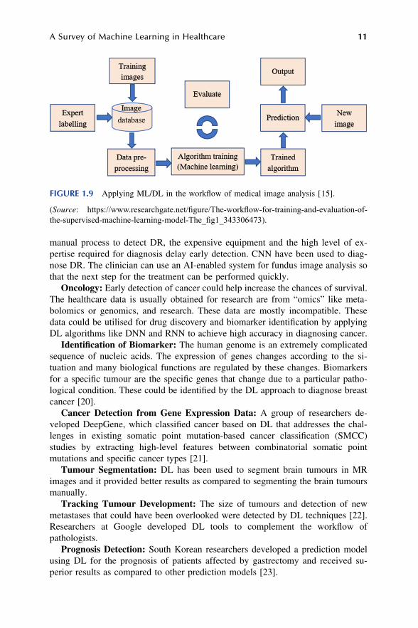
Medical images are analysed by radiologists who are constrained by human factors. Hence, it is an ideal case for automation. The data is relatively organised in the domain of medical imaging due to which it is one of the domains in which ML is being applied. Different ways of digital medical images include magnetic resonance imaging (MRI) scans, X-rays, retinal photography and histology slides. Some of the domains in which ML systems have been applied are discussed below. The workflow of an AI-integrated medical image analysis system is depicted in Figure 1.9.
Radiology: Research on AI in radiology has seen some success as DL-based algorithms equalled or exceeded humans in several tasks, such as the detection of pneumonia on a chest X-ray or the analysis of white matter lesions on MRI scans of the brain [16].
Several efforts have compared DL methods with ML algorithms and the per-formance of DL techniques is found to be better [17]. The performance of radi-ologists has been equalled or is better by DL-based algorithms for both detection and segmentation. Classification of lymph node metastasis in PET-CT by DL had lower specificities, but higher sensitivities than the radiologists [18]. The integration of ML/AI into clinical practice will reduce the workload of radiologists and increase their effectiveness.
Diabetic Retinopathy: Diabetic retinopathy (DR) causes damage to the retina and timely treatment can reduce the risk of blindness [19]. The time-consuming
10 Artificial Intelligence Applications for Health Care
manual process to detect DR, the expensive equipment and the high level of ex-pertise required for diagnosis delay early detection. CNN have been used to diag-nose DR. The clinician can use an AI-enabled system for fundus image analysis so that the next step for the treatment can be performed quickly.
Oncology: Early detection of cancer could help increase the chances of survival. The healthcare data is usually obtained for research are from “omics” like meta-bolomics or genomics, and research. These data are mostly incompatible. These data could be utilised for drug discovery and biomarker identification by applying DL algorithms like DNN and RNN to achieve high accuracy in diagnosing cancer.
Identification of Biomarker: The human genome is an extremely complicated sequence of nucleic acids. The expression of genes changes according to the si-tuation and many biological functions are regulated by these changes. Biomarkers for a specific tumour are the specific genes that change due to a particular patho-logical condition. These could be identified by the DL approach to diagnose breast cancer [20].
Cancer Detection from Gene Expression Data: A group of researchers de-veloped DeepGene, which classified cancer based on DL that addresses the chal-lenges in existing somatic point mutation-based cancer classification (SMCC) studies by extracting high-level features between combinatorial somatic point mutations and specific cancer types [21].
Tumour Segmentation: DL has been used to segment brain tumours in MR images and it provided better results as compared to segmenting the brain tumours manually.
Tracking Tumour Development: The size of tumours and detection of new metastases that could have been overlooked were detected by DL techniques [22]. Researchers at Google developed DL tools to complement the workflow of pathologists.
Prognosis Detection: South Korean researchers developed a prediction model using DL for the prognosis of patients affected by gastrectomy and received su-perior results as compared to other prediction models [23].
FIGURE 1.9 Applying ML/DL in the workflow of medical image analysis [ 15].
(Source: https://www.researchgate.net/figure/The-workflow-for-training-and-evaluation-of- the-supervised-machine-learning-model-The_fig1_343306473).
A Survey of Machine Learning in Healthcare 11

Feature Detection in MRI: Tumour images obtained by non-invasive medical technologies have been used to measure tumour growth in patients affected by using the CNN method of DL.
The use of DL in oncology for early cancer detection and prognosis increases the chances of providing early treatment, saving both time and cost.
1.3.3 DRUG DISCOVERY/MANUFACTURING
Researchers are applying ML algorithms in the discovery of new drugs as ML helps detect new patterns in the data. Researchers have started using DL in drug discovery as it is possible to create an NN architecture customised for a problem [24]. The properties of a molecule can be predicted using the CNN method of DL [25].
Researchers are turning to AI to improve clinical trials due to a large number of failures [26]. Attempts have been made to help patients look for clinical trials on their own by using AI, like the open-source web tool DQueST, developed by Weng and colleagues. They also developed a software called Criteria2Query that uses NLP to help researchers search databases easily for people who are eligible to take part in clinical trials [26].
Clinical trials are now being designed using AI. The ultimate possibility of AI is to eliminate clinical trials by conducting virtual clinical trials using the vast elec-tronic health records to see how the population would respond [26]. The cost of clinical trials could be reduced by analysing the biomedical data generated from research experiments to predict the effects and side effects of a drug [27].
1.3.4 ELECTRONIC HEALTH RECORDS
EHRs are systems that enable healthcare personnel and patients to access all the medical data of the patient instantly after authentication of identity. They allow tools that help in decision-making and thus enable automating and streamlining of workflows in healthcare.
The use of ML-enabled EHR enables more effective care by giving relevant information in real time to improve interaction with the patient. The system can remember what works for each user. ML helps in predictive, diagnostic and pre-scriptive analysis, thus, helping in the personalisation of the treatment, which was difficult earlier since each outcome needed a custom dataset.
NLP allows the DL algorithm to process free text that is generated. Rajkumar et al. proposed a new format based on the Fast Healthcare Interoperability Resources (FHIR, it is a standard for exchanging healthcare information electro-nically) for representing patients’ entire raw EHR records and showed that DL methods using this format can predict medical events from different centres ac-curately without having to customise the system for a particular site [28]. These models outperformed earlier models built without ML in all cases. Figure 1.10 depicts the processing of data by an ML-enabled EHR system that can process different types of structured as well as raw data. The system can analyse images using AI and also using NLP for processing clinical notes.
12 Artificial Intelligence Applications for Health Care

1.3.5 CLINICAL DECISION SUPPORT SYSTEM
Clinical decision support (CDS) provides filtered information of a person and knowledge at appropriate times, to improve the outcome by enabling the stream-lining of the workflow. Alert fatigue of doctors can be reduced by incorporating ML into the CDS design to filter the alerts [30].
1.3.6 SURGICAL ROBOTICS
Robotic surgery or robotics-assisted-surgery (RAS) is minimally invasive surgery using a robotic arm with small tools. DL uses CNN for computer vision to enhance the robustness and adaptability of RAS to perceive the environment and RL tech-niques to learn from a surgeon’s physical motion. Researchers are trying to improve surgical workflow through automation.
1.3.7 PRECISION MEDICINE
Precision medicine aims to enhance healthcare by personalising treatment and prevention in a person by studying the effect of the environment and lifestyle of the person along with the person’s genes to give a unique treatment or prevention which suits that person. This is because each person’s susceptibility to a particular disease is different due to several factors, which the traditional methods did not consider.
Using ML to analyse the large genomic datasets that are available now will help in identifying a combination of genes whose expression can help in classifying a
FIGURE 1.10 Machine learning in the healthcare sector [ 29].
(Source: https://medium.com/sciforce/top-ai-algorithms-for-healthcare-aa5007ffa330).
A Survey of Machine Learning in Healthcare 13

group of patients as belonging to a particular category for treatment and or prevention.
1.3.8 POPULATION HEALTH MANAGEMENT
Population Health Management (PHM) is the collection and analysis of patient data across different healthcare sectors by coordinated steps by all stakeholders in-cluding the government to get actionable insights so that proactive and preventive measures can be taken to address the lack of care provided and increase the general level of health in the population. This will enable targeted intervention after identifying vulnerable patients or a subgroup of the population and will help in reducing the cost of healthcare as well as the burden on the healthcare system. It will also result in knowledge that can help in developing and implementing policies to improve the health of the population [31]. Applying AI can help identify groups of patients that are at a higher risk and enable preventive measures.
1.3.9 MHEALTH AND SMART DEVICES
mHealth is defined by the WHO as the “medical and public health practice sup-ported by mobile devices, such as mobile phones, patient monitoring devices, personal digital assistances and other wireless devices” [32]. The significant in-crease in computing power in mobile and Internet connectivity has resulted in mobile health technologies that can improve the quality of healthcare and research. mHealth integrated with AI can predict and prevent conditions and diseases.
An example is eKidneyCare (details in Table 1.1), an interactive smartphone app that synchronises data with the pharmacies and prompts chronic kidney disease (CKD) patients to review medications every month. The app was designed to help patients adhere to medication and to improve patient safety [33].
Smart wearable devices including smartwatches with sensors for measuring various health parameters, including pulse rate and ECG, are increasingly becoming available and are expected to become as common as the cell phone. These devices with increasing functionality may change the way the healthcare system functions.
1.3.10 AI FOR TACKLING PANDEMIC
AI tools can be used in different areas for early alerts, predictions, detection, tracking and monitoring, managing the population and development of cures and vaccination [34]. Early warnings about pandemics can help nations take precau-tionary measures. ML and DL in particular can be used to analyse the huge and varied type of data.
AI tools can also be used for the detection of affected people as well as their tracking. Several researchers are using AI for discovering drugs to fight the pan-demic. Some researchers have used ML models to identify existing drugs that could treat COVID-19 [34].
The recent research efforts to implement Ml in various areas of the heathcare domains and their performance are given in Table 1.1.
14 Artificial Intelligence Applications for Health Care

TABLE 1.1 ML Performance Summary in Some of the Healthcare Domains
S. No. ML method used
Healthcare domain
Result Discussion
1 CNN DR Accuracy = 75% Classified images of the fundus of the eye into 5 groups based on the severity of DR after training the CNN model on 90,000 fundus images [ 35]
2 CNN DR F-score = 0.95 compared to F-score of 0.91 by ophthalmologists
Detection of DR in retinal fundus photographs [ 36]
3 NLP Clinical trials Increased enrolment of patients by 80%
IBM’s Watson to match participants for clinical trials [ 27]
4 NLP Clinical trials Identified 16 participants in 1 hour compared to 2 participants in 6 months
Developed by Deep 6 AI, an AI-based trial recruitment company [ 26]
5 DL- Oncology Localisation score = 89% compared to 73% by pathologists
Identification of breast cancer tumours that have spread to adjacent lymph nodes. Use of a tool developed by Google to complement pathologist’s workflow [ 23] resulted in an 85% reduction in human error rate. Used algorithm Inception (GoogLeNet)
6 CNN Surgical robotics
Success rate = 87% Berkeley researchers developed an algorithm for automated suturing [ 37]
7 CNN Surgical robotics
Reduced complications due to suturing by at least 20%
A robotic surgical system called STAR was developed to guide the suturing process [ 38, 39]
8 mHealth App mHealth Adverse drug reactions reduced by 32%
eKidneyCare, an mHealth app used for storing and sharing medical and health information was installed on the phone of chronic kidney patients [ 33, 40]
9 CNN Oncology Accuracy = 93% Tumour segmentation – Differentiate between malignant and benign breast tumours in the elastogram images of more than 200 patients [ 41]
(Continued)
A Survey of Machine Learning in Healthcare 15

1.4 ML USE CASES IN HEALTHCARE
Breast Cancer Detection: Around 95% of women who are asked to undergo further tests after the first test is done by the conventional method are false-positive cases [46]. These people have to undergo advanced tests unnecessarily. Therapixel, a start-up, is using AI to analyse mammograms, which reduces the number of women called for further testing [46].
TABLE 1.1 (Continued) ML Performance Summary in Some of the Healthcare Domains
S. No. ML method used
Healthcare domain
Result Discussion
10 CNN Oncology Reduced false-positive findings
Detection in MRI image – CNN applied on diffusion- weighted MRI [ 22].
11 DNN Oncology A classifier was built to detect malignant cancer cells
The University of Wisconsin provided the data for breast cancer [ 20]
12 DNN Sickel cell disease
Classified the different RBC types and could distinguish between oxygenated and deoxygenated RBCs
Datasets from 7,000 images of single red blood cells (RBCs) from 8 patients with sickle cell disease were investigated [ 20].
13 NLP Spread of pandemic
BlueDot predicted the spread of the COVID- 19 virus accurately
BlueDot used NLP based tool to analyse social media, Web searches, news and health reports and predicted based on flight paths [ 42]
14 CNN Pathology Prostate cancer was identified immediately. 30% of normal slides of breast cancer could be identified
prostate cancer and micro-and macro-metastases of breast cancer were immediately- identified; 30% of the normal and benign tissue slides need not be processed further [ 43]
15 DL AI for pandemic
Prediction of the protein structure of COVID19
Google’s DeepMind was used for analysis [ 34]
16 RL Sepsis Value of the software’s selected treatment more than clinicians
RL agent called AI Clinician developed to suggest treatment for sepsis patients in ICU dynamically [ 44]
17 Unsupervised Learning
EHR Discovery of latent disease clusters
Detection of disease clusters and patient subgroups using EHR [ 45]
16 Artificial Intelligence Applications for Health Care

Sophia Genetics of Switzerland is using AI to identify mutations of the genes [47] causing cancer to aid doctors in selecting the treatment. This product is being used by around 970 hospitals across the world [46].
IDx-DR: The first FDA-approved AI algorithm is the IDx-DR for detecting DR [48]. It is the first AI-based autonomous medical diagnostic system [49].
Arterys Medical Imaging Platform: One of the first ML applications approved by the FDA is Arterys medical imaging platform to diagnose heart problems. Arterys takes 10 seconds to differentiate the two ventricles compared to a clinician who takes around 60 minutes to do the same. The patient examination time is also reduced from 71 minutes to around 52 minutes. This model has now been extended to analyse other body parts.
BenevolentAI: BenevolentAI, an AI start-up in London, used their AI-based systems to search the scientific literature and identified a possible treatment for the coronavirus with an existing drug within a few days [50]. BenevolentAI, in col-laboration with AstraZeneca, has identified a key molecule that plays a key role in chronic kidney disease (CKD) by using AI to comb through the literature.
SPOT: Around 270,000 people die every year in the USA due to sepsis pro-gressing to sepsis shock. The SPOT (Sepsis Prediction and Optimization of Therapy) system by HCA healthcare is a real-time predictive analytics system in-tegrated with AI to detect sepsis up to 20 hours earlier than the conventional methods [51].
InnerEye: InnerEye, a software developed by Microsoft and approved by the FDA, can identify and display possible tumours and other anomalies from the 3D patient scan in X-ray images and helps to do radiotherapy 13 times faster [52].
AirStrip: The AirStrip is a mobile app facilitating the exchange of data by allowing sharing of EHRs between various devices to improve the workflow [52] and is particularly useful during an emergency.
Thermalytix: It is an ML-enabled portable and lightweight computer-aided diagnostic tool, developed by a Bangalore-based start-up named Niramai, to detect breast cancer at very early stages without invading privacy and without using harmful radiations. It is more accurate than physical examination. The system is coupled with a web interface software called SMILE to upload patient information to enable the software solution running on the server to generate a report after analysing it based on the Software as a Service (SaaS) model [53].
1.5 LIMITATIONS AND CHALLENGES IN ADOPTION OF AI IN HEALTHCARE
There are major challenges that healthcare entities face regarding the adoption of AI-enabled systems that are preventing quicker implementation of these systems.
Limited Data: Limited availability of datasets is the biggest challenge for DL. The development of a huge training dataset is a time-consuming task for medical experts and data scientists.
Black Box Issue: A major issue is the inability to explain the logic used by a DL system to give its output. Beyond a few layers, even experts who designed the DL system may not be able to explain the logic used by the system. In the healthcare
A Survey of Machine Learning in Healthcare 17

domain, the interpretation of the diagnosis or recommendation is important for all the stakeholders. The General Data Protection Regulation (GPDR) makes the “right to explanation” mandatory. Explainable AI (XAI) is a recent area of research and refers to methods that result in solutions to the system being understood by human experts and will help produce more explainable models.
Security and Privacy of Data: Healthcare entities collect very sensitive in-formation, which is required to be safeguarded from unauthorised access. HIPAA (Health Insurance Portability and Accountability Act, passed by US Congress in 1996) laws protect medical records of patients. ML in healthcare must completely align with the data protection requirements.
Regulatory Issues: The recent GDPR rules in Europe are the main hindrance to the advancement of AI. The GDPR requires informed consent from the person and also allows the person to seek removal of his data [54]. The regulatory bodies, in general, approve static software. The algorithm in the static software does not change with use. But the AI/ML algorithms learn from more data as it is used and evolves. The regulatory cycle must address the issue of such “adaptive algorithms” [55]. Another legal issue is accountability in case the ML system makes a harmful re-commendation. Is it the company that has produced the ML system, the doctor or the researchers/engineers who developed the system?
1.6 CONCLUSION
This chapter introduced AI, ML and DL, the different types of AI and the various methods of ML and DL and the relationship between AI, ML and DL. DL is more powerful and yields higher accuracy, but requires large data and powerful systems. ML could be applied even when there is less data and is sufficient for many applications.
The chapter then discussed the research being done to apply ML in different areas of healthcare, including cardiac care, medical imaging, drug discovery, sur-gical robotics and EHR.
ML has the potential to reduce or eliminate the routine manual workflows in healthcare by automation, help in prevention by prediction due to its capacity to analyse enormous amounts of data and also aid in diagnosis by detecting diseases early and accurately so that timely treatment can be initiated. The use of ML in medical imaging has been demonstrated and has shown better performance than the physicians in the diagnosis and will increasingly be used by physicians as a tool to increase their effectiveness. ML is also being used for surgery and suturing and is expected to be more widely used in the near future. Physicians are overworked and have very little time and information about the history of the patient when treating any patient. The use of EHR and precision medicine will help physicians in giving a better treatment by providing them with timely information and quicker diagnosis.
The ultimate result of integrating ML in the healthcare system will be to make healthcare cheaper, more accessible and faster. It is leading to the development of portable biomedical devices, which will enable healthcare to be accessible for pa-tients and reduce the care gap. Combining this with mHealth will change the way the healthcare system functions. In short, ML is transforming healthcare.
18 Artificial Intelligence Applications for Health Care

ACKNOWLEDGEMENTS
I am grateful to Dr Girish S, Dr R. Anand and Mr Ganesh Kumar for their valuable feedback after reviewing the draft and Mrs Shobha Sathyanarayanan for drawing the figures.
REFERENCES
[1] McCarthy, J. “WHAT IS ARTIFICIAL INTELLIGENCE?,” 12 November 2007. [Online]. Available: http://www-formal.stanford.edu/jmc/whatisai.html.
[2] Wikipedia. “Artificial intelligence,” [Online]. Available: https://en.wikipedia.org/ wiki/Artificial_intelligence.
[3] Mitchell, T. Machine Learning, McGraw Hill, 1997. [4] Wikipedia. “Feature engineering,” [Online]. Available: https://en.wikipedia.org/
wiki/Feature_engineering. [5] Heath, N. “What is machine learning? Everything you need to know,” 14 September
2018. [Online]. Available: https://www.zdnet.com/article/what-is-machine- learning-everything-you-need-to-know/.
[6] Kailas, A. K. B. Vodrahalli, “3D computer vision based on machine learning with deep neural networks: A review,” Journal of the Society for Information Display 25.11 (25 January 2018): 676–694.
[7] Ray, T. “Demystifying neural networks, deep learning, machine learning, and artificial intelligence,” 29 March 2018. [Online]. Available: https://www.stoodnt.com/blog/ann- neural-networks-deep-learning-machine-learning-artificial-intelligence-differences/.
[8] LeCun, Y., Bengio, Y., Hinton, G. “Deep learning,” Nature, 521 (28 May 2015): 436–444.
[9] Wikipedia. “Feature learning,” [Online]. Available: https://en.wikipedia.org/wiki/ Feature_learning.
[10] Nicholson, C. “A beginner’s guide to neural networks and deep learning,” [Online]. Available: https://pathmind.com/wiki/neural-network.
[11] Karagiannakos, S. “Deep learning algorithms: The complete guide,” 26 February 2020. [Online]. Available: https://theaisummer.com/Deep-Learning-Algorithms/.
[12] Wikipedia. “Types of artificial neural networks,” [Online]. Available: https://en. wikipedia.org/wiki/Types_of_artificial_neural_networks.
[13] Developers.google.com. “Generative adversarial networks - introduction,” [Online]. Available: https://developers.google.com/machine-learning/gan.
[14] Saxena, A. “Congenital heart disease in India: A status report,” Indian Pediatric, 55 (2018): 1075–1082.
[15] Ranschaert, E. “Artificial intelligence in radiology: Hype or hope?,” Journal of the Belgian Society of Radiology 47 (17 November 2018): 1–2.
[16] Choudhary, A. K., Jansche, A., Schneider, G., Bernthaler T . “Machine learning for microstructure quantification of different material classes,” Practical Metallography 57 (July 2020).
[17] Paul, R., Hawkins, S. H., Balagurunathan, Y., Schabath, M., Gillies, R. J., Hall, L. O., Goldgof, D. B. “Deep feature transfer learning in combination with traditional features predicts survival among patients with lung adenocarcinoma,” Tomography 2 (December 2016): 385–395.
[18] Wang, H., Zhou, Z., Li, Y., Chen, Z., Lu, P., Wang, W., Liu, W., Yu, L. “Comparison of machine learning methods for classifying mediastinal lymph node metastasis of non-small cell lung cancer from 18F-FDG PET/CT images,” EJNMMI Research 7.1 (2017).
A Survey of Machine Learning in Healthcare 19

[19] Padhy, S. K., Takkar, B., Chawla, R., Kumar, A. “Artificial intelligence in diabetic retinopathy: A natural step to the future,” Indian Journal of Ophthalmology 67 (July 2019): 1004–1009.
[20] Software.intel.com. “Deep learning for cancer diagnosis: A bright future,” 24 January 2018. [Online]. Available: https://software.intel.com/en-us/articles/deep- learning-for-cancer-diagnosis-a-bright-future. [Accessed June 2021].
[21] Yuan, Y., Yi, S., Li, C., Kim, J., Cai, W., Feng Z. H., Dagan, D. “DeepGene: An advanced cancer type classifier based on deep learning and somatic point muta-tions,” BMC Bioinformatics 17 (2016).
[22] Jäger, P., Bickelhaupt, S., Laun, F. B., Lederer, W., Heidi, D., Kuder, T. A., Paech, D., Bonekamp, D., Radbruch, A., Delorme, S., Schlemmer, H.-P., Steudle, F., Maier-Hein, K. H. “Revealing hidden potentials of the q-Space signal in breast cancer,” in Bildverarbeitung für die Medizin 2018, Springer Berlin Heidelberg, 2018, pp. 664–671.
[23] Stumpe M., Peng, L. “Assisting pathologists in detecting cancer with deep learning,” 3 March 2017. [Online]. Available: https://ai.googleblog.com/2017/03/ assisting-pathologists-in-detecting.html.
[24] Chen, H., Engkvist Ola, Wang, Y., Olivecrona, M., Blaschke, T. “The rise of deep learning in drug discovery,” Drug Discovery Today 23.6 (June 2018): 1241–1250.
[25] Budek, K. “Machine learning in drug discovery,” 28 February 2019 [Online]. Available: https://deepsense.ai/machine-learning-in-drug-discovery/.
[26] Woo, M. “Trial by artificial intelligence,” Nature 573 (26 September 2019): S100–S102.
[27] Microsoft, “Project Hanover - Machine reading for precision medicine,” 2019. [Online]. Available: https://www.microsoft.com/en-us/research/project/project- hanover/.
[28] Rajkomar, A., Oren, E., Dean, J. “Scalable and accurate deep learning with elec-tronic health,” npj Digital Medicine 1 (8 May 2018):1–10.
[29] Sciforce, “Top AI algorithms for healthcare,” Medium.com, 24 April 2019. [Online]. Available: https://medium.com/sciforce/top-ai-algorithms-for-healthcare-aa5007ffa330.
[30] Heringa, M., van der Heide, A., Floor-Schreudering, A., De Smet, P. A. G. M., Bouvy, M. L. “Better specification of triggers to reduce the number of drug inter-action alerts in primary care,” International Journal of Medical Informatics 109 (12 November 2017): 96–102.
[31] Healthcatalyst.com. “Population health management -- An example of why it really matters,” [Online]. Available: https://www.healthcatalyst.com/Population-Health- Management-Outcomes.
[32] Moyle, S. “What is mHealth?,” Ausmed, 26 March 2015. [Online]. Available: https://www.ausmed.com/cpd/articles/what-is-mhealth.
[33] Nelson, H. “mHealth App improves kidney medication adherence, engagement,” 22 March 2021. [Online]. Available: https://mhealthintelligence.com/news/mhealth- app-improves-kidney-medication-adherence-engagement.
[34] Kambli, R. “Examining the true potential of AI tools in tackling COVID-19 crisis,” 30 April 2020. [Online]. Available: https://www.expresshealthcare.in/covid19-updates/ examining-the-true-potential-of-ai-tools-in-tackling-covid-19-crisis/419594/.
[35] HarryPratt, F., Coenen, D. M., Broadbent, S. P., Harding, Zheng, Y. “Convolutional neural networks for diabetic retinopathy,” in 20th Conference on Medical Image Understanding and Analysis, 2016.
[36] Gulshan, V., Peng, L., Coram, M. “Development and validation of a deep learning algorithm for detection of diabetic retinopathy in retinal fundus photographs,” JAMA 316 (2016): 2402–2410.
20 Artificial Intelligence Applications for Health Care

[37] Schulman, J., Gupta, A., Venkateshan, S., Tayson-Frederick, M. “A case study of trajectory transfer through non-rigid registration for a simplified suturing scenario,” in IEEE/RSJ International Conference on Intelligent Robots and Systems, 2013.
[38] Sennaar, K. “Machine learning in surgical robotics – 4 applications that matter,” emerj, 3 February 2019. [Online]. Available: https://emerj.com/ai-sector-overviews/ machine-learning-in-surgical-robotics-4-applications/.
[39] Shademan, A., Decker, R. S., Opfermann, J. D., Leonard, S., Krieger, A., Kim, P. C. W. “Supervised autonomous robotic soft tissue surgery,” Science Translational Medicine 8.337 (04 May 2016).
[40] Ong, S. W., Jassal, S. V., Porter, E. C., Min, K. K., Uddin, A., Cafazzo, J. A., Rac, V. E., Tomlinson, G., Logan, A. G. “Digital applications targeting medication safety in ambulatory high-risk CKD patients: Randomized controlled clinical trial,” Clinical Journal of the American Society of Nephrology 16.4 (18 March 2021): 532–542.
[41] Zhang, Q., Xiao, Y., Suo, J., Shi, J., Yu, J., Guo, Y., Wang, Y., Zheng, H. “Sonoelastomics for breast tumor classification: A radiomics approach with clustering-based feature selection on sonoelastography,” Ultrasound in Medicine and Biology 43.5 (21 February 2017): 1058–1069.
[42] Chen, J., See, K. C. “Artificial intelligence for COVID-19: Rapid review,” Journal Of Medical Internet Research 22.10 (27 October 2020): 12.
[43] Litjens, G., Sánchez, C. I., Timofeeva, N., Hermsen, M., Nagtegaal, I., Kovacs, I., Kaa, C. H. v. d., Bult, P., Ginneken, B. v., Laak, J. v. d. “Deep learning as a tool for increased accuracy and efficiency of histopathological diagnosis,” Scientific Reports 6 (23 May 2016):1–11.
[44] Komorowski, M., Celi, L. A., Badawi, O., Gordon, A. C., Faisal, A. A. “The ar-tificial intelligence clinician learns optimal treatment strategies for sepsis in in-tensive care,” Nature Medicine 24 (November 2018): 1716–1720.
[45] Wanga, Y., Zhaoa, Y., Therneaub, T. M., Atkinson, E. J., Tafti, A. P., Nan, Zhang, Shreyase, Amin, Limper, A. H., Khosla, S., Liu, H. “Unsupervised machine learning for the discovery of latent disease clusters and patient subgroups using electronic health records,” Journal of Biomedical Informatics 102 (February 2020).
[46] Cynober, T. “Artificial intelligence in oncology: Fantasy or reality?,” Labiotech.eu, 17 June 2019. [Online]. Available: https://www.labiotech.eu/ai/artificial-intelligence- oncology/.
[47] Smith, J. “Swiss multi-cancer diagnostic test approved in Europe,” 11 April 2019. [Online]. Available: https://www.labiotech.eu/medical/sophia-genetics-cancer-ai/.
[48] FDA. “FDA permits marketing of artificial intelligence-based device to detect certain diabetes-related eye problems,” 11 April 2018. [Online]. Available: https://www.fda. gov/news-events/press-announcements/fda-permits-marketing-artificial-intelligence- based-device-detect-certain-diabetes-related-eye.
[49] Vidal-Alaball, J., Fibla, D. R., Zapata, M. A., Marin-Gomez, F. X., Fernandez, O. S. “Artificial intelligence for the detection of diabetic retinopathy in primary care: Protocol for algorithm development,” JMIR Research Protocols 8 (1 February 2019):e12539.
[50] Metz, C. “How AI steered doctors toward a possible Coronavirus treatment,” 1 May 2020. [Online]. Available: https://economictimes.indiatimes.com/small-biz/ startups/newsbuzz/how-ai-steered-doctorstoward-a-possible-coronavirus-treatment/ articleshow/75484021.cms.
[51] CDC. “Sepsis data and reports,” CDC, 14 February 2020. [Online]. Available: https://www.cdc.gov/sepsis/datareports/index.html.
[52] Beaton, T. “Top 10 healthcare mobile apps among hospital, health systems,” mheal-thintelligence.com, 6 July 2017. [Online]. Available: https://mhealthintelligence.com/ news/top-10-healthcare-mobile-apps-among-hospital-health-systems.
A Survey of Machine Learning in Healthcare 21

[53] Niramai. “Introduction,” Niramai, [Online]. Available: https://www.niramai.com/ about/overview/. [Accessed 9 July 2021].
[54] He, J., Baxter, S. L., Jie Xu, J. X., Zhou, X., Zhang, K. “The practical im-plementation of artificial intelligence technologies in medicine,” Nature Medicine 25 (7 January 2019):30–36.
[55] Newsfortomorrow.com. “Reducing risk in AI and machine learning-based medical technology,” [Online]. Available: https://newsfortomorrow.com/index.php/2019/ 12/06/reducing-risk-in-ai-and-machine-learning-based-medical-technology/.
22 Artificial Intelligence Applications for Health Care

A Survey of Machine Learning in Healthcare McCarthy, J. “WHAT IS ARTIFICIAL INTELLIGENCE?,” 12 November 2007. [Online]. Available:http://www-formal.stanford.edu/jmc/whatisai.html. Wikipedia. “Artificial intelligence,” [Online]. Available:https://en.wikipedia.org/wiki/Artificial_intelligence. Mitchell, T. Machine Learning, McGraw Hill, 1997. Wikipedia. Feature engineering, [Online]. Available:https://en.wikipedia.org/wiki/Feature_engineering. Heath, N. What is machine learning? Everything you need to know, 14 September 2018.[Online]. Available: https://www.zdnet.com/article/what-is-machine-learning-everything-you-need-to-know/. Kailas, A. K. B. Vodrahalli, “3D computer vision based on machine learning with deep neuralnetworks: A review,” Journal of the Society for Information Display 25.11 (25 January 2018):676–694. Ray, T. Demystifying neural networks, deep learning, machine learning, and artificialintelligence, 29 March 2018. [Online]. Available: https://www.stoodnt.com/blog/ann-neural-networks-deep-learning-machine-learning-artificial-intelligence-differences/. LeCun, Y., Bengio, Y., Hinton, G. Deep learning, Nature, 521 (28 May 2015): 436–444. Wikipedia. Feature learning, [Online]. Available: https://en.wikipedia.org/wiki/Feature_learning. Nicholson, C. A beginner's guide to neural networks and deep learning, [Online]. Available:https://pathmind.com/wiki/neural-network. Karagiannakos, S. Deep learning algorithms: The complete guide, 26 February 2020. [Online].Available: https://theaisummer.com/Deep-Learning-Algorithms/. Wikipedia. Types of artificial neural networks, [Online]. Available:https://en.wikipedia.org/wiki/Types_of_artificial_neural_networks. Developers.google.com. Generative adversarial networks - introduction, [Online]. Available:https://developers.google.com/machine-learning/gan. Saxena, A. Congenital heart disease in India: A status report, Indian Pediatric, 55 (2018):1075–1082. Choudhary, A. K., Jansche, A., Schneider, G., Bernthaler T. Machine learning for microstructurequantification of different material classes, Practical Metallography 57 (July 2020). Ranschaert, E. Artificial intelligence in radiology: Hype or hope?, Journal of the Belgian Societyof Radiology 47 (17 November 2018): 1–2. Paul, R., Hawkins, S. H., Balagurunathan, Y., Schabath, M., Gillies, R. J., Hall, L. O., Goldgof,D. B. Deep feature transfer learning in combination with traditional features predicts survivalamong patients with lung adenocarcinoma, Tomography 2 (December 2016): 385–395. Wang, H., Zhou, Z., Li, Y., Chen, Z., Lu, P., Wang, W., Liu, W., Yu, L. Comparison of machinelearning methods for classifying mediastinal lymph node metastasis of non-small cell lungcancer from 18F-FDG PET/CT images, EJNMMI Research 7.1 (2017). Padhy, S. K., Takkar, B., Chawla, R., Kumar, A. Artificial intelligence in diabetic retinopathy: Anatural step to the future, Indian Journal of Ophthalmology 67 (July 2019): 1004–1009. Software.intel.com. Deep learning for cancer diagnosis: A bright future, 24 January 2018.[Online]. Available: https://software.intel.com/en-us/articles/deep-learning-for-cancer-diagnosis-a-bright-future. [Accessed June 2021]. Yuan, Y., Yi, S., Li, C., Kim, J., Cai, W., Feng Z. H., Dagan, D. DeepGene: An advanced cancertype classifier based on deep learning and somatic point mutations, BMC Bioinformatics 17(2016). Jäger, P., Bickelhaupt, S., Laun, F. B., Lederer, W., Heidi, D., Kuder, T. A., Paech, D.,Bonekamp, D., Radbruch, A., Delorme, S., Schlemmer, H.-P., Steudle, F., Maier-Hein, K. H.Revealing hidden potentials of the q-Space signal in breast cancer, in Bildverarbeitung für dieMedizin 2018, Springer Berlin Heidelberg, 2018, pp. 664–671. Stumpe M., Peng, L. Assisting pathologists in detecting cancer with deep learning, 3 March2017. [Online]. Available: https://ai.googleblog.com/2017/03/assisting-pathologists-in-detecting.html. Chen, H., Engkvist Ola, Wang, Y., Olivecrona, M., Blaschke, T. The rise of deep learning indrug discovery, Drug Discovery Today 23.6 (June 2018): 1241–1250.
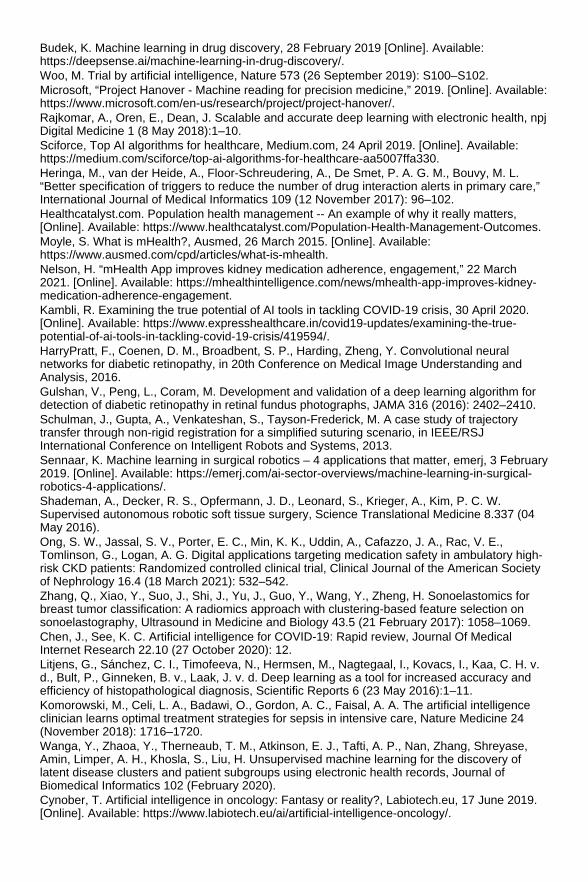
Budek, K. Machine learning in drug discovery, 28 February 2019 [Online]. Available:https://deepsense.ai/machine-learning-in-drug-discovery/. Woo, M. Trial by artificial intelligence, Nature 573 (26 September 2019): S100–S102. Microsoft, “Project Hanover - Machine reading for precision medicine,” 2019. [Online]. Available:https://www.microsoft.com/en-us/research/project/project-hanover/. Rajkomar, A., Oren, E., Dean, J. Scalable and accurate deep learning with electronic health, npjDigital Medicine 1 (8 May 2018):1–10. Sciforce, Top AI algorithms for healthcare, Medium.com, 24 April 2019. [Online]. Available:https://medium.com/sciforce/top-ai-algorithms-for-healthcare-aa5007ffa330. Heringa, M., van der Heide, A., Floor-Schreudering, A., De Smet, P. A. G. M., Bouvy, M. L.“Better specification of triggers to reduce the number of drug interaction alerts in primary care,”International Journal of Medical Informatics 109 (12 November 2017): 96–102. Healthcatalyst.com. Population health management -- An example of why it really matters,[Online]. Available: https://www.healthcatalyst.com/Population-Health-Management-Outcomes. Moyle, S. What is mHealth?, Ausmed, 26 March 2015. [Online]. Available:https://www.ausmed.com/cpd/articles/what-is-mhealth. Nelson, H. “mHealth App improves kidney medication adherence, engagement,” 22 March2021. [Online]. Available: https://mhealthintelligence.com/news/mhealth-app-improves-kidney-medication-adherence-engagement. Kambli, R. Examining the true potential of AI tools in tackling COVID-19 crisis, 30 April 2020.[Online]. Available: https://www.expresshealthcare.in/covid19-updates/examining-the-true-potential-of-ai-tools-in-tackling-covid-19-crisis/419594/. HarryPratt, F., Coenen, D. M., Broadbent, S. P., Harding, Zheng, Y. Convolutional neuralnetworks for diabetic retinopathy, in 20th Conference on Medical Image Understanding andAnalysis, 2016. Gulshan, V., Peng, L., Coram, M. Development and validation of a deep learning algorithm fordetection of diabetic retinopathy in retinal fundus photographs, JAMA 316 (2016): 2402–2410. Schulman, J., Gupta, A., Venkateshan, S., Tayson-Frederick, M. A case study of trajectorytransfer through non-rigid registration for a simplified suturing scenario, in IEEE/RSJInternational Conference on Intelligent Robots and Systems, 2013. Sennaar, K. Machine learning in surgical robotics – 4 applications that matter, emerj, 3 February2019. [Online]. Available: https://emerj.com/ai-sector-overviews/machine-learning-in-surgical-robotics-4-applications/. Shademan, A., Decker, R. S., Opfermann, J. D., Leonard, S., Krieger, A., Kim, P. C. W.Supervised autonomous robotic soft tissue surgery, Science Translational Medicine 8.337 (04May 2016). Ong, S. W., Jassal, S. V., Porter, E. C., Min, K. K., Uddin, A., Cafazzo, J. A., Rac, V. E.,Tomlinson, G., Logan, A. G. Digital applications targeting medication safety in ambulatory high-risk CKD patients: Randomized controlled clinical trial, Clinical Journal of the American Societyof Nephrology 16.4 (18 March 2021): 532–542. Zhang, Q., Xiao, Y., Suo, J., Shi, J., Yu, J., Guo, Y., Wang, Y., Zheng, H. Sonoelastomics forbreast tumor classification: A radiomics approach with clustering-based feature selection onsonoelastography, Ultrasound in Medicine and Biology 43.5 (21 February 2017): 1058–1069. Chen, J., See, K. C. Artificial intelligence for COVID-19: Rapid review, Journal Of MedicalInternet Research 22.10 (27 October 2020): 12. Litjens, G., Sánchez, C. I., Timofeeva, N., Hermsen, M., Nagtegaal, I., Kovacs, I., Kaa, C. H. v.d., Bult, P., Ginneken, B. v., Laak, J. v. d. Deep learning as a tool for increased accuracy andefficiency of histopathological diagnosis, Scientific Reports 6 (23 May 2016):1–11. Komorowski, M., Celi, L. A., Badawi, O., Gordon, A. C., Faisal, A. A. The artificial intelligenceclinician learns optimal treatment strategies for sepsis in intensive care, Nature Medicine 24(November 2018): 1716–1720. Wanga, Y., Zhaoa, Y., Therneaub, T. M., Atkinson, E. J., Tafti, A. P., Nan, Zhang, Shreyase,Amin, Limper, A. H., Khosla, S., Liu, H. Unsupervised machine learning for the discovery oflatent disease clusters and patient subgroups using electronic health records, Journal ofBiomedical Informatics 102 (February 2020). Cynober, T. Artificial intelligence in oncology: Fantasy or reality?, Labiotech.eu, 17 June 2019.[Online]. Available: https://www.labiotech.eu/ai/artificial-intelligence-oncology/.

Smith, J. Swiss multi-cancer diagnostic test approved in Europe, 11 April 2019. [Online].Available: https://www.labiotech.eu/medical/sophia-genetics-cancer-ai/. FDA. FDA permits marketing of artificial intelligence-based device to detect certain diabetes-related eye problems, 11 April 2018. [Online]. Available: https://www.fda.gov/news-events/press-announcements/fda-permits-marketing-artificial-intelligence-based-device-detect-certain-diabetes-related-eye. Vidal-Alaball, J., Fibla, D. R., Zapata, M. A., Marin-Gomez, F. X., Fernandez, O. S. Artificialintelligence for the detection of diabetic retinopathy in primary care: Protocol for algorithmdevelopment, JMIR Research Protocols 8 (1 February 2019):e12539. Metz, C. “How AI steered doctors toward a possible Coronavirus treatment,” 1 May 2020.[Online]. Available: https://economictimes.indiatimes.com/small-biz/startups/newsbuzz/how-ai-steered-doctorstoward-a-possible-coronavirus-treatment/articleshow/75484021.cms. CDC. Sepsis data and reports, CDC, 14 February 2020. [Online]. Available:https://www.cdc.gov/sepsis/datareports/index.html. Beaton, T. Top 10 healthcare mobile apps among hospital, health systems,mhealthintelligence.com, 6 July 2017. [Online]. Available:https://mhealthintelligence.com/news/top-10-healthcare-mobile-apps-among-hospital-health-systems. Niramai. “Introduction,” Niramai, [Online]. Available: https://www.niramai.com/about/overview/.[Accessed 9 July 2021]. He, J., Baxter, S. L., Jie Xu, J. X., Zhou, X., Zhang, K. The practical implementation of artificialintelligence technologies in medicine, Nature Medicine 25 (7 January 2019):30–36. Newsfortomorrow.com. Reducing risk in AI and machine learning-based medical technology,[Online]. Available: https://newsfortomorrow.com/index.php/2019/12/06/reducing-risk-in-ai-and-machine-learning-based-medical-technology/.
A Review on Biomedical Signals with Fundamentals of Digital SignalProcessing Smith, S. W. The scientist and engineer's guide to digital signal processing, 1997.https://doi.org/(http://www.dspguide.com/). Ahirwal M. K., Kumar A., Singh G. K. Biomedical signals, in: Computational Intelligence andBiomedical Signal Processing. SpringerBriefs in Electrical and Computer Engineering. Springer,Cham, 2021. 10.1007/978-3-030-67098-6_1. B.C.O. Textbooks. 19.2 Cardiac muscle and electrical activity, 2018, 1–22.https://openstax.org/books/anatomy-and-physiology/pages/19-2-cardiac-muscle-and-electrical-activity (accessed August 16, 2021). Plawiak, P. ECG signals (744 fragments), Mendeley Data, V3. 3, 2017.https://data.mendeley.com/datasets/63zm778szb/3. Malmivuo, J., Plonsey, R. Bioelectromagnetism: Principles and applications of bioelectric andbiomagnetic bields, Oxford University Press, USA, 1995. Silva, H., Lourenço, A., Canento, F., Fred, A., Raposo, N. ECG biometrics: Principles andapplications, in: BIOSIGNALS 2013 - Proceedings of the International Conference on Bio-Inspired Systems and Signal Processing, SciTePress - Science and and TechnologyPublications, 2013, 215–220. 10.5220/0004243202150220. Ye, C., Coimbra, M. T., Kumar, B. V. K. V. Investigation of human identification using two-leadElectrocardiogram (ECG) signals, in: 2010 Fourth IEEE International Conference on Biometrics:Theory, Applications and Systems (BTAS), IEEE, 2010, 1–8. 10.1109/BTAS.2010.5634478. Drew, B. J., Califf, R. M., Funk, M., Kaufman, E. S., Krucoff, M. W., Laks, M. M., Macfarlane, P.W., Sommargren, C., Swiryn, S., Van Hare, G. F. Practice standards for electrocardiographicmonitoring in hospital settings: An American Heart Association scientific statement from thecouncils on cardiovascular nursing, clinical cardiology, and cardiovascular disease in the young,Circulation 110, 2004, 2721–2746. 10.1161/01.CIR.0000145144.56673.59. Li, M., Xiong, W., Li, Y., Wearable measurement of ECG signals based on smart clothing,International Journal of Telemedicine and Applications 2020..10.1155/2020/6329360.

Navin, O., Kumar, G., Kumar, N., Baderia, K., Kumar, R., Kumar, A. R-peaks detection usingshannon energy for HRV analysis, in: Lecture Notes in Electrical Engineering, 2019, 401–409.10.1007/978-981-13-2553-3_39. Anand, R. S., Kumar, V. Efficient and reliable detection of QRS-segment in ECG signals, in:IEEE/ Engineering in Medicine and Biology Society Annual Conference, 1995.10.1109/rcembs.1995.511734. Ahirwal, M. K., Janghel, R. R., Kose, M. R. Descendant adaptive filter to remove different noisesfrom ECG signals, International Journal of Biomedical Engineering and Technology, 33, 2020,258. 10.1504/ijbet.2020.10029989. Moody, G. B., Mark, R. G. The impact of the MIT-BIH arrhythmia database, IEEE Engineering inMedicine and Biology Magazine 20, 2001, 45–50. 10.1109/51.932724. Moody, G. B., Muldrow, W. K., Mark, R. G. Noise stress test for arrhythmia detectors,Computers in Cardiology 11, 1984, 381–384. Sanei, S., Chambers, J. A. EEG signal processing, John Wiley & Sons, 2013. Shanbao, T., Thankor N. V. Quantitative EEG analysis methods and clinical applications, ArtechHouse, 2009. Collura, T. F., History and evolution of electroencephalographic instruments and techniques,Journal of Clinical Neurophysiology 10, 1993, 476–504. 10.1097/00004691-199310000-00007. Teplan, M. Fundamentals of EEG measurement, Measurement Science Review, 2, 2002, 1–11. Pfurtscheller, G., Lopes Da Silva, F. H. Event-related EEG/MEG synchronization anddesynchronization: Basic principles, Clinical Neurophysiology 110, 1999, 1842–1857.10.1016/S1388-2457(99)00141-8. Odom, J. V., Bach, M., Barber, C., Brigell, M., Marmor, M. F., Tormene,A. P., Holder, G. E.,Vaegan, A. Visual evoked potentials standard, Documenta Ophthalmologica 108, 2004,115–123. 10.1023/B:DOOP.0000036790.67234.22. Kropotov, J. D. Functional neuromarkers for psychiatry: Applications for diagnosis andtreatment, 2016. 10.1016/C2012-0-07144-X. Quian Quiroga R., EEG, ERP and single cell recordings database free EEG data databasefreely ERP data publicly available(ucsd.edu)[https://sccn.ucsd.edu/~arno/fam2data/publicly_available_EEG_data.html] Kim, M. R., Yoon, G. Control signal from EOG analysis and its application, Electronic andCommunication Engineering 7, 2013, 1352–1355. Ahirwal, M. K., Kumar, A., Singh, G. K. Descendent adaptive noise cancellers to improve SNRof contaminated EEG with gradient-based and evolutionary approach, International Journal ofBiomedical Engineering and Technology 13, 2013, 49–68. 10.1504/IJBET.2013.057713. Ichimaru, Y., Moody, G. B. Development of the polysomnographic database on CD-ROM,Psychiatry and Clinical Neurosciences 53, 1999, 175–177. Goldberger A. L., Amaral L. A. N., Glass L., Hausdorff J. M., Ivanov P. C., Mark R. G., Mietus J.E., Moody G. B., Peng C.-K., Eugene Stanley H. PhysioBank, PhysioToolkit, and PhysioNet:Components of a new research resource for complex physiologic signals, Circulation 101, 2000,e215–e220. https://www.ncbi.nlm.nih.gov/pubmed/10851218. Levens, A. J. Muscles alive. Their functions revealed by electromyography, Military Medicine145, 1980, 262–262. 10.1093/milmed/145.4.262a. Zahak, M. Signal acquisition using surface EMG and circuit design considerations for roboticprosthesis, in: Computational Intelligence in Electromyography Analysis - A Perspective onCurrent Applications and Future Challenges, 2012, 13. 10.5772/52556. Chowdhury, R. H., Reaz, M. B. I., Bin Mohd Ali, M. A., Bakar, A. A. A., Chellappan, K., Chang,T. G. Surface electromyography signal processing and classification techniques, Sensors(Switzerland) 13, 2013, 12431–12466. 10.3390/s130912431. Koelstra, S., Muhl, C., Soleymani, M., Jong-Seok Lee, A., Yazdani, T., Ebrahimi, T., Pun, A.,Nijholt, I. P. DEAP: A database for emotion analysis; using physiological signals, IEEETransactions on Affective Computing 3, 2012, 18–31. 10.1109/T-AFFC.2011.15. Godana, B., Leus, G., Barroso, A. Estimating indoor walking velocity profile using a softwareradio-based radar, in: Proceedings - 1st International Conference on Sensor DeviceTechnologies and Applications, SENSORDEVICES 2010, 2010, 44–51.10.1109/SENSORDEVICES.2010.16. Aungsakul, S., Phinyomark, A., Phukpattaranont, P., Limsakul, C. Evaluating feature extractionmethods of electrooculography (EOG) signal for human-computer interface, in: Procedia

Engineering, 2012, 246–252. 10.1016/j.proeng.2012.01.1264. Tibarewala, D. N., Banerjee, A. S., Paul, S. Emsotional eye movement analysis usingelectrooculography signal, International Journal of Biomedical Engineering and Technology 23,2017, 59. 10.1504/ijbet.2017.10003041.
Images in Radiology: Concepts of Image Acquisition and the Nature ofImages Artz, D. S. Computed radiography for the radiological technologist. Seminars in Roentgenology32.1 (1997). Bansal, G. J. Digital radiography. A comparison with modern conventional imaging.Postgraduate Medical Journal 82.969 (2006): 425–428. Cowen, A. R., Kengyelics, S. M., Davies, A. G. Solid-state, flat-panel, digital radiographydetectors and their physical imaging characteristics. Clinical Radiology 63.5 (2008): 487–498. Gold, R. H., Bassett, L. W., Widoff, B. E. Highlights from the history of mammography.Radiographics: A Review Publication of the Radiological Society of North America, Inc 10.6(1990): 1111–1131. Miller, P. D. The history of bone densitometry. Bone 104 (2017): 4–6. Hallikainen, D. History of panoramic radiography. Acta Radiologica 37.3 (1996): 441–445. Campbell, S. A short history of sonography in obstetrics and gynaecology. Facts, Views &Vision in ObGyn 5.3 (2013): 213. Donati, F., et al. Beyond Bernoulli: Improving the accuracy and precision of noninvasiveestimation of peak pressure drops. Circulation: Cardiovascular Imaging 10.1 (2017): e005207. Lell, M. M., et al. Evolution in computed tomography: The battle for speed and dose.Investigative Radiology 50.9 (2015): 629–644. Scalise, P., et al. Computed tomography colonography for the practicing radiologist: A review ofcurrent recommendations on methodology and clinical indications. World Journal of Radiology8.5 (2016): 472. Moraff, A., Jeremy, H., Max, W. Stroke/Cerebral perfusion CT: Technique and clinicalapplications. Multislice CT. Springer, Cham, 2017: 133–143. Edelman, R. R. The history of MR imaging as seen through the pages of radiology. Radiology273.2S (2014): S181–S200. Lohrke, J., et al. 25 years of contrast-enhanced MRI: Developments, current challenges andfuture perspectives. Advances in Therapy 33.1 (2016): 1–28. Hartung, M. P., Grist, T. M., François, C. J. Magnetic resonance angiography: Current statusand future directions. Journal of Cardiovascular Magnetic Resonance 13.1 (2011): 1–11. Paoletti, M., et al. Intra-and extracranial MR venography: technical notes, clinical application,and imaging development. Behavioural Neurology 2016 (2016). Cavallo, A. U., et al. Noncontrast magnetic resonance angiography for the diagnosis ofperipheral vascular disease. Circulation: Cardiovascular Imaging 12.5 (2019): e008844. Romano, A., et al. The role of time-resolved imaging of contrast kinetics (TRICKS) magneticresonance angiography (MRA) in the evaluation of head–neck vascular anomalies: Apreliminary experience. Dentomaxillofacial Radiology 44.3 (2015): 20140302. Jahng, G.-H., et al. Perfusion magnetic resonance imaging: A comprehensive update onprinciples and techniques. Korean Journal of Radiology 15.5 (2014): 554–577. Bradley, W. G. MR spectroscopy of the brain for radiologists. Biomedical Imaging andIntervention Journal 3 (2007): 112–117. Lerner, A., et al. Clinical applications of diffusion tensor imaging. World Neurosurgery 82.1–2(2014): 96–109. Malayeri, A. A., et al. Principles and applications of diffusion-weighted imaging in cancerdetection, staging, and treatment follow-up. Radiographics 31.6 (2011): 1773–1791. Bluemke, D. A., et al. Segmented K-space cine breath-hold cardiovascular MR imaging: Part 1.Principles and technique. American Journal of Roentgenology 169.2 (1997): 395–400. Crummy, A. B., Strother, C. M., Mistretta, C. A., The history of digital subtraction angiography.J Vasc Interv Radiol 1 (2018): 1138–1141.

Fundamentals of Artificial Intelligence and Computational IntelligenceTechniques with Their Applications in Healthcare Systems Dilsizian S. E., Siegel E. L. Artificial intelligence in medicine and cardiac imaging: Harnessingbig data and advanced computing to provide personalized medical diagnosis and treatment.Curr Cardiol Rep 16 (2014): 441 Patel V. L., Shortliffe E. H., Stefanelli M., et al. The coming of age of artificial intelligence inmedicine. Artif Intell Med 46 (2009): 5–17. Pearson T. How to replicate Watson hardware and systems design for your own use in yourbasement. 2011.https://www.ibm.com/developerworks/community/blogs/InsideSystemStorage/entry/ibm_watson_how_to_build_your_own_watson_jr_in_your_basement7?lang=en (accessed 1 Jun 2017). Weingart S. N., Wilson R. M., Gibberd R. W., et al. Epidemiology of medical error. BMJ 320(2000): 774–777. Graber M. L., Franklin N., Gordon R. Diagnostic error in internal medicine. Arch Intern Med 165(2005): 1493–1499. Winters B., Custer J., Galvagno S. M., et al. Diagnostic errors in the intensive care unit: Asystematic review of autopsy studies. BMJ Qual Saf 21 (2012): 894–902. Lee C. S., Nagy P. G., Weaver S. J., et al. Cognitive and system factors contributing todiagnostic errors in radiology. AJR Am J Roentgenol 201 (2013): 611–617. Jha S., Topol E. J. Adapting to Artificial Intelligence: Radiologists and pathologists asinformation specialists. JAMA 316 (2016): 2353–2354. Devi M., et al. A survey of soft computing approaches in biomedical imaging. J Healthc Eng2021 (2021). Yardimci A. Soft computing in medicine. Appl Soft Comput 9.3 (2009): 1029–1043. Dhillon A., Singh A. Machine learning in healthcare data analysis: A survey. J Biol Today'sWorld 8.6 (2019): 1–10. Dagli Y., Choksi S., Sudipta R. Prediction of two year survival among patients of non small celllung cancer. Computer Aided Intervention and Diagnostics in Clinical and Medical Images.Springer, Cham (2019): 169–177. Kayal C. K., Bagchi S., Dhar D., Maitra T., Chatterjee S. Hepatocellular carcinoma survivalprediction using deep neural network. Proceedings of International Ethical Hacking Conference.2019: 349–358. Administration UFaD. Guidance for industry: Electronic source data in clinical investigations.2013. https://www.fda.gov/downloads/drugs/guidances/ucm328691.pdf (accessed 1 Jun 2017). Attia Z. I., et al. An artificial intelligence-enabled ECG algorithm for the identification of patientswith atrial fibrillation during sinus rhythm: A retrospective analysis of outcome prediction. Lancet394.10201 (2019): 861–867. Mincholé A., Rodriguez B. Artificial intelligence for the electrocardiogram. Nat Med 25 (2019):22–23. 10.1038/s41591-018-0306-1. Gang L., Wenyu Y., Ling L., Qilian Y., Xuemin Y. An artificial-intelligence approach to ECGanalysis. IEEE Eng Med Biol Mag 19.2 (March–April 2000): 95–100. 10.1109/51.827412. Subasi A., Ercelebi E. Classification of EEG signals using neural network and logisticregression. Comput Methods Programs Biomed 78.2 (2005): 87–99. Fürbass F., et al. An artificial intelligence-based EEG algorithm for detection of epileptiformEEG discharges: Validation against the diagnostic gold standard. Clin Neurophysiol 131.6(2020): 1174–1179. Castellaro, C., et al. An artificial intelligence approach to classify and analyse EEG traces. ClinNeurophysiol 32.3 (2002): 193–214. Wojtczak P., et al. Hand movement recognition based on biosignal analysis. Eng Appl Artif Intell22.4–5 (2009): 608–615. Clark K., Vendt B., Smith K., Freymann J., Kirby J., Koppel P., Moore S., Phillips S., Maffitt D.,Pringle M., Tarbox L., Prior F. The Cancer Imaging Archive (TCIA): Maintaining and operating apublic information repository. J Digit Imaging 26.6 (2013 Dec): 1045–1057. 10.1007/s10278-013-9622-7. Suckling J., et al. The mammographic image analysis society digital mammogram database. IntCongr 1069 (1994): 375–378.

Kauppi T., Kalesnykiene V., Kamarainen J.-K., Lensu L., Sorri I., Raninen A., Voutilainen R.,Uusitalo H., Kälviäinen H., Pietilä J. DIARETDB1 diabetic retinopathy database and evaluationprotocol. Medical Image Understanding and Analysis 2007 (2007): 61–71. Lonini L., et al. Wearable sensors for Parkinson's disease: Which data are worth collecting fortraining symptom detection models. NPJ Digit Med 1.1 (2018): 1–8. Goodfellow D., et al. Predicting infant motor development status using day long movement datafrom wearable sensors. arXiv preprint arXiv:1807.02617 (2018). Zacharaki E. I., et al. Classification of brain tumor type and grade using MRI texture and shapein a machine learning scheme. Magn Reson Med 62.6 (2009): 1609–1618. Bi W. L., et al. Artificial intelligence in cancer imaging: Clinical challenges and applications. CACancer J Clin 69.2 (2019): 127–157. Esteva A., Kuprel B., Novoa R. A., et al. Dermatologist-level classification of skin cancer withdeep neural networks. Nature 542 (2017): 115–118. Somashekhar S. P., Kumarc R., Rauthan A., et al. Abstract S6-07: Double blinded validationstudy to assess performance of IBM artificial intelligence platform. Cancer Res 77.4 Suppl(2017): S6–S7. Bouton C. E., Shaikhouni A., Annetta N. V., et al. Restoring cortical control of functionalmovement in a human with quadriplegia. Nature 533 (2016): 247–250. Farina D., Vujaklija I., Sartori M., et al. Man/machine interface based on the discharge timings ofspinal motor neurons after targeted muscle reinnervation. Nat Biomed Eng 1 (2017): 0025. Marr B. First FDA approval for clinical cloud-based deep learning in healthcare. 2017.https://www.forbes.com/sites/bernardmarr/2017/01/20/first-fda-approval-for-clinical-cloud-based-deep-learning-inhealthcare/#7a0ed8dc161c (accessed 1 Jun 2017). Zhu X. Computational intelligence techniques and applications. Computational IntelligenceTechniques in Earth and Environmental Sciences. Springer, Dordrecht (2014): 3–26. Khan J., Wei J. S., Ringnér M., et al. Classification and diagnostic prediction of cancers usinggene expression profiling and artificial neural networks. Nat Med 7 (2001): 673–679. Dheeba J., Albert Singh N., Tamil Selvi S. Computer-aided detection of breast Cancer onmammograms: A swarm intelligence optimized wavelet neural network approach. J BiomedInform 49 (2014): 45–52. Hirschauer T. J., Adeli H., Buford J. A. Computer-aided diagnosis of Parkinson's disease usingenhanced probabilistic neural network. J Med Syst 39 (2015): 179. Zhang B., et al. Evolutionary computation and its applications in neural and fuzzy systems. ApplComput Intell Soft Comput 2011 (2011). Pena-Reyes C. A., Sipper M. Evolutionary computation in medicine: An overview. Artif IntellMed 19.1 (2000): 1–23. Holland J. H. Outline for a logical theory of adaptive systems. JACM 9.3 (1962): 297–314. Goldberg D. E., Holland J. H. Genetic algorithms and machine learning, Kluwer AcademicPublishers, (1988). Koza J. R. Genetic programming: A paradigm for genetically breeding populations of computerprograms to solve problems. Vol. 34. Stanford University, Department of Computer Science,Stanford, CA (1990). Koza J. R. Genetic programming: On the programming of computers by means of naturalselection. Vol. 1. MIT Press (1992). Rechenberg I. Cybernetic solution path of an experimental problem. Royal AircraftEstablishment Library Translation 1122 (1965). Vent W. Rechenberg, Ingo,Evolutionsstrategie—OptimierungtechnischerSystemenachPrinzipien der biologischenEvolution. 170 S. mit 36 Abb. FrommannHolzboogVerlag. Stuttgart 1973. Broschiert 62 (1975):337–337. Schwefel H.-P. Kybernetische Evolution alsStrategie der experimentellenForschung in derStromungstechnik. Diploma thesis, Technical University of Berlin (1965). Beyer H.-G., Schwefel H.-P. Evolution strategies–a comprehensive introduction. Nat Comput1.1 (2002): 3–52. Fogel L. J. Autonomous automata. Ind Res 4 (1962): 14–19. Fogel L. J., Owens A. J. MJ Walsh artificial intelligence through simulated evolution. Wiley-IEEEPress (1962).

Phuong N. H., Kreinovich V. Fuzzy logic and its applications in medicine.Int J Med Informat62.2–3 (2001): 165–173. Thakkar M. Custom core ml models using create ml. Beginning Machine Learning in iOS.Apress, Berkeley, CA (2019): 95–138. Carney M., et al. Teachable machine: Approachable Web-based tool for exploring machinelearning classification. Extended abstracts of the 2020 CHI conference on human factors incomputing systems. (2020):1–8, https://doi.org/10.1145/3334480.3382839. Kayyali B., Knott D., Kuiken S. V. The big-data revolution in US health care: accelerating valueand innovation. 2013. http://www.mckinsey.com/industries/healthcare-systems-and-services/our-insights/thebig-data-revolution-in-us-health-care (accessed 1 Jun 2017). Graham J. Artificial intelligence, machine learning, and the FDA. 2016.https://www.forbes.com/sites/theapothecary/2016/08/19/artificial-intelligence-machine-learning-and-the-fda/#4aca26121aa1. (accessed 1 Jun 2017). Alkan A., Günay M. Identification of EMG signals using discriminant analysis and SVMclassifier. Expert Syst Appl 39.1 (2012): 44–47.
Machine Learning Approach with Data Normalization Technique forEarly Stage Detection of Hypothyroidism Peter Z. G., Berardi V. L. An investigation of neural networks in thyroid function diagnosis.Health Care Management Science 1.1 (Sep 1998). Godara S., Singh R. Evaluation of predictive machine learning techniques as expert systems inmedical diagnosis. Indian Journal of Science and Technology 910 (March 2016). Kececi A., Yildirak A., Ozyazici K., Ayluctarhan G., Agbulut O., Zincir I. Implementation ofmachine learning algorithms for gait recognition. Engineering Science and Technology 23(2020): 931–937. Dharavath R., Katheria Y. S. Ensemble method based predictive model for analyzing diseasedatasets: A predictive analysis approach. Health and Technology 9 (2019): 533–545.10.1007/s12553-019-00299-3. Godara S., Singh R. Evaluation of predictive machine learning techniques as expert systems inmedical diagnosis. Indian Journal of Science and Technology 9 (2016): 1–14.10.17485/ijst/2016/v9i10/87212. Milan K., Godara S. Comparative study of data mining classification methods in cardiovasculardisease prediction 1. IJCST 2 (2011): 2229–4333. Roberto J. L., Ridgway B., Sarwate S., Oelze M. Imaging of follicular variant papillary thyroidcarcinoma in a rodent model using spectral-based quantitative ultrasound techniques. In IEEE10th International Symposium on Biomedical Imaging (2013): 732–735. Savelonas A. M., Iakovidis D. K., Legakis I., Maroulis D. Active contours guided by echogenicityand texture for delineation of thyroid nodules in ultrasound images. IEEE Transactions onInformation Technology in Biomedicine 13.4 (July 2009). Ozyilmaz L., Yildirim T. Diagnosis of thyroid disease using artificial neural network methods. InProceedings of ICONIP (2002). Polat K., Seral S., Salih G. A novel hybrid method based on artificial immune recognition system(AIRS) with fuzzy weighted pre-processing for thyroid disease diagnosis. Expert Systems withApplications 32.4 (May 2007). Shukla A., Kaur P., Tiwari R., Janghel R. R. Diagnosis of thyroid disease using artificial neuralnetwork. In Proceedings of IEEE IACC (2009). Varde A. S., Massey K. L., Wood H. C. A clinical laboratory expert system for the diagnosis ofthyroid dysfunction. In Proceeding of Annual International Conference of the IEEE Engineeringin Medicine and Biology Society (1991). Keleş A. ESTDD: Expert system for thyroid diseases diagnosis. Expert Systems withApplications 34.1 (2008). Temurtas F. A comparative study on thyroid disease diagnosis using neural networks. ExpertSystems with Applications 36.1 (2009). Dogantekin E., Dogantekin A., Avci D. An expert system based on generalized discriminantanalysis and wavelet support vector machine for diagnosis of thyroid diseases. Expert Systems

with Applications 38.1 (2011). Stegmayer G., Gerard M., Milone D. H. Data mining over biological datasets: An integratedapproach based on computational intelligence. IEEE Computational Intelligence Magazine 7.4(Nov 2012). Palanichamy J., Nallamuthu R. An expert system for optimizing thyroid disease diagnosis. InProceedings of International Journal of Computational Science and Engineering (2012). Chen H.-L., Yang B., Wang G., Liu J., Chen Y., Liu D. A three-stage expert system based onsupport vector machines for thyroid disease diagnosis. Journal of Medical Systems 36.3 (2012). Young J., Ha E. J., Cho Y. J., Kim H. L., Han M., Kang S. Computer-aided diagnosis of thyroidnodules via ultrasonography: Initial clinical experience. Korean Journal of Radiology 19.4(2018). Chen Shao-Jer, Chang C., Chang K., Tzeng J., Chen Y., Lin C., Hsu W. C., Chang-K. W.Classification of the thyroid nodules based on characteristic sonographic textural feature andcorrelated histopathology using hierarchical support vector machines. Ultrasound in Medicine &Biology 36.12 (2010). Hayashi Y. Synergy effects between grafting and subdivision in Re-RX with J48graft for thediagnosis of thyroid disease. Knowledge-Based Systems 131 (2017): 170–182. Fatemeh S., Mahdi A. Thyroid disease diagnosis based on genetic algorithms using PNN andSVM. In Proceedings of IEEE (2009). Ordonez C. Comparing association rules and decision trees for disease prediction. InProceedings of International Conference of Information Knowledge Management (2006). Ahmad W., Ahmad A., Iqbal A., et al. Intelligent hepatitis diagnosis using adaptive neuro-fuzzyinference system and information gain method. Soft Computing 23 (2018): 1–8. Ahmad W., Ahmad A., Lu C., Khoso B. A., Huang L. A novel hybrid decision support system forthyroid disease forecasting. Soft Computing 22 (2018): 5377–5383. Ma C., Guan J., Zhao W., Wang C. An efficient diagnosis system for thyroid disease based onenhanced kernelized extreme learning machine approach. In International Conference onCognitive Computing, Springer, Cham (2018): 86–101. UCI repository. https://archive.ics.uci.edu/ml/machine-learning-databases/thyroid-disease/. Loukas S. Everything you need to know about Min-Max normalization: A Python tutorial (2020).https://towardsdatascience.com/everything-you-need-to-know-about-min-max-normalization-in-python-b79592732b79. KNN algorithm - Finding nearest neighbors.https://www.tutorialspoint.com/machine_learning_with_python/machine_learning_with_python_knn_algorithm_finding_nearest_neighbors.htm Donges N. A complete guide to the random forest algorithm (2019). https://builtin.com/data-science/random-forest-algorithm. Navlani A. Decision tree classification in Python (2018).https://www.datacamp.com/community/tutorials/decision-tree-classification-python. Kulkarni A., Chong D., Batarseh F. A. 5 - Foundations of data imbalance and solutions for adata democracy. Editor(s): Feras A. Batarseh, Ruixin Yang, Data Democracy. Academic Press(2020): 83–106. 10.1016/B978-0-12-818366-3.00005-8,2020. Fawcett T. An introduction to ROC analysis pattern recognition. Letters 27.8 (2006): 861–874.10.1016/j.patrec.2005.10.010. Czakon J. F1 Score vs ROC AUC vs accuracy vs PR AUC: Which evaluation metric should youchoose? (2021). https://neptune.ai/blog/F1-score-accuracy-roc-auc-pr-auc.
GPU-based Medical Image Segmentation: Brain MRI Analysis Using 3DSlicer Owens, J. D., Houston, M., Luebke, D., Green, S., Stone, J. E., Phillips, J. C. GPU computing.In: Proceedings of the IEEE 96.5 (2008): 879–899. Lee, C., Kim, S.-W., Yoo, C. VADI: GPU virtualization for an automotive plat form. In: IEEETransactions on Industrial Informatics 12.1 (2015): 277–290.

Talukder, S., Sakib, M. I. I., Talukder, Z. R., Das, U., Saha, A., Bayev, N. S. N. Usensewer:Ultrasonic sensor and gsm-arduino based automated sewerage management. In: 2017International Conference on Current Trends in Computer, Electrical, Electronics andCommunication (CTCEEC). IEEE. 2017, pages 12–17. Baji, T. Evolution of the GPU Device widely used in AI and massive parallel processing. In:2018 IEEE 2nd Electron Devices Technology and Manufacturing Conference (EDTM). IEEE.2018, pages 7–9. Bhattacharya, S., Maddikunta, P. K. R., Kaluri, R., Singh, S., Gadekallu, T. R., Alazab, M.,Tariq, U., et al. A novel PCA-firefly based XGBoost classification model for intrusion detection innetworks using GPU. In: Electronics 9.2 (2020): 219. Talukder, S., Sakib, I. I., Hossen, F., Talukder, Z. R., Hossain, S. Attacks and defenses inmobile IP: Modeling with stochastic game petri net. In: 2017 International Conference onCurrent Trends in Computer, Electrical, Electronics and Communication (CTCEEC) (2017):18–23. Talukder, S., Talukder, Z. A survey on malware detection and analysis tools. In: InternationalJournal of Network Security & Its Applications 12.2 (2020). Smistad, E., Falch, T. L., Bozorgi, M., Elster, A. C., Lindseth, F. Medical image segmentation onGPUs–A comprehensive review. In: Medical Image Analysis 20.1 (2015): 1–18. Chen, H., Lu, M., Ma, Z., Zhang, X., Xu, Y., Shen, Q., Zhang, W. Learned resolution scalingpowered gaming-as-a-service at scale. IEEE Transactions on Multimedia 23 (2020): 584–596. Shapiro, L. G., Stockman, G. “Computer vision”, Prentice Hall (2001). Talukder, S., Carbunar, B. AbuSniff: Automatic detection and defenses against abusiveFacebook friends. In: Twelfth International AAAI Conference on Web and Social Media. 2018. NVIDIA AI-Assisted Annotation (AIAA). https://docs.nvidia.com/clara/tltmi/aiaa/index.html.Accessed: 2021-01-06. 3D Slicer. https://www.slicer.org/. Accessed: 2021-01-06. Sharma, N., Aggarwal, L. M. Automated medical image segmentation techniques. In: Journal ofMedical Physics/Association of Medical Physicists of India 35.1 (2010): 3. Taha, A. A., Hanbury, A. Metrics for evaluating 3D medical image segmentation: Analysis,selection, and tool. In: BMC Medical Imaging 15.1 (2015): 1–28. Di Salvo, R., Pino, C. “Image and video processing on CUDA: State of the art and futuredirections”. In: MACMESE 11 (2011): 60–66. Eklund, A., Dufort, P., Forsberg, D., LaConte, S. M. Medical image processing on theGPU–Past, present and future. In: Medical Image Analysis 17.8 (2013): 1073–1094. Shi, L., Liu, W., Zhang, H., Xie, Y., Wang, D. A survey of GPU-based medical image computingtechniques. In: Quantitative Imaging in Medicine and Surgery 2.3 (2012): 188. Bankman, I. N. “Introduction to segmentation”, Handbook of Medical Imaging, 2000. Kafieh, R., Rabbani, H., Kermani, S. A review of algorithms for segmentation of opticalcoherence tomography from retina. In: Journal of Medical Signals and Sensors 3.1 (2013): 45. Kass, M., Witkin, A., Terzopoulos, D. Snakes: Active contour models. In: International Journal ofComputer Vision 1.4 (1988): 321–331. Sherbondy, A., Houston, M., Napel, S. Fast volume segmentation with simultaneousvisualization using programmable graphics hardware. In: IEEE Visualization, 2003 (2003):171–176. Kauffmann, C., Piche, N. Cellular automaton for ultra-fast watershed transform on GPU. In:2008 19th International Conference on Pattern Recognition. IEEE. 2008, pages 1–4. Wagner, B., Müller, P., Haase, G. A parallel watershed-transformation algorithm for the GPU.In: Workshop on Applications of Discrete Geometry and Mathematical Morphology (2010):111–115. Rumpf, M., Strzodka, R. Level set segmentation in graphics hardware. In: ProceedingsInternational Conference on Image Processing, IEEE. 2001, pages 1103–1106. Hong, J. J. Y., Wang, M. D. M. High speed processing of biomedical images usingprogrammable GPU. In: International Conference on Image Processing, 2004, pages2455–2458. Xu, C., Prince, J. L. Snakes, shapes, and gradient vector flow. In: IEEE Transactions on ImageProcessing 7.3 (1998): 359–369.

Preliminary Study of Retinal Lesions Classification on Retinal FundusImages for the Diagnosis of Retinal Diseases Sarah, W., Gojka, R., Anders, G., Richard, S., Hilary, K. Global prevalence of diabetes:Estimates for the year 2000 and projection for 2030, Diabetes Care 27.5 (2004): 1047–1053. WHO: Blindness and vision impairment, Available: https://www.who.int/news-room/fact-sheets/detail/blindness-and-visual-impairment. [Accessed: Jun. 11, 2020]. International Diabetes Federation. “Diabetes complications,” [Online]. Available:https://www.idf.org/aboutdiabetes/what-is-diabetes/complications.html. [Accessed: Jun. 11,2020]. Firstpost: Diabetes is indias fastest growing disease, Available:https://www.firstpost.com/india/diabetes-is-indias-fastest-growing-disease-72-million-cases-recorded-in-2017-figure-expected-to-nearly-double-by-2025–4435203.html. [Accessed: Dec. 3,2017]. Chu, J., Ali, Y. Diabetic retinopathy: A review, Drug Development Research 2.4 (1989):226–237. Salz, D. A., Witkin, A. J. Identifying and monitoring diabetic retinopathy, [Online]. Available:http://retinatoday.com/2016/03/identifying-and-monitoring-diabetic-retinopathy/. [Accessed: Dec.5, 2017]. RxList-Fluorescite side effects center, [Online]. Available: https://www.rxlist.com/fluorescite-side-effects-drug-center.htm. [Accessed: Jan. 3, 2015]. The university of british columbia, Department of Ophthalmology & Visual Sciences, Available:https://ophthalmology.med.ubc.ca/. [Accessed: June. 21, 2020]. Sjølie, A., Stephenson, J., Aldington, S., Kohner, E., Janka, H., Stevens, L., Fuller, J.Retinopathy and vision loss in insulin-dependent diabetes in Europe. The EURODIAB IDDMComplications Study, Ophthalmology 2 (1997): 252–260. Frith, P., Gray, R., MacLennan, S., Ambler, P. The Eye in Clinical Practice, 2nd ed., BlackwellScience Ltd., London (2001). Hollyfield, J. G., Anderson, R. E., LaVail, M. M. (Eds.). Retinal degenerative diseases, Springer,Laser Photocoagulation: Ocular Research and Therapy in Diabetic Retinopathy 572 (2006):195–200. Rokade, P. M., Manza, R. R. Automatic detection of hard exudates in retinal images using Haarwavelet transform, International Journal of Application or Innovation Engineering andManagement 4.5 (2015): 402–410. Zhang, B., Zhang, L., Zhang, L., Karray, F. Retinal vessel extraction by matched filter with first-order derivative of Gaussian, Computers in Biology and Medicine 40.4 (2010): 438–445. Welfer, D., Scharcanski, J., Marinho, D. R. A coarse-to-fine strategy for automatically detectingexudates in color eye fundus images, Computerized Medical Imaging and Graphics 34.3 (2010):228–235. Zhang, X. et al. Exudate detection in color retinal images for mass screening of diabeticretinopathy, Medical Image Analysis 18.7 (2014): 1026–1043. Liu, Q. et al. A location-to-segmentation strategy for automatic exudate segmentation in colourretinal fundus images, Computerized Medical Imaging Graphics 55 (2017): 78–86. Habib et al. Incorporating spatial information for microaneurysm detection in retinalimages,Advances in Science, Technology and Engineering Systems Journal 2.3 (2017):642–649. Usman Akram, M., Khalid, S., Tariq, A., Khan, S. A., Azam, F. Detection and classification ofretinal lesions for grading of diabetic retinopathy, Computers in Biology and Medicine 45.1(2014): 161–171. Figueiredo, I. N., Kumar, S., Oliveira, C. M., Ramos, J. D., Engquist, B. Automated lesiondetectors in retinal fundus images, Computers in Biology and Medicine 66 (2015): 47–65. Santhi, D., Manimegalai, D., Parvathi, S., Karkuzhali, S. Segmentation and classification ofbright lesions to diagnose diabetic retinopathy in retinal images, Biomedical Engineering 61.4(2016): 443–453. Salem, N. M., Nandi, A. K. Segmentation of retinal blood vessels using scale-space featuresand K-nearest neighbour classifier, International Conference on Acoustics, Speech, SignalProcessing (2006): 1001–1004.

Garcia, M., Valverde, C., Lopez, M. I., Poza, J., Hornero, R. Comparison of logistic regressionand neural network classifiers in the detection of hard exudates in retinal images, 35th AnnualInternational Conference of the IEEE Engineering in Medicine and Biology Society (EMBC)(2013): 5891–5894. Mittal, D., Kumar, V., Saxena, S. C., Khandelwal, N., Kalra, N. Neural network based focal liverlesion diagnosis using ultrasound images,Computerized Medical Imaging and Graphics 35.4(2011): 315–323. Kharat, K., Kulkarni, P., Nagori, M. Brain tumor classification using neural network basedmethods, International Journal of Computer Science and Informatics 1.4 (2012): 85–90. Esener, I. I., Ergin, S., Yuksel, T. A new feature ensemble with a multistage classificationscheme for breast cancer diagnosis, Journal of Healthcare Engineering 2017 (2017). Kaur, J., Mittal, D. Construction of benchmark retinal image database for diabetic retinopathyanalysis. Part H: Journal of Engineering in Medicine, SAGE 234.9 (2020).
Automatic Screening of COVID-19 Based on CT Scan Images ThroughExtreme Gradient Boosting World Health Organization. World Health Organization coronavirus update. (2020). [Online].Retrieved May 5, 2020. https://www.who.int/emergencies/diseases/novel-coronavirus-2019. World Meter. Coronavirus cases. https://www.worldometers.info/coronavirus/ [Accessed, June2021]. Xie, X., Zhong, Z., Zhao, W., Zheng, C., Wang, F., Liu, J. Chest CT for typical 2019-nCoVpneumonia: Relationship to negative RT-PCR testing. Radiology 12 (Feb. 2020). “Key Challenges Faced by Covid-19 Tests: Opinion.” Hindustan Times, 5 Oct. 2020,http://www.hindustantimes.com/india-news/key-challenges-faced-by-covid- 19-tests/story-GxvVfnEVFhpWvvcqKYfItM.html, 5 Oct. 2020, Ai, T., Yang, Z., Hou, H., Zhan, C., Chen,C., Lv, W., et al. Correlation of chest CT and RT-PCRtesting in coronavirus disease 2019 (COVID-19) in China: A report of 1014 cases. Radiology 26(Feb. 2020). Fang, Y., Zhang, H., Xie, J., Lin, M., Ying, L., Pang, P., et al. Sensitivity of chest CT for COVID-19: Comparison to RT-PCR. Radiology 19 (Feb. 2020). Rajaraman, S., Siegelman J., Alderson P. O., Folio L. S., Folio L. R., Antani S. K. Iterativelypruned deep learning ensembles for COVID-19 detection in chest X-rays. arXiv preprint arXiv2004.08379 (2020). Han, Zhongyi, Wei, Benzheng, Hong, Yanfei, Li, Tianyang, Cong, Jinyu, Zhu, Xue, Wei,Haifeng, Zhang, Wei. Accurate screening of COVID-19 using attention based deep 3D multipleinstance learning. IEEE Transactions on Medical Imaging 39 (2020):2584–2594. Kanne, J. P. Chest CT findings in 2019 novel coronavirus (2019-nCoV) infections from Wuhan,China: Key points for the radiologist. Radiology (2020): 200241. Murala, S., Wu Q. J. Local ternary co-occurrence patterns: a new feature descriptor for MRI andCT image retrieval. Neurocomputing 119 (2013): 399–412. Gjesteby, L., Yang, Q., Xi, Y., Zhou, Y., Zhang, J., Wang, G. Deep learning methods to guideCT image reconstruction and reduce metal artifacts. Proceedings of SPIE 10132 (2017):101322W. American College of Radiology. Recommendations for Chest Radiography and CT forSuspected COVID19 Infection. https://www.acr.org/Advocacy-and-Economics/ACR-Position-Statements/Recommendations-for-Chest-Radiography-and-CT-for-Suspected-COVID19-Infection, Updated March 22 (2020). Castellano, G., Bonilha, L., Li, L., Cendes, F. Texture analysis of medical images. ClinicalRadiology 59.12 (2004): 1061–1069. Wang, D., Hu, B., Hu, C., Zhu, F., Liu, X., Zhang, J., Wang, B., Xiang, H., Cheng, Z., Xiong, Y.,Zhao, Y., Li, Y., Wang, X., Peng, Z. Clinical characteristics of 138 hospitalized patients with2019 novel Coronavirus-infected pneumonia in Wuhan, China. JAMA 323.11 (2020 Mar 17):1061–1069. DOI: 10.1001/jama.2020.1585. PMID: 32031570; PMCID: PMC7042881. Doi, K. Computer-aided diagnosis in medical imaging: Historical review, current status, andfuture potential. Computerized Medical Imaging and Graphics 31.4–5 (2007): 198–211.
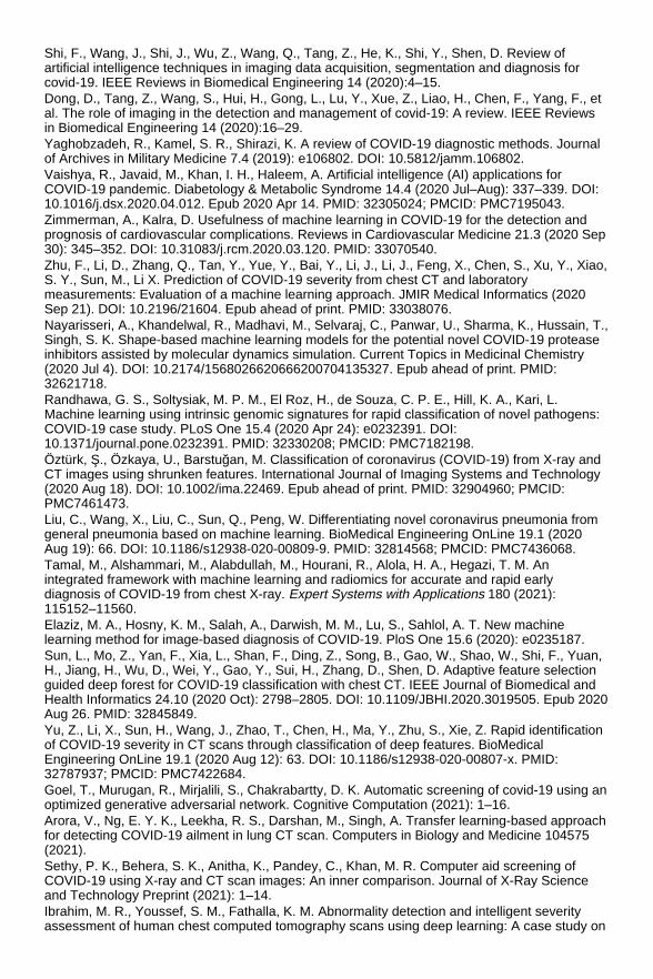
Shi, F., Wang, J., Shi, J., Wu, Z., Wang, Q., Tang, Z., He, K., Shi, Y., Shen, D. Review ofartificial intelligence techniques in imaging data acquisition, segmentation and diagnosis forcovid-19. IEEE Reviews in Biomedical Engineering 14 (2020):4–15. Dong, D., Tang, Z., Wang, S., Hui, H., Gong, L., Lu, Y., Xue, Z., Liao, H., Chen, F., Yang, F., etal. The role of imaging in the detection and management of covid-19: A review. IEEE Reviewsin Biomedical Engineering 14 (2020):16–29. Yaghobzadeh, R., Kamel, S. R., Shirazi, K. A review of COVID-19 diagnostic methods. Journalof Archives in Military Medicine 7.4 (2019): e106802. DOI: 10.5812/jamm.106802. Vaishya, R., Javaid, M., Khan, I. H., Haleem, A. Artificial intelligence (AI) applications forCOVID-19 pandemic. Diabetology & Metabolic Syndrome 14.4 (2020 Jul–Aug): 337–339. DOI:10.1016/j.dsx.2020.04.012. Epub 2020 Apr 14. PMID: 32305024; PMCID: PMC7195043. Zimmerman, A., Kalra, D. Usefulness of machine learning in COVID-19 for the detection andprognosis of cardiovascular complications. Reviews in Cardiovascular Medicine 21.3 (2020 Sep30): 345–352. DOI: 10.31083/j.rcm.2020.03.120. PMID: 33070540. Zhu, F., Li, D., Zhang, Q., Tan, Y., Yue, Y., Bai, Y., Li, J., Li, J., Feng, X., Chen, S., Xu, Y., Xiao,S. Y., Sun, M., Li X. Prediction of COVID-19 severity from chest CT and laboratorymeasurements: Evaluation of a machine learning approach. JMIR Medical Informatics (2020Sep 21). DOI: 10.2196/21604. Epub ahead of print. PMID: 33038076. Nayarisseri, A., Khandelwal, R., Madhavi, M., Selvaraj, C., Panwar, U., Sharma, K., Hussain, T.,Singh, S. K. Shape-based machine learning models for the potential novel COVID-19 proteaseinhibitors assisted by molecular dynamics simulation. Current Topics in Medicinal Chemistry(2020 Jul 4). DOI: 10.2174/1568026620666200704135327. Epub ahead of print. PMID:32621718. Randhawa, G. S., Soltysiak, M. P. M., El Roz, H., de Souza, C. P. E., Hill, K. A., Kari, L.Machine learning using intrinsic genomic signatures for rapid classification of novel pathogens:COVID-19 case study. PLoS One 15.4 (2020 Apr 24): e0232391. DOI:10.1371/journal.pone.0232391. PMID: 32330208; PMCID: PMC7182198. Öztürk, Ş., Özkaya, U., Barstuğan, M. Classification of coronavirus (COVID-19) from X-ray andCT images using shrunken features. International Journal of Imaging Systems and Technology(2020 Aug 18). DOI: 10.1002/ima.22469. Epub ahead of print. PMID: 32904960; PMCID:PMC7461473. Liu, C., Wang, X., Liu, C., Sun, Q., Peng, W. Differentiating novel coronavirus pneumonia fromgeneral pneumonia based on machine learning. BioMedical Engineering OnLine 19.1 (2020Aug 19): 66. DOI: 10.1186/s12938-020-00809-9. PMID: 32814568; PMCID: PMC7436068. Tamal, M., Alshammari, M., Alabdullah, M., Hourani, R., Alola, H. A., Hegazi, T. M. Anintegrated framework with machine learning and radiomics for accurate and rapid earlydiagnosis of COVID-19 from chest X-ray. Expert Systems with Applications 180 (2021):115152–11560. Elaziz, M. A., Hosny, K. M., Salah, A., Darwish, M. M., Lu, S., Sahlol, A. T. New machinelearning method for image-based diagnosis of COVID-19. PloS One 15.6 (2020): e0235187. Sun, L., Mo, Z., Yan, F., Xia, L., Shan, F., Ding, Z., Song, B., Gao, W., Shao, W., Shi, F., Yuan,H., Jiang, H., Wu, D., Wei, Y., Gao, Y., Sui, H., Zhang, D., Shen, D. Adaptive feature selectionguided deep forest for COVID-19 classification with chest CT. IEEE Journal of Biomedical andHealth Informatics 24.10 (2020 Oct): 2798–2805. DOI: 10.1109/JBHI.2020.3019505. Epub 2020Aug 26. PMID: 32845849. Yu, Z., Li, X., Sun, H., Wang, J., Zhao, T., Chen, H., Ma, Y., Zhu, S., Xie, Z. Rapid identificationof COVID-19 severity in CT scans through classification of deep features. BioMedicalEngineering OnLine 19.1 (2020 Aug 12): 63. DOI: 10.1186/s12938-020-00807-x. PMID:32787937; PMCID: PMC7422684. Goel, T., Murugan, R., Mirjalili, S., Chakrabartty, D. K. Automatic screening of covid-19 using anoptimized generative adversarial network. Cognitive Computation (2021): 1–16. Arora, V., Ng, E. Y. K., Leekha, R. S., Darshan, M., Singh, A. Transfer learning-based approachfor detecting COVID-19 ailment in lung CT scan. Computers in Biology and Medicine 104575(2021). Sethy, P. K., Behera, S. K., Anitha, K., Pandey, C., Khan, M. R. Computer aid screening ofCOVID-19 using X-ray and CT scan images: An inner comparison. Journal of X-Ray Scienceand Technology Preprint (2021): 1–14. Ibrahim, M. R., Youssef, S. M., Fathalla, K. M. Abnormality detection and intelligent severityassessment of human chest computed tomography scans using deep learning: A case study on

SARS-COV-2 assessment. Journal of Ambient Intelligence and Humanized Computing (2021):1–24. Dalal, N., Triggs, B. Histograms of oriented gradients for human detection. In 2005 IEEEComputer Society Conference on Computer Vision and Pattern Recognition (CVPR'05).IEEE 1(2005): 886–893. Ojala, T., Pietikainen, M., Maenpaa, T. Multiresolution grayscale and rotation invariant textureclassification with local binary patterns. IEEE Transactions on Pattern Analysis and MachineIntelligence 24.7 (2002 Aug 7): 971–987. Krishnan, M. M., Shah, P., Choudhary, A., Chakraborty, C., Paul, R. R., Ray, A. K. Texturalcharacterization of histopathological images for oral sub-mucous fibrosis detection. Tissue andCell 43.5 (2011 Oct 1): 318–330. Ojala, T., Pietikäinen, M., Harwood, D. A. Comparative study of texture measures withclassification based on featured distributions. Pattern Recognition 29 (1996): 51–59. Alcantarilla, P. F., Bartoli, A., Davison, A. J. KAZE features. In European Conference onComputer Vision.Springer (2012 Oct 7): 214–227. Lowe, D. G. Object recognition from local scale-invariant features. In Proceedings of theInternational Conference on Computer Vision. (1999): 1150–1157.DOI:10.1109/ICCV.1999.790410. Lowe, D. G. Distinctive image features from scale-invariant keypoints. International Journal ofComputer Vision 60.2 (2004): 91–110. Nixon, M., Aguado, A. “Feature extraction and image processing for computer vision.” AcademicPress (2019 Nov 17). Lindeberg, Tony. “Scale Invariant Feature Transform.” Scholarpedia,http://www.scholarpedia.org/article/Scale_Invariant_Feature_Transform [Accessed, June 2021]. Bay, H., Tuytelaars, T., Gool, L. V. Surf: Speeded up robust features. In European Conferenceon Computer Vision 2006.Springer (2006): 404417. Bay, H., Ess, A., Tuytelaars, T., Gool, L. V. Speeded up robust features (SURF). ComputerVision and Image Understanding 110.3 (2008): 346359. Reinhold, J. C., Dewey, B. E., Carass, A., Prince, J. L. Evaluating the impact of intensitynormalization on MR image synthesis. SPIE Conference Proceedings 10949 (2019 Mar):109493H. DOI: 10.1117/12.2513089. PMID: 31551645; PMCID: PMC6758567. Jolliffe, I. T. “Principal component analysis.” 2nd edition, Springer-Verlag New York, New York(2002). Soares, E., Angelov, P., Biaso, S., Higa, F. M., Kanda, A. D. “SARS-CoV-2 CT-scan dataset: Alarge dataset of real patients CT scans for SARS-CoV-2 identification.” medRxiv (2020).10.1101/2020.04.24.20078584. [online]. www.kaggle.com/plameneduardo/sarscov2-ctscan-dataset. Freund, Y., Schapire, R. E. Experiments with a new boosting algorithm. ICML 96 (1996):148–156. Breiman, L. Bagging predictors. Department of Statistics, University of California Berkeley(September 1994). Technical Report No. 421. Retrieved 2019-07-28. Hodges, J. L., Fix, E. Nonparametric discrimination: consistency properties. Report for theUSAF School of Aviation Medicine, Randolph Field (1951). Murphy, K. P. Naive Bayes classifiers. University of British Columbia 18.60 (2006). Ho, T. K. Random Decision Forests. In Proceedings of the 3rd International Conference onDocument Analysis and Recognition, Montreal, QC(1995): 278–282. Cortes, C., Vapnik Vladimir, N. Support-vector networks. Machine Learning 20.3 (1995):273–297. Friedman, J. H. Greedy function approximation: A gradient boosting machine. Annals ofStatistics (2001): 1189–1232. Freund, Y., Schapire, R. A decision-theoretic generalization of on-line learning and anapplication to boosting. (1995). [Online]. https://scikit-learn.org/stable/modules/generated/sklearn.ensemble.AdaBoostClassifier.html. Scikit-learn: Machine Learning in Python, Pedregosa, et al., JMLR 12, pp. 2825-2830, 2011.[online: https://scikit-learn.org/stable/modules/generated/sklearn.ensemble.BaggingClassifier.html] [Access date:April 2021].

Scikit-learn: Machine Learning in Python, Pedregosa, et al., JMLR 12, pp. 2825-2830, 2011.[online: https://scikit-learn.org/stable/modules/generated/sklearn.neighbors.KNeighborsClassifier.html] [Access date:May 2021]. Liu, R., Boyuan, Y., Enrico, Zio, Xuefeng, C. Artificial intelligence for fault diagnosis of rotatingmachinery: A review. Mechanical Systems and Signal Processing 108 (2018): 33–47. Scikit-learn: Machine Learning in Python, Pedregosa, et al., JMLR 12, pp. 2825-2830, 2011.[online: https://scikit-learn.org/stable/modules/naive_bayes.html] [Access date: April 2021]. Breiman, L. Random forests. Machine Learning 45.1 (2001): 5–32. DOI:10.1023/A:1010933404324. Sarica, A., Cerasa, A., Aldo, Q. Random forest algorithm for the classification of neuroimagingdata in Alzheimer's disease: A systematic review. Frontiers in Aging Neuroscience 9 (2017):329. Scikit-learn: Machine Learning in Python, Pedregosa, et al., JMLR 12, pp. 2825-2830, 2011.[online: https://scikit-learn.org/stable/modules/generated/sklearn.ensemble.RandomForestClassifier.html ] [Accessdate: May 2021]. Scikit-learn: Machine Learning in Python, Pedregosa, et al., JMLR 12, pp. 2825-2830, 2011.[online: https://scikit-learn.org/stable/modules/generated/sklearn.svm.SVC.html ] [Access date:April 2021]. Chen, T., Guestrin, C. XGBoost: A scalable tree boosting system. In Proceedings of the 22ndAcmSigkdd International Conference on Knowledge Discovery and Data Mining.(2006):785–794. https://doi.org/10.1145/2939672.2939785.
Investigations on Convolutional Neural Network in Classification of theChest X-Ray Images for COVID-19 and Pneumonia Roosa K., Lee Y., Luo R., Kirpich A., Rothenberg R., Hyman J. M., Yan P., Chowell G., Real-time forecasts of the COVID-19 epidemic in China from February 5th to February 4th, 2020.Infectious Disease Modelling 5 (2020): 256–263. 10.1016/j.idm.2020.02.002. WHO Technical guide, https://www.who.int/emergencies/diseases/novel-coronavirus-2019/technical-guidance/naming-the-coronavirus-disease-(covid-2019)-and-the-virus-that-causes-it. World Health Organization, Coronavirus disease 2019 (COVID-19): Situation report, 92. WorldHealth Organization, 2020. https://apps.who.int/iris/handle/10665/331863. Bedford J., Enria D., Giesecke J., et al., COVID-19: Towards controlling of a pandemic. TheLancet Journals 395(10229) (2020): 1015–1018. 10.1016/S0140-6736(20)30673-5. Cheng Z., Lu Y., Cao Q., Qin L., Pan Z., Yan F., Clinical Yang W., Features and chest CTmanifestations of coronavirus disease, (COVID-19) in a single-centre study in Shanghai, China.American Journal of Roentgenology (2020): 1–6.https://www.ajronline.org/doi/pdf/10.2214/AJR.20.22959. World Health Organization, Pneumonia Vaccine Trial Investigators' Group & World HealthOrganization. Standardization of interpretation of chest radiographs for the diagnosis ofpneumonia in children / World Health Organization Pneumonia Vaccine Trial Investigators'Group. World Health Organization (2001). https://apps.who.int/iris/handle/10665/66956. Parveen N., Sathik M., Detection of pneumonia in chest X-ray images. Journal of X-RayScience and Technology 19(4) (2011): 423–428. 10.3233/XST-2011-0304. Jain R., Nagrath P., Kataria G., Sirish Kaushik V., Jude Hemanth D., Pneumonia detection inchest X-ray images using convolutional neural networks and transfer learning. Measurement165 (2020). 10.1016/j.measurement.2020.108046. Cha S., Lee S., Ko B., Attention-based transfer learning for efficient pneumonia detection inchest X-ray images. Applied Sciences (2021). 10.3390/app11031242. Wang W., Xu Y., Gao R., Lu R., Han K., Wu G., Tan W., Detection of SARS-CoV-2 in differenttypes of clinical specimens. Journal of American Medical Association E1–E2 (2020).10.1001/jama.2020.3786.

Noh J., Yoon S., Kim D., et al., Simultaneous detection of severe acute respiratory syndrome,Middle East respiratory syndrome, and related bat coronaviruses by real-time reversetranscription PC. Archives of Virology 162(6) (2017): 1617–1623. 10.1007/s00705-017-3281-9. Shen M., Zhou Y., Ye J., Maskri A., Kang Y., Zeng S., Recent advances and perspectives ofnucleic acid detection for coronavirus. Journal of Pharmaceutical Analysis (2020).10.1016/j.jpha.2020.02.010. Wan Z., Zhang Y., He K., Liu J., Lan K., Hu Y., Zhang C., A melting curve-based multiplex RT-qPCR assay for simultaneous detection of four human coronaviruses. International Journal ofMolecular Sciences (2016): 2–10. https://doi.org/17.1880.10.3390/ijms17111880. Tahamtan A., Ardebili A., Real-time RT-PCR in COVID-19 detection: issues affecting theresults. Journal Expert Review of Molecular Diagnostics, (2020).10.1080/14737159.2020.1757437 Wang Y., Kang H., Liu X., et al., Combination of RT-qPCR testing and clinical features fordiagnosis of COVID-19 facilitates management of SARS-CoV-2 Outbreak. Journal of MedicalVirology (2020): 538–539. 10.1002/jmv.25721. Mahase E., Coronavirus: covid-19 has killed more people than SARS and MERS combined,despite lower case fatality rate. The BMJ 368 (2020). 10.1136/bmj.m641. Ng M., Lee E., Yang J., Yang F., Li X., Wang H., Lui M., Lo C., Leung B., Siu T., Khong P., HuiC., Yuen K., Kuo M., Imaging profile of the COVID-19 infection: Radiologic findings andliterature review. Radiology: Cardiothoracic Imaging 2 (2020): e200034.10.1148/ryct.2020200034. Tao A., Yang Z., Correlation of chest CT and RT-PCR testing in coronavirus disease 2019(COVID-19) in china: A report of 1014 cases. Radiology (2020). 10.1148/radiol.2020200642. Gozes, O., Frid-Adar, M., Greenspan, H., Browning, P., Zhang, H., Ji, W., Bernheim, A., RapidAI development cycle for the Coronavirus (COVID-19) pandemic: Initial results for automateddetection & patient monitoring using deep learning CT image analysis. arXiv preprintarXiv:2003.05037 (2020). Wang S., Kang B., Ma J., Zeng X., Xiao M., Guo J., Cai M., Yang J., Li Y., Meng X., Xu B., Adeep learning algorithm using CT images to screen for Corona Virus Disease (COVID-19).European Radiology 31(8) (2021): 6096–6104. 10.1007/s00330-021-07715-1. Shan F., Gao Y., Wang J., Shi W., Shi N., Han M., Xue Z., Shi Y., Abnormal lung quantificationin chest CT images of COVID-19 patients with deep learning and its application to severityprediction. Medical Physics 48(4) (2021): 1633–1645. 10.1002/mp.14609. Hamimi A., MERS-CoV: Middle East respiratory syndrome corona virus: Can radiology be ofhelp? Initial single center experience. The Egyptian Journal of Radiology and Nuclear Medicine(2015). 10.1016/j.ejrnm.2015.11.004. Xie X., Li X., Wan S., Gong Y., Mining X-ray images of SARS patients. Data Mining: Theory,Methodology, Techniques, and Applications (2006): 282–294.https://link.springer.com/chapter/10.1007/11677437_22. Rubin G., Ryerson C., Haramati L., et al., The role of chest imaging in patient managementduring the covid-19 pandemic: A multinational consensus statement from the Fleischner society.Radiology 296(1) (2020): 172–180. 10.1148/radiol.2020201365. Ali N., Ceren K., Ziynet P., Automatic detection of Coronavirus Disease (COVID-19) using X-rayimages and deep convolutional neural networks. Pattern Analysis and Applications 24 (2020):1207–1220. 10.1007/s10044-021-00984-y. Linda W., Zhong Q. L., Alexander W., COVID-Net: A tailored deep convolutional neural networkdesign for detection of COVID-19 cases from chest X-ray images. Scientific Reports 10(1)(2020): 19549.10.1038/s41598-020-76550-z. Tawfiq L., Hu Y. H., Hwang J., Handbook of neural network signal processing, CRC Press,Boca Raton (2002). 10.1121/1.1480419. Radiological Society of North America, Pneumonia detection challenge. (2019).https://www.kaggle.com/c/rsnaPneumonia-detection-challenge/data. Joseph P. C., Paul M., Lan D., COVID-19 image data collection. 2020.arXiv 2003.11597 (2020).https://github.com/ieee8023/covid-chestxray-dataset. Huan L., Yibo Y., Dongmin C., Zhouchen L., Optimization algorithm inspired deep neuralnetwork structure design. In Proceedings of the 10th Asian Conference on Machine LearningResearch, PMLR95 (2018): 614–629.

Ning Q., On the momentum term in gradient descent learning algorithms, neural networks. TheOfficial Journal of the International Neural Network Society 12(1) (1999):145–151.10.1016/S0893-6080(98)00116-6. Duchi J., Hazan E., Singer Y., Adaptive subgradient methods for online learning and stochasticoptimization. Journal of Machine Learning Research 12 (2011): 2121–2159. Kingma D., Ba Jimmy A., A method for stochastic optimization. International Conference onLearning Representations (2014). https://arxiv.org/pdf/1412.6980.pdf. Dogo E., Afolabi, O., Nwulu N., Twala, B., Aigbavboa C., A comparative analysis of gradientdescent-based optimization algorithms on convolutional neural networks (2018): 92–99.10.1109/CTEMS.2018.8769211. MatLab Help: [https://in.mathworks.com/help/deeplearning/ug/setting-up-parameters-and-training-of-a-convnet.html] (Accessed: Jan 2021).
Improving the Detection of Abdominal and Mediastinal Lymph Nodes inCT Images Using Attention U-Net Based Deep Learning Model Roth H. R., Lu L., Seff A., Cherry K. M., Hoffman J., Wang S., et al. A new 2.5 D representationfor lymph node detection using random sets of deep convolutional neural network observations.Int. Conf. Med. image Comput. Comput. Interv. (2014): 520–527. Seff A., Lu L., Cherry K. M., Roth H. R., Liu J., Wang S., et al. 2D view aggregation for lymphnode detection using a shallow hierarchy of linear classifiers. Int. Conf. Med. Image Comput.Comput. Interv. (2014): 544–552. Kitasaka T., Tsujimura Y., Nakamura Y., Mori K., Suenaga Y., Ito M., et al. Automatedextraction of lymph nodes from 3-D abdominal CT images using 3-D minimum directionaldifference filter. Int. Conf. Med. Image Comput. Comput. Interv. (2007): 336–343. Oda M., Kitasaka T., Fujiwara M., Misawa K., Mori K. Method for detecting enlarged lymphnodes from 3d abdominal CT images with a multi-shape and multi-scale ellipsoidal structuredetection filter. Int. MICCAI Work Comput. Clin. Challenges Abdom. Imaging. (2012): 238–245. Gomolka R., Klonowski W., Korzynska A., Siemion K. Automatic segmentation of morphologicalstructures in microscopic images of lymphatic nodes-preliminary results. Int. J. Med. Physiol. 2(2017). Feulner J., Zhou S. K., Hammon M., Hornegger J., Comaniciu D. Lymph node detection andsegmentation in chest CT data using discriminative learning and a spatial prior. Med. ImageAnal. 17 (2013): 254–270. Nakamura Y., Nimura Y., Kitasaka T., Mizuno S., Furukawa K., Goto H., et al. Automaticabdominal lymph node detection method based on local intensity structure analysis from 3D x-ray CT images. Med. Imaging 2013 Comput. Diagnosis 8670 (2013): 86701K. Cherry K. M., Wang S., Turkbey E. B., Summers R. M. Abdominal lymphadenopathy detectionusing random forest. . Med. Imaging 2014 Comput. Diagnosis 9035 (2014): 90351G. Seff A., Lu L., Barbu A., Roth H., Shin H.-C., Summers R. M. Leveraging mid-level semanticboundary cues for automated lymph node detection. Int. Conf. Med. Image Comput. Comput.Interv. (2015): 53–61. Beichel R. R., Wang Y. Computer-aided lymph node segmentation in volumetric CT data. Med.Phys. 39 (2012): 5419–5428. Tan Y., Lu L., Bonde A., Wang D., Qi J., Schwartz L. H., et al. Lymph node segmentation bydynamic programming and active contours. Med. Phys. 45 (2018): 2054–2062.10.1002/mp.12844. Barbu A., Suehling M., Xu X., Liu D., Zhou S. K., Comaniciu D. Automatic detection andsegmentation of lymph nodes from CT data. IEEE Trans. Med. Imaging 31 (2011): 240–250. Nakamura Y., Nimura Y., Kitasaka T., Mizuno S., Furukawa K., Goto H., et al. Investigation ofoptimal feature value set in false positive reduction process for automated abdominal lymphnode detection method. Med. Imaging 2015 Comput. Diagnosis 9414 (2015): 94143N. Nimura Y., Hayashi Y., Kitasaka T., Furukawa K., Misawa K., Mori K. Automated abdominallymph node segmentation based on RST analysis and SVM. In: Aylward S., Hadjiiski L. M.,editors. Prog. Biomed. Opt. Imaging - Proc. SPIE, 9035 (2014): 90352U. 10.1117/12.2043349.

Nogues I., Lu L., Wang X., Roth H., Bertasius G., Lay N., et al. Automatic lymph node clustersegmentation using holistically-nested neural networks and structured optimization in CTimages. Int. Conf. Med. Image Comput. Comput. Interv. (2016): 388–397. Roth H. R., Lu L., Liu J., Yao J., Seff A., Cherry K., et al. Improving computer-aided detectionusing convolutional neural networks and random view aggregation. IEEE Trans. Med. Imaging35 (2015): 1170–1181. Sengupta D. Deep learning architectures for automated image segmentation. University ofCalifornia (2019). Shin H.-C., Roth H. R., Gao M., Lu L., Xu Z., Nogues I., et al. Deep convolutional neuralnetworks for computer-aided detection: CNN architectures, dataset characteristics and transferlearning. IEEE Trans. Med. Imaging 35 (2016): 1285–1298. Deng J., Dong W., Socher R., Li L.-J., Li K., Fei-Fei L. Imagenet: A large-scale hierarchicalimage database. 2009 IEEE Conf. Comput. Vis. Pattern Recognit. (2009): 248–255. Li Z., Xia Y. Deep reinforcement learning for weakly-supervised lymph node segmentation in CTimages. IEEE J. Biomed. Heal. Informatics 1 (2020). 10.1109/JBHI.2020.3008759. Nishino M., Jagannathan J. P., Ramaiya N. H., den Abbeele A. D. Revised RECIST guidelineversion 1.1: What oncologists want to know and what radiologists need to know. Am. J.Roentgenol. 195 (2010): 281–289. Cai J., Tang Y., Lu L., Harrison A. P., Yan K., Xiao J., et al. Accurate weakly-supervised deeplesion segmentation using large-scale clinical annotations: Slice-propagated 3d maskgeneration from 2d recist. Int. Conf. Med. Image Comput. Comput. Interv. (2018): 396–404. Hoogi A., Lambert J. W., Zheng Y., Comaniciu D., Rubin D. L. A fully-automated pipeline fordetection and segmentation of liver lesions and pathological lymph nodes. ArXiv PreprArXiv170306418 (2017). Feuerstein M., Deguchi D., Kitasaka T., Iwano S., Imaizumi K., Hasegawa Y., et al. Automaticmediastinal lymph node detection in chest CT. Med. Imaging 2009 Comput. Diagnosis 7260(2009): 72600V. Oda H., Bhatia K. K., Oda M., Kitasaka T., Iwano S., Homma H., et al. Automated mediastinallymph node detection from CT volumes based on intensity targeted radial structure tensoranalysis. J. Med. Imaging 4 (2017): 44502. Liu J., Hoffman J., Zhao J., Yao J., Lu L., Kim L., et al. Mediastinal lymph node detection andstation mapping on chest CT using spatial priors and random forest. Med. Phys. 43 (2016):4362–4374. Zhao W., Shi F. A method for lymph node segmentation with scaling features in a random forestmodel. Curr. Proteomics 15 (2018): 128–134. Allen D., Lu L., Yao J., Liu J., Turkbey E., Summers R. M. Robust automated lymph nodesegmentation with random forests. Med. Imaging 2014 Image Process. 9034 (2014): 90343X. Oda H., Bhatia K. K., Roth H. R., Oda M., Kitasaka T., Iwano S., et al. Dense volumetricdetection and segmentation of mediastinal lymph nodes in chest CT images. In: Mori K., PetrickN., editors. Med. Imaging 2018 Comput. Diagnosis, SPIE 1 (2018). 10.1117/12.2287066. Ronneberger O., Fischer P., Brox T. U-net: Convolutional networks for biomedical imagesegmentation. Int. Conf. Med. Image Comput. Comput. Interv. (2015): 234–241. Bouget D., Jørgensen A., Kiss G., Leira H. O., Langø T. Semantic segmentation and detectionof mediastinal lymph nodes and anatomical structures in CT data for lung cancer staging. Int. J.Comput. Assist. Radiol. Surg. (2019): 1–10. Tang Y., Oh S., Xiao J., Summers R., Tang Y. CT-realistic data augmentation using generativeadversarial network for robust lymph node segmentation. Med. Imaging 2019 Comput.Diagnosis, San Diego, California, United States 139 (2019). 10.1117/12.2512004. Long J., Shelhamer E., Darrell T. Fully convolutional networks for semantic segmentation. Proc.IEEE Conf. Comput. Vis. Pattern Recognit. (2015): 3431–3440. Bahdanau D., Cho K., Bengio Y. Neural machine translation by jointly learning to align andtranslate. ArXiv Prepr ArXiv14090473 (2014). Sinha A., Dolz J. Multi-scale self-guided attention for medical image segmentation. IEEE. J.Biomed. Heal. Informatics (2020). Wang Y., Deng Z., Hu X., Zhu L., Yang X., Xu X., et al. Deep attentional features for prostatesegmentation in ultrasound. Int. Conf. Med. Image Comput. Comput. Interv. (2018): 523–530. Li C., Tong Q., Liao X., Si W., Sun Y., Wang Q., et al. Attention based hierarchical aggregationnetwork for 3D left atrial segmentation. Int. Work. Stat. Atlases Comput. Model. Hear. (2018):
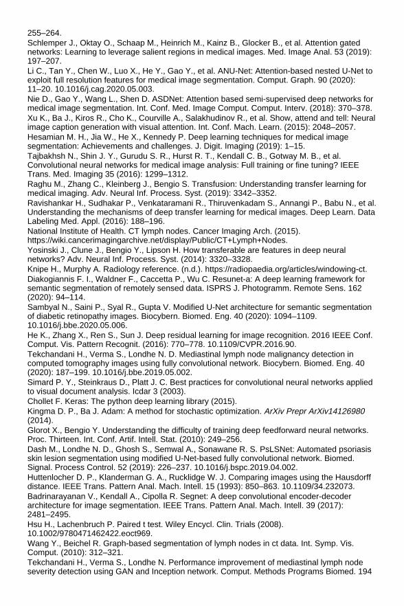
255–264. Schlemper J., Oktay O., Schaap M., Heinrich M., Kainz B., Glocker B., et al. Attention gatednetworks: Learning to leverage salient regions in medical images. Med. Image Anal. 53 (2019):197–207. Li C., Tan Y., Chen W., Luo X., He Y., Gao Y., et al. ANU-Net: Attention-based nested U-Net toexploit full resolution features for medical image segmentation. Comput. Graph. 90 (2020):11–20. 10.1016/j.cag.2020.05.003. Nie D., Gao Y., Wang L., Shen D. ASDNet: Attention based semi-supervised deep networks formedical image segmentation. Int. Conf. Med. Image Comput. Comput. Interv. (2018): 370–378. Xu K., Ba J., Kiros R., Cho K., Courville A., Salakhudinov R., et al. Show, attend and tell: Neuralimage caption generation with visual attention. Int. Conf. Mach. Learn. (2015): 2048–2057. Hesamian M. H., Jia W., He X., Kennedy P. Deep learning techniques for medical imagesegmentation: Achievements and challenges. J. Digit. Imaging (2019): 1–15. Tajbakhsh N., Shin J. Y., Gurudu S. R., Hurst R. T., Kendall C. B., Gotway M. B., et al.Convolutional neural networks for medical image analysis: Full training or fine tuning? IEEETrans. Med. Imaging 35 (2016): 1299–1312. Raghu M., Zhang C., Kleinberg J., Bengio S. Transfusion: Understanding transfer learning formedical imaging. Adv. Neural Inf. Process. Syst. (2019): 3342–3352. Ravishankar H., Sudhakar P., Venkataramani R., Thiruvenkadam S., Annangi P., Babu N., et al.Understanding the mechanisms of deep transfer learning for medical images. Deep Learn. DataLabeling Med. Appl. (2016): 188–196. National Institute of Health. CT lymph nodes. Cancer Imaging Arch. (2015).https://wiki.cancerimagingarchive.net/display/Public/CT+Lymph+Nodes. Yosinski J., Clune J., Bengio Y., Lipson H. How transferable are features in deep neuralnetworks? Adv. Neural Inf. Process. Syst. (2014): 3320–3328. Knipe H., Murphy A. Radiology reference. (n.d.). https://radiopaedia.org/articles/windowing-ct. Diakogiannis F. I., Waldner F., Caccetta P., Wu C. Resunet-a: A deep learning framework forsemantic segmentation of remotely sensed data. ISPRS J. Photogramm. Remote Sens. 162(2020): 94–114. Sambyal N., Saini P., Syal R., Gupta V. Modified U-Net architecture for semantic segmentationof diabetic retinopathy images. Biocybern. Biomed. Eng. 40 (2020): 1094–1109.10.1016/j.bbe.2020.05.006. He K., Zhang X., Ren S., Sun J. Deep residual learning for image recognition. 2016 IEEE Conf.Comput. Vis. Pattern Recognit. (2016): 770–778. 10.1109/CVPR.2016.90. Tekchandani H., Verma S., Londhe N. D. Mediastinal lymph node malignancy detection incomputed tomography images using fully convolutional network. Biocybern. Biomed. Eng. 40(2020): 187–199. 10.1016/j.bbe.2019.05.002. Simard P. Y., Steinkraus D., Platt J. C. Best practices for convolutional neural networks appliedto visual document analysis. Icdar 3 (2003). Chollet F. Keras: The python deep learning library (2015). Kingma D. P., Ba J. Adam: A method for stochastic optimization. ArXiv Prepr ArXiv14126980(2014). Glorot X., Bengio Y. Understanding the difficulty of training deep feedforward neural networks.Proc. Thirteen. Int. Conf. Artif. Intell. Stat. (2010): 249–256. Dash M., Londhe N. D., Ghosh S., Semwal A., Sonawane R. S. PsLSNet: Automated psoriasisskin lesion segmentation using modified U-Net-based fully convolutional network. Biomed.Signal. Process Control. 52 (2019): 226–237. 10.1016/j.bspc.2019.04.002. Huttenlocher D. P., Klanderman G. A., Rucklidge W. J. Comparing images using the Hausdorffdistance. IEEE Trans. Pattern Anal. Mach. Intell. 15 (1993): 850–863. 10.1109/34.232073. Badrinarayanan V., Kendall A., Cipolla R. Segnet: A deep convolutional encoder-decoderarchitecture for image segmentation. IEEE Trans. Pattern Anal. Mach. Intell. 39 (2017):2481–2495. Hsu H., Lachenbruch P. Paired t test. Wiley Encycl. Clin. Trials (2008).10.1002/9780471462422.eoct969. Wang Y., Beichel R. Graph-based segmentation of lymph nodes in ct data. Int. Symp. Vis.Comput. (2010): 312–321. Tekchandani H., Verma S., Londhe N. Performance improvement of mediastinal lymph nodeseverity detection using GAN and Inception network. Comput. Methods Programs Biomed. 194
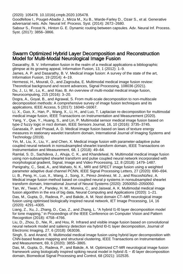
(2020): 105478. 10.1016/j.cmpb.2020.105478. Goodfellow I., Pouget-Abadie J., Mirza M., Xu B., Warde-Farley D., Ozair S., et al. Generativeadversarial nets. Adv. Neural Inf. Process. Syst. (2014): 2672–2680. Sabour S., Frosst N., Hinton G. E. Dynamic routing between capsules. Adv. Neural Inf. Process.Syst. (2017): 3856–3866.
Swarm Optimized Hybrid Layer Decomposition and ReconstructionModel for Multi-Modal Neurological Image Fusion Dasarathy, B. V. Information fusion in the realm of a medical applications-a bibliographicglimpse at its growing appeal, Information Fusion, 13, 1 (2012): 1–9. James, A. P. and Dasarathy, B. V. Medical image fusion: A survey of the state of the art,Information Fusion, 19 (2014): 4–19. Hermessi, H., Mourali, O., and Zagrouba, E. Multimodal medical image fusion review:Theoretical background and recent advances, Signal Processing, 108036 (2021). Du, J., Li, W., Lu, K., and Xiao, B. An overview of multi-modal medical image fusion,Neurocomputing, 215 (2016): 3–20. Dogra, A., Goyal, B., and Agrawal, S. From multi-scale decomposition to non-multiscaledecomposition methods: A comprehensive survey of image fusion techniques and itsapplications, IEEE Access, 5 (2017): 16040–16067. Li, X., Guo, X., Han, P., Wang, X., Li, H., and Luo, T. Laplacian re-decomposition for multimodalmedical image fusion, IEEE Transactions on Instrumentation and Measurement (2020). Yang, Y., Que, Y., Huang, S., and Lin, P. Multimodal sensor medical image fusion based ontype-2 fuzzy logic in nsct domain, IEEE Sensors Journal, 16, 10 (2016): 3735–3745. Ganasala, P. and Prasad, A. D. Medical image fusion based on laws of texture energymeasures in stationary wavelet transform domain, International Journal of Imaging Systems andTechnology (2019). Yin, M., Liu, X., Liu, Y., and Chen, X. Medical image fusion with parameter-adaptive pulsecoupled neural network in nonsubsampled shearlet transform domain, IEEE Transactions onInstrumentation and Measurement, 68, 1 (2018): 49–64. Ramlal, S. D., Sachdeva, J., Ahuja, C. K., and Khandelwal, N. Multimodal medical image fusionusing non-subsampled shearlet transform and pulse coupled neural network incorporated withmorphological gradient, Signal, Image and Video Processing, 12, 8 (2018): 1479–1487. Panigrahy, C., Seal, A., and Mahato, N. K., MRI and SPECT image fusion using a weightedparameter adaptive dual channel PCNN, IEEE Signal Processing Letters, 27 (2020): 690–694. Li, B., Peng, H., Luo, X., Wang, J., Song, X., Pérez-Jiménez, M. J., and RiscosNúñez, A.Medical image fusion method based on coupled neural p systems in nonsubsampled shearlettransform domain, International Journal of Neural Systems (2020): 2050050–2050050. Tan, W., Tiwari, P., Pandey, H. M., Moreira, C., and Jaiswal, A. K. Multimodal medical imagefusion algorithm in the era of big data, Neural Computing and Applications (2020): 1–21. Das, M., Gupta, D., Radeva, P., and Bakde, A. M. Nsst domain CT–MR neurological imagefusion using optimised biologically inspired neural network, IET Image Processing, 14, 16(2020): 4291–4305. Liang, Z., Xu, J., Zhang, D., Cao, Z., and Zhang, L. “A hybrid l1-l0 layer decomposition modelfor tone mapping,” in Proceedings of the IEEE Conference on Computer Vision and PatternRecognition (2018): 4758–4766. Liu, D., Zhou, D., Nie, R., and Hou, R. Infrared and visible image fusion based on convolutionalneural network model and saliency detection via hybrid l0-l1 layer decomposition, Journal ofElectronic Imaging, 27, 6 (2018): 063036. Singh, S. and Anand, R. Multimodal medical image fusion using hybrid layer decomposition withCNN-based feature mapping and structural clustering, IEEE Transactions on Instrumentationand Measurement, 69, 6 (2020): 3855–3865. Das, M., Gupta, D., Radeva, P., and Bakde, A. M. Optimized CT-MR neurological image fusionframework using biologically inspired spiking neural model in hybrid ℓ1 − ℓ0 layer decompositiondomain, Biomedical Signal Processing and Control, 68 (2021): 102535.

Mirjalili, S. and Lewis, A. The whale optimization algorithm, Advances in Engineering Software,95 (2016): 51–67. Xydeas, C. and Petrovic, V. Objective image fusion performance measure, Electronics Letters,36, 4 (2000): 308–309. Qu, G., Zhang, D., and Yan, P. Information measure for performance of image fusion,Electronics Letters, 38, 7 (2002): 313–315. Gupta, D. Nonsubsampled shearlet domain fusion techniques for CT–MR neurological imagesusing improved biological inspired neural model, Biocybernetics and Biomedical Engineering,38, 2 (2018): 262–274. Cvejic, N., Loza, A., Bull, D., and Canagarajah, N. A similarity metric for assessment of imagefusion algorithms, International Journal of Signal Processing, 2, 3 (2005): 178–182. Yang, C., Zhang, J.-Q., Wang, X.-R., and Liu, X. A novel similarity based quality metric forimage fusion, Information Fusion, 9, 2 (2008): 156–160. Sengupta, A., Seal, A., Panigrahy, C., Krejcar, O., and Yazidi, A. Edge information based imagefusion metrics using fractional order differentiation and sigmoidal functions, IEEE Access, 8(2020): 88385–88398.
Hybrid Seeker Optimization Algorithm-based Accurate Image Clusteringfor Automatic Psoriasis Lesion Detection Huerta, C., Rivero, E., Rodriguez, L. A. G. Incidence and risk factors for psoriasis in the generalpopulation. Arch Dermatol. 143(12) (2007): 1559–1565. Sander, H. M., Morris, L. F., Phillips, C. M., Harrison, P. E., Menter, A. The annual cost ofpsoriasis. J Am Acad Dermatol. 28(3) (1993): 422–425. Adam, B. A. Psoriasis in hospital population. Med J Malaysia. 34(1980): 370–374. Yee, K. C., Choon, S. E., Khaw, G. E., Baba, R., Hussein, S. H., Ratti, S. K. Malaysian patients’knowledge of psoriasis: Psoriasis Association Members Vs non-members. Persat DermatologiMalaysia. (1999): 840–848. Shrivastava, V. K., Londhe, N. D., Sonawane, R. S., Suri, J. S. First review on psoriasis severityrisk stratification: An engineering perspective. Comput Biol Med. 63(2015): 52–63. Roenigk, H. H., Maibach, H. I. Psoriasis. Marcel Dekker Inc., New York (1985). Camisa, C. Handbook of psoriasis. Wiley Online Library, Massachusetts, USA (2008). Henseler, T. Genetics of psoriasis. Arch Dermatol Res. 290(9) (1998): 463–476. Taur, J. S., Lee, G.-H., Tao, C.-W., Chen, C.-C., Yang, C.-W. Segmentation of psoriasisvulgaris images using multiresolution-based orthogonal subspace techniques. IEEE Trans SystMan, Cybern Part B. 36(2) (2006): 390–402. Feldman, S. R., Krueger, G. G. Psoriasis assessment tools in clinical trials. Ann Rheum Dis.64(suppl 2) (2005): ii65–ii68. Xie, F., Bovik, A. C. Automatic segmentation of dermoscopy images using self-generatingneural networks seeded by genetic algorithm. Pattern Recognit. 46(3) (2013): 1012–1019. Shrivastava, V. K., Londhe, N. D., Sonawane, R. S., Suri, J. S. Reliable and accurate psoriasisdisease classification in dermatology images using comprehensive feature space in machinelearning paradigm. Expert Syst Appl. 42(15–16) (2015): 6184–6195. Shrivastava, V. K., Londhe, N. D., Sonawane, R. S., Suri, J. S. Exploring the color featurepower for psoriasis risk stratification and classification: A data mining paradigm. Comput BiolMed. 65(2015): 54–68. Shrivastava, V. K., Londhe, N. D., Sonawane, R. S., Suri, J. S. Computer-aided diagnosis ofpsoriasis skin images with HOS, texture and color features: A first comparative study of its kind.Comput Methods Programs Biomed. 126(2016): 98–109. Shrivastava, V. K., Londhe, N. D., Sonawane, R. S., Suri, J. S. A novel approach to multiclasspsoriasis disease risk stratification: Machine learning paradigm. Biomed Signal Process Control.28(2016): 27–40. Shrivastava, V. K., Londhe, N. D., Sonawane, R. S., Suri, J. S. Reliability analysis of psoriasisdecision support system in principal component analysis framework. Data Knowl Eng.106(2016): 1–17.

Nidhal, K. A., Nizar, S. D., Muhsin, A. A., Hind, R. Psoriasis detection using color and texturefeatures. J Comput Sci. 6(6) (2010): 648–652. Taur, J.-S. Neuro-fuzzy approach to the segmentation of psoriasis images. J VLSI SignalProcess Syst Signal, Image Video Technol. 35(1) (2003): 19–27. Bogo, F., Samory, M., Fortina, A. B., Piaserico, S., Peserico, E. Psoriasis segmentation throughchromatic regions and geometric active contours. In: 2012 Annual International Conference ofthe IEEE Engineering in Medicine and Biology Society (2012): 5388–5391. Juang, L.-H., Wu, M.-N. Psoriasis image identification using k-means clustering withmorphological processing. Measurement. 44(5) (2011): 895–905. Shrivastava, V. K., Londhe, N. D. Measurement of psoriasis area and severity index area scoreof Indian psoriasis patients. J Med Imaging Heal Informatics. 5(4) (2015): 675–682. Ma, G., He, B., Yang, W., Shu, C. Easy-interactive and quick psoriasis lesion segmentation. In:2013 International Conference on Optical Instruments and Technology: Optoelectronic Imagingand Processing Technology (2013): 90450T. Su, M.-C., Chou, C.-H. A modified version of the K-means algorithm with a distance based oncluster symmetry. IEEE Trans Pattern Anal Mach Intell. (6)(2001): 674–680. Kalyani, S., Swarup, K. S. Particle swarm optimization based K-means clustering approach forsecurity assessment in power systems. Expert Syst Appl. 38(9) (2011): 10839–10846. Omran, M. G. H., Salman, A., Engelbrecht, A. P. Dynamic clustering using particle swarmoptimization with application in image segmentation. Pattern Anal Appl. 8(4) (2006): 332. Ramos, V., Almeida, F. Artificial ant colonies in digital image habitats-a mass behaviour effectstudy on pattern recognition. arXivPreprcs/0412086. (2004) Huang, P., Cao, H., Luo, S. An artificial ant colonies approach to medical image segmentation.Comput Methods Programs Biomed. 92(3) (2008): 267–273. Ma, M., Liang, J., Guo, M., Fan, Y., Yin, Y., SAR image segmentation based on artificial beecolony algorithm. Appl Soft Comput. 11(8) (2011): 5205–5214. Cinsdikici, M. G., Aydin, D. Detection of blood vessels in ophthalmoscope images using MF/ant(matched filter/ant colony) algorithm. Comput Methods Programs Biomed. 96(2) (2009): 85–95. Aydin, D., Ugur, A. Extraction of flower regions in color images using ant colony optimization.Procedia Comput Sci. 3 (2011): 530–536. Akay, B. A study on particle swarm optimization and artificial bee colony algorithms formultilevel thresholding. Appl Soft Comput. 13(6) (2013): 3066–3091. Sag, T., Cunkas, M. Development of image segmentation techniques using swarm intelligence(ABC-based clustering algorithm for image segmentation). In: International Conference onComputing and Information Technology Al-Madinah Al-Munawwarah, Saudi Arabia (2012):95–100. Puri, P., Ghosh, S. A hybrid optimization approach for PI controller tuning based on gain andphase margin specifications. Swarm Evol Comput. 8 (2013): 69–78. Dai, C., Zhu, Y., Chen, W. Seeker optimization algorithm. Lect Notes Comput Sci. 4456 (2007):167–176. Tuba, M., Brajevic, I., Jovanovic, R. Hybrid seeker optimization algorithm for global optimization.Appl Math Inf Sci. 7(3) (2013): 867. Kumar, M. R., Ghosh, S., Das, S. Identification of fractional order circuits from frequencyresponse data using seeker optimization algorithm. In: 2015 International Conference onIndustrial Instrumentation and Control (ICIC) (2015): 197–202. Selim, S. Z., Ismail, M. A. K-means-type algorithms: A generalized convergence theorem andcharacterization of local optimality. IEEE Trans Pattern Anal Mach Intell. 1 (1984): 81–87. Bansal, S., Aggarwal, D. Color image segmentation using CIELab color space using ant colonyoptimization. Int J Comput Appl. 29(9) (2011): 28–34. Connolly, C., Fleiss, T. A study of efficiency and accuracy in the transformation from RGB toCIELAB color space. IEEE Trans Image Process. 6(7) (1997): 1046–1048. Jailani, R., Hashim, H., Taib, M. N., Sulaiman, S. Border segmentation on digitized psoriasisskin lesion images. In: 2004 IEEE Region 10 Conference TENCON 2004 (2004): 596–599. Huang, L., Xia, T., Zhang, Y., Lin, S. Human skin detection in images by MSER analysis. In:2011 18th IEEE International Conference on Image Processing (2011): 1257–1260. Cheddad, A., Condell, J., Curran, K., Mc Kevitt, P. Skin tone based steganography in video filesexploiting the YCbCr colour space. In: 2008 IEEE International Conference on Multimedia andExpo (2008): 905–908.
Moghaddam, R. F., Cheriet, M. AdOtsu: An adaptive and parameterless generalization of Otsu'smethod for document image binarization. Pattern Recognit. 45(6) (2012): 2419–2431. Dash, M., Londhe, N. D., Ghosh, S., Semwal, A., Sonawane, R. S. PsLSNet: Automatedpsoriasis skin lesion segmentation using modified U-Net-based fully convolutional network.Biomed Signal Process Control. 52 (2019): 226–237.
A COVID-19 Tracker for Medical Front-Liners Kumar, A., Nayar, K. and FazaludeenKoya, S., COVID-19: Challenges and its consequencesfor rural health care in India, Public Health in Practice, 1, 22 (2020): 12. Mallik, R., Hazarika, A., Ghosh, S., Sing, D. and Bandyopadhyay, R., Development of anAndroid application for viewing Covid-19 containment zones and monitoring violators who aretrespassing into it using Firebase and Geofencing. Transactions of the Indian National Academyof Engineering 5.2 (2020): 163–179. Emmanuel, G., Hungilo, G. G. and Emanuel, A. W. R., A mobile application system forcommunity health workers-A review, in Proceedings of the 2019 5th International Conferenceon Computing and Artificial Intelligence - ICCAI 19 (2019). “Aarogya Setu,” [Online]. Available:https://play.google.com/store/apps/details?id=nic.goi.aarogyasetu&hl=en. “Corona Warrior,” [Online]. Available:https://play.google.com/store/apps/details?id=com.intutrack.covidtrack&hl=en. “COVID-19 Care Tamil Nadu,” [Online]. Available:https://play.google.com/store/apps/details?id=com.rvmatrix.corona&hl=en. Amirkhanyan, A., Cheng, F. and Meinel, C., Real-time clustering of massive geodata for onlinemaps to improve visual analysis, in Proceedings of the 2015 11th International Conference onInnovations in Information Technology (IIT) (2015). “Flash Report,” [Online]. Available: https://echalliance.com/wp-content/uploads/2020/04/covid-flash-report-final.pdf. “Covid Thane,” [Online]. Available: https://covidthane.org/covid-19.html. Soppin, S., Nagaraj, C. B. and Iyer, M., Tracking the crowd and Covid-19 patients for theprevention and spread of disease, Community Medicine, 27, 03 (2020). Franch-Pardo, I., Napoletano, B., Rosete-Verges, F. and Billa, L., Spatial analysis and GIS inthe study of COVID-19. A review, Science of the Total Environment, 08, 06 (2020). Gupta, R., Bedi, M., Goyal, P., Wadhera, S. and Verma, V., Analysis of COVID-19 tracking toolin India, Digital Government: Research and Practice, 1, 04 (2020): 1–8. Shi, G. and Barker, K., Extraction of geospatial information on the Web for GIS applications, inProceedings of the IEEE 10th International Conference on Cognitive Informatics and CognitiveComputing (ICCI-CC11) (2011). Balaraman, P. and Kosalram, K., E –Hospital Management & Hospital Information Systems –Changing trends, International Journal of Information Engineering and Electronic Business, 5, 1(2013): 50–58. Mittal, S., An exploratory data analysis of COVID-19 in India, International Journal ofEngineering Research & Technology, 9 (2020).
Implementation of One Dimensional Convolutional Neural Network forECG Classification on Python Platform Serhani, M. A., et al. ECG monitoring systems: Review, architecture, processes, and keychallenges. Sensors 20(6) (2020): 1796. Chen, X., Wang, Y., Wang, L. Arrhythmia recognition and classification using ECG morphologyand segment feature analysis. IEEE/ACM Transactions on Computational Biology andBioinformatics 16(1) (2018): 131–138
Apandi, Z. F. M., Ikeura, R., Hayakawa, S. Arrhythmia detection using MIT-BIH dataset: Areview. 2018 International Conference on Computational Approach in Smart Systems Designand Applications (ICASSDA) (2018): 1–5. 10.1109/ICASSDA.2018.8477620. Ahirwal, M. K., Kumar, A., Singh, G. K. Biomedical signals. In: Computational Intelligence andBiomedical Signal Processing. SpringerBriefs in Electrical and Computer Engineering. Springer,Cham (2021). 10.1007/978-3-030-67098-6_1. Navin, O., Kumar, G., Kumar, N., Baderia, K., Kumar, R., Kumar, A. R-peaks detection usingShannon energy for HRV analysis. In: Advances in Signal Processing and Communication,Lecture Notes in Electrical Engineering, ed. by B. Rawat, A. Trivedi, S. Manhas, V. Karwal, 526,Springer, Singapore (2019). 10.1007/978-981-13-2553-3_39. Anand, R. S., Kumar, V. Efficient and reliable detection of QRS-segment in ECG signals. In:Proceedings of the First Regional Conference, IEEE Engineering in Medicine and BiologySociety and 14th Conference of the Biomedical Engineering Society of India. An InternationalMeet, New Delhi, India (1995): 2/56–2/57. 10.1109/RCEMBS.1995.511734. Somani, S., Russak, A. J., Richter, F., Zhao, S., Vaid, A., Chaudhry, F., De Freitas, J. K., Naik,N., Miotto, R., Nadkarni, G. N., Narula, J., Argulian, E., Glicksberg, B. S. Deep learning and theelectrocardiogram: review of the current state-of-the-art. Europace 23(8) (August 2021):1179–1191. Xu, S. S., Mak, M.-W., Cheung, C. C. Towards end-to-end ECG classification with raw signalextraction and deep neural networks. IEEE Journal of Biomedical and Health Informatics 23(4)(2018): 1574–1584. Kiranyaz, S., Ince, T., Abdeljaber, O., Avci, O., Gabbouj, M. 1-D convolutional neural networksfor signal processing applications. ICASSP 2019 - 2019 IEEE International Conference onAcoustics, Speech and Signal Processing (ICASSP) (2019): 8360–8364.10.1109/ICASSP.2019.8682194. Murat, F., et al. Application of deep learning techniques for heartbeats detection using ECGsignals-analysis and review. Computers in Biology and Medicine 120 (2020): 103726. Manaswi, N. K. Understanding and working with Keras. In: Deep Learning with ApplicationsUsing Python. Apress, Berkeley, CA (2018). 10.1007/978-1-4842-3516-4_2. Nanna Vecchia, A., Girardi, F., Fina, P. R., Scalera, M., Dimauro, G. Personal heart healthmonitoring based on 1D convolutional neural network. Journal of Imaging 7 (2021): 26. Google Colab Free GPU Tutorial (July 2021), [online]. Available: https://medium.com/deep-learning-turkey/google-colab-free-gpu-tutorial-e113627b9f5d. Colaboratory: Frequently Asked Questions (June 2021), [online]. Available:https://research.google.com/colaboratory/faq.html. Erickson, B. J. Magician's corner: How to start learning about deep learning. Radiology: ArtificialIntelligence (2019): e190072. PTB and MIT-BIH datasets on Kaggle: ECG Heartbeat Categorization Dataset | Kaggle.Available: https://www.kaggle.com/shayanfazeli/heartbeat. PTB Diagnostic ECG Database v1.0.0 (physionet.org). Available:https://www.physionet.org/content/ptbdb/1.0.0/. Bousseljot, R., Kreiseler, D., Schnabel, A. Nutzung der EKG-Signaldatenbank CARDIODAT derPTB über das Internet. Biomedizinische Technik, Band 40, Ergänzungsband 1 (1995): S317. Kachuee, M., Fazeli, S., Sarrafzadeh, M. ECG heartbeat classification: A deep transferablerepresentation. arXiv preprint arXiv:1805.00794 (2018). Marina, S., Lapalme, G. A systematic analysis of performance measures for classification tasks.Information Processing & Management 45(4) (2009): 427–437. MIT-BIH Arrhythmia Database v1.0.0 (physionet.org). Available:https://www.physionet.org/content/mitdb/1.0.0/. Moody, G. B., Mark, R. G. The impact of the MIT-BIH arrhythmia database. IEEE Engineering inMedicine and Biology 20(3) (May–June 2001): 45–50. PMID: 11446209. Goldberger, A., Amaral, L., Glass, L., Hausdorff, J., Ivanov, P. C., Mark, R., … & Stanley, H. E.PhysioBank, PhysioToolkit, and PhysioNet: Components of a new research resource forcomplex physiologic signals. Circulation 101(23) (2000): e215–e220.
Pneumonia Detection from X-Ray Images by Two DimensionalConvolutional Neural Network on Python Platform Hashmi, M. F., Katiyar, S., Keskar, A. G., Bokde, N. D., Geem, Z. W. Efficient PneumoniaDetection in Chest X-ray Images Using Deep Transfer Learning. Diagnostics (Basel) 10(6)(2020): 417. Published 2020 Jun 19. doi: 10.3390/diagnostics10060417. Ayan, E., Halil, M. Ü. Diagnosis of Pneumonia from Chest X-ray Images Using Deep Learning.2019 Scientific Meeting on Electrical-Electronics & Biomedical Engineering and ComputerScience (EBBT). IEEE (2019). Kermany, D., Zhang, K., Goldbaum, M. Labeled Optical Coherence Tomography (OCT) andChest X-Ray Images for Classification. Mendeley Data 2 (2018). doi: 10.17632/rscbjbr9sj.2. Kermany, D. S. et al. Identifying Medical Diagnoses and Treatable Diseases by Image-BasedDeep Learning. Cell 172(5) (Feb. 2018): 1122–1131e9. doi: 10.1016/j.cell.2018.02.010. Kaggle Database, https://www.kaggle.com/paultimothymooney/chest-xray-pneumonia[accessed: Feb 2021]. Karen, S., Zisserman, A. Very Deep Convolutional Networks for Large-Scale ImageRecognition. (2015). arXiv:1409.1556. Alex, K., Ilya, S., Geoffrey, E. H. ImageNet Classification with Deep Convolutional NeuralNetworks. Commun. ACM 60 6 (June 2017) (2017): 84–90. doi: 10.1145/3065386. Goodfellow, I. J., Bengio, Y., Courville, A. Deep Learning, MIT Press (2016). Stanford University Course CS231n: Convolutional Neural Networks for Visual Recognition,http://cs231n.stanford.edu/ [accessed: Feb 2021]. https://cs231n.github.io/assets/cnn/cnn.jpeg. Kaggle Online Platform, https://www.kaggle.com [accessed: June 2020]. Sokolova, M., Lapalme, G. A Systematic Analysis of Performance Measures for ClassificationTasks. Information Processing & Management 45(4) (2009): 427–437.