Aromatic-Based Design of Highly Active and Noncalcemic ...
35
HAL Id: hal-02383159 https://hal.archives-ouvertes.fr/hal-02383159 Submitted on 11 Jan 2022 HAL is a multi-disciplinary open access archive for the deposit and dissemination of sci- entific research documents, whether they are pub- lished or not. The documents may come from teaching and research institutions in France or abroad, or from public or private research centers. L’archive ouverte pluridisciplinaire HAL, est destinée au dépôt et à la diffusion de documents scientifiques de niveau recherche, publiés ou non, émanant des établissements d’enseignement et de recherche français ou étrangers, des laboratoires publics ou privés. Aromatic-Based Design of Highly Active and Noncalcemic Vitamin D Receptor Agonists Pranjal Gogoi, Samuel Seoane, Rita Sigüeiro, Thierry Guiberteau, Miguel Maestro, Román Pérez-Fernández, Natacha Rochel, Antonio Mouriño To cite this version: Pranjal Gogoi, Samuel Seoane, Rita Sigüeiro, Thierry Guiberteau, Miguel Maestro, et al.. Aromatic- Based Design of Highly Active and Noncalcemic Vitamin D Receptor Agonists. Journal of Medicinal Chemistry, American Chemical Society, 2018, 61 (11), pp.4928-4937. 10.1021/acs.jmedchem.8b00337. hal-02383159
-
Upload
khangminh22 -
Category
Documents
-
view
2 -
download
0
Transcript of Aromatic-Based Design of Highly Active and Noncalcemic ...
HAL Id: hal-02383159https://hal.archives-ouvertes.fr/hal-02383159
Submitted on 11 Jan 2022
HAL is a multi-disciplinary open accessarchive for the deposit and dissemination of sci-entific research documents, whether they are pub-lished or not. The documents may come fromteaching and research institutions in France orabroad, or from public or private research centers.
L’archive ouverte pluridisciplinaire HAL, estdestinée au dépôt et à la diffusion de documentsscientifiques de niveau recherche, publiés ou non,émanant des établissements d’enseignement et derecherche français ou étrangers, des laboratoirespublics ou privés.
Aromatic-Based Design of Highly Active andNoncalcemic Vitamin D Receptor Agonists
Pranjal Gogoi, Samuel Seoane, Rita Sigüeiro, Thierry Guiberteau, MiguelMaestro, Román Pérez-Fernández, Natacha Rochel, Antonio Mouriño
To cite this version:Pranjal Gogoi, Samuel Seoane, Rita Sigüeiro, Thierry Guiberteau, Miguel Maestro, et al.. Aromatic-Based Design of Highly Active and Noncalcemic Vitamin D Receptor Agonists. Journal of MedicinalChemistry, American Chemical Society, 2018, 61 (11), pp.4928-4937. �10.1021/acs.jmedchem.8b00337�.�hal-02383159�

1
Aromatic-Based Design of Highly Active and
Noncalcemic Vitamin D Receptor Agonists
Pranjal Gogoi,†#+ Samuel Seoane,‡# Rita Sigüeiro,†§# Thierry Guiberteau,¤ Miguel A.
Maestro,¶ Román Pérez-Fernández,‡* Natacha Rochel,§* Antonio Mouriño.†*
†Department of Organic Chemistry, Research Laboratory Ignacio Ribas, University of Santiago
de Compostela, Avda. das Ciencias s/n, 15782 Santiago de Compostela, Spain
‡Department of Physiology–Center for Research in Molecular Medicine and Chronic Diseases
(CIMUS), University of Santiago de Compostela, Avda. Barcelona s/n, 15706 Santiago de
Compostela, Spain
§Department of Integrative Structural Biology, IGBMC - Université de Strasbourg, CNRS UMR
7104, INSERM U1258, 67400 Illkirch, France
¤Laboratoire ICube - Université de Strasbourg, CNRS UMR 7357, 67000 Strasbourg, France
¶Department of Chemistry-CICA, University of A Coruña, Campus da Zapateira s/n, 15071
A Coruña, Spain
+Current address: Chemical Science and Technology Division, CSIR-NEIST, Jorhat, Assam,
India
KEYWORDS: active vitamin D analog; calcemia; antiproliferation; crystal structure
Page 2 of 35
ACS Paragon Plus Environment
Journal of Medicinal Chemistry
123456789101112131415161718192021222324252627282930313233343536373839404142434445464748495051525354555657585960

2
ABSTRACT
We report the design, synthesis, biological evaluation and structural analysis of a new class of
vitamin D analogs that possess an aromatic m-phenylene D-ring and an alkyl chain replacing the
C-ring. A key feature of the synthetic strategy is a stereoselective Pd-catalyzed construction of
the triene system in aqueous medium that allows the rapid preparation of small amounts of VDR
ligands for biological screening. Analogs with the shorter (2a) and longer (2d, 2e) side chains
attached to the triene system have no calcemic activity. Compound 2a binds to VDR with the
same order of magnitude than calcipotriol and oxacalcitriol. It also reduces proliferation in
normal and tumor cells similarly to the natural hormone 1α,25-dihydroxyvitamin D3, calcipotriol
and oxacalcitriol, suggesting preclinical studies related to hyperproliferative disorders such as
psoriasis and cancer.
Page 3 of 35
ACS Paragon Plus Environment
Journal of Medicinal Chemistry
123456789101112131415161718192021222324252627282930313233343536373839404142434445464748495051525354555657585960

3
INTRODUCTION
Increasing synthetic efforts have been directed toward the development of noncalcemic analogs
of the natural hormone 1α,25-dihydroxyvitamin D3 (1,25D, calcitriol, 1, Fig. 1a) for treatment of
specific disorders, but only a few of them have found clinical applications.1,2 Among these,
calcipotriol3 and oxacalcitriol (OCT),4 two 1,25D-analogs modified at the side chain, have been
successfully used for topical treatment of psoriasis, because they are rapidly metabolized before
exerting calcemic effects.5 Various classic synthetic 1,25D analogs have been shown to have
anti-tumor activities.2 Highly active 1,25D-analogs that exert less calcemic activity than the
natural hormone by a non rapid metabolism and unknown mechanism have also been developed.
The most notable structural features of these compounds include lack of the 19-methylene
group,6 unsaturation at the side chain or D-ring,7,8 14-epi-configuration,9,10 3-epi-configuration,11
short nonhydroxylated side chains,12 and carboranic side chains.13 CD-carboranic and aromatic
VDR modulators have also been reported.14,15
Recently we described a flexible and efficient convergent synthetic approach for the preparation
of vitamin D analogs.16 This synthesis has been used to access the natural hormone 1,25D17 and a
variety of vitamin D analogs modified at the side-chain,13 triene system,18 C-ring,19 and
A-ring,11,20 that were required to establish structure-function relationships for the development of
active ligands of the VDR with potential therapeutic value. Biological studies on analogs with
modified C/D region that comprise C-ring analogs lacking the D-ring,21,22 D-ring analogs
lacking the C-ring23,24 and six-membered D-ring analogs,25,26 have demonstrated that the native
bicyclic CD-core is not required for biological activity and its alteration might lower the
calcemic activity.27
Page 4 of 35
ACS Paragon Plus Environment
Journal of Medicinal Chemistry
123456789101112131415161718192021222324252627282930313233343536373839404142434445464748495051525354555657585960

4
In a search for vitamin D analogs that are highly active in the circulation, but with low or
negligible calcemic effects, we describe a modification of the reported synthetic approach for
rapid and economic access to novel vitamin D analogs 2 (Fig. 1a) and disclose their structures
and their biological activities with respect to cell differentiation-proliferation, transactivation and
secondary calcemic effects. This novel class of analogs lack the C-ring and the D-ring is
replaced by an aromatic ring, which offers flexibility for further chemical functionalization.
Some of these analogs are highly active and noncalcemic in comparison to the natural hormone
1,25D. X-ray crystallographic analysis of VDR complexes with analogs 2 revealed their binding
mode.
<FIGURE 1>
RESULTS and DISCUSSION
Aromatic-based design of 1,25D analogs. We previously described the successful structure-
based design of highly active and superagonist ligands for the VDR.18-20 We found that these
ligands bind significantly in silico to VDR and adopt an elongate conformation similar to that of
the natural hormone with the hydroxyl groups interacting through the same hydrogen
bonds.18,19,28 These compounds bind to VDR LBD more efficiently than their corresponding
derivatives with shorter hydroxylated side chain. On the basis that alteration of the CD-rings
lowers the calcemic activity,27 we designed the five novel 1,25D analogs 2 by docking studies
into the crystal structure of the hVDR ligand binding domain (LBD)-1,25D complex,29 to
investigate whether the lack of the C-ring and the presence of a m-phenylene ring replacing the
D-ring of the natural hormone could serve for the development of potent analogs with improved
pharmacological properties, in particular, without the undesired calcemic action. We reasoned
Page 5 of 35
ACS Paragon Plus Environment
Journal of Medicinal Chemistry
123456789101112131415161718192021222324252627282930313233343536373839404142434445464748495051525354555657585960

5
that the aromatic ring might alter the natural ligand-VDR interactions, thus inducing selective
properties. In addition, the aromatic unit allows flexibility to access to a large number of
aromatic-substituted analogs.
Docking studies were carried out into the hVDR LBD-1,25D crystal complex29 using the GOLD
program (Table S1). In all cases, the A-ring and the triene system of the new vitamin D ligands
adopt similar positions in comparison with the natural hormone and the A-ring-hydroxyl groups
form hydrogen bonds with the same amino acid residues (Fig. 1b and Fig. S1). The alkyl chains
attached to C8 of the triene system adopt different elongated conformations that interact strongly
with the protein residues Trp286 and Tyr295. Side-chain length increasing generates additional
interactions with other LBP residues such as Val300. To accommodate the C8-alkyl chains,
substantial differences are observed in the conformations adopted by the aromatic rings and the
main side chain. In ligands 2a, 2b and 2c, where the C8-alkyl chains are shorter, the aromatic
rings show a parallel orientation with respect to Trp286 and the corresponding hydroxylated side
chains adopt extended conformations, where the hydroxyl groups interact optimally by hydrogen
bonding with His305 and His397. The aromatic unit of ligands 2d and 2e, which is attached to
the longest C8-alkyl chains, adopts a perpendicular position with respect to Trp286, and the
hydroxyl group of the side-chain does not interact with His397 by hydrogen bonding.
Synthesis of compounds 2. The synthetic route to target analogs 2 is described in Scheme 1, and
starts with commercially available 3-bromobenzaldehyde (3), which was converted to the key
alkyne 7 in 66% overall yield over 4 steps. The aldehyde 3 was coupled with freshly prepared
organozinc reagent 430 in the presence of catalytic amounts of tris(dibenzylidene-
acetone)dipalladium(0) and tri-tert-butylphosphine in tetrahydrofuran to provide the ester 5 in
Page 6 of 35
ACS Paragon Plus Environment
Journal of Medicinal Chemistry
123456789101112131415161718192021222324252627282930313233343536373839404142434445464748495051525354555657585960

6
good yield. Chain-extension on 5 by the method of Corey-Fuchs31 utilizing the ylide prepared
from zinc, triphenylphosphine and carbon tetrabromide in dichloromethane, gave the vinyl
dibromide 6, which was treated with methyllithium in tetrahydrofuran to generate 7 with both the
tertiary hydroxyl and the alkyne functionalities in good yield. Protection of the tertiary hydroxyl
group of 7 with triethylsilyltrifluoromethanesulfonate and triethylamine in dichloromethane
afforded alkyne 8. This four-step synthetic strategy allows flexibility for variations at the side
chain and aromatic ring. Next goal was the introduction of the five different alkyl groups
corresponding to target analogs 2a-e. This was accomplished in good yield by exposure of 8 to
the cuprate generated from the alkylmagnesium bromide, copper(I) iodide and lithium chloride
in tetrahydrofuran followed by trapping of the resulting alkenyl copper intermediate with
trimethylsilyl chloride in hexamethylphosphoramide. The resulting alkenylsilanes 9a-e were then
treated with N-iodosuccinimide in dichloromethane to afford stereoselectively the corresponding
iodides 10a-e in good yield, which were converted to the desired upper boronates 11a-e by
Miyaura’s modified method16,32 utilizing bis(pinacolato)diboron and potasium acetate in the
presence of catalytic [1,1’-bis(diphenylphosphino)ferrocene]dichloropalladium(II) in
dimethylsulfoxide. With the upper fragment in hand, the stage was set for the final convergent
formation of the triene system. Pd-catalyzed ring-closure of the enol triflate 1216 (1.1 equiv) and
a subsequent Suzuki-Miyaura reaction with boronate 11a-e (1 equiv) in aqueous medium
provided,16 after deprotection, the desired vitamin D analogs 2a-e (15-37% overall yield from
3-bromobenzaldehyde, 8 steps).
<SCHEME 1>
Page 7 of 35
ACS Paragon Plus Environment
Journal of Medicinal Chemistry
123456789101112131415161718192021222324252627282930313233343536373839404142434445464748495051525354555657585960

7
Functional activity of compounds 2a-e. It is noteworthy that all des-C-analogs bind
significantly to VDR despite the fact that the natural CD-ring is replaced by a linear short side-
chain attached to C8 and a m-disubstituted benzene ring (Table 1; Fig. S2). These interesting
results prompted us to study their biological activity. As in case of 1,25D, all compounds
strongly induce differentiation in human keratinocyte cells and increase p21, p27 and p53 protein
expression, well-known 1,25D target genes (Table 1; Fig. 2a-b). The new ligands show similar
anti-proliferative activity in both bi- and three-dimensional analyses than 1,25D in human
normal keratinocytes and breast, prostate, and ovarian cancer cell lines (Table 1; Fig. S3a-f),
where 2a, with shortest chain at C8, is the most active. The CYP24A1 (24-hydroxylase)-
transcriptional activity induced by all compounds correlates with VDR affinity, with 2a being the
most potent one, as compared to 1,25D (Table 1; Fig S4a). These results confirm that the natural
CD-ring is not required for genomic activity. Notably, in comparison with vehicle-treated mice,
none of compounds 2a-e induce hypercalcemia (Table 1; Fig S4b). Furthermore, high doses of
compound 2a did not increase calcium levels in mice sera (Fig. 3a).
<Table 1>
<Figure 2>
Numerous studies have associated 1,25D with anti-tumoral properties.33-35 VDR is present in
breast cancer cells,36 and a recent study has shown that VDR expression should be considered for
breast cancer treatment.37 However, hypercalcemia is an undesirable side effect of the natural
hormone when used at pharmacological doses.38 The remarkable lack of calcemic activity of
compound 2a, together with its significant VDR binding and transcriptional activity led us to
study its antitumor properties. Severe combined immunodeficiency (SCID) mice injected with
Page 8 of 35
ACS Paragon Plus Environment
Journal of Medicinal Chemistry
123456789101112131415161718192021222324252627282930313233343536373839404142434445464748495051525354555657585960

8
the highly aggressive human breast cancer MDA-MB-231 cell line and treated with compound
2a significantly reduced tumor growth as compared controls (Fig. 3b-c). Notably, a significant
increase in overall survival of 2a treated-mice without signs of toxicity in the liver and kidney as
well as no significant weight loss was also observed (Fig. 3d; Fig. S5a-b). To further study the
therapeutic relevance of compound 2a on the tumorigenesis process, we focus on its effects on
cancer stem cells. The role of cancer stem cells in cancer initiation, progression, drug resistance,
and relapse39 is well known. As shown in Fig. 3e-g, a significant decrease in both growth and
number of mammospheres was observed after treatment of cancer stem cells (CSC)-like with
100 nM of 2a as compared to control. Altogether, our data indicate that compound 2a acts on
both cancer cells and cancer stem cells, suggesting that it could be a therapeutic option in breast
cancer.
<Figure 3>
Structural features of VDR interaction with compounds 2. The docking data were
substantially corroborated by the crystal structures of 2a-2e bound to the LBD of zebrafish-VDR
[(zVDR) LBD] in the presence of SRC-2 coactivator peptide, solved at a resolution of 2.5 Å,
2.7 Å, 2.6 Å, 2.3 Å and 2.75 Å for zVDR-2a, -2b, -2c, -2d and -2e complexes, respectively (see
supporting information and Table S2 for details). All complexes display the canonical agonist
conformation of previously reported structures of VDR bound to agonist ligands with helix H12
folded in the agonistic position.40,41 The ligands are buried in the predominantly hydrophobic
pocket and adopt the same orientation as 1,25D (Fig. 4). The alkyl chains attached to C8 of the
triene system and the aromatic ring occupy the space filled by the CD-rings of the natural 1,25D
ligand. Some flexibility of the terminal methyl group of the alkyl chain was observed for most of
Page 9 of 35
ACS Paragon Plus Environment
Journal of Medicinal Chemistry
123456789101112131415161718192021222324252627282930313233343536373839404142434445464748495051525354555657585960

9
the ligands as indicated by the weak density of the corresponding atoms (Fig. S6). A minor
discrepancy between modeling and X-ray data is observed for the conformation of the aromatic
ring that is identical and parallel to zTrp314 (hTrp286 in docking) in all crystallographic
complexes (Fig. 4a). Indeed, the VDR LBP accommodates the different variants of the C8-alkyl
central chain through different conformations of the C8-alkyl and aromatic terminal side chains,
together with rearrangement of the protein. The three hydroxyl groups of the compounds form
H-bonds with a pair of residues for each OH group, similarly to 1,25D (Fig. 4b-f).
The main differences in the interactions of the VDR-2a-e complexes compared to the VDR-
1,25D complex are observed in the region of the central part (aromatic moiety and C8-alkyl
chain) and terminal aliphatic side chain (Fig. 5 and Fig. S7). Interestingly, analogs 2 form tighter
interactions with zIle299(hIle268), a specific interaction already observed for some low-calcemic
analog.42 zTrp314 (hTrp286) does not interact with the aromatic moiety of all compounds, but
interacts with the C8-alkyl chains. Increasing the length of the C8-alkyl side chain in 2e leads to
the formation of additional interactions as with zMet254 (hMet226) and zVal328 (hVal300)
(Fig. 5). However, these additional contacts of the heptyl-chain of 2e induce a different
positioning of the terminal aliphatic side chain and reorientation of some nearby amino acids to
maintain interactions. In all complexes, the terminal aliphatic side chains form similar
interactions and notably a stronger contact with zPhe448 (hPhe422) in H12 compared to 1,25D.
In comparison to bis and tri-aromatic derivatives,43 the analogs 2 better mimic the conformation
of 1,25D and maintain the hydrophobic interaction of 1,25D.
Overall the crystal structures of VDR complexes reveal that analogs 2 form similar interactions
as 1,25D, stabilizing the agonistic conformation of VDR.
Page 10 of 35
ACS Paragon Plus Environment
Journal of Medicinal Chemistry
123456789101112131415161718192021222324252627282930313233343536373839404142434445464748495051525354555657585960
10
<Figure 4>
CONCLUSION
This paper reports a series of new analogs which possess a m-phenylene D-ring and an alkyl
chain replacing the C-ring. These analogs are clearly no calcemic, but are as active as the nature
hormone 1,25D. Indeed, hybrid analogs with the shorter (2a) and longer (2d, 2e) side chains
attached to the triene system have no calcemic activity. The compounds are similarly
accommodated by the VDR LBD as 1,25D. The molecular basis for their transcriptional activity
is the conservation of the hydrogen binding network and hydrophobic interactions of the natural
ligand. While some specific interactions are observed, the relationship among the differential
interactions mediated by the ligands compared to 1,25D and non-calcemic activities of the
ligands remains to be elucidated. Compound 2a reduces proliferation in normal and tumor cells
similarly to the natural hormone without increasing serum calcium levels. We tested its in vivo
efficacy in a xenograft model of breast cancer. In the aggressive MDA-MB-231 cells implanted
in SCID mice, compound 2a had high efficacy for tumor growth inhibition and overall survival.
In summary, this study provides important structure-activity relationship information on novel
aromatic-based vitamin D analogs. Their properties, combined with the low calcemic actions,
make these analogs promising agents for clinical treatment of hyperproliferative disorders
including cancer.
Page 11 of 35
ACS Paragon Plus Environment
Journal of Medicinal Chemistry
123456789101112131415161718192021222324252627282930313233343536373839404142434445464748495051525354555657585960

11
EXPERIMENTAL SECTION
Docking procedure. The receptor and the ligands were used as MOL2 files. Energy
minimization was not performed on the protein. Compounds 2a-e were built using the Builder
module of the InsightII molecular modeling program44 and the crystal structure of 1,25D,
obtained from the complex of 1,25D-hVDR LBD (protein data bank code: 1DB1).29 The initial
conformation of the ligand was energy-minimized using the Discover program and cvff
forcefield (5000-step, steepest descent, in vacuum at 300 K).45 Finally, docking studies to predict
the affinity of the ligands for the VDR were carried out using the GOLD program (version Suite
5.2). A modified crystal structure (addition of hydrogen, reconstituted gaps and corrected His
tautomers) of the complex between 1,25D-hVDR LBD was chosen as protein. The Ligand
Binding Pocket of the LBD was defined as Binding Site with the automatic active-site detection
on, and the radius was set to 10 Å. Ligands were docked in 25 independent genetic algorithm
(GA) runs, for each of which a maximum of 125000 GA operations were performed on a single
population of 100 individuals. Operator weights for crossover, mutation, and migration in the
entry box were used as default parameters (95, 95, and 10, respectively), as well as the hydrogen
bonding (4.0 Å) and van der Waals (2.5 Å) parameters. The “flip ring corners” flag was switched
off, while all the other flags were on. CHEMPLP was used as a scoring function and GoldScore
as a re-scoring function. The best three solutions were obtained with an associated score. The
values obtained are shown in the Table S1. The punctuations were compared with those obtained
for 1,25D. In all cases, the fitness scores for the new ligands were lower than those obtained for
1,25D.
Cell Culture. Human breast adenocarcinoma MCF-7 and MDA-MB-231 cells, human prostate
adenocarcinoma PC3 cells, human ovary adenocarcinoma SKOV-3 cells, and human
Page 12 of 35
ACS Paragon Plus Environment
Journal of Medicinal Chemistry
123456789101112131415161718192021222324252627282930313233343536373839404142434445464748495051525354555657585960
12
keratinocytes HaCaT cells were obtained from ATCC-LGC (Barcelona, Spain). Cells were tested
and authenticated according to microscopic morphology, growth curve analysis, and
mycoplasma detection according to the ECACC cell line verification test recommendations.
Cells were grown in DMEM media supplemented with 10% FBS, 100 U/mL penicillin,
100 U/mL streptomycin, and 2 mM L-glutamine (all from Invitrogen, Paisley, UK), in air-CO2
(95:5) atmosphere a 37 °C. Confluent cells were washed twice with PBS and harvested by a brief
incubation with trypsin−EDTA solution (Sigma-Aldrich, St. Louis, USA) in PBS. Treatments
with 1,25D, or compounds 2a-e were carried out using medium supplemented with charcoal-
treated FCS to remove liposoluble hormones. Control cells were treated with ethanol as vehicle.
Cell proliferation analyses. Two-dimensional (2D) cell proliferation was carried out in MCF-7,
PC3, SKOV-3 and HaCaT cells using 3-(4,5-dimethylthiazol-2-yl)-2,5-diphenyltetrazolium
bromide (MTT) assay as previously described.46 Cells were treated with 1,25D or compounds
2a-e at 10−8 or 10−7 M for 48 hours. Three-dimensional (3D) cell cultures were performed as
previously described. Cells were then treated with 10−7 M of 1,25D and 2a-e for 5 days. Phase
contrast photographs three-dimensional cultures were taken with a Olympus DP72 camera.
Quantitation of the sphere diameters was performed manually by tracing a straight line across the
diameter of the sphere and scoring its value as relative units. Three-dimensional Cancer Stem
Cell culture (3D-CSC) was performed using a commercial assay Cell2SphereTM (Stemtek
Therapeutics, Bilbao, Spain) containing the human breast adenocarcinoma MDA-MB-436 cell
line enriched with CSCs under manufacturer’s instructions. Ethanol (as control) or compound 2a
(10−8 or 10−7 M) were added for 5 days and quantitation of sphere number and diameter was
performed as described above.
Page 13 of 35
ACS Paragon Plus Environment
Journal of Medicinal Chemistry
123456789101112131415161718192021222324252627282930313233343536373839404142434445464748495051525354555657585960

13
Human VDR Binding Assay. Binding affinity to VDR was evaluated using a 1,25D assay kit
under manufacturer conditions (Polarscreen Vitamin D receptor competitor assay, Invitrogen) as
previously described.19 All 2a-e compounds were evaluated within the range from 10−11 to
10-5 M. IC50 values were calculated using average of measured values. Activity of each
compound is also shown as percentage, in which the activity of the 1,25D natural hormone was
normalized to 100%.
Luciferase Reporter Assays. Luciferase reporter assays were performed as previously
described.19 Transfections were performed using 1 µg of pCYP24A1-Luc plasmid (kindly
provided by Dr. Aranda). This vector encoding the luciferase gene under control of a consensus
vitamin D response element (24-hydroxylase promoter, CYP24A1). After incubation for 24 h the
medium was replaced by each compound (1,25D or 2a-e) at concentrations 10-11 to 10-6 M.
Bioluminescence images was acquired with the In Vivo Imaging System (IVIS, Caliper Life
Sciences, Alameda, USA), and total photon counts were quantified using Living Image software
(Caliper Life Sciences). The EC50 values are derived from dose-response curves and represent
the analogue concentration capable of increasing the luciferase activity by 50%. The luciferase
activity ratio is the average ratio of the EC50 for the analogue to the 1,25D value at the same
dose.
Crystallization and Structure Determination. cDNA encoding zVDR LBD (156-453 AA) was
cloned into pET28b vector to generate N-terminal His-tag fusion proteins. Purification was
carried out as previously described, including metal affinity chromatography and gel filtration.47
The protein was concentrated using Amicon ultra-30 (Millipore) to 3-7 mg/ml and incubated
with a two-fold excess of ligand and a three-fold excess of the coactivator SRC-2 peptide (686-
KHKILHRLLQDSS-698). Crystals were obtained in 50 mM Bis-Tris pH 6.5, 1.6 M lithium
Page 14 of 35
ACS Paragon Plus Environment
Journal of Medicinal Chemistry
123456789101112131415161718192021222324252627282930313233343536373839404142434445464748495051525354555657585960

14
sulfate and 50 mM magnesium sulfate. Protein crystals were mounted in a fiber loop and flash-
cooled under a nitrogen flux after cryo-protection with 20% glycerol. Data collection from a
single frozen crystal was performed at 100 K on the PX1 beamline at SOLEIL (France). The raw
data were processed and scaled with the HKL2000 program suite.48 The crystals belong to the
space group P6522, with one LBD complex per asymmetric unit. The structure was solved and
refined using BUSTER,49 Phenix50 and iterative model building using COOT.51 Crystallographic
refinement statistics are presented in Table S2. All structural figures were prepared using
PyMOL (www.pymol.org/).
Synthesis. General procedures and spectroscopic data (1H, 13C-NMR, HRMS, IR, [α]) for all
compounds are described detailed in supporting information.
Ethyl 6-(3-formylphenyl)hexanoate (5). Aldehyde 3 (1.5 g, 8.1 mmol, 1 equiv) was dissolved in
THF (5 mL). Pd2(dba)3 (0.074 g, 0.08 mmol, 0.01 equiv) and tBu3P (0.160 mL, 0.160 mmol,
0.02 equiv, 1M) were successively added. A solution of the organozinc compound 430 (see
supporting information) in THF (~1.5 equiv) was added and the reaction mixture was stirred at
23 ºC for
30 min. The reaction was quenched with saturated NH4Cl (10 mL). The mixture was extracted
with Et2O (3x20 mL). The combined organic fractions were dried, filtered and concentrated in
vacuo. The residue was purified by flash chromatography (2% EtOAc/hexanes) to afford ester 5
[1.95 g, 7.86 mmol, 97%, Rf = 0.45 (20% EtOAc/hexanes), colorless oil].
Ethyl 6-[3-(2,2-dibromovinyl)phenyl] hexanoate (6). A mixture of Ph3P (3.96 g, 15.1 mmol,
2.5 equiv) and Zn (0.987 g, 15.1 mmol, 2.5 equiv) in CH2Cl2 (40 mL) was stirred at 23 ºC for
15 min and then cooled to 0 oC. After 5 min, CBr4 (5 g, 15.1 mmol, 2.5 equiv) was added. The
Page 15 of 35
ACS Paragon Plus Environment
Journal of Medicinal Chemistry
123456789101112131415161718192021222324252627282930313233343536373839404142434445464748495051525354555657585960
15
reaction mixture was stirred at 0 oC for 1 h and at 23 ºC for 1.5 h. A solution of 5 (1.5 g, 6.04
mmol, 1 equiv) in CH2Cl2 (10 mL) was added via cannula. The mixture was stirred at 23 ºC for
1 h and filtered through a pad of celite (the solids were washed with Et2O). The combined
organic layers were concentrated in vacuo and the residue was purified by flash chromatography
(2% EtOAc/hexanes) to afford dibromide 6 [2.24 g, 5.54 mmol, 92%, Rf = 0.68 (20%
EtOAc/hexanes), brown oil].
7-(3-Ethynylphenyl)-2-methylheptan-2-ol (7). A solution of MeLi in Et2O (19.8 mL, 29.7 mmol,
6 equiv, 1.5M) was added to a -78 ºC cooled solution of 6 (2 g, 4.95 mmol, 1 equiv) in THF
(30 mL). The reaction mixture was allowed to reach 23 ºC. The reaction was quenched with sat
NH4Cl (20 mL). The mixture was extracted with Et2O (3x20 mL). The combined organic
fractions were dried, filtered and concentrated in vacuo. The residue was purified by flash
chromatography (5% EtOAc/hexanes) to afford the alkyne 7a [0.89 g, 3.86 mmol, 78%, Rf=0.3
(20% EtOAc/hexanes), colorless oil].
7-(3-Ethynylphenyl)-2-methylheptan-2-triethylsilyl ether (8). TESOTf (2.34 mL, 10.33 mmol,
1.5 equiv) and dry Et3N (2.86 mL, 20.66 mmol, 3 equiv) were successively added to a -78 ºC
cooled solution of compound 7 (1.58 g, 6.89 mmol, 1 equiv) in CH2Cl2 (30 mL). The mixture
was stirred for 3 h. The reaction was quenched with sat NH4Cl (20 mL). The mixture was
extracted with Et2O (3x30 mL). The combined organic fractions were dried, filtered and
concentrated in vacuo. The residue was purified by flash chromatography (hexanes) to afford
alkyne 8 [2.25 g, 6.53 mmol, 95%, Rf = 0.4 (1% EtOAc/hexanes), colorless oil].
(E)-2-Methyl-7-{3-[1-(trimethylsilyl)but-1-en-2-yl]phenyl}heptan-2-triethylsilyl ether (9a). A
mixture of anhydrous LiCl (0.177 g, 4.18 mmol, 4.8 equiv) and CuI (0.206 g, 2.08 mmol,
2.4 equiv) was dried in a reaction tube at 120 oC for 3 h under vacuum. The mixture was allowed
Page 16 of 35
ACS Paragon Plus Environment
Journal of Medicinal Chemistry
123456789101112131415161718192021222324252627282930313233343536373839404142434445464748495051525354555657585960

16
to reach 23 ºC. THF (3 mL) was added and the resulting mixture was stirred at 23 ºC for 30 min.
The homogeneous mixture was cooled to -60 oC and stirred for 5 min. A solution of EtMgBr in
THF (1.4 mL, 4.18 mmol, 4.8 equiv, 3M) was added dropwise and the mixture was stirred at
-60 oC for 1 h. Then, a solution of alkyne 8 (0.3 g, 0.87 mmol, 1 equiv) and HMPA (0.7 mL) in
THF (5 mL) was added via cannula. After 5 min, a mixture of HMPA
(0.3 mL) and freshly distilled TMSCl (0.53 mL, 4.18 mmol, 4.8 equiv) were successively added.
The reaction mixture was allowed to reach 23 ºC for 7 h and then poured into sat NH4Cl
(15 mL). The solution was extracted with Et2O (4x20 mL). The combined organic fractions were
dried, filtered and concentrated in vacuo. The residue was purified by flash chromatography
(hexanes) to afford vinylsilane 9a [0.309 g, 0.69 mmol, 79%, Rf = 0.7 (1% EtOAc/hexanes),
colorless oil].
(E)-7-[3-(1-Iodobut-1-en-2-yl)phenyl]-2-methylheptan-2-triethylsilyl ether (10a). N-Iodosuccinimide
(0.151 g, 0.67 mmol, 1 equiv) was added to a -45 ºC cooled solution of vinylsilane 9a (0.3 g,
0.67 mmol, 1 equiv) in CH2Cl2 (20 mL).The mixture was stirred for 3 h. The reaction was
quenched with sat Na2S2O3 (20 mL). The mixture was allowed to reach 23 ºC and extracted with
CH2Cl2 (3x15 mL). The combined organic layers were dried, filtered and concentrated in vacuo.
The residue was purified by flash chromatography (hexanes) to afford vinyl iodide 10a [0.331 g,
0.66 mmol, 99%, Rf = 0.7 (1% EtOAc/hexanes), light yellow oil].
(E)-2-Methyl-7-{3-[1-(4,4,5,5-tetramethyl-1,3,2-dioxaborolan-2-yl]but-1-en-2-yl}phenyl)heptan-
2-triethylsilyl ether (11a). PdCl2(dppf)·CH2Cl2 (0.0155 g, 0.019 mmol, 0.03 equiv), KOAc
(0.188 g, 1.92 mmol, 3 equiv, dried in vacuo at 120 oC for 2 h) and bis(pinacolato)diboron
(0.195 g, 0.77 mmol, 1.2 equiv) were successively added to a solution of vinyl iodide 10a
(0.32 g, 0.64 mmol, 1 equiv) in DMSO (3 mL). The reaction mixture was stirred at 80 oC for 1 h
Page 17 of 35
ACS Paragon Plus Environment
Journal of Medicinal Chemistry
123456789101112131415161718192021222324252627282930313233343536373839404142434445464748495051525354555657585960

17
and then allowed to reach 23 ºC. Water (10 mL) was added and the mixture was extracted with
Et2O (3x10 mL). The combined organic fractions were dried, filtered and concentrated in vacuo.
The residue was purified by flash chromatography (1% EtOAc/hexanes) to afford vinyl boronic
ester 11a [0.227 g, 0.45 mmol, 71%, Rf = 0.57 (3% EtOAc/hexanes), light brown oil].
(1R,3S,Z)-5-{(E)-3-[3-(6-Hydroxy-6-methylheptyl)phenyl]pent-2-en-1-ylidene}-4-
methylenecyclohexane-1,3-diol (2a). Aqueous K3PO4 (3 mL, 2M) and PdCl2(PPh3)2 (0.007 g,
0.01 mmol, 0.05 equiv) were successively added to a solution of boronate 11a (0.1 g, 0.2 mmol,
1 equiv) and enol-triflate 12 (0.14 g, 0.233 mmol, 1.17 equiv) in THF (10 mL). The reaction
mixture was vigorously stirred at 23 ºC for 1.5 h and then diluted with H2O (5 mL). The mixture
was extracted with Et2O (3x10 mL). The combined organic layers were dried, filtered and
concentrated in vacuo. The residue was purified by flash chromatography (1% Et2O/hexanes) to
afford the protected compound which was dissolved in THF (5 mL). A solution of TBAF in THF
(1.2 mL, 1.2 mmol, 6 equiv, 1M) was added. The mixture was stirred at 23 ºC for 24 h and then
diluted with sat NH4Cl (5 mL). The mixture was extracted with EtOAc (5x10 mL). The
combined organic layers were dried, filtered and concentrated in vacuo. The residue was purified
by flash chromatography (70% EtOAc/hexanes) to afford 2a [0.053 g, 0.133 mmol, 67%,
Rf = 0.4 (90% EtOAc/hexanes), colorless oil], which was further purified by HPLC (22%
iPrOH/hexanes). [α]D25= +19.1o (c = 1.2 in EtOH).
ASSOCIATED CONTENT
Accession Code. The atomic coordinates and structure factors have been deposited in the Protein
Data Bank, Research Collaboratory for Structural Bioinformatics, Rutgers University, New
Page 18 of 35
ACS Paragon Plus Environment
Journal of Medicinal Chemistry
123456789101112131415161718192021222324252627282930313233343536373839404142434445464748495051525354555657585960

18
Brunswick, New Jersey, with entry code 6FOD (compound 2a), 6FO9 (compound 2b), 6FO7
(compound 2c), 6FO8 (compound 2d) and 6FOB (compound 2e). Authors will release the
atomic coordinates and experimental data upon article publication.
Supporting Information. The Supporting Information is available free of charge on the ACS
Publications website at DOI: 10.1021/acs.jmedchem.xxxx.
Additional supplemental figures, synthetic procedures and characterization data for compounds
along with protocols for cell culture, MTT metabolization, human VDR binding affinity,
luciferase reporter assays, Western blot assays and animal studies, crystallization assays and
refinement of the crystals can be found in SI appendix. Each analog reported as a novel
compound in this study (2a, 2b, 2c, 2d and 2e) present a single sharp peaks on HPLC
(Phenomenex Luna 5µ Silica(2) 100A, 250 x 21.2 mm, normal phase; isocratic mode;
iPrOH/hexanes). HPLC analysis was used to determine the purity (>95%) of the vitamin D
analogues.
AUTHOR INFORMATION
Corresponding Authors
* E-mail: [email protected]
* E-mail: [email protected]
* E-mail: [email protected]
Author Contributions
P.G., S.S., R.S., T.G., N.R. conducted experiments; M.A.M, R.P.F., N.R., A.M. designed
experiments; all authors contributed to the writing of the paper.
Page 19 of 35
ACS Paragon Plus Environment
Journal of Medicinal Chemistry
123456789101112131415161718192021222324252627282930313233343536373839404142434445464748495051525354555657585960
19
# These authors contributed equally.
Notes
The authors declare no competing financial interest.
ACKNOWLEDGMENTS
We thank the Spanish Ministry of Science and Innovation (SAF2010-15291, FEDER to AM),
Ministry of Economy and Competitiveness (SAF2015-69221-R, MINECO/FEDER to RP-F),
and Xunta de Galicia (GPC2014/001). The work was also supported by grant ANR-13-BSV8-
0024-01 (N.R.) from ANR and institutional funds (N.R.) from Instruct-ERIC for support and the
use of resources of the French Infrastructure for Integrated Structural Biology. We thank CESGA
for the computing time. RS thanks Xunta de Galicia for a Postdoctoral Fellowship (Plan I2C ano
2012, modalidade A). The authors would like to thank the staff of Proxima 1 at SOLEIL for
assistance in using the beamlines and Alastair McEwen (IGBMC) for help in X-ray data
collections.
ABBREVIATIONS
1,25D, 1α,25-dihydroxyvitaminD3; VDR, vitamin D receptor; LBD, ligand binding domain.
REFERENCES
1 Plum, L. A.; DeLuca, H. F. Vitamin D, disease and therapeutic opportunities. Nat. Rev.
Drug Discovery 2010, 9, 941-955.
2 Leyssens, C.; Verlinden, L.; Verstuyf, A. The future of vitamin D analogs. Front. Physiol.
2014, 5, 1-18.
Page 20 of 35
ACS Paragon Plus Environment
Journal of Medicinal Chemistry
123456789101112131415161718192021222324252627282930313233343536373839404142434445464748495051525354555657585960
20
3 Binderup, L.; Binderup, E.; Godfredsen, W. O. Development of new vitamin D analogs. In
Vitamin D; Feldman, D., Glorieux, F. H., Pike, J. W., Eds.; Academic Press: San Diego, CA,
1997; Vol.61, pp 1027-1041.
4 Kubodera, N.; Sato, K.; Nishii, Y. Characteristics of 22-oxacalcitriol (OCT) and 2β-(3-
hydroxypropoxy)-calcitriol (ED-71). In Vitamin D; Feldman, D., Glorieux, F. H., Pike, J.
W., Eds.; Academic Press: San Diego, CA, 1997; Vol.61, pp 1071-1086.
5 G. Jones. Analog metabolism. In Vitamin D; Feldman, D., Glorieux, F. H., Pike, J. W., Eds.;
Academic Press: San Diego, CA, 1997; Vol. 61, pp 973-994.
6 Slatopolsky, E.; Finch, J.; Ritter, C.; Denda, M.; Morrissey, J.; Brown, A.; DeLuca, H. F.
A new analog of calcitriol, 19-nor-1,25-(OH)2D2, suppresses parathyroid hormone secretion
in uremic rats in the absence of hypercalcemia. Am. J. Kidney Dis. 1995, 26, 852-860.
7 Uskokovic, M. R.; Studzinski, G. P.; Reddy, G. S. The 16-ene vitamin D analogs. Vitamin
D, eds Feldman D, Glorieux FH, Pike JW (Academic, New York), 1997, 62, 1045.
8 Kensler, T. W.; Dolan, P. M.; Gange, S. J.; Lee, J. K.; X Wang, Q.; Posner, G. H.
Conceptually new deltanoids (vitamin D analogs) inhibit skin tumorigenesis. Carcinogenesis
2000, 21, 1341-1345.
9 Verlinden, L.; Verstuyf, A.; Van Camp, M.; Marcelis, S.; Sabbe, K.; Zhao, X. Y.; De
Clercq, P.; Vandewalle, M.; Bouillon, R. Two novel 14-epi-analogues of 1,25-
dihydroxyvitamin D3 inhibit the growth of human breast cancer cells in vitro and in vivo.
Cancer Res. 2000, 60, 2673-2679.
10 Ma, Y.; Yu, W. D.; Hidalgo, A. A. ; Luo, W.; Delansorne, R.; Johnson, C. S.; Trump, D. L.
Inecalcitol, an analog of 1,25D3 displays enhanced antitumor activity through the induction
of apopotosis in a squamous cell carcinoma model system. Cell Cycle 2013, 12, 743-752.
Page 21 of 35
ACS Paragon Plus Environment
Journal of Medicinal Chemistry
123456789101112131415161718192021222324252627282930313233343536373839404142434445464748495051525354555657585960

21
11 Molnar, F.; Sigüeiro, R.; Sato, Y.; Araujo, C.; Schuste, I.; Antony, P.; Peluso, J.; Muller, C.;
Mouriño, A.; Moras, D.; Rochel, N. 1α,25(OH)2-epi-vitamin D3, a natural physiological
metabolite of vitamin D3: its synthesis, biological activity and crystal structure with its
receptor. PLoS ONE, 2011, 6, e18124.
12 Plum, L. A.; Prahl, J. M.; Ma, X.; Sicinski, R. R.; Gowlugari, S.; Clagett-Dame, M.;
DeLuca, H. F. Biologically active noncalcemic analogs of 1α,25-dihydroxyvitamin D with
an abbreviated side chain containing no hydroxyl. Proc. Natl. Acad. Sci. USA, 2004, 101,
6900-6904.
13 Otero, R.; Seoane, S.; Sigüeiro, R.; Belorusova, A. Y.; Maestro, M. A.; Pérez-Fernández, R.;
Rochel, N.; Mouriño, A. Carborane-based design of a potente vitamin D receptor agonist.
Chem. Sci. 2016, 7, 1033-1037.
14 Yamada, S.; Makishima, M. Structure-activity relationship of nonsecosteroidal vitamin D
receptor modulators. Trends Pharmacol. Sci. 2014, 35, 324-337.
15 Ma, Y.; Khalifa, B.; Yee, Y. K.; Lu, J.; Memezawa, A.; Savkur, R. S.; Yamamoto, Y.;
Chintalacharuvu, R. S.; Yamaoka, K.; Stayrook, K. R.; Bramlett, K. S.; Zeng, Q.
Q.;Chandrasekhar, S.; Yu, X. P.; Linebarger, J. H.; Iturria, S. J.; Burris, T. P.; Kato, S.;
Chin, W. W.; Nagpal, S. Identification and characterization of noncalcemic, tissue-selective,
nonsecosteroidal vitamin D receptor modulators. J. Clin. Invest. 2006, 116, 892-904.
16 Gogoi, P.; Sigüeiro, R.; Eduardo, S; Mouriño, A. An expeditious route to 1α,25-
dihydroxyvitamin D3 and its analogues by an aqueous tandem palladium-catalyzed A-ring
closure and Suzuki coupling to the C/D unit. Chem. Eur. J. 2010, 16, 1432-1435.
17 López-Pérez, B.; Maestro, M. A.; Mouriño, A. Total synthesis of 1α,25-dihydroxyvitamin
D3 (calcitriol) through a Si-assisted allylic substitution. Chem. Comm. 2017, 53, 8144-8147.
Page 22 of 35
ACS Paragon Plus Environment
Journal of Medicinal Chemistry
123456789101112131415161718192021222324252627282930313233343536373839404142434445464748495051525354555657585960

22
18 Sokolowska, S.; Carballa, D.; Seoane, S.; Pérez-Fernández, R.; Mouriño, A.; Sicinski, R. R.
Synthesis and biological activity of two C-7 methyl analogues of vitamin D. J. Org. Chem.
2015, 80, 165-173.
19 Carballa, D. M.; Seoane, S.; Zacconi, F.; Pérez, X.; Rumbo, A., Alvarez-Díaz, S.; Larriba,
M. J.; Pérez-Fernández, R.; Muñoz, A.; Maestro, M.; Mouriño, A.; Torneiro, M. Synthesis
and biological evaluation of 1α,25-dihydroxyvitamin D3 analogues with a long side chain at
C12 and short C17 side chains. J. Med. Chem. 2012, 55, 8642-8656.
20 Antony, P.; Sigüeiro, R.; Huet, T.; Sato, Y.; Ramalanjaona, N.; Rodrigues, L. C.; Mouriño,
A.; Moras, D.; Rochel, N. Structure-function relationships and crystal structures of the
vitamin D receptor bound 2α-methyl-(20S,23S)- and 2α-methyl-(20S,23R)-epoxymethano-
1α,25-dihydroxyvitamin D3. J. Med. Chem. 2010, 53, 1159-1171.
21 Zhu, G. D.; Chen, Y. J.; Zhou, X. M.; Vandewalle, M.; De Clercq, P. J. Synthesis of CD-
ring modified 1α,25-dihydroxy vitamin D analogues: C-ring analogues. Bioorg. Med. Chem.
Lett. 1996, 6, 1703-1708.
22 Zhou, X. M.; Zhu, G. D.; Van Haver, D.; Vandewalle, M.; De Clercq, P. J.; Verstuyf, A.;
Bouillon, R. Synthesis, biological activity, and conformational analysis of four seco-D-
15,19-bisnor-1α,25-dihydroxyvitamin D analogues, diastereomeric at C17 and C20. J. Med.
Chem. 1999, 42, 3539-3556.
23 Verstuyf, A.; Verlinden, L.; Van Etten, E.; Shi, L.; Wu, Y.; D’Halleweyn, C.; Van Haver,
D.; Zhu, G. D.; Chen, Y. J.; Zhou, X.; Haussler, M. R.; De Clercq, P.; Vandewalle, M.; Van
Baelen, H.; Mathieu, C.; Bouillon, R. Biological activity of CD-ring modified 1α,25-
dihydroxyvitamin D analogues: C-ring and five-membered D-ring analogues. J. Bone Min.
Res. 2000, 15, 237-252.
Page 23 of 35
ACS Paragon Plus Environment
Journal of Medicinal Chemistry
123456789101112131415161718192021222324252627282930313233343536373839404142434445464748495051525354555657585960
23
24 Yong, W.; Ling, S.; D’Halleweyn, C.; Van Haver, D.; De Clercq, P.; Vandewalle, M.;
Bouillon, R.; Verstuyf, A. Synthesis of CD-ring modified 1α,25-dihydroxy vitamin D
analogues: five-membered D-ring analogues. Bioorg. Med. Chem. Lett. 1977, 7, 923-928.
25 Linclau, B.; De Clercq, P.; Vandewalle, M.; Bouillon, R.; Verstuyf, A. The Synthesis of CD-
ring modified 1α,25-dihydroxy vitamin D analogues: Six-membered D-ring analogues I.
Bioorg. Med. Chem. Lett., 1977, 7, 1461-1464.
26 Vrielynck, F.; Van Haver, D.; Vandewalle, M.; Verlinden, L.;Verstuyf, A.; Bouillon, R.;
Croce, G.; DeClercq, P. Development of analogues of 1α,25-dihydroxyvitamin D3 with
biased side-chain orientation: C20 methylated des-C,D-homo analogues. Eur. J. Org. Chem.
2009, 1720-1737.
27 Eelen, G.; Verlinden, L.; Bouillon, R.; De Clercq, P.; Muñoz, A.; Verstuyf, A. CD-ring
modified vitamin D3 analogs and their superagonistic action. J. Steroid. Biochem. Mol. Biol.
2010, 121, 417-419.
28 Hourai, S.; Rodrigues, L. C.; Antony, P.; Reina-San-Martin, B.; Ciesielski, F.; Magnier, B.
C.; Schoonjans, K.; Mouriño, A.; Rochel, N.; Moras, D. Structure-based design of a
superagonist ligandfor the vitamin D nuclear receptor. Chem. Biol. 2008, 15, 383-392.
29 Rochel, N.; Wurtz, J. M.; Mitschler, A.; Klaholz, B.; Moras, D. The crystal structure of the
nuclear receptor for vitamin D bound to its natural ligand. Mol. Cell 2000, 5, 173-179.
30 Krasovskiy, A.; Malakhov, V.; Gavryushin, A.; Knochel, P. Efficient synthesis of
functionalized organozinc compounds by the direct insertion of zinc into organic iodides and
bromides. Angew Chem. Int. Ed. 2006, 45, 6040-6044
31 Corey, E. J.; Fuchs, P. L. A synthetic method for formyl→ethynyl conversion
(RCHO→RC≡CH or RC≡CR′). Tetrahedron Lett. 1972, 13, 3769-3772.
Page 24 of 35
ACS Paragon Plus Environment
Journal of Medicinal Chemistry
123456789101112131415161718192021222324252627282930313233343536373839404142434445464748495051525354555657585960
24
32 Ishiyama, T.; Murata, T.; Miyaura, M. Palladium(0)-catalyzed cross-coupling reaction of
alkoxydiboron with haloarenes: a direct procedure for arylboronic esters. J. Org. Chem.
1995, 60, 7508-7510.
33 Deeb, K. K.; Trump D. L.; Johnson, C. S. Vitamin D signalling pathways in cancer:
potential for anticancer therapeutics. Nat. Rev. Cancer 2007, 7, 684-700.
34 Feldman, D.; Krishnan, A. V.; Swami, S.; Giovannucci, E.; Feldman, B. J. The role of
vitamin D in reducing cancer risk and progression. Nat. Rev. Cancer 2014, 14, 342-357.
35 Ma, Y.; Trump D. L.; Johnson, C. S. Vitamin D in combination cancer treatment. J. Cancer
2010, 1: 101-107.
36 Buras, R. R.; Schumaker, L. M.; Davood, F.; Brenner, R. V.; Shabahang, M.; Nauta, R. J.;
Evans, S. R. Vitamin D receptors in breast cancer cells. Breast Cancer Res. Treat. 1994, 31,
191-202.
37 Santagata, S.; Thakkar, A.; Ergonul, A.; Wang, B.; Woo, T.; Hu, R.; Harrell, J. C.;
McNamara, G.; Schwede, M.; Culhane, A. C.; Kindelberger, D.; Rodig, S.; Richardson, A.;
Schnitt, S. J.; Tamimi, R. M.; Ince, T. A. Taxonomy of breast cancer based on normal cell
phenotype predicts outcome. J. Clin. Invest. 2014, 124, 859-870.
38 Liu, S.; Dontu, G.; Wicha, M. S. Mammary stem cells, self-renewal pathways, and
carcinogenesis. Breast Cancer Res. 2005, 7, 86-95.
39 Yang, F.; Xu, J.; Tang, L.; Guan, X. Breast cancer stem cell: the roles and therapeutic
implications. Cell Mol. Life Sci. 2017, 74, 951-966.
40 Belorusova, A. Y.; Rochel, N. Structural studies of vitamin D nuclear receptor ligand-
binding properties. Vitam. Horm. 2016, 100, 83-116.
Page 25 of 35
ACS Paragon Plus Environment
Journal of Medicinal Chemistry
123456789101112131415161718192021222324252627282930313233343536373839404142434445464748495051525354555657585960

25
41 Eelen, G.; Verlinden, L.; Bouillon, R.; De Clercq, P.; Muñoz, A.; Verstuyf, A. CD-ring
modified vitamin D3 analogs and their superagonistic action. J. Steroid. Biochem. Mol. Biol.
2010, 121, 417-419.
42 Eelen, G., Verlinden, L., Rochel, N., Claessens, F., De Clercq, P., Vandewalle, M.,
Tocchini-Valentini, G., Moras, D., Bouillon, R., Verstuyf, A. Superagonistic action of 14-
epi-analogs of 1,25-dihydroxyvitamin D explained by vitamin D receptor-coactivator
interaction. Mol Pharmacol. 2005, 67, 1566-1573.
43 Ciesielski, F.; Sato, Y.; Chebaro, Y.; Moras, D.; Dejaegere, A.; Rochel, N. Structural basis
for the accommodation of bis- and tris-aromatic derivatives in vitamin D nuclear receptor. J.
Med. Chem. 2012, 55, 8440-8849.
44 InsightII, Discover and Builder are trademarked software of Accelrys Inc., San Diego, CA
45 Dauber-Osguthorpe, P.; Roberts, V. A.; Osguthorpe, D. J.; Wolff, J.; Genest, M.; Hagler, A. T.
Structure and energetics of ligand binding to proteins: E. coli dihydrofolate reductase-
trimethoprim, a drug-receptor system. Proteins: Structure, Function and Genetics 1998, 4,
31-47.
46 Seoane, S.; Arias, E.; Sigüeiro, R.; Sendon-Lago, J.; Martínez-Ordóñez, A.; Castelao, E.;
Eiró, N; García-Caballero, T.; Macías, M.; López-López, R.; Maestro, M.; Vizoso, F.;
Mouriño, A.; Pérez-Fernández, R. Pit-1 inhibits BRCA1 and sensitizes human breast tumors
to cisplatin and vitamin D treatment. Oncotarget 2015, 6, 14456-14471.
47 Belorusova, A. Y.; Eberhardt, J.; Potier, N.; Stote, R. H.; Dejaegere, A.; Rochel, N.
Structural insights into the molecular mechanism of vitamin D receptor activation by
Page 26 of 35
ACS Paragon Plus Environment
Journal of Medicinal Chemistry
123456789101112131415161718192021222324252627282930313233343536373839404142434445464748495051525354555657585960

26
lithocholic acid involving a new mode of ligand recognition. J. Med. Chem. 2014, 57, 4710-
4719.
48 Otwinowski, Z.; Minor, W. Processing of X-ray diffraction data collected in oscillation
mode. Method Enzymol. 1997, 276, 307-326.
49 Bricogne, G.; Blanc, E.; Brandl, M.; Flensburg, C.; Keller, P.; Paciorek, W.; Roversi, P.;
Sharff, A; Smart, O. S.; Vonrhein, C.; Womack, T. O. BUSTER version 2.11.2. Cambridge,
United Kingdom: Global Phasing Ltd 2011.
50 Adams, P. D.; Afonine, P. V.; Bunkoczi, G.; Chen, V. B.; Davis, I. W.; Echols, N.; Headd,
J. J.; Hung, L. W.; Kapral, G. J.; Grosse-Kunstleve, R. W.; McCoy, A. J., Moriarty, N. W.;
Oeffner, R.; Read, R. J.; Richardson, D. C.; Richardson, J. S; Terwilliger, T. C; Zwart, P. H.
PHENIX: a comprehensive Python-based system for macromolecular structure solution.
Acta Crystallogr. D 2010, 66, 213-221.
51 Emsley, P.; Cowtan, K. Coot: model-building tools for molecular graphics. Acta
Crystallogr. D 2004, 60, 2126-2132.
Page 27 of 35
ACS Paragon Plus Environment
Journal of Medicinal Chemistry
123456789101112131415161718192021222324252627282930313233343536373839404142434445464748495051525354555657585960
27
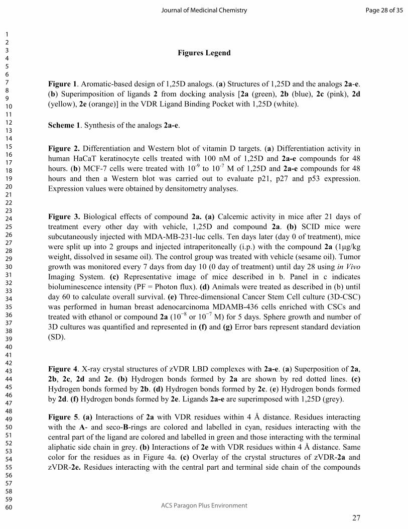
Figures Legend
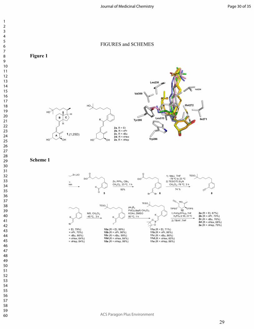
Figure 1. Aromatic-based design of 1,25D analogs. (a) Structures of 1,25D and the analogs 2a-e. (b) Superimposition of ligands 2 from docking analysis [2a (green), 2b (blue), 2c (pink), 2d (yellow), 2e (orange)] in the VDR Ligand Binding Pocket with 1,25D (white).
Scheme 1. Synthesis of the analogs 2a-e.
Figure 2. Differentiation and Western blot of vitamin D targets. (a) Differentiation activity in human HaCaT keratinocyte cells treated with 100 nM of 1,25D and 2a-e compounds for 48 hours. (b) MCF-7 cells were treated with 10-9 to 10-7 M of 1,25D and 2a-e compounds for 48 hours and then a Western blot was carried out to evaluate p21, p27 and p53 expression. Expression values were obtained by densitometry analyses.
Figure 3. Biological effects of compound 2a. (a) Calcemic activity in mice after 21 days of treatment every other day with vehicle, 1,25D and compound 2a. (b) SCID mice were subcutaneously injected with MDA-MB-231-luc cells. Ten days later (day 0 of treatment), mice were split up into 2 groups and injected intraperitoneally (i.p.) with the compound 2a (1µg/kg weight, dissolved in sesame oil). The control group was treated with vehicle (sesame oil). Tumor growth was monitored every 7 days from day 10 (0 day of treatment) until day 28 using in Vivo Imaging System. (c) Representative image of mice described in b. Panel in c indicates bioluminescence intensity (PF = Photon flux). (d) Animals were treated as described in (b) until day 60 to calculate overall survival. (e) Three-dimensional Cancer Stem Cell culture (3D-CSC) was performed in human breast adenocarcinoma MDAMB-436 cells enriched with CSCs and treated with ethanol or compound 2a (10−8 or 10−7 M) for 5 days. Sphere growth and number of 3D cultures was quantified and represented in (f) and (g) Error bars represent standard deviation (SD).
Figure 4. X-ray crystal structures of zVDR LBD complexes with 2a-e. (a) Superposition of 2a, 2b, 2c, 2d and 2e. (b) Hydrogen bonds formed by 2a are shown by red dotted lines. (c) Hydrogen bonds formed by 2b. (d) Hydrogen bonds formed by 2c. (e) Hydrogen bonds formed by 2d. (f) Hydrogen bonds formed by 2e. Ligands 2a-e are superimposed with 1,25D (grey).
Figure 5. (a) Interactions of 2a with VDR residues within 4 Å distance. Residues interacting with the A- and seco-B-rings are colored and labelled in cyan, residues interacting with the central part of the ligand are colored and labelled in green and those interacting with the terminal aliphatic side chain in grey. (b) Interactions of 2e with VDR residues within 4 Å distance. Same color for the residues as in Figure 4a. (c) Overlay of the crystal structures of zVDR-2a and zVDR-2e. Residues interacting with the central part and terminal side chain of the compounds
Page 28 of 35
ACS Paragon Plus Environment
Journal of Medicinal Chemistry
123456789101112131415161718192021222324252627282930313233343536373839404142434445464748495051525354555657585960

28
are shown. The heptyl central group of 2e induces some conformational changes of some side chains and a different positioning of the terminal side chain.
Table 1. Two- (2D) and three-(3D) dimensional cellular proliferation (expressed as percentage ± SD with respect to vehicle treated cells) of the human breast adenocarcinoma MCF-7, the human prostate adenocarcinoma PC-3, the human ovarian cancer SKOV-3, and the human keratinocyte HaCaT cell lines after culture for 48 h (2D) and 5 days (3D) with 100 nM of 1,25D and the 2a-2e compounds. Transcriptional activity is expressed as EC50 molar range, and as percentage of the EC50 M mean with respect to 1,25D (100%). Serum calcium levels (mean ± SD) in mice treated every other day for 21 days with 1,25D and the 2a-2e compounds (0.3 µg/kg weight, n=5 per group). VDR binding of the 1,25D and the 2a-2e compounds expressed as IC50 M range, and as percentage of the IC50 M mean with respect to 1,25D.
Page 29 of 35
ACS Paragon Plus Environment
Journal of Medicinal Chemistry
123456789101112131415161718192021222324252627282930313233343536373839404142434445464748495051525354555657585960
29
FIGURES and SCHEMES
Figure 1
HOH
H
A
B C
OHHO
HO
R
OHHO
2a, R = Et
2b, R = nPr
2c, R = nBu
2d, R = nHex
2e, R = nHep
1,(1,25D)1
Scheme 1
O
H
TESO
R
TMS
TESO
R
I
TESO
R
BO
O
TfO
TIPSO OTIPS
Br Br5 6 8
12
10a (R = Et, 99%)10b (R = nPr, 96%)
10c (R = nBu, 99%)10d (R = nHex, 84%)10e (R = nHep, 99%)
11a (R = Et, 71%)11b (R = nPr, 86%)
11c (R = nBu, 86%)11d (R = nHex, 60%)11e (R = nHep, 86%)
2a (R = Et, 67%)2b (R = nPr, 70%)2c (R = nBu, 76%)2d (R = nHex, 68%)2e (R = nHep, 79%)
= Et, 79%)= nPr, 70%)
= nBu, 66%)= nHex, 64%)= nHep, 84%)
O
EtO
O
EtO
3,
min
Zn.LiCl
Zn, PPh3, CBr4CH2Cl2, 23 ºC, 1 h
1) MeLi, THF
-78 ºC to 23 ºC
74 %92%
2) TESOTf, Et3N
CH2Cl2,-78 ºC, 3 h
NIS, CH2Cl2-45 ºC, 3 h
1) PdCl2(PPh3)2,THF
K3PO4 (2 M), 23 ºC
2) TBAF, THF
pin2B2
PdCl2(dppf)�CH2Cl2KOAc, DMSO
80 ºC, 1 h
Page 30 of 35
ACS Paragon Plus Environment
Journal of Medicinal Chemistry
123456789101112131415161718192021222324252627282930313233343536373839404142434445464748495051525354555657585960

30
Figure 2
Page 31 of 35
ACS Paragon Plus Environment
Journal of Medicinal Chemistry
123456789101112131415161718192021222324252627282930313233343536373839404142434445464748495051525354555657585960

31
Figure 3.
Page 32 of 35
ACS Paragon Plus Environment
Journal of Medicinal Chemistry
123456789101112131415161718192021222324252627282930313233343536373839404142434445464748495051525354555657585960

32
Figure 4.
Page 33 of 35
ACS Paragon Plus Environment
Journal of Medicinal Chemistry
123456789101112131415161718192021222324252627282930313233343536373839404142434445464748495051525354555657585960

33
Figure 5.
Page 34 of 35
ACS Paragon Plus Environment
Journal of Medicinal Chemistry
123456789101112131415161718192021222324252627282930313233343536373839404142434445464748495051525354555657585960

34
Table 1.
Cellular proliferation
Transcriptional
activity Calcemia VDR binding
MCF-7 (%)
PC-3 (%)
SKOV-3 (%)
HaCaT (%)
2D - 3D 2D 2D 2D EC50 M range (%) mg/dl (%) IC50 M range (%) 1,25D 66±2.3 - 61±9.6 77±1.2 78±2.3 69±1.5 1.6 - 2.3x10⁻⁹ (100) 14.7±0.5 (100) 1.3 - 2.3x10⁻⁹ (100) 2a 65±2.4 - 69±10.3 78±2.4 78±3.4 68±1.1 5.6 - 6.2x10⁻⁹ (33) 11.8±1.4 (0) 4.5x10⁻⁹ - 1.1x10⁻⁸ (24) 2b 69±2.8 - 74±10.3 83±3.0 82±2.9 71±1.3 8.6 - 9.9x10⁻⁹ (21) 13.1±1.2 (41) 5.6x10⁻⁹ - 5.5x10⁻⁸ (10) 2c 74±2.4 - 84±8.2 84±5.6 81±5.3 71±1.0 9.0x10⁻⁹ - 1.1x10⁻⁸ (19) 12.9±0.8 (34) 1.0 - 1.8x10⁻⁸ (12) 2d 78±2.9 - 79±7.5 80±1.9 76±0.9 74±2.1 1.0 - 1.3x10⁻⁸ (17) 11.7±0.8 (0) 8.3x10⁻⁹ - 4.0x10⁻⁸ (9) 2e 69±4.4 - 55±6.2 79±3.2 83±5.6 71±3.2 2.9 - 4.0x10⁻⁸ (5) 10.9±1.3 (0) 3.5x10⁻⁹ - 1.6x10⁻⁷ (7)
Page 35 of 35
ACS Paragon Plus Environment
Journal of Medicinal Chemistry
123456789101112131415161718192021222324252627282930313233343536373839404142434445464748495051525354555657585960