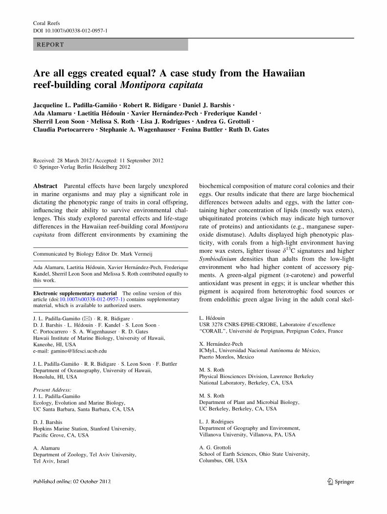
Are all eggs created equal? A case study from the Hawaiian ...
16
REPORT Are all eggs created equal? A case study from the Hawaiian reef-building coral Montipora capitata Jacqueline L. Padilla-Gamin ˜o • Robert R. Bidigare • Daniel J. Barshis • Ada Alamaru • Laetitia He ´douin • Xavier Herna ´ndez-Pech • Frederique Kandel • Sherril Leon Soon • Melissa S. Roth • Lisa J. Rodrigues • Andrea G. Grottoli • Claudia Portocarrero • Stephanie A. Wagenhauser • Fenina Buttler • Ruth D. Gates Received: 28 March 2012 / Accepted: 11 September 2012 Ó Springer-Verlag Berlin Heidelberg 2012 Abstract Parental effects have been largely unexplored in marine organisms and may play a significant role in dictating the phenotypic range of traits in coral offspring, influencing their ability to survive environmental chal- lenges. This study explored parental effects and life-stage differences in the Hawaiian reef-building coral Montipora capitata from different environments by examining the biochemical composition of mature coral colonies and their eggs. Our results indicate that there are large biochemical differences between adults and eggs, with the latter con- taining higher concentration of lipids (mostly wax esters), ubiquitinated proteins (which may indicate high turnover rate of proteins) and antioxidants (e.g., manganese super- oxide dismutase). Adults displayed high phenotypic plas- ticity, with corals from a high-light environment having more wax esters, lighter tissue d 13 C signatures and higher Symbiodinium densities than adults from the low-light environment who had higher content of accessory pig- ments. A green-algal pigment (a-carotene) and powerful antioxidant was present in eggs; it is unclear whether this pigment is acquired from heterotrophic food sources or from endolithic green algae living in the adult coral skel- Communicated by Biology Editor Dr. Mark Vermeij Ada Alamaru, Laetitia He ´douin, Xavier Herna ´ndez-Pech, Frederique Kandel, Sherril Leon Soon and Melissa S. Roth contributed equally to this work. Electronic supplementary material The online version of this article (doi:10.1007/s00338-012-0957-1) contains supplementary material, which is available to authorized users. J. L. Padilla-Gamin ˜o (&) R. R. Bidigare D. J. Barshis L. He ´douin F. Kandel S. Leon Soon C. Portocarrero S. A. Wagenhauser R. D. Gates Hawaii Institute of Marine Biology, University of Hawaii, Kaneohe, HI, USA e-mail: [email protected] J. L. Padilla-Gamin ˜o R. R. Bidigare S. Leon Soon F. Buttler Department of Oceanography, University of Hawaii, Honolulu, HI, USA Present Address: J. L. Padilla-Gamin ˜o Ecology, Evolution and Marine Biology, UC Santa Barbara, Santa Barbara, CA, USA D. J. Barshis Hopkins Marine Station, Stanford University, Pacific Grove, CA, USA A. Alamaru Department of Zoology, Tel Aviv University, Tel Aviv, Israel L. He ´douin USR 3278 CNRS-EPHE-CRIOBE, Laboratoire d’excellence ‘‘CORAIL’’, Universite ´ de Perpignan, Perpignan Cedex, France X. Herna ´ndez-Pech ICMyL, Universidad Nacional Auto ´noma de Me ´xico, Puerto Morelos, Mexico M. S. Roth Physical Biosciences Division, Lawrence Berkeley National Laboratory, Berkeley, CA, USA M. S. Roth Department of Plant and Microbial Biology, UC Berkeley, Berkeley, CA, USA L. J. Rodrigues Department of Geography and Environment, Villanova University, Villanova, PA, USA A. G. Grottoli School of Earth Sciences, Ohio State University, Columbus, OH, USA 123 Coral Reefs DOI 10.1007/s00338-012-0957-1
-
Upload
khangminh22 -
Category
Documents
-
view
0 -
download
0
Transcript of Are all eggs created equal? A case study from the Hawaiian ...
REPORT
Are all eggs created equal? A case study from the Hawaiianreef-building coral Montipora capitata
Jacqueline L. Padilla-Gamino • Robert R. Bidigare • Daniel J. Barshis •
Ada Alamaru • Laetitia Hedouin • Xavier Hernandez-Pech • Frederique Kandel •
Sherril Leon Soon • Melissa S. Roth • Lisa J. Rodrigues • Andrea G. Grottoli •
Claudia Portocarrero • Stephanie A. Wagenhauser • Fenina Buttler • Ruth D. Gates
Received: 28 March 2012 / Accepted: 11 September 2012
� Springer-Verlag Berlin Heidelberg 2012
Abstract Parental effects have been largely unexplored
in marine organisms and may play a significant role in
dictating the phenotypic range of traits in coral offspring,
influencing their ability to survive environmental chal-
lenges. This study explored parental effects and life-stage
differences in the Hawaiian reef-building coral Montipora
capitata from different environments by examining the
biochemical composition of mature coral colonies and their
eggs. Our results indicate that there are large biochemical
differences between adults and eggs, with the latter con-
taining higher concentration of lipids (mostly wax esters),
ubiquitinated proteins (which may indicate high turnover
rate of proteins) and antioxidants (e.g., manganese super-
oxide dismutase). Adults displayed high phenotypic plas-
ticity, with corals from a high-light environment having
more wax esters, lighter tissue d13C signatures and higher
Symbiodinium densities than adults from the low-light
environment who had higher content of accessory pig-
ments. A green-algal pigment (a-carotene) and powerful
antioxidant was present in eggs; it is unclear whether this
pigment is acquired from heterotrophic food sources or
from endolithic green algae living in the adult coral skel-
Communicated by Biology Editor Dr. Mark Vermeij
Ada Alamaru, Laetitia Hedouin, Xavier Hernandez-Pech, Frederique
Kandel, Sherril Leon Soon and Melissa S. Roth contributed equally to
this work.
Electronic supplementary material The online version of thisarticle (doi:10.1007/s00338-012-0957-1) contains supplementarymaterial, which is available to authorized users.
J. L. Padilla-Gamino (&) � R. R. Bidigare �D. J. Barshis � L. Hedouin � F. Kandel � S. Leon Soon �C. Portocarrero � S. A. Wagenhauser � R. D. Gates
Hawaii Institute of Marine Biology, University of Hawaii,
Kaneohe, HI, USA
e-mail: [email protected]
J. L. Padilla-Gamino � R. R. Bidigare � S. Leon Soon � F. Buttler
Department of Oceanography, University of Hawaii,
Honolulu, HI, USA
Present Address:J. L. Padilla-Gamino
Ecology, Evolution and Marine Biology,
UC Santa Barbara, Santa Barbara, CA, USA
D. J. Barshis
Hopkins Marine Station, Stanford University,
Pacific Grove, CA, USA
A. Alamaru
Department of Zoology, Tel Aviv University,
Tel Aviv, Israel
L. Hedouin
USR 3278 CNRS-EPHE-CRIOBE, Laboratoire d’excellence
‘‘CORAIL’’, Universite de Perpignan, Perpignan Cedex, France
X. Hernandez-Pech
ICMyL, Universidad Nacional Autonoma de Mexico,
Puerto Morelos, Mexico
M. S. Roth
Physical Biosciences Division, Lawrence Berkeley
National Laboratory, Berkeley, CA, USA
M. S. Roth
Department of Plant and Microbial Biology,
UC Berkeley, Berkeley, CA, USA
L. J. Rodrigues
Department of Geography and Environment,
Villanova University, Villanova, PA, USA
A. G. Grottoli
School of Earth Sciences, Ohio State University,
Columbus, OH, USA
123
Coral Reefs
DOI 10.1007/s00338-012-0957-1
etons. Despite the broad phenotypic plasticity displayed by
adults, parental investment in the context of provisioning of
energy reserves and antioxidant defense was the same in
eggs from the different sites. Such equality in investment
maximizes the capacity of all embryos and larvae to cope
with challenging conditions associated with floating at the
surface and to disperse successfully until an appropriate
habitat for settlement is found.
Keywords Biochemical phenotype � Coral eggs �Coral reproduction � Egg provisioning � Gamete variation �Maternal effects � Spawner
Introduction
Currently, coral reefs are experiencing unprecedented
pressure and extinction risk due to climate change (e.g.,
global warming, ocean acidification) and other local
impacts associated with anthropogenic disturbances (e.g.,
pollution, dredging and overexploitation; Hughes et al.
2003; Hoegh-Guldberg et al. 2007; Lough 2008). How
these environmental changes will impact the physiological
status of adult corals and in turn influence their offspring’s
phenotype is, however, relatively unexplored.
Adult corals are sessile, but their early life stages are
pelagic and therefore experience environmental conditions
that can differ from and be more dynamic than those of
adults. For example, eggs and larvae float on or swim close
to the surface and experience high and variable levels of
UV radiation and temperature, and exposure to free radi-
cals (Epel et al. 1999; Marquis et al. 2005; Hamdoun and
Epel 2007; Markey et al. 2007; Yakovleva et al. 2009).
Thus, parental investment in protective mechanisms and
energy reserves for the egg/early embryo is critical to
provisioning offspring with the capacity to cope with
environmental challenges and develop successfully (Ham-
doun and Epel 2007).
Parental effects (e.g., parental investment) occur when
the phenotype of offspring is affected by the phenotype or
environmental conditions of the parents (Mousseau and
Fox 1998; Badyaev and Uller 2009). Parental effects are
fundamentally important in biological systems and can
impact the life history (Donelson et al. 2009), competitive
ability (Wulff 1986), evolutionary trajectories, speciation
rates (Wade 1998) and population dynamics (Ginzburg
1998). These effects have been extensively studied in
plants, insects and terrestrial vertebrates, but have received
much less attention in the marine environment and spe-
cifically in natural tropical settings (Mousseau and Fox
1998; Marshall et al. 2008). Indeed, only four studies have
focused on the phenotypic relationship between parents
and offspring in corals (Michalek-Wagner and Willis 2001;
Wellington and Fitt 2003; Alamaru et al. 2009a; Padilla-
Gamino et al. 2012). These studies have revealed pre- and
post-zygotic parental effects influencing the physiological
characteristics of the egg/larvae (Michalek-Wagner and
Willis 2001; Wellington and Fitt 2003; Alamaru et al.
2009a) and the transmission of symbionts to the eggs
(Padilla-Gamino et al. 2012). For example, experimentally
bleached parents of the soft coral Lobophytum compactum
release eggs with lower levels of protein, lipid, mycospo-
rine-like amino acids (MAA) and carotenoid concentra-
tions than those released by healthy corals, which may
jeopardize egg and larval viability (Michalek-Wagner and
Willis 2001). Furthermore, eggs and larvae of the broad-
cast-spawning reef corals Acropora palmata, Montastraea
annularis and M. franksi exhibit different levels of photo-
protective compounds and survival capabilities depending
on the depth of origin of the parent colonies (Wellington
and Fitt 2003). Similarly, a strong relationship between the
isotopic signatures of parental tissues and those of their
planulae has been found in the brooder Stylophora pistil-
lata sampled across different depths (Alamaru et al.
2009a), and a recent study has revealed that parental effects
may play an important role in the transmission of Symbi-
odinium to the eggs of Montipora capitata (Padilla-Gamino
et al. 2012), with corals and eggs from a more challenging
environment (high light and temperature) generally asso-
ciated with Symbiodinium clade D, which are known to
confer greater thermal tolerance (Rowan 2004).
In this study, we compared biochemical traits (Table 1)
in adult colonies of the coral Montipora capitata (Family
Acroporidae) and their eggs, from two different environ-
ments. The goal was to better understand the intraspecific
variation in egg composition and the relationship between
the biochemical phenotype of parent colonies and the eggs
they release. Specifically, we address the following ques-
tions: (1) Are the biochemical phenotypes of adults and
eggs different between sites? (2) Does the biochemical
phenotype of the eggs reflect differences in environmental
conditions of the adult?
Life-stage differences and parental investment were
explored in the context of energetic (e.g., lipid reservoirs,
symbiotic algae, isotopic signatures) and protective (e.g.,
antioxidants) functions, traits that influence the ability of
the embryo/larvae to disperse and cope with variable
environmental conditions (e.g., light intensities, oxidative
stress, temperature) and settle successfully. These data
improve our understanding of what determines the natural
biochemical variability of M. capitata eggs (host and
symbiont), a facet of biology that contributes to the resil-
ience of one of the most important reef-building corals in
Hawaii.
Coral Reefs
123
Materials and methods
Study sites and sample collections
Montipora capitata is a broadcast spawner that has high
morphological and physiological plasticity and can inhabit
a broad range of environments (Maragos 1972; Grottoli
et al. 2004; Palardy et al. 2008; Rodrigues et al. 2008). In
contrast to most coral spawners that release asymbiotic
eggs, M. capitata release eggs with Symbiodinium directly
transmitted from the parental colony (vertical transmis-
sion). Samples of parent colonies and their gametes were
collected during the spawning events in summer 2007 from
two sites located on the western side of Moku O Lo’e
Island in Kane’ohe Bay, O’ahu, Hawai’i, the same sam-
pling event as in Padilla-Gamino and Gates (2012) and
Padilla-Gamino et al. (2012). Coral colonies were sampled
from *1 to 2 m depth at two sites: Bridge to Nowhere
(BTN; 21� 25.8930 N; 157� 47.3760W, n = 21) and Gilli-
gan’s Lagoon (GL; 21� 25.9730N; 157� 47.3920W, n = 20).
Colonies located at the GL site were closer to shore than
colonies from the BTN site and were shaded by an over-
story of trees located on land. Montipora capitata colonies
at the BTN site were generally branching in morphology
(Fig. 1a), whereas colonies at the GL site were predomi-
nantly plating in morphology (Fig. 1b).
To compare temperature and light conditions at the two
collection sites, temperature was measured at 10-min
intervals for *1 year (July 2007–August 2008) using
StowAway Tidbit data loggers (Onset Computer) accurate
to ±0.2 �C. Light was measured during two 2-week peri-
ods in 2008 (22 September–1 October and 25 November–5
December 2008). Light measurements were taken at
10-min intervals using Odyssey Photosynthetic Irradiance
Recording Systems (Odyssey).
Montipora capitata released (spawned) egg–sperm bun-
dles (Fig. 1c) between 20:45 and 21:15 h during the first
quarter of the new moon in June 2007. Gametes were col-
lected on the reef using a novel net system specifically
designed to collect spawn from shallow colonies with min-
imum damage to both adult coral colonies and released
gametes. The cylindrical nets surrounded the coral colony,
allowing the collection of egg–sperm bundles at the water
surface after release (for details see Padilla-Gamino and
Gates 2012). The nets were placed over the corals 1–2 h
before spawning and removed each night after spawning.
The positively buoyant egg–sperm bundles were collected
using scoop nets, transferred to plastic beakers and broken
apart by rinsing with 0.2-lm-filtered seawater (Fig. 1d). A
subset of the freshly collected and unfertilized eggs were
observed and photographed immediately upon collection
using dissecting and compound microscopes (Olympus
SZX7 and BX51, respectively) (Fig. 1c–f). Most eggs were
stored at -80 �C until further analysis. Samples from adult
coral colonies were collected 5 days prior to spawning by
breaking small fragments (*25 cm2) at least 4 cm away
from the tips and edges of the colonies where polyps were
anticipated to be reproductively active (Wallace 1985; pers
obs). Samples were immediately placed in dry ice and stored
at -80 �C for further analysis. Of the 41 colonies followed
throughout the reproductive season, 25 colonies spawned in
June 2007 (Padilla-Gamino and Gates 2012). Reproductive
output was highly variable between colonies providing us
with different amounts of egg material for biochemical
measurements. For specific details on the spawning
dynamics of M. capitata in 2007, see Padilla-Gamino and
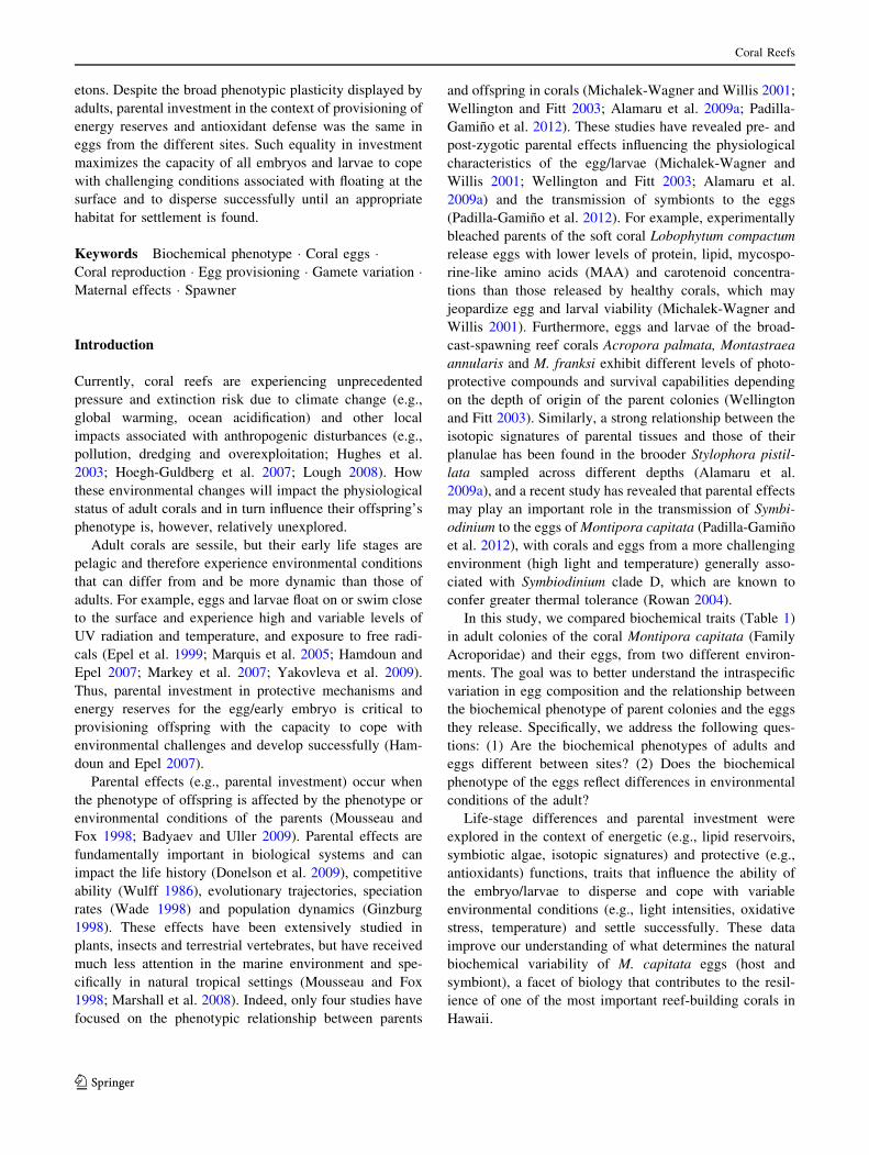
Table 1 Traits investigated in adults and eggs of the coral Montipora capitata and their biological function
Holobiont trait Biological function
Lipids Long-term energy reserves, storage of excess fixed carbon in host tissue
Triacylglycerols and wax esters are primary energy sources utilized during development and metamorphosis
Isotopic signatures d13C is a marker of carbon sources, d13C decreases as photosynthesis decreases and as heterotrophy increases. d15N
is a marker of trophic level
C:N:P ratios Carbon, nitrogen and phosphorous are essential bioelements of living organisms
Ubiquitin Cellular protein that tags proteins for degradation in the proteosomes. Increased levels indicate higher levels of
protein degradation
MnSOD Manganese superoxidase dismutase, a mitochondrial antioxidant enzyme, plays a role in oxidative stress
Fluorescent proteins Fluorescent proteins serve as antioxidants by scavenging reactive oxygen species (e.g., H2O2)
Photosynthetic pigment
profiles
Light harvesting compounds: Chl a and accessory pigments Chl c2 and peridinin
Photoprotective compounds (carotenoids): non-chlorophyll accessory pigments that serve as antioxidants, carotenes
(a-carotene, b-carotene) and xanthophylls (dinoxanthin, diadinoxanthin and diatoxanthin)
a) Pigments/g dw—pigment concentration per holobiont dry mass
b) Pigment/cell—pigment concentration in each dinoflagellate cell
Dinoflagellate density Symbiodinium dinoflagellate cells provide the host with energy in the form of translocated reduced carbon
compounds (glucose, glycerol and aminoacids), which are products of photosynthesis
Coral Reefs
123

Gates (2012). Sample size differed among the biochemical
analyses (n = 5–11 per site for both adults and eggs) and was
constrained by the availability of eggs and instrumentation.
Laboratory analysis
Total lipids and lipid classes
Total lipids (n = 8 per site for both adults and eggs) were
extracted according to Rodrigues and Grottoli (2007). In
brief, ground samples were extracted in a 2:1 chloroform/
methanol solution, the organic phase washed using 0.88 %
KCl, and the extract dried to a constant weight. Lipids
were normalized to total ash-free dry tissue biomass of
the organic fraction (host and symbionts). Triacylglycerol
(TAG) and wax ester (WE) concentrations in total
lipid extracts were determined by high-performance
liquid chromatography/evaporative light-scattering detec-
tion (HPLC/ELSD) using triolein (Sigma-Aldrich, #1787-
1AMP) and oleyl oleate (Sigma-Aldrich, # O3380) as
reference standard (Silversand and Haux 1997). The per-
centage of ‘‘other lipids’’ (OLs; polar lipids, free fatty
acids, sterols, diacylglycerols and monoacylglycerols) was
estimated as the difference between total lipids and the sum
of TAG and WE.
Stable isotopes and element ratios
Coral samples (n = 6 per site for both adults and eggs)
were ground in 0.2-lm-filtered sea water (FSW). If skel-
eton was present, tissue was separated from the ground
skeleton in several washes with FSW. Isolated tissue was
then homogenized, and aliquots from the same homoge-
nate were taken for different analyses (stable isotopes,
algal pigments, Symbiodinium densities and ash-free dry
weight determination). Coral d13C and d15N analyses were
performed using standard methods (i.e., Rodrigues and
Grottoli 2006; Hughes et al. 2010). Briefly, pre-burned
Glass Fiber Filters (0.7 lm pore size, GF/F; Whatman)
containing the homogenized coral tissue were dried at
ba
dc e f
120µm1mm 1mm
5 cm 30cm
Fig. 1 Montipora capitata is a simultaneous hermaphrodite that
releases egg–sperm bundles. a Branching and b plating morphologies
of Montipora capitata. c Bundles contain approximately 14 eggs and
a sperm mass. d Eggs contain Symbiodinium cells transferred from the
parent. A Montipora capitata egg under e white light and d blue light.In f the green fluorescence is from fluorescent proteins and the redfluorescence is from the chlorophyll in the symbiotic dinoflagellates
Coral Reefs
123

60 �C prior to isotopic and phosphorus analyses. Samples
were analyzed on a Costech Elemental Analyzer coupled
to a Finnigan Delta IV Plus stable isotope ratio mass
spectrometer under continuous flow using a CONFLO III
interface in the Stable Isotope Biogeochemistry Labora-
tory at Ohio State University. Approximately 10 % of all
samples were run in duplicate. Stable carbon (d13C = %deviation of the ratio of 13C: 12C relative to the Vienna
Peedee Belmenite Limestone standard) and stable nitrogen
(d15N = % deviation of 15N:14N relative to air) mea-
surements were made where the average standard
deviations of repeated measurements of the USGS24 and
IAEA-N1 standards were 0.06 % for d13C and 0.12 % for
d15N. Total phosphorus was obtained using the modified
high-temperature ashing/hydrolysis method (Monaghan
and Ruttenberg 1999). For each coral tissue homogenate
(1–0.5 ml), 40 lL of MgNO3 was added; then, the solu-
tion was dried and burned at 550 �C for 2 h. After com-
bustion, the residual material was hydrolyzed in 1 mL of
1 M HCl and analyzed via the molybdenum blue method
(Grasshoff et al. 1983). Stable isotopes, algal pigments and
Symbiodinium densities were normalized to total ash-free
dry tissue biomass of the organic fraction (host and
symbionts).
Protein analysis
The levels of Manganese Superoxide Dismutase (MnSOD)
and ubiquitin-conjugated proteins were studied (n = 11 per
site for both adults and eggs) via western blot according to
Barshis et al. (2010). All western blots were standardized
using a standard curve dilution of a single, reference coral
extract; hence, concentrations of specific experimental
samples are relative to the concentration of the ubiquitin-
conjugated proteins or MnSOD contained in the reference
extract. 10 lg of total protein for each experimental extract
was loaded on the gel in triplicate. Antibodies against
ubiquitin (Cat, #SPA-200), manganese superoxide dismu-
tase (MnSOD; Cat.#SOD-110) and anti-rabbit- and anti-
mouse-conjugated (Cat. #SAB-300, SAB-100) horseradish
peroxidase were obtained from Stressgen Biotechnologies
(Victoria, British Columbia, Canada). Primary antibody
dilutions were 1:5,000 for MnSOD and 1:2,000 for ubiq-
uitin; secondary antibody dilutions were all 1:10,000. All
images were recorded using a ChemiDoc XRS molecular
imager (Bio-Rad) and analyzed and quantified with ImageJ
software (Abramoff et al. 2004).
Fluorescent properties of eggs and sperm were
explored using epifluorescence microscopy. Emission
characteristics of coral fluorescent proteins (FP) in eggs
and adults were examined using a fluorescence spectro-
photometer (SpectraMax M2, Molecular Devices, Sun-
nyvale, California).
Algal pigments
Glass Fiber Filters (0.7 lm pore size, GF/F; Whatman)
containing the coral and algal tissue homogenate (n = 6
per site for both adults and eggs) were extracted in 3 mL of
HPLC-grade acetone in culture tubes along with 50 lL of
an internal standard (canthaxanthin) at 4 �C in the dark for
24 h. The extracts were processed according to Bidigare
et al. 2005; for details, see methods in the electronic sup-
plemental material, ESM. Pigment concentrations and
ratios at the colony and algal cellular level were computed
by normalizing to sample dry tissue biomass (lg pigment/g
dw and symbiont density (pg pigment/cell) (see Table 1 to
review the differences between pigment normalizations).
Symbiodinium densities
Symbiodinium cells (n = 5 at BTN and n = 6 at GL for
both adults and eggs, an adult–egg set of BTN samples was
lost) were separated from the tissue homogenate by cen-
trifuging and performing multiple washing steps using
FSW. After separation, the Symbiodinium pellets were
resuspended in FSW (n = 5 at BTN and n = 6 at GL for
both adults and eggs) and homogenized, and three subs-
amples counted manually with a hemocytometer and a light
microscope (Olympus BX-51). A linear regression was
used to estimate the number of bundles (or eggs) per ash-
free dry tissue mass of the egg sample to provide an esti-
mate of the amount of symbiont cells per egg (ESM
Appendix I).
Statistical analyses
Prior to analysis, data were normalized as necessary using
logarithmic or inverse transformations to achieve homo-
geneity of variances and normality. The xanthophylls DDX
and DTX had similar patterns throughout and were com-
bined for the statistical analysis. All variables were ana-
lyzed using a general linear model, with developmental
stage and site as fixed factors and colony modeled as a
random factor nested within site to account for the repeated
measurements between adults and eggs. When significant
effects were identified, Tukey’s post hoc tests were per-
formed to determine differences between groups (i.e., dif-
ferences between sites within life stage). Parental effects
were recognized if the interaction between site and stage
factors was significant and/or if significant differences
between the eggs reflected differences between the adults.
Means, standard deviations and ranges (minimum–maxi-
mum) of temperature and light were calculated for each
site during the periods sampled. Temperature and light
measurements were compared between sites using a Mann–
Whitney test. All statistical analyses were performed using
Coral Reefs
123

Minitab statistical software (version 15). p-values were
considered significant below an alpha of 0.05.
Results
Study site environment Temperature was higher and
more variable at the BTN site than the GL site throughout
the year (W = 4.5 9 1010, p \ 0.0001, Mann–Whitney,
Fig. 2a), with up to 3 �C fluctuations observed over a
single 24-h period. The BTN site was also characterized by
the highest and broadest light intensities in late summer
and late autumn sampling times compared with the GL site
(W = 1773932, p \ 0.0001, W = 1413959, p \ 0.0001,
September and November, respectively, Fig. 2b–c). The
GL site had 23 % of the light levels of the BTN site.
Effects of stage
Total lipids and lipid composition Total lipid concentra-
tions were higher in eggs than in adults (Fig. 3a, Table 2)
representing 80 and 25 % of the total dry tissue weight, in
eggs and adults, respectively. Lipid composition differed
between life stages, with WE representing approximately
56 % of the total lipid weight in the eggs and only
9–17 % of the total lipid weight in adults (Fig. 3b, d,
Table 2). In contrast, TAG was found in higher concen-
trations in adults than in eggs (Fig. 3c–d, Table 2). The
‘‘other lipids’’ category ranged between 36 and 92 %
(mean 74.9 %) in adults and 25–57 % (mean 45.5 %) in
eggs (Fig. 3d).
d13C, d15N, %C, %N, %P and elemental ratios (C:N, C:P,
N:P) Adult colonies were enriched in 13C compared with
eggs (p = 0.009, Fig. 4a, Table 2). Average d15N values of
adults and eggs from both sites ranged between 3 and 5.7 %and were not different between life stages or sites (ESM
Appendix IIa, Table 2). Carbon content was higher in eggs
than in adult corals (p \ 0.0001, Fig. 4b, Table 2). In
contrast, nitrogen and total phosphorus contents were
higher in the adult coral tissue than in the eggs (Fig. 4c–d,
Table 2). C:N and C:P ratios in eggs were higher than in the
adult colonies (Fig. 4e–f, Table 2). N:P ratios did not differ
Fig. 2 a Temperature (�C) and
b–c light (lmol quanta/m2s)
data from the two study sites in
Moku O Lo’e Island, Kane’ohe
Bay Hawaii, b 22 September–1
October and c 25 November–
December 5, 2008
Coral Reefs
123

between life stages (ESM Appendix IIb, Table 2).Thus, the
major differences in tissue composition between life stages
are higher levels of carbon–lipid in the eggs.
Protein biomarkers Relative concentrations of MnSOD
were higher in eggs than in adults (Fig. 5a, Table 2).
Relative concentrations of ubiquitin conjugates were also
0.1
0.2
0.3
0.4
0.5
0.6
0.7
0.8
Adults Eggs0
1
2
3
4
5
Adults Eggs
Wax
est
ers
(mg
/mL)
0.00
0.10
0.20
0.30
0.40
0.50
0.60
Adults Eggs
Tria
cylg
lyce
rols
(m
g/m
L)
BTN GL BTN GL
Eggs
% o
f tot
al li
pid
BTNGL
a b c
d
g to
tal l
ipid
s / g
dw
0%
10%
20%
30%
40%
50%
60%
70%
80%
90%
100%
% Other
% TAG
% WE
Adults
A
B
A
AB
B
*
0.9
Fig. 3 a Total lipids, b wax esters (WE), c triacylglycerols (TAG) and d percentage of lipid classes in adults and eggs of Montipora capitata.
Means ± SE. Capital letters indicate differences between stages, and asterisks indicate differences between sites within stage
Table 2 Results of the general linear model testing the effects of life stage and site on the traits investigated
Effect of stage Effect of site Effect of colony (site) Effect of site 9 stage
Physiological parameter F P F P F P F P
Lipid (df = 31) 1006.77 \0.0001 2.93 0.109 1.00 0.497 4.59 0.050
Wax esters (df = 25) 189.65 \0.0001 3.33 0.093 2 0.122 24.37 \0.0001
Triacylglycerols (df = 25) 5.4 0.04 1.59 0.234 0.5 0.868 4.57 0.056
d13 C (df = 23) 10.61 0.009 1.74 0.216 7.80 0.002 3.93 0.076
d15 N (df = 23) 0.52 0.489 3.98 0.074 0.81 0.627 2.43 0.150
C (%) (df = 23) 35.19 \0.0001 0.09 0.773 1.42 0.294 5.44 0.042
N (%) (df = 23) 22.88 0.001 1.37 0.269 1.18 0.398 0.02 0.903
Total P (df = 23) 22.45 0.001 0.84 0.382 1.3 0.353 0.01 0.935
C:N (df = 23) 233.55 \0.0001 6.63 0.028 0.73 0.685 4.83 0.053
C:P (df = 21) 64.24 \0.0001 0.01 0.935 1.29 0.355 1.49 0.253
N:P (df = 21) 0.18 0.680 1.15 0.312 1.51 0.273 0.00 0.986
MnSOD (df = 35) 63.57 \0.0001 1.11 0.308 1.44 0.236 4.13 0.059
Ubiquitin-conjugates (df = 34) 34.84 \0.0001 6.26 0.024 0.93 0.559 0.22 0.648
Symbiont no. of cells (df = 21) 42.31 \0.0001 7.68 0.022 1.34 0.336 4.03 0.076
Significant values at 95 % confidence (p \ 0.05) are in bold, df degrees of freedom
Coral Reefs
123

higher in eggs than in adult colonies (Fig. 5b, Table 2).
Fluorescent proteins were detected in both eggs and adult
tissues. Both emitted cyan-green light, characteristic of
fluorescent proteins (k = 491, 3 nm resolution), and far-
red light from chlorophyll (k = 682, 3 nm resolution)
when excited by blue light (450 nm) (Fig. 1 c, d).
Algal pigments and Symbiodinium densities The dino-
flagellate pigments Chl a, Chl c2, peridinin, b-carotene,
dinoxanthin, diadinoxanthin (DDX) and diatoxanthin
(DTX) were detected in samples from all colonies for both
adults and eggs. Dinoflagellate pigment concentrations per
g ash-free dry tissue were always higher in the adults
-16.5
-16.0
-15.5
-15.0
-14.5
-14.0
-13.5
-13.0Adults Eggs
0
5
10
15
20
25
30
35
40
45
Adults Eggs
Car
bon
(%)
0
1
2
3
4
5
6
Adults Eggs
0.00
0.05
0.10
0.15
0.20
0.25
0.30
0.35
0.40
Adults Eggs0
2
4
6
8
10
12
14
16
18
Adults Eggs0
100
200
300
400
500
600
Adults Eggs
δ13C
v-P
DB
Nitr
ogen
(%
)
Pho
spho
rus
wt%
C:N
C:P
cba
fed
BTNGL
*
A
B
A
B
A
B
A
B
A
B
*
A B
(‰)
Fig. 4 a d13C, b % carbon, c % nitrogen, d % phosphorus, e C:N and f C:P ratios in adults and eggs of Montipora capitata. Means ± SE.
Capital letters indicate differences between stages, and asterisks indicate differences between sites within stage
0
2
4
6
8
10
12
14
16
0
2
4
6
8
10
12
14
Adults EggsAdults Eggs
a b BTNGL
A
B B
A
16
18
Rel
ativ
e [M
nSO
D]
Rel
ativ
e [U
biqu
itin-
conj
ugat
e]
Fig. 5 a Manganese superoxide dismutase (MnSOD) and b ubiquitin conjugate levels in adults and eggs of Montipora capitata. Means ± SE.
Capital letters indicate differences between stages
Coral Reefs
123

compared with the eggs (Fig. 6a–f, Table 3). The pigment
alpha-carotene (a-carotene), characteristic of green algae,
was also detected in all colonies and both life stages. In
contrast to dinoflagellate (brown algae) pigments examined
here in which pigment concentrations range between 2.5
and 8 fold higher in parent tissue (Fig. 6a–f), a-carotene
concentration was approximately sixfold higher in the eggs
compared with the adults (Fig. 6g, Table 3). In addition,
Symbiodinium densities (cell number normalized by tissue
dry mass) were higher in adult corals than in eggs (Fig. 6h,
Table 2). Symbiodinium densities in the eggs were
estimated to range between 2.3 and 4.2 9 103 symbiont
cells per egg. The concentrations of Chl a/cell, b-carotene/
cell, dinoxanthin/cell, DDX ? DTX/cell were greater in
adults than in eggs (Fig. 7a, d, e, f, Table 3).
Effects of site
Total lipids and lipid composition Total lipid content did
not change between sites for both adults and eggs (Fig. 3a,
Table 2). Eggs from both sites had similar levels of WE,
even though parents from the BTN site had higher levels of
0
200
400
600
800
1000
1200
1400
1600
1800
2000
Adults Eggs0
10
20
30
40
50
60
70
80
90
100
Adults Eggs0
100
200
300
400
500
600
700
800
Adults Eggs
0
10
20
30
40
50
60
Adults Eggs0
10
20
30
40
50
60
70
80
90
Adults Eggs0
50
100
150
200
250
300
350
400
Adults Eggs
Chl
a (
µg/g
dw)
β-ca
rote
ne (
µg/g
dw)
Chl
c2 (
µg/g
dw)
Per
idin
in (
µg/g
dw)
Din
oxan
thin
(µg
/gdw
)
DD
X+
DD
T (
µg/g
dw)
cba
fed
BTNGL
0
1
2
3
4
5
Adults Eggs0
2
4
6
8
10
12
14
16
18
Adults Eggs
α-ca
rote
ne (
µg/g
dw)
Sym
b. c
ells
/ gdw
(x1
08 )
hg
A
B
A
B
A
B
A
B
A
B
A
B
A
B A
B
*
Fig. 6 Photosynthetic pigment concentrations and Symbiodiniumdensities of adults and eggs of Montipora capitata normalized by
tissue ash-free dry weight. a Chl a—chlorophyll a; b Chl c2—
chlorophyll c2; c peridinin; d b-carotene; e dinoxanthin; f DDX ?
DDT-diadinoxanthin ? diatoxanthin, g a-carotene and h Symbiodini-um density. Means ± SE. Capital letters indicate differences between
stages, and asterisks indicate differences between sites within stage
Coral Reefs
123

WE (post hoc test p \ 0.001, Fig. 3b, d, Table 2). No
differences in TAG between sites were found in either
stage (Fig. 3c, Table 2).
d13C, d15N, %C, %N, %P and elemental ratios (C:N, C:P,
N:P) d13C was significantly higher in adults from the
BTN site (post hoc test p = 0.011, Fig. 4a, Table 2), but
no difference was found in d13C between the eggs from
different sites. No site effects on adults or eggs were
observed in d15N, % carbon, % nitrogen, % phosphorous,
C:P and N:P (Fig. 4b–d, f, Table 2). Only C:N had a sig-
nificant site effect, which was primarily driven by the
differences in the eggs between sites (post hoc test
p = 0.017, Fig. 4e, Table 2).
Protein biomarkers Adults and eggs from both sites had
similar concentrations of MnSOD (Fig. 5a, Table 2). A
significant effect of site was found in the relative concen-
trations of ubiquitin–conjugates. Overall, ubiquitin conju-
gates were higher at the GL site in both stages; however,
post hoc tests did not detect differences between sites
within stages (post hoc test p = 0.534, p = 0.220, adults
and eggs respectively, Fig. 5b, Table 2).
Algal pigments and Symbiodinium densities Algal pig-
ment concentrations per g ash-free dry tissue did not vary
between sites (Fig. 6a–g, Table 3). However, there was an
effect of site on Symbiodinium densities, mostly driven by
the differences in the adults between sites. Higher Symbi-
odinium densities were found in adults from the BTN site
as compared to counterparts from GL (post hoc test,
p = 0.021, Fig. 6h, Table 3), and eggs had similar Sym-
biodinium densities between sites (post hoc test,
p = 0.831). Concentrations of some pigments normalized
per Symbiodinium cell showed differences between sites
(Fig. 7c–f, Table 3), which were primarily driven by the
differences between adults and not the eggs. Peridinin/cell,
b-carotene/cell, dinoxanthin/cell and DDX ? DTX/cell
were higher at the GL site as compared to BTN (post hoc
test, p = 0.033, p = 0.050, p = 0.025, p = 0.018, p = 0.
respectively, Fig. 7c–f).
Discussion
This study explored the natural variability of biochemical
traits in the adult coral colonies and eggs of M. capitata.
Although considerable differences in the parental pheno-
type were observed, the biochemical composition and
photochemical characteristics of Symbiodinium in their
0
2
4
6
8
10
12
Adults Eggs0.0
0.1
0.2
0.3
0.4
0.5
0.6
0.7
Adults Eggs
0.0
0.1
0.1
0.2
0.2
0.3
0.3
0.4
Adults Eggs
0
1
2
3
4
5
6
Adults Eggs
0.0
0.1
0.2
0.3
0.4
0.5
0.6
Adults Eggs0.0
0.5
1.0
1.5
2.0
2.5
3.0
Adults Eggs
a b c
d e f
Chl
a (
pg /
cell)
Chl
c
β-ca
rote
ne (
pg /
cell)
Per
idin
in (
pg /
cell)
Din
oxan
thin
(pg
/ ce
ll)
DD
X+
DT
X (
pg /
cell)
BTNGL
A
B
A
B*
** *A
B
A
B
A
B
Fig. 7 Photosynthetic pigment concentrations of adults and eggs of
Montipora capitata normalized by density of Symbiodinium cells.
a Chl a—chlorophyll a; b Chl c2—chlorophyll c2; c peridinin;
d b-carotene’ e dinoxanthin’ f DDX ? DDT-diadinoxanthin ?
diatoxanthin. Means ± SE. Capital letters indicate differences between
stages, and asterisks indicate differences between sites within stage
Coral Reefs
123

eggs were similar. Thus, regardless of the parental condi-
tion, eggs are provisioned with similar energy reserves and
antioxidant levels. Such equality of provisioning would
allow offspring to settle in a variety of habitats, rather than
being restricted to the same habitat as the adults, and may
explain, in part, why this species has such a broad distri-
bution in the West Pacific (Veron 2000).
Energy reserves and acquisition
Lipids, particularly wax esters (WE), were a major com-
ponent in M. capitata eggs as compared to adults. WE are
largely responsible for the positive buoyancy of the eggs,
which is critical for successful fertilization, and represent a
long-term energy store (major source of fatty acids) that is
consumed throughout development to provide energy for
larval dispersal, settlement and metamorphosis (Arai et al.
1993; Lee et al. 2006; Harii et al. 2010). Eggs of M. cap-
itata had similar WE content (53–59 % of total lipid
weight) to symbiotic propagules of other species
(56–69 %, Montipora digitata, 52–60 %, Pocillopora
damicornis) (Arai et al. 1993; Harii et al. 2007, 2010). This
is generally lower than the WE content of asymbiotic
propagules, which can account for up to 58–85 % of total
lipid weight (Figueiredo et al. 2012). Low WE content in
symbiotic propagules may reflect the fact that Symbiodi-
nium have the capacity to provide additional carbon and
energy for dispersal (Harii et al. 2010; Figueiredo et al.
2012) and may also serve to decrease buoyancy and reduce
oxidative stress in algal symbionts caused by exposure to
high temperature and UV (Yakovleva et al. 2009; Nesa
et al. 2012).
Although energy reserves (total lipids) were similar in
adults from different sites, WE were higher at the BTN
(high light) site, which is consistent with the idea that
storage lipids in Montipora capitata are composed of
photosynthetically derived carbon (Rodrigues et al. 2008).
Eggs did not differ in their d13C signatures between sites,
suggesting that similar carbon sources are used for egg
production at the two sites. Overall, eggs had lower d13C
values than adults, most likely due to the high concentra-
tions of lipids (Bodin et al. 2007; Alamaru et al. 2009a),
which typically have lower d13C values (DeNiro and
Epstein 1977). Lighter d13C signatures in the adults at the
GL (low light) site suggest that these corals have lower
rates of photosynthesis or higher heterotrophy, or a com-
bination of both, as compared to corals from the BTN site
(Grottoli and Wellington 1999; Rodrigues and Grottoli
2006; Alamaru et al. 2009b). It is likely that the differences
in d13C in adults were mostly driven by lower rates of
photosynthesis due to lower light availability at the GL
site, which receives only 23 % of the light levels of the
BTN site. Increases in heterotrophy (due to lower light
availability) at the GL could also contribute to the differ-
ences in d13C. However, this is less likely since previous
work has shown that healthy M. capitata colonies located
Table 3 Results of the general linear model testing the effects of life stage and site on the photosynthetic pigments of Montipora capitata
Effect of stage Effect of site Effect of colony (site) Effect of site 9 stage
Pigment df F P F P F P F P
Pigment/tissue
Chl a/mg dm 22 220.99 <0.0001 0.53 0.481 2.00 0.155 0.35 0.566
Chl c2/mg dm 22 112.65 <0.0001 0.01 0.917 3.74 0.030 1.25 0.292
Peridinin/mg dm 23 129.66 <0.0001 0.00 0.949 4.04 0.019 0.79 0.394
b-carotene/mg dm 23 113.23 <0.0001 0.07 0.801 1.27 0.358 1.76 0.214
Dinoxanthin/mg dm 23 283.93 <0.0001 0.00 0.945 4.66 0.011 1.39 0.266
DDX ? DTX/mg dm 23 346.24 <0.0001 0.01 0.914 5.97 0.005 0.77 0.402
a-carotene/mg dma 23 85.13 <0.0001 2.21 0.168 1.03 0.483 0.35 0.568
Pigment/cell
Chl a/cell 22 25.72 0.001 3.75 0.079 0.82 0.620 3.74 0.085
Chl c2/cell 22 2.31 0.163 2.07 0.177 0.84 0.606 2.44 0.153
Peridinin/cell 22 0.01 0.934 5.75 0.036 1.54 0.263 2.63 0.139
b-carotene/cell 22 11.4 0.008 9.17 0.012 0.98 0.516 1.44 0.261
Dinoxanthin/cell 22 14.83 0.004 5.26 0.043 1.63 0.238 3.72 0.086
DDX ? DTX/cell 22 12.07 0.007 6.35 0.029 1.5 0.278 4.23 0.070
Pigment concentrations were normalized to dry mass (dm), chlorophyll a (Chl a) and Symbiodinium cell density
Significant values at 95 % confidence (p \ 0.05) are in bold
Chl a chlorophyll a, Chl c2 chlorophyll c2, DDX diadinoxanthin, DTX diatoxanthin, df degrees of freedoma alpha-carotene is a green-algal pigment
Coral Reefs
123

at 1 m depth have similar feeding rates as colonies at 6 m
depth (Palardy et al. 2008), which were receiving less than
42 % of the photosynthetically active radiation (PAR)
received by the colonies at 1 m depth (Jokiel et al. 1997).
Isotopic nitrogen signatures (d15N) of M. capitata ranged
between 4 and 5 % and were similar across sites and
between life stages, suggesting that adults from all these
sites were feeding at similar trophic levels and that similar
nitrogen sources are passed from the parents to the eggs.
Antioxidant defense and photoprotection
Oxidative stress is caused by the production and accumu-
lation of reactive oxygen species—ROS—(1O2, O2-, H2O2,
HO•) which can damage cellular components such as lipids,
proteins and DNA (Lesser 2006). In corals, oxidative stress
can cause bleaching due to elevated temperature and high
UV levels and in coral symbiotic larvae, the presence of
Symbiodinium has been associated with higher vulnerability
to oxidative stress than asymbiotic larvae (Yakovleva et al.
2009; Nesa et al. 2012). In our study, eggs from both sites
(which contain Symbiodinium) seem to be similarly pre-
conditioned to cope with oxidative stress and possessed
elevated antioxidant levels and increased turnover rates of
proteins when compared to the adults (Table 4a). Fluores-
cent proteins in the eggs may serve as antioxidants by
scavenging ROS such as hydrogen peroxide (H2O2) (Palmer
et al. 2009), which is able to move easily through biological
membranes and cause lipid peroxidation (Halliwell and
Gutteridge 1999). In addition, the higher concentrations of
MnSOD in the eggs (two fold higher than adults, Table 4a)
also suggest that MnSOD may prevent the accumulation of
the ROS superoxide in the early life stages of the coral
holobiont and reduce oxidative damage. Higher levels of
ubiquitin-conjugated proteins in eggs as compared to adults
suggest greater rates of protein damage and/or enhanced
capacity for turnover (Table 4a). In sea urchin embryos,
synthesis rates of ubiquitin have been associated with
selective protein degradation during embryogenesis where
total ubiquitin content of the embryo increased almost
tenfold between fertilization and the pluteus larva stage
(Pickart et al. 1991).
The photochemical characteristics of coral adults reflect
acclimatization responses to the different light conditions
between sites/morphologies. By increasing accessory pig-
ments relative to chlorophyll, corals at the GL site increase
the amount of photoprotection and reduce their suscepti-
bility to photodamage and oxidative stress (Lesser et al.
1990). Although corals at the BTN site are experiencing
significantly higher light levels, corals in the low-light
environment have a plating morphology which may make
them more vulnerable to light stress than corals at the BTN
site, which have a branching morphology and thus self-
shade more. It is important to note that corals at these two
sites can harbor different assemblages of Symbiodinium
(Padilla-Gamino et al. 2012), and this may also play an
important role in the acclimatization strategies and photo-
biological characteristics of the coral holobionts from the
different environments (Little et al. 2004; Rowan 2004;
Abrego et al. 2008; Cantin et al. 2009). In contrast to the
adults, eggs did not differ in their Symbiodinium densities,
which suggest that adults provision the egg similarly in
Table 4 (a) Differences in
traits between life stages (adult
vs. eggs) and (b) between adults
from different sites of
Montipora capitata
a. Differences between stages b. Parental differences
Higher in adults Higher in eggs Higher in high light environment
Triacylglycerol Total lipids Wax esters
d13C Wax esters d13C
Total phosphorous Carbon Symbiont cells/mg dw
Symbiont cells/mg dw C:N
Chl a/mg dw C:P
Chl c/mg dw Ubiquitin
b-carotene/mg dw SOD
Peridinin/mg dw a-carotene/mg dw Higher in low light environment
Dinoxanthin/mg dw Chl c/Chl a Peridinin/symb cell
DDX ? DTX/mg dw b-carotene/Chl a b-carotene/symb cell
Chl a/symb cell Peridinin/Chl a Dinoxanthin/symb cell
Chl c/symb cells Dinoxanthin/Chl a DDX ? DTX/symb cell
b-carotene/symb cell DDX ? DTX/Chl a
Peridinin/symb cell
Dinoxanthin/symb cell
DDX ? DTX/symb cell
Coral Reefs
123

terms of the number of Symbiodinium cells transferred and/
or there is a limit to the number of Symbiodinium cells that
an egg can contain. To date, only one study has explored
the process of entry of dinoflagellate endosymbionts into
the eggs of reef-building corals (Hirose et al. 2001). These
authors found that Symbiodinium appear to enter mature
eggs through follicle cells that surround the eggs in tem-
porary gaps formed in the mesoglea. This process may
limit the number of dinoflagellate symbionts that can be
transferred and may account for the similar quantities
found in eggs from different sites.
Lower pigments (per Symbiodinium cell) in the eggs
suggest that Symbiodinium cells with lower pigmentation
are preferentially transferred to the eggs or that Symbi-
odinium cells photoacclimatize differently when in eggs.
Symbiodinium in eggs are embedded in lipid droplets (the
main component of egg cytoplasm), which might add an
extra barrier to solar energy acquisition and influence light
microenvironment of the symbiont. Lower Symbiodinium
densities (per ash-free dry weight) and pigment content in
the Symbiodinium cells may help the early embryo/larvae
to reduce the risk of photodamage and subsequent oxida-
tive stress (Lesser et al. 1990). This would be advantageous
since M. capitata larvae swim near the surface (Hodgson
1985), and in culture, this species has been reported to
maintain a high ability to settle for *6 weeks and in a few
cases to delay the onset of settlement competency for
7 months or longer (Kolinski 2004). Chl c2 and peridinin
(per cell) were the only two pigments that had similar
concentrations among life stages. In dinoflagellates, these
two pigments are efficient light-harvesting accessory pig-
ments that transfer excitation energy to chlorophyll a (Chl
a) (Govindjee et al. 1979) and are part of the Chl-protein
complexes (peridinin-Chl a-protein and Chl a-Chl
c2-peridinin-protein complexes, PCP and acpPC, respec-
tively) (Jeffrey 1976).
Interestingly, a pigment characteristic of green algae
(a-carotene) was also detected in both coral adults and
eggs. This carotenoid is a fat-soluble pigment (unsaturated
hydrocarbon) and a powerful antioxidant that can stop free
radicals from causing cells to break down in algae (Niyogi
et al. 1997) and may therefore serve to prevent oxidative
stress inside the coral eggs. It is important to note that
a-carotene is also a major carotene pigment in endolithic
green algae (Ostreobium) (Jeffrey 1968) which are known
to reside in the skeletons of the coral genus Montipora
(Fine et al. 2005; Magnusson et al. 2007). The presence of
high a-carotene levels in the eggs with respect to adults
may be due to the fact that pigment extractions were per-
formed using mostly coral homogenate tissue with very
small remnants of skeleton (where endolithic algae reside).
Furthermore, it remains unclear whether the presence of
a-carotene in the eggs is due to (1) translocation from
endolithic algae (2) parent’s heterotrophy or (3) egg con-
tamination. The first scenario is more likely since there is
evidence that endolithic algae can translocate photosyn-
thetic materials to the eggs (Schlichter et al. 1995; Fine and
Loya 2002). This pigment could be actively translocated by
the host from the gastrodermis or tissue within the skeleton
or acquired by the eggs due to similarities in solubility,
since lipid is a major component of the egg cell (Arai et al.
1993). If a-carotene is transferred to coral eggs by endo-
lithic algae, this could represent the first evidence of ver-
tical transmission of endolithic algal pigments/metabolites
to the coral eggs and highlight an important role for
endolithic algae in coral reproduction. A second scenario is
that a-carotene is obtained by coral’s feeding on the
plankton and then transferred to the egg. Arachidonic acid,
for example, is a fatty acid that can be present in significant
amounts in coral eggs (Arai et al. 1993; Figueiredo et al.
2012) and is most likely acquired by heterotrophy since
this fatty acid cannot be synthesized de novo or provided
by Symbiodinium (Papina et al. 2003; Zhukova and Tit-
lyanov 2003). Finally, an alternative explanation is that
endolithic algae ‘‘contaminate’’ eggs by attaching on the
surface rather than being incorporated into the eggs. Future
research is necessary to identify the localization and
sources of this pigment in the eggs.
Despite broad physiological and morphological differ-
ences displayed by adult coral colonies, our results indicate
that eggs released by corals that are acclimatized/adapted to
different environments are provisioned similarly in terms of
stored lipids, capacity for light utilization and protection
from photodamage. In contrast, Wellington and Fitt (2003)
found that larvae released by shallow-water corals had
higher concentrations of UVR-protective compounds (e.g.,
mycosporine-like amino acids) and survived better than
larvae from deeper environments when exposed to ambient
surface levels of ultraviolet radiation (UVR). In our study,
the only difference in the eggs between sites was found in
the C:N ratios, and there was no difference in the parental
C:N ratios. Eggs from the BTN (high light) site had higher
C:N ratios as compared to eggs from the GL site. Because
lipid content did not differ between eggs from different sites,
we propose that the extra carbon in the eggs from the BTN
site reflects higher amounts of carbohydrates, a conclusion
consistent with the idea that photosynthetically derived
carbon can be incorporated into the propagule (Rinkevich
1989; Gaither and Rowan 2010). This may be due to higher
light levels penetrating the tissues and reaching the eggs
and/or differences in carbon fixation that link to taxonomic
variability in the symbiont assemblages in the eggs (Stat
et al. 2008; Padilla-Gamino et al. 2012). Further research is
needed to understand how the physiological capabilities of
different Symbiodinium clades influence the dispersal of
larvae and successful recruitment of the offspring.
Coral Reefs
123

Conclusion
Although parental effects were observed previously in the
Symbiodinium diversity patterns of Montipora capitata
(Padilla-Gamino et al. 2012), parental investment in the
context of energy reserves and antioxidant protection was
the same in adults of M. capitata from sites characterized
by different light and temperature regimes. This strategy is
particularly advantageous because it ensures that coral eggs
and early embryos, regardless of the environment of origin,
have similar mechanisms available to reduce the damaging
effects of ultraviolet radiation and successfully disperse
until a habitat suitable for settlement is found. Less vari-
ability in the biochemical phenotype of eggs would also
have an evolutionary advantage if there was a high cost of
gamete plasticity or if offspring fitness was severely
affected by an excessive variation in the ‘‘optimal bio-
chemical phenotype’’ of the eggs (Moran and McAlister
2009; Jacobs and Podolsky 2010). In other sedentary
organisms, like plants, homeostasis for seed provisioning
has been observed between parents growing under optimal
and resource-deprived conditions, suggesting that parents
can regulate the resource allocation to their offspring in
response to environmental conditions (Sultan 1996). Fur-
ther research is necessary to understand how parental
provisioning and egg biochemical composition may change
in reef-building corals with a compromised state of health
(e.g., bleaching), which may have important consequences
for the parental investment in future generations and
resilience and persistence of coral reef ecosystems.
Acknowledgments Special thanks to M. Sales, J. Cozo, R. Gabriel,
G. Carter, M. Hagedorn, P. Duarte-Quiroga, K. Stender and the
wonderful volunteers who helped to collect samples during the
spawning events. Thanks to R. Briggs, S. Christensen and Y. Matsui
for their invaluable technical support and to K. Ruttenberg for labo-
ratory space. Thanks to M. Gorbunov, R. Kinzie and anonymous
reviewers for their helpful comments. JLPG was supported by the
Mexican National Council for Science and Technology (CONACyT),
the World Bank Coral Reef Targeted Research program and the Center
for Microbial Oceanography: Research and Education (C-MORE).
The research was funded by the National Science Foundation (OCE-
0752604 to RDG and OIA-0554657 administered by the University of
Hawai’i, OCE-0542415 to AGG) and the Pauley Foundation. This is
HIMB contribution number 1519, SOEST contribution number 8753
and 2007 Pauley Summer Program Contribution number 8.
References
Abramoff MD, Magalhaes PJ, Ram SJ (2004) Image Processing with
Image. J. Biophotonics International 11:36–42
Abrego D, Ulstrup KE, Willis BL, van Oppen MJH (2008) Species-
specific interactions between algal endosymbionts and coral
hosts define their bleaching response to heat and light stress.
Proc R Soc B-Biol Sci 275:2273–2282
Alamaru A, Yam R, Shemesh A, Loya Y (2009a) Trophic biology of
Stylophora pistillata larvae: evidence from stable isotope
analysis. Mar Ecol Prog Ser 383:85–94
Alamaru A, Loya Y, Brokovich E, Yam R, Shemesh A (2009b)
Carbon and nitrogen utilization in two species of Red Sea corals
along a depth gradient: Insights from stable isotope analysis of
total organic material and lipids. Geochim Cosmochim Acta 73:
5333–5342
Arai T, Kato M, Heyward A, Ikeda Y, Iizuka T, Maruyama T (1993)
Lipid-composition of positively buoyant eggs of reef building
corals. Coral Reefs 12:71–75
Badyaev AV, Uller T (2009) Parental effects in ecology and evolution:
mechanisms, processes and implications. Proc R Soc B-Biol Sci
364:1169–1177
Barshis DJ, Stillman JH, Gates RD, Toonen RJ, Smith LW, Birkeland
C (2010) Protein expression and genetic structure of the coral
Porites lobata in an environmentally extreme Samoan back reef:
does host genotype limit phenotypic plasticity? Mol Ecol
19:1705–1720
Bidigare RR, Van Heukelem L, Trees CC (2005) Analysis of algal
pigments by high-performance liquid chromatography. In:
Andersen R (ed) Algal culturing techniques. Academic Press,
London, pp 327–345
Bodin N, Le Loc’h F, Hily C (2007) Effect of lipid removal on carbon
and nitrogen stable isotope ratios in crustacean tissues. J Exp
Mar Biol Ecol 341:168–175
Cantin NE, van Oppen MJH, Willis BL, Mieog JC, Negri AP (2009)
Juvenile corals can acquire more carbon from high-performance
algal symbionts. Coral Reefs 28:405–414
DeNiro MJ, Epstein S (1977) Mechanisms of carbon isotope
fractionation associated with lipid synthesis. Science 197:261–
263
Donelson JM, Munday PL, McCormick MI (2009) Parental effects on
offspring life histories: when are they important? Biol Lett 5:
262–265
Epel D, Hemela K, Shick M, Patton C (1999) Development in the
floating world: Defenses of eggs and embryos against damage
from UV radiation. Am Zool 39:271–278
Figueiredo J, Baird AH, Cohen MF, Flot JF, Kamiki T, Meziane T,
Tsuchiya M, Yamasaki H (2012) Ontogenetic change in the lipid
and fatty acid composition of scleractinian coral larvae. Coral
Reefs 31:613–619
Fine M, Loya Y (2002) Endolithic algae: an alternative source of
photoassimilates during coral bleaching. Proc R Soc B-Biol Sci
269:1205–1210
Fine M, Meroz-Fine E, Hoegh-Guldberg O (2005) Tolerance of
endolithic algae to elevated temperature and light in the coral
Montipora monasteriata from the southern Great Barrier Reef.
J Exp Biol 208:75–81
Gaither MR, Rowan R (2010) Zooxanthellar symbiosis in planula
larvae of the coral Pocillopora damicornis. J Exp Mar Biol Ecol
386:45–53
Ginzburg LR (1998) Inertial growth: population dynamics based on
maternal effects. In: Mousseau TA, Fox CW (eds) Maternal
effects as adaptations. Oxford University Press, Oxford, U.K.,
pp 42–53
Govindjee Wong D, Prezelin BB, Sweeney BM (1979) Chlorophyll a
fluorescence of Gonyaulax polyedra grown on a light-dark cycle
and after transfer to constant light. Photochem Photobiol 30:
405–411
Grasshoff K, Ehrhardt M, Kremling K (eds) (1983) Methods of
seawater analysis. Verlag-Chemie, Weinheim
Grottoli AG, Wellington GM (1999) Effect of light and zooplankton
on skeletal delta C-13 values in the eastern Pacific corals Pavonaclavus and Pavona gigantea. Coral Reefs 18:29–41
Coral Reefs
123

Grottoli AG, Rodrigues LJ, Juarez C (2004) Lipids and stable carbon
isotopes in two species of Hawaiian corals, Porites compressaand Montipora verrucosa, following a bleaching event. Mar Biol
145:621–631
Halliwell B, Gutteridge J (1999) Free radicals in biology and
medicine. Oxford University Press, Oxford
Hamdoun A, Epel D (2007) Embryo stability and vulnerability in an
always changing world. Proc Natl Acad Sci USA 104:1745–
1750
Harii S, Nadaoka K, Yamamoto M, Iwao K (2007) Temporal changes
in settlement, lipid content and lipid composition of larvae of the
spawning hermatypic coral Acropora tenuis. Mar Ecol Prog Ser
346:89–96
Harii S, Yamamoto M, Hoegh-Guldberg O (2010) The relative
contribution of dinoflagellate photosynthesis and stored lipids to
the survivorship of symbiotic larvae of the reef-building corals.
Mar Biol 157:1215–1224
Hirose M, Kinzie RA, Hidaka M (2001) Timing and process of entry
of zooxanthellae into oocytes of hermatypic corals. Coral Reefs
20:273–280
Hodgson G (1985) Abundance and distribution of planktonic coral
larvae in Kaneohe bay, Oahu. Hawaii. Mar Ecol Prog Ser
26:61–71
Hoegh-Guldberg O, Mumby PJ, Hooten AJ, Steneck RS, Greenfield
P, Gomez E, Harvell CD, Sale PF, Edwards AJ, Caldeira K,
Knowlton N, Eakin CM, Iglesias-Prieto R, Muthiga N, Bradbury
RH, Dubi A, Hatziolos ME (2007) Coral reefs under rapid
climate change and ocean acidification. Science 318:1737–1742
Hughes TP, Baird AH, Bellwood DR, Card M, Connolly SR, Folke C,
Grosberg R, Hoegh-Guldberg O, Jackson JBC, Kleypas J, Lough
JM, Marshall P, Nystrom M, Palumbi SR, Pandolfi JM, Rosen B,
Roughgarden J (2003) Climate change, human impacts, and the
resilience of coral reefs. Science 301:929–933
Hughes AD, Grottoli AG, Pease TK, Matsui Y (2010) Acquisition and
assimilation of carbon in non-bleached and bleached corals. Mar
Ecol-Prog Ser 420:91–101
Jacobs MW, Podolsky RD (2010) Variety is the spice of life histories:
Comparison of intraspecific variability in marine invertebrates.
Integr Comp Biol 50:630–642
Jeffrey SW (1968) Pigment composition of Siphonales algae in the
brain coral Favia. Biol Bull 135:141–148
Jeffrey SW (1976) The occurrence of chlorophyll c1 and c2 in algae.
J Phycol 12:349–354
Jokiel PH, Lesser MP, Ondrusek ME (1997) UV-absorbing com-
pounds in the coral Pocillopora damicornis: Interactive effects
of UV radiation, photosynthetically active radiation, and water
flow. Limnol Oceanogr 42:1468–1473
Kolinski SP (2004) Sexual reproduction and the early life history of
Montipora capitata in Kane’ohe Bay, O’ahu, Hawai’i. Ph.D.
thesis, University of Hawaii at Manoa, p 152
Lee RF, Hagen W, Kattner G (2006) Lipid storage in marine
zooplankton. Mar Ecol Prog Ser 307:273–306
Lesser MP (2006) Oxidative stress in marine environments: Bio-
chemistry and physiological ecology. Annu Rev Physiol 68:253–
278
Lesser MP, Stochaj WR, Tapley DW, Shick JM (1990) Bleaching in
coral-reef anthozoans - Effects of irradiance, ultraviolet-radia-
tion, and temperature on the activities of protective enzymes
against active oxygen. Coral Reefs 8:225–232
Little AF, van Oppen MJH, Willis BL (2004) Flexibility in algal
endosymbioses shapes growth in reef corals. Science 304:1492–
1494
Lough JM (2008) 10th anniversary review: a changing climate for
coral reefs. J Environ Monit 10:21–29
Magnusson SH, Fine M, Kuhl M (2007) Light microclimate of
endolithic phototrophs in the scleractinian corals Montipora
monasteriata and Porites cylindrica. Mar Ecol Prog Ser 332:
119–128
Maragos JE (1972) A study of the ecology of Hawaiian reef corals.
Dissertation, University of Hawaii, Ph.D
Markey KL, Baird AH, Humphrey C, Negri AP (2007) Insecticides
and a fungicide affect multiple coral life stages. Mar Ecol Prog
Ser 330:127–137
Marquis CP, Baird AH, de Nys R, Holmstrom C, Koziumi N (2005)
An evaluation of the antimicrobial properties of the eggs of 11
species of scleractinian corals. Coral Reefs 24:248–253
Marshall DJ, Allen RM, Crean AJ (2008) The ecological and
evolutionary importance of maternal effects in the sea. Oceanogr
Mar Biol Annu Rev 46:203–250
Michalek-Wagner K, Willis BL (2001) Impacts of bleaching on the
soft coral Lobophytum compactum. II. Biochemical changes in
adults and their eggs. Coral Reefs 19:240–246
Monaghan EJ, Ruttenberg KC (1999) Dissolved organic phosphorus
in the coastal ocean: Reassessment of available methods and
seasonal phosphorus profiles from the Eel River Shelf. Limnol
Oceanogr 44:1702–1714
Moran AL, McAlister JS (2009) Egg size as a life history character of
marine invertebrates: Is it all it’s cracked up to be? Biol Bull
216:226–242
Mousseau TA, Fox CW (1998) Maternal effects as adaptations.
Oxford University Press, New York
Nesa B, Baird AH, Harii S, Yakovleva I, Hidaka M (2012) Algal
symbionts increase DNA damage in coral planulae exposed to
sunlight. Zool Stud 51:12–17
Niyogi KK, Bjorkman O, Grossman AR (1997) The roles of specific
xanthophylls in photoprotection. Proc Natl Acad Sci USA
94:14162–14167
Padilla-Gamino JL, Gates RD (2012) Spawning dynamics in the
Hawaiian reef building coral Montipora capitata. Mar Ecol Prog
Ser 449:145–160
Padilla-Gamino JL, Pochon X, Bird C, Concepcion GT, Gates RD
(2012) From parent to gamete: Vertical transmission of Symbi-odinium (Dinophyceae) ITS2 Sequence Assemblages in the reef
building coral Montipora capitata. PLoS ONE 7(6):e38440. doi:
10.1371/journal.pone.0038440
Palardy JE, Rodrigues LJ, Grottoli AG (2008) The importance of
zooplankton to the daily metabolic carbon requirements of
healthy and bleached corals at two depths. J Exp Mar Biol Ecol
367:180–188
Palmer CV, Modi CK, Mydlarz LD (2009) Coral fluorescent proteins
as antioxidants. PLoS ONE 4(10):e7298. doi:10.1371/journal.
pone.0007298
Papina M, Meziane T, van Woesik R (2003) Symbiotic zooxanthellae
provide the host-coral Montipora digitata with polyunsaturated
fatty acids. Comp Biochem Phys B 135:533–537
Pickart CM, Summers RG, Shim H, Kasperek EM (1991) Dynamics
of ubiquitin pools in developing sea-urchin embryos Dev Grow
Differ 33:587–598
Rinkevich B (1989) The contribution of photosynthetic products to
coral reproduction. Mar Biol 101:259–263
Rodrigues LJ, Grottoli AG (2006) Calcification rate and the stable
carbon, oxygen, and nitrogen isotopes in the skeleton, host
tissue, and zooxanthellae of bleached and recovering Hawaiian
corals. Geochim Cosmochim Acta 70:2781–2789
Rodrigues LJ, Grottoli AG (2007) Energy reserves and metabolism as
indicators of coral recovery from bleaching. Limnol Oceanogr
52:1874–1882
Rodrigues LJ, Grottoli AG, Lesser MP (2008) Long-term changes in
the chlorophyll fluorescence of bleached and recovering corals
from Hawaii. J Exp Biol 211:2502–2509
Rowan R (2004) Coral bleaching—Thermal adaptation in reef coral
symbionts. Nature 430:742–742
Coral Reefs
123

Schlichter D, Zscharnack B, Krisch H (1995) Transfer of photoas-
similates from endolithic algae to coral tissue. Naturwissens-
chaften 82:561–564
Silversand C, Haux C (1997) Improved high-performance liquid
chromatographic method for the separation and quantification of
lipid classes: application to fish lipids. J Chromatogr B 703:7–14
Stat M, Morris E, Gates RD (2008) Functional diversity in coral-
dinoflagellate symbiosis. Proc Natl Acad Sci USA 105:9256–9261
Sultan SE (1996) Phenotypic plasticity for offspring traits in
Polygonum persicaria. Ecology 77:1791–1807
Veron JEN (2000) Corals of the world. Sea Challengers, Townsville,
Australia
Wade MJ (1998) The evolutionary genetics of maternal effects. In:
Mousseau TA, Fox CW (eds) Maternal effects as adaptations.
Oxford University Press, Oxford, UK, pp 5–21
Wallace CC (1985) Reproduction, recruitment and fragmentation in
nine sympatric species of the coral genus Acropora. Mar Biol
88:217–233
Wellington GM, Fitt WK (2003) Influence of UV radiation on the
survival of larvae from broadcast-spawning reef corals. Mar Biol
143:1185–1192
Wulff RD (1986) Seed size variation in Desmodium paniculatum. 2.
Effects on seedling growth and physiological performance.
J Ecol 74:99–114
Yakovleva IM, Baird AH, Yamamoto HH, Bhagooli R, Nonaka M,
Hidaka M (2009) Algal symbionts increase oxidative damage
and death in coral larvae at high temperatures. Mar Ecol Prog
Ser 378:105–112
Zhukova NV, Titlyanov EA (2003) Fatty acid variations in symbiotic
dinoflagellates from Okinawan corals. Phytochem 62:191–195
Coral Reefs
123