AR_2015-16.pdf - National Brain Research Centre
203
-
Upload
khangminh22 -
Category
Documents
-
view
0 -
download
0
Transcript of AR_2015-16.pdf - National Brain Research Centre


Mandate .............................................................................................................................................................................................. v
From the Director’s Desk .................................................................................................................................................................... vii
Molecular & Cellular Neuroscience Division ............................................................................................................................. 01
• Dr.SouravBanerjee ........................................................................................................................................................ 03
• Dr.AnirbanBasu .............................................................................................................................................................. 08
• Dr.RanjitKumarGiri ...................................................................................................................................................... 12
• Prof.NiharRanjanJana ................................................................................................................................................. 16
• Dr.AnindyaGhoshRoy ................................................................................................................................................. 20
• Dr.ElloraSen..................................................................................................................................................................... 23
• Prof.PankajSeth ............................................................................................................................................................. 26
• Prof.SubrataSinha ......................................................................................................................................................... 30
Systems & Cognitive Neuroscience Division ............................................................................................................................ 35
• Dr.YoganarasimhaDoreswamy ................................................................................................................................. 37
• Dr.SoumyaIyengar ........................................................................................................................................................ 39
• Prof.NeerajJain ............................................................................................................................................................... 44
• Dr.ShivKumarSharma ................................................................................................................................................. 48
Computational Neuroscience & Neuroimaging Division ................................................................................................... 51
• Dr.ArpanBanerjee ......................................................................................................................................................... 53
• Prof.PravatKumarMandal .......................................................................................................................................... 58
• Prof.PrasunKumarRoy ................................................................................................................................................ 63
• Dr.NandiniChatterjeeSingh ...................................................................................................................................... 72
Contents
Publications, Patents & Presentations ...................................................................................................................................... 79
• Publications ...................................................................................................................................................................... 81
• Patents................................................................................................................................................................................ 84
• Presentations ................................................................................................................................................................... 85
Externally Funded Research Projects ........................................................................................................................................... 89
Distinctions, Honors & Awards ........................................................................................................................................................ 95
Academic Programs .............................................................................................................................................................................. 99
• Ph.D.inNeuroscience ................................................................................................................................................... 101
• IntegratedPh.D.inNeuroscience ............................................................................................................................. 101
• SummerTraining&ShortTermPrograms ............................................................................................................. 101
Core Facilities ........................................................................................................................................................................................... 103
• DistributedInformationCentre(DIC)...................................................................................................................... 105
• AnimalFacility ................................................................................................................................................................. 107
• DigitalLibrary .................................................................................................................................................................. 109
DBT’s Electronic Library Consortium (DeLCON) ...................................................................................................................... 111
National Neuroimaging Facility ...................................................................................................................................................... 117
Translational Research: Clinical Unit ............................................................................................................................................ 121
Centre of Excellence.............................................................................................................................................................................. 125
Lectures, Meetings & Workshops ................................................................................................................................................... 135
General & Academic Administration ............................................................................................................................................ 143
Institutional Governance Structure & People at NBRC ........................................................................................................ 147
Annual Financial Statements ........................................................................................................................................................... 161
NBRC Annual Report 2015 • 2016 v
MANDATE• Pursuebasicresearchtounderstandbrainfunction
inhealthanddisease.
• Generate trained human resources with thecapabilitytocarryoutinter-disciplinaryresearchinneuroscience.
• PromoteneuroscienceinIndiathroughnetworkingamonginstitutionsacrossthecountry
OBjECTIvES• To undertake, aid, promote, guide and
coordinate research of high caliber in basic andclinical neuroscience related to diseases anddisordersofthenervoussystem.
• To develop NBRC as the national apex centre forneuroscience research and promote neuroscienceresearch at different centres in the country and toprovide consulting services to other institutions,agenciesandindustries.
• To promote, encourage and augment effectivelinkages, alliances and affiliations between theCentreandNationalandInternationalScientificandResearch Institutions, bodies, agencies/laboratoriesandotherorganizationsworkinginthefieldofbrainandneurosciencesresearch.
• To establish one or more satellite centers to
serve different regions of the country for efficientachievementoftheobjectivesoftheCenter.
• Tocollect,assimilate,publishanddisseminatedataandinformationonrelevantaspectsofneurosciencetothescientificcommunity.
• To establish, operate and maintain state-of-the-artfacilities as well as databases for carrying researchanddevelopmentactivitiesandmakesuchfacilitiesanddatabasesavailabletoscientistsandresearchersfromalloverthecountryandabroad.
• ToprovideforinstructionsandtraininginsuchotherbranchesoflearningastheCentremaydeemfit.
• To provide facilities for the advancement ofresearchanddevelopmenttofacilitatelearninganddisseminationofknowledge.
• To undertake extramural studies, extensionprogrammes and field outreach activities tocontributetothedevelopmentofsociety.
• Topromote,develop,collaborateorotherwiseassistinprovidingservicesofresearch,training,consultingor guidance related to neurosciences activitiescomprising biological, psychological, sociologicalandclinicalaspects;and
• To do all such other acts and things as may benecessary or desirable to further the objectives oftheCentre.
Mandate & Objective


NBRC Annual Report 2015 • 2016 vii
ResearchhighlightsfromNBRCincludethosefromtheNeuroAIDSlaboratoryledbyProfPankajSeth,whichistheonlylaboratoryinIndiausingprimary
culturesofhumanbraincellstounderstandcellularandmolecular basis of HIV-1 neuropathogenesis. HavingcarvedanicheinareaofNeuroAIDSworldwidewithitsresearch contributions to discover the clade specificityofHIVneurotoxicity,ithasrecentlyuncoveredtheroleofpurinergicreceptorsinglia-mediatedneuronaldamagein HIV-1 neuropathogenesis. Currently, the laboratoryinvolved in deriving human inducible pluripotent cells(HumaniPSCs)frombloodcellsthatwouldofferaniPSCplatform for various laboratories within NBRC and theneurosciencecommunityofIndia.
AmajorhealthprobleminIndiaisencephalitis,causedbytheJapaneseEncephalitisVirus(JEV).Inendemicareas,JEVcausesyearlyepidemicsofencephalitiswhichaffectsbothchildrenandadults. DrAnirbanBasu’s laboratoryatNBRC,whosemajorfocushasbeentostudyJEV,hasrecentlyevaluatedtheinvolvementofmicroRNA-155inmodulatingJEV-inducedneuroinflammation.Theyhaveobserved that miR-155 expression was up-regulatedduringJEVinfectioninbothmouseandhumanbrain(dataobtainedfrompostmortemJEsamples).TheyhavealsoshowedthatthemodulationofmiR-155couldbeanovelstrategytoregulateJEV-inducedneuroinflammation.Inthepastyear,theyhavealsoconcludedaclinicaltrialatKingGeorgeMedicalUniversityatLucknowontheuseoftheantibioticMinocyclinefortreatingJEVinfections.ThistrialdemonstratedthatMinocyclinehadabeneficialeffect in patients with Acute Encephalitis Syndrome(AES), especially in those patients who survived theinitial days in hospital. These findings could form thebasisforplanningalargerstudyandpossiblyincludingminocyclineinthemanagementofAES.
DrShivKumarSharma’sgroupwhichworksonprocessesinvolved in memory formation has been interested inthe fact that several protein modifications play criticalroles in synaptic plasticity and memory. However, it is unclear whether these different modificationsinteract in the service of memory. Their recent resultssuggestthatthemodificationsoftwoproteins interactinmemoryformation.
DrRanjitGiri’sresearchgroupatNBRChavedevelopedarobustandnovelcellularmodelofprionandAlzheimer’sdisease utilizing a CNS stem/progenitor cell culturesystem.Thesignificanceofthesecellularmodelsofprionand Alzheimer’s disease is that they mimic the cellularpathologies seen in animal models, making it suitabletostudymolecularmechanismsassociatedwithcellularpathologiesinacontrolledmanner.
ResearchatNBRCalsofocusesonstudyingmechanismsunderlying various cancers which affect the brain andpossiblecurativeagents.RecentstudiesinDrElloraSen’slaboratoryhave indicatedthe involvementofoxidativestress in metabolic programming of glioma cells. DrRanjit Giri’s laboratory has shown that curcumin, theactive principle of turmeric can induce a newer groupof tumour suppressor Bex genes and regulates celldeath in cultures of neuroblastoma cells. Further, theyhave discovered the anti-cance properties of a novelDNA intercalating agent [b) quinoline-4(3H)-one] onneuroblastoma cells. Dr. Sourav Banerjee’s laboratoryis focused on investigating fundamental mechanismsrelated to the development and functions of neuronalcircuitry using various biochemical, cell biological, andmicroscopybasedtoolsaswellaswholecellpatch-clamprecording.Thisstudywillenhanceourunderstandingofhowdevelopmentalaswellasfunctionalimpairmentofprecise neural circuitry leads to emergence of variousneurodevelopmentaldisordersincludingautism.
Some of the research groups at NBRC are involved instudying various aspects of spinal cord injuries, with aviewtounderstandingchangesinthebrainandvariousneural circuits following such injury. Prof Neeraj Jain’slaboratorywhichhasbeenfocusingonspinalcordinjuryhasrecentlyshownthatsubcorticalmechanismsarethemajor mediators of brain plasticity, with perhaps onlya smaller contribution from the cortex. In Dr AnindyaGhosh’slaboratorywhichworksonneuralcircuitfunctionandrepairusingthenematodeCaenorhabditiselegansasamodelsystem,multiplefemto-secondlaserswereusedsimultaneouslytoablateneuronalprocessesacrossthewholelengthoftheworm’sbody.C.elegansprovidesaparticularlyinterestingmodeltostudyvariousquestionsregardingsystemsneuroscience, since theconnections
From the Director’s Desk

viii NBRC Annual Report 2015 • 2016
ofeveryoneofits302neuronshavebeenmappedandeach has been characterized genetically as well. FromtheCognitiveandComputationalNeurosciencedivision,Prof Pravat Mandal’s research group has discovered adiagnostic biomarker for Alzheimer disease which has100%sensitivityand92%specificity,usingstate-of-the-art non-invasive imaging techniques. This biomarkerwas tested on a large cohort of elderly subjects whichincluded normal healthy subjects, subjects with mildcognitive impairment and Alzheimer patients. Incontinuingeffortstoimprovethecommunicationskillsofautisticchildren,DrNandiniC.Singh’slaboratoryhasrecentlyshownthat‘sungwordprocessing’ispreservedin these children and is effective in improving botheye contact and social gestures in such kids, usingneuroimagingandbehaviour.
Ontheacademicfront,initsroleasaDeemedUniversity,NBRCawardedthedegreeofDoctorofPhilosophy(PhD)in Neuroscience to 9 students. With these studentssuccessfullydefendingtheirdoctoralresearch,thetotalnumberofPhDdegreesthathavebeengrantedbyNBRCfromitsinceptionis50.Oneofthemajorchangesintheacademics at NBRC is the introduction of the Masterof Sciences (M.Sc) programme in Neuroscience whichwill begin in August, 2015. The M.Sc programme hasbeenenvisagedasatwo-yearprogrammewhereinthefirst year will provide a thorough grounding in all thesubjectswhichcompriseNeuroscienceaswellasrelatedareas. Besides course-work, a laboratory equippedwith various instruments and computer workstationshas been dedicated for hands-on practical training forM.Sc students. NBRC continues to provide hands-ontraining to summer trainees (under the aegis of theIndian Academy of Science, Bangalore, Indian NationalScienceAcademy,NewDelhi and National AcademyofSciences, Allahabad). Each trainee is allotted to a labandisprovidedhands-ontraininginNeuroscienceforaperiodoftwomonthstointroducethemtothesubject.Besides academics, NBRC also encourages students toparticipate participation in extra-curricular activitiesandsports. Theseareshowcased in itsannualstudentfestivalTantrika,whichcombinesscientificevents,sports,arts and crafts and is organized by students at NBRC.This year’s Tantrika celebrations included a talk by DrMaithreyi Narasimha (Dept. of Biological Sciences,TIFR,Mumbai) entitled“How tissues are built and how theyhealthemselves:insightsfromepithelialmorphogenesisinDrosophila”andculminatedinacolourfulprogramme
ofmusicanddanceperformances.
Since its inception, NBRC has used its Foundation Dayasanopportunityforcommunityoutreach.Thecentre’s11thFoundationDaywascelebratedon16thDecember,2014 by inviting students from five schools in theGurgaon/Manesarregionwereinvitedtoattendlectures,posterpresentations,demonstrationsandasciencequizontheNBRCcampusalmostentirelyconductedbyNBRCstudents.Thehighlightofthedaywasthepubliclectureentitled“HowtheBrainTellstheNewfromtheOld(andWhy this Matters)” by Prof Mani Ramaswami, (TrinityCollege, Dublin, Ireland) who is interested in linkingmolecular and circuit mechanisms with simple learnedbehaviours in fruit flies (Drosophila melanogaster).AnothercommunityoutreachprogrammeatNBRCistheBrainAwarenessProgramme,forspreadingawarenessofthenormalphysiologyofthebrainandbraindisordersin colleges and schools. NBRC actively supports theseeventsandalsoprovidesresourcesforholdingtheBrainAwarenessWeekeveryyearinMarch.Thisyear,theBrainAwareness Week was held in collaboration with thePresidency University, Kolkata which included talks byDrSouravBanerjeeandDrArpanBanerjee (NBRC). AnawarenesscampwasalsoorganizedjointlyinIndorebytheSeekaMiracleAtaxiaGroupandNBRC, to increaseawareness about ataxia amongst patients, which wasverywellattended.
A brain storming session on “Evolving Strategies forNeuroscience Research” was held in NBRC, Manesar onNovember 3, 2015. Several prominent neuroscientistsfrom different research institutions across the countryincluding the Secretary, DBT Prof K VijayRaghavanand Prof P N Tandon, President, NBRC Society, officersfrom DBT and ICMR as well as education experts fromUniversities participated in the meeting to discussNeuroscienceResearchinIndia, itspotentialandfuturedirections. It was concluded that there was a need todevelop a National Brain Program and to take stock ofhowtoachievethisgoal. Further, therewasanurgentneed to spread the achievements of neuroscienceresearch in the country and to develop human BrainBanks, Bio-banks and other core facilities required fortheprogressofcutting-edgeneuroscienceresearch.Theneedforworkingandspeakingtogetherasacommunityandtohavemorecollaborationsandnetworksinordertounderstandthebiologyof thehealthyanddiseasedhumanbrainwasalsoemphasized.
From the Director’s Desk


NBRC Annual Report 2015 • 2016 3
Circuit switch: Reversible Regulatory Mechanisms of Synapse Formation and Plasticity
Principal Investigator:
Sourav Banerjee
Research Fellows: Pushpa Kumari Balakumar Srinivasan Sarbani Samaddar Tipu Khan Gourav Sharma
Project AssistantRohini Roy
Technical Assistant: D. Narendar Musadiq Hussain
Synapses are basic unit of neural circuits that regulate integration and processing of information for various cognitive functions.
Precise neuronal connections or synapses are established with spatial and temporal precision during development, as well as in the adult brain. Synapses are dynamic and modification of synaptic connections or synaptic plasticity occurs in response to neuronal activity. Thus, identifying the processes that regulate synaptic development and function will be essential to understand how development and function of synapses are regulated and how these programs are impaired during various neuro developmental disorders including autism spectrum disorder.
Regulatory mechanisms of synapse formation by non-proteolytic ubiquitinationAlthough several regulatory factors, such as cell adhesion molecules, ligand-receptor complexes, transcription factors and signaling molecules have been implicated in synapse development, our understanding of activity regulated bi-directinal switches that influence maturation of synapses to establish functional connectivity is poorly understood. Recent reports suggest that reversible post-translation modification plays a pivotal role in synaptic remodelling. Among
various post-translational modifications, much interest has been focused on Ubiquitin Proteasome System (UPS) as they can reversibly fine-tune gene expression with spatio-temporal precision. Importantly, recent reports have turned the spotlight on the non-canonical functions of the UPS in modulation of nervous system; apart from its conventional role in protein degradation. We focused on E3 ubiquitin ligases and deubiquitinases (DUBs), critical component of UPS, that can reversibly regulate ubiquitination of target protein to modulate various cellular process. Although, degradative control of synaptogenic program through ubiqutination has been demonstrated previously, novel mechanisms of synapse formation involving non-canonical functions of ubiquitination remains to be elucidated. These non-canonical mechanisms potentially include assembly of protein complexes, protein sorting, protein transport and modulation of activity of signaling molecules.
Towards this goal, we have focused on some of the intriguing questions. These include: (i) Are these E3 ligases or deubiquitanases differentially expressed in response to neuronal activity during synapse formation? (ii) Can activity regulated E3 ligases or deubiquitinases can specifically modulate excitatory or inhibitory synapse formation? (iii) Do they regulate balance between excitation and inhibition in developing nervous

4 NBRC Annual Report 2015 • 2016
system? (iv) Do these factors employ novel mechanisms, other than tagging protein for degradation, to modulate functional synapse formation? (v) What are their targets and how they modulate synaptogenic program with spatio-temporal precision?
Using cultured hippocampal neurons as a model system, our research programme aims to assess role of E3 ubiquitin ligase and deubiquitinases in spatio-temporal modulation of synapse formation. Of particular interest, we are investigating non-canonical role of ubiquitination that can potentially modulate synaptogenic programme. Towards this aspect, we have identified specific candidate E3 ligases that are differentially expressed in response to neuronal activity during temporal window of synaptic maturation.
Among these differentially expressed E3ligases, we focused on an RING domain containing E3ligase, RNF2 that is a component of Polycomb group (PcG) transcription repressor complex regulating various developmental programme. RNF2 has been shown to function as master E3ligase modulating function of several downstream E3ligases implicated in developmental decisions. More recently, RNF2 has shown to be ubiquitinatinated by UBE3A, another master E3ligase implicated in Angelman Syndrome – a neurodevelopmental disorder, and subsequently degrdated. On the contrary, RNF2 is also self-ubiquitinated that protects the protein from its degradation. Interestingly, polyubiquitination of RNF2 occurs at same lysine residue. However, branching pattern of polyubiquitin chain trigger its stabilization through self-ubiquitination or degradation through UBE-3A –mediated polyubiquitination.
Apart from these observations, our in situ hybridization data has revelaed cell type specific expression of RNF2 in hippocampus and cerebellum. Thus, RNF2 is pivotally positioned to modulate gene expression programme for functional synapse development. Prompted by these observations, we hypothesized that neuronal activity can potentially modulate RNF2 stability through ubiquitination and thereby regulates functional synapse development.
To investigate the role of RNF2 in activity regulated synapse formation, we have used primary neuron-glia co-culture as model system and induced neuronal activity by membrane depolarization. We observed that RNF2 expression is increased upon NMDA (N-Methyl-D-Aspartate) receptor activation and there is subsequent influx of Ca2+ ions. We then investigated whether neuronal activity can enhance expression of RNF2 through its self
polyubiquitination. To test this hypothesis, we stimulated primary neuronal culture derived from UBE3A mutant mice, stimulated these neurons, immunoprecipitated RNF2 and assesed its polyubiquitination by western blot analysis. Our observations indicate that neuronal activity dependent increase of RNF2 expression occur via self polyubiquitination, suggesting activity dependent non-proteolytic function of polyubiquitination. After visualizing mechanistic details related to activity dependent increase of RNF2 level, we assessed role of RNF2 in excitatory synapse development. We inhibited RNF2 function by lentivirus –mediated RNA interferance (RNAi) method and then measured synapse density after staining these neurons with pre and post synaptic markers of excitatory synapses. Synapse density was measured from confocal images using custom written algorithm.
Figure 1: Role of RNF2 in excitatory synapse formation. A) Photomi crograph showing excitatory synapses after Control and RNF2 RNAi. B) Quantitative analysis of synapse density.

NBRC Annual Report 2015 • 2016 5
Figure 2: RNF2 –mediated control of functional synapse development. A) traces of whole cell patch recording. B) Quantitative analysis of amplitude as well as frequency of spontaneous EPSC.
We hypothesize that these silent synapses may lack AMPA receptors as these receptors are key to synaptic maturation. To address this possibility, we have measured surface expression of AMPA receptor after loss of RNF2 function. We have immunostained hippocampal neurons after RNF2 RNAi using antibodies specific to subunit AMPA receptor, such as GluR1 and GluR2 and measured density of these receptor subunits expressed on synaptic surface. Our observation demonstrates that reduced expression of surface AMPA receptor containing GluR1 contributes to immature or silent synapse development upon loss of RNF2 function. In conclusion, our data point toward activity dependent regulation of a novel non-proteolytic function of protein ubiquitination and its importance in functional synapse development.
Taken together, this study will not only address novel mechansisms of synapse formation through non-canonical functions of ubiquitination but also will elucidate how impairment of these synaptogenic programme leads to onset of neurodevelopment disorders, such as Angelman Syndrome that occurs due to dysregulation of ubiquitin proteasome system.
Molecular mechanisms of synaptic plasticity by activity dependent miRNA turnover at the synapseSpatio-temporal regulation of dendritic protein synthesis
has emerged as a key modulator of synaptic plasticity. Neuronal activity can induce new protein synthesis at discrete locations along the dendrite that results in persistent structural, physiological, and biochemical changes in dendritic spines. The reversibility of miRNA-mediated regulation of their targets makes them perfect candidates for activity-dependent translational control of neuronal plasticity. miRNAs guide a multi-protein complex, known as the RNA-induced silencing complex (RISC), to specific sites on mRNAs targeted for translational silencing or transcript degradation. Although emerging studies have demonstrated mechanisms involved in RISC –mediated control of dendritic protein synthesis, some intriguing questions are yet to be addressed. These include: (i) which miRNAs are enriched at the synaptic compartment? ii)how miRNA activity itself is regulated at the synapse? (ii) can modulation of miRNA activity fine-tune structural and functional changes at the synapse? (iii) how localized modification of synapse contribute to specific cognitive function including formation of long-term memory?
A recent study has demonstrated that miRNAs can be rapidly degraded in retina upon light induced neuronal activity. This additional layer of regulatory control on miRNA activity has been proposed to be responsible for rapid fine-tuning of miRNA expression. However, detailed mechanisms of activity regulated miRNA
We observed that loss of RNF2 function lead to increase in synapse density. To assess whether these synapses are functional, we have measured amplitude as well as frequency of spontaneous Excitatory Post-Synaptic Current (EPSC) using whole cell patch clamp recording. Surprisingly, we observed significant reduction of frequency of synaptic events after loss of RNF2 function, suggesting that these synapse are ‘silent’.
6 NBRC Annual Report 2015 • 2016
turnover, its importance in fine-tuning of synaptic function de novo, implication of these local changes in modulation of neural circuitry and associated behaviour are poorly understood. Prompted by these observations, we aim to identify miRNAs those are enriched at the synaptic compartment and investigate how activity regulated miRNA turnover modulate dendritic protein synthesis to fine-tune long-term modifications of synapses. Furthermore, we aim to visualize how localized modification of these synapses regulate functions of neuronal circuitry involved in cognitive function, such as formation of long-term memory.
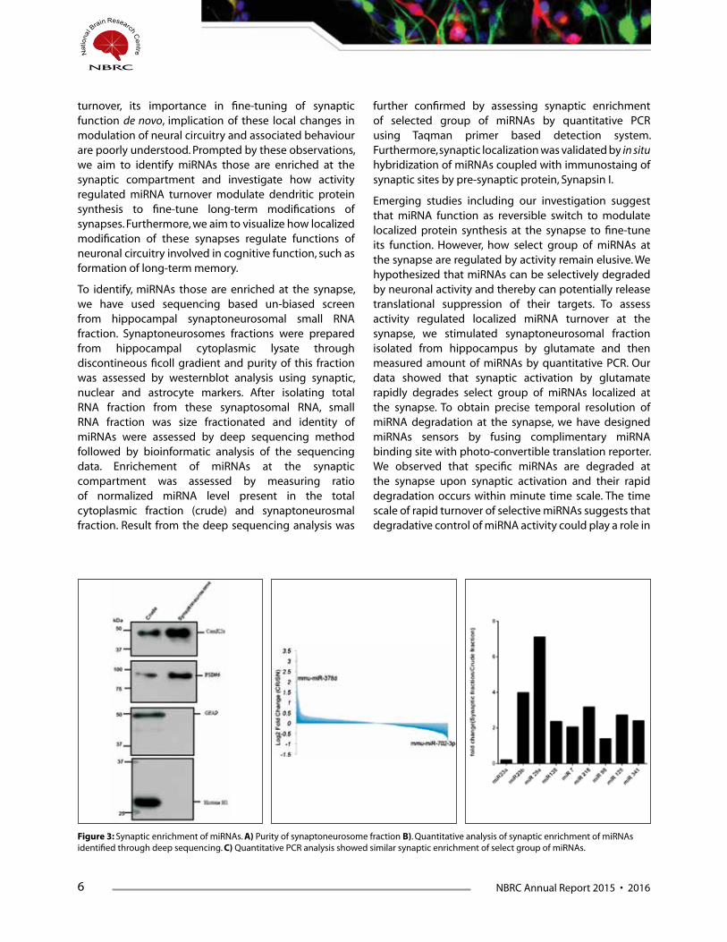
To identify, miRNAs those are enriched at the synapse, we have used sequencing based un-biased screen from hippocampal synaptoneurosomal small RNA fraction. Synaptoneurosomes fractions were prepared from hippocampal cytoplasmic lysate through discontineous ficoll gradient and purity of this fraction was assessed by westernblot analysis using synaptic, nuclear and astrocyte markers. After isolating total RNA fraction from these synaptosomal RNA, small RNA fraction was size fractionated and identity of miRNAs were assessed by deep sequencing method followed by bioinformatic analysis of the sequencing data. Enrichement of miRNAs at the synaptic compartment was assessed by measuring ratio of normalized miRNA level present in the total cytoplasmic fraction (crude) and synaptoneurosmal fraction. Result from the deep sequencing analysis was
further confirmed by assessing synaptic enrichment of selected group of miRNAs by quantitative PCR using Taqman primer based detection system. Furthermore, synaptic localization was validated by in situ hybridization of miRNAs coupled with immunostaing of synaptic sites by pre-synaptic protein, Synapsin I.
Emerging studies including our investigation suggest that miRNA function as reversible switch to modulate localized protein synthesis at the synapse to fine-tune its function. However, how select group of miRNAs at the synapse are regulated by activity remain elusive. We hypothesized that miRNAs can be selectively degraded by neuronal activity and thereby can potentially release translational suppression of their targets. To assess activity regulated localized miRNA turnover at the synapse, we stimulated synaptoneurosomal fraction isolated from hippocampus by glutamate and then measured amount of miRNAs by quantitative PCR. Our data showed that synaptic activation by glutamate rapidly degrades select group of miRNAs localized at the synapse. To obtain precise temporal resolution of miRNA degradation at the synapse, we have designed miRNAs sensors by fusing complimentary miRNA binding site with photo-convertible translation reporter. We observed that specific miRNAs are degraded at the synapse upon synaptic activation and their rapid degradation occurs within minute time scale. The time scale of rapid turnover of selective miRNAs suggests that degradative control of miRNA activity could play a role in
Figure 3: Synaptic enrichment of miRNAs. A) Purity of synaptoneurosome fraction B). Quantitative analysis of synaptic enrichment of miRNAs identified through deep sequencing. C) Quantitative PCR analysis showed similar synaptic enrichment of select group of miRNAs.
NBRC Annual Report 2015 • 2016 7
modulating protein synathesis dependent form of long lasting synaptic plasticity. Furthermore, to investigate mechanistic details of miRNA turnover-mediated control of synaptic plasticity, we have identified factors that can potentially modulate rapid miRNA turnover and subsequently release translational suppression of specific subset of transcripts localized at the synapse. We have identified traget of a specific miRNA that is rapidly degradred at the synapse. Experiments are in
progress to address how miRNA turnover-mediated control of protein synthesis at the synapse can make long lasting modification of the activated synapse and how these localized modification can fine tune neural circuitry to regulate long-term memory formation. Taken together our study will enumerate novel mechanisms of miRNA-mediated protein synthesis dependent form of long lasting plasticity and its implication in long-term memory formation.
Presentations1. Sourav Banerjee. “Dynamic connections: Moleculues and mechanisms of synapse formation and plasticity” IBRO-
APRC school on “Molecular Advancement in Neurobiology.” Banaras Hindu University, Varanasi, September 2015.
2. Sourav Banerjee. “Making connections: Regulatory mechanisms of synapse formation by ubiquitin proteasome system” Invited talk at Konkuk University, South Korea, November 2015.
3. Sourav Banerjee. “Ying and Yang: Functional interplay between constructive and destructive mechanisms to modulate synaptic plasticity. Invited talk at iCeMS, Kyoto University, Japan, January 2016.
FundingRamalingaswami Fellowship, DBT
RNAi grant, DBT
Genome Engineering Grant, DBT
NBRC core fund
CollaboratorDr. Dasaradhi Palakodeti, inSTEM, Bangalore
Dr. Sharba Bandyopadhyay, IIT, Kharagpur
Dr. Nihar Ranjan Jana, NBRC, Manesar.
Dr. Dan Ohtan Wang, iCeMS, Kyoto University, Japan
Prof. Ted Abel, University of Pennsylvania, USA

8 NBRC Annual Report 2015 • 2016
Molecular approaches to understand the pathophysiology and pharmacology of infection and inflammatory disorders of Central Nervous System
Principal Investigator
Anirban Basu
Research Fellow Sourish Ghosh Shalini Swaroop Abhishek Kumar Verma
DST Inspire Fellow Sriparna Mukherjee
Research Associate Dr Bibhabasu Hazra Dr Suvadip Mallick
Project Assistant Noopur Singh
Technical Assistant Kanhaiya Lal Kumawat
Lab attendant Manish Dogra
Immune responses in the CNS are common, despite its perception as a site of immune privilege. These responses can be mediated by resident microglia
and astrocytes, which are innate immune cells without direct counterparts in the periphery. Furthermore, CNS immune reactions often take place in virtual isolation from the innate/adaptive immune interplay that characterize peripheral immunity. However, microglia and astrocytes are also engaged in significant cross-talk with CNS-infiltrating T cells and other components of the innate immune system. On the other hand, a sustained chronic neuroinflammatory response can be detrimental and can initiate neuronal damage, neuronal circuits impairments, astrocytic and microglia involvement and neurodegeneration via long-lasting formation and accumulation of neurotoxic pro-inflammatory mediators.
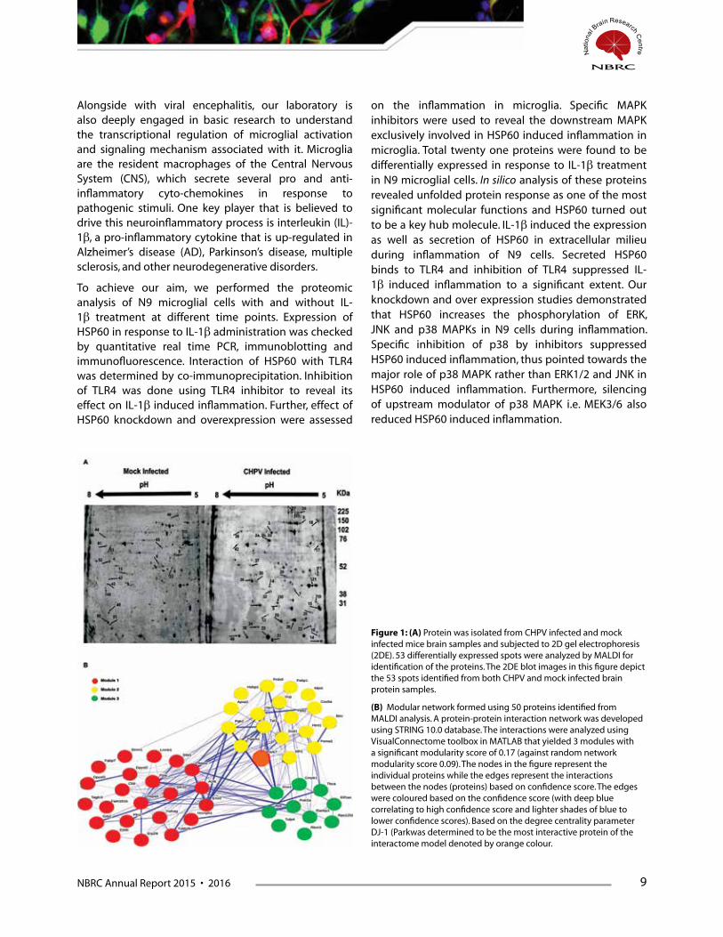
Network analysis through graph theory provides a bottom-up approach to understand host-pathogen interactions. We applied graph theory approach to analyze the interactome of 53 differentially expressed
proteins from proteomic analysis of Chandipura Virus (CHPV, Family: Rhabdoviridae) infected mouse brain tissue. Using the measure of degree centrality, which quantifies the connectedness of single protein within a milieu several other interacting proteins, DJ-1 was selected for further molecular validation (Figure 1). The role of DJ-1 was also monitored in another RNA virus, Japanese Encephalitis Virus (JEV, Family: Flaviviridae) along with CHPV. In the early phase of infection DJ-1 got over-expressed in response to reactive oxygen species (ROS) generation which migrated to mitochondria to remove dysfunctional mitochondria through the process of mitophagy. DJ-1 was also observed to modulate the viral replication and interferon responses along with low-density lipoprotein (LDL) receptor expression in neurons. Collectively these evidences reveal a novel role for DJ-1 in neurotropic virus infection in the brain. Hence our study proposes to investigate the role of DJ-1 in other neurotropic RNA viruses in order to establish as a potential therapeutic target.
NBRC Annual Report 2015 • 2016 9
Alongside with viral encephalitis, our laboratory is also deeply engaged in basic research to understand the transcriptional regulation of microglial activation and signaling mechanism associated with it. Microglia are the resident macrophages of the Central Nervous System (CNS), which secrete several pro and anti-inflammatory cyto-chemokines in response to pathogenic stimuli. One key player that is believed to drive this neuroinflammatory process is interleukin (IL)-1β, a pro-inflammatory cytokine that is up-regulated in Alzheimer’s disease (AD), Parkinson’s disease, multiple sclerosis, and other neurodegenerative disorders.
To achieve our aim, we performed the proteomic analysis of N9 microglial cells with and without IL-1β treatment at different time points. Expression of HSP60 in response to IL-1β administration was checked by quantitative real time PCR, immunoblotting and immunofluorescence. Interaction of HSP60 with TLR4 was determined by co-immunoprecipitation. Inhibition of TLR4 was done using TLR4 inhibitor to reveal its effect on IL-1β induced inflammation. Further, effect of HSP60 knockdown and overexpression were assessed
on the inflammation in microglia. Specific MAPK inhibitors were used to reveal the downstream MAPK exclusively involved in HSP60 induced inflammation in microglia. Total twenty one proteins were found to be differentially expressed in response to IL-1β treatment in N9 microglial cells. In silico analysis of these proteins revealed unfolded protein response as one of the most significant molecular functions and HSP60 turned out to be a key hub molecule. IL-1β induced the expression as well as secretion of HSP60 in extracellular milieu during inflammation of N9 cells. Secreted HSP60 binds to TLR4 and inhibition of TLR4 suppressed IL-1β induced inflammation to a significant extent. Our knockdown and over expression studies demonstrated that HSP60 increases the phosphorylation of ERK, JNK and p38 MAPKs in N9 cells during inflammation. Specific inhibition of p38 by inhibitors suppressed HSP60 induced inflammation, thus pointed towards the major role of p38 MAPK rather than ERK1/2 and JNK in HSP60 induced inflammation. Furthermore, silencing of upstream modulator of p38 MAPK i.e. MEK3/6 also reduced HSP60 induced inflammation.
Figure 1: (A) Protein was isolated from CHPV infected and mock infected mice brain samples and subjected to 2D gel electrophoresis (2DE). 53 differentially expressed spots were analyzed by MALDI for identification of the proteins. The 2DE blot images in this figure depict the 53 spots identified from both CHPV and mock infected brain protein samples.
(B) Modular network formed using 50 proteins identified from MALDI analysis. A protein-protein interaction network was developed using STRING 10.0 database. The interactions were analyzed using VisualConnectome toolbox in MATLAB that yielded 3 modules with a significant modularity score of 0.17 (against random network modularity score 0.09). The nodes in the figure represent the individual proteins while the edges represent the interactions between the nodes (proteins) based on confidence score. The edges were coloured based on the confidence score (with deep blue correlating to high confidence score and lighter shades of blue to lower confidence scores). Based on the degree centrality parameter DJ-1 (Parkwas determined to be the most interactive protein of the interactome model denoted by orange colour.

10 NBRC Annual Report 2015 • 2016
Publications1. R Kumar, A Basu, S Sinha, Das M, Tripathi P, Jain A, Kumar C, Atam V, Khan S, Singh AS (2016) Role of oral Minocycline
in acute encephalitis syndrome in India - a randomized controlled trial. BMC Infect Dis. 2016 Feb 4;16(1):67.
2. A K Verma, S Ghosh, S Pradhan, and A Basu (2016) Microglial activation induces neuronal death in Chandipura virus infection. Scientific Reports; 6:22544.
3. B Kumari, P Jain, S Das, S Ghosal, B Hazra, A C Trivedi, A Basu, J Chakrabarti, S Vrati, A Banerjee. (2016) Dynamic changes in global microRNAome and transcriptome reveal complex miRNA-mRNA regulated host response to Japanese Encephalitis Virus in microglial cells. Scientific Reports. 2016 Feb 3;6:20263.
4. Swaroop S, Sengupta N, Suryawanshi AR, Adlakha YK, Basu A (2016) HSP60 plays a regulatory role in IL-1β-induced microglial inflammation via TLR4-p38 MAPK axis. J Neuroinflammation. 2016 Feb. 2;13(1):27.
5. K L Handore, P D Jadhav, B Hazra, A Basu*, and D S Reddy (2015) Total Syntheses and Biological Evaluation of (±)-Botryosphaeridione, 2 (±)-Pleodendione, 4-epi-Periconianone B, and Analogues. ACS Medicinal Chemistry Letters 6 (11), pp 1117–1121 [* joint corresponding author]
6. N Sengupta, S Mukherjee, P Tripathi, R Kumar, A R Suryawanshi, A Basu (2015) Cerebrospinal Fluid Biomarkers of Japanese Encephalitis. F1000 Research 4:334
7. S Ghosh, S Mukherjee, and A Basu (2015) Chandipura Virus Perturbs Cholesterol Homeostasis Leading to Neuronal Apoptosis. Journal of Neurochemistry 135(2):368-80 (cover photo).
8. S Ghosh, G. Vinodh Kumar, A Basu, and A Banerjee (2015) Graph theoretic network analysis reveals protein pathways underlying cell death following neurotropic viral infection. Scientific Reports 5:14438
9. S Mahanti, A Majhi, K Kundu, A Basu, and B Bishayi (2015) Systemic Staphylococcus aureus infection in restraint stressed mice modulates impaired immune response resulting in improved behavioural activities. Journ of Neuroimmunology 288:102-13.
10. S Vasaikar, S Ghosh,P Narain, A Basu, and J Gomes (2015) HSP70 mediates survival in apoptotic cells – Boolean network prediction and experimental validation. Frontiers in Cellular Neuroscience 9:319
Presentation
1. A Basu (2016) Acute Encephalitis Syndrome in India: the changing scenario. Brain Awareness Week; PUB KAMRUP COLLEGE, Baihata Chariali, Kamrup, Assam, 28th March, 2016.
2. A Basu (2016) Acute Encephalitis Syndrome in India: the changing scenario and the newer challenges. Sun Pharma Advanced Research Center (SPARC), Vadodara. 17th March, 2016.
3. A Basu (2016) Microglia: the movers and shakers of the brain. Brain Awareness Week, Presidency University, Kolkata, 1st March 2016.
4. A Basu (2016) Search for novel Anti virals from natural resources. CIRMM; West Bengal State University, Barasat; 25th-26th February, 2016.
5. A Basu (2015) Inflammation as a therapeutic target in Viral Encephalitis. School of Cognitive Sciences; Jadavpur University, 23rd December, 2015
6. A Basu (2015) Deciphering the molecular mechanism underlying IL-1β induced inflammation in microglia; APPICON 2015; 26th-28th Nov, 2015. AIIMS Jodhpur
7. A Basu (2015) Brain’s Innate Immune Response, as seen by neurotropic virus. IMMUNOCON-2015, 9th-11th October, 2015; Patna.
8. A Basu (2015) Molecular and biochemical mechanism of Neuronal death following Chandipura Virus infection; Symposium on Immunology and Cell Biology; CSIR-IICB, Kolkata; 28th September, 2015.
NBRC Annual Report 2015 • 2016 11
9. A Basu (2015) Deciphering Mechanism of Neuronal Death In Neurotropic Virus Infection: From Molecules to Network. IIT Delhi-NBRC conclave, 21st May, 2015.
10. A Basu (2015) Host pathogen interaction in Japanese Encephalitis: from bench to bedside. Kurukshetra University, 23rd April, 2015.
11. A Basu (2015) Molecular and Biochemical mechanism of Neuronal death following Chandipura virus infection. 4th Molecular Virology Meeting, RGCB, Thiruvananthapuram, 16-17th April 2015.
FundingmicroRNAs as a potential therapeutic target in Neuro-tropic Viral infection [Tata Innovation Fellowship from the Department of Biotechnology (BT/HRD/35/01/02/2014)]
Identification and characterization of brain cellular membrane components acting as receptors for Japanese Encephalitis virus. [CSIR, 27(0307)/14/EMR-II]
To study the molecular mechanism of microglial activation and identify the therapeutics targets critical for IL-1β signaling in brain following inflammation. [Department of Science and Technology, No SB/SO/HS-070/2013]
Implementing proteomic approach to understand the etiology of Neuropathogenesis induces Chandipura Virus infection [Department of Biotechnology (BT/PR7907/MED/29/702/2013)]
AwardSenior Scientist Oration Award (Indian Immunology Society); Immunocon- 2015, Patna.
CollaborationsArpan Banerjee and Pankaj Seth, NBRC
Rashmi Kumar, Dept of Pediatrics, CSM Medical University, Lucknow
SK Shankar, and Anita Mahadevan, NIMHANS, Bangalore
James Gomes, School of Biological Sciences, IITD
Sunit Singh, BHU, Varanasi.
Dhrubajyoti Chattopadyay, BC Guha Center for Genetic Engineering and Biotechnology, University of Calcutta, Kolkata
Amol Suryawanshi, Institute of Life Sciences, Bhubneswar.
Sudhanshu Vrati, and Arup Banerjee, Vaccine and Infection Disease Research Center, THSTI, Faridabad

12 NBRC Annual Report 2015 • 2016
Utilization of TgAPPswe PS1dE9 neurosphere model of Alzheimer’s disease for drug discovery in lowering intracellular amyloid beta load
Principal Investigator:
Ranjit Kumar Giri
Lab. Attendant: Lalit Bidla
Accumulation of beta amyloid peptides is the hallmark event in the pathoprogression of Alzheimer’s disease (AD). Beta amyloid peptides
undergo post-translational modification to form beta-sheet structure which favors its oligomerization and subsequent fibrillization. In India, more than a million people are suffering from this disease and the number will increase dramatically in coming decade as per the WHO projection. No therapy is available to combat this disease. A variety of compounds have been proposed as potential therapeutics for the treatment of Alzheimer’s diseases. However, none of these compounds are effective in halting the disease so far. Therefore, novel molecules those could reduce beta amyloid load either by inhibiting beta amyloid synthesis or enhancing beta amyloid degradation need to be studied. Molecules with anti-amyloid beta oligomerization may be of great importance. In past, with the funding from BIRAC and in collaboration with Prof. Rani Gupta and LeadInvent, Delhi University, New Delhi, India, P1 peptide was found to reduce intracellular amyloid beta load utilizing conformation dependent immunocytochemistry (for technique please
read Ghate et al., Springer Plus, 3:161, 2014).
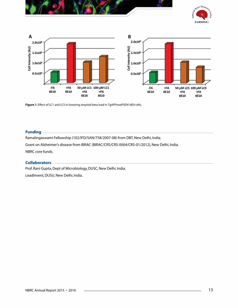
Currently, based on P1 peptides and amyloid beta oligomers ducking studies small chemical conformers were designed and tested using chemical simulation. From these studies, five molecules were tested for anti-oligomerization of amyloid beta peptide in vitro using thioflavin-s binding assay. Two molecules with higher efficacy were tested for intracellular amyloid beta lowering capacity. These two molecules are named as LC1 and LC5 (details can’t be revealed as per patent policy). LC1 reduced intracellular amyloid beta load by 65% at 50 mM and by 46% at 100 mM concentration LC5 (Figure 1A). Such loss of dose-dependent inhibition of LC1 could be because of its fluorescence property, which needs further testing. Moreover, LC5 reduced amyloid beta load by 65% at 50 mM and 82% at 100 mM concentration suggesting LC5 inhibited intracellular amyloid beta aggregation/load in a dose dependent manner (Figure 1B). Therefore, LC5 can be further optimized for the development of a novel anti-amyloid beta aggregating/oligomerizing compound in the treatment of Alzheimer’s disease.
NBRC Annual Report 2015 • 2016 13
Figure 1. Effect of LC1 and LC5 in lowering amyloid beta load in TgAPPswePSEN1dE9 cells.
FundingRamalingaswami Fellowship (102/IFD/SAN/758/2007-08) from DBT, New Delhi, India.
Grant on Alzheimer’s disease from BIRAC (BIRAC/CRS/CRS-0004/CRS-01/2012), New Delhi, India.
NBRC core funds.
CollaboratorsProf. Rani Gupta, Dept of Microbiology, DUSC, New Delhi, India.
LeadInvent, DUSU, New Delhi, India.

14 NBRC Annual Report 2015 • 2016
Curcumin-mediated induction of proapoptotic Bex genes is associated with apoptosis in mouse neuro 2a neuroblastoma cells and involves activation of p53
Principle Investigator:
Ranjit Kumar Giri
Research Fellow: Himakshi Sidhar
Lab Attendant: Lalit Bidla
Brain expressed X-linked (Bex) genes are newer group of tumor suppressor genes. The Bex genes belong to a small family of genes
including Bex1, Bex2, Bex3, Bex4 and Bex6 in mouse while Bex5 instead of Bex6 in humans. All these genes are located on X-chromosome except Bex6, which is located on chromosome 16 in the mouse genome. Bex1 and Bex2 genes are silenced in malignant glioblastoma and act as tumor suppressor genes in various cancers. Re-expression of Bex1 and Bex2 genes by gene transfection reduces glioblastoma cells growth and sensitizes the glioma cells to anti-cancer drugs. Similarly, overexpression of Bex3 inhibited breast cancer xenografts in mouse models. The role of Bex4 and Bex6 in cancer formation and treatment is not known. Furthermore, there is no report on any chemical that can induce all the endogenous Bex gene/s to harness its anti-cancer properties. Effect of Bex gene/s on neuroblastoma cells is also not known. In the present study, we investigated the induction of all endogenous Bex genes to proapoptotic effects of curcumin using a murine neuroblastoma
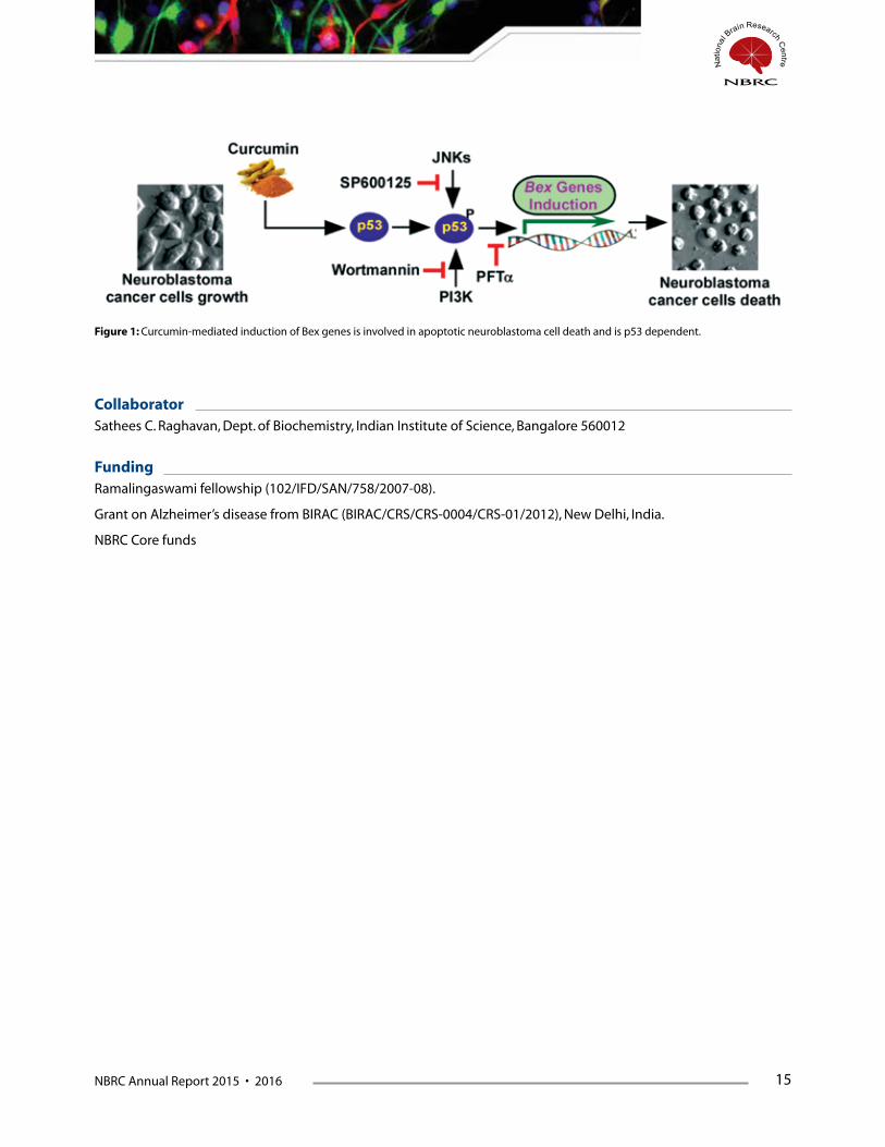
neuro 2a cell line. Cell toxicity assays such as MTT, LIVE-DEAD assay, DNA fragmentation and TUNEL assay were performed to study curcumin-mediated cell death and apoptosis. Semi-quantitative RT-PCR, western blotting, immunofluorocytochemistry were used to study the induction and regulation of Bex genes. Pharmacological inhibitors were used to study cell signaling associated with curcumin-mediated induction of Bex genes. Our results show curcumin induced Bex genes in dose and time dependent manner prior to apoptosis in neuro 2a neuroblastoma cells. Curcumin treatment also activated p53 through hyperphosphorylation at serine 15 prior to Bex genes induction. Pifithrin-, an inhibitor of p53, abrogated curcumin-mediated induction of Bex genes and apoptosis indicating the involvement of p53 in induction of Bex genes and a direct role of Bex genes in apoptotic neuro 2a neuroblastoma cells death. This study highlights curcumin as a specific inducer of all known Bex genes in apoptotic neuroblastoma cells death paving a new way to treat neuroblastoma and other cancers with silenced Bex genes (manuscript in Preparation).
NBRC Annual Report 2015 • 2016 15
Figure 1: Curcumin-mediated induction of Bex genes is involved in apoptotic neuroblastoma cell death and is p53 dependent.
CollaboratorSathees C. Raghavan, Dept. of Biochemistry, Indian Institute of Science, Bangalore 560012
FundingRamalingaswami fellowship (102/IFD/SAN/758/2007-08).
Grant on Alzheimer’s disease from BIRAC (BIRAC/CRS/CRS-0004/CRS-01/2012), New Delhi, India.
NBRC Core funds
16 NBRC Annual Report 2015 • 2016
Understanding the physiological function of Ube3a and pathogenesis of Angelman syndrome
Principal Investigator:
Nihar Ranjan Jana
Research Associate: Vivek Tripathi
Research fellows: Imran Jamal, Brijesh Kumar Singh, Naman Vatsa, Shashi Sekhar, Vipendra Kumar and Shruti Nagaral
Technical assistants: Ankit Sharma Mahendra Singh
Angelman syndrome (AS) is a neurodevelopmental disorder characterized by severe developmental delay, lack of speech, cognitive and motor
deficits and epileptic seizures along with multiple other associated features particularly excessive laughter and sleep disturbances. Genetic studies have revealed that the AS is caused by the loss of function of the maternally inherited UBE3A allele. Because the paternally inherited UBE3A is epigenetically silenced in the neuronal tissues through cell type specific imprinting, the defect in maternally inherited UBE3A results its loss of function in the brain. The UBE3A gene encodes a 100kDa protein that has been characterised as an E3 ubiquitin ligase (involved in targeting proteins for ubiquitination) and transcriptional co-activator for steroid hormone receptors. Therefore, it is hypothesized that loss of ubiquitin ligase activity or co-activator function of Ube3a might be linked with the AS phenotypes.
To gain deeper insight into the AS patho-mechanism, several mice models has been generated by disrupting the maternally inherited Ube3a. Mouse model generated by Jiang et al reproduced many characteristic features of AS and are widely used. These mice not only exhibits classical cognitive and motor deficits, but also displays audiogenic seizure, anxiety-like behaviour, disturbances in circadian clock and sleep homeostasis. Moreover these AS mice also become obese. In depth study in this mouse
model further demonstrates defect in hippocampal calcium/calmodulin dependent protein kinase-II and long-term potentiation, experience-dependent synaptic plasticity and imbalance of excitatory/inhibitory circuitry. These results strongly indicates that Ube3a plays a crucial role in regulating synaptic function.
Past several years we are involved in exploring the physiological function of Ube3a and how its loss of function is associated with AS using mouse model of AS. In addition to identify the novel interacting partner of Ube3a, we are also trying to identify novel miRNA that regulates Ube3a as well as miRNA that are directly linked with AS pathogenesis. We are also exploring the defects in signalling pathways that contributes behavioural deficits in AS mouse model and how those behavioural abnormalities can be reversed.
Last year we have shown that rearing AS mice in enriched environment significantly improved their cognitive, motor and anxiety-like behaviour. Interestingly, enriched environment also significantly increased the number of parvalbumin-positive GABAergic neurons in the hippocampus and basolateral amygdala of AS mice. This year we report that the absence of Ube3a in AS mice brain leads to aberrant increase in HDAC1/2 along with decreased acetylation of histone H3/H4 (Figure 1). Partial knockdown of Ube3a in cultured neuronal cells

NBRC Annual Report 2015 • 2016 17
also results in significant up-regulation of HDAC1/2 and decreased levels of histone H3/H4 acetylation indicating direct role Ube3a in regulating HDAC1/2 activity. Histone acetylation is a dynamic process that is tightly regulated by the antagonistic function of histone acetyl transferase (HAT) and HDAC (histone deacetylase). Balance between the activities of these two groups of enzymes is crucial in regulating the gene expression and directs several physiological functions. Imbalance of their activities are linked with various disease states. HDAC2 has been shown to negatively regulate synaptic plasticity and memory formation. It recruits to the promoters of several neuronal activity, synaptic plasticity and memory related genes and regulates their expression. Aberrant high levels of HDAC2 as well as HDAC1 also have been reported in the brain of various neuro-psychiatric disorders or their model systems having cognitive deficits. These reports strongly suggests that the aberrant increased level of HDAC1/2 observed in AS mice brain might be directly linked with synaptic and cognitive dysfunctions in these mice. Decreased expression of some of the HDAC2 regulatory genes like BDNF and synaptophysin in the hippocampus of AS mice observed by us further supports our conclusion.
Aberrantly increased HDAC1/2 activity in the AS mice brain led us to test the effect of HDAC inhibitor sodium valproate (Class I HDAC inhibitor) on behavioural outcome in these mice. Interestingly, we have found that prolonged treatment of sodium valproate significantly improved various behavioural abnormalities (deficits in social interaction, cognitive and motor performances) in AS mice. Valproate treatment also restores increased HDAC1/2 levels and hypo-acetylation of histones H3 and H4 (Figure 1). Another interesting aspect of our study is that sodium valproate increased the expression of Ube3a in the brain of wild type but not in AS mice indicating HDAC might not be involved in regulating the expression UBE3A-ATS. Therefore, valproate-dependent behavioural improvement in AS mice is not linked with the activation of paternally silenced Ube3a. Altogether, our study concludes that the aberrant increase in HDAC1/2 activity in the brain of AS mice might be linked with synaptic dysfunction and associated behavioural anomalies in these mice. Our finding also suggests that HDAC inhibitors could be promising drugs to treat AS.
Role of Ube3a in the progression of neurodegenerative disorders using mice modelsOne of the shared pathological hallmark of most age-related neurodegenerative disorders comprising Huntington’s disease (HD) and Alzheimer’s disease (AD) is the accumulation of mutant disease proteins as inclusion bodies. Appearance of aggregates of the mutant disease proteins advocate that the cell is unable to clear them, and failure of elimination leads to the defect in cellular protein quality control system. We have shown earlier that Ube3a is involved in cellular protein quality control and enhance the clearance in mutant huntingtin (that causes HD). Furthermore, deficiency of Ube3a accelerates disease progression in HD mouse model (Maheshwari et al. Hum. Mol. Genet. 2014).
Figure 1. Representative immunohistochemical staining of acetylated histones H3(K9) and H4(K12) in the hippocampal dentate gyrus region of wild type (m+/p+) and AS (m-/p+) mice received saline or sodium valproate (VPA) treatment. Mice were intraperitoneally injected VPA (300mg/kg body weight daily, 100 µl) for 60 days. Control group received same volume of saline.

18 NBRC Annual Report 2015 • 2016
Because the paternally inherited Ube3a is epigenetically silenced in neuronal tissues which can be reactivated topoisomerase 1 inhibitors, we tested the effect of topoisomerase 1 inhibitor, topotecan on the progression of HD using mouse model. Topotecan is a semisynthetic water soluble derivative of plant alkaloid camptothecin that can cross the blood brain barrier and long been implicated for cancer chemotherapy. We have found that that the treatment of topotecan to HD transgenic mice considerably improved their motor behavioural abnormalities along with significant
extension of lifespan (Figure 2). Improvement of behavioural deficits are accompanied with the significant rescue of their progressively decreased body weight, brain weight and striatal volume. Interestingly, topotecan treatment also significantly reduced insoluble mutant huntingtin load in the HD mice brain. Finally, we show that topotecan treatment to HD mice not only inhibits the expression of transgenic mutant huntingtin, but also at the same time induces the expression of Ube3a, an ubiquitin ligase linked with the clearance of mutant huntingtin.
Figure 2. Administration of topotecan through tail vein route rescued decreased body weight and increased survival rate in HD mice. HD and wild type mice were injected topotecan (0.7 mg/kg, 50µl) or saline through tail vein at their age of 9 weeks. Every mouse was given 9 doses (3 consecutive doses/week).
Topotecan by the virtue of topoisomerase 1 inhibition prevents DNA unwinding, a crucial event that is required for DNA replication and transcription. Therefore, one could expect severe detrimental effect of this drug not only on rapidly dividing cells like cancer cells, but also on normal differentiated cells like neurons. However, this drug under specific treatment regime is well tolerated by cancer patients. In our experimental paradigm, we have not seen any lethality or gross visible side effects of this drug when treated to wild type and HD mice. The dose of topotecan that we used in our animal experiment was nearly equivalent to the dose recommended for treating cancer patients. Interestingly, topotecan-mediated down-regulation of huntingtin as well as up-regulation of Ube3a are two crucial event that are extremely beneficial in the context of HD. Same drug could not only inhibits the expression but also enhance the clearance of mutant huntingtin leading to decrease in aggregate burden. The potential side effect of other abnormally regulated genes could be minimized by optimizing the dose and duration of the drug. Since many of the negatively regulated genes are linked with synaptic function, their effect could be counter balanced by increased expression of Ube3a or other imprinted genes performing similar function. Altogether, our study provide strong evidence that topotecan could be a potential therapeautic molecule to delay the progression of HD.
NBRC Annual Report 2015 • 2016 19
Publications1. SK Godavarthi, P Dey, A. Sharma, NR Jana. Impaired adult hippocampal neurogenesis and its partial reversal by
chronic treatment of fluoxetine in a mouse model of Angelman syndrome. Biochemical and Biophysical Research Communications. 464 (4), 1196-1201, 2015.
2. *J Chakraborty, U Rajamma, NR Jana, KP Mohanakumar. Quercetin improves the activity of the ubiquitin‐proteasomal system in 150Q mutated huntingtin-expressing cells but exerts detrimental effects on neuronal survivability. Journal of Neuroscience Research. 93 (10), 1581-1591, 2015.
3. E Das, NR Jana, N. P. Bhattacharyya. Delayed Cell Cycle Progression in STHdhQ111/HdhQ111 Cells, a Cell Model for Huntington’s Disease Mediated by microRNA-19a, microRNA-146a and microRNA-432 MicroRNA 4 (2), 86-100, 2015.
*Last year in press
Presentations1. N. R. Jana. Lack of ubiquitin ligase Ube3a in the brain accelerates disease progression in a mouse model of
Huntington’s disease. International symposium on Molecular Signaling, NEHU, Shilong, November, 2015
2. N. R. Jana. Defective protein quality control in Huntington’s disease. Centre for Brain Research, IISc, Bangalore, November, 2015.
3. N. R. Jana. Neurodegenerative disorders involving protein misfolding and aggregation. Invited talk at West Bengal State University, March, 2016
4. N. R. Jana. Neurodegenerative disorders involving protein aggregation. IBRO (International Brain Research Organization) School, NBRC, Manesar, March, 2016.
5. N. Vatsa and N. R. Jana. Understanding the role of microRNA in Angelman Syndrome pathogenesis using mouse model. Annual meeting of Indian Academy of Neurosciences, Chandigarh, November, 2015
6. I. Jamal, V. Kumar, N. Vatsa, B. Singh, S. Sekhar, A. Sharma and N. R. Jana. Enriched environment partially improves behavioural deficits in a mouse model of Angelman syndrome. Annual meeting of Indian Academy of Neurosciences, Chandigarh, November, 2015
7. B. K. Singh and N. R. Jana. Deficiency of Ube3a worsens behavior and cognition in APPswe/PS1dE9 transgenic mouse model of Alzheimer’s Disease. Annual meeting of Indian Academy of Neurosciences, Chandigarh, November, 2015
FundingDeregulation of micro RNA in cell and animal models of Huntington’s disease: role of altered micro RNA in neuronal differentiation and cell cycle regulation. A joint project with Biomedical Genomics Centre and SNIP, Kolkata. Department of Biotechnology. Govt. of India. Grant No: BT/PR7185/MED/30/910/2012.
Ube3a as a therapeutic target of Huntington’s disease. TATA Innovation project, Department of Biotechnology, Govt. of India. Grant No: BT/HRD/35/01/03/2013
CollaboratorsDr. Nikhil Jana, Indian Association for the Cultivation of Science, Kolkata.
Dr. Subramanian Ganesh, IIT Kanpur.
Dr. Nitai Bhattacharya, Biomedical Genomics Centre, Kolkata.
Dr. Ranjit Giri, NBRC.
20 NBRC Annual Report 2015 • 2016
Development and repair of neural circuit in C. elegans
Principal Investigator:
Anindya Ghosh Roy
Research Fellows: Dharmendra Puri Atrayee Basu Harjot Kaur
Post-doctoral Fellow: Nilanjana Das Saha
Project Assistant: Pankajam Thyagarajan Titash Mukherjee Mekhala Chitagudigi
Technical Assistant: Sumit Mahapatra Yunis Khan
Our lab is interested in understanding how nervous system develops and after injury how it repairs. Towards this goal we use a combination
of genetics, biochemistry and imaging in Caenorhabditis elegans. To address these questions we focused our attention to the regulation of microtubule cytoskeleton in neuron.
Cell biological mechanisms regulating neuronal polarity and maintainenaceMicrotubule (MT) cytoskeleton is the basis of the polarized structure of neuron. We found that loss of the kinesin-13 family depolymerizing factor KLP-7 stabilizes microtubules and causes multi-polar neuron formation (Figure1). To find out novel regulators of microtubule cytoskeleton in neuron, we have screened and identified mutants those suppress the neuronal phenotype of klp-7 mutant (Figure1). None of the known microtubule stabilizing factors involving plus or minus end binding proteins, and centrosomal proteins suppressed klp-7(0). However, the drug Colchicine that destabilizes MTs suppressed the same. This indicated that our genetic screening might identify novel regulators neuronal cytoskeleton. By combining meiotic recombination and
whole genome sequencing, we have mapped three suppressors in three individual genes. These genes encode metalloprotease, immunoglobulin like molecule and RNA binding protein. Loss of the RNA binding protein causes a strong phenotype in axon growth and its overexpression causes overgrowth of axon indicating that it is necessary and sufficient for axon deveopment. We are investigating the molecular mechanism by which these genes regulate microtubule cytoskeleton and thereby influence nerve cell development.
To understand how microtubules are maintained after axon development, we are studying posttranslational modification of tubulin involving. We found that simultaneous loss of two tubulin carboxypeptidases suppresses the axon overgrowth phenotype caused due to lack of E3 family ligase rpm-1. This indicated that there is a link between neuronal homeostatic signalling and post-translation modification of tubulin. We are investigating the mechanistic link between these two pathways.
Neuronal RegenerationWe are interested in understanding how a given
NBRC Annual Report 2015 • 2016 21
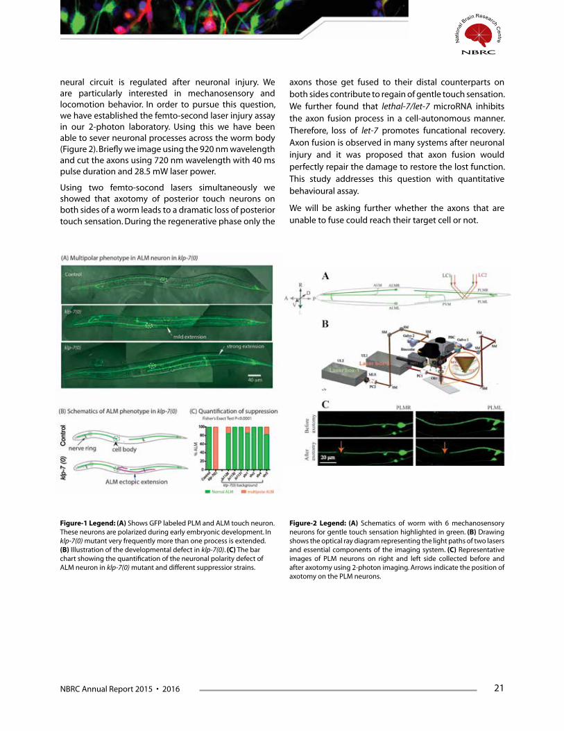
neural circuit is regulated after neuronal injury. We are particularly interested in mechanosensory and locomotion behavior. In order to pursue this question, we have established the femto-second laser injury assay in our 2-photon laboratory. Using this we have been able to sever neuronal processes across the worm body (Figure 2). Briefly we image using the 920 nm wavelength and cut the axons using 720 nm wavelength with 40 ms pulse duration and 28.5 mW laser power.
Using two femto-socond lasers simultaneously we showed that axotomy of posterior touch neurons on both sides of a worm leads to a dramatic loss of posterior touch sensation. During the regenerative phase only the
axons those get fused to their distal counterparts on both sides contribute to regain of gentle touch sensation. We further found that lethal-7/let-7 microRNA inhibits the axon fusion process in a cell-autonomous manner. Therefore, loss of let-7 promotes funcational recovery. Axon fusion is observed in many systems after neuronal injury and it was proposed that axon fusion would perfectly repair the damage to restore the lost function. This study addresses this question with quantitative behavioural assay.
We will be asking further whether the axons that are unable to fuse could reach their target cell or not.
Figure-1 Legend: (A) Shows GFP labeled PLM and ALM touch neuron. These neurons are polarized during early embryonic development. In klp-7(0) mutant very frequently more than one process is extended. (B) Illustration of the developmental defect in klp-7(0). (C) The bar chart showing the quantification of the neuronal polarity defect of ALM neuron in klp-7(0) mutant and different suppressior strains.
Figure-2 Legend: (A) Schematics of worm with 6 mechanosensory neurons for gentle touch sensation highlighted in green. (B) Drawing shows the optical ray diagram representing the light paths of two lasers and essential components of the imaging system. (C) Representative images of PLM neurons on right and left side collected before and after axotomy using 2-photon imaging. Arrows indicate the position of axotomy on the PLM neurons.

22 NBRC Annual Report 2015 • 2016
Presentations1. Anindya Ghosh Roy: “Functional restoration after neutronal injury” in 1st Indian Caenorhabditis elegans. Meeting:
40th Mahabaleshwar seminar. February, 2016
2. Atrayee Basu: “Restoration of functional connectivity after neutronal injury” in C. elegans Topic Meeting: Neuronal Development, Synaptic Function & Behavior at the Nagoya University Japan during July 2016
FundingWellcome Trust-DBT and NBRC Core
CollaboratorSandhya Koushika, TIFR, Mumbai, India
Shalini Gupta, IIT-Delhi, India
Sourav Banerjee, NBRC, India
AwardWellcome Trust-DBT Intermediate fellowship-2013-2018
Ramalingaswami Fellowship-2013 (declined)
NBRC Annual Report 2015 • 2016 23
Metabolic reprogramming in Glioblastoma: Implications in survival and resistance to chemotherapeutics
Principal Investigator
Ellora Sen
Research Fellows: Deobrat Dixit, Sadashib Ghosh, Ruchi Ghildiyal, Piyushi Gupta, Fahim Ahmad, Sk. Touseef Ahmad, Pruthvi Gowda
Post doctoral fellow: Dr. Arpita Chatterjee Dr. Pinaki Mondal Dr. Ankita Singh
Technical Assistant: Shanker Datt Joshi
Lab attendant: Rajesh Kumar Kumawat
Background and significanceDysregulated metabolism characterized by the Warbug effect is an integral component of tumor evolution. Glioblastoma multiforme (GBM) - the most malignant of brain cancers characterized by aberrant metabolic profile, is largely refractory to current therapeutic regimens. As metabolic reprogramming deregulates a number of cellular functions, and since targeting metabolic remodelers is regarded as a potential anti-cancer strategy; the focus of our study is to dissect molecular circuitries that regulate expression of metabolic modelers to subsequently affect genes associated with resistance to apoptosis, cellular bioenergetics and immune evasive responses in GBM.
(i) By switching their glucose metabolism toward aerobic glycolysis, cancer cells accumulate glycolytic intermediates that are used as building blocks for macromolecular synthesis. Casein kinase (CK2) is known to regulate the enzymatic activity of key glycolytic enzyme phosphoglucose isomerase. As we have previously demonstrated the involvement of CK2 in
conferring resistance to apoptosis in glioma cells, we investigated the association of CK2 with deregulated metabolism in cancer cells. Inhibition of CK2 increased expression of metabolic modeler AMP-activated protein kinase (AMPK) and Pyruvate dehydrogenase Kinase 4 (PDK4) in glioma cells. Our findings suggest that CK2 affects PDK4-AMPK axis to sustain the elevated energy demands in glioma cells and thus regulate cell proliferation. Through AMPK activation, PDK4 inhibits glucose uptake and maintains glioma cells in a chronic energy deprived state that triggers apoptosis upon CK2 inhibition.
(ii) Given the link between metabolism and responsiveness to chemotherapeutics, we investigated the role of cell death-inducing DNA fragmentation factor-a-like effector-A (CIDEA) in conferring resistance to apoptosis in glioma cells. As low expression of CIDEA in GBM tumors is concomitant with elevated levels of PPARg, the (i) role of PPARg in maintaining the low basal expression of CIDEA in glioma, and (ii) effect of CIDEA over-expression on glioma cell survival was investigated. Our findings suggest that inhibition

24 NBRC Annual Report 2015 • 2016
of PPARg enhances CIDEA expression, and ectopic expression of CIDEA elevated PPARg levels. Also, CIDEA over-expression not only triggered apoptosis, but also induced actin cytoskeletal disruption, cell cycle arrest, and release of pro-inflammatory cytokine IL-6. This study has highlighted the clinical relevance of elevated PPARg levels in regulating expression of pro-apoptotic CIDEA in glioma.
(iii) Given the well known ability of the immune-stimulatory cytokine IFNg to exhibit anti-tumorigenic effects, its ability to effect glioma cell survival was investigated. Though IFNg had no effect on glioma cell viability, it induced cell cycle arrest and de-differentiation. This was concomitant with increased expression of retinoic acid inducible gene (RIG-I) and histone methyltransferase (HMT) G9a and PRMT1. IFNg induced metabolic modeller PPAR gamma coactivator 1alpha (PGC-1a) positively regulated RIG-I; with PRMT-1 and G9a affecting PGC-1a in a counter-regulatory manner. The concerted action of HMTs affects PGC-1a driven RIG-I crucial for maintaining glioma cells in a de-differentiated state. Also, IFNg induced CK2 regulated RIG-I affects a complex network that compromises the glioma cells’ proliferative potential by affecting redox homeostasis, metabolic adaptations and cell cycle. This is achieved by RIG-I through ROS generation and
dampening glycolysis and pentose phosphate pathway. Our finding suggest, that by positioning itself at the hub, RIG-I tethers metabolic and redox homeostasis with cell survival responses in glioma cells.
(iv) Besides playing a pivotal role in the induction and maintenance of adaptive immune responses, MHC I also serve as an important component in cancer immuno-surveillance. As mobility of MHC I-peptide complexes regulates the sensitivity of antigen recognition, understanding mechanisms regulating its clustering is crucial for understanding immune escape mechanisms in tumors. Using fluorescence recovery after photobleaching (FRAP), we demonstrated the indispensability of hypoxia inducible factor (HIF-1a in regulating TNFa-induced increase in MHC-I membrane clusters. Importantly, this study provides evidence incriminating HIF-1a driven metabolic enzyme hexokinase (HK2) dependent actin dynamics in the regulation of stable MHC-I cluster formation in glioma. This further proved the locational specificity of the MHC-I-actin interaction in the cortical membrane domains.
Taken together, this study offers the prospect for future anti-glioma therapies aimed at targeting aberrant metabolism associated with resistance to apoptosis and immune evasive responses.
Publications1. Dixit D, Ahmad F, Ghildiyal R, Joshi SD, Sen E. CK2 inhibition induced PDK4-AMPK axis regulates metabolic
adaptation and survival responses in glioma Exp Cell Res. 2016 Mar 18. pii: S0014-4827(16)30057-X.
2. Ghildiyal R, Sen E. Concerted action of histone methyltransferases G9a and PRMT-1 regulates PGC-1a-RIG-I axis in IFNg treated glioma cells Cytokine. 2015 Dec 22. pii: S1043-4666(15)30121-6.
3. Ghildiyal R, Sen E. CK2 induced RIG-I drives metabolic adaptations in IFNg-treated glioma cells Cytokine. 2015 Nov 26. pii: S1043-4666(15)30080-6.
4. Ghosh S, Gupta P, Sen E. TNFa driven HIF-1a-Hexokinase II axis regulates MHC-I cluster stability through actin cytoskeleton Exp Cell Res. 2016 Jan 1;340(1):116-24.
5. Chatterjee A, Mondal P, Ghosh G, Mehta VS, Sen E. PPARg regulated CIDEA effects pro-apoptotic responses in glioblastoma. Cell Death and Discovery 2015
Presentations1. Ellora Sen. Decoding Signaling Networks in Cancer: Lessons learnt Maulaza Azad College, Kolkata, April 13th 2015
2. Ellora Sen. Science meets philosophy: A Magical transformation. Department of Biochemistry, Shivaji College, University of Delhi, 9th November, 2015
3. Ellora Sen. Inflammation to tumor progression: Retracing the journey. APPICON 26th November, 2015, AIIMS Jodhpur

NBRC Annual Report 2015 • 2016 25
4. Ellora Sen. Evolution of a cancer cell: Role of tumor microenvironment. Ram Mohan College, Kolkata, December 23rd 2015
5. Brain and its common disorders , Brain Awareness Week, Pub Kamrup College, Guwahati, 26th March, 2016
6. Ellora Sen. Choosing Science as a Career. Department of Biochemistry, Deshbandhu College, Biospark, 30th March 2016
7. Ahmad Fahim and Sen Ellora participated and presented a poster in 4th AACR International Conference on Frontiers in Basic Cancer Research, 23-26th October 2015 Pennsylvania convention centre, Philadelphia, USA. “Telomerase Inhibition Impedes Metabolism in Glioblastoma”.
FundingRole of chromatin remodelers in regulating genes associated with resistance to apoptosis under inflammatory and hypoxic conditions in glioma cells.DBT (BT/PR5818/MED/30/839/2012)
Inflammation regulated metabolic reprogramming: Implications in tumor progression’. Unit of excellence in cancer biology DBT. (#BT/MED/30/SP11016/2015).
Understanding inflammation driven regulation of macrophage function: Implications in glioblastoma progression. DBT. National Bioscience Award for Career Development, 2013
CollaboratorDr. VS Mehta, Paras Hospital

26 NBRC Annual Report 2015 • 2016
Cellular and Molecular Mechanisms of HIV-1 Neuropathogenesis
Principal Investigator:
Pankaj Seth
Research Fellows: Manju Tewari, Mahar Fatima, Chitra Singal, Hriday Pandey, Reshma Bhagat and Priyanka Singh
Project Assistants: Banshi Nath, Rina Kumari and Kanza Saleem
Technical Assistant: Durgalal Meena and Naushad Alam
Human immunodeficiency virus (HIV-1) traffics across blood brain barrier into the central nervous system (CNS). As evident from autopsy
studies in HIV/AIDS patients basal ganglion, subcortical region and frontal cortex are the sites of virus localization. As these areas are important for executive functions, memory storage and recall, the presence of HIV-1 in brain culminates in impairment of neurocognitive and motor functions in HIV/AIDS patients. Such neurological impairments are referred to as HIV-1 associated neurological disorders (HAND) and are studied under a relatively new field of brain disorders, called neuroAIDS. The neurocognitive deficits are attributed to irreversible damage of neurons in the affected areas. Most of the neuronal damage is due to viral proteins that are secreted out from infected cells in the vicinity, particularly glial cells. Among several viral proteins produced by HIV-1, Transactivator of Transcription (Tat) and glycoprotein-120 (gp-120) have been investigated in great details, as they are highly neurotoxic. Our laboratory has maintained its research focus on HIV-1 Tat protein, as Tat is reported to be produced from actively or latently infected brain cells of HIV-1 patients, including those who are on anti-HIV therapy. In previous years, we had reported novel insights into the mechanism of glia mediated neuronal damage. Using molecular and cell biology approaches, we had
pin-pointed a purinergic receptor, P2X7 to be the critical entity for HIV-1 Tat mediated neuronal damage. We are currently studying cellular and molecular mechanisms to gain novel insights into the ATP release from HIV-1 Tat treated or transfected astrocytes. We utilize a well characterized human fetal brain derived neural stem cell culture system established at National Brain Research Centre.
In addition to this, in another ongoing project, we have obtained interesting insights into how HIV-1 protein Tat affects the properties of human neural precursor cells (hNPCs) thereby stalling the proliferation of hNPCs. The severity with which hNPCs are affected by HIV-1 is a major concern in pediatric and adult neuroAIDS patients. HIV-1 infection of hNPC in pediatric neuro AIDS patients can result in cognitive impairment, deranged brain development, brain atrophy and cerebrovascular abnormalities. In adult neuroAIDS, it might interfere with learning and memory as well as endogenous restoration following virus-mediated neuronal insults. The viral proteins particularly Tat has been shown to interfere with the self-renewing capability of the precursors cells. However, the mechanisms as to how the virus via its viral protein Tat disturbs the normal functionality of these precursor cells, is still unclear. A recent study from our laboratory

NBRC Annual Report 2015 • 2016 27
demonstrated Erk(1/2)-p21-p53 cell cycle axis is involved in the HIV-1 Tat mediated proliferation arrest of hNPCs. In an attempt to gain insights into cellular cascades important for HIV-1 Tat mediated quiescence of NPCs, we searched for cellular stemness determinants and their possible association with HIV-1 Tat. We came across an interesting protein, Tripartite containing motif 32 (TRIM32) which was discovered as a HIV-1 Tat interacting partner and recently reported to regulate the stemness of the NPCs. TRIM32 belongs to TRIM family of proteins comprising more than 75 members. Many members of this family, including TRIM32 are crucial component of innate immune response to viral replication.TRIM32 interacts with HIV-1 Tat using its NHL domain and the activation domain of HIV-1 Tat in the nucleus. As HIV-1 Tat affects stemness of NPCs and TRIM32 is a crucial stem cell fate determinant influencing stemness of NPCs, we investigated whether effects of HIV-1 Tat on stemness of neural precursor cells are mediated via TRIM32.
Over expression of TRIM32 in hNPCs is related to their attenuated proliferation. When a neural stem cell undergoes mitosis, the number of TRIM32 molecules it inherits decides the fate of the progeny. Following asymmetric division of a mouse NPC, TRIM32 accumulation in one of the progeny causes it to commit towards neuronal lineage, whereas the other progeny which has inherited lesser number of TRIM32 molecules retains its precursor fate. We observed higher expression of TRIM32 at the protein level induced by HIV-1 Tat which was possibly mediating the proliferation arrest. We then studied the brain sections collected from autopsied HIV/AIDS patients under collaboration with NIMHANS, Bangalore. We also observed increased intensity of
TRIM32 in HIV-1 infected brain tissue samples as compared to control subjects. Further probing into the mechanisms for alterations in TRIM32, we demonstrated a novel pathway of NPC proliferation via miR-155 and TRIM32 cascade and showed that TRIM32 knockdown or miR-155 mimic can effectively rescue the cells from HIV-1 induced quiescence in hNPCs. It is believed that TRIM32 translocates to the nucleus upon neurogenesis signal and ubiquitinates c-myc which results into its proteasomal-mediated degradation, and hence arrests the proliferation of neural stem cells upon translocation into the nucleus. In our experiments we found that Tat induces c-myc ubiquitination in hNPCs, so we investigated further if TRIM32 has any role in c-myc degradation. We also observed that Tat induced increased trafficking of TRIM32 molecules into the nucleus. To validate our in vitro data, we utilized human brain tissues from HIV-1 infected individuals and re-confirmed the increased nuclear translocation of TRIM32 molecules in the SVZ area of diseased brain tissues. We observed that our in vitro findings correlated very well with sections obtained from HIV/AIDS patients. Furthermore, we also carried out live virus infection experiments with hNPCs and observed increase in TRIM32 expression at the whole cell level, down-regulated miR-155 level and increased nuclear trafficking which further strengthened our observations with HIV-1 Tat.
These findings are critical for management of HAND patients in post anti-retroviral therapy era. We are continuing to aggressively pursue investigations in to the cellular and molecular mechanisms HIV-1 neuropathogenesis, with an aim to reduce the morbidity of HIV/AIDS patients.
Publications1. M. Fatima, R. Kumari, JC. Schwamborn, A. Mahadevan, SK. Shankar, R. Raja and P. Seth (2015). Tripartite Containing
Motif 32 Modulates Proliferation of Human Neural Precursor Cells in HIV-1 Neurodegeneration. Cell Death and Differentiation Epub 2015 Nov 20 (In Press).
2. M. Tewari and P. Seth (2015). Emerging Role of P2X7 Receptors in CNS Health and Disease. Ageing Research Reviews 24:328-342. (doi: 10.1016/j.arr.2015.10.001) 2015.
3. M. Bhagat, JK. Palanichamy, P. Ramalingam, M. Mudassir, K. Irshad, K. Chosdol, C. Sarkar, P. Seth, S. Goswami, S. Sinha, P. Chattopadhyay (2016). HIF-2alpha mediates a marked increase in migration and stemness characteristics in a subset of Glioma cells under hypoxia by activating an Oct-4/Sox-2- Mena (INV) axis. Int J Biochemistry and Cell Biology, Epub 2016 Feb 26 (In Press).
4. S. Dev, S. Kumari, N. Singh, SK Bal, P. Seth, CK. Mukhopadhyay (2015). Role of extracellular hydrogen peroxide on regulation of iron homeostasis genes in neuronal cell: Implication in iron accumulation. Free Radical Biology and Medicine 86:78-89, 2015.

28 NBRC Annual Report 2015 • 2016
Book Chapter
5. M. Tewari and P. Seth (2016). Astrocytes in Neuroinflammation and Neuronal Disorders: Shifting the focus from neurons. In: Inflammation: the Common Link in Brain Pathologies. Springer Eds. NR Jana and A. Basu. (In Press, 2016).
Presentations1. P. Seth (Invited Speaker), Neural Stem cells as model for understanding healthy and diseased brain. International
Conference on Translation Medicine: Emerging Trends in Biomedicine, Biotechnology and Stem Cells Research – Present Status and Future Prospects. Amity University, Gurgaon, India, February 19-20, 2016.
2. P. Seth (Keynote Speaker), Second International Conference of Public Mental Health and Neurosciences, Bangalore, India, December 9-10, 2015.
3. P. Seth (Session Speaker), Stem cell fate determinant TRIM32 mediates HIV-1 neuropathogenesis. 33rd Annual Meeting of Indian Academy of Neurosciences, organized at Punjab University, Chandigarh, India, October 31 - November 2, 2015.
4. P. Seth (Invited International Faculty) Cellular and Molecular Pathways of HIV-1 Neuropathogenesis, IBRO/APRC Neuroscience School, organized at National University of Singapore, Singapore. July 6-10, 2015.
5. M. Fatima and P. Seth (2015). “Disruption in miRNA Regulation of Tripartite Containing Motif 32 Mediates HIV-Tat Induced Quiescence of Human Neural Precursor Cells” Oral talk at EMBO Conference on Protein Synthesis and Translational Control, Heidelberg, Germany, September 9-13, 2015.
6. M. Tewari and P. Seth (2015). Invited talk “Glia the Unacknowledged Partner: Gaining Novel Insights in HIV-1 Neuropathogenesis” at Advanced Institute of Science and Technology, Tsukuba, Japan, August 6, 2015.
7. M. Tewari, Monika and P. Seth (2015). Involvement of P2X7R in Tat-mediated neuronal damage: Implication in HIV-1 neuropathogenesis, Poster presentation, at the 38th Annual Meeting of the Japan Neuroscience Society, Kobe, Japan July 28-31, 2015.
8. M. Tewari and P. Seth. (2015). Neuron-Glia crosstalk in HIV-1 neuropathogenesis: Role of ligand-gated purinergic receptor, P2X7R; Poster presentation at the 9th International Brain Research Organization world (IBRO) congress on Neuroscience, Rio de Janeiro, Brazil, July 7-11 2015.
FundingThis work is supported by NBRC Core and DBT funds.
CollaboratorsS. Sharma, A. Basu and S. Sinha, NBRC, Manesar, India.
B. Sindhu, S. Sharma, and A. Singh, Civil Hospital, Gurgaon, India.
S. Shankar and A. Mahadevan, NIMHANS, Bangalore, India.
M. Mukherjee, IGIB, New Delhi, India.
P. Chattopadhyay, AIIMS, New Delhi, India.
C. Mukhopadhyay, Jawaharlal Nehru University, New Delhi, India.
C. Pardo, Johns Hopkins University, Baltimore, USA.
A. Nath, National Institute of Health, Bethesda, USA.
S. Buch, University of Nebraska Medical College, Nebraska, USA.
J. Schwamborn, University of Luxembourg, Luxembourg (EU).
NBRC Annual Report 2015 • 2016 29
AwardsMahar Fatima – Awarded - Tulsabai Educational Trust Award for Best Paper presentation in the Oral Session at XXXIII Annual Meeting of Indian Academy of Neurosciences held during at Chandigarh, India, October 30th – November 2nd, 2015.
Awarded International Travel Grant from Department of Biotechnology, Government of India in 2015.
Manju Tewari - Awarded International travel grant for by Japanese Neuroscience Society, Japan, in 2015.
Awarded International Travel Award by International Brain Research Organization (IBRO) for presenting her work at 9th World Congress of Neuroscience organized by IBRO at Rio de Janeiro, Brazil during July 7-11 2015.

30 NBRC Annual Report 2015 • 2016
Therapy of glioma: Role of hypoxia and aberrant gene expression
Principal Investigator:
Subrata Sinha
Glial tumour biology has been a major focus of our work. This work is in collaboration with Prof Parthaprasad Chattopadhyay and Dr
Kunzang Chosdol, Department of Biochemistry, Prof Chitra Sarkar, Dept of Pathology and Profs P S Chandra and Deepak Gupta, Dept of Neurosurgery, AIIMS. Glioma are amongst the most hypoxic of human tumours. Hypoxic stress determines many features of the tumour phenotype including genomic instability, invasiveness, cell migration and other phenotypes linked to adverse outcomes. Our work is related to alterations in signaling pathways and cellular properties like invasion and metastasis by hypoxia and rational strategies to counter the same. Our published recent work is related to mechanisms of increase in hypoxia and stemness by hypoxia in glioma cells and subsequent activation of a specific protease isozyme. We are also engaged in collaborative work related to hedgehog signaling in glioma.
Another aspect is ekated to the role of an atypical cadherin FAT1 in glioma. FAT1 was initially thought to
be a tumour suppressor gene, but its role in glioma has been shown by us to be protumorigenic, acting through providing an ‘enabling’ inflammatory network. (Dikshit et al 2013). Our current work focused on how FAT1 may be modulating the hypoxic response in glioma. We have demonstrated that up-regulated FAT1 is a possible master regulator of HIF1a during hypoxia, in such tumours high FAT1 expression. Since glial tumours have a varying degree of FAT1 expression ranging from very low to high even within Grade IV glioma, this provides a mechanism of differential tumour behavior within a tumour grade. The processes leading to this modulation are being elucidated.
We are also studying the epigenetics of gene regulation by promoter associated transcripts, using the Human Papilloma virus as a model. In addition to increasing our understanding of basic aspects of gene regulation this would help in devising strategies for the regulation of integrated genetic elements. This would also improve our knowledge of epigenetic silencing and activation of deleterious genes in cancers
Publication 1. Bhagat M, Palanichamy JK, RamalingamP,Mudassir M, Irshad K, Chosdol K, Sarkar C, SethP,Goswami S, Sinha S,
Chattopadhyay P. HIF-2 mediates a marked increase in migration and stemness characteristics in a subset of glioma cells under hypoxia by activating an Oct-4/Sox-2-Mena (INV) axis. The International Journal of Biochemistry & Cell Biology 74 (2016) 60–71.
2. Shahi MH, Zazpe I, Afzal M, Sinha S, Rebhun RB, Meléndez B, Rey JA, CastresanaJ S. Epigenetic regulation of human hedgehog interacting protein in glioma cell lines and primary tumorsamples. Tumour Biol. 2015 Apr;36(4):2383-91. doi: 10.1007/s13277-014-2846-4. Epub 2014 Nov 22.

NBRC Annual Report 2015 • 2016 31
3. Kassab MA, Mudassir M, Singh A, N M, Bhagat M, Palanichamy JK, Ramalingam P, Chosdol K, Sinha S, Chattopadhyay P. Gene Silencing and Activation of Human Papillomavirus 18 Is Modulated by Sense Promoter Associated RNA in Bidirectionally Transcribed Long Control Region. PLoS One. 2015 Jun 5;10(6)
CollaboratorsDr. Kunzang Chosdol (Biochemistry), AIIMS
Dr. Parthaprasad Chattopadhyay (Biochemistry), AIIMS
Dr. Chitra Sarkar (Pathology), AIIMS
Dr. P S Chandra (Neurosurgery), AIIMS
Dr. Deepak Gupta (Neurosurgery), AIIMS
Dr. Tapasya Srivastava, Univ of Delhi South Campus
Dr. Ellora Sen, NBRC
Dr. Mehdi H Shahi, AMU
32 NBRC Annual Report 2015 • 2016
Recombinant antibodies for targeting and therapy
Principal Investigator:
Subrata Sinha
a) Targeting strategies for potential therapeutics Combined recombinant and promoter based strategies for the onco- developmental agent, the Placental Isozyme of Alkaline Phosphatase
Recombinant antibodies to the placental isozyme (PAP) and the Placental Like Isozyme (PLAP) – also called Germ Cell Alkaline Phosphatase (GCAP) have been generated. The isozymes of Alkaline Phosphatase (AP) are oncodevelopmental in nature. In addition to the normal placenta, these also are expressed ectopically in tumours. PAP and PLAP/GCAP are very highly homologous and are immunologically indistinguishable. While both PAP and PLAP/GCAP are highly expressed in germ cell tumours, these are also expressed in a variety of cancers, including cervix, breast etc. Germ cell tumours comprise of 2 to 10% of childhood brain tumours. In addition, PAP is expressed on tumours metastatic to be brain, depending on tumour type.
We have been successful in demonstrating that a Sendai virosome using a fusion protein of an isozyme specific scFv with the virus F protein fragment for targeting
have been able to successfully deliver the cargo to the cytoplasm and thus escape endosomal degradation. When combined with gene delivery using a PLAP specific promoter (with an NFkB enhancer) we can demonstrate specific expression in PLAP expressing cells. This achieves a dual specificity which would be useful for cancer cell targetingThe cell killing modality has been either Transcriptional Gene Silencing for the suppression of the c-myc gene gene dependent enzyme prodrug therapy (GDEPT).
B) Targeting of Infectious diseases: Generation of neutralizing antibodies to HIV1 clade C and to Hepatitis BA recombinant antibody library from the cured patients of Hepatitis B infection has been gegerated. Antibodies to the pre S1 region of Hepatitis B are being isolated and those which inhibit attachment of the preS1 peptide to hepatocytes modified for enhancing viral attachment have been identified. Addiitonally we have been participating in the planning, conduct and analysis of the clinical trial of Minocycline in Japapnese Encephalitis/Acute Encephalitis syndrome.
Publications1. Kumar M, Mani P, Pratheesh P, Chandra S, Jeyakkodi M, Chattopadhyay P, Sarkar DP, Dagger and Sinha S Membrane
fusion mediated targeted cytosolic drug delivery through scFv engineered Sendai viral envelopes Current Molecular Medicine 2015 15 386 – 400.
2. Khan I, Zakaria MK, Kumar M, Mani P, Chattopadhyay P, Sarkar DP, Sinha S.A novel placental like alkaline phosphatase promoter driven transcriptional silencing combined with single chain variable fragment antibody based virosomal delivery for neoplastic cell targeting [corrected]. J Transl Med. 2015 Aug 5;13:254. doi: 10.1186/

NBRC Annual Report 2015 • 2016 33
s12967-015-0602-1. Erratum in: J Transl Med.2015;13:300. PMID 26242403. Erratum based on a proof reading error in title
3. Kumar M, Khan I, Sinha S. Nature of immobilization surface affects antibody specificity to placental alkaline phosphatase. J Immunoassay Immunochem. 2015;36(4):405-13. doi: 10.1080/15321819.2014.973117 PMID: 25321174
4. Sankhyan A, Sharma C, Dutta D, Sharma T, Chosdol K, Wakita T, Watashi W, Awasthi A, Acharya SK, Khanna N, Tiwari A and Sinha S. Inhibition of preS1-hepatocyte interaction by an array of recombinant human antibodies from naturally recovered individuals. Scientific Reports 2016 6: 21240 doi: 10.1038/srep21240.
5. Kumar R, Basu A, Sinha S, Das M, Tripathi P, Jain A, Kumar C, Atam V, Khan S and Singh AS. Role of oral minocycline in acute encephalitis syndrome in India – a randomized controlled trial. BMC Infectious Diseases 2016 doi:10.1186/s12879-016-1385-6.
CollaboratorsDr. Parthaprasad Chattopadhyay, AIIMS
Dr. Kunzang Chosdol, AIIMS
Dr. Kalpana Luthra, (Biochemistry), AIIMS
Prof. Debi P Sarkar, Dept. of Biochemistry, University of Delhi, South Campus
Dr. Ashutosh Tiwari THSTI
Dr. Anirban Basu NBRC
Dr. Rashmi Kumar, KGMU Lucknow
34 NBRC Annual Report 2015 • 2016
Genetics of Dyslexia
Principal Investigator:
Subrata SinhaPost Doctoral Fellow: Dr. D Subhashree
Research Fellow: Bharat Prajapati, Teesta Naskar, Shubham Krishna
Project Assistant: Aditi Charak
Genetic analysis of dyslexia Dyslexia, (specific learning disorder) has a multifunctional origin with a strong familial pattern. Dyslexia affects
5-10% of the population, and could exist by itself or with other comorbidities, like ADHD. There are a number of genetic studies on dyslexic, both population and family based. Methods of classical genetics as well as next generation sequencing have been utilized. There have been a number of candidate genes demonstrated with varying degrees of replicability. Dr Nandini Singh has carried out screening and diagnosis of a number of cases of dyslexia, including a number of familial cases. Her group has identified three large extended multi-generational families from different endogamous groups, as well as nuclear families with one or two affected siblings.
We are studying three multi-generational families for genetic studies. These are from 3 different endogamous groups. It is expected that the relative genetic homogeneity within the families would assist in the identification of susceptible genes. This is being done by next generation sequencing. In addition we are studying the inheritance in nuclear families with a candidate gene
approach. While the candidate gene approach is being used for affected nuclear families, exome sequencing followed by validation used for the large extended families.
Distinct patterns of inheritance are observed in each family. While in one case the pattern is recessive, in another the pattern is dominant in nature. There is also a difference in the disease associated loci. The results are still being validated. The results so far indicate that there are multiple pathways to a similar dyslexic phenotype, which however may have subtle variations that are not always possible to distinguish by routine testing. In some cases there appear to be quantitative effects of copy number variations. The genetic studies are being followed up for validation and functional characterization. In one instance, a novel function of a long non coding RNA linked to inherited dyslexia has been found to be related to neural progenitor differentiation. How different endophenotypes of dyslexia can be identified and what is the basis for the same is a key question in the field. It is hoped that the molecular genetic dissection of familial dyslexia will help in this effort.
CollaboratorsDr. Nandini Singh and Dr. Pankaj Seth NBRCDr. Mitali Mukherjee and Dr. M Faruq, Institute of Genomics and Integrative Biology


NBRC Annual Report 2015 • 2016 37
Neural Mechanisms of Spatial Navigation
Principal Investigator:
Yoganarasimha Doreswamy
Research Fellows: Chetan Yadav, Apoorv Sharma, Guncha Bhasin, Indrajit Nair
Project Assistant: Kuldeep Shrivasthava
The research focus of our laboratory is to understand how the brain constructs an internal representation of the outside world and how those representations
are stored and recalled as conscious memories, thus forming a spatial relation between an organism and its environment. The hippocampus and related medial temporal lobe areas play a major role in learning and memory. The spatial component of the memory is encoded in these brain areas as a cognitive map of the external environment, resulting in efficient spatial navigation, orientation and successful interpretation of external sensory cues. Place cells in the hippocampal formation, Grid cells in the medial entorhinal cortex and head direction system play a very critical role in spatial memory and navigation, and acts as model system for deciphering the neural network mechanisms by which the brain constructs these cognitive representations from multimodal inputs. Spatially active place cells selectively fires at specific location in an environment, indicating that the hippocampus may form the locus of a cognitive map of the surrounding environment. Head direction cells present in various cortical and subcortical areas, fire selectively when the rat’s head is pointed in a particular direction in allocentric space regardless of its location and serve as internal compass for the animal.
Through in vivo neurophysiology studies, the network dynamics of neural representation in subicular complex region was assessed in various experimental conditions to understand the functional properties of subicular complex neurons during spatial navigation. Our data has showed an attractor-like network activity in subicular
complex region, wherein different types of cells encode the environmental novelty as an ensemble showing strong coherence between place cells, head direction cells and place x direction cells in various experimental conditions. We observed switching of directional bearings to stable landmarks, thus impacting the orientation of the spatial representations, suggesting integration of directional information onto the spatial framework at the subicular complex region. Our results further revealed dominance of salient landmarks in reorienting the spatial representations in familiar environments due to a shift in reference frame anchoring, indicating a distinct way of information processing in subicular complex region.
In order to understand one of the most fundamental cognitive properties of mammals for survival, i.e. establishing a spatial relation between self and the surrounding environment, we have carried out in vivo neurophysiology studies to assess the specific role of hippocampal sub regions. The CA2 region has been acknowledged as an important part of the hippocampal circuitry, much more than a mere ‘transition zone’ between CA3 and CA1 regions. Its unique connections and striking differences in its biophysical and synaptic properties along with different morphological characteristics and gene expression pattern, makes CA2 a very important and strategic region to modulate both CA3 and CA1 firing, thus influencing the spatial navigation mechanisms within the hippocampus. We have simultaneously recorded neural activity from different sub regions of the hippocampus in awake behaving rodents, in vivo using multitetrode electrophysiolgical
38 NBRC Annual Report 2015 • 2016
technique on a custom made behavioral track followed with sleep recording sessions, to analyze synchrony between hippocampal sub regions and replay of firing sequence during sleep.
Network communication between Hippocampus and neocortex is involved in transfer, storage, organization and retrieval of information associated with learning and memory. Bidirectional interactions between these two regions are necessary for cognitively demanding tasks such as decision making. The temporal interaction between these regions during learning, memory and in decision-making is still not very clear. We have initiated studies to understand interaction between hippocampus and Orbitofrontal Cortex (OFC) regions during spatial memory and decision making, through in vivo multitetrode single-unit recordings from OFC and hippocampus during specific behavioral experiments in rodents. As the Hippocampus is a critical brain region involved in spatial learning and memory, the place cells encode spatial information by firing at specific location of an environment when the animal moves through that area. Whereas, the neurons in the Orbitofrontal Cortex (OFC) region encode various parameters of reinforcement, like the magnitude, expectation and flavor of the reward. OFC activity thus exerts a strong influence on behavior and plays a key role in decision making. Information transfer between OFC and hippocampus is required for successful output of behavior during spatial decision making and memory. To understand how exactly these
brain regions interact, we are recording neural activity from these brain regions simultaneously under different behavioral scenarios. Studying the nature of interaction of these specific brain regions will help us understand how information from one region is integrated into other regions and how these regions encode mnemonic properties of the task.
Rats are trained separately for Spatial Memory (Auditory-Spatial Association) task and Spatial Decision Making task, and the neurophysiological recordings are carried out once the rats are trained. In Auditory-Spatial Association task, the rats are trained to visit a particular location in a custom made three arm maze upon hearing a distinct auditory stimulus to get a specific reward. In Spatial Decision Making task, the rats are trained to perform the task in a custom made three arm maze wherein each arm is baited with a reward of different magnitude, while the reward contingencies are changed in each arm after a fixed number of trials. Neural activity is recorded simultaneously from OFC and Hippocampus while they are performing respective tasks. Interaction between the brain regions will be studied in terms of their temporal spiking activity, sleep reactivation, and the network communication between these regions will be studied by analyzing LFP-LFP coherency, LFP-spike coherency between these brain regions. These studies can shed light into how different brain regions interact during cognitive processes and help build computational models on the network interaction.
FundingNBRC Core FundsDepartment of Biotechnology, Govt. of India

NBRC Annual Report 2015 • 2016 39
Cognition in Corvids
Principal Investigator:
Soumya Iyengar
Research Fellow: Shankhamala Sen
Technician: Krishan Sharma
The cognitive abilities of birds belonging to the family Corvidae are comparable to those of higher mammals. Higher brain functions such as
tool-use, logical deduction, long-term facial memory and the use of working memory, which these birds exhibit, are almost at par with that of apes and chimpanzees. Amongst corvids, crows are especially proficient at these tasks and fast emerging as a potential model to study these complex cognitive behaviors. Currently, a number of labs are interested in understanding the neural basis of higher cognitive functions using non-invasive neuroimaging modalities such as functional Magnetic Resonance Imaging (fMRI) and Positron Emission Tomography (PET). MRI-based anatomical brain atlases are used as the standard reference space in neuroimaging studies. In order to decrease the variability between different brain structures and/or functions and to compare
across different groups of experimental animals, individual MR images are normalized to the brain atlas space. Since we were interested in using Indian house crows (Corvus splendens) as an avian model of cognition, we decided to construct an MRI brain atlas for corvids using the structural MR images of the house crow brain. The brain atlas comprises of an MRI brain template and a parcellation map delineating major avian brain areas such as the striatum and different parts of the pallium and brainstem which provides a standard reference space for neuroimaging-based studies on corvids (Fig 1). Not only would it be useful for marking stereotaxic locations of various corvid brain regions at any given head-angle, it would also be important for future experiments involving stereotaxic injections of neuroanatomical tracers or pharmacological agents into the brain and performing electrophysiological recordings.
Figure 1: Magnetic resonance images of a house crow brain in the coronal plane at the level of the (a) Anterior Commissure (AC) and more caudally, at the level of the (b) thalamus. At both planes, the pallium (P) can be delineated from the striatum (St). Different divisions of the pallium that can be visualized are HA (hyperpallium accessorium), M (mesopallium), N (nidopallium) and E (entopallium). The CO (optic chiasm), TeO (optic tectum) and Th (thalamus) can also be distinguished in these sections. Dorsal, top.
40 NBRC Annual Report 2015 • 2016
FundingThis study was supported by NBRC core funds and partially by a grant from DST (SR/CSI/03/2010) “Neurobiology and Understanding the Circadian System Linkage of Cognitive Performance in an Avian Model System” awarded in 2010.
CollaboratorsProf Partha Raghunathan, NBRC, Manesar
Dr. S. Senthil Kumaran, AIIMS, New Delhi
NBRC Annual Report 2015 • 2016 41
Opioid Modulation of Song in Adult Male Zebra Finches
Principal Investigator:
Soumya Iyengar
Research Fellows: Sandeep Kumar Utkarsha Singh
Project Assistants: Alok Nath Mohapatra
Technicians: Arvind Singh Pundir Ram Mehar
Zebra finches are an excellent model system to study vocalization. Adult male zebra finches sing highly stereotyped or fixed songs in adulthood
in two different contexts: to court females (female-directed song) or in isolation (undirected song). Further, zebra finches possess well-characterized neural circuits (the song control system or SCS) which underlie song learning, vocalization and auditory perception, as in humans. The SCS is homologous to the basal ganglia pathway in mammals. The neural circuit connecting the song control nuclei HVC and RA (robust nucleus of the arcopallium) is important for vocalization. HVC also projects to Area X (a nucleus of the avian basal ganglia) which projects to DLM (dorsolateral nucleus of the thalamus) which in turn projects to LMAN (lateral magnocellular nucleus of the anterior nidopallium, avian pallium or cortex). LMAN projects to both Area X and RA, forming loops within the SCS. Area X further projects to a region called the ventral pallidum (VP) which projects to the ventral tegmental area – substantia nigra complex (VTA-SNc). Earlier studies have shown that there is an increase in dopamine release by the VTA-SNc into Area X whenever male zebra finches sing to females. Further, it has also been shown that LMAN and Area X are also involved in context-dependent singing.
We had earlier demonstrated that the endogenous opioid system modulates different kinds of behavior, including
vocalization in adult male zebra finches (Khurshid et al., 2010). Systemic injections of low doses of naloxone led to a decrease in both female-directed and undirected songs in adult male zebra finches, whereas other behaviours were not affected. These results suggested that naloxone administration led to a decrease in the motivation to sing, since µ-ORs were present in the VTA-SNc (ventral tegmental area - subtantia nigra complex) and other areas important for motivation and reward. In order to determine the site of action of naloxone in the brain which led to the changes in song behaviour, we decided to inject naloxone specifically into different brain regions and then study changes in the behaviour of adult male birds.
Cannulae were surgically implanted into Area X or LMAN of adult male zebra finches unilaterally and female-directed songs were recorded while naloxone was infused directly into these brain regions. We confirmed that injecting naloxone directly into Area X in awake singing birds led to an increase in the number of songs that they sang to females whereas similar site-specific injections into LMAN led to a decrease in the number of songs that they sang to females. Preliminary results of microdialysis experiments also demonstrated that there was an initial dose-dependent increase followed by a decrease in the neurotransmitter dopamine in Area X. Our data suggests that the increase in female-
42 NBRC Annual Report 2015 • 2016
directed singing in male zebra finches which stems from blocking µ-ORs unilaterally in Area X may result from changes in the release of dopamine by the VTA-SNc complex which is downstream to Area X (LMAN→Area
X→VP→VTA-SNc). The initial increase followed by decrease in DA levels may result from differential blocking of µ-ORs present on striatal and pallidal neurons within Area X.
FundingThis work is supported by a DST grant (SR/SO/AS-39/2009) “Opioid Modulation of song in Male Zebra Finches” awarded in 2010 and NBRC Core funds.
CollaboratorsProf. Neeraj Jain, NBRC.
Lab members:
Niranjan Kambi
Vasav Arora
Atanu Datta
Publication1. Pundir AS, Singh UA, Ahuja N, Makhija S, Dikshit PC, Radotra B,Kumar P, Shankar SK, Mahadevan A, Roy TS, Iyengar
S (2016): Growth and Refinement of Excitatory Synapses in the Human Auditory Cortex, Brain Structure and Function; 221(7);3641-3674.
Presentations1. Pundir AS, Singh UA, Ahuja N, Makhija S, Dikshit PC, Radotra B,Kumar P, Shankar SK, Mahadevan A, Roy TS, Iyengar
S: Establishment of Cortico-Cortical and Thalamocortical Circuits in the Human Auditory Cortex. Poster presented at the Bangalore Microscopy Course, Sept 20-27, 2015
2. Kumar S, Narayanan R, Mohapatra AN, Singh UA, Sharma S, Iyengar S: Role of µ -ORs in the Motivation to Sing and on Song Structure in Male Zebra Finches. Poster presented at The International Symposium on Neuropeptides and Neurotransmitters: Role in Physiology and Pathophysiology, Second Meeting of Indian Sub-Continental Branch of the International Neuropeptide Society, NISER and ILS (Inst. of Life Sciences), Bhubaneswar, Dec 13-14, 2015
3. Singh UA, Ramanathan N, Kumar S, Parishar P, Iyengar S: Learning to strike the right chord: Delta opioid receptors and their role in the development of song structure in zebra finches. Poster presented at The International Symposium on Neuropeptides and Neurotransmitters: Role in Physiology and Pathophysiology, Second Meeting of Indian Sub-Continental Branch of the International Neuropeptide Society, NISER and ILS (Inst. of Life Sciences), Bhubaneswar, Dec 13-14, 2015.
4. Soumya Iyengar: Development of Neural Circuits in the Human Auditory Cortex. Invited lecture, International Symposium on Translational Neuroscience and XXII Annual Conference of Indian Academy of Neuroscience, NIMHANS, Bangalore, November 2, 2014.
5. Soumya Iyengar: Vocal Learning and the Songbird Brain, Third DST-SERB School in Avian Biology, Department of Zoology, North Eastern Hill University, Shillong, October 3, 2015.
6. Soumya Iyengar: Opioid Modulation of Singing and Song Learning in Zebra Finches, Third DST-SERB School in Avian Biology, Department of Zoology, North Eastern Hill University, Shillong, October 3, 2015.
7. Soumya Iyengar: Establishment of Neural Circuits in the Human Auditory Cortex - Invited lecture, School of Life Sciences, Jawaharlal Nehru University, New Delhi, November17, 2015.
NBRC Annual Report 2015 • 2016 43
8. Soumya Iyengar: Avian Cognition - from the Perspective of Neuroscience and Behaviour - Invited lecture, International Conference presented by NIAS Consciousness Studies Programme on Consciousness, Cognition and Culture: Implications for the 21st Century, National Institute of Advanced Studies, Bangalore, December 9-11, 2015.
9. Soumya Iyengar: The Opioid System in Songbirds - its Role in Singing, Invited lecture, An International Symposium On Neuropeptides and Neurotransmitters: Role in Physiology and Pathophysiology, Second Meeting of the Indian Sub-Continental Branch of the International Neuropeptide Society, National Institute of Science Eduction and Research and Institute of Life Sciences, Bhubaneswar, December 13-14, 2015.
10. Soumya Iyengar: Cognition in Corvids – an Avian Model System. Lecture presented at the Cognitive Science workshop and NBRC-IITD workshop, Indian Institute of Technology, Delhi, January16, 2015.
11. Soumya Iyengar: The Effects of Opioids on Vocalization and Vocal learning using Songbirds as a Model System. Lecture presented at the IBRO-APRC (Asia Pacific Regional Committee) School, Theme: Development and Functions of Brain Circuits: From Molecules to Behaviour, National Brain Research Centre, Manesar, March18, 2015.
12. Uzma Din and Soumya Iyengar: Mu-Opioid Receptors Modulate Proliferation and Differentiation in the brain of Adult Male Zebra Finches. Invited lecture, An International Symposium on Neuropeptides and Neurotransmitters: Role in Physiology and Pathophysiology, Second Meeting of the Indian Sub-Continental Branch of the International Neuropeptide Society, National Institute of Science Eduction and Research and Institute of Life Sciences, Bhubaneswar, December 13-14, 2015.

44 NBRC Annual Report 2015 • 2016
Organization of Somatosensory and Motor Systems and the Effects of Spinal Cord Injuries
Principal Investigator:
Neeraj Jain
Post Doctoral Fellow: Prem Chand, Ashok Kumar Datusalia
Research Fellows: Priyabrata Halder, John Thomas, Atanu Datta Arti Kumari, Raghav Shankar
R&D Engineer: Meera Srikrishna
Junior Research Fellows: Arun E.V.R. Sounak Mohanta
Project Assistants: Kalpana Gupta Shanah Rachel John
Technical Assistant: Mithlesh Kumar Singh
Laboratory Assistant: Hari Shankar
Spinal cord injuries lead to permanent motor and sensory disabilities. Main focus of research in our laboratory is to determine how spinal cord injuries
affect parts of the brain that control movements and enable sense of touch. Such injuries lead to alterations in the organization and information processing in the brain leading to brain plasticity. It has been proposed that brain plasticity mediates spontaneous as well as physiotherapy-induced recoveries in function. However, mechanisms of brain plasticity are not known hindering efforts to devise more effective interventions. In our laboratory we are focused on understanding the extent, nature and mechanisms of brain plasticity following spinal cord injuries.
Our spinal cord injury model is unilateral lesions of
dorsal columns of the spinal cord at cervical levels. These injuries deafferent sensory inputs from parts of the body below the level of the lesion. The deafferentation results in topographical reorganization of somatosensory area 3b (the primary somatosensory cortex; e.g. Dutta et al., Brain Structure and Function, 2014), areas S2 and PV (somatosensory areas in the upper bank of the lateral sulcus; Tandon et al., Journal of Neuroscience, 2009), ventroposterior nucleus of the thalamus, and cuneate nucleus of the brain stem (Halder et al., unpublished results). In all these areas, intact face inputs expand to reactivate neurons in the deafferented hand representation. We have also shown that loss of sensory inputs results in subtle but significant changes in movement representation in the primary motor cortex (Kambi et al., Journal of Neuroscience, 2011).
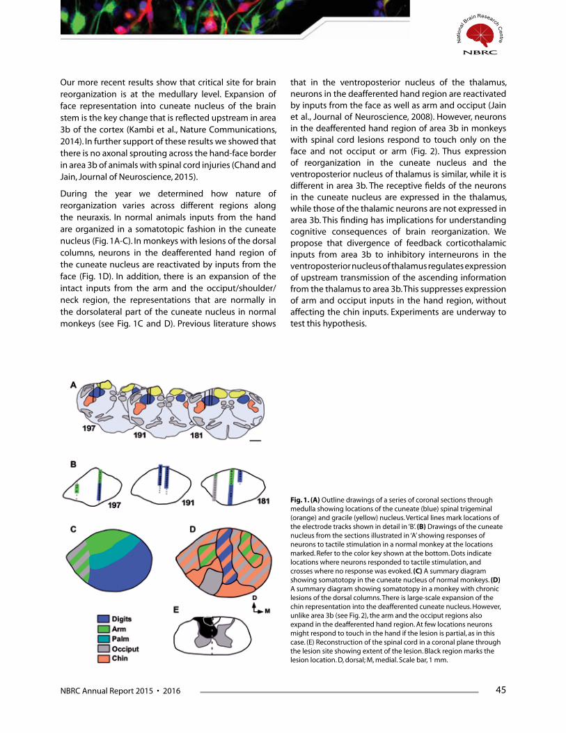
NBRC Annual Report 2015 • 2016 45
Our more recent results show that critical site for brain reorganization is at the medullary level. Expansion of face representation into cuneate nucleus of the brain stem is the key change that is reflected upstream in area 3b of the cortex (Kambi et al., Nature Communications, 2014). In further support of these results we showed that there is no axonal sprouting across the hand-face border in area 3b of animals with spinal cord injuries (Chand and Jain, Journal of Neuroscience, 2015).
During the year we determined how nature of reorganization varies across different regions along the neuraxis. In normal animals inputs from the hand are organized in a somatotopic fashion in the cuneate nucleus (Fig. 1A-C). In monkeys with lesions of the dorsal columns, neurons in the deafferented hand region of the cuneate nucleus are reactivated by inputs from the face (Fig. 1D). In addition, there is an expansion of the intact inputs from the arm and the occiput/shoulder/neck region, the representations that are normally in the dorsolateral part of the cuneate nucleus in normal monkeys (see Fig. 1C and D). Previous literature shows
that in the ventroposterior nucleus of the thalamus, neurons in the deafferented hand region are reactivated by inputs from the face as well as arm and occiput (Jain et al., Journal of Neuroscience, 2008). However, neurons in the deafferented hand region of area 3b in monkeys with spinal cord lesions respond to touch only on the face and not occiput or arm (Fig. 2). Thus expression of reorganization in the cuneate nucleus and the ventroposterior nucleus of thalamus is similar, while it is different in area 3b. The receptive fields of the neurons in the cuneate nucleus are expressed in the thalamus, while those of the thalamic neurons are not expressed in area 3b. This finding has implications for understanding cognitive consequences of brain reorganization. We propose that divergence of feedback corticothalamic inputs from area 3b to inhibitory interneurons in the ventroposterior nucleus of thalamus regulates expression of upstream transmission of the ascending information from the thalamus to area 3b. This suppresses expression of arm and occiput inputs in the hand region, without affecting the chin inputs. Experiments are underway to test this hypothesis.
Fig. 1. (A) Outline drawings of a series of coronal sections through medulla showing locations of the cuneate (blue) spinal trigeminal (orange) and gracile (yellow) nucleus. Vertical lines mark locations of the electrode tracks shown in detail in ‘B’. (B) Drawings of the cuneate nucleus from the sections illustrated in ‘A’ showing responses of neurons to tactile stimulation in a normal monkey at the locations marked. Refer to the color key shown at the bottom. Dots indicate locations where neurons responded to tactile stimulation, and crosses where no response was evoked. (C) A summary diagram showing somatotopy in the cuneate nucleus of normal monkeys. (D) A summary diagram showing somatotopy in a monkey with chronic lesions of the dorsal columns. There is large-scale expansion of the chin representation into the deafferented cuneate nucleus. However, unlike area 3b (see Fig. 2), the arm and the occiput regions also expand in the deafferented hand region. At few locations neurons might respond to touch in the hand if the lesion is partial, as in this case. (E) Reconstruction of the spinal cord in a coronal plane through the lesion site showing extent of the lesion. Black region marks the lesion location. D, dorsal; M, medial. Scale bar, 1 mm.
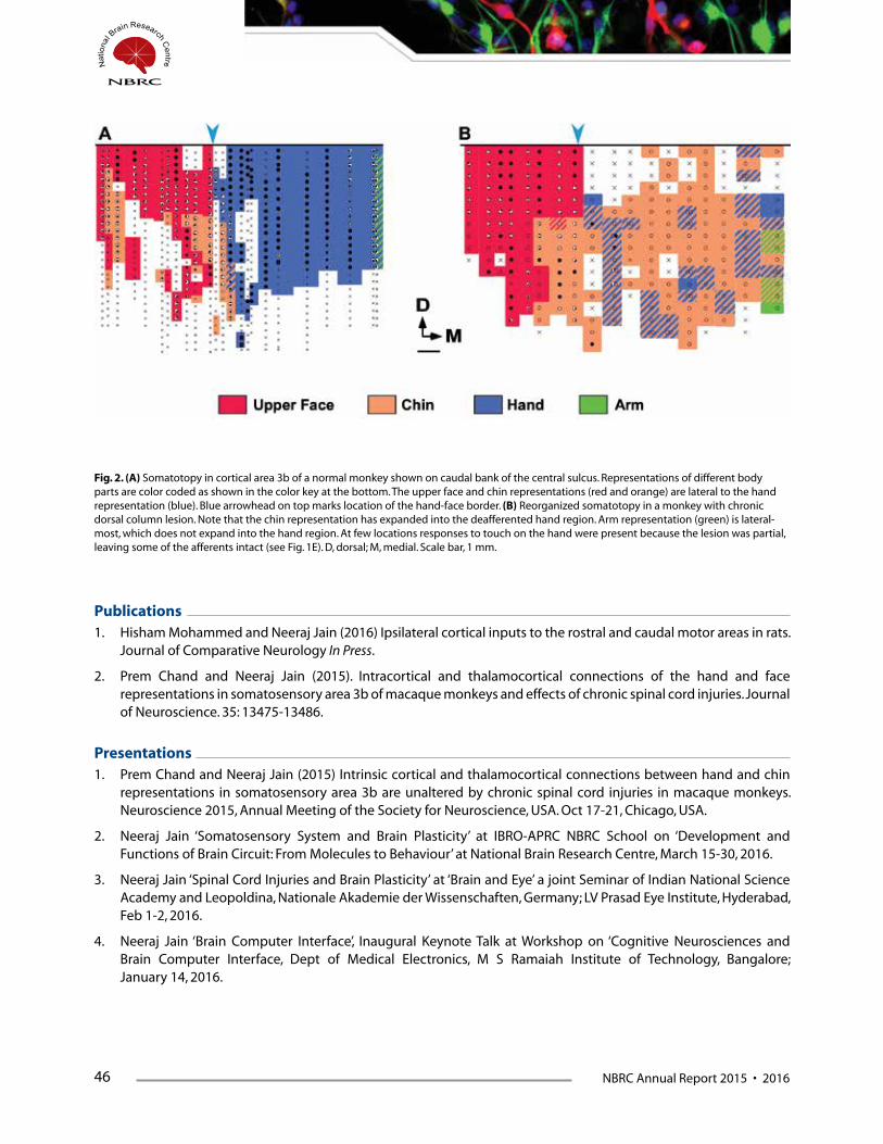
46 NBRC Annual Report 2015 • 2016
Publications1. Hisham Mohammed and Neeraj Jain (2016) Ipsilateral cortical inputs to the rostral and caudal motor areas in rats.
Journal of Comparative Neurology In Press.
2. Prem Chand and Neeraj Jain (2015). Intracortical and thalamocortical connections of the hand and face representations in somatosensory area 3b of macaque monkeys and effects of chronic spinal cord injuries. Journal of Neuroscience. 35: 13475-13486.
Presentations1. Prem Chand and Neeraj Jain (2015) Intrinsic cortical and thalamocortical connections between hand and chin
representations in somatosensory area 3b are unaltered by chronic spinal cord injuries in macaque monkeys. Neuroscience 2015, Annual Meeting of the Society for Neuroscience, USA. Oct 17-21, Chicago, USA.
2. Neeraj Jain ‘Somatosensory System and Brain Plasticity’ at IBRO-APRC NBRC School on ‘Development and Functions of Brain Circuit: From Molecules to Behaviour’ at National Brain Research Centre, March 15-30, 2016.
3. Neeraj Jain ‘Spinal Cord Injuries and Brain Plasticity’ at ‘Brain and Eye’ a joint Seminar of Indian National Science Academy and Leopoldina, Nationale Akademie der Wissenschaften, Germany; LV Prasad Eye Institute, Hyderabad, Feb 1-2, 2016.
4. Neeraj Jain ‘Brain Computer Interface’, Inaugural Keynote Talk at Workshop on ‘Cognitive Neurosciences and Brain Computer Interface, Dept of Medical Electronics, M S Ramaiah Institute of Technology, Bangalore; January 14, 2016.
Fig. 2. (A) Somatotopy in cortical area 3b of a normal monkey shown on caudal bank of the central sulcus. Representations of different body parts are color coded as shown in the color key at the bottom. The upper face and chin representations (red and orange) are lateral to the hand representation (blue). Blue arrowhead on top marks location of the hand-face border. (B) Reorganized somatotopy in a monkey with chronic dorsal column lesion. Note that the chin representation has expanded into the deafferented hand region. Arm representation (green) is lateral-most, which does not expand into the hand region. At few locations responses to touch on the hand were present because the lesion was partial, leaving some of the afferents intact (see Fig. 1E). D, dorsal; M, medial. Scale bar, 1 mm.
NBRC Annual Report 2015 • 2016 47
FundingThis work is supported by Department of Biotechnology and NBRC Core funds.
Collaborators Prof Mriganka Sur, MIT, USA
Prof P Raghunathan, NBRC
Degrees Awarded (Ph.D.)Hisham Mohammed (Ph.D.)

48 NBRC Annual Report 2015 • 2016
Protein Modifications in Synaptic Plasticity and Memory
Principal Investigator:
Shiv K Sharma
Research Fellows: Kaushik P. Sharma (PhD student), Kautuk Kamboj (PhD student), Biswaranjan Sahoo (PhD student) Deborah Daphne (Project Assistant)
Lab attendant: Narayan
Synaptic plasticity is the ability of neuronal synapses to change in response to activity. It is well recognized that activity-dependent neuronal
changes are important players in the development of synaptic plasticity. These changes are also important for formation of memory. Despite considerable research using various approaches, the mechanisms involved in synaptic plasticity and memory are far from clear. My laboratory has been attempting to elucidate the processes that are involved in synaptic plasticity and memory. We previously reported that increasing the level of one kind of posttranslational modification, acetylation,can enhance synaptic plasticity known as long-term potentiation, induced by massed pattern of electrical stimulation. Additionally, we showed that memory induced by massed training can be enhanced when the level of acetylation is increased. In this report, I will mention our studies regarding the role of a phosphatase which seems to play crucial roles in hippocampal long-term potentiation.
Differential requirement of a phosphatase in long-term potentiation Memories are important for our proper functioning on a daily basis. Memories help us plan for the future also. It is not entirely clear how memories are formed in the brain. With regards to the synaptic mechanisms of memory formation, one kind of synaptic plasticity, long-
term potentiation, is widely considered to be the cellular basis of memory formation. Since its discovery several years ago, a lot of studies have been directed towards understanding the neuronal changes that are involved in long-term potentiation. Protein modifications including phosphorylation are known to critically regulate long-term potentiation. Whereas protein kinases add a phosphate group to a substrate, the protein phosphatases remove the phosphate group. Thus, the level of protein phosphorylation in a cell is regulated by the activities of protein kinases as well as protein phosphatases. Although many studies have examined the role of protein kinases in long-term potentiation, the role of protein phosphatases in this synaptic phenomenon is less explored. Some phosphatases remove the phosphate group from serine/threonine residues in a protein. Some other phosphatases remove the phosphate group from tyrosine residues.
One of the focus in my laboratory is to understand the role of a phosphatase in long-term potentiation induced by different stimulation protocols. We use hippocampal slices to study long-term potentiation. For these experiments, we electrically stimulate the CA3 region of the hippocampus, and record the response in the CA1 region.Different stimulation protocols are used to induce long-term potentiation. To examine the role of the phosphatase under study, we used a compound to inhibit its activity. Long-term potentiation was
NBRC Annual Report 2015 • 2016 49
recorded from the slices in the absence or presence of the phosphatase inhibitor. The results suggest that the phosphatase inhibitor blockslong-term potentiation induced by some stimulation protocols, but does
not affects long-term potentiation induced by other stimulation protocol. This is an important finding regarding the role of protein phosphatases in long-term potentiation.
Publication1. Pandey K, Sharma KP, Sharma SK (2015) Histone deacetylase inhibition facilitates massed pattern-induced synaptic
plasticity and memory. Learn Mem. 22:514-8.
Presentations1. Delivered a lecture as part of Brain awareness week seminars at Pub Kamrup College,BaihataChariali, Kamrup,
Assam.
2. Delivered a lecture as invited speaker in the IBRO/APRC School at National Brain Research Centre, Manesar, Haryana.
3. I gave lectures when students from different institutions visited NBRC.
FundingThis work is supported by NBRC Core.
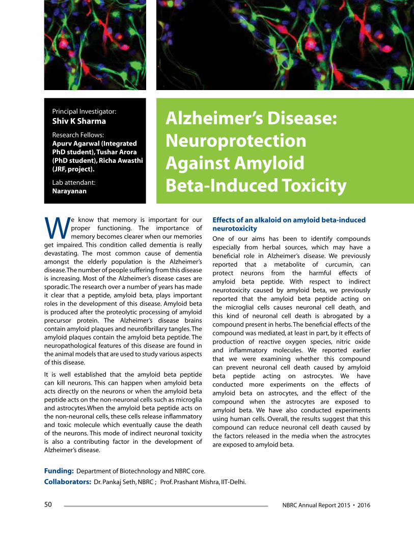
50 NBRC Annual Report 2015 • 2016
Alzheimer’s Disease: Neuroprotection Against Amyloid Beta-Induced Toxicity
Principal Investigator:
Shiv K Sharma
Research Fellows: Apurv Agarwal (Integrated PhD student), Tushar Arora (PhD student), Richa Awasthi (JRF, project).
Lab attendant: Narayanan
We know that memory is important for our proper functioning. The importance of memory becomes clearer when our memories
get impaired. This condition called dementia is really devastating. The most common cause of dementia amongst the elderly population is the Alzheimer’s disease. The number of people suffering from this disease is increasing. Most of the Alzheimer’s disease cases are sporadic. The research over a number of years has made it clear that a peptide, amyloid beta, plays important roles in the development of this disease. Amyloid beta is produced after the proteolytic processing of amyloid precursor protein. The Alzheimer’s disease brains contain amyloid plaques and neurofibrillary tangles. The amyloid plaques contain the amyloid beta peptide. The neuropathological features of this disease are found in the animal models that are used to study various aspects of this disease.
It is well established that the amyloid beta peptide can kill neurons. This can happen when amyloid beta acts directly on the neurons or when the amyloid beta peptide acts on the non-neuronal cells such as microglia and astrocytes.When the amyloid beta peptide acts on the non-neuronal cells, these cells release inflammatory and toxic molecule which eventually cause the death of the neurons. This mode of indirect neuronal toxicity is also a contributing factor in the development of Alzheimer’s disease.
Effects of an alkaloid on amyloid beta-induced neurotoxicityOne of our aims has been to identify compounds especially from herbal sources, which may have a beneficial role in Alzheimer’s disease. We previously reported that a metabolite of curcumin, can protect neurons from the harmful effects of amyloid beta peptide. With respect to indirect neurotoxicity caused by amyloid beta, we previously reported that the amyloid beta peptide acting on the microglial cells causes neuronal cell death, and this kind of neuronal cell death is abrogated by a compound present in herbs. The beneficial effects of the compound was mediated, at least in part, by it effects of production of reactive oxygen species, nitric oxide and inflammatory molecules. We reported earlier that we were examining whether this compound can prevent neuronal cell death caused by amyloid beta peptide acting on astrocytes. We have conducted more experiments on the effects of amyloid beta on astrocytes, and the effect of the compound when the astrocytes are exposed to amyloid beta. We have also conducted experiments using human cells. Overall, the results suggest that this compound can reduce neuronal cell death caused by the factors released in the media when the astrocytes are exposed to amyloid beta.
Funding: Department of Biotechnology and NBRC core.
Collaborators: Dr. Pankaj Seth, NBRC ; Prof. Prashant Mishra, IIT-Delhi.

NBRC Annual Report 2015 • 2016 53
Investigating neuro-cognitive network mechanisms using multimodal neuroimaging
Principal Investigator:
Arpan Banerjee
Researchers: Dr. Dipanjan Ray G. Vinodh Kumar TameshHalder Amit Kumar Jaiswal Nilambari Hajare Shrey Dutta
Cognitive Brain Lab (CBL) is engaged in basic and translational research using non-invasive neuroimaging tools EEG, MEG, TMS & fMRI. We
have primarily two themes of research: 1) Exploring and innovating novel research designs and analysis tools for MEG/ EEG & fMRI recordings and 2) Studying cognitive impairments in epilepsy and investigating various functional brain networks related to speech perception and multisensory integration. Here we outline the major project updates from the period April, 2015-March, 2016. The overarching goal of these studies is to develop an understanding for the neurobiological mechanisms of multisensory integration.
1. Temporal congruence of audio-visual stimuli involves processing in human posterior superior temporal sulcus (pSTS) during multisensory speech perception.
Speech perception emerges from harmonious interaction of multiple neural systems. “McGurk illusion” (McGurk and MacDonald, 1976): a classic example of how visual feedback shapes speech perception provides an entry point to study the underlying brain networks. Earlier research have shown that modulating the degree of audio-visual (AV) integration by psychophysical parameters weakened the effect (Munhall et al., 1996,
Pare et al., 2003, Alsius et al., 2007, Munhall et al., 2009, Nath and Beauchamp, 2012).
We performed a fMRI study with human volunteers when McGurk-stimuli (incongruent audio-video signal) were presented with varying AV lags. We observed across a large group of 34 volunteers there was a regime where the illusory perception was maximum, [-150, 300] ms in concordance with earlier studies (Fig 1). When the block at which maximum illusory response occurred was pooled for a group statistical parametric mapping (SPM) analysis with respect to rest we observed significant activations in inferior frontal gyrus (IFG), posterior superior temporal sulcus (pSTS), V5 and superior temporal gyrus (STG). In a conjunction analysis with blocks where minimum illusory perception was reported by volunteers, the highest difference was found in pSTS. In a regime where auditory precede visual stimulus, pSTS was not even activated. Functional connectivity among network nodes involving IFG, pSTS, auditory cortex/ STG and V5 using partial correlations were not altered by illusory perception but changed from a negative to positive AV lag. Overall, our results indicate neural activity in pSTS is most likely reflective of temporal congruence of multisensory stimuli, incongruent or congruent.

54 NBRC Annual Report 2015 • 2016
2. Large scale functional brain networks underlying temporal integration of audio-visual speech perception: An EEG study
It is evident from the fMRI study that networks across the whole brain participates during multisensory processing of speech stimuli. We posit that a large-scale functional connectivity among the neural population situated in distributed brain sites may provide valuable insights involved in processing and fusing of AV speech. Varying the psychophysical parameters in tandem with electroencephalogram (EEG) recordings, we exploited the trial-by-trial perceptual variability of incongruent audio-visual (AV) speech stimuli to identify the characteristics of the large-scale cortical network that facilitates multisensory perception during synchronous and asynchronous AV speech. We replicated similar
behavioral dynamics as observed for project 1 and observed that gaze fixation on mouth region. We evaluated the spectral landscape of EEG signals during multisensory speech perception at varying AV lags. Functional connectivity dynamics for all sensor pairs was computed using the time-frequency global coherence, the vector sum of pairwise coherence changes over time. During synchronous AV speech, we observed enhanced global gamma-band coherence and decreased alpha and beta-band coherence aids multisensory perception. During asynchronous speech stimuli, a global broadband coherence facilitated multisensory perception. Thus our study indicates the representational space of temporal integration underlying multisensory speech perception needs to account the presence of large-scale functional brain network mechanisms.
Figure 1: a) Stimulus videos are created using visual lip movement of /ka superimposed on auditory /pa. Participants reported the auditory object they heard while watching the video using a button press response box. b) Stimuli videos were created at different audio-visual lags, the timing difference between the onset of sound and lip movement, with values ranging from [-300; 450] ms c) Normalized behavioral responses from 34 subjects. Mean response for each perceptual category is presented as a function of AV lag. The error bars reflect 95% significance thresholds.
Figure 2: a) BOLD response in 34 subjects during presentation of McGurk stimuli at various AV lags. Only activations that survived p<0.01 (uncorrected) are shown. Left pSTS show maximal activation at lag 150 ms.

NBRC Annual Report 2015 • 2016 55
Figure 3: (a) shows the number of normalized group responses in each of the three perceptual categories: “/pa/”, “/ta/” and “other” for each AV Lag. (b) overall eye gaze fixation overlaid over a single frame of the stimuli (c) Normalized group-level histogram of eye gaze fixation at mouth for each perceptual category at the respective stimuli.
Figure 4: Time-frequency representations of large-scale functional brain networks. Time frequency coherogram for different perceptual categories time locked to the onset of the first sensory component (A or V) during the three conditions and the coherence difference between /ta/ and /pa/ responses at different AV lags: for -450 ms (A) /ta/ (B) /pa/ (C) /ta/-//pa/; for 0 ms (D) /ta/ (E) /pa/ (F) /ta/-/pa/; for 450 ms (G) /ta/ (H) /pa/ (I) /ta/-/pa/ .
3. A dynamical framework to relate perceptual variability with multisensory information processing.
Figure 5: a)The dynamical model for multisensory speech perception consisting of three Kuramoto oscillators for positive AV lags when visual leads auditory stimulus. Oscillators A, V and AV represent the audio, visual and multisensory systems respectively. There is no direct coupling between oscillators
A and V. b) Order parameter vs time delay derived from numerical integration of equation (1) for different coupling strengths mimics the perceptual states as a function of order parameter from Fig 1 c. c) Analytical solutions to equation (1), following linear stability analysis also matches with numerically derived curves.

56 NBRC Annual Report 2015 • 2016
Multisensory processing involves participation of individual sensory streams, e.g., vision, audition to facilitate perception of environmental stimuli. Our fMRI and EEG work suggests systems level interactions among auditory, visual and multisensory sub-systems during multisensory perception. Further studies have established that time-delay between onset of auditory and visual signals (AV lag) and perturbations in the unisensory streams are key variables that modulate perception. However, as of now only few quantitative theoretical frameworks have been proposed to understand the interplay among these psychophysical variables or the neural systems level interactions that govern perceptual variability. Here, we propose a dynamic systems model consisting of the basic ingredients of any multisensory processing, two unisensory and one multisensory sub-system (nodes) as reported by several researchers. The nodes are connected such that biophysically inspired coupling parameters and time delays become key parameters of this network. We achieved this by representing the auditory (A), visual (V) and audio-visual (AV) sub-systems with Kuramoto oscillators (Fig 5). Here, the phase of each oscillator is represented with one-dimensional differential equations and represents the coupling among the nodes. The AV
lag can be represented as a time delay term in the system and the dynamics of the network can be expressed by the following rate equations.
The complex order parameter represents the synchronization state across the network and can expressed as
Order parameter can be conceptualized as the quantity that encodes the perceptual states of an organism. We observed that zero AV lag results in maximum synchronization of constituent nodes (R ~ 1) and the degree of synchronization decreases (R<<1) when we have non-zero lags (Fig 5 b and c). The attractor states of this network can thus be interpreted as the facilitator for stabilizing specific perceptual experience. Thereby, the dynamic model presents a quantitative framework for understanding multisensory information processing.
Publications1. Thakur, B., Mukherjee , A., Sen A., & Banerjee, A. (2016) A dynamical framework to relate perceptual variability with
multisensory information processing. (accepted with minor revision)
2. Ghosh, S., Kumar, V. G. ,Basu, A. & Banerjee, A. (2015) : Graph theoretic network analysis reveals protein pathways underlying cell death following neurotropic viral infection. Scientific Reports 5: 14438
Presentations1. Mukherjee, A., Raghunathan, P. & Banerjee, A. Multisensory perception, but not multisensory stimuli drive the
activity of posterior superior temporal sulcus (pSTS) ACCS-2015, 6-8 July, IIT Kanpur
2. Ray, D. & Banerjee, A. Differential involvement of ventral and dorsal visual streams in “novel” and “practiced” visually guided actions ACCS-2015, 6-8 July, IIT Kanpur
3. Kumar, V. G., Jaiswal, A., Mukherjee, A., Roy, D. & Banerjee, A. Spatiotemporal structure of oscillatory cortical activity underlying multisensory speech perception ACCS-2015, 6-8 July, IIT Kanpur
4. Halder, T., Jaiswal, A. K, & Banerjee, A. CS-sLORETA: A localization tool for brain electromagnetic activity ACCS-2015, 6-8 July, IIT Kanpur
5. Kumar, V. G., Halder, T., Jaiswal, A., Mukherjee, A., Roy, D. & Banerjee, A. Multi-level neuromarkers of multisensory speech perception, NAOP convention, 2-5 February, 2016, CBCS

NBRC Annual Report 2015 • 2016 57
FundingNBRC CoreRamalingaswami Fellowship, Department of Biotechnology Innovative Young Biotechnologist Award (IYBA), Department of Biotechnology Cognitive Science Research Fellowship, Department of Science and Technology
CollaboratorsAbhijit Sen & Bhumika Thakur, Institute for Plasma Research (IPR), Gandhinagar
Anirban Basu, NBRC
Dipanjan Roy, International Institute of Information Technology (IIIT), Hyderabad
James Gomes, Indian Institute of Technology Delhi (IIT-Delhi), New Delhi
Manjari Tripathi, All India Institute of Medical Sciences (AIIMS), New Delhi
Partha Ragunathan, NBRC
58 NBRC Annual Report 2015 • 2016
Development of Novel Brain Signal Processing Toolbox ‘KALPANA’
Principal Investigator:
Pravat Kumar Mandal
Research Scientist: Sumiti Saharan
Senior R&D engineer: Monika Grewa l, Shammi More
Neuropsychologist: Sarah A. Khan
Neuro-analyst: Aroma Dabas
ObjectiveTo develop a user-friendly signal processing toolbox for an efficient analysis of magnetic resonance spectroscopic data to perform an absolute quantitation of neuro-chemicals/ brain anti-oxidants/ neurotransmitters from various brain regions. The development of such a tool shall ease the quantitation outcome with normal and clinical conditions as well as disease severity.
The NeuroImaging and NeuroSpectroscopy (NINS) laboratory focuses on identifying early diagnostic biomarkers for neurodegenerative disorders such as Alzheimer’s disease (AD). AD is the most common form of dementia in the world with a whopping 47.5 million sufferers worldwide and 7.7 million additions every year (2016 WHO Dementia). The alarming exponential increase of this severe mental illness has catapulted the research to detect and diagnose the disease at its incipient stages. An understanding of the underlying molecular mechanisms that transform a healthy brain into a diseased condition would help us learn the causal molecular process of this disease.
Our lab is focusing on assessment and quantification of the brain pH, gamma-aminobutyric acid (GABA) and glutathione (GSH) levels in various study groups: normal healthy control (HC), mild cognitive impaired (MCI), and AD. This assessment shall not only elucidate the etiology and pathophysiology of AD but
also aid in understanding the staging of AD.
GSH, a predominant brain antioxidant responsible for neutralization of reactive oxygen species (ROS), has been correlated with cognitive impairment and emerging as a potential biomarker in AD. The impact of GABA, the chief neurotransmitter in the central nervous system (CNS) is implicated in AD but has never been correlated or quantified under clinical settings. Variations in brain pH is another indicator of the brain neuronal health and neuron degeneration and could become a potential biomarker of AD. These neurochemicals are being evaluated by proton (1H) and phosphorous (31P) MRS. Hence, for reliable outcome and enhanced signals, we have been employing MEscher-GArwood-PRESS (MEGA PRESS) spectral editing techniques for GSH/GABA as well as specially designed 31P MRS techniques for pH measurement.
Furthermore, absolute quantitation of neurochemicals from the measured MRS signals is required for meticulous processing of these small signals. Currently available software tools have limitations in terms of experimental design, processing mode and the post-processing scheme. These tools are being developed in different labs and for specific purposes. Our lab has therefore developed a novel comprehensive MATLAB-based software package called ‘KALPANA’ for processing, visualization and absolute quantitation of magnetic resonance spectroscopy data.
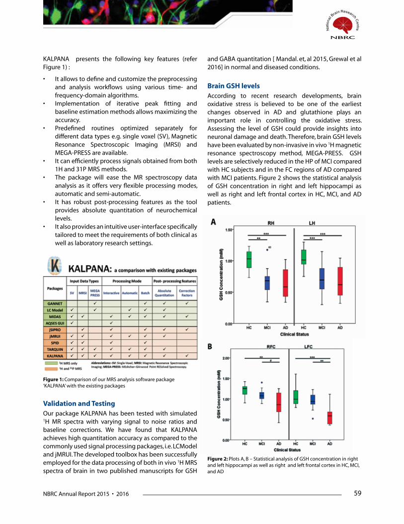
NBRC Annual Report 2015 • 2016 59
KALPANA presents the following key features (refer Figure 1) :
• Itallowstodefineandcustomizethepreprocessingand analysis workflows using various time- and frequency-domain algorithms.
• Implementation of iterative peak fitting andbaseline estimation methods allows maximizing the accuracy.
• Predefined routines optimized separately fordifferent data types e.g. single voxel (SV), Magnetic Resonance Spectroscopic Imaging (MRSI) and MEGA-PRESS are available.
• Itcanefficientlyprocesssignalsobtainedfromboth1H and 31P MRS methods.
• The package will ease the MR spectroscopy dataanalysis as it offers very flexible processing modes, automatic and semi-automatic.
• It has robust post-processing features as the toolprovides absolute quantitation of neurochemical levels.
• Italsoprovidesanintuitiveuser-interfacespecificallytailored to meet the requirements of both clinical as well as laboratory research settings.
Validation and TestingOur package KALPANA has been tested with simulated 1H MR spectra with varying signal to noise ratios and baseline corrections. We have found that KALPANA achieves high quantitation accuracy as compared to the commonly used signal processing packages, i.e. LCModel and jMRUI. The developed toolbox has been successfully employed for the data processing of both in vivo 1H MRS spectra of brain in two published manuscripts for GSH
Figure 1: Comparison of our MRS analysis software package ‘KALPANA’ with the existing packages
Figure 2: Plots A, B – Statistical analysis of GSH concentration in right and left hippocampi as well as right and left frontal cortex in HC, MCI, and AD
and GABA quantitation [ Mandal. et, al 2015, Grewal et al 2016] in normal and diseased conditions.
Brain GSH levelsAccording to recent research developments, brain oxidative stress is believed to be one of the earliest changes observed in AD and glutathione plays an important role in controlling the oxidative stress. Assessing the level of GSH could provide insights into neuronal damage and death. Therefore, brain GSH levels have been evaluated by non-invasive in vivo 1H magnetic resonance spectroscopy method, MEGA-PRESS. GSH levels are selectively reduced in the HP of MCI compared with HC subjects and in the FC regions of AD compared with MCI patients. Figure 2 shows the statistical analysis of GSH concentration in right and left hippocampi as well as right and left frontal cortex in HC, MCI, and AD patients.

60 NBRC Annual Report 2015 • 2016
GSH levels are found to be significantly reduced in the LH and RH of MCI with respect to HC subjects. Furthermore, GSH levels are significantly reduced in the LFC and RFC of AD compared with MCI.
Further analyses were performed to validate the efficacy of the glutathione assessment. The sensitivity and specificity analysis (ROC curves) of AD v/s HC, MCI v/s HC and AD v/s MCI are shown in Figure 3. Sensitivity of 87.5% and specificity of 100% were obtained for distinguishing MCI versus HC on the basis of GSH estimation in LH (Left hippocampus) and sensitivity of 91.7% and specificity of 100% were obtained for MCI versus AD subjects on the basis of GSH level in FC (bilateral frontal cortices). Hence, it’s predictive that GSH can be an efficient biomarker.
Brain pH levels: For 31P MRS clinical brain pH data obtained from healthy control, MCI and AD patients, we employed KALPANA toolbox to quantify the brain pH values and mapped them to specific brain regions (Figure 4). The pH of the brain, which is normally ~ 7.02, was found to be alkaline in the left hippocampus regions of AD brain as compared to healthy control subjects in the same anatomical region.
Figure 3: ROC curves C, D, E are the Sensitivity and Specificity analyses of AD v/s HC, MCI v/s HC and AD v/s MCI. GSH levels are found to be reduced in MCI and AD; HC, MCI AD
Figure 4: 31P MRS clinical data for brain pH in HC, MCI and AD patients

NBRC Annual Report 2015 • 2016 61
Brain GABA levels: To assess regional and hemispheric differences among young healthy subjects, brain GABA concentrations were measured in vivo using 1H magnetic resonance spectroscopy.
KALPANA toolbox was used extensively for quantitation of GABA levels. We found a significant regional dependence of GABA levels with lower concentrations in the frontal cortex (FC) as compared to both parietal cortex (PC) and occipital cortex (OC) regions. However, no significant hemispheric differences in GABA levels were seen (refer Figure 5 and Figure 6).
Figure 5: Plot a – Mapping of GABA levels to Frontal (FC), Parietal (PC) and Occipital (OC) cortex regions of brain among healthy young subjects; Plot b – 1H MRS spectrum and quantitation of GABA using KALPANA
Figure 6: Region-wise (Plot a) and Hemispheric (Plot b) GABA distribution among healthy young brains: GABA levels in FC are lower than those in PC and OC. GABA distribution exhibits hemispheric Symmetry
62 NBRC Annual Report 2015 • 2016
Publications1. Pravat K Mandal*, Sumiti Saharan, Manjari Tripathi and Geetanjali Murari, ‘Brain Glutathione Levels – A Novel
Biomarker for Mild Cognitive Impairment and Alzheimer’s Disease’, Biological Psychiatry, 2015, 78 (10), 702–710
2. Monika Grewal, Aroma Dabas, Sumiti Saharan, Peter B. Barker, Richard A.E. Edden, Pravat K Mandal*, ‘GABA Quantitation using MEGA-PRESS: Regional and Hemispheric Differences’, Journal of Magnetic Resonance Imaging, JMRI; DOI: 10.1002/jmri.25324 (2016)
3. Suvarnalata Xanthate Duggirala, Sumiti Saharan, Partha Raghunathan, Pravat K. Mandal*, ‘Stimulus-dependent modulation of working memory for identity monitoring: A functional MRI study’, Brain and Cognition, Volume 102, February 2016, Pages 55–64
4. Pravat K Mandal, Sumiti Saharan, Olivia Penna and Vincenzo Fodale ‘Anesthesia Issues in Central Nervous System Disorders’, Current Aging Science, 9 (2), 2016, 116-143
FundingDepartment of Biotechnology, Government of India, Department of Information Technology, Government of India, Department of Science and Technology, Government of India
CollaboratorsProf. (Dr.) Manjari Tripathi, MD, DM Department of Neurology, All India Institute of Medical Science (AIIMS), New Delhi
Prof. Peter Barker, D.Phil, Department of Radiology and Radiological Science, Johns Hopkins Medicine, USA
Patent applications detailsThe methodology which is applied in ‘KALPANA’ was filed for a National patent at the Indian Patent Office (IPO) on 19 January 2016 and PCT application filed for an International patent at the World Intellectual Property Organization (WIPO).
NBRC Annual Report 2015 • 2016 63
Spatiotemporal Processing and Signal Transmission in Brain
Principal Investigator:
Prasun K. Roy
Fellow: Aftab Alam
Research engineer: V P Subramanyam
Of the significant challenges facing the neuroscientist is the query of the researcher to decipher how flow occurs, whether that
of information, electrical current, drugs, cells or tissue displacement, across the layered brain extent. There is a critical requirement of a versatile quantitative approach that can account for, and furnish a substantive description, of flow processes and its modulation across brain. We have developed the methodology of dynamic functional tensor neuroimaging and obtained accurate measurement of matrix-tensor maps to describe flow and deformation processes, information flux or connectivity in the brain. The research has considerable potentiality of applications to clinical medicine as well as to biological engineering, especially for diagnosis, therapy and neurophysiological investigations. The overall objective of this program is to understand the physiological or pathological dynamics of transport or flow processes in the brain, whether that of fluids, tissue, energy or information. The approach has appreciable prospect of applications to neurotechnology and to the clinical domain.
Repurposing of Multimodal therapy for Enhancing Neuroprotection in StrokeThe unsuccessful performance of pharmacological intervention for stroke, across numerous clinical trials over decades, is a veritable challenge. Most of the patients cannot make it to an intensive care unit to have thrombolytic drug TPA which can be given only within 4 hours of the cerebral insult. An important observation is that, dur to reperfusive and oxidative stress injury, there is a great toxic immunomodulative changes in the initial
phases, namely activation of microglia into neurotoxic form (M
1 microglia). However, it is also known that
specific immunological activation in the later phases is beneficial to stroke outcome, this activation consists of potentiation of neurotropic form of microglia (M
2
microglia). Immunomodulative drugs as tigecycline or minocycline are known to decrease M
1 level and increase
M2 level, and thus function as neuroprotectants. However,
minocycline effects in clinical trial for stoke is found to be only modestly effective only when ischaemic volume is large, i.e. when higher perfusion-deficit happens. In the latter, there occurs higher activation level of free radical and M
1 microglia, and tigecycline or minocycline shifts
the immunodynamic equilibrium to M2 phenotype. We
explore the possibility of enhancing this neuroprotection ability. However, adding other pharmacological components orally can produce pharmacodynamic incompatibility, biodegradation, competitive inhibition, and lower transmission through the blood-brain barrier.
Hence, the attraction of nonpharmacological modalities, as physical medicine or biophysics-based modalities, which would not have these drawbacks. We also need to ensure that the therapeutic signal-transduction pathways of minocycline and the physical therapy agents, should not interfere, so that synergistic additional neuroprotection is obtained. We probed whether this could be enabled by standard physical therapies, such as using functional stimulation, namely mild magnetic stimulation (TMS) or mild galvanic stimulation (DCS) [fig. 1(a)]. We develop a systems biology approach using preclinical experimental findings [fig. 1(b)], we utilize ordinary differential equations to study the behaviour of both phenotypes of microglia [fig. 1(c)].
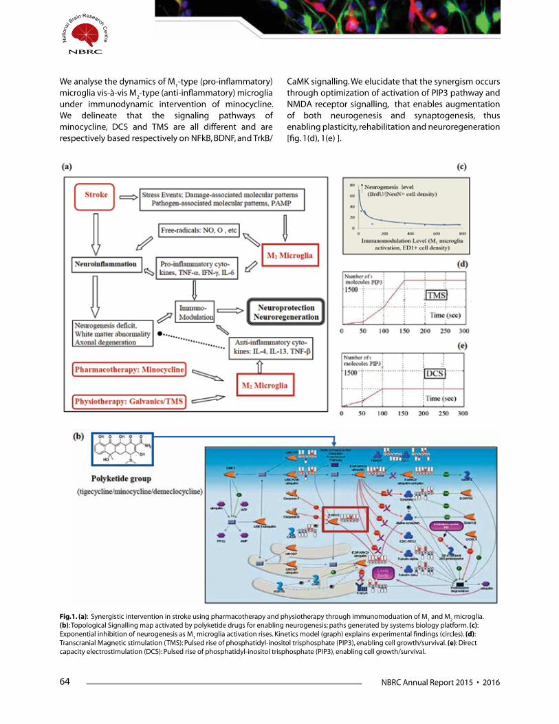
64 NBRC Annual Report 2015 • 2016
We analyse the dynamics of M1-type (pro-inflammatory)
microglia vis-à-vis M2-type (anti-inflammatory) microglia
under immunodynamic intervention of minocycline. We delineate that the signaling pathways of minocycline, DCS and TMS are all different and are respectively based respectively on NFkB, BDNF, and TrkB/
CaMK signalling. We elucidate that the synergism occurs through optimization of activation of PIP3 pathway and NMDA receptor signalling, that enables augmentation of both neurogenesis and synaptogenesis, thus enabling plasticity, rehabilitation and neuroregeneration [fig. 1(d), 1(e) ].
Fig.1. (a): Synergistic intervention in stroke using pharmacotherapy and physiotherapy through immunomoduation of M1 and M
2 microglia.
(b): Topological Signalling map activated by polyketide drugs for enabling neurogenesis; paths generated by systems biology platform. (c): Exponential inhibition of neurogenesis as M
1 microglia activation rises. Kinetics model (graph) explains experimental findings (circles). (d):
Transcranial Magnetic stimulation (TMS): Pulsed rise of phosphatidyl-inositol trisphosphate (PIP3), enabling cell growth/survival. (e): Direct capacity electrostimulation (DCS): Pulsed rise of phosphatidyl-inositol trisphosphate (PIP3), enabling cell growth/survival.

NBRC Annual Report 2015 • 2016 65
MRI Tensor Imaging Evidence of Glymphatic Fluid system in Brain for clearing AmyloidOne of the major factors of Alzheimer’s disease is lowering of vascular clearance of amyloid from the brain. An important advancement in exploring enhancement of this clearance is the recent identification of a novel anatomical gliovascular system that enables the clearance of amyloid and solutes from brain parenchyma, via its interstitial Gliovascular lymphatic fluid system (brain lymphatics) which has been experimentally found in rodent models. Using human MRI, we have endeavoured to provide a collateral empirical substantiation of gliovascular fluid system in man (fig. 2(a)-(c)]. We use thermodynamic reciprocity principle to elucidate that the transport tensors in a fluidized phase of brain interstice share eigenvectors. Using MRI scanning, we delineate how one can construct the general transport tensor from MRI diffusion tensor for each voxel by choosing a set of basis functions in which both the tensor ellipsoids become in comparable standardized format.
Fig.2. (a): MRI image of brain used for elastoporosity analysis of parenchyma tissue. (b). Normal brain tissue as hydroporous medium consisting of two phases (intracellular and extracellular phases). (c). Formation of the third phase of amyloid dystrophic neurotic domain under advanced ageing process. (d). 2D Heat conduction tensor images of brain parenchyma tissue pixel-wise, using MRI scanning via mobility-sensitive gradients. (e). 3D Heat conduction tensor image of portion of brain parenchyma tissue, obtained in terms of ellipsoids in 3D tissue, voxel-by-voxel. (f). Biophysics-based model of the linear thermodynamic relationship between various transport and conduction processes in brain parenchyma tissue consisting of interstitial gliovascular fluid and cellular matrix. Circles are experimental findings that are accounted by the linear graph of thermodynamic relationship. Analyzing the graph, the transport properties of gliovascular fluid is derived,
We consider the tissue microstructure consisting of a parenchymal space embedding an amorphous gliovascular space. From the MRI diffusion tensor image, we derive the pixelated and voxellated heat conduction image of brain parenchyma [fig. 2(d)-(e)]. We show the linearity between eigenvalues of tensors describing the layered permeation and perfusion processes
in brain parenchyma tissue, using our experimental findings from diffusion tensor imaging and fluidic mobility measurement (fig. 2(f )]. Utilizing the generalized tensor MRI approach, we noninvasively estimate the mean energy flux index, diffusion index and conduction index for the interstitial fluid phase to be respectively 0.49 W/m/0K, 5.62 um2/ms and 4.85 siemens/m (n=10 subjects).
66 NBRC Annual Report 2015 • 2016
These values are respectively within 94%, 95% and 84% range of the available surgically measured values of those indices in the fluidic phase of the lymphatic system using invasive electrodes. Thus, one notes that the mean predictive accuracy of our mathematical model is 91%. Using thermodynamic transport principles,
we obtain a quantitative approach to solute/amyloid clearance from brain. Obtaining precise mapping of gliovascular flow system in patients has considerable potentiality in understanding and enhancement of amyloid clearance from brain in neurodegene- rative disease.
Publications1. Alam A, Rallabandi V, Roy PK, Systems biology of Immunomodulation for post-Stroke Neuroplasticity: Multimodal
implications for Pharmacotherapy and Neuro-rehabilitation, Frontiers in Neurology, 2016. DOI: 10.3389/fneur.2016.00094
2. Rallabandi V, Roy PK. Role of Structural Neuroimaging in Classification of Dementia Subtypes. In: R K Jalali (ed.), Neurodegeneration. Ranbaxy Science Foundation. New Delhi, 2015 (accepted).
3. Pal S, Roy PK, Strategy for Stochastic Dose-rate induced Enhanced Elimination of Malignant Tumour without Needing Dose-escalation, Mathematical Medicine & Biology, 32(2), 12-21, 2015 (shown as ‘in press’ last year).
Presentations1. Roy, PK. Human MRI Tensor-based corroboration of Transport indices of Gliovascular glymphatic Fluid system of
Brain for clearing Alzheimer’s Amyloid. Proc. Neuroscience-2015, Society for Neuroscience, Chicago, Oct 2015.
2. Alam, A, Subramanyam, V, Roy, PK. Harnessing the Immunomodulatory Milieu in the Quest of Neuroprotection in Stroke: A Systems Biology Approach, Proc. Int. Soc. Neurochemistry, Brisbane, Aug. 2015.
3. Alam, A. Role of Neurogenesis in Reward-based Networks, Munich Workshop on Neuroregeneration, Germany, Feb. 2016.
4. Alam, A. Systems Biological Model of Neuroinflammation in Ischaemic Stroke – From Mouse to Man, Indian Academy of Neuroscience, Panjab University and Institute of Postgraduate Medical Education & Research, Chandigarh, Nov. 2015
Collaborators Dr Anirban Basu & Dr Partha Raghunathan, National Brain Research Centre.
Dr Michael Hornberger, Addenbrooke’s Hospital, University of Cambridge.
Dr Peter Luijten, Dutch Centre for Translational Molecular Medicine & Utrecht University.
Dr M V Padma & Dr T S Roy, All-India Institute of Medical Sciences, Delhi.
Dr Santanu Chaudhuri and Dr Raj Khanna, Indian Institute of Technology – Delhi.
Dr Shinjini Bhatnagar, Translational Health Science & Technology Institute, Gurgaon.
Funding Dept. of Biotechnology, Govt. of India.
The Royal Society, U.K. (postdoctoral training project)
NBRC Annual Report 2015 • 2016 67
AwardsPal S, Isaac Newton Fellowship, The Institute of Psychiatry & Neuroscience, King’s College, University of London, 2016.
Alam A, International Society of Neurochemistry, Visiting Scholar Award, Australian Neuroscience Conference, Brisbane 2015.
Ramaswamy R, Best Presentation Award, Annual convocation, International Council of Engineering Academies, New Delhi, 2015.
Alam A, Route 28 Workshop on Adult Neurogenesis, Best Project Award, Munich, Germany, 2015.
ThesisKapoor S. Computational Systems Biology Approach to the Performance Optimization of Cell Population Dynamics: Implication for Cerebrovascular and Neuro-oncological Disorders (NBRC).

68 NBRC Annual Report 2015 • 2016
Application of Stochastic Activation and Stability Analysis for Brain Imaging and Therapy
Principal Investigator:
Prasun K. Roy
Fellow: Suhela Kapoor Vikas Pareek
Project Assistant: Rajiv Ramaswamy
The augmentation of clinical performance ---- such as that of neuroimaging processes, whether diagnostic or therapeutic ---- is an exigent
endeavour in applied neuroscience. It transpires that a critical perspective is provided by the process of perturbation-induced activation, which has become an emerging research field in computational neuroscience and biomedical engineering. This procedure of stochastic activation, noise-aided resonance or fluctuation-induced diffusion, is a general principle of nonlinear behaviour applicable to various systems, whether physical or biological. The behaviour happens basically due to the statistical kinetic nature of the components that exhibit probabilistic fluctuations of parameters. There has not been much systematic investigation of the practical applicability of probabilistic and stochastic processes as a novel approach to understand or analyse biological mechanisms in preclinical/clinical neuroscience. Exploring the feasibility of such applications towards the translational arena is the aim of our program.
Charting the Information Flow in Brain: Causative Path Mapping by fMRI.The direction of information flow between 2 different activated regions of the brain, is a critical problem in therapy planning of neurology or neurosurgery. For instance, in several functionally connected nodes in epilepsy, one needs to know which is the source node so that this source can be ablated. This customary method of Granger Causality Analysis presumes some assumptions of the fMRI signals, like linearity, non-recurrence and Gaussian distribution of the noise which may not be applicable to fMRI signals in general. To circumvent this problem, we developed a novel generalized approach using stochastic probabilistic analysis to find out the causative direction of information flow among nodes of different brain regions. We utilize the dynamics of stochastic binary channel of information and communication systems. Our method uses Discrete Dynamic Bayesian Network approach, formulated using Dynamic Programming, and we devised evaluation criteria based on the connectivity matrix. We analysed fMRI recordings of sensori-motor tasks by devising a validation test, using finger-tapping fMRI on audio-visual stimulation [fig.3(a)].

NBRC Annual Report 2015 • 2016 69
Using our causal analysis procedure, we enabled the mapping of the nodes on the cerebral cortex neuropil, viz. Perceptual / Sensory areas, as in Visual cortex and Auditory cortex; Motor action areas: Primary and Supplementary motor area, along with Coordinative areas: Cerebellum and Superior Frontal gyrus. Our technique could identify cerebro-cerebellar coordination pathway and several other pathways, though these could not be identified by the customary Granger method [fig.3(b)]. We validated our pathways subsequently utilizing available anatomical experimental findings known from invasive studies done on patients. The Error rate and computational Time taken for our technique are only 29% and 22% respectively of error rate and computed time of the Granger fMRI analysis [fig.3(c)]. Thus one notes the high versatility of the directionality of causal path mapping thus developed. We used our robust causality analysis approach to develop application-oriented procedure for obtaining prediction in financial temporal signals, for forecasting future stock market returns, given fiscal parameters of today and segment of past days. This procedure has been communicated for patent filing.
Hippocampal Torsion as Neurodevelopmental marker in Epilepsy using MRIThe neurological affliction that has the maximal disease burden in India is Epilepsy, with the treatment gap being 96%, and the unattended patients being 22 million as per lifetime prevalence. A major need is standardized rapid screening. Hippocampal morphometric measurements, like the estimation of hippocampal sclerosis in temporal lobe epilepsy, TLE (major form of epilepsy), is a time-consuming process of around 1 hour, as one needs to determine topographic extent by analyzing numerous MRI slices. What is needed is a fast MRI assessment, preferably requiring only a single slice that would furnish an indicator to the epilepsy. From a neuro-developmental perspective, we approach the issue of epilepsy and its associated seizure, as a disturbance of neural pruning that forms a developmental factor of epilepsy. In brain development, the massive cortical growth generates a dorsoventral torsion and angular inversion of hippocampus.
Fig.3. (a): Mapping precise Information Flow in Brain to delineate newer connections with more sensitivity: Basic activation regions by sensorimotor task in fMRI. (b). The information transmission paths obtained by the improved model compared to those obtained by using customary Granger causation analysis. Note the newer paths that are identified by the new analysis (red arrows) which are not there is the Granger analysis; the new paths are also validated from available collateral neurosurgical data. (c). Proposed approach has more reliability: Misclassification of signal directionality in new model is a third of the Granger causality model.
Fig.2. (a): Neurogenesis modelling by cascade of asymmetrical cell division stages with time of cell cycle (t
1, t
2…), and
G0 phase duration (
…). (b). Rapid
estimation of torsional hippocampal rotation using single slice analysis, the torsional rotation is shown. (c): Torsional Rotation of hippocampus as marker of neurodevelopmental dysgenesis for differentiating normal vs. epileptic (TLE) subjects.
70 NBRC Annual Report 2015 • 2016
We formulate a quantitative stochastic model of neurogenesis from germinal subventricular/subgranular zone of brain (cortex and hippocampus) [fig. 4(a)], and show how neurodevelopmental impediment induces cortical dysgenesis and torsion effect on hippocampus. The torsion alters the rotational angulation of hippocampus, which we can accurately delineate in single-slice brain MRI scan readily. From the quantitative neurodynamical model, we infer that hippocampal angular rotation will become more retarded in
temporal lobe epilepsy than normal subjects. We confirm this prediction by MRI scanning and analysis of patients and normal subjects. A rapid single- slice MRI screening program (15 minutes) has been developed by us which utilizes angular torsional measurements of brain areas for screening the epilepsy cases [fig. 4(b)]. We find that the Hippocampal Angle of rotation satisfactorily provides a Neurodevelopmental marker of Cortical Malformation in temporal lobe epilepsy [fig. 4(c)].
Publication1. Shilpa D, Chaudhury S, Lal B, Roy PK; Assessing Assumptions of Multivariate Linear Regression Framework
implemented for Directionality Analysis on cerebral fMRI. IEEE EMBS Proceedings on Engineering in Biology & Medicine, 2015. DOI: 10.1109/EMBC.2015.7318990.
2. Datta, S., Chakraborty, S, Mulpuru, S., Tiwary,B., Chakrabarti, N., and Roy, PK. MRI characteristics of temporal lobe epilepsy using rapidly-measurable spatial indices with hemispheric asymmetry and gender features. Neuroradiology, 57(9), 873-86, 2015 (mentioned as accepted last year).
3. Roy, PK. MRI Analysis of Neurodevelopmental Inversion of Allocortex for Fast Screening of Seizure Patients: A Clinical Informatics Approach, Frontiers of Neuroinformatics, 2015. DOI: 10.3389/conf.fnins.2015.91.00006
4. Pal S, Roy, PK. The consequence of day-to-day stochastic dose deviation from planned dose in fractionated radiation therapy, Mathematical Biosciences and Engineering, 2016 DOI: 10.3934/mbe.2016.13.159
Presentations1. Roy PK. Harnessing Regenerative Platform for Neuroprotection, University of California, San Francisco, Oct. 2015.
2. Pareek, V. Predicting Efficiency of members of Beta-Lactam Antibiotics in Neuroprotective Therapy of Stroke, APRC Worksop on Neuroscience, National University of Singapore, Singapore, Aug 2015.
3. Rishu, R. Relationship of Structural Grey and White Matter with Diffusion Tensor Imaging Indices in Healthy Ageing, Indian Academy of Neuroscience, Panjab University and Institute of Postgraduate Medical Education & Research, Chandigarh, Nov. 2015
4. Pareek, V. A quantitative approach for neuroprotection in traumatic brain injury, National Institute of Education Research and Training (NISER), Dept. of Atomic Energy, Govt. of India, May 2015.
CollaboratorsDr Jyotirmoy Bannerjee & Dr Aparna Dixit, Epilepsy Centre, NBRC.
Dr Alan Evans, Montreal Neurological Institute, McGill University
Dr. P Sarat Chandra & Dr Manjari Tripathi, All-India Institute of Medical Sciences, Delhi.
Dr Ralph Martins, Edith Cowan University & CRC Mental Health Instt., University of Melbourne.
Dr. R. K. Padhi, Indian Institute of Science, Bangalore.
Dr Sashibala Singh & Dr Sunil Hota, Defense Research & Development Organization, Delhi.

NBRC Annual Report 2015 • 2016 71
FundingTata Innovation Award Fund.
Dept. of Electronics & Information Technology, Govt. of India.
AwardsKapoor S, European Molecular Biology Organization. Travel Award, Annual Conference of Spanish Society of Neuroscience, 2015.
Pareek V, International Brain Research Organization, APRC Travel Award, National University of Singapore workshop, 2015.
Kapoor S, Japanese Neuroscience Conference, Travel Fellowship Award, Yokohama, 2016.
Pareek V, Training Program Fellowship Award, Centre for Fundamental Studies, National Institute of Education Research and Training (NISER), Dept. of Atomic Energy, Govt. of India, 2015.
ThesisSarkar, S. Study of temporal lobe epilepsy using cerebral asymmetry features (co-mentor). West Bengal University of Health Sciences, Kolkata.
72 NBRC Annual Report 2015 • 2016
Distinct neuroanatomical correlates for distinct phonological units in simultaneous biliterate children
Principal Investigator:
Nandini Chatterjee Singh
Senior Research Fellow: Sarika Cherodath
BackgroundKnowledge of phonological structure of a language develops early in life through natural exposure to speech in the home environment (Jusczyk et al., 1993). Phonological Awareness (PA) refers to the inherent awareness of sound structure of a language, which enables complex manipulations of speech sounds (Caravolas and Bruck, 1993, Chuang, Joshi and Dixon, 2011). This not only mediates learning sounds specific to a language, but also lays the foundation for development of ‘phonological awareness’. An extensive body of work has shown that phonological awareness skills are also the key pre-requisites for reading acquisition (Wagner and Torgesen, 1987, Wagner et al., 1997, Harm and Seidenberg, 1999, Goswami, 2001). In many biliterate environments, children acquire literacy skills simultaneously in two languages (Bialystok, Luk, & Kwan, 2005). In instances like Chinese-English, Hindi-English, the languages belong to distinct writing systems. Since the writing system that a language uses affects children’s acquisition of literacy, successful biliteracy would rely on the successful acquisition of phonological information of two languages.
We examined neuroanatomical markers for distinct phonological structures in Hindi-English bilingual biliterates. Hindi is written in the Devanagari script, which has a transparent orthography with almost univalent grapheme-sound mapping. Devanagari is termed an alphasyllabary since it has distinct consonants and vowels akin to alphabetic scripts, and each grapheme or akshara roughly corresponds to a syllable, similar to syllabic scripts (Vaid and Gupta, 2002). English, on the other hand, is written in an alphabetic, Roman script, and is relatively inconsistent in its sound-to-spelling mapping. The unique features of this language pair make H-E biliterates an interesting sample for studying reading development.
In the present study, association of brain structure with PA and reading skills was examined using voxel based morphometry (VBM) in H-E biliterate children between 8 and 10 years of age. Due to the distinct characteristics of the orthographies described above, the study assessed phonological awareness as well as nonword reading skill separately in each language, in order to bring out clearly, any differences between the languages in either the underlying phonological abilities involved, or in the pattern of performance of H-E biliterate children.

NBRC Annual Report 2015 • 2016 73
MethodsThirty typically developing, right handed, H-E bilingual biliterate children (Details in Table 1) from a private school in National Capital Region, Delhi in northern India participated in the study. The participants were recruited from a single school to ensure reduced variability in the literacy environment. All participants acquired simultaneous reading instruction in Hindi and English in school from 5 years of age. Hindi was the native language (L1) of all participants, whereas English (L2) was primarily acquired through schooling, where it was used for instruction. As per teacher reports, children at school were provided 6 hours of daily instruction in English and one hour of daily instruction in Hindi. Children spent 7 hours at school and were encouraged to communicate primarily in English. Language of communication at home was Hindi. Participants had no history of reading difficulty, sensory, neurological or intellectual deficits, and had normal or corrected to normal vision.
All except one participant reported exposure to only English and Hindi and knew no other dialects or languages. The experimental procedures were approved by the Human Ethics Committee of National Brain Research Centre. All experimental procedures were carried out in accordance with approved guidelines. Written informed consent was obtained from parents of all participants.
Phonological Awareness (PA) tasks - To measure PA, two tasks were used, namely rhyming and spoonerism tasks. These tasks respectively tap into a child’s ability to manipulate phonological units at the syllable level (onset – rime) and at the level of individual phonemes. All stimuli were monosyllabic words with 3 to 5 letters. In the rhyming task, children were instructed to identify the non-rhyming word from a set of three words while the spoonerism task required the participants to replace the first phoneme in a word with a given phoneme.
74 NBRC Annual Report 2015 • 2016
A number of other behavioural measures namely picture naming, fluency, rapid naming, word and non-word reading in both languages were also obtained and were found to be matched across the biliterate bilingual children.
Structural MRI data was processed using the VBM8 toolbox of SPM8 (Wellcome Department of Cognitive Neurology, UK) through a Matlab (version 7.12.0) interface. Detailed information on the methodology have been provided in Cherodath et al, 2016.
Whole brain analysis of grey matter volumes (GMV) correlated with PA measures for Hindi and English is illustrated in Fig. 1. PA scores in Hindi showed a strong positive correlation with a large cluster covering right subcortical regions including the putamen as well as temporal regions (cluster level FWE, p < 0.05) (Figure 1.(i), graph (a)).
PA scores in L2 English showed a positive correlation with GMV in putamen bilaterally (cluster-level FWE, p < 0.05) (Figure 1.(ii), graphs (c) and (e)).
ConclusionsOur results thus provide converging evidence for a strong association between GMV in the putamen and measures of reading-related skills in both L1 and L2 of Hindi-English biliterate children. To our knowledge, this study is the first to identify distinct anatomical correlates of phonological skills in bilingual, biliterate children.
We account for this difference in terms of the differences in the orthography-to-phonology mappings of these two languages, and interpret the result as follows. The Hindi akshara, the basic unit of writing, represents approximately one spoken syllable (Nag, Caravolas and Snowling, 2011). Initial literacy instruction in alphasyllabic orthographies like Hindi focuses on acquisition and
mastery of the aksharam-l or repertory of characters, and on learning to read by assembling simple aksharas (Nag, 2007, Tiwari, Nair and Krishnan, 2011). Although data on phonological awareness in beginning Hindi readers is unavailable, evidence from alphasyllabaries like Kannada and Telugu indicates that successful early readers of these languages exhibit awareness of syllable but not phoneme level units (Vasanta, 2004, Nag, 2007), suggesting that early reading in an alphasyllabary may rely predominantly on phonological awareness at the level of syllables.
In contrast, learning to read in English requires phonological awareness of both phonemes and syllables—mastering grapheme to phoneme correspondences fosters phoneme-level skills, while reading by analogy promotes awareness of syllable structure, especially of the rime as a key to decoding syllables (Melby-Lervåg and Lervåg, 2011). Reflecting this difference, the current study employed measures of phonological awareness (rhyming and spoonerism tasks) that could be successfully performed in Hindi by relying solely on syllable level phonological awareness, whereas performance on the English spoonerism task also required awareness at the phonemic level.
Long term goalsThese findings have significant ramifications for bilingual environments across the globe where children receive simultaneous reading instruction in non-native and native languages and consequently learn to read the non-native language in parallel with language learning itself. Given the current global scenario of growing English language learning populations, these results provide an interesting perspective for understanding reading development in multicultural environments wherein children are bilingual as well as biliterate.
NBRC Annual Report 2015 • 2016 75
Development of Dyslexia Assessment Batteries for Languages of India
Principal Investigator:
Nandini Chatterjee Singh
Senior Research Fellow: T. Sumathi
IntroductionIt is estimated that anywhere between 5% and 10% of school-going children across the world today have dyslexia, a disability due to which children face serious difficulties in acquiring reading and writing skills as conventionally taught in a classroom. To be considered dyslexic, an individual must lag significantly behind his/her peers in literacy skills, despite having normal intelligence, vision and hearing, as well as being neurologically healthy. In addition, the individual’s reading difficulties should not arise from lack of instruction, non-conducive home environment or other socio-economic problems.
Although Indian laws are currently being modified to include special provisions for individuals with ‘Specific Learning Disability’, a category which incorporates dyslexia and related disabilities, the real challenge facing Indian society is the early identification of dyslexia-like problems in children, in order to provide them with the necessary remedial instruction and coping skills.
Until now, no tests or screening measures have been developed and standardized across multiple Indian languages for identifying children who may potentially be dyslexic. To address this gap, the Cognitive Science Initiative of the Department of Science and Technology, Government of India awarded a multi-institutional project to the National Brain Research Centre (NBRC) to develop and validate a set of screening tools in multiple languages to be used by school teachers to early identify children a risk for dyslexia. for and assessment measures
to identify reading-related problems among children being
The full set of these screening tools and language assessment batteries has been compiled under the umbrella term ‘Dyslexia Assessment in Indian Languages’ or DALI.
DALI is a package that contains screening tools for school teachers and assessment tools for psychologists in Indian Languages to identify dyslexia. In keeping with Prime Minister Narendra Modi’s, ‘Make in India’ and ‘Skill Development’ policy it was developed by National Brain Research Centre (NBRC). For the first time, India will have indigenously developed screening and assessment tools that have been standardized and validated across a large population of nearly 4840 children. The tools are available in Hindi, Marathi, Kannada and English and development in other languages is in process.
Screening Tools: This can be filled out by a school teacher (class and subject teacher are both encouraged to do so)
Two screening tools – a Junior Screening Tool or JST (classes 1 and 2) and a Middle Screening Tool or MST (classes 3, 4 and 5) have been developed in each of the four languages. Each screening tool provides a simple, easy to answer checklist of problems being faced by the child in the domains of reading, writing, arithmetic, visuo-motor coordination, attention and concentration, communication and overall classroom behaviour. On each item, a rating of ‘2’ (most of the time), ‘1’ (sometimes) or ‘0’ (rarely or never) should be chosen to indicate the severity of the child’s problem. The screening tool must

76 NBRC Annual Report 2015 • 2016
be completed by a school teacher who has taught the child regularly for a minimum of six months. The child’s screening profile as well as his/her total score should then be compared against the reference profiles and cutoff scores provided in the DALI manual, in order to determine whether s/he requires in-depth assessment for dyslexia.
Language Assessment BatteriesTwo language assessment batteries (LAB) of tests have been compiled, one for use with children in classes 1 and 2, and the other for classes 3, 4 and 5. The LAB for classes 1 and 2 includes seven sub-tests – picture naming, rhyme, phoneme / syllable replacement, fluency, letter and word reading, letter writing and word spelling, as well as listening comprehension. The LAB for classes 3, 4 and 5 includes eight sub-tests – picture naming, rhyme, phoneme / syllable replacement, fluency, word reading, nonword reading, word spelling, as well as reading comprehension. Equivalent versions of the same tests have been developed in each of the four languages – English, Hindi, Kannada and Marathi. The scoring and interpretation of children’s performance should follow the procedure outlined in Appendix 2 of the DALI manual, as well as the guidelines provided in the section in the manual titled ‘Interpretation of Test Scores and Recommendations for Remediation’
DALI was developed at the National Brain Research Centre under the leadership of (Principal Investigator) Prof Nandini Chatterjee Singh and her team under the aegis of a project supported by the Department of Science and Technology, Government of India.
It was standardized and validated across four languages (Hindi, Marathi, Kannada and English) across schools at five centres (4840 children from classes 1-5),Orkids Centre for Learning Disabilities (Co-Investigator,Geet
Publications1. Educational Neuroscience: Challenges and Opportunities P. N. TAndon and Nandini C Singh, Annals of
Neurosciences, (in press), 2016
2. A role for putamen in phonological processing in children, Sarika Cherodath, Chaitra Rao, T. Sumathi, Rashi Midha and Nandini C Singh, Bilingualism: language and cognition, (in press), 2016.
3. Reading skills in children provided simultaneous instruction in two distinct writing systems - Insights from behaviour and neuroimaging, Nandini C Singh, Sarika Cherodath, T A Sumathi, R. Kosera, K. Currawala, B. Kar, G. Oberoi, Multilingualism, Literacy and Dyslexia: Breaking Down Barriers for Educators (2016)
4. Dyslexia Assessment for Languages of India, National Brain Research Centre, India (2015).
Oberoi),Delhi, Centre of Behavioural and Cognitive Sciences (Co-Investigator,Bhoomika Rastogi Kar), University of Allahabad, Allahabad, Maharshtra Dyslexia Association (Co-Investigator, Kate Currawala), Mumbai, Dr.Shanta Vaidya Memoria lFoundation (Co-Investigator, Kshipra Vaidya), Pune, and All India Institute of Speech and Hearing (Co-Investigator, Prema Rao), Mysore. This is the largest such project to have been undertaken in India and was funded by the Cognitive Science Initiative of the Department of Science and Technology, Government of India.
The Hon’ble Minister for Science & Technology and Earth Sciences, Dr. Harsh Vardhan released DALI (Dyslexia Assessment for Languages of India) on 15th October 2015 at 4;00 pm at Anusandhan Bhawan, New Delhi.
Long term goalsThese findings have significant ramifications for bilingual environments across the globe where children receive simultaneous reading instruction in non-native and native languages and consequently learn to read the non-native language in parallel with language learning itself. In the next phase, DALI will be translated into three more languages, Tamil, Gujarati and Bengali.
NBRC Annual Report 2015 • 2016 77
5. The effect of sung speech on socio-communicative responsiveness in children with autism spectrum disorders, Arkoprovo Paul, Megha Sharda, Soumini Menon, Iti Arora, Nayantara Kansal, Kavita Arora and Nandini C. Singh, Front. Hum. Neurosci., 29 October 2015.
6. The influence of orthographic depth on reading networks in simultaneous biliterate children, Sarika Cherodath and Nandini C Singh, Brain and Language, 143, 42-51, 2015.
7. Emotional responses to Hindustani raga music: the role of musical structure, Avantika Mathur, Suhas H. Vijayakumar, Bhismadev Chakrabarti and Nandini C. Singh, Frontiers in Psychology (Emotion Science), doi: 10.3389/fpsyg.2015.00513, 2015.
Presentations1. The ‘rasa’ in the ‘raga’? Brain networks of emotion responses to North Indian Classical ragas, Brain Awareness
Week, Presidency College, February 2016.
2. Dyslexia Assessment for Languages of India, READ, Annual conference of the Maharshtra Dyslexia Association, Mumbai, 29th January 2016.
3. Dyslexia Assessment for Languages of India, NAOP, Annual Meeting for the National Association of Psychology, Allahabad, India, fenruary 2016.
4. Where words fail, music speaks – connecting with children with autism spectrum disorder – Nehru Musuem Memorial Lecture, 31st August 2015.
FundingDeciphering the cognitive and neural mechanisms underlying auditory learning in: the general population, musicians and individuals with dyslexia, Collaborative research grant between Robert Zatorre, Montreal, Neurological Institute, Canada, Merav Ahisaar, Hebrew University of Jerusalem, Israel and Nandini Chatterjee Singh, National Brain Research Centre, India, International Development Research Council, Canada, 2015-2018.
Neuroanatomical correlates of Vedic recitation, Dept. of Science and Technology, Govt. of India, Joint research grant with David Melcher, University of Trento, Italy, under the ITPAR program 2013-2016
A longitudinal study to test responsiveness to song-based stimuli in children with autism – behaviour and Diffusion Tensor Imaging A longitudinal study of music intervention on children with autism , Principal Investigator, Dept. of Biotechnology, Govt. of India, 2013-2018.
Research grant on ‘Development and validation of screening tool to identify learning disability (teacher administered screening tool’, Department of Science and Technology, 2013-15.
CollaboratorsProf. Rober Zatoree, McGill University, Montreal, Canada
Prof. Merav Ahisaar, Hebrew University of Jerusalem, Israel
Prof. Bhoomika Kar, Centre for Behavioural and Cognitive Sciences, University of Allhabad, Allahad
Dr. Geet Oberoi, ORKIDS, Centre for Children with Learning Disabilities, New Delhi
Prof. Prema Rao, All India Institute for Speech and Hearing, Mysore
Kate Currawala, Maharashtra Dyslexia Association, Mysore.
Prof. Arvind Sahay, Indian Institute of Management, Ahmedabad.


NBRCAnnualReport2015•2016 81
Publications1. R Kumar, A Basu, S Sinha, Das M, Tripathi P, Jain A,
Kumar C, Atam V, Khan S, Singh AS (2016) Role of oral Minocycline in acute encephalitis syndrome in India - a randomized controlled trial. BMC Infect Dis. 2016 Feb 4;16(1):67.
2. A K Verma, S Ghosh, S Pradhan, and A Basu (2016) Microglial activation induces neuronal death in Chandipura virus infection. Scientific Reports; 6:22544.
3. B Kumari, P Jain, S Das, S Ghosal, B Hazra, A C Trivedi, A Basu, J Chakrabarti, S Vrati, A Banerjee. (2016) Dynamic changes in global microRNAome and transcriptome reveal complex miRNA-mRNA regulated host response to Japanese Encephalitis Virus in microglial cells. Scientific Reports. 2016 Feb 3;6:20263.
4. Swaroop S, Sengupta N, Suryawanshi AR, Adlakha YK, Basu A (2016) HSP60 plays a regulatory role in IL-1β-induced microglial inflammation via TLR4-p38 MAPK axis. J Neuroinflammation. 2016 Feb. 2;13(1):27.
5. K L Handore, P D Jadhav, B Hazra, A Basu*, and D S Reddy (2015) Total Syntheses and Biological Evaluation of (±)-Botryosphaeridione, 2 (±)-Pleodendione, 4-epi-Periconianone B, and Analogues. ACS Medicinal Chemistry Letters 6 (11), pp 1117–1121 [* joint corresponding author].
6. N Sengupta, S Mukherjee, P Tripathi, R Kumar, A R Suryawanshi, A Basu (2015) Cerebrospinal Fluid Biomarkers of Japanese Encephalitis. F1000 Research 4:334.
7. S Ghosh, S Mukherjee, and A Basu (2015) Chandipura Virus Perturbs Cholesterol Homeostasis Leading to Neuronal Apoptosis. Journal of Neurochemistry 135(2):368-80 (cover photo).
8. S Ghosh, G. Vinodh Kumar, A Basu, and A Banerjee (2015) Graph theoretic network analysis reveals protein pathways underlying cell death following neurotropic viral infection. Scientific Reports 5:14438
9. S Mahanti, A Majhi, K Kundu, A Basu, and B Bishayi (2015) Systemic Staphylococcus aureus infection in
restraint stressed mice modulates impaired immune response resulting in improved behavioural activities. Journal of Neuroimmunology 288:102-13.
10. S Vasaikar, S Ghosh,P Narain, A Basu,and J Gomes (2015) HSP70 mediates survival in apoptotic cells – Boolean network prediction and experimental validation. Frontiers in Cellular Neuroscience 9:319
11. SK Godavarthi, P Dey, A. Sharma, NR Jana. Impaired adult hippocampal neurogenesis and its partial reversal by chronic treatment of fluoxetine in a mouse model of Angelman syndrome. Biochemical and Biophysical Research Communications. 464 (4), 1196-1201, 2015.
12. J Chakraborty, U Rajamma, NR Jana, KP Mohanakumar. Quercetin improves the activity of the ubiquitin-proteasomal system in 150Q mutated huntingtin-expressing cells but exerts detrimental effects on neuronal survivability. Journal of Neuroscience Research. 93 (10), 1581-1591, 2015.
13. E Das, NR Jana, N. P. Bhattacharyya. Delayed Cell Cycle Progression in STHdhQ111/HdhQ111 Cells, a Cell Model for Huntington’s Disease Mediated by microRNA-19a, microRNA-146a and microRNA-432 MicroRNA 4 (2), 86-100, 2015.
14. Dixit D, Ahmad F, Ghildiyal R, Joshi SD, Sen E. CK2 inhibition induced PDK4-AMPK axis regulates metabolic adaptation and survival responses in glioma Exp Cell Res. 2016 Mar 18. pii: S0014-4827(16)30057-X.
15. Ghildiyal R, Sen E. Concerted action of histone methyltransferases G9a and PRMT-1 regulates PGC-1-RIG-I axis in IFNg treated glioma cells Cytokine. 2015 Dec 22. pii: S1043-4666(15)30121-6.
16. Ghildiyal R, Sen E. CK2 induced RIG-I drives metabolic adaptations in IFNg-treated glioma cells Cytokine. 2015 Nov 26. pii: S1043-4666(15)30080-6.
17. Ghosh S, Gupta P, Sen E. TNF driven HIF-1-Hexokinase II axis regulates MHC-I cluster stability through actin cytoskeleton Exp Cell Res. 2016 Jan 1;340(1):116-24.
18. Chatterjee A, Mondal P, Ghosh G, Mehta VS, Sen
Publication
82 NBRC Annual Report 2015 • 2016
E. PPARg regulated CIDEA effects pro-apoptotic responses in glioblastoma. Cell Death and Discovery 2015
19. M. Fatima, R. Kumari, JC. Schwamborn, A. Mahadevan, SK. Shankar, R. Raja and P. Seth (2015). Tripartite Containing Motif 32 Modulates Proliferation of Human Neural Precursor Cells in HIV-1 Neurodegeneration. Cell Death and Differentiation Epub 2015 Nov 20 (In Press).
20. M. Tewari and P. Seth (2015). Emerging Role of P2X7 Receptors in CNS Health and Disease. Ageing Research Reviews 24:328-342. (doi: 10.1016/j.arr.2015.10.001) 2015.
21. M. Bhagat, JK. Palanichamy, P. Ramalingam, M. Mudassir, K. Irshad, K. Chosdol, C. Sarkar, P. Seth, S. Goswami, S. Sinha, P. Chattopadhyay (2016). HIF-2alpha mediates a marked increase in migration and stemness characteristics in a subset of Glioma cells under hypoxia by activating an Oct-4/Sox-2- Mena (INV) axis. Int J Biochemistry and Cell Biology, Epub 2016 Feb 26 (In Press).
22. S. Dev, S. Kumari, N. Singh, SK Bal, P. Seth, CK. Mukhopadhyay (2015). Role of extracellular hydrogen peroxide on regulation of iron homeostasis genes in neuronal cell: Implication in iron accumulation. Free Radical Biology and Medicine 86:78-89, 2015.
23. Bhagat M, Palanichamy JK, RamalingamP,Mudassir M, Irshad K, Chosdol K, Sarkar C, SethP,Goswami S, Sinha S, Chattopadhyay P. HIF-2 mediates a marked increase in migration and stemness characteristics in a subset of glioma cells under hypoxia by activating an Oct-4/Sox-2-Mena (INV) axis. The International Journal of Biochemistry & Cell Biology 74 (2016) 60–71.
24. Shahi MH, Zazpe I, Afzal M, Sinha S, Rebhun RB, Meléndez B, Rey JA, CastresanaJ S. Epigenetic regulation of human hedgehog interacting protein in glioma cell lines and primary tumorsamples. Tumour Biol. 2015 Apr;36(4):2383-91. doi: 10.1007/s13277-014-2846-4. Epub 2014 Nov 22.
25. Kassab MA, Mudassir M, Singh A, N M, Bhagat M, Palanichamy JK, Ramalingam P, Chosdol K, Sinha S, Chattopadhyay P. Gene Silencing and Activation of Human Papillomavirus 18 Is Modulated by Sense Promoter Associated RNA in Bidirectionally Transcribed Long Control Region. PLoS One. 2015 Jun 5;10(6)
26. Kumar M, Mani P, Pratheesh P, Chandra S, Jeyakkodi M, Chattopadhyay P, Sarkar DP, Dagger and Sinha
S Membrane fusion mediated targeted cytosolic drug delivery through scFv engineered Sendai viral envelopes Current Molecular Medicine 2015 15 386 – 400.
27. Khan I, Zakaria MK, Kumar M, Mani P, Chattopadhyay P, Sarkar DP, Sinha S.A novel placental like alkaline phosphatase promoter driven transcriptional silencing combined with single chain variable fragment antibody based virosomal delivery for neoplastic cell targeting [corrected]. J Transl Med. 2015 Aug 5;13:254. doi: 10.1186/s12967-015-0602-1. Erratum in: J Transl Med.2015;13:300. PMID 26242403. Erratum based on a proof reading error in title
28. Kumar M, Khan I, Sinha S. Nature of immobilization surface affects antibody specificity to placental alkaline phosphatase. J Immunoassay Immunochem. 2015; 36(4): 405-13. doi: 10.1080/ 15321819. 2014.973117 PMID: 25321174
29. Sankhyan A, Sharma C, Dutta D, Sharma T, Chosdol K, Wakita T, Watashi W, Awasthi A, Acharya SK, Khanna N, Tiwari A and Sinha S. Inhibition of preS1-hepatocyte interaction by an array of recombinant human antibodies from naturally recovered individuals. Scientific Reports 2016 6: 21240 doi: 10.1038/srep21240.
30. Kumar R, Basu A, Sinha S, Das M, Tripathi P, Jain A, Kumar C, Atam V, Khan S and Singh AS. Role of oral minocycline in acute encephalitis syndrome in India – a randomized controlled trial. BMC Infectious Diseases 2016 doi:10.1186/s12879-016-1385-6.
31. Pundir AS, Singh UA, Ahuja N, Makhija S, Dikshit PC, Radotra B,Kumar P, Shankar SK, Mahadevan A, Roy TS, Iyengar S (2016): Growth and Refinement of Excitatory Synapses in the Human Auditory Cortex, Brain Structure and Function; 221(7);3641-3674.
32. Hisham Mohammed and Neeraj Jain (2016) Ipsilateral cortical inputs to the rostral and caudal motor areas in rats. Journal of Comparative Neurology (In Press)
33. Prem Chand and Neeraj Jain (2015). Intracortical and thalamocortical connections of the hand and face representations in somatosensory area 3b of macaque monkeys and effects of chronic spinal cord injuries. Journal of Neuroscience. 35: 13475-13486.
34. Pandey K, Sharma KP, Sharma SK (2015) Histone deacetylase inhibition facilitates massed pattern-induced synaptic plasticity and memory. Learn Mem. 22:514-8.
NBRC Annual Report 2015 • 2016 83
35. Thakur, B., Mukherjee , A., Sen A., & Banerjee, A. (2016) A dynamical framework to relate perceptual variability with multisensory information processing. (accepted with minor revision)
36. Ghosh, S., Kumar, V. G. , Basu, A. & Banerjee, A. (2015) : Graph theoretic network analysis reveals protein pathways underlying cell death following neurotropic viral infection. Scientific Reports 5: 14438
37. Pravat K Mandal*, Sumiti Saharan, Manjari Tripathi and Geetanjali Murari, ‘Brain Glutathione Levels – A Novel Biomarker for Mild Cognitive Impairment and Alzheimer’s Disease’, Biological Psychiatry, 2015, 78 (10), 702–710
38. Monika Grewal, Aroma Dabas, Sumiti Saharan, Peter B. Barker, Richard A.E. Edden, Pravat K Mandal*, ‘GABA Quantitation using MEGA-PRESS: Regional and Hemispheric Differences’, Journal of Magnetic Resonance Imaging, JMRI; DOI: 10.1002/jmri.25324 (2016)
39. Suvarnalata Xanthate Duggirala, Sumiti Saharan, Partha Raghunathan, Pravat K. Mandal*, ‘Stimulus-dependent modulation of working memory for identity monitoring: A functional MRI study’, Brain and Cognition, Volume 102, February 2016, Pages 55–64
40. Pravat K Mandal, Sumiti Saharan, Olivia Penna and Vincenzo Fodale ‘Anesthesia Issues in Central Nervous System Disorders’, Current Aging Science, 9 (2), 2016, 116-143
41. Alam A, Rallabandi V, Roy PK, Systems biology of Immunomodulation for post-Stroke Neuroplasticity: Multimodal implications for Pharmacotherapy and Neuro-rehabilitation, Frontiers in Neurology, 2016. DOI: 10.3389/fneur.2016.00094
42. Rallabandi V, Roy PK. Role of Structural Neuroimaging in Classification of Dementia Subtypes. In: R K Jalali (ed.), Neurodegeneration. Ranbaxy Science Foundation. New Delhi, 2015 (accepted).
43. Pal S, Roy PK, Strategy for Stochastic Dose-rate induced Enhanced Elimination of Malignant Tumour without Needing Dose-escalation, Mathematical Medicine & Biology, 32(2), 12-21, 2015 (shown as ‘in press’ last year).
44. Shilpa D, Chaudhury S, Lal B, Roy PK; Assessing Assumptions of Multivariate Linear Regression Framework implemented for Directionality Analysis on cerebral fMRI. IEEE EMBS Proceedings on Engineering in Biology & Medicine, 2015. DOI: 10.1109/EMBC.2015.7318990.
45. Datta, S., Chakraborty, S, Mulpuru, S., Tiwary,B., Chakrabarti, N., and Roy, PK. MRI characteristics of temporal lobe epilepsy using rapidly-measurable spatial indices with hemispheric asymmetry and gender features. Neuroradiology, 57(9), 873-86, 2015 (mentioned as accepted last year).
46. Roy, PK. MRI Analysis of Neurodevelopmental Inversion of Allocortex for Fast Screening of Seizure Patients: A Clinical Informatics Approach, Frontiers of Neuroinformatics, 2015. DOI: 10.3389/conf.fnins.2015.91.00006
47. Pal S, Roy, PK. The consequence of day-to-day stochastic dose deviation from planned dose in fractionated radiation therapy, Mathematical Biosciences and Engineering, 2016 DOI: 10.3934/mbe.2016.13.159
48. Educational Neuroscience: Challenges and Opportunities P. N. Tandon and Nandini C Singh, Annals of Neurosciences, (in press), 2016
49. A role for putamen in phonological processing in children, Sarika Cherodath, Chaitra Rao, T. Sumathi, Rashi Midha and Nandini C Singh, Bilingualism: language and cognition, (in press), 2016.
50. Reading skills in children provided simultaneous instruction in two distinct writing systems - Insights from behaviour and neuroimaging, Nandini C Singh, Sarika Cherodath, T A Sumathi, R. Kosera, K. Currawala, B. Kar, G. Oberoi, Multilingualism, Literacy and Dyslexia: Breaking Down Barriers for Educators (2016)
51. Nandini C. Singh Dyslexia Assessment for Languages of India, National Brain Research Centre, India (2015).
52. The effect of sung speech on socio-communicative responsiveness in children with autism spectrum disorders, Arkoprovo Paul, Megha Sharda, Soumini Menon, Iti Arora, Nayantara Kansal, Kavita Arora and Nandini C. Singh, Front. Hum. Neurosci., 29 October 2015.
53. The influence of orthographic depth on reading networks in simultaneous biliterate children, Sarika Cherodath and Nandini C Singh, Brain and Language, 143, 42-51, 2015.
54. Emotional responses to Hindustani raga music: the role of musical structure, Avantika Mathur, Suhas H. Vijayakumar, Bhismadev Chakrabarti and Nandini C. Singh, Frontiers in Psychology (Emotion Science), doi: 10.3389/fpsyg.2015.00513, 2015.

84 NBRC Annual Report 2015 • 2016
Patents1. Ramaswamy R, Khanna R, Roy PK. Diagnosis and
Classification Technique using Elasticity Modulus and Topological Connectivity of brain tissue from Magnetic Resonance Imaging. Assignee: NBRC, IIT-Delhi and DBT-Govt. of India (Patent communicated; under filing). 2015.
2. Shilpa D, Chaudhuri S, Lal B, Roy PK. Determining Quantifiable Deterministic Factors from Previous Trading Day Influencing Stock Market Returns on the Current Trading Day for Improved Financial Forecasting. Assignee: NBRC, IIT-Delhi, DIT-Govt. of India (Patent communicated, under filing). 2015.
This patenting work is based on our approach Prediction of Causality Influence and Information Flow Model of Brain using fMRI neuroimaging signals, as in the first publication listed in the sub-heading on “Publications” just above.
Book Chapter
1. M. Tewari and P. Seth (2016). Astrocytes in Neuroinflammation and Neuronal Disorders: Shifting the focus from neurons. In: Inflammation: the Common Link in Brain Pathologies. Springer Eds. NR Jana and A. Basu. (In Press, 2016).
Patents
NBRCAnnualReport2015•2016 85
Presentations1. Sourav Banerjee. “Dynamic connections: Molecules
and mechanisms of synapse formation and plasticity” IBRO-APRC school on “Molecular Advancement in Neurobiology.” Banaras Hindu University, Varanasi, September 2015.
2. Sourav Banerjee. “Making connections: Regulatory mechanisms of synapse formation by ubiquitin proteasome system” Invited talk at Konkuk University, South Korea, November 2015.
3. Sourav Banerjee. “Ying and Yang: Functional interplay between constructive and destructive mechanisms to modulate synaptic plasticity. Invited talk at iCeMS, Kyoto University, Japan, January 2016.
4. A Basu (2016) Acute Encephalitis Syndrome in India: the changing scenario. Brain Awareness Week; PUB KAMRUP COLLEGE, Baihata Chariali, Kamrup, Assam, 28th March, 2016.
5. A Basu (2016) Acute Encephalitis Syndrome in India: the changing scenario and the newer challenges. Sun Pharma Advanced Research Center (SPARC), Vadodara. 17th March, 2016.
6. A Basu (2016) Microglia: the movers and shakers of the brain. Brain Awareness Week, Presidency University, Kolkata, 1st March 2016.
7. A Basu (2016) Search for novel Anti virals from natural resources. CIRMM; West Bengal State University, Barasat; 25th-26th February, 2016.
8. A Basu (2015) Inflammation as a therapeutic target in Viral Encephalitis. School of Cognitive Sciences; Jadavpur University, 23rd December, 2015
9. A Basu (2015) Deciphering the molecular mechanism underlying IL-1β induced inflammation in microglia; APPICON 2015; 26th-28th Nov, 2015. AIIMS Jodhpur
10. A Basu (2015) Brain’s Innate Immune Response, as seen by neurotropic virus. IMMUNOCON-2015, 9th-11th October, 2015; Patna.
11. A Basu (2015) Molecular and biochemical mechanism of Neuronal death following Chandipura Virus infection; Symposium on Immunology and Cell
Biology; CSIR-IICB, Kolkata; 28th September, 2015.
12. A Basu (2015) Deciphering Mechanism of Neuronal Death In Neurotropic Virus Infection: From Molecules to Network. IIT Delhi-NBRC conclave, 21st May, 2015.
13. A Basu (2015) Host pathogen interaction in Japanese Encephalitis: from bench to bedside. Kurukshetra University, 23rd April, 2015.
14. A Basu (2015) Molecular and Biochemical mechanism of Neuronal death following Chandipura virus infection. 4th Molecular Virology Meeting, RGCB, Thiruvananthapuram, 16-17th April 2015.
15. N. R. Jana. Lack of ubiquitin ligase Ube3a in the brain accelerates disease progression in a mouse model of Huntington’s disease. International symposium on Molecular Signaling, NEHU, Shilong, November, 2015
16. N. R. Jana. Defective protein quality control in Huntington’s disease. Centre for Brain Research, IISc, Bangalore, November, 2015.
17. N. R. Jana. Neurodegenerative disorders involving protein misfolding and aggregation. Invited talk at West Bengal State University, March, 2016
18. N. R. Jana. Neurodegenerative disorders involving protein aggregation. IBRO (International Brain Research Organization) School, NBRC, Manesar, March, 2016.
19. N. Vatsa and N. R. Jana. Understanding the role of microRNA in Angelman Syndrome pathogenesis using mouse model. Annual meeting of Indian Academy of Neurosciences, Chandigarh, November, 2015
20. I. Jamal, V. Kumar, N. Vatsa, B. Singh, S. Sekhar, A. Sharma and N. R. Jana. Enriched environment partially improves behavioural deficits in a mouse model of Angelman syndrome. Annual meeting of Indian Academy of Neurosciences, Chandigarh, November, 2015
21. B. K. Singh and N. R. Jana. Deficiency of Ube3a worsens behavior and cognition in APPswe/PS1dE9 transgenic mouse model of Alzheimer’s Disease. Annual meeting of Indian Academy of
Presentations

86 NBRC Annual Report 2015 • 2016
Neurosciences, Chandigarh, November, 2015
22. Anindya Ghosh Roy. “Functional restoration after neutronal injury” in 1st Indian Caenorhabditis elegans. Meeting: 40th Mahabaleshwar seminar. February, 2016
23. Atrayee Basu, Anindya Ghosh Roy. “Restoration of functional connectivity after neutronal injury” in C. elegans Topic Meeting: Neuronal Development, Synaptic Function & Behavior at the Nagoya University Japan during July 2016
24. Ellora Sen. Decoding Signaling Networks in Cancer: Lessons learnt Maulaza Azad College, Kolkata, April 13th 2015
25. Ellora Sen. Science meets philosophy: A Magical transformation. Department of Biochemistry, Shivaji College, University of Delhi, 9th November, 2015
26. Ellora Sen. Inflammation to tumor progression: Retracing the journey. APPICON 26th November, 2015, AIIMS Jodhpur
27. Ellora Sen. Evolution of a cancer cell: Role of tumor microenvironment. Ram Mohan College, Kolkata, December 23rd 2015
28. Ellora Sen. Brain and its common disorders , Brain Awareness Week, Pub Kamrup College, Guwahati, 26th March, 2016
29. Ellora Sen. Choosing Science as a Career. Department of Biochemistry, Deshbandhu College, Biospark, 30th March 2016
30. Ahmad Fahim and Sen Ellora participated and presented a poster in 4th AACR International Conference on Frontiers in Basic Cancer Research, 23-26th October 2015 Pennsylvania convention centre, Philadelphia, USA. “Telomerase Inhibition Impedes Metabolism in Glioblastoma”.
31. P. Seth. Neural Stem cells as model for understanding healthy and diseased brain. International Conference on Translation Medicine: Emerging Trends in Biomedicine, Biotechnology and Stem Cells Research – Present Status and Future Prospects. Amity University, Gurgaon, India, February 19-20, 2016.
32. P. Seth. Second International Conference of Public Mental Health and Neurosciences, Bangalore, India, December 9-10, 2015.
33. P. Seth. Stem cell fate determinant TRIM32 mediates
HIV-1 neuropathogenesis. 33rd Annual Meeting of Indian Academy of Neurosciences, organized at Punjab University, Chandigarh, India, October 31 - November 2, 2015.
34. P. Seth. Cellular and Molecular Pathways of HIV-1 Neuropathogenesis, IBRO/APRC Neuroscience School, organized at National University of Singapore, Singapore. July 6-10, 2015.
35. M. Fatima and P. Seth (2015). “Disruption in miRNA Regulation of Tripartite Containing Motif 32 Mediates HIV-Tat Induced Quiescence of Human Neural Precursor Cells” Oral talk at EMBO Conference on Protein Synthesis and Translational Control, Heidelberg, Germany, September 9-13, 2015.
36. M. Tewari and P. Seth (2015). Invited talk “Glia the Unacknowledged Partner: Gaining Novel Insights in HIV-1 Neuropathogenesis” at Advanced Institute of Science and Technology, Tsukuba, Japan, August 6, 2015.
37. M. Tewari, Monika and P. Seth (2015). Involvement of P2X7R in Tat-mediated neuronal damage: Implication in HIV-1 neuropathogenesis, Poster presentation, at the 38th Annual Meeting of the Japan Neuroscience Society, Kobe, Japan July 28-31, 2015.
38. M. Tewari and P. Seth. (2015). Neuron-Glia crosstalk in HIV-1 neuropathogenesis: Role of ligand-gated purinergic receptor, P2X7R; Poster presentation at the 9th International Brain Research Organization world (IBRO) congress on Neuroscience, Rio de Janeiro, Brazil, July 7-11 2015.
39. Pundir AS, Singh UA, Ahuja N, Makhija S, Dikshit PC, Radotra B,Kumar P, Shankar SK, Mahadevan A, Roy TS, Iyengar S: Establishment of Cortico-Cortical and Thalamocortical Circuits in the Human Auditory Cortex. Poster presented at the Bangalore Microscopy Course, Sept 20-27, 2015
40. Kumar S, Narayanan R, Mohapatra AN, Singh UA, Sharma S, Iyengar S: Role of µ -ORs in the Motivation to Sing and on Song Structure in Male Zebra Finches. Poster presented at The International Symposium on Neuropeptides and Neurotransmitters: Role in Physiology and Pathophysiology, Second Meeting of Indian Sub-Continental Branch of the International Neuropeptide Society, NISER and ILS (Inst. of Life Sciences), Bhubaneswar, Dec 13-14, 2015
41. Singh UA, Ramanathan N, Kumar S, Parishar P,

NBRC Annual Report 2015 • 2016 87
Iyengar S: Learning to strike the right chord: Delta opioid receptors and their role in the development of song structure in zebra finches. Poster presented at The International Symposium on Neuropeptides and Neurotransmitters: Role in Physiology and Pathophysiology, Second Meeting of Indian Sub-Continental Branch of the International Neuropeptide Society, NISER and ILS (Inst. of Life Sciences), Bhubaneswar, Dec 13-14, 2015.
42. Soumya Iyengar: Development of Neural Circuits in the Human Auditory Cortex. Invited lecture, International Symposium on Translational Neuroscience and XXII Annual Conference of Indian Academy of Neuroscience, NIMHANS, Bangalore, November 2, 2014.
43. Soumya Iyengar: Vocal Learning and the Songbird Brain, Third DST-SERB School in Avian Biology, Department of Zoology, North Eastern Hill University, Shillong, October 3, 2015.
44. Soumya Iyengar: Opioid Modulation of Singing and Song Learning in Zebra Finches, Third DST-SERB School in Avian Biology, Department of Zoology, North Eastern Hill University, Shillong, October 3, 2015.
45. Soumya Iyengar: Establishment of Neural Circuits in the Human Auditory Cortex - Invited lecture, School of Life Sciences, Jawaharlal Nehru Universit, New Delhi, November17, 2015.
46. Soumya Iyengar: Avian Cognition - from the Perspective of Neuroscience and Behaviour - Invited lecture, International Conference presented by NIAS Consciousness Studies Programme on Consciousness, Cognition and Culture: Implications for the 21st Century, National Institute of Advanced Studies, Bangalore, December 9-11, 2015.
47. Soumya Iyengar: The Opioid System in Songbirds - its Role in Singing, Invited lecture, An International Symposium On Neuropeptides and Neurotransmitters: Role in Physiology and Pathophysiology, Second Meeting of the Indian Sub-Continental Branch of the International Neuropeptide Society, National Institute of Science Eduction and Research and Institute of Life Sciences, Bhubaneswar, December 13-14, 2015.
48. Soumya Iyengar: Cognition in Corvids – an Avian Model System. Lecture presented at the Cognitive Science workshop and NBRC-IITD workshop, Indian Institute of Technology, Delhi, January16, 2015.
49. Soumya Iyengar: The Effects of Opioids on Vocalization and Vocal learning using Songbirds as a Model System. Lecture presented at the IBRO-APRC (Asia Pacific Regional Committee) School, Theme: Development and Functions of Brain Circuits: From Molecules to Behaviour, National Brain Research Centre, Manesar, March18, 2015.
50. Uzma Din and Soumya Iyengar: Mu-Opioid Receptors Modulate Proliferation and Differentiation in the brain of Adult Male Zebra Finches. Invited lecture, An International Symposium on Neuropeptides and Neurotransmitters: Role in Physiology and Pathophysiology, Second Meeting of the Indian Sub-Continental Branch of the International Neuropeptide Society, National Institute of Science Eduction and Research and Institute of Life Sciences, Bhubaneswar, December 13-14, 2015.
51. Prem Chand and Neeraj Jain (2015) Intrinsic cortical and thalamocortical connections between hand and chin representations in somatosensory area 3b are unaltered by chronic spinal cord injuries in macaque monkeys. Neuroscience 2015, Annual Meeting of the Society for Neuroscience, USA. Oct 17-21, Chicago, USA.
52. Neeraj Jain. ‘Somatosensory System and Brain Plasticity’ at IBRO-APRC NBRC School on ‘Development and Functions of Brain Circuit: From Molecules to Behaviour’ at National Brain Research Centre, March 15-30, 2016.
53. Neeraj Jain. ‘Spinal Cord Injuries and Brain Plasticity’ at ‘Brain and Eye’ a joint Seminar of Indian National Science Academy and Leopoldina, Nationale Akademie der Wissenschaften, Germany; LV Prasad Eye Institute, Hyderabad, Feb 1-2, 2016.
54. Neeraj Jain. ‘Brain Computer Interface’, Inaugural Keynote Talk at Workshop on ‘Cognitive Neurosciences and Brain Computer Interface, Dept of Medical Electronics, M S Ramaiah Institute of Technology, Bangalore; January 14, 2016.
55. Shiv K Sharma. Delivered a lecture as part of Brain awareness week seminars at Pub Kamrup College, Baihata Chariali, Kamrup, Assam.
56. Shiv K Sharma. Delivered a lecture as invited speaker in the IBRO/APRC School at National Brain Research Centre, Manesar, Haryana.
57. Shiv K Sharma. Delivered lectures when students from different institutions visited

88 NBRC Annual Report 2015 • 2016
58. Mukherjee, A., Raghunathan, P. & Banerjee, A. Multisensory perception, but not multisensory stimuli drive the activity of posterior superior temporal sulcus (pSTS) ACCS-2015, 6-8 July, IIT Kanpur
59. Ray, D. & Banerjee, A. Differential involvement of ventral and dorsal visual streams in “novel” and “practiced” visually guided actions ACCS-2015, 6-8 July, IIT Kanpur
60. Kumar, V. G., Jaiswal, A., Mukherjee, A., Roy, D. & Banerjee, A. Spatiotemporal structure of oscillatory cortical activity underlying multisensory speech perception ACCS-2015, 6-8 July, IIT Kanpur
61. Halder, T., Jaiswal, A. K, & Banerjee, A. CS-sLORETA: A localization tool for brain electromagnetic activity ACCS-2015, 6-8 July, IIT Kanpur
62. Kumar, V. G., Halder, T., Jaiswal, A., Mukherjee, A., Roy, D. & Banerjee, A. Multi-level neuromarkers of multisensory speech perception, NAOP convention, 2-5 February, 2016, CBCS
63. Roy, PK. Human MRI Tensor-based corroboration of Transport indices of Gliovascular glymphatic Fluid system of Brain for clearing Alzheimer’s Amyloid. Proc. Neuroscience-2015, Society for Neuroscience, Chicago, Oct 2015.
64. Alam, A, Subramanyam, V, Roy, PK. Harnessing the Immunomodulatory Milieu in the Quest of Neuroprotection in Stroke: A Systems Biology Approach, Proc. Int. Soc. Neurochemistry, Brisbane, Aug. 2015.
65. Alam, A., Roy, PK. Role of Neurogenesis in Reward-based Networks, Munich Workshop on neuroregeneration, Germany, Feb. 2016.
66. Alam, A., Roy, PK. Systems Biological Model of Neuroinflammation in Ischaemic Stroke – From Mouse to Man, Indian Academy of Neuroscience,
Panjab University and Institute of Postgraduate Medical Education & Research, Chandigarh, Nov. 2015
67. Roy PK. Harnessing Regenerative Platform for Neuroprotection, University of California, San Francisco, Oct. 2015.
68. Pareek, V., Roy PK. Predicting Efficiency of members of Beta-Lactam Antibiotics in Neuroprotective Therapy of Stroke, APRC Worksop on Neuroscience, National University of Singapore, Singapore, Aug 2015.
69. Rishu, R., Roy PK. Relationship of Structural Grey and White Matter with Diffusion Tensor Imaging Indices in Healthy Ageing, Indian Academy of Neuroscience, Panjab University and Institute of Postgraduate Medical Education & Research, Chandigarh, Nov. 2015
70. Pareek, V., Roy PK. A quantitative approach for neuroprotection in traumatic brain injury, National Institute of Education Research and Training (NISER), Dept. of Atomic Energy, Govt. of India, May 2015.
71. Nandini C. Singh. The ‘rasa’ in the ‘raga’? Brain networks of emotion responses to North Indian Classical ragas, Brain Awareness Week, Presidency College, February 2016.
72. Nandini C. Singh. Dyslexia Assessment for Languages of India, READ, Annual conference of the Maharshtra Dyslexia Association, Mumbai, 29th January 2016.
73. Nandini C. Singh. Dyslexia Assessment for Languages of India, NAOP, Annual Meeting for the National Association of Psychology, Allahabad, India, February 2016.
74. Nandini C. Singh. Where words fail, music speaks – connecting with children with autism spectrum disorder – Nehru Musuem Memorial Lecture, 31st August 2015.


NBRC Annual Report 2015 • 2016 91
S. No.
Name of P. I. Name of Project Name of the Implementing
Agency
Date of Sanction of
Project
Original Sanctioned
Cost (Rs. In Lakh)
Date of Completion
1 Dr Neeraj Jain Two Photon microscope facility for advance research in basic neuroscience and unraveling the mechanism of brain disease
D.B.T. 17.09.2010 934.00 16.09.2015
2 Dr Neeraj Jain Mechanisms of Adult Brain Reorganization D.B.T. 28.05.2014 35.74 27.05.2018
3 Dr. Anirban Basu Implementing Proteomic approach to understand the Etiology of Neuropathogenesis induced by Chandipura Virus infection
DBT 21.08.2013 37.4 20.08.2016
4 Dr. Anirban Basu To Study the molecular mechanism of microbial activation and identify the therapeutic targets critical for IL-IB signaling in brain following inflammation
S.E.R.B. 20.10.2014 41.95 19.10.2017
5 Dr. Anirban Basu Identification and Characterization of brain cellular membrane components acting as receptors for Japanese Encephalitis virus
C.S.I.R. 21.11.2014 15.00 20.11.2017
6 Dr. Anirban Basu MicroRNAs as a potential therapeutic target in Neuro tropic viral infection (Tata Innovation Fellowship)
D.B.T. 01.05.2015 27.00 31.04.2018
7 Dr. Ellora Sen Understanding inflammation driven regulation of macrophages function: Implications in glioblastoma progression(National Bioscience Award)
D.B.T. 25.11.2014 15.00 24.11.2017
8 Dr. Ellora Sen Role of Chromatin Remodelers in regulating associated with resistance to apoptosis under inflammatory and hypoxic conditions in glioma cells
D.B.T. 25.07.2013 36.75 24.07.2016
9 Dr. Ellora Sen Inflammation regulated metabolic reprogramming. Implications in tumor progression.
D.B.T. 30.03.2015 172.9 29.03.2018
10 Dr. Pankaj Seth Role of human umbilical cord blood stem and neural stem cells in neuronal regeneration and functional restoration :A comparative study in male adults rats with acute spinal cord injuries
D.B.T. 28.06.2011 9.00 31.03.2016
11 Dr. Pankaj Seth Understanding Neuron-Glia Crosstalk in HIV Research in Health and Disease
D.B.T. 22.03.2012 35.14 21.03.2016
12 Dr. Prasun Kumar Roy
India Integration with Global Imaging System via MCGill Linkage (NKN)
NICS 06.07.2011 89.89 05.07.2017
13 Dr. Prasun Kumar Roy
Collaboration for translation & Clinical Research Between Translational Helth science and technology institute (glue Grant)
D.B.T. 30.08.2011 234.22 29.08.2016
14 Dr. Prasun Kumar Roy
Using Stereo X-Ray image to develop a ready automated method for screening of Alzheimer type mild cognitive impairment from normal ageing in resource constrained setting (Tata Innovation Fellowship Award)
D.B.T. 04.05.2012 22.02 03.05.2017
15 Dr. Prasun Kumar Roy
Spatiotemporal Dynamics of the Neural System (DEIT)
DEIT 01.01.2014 66.28 31.12.2017
16 Dr. Nandini C. Singh
Neuro plasticity and language Anatomical Correlates of Vedic Recitation (ITPAR)
D.S.T 13.03.2013 31.65 30.08.2016
17 Dr. Nandini C. Singh
Speech and music processing in Autism Spectrum Disorder A functional neuroimaging study
D.S.T. 20.03.2012 27.12 19.03.2015
Externally Funded Research Projects

92 NBRC Annual Report 2015 • 2016
18 Dr. Nandini C. Singh
A longitudinal study to responsiveness to song based stimuli in children with autism behavior and diffusion tensor Imaging(National Women Bioscientists Awards)
D.B.T. 12.11.2013 25.00 11.11.2018
19 Dr. Soumya Iyengar
Neurobiology and understanding circadian system linkage of cognitive performance in an avian model system(CSI)
D.S.T. 20.07.2011 25.80 15.02.2015
20 Dr. Pravat kumar Mandal
Non-invasive Imaging based detection and of brain oxidative (U.S. Air force)
AOARD (Tokyo) 01.04.2013 $30,000 31.03.2015
21 Dr. Pravat Kumar Mandal
Characterizing biomarkers of Alzheimer’s disease :A longitudinal multii modal brain imaging study (Brain imagining )
D.B.T. 25.09.2013 120.68 24.09.2018
22 Dr. Pravat Kumar Mandal
National Program On Perception Engg.Phase II D.E.I.T. 20.12.2013 86.40 19.12.2017
23 Dr. Pravat Kumar Mandal
Non-invasive imaging Technology Development to aid Differential Diagnosis of Alzheimer, Dementia with Lewy body and Parkinson Disease from Brain Glutathione Quantiation and ph Mapping(Tata Innovation Fellowship)
D.B.T. 01.04.2015 27.00 31.03.2018
24 Dr. Pravat Kumar Mandal
Construction of an Indian population specific brain template
C.S.I.R. 11.05.2016 26.12 10.05.2019
25 Dr. Pravat Kumar Mandal
Unraveling the causes of stroke and cognitive decline in general population A cross-Cultural perspective (DBT Netherland Grant)
D.B.T. 21.04.2016 73.66 20.04.2022
26 Dr. Nihar Ranjan Jana
Deregulation of micro RNA in cell and animal models of Huntington’s disease: role of altered micro RNA in neuronal differentiation and cell cycle regulation
D.B.T. 02.08.2013 15.56 01.08.2016
27 Dr. Nihar Ranjan Jana
Tata Innovation Fellow D.B.T. 22.01.2014 18.00 21.01.2017
28 Dr. Sourav Banerjee
CRISPRi system : A toolbox to investigate novel regulatory mechanisms of synapse formation by long non-coing RNAs”
D.B.T. 11.01.2016 74.19 10.01.2019
29 Dr. Sourav Banerjee
Regulation of energy metabolism by miRNA-mediated control of neurogenesis
D.B.T. 21.02.2015 78.09 20.02.2018
30 Dr. Sourav Banerjee
Ramalingaswamy Fellowship 2011-12 D.S.T. 07.06.2012 74.50 06.06.2017
31 Dr. Ranjit Kumar Giri
Using Peptidomimetics to design small molecules from a novel P1 peptide(BIRAC)
BIRAC 26-11-2012 29.89 25.11.2014
32 Dr. Shiv Kumar Sharma
National Initiative on glial cell research in health and disease
D.B.T. 27.03.2012 31.64 26.03.2015
33 Dr. Subrata Sinha Epilepsy Project (M.E.G.) D.B.T. 11.02.2011 3344.17 10.02.2016
34 Dr. Subrata Sinha Neuroscience education research fellowships in clinical neuroscience and Neuro-informatics & Computational neuroscience
D.B.T. 27.09.2012 620.00 26.09.2017
35 Dr. Subrata Sinha Distributed Information Centre(DIC) D.B.T. continue
36 Dr. Subrata Sinha Dementia Program D.B.T. 14.09.2007 37.50 continue
37 Dr. Subrata Sinha DeLCON (E- Library) Project D.B.T. 01.09.1999 2180.3 continue
38 Dr. Yoga (D.B.T. Project)Natural Network Mechanism D.B.T. 21.01.2011 97.42 23.01.2016
39 Dr. Chaitra Rao Cognitive Science Research Initiative (CSI) D.S.T. 02.02.2012 12.48 01.02.2011
40 Dr. Supriya Bhavnani
INSPIRE Project D.S.T. 27.08.2012 19.00 26.08.2017
41 Dr. Supriya Bhavnani
Innovative Young Biotechnologist Award-2013 (IYBA 2013)
D.B.T. 30.06.2014 36.92 29.06.2017
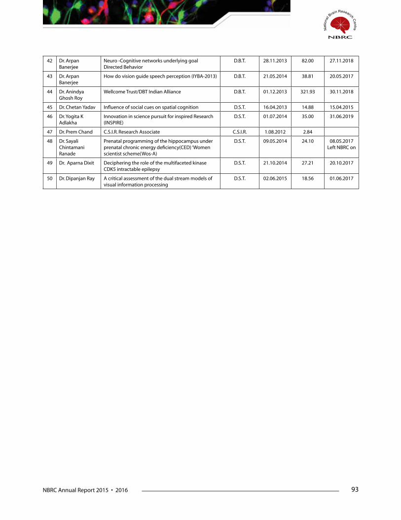
NBRC Annual Report 2015 • 2016 93
42 Dr. Arpan Banerjee
Neuro -Cognitive networks underlying goal Directed Behavior
D.B.T. 28.11.2013 82.00 27.11.2018
43 Dr. Arpan Banerjee
How do vision guide speech perception (IYBA-2013) D.B.T. 21.05.2014 38.81 20.05.2017
44 Dr. Anindya Ghosh Roy
Wellcome Trust/DBT Indian Alliance D.B.T. 01.12.2013 321.93 30.11.2018
45 Dr. Chetan Yadav Influence of social cues on spatial cognition D.S.T. 16.04.2013 14.88 15.04.2015
46 Dr. Yogita K Adlakha
Innovation in science pursuit for inspired Research (INSPIRE)
D.S.T. 01.07.2014 35.00 31.06.2019
47 Dr. Prem Chand C.S.I.R. Research Associate C.S.I.R. 1.08.2012 2.84
48 Dr. Sayali Chintamani Ranade
Prenatal programming of the hippocampus under prenatal chronic energy deficiency(CED) ‘Women scientist scheme(Wos-A)
D.S.T. 09.05.2014 24.10 08.05.2017 Left NBRC on
49 Dr. Aparna Dixit Deciphering the role of the multifaceted kinase CDK5 intractable epilepsy
D.S.T. 21.10.2014 27.21 20.10.2017
50 Dr. Dipanjan Ray A critical assessment of the dual stream models of visual information processing
D.S.T. 02.06.2015 18.56 01.06.2017


NBRC Annual Report 2015 • 2016 97
FacultyAnirban Basu
Senior Scientist Oration Award (Indian Immunology Society); Immunocon- 2015, Patna.
StudentsMahar Fatima
Awarded - Tulsabai Educational Trust Award for Best Paper presentation in the Oral Session at XXXIII Annual Meeting of Indian Academy of Neurosciences held during at Chandigarh, India, October 30th – November 2nd, 2015.
Awarded International Travel Grant from Department of Biotechnology, Government of India in 2015.
Manju Tewari
Awarded International travel grant for by Japanese Neuroscience Society, Japan.
Awarded International Travel Award by International Brain Research Organization (IBRO) for presenting her work at 9th World Congress of Neuroscience organized by IBRO at Rio de Janeiro, Brazil during July 7-11 2015.
Subhadip PalIsaac Newton Fellowship, The Institute of Psychiatry & Neuroscience, King’s College, University of London, 2016.
Aftab AlamInternational Society of Neurochemistry, Visiting Scholar Award, Australian Neuroscience Conference, Brisbane 2015.
Route 28 Workshop on Adult Neurogenesis, Best Project Award, Munich, Germany, 2015.
Rajiv RamaswamyBest Presentation Award, Annual convocation, International Council of Engineering Academies, New Delhi, 2015.
Suhela KapoorEuropean Molecular Biology Organization. Travel Award, Annual Conference of Spanish Society of Neuroscience, 2015.
Vikas PareekInternational Brain Research Organization, APRC Travel Award, National University of Singapore workshop, 2015.
Training Program Fellowship Award, Centre for Fundamental Studies, National Institute of Education Research and Training (NISER), Dept. of Atomic Energy, Govt. of India, 2015.
Course-Work
Integrated Ph.D. 2014Mr. Gourav Sharma
Integrated Ph.D. student, has been awarded first rank upon completion of Course-Work during the year 2014-15 and a certificate was given to him on the 12th Foundation Day, the 16th December 2015.
Ms. Harjot Kaur
Integrated Ph.D. student, has been awarded second rank upon completion of Course-Work during the year 2014-15 and a certificate was given to her on the 12th Foundation Day, the 16th December 2015.
Ph.D. 2014Ms. Mukta Kumari
Ph.D. student, has been awarded first rank upon completion of Course-Work during the year 2014-15 and a certificate was given to her on the 12th Foundation Day, the 16th December 2015.
Mr. Dharmendra Puri
Ph.D. student, has been awarded second rank upon completion of Course-Work during the year 2014-15 and a certificate was given to him on the 12th Foundation Day, the 16th December 2015.
Distinctions, Honours & Awards
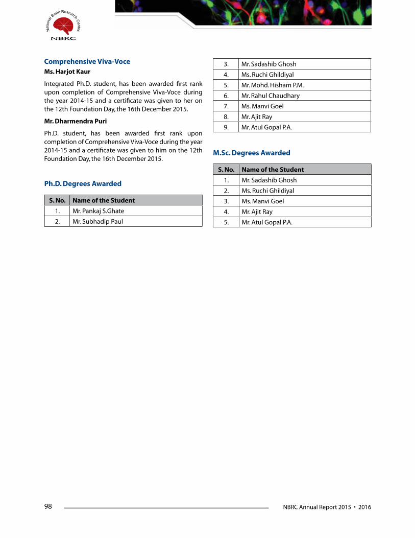
98 NBRC Annual Report 2015 • 2016
Ph.D. Degrees Awarded
M.Sc. Degrees Awarded
S. No. Name of the Student
1. Mr. Sadashib Ghosh
2. Ms. Ruchi Ghildiyal
3. Ms. Manvi Goel
4. Mr. Ajit Ray
5. Mr. Atul Gopal P.A.
S. No. Name of the Student
1. Mr. Pankaj S.Ghate
2. Mr. Subhadip Paul
Comprehensive Viva-VoceMs. Harjot Kaur
Integrated Ph.D. student, has been awarded first rank upon completion of Comprehensive Viva-Voce during the year 2014-15 and a certificate was given to her on the 12th Foundation Day, the 16th December 2015.
Mr. Dharmendra Puri
Ph.D. student, has been awarded first rank upon completion of Comprehensive Viva-Voce during the year 2014-15 and a certificate was given to him on the 12th Foundation Day, the 16th December 2015.
3. Mr. Sadashib Ghosh
4. Ms. Ruchi Ghildiyal
5. Mr. Mohd. Hisham P.M.
6. Mr. Rahul Chaudhary
7. Ms. Manvi Goel
8. Mr. Ajit Ray
9. Mr. Atul Gopal P.A.


NBRC Annual Report 2015 • 2016 101
NBRC was awarded Deemed University status (de-novo category) in 2002 under Section 3 of UGC Act, 1956 (3 of 1956) vide notification No.F.9-
52/2001-U.3 dated 20th May, 2002 issued by Ministry of Human Resources Development, Government of India. NBRC is the first autonomous Institution to attain the status of Deemed University among the other Institutes of the Department of Biotechnology. The ‘Deemed to be University’ status of NBRC has been reviewed by the Committee duly constituted by the UGC and also by an independent Committee constituted by Ministry of HRD, on completion of five years as Deemed University. The committee recommended extension of Deemed University status and placed NBRC under “A’’ category. The notification from Ministry of HRD is awaited.
Ph.D. in NeuroscienceNBRC has a Ph.D. Programme in Neuroscience to develop trained manpower having a broad overview of different aspects of Neuroscience.
NBRC provides a fellowship of `25,000/- per month for Junior Research Fellows and `28,000/- per month for Senior Research Fellows.
M.Sc. in NeuroscienceNBRC is one of the first Institutes in the country to develop an integrated multidisciplinary teaching programme
in Neurosciences. During the academic year 2015-2016 NBRC reintroduced the M.Sc. (Neuroscience) programme to develop trained manpower having a broad overview of different aspects of Neuroscience.
M.Sc. (Neuroscience) students are provided a fellowship of `12,000/- per month.
NBRC inducts students for its M.Sc. (Neuroscience) and Ph.D. programmes from diverse backgrounds having Bachelors or Masters degree in any branch related to Neurosciences, Psychology or M.B.B.S., B.E., or B.Tech. NBRC recognizes that understanding brain functions requires a fusion of knowledge from multiple disciplines.
Summer Training and Short-term ProgrammesNBRC conducts Summer Training Programme for the Students, recommended through three National Science Academies viz: (1) Indian Academy of Science, Bangalore (2) Indian National Science Academy, New Delhi (3) National Academy of Sciences, Allahabad. The summer training is for a period of eight weeks and the trainees are provided with shared accommodation at NBRC hostels. Summer trainees are encouraged to attend seminars and journal clubs organized at the Institute. The summer training projects provides an exposure to Neuroscience and motivates trainees to consider it as a future career option.
Academic Programmes


NBRC Annual Report 2015 • 2016 105
Distributed Information Centre (DIC)
The Distributed Information Centre (DIC-department) manages the overall ICT infrastructure of the Institute. It manages the campus converged network (data and voice traffic), communications links (Network and PSTN), Institute’s Datacentre, hosting core network and application servers, software development, ICT Modernization, e-Governance initiatives, technical support to users, common computing facility etc. The details of some of them are summarized as under :
A) Campus Converged Network (NBRC-IntraNet)
The campus converged network consists of campus wide Local Area Network running on 10 Gbps fiber optic backbone with redundant paths over manageable switching fabric. The redundancy and robustness is built in the network architecture itself. The core LAN network is further integrated with wireless access points installed across the campus and managed through central wireless controller. The network is further supplemented with secure firewall and unified threat management appliances for network safety, intrusion detection system, gateway level antivirus, VPN facility, managing IT policy and detailed auditing/ logging etc. The campus network is a fully IPv6 compliant and IPv6 services are functional in dual stack. The wireless network of the institute has further been integrated with Eduroam service by integrating it with National NREN (ERNET-India), the eduroam service thus provides visiting scientists and researchers seamless secure wireless access in all participating institutions across the world.
The campus converged network of the institute is further integrated with National Knowledge Network (NKN), the last mile link to NKN-Delhi POP is on 1 Gbps optical fibre link provided by BSNL. The NKN linkage is instrumental in the running of several scientific projects for multi-site high volume data applications like –
a) multi site neuro-imaging data repository project (Model NKN project PI : Prof. P K Roy)
b) NBRC-AIIMS data pipeline for MEG as part of
collaborative Centre of Excellence in Epilepsy project funded by DBT.
B) IP-PBX facility
The tele-communication systems of the institute were running on IP-PBX and the campus network is used to carry the voice traffic along with data traffic, the user endpoints are IP-Phones connected to LAN. The facility is running on active-passive automatic failover mode on virtualized servers from institute’s datacenter. The external incoming and outgoing voice traffic is routed on E1-PRI of BSNL. The users are also provided with various facilities like multi-point conferencing, voicemail, directory, call forwarding etc. over the provided end-points.
C) Institute Core and Application Servers
The computing facility manages and maintains the server infrastructure of the institute; they are housed and maintained in the mini-datacenter facility. In essence the institute currently has four numbers of fully utilized 42U server racks in the datacenter facility. The various service running on these server can be classified as under:
a. Web-servers for the institute website (http://www.nbrc.ac.in) and other website like http://neuroscienceacademy.org.in & http://snci.nbrc.ac.in. In addition various web-servers related to ongoing computational projects and applications of various scientific groups is also hosted and managed in the central facility
b. E-mail servers for institute mailing along with list servers.
c. DNS servers for the official and hosted domains.
d. Virtualization servers for providing virtualized hardware to run various applications and service in a more managed manner and to consolidate and utilize the existing physical server infrastructure.
e. Radius and authentication servers for access, accounting and authorization of computing resources
Core Facilities
106 NBRC Annual Report 2015 • 2016
f. License management servers for managing institutional site/network/concurrent licenses.
g. Antivirus and security servers for providing protection to user end-points across the campus.
h. Central Storage servers along with backup servers handling storage requirements of the users and laboratories for online central storage and data processing.
i. Application servers running on windows and Linux platforms for common computing requirements of the users and also other specialized computing servers for specific data processing requirements of various laboratories.
D) Other Facilities & Services
a. The computing facility also provides support and maintenance activities for the entire computing infrastructure of the institute which also includes user endpoints like computers, peripherals, software’s etc. An online support ticketing system with automated workflow management is functional for support activities.
b. The computing facility also undertakes software development activities in line with the institute
requirements, several scientific and e-Governance applications have been developed in-house.
c. The computing facility also undertakes planning and implementation of new computational infrastructure facilities and services, software/hardware/network upgradations of Institute computers/peripherals etc.
E) Planned Future expansions
a. An Enhanced storage server facility with Disaster Recover storage solution has been proposed in collaboration with another DBT institution in near future subject to necessary administrative and financial approval from the competent authority.
b. A dedicated central documentation facility and computer centre for academic activities is proposed to be made operational with the round the clock availability. This along with the planned expansion of the datacenter facility will provide the roadmap for future expansion of the computer facility.
c. Integrated Multi-Media Digital Classrooms with facility for webcasting etc. has also been proposed while upgrading the existing Video-Conferencing Facility in near future.
NBRC Annual Report 2015 • 2016 107
Animal Facility
NBRC is an autonomous institute created by the Department of Biotechnology, Govt. of India, with the mandate of carrying out frontline research to understand neurobiology of brain disorders. As part of the infrastructure, NBRC has a state of the art animal facility to meet the requirements of the scientists for advanced neuroscience research. The Institute recognizes that use of laboratory animals in research is an important privilege accompanied by a great ethical responsibility to ensure humane care and use of these valuable subjects. To ensure appropriate care and use, detailed programs of excellent veterinary and husbandry care, and programs for peer-reviewed evaluation of all activities prior to use of any animal in research are in place. NBRC is committed to the highest standards of research and recognizes that laboratory animals must receive the best possible care, not only to obtain valid research data, but also to ensure the health and safety of animals, researchers, and animal caretakers. Qualified and trained veterinarians oversee all the animal health concerns, and provide all necessary veterinary care to ensure that healthy animals are available for research. The Animal Facility is registered with the Committee for the Purpose of Control and Supervision of Experiments on Animals (CPCSEA), Ministry of Environment and Forests, Government of India, New Delhi. (Registration number: 464/GO/bc/2001/CPCSEA, dated 24/08/2001. All activities of the Laboratory Animal Facility are carried out as per standard operating procedures (SOPs). The Animal Facility maintains the records of day-to-day activities as well as breeding, maintenance and experimentation as per the statutory requirement of CPCSEA.
The main activity of Animal Facility is to procure and breed a wide variety of species of laboratory animals and supply quality animals to in-house researchers, which are used as animal models for understanding the human brain in health and disease. A high degree of hygienic conditions are maintained in the animal house by regular cleaning and sterilization of the cages, water bottles, bedding and feed. The animal rooms are also regularly disinfected. Heavy-duty steam autoclaves have been installed for these purposes. A hot vapour jet machine is used for cleaning the large rabbit and monkey cages. The staff is required to take shower, before changing to work-overalls before entering the animal rooms, and again in the evening after finishing the work. All users wear facemasks and gloves before handling animals.
All the animal species are housed in species appropriate cages, which are designed as per the CPCSEA guidelines. The outdoor play area for non-human primates has six large interconnected enclosures that provide a flexible layout for optimising enrichment and social interactions. The transgenic, knock-out and mutant mice are housed under germ-free conditions in filter top cages and individually ventilated cages (IVC). Such animals are handled in laminar hoods, and the moved to fresh cages in cage-changing station under hepa-filtered air.
The animals are maintained under controlled environmental conditions as specified in CPCSEA guidelines, with temperature maintained between 22 ± 20 C, relative humidity between 45-55%, 12:12 hr light-dark cycle, and 12-15 air changes per hour. The air-handling system uses 100% fresh air for each change.
All animals are procured as per CPCSEA guidelines. A health surveillance program for screening incoming animals is carried out to assess animal quality. Animals procured from other places are kept in quarantine to minimize risk for introduction of infection in established colony.
The animal facility has a state-of-art surgical suite equipped with intensity controlled surgical lights, advanced surgical microscopes, gas anesthesia machines, equipment for monitoring the physiological state of the animals, including heart rate monitor, pulse oximeter and rectal thermometer. For cleaning and sterilization of the surgical instruments there is an ultrasonic instrument cleaner, glass bead sterilizer and ethylene oxide gas sterilizer.
The animal facility has a necropsy room, perfusion room with a perfusion hood, deep freezer for carcass storage, and incinerator for disposal of the animal carcass.
The animal facility has been equipped with a card reader security system. The access is restricted to the animal house staff, maintenance staff and the investigators who are listed in the IAEC approved protocols. All the personnel who handle animals are required to have a current tetanus vaccination, and those who handle non-human primates (NHP) are screened for tuberculosis. Everyone handling NHP’s is trained in the procedures for the first-aid in case of an injury from an animal bite or scratch.
Close circuit monitoring cameras have been installed at various locations in the facility to help in effective monitoring of the animal facility.
The Veterinary staff of Animal Facility is also conducts
108 NBRC Annual Report 2015 • 2016
short term training for M.Sc. and Ph.D. students, Project Assistants and other scientific staff in the field of laboratory animal science covering ethical and statutory guidelines that regulate scientific experiment on animals, general biology and reproduction of the laboratory animals, animal identification techniques, blood collection, injections, anesthesia and monitoring, handling and restraint, husbandry and care, sex differentiation, humane euthanasia, etc
The animal facility is currently maintaining the following species and strains of laboratory animals.
Mice Strains
• SWISS
• BALB/c
• C57BL/6J
• CD1
Transgenic Mice
• B6C3-Tg (APP6 95) 85 DboTg (PSEN1) 85 Dbo(Alzheimer disease model)
• UBC-GFP(Greenfluorescentprotein)
• B6CBA-Tg(Hdexon1)62Gpb/3J(Huntingtondiseasemodel)
• B6.Cg – Mapttm1 (EGFP) Klt Tg (MAPT) 8cPdav/J(Alzheimer disease model)
• B6.129P2 - Gsk3 btm1 Dgen/J (Alzheimer diseasemodel)
• B6.129P2Pvalb<tm1(cre)Arbr>/J
• B6.CgGt(ROSA)26Sor<tm9(CAGtdTomato)
• B6.CgTg(Scnn1acre)3Aibs/J
• STOCKGad2<tm2(cre)Zjh>/J
• B6.CgTg(Camk2a-cre)T29-1Stl/j
• B6.129-Rp122<tm1.1Psam>/j
• STOCKTg(Thy1-EGFP)MJrs/J
• B6.Cg-Tg(Thy1-YFP)16Jrs/J
• B6.Cg-Tg(Thy1-YFP)HJrs/J
• B6;129S6-Tg(Camk2a-cre/ERT2)1Aibs/J
• STOCKSsttm2.1(cre)Zjh/J
• B6.Cg-Gt(ROSA)26Sortm6(CAG-ZsGreen1)Hze/J
Knock out Mice
• UBE3Anullmice(Angelmansyndromemodel)
Mutant Mice
• CBA/Jmice(Retinaldegenerationmodel)
Rat Strains
• LongEvans
• SpragueDawley
• WistarRat
Non-Human Primates
• RhesusMonkeys(Macaca mulatta)
• BoneetMonkeys(Macaca radiata)
Birds
• Zebrafinches(Taeniopygia guttata)
• Housecrows(Corvus splendens)
• Junglecrows(Corvus levaillantii)
All the mice strains are maintained by inbreeding and the rat strains by out breeding. Zebra finch colonies are maintained by out breeding. The transgenic and knockout mice are maintained under a specialized breeding program after the investigators provide the molecular genotyping of these strains based on presence or absence of the gene of interest.
NBRC Annual Report 2015 • 2016 109
Library
The NBRC Library plays a vital role in the collection, development and dissemination of scientific and technical information to meet the present and future needs of the centre and also provides facilities and support to the scientists, researchers, students, staff and NBRC’s networked centers.
The NBRC library has a large collection of journals, books and other relevant research materials on Neuroscience and allied subjects such as Biochemistry, Genetics, Molecular Biology, Immunology & Microbiology, Pharmacology and Toxicology, Psychology, Physics, Mathematics, Computer Science and General Subjects. The NBRC Library currently subscribes to 1171 online journals through the DBT e-Library Consortium (DeLCON), 3 specialty journals, and 122 other journals for the Centre. It also maintains digital archives and news-clips about the Centre and subscribes to Newspapers and News Letters. The collection of the NBRC Library is growing day-by-day along with new developments in research and knowledge in the field of Neuroscience and related areas.
To provide optimum service to all users, the NBRC library is currently digitizing its list of collections using the LSEASE software, to which all users will have full access. A barcode technology has also been installed for accurate and speedy circulation and the management of all library documents. The new software will also help in efficient library operations viz. administration, acquisition, circulation, serial control, cataloguing and information retrieval.
The Library has set up 22 IBM PC-Pentium-IV Computers to provide services for use of researchers and students and has been providing electronic access to the subscribed journals within the campus portal.
The NBRC Library also provides “Inter Library Loan” Services to NBRC’s 48 network centres all over
India. Researchers at different centers send their requirement for research material or journal articles through email to NBRC Library ([email protected]), or to the Librarian Dr. D. D. Lal ([email protected]), and the downloaded articles are sent free of cost. The library entertains an average of approximately 450 requests for articles.
The NBRC Library regularly evaluates its information services to ensure that the Institution’s requirements are met. It promotes resource sharing and cooperation activities among libraries by providing an efficient and reliable means of resource sharing, that is, the inter library loan for the maximum use of resources.
Main Activities of NBRC Library are
1. Book Acquisition
2. Periodicals Acquisition
3. Selective Dissemination Information (SDI),
4. Current Awareness Services (CAS)
5. Inter Library Loan
6. Resource Sharing
7. Circulation Services
8. Reference Services, Bibliographic Services
9. Indexing and Special Services
10. Collects maintains, store and retrieves information and data keeping in the view of evolving needs of its researchers
11. Provide service to Network Centres.
During the year the Library has moved to a new spacious two-storey building, with reading room, reference room, video conferencing, online journal access facility, book section, internet access and reprographic facilities etc. The main aim of the NBRC Library staff is to provide excellent services to users in NBRC and all centers associated with the Institute.


NBRC Annual Report 2015 • 2016 113
DeLCON Consortium: An National Library Consortium for Life Sciences & Biotechnology hosted and admin-istered by NBRC and sponsored by Department of Biotechnology
The ‘DBT’s Electronic Library Consortium (DeLCON)’ is major project of the ‘Department of Biotechnology (DBT)’ to bring qualitative change in their research Institutions. It was launched in January, 2009 with the 10 DBT member Institutions (including DBT H.Q. & ICGEB) with a large number of high impact online journals. It is a national initiative for providing access to scholarly electronic resources including full-text and bibliographic databases in all the life science subject disciplines to DBT Institutional community. It facilitates access to high quality e-resources to DBT research Institutions in the country to improve teaching, learning and research. The Faculty, Scientists, Research Scholars, Students and Project Assistants of Institutions covered under DeLCON are the primary beneficiaries.
The facility was extended to 17 more DBT Institutions in 2nd phase of extension in the year 2010, and 7 additional members were added in the 3rd phase in 2011. Currently DeLCON consortium has 34 member institutions. The ‘DeLCON Consortium’ provides current as well as archival access to more than 963 core and peer-reviewed journals and SCOPUS bibliographic database in different disciplines sourced from 21 publishers and aggregators.
Individual Institution have unique static IP address through which access is given by the publishers to the subscribed journals. However whole programme is administered, monitored and maintained by ‘DeLCON Nodal Centre’ at NBRC and ‘DeLCON National Steering Committee’.
DeLCON currently compromises of the following 34 Member Institution.
DBT Institutions
1. Department of Biotechnology (DBT), New Delhi
2. National Brain Research Centre (NBRC), Manesar
3. National Institute of Plant Genome Research (NIPGR) - New Delhi
4. National Institute of Immunology (NII), New Delhi
5. National Centre for Cell Science (NCCS), Pune
6. Institute of Life Sciences (ILS), Bhubaneshwar
7. Institute of Bioresources and Sustainable Development (ISBD), Imphal
8. Centre for DNA Fingerprinting and Diagnostics (CDFD), Hyderabad
9. Rajiv Gandhi Centre for Biotechnology (RGCB), Thiruvananthapuram
10. International Centre for Genetics and Engineering Biotechnology (ICGEB), New Delhi
11. National Agri-Food Biotechnology Institute (NABI), Mohali, Punjab
12. National Institute of Biomedical Genomics (NIBMG), Kalyani, Kolkata
13. National Institute of Animal Biotechnology (NIAB), Hyderabad
14. Regional Centre for Biotechnology (RBC), Faridabad
15. Transnational Health Science & Technology Institute (THSTI), Faridabad
16. Biotechnology Industry Research Assistance Council (BIRAC), New Delhi
North Eastern Region (NER) Institutes
17. Dibrugarh University, Assam
18. Assam University, Silchar
19. North Eastern Regional Institute of Science & Technology, Arunachal Pradesh
20. North East Institute of Science & Technology, Assam
21. Mizoram University, Mizoram
22. D. M. College of Science (DMC), Mizoram University, Manipur
23. Sikkim University, Gangtok
24. College of Veterinary Science, Assam Agricultural University, Guwahati
25. Gauhati University, Assam
26. Manipur University, Imphal
27. College of Veterinary Science & Animal Husbandry Central Agricultural University, Mizoram
28. Rajiv Gandhi University, Arunachal Pradesh
29. Nagaland University, Nagaland
30. North-Eastern Hill University (NEHU), Shillong
31. St. Anthony’s College (SAC), NEHU, Meghalaya
32. Indian Institute of Technology Guwahati, Assam
33. Tezpur University, Tezpur, Sonitpur, Assam
114 NBRC Annual Report 2015 • 2016
34. Sikkim State Council of Science and Technology, Gangtok, Sikkim
In terms of number of users, the DBT’s Electronic Library Consortium (DeLCON) is the largest Consortium in India in the area of Biotechnology and Life Sciences with a vision and plan to reach out to all DBT Institutes, Departments, Research Institutes, Universities, and their colleges affiliated to DBT, over a period of time.
Subject Areas of DeLCON Consortium
The DeLCON Consortium cover-up all the disciplines and subjects coming under Life Sciences i.e. Biotechnology, Bioinformatics, Biochemistry, Biology, Chemical Biology, Sciences, Immunology, Neuroscience, Plant Genome, Plant Biology, Microbiology, Physiology, Psychology, Physiotherapy, Psychotherapy, Genome, Gene, Genetics, Mathematics, Physics, Chemistry, Radiology, Medicines, Computational Biology, Cell Biology, Cell Sciences, Molecular biology, Molecular and Cellular Biology, Computational Neuroscience, System Neuroscience etc.
All e-resources were evaluated for their i) qualitative and quantitative contents; ii) coverage; iii) availability on different platforms and their comparative advantages/disadvantages; and iv) rates applicable for these resources to individual institutions as well as the other consortia. The electronic resources for consortia- based subscription are selected based on the following major criteria:
• Resources from scholarly societies, universitypresses and non-for-profit projects are preferred over commercial publishers.
• Well-established multi disciplinary resources withbroad cover coverage are preferred over highly specialized sources targeted for specialists.
• Electronic resourcesalreadyonsubscription in theDBT research institutions are preferred over those which are not being used in any of them.
• Resources that are ‘electronic-only’ are preferredover those that are print based.
• Resources those are very important even thoughcost-intensive are preferred over those which are less important or less used but low cost.
• Resources where electronic versions are madeavailable free on subscription to their print versions are avoided as far as possible.
• Selections are made on usage/suitability of
e-resources to DBT Institutions.
Benefits of DeLCON Consortium
The consortia-based subscription to e-resources is a viable solution for increasing the access to electronic resources across DBT institutions at a lower rate of subscription. Major benefits of DeLCON Consortium are:
• DeLCON acts as a single-window service for alarge number of DBT Institutions with their diverse research and academic interest.
• DeLCONwithitscollectivestrengthofparticipatinginstitutions, attracts highly discounted rates of subscription with most favourable terms of agreement for a wider range of e-resources. Most of the e-publishers have responded positively to the call of the Consortium. The rates offered to the consortium are lower by 66% to 99% depending upon the category of DBT institutions.
• DeLCON has triggered remarkable increasein sharing of electronic resources amongst participating DeLCON members
• The research productivity of DBT institutions hasimproved with increased access to international full-text resources (Journals and database).
• Usershaveimmediateaccesstomaterialpreviouslynot subscribed to, at no incremental cost for accessing back files.
• Itimprovestheexistinglibraryservicesandreducedthe subscription cost.
• DeLCON is open so that other DBT institution canalso join the DeLCON Consortium.
• DeLCON offers better terms of agreement for use,archival access and preservation of subscribed electronic resources, which would not have been possible for any single institutions.
• Members of the DeLCON Consortium have thebenefit of cap on the annual increase in the rates of subscription. While the usual increase in price of e- resources is vary from 15% to 20%, but the DeLCON members enjoy a cap on increase in price ranging from 5% to 7%.
• Since thesubscribed resources isaccessibleonlinein electronic format, the DBT institutions have less pressure on space requirement for storing and managing print-based library resources.
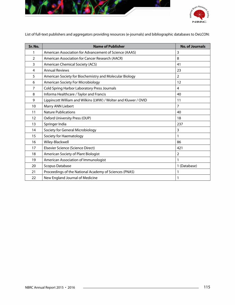
NBRC Annual Report 2015 • 2016 115
Sr. No. Name of Publisher No. of Journals
1 American Association for Advancement of Science (AAAS) 3
2 American Association for Cancer Research (AACR) 8
3 American Chemical Society (ACS) 41
4 Annual Reviews 23
5 American Society for Biochemistry and Molecular Biology 2
6 American Society For Microbiology 12
7 Cold Spring Harbor Laboratory Press Journals 4
8 Informa Healthcare / Taylor and Francis 40
9 Lippincott William and Wilkins (LWW) / Wolter and Kluwer / OVID 11
10 Marry ANN Liebert 7
11 Nature Publications 40
12 Oxford University Press (OUP) 18
13 Springer India 237
14 Society for General Microbiology 3
15 Society for Haematology 1
16 Wiley-Blackwell 86
17 Elsevier Science (Science Direct) 421
18 American Society of Plant Biologist 2
19 American Association of Immunologist 1
20 Scopus Database 1 (Database)
21 Proceedings of the National Academy of Sciences (PNAS) 1
22 New England Journal of Medicine 1
List of full-text publishers and aggregators providing resources (e-journals) and bibliographic databases to DeLCON:


NBRC Annual Report 2015 • 2016 119
The National Neuroimaging facility, sponsored by the Department of Biotechnology, Govt. of India, came into existence in the year of 2006. The main purpose of this National Facility is to facilitate and support advanced brain imaging research. The facility is equipped with state-of-the-art equipments such as,
1. 3 tesla Magnetic Resonance Imaging (MRI) Scanner2. Electroencephalography (EEG)3. Evoked Response Potential Recording (ERP)
Magnetic Resonance Imaging (MRI)MRI provides much greater contrast between the different soft tissues of the body compared to computed tomography (CT), making MRI it especially useful in neurological musculoskeletal and cardiovascular studies. Imaging modalities play important role providing crucial information which can aid to various diagnostic procedures. There are various imaging modalities, which are:
1. MR Spectroscopy (MRS) that provides non-invasive neurochemical level estimations and enables clinical correlation.
2. Functional MRI (fMRI) which, as the name suggests correlates functional (haemodynamics) activity with images of brain activation
The 3 Tesla Phillips whole body MRI scanner at our Facility is equipped with high precision hardware,
software and data processing platforms required for each imaging modality. The facility is being used for performing structural, metabolic (multinuclear, e.g. proton or phosphorous) and functional MRI. In addition to understanding brain function and clinical research, the center is closely interacting with leading imaging centers within the country and across the globe.
Electroencephalography (EEG)This is a test that measures and records the electrical activity of the brain. Special sensors are attached to the head and accessed by wires to a computer. The computer records brain’s electrical activity on the screen or on paper as wavy lines. Certain conditions, such as epilepsy, dementia, consciousness and narcolepsy (sleeping disorder) can be studied by EEG.
Evoked Response Potential Recording (ERP)ERP is an electrical potential recorded from the nervous system of a human or other animal following presentation of a stimulus. Evoked potential amplitudes tend to be low, ranging from less than a microvolt to several microvolts
Clinical studies on patients with Alzheimer’s Disease, Parkinson’s Disease, Autism and Brain Tumours, as well as monitoring of aging in normal healthy brain are being performed extensively in the National Neuro imaging facility.
National Neuroimaging Facility


NBRC Annual Report 2015 • 2016 123
The unit is located at the Government General Hospital, Civil Lines, Gurgaon 122 001.
Investigation facilitiesThe following facilities are available to the patients of the unit through the hospital/clinics at concessional rates:
MRI system: Siemens Magnetom 1.5 Tesla scanner with various study protocols
CT (computed tomography) system
Ultrasonography
Neurophysiology: EEG, Evoked response, EMG.
X-ray and Contrast imaging.
Laboratory facilities Biochemistry, Microbiology, Haematology, Pathology & Immunology.
The expertise of the following faculty are available at the NBRC Unit:
Consultant Clinical Professor: Dr. V. S. Mehta
Consultant Clinical Assistant Professor: Dr Rajnish Kumar
Consultant Clinical Assistant Professor: Dr Amit Arora
Clinical Neuropsychologist: Sumati Chauhan
Clinic Assistant: Hanuman Singh and Pawan Kumar
Translational research aims to connect basic research to patient care bidirectionally for mutual benefit: “From the Bench lab to the Bedside patient and back to the Bench”. The Translational & Clinical Research Unit of NBRC covers the full spectrum of clinical neuroscience: Neurology, Neuropsychology, Neuropsychiatry, Neurosurgery, Behavioral Therapy, Psychology, and Psychometry. The unit has a morning outpatient facility, at the Government General Hospital five days a week, each of the consultant clinical faculty is available on one of the designated days. The NBRC Unit has integrated well with the Civil hospital medical team and there is an increasing number of referrals from other in-house departments and local hospitals. If a patient of the unit requires indoor treatment or observation, then, with courtesy of Neuropsychiatrists
and Specialist Clinicians of internal medicine of the General Hospital, the patient is taken care of.
The out-patients facility is busy, and on some days attendance can exceed 70 to 80 patients. The follow up by the patients is about 75%. Male to female ratio is almost equal. Paediatric group patient attendance is mainly for management of Epileptic Seizure and disorders of the Mentally Challenged. There are also Elderly or Geriatric patients attending, and Movement Disorders are an important factor of attendance.
Patients attending the OPD at Civil Hospital come from old Gurgaon township and the villages and towns in the surrounding districts of Haryana, while some come from neighbouring states as Rajasthan, Delhi, Uttaranchal, Himachal Pradesh, Punjab and Uttar Pradesh.
Patients requiring advanced specialist neurology in-patient care are referred to All-India Institute of Medical Sciences (AIIMS), Institute of Postgraduate Medical Education & Research – Rohtak, Institute of Human Behaviour & Allied Sciences (IHBAS), or Vardhaman Medical College (Safdarjung Hospital), New Delhi or to other tertiary hospital as per the choice of the patient, if he or she so desires. The Clinical Neuroscience OPD rooms have been refurbished and space has been allotted for NBRC in the outpatient area, which can accommodate other members of our team.
Outpatient case records in neurology are maintained from the outset, aside from relevant entry into the patient OP case sheets, which the patients keep in their possession. A comprehensive Neurology case sheet has been formulated and formatted by Distributed Information Centre of NBRC. We are undertaking to prospectively enter all the medical data of new patients, to create computer database with relevant patient data along with any planned imaging/molecular/neurophysiological studies at the NBRC labs, thus creating a well documented “clinical window” for NBRC. In this effort to narrow the gap between Basic Neuroscience and Applied Neuroscience, an ethics committee has been formulated jointly with the Government General Hospital/Government of Haryana.
The association of NBRC with Alzheimer’s & Related
Translational & Clinical Neuroscience Unit

124 NBRC Annual Report 2015 • 2016
Disorders Society of India (ARDSI), which has been going on for a decade, has been further fostered. This interaction promises to grow to the mutual benefit of both institutions concerned primarily with the common goal of the care of the elderly in its varied aspects. Besides medical and neurological health issues, one is exposed to the psychosocial and public health problems of the ageing populace in their home environment.
Electrophysiological facility incorporates support like Electromyography (EMG) and Neurophysiological studies as Nerve Conduction velocity system and Neurometry.
For proper functioning and further clinical support, the
NBRC Unit at the Hospital receives the fullest cooperation of the Ministry of Health - Government of Haryana, and the Deputy Commissioner - Gurgaon, as well as from the Chief Medical Officer & Civil Surgeon together with the Principal Medical Officer of the Hospital.
Due to the extreme shortage of Neuropsychiatric manpower in the northern regions of the country, the Directorate General of Health Sciences, Government of Haryana, has approved the setting up of Post- Graduate Educational Program in Psychological Medicine (DNB, Diplomate of the National Board in Psychiatry) at the hospital, enabling our Unit to have a seminal productive academic output.

NBRC Annual Report 2015 • 2016 127
Centre of Excellence (CoE) for Epilepsy Research (Funded by Department of Biotechnology, Govt. of India)
Scientific Faculty:Prof. Sarat Chandra, AIIMSProf. ManjariTripathi, AIIMSProf. Subrata Sinha, NBRCProf. Prasun K Roy, NBRCProf. Pravat Mandal, NBRCDr. Arpan Banerjee, NBRCDr. Jyotirmoy Banerjee, NBRCDr. Aparna Dixit, NBRC
MEG TechnologistsMr. V. VibhinMr. Kamal Bharti
Research StaffArpna Srivastava, Research AssociateSoumilDey, Research FellowDebasmita Paul, Research FellowDevina Sharma, Research Fellow
Centre of Excellence (CoE) for Epilepsy Research is a collaborative project between National Brain Research Centre (NBRC), Manesar and All India Institute of medical Sciences (AIIMS), New Delhi. CoEfor Epilepsy iscomprised of, a Magnetoencephalography (MEG) &Brain Mapping facility at NBRC and Clinical Electrophysiology facility, Epilepsy Surgery facility and Prof. P.N. Tandon’sEpilepsy Neurobiology lab at AIIMS. The primary aim of this center is to find a cure for drug resistant epilepsy (DRE) by studyingthe epileptogenic networks, and to identify potential biomarkers using a systems biology approach from the molecular/cellular level to clinical/imaging level. Based on the approved objectives for CoE for Epilepsy, we have used multimodal imaging along with MEG and electrocorticography (ECoG) for the localization of epileptogenic zone, followed by resective surgery of patients with DRE. Further we have performed histopathological studies, gene expression analysis and patch clamp recordings from neurons in
the resected brain samples. As epilepsy is associated with alteration in distributed neural networks and it requires a multidisciplinary approach to explore network abnormalities. So, in the current year we have investigated the epileptogenic network hubs in these patients and also studied the cellular basis of abnormal neural network activity. The most common form of DRE is mesial temporal lobe epilepsy (MTLE), where the hippocampus is thought to be involved in seizure generation. We have used MEG investigations to identify the number and region of network hubs in patients with MTLE and correlated it with the cellular and molecular findings.
Understanding epileptogenic networks in mesial temporal lobe epilepsy-hippocampal sclerosis (Jyotirmoy Banerjee)The most common form of drug-resistant epilepsy is mesial temporal lobe epilepsy- hippocampal sclerosis (MTLE-HS), where the mesial temporal lobe structures (including hippocampus, amygdala and other entero-rhinal structures) are thought to be involved in seizure generation through networks that involve these regions. Our recent finding based on cellular electrophysiological studies has revealed that in hippocampal samples obtained from patients withMTLE-HS enhanced endogenous activity of NMDA receptor contributes to excitability in pyramidal neurons. We observed that frequency of spontaneous excitatory postsynaptic currents (EPSCs),recorded using whole cell patch-clamp technique, were 28.2 ± 2.1% higher in slices obtained from patients with MTLE-HS compared to that in case of non-epileptic controls. We also examined spontaneous fast current transients (CTs) recorded from these pyramidal neurons under cell-attached configuration. The frequency of CTs increased in the absence of extracellular Mg2+ in brain slice preparations and were completely blocked by APV. We found that the frequency of CTs in pyramidal neurons were higher in case of MTLE samples compared to non-epileptic controls. Our finding that the NMDA receptor antagonist APV decreased the frequency of EPSCs in the pyramidal neurons suggests that tonically active NMDA receptors on glutamate neurons/axons contribute to the maintenance of
Centre of Excellence

128 NBRC Annual Report 2015 • 2016
glutamatergic transmission to pyramidal neurons in human cortical slices. The magnitude of inhibition of frequency of EPSCs by APV alone was higher in MTLE compared to that in non-epileptic control suggesting the role of enhanced tonic NMDA receptor activity in altered synaptic transmission. When the slices were exposed to Mg2+-free ACSF, there was increase in the frequency of CTs indicating an increase in activity of synaptic NMDA receptors on the pyramidal neurons due to a reduction of Mg2+ block of these receptors. This indicates that tonically active NMDA receptors have a critical role in the maintenance of neuronal firing. The application of specific glutamate receptor antagonists on the slices revealed the subtype specific regulation of CTs. The observation that APV completely blocks the CTs in pyramidal neurons, both in epileptic and non-epileptic samples, further confirms the role of endogenously activated NMDA
receptors in the excitability of pyramidal neurons. Data showing that the frequency of CTs significantly increased in MTLE indicates the role of enhanced activity of NMDA receptors in these patients. Taken together, contribution of endogenously activated NMDA receptors to EPSCs and to the neuronal firing in slice preparations obtained from hippocampal samples clearly demonstrates its role in neuronal hyper excitability, a distinctive feature of MTLE.
Magnetoencephalographic (MEG) investigation was used to identify the number and region of networks using the coherence analysis which was then correlated with the cellular and molecular findings. We observed that under
Figure 2: MEG coherence analysis showing phase relationship in the hippocampus and the anterior temporal lobe of patients with MTLE.
Figure 1: Pattern of current transients (CTs) of pyramidal neurons in resected hippocampal specimen obtained from patients with MTLE.
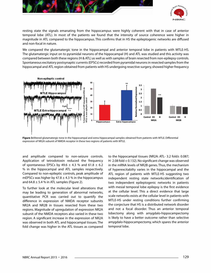
NBRC Annual Report 2015 • 2016 129
and amplitude compared to non-seizure controls. Application of tetrodotoxin reduced the frequency of spontaneous EPSCs by 49.6 ± 4.3 % and 61.8 ± 6.2 % in the hippocampal and ATL samples respectively. Compared to non-epileptic controls, peak amplitude of mEPSCs was higher by 47.8 ± 4.3 % in the hippocampus and 64.8 ± 5.4 % in ATL samples (Figure 2).
To further look at the molecular level alterations that may be leading to generation of abnormal networks, quantitative PCR was carried out to quantify the difference in expression of NMDA receptor subunits NR2A and NR2B in tissues resected from these two regions. Magnitude of upregulation of expression NR2A subunit of the NMDA receptors also varied in these two region. A significant increase in the expression of NR2A was observed in both ATL and hippocampal tissues. The fold change was higher in the ATL tissues as compared
resting state the signals emanating from the hippocampus were highly coherent with that in case of anterior temporal lobe (ATL). In most of the patients we found that the intensity of source coherence were higher in magnitude in ATL compared to the hippocampus. This confirms that in HS the epileptogenic networks are diffused and non-focal in nature.
We compared the glutamatergic tone in the hippocampal and anterior temporal lobe in patients with MTLE-HS. The glutamatergic input on to pyramidal neurons of the hippocampal (H) and ATL was studied and this activity was compared between both these regions (H & ATL) as well as with samples of brain resected from non-epilepsy controls. Spontaneous excitatory postsynaptic currents (EPSCs) recorded from pyramidal neurons in resected samples from the hippocampal and ATL region obtained from patients with HS undergoing resective surgery, showed higher frequency
to the hippocampal tissues (NR2A: ATL- 3.2 fold± 0.087; H- 2.08 fold ± 0.132). No significant change was observed in the mRNA levels of NR2B genes. Thus, the mechanism of hyperexcitabilty varies in the hippocampal and the ATL region of patients with MTLE-HS suggesting two independent resting state networks.Identification of two independent epileptogenic networks in patients with mesial temporal lobe epilepsy is the first evidence at the cellular level. This a direct evidence that large scale networks exists at the cellular level in patients with MTLE-HS under resting conditions further confirming the conjecture that HS is a distributed network disorder and not a focal disorder. Thus an anterior temporal lobectomy along with amygdalo-hippocampectomy is likely to have a better outcome rather than selective amygdalo-hippocampectomy, which spares the anterior temporal lobe.
Figure 3:Altered glutamatergic tone in the hippocampal and extra-hippocampal samples obtained from patients with MTLE. Differential expression of NR2A subunit of NMDA receptor in these two regions of patients with MTLE.

130 NBRC Annual Report 2015 • 2016
Publications1. Banerjee J, Dixit AB, Tripathi M, Sarkar C, Gupta
YK & Chandra PS*. (2015) Enhanced endogenous activation of NMDA receptors in pyramidal neurons of hippocampal tissues from patients with mesial temporal lobe epilepsy: a mechanism of hyper excitation. Epilepsy Research. 117, 11-16.
2. Chandra SP, Kurwale NS, Chibber SS, Banerjee J, Dwivedi R, Garg A, Bal C, Tripathi M, Sarkar C, Tripathi M. (2016) Endoscopic-Assisted (Through a Mini Craniotomy) Corpus Callosotomy Combined With Anterior, Hippocampal, and Posterior Commissurotomy in Lennox-Gastaut Syndrome: A Pilot Study to Establish Its Safety and Efficacy. Neurosurgery. 78, 743-51.
3. Dixit AB, Tripathi M, Chandra PS &Banerjee J. (2016) Molecular Biomarkers in Drug-Resistant Epilepsy: Facts & Possibilities. International Journal of Surgery (In Press)
Presentations1. Jyotirmoy Banerjee. How do networks and
neurotransmitters interact in epileptogenesis.Invited talk at 9th Asian Epilepsy Surgery Congress and 10th Indian Epilepsy Society School at Udaipur on 23rd -25th October, 2015.
2. Jyotirmoy Banerjee. Understanding complexities of drug-resistant epilepsy using bedside-back-to-bench approach: A paradigm shift in epilepsy research. Invited lecture at XXXIII Annual Conference of IAN, Panjab University, Chandigarh on 31st October to 2nd November, 2015.
3. Jyotirmoy Banerjee. Invited talk on “Brain Research”at 4th National Knowledge Network (NKN) Annual Workshop at Jawaharlal Nehru Technological University (JNTU), Hyderabad on 21st to 22nd February, 2016.
4. Jyotirmoy Banerjee. Invited lecture at IBRO-APRC school at NBRC, Manesar. 15th -30th March, 2016.
5. Facilitated workshop on” Advanced Epilepsy Live Surgery, Neurobiology and functional Neurosurgery” held at AIIMS, New Delhi on 9-10th April 2015.
6. Poster presentations at 9thAESC, 2015 on the topics:
i) Endogenous activity of NMDA receptors contributes to the enhanced glutamatergic tone and hyperexcitability in resected brain samples obtained for patients with mesial temporal lobe epilepsy”. Banerjee J, Dixit AB, Tripathi M, Chandra PS.
ii) Endogenous kynurenic acid, a tryptophan metabolite, synthesis is altered in resected brain specimens obtained from patients with mesial temporal lobe epilepsy. Dey S, Dixit AB, Tripathi M, Chandra PS, Banerjee J.
Funding:“Centre of Excellence for Epilepsy Research” funded by Department of Biotechnology, India.
Award:Best poster award at 9th Asian Epilepsy Surgery Congress (AESC), 2015, Udaipur, India.
RNA-Seq analysis of hippocampal tissues reveals novel candidate genes for drug refrac-tory epilepsy in patients with MTLE-HS (Aparna Dixit)Epilepsy is a common brain disorder affecting approximately 50 million people worldwide. MTLE is the most frequent form of partial epilepsy observed in adults, accounting for 40% of cases. About 30% of MTLE cases are resistant to anti-epileptic drugs (AEDs). Surgery is the only effective treatment for patients with medically refractory MTLE. However, about one third of patients continue with seizures after surgical therapy. Therefore, there is an urgency to understand the mechanisms of epileptogenesis and pharmacoresistance in MTLE in order to identify biomarkers as well as novel therapeutics to prevent epilepsy patients at risk. Despite increasing information about the abnormal synaptic transmission associated with epileptogenesis, the molecular mechanisms involved in the genesis of seizures in patients with epilepsy, especially with DRE, still remains unexplained. Array-based profiling studies have detected aberrant gene expression patterns in large number of genes, still most of the genes that participate in the development of pharmacoresistance in epilepsy remain unidentified. Several mechanisms of pharmacoresistance have been proposed like dysregulation of multidrug transporters (MDTs), reduced drug-target sensitivity, increased neuronal apoptosis, cytoskeletal alterations and reorganization of neuronal networks but the exact mechanisms underlying pharmacoresistance are still enigmatic.
We have performed RNAseq analysis of the hippocampal tissues resected from the patients with MTLE-HS to identify potential molecules and pathways associated with drug refractory MTLE. For non-epileptic control experiments, healthy tissues from tumour margins obtained during tumour surgeries were used. RNA sequencing was performed using standard protocols on Illumina HiSeq 2500 platform. Differential gene expression analysis of the RNAseq data revealed 56
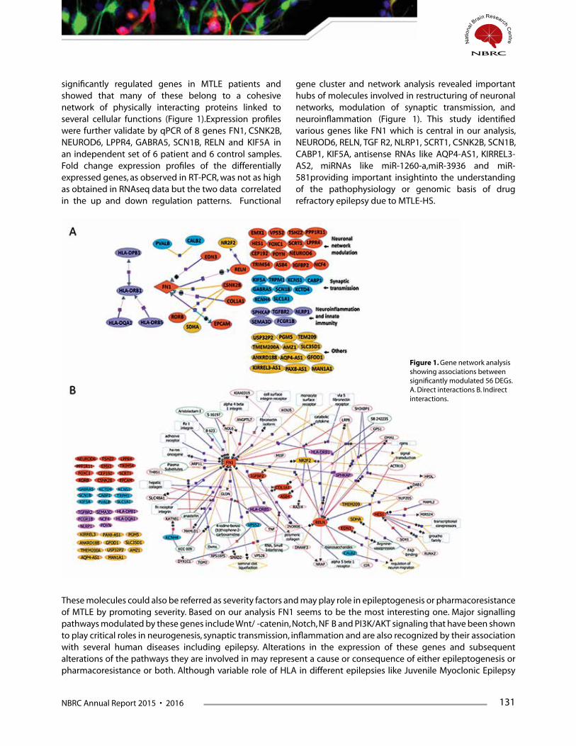
NBRC Annual Report 2015 • 2016 131
significantly regulated genes in MTLE patients and showed that many of these belong to a cohesive network of physically interacting proteins linked to several cellular functions (Figure 1).Expression profiles were further validate by qPCR of 8 genes FN1, CSNK2B, NEUROD6, LPPR4, GABRA5, SCN1B, RELN and KIF5A in an independent set of 6 patient and 6 control samples. Fold change expression profiles of the differentially expressed genes, as observed in RT-PCR, was not as high as obtained in RNAseq data but the two data correlated in the up and down regulation patterns. Functional
gene cluster and network analysis revealed important hubs of molecules involved in restructuring of neuronal networks, modulation of synaptic transmission, and neuroinflammation (Figure 1). This study identified various genes like FN1 which is central in our analysis, NEUROD6, RELN, TGF‐R2, NLRP1, SCRT1, CSNK2B, SCN1B, CABP1, KIF5A, antisense RNAs like AQP4-AS1, KIRREL3-AS2, miRNAs like miR-1260-a,miR-3936 and miR-581providing important insightinto the understanding of the pathophysiology or genomic basis of drug refractory epilepsy due to MTLE-HS.
These molecules could also be referred as severity factors and may play role in epileptogenesis or pharmacoresistance of MTLE by promoting severity. Based on our analysis FN1 seems to be the most interesting one. Major signalling pathways modulated by these genes include Wnt/‐-catenin, Notch, NF‐B and PI3K/AKT signaling that have been shown to play critical roles in neurogenesis, synaptic transmission, inflammation and are also recognized by their association with several human diseases including epilepsy. Alterations in the expression of these genes and subsequent alterations of the pathways they are involved in may represent a cause or consequence of either epileptogenesis or pharmacoresistance or both. Although variable role of HLA in different epilepsies like Juvenile Myoclonic Epilepsy
Figure 1. Gene network analysis showing associations between significantly modulated 56 DEGs. A. Direct interactions B. Indirect interactions.

132 NBRC Annual Report 2015 • 2016
(JME), Lenox-Gastaut syndrome is known, changes in expression patterns of the HLA haplotypes found in our study demands more investigations to look for the role of HLA haplotypes in disease progression like MTLE. We could not find differential regulation of any drug transporters like MRP1 and MRP2 or drug metabolising enzymes and also not many candidate genes for drug target sensitivity were identified. Rather the three distinct yet overlapping hubs of molecules found in our study are more supportive of intrinsic severity hypothesis of pharmacoresistance. Novel therapeutic interventions affecting at network level and not just restricted to one or two targets and treatments devised to reverse severity mechanisms might hold promise for the treatment of pharmacoresistant epilepsy. Also further studies in serum or CSF samples in a larger cohort of patients is needed to validate these molecules specifically “FN1” as potential diagnostic/prognostic biomarkers of intractable MTLE.
Future study: We would further like to evaluate the role of FN1 in epileptogenesis by decipheringFN1 signaling in MTLE (Figure 2).
Exploring the inflammatory mediators, microR-NAs and transcription factors regulatory net-works in mesial temporal lobe epilepsy (MTLE) and focal cortical dysplasia (FCD)Neuroinflammation and innate immunity play important role in the pathogenesis of epilepsy. Cytokines and chemokines induced inflammation may lead to a disturbance of the glutamatergic system, and subsequently to the persistence of seizures by chronic
Figure 2: FN1 signaling in MTLE
neuronal over excitation. Numerous candidate gene specific studies have postulated the role of inflammatory and immune modulators in neuronal death and/or development of pharmacoresistance in MTLE however there are not many reports in FCD. Inflammation is a complex and highly coordinated process, regulated by several factors i.e. micro RNAs, transcription factors, as well as various transcriptional co-regulators and chromatin modifications. miRNAs play a significant role in inflammatory pathways which have been shown to be involved in epilepsy. There is limited information regarding the regulation of cytokine/chemokine-receptor regulatory network in epilepsy, suggesting contribution of miRNAs and transcription factors (TFs).
In this study we have measured multiple inflammatory mediators (cytokines, chemokines and growth factors) which includes IL1β, IL1Ra, IL6, IL10, MIP-1a (CCL3), MIP-1β (CCL4) and TNFa in brain tissue samples resected from MTLE (10), FCD (10) and tumor periphery (8) of glioma patients (non-epileptic controls) by quantitative cytokine assays using a customized Bioplex TM Pro-human cytokine 8-plex panel kit. We have investigated simultaneously the transcriptional changes of mRNA expression levels of downstream targets (STAT-3, C-JUN, ICER, and CCR5) of significantly altered cytokines and upstream regulators miRNAs (miR-21-5p, miR-204-5p, miR-106a-5p, let-7c-5p, miR-203a-3p, let-7a-5p, miR-155-5p, miR-223-3p, miR-195-5p) by quantitative real time PCR.
Upregulation of IL-1 ß, IL-6, MIP-1β and MIP-1β were observed in both MTLE and FCD patients as compared to controls (p<0.05). Except IL-1β, upregulation was relatively higher in FCD. IL-10 showed down regulation in both, MTLE and FCD as compared to controls. TNF-a, VEGF A and IL-1RA did not show any significant change between groups. Downstream targets of these cytokines, STAT-3, C-JUN, ICER, and CCR5 are also found to be significantly altered in MTLE and FCD as compared to controls (p<0.05). Only CCR5 (downstream target of MIP-1a and MIP-1ß ) has been found to be significantly upregulated in FCD as compared to MTLE, other transcription factors did not show significant difference.Linking expression profiles of transcriptional regulators and miRNAs with their annotated functions, we demonstrate dynamic interplay of miRNAs and downstream regulators with biological functions (Figure 3).
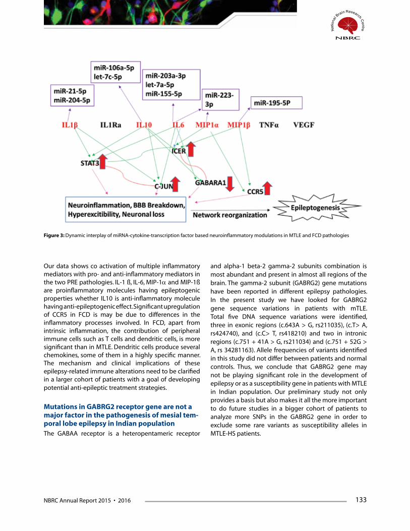
NBRC Annual Report 2015 • 2016 133
Our data shows co activation of multiple inflammatory mediators with pro- and anti-inflammatory mediators in the two PRE pathologies. IL-1 ß, IL-6, MIP-1a and MIP-1ß are proinflammatory molecules having epileptogenic properties whether IL10 is anti-inflammatory molecule having anti-epileptogenic effect. Significant upregulation of CCR5 in FCD is may be due to differences in the inflammatory processes involved. In FCD, apart from intrinsic inflammation, the contribution of peripheral immune cells such as T cells and dendritic cells, is more significant than in MTLE. Dendritic cells produce several chemokines, some of them in a highly specific manner. The mechanism and clinical implications of these epilepsy-related immune alterations need to be clarified in a larger cohort of patients with a goal of developing potential anti-epileptic treatment strategies.
Mutations in GABRG2 receptor gene are not a major factor in the pathogenesis of mesial tem-poral lobe epilepsy in Indian populationThe GABAA receptor is a heteropentameric receptor
Figure 3: Dynamic interplay of miRNA-cytokine-transcription factor based neuroinflammatory modulations in MTLE and FCD pathologies
and alpha-1 beta-2 gamma-2 subunits combination is most abundant and present in almost all regions of the brain. The gamma-2 subunit (GABRG2) gene mutations have been reported in different epilepsy pathologies. In the present study we have looked for GABRG2 gene sequence variations in patients with mTLE. Total five DNA sequence variations were identified, three in exonic regions (c.643A > G, rs211035), (c.T> A,rs424740), and (c.C> T, rs418210) and two in intronicregions(c.751+41A>G,rs211034)and(c.751+52G>A, rs 34281163). Allele frequencies of variants identified in this study did not differ between patients and normal controls. Thus, we conclude that GABRG2 gene may not be playing significant role in the development of epilepsy or as a susceptibility gene in patients with MTLE in Indian population. Our preliminary study not only provides a basis but also makes it all the more important to do future studies in a bigger cohort of patients to analyze more SNPs in the GABRG2 gene in order to exclude some rare variants as susceptibility alleles in MTLE-HS patients.
134 NBRC Annual Report 2015 • 2016
Publications1. Dixit AB, Banerjee J, Srivastava A, Tripathi M, Sarkar C, Kakkar A, Jain M and Chandra PS. (2016). RNA-Seq analysis
of hippocampal tissues reveals novel candidate genes for drug refractory epilepsy in patients with MTLE-HS. Genomics, Volume 107, Issue 5, May 2016, 178–188.
2. Tripathi M, DixitA, Chandra PS. (2016). Galectin-3, an important yet unexplored molecule in drug resistant epilepsy. Neurol India 64:237-8.
3. Srivastava A, Dixit AB, Banerjee J, Tripathi M, Chandra PS. (2015). Role of inflammation and its miRNA based regulation in epilepsy: Implications for therapy. ClinicaChimicaActa 452 (2016) 1–9.
4. Dixit AB, Banerjee J, Tripathi M, Sarkar C, Chandra PS. (2016). Synaptic roles of Cyclin dependent Kinase 5 and its implications in epilepsy. IJMR (In press).
5. Dixit AB, Banerjee J, Tripathi M, Chandra PS. (2015). Presurgical epileptogenic network analysis: A way to enhance epilepsy surgery outcome. Neurol India 2015;63:743-50.
6. Dixit AB, Ansari A, Banerjee J, TripathiM and Chandra PS. (2015) Mutations in GABRG2 receptor gene are not a major factor in the pathogenesis of mesial temporal Lobe Epilepsy in Indian population. Ann. Indian Acad. Neurol 19, 236-241.
Presentations1. Executive committee member of 9th Asian Epilepsy Surgery Congress, Udaipur, India on October 24rd - 25th,
2014.
Posters presented in AESC, 2015
i. Differential modulation of various inflammatory mediators in mesial temporal lobe epilepsy and focal cortical dysplasia patients.” Dixit AB, Paul D, Srivastava A, Banerjee J, Tripathi M, and Chandra PS.
ii. Gene Expression Analysis of Drug Transporters & Biotransformation Enzyme in patients with MTLE & FCD: A Comparative Study. Sharma D, Srivastava A, Dixit AB, Banerjee J, Tripathi M, Chandra PS.
2. Poster presented at 31st International Epilepsy Congress (IEC), Istanbul, 5th – 9th September, 2015.RNA-Seq analysis of hippocampal tissues reveals novel candidate genes for drug refractory epilepsy in patients with MTLE-HS”. Dixit AB, Banerjee J, Srivastava A, Tripathi M, Sarkar C, Kakkar A, Jain M and Chandra PS.
3. Facilitated workshop on” Advanced Epilepsy Live Surgery, Neurobiology and functional Neurosurgery” held at All India Institute of Medical Sciences, New Delhi on 9-10th April 2015.
Funding1. “Bio-CARe” Women Scientist Scheme (RGO) BT/AB/08/01/2008 (III) Grant entitled “Deciphering the role of the
multifaceted kinase CDK5 in intractable epilepsy.” Since October 2014, funded by Department of Biotechnology, India.
2. “Centre of Excellence in Epilepsy” a collaborative project between NBRC & AIIMS, funded by Department of Biotechnology, India, since July 2014.
AwardsYoung Investigator’s Award, Best poster & Travel Award in 31st International Epilepsy Congress (IEC), Istanbul, 5th – 9th September, 2015.

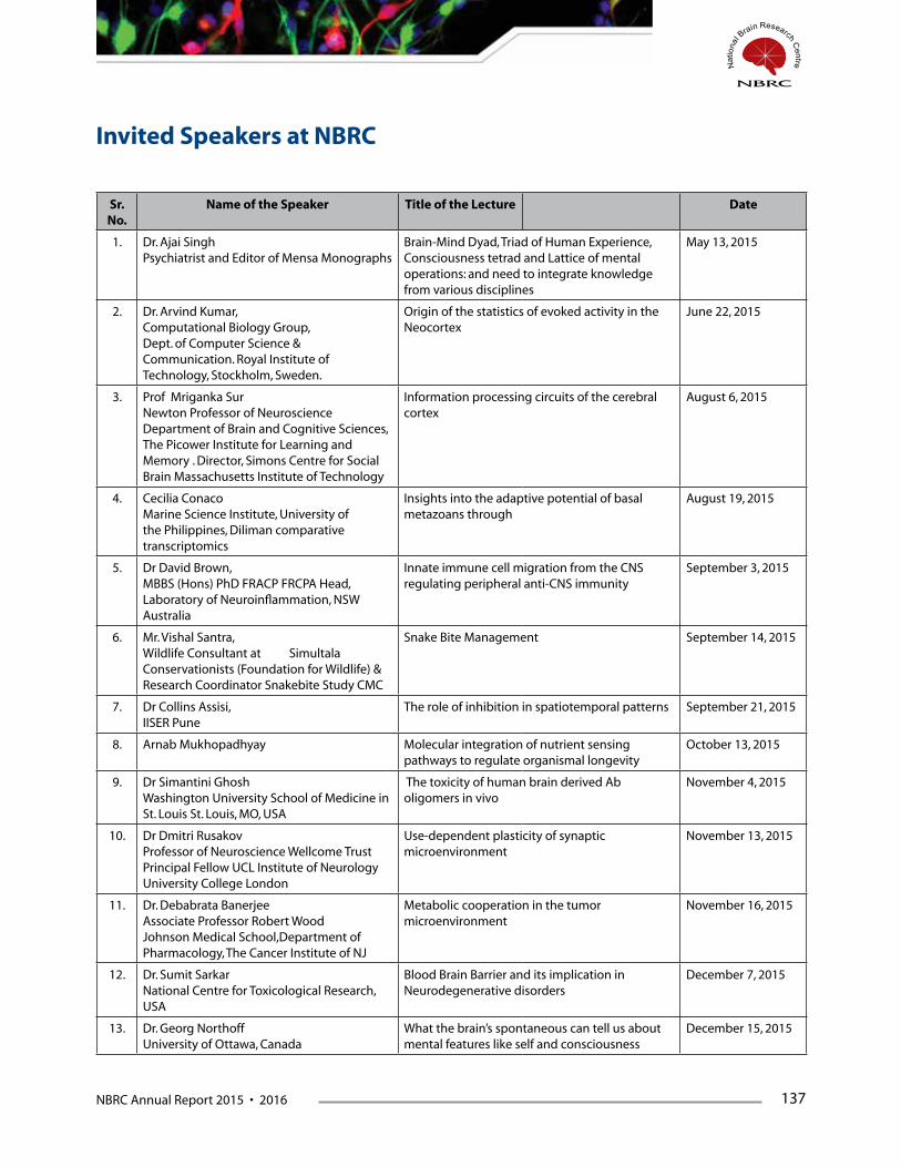
NBRC Annual Report 2015 • 2016 137
Sr. No.
Name of the Speaker Title of the Lecture Date
1. Dr. Ajai SinghPsychiatrist and Editor of Mensa Monographs
Brain-Mind Dyad, Triad of Human Experience, Consciousness tetrad and Lattice of mental operations: and need to integrate knowledge from various disciplines
May 13, 2015
2. Dr. Arvind Kumar,Computational Biology Group,Dept. of Computer Science & Communication. Royal Institute of Technology, Stockholm, Sweden.
Origin of the statistics of evoked activity in the Neocortex
June 22, 2015
3. Prof Mriganka SurNewton Professor of Neuroscience Department of Brain and Cognitive Sciences, The Picower Institute for Learning and Memory . Director, Simons Centre for Social Brain Massachusetts Institute of Technology
Information processing circuits of the cerebral cortex
August 6, 2015
4. Cecilia ConacoMarine Science Institute, University of the Philippines, Diliman comparative transcriptomics
Insights into the adaptive potential of basal metazoans through
August 19, 2015
5. Dr David Brown,MBBS (Hons) PhD FRACP FRCPA Head, Laboratory of Neuroinflammation, NSW Australia
Innate immune cell migration from the CNS regulating peripheral anti-CNS immunity
September 3, 2015
6. Mr. Vishal Santra,Wildlife Consultant at Simultala Conservationists (Foundation for Wildlife) & Research Coordinator Snakebite Study CMC
Snake Bite Management September 14, 2015
7. Dr Collins Assisi, IISER Pune
The role of inhibition in spatiotemporal patterns September 21, 2015
8. Arnab Mukhopadhyay Molecular integration of nutrient sensing pathways to regulate organismal longevity
October 13, 2015
9. Dr Simantini GhoshWashington University School of Medicine in St. Louis St. Louis, MO, USA
The toxicity of human brain derived Ab oligomers in vivo
November 4, 2015
10. Dr Dmitri RusakovProfessor of Neuroscience Wellcome Trust Principal Fellow UCL Institute of Neurology University College London
Use-dependent plasticity of synaptic microenvironment
November 13, 2015
11. Dr. Debabrata BanerjeeAssociate Professor Robert Wood Johnson Medical School,Department of Pharmacology, The Cancer Institute of NJ
Metabolic cooperation in the tumor microenvironment
November 16, 2015
12. Dr. Sumit SarkarNational Centre for Toxicological Research, USA
Blood Brain Barrier and its implication in Neurodegenerative disorders
December 7, 2015
13. Dr. Georg NorthoffUniversity of Ottawa, Canada
What the brain’s spontaneous can tell us about mental features like self and consciousness
December 15, 2015
Invited Speakers at NBRC
138 NBRC Annual Report 2015 • 2016
14. Dr. Ratna B RayProfessor, Saint Louis University, Department of Pathology Edward A. Doisy Research Center St. Louis, Missouri
Hepatitis C Virus Mediated Innate Immune Regulation
December 18, 2015
15. Subhojit SenRamalingaswami Fellow, UM-DAE Center for Excellence in Basic Sciences (CBS), Mumbai
Bivalent nucleosomes - Memories from Stem Cells to Cancer
December 21, 2015
16. Dr. Partha P. MajumderDirector, National Institute of Biomedical Genomics, Kalyani
A Genomic Reconstruction of Ancestral Footfalls in India
January 29, 2016
17. Dr Arnab BarikNIH, USA
Cellular and molecular mechanisms of neuromuscular junction (NMJ) formation and maintenance
February 1, 2016
18. Prof. Semir ZekiBritish Neurobiologist
The neurobiology of aesthetic experience and the significance of beauty
February 2, 2016
19. Dr. Vijay ChandraMD (Neurology, USA) Ph.D (Johns Hopkins)
Dementias, Alzheimer disease, Secondary dementias
February 11, 2016
20. Shri . A. B. Ramteke (Consultant Regulatory Affairs)
Regulatory procedure/support for clinical studies
March 11, 2016

NBRC Annual Report 2015 • 2016 139
The National Brain Research Centre, Manesar organized its annual event of B. Ramamurthi Memorial lecture on January 29th, 2016 in the memory of late Prof B Ramamurthi, a stalwart of Indian neurosurgery.
The 11th B. Ramamurthi Memorial Lecture was delivered by Prof Partha Majumdhar, Director, National Institute of Biomedical Genomics, Kalyani, The annual lecture series was initiated in 2006 to honor Prof. Ramamurti for his critical and significant role in the establishment of National Brain Research Centre. The event was a rich tribute to Prof. B. Ramamurthi, and started with vivid
introduction of Prof. Ramamurthi to Indian neuroscience and building of NBRC by Prof. P.N. Tandon. On this occasion Dr. Subrata Sinha, Director, NBRC presented highlights of recent developments and achievements at the Centre.
Prof. Majumdhar gave a fascinating lecture entitled ‘A genomic reconstruction of Ancestral footfalls in India’.
Professor B. Ramamurthi Memorial Lecture
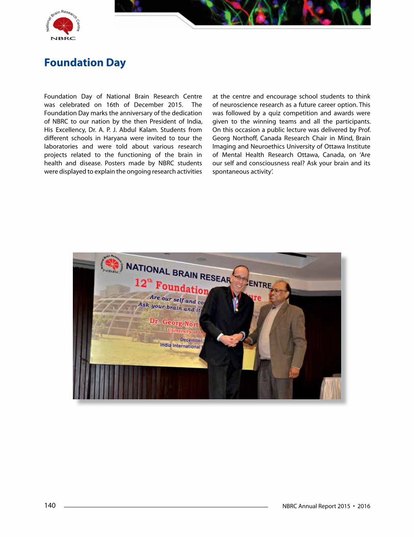
140 NBRC Annual Report 2015 • 2016
Foundation Day of National Brain Research Centre was celebrated on 16th of December 2015. The Foundation Day marks the anniversary of the dedication of NBRC to our nation by the then President of India, His Excellency, Dr. A. P. J. Abdul Kalam. Students from different schools in Haryana were invited to tour the laboratories and were told about various research projects related to the functioning of the brain in health and disease. Posters made by NBRC students were displayed to explain the ongoing research activities
at the centre and encourage school students to think of neuroscience research as a future career option. This was followed by a quiz competition and awards were given to the winning teams and all the participants. On this occasion a public lecture was delivered by Prof. Georg Northoff, Canada Research Chair in Mind, Brain Imaging and Neuroethics University of Ottawa Institute of Mental Health Research Ottawa, Canada, on ‘Are our self and consciousness real? Ask your brain and its spontaneous activity’.
Foundation Day
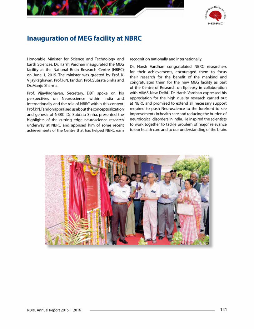
NBRC Annual Report 2015 • 2016 141
Honorable Minister for Science and Technology and Earth Sciences, Dr. Harsh Vardhan inaugurated the MEG facility at the National Brain Research Centre (NBRC) on June 1, 2015. The minister was greeted by Prof. K. VijayRaghavan, Prof. P. N. Tandon, Prof. Subrata Sinha and Dr. Manju Sharma.
Prof. VijayRaghavan, Secretary, DBT spoke on his perspectives on Neuroscience within India and internationally and the role of NBRC within this context. Prof. P. N. Tandon appraised us about the conceptualization and genesis of NBRC. Dr. Subrata Sinha, presented the highlights of the cutting edge neuroscience research underway at NBRC and apprised him of some recent achievements of the Centre that has helped NBRC earn
recognition nationally and internationally.
Dr. Harsh Vardhan congratulated NBRC researchers for their achievements, encouraged them to focus their research for the benefit of the mankind and congratulated them for the new MEG facility as part of the Centre of Research on Epilepsy in collaboration with AIIMS-New Delhi. Dr. Harsh Vardhan expressed his appreciation for the high quality research carried out at NBRC and promised to extend all necessary support required to push Neuroscience to the forefront to see improvements in health care and reducing the burden of neurological disorders in India. He inspired the scientists to work together to tackle problem of major relevance to our health care and to our understanding of the brain.
Inauguration of MEG facility at NBRC
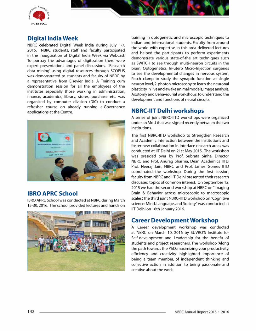
142 NBRC Annual Report 2015 • 2016
Digital India Week NBRC celebrated Digital Week India during July 1-7, 2015. NBRC students, staff and faculty participated in the inauguration of Digital India Week via Webcast. To portray the advantages of digitization there were expert presentations and panel discussions. ‘Research data mining’ using digital resources through SCOPUS was demonstrated to students and faculty of NBRC by a representative from Elsevier India. A Training cum demonstration session for all the employees of the institutes especially those working in administration, finance, academics, library, stores, purchase etc. was organized by computer division (DIC) to conduct a refresher course on already running e-Governance applications at the Centre.
IBRO APRC SchoolIBRO APRC School was conducted at NBRC during March 15-30, 2016. The school provided lectures and hands on
training in optogenetic and microscopic techniques to Indian and international students. Faculty from around the world with expertise in this area delivered lectures and helped the participants to perform experiments demonstrate various state-of-the art techniques such as SWITCH to see through multi-neuron circuits in the brain, Optogenetics, In-utero Micro-Injection surgeries to see the developmental changes in nervous system, Patch clamp to study the synaptic function at single neuron level, 2-photon microscopy to learn the neuronal plasticity in live and awake animal models, Image analysis, Axotomy and Behaviourial workshops, to understand the development and functions of neural circuits.
NBRC-IIT Delhi workshopsA series of joint NBRC-IITD workshops were organized under an MoU that was signed recently between the two institutions.
The first NBRC-IITD workshop to Strengthen Research and Academic Interaction between the institutions and foster new collaboration in interface research areas was conducted at IIT Delhi on 21st May 2015. The workshop was presided over by Prof. Subrata Sinha, Director NBRC and Prof. Anurag Sharma, Dean Academics IITD. Prof. Neeraj Jain, NBRC and Prof. James Gomes IITD coordinated the workshop. During the first session, faculty from NBRC and IIT Delhi presented their research discussed topics of common interest. On September 12, 2015 we had the second workshop at NBRC on “Imaging Brain & Behavior across microscopic to macroscopic scales”. The third joint NBRC-IITD workshop on “Cognitive science: Mind, Language, and Society” was conducted at IIT Delhi on 16th January 2016.
Career Development WorkshopA Career development workshop was conducted at NBRC on March 10, 2016 by SUVRO’S Institute for Self-development and Leadership for the benefit of students and project researchers. The workshop ‘Along the path towards the PhD: maximizing your productivity, efficiency and creativity’ highlighted importance of being a team member, of independent thinking and collective action in addition to being passionate and creative about the work.


NBRC Annual Report 2015 • 2016 145
The Administration of the Institute consists of the following major wings:
1. General Administration is headed by the Chief Administrative Officer, who is responsible for overall Management of Establishment, Personnel & Administration Wing, Stores & Purchase Wing, Import & Project Cell, Finance & Accounts Wing, Estate Management & Engineering Maintenance Wing – Civil, Electrical & Mechanical. The officer is also responsible for the administration of DIC.
2. Academic Administration is headed by the Registrar, who is responsible for the students’ administration, project co-ordination, new students’ admissions, course co-ordination etc. The officer is also responsible for administration of all the projects.
During the year under review, the Administration of NBRC observed all the important days as directed by the Government of India such as Anti-terrorism day, Sadbhavana Diwas, Independence Day, Hindi Week, Vigilance Awareness week etc. The Administration achieved excellence in execution of the following activities at NBRC:
• TheannualculturalfestivalofNBRC,‘TANTRIKA2015’was organized within the campus which included a variety of cultural and sports events during 17th to 19th September, 2015. Students, officers, and staff of NBRC participated in the event. On 17th September, 2015, a special guest lecture by Prof. Satyajit Rath, Scientist, National Institute of Immunology (NII), New Delhi was organized.
• Provided necessary logistics in conductinginternational and national conferences/seminars organized in the campus as well outside the campus.
• Made major imports from different countries interms of equipment and other consumables with meticulous planning and adhered to a precise schedule.
• The12thFoundationDayofNBRCwasheldon16thday of December, 2015. On this occasion, several programmes were organized within and outside the campus. The daylong celebrations included the poster presentations on ongoing research activities of NBRC. Students from various schools were invited to interact with NBRC scientists and they visited the
laboratories. A quiz programme for students from local schools was also organized on this occasion. Students from Presidency College, Kolkata exhibited the models on Crude Brain Map and Magnetic Target Therapy at NBRC. On this august occasion, Dr. Georg Northoff, Canada Research Chair in Mind, Brain Imaging and Neuroethics, University of Ottawa, Canada delivered the Foundation Day lecture on “Are our self and consciousness real? Ask your brain and its spontaneous activity” to the students and scientific community at India International Centre, New Delhi.
• The Sadbhavana Diwas was observed in NBRC onAugust 20, 2015. The faculty, students and staff were administered a solemn pledge to work in harmony and emotional oneness of all people regardless of region, caste, religion or language.
Implementation of Official Language NBRC Administration has given due importance for the implementation of Hindi as the Official Language at this centre and has made full efforts to implement the use of Official Language in all the administrative jobs such as internal official meetings, interviews, debates, general applications etc. During this year on the occasion of celebration of HINDI DIWAS on 14th September, 2015, lecture competition and other on the spot competitions were organized. Mr. Nishikant Mahajan, Retd. Joint Secretary, Deptt. Of Official Language, Govt. of India was the external guest. Students and staff participated in these competitions held at Auditorium of NBRC. The winners were distributed the prize money.
RTI ActThe provisions of RTI Act are being followed at NBRC in letter and in spirit. All RTI applications received during 2015-16 seeking information on various matters concerning NBRC were provided the requisite information within the prescribed time limit. The quarterly reports containing number of requests received with date, details of compliance, amount of charges etc., were sent to CIC and updated in NBRC website.
Women EmpowermentNBRC has a distinct feature of giving equal opportunity
General & Academic Administration - A Profile
146 NBRC Annual Report 2015 • 2016
to women. The Committees, constituted to do various work of Administration, Academics and scientific activities, have women members in them which ensure fair participation and protection of women. There is a committee for redressal of complaints relating to any sexual harassment of women at NBRC and grievances, if any, from aggrieved girl students/ women employees of NBRC. Any lady/ woman of NBRC, among the Students/ Employees who is subjected to sexual harassment can approach any of the committee members.
Reservations and concessions in Employment & Admissions of Students NBRC follows reservations & concessions as per rules of
Government of India in employment, and in the matter of students’ admissions, the provision of exemption as provided in Gazette Notification No. 5 dated 4th January, 2007 is implemented.
Vigilance
The Institute has a Chief Vigilance Officer. As
per the guidelines of DBT, one of the scientists
of NBRC has been nominated as Chief Vigilance
Officer of the Centre. NBRC observed the Vigilance
Awareness Week, 2015 during 26th October to 31st
October, 2015 on the theme “Preventive Vigilance as a
tool of Good Governance”.

NBRC Annual Report 2015 • 2016 147

148 NBRC Annual Report 2015 • 2016
1 Prof. P.N. Tandon (President, Chairman) No. 1, Jagriti Enclave Vikas Marg, New Delhi – 110 092
2 Prof. K. VijayRaghavan Secretary Department of Biotechnology C.G.O Complex, New Delhi – 110 003
3 Prof. Ashutosh Sharma Secretary Department of Science & Technology Technology Bhawan, New Mehrauli Road New Delhi – 110 016
4 Director-General Indian Council of Medical Research New Delhi – 110 029
5 Dr. Sandip K. Basu JC Bose Chair Professor National Institute of Science Commination & Information Resources (NISCAIR) 14, Satsang Vihar Marg New Delhi – 110 067
6 Smt. Sumita Mukherjee, IRAS Joint Secretary & Financial Advisor Department of Biotechnology, New Delhi – 110 003
7 Director General CSIR Institute of Genomics & Integrative Biology, Mall Road, Near Jubilee Hall, Delhi – 110 007
8 Dr. Suman Govil Adviser Department of Biotechnology, New Delhi
9 Dr. M. Gourie Devi Director (Retd.) Flat –9, Doctors Apartments, Vasundhara Enclave Delhi – 110 096
10 Dr. L. M. Patnaik CSA Department Indian Institute of Science Bangalore - 560012
11 Dr. Kalluri Subba Rao (INSA Hon. Scientist & Professor) School of Medical Sciences University of Hyderabad Hyderabad – 500 046
12 Prof. Gomathy Gopinath Flat # 001, Kanchanjunga Apartments, 122/2, Nagavarapalaya Varthur Road Bangalore – 560 093
13 Prof. Subrata Sinha Director National Brain Research Centre Manesar – 122 051
Members of NBRC Society

NBRC Annual Report 2015 • 2016 149
Members of NBRC Governing Council
1. Prof. K. VijayRaghavan (Chairman) (Ex-officio) Secretary Department of Biotechnology, Lodhi Road, CGO Complex, NEW DELHI – 110 003
2. Prof. P. N. Tandon (Ex-officio) No. 1, Jagriti Enclave, Vikas Marg Delhi – 110 092
3. Prof. Upinder S. Bhalla Scientist National Centre for Biological Sciences, NCBS, Bellary Road, Bangalore- 560 065
4. Prof. Dinakar M. Salunke Director International Centre for Genetic Engineering and Biotechnology (ICGEB), Aruna Asaf Ali Marg, New Delhi – 110 067
5. Dr. A. K. Agarwal Ex. Dean, Director, Professor & HOD Maulana Azad Medical College (Ex-officio), New Delhi – 110 002
6. Professor G. Mehta, FNA, FRS Bhartia Chair School of Chemistry University of Hyderabad, Hyderabad 500046, INDIA
7. Dr. Chitra Sarkar Department of Pathology All India Institute of Medical Sciences Ansari Nagar, New Delhi – 110 029
8. Prof. Seyed E. Hasnain Professor Kusuma School of Biological Sciences Indian Institute of Technology (IIT Delhi), Hauz Khas, Delhi – 110016
9. Smt. Sumita Mukherjee, IRAS Joint Secretary & Financial Advisor (Ex-officio), Department of Biotechnology New Delhi – 110 003
10. Director General (Ex-officio) Indian Council for Medical Research V. Ramalingaswamy Bhawan, Ansari Nagar, New Delhi – 110 029
11. Prof. Ashutosh Sharma (Ex-officio) Secretary Department of Science & Technology, Technology Bhawan, New Mehrauli Road, New Delhi – 110 016
12. Dr. Suman Govil Adviser Department of Biotechnology, Lodhi Road, CGO Complex, New Delhi – 110 003
13. Dr. Sanjeev Jain (Special Invitee) Professor & HOD Department of Psychiatry, NIMHANS, Bangalore
14. Prof. Subrata Sinha (Ex-officio) Director National Brain Research Centre Manesar – 122 051
150 NBRC Annual Report 2015 • 2016
1 Prof. K. VijayRaghavan (Chairman) Secretary Department of Biotechnology, Lodhi Road, CGO Complex, New Delhi – 110 003
2 Smt. Sumita Mukherjee, IRAS, Joint Secretary & Financial Advisor Department of Biotechnology New Delhi – 110 003
3 Joint Secretary, University Grants Commission (UGC) Bahadur Shah Jafar Marg, New Delhi – 110 002
4 Dr. Suman Govil Adviser Department of Biotechnology Lodhi Road, CGO Complex New Delhi – 110 003
5 Prof. Dinakar M. Salunke Director International Centre for Genetic Engineering and Biotechnology (ICGEB) Aruna Asaf Ali Marg New Delhi – 110 067
6 Prof. Seyed E. Hasnain Professor Kusuma School of Biological Sciences Indian Institute of Technology (IIT Delhi) Hauz Khas, Delhi – 110016
7 Prof. Subrata Sinha Director National Brain Research Centre Manesar-122051
8 Finance & Accounts Officer (Non-Member-Secretary) National Brain Research Centre Manesar-122051
Members of NBRC Finance Committee

NBRC Annual Report 2015 • 2016 151
Members of Scientific Advisory Committee
ChairpersonProf. P. N. Tandon President, NBRC Society
Co-Chairperson
Prof. Upinder S. Bhalla Scientist National Centre for Biological Sciences, Bangalore- 560 065
Members
1. Prof. K. VijayRaghavan Secretary Department of Biotechnology, CGO Complex, Lodi Road New Delhi – 110 003
2. Prof. Vijayalakshmi Ravindranath, Chairperson, Centre for Neuroscience Indian Institute of Sciences (IISc) Bangalore – 560 012
3. Prof. Amitabha Chattopadhyay Scientist Centre for Cellular and Molecular Biology (CCMB) Uppal Road, Hyderabad - 500 007 Andhra Pradesh
4. Dr. Ayub Qadri Scientist National Institute of Immunology (NII) Aruna Asaf Ali Marg New Delhi – 110 067
5. Prof. Dinakar M. Salunke Director ICGEB, New Delhi
6. Prof. Siddhartha Roy Director Indian Institute of Chemical Biology (IICB) Kolkata - 700 032
7. Prof. Jyotsna Dhawan Sr. Principal Scientist Centre for Cellular & Molecular Biology (CCMB) Habsiguda, Uppal Road, Hyderabad - 500 007 Andhra Pradesh
8. Prof. Rohit Manchanda Professor Biomedical Engineering Group School of Biosciences and Bioengineering Indian Institute of Technology (Bombay) Powai, Mumbai-400076
9. Prof. B. N. Mallick Professor School of Life Sciences Jawaharlal Nehru University New Delhi - 110067
10. Dr. V. Rajshekhar Professor and Head Neurosurgery Unit-II Department of Neurological Sciences Christian Medical College Hospital IDA Scudder Road, Vellore Tamil Nadu - 632 004
11. Dr. Sanjeev Jain Professor Department of Psychiatry National Institute of Mental Health and Neuro Sciences (NIMHANS), Hosur Road, Bangalore - 560029
12. Prof. Sudipta Maiti Professor Deptt. of Chemical Sciences Tata Institute of Fundamental Research (TIFR) Mumbai - 400 005
13. Prof. N. R. Jagannathan HoD of NMR and MRI Facility All India Institute of Medical Sciences (AIIMS) New Delhi – 110 029
14. Dr. Chitra Sarkar, Professor, Department of Pathology, All India Institute of Medical Sciences, Ansari Nagar, New Delhi– 110 029 Tel No: 011-2659 3371, Cell: 9868397191, E-Mail: [email protected], [email protected]
152 NBRC Annual Report 2015 • 2016
15. Prof. Ajoy Kumar Ray Director Indian Institute of Engineering Science and Technology (IIEST) Shibpur (Formerly Bengal Engineering & Science University) P. O. Botanic Garden, Shalimar, Howrah, West Bengal - 711103
International Members
1. Prof. Ariel Ruiz i Altaba Professor Faculty of Medicine, University of Geneva Department of Medicinal Genetics 8242 CMU, 1 rule Michel Servet CH-1211, Geneva 4, Switzerland
2. Prof. Baroness Susan Greenfield, Professor Department of Pharmacology, Lincoln College, Oxford University, UK
3. Prof. Thomas D. Albright Professor The Salk Institute for Biological Studies, La Jolla, California, USA 92037
4. Michael W. Weiner, MD Director of the Center for Imaging of Neurodegenerative Diseases SFVAMC, Professor of Radiology Medicine, Psychiatry and Neurology, UCSF
Ex-officio Member
1. Dr. Suman Govil Adviser Department of Biotechnology CGO Complex, Lodi Road New Delhi – 110 003
1. Dr. Suman Govil (Chairperson) Adviser Department of Biotechnology CGO Complex, Lodi Road New Delhi – 110 003
2. Prof. Subrata Sinha Director National Brain Research Centre Manesar-122051
3. Dr. S. K. Gupta Deputy Director (Retired) & Emeritus Scientist NII, New Delhi
4. Mr. M. K. Gupta Engineer-In-Charge (Civil) IUAC
5. Prof. Sidhartha Satpathy HOD Hospital Administration AIIMS, New Delhi
Members of Building Committee

NBRC Annual Report 2015 • 2016 153
Members of Academic Council
1. Prof. Subrata Sinha (Chairman) Director National Brain Research Centre Manesar, Haryana
2. Prof. Shiv K. Sharma National Brain Research Centre Manesar, Haryana
3. Prof. Basabi Bhaumik (till Oct 2015) Department of Electrical Engineering Indian Institute of Technology New Delhi
4. Prof. Nandini C Singh National Brain Research Centre Manesar, Haryana
5. Dr. V. S. Mehta (till Oct 2015) Paras Hospitals Gurgaon, Haryana
6. Dr. Soumya Iyengar National Brain Research Centre Manesar, Haryana
7. Prof. K. Muralidhar (till Oct 2015) Head, Department of Zoology University of Delhi, New Delhi
8. Dr. Anirban Basu National Brain Research Centre Manesar, Haryana
9. Prof. Sudha Bhattacharya (from Oct 2015) School of Environmental Sciences Jawaharlal Nehru University, New Delhi
10. Dr. Narender K. Dhingra (till 01.12.2015) National Brain Research Centre, Manesar, Haryana
11. Prof. Ishan Patro (from Oct 2015) School of Studies in Zoology/Neuroscience Jiwaji University, Gwalior
12. Dr. Ellora Sen National Brain Research Centre Manesar, Haryana
13. Prof. Gurcharan Kaur (from Oct 2015) Department of Biotechnology Guru Nanak Dev University, Amritsar
14. Dr. Ranjit K. Giri National Brain Research Centre Manesar, Haryana
15. Prof. Prasun Kumar Roy National Brain Research Centre Manesar, Haryana
16. Dr. Yoganarasimha Doreswamy National Brain Research Centre Manesar, Haryana
17. Prof. Neeraj Jain National Brain Research Centre Manesar, Haryana
18. Dr. Sourav Banerjee National Brain Research Centre Manesar, Haryana
19. Prof. Nihar Ranjan Jana National Brain Research Centre Manesar, Haryana
20. Dr. Arpan Banerjee National Brain Research Centre Manesar, Haryana
21. Prof. Pravat K. Mandal National Brain Research Centre Manesar, Haryana
22. Dr. Anindya Ghosh Roy National Brain Research Centre Manesar, Haryana
23. Prof. Pankaj Seth National Brain Research Centre Manesar, Haryana
24. Mr. K.V.S. Kameswara Rao (till 31.12.2015) National Brain Research Centre Manesar, Haryana

154 NBRC Annual Report 2015 • 2016
1. Prof. Subrata Sinha Director National Brain Research Centre Manesar, Haryana
2. Prof. Nandini C. Singh National Brain Research Centre Manesar, Haryana
3. Prof. D. N. Rao (till Oct 2015) Indian Institute of Sciences Bangalore, Karnataka
4. Dr. Soumya Iyengar National Brain Research Centre Manesar, Haryana
5. Prof. Rohit Manchanda (till Oct 2015) Indian Institute of Technology, Mumbai, Maharashtra
6. Dr. Anirban Basu National Brain Research Centre Manesar, Haryana
7. Prof. K. Natarajan (from Oct 2015) Director Ambedkar Centre for Biomedical Research University of Delhi New Delhi
8. Dr. Narender K. Dhingra (till 01.12.2015) National Brain Research Centre Manesar, Haryana
9. Prof. Chitra Sarkar (from Oct 2015) Department of Pathology All India Institute of Medical Sciences New Delhi
10. Dr. Ellora Sen National Brain Research Centre Manesar, Haryana
11. Prof. Prasun Kumar Roy National Brain Research Centre Manesar, Haryana
12. Dr. Ranjit K. Giri National Brain Research Centre Manesar, Haryana
13. Prof. Neeraj Jain National Brain Research Centre Manesar, Haryana
14. Dr. Yoganarasimha Doreswamy National Brain Research Centre Manesar, Haryana
15. Prof. Nihar Ranjan Jana National Brain Research Centre Manesar, Haryana
16. Dr. Sourav Banerjee National Brain Research Centre Manesar, Haryana
17. Prof. Pravat K. Mandal National Brain Research Centre Manesar, Haryana
18. Dr. Arpan Banerjee National Brain Research Centre Manesar, Haryana
19. Prof. Pankaj Seth National Brain Research Centre Manesar, Haryana
20. Dr. Anindya Ghosh Roy National Brain Research Centre Manesar, Haryana
21. Prof. Shiv K. Sharma National Brain Research Centre Manesar, Haryana
22. Mr. K.V.S.Kameswara Rao (till 31.12.2015) National Brain Research Centre Manesar, Haryana
Members of Board of Studies

NBRC Annual Report 2015 • 2016 155
Scientific Staff
Scientists
1. Prof. Subrata Sinha (Director)
2. Prof. Prasun Kumar Roy
3. Prof. Neeraj Jain
4. Dr. Nihar Ranjan Jana
5. Dr. Pravat Kumar Mandal
6. Dr. Pankaj Seth
7. Dr. Narender K. Dhingra (till 01.12.15 A/N)
8. Dr. Shiv Kumar Sharma
9. Dr. Ranjit Kumar Giri
10. Dr. Nandini C. Singh
11. Dr. Soumya Iyengar
12. Dr. Anirban Basu
13. Dr. Ellora Sen
14. Dr. Yoganarasimha Doreswamy
15. Dr. Sourav Banerjee
16. Dr. Arpan Banerjee
17. Dr. Anindya Ghosh Roy
Consultants1. Dr. Rema Velayudhan
2. Prof. Partha Raghunathan
3. Mr. Suman Kumar
DST-Inspire Faculty1. Dr. Supriya Bhavnani (Till 31-08-2015 A.N.)
2. Dr. Yogita Kapil Adlakha
NBRC Students
Ph.D. Students
1. Mr. Mohammed Hisham P.M (Till 23/04/2015 A.N.)
2. Mr. Kaushik Pramod Sharma
3. Mr. Rahul Chaudhary (Till 23/06/2015 A.N.)
4. Mr. Apoorv Sharma
5. Ms. Manju Tewari
6. Mr. Sandeep Kumar
7. Mr. Sourish Ghosh
8. Mr. Bharat Prajapati
9. Ms. Mahar Fatima
10. Mr. Brijesh Kumar Singh
11. Mr. John Thomas
12. Mr. Kautuk Kamboj
13. Mr. Biswaranjan Sahoo
14. Mr. Indrajith R. Nair
15. Ms. Pushpa Kumari
16. Ms. Shalini Swaroop
17. Mr. Shashi Shekhar Kumar
18. Mr. Touseef Ahmad Sheikh
19. Mr. Tushar Arora
20. Mr. Neeraj Singh (Till 29/02/2016 A.N.)
21. Mr. S Balakumar
22. Mr. G Vinodh Kumar
23. Ms. Arti Kumari
24. Mr. Debajit Bagchi
25. Mr. Dharmendra Puri
26. Ms. Mukta Kumari
27. Mr. Raghav Shankar
28. Md. Tipu Khan
29. Mr. Amit Ranjan
30. Ms. Priyanka Ghosh
31. Ms. Sarbani Samaddar
32. Ms. Shruti Patrick
33. Mr. Surajit Chakraborty
34. Mr. Varun Chaturvedi
Integrated Ph.D. Students
1 Mr. Ajit Ray (Till 12/05/2015 A.N.)
2 Mr. Sadashib Ghosh (Till 05/03/2015 A.N.)
3 Mr. Deepak Poria
4 Ms. Manvi Goel (Till 03/07/2015 A.N.)
5 Mr. Atul Gopal P.A (Till 13/08/2015)
6 Ms. Suhela Kapoor
7 Ms. Guncha Bhasin
156 NBRC Annual Report 2015 • 2016
8 Ms. Sarika Cherodath
9 Ms. Himakshi
10 Ms. Ruchi Ghildiyal (Till 29/04/2015 A.N.)
11 Ms. Piyushi Gupta
12 Ms. Avantika Mathur
13 Ms. Shankhamala Sen
14 Mr. Priyabrata Halder
15 Mr. Imran Jamal
16 Mr. Fahim Ahmad
17 Ms. Manika Arora
18 Ms. Uzma Din
19 Ms. Chitra Mohinder Singh Singal
20 Ms. Utkarsha A Singh
21 Ms. Pooja Parishar
22 Mr. Apurva Agrawal
23 Mr. Atanu Datta
24 Mr. Naman Vatsa
25 Mr. Abhishek Kumar Verma
26 Mr. Hriday Shanker Pandey
27 Ms. Reshma
28 Mr. Vikas Pareek
29 Mr. Vipendra Kumar
30 Ms. Atrayee Basu
31 Ms. Priyanka
32 Ms. Shruti F Nagaral
33 Mr. Gourav Sharma
34 Ms. Harjot Kaur
35 Mr. Pruthvi S.G
36 Ms. Shelly Pal
37 Mr. Shubham Krishna
M.Sc. Students
1 Mr. Gaurav Sharma
2 Ms. Himali Arora
3 Ms. Meenakshi Bhaskar
4 Mr. Neeraj Kumar
5 Ms. Sanskriti
6 Mr. Utsav Mukherjee
NBRC Project Staff
Project Assistant
1 Ms. Sarbani Samaddar (Till 31/07/2015 A.N.)
2 Ms. Anindita Mandal (Till 16/07/2015 A.N.)
3 Mr. Blesson K Paul (Till 31/08/2015 A.N.)
4 Ms. Prerna Srivastava
5 Ms. Rina Kumari (Till 12/06/2015 A.N.)
6 Ms. Kalpana Gupta
7 Ms. Sonal Makhija (Till 28/12/2015 A.N.)
8 Mr. Tamesh Halder (Till 07/10/2015 A.N.)
9 Ms. Shanah Rachel John
10 Mr. Bathini Praveen
11 Mr. Kuldeep Shrivastava (Till 01/03/2016 F.N.)
12 Mr. Alok Nath Mohapatra
13 Mr. Abir Mondal (Till 13/08/2015 F.N.)
14 Mr. Giri Raj Kishore Sharma
15 Ms. Noopur Singh
16 Ms. Srujana Raili
17 Ms. Rishu
18 Ms. Priyanka Singh Kshatriya (Till 31/03/2016 A.N.)
19 Ms. Tanya Singh
20 Ms. Moumita Chakraborty (Till 27/04/2015 A.N.)
21 Mr. Kaushik Kumar Deka (Till 27/05/2015 A.N.)
22 Mr. Jacob Antony Alappatt
23 Ms. Pankajam T
24 Ms. Kanza Saleem
25 Ms. Mena Fatma
26 Ms. Rohini Roy
27 Ms. Hajare Nilambari Anil
28 Ms. Kshipra Gurunandan
29 Mr. Shrey Dutta
30 Ms. Deborah Daphne P
31 Ms. Monika
NBRC Annual Report 2015 • 2016 157
Research Associates 1. Dr. Prem Chand, Research Associate-3
2. Dr. Arpita Chatterjee, Research Associate-3 (Till 28-10-2015 A.N.)
3. Dr. Chetan Kumar Yadav, Research Associate-3
4. Dr. Pinaki Mondal, Research Associate-2 (Till 28-10-2015 A.N.)
5. Dr. Md. Aftab Alam, Research Associate-3
6. Dr. Vivek Kumar Tripathi, Research Associate-3
7. Dr. Md. Khalid Zakaria, Research Associate-1 (Till 23-09-2015 A.N.)
8. Dr. Kiran, Research Associate-1 (Till 22-07-2015 A.N.)
9. Dr. Shaily Malik, Research Associate-1 (Till 29-05-2015 A.N.)
10. Dr. Shabir Ahmad Ganai, Research Associate-1 (Till 15-06-2015 A.N. )
11. Dr. D. Subhashree, Research Associate-1
12. Mr. Suvadip Mallick, Research Associate-1
13. Mr. Saurabh Srivastav, Research Associate-1
14. Dr. Payal Arya, Research Associate-1
Research Fellows 1. Mr. Mohammed Hisham P.M (Research Fellow)
(From 24/04/2015)
2. Mr. Subhadip Paul (Research Fellow) (Till 24/03/2016 A.N.)
3. Mr. Rahul Chaudhary (Research Fellow) (From 24/06/2015 Till 23/09/2015 A.N.)
4. Mr. Deobrat Dixit (Research Fellow) (Till 22/07/2015 A.N.)
5. Mr. Sadashib Ghosh (Research Fellow) (From 06/03/2015 Till 19/06/2015 A.N.)
6. Mr. Atul Gopal P.A (Research Fellow) (From 14/08/2015)
7. Ms. Ruchi Ghildiyal (Research Fellow) (From 30/04/2015 Till 21/12/2015 A.N.)
Project Employees1. Ms. D. Suvarnalata Xanthate, R&D Engineer (Project)
(Till 30-06-2015 A.N.)
2. Mr. V.P. Subramanyam Rallabandi, Senior Research
Officer (Computer Engineering)
3. Mr. Arkoprovo Paul, Junior Research Fellow (Project) (Till 03-07-2015 A.N.)
4. Ms. Shammi More, Senior R&D Engineer (Project)
5. Mr. Rajiv Ramaswamy, Senior Research Fellow (Project)
6. Ms. Archana Vadiraj Malagi, Junior Research Fellow (Project) (Till 14-08-2015 A.N.)
7. Dr. Bibhabasu Hazra, ICMR-Research Associate-1
8. Mr. Abhishek Mukherjee, R&D Engineer (Project)
(Till 14-07-2015 A.N.)
9. Dr. Sumiti Saharan, Research Scientist (Project)
10. Ms. Monika, Scientist ‘B’ (Project)
11. Ms. T. Ammaponnu@Sumathi, Senior Research Fellow (Project)
12. Ms. Rashi Midha, Project Officer (Till 30-04-2015 A.N.)
13. Dr. Mukesh Kumar, ICMR-Research Associate-2 (Till 13-11-2015 A.N.)
14. Mr. Sourav Bhaduri, Senior R&D Engineer (Project) (Till 08-07-2015 A.N.)
15. Mr. Kamal Bharti, Technologist (MEG Project)
16. Mr. Vibhin V., Technologist (MEG Project)
17. Ms. Km. Ruchika Mittal, Data Entry Coordinator (Project)
18. Dr. Nilanjana Das Saha, DBT-Research Associate-1
19. Dr. Dipanjan Ray, Post Doctoral Fellow (Project)
20. Ms. Richa Awasthi, Junior Research Fellow (Project) (Till 29-02-2016 A.N.)
21. Mr. Sanjeev Bhardwaj, Manager (MEG Project)
22. Ms. Khan Sarah Aziz, Psychologist (Project)
23. Mr. Manjit, Lab Attendant (MEG Project)
24. Mr. Rakesh Yadav, Nursing Orderly (MEG Project)
25. Dr. Aparna Dixit, Assistant Professor (MEG Project)
26. Dr. Sayali Chintamani Ranade, Women Scientist Scheme - A (WOS-A) (Till 17-11-2015 A.N.)
27. Dr. Jyotirmoy Banerjee, Assistant Professor (MEG Project)
158 NBRC Annual Report 2015 • 2016
28. Dr. Narottam Sharma, Casualty Medical Officer (MEG Project) (Till 07-10-2015 A.N.)
29. Mr. Ashok Kumar, Nurse (MEG Project)
30. Mr. Gaurav Singh, Technician (MEG Project)
31. Mr. Vivek Singh, Technician (MEG Project)
32. Mr. Amit Kumar Jaiswal, Junior Research Fellow (Project (Till 29-01-2016 A.N.)
33. Mr. Om Prakash Jakhar, Nurse (MEG Project)
34. Mr. Tony C. Paikada, Nurse (MEG Project)
35. Ms. Mini Mohan, Nurse (MEG Project)
36. Mr. Banshi Nath, Junior Research Fellow (Project) (Till 31-03-2016 A.N. )
37. Mr. Sounak Mohanta, Junior Research Fellow (Project)
38. Ms. Monika, Technical Officer (Imaging)(Project) (Till 15-12-2015 A.N.)
39. Ms. Mariam Siddiqui, R&D Scientist (Project)
40. Mr. Sushil Kumar Gupta, Technical Assistant (Libray)
41. Ms. Shipra Jain, Clinical Coordinator (Project) (Till 03-09-2015 A.N.)
42. Ms. Devina Sharma, Junior Research Fellow (Project)
43. Ms. Vijay Laxmi Rathore, Junior R&D Engineer (Project)
44. Mr. Arun E V R, Junior Research Fellow (Project)
45. Ms. Meera Srikrishna, R&D Engineer (Project)
46. Ms. Anindita Mandal, Human Ethics Coordinator (Project)
47. Mr. Ashok Kumar Datusalia, Research Associate (Project)
48. Ms. Krity Kansara, Senior R&D Engineer (Project)
49. Mr. Hariharan V, Technician (MEG Project)
50. Mr. Archith Rajan, Junior Research Fellow (Project)
51. Ms. Tejasvini Sinha, Clinical Psychologist (Project)
52. Mr. Tamesh Halder, R&D Engineer (Project)
53. Ms. Ankita Singh, Research Associate (Project)
54. Dr. K M Sangeeta Maini, Scientist ‘C’ (Project)
55. Ms. Aroma Dabas, Neuro-analyst (Project)
56. Mr. Sukhvir Singh Pundir, Technical Associate (Computer / IT)
57. Mr. Prem Chand, Accounts Administrative Assistant (DeLCON Project)
58. Ms. Ragini, Lab Technical Assistant (Project)
59. Ms. Bhanupriya Chouhan, R&D Engineer (Project)
60. Ms. Yamini Yadav, Research Manager (Project)
61. Dr. Siya Sherif, Senior R&D Engineer (Project)
Technical Staff1. Mr. Rajbir Singh
2. Mr. Sanjeev K. Choudhary
3. Dr. Dev Das Lal
4. Mr. Mahender Kumar Singh
5. Dr. Inderjeet Yadav
6. Mr. Jitender Ahlawat
7. Mr. Arvind Singh Pundir
8. Mr. Kanhaiya Lal Kumawat
9. Mr. Kedar Singh Bajetha
10. Mr. Shankar Dutt Joshi
11. Mr. Sumit Kumar Sinha Mahapatra
12. Mr. D. Narender
13. Mr. Sanjay Kumar
14. Mr. Mithlesh Kumar Singh
15. Mr. Ankit Sharma
16. Mr. Sanjeev Bhardwaj
17. Mr. Yunis Khan
18. Ms. Seepika
19. Mr. Amit Kumar Gaurav
20. Mr. Sachin Kumar
21. Ms. Tarnnum Mansoori
22. Mr. Durgalal Meena
23. Mr. Irshad Alam
24. Mr. P. Manish
25. Mr. Dil Bahadur Karki
26. Mr. Rammehar
27. Mr. Manish Kumar
28. Mr. Hari Shankar

NBRC Annual Report 2015 • 2016 159
29. Mr. Mahendra Singh
30. Mr. Sanjay Kumar Singh
Administrative Staff 1. Mr. Tanmoy Bhattacharyya
2. Mr. Rajesh Kumar Vyas (till 01.02.16 A/N)
3. Mr. Santosh Kumar Choudhary
4. Mr. Debashish Bhattacharjee
5. Mr. Ravinder Pal
6. Mr. Sunil Kumar Dwivedi (till 31.08.15 A/N)
7. Ms. Pooja Gosain
8. Mr. Sanjay Kumar Gupta
9. Mr. Suraj Bhan
10. Mr. Ajay Kumar Dehariaya
11. Mr. Himanshu Mal
12. Mr. Rakesh Kumar Yadav
13. Mr. Parmander Singh Rawat
14. Mr. Jitendra Kumar Meena
15. Mr. Surender Kumar
16. Mr. Bhupender Pal Sharma
17. Mr. Satish Kumar
18. Kailash Chandra Khuntia
DIC Project Staff1. Ms. Reema Saxena
2. Mr. Amit Kumar
3. Ms. Sunita
4. Mr. R. Ganesh Gurumoorthy
DeLCON Project Staff 1. Mr. Sushil Kumar Gupta
2. Sukhvir Singh Pundir
3. Prem Chand
Contractual Staff 1. Dr. Mithun James
2. Dr. Karan Singh
3. Ms. Nisha Devi
4. Mr. Mukesh Chauhan (till 24.03.16 A/N)
5. Ms. Shweta Mishra
6. Mr. Hanish Kumar Sauda


NBRC Annual Report 2015 • 2016 163
Independent Auditor’s Report
Re: The Members of National Brain Research Centre
A. We have audited the accompanying financial statements of M/s National Brain Research Centre (hereinafter referred to as “NBRC”), which comprises of the Balance- Sheet as at March 31, 2016, the Income & Expenditure Account and the Receipts & Payments Account for the year ending on that date read with significant accounting policies and notes to financial statements.
B. Management’s Responsibility for the Standalone Financial Statements
The Management of the NBRC is responsible with respect to preparation of these financial statements that give a true and fair view of the financial position, financial performance and of the Receipts & Payments thereof in accordance with the Accounting Principles generally accepted in India including the Accounting Standards issued by the Institute of Chartered Accountants of India (ICAI). The responsibility also includes maintenance of adequate accounting records in accordance with the provisions of the Act for safeguarding the assets of the NBRC and for preventing and detecting frauds and other irregularities; selection and application of appropriate accounting policies; making judgments and estimates that are reasonable and prudent; and design, implementation and maintenance of adequate internal financial controls, that were operating effectively for ensuring the accuracy and completeness of the accounting records, relevant to the preparation and presentation of the financial statements that give a true and fair view and are free from material misstatement, whether due to fraud or error.
C. Auditor’s Responsibility
Our responsibility is to express an opinion on these financial statements based on our audit. We conducted our audit in accordance with the Standards on Auditing issued by the ICAI. Those standards required that we comply with ethical requirements and plan and perform the audit to obtain reasonable assurance about whether the financial statements are free from material misstatement. An audit involves performing procedures to obtain audit-evidence about the amounts and the disclosures in the financial statements. The procedures selected depend on the auditor’s judgment, including the assessment of the risks of material misstatement of the financial statements, whether due to fraud or error. In making those risk assessments, the auditor considers internal financial control relevant to the NBRC’s preparation & fair presentation of the financial statements that give a true and fair view in order to design audit procedures that are appropriate in the circumstances, but not for the purpose of expressing an opinion on whether the NBRC has in place an adequate internal financial controls system over financial reporting and the operating effectiveness of such controls. An audit also includes evaluating the appropriateness of the accounting policies used and the reasonableness of the accounting estimates made by the management, as well as evaluating the overall presentation of the financial statements. We believe that the audit evidence we have obtained is sufficient and appropriate to provide a basis for our audit opinion on these financial statements.
Auditor’s Report

164 NBRC Annual Report 2015 • 2016
D. Opinion
Subject to clauses (4), (7), (9), (11), (12), (14), (15) & (17) of Notes to Accounts (Schedule-17) forming part of financial statements for current year, the impact whereof on results of operations for the year of NBRC and its state of affairs as at March 31, 2016 is not ascertainable due to its pending status, in our opinion and to the best of our information and according to the explanations given to us, the aforementioned financial statements gives a true and fair view in conformity with the accounting principles generally accepted in India:
(a) in case of the Balance Sheet, of the state of affairs of the Company as at March 31, 2016;
(b) in case of the Income and Expenditure Account, of the excess of income over expenditure for the year ended on that date;
(c) in case of the Receipts & Payment Account, of the receipts & payments during the year ended on that date.
E. Report on Other, Legal and Regulatory Requirements
(a) Subject to our observations as referred to in para (D) above, we have obtained all the information and explanations which to the best of our knowledge and belief were necessary for the purpose of our audit;
(b) In our opinion proper books of accounts have been kept by the NBRC so far as appears from our examination of those books;
(c) The Balance Sheet, Income and Expenditure Account & the Receipts and Payment Account dealt with by this report are in agreement with the books of accounts;
Place: New Delhi For N.C. MITTAL & CO.Date : October 28, 2016 Chartered Accountants (FRN-000237N)
Sd/- (CA. KAPIL MITTAL) B.Com (H), F.C.A, D.I.S.A.(ICAI), A.I.I.I.S.L.A. M No.-503378
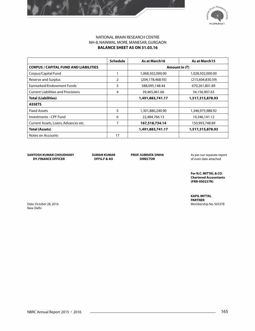
NBRC Annual Report 2015 • 2016 165
Schedule As at March16 As at March15
CORPUS / CAPITAL FUND AND LIABILITIES Amount in (`)
Corpus/Capital Fund 1 1,068,502,000.00 1,028,502,000.00
Reserve and Surplus 2 (204,178,468.93) (215,604,830.59)
Earmarked/Endowment Funds 3 588,095,148.44 670,261,801.89
Current Liabilities and Provisions 4 39,465,061.66 34,156,907.63
Total (Liabilities) 1,491,883,741.17 1,517,315,878.93
ASSETS
Fixed Assets 5 1,301,880,240.90 1,346,975,988.92
Investments - CPF Fund 6 22,484,766.13 19,346,141.12
Current Assets, Loans, Advances etc. 7 167,518,734.14 150,993,748.89
Total (Assets) 1,491,883,741.17 1,517,315,878.93
Notes on Accounts 17
SANTOSH KUMAR CHOUDHARY SUMAN KUMAR PROF. SUBRATA SINHA As per our separate report DY. FINANCE OFFICER OFFG.F & AO DIRECTOR of even date attached
For N.C. MITTAL & CO. Chartered Accountants (FRN-000237N)
KAPIL MITTAL PARTNERDate: October 28, 2016 Membership No. 503378New Delhi
NATIONAL BRAIN RESEARCH CENTRENH-8, NAINWAL MORE, MANESAR, GURGAON
BALANCE SHEET AS ON 31.03.16
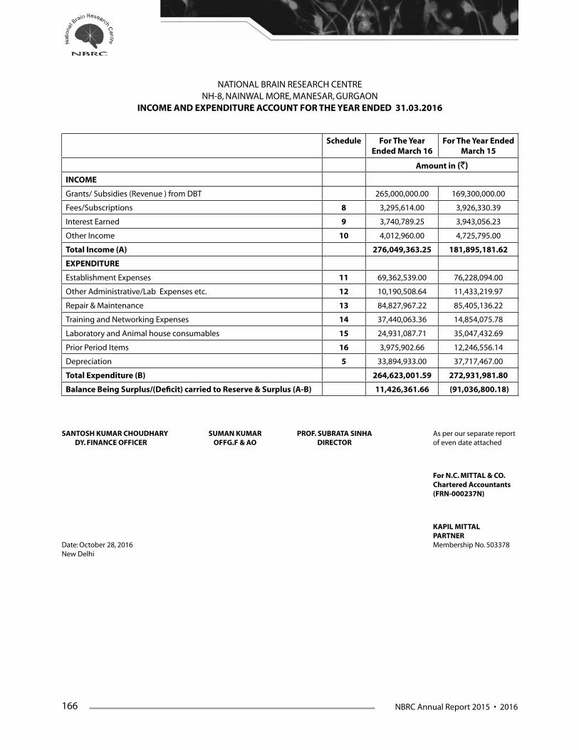
166 NBRC Annual Report 2015 • 2016
Schedule For The Year Ended March 16
For The Year Ended March 15
Amount in (`)
INCOME
Grants/ Subsidies (Revenue ) from DBT 265,000,000.00 169,300,000.00
Fees/Subscriptions 8 3,295,614.00 3,926,330.39
Interest Earned 9 3,740,789.25 3,943,056.23
Other Income 10 4,012,960.00 4,725,795.00
Total Income (A) 276,049,363.25 181,895,181.62
EXPENDITURE
Establishment Expenses 11 69,362,539.00 76,228,094.00
Other Administrative/Lab Expenses etc. 12 10,190,508.64 11,433,219.97
Repair & Maintenance 13 84,827,967.22 85,405,136.22
Training and Networking Expenses 14 37,440,063.36 14,854,075.78
Laboratory and Animal house consumables 15 24,931,087.71 35,047,432.69
Prior Period Items 16 3,975,902.66 12,246,556.14
Depreciation 5 33,894,933.00 37,717,467.00
Total Expenditure (B) 264,623,001.59 272,931,981.80
Balance Being Surplus/(Deficit) carried to Reserve & Surplus (A-B) 11,426,361.66 (91,036,800.18)
NATIONAL BRAIN RESEARCH CENTRENH-8, NAINWAL MORE, MANESAR, GURGAON
INCOME AND EXPENDITURE ACCOUNT FOR THE YEAR ENDED 31.03.2016
SANTOSH KUMAR CHOUDHARY SUMAN KUMAR PROF. SUBRATA SINHA As per our separate report DY. FINANCE OFFICER OFFG.F & AO DIRECTOR of even date attached
For N.C. MITTAL & CO. Chartered Accountants (FRN-000237N)
KAPIL MITTAL PARTNERDate: October 28, 2016 Membership No. 503378New Delhi
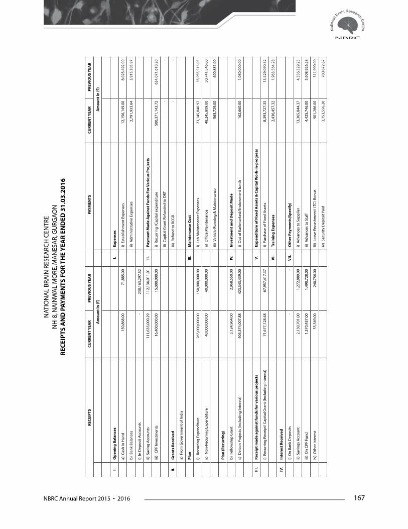
NBRC Annual Report 2015 • 2016 167
REC
EIP
TS
CU
RR
ENT
YEA
R
PR
EVIO
US
YEA
R
PAY
MEN
TS C
UR
REN
T Y
EAR
P
REV
IOU
S Y
EAR
Am
ou
nt
in (`
) A
mo
un
t in
(`)
I.O
pen
ing
Bal
ance
sI.
Exp
ense
s
a) C
ash
in H
and
150
,868
.00
71,
885.
00
i) E
stab
lish
men
t Ex
pen
ses
12,
156,
149.
00
8,0
28,4
92.0
0
b)
Ban
k B
alan
ces
ii) A
dm
inis
trat
ive
Exp
ense
s 2
,791
,933
.64
3,9
15,3
05.9
7
i) In
Dep
osi
t A
cco
un
ts -
230
,163
,297
.52
ii) S
avin
g A
cco
un
ts 1
11,6
35,0
00.2
9 1
12,1
36,0
11.0
1 II
.P
aym
ent
Mad
e A
gai
nst
Fu
nd
s Fo
r Var
iou
s P
roje
cts
iii)
CPF
Inve
stm
ents
16,
400,
000.
00
15,
000,
000.
00
i) R
ecu
rrin
g /
Cap
ital
exp
end
itu
re
500
,371
,143
.72
634
,071
,610
.20
ii) C
apit
al G
ran
t R
efu
nd
ed to
DBT
- -
II.
Gra
nts
Rec
eive
diii
) R
efu
nd
to R
CG
B -
-
a) F
rom
Gov
ern
men
t o
f In
dia
Pla
nII
I.M
ain
ten
ance
Co
st
i) R
ecu
rrin
g E
xpen
dit
ure
265
,000
,000
.00
150
,000
,000
.00
i) L
ab M
ain
ten
ance
Exp
ense
s 2
3,14
5,84
0.97
3
5,95
5,51
3.05
ii)
No
n-R
ecu
rrin
g E
xpen
dit
ure
40,
000,
000.
00
40,
000,
000.
00
ii) O
ffice
Mai
nte
nan
ce 4
8,24
5,80
9.00
5
0,74
1,54
6.00
iii)
Veh
icle
Ru
nn
ing
& M
ain
ten
ance
565
,729
.00
600
,881
.00
Pla
n (R
ecu
rrin
g)
b)
Fello
wsh
ip G
ran
t 3
,124
,964
.00
2,9
68,5
50.0
0 IV
.In
vest
men
t an
d D
epo
sit
Mad
e
c) D
elco
n P
roje
cts
(Incl
ud
ing
Inte
rest
) 4
06,3
74,0
07.8
8 4
23,3
43,4
39.0
0 i)
Ou
t o
f Ear
kmar
ked
/En
do
wm
ent
fun
ds
162
,660
.00
1,0
80,0
00.0
0
III.
Rec
eip
t m
ade
agai
nst
fun
ds
for
vari
ou
s p
roje
cts
V.Ex
pen
dit
ure
of F
ixed
Ass
ets
& C
apit
al W
ork
-in
-pro
gre
ss
i) R
ecu
rrin
g R
ecei
pt/
Cap
ital
Gra
nt
(Incl
ud
ing
Inte
rest
) 7
1,67
7,12
8.48
6
7,95
7,41
7.37
i)
Pu
rch
ase
of F
ixed
Ass
ets
8,3
93,7
27.3
5 1
3,52
9,09
0.32
VI.
Trai
nin
g E
xpen
ses
2,4
36,4
57.3
2 1
,963
,564
.28
IV.
Inte
rest
Rec
eive
d
i) O
n B
ank
Dep
osi
ts -
- V
II.
Oth
er P
aym
ents
(Sp
ecif
y)
ii) S
avin
gs
Acc
ou
nt
2,1
30,7
01.0
0 1
,272
,889
.00
i) A
dva
nce
s to
Su
pp
lier
13,
365,
844.
37
4,3
56,3
29.2
3
iii)
On
CPF
Fu
nd
1,5
70,4
37.0
0 1
,490
,728
.00
ii) A
dva
nce
s to
Sta
ff 4
,425
,746
.00
5,6
08,9
26.2
8
iv)
Oth
er In
tere
st 3
3,34
9.00
2
40,7
56.0
0 iii
) Le
ave
Enca
shm
ent/
LTC
/ B
on
us
901
,286
.00
311
,990
.00
iv)
Secu
rity
Dep
osi
t Pa
id 2
,753
,556
.20
780
,672
.67
NAT
ION
AL
BR
AIN
RES
EAR
CH
CEN
TRE
NH
-8, N
AIN
WA
L M
ORE
, MA
NES
AR,
GU
RG
AO
N
REC
EIP
TS A
ND
PA
YM
ENTS
FO
R T
HE
YEA
R E
ND
ED 3
1.0
3.2
01
6

168 NBRC Annual Report 2015 • 2016
V.A
ny
Oth
er R
ecei
pt
v) E
MD
Ref
un
ded
1,7
94,9
13.0
0 3
,750
,339
.72
Ind
irec
t In
com
evi
) TD
S Pa
id 7
,555
,651
.00
6,5
70,4
72.0
0
i) A
dva
nce
to S
up
plie
r Rec
eive
d 3
,738
,754
.02
75,
145.
00
vii)
Imp
rest
225
,916
.00
256
,887
.00
ii) A
dva
nce
to S
taff
Rec
eive
d 1
,093
,039
.00
1,3
98,7
53.2
4 vi
ii) P
aym
ent
of C
urr
ent
Liab
iliti
es 1
50,2
36,6
07.0
2 1
51,3
85,4
03.0
0
iii)
Sale
of T
end
er D
ocu
men
ts 2
1,50
0.00
1
4,50
0.00
ix
) Pr
epai
d In
sura
nce
895
,911
.00
940
,220
.00
iv)
Mis
c. R
ecei
pts
. 6
57,0
91.0
0 4
70,2
80.8
7
v) E
arn
est
Mo
ney
Dep
osi
t R
ecei
ved
2,8
95,4
00.0
0 1
,482
,426
.00
vi)
Sale
of S
crap
13,
000.
00
12,
000.
00
VII
I.C
losi
ng
bal
ance
s
vii)
Gu
est
Ho
use
Ch
arg
es 2
41,5
50.0
0 2
12,5
50.0
0 a)
Cas
h in
Han
d 1
64,2
13.0
0 1
50,8
68.0
0
viii)
Ho
stel
Dep
osi
t 3
73,0
00.0
0 1
56,0
00.0
0 b
) B
ank
Bal
ance
ix)
CPF
Fu
nd
Rec
eive
d 1
,604
,086
.00
1,8
75,8
04.0
0 i)
In D
epo
sit
Acc
ou
nts
- -
x) L
ibra
ry D
epo
sit
125
,000
.00
44,
000.
00
ii) S
avin
g A
cco
un
ts 1
50,2
27,0
29.3
0 1
11,6
35,0
00.2
9
xi)
Cu
rren
t Li
abili
ties
Rec
. 6
27,6
84.2
2 1
,646
,679
.00
iii)
CPF
Inve
stm
ents
17,
970,
437.
00
16,
400,
000.
00
xii)
Oth
er R
ecei
pts
19,
300,
000.
00
-
TOTA
L 9
48
,78
6,5
59
.89
1
,05
2,0
33
,11
1.0
1
TOTA
L 9
48
,78
6,5
59
.89
1
,05
2,0
33
,11
1.0
1
SAN
TOSH
KU
MA
R C
HO
UD
HA
RY
SU
MA
N K
UM
AR
P
RO
F. S
UB
RA
TA S
INH
A
As
per
ou
r sep
arat
e re
po
rt
DY.
FIN
AN
CE
OFF
ICER
O
FFG
.F &
AO
D
IREC
TOR
o
f eve
n d
ate
atta
ched
For
N.C
. MIT
TAL
& C
O.
Ch
arte
red
Acc
ou
nta
nts
(FR
N-0
00
23
7N
)
KA
PIL
MIT
TAL
PAR
TNER
Dat
e: O
cto
ber
28,
201
6
M
emb
ersh
ip N
o. 5
0337
8N
ew D
elh
i
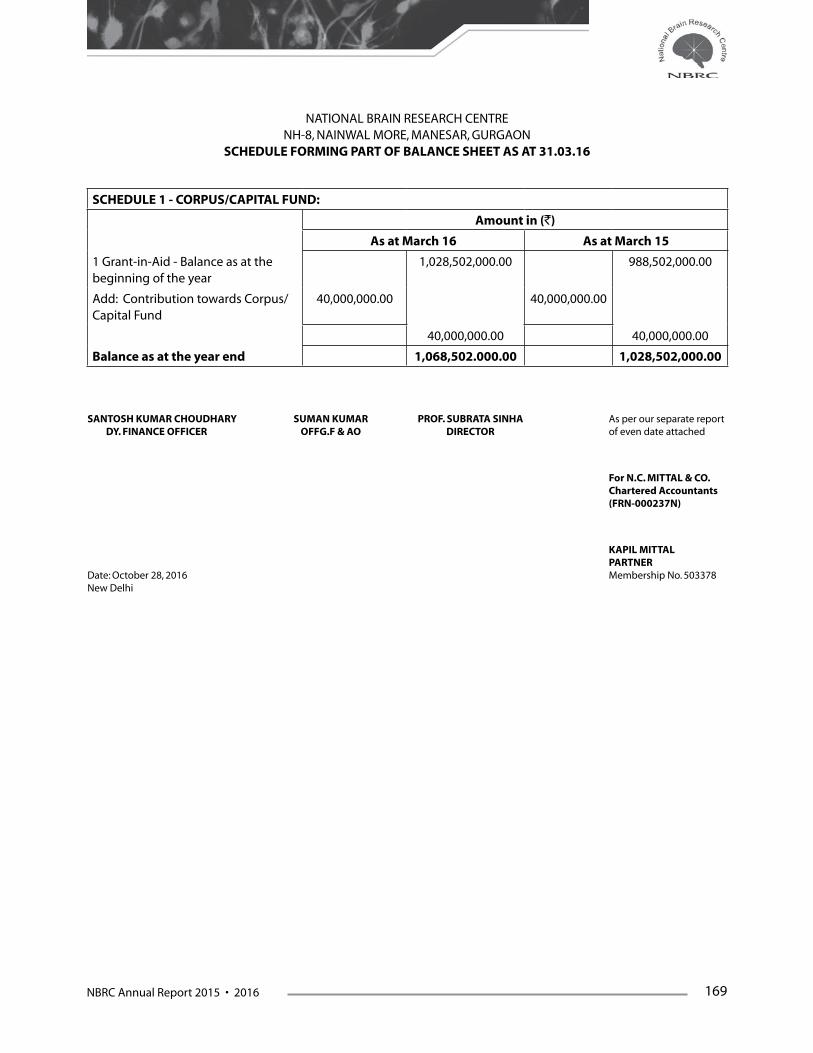
NBRC Annual Report 2015 • 2016 169
SCHEDULE 1 - CORPUS/CAPITAL FUND:
Amount in (`)
As at March 16 As at March 15
1 Grant-in-Aid - Balance as at the beginning of the year
1,028,502,000.00 988,502,000.00
Add: Contribution towards Corpus/Capital Fund
40,000,000.00 40,000,000.00
40,000,000.00 40,000,000.00
Balance as at the year end 1,068,502.000.00 1,028,502,000.00
NATIONAL BRAIN RESEARCH CENTRENH-8, NAINWAL MORE, MANESAR, GURGAON
SCHEDULE FORMING PART OF BALANCE SHEET AS AT 31.03.16
SANTOSH KUMAR CHOUDHARY SUMAN KUMAR PROF. SUBRATA SINHA As per our separate report DY. FINANCE OFFICER OFFG.F & AO DIRECTOR of even date attached
For N.C. MITTAL & CO. Chartered Accountants (FRN-000237N)
KAPIL MITTAL PARTNERDate: October 28, 2016 Membership No. 503378New Delhi

170 NBRC Annual Report 2015 • 2016
NATIONAL BRAIN RESEARCH CENTRE NH-8, NAINWAL MORE, MANESAR, GURGAON
SCHEDULE FORMING PART OF BALANCE SHEET AS AT 31.03.16
SCHEDULE 2 - RESERVES AND SURPLUS:
Amount in (`)
As at March 16 As at March 15
1 General Reserve
As per last Account (215,604,830.59) (124,568,030.41)
Addition during the Year 11,426,361,66 (91,036,800.18)
Less : Deductions during the year (deficit)
- (204,178,468.93) - (215,604,830.59)
Balance as at the year end (204,178,468.93) (215,604,830.59)
SANTOSH KUMAR CHOUDHARY SUMAN KUMAR PROF. SUBRATA SINHA As per our separate report DY. FINANCE OFFICER OFFG.F & AO DIRECTOR of even date attached
For N.C. MITTAL & CO. Chartered Accountants (FRN-000237N)
KAPIL MITTAL PARTNERDate: October 28, 2016 Membership No. 503378New Delhi
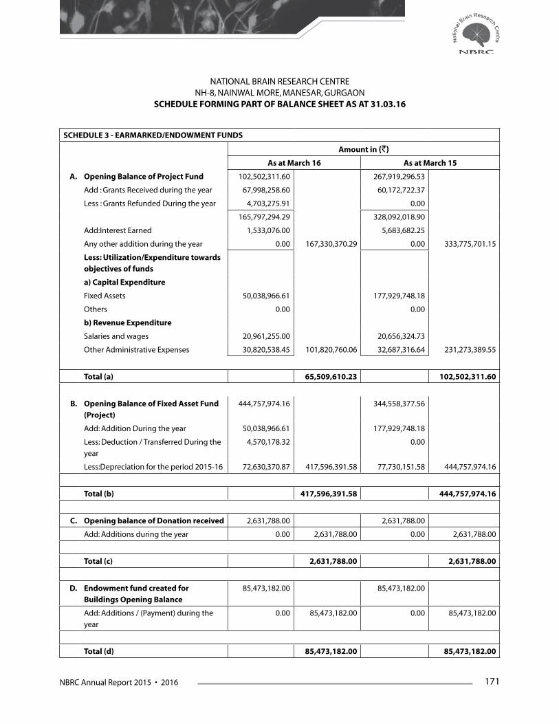
NBRC Annual Report 2015 • 2016 171
NATIONAL BRAIN RESEARCH CENTRE NH-8, NAINWAL MORE, MANESAR, GURGAON
SCHEDULE FORMING PART OF BALANCE SHEET AS AT 31.03.16
SCHEDULE 3 - EARMARKED/ENDOWMENT FUNDS
Amount in (`)
As at March 16 As at March 15
A. Opening Balance of Project Fund 102,502,311.60 267,919,296.53
Add : Grants Received during the year 67,998,258.60 60,172,722.37
Less : Grants Refunded During the year 4,703,275.91 0.00
165,797,294.29 328,092,018.90
Add:Interest Earned 1,533,076.00 5,683,682.25
Any other addition during the year 0.00 167,330,370.29 0.00 333,775,701.15
Less: Utilization/Expenditure towards
objectives of funds
a) Capital Expenditure
Fixed Assets 50,038,966.61 177,929,748.18
Others 0.00 0.00
b) Revenue Expenditure
Salaries and wages 20,961,255.00 20,656,324.73
Other Administrative Expenses 30,820,538.45 101,820,760.06 32,687,316.64 231,273,389.55
Total (a) 65,509,610.23 102,502,311.60
B. Opening Balance of Fixed Asset Fund
(Project)
444,757,974.16 344,558,377.56
Add: Addition During the year 50,038,966.61 177,929,748.18
Less: Deduction / Transferred During the
year
4,570,178.32 0.00
Less:Depreciation for the period 2015-16 72,630,370.87 417,596,391.58 77,730,151.58 444,757,974.16
Total (b) 417,596,391.58 444,757,974.16
C. Opening balance of Donation received 2,631,788.00 2,631,788.00
Add: Additions during the year 0.00 2,631,788.00 0.00 2,631,788.00
Total (c) 2,631,788.00 2,631,788.00
D. Endowment fund created for
Buildings Opening Balance
85,473,182.00 85,473,182.00
Add: Additions / (Payment) during the
year
0.00 85,473,182.00 0.00 85,473,182.00
Total (d) 85,473,182.00 85,473,182.00

172 NBRC Annual Report 2015 • 2016
E. Contributory Provident Fund 10,698,712.00 9,902,908.00
Add: Additions / (Payment) during the
year
1,441,426.00 12,140,138.00 795,804.00 10,698,712.00
Total (e) 12,140,138.00 10,698,712.00
F. DeLcon E-library Consortium
Opening balance of Consortium 24,197,834.13 32,896,815.16
Add : Grants Received during the year 405,673,921.88 421,449,577.00
Add : Interest Earned 700,086.00 1893862.00
Less: Utilization/Expenditure towards
objectives of funds
425,827,803.38 4,744,038.63 432,042,420.03 24,197,834.13
Total (f) 4,744,038.63 24,197,834.13
G. Escrow Account-DBT
Opening balance 0.00 0.00
Less: Grant Received During the Year 0.00 0.00 0.00 0.00
Total (g) 0.00 0.00
Balance as at the year end
(a+b+c+d+e+f+g)
588,095,148.44 670,261,801.89
SANTOSH KUMAR CHOUDHARY SUMAN KUMAR PROF. SUBRATA SINHA As per our separate report DY. FINANCE OFFICER OFFG.F & AO DIRECTOR of even date attached
For N.C. MITTAL & CO. Chartered Accountants (FRN-000237N)
KAPIL MITTAL PARTNERDate: October 28, 2016 Membership No. 503378New Delhi

NBRC Annual Report 2015 • 2016 173
SCHEDULE-4 CURRENT LIABILITIES AND PROVISIONS
Amount in (`)
As at March 16 As at March 15
A. Current Liabilities
1 Sundry Creditors 777,687.25 796,753.25
2 Advances Received (Security deposit) 2,621,446.42 5,013,848.62
3 Other Liabilities-TDS Payable 139,658.50 822,205.50
4 Earnest Money Deposit 3,968,695.00 3,005,008.00
5 Hostel Deposit 881,000.00 624,000.00
6 Library Deposit 252,000.00 160,000.00
7 Expenses Payable 19,590,060.47 12,607,531.46
8 CPF Payable 43,798.00 54,437.00
9 GIS Payable 2,954.00 2,764.00
10 Salary Payable 5,314.00 5,314.00
11 NPS(Employees Subscription) 24,090.00 24,090.00
12 Audit Fees Payable 28,175.00 0.00
13 Stale Cheque 316,874.22 0.00
14 Labour Cess Payable 104,890.00 28,756,642.86 338,537.00 23,454,488.83
Total (a) 28,756,642.86 23,454,488.83
B. Provisions
1 Gratuity 6749850.00 6,743,850.00
2 Accumulated Leave Encashment 3958568.80 10,708,418.80 3,958,568.80 10,702,418.80
Total (b) 10,708,418.80 10,702,418.80
Balance as at the year end (a+b) 39,465,061.66 34,156,907.63
NATIONAL BRAIN RESEARCH CENTRE NH-8, NAINWAL MORE, MANESAR, GURGAON
SCHEDULE FORMING PART OF BALANCE SHEET AS AT 31.03.16
SANTOSH KUMAR CHOUDHARY SUMAN KUMAR PROF. SUBRATA SINHA As per our separate report DY. FINANCE OFFICER OFFG.F & AO DIRECTOR of even date attached
For N.C. MITTAL & CO. Chartered Accountants (FRN-000237N)
KAPIL MITTAL PARTNERDate: October 28, 2016 Membership No. 503378New Delhi

174 NBRC Annual Report 2015 • 2016
SCH
EDU
LE 5
- FI
XED
ASS
ETS/
DEP
REC
IATO
N
GR
OSS
BLO
CK
DEP
REC
IATI
ON
NET
BLO
CK
Rat
e o
f D
ep.
Co
st /v
alu
atio
n A
s at
beg
inn
ing
o
f th
e Ye
ar
Ad
dit
ion
s d
uri
ng
th
e Ye
arD
edu
ctio
ns
d
uri
ng
th
e Ye
ar
Co
st /v
alu
atio
n
As
at e
nd
of t
he
Year
As
at t
he
beg
inn
ing
of
the
Year
Dep
reci
atio
n
for
curr
ent
year
Tota
l Dep
n.
Up
to 3
1.0
3.1
6A
s at
Cu
rren
t ye
ar-e
nd
As
at P
revi
ou
s ye
ar-e
nd
A.
Fixe
d A
sset
sM
ore
th
anLe
ss t
han
Am
ou
nt
in (`
)
6 M
on
ths
1B
uild
ing
10%
69,
568,
014.
00
0.00
2,0
00,9
70.0
0 0.
00 7
1,56
8,98
4.00
1
8,33
9,84
0.86
5
,222
,865
.81
23,
562,
706.
67
48,
006,
277.
33
51,
228,
173.
14
2Pl
ant
& M
ach
iner
y an
d E
qu
ipm
ent
15%
263
,090
,548
.76
3,6
12,4
91.3
5 4
,475
,163
.21
0.00
271
,178
,203
.32
121
,125
,038
.54
22,
172,
337.
48
143
,297
,376
.02
127
,880
,827
.30
141
,965
,510
.22
3Ve
hic
les
15%
2,0
86,3
42.0
0 0.
000.
000.
00 2
,086
,342
.00
1,2
89,5
86.4
8 1
19,5
13.3
3 1
,409
,099
.81
677
,242
.19
796
,755
.52
4Fu
rnit
ure
& F
ixtu
res
10%
34,
616,
867.
00
626
,957
.00
2,8
84,1
77.0
0 0.
00 3
8,12
8,00
1.00
1
5,83
0,07
7.32
2
,085
,583
.52
17,
915,
660.
84
20,
212,
340.
16
18,
786,
789.
68
5C
om
pu
ter &
So
ftw
are
60%
4,5
63,5
08.8
1 3
02,2
00.0
0 1
,031
,101
.00
0.00
5,8
96,8
09.8
1 3
,701
,100
.00
1,0
08,0
95.5
9 4
,709
,195
.59
1,1
87,6
14.2
2 8
62,4
08.8
1
6O
ffice
Eq
uip
men
t15
% 3
1,14
0,84
6.79
8
0,66
9.00
2
04,5
11.0
0 0.
00 3
1,42
6,02
6.79
1
3,79
2,94
1.12
2
,629
,624
.53
16,
422,
565.
65
15,
003,
461.
14
17,
347,
905.
67
Tota
l of T
he
Cu
rren
t Yea
r 4
05
,06
6,1
27
.36
4
,62
2,3
17
.35
1
0,5
95
,92
2.2
1
0.0
0 4
20
,28
4,3
66
.92
1
74
,07
8,5
84
.32
3
3,2
38
,02
0.2
5
20
7,3
16
,60
4.5
7
21
2,9
67
,76
2.3
5
30
,98
7,5
43
.04
B.
Fixe
d A
sset
s (P
roje
cts)
1Pr
oje
ct E
qu
ipm
ents
15%
674
,198
,080
.13
37,
990,
386.
68
12,
048,
579.
93
4,5
70,1
78.3
2 7
19,6
66,8
68.4
2 2
29,4
40,1
05.9
8 7
2,63
0,37
0.87
3
02,0
70,4
76.8
5 4
17,5
96,3
91.5
7 4
44,7
57,9
74.1
5
Tota
l of T
he
Cu
rren
t Yea
r (C
) (A
+B
) 1
,07
9,2
64
,20
7.4
9
42
,61
2,7
04
.03
2
2,6
44
,50
2.1
4
4,5
70
,17
8.3
2
1,1
39
,95
1,2
35
.34
40
3,5
18
,69
0.3
0
1
05
,86
8,3
91
.12
50
9,3
87
,08
1.4
2
63
0,5
64
,15
3.9
2
67
5,7
45
,51
7.1
9
Prev
iou
s Ye
ar
D.
Cap
ital
Wo
rk in
Pro
gre
ss
1B
uild
ing
s
Cap
ital
wo
rk-i
n-p
rog
ress
incl
ud
ing
ad
van
ces,
668
,974
,084
.73
0.00
0.00
0.00
668
,974
,084
.73
0.00
0.00
0.00
668
,974
,084
.73
668
,974
,084
.73
con
stru
ctio
n m
ater
ials
an
d b
uild
ing
u
nd
er
con
stru
ctio
n (n
et o
f rec
over
y)
E.In
tan
gib
le A
sset
s
1Pa
ten
t an
d C
op
y R
igh
t (A
pp
licat
ion
Fe
e)25
% 4
,357
,864
.00
0.00
742
,528
.00
0.00
5,1
00,3
92.0
0 2
,101
,477
.00
656
,912
.75
2,7
58,3
89.7
5 2
,342
,002
.25
2,2
56,3
87.0
0
TOTA
L (C
+D
+E)
1,7
52
,59
6,1
56
.22
4
2,6
12
,70
4.0
3
23
,38
7,0
30
.14
4
57
01
78
.32
1,8
14
,02
5,7
12
.07
0
5,6
20
,16
7.3
0
10
6,5
25
,30
3.8
7
51
2,1
45
,47
1.1
7
1,3
01
,88
0,2
40
.90
1
,34
6,9
75
,98
8.9
2
NAT
ION
AL
BR
AIN
RES
EAR
CH
CEN
TRE
NH
-8, N
AIN
WA
L M
ORE
, MA
NES
AR,
GU
RG
AO
NSC
HED
ULE
FO
RM
ING
PA
RT
OF
BA
LAN
CE
SHEE
T A
S A
T 3
1.0
3.1
6
SAN
TOSH
KU
MA
R C
HO
UD
HA
RY
SU
MA
N K
UM
AR
P
RO
F. S
UB
RA
TA S
INH
A
As
per
ou
r sep
arat
e re
po
rt
DY.
FIN
AN
CE
OFF
ICER
O
FFG
.F &
AO
D
IREC
TOR
o
f eve
n d
ate
atta
ched
For
N.C
. MIT
TAL
& C
O.
Ch
arte
red
Acc
ou
nta
nts
(FR
N-0
00
23
7N
)
KA
PIL
MIT
TAL
PAR
TNER
Dat
e: O
cto
ber
28,
201
6
M
emb
ersh
ip N
o. 5
0337
8N
ew D
elh
i
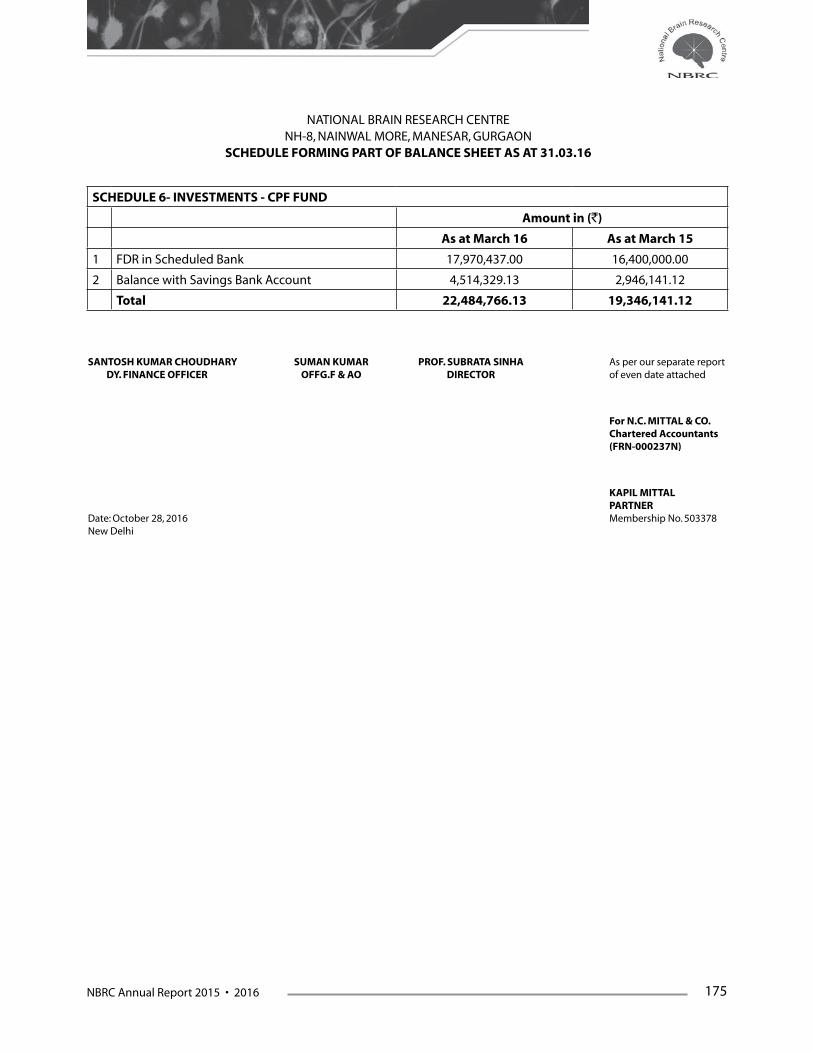
NBRC Annual Report 2015 • 2016 175
SCHEDULE 6- INVESTMENTS - CPF FUND
Amount in (`)
As at March 16 As at March 15
1 FDR in Scheduled Bank 17,970,437.00 16,400,000.00
2 Balance with Savings Bank Account 4,514,329.13 2,946,141.12
Total 22,484,766.13 19,346,141.12
NATIONAL BRAIN RESEARCH CENTRE NH-8, NAINWAL MORE, MANESAR, GURGAON
SCHEDULE FORMING PART OF BALANCE SHEET AS AT 31.03.16
SANTOSH KUMAR CHOUDHARY SUMAN KUMAR PROF. SUBRATA SINHA As per our separate report DY. FINANCE OFFICER OFFG.F & AO DIRECTOR of even date attached
For N.C. MITTAL & CO. Chartered Accountants (FRN-000237N)
KAPIL MITTAL PARTNERDate: October 28, 2016 Membership No. 503378New Delhi

176 NBRC Annual Report 2015 • 2016
SCHEDULE 7 - CURRENT ASSETS, LOANS, ADVANCES ETC.
Amount in (`)
As at March 16 As at March 15
A. Current Assets
1 Cash Balances in hand (Including Cheques/Drafts) 164,213.00 150,868.00
2 Bank Balances:
a) With Schduled Banks:
-In Deposit Account 0.00 0.00
-In Saving Accounts (Core & Projects) 145,712,700.17 108,688,859.18
-In Deposit Against various Project Assets 0.00 145,712,700.17 0.00 108,688,859.18
3 Interest Accrued on FD(CPF) 1.481.965.00 1,475,662.76
4 Grant Receivable (DBT) 0.00 19,300,000.00
Total (a) 147,358,878.17 129,615,389.94
B. Loans, advances and other assets
1 Advances and other amounts receivable in cash
or in kind or for value to be received
a) Staff 8,077,848.55 8,819,350.59
b) Imprest 125,966.00 86,462.00
c) Advance to Parties 4,461,913.63 5,173,043.57
d) Others(Security & other Deposits) 2,161,491.20 2,016,902.20
e) Other Advances 92,625.00 0.00
f ) TDS Receivable 4,344,100.59 4,342,380.59
g) Prepaid Insurance 895,911.00 20,159,855.97 940,220.00 21,378,358.95
Total (b) 20,159,855.97 21,378,358.95
Balance as at the year end (a+b) 167,518,734.14 150,993,748.89
SANTOSH KUMAR CHOUDHARY SUMAN KUMAR PROF. SUBRATA SINHA As per our separate report DY. FINANCE OFFICER OFFG.F & AO DIRECTOR of even date attached
For N.C. MITTAL & CO. Chartered Accountants (FRN-000237N)
KAPIL MITTAL PARTNERDate: October 28, 2016 Membership No. 503378New Delhi
NATIONAL BRAIN RESEARCH CENTRE NH-8, NAINWAL MORE, MANESAR, GURGAON
SCHEDULE FORMING PART OF BALANCE SHEET AS AT 31.03.16

NBRC Annual Report 2015 • 2016 177
NATIONAL BRAIN RESEARCH CENTRENH-8, NAINWAL MORE, MANESAR, GURGAON
SCHEDULE FORMING PART OF INCOME AND EXPENDITURE FOR THE YEAR ENDED 31.03.2016
SCHEDULE 8 - FEES/ SUBSCRIPTIONS
Amount in (`)
For the year ended March 16 For the year ended March 15
1 Application Fees 433,000.00 922,996.39
2 Annual Fees/ Subscription to Journals 0.00 201,652.00
3 Others (Specify)-Fellowship Grants 2,862,614.00 2,801,682.00
Total 3,295,614.00 3,926,330.39
SANTOSH KUMAR CHOUDHARY SUMAN KUMAR PROF. SUBRATA SINHA As per our separate report DY. FINANCE OFFICER OFFG.F & AO DIRECTOR of even date attached
For N.C. MITTAL & CO. Chartered Accountants (FRN-000237N)
KAPIL MITTAL PARTNERDate: October 28, 2016 Membership No. 503378New Delhi
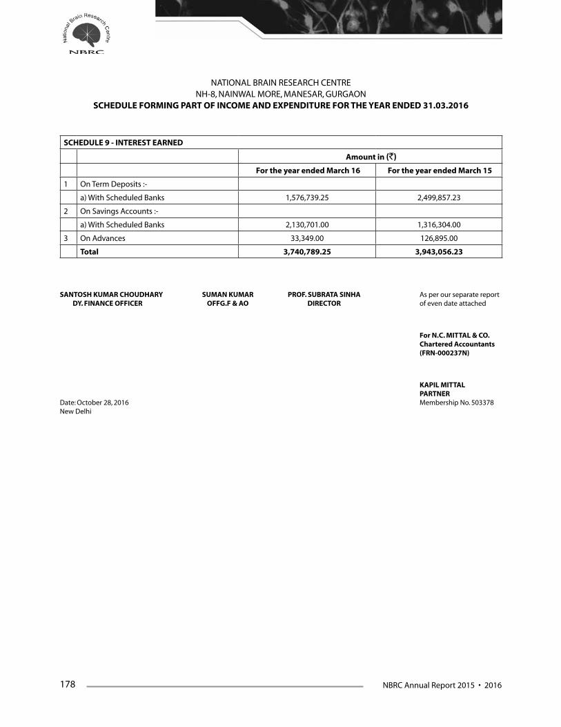
178 NBRC Annual Report 2015 • 2016
SCHEDULE 9 - INTEREST EARNED
Amount in (`)
For the year ended March 16 For the year ended March 15
1 On Term Deposits :-
a) With Scheduled Banks 1,576,739.25 2,499,857.23
2 On Savings Accounts :-
a) With Scheduled Banks 2,130,701.00 1,316,304.00
3 On Advances 33,349.00 126,895.00
Total 3,740,789.25 3,943,056.23
SANTOSH KUMAR CHOUDHARY SUMAN KUMAR PROF. SUBRATA SINHA As per our separate report DY. FINANCE OFFICER OFFG.F & AO DIRECTOR of even date attached
For N.C. MITTAL & CO. Chartered Accountants (FRN-000237N)
KAPIL MITTAL PARTNERDate: October 28, 2016 Membership No. 503378New Delhi
NATIONAL BRAIN RESEARCH CENTRE NH-8, NAINWAL MORE, MANESAR, GURGAON
SCHEDULE FORMING PART OF INCOME AND EXPENDITURE FOR THE YEAR ENDED 31.03.2016
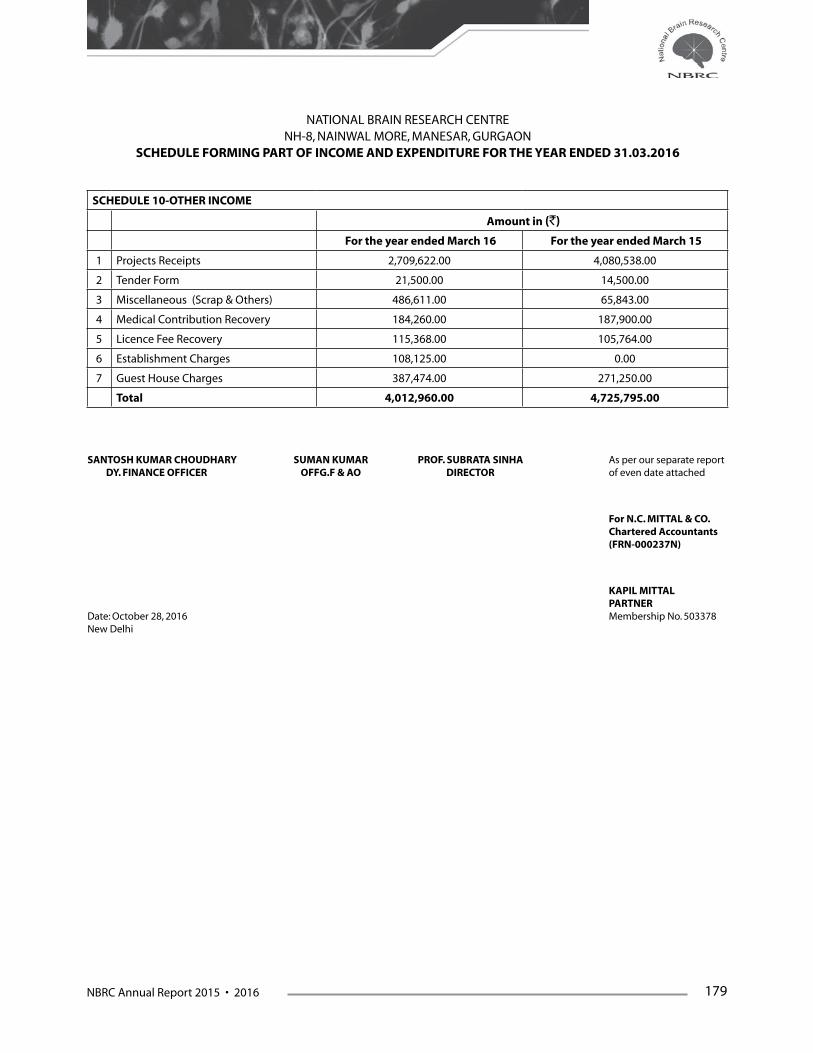
NBRC Annual Report 2015 • 2016 179
SCHEDULE 10-OTHER INCOME
Amount in (`)
For the year ended March 16 For the year ended March 15
1 Projects Receipts 2,709,622.00 4,080,538.00
2 Tender Form 21,500.00 14,500.00
3 Miscellaneous (Scrap & Others) 486,611.00 65,843.00
4 Medical Contribution Recovery 184,260.00 187,900.00
5 Licence Fee Recovery 115,368.00 105,764.00
6 Establishment Charges 108,125.00 0.00
7 Guest House Charges 387,474.00 271,250.00
Total 4,012,960.00 4,725,795.00
SANTOSH KUMAR CHOUDHARY SUMAN KUMAR PROF. SUBRATA SINHA As per our separate report DY. FINANCE OFFICER OFFG.F & AO DIRECTOR of even date attached
For N.C. MITTAL & CO. Chartered Accountants (FRN-000237N)
KAPIL MITTAL PARTNERDate: October 28, 2016 Membership No. 503378New Delhi
NATIONAL BRAIN RESEARCH CENTRE NH-8, NAINWAL MORE, MANESAR, GURGAON
SCHEDULE FORMING PART OF INCOME AND EXPENDITURE FOR THE YEAR ENDED 31.03.2016
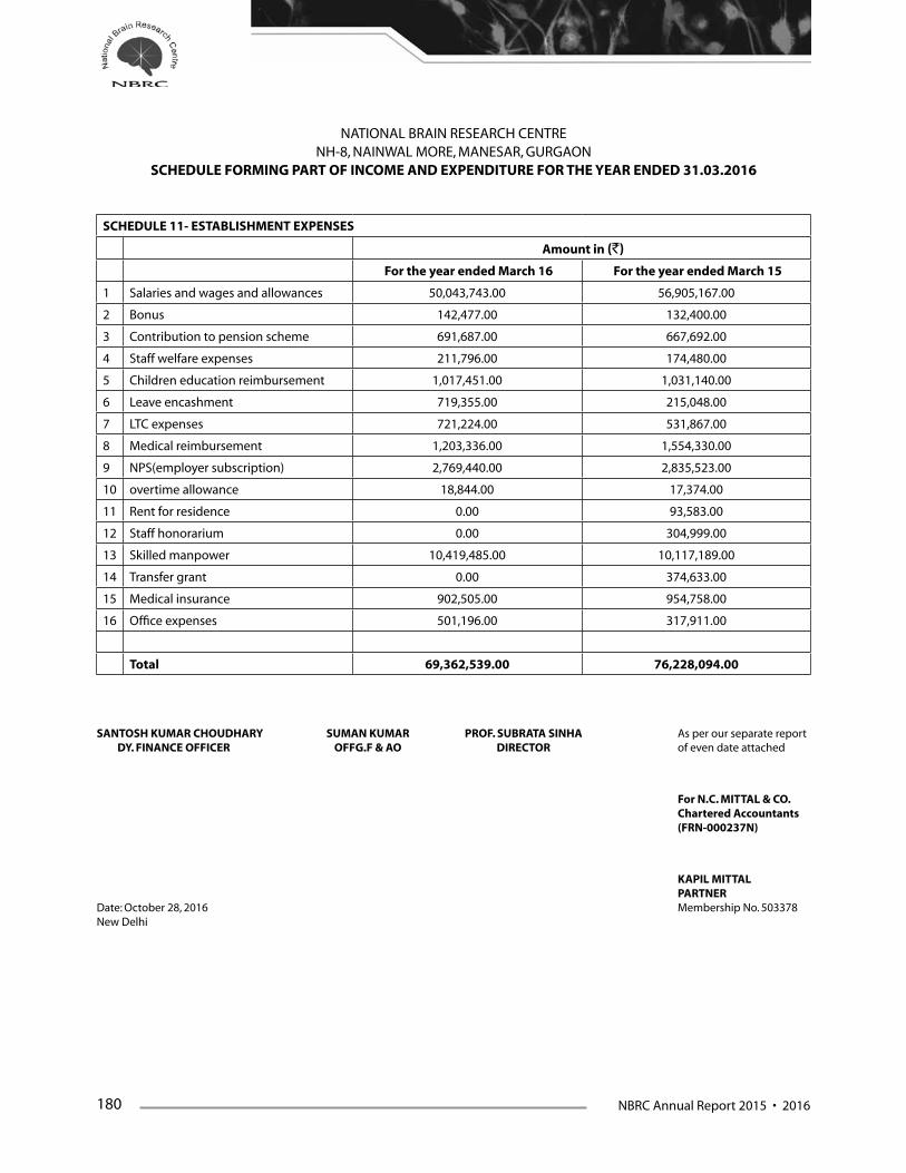
180 NBRC Annual Report 2015 • 2016
SCHEDULE 11- ESTABLISHMENT EXPENSES
Amount in (`)
For the year ended March 16 For the year ended March 15
1 Salaries and wages and allowances 50,043,743.00 56,905,167.00
2 Bonus 142,477.00 132,400.00
3 Contribution to pension scheme 691,687.00 667,692.00
4 Staff welfare expenses 211,796.00 174,480.00
5 Children education reimbursement 1,017,451.00 1,031,140.00
6 Leave encashment 719,355.00 215,048.00
7 LTC expenses 721,224.00 531,867.00
8 Medical reimbursement 1,203,336.00 1,554,330.00
9 NPS(employer subscription) 2,769,440.00 2,835,523.00
10 overtime allowance 18,844.00 17,374.00
11 Rent for residence 0.00 93,583.00
12 Staff honorarium 0.00 304,999.00
13 Skilled manpower 10,419,485.00 10,117,189.00
14 Transfer grant 0.00 374,633.00
15 Medical insurance 902,505.00 954,758.00
16 Office expenses 501,196.00 317,911.00
Total 69,362,539.00 76,228,094.00
SANTOSH KUMAR CHOUDHARY SUMAN KUMAR PROF. SUBRATA SINHA As per our separate report DY. FINANCE OFFICER OFFG.F & AO DIRECTOR of even date attached
For N.C. MITTAL & CO. Chartered Accountants (FRN-000237N)
KAPIL MITTAL PARTNERDate: October 28, 2016 Membership No. 503378New Delhi
NATIONAL BRAIN RESEARCH CENTRE NH-8, NAINWAL MORE, MANESAR, GURGAON
SCHEDULE FORMING PART OF INCOME AND EXPENDITURE FOR THE YEAR ENDED 31.03.2016

NBRC Annual Report 2015 • 2016 181
SCHEDULE 12- OTHER ADMINISTRATIVE EXPENSES
Amount in (`)
For the year ended March 16 For the year ended March 15
1 Postage, telephone and communication charges 803,375.00 926,210.70
2 Printing and stationary 1,094,501.00 539,370.00
3 Travelling expenses 1,664,640.00 741,571.81
4 Auditor remuneration 28,175.00 18,090.00
5 Hospitality/local meeting expenses 241,325.00 489,991.00
6 Legal and professional charges 872,330.00 804,785.00
7 Lease rent 1,000,000.00 1,000,000.00
8 Bank charges 9,326.14 925.46
9 Advertisement and publicity 402,610.00 760,566.00
10 Misc. expenses 582,196.50 408,053.00
11 Books and periodicals 89,318.00 78,836.00
12 Transportation charges 3,097,932.00 5,171,719.00
13 Conveyance reimbursement 85,780.00 97,769.00
14 Honorarium (others) 219,000.00 395,333.00
Total 10,190,508.64 11,433,219.97
SANTOSH KUMAR CHOUDHARY SUMAN KUMAR PROF. SUBRATA SINHA As per our separate report DY. FINANCE OFFICER OFFG.F & AO DIRECTOR of even date attached
For N.C. MITTAL & CO. Chartered Accountants (FRN-000237N)
KAPIL MITTAL PARTNERDate: October 28, 2016 Membership No. 503378New Delhi
NATIONAL BRAIN RESEARCH CENTRE NH-8, NAINWAL MORE, MANESAR, GURGAON
SCHEDULE FORMING PART OF INCOME AND EXPENDITURE FOR THE YEAR ENDED 31.03.2016
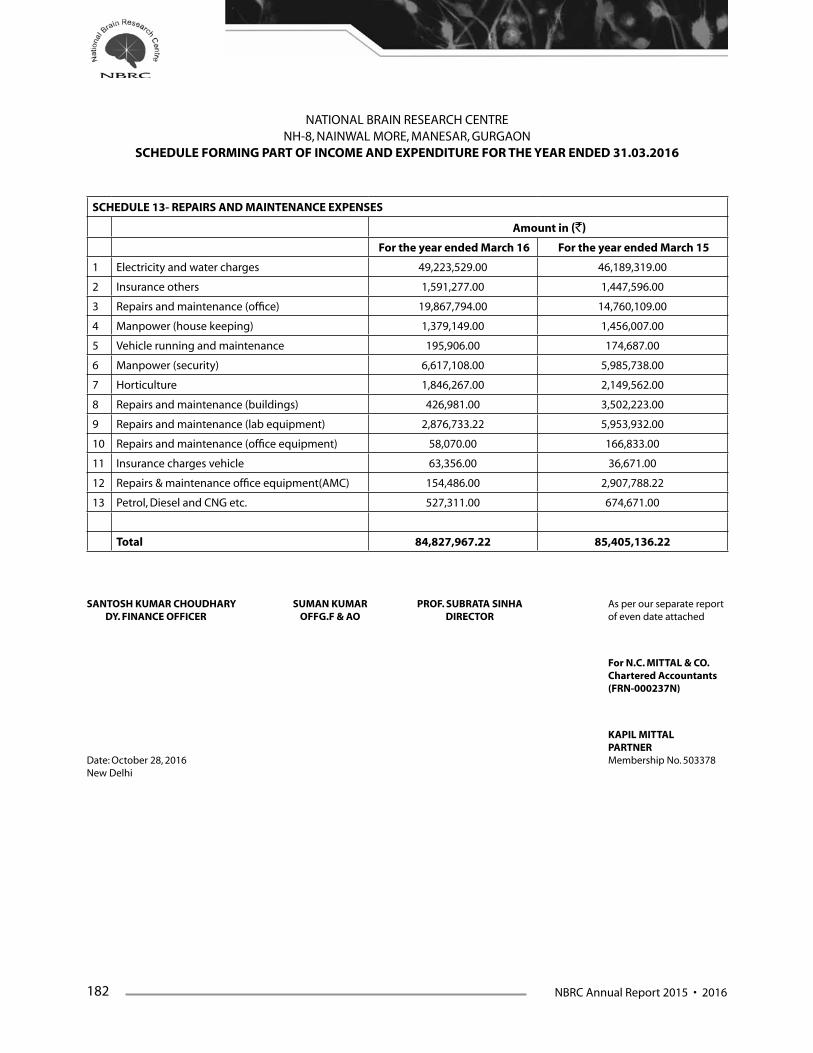
182 NBRC Annual Report 2015 • 2016
SCHEDULE 13- REPAIRS AND MAINTENANCE EXPENSES
Amount in (`)
For the year ended March 16 For the year ended March 15
1 Electricity and water charges 49,223,529.00 46,189,319.00
2 Insurance others 1,591,277.00 1,447,596.00
3 Repairs and maintenance (office) 19,867,794.00 14,760,109.00
4 Manpower (house keeping) 1,379,149.00 1,456,007.00
5 Vehicle running and maintenance 195,906.00 174,687.00
6 Manpower (security) 6,617,108.00 5,985,738.00
7 Horticulture 1,846,267.00 2,149,562.00
8 Repairs and maintenance (buildings) 426,981.00 3,502,223.00
9 Repairs and maintenance (lab equipment) 2,876,733.22 5,953,932.00
10 Repairs and maintenance (office equipment) 58,070.00 166,833.00
11 Insurance charges vehicle 63,356.00 36,671.00
12 Repairs & maintenance office equipment(AMC) 154,486.00 2,907,788.22
13 Petrol, Diesel and CNG etc. 527,311.00 674,671.00
Total 84,827,967.22 85,405,136.22
SANTOSH KUMAR CHOUDHARY SUMAN KUMAR PROF. SUBRATA SINHA As per our separate report DY. FINANCE OFFICER OFFG.F & AO DIRECTOR of even date attached
For N.C. MITTAL & CO. Chartered Accountants (FRN-000237N)
KAPIL MITTAL PARTNERDate: October 28, 2016 Membership No. 503378New Delhi
NATIONAL BRAIN RESEARCH CENTRE NH-8, NAINWAL MORE, MANESAR, GURGAON
SCHEDULE FORMING PART OF INCOME AND EXPENDITURE FOR THE YEAR ENDED 31.03.2016

NBRC Annual Report 2015 • 2016 183
SCHEDULE 14 - TRAINING AND NETWORKING EXPENSES
Amount in (`)
For the year ended March 16 For the year ended March 15
1 Subscription to journals 2,184,923.32 854,258.78
2 Training expenses 33,556,076.00 13,005,220.00
3 Contingencies (CSIR/UGC/DBT/ICMR students) 454,809.00 94,902.00
4 Conference & workshop expenses 1,122,111.04 753,586.00
5 Student medical expenses 122,144.00 146,109.00
Total 37,440,063.36 14,854,075.78
SANTOSH KUMAR CHOUDHARY SUMAN KUMAR PROF. SUBRATA SINHA As per our separate report DY. FINANCE OFFICER OFFG.F & AO DIRECTOR of even date attached
For N.C. MITTAL & CO. Chartered Accountants (FRN-000237N)
KAPIL MITTAL PARTNERDate: October 28, 2016 Membership No. 503378New Delhi
NATIONAL BRAIN RESEARCH CENTRE NH-8, NAINWAL MORE, MANESAR, GURGAON
SCHEDULE FORMING PART OF INCOME AND EXPENDITURE FOR THE YEAR ENDED 31.03.2016

184 NBRC Annual Report 2015 • 2016
SCHEDULE 15 - LABORATORY AND ANIMAL HOUSE CONSUMABLS
Amount in (`)
For the year ended March 16 For the year ended March 15
1 Lab consumables and chemicals 23,006,896.71 33,229,916.19
2 Medicines and consumables animal 1,924,191.00 1,817,516.50
Total 24,931,087.71 35,047,432.69
SANTOSH KUMAR CHOUDHARY SUMAN KUMAR PROF. SUBRATA SINHA As per our separate report DY. FINANCE OFFICER OFFG.F & AO DIRECTOR of even date attached
For N.C. MITTAL & CO. Chartered Accountants (FRN-000237N)
KAPIL MITTAL PARTNERDate: October 28, 2016 Membership No. 503378New Delhi
NATIONAL BRAIN RESEARCH CENTRE NH-8, NAINWAL MORE, MANESAR, GURGAON
SCHEDULE FORMING PART OF INCOME AND EXPENDITURE FOR THE YEAR ENDED 31.03.2016
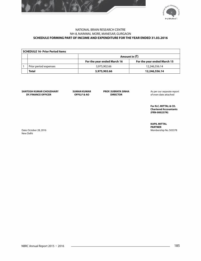
NBRC Annual Report 2015 • 2016 185
SCHEDULE 16- Prior Period Items
Amount in (`)
For the year ended March 16 For the year ended March 15
1 Prior period expenses 3,975,902.66 12,246,556.14
Total 3,975,902.66 12,246,556.14
SANTOSH KUMAR CHOUDHARY SUMAN KUMAR PROF. SUBRATA SINHA As per our separate report DY. FINANCE OFFICER OFFG.F & AO DIRECTOR of even date attached
For N.C. MITTAL & CO. Chartered Accountants (FRN-000237N)
KAPIL MITTAL PARTNERDate: October 28, 2016 Membership No. 503378New Delhi
NATIONAL BRAIN RESEARCH CENTRE NH-8, NAINWAL MORE, MANESAR, GURGAON
SCHEDULE FORMING PART OF INCOME AND EXPENDITURE FOR THE YEAR ENDED 31.03.2016

186 NBRC Annual Report 2015 • 2016
Significant Accounting Policies & Notes on Accounts Forming Part of the Balance Sheet as at 31st March, 2016 and Income & Expenditure Account
For The Year Ended 31st March, 2016
Significant Accounting Policies & Notes on Accounts
1. Accounting Convention1.1 The financial statements of National Brain
Research Centre (NBRC) are prepared on the basis of historical cost convention, unless otherwise stated and on the accrual basis of accounting.
1.2 The NBRC is moving towards adopting the ‘Uniform Format of Accounting’ prescribed for the Central Autonomous Bodies by the Ministry of Finance, Govt. of India for preparing the Income & Expenditure Account, Receipts & Payments Account, Balance Sheet & other Schedules thereto.
2. Inventory2.1 All purchases of chemicals, glassware,
consumables and printing & stationery have been booked/charged to consumption/expenditure at the time of purchases.
3. Fixed Assets3.1 Fixed Assets are stated at historical cost.
3.2 Physical verification of assets had not been conducted during the year.
3.3 The capital work-in-progress includes completed work/buildings under Phase-I as these works could not be transferred to ‘Fixed Assets – Buildings’ category for want of Building-wise information from the Project Management Consultant i.e. Directorate of Construction Services and Estate Management (DC&SEM) of Department of Atomic Energy.
3. NBRC has entered into a Memorandum of Understanding (MOU) with DC&SEM for construction of NBRC’s Building at Manesar, Gurgaon. As per the MOU with the DC&SEM, NBRC is depositing funds with DC&SEM from time to
time to be utilized by DC&SEM for construction. Total amount deposited with DC&SEM is Rs. 44,46,52,000.00 till 31st March 2016. Pending completion of construction, the payments made to DC&SEM are being shown as Deposit under the head Building under Construction. Final adjustment shall be done on submission of final account of the project by DC&SEM; Now the MOU with DC&SEM is discontinued. NBRC has again engaged Civil & Construction Wing (CCW) AIR, Prasar Bharti, as Project Management Consultant (PMC) for completing balance work and final bill is yet to be settled.
3.5 Fixed Assets have been created mainly out of grants received from the Department of Biotechnology, Ministry of Science and Technology, Government of India & Project grants.
4. Depreciation4.1 From F.Y 2012-2013 Depreciation is being charged
as per Income Tax Act 1961 on W.D.V basis. As stated in F.Y 2012-13, in view of old information not being readily available, the retrospective calculation of depreciation as per Income Tax Act 1961 for adjustment of excess/short depreciation is vis-a-vis the old rates, as required under the Accounting Standard-6 issued by Institute of Chartered Accounts of India (ICAI), could not be made till date. The same shall be made in due course of the determination of the same.
4.2 Depreciation provided for current year on the fixed assets of Project for Rs. 7,26,30,370.87 (previous year Rs. 7,77,30,151.58) and which has been directly debited to the fixed assets funds account. These assets were created through the Non-Recurring and project based grant from the
SCHEDULE-17
NATIONAL BRAIN RESEARCH CENTRE, MANESAR, GURGAON

NBRC Annual Report 2015 • 2016 187
funding agencies. Depreciation for other than project assets amounting to Rs. 3,38,94,933.00 for current financial year (Rs. 3,77,17,467.00 for previous year) had been debited to Income & Expenditure Account.
5. Investments5.1 Investments in term deposits with banks are
basically for Current Investments and are therefore valued on cost.
5.2 Interest received on term deposits are accounted for on accrual basis, which results in increase in profitability.
6. Government Grants / Subsidies6.1 Government grants of the nature of contribution
towards capital cost of setting up projects are treated as Capital Reserve/Fund.
6.2 Government grants / subsidy are accounted for in accordance with the sanctioned terms.
6.3 Interest on Government Grant has been considered under the respective projects due to which loss has increased by Rs. 22,33,162.00 (previous year Rs. 75,77,544.25).
7. Foreign Currency Transactions/ Grants7.1 Transactions denominated in foreign currency are
accounted at the exchange rate prevailing at the date of the transaction.
7.2 The Centre had one FCRA Bank Account PNB Manesar related to the Grants. The submission of the returns of these accounts has been made up to Financial Year 31st March, 2009 under the FCR Act. NBRC had recently received a notice for compliance with this regards from Government of India.
8. Lease The NBRC is located on the leasehold land at
Manesar taken from Indian Vaccine Corporation Ltd. for Rs. 10,00,000/- per annum lease rent with certain semi-built structure at a cost Rs. 45,17,000/- towards such structures. The lease is for the period of 33 years, after which the land along with premises thereon are to be handed over to the lessor. No amortization/write off have been done in respect of the assets acquired on lease.
9. Retirement Benefits9.1 The NBRC is registered with the Provident Fund
authorities and it maintains a separate CPF Trust, which is yet to be recognized and the CPF fund required the separate accounting.
9.2 The NBRC has not made any provision for gratuity and leave encashment during financial year 2015-2016 as against the requirement of AS-15 issued by ICAI. However the amount of gratuity and leave encashment to the extent of Rs. 67,49,850.00 and Rs. 39,58,568.80 respectively already exists on 31st March, 2016, (Rs. 67,43,850.00 & Rs. 39,58,568.00 respectively as on 31st March, 2015) against provision made earlier.
10. Taxation In view of the tax exemption status of the Center,
no provision for income tax has been considered necessary.
11. Loans & Advances Advances appearing under the head Current
assets, Loans & Advances under Schedule-7 are subject to confirmation from parties.
12. Bank Balance Bank balance in Axis Bank Limited, Gurgaon (A/c
No.056010100453998) & Punjab National Bank, Manesar (FCRA)
(A/c No. 4136000100008889) as on 31st March, 2016 of Rs. 13,53,90,801.46 & Rs. 10,73,959.54 respectively (Previous Year Rs. 10,53,63,459.58 & 10,61,784.54 respectively) are subject to reconciliation.
13. Prior Period Items Accounting Standard-5 Issued by Institute of
Chartered Accountants of India (ICAI), Prior Period items are income or expenses, which arises, in current period as a result of error or omission in the preparation of financial statement of one or more prior periods. In the current year, the Prior Period items recognized, related to expenditure i.e. Rs. 39,75,902.66 for the financial year 2014-15 (previous year 2014-15 Rs. 1,22,46,556.14 for the financial year 2013-14) that was omitted in that year.
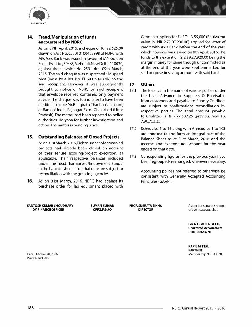
188 NBRC Annual Report 2015 • 2016
14. Fraud/Manipulation of funds encountered by NBRC
As on 27th April, 2015, a cheque of Rs. 92,625.00 drawn on A/c No. 056010100453998 of NBRC with M/s Axis Bank was issued in favour of M/s Golden Feeds Pvt. Ltd., 894/8, Mehrauli, New Delhi-110030, against their invoice No. 2591 dtd. 09th March, 2015. The said cheque was dispatched via speed post (India Post Ref. No. EH643251489IN) to the said receipient. However it was subsequently brought to notice of NBRC by said receipient that envelope received contained only payment advice. The cheque was found later to have been credited to some Mr. Bhagirath Chauhan’s account, at Bank of India, Rajnagar Extn., Ghaziabad (Uttar Pradesh). The matter had been reported to police authorities, Haryana for further investigation and action. The matter is pending since.
15. Outstanding Balances of Closed Projects As on 31st March, 2016, Eight number of earmarked
projects had already been closed on account of their tenure expiring/project execution, as applicable. Their respective balances included under the head ‘’Earmarked/Endowment Funds” in the balance sheet as on that date are subject to reconciliation with the granting agencies.
16. As on 31st March, 2016, NBRC had against its purchase order for lab equipment placed with
German suppliers for EURO 3,55,000 (Equivalent value in INR 2,72,07,200.00) applied for letter of credit with Axis Bank before the end of the year, which however was issued on 8th April, 2016. The funds to the extent of Rs. 2,99,27,920.00 being the margin money for same though uncommitted as at the end of the year were kept earmarked for said purpose in saving account with said bank.
17. Others17.1 The Balance in the name of various parties under
the head Advance to Suppliers & Receivable from customers and payable to Sundry Creditors are subject to confirmation/ reconciliation by respective parties. The total amount payable to Creditors is Rs. 7,77,687.25 (previous year Rs. 7,96,753.25).
17.2 Schedules 1 to 16 along with Annexures 1 to 103 are annexed to and form an integral part of the Balance Sheet as at 31st March, 2016 and the Income and Expenditure Account for the year ended on that date.
17.3 Corresponding figures for the previous year have been regrouped/ rearranged, wherever necessary.
Accounting polices not referred to otherwise be consistent with Generally Accepted Accounting Principles (GAAP).
SANTOSH KUMAR CHOUDHARY SUMAN KUMAR PROF. SUBRATA SINHA As per our separate report DY. FINANCE OFFICER OFFG.F & AO DIRECTOR of even date attached
For N.C. MITTAL & CO. Chartered Accountants (FRN-000237N)
KAPIL MITTAL PARTNERDate: October 28, 2016 Membership No. 503378Place: New Delhi

NBRC Annual Report 2015 • 2016 189
S. N.
Name of Project Opening Balance as on
01.04.2015
Grants received
during the year 2015-16
Interest earned
during the year 2015-16
Capital Exp. during the
year 2015-16
Revenue Expenditure during the year 2015-16 Refund Unspent Balance
Closing Balance as on
31.03.2016Manpower Others Total Expenditure
1 Distributed Information Centre (2,031,491.67) 6,380,000.00 0.00 276,630.00 1,722,551.00 530,325.00 2,252,876.00 0.00 1,819,002.33
2 Programme of Co-Operation Between India and Syria Project
3558649.00 0.00 0.00 0.00 0.00 0.00 0.00 0.00 3,558,649.00
3 Mole. Role of Transc. Factors - Dr. Prabodha Kumar Swain
(644,021.00) 0.00 0.00 0.00 0.00 0.00 0.00 0.00 (644,021.00)
4 Multifactorial Risk Factor - Prof. V. Ravindranath (29,346.00) 0.00 0.00 0.00 0.00 0.00 0.00 0.00 (29,346.00)
5 Func. Magnetic Resonance Imaging - Prof. V. Ravindranath
(355,435.00) 0.00 0.00 0.00 0.00 0.00 0.00 0.00 (355,435.00)
6 Material Malnutrition - Dr. Shyamala (579,048.00) 0.00 0.00 0.00 0.00 0.00 0.00 0.00 (579,048.00)
7 M.Sc. Neuroscience - Prof. P.K. Roy 5,073.00 0.00 0.00 0.00 0.00 0.00 0.00 0.00 5,073.00
8 Stochastic Resonance - Prof. P.K. Roy (471.00) 0.00 0.00 0.00 0.00 0.00 0.00 0.00 (471.00)
9 Dementia Meeting 2,364,225.00 0.00 0.00 0.00 0.00 0.00 0.00 0.00 2,364,225.00
10 Comp. Analysis of Speech Imp. - Dr. Nandini Singh
(547,567.00) 0.00 0.00 0.00 0.00 0.00 0.00 0.00 (547,567.00)
11 Spinal Cord Plasticity ILTP - Dr. Neeraj Jain (31,869.00) 0.00 0.00 0.00 0.00 0.00 0.00 0.00 (31,869.00)
12 Study of Mole. Mechanism - Dr. Anirban Basu (68,830.00) 0.00 0.00 0.00 0.00 0.00 0.00 0.00 (68,830.00)
13 BBNSC - Dr. Rema 1,809,628.00 0.00 0.00 0.00 0.00 0.00 0.00 0.00 1,809,628.00
14 BBNSC - Dr. Dhingra 144.00 0.00 0.00 0.00 0.00 0.00 0.00 0.00 144.00
15 BBNSC - Dr. Shyamala (392,947.00) 0.00 0.00 0.00 0.00 0.00 0.00 0.00 (392,947.00)
16 BBNSC - Dr. Neeraj 296,937.00 0.00 0.00 0.00 0.00 0.00 0.00 0.00 296,937.00
17 BBNSC - Dr. Ellora (403,419.00) 0.00 0.00 0.00 0.00 0.00 0.00 0.00 (403,419.00)
18 BBNSC - Dr. Soumya 1,246.00 0.00 0.00 0.00 0.00 0.00 0.00 0.00 1,246.00
19 Cellular & Mole. Basis - Dr. Pankaj (34,974.00) 0.00 0.00 0.00 0.00 0.00 0.00 0.00 (34,974.00)
20 Est. of Translational Res. Unit - Prof. P.K. Roy 4,307,442.00 0.00 0.00 0.00 0.00 0.00 0.00 0.00 4,307,442.00
21 Japanese Enceph. Virus - Dr. Basu 451.00 0.00 0.00 0.00 0.00 0.00 0.00 0.00 451.00
22 Functional Role of E6-AP - Dr. Jana 168.00 0.00 0.00 0.00 0.00 0.00 0.00 0.00 168.00
23 Charac. of Molecular Interac. - Dr. Pravat Kumar Mandal
2,106.00 0.00 0.00 0.00 0.00 0.00 0.00 0.00 2,106.00
24 Cognitive Neuro Science Workshop - Aditiya Murthy
(437,464.00) 0.00 0.00 0.00 0.00 0.00 0.00 0.00 (437,464.00)
25 EBM Including Alzheimer Disease - Dr. Vijaylaxmi (230,717.00) 0.00 0.00 0.00 0.00 0.00 0.00 0.00 (230,717.00)
26 Ramalinga Swamy - Dr. Ranjit Kr. Giri (68,440.70) 0.00 0.00 0.00 0.00 0.00 0.00 0.00 (68,440.70)
27 Multilingualism - Dr. Nandini 823.00 0.00 0.00 0.00 0.00 0.00 0.00 0.00 823.00
28 DBT Grant - Dr. Kallol Dutta 7,920.00 0.00 0.00 0.00 0.00 0.00 0.00 0.00 7,920.00
29 INDO-US & NIH RO1 - Dr. Pankaj 142,087.58 0.00 0.00 0.00 0.00 0.00 0.00 0.00 142,087.58
30 CSIR - Dr. Nihar Ranjan Jana 73,089.50 0.00 0.00 0.00 0.00 0.00 0.00 0.00 73,089.50
31 Perception Engineering Project of DIT - Dr. Neeraj Jain
(1.31) 0.00 0.00 0.00 0.00 0.00 0.00 0.00 (1.31)
32 Functional Imaging Study of Dyslexia - Dr. Nandini C. Singh
(0.23) 0.00 0.00 0.00 0.00 0.00 0.00 0.00 (0.23)
33 Epilepsy Project of NBRC 42,098,938.27 17,112,000.00 304,610.00 29,822,994.26 8,128,235.00 9,098,406.00 17,226,641.00 0.00 12,465,913.01
34 Motivated Behaviour in Male Zebra Finches - Dr. Soumya
73,194.65 0.00 0.00 0.00 0.00 0.00 0.00 0.00 73,194.65
35 Multi Displinary System of Parkinson Disease - Dr. Nandini C. Singh
1,189,000.00 0.00 0.00 0.00 0.00 0.00 0.00 0.00 1,189,000.00
37 Understanding the Signaling Circuities - Dr. Ellora Sen
(575,915.39) 0.00 0.00 0.00 0.00 0.00 0.00 0.00 (575,915.39)
NATIONAL BRAIN RESEARCH CENTRE NH-8, NAINWAL MORE, MANESAR, GURGAON
ANNEXURE OF PROjECT GRANTS AND EXPENDITURE FOR THE YEAR ENDED 31.03.2016
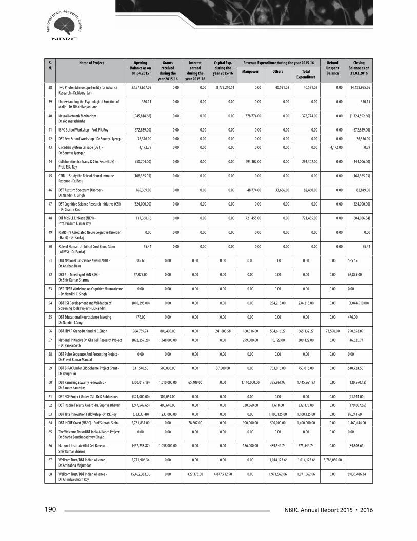
190 NBRC Annual Report 2015 • 2016
S. N.
Name of Project Opening Balance as on
01.04.2015
Grants received
during the year 2015-16
Interest earned
during the year 2015-16
Capital Exp. during the
year 2015-16
Revenue Expenditure during the year 2015-16 Refund Unspent Balance
Closing Balance as on
31.03.2016Manpower Others Total Expenditure
38 Two Photon Microscope Facility for Advance Research - Dr. Neeraj Jain
23,272,667.09 0.00 0.00 8,773,210.51 0.00 40,531.02 40,531.02 0.00 14,458,925.56
39 Understanding the Psychological Function of Malin - Dr. Nihar Ranjan Jana
350.11 0.00 0.00 0.00 0.00 0.00 0.00 0.00 350.11
40 Neural Network Mechanism - Dr. Yoganarashimha
(945,818.66) 0.00 0.00 0.00 378,774.00 0.00 378,774.00 0.00 (1,324,592.66)
41 IBRO School Workshop - Prof. P.K. Roy (672,839.00) 0.00 0.00 0.00 0.00 0.00 0.00 0.00 (672,839.00)
42 DST Serc School Workshop - Dr. Soumya Iyengar 36,376.00 0.00 0.00 0.00 0.00 0.00 0.00 0.00 36,376.00
43 Circadian System Linkage (DST) - Dr. Soumya Iyengar
4,172.39 0.00 0.00 0.00 0.00 0.00 0.00 4,172.00 0.39
44 Collaboration for Trans. & Clin. Res. (GLUE) - Prof. P. K. Roy
(50,704.00) 0.00 0.00 0.00 293,302.00 0.00 293,302.00 0.00 (344,006.00)
45 CSIR -II Study the Role of Neural Immune Respnce - Dr. Basu
(168,365.93) 0.00 0.00 0.00 0.00 0.00 0.00 0.00 (168,365.93)
46 DST Austism Spectrum Disorder - Dr. Nandini C. Singh
165,309.00 0.00 0.00 0.00 48,774.00 33,686.00 82,460.00 0.00 82,849.00
47 DST Cognitive Science Research Initiative (CSI) - Dr. Chaitra Rao
(324,000.00) 0.00 0.00 0.00 0.00 0.00 0.00 0.00 (324,000.00)
48 DIT McGILL Linkage (NKN) - Prof. Prasum Kumar Roy
117,368.16 0.00 0.00 0.00 721,455.00 0.00 721,455.00 0.00 (604,086.84)
49 ICMR HIV Associated Neuro Cognitive Disorder (Hand) - Dr. Pankaj
0.00 0.00 0.00 0.00 0.00 0.00 0.00 0.00 0.00
50 Role of Human Umbilical Cord Blood Stem (AIIMS) - Dr. Pankaj
55.44 0.00 0.00 0.00 0.00 0.00 0.00 0.00 55.44
51 DBT National Bioscience Award 2010 - Dr. Anirban Basu
585.65 0.00 0.00 0.00 0.00 0.00 0.00 0.00 585.65
52 DBT 5th Meeting of EGN-CDB - Dr. Shiv Kumar Sharma
67,875.00 0.00 0.00 0.00 0.00 0.00 0.00 0.00 67,875.00
53 DST ITPAR Workshop on Cognitive Neuroscience - Dr. Nandini C. Singh
0.00 0.00 0.00 0.00 0.00 0.00 0.00 0.00 0.00
54 DBT CSI Development and Validation of Screening Tools Project- Dr. Nandini
(810,295.00) 0.00 0.00 0.00 0.00 234,215.00 234,215.00 0.00 (1,044,510.00)
55 DBT Educational Neuroscience Meeting Dr. Nandini C Singh
476.00 0.00 0.00 0.00 0.00 0.00 0.00 0.00 476.00
56 DBT ITPAR Grant-Dr.Nandini C.Singh 964,759.74 806,400.00 0.00 241,883.58 160,516.00 504,616.27 665,132.27 73,590.00 790,553.89
57 National Initiative On Glia Cell Research Project - Dr. Pankaj Seth
(892,257.29) 1,348,000.00 0.00 0.00 299,000.00 10,122.00 309,122.00 0.00 146,620.71
58 DBT Pulse Sequence And Processing Project - Dr. Pravat Kumar Mandal
0.00 0.00 0.00 0.00 0.00 0.00 0.00 0.00 0.00
59 DBT BIRAC Under CRS Scheme Project Grant - Dr. Ranjit Giri
831,540.50 500,000.00 0.00 37,800.00 0.00 753,016.00 753,016.00 0.00 540,724.50
60 DBT Ramalingaswamy Fellowship - Dr. Saurav Banerjee
(350,017.19) 1,610,000.00 65,409.00 0.00 1,110,000.00 335,961.93 1,445,961.93 0.00 (120,570.12)
61 DST PDF Project Under CSI - Dr.D Subhashree (324,000.00) 302,059.00 0.00 0.00 0.00 0.00 0.00 0.00 (21,941.00)
62 DST Inspire Faculty Award -Dr. Supriya Bhavani (247,549.65) 400,640.00 0.00 0.00 330,560.00 1,618.00 332,178.00 0.00 (179,087.65)
63 DBT Tata Innovation Fellowship -Dr P.K.Roy (33,633.40) 1,233,000.00 0.00 0.00 0.00 1,100,125.00 1,100,125.00 0.00 99,241.60
64 DBT INCRE Grant (NBRC) - Prof Subrata Sinha 2,781,837.00 0.00 78,607.00 0.00 900,000.00 500,000.00 1,400,000.00 0.00 1,460,444.00
65 The Welcome Trust/DBT India Alliance Project - Dr. Sharba Bandhopadhyay Dhyag
0.00 0.00 0.00 0.00 0.00 0.00 0.00 0.00 0.00
66 National Institute Glial Cell Research - Shiv Kumar Sharma
(467,258.87) 1,058,000.00 0.00 0.00 186,000.00 489,544.74 675,544.74 0.00 (84,803.61)
67 Wellcom Trust/DBT Indian Alliance - Dr. Amitabha Majumdar
2,771,906.34 0.00 0.00 0.00 0.00 -1,014,123.66 -1,014,123.66 3,786,030.00 -
68 Wellcom Trust/DBT Indian Alliance - Dr. Anindya Ghosh Roy
15,462,383.30 0.00 422,378.00 4,877,712.90 0.00 1,971,562.06 1,971,562.06 0.00 9,035,486.34
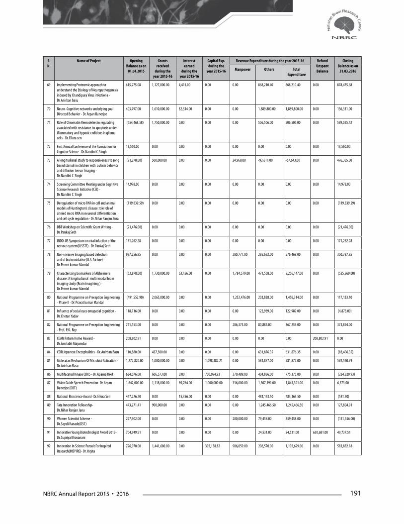
NBRC Annual Report 2015 • 2016 191
S. N.
Name of Project Opening Balance as on
01.04.2015
Grants received
during the year 2015-16
Interest earned
during the year 2015-16
Capital Exp. during the
year 2015-16
Revenue Expenditure during the year 2015-16 Refund Unspent Balance
Closing Balance as on
31.03.2016Manpower Others Total Expenditure
69 Implementing Proteomic approach to understand the Etiology of Neuropathogenesis induced by Chandipura Virus infectiona - Dr. Anirban basu
615,275.08 1,127,000.00 4,411.00 0.00 0.00 868,210.40 868,210.40 0.00 878,475.68
70 Neuro -Cognitive networks underlying goal Directed Behavior - Dr. Arpan Banerjee
403,797.00 1,610,000.00 32,334.00 0.00 0.00 1,889,800.00 1,889,800.00 0.00 156,331.00
71 Role of Chromatin Remodelers in regulating associated with resistance to apoptosis under iflammatory and hypoxic cnditions in glioma cells - Dr. Ellora sen
(654,468.58) 1,750,000.00 0.00 0.00 0.00 506,506.00 506,506.00 0.00 589,025.42
72 First Annual Conference of the Association for Cogntive Science - Dr. Nandini C. Singh
13,560.00 0.00 0.00 0.00 0.00 0.00 0.00 0.00 13,560.00
73 A longitudianal study to responsiveness to song based stimuli in children with autism behavior and diffusion tensor Imaging - Dr. Nandini C. Singh
(91,278.00) 500,000.00 0.00 0.00 24,968.00 -92,611.00 -67,643.00 0.00 476,365.00
74 Screening Committee Meeting under Cognitive Science Research Initiative (CSI) - Dr. Nandini C. Singh
14,978.00 0.00 0.00 0.00 0.00 0.00 0.00 0.00 14,978.00
75 Deregulation of micro RNA in cell and animal models of Huntington’s disease: role role of altered micro RNA in neuronal differentiation and cell cycle regulation - Dr. Nihar Ranjan Jana
(119,839.59) 0.00 0.00 0.00 0.00 0.00 0.00 0.00 (119,839.59)
76 DBT Workshop on Scientific Grant Writing - Dr. Pankaj Seth
(21,476.00) 0.00 0.00 0.00 0.00 0.00 0.00 0.00 (21,476.00)
77 INDO-US Symposium on viral infaction of the nervous system(IUSSTF) - Dr. Pankaj Seth
171,262.28 0.00 0.00 0.00 0.00 0.00 0.00 0.00 171,262.28
78 Non-invasive Imaging based detection and of brain oxidative (U.S. Airfore) - Dr. Pravat kumar Mandal
927,256.85 0.00 0.00 0.00 280,777.00 295,692.00 576,469.00 0.00 350,787.85
79 Characterizing biomarkers of Alzheimer’s disease :A longitudianal multii modal brain imaging study (Brain imagining ) - Dr. Pravat kumar Mandal
(62,878.00) 1,730,000.00 63,156.00 0.00 1,784,579.00 471,568.00 2,256,147.00 0.00 (525,869.00)
80 National Programme on Preception Engineering - Phase II - Dr. Pravat kumar Mandal
(491,552.90) 2,065,000.00 0.00 0.00 1,252,476.00 203,838.00 1,456,314.00 0.00 117,133.10
81 Influence of social cues omapatial cognition - Dr. Chetan Yadav
118,116.00 0.00 0.00 0.00 0.00 122,989.00 122,989.00 0.00 (4,873.00)
82 National Programme on Preception Engineering - Prof. P. K. Roy
741,153.00 0.00 0.00 0.00 286,375.00 80,884.00 367,259.00 0.00 373,894.00
83 CEAN Return Home Reward - Dr. Amitabh Majumdar
208,802.91 0.00 0.00 0.00 0.00 0.00 0.00 208,802.91 0.00
84 CSIR Japanese Enccephalities - Dr. Anirban Basu 110,880.00 437,500.00 0.00 0.00 0.00 631,876.35 631,876.35 0.00 (83,496.35)
85 Molecular Mechanism Of Microbial Activation - Dr. Anirban Basu
1,272,820.00 1,000,000.00 0.00 1,098,382.21 0.00 581,877.00 581,877.00 0.00 592,560.79
86 Multifaceted Kinase CDK5 - Dr. Aparna Dixit 634,076.00 606,573.00 0.00 700,094.93 370,489.00 404,886.00 775,375.00 0.00 (234,820.93)
87 Vision Guide Speech Percention- Dr. Arpan Banerjee (DBT)
1,642,000.00 1,118,000.00 89,764.00 1,000,000.00 336,000.00 1,507,391.00 1,843,391.00 0.00 6,373.00
88 National Bioscience Award- Dr. Ellora Sen 467,226.20 0.00 15,356.00 0.00 0.00 483,163.50 483,163.50 0.00 (581.30)
89 Tata Innovation Fellowship- Dr. Nihar Ranjan Jana
473,271.41 900,000.00 0.00 0.00 0.00 1,245,466.50 1,245,466.50 0.00 127,804.91
90 Women Scientist Scheme - Dr. Sayali Ranade(DST)
227,902.00 0.00 0.00 0.00 280,000.00 79,458.00 359,458.00 0.00 (131,556.00)
91 Innovative Young Biotechnolgist Award 2013- Dr. Supriya Bhavanani
704,949.51 0.00 0.00 0.00 0.00 24,531.00 24,531.00 630,681.00 49,737.51
92 Innovation In Science Pursuit For Inspired Research(INSPIRE)- Dr. Yogita
726,970.00 1,441,680.00 0.00 392,138.82 986,059.00 206,570.00 1,192,629.00 0.00 583,882.18

192 NBRC Annual Report 2015 • 2016
S. N.
Name of Project Opening Balance as on
01.04.2015
Grants received
during the year 2015-16
Interest earned
during the year 2015-16
Capital Exp. during the
year 2015-16
Revenue Expenditure during the year 2015-16 Refund Unspent Balance
Closing Balance as on
31.03.2016Manpower Others Total Expenditure
93 Mechanisms Of Adult Brain Reorganisation- Dr. Neeraj Jain
3,779,421.00 0.00 209,982.00 2,332,354.40 885,365.00 533,279.21 1,418,644.21 0.00 238,404.39
94 Inspired Faculty Award- Dr. Deepashri 0.00 1,900,000.00 0.00 0.00 0.00 0.00 0.00 0.00 1,900,000.00
95 A critical assessment of the dual stream models of visual information processing- DST - Dr. Dipanjay ray
0.00 928,000.00 21,747.00 0.00 0.00 746,548.00 746,548.00 0.00 203,199.00
96 Tata innovation fellowship award- Dr. Anirban basu
0.00 900,000.00 16,261.00 0.00 0.00 911,929.19 911,929.19 0.00 4,331.81
97 Implications in tumor progression- Dr. Ellora sen 0.00 5,474,000.00 193,952.00 485,765.00 195,000.00 891,970.24 1,086,970.24 0.00 4,095,216.76
98 Workshop DST Csi- Dr. Nandini 0.00 500,000.00 0.00 0.00 0.00 497,674.00 497,674.00 0.00 2,326.00
99 Tata innovation fellowship Award - Dr. Pravat Mandal
0.00 900,000.00 15,109.00 0.00 0.00 874,811.00 874,811.00 0.00 40,298.00
100 Crspri System - Dr. Sourav Banarjee 0.00 4,473,800.00 0.00 0.00 0.00 0.00 0.00 0.00 4,473,800.00
101 DBT Mirna Meditate Control - Dr. Sourav Banarjee
0.00 4,303,200.00 0.00 0.00 0.00 871,174.00 871,174.00 0.00 3,432,026.00
102 Ibro Workshop 2016 - Dr. Sourav Banarjee 0.00 3,183,406.60 0.00 0.00 0.00 1,022,958.70 1,022,958.70 0.00 2,160,447.90
103 Grant DBT 30 Years Commemorate 0.00 400,000.00 0.00 0.00 0.00 368,493.00 368,493.00 0.00 31,507.00
Total (A) 102,502,311.60 67,998,258.60 1,533,076.00 50,038,966.61 20,961,255.00 30,820,538.45 51,781,793.45 4,703,275.91 65,509,610.23
36 DELCON E-LIBRARY CONSORTIUM (B)* 24,197,834.13 405,673,921.88 700,086.00 0.00 1,817,009.00 424,010,794.38 425,827,803.38 0.00 4,744,038.63
Grand Total (A+B) 126,700,145.73 473,672,180.48 2,233,162.00 50,038,966.61 22,778,264.00 454,831,332.83 477,609,596.83 4,703,275.91 70,253,648.86
SANTOSH KUMAR CHOUDHARY SUMAN KUMAR PROF. SUBRATA SINHA As per our separate report DY. FINANCE OFFICER OFFG.F & AO DIRECTOR of even date attached
For N.C. MITTAL & CO. Chartered Accountants (FRN-000237N)
KAPIL MITTAL PARTNERDate: October 28, 2016 Membership No. 503378New Delhi