synthesis of carbon nanomaterials on - UM Students' Repository
Upload
khangminh22Category
view
1download
0
Citation: Waris, A.; Ali, A.; Khan,
A.U.; Asim, M.; Zamel, D.; Fatima, K.;
Raziq, A.; Khan, M.A.; Akbar, N.;
Baset, A.; et al. Applications of
Various Types of Nanomaterials for
the Treatment of Neurological
Disorders. Nanomaterials 2022, 12,
2140. https://doi.org/10.3390/
nano12132140
Academic Editors: Yi-Xian Qin,
Donghui (Don) Zhu and Nan Zhao
Received: 24 May 2022
Accepted: 19 June 2022
Published: 22 June 2022
Publisher’s Note: MDPI stays neutral
with regard to jurisdictional claims in
published maps and institutional affil-
iations.
Copyright: © 2022 by the authors.
Licensee MDPI, Basel, Switzerland.
This article is an open access article
distributed under the terms and
conditions of the Creative Commons
Attribution (CC BY) license (https://
creativecommons.org/licenses/by/
4.0/).
nanomaterials
Review
Applications of Various Types of Nanomaterials for theTreatment of Neurological DisordersAbdul Waris 1,† , Asmat Ali 2,†, Atta Ullah Khan 3,4,5 , Muhammad Asim 1, Doaa Zamel 4,5, Kinza Fatima 3,4,5,Abdur Raziq 6, Muhammad Ajmal Khan 7 , Nazia Akbar 2 , Abdul Baset 8
and Mohammed A. S. Abourehab 9,10,*
1 Department of Biomedical Sciences, City University of Hong Kong, 83 Tat Chee Avenue, Kowloon,Hong Kong SAR, China; [email protected] (A.W.); [email protected] (M.A.)
2 Department of Biotechnology and Genetic Engineering, Hazara University Mansehra,Mansehra 21300, Pakistan; [email protected] (A.A.); [email protected] (N.A.)
3 CAS Key Laboratory of Standardization and Measurement for Nanotechnology, CAS Center for Excellencein Nanoscience, National Center for Nanoscience and Technology, No. 11 Zhongguancun Beiyitiao,Beijing 100190, China; [email protected] (A.U.K.); [email protected] (K.F.)
4 Department of Environmental Engineering, Institute of Urban Environment, CAS, Xiamen 361021, China;[email protected]
5 Department of Physical Chemistry, University of Chinese Academy of Sciences, Beijing 100049, China6 Department of Physics, Bacha Khan University Charsadda, Charsadda 24420, Pakistan;
[email protected] Divison of Life Sciences, Center for Cancer Research and State Key Laboratory of Molecular Neurosciences,
The Hong Kong University of Science and Technology, Clear Water Bay, Hong Kong SAR, China;[email protected]
8 Department of Zoology, Bacha Khan University Charsadda, Charsadda 24420, Pakistan;[email protected]
9 Department of Pharmaceutics, Faculty of Pharmacy, Umm Al-Qura University, Makkah 21955, Saudi Arabia10 Department of Pharmaceutics and Industrial Pharmacy, Faculty of Pharmacy, Minia University,
Minia 61519, Egypt* Correspondence: [email protected]† These authors contributed equally to this work.
Abstract: Neurological disorders (NDs) are recognized as one of the major health concerns globally.According to the World Health Organization (WHO), neurological disorders are one of the maincauses of mortality worldwide. Neurological disorders include Alzheimer’s disease, Parkinson’sdisease, Huntington’s disease, Amyotrophic lateral sclerosis, Frontotemporal dementia, Prion disease,Brain tumor, Spinal cord injury, and Stroke. These diseases are considered incurable diseases becauseno specific therapies are available to cross the blood-brain barrier (BBB) and reach the brain in asignificant amount for the pharmacological effect in the brain. There is a need for the developmentof strategies that can improve the efficacy of drugs and circumvent BBB. One of the promisingapproaches is the use of different types of nano-scale materials. These nano-based drugs have theability to increase the therapeutic effect, reduce toxicity, exhibit good stability, targeted delivery, anddrug loading capacity. Different types and shapes of nanomaterials have been widely used for thetreatment of neurological disorders, including quantum dots, dendrimers, metallic nanoparticles,polymeric nanoparticles, carbon nanotubes, liposomes, and micelles. These nanoparticles haveunique characteristics, including sensitivity, selectivity, and the ability to cross the BBB when used innano-sized particles, and are widely used for imaging studies and treatment of NDs. In this review,we briefly summarized the recent literature on the use of various nanomaterials and their mechanismof action for the treatment of various types of neurological disorders.
Keywords: nanotechnology; nanomaterials; neurological disorders; blood-brain barrier
Nanomaterials 2022, 12, 2140. https://doi.org/10.3390/nano12132140 https://www.mdpi.com/journal/nanomaterials

Nanomaterials 2022, 12, 2140 2 of 29
1. Introduction
Neurodegenerative disorders (ND) are the most devastating and challenging disor-ders of the central nervous system (CNS) and are recognized as a major threat to publichealth [1]. ND refers to the loss of structure or functions of the neuron. There are dif-ferent types of ND, including stroke, Alzheimer’s, Parkinson’s, Huntington’s, and priondiseases. The pathophysiology of each disease is different. Some NDs lead to memoryand cognitive impairment, while some affect the ability of the person to speak, move, andbreathe [2]. According to the World Health Organization report, in 2019, in the list of top10 leading causes of diseases globally, stroke is the second leading cause of death, followedby Alzheimer’s disease and other dementias [3]. In developing countries, neurologicaldisease cases increase each year with the progressive rise in life expectancy. The causeof most neurological diseases is well known, and many studies have been done on itstreatment. The central nervous system is a vulnerable and complex system that createscomplications in diagnosing and treating neurological diseases [4,5]. Some barriers createcomplications for therapeutic intervention, and the functions of these barriers are to regu-late the molecular exchange between blood and the brain. The barriers are made up of glialcells and endothelial cells in the brain. The blood-brain barrier (BBB) is the main gatewayof the system as it administers the drugs’ access to the brain [6].
NDs are considered incurable diseases, and the prevalence of these diseases is rapidlyrising. The majorities of these diseases have no specific effective therapies because of theinability of drugs to cross the BBB to reach the brain and being available in an amount that ishigh enough for a pharmacological effect in the brain. The BBB is one of the challenges—butnot the only challenge—for treating neurological disorders. Approximately 95% of drugscannot cross the BBB because of their molecular property. The central nervous system isrestricted by BBB and blood-cerebrospinal fluid. Due to the restrictive nature of the BBB,lipophilic drugs with molecular weight of <500 Da can cross, while traditional drugs cannotmeet this requirement [7,8]. The BBB is highly semipermeable, which leads to limiting theentry of therapeutic drugs to enter the CNS, and this property of the BBB is considered oneof the main challenges in the development of modern medicine. Photothermal and photo-dynamic therapies are considered alternative approaches and are used to treat various NDsdue to their ability to bypass the BBB. However, these therapies have side effects, includingdamage to tissues (especially skin tissues) and photosensitization [9,10]. Therefore, thereis an urgent need for the development of potential therapeutic agents that can cross theBBB with no side effects to combat these diseases. However, a deep understanding of themechanism and causes are necessary for the effective treatment of each disease [2,11].
There is a need for the development of therapeutic strategies that can overcomethe BBB and also can improve its efficacy. Researchers are continuously working on thedevelopment of delivery strategies of therapeutics to solve this problem [12,13]. Differentstrategies that can overcome this problem have been developed. Among these approaches,the nano-based approach is at the core of these advances in the delivery of therapeutics [14].Nanotechnology uses different types of engineered nanomaterial and nanoparticles witha size of 1–100 nm in at least one dimension [12,15]. Nanotechnology and nanomaterialsopen new avenues in biomedical science, as multiple nanoparticles have been appliedin brain studies and research, including quantum dots (QDs), polymeric nanoparticles,micelles, metallic nanoparticles, etc. [16]. These nano-scale materials possess uniquecharacteristics such as small size due to which they can interact with the biological systemat the molecular level, a high surface to volume ratio that can be mono or multifariouswith surface modification, and good stability. On the other hand, the application ofchemotherapy and drugs may lead to side effects such as anemia, alopecia, gastric irritation,neurotoxicity, and loss of appetite. Encapsulation of drugs in metallic nanoparticles, forexample, silver, gold, and metal oxides (magnetic), aids in overcoming the complications ofmedications [17]. In the past decade, different types and shapes of nanomaterials have beenwidely used to treat different types of NDs [18,19]. Researchers have achieved remarkableprogress in the field of nanotechnology in nanomedicine and biomedical sciences, especially

Nanomaterials 2022, 12, 2140 3 of 29
in neurosciences. In this contribution, we briefly discussed the types of nanomaterials andthe advancement of nanotechnology in the field of neurosciences.
2. Types of Nanomaterials Used for the Treatment of Neurological Disorders2.1. Quantum Dots
QDs are unique nanoparticles that have a wide range of applications as a treatingagent for various neurodegenerative diseases. QDs have unique characteristics such assensitivity and selectivity when used in nano-sized particles. Several research articles havebeen published studying the QDs applications in diagnosing and treating Alzheimer’sdisease (AD). It has been proved that QDs have the ability to cross the BBB, whereaspotential toxicity could be further evaluated [16].
Furthermore, QDs have unique optical properties, enriching their widespread usein biomedical applications. However, great concern towards QDs has been taken in thelast years due to their potential toxicity. Herein, neurotoxicity increases in the case of thedistribution of QDs mass production in the nervous system. Due to the very small size ofQDs particles, they can cross the BBB or move with the blood circulation entering the brain.Nevertheless, the interactions between QDs and the nervous system cells and tissues arenot clear yet; the obvious is the neurotoxicity, which includes oxidative stress, increasedCa2+ levels in the cytoplasm, autophagy—which damages in vitro neural cells, impairmentof synaptic transmission, and loss of plasticity, as demonstrated in tested animals [20]. QDsare basically nanocrystals in nano-sized structures that can emit light, which gives themunique optical properties. QDs have fluorescent light of several wavelengths, which givesthem brightness and resistance to bleaching. Therefore, QDs can visualize brain structuresand mechanisms of their functions as well as be applied for drug delivery purposes [21].In the past few years, in vivo imaging of biological functions has been performed by near-infrared (NIR) fluorescence imaging techniques. NIR (700–900 nm) could be demonstratedfor sensitive and accurate detection techniques. Then there are the QDs semiconductors,which could be utilized in detection due to their electronic, magnetic, optical, and structuralcharacteristics, which are different from bulk materials. QDs could be a contrast agentfor optical imaging, especially in deep tissue imaging [22]. Moreover, though emergingevidence indicates that the pathogenesis of Parkinson’s disease is strongly correlated tothe accumulation and transmission of α-synuclein (α-syn) aggregates in the midbrain, noanti-aggregation agents have been successful at treating the disease in the clinic. Graphenequantum dots (GQDs) inhibit the fibrillization of α-syn and interact directly with maturefibrils, triggering their dis-aggregation. Moreover, GQDs can rescue neuronal death andsynaptic loss, reduce Lewy body and Lewy neurite formation, ameliorate mitochondrialdysfunctions, and prevent neuron-to-neuron transmission of α-syn pathology provokedby α-syn preformed fibrils. We observe, in vivo, that GQDs penetrate the BBB and protectagainst dopamine neuron loss induced by α-syn preformed fibrils, Lewy body/Lewyneurite pathology, and behavioral deficits [23]. Further studies worked on GQDs as label-ing agents for stem cells, which develop little cytotoxicity. They were studied on humanneural stem cells to determine their uptake and biocompatibility. The results came up withno significant change in the viability, metabolic activity, proliferation, and differentiationpotential of human neural stem cells after the treatment with GQDs. Furthermore, GQDshave been taken up into the neural stem cells via endocytosis [24]. GQDs could be consid-ered promising treatment agents for NPC and other related diseases [21]. Compared tolarge graphene oxide nanosheets, graphene oxide quantum dots (GOQDs), as nanozymes,could be proved to efficiently decrease the reactive oxygen species (ROS) and H2O2 in1-methyl-4-phenyl-pyridinium ion (MPP+)-induced PC12 cells. Furthermore, GOQDs havethe ability to reduce apoptosis and α-synuclein and mitochondrial damage in zebrafishtreated with MPP+. To sum up, biocompatible GOQDs have a high potential for humanhealth by diminishing oxidative stress signals and reducing neurotoxicity [25].
Silicon nanocrystals provide excellent imaging capabilities for toxic heavy-metal-basedQDs. However, understanding the toxicity developed by silicon quantum dots (SiQDs) is
Nanomaterials 2022, 12, 2140 4 of 29
mandatory for estimating its potential. There are only a limited number of studies on thebiocompatibility of SiQDs and no obvious systematic progression from clinical studies onsmall-animal to large-animal. The nano-construct of SiQDs and FDA-approved materialsare applied intravenously in mice and monkeys, and the results demonstrated no toxicityin both mice and monkeys in their behavior, blood, and body mass at a dose of 200 mg/kg.The drug formula did not biodegrade as well, as high levels of silicon were accumulatedin the liver and spleen of mice after three months of the treatment and nothing showed inmonkeys [23].
Another study targeted carbon dots (CDs) and showed their promising effects forvarious biomedical applications such as bio-imaging, treatment of brain tumors, and neu-rodegenerative diseases. CDs showed unique properties like biocompatibility and smallsized-particles less than 10 nm, enabling them to enter the BBB. They have different opticalproperties and stability toward the light, which lets them acquire ideal characteristics forapplications in several scientific fields [26]. The BBB is the physiological checkpoint thatrestricts the passage of molecules present in the blood into the central nervous system.One of the scientists’ serious challenges is delivering drugs and active materials acrossthe BBB. Therefore, developing novel materials and methodologies to address this chal-lenge is vital for diagnosing and treating brain diseases. In a study, bio-conjugated andfunctionalized QDs were revealed to be outstanding nano-vectors and fluorescent probesfor the transmission across the BBB for treating brain tumors and diseases [27]. Previousstudies on photoluminescent 9-nm diameter QDs with a CdSe core, a ZnS shell, and anegatively charged compact molecular ligand coating (CL4) selectively target neuronsinstead of glia. On the other side, current research studies focus on the explanation forthe selective delivery in neurons. Three zwitterionic QD coatings differ in the regions ofpositive or negative charges, as well as a positively charged (NH2) polyethylene glycol(PEG) coat, which enriches them with the ability to deliver the cell-membrane-penetratingchaperone lipopeptide JB577 (WG(Palmitoyl) VKIKKP9G2H6) to the individual cells inneonatal rat hippocampal slices. The results confirmed the preferential uptake in neuronsand vice-versa in glia, which showed no uptake, and can be explained due to the nega-tively charged regions on the coating of QDs. Moreover, the treatment was not effectivein the astrocytes and microglia cells, and researchers suggested the administration of ahistidine-tagged green fluorescent protein (eGFP-His6) to hippocampal slices to enhancethe neuronal uptake [28]. In another in vivo study using graphene-Quantum dots (GQDs)in NPC, GQDs were revealed, which can possess little long-term toxicity and which can beneglected as they have the penetration capability of the BBB. The treatment using GQDsdecreases the cholesterol aggregation in the lysosome via expressed interactions [29].
Further studies performed on the selenium-doped carbon quantum dots (Se-CQDs)showed their ability to diminish reactive oxygen species and that they have been appliedto efficiently ameliorate secondary injury in TSCI. The results demonstrated the good bio-compatibility and remarkable protective effect of Se-CQDs against H2O2-induced oxidativedamage in astrocytes and PC12 cells [30]. The schematic process of the drug deliverysystem is shown in Figure 1 for the treatment of neurological disorders.
2.2. Metallic Nanoparticles
Metallic nanoparticles can be fabricated, modifying their shape and size, and can linkto various types of chemical functional groups [31]. These modifications and linkings allowthem to attach and bind with various ligands, including drugs, antibodies, peptides, andpolymers. Different types of metallic nanoparticles are used as a carrier for brain-targetedtherapy. These nanoparticles include FeONPs, AgNPs, Gadolinium metallofullerene NPs,Ultrasmall gadolinium oxide NPs, Ce2O3 NPs, ZnONPs, AuNPs, and PtNPs. Metallicnanoparticles are also used for the imaging of CNS [9,32].

Nanomaterials 2022, 12, 2140 5 of 29Nanomaterials 2022, 12, x FOR PEER REVIEW 5 of 30
Figure 1. Schematic illustration of drug delivery system for the treatment of neurological disorders.
2.2. Metallic Nanoparticles Metallic nanoparticles can be fabricated, modifying their shape and size, and can link
to various types of chemical functional groups [31]. These modifications and linkings al-low them to attach and bind with various ligands, including drugs, antibodies, peptides, and polymers. Different types of metallic nanoparticles are used as a carrier for brain-targeted therapy. These nanoparticles include FeONPs, AgNPs, Gadolinium metallofull-erene NPs, Ultrasmall gadolinium oxide NPs, Ce2O3 NPs, ZnONPs, AuNPs, and PtNPs. Metallic nanoparticles are also used for the imaging of CNS [9,32].
Metallic nanoparticles have shown their outstanding role in biomedical applications [13,33–35]. Currently, these materials can be manufactured and functionalized with groups that facilitate their conjugation with antibodies, drugs, and ligands—these modi-fications and functionalization open new avenues of potential applications in magnetic separation, biotechnology, and drug delivery. Furthermore, various imaging models have been promoted over the years, such as CT, MRI, PET, ultrasound, optical imaging, and SERS, which are good tools for several disease images [36]. Nanomaterials have been used widely in recent years for the diagnosis, imaging, and treatment of various disorders. However, nanoparticles possess potential hazards and neurotoxicity to the CNS, such as autophagy, oxidative stress, and lysosomal dysfunction via possible mechanisms [37].
Moreover, nano-delivery methods can effectively cross the BBB and reach remote re-gions in the brain. However, a deep understanding of developing the long-term toxicities is the primary question of scientists, which needs to be addressed [38]. Research articles on biogenic metal or metal oxide nanoparticles are limited, especially those for treating Alzheimer’s disorder. In addition, their functionalization is suggested to develop their therapeutic potential for treating neurodegenerative diseases [39]. Furthermore, metal na-noparticles are excellent carriers and therapeutic materials that aid in biomedical applica-tions due to their unique characteristics. They have physicochemical properties, which enrich their possibility to be used in many fields in science.
Moreover, metal nanoparticles can be manufactured while modulating their size and morphology as well as functionalization with other ligands, which widen their applica-tions and increase their efficiencies [32,40]. Additionally, biodegradable biomaterials that
Figure 1. Schematic illustration of drug delivery system for the treatment of neurological disorders.
Metallic nanoparticles have shown their outstanding role in biomedical applica-tions [13,33–35]. Currently, these materials can be manufactured and functionalized withgroups that facilitate their conjugation with antibodies, drugs, and ligands—these mod-ifications and functionalization open new avenues of potential applications in magneticseparation, biotechnology, and drug delivery. Furthermore, various imaging models havebeen promoted over the years, such as CT, MRI, PET, ultrasound, optical imaging, andSERS, which are good tools for several disease images [36]. Nanomaterials have beenused widely in recent years for the diagnosis, imaging, and treatment of various disorders.However, nanoparticles possess potential hazards and neurotoxicity to the CNS, such asautophagy, oxidative stress, and lysosomal dysfunction via possible mechanisms [37].
Moreover, nano-delivery methods can effectively cross the BBB and reach remoteregions in the brain. However, a deep understanding of developing the long-term toxicitiesis the primary question of scientists, which needs to be addressed [38]. Research articleson biogenic metal or metal oxide nanoparticles are limited, especially those for treatingAlzheimer’s disorder. In addition, their functionalization is suggested to develop theirtherapeutic potential for treating neurodegenerative diseases [39]. Furthermore, metalnanoparticles are excellent carriers and therapeutic materials that aid in biomedical appli-cations due to their unique characteristics. They have physicochemical properties, whichenrich their possibility to be used in many fields in science.
Moreover, metal nanoparticles can be manufactured while modulating their size andmorphology as well as functionalization with other ligands, which widen their applicationsand increase their efficiencies [32,40]. Additionally, biodegradable biomaterials that havebeen functionalized offer promising ways to address that question for solutions due to theirunique properties, such as the ability to respond to external stimulation. These uniqueproperties enriched their applications in neuro-sensing, neuro-imaging, specific targeting,drug delivery, and treatment of hyperthermia [41]. Though nanoparticles possess goodphysical and chemical properties, which demonstrate a wide range of applications for CNS,they may develop neurotoxicity owing to cell necrosis, immune response, and free radicalformation [42].
2.3. Dendrimers
Optical dendrimers provide the combined advantages of particle-defined structureand the functionalized surface, which makes them outstanding in the application of neu-roscience. It is important to mention that the most recent discovery based on dendrimers
Nanomaterials 2022, 12, 2140 6 of 29
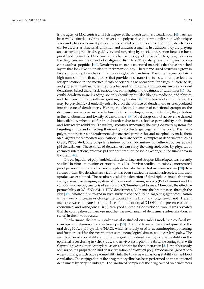
is the agent of MRI contrast, which improves the bloodstream’s visualization [43]. As hasbeen well defined, dendrimers are versatile polymeric compartmentalization with uniquesizes and physicochemical properties and resemble biomolecules. Therefore, dendrimerscan be used as antibacterial, antiviral, and anticancer agents. In addition, they are playingan outstanding role in drug delivery and targeting by special interaction between host–guest binding motifs. Dendrimers may be used as glycol carriers for targeting tissues inthe diagnosis and treatment of malignant disorders. They also present antigens for vac-cines, such as peptides [44]. Dendrimers are nanostructured materials that have branchedlayers that look like onion skin in their morphology. These nano-sized structures grow inlayers producing branches similar to as in globular proteins. The outer layers contain ahigh number of functional groups that provide these nanostructures with unique featuresfor applications in the medical fields of science as nanocarriers for drugs, nucleic acids,and proteins. Furthermore, they can be used in imaging applications such as a noveldendrimer-based theranostic nanodevice for imaging and treatment of carcinoma [45]. Re-cently, dendrimers are invading not only chemistry but also biology, medicine, and physics,and their fascinating results are growing day by day [46]. The bioagents or biomoleculesmay be physically/chemically adsorbed on the surface of dendrimers or encapsulatedinto the core of dendrimers. Herein, the elevated number of functional groups on thedendrimer surfaces aid in the attachment of the targeting groups, and further, they interferein the functionality and toxicity of dendrimers [47]. Most drugs cannot achieve the desiredbioavailability when used for brain disorders due to the selective permeability in the brainand low water solubility. Therefore, scientists innovated the drug delivery systems fortargeting drugs and directing their entry into the target organs in the body. The nano-polymeric structures of dendrimers with ordered particle size and morphology make themideal agents for biomedical applications. There are several examples of dendrimers such asGlyco, PEGylated, poly(propylene imine), poly(amidoamine), polyether-copolyester, andpH dendrimers. These kinds of dendrimers can carry the drug molecules by physical orchemical interactions, whereas pH dendrimers alter the ionic exchange in the tumor area inthe brain [48].
Bio conjugation of poly(amido)amine dendrimer and streptavidin adapter was recentlystudied in vitro on murine or porcine models. In vivo studies on mice demonstratedgood permeation of dendronized streptavidin into the central nervous system [30]. In afurther study, the dendrimers viability has been studied in human astrocytes, and theiruptake was explained. The results revealed the detection of dendriplexes inside the brainusing a sensitive imaging system of fluorescent imaging in vivo (IVIS Lumina) and byconfocal microscopy analysis of sections of OCT-embedded tissues. Moreover, the effectivepermeability of 2G-(SNMe3I)11-FITC dendrimer siRNA into the brain passes through theBBB [49]. Another in vitro and in vivo study tested the effect of targeting agent conjugationif they would increase or change the uptake by the brain and organs—or not. Herein,mannose was conjugated to the surface of multifunctional D4-OH in the presence of atom-economical and orthogonal Cu (I)-catalyzed alkyne–azide cycloaddition. It was revealedthat the conjugation of mannose modifies the mechanism of dendrimers internalization, asstated in the in vitro results.
Furthermore, the brain uptake was also studied on a rabbit model via confocal mi-croscopy and fluorescence spectroscopy [50]. A study targeted the development of theoral drug N-Acetyl-l-cysteine (NAC), which is widely used in acetaminophen poisoningand further used for the treatment of some neurological diseases like cerebral palsy. Theresults showed its stability for 6 h in the gastrointestinal tract, good permeability in theepithelial layer during in vitro study, and in vivo absorption in rats while conjugation withCapmul (glycerol monocaprylate) as an enhancer for the penetration [51]. Another studyfocuses on the preparation and characterization of hydroxyl poly(amidoamine) generation-6 dendrimers, which have permeability into the brain as well as long stability in the bloodcirculation. The conjugation of the drug minocycline has been performed on the mentioneddendrimers by enzyme linkages. The produced complex of the drug carried on dendrimers

Nanomaterials 2022, 12, 2140 7 of 29
has been further investigated for antioxidant and anti-inflammatory activities in murinemicroglial cells. The in vivo results revealed a permeation of the prepared drug complexcrossing the BBB [52]. A recent in vivo study used the rabbit model for applications ofdendrimers for neuroinflammation and drug delivery without ligands. Here, neutral den-drimers moved towards the parenchyma and were highly localized in the glial cells inthe brain injury areas. It was found that the uptake of dendrimers is dependent on thebreak-down severity of the BBB [53]. Dendrimers have invaded neuroscience applicationsas they have unique structural properties and morphologies, for instance; they are globular,highly branched, well-defined, have low polydispersity, nanosize-scale structure, and theexistence of variable terminal functional groups that have the ability to conjugate withseveral ligands to be suitable for applications in various biological fields [54]. Additionally,dendrimers can be considered promising nanocarriers for central nervous system drugsowing to their unique features and wide applications [55].
2.4. Carbon Nanotubes
In nanomaterials, carbon nanotubes are a kind of nanomaterial that are gaining inter-est due to their electronic, intrinsic mechanical, and physico-chemical properties [56,57].Carbon nanotubes (CNTs) can diagnose and treat neurological pathologies like Alzheimer’sand Parkinson’s disease. Approximately 24 million people around the globe suffer fromAlzheimer’s and Parkinson’s diseases [58]. CNTs are classified into two nanotubes: first is asingle-walled carbon nanotube (SWCNTs), and the other is a multi-walled carbon nanotube(MWCNTs) [59]. Most of the time, we use carbon nanotubes for drug delivery and bioimag-ing; due to this, we neglect its application as a therapeutic drug [60]. Because of the uniqueproperty and novel structure of carbon nanotubes, they have emerged as a promising optionfor tracking central nervous system diseases and their treatment. Advances in CNTs havemade a significant contribution to therapeutic applications in different neuropathologicaldisorders in vitro and in vivo [1]. CNTs are synthesized using techniques such as chemicalvapor deposition (CVD), laser ablation, and arc discharge. Chemical vapor depositionis the most suitable and preferred method because it shows evidence of properties likeelectrical conductive capacity, strong mechanical property, morphological similarities toneurites, low cost, deposition, and scalability [61]. CNTs in their natural state do notdissolve in an aqueous solution; due to this property, their application in nanomedicineis complex. However, CNTs show their toxicity in their biological environment in clinicaltrials. However, their efficacy and side effects depend upon their exposure, dose, and route.Coating carbon nanotubes can minimize their toxicity to cells with surfactants, reducingtheir connection between cells and CNTs [62]. Metal-catalyzed prepared carbon nanotubesand the application of carbon nanotubes show free radical production, and peroxidativeproduction, DNA damage, and inflammations are obstacles in the pathway of applicationof carbon nanotubes in the diagnosis and treatment of neurological disorders [63].
2.5. Polymeric Nanoparticles
Nanocarriers are fabricated from polymers, lipids, and carbon nanotubes, but polymermaterials have good properties as they are stable, allow for many agents, and for controllingthe drugs’ kinetic energy [64]. Polymers are safe to use in humans. Different kinds ofpolymers are used to synthesize nanoparticles for drug delivery to the central nervoussystem, such as are polysaccharides, proteins, amino acids, and polyesters [65]. Polymericnanoparticles are made up of natural or synthetic polymers where the drug is loadedwith a solid-state or in a solution. Polymeric nanoparticles are a promising method fordrug delivery to the central nervous system. They protect the drug against enzymaticdegradation, and help the active molecule reach its target site [66]. The most commonlyused polymers are polyalkyilcyanoacrylate (PACA), which is used in nanoparticles for drugdelivery to the central nervous system. PACA has been used for the treatment of tumors. APACA nanoparticle is coated with the antitumor drug doxorubicin, which shows excellenttolerance against resistant tumors [67]. Different strategies have been made to cross the

Nanomaterials 2022, 12, 2140 8 of 29
BBB to target nanoparticles to the central nervous system. Magnetic nanoparticles, nanogel, emulsifying wax, and ligand-based approaches are used [68]. Polymeric nanoparticlesare the best alternative way of drug delivery due to their inherent biodegradability andnontoxicity [69]. The advantages of polymeric nanoparticles are their increased stability,delivery of a higher concentration, and because they can be easily incorporated. Lipid-based nanocarriers are recommended due to their natural-based condition, and becausethey are similar to the BBB membrane. Nevertheless, polymeric nanoparticles show ahigh capacity, and their surface is easily modified by targeting molecules; due to this,the polymeric nanostructured based system is the best alternative to drug delivery to thecentral nervous system [70].
2.6. Liposomes
Liposomes consists of lipid bilayers and are in the shape of spherical vesicles. Theadvantage of liposomes is that they are highly biocompatible, have low toxicity, andare without any side effects in the drug delivery system. Liposomes are made up ofphosphatidylcholine and cholesterol [71,72]. For targeting the brain system, GABA, whichcontains liposomes, must consider two challenges: the BBB and microglia reaction. Osmoticshock temporarily opens the BBB to enhance the drug delivery through liposomes. GABA-based liposomes can target neurodegenerative diseases like anxiety, stress, epilepsy, andother physiological disorders like hypertension and heart failure [4,73].
Multiple methods have been proposed for the delivery of drugs to the brain. Thereare two families of transporters; the first is reversible, and the second one is irreversiblenanoparticles. Liposomes and micelles are an example of reversible nanoparticles [74].Theranostics is the new discipline in medicine that is simultaneously performed as atherapeutic and diagnostic function. Theranostic agents are molecules such as liposomesand micelles that can be used as a drug delivery vehicle in a protected and controlledmanner. Using magnetic targeting of cells and tissues makes it more efficient by focusingon the activity of the pathological tissues, reducing unnecessary delivery and an excessiveamount of the drug [4,75]. The theranostic approach is being used to target the brainbecause of the multitasking nature involved in constructing complex nanostructures likedrug delivery vehicles to cross the BBB. A theranostic molecule like heat shock protein[HSP]-72 targeted liposomes has been shown to increase the efficiency of the treatment ofneurological disorders [76].
2.7. Micelles
Displaying a variety of the arrangement along a chain, angle copolymers carry freshblood to the old story of polymeric micelles. The angle chain structure brings about a fewextraordinary highlights in micelles designs and prompts exceptional primary advances, pos-sibly prompting new properties and applications. Hence, slope copolymer micelle structuresand their advances form the perspective of delicate matter physical science [77,78]. Polymericmicelles are nanoscopic center/shell structures framed by amphiphilic block copolymers.The inborn and modifiable properties of polymeric micelles make them especially appro-priate for drug conveyance purposes. The benefits and applications examined incorporatesolubilization of ineffectively solvent atoms, supported delivery and size benefits, and in-surance of embodied substances from debasement and digestion. The three most generallyconcentrated block copolymer classes are described by their hydrophobic squares and arepoly(propylene oxide), poly(L-amino acid)s, and poly(ester)s. These three classes of squarecopolymers are investigated with numerous ebb and flow research instances in which defi-nition procedures with polymeric micelles have been applied to the absolute most testingatoms in the drug business. The polymeric micelles utilized for drug conveyance in thesemodels have shown a capacity to lessen poison levels, upgrade the conveyance to wantednatural destinations, and work on the remedial viability of dynamic drug delivery [79].
For the treatment of neurological diseases, many chemically synthesized drugs areused: cholinesterase inhibitors, Anti-Aβ regimens, and β-site peptide cleaving enzyme-

Nanomaterials 2022, 12, 2140 9 of 29
1 (BACE1) inhibitors. These drugs are not considered as efficient for treatment due totheir side effects [80]. As the brain is protected by barriers such as the BBB and blood-cerebrospinal fluid barrier (BCSFB), the drug molecules cannot move to the target site ofthe brain due to these barriers [81]. Nanocarriers are used for the treatment of centralnervous system disorders. They are fabricated with features that control the drug deliveryto the brain and increase the efficacy [82]. Polymeric micelles, with their unique properties,particle size, and shell structure, work as a drug delivery vehicle to the brain. The nature ofthe micelles is amphiphilic, as it can easily assemble itself in an aqueous medium; due tothis, it protects the drug from the non-target cells [83].
Natural proteins derived from milk are used in the making of nanocarriers, suchas lactoferrin (LF), which is expressed over the brain’s surface, through which the LF isattached to the receptor; due to this, it can easily pass through the BBB [83]. It was preparedby the solvent evaporation technique, and fabricated through covalent conjugation betweenlactoferrin. Micelles were utilized to encapsulate conjugated linoleic acid [84]. In vivorhodamine micelles show better biodistribution in brain tissue, enhancing the active targetcapacity. It is concluded that lactoferrin and conjugated linoleic acid micelles are non-toxic, and they naturally activate the target nano platform, which provides a solution toAlzheimer’s disease [85].
3. Nanomaterials for the Treatment of Neurodegenerative Diseases3.1. Alzheimer’s Disease (AD)
Alzheimer’s disease (AD) is one of the most common neurodegenerative disordersin the elderly, accounting for more than 80% of dementia cases worldwide. It causesprogressive mental, behavioral, and functional impairment as well as loss of learningability [86]. The case of lipoic acid, a chemical naturally present in the mitochondria withsubstantial anti-inflammatory and antioxidant capabilities capable of reducing oxidativestress, exemplifies the potential of NPS to improve the efficacy of AD therapy [87]. They aredesigned in such a way that they are harmless, biodegradable, and target-specific [88]. Inthe context of treating Alzheimer’s disease, these nano systems could effectively retain anddistribute medicines and other neuroprotective chemicals to the brain [89]. The intranasalmethod helps to bypass the BBB, allowing medications to be delivered directly to thebrain. Endocytosis, such as receptor-mediated endocytosis, phagocytosis, and pinocytosis,are the most frequent processes for nanoparticle transportation, with receptor-mediatedendocytosis being the most preferred approach [90].
Curcumin is formed from the rhizome of Curcuma longa, cultivated in South Asiancountries, especially India and China. Curcuminoids are widely used in herbal medicine forthe treatment of various disorders. Recent studies revealed that curcumin plays an essentialrole in the management of AD, mainly acts as an essential diagnostic agent, multi target-directed drug, as well as promotes lifelong nutraceuticals [91]. Several nano-formulationshave been recognized as theranostic agents that can improve the pharmacokinetic prop-erties of curcumin and other bioactive substances, which can help overcome diagnosticand therapeutic limitations. Nanocarriers have proved to be effective in delivering cur-cumin and other nutritional components across the BBB, allowing them to be distributedmore efficiently throughout the brain [92]. Diffusion and erosion or degradation processesconvey the integrated medication to the targeted site. Liposomes, polymeric nanoparti-cles, micro-and nano-emulsions and dendrimers are some of the most commonly utilizednanoparticles [90].
The type of the portion forming the outermost layer determines whether polymericnanoparticles are hydrophilic or hydrophobic. Their delivery to the target region can beaccomplished through receptor-mediated endocytosis or endothelial cell transcytosis. Drugabsorption can be improved by coating PNPs with antibodies or PEG (polyethylene glycol),especially when the drug is delivered intranasally. When coated with polysorbate-80,poly(n-butyl cyanoacrylate) loaded with the drug Rivastigmine exhibited enhanced drugdelivery in the case of AD [93]. Aβ1-42 monoclonal antibody-decorated nanoparticle-

Nanomaterials 2022, 12, 2140 10 of 29
based treatment against AD completely corrects the memory deficit in an experimentalAD animal [92]. Liposomes are biodegradable delivery nanosystems that can encapsulatea wide range of hydrophilic and hydrophobic biopharmaceuticals, including proteins,peptides, RNAs, and small molecules, without changing their characteristics [93]. Thequick penetration of therapeutic biomolecules into the brain is aided by their characteristicphospholipid bilayer architectures, which resemble biological membranes. Liposomesare one of the most studied and clinically recognized nanostructured carriers due to theirlow toxicity, extensive track record, and ability to carry both hydrophilic and lipophiliccompounds [94]. Figure 2 represents nanomaterials mediated drug delivery of therapeuticagents targeting the brains of patients suffering from Alzheimer’s disease. Dendrimers arealso one of the promising NPS in the near future for the treatment of AD because of theirhigh drug loading capacity, which includes both the inner chamber and the dendrimer’souter surface. The size of dendrimers can be easily controlled by carefully selectingmonomers and the degree of polymerization [93]. In comparison to the normal aging brain,the formation of senile plaques in AD is characterized by a higher concentration of solubleand insoluble A-42, more alterations of terminal amino acids of A and Aβ-40-42, and arise in their concentration. The delayed accumulation of Aβ promotes pathogenic peptidechanges such as racemization, isomerization, cyclization, and oligomerization, resulting inAβ-42 insolubility [95].
PAMAM dendrimers were first reported in the 1980s and are also the first family ofdendrimers to be fabricated and commercialized as well as the most studied. PAMAMdendrimers have a diameter of about 1–14 nm. These dendrimers have many applicationsin the field of biomedical sciences, including cargo and drug delivery, gene transfection,imaging agents, and act as potential antimicrobial agents [96]. Dendrimers PAMAM3, 4,and 5 have been demonstrated in studies to suppress the production of amyloid deposits.In comparison to other kinds of aggregated proteins, they have hydrolytic characteristics.There were no forms of Aβ resistance to hydrolysis after incubating cells with dendrimernano-molecules. This finding suggests that PAMAM dendrimers are not only amyloid ag-gregation inhibitors but also have the potential to eliminate hazardous types of aggregatedproteins [97]. The PAMAM dendrimers can reduce the effective concentration of amyloidformations in three ways: (a) dendrimers bind peptides, (b) increasing dendrimers locksthe free ends of the fibrils, and (c) dendrimers accelerate fibril disintegration [98].
Nanomaterials 2022, 12, x FOR PEER REVIEW 11 of 30
Figure 2. Nanomaterials mediated drug delivery of therapeutic agents targeting the brains of pa-tients suffering from Alzheimer’s disease to improve clinical outcomes. Adopted from [99].
3.2. Parkinson Disease Parkinson’s disease (PD) is a customary neuro-degenerative disorder besides Alz-
heimer′s disease [100]. PD emerges from the loss of neurons that make dopamine in the basal ganglia and substantia nigra of PD patients [101]. Signs and symptoms of PD include muscle rigidity, tremors, rigidity, sluggish motion, and postural instability. Other preva-lent motor symptoms include the freezing of gait, modified gait patterns, and a lack of motor coordination [102]. Conventional medications have a lot of adverse effects and are not as bioavailable in the brain. As a result, essential measurements are required to ad-dress the therapeutic constraints of Parkinson′s disease. Nanotechnology has made a sig-nificant addition to our understanding of Parkinson′s disease pathophysiology. Nano-technology has the potential to produce effective medicines with fewer side effects and greater bioavailability in the brain [103].
Previous research has shown that the BBB expresses a variety of receptors, including low-density lipoprotein (LDL) and low-density lipoprotein related protein-1 and 2 (LRP-1,2) transferrin and the P-gp efflux transporter. Endocytosis at the luminal side of the BBB is mediated by ligand-receptor contact, which is followed by translocation across the en-dothelial cytoplasm to the abluminal side, and ultimately exocytosis, which allows pay-load release at the target region [104]. A drug or a gene could be one of the payloads. Gene therapy is becoming a popular technique for treating Parkinson′s disease; however, there are several limitations in the currently available therapies, such as the gene vector′s lim-ited permeability through the BBB after intravenous delivery restricts therapeutic efficacy. Due to the high invasiveness and repetitive injection schedule, local intracerebral admin-istration of the gene vector at the target region is frequently limited by patient compliance. Schlachetzki and colleagues discussed trans-vascular delivery, a unique method for gene therapy to deliver targeted nano-carriers to the brain [105]. Because nanoparticles (NPs) can shield encapsulated drugs against biological and/or chemical degradation, as well as extracellular transport via P-glycoprotein efflux, they may increase nose to brain drug de-livery. This would boost medication availability in the central nervous system (CNS) [106]. Kaili Hu et al. constructed the lactoferrin (Lf) conjugated polyethylene glycolpolylactide-
Figure 2. Nanomaterials mediated drug delivery of therapeutic agents targeting the brains of patientssuffering from Alzheimer’s disease to improve clinical outcomes. Adopted from [99].

Nanomaterials 2022, 12, 2140 11 of 29
3.2. Parkinson Disease
Parkinson’s disease (PD) is a customary neuro-degenerative disorder besides Alzheimer’sdisease [100]. PD emerges from the loss of neurons that make dopamine in the basal gan-glia and substantia nigra of PD patients [101]. Signs and symptoms of PD include musclerigidity, tremors, rigidity, sluggish motion, and postural instability. Other prevalent motorsymptoms include the freezing of gait, modified gait patterns, and a lack of motor coordina-tion [102]. Conventional medications have a lot of adverse effects and are not as bioavailablein the brain. As a result, essential measurements are required to address the therapeuticconstraints of Parkinson’s disease. Nanotechnology has made a significant addition to ourunderstanding of Parkinson’s disease pathophysiology. Nanotechnology has the potentialto produce effective medicines with fewer side effects and greater bioavailability in thebrain [103].
Previous research has shown that the BBB expresses a variety of receptors, includinglow-density lipoprotein (LDL) and low-density lipoprotein related protein-1 and 2 (LRP-1,2)transferrin and the P-gp efflux transporter. Endocytosis at the luminal side of the BBBis mediated by ligand-receptor contact, which is followed by translocation across the en-dothelial cytoplasm to the abluminal side, and ultimately exocytosis, which allows payloadrelease at the target region [104]. A drug or a gene could be one of the payloads. Genetherapy is becoming a popular technique for treating Parkinson’s disease; however, thereare several limitations in the currently available therapies, such as the gene vector’s limitedpermeability through the BBB after intravenous delivery restricts therapeutic efficacy. Dueto the high invasiveness and repetitive injection schedule, local intracerebral administra-tion of the gene vector at the target region is frequently limited by patient compliance.Schlachetzki and colleagues discussed trans-vascular delivery, a unique method for genetherapy to deliver targeted nano-carriers to the brain [105]. Because nanoparticles (NPs)can shield encapsulated drugs against biological and/or chemical degradation, as well asextracellular transport via P-glycoprotein efflux, they may increase nose to brain drug de-livery. This would boost medication availability in the central nervous system (CNS) [106].Kaili Hu et al. constructed the lactoferrin (Lf) conjugated polyethylene glycolpolylactide-polyglycolide (PEG-PLGA) nanoparticle (Lf-NP), which was used to evaluate the in vitroand in vivo delivery properties of a novel biodegradable brain drug delivery system. Thebehavior, immunohistochemistry, and transmitter content revealed an IV injection of 28 gUCN transported by Lf-NP might successfully attenuate the 6-OHDA-induced striatumdamage. In the rat 6-OHDA Parkinson’s disease models, intracerebral injection of UCN waspreviously shown to stop the development of Parkinsonian-like symptoms. Noninvasivesystemic administration was still impossible due to UCN’s limited BBB permeability. Thisstudy was the first to show that noninvasive delivery of UCN to the brain was possibleand that the Lf-NP delivery system may be used for noninvasive Parkinson’s disease treat-ment [107]. Autopsy specimens collected from the brains of deceased PD patients revealedunique protein clumps called amyloid fibrils and Lewy bodies within the brain tissue [108].These aggregates are dense protein polymers made up of monomers of α-synuclein. α-synuclein is a member of the synuclein family with 140 amino acids that plays a key role inneuronal signaling and intracellular processes [109].
In recent years, there has been a lot of interest in looking into the potential of differentnanoparticles for preventing the production of α -synuclein amyloid. Several nanoparticleshave been studied for this purpose, including gold nanoparticles, super paramagnetic ironoxide nanoparticles, QDs graphene, and graphene derivatives [110]. The CNTs interactedstrongly with α-synuclein, with the phosphorus-doped CNTs exhibiting the strongestconnections. Doped-CNTs, particularly phosphorus-doped carbon nanotubes, have beenshown to suppress the development of α-synuclein amyloid efficiently and hence couldbe considered as a potential Parkinson’s disease treatment. However, additional in vitroand clinical research is required. Direct contact with a misfolded α-synuclein proteincan cause the misfolding of other α-synuclein proteins, exacerbating the condition. As a
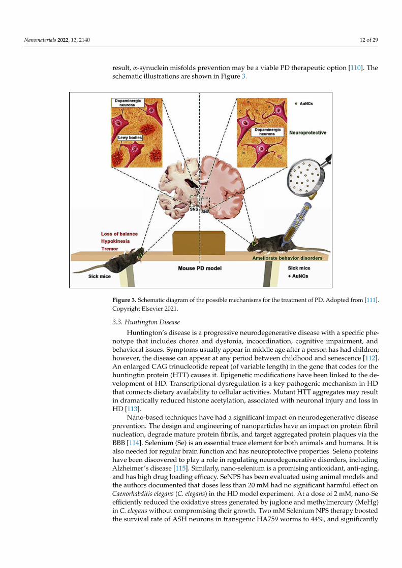
Nanomaterials 2022, 12, 2140 12 of 29
result, α-synuclein misfolds prevention may be a viable PD therapeutic option [110]. Theschematic illustrations are shown in Figure 3.
Nanomaterials 2022, 12, x FOR PEER REVIEW 12 of 30
polyglycolide (PEG-PLGA) nanoparticle (Lf-NP), which was used to evaluate the in vitro and in vivo delivery properties of a novel biodegradable brain drug delivery system. The behavior, immunohistochemistry, and transmitter content revealed an IV injection of 28 g UCN transported by Lf-NP might successfully attenuate the 6-OHDA-induced striatum damage. In the rat 6-OHDA Parkinson′s disease models, intracerebral injection of UCN was previously shown to stop the development of Parkinsonian-like symptoms. Nonin-vasive systemic administration was still impossible due to UCN′s limited BBB permeabil-ity. This study was the first to show that noninvasive delivery of UCN to the brain was possible and that the Lf-NP delivery system may be used for noninvasive Parkinson′s dis-ease treatment [107]. Autopsy specimens collected from the brains of deceased PD pa-tients revealed unique protein clumps called amyloid fibrils and Lewy bodies within the brain tissue [108]. These aggregates are dense protein polymers made up of monomers of α-synuclein. α-synuclein is a member of the synuclein family with 140 amino acids that plays a key role in neuronal signaling and intracellular processes [109].
In recent years, there has been a lot of interest in looking into the potential of different nanoparticles for preventing the production of α -synuclein amyloid. Several nanoparti-cles have been studied for this purpose, including gold nanoparticles, super paramagnetic iron oxide nanoparticles, QDs graphene, and graphene derivatives [110]. The CNTs inter-acted strongly with α-synuclein, with the phosphorus-doped CNTs exhibiting the strong-est connections. Doped-CNTs, particularly phosphorus-doped carbon nanotubes, have been shown to suppress the development of α-synuclein amyloid efficiently and hence could be considered as a potential Parkinson′s disease treatment. However, additional in vitro and clinical research is required. Direct contact with a misfolded α-synuclein protein can cause the misfolding of other α-synuclein proteins, exacerbating the condition. As a result, α-synuclein misfolds prevention may be a viable PD therapeutic option [110]. The schematic illustrations are shown in Figure 3.
Figure 3. Schematic diagram of the possible mechanisms for the treatment of PD. Adopted from[111]. Copyright Elsevier 2021.
Figure 3. Schematic diagram of the possible mechanisms for the treatment of PD. Adopted from [111].Copyright Elsevier 2021.
3.3. Huntington Disease
Huntington’s disease is a progressive neurodegenerative disease with a specific phe-notype that includes chorea and dystonia, incoordination, cognitive impairment, andbehavioral issues. Symptoms usually appear in middle age after a person has had children;however, the disease can appear at any period between childhood and senescence [112].An enlarged CAG trinucleotide repeat (of variable length) in the gene that codes for thehuntingtin protein (HTT) causes it. Epigenetic modifications have been linked to the de-velopment of HD. Transcriptional dysregulation is a key pathogenic mechanism in HDthat connects dietary availability to cellular activities. Mutant HTT aggregates may resultin dramatically reduced histone acetylation, associated with neuronal injury and loss inHD [113].
Nano-based techniques have had a significant impact on neurodegenerative diseaseprevention. The design and engineering of nanoparticles have an impact on protein fibrilnucleation, degrade mature protein fibrils, and target aggregated protein plaques via theBBB [114]. Selenium (Se) is an essential trace element for both animals and humans. It isalso needed for regular brain function and has neuroprotective properties. Seleno proteinshave been discovered to play a role in regulating neurodegenerative disorders, includingAlzheimer’s disease [115]. Similarly, nano-selenium is a promising antioxidant, anti-aging,and has high drug loading efficacy. SeNPS has been evaluated using animal models andthe authors documented that doses less than 20 mM had no significant harmful effect onCaenorhabditis elegans (C. elegans) in the HD model experiment. At a dose of 2 mM, nano-Seefficiently reduced the oxidative stress generated by juglone and methylmercury (MeHg)in C. elegans without compromising their growth. Two mM Selenium NPS therapy boostedthe survival rate of ASH neurons in transgenic HA759 worms to 44%, and significantly

Nanomaterials 2022, 12, 2140 13 of 29
improved the recovery of physiological activities in ASH neurons, such as a proper responseto external stimuli. Antioxidative nano-Se was thought to serve a key role in controllingthe production of histone deacetylase (HDAC) members and efficiently reducing HTTaggregation [116]. A schematic diagram of the neuroprotective effect of nano-Se in a modelof C. elegans HD is shown in Figure 4. The poly(trehalose) nanoparticles are formed of a6 nm iron oxide core and a zwitterionic polymer shell containing 5–12 wt percent covalentlybonded trehalose and have a hydrodynamic dimension of 20–30 nm. The nanoparticle formof trehalose with a zwitterionic surface charge and a trehalose multivalency (i.e., number oftrehalose moles) is 1000–10,000 times more efficient than molecular trehalose in inhibitingprotein fibrillation in extracellular space, blocking the aggregation of the polyglutamine-containing mutant huntingtin protein in model neuronal cells, and suppressing mutanthuntingtin aggregates in HD mouse brain [114].
Biocompatible and non-toxic poly(lactic-co-glycolic acid) nanoparticles (PLGA NPs)offer several advantages, including greater drug solubility, resistance to enzyme digestion,increased targeting efficiency, and improved cellular internalization [117]. The most abun-dant polyphenol in the tea plant, epigallocatechin-3-gallate (EGCG), has previously beenfound to have positive benefits in Huntington’s disease (HD), but its therapeutic stabilityrestricts its therapeutic efficacy. EGCG encapsulation in poly(lactic-co-glycolic) acid-PEGnanoparticles (NPs) increased EGCG stability and brain penetration, as well as the thera-peutic effects in other ND. To test the possible usefulness of the proposed nano-carrier inthis ND, EGCG-loaded NPs were given to 3-nitropropionic acid (3-NP)-intoxicated mice,which is a well-known animal model of HD [118]. Thus, research in this field is quitefascinating in order to increase the therapeutic armament, particularly through the use ofPLGA NPs [117].
Nanomaterials 2022, 12, x FOR PEER REVIEW 14 of 30
Figure 4. Schematic diagram of the neuroprotective effect of nano-Se in a model of C. elegans HD. Reproduced with permission [116]. Copyright 2019 The American Chemical Society.
3.4. Amyotrophic Lateral Sclerosis (ALS) The progressive loss of motor neurons in the brain and spinal cord is a hallmark of
Amyotrophic lateral sclerosis. The pathobiological aspects of this neurodegenerative ill-ness are similar to those of front temporal dementia, and many individuals exhibit symp-toms of both diseases. Various genes and pathophysiological processes cause the disease, and it will be required to comprehend this heterogeneity to develop effective treatments [119]. The development of diagnostic techniques for the early detection and successful treatment of various illnesses has been the subject of extensive research. However, just a small amount of progress has been made. The BBB, which prevents therapeutic drugs and diagnostic equipment from penetrating the brain, remains one of the reasons for the lack of success in the development of treatments to date. There are few techniques for treating neurodegenerative illnesses that are nearing completion, including the use of stem cells and antitoxins against mutant forms of the copper and zinc superoxide dismutase (SOD1), as well as nanotechnology [120].
Since oxidative stress plays a role in amyotrophic lateral sclerosis (ALS) in humans, the SOD1 Cerium oxide nanoparticles (CeNPs) are a type of cerium oxide that can neu-tralize reactive oxygen and nitrogen species. Because oxidative stress is linked to amyo-trophic lateral sclerosis (ALS) in humans and the SOD1 G93A mouse model of ALS, CeNPs was tested in SOD1 G93A transgenic mice, which would increase their survival and reduce disease severity. SOD1 G93A mice were given CeNPs twice a week starting at the onset of muscular weakness, which preserved muscle function and enhanced longev-ity in both males and females [121]. According to research, only three attempts have been made to produce an ALS therapy that combines nanotechnology with riluzole medication. Bondì et al. created solid lipid nanoparticles containing riluzole for ALS treatment. Be-cause of their lipophilic characteristics, these nanoparticles could target the brain via en-
Figure 4. Schematic diagram of the neuroprotective effect of nano-Se in a model of C. elegans HD.Reproduced with permission [116]. Copyright 2019 The American Chemical Society.

Nanomaterials 2022, 12, 2140 14 of 29
3.4. Amyotrophic Lateral Sclerosis (ALS)
The progressive loss of motor neurons in the brain and spinal cord is a hallmark ofAmyotrophic lateral sclerosis. The pathobiological aspects of this neurodegenerative illnessare similar to those of front temporal dementia, and many individuals exhibit symptomsof both diseases. Various genes and pathophysiological processes cause the disease, andit will be required to comprehend this heterogeneity to develop effective treatments [119].The development of diagnostic techniques for the early detection and successful treatmentof various illnesses has been the subject of extensive research. However, just a smallamount of progress has been made. The BBB, which prevents therapeutic drugs anddiagnostic equipment from penetrating the brain, remains one of the reasons for the lackof success in the development of treatments to date. There are few techniques for treatingneurodegenerative illnesses that are nearing completion, including the use of stem cellsand antitoxins against mutant forms of the copper and zinc superoxide dismutase (SOD1),as well as nanotechnology [120].
Since oxidative stress plays a role in amyotrophic lateral sclerosis (ALS) in humans, theSOD1 Cerium oxide nanoparticles (CeNPs) are a type of cerium oxide that can neutralizereactive oxygen and nitrogen species. Because oxidative stress is linked to amyotrophiclateral sclerosis (ALS) in humans and the SOD1 G93A mouse model of ALS, CeNPs wastested in SOD1 G93A transgenic mice, which would increase their survival and reducedisease severity. SOD1 G93A mice were given CeNPs twice a week starting at the onsetof muscular weakness, which preserved muscle function and enhanced longevity in bothmales and females [121]. According to research, only three attempts have been made toproduce an ALS therapy that combines nanotechnology with riluzole medication. Bondìet al. created solid lipid nanoparticles containing riluzole for ALS treatment. Because oftheir lipophilic characteristics, these nanoparticles could target the brain via endocytosis.The rats were given riluzole-loaded nanoparticles and regular riluzole separately, and theresults were compared. Compared to traditional riluzole, the riluzole-loaded nanoparticleswere able to bypass the BBB and transport more medicine to the brain [120].
In another study, Solid Lipid nanoparticles (SLNs) were used as carriers for riluzole.Compritol® 888 ATO was used as the lipid matrix, phosphatidylcholine as the surfactant,taurocholate sodium salt as the cosurfactant, and riluzole-loaded SLNs were successfullysynthesized utilizing the microemulsion process. Under various physiological settings, thesystems were characterized in terms of particle size, z-potential, and drug-release profile.The blood and tissue temporal bio-distribution of riluzole, free or blended into SLNs atequivalent dosages (8 mg/kg), was investigated after intra-peritoneal treatment to ratsin order to demonstrate the preferential accumulation of riluzole-loaded SLNs into thebrain over water dispersion of riluzole. The drug was successfully transported into theCNS using solid lipid nanoparticles. When riluzole was given as drug-loaded solid lipidnanoparticles, the drug bio-distribution in organs such as the liver, spleen, heart, kidneys,and lung was low [121].
Exosomes are natural nanomaterials based on internal membranes that have demon-strated significant advantages over other nanomaterials due to their non-immunogenicproperties and capacity to carry a range of payloads [122]. Exosomes, for example, havebeen produced by Lydia et al. to carry siRNA into the brains of mice. After activation,exosomes were isolated from dendritic cells collected from bone marrow with interleukinsto minimize immunogenicity. To improve brain targeting, dendritic cells were modified toexpress Lamp2b, an exosomal membrane protein linked to the neuron-specific RVG pep-tide [123]. In the mouse brain, the manufactured exosomes could carry siRNA to microglia,neurons, or oligodendrocytes, resulting in cell-specific gene suppression [124]. Exosomeswere created by human mesenchymal stem cells (MSCs) and treated with interferon-gamma(IFN-) ((IFN-) was utilized to increase the production of numerous critical immunosup-pressive cytokines in MSCs in another investigation). These exosomes were administeredintravenously in an autoimmune encephalomyelitis (EAE) animal model after being loadedwith anti-inflammatory, neuroprotective RNA, and protein components, demonstrating
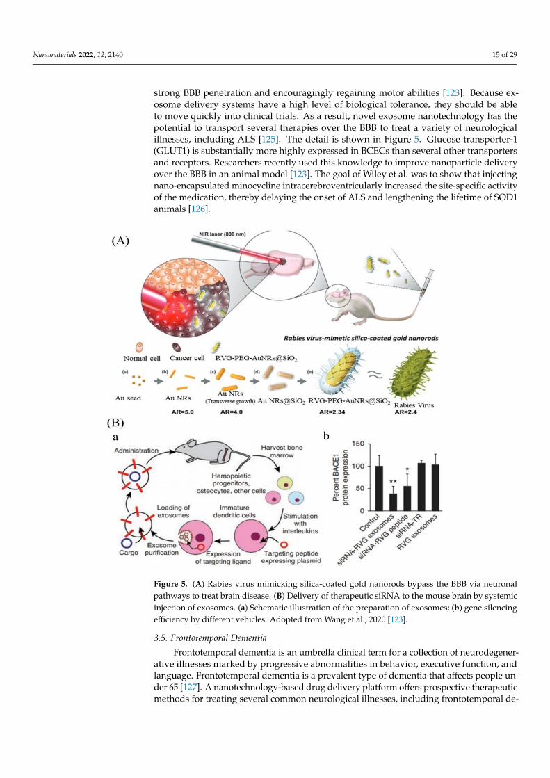
Nanomaterials 2022, 12, 2140 15 of 29
strong BBB penetration and encouragingly regaining motor abilities [123]. Because ex-osome delivery systems have a high level of biological tolerance, they should be ableto move quickly into clinical trials. As a result, novel exosome nanotechnology has thepotential to transport several therapies over the BBB to treat a variety of neurologicalillnesses, including ALS [125]. The detail is shown in Figure 5. Glucose transporter-1(GLUT1) is substantially more highly expressed in BCECs than several other transportersand receptors. Researchers recently used this knowledge to improve nanoparticle deliveryover the BBB in an animal model [123]. The goal of Wiley et al. was to show that injectingnano-encapsulated minocycline intracerebroventricularly increased the site-specific activityof the medication, thereby delaying the onset of ALS and lengthening the lifetime of SOD1animals [126].
Nanomaterials 2022, 12, x FOR PEER REVIEW 16 of 30
Figure 5. (A) Rabies virus mimicking silica-coated gold nanorods bypass the BBB via neuronal pathways to treat brain disease. (B) Delivery of therapeutic siRNA to the mouse brain by systemic injection of exosomes. (a) Schematic illustration of the preparation of exosomes; (b) gene silencing efficiency by different vehicles. Adopted from Wang et al., 2020 [123].
3.5. Frontotemporal Dementia Frontotemporal dementia is an umbrella clinical term for a collection of neurodegen-
erative illnesses marked by progressive abnormalities in behavior, executive function, and language. Frontotemporal dementia is a prevalent type of dementia that affects people under 65 [127]. A nanotechnology-based drug delivery platform offers prospective thera-peutic methods for treating several common neurological illnesses, including frontotem-poral dementia, among innovative strategies to overcome these restrictions and success-fully deliver medications to the CNS [128].
Curcumin′s limited absorption, high metabolism, and rapid excretion make it chal-lenging to employ in vivo. To overcome these limitations, various techniques might be used. In a neural cell line containing TDP-43 mutations Q331K or M337V, the protective effect of analogue dimethoxy curcumin was demonstrated. Dimethoxy curcumin im-proved the transmembrane potential, increased electron transfer chain complex I activity, and up regulated UCP2 to repair mitochondrial damage (uncoupling protein 2). Cells ex-pressing mutant TDP-43 had abnormally high excitability, which was improved by the same drug. Furthermore, monocarbonyl dimethoxycurcumin C, an enhanced curcumin analogue, inhibited mutant TDP-43 aggregation and reduced oxidative stress, presuma-bly due to the increased production of heme oxygenase-1 [129]. Another method for in-creasing curcumin bioavailability is to use nanoparticles. Curcumin-loaded inulin-d-alfa-
Figure 5. (A) Rabies virus mimicking silica-coated gold nanorods bypass the BBB via neuronalpathways to treat brain disease. (B) Delivery of therapeutic siRNA to the mouse brain by systemicinjection of exosomes. (a) Schematic illustration of the preparation of exosomes; (b) gene silencingefficiency by different vehicles. Adopted from Wang et al., 2020 [123].
3.5. Frontotemporal Dementia
Frontotemporal dementia is an umbrella clinical term for a collection of neurodegener-ative illnesses marked by progressive abnormalities in behavior, executive function, andlanguage. Frontotemporal dementia is a prevalent type of dementia that affects people un-der 65 [127]. A nanotechnology-based drug delivery platform offers prospective therapeuticmethods for treating several common neurological illnesses, including frontotemporal de-

Nanomaterials 2022, 12, 2140 16 of 29
mentia, among innovative strategies to overcome these restrictions and successfully delivermedications to the CNS [128].
Curcumin’s limited absorption, high metabolism, and rapid excretion make it chal-lenging to employ in vivo. To overcome these limitations, various techniques might beused. In a neural cell line containing TDP-43 mutations Q331K or M337V, the protectiveeffect of analogue dimethoxy curcumin was demonstrated. Dimethoxy curcumin improvedthe transmembrane potential, increased electron transfer chain complex I activity, and upregulated UCP2 to repair mitochondrial damage (uncoupling protein 2). Cells expressingmutant TDP-43 had abnormally high excitability, which was improved by the same drug.Furthermore, monocarbonyl dimethoxycurcumin C, an enhanced curcumin analogue, in-hibited mutant TDP-43 aggregation and reduced oxidative stress, presumably due to theincreased production of heme oxygenase-1 [129]. Another method for increasing curcuminbioavailability is to use nanoparticles. Curcumin-loaded inulin-d-alfa-tocopherol succinatemicelles were successfully transported into mesenchymal stromal cells, demonstrating thepossibility for ALS treatment [130].
3.6. Prion Disease
Prion disorders are a set of human and animal neurodegenerative illnesses in whichthe prion protein plays a crucial role in their pathogenesis. Creutzfeldt–Jakob disease (CJD),Gerstmann–Straussler–Scheinker syndrome (GSS), fatal familial insomnia (FFI), and kuruare the traditional clinical classifications for human prion illnesses. They can also be classedas acquired (transmitted between animals or people), hereditary, or sporadic based on theiraetiology (unknown cause). In biology, the supremacy of a single protein in a disease withsuch varied pathways is unprecedented [131]. There is currently no effective treatmentfor prion diseases in humans, and these diseases are usually deadly in people. Drugs thatdemonstrate some efficacy in treating prion disorders in tissue culture systems or wholeanimal systems have been found [132]. There is a legitimate need to develop medication oralternative therapy for prion diseases, given the existing state of the treatment plan. As aresult, Michal Mizrahi et al. investigated whether pomegranate seed oil (PSO) in NE formslows clinical progression, neurodegenerative pathological characteristics, and prions inTgMHu2ME199′K mice. E200K PrPmutation was connected to this mouse model. In mousemodels with the idea of fight ND, it was discovered that PSO administration delayed thecommencement of action [133]. Nano-PSO was effective in the prevention and therapy of agenetic prion disease model, suggesting that chemicals that inhibit lipid oxidation could behelpful for a variety of neurodegenerative disorders [134].
Prefibrillar amyloid formations, such as spheroidal aggregates (nanoparticles andnanospheres), have been hypothesized to be highly cytotoxic in many neurodegenerativedisorders, similar to the molecular oligomers discussed above [135]. Large prion protein(PrP) aggregates were degraded into smaller PrP nanoparticles with diameters rangingfrom 17 to 27 nm by Silveira and colleagues, and then PrP nanoparticles were subse-quently studied with DLS, non-denaturing gel electrophoresis, and transmission electronmicroscopy (TEM) [136,137]. Their findings revealed that the most infectious initiators forprion disorders are PrP nanoparticles, with masses corresponding to 14–28 PrP molecules.In another investigation, Moustaine et al. created amyloid nanofibrils and nanoparticlesfrom recombinant PrP under high pressure [136].
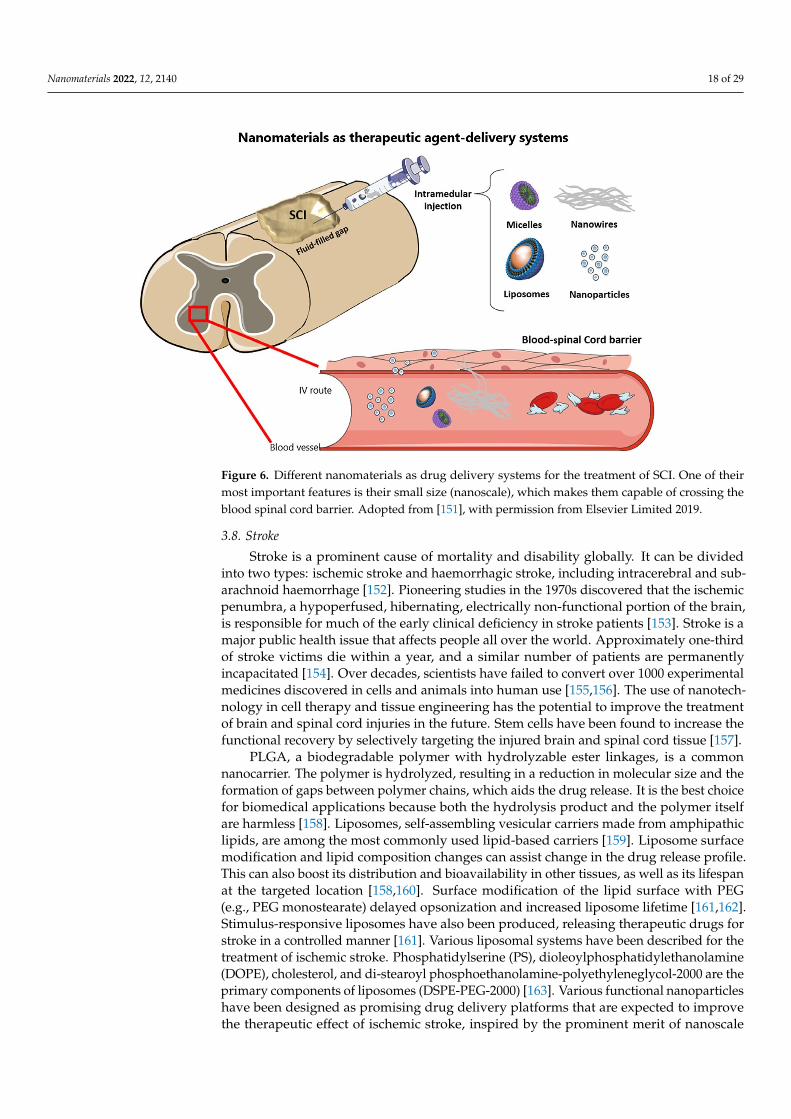
3.7. Spinal Cord Injury
Spinal Cord Injury (SCI) is a neurological disorder caused by a traumatic injury ordisease that affects voluntary motor control and sensory function and the autonomic ner-vous system, impacting cardiovascular, cognitive, bladder, and bowel functions, amongother things. Changes in sexuality, weight gain, poor sleep, diminished cognitive perfor-mance, and chronic pain is common psychosocial issues for SCIs: relationship stress andbreakup, societal prejudice, and reduced work possibilities [138]. Although spinal cordinjury (SCI) is not as prevalent as other injuries, it has devastating physical and emotional

Nanomaterials 2022, 12, 2140 17 of 29
implications. After a spinal cord injury, only a small percentage of people recover com-pletely [139]. Applied neuroprotective techniques have been attempted in SCI models inthe hopes of preserving neuronal and glial cell populations by targeting one or more ofthe aforementioned secondary damage events through drug-mediated protective effects.However, few pharmaceutical treatments have been successful in translating therapeuticvalue in patients [140].
At pH 7.4, micelles encapsulating 1,2-benzisoxazole-3-methanesulfonamide with asize of about 60–90 nm enabled a prolonged drug release. It was discovered that drug-loaded micelles had a more significant protective impact against neurotoxicity than non-formulated species [141]. Various neuroprotective regimens, such as maintaining cyclicadenosine monophosphate (cyclic AMP) levels with injection of the phosphodiesterase4 (PDE4) inhibitor Rolipram (Rm), can lessen the extent of damage following SCI [142]. Rm,like many other medications, is relatively hydrophobic and has a low aqueous solubility(0.2 mg/mL) [143]. Mack et al. (2018) created the PgP[poly(lactide-co-glycolide)-graft-polyethylenimine] polymeric micelle nanoparticle as a carrier for rolipram in SCI improve-ment. PgP has been constructed and polymerized to deliver therapeutic nucleic acids andmedicines for SCI repair in a combinational delivery system. PgP has a hydrophobic coreand a hydrophilic shell, allowing rolipram and small-interfering RNA to be carried to theSCI site [144].
Silica nanoparticles (SiNPs), which have been shown to be non-toxic in vivo, havealso been explored extensively for the treatment of SCI. In ex vivo and in vivo contusionguinea pig models of SCI, Cho et al. established the efficacy of PEG decorated SiNPs(PSiNPs). The NPs, in this case, are not carrying any drugs; instead, they are increasing thebioavailability of PEG, which has been shown to have neuroprotective properties and closesbroken cell membranes [145]. When NPs were used instead of PEG alone, the effectiveconcentration of PEG was reduced by two orders of magnitude. This is crucial becausePEG has been demonstrated to be beneficial for treatment. Its administration is restrictedby viscosity and the concentration of PEG monomers, which can be poisonous at largedoses [146,147]. Because of their favorable qualities, such as free radical scavenging orthe capacity to traverse the blood spinal cord barrier (BSCB), several other lesser-knownNPs are being investigated in the early stages of treating SCI. Poly(butyl cyanoacrylate)NPs (PBCANPs) coated with the surfactant polysorbate-80 have been demonstrated tocross the BBB in previous experiments [148,149]. These particles are covered with adsorbedplasma proteins, including apoplipoprotein E, and it is thought that they are mistakenfor low-density lipoprotein particles and ingested by the low-density lipoprotein uptakesystem, allowing them to pass across the BBB [149].
Aside from enhancing BSCB crossing, nanoparticles, micelles, and liposomes mightboost the bioavailability of various therapeutic medications over long periods, avoidingthe numerous adverse effects now associated with drugs used in clinics. On the otherhand, nano-electrospun fibers, SAP fibers, and nanotubes have a lot of potential sincethey can replicate cells’ physical and structural structure and the ECM. This improves thematerial’s affinity for neurons and axons, resulting in appropriate substrates for neuralregeneration. Several issues still need to be addressed, including the clearance of thesenanosystems as well as the still small gains seen in preclinical animals. Figure 6 representsdifferent nanomaterials as a drug delivery system for the treatment of SCI. Future researchshould concentrate on merging nanotechnologies with other treatments, as this is the onlyapproach to address a condition as complicated as SCI [150].
Nanomaterials 2022, 12, 2140 18 of 29Nanomaterials 2022, 12, x FOR PEER REVIEW 19 of 30
Figure 6. Different nanomaterials as drug delivery systems for the treatment of SCI. One of their most important features is their small size (nanoscale), which makes them capable of crossing the blood spinal cord barrier. Adopted from [151], with permission from Else-vier Limited 2019.
3.8. Stroke Stroke is a prominent cause of mortality and disability globally. It can be divided into
two types: ischemic stroke and haemorrhagic stroke, including intracerebral and sub-arachnoid haemorrhage [152]. Pioneering studies in the 1970s discovered that the ischemic penumbra, a hypoperfused, hibernating, electrically non-functional portion of the brain, is responsible for much of the early clinical deficiency in stroke patients [153]. Stroke is a major public health issue that affects people all over the world. Approximately one-third of stroke victims die within a year, and a similar number of patients are permanently in-capacitated [154]. Over decades, scientists have failed to convert over 1000 experimental medicines discovered in cells and animals into human use [155,156]. The use of nanotech-nology in cell therapy and tissue engineering has the potential to improve the treatment of brain and spinal cord injuries in the future. Stem cells have been found to increase the functional recovery by selectively targeting the injured brain and spinal cord tissue [157].
PLGA, a biodegradable polymer with hydrolyzable ester linkages, is a common nanocarrier. The polymer is hydrolyzed, resulting in a reduction in molecular size and the formation of gaps between polymer chains, which aids the drug release. It is the best choice for biomedical applications because both the hydrolysis product and the polymer itself are harmless [158]. Liposomes, self-assembling vesicular carriers made from amphi-pathic lipids, are among the most commonly used lipid-based carriers [159]. Liposome surface modification and lipid composition changes can assist change in the drug release profile. This can also boost its distribution and bioavailability in other tissues, as well as its lifespan at the targeted location [158,160]. Surface modification of the lipid surface with PEG (e.g., PEG monostearate) delayed opsonization and increased liposome lifetime [161,162]. Stimulus-responsive liposomes have also been produced, releasing therapeutic drugs for stroke in a controlled manner [161]. Various liposomal systems have been de-scribed for the treatment of ischemic stroke. Phosphatidylserine (PS), dioleoylphosphati-dylethanolamine (DOPE), cholesterol, and di-stearoyl phosphoethanolamine-polyeth-yleneglycol-2000 are the primary components of liposomes (DSPE-PEG-2000) [163]. Vari-ous functional nanoparticles have been designed as promising drug delivery platforms
Figure 6. Different nanomaterials as drug delivery systems for the treatment of SCI. One of theirmost important features is their small size (nanoscale), which makes them capable of crossing theblood spinal cord barrier. Adopted from [151], with permission from Elsevier Limited 2019.
3.8. Stroke
Stroke is a prominent cause of mortality and disability globally. It can be dividedinto two types: ischemic stroke and haemorrhagic stroke, including intracerebral and sub-arachnoid haemorrhage [152]. Pioneering studies in the 1970s discovered that the ischemicpenumbra, a hypoperfused, hibernating, electrically non-functional portion of the brain,is responsible for much of the early clinical deficiency in stroke patients [153]. Stroke is amajor public health issue that affects people all over the world. Approximately one-thirdof stroke victims die within a year, and a similar number of patients are permanentlyincapacitated [154]. Over decades, scientists have failed to convert over 1000 experimentalmedicines discovered in cells and animals into human use [155,156]. The use of nanotech-nology in cell therapy and tissue engineering has the potential to improve the treatmentof brain and spinal cord injuries in the future. Stem cells have been found to increase thefunctional recovery by selectively targeting the injured brain and spinal cord tissue [157].
PLGA, a biodegradable polymer with hydrolyzable ester linkages, is a commonnanocarrier. The polymer is hydrolyzed, resulting in a reduction in molecular size and theformation of gaps between polymer chains, which aids the drug release. It is the best choicefor biomedical applications because both the hydrolysis product and the polymer itselfare harmless [158]. Liposomes, self-assembling vesicular carriers made from amphipathiclipids, are among the most commonly used lipid-based carriers [159]. Liposome surfacemodification and lipid composition changes can assist change in the drug release profile.This can also boost its distribution and bioavailability in other tissues, as well as its lifespanat the targeted location [158,160]. Surface modification of the lipid surface with PEG(e.g., PEG monostearate) delayed opsonization and increased liposome lifetime [161,162].Stimulus-responsive liposomes have also been produced, releasing therapeutic drugs forstroke in a controlled manner [161]. Various liposomal systems have been described for thetreatment of ischemic stroke. Phosphatidylserine (PS), dioleoylphosphatidylethanolamine(DOPE), cholesterol, and di-stearoyl phosphoethanolamine-polyethyleneglycol-2000 are theprimary components of liposomes (DSPE-PEG-2000) [163]. Various functional nanoparticleshave been designed as promising drug delivery platforms that are expected to improvethe therapeutic effect of ischemic stroke, inspired by the prominent merit of nanoscale

Nanomaterials 2022, 12, 2140 19 of 29
particles in brain targeting and BBB penetration [164]. Perfluorocorban Nanoparticles(PFC-NPS) are non-metabolizable, non-toxic, and inert. PFC-NPS have the ability to targetepitopes while also being integrated onto the NP surface and carrying a high paramagneticpayload of 60,000 to 90,000 gadolinium ion per particle. Therefore, it is a very sensitivedetector to target epitopes with MRI. Early manifestations were detected for αvβ3-integrin-targeted PEC-NPS along with MRI. The magnitude of the MRI signal is proportional tothe neurovascular density, and PFC-NPS can increase this magnitude. Ligand-mediatedPFC-NPS show promise for detecting therapeutic responses in stroke patients early andaccurately [165]. Deep Vein Thrombosis (DVT) and Pulmonary Embolism (PE) are seriousstroke complications [166].The surgical procedures of thrombin injection in the middlecerebral artery or clot injection in the carotid artery involved in embolic stroke modelsin thrombolytic research are too sophisticated. Haoyuan et al. present a new mousestroke model that embolizes magnetic nanoparticles (MNP) cross-linked with thrombin.MNP@Thrombin was injected from the tail vein after the magnet was placed in the commoncarotid artery. The MNP@Thrombin accumulated in the carotid artery and caused thrombusformation within minutes of injection. These complex clots were flushed into the cerebralartery and subsequently blocked it [167].
More than 200 nano-medicines based products have been approved for clinicaluse or are accurately studied in clinical trials. Liposomes are the most widely studiednanomedicine method and have been approved by FDA (e.g., Doxil, Ambisome, DepotDur,and DaunoXome) [166]. A few examples of nano-based drugs approved by the FDA forthe treatment of neurological disorders are shown in Table 1. Different nanomaterials usedfor the treatment of stroke are shown in Figure 7.
Table 1. Examples of FDA-approved drugs for the treatment of various NDs.
Nanomaterials/Nanoparticles Used Drug Name Diseases Year of Approval Route References
PEG-PLGA Riluzole Amyotrophic lateralsclerosis (ALS) 1995 Orally [123]
Poly(n-butylcyanoacrylate) Rivastigmine Alzheimer’s disease 2000 Intravenous [168]CeO2 NP L-DOPA Parkinson disease 2003 Intranasal [169]
(MPB-PE) and (PDP-PE) couples D-Penicillamine Alzheimer’s disease 2005 Nasal delivery [170]Gold Nanocrystals Molecular surgery Alzheimer’s disease 2006 [171]
Inorganic and metallic nanoparticles Nanotherm®(MagForce) Glioblastomamultiform 2010 Intravenous
injection [128]
Intracerebral Biodegradable gelmatrices/Polymer nanoparticles Temozolomide - 2010 Orally [172]
Liposomes/Microcapsules lomustine Brain Tumor 2010 Orally [172]CED/Intracerebral/Intraarterial/Liposomes Carboplatin Brain Tumor 2010 Intravenous [172]
Polymer NP Copaxone®/Glatopa (Teva) Multiple Sclerosis 2018 Subcutaneousinjection [128]
Liposomes and polymers Riluzole Amyotrophic lateralsclerosis (ALS) 2019 Intravenous [123]
Gold nanocrystals CNM-Au8 Multiple sclerosis 2021 Orally [173]PLGA L-DOPA Parkinson disease - Intranasal [174]
Cerium oxide Photothermal therapy Stroke 2021 - [26]Thermotherapy and magnetic iron-oxide
NPs + reduced dose radiotherapy Nano-thermotherapy Glioblastomamultiforme
Phase IIclinical trials Intrathecal [128]
3.9. Brain Tumors
A brain tumor can affect both adults and children and is caused by various cell typesthat give rise to tumors with varying degrees of malignancy and invasiveness [175,176].Brain tumors are a diverse group of neoplasms that are typically classified as benign ormalignant. The leading causes of brain tumor malignancy are thought to be uncontrolledproliferation and tissue infiltration of dedifferentiated cells caused by harmful chemical,physical, and biological exposures [177]. CNS tumors are the most common solid tumorand the leading cause of death from cancer in children, adolescents, and young adults,with an incidence of 1 to 5 cases per 100,000 people [178]. Surgery, radiation, and systemicchemotherapy are common treatments for brain tumors, but they are associated with ahigh recurrence rate and a poor prognosis. Intra-arterial administration of anti-cancerdrugs has been considered a viable alternative to intravenous and oral administration in

Nanomaterials 2022, 12, 2140 20 of 29
recent decades [177]. Liposomes, nanoparticles, drug-loaded micro bubbles, and magneticattraction of cells are examples of these strategies [178].
Nanomaterials 2022, 12, x FOR PEER REVIEW 20 of 30
that are expected to improve the therapeutic effect of ischemic stroke, inspired by the prominent merit of nanoscale particles in brain targeting and BBB penetration [164]. Per-fluorocorban Nanoparticles (PFC-NPS) are non-metabolizable, non-toxic, and inert. PFC-NPS have the ability to target epitopes while also being integrated onto the NP surface and carrying a high paramagnetic payload of 60,000 to 90,000 gadolinium ion per particle. Therefore, it is a very sensitive detector to target epitopes with MRI. Early manifestations were detected for αvβ3-integrin-targeted PEC-NPS along with MRI. The magnitude of the MRI signal is proportional to the neurovascular density, and PFC-NPS can increase this magnitude. Ligand-mediated PFC-NPS show promise for detecting therapeutic responses in stroke patients early and accurately [165]. Deep Vein Thrombosis (DVT) and Pulmo-nary Embolism (PE) are serious stroke complications [166].The surgical procedures of thrombin injection in the middle cerebral artery or clot injection in the carotid artery in-volved in embolic stroke models in thrombolytic research are too sophisticated. Haoyuan et al. present a new mouse stroke model that embolizes magnetic nanoparticles (MNP) cross-linked with thrombin. MNP@Thrombin was injected from the tail vein after the magnet was placed in the common carotid artery. The MNP@Thrombin accumulated in the carotid artery and caused thrombus formation within minutes of injection. These com-plex clots were flushed into the cerebral artery and subsequently blocked it [167].
More than 200 nano-medicines based products have been approved for clinical use or are accurately studied in clinical trials. Liposomes are the most widely studied nano-medicine method and have been approved by FDA (e.g., Doxil, Ambisome, DepotDur, and DaunoXome) [166]. A few examples of nano-based drugs approved by the FDA for the treatment of neurological disorders are shown in Table 1. Different nanomaterials used for the treatment of stroke are shown in Figure 7.
Figure 7. Schematic diagram of different nanomaterials for the treatment of stroke. Adopted from Chao Li et al., 2020 [163].
Figure 7. Schematic diagram of different nanomaterials for the treatment of stroke. Adopted fromChao Li et al., 2020 [163].
NPs, on the other hand, have been shown to have a therapeutic advantage in deliveringpotential antitumor drugs in several studies [179]. PH-responsive nano-drug deliverysystems, nanosomes, and exosomes loaded with therapeutic drugs or RNAi make up thenanomedicine preparations [180]. The use of polymeric NPs to deliver small interferingRNA to several genes, including sodium-potassium (Na–K)–chloride cotransporter 1, yes–associated protein 1, roundabout homolog 1, EGFR, and survivin, can significantly reduceglioblastoma cell growth and migration in a selective manner [181,182]. Different types ofnanomaterials with their mechanism of action are shown in Table 2 and Figure 8.
Table 2. Different types of nanomaterials with their mechanism of action.
NPS Mechanism of Action Disease Animal Model Used References
Polymeric NPs Transport vectors/penetrate the cellmembrane through endocytosis Stroke NA [184]
Micelles Intravenous delivery/efficientdrug delivery PD Mouse model [184]
Graphene NPs Destroy cancer cells Brain tumors Chicken embryonicangiogenesis assay [185]
Gold NPs Improved selectivity to brain AD Mice [186]Carbon NPs Platelets aggregation/stem cell therapy Stroke Rat [183,187]Silver NPs Efficient drug delivery Brain tumors - [188]Zinc oxide Efficient drug delivery Brain tumors Mature Rats [188]dendrimers Efficient drug delivery Brain tumors Mice [188]
Lipid NPs Accumulation of edelfosine Brain tumors Xenograft mouse modelof glioma [189]
Theranostic NPs Improved pharmacokinetics Brain tumors - [190]
Cerum oxide therapeutic effect by acting as redoxactive agents ALS Male and female mice [191]
Magnetic NPs
Regulate the metal homeostasis in thebrain, carry a large dose of drug to
achieve high local concentration andavoid toxicity, target and detect amyloid
plaques in AD
AD/HD/FrontotemporalDementia - [192]

Nanomaterials 2022, 12, 2140 21 of 29Nanomaterials 2022, 12, x FOR PEER REVIEW 22 of 30
Figure 8. Nanomaterials for the treatment and diagnosis of brain tumors. Adopted from Simona et al., 2020 [183].
Table 2. Different types of nanomaterials with their mechanism of action.
NPS Mechanism of Action Disease Animal Model Used References
Polymeric NPs Transport vectors/penetrate the cell
membrane through endocytosis Stroke NA [184]
Micelles Intravenous delivery/efficient drug delivery PD Mouse model [184]
Graphene NPs Destroy cancer cells Brain tumors Chicken embryonic angiogenesis assay
[185]
Gold NPs Improved selectivity to brain AD Mice [186] Carbon NPs Platelets aggregation/stem cell therapy Stroke Rat [183,187] Silver NPs Efficient drug delivery Brain tumors - [188] Zinc oxide Efficient drug delivery Brain tumors Mature Rats [188] dendrimers Efficient drug delivery Brain tumors Mice [188]
Lipid NPs Accumulation of edelfosine Brain tumors Xenograft mouse model of glioma
[189]
Theranostic NPs Improved pharmacokinetics Brain tumors - [190]
Cerum oxide therapeutic effect by acting as redox active
agents ALS Male and female mice [191]
Magnetic NPs
Regulate the metal homeostasis in the brain, carry a large dose of drug to achieve high
local concentration and avoid toxicity, target and detect amyloid plaques in AD
AD/HD/Frontotemporal Dementia
- [192]
4. Conclusions According to the Global Burden of Disease studies and the World Health
Organization, neurological disorders cause approximately 276 million disabilities with 9 million mortalities throughout the globe each year [9,193]. At the same time, these cases are expected to increase tremendously. NDs are difficult to treat and manage due to the BBB, a physiological interface that limits the entry of various therapeutic agents into the brain, which is one of the most critical issues in the treatment of NDs. Therefore, there is a need for brain-targeted drug carriers to treat NDs. Due to the inability to cross the BBB and the lower efficacy of traditional drugs, the nano-scaled drugs delivery system has unique and versatile properties, which attracts scientists from different areas of biomedical science, especially neurosciences, due to its unique characteristics, including nano range size, high surface to volume ratio, selectivity, sensitivity, surface modification, charges, and stability. The nano-based approach represents an innovative and promising
Figure 8. Nanomaterials for the treatment and diagnosis of brain tumors. Adopted from Simonaet al., 2020 [183].
4. Conclusions
According to the Global Burden of Disease studies and the World Health Organiza-tion, neurological disorders cause approximately 276 million disabilities with 9 millionmortalities throughout the globe each year [9,193]. At the same time, these cases are ex-pected to increase tremendously. NDs are difficult to treat and manage due to the BBB, aphysiological interface that limits the entry of various therapeutic agents into the brain,which is one of the most critical issues in the treatment of NDs. Therefore, there is aneed for brain-targeted drug carriers to treat NDs. Due to the inability to cross the BBBand the lower efficacy of traditional drugs, the nano-scaled drugs delivery system hasunique and versatile properties, which attracts scientists from different areas of biomedicalscience, especially neurosciences, due to its unique characteristics, including nano rangesize, high surface to volume ratio, selectivity, sensitivity, surface modification, charges, andstability. The nano-based approach represents an innovative and promising strategy for thetreatment and diagnosis of various diseases such as cancer and NDs. Different types ofnanomaterials are widely used as potential candidates for the imaging and treatment ofNDs. In recent years, the development of nanoparticles mediated delivery of drugs haveshowed a potential approach to cross the BBB, reach the CNS, and perform their function.
The potential use of various types of nanomaterials and nanoparticles for the treatmentof various types of NDs, especially PD and AD, has been widely investigated. However,these nanotherapeutic approaches also have several issues. The main issue is the safetyconcern, because the degradation pathways of various nanomaterials and their toxicityin the brain have not been fully understood. Therefore, further studies are needed tofully elucidate these issues. The second issue is the complex pathogenesis of neurologicaldiseases. These diseases′ clinical and pathological features may vary in different diseasestages. Therefore, the animal models currently used for these diseases do not fully mimicthe pathology of the disease. Hence, the animal model selection is noteworthy. The majorand most challenging issue in this area is clinical translation. Many studies are in thein-vitro stage or at the stage of the animal model. Only a limited number of drugs areapproved by the FDA and/or are under various stages of clinical trials. There is a lack ofclinical trials for nano-based approaches for NDs treatment because the potential side effectsof these strategies in humans are still largely unknown. Similarly, The pharmacodynamicand pharmacokinetics of these nanomedicines are still unknown and limited. Therefore,the translation of these results into a clinical application is still a big challenge in the areaof nanotherapeutic strategies, and it will be the main focus of nanomedicine research inthe next decade. As the nanomedicine field is still new and not fully explored, from bench

Nanomaterials 2022, 12, 2140 22 of 29
to clinical trials, rigorous protocols of validation of in vitro as well as in vivo protocolsare required. Innovation from engineers, chemists, and other researchers in the field isrequired for large-scale production. Similarly, to facilitate access to the trails and patients,the adaptation of regulatory policies is necessary. More research is required to understandthe nanoparticles′ safety concerns and validate their therapeutic applications.
Author Contributions: Author Contributions: Conceptualization, A.W., A.A., M.A. and M.A.K.;investigation, A.W. and A.U.K. resources, D.Z., K.F., A.R., M.A.K., N.A. and A.B.; formal analysis,A.W., A.A., M.A., A.U.K. and A.B.; writing—original draft preparation, A.W., A.A. and D.Z.; writing—review and editing, A.W. and M.A.S.A.; supervision, M.A.S.A. and A.B.; project administration,M.A.S.A.; funding acquisition, M.A.S.A. All authors have read and agreed to the published versionof the manuscript.
Funding: This research was funded by Umm Al-Qura University Saudi Arabia (Grant Code:22UQU4290565DSR30).
Acknowledgments: The authors would like to thank the Deanship of Scientific Research at UmmAl-Qura University for supporting this work.
Conflicts of Interest: The authors declare no conflict of interest.
References1. Re, F.; Gregori, M.; Masserini, M. Nanotechnology for neurodegenerative disorders. Maturitas 2012, 73, 45–51. [CrossRef]
[PubMed]2. Gitler, A.D.; Dhillon, P.; Shorter, J. Neurodegenerative Disease: Models, Mechanisms, and a New Hope; The Company of Biologists Ltd.:
Cambridge, UK, 2017; Volume 10, pp. 499–502.3. WHO. The Top 10 Causes of Death-WHO|World Health Organization; WHO: Geneva, Switzerland, 2020. Available online: https:
//www.who.int/news-room/fact-sheets/detail/the-top-10-causes-of-death (accessed on 12 January 2022).4. Ramos-Cabrer, P.; Agulla, J.; Argibay, B.; Pérez-Mato, M.; Castillo, J. Serial MRI study of the enhanced therapeutic effects of
liposome-encapsulated citicoline in cerebral ischemia. Int. J. Pharm. 2011, 405, 228–233. [CrossRef] [PubMed]5. El-aziz, E.A.E.-d.A.; Elgayar, S.F.; Mady, F.M.; Abourehab, M.A.; Hasan, O.A.; Reda, L.M.; Alaaeldin, E. The Potential of Optimized
Liposomes in Enhancement of Cytotoxicity and Apoptosis of Encapsulated Egyptian Propolis on Hep-2 Cell Line. Pharmaceutics2021, 13, 2184. [CrossRef]
6. Abbott, N.J.; Patabendige, A.A.; Dolman, D.E.; Yusof, S.R.; Begley, D.J. Structure and function of the blood–brain barrier. Neurobiol.Dis. 2010, 37, 13–25. [CrossRef] [PubMed]
7. Tosi, G.; Costantino, L.; Ruozi, B.; Forni, F.; Vandelli, M.A. Polymeric nanoparticles for the drug delivery to the central nervoussystem. Expert Opin. Drug Deliv. 2008, 5, 155–174. [CrossRef]
8. Cano, A.; Sánchez-López, E.; Ettcheto, M.; López-Machado, A.; Espina, M.; Souto, E.B.; Galindo, R.; Camins, A.; García, M.L.;Turowski, P. Current advances in the development of novel polymeric nanoparticles for the treatment of neurodegenerativediseases. Nanomedicine 2020, 15, 1239–1261. [CrossRef]
9. Zhang, W.; Sigdel, G.; Mintz, K.J.; Seven, E.S.; Zhou, Y.; Wang, C.; Leblanc, R.M. Carbon dots: A future Blood–Brain Barrierpenetrating nanomedicine and drug nanocarrier. Int. J. Nanomed. 2021, 16, 5003. [CrossRef]
10. Ashrafizadeh, M.; Mohammadinejad, R.; Kailasa, S.K.; Ahmadi, Z.; Afshar, E.G.; Pardakhty, A. Carbon dots as versatilenanoarchitectures for the treatment of neurological disorders and their theranostic applications: A review. Adv. Colloid InterfaceSci. 2020, 278, 102123. [CrossRef]
11. Sim, T.M.; Tarini, D.; Dheen, S.T.; Bay, B.H.; Srinivasan, D.K. Nanoparticle-based technology approaches to the management ofneurological disorders. Int. J. Mol. Sci. 2020, 21, 6070. [CrossRef]
12. Huang, M.; Gu, X.; Gao, X. Nanotherapeutic strategies for the treatment of neurodegenerative diseases. In Brain Targeted DrugDelivery System; Elsevier: Amsterdam, The Netherlands, 2019; pp. 321–356.
13. Ilyas, M.; Waris, A.; Khan, A.U.; Zamel, D.; Yar, L.; Baset, A.; Muhaymin, A.; Khan, S.; Ali, A.; Ahmad, A. Biological synthesisof titanium dioxide nanoparticles from plants and microorganisms and their potential biomedical applications. Inorg. Chem.Commun. 2021, 133, 108968. [CrossRef]
14. Hussain, Z.; Thu, H.E.; Elsayed, I.; Abourehab, M.A.; Khan, S.; Sohail, M.; Sarfraz, R.M.; Farooq, M.A. Nano-scaled materialsmay induce severe neurotoxicity upon chronic exposure to brain tissues: A critical appraisal and recent updates on predisposingfactors, underlying mechanism, and future prospects. J. Control. Release 2020, 328, 873–894. [CrossRef] [PubMed]
15. Gregori, M.; Masserini, M.; Mancini, S. Nanomedicine for the treatment of Alzheimer’s disease. Nanomedicine 2015, 10, 1203–1218.[CrossRef]
16. Villalva, M.D.; Agarwal, V.; Ulanova, M.; Sachdev, P.S.; Braidy, N. Quantum dots as a theranostic approach in Alzheimer’s disease:A systematic review. Nanomedicine 2021, 16, 1595–1611. [CrossRef] [PubMed]

Nanomaterials 2022, 12, 2140 23 of 29
17. Gandhi, S.; Shende, P. Cyclodextrins-modified metallic nanoparticles for effective cancer therapy. J. Control. Release 2021, 339,41–50. [CrossRef]
18. Modi, G.; Pillay, V.; Choonara, Y.E. Advances in the treatment of neurodegenerative disorders employing nanotechnology. Ann.N. Y. Acad. Sci. 2010, 1184, 154–172. [CrossRef] [PubMed]
19. Hussain, Z.; Rahim, M.A.; Jan, N.; Shah, H.; Rawas-Qalaji, M.; Khan, S.; Sohail, M.; Thu, H.E.; Ramli, N.A.; Sarfraz, R.M.Cell membrane cloaked nanomedicines for bio-imaging and immunotherapy of cancer: Improved pharmacokinetics, cellinternalization and anticancer efficacy. J. Control. Release 2021, 335, 130–157. [CrossRef]
20. Wu, T.; Zhang, T.; Chen, Y.; Tang, M. Research advances on potential neurotoxicity of quantum dots. J. Appl. Toxicol. 2016, 36,345–351. [CrossRef] [PubMed]
21. Utkin, Y.N. Brain and quantum dots: Benefits of nanotechnology for healthy and diseased brain. Cent. Nerv. Syst. Agents Med.Chem. (Former. Curr. Med. Chem.-Cent. Nerv. Syst. Agents) 2018, 18, 193–205. [CrossRef]
22. Aswathy, R.G.; Yoshida, Y.; Maekawa, T.; Kumar, D.S. Near-infrared quantum dots for deep tissue imaging. Anal. Bioanal. Chem.2010, 397, 1417–1435. [CrossRef]
23. Liu, J.; Erogbogbo, F.; Yong, K.-T.; Ye, L.; Liu, J.; Hu, R.; Chen, H.; Hu, Y.; Yang, Y.; Yang, J. Assessing clinical prospects of siliconquantum dots: Studies in mice and monkeys. ACS Nano 2013, 7, 7303–7310. [CrossRef]
24. Shang, W.; Zhang, X.; Zhang, M.; Fan, Z.; Sun, Y.; Han, M.; Fan, L. The uptake mechanism and biocompatibility of graphenequantum dots with human neural stem cells. Nanoscale 2014, 6, 5799–5806. [CrossRef]
25. Ren, C.; Hu, X.; Zhou, Q. Graphene oxide quantum dots reduce oxidative stress and inhibit neurotoxicity in vitro and in vivothrough catalase-like activity and metabolic regulation. Adv. Sci. 2018, 5, 1700595. [CrossRef] [PubMed]
26. Tian, X.; Fan, T.; Zhao, W.; Abbas, G.; Han, B.; Zhang, K.; Li, N.; Liu, N.; Liang, W.; Huang, H. Recent advances in the developmentof nanomedicines for the treatment of ischemic stroke. Bioact. Mater. 2021, 6, 2854–2869. [CrossRef]
27. Xu, G.; Mahajan, S.; Roy, I.; Yong, K.-T. Theranostic quantum dots for crossing blood–brain barrier in vitro and providing therapyof HIV-associated encephalopathy. Front. Pharmacol. 2013, 4, 140. [CrossRef] [PubMed]
28. Walters, R.; Medintz, I.L.; Delehanty, J.B.; Stewart, M.H.; Susumu, K.; Huston, A.L.; Dawson, P.E.; Dawson, G. The role of negativecharge in the delivery of quantum dots to neurons. Asn Neuro 2015, 7, 1759091415592389. [CrossRef] [PubMed]
29. Kang, I.; Yoo, J.M.; Kim, D.; Kim, J.; Cho, M.K.; Lee, S.-E.; Kim, D.J.; Lee, B.-C.; Lee, J.Y.; Kim, J.-J. Graphene quantum dotsalleviate impaired functions in Niemann-pick disease type C in vivo. Nano Lett. 2021, 21, 2339–2346. [CrossRef] [PubMed]
30. Luo, W.; Wang, Y.; Lin, F.; Liu, Y.; Gu, R.; Liu, W.; Xiao, C. Selenium-doped carbon quantum dots efficiently ameliorate secondaryspinal cord injury via scavenging reactive oxygen species. Int. J. Nanomed. 2020, 15, 10113. [CrossRef] [PubMed]
31. Dong, J.; Tao, L.; Abourehab, M.A.; Hussain, Z. Design and development of novel hyaluronate-modified nanoparticles forcombo-delivery of curcumin and alendronate: Fabrication, characterization, and cellular and molecular evidences of enhancedbone regeneration. Int. J. Biol. Macromol. 2018, 116, 1268–1281. [CrossRef]
32. Sintov, A.; Velasco-Aguirre, C.; Gallardo-Toledo, E.; Araya, E.; Kogan, M. Metal nanoparticles as targeted carriers circumventingthe blood–brain barrier. Int. Rev. Neurobiol. 2016, 130, 199–227.
33. Waris, A.; Din, M.; Ali, A.; Afridi, S.; Baset, A.; Khan, A.U.; Ali, M. Green fabrication of Co and Co3O4 nanoparticles and theirbiomedical applications: A review. Open Life Sci. 2021, 16, 14–30. [CrossRef] [PubMed]
34. Waris, A.; Din, M.; Ali, A.; Ali, M.; Afridi, S.; Baset, A.; Khan, A.U. A comprehensive review of green synthesis of copper oxidenanoparticles and their diverse biomedical applications. Inorgan. Chem. Commun. 2021, 123, 1387–7003. [CrossRef]
35. Dawoud, M.; Abourehab, M.A.; Abdou, R. Monoolein cubic nanoparticles as novel carriers for docetaxel. J. Drug Deliv. Sci.Technol. 2020, 56, 101501. [CrossRef]
36. Mody, V.V.; Siwale, R.; Singh, A.; Mody, H.R. Introduction to metallic nanoparticles. J. Pharm. Bioallied Sci. 2010, 2, 282. [CrossRef][PubMed]
37. Feng, X.; Chen, A.; Zhang, Y.; Wang, J.; Shao, L.; Wei, L. Central nervous system toxicity of metallic nanoparticles. Int. J. Nanomed.2015, 10, 4321.
38. Sriramoju, B.; Kanwar, R.K.; Kanwar, J.R. Nanomedicine based nanoparticles for neurological disorders. Curr. Med. Chem. 2014,21, 4154–4168. [CrossRef]
39. Ayaz, M.; Ovais, M.; Ahmad, I.; Sadiq, A.; Khalil, A.T.; Ullah, F. Biosynthesized metal nanoparticles as potential Alzheimer’sdisease therapeutics. In Metal Nanoparticles for Drug Delivery and Diagnostic Applications; Elsevier: Amsterdam, The Netherlands,2020; pp. 31–42.
40. Waris, A.; Ali, M.; Khan, A.U.; Ali, A.; Baset, A. Role of nanotechnology in diagnosing and treating COVID-19 during thepandemic. Int. J. Clin. Virol. 2020, 4, 65–70.
41. Shan, D.; Ma, C.; Yang, J. Enabling biodegradable functional biomaterials for the management of neurological disorders. Adv.Drug Deliv. Rev. 2019, 148, 219–238. [CrossRef]
42. Muheem, A.; Jahangir, M.A.; Jaiswal, C.P.; Jafar, M.; Ahmad, M.Z.; Ahmad, J.; Warsi, M.H. Recent patents, regulatory issues, andtoxicity of nanoparticles in neuronal disorders. Curr. Drug Metab. 2021, 22, 263–279. [CrossRef]
43. Fischer, M.; Vögtle, F. Dendrimers: From design to application—A progress report. Angew. Chem. Int. Ed. 1999, 38, 884–905.[CrossRef]
44. Boas, U.; Heegaard, P.M. Dendrimers in drug research. Chem. Soc. Rev. 2004, 33, 43–63. [CrossRef]

Nanomaterials 2022, 12, 2140 24 of 29
45. Kannan, R.; Nance, E.; Kannan, S.; Tomalia, D.A. Emerging concepts in dendrimer-based nanomedicine: From design principlesto clinical applications. J. Intern. Med. 2014, 276, 579–617. [CrossRef] [PubMed]
46. Vögtle, F.; Gestermann, S.; Hesse, R.; Schwierz, H.; Windisch, B. Functional dendrimers. Prog. Polym. Sci. 2000, 25, 987–1041.[CrossRef]
47. Svenson, S.; Tomalia, D.A. Dendrimers in biomedical applications—Reflections on the field. Adv. Drug Deliv. Rev. 2012, 64,102–115. [CrossRef]
48. Beg, S.; Samad, A.; Alam, M.I.; Nazish, I. Dendrimers as novel systems for delivery of neuropharmaceuticals to the brain. CNSNeurol. Disord.-Drug Targets (Former. Curr. Drug Targets-CNS Neurol. Disord.) 2011, 10, 576–588.
49. Serramía, M.J.; Álvarez, S.; Fuentes-Paniagua, E.; Clemente, M.I.; Sánchez-Nieves, J.; Gómez, R.; de la Mata, J.; Muñoz-Fernández,M.Á. In vivo delivery of siRNA to the brain by carbosilane dendrimer. J. Control. Release 2015, 200, 60–70. [CrossRef] [PubMed]
50. Sharma, A.; Porterfield, J.E.; Smith, E.; Sharma, R.; Kannan, S.; Kannan, R.M. Effect of mannose targeting of hydroxyl PAMAMdendrimers on cellular and organ biodistribution in a neonatal brain injury model. J. Control. Release 2018, 283, 175–189. [CrossRef][PubMed]
51. Yellepeddi, V.K.; Mohammadpour, R.; Kambhampati, S.P.; Sayre, C.; Mishra, M.K.; Kannan, R.M.; Ghandehari, H. Pediatric oralformulation of dendrimer-N-acetyl-l-cysteine conjugates for the treatment of neuroinflammation. Int. J. Pharm. 2018, 545, 113–116.[CrossRef]
52. Sharma, R.; Kim, S.-Y.; Sharma, A.; Zhang, Z.; Kambhampati, S.P.; Kannan, S.; Kannan, R.M. Activated microglia targetingdendrimer–minocycline conjugate as therapeutics for neuroinflammation. Bioconjug. Chem. 2017, 28, 2874–2886. [CrossRef]
53. Nance, E.; Zhang, F.; Mishra, M.K.; Zhang, Z.; Kambhampati, S.P.; Kannan, R.M.; Kannan, S. Nanoscale effects in dendrimer-mediated targeting of neuroinflammation. Biomaterials 2016, 101, 96–107. [CrossRef]
54. Leiro, V.; Duque Santos, S.; Lopes, C.D.; Paula Pêgo, A. Dendrimers as powerful building blocks in central nervous systemdisease: Headed for successful nanomedicine. Adv. Funct. Mater. 2018, 28, 1700313. [CrossRef]
55. Mignani, S.; Bryszewska, M.; Zablocka, M.; Klajnert-Maculewicz, B.; Cladera, J.; Shcharbin, D.; Majoral, J.-P. Can dendrimer basednanoparticles fight neurodegenerative diseases? Current situation versus other established approaches. Prog. Polym. Sci. 2017, 64,23–51. [CrossRef]
56. Xiang, C.; Zhang, Y.; Guo, W.; Liang, X.-J. Biomimetic carbon nanotubes for neurological disease therapeutics as inherentmedication. Acta Pharm. Sin. B 2020, 10, 239–248. [CrossRef] [PubMed]
57. Mbayachi, V.B.; Ndayiragije, E.; Sammani, T.; Taj, S.; Mbuta, E.R. Graphene synthesis, characterization and its applications: Areview. Results Chem. 2021, 3, 100163. [CrossRef]
58. Komane, P.P.; Choonara, Y.E.; du Toit, L.C.; Kumar, P.; Kondiah, P.P.; Modi, G.; Pillay, V. Diagnosis and treatment of neurologicaland ischemic disorders employing carbon nanotube technology. J. Nanomater. 2016, 2016, 9417874. [CrossRef]
59. Nunes, A.; Al-Jamal, K.; Nakajima, T.; Hariz, M.; Kostarelos, K. Application of carbon nanotubes in neurology: Clinicalperspectives and toxicological risks. Arch. Toxicol. 2012, 86, 1009–1020. [CrossRef]
60. Belyanskaya, L.; Weigel, S.; Hirsch, C.; Tobler, U.; Krug, H.F.; Wick, P. Effects of carbon nanotubes on primary neurons and glialcells. Neurotoxicology 2009, 30, 702–711. [CrossRef]
61. Fabbro, A.; Prato, M.; Ballerini, L. Carbon nanotubes in neuroregeneration and repair. Adv. Drug Deliv. Rev. 2013, 65, 2034–2044.[CrossRef]
62. Jain, S.; Singh, S.R.; Pillai, S. Toxicity issues related to biomedical applications of carbon nanotubes. J. Nanomed. Nanotechol. 2012,3, 2. [CrossRef]
63. Gustavsson, P.; Hedmer, M.; Rissler, J. Carbon Nanotubes-Exposure, Toxicology and Protective Measures in the Work Environment; TheSwedish Work Environment Authority: Arbetsmiljöverket, Sweden, 2011.
64. Hussain, Z.; Pandey, M.; Thu, H.E.; Kaur, T.; Jia, G.W.; Ying, P.C.; Xian, T.M.; Abourehab, M.A. Hyaluronic acid functionalizationimproves dermal targeting of polymeric nanoparticles for management of burn wounds: In vitro, ex vivo and in vivo evaluations.Biomed. Pharmacother. 2022, 150, 112992. [CrossRef]
65. Tiwari, S.B.; Amiji, M.M. A review of nanocarrier-based CNS delivery systems. Curr. Drug Deliv. 2006, 3, 219–232. [CrossRef]66. Pardridge, W.M. Blood-brain barrier drug targeting: The future of brain drug development. Mol. Interv. 2003, 3, 90. [CrossRef]
[PubMed]67. Garcia-Garcia, E.; Andrieux, K.; Gil, S.; Couvreur, P. Colloidal carriers and blood–brain barrier (BBB) translocation: A way to
deliver drugs to the brain? Int. J. Pharm. 2005, 298, 274–292. [CrossRef]68. Palmer, A.M. The role of the blood brain barrier in neurodegenerative disorders and their treatment. J. Alzheimer’s Dis. 2011, 24,
643–656. [CrossRef]69. Nagpal, K.; Singh, S.K.; Mishra, D.N. Chitosan nanoparticles: A promising system in novel drug delivery. Chem. Pharm. Bull.
2010, 58, 1423–1430. [CrossRef] [PubMed]70. Bhaskar, S.; Tian, F.; Stoeger, T.; Kreyling, W.; de la Fuente, J.M.; Grazú, V.; Borm, P.; Estrada, G.; Ntziachristos, V.; Razansky, D.
Multifunctional Nanocarriers for diagnostics, drug delivery and targeted treatment across blood-brain barrier: Perspectives ontracking and neuroimaging. Part. Fibre Toxicol. 2010, 7, 3. [CrossRef] [PubMed]
71. Wang, L.-S.; Chuang, M.-C.; Ho, J.-a.A. Nanotheranostics—A review of recent publications. Int. J. Nanomed. 2012, 7, 4679.72. Shim, M.S.; Kwon, Y.J. Stimuli-responsive polymers and nanomaterials for gene delivery and imaging applications. Adv. Drug
Deliv. Rev. 2012, 64, 1046–1059. [CrossRef]

Nanomaterials 2022, 12, 2140 25 of 29
73. Fontes, M.A.P.; Vaz, G.C.; Cardoso, T.Z.D.; de Oliveira, M.F.; Campagnole-Santos, M.J.; Dos Santos, R.A.S.; Sharma, N.M.; Patel,K.P.; Frézard, F. GABA-containing liposomes: Neuroscience applications and translational perspectives for targeting neurologicaldiseases. Nanomed. Nanotechnol. Biol. Med. 2018, 14, 781–788. [CrossRef]
74. Frézard, F.; dos Santos, R.A.; Fontes, M.A. Liposome-encapsulated neuropeptides for site-specific microinjection. In Neuropeptides;Springer: Berlin/Heidelberg, Germany, 2011; pp. 343–355.
75. Ulrich, A.S. Biophysical aspects of using liposomes as delivery vehicles. Biosci. Rep. 2002, 22, 129–150. [CrossRef]76. Spuch, C.; Navarro, C. Liposomes for targeted delivery of active agents against neurodegenerative diseases (Alzheimer’s disease
and Parkinson’s disease). J. Drug Deliv. 2011, 2011, 469679. [CrossRef]77. Zheng, C. Gradient copolymer micelles: An introduction to structures as well as structural transitions. Soft Matter 2019, 15,
5357–5370. [CrossRef] [PubMed]78. Abourehab, M.A.; Ahmed, O.A.; Balata, G.F.; Almalki, W.H. Self-assembled biodegradable polymeric micelles to improve
dapoxetine delivery across the blood-brain barrier. Int. J. Nanomed. 2018, 13, 3679. [CrossRef] [PubMed]79. Croy, S.; Kwon, G. Polymeric micelles for drug delivery. Curr. Pharm. Des. 2006, 12, 4669–4684. [CrossRef] [PubMed]80. Cosgrove, K.P.; Mazure, C.M.; Staley, J.K. Evolving knowledge of sex differences in brain structure, function, and chemistry. Biol.
Psychiatry 2007, 62, 847–855. [CrossRef] [PubMed]81. Monteiro, A.F.M.; Viana, J.D.O.; Nayarisseri, A.; Zondegoumba, E.N.; Mendonça Junior, F.J.B.; Scotti, M.T.; Scotti, L. Computa-
tional studies applied to flavonoids against alzheimer’s and parkinson’s diseases. Oxidative Med. Cell. Longev. 2018, 2018, 7912765.[CrossRef]
82. Islam, A.; Saif Khandker, S.; Alam, F.; Khalil, I.; Amjad Kamal, M.; Hua Gan, S. Alzheimer’s disease and natural products: Futureregimens emerging from nature. Curr. Top. Med. Chem. 2017, 17, 1408–1428. [CrossRef]
83. Patel, M.M.; Patel, B.M. Crossing the blood–brain barrier: Recent advances in drug delivery to the brain. CNS Drugs 2017, 31,109–133. [CrossRef]
84. Xu, W.; Ling, P.; Zhang, T. Polymeric micelles, a promising drug delivery system to enhance bioavailability of poorly water-solubledrugs. J. Drug Deliv. 2013, 2013, 340315. [CrossRef]
85. Kumari, S.; Ahsan, S.M.; Kumar, J.M.; Kondapi, A.K.; Rao, N.M. Overcoming blood brain barrier with a dual purpose Temozolo-mide loaded Lactoferrin nanoparticles for combating glioma (SERP-17-12433). Sci. Rep. 2017, 7, 6602. [CrossRef]
86. Kumar, A.; Singh, A. A review on Alzheimer’s disease pathophysiology and its management: An update. Pharmacol. Rep. 2015,67, 195–203. [CrossRef]
87. Furtado, D.; Björnmalm, M.; Ayton, S.; Bush, A.I.; Kempe, K.; Caruso, F. Overcoming the blood–brain barrier: The role ofnanomaterials in treating neurological diseases. Adv. Mater. 2018, 30, 1801362. [CrossRef] [PubMed]
88. Li, A.; Tyson, J.; Patel, S.; Patel, M.; Katakam, S.; Mao, X.; He, W. Emerging Nanotechnology for Treatment of Alzheimer’s andParkinson’s Disease. Front. Bioeng. Biotechnol. 2021, 9, 672594. [CrossRef] [PubMed]
89. Sood, S.; Jain, K.; Gowthamarajan, K. Intranasal therapeutic strategies for management of Alzheimer’s disease. J. Drug Target.2014, 22, 279–294. [CrossRef] [PubMed]
90. Agarwal, M.; Alam, M.R.; Haider, M.K.; Malik, M.; Kim, D.-K. Alzheimer’s Disease: An Overview of Major Hypotheses andTherapeutic Options in Nanotechnology. Nanomaterials 2021, 11, 59. [CrossRef]
91. Chen, M.; Du, Z.-Y.; Zheng, X.; Li, D.-L.; Zhou, R.-P.; Zhang, K. Use of curcumin in diagnosis, prevention, and treatment ofAlzheimer’s disease. Neural Regen. Res. 2018, 13, 742.
92. Shabbir, U.; Rubab, M.; Tyagi, A.; Oh, D.-H. Curcumin and its derivatives as theranostic agents in Alzheimer’s disease: Theimplication of nanotechnology. Int. J. Mol. Sci. 2021, 22, 196. [CrossRef]
93. Dimov, N.; Kastner, E.; Hussain, M.; Perrie, Y.; Szita, N. Formation and purification of tailored liposomes for drug delivery usinga module-based micro continuous-flow system. Sci. Rep. 2017, 7, 12045. [CrossRef]
94. Bilal, M.; Barani, M.; Sabir, F.; Rahdar, A.; Kyzas, G.Z. Nanomaterials for the treatment and diagnosis of Alzheimer’s disease: Anoverview. NanoImpact 2020, 20, 100251. [CrossRef]
95. De Campos, A.M.; Sanchez, A.; Alonso, M.a.J. Chitosan nanoparticles: A new vehicle for the improvement of the delivery ofdrugs to the ocular surface. Application to cyclosporin A. Int. J. Pharm. 2001, 224, 159–168. [CrossRef]
96. Fox, L.J.; Richardson, R.M.; Briscoe, W.H. PAMAM dendrimer-cell membrane interactions. Adv. Colloid Interface Sci. 2018, 257,1–18. [CrossRef]
97. Song, S.; Wu, J.; Cheng, Y.; Ma, L.; Liu, T.; Liu, J.; Liu, J.; Sotor, J.; Luan, P. Emerging two-dimensional materials-enabled diagnosisand treatments of Alzheimer’s disease: Status and future challenges. Appl. Mater. Today 2021, 23, 101028. [CrossRef]
98. Park, H.-Y.; Seok, H.-Y.; Park, B.-K.; Kim, S.-H.; Goh, C.-H.; Lee, B.-h.; Lee, C.-H.; Moon, Y.-H. Overexpression of Arabidopsis ZEPenhances tolerance to osmotic stress. Biochem. Biophys. Res. Commun. 2008, 375, 80–85. [CrossRef] [PubMed]
99. Faiyaz, M.; Ganayee, M.A.; Akhtar, S.; Krishnan, S.; Flora, B.; Dogra, D.; Jha, N.K.; Chellappan, D.K.; Negi, P.; Dua, K.Nanomaterials in Alzheimer’s disease treatment: A comprehensive review. Front. Biosci. (Landmark Ed.) 2021, 26, 851–865.[PubMed]
100. American Psychiatric Association, A. Diagnostic and Statistical Manual of Mental Disorders; American Psychiatric Association:Washington, DC, USA, 1980; Volume 3.
101. Colvert, E.; Tick, B.; McEwen, F.; Stewart, C.; Curran, S.R.; Woodhouse, E.; Gillan, N.; Hallett, V.; Lietz, S.; Garnett, T. Heritabilityof autism spectrum disorder in a UK population-based twin sample. JAMA Psychiatry 2015, 72, 415–423. [CrossRef]
Nanomaterials 2022, 12, 2140 26 of 29
102. Hua, X.; Thompson, P.M.; Leow, A.D.; Madsen, S.K.; Caplan, R.; Alger, J.R.; O’Neill, J.; Joshi, K.; Smalley, S.L.; Toga, A.W. Braingrowth rate abnormalities visualized in adolescents with autism. Hum. Brain Mapp. 2013, 34, 425–436. [CrossRef]
103. Moni, M.; Rahman, M.; Begum, M.; Uddin, M.; Ashraf, G.M. Deciphering the Role of Nanoparticle-Based Treatment forParkinson’s Disease. Curr. Drug Metab. 2021, 22, 550–560. [CrossRef]
104. Adhikary, R.R.; Sandbhor, P.; Banerjee, R. Nanotechnology platforms in Parkinson’s Disease. ADMET DMPK 2015, 3, 155–181.[CrossRef]
105. Pardridge, W.M. Molecular Trojan horses for blood–brain barrier drug delivery. Curr. Opin. Pharmacol. 2006, 6, 494–500. [CrossRef]106. Pardeshi, C.V.; Belgamwar, V.S.; Surana, S.J. Nanotechnology-Mediated Nose-to-Brain Drug Delivery for Neurodegenerative
Disorders. In Nanobiotechnology in Neurodegenerative Diseases; Springer: Berlin/Heidelberg, Germany, 2019; pp. 163–175.107. Hu, K.; Shi, Y.; Jiang, W.; Han, J.; Huang, S.; Jiang, X. Lactoferrin conjugated PEG-PLGA nanoparticles for brain delivery:
Preparation, characterization and efficacy in Parkinson’s disease. Int. J. Pharm. 2011, 415, 273–283. [CrossRef]108. Soto, C.; Pritzkow, S. Protein misfolding, aggregation, and conformational strains in neurodegenerative diseases. Nat. Neurosci.
2018, 21, 1332–1340. [CrossRef]109. Ulmer, T.S.; Bax, A.; Cole, N.B.; Nussbaum, R.L. Structure and dynamics of micelle-bound human α-synuclein. J. Biol. Chem. 2005,
280, 9595–9603. [CrossRef] [PubMed]110. Alimohammadi, E.; Nikzad, A.; Khedri, M.; Rezaian, M.; Jahromi, A.M.; Rezaei, N.; Maleki, R. Potential treatment of Parkinson’s
disease using new-generation carbon nanotubes: A biomolecular in silico study. Nanomedicine 2020, 16, 189–204. [CrossRef][PubMed]
111. Gao, G.; Chen, R.; He, M.; Li, J.; Wang, L.; Sun, T. Gold nanoclusters for Parkinson’s disease treatment. Biomaterials 2019, 194,36–46. [CrossRef]
112. Walker, F.O. Huntington’s disease. Lancet 2007, 369, 218–228. [CrossRef]113. Cong, W.; Bai, R.; Li, Y.-F.; Wang, L.; Chen, C. Selenium nanoparticles as an efficient nanomedicine for the therapy of Huntington’s
disease. ACS Appl. Mater. Interfaces 2019, 11, 34725–34735. [CrossRef] [PubMed]114. Debnath, K.; Pradhan, N.; Singh, B.K.; Jana, N.R.; Jana, N.R. Poly (trehalose) nanoparticles prevent amyloid aggregation and
suppress polyglutamine aggregation in a Huntington’s disease model mouse. ACS Appl. Mater. Interfaces 2017, 9, 24126–24139.[CrossRef]
115. Cardoso, B.R.; Roberts, B.R.; Bush, A.I.; Hare, D.J. Selenium, selenoproteins and neurodegenerative diseases. Metallomics 2015, 7,1213–1228. [CrossRef]
116. An, Z.; Yan, J.; Zhang, Y.; Pei, R. Applications of nanomaterials for scavenging reactive oxygen species in the treatment of centralnervous system diseases. J. Mater. Chem. B 2020, 8, 8748–8767. [CrossRef]
117. Cunha, A.; Gaubert, A.; Latxague, L.; Dehay, B. PLGA-Based nanoparticles for neuroprotective drug delivery in neurodegenerativediseases. Pharmaceutics 2021, 13, 1042. [CrossRef]
118. Cano, A.; Ettcheto, M.; Espina, M.; Auladell, C.; Folch, J.; Kühne, B.A.; Barenys, M.; Sánchez-López, E.; Souto, E.B.; García, M.L.Epigallocatechin-3-gallate PEGylated poly (lactic-co-glycolic) acid nanoparticles mitigate striatal pathology and motor deficits in3-nitropropionic acid intoxicated mice. Nanomedicine 2020, 16, 19–35. [CrossRef]
119. Van Es, M.A.; Hardiman, O.; Chio, A.; Al-Chalabi, A.; Pasterkamp, R.J.; Veldink, J.H.; Van den Berg, L.H. Amyotrophic lateralsclerosis. Lancet 2017, 390, 2084–2098. [CrossRef]
120. Mazibuko, Z.; Choonara, Y.E.; Kumar, P.; Du Toit, L.C.; Modi, G.; Naidoo, D.; Pillay, V. A review of the potential role of nano-enabled drug delivery technologies in amyotrophic lateral sclerosis: Lessons learned from other neurodegenerative disorders. J.Pharm. Sci. 2015, 104, 1213–1229. [CrossRef] [PubMed]
121. Bondì, M.L.; Craparo, E.F.; Giammona, G.; Drago, F. Brain-targeted solid lipid nanoparticles containing riluzole: Preparation,characterization and biodistribution. Nanomedicine 2010, 5, 25–32. [CrossRef] [PubMed]
122. Andaloussi, S.E.; Lakhal, S.; Mäger, I.; Wood, M.J. Exosomes for targeted siRNA delivery across biological barriers. Adv. DrugDeliv. Rev. 2013, 65, 391–397. [CrossRef] [PubMed]
123. Wang, G.; Rayner, S.; Chung, R.; Shi, B.; Liang, X. Advances in nanotechnology-based strategies for the treatments of amyotrophiclateral sclerosis. Mater. Today Bio 2020, 6, 100055. [CrossRef]
124. Alvarez-Erviti, L.; Seow, Y.; Yin, H.; Betts, C.; Lakhal, S.; Wood, M.J. Delivery of siRNA to the mouse brain by systemic injection oftargeted exosomes. Nat. Biotechnol. 2011, 29, 341–345. [CrossRef]
125. Haney, M.J.; Klyachko, N.L.; Zhao, Y.; Gupta, R.; Plotnikova, E.G.; He, Z.; Patel, T.; Piroyan, A.; Sokolsky, M.; Kabanov, A.V.Exosomes as drug delivery vehicles for Parkinson’s disease therapy. J. Control. Release 2015, 207, 18–30. [CrossRef]
126. Wiley, N.J.; Madhankumar, A.; Mitchell, R.M.; Neely, E.B.; Rizk, E.; Douds, G.L.; Simmons, Z.; Connor, J.R. LipopolysaccharideModified Liposomes for Amyotropic Lateral Sclerosis Therapy: Efficacy in SOD1 Mouse Model. Adv. Nanoparticles 2012, 1, 44–53.[CrossRef]
127. Shanmukhapuvvada, Y.; Vankayalapati, S. Design and development of riluzole loaded chitosan nanoparticles by emulsificationcrosslinking. Int. J. Pharm. Pharm. Sci 2012, 4, 244–248.
128. Nguyen, T.T.; Nguyen, T.T.D.; Vo, T.K.; Nguyen, M.K.; Van Vo, T.; Van Vo, G. Nanotechnology-based drug delivery for centralnervous system disorders. Biomed. Pharmacother. 2021, 143, 112117. [CrossRef]
129. Novak, V.; Rogelj, B.; Župunski, V. Therapeutic potential of polyphenols in amyotrophic lateral sclerosis and frontotemporaldementia. Antioxidants 2021, 10, 1328. [CrossRef] [PubMed]

Nanomaterials 2022, 12, 2140 27 of 29
130. Tripodo, G.; Chlapanidas, T.; Perteghella, S.; Vigani, B.; Mandracchia, D.; Trapani, A.; Galuzzi, M.; Tosca, M.C.; Antonioli,B.; Gaetani, P. Mesenchymal stromal cells loading curcumin-INVITE-micelles: A drug delivery system for neurodegenerativediseases. Colloids Surf. B Biointerfaces 2015, 125, 300–308. [CrossRef] [PubMed]
131. Mead, S. Prion disease genetics. Eur. J. Hum. Genet. 2006, 14, 273–281. [CrossRef] [PubMed]132. Skinner, P.J.; Kim, H.O.; Bryant, D.; Kinzel, N.J.; Reilly, C.; Priola, S.A.; Ward, A.E.; Goodman, P.A.; Olson, K.; Seelig, D.M.
Treatment of prion disease with heterologous prion proteins. PLoS ONE 2015, 10, e0131993. [CrossRef] [PubMed]133. Nirale, P.; Paul, A.; Yadav, K.S. Nanoemulsions for targeting the neurodegenerative diseases: Alzheimer’s, Parkinson’s and
Prion’s. Life Sci. 2020, 245, 117394. [CrossRef]134. Chountoulesi, M.; Demetzos, C. Promising nanotechnology approaches in treatment of autoimmune diseases of central nervous
system. Brain Sci. 2020, 10, 338. [CrossRef]135. Maresca, M.; Derghal, A.; Carravagna, C.; Dudin, S.; Fantini, J. Controlled aggregation of adenine by sugars: Physicochemical
studies, molecular modelling simulations of sugar–aromatic CH–π stacking interactions, and biological significance. Phys. Chem.Chem. Phys. 2008, 10, 2792–2800. [CrossRef]
136. El Moustaine, D.; Perrier, V.; Smeller, L.; Lange, R.; Torrent, J. Full-length prion protein aggregates to amyloid fibrils and sphericalparticles by distinct pathways. FEBS J. 2008, 275, 2021–2031. [CrossRef]
137. Wei, G.; Su, Z.; Reynolds, N.P.; Arosio, P.; Hamley, I.W.; Gazit, E.; Mezzenga, R. Self-assembling peptide and protein amyloids:From structure to tailored function in nanotechnology. Chem. Soc. Rev. 2017, 46, 4661–4708. [CrossRef]
138. Li, J.; Cai, S.; Zeng, C.; Chen, L.; Zhao, C.; Huang, Y.; Cai, W. Urinary exosomal vitronectin predicts vesicoureteral reflux inpatients with neurogenic bladders and spinal cord injuries. Exp. Ther. Med. 2022, 23, 65. [CrossRef]
139. Chen, Y.; Tang, Y.; Vogel, L.; DeVivo, M. Causes of spinal cord injury. Top. Spinal Cord Inj. Rehabil. 2013, 19, 1–8. [CrossRef][PubMed]
140. Zimmermann, R.; Vieira Alves, Y.; Sperling, L.E.; Pranke, P. Nanotechnology for the treatment of spinal cord injury. Tissue Eng.Part B Rev. 2021, 27, 353–365. [CrossRef] [PubMed]
141. Schaal, S.M.; Garg, M.S.; Ghosh, M.; Lovera, L.; Lopez, M.; Patel, M.; Louro, J.; Patel, S.; Tuesta, L.; Chan, W.-M. The therapeuticprofile of rolipram, PDE target and mechanism of action as a neuroprotectant following spinal cord injury. PLoS ONE 2012, 7,e43634. [CrossRef] [PubMed]
142. Macks, C.; Gwak, S.-J.; Lynn, M.; Lee, J.S. Rolipram-loaded polymeric micelle nanoparticle reduces secondary injury after ratcompression spinal cord injury. J. Neurotrauma 2018, 35, 582–592. [CrossRef] [PubMed]
143. Tyler, J.Y.; Xu, X.-M.; Cheng, J.-X. Nanomedicine for treating spinal cord injury. Nanoscale 2013, 5, 8821–8836. [CrossRef] [PubMed]144. Khan, T.I.; Hemalatha, S.; Waseem, M. Promising role of nano-encapsulated drugs for spinal cord injury. Mol. Neurobiol. 2020, 57,
1978–1985. [CrossRef] [PubMed]145. Cho, Y.; Shi, R.; Borgens, R.B. Chitosan produces potent neuroprotection and physiological recovery following traumatic spinal
cord injury. J. Exp. Biol. 2010, 213, 1513–1520. [CrossRef]146. Brent, J. Current management of ethylene glycol poisoning. Drugs 2001, 61, 979–988. [CrossRef]147. Kreuter, J.; Alyautdin, R.N.; Kharkevich, D.A.; Ivanov, A.A. Passage of peptides through the blood-brain barrier with colloidal
polymer particles (nanoparticles). Brain Res. 1995, 674, 171–174. [CrossRef]148. Alyaudtin, R.N.; Reichel, A.; Löbenberg, R.; Ramge, P.; Kreuter, J.; Begley, D.J. Interaction of poly (butylcyanoacrylate) nanoparti-
cles with the blood-brain barrier in vivo and in vitro. J. Drug Target. 2001, 9, 209–221. [CrossRef]149. Ramge, P.; Unger, R.E.; Oltrogge, J.B.; Zenker, D.; Begley, D.; Kreuter, J.; Von Briesen, H. Polysorbate-80 coating enhances uptake
of polybutylcyanoacrylate (PBCA)-nanoparticles by human and bovine primary brain capillary endothelial cells. Eur. J. Neurosci.2000, 12, 1931–1940. [CrossRef] [PubMed]
150. Amar, S.K.; Anand, U.; Verma, A. Cerebrovascular Imaging in a Rodent Stroke Model. Models Tech. Stroke Biol. 2021, 1, 97–105.151. Assunção-Silva, R.; Gomes, E.; Silva, N.; Salgado, A. Nanoengineered biomaterials for spinal cord regeneration. In Nanoengineered
Biomaterials for Regenerative Medicine; Elsevier: Amsterdam, The Netherlands, 2019; pp. 167–185.152. Astrup, J.; Siesjö, B.K.; Symon, L. Thresholds in cerebral ischemia-the ischemic penumbra. Stroke 1981, 12, 723–725. [CrossRef]
[PubMed]153. Cook, D.J.; Teves, L.; Tymianski, M. Treatment of stroke with a PSD-95 inhibitor in the gyrencephalic primate brain. Nature 2012,
483, 213–217. [CrossRef]154. Sacchetti, M.L. Is it time to definitely abandon neuroprotection in acute ischemic stroke? Am. Heart Assoc. 2008, 39, 1659–1660.
[CrossRef]155. Kubinová, Š.; Syková, E. Nanotechnology for treatment of stroke and spinal cord injury. Nanomedicine 2010, 5, 99–108. [CrossRef]156. Sarmah, D.; Banerjee, M.; Datta, A.; Kalia, K.; Dhar, S.; Yavagal, D.R.; Bhattacharya, P. Nanotechnology in the diagnosis and
treatment of stroke. Drug Discov. Today 2021, 26, 585–592. [CrossRef]157. Mohan, A.; Narayanan, S.; Balasubramanian, G.; Sethuraman, S.; Krishnan, U.M. Dual drug loaded nanoliposomal chemotherapy:
A promising strategy for treatment of head and neck squamous cell carcinoma. Eur. J. Pharm. Biopharm. 2016, 99, 73–83. [CrossRef]158. Kaviarasi, S.; Yuba, E.; Harada, A.; Krishnan, U.M. Emerging paradigms in nanotechnology for imaging and treatment of cerebral
ischemia. J. Control. Release 2019, 300, 22–45. [CrossRef]159. Yan, X.; Scherphof, G.L.; Kamps, J.A. Liposome opsonization. J. Liposome Res. 2005, 15, 109–139. [CrossRef]

Nanomaterials 2022, 12, 2140 28 of 29
160. Kang, X.; Chen, H.; Li, S.; Jie, L.; Hu, J.; Wang, X.; Qi, J.; Ying, X.; Du, Y. Magnesium lithospermate B loaded PEGylated solid lipidnanoparticles for improved oral bioavailability. Colloids Surf. B Biointerfaces 2018, 161, 597–605. [CrossRef] [PubMed]
161. Zarhri, Z.; Houmad, M.; Ziat, Y.; El Rhazouani, O.; Slassi, A.; Benyoussef, A.; El Kenz, A. Ab-initio study of magnetism behaviorin TiO2 semiconductor with structural defects. J. Magn. Magn. Mater. 2016, 406, 212–216. [CrossRef]
162. Sarmah, D.; Saraf, J.; Kaur, H.; Pravalika, K.; Tekade, R.K.; Borah, A.; Kalia, K.; Dave, K.R.; Bhattacharya, P. Stroke management:An emerging role of nanotechnology. Micromachines 2017, 8, 262. [CrossRef]
163. Li, C.; Sun, T.; Jiang, C. Recent advances in nanomedicines for the treatment of ischemic stroke. Acta Pharm. Sin. B 2021, 11,1767–1788. [CrossRef]
164. Naccarato, M.; Grandi, F.C.; Dennis, M.; Sandercock, P.A. Physical methods for preventing deep vein thrombosis in stroke.Cochrane Database Syst. Rev. 2010, 8, CD001922. [CrossRef]
165. Li, H.; Yang, Z.; Tang, Q.; Shi, Z.; Mao, Y. Embolic Stroke Model with Magnetic Nanoparticles. ACS Appl. Mater. Interfaces 2021,13, 43993–44001. [CrossRef] [PubMed]
166. Su, M.; Dai, Q.; Chen, C.; Zeng, Y.; Chu, C.; Liu, G. Nano-medicine for thrombosis: A precise diagnosis and treatment strategy.Nano-Micro Lett. 2020, 12, 96. [CrossRef]
167. Sampetrean, O.; Saya, H. Modeling phenotypes of malignant gliomas. Cancer Sci. 2018, 109, 6–14. [CrossRef]168. Wilson, B.; Samanta, M.K.; Santhi, K.; Kumar, K.P.S.; Paramakrishnan, N.; Suresh, B. Poly(n-butylcyanoacrylate) nanoparticles
coated with polysorbate 80 for the targeted delivery of rivastigmine into the brain to treat Alzheimer’s disease. Brain Res. 2008,1200, 159–168. [CrossRef]
169. Kaushik, A.C.; Bharadwaj, S.; Kumar, S.; Wei, D.-Q. Nano-particle mediated inhibition of Parkinson’s disease using computationalbiology approach. Sci. Rep. 2018, 8, 9169. [CrossRef]
170. Cui, Z.; Lockman, P.R.; Atwood, C.S.; Hsu, C.-H.; Gupte, A.; Allen, D.D.; Mumper, R.J. Novel D-penicillamine carryingnanoparticles for metal chelation therapy in Alzheimer’s and other CNS diseases. Eur. J. Pharm. Biopharm. 2005, 59, 263–272.[CrossRef] [PubMed]
171. Kogan, M.J.; Bastus, N.G.; Amigo, R.; Grillo-Bosch, D.; Araya, E.; Turiel, A.; Labarta, A.; Giralt, E.; Puntes, V.F. Nanoparticle-mediated local and remote manipulation of protein aggregation. Nano Lett. 2006, 6, 110–115. [CrossRef] [PubMed]
172. Gilert, A.; Machluf, M. Nano to micro delivery systems: Targeting angiogenesis in brain tumors. J. Angiogenesis Res. 2010, 2, 20.[CrossRef] [PubMed]
173. Jagaran, K.; Singh, M. Nanomedicine for Neurodegenerative Disorders: Focus on Alzheimer’s and Parkinson’s Diseases. Int. J.Mol. Sci. 2021, 22, 9082. [CrossRef]
174. Gambaryan, P.; Kondrasheva, I.; Severin, E.; Guseva, A.; Kamensky, A. Increasing the Efficiency of Parkinson’s Disease TreatmentUsing a poly (lactic-co-glycolic acid)(PLGA) Based L-DOPA Delivery System. Exp. Neurobiol. 2014, 23, 246. [CrossRef]
175. Huang, R.; Boltze, J.; Li, S. Strategies for improved intra-arterial treatments targeting brain tumors: A systematic review. Front.Oncol. 2020, 10, 1443. [CrossRef]
176. Beccaria, K.; Canney, M.; Bouchoux, G.; Puget, S.; Grill, J.; Carpentier, A. Blood-brain barrier disruption with low-intensity pulsedultrasound for the treatment of pediatric brain tumors: A review and perspectives. Neurosurg. Focus 2020, 48, E10. [CrossRef]
177. Reese, T.; Karnovsky, M.J. Fine structural localization of a blood-brain barrier to exogenous peroxidase. J. Cell Biol. 1967, 34,207–217. [CrossRef]
178. Chen, E.M.; Quijano, A.R.; Seo, Y.-E.; Jackson, C.; Josowitz, A.D.; Noorbakhsh, S.; Merlettini, A.; Sundaram, R.K.; Focarete, M.L.;Jiang, Z. Biodegradable PEG-poly (ω-pentadecalactone-co-p-dioxanone) nanoparticles for enhanced and sustained drug deliveryto treat brain tumors. Biomaterials 2018, 178, 193–203. [CrossRef]
179. Kozielski, K.L.; Ruiz-Valls, A.; Tzeng, S.Y.; Guerrero-Cázares, H.; Rui, Y.; Li, Y.; Vaughan, H.J.; Gionet-Gonzales, M.; Vantucci, C.;Kim, J. Cancer-selective nanoparticles for combinatorial siRNA delivery to primary human GBM in vitro and in vivo. Biomaterials2019, 209, 79–87. [CrossRef]
180. Caruso, G.; Marino, D.; Caffo, M. Nanoparticles and CNS delivery of therapeutic agents in the treatment of primary brain tumors.J. Anal. Oncol. 2014, 3, 105–112.
181. Sun, X.; Liu, Z.; Welsher, K.; Robinson, J.T.; Goodwin, A.; Zaric, S.; Dai, H. Nano-graphene oxide for cellular imaging and drugdelivery. Nano Res. 2008, 1, 203–212. [CrossRef] [PubMed]
182. Rehman, F.U. Impact of bioinspired nanotechnology on brain diseases amelioration. Biomed. Lett. 2020, 6, 17–22.183. Mukhtar, M.; Bilal, M.; Rahdar, A.; Barani, M.; Arshad, R.; Behl, T.; Brisc, C.; Banica, F.; Bungau, S. Nanomaterials for diagnosis
and treatment of brain cancer: Recent updates. Chemosensors 2020, 8, 117. [CrossRef]184. Brynskikh, A.M.; Zhao, Y.; Mosley, R.L.; Li, S.; Boska, M.D.; Klyachko, N.L.; Kabanov, A.V.; Gendelman, H.E.; Batrakova,
E.V. Macrophage delivery of therapeutic nanozymes in a murine model of Parkinson’s disease. Nanomedicine 2010, 5, 379–396.[CrossRef]
185. Mukherjee, A.; Madamsetty, V.S.; Paul, M.K.; Mukherjee, S. Recent advancements of nanomedicine towards antiangiogenictherapy in cancer. Int. J. Mol. Sci. 2020, 21, 455. [CrossRef] [PubMed]
186. Siddiqi, K.S.; Husen, A.; Sohrab, S.S.; Yassin, M.O. Recent status of nanomaterial fabrication and their potential applications inneurological disease management. Nanoscale Res. Lett. 2018, 13, 231. [CrossRef]
187. De Mendoza, A.E.-H.; Préat, V.; Mollinedo, F.; Blanco-Prieto, M.J. In vitro and in vivo efficacy of edelfosine-loaded lipidnanoparticles against glioma. J. Control. Release 2011, 156, 421–426. [CrossRef]

Nanomaterials 2022, 12, 2140 29 of 29
188. Caruso, G.; Raudino, G.; Caffo, M. Patented nanomedicines for the treatment of brain tumors. Pharm. Pat. Anal. 2013, 2, 745–754.[CrossRef]
189. Discher, B.M.; Won, Y.-Y.; Ege, D.S.; Lee, J.C.; Bates, F.S.; Discher, D.E.; Hammer, D.A. Polymersomes: Tough vesicles made fromdiblock copolymers. Science 1999, 284, 1143–1146. [CrossRef]
190. Shevtsov, M.; Multhoff, G. Recent developments of magnetic nanoparticles for theranostics of brain tumor. Curr. Drug Metab.2016, 17, 737–744. [CrossRef] [PubMed]
191. DeCoteau, W.; Heckman, K.L.; Estevez, A.Y.; Reed, K.J.; Costanzo, W.; Sandford, D.; Studlack, P.; Clauss, J.; Nichols, E.; Lipps, J.Cerium oxide nanoparticles with antioxidant properties ameliorate strength and prolong life in mouse model of amyotrophiclateral sclerosis. Nanomed. Nanotechnol. Biol. Med. 2016, 12, 2311–2320. [CrossRef] [PubMed]
192. Asil, S.M.; Ahlawat, J.; Barroso, G.G.; Narayan, M. Nanomaterial based drug delivery systems for the treatment of neurodegener-ative diseases. Biomater. Sci. 2020, 8, 4109–4128. [CrossRef] [PubMed]
193. Feigin, V.L.; Nichols, E.; Alam, T.; Bannick, M.S.; Beghi, E.; Blake, N.; Culpepper, W.J.; Dorsey, E.R.; Elbaz, A.; Ellenbogen, R.G.Global, regional, and national burden of neurological disorders, 1990–2016: A systematic analysis for the Global Burden ofDisease Study 2016. Lancet Neurol. 2019, 18, 459–480. [CrossRef]
Copyright © 2022 FDOKUMEN