Do Employment Quotas Explain the Occupational Choices of ...
Apolipoprotein A-V N-terminal Domain Lipid InteractionProperties in Vitro Explain the HypertriglyceridemicPhenotype Associated with Natural Truncation Mutants*
Received for publication, July 3, 2009, and in revised form, October 6, 2009 Published, JBC Papers in Press, October 13, 2009, DOI 10.1074/jbc.M109.040972
Kasuen Wong-Mauldin‡§1, Vincent Raussens¶2, Trudy M. Forte‡, and Robert O. Ryan‡§3
From the ‡Center for Prevention of Obesity, Diabetes and Cardiovascular Disease, Children’s Hospital Oakland ResearchInstitute, Oakland, California 94609, the §Department of Nutritional Sciences and Toxicology, University of California,Berkeley, California 94720, and the ¶Center for Structural Biology and Bioinformatics, Laboratory for Structure and Function ofBiological Membranes, Faculte des Sciences, Universite Libre de Bruxelles, CP 206/2, B-1050 Brussels, Belgium
The N-terminal 146 residues of apolipoprotein (apo) A-Vadopt a helix bundle conformation in the absence of lipid.Because similarly sized truncation mutants in human subjectscorrelate with severe hypertriglyceridemia, the lipid bindingproperties of apoA-V(1–146) were studied. Upon incubationwith phospholipid in vitro, apoA-V(1–146) forms reconstitutedhighdensity lipoproteins 15–17nm indiameter. FarUVcirculardichroism spectroscopy analyses of lipid-bound apoA-V(1–146)yielded an �-helix secondary structure content of 60%. Fouriertransformed infrared spectroscopy analysis revealed that apoA-V(1–146) �-helix segments align perpendicular with respect toparticle phospholipid fatty acyl chains. Fluorescence spectros-copy of single Trp variant apoA-V(1–146) indicates that lipidinteraction is accompanied by a conformational change. Thedata are consistent with amodel wherein apoA-V(1–146)�-hel-ices circumscribe the perimeter of a disk-shaped bilayer. Theability of apoA-V(1–146) to solubilize dimyristoylphosphatidyl-choline vesicles at a rate faster than full-length apoA-V suggeststhat N- and C-terminal interactions in the full-length proteinmodulate its lipid binding properties. Preferential association ofapoA-V(1–146) with murine plasma HDL, but not with VLDL,suggests that particle size is a determinant of its lipoproteinbinding specificity. It may be concluded that defective lipopro-tein binding of truncated apoA-V contributes to the hypertrig-lyceridemia phenotype associated with truncation mutations inhuman subjects.
The helix bundle motif is a common molecular architecturein proteins (1). Exchangeable apolipoproteins (apo)4 are knownto adopt this conformation, which supports their dual existence
in alternate lipid-free and lipid-bound states. Classic examplesof the helix bundle structure include the N-terminal (NT)domains of apoE (2) and apoA-I (3) as well as apolipophorin III(4). In the case of apoE and apoA-I, the helix bundle motifs arepresent within the context of a larger protein structure. In eachof these examples the bundle exists as an up-and-down series ofamphipathic �-helices wherein the hydrophobic face of eachhelical segment orients toward the interior of the bundle. At thesame time, the polar face of the amphipathic helices is directedtoward the exterior of the bundle. In thisway the globular struc-ture is stabilized by hydrophobic helix-helix interactions and isconferred with water solubility through projection of polar andcharged amino acid side chains toward the aqueous milieu.Upon interaction with lipid surfaces, the helix bundle is postu-lated to unfurl, adopting an extended open conformation thatpromotes interaction between the hydrophobic faces ofamphipathic helices and the lipid surface. Essentially, lipidbinding of helix bundle apolipoproteins substitutes helix-helixcontacts in the bundle for helix-lipid contacts that stabilize thelipid-bound state.In 2001 a new apolipoprotein, termed apoA-V, was reported
that profoundly affects plasma TG levels (5, 6). Structural stud-ies revealed that apoA-V is a two-domain protein (7) and thatits N-terminal 146 residues adopt a helix bundle structure inthe absence of lipid (8). Truncated apoA-V proteins in this sizerange have been reported in human subjects with severe hyper-triglyceridemia (HTG) (9, 10). One of these truncationmutants, Q139X apoA-V, does not associate with VLDL orHDL in circulation (9). By contrast, full-length apoA-V is foundon both of these lipoprotein classes in normolipidemic subjects(11). Insofar as these individuals had no other common muta-tions known to causeHTG, it is likely that a lipid-binding defectin truncated apoA-V is associatedwith theHTGphenotype. Toaddress this mechanistically, the lipid interaction properties ofrecombinant apoA-V(1–146) were investigated. The resultsobtained provide a molecular explanation for the correlationbetween naturally occurring C-terminal (CT) truncations inapoA-V and HTG.
EXPERIMENTAL PROCEDURES
Site-directed Mutagenesis and Recombinant Protein Ex-pression—Recombinant human apoA-V and CT truncationvariants were produced in Escherichia coli and isolated as
* This work was supported, in whole or in part, by National Institutes of HealthGrant HL-073061.
1 Recipient of a Gates Millennium Scholarship.2 Senior Research Associate for the National Fund for Scientific Research
(Belgium).3 To whom correspondence should be addressed: CHORI, 5700 Martin Luther
King Jr. Way, Oakland, CA 94609. Tel.: 510-450-7645; Fax: 510-450-7910;E-mail: [email protected].
4 The abbreviations used are: apo, apolipoprotein; HTG, hypertriglyceri-demia; DMPC, dimyristoylphosphatidylcholine; NT, N terminus; CT, Cterminus; HDL, high density lipid; ATR-FTIR, attenuated total reflec-tance Fourier transformed infrared spectroscopy; VLDL, very low den-sity lipid; PL-C, phospholipase C; Tricine, N-[2-hydroxy-1,1-bis(hydroxy-methyl)ethyl]glycine; TG, triacylglycerol.
THE JOURNAL OF BIOLOGICAL CHEMISTRY VOL. 284, NO. 48, pp. 33369 –33376, November 27, 2009© 2009 by The American Society for Biochemistry and Molecular Biology, Inc. Printed in the U.S.A.
NOVEMBER 27, 2009 • VOLUME 284 • NUMBER 48 JOURNAL OF BIOLOGICAL CHEMISTRY 33369
by guest, on Novem
ber 23, 2012w
ww
.jbc.orgD
ownloaded from

described (12). ApoE3 NT was prepared as described by Fisheret al. (13). Recombinant apoA-I was prepared as described byRyan et al. (14). Site-directed mutagenesis was performed withthe QuikChange II XL site-directed mutagenesis kit (Strat-agene). Primers were designed to introduce a premature stopcodon or to substitute Trp5 with Phe, substituteTrp97 with Phe,or substitute Asn45, Leu73, Val117, or Leu128 of Trp-null apoA-V(1–146) with Trp. In all cases, introduction of the desiredmutations was verified by DNA sequencing.Interaction of ApoA-V(1–146) with Phospholipid—Dimyris-
toylphosphatidylcholine (DMPC) bilayer vesicles were incubatedwith apoA-V(1–146) at a ratio of 5:1 by weight as previouslydescribed (12). The size distribution of DMPC-apoA-V(1–146)complexes was evaluated by nondenaturing gradient PAGE asdescribed by Nichols et al. (15).Circular Dichroism Spectroscopy—CD spectroscopy meas-
urements were performed on anAVIV 410 spectrophotometer.Far UV CD scans were obtained between 195 and 250 nm in 10mM sodiumphosphate, pH7.4, using a protein concentration of0.5 mg/ml determined from the absorbance at 280 nm. The�-helical content was calculated with the self-consistentmethod using Dicroprot version 2.6 (16).Infrared Spectroscopy—Attenuated total reflectance Fourier
transformed infrared spectroscopy (ATR-FTIR) spectra wererecorded on a Bruker IFS 55 infrared spectrophotometerequipped with a reflectance accessory and a polarizer mountassembly with an aluminumwire grid on a KRS-5 element. Theinternal reflection element was a germanium ATR plate (50mm� 20mm� 2mm)with an aperture angle of 45° yielding 25internal reflections. Oriented multilayers were formed by slowevaporation of �10 �l of apoA-V(1–146)�DMPC (1 mg/ml) onone side of the ATR plate under a gentle stream of nitrogen,yielding a semi-dry film bearing residual water molecules. TheATR plate was then sealed in a universal sample holder. Thespectra were recorded at a 2 cm�1 nominal resolution. A totalof 128 accumulations were performed to improve the signal/noise ratio. The spectrometer was constantly purged with dryair. All of the measurements were made at 25 °C.Secondary structuremeasurementswere carried out on sam-
ples following deuteration for 1 h as previously described (17).Briefly, Fourier self-deconvolution was applied to enhance theresolution of the spectra in the amide I region. Least squaresiterative curve fitting was performed to fit different compo-nents of the amide I band revealed by the self-deconvolution tothe nondeconvolved spectrum between 1700 and 1600 cm�1.The proportion of various secondary structural elements wascomputed as reported in Ref. 17.Analytical Procedures—Protein concentrations were deter-
mined with the bicinchoninic acid assay (Pierce) with bovineserum albumin as standard.Fluorescence Spectroscopy—Fluorescence spectra were
obtained on a Horiba Jobin Yvon FluoroMax-4 luminescencespectrometer. Protein (0.5 �g/�l) was dissolved in 20 mM
sodium phosphate, pH 7.4, 150 mM NaCl. The samples wereexcited at 295 nm, and emission was collected from 300 to 450nm (slit width, 2.0 nm). For Trp fluorescence quenching studiesDMPC-bound apoA-V(1–146) samples (200 �g of protein in400 �l of total volume) were excited at 295 nm, and emission
was monitored at their �max (slit width, 2.0 nm). Quenching ofTrp fluorescence in 20 mM sodium phosphate, pH 7.4, 150 mM
NaCl by the addition of either KI or acrylamide in the concen-tration range 0.0–0.6 M was measured. The solution of KI con-tained 1 mM sodium thiosulfate to prevent formation of freeiodine, and all of the readings were corrected for dilution. Foracrylamide quenching, fluorescence intensities were correctedfor the absorption of acrylamide at 295 nm,
Icorr.�10�0.23[acryl]
2 �Imeas. (Eq. 1)
where 0.23 is the molar extinction coefficient of acrylamide at295 nm (18). The data were analyzed using the following Stern-Volmer equation,
F0/F � 1 � KSV[Q] (Eq. 2)
where F0 and F represent the emission intensity maximum inthe absence and presence of quencher, respectively, and [Q] isquencher concentration. The collisional quenching constant(KSV) was determined from the initial slope of plots of F0/Fversus [Q].DMPC Vesicle Solubilization—DMPC bilayer vesicles were
prepared by extrusion through a 100-nm filter as described byWeers et al. (19). Stock solutions of protein and lipid vesicleswere prepared in 50mM citrate, pH 3.0, 150mMNaCl. 500�g ofDMPC was incubated in the absence or presence of 100 �g ofapolipoprotein (total volume, 400 �l) at 23 °C in a thermostat-ted cell holder. Sample right angle light scattering intensity wasmonitored on a PerkinElmer Life Sciences LS50B luminescencespectrometer as a function of time, with the excitation andemission monochromaters set at 600 nm (slit width, 2 nm).LDL Binding Assay—Human LDL (Intracel) was incubated
for 90min at 37 °C in the presence or absence of Bacillus cereusphospholipaseC (PL-C) (0.6 unit/50�g of LDLprotein).Whereindicated, apolipoprotein (50 �g/50 �g of LDL protein) wasincluded in the reaction mixture. The incubations were con-ducted in 50 mM Tris-HCl, pH 7.5, 150 mM NaCl, and 2 mM
CaCl2 in a total sample volume of 200 �l. Sample turbidity wasmeasured at 340 nm on a Spectramax 340 microtiter platereader (Sunnyvale, CA) (20).Incubation of ApoA-V(1–146) with Isolated HDL and VLDL
fromApoa5�/�mice—Blood frommale apoA-V-deficientmice(average age 4 months, fasted for 4 h) was collected into tubescontaining K3EDTA and kept on ice. After centrifugation at2,000 � g for 10 min at 4 °C, the plasma was removed andtreated with a protease inhibitor mixture as described by Nel-bach et al. (21). Plasma was pooled, stored at 4 °C, and usedwithin 2 days. Plasma VLDL andHDLwere isolated by sequen-tial ultracentrifugation as described by Lindgren et al. (22). Iso-latedVLDL (d� 1.006 g/ml) orHDL (d� 1.063–1.21 g/ml) wasincubated with recombinant apoA-V(1–146) (weight ratio,10:1, lipoprotein protein:apoA-V(1–146)) for 1 h at 22 °C inTris-buffered saline. After incubation, ultracentrifugation wasperformed to reisolate VLDL (d� 1.006 g/ml, top fraction) andHDL (d � 1.21 g/ml, top fraction). The bottom, lipoprotein-free, fractions fromeach tubewere also recovered. Similar stud-ies were carried out with full-length recombinant apoA-V.
ApoA-V N-terminal Domain Altered Lipid Interaction Properties
33370 JOURNAL OF BIOLOGICAL CHEMISTRY VOLUME 284 • NUMBER 48 • NOVEMBER 27, 2009
by guest, on Novem
ber 23, 2012w
ww
.jbc.orgD
ownloaded from

Immunoblotting—For immunoblotting, the protein sampleswere separated on a 10–20% acrylamide gradient, Tricine-SDSslab gel (Invitrogen). The proteins were electrophoreticallytransferred to a 0.2-�m polyvinylidene difluoride membrane(Bio-Rad) at a constant current of 150 mA for 3 h. Nonspecificbinding sites on the membrane were blocked with 0.1% TTBS(0.1% Tween 20, 20 mM Tris, and 150 mM NaCl, pH 7.2) over-night, at 4 °C with rotation. Goat anti-apoA-V IgG (12)(1:10,000 dilution in 0.1% TTBS) was incubated with the mem-brane for 1 h with rotation. The mixture was washed threetimes in TTBS, and then HRP-conjugated donkey anti-goatsecondary antibody was incubated with the membrane for 1 h.After washing, the membrane was incubated with SuperSignalWest Femto maximum sensitivity substrate (Pierce) andexposed toCL-Xposure Film (Pierce) for less than 10 s. The filmwas developed using a Kodak M35A X-OMAT processor.
RESULTS
Characterization of ApoA-V(1–146) Binding to PhospholipidVesicles—Incubation of apoA-V(1–146) with a DMPC vesicledispersion caused a decrease in solution light scattering inten-sity consistent with formation of lipid complexes (i.e. reconsti-tuted HDL). Native gradient PAGE analysis revealed a discretepopulation of particles with a Stoke’s diameter in the range of15–17 nm (Fig. 1). These complexes, which are similar in size to
discoidal particles formed with full-length apoA-V (12), indi-cate that the NT domain of apoA-V possesses intrinsic lipidbinding capability.Far UV CD Spectroscopy Analysis—The effect of lipid asso-
ciation on the secondary structure content of apoA-V(1–146)was evaluated by far UV CD spectroscopy (Fig. 2). The spec-trum of lipid-free apoA-V(1–146) (Fig. 2, curve a) has minimaat 208 and 222 nm, indicating the presence of�-helix secondarystructure. Deconvolution of the spectrum yielded a value of40% �-helix. Upon interaction with DMPC, however, a signifi-cant enhancement in �-helix content was noted as seen by theincrease in negative ellipticity at 208 and 222 nm (Fig. 2, curveb). Deconvolution of the spectra yielded a value of 60% �-helixstructure.Orientation of �-Helices in ApoA-V(1–146) Lipid Complexes—
ATR-FTIR spectra of apoA-V(1–146)�DMPC complexes wererecorded with parallel and perpendicular polarized light (Fig.3). The amide I region of the spectra (1700–1600 cm�1) wasused to determine the orientation of �-helices in apoA-V(1–146) relative to DMPC hydrocarbon chains of the bilayer com-ponent of the reconstituted HDL. A dichroic spectrum wasobtained by subtracting the spectrumrecordedwith perpendic-ular polarized light from the spectrum recorded with parallelpolarized light. Bands corresponding to dipoles that orient par-allel to the hydrocarbon chain progression observed for satu-rated hydrocarbon chains had a positive deviation in the dich-roic spectrum. Thus, these orient parallel to the normal of thegermanium plate. The apoA-V(1–146) helix orientation wasdetermined using the amide I band. In the dichroic spectrum, anegative deviation at�1650 cm�1 is observed, indicating a par-allel orientation of the associated dipole to the surface of thegermanium plate and, thus, a perpendicular orientation withrespect to a vector normal to the face of the disk. From thesecondary structure of apoA-V(1–146) and the frequency of thenegative deviation, it may be concluded that the dipole respon-sible for this deviation is associatedwith�-helices, which orientin the direction of the helical axes. Thus, the negative deviationobserved indicates that the helices are primarily oriented per-
FIGURE 1. Native gradient PAGE of apoA-V(1–146)�DMPC complexes.ApoA-V(1–146)�DMPC complexes were prepared as described under “Exper-imental Procedures” and applied to a 4 –20% acrylamide gradient gel. Follow-ing electrophoresis the gel was stained with Coomassie Blue. Lane 1, apoA-V(1–146)�DMPC complexes (5 �g of protein); lane 2, molecular size standards.
FIGURE 2. Effect of lipid interaction in the spectroscopic properties ofapoA-V(1–146). Far UV circular dichroism spectra of lipid-free apoA-V(1–146) (curve a) and apoA-V(1–146)�DMPC (curve b) were collected in 10 mM
sodium phosphate at a protein concentration of 0.5 mg/ml.
ApoA-V N-terminal Domain Altered Lipid Interaction Properties
NOVEMBER 27, 2009 • VOLUME 284 • NUMBER 48 JOURNAL OF BIOLOGICAL CHEMISTRY 33371
by guest, on Novem
ber 23, 2012w
ww
.jbc.orgD
ownloaded from
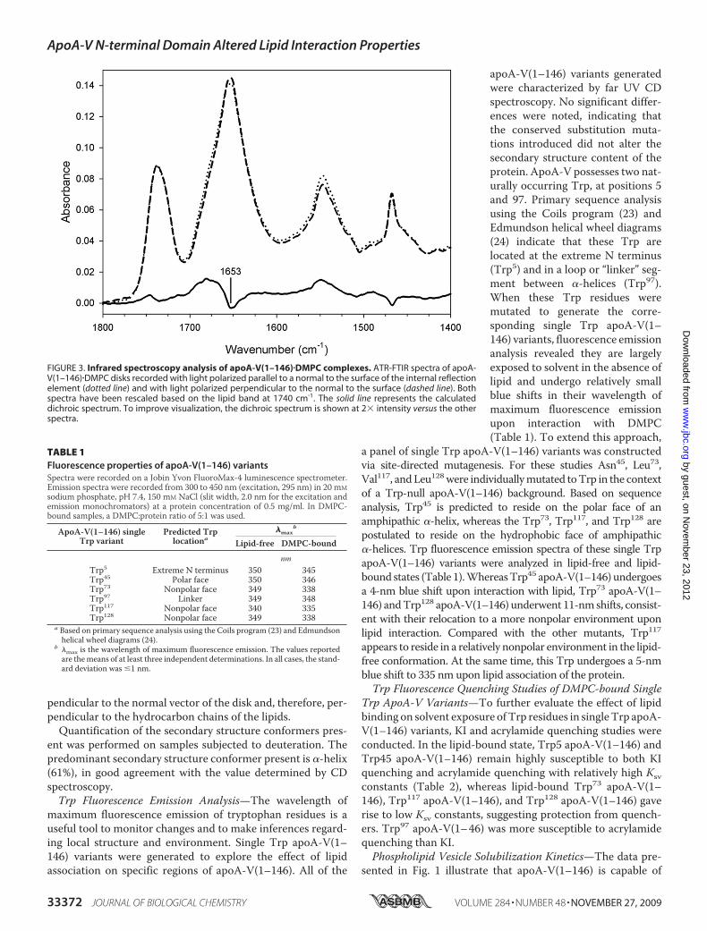
pendicular to the normal vector of the disk and, therefore, per-pendicular to the hydrocarbon chains of the lipids.Quantification of the secondary structure conformers pres-
ent was performed on samples subjected to deuteration. Thepredominant secondary structure conformer present is �-helix(61%), in good agreement with the value determined by CDspectroscopy.Trp Fluorescence Emission Analysis—The wavelength of
maximum fluorescence emission of tryptophan residues is auseful tool to monitor changes and to make inferences regard-ing local structure and environment. Single Trp apoA-V(1–146) variants were generated to explore the effect of lipidassociation on specific regions of apoA-V(1–146). All of the
apoA-V(1–146) variants generatedwere characterized by far UV CDspectroscopy. No significant differ-ences were noted, indicating thatthe conserved substitution muta-tions introduced did not alter thesecondary structure content of theprotein. ApoA-V possesses two nat-urally occurring Trp, at positions 5and 97. Primary sequence analysisusing the Coils program (23) andEdmundson helical wheel diagrams(24) indicate that these Trp arelocated at the extreme N terminus(Trp5) and in a loop or “linker” seg-ment between �-helices (Trp97).When these Trp residues weremutated to generate the corre-sponding single Trp apoA-V(1–146) variants, fluorescence emissionanalysis revealed they are largelyexposed to solvent in the absence oflipid and undergo relatively smallblue shifts in their wavelength ofmaximum fluorescence emissionupon interaction with DMPC(Table 1). To extend this approach,
a panel of single Trp apoA-V(1–146) variants was constructedvia site-directed mutagenesis. For these studies Asn45, Leu73,Val117, andLeu128were individuallymutated toTrp in the contextof a Trp-null apoA-V(1–146) background. Based on sequenceanalysis, Trp45 is predicted to reside on the polar face of anamphipathic �-helix, whereas the Trp73, Trp117, and Trp128 arepostulated to reside on the hydrophobic face of amphipathic�-helices. Trp fluorescence emission spectra of these single TrpapoA-V(1–146) variants were analyzed in lipid-free and lipid-bound states (Table 1).WhereasTrp45 apoA-V(1–146) undergoesa 4-nm blue shift upon interaction with lipid, Trp73 apoA-V(1–146) andTrp128 apoA-V(1–146) underwent 11-nmshifts, consist-ent with their relocation to a more nonpolar environment uponlipid interaction. Compared with the other mutants, Trp117appears to reside in a relatively nonpolar environment in the lipid-free conformation. At the same time, this Trp undergoes a 5-nmblue shift to 335 nm upon lipid association of the protein.Trp Fluorescence Quenching Studies of DMPC-bound Single
Trp ApoA-V Variants—To further evaluate the effect of lipidbinding on solvent exposure ofTrp residues in singleTrp apoA-V(1–146) variants, KI and acrylamide quenching studies wereconducted. In the lipid-bound state, Trp5 apoA-V(1–146) andTrp45 apoA-V(1–146) remain highly susceptible to both KIquenching and acrylamide quenching with relatively high Ksvconstants (Table 2), whereas lipid-bound Trp73 apoA-V(1–146), Trp117 apoA-V(1–146), and Trp128 apoA-V(1–146) gaverise to low Ksv constants, suggesting protection from quench-ers. Trp97 apoA-V(1–46) was more susceptible to acrylamidequenching than KI.Phospholipid Vesicle Solubilization Kinetics—The data pre-
sented in Fig. 1 illustrate that apoA-V(1–146) is capable of
FIGURE 3. Infrared spectroscopy analysis of apoA-V(1–146)�DMPC complexes. ATR-FTIR spectra of apoA-V(1–146)�DMPC disks recorded with light polarized parallel to a normal to the surface of the internal reflectionelement (dotted line) and with light polarized perpendicular to the normal to the surface (dashed line). Bothspectra have been rescaled based on the lipid band at 1740 cm�1. The solid line represents the calculateddichroic spectrum. To improve visualization, the dichroic spectrum is shown at 2� intensity versus the otherspectra.
TABLE 1Fluorescence properties of apoA-V(1–146) variantsSpectra were recorded on a Jobin Yvon FluoroMax-4 luminescence spectrometer.Emission spectra were recorded from 300 to 450 nm (excitation, 295 nm) in 20 mMsodium phosphate, pH 7.4, 150 mM NaCl (slit width, 2.0 nm for the excitation andemission monochromators) at a protein concentration of 0.5 mg/ml. In DMPC-bound samples, a DMPC:protein ratio of 5:1 was used.
ApoA-V(1–146) singleTrp variant
Predicted Trplocationa
�maxb
Lipid-free DMPC-bound
nmTrp5 Extreme N terminus 350 345Trp45 Polar face 350 346Trp73 Nonpolar face 349 338Trp97 Linker 349 348Trp117 Nonpolar face 340 335Trp128 Nonpolar face 349 338
a Based on primary sequence analysis using the Coils program (23) and Edmundsonhelical wheel diagrams (24).
b �max is the wavelength of maximum fluorescence emission. The values reportedare themeans of at least three independent determinations. In all cases, the stand-ard deviation was �1 nm.
ApoA-V N-terminal Domain Altered Lipid Interaction Properties
33372 JOURNAL OF BIOLOGICAL CHEMISTRY VOLUME 284 • NUMBER 48 • NOVEMBER 27, 2009
by guest, on Novem
ber 23, 2012w
ww
.jbc.orgD
ownloaded from

binding lipid to form reconstituted HDL. In an effort to char-acterize the relative lipid binding activity of this protein, kineticanalysis of apoA-V(1–146)-dependent solubilization of DMPCvesicles was measured. (Fig. 4). In control incubations lackingapolipoprotein, DMPC vesicles remain turbid with no changein solution light scattering intensity as a function of time (Fig. 4,curve a). Upon theadditionof full-lengthapoA-V, a time-depend-ent reduction in turbidity was observed with a calculated initialrateconstant (k)of1.3�10�2 s�1 (Fig. 4, curveb).The isolatedNTdomain, apoA-V(1–146), induced faster solubilization, with aninitial rate constant of 3.3 � 10�2 s�1 (Fig. 4, curve c).ApoA-V(1–146) Binding to Phospholipase C Modified LDL—
When human LDL is incubated with PL-C, enzymatic conver-sion of LDL phosphatidylcholine to diacylglycerol induces apo-lipoprotein association and prevents LDL particle aggregationand subsequent sample turbidity development (20). Incubationof LDL with PL-C in the absence of exogenous apolipoproteinresults in rapid sample turbidity development (Fig. 5, curve a),whereas control incubations lacking PL-C do not develop tur-bidity over time (Fig. 5, curve e). Although incubations withrecombinant human apoA-I remained clear (Fig. 5, curve d),incubations with apoE3 NT become turbid over time (Fig. 5,curve b), consistent with previous results (19). LDL incubationswith PL-C in the presence of apoA-V(1–146) showed an inter-
mediate level of protection from sample turbidity developmentas a function of time (Fig. 5, curve c).ApoA-V(1–146) Binding to VLDL and HDL from ApoA5�/�
Mice—It was previously reported that the naturally occurringtruncation variant, Q139X apoA-V, does not associate withlipoproteins in vivo (9). To evaluate the ability of apoA-V(1–146) to associate with VLDL and HDL in vitro, recombinantapoA-V(1–146) was incubated with apoA-V-deficient VLDLandHDLobtained from apoA5�/�mice. Following incubation,the lipoproteinswere reisolated by ultracentrifugation and ana-lyzed by immunoblotting (Fig. 6). The data show that, whereasfull-length apoA-V associateswith bothVLDL andHDL, apoA-V(1–146) does not associate withVLDL. Furthermore, whereasnearly all of the full-length apoA-V bound to HDL, only a frac-tion of apoA-V(1–146) was recovered in association with HDL.
DISCUSSION
Exchangeable apolipoproteins can transfer among lipopro-teins in plasma and, in the process, likely exist in a lipid-poor
FIGURE 4. Effect of apoA-V truncation on DMPC vesicle solubilizationkinetics. DMPC vesicles (500 �g) in 50 mM sodium citrate, pH 3.0, 150 mM
NaCl, were incubated at 23 °C in the absence (curve a) or presence of 100 �g offull-length apoA-V (curve b) or apoA-V (1–146) (curve c). Right angle light scat-tering was monitored at 600 nm as a function of time.
FIGURE 5. Effect of apolipoproteins on PL-C induced aggregation ofhuman LDL. Human LDL (50 �g protein) was incubated at 37 °C in theabsence (curve e) or presence (curve a) of PL-C (0.6 unit). Other incubationscontained LDL, PL-C, and 50 �g of apoE3 NT (curve b), apoA-V(1–146) (curve c),or apoA-I (curve d). Sample absorbance at 340 nm was measured as a functionof time. The values represent the means � standard deviation (n � 3).
FIGURE 6. Lipoprotein binding properties of apoA-V(1–146). VLDL or HDL(0.1 mg/ml protein), isolated from plasma of apoa5�/� mice were incubatedwith 0.02 mg/ml full-length apoA-V(1–343) (A) or 0.01 mg/ml apoA-V(1–146)(B) for 1 h at 22 °C in Tris-buffered saline. Following incubation, VLDL and HDLwere reisolated by density ultracentrifugation and subjected to immunoblotanalysis with goat anti-human apoA-V IgG. The results shown are represent-ative of at least three independent experiments. On the left of each panel is acontrol blot with recombinant apoA-V.
TABLE 2Trp fluorescence quenching of apoA-V(1–146) variants bound toDMPCSpectra were recorded on a Jobin Yvon FluoroMax-4 luminescence spectrometer.The average of two emission spectra were recorded from 320 to 355 nm (excitation,295 nm) in 20 mM sodium phosphate, pH 7.4, 150 mM NaCl (slit width, 2.0 nm forthe excitation and emission monochromators) at a protein concentration of 0.5mg/ml with a DMPC:protein ratio of 5:1.
ApoA-V(1–146) singleTrp variant
Predicted Trplocationa
Ksvb
KI Acrylamide
M�1
Trp5 Extreme N terminus 2.7 3.8Trp45 Polar face 2.8 3.6Trp73 Nonpolar face 1.0 2.2Trp97 Linker 1.4 4.1Trp117 Nonpolar face 1.1 2.0Trp128 Nonpolar face 1.0 2.2
a Based on primary sequence analysis using the Coils program (23) and Edmundsonhelical wheel diagrams (24).
b Ksv is the Stern-Volmer constant.
ApoA-V N-terminal Domain Altered Lipid Interaction Properties
NOVEMBER 27, 2009 • VOLUME 284 • NUMBER 48 JOURNAL OF BIOLOGICAL CHEMISTRY 33373
by guest, on Novem
ber 23, 2012w
ww
.jbc.orgD
ownloaded from

state. The helix bundle motif is postulated to facilitate thisexchange by promoting apolipoprotein solubility in bothpolar and nonpolar environments. The size of potential lipidsubstrate particles is an important factor regulating apoli-poprotein transfer in the circulation (25). ApoA-V is anexchangeable apolipoprotein that in humans, is found onchylomicrons, VLDL, and HDL (11). Presumably, apoA-Vtransfers among the different lipoprotein populations, asproposed by Nelbach et al. (21). The NT domain of apoA-Vadopts a helix bundle conformation in the absence of lipid,and this may facilitate exchange between lipoprotein parti-cles. In the present studies, we characterized the ability ofthis domain to bind to particles of various lipid compositionsand sizes, including DMPC vesicles, modified LDL, and HDLand VLDL from apoa5�/� mice, in an effort to gain insightinto its intrinsic lipid binding properties.Comparedwith other well studied exchangeable apolipopro-
teins, full-length apoA-V is unique in that it is not soluble atneutral pH in a lipid-free state (6). When bound to lipid, how-ever, apoA-V is soluble at physiological pH. This suggests thatduring transfer between lipoprotein particles in circulation,apoA-V may exist in a lipid-poor rather than lipid-free state.The ability of apoA-V(1–146) to formdiscoidal complexes withphospholipid in the size range of nascent HDL particles may bephysiologically relevant because apoA-V exchange betweenlipoprotein particles in circulation may require transient exist-ence in a lipid core-depleted particle.Upon lipid interaction, it appears that apoA-V(1–146)
undergoes a conformational change, as judged by an increase in�-helix secondary structure content and altered solvent expo-sure of reporter Trp residues. The increase in helix contentupon lipid interaction is similar to that of other exchangeableapolipoproteins, such as apoA-I (26). The amphipathic helixbundlemotif in the lipid-free state adopts a globular conforma-tion wherein the hydrophobic faces of its helices orient towardthe center of the bundle (27). Upon lipid association, the pro-tein is predicted to adopt an open conformation, where thehydrophobic faces of its amphipathic helices interact directlywith the lipid surface (27). Linear infrared dichroism experi-ments of apoA-V(1–146)�DMPC disks are consistent with amodel wherein the helices orient perpendicular with respect tothe DMPC bilayer fatty acyl chains. This indicates that, in thelipid-bound state, apoA-V(1–146) adopts a belt-like conforma-tion around the perimeter of the particle, as described for otherexchangeable apolipoproteins (28–33).The lipid-induced conformational change in apoA-V(1–146)
was studied using a panel of single Trp variants. Each single Trpvariant reported on a specific region within apoA-V(1–146).Certain Trp residues were predicted to reside in a linker regionbetween amphipathic helices or on the polar face of anamphipathic helix, whereas the others were predicted to resideon the nonpolar face of amphipathic helices. In keeping withthese predictions, only variants with Trp predicted to reside onthe hydrophobic face of amphipathic helices showed a blueshift in the wavelength of maximum fluorescence emissionbetween alternate lipid-free and lipid-bound states. This sug-gests that, whenpresentedwith a suitable lipid surface, the helixbundle opens, exposing the bundle interior.
To further characterize conformational adaptations inapoA-V(1–146), the relative exposure of various regions withinthe NT domain were investigated as a function of lipid binding.In Trp fluorescence quenching studies, it was observed thatsingle Trp apoA-V(1–146) variants with their Trp predicted tobe on the nonpolar face of amphipathic helices gave rise to thelowest Ksv values, suggesting that these regions of the proteinmaintain close contact with the lipid surface. For example,Trp97 is predicted to reside in a linker region between twoamphipathic helices. Consistent with this, Trp97 apoA-V(1–146) fluorescence emission was highly quenched by acrylamidebut less so by KI. This could be due to electrostatic repulsion ofKI by negatively charged residues located near Trp97.The ability of apoA-V(1–146) to initiate contact with lipid
surfaces is suggested by phospholipid vesicle solubilizationstudies comparing truncated and full-length apoA-V. The CTdomain of apoA-V has been previously shown to avidly bindlipid (7). Interestingly, despite the absence of the CT domain,apoA-V(1–146) solubilizes DMPC at a faster rate than full-length apoA-V. This suggests that N- and C-terminal domaininteractions in the intact protein modulate the lipid bindingproperties of apoA-V. This may be similar to interactions inapoA-I. In this apolipoprotein, the CT domain initiates lipid-binding, whereas the NT helix bundle opens up to stabilize thelipid-associated state (34).Although DMPC solubilization assays characterize apoli-
poprotein-induced phospholipid vesicle disruption andreorganization, PL-C-modified LDL provides a means toassess binding to spherical lipoprotein substrates. PL-Cactivity generates apolipoprotein-binding sites by hydrolyz-ing phosphatidylcholine moieties present in the surfacemonolayer (20). Conversion of phosphatidylcholine into dia-cylglycerol destabilizes the lipoprotein particle and pro-motes aggregation (35). Apolipoproteins protect LDLagainst PL-C-induced aggregation by forming a stable bind-ing interaction with the modified particles (20). ApoA-V(1–146) binding to PL-C-treated LDL was intermediate withrespect to other apolipoproteins examined. This finding isconsistent with the stability properties of these apolipopro-teins. At physiological pH apoA-V(1–146) is less stable thanapoE3 NT (guanidine HCl denaturation midpoint of 2.0 M
versus 2.5 M, respectively) (8, 36) yet is more stable thanapoA-I (1 M guanidine HCl denaturation midpoint) (37).Thus, it appears that the intrinsic stability of helix bundleapolipoproteins in solution correlates directly with the abil-ity to bind newly created sites on a spherical lipoproteinsubstrate (8).The ability of apoA-V(1–146) to associate with physiolog-
ically relevant lipoproteins was assessed using VLDL andHDL isolated from apoa5 knock-out mice. Importantly,apoA-V(1–146) failed to associate with VLDL and associatedsparingly with HDL. This finding is intriguing in light of thereport that, unlike full-length apoA-V, apoA-V(1–146) alsofails to associate with intracellular lipid droplets (38). In con-sidering the sizes of various lipid substrates, VLDL and lipiddroplets are considerably larger than HDL or other lipid par-ticles employed in this study. The data indicate that apoA-V(1–146) can associate with smaller lipid substrate particles
ApoA-V N-terminal Domain Altered Lipid Interaction Properties
33374 JOURNAL OF BIOLOGICAL CHEMISTRY VOLUME 284 • NUMBER 48 • NOVEMBER 27, 2009
by guest, on Novem
ber 23, 2012w
ww
.jbc.orgD
ownloaded from
to a limited degree but apparently lacks the ability to interactwith larger lipid particles. One explanation could be relatedto the tighter packing of phospholipid molecules on the sur-face of larger particles because of their decreased radius ofcurvature, as compared with smaller particles (39, 40).Tighter packing of phospholipid polar head groups envelop-ing a lipid core could interfere with the ability of apoA-V(1–146) to access hydrophobic surfaces and initiate binding.This aspect is obviated in the case of PL-C-modified LDL butnot in the binding experiments with VLDL and HDL. In thelatter case, differences in surface lipid composition and/orprotein content could also influence binding of apoA-V(1–146). Regardless, it is apparent that, with natural lipoproteinsubstrates in vitro, apoA-V(1–146) binding is defective.Because this is not due to an intrinsic inability to bind lipid,it suggests that the CT of apoA-V modulates the lipid inter-action properties of the NT domain.Naturally occurring apoA-V truncations, including Q139X
(9), Q148X (10), and Q97X (41), have been reported inhuman subjects and are associated with severe hypertriglyc-eridemia. The distribution of apoA-V in ultracentrifugallyisolated lipoprotein classes was evaluated in several carriersof the Q139X truncation mutation and demonstrated thatthe truncated form of the protein does not associate withplasma lipoproteins and is found only in the lipid-poor d �1.21 g/ml fraction. Because the Q139X apoA-V mutationnomenclature includes the 23-amino acid signal peptide, themature protein is actually 116 amino acids in length.Although apoA-V(1–146) is longer than these naturalmutants, binding studies with natural lipoproteins recapitu-late observations in human plasma of individuals carryingthese mutant forms of apoA-V.The present findings provide a potential explanation for
the HTG observed in patients with truncated apoA-V. Thelack of a CT domain alters lipid binding activity such thattruncated apoA-V fails to effectively bind to circulatinglipoproteins, particularly TG-rich particles. Currenthypotheses suggest apoA-V interactions with heparan-sul-fate proteoglycans (42, 43) and/or glycosyl phosphatidylino-sitol high density lipoprotein-binding protein-1 (44) indi-rectly enhances lipoprotein lipase activity to facilitatehydrolysis of VLDL- associated TG. The data fromNilsson etal. (45) indicate apoA-V also serves as a ligand for endocyticreceptors of the LDL receptor family, where it is possible thatapoA-V may have an important role in clearance of VLDLremnants. The putative binding site on apoA-V for lipopro-tein lipase activity enhancement and cell surface moleculeinteractions resides within the CT domain of the protein.The absence of this binding site most likely contributes todefective hydrolysis and clearance of TG-rich lipoproteins.In any case, association of apoA-V with TG-rich lipoproteinsis presumably required for manifestation of these effects. If aCT truncated apoA-V is unable to bind larger, TG-richlipoproteins, then the resulting apoA-V-deficient particlescould potentially have an increased plasma residence time,contributing to HTG. Thus, it may be that defective lipidbinding arising from the lack of a CT domain precludes bind-
ing to circulating lipoproteins, thereby preventing potentialTG lowering effects attributed to full-length apoA-V.
Acknowledgments—We thank Lisa Nelbach for providing mouseblood and Dr. Paul Weers for helpful discussions. We also thank Dr.Susan Marqusee for access to the circular dichroism spectrometer.
REFERENCES1. Kamtekar, S., and Hecht, M. H. (1995) FASEB J. 9, 1013–10222. Wilson, C., Wardell, M. R., Weisgraber, K. H., Mahley, R. W., and Agard,
D. A. (1991) Science 252, 1817–18223. Ajees, A. A., Anantharamaiah, G. M., Mishra, V. K., Hussain, M. M., and
Murthy, H. M. (2006) Proc. Natl. Acad. Sci. U.S.A. 103, 2126–21314. Breiter, D. R., Kanost, M. R., Benning, M. M., Wesenberg, G., Law, J. H.,
Wells, M. A., Rayment, I., and Holden, H. M. (1991) Biochemistry 30,603–608
5. Pennacchio, L. A., Olivier, M., Hubacek, J. A., Cohen, J. C., Cox, D. R.,Fruchart, J. C., Krauss, R. M., and Rubin, E. M. (2001) Science 294,169–173
6. van der Vliet, H. N., Sammels, M. G., Leegwater, A. C., Levels, J. H., Re-itsma, P. H., Boers, W., and Chamuleau, R. A. (2001) J. Biol. Chem. 276,44512–44520
7. Beckstead, J. A., Wong, K., Gupta, V., Wan, C. P., Cook, V. R., Weinberg,R. B., Weers, P. M., and Ryan, R. O. (2007) J. Biol. Chem. 282,15484–15489
8. Wong, K., Beckstead, J. A., Lee, D., Weers, P. M., Guigard, E., Kay, C. M.,and Ryan, R. O. (2008) Biochemistry 47, 8768–8774
9. Marcais, C., Verges, B., Charriere, S., Pruneta, V., Merlin, M., Billon, S.,Perrot, L., Drai, J., Sassolas, A., Pennacchio, L. A., Fruchart-Najib, J., Fru-chart, J. C., Durlach, V., and Moulin, P. (2005) J. Clin. Invest. 115,2862–2869
10. Priore Oliva, C., Pisciotta, L., Li Volti, G., Sambataro, M. P., Cantafora, A.,Bellocchio, A., Catapano, A., Tarugi, P., Bertolini, S., and Calandra, S.(2005) Arterioscler. Thromb. Vasc. Biol. 25, 411–417
11. O’Brien, P. J., Alborn, W. E., Sloan, J. H., Ulmer, M., Boodhoo, A., Knier-man, M. D., Schultze, A. E., and Konrad, R. J. (2005) Clin. Chem. 51,351–359
12. Beckstead, J. A., Oda, M. N., Martin, D. D., Forte, T. M., Bielicki, J. K.,Berger, T., Luty, R., Kay, C. M., and Ryan, R. O. (2003) Biochemistry 42,9416–9423
13. Fisher, C. A., Wang, J., Francis, G. A., Sykes, B. D., Kay, C. M., and Ryan,R. O. (1997) Biochem. Cell Biol. 75, 45–53
14. Ryan, R. O., Forte, T. M., and Oda, M. N. (2003) Protein Expr. Purif. 27,98–103
15. Nichols, A. V., Krauss, R.M., andMusliner, T.A. (1986)Methods Enzymol.128, 417–431
16. Sreerama, N., and Woody, R. W. (1993) Anal. Biochem. 209, 32–4417. Goormaghtigh, E., Cabiaux, V., and Ruysschaert, J. M. (1990) Eur. J. Bio-
chem. 193, 409–42018. Tallmadge, D. H., Huebner, J. S., and Borkman, R. F. (1989) Photochem.
Photobiol. 49, 381–38619. Weers, P. M., Narayanaswami, V., and Ryan, R. O. (2001) Eur. J. Biochem.
268, 3728–373520. Liu, H., Scraba, D. G., and Ryan, R. O. (1993) FEBS Lett. 316, 27–3321. Nelbach, L., Shu, X., Konrad, R. J., Ryan, R. O., and Forte, T. M. (2008) J.
Lipid Res. 49, 572–58022. Lindgren, F. T., Jensen, L. C., and Hatch, F. T. (1972) in Blood Lipids and
Lipoproteins: Quantitation, Composition, and Metabolism (Nelson, G. J.,ed) pp. 181–274, Wiley-Interscience, New York
23. Lupas, A., Van Dyke, M., and Stock, J. (1991) Science 252, 1162–116424. Schiffer, M., and Edmundson, A. B. (1967) Biophys. J. 7, 121–13525. Connelly, P. W., and Kuksis, A. (1981) Biochim. Biophys. Acta 666,
80–8926. Wald, J. H., Krul, E. S., and Jonas, A. (1990) J. Biol. Chem. 265,
20037–2004327. Segrest, J. P., Jones, M. K., De Loof, H., Brouillette, C. G., Venkatachalapa-
ApoA-V N-terminal Domain Altered Lipid Interaction Properties
NOVEMBER 27, 2009 • VOLUME 284 • NUMBER 48 JOURNAL OF BIOLOGICAL CHEMISTRY 33375
by guest, on Novem
ber 23, 2012w
ww
.jbc.orgD
ownloaded from

thi, Y. V., and Anantharamaiah, G. M. (1992) J. Lipid Res. 33, 141–16628. Raussens, V., Narayanaswami, V., Goormaghtigh, E., Ryan, R. O., and
Ruysschaert, J. M. (1995) J. Biol. Chem. 270, 12542–1254729. Raussens, V., Fisher, C. A., Goormaghtigh, E., Ryan, R. O., and Ruyss-
chaert, J. M. (1998) J. Biol. Chem. 273, 25825–2583030. Raussens, V., Drury, J., Forte, T. M., Choy, N., Goormaghtigh, E., Ruyss-
chaert, J. M., and Narayanaswami, V. (2005) Biochem. J. 387, 747–75431. Narayanaswami, V., Maiorano, J. N., Dhanasekaran, P., Ryan, R. O., Phill-
ips, M. C., Lund-Katz, S., and Davidson, W. S. (2004) J. Biol. Chem. 279,14273–14279
32. Martin, D. D., Budamagunta,M. S., Ryan, R. O., Voss, J. C., andOda,M.N.(2006) J. Biol. Chem. 281, 20418–20426
33. Silva, R. A., Schneeweis, L. A., Krishnan, S. C., Zhang, X., Axelsen, P. H.,and Davidson, W. S. (2007) J. Biol. Chem. 282, 9713–9721
34. Ji, Y., and Jonas, A. (1995) J. Biol. Chem. 270, 11290–1129735. Suits, A. G., Chait, A., Aviram, M., and Heinecke, J. W. (1989) Proc. Natl.
Acad. Sci. U.S.A. 86, 2713–271736. Wetterau, J. R., Aggerbeck, L. P., Rall, S. C., Jr., and Weisgraber, K. H.
(1988) J. Biol. Chem. 263, 6240–6248
37. Reijngoud, D. J., and Phillips, M. C. (1982) Biochemistry 21, 2969–297638. Shu, X., Ryan, R. O., and Forte, T. M. (2008) J. Lipid Res. 49, 1670–167639. Tajima, S., Yokoyama, S., and Yamamoto, A. (1983) J. Biol. Chem. 258,
10073–1008240. Wetterau, J. R., and Jonas, A. (1982) J. Biol. Chem. 257, 10961–1096641. Priore Oliva, C., Tarugi, P., Calandra, S., Pisciotta, L., Bellocchio, A., Ber-
tolini, S., Guardamagna, O., and Schaap, F. G. (2006) Atherosclerosis 188,215–217
42. Lookene, A., Beckstead, J. A., Nilsson, S., Olivecrona, G., and Ryan, R. O.(2005) J. Biol. Chem. 280, 25383–25387
43. Merkel, M., Loeffler, B., Kluger, M., Fabig, N., Geppert, G., Pennacchio,L. A., Laatsch, A., and Heeren, J. (2005) J. Biol. Chem. 280, 21553–21560
44. Beigneux, A. P., Davies, B. S., Gin, P., Weinstein, M. M., Farber, E., Qiao,X., Peale, F., Bunting, S., Walzem, R. L., Wong, J. S., Blaner, W. S., Ding,Z. M., Melford, K., Wongsiriroj, N., Shu, X., de Sauvage, F., Ryan, R. O.,Fong, L. G., Bensadoun, A., and Young, S. G. (2007) Cell Metab. 5,279–291
45. Nilsson, S. K., Lookene, A., Beckstead, J. A., Gliemann, J., Ryan, R. O., andOlivecrona, G. (2007) Biochemistry 46, 3896–3904
ApoA-V N-terminal Domain Altered Lipid Interaction Properties
33376 JOURNAL OF BIOLOGICAL CHEMISTRY VOLUME 284 • NUMBER 48 • NOVEMBER 27, 2009
by guest, on Novem
ber 23, 2012w
ww
.jbc.orgD
ownloaded from
Copyright © 2022 FDOKUMEN