ANTIOXIDANT EFFECT OF THYME ON PIRIMIPHOS METHYL INDUCED TOXICITY IN RATS
12
184 ANTIOXIDANT EFFECT OF THYME ON PIRIMIPHOS METHYL INDUCED TOXICITY IN RATS Hussein, S.A., Omayma, A. Ragab, Abd Elmaksoud, H., Afaf D. Abd El-Mageid and Alshaimaa, M. Said Biochemistry Department, Fac.Vet. Med. Benha Univ., Egypt. A B S T R A C T In the present study, the effects of thyme supplementation on lipid peroxidation and antioxidant enzymes in chronic intoxicated rats with organophosphorous pesticides have been evaluated. This study was carried out on 60 male rats. The rats were divided into four equal groups. 1) Normal group (N): received no drugs. 2) POM group (P): received single oral dose of POM (50 mg/ Kg b.w) daily for 3 months. 3) Thyme group (Thy): received single dose of Thyme per os (45 mg/ Kg b.w) daily for 3 months. 4) Thyme + POM group (Thy + P): received single dose of POM (50 mg/ Kg b.w) + Thyme (45 mg/ Kg b.w) per os daily for 3 months. Blood and brain samples were collected from all animal groups three times at one, two and three months from the onset of experiment and used for determination of antioxidant enzyme activities, non- enzymatic antioxidant (GSH and Ceruloplasmin) and free radicals (MDA and NO). Moreover, AChE activity in erythrocytes and brain were also determined. Pirimiphos methyl induced a significant decrease in antioxidant enzyme activities and erythrocytes and brain AChE. In addition, marked depletion in erythrocytes and brain GSH, were observed. Moreover, administration of Pirimiphos methyl exhibited a significant increase in serum ceruloplasmin and erythrocytes SOD and serum GGT activities in addition to a marked increase in erythrocytes and brain MDA and serum NO levels. While administration of thyme to rats received, oral dose of POM exhibited a significant increase in antioxidant activities, GSH, ceruloplasmin and a marked decrease in erythrocytes and brain MDA and serum NO. These results concluded that, thyme constitutes a powerful antioxidant property that was able to improve the adverse biochemical changes induced by organophosphorous compounds that are widely in our country. Key Words: Antioxidants, brain, erythrocytes, organophosphorous pesticides, oxidative stress, Pirimiphos methyl, Thyme. (BVMJ-24(1): 184-195, 2013) 1. INTRODUCTION esticides are used extensively in agriculture to enhance food production by eradicating unwanted insects and controlling disease vectors [33]. The unregulated use and its aerial application over large agricultural and urban areas has caused severe environmental pollution and potential health hazards. Organophosphate (OP) compounds are widely used and include some of the most toxic chemical agents. Recently, more than 100 different OP compounds have been synthesized and are extensively used worldwide in agriculture and public health programs as insecticides, acaricides and nematicides, in veterinary medicine as ectoparasiticides, and in commerce as lubricants, plasticizers, and flame-retardants [8]. Exposure to low-level of pesticides is P BENHA UNIVERSITY FACULTY OF VETERINARY MEDICINE BENHA VETERINARY MEDICAL JOURNAL BENHA VETERINARY MEDICAL JOURNAL, VOL. 24, NO. 1, JUNE 2013:184-195
Transcript of ANTIOXIDANT EFFECT OF THYME ON PIRIMIPHOS METHYL INDUCED TOXICITY IN RATS
184
ANTIOXIDANT EFFECT OF THYME ON PIRIMIPHOS METHYL INDUCED
TOXICITY IN RATS
Hussein, S.A., Omayma, A. Ragab, Abd Elmaksoud, H., Afaf D. Abd El-Mageid and
Alshaimaa, M. Said
Biochemistry Department, Fac.Vet. Med. Benha Univ., Egypt.
A B S T R A C T
In the present study, the effects of thyme supplementation on lipid peroxidation and antioxidant enzymes in
chronic intoxicated rats with organophosphorous pesticides have been evaluated. This study was carried out
on 60 male rats. The rats were divided into four equal groups. 1) Normal group (N): received no drugs. 2)
POM group (P): received single oral dose of POM (50 mg/ Kg b.w) daily for 3 months. 3) Thyme group
(Thy): received single dose of Thyme per os (45 mg/ Kg b.w) daily for 3 months. 4) Thyme + POM group
(Thy + P): received single dose of POM (50 mg/ Kg b.w) + Thyme (45 mg/ Kg b.w) per os daily for 3
months. Blood and brain samples were collected from all animal groups three times at one, two and three
months from the onset of experiment and used for determination of antioxidant enzyme activities, non-
enzymatic antioxidant (GSH and Ceruloplasmin) and free radicals (MDA and NO). Moreover, AChE
activity in erythrocytes and brain were also determined. Pirimiphos methyl induced a significant decrease
in antioxidant enzyme activities and erythrocytes and brain AChE. In addition, marked depletion in
erythrocytes and brain GSH, were observed. Moreover, administration of Pirimiphos methyl exhibited a
significant increase in serum ceruloplasmin and erythrocytes SOD and serum GGT activities in addition to
a marked increase in erythrocytes and brain MDA and serum NO levels. While administration of thyme to
rats received, oral dose of POM exhibited a significant increase in antioxidant activities, GSH,
ceruloplasmin and a marked decrease in erythrocytes and brain MDA and serum NO. These results
concluded that, thyme constitutes a powerful antioxidant property that was able to improve the adverse
biochemical changes induced by organophosphorous compounds that are widely in our country.
Key Words: Antioxidants, brain, erythrocytes, organophosphorous pesticides, oxidative stress, Pirimiphos
methyl, Thyme. (BVMJ-24(1): 184-195, 2013)
1. INTRODUCTION
esticides are used extensively in
agriculture to enhance food production
by eradicating unwanted insects and
controlling disease vectors [33]. The
unregulated use and its aerial application over
large agricultural and urban areas has caused
severe environmental pollution and potential
health hazards. Organophosphate (OP)
compounds are widely used and include some
of the most toxic chemical agents. Recently,
more than 100 different OP compounds have
been synthesized and are extensively used
worldwide in agriculture and public health
programs as insecticides, acaricides and
nematicides, in veterinary medicine as
ectoparasiticides, and in commerce as
lubricants, plasticizers, and flame-retardants
[8]. Exposure to low-level of pesticides is
P
BENHA UNIVERSITY FACULTY OF VETERINARY MEDICINE
BENHA VETERINARY MEDICAL JOURNAL
BENHA VETERINARY MEDICAL JOURNAL, VOL. 24, NO. 1, JUNE 2013:184-195
Hussein et al. (2013)
185
known to produce a variety of biochemical
changes, some of which may be responsible
for the adverse biological effects reported in
human and experimental studies. Conversely,
some biochemical alterations may not
necessarily lead to clinically recognizable
symptoms, although all the biochemical
responses can be used as markers of exposure
or effect. The biochemical changes induced
after exposure to pesticides or their active
metabolites include target cell/receptor
binding, protein and DNA adduct formation,
and induction or inhibition of enzymes.
Oxidative stress can also be induced by
pesticides, either by overproduction of free
radicals or by alteration in antioxidant
defense mechanisms, including
detoxification and scavenging enzymes [1].
There have been great efforts to find safe and
potent natural antioxidants from various plant
sources. As harmless sources of antioxidants,
wild herbs, spices, fruits, nuts, and leafy
vegetables have been investigated, for their
antioxidant properties, The increasing interest
in natural dietary components has focused
attention on plants used as food or spices
which are a rich source of bionutrients or bio-
active phytochemicals. Thyme (Thymus
vulgaris L.) are aromatic herbs that are used
extensively to add a distinctive aroma and
flavour to food. The leaves can be used fresh
or dried for use as a spice. Essential oils
extracted from fresh leaves and flowers can
be used as aroma additives in food,
pharmaceuticals, and cosmetics [22]. Thymus
vulgaris L is a perennial herb indigenous in
central and southern Europe, Africa and Asia
that are rich in essential oils and antioxidative
phenolic substances [42]. It is widely used in
folk medicine for the treatment of a variety of
diseases including gastroenteric and
bronchopulmonary disorders, anthelmintic,
antispasmodic, carminative, sedative,
diaphoretic [35]. It has been reported that
thyme possesses numerous biological
activities including antispasmodic,
antimicrobial, antioxidant and antifungal
[38]. Phenolic phytochemicals
(phenylpropanoids) serve as effective
antioxidants (phenolic antioxidants) due to
their ability to donate hydrogen from
hydroxyl groups positioned along the
aromatic ring to terminate free radical
oxidation of lipids and other biomolecules
[16]. Phenolic antioxidants therefore short-
circuit a destructive chain reaction that
ultimately degrades cellular membranes.
Accordingly, the aim of the present study was
to evaluate the antioxidant effect of rutin in
pirimiphos methyl intoxicated rats.
2. MATERIALS AND METHODS
2.1. Animals
Sixty adult white male albino rats weighting
150 - 200 g were used for the study. Rats were
housed in separated metal cages and kept at
constant environmental and nutritional
conditions throughout the period of
experiment. The animals were allowed free
access to standard dry rat diet and tap water
ad libitum.
2.2.Experimental design:
The rats were divided into four groups:
1) Control normal group (C): Comprized 15
rats, received no drugs, fed on normal diet for
3 months.
2) POM group (P): Consisted of 15 rats, were
fed on normal diet and administrated
Pirimiphos methyl orally for 3 months at a
dose level of 50 mg/ Kg b.w/day (1/40 LD50).
3) Thyme group (Thy): Composed of 15 rats,
were fed on normal diet and received single
oral dose of Thyme at a dose of 45 mg/ Kg
b.w/day dissolved in distilled water for 3
months.
4) Thyme + POM group (Thy + P): Included
15 rats, were fed on normal diet and received
single dose of Pirimiphos methyl per os daily
for 3 months at a dose of 50 mg/ Kg b.w (1/40
LD50) followed by oral administration of
Thyme at a dose level of 45 mg/ Kg b.w.

Antioxidant Effect Of Thyme
186
2.3. Sampling
1- Blood samples:
Blood samples were collected by ocular vein
puncture from all animal groups 3 times along
the duration of experiment in dry, clean and
screw capped heparinized tubes and plasma
were separated by centrifugation at 2500
r.p.m. for 15 minutes. The clean clear plasma
was separated by Pasteur pipette and kept in
a deep freeze at -20°C until used for
subsequent biochemical analysis Moreover,
after plasma separation, erythrocytes were
washed for subsequent biochemical analysis.
2- Brain specimens:
Rats were killed by decapitation. The brain
specimen quickly removed, cleaned by
rinsing with cold saline and stored at -20°C.
Briefly, brain tissues were minced into small
pieces, homogenized in normal saline 0.9%.
The homogenates were centrifuged at 10,000
for 15 minute at 4°C. The supernatant was
used for subsequent biochemical analysis.
2.4.Biochemical analysis
Biochemical analysis were determined
according to the methods described
previously. Erythrocytes and brain acetyl
cholinesterase (AChE) [21], Reduced
glutathione (GSH) [11], Lipid peroxidation
(MDA) [15], Catalase activity (CAT) [38],
serum Ceruloplasmin [36], Nitric oxide (NO)
[29], Gamma glutamyle transferase (γGT)
[26], erythrocytes Super oxide dismutase
activity (SOD) [30], Glutathione peroxidase
(GPx) [32], Glutathione reductase (GR) [18],
glutathione-S-transferase (GST) [19].
2.5. Statistical analysis
The results were expressed as mean ± SE and
statistical significance was evaluated by one
way ANOVA using SPSS (version 10.0)
program followed by the post hoc test, least
significant difference (LSD). Values were
considered statistically significant when p <
0.05.
3. RESULTS & DISCUSSION
The obtained data in table (1 & 2) revealed
that, administration of POM to normal rats
exhibited a significant decrease in
erythrocytes and brain AChE activities as
compared with C group allover the
experimental period. AChE activity is known
as biomarker of chronic toxicity in human
following pesticide exposure. It was recorded
that, in acute exposure, the main mechanism
of toxicity of (OP) is irreversibly binding to
the enzyme acetylcholinesterase and
inhibiting its activity that results in
accumulation and prolonged effect of
acetylcholine and consequently followed
with acute muscarinic and nicotinic effects.
[1]. Moreover, the recorded decrease in
AChE activity might be due to the inhibition
of the enzyme by the toxic metabolites of
OPI. It was recorded that, phosphorothioate
insecticides converted to their corresponding
oxygen analogs by called mixed-function
oxidases (MFO), a microsomal system of
enzymes among which the enzyme
cytochrome P450 (CYP450) plays a major
role. The oxons are direct inhibitors of AChE
[17].
However, administration of thyme to rats
exposed to POM exhibited a significant
increase in erythrocytes AChE activity after 2
and 3 months and in brain AChE activity after
3 months as compared with P group. The
recorded increase in AChE activity may be
related to the antioxidant capacity of thyme
which inhibit or decrease formation of free
radiacls resulted from POM administration.
This suggestion was supported by the
findings of [16] who mentioned that,
hydroxyl groups positioned along the
aromatic ring of thyme have the ability to
donate hydrogen thus terminate free radical
oxidation of lipids and other biomolecules
187
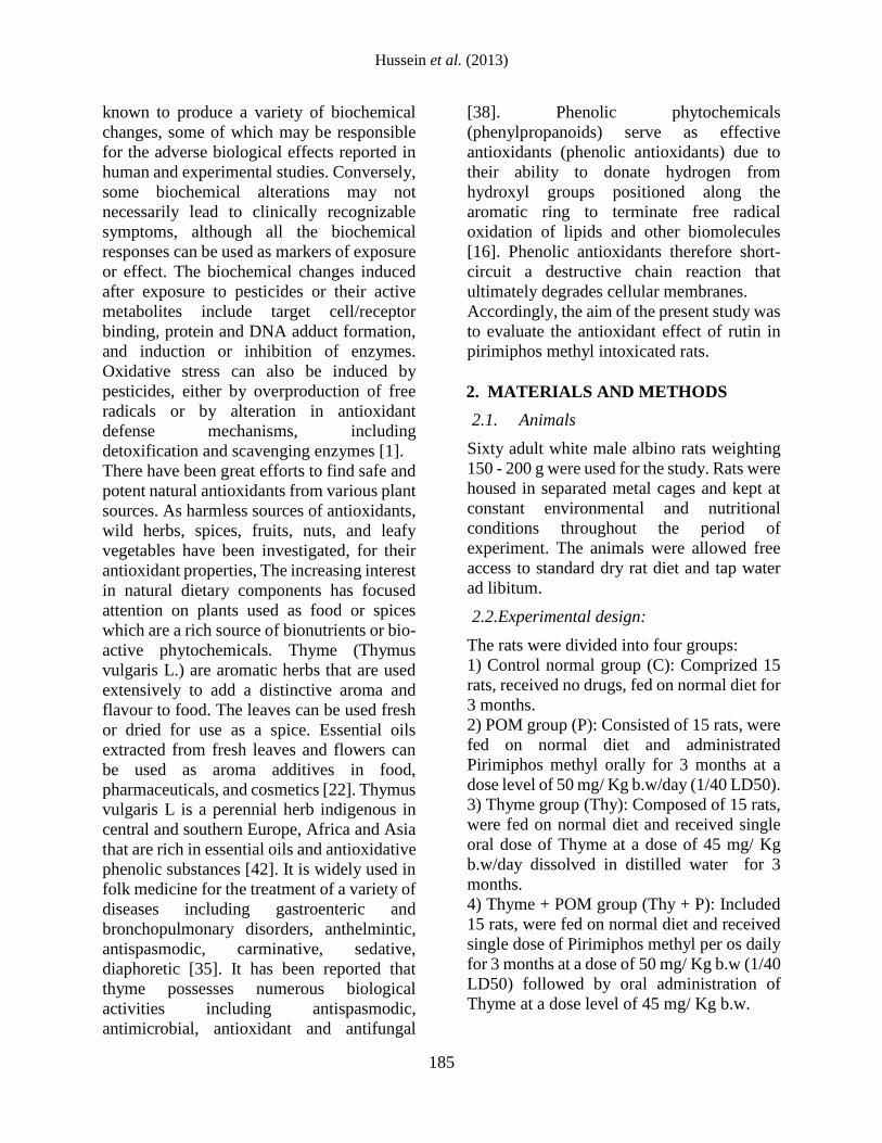
Table (1): Effect of Thyme administration on erythrocytes and brain AChE activity, MDA, GSH levels and serum NO, Ceruloplasmin
concentration in normal and Pirimiphos Methyl (POM) intoxicated rats:
Parameter
Groups
Eryth. AChE
(U/g Hb)
Brain AChE
(U/g tissue)
Eryth. MDA
(nmol/g Hg)
Brain MDA
(nmol/g tissue)
Serum NO
(μmol/L)
Eryth. GSH
(mg/g Hb)
Brain GSH
(mg/g tissue)
Serum Cerulo.
(mg/dl)
1st
Mo
nth
C 2.71± 0.05a
58.48± 0.54 a
7.59± 0.17 c
108.00± 2.00 c
24.60± 1.50 c
52.20 ± 0.97b
75.60 ± 1.69a
18.28 ± 0.75c
P 2.33 ± 0.02b
47.44 ± 0.59b
14.21± 0.24 a
379.60 ± 12.80a
50.80 ± 1.28a
32.40± 0.68 c
34.60 ± 1.50c
32.06± 0.69 b
Thy 2.75± 0.04 a
58.00± 0.91 a
7.41± 0.14 c
123.40± 3.14 c
24.80± 1.77 c
55.60± 1.44 b
75.00± 1.48 a
19.96± 1.18 c
Thy +P 2.32± 0.02 b
48.56± 0.83 b
12.15± 0.16 b
301.40± 5.70 b
42.20± 1.24 b
82.20±3.72 a
54.40± 1.66 b
51.26± 1.92 a
2n
d M
on
th
C 2.71 ± 0.07a
58.48 ± 0.50a
7.72 ± 0.08c
110.40 ± 4.17c
21.80 ± 1.32c
55.00± 1.67 b
73.80± 1.83 a
17.64± 0.26 c
P 2.30 ± 0.02c
47.66± 0.54 b
14.84± 0.27 a
350.20 ± 18.14a
52.00 ± 2.35a
33.60 ± 1.57c
34.40± 1.33 c
36.52± 1.46 b
Thy 2.75± 0.03 a
58.52± 0.44 a
7.41± 0.19 c
114.60± 6.55 c
23.80± 1.83 c
54.40± 1.44 b
75.00± 1.70 a
18.20± 0.33 c
Thy +P 2.54± 0.02 b
46.50± 0.29 b
11.52± 0.16 b
292.20± 4.26 b
41.20± 0.80 b
85.20± 2.31 a
57.40± 1.50 b
50.42± 2.38 a
3rd
Mon
th
C 2.74 ± 0.10a
58.10± 0.47 a
7.74 ± 0.09c
112.00± 4.34 c
23.80± 1.46 c
51.80± 3.6 b
76.60 ± 1.33a
17.90± 0.42 c
P 2.11± 0.03 b
42.10± 0.36 c
16.66± 0.31 a
377.00± 11.32 a
51.40± 2.16 a
33.00± 1.5 c
34.80± 1.62 c
23.04± 1.36 b
Thy 2.74± 0.04 a
58.10± 0.71 a
7.72± 0.11 c
110.60± 3.14 c
25.20± 1.98 c
54.60± 2.80 b
76.80± 1.24 a
19.36± 1.00 c
Thy +P 2.77± 0.03 a
46.24± 0.87 b
10.88± 0.25 b
245.60± 6.73 b
33.00± 1.76 b
75.80± 2.41 a
65.80± 1.77 b
35.16± 0.57 a
(C: Control Normal group, P: POM group, Thy: Thyme group, Thy + P: Thyme + POM group), Data are presented as (Mean ± S.E). S.E = Standard
error. Mean values with different superscript letters in the same column are significantly different at (P<0.05).
Antioxidant Effect Of Thyme
188
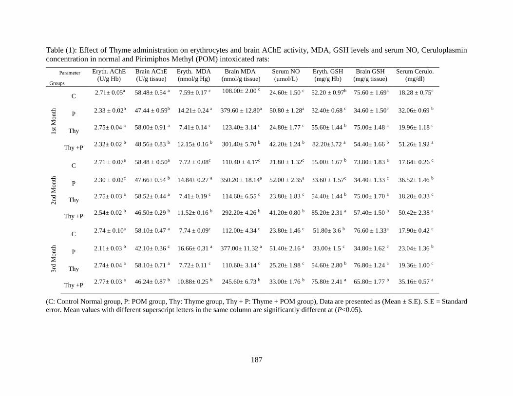
Table (2): Effect of Thyme administration on erythrocytes and brain antioxidant enzymes in normal and Pirimiphos Methyl (POM)
intoxicated rats:
Parameter
Groups
Eryth. SOD
(U/g Hb)
Eryth. CAT
(U/g Hg)
Brain CAT
(U/g tissue)
Eryth. GPx
(mU/g Hb)
Eryth. GR
(U/g Hb)
Eryth. GST
(U/g Hb)
Serum GGT
(U/L)
1st
Mo
nth
C 33.54± 0.55 c
2.74± 0.07 a
13.22± 0.28 a
16.82± 0.44 a
6.68± 0.13 a
5.30± 0.05 a
3.31± 0.16 c
P 59.74 ± 0.57a
0.98± 0.04 b
7.36± 0.18 c
10.70±0.18 b
3.10± 0.11 c
4.28± 0.04 b
11.01± 0.15 a
Thy 34.30± 0.36 c
2.84± 0.05 a
13.62± 0.20 a
17.07± 0.52 a
6.52± 0.19 a
5.35± 0.17 a
3.68± 0.29 c
Thy +P 42.14± 1.19 b
1.04± 0.09 b
9.88± 0.07 b
12.01± 0.60 b
4.34± 0.18 b
5.25± 0.05 a
9.91± 0.50 b
2n
d M
on
th
C 33.54± 0.53 c
2.80± 0.08 a
13.62± 0.26 a
17.72± 0.27 a
6.40± 0.231 a
5.31± 0.02 a
3.68± 0.22 c
P 64.32± 0.51 a
0.90± 0.07 c
6.96± 0.05 c
10.20± 0.27 c
2.73± 0.17 c
4.42± 0.15 b
10.62± 0.15 a
Thy 34.16± 0.28 c
2.72± 0.06 a
13.52± 0.22 a
18.01± 0.28 a
6.85± 0.23 a
5.47± 0.15 a
4.02± 0.18 c
Thy +P 44.30± 1.05 b
1.72± 0.15 b
9.80± 0.07 b
13.11± 0.33 b
4.77± 0.24 b
5.38± 0.11 a
6.03± 0.58 b
3rd
Mon
th
C 31.62± 0.57 c
2.88± 0.07 a
13.34± 0.72 a
18.19± 0.30 a
6.64± 0.19 a
5.50± 0.13 a
3.84± 0.29 b
P 66.56± 2.11 a
0.86± 0.05 c
7.12± 0.10 c
8.91± 0.35 c
2.71± 0.24 c
4.47± 0.05 b
10.60± 0.22 a
Thy 34.40± 0.90 c
2.52± 0.16 a
13.68± 0.17 a
17.89± 0.27 a
6.64± 0.17 a
5.42± 0.14 a
4.10± 0.27 b
Thy +P 41.16± 0.83 b
1.76± 0.18 b
9.88± 0.07 b
14.60± 0.25 b
5.74± 0.10 b
5.17± 0.11 a
4.20± 0.23 b
(C: Control Normal group, P: POM group, Thy: Thyme group, Thy + P: Thyme + POM group). Data are presented as (Mean ± S.E). S.E = Standard
error. Mean values with different superscript letters in the same column are significantly different at (P<0.05).

189
The obtained data in table (1) revealed that,
administration of POM to normal rats
exhibited a significant increase in
erythrocytes and brain (MDA) and serum NO
levels when compared to C group allover the
experimental period. LPO has been
implicated in a number of deleterious effects
such as increased membrane rigidity, osmotic
fragility, decreased cellular deformation,
reduced erythrocyte survival, and membrane
fluidity. Increase in the levels of TBARS
indicates enhanced lipid peroxidation leading
to tissue injury and failure of the antioxidant
defense mechanisms to prevent the formation
of excess free radicals [12].The recorded
results might be due to induction of
cytochrome 450, inhibition of AChE and
disturbance in activities of GSH and GST
enzymes causing lipid peroxidation as
reported by [34]. Another suggestion for the
recorded results stated by [39] who suggested
that, dimethoate induced LPO in liver could
possibly result from an enhanced microsomal
oxidative capacity induced by the insecticide.
Thus, elevated levels of cytochrome P450
lead to high rates of radicals production,
which, in turn, increase the rate of lipid
peroxidation.
Nitrate and nitrite [a marker of endogenous
nitric oxide (NO) production], possesses both
antioxidant and pro-oxidant properties. An
antioxidative property of NO has been
reported in the many studies [6]. NO is an
effective chain-breaking antioxidant in free
radical-mediated LPO and reacts rapidly with
peroxyl radicals as a sacrificial chain-
terminating antioxidant. It is well
documented that iNOS produces NO and NO-
derived reactive nitrogen species such as
peroxynitrite. In healthy neuronal tissue,
iNOS is not commonly present, but it can be
expressed by astrocyte, neurons, and
endothelial cells after brain offence where it
can initiate the production of high amounts of
NO. Overproduction of NO may lead to
neuronal damage and death. The reaction
between NO and super-oxide anion generates
the cytotoxic compound, peroxynitrite, that
leads to neuronal toxicity [41]. Under normal
physiological conditions, antioxidant
enzymes are responsible to eliminate the
highly reactive molecules. However, under
unphysiological conditions, the excessive
accumulation of reactive species induces
several cellular dysfunctions [40].
However, administration of thyme to POM
intoxicated rats exhibited a significant
decrease in erythrocytes and brain (MDA)
and serum NO levels allover the experimental
period as compared with P group. The
recorded results may be related to the
antioxidant properties of phenolic
compounds found in thyme [10]. It is well
documented that the leaves and flowers of
plants containing numerous aroma chemicals.
Phenolic phytochemicals are thought to
promote optimal health partly via their
antioxidant and free radical scavenging
effects thereby protecting cellular
components against free radical induced
damage [13].
The obtained data in table (1) revealed that,
administration of POM to normal rats
exhibited a significant decrease in
erythrocytes and brain (GSH) level and
significant increase in serum Ceruloplasmin
level when compared to C group allover the
experimental period. The recorded results
may be attributed to the utilization of GSH in
the metabolism of pesticides through GST.
The study of [36] revealed that, lindane,
malathion and propoxur increased the activity
of GST by conjugation of GSH to pesticides
in vivo. This could be understood in view of
the fact that some pesticides (organochlorine
and organophosphate) consume GSH through
GST catalyzed reaction as a major way of
detoxification of these chemicals. In contrast,
other classes of pesticides such as carbamate
may utilize GSH in conjugation reaction but
only in minor amounts compared to
organophosphate or organochlorine [3].
However, administration of thyme to POM
intoxicated rats exhibited a significant

Antioxidant Effect Of Thyme
190
increase in erythrocytes and brain reduced
glutathione (GSH) and serum Ceruloplasmin
level allover the experimental period as
compared with P group. These results may be
attributed to the antioxidant activity of thyme.
This suggestion was supported by the
findings of [4] who mentioned that, TOH
could optimally antagonize radiation-induced
toxicity, which may be due to its free radical
scavenging potential, by normalizing the
intracellular antioxidant levels, also by its
anti-lipid peroxidative potential.
The obtained data in table (2) revealed that,
administration of POM to normal rats
exhibited a significant increase in
erythrocytes (SOD) activity allover the
experimental period when compared to C
group. The increased in SOD activity has
been attributed to activation of the
compensatory mechanism through the effect
of OP on progenitor cells with its extent
depending on the magnitude of the oxidative
stress and hence, on the dose of the stressor.
Supporting this idea, there is evidence that
administration of malathion for 4 weeks
increases the (SOD) activity in erythrocytes
and liver [2].
However, administration of thyme to POM
intoxicated rats exhibited a significant
decrease allover the experimental period as
compared with P group. The recorded results
may be related to the antioxidant activity of
thyme and its free radical scavenging power
through its phenolic hydroxyl group as
reported by [4].
The obtained data in table (2) revealed that,
administration of POM to normal rats
exhibited a significant decrease in
erythrocytes and brain CAT, erythrocytes
GPx, GR and GST activities allover the
experimental period as compared with C
group . The decrease in antioxidant enzymes
has been interpreted as an indirect inhibition
of the enzymes resulting from the binding of
oxidative molecules produced during
pesticide metabolism. Three enzyme systems
(GST, esterases and monoxygenases) are
involved in the detoxification of
organophosphate insecticide class. These
enzymes act by rapidly metabolizing the
insecticide to non-toxic products or by
rapidly binding and very slowly turning over
the insecticide [24]. The present results
confirm the previous reports of [14], who
showed that repeated administration of
dimethoate induced disturbances in the
activities of the enzyme regulating GSH
metabolism. Glutathione S-transferases are
detoxifying enzymes that catalyze the
conjugation of a variety of electrophilic
substrates to the thiol group of GSH,
producing less toxic forms. As demonstrated
in other studies, the activities of antioxidant
enzymes can be altered in a variety of animal
tissues poisoned with OPI [20]. Generally,
oxidative stress results in reduction in tissue
antioxidants because these agents are utilized
in terminating the lipid peroxidation chain
reactions.
However, administration of thyme to POM
intoxicated rats exhibited a significant
increase in erythrocytes CAT activity after 2
and 3 months and a significant increase in
brain CAT activity allover the experimental
period. Also, daily dose of thyme to rats
which received oral dose of POM exhibited a
significant increase in erythrocytes GPx after
2 and 3 months. Moreover, daily dose of
thyme to rats which received oral dose of
POM exhibited a significant increase in
erythrocytes GR and GST activities allover
the experimental period when compared to P
group. The recorded results might be due to
the capacity for free radical trapping due to
the presence of the phenolic hydroxyl group
in thyme as reported by [4] who observed
that, pretreatment of TOH increased the
activity of SOD, GST and CAT in gamma
irradiated V79 cells. The radioprotective
effect of TOH can thus be explained by the
scavenging of free radicals generated by
radiation before they cause damage to cellular
macromolecules. Moreover, [26] attributed
the antioxidant activity of thyme to the

Hussein et al. (2013)
191
presence of phenolic compounds thymol and
carvacrol. Another suggestion for the
recorded results stated by [43] who reported
that, the antioxidant properties of thyme oil
are being utilised by the cells, thus sparing the
intracellular antioxidant systems such as SOD
and GPx. It is also possible that thyme oil is
influencing other cellular systems suggesting
more detailed examination of further
antioxidant parameters is required.
The obtained data in table (2) revealed that,
administration of POM to normal rats
exhibited a significant increase in serum
(γGT) activity allover the experimental
period when compared to C group. Among
the enzymes usually determined to evaluate
hepatic function, GGT is considered by many
authors to be a reliable biomarker closely
involved in the establishment of oxidative
stress damage [31]. This enzyme has a central
role in glutathione hepatic re-synthesis.
Moreover, as suggested by [27], it has an
inverse relationship with the levels of many
other antioxidants. [5] observed that GGT is
more sensitive than other enzymes (AST,
ALT, and ALP), changing by almost 90
percent compared to control values. In
addition, this enzyme is positively correlated
with LDH, total copper and NCBC and is
negatively correlated with the production of
albumin. GGT has been used as a biomarker
of pesticide-induced liver damage, and other
researchers have demonstrated an association
between increased activity of this enzyme and
reduced antioxidant ability in rats [27] and
humans [24]. Biological significance of γ-
GT-dependent lipid peroxidation in vivo
might be multifold. Varying levels of γ-GT
activity can be detected in erythrocytes and
lymphocytes. It is conceivable that the pro-
oxidant effects of γ-GT activity are normally
balanced by its established role in favoring
the cellular uptake of precursors for GSH
resynthesis, thus allowing the reconstitution
of cellular antioxidant defense [9]. The
increased serum (γGT) activity has been
attributed to the significant tissue injury
provoked by pesticides, even at low doses
employed in this study as stated by [7].
However, administration of thyme to POM
intoxicated rats exhibited a significant
decrease allover the experimental period as
compared with P group. It has been
hypothesized that one of the principal causes
of leakage of cellular enzyme into plasma is
hepatic injury as reported by [23].When the
liver cell plasma membrane is damaged, a
variety of enzymes normally located in the
cytosol are released into blood stream. The
recorded results may be related to antioxidant
activity of chlorophyllin and its radical
scavenging capacity to free radicals produced
due to metabolism of organophosphorous
pesticides by cytochrome P450 [39]. These
radicals increase the rate of lipid
peroxidation, increased membrane rigidity,
osmotic fragility, decreased cellular
deformation, reduced cellular survival, and
membrane fluidity [12]. Administration of
antioxidants restores the imbalance in
antioxidant defense mechanism and preserves
the struc¬tural integrity of the hepatocellular
membrane against free radicals [28].
From the obtained results it could be
concluded that, chronic toxicity
experimentally induced with POM in rats
extensively alters and induced disturbances in
enzymatic and non enzymatic antioxidant
system in erythrocytes and brain tissues.
Moreover, thyme administration efficiently
protects erythrocytes and brain from
deleterious effect of oxidative stress induced
by POM. This study suggested that, thyme
may be effective in controlling oxidative
damage through its powerful antioxidant
capacity.
4. REFERENCES
1. Abdollahi, M., Mostafalou, S.,
Pournourmohammadi, S., Shadnia, S.
2004. Oxidative stress and cholinesterase
inhibition in saliva and plasma of rats
following subchronic exposure to

Antioxidant Effect Of Thyme
192
Malathion. Comparative Biochemistry
and Physiology Part C 137: 29–34.
2. Akhgari, M., Abdollahi, M.,
Kebryaeezadeh, A., Hosseini, R.,
Sabzevari, O. 2003. Biochemical
evidence for free radical induced lipid
peroxidation as a mechanism for
subchronic toxicity of Malathion in
blood and liver of rats. Hum. Exp.
Toxicol. 22: 205–211.
3. Almeida, M.G., Fanini, F., Davino, S.C.,
Aznar, A.E., Koch, O.R., Barros, S.B.M.
1997. Pro- and anti-oxidant parameters
in rat liver after short-term exposure to
hexachlorobenzene. Hum. Exp. Toxicol.
16: 257–261.
4. Archana, P. R., Rao B. N., Ballal M., Rao
B. S.S. 2009. Thymol, a naturally
occurring monocyclic dietary phenolic
compound protects Chinese hamster lung
fibroblasts from radiation-induced
cytotoxicity. Mutation Research 680:
70–77.
5. Arnal, N., Astiz, M., deAlaniz, M.J.T.,
Marra, C.A. 2011. Clinical parameters
and biomarkers of oxidative stress in
agricultural workers who applied copper-
based pesticides. Ecotoxicology and
Environmental Safety 74: 1779–1786.
6. Aslan, A., Cemek, M., Eser, O.,
Altunbas, K., Buyukokurog˘lu, M.E.,
Cosar, M., Bas, O., Ela, Y., Fidan, H.
2009. Does dexmedetomidine reduce
secondary damage after spinal cord
injury? An experimetal study, Eur. Spine
J. 18: 336–344.
7. Astiz, M., Maria, J.T., Carlos, A.M.,
2009a. The impact of simultaneous
intoxication with agrochemicals on the
antioxidant defense system in rat.
Pesticide Biochemistry and Physiology
94: 93–99.
8. Balali-Mood M., Balali-Mood K., 2008.
Neurotoxic disorders of
organophosphorus compounds and their
managements, Arch. Iran. Med. 11: 65–
89.
9. Banerjee, B. D., Seth, V., Bhattacharya,
A., Pasha, S.T., Chakraborty, A. K. 1999.
Biochemical effects of some pesticides
on lipid peroxidation and free-radical
scavengers. Toxicology Letters 107: 33–
47
10. Baranauskiene, R., Venskutonis, P.R.,
Viskelis, P., Dambrauskiene, E. 2003.
Influence of nitrogen fertilizers on the
yield and composition of thyme (Thymus
vulgaris) J. Agric. Food Chem. 51: 7751–
7758.
11. Beutler, E., Duron, O., and Kelly, B. M.
1963. Improved method for
determination for liver glutathione. J.
Lab. Clin. Med. 61: 882 888.
12. Comporti, M. 1985. Lipid peroxidation
and cellular damage in toxic liver injury,
Lab. Invest. 53: 599–603.
13. Dapkevicius, A., van Beek, T.A.,
Lelyveld, G.P., van Veldhuizen, A., de
Groot, A., Linssen, J.P.H., Venskutonis,
R. 2002. Isolation and structure
elucidation of radical scavengers from
Thymus vulgaris leaves. J. Nat. Prod. 65:
892–896.
14. El-Sharkawy, A.M., Abdel-Rahman,
S.Z., Hassan, A.A., Gabr, M.H., El-
Zoghby, S.M., El-Sewedy, S.M. 1994.
Biochemical effects of some insecticides
on the metabolic enzymes regulating
glutathione metabolism. Bull. Environ.
Contam. Toxicol. 52: 505–510.
15. Esterbauer, H., Cheeseman, K.H.,
Danzani, M.U., Poli, G., and Slater, T.F.
1982. Separation and characterization of
the aldehyde products of ADP/Fe+2 C
stimulated lipid peroxidation in rat liver
microsomes. Biochem. J, 208: 129-140.
16. Foti, M., Piattelli, M., Amico, V.,
Ruberto, G. 1994. Antioxidant activity of
phenolic meroditerpenoids from marine
algae. J Photochem Photobiol, 26: 159–
64.
17. Giri, S., Prasad, S. B., Giri, A., Sharma,
G. D., (2002): Genotoxic effects of
Malathion: an organophosphorus
Hussein et al. (2013)
193
insecticide, using three mammalian
bioassays in vivo. Mutat. Res. 514: 223–
231.
18. Goldberg, D.M., and Spooner, R.J. 1983.
UV method for determination of
glutathione reductase activity. Methods
of enzymatic analysis (Bergmeyen, H.V.
Ed.) Third Ed, pp. 258-265.
19. Habig, W., Pabst, M., and Jakoby, W.
1974. Colorimetric determination of
Glutathion S Transferase. J. Biol. Chem
249: 7130-7139.
20. Hazarika, A., Sarkar, S.N., Hajare, S.,
Kataria, M., Malik, J.K. 2003. Influence
of Malathion pretreatment on the toxicity
of anilofos in male rats: a biochemical
interaction study. Toxicology 185: 1-8
21. Henry, R. J. 1974. Colorimetric assay for
the determination of Cholinesterase in
serum and plasma. Clin. Chem.,
Principle and Tech. Harber u. Row
Puplishers Inc., II edition, pp. 917.
22. Javanmardi, J., Khalighi, A., Kashi, A.,
Bais, H. P., Vivanco, J. M. 2002.
Chemical characterization of basil
(Ocimum basilicum L.) found in local
accessions and used in traditional
medicines in Iran. Journal of Agricultural
and Food Chemistry, 50: 5878–5883.
23. Kumar, S.S., Shankar, B., Sainis, K.B.,
2004. Effect of chlorophyllin against
oxidative stress in splenic lymphocytes
in vitro and in vivo. Biochimica et
Biophysica Acta 1672: 100–111
24. L´opez, O.A.F., Hern´andez, L.,
Rodrigo, F., Gil, G., Pena, J.L., Serrano,
T., Parr´on, E., Villanueva, A. 2007.
Changes in antioxidant enzymes in
humans with long-term exposure to
pesticides. Toxicology Letters 171: 146–
153.
25. Lee, D.H. 2003: kinetic colorimetric
method for determination of γ-GT
activity. Clin. Chem. 49: 1358-1366.
26. Lee, K.G., and Shibamoto, T. 2002.
Determination of antioxidant potential of
volatile extracts isolated from various
herbs and spices. J. Agric. Food Chem.
50: 4947–4952.
27. Lim, J.S., Yang, J.H., Chun, B.Y., Kam,
S., Jacobs, D.R., Lee, D.H. 2004. Is
serum g glutamyl transferase inversely
associated with serum antioxidants as a
marker of oxidative stress? Free Radic.
Biol. Med. 37:1018–1023.
28. Mahmoud, A.M. 2011. Influence of rutin
on biochemical alterations in
hyperammonemia in rats. Experimental
and Toxicologic Pathology.
29. Montgomery, H.A.C., and Dymock, J. F.
1961. Colorimetric determination of
nitrite. Analyst 86: 414.
30. Nishikimi, M., Rao, N.A., and Yogi, K.
1972. Colorimetric determination of
super oxide dismutase. Biochem. Bioph.
Common. 46: 849-854.
31. O¨zer, S., Ayfer, T., G¨ ulden, O., Sule,
C., Gazi, C., Nursal, G., G¨ oksel, S.,
2008. Protective effect of resveratrol
against naphthalene-induced oxidative
stress in mice. Ecotoxicol. Environ.
Safety 71: 301–308.
32. Paglia, D.E., and Valetine W.N. 1967.
UV method for determination of
glutathione peroxidase activity. J. Lab.
Clin. Med. 70: 158-169.
33. Pajoumand, A., Jalali, N., Abdollahi, M.,
Shadnia, S. 2002. Survival following
severe aluminum phosphide poisoning. J.
Pharm. Pract. Res. 32: 297–299.
34. Roy, S., Roy, S., Sharma, C.B. 2004.
Fenitrothion-induced changes in lipids of
rats, Biomed. Chromatogr. 18: 648–654.
35. Rustaiyan, A., Masoudi, S.h., Monfared,
A., Kamalinejad, M., Lajevardi, T.,
Sedaghat, S.Y. 2000. Volatile
constituents of three Thymus species
grown wild in Iran. Planta Med. 66: 197.
36. Schoslnsky, K. H., Lehmann, H. P., and
Beeler, M.F. 1974. Measurement of
ceruloplasmin from its oxidase activity in
Serum by Use of O-Dianisidine
Dihydrochioride. Clinical Chemistry, 20:
1556-1563.

Antioxidant Effect Of Thyme
194
37. Silva, A.P., Meotti, F.C., Santos, A.R.S.,
Farina, M. 2006: Lactational exposure to
Malathion inhibits brain
acetylcholinesterase in mice.
NeuroToxicology 27: 1101–1105
38. Sinha, A.K., 1972. Colorimetric assay of
catalase. Analytical Biochemistry 47:
389.
39. Soliman, K.M., and Badeaa, R.I. 2002.
Effect of oil extracted from some
medicinal plants on different
mycotoxigenic fungi. Food Chem.
Toxicol. 40: 1669–1675.
40. Thampi, H.B.S., Manoj, G., Leelamma,
S., Menon, V.P. 1991. Dietary fiber and
lipid peroxidation: effect of dietary fiber
on levels of lipids and lipid peroxides in
high fat diet, Indian J. Exp. Biol. 29:
563–567.
41. Thannickal, V.J., Fanburg, B.L. 2000.
Reactive oxygen species in cell
signaling. Am J Physiol Lung Cell Mol
Physiol 279: 1005–1028.
42. Vallance, P., Leiper, J. 2002. Blocking
NO synthesis: how, where and why? Nat
Rev Drug Discov. 1: 939–950.
43. WHO, (1999): WHO Monographs on
Selected Medicinal Plants, vol. 1
(Geneva).
44. Youdim, K.A., and Deans, S.G. 1999.
Dietary supplementation of thyme
(Thymus vulgaris L.) essential oil during
the lifetime of the rat: its effects on the
antioxidant status in liver, kidney and
heart tissues. Mechanisms of Ageing and
Development 109: 163–175.

Hussein et al. (2013)
195
التأثير المضاد لألكسدة للزعتر على التسمم المحدث باستخدام البيريميفوس ميثيل فى فئران التجارب محمد سعيد الشيماء-المجيدعبد دسوقيعفاف –حسين عبد المقصود على –أميمة أحمد رجب –على حسين سامي
جامعة بنها – البيطريكلية الطب –قسم الكيمياء الحيوية
العربي الملخص
وقد يسبب عالمي.الالمستوي علىأصبحت المركبات الفسفورية العضوية هي أكثر المبيدات الحشرية انتشارًا اليوم على االطالق التعرض المزمن لهذه المركبات أشد الضرر على الصحة، حيث أنها قد تسبب السرطان، واضطراب فى وظائف األيض والهرمونات
فئران المحدث فى دم ومخ ال التأكسديللزعتر على اإلجهاد الحيوي الكيميائيالتأثير إلى دراسةولذلك يهدف هذا البحث .والمناعةمن ذكور الفئران البيضاء 06فيها التسمم المزمن تجريبيًا باستخدام مادة البيريميفوس ميثيل. وقد أجريت هذه الدراسة على عدد
05اربعة مجموعات متساوية اشتملت كل مجموعة على عدد إلىالفئران جرام هذا وقد تم تقسيم 066-056بين وتتراوح أوزانهاتجرعت البيريميفوس ميثيل (:0) مجموعه دواء.تعتبر المجموعة الضابطة لم تتناول أي (:0) : مجموعهكاالتيفأر وتم توزيعها تجرعت البيريميفوس (:4) مجموعه. أشهرلمدة ثالثة الزعتر يومياً تجرعت مستخلص (:3) . مجموعهأشهريوميًا لمدة ثالثة
هذا و قد أظهرت النتائج وجود نقص واضح فى نشاط االنزيمات المضادة . أشهريوميًا لمدة ثالثة ومستخلص الزعترميثيل لألكسدة فى المجموعة الثانية وعلى العكس ظهر تحسن واضح فى النتائج فى المجموعة الرابعة. كذلك مستوى مضادات األكسدة
انزيمية فى دم ومخ الفئران أظهرت النتائج وجود نقص واضح فى المجموعة الثانية وعلى العكس ظهر تحسن واضح فى الغير .نتائج المجموعة والرابعة
لك واضح فى حماية الفئران من التأثير الضار لمادة البيريميفوس ميثيل ولذ وقائيمما سبق نستنتج أن الزعتر له تأثير د من ملوثات الناتج عن التعرض للعدي التأكسديه كمواد فعالة فى العقاقير المستخدمة للوقاية من اإلجهاد ننصح بضرورة استخدام
.البيئة ومن ضمنها المبيدات الحشرية (815-814 :2013(، يونيو 1) 24مجلة بنها للعلوم الطبية البيطرية: عدد )
195-184 :2013 يونيو(، 1) 24عدد مجلة بنها للعلوم الطبية البيطرية
BENHA UNIVERSITY FACULTY OF VETERINARY MEDICINE
مجلة بنها للعلوم الطبية البيطرية

![The cytotoxic properties and preferential toxicity to tumour cells displayed by some 2,4- bis(benzylidene)-8-methyl-8-azabicyclo[3.2.1] octan-3-ones and 3,5- bis(benzylidene)-1-methyl-4-piperidones](https://static.fdokumen.com/doc/165x107/63160f7c511772fe4510a640/the-cytotoxic-properties-and-preferential-toxicity-to-tumour-cells-displayed-by.jpg)