Anticodon Recognition and Discrimination by the α-Helix Cage Domain of Class I Lysyl-tRNA...
15
Anticodon Recognition and Discrimination by the α-Helix Cage Domain of Class I Lysyl-tRNA Synthetase † Jeffrey D. Levengood ‡ , Hervé Roy § , Ryuichiro Ishitani ∥ , Dieter Söll ⊥ , Osamu Nureki ∥ , and Michael Ibba *,‡,§ Ohio State Biochemistry Program and Department of Microbiology, The Ohio State University, Columbus, Ohio 43210, Department of Biological Information, Graduate School of Bioscience and Biotechnology, Tokyo Institute of Technology, Tokyo, Japan, and Department of Molecular Biophysics and Biochemistry, Yale University, New Haven, Connecticut 06511 Abstract Aminoacyl-tRNA synthetases are normally found in one of two mutually exclusive structural classes, the only known exception being lysyl-tRNA synthetase which exists in both classes I (LysRS1) and II (LysRS2). Differences in tRNA acceptor stem recognition between LysRS1 and LysRS2 do not drastically impact cellular aminoacylation levels, focusing attention on the mechanism of tRNA anticodon recognition by LysRS1. On the basis of structure-based sequence alignments, seven tRNA Lys anticodon variants and seven LysRS1 anticodon binding site variants were selected for analysis of the Pyrococcus horikoshii LysRS1–tRNA Lys docking model. LysRS1 specifically recognized the bases at positions 35 and 36, but not that at position 34. Aromatic residues form stacking interactions with U34 and U35, and aminoacylation kinetics also identified direct interactions between Arg502 and both U35 and U36. Tyr491 was also found to interact with U36, and the Y491E variant exhibited significant improvement compared to the wild type in aminoacylation of a tRNA Lys UUG mutant. Refinement of the LysRS1–tRNA Lys docking model based upon these data suggested that anticodon recognition by LysRS1 relies on considerably fewer interactions than that by LysRS2, providing a structural basis for the more significant role of the anticodon in tRNA recognition by the class II enzyme. To date, only glutamyl-tRNA synthetase (GluRS) has been found to contain an α-helix cage anticodon binding domain homologous to that of LysRS1, and these data now suggest that specificity for the anticodon of tRNA Lys could have been acquired through relatively few changes to the corresponding domain of an ancestral GluRS enzyme. Translation, central to all living cells, is the process of synthesizing proteins from nucleic acid templates using the rules defined by the genetic code. During translation, ribosomes utilize aminoacylated tRNAs to decode mRNA codons into the corresponding polypeptide sequence. The tRNAs used in translation are aminoacylated at their 3′ ends by a family of enzymes called the aminoacyl-tRNA synthetases [aaRS 1 (1)]. The aaRSs, one of which exists for each canonical amino acid, are a highly conserved family containing two mutually exclusive structural groups, classes I and II (2-4). The high fidelity with which aaRSs match amino acids to the corresponding tRNA isoacceptors is critical for accurate translation of the genetic code. † This work was supported by Grants GM22854 (D.S.) and GM65183 (M.I.) from the National Institute of General Medical Sciences. * To whom correspondence should be addressed: Department of Microbiology, The Ohio State University, 484 W. 12th Ave., Columbus, OH 43210−1292. Phone: (614) 292−2120. Fax: (614) 292−8120. E-mail: [email protected].. ‡ Ohio State Biochemistry Program, The Ohio State University. § Department of Microbiology, The Ohio State University. ∥ Tokyo Institute of Technology. ⊥ Yale University. 1 Abbreviations: aaRS, aminoacyl-tRNA synthetase; GluRS, glutamyl-tRNA synthetase; LysRS, lysyl-tRNA synthetase. NIH Public Access Author Manuscript Biochemistry. Author manuscript; available in PMC 2008 November 16. Published in final edited form as: Biochemistry. 2007 October 2; 46(39): 11033–11038. doi:10.1021/bi700815a. NIH-PA Author Manuscript NIH-PA Author Manuscript NIH-PA Author Manuscript
Transcript of Anticodon Recognition and Discrimination by the α-Helix Cage Domain of Class I Lysyl-tRNA...

Anticodon Recognition and Discrimination by the α-Helix CageDomain of Class I Lysyl-tRNA Synthetase†
Jeffrey D. Levengood‡, Hervé Roy§, Ryuichiro Ishitani∥, Dieter Söll⊥, Osamu Nureki∥, andMichael Ibba*,‡,§Ohio State Biochemistry Program and Department of Microbiology, The Ohio State University,Columbus, Ohio 43210, Department of Biological Information, Graduate School of Bioscience andBiotechnology, Tokyo Institute of Technology, Tokyo, Japan, and Department of MolecularBiophysics and Biochemistry, Yale University, New Haven, Connecticut 06511
AbstractAminoacyl-tRNA synthetases are normally found in one of two mutually exclusive structural classes,the only known exception being lysyl-tRNA synthetase which exists in both classes I (LysRS1) andII (LysRS2). Differences in tRNA acceptor stem recognition between LysRS1 and LysRS2 do notdrastically impact cellular aminoacylation levels, focusing attention on the mechanism of tRNAanticodon recognition by LysRS1. On the basis of structure-based sequence alignments, seventRNALys anticodon variants and seven LysRS1 anticodon binding site variants were selected foranalysis of the Pyrococcus horikoshii LysRS1–tRNALys docking model. LysRS1 specificallyrecognized the bases at positions 35 and 36, but not that at position 34. Aromatic residues formstacking interactions with U34 and U35, and aminoacylation kinetics also identified directinteractions between Arg502 and both U35 and U36. Tyr491 was also found to interact with U36,and the Y491E variant exhibited significant improvement compared to the wild type inaminoacylation of a tRNALys
UUG mutant. Refinement of the LysRS1–tRNALys docking model basedupon these data suggested that anticodon recognition by LysRS1 relies on considerably fewerinteractions than that by LysRS2, providing a structural basis for the more significant role of theanticodon in tRNA recognition by the class II enzyme. To date, only glutamyl-tRNA synthetase(GluRS) has been found to contain an α-helix cage anticodon binding domain homologous to that ofLysRS1, and these data now suggest that specificity for the anticodon of tRNALys could have beenacquired through relatively few changes to the corresponding domain of an ancestral GluRS enzyme.
Translation, central to all living cells, is the process of synthesizing proteins from nucleic acidtemplates using the rules defined by the genetic code. During translation, ribosomes utilizeaminoacylated tRNAs to decode mRNA codons into the corresponding polypeptide sequence.The tRNAs used in translation are aminoacylated at their 3′ ends by a family of enzymes calledthe aminoacyl-tRNA synthetases [aaRS1 (1)]. The aaRSs, one of which exists for eachcanonical amino acid, are a highly conserved family containing two mutually exclusivestructural groups, classes I and II (2-4). The high fidelity with which aaRSs match amino acidsto the corresponding tRNA isoacceptors is critical for accurate translation of the genetic code.
†This work was supported by Grants GM22854 (D.S.) and GM65183 (M.I.) from the National Institute of General Medical Sciences.* To whom correspondence should be addressed: Department of Microbiology, The Ohio State University, 484 W. 12th Ave., Columbus,OH 43210−1292. Phone: (614) 292−2120. Fax: (614) 292−8120. E-mail: [email protected]..‡Ohio State Biochemistry Program, The Ohio State University.§Department of Microbiology, The Ohio State University.∥Tokyo Institute of Technology.⊥Yale University.1Abbreviations: aaRS, aminoacyl-tRNA synthetase; GluRS, glutamyl-tRNA synthetase; LysRS, lysyl-tRNA synthetase.
NIH Public AccessAuthor ManuscriptBiochemistry. Author manuscript; available in PMC 2008 November 16.
Published in final edited form as:Biochemistry. 2007 October 2; 46(39): 11033–11038. doi:10.1021/bi700815a.
NIH
-PA Author Manuscript
NIH
-PA Author Manuscript
NIH
-PA Author Manuscript

The need to differentiate between similar molecules within a larger cellular pool caused thesynthetases to evolve precise mechanisms of substrate selection. While the catalyticmechanisms of the various aaRSs are broadly similar (5), each enzyme has developed its ownmethod for recognizing its cognate amino acid and tRNA. For the amino acids, differentiationhas been focused on recognizing differences in the size and charge of the molecules (6). Toincrease fidelity, several synthetases have also adopted an editing reaction to proofreadmisactivated noncognate amino acids (7). Errors in editing can deleteriously affect enzymeactivity and lead to pathological diseases, including neurological disorders (8).
While tRNA molecules all share a basic tertiary structure that conforms to the requirements ofthe translation machinery (9), their diverse primary structures allow them to be more readilydiscriminated than amino acids. The recognition of tRNAs by synthetases is dependent on theposition or modification of particular nucleotides that compose the unique identity set of eachtRNA (10,11). These can be either positive determinants which enhance aminoacylation ornegative determinants which prevent aminoacylation (e.g., ref 12). The identity elements of atRNA are generally found in the acceptor stem and anticodon regions and, in a few instances,are also located in the D arm, T arm, and variable loop. The most common identity elementsused are the anticodon nucleotides and the discriminator base at N73, which precedes the 3′-CCA end. By recognizing a combination of these various structural and sequential elementsin tRNA, the synthetases are able to select the correct isoacceptor tRNAs with exceptionalfidelity. Beyond these common features, aaRSs show divergent strategies for tRNArecognition. Most notably, the class I and class II aaRSs approach tRNAs from the minor andmajor groove sides of the acceptor stem, respectively (4). For most aaRSs, this difference inrecognition provides an additional level at which particular tRNA isoacceptors can bedifferentiated. For lysyl-tRNA synthetase (LysRS), the situation is somewhat different as theenzyme is represented in both aaRS structural classes. In eukaryotes, most bacteria, and a fewarchaea, a class II enzyme (LysRS2) is found, whereas in some bacteria and most archaea, aclass I form (LysRS1) is present (13). Aside from subtle differences in antideterminantrecognition at the 2:71 base pair (14), the two forms of LysRS recognize broadly the sameelements in tRNALys, namely, the acceptor stem and the anticodon (15-19).
For LysRS2, anticodon recognition is mediated by the amino-terminal oligonucleotide-binding(OB) fold (20), a common motif employed by both aaRSs and other proteins that interact withRNA (21). While LysRS1 recognizes the same nucleotides as LysRS2, the anticodon is lessimportant for tRNA identity in the class I enzyme (19). Little was known about the manner inwhich LysRS1 recognizes the anticodon until the crystal structure of the protein fromPyrococcus horikoshii was determined (22). While the crystal structure of LysRS1 withtRNALys could not be determined, the apoenzyme structure suggested the anticodon bindingdomain was an all-α-helix cage domain homologous to the C-terminus of Thermusthermophilus glutamyl-tRNA synthetase (GluRS). The α-helix cage domain comprises five orsix α-helices folding into a distinctive hemispheric shape, which has been observed only inLysRS1 and eubacterial GluRS, suggesting a strong evolutionary link between the two aaRSs.On the basis of this homology, the structure of the tRNAGlu–GluRS complex (23) was used asa template to construct a model for tRNALys binding by LysRS1 (22). From this model, abinding pocket for the anticodon was identified. To test the validity of this model and compareanticodon recognition between the different forms of LysRS, steady-state kinetics were usedwith variant enzymes and mutant tRNAs to examine the binding of tRNALys by LysRS1. Thisrevealed a detailed picture of anticodon recognition by LysRS1 and also provided insights intothe co-evolution of aaRSs that recognize NUY anticodon-containing tRNAs.
Levengood et al. Page 2
Biochemistry. Author manuscript; available in PMC 2008 November 16.
NIH
-PA Author Manuscript
NIH
-PA Author Manuscript
NIH
-PA Author Manuscript

EXPERIMENTAL PROCEDURESP. horikoshii LysRS1 Purification
Genes encoding the LysRS1 variants were constructed by standard methods and subclonedinto the pET26b vector (Novagen) for subsequent overexpression. Wild-type P. horikoshiiLysRS1 (22) and all variants were overexpressed in Escherichia coli BL21 (DE3+ RPC) cellsovernight at 37 °C using Express Autoinduction System 1 (Novagen) following themanufacturer's protocol. All subsequent steps were carried out at 4 °C unless otherwise noted.Cells were harvested by centrifugation, washed, and resuspended in column buffer [50 mMHEPES (pH 7.2), 25 mM KCl, 10 mM MgCl2, and 5 mM DTT]. Cells were lysed with a Frenchpressure cell, and debris was removed by centrifugation at 45000g for 20 min. The resultingsupernatant was flocculated at 65 °C, and floculant was removed by ultracentrifugation at100000g for 1 h. The resulting solution was fractionated by FPLC using a MonoQ 5/5 column(GE Healthcare), and LysRS1 was eluted using a gradient from 0 to 500 mM NaCl in columnbuffer. Fractions containing LysRS1 were pooled and concentrated by ultrafiltration using anAmicon 30 filter (Millipore). The resulting solution was further purified through FPLC usinga Superose 12 gel filtration column (GE Healthcare) equilibrated in column buffer. A PD-10desalting column (GE Healthcare) was used to exchange LysRS1-containing fractions intoLysRS buffer [50 mM HEPES (pH 7.2), 25 mM KCl, 10 mM MgCl2, 5 mM DTT, and 10%glycerol]. The sample was again concentrated by ultrafiltration, and aliquots were then storedat −80 °C. LysRS1 was judged to be >95% pure by SDS–PAGE and Coomassie brilliant bluestaining. The concentration of enzyme was determined by the Bradford assay (Bio-Rad).
tRNALys Transcription and PurificationP. horikoshii tRNALys and variants were produced and purified as previously described forBorrelia burgdorferi tRNALys except that NsiI replaced BstNI (24). After determination of thetotal RNA concentration by UV spectroscopy, the plateau of charging was found in theaminoacylation reaction to determine the percentage of active tRNALys.
Aminoacylation AssaysAminoacylation was performed at 37 °C in 100 mM HEPES (pH 7.2), 25 mM KCl, 10 mMMgCl2, 4 mM DTT, 5 mM ATP, 50 μM [14C]-L-Lys, and varying concentrations of LysRS1and tRNALys. Aliquots (20 μL) were taken every 30 s and spotted onto Whatman 3MM filterdisks presoaked in 5% (w/v) trichloroacetic acid containing 0.5% (w/v) [12C]-L-Lys. Sampledisks were washed three times for 10 min at room temperature in 5% (w/v) trichloroacetic acidcontaining 0.5% (w/v) [12C]-L-Lys and dried at 80 °C, and the level of [14C]-L-Lys-tRNALys
was quantified by the addition of liquid scintillant (Ultima Gold, Packard Corp.) and counting.The kcat/KM values presented here represent the average of at least three independentexperiments where values deviated by no more than 15% between individual determinations.
RESULTSThe LysRS1–tRNALys Complex
The structure of the GluRS–tRNAGlu complex was used as a template to construct a model forthe P. horikoshii LysRS1–tRNALys complex [Figure 1A (22)]. In the vicinity of the anticodonstem, the conformation of the α-helix cage domain of LysRS1 was altered to match thecorresponding structure in the GluRS–tRNAGlu complex (Figure 1B). This provided a possiblestructure for the α-helix cage when tRNA is bound, involving both specific and nonspecificinteractions. The nonspecific interactions are formed by F487 and Y491 stacking with U34and U35, while K497 and R502 form specific hydrogen bonds with U35 and U36, respectively(Figure 2A). Analyses of the sequences of LysRS1 from various organisms support the
Levengood et al. Page 3
Biochemistry. Author manuscript; available in PMC 2008 November 16.
NIH
-PA Author Manuscript
NIH
-PA Author Manuscript
NIH
-PA Author Manuscript

importance of the four residues in anticodon recognition (Figure 2B). F487 and Y491 werefound to be universally conserved, while K497 (43% K, 43% Q, 8% A, 4% R, and 2% E) andR502 (69% R and 31% K) are less well-conserved. Of these, only K497 could not be replacedto produce active LysRS1 variants (data not shown). In addition to replacing the key residues,we used a series of tRNALys variants with mutated anticodons. Some of the anticodons selectedwere used to test the hypothesis that YUY anticodon selection could have been a cause ofdivergence of the class Ib synthetase enzymes, while others were selected to test the similaritiesand differences in anticodon recognition between LysRS1 and LysRS2.
Differentiation of Anticodon Nucleotides by Wild-Type LysRS1For wild-type LysRS1, little difference in catalytic efficiency (kcat/KM) was observed betweenUUU and CUU, while the GUU variant exhibited an approximately 2-fold loss of activity(Table 1). The nucleotide at position 34 is believed to be recognized by a nonspecific stackinginteraction with Phe487, consistent with comparable recognition of the UUU and CUUanticodons. The slightly reduced efficiency of the GUU substrate suggests that the size of thenucleotide may also play a role; substitution with the larger purine perhaps causes steric clasheswith the amino acids of the binding site. C and G replacements at position 35 both decreasedactivity by a similar amount, indicating that differences in size between the purine and thepyrimidine were less important than the overall disruption of putative hydrogen bondinginteractions. Significant differences were observed for the UUG and UUC mutant anticodons.UUG was discriminated at a level similar to that of UCU and UGU, while UUC exhibited aless pronounced loss of aminoacylation efficiency. The difference between C and G at position36 may reflect the fact that either the smaller cytosine is better accommodated by the enzymethan guanosine or the extent of the disruption of the network of hydrogen bonds is smaller forcytosine than for guanosine.
Stacking Interactions with F487The role of F487 in forming stacking interactions with U34 was investigated using an F487ALysRS1 variant (Table 2). The wild-type CUU and GUU tRNAs exhibited similar losses ofaminoacylation efficiency, confirming the role of F487 in anticodon binding (ΔΔG ∼ 2 kcal/mol). The losses of aminoacylation efficiency for changes at positions 35 and 36 were additivewhen compared to the efficiency of the wild type (Table 2), indicating that F487 functions inthe recognition of position 34.
Recognition of U35 and U36 by Y491The ability of Y491 to form stacking interactions with U35 was investigated with the variantenzymes Y491A and Y491E (Table 3). A similar decrease in efficiency was found for theUUU, GUU, and CUU tRNAs with the Y491A variant, indicating no differentiation betweenthe bases is taking place at position 34. For the mutant tRNAs with alterations to U35 and U36,the results were also similar to those previously obtained. All position 35 and 36 variantsexhibited substantially lower catalytic efficiencies than the wild type but provided no indicationof specific recognition of either position. While the model of LysRS1 with tRNALys suggestedY491 to be primarily involved in stacking with U35 (Figure 2A), it showed that it might alsocome close to U34. To test this placement, Y491 was mutated to Glu to examine if this alteredenzyme could form new hydrogen bonds with G34 (Table 3). Most of the data obtained for theY491E variant are nearly identical to the data for Y491A; in particular, the UUU tRNA hadnearly identical kcat/KM values to GUU and CUU, indicating no differentiation of the nucleotideat position 34. The most interesting results with the variant enzyme were obtained with UUG.The kcat/KM value for this anticodon was 4 times greater than that obtained for wild-type tRNA,suggesting a substantial restoration of binding (Figure 3) and is consistent with formation of
Levengood et al. Page 4
Biochemistry. Author manuscript; available in PMC 2008 November 16.
NIH
-PA Author Manuscript
NIH
-PA Author Manuscript
NIH
-PA Author Manuscript
a new hydrogen bond. This indicates that the Y491 residue, instead of fitting between U34 andU35, must stack with U35 on the opposite face bringing it close to U36.
Recognition of U36 by R502R502 is believed to hydrogen bond with U36 in a base-specific manner. Replacement of thisresidue with alanine should remove the ability of the enzyme to recognize U36 specifically.As seen with the other variants, the wild-type tRNA exhibited a substantial decrease inaminoacylation efficiency with R502A, indicating an involvement of this residue in tRNAbinding (Table 4). However, the loss seen for wild-type tRNA was not as large as that seen forthe other variants that removed stacking interactions (Tables 2 and 3). The anticodons mutatedat position 34 had kcat/KM values within the error range of UUU, indicating no specificrecognition of this position. In contrast to the previous variants, UCU and UGU did not haveidentical losses of efficiency, with R502A twice as effective at recognizing UCU as UGU,giving some indication that this residue is involved in recognition of U35. The R502Q variantprovided further evidence for the role of this residue. The original hypothesis was that if R502formed a hydrogen bond with U36, Gln would be able to form a new bond with the UUCanticodon and rescue aminoacylation. In contrast to this expectation, all the tRNA variantsexhibited substantial losses of aminoacylation efficiency. Interestingly, the effects on position36 were significantly greater than on position 35. Taken together, these findings suggest thatR502 recognizes U36, but not through hydrogen bonding interactions as originally proposed.
DISCUSSIONRemodeling of the LysRS1–tRNALys Complex
The overall picture of anticodon binding by P. horikoshii LysRS1 is consistent with what wasfound for other LysRS1 enzymes. The enzyme recognized U35 and U36 specifically, but notU34. Some of the data also suggest that at position 36 the enzyme is more accommodating ofcytosine than of guanosine replacements. The anticodon did not appear to be as crucial forrecognition as in other aaRSs, with losses of aminoacylation efficiency only ∼1−2 orders ofmagnitude. LysRS2, which also uses the anticodon for recognition, showed losses ofaminoacylation efficiency 2−4 orders of magnitude or greater upon replacement of several ofthe same anticodon nucleotides investigated here (20). In an attempt to better understand thesedifferences in the apparent importance of anticodon recognition between class I and class IILysRSs, the interactions between LysRS1 and the region from nucleotides 30−40 oftRNALys were remodeled on the basis of the biochemical data described above (Figure 4). Inthis model, the side chain of R502 is shifted and makes a tighter contact with U36 than originallyproposed, thereby explaining the inability of the R502Q variant to rescue aminoacylation ofthe UUC anticodon which had been expected from the previous model (Figure 2A). In thisrevised model, F487 stacks with U34 and Y491 stacks with U35, in agreement with thebiochemical data. However, it is impossible for Y491 to directly interact with U36 in this orany other reasonable orientation, suggesting additional conformational changes upon complexformation, which will be clarified only upon determination of the LysRS1–tRNALys cocrystalstructure. The availability of this structure would also help to clarify whether both LysRS1 andLysRS2 are able to simultaneously recognize the anticodon during formation of a ternarycomplex with tRNALys (25), or if instead only one of the synthetases binds the anticodon.While the picture of exactly how LysRS1 recognizes the anticodon remains incomplete, it isclear that considerably fewer stacking and hydrogen bonding interactions are involved inbinding this region of tRNALys than in LysRS2 (20,26), providing a structural basis for thediminished dependence on anticodon recognition for the class I compared to the class IIenzyme.
Levengood et al. Page 5
Biochemistry. Author manuscript; available in PMC 2008 November 16.
NIH
-PA Author Manuscript
NIH
-PA Author Manuscript
NIH
-PA Author Manuscript

Differentiation of NUY Anticodon tRNAs by LysRS1 and GluRSStructural and phylogenetic studies indicate that LysRS1 may have evolved from an ancestralGluRS (23,27). This study supports this suggestion and provides insights into the possibleevolution of tRNA specificity in LysRS1 originating from an enzyme perhaps similar to oneof the contemporary nondiscriminating GluRSs (ndGluRS) that are able to recognize YUY/Ganticodons. GluRS and LysRS1 are both class Ib aaRSs that recognize YUY anticodons usingthe same RNA binding domain architecture. LysRS1 is essentially indifferent to the firstposition of the anticodon (Table 1), in contrast to LysRS2, which shows some discriminationof this position (20). In the absence of differentiation of the nucleotide at position 34, the onlynecessary change in tRNA recognition for LysRS1 compared to an ndGluRS is the ability todiscriminate U from C/G at position 36. For example, in T. thermophilus, GluRS R358 isresponsible for recognition of C at position 36 in tRNAGlu, and replacement of this positionwith Gln is sufficient to accommodate the UUG anticodon (23). P. horikoshii LysRS1 R502plays a somewhat analogous role, but in this case, recognition is more stringent since changesof this residue do not accommodate changes at the third position of the anticodon (Table 4).Instead, the specificity of recognition of U36 is at least in part determined by interactions withY491 (Table 3 and Figure 3), but no potentially comparable interactions have been seen duringanticodon recognition by GluRS. Taken together with previous studies of GluRS, the datapresented here indicate how specificity for the anticodon of tRNALys could have been acquiredthrough relatively few changes in the α-helix cage RNA binding domain of an ancestral GluRSenzyme.
ACKNOWLEDGMENTWe thank C. Hausmann, J. Ling, and T. Rogers for critical reading of the manuscript.
REFERENCES1. Ibba M, Söll D. Aminoacyl-tRNA synthesis. Annu. Rev. Biochem 2000;69:617–650. [PubMed:
10966471]2. Eriani G, Delarue M, Poch O, Gangloff J, Moras D. Partition of tRNA synthetases into two classes
based on mutually exclusive sets of sequence motifs. Nature 1990;347:203–206. [PubMed: 2203971]3. Cusack S, Berthet-Colominas C, Hartlein M, Nassar N, Leberman R. A second class of synthetase
structure revealed by X-ray analysis of Escherichia coli seryl-tRNA synthetase at 2.5 Å. Nature1990;347:249–255. [PubMed: 2205803]
4. Ribas De Pouplana L, Schimmel P. Two classes of tRNA synthetases suggested by sterically compatibledockings on tRNA acceptor stem. Cell 2001;104:191–193. [PubMed: 11269237]
5. Zhang CM, Perona JJ, Ryu K, Francklyn C, Hou YM. Distinct kinetic mechanisms of the two classesof aminoacyl-tRNA synthetases. J. Mol. Biol 2006;361:300–311. [PubMed: 16843487]
6. Ataide SF, Ibba M. Small molecules: Big players in the evolution of protein synthesis. ACS Chem.Biol 2006;1:285–297. [PubMed: 17163757]
7. Hendrickson, TL.; Schimmel, P. Translation Mechanisms. Lapointe, J.; Brakier-Gingras, L., editors.Kluwer Academic/Plenum Publishers, Dordrecht, The Netherlands; 2003. p. 34-64.
8. Lee JW, Beebe K, Nangle LA, Jang J, Longo-Guess CM, Cook SA, Davisson MT, Sundberg JP,Schimmel P, Ackerman SL. Editing-defective tRNA synthetase causes protein misfolding andneurodegeneration. Nature 2006;443:50–55. [PubMed: 16906134]
9. Dale T, Uhlenbeck OC. Amino acid specificity in translation. Trends Biochem. Sci 2005;30:659–665.[PubMed: 16260144]
10. Giegé R, Puglisi JD, Florentz C. tRNA structure and aminoacylation efficiency. Prog. Nucleic AcidRes. Mol. Biol 1993;45:129–206. [PubMed: 8341800]
11. Giegé R, Sissler M, Florentz C. Universal rules and idiosyncratic features in tRNA identity. NucleicAcids Res 1998;26:5017–5035. [PubMed: 9801296]
Levengood et al. Page 6
Biochemistry. Author manuscript; available in PMC 2008 November 16.
NIH
-PA Author Manuscript
NIH
-PA Author Manuscript
NIH
-PA Author Manuscript

12. Fukunaga R, Yokoyama S. Aminoacylation complex structures of leucyl-tRNA synthetase andtRNALeu reveal two modes of discriminator-base recognition. Nat. Struct. Mol. Biol 2005;12:915–922. [PubMed: 16155584]
13. Ambrogelly A, Korencic D, Ibba M. Functional annotation of class I lysyl-tRNA synthetasephylogeny indicates a limited role for gene transfer. J. Bacteriol 2002;184:4594–4600. [PubMed:12142429]
14. Ibba M, Losey HC, Kawarabayasi Y, Kikuchi H, Bunjun S, Söll D. Substrate recognition by class IlysyltRNA synthetases: A molecular basis for gene displacement. Proc. Natl. Acad. Sci. U.S.A1999;96:418–423. [PubMed: 9892648]
15. Tamura K, Himeno H, Asahara H, Hasegawa T, Shimizu M. In vitro study of E. coli tRNAArg andtRNALys identity elements. Nucleic Acids Res 1992;20:2335–2339. [PubMed: 1375736]
16. Commans S, Plateau P, Blanquet S, Dardel F. Solution structure of the anticodon-binding domain ofEscherichia coli lysyl-tRNA synthetase and studies of its interaction with tRNALys. J. Mol. Biol1995;253:100–113. [PubMed: 7473706]
17. Shiba K, Stello T, Motegi H, Noda T, Musier-Forsyth K, Schimmel P. Human lysyl-tRNA synthetaseaccepts nucleotide 73 variants and rescues Escherichia coli double-defective mutant. J. Biol. Chem1997;272:22809–22816. [PubMed: 9278442]
18. Söll D, Becker HD, Plateau P, Blanquet S, Ibba M. Context-dependent anticodon recognition by classI lysyl-tRNA synthetases. Proc. Natl. Acad. Sci. U.S.A 2000;97:14224–14228. [PubMed: 11121028]
19. Ambrogelly A, Frugier M, Ibba M, Söll D, Giegé R. Transfer RNA recognition by class I lysyl-tRNAsynthetase from the Lyme disease pathogen Borrelia burgdorferi. FEBS Lett 2005;579:2629–2634.[PubMed: 15862301]
20. Commans S, Lazard M, Delort F, Blanquet S, Plateau P. tRNA anticodon recognition and specificationwithin subclass IIb aminoacyl-tRNA synthetases. J. Mol. Biol 1998;278:801–813. [PubMed:9614943]
21. Theobald DL, Wuttke DS. Divergent evolution within protein superfolds inferred from profile-basedphylogenetics. J. Mol. Biol 2005;354:722–737. [PubMed: 16266719]
22. Terada T, Nureki O, Ishitani R, Ambrogelly A, Ibba M, Söll D, Yokoyama S. Functional convergenceof two lysyl-tRNA synthetases with unrelated topologies. Nat. Struct. Biol 2002;9:257–262.[PubMed: 11887185]
23. Sekine S, Nureki O, Shimada A, Vassylyev DG, Yokoyama S. Structural basis for anticodonrecognition by discriminating glutamyl-tRNA synthetase. Nat. Struct. Biol 2001;8:203–206.[PubMed: 11224561]
24. Levengood J, Ataide SF, Roy H, Ibba M. Divergence in noncognate amino acid recognition betweenclass I and class II lysyl-tRNA synthetases. J. Biol. Chem 2004;279:17707–17714. [PubMed:14747465]
25. Polycarpo C, Ambrogelly A, Ruan B, Tumbula-Hansen D, Ataide SF, Ishitani R, Yokoyama S, NurekiO, Ibba M, Söll D. Activation of the pyrrolysine suppressor tRNA requires formation of a ternarycomplex with class I and class II lysyl-tRNA synthetases. Mol. Cell 2003;12:287–294. [PubMed:14536069]
26. Cusack S, Yaremchuk A, Tukalo M. The crystal structures of T. thermophilus lysyl-tRNA synthetasecomplexed with E. coli tRNALys and a T. thermophilus tRNALys transcript: Anticodon recognitionand conformational changes upon binding of a lysyl-adenylate analogue. EMBO J 1996;15:6321–6334. [PubMed: 8947055]
27. Salazar JC, Ahel I, Orellana O, Tumbula-Hansen D, Krieger R, Daniels L, Söll D. Coevolution of anaminoacyl-tRNA synthetase with its tRNA substrates. Proc. Natl. Acad. Sci. U.S.A 2003;100:13863–13868. [PubMed: 14615592]
Levengood et al. Page 7
Biochemistry. Author manuscript; available in PMC 2008 November 16.
NIH
-PA Author Manuscript
NIH
-PA Author Manuscript
NIH
-PA Author Manuscript

Figure 1.Comparison of LysRS1 and GluRS structures. (A) Model of the LysRS1–tRNALys complex.The Rossman fold is colored orange, while the α-helix cage domain is colored red. Some ofthe residues involved in tRNA recognition are labeled. Adapted from ref 22. (B) Structure ofthe GluRS–tRNAGlu complex. Crystal structure of GluRS with tRNAGlu bound. The Rossmanfold is colored green, while the α-helix cage domain, which is homologous to the one inLysRS1, is colored purple. The tRNA is colored yellow with the anticodon colored gray.Adapted from ref 23.
Levengood et al. Page 8
Biochemistry. Author manuscript; available in PMC 2008 November 16.
NIH
-PA Author Manuscript
NIH
-PA Author Manuscript
NIH
-PA Author Manuscript
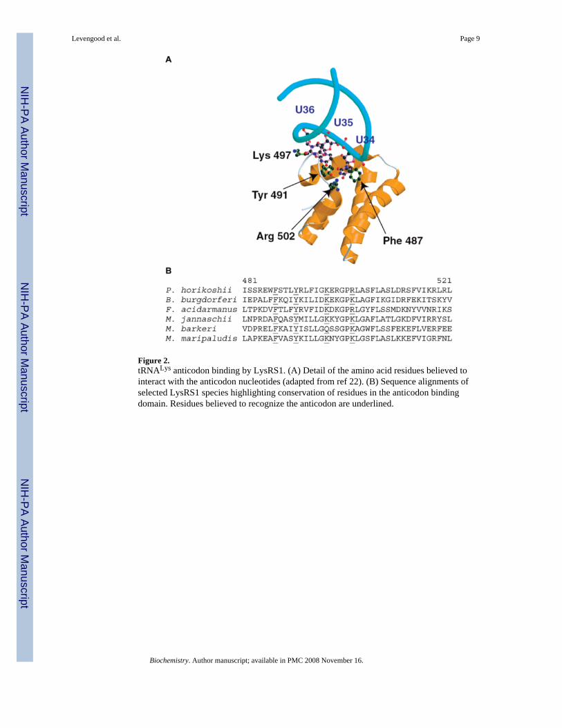
Figure 2.tRNALys anticodon binding by LysRS1. (A) Detail of the amino acid residues believed tointeract with the anticodon nucleotides (adapted from ref 22). (B) Sequence alignments ofselected LysRS1 species highlighting conservation of residues in the anticodon bindingdomain. Residues believed to recognize the anticodon are underlined.
Levengood et al. Page 9
Biochemistry. Author manuscript; available in PMC 2008 November 16.
NIH
-PA Author Manuscript
NIH
-PA Author Manuscript
NIH
-PA Author Manuscript

Figure 3.Double mutant cycle analysis for LysRS1 Y491E aminoacylation of tRNALys UUG.
Levengood et al. Page 10
Biochemistry. Author manuscript; available in PMC 2008 November 16.
NIH
-PA Author Manuscript
NIH
-PA Author Manuscript
NIH
-PA Author Manuscript

Figure 4.Model for tRNALys anticodon binding by P. horikoshii LysRS1. Molecular modeling of theLysRS1–tRNA complex was accomplished with O. Adjustments were made manually on thebasis of the results from mutation of tRNALys and LysRS1. The conformational changes werereminiscent of those previously observed in the GluRS–tRNAGlu complex.
Levengood et al. Page 11
Biochemistry. Author manuscript; available in PMC 2008 November 16.
NIH
-PA Author Manuscript
NIH
-PA Author Manuscript
NIH
-PA Author Manuscript

NIH
-PA Author Manuscript
NIH
-PA Author Manuscript
NIH
-PA Author Manuscript
Levengood et al. Page 12
Table 1Kinetic Data for Wild-Type LysRS1 with Mutant tRNAs
tRNALys (N34N35N36) KM(μM) kcat(s−1) kcat/KM(s−1μM−1) La
UUU 0.87 ± 0.09 0.044 ± 0.002 0.051 ± 0.01 1CUU 1.4 ± 0.2 0.062 ± 0.004 0.044 ± 0.05 1.2GUU 1.5 ± 0.01 0.043 ± 0.0001 0.029 ± 0.0003 1.8UCU 0.0019 ± 0.00008b 27UGU 0.0017 ± 0.0001 30UUC 0.0035 ± 0.0002 15UUG 0.0021 ± 0.00006 24
aThe aminoacylation efficiency of each enzyme—tRNA pair was compared to that of the wild-type enzyme with wild-type tRNA. A value, L, was found
as the loss of aminoacylation efficiency [kcat(wt)/KM(wt)]/[kcat(mut)/KM(mut)].
bFor tRNALys variants containing changes at positions 35 and 36, tRNA could not be used at saturating concentrations because of the high KM compared
to practical tRNA concentrations. When [S] << KM, kcat/KM was directly estimated from the equation v = kcat/KM([E][S]).
Biochemistry. Author manuscript; available in PMC 2008 November 16.

NIH
-PA Author Manuscript
NIH
-PA Author Manuscript
NIH
-PA Author Manuscript
Levengood et al. Page 13
Table 2Kinetic Data for LysRS1 Variant F487A with Mutant tRNAs
tRNA (N34N35N36) kcat/KM (s−1μM−1) L Ma
UUU 0.0019 ± 0.00006 27 1CUU 0.0027 ± 0.0001 19 0.7GUU 0.0022 ± 0.0002 23 0.9UCU 0.00099 ± 0.00006 52 1.9UGU 0.00087 ± 0.00006 59 2.2UUC 0.0015 ± 0.0001 34 1.3UUG 0.0010 ± 0.00006 49 1.8
akcat/KM for the variant enzyme with the wild-type tRNA was set to 1, and each mutant tRNA was then compared to this to give a value M.
Biochemistry. Author manuscript; available in PMC 2008 November 16.

NIH
-PA Author Manuscript
NIH
-PA Author Manuscript
NIH
-PA Author Manuscript
Levengood et al. Page 14
Table 3Kinetic Data for LysRS1 Y491 Variants with Mutant tRNAs
tRNA (N34N35N36) kcat/KM (s−1μM−1) L M
Y491A UUU 0.0016 ± 0.00007 32 1 CUU 0.0017 ± 0.00009 30 0.9 GUU 0.0015 ± 0.0001 34 1.1 UCU 0.00079 ± 0.00009 65 2.0 UGU 0.00076 ± 0.00003 67 2.1 UUC 0.0010 ± 0.00007 51 1.6 UUG 0.00090 ± 0.000005 57 1.8Y491E UUU 0.0015 ± 0.00007 34 1 CUU 0.0014 ± 0.00008 36 1.1 GUU 0.0015 ± 0.0001 34 1 UCU 0.00094 ± 0.00006 54 1.6 UGU 0.00087 ± 0.00007 59 1.7 UUC 0.0013 ± 0.0001 39 1.2 UUG 0.0064 ± 0.0004 8 0.2
Biochemistry. Author manuscript; available in PMC 2008 November 16.

NIH
-PA Author Manuscript
NIH
-PA Author Manuscript
NIH
-PA Author Manuscript
Levengood et al. Page 15
Table 4Kinetic Data for LysRS1 R502 Variants with Mutant tRNAs
tRNA (N34N35N36) kcat/KM (s−1μM−1) L M
R502A UUU 0.0029 ± 0.0002 18 1 GUU 0.0031 ± 0.0001 16 0.9 CUU 0.0032 ± 0.0001 16 0.9 UCU 0.0013 ± 0.00007 39 2.2 UGU 0.00079 ± 0.00003 65 3.7 UUC 0.0007 ± 0.00006 73 4.1 UUG 0.00085 ± 0.00005 60 3.4R502Q UUU 0.00057 ± 0.00003 89 1 GUU 0.00053 ± 0.000006 96 1.1 CUU 0.00055 ± 0.00003 93 1.0 UCU 0.00018 ± 0.00001 280 3.2 UGU 0.00018 ± 0.0000007 280 3.2 UUC 0.000073 ± 0.000007 700 7.8 UUG 0.000078 ± 0.000004 650 7.3
Biochemistry. Author manuscript; available in PMC 2008 November 16.