Antibiotics and translation
195
Dissertation zur Erlangung des Doktorgrades der Fakultät fur Chemie und Pharmazie der Ludwig-Maximilians-Universität München Antibiotics and translation Overcoming emerging bacterial resistance to old and new antimicrobials Agata Lucyna Starosta aus Rzeszow, Polen 2011
-
Upload
khangminh22 -
Category
Documents
-
view
1 -
download
0
Transcript of Antibiotics and translation
Dissertation zur Erlangung des Doktorgrades der Fakultät fur Chemie und Pharmazie der Ludwig-Maximilians-Universität München
Antibiotics and translation
Overcoming emerging bacterial resistance to old and new antimicrobials
Agata Lucyna Starosta
aus Rzeszow, Polen
2011
2
Erklärung
Diese Dissertation wurde im Sinne von § 13 Abs. 3 bzw. 4 der Promotionsordnung vom 29. Januar 1998 (in der Fassung der Sechsten Änderungssatzung von 16. August 2010) von Herrn Prof. Dr. Roland Beckmann betreut.
Ehrenwörtliche Versicherung
Diese Dissertation wurde selbstständig, ohne unerlaubte Hilfe erarbeitet.
München, am 24.11.2011
Agata Lucyna Starosta
Dissertation eingereicht am 24.11.2011
1. Gutachter: Herr Prof. Dr. Roland Beckmann
2. Gutachter: Herr Prof. Dr. Klaus Förstemann
Mündliche Prüfung am 31.01.2012
Agata L. Starosta List of contents
3
List of contents
Acknowledgements .................................................................................................................................. 5
List of original publications .................................................................................................................... 6
Contribution report ................................................................................................................................. 8
Abbreviations ........................................................................................................................................ 10
Summary ................................................................................................................................................ 11
1 Introduction ........................................................................................................................................ 12
1.1 Traditional antibiotics .................................................................................................................. 13
1.1.1 Introduction to antibiotics .................................................................................................... 13
1.1.2 Targets for antibiotic action ................................................................................................. 14
1.1.2.1 Inhibition of cell wall synthesis ..................................................................................... 14
1.1.2.2 Inhibition of DNA replication ........................................................................................ 16
1.1.2.3 Inhibition of RNA synthesis ........................................................................................... 16
1.1.2.4 Inhibition of protein synthesis ....................................................................................... 16
1.1.3 Protein synthesis ................................................................................................................... 17
1.1.3.1 Inhibitors of the small ribosomal subunit .................................................................. 22
1.1.3.2 Inhibitors of the large ribosomal subunit ................................................................... 23
1.1.3.2.1 Inhibitors of the peptidyl-transferase center ...................................................... 23
1.1.3.2.2 Inhibitors of the progressing nascent polypeptide chain .................................... 24
1.1.3.2.3 Inhibitors of the GTPase-associated center (GAC) ............................................ 24
1.1.4 Mechanism of cell death induced by bactericidal antibiotics .............................................. 25
1.2 Antibiotics used in these studies .................................................................................................. 27
1.2.1 Hygromycin A ....................................................................................................................... 27
1.2.2 Macrolides ............................................................................................................................ 29
1.2.3 Thiopeptides ......................................................................................................................... 31
1.2.4 Orthosomycins ...................................................................................................................... 34
1.2.5 Fusidic acid .......................................................................................................................... 35
1.3 Alternative antimicrobials targeting virulence ............................................................................ 36
1.3.1 Definition of virulence .......................................................................................................... 36
1.3.2 Colonization ......................................................................................................................... 36
1.3.3 Biofilm .................................................................................................................................. 36
1.3.4 Quorum sensing .................................................................................................................... 37
Agata L. Starosta List of contents
4
1.3.5 Motility ................................................................................................................................. 37
1.3.6 Secretion systems .................................................................................................................. 37
1.3.7 Elongation factor P .............................................................................................................. 38
2 Objectives of these studies .................................................................................................................. 41
3 Cumulative thesis ............................................................................................................................... 43
3.1 Hygromycin A ............................................................................................................................. 43
3.1.1 Paper 1 .................................................................................................................................. 43
3.1.2 Paper 2 .................................................................................................................................. 43
3.2 Macrolides ................................................................................................................................... 47
3.2.1 Paper 3 .................................................................................................................................. 47
3.2.2 Paper 4 .................................................................................................................................. 47
3.3 Thiopeptides ................................................................................................................................ 50
3.3.1 Paper 5 .................................................................................................................................. 50
3.3.2 Paper 6 .................................................................................................................................. 50
3.4 Orthosomycins ............................................................................................................................. 52
3.5 Fusidic acid.................................................................................................................................. 53
3.5.1 Paper 7 .................................................................................................................................. 53
3.6 Elongation factor P ...................................................................................................................... 54
3.6.1 Paper 8 .................................................................................................................................. 54
4 Conclusions ........................................................................................................................................ 58
5 References .......................................................................................................................................... 59
Agata L. Starosta Ackowledgements
5
Acknowledgements This would not be possible without the support of many people that I had a pleasure of meeting
during my PhD studies.
First of all I would like to thank Dr. Daniel Wilson for giving me an opportunity to work in his
group in the Gene Center . I am grateful for the trust and support I received from him during all those
years. I appreciate each and every advice, discussion, criticism and praise I ever received from him.
That was a lesson I will never forget.
I would like to thank Prof. Roland Beckmann for all the expertise, good advices and support as
well as for providing us with a great scientific environment without which this work could not happen.
I am grateful to all our collaborators for having fruitful time collecting all the data: to Prof. B.
Cooperman for great work on thiopeptides; Prof. A. Bogdanov for macrolide story; Prof. K. Reynolds
for hygromycin A studies; Prof. C. Spahn for first and hopefuly not the last Nature paper; Prof. G.
Dinos for hosting me in Greece and support in experiments; and last but not least, to Prof. J. Remme
for great work on EF-P.
I would also like to thank people, whom I had a pleasure to have conducted the experiments with:
Aleksandra Mikolajka, Alexandra Dönhöfer, Viktorija Karpenko, Gemma Atkinson and Vidya Dhote.
Special thanks to Lauri Peil for being my Mass Spectrometry master, for all the hours we spent
discussing the most crazy ideas and then making them come true.
I would like to thank to all the former and present members of the Wilson and the Beckmann labs
for the atmosphere, discussions, parties, trips and many more.
Lots of thanks to Ingegerd Walz for all the help in the emergency situations.
I would like to thank Prof. Klaus Förstemann, Prof. Mario Halic and Prof. Knud Nierhaus for
being in my thesis committee.
I am very grateful to Marta Danecka and Jean-Paul Armache for surviving corrections of my
thesis.
Wielki dzieki Marto i Jasiu za te wszystkie lata spedzone razem w Monachium, i w Krakowie
(Marta). Bez waszego wsparcia wszystko byloby trudniejsze.
Najbardziej chcialabym podziekowac moim kochanym rodzicom i kochanej siostrze, za
wychowanie i wsparcie przez te wszystkie lata, ktore spedzilam z daleka od domu, bez was to
wszysko nie byloby mozliwe.

Agata L. Starosta List of original publications
6
List of original publications This thesis is based upon the following original publications Reprints were made with permission of the publisher. Paper 1 Palaniappan, N., Dhote, V., Ayers, S., Starosta, A.L., Wilson, D.N., and Reynolds, K.A. (2009). Biosynthesis of the aminocyclitol subunit of hygromycin A in Streptomyces hygroscopicus NRRL 2388. Chem Biol 16, 1180-1189. Paper 2 Dhote, V., Starosta, A.L., Wilson, D.N., and Reynolds, K.A. (2009). The final step of hygromycin A biosynthesis, oxidation of C-5''-dihydrohygromycin A, is linked to a putative proton gradient-dependent efflux. Antimicrob Agents Chemother 53, 5163-5172. Paper 3 Petropoulos, A.D., Kouvela, E.C., Starosta, A.L., Wilson, D.N., Dinos, G.P., and Kalpaxis, D.L. (2009). Time-resolved binding of azithromycin to Escherichia coli ribosomes. J Mol Biol 385, 1179-1192. Paper 4 Starosta, A.L., Karpenko, V.V., Shishkina, A.V., Mikolajka, A., Sumbatyan, N.V., Schluenzen, F., Korshunova, G.A., Bogdanov, A.A., and Wilson, D.N. (2010). Interplay between the ribosomal tunnel, nascent chain, and macrolides influences drug inhibition. Chem Biol 17, 504-514. Paper 5 Starosta, A.L.*, Qin, H.*, Mikolajka, A.*, Leung, G.Y., Schwinghammer, K., Nicolaou, K.C., Chen, D.Y., Cooperman, B.S., and Wilson, D.N. (2009). Identification of distinct thiopeptide-antibiotic precursor lead compounds using translation machinery assays. Chem Biol 16, 1087-1096. Paper 6 Mikolajka, A., Liu, H., Chen, Y., Starosta, A.L., Marquez, V., Ivanova, M., Cooperman, B.S., and Wilson, D.N. (2011). Differential effects of thiopeptide and orthosomycin antibiotics on translational GTPases. Chem Biol 18, 589-600. Paper 7 Ratje, A.H., Loerke, J., Mikolajka, A., Brunner, M., Hildebrand, P.W., Starosta, A.L., Donhofer, A., Connell, S.R., Fucini, P., Mielke, T., Whitford, P. C., Onuchic, J. N., Yu, Y., Sanbonmatsu, K. Y., Hartmann, R. K., Penczek, P. A., Wilson, D. N., and Spahn, C. M. (2010). Head swivel on the ribosome facilitates translocation by means of intra-subunit tRNA hybrid sites. Nature 468, 713-716. Paper 8 Peil L.*, Starosta, A.L.*, Virumäe, K, Aktinson, G.C., Tenson, T., Remme, J., Wilson, D.N. (2011). Formation of ε(R)-β-lysyl-hydroxylysine on translation elongation factor EF-P requires YjeK, YjeA and YfcM. Under revision at Nature Chemical Biology * Equally contributed
Agata L. Starosta List of original publications
7
List of publications not included in the thesis: Paper 9 Wilson, D.N., Schluenzen, F., Harms, J.M., Starosta, A.L., Connell, S.R., and Fucini, P. (2008). The oxazolidinone antibiotics perturb the ribosomal peptidyl-transferase center and effect tRNA positioning. Proc. Natl Acad. Sci USA 105, 13339-13344. Paper 10 Bhushan, S., Meyer, H., Starosta, A.L., Becker, T., Mielke, T., Berninghausen, O., Sattler, M., Wilson, D.N., and Beckmann, R. (2010). Structural basis for translational stalling by human cytomegalovirus and fungal arginine attenuator peptide. Mol Cell 40, 138-146.
Agata L. Starosta Contribution report
8
Contribution report Work presented in this dissertation comprises part of the results of my doctoral
research conducted from October 2007 to November 2011 in cooperation with scientists from
the laboratories of: Professor K. Reynolds (Portland, Oregon, US), Professor. B. Cooperman
(Pennsylvania, Philadelphia,US), Professor A. Bogdanov (Moscow, Russia), Professor G.
Dinos (Patras, Greece), Professor C. Spahn (Berlin, Germany) and Professor J. Remme
(Tartu, Estonia).
Paper 1 (Palaniappan et al., 2009) and Paper 2 (Dhote et al., 2009)
I performed all the biochemical analysis determining the antimicrobial activities of
Hygromycin A (HygA) and intermediates of HygA, using (i) Escherichia coli lysate based in
vitro coupled transcription-translation assay (TT) with green fluorescence protein (GFP) as a
reporter, and (ii) the AcPhe-Puromycin (AcPhe-Puro) synthesis assay. These results comprise
Figure 6 (Paper 1), and Figure 4 (Paper 2), which I prepared for the papers as well as
contributing to the interpretation and analysis of these results.
Paper 3 (Petropoulos et al., 2009)
I cultivated Deinococcus radiodurans cells and used sucrose gradient centrifugation
protocol to isolated highly active 70S ribosomes, which were then used subsequently to
evaluate the binding mode of azithromycin (AZI) to bacterial ribosomes in collaboration with
Prof. Dinos.
Paper 4 (Starosta et al., 2010)
I carried out all the biochemical experiments to determine the inhibitory activity of
tylosin (Tyl) and derivatives of Tyl using (i) TT assay using GFP and firefly luciferase (Fluc)
as templates, and (ii) competition binding assay with [14C]-erythromycin and D. radiodurans
70S ribosomes. The results are depicted in Figures 3-6 and in Supplementary Table 1 of the
paper. In addition, I participated in the interpretation of the results and made Figures 1, 3-6 as
well as contributing to writing of the manuscript.
Paper 5 (Starosta et al., 2009)
I measured the potency of the entire library of thiopeptide precursor compounds to
inhibit synthesis of GFP in TT assay. I also measured the ability of precursors to restore
translation in the presence of inhibitory concentration of parental compounds, which is
Agata L. Starosta Contribution report
9
presented in Figures 4-5. I prepared Figures 1-5 and participated in the preparations of the
draft of the manuscript.
Paper 6 (Mikolajka et al., 2011)
I evaluated the antimicrobial activity of thiostrepton, micrococcin and evernimicin
(Evn) in TT assay using GFP as a reporter gene. These results are presented in Supplementary
Figure 3, which I prepared together with Figure 5.
Paper 7 (Ratje et al., 2010)
I grew Thermus thermophilus cells and prepared highly pure 70S ribosomal particles
using sucrose gradient centrifugation protocol. The 70S ribosomes were used for preparation
of the complex of EF-G stalled with GDP and fusidic acid on the 70S ribosome.
Paper 8 (Peil et al. 2011)
I cloned and purified elongation factor P (EF-P) which was used to raise rabbit
polyclonal antibodies. I established the immuno-precipitation protocol using protein A and
prepared all constructs used for the rescue experiments. I identified the YfcM protein using
STRING database and cloned yfcM gene. I purified recombinant YfcM, which I characterized
using differential scanning fluorimetry (DSF). I optimized protocol for expression of EF-P
modified by 128 Da and established the in vitro hydroxylation assay. I prepared all figures
and participated in preparation of the manuscript. Additionally, I started initial
characterization of phenotypes of the efp and modification enzymes deletion strains.
Agata L. Starosta Abbreviations
10
Abbreviations · 30SIC – 30S initiation complex · 70SIC – 70S initiation complex · A/A position – both anticodon stem loop and
aminoacyl arm of tRNA in A-site · A/T position – anticodon stem loop in A site,
aminoacyl arm of tRNA bound to EF-Tu · aa-tRNA – aminoacyl-tRNA · AB – subunits A and B of hygromycin A · AcPhe-Puro – AcPhe-Puromycin · AHLs – acylated homoserine lactones · A-site – aminoacyl-tRNA binding site · Avi – avilamycin · AZI – azithromycin · Cryo-EM – cryo-electron microscopy · Des-HygA – desmethylenehygromycin A · DGI – German Society for Infectious Diseases
· DH-HygA – 5”-dihydrohygromycin A
· DHS – deoxyhypusine synthase · DOHH – deoxyhypusine hydroxylase · E. coli – Escherichia coli · ECDC – European Center for Disease
Prevention and Control · EF – elongation factor · EHEC – enterohemorrhagic E. coli · Ery –erythromycin · E-site – exit site · Evn – evernimicin · FA – fusidic acid · fMet-tRNAfMet – initiator-tRNA
· GAC – GTPase-associated center · GFP – green fluorescent protein · GlcNAc – N-acetylglucosamine · HygA – hygromycin A
· IC50 – half-inhibitory concentration
· IF – initiation factor · K. pneumoniae – Klebsiella pneumoniae · LSU – 50S large subunit · MBL – metallo-β-lactamase · Met-HygA – methoxyhygromycin A
· MiC – Micrococcin · MIC – minimal inhibitory concentration · mRNA – messenger RNA · MurNAc – N-acetyl-muramic acid · NLPH – National Laboratory of Public Health
of the Ministry of Public Health and Population · OMT – 5-O-mycaminosyl-tylonolid · ORF – open reading frame · P/E - anticodon stem loop in P site, aminoacyl
arm bound to E-site · PBS – penicillin-binding proteins · P-HygA – phosphorylated hygromycin A · Pi – Inorganic phosphate · POST – post-translocation state · PRE – pre-translocation state · Pseudomonas aeruginosa · P-site – peptidyl-tRNA binding site · PTC – peptidyl-transferase center · RF – release factor · rRNA – ribosomal RNA · SAM – S-adenosylmethionine · SAXS – small-angle X-ray scattering · SD – Shine-Dalgarno · SRL – sarcin-ricin loop · SS – secretion systems · SSU – 30S small ribosomal subunits · SubA – subunit A, dehydrofucofuranose
moiety of HygA · SubB – subunit B, α-methyl cinnamic acid
moiety of HygA · SubC – subunit C, aminocyclitol moiety of
HygA · ThS – thiostrepton · tRNA – transfer tRNA · TT – E. coli lysate-based in vitro coupled
transcription-translation assay · Tyl – tylosin · UTR – untranslated region
Agata L. Starosta Summary
11
Summary Emerging bacterial resistance to antibiotics has led to increased interest in
development of new, improved antimicrobials. In these studies we have used a number of
biochemical assays in order to investigate the mechanism of action of several antibiotics
inhibiting protein synthesis. We show that using biosynthetic intermediates of parental
compounds we are able to evaluate the functionality of structural features of the drug
(hygromycin A, thiopeptides). We revisited the mechanism of action of three classes of
antibiotics (macrolides, thiopeptides and orthosomycins) and demonstrated that cryo-electron
microscopy can be successfully applied to localize and visualize small molecules, such as
drugs (fusidic acid). To overcome cross-resistance with clinically used antibiotics, new
antimicrobial targets are needed. Bacterial virulence and pathogenicity factors have been
suggested as such future targets. One such factor is elongation factor P (EF-P), a post-
translationally modified protein that regulates expression of virulence determinants. Here we
have investigated the discrepancies between the 144 Da and 128 Da modification states
reported for EF-P in vivo and in vitro, respectively. This led us to identify a third enzyme in
the EF-P modification pathway. In addition to providing fundamental insight into the role of
EF-P in the cell, these studies may provide additional targets for development of novel
“antivirulence“ agents.
Agata L. Starosta Introduction
12
1 Introduction Since their discovery in 1928 (Fleming, 1929), penicillins remain the most
frequently prescribed antibiotics (ECDC, 2010). However, as shown in the “Annual
epidemiological report on communicable diseases in Europe” published by the European
Center for Disease Prevention and Control (ECDC), Escherichia coli, the most common
Gram-negative bacteria responsible for bloodstream and urinary tract infections, showed a
Europe-wide increase of resistance to all antibiotic classes under surveillance (ECDC, 2010).
At this level of resistance, it eliminates aminopenicillins as a therapeutical strategy for these
common infections (ECDC, 2010). Moreover, multidrug resistance among Gram-negative
bacteria such as E. coli, Klebsiella pneumoniae and Pseudomonas aeruginosa, further
increases, which limits the possibility of the antimicrobial treatment (ECDC, 2010) and raises
the problem of bacterial infection to a level never seen before.
Although antibiotics have been available for the treatment of bacterial infections for
over 70 years, analyses of a 30,000 years old sample show that genes encoding resistance to
β-lactams, tetracycline and glycopeptides antibiotics are indeed much more ancient (D'Costa
et al., 2011). This underlines the fact that bacteria are years ahead of researchers in the
process of learning how to neutralize antibiotics. Furthermore, the time from the discovery of
a new compound until its introduction into the market takes an average of 10-15 years
(DrugDiscovery), while, as shown in experiments with E. coli treated with ciprofloxacin,
bacteria can develop resistance within as little as 10 hours (Zhang et al., 2011). This finding,
as well as recent bacterial outbreaks, emphasizes the scale of the problem of emerging
antibiotic resistance among bacteria.
Cholera outbreak
Roughly 20,000 clinical cases of cholera and 1100 deaths were officially reported
since the outbreak was first detected on the 21st October 2010 in the Artibonite region of Haiti
(Butler, 2010). Toxigenic Vibrio cholerae O1, serotype Ogawa, biotype El Tor, was identified
by the National Laboratory of Public Health of the Ministry of Public Health and Population
in Haiti (NLPH). Identification of the isolate was confirmed by the Center for Disease
Prevention and Control (CDC) in Atlanta (US). Antimicrobial susceptibility testing of
selected V. cholerae O1 isolates conducted at the NLPH and at the CDC demonstrated
susceptibility to tetracyclines, ciprofloxacin, and kanamycin; and resistance to trimethoprim,
sulfamethoxazole, furazolidone, nalidixic acid, sulfisoxazole, and streptomycin (CDC, 2010).
Agata L. Starosta Introduction
13
NDM-1
The NDM-1 strain was discovered in 2008 in a Swedish patient of Indian origin
who traveled to New Delhi, India, and acquired a urinary tract infection. The infection caused
by a carbapenem-resistant K. pneumoniae strain that typed to the sequence type 14 complex.
The isolate, K. pneumoniae 05-506, was shown to possess a metallo-β-lactamase (MBL), but
was negative for previously known MBL genes (Yong et al., 2009). Gene libraries and
amplification of class 1 integrons revealed three resistance-conferring regions which are (i)
easily transferable to recipient strains and (ii) that confer resistance to all antibiotics, except
fluoroquinolones and colistin. NDM-1 (New Delhi metallo-β-lactamase) has a molecular
mass of 28 kDa, is monomeric and can hydrolyze all β-lactams, except aztreonam (Yong et
al., 2009). Further investigations revealed cases of NDM-1 bacterial strains in Pakistan, India
and the United Kingdom. Isolated strains were highly resistant to many antibiotics (including
β-lactams, fluoroquinolones and aminoglycosides), but not to tigecycline and colistin
(Kumarasamy et al., 2010).
An Escherichia coli O104:H4 outbreak
The E. coli bacteria outbreak begun in May 2011 in Germany. Since then, nearly
4000 cases and more than 40 deaths were reported (ECDC, 2011). A virulent E. coli strain
O104:H4 causes haemolytic uraemic syndrome and bloody diarrhea (also known as
enterohemorrhagic E. coli, EHEC) (Bielaszewska et al., 2011). The strain produces Shiga
toxin 2 and aggregative adherences to epithelial cells (Bielaszewska et al., 2011). A Shiga-
like toxin (verocytotoxin) may cause direct renal and endothelial cell damage and may adhere
to the intestinal epithelium resulting in bloody diarrhea (Bae et al., 2006). Moreover, the
bacteria produces extended-spectrum β-lactamases (Bielaszewska et al., 2011). According to
the German Society for Infectious Diseases (DGI), the EHEC strain is susceptible to
carbapenem, new generation macrolides and rifampicin, while being resistant, or not
responding to fluoroquinolones, aminoglycosides, and fosfomycin (DGI, 2011).
1.1 Traditional antibiotics
1.1.1 Introduction to antibiotics
Traditional antibiotics, depending on their ability to either kill, or inhibit the growth
of bacteria, can be classified as bactericidal, or bacteriostatic, respectively. They act by
inhibiting processes essential for exponential growth, namely (i) cell wall synthesis, (ii) DNA
replication, (iii) RNA transcription, and (iv) protein synthesis (Clatworthy et al., 2007). The
Agata L. Starosta Introduction
14
history of antibiotics begun in 1929 when Sir Alexander Fleming discovered penicillin
(Fleming, 1929). Since that time a significant number of new antimicrobials have been
discovered. Development and availability of biochemical tools have provided an
understanding into the mechanism of action of new compounds, and provide a basis for
development of new derivatives through the chemical modification of already existing
molecules (Kohanski et al., 2010b). The dynamics of discovery of new classes of antibiotics
lapsed in the 1960’s following the introduction of quinolones. Nearly 40 years passed before a
new class of antimicrobials was introduced, namely, the oxazolidinones with linezolid
(Zyvox) as the lead compound (Walsh, 2003). However, the clinically significant resistance to
each of the known classes of antimicrobials has emerged within a few years following their
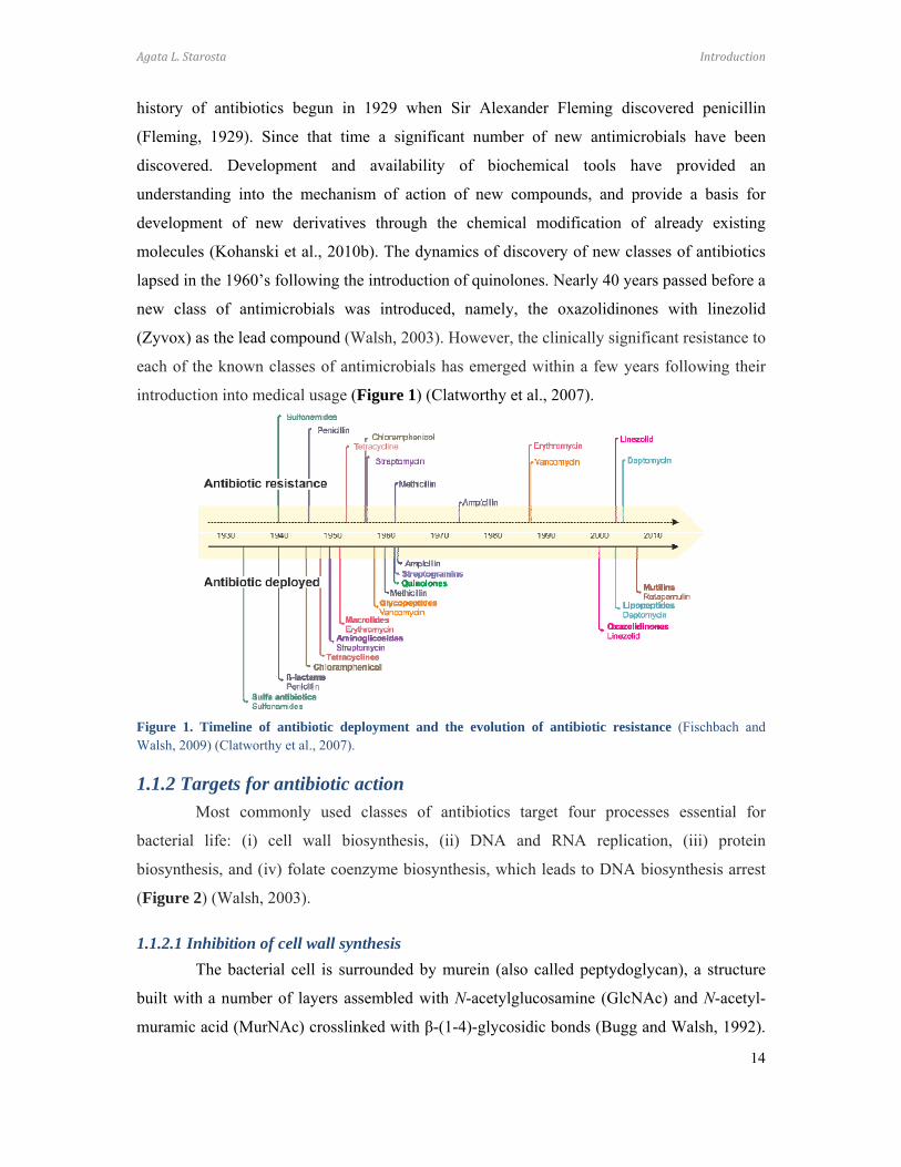
introduction into medical usage (Figure 1) (Clatworthy et al., 2007).
Figure 1. Timeline of antibiotic deployment and the evolution of antibiotic resistance (Fischbach and Walsh, 2009) (Clatworthy et al., 2007).
1.1.2 Targets for antibiotic action
Most commonly used classes of antibiotics target four processes essential for
bacterial life: (i) cell wall biosynthesis, (ii) DNA and RNA replication, (iii) protein
biosynthesis, and (iv) folate coenzyme biosynthesis, which leads to DNA biosynthesis arrest
(Figure 2) (Walsh, 2003).
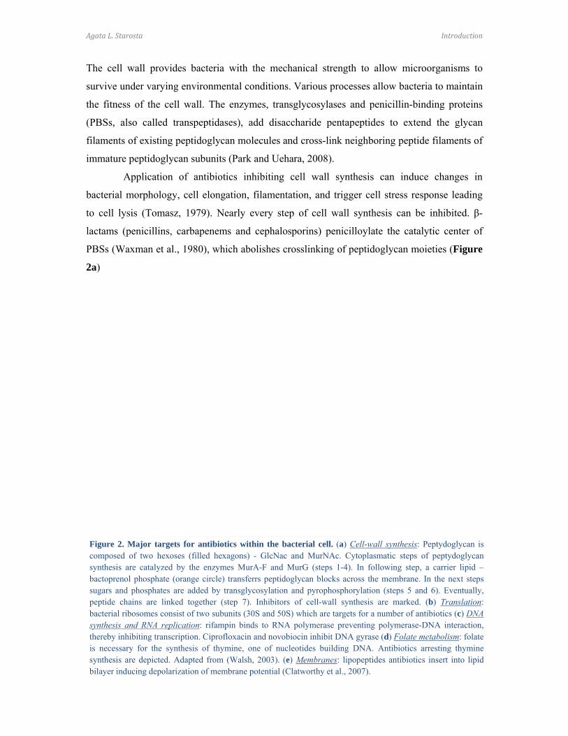
1.1.2.1 Inhibition of cell wall synthesis
The bacterial cell is surrounded by murein (also called peptydoglycan), a structure
built with a number of layers assembled with N-acetylglucosamine (GlcNAc) and N-acetyl-
muramic acid (MurNAc) crosslinked with β-(1-4)-glycosidic bonds (Bugg and Walsh, 1992).
Agata L. Starosta Introduction
15
The cell wall provides bacteria with the mechanical strength to allow microorganisms to
survive under varying environmental conditions. Various processes allow bacteria to maintain
the fitness of the cell wall. The enzymes, transglycosylases and penicillin-binding proteins
(PBSs, also called transpeptidases), add disaccharide pentapeptides to extend the glycan
filaments of existing peptidoglycan molecules and cross-link neighboring peptide filaments of
immature peptidoglycan subunits (Park and Uehara, 2008).
Application of antibiotics inhibiting cell wall synthesis can induce changes in
bacterial morphology, cell elongation, filamentation, and trigger cell stress response leading
to cell lysis (Tomasz, 1979). Nearly every step of cell wall synthesis can be inhibited. β-
lactams (penicillins, carbapenems and cephalosporins) penicilloylate the catalytic center of
PBSs (Waxman et al., 1980), which abolishes crosslinking of peptidoglycan moieties (Figure
2a)
Figure 2. Major targets for antibiotics within the bacterial cell. (a) Cell-wall synthesis: Peptydoglycan iscomposed of two hexoses (filled hexagons) - GlcNac and MurNAc. Cytoplasmatic steps of peptydoglycansynthesis are catalyzed by the enzymes MurA-F and MurG (steps 1-4). In following step, a carrier lipid –bactoprenol phosphate (orange circle) transferrs peptidoglycan blocks across the membrane. In the next stepssugars and phosphates are added by transglycosylation and pyrophosphorylation (steps 5 and 6). Eventually,peptide chains are linked together (step 7). Inhibitors of cell-wall synthesis are marked. (b) Translation:bacterial ribosomes consist of two subunits (30S and 50S) which are targets for a number of antibiotics (c) DNAsynthesis and RNA replication: rifampin binds to RNA polymerase preventing polymerase-DNA interaction,thereby inhibiting transcription. Ciprofloxacin and novobiocin inhibit DNA gyrase (d) Folate metabolism: folateis necessary for the synthesis of thymine, one of nucleotides building DNA. Antibiotics arresting thyminesynthesis are depicted. Adapted from (Walsh, 2003). (e) Membranes: lipopeptides antibiotics insert into lipidbilayer inducing depolarization of membrane potential (Clatworthy et al., 2007).
Agata L. Starosta Introduction
16
(Wise and Park, 1965). Glycopeptide antibiotics (vancomycin) (Figure 2a) bind
peptidoglycan subunits and are steric inhibitors of transglycosylases and transpeptidases
(Kahne et al., 2005). Fosfomycin inhibits synthesis (Kahan et al., 1974), while bacitracin
inhibits transport (Storm, 1974) of single peptidoglycan units (Figure 2a). Lipopeptides
(daptomycin) modulate the structural integrity of bacterial cell by inserting themselves into
the cell membrane and triggering membrane depolarization (Figure 2e) (Jung et al., 2004).
Polymyxins are cyclic, positively charged peptide antibiotics, which have high affinity for
lipid moiety of lipopolysaccharides. These cationic agents bind to the anionic bacterial outer
membrane and disrupt their integrity (Landman et al., 2008).
1.1.2.2 Inhibition of DNA replication
DNA synthesis, messenger RNA (mRNA) transcription and cell division involve
topoisomerase reactions to maintain these essential processes. Topoisomerases control the
DNA winding by breaking strands, untangling the nucleic acid and then rejoining the DNA
strands (Drlica and Zhao, 1997). Quinolones stall both the topoisomerase II and IV at the
DNA cutting phase and prevent DNA strand ligation (Figure 2b) (Chen et al., 1996). Arrested
DNA synthesis induces bacteriostasis leading to cell death (Kohanski et al., 2007). Sublethal
dosage of quinolones, as well as β-lactams, can lead to hydroxyradical formation. This
induces mutagenesis, which together with SOS-related mutagenesis and RecA-mediated
processes, can stimulate evolution of mutations conferring antibiotic resistance (Kohanski et
al., 2010a).
1.1.2.3 Inhibition of RNA synthesis
Rifampicin is a semi-synthetic bactericidal antibiotic, which like quinolones, can
induce cell death (Kohanski et al., 2007). It binds to the β-subunit of DNA-dependent RNA
polymerase leading to arrest of mRNA transcription (Figure 2b) (Campbell et al., 2001).
1.1.2.4 Inhibition of protein synthesis
The complexity of the structure and function of the ribosome makes it an important
target for antimicrobial therapies. High-resolution crystal structures of ribosomes and
ribosome-antibiotic complexes have provided detailed understanding into the mechanism of
action of many classes of antibiotics (Figure 2c). Inhibitors of translation can bind to the
functional centers on either of the subunits and inhibit nearly every step of translation (Figure
3). Four major binding sites for ribosomal inhibitors can be distinguished: (i) the decoding
Agata L. Starosta Introduction
17
center on the small subunit, (ii) the peptidyl-transferase center (PTC), (iii) exit tunnel and (iv)
GTPase-associated center (GAC) on the large subunit.
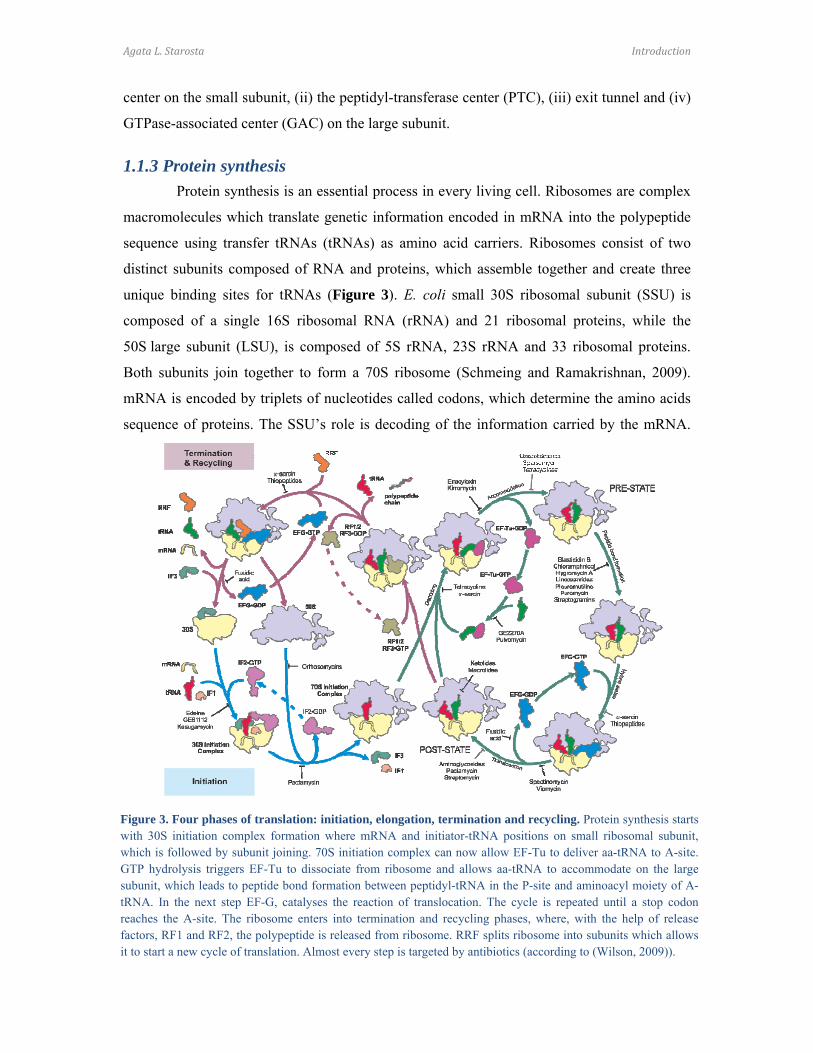
1.1.3 Protein synthesis
Protein synthesis is an essential process in every living cell. Ribosomes are complex
macromolecules which translate genetic information encoded in mRNA into the polypeptide
sequence using transfer tRNAs (tRNAs) as amino acid carriers. Ribosomes consist of two
distinct subunits composed of RNA and proteins, which assemble together and create three
unique binding sites for tRNAs (Figure 3). E. coli small 30S ribosomal subunit (SSU) is
composed of a single 16S ribosomal RNA (rRNA) and 21 ribosomal proteins, while the
50S large subunit (LSU), is composed of 5S rRNA, 23S rRNA and 33 ribosomal proteins.
Both subunits join together to form a 70S ribosome (Schmeing and Ramakrishnan, 2009).
mRNA is encoded by triplets of nucleotides called codons, which determine the amino acids
sequence of proteins. The SSU’s role is decoding of the information carried by the mRNA.
Figure 3. Four phases of translation: initiation, elongation, termination and recycling. Protein synthesis startswith 30S initiation complex formation where mRNA and initiator-tRNA positions on small ribosomal subunit,which is followed by subunit joining. 70S initiation complex can now allow EF-Tu to deliver aa-tRNA to A-site.GTP hydrolysis triggers EF-Tu to dissociate from ribosome and allows aa-tRNA to accommodate on the largesubunit, which leads to peptide bond formation between peptidyl-tRNA in the P-site and aminoacyl moiety of A-tRNA. In the next step EF-G, catalyses the reaction of translocation. The cycle is repeated until a stop codonreaches the A-site. The ribosome enters into termination and recycling phases, where, with the help of releasefactors, RF1 and RF2, the polypeptide is released from ribosome. RRF splits ribosome into subunits which allowsit to start a new cycle of translation. Almost every step is targeted by antibiotics (according to (Wilson, 2009)).
Agata L. Starosta Introduction
18
The LSU has the transferase activity and catalyses peptide bond formation between the tRNA
carrying the nascent polypeptide chain, the peptidyl-tRNA, and an aminoacyl-tRNA (aa-
tRNA), which carries an amino acid. Newly delivered aa-tRNAs accommodate into the A-
site, peptidyl-tRNA binds in the P-site, while deacylated tRNA exits the ribosome via the E-
site.
Translation is a cyclic reaction, which can be divided into four sequential steps:
initiation, elongation, termination and recycling (Figure 3). In each step, additional factors
facilitate the reaction. Translation starts with 30S initiation complex (30SIC) formation.
Initiation factor 3 (IF3) binds to the SSU and prevents premature subunit joining (Karimi et
al., 1999). The SSU-IF3 complex then assembles with mRNA, IF1, IF2 and initiator fMet-
tRNA. The ribosomal protein S1 binds to the A/U rich 5’ untranslated region (UTR) of
mRNAs (Ringquist et al., 1993) and helps to position the mRNA on the SSU. The GGAGG
Shine-Dalgarno (SD) sequence (Shine and Dalgarno, 1974) of canonical mRNA interacts with
3’-end of 16S rRNA (anti-SD) (Demeshkina et al., 2010; Jenner et al., 2010b; Kaminishi et
al., 2007) and the start codon (AUG) is positioned in P-site. IF1 binds at a site overlapping the
A-site (Carter et al., 2001; Dahlquist and Puglisi, 2000) and, together with IF2 and IF3,
cooperatively promotes fMet-tRNA binding to the SSU and its positioning in a P/I site
(peptidyl/initiation site) (Antoun et al., 2006; Julian et al., 2011). IF3 discriminates fMet-
tRNA from other aa-tRNAs in a P-site by recognition of three unique G:C pairs in the initiator
tRNA, and probably the guanosine at third position of the initiation codon (Hartz et al., 1990;
Hartz et al., 1989; O'Connor et al., 2001; Risuleo et al., 1976; Sussman et al., 1996). IF
binding induces head swiveling of the SSU (Julian et al., 2011). During the formation of the
70S initiation complex (70SIC), subunits join in rotated orientation and upon IF2-driven GTP
hydrolysis, fMet-tRNAfMet accommodates into the P-site, while subunits unratchet (Marshall
et al., 2009). IFs are released from the ribosome, which then enters into the elongation cycle.
Ribosomes entering the elongation cycle have an fMet-tRNAfMet bound to P-site and
vacant A-site. The aa-tRNA is delivered as a ternary complex with EF-Tu and GTP
(Schmeing et al., 2009; Schuette et al., 2009). tRNA within the ternary complex binds to
ribosomes in a bent conformation (Blanchard et al., 2004; Schmeing et al., 2009; Voorhees et
al., 2010) in an A/T position, where anticodon stem loop is bound to the A-site on the SSU
and the acceptor stem carrying the amino acid is still bound to EF-Tu. Such a conformation
enables direct contact with the decoding site on the SSU and discrimination of a cognate
codon-anticodon interaction while remaining bound to EF-Tu. Rearrangements in the

Agata L. Starosta Introduction
19
decoding site trigger conformational changes in the ribosome, namely shoulder movement
towards EF-Tu (domain closure) that increases the rate of GTPase activation of EF-Tu (Ogle
et al., 2002). EF-Tu undergoes conformational changes which positions the catalytic histidine
84 (His84) in proximity with the phosphate of the nucleotide A2662 (E. coli numbering) of
the sarcin-ricin loop (SRL) located in helix 95 (H95) of 23S rRNA (Voorhees et al., 2010).
His84 places a water molecule in a position to nucleophilically attack the γ-phosphate of GTP,
which leads to GTP hydrolysis (Daviter et al., 2003) and EF-Tu-GDP dissociation from the
ribosome. The aa-tRNA accommodates from A/T position to A/A position, where aa-tRNA is
bound to A-site on both the SSU and LSU (Blanchard et al., 2004; Douthwaite et al., 1983;
Schmeing et al., 2009; Voorhees et al., 2010). Three nucleotides of the 16S rRNA, namely
G530, A1492 and A1493, were shown to be crucial for tRNA binding in the decoding center
(Moazed and Noller, 1990). Nucleotides A1492 and A1493 flip-out from helix 44 (h44) and
together with G530 monitor Watson-Crick base-pairing geometry of the minor groove of the
first two base pairs between codon and anticodon (Ogle et al., 2001). Both, rearrangements in
the proteins and 23S rRNA in the LSU, accelerate interactions with the anticodon stem loop
and elbow region of the tRNA, leading to positioning of the acceptor end at the PTC (Jenner
et al., 2010a).
Accommodation of the aa-tRNA at the A site is followed by peptide bond formation
at the PTC. The PTC is composed of nucleotides of domain V of 23S rRNA and is an active
site of the ribosome. The PTC binds the CCA ends of peptidyl- and aminoacyl-tRNAs and
catalyzes the nucleophilic attack of the α-amino group of the aa-tRNA on the ester carbonyl
carbon of the peptidyl-tRNA, which leads to a new peptide bond (Green and Lorsch, 2002).
Crystal structures of the CCA end analogues showed that the A-site CCA end is
coordinated by nucleobases G2553, U2555, U2556 of an A-loop and G2583, while the P-site
CCA stacks onto nucleobases from the P-loop, including G2251, G2252, and additionally
A2451 (Hansen et al., 2002b; Nissen et al., 2000), which is consistent with the CCA end
positions found for entire tRNA substrates bound to ribosome (Selmer et al., 2006) and is in
agreement with biochemical data (Moazed and Noller, 1991). The A-tRNA induces
conformational changes in the PTC (Schmeing et al., 2005a; Schmeing et al., 2005b), which
orients reactive nucleotides in the position for peptide bond formation. The 2’-OH group of
A2451 positions the A76 of the P-tRNA to promote peptide bond formation (Lang et al.,
2008). The 2’-OH group of the A76 of the P-tRNA mediates proton passage from attacking α-
amino group of the A-tRNA to the leaving 3’-OH group (Weinger et al., 2004), while the
Agata L. Starosta Introduction
20
water molecule, coordinated by U2584 and A2602 (Schmeing et al., 2005a), acts as a proton
shuttle and stabilizes the (eight-membered) transition state (Kuhlenkoetter et al., 2011). In this
model, the ribosome positions substrates, but does not contribute to the catalysis of the
reaction. However, recent works show that the ribosome can modulate the catalysis by
changing the transition state rates (Hiller et al., 2011). Post-peptide bond formation the
ribosome carries a peptidyl-tRNA at A-site and a deacylated tRNA at the P-site.
Once the peptide bond is formed, the mRNA-tRNA2 complex must be moved across
the ribosome in a process called translocation. In the pre-translocation state (PRE), P- and A-
tRNAs move in respect to the LSU forming P/E and A/P hybrid states, where the anticodon
stem loops interact with P- and A-sites on the SSU and at the same time, the acceptor arms of
respective tRNAs interact with E- and P-sites on the LSU (Munro et al., 2007). The tRNA
hybrid state formation is coupled with a subunit rotation (Agirrezabala et al., 2008). EF-G
interacts with the ribosome (Clementi et al., 2010; Datta et al., 2005; Moazed et al., 1988;
Stark et al., 2000) and stabilizes the ratcheted state of the ribosome (Munro et al., 2010a).
GTP hydrolysis triggers conformational changes in EF-G (Rodnina et al., 1997). The
translocation reaction, i.e. the movement of mRNA and anticodon stem-loops of P- and A-
tRNAs across the SSU, is catalyzed by domain IV of EF-G (Savelsbergh et al., 2000) and is
coupled with inorganic phosphate (Pi) release (Rodnina et al., 1999). In the post-translocation
state (POST), the A-site becomes vacant and is ready to accept the next incoming aa-tRNA.
The elongation cycle ends when a stop codon reaches the A-site, which is then recognized by
release factors (RFs) (Caskey et al., 1968). Two release factors, RF1 and RF2, recognize
UAA/UAG and UAA/UGA stop codons, respectively (Scolnick et al., 1968). RFs bind in the
A-site spanning both subunits (Jin et al., 2010; Korostelev et al., 2008; Korostelev et al.,
2010; Laurberg et al., 2008; Petry et al., 2005; Weixlbaumer et al., 2008). RFs recognize stop
codons directly, and conserved nucleotides G530, A1492, A1493 of 16S rRNA do not
participate in stop codon recognition (Korostelev et al., 2008). Instead, these nucleotides
stabilize the open form of the RFs. Amino acids GxxE from helix 5 of both RF1 and RF2
recognize the first nucleobase, uridine in all stop codons. The second position is recognized
by PVT motif of RF1 and SPF motif in RF2 located in the recognition loop of RFs, while the
third position is monitored by threonine (T194), and additionally by glutamine (Q181) in the
RF1 (Korostelev et al., 2008; Korostelev et al., 2010; Laurberg et al., 2008; Weixlbaumer et
al., 2008). Upon recognition of the stop codon, RFs induce conformational changes in H69 of
23S rRNA. Nucleobase A1913 (H69) stacks onto A1493 (h44) and, together with A1492,
Agata L. Starosta Introduction
21
forms a pocket into which RFs accommodate (Korostelev et al., 2010; Weixlbaumer et al.,
2008). Structural findings are in accordance with biochemical data showing defects in the
termination of the ribosomes lacking H69 (Ali et al., 2006; Korostelev et al., 2010).
Moreover, interactions between RFs and H69 help to position GGQ motif in a PTC
(Korostelev et al., 2008; Laurberg et al., 2008). It should be noted that the structures of free
RFs compared with RFs bound to the ribosome can differ significantly. The crystal structures
of free RF1/2 showed they adopt closed conformations where GGQ and PTV/SPF motifs are
approximately 23Å from each other, which is not sufficient to establish interactions with the
PTC and the decoding center (Shin et al., 2004; Vestergaard et al., 2001; Zoldak et al., 2007).
However, small-angle X-ray scattering (SAXS) studies showed that in the solution, RFs are
present in equilibrium between open and closed forms (Vestergaard et al., 2005; Zoldak et al.,
2007). Upon recognition of the stop codon, RFs undergo conformational changes from closed
to active, open form where GGQ motif residing in the PTC and PTV/SPF motifs interacting
with decoding center are 73 Å apart from each other (Klaholz et al., 2003; Korostelev et al.,
2008; Korostelev et al., 2010; Laurberg et al., 2008; Rawat et al., 2006; Rawat et al., 2003;
Weixlbaumer et al., 2008). A universally conserved GGQ motif is critical for the function of
RFs, (Frolova et al., 1999; Shaw and Green, 2007). The glycines from the GGQ motif
position the catalytically active glutamine (Q230) in the PTC between 23S rRNA nucleotides
U2584, U2585, A2602, and A2451 (Jin et al., 2010; Korostelev et al., 2008; Korostelev et al.,
2010; Weixlbaumer et al., 2008). Mutational analysis of Q230 showed that the glutamine side
chain is not critical for the release reaction of the nascent polypeptide (Seit-Nebi et al., 2001;
Shaw and Green, 2007), however the Q230 side chain discriminates the RF’s specificity for a
water molecule as the nucleophile (Shaw and Green, 2007). The amide group of Q230
backbone interacts with leaving 3’-OH group of A76 of the peptidyl-tRNA, which accounts
for the catalytic activity of glutamine (Laurberg et al., 2008). The crystal structure of RF2
bound to the ribosome in a presence of non-hydrolysable analogues of Phe-tRNAPhe where the
ester bond was replaced by an amide bond gives inside into structural basis of catalysis of
peptide release (Jin et al., 2010). Consistent with biochemical data (Brunelle et al., 2008;
Zaher et al., 2011), it was suggested that 2’-OH group of A76 of the peptidyl-tRNA
coordinates a water molecule for nucleophilic attack onto the ester bond, leading to hydrolysis
of the newly synthesized nascent polypeptide (Jin et al., 2010). RF3 is a GTPase that
destabilizes the RF1 and RF2 interaction with the ribosome (Freistroffer et al., 1997; Gao et
al., 2007) by stabilization of the ratcheted form of the ribosome with tRNAs in hybrid states

Agata L. Starosta Introduction
22
(Jin et al., 2011). Moreover recent data suggest that RF3 plays a role in the fidelity of
translation by detecting mis-incorporated amino acids and premature terminating protein
synthesis (Zaher and Green, 2009, 2011).
Post-termination ribosomes are split into subunits with the help of the ribosome
recycling factor (RRF) and EF-G, in a GTP-dependent manner (Karimi et al., 1999). RRF
binds in the P-site of LSU in a fully rotated state of ribosome with deacylated tRNA in P/E
position (Dunkle et al., 2011). Upon binding of EF-G, RRF promotes disruption of
intersubunit bridges leading to subunit dissociation, which is stabilized by the interaction of
IF3 with the SSU (Barat et al., 2007; Ito et al., 2002; Pai et al., 2008; Singh et al., 2005;
Zavialov et al., 2005).
1.1.3.1 Inhibitors of the small ribosomal subunit
A number of antibiotics inhibit the delivery of tRNA to a vacant A-site and the
process of decoding (Figure 4).
Edeine interrupts binding of
initiator-tRNA to the SSU (Schafer
et al., 2002) and can stimulate
translational misreading (Dinos et
al., 2004). Pactamycin mimics
mRNA (Brodersen et al., 2000),
and aborts the first translocation
reaction (Dinos et al., 2004).
Tetracyclines disable stable binding
of the ternary complex aa-
tRNA•EF-Tu•GTP to the A-site of the ribosome, thus impairing accommodation of aa-tRNA
(Blanchard et al., 2004). Aminoglycosides have been proposed to stimulate misreading by
inducing flipping-out of A1492 and A1493 and stabilizing them in the open conformation,
allowing near-cognate aa-tRNA to accommodate (reviewed by (Ogle et al., 2003)).
Streptomycin changes the GTPase rates of EF-Tu, thus stabilizes both cognate and near-
cognate aa-tRNAs, which leads to loss of selectivity and induces high rate of misreading
(Gromadski and Rodnina, 2004). Kasugamycin mimics codon nucleotides at the P- and E-site
(Schluenzen et al., 2006) and inhibits translation of canonical mRNAs by destabilization of
initiator tRNA binding, however translation of leaderless mRNAs remains unaffected (Moll
Figure 4. Inhibitors of the small ribosomal subunit. (A-B)Superimposition of binding sites of antibiotics (from (Wilson2009)).
Agata L. Starosta Introduction
23
and Blasi, 2002). Moreover, kasugamycin induces the formation of 61S ribosomal particles
that lack several ribosomal proteins of the SSU, including protein S1, in order to translate
leaderless mRNAs (Kaberdina et al., 2009). Spectinomycin arrests the translocation reaction
(Fredrick and Noller, 2003) by stabilizing an intermediate state of the translocating ribosome
(Pan et al., 2007). Viomycin binds in the interface of ribosomal subunits, namely within
bridge B2a (Stanley et al., 2010) and inhibits translocation by locking tRNAs in hybrid state
in PRE complex and destabilizing the POST complex (Feldman et al., 2010; Ly et al., 2010).
1.1.3.2 Inhibitors of the large ribosomal subunit
The translation cycle is a target for a number of antibiotics, including inhibitors of
peptide bond formation, progression of the nascent polypeptide, and the function of
translational factors.
1.1.3.2.1 Inhibitors of the peptidyl-transferase center
Most of antibiotics targeting the LSU inhibit the PTC and bind in positions
overlapping A- and/or P-site tRNA (Figure 5). Puromycin, hygromycin A, chloramphenicol,
linezolid and lincosamides belong to the class of A-site inhibitors. Puromycin mimics a
terminal adenosine (A76) of the CCA-end of aa-tRNA (Nissen et al., 2000) and accepts the
nascent polypeptide chain from the
P-tRNA (Pestka, 1969; Traut and
Monro, 1964). Hygromycin A
inhibits the PTC reaction and
competes with chloramphenicol
(Guerrero and Modolell, 1980).
Chloramphenicol overlaps the
aminoacyl moiety of an aa-tRNA
(Bulkley et al., 2010; Dunkle et al.,
2010) and inhibits peptide bond
formation (Irvin and Julian, 1970).
Moreover, P-tRNA enhances significantly the affinity of chloramphenicol to ribosome
(Pestka, 1974). Although the binding site of the oxazolidinone (linezolid) is known (Leach et
al., 2007; Wilson et al., 2008), the mechanism of inhibition remains unclear. However, it was
proposed that oxazolidinones prevent accommodation of initiator fMet-tRNA on the ribosome
(Wilson et al., 2008). Lincosamides (lincomycin, clindamycin) inhibit peptide bond formation
Figure 5. Inhibitors of the peptidyl transferase center. (A-B) superimposition of binding sites of antibiotics (from (Wilson, 2009)).
Agata L. Starosta Introduction
24
(Kallia-Raftopoulos et al., 1994) by sterically clashing with the A-site tRNA (Dunkle et al.,
2010).
Blasticidin S, sparsomycin and pleuromutilins are P-site inhibitors. Blasticidin S
mimics C74 and C75 of the CCA-end of P-tRNA in the interaction with P-loop (Hansen et al.,
2003) and inhibits PTC reaction of both bacterial and eukaryotic ribosomes (Petropoulos et
al., 2004). Sparsomycin inhibits the PTC reaction by blocking A-tRNA from binding while, at
the same time, increasing the affinity of the P-tRNA (Ottenheijm et al., 1986). Pleuromutilins
overlap with both A- and P-tRNA (Schlunzen et al., 2004), prevent aa-tRNA binding to the
A-site and inhibit the PTC reaction (Hodgin and Hogenauer, 1974). Streptogramins are a
mixture of two distinct molecules, type A (SA) and type B (SB) which act synergistically
(Cocito and Chinali, 1985). SA disrupts the binding of both A- and P-tRNA (Harms et al.,
2004), while SB acts analogous to macrolides by blocking progression of polypeptide (Tenson
et al., 2003).
1.1.3.2.2 Inhibitors of the progressing nascent polypeptide chain
Recent studies underline the importance of the ribosomal exit tunnel in the regulation
of polypeptide translation (reviewed by (Bogdanov et al., 2010; Lovett and Rogers, 1996)).
The growing nascent polypeptide chain can interact with the exit tunnel (reviewed by (Wilson
and Beckmann, 2011)). This interaction depends on the sequence of the emerging polypeptide
and can modulate the rate of translation (Lu and Deutsch, 2008) as well as induce
translational stalling of leader peptides, thus leading to an expression of downstream genes
(Ramu et al., 2009; Tenson and Ehrenberg, 2002) or even induce antibiotic resistance
(Lovmar et al., 2006; Ramu et al., 2009). Macrolides and ketolides bind in the upper part of
ribosomal tunnel (Figure 5) (Hansen et al., 2002a) and induce drop-off of the peptidyl-tRNA
(Mankin, 2008; Tenson et al., 2003).
1.1.3.2.3 Inhibitors of the GTPase-associated center (GAC)
Although each class of antibiotics has a distinct structure, their binding sites tend
either to overlap or are in close proximity from each other, promoting the situation where
resistance mutations to one class of antibiotics often leads to cross-resistance to other classes
(Li et al., 2011; Long et al., 2006; Tu et al., 2005). The GAC is a functional site involved in
the interaction with translational factors and stimulation of their GTPase activities. The GAC
is located approximately ~50Å from the PTC and consists of helices 42-44 of 23S rRNA
Agata L. Starosta Introduction
25
(H42-44), SRL (H95) and ribosomal proteins L10, L11 and L7/L12 (Connell et al., 2007; Li
et al., 2006).
Thiopeptides interfere with the binding of translational factors, such as EF-G (Harms
et al., 2008), and are effective inhibitors of the translocation reaction (Munro et al., 2010b). In
contrast, a subgroup of thiopeptides, including GE2270A, prevent ternary complex formation
by inhibiting the binding of aa-tRNA to EF-Tu (Parmeggiani and Nissen, 2006).
Orthosomycins interfere with fMet-puromycin reaction in an IF2-dependent manner (Belova
et al., 2001). Fusidic acid (FA) stabilizes EF-G binding to the ribosome in the GTP
conformation (Gao et al., 2009) and inhibits EF-G turnover (Ticu et al., 2009), although at
low concentrations FA allows multiple GTPase cycles (Seo et al., 2006). α-sarcin and ricin
are toxins targeting the conserved ribosomal region called the SRL. α-sarcin cleaves rRNA
between nucleotides G2661 and A2662 (Endo and Wool, 1982) while ricin depurinates
nucleotide A2660 (E. coli numbering) of eukaryotic ribosomes (Endo et al., 1987).
Ribosomes lacking the SRL or having depurinated A2660 are not able to activate GTP
hydrolysis (Benson et al., 1975; Clementi et al., 2010; Sperti et al., 1975) and thus the
translocation reaction is inhibited (Gessner and Irvin, 1980; Sperti et al., 1976). Kirromycin
stalls ternary complex aa-tRNA·EF-Tu·GTP on the ribosome by arresting conformational
changes of EF-Tu (Parmeggiani and Nissen, 2006).
1.1.4 Mechanism of cell death induced by bactericidal antibiotics
Antibiotic-induced cell death is linked with rifampicin-induced inhibition of mRNA
synthesis (Floss and Yu, 2005), fluoroquinolone-associated double-stranded DNA breaks
(Drlica et al., 2008), β-lactamase-stimulated cell wall damage (Tomasz, 1979) and protein
mistranslation as a result of aminoglycoside action (Vakulenko and Mobashery, 2003).
Lethal concentrations of bactericidal antibiotics induce hydroxyl radicals as a result
of activation of the oxidative damage cellular death pathway, involving the tricarboxylic acid
cycle, NADH deprivation, disruption of iron-sulfur clusters and induction of the Fenton
reaction (Kohanski et al., 2007).
Aminoglycosides, as well as edeine and streptomycin, induce translational
misreading leading to incorporation of incorrect amino acids into the polypeptide chain.
Mistranslated membrane proteins can increase the permeability of the membrane to antibiotics
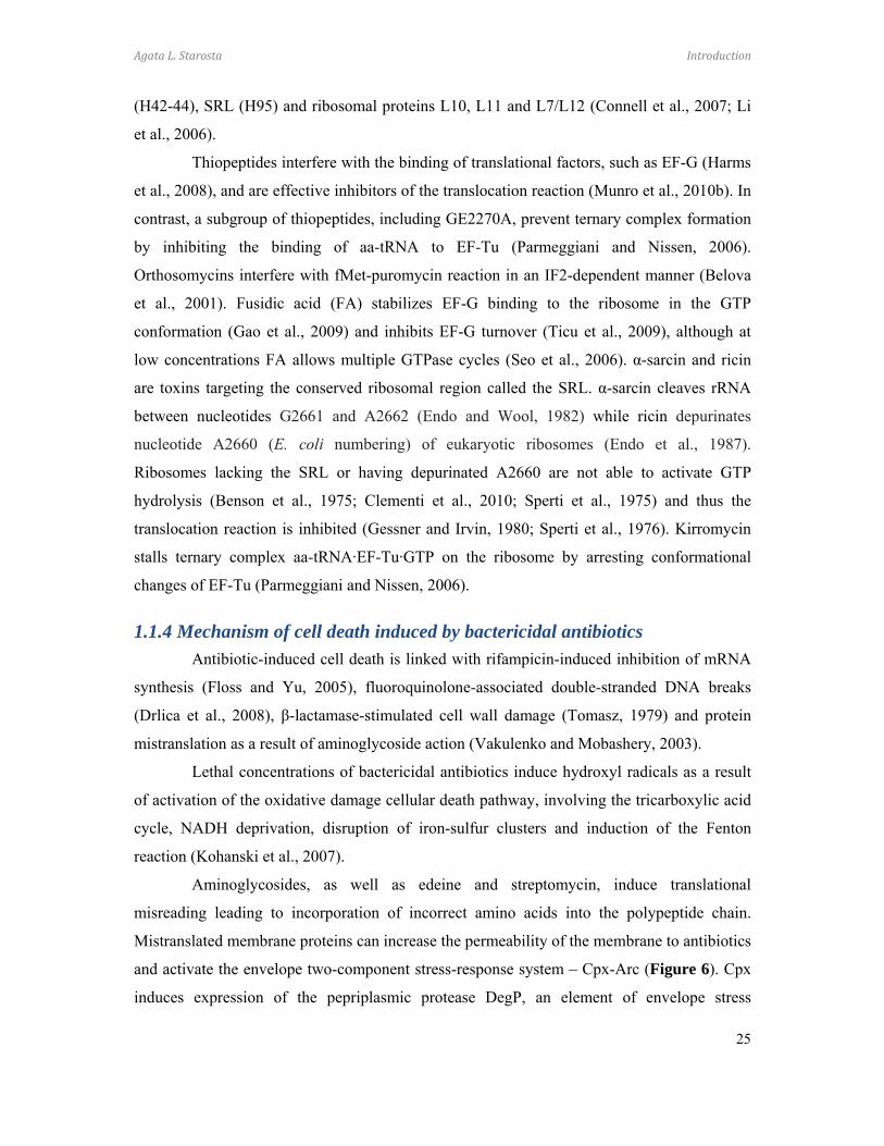
and activate the envelope two-component stress-response system – Cpx-Arc (Figure 6). Cpx
induces expression of the pepriplasmic protease DegP, an element of envelope stress
Agata L. Starosta Introduction
26
response, and activates the redox-responsive two-component system Arc, which leads to
modulation of the metabolic and respiratory pathways, formation of hydroxyl radicals, and
eventually to cell death (Kohanski et al., 2008).
Figure 6 Mechanism of cell death induced by bactericidal antibiotics. (a) Aminoglycosides, (b) quinolones and (c) β-lactams stimulate the oxidation of NADH through the electron transport chain, which is dependent on the tricarboxylic acid (TCA) cycle. Hyperactivation of the electron transport chain stimulates superoxide (O2-) formation. Superoxide damages Fe–S clusters, which results in release of ferrous iron and induction of the Fenton reaction. The Fenton reaction leads to the formation of hydroxyl radicals (•OH), which damage DNA, lipids and proteins. This accounts for antibiotic-induced cell death. Antibiotics also trigger hydroxyl radical formation and cell death through the envelope (Cpx) and redox-responsive (Arc) two-component systems. Redox-sensitive proteins, such as those containing disulphides (dashed lines) may also contribute to the cell death mechanism. acnb, aconitase b; mdh, malate dehydrogenase; uq, ubiquinone, according to (Kohanski et al., 2010b; Kohanski et al., 2007; Kohanski et al., 2008).

Agata L. Starosta Introduction
27
1.2 Antibiotics used in these studies
1.2.1 Hygromycin A
Hygromycin A (HygA) was discovered in the 1950’s as a metabolite excreted by
Streptomyces hygroscopicus NRRL 2388 (Pettinger and Wolfe, 1953) and does not share
structural similarities with the aminoglycoside with a similar name, hygromycin B. HygA is a
potent inhibitor of Gram-positive bacteria, while ineffective against Gram-negative strains
due mainly to the ArcAB efflux pump mechanism (Hayashi et al., 1997). It was shown that
HygA inhibits peptide bond formation and competes with chloramphenicols for binding to
ribosomes (Guerrero and Modolell, 1980), suggesting that HygA is an inhibitor of the LSU.
Footprinting experiments showed that HygA binding to the ribosome can be abolished by
macrolides carrying two sugar moieties at the C5 position (Poulsen et al., 2000), which would
extend towards the PTC (Hansen et al., 2002a). HygA protects nucleotides located in the
PTC, namely U2585 (Poulsen et al., 2000), which is important for tRNA accommodation and
peptidyltransferase reaction (Schmeing et al., 2005b), and U2506, from CMCT modification
(Poulsen et al., 2000).
HygA is composed of the three structurally distinct moieties: subunit A –
dehydrofucofuranose (SubA), B – α-methyl cinnamic acid (SubB), and C – aminocyclitol
(SubC) (Habib el et al., 2003). SubA is moderately sensitive to chemical manipulation and
when replaced with a hydrophobic allyl group retains antimicrobial potency (Hayashi et al.,
1997; Jaynes et al., 1992). In contrast, substitution of the methyl group with propyl, allyl or
hydrogen in SubB leads to decreased activity of the derivatives compared with HygA
(Hayashi et al., 1997). SubC is essential for the antimicrobial activity of HygA (Hayashi et al.,
1997).
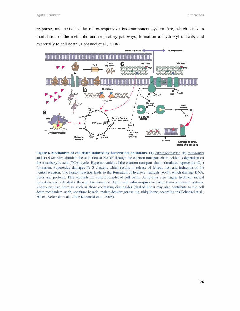
The HygA biosynthesis operon consists of 29 open reading frames (ORFs) (Figure
7) and includes genes responsible for providing resistance to HygA, as well as regulation and
synthesis of each of the HygA moieties (Palaniappan et al., 2006). The biosynthetic origin of
each HygA moiety has been established using isotope-labeled precursor studies (Habib el et
al., 2003): SubA is derived from glucose, SubB originates from 4-hydroxybenzoic acid and
propionic acid, and SubC is synthesized from myo-inositol and methionine (Habib el et al.,
2003). The glycoside bond formation between SubA and SubB, and the amide bond between
SubB and SubC, follow the assembly of each of the respective moieties (Habib el et al.,
2003).
Agata L. Starosta Introduction
28
The function of some of the gene products within the HygA operon was analysed by
genetic manipulations. Knock-out of hyg26 gene showed that its role plays crucial role in final
oxidation step in biosynthesis of SubA. S. hygroscopicus Δhyg26 mutant strain was shown to
produce 5”-dihydro-HygA, instead of HygA (Palaniappan et al., 2006). However, it was not
possible to establish whether oxidation happened before or after moieties A and B were linked
together.
Methylation at position A2503 of 23S rRNA prevents HygA from binding to the
ribosome (Toh et al., 2008). Moreover, among the genes assigned as resistance genes, hyg21
was shown to encode a 2-O-phosphotransferase, which inactivates HygA (Dhote et al., 2008).
Hyg6 and hyg29 genes were assigned as putative methyltranferases, and hyg19 and hyg28, as
efflux pumps.
From the perspective of development of new antimicrobials, elucidation of the
biosynthetic pathway of the aminocyclitol moiety remains an interesting and open question.
Aminocyclitol, as shown in previous studies (Hayashi et al., 1997), is essential for HygA’s
inhibitory activity. Aminocyclitols are often found in active natural compounds, for example,
the aminocyclitole-aminoglycosides (e.g. gentamycin, kanamycin or streptomycin), which are
very potent antibiotics (Flatt and Mahmud, 2007).
Figure 7 A HygA biosynthesis genes cluster from Streptomyces hygroscopicus NRRL 2388. Subunit A(shown in green) originates from glucose-6-phosphate via a mannose intermediate, reaction is driven by Hyg5,23, 29, 16. The final step, oxidation of C-5”-dihydrohygromycin A is catalyzed by NAD dependent Hyg26Subunit B (in purple) is derived from 4-hydroxybenzoic acid and propionic acid in a polyketide-like manner. Subunit C (in pink) Aminocyclitol originates from glucose-6-phosphate via myo-inositol intermediate,in a series of reactions catalyzed by Hyg 18, 25, 17, 8. The methylenedioxy bridge formation between C4 and C5 involves Hyg6 and Hyg7 (Dhote et al., 2009; Habib el et al., 2003; Palaniappan et al., 2006; Palaniappan et al., 2009)
Agata L. Starosta Introduction
29
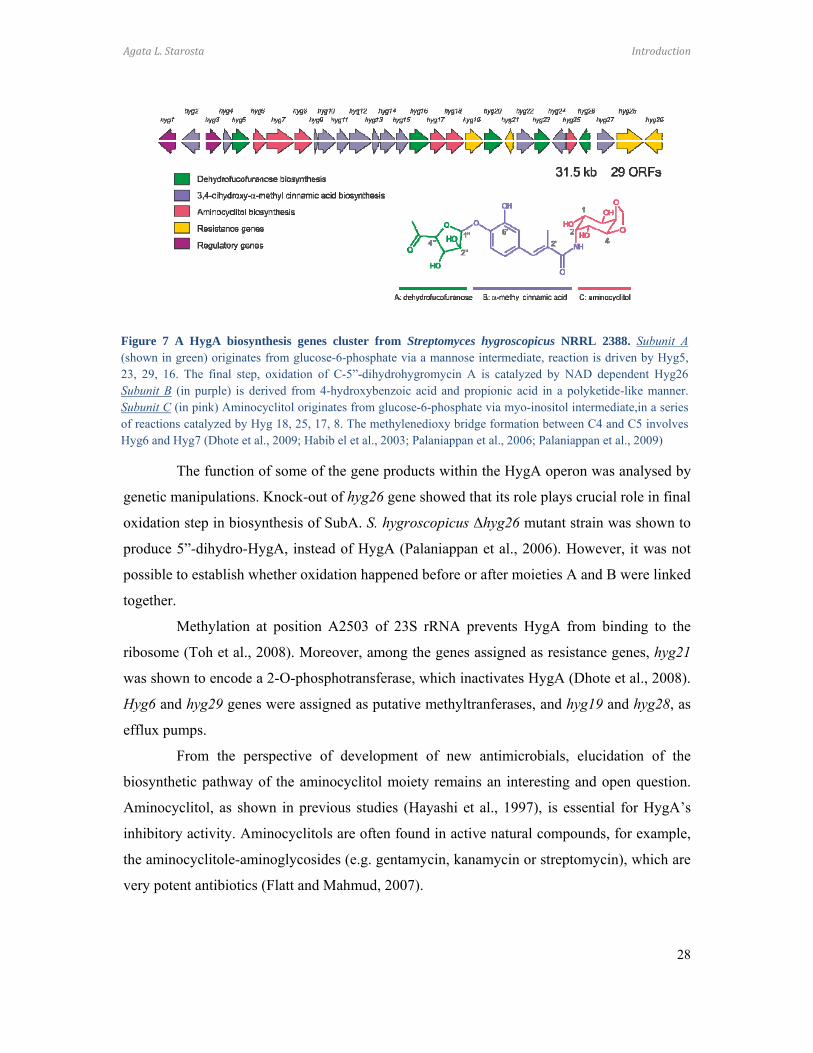
1.2.2 Macrolides The first members of the macrolide class of antibiotics, picromycin (Brockmann and
Henkel, 1950) produced by Streptomyces venezuelae, and erythromycin (McGuire et al.,
1952) produced by Saccharopolyspora erythraea, were discovered in the 1950’s. The
foundation of the chemical structure of macrolides is the 12-16 membered lactone ring, to
which a range of sugar moieties is attached. The classic macrolide, erythromycin (Ery),
belongs to the 14-membered family with C3 cladinose and C5 desosamine sugars, while
azithromycin (Azi) has a 15-membered ring. Tylosin (Tyl) (Figure 8c) is an example of a 16-
membered macrolide with a C5 mycaminose-mycarose disaccharide as well as a C14
mycinose (reviewed by (Mankin, 2008; Wilson, 2009)). Macrolides bind to non-translating
ribosomes (Pestka, 1974). Chemical probing showed that macrolides universally protect
nucleotide A2058 (Poulsen et al., 2000) located at the entrance of the exit tunnel, adjacent to
the PTC. Macrolides with a mycarose moiety protect U2506 in the central loop of domain V
23S rRNA, while A752 (domain II), lying deeper in the exit tunnel, is protected from
modifications by macrolides with a mycinose moiety (Poulsen et al., 2000). Crystal structures
of macrolides bound to ribosomes confirm that the lactone ring binds to the wall of the tunnel,
near the tunnel constriction formed by ribosomal proteins L4 and L22, with the C5 sugar
moiety extending towards the PTC, and interacts with A2058 and A2059 (Figure 8a, b)
(Hansen et al., 2002a; Schlunzen et al., 2001; Tu et al., 2005; Wilson et al., 2005). However,
the C14 mycinose faces down the nascent polypeptide exit tunnel and interacts with A748-
A752 (Hansen et al., 2002a). Tyl, carrying C6 ethyl aldehyde group, was shown to form an
Figure 8 Binding of macrolide antibiotics within the ribosomal tunnel (A) The large subunit of the ribosomewith visualized ribosomal exit tunnel. The position of tylosin (red), P-site tRNA (green), and a polypeptide chain(yellow) are indicated. rRNA in light blue, ribosomal proteins in dark blue. (B) Zoom in on the macrolide-bindingsite within the ribosomal tunnel. Tylosin (pink) shown with lactone ring, C14-mycinose, and C5-mycaminose-mycarose are labeled. The arrow indicates the carbinolamine bond that forms between tylosin and the N6 of 23SrRNA nucleotide A2062. The positions of A- and P-site aminoacyl-tRNAs are shown in green and orange,respectively. (C) Chemical structure of tylosin
Agata L. Starosta Introduction
30
unusual reversible covalent bond with the N6 of nucleobase A2062 (Hansen et al., 2002a).
Macrolides bound to the ribosome prevent the progression of most nascent
polypeptide chains through the tunnel and induce peptidyl-tRNA drop-off. However, in the
presence of macrolides, synthesis of short oligopeptides is still possible. The length of the
peptide synthesized before peptidyl-tRNA drop-off depends on the number of sugar
substituents at the position C5 and the amino acid sequence of the peptide (Tenson et al.,
2003). Monosaccharides, for example as in Ery, allow synthesis of up to eight amino acid
long oligopeptides, while macrolides with disaccharides (e.g., Tyl) allow translation of di- to
tetrapeptides (Tenson et al., 2003). Moreover, macrolides with a mycarose moiety can directly
inhibit peptide bond formation (Poulsen et al., 2000). A resistance mechanism to macrolides
involving translation of pentapeptides, which leads to dissociation of the drug from the
ribosome, suggests a direct interaction of the macrolide with the nascent polypeptide chain
(Lovmar et al., 2006; Tenson et al., 1996; Tenson and Mankin, 2001). Furthermore,
pentapeptide effects on distinct macrolides vary, which implies specific interactions between
the drug and the nascent polypeptide (Vimberg et al., 2004). A number of additional
resistance mechanisms are known, including (i) mono- and dimethylation of the N-6 position
of A2058 by the Erm family of methyltransferases (Lai and Weisblum, 1971; Roberts et al.,
1999), (ii) single substitution of A2058 to guanosine in the 23S rRNA (Vester and
Douthwaite, 2001), (iii) mutations in ribosomal protein L4, such as single amino acid
substitutions K63E (Chittum and Champney, 1994) and G69C as well as insertion of two
amino acids, SQ, between Q67K68 (Tait-Kamradt et al., 2000), (iv) deletion of three amino
acids ΔM82R83K84 in ribosomal protein L22 (Gregory and Dahlberg, 1999), (v) MefA and
MsrA macrolide efflux pumps (Roberts et al., 1999; Ross et al., 1990), or (vi) inactivation of
the antibiotic by modification, for example by Mph family of macrolide phosphorylases
(Roberts et al., 1999).
Many macrolide resistance genes are expressed upon induction, which is driven by
the presence of sublethal concentrations of the antibiotic in a mechanism called translational
stalling. One of the best-characterized examples is the regulation of expression of the
methyltransferase, ErmC, which is controlled by an upstream leader sequence ermCL
(Gryczan et al., 1980; Horinouchi and Weisblum, 1980; Ramu et al., 2009). In the absence of
the macrolide, ermC translation is attenuated, whereas the presence of the drug induces
ribosome stalling during translation of ermCL. This, in turn, leads to a rearrangement of the
mRNA secondary structure, exposure of the Shine-Dalgarno sequence and start codon, and
Agata L. Starosta Introduction
31
expression of downstream ermC gene. Recent results suggest that in addition to the sequence
of the polypeptide, the structure of the antibiotic is also important for stalling (Vazquez-
Laslop et al., 2011). The rRNA nucleotides A2062 and U1782 monitor the structure of the
nascent polypeptide, while nucleotides C2610, A2503, and possibly A2058 and A2059
recognize the structure of the macrolide antibiotic (Vazquez-Laslop et al., 2011) and together
transmit the signal that induces ribosomal stalling. Moreover, it was shown that the stalled
peptide alters the A-site function for peptide bond formation which accounts for the
translational arrest (Ramu et al., 2011).
1.2.3 Thiopeptides Thiopeptide antibiotics are ribosomally synthesized antimicrobials. They are produced
as oligopeptides which undergo a series of post-translational modifications resulting in
mature, active compounds (Wieland Brown et al., 2009). Compounds belonging to the
thiopeptide family have a characteristic trithiazolyl pyridine core, and a macrocyclic loop
consisting of a varying number of atoms (for example, 26 in nosiheptide, thiocillin, and
thiostrepton, 29 in GE2270A and 35 in berninamycin) (reviewed by (Bowers et al., 2010;
Walsh et al., 2010)). Microccocin P1 (MiC), discovered in 1948 as a metabolite of
Microccocus (Su, 1948) and Bacillus cereus (Bycroft and Gowland, 1878), was the first
described member of the thiopeptide family, followed by the discovery of thiostrepton (ThS),
which is produced by Streptomyces azureus and S. laurentii (Donovick et al., 1955). To date,
more than 80 members of this class of antimicrobials are known. Thiopeptides due to their
poor solubility, are used only in veterinary practice. However the potency of thiopeptides
against multidrug resistant Gram-positive strains and recent advances in total synthesis of a
number of thiopeptides (ThS (Nicolaou et al., 2005a; Nicolaou et al., 2005b), GE2270A
(Muller et al., 2007) and MiC (Lefranc and Ciufolini, 2009), has renewed interest in this
group of antibiotics.
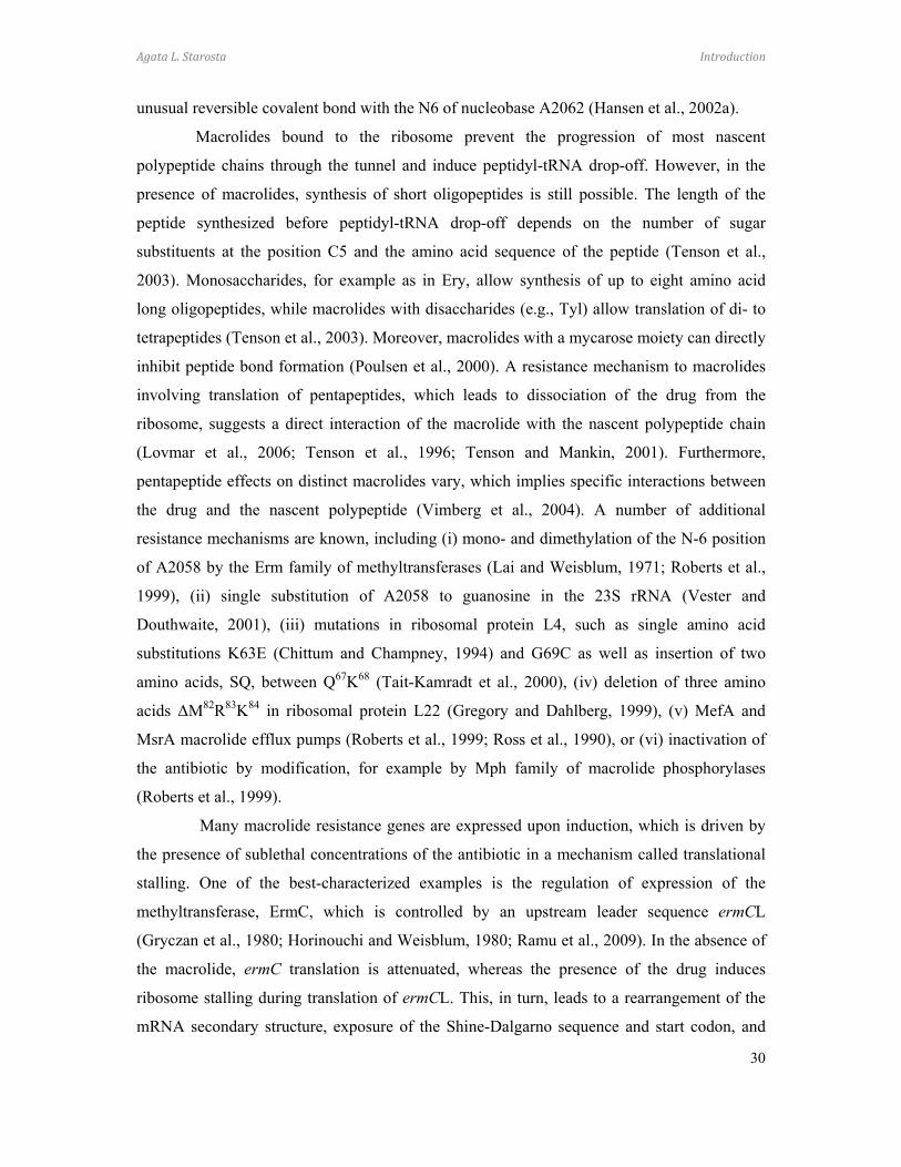
The Bacillus cereus ATCC 14579 thiocillin biosynthesis operon has been well-
characterized (Figure 9), revealing that thiopeptides are synthesized as pre-peptides with a
leader peptide which is cleaved-off post-translationally. The C-termini derived peptide
undergoes a series of modification steps, thirteen in the thiocillin maturation process, resulting
in a fully active antimicrobial compound (Wieland Brown et al., 2009). In vivo genetic
manipulations of the thiocillin structural gene has led to generation of more than 60 new
Agata L. Starosta Introduction
32
thiocillin variants (Acker et al., 2009), opening up new possibilities for generating more
active compounds with improved pharmacokinetic properties.
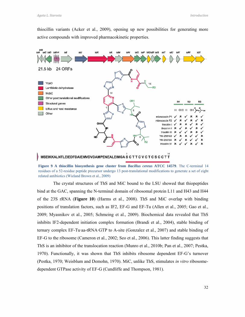
The crystal structures of ThS and MiC bound to the LSU showed that thiopeptides
bind at the GAC, spanning the N-terminal domain of ribosomal protein L11 and H43 and H44
of the 23S rRNA (Figure 10) (Harms et al., 2008). ThS and MiC overlap with binding
positions of translation factors, such as IF2, EF-G and EF-Tu (Allen et al., 2005; Gao et al.,
2009; Myasnikov et al., 2005; Schmeing et al., 2009). Biochemical data revealed that ThS
inhibits IF2-dependent initiation complex formation (Brandi et al., 2004), stable binding of
ternary complex EF-Tu·aa-tRNA·GTP to A-site (Gonzalez et al., 2007) and stable binding of
EF-G to the ribosome (Cameron et al., 2002; Seo et al., 2006). This latter finding suggests that
ThS is an inhibitor of the translocation reaction (Munro et al., 2010b; Pan et al., 2007; Pestka,
1970). Functionally, it was shown that ThS inhibits ribosome dependent EF-G’s turnover
(Pestka, 1970; Weisblum and Demohn, 1970). MiC, unlike ThS, stimulates in vitro ribosome-
dependent GTPase activity of EF-G (Cundliffe and Thompson, 1981).
Figure 9 A thiocillin biosynthesis gene cluster from Bacillus cereus ATCC 14579. The C-terminal 14residues of a 52-residue peptide precursor undergo 13 post-translational modifications to generate a set of eightrelated antibiotics (Wieland Brown et al., 2009)

Agata L. Starosta Introduction
33
Figure 10 Thiostrepton. (a) An overview of thiopeptide binding site on the LSU. Interface view with helix 43 and 44 (H43/44; orange), L11 (yellow), and ThS (green) highlighted with surface representation (PDB 3CF5) (Harms et al., 2008). (b) The thiazole rings of ThS (green) interact with the RNA bases at the tips of H43/44 as well as the prolines in the N-terminal domain of L11 (yellow) (Starosta et al., 2009).
In contrast, GE2270A does not bind to the GAC, but instead binds to EF-Tu
(Figure 11) and interferes with binding of the aa-tRNA, leading to inhibition of ternary
complex formation (Parmeggiani and Nissen, 2006). Crystallographic data showed that
GE2270A spans domain I and II of EF-Tu, and therefore prevents the aminoacyl moiety of
the aa-tRNA from binding (Parmeggiani and Nissen, 2006).
Figure 11. Binding site of GE2270A on EF-Tu. (a) Structure of the thiopeptide GE2270A (green) bound to EF-Tu (yellow), with domains I, II, and III indicated (PDB 2C77) (Parmeggiani et al., 2006). (b) GE2270A overlaps the binding position on EF-Tu of the terminal A76 and aminoacyl moiety of tRNA. Inset shows overview of EF-Tu•tRNA ternary complex (PDB 1TTT) (Nissen et al., 1995) with superimposition of GE2270A. (c) Superimposition of EF-Tu_GE2270A (yellow) and EF-Tu•tRNA (blue) aligned on basis of domain II. Note that GE2270A (green) clashes with domain I of EF-Tu from the ternary complex (blue) (Starosta et al., 2009).
Thiopeptides that interact with the ribosome contact nucleotides A1067 (H43) and
A1095 (H44), and interact with P22 and P26 of L11. In accordance with structural data,
substitutions of A1067 and A1095 (Rosendahl and Douthwaite, 1994; Thompson et al., 1988),
or within the proline-rich region of L11’s N-terminus (Cameron et al., 2004) lower the
affinity of the drug for the ribosome and confer resistance (Baumann et al., 2010). Bacteria
producing thiopeptides protect themselves from the inhibitory effects of drugs through efflux
mechanisms (Wieland Brown et al., 2009). The Streptomyces azureus genome encodes a 2’-

Agata L. Starosta Introduction
34
O-methylase that modifies nucleotide A1067 (Thompson et al., 1982), which is used in
molecular biology as ThS selection marker.
1.2.4 Orthosomycins The evernimicin class (orthosomycins) of antibiotics were first isolated from
Micromonospora carbonaceae in the 1960’s (Weinstein et al., 1964). Orthosomycins are
oligosaccharide antibiotics rich in modified L- and D-deoxysugars containing unusual
orthoester and glycosyl linkages (Figure 12c) (Hosted et al., 2001; Weitnauer et al., 2001)
and are active against Gram-positive bacteria (Nakashio et al., 1995). Evernimicin (Evn) and
avilamycin (Avi) bind to the LSU and are potent protein synthesis inhibitors (McNicholas et
al., 2000). Chemical probing shows that Evn and Avi protect nucleotides A2468, A2469,
A2476, A2478, and A2482 in H89, and A2534 in H91 from DMS modification (Belova et al.,
2001; Kofoed and Vester, 2002), which locates the orthosomycin binding site at the base of
the L7/L12 ribosomal stalk (Figure 12a, b). Consistent with primer extension data, mutations
conferring resistance to Evn are located in H89 and H91 (Belova et al., 2001). Also mutations
in ribosomal protein L16 are linked to Evn resistance (Belova et al., 2001; McNicholas et al.,
2001). Moreover, the modification of U2479 by AviRB, a 2’-O-methyltransferase from
Streptomyces viridochromogenes (Mosbacher et al., 2005), and of G2470 by EmtA, a rRNA
methyltransferase from Enterococcus faecium (Mann et al., 2001), led to an increase in the
resistance to Evn. Although the exact mechanism of action of Evn remains unclear,
biochemical data showed that Evn overlaps with the IF2 binding site on the LSU (La Teana et
al., 2001) and inhibits fMet-puromycin synthesis on ribosome reconstituted from the 30SIC
Figure 12 Orthosomycins. (a) An overview of the binding sites of orthosomycins on the LSU relative to EF-G. Ribosomal proteins L1, L11, and L7 are shown for reference; (b) Putative binding site of orthosomycins spanning from H89 and H91 of the 23S rRNA. Residues highlighted in red have been associated biochemicallywith Evn (review by (Wilson, 2009); (c) Chemical structure of Evn.
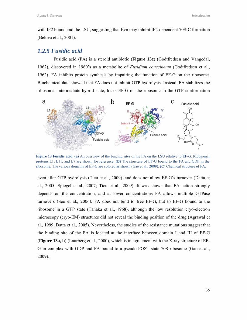
Agata L. Starosta Introduction
35
with IF2 bound and the LSU, suggesting that Evn may inhibit IF2-dependent 70SIC formation
(Belova et al., 2001).
1.2.5 Fusidic acid Fusidic acid (FA) is a steroid antibiotic (Figure 13c) (Godtfredsen and Vangedal,
1962), discovered in 1960’s as a metabolite of Fusidium conccineum (Godtfredsen et al.,
1962). FA inhibits protein synthesis by impairing the function of EF-G on the ribosome.
Biochemical data showed that FA does not inhibit GTP hydrolysis. Instead, FA stabilizes the
ribosomal intermediate hybrid state, locks EF-G on the ribosome in the GTP conformation
even after GTP hydrolysis (Ticu et al., 2009), and does not allow EF-G’s turnover (Datta et
al., 2005; Spiegel et al., 2007; Ticu et al., 2009). It was shown that FA action strongly
depends on the concentration, and at lower concentrations FA allows multiple GTPase
turnovers (Seo et al., 2006). FA does not bind to free EF-G, but to EF-G bound to the
ribosome in a GTP state (Tanaka et al., 1968), although the low resolution cryo-electron
microscopy (cryo-EM) structures did not reveal the binding position of the drug (Agrawal et
al., 1999; Datta et al., 2005). Nevertheless, the studies of the resistance mutations suggest that
the binding site of the FA is located at the interface between domain I and III of EF-G
(Figure 13a, b) (Laurberg et al., 2000), which is in agreement with the X-ray structure of EF-
G in complex with GDP and FA bound to a pseudo-POST state 70S ribosome (Gao et al.,
2009).
Figure 13 Fusidic acid. (a) An overview of the binding sites of the FA on the LSU relative to EF-G. Ribosomalproteins L1, L11, and L7 are shown for reference; (B) The structure of EF-G bound to the FA and GDP in theribosome. The various domains of EF-G are colored as shown (Gao et al., 2009); (C) Chemical structure of FA.
Agata L. Starosta Introduction
36
1.3 Alternative antimicrobials targeting virulence
Traditional antibiotics, which inhibit essential functions of bacteria, are under
selective pressure that favors resistance mutations (Clatworthy et al., 2007). The need for
novel antimicrobial therapies is greater than ever before. New approaches in antimicrobial
development focus on bacterial virulence and interactions between bacterial and host cells
(Clatworthy et al., 2007).
1.3.1 Definition of virulence
In the traditional meaning, virulence is understood as the ability of a microorganism
to cause illness (Clatworthy et al., 2007). Virulence factors (toxins, cytolysines, proteases) are
generally not necessary for a pathogen to survive under laboratory conditions, however their
role in vivo is crucial for the microorganism to survive in the environment (Alksne and
Projan, 2000). The analysis of genomes of many virulent bacteria has revealed diversity in
their gene content, for example different E. coli strains, e.g. laboratory K12 strain compared
with pathogenic E. coli 0157, differ by up to 25% of their genome, while Salmonella enterica
differs from E.coli K12 by up to 12% (Raskin et al., 2006). The variable genes are located on
so-called pathogenicity islands, clustering genes responsible for virulence and antibiotic
resistance (Pallen and Wren, 2007; Raskin et al., 2006). Bacterial virulence factors are
responsible for colonization, survival within host cells, modification of host cell response as
well as intracellular communication (Wu et al., 2008).
1.3.2 Colonization
Pathogenic bacteria invading host cells adapt the expression of entire pathways of
genes to transition from the free-living form to one that colonizes the host cell (Alksne and
Projan, 2000). Colonization of host cells requires induction of expression and assembly of
adhesions, fimbriae and pili, which allow bacteria to attach to the cell surface (Finlay and
Cossart, 1997). Bacteria invading mammal cells must reprogram their metabolism to adapt to
new living conditions. Varying pH, oxygen accessibility, osmotic pressure, nutrient
availability and ion homeostasis are signals to rearrange gene expression of the pathogen (Ray
et al., 2009).
1.3.3 Biofilm
Bacteria hardly ever exist as free-floating cells. They tend to live in organized
colonies associated with surfaces (Jayaraman and Wood, 2008). Such colonies, called

Agata L. Starosta Introduction
37
biofilms, are highly organized structures of bacteria embedded within exopolysaccharide
matrices (Lawrence et al., 1991), where nutrient and metabolite transport is regulated by
bacterial communication using small messenger molecules (acylated homoserine lactones,
AHLs) (Davies et al., 1998). Biofilms give bacteria a greater chance to survive in
unfavourable environmental conditions, increasing their chances for successful colonization.
Bacteria in biofilms show significantly decreased susceptibility to antibiotics compared to
free-floating cells (Luppens et al., 2002).
1.3.4 Quorum sensing
Communication between bacteria is mostly mediated by small autoinducer
molecules. Autoinducers, mostly AHLs that are secreted into the environment, can modulate
transcription of genes, including virulence factors. Quorum sensing occurs when bacterial
populations reach high densities, which leads to the accumulation of high concentrations of
autoinducers (reviewed by (Ng and Bassler, 2009)).
1.3.5 Motility
Successful colonization of new niches requires bacteria to have the ability to
migrate. Motility is a high-energy consuming process powered by rotation of flagella or
movement of pili (Kearns, 2010; Verstraeten et al., 2008). Swarming and swimming motilities
are flagella-dependent. While swarming is the movement of a group of bacteria over a solid
surface, swimming, in contrast, is an act of a single cell moving though a viscous liquid
environment (Henrichsen, 1972). Twitching motility is driven by extension and retraction of
type IV pili and is important for biofilm formation (Mattick, 2002).
1.3.6 Secretion systems
Bacteria can modulate the environment or even alter the host’s physiology using
several secretion systems (SS) (Tseng et al., 2009). However here, only three of the major SS
in bacterial pathogenicity are mentioned. The type I SS is an ABC transporter responsible for
efflux of antimicrobial molecules as well as toxins, proteases and lipases (Koronakis et al.,
2004). The type III SS is called the injectisome due to its ability to protrude into the host
cell’s surface with a needle-like structure, which allows direct release of toxins into the host’s
cytoplasm (Cornelis, 2010). The type IV SS is the only SS which can transport nucleic acids
as well as proteins. F-plasmid based conjugation, accounts for evolution of infectious
pathogens though propagation of antibiotic resistance and virulence genes (Cascales and

Agata L. Starosta Introduction
38
Christie, 2003). Moreover type IV pili have been shown to promote bacterial motility and
stimulate biofilm formation (Mattick, 2002).
1.3.7 Elongation factor P
Elongation factor P (EF-P) was identified as a protein binding to the ribosome (Glick
and Ganoza, 1975). Two-dimensional gel electrophoresis (2D) analysis of E. coli cell extract
showed EF-P to be present in a ratio 1 per 10 ribosomes (An et al., 1980). EF-P was found to
bind to subunits, 70S ribosomes and polysomes (Aoki et al., 2008) suggesting its role not only
in translation initiation, but also in elongation steps. Although, EF-P function remains unclear,
it was shown that the elongation factor stimulates formation of the first peptide bond (Ganoza
and Aoki, 2000; Glick et al., 1979; Glick and Ganoza, 1975; Swaney et al., 2006). The
deletion of efp gene leads to defects in the growth of E. coli (Aoki et al., 1997), virulence of
Agrobacterium tumefaciens dependent on type IV SS (Peng et al., 2001), intestinal
colonization of host cells by Vibrio cholerae also dependent on type IV SS (Merrell et al.,
2002) and Brucella abortus (Iannino et al., 2011), flagella driven motility of Bacillus subtilis
and E. coli (Inoue et al., 2007; Kearns et al., 2004). Moreover, it was shown that EF-P is
specifically expressed in porcine lymph nodes upon S. typhimurium infection (Van Parys et
al., 2011). The EF-P crystal structures revealed a three domain architecture that resembles a
classical tRNA L-shape, suggesting possible mimicry (Figure 14a) (Choi and Choe, 2011;
Hanawa-Suetsugu et al., 2004; Yanagisawa et al., 2010). The crystal structure of EF-P bound
to 70S ribosome (Blaha et al., 2009) shows that EF-P does not bind in a canonical tRNA
binding site, but rather sits between the P- and E-sites, spanning both subunits (Blaha et al.,
2009). The conserved loop of domain I reaches the CCA-end of the initiator-tRNA, while
domain III contacts the anticodon stem loop of tRNA (Blaha et al., 2009), supporting the
hypothesis that EF-P stabilizes the initiator tRNA to promote first peptide bond reaction.

Agata L. Starosta Introduction
39
EF-P is conserved among bacteria (Bailly and de Crecy-Lagard, 2010) and is
homologous with eukaryotic initiation factor 5A (eIF5A) (Hanawa-Suetsugu et al., 2004).
Unlike EF-P, eIF5A has only two domains, corresponding to domain I and II of EF-P (Kim et
al., 1998; Peat et al., 1998). However, like eIF5A, EF-P also has a highly conserved loop in
domain I (Hanawa-Suetsugu et al., 2004). A unique feature of eIF5A is the presence of a post-
translational modification with hypusine, which is essential and characteristic only to eIF5A
(Zanelli et al., 2006). The highly conserved lysine 50 (K50 in human eIF-5A) is modified into
an unusual amino acid derived from spermidine. Hypusylation is a two-step reaction,
catalyzed by two enzymes. First, the deoxyhypusine synthase (DHS) catalyzes the cleavage of
the polyamine spermidine as well as the transfer of its 4-aminobutyl moiety to the ε-amino
group of lysine residue of the eIF-5A precursor to form deoxyhypusine (Park, 2006). In a
second step, deoxyhypusine hydroxylase (DOHH) mediates the addition of a hydroxyl group
to the deoxyhypusine residue forming hypusine (Park, 2006). DHS and DOHH are not present
in bacterial genomes, however, analyses of endogenous E. coli EF-P revealed the presence of
an additional 144 Da mass (Aoki et al., 2008). Two unrelated to DHS and DOHH gene were
implied to function as modification enzymes for EF-P pathway, namely, yjeA (a paralog of
lysyl-tRNA synthetase) and yjeK (lysine 2,3-aminomutase) (Figure 14c) (Bailly and de
Crecy-Lagard, 2010). Interactions between yjeA and yjeK were reported to be critical for
virulence and stress resistance in S. enterica (Navarre et al., 2010). Salmonella yjeA and yjeK
mutants share extensive phenotypic pleiotropy, including attenuated virulence in mice, a
decreased ability of bacteria to colonize swine host, growth defects, susceptibility to a variety
Figure 14. Elongation factor P. (a) EF-P crystal structure shows classical tRNA L-shape, in red - Lysine 34undergoing modification (PDB 3A5Z); (b) EF-P (violet) in a complex with YjeA (yellow) (PDB 3A5Z)(Yanagisawa et al., 2010); (c) Physical clustering of efp (violet), yjeK (pink), and yjeA (yellow), according to(Bailly and de Crecy-Lagard, 2010).
Agata L. Starosta Introduction
40
of various growth inhibitors and altered expression of multiple proteins, including several
encoded on the SPI-1 pathogenicity island (Bearson et al., 2011; Bearson et al., 2006; Kaniga
et al., 1998; Navarre et al., 2010; Yanagisawa et al., 2010). YjeA mediates the post-
translational modification of bacterial elongation factor P resulting in an additional 128 Da
mass on EF-P (Figure 14b) (Navarre et al., 2010). In vivo experiments show that YjeK
enhances the activity of YjeA (Yanagisawa et al., 2010), which is consistent with observation
that (R)-β-lysine is 100-fold better substrate for YjeA than (S)-β-lysine or α-lysine (Roy et al.,
2011).
Agata L. Starosta Objectives
41
2 Objectives of these studies
(I) Hygromycin A (Paper 1 and 2). Analysis of fermentation broth contents of several
S. hygroscopiscus NRRL 2388 mutants revealed the presence of a mixture of HygA
derivatives. Intermediates of the HygA biosynthetic pathway were investigated and
affects of alterations of the aminocyclitol moiety on the antimicrobial activity of
HygA were studied. Moreover how differential oxidation states of the C5” position
of dehydrofucofuranose moiety affects the activity of HygA was also examined.
Additionally, unpublished data is presented showing the inhibitory effect of HygA
derivatives on peptide bond formation and the effect of phosphorylation of HygA by
the 2-O-phosphotransferase was evaluated.
(II) Macrolides. (Paper 3) Structural data show that D. radiodurans ribosome can bind two
molecules of azithromycin (AZI) (Schlunzen et al., 2003). However in the crystal
structure of AZI with Haloarcula marismortui LSU only one molecule of AZI was
bound (Hansen et al., 2002a). Here the kinetics of AZI binding to E. coli and
D. radiodurans ribosomes were studied to investigate in more details the
discrepancies between antibiotic binding in different species. (Paper 4). The C6
ethyl aldehyde group of the Tylosin (Tyl) forms a unique covalent bond with
ribosomal RNA A2062 (Hansen et al., 2002a). The contribution of the covalent
linkage in the binding to the ribosome and the inhibitory activity of Tyl was
determined. Moreover, Tyl derivatives designed to establish interactions with
ribosomal tunnel wall, and derivatives with substituents mimicking nascent
polypeptide was also tested.
(III) Thiopeptides. (Paper 5) The complicated structure and poor solubility of thiopeptides
account for their limited possibilities in terms of application for bacterial infection
therapies. Therefore, a library of thiopeptide synthetic precursors was screened for
their potency to inhibit protein synthesis and ribosome-dependent GTPase activity of
EF-G in order to find the minimal carbon scaffold necessary for targeting the
ribosome and which, in the future, could provide a basis for design of more soluble
compounds. (Paper 6) MiC binds in a cleft between ribosomal protein L11 and H43
and H44 of the 23S rRNA (Harms et al., 2008), and stabilizes the C-terminal domain
of protein L7/L12 (CTD-L7/L12) (Harms et al., 2008). Crystallographic data shows
Agata L. Starosta Objectives
42
that the G’-domain of EF-G interacts with CTD-L7/L12 (Gao et al., 2009), and was
shown to be crucial for GTPase activity and Pi release (Nechifor et al., 2007;
Savelsbergh et al., 2005). By comparison of different GTPases, which have reduced
or absent G’ domain, the hypothesis was tested that the enhanced turnover of EF-G
by MiC is a result of stabilizing effects of the drug on L11- CTD-L7/L12 interaction.
(IV) Orthosomycins. (Paper 6). The available data suggest that Evn inhibits initiation of
translation, however, the exact step at which Evn acts remains unclear. Therefore, the
role of Evn in the translation cycle was dissected.
(V) Fusidic acid. (Paper 7) Antibiotics can trap the ribosome in distinct functional states. FA
was used to visualize intermediate translocation events and ascertain whether it is
possible to localize the antibiotic using Cryo-EM.
(VI) Elongation factor P. (Paper 8) Emerging resistance to antibiotics stresses the need for
new antimicrobial targets. EF-P is linked to virulence in a number of bacterial
strains. The modification of EF-P is crucial for pathogenicity. The differences
between the 144 Da modifications reported for endogenous EF-P compared with the
128 Da modification observed in vitro was investigated. Moreover, the chemical
composition of the modification was also dissected. Additionally, initial unpublished
data is presented showing variability in phenotypes of wild type E. coli with deletion
strains of the efp gene and genes encoding the EF-P modification enzymes.

Agata L. Starosta Cumulative thesis
43
3 Cumulative thesis
3.1 Hygromycin A
3.1.1 Paper 1
Palaniappan, N., Dhote, V., Ayers, S., Starosta, A.L., Wilson, D.N., and Reynolds, K.A. (2009). Biosynthesis of the aminocyclitol subunit of hygromycin A in Streptomyces hygroscopicus NRRL 2388. Chem Biol 16, 1180-1189.
3.1.2 Paper 2
Dhote, V., Starosta, A.L., Wilson, D.N., and Reynolds, K.A. (2009). The final step of hygromycin A biosynthesis, oxidation of C-5''-dihydrohygromycin A, is linked to a putative proton gradient-dependent efflux. Antimicrob Agents Chemother 53, 5163-5172.
The aminocyclitol subunit was shown to be essential for the antimicrobial activity of
HygA (Hayashi et al., 1997). The HygA biosynthesis operon has seven genes assigned for
synthesis of the aminocyclitol moiety (SubC) (Figure 7). Using a radiolabeled precursor
approach, it was shown that the SubC of HygA is derived from glucose via a myo-inositol
(MI) intermediate and methionine (Habib el et al., 2003). In Paper 1, Professor K. Reynold’s
group continued studies on HygA biosynthesis pathway. Gene disruption experiments and
chemical complementation with MI and/or mature SubC enabled the series of steps leading to
production of mature SubC to be understood. The studies demonstrated that glucose-6-
phosphate is a substrate for Hyg18, assigned as myo-inositol-1 phosphate synthase. In the
next step, MI-phosphate is dephosphorylated by Hyg25. MI is a substrate for inositol
dehydrogenase (Hyg17). The reaction of Hyg17 is followed by transamination of the C5-
oxydized MI to produce neo-inosamine-2 in a reaction catalyzed by Hyg8 aminotransferase.
Hyg6 is a SAM-dependent methyltransferase that methylates neo-inosamine-2. The structure
of HygA revealed a unique methylenedioxy bridge between the C4 and C5 carbons of the
aminocyclitol. Therefore, it was suggested that the cyclization of the C5 methoxy group is
catalyzes by Hyg7. Disruption of hyg8 and hyg7 genes did not lead to HygA production.
However, interestingly, the intermediates of HygA were accumulated. –
desmethylenehygromycin A (Des-HygA) containing a hydroxyl group at the C5 carbon, and
methoxyhygromycin A (Met-HygA), with a C5-OCH3 group instead of the methylenedioxy
bridge.

Agata L. Starosta Cumulative thesis
44
The antimicrobial potency of the intermediates of HygA with altered aminocyclitol
ring was examined against ΔtolC E. coli. The minimal inhibitory concentration (MIC)
experiments for HygA and intermediates showed that, the modifications of aminocyclitol
moiety lead to a significant loss (15-fold) of an in vivo activity of Des-HygA and Met-HygA.
Moreover, the HygA-derivative lacking the aminocyclitol moiety (AB) (Palaniappan et al.,
2006) displayed no detectable activity. I tested the intermediate compounds for their ability to
inhibit protein synthesis in an E. coli lysate-based in vitro coupled transcription-translation
assay (TT) using green fluorescent protein (GFP) as a reporter. HygA showed high in vitro
activity with half-inhibitory concentration (IC50) at 0.18 µM. Surprisingly, the intermediates
of HygA displayed significant in vitro activity, with IC50 of Met-HygA at 0.32 µM and Des-
HygA at a concentration of 0.5 µM, whereas the AB compound displayed no or minor
inhibitory activity, even at concentrations of 75 µM. Interestingly, the order of the activity of
the compound reflected the progression of methylenedioxy bridge synthesis. Additionally, in
the context of a DAAD fellowship, I visited the laboratory of Professor G. Dinos (Patras,
Greece) where I tested the HygA analogs for their ability to inhibit AcPhe-puromycin
(AcPhe-Puro) synthesis. Interestingly, I found the same trend of the potency to inhibit peptide
bond formation as I observed for the inhibition of GFP synthesis (Figure 15a,c vs b,d). These
data suggest that HygA primary activity is the inhibition of the PTC. The MIC, TT and
AcPhe-Puro data on the HygA intermediates confirmed that the aminocyclitol is necessary for
HygA inhibitory activity in vivo and in vitro. The data showed that for the full in vivo activity,
the aminocyclitol must be modified with an intact methylenedioxy bridge. However,
alterations of the methylenedioxy ring did not lead to a loss in the in vitro protein synthesis
inhibition activity, in particular on the peptide bond formation. Taken together, these data
suggest that the aminocyclitol ring is essential for translation inhibition activity, whereas a
methylenedioxy ring is crucial for full in vivo activity, possibly for the uptake of the drug into
the cell.
Previous studies from Professor K. Reynolds’s group showed that disruption of hyg26
gene led to the accumulation of 5”-dihydro-HygA (DH-HygA) intermediate (Palaniappan et
al., 2006). Hyg26 was suggested to catalyze dehydrogenation of the C5”-OH of the
fucofuranose moiety (SubA). In Paper 2, it was shown that recombinant Hyg26 is a
NAD/NADH-dependent oxidoreductase, and that the Hyg26 reaction is reversible and favors
DH-HygA. Surprisingly, the disruption of the hyg19 gene also led to the accumulation of DH-
HygA. Moreover, although HygA was still detected, the level of antibiotic production was
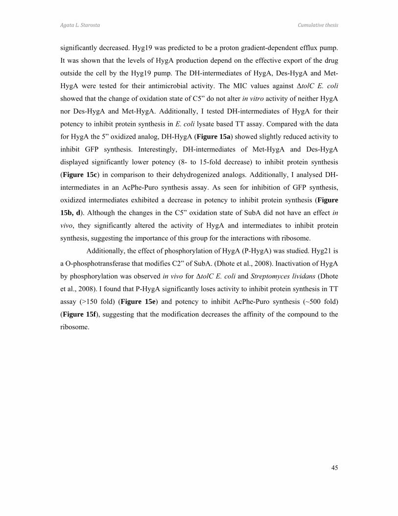
Agata L. Starosta Cumulative thesis
45
significantly decreased. Hyg19 was predicted to be a proton gradient-dependent efflux pump.
It was shown that the levels of HygA production depend on the effective export of the drug
outside the cell by the Hyg19 pump. The DH-intermediates of HygA, Des-HygA and Met-
HygA were tested for their antimicrobial activity. The MIC values against ΔtolC E. coli
showed that the change of oxidation state of C5” do not alter in vitro activity of neither HygA
nor Des-HygA and Met-HygA. Additionally, I tested DH-intermediates of HygA for their
potency to inhibit protein synthesis in E. coli lysate based TT assay. Compared with the data
for HygA the 5” oxidized analog, DH-HygA (Figure 15a) showed slightly reduced activity to
inhibit GFP synthesis. Interestingly, DH-intermediates of Met-HygA and Des-HygA
displayed significantly lower potency (8- to 15-fold decrease) to inhibit protein synthesis
(Figure 15c) in comparison to their dehydrogenized analogs. Additionally, I analysed DH-
intermediates in an AcPhe-Puro synthesis assay. As seen for inhibition of GFP synthesis,
oxidized intermediates exhibited a decrease in potency to inhibit protein synthesis (Figure
15b, d). Although the changes in the C5” oxidation state of SubA did not have an effect in
vivo, they significantly altered the activity of HygA and intermediates to inhibit protein
synthesis, suggesting the importance of this group for the interactions with ribosome.
Additionally, the effect of phosphorylation of HygA (P-HygA) was studied. Hyg21 is
a O-phosphotransferase that modifies C2” of SubA. (Dhote et al., 2008). Inactivation of HygA
by phosphorylation was observed in vivo for ΔtolC E. coli and Streptomyces lividans (Dhote
et al., 2008). I found that P-HygA significantly loses activity to inhibit protein synthesis in TT
assay (>150 fold) (Figure 15e) and potency to inhibit AcPhe-Puro synthesis (~500 fold)
(Figure 15f), suggesting that the modification decreases the affinity of the compound to the
ribosome.
Agata L. Starosta Cumulative thesis
46
Collectively, it was shown that gene disruption of the HygA biosynthesis pathway
results in accumulation of intermediates, which enables functional studies of distinct chemical
moieties of HygA to be performed. The intermediates are becoming progressively more active
antimicrobials, with HygA as the most potent inhibitor. The methylene bridge was proven to
be important for the in vivo activity of HygA, whereas it does not significantly affect
translation inhibition in vitro. Furthermore, the C5” oxidation has no effect on in vivo activity,
but alters the effect of HygA on the translation in vitro. I showed that HygA and intermediates
inhibit peptide bond formation, suggesting that HygA binds in the PTC, in agreement with
previous biochemical data (Guerrero and Modolell, 1980; Poulsen et al., 2000). Additionally,
I demonstrated that the modification of C2” leads to the loss of inhibitory activity of HygA,
possibly through sterical clashes with ribosomal components and altering the affinity to the
ribosome. In conclusion, the new structurally diverse compounds may lead to design of novel
antimicrobials.
Figure 15. HygA and derivatives action (a, c, e) on the GFP synthesis in a TT assay and (b, d, f) on the AcPhe-Puro synthesis
Agata L. Starosta Cumulative thesis
47
3.2 Macrolides
3.2.1 Paper 3
Petropoulos, A.D., Kouvela, E.C., Starosta, A.L., Wilson, D.N., Dinos, G.P., and Kalpaxis, D.L. (2009). Time-resolved binding of azithromycin to Escherichia coli ribosomes. J Mol Biol 385, 1179-1192.
3.2.2 Paper 4
Starosta, A.L., Karpenko, V.V., Shishkina, A.V., Mikolajka, A., Sumbatyan, N.V., Schluenzen, F., Korshunova, G.A., Bogdanov, A.A., and Wilson, D.N. (2010). Interplay between the ribosomal tunnel, nascent chain, and macrolides influences drug inhibition. Chem Biol 17, 504-514.
Azithromycin (AZI) is a 15-membered semi-synthetic derivative of erythromycin,
structurally distinguished by the absence of a keto oxygen at position C9 and the insertion of a
methyl-substituted nitrogen at position C10. AZI shows higher stability and improved potency
against Gram-negative bacteria. AZI displayed anti-inflammatory and modulatory effects on
the expression of virulence factors of P. aeruginosa (Bala et al., 2011). Interestingly, crystal
structures of AZI bound to either the bacterial (Schlunzen et al., 2003) or archaeal (Hansen et
al., 2002a) ribosome showed a different number of molecules bound per ribosome. A single
molecule was found in the archaeal structure, whereas two molecules of the drug were
observed in the structure of D. radiodurans 50S subunit.
In Paper 3, the group of Professor G. Dinos used kinetic and footprinting methods to
characterise binding process of the AZI. It was shown that AZI binds to E. coli ribosomes in a
two-step process. First, an initial rapid low-affinity binding state occurs where AZI inserts
into a hydrophobic cleft formed by nucleotides G2057-A2059 and is also positioned by
G2505. Secondly, AZI slowly accommodates into a high-affinity binding state, where
interaction with hydrophobic cleft weakens and AZI moves towards U2609 and nucleotides of
domain II of 23S rRNA. Moreover, U2609C substitution promotes formation of the stable
complex. The low- and high-affinity AZI binding sites are unique and only one molecule of
the drug binds per E. coli ribosome at a time. In order to to evaluate the species specific mode
of AZI binding, I prepared D. radiodurans 70S ribosomes which were used. In contrast to
previously obtained data for AZI binding to E. coli ribosomes, kinetic and binding data
indicated that two molecules of AZI bind cooperatively to the D. radiodurans ribosome. This
finding supported the crystallographic observations on differential binding of AZI to
Agata L. Starosta Cumulative thesis
48
ribosomes from varying species. In conclusions, in the future, our results may lead to design
of compounds that preferentially target ribosomes of specific species of bacteria
The crystal structure of tylosin (Tyl) bound to Haloarcula marismortui LSU showed
a covalent bond between the C6 ethyl aldehyde group of Tyl and the N6 of nucleobase A2062
of 23S rRNA (Hansen et al., 2002a). Nucleobase A2062 was suggested to establish
interaction with nascent polypeptide (Bhushan et al., 2010; Seidelt et al., 2009; Vazquez-
Laslop et al., 2011) and contribute for the stalling mechanism of ErmCL (Vazquez-Laslop et
al., 2008). In Paper 4, the importance of ethyl aldehyde group for Tyl binding was elucidated.
A series of macrolide derivatives with oligopeptide substituents was tested in order to
investigate tunnel-nascent polypeptide interactions.
The group of Professor A. Bogdanov designed and synthesized several Tyl
derivatives, which I have tested for their ability to establish interactions with either the
ribosomal exit tunnel wall and/or the nascent polypeptide. Two sets of Tyl derivatives, with
substitution at either the C6 or C14 positions were monitored for their ability to (i) compete
for binding to empty D. radiodurans 70S ribosomes with [14C]-erythromycin ([14C]-Ery), or
(ii) inhibit GFP synthesis in E. coli lysate-based TT assays.
I demonstrated that the reduced form of Tyl (Tyl-H2), where the C6 ethyl aldehyde is
replaced with a hydroxyl group, is unable to compete with [14C]-Ery. Moreover, in the TT
assay, Tyl-H2, displayed significantly decreased potency to inhibit GFP synthesis, and was
not able to inhibit protein synthesis completely. This showed that the covalent bond
established between Tyl and ribosomal nucleotide A2062 is critical for drug binding and
inhibitory activity. Furthermore, several C6 ethyl aldehyde oligopeptide substituents were
tested. Tyl with C6-Ala-Ala dipeptide (Tyl-Ala) showed improved binding ability compared
to Tyl-H2, however was still significantly worse than Tyl. The potency of Tyl-Ala to inhibit
GFP synthesis was comparable with Tyl suggesting that the new analog may establish
additional interactions with nascent polypeptide or ribosomal tunnel wall. The crystal
structure of Tyl-Ala bound to D. radiodurans 50S subunits showed that Ala dipeptide stacks
onto nucleotide A2062, which could account for the improved binding affinity. On this basis,
a series of Tyl derivatives were designed to optimize stacking interactions with A2062.
Additionally, I tested several Tyl derivatives with nucleotides and aromatic amino acids
attached to C6 carbon with linkers varying in length and flexibility, including uridine, tyrosine
and phenylalanine. Tyl-U with a uridine attached with a 4 atom linker was not sufficient to
Agata L. Starosta Cumulative thesis
49
significantly improve the potency of a compound in either binding to the ribosome or
inhibition of a GFP synthesis. A tyrosine with either rigid or flexible 5 atom linker (Tyl=Tyr
and Tyl-Tyr, respectively) showed differential effects. While Tyl=Tyr had poor properties,
Tyl-Tyr was as effective as Tyl for inhibition of protein synthesis. Interestingly,
a phenylalanine attached with flexible 6 atom linker showed enhanced potency to inhibit GFP
synthesis compared with Tyl. Nevertheless, it has to be noted, that none of the new
compounds displayed better binding properties in comparison to Tyl. This suggested that the
potency of new derivatives originated not from the binding affinity but rather from the steric
perturbation of growing nascent polypeptide or/and additional interactions established with
nascent polypeptide.
Additionally, the activity of 5-O-mycaminosyl-tylonolid (OMT), lacking both the C5
mycarose and the C14 mycinose, was evaluated. OMT is known to be a potent antimicrobial
with MIC values comparable with Tyl (Fu et al., 2006). However, little or no significant
inhibitory effect of OMT on GFP synthesis was detected in our experimental set-up.
To investigate the differential effects of OMT in more detail, our collaborators
synthesized a series of peptide-OMT compounds, where BocAlaAlaPhe and fMetLeuPhe
peptides are attached to the C14 position. In the TT assay, only fMLF-OMT showed
improved potency to inhibit GFP synthesis compared to OMT, although translation was not
inhibited by more than 60%, even at a concentration 200-fold higher than the inhibitory
concentration (IC90) for Tyl. In contrast, the competition assay showed that OMT and OMT
analogs bind to empty ribosomes. The discrepancy between the in vivo data, TT assay and
binding experiments has led us hypothesis that OMT displays some template specificity. To
address this question, the GFP reporter was substituted with firefly luciferase (Fluc).
Interestingly, OMT displayed an excellent inhibitor effect of Fluc synthesis, with a potency
similar to that observed for the inhibition of GFP by Tyl. The effect was remarkable for the
macrolide class of antibiotics, since such differences for tetracycline (Tet) and
chloramphenicol (Cam) were not observed.
In summary, for many macrolide derivatives, a lack of correlation between the
binding affinities and inhibitory activity was observed. In other words, despite their potency
to inhibit protein synthesis, all of the macrolide derivatives tested here were less effective at
binding to the ribosome than Tyl. This suggests that the binding affinity does not exclusively
determine the effectiveness of the compound and that peptide substituents of the lactone ring
may contribute to the inhibitory activity. This might occur due to steric blockage of the
Agata L. Starosta Cumulative thesis
50
passage of the nascent polypeptide chain or establishment of interactions with the nascent
polypeptide. Moreover, the data suggest that the sequence of the nascent chain may influence
the inhibitory ability of particular macrolide antibiotics. This finding may lead to design of
macrolide antibiotics exclusively inhibiting particular polypeptides.
3.3 Thiopeptides
3.3.1 Paper 5
Starosta, A.L., Qin, H., Mikolajka, A., Leung, G.Y., Schwinghammer, K., Nicolaou, K.C., Chen, D.Y., Cooperman, B.S., and Wilson, D.N. (2009). Identification of distinct thiopeptide-antibiotic precursor lead compounds using translation machinery assays. Chem Biol 16, 1087-1096.
3.3.2 Paper 6
Mikolajka, A., Liu, H., Chen, Y., Starosta, A.L., Marquez, V., Ivanova, M., Cooperman, B.S., and Wilson, D.N. (2011). Differential effects of thiopeptide and orthosomycin antibiotics on translational GTPases. Chem Biol 18, 589-600.
Recent advances in thiopeptide total synthesis have identified synthetic fragments
that can serve as lead compounds for drug development. In Paper 5, a library of precursors of
nitrogen heterocycle core of thiopeptides synthesis provided by Professor K. C. Nicolaou was
tested. Compounds were divided into four structurally distinct groups (PA-PD), which were
tested for their ability to inhibit (i) 70S initiation complex formation (70SIC), monitored via
fluorescence of IF2Cy3, which increases upon LSU binding to the 30SIC; (ii) the ribosome-
dependent GTPase activity of EF-G using Malachite Green assay, and (iii) in vitro synthesis
of GFP in E. coli lysate based TT.
The group of Professor B. Cooperman determined that ThS and nosiheptide (NoS)
are potent inhibitors of the 70SIC. However, only one compound from group A – PA1 –
showed detectable inhibition of the 70SIC, but at concentrations significantly higher (60- to
400- fold) than for ThS and NoS. It should be noted that the stereochemical derivatives of
PA1 showed no inhibitory activity, which emphasizes the specificity of the compound. ThS
was a very effective inhibitor of the ribosome-stimulated multiple turnover GTPase activity of
EF-G . However, the tested compounds showed significantly lower potency to inhibit the
GTPase activity of EF-G, even at the concentrations 50- to 100-times higher than ThS. I
tested ThS, NoS and GE2270A in the TT assay. All three compounds displayed high potency
to inhibit GFP synthesis, whereas in contrast, none of the tested precursor compounds showed

Agata L. Starosta Cumulative thesis
51
any significant inhibitory activity, even at concentrations as high as 10-fold of ThS inhibitory
concentration (IC90). Surprisingly, addition of 10-fold excess of representative precursor
compounds over ThS IC90 could reverse the inhibitory effect of ThS. Moreover, different
subsets of compounds were able to restore translation inhibited by ThS, NoS or GE2270A.
This finding suggests that although the active compounds in the library showed no potency to
inhibit translation, they retained the ability to bind to their targets. Additionally, the ability to
restore translation in the presence of GAC and EF-Tu inhibitors, suggest they may lead to
design of dual inhibitors, targeting ribosomes and ternary complex formation.
Crystallographic data showed that MiC binds in a cleft spanning protein L11 and
H43-44, and stabilizes CTD-L7/L12 (Harms et al., 2008). Stimulation of the GTPase activity
of EF-G by MiC was suggested to be due to the stabilization of the interaction of L7-CTD
with G’ domain of EF-G (Harms et al., 2008). In Paper 6, the mode of action of MiC was
elucidated. A set of translational GTPases, namely, EF-G, IF2, EF4 (LepA), BipA and TetM
was tested in (i) the ribosome-dependent GTPase activity assay using the Malachite Green
assay, and (ii) translocation reaction stopped flow fluorescence.
Unlike ThS that inhibited ribosome-dependent GTPase of EF-G, MiC stimulated the
GTPase activity of EF-G. I confirmed that both ThS and MiC inhibit GFP synthesis in the TT
assay. Therefore, the group of Professor B. Cooperman has tested the effect of ThS and MiC
on EF-G dependent translocation. Despite the differential effects of MiC and ThS on the
GTPase activities of EF-G, it was found that both antibiotics inhibit the translocation reaction,
probably by reducing the binding affinity of EF-G to the ribosome. The MiC-dependent
activation was investigated in more details. Interestingly, crystallographic data suggest that
the G’ domain of EF-G and ribosomal protein L7/L12 play a role in GTPase stimulation by
MiC. In these study, it was found that G-proteins naturally lacking or having reduced G’
domains (IF2, BipA, EF4, and TetM) were inhibited by MiC. Therefore, EF-G lacking G’
domain (EF-GΔG’), as well as ribosomes lacking proteins L7/L12 (ΔL7/L12) were tested.
MiC inhibited the ribosome-dependent GTPase activity of EF-GΔG’. Moreover the ΔL7/L12-
ribosome dependent GTPase activity of EF-G was arrested. Collectively, these results suggest
that the interaction between G’-domain of EF-G and CTD-L7/L12 is indeed necessary for Pi
release. MiC, by stabilizing CTD-L7/L12, enhances turnover of EF-G.

Agata L. Starosta Cumulative thesis
52
3.4 Orthosomycins Evn was observed to inhibit fMet-Puro synthesis in an IF2-dependent manner
(Belova et al., 2001). In Paper 6, the step at which Evn inhibits protein synthesis was
investigated. Evn was tested in (i) the ribosome-dependent GTPase activity of G-proteins
using the Malachite Green assay; (ii) 70S initiation complex formation (70SIC); (iii) EF4-
dependent back-translocation, and (iv) the translocation reaction using stop-flow fluorescence.
Professor B. Cooperman’s group demonstrated that Evn inhibits 70SIC formation by
preventing the binding of the LSU to 30SIC in an IF2 dependent manner. This is in
accordance with the observation that Evn inhibits ribosome-dependent GTPase activity of
IF2. Thus, it was concluded that Evn sterically interferes with 30SIC-IF2 binding to the LSU.
Additionally, Evn was also tested for potency to inhibit the translocation step. Yet
unlike thiopeptide antibiotics, Evn did not inhibit the EF-G catalyzed translocation reaction.
This result is consistent with the observation that Evn did not affect ribosome-dependent
GTPase activity of EF-G. In contrast, Evn was found to be a potent inhibitor of ribosome-
dependent EF4 (LepA) GTPase activity, as well as EF4-dependent back-translocation
reaction. Since EF4 is not essential for E. coli viability (Dibb and Wolfe, 1986), IF2-
dependent initiation seems to be a major target of Evn action. Moreover, the values for
inhibition of 70SIC were comparable with results I obtained for inhibition of GFP synthesis in
TT assay. This observation supports the hypothesis that Evn targets predominantly the
initiation phase.
Agata L. Starosta Cumulative thesis
53
3.5 Fusidic acid
3.5.1 Paper 7
Ratje, A.H., Loerke, J., Mikolajka, A., Brunner, M., Hildebrand, P.W., Starosta, A.L., Donhofer, A., Connell, S.R., Fucini, P., Mielke, T., Whitford, P. C., Onuchic, J. N., Yu, Y., Sanbonmatsu, K. Y., Hartmann, R. K., Penczek, P. A., Wilson, D. N., and Spahn, C. M. (2010). Head swivel on the ribosome facilitates translocation by means of intra-subunit tRNA hybrid sites. Nature 468, 713-716.
Despite the availability of a number of EF-G-ribosome structures, the precise
mechanism of tRNA movement through the ribosome remains unclear. In Paper 7, Dr. A.
Mikolajka, A Dönhöfer and I prepared the complex of EF-G stalled with FA on the
T. thermophilus 70S in the presence of GDP, which was investigated by the group of
Professor C.M. Spahn using cryo-EM. Two separate ribosome populations were observed,
which varied in the degree of ratcheting and positioning of the L1 stalk, as well as by the
degree of head swivelling of the SSU. Population I showed ~7° ratcheting with respect to the
LSU, while at the same time head swivelling of ~5° was observed. On contrast, population II
exhibited a 4° ratcheting but strong swiveling of the head by 18°. Examination of these two
ribosomal populations revealed that the movement of tRNA with respect to the SSU is
coupled with 30S head swivelling and unratcheting of the SSU. Moreover, the P-tRNA on the
SSU maintains contact with P-site of the body and simultaneously establishes interaction with
the exit site (E site) of the head to form previously unseen intrasubunit pe/E hybrid states.
In both populations, EF-G·GDP stalled with FA on T. thermophilus 70S ribosomes
showed an open conformation compared to free EF-G·GDP GDP (A et al., 1994;
Czworkowski et al., 1994; Laurberg et al., 2000), with domains IV and V moved relative to
domains I and II. Domain III was rotated and an additional density, spanning domain I and III
into which FA could be modeled, was found. The position of FA was close to the position of
the switch I region in the GTP state. This suggests that FA stabilizes the rotated state of
domain III, preventing the conformational change of EF-G into the GDP state and thus
prevents EF-G recycling from the ribosome.
In summary, using Cryo-EM, it was possible not only to characterise translocation
events, but also identify and visualize binding of a small molecule, namely the antibiotic
fusidic acid.

Agata L. Starosta Cumulative thesis
54
3.6 Elongation factor P
3.6.1 Paper 8
Peil L., Starosta, A.L., Virumäe, K, Aktinson, G.C., Tenson, T., Remme, J., Wilson, D.N. (2011). Formation of ε(R)-β-lysyl-hydroxylysine on translation elongation factor EF-P requires YjeK, YjeA and YfcM. Under revision in Nature Chemical Biology.
Elongation factor P (EF-P) and its modification are important for growth, drug
resistance and virulence in pathogenic bacteria. Curiously, mass spectrometry analyses
indicated that lysinylation of K34 of EF-P by YjeA results in a ~128 Da modification, leaving
a difference of ~16 Da when compared with the ~144 Da reported for endogenous EF-P (Aoki
et al., 2008). In collaboration with the group of Professor J. Remme, the discrepancies
between in vivo and in vitro data were investigated using (i) an E. coli lysate-based immuno-
precipitation approach, coupled with (ii) high-resolution mass spectrometry (MS) and (iii)
SILAC labeling were applied in order to characterize the chemical nature of the E. coli EF-P
modification and define the modification pathway in vivo.
The analyses of endogenous EF-P revealed an increase of the mass of EF-P by 144.2 ±
0.1 Da compared to expected mass of unmodified EF-P. Chymotrypsin digested EF-P showed
that peptide containing lysine 34 (K34) had an additional mass of 144.09 Da
(F.30VKPGK*GQAF38.A) which was in accordance with digestion of endogenous EF-P with
LysC (K.32PGK*GQAFARVK42.L). Fragmentation of both the chymotrypsin and the LysC
peptides confirmed K34 as the site of modification. Moreover, it was shown that modification
of endogenous EF-P involves attachment of β-lysine to ε-amino group of K34 accounting for
128 Da, and that the additional 16 Da modification is associated with K34 of endogenous EF-
P, rather than the added lysine.
In order to demonstrate that YjeA and YjeK were responsible for the modification of
K34 of EF-P in vivo, endogenous EF-P was immuno-precipitated from E. coli AT713 strains
lacking the genes for yjeA (ΔyjeA) or yjeK (ΔyjeK). Chymotrypsin proteolysis of EF-P from
the ΔyjeA strain produced a fragment containing K34 (F.30VKPGKGQAF38.A) with no
detectable modification. Similarly, an unmodified chymotrypsin fragment was also detected
upon proteolysis of EF-P isolated from the ΔyjeK strain. Additionally, a peptide with a mass
increase of +128.09 Da was found. Quantification of modified/unmodified peptide ratios
revealed that the modified form is present at only ~2 % of the level of the unmodified form.
These observations indicated that while YjeA can perform the lysinylation of EF-P in vivo in
Agata L. Starosta Cumulative thesis
55
the absence of YjeK (and therefore in the absence of (R)-β-lysine), the reaction appears to be
extremely inefficient.
Given the discrepancy of 16 Da between the modification mass of the endogenous EF-
P and in vitro lysinylated EF-P (+128 Da) (Navarre et al., 2010; Yanagisawa et al., 2010), it
was concluded that a third enzyme should exist that is also involved in the modification of
K34 of EF-P in vivo. I used STRING (Search Tool for the Retrieval of Interacting
Genes/Proteins) database (http://string.embl.de) to search for proteins associated with YjeA,
YjeK and EF-P. From the output, YfcM, a protein that has no reasonable homology with any
protein of known structure or function, was chosen. Chymotrypsin proteolysis of EF-P from
the ΔyfcM strain produced a fragment containing K34 (F.30VKPGKGQAF38.A) with a mass
increase of 128.09 Da. Consistently, the LysC fragment of EF-P containing K34
(K.32PGK*GQAFARVK42.L) isolated from the ΔyfcM strain had a mass increased of 128.09
Da. The precision of the mass difference, i.e. 15.995 Da, is indicative of one atom of
molecular oxygen, which has a monoisotopic mass of 15.995 Da, thus revealing YfcM as a
potential hydroxylase (mono-oxygenase) that modifies K34 of lysinylated EF-P.
In order to determine the site of hydroxylation by YfcM on K34 of EF-P, the
differentially deuterated lysine isoform of lysine was used. 4,4,5,5-D4-L-lysine (D4-Lys) has
the hydrogen atoms (1.008 Da) of the C4-C5 positions of L-lysine substituted with deuterium
(2.014 Da). LysC fragments from the wildtype were 14.993 Da larger, instead of the expected
15.995 Da. This is consistent with 14.988 Da increase expected by the mass loss of a
deuterium atom (-2.014 Da) and the addition of a hydroxyl OH group. The 1 Da loss indicates
that YfcM hydroxylates either the C4 (γ) or C5 (δ) position.
To screen for potential substrates of YfcM, I applied differential scanning fluorimetry
(DSF), which monitors the increase in thermal stability of a protein due to the interaction with
a ligand. The largest increases in thermal stability for YfcM were observed with nucleotide-
based chemicals, for example, the maximum shift of ~5°C was observed in the presence of
<600 μM NADP or CoA.
Dr. G. Atkinson checked genomic distribution and context of the yfcM gene. All
bacterial species that contain the yfcM gene also have efp, yjeA and, with one exception
(Haemophilus influenzae strain PittGG), yjeK. However, unlike yjeA and yjeK that often
cluster together in the same operon as efp, yfcM is always located in a distinct region of the
chromosome. The six-gene operon structure of the prmC-aroC-mepA-yfcA-yfcM-yfcL operon
is highly conserved within the Enterobacteriaceae family.
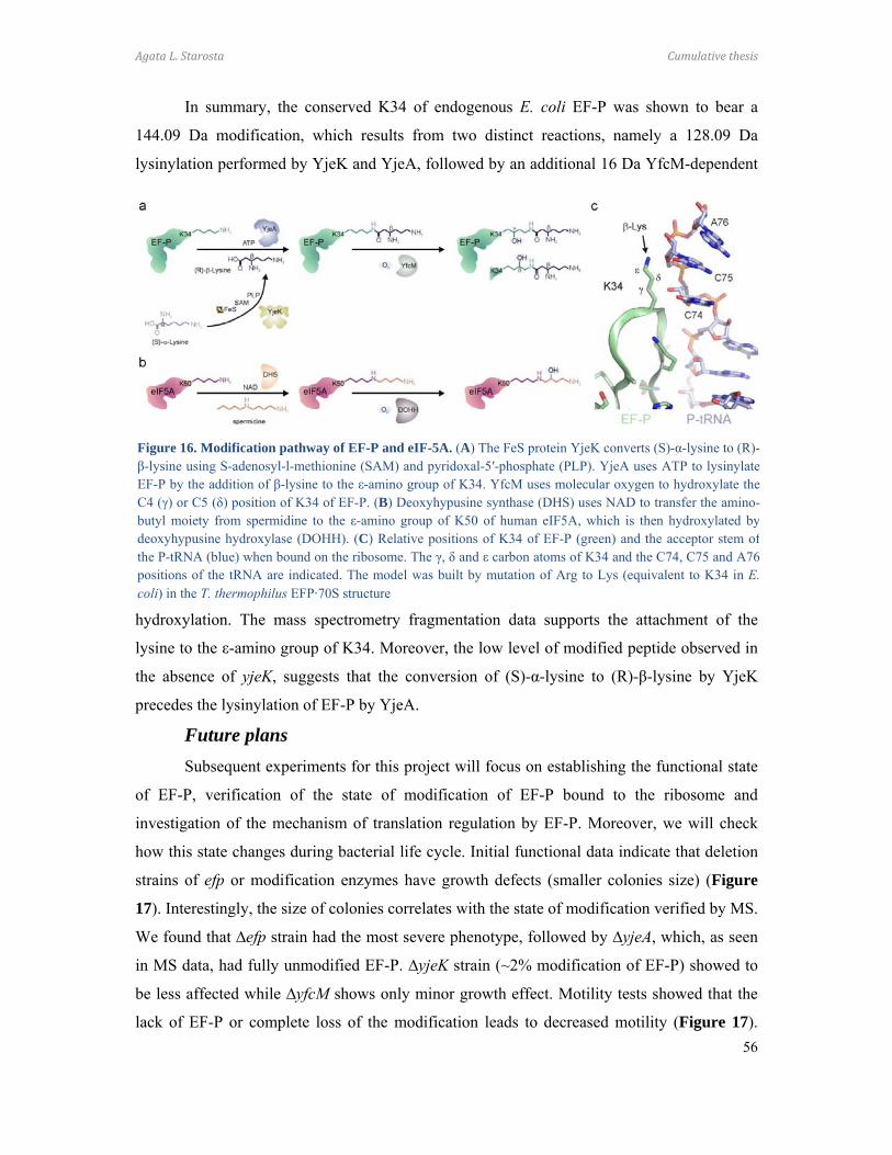
Agata L. Starosta Cumulative thesis
56
In summary, the conserved K34 of endogenous E. coli EF-P was shown to bear a
144.09 Da modification, which results from two distinct reactions, namely a 128.09 Da
lysinylation performed by YjeK and YjeA, followed by an additional 16 Da YfcM-dependent
hydroxylation. The mass spectrometry fragmentation data supports the attachment of the
lysine to the ε-amino group of K34. Moreover, the low level of modified peptide observed in
the absence of yjeK, suggests that the conversion of (S)-α-lysine to (R)-β-lysine by YjeK
precedes the lysinylation of EF-P by YjeA.
Future plans
Subsequent experiments for this project will focus on establishing the functional state
of EF-P, verification of the state of modification of EF-P bound to the ribosome and
investigation of the mechanism of translation regulation by EF-P. Moreover, we will check
how this state changes during bacterial life cycle. Initial functional data indicate that deletion
strains of efp or modification enzymes have growth defects (smaller colonies size) (Figure
17). Interestingly, the size of colonies correlates with the state of modification verified by MS.
We found that ∆efp strain had the most severe phenotype, followed by ∆yjeA, which, as seen
in MS data, had fully unmodified EF-P. ∆yjeK strain (~2% modification of EF-P) showed to
be less affected while ∆yfcM shows only minor growth effect. Motility tests showed that the
lack of EF-P or complete loss of the modification leads to decreased motility (Figure 17).
Figure 16. Modification pathway of EF-P and eIF-5A. (A) The FeS protein YjeK converts (S)-α-lysine to (R)-β-lysine using S-adenosyl-l-methionine (SAM) and pyridoxal-5′-phosphate (PLP). YjeA uses ATP to lysinylateEF-P by the addition of β-lysine to the ε-amino group of K34. YfcM uses molecular oxygen to hydroxylate theC4 (γ) or C5 (δ) position of K34 of EF-P. (B) Deoxyhypusine synthase (DHS) uses NAD to transfer the amino-butyl moiety from spermidine to the ε-amino group of K50 of human eIF5A, which is then hydroxylated bydeoxyhypusine hydroxylase (DOHH). (C) Relative positions of K34 of EF-P (green) and the acceptor stem ofthe P-tRNA (blue) when bound on the ribosome. The γ, δ and ε carbon atoms of K34 and the C74, C75 and A76positions of the tRNA are indicated. The model was built by mutation of Arg to Lys (equivalent to K34 in E.coli) in the T. thermophilus EFP·70S structure
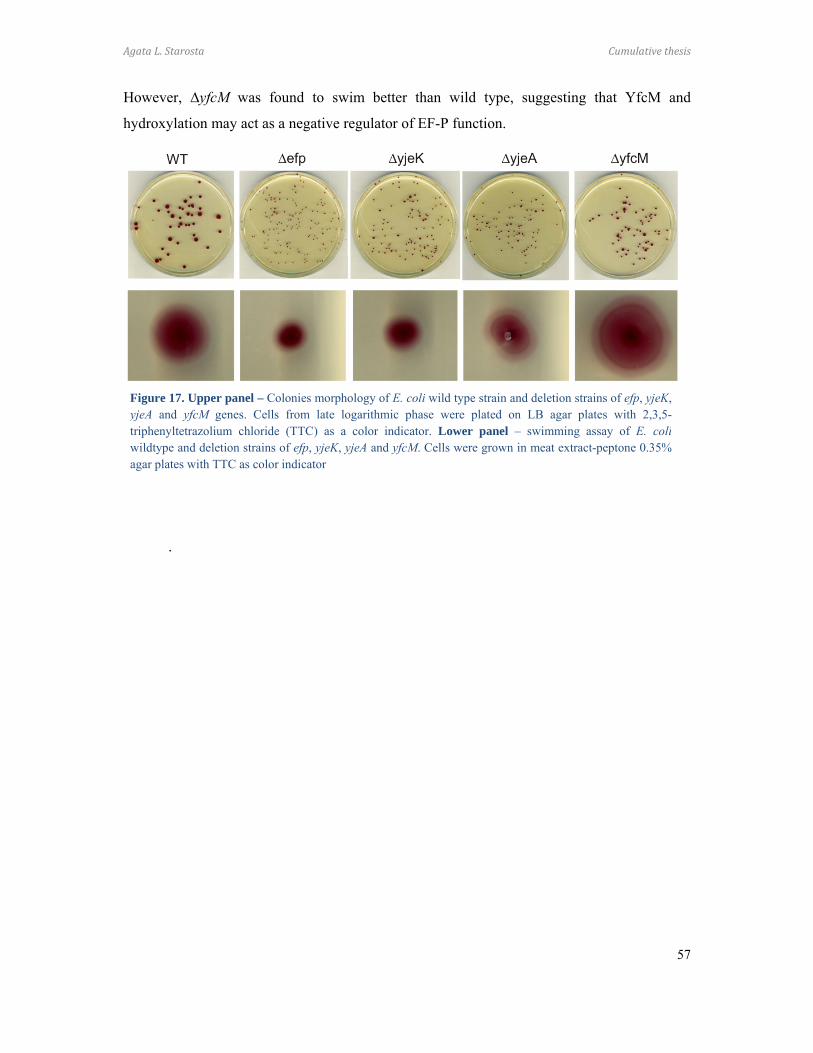
Agata L. Starosta Cumulative thesis
57
However, ∆yfcM was found to swim better than wild type, suggesting that YfcM and
hydroxylation may act as a negative regulator of EF-P function.
.
Figure 17. Upper panel – Colonies morphology of E. coli wild type strain and deletion strains of efp, yjeK,yjeA and yfcM genes. Cells from late logarithmic phase were plated on LB agar plates with 2,3,5-triphenyltetrazolium chloride (TTC) as a color indicator. Lower panel – swimming assay of E. coliwildtype and deletion strains of efp, yjeK, yjeA and yfcM. Cells were grown in meat extract-peptone 0.35%agar plates with TTC as color indicator
Agata L. Starosta Conclusions
58
4 Conclusions (I) Hygromycin A. (i) Gene disruption of HygA biosynthesis pathway leads to
accumulation of intermediates; (ii) the methylene bridge is important for the in vivo activity of
HygA, probably in terms of uptake; (iii) the C5” oxidation has a negative effect on the
translation, possibly alters binding to ribosome; (iv) HygA and intermediates inhibit peptide
bond formation; and (v) C2”-phosphorylation leads to loss of inhibitory activity of HygA.
(II) Macrolides. (i) Two molecules of AZI bind cooperatively to the D. radiodurans
ribosome, while only one binds to E. coli ribosomes; (ii) the C6 ethyl aldehyde interaction
with A2062 is crucial for binding of Tyl to ribosomes and translation inhibitory activity; (iii)
the loss of ethyl aldehyde can be compensated by oligopeptides; (iv) macrolides display
polypeptide chain-specific inhibition.
(III) Thiopeptides. (i) Thiopeptide synthesis precursors, despite the loss of potency
to inhibit translation, display dual activity to restore translation in the presence of inhibitory
concentrations of thiopeptides targeting ribosome or EF-Tu; (ii) the activation by MiC of the
ribosome-dependent GTPase activity of EF-G requires the presence of ribosomal proteins
L7/L12 as well as the G’ domain of EF-G.
(IV) Orthosomycins. (i) Evn sterically interferes with 30SIC-IF2 binding to the
LSU; (ii) Evn inhibits of EF4-dependent back-translocation.
(V) Fusidic acid. (i) FA binds in cleft between domain I and III of EF-G and
stabilizes the rotated state of domain III and prevents EF-G recycling from the ribosome.
(VI) Elongation factor P. (i) endogenous EF-P is modified with a 144.09 Da
modification; (ii) lysinylation of K34 is performed by YjeK and YjeA and is followed by an
additional 16 Da hydroxylation by YfcM; (iii) lysine is attached to the ε-amino group of K34;
(iv) conversion of (S)-α-lysine to (R)-β-lysine by YjeK precedes the lysinylation of EF-P by
YjeA; (v) YfcM displays specificity for nucleotide compounds.
Agata L. Starosta References
59
5 References A, A.E., Brazhnikov, E., Garber, M., Zheltonosova, J., Chirgadze, Y., al-Karadaghi, S.,
Svensson, L.A., and Liljas, A. (1994). Three-dimensional structure of the ribosomal
translocase: elongation factor G from Thermus thermophilus. EMBO J 13, 3669-3677.
Acker, M.G., Bowers, A.A., and Walsh, C.T. (2009). Generation of thiocillin variants by
prepeptide gene replacement and in vivo processing by Bacillus cereus. J Am Chem
Soc 131, 17563-17565.
Agirrezabala, X., Lei, J., Brunelle, J.L., Ortiz-Meoz, R.F., Green, R., and Frank, J. (2008).
Visualization of the hybrid state of tRNA binding promoted by spontaneous ratcheting
of the ribosome. Mol Cell 32, 190-197.
Agrawal, R.K., Heagle, A.B., Penczek, P., Grassucci, R.A., and Frank, J. (1999). EF-G-
dependent GTP hydrolysis induces translocation accompanied by large conformational
changes in the 70S ribosome. Nat Struct Biol 6, 643-647.
Ali, I.K., Lancaster, L., Feinberg, J., Joseph, S., and Noller, H.F. (2006). Deletion of a
conserved, central ribosomal intersubunit RNA bridge. Mol Cell 23, 865-874.
Alksne, L.E., and Projan, S.J. (2000). Bacterial virulence as a target for antimicrobial
chemotherapy. Curr Opin Biotechnol 11, 625-636.
Allen, G.S., Zavialov, A., Gursky, R., Ehrenberg, M., and Frank, J. (2005). The cryo-EM
structure of a translation initiation complex from Escherichia coli. Cell 121, 703-712.
An, G., Glick, B.R., Friesen, J.D., and Ganoza, M.C. (1980). Identification and quantitation of
elongation factor EF-P in Escherichia coli cell-free extracts. Can J Biochem 58, 1312-
1314.
Antoun, A., Pavlov, M.Y., Lovmar, M., and Ehrenberg, M. (2006). How initiation factors
maximize the accuracy of tRNA selection in initiation of bacterial protein synthesis.
Mol Cell 23, 183-193.
Aoki, H., Dekany, K., Adams, S.L., and Ganoza, M.C. (1997). The gene encoding the
elongation factor P protein is essential for viability and is required for protein
synthesis. J Biol Chem 272, 32254-32259.
Aoki, H., Xu, J., Emili, A., Chosay, J.G., Golshani, A., and Ganoza, M.C. (2008). Interactions
of elongation factor EF-P with the Escherichia coli ribosome. FEBS J 275, 671-681.
Bae, W.K., Lee, Y.K., Cho, M.S., Ma, S.K., Kim, S.W., Kim, N.H., and Choi, K.C. (2006). A
case of hemolytic uremic syndrome caused by Escherichia coli O104:H4. Yonsei Med
J 47, 437-439.
Agata L. Starosta References
60
Bailly, M., and de Crecy-Lagard, V. (2010). Predicting the pathway involved in post-
translational modification of elongation factor P in a subset of bacterial species. Biol
Direct 5, 3.
Bala, A., Kumar, R., and Harjai, K. (2011). Inhibition of quorum sensing in Pseudomonas
aeruginosa by azithromycin and its effectiveness in urinary tract infections. J Med
Microbiol 60, 300-306.
Barat, C., Datta, P.P., Raj, V.S., Sharma, M.R., Kaji, H., Kaji, A., and Agrawal, R.K. (2007).
Progression of the ribosome recycling factor through the ribosome dissociates the two
ribosomal subunits. Mol Cell 27, 250-261.
Baumann, S., Schoof, S., Bolten, M., Haering, C., Takagi, M., Shin-ya, K., and Arndt, H.D.
(2010). Molecular determinants of microbial resistance to thiopeptide antibiotics. J
Am Chem Soc 132, 6973-6981.
Bearson, S.M., Bearson, B.L., Brunelle, B.W., Sharma, V.K., and Lee, I.S. (2011). A
mutation in the poxA gene of Salmonella enterica serovar Typhimurium alters protein
production, elevates susceptibility to environmental challenges, and decreases swine
colonization. Foodborne Pathog Dis 8, 725-732.
Bearson, S.M., Bearson, B.L., and Rasmussen, M.A. (2006). Identification of Salmonella
enterica serovar Typhimurium genes important for survival in the swine gastric
environment. Appl Environ Microbiol 72, 2829-2836.
Belova, L., Tenson, T., Xiong, L., McNicholas, P.M., and Mankin, A.S. (2001). A novel site
of antibiotic action in the ribosome: interaction of evernimicin with the large
ribosomal subunit. Proc Natl Acad Sci U S A 98, 3726-3731.
Benson, S., Olsnes, S., Pihl, A., Skorve, J., and Abraham, A.K. (1975). On the mechanism of
protein-synthesis inhibition by abrin and ricin. Inhibition of the GTP-hydrolysis site
on the 60-S ribosomal subunit. Eur J Biochem 59, 573-580.
Bhushan, S., Gartmann, M., Halic, M., Armache, J.P., Jarasch, A., Mielke, T., Berninghausen,
O., Wilson, D.N., and Beckmann, R. (2010). alpha-Helical nascent polypeptide chains
visualized within distinct regions of the ribosomal exit tunnel. Nat Struct Mol Biol 17,
313-317.
Bielaszewska, M., Mellmann, A., Zhang, W., Kock, R., Fruth, A., Bauwens, A., Peters, G.,
and Karch, H. (2011). Characterisation of the Escherichia coli strain associated with
an outbreak of haemolytic uraemic syndrome in Germany, 2011: a microbiological
study. Lancet Infect Dis.

Agata L. Starosta References
61
Blaha, G., Stanley, R.E., and Steitz, T.A. (2009). Formation of the first peptide bond: the
structure of EF-P bound to the 70S ribosome. Science 325, 966-970.
Blanchard, S.C., Gonzalez, R.L., Kim, H.D., Chu, S., and Puglisi, J.D. (2004). tRNA selection
and kinetic proofreading in translation. Nat Struct Mol Biol 11, 1008-1014.
Bogdanov, A.A., Sumbatyan, N.V., Shishkina, A.V., Karpenko, V.V., and Korshunova, G.A.
(2010). Ribosomal tunnel and translation regulation. Biochemistry (Mosc) 75, 1501-
1516.
Bowers, A.A., Acker, M.G., Koglin, A., and Walsh, C.T. (2010). Manipulation of thiocillin
variants by prepeptide gene replacement: structure, conformation, and activity of
heterocycle substitution mutants. J Am Chem Soc 132, 7519-7527.
Brandi, L., Marzi, S., Fabbretti, A., Fleischer, C., Hill, W.E., Gualerzi, C.O., and Stephen
Lodmell, J. (2004). The translation initiation functions of IF2: targets for thiostrepton
inhibition. J Mol Biol 335, 881-894.
Brockmann, H., and Henkel, W. (1950). Pikromycin, ein neues Antibioticum aus
Actinomyceten. Naturwissensch 37, 138-139.
Brunelle, J.L., Shaw, J.J., Youngman, E.M., and Green, R. (2008). Peptide release on the
ribosome depends critically on the 2' OH of the peptidyl-tRNA substrate. RNA 14,
1526-1531.
Bugg, T.D., and Walsh, C.T. (1992). Intracellular steps of bacterial cell wall peptidoglycan
biosynthesis: enzymology, antibiotics, and antibiotic resistance. Nat Prod Rep 9, 199-
215.
Bulkley, D., Innis, C.A., Blaha, G., and Steitz, T.A. (2010). Revisiting the structures of
several antibiotics bound to the bacterial ribosome. Proc Natl Acad Sci U S A 107,
17158-17163.
Butler, D. (2010). Cholera tightens grip on Haiti. Nature 468, 483-484.
Bycroft, B.W., and Gowland, M.S. (1878). The structures of the highly modified peptide
antibiotics micrococcin P1 and P2. J Chem Soc, Chem Commun, 256-258.
Cameron, D.M., Thompson, J., Gregory, S.T., March, P.E., and Dahlberg, A.E. (2004).
Thiostrepton-resistant mutants of Thermus thermophilus. Nucleic Acids Res 32, 3220-
3227.
Cameron, D.M., Thompson, J., March, P.E., and Dahlberg, A.E. (2002). Initiation factor IF2,
thiostrepton and micrococcin prevent the binding of elongation factor G to the
Escherichia coli ribosome. J Mol Biol 319, 27-35.

Agata L. Starosta References
62
Campbell, E.A., Korzheva, N., Mustaev, A., Murakami, K., Nair, S., Goldfarb, A., and Darst,
S.A. (2001). Structural mechanism for rifampicin inhibition of bacterial rna
polymerase. Cell 104, 901-912.
Carter, A.P., Clemons, W.M., Jr., Brodersen, D.E., Morgan-Warren, R.J., Hartsch, T.,
Wimberly, B.T., and Ramakrishnan, V. (2001). Crystal structure of an initiation factor
bound to the 30S ribosomal subunit. Science 291, 498-501.
Cascales, E., and Christie, P.J. (2003). The versatile bacterial type IV secretion systems. Nat
Rev Microbiol 1, 137-149.
Caskey, C.T., Tompkins, R., Scolnick, E., Caryk, T., and Nirenberg, M. (1968). Sequential
translation of trinucleotide codons for the initiation and termination of protein
synthesis. Science 162, 135-138.
CDC (2010). Cholera outbreak --- Haiti, October 2010. MMWR Morb Mortal Wkly Rep 59,
1411.
Chen, C.R., Malik, M., Snyder, M., and Drlica, K. (1996). DNA gyrase and topoisomerase IV
on the bacterial chromosome: quinolone-induced DNA cleavage. J Mol Biol 258, 627-
637.
Chittum, H.S., and Champney, W.S. (1994). Ribosomal protein gene sequence changes in
erythromycin-resistant mutants of Escherichia coli. J Bacteriol 176, 6192-6198.
Choi, S., and Choe, J. (2011). Crystal structure of elongation factor P from Pseudomonas
aeruginosa at 1.75 A resolution. Proteins 79, 1688-1693.
Clatworthy, A.E., Pierson, E., and Hung, D.T. (2007). Targeting virulence: a new paradigm
for antimicrobial therapy. Nat Chem Biol 3, 541-548.
Clementi, N., Chirkova, A., Puffer, B., Micura, R., and Polacek, N. (2010). Atomic
mutagenesis reveals A2660 of 23S ribosomal RNA as key to EF-G GTPase activation.
Nat Chem Biol 6, 344-351.
Cocito, C., and Chinali, G. (1985). Molecular mechanism of action of virginiamycin-like
antibiotics (synergimycins) on protein synthesis in bacterial cell-free systems. J
Antimicrob Chemother 16 Suppl A, 35-52.
Connell, S.R., Takemoto, C., Wilson, D.N., Wang, H., Murayama, K., Terada, T., Shirouzu,
M., Rost, M., Schuler, M., Giesebrecht, J., et al. (2007). Structural basis for interaction
of the ribosome with the switch regions of GTP-bound elongation factors. Mol Cell
25, 751-764.
Agata L. Starosta References
63
Cornelis, G.R. (2010). The type III secretion injectisome, a complex nanomachine for
intracellular 'toxin' delivery. Biol Chem 391, 745-751.
Cundliffe, E., and Thompson, J. (1981). Concerning the mode of action of micrococcin upon
bacterial protein synthesis. Eur J Biochem 118, 47-52.
Czworkowski, J., Wang, J., Steitz, T.A., and Moore, P.B. (1994). The crystal structure of
elongation factor G complexed with GDP, at 2.7 A resolution. EMBO J 13, 3661-
3668.
D'Costa, V.M., King, C.E., Kalan, L., Morar, M., Sung, W.W., Schwarz, C., Froese, D.,
Zazula, G., Calmels, F., Debruyne, R., et al. (2011). Antibiotic resistance is ancient.
Nature 477, 457-461.
Dahlquist, K.D., and Puglisi, J.D. (2000). Interaction of translation initiation factor IF1 with
the E. coli ribosomal A site. J Mol Biol 299, 1-15.
Datta, P.P., Sharma, M.R., Qi, L., Frank, J., and Agrawal, R.K. (2005). Interaction of the G'
domain of elongation factor G and the C-terminal domain of ribosomal protein L7/L12
during translocation as revealed by cryo-EM. Mol Cell 20, 723-731.
Davies, D.G., Parsek, M.R., Pearson, J.P., Iglewski, B.H., Costerton, J.W., and Greenberg,
E.P. (1998). The involvement of cell-to-cell signals in the development of a bacterial
biofilm. Science 280, 295-298.
Daviter, T., Wieden, H.J., and Rodnina, M.V. (2003). Essential role of histidine 84 in
elongation factor Tu for the chemical step of GTP hydrolysis on the ribosome. J Mol
Biol 332, 689-699.
Demeshkina, N., Jenner, L., Yusupova, G., and Yusupov, M. (2010). Interactions of the
ribosome with mRNA and tRNA. Curr Opin Struct Biol 20, 325-332.
DGI (2011). EHEC infection and antibiotic therapy. Deutsche Gesellschaft für Infectiologie
eV.
Dhote, V., Gupta, S., and Reynolds, K.A. (2008). An O-phosphotransferase catalyzes
phosphorylation of hygromycin A in the antibiotic-producing organism Streptomyces
hygroscopicus. Antimicrob Agents Chemother 52, 3580-3588.
Dhote, V., Starosta, A.L., Wilson, D.N., and Reynolds, K.A. (2009). The final step of
hygromycin A biosynthesis, oxidation of C-5''-dihydrohygromycin A, is linked to a
putative proton gradient-dependent efflux. Antimicrob Agents Chemother 53, 5163-
5172.
Agata L. Starosta References
64
Dibb, N.J., and Wolfe, P.B. (1986). lep operon proximal gene is not required for growth or
secretion by Escherichia coli. J Bacteriol 166, 83-87.
Dinos, G., Wilson, D.N., Teraoka, Y., Szaflarski, W., Fucini, P., Kalpaxis, D., and Nierhaus,
K.H. (2004). Dissecting the ribosomal inhibition mechanisms of edeine and
pactamycin: the universally conserved residues G693 and C795 regulate P-site RNA
binding. Mol Cell 13, 113-124.
Donovick, R., Pagano, J.F., Stout, H.A., and Weinstein, M.J. (1955). Thiostrepton, a new
antibiotic. I. In vitro studies. Antibiot Annu 3, 554-559.
Douthwaite, S., Garrett, R.A., and Wagner, R. (1983). Comparison of Escherichia coli
tRNAPhe in the free state, in the ternary complex and in the ribosomal A and P sites
by chemical probing. Eur J Biochem 131, 261-269.
Drlica, K., Malik, M., Kerns, R.J., and Zhao, X. (2008). Quinolone-mediated bacterial death.
Antimicrob Agents Chemother 52, 385-392.
Drlica, K., and Zhao, X. (1997). DNA gyrase, topoisomerase IV, and the 4-quinolones.
Microbiol Mol Biol Rev 61, 377-392.
DrugDiscovery Drug Discovery and Development.
http://wwwphrmaorg/sites/default/files/159/rd_brochure_022307pdf
Dunkle, J.A., Wang, L., Feldman, M.B., Pulk, A., Chen, V.B., Kapral, G.J., Noeske, J.,
Richardson, J.S., Blanchard, S.C., and Cate, J.H. (2011). Structures of the bacterial
ribosome in classical and hybrid states of tRNA binding. Science 332, 981-984.
Dunkle, J.A., Xiong, L., Mankin, A.S., and Cate, J.H. (2010). Structures of the Escherichia
coli ribosome with antibiotics bound near the peptidyl transferase center explain
spectra of drug action. Proc Natl Acad Sci U S A 107, 17152-17157.
ECDC (2010). Annual epidemiological report on communicable diseases in Europe. European
Centre for Disease Prevention and Control, Surveillance report.
ECDC (2011). Shiga toxin-producing E. coli (STEC): Update on outbreak in the EU (27 July
2011, 11:00).
Endo, Y., Mitsui, K., Motizuki, M., and Tsurugi, K. (1987). The mechanism of action of ricin
and related toxic lectins on eukaryotic ribosomes. The site and the characteristics of
the modification in 28 S ribosomal RNA caused by the toxins. J Biol Chem 262, 5908-
5912.

Agata L. Starosta References
65
Endo, Y., and Wool, I.G. (1982). The site of action of alpha-sarcin on eukaryotic ribosomes.
The sequence at the alpha-sarcin cleavage site in 28 S ribosomal ribonucleic acid. J
Biol Chem 257, 9054-9060.
Feldman, M.B., Terry, D.S., Altman, R.B., and Blanchard, S.C. (2010). Aminoglycoside
activity observed on single pre-translocation ribosome complexes. Nat Chem Biol 6,
54-62.
Finlay, B.B., and Cossart, P. (1997). Exploitation of mammalian host cell functions by
bacterial pathogens. Science 276, 718-725.
Fischbach, M.A., and Walsh, C.T. (2009). Antibiotics for emerging pathogens. Science 325,
1089-1093.
Flatt, P.M., and Mahmud, T. (2007). Biosynthesis of aminocyclitol-aminoglycoside
antibiotics and related compounds. Nat Prod Rep 24, 358-392.
Fleming, A. (1929). On the antibacterial action of cultures of a penicillium, with special
reference to their use in the isolation of B. influenzae. Br J Exp Pathol 10, 226–236.
Floss, H.G., and Yu, T.W. (2005). Rifamycin-mode of action, resistance, and biosynthesis.
Chem Rev 105, 621-632.
Fredrick, K., and Noller, H.F. (2003). Catalysis of ribosomal translocation by sparsomycin.
Science 300, 1159-1162.
Freistroffer, D.V., Pavlov, M.Y., MacDougall, J., Buckingham, R.H., and Ehrenberg, M.
(1997). Release factor RF3 in E.coli accelerates the dissociation of release factors RF1
and RF2 from the ribosome in a GTP-dependent manner. EMBO J 16, 4126-4133.
Frolova, L.Y., Tsivkovskii, R.Y., Sivolobova, G.F., Oparina, N.Y., Serpinsky, O.I., Blinov,
V.M., Tatkov, S.I., and Kisselev, L.L. (1999). Mutations in the highly conserved GGQ
motif of class 1 polypeptide release factors abolish ability of human eRF1 to trigger
peptidyl-tRNA hydrolysis. RNA 5, 1014-1020.
Fu, H., Marquez, S., Gu, X., Katz, L., and Myles, D.C. (2006). Synthesis and in vitro
antibiotic activity of 16-membered 9-O-arylalkyloxime macrolides. Bioorg Med Chem
Lett 16, 1259-1266.
Ganoza, M.C., and Aoki, H. (2000). Peptide bond synthesis: function of the efp gene product.
Biol Chem 381, 553-559.
Gao, H., Zhou, Z., Rawat, U., Huang, C., Bouakaz, L., Wang, C., Cheng, Z., Liu, Y.,
Zavialov, A., Gursky, R., et al. (2007). RF3 induces ribosomal conformational
changes responsible for dissociation of class I release factors. Cell 129, 929-941.
Agata L. Starosta References
66
Gao, Y.G., Selmer, M., Dunham, C.M., Weixlbaumer, A., Kelley, A.C., and Ramakrishnan,
V. (2009). The structure of the ribosome with elongation factor G trapped in the
posttranslocational state. Science 326, 694-699.
Gessner, S.L., and Irvin, J.D. (1980). Inhibition of elongation factor 2-dependent translocation
by the pokeweed antiviral protein and ricin. J Biol Chem 255, 3251-3253.
Glick, B.R., Chladek, S., and Ganoza, M.C. (1979). Peptide bond formation stimulated by
protein synthesis factor EF-P depends on the aminoacyl moiety of the acceptor. Eur J
Biochem 97, 23-28.
Glick, B.R., and Ganoza, M.C. (1975). Identification of a soluble protein that stimulates
peptide bond synthesis. Proc Natl Acad Sci U S A 72, 4257-4260.
Godtfredsen, W.O., Jahnsen, S., Lorck, H., Roholt, K., and Tybring, L. (1962). Fusidic acid: a
new antibiotic. Nature 193, 987.
Godtfredsen, W.O., and Vangedal, S. (1962). The structure of fusidic acid. Tetrahedron 18,
1029-1048.
Gonzalez, R.L., Jr., Chu, S., and Puglisi, J.D. (2007). Thiostrepton inhibition of tRNA
delivery to the ribosome. RNA 13, 2091-2097.
Green, R., and Lorsch, J.R. (2002). The path to perdition is paved with protons. Cell 110, 665-
668.
Gregory, S.T., and Dahlberg, A.E. (1999). Erythromycin resistance mutations in ribosomal
proteins L22 and L4 perturb the higher order structure of 23 S ribosomal RNA. J Mol
Biol 289, 827-834.
Gromadski, K.B., and Rodnina, M.V. (2004). Streptomycin interferes with conformational
coupling between codon recognition and GTPase activation on the ribosome. Nat
Struct Mol Biol 11, 316-322.
Gryczan, T.J., Grandi, G., Hahn, J., Grandi, R., and Dubnau, D. (1980). Conformational
alteration of mRNA structure and the posttranscriptional regulation of erythromycin-
induced drug resistance. Nucleic Acids Res 8, 6081-6097.
Guerrero, M.D., and Modolell, J. (1980). Hygromycin A, a novel inhibitor of ribosomal
peptidyltransferase. Eur J Biochem 107, 409-414.
Habib el, S.E., Scarsdale, J.N., and Reynolds, K.A. (2003). Biosynthetic origin of hygromycin
A. Antimicrob Agents Chemother 47, 2065-2071.
Hanawa-Suetsugu, K., Sekine, S., Sakai, H., Hori-Takemoto, C., Terada, T., Unzai, S., Tame,
J.R., Kuramitsu, S., Shirouzu, M., and Yokoyama, S. (2004). Crystal structure of
Agata L. Starosta References
67
elongation factor P from Thermus thermophilus HB8. Proc Natl Acad Sci U S A 101,
9595-9600.
Hansen, J.L., Ippolito, J.A., Ban, N., Nissen, P., Moore, P.B., and Steitz, T.A. (2002a). The
structures of four macrolide antibiotics bound to the large ribosomal subunit. Mol Cell
10, 117-128.
Hansen, J.L., Moore, P.B., and Steitz, T.A. (2003). Structures of five antibiotics bound at the
peptidyl transferase center of the large ribosomal subunit. J Mol Biol 330, 1061-1075.
Hansen, J.L., Schmeing, T.M., Moore, P.B., and Steitz, T.A. (2002b). Structural insights into
peptide bond formation. Proc Natl Acad Sci U S A 99, 11670-11675.
Harms, J.M., Schlunzen, F., Fucini, P., Bartels, H., and Yonath, A. (2004). Alterations at the
peptidyl transferase centre of the ribosome induced by the synergistic action of the
streptogramins dalfopristin and quinupristin. BMC Biol 2, 4.
Harms, J.M., Wilson, D.N., Schluenzen, F., Connell, S.R., Stachelhaus, T., Zaborowska, Z.,
Spahn, C.M., and Fucini, P. (2008). Translational regulation via L11: molecular
switches on the ribosome turned on and off by thiostrepton and micrococcin. Mol Cell
30, 26-38.
Hartz, D., Binkley, J., Hollingsworth, T., and Gold, L. (1990). Domains of initiator tRNA and
initiation codon crucial for initiator tRNA selection by Escherichia coli IF3. Genes
Dev 4, 1790-1800.
Hartz, D., McPheeters, D.S., and Gold, L. (1989). Selection of the initiator tRNA by
Escherichia coli initiation factors. Genes Dev 3, 1899-1912.
Hayashi, S.F., Norcia, L.J., Seibel, S.B., and Silvia, A.M. (1997). Structure-activity
relationships of hygromycin A and its analogs: protein synthesis inhibition activity in
a cell free system. J Antibiot (Tokyo) 50, 514-521.
Henrichsen, J. (1972). Bacterial surface translocation: a survey and a classification. Bacteriol
Rev 36, 478-503.
Hiller, D.A., Singh, V., Zhong, M., and Strobel, S.A. (2011). A two-step chemical mechanism
for ribosome-catalysed peptide bond formation. Nature 476, 236-239.
Hodgin, L.A., and Hogenauer, G. (1974). The mode of action of pleuromutilin derivatives.
Effect on cell-free polypeptide synthesis. Eur J Biochem 47, 527-533.
Horinouchi, S., and Weisblum, B. (1980). Posttranscriptional modification of mRNA
conformation: mechanism that regulates erythromycin-induced resistance. Proc Natl
Acad Sci U S A 77, 7079-7083.
Agata L. Starosta References
68
Hosted, T.J., Wang, T.X., Alexander, D.C., and Horan, A.C. (2001). Characterization of the
biosynthetic gene cluster for the oligosaccharide antibiotic, Evernimicin, in
Micromonospora carbonacea var. africana ATCC39149. J Ind Microbiol Biotechnol
27, 386-392.
Iannino, F., Ugalde, J.E., and Inon de Iannino, N. (2011). Brucella abortus efp gene is
required for an efficient internalization in HeLa cells. Microb Pathog.
Inoue, T., Shingaki, R., Hirose, S., Waki, K., Mori, H., and Fukui, K. (2007). Genome-wide
screening of genes required for swarming motility in Escherichia coli K-12. J
Bacteriol 189, 950-957.
Irvin, J.D., and Julian, G.R. (1970). The distribution of 14C-proline peptides synthesized in
vitro directed by polycytidylic acid; the effect of chloramphenicol. FEBS Lett 8, 129-
132.
Ito, K., Fujiwara, T., Toyoda, T., and Nakamura, Y. (2002). Elongation factor G participates
in ribosome disassembly by interacting with ribosome recycling factor at their tRNA-
mimicry domains. Mol Cell 9, 1263-1272.
Jayaraman, A., and Wood, T.K. (2008). Bacterial quorum sensing: signals, circuits, and
implications for biofilms and disease. Annu Rev Biomed Eng 10, 145-167.
Jaynes, B.H., Elliott, N.C., and Schicho, D.L. (1992). Semisynthetic hygromycin A analogs:
synthesis and anti-bacterial activity of derivatives lacking the furanose moiety. J
Antibiot (Tokyo) 45, 1705-1707.
Jenner, L., Demeshkina, N., Yusupova, G., and Yusupov, M. (2010a). Structural
rearrangements of the ribosome at the tRNA proofreading step. Nat Struct Mol Biol
17, 1072-1078.
Jenner, L.B., Demeshkina, N., Yusupova, G., and Yusupov, M. (2010b). Structural aspects of
messenger RNA reading frame maintenance by the ribosome. Nat Struct Mol Biol 17,
555-560.
Jin, H., Kelley, A.C., Loakes, D., and Ramakrishnan, V. (2010). Structure of the 70S
ribosome bound to release factor 2 and a substrate analog provides insights into
catalysis of peptide release. Proc Natl Acad Sci U S A 107, 8593-8598.
Jin, H., Kelley, A.C., and Ramakrishnan, V. (2011). Crystal structure of the hybrid state of
ribosome in complex with the guanosine triphosphatase release factor 3. Proc Natl
Acad Sci U S A.

Agata L. Starosta References
69
Julian, P., Milon, P., Agirrezabala, X., Lasso, G., Gil, D., Rodnina, M.V., and Valle, M.
(2011). The Cryo-EM structure of a complete 30S translation initiation complex from
Escherichia coli. PLoS Biol 9, e1001095.
Jung, D., Rozek, A., Okon, M., and Hancock, R.E. (2004). Structural transitions as
determinants of the action of the calcium-dependent antibiotic daptomycin. Chem Biol
11, 949-957.
Kaberdina, A.C., Szaflarski, W., Nierhaus, K.H., and Moll, I. (2009). An unexpected type of
ribosomes induced by kasugamycin: a look into ancestral times of protein synthesis?
Mol Cell 33, 227-236.
Kahan, F.M., Kahan, J.S., Cassidy, P.J., and Kropp, H. (1974). The mechanism of action of
fosfomycin (phosphonomycin). Ann N Y Acad Sci 235, 364-386.
Kahne, D., Leimkuhler, C., Lu, W., and Walsh, C. (2005). Glycopeptide and lipoglycopeptide
antibiotics. Chem Rev 105, 425-448.
Kallia-Raftopoulos, S., Kalpaxis, D.L., and Coutsogeorgopoulos, C. (1994). New aspects of
the kinetics of inhibition by lincomycin of peptide bond formation. Mol Pharmacol 46,
1009-1014.
Kaminishi, T., Wilson, D.N., Takemoto, C., Harms, J.M., Kawazoe, M., Schluenzen, F.,
Hanawa-Suetsugu, K., Shirouzu, M., Fucini, P., and Yokoyama, S. (2007). A snapshot
of the 30S ribosomal subunit capturing mRNA via the Shine-Dalgarno interaction.
Structure 15, 289-297.
Kaniga, K., Compton, M.S., Curtiss, R., 3rd, and Sundaram, P. (1998). Molecular and
functional characterization of Salmonella enterica serovar typhimurium poxA gene:
effect on attenuation of virulence and protection. Infect Immun 66, 5599-5606.
Karimi, R., Pavlov, M.Y., Buckingham, R.H., and Ehrenberg, M. (1999). Novel roles for
classical factors at the interface between translation termination and initiation. Mol
Cell 3, 601-609.
Kearns, D.B. (2010). A field guide to bacterial swarming motility. Nat Rev Microbiol 8, 634-
644.
Kearns, D.B., Chu, F., Rudner, R., and Losick, R. (2004). Genes governing swarming in
Bacillus subtilis and evidence for a phase variation mechanism controlling surface
motility. Mol Microbiol 52, 357-369.

Agata L. Starosta References
70
Kim, K.K., Hung, L.W., Yokota, H., Kim, R., and Kim, S.H. (1998). Crystal structures of
eukaryotic translation initiation factor 5A from Methanococcus jannaschii at 1.8 A
resolution. Proc Natl Acad Sci U S A 95, 10419-10424.
Klaholz, B.P., Pape, T., Zavialov, A.V., Myasnikov, A.G., Orlova, E.V., Vestergaard, B.,
Ehrenberg, M., and van Heel, M. (2003). Structure of the Escherichia coli ribosomal
termination complex with release factor 2. Nature 421, 90-94.
Kofoed, C.B., and Vester, B. (2002). Interaction of avilamycin with ribosomes and resistance
caused by mutations in 23S rRNA. Antimicrob Agents Chemother 46, 3339-3342.
Kohanski, M.A., DePristo, M.A., and Collins, J.J. (2010a). Sublethal antibiotic treatment
leads to multidrug resistance via radical-induced mutagenesis. Mol Cell 37, 311-320.
Kohanski, M.A., Dwyer, D.J., and Collins, J.J. (2010b). How antibiotics kill bacteria: from
targets to networks. Nat Rev Microbiol 8, 423-435.
Kohanski, M.A., Dwyer, D.J., Hayete, B., Lawrence, C.A., and Collins, J.J. (2007). A
common mechanism of cellular death induced by bactericidal antibiotics. Cell 130,
797-810.
Kohanski, M.A., Dwyer, D.J., Wierzbowski, J., Cottarel, G., and Collins, J.J. (2008).
Mistranslation of membrane proteins and two-component system activation trigger
antibiotic-mediated cell death. Cell 135, 679-690.
Koronakis, V., Eswaran, J., and Hughes, C. (2004). Structure and function of TolC: the
bacterial exit duct for proteins and drugs. Annu Rev Biochem 73, 467-489.
Korostelev, A., Asahara, H., Lancaster, L., Laurberg, M., Hirschi, A., Zhu, J., Trakhanov, S.,
Scott, W.G., and Noller, H.F. (2008). Crystal structure of a translation termination
complex formed with release factor RF2. Proc Natl Acad Sci U S A 105, 19684-
19689.
Korostelev, A., Zhu, J., Asahara, H., and Noller, H.F. (2010). Recognition of the amber UAG
stop codon by release factor RF1. EMBO J 29, 2577-2585.
Kuhlenkoetter, S., Wintermeyer, W., and Rodnina, M.V. (2011). Different substrate-
dependent transition states in the active site of the ribosome. Nature 476, 351-354.
Kumarasamy, K.K., Toleman, M.A., Walsh, T.R., Bagaria, J., Butt, F., Balakrishnan, R.,
Chaudhary, U., Doumith, M., Giske, C.G., Irfan, S., et al. (2010). Emergence of a new
antibiotic resistance mechanism in India, Pakistan, and the UK: a molecular,
biological, and epidemiological study. Lancet Infect Dis 10, 597-602.
Agata L. Starosta References
71
La Teana, A., Gualerzi, C.O., and Dahlberg, A.E. (2001). Initiation factor IF 2 binds to the
alpha-sarcin loop and helix 89 of Escherichia coli 23S ribosomal RNA. RNA 7, 1173-
1179.
Lai, C.J., and Weisblum, B. (1971). Altered methylation of ribosomal RNA in an
erythromycin-resistant strain of Staphylococcus aureus. Proc Natl Acad Sci U S A 68,
856-860.
Landman, D., Georgescu, C., Martin, D.A., and Quale, J. (2008). Polymyxins revisited. Clin
Microbiol Rev 21, 449-465.
Lang, K., Erlacher, M., Wilson, D.N., Micura, R., and Polacek, N. (2008). The role of 23S
ribosomal RNA residue A2451 in peptide bond synthesis revealed by atomic
mutagenesis. Chem Biol 15, 485-492.
Laurberg, M., Asahara, H., Korostelev, A., Zhu, J., Trakhanov, S., and Noller, H.F. (2008).
Structural basis for translation termination on the 70S ribosome. Nature 454, 852-857.
Laurberg, M., Kristensen, O., Martemyanov, K., Gudkov, A.T., Nagaev, I., Hughes, D., and
Liljas, A. (2000). Structure of a mutant EF-G reveals domain III and possibly the
fusidic acid binding site. J Mol Biol 303, 593-603.
Lawrence, J.R., Korber, D.R., Hoyle, B.D., Costerton, J.W., and Caldwell, D.E. (1991).
Optical sectioning of microbial biofilms. J Bacteriol 173, 6558-6567.
Leach, K.L., Swaney, S.M., Colca, J.R., McDonald, W.G., Blinn, J.R., Thomasco, L.M.,
Gadwood, R.C., Shinabarger, D., Xiong, L., and Mankin, A.S. (2007). The site of
action of oxazolidinone antibiotics in living bacteria and in human mitochondria. Mol
Cell 26, 393-402.
Lefranc, D., and Ciufolini, M.A. (2009). Total synthesis and stereochemical assignment of
micrococcin P1. Angew Chem Int Ed Engl 48, 4198-4201.
Li, B.B., Wu, C.M., Wang, Y., and Shen, J.Z. (2011). Single and dual mutations at positions
2058, 2503 and 2504 of 23S rRNA and their relationship to resistance to antibiotics
that target the large ribosomal subunit. J Antimicrob Chemother 66, 1983-1986.
Li, W., Sengupta, J., Rath, B.K., and Frank, J. (2006). Functional conformations of the L11-
ribosomal RNA complex revealed by correlative analysis of cryo-EM and molecular
dynamics simulations. RNA 12, 1240-1253.
Long, K.S., Poehlsgaard, J., Kehrenberg, C., Schwarz, S., and Vester, B. (2006). The Cfr
rRNA methyltransferase confers resistance to Phenicols, Lincosamides,
Agata L. Starosta References
72
Oxazolidinones, Pleuromutilins, and Streptogramin A antibiotics. Antimicrob Agents
Chemother 50, 2500-2505.
Lovett, P.S., and Rogers, E.J. (1996). Ribosome regulation by the nascent peptide. Microbiol
Rev 60, 366-385.
Lovmar, M., Nilsson, K., Vimberg, V., Tenson, T., Nervall, M., and Ehrenberg, M. (2006).
The molecular mechanism of peptide-mediated erythromycin resistance. J Biol Chem
281, 6742-6750.
Lu, J., and Deutsch, C. (2008). Electrostatics in the ribosomal tunnel modulate chain
elongation rates. J Mol Biol 384, 73-86.
Luppens, S.B., Reij, M.W., van der Heijden, R.W., Rombouts, F.M., and Abee, T. (2002).
Development of a standard test to assess the resistance of Staphylococcus aureus
biofilm cells to disinfectants. Appl Environ Microbiol 68, 4194-4200.
Ly, C.T., Altuntop, M.E., and Wang, Y. (2010). Single-molecule study of viomycin's
inhibition mechanism on ribosome translocation. Biochemistry 49, 9732-9738.
Mankin, A.S. (2008). Macrolide myths. Curr Opin Microbiol 11, 414-421.
Mann, P.A., Xiong, L., Mankin, A.S., Chau, A.S., Mendrick, C.A., Najarian, D.J., Cramer,
C.A., Loebenberg, D., Coates, E., Murgolo, N.J., et al. (2001). EmtA, a rRNA
methyltransferase conferring high-level evernimicin resistance. Mol Microbiol 41,
1349-1356.
Marshall, R.A., Aitken, C.E., and Puglisi, J.D. (2009). GTP hydrolysis by IF2 guides
progression of the ribosome into elongation. Mol Cell 35, 37-47.
Mattick, J.S. (2002). Type IV pili and twitching motility. Annu Rev Microbiol 56, 289-314.
McGuire, J.M., Bunch, R.L., Anderson, R.C., Boaz, H.E., Flynn, E.H., Powell, H.M., and
Smith, J.W. (1952). Ilotycin, a new antibiotic. Antibiot Chemother 2, 281-283.
McNicholas, P.M., Mann, P.A., Najarian, D.J., Miesel, L., Hare, R.S., and Black, T.A. (2001).
Effects of mutations in ribosomal protein L16 on susceptibility and accumulation of
evernimicin. Antimicrob Agents Chemother 45, 79-83.
McNicholas, P.M., Najarian, D.J., Mann, P.A., Hesk, D., Hare, R.S., Shaw, K.J., and Black,
T.A. (2000). Evernimicin binds exclusively to the 50S ribosomal subunit and inhibits
translation in cell-free systems derived from both gram-positive and gram-negative
bacteria. Antimicrob Agents Chemother 44, 1121-1126.
Merrell, D.S., Hava, D.L., and Camilli, A. (2002). Identification of novel factors involved in
colonization and acid tolerance of Vibrio cholerae. Mol Microbiol 43, 1471-1491.

Agata L. Starosta References
73
Mikolajka, A., Liu, H., Chen, Y., Starosta, A.L., Marquez, V., Ivanova, M., Cooperman, B.S.,
and Wilson, D.N. (2011). Differential effects of thiopeptide and orthosomycin
antibiotics on translational GTPases. Chem Biol 18, 589-600.
Moazed, D., and Noller, H.F. (1990). Binding of tRNA to the ribosomal A and P sites protects
two distinct sets of nucleotides in 16 S rRNA. J Mol Biol 211, 135-145.
Moazed, D., and Noller, H.F. (1991). Sites of interaction of the CCA end of peptidyl-tRNA
with 23S rRNA. Proc Natl Acad Sci U S A 88, 3725-3728.
Moazed, D., Robertson, J.M., and Noller, H.F. (1988). Interaction of elongation factors EF-G
and EF-Tu with a conserved loop in 23S RNA. Nature 334, 362-364.
Moll, I., and Blasi, U. (2002). Differential inhibition of 30S and 70S translation initiation
complexes on leaderless mRNA by kasugamycin. Biochem Biophys Res Commun
297, 1021-1026.
Mosbacher, T.G., Bechthold, A., and Schulz, G.E. (2005). Structure and function of the
antibiotic resistance-mediating methyltransferase AviRb from Streptomyces
viridochromogenes. J Mol Biol 345, 535-545.
Muller, H.M., Delgado, O., and Bach, T. (2007). Total synthesis of the thiazolyl peptide
GE2270 A. Angew Chem Int Ed Engl 46, 4771-4774.
Munro, J.B., Altman, R.B., O'Connor, N., and Blanchard, S.C. (2007). Identification of two
distinct hybrid state intermediates on the ribosome. Mol Cell 25, 505-517.
Munro, J.B., Altman, R.B., Tung, C.S., Sanbonmatsu, K.Y., and Blanchard, S.C. (2010a). A
fast dynamic mode of the EF-G-bound ribosome. EMBO J 29, 770-781.
Munro, J.B., Wasserman, M.R., Altman, R.B., Wang, L., and Blanchard, S.C. (2010b).
Correlated conformational events in EF-G and the ribosome regulate translocation.
Nat Struct Mol Biol 17, 1470-1477.
Myasnikov, A.G., Marzi, S., Simonetti, A., Giuliodori, A.M., Gualerzi, C.O., Yusupova, G.,
Yusupov, M., and Klaholz, B.P. (2005). Conformational transition of initiation factor
2 from the GTP- to GDP-bound state visualized on the ribosome. Nat Struct Mol Biol
12, 1145-1149.
Nakashio, S., Iwasawa, H., Dun, F.Y., Kanemitsu, K., and Shimada, J. (1995). Everninomicin,
a new oligosaccharide antibiotic: its antimicrobial activity, post-antibiotic effect and
synergistic bactericidal activity. Drugs Exp Clin Res 21, 7-16.
Navarre, W.W., Zou, S.B., Roy, H., Xie, J.L., Savchenko, A., Singer, A., Edvokimova, E.,
Prost, L.R., Kumar, R., Ibba, M., et al. (2010). PoxA, yjeK, and elongation factor P
Agata L. Starosta References
74
coordinately modulate virulence and drug resistance in Salmonella enterica. Mol Cell
39, 209-221.
Nechifor, R., Murataliev, M., and Wilson, K.S. (2007). Functional interactions between the G'
subdomain of bacterial translation factor EF-G and ribosomal protein L7/L12. J Biol
Chem 282, 36998-37005.
Ng, W.L., and Bassler, B.L. (2009). Bacterial quorum-sensing network architectures. Annu
Rev Genet 43, 197-222.
Nicolaou, K.C., Safina, B.S., Zak, M., Lee, S.H., Nevalainen, M., Bella, M., Estrada, A.A.,
Funke, C., Zecri, F.J., and Bulat, S. (2005a). Total synthesis of thiostrepton.
Retrosynthetic analysis and construction of key building blocks. J Am Chem Soc 127,
11159-11175.
Nicolaou, K.C., Zak, M., Safina, B.S., Estrada, A.A., Lee, S.H., and Nevalainen, M. (2005b).
Total synthesis of thiostrepton. Assembly of key building blocks and completion of
the synthesis. J Am Chem Soc 127, 11176-11183.
Nissen, P., Hansen, J., Ban, N., Moore, P.B., and Steitz, T.A. (2000). The structural basis of
ribosome activity in peptide bond synthesis. Science 289, 920-930.
O'Connor, M., Gregory, S.T., Rajbhandary, U.L., and Dahlberg, A.E. (2001). Altered
discrimination of start codons and initiator tRNAs by mutant initiation factor 3. RNA
7, 969-978.
Ogle, J.M., Brodersen, D.E., Clemons, W.M., Jr., Tarry, M.J., Carter, A.P., and
Ramakrishnan, V. (2001). Recognition of cognate transfer RNA by the 30S ribosomal
subunit. Science 292, 897-902.
Ogle, J.M., Carter, A.P., and Ramakrishnan, V. (2003). Insights into the decoding mechanism
from recent ribosome structures. Trends Biochem Sci 28, 259-266.
Ogle, J.M., Murphy, F.V., Tarry, M.J., and Ramakrishnan, V. (2002). Selection of tRNA by
the ribosome requires a transition from an open to a closed form. Cell 111, 721-732.
Ottenheijm, H.C., van den Broek, L.A., Ballesta, J.P., and Zylicz, Z. (1986). Chemical and
biological aspects of sparsomycin, an antibiotic from Streptomyces. Prog Med Chem
23, 219-268.
Pai, R.D., Zhang, W., Schuwirth, B.S., Hirokawa, G., Kaji, H., Kaji, A., and Cate, J.H.
(2008). Structural Insights into ribosome recycling factor interactions with the 70S
ribosome. J Mol Biol 376, 1334-1347.
Agata L. Starosta References
75
Palaniappan, N., Ayers, S., Gupta, S., Habib el, S., and Reynolds, K.A. (2006). Production of
hygromycin A analogs in Streptomyces hygroscopicus NRRL 2388 through
identification and manipulation of the biosynthetic gene cluster. Chem Biol 13, 753-
764.
Palaniappan, N., Dhote, V., Ayers, S., Starosta, A.L., Wilson, D.N., and Reynolds, K.A.
(2009). Biosynthesis of the aminocyclitol subunit of hygromycin A in Streptomyces
hygroscopicus NRRL 2388. Chem Biol 16, 1180-1189.
Pallen, M.J., and Wren, B.W. (2007). Bacterial pathogenomics. Nature 449, 835-842.
Pan, D., Kirillov, S.V., and Cooperman, B.S. (2007). Kinetically competent intermediates in
the translocation step of protein synthesis. Mol Cell 25, 519-529.
Park, J.T., and Uehara, T. (2008). How bacteria consume their own exoskeletons (turnover
and recycling of cell wall peptidoglycan). Microbiol Mol Biol Rev 72, 211-227, table
of contents.
Park, M.H. (2006). The post-translational synthesis of a polyamine-derived amino acid,
hypusine, in the eukaryotic translation initiation factor 5A (eIF5A). J Biochem 139,
161-169.
Parmeggiani, A., and Nissen, P. (2006). Elongation factor Tu-targeted antibiotics: four
different structures, two mechanisms of action. FEBS Lett 580, 4576-4581.
Peat, T.S., Newman, J., Waldo, G.S., Berendzen, J., and Terwilliger, T.C. (1998). Structure of
translation initiation factor 5A from Pyrobaculum aerophilum at 1.75 A resolution.
Structure 6, 1207-1214.
Peng, W.T., Banta, L.M., Charles, T.C., and Nester, E.W. (2001). The chvH locus of
Agrobacterium encodes a homologue of an elongation factor involved in protein
synthesis. J Bacteriol 183, 36-45.
Pestka, S. (1969). Studies on the formation of transfer ribonucleic acid-ribosome complexes.
XI. Antibiotic effects on phenylalanyl-oligonucleotide binding to ribosomes. Proc Natl
Acad Sci U S A 64, 709-714.
Pestka, S. (1970). Thiostrepton: a ribosomal inhibitor of translocation. Biochem Biophys Res
Commun 40, 667-674.
Pestka, S. (1974). Antibiotics as probes of ribosome structure: binding of chloramphenicol
and erythromycin to polyribosomes; effect of other antibiotics. Antimicrob Agents
Chemother 5, 255-267.
Agata L. Starosta References
76
Petropoulos, A.D., Kouvela, E.C., Starosta, A.L., Wilson, D.N., Dinos, G.P., and Kalpaxis,
D.L. (2009). Time-resolved binding of azithromycin to Escherichia coli ribosomes. J
Mol Biol 385, 1179-1192.
Petropoulos, A.D., Xaplanteri, M.A., Dinos, G.P., Wilson, D.N., and Kalpaxis, D.L. (2004).
Polyamines affect diversely the antibiotic potency: insight gained from kinetic studies
of the blasticidin S AND spiramycin interactions with functional ribosomes. J Biol
Chem 279, 26518-26525.
Petry, S., Brodersen, D.E., Murphy, F.V.t., Dunham, C.M., Selmer, M., Tarry, M.J., Kelley,
A.C., and Ramakrishnan, V. (2005). Crystal structures of the ribosome in complex
with release factors RF1 and RF2 bound to a cognate stop codon. Cell 123, 1255-
1266.
Pettinger, R.C., and Wolfe, R.N. (1953). Hygromycin I. Preliminary studies on the production
and biological activity of a new antibiotic. Antibiot Chemother 3, 1268-1278.
Poulsen, S.M., Kofoed, C., and Vester, B. (2000). Inhibition of the ribosomal peptidyl
transferase reaction by the mycarose moiety of the antibiotics carbomycin, spiramycin
and tylosin. J Mol Biol 304, 471-481.
Ramu, H., Mankin, A., and Vazquez-Laslop, N. (2009). Programmed drug-dependent
ribosome stalling. Mol Microbiol 71, 811-824.
Ramu, H., Vazquez-Laslop, N., Klepacki, D., Dai, Q., Piccirilli, J., Micura, R., and Mankin,
A.S. (2011). Nascent peptide in the ribosome exit tunnel affects functional properties
of the A-site of the peptidyl transferase center. Mol Cell 41, 321-330.
Raskin, D.M., Seshadri, R., Pukatzki, S.U., and Mekalanos, J.J. (2006). Bacterial genomics
and pathogen evolution. Cell 124, 703-714.
Ratje, A.H., Loerke, J., Mikolajka, A., Brunner, M., Hildebrand, P.W., Starosta, A.L.,
Donhofer, A., Connell, S.R., Fucini, P., Mielke, T., et al. (2010). Head swivel on the
ribosome facilitates translocation by means of intra-subunit tRNA hybrid sites. Nature
468, 713-716.
Rawat, U., Gao, H., Zavialov, A., Gursky, R., Ehrenberg, M., and Frank, J. (2006).
Interactions of the release factor RF1 with the ribosome as revealed by cryo-EM. J
Mol Biol 357, 1144-1153.
Rawat, U.B., Zavialov, A.V., Sengupta, J., Valle, M., Grassucci, R.A., Linde, J., Vestergaard,
B., Ehrenberg, M., and Frank, J. (2003). A cryo-electron microscopic study of
ribosome-bound termination factor RF2. Nature 421, 87-90.
Agata L. Starosta References
77
Ray, K., Marteyn, B., Sansonetti, P.J., and Tang, C.M. (2009). Life on the inside: the
intracellular lifestyle of cytosolic bacteria. Nat Rev Microbiol 7, 333-340.
Ringquist, S., MacDonald, M., Gibson, T., and Gold, L. (1993). Nature of the ribosomal
mRNA track: analysis of ribosome-binding sites containing different sequences and
secondary structures. Biochemistry 32, 10254-10262.
Risuleo, G., Gualerzi, C., and Pon, C. (1976). Specificity and properties of the destabilization,
induced by initiation factor IF-3, of ternary complexes of the 30-S ribosomal subunit,
aminoacyl-tRNA and polynucleotides. Eur J Biochem 67, 603-613.
Roberts, M.C., Sutcliffe, J., Courvalin, P., Jensen, L.B., Rood, J., and Seppala, H. (1999).
Nomenclature for macrolide and macrolide-lincosamide-streptogramin B resistance
determinants. Antimicrob Agents Chemother 43, 2823-2830.
Rodnina, M.V., Savelsbergh, A., Katunin, V.I., and Wintermeyer, W. (1997). Hydrolysis of
GTP by elongation factor G drives tRNA movement on the ribosome. Nature 385, 37-
41.
Rodnina, M.V., Savelsbergh, A., Matassova, N.B., Katunin, V.I., Semenkov, Y.P., and
Wintermeyer, W. (1999). Thiostrepton inhibits the turnover but not the GTPase of
elongation factor G on the ribosome. Proc Natl Acad Sci U S A 96, 9586-9590.
Rosendahl, G., and Douthwaite, S. (1994). The antibiotics micrococcin and thiostrepton
interact directly with 23S rRNA nucleotides 1067A and 1095A. Nucleic Acids Res 22,
357-363.
Ross, J.I., Eady, E.A., Cove, J.H., Cunliffe, W.J., Baumberg, S., and Wootton, J.C. (1990).
Inducible erythromycin resistance in staphylococci is encoded by a member of the
ATP-binding transport super-gene family. Mol Microbiol 4, 1207-1214.
Roy, H., Zou, S.B., Bullwinkle, T.J., Wolfe, B.S., Gilreath, M.S., Forsyth, C.J., Navarre,
W.W., and Ibba, M. (2011). The tRNA synthetase paralog PoxA modifies elongation
factor-P with (R)-beta-lysine. Nat Chem Biol 7, 667-669.
Savelsbergh, A., Matassova, N.B., Rodnina, M.V., and Wintermeyer, W. (2000). Role of
domains 4 and 5 in elongation factor G functions on the ribosome. J Mol Biol 300,
951-961.
Savelsbergh, A., Mohr, D., Kothe, U., Wintermeyer, W., and Rodnina, M.V. (2005). Control
of phosphate release from elongation factor G by ribosomal protein L7/12. EMBO J
24, 4316-4323.
Agata L. Starosta References
78
Schafer, M.A., Tastan, A.O., Patzke, S., Blaha, G., Spahn, C.M., Wilson, D.N., and Nierhaus,
K.H. (2002). Codon-anticodon interaction at the P site is a prerequisite for tRNA
interaction with the small ribosomal subunit. J Biol Chem 277, 19095-19105.
Schluenzen, F., Takemoto, C., Wilson, D.N., Kaminishi, T., Harms, J.M., Hanawa-Suetsugu,
K., Szaflarski, W., Kawazoe, M., Shirouzu, M., Nierhaus, K.H., et al. (2006). The
antibiotic kasugamycin mimics mRNA nucleotides to destabilize tRNA binding and
inhibit canonical translation initiation. Nat Struct Mol Biol 13, 871-878.
Schlunzen, F., Harms, J.M., Franceschi, F., Hansen, H.A., Bartels, H., Zarivach, R., and
Yonath, A. (2003). Structural basis for the antibiotic activity of ketolides and azalides.
Structure 11, 329-338.
Schlunzen, F., Pyetan, E., Fucini, P., Yonath, A., and Harms, J.M. (2004). Inhibition of
peptide bond formation by pleuromutilins: the structure of the 50S ribosomal subunit
from Deinococcus radiodurans in complex with tiamulin. Mol Microbiol 54, 1287-
1294.
Schlunzen, F., Zarivach, R., Harms, J., Bashan, A., Tocilj, A., Albrecht, R., Yonath, A., and
Franceschi, F. (2001). Structural basis for the interaction of antibiotics with the
peptidyl transferase centre in eubacteria. Nature 413, 814-821.
Schmeing, T.M., Huang, K.S., Kitchen, D.E., Strobel, S.A., and Steitz, T.A. (2005a).
Structural insights into the roles of water and the 2' hydroxyl of the P site tRNA in the
peptidyl transferase reaction. Mol Cell 20, 437-448.
Schmeing, T.M., Huang, K.S., Strobel, S.A., and Steitz, T.A. (2005b). An induced-fit
mechanism to promote peptide bond formation and exclude hydrolysis of peptidyl-
tRNA. Nature 438, 520-524.
Schmeing, T.M., and Ramakrishnan, V. (2009). What recent ribosome structures have
revealed about the mechanism of translation. Nature 461, 1234-1242.
Schmeing, T.M., Voorhees, R.M., Kelley, A.C., Gao, Y.G., Murphy, F.V.t., Weir, J.R., and
Ramakrishnan, V. (2009). The crystal structure of the ribosome bound to EF-Tu and
aminoacyl-tRNA. Science 326, 688-694.
Schuette, J.C., Murphy, F.V.t., Kelley, A.C., Weir, J.R., Giesebrecht, J., Connell, S.R.,
Loerke, J., Mielke, T., Zhang, W., Penczek, P.A., et al. (2009). GTPase activation of
elongation factor EF-Tu by the ribosome during decoding. EMBO J 28, 755-765.
Scolnick, E., Tompkins, R., Caskey, T., and Nirenberg, M. (1968). Release factors differing in
specificity for terminator codons. Proc Natl Acad Sci U S A 61, 768-774.

Agata L. Starosta References
79
Seidelt, B., Innis, C.A., Wilson, D.N., Gartmann, M., Armache, J.P., Villa, E., Trabuco, L.G.,
Becker, T., Mielke, T., Schulten, K., et al. (2009). Structural insight into nascent
polypeptide chain-mediated translational stalling. Science 326, 1412-1415.
Seit-Nebi, A., Frolova, L., Justesen, J., and Kisselev, L. (2001). Class-1 translation
termination factors: invariant GGQ minidomain is essential for release activity and
ribosome binding but not for stop codon recognition. Nucleic Acids Res 29, 3982-
3987.
Selmer, M., Dunham, C.M., Murphy, F.V.t., Weixlbaumer, A., Petry, S., Kelley, A.C., Weir,
J.R., and Ramakrishnan, V. (2006). Structure of the 70S ribosome complexed with
mRNA and tRNA. Science 313, 1935-1942.
Seo, H.S., Abedin, S., Kamp, D., Wilson, D.N., Nierhaus, K.H., and Cooperman, B.S. (2006).
EF-G-dependent GTPase on the ribosome. conformational change and fusidic acid
inhibition. Biochemistry 45, 2504-2514.
Shaw, J.J., and Green, R. (2007). Two distinct components of release factor function
uncovered by nucleophile partitioning analysis. Mol Cell 28, 458-467.
Shin, D.H., Brandsen, J., Jancarik, J., Yokota, H., Kim, R., and Kim, S.H. (2004). Structural
analyses of peptide release factor 1 from Thermotoga maritima reveal domain
flexibility required for its interaction with the ribosome. J Mol Biol 341, 227-239.
Shine, J., and Dalgarno, L. (1974). The 3'-terminal sequence of Escherichia coli 16S
ribosomal RNA: complementarity to nonsense triplets and ribosome binding sites.
Proc Natl Acad Sci U S A 71, 1342-1346.
Singh, N.S., Das, G., Seshadri, A., Sangeetha, R., and Varshney, U. (2005). Evidence for a
role of initiation factor 3 in recycling of ribosomal complexes stalled on mRNAs in
Escherichia coli. Nucleic Acids Res 33, 5591-5601.
Sperti, S., Montanaro, L., Mattioli, A., and Testoni, G. (1975). Relationship between
elongation factor I- and elongation factor II- dependent guanosine triphosphatase
activities of ribosomes. Inhibition of both activities by ricin. Biochem J 148, 447-451.
Sperti, S., Montanaro, L., Mattioli, A., Testoni, G., and Stirpe, F. (1976). Inhibition of protein
synthesis in vitro by crotins and ricin. Effect on the steps of peptide chain elongation.
Biochem J 156, 7-13.
Spiegel, P.C., Ermolenko, D.N., and Noller, H.F. (2007). Elongation factor G stabilizes the
hybrid-state conformation of the 70S ribosome. RNA 13, 1473-1482.
Agata L. Starosta References
80
Stanley, R.E., Blaha, G., Grodzicki, R.L., Strickler, M.D., and Steitz, T.A. (2010). The
structures of the anti-tuberculosis antibiotics viomycin and capreomycin bound to the
70S ribosome. Nat Struct Mol Biol 17, 289-293.
Stark, H., Rodnina, M.V., Wieden, H.J., van Heel, M., and Wintermeyer, W. (2000). Large-
scale movement of elongation factor G and extensive conformational change of the
ribosome during translocation. Cell 100, 301-309.
Starosta, A.L., Karpenko, V.V., Shishkina, A.V., Mikolajka, A., Sumbatyan, N.V.,
Schluenzen, F., Korshunova, G.A., Bogdanov, A.A., and Wilson, D.N. (2010).
Interplay between the ribosomal tunnel, nascent chain, and macrolides influences drug
inhibition. Chem Biol 17, 504-514.
Starosta, A.L., Qin, H., Mikolajka, A., Leung, G.Y., Schwinghammer, K., Nicolaou, K.C.,
Chen, D.Y., Cooperman, B.S., and Wilson, D.N. (2009). Identification of distinct
thiopeptide-antibiotic precursor lead compounds using translation machinery assays.
Chem Biol 16, 1087-1096.
Storm, D.R. (1974). Mechanism of bacitracin action: a specific lipid-peptide interaction. Ann
N Y Acad Sci 235, 387-398.
Su, T.L. (1948). Micrococcin, an antibacterial substance formed by a strain of Micrococcus.
Br J Exp Pathol 29, 473-481.
Sussman, J.K., Simons, E.L., and Simons, R.W. (1996). Escherichia coli translation initiation
factor 3 discriminates the initiation codon in vivo. Mol Microbiol 21, 347-360.
Swaney, S., McCroskey, M., Shinabarger, D., Wang, Z., Turner, B.A., and Parker, C.N.
(2006). Characterization of a high-throughput screening assay for inhibitors of
elongation factor p and ribosomal peptidyl transferase activity. J Biomol Screen 11,
736-742.
Tait-Kamradt, A., Davies, T., Cronan, M., Jacobs, M.R., Appelbaum, P.C., and Sutcliffe, J.
(2000). Mutations in 23S rRNA and ribosomal protein L4 account for resistance in
pneumococcal strains selected in vitro by macrolide passage. Antimicrob Agents
Chemother 44, 2118-2125.
Tanaka, N., Kinoshita, T., and Masukawa, H. (1968). Mechanism of protein synthesis
inhibition by fusidic acid and related antibiotics. Biochem Biophys Res Commun 30,
278-283.
Tenson, T., DeBlasio, A., and Mankin, A. (1996). A functional peptide encoded in the
Escherichia coli 23S rRNA. Proc Natl Acad Sci U S A 93, 5641-5646.
Agata L. Starosta References
81
Tenson, T., and Ehrenberg, M. (2002). Regulatory nascent peptides in the ribosomal tunnel.
Cell 108, 591-594.
Tenson, T., Lovmar, M., and Ehrenberg, M. (2003). The mechanism of action of macrolides,
lincosamides and streptogramin B reveals the nascent peptide exit path in the
ribosome. J Mol Biol 330, 1005-1014.
Tenson, T., and Mankin, A.S. (2001). Short peptides conferring resistance to macrolide
antibiotics. Peptides 22, 1661-1668.
Thompson, C.J., Skinner, R.H., Thompson, J., Ward, J.M., Hopwood, D.A., and Cundliffe, E.
(1982). Biochemical characterization of resistance determinants cloned from
antibiotic-producing streptomycetes. J Bacteriol 151, 678-685.
Thompson, J., Cundliffe, E., and Dahlberg, A.E. (1988). Site-directed mutagenesis of
Escherichia coli 23 S ribosomal RNA at position 1067 within the GTP hydrolysis
centre. J Mol Biol 203, 457-465.
Ticu, C., Nechifor, R., Nguyen, B., Desrosiers, M., and Wilson, K.S. (2009). Conformational
changes in switch I of EF-G drive its directional cycling on and off the ribosome.
EMBO J 28, 2053-2065.
Toh, S.M., Xiong, L., Bae, T., and Mankin, A.S. (2008). The methyltransferase YfgB/RlmN
is responsible for modification of adenosine 2503 in 23S rRNA. RNA 14, 98-106.
Tomasz, A. (1979). The mechanism of the irreversible antimicrobial effects of penicillins:
how the beta-lactam antibiotics kill and lyse bacteria. Annu Rev Microbiol 33, 113-
137.
Traut, R.R., and Monro, R.E. (1964). The Puromycin Reaction and Its Relation to Protein
Synthesis. J Mol Biol 10, 63-72.
Tseng, T.T., Tyler, B.M., and Setubal, J.C. (2009). Protein secretion systems in bacterial-host
associations, and their description in the Gene Ontology. BMC Microbiol 9 Suppl 1,
S2.
Tu, D., Blaha, G., Moore, P.B., and Steitz, T.A. (2005). Structures of MLSBK antibiotics
bound to mutated large ribosomal subunits provide a structural explanation for
resistance. Cell 121, 257-270.
Vakulenko, S.B., and Mobashery, S. (2003). Versatility of aminoglycosides and prospects for
their future. Clin Microbiol Rev 16, 430-450.

Agata L. Starosta References
82
Van Parys, A., Boyen, F., Leyman, B., Verbrugghe, E., Haesebrouck, F., and Pasmans, F.
(2011). Tissue-Specific Salmonella Typhimurium Gene Expression during Persistence
in Pigs. PLoS One 6, e24120.
Vazquez-Laslop, N., Klepacki, D., Mulhearn, D.C., Ramu, H., Krasnykh, O., Franzblau, S.,
and Mankin, A.S. (2011). Role of antibiotic ligand in nascent peptide-dependent
ribosome stalling. Proc Natl Acad Sci U S A 108, 10496-10501.
Vazquez-Laslop, N., Thum, C., and Mankin, A.S. (2008). Molecular mechanism of drug-
dependent ribosome stalling. Mol Cell 30, 190-202.
Verstraeten, N., Braeken, K., Debkumari, B., Fauvart, M., Fransaer, J., Vermant, J., and
Michiels, J. (2008). Living on a surface: swarming and biofilm formation. Trends
Microbiol 16, 496-506.
Vester, B., and Douthwaite, S. (2001). Macrolide resistance conferred by base substitutions in
23S rRNA. Antimicrob Agents Chemother 45, 1-12.
Vestergaard, B., Sanyal, S., Roessle, M., Mora, L., Buckingham, R.H., Kastrup, J.S., Gajhede,
M., Svergun, D.I., and Ehrenberg, M. (2005). The SAXS solution structure of RF1
differs from its crystal structure and is similar to its ribosome bound cryo-EM
structure. Mol Cell 20, 929-938.
Vestergaard, B., Van, L.B., Andersen, G.R., Nyborg, J., Buckingham, R.H., and Kjeldgaard,
M. (2001). Bacterial polypeptide release factor RF2 is structurally distinct from
eukaryotic eRF1. Mol Cell 8, 1375-1382.
Vimberg, V., Xiong, L., Bailey, M., Tenson, T., and Mankin, A. (2004). Peptide-mediated
macrolide resistance reveals possible specific interactions in the nascent peptide exit
tunnel. Mol Microbiol 54, 376-385.
Voorhees, R.M., Schmeing, T.M., Kelley, A.C., and Ramakrishnan, V. (2010). The
mechanism for activation of GTP hydrolysis on the ribosome. Science 330, 835-838.
Walsh, C. (2003). Where will new antibiotics come from? Nat Rev Microbiol 1, 65-70.
Walsh, C.T., Acker, M.G., and Bowers, A.A. (2010). Thiazolyl peptide antibiotic
biosynthesis: a cascade of post-translational modifications on ribosomal nascent
proteins. J Biol Chem 285, 27525-27531.
Waxman, D.J., Yocum, R.R., and Strominger, J.L. (1980). Penicillins and cephalosporins are
active site-directed acylating agents: evidence in support of the substrate analogue
hypothesis. Philos Trans R Soc Lond B Biol Sci 289, 257-271.
Agata L. Starosta References
83
Weinger, J.S., Parnell, K.M., Dorner, S., Green, R., and Strobel, S.A. (2004). Substrate-
assisted catalysis of peptide bond formation by the ribosome. Nat Struct Mol Biol 11,
1101-1106.
Weinstein, M.J., Luedemann, G.M., Oden, E.M., and Wagman, G.H. (1964). Everninomicin,
a New Antibiotic Complex from Micromonospora Carbonacea. Antimicrob Agents
Chemother (Bethesda) 10, 24-32.
Weisblum, B., and Demohn, V. (1970). Inhibition by thiostrepton of the formation of a
ribosome-bound guanine nucleotide complex. FEBS Lett 11, 149-152.
Weitnauer, G., Muhlenweg, A., Trefzer, A., Hoffmeister, D., Sussmuth, R.D., Jung, G.,
Welzel, K., Vente, A., Girreser, U., and Bechthold, A. (2001). Biosynthesis of the
orthosomycin antibiotic avilamycin A: deductions from the molecular analysis of the
avi biosynthetic gene cluster of Streptomyces viridochromogenes Tu57 and production
of new antibiotics. Chem Biol 8, 569-581.
Weixlbaumer, A., Jin, H., Neubauer, C., Voorhees, R.M., Petry, S., Kelley, A.C., and
Ramakrishnan, V. (2008). Insights into translational termination from the structure of
RF2 bound to the ribosome. Science 322, 953-956.
Wieland Brown, L.C., Acker, M.G., Clardy, J., Walsh, C.T., and Fischbach, M.A. (2009).
Thirteen posttranslational modifications convert a 14-residue peptide into the
antibiotic thiocillin. Proc Natl Acad Sci U S A 106, 2549-2553.
Wilson, D.N. (2009). The A-Z of bacterial translation inhibitors. Crit Rev Biochem Mol Biol
44, 393-433.
Wilson, D.N., and Beckmann, R. (2011). The ribosomal tunnel as a functional environment
for nascent polypeptide folding and translational stalling. Curr Opin Struct Biol 21,
274-282.
Wilson, D.N., Harms, J.M., Nierhaus, K.H., Schlunzen, F., and Fucini, P. (2005). Species-
specific antibiotic-ribosome interactions: implications for drug development. Biol
Chem 386, 1239-1252.
Wilson, D.N., Schluenzen, F., Harms, J.M., Starosta, A.L., Connell, S.R., and Fucini, P.
(2008). The oxazolidinone antibiotics perturb the ribosomal peptidyl-transferase center
and effect tRNA positioning. Proc Natl Acad Sci U S A 105, 13339-13344.
Wise, E.M., Jr., and Park, J.T. (1965). Penicillin: its basic site of action as an inhibitor of a
peptide cross-linking reaction in cell wall mucopeptide synthesis. Proc Natl Acad Sci
U S A 54, 75-81.
Agata L. Starosta References
84
Wu, H.J., Wang, A.H., and Jennings, M.P. (2008). Discovery of virulence factors of
pathogenic bacteria. Curr Opin Chem Biol 12, 93-101.
Yanagisawa, T., Sumida, T., Ishii, R., Takemoto, C., and Yokoyama, S. (2010). A paralog of
lysyl-tRNA synthetase aminoacylates a conserved lysine residue in translation
elongation factor P. Nat Struct Mol Biol 17, 1136-1143.
Yong, D., Toleman, M.A., Giske, C.G., Cho, H.S., Sundman, K., Lee, K., and Walsh, T.R.
(2009). Characterization of a new metallo-beta-lactamase gene, bla(NDM-1), and a
novel erythromycin esterase gene carried on a unique genetic structure in Klebsiella
pneumoniae sequence type 14 from India. Antimicrob Agents Chemother 53, 5046-
5054.
Zaher, H.S., and Green, R. (2009). Quality control by the ribosome following peptide bond
formation. Nature 457, 161-166.
Zaher, H.S., and Green, R. (2011). A Primary Role for Release Factor 3 in Quality Control
during Translation Elongation in Escherichia coli. Cell 147, 396-408.
Zaher, H.S., Shaw, J.J., Strobel, S.A., and Green, R. (2011). The 2'-OH group of the peptidyl-
tRNA stabilizes an active conformation of the ribosomal PTC. EMBO J 30, 2445-
2453.
Zanelli, C.F., Maragno, A.L., Gregio, A.P., Komili, S., Pandolfi, J.R., Mestriner, C.A., Lustri,
W.R., and Valentini, S.R. (2006). eIF5A binds to translational machinery components
and affects translation in yeast. Biochem Biophys Res Commun 348, 1358-1366.
Zavialov, A.V., Hauryliuk, V.V., and Ehrenberg, M. (2005). Splitting of the posttermination
ribosome into subunits by the concerted action of RRF and EF-G. Mol Cell 18, 675-
686.
Zhang, Q., Lambert, G., Liao, D., Kim, H., Robin, K., Tung, C.K., Pourmand, N., and Austin,
R.H. (2011). Acceleration of emergence of bacterial antibiotic resistance in connected
microenvironments. Science 333, 1764-1767.
Zoldak, G., Redecke, L., Svergun, D.I., Konarev, P.V., Voertler, C.S., Dobbek, H., Sedlak, E.,
and Sprinzl, M. (2007). Release factors 2 from Escherichia coli and Thermus
thermophilus: structural, spectroscopic and microcalorimetric studies. Nucleic Acids
Res 35, 1343-1353.

Paper 1
Please cite this article in press as: Palaniappan et al., Biosynthesis of the Aminocyclitol Subunit of Hygromycin A in Streptomyces hygroscopicus NRRL2388, Chemistry & Biology (2009), doi:10.1016/j.chembiol.2009.10.013
Chemistry & Biology
Article
Biosynthesis of the AminocyclitolSubunit of Hygromycin Ain Streptomyces hygroscopicus NRRL 2388Nadaraj Palaniappan,1 Vidya Dhote,1 Sloan Ayers,1 Agata L. Starosta,2,3 Daniel N. Wilson,2,3 and Kevin A. Reynolds1,*1Department of Chemistry, Portland State University, P.O. Box 751, Portland, OR 97207-0751, USA2Center for Integrated Protein Science Munich (CiPS-M)3Gene Center and Department of Chemistry and BiochemistryLudwig-Maximilians-Universitat Munchen, Feodor-Lynen-Strasse 25, D-81377 Munich, Germany
*Correspondence: [email protected]
DOI 10.1016/j.chembiol.2009.10.013
SUMMARY
The antibacterial activity of hygromycin A (HA) arisesfrom protein synthesis inhibition and is dependentupon a methylenedioxy bridged-aminocyclitol moiety.Selective gene deletions and chemical complementa-tion in Streptomyces hygroscopicus NRRL 2388showed that the hyg18 and hyg25 gene products,proposed to generate a myo-inositol intermediate,are dispensable for HA biosynthesis but contributeto antibiotic yields. Hyg8 and Hyg17, proposed tointroduce the amine functionality, are essential forHA biosynthesis. Hyg6 is a methyltransferase actingon the aminocyclitol, and a Dhyg6 mutant producesdesmethylenehygromycin A. Deletion of hyg7, a met-allo-dependant hydrolase homolog gene, resulted inmethoxyhygromycin A production, demonstratingthat the corresponding gene product is responsiblefor the proposed oxidative cyclization step of methyl-enedioxy bridge formation. The methyl/methylenegroup is not required for in vitro protein synthesisinhibition but is essential for activity against Escheri-chia coli.
INTRODUCTION
Aminocyclitols, which are characterized by the presence of
a cyclohexane moiety with hydroxyl and amino or guanidino
substituents, are found in a large class of natural products with
broad-ranging biological properties. The aminocyclitol-amino-
glycosides have long been known for their antibacterial activities
and have found applications as antibiotics in clinical use (strepto-
mycin, gentamicin, and kanamycin), veterinary medicine (specti-
nomycin), and agriculture (kasugamycin and validamycin A) (Flatt
and Mahmud, 2007; Mahmud, 2009). The C7N aminocyclitol-
containing compound, acarbose, has a-glucosidase inhibitory
activity, potentially useful in the treatment of type II insulin-inde-
pendent diabetes mellitus (Mahmud, 2003). Structure-activity
relationship studies have established the importance of the
aminocyclitol moiety for the biological activity of many of these
natural products. Genetic and biochemical studies have also
revealed the complexity and diversity of the biosynthetic path-
Chemistry & Biology 16,
CHBIOL
ways that produce these critical aminocyclitol moieties (Flatt
and Mahmud, 2007; Mahmud, 2009).
Hygromycin A (HA, compound 1) (Figure 1) is a secondary
metabolite produced by the soil bacterium Streptomyces hygro-
scopicus NRRL 2388 (Mann et al., 1953; Pittenger et al., 1953).
HA is structurally distinguished by the presence of three distinct
moieties, 5-dehydro-a-L-fucofuranose (subunit A), (E)-3-(3,4-
dihydroxyphenyl)-2-methylacrylic acid (subunit B), and the ami-
nocyclitol, 2L-2-amino-2-deoxy-4,5-O-methylene-neo-inositol
(subunit C). The mechanism of action of HA as a bacterial
ribosomal peptidyl transferase inhibitor, and also its hemaggluti-
nation inactivation, antitreponemal, and immunosuppressant
properties have been well-elucidated (Guerrero and Modolell,
1980; Nakagawa et al., 1987; Omura et al., 1987; Uyeda et al.,
2001; Yoshida et al., 1986). Herbicidal properties have been
identified for HA and methoxyhygromycin A (2) (Figure 1), a shunt
product or pathway intermediate obtained in the course of HA
biosynthesis (Kim et al., 1990; Lee et al., 2003). Structure-activity
relationship studies have revealed that subunit C is indispens-
able for HA’s antibacterial activity (Hayashi et al., 1997).
A plausible pathway for HA biosynthesis has been proposed,
based on isotope-labeled precursor incorporation studies
(Habib et al., 2003). Analyses of labeling patterns of the resulting
HA have shown that (1) subunit A originates from glucose-6-
phosphate via a mannose intermediate, (2) the central subunit
B is derived from 4-hydroxybenzoic acid and propionic acid in
a polyketide-like manner, and (3) myo-inositol and methionine
are the precursors for subunit C. It has further been suggested
that the glycoside bond (which links subunits A and B) and the
amide bond (linking subunits B and C) are formed after each of
the subunits is assembled. The results from the biosynthetic
incorporation studies provided a strategy for identification, and
subsequent cloning and sequencing of the HA biosynthetic
gene cluster of S. hygroscopicus (GenBank DQ314862) (Pala-
niappan et al., 2006). Analysis of this 31.5 kb gene cluster led
to the identification of 29 open reading frames (ORFs).
Several of the gene products were assigned putative roles
in biosynthesis of the aminocyclitol subunit of HA. In the
proposed pathway, glucose-6-phosphate is first converted to
myo-inositol-1-phosphate (MIP) by a myo-inositol-1-phosphate
synthase encoded by hyg18. MIP is then dephosphorylated
to form myo-inositol by the hyg25 gene product, a putative
myo-inositol phosphatase. Analogous steps are also encoun-
tered in the biosynthesis of the aminocyclitol moiety of
1–10, November 25, 2009 ª2009 Elsevier Ltd All rights reserved 1
1595
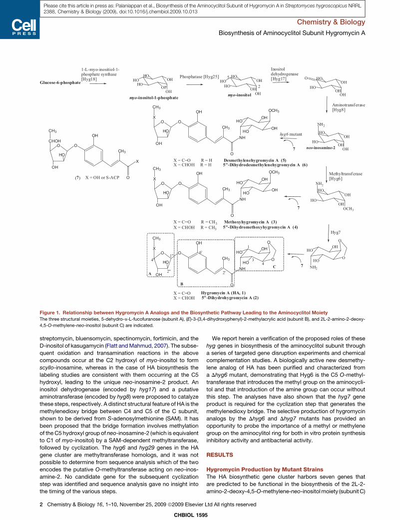
OH
O
CH3
O
NH
HO
HO
OH
O
O
HO
OH
X
CH3O
X = C=O Hygromycin A (HA, 1)X = CHOH 5"-Dihydrohygromycin A (2)
X = C=O R = H Desmethylenehygromycin A (5)X = CHOH R = H 5"-Dihydrodesmethylenehygromycin A (6)
(7)
A
B
C
1
34
2'
6'1"
2"
4"
X = OH or S-ACP
HOHO
HO
OP
OH
OH
OH
OH
HOHO
HO
OH HO
HO
OH
OH
OH
O
HO
HO
OH
OH
OH
NH2
myo-inositol-1-phosphate myo-inositol
neo-inosamine-2
5
2Glucose-6-phosphate
1-L-myo-inositiol-1-phosphate synthase[Hyg18] Phosphatase [Hyg25]
Inositoldehydrogenase[Hyg17]
Aminotransf erase[Hyg8]
7
hyg6 mutant
NH2
HO
HO
OHO
O
HO
HO
OH
OH
OCH3
NH2
Methyltransf erase[Hyg6]
Hyg7
7
7
OH
O
CH3
O
NH
HO
HO
OH
OH
O
HO
OH
X
CH3OCH3
X = C=O R = CH3 Methoxyhygromycin A (3)X = CHOH R = CH3 5"-Dihydromethoxyhygromycin A (4)
OH
O
CH3
O
NH
HO
HO
OH
OH
O
HO
OH
X
CH3OCH3
OH
O
CH3
O
O
HO
OH
CHOH
CH3
X
Figure 1. Relationship between Hygromycin A Analogs and the Biosynthetic Pathway Leading to the Aminocyclitol Moiety
The three structural moieties, 5-dehydro-a-L-fucofuranose (subunit A), (E)-3-(3,4-dihydroxyphenyl)-2-methylacrylic acid (subunit B), and 2L-2-amino-2-deoxy-
4,5-O-methylene-neo-inositol (subunit C) are indicated.
Chemistry & Biology
Biosynthesis of Aminocyclitol Subunit Hygromycin A
Please cite this article in press as: Palaniappan et al., Biosynthesis of the Aminocyclitol Subunit of Hygromycin A in Streptomyces hygroscopicus NRRL2388, Chemistry & Biology (2009), doi:10.1016/j.chembiol.2009.10.013
streptomycin, bluensomycin, spectinomycin, fortimicin, and the
D-inositol of kasugamycin (Flatt and Mahmud, 2007). The subse-
quent oxidation and transamination reactions in the above
compounds occur at the C2 hydroxyl of myo-inositol to form
scyllo-inosamine, whereas in the case of HA biosynthesis the
labeling studies are consistent with them occurring at the C5
hydroxyl, leading to the unique neo-inosamine-2 product. An
inositol dehydrogenase (encoded by hyg17) and a putative
aminotransferase (encoded by hyg8) were proposed to catalyze
these steps, respectively. A distinct structural feature of HA is the
methylenedioxy bridge between C4 and C5 of the C subunit,
shown to be derived from S-adenosylmethionine (SAM). It has
been proposed that the bridge formation involves methylation
of the C5 hydroxyl group of neo-inosamine-2 (which is equivalent
to C1 of myo-inositol) by a SAM-dependent methyltransferase,
followed by cyclization. The hyg6 and hyg29 genes in the HA
gene cluster are methyltransferase homologs, and it was not
possible to determine from sequence analysis which of the two
encodes the putative O-methyltransferase acting on neo-inos-
amine-2. No candidate gene for the subsequent cyclization
step was identified and sequence analysis gave no insight into
the timing of the various steps.
CHBIOL 159
2 Chemistry & Biology 16, 1–10, November 25, 2009 ª2009 Elsevier
We report herein a verification of the proposed roles of these
hyg genes in biosynthesis of the aminocyclitol subunit through
a series of targeted gene disruption experiments and chemical
complementation studies. A biologically active new desmethy-
lene analog of HA has been purified and characterized from
a Dhyg6 mutant, demonstrating that Hyg6 is the C5 O-methyl-
transferase that introduces the methyl group on the aminocycli-
tol and that introduction of the amine group can occur without
this step. The analyses have also shown that the hyg7 gene
product is required for the cyclization step that generates the
methylenedioxy bridge. The selective production of hygromycin
analogs by the Dhyg6 and Dhyg7 mutants has provided an
opportunity to probe the importance of a methyl or methylene
group on the aminocylitol ring for both in vitro protein synthesis
inhibitory activity and antibacterial activity.
RESULTS
Hygromycin Production by Mutant StrainsThe HA biosynthetic gene cluster harbors seven genes that
are predicted to be functional in the biosynthesis of the 2L-2-
amino-2-deoxy-4,5-O-methylene-neo-inositol moiety (subunit C)
5
Ltd All rights reserved
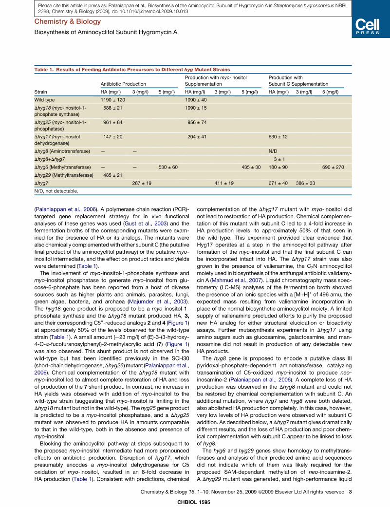
Table 1. Results of Feeding Antibiotic Precursors to Different hyg Mutant Strains
Strain
Antibiotic Production
Production with myo-inositol
Supplementation
Production with
Subunit C Supplementation
HA (mg/l) 3 (mg/l) 5 (mg/l) HA (mg/l) 3 (mg/l) 5 (mg/l) HA (mg/l) 3 (mg/l) 5 (mg/l)
Wild type 1190 ± 120 1090 ± 40
Dhyg18 (myo-inositol-1-
phosphate synthase)
588 ± 21 1090 ± 15
Dhyg25 (myo-inositol-1-
phosphatase)
961 ± 84 956 ± 74
Dhyg17 (myo-inositol
dehydrogenase)
147 ± 20 204 ± 41 630 ± 12
Dhyg8 (Aminotransferase) — — N/D
Dhyg8+Dhyg7 3 ± 1
Dhyg6 (Methyltransferase) — — 530 ± 60 435 ± 30 180 ± 90 690 ± 270
Dhyg29 (Methyltransferase) 485 ± 21
Dhyg7 287 ± 19 411 ± 19 671 ± 40 386 ± 33
N/D, not detectable.
Chemistry & Biology
Biosynthesis of Aminocyclitol Subunit Hygromycin A
Please cite this article in press as: Palaniappan et al., Biosynthesis of the Aminocyclitol Subunit of Hygromycin A in Streptomyces hygroscopicus NRRL2388, Chemistry & Biology (2009), doi:10.1016/j.chembiol.2009.10.013
(Palaniappan et al., 2006). A polymerase chain reaction (PCR)-
targeted gene replacement strategy for in vivo functional
analyses of these genes was used (Gust et al., 2003) and the
fermentation broths of the corresponding mutants were exam-
ined for the presence of HA or its analogs. The mutants were
also chemically complemented with either subunit C (the putative
final product of the aminocyclitol pathway) or the putative myo-
inositol intermediate, and the effect on product ratios and yields
were determined (Table 1).
The involvement of myo-inositol-1-phosphate synthase and
myo-inositol phosphatase to generate myo-inositol from glu-
cose-6-phosphate has been reported from a host of diverse
sources such as higher plants and animals, parasites, fungi,
green algae, bacteria, and archaea (Majumder et al., 2003).
The hyg18 gene product is proposed to be a myo-inositol-1-
phosphate synthase and the Dhyg18 mutant produced HA, 3,
and their corresponding C500-reduced analogs 2 and 4 (Figure 1)
at approximately 50% of the levels observed for the wild-type
strain (Table 1). A small amount (�23 mg/l) of (E)-3-(3-hydroxy-
4-O-a-fucofuranosylphenyl)-2-methylacrylic acid (7) (Figure 1)
was also observed. This shunt product is not observed in the
wild-type but has been identified previously in the SCH30
(short-chain dehydrogenase, Dhyg26) mutant (Palaniappan et al.,
2006). Chemical complementation of the Dhyg18 mutant with
myo-inositol led to almost complete restoration of HA and loss
of production of the 7 shunt product. In contrast, no increase in
HA yields was observed with addition of myo-inositol to the
wild-type strain (suggesting that myo-inositol is limiting in the
Dhyg18 mutant but not in the wild-type). The hyg25 gene product
is predicted to be a myo-inositol phosphatase, and a Dhyg25
mutant was observed to produce HA in amounts comparable
to that in the wild-type, both in the absence and presence of
myo-inositol.
Blocking the aminocyclitol pathway at steps subsequent to
the proposed myo-inositol intermediate had more pronounced
effects on antibiotic production. Disruption of hyg17, which
presumably encodes a myo-inositol dehydrogenase for C5
oxidation of myo-inositol, resulted in an 8-fold decrease in
HA production (Table 1). Consistent with predictions, chemical
CHBIOL
Chemistry & Biology 16,
complementation of the Dhyg17 mutant with myo-inositol did
not lead to restoration of HA production. Chemical complemen-
tation of this mutant with subunit C led to a 4-fold increase in
HA production levels, to approximately 50% of that seen in
the wild-type. This experiment provided clear evidence that
Hyg17 operates at a step in the aminocyclitol pathway after
formation of the myo-inositol and that the final subunit C can
be incorporated intact into HA. The Dhyg17 strain was also
grown in the presence of valienamine, the C7N aminocyclitol
moiety used in biosynthesis of the antifungal antibiotic validamy-
cin A (Mahmud et al., 2007). Liquid chromatography mass spec-
trometry (LC-MS) analyses of the fermentation broth showed
the presence of an ionic species with a [M+H]+ of 496 amu, the
expected mass resulting from valienamine incorporation in
place of the normal biosynthetic aminocyclitol moiety. A limited
supply of valienamine precluded efforts to purify the proposed
new HA analog for either structural elucidation or bioactivity
assays. Further mutasynthesis experiments in Dhyg17 using
amino sugars such as glucosamine, galactosamine, and man-
nosamine did not result in production of any detectable new
HA products.
The hyg8 gene is proposed to encode a putative class III
pyridoxal-phosphate-dependent aminotransferase, catalyzing
transamination of C5-oxidized myo-inositol to produce neo-
inosamine-2 (Palaniappan et al., 2006). A complete loss of HA
production was observed in the Dhyg8 mutant and could not
be restored by chemical complementation with subunit C. An
additional mutation, where hyg7 and hyg8 were both deleted,
also abolished HA production completely. In this case, however,
very low levels of HA production were observed with subunit C
addition. As described below, a Dhyg7 mutant gives dramatically
different results, and the loss of HA production and poor chem-
ical complementation with subunit C appear to be linked to loss
of hyg8.
The hyg6 and hyg29 genes show homology to methyltrans-
ferases and analysis of their predicted amino acid sequences
did not indicate which of them was likely required for the
proposed SAM-dependant methylation of neo-inosamine-2.
A Dhyg29 mutant was generated, and high-performance liquid
1595
1–10, November 25, 2009 ª2009 Elsevier Ltd All rights reserved 3

Minutes
0 2 4 6 8 10 12 14 16 18 20 22 24
AU
0
1
2
3
4
5
AU
0
1
2
3
4
5
HA
HA Δhyg29
Wild type
Δhyg65
6
2
3
4
A
B
498
0
1
2
3
4
4x10
Intens.
440 460 480 500 520 540 m/z
500
0
1000
2000
3000
4000
5000
Intens.
440 460 480 500 520 540 m/z
Figure 2. Effect of Methyltransferase gene
(hyg6, hyg29) Disruptions on Hygromycin A
Production
(A) Reverse-phase HPLC analysis of fermentation
broths of wild-type, Dhyg29, and Dhyg6 strains.
Disruption of hyg29 caused a decrease in hygro-
mycin A production. Disruption of hyg6 resulted
in the generation of new metabolites with shorter
retention times than hygromycin A.
(B) Mass spectrometric analysis (negative mode)
of Dhyg6 fermentation broth. Two new species
were identified with [M-H]- values of 498 and 500
corresponding to desmethylenehygromycin A (5)
and 500-dihydrodesmethylenehygromycin A (6),
respectively.
Chemistry & Biology
Biosynthesis of Aminocyclitol Subunit Hygromycin A
Please cite this article in press as: Palaniappan et al., Biosynthesis of the Aminocyclitol Subunit of Hygromycin A in Streptomyces hygroscopicus NRRL2388, Chemistry & Biology (2009), doi:10.1016/j.chembiol.2009.10.013
chromatography (HPLC) and MS analyses of the fermentation
broth of this strain revealed the same products (HA and 3) at
approximately 60% of those seen in the wild-type. In contrast,
the production of HA was completely abolished in a Dhyg6 strain,
and HPLC analysis of the fermentation broth of this mutant
revealed a dominant new peak and an additional minor peak
with shorter retention times than HA under the standard
reverse-phase HPLC conditions (Figure 2). The major peak
showed a mass in negative mode of m/z = 498 [M - H]- and the
minor peak showed a mass of m/z = 500 [M - H]-. An m/z of
499 is consistent with the mass of HA that is missing the methy-
lene group bridging O-4 and O-5. An m/z of 501 observed for the
smaller peak corresponds to a reduced derivative of the major
peak. The MS analyses were thus consistent with production
of desmethylene HA analogs (5, 6) (Figure 1) by the Dhyg6 strain.
The production titers of 5 were �400 mg/l. When subunit C was
provided to this strain, low levels of HA could be observed
although the levels of 5 were not significantly altered.
A unique feature of the aminocyclitol moiety of HA is the C4-C5
methylenedioxy bridge, which is essential for optimal biological
activity (Hecker et al., 1992). Sequence analyses of the 29
ORFs of the HA gene cluster did not reveal a putative candidate
gene product for the cyclization step of bridge formation. We had
CHBIOL 1595
4 Chemistry & Biology 16, 1–10, November 25, 2009 ª2009 Elsevier Ltd All rights reserved
hypothesized that the hyg7 gene product,
which is homologous to D-aminoacy-
lases/amidohydrolases, mediates forma-
tion of the amide bond between subunits
B and C, and that a Dhyg7 mutant would
accumulate 7. However, as shown in
Figure 3A, the Dhyg7 strain produced 3
and trace levels of 4. Chemical comple-
mentation with subunit C, but not myo-
inositol, led to a restoration of HA produc-
tion (55% of that seen in the wild-type
strain) (Figure 3A). A series of chemical
complementation experiments of this
strain with subunit C in the presence of
either radiolabeled [2-3H] myo-inositol or
[carboxyl-14C] 4-hydroxybenzoic acid
was also carried out (Figure 3B). These
analyses demonstrated that while HA
production was restored by supplemen-
tation with subunit C, radiolabeled myo-
inositol incorporation was only observed into 3. A similar study
with radiolabeled 4-hydroxybenzoic acid resulted, predictably,
in its incorporation into both HA and 3. Taken together, these
observations clearly indicate that cyclization of the C5 methoxy
group, resulting in formation of the methylenedioxy bridge, is
dependant upon the hyg7 gene product.
Structural Elucidation of Desmethylene HA AnalogsThe two new compounds isolated from the Dhyg6 strain were
purified by semipreparative HPLC and their structures were
elucidated by nuclear magnetic resonance (NMR). The most
obvious change observed in the 1H-NMR spectra of 5 and 6
was the absence of either the three-proton singlet at �3.5 ppm
(for the O-5 methyl group of 3), or two one-proton singlets
at �5.18 ppm and 4.83 ppm (for the methylene group bridging
O-4 and O-5 of HA).
The differences in the 1H-NMR spectra between 5 and 6
involve the fucofuranose moiety. The H-600 signal for 5 is a singlet
at 1.99 ppm (overlapping with protons from a–CH3), whereas the
H-600 signal for 6 is a doublet at 1.20 ppm. The H-400 signal is also
shifted from �4.4 ppm in 5 to 3.74 ppm in 6 (shifts are approxi-
mate for H-400 in 5 because this signal is obscured by other
signals at the same chemical shift). There is also a new signal
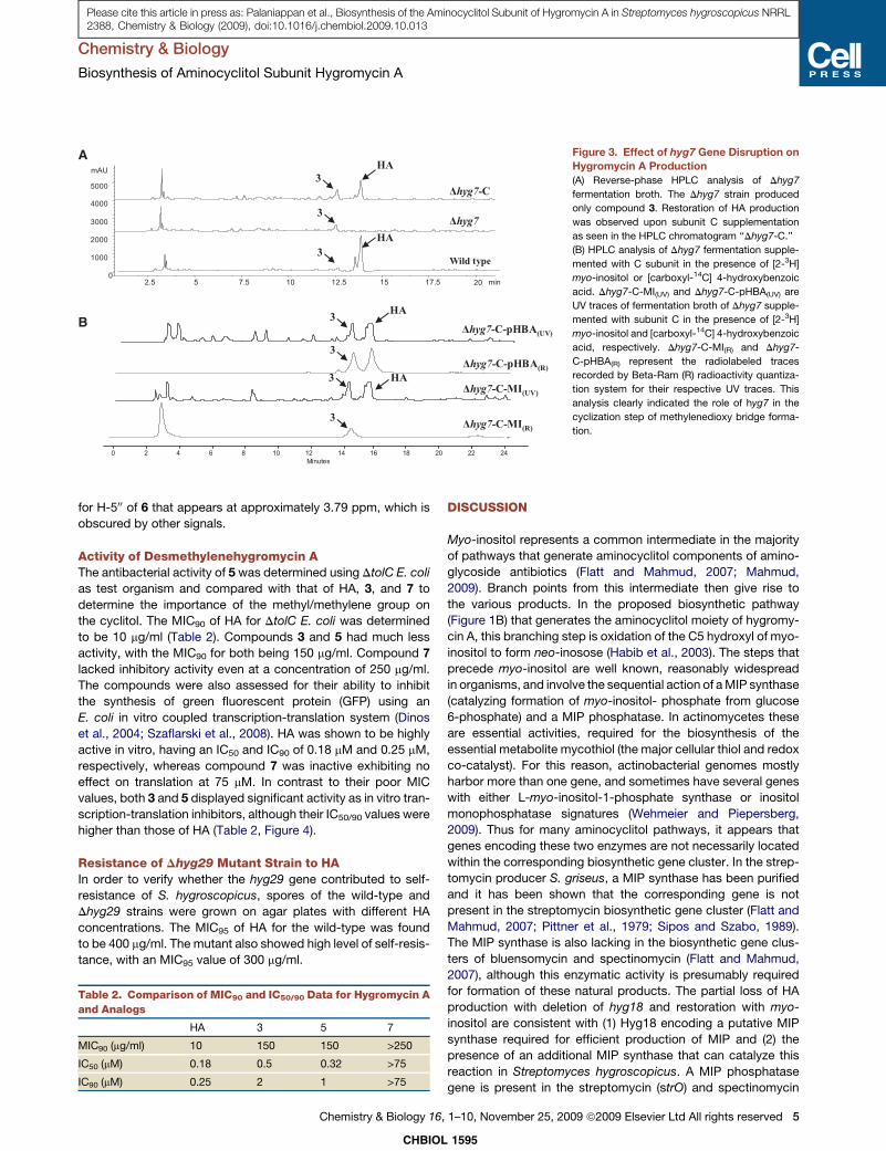
3
min2.5 5 7.5 10 12.5 15 17.5 20
mAU
0
1000
2000
3000
4000
5000
Wild type
Δhyg7
Δhyg7-C
HA
HA
Minutes
0 2 4 6 8 10 12 14 16 18 20 22 24
Δhyg7-C-MI(R)
Δhyg7-C-MI(UV)
Δhyg7-C-pHBA(UV)
Δhyg7-C-pHBA(R)
HA
HA
A
B
3
3
3
3
3
3
Figure 3. Effect of hyg7 Gene Disruption on
Hygromycin A Production
(A) Reverse-phase HPLC analysis of Dhyg7
fermentation broth. The Dhyg7 strain produced
only compound 3. Restoration of HA production
was observed upon subunit C supplementation
as seen in the HPLC chromatogram ‘‘Dhyg7-C.’’
(B) HPLC analysis of Dhyg7 fermentation supple-
mented with C subunit in the presence of [2-3H]
myo-inositol or [carboxyl-14C] 4-hydroxybenzoic
acid. Dhyg7-C-MI(UV) and Dhyg7-C-pHBA(UV) are
UV traces of fermentation broth of Dhyg7 supple-
mented with subunit C in the presence of [2-3H]
myo-inositol and [carboxyl-14C] 4-hydroxybenzoic
acid, respectively. Dhyg7-C-MI(R) and Dhyg7-
C-pHBA(R) represent the radiolabeled traces
recorded by Beta-Ram (R) radioactivity quantiza-
tion system for their respective UV traces. This
analysis clearly indicated the role of hyg7 in the
cyclization step of methylenedioxy bridge forma-
tion.
Chemistry & Biology
Biosynthesis of Aminocyclitol Subunit Hygromycin A
Please cite this article in press as: Palaniappan et al., Biosynthesis of the Aminocyclitol Subunit of Hygromycin A in Streptomyces hygroscopicus NRRL2388, Chemistry & Biology (2009), doi:10.1016/j.chembiol.2009.10.013
for H-500 of 6 that appears at approximately 3.79 ppm, which is
obscured by other signals.
Activity of Desmethylenehygromycin AThe antibacterial activity of 5 was determined using DtolC E. coli
as test organism and compared with that of HA, 3, and 7 to
determine the importance of the methyl/methylene group on
the cyclitol. The MIC90 of HA for DtolC E. coli was determined
to be 10 mg/ml (Table 2). Compounds 3 and 5 had much less
activity, with the MIC90 for both being 150 mg/ml. Compound 7
lacked inhibitory activity even at a concentration of 250 mg/ml.
The compounds were also assessed for their ability to inhibit
the synthesis of green fluorescent protein (GFP) using an
E. coli in vitro coupled transcription-translation system (Dinos
et al., 2004; Szaflarski et al., 2008). HA was shown to be highly
active in vitro, having an IC50 and IC90 of 0.18 mM and 0.25 mM,
respectively, whereas compound 7 was inactive exhibiting no
effect on translation at 75 mM. In contrast to their poor MIC
values, both 3 and 5 displayed significant activity as in vitro tran-
scription-translation inhibitors, although their IC50/90 values were
higher than those of HA (Table 2, Figure 4).
Resistance of Dhyg29 Mutant Strain to HAIn order to verify whether the hyg29 gene contributed to self-
resistance of S. hygroscopicus, spores of the wild-type and
Dhyg29 strains were grown on agar plates with different HA
concentrations. The MIC95 of HA for the wild-type was found
to be 400 mg/ml. The mutant also showed high level of self-resis-
tance, with an MIC95 value of 300 mg/ml.
Table 2. Comparison of MIC90 and IC50/90 Data for Hygromycin A
and Analogs
HA 3 5 7
MIC90 (mg/ml) 10 150 150 >250
IC50 (mM) 0.18 0.5 0.32 >75
IC90 (mM) 0.25 2 1 >75
CHBIOL
Chemistry & Biology 16,
DISCUSSION
Myo-inositol represents a common intermediate in the majority
of pathways that generate aminocyclitol components of amino-
glycoside antibiotics (Flatt and Mahmud, 2007; Mahmud,
2009). Branch points from this intermediate then give rise to
the various products. In the proposed biosynthetic pathway
(Figure 1B) that generates the aminocyclitol moiety of hygromy-
cin A, this branching step is oxidation of the C5 hydroxyl of myo-
inositol to form neo-inosose (Habib et al., 2003). The steps that
precede myo-inositol are well known, reasonably widespread
in organisms, and involve the sequential action of a MIP synthase
(catalyzing formation of myo-inositol- phosphate from glucose
6-phosphate) and a MIP phosphatase. In actinomycetes these
are essential activities, required for the biosynthesis of the
essential metabolite mycothiol (the major cellular thiol and redox
co-catalyst). For this reason, actinobacterial genomes mostly
harbor more than one gene, and sometimes have several genes
with either L-myo-inositol-1-phosphate synthase or inositol
monophosphatase signatures (Wehmeier and Piepersberg,
2009). Thus for many aminocyclitol pathways, it appears that
genes encoding these two enzymes are not necessarily located
within the corresponding biosynthetic gene cluster. In the strep-
tomycin producer S. griseus, a MIP synthase has been purified
and it has been shown that the corresponding gene is not
present in the streptomycin biosynthetic gene cluster (Flatt and
Mahmud, 2007; Pittner et al., 1979; Sipos and Szabo, 1989).
The MIP synthase is also lacking in the biosynthetic gene clus-
ters of bluensomycin and spectinomycin (Flatt and Mahmud,
2007), although this enzymatic activity is presumably required
for formation of these natural products. The partial loss of HA
production with deletion of hyg18 and restoration with myo-
inositol are consistent with (1) Hyg18 encoding a putative MIP
synthase required for efficient production of MIP and (2) the
presence of an additional MIP synthase that can catalyze this
reaction in Streptomyces hygroscopicus. A MIP phosphatase
gene is present in the streptomycin (strO) and spectinomycin
1595
1–10, November 25, 2009 ª2009 Elsevier Ltd All rights reserved 5
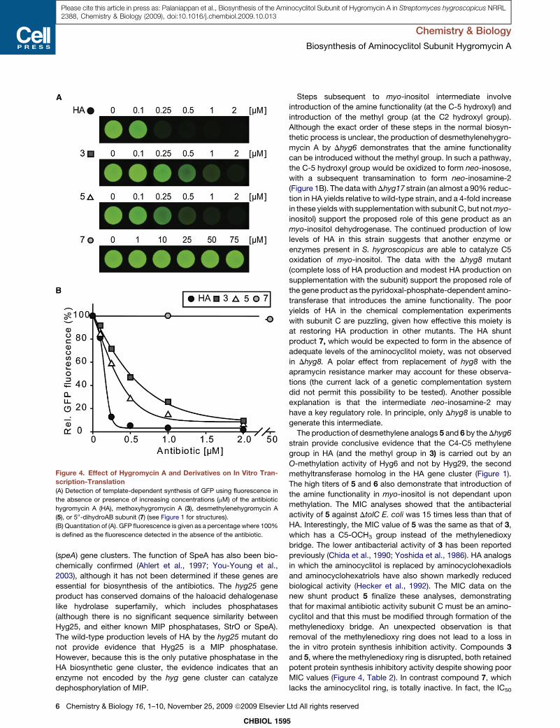
Figure 4. Effect of Hygromycin A and Derivatives on In Vitro Tran-
scription-Translation
(A) Detection of template-dependent synthesis of GFP using fluorescence in
the absence or presence of increasing concentrations (mM) of the antibiotic
hygromycin A (HA), methoxyhygromycin A (3), desmethylenehygromycin A
(5), or 500-dihydroAB subunit (7) (see Figure 1 for structures).
(B) Quantitation of (A). GFP fluorescence is given as a percentage where 100%
is defined as the fluorescence detected in the absence of the antibiotic.
Chemistry & Biology
Biosynthesis of Aminocyclitol Subunit Hygromycin A
Please cite this article in press as: Palaniappan et al., Biosynthesis of the Aminocyclitol Subunit of Hygromycin A in Streptomyces hygroscopicus NRRL2388, Chemistry & Biology (2009), doi:10.1016/j.chembiol.2009.10.013
(speA) gene clusters. The function of SpeA has also been bio-
chemically confirmed (Ahlert et al., 1997; You-Young et al.,
2003), although it has not been determined if these genes are
essential for biosynthesis of the antibiotics. The hyg25 gene
product has conserved domains of the haloacid dehalogenase
like hydrolase superfamily, which includes phosphatases
(although there is no significant sequence similarity between
Hyg25, and either known MIP phosphatases, StrO or SpeA).
The wild-type production levels of HA by the hyg25 mutant do
not provide evidence that Hyg25 is a MIP phosphatase.
However, because this is the only putative phosphatase in the
HA biosynthetic gene cluster, the evidence indicates that an
enzyme not encoded by the hyg gene cluster can catalyze
dephosphorylation of MIP.
CHBIOL 159
6 Chemistry & Biology 16, 1–10, November 25, 2009 ª2009 Elsevier
Steps subsequent to myo-inositol intermediate involve
introduction of the amine functionality (at the C-5 hydroxyl) and
introduction of the methyl group (at the C2 hydroxyl group).
Although the exact order of these steps in the normal biosyn-
thetic process is unclear, the production of desmethylenehygro-
mycin A by Dhyg6 demonstrates that the amine functionality
can be introduced without the methyl group. In such a pathway,
the C-5 hydroxyl group would be oxidized to form neo-inosose,
with a subsequent transamination to form neo-inosamine-2
(Figure 1B). The data with Dhyg17 strain (an almost a 90% reduc-
tion in HA yields relative to wild-type strain, and a 4-fold increase
in these yields with supplementation with subunit C, but not myo-
inositol) support the proposed role of this gene product as an
myo-inositol dehydrogenase. The continued production of low
levels of HA in this strain suggests that another enzyme or
enzymes present in S. hygroscopicus are able to catalyze C5
oxidation of myo-inositol. The data with the Dhyg8 mutant
(complete loss of HA production and modest HA production on
supplementation with the subunit) support the proposed role of
the gene product as the pyridoxal-phosphate-dependent amino-
transferase that introduces the amine functionality. The poor
yields of HA in the chemical complementation experiments
with subunit C are puzzling, given how effective this moiety is
at restoring HA production in other mutants. The HA shunt
product 7, which would be expected to form in the absence of
adequate levels of the aminocyclitol moiety, was not observed
in Dhyg8. A polar effect from replacement of hyg8 with the
apramycin resistance marker may account for these observa-
tions (the current lack of a genetic complementation system
did not permit this possibility to be tested). Another possible
explanation is that the intermediate neo-inosamine-2 may
have a key regulatory role. In principle, only Dhyg8 is unable to
generate this intermediate.
The production of desmethylene analogs 5 and 6 by the Dhyg6
strain provide conclusive evidence that the C4-C5 methylene
group in HA (and the methyl group in 3) is carried out by an
O-methylation activity of Hyg6 and not by Hyg29, the second
methyltransferase homolog in the HA gene cluster (Figure 1).
The high titers of 5 and 6 also demonstrate that introduction of
the amine functionality in myo-inositol is not dependant upon
methylation. The MIC analyses showed that the antibacterial
activity of 5 against DtolC E. coli was 15 times less than that of
HA. Interestingly, the MIC value of 5 was the same as that of 3,
which has a C5-OCH3 group instead of the methylenedioxy
bridge. The lower antibacterial activity of 3 has been reported
previously (Chida et al., 1990; Yoshida et al., 1986). HA analogs
in which the aminocyclitol is replaced by aminocyclohexadiols
and aminocyclohexatriols have also shown markedly reduced
biological activity (Hecker et al., 1992). The MIC data on the
new shunt product 5 finalize these analyses, demonstrating
that for maximal antibiotic activity subunit C must be an amino-
cyclitol and that this must be modified through formation of the
methylenedioxy bridge. An unexpected observation is that
removal of the methylenedioxy ring does not lead to a loss in
the in vitro protein synthesis inhibition activity. Compounds 3
and 5, where the methylenedioxy ring is disrupted, both retained
potent protein synthesis inhibitory activity despite showing poor
MIC values (Figure 4, Table 2). In contrast compound 7, which
lacks the aminocyclitol ring, is totally inactive. In fact, the IC50
5
Ltd All rights reserved
Chemistry & Biology
Biosynthesis of Aminocyclitol Subunit Hygromycin A
Please cite this article in press as: Palaniappan et al., Biosynthesis of the Aminocyclitol Subunit of Hygromycin A in Streptomyces hygroscopicus NRRL2388, Chemistry & Biology (2009), doi:10.1016/j.chembiol.2009.10.013
of these compounds (0.3–0.5 mM) is comparable to that of the
macrolide antibiotic tylosin (�0.4 mM) in the same in vitro tran-
scription-translation assay (A.L.S. and D.N.W., unpublished
data). These data suggest that the aminocyclitol ring is indeed
essential for translation inhibition activity, whereas an intact
methylenedioxy ring is not. It appears that the loss in biological
activity due to methylenedioxy ring disruption stems from
another factor, potentially a reduced uptake of the drug into
the cell rather than abolishing the ribosome binding ability of
the compound.
Inactivation of the second methyltransferase homolog, hyg29,
resulted in a decrease in the yield of HA, although the antibiotic
production profile and self-resistance were not affected.
Because HA binds to the ribosomes, rRNA methylation by
Hyg29 may provide a self-resistance mechanism in S. hygrosco-
picus. There is as yet no evidence to support such a role for
hyg29 and, moreover, ribosomal self-resistance is not always
observed for ribosome-targeting antibiotics (e.g., oleandomycin)
(Cundliffe, 1989). If the hyg29 gene product is indeed an rRNA
methylase, the high level of HA resistance of the Dhyg29 mutant
suggests that in the absence of target-site modification by
hyg29, self-resistance is possibly being conferred by the other
resistance determinants in the HA gene cluster, namely a HA
inactivating O-phosphotransferase (Dhote et al., 2008), and
putative efflux pumps encoded by hyg19 and hyg28.
The final step in the proposed pathway for formation of the
C-subunit of HA is a oxidative cyclization to provide the methyl-
enedioxy bridge. No gene product likely responsible for cata-
lyzing this step was identified in the initial analyses of the hyg
biosynthetic gene cluster (Palaniappan et al., 2006). The role of
the hyg7 gene product was unknown. A more detailed analysis
has revealed clear homology of Hyg7 with aminoacylases, which
catalyze hydrolytic deacetylation of N-acetyl-D-amino acids.
Furthermore, Hyg7 is a member of the metallo-dependant hydro-
lase superfamily. Enzymes in this class have hydrolytic activities
and typically have two, or less commonly one, metal ion-binding
sites (Lai et al., 2004). Our analysis of Hyg7 revealed the four
highly conserved residues (a Cys, Asp, and two His) that
comprise a single metal-binding site and suggest a role in
a hydrolytic process. Nonetheless, the selective production of
methoxyhygromycin A (3) by Dhyg7 and the restoration of HA
upon supplementation with subunit C clearly demonstrate
that Hyg7 is required for the oxidative cyclization process which
forms the methylenedioxy bridge of HA. Natural products with
a methylenedioxy bridge have been identified more commonly
in plants, and involve an initial methylation with subsequent
action by a cytochrome p450 enzyme complex (Ikezawa et al.,
2003). There are a limited number of examples of secondary
metabolites from actinomycetes possessing methylenedioxy
bridge. A 1,3-dioxine ring is found in dioxapyrrolomycin
from Streptomyces fumanus, simaomicin from Actinomadura
madurae, and the streptovaricins from Streptomyces spectabilis
(Charan et al., 2006; Carter et al., 1989; Staley and Rinehart,
1991). FR-900109 from S. prunicolor, the dioxolides from
S. tendae, and pseudoverticin from S. pseudoverticillus possess
a 1,3-dioxolane ring similar to that seen in HA (Blum et al., 1996;
Cui et al., 2007; Koda et al., 1983). Deuterium labeling studies in
dioxapyrrolomycin by Charan et al. indicated an oxidative cycli-
zation mechanism for methylenedioxy bridge formation similar to
CHBIOL
Chemistry & Biology 16,
that reported for the plant metabolite berberine (Bjorklund et al.,
1995; Charan et al., 2006). The pyr20 gene product in the biosyn-
thetic gene cluster of pyrrolomycin has resemblance to cyto-
chrome P450 enzymes (and not to Hyg7) and has been proposed
to potentially carry out the above function (Zhang and Parry,
2007). However, to the best of our knowledge there have been
no genetic or biochemical characterization of the processes
that lead to methylenedioxy bridge formation in bacterial natural
product biosynthesis. Our results with hyg7 represent the first
example of in vivo characterization of a functional methylene-
dioxy bridge-forming gene of bacterial origin. Furthermore, the
data indicate that a member of the metallo-dependent hydrolase
superfamily, rather than a cytochrome p450 enzyme, is involved.
The role of Hyg7 in this process, and the need for other enzymes
or cofactors, remains to be biochemically determined.
The ability of subunit C to restore HA biosynthesis in most of
the mutants described herein, demonstrates that it is an effective
substrate for the enzyme that catalyzes amide bond formation. It
is thus possible that the normal HA biosynthetic process involves
formation of a complete C subunit prior to formation of the amide
bond. Formation of 3 and 5, and the apparent incorporation of
valienamine, suggest relaxed substrate specificity of the corre-
sponding coupling enzyme (Figure 1B). However, the data do
not preclude the possibility that the final steps of C-subunit
biosynthesis (methylation and formation of the methylenedioxy
bridge) occur after amide bond formation.
SIGNIFICANCE
The roles of key hyg gene products in formation of the struc-
turally unusual, methylene-bridged aminocyclitol of HA
have been elucidated. Deletion of key genes in the pathway
leads in some cases to accumulation of intermediates in
the aminocyclitol biosynthetic process. As such the genetic
tools to access novel HAs in Streptomyces hygroscopicus
and to generate various aminocyclitols for combinatorial
biosynthetic processes (generating new structurally diverse
compounds) have been identified. The methylenedioxy
bridge in HA has been shown not to be required for in vivo
versus translation inhibition activity, but is required for bio-
logical uptake. These discoveries may help in the continued
evaluation of HA-based structures for development of novel
antibacterials. The discovery of the need for Hyg7 in the
oxidative cyclization that yields the methylene-bridged ami-
nocyclitol suggests a new enzymatic paradigm for formation
of these unusual structural moieties.
EXPERIMENTAL PROCEDURES
Chemicals, Bacterial Strains, Growth Conditions,
and General Procedures
Hygromycin A and 2L-2-amino-2-deoxy-4,5-O-methylene-neo-inositol (sub-
unit C) were kindly supplied by Pfizer Inc. [2-3H] myo-inositol (20.0 Ci/mmol)
was purchased from Moravek Biochemicals. [carboxyl-14C]-4-hydroxyben-
zoic acid was obtained from American Radiolabeled Chemicals Inc. All other
antibiotics and chemicals were purchased from Sigma Aldrich. The E. coli
DtolC strain, deficient in the outer membrane protein TolC, was procured
from the E. coli Genetic Stock Center at Yale University. All E. coli strains
were grown following standard protocols (Sambrook and Russell, 2001).
S. hygroscopicus wild-type and mutant strains were maintained using media
1595
1–10, November 25, 2009 ª2009 Elsevier Ltd All rights reserved 7
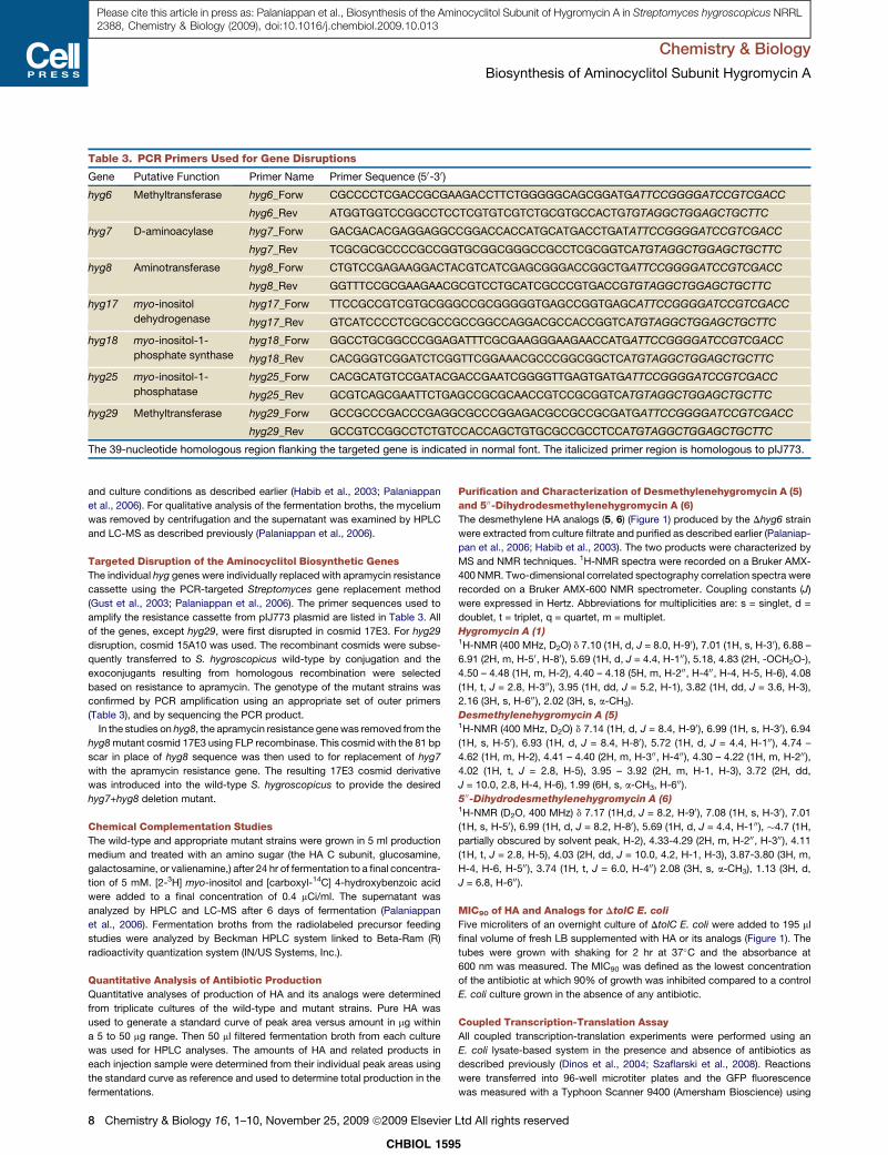
Table 3. PCR Primers Used for Gene Disruptions
Gene Putative Function Primer Name Primer Sequence (50-30)
hyg6 Methyltransferase hyg6_Forw CGCCCCTCGACCGCGAAGACCTTCTGGGGGCAGCGGATGATTCCGGGGATCCGTCGACC
hyg6_Rev ATGGTGGTCCGGCCTCCTCGTGTCGTCTGCGTGCCACTGTGTAGGCTGGAGCTGCTTC
hyg7 D-aminoacylase hyg7_Forw GACGACACGAGGAGGCCGGACCACCATGCATGACCTGATATTCCGGGGATCCGTCGACC
hyg7_Rev TCGCGCGCCCCGCCGGTGCGGCGGGCCGCCTCGCGGTCATGTAGGCTGGAGCTGCTTC
hyg8 Aminotransferase hyg8_Forw CTGTCCGAGAAGGACTACGTCATCGAGCGGGACCGGCTGATTCCGGGGATCCGTCGACC
hyg8_Rev GGTTTCCGCGAAGAACGCGTCCTGCATCGCCCGTGACCGTGTAGGCTGGAGCTGCTTC
hyg17 myo-inositol
dehydrogenase
hyg17_Forw TTCCGCCGTCGTGCGGGCCGCGGGGGTGAGCCGGTGAGCATTCCGGGGATCCGTCGACC
hyg17_Rev GTCATCCCCTCGCGCCGCCGGCCAGGACGCCACCGGTCATGTAGGCTGGAGCTGCTTC
hyg18 myo-inositol-1-
phosphate synthase
hyg18_Forw GGCCTGCGGCCCGGAGATTTCGCGAAGGGAAGAACCATGATTCCGGGGATCCGTCGACC
hyg18_Rev CACGGGTCGGATCTCGGTTCGGAAACGCCCGGCGGCTCATGTAGGCTGGAGCTGCTTC
hyg25 myo-inositol-1-
phosphatase
hyg25_Forw CACGCATGTCCGATACGACCGAATCGGGGTTGAGTGATGATTCCGGGGATCCGTCGACC
hyg25_Rev GCGTCAGCGAATTCTGAGCCGCGCAACCGTCCGCGGTCATGTAGGCTGGAGCTGCTTC
hyg29 Methyltransferase hyg29_Forw GCCGCCCGACCCGAGGCGCCCGGAGACGCCGCCGCGATGATTCCGGGGATCCGTCGACC
hyg29_Rev GCCGTCCGGCCTCTGTCCACCAGCTGTGCGCCGCCTCCATGTAGGCTGGAGCTGCTTC
The 39-nucleotide homologous region flanking the targeted gene is indicated in normal font. The italicized primer region is homologous to pIJ773.
Chemistry & Biology
Biosynthesis of Aminocyclitol Subunit Hygromycin A
Please cite this article in press as: Palaniappan et al., Biosynthesis of the Aminocyclitol Subunit of Hygromycin A in Streptomyces hygroscopicus NRRL2388, Chemistry & Biology (2009), doi:10.1016/j.chembiol.2009.10.013
and culture conditions as described earlier (Habib et al., 2003; Palaniappan
et al., 2006). For qualitative analysis of the fermentation broths, the mycelium
was removed by centrifugation and the supernatant was examined by HPLC
and LC-MS as described previously (Palaniappan et al., 2006).
Targeted Disruption of the Aminocyclitol Biosynthetic Genes
The individual hyg genes were individually replaced with apramycin resistance
cassette using the PCR-targeted Streptomyces gene replacement method
(Gust et al., 2003; Palaniappan et al., 2006). The primer sequences used to
amplify the resistance cassette from pIJ773 plasmid are listed in Table 3. All
of the genes, except hyg29, were first disrupted in cosmid 17E3. For hyg29
disruption, cosmid 15A10 was used. The recombinant cosmids were subse-
quently transferred to S. hygroscopicus wild-type by conjugation and the
exoconjugants resulting from homologous recombination were selected
based on resistance to apramycin. The genotype of the mutant strains was
confirmed by PCR amplification using an appropriate set of outer primers
(Table 3), and by sequencing the PCR product.
In the studies on hyg8, the apramycin resistance gene was removed from the
hyg8 mutant cosmid 17E3 using FLP recombinase. This cosmid with the 81 bp
scar in place of hyg8 sequence was then used to for replacement of hyg7
with the apramycin resistance gene. The resulting 17E3 cosmid derivative
was introduced into the wild-type S. hygroscopicus to provide the desired
hyg7+hyg8 deletion mutant.
Chemical Complementation Studies
The wild-type and appropriate mutant strains were grown in 5 ml production
medium and treated with an amino sugar (the HA C subunit, glucosamine,
galactosamine, or valienamine,) after 24 hr of fermentation to a final concentra-
tion of 5 mM. [2-3H] myo-inositol and [carboxyl-14C] 4-hydroxybenzoic acid
were added to a final concentration of 0.4 mCi/ml. The supernatant was
analyzed by HPLC and LC-MS after 6 days of fermentation (Palaniappan
et al., 2006). Fermentation broths from the radiolabeled precursor feeding
studies were analyzed by Beckman HPLC system linked to Beta-Ram (R)
radioactivity quantization system (IN/US Systems, Inc.).
Quantitative Analysis of Antibiotic Production
Quantitative analyses of production of HA and its analogs were determined
from triplicate cultures of the wild-type and mutant strains. Pure HA was
used to generate a standard curve of peak area versus amount in mg within
a 5 to 50 mg range. Then 50 ml filtered fermentation broth from each culture
was used for HPLC analyses. The amounts of HA and related products in
each injection sample were determined from their individual peak areas using
the standard curve as reference and used to determine total production in the
fermentations.
CHBIOL 159
8 Chemistry & Biology 16, 1–10, November 25, 2009 ª2009 Elsevier
Purification and Characterization of Desmethylenehygromycin A (5)
and 500-Dihydrodesmethylenehygromycin A (6)
The desmethylene HA analogs (5, 6) (Figure 1) produced by the Dhyg6 strain
were extracted from culture filtrate and purified as described earlier (Palaniap-
pan et al., 2006; Habib et al., 2003). The two products were characterized by
MS and NMR techniques. 1H-NMR spectra were recorded on a Bruker AMX-
400 NMR. Two-dimensional correlated spectography correlation spectra were
recorded on a Bruker AMX-600 NMR spectrometer. Coupling constants (J)
were expressed in Hertz. Abbreviations for multiplicities are: s = singlet, d =
doublet, t = triplet, q = quartet, m = multiplet.
Hygromycin A (1)1H-NMR (400 MHz, D2O) d 7.10 (1H, d, J = 8.0, H-90), 7.01 (1H, s, H-30), 6.88 –
6.91 (2H, m, H-50, H-80), 5.69 (1H, d, J = 4.4, H-100), 5.18, 4.83 (2H, -OCH2O-),
4.50 – 4.48 (1H, m, H-2), 4.40 – 4.18 (5H, m, H-200, H-400, H-4, H-5, H-6), 4.08
(1H, t, J = 2.8, H-300), 3.95 (1H, dd, J = 5.2, H-1), 3.82 (1H, dd, J = 3.6, H-3),
2.16 (3H, s, H-600), 2.02 (3H, s, a-CH3).
Desmethylenehygromycin A (5)1H-NMR (400 MHz, D2O) d 7.14 (1H, d, J = 8.4, H-90), 6.99 (1H, s, H-30), 6.94
(1H, s, H-50 ), 6.93 (1H, d, J = 8.4, H-80), 5.72 (1H, d, J = 4.4, H-100), 4.74 –
4.62 (1H, m, H-2), 4.41 – 4.40 (2H, m, H-300, H-400), 4.30 – 4.22 (1H, m, H-200),
4.02 (1H, t, J = 2.8, H-5), 3.95 – 3.92 (2H, m, H-1, H-3), 3.72 (2H, dd,
J = 10.0, 2.8, H-4, H-6), 1.99 (6H, s, a-CH3, H-600).
500-Dihydrodesmethylenehygromycin A (6)1H-NMR (D2O, 400 MHz) d 7.17 (1H,d, J = 8.2, H-90), 7.08 (1H, s, H-30), 7.01
(1H, s, H-50), 6.99 (1H, d, J = 8.2, H-80), 5.69 (1H, d, J = 4.4, H-100), �4.7 (1H,
partially obscured by solvent peak, H-2), 4.33-4.29 (2H, m, H-200, H-300), 4.11
(1H, t, J = 2.8, H-5), 4.03 (2H, dd, J = 10.0, 4.2, H-1, H-3), 3.87-3.80 (3H, m,
H-4, H-6, H-500), 3.74 (1H, t, J = 6.0, H-400) 2.08 (3H, s, a-CH3), 1.13 (3H, d,
J = 6.8, H-600).
MIC90 of HA and Analogs for DtolC E. coli
Five microliters of an overnight culture of DtolC E. coli were added to 195 ml
final volume of fresh LB supplemented with HA or its analogs (Figure 1). The
tubes were grown with shaking for 2 hr at 37�C and the absorbance at
600 nm was measured. The MIC90 was defined as the lowest concentration
of the antibiotic at which 90% of growth was inhibited compared to a control
E. coli culture grown in the absence of any antibiotic.
Coupled Transcription-Translation Assay
All coupled transcription-translation experiments were performed using an
E. coli lysate-based system in the presence and absence of antibiotics as
described previously (Dinos et al., 2004; Szaflarski et al., 2008). Reactions
were transferred into 96-well microtiter plates and the GFP fluorescence
was measured with a Typhoon Scanner 9400 (Amersham Bioscience) using
5
Ltd All rights reserved
Chemistry & Biology
Biosynthesis of Aminocyclitol Subunit Hygromycin A
Please cite this article in press as: Palaniappan et al., Biosynthesis of the Aminocyclitol Subunit of Hygromycin A in Streptomyces hygroscopicus NRRL2388, Chemistry & Biology (2009), doi:10.1016/j.chembiol.2009.10.013
a Typhoon blue laser module (Amersham Bioscience). Images were then quan-
tified using ImageQuantTL (GE Healthcare) and represented graphically using
SigmaPlot (Systat Software, Inc.).
Hygromycin Sensitivity of Dhyg29 strain
The MIC95 of HA for the Dhyg29 mutant and the wild-type strain were deter-
mined by the agar plate dilution method (Dhote et al., 2008). Briefly, spores
were plated on ISP2 agar plates containing varying amounts of HA, incubated
at 30�C for 48 hr, and scored for growth. The MIC95 was defined as the lowest
concentration of HA that prevented visible growth of 95% or more of the
colony forming units on the agar plate.
ACKNOWLEDGMENTS
A part of this work was supported by the Deutsche Forschungsgemeinschaft
(WI3285/1-1 to D.N.W.). We are grateful to Taifo Mahmud for providing valien-
amine.
Received: June 13, 2009
Revised: September 25, 2009
Accepted: October 16, 2009
Published: November 24, 2009
REFERENCES
Ahlert, J., Distler, J., Mansouri, K., and Piepersberg, W. (1997). Identification of
stsC, the gene encoding the L-glutamine:scyllo-inosose aminotransferase
from streptomycin-producing Streptomycetes. Arch. Microbiol. 168, 102–113.
Bjorklund, J.A., Frenzel, T., Rueffer, M., Kobayashi, M., Mocek, U., Fox, C.,
Beale, J.M., Grtiger, S., Zenk, M.H., and Floss, H.G. (1995). Cryptic ste-
reochemistry of berberine alkaloid biosynthesis. J. Am. Chem. Soc. 117,
1533–1545.
Blum, S., Groth, I., Rohr, J., and Fielder, H.P. (1996). Biosynthetic capacities of
actinomycetes. 5. Dioxolides, novel secondary metabolites from Strepto-
myces tendae. J. Basic Microbiol. 36, 19–25.
Carter, G.T., Goodman, J.J., Torrey, M.J., Borders, D.B., and Gould, S.J.
(1989). Biosynthetic origin of the carbon skeleton of simaomicin alpha.,
a hexacyclic xanthone antibiotic. J. Org. Chem. 54, 4321–4323.
Charan, R.D., Schlingmann, G., Bernan, V.S., Feng, X., and Carter, G.T. (2006).
Dioxapyrrolomycin biosynthesis in Streptomyces fumanus. J. Nat. Prod. 69,
29–33.
Chida, N., Nakazawa, K., Ohtsuka, M., Suzuki, M., and Ogawa, S. (1990).
Total synthesis of methoxyhygromycin and its 5-epimer. Chem. Lett. 19,
423–426.
Cui, C.B., Han, B., Caib, B., and Wanga, H. (2007). Pseudoverticin, a novel
benzoquinone-derived ansamycin antibiotic obtained as new cell cycle inhib-
itor from Streptomyces pseudoverticillus YN17707. Tetrahedron Lett. 48,
4839–4843.
Cundliffe, E. (1989). How antibiotic-producing organisms avoid suicide. Annu.
Rev. Microbiol. 43, 207–233.
Dhote, V., Gupta, S., and Reynolds, K.A. (2008). An O-phosphotransferase
catalyzes phosphorylation of hygromycin A in the antibiotic-producing
organism Streptomyces hygroscopicus. Antimicrob. Agents Chemother. 52,
3580–3588.
Dinos, G., Wilson, D.N., Teraoka, Y., Szaflarski, W., Fucini, P., Kalpaxis, D., and
Nierhaus, K.H. (2004). Dissecting the ribosomal inhibition mechanisms of
edeine and pactamycin: the universally conserved residues G693 and C795
regulate P-site RNA binding. Mol. Cell 13, 113–124.
Flatt, P.M., and Mahmud, T. (2007). Biosynthesis of aminocyclitol-aminoglyco-
side antibiotics and related compounds. Nat. Prod. Rep. 24, 358–392.
Guerrero, M.D., and Modolell, J. (1980). Hygromycin A, a novel inhibitor of ribo-
somal peptidyltransferase. Eur. J. Biochem. 107, 409–414.
Gust, B., Challis, G.L., Fowler, K., Kieser, T., and Chater, K.F. (2003). PCR-
targeted Streptomyces gene replacement identifies a protein domain needed
CHBIOL
Chemistry & Biology 16,
for biosynthesis of the sesquiterpene soil odor geosmin. Proc. Natl. Acad. Sci.
USA 100, 1541–1546.
Habib, el-S.E., Scarsdale, J.N., and Reynolds, K.A. (2003). Biosynthetic origin
of hygromycin A. Antimicrob. Agents Chemother. 47, 2065–2071.
Hayashi, S.F., Norcia, L.J., Seibel, S.B., and Silvia, A.M. (1997). Structure-
activity relationships of hygromycin A and its analogs: protein synthesis inhibi-
tion activity in a cell free system. J. Antibiot. (Tokyo) 50, 514–521.
Hecker, S.J., Lilley, S.C., and Werner, K.M. (1992). Hygromycin A: preparation
of aminocyclitol analogs defining the minimum functionality required for
biological activity. Bioorg. Med. Chem. Lett. 2, 1043–1046.
Ikezawa, N., Tanaka, M., Nagayoshi, M., Shinkyo, R., Sakaki, T., Inouye, K.,
and Sato, F. (2003). Molecular cloning and characterization of CYP719, a meth-
ylenedioxy bridge-forming enzyme that belongs to a novel P450 family, from
cultured Coptis japonica cells. J. Biol. Chem. 278, 38557–38565.
Kim, S.D., Kweon, M.H., Kim, C.J., and Yoo, I.D. (1990). Hygromycin, a plant
growth inhibitor. Korean J. Biochem. 23, 23–25.
Koda, S., Morimoto, Y., Yamashita, M., Komori, T., and Imanaka, H. (1983).
Studies on a new antibiotic FR-900109. 2. X-ray structure determination of
FR-900109 p-bromophenyl ester. J. Antibiot. (Tokyo) 36, 1237–1238.
Lai, W.L., Chou, L.Y., Ting, C.Y., Kirby, R., Tsai, Y.C., Wang, A.H., and Liaw,
S.H. (2004). The functional role of the binuclear metal center in D-aminoacy-
lase: one-metal activation and second-metal attenuation. J. Biol. Chem.
279, 13962–13967.
Lee, H.B., Kim, C.J., Kim, J.S., Hong, K.S., and Cho, K.Y. (2003). A bleaching
herbicidal activity of methoxyhygromycin (MHM) produced by an actinomy-
cete strain Streptomyces sp. 8E-12. Lett. Appl. Microbiol. 36, 387–391.
Mahmud, T. (2003). The C7N aminocyclitol family of natural products. Nat.
Prod. Rep. 20, 137–166.
Mahmud, T. (2009). Progress in aminocyclitol biosynthesis. Curr. Opin. Chem.
Biol. 13, 161–170.
Mahmud, T., Flatt, P.M., and Wu, X. (2007). Biosynthesis of unusual aminocy-
clitol-containing natural products. J. Nat. Prod. 70, 1384–1391.
Majumder, A.L., Chatterjee, A., Ghosh Dastidar, K., and Majee, M. (2003).
Diversification and evolution of L-myo-inositol 1-phosphate synthase. FEBS
Lett. 553, 3–10.
Mann, R.L., Gale, R.M., and Van Abeele, R.F. (1953). Hygromycin. II. Isolation
and properties. Antibiot. Chemother. 3, 1279–1282.
Nakagawa, A., Fujimoto, T., Omura, S., Walsh, J.C., Stotish, R.L., and George,
B. (1987). Hygromycin A, an antitreponemal substance. II. Therapeutic effect
for swine dysentery. J. Antibiot. (Tokyo) 40, 1627–1635.
Omura, S., Nakagawa, A., Fujimoto, T., Saito, K., Otoguro, K., and Walsh, J.C.
(1987). Hygromycin A, an antitreponemal substance. I. Screening method and
therapeutic effect for Treponema hyodysenteriae-caused infection in CF-1
mice. J. Antibiot. (Tokyo) 40, 1619–1626.
Palaniappan, N., Ayers, S., Gupta, S., Habib, el-S., and Reynolds, K.A. (2006).
Production of hygromycin A analogs in Streptomyces hygroscopicus NRRL
2388 through identification and manipulation of the biosynthetic gene cluster.
Chem. Biol. 13, 753–764.
Pittenger, R.C., Wolfe, R.N., Hoehn, P.N., Daily, W.A., and McGuire, J.M.
(1953). Hygromycin. I. Preliminary studies in the production and biologic
activity on a new antibiotic. Antibiot. Chemother. 3, 1268–1278.
Pittner, F., Tovarova, I.I., Kornitskaya, E.Y., Khokhlov, A.S., and Hoffmann-
Ostenhof, O. (1979). Myo-inositol-1-phosphate synthase from Streptomyces
griseus (studies on the biosynthesis of cyclitols, XXXVIII). Mol. Cell. Biochem.
25, 43–46.
Sambrook, J., and Russell, D.W. (2001). Molecular Cloning: A Laboratory
Manual, Third Edition (Cold Spring Harbor, NY: Cold Spring Harbor Laboratory
Press).
Sipos, L., and Szabo, G. (1989). Myo-inositol-1-phosphate synthase in
different Streptomyces griseus variants. FEMS Microbiol. Lett. 53, 339–343.
Staley, A.L., and Rinehart, K.L. (1991). Biosynthesis of the streptovaricins:
3-amino-5-hydroxybenzoic acid as a precursor to the meta-C7N unit.
J. Antibiot. (Tokyo) 44, 218–224.
1595
1–10, November 25, 2009 ª2009 Elsevier Ltd All rights reserved 9
Chemistry & Biology
Biosynthesis of Aminocyclitol Subunit Hygromycin A
Please cite this article in press as: Palaniappan et al., Biosynthesis of the Aminocyclitol Subunit of Hygromycin A in Streptomyces hygroscopicus NRRL2388, Chemistry & Biology (2009), doi:10.1016/j.chembiol.2009.10.013
Szaflarski, W., Vesper, O., Teraoka, Y., Plitta, B., Wilson, D.N., and Nierhaus,
K.H. (2008). New features of the ribosome and ribosomal inhibitors: non-
enzymatic recycling, misreading and back-translocation. J. Mol. Biol. 380,
193–205.
Uyeda, M., Mizukami, M., Yokomizo, K., and Suzuki, K. (2001). Pentalenolac-
tone I and hygromycin A, immunosuppressants produced by Streptomyces
filipinensis and Streptomyces hygroscopicus. Biosci. Biotechnol. Biochem.
65, 1252–1254.
Wehmeier, U.F., and Piepersberg, W. (2009). Enzymology of aminoglycoside
biosynthesis-deduction from gene clusters. Methods Enzymol. 459, 459–491.
CHBIOL 159
10 Chemistry & Biology 16, 1–10, November 25, 2009 ª2009 Elsevier
Yoshida, M., Takahashi, E., Uozumi, T., and Beppu, T. (1986). Hygromycin A
and methoxyhygromycin A, novel inhibitors of K88 antigen synthesis of enter-
otoxic Escherichia coli strain. Agric. Biol. Chem. 50, 143–149.
You-Young, J., Kim, S., Yang, Y.Y., Kang, C., Sohng, J., and Suh, J. (2003).
Functional analysis of spectinomycin biosynthetic genes from Streptomyces
spectabilis ATCC 27741. J. Microbiol. Biotechnol. 13, 906–911.
Zhang, X., and Parry, R.J. (2007). Cloning and characterization of the pyrrolo-
mycin biosynthetic gene clusters from Actinosporangium vitaminophilum
ATCC 31673 and Streptomyces sp. strain UC 11065. Antimicrob. Agents
Chemother. 51, 946–957.
5
Ltd All rights reserved

Paper 2
ANTIMICROBIAL AGENTS AND CHEMOTHERAPY, Dec. 2009, p. 5163–5172 Vol. 53, No. 120066-4804/09/$12.00 doi:10.1128/AAC.01069-09Copyright © 2009, American Society for Microbiology. All Rights Reserved.
The Final Step of Hygromycin A Biosynthesis, Oxidation ofC-5�-Dihydrohygromycin A, Is Linked to a Putative Proton
Gradient-Dependent Efflux�
Vidya Dhote,1 Agata L. Starosta,2,3 Daniel N. Wilson,2,3 and Kevin A. Reynolds1*Department of Chemistry, Portland State University, P.O. Box 751, Portland, Oregon 97207-0751,1 and Center for
Integrated Protein Science Munich (CiPS-M)2 and Gene Center and Department of Chemistry and Biochemistry,3
Ludwig-Maximilians-Universitat Munchen, Feodor-Lynen-Strasse 25, Munich D-81377, Germany
Received 30 July 2009/Returned for modification 28 August 2009/Accepted 9 September 2009
Hygromycin A (HA) is an aminocyclitol antibiotic produced and excreted by Streptomyces hygroscopicus.Deletion of hyg26 from the hygromycin A biosynthetic gene cluster has previously been shown to result in amutant that produces 5�-dihydrohygromycin A (DHHA). We report herein on the purification and character-ization of Hyg26 expressed in Escherichia coli. The enzyme catalyzes an NAD(H)-dependent reversible inter-conversion of HA and DHHA, supporting the role of the reduced HA as the penultimate biosynthetic pathwayintermediate and not a shunt product. The equilibrium for the Hyg26-catalyzed reaction heavily favors theDHHA intermediate. The high-titer production of the HA product by S. hygroscopicus must be dependent upona subsequent energetically favorable enzyme-catalyzed process, such as the selective and efficient export of HA.hyg19 encodes a putative proton gradient-dependent transporter, and a mutant lacking this gene was observedto produce less HA and to produce the DHHA intermediate. The DHHA produced by either the �hyg19 or the�hyg26 mutant had slightly reduced activity against E. coli and reduced protein synthesis-inhibitory activity invitro. The data indicate that Hyg26 and Hyg19 have evolved to produce and export the final potent HA productin a coordinated fashion.
Antibiotic biosynthetic gene clusters contain genes requiredfor the process of assembling the natural product and for thegeneration of specific biosynthetic precursors that are notreadily available from primary metabolism (16). In addition,they often contain genes that are responsible for self-resis-tance, thereby protecting the organism from its own naturalproduct, and regulatory genes that control the expression ofbiosynthetic genes. Although these gene classifications are use-ful, they are often incomplete in their descriptions. In fact, acomplex interplay between biosynthesis, resistance, and regu-lation has been observed for many natural product biosyntheticprocesses, and it is becoming increasingly evident that some ofthese gene products perform multiple functions.
Hygromycin A (HA; compound 1) (Fig. 1) is an aminocycli-tol antibiotic that acts by inhibiting the peptidyltransferasereaction of protein synthesis in gram-positive and gram-nega-tive bacteria (7, 15, 25). Biosynthetic studies have shown thatHA is assembled from three independently synthesized sub-units, 5-dehydro-�-L-fucofuranose (subunit A), (E)-3-(3,4-di-hydroxyphenyl)-2-methylacrylic acid (subunit B), and the ami-nocyclitol, 2L-2-amino-2-deoxy-4,5-O-methylene-neo-inositol(subunit C) (6). The corresponding biosynthetic gene clusterhas been cloned and sequenced from Streptomyces hygroscopi-cus NRRL 2388, and putative functions have been assigned tothe 29 open reading frames (22). The proposed role of hyg26 as
a short-chain dehydrogenase gene has also been verified at thegenetic level (22).
Disruption of hyg26 in S. hygroscopicus abolished theproduction of HA and methoxyhygromycin A (compound 3)and, instead, yielded their reduced analogs, 5�-dihydrohy-gromycin A (DHHA; compound 2) and 5�-dihydromethoxy-hygromycin A (compound 4), respectively, and smallamounts of (E)-3-(3-hydroxy-4-O-�-fucofuranosylphenyl)-2-methylacrylic acid (compound 7). Compound 7 is DHHAwithout the C subunit (see Fig. 1 for the structures). Whilethese results showed that Hyg26 is required for the dehy-drogenation of the C-5� OH on the fucofuranose moiety, thetiming of this reaction in the biosynthetic sequence and,subsequently, the question as to whether DHHA is a shuntmetabolite or a precursor of the final product (HA) remainunresolved.
Analysis of the HA biosynthetic gene cluster has also re-vealed multiple genes likely to be involved in antibiotic resis-tance. Of these, the function of hyg21 has been biochemicallydetermined (4). hyg21 encodes a phosphotransferase that in-activates HA by phosphorylating the C-2� OH on the fuco-furanose. A �hyg21 strain has been shown to retain resistanceto HA, indicating the presence of other mechanisms of self-resistance. Antibiotic transporters/efflux pumps are additionalmechanisms of self-resistance in antibiotic producers and likelyoperate in S. hygroscopicus also, as all of the HA produced isfound outside the cell. The HA biosynthetic gene cluster con-tains two putative HA transporter genes, hyg19 and hyg28. Thehyg19 gene shows similarity to proton gradient-dependenttransporter genes. The hyg28 gene encodes a putative type IIATP-binding cassette transporter. The role of these gene prod-
* Corresponding author. Mailing address: Department of Chemis-try, Portland State University, P.O. Box 751, Portland, OR 97207.Phone: (503) 725-3886. Fax: (503) 725-9525. E-mail: [email protected].
� Published ahead of print on 21 September 2009.
5163

ucts in exporting HA or pathway intermediates and in contrib-uting to self-resistance has not been determined.
In the work described here, we show that a recombinantHyg26 catalyzes the reversible interconversion of DHHA andHA and that the equilibrium for this reaction lies heavilytoward the biologically less active, reduced HA product. Theputative proton gradient-dependent transporter encoded byhyg19 is observed to be required for the high-level productionof HA and is hypothesized to operate by the selective andefficient exportation of HA. A hyg19 deletion mutant did notshow increased sensitivity to HA, but a mutant in which bothhyg19 and hyg21 were deleted had markedly increased HAsensitivity. This work has demonstrated that DHHA is likelythe final biosynthetic pathway intermediate and that Hyg19 hasa role in both self-resistance and biosynthesis, ensuring that thefinal and most biologically active HA product is generated.
MATERIALS AND METHODS
Chemicals, bacterial strains, and growth conditions. All antibiotics and chem-icals were purchased from Sigma Aldrich unless otherwise stated. PCR primerswere obtained from Integrated DNA Technologies. The enzymes used for DNAmanipulations were obtained from New England Biolabs. HA was kindly pro-vided by Pfizer Inc. DHHA, 5�-dihydromethoxyhygromycin A (compound 4), and(E)-3-(3-hydroxy-4-O-�-fucofuranosylphenyl)-2-methylacrylic acid (compound7) were purified from the fermentation broth of mutant strain S. hygroscopicusSCH30 by a semipreparative high-performance liquid chromatography (HPLC)method, as described earlier (22). A �hyg7 strain of S. hygroscopicus was used asthe source for methoxyhygromycin A (compound 3). A �hyg6 disruption strainwas used as the source for desmethylenehygromycin A (compound 5) and 5�-dihydrodesmethylenehygromycin A (compound 6) (submitted for publication).The S. hygroscopicus strains were propagated by using the media and cultureconditions described earlier (6, 22). The Escherichia coli strains were cultured bystandard protocols (28). E. coli �tolC was procured from the E. coli GeneticStock Center at Yale University.
Nucleotide and protein sequence analysis. Searches for sequences with ho-mology to the sequences of the hyg19, hyg26, and hyg28 genes were carried outwith the BLAST suite of programs at the National Center for BiotechnologyInformation website (1). Protein sequence alignments were obtained by using theCLUSTALX program at the European Bioinformatics Institute website (14).Protein motif searches were carried out by using the PROSITE database at the
Expasy proteomics server (9). Hydropathy analysis for Hyg19 was based on theamino acid hydropathy values of Kyte and Doolittle (13) and carried out at theTransporter Classification Database server (26, 27). The MEMSAT3 programwas also used for prediction of the secondary structure and topology of Hyg19(10).
Cloning, overexpression, and purification of recombinant Hyg26. The 816-bphyg26 gene was amplified from cosmid 17E3 (22). Primers were designed tointroduce an NdeI restriction site at the initiation codon and a BamHI restrictionsite 3� to the termination codon (Table 1). Amplification was performed by usingthe following temperature program: initial denaturation at 95°C for 5 min, 25cycles of amplification (45 s of denaturation at 95°C, 45 s of primer annealing at56°C, 1 min of extension at 72°C), and a final 7-min extension at 72°C. The genewas subsequently cloned into the NdeI and BamHI restriction sites of thepET15b vector (Novagen). The resulting pET15b-hyg26 construct was intro-duced into the E. coli BL21-CodonPlus (DE3) expression host (Stratagene) bytransformation. For protein expression, cells were grown at 37°C in LB mediumcontaining 100 �g/ml ampicillin and 50 �g/ml chloramphenicol. When an A600 of0.6 was reached, the culture flask was brought to room temperature (�23°C) andexpression was induced by adding 0.5 mM isopropyl thio-�-D-galactoside. Aftera further 6 h of incubation, cells were harvested by centrifugation and stored at�80°C.
The N-terminal His-tagged Hyg26 was purified under native conditions ac-cording to Qiagen’s Ni-nitrilotriacetic acid protocol. The cells were resuspendedin BugBuster master mixture protein extraction reagent (Novagen) and lysed byincubation at room temperature for 30 min. The lysate was cleared by centrifu-gation; and the supernatant was loaded onto Ni-nitrilotriacetic acid agarosepreequilibrated with buffer containing 50 mM NaH2PO4, 300 mM NaCl, and 10mM imidazole (pH 8.0). The column was washed with buffer containing 50 mMNaH2PO4, 300 mM NaCl, and 20 mM imidazole (pH 8.0). The protein waseluted with the same buffer containing a higher concentration of imidazole (250mM). Fractions containing Hyg26 were pooled, dialyzed overnight against abuffer (50 mM Tris-HCl, 20% glycerol, 5 mM 2-mercaptoethanol at pH 7.5), andstored at �80°C until further use.
Determination of molecular mass of Hyg26. The molecular mass of purifiedHis-tagged Hyg26 was determined by size-exclusion chromatography with a Phe-nomenex BioSep-SEC-S4000 size-exclusion column fitted to an Agilent 1100series HPLC system. The column was equilibrated and eluted with 20 mMsodium phosphate (pH 7.2) at a flow rate of 0.5 ml/min. Carbonic anhydrase (25kDa), bovine serum albumin (66 kDa), �-amylase (200 kDa), apoferritin (443kDa), and thyroglobulin (669 kDa) were used as standards.
Hyg26 enzyme assays and kinetic analyses. Initial enzyme assays were per-formed at 30°C with 4 �g of purified enzyme in a 100-�l reaction mixture of 50mM Tris-HCl (pH 7.5) and 2 mM Tris(2-carboxyethyl) phosphine (TCEP). Thedehydrogenase activity was studied by incubating Hyg26 with 100 �M DHHA,
FIG. 1. Structures of HA and related compounds.
5164 DHOTE ET AL. ANTIMICROB. AGENTS CHEMOTHER.
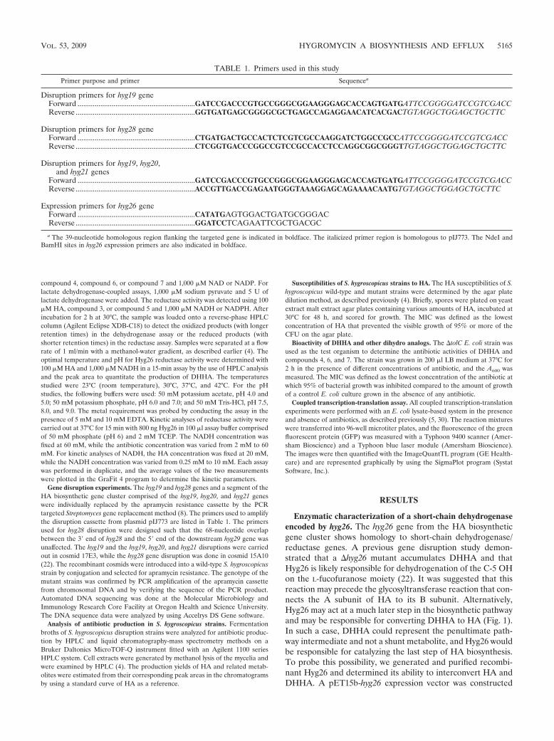
compound 4, compound 6, or compound 7 and 1,000 �M NAD or NADP. Forlactate dehydrogenase-coupled assays, 1,000 �M sodium pyruvate and 5 U oflactate dehydrogenase were added. The reductase activity was detected using 100�M HA, compound 3, or compound 5 and 1,000 �M NADH or NADPH. Afterincubation for 2 h at 30°C, the sample was loaded onto a reverse-phase HPLCcolumn (Agilent Eclipse XDB-C18) to detect the oxidized products (with longerretention times) in the dehydrogenase assay or the reduced products (withshorter retention times) in the reductase assay. Samples were separated at a flowrate of 1 ml/min with a methanol-water gradient, as described earlier (4). Theoptimal temperature and pH for Hyg26 reductase activity were determined with100 �M HA and 1,000 �M NADH in a 15-min assay by the use of HPLC analysisand the peak area to quantitate the production of DHHA. The temperaturesstudied were 23°C (room temperature), 30°C, 37°C, and 42°C. For the pHstudies, the following buffers were used: 50 mM potassium acetate, pH 4.0 and5.0; 50 mM potassium phosphate, pH 6.0 and 7.0; and 50 mM Tris-HCl, pH 7.5,8.0, and 9.0. The metal requirement was probed by conducting the assay in thepresence of 5 mM and 10 mM EDTA. Kinetic analyses of reductase activity werecarried out at 37°C for 15 min with 800 ng Hyg26 in 100 �l assay buffer comprisedof 50 mM phosphate (pH 6) and 2 mM TCEP. The NADH concentration wasfixed at 60 mM, while the antibiotic concentration was varied from 2 mM to 60mM. For kinetic analyses of NADH, the HA concentration was fixed at 20 mM,while the NADH concentration was varied from 0.25 mM to 10 mM. Each assaywas performed in duplicate, and the average values of the two measurementswere plotted in the GraFit 4 program to determine the kinetic parameters.
Gene disruption experiments. The hyg19 and hyg28 genes and a segment of theHA biosynthetic gene cluster comprised of the hyg19, hyg20, and hyg21 geneswere individually replaced by the apramycin resistance cassette by the PCRtargeted Streptomyces gene replacement method (8). The primers used to amplifythe disruption cassette from plasmid pIJ773 are listed in Table 1. The primersused for hyg28 disruption were designed such that the 68-nucleotide overlapbetween the 3� end of hyg28 and the 5� end of the downstream hyg29 gene wasunaffected. The hyg19 and the hyg19, hyg20, and hyg21 disruptions were carriedout in cosmid 17E3, while the hyg28 gene disruption was done in cosmid 15A10(22). The recombinant cosmids were introduced into a wild-type S. hygroscopicusstrain by conjugation and selected for apramycin resistance. The genotype of themutant strains was confirmed by PCR amplification of the apramycin cassettefrom chromosomal DNA and by verifying the sequence of the PCR product.Automated DNA sequencing was done at the Molecular Microbiology andImmunology Research Core Facility at Oregon Health and Science University.The DNA sequence data were analyzed by using Accelrys DS Gene software.
Analysis of antibiotic production in S. hygroscopicus strains. Fermentationbroths of S. hygroscopicus disruption strains were analyzed for antibiotic produc-tion by HPLC and liquid chromatography-mass spectrometry methods on aBruker Daltonics MicroTOF-Q instrument fitted with an Agilent 1100 seriesHPLC system. Cell extracts were generated by methanol lysis of the mycelia andwere examined by HPLC (4). The production yields of HA and related metab-olites were estimated from their corresponding peak areas in the chromatogramsby using a standard curve of HA as a reference.
Susceptibilities of S. hygroscopicus strains to HA. The HA susceptibilities of S.hygroscopicus wild-type and mutant strains were determined by the agar platedilution method, as described previously (4). Briefly, spores were plated on yeastextract malt extract agar plates containing various amounts of HA, incubated at30°C for 48 h, and scored for growth. The MIC was defined as the lowestconcentration of HA that prevented the visible growth of 95% or more of theCFU on the agar plate.
Bioactivity of DHHA and other dihydro analogs. The �tolC E. coli strain wasused as the test organism to determine the antibiotic activities of DHHA andcompounds 4, 6, and 7. The strain was grown in 200 �l LB medium at 37°C for2 h in the presence of different concentrations of antibiotic, and the A600 wasmeasured. The MIC was defined as the lowest concentration of the antibiotic atwhich 95% of bacterial growth was inhibited compared to the amount of growthof a control E. coli culture grown in the absence of any antibiotic.
Coupled transcription-translation assay. All coupled transcription-translationexperiments were performed with an E. coli lysate-based system in the presenceand absence of antibiotics, as described previously (5, 30). The reaction mixtureswere transferred into 96-well microtiter plates, and the fluorescence of the greenfluorescent protein (GFP) was measured with a Typhoon 9400 scanner (Amer-sham Bioscience) and a Typhoon blue laser module (Amersham Bioscience).The images were then quantified with the ImageQuantTL program (GE Health-care) and are represented graphically by using the SigmaPlot program (SystatSoftware, Inc.).
RESULTS
Enzymatic characterization of a short-chain dehydrogenaseencoded by hyg26. The hyg26 gene from the HA biosyntheticgene cluster shows homology to short-chain dehydrogenase/reductase genes. A previous gene disruption study demon-strated that a �hyg26 mutant accumulates DHHA and thatHyg26 is likely responsible for dehydrogenation of the C-5 OHon the L-fucofuranose moiety (22). It was suggested that thisreaction may precede the glycosyltransferase reaction that con-nects the A subunit of HA to its B subunit. Alternatively,Hyg26 may act at a much later step in the biosynthetic pathwayand may be responsible for converting DHHA to HA (Fig. 1).In such a case, DHHA could represent the penultimate path-way intermediate and not a shunt metabolite, and Hyg26 wouldbe responsible for catalyzing the last step of HA biosynthesis.To probe this possibility, we generated and purified recombi-nant Hyg26 and determined its ability to interconvert HA andDHHA. A pET15b-hyg26 expression vector was constructed
TABLE 1. Primers used in this study
Primer purpose and primer Sequencea
Disruption primers for hyg19 geneForward ..............................................................GATCCGACCCGTGCCGGGCGGAAGGGAGCACCAGTGATGATTCCGGGGATCCGTCGACCReverse ...............................................................GGTGATGAGCGGGGCGCTGAGCCAGAGGAACATCACGACTGTAGGCTGGAGCTGCTTC
Disruption primers for hyg28 geneForward ..............................................................CTGATGACTGCCACTCTCGTCGCCAAGGATCTGGCCGCCATTCCGGGGATCCGTCGACCReverse ...............................................................CTCGGTGACCCGGCCGTCCGCCACCTCCAGGCGGCGGGTTGTAGGCTGGAGCTGCTTC
Disruption primers for hyg19, hyg20,and hyg21 genes
Forward ..............................................................GATCCGACCCGTGCCGGGCGGAAGGGAGCACCAGTGATGATTCCGGGGATCCGTCGACCReverse ...............................................................ACCGTTGACCGAGAATGGGTAAAGGAGCAGAAAACAATGTGTAGGCTGGAGCTGCTTC
Expression primers for hyg26 geneForward ..............................................................CATATGAGTGGACTGATGCGGGACReverse ...............................................................GGATCCTCAGAATTCGCTGACGC
a The 39-nucleotide homologous region flanking the targeted gene is indicated in boldface. The italicized primer region is homologous to pIJ773. The NdeI andBamHI sites in hyg26 expression primers are also indicated in boldface.
VOL. 53, 2009 HYGROMYCIN A BIOSYNTHESIS AND EFFLUX 5165
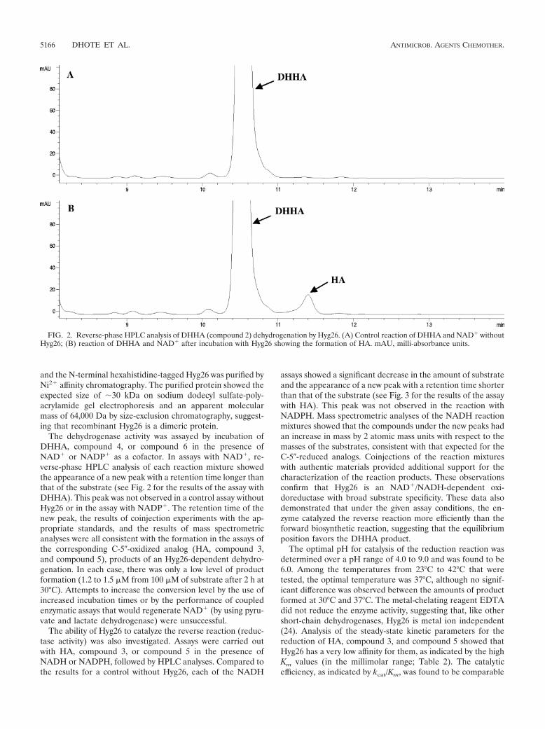
and the N-terminal hexahistidine-tagged Hyg26 was purified byNi2 affinity chromatography. The purified protein showed theexpected size of �30 kDa on sodium dodecyl sulfate-poly-acrylamide gel electrophoresis and an apparent molecularmass of 64,000 Da by size-exclusion chromatography, suggest-ing that recombinant Hyg26 is a dimeric protein.
The dehydrogenase activity was assayed by incubation ofDHHA, compound 4, or compound 6 in the presence ofNAD or NADP as a cofactor. In assays with NAD, re-verse-phase HPLC analysis of each reaction mixture showedthe appearance of a new peak with a retention time longer thanthat of the substrate (see Fig. 2 for the results of the assay withDHHA). This peak was not observed in a control assay withoutHyg26 or in the assay with NADP. The retention time of thenew peak, the results of coinjection experiments with the ap-propriate standards, and the results of mass spectrometricanalyses were all consistent with the formation in the assays ofthe corresponding C-5�-oxidized analog (HA, compound 3,and compound 5), products of an Hyg26-dependent dehydro-genation. In each case, there was only a low level of productformation (1.2 to 1.5 �M from 100 �M of substrate after 2 h at30°C). Attempts to increase the conversion level by the use ofincreased incubation times or by the performance of coupledenzymatic assays that would regenerate NAD (by using pyru-vate and lactate dehydrogenase) were unsuccessful.
The ability of Hyg26 to catalyze the reverse reaction (reduc-tase activity) was also investigated. Assays were carried outwith HA, compound 3, or compound 5 in the presence ofNADH or NADPH, followed by HPLC analyses. Compared tothe results for a control without Hyg26, each of the NADH
assays showed a significant decrease in the amount of substrateand the appearance of a new peak with a retention time shorterthan that of the substrate (see Fig. 3 for the results of the assaywith HA). This peak was not observed in the reaction withNADPH. Mass spectrometric analyses of the NADH reactionmixtures showed that the compounds under the new peaks hadan increase in mass by 2 atomic mass units with respect to themasses of the substrates, consistent with that expected for theC-5�-reduced analogs. Coinjections of the reaction mixtureswith authentic materials provided additional support for thecharacterization of the reaction products. These observationsconfirm that Hyg26 is an NAD/NADH-dependent oxi-doreductase with broad substrate specificity. These data alsodemonstrated that under the given assay conditions, the en-zyme catalyzed the reverse reaction more efficiently than theforward biosynthetic reaction, suggesting that the equilibriumposition favors the DHHA product.
The optimal pH for catalysis of the reduction reaction wasdetermined over a pH range of 4.0 to 9.0 and was found to be6.0. Among the temperatures from 23°C to 42°C that weretested, the optimal temperature was 37°C, although no signif-icant difference was observed between the amounts of productformed at 30°C and 37°C. The metal-chelating reagent EDTAdid not reduce the enzyme activity, suggesting that, like othershort-chain dehydrogenases, Hyg26 is metal ion independent(24). Analysis of the steady-state kinetic parameters for thereduction of HA, compound 3, and compound 5 showed thatHyg26 has a very low affinity for them, as indicated by the highKm values (in the millimolar range; Table 2). The catalyticefficiency, as indicated by kcat/Km, was found to be comparable
FIG. 2. Reverse-phase HPLC analysis of DHHA (compound 2) dehydrogenation by Hyg26. (A) Control reaction of DHHA and NAD withoutHyg26; (B) reaction of DHHA and NAD after incubation with Hyg26 showing the formation of HA. mAU, milli-absorbance units.
5166 DHOTE ET AL. ANTIMICROB. AGENTS CHEMOTHER.
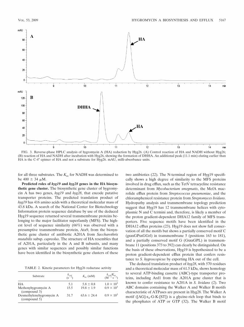
for all three substrates. The Km for NADH was determined tobe 400 34 �M.
Predicted roles of hyg19 and hyg28 genes in the HA biosyn-thetic gene cluster. The biosynthetic gene cluster of hygromy-cin A has two genes, hyg19 and hyg28, that encode putativetransporter proteins. The predicted translation product ofhyg19 has 416 amino acids with a theoretical molecular mass of43.8 kDa. A search of the National Center for BiotechnologyInformation protein sequence database by use of the deducedHyg19 sequence returned several transmembrane proteins be-longing to the major facilitator superfamily (MFS). The high-est level of sequence similarity (66%) was observed with apresumptive transmembrane protein, Ata9, from the biosyn-thetic gene cluster of antibiotic A201A from Saccharothrixmutabilis subsp. capreolus. The structure of HA resembles thatof A201A, particularly in the A and B subunits, and manygenes with similar sequences and possibly similar functionshave been identified in the biosynthetic gene clusters of these
two antibiotics (22). The N-terminal region of Hyg19 specifi-cally shows a high degree of similarity to the MFS proteinsinvolved in drug efflux, such as the TetV tetracycline resistancedeterminant from Mycobacterium smegmatis, the MefA mac-rolide efflux protein from Streptococcus pneumoniae, and thechloramphenicol resistance protein from Streptomyces lividans.Hydropathy analysis and transmembrane topology predictionsuggest that Hyg19 has 12 transmembrane helices with cyto-plasmic N and C termini and, therefore, is likely a member ofthe proton gradient-dependent DHA12 family of MFS trans-porters. Five sequence motifs have been identified in theDHA12 efflux proteins (23). Hyg19 does not show full conser-vation of all the motifs but shows a partially conserved motif C(gxxxGPxxGGrl) in transmembrane 5 (positions 163 to 181),and a partially conserved motif G (GxxxGPL) in transmem-brane 11 (positions 373 to 392) can clearly be distinguished. Onthe basis of these observations, Hyg19 is hypothesized to be aproton gradient-dependent efflux protein that confers resis-tance to S. hygroscopicus by exporting HA out of the cell.
The deduced translation product of hyg28, with 570 residuesand a theoretical molecular mass of 61.5 kDa, shows homologyto several ATP-binding cassette (ABC)-type transporter pro-teins, including Ard1 from the A201A gene cluster that isknown to confer resistance to A201A in S. lividans (2). TwoABC domains containing the Walker A and Walker B motifscharacteristic of ATPases are present in Hyg28. The Walker Amotif ([AG]-x4-G-K-[ST]) is a glycine-rich loop that binds tothe phosphates of ATP or GTP (32). The Walker B motif
FIG. 3. Reverse-phase HPLC analysis of hygromycin A (HA) reduction by Hyg26. (A) Control reaction of HA and NADH without Hyg26;(B) reaction of HA and NADH after incubation with Hyg26, showing the formation of DHHA. An additional peak (11.1 min) eluting earlier thanHA is the C-4� epimer of HA and not a substrate for Hyg26. mAU, milli-absorbance units.
TABLE 2. Kinetic parameters for Hyg26 reductase activity
Substrate kcat(s�1) Km (mM) kcat/Km
(M�1 s�1)
HA 5.1 5.8 0.8 1.0 � 103
Methoxyhygromycin A(compound 3)
15.5 19.8 1.9 0.9 � 103
Desmethylenehygromycin A(compound 5)
31.7 63.6 24.4 0.9 � 103
VOL. 53, 2009 HYGROMYCIN A BIOSYNTHESIS AND EFFLUX 5167
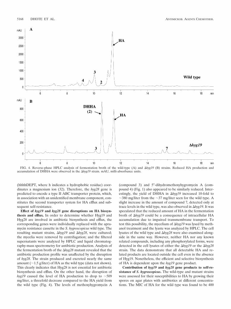
(hhhhDEPT, where h indicates a hydrophobic residue) coor-dinates a magnesium ion (32). Therefore, the hyg28 gene ispredicted to encode a type II ABC transporter protein, which,in association with an unidentified membrane component, con-stitutes the second transporter system for HA efflux and sub-sequent self-resistance.
Effect of hyg19 and hyg28 gene disruptions on HA biosyn-thesis and efflux. In order to determine whether Hyg19 andHyg28 are involved in antibiotic biosynthesis and efflux, thecorresponding genes were individually replaced with the apra-mycin resistance cassette in the S. hygroscopicus wild type. Theresulting mutant strains, �hyg19 and �hyg28, were cultured;the mycelia were removed by centrifugation; and the filteredsupernatants were analyzed by HPLC and liquid chromatog-raphy-mass spectrometry for antibiotic production. Analysis ofthe fermentation broth of the �hyg28 mutant revealed that theantibiotic production profile was unaffected by the disruptionof hyg28. The strain produced and excreted nearly the sameamount (�1.5 g/liter) of HA as the wild type (data not shown).This clearly indicates that Hyg28 is not essential for antibioticbiosynthesis and efflux. On the other hand, the disruption ofhyg19 caused the level of HA production to drop to �509mg/liter, a threefold decrease compared to the HA yield fromthe wild type (Fig. 4). The levels of methoxyhygromycin A
(compound 3) and 5�-dihydromethoxyhygromycin A (com-pound 4) (Fig. 1) also appeared to be similarly reduced. Inter-estingly, the yield of DHHA in �hyg19 increased 10-fold to�380 mg/liter from the �37 mg/liter seen for the wild type. Aslight increase in the amount of compound 7, detected only attrace levels in the wild type, was also observed in �hyg19. It wasspeculated that the reduced amount of HA in the fermentationbroth of �hyg19 could be a consequence of intracellular HAaccumulation due to impaired transmembrane transport. Totest this possibility, the mycelium of �hyg19 was lysed by meth-anol treatment and the lyaste was analyzed by HPLC. The celllysates of the wild type and �hyg28 were also examined along-side in the same way. However, neither HA nor any knownrelated compounds, including any phosphorylated forms, weredetected in the cell lysates of either the �hyg19 or the �hyg28strain. The data demonstrate that all detectable HA and re-lated products are located outside the cell even in the absenceof Hyg19. Nonetheless, the efficient and selective biosynthesisof HA is dependent upon the hyg19 gene product.
Contribution of hyg19 and hyg28 gene products to self-re-sistance of S. hygroscopicus. The wild-type and mutant strainswere assessed for their susceptibilities to HA by growing theirspores on agar plates with antibiotics at different concentra-tions. The MIC of HA for the wild type was found to be 400
FIG. 4. Reverse-phase HPLC analysis of fermentation broth of the wild-type (A) and �hyg19 (B) strains. Reduced HA production andaccumulation of DHHA were observed in the �hyg19 strain. mAU, milli-absorbance units.
5168 DHOTE ET AL. ANTIMICROB. AGENTS CHEMOTHER.
�g/ml. The mutants also showed high levels of resistance,with MICs of 400 �g/ml and 300 �g/ml for �hyg19 and�hyg28, respectively. These data indicate that the self-resis-tance of S. hygroscopicus is not entirely conferred by hyg19or hyg28. Another mutant strain was generated by disruptinghyg19 together with the hyg20 and hyg21 genes present im-mediately downstream of it (Fig. 5). The hyg20 gene encodesa putative transglucosylase/mutase for the generation of theA subunit, and its disruption has been observed to abolishantibiotic production completely (unpublished data). hyg21encodes an antibiotic-modifying O-phosphotransferase witha role in self-resistance, and loss of this gene results inreduced HA production (4). The �(hyg19-hyg20-hyg21)strain did not produce HA and showed significantly in-creased susceptibility (MIC, 75 �g/ml). A �hyg20 strain hada MIC of 300 �g/ml, which was not significantly lower thanthat of the wild type. The MIC of S. lividans for HA wasdetermined to be 20 �g/ml. These data suggest that theincreased susceptibility of �(hyg19-hyg20-hyg21) is due tothe loss of hyg19 and hyg21, implying a synergistic role ofthese two genes in self-resistance.
Biological activity of C-5�-dihydro analogs. DHHA andcompounds 4 and 6 are the C-5�-dihydro analogs of HA, me-thoxyhygromycin A (compound 3), and desmethylenehygromy-cin A (compound 5), respectively (Fig. 1). These three C-5�-dihydro compounds and compound 7 were studied for theirability both to inhibit the growth of �tolC E. coli and to inhibitprotein synthesis in an E. coli in vitro-coupled transcription-translation system (5, 30). The MICs of DHHA, compound 2,and compound 4 were determined to be 20 �g/ml, 150 �g/ml,and 150 �g/ml, respectively (Table 3). These values are similarto the MICs for the corresponding 5�-oxidized analogs, albeitfor the most active pair, HA appeared to be slightly moreactive (10 �g/ml) than DHHA. The DHHA analog lacking theC subunit (compound 7) was inactive against E. coli �tolC evenat 250 �g/ml. The data demonstrate that for in vivo activity, thepresence of the methylene bridge on the C subunit is of greaterimportance than the C-5� oxidation state of the A subunit. HA
was also the most active compound in vitro, having a 50%inhibitory concentration (IC50) and an IC90 of 0.18 �M and0.25 �M, respectively, whereas compound 7 was inactive (con-sistent with the MICs), exhibiting no effect on translation at 75�M (Fig. 6A and B and Table 3). On the basis of the IC50s,compounds 3 and 5 were as effective as DHHA and theiractivities were similar to the activity of HA. In contrast, thereduced forms of these compounds, compounds 4 and 6, hadsignificantly lower (8 to 15 times) potency (Fig. 6C and Table3). The data demonstrate that for in vitro activity, the presenceof the methylene bridge on the C subunit is of lesser impor-tance than the C-5� oxidation state of the A subunit.
DISCUSSION
hyg26 in the HA biosynthetic gene cluster encodes a proteinthat is a member of the short-chain dehydrogenase family. Therequirement for this gene to obtain HA and not DHHA has
FIG. 5. Schematic representation of replacement of the hyg19,hyg20, and hyg21 genes with the apramycin resistance cassette. WT,wild type.
TABLE 3. Comparison of MIC and IC50/IC90 data for HA andDHHA analogs
Compound MIC(�g/ml)
IC50(�M)
IC90(�M)
HA 10 0.18 0.25DHHA 20 0.3 1.53 150 0.5 24 150 7.4 505 150 0.32 16 150 2.7 14.1
FIG. 6. Effects of HA and derivatives on in vitro transcription-translation. (A) Detection of template-dependent synthesis of GFP byusing fluorescence and native polyacrylamide gel electrophoresis in thepresence of template with increasing concentrations (�M) of the an-tibiotic HA, DHHA, and compound 7 on GFP synthesis; (B) quanti-tation of effects of HA, DHHA, and compound 7 on GFP synthesis;(C) quantitation of effects of HA and compounds 3, 4, 5, and 6 on GFPsynthesis. The experiments whose results are shown in panels B and Cwere performed in triplicate. GFP fluorescence is given as a percent-age, where 100% is defined as the fluorescence detected in the absenceof the antibiotic. Rel., relative.
VOL. 53, 2009 HYGROMYCIN A BIOSYNTHESIS AND EFFLUX 5169

previously been demonstrated by a gene disruption study (22).Most short-chain dehydrogenase enzymes are dimers or tet-ramers and typically do not need a divalent metal ion foractivity, unlike the classical metal-dependent medium-chainalcohol dehydrogenases (11, 18). The results of analyses ofrecombinant Hyg26 in the present study are consistent with itbeing a dimeric, metal ion-independent protein. The enzymeshowed an absolute preference for NAD(H) and did not utilizeNADP(H). The coenzyme-binding site of short-chain dehydro-genases preferring NAD(H) has an acidic residue (Asp or Glu)that forms hydrogen bonds with the hydroxyl groups of theadenine ribose, whereas the binding site for NADP(H) has twobasic residues (Arg or Lys) that bind to the phosphate (12, 31).The specificity of Hyg26 for NAD(H) is consistent with thepresence of an Asp residue at position 38 in the putativecoenzyme-binding region within residues 12 to 38 (VTGAARGQGREHAVRMAGEGADVIAID; conserved residues areindicated in boldface). Both the ability of Hyg26 to catalyze theinterconversion of DHHA and HA and the production ofDHHA by a �hyg26 mutant are consistent with this being thelast step of HA biosynthesis. A previous proposal that Hyg26may catalyze the conversion of L-fucofuranose to 5-dehydro-�-L-fucofuranose during the formation of just the A subunitappears to be less likely.
Additional support for the hypothesis that DHHA is thepenultimate intermediate in the HA pathway comes fromthe analyses of the �hyg19 mutant. The equilibrium for theHyg26-catalyzed process was observed to heavily favorDHHA, even though HA is the primary fermentation prod-uct. Loss of the subsequent energetically favorable step,which would shift the equilibrium toward HA, would bepredicted to result in the formation of decreased of levels ofHA and the formation of increased levels of DHHA, as seenfor the �hyg19 mutant. The possibility that this accumula-tion of DHHA arises from a polar effect on the adjacenthyg20 is unlikely, as a hyg20 deletion mutant does not pro-duce any detectable levels of HA. There are several exam-ples of this, in which the final steps of antibiotic biosynthesisare linked with efflux. In the case of nystatin biosynthesis byS. noursei, disruption of the nysH and nysG genes for aputative type III ABC transporter resulted in the accumu-lation of 10-deoxynystatin, a precursor of nystatin lackingthe hydroxyl at C-10 (29). Furthermore, expression in themutant strains of an additional copy of nysL, which presum-ably encodes a monooxygenase for C-10 hydroxylation, par-tially restored nystatin production with a concomitant de-crease in 10-deoxynystatin. NysL has been expressed and hasbeen shown to catalyze the hydroxylation of 10-deoxynysta-tin (29). The reaction has not been shown to be reversible(in contrast to the reaction observed for Hyg26), and theredoes not appear to be detectable inhibition of NysL by theproduct nystatin. Thus, while the observations indicate thatthe final biosynthetic step is balanced with efficient efflux,the biochemical basis seems to be less clear than that pro-posed herein for HA. A linkage between the final proposedstep in chromomycin A3 biosynthesis, the conversion ofdideacetylated precursor by a membrane-bound acetyltrans-ferase, and exportation by a type I ABC transporter in S.griseus subsp. griseus have also been reported (17).
The increased level of production of DHHA by the �hyg19
mutant is consistent with Hyg19 being the primary efflux pumpresponsible for the selective and efficient transport of HA. Inour working hypothesis, Hyg19 is able to preferentially bind toand export HA over the intermediate DHHA (Fig. 7). Theseproperties would contrast with those observed for Hyg26,which, in the reduction reaction, exhibited poor binding forHA (approximately 6 mM) and showed limited evidence ofbinding specificity (compounds 3 and 5 were also substrates).The poor HA binding by Hyg26 would permit facile transfer toHyg19 for export. Hyg19 must also export HA sufficiently fastsuch that it is not phosphorylated by the resistance element,Hyg21 (this enzyme has an observed kcat of 2.2 min�1 and a Km
for HA of 30 �M), and gets trapped inside the cell (4).The presence of HA and DHHA in the fermentation broth
of �hyg19 implies the existence of an alternative system forantibiotic efflux (Fig. 7). This system would have reduced se-lectivity (exporting both HA and DHHA) relative to that forthe process mediated by Hyg19. The most obvious candidatewithin the hyg biosynthetic gene cluster is the hyg28-encodedABC transporter. Hyg28 does not appear to have a significantrole in efflux in the wild type, as the �hyg28 strain continues toproduce wild-type levels of HA in the fermentation broth. TheHyg19 process may be significantly faster that that proposed tobe catalyzed by Hyg28, or hyg28 expression may increase in the�hyg19 strain. Attempts to probe these possibilities by thegeneration of a mutant in which both genes have been deletedhave thus far been unsuccessful. There are numerous prece-dents for the association of multiple efflux systems with anti-biotic biosynthetic processes and, in particular, transportersthat can excrete pathway intermediates. The 10-deoxynystatinintermediate, produced by deletion of the putative type IIIABC transporter in S. noursei, is found in the fermentationbroth, suggesting an additional efflux process (29). The prema-ture efflux of landomycin D, an intermediate in landomycin Abiosynthesis in S. cyanogenus, has been reported with either theoverexpression of the proton gradient-dependent transportergene lanJ or the disruption of the regulatory gene, lanK, thatrepresses lanJ (19).
In addition to ensuring the production of the final HA prod-uct, Hyg19 also contributes to HA resistance in the producingorganism. The loss of hyg19 alone did not lead to decreasedHA resistance. Similar observations with regard to resistancehave been made for the landomycin E producer S. globisporus1912. Self-resistance was not affected when the proton gradi-ent-dependent transporter gene lndJ and the ABC transportergene lndW, both of which occur in the landomycin E biosyn-thetic gene cluster, were independently disrupted. Heterolo-gous expression of these genes, however, conferred resistanceto landomycin E, proving their function as resistance genes (20,21). The intrinsic resistance of an antibiotic producer is oftendue to the synergistic action of multiple gene products (3). Thisappears to be the case for HA self-resistance, as the simulta-neous deletion of two putative HA resistance determinants,hyg19 and hyg21 (in addition to the hyg20 biosynthetic gene),resulted in an increase in susceptibility greater than thatachieved when either of them was individually disrupted (theHA susceptibility of the �hyg21 mutant has been describedseparately [4]). In this mutant with triple mutations, both theproposed efficient HA efflux system and an enzyme that canmodify and inactivate HA inside the cell are absent. The high
5170 DHOTE ET AL. ANTIMICROB. AGENTS CHEMOTHER.
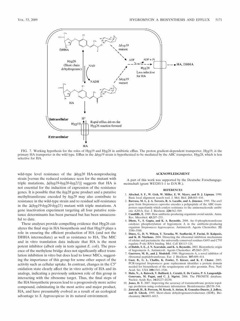
wild-type level resistance of the �hyg20 HA-nonproducingstrain [versus the reduced resistance seen for the mutant withtriple mutations, �(hyg19-hyg20-hyg21)] suggests that HA isnot essential for the induction of expression of the resistancegenes. It is possible that the hyg28 gene product and a putativemethyltransferase encoded by hyg29 may also contribute toresistance in the wild-type strain and to residual self-resistancein the �(hyg19-hyg20-hyg21) mutant with triple mutations. Agene inactivation experiment targeting all four putative resis-tance determinants has been pursued but has been unsuccess-ful to date.
These analyses provide compelling evidence that Hyg26 cat-alyzes the final step in HA biosynthesis and that Hyg19 plays arole in ensuring the efficient production of HA (and not theDHHA intermediate) as well as resistance to HA. The MICand in vitro translation data indicate that HA is the mostpotent inhibitor (albeit only in tests against E. coli). The pres-ence of the methylene bridge does not significantly affect trans-lation inhibition in vitro but does lead to lower MICs, suggest-ing the importance of this group for some other aspect of theactivity such as cellular uptake. In contrast, changes in the C5�oxidation state clearly affect the in vitro activity of HA and itsanalogs, indicating a previously unknown role of this group ininteracting with the ribosome target. Thus, the final steps ofthe HA biosynthetic process lead to a progressively more activecompound, culminating in the most active and major product,HA, and have presumably evolved as a result of an ecologicaladvantage to S. hygroscopicus in its natural environment.
ACKNOWLEDGMENT
A part of this work was supported by the Deutsche Forschungsge-meinschaft (grant WI3285/1-1 to D.N.W.).
REFERENCES
1. Altschul, S. F., W. Gish, W. Miller, E. W. Myers, and D. J. Lipman. 1990.Basic local alignment search tool. J. Mol. Biol. 215:403–410.
2. Barrasa, M. I., J. A. Tercero, R. A. Lacalle, and A. Jimenez. 1995. The ard1gene from Streptomyces capreolus encodes a polypeptide of the ABC-trans-porters superfamily which confers resistance to the aminonucleoside antibi-otic A201A. Eur. J. Biochem. 228:562–569.
3. Cundliffe, E. 1989. How antibiotic-producing organisms avoid suicide. Annu.Rev. Microbiol. 43:207–233.
4. Dhote, V., S. Gupta, and K. A. Reynolds. 2008. An O-phosphotransferasecatalyzes phosphorylation of hygromycin A in the antibiotic-producingorganism Streptomyces hygroscopicus. Antimicrob. Agents Chemother. 52:3580–3588.
5. Dinos, G., D. N. Wilson, Y. Teraoka, W. Szaflarski, P. Fucini, D. Kalpaxis,and K. H. Nierhaus. 2004. Dissecting the ribosomal inhibition mechanismsof edeine and pactamycin: the universally conserved residues G693 and C795regulate P-site RNA binding. Mol. Cell 13:113–124.
6. el Habib, S. E., J. N. Scarsdale, and K. A. Reynolds. 2003. Biosynthetic originof hygromycin A. Antimicrob. Agents Chemother. 47:2065–2071.
7. Guerrero, M. D., and J. Modolell. 1980. Hygromycin A, a novel inhibitor ofribosomal peptidyltransferase. Eur. J. Biochem. 107:409–414.
8. Gust, B., G. L. Challis, K. Fowler, T. Kieser, and K. F. Chater. 2003.PCR-targeted Streptomyces gene replacement identifies a protein domainneeded for biosynthesis of the sesquiterpene soil odor geosmin. Proc. Natl.Acad. Sci. USA 100:1541–1546.
9. Hulo, N., A. Bairoch, V. Bulliard, L. Cerutti, E. De Castro, P. S. Langendijk-Genevaux, M. Pagni, and C. J. Sigrist. 2006. The PROSITE database.Nucleic Acids Res. 34:D227–D230.
10. Jones, D. T. 2007. Improving the accuracy of transmembrane protein topol-ogy prediction using evolutionary information. Bioinformatics 23:538–544.
11. Jornvall, H., B. Persson, M. Krook, S. Atrian, R. Gonzalez-Duarte, J. Jeffery,and D. Ghosh. 1995. Short-chain dehydrogenases/reductases (SDR). Bio-chemistry 34:6003–6013.
FIG. 7. Working hypothesis for the roles of Hyg19 and Hyg28 in antibiotic efflux. The proton gradient-dependent transporter, Hyg19, is theprimary HA transporter in the wild type. Efflux in the �hyg19 strain is hypothesized to be mediated by the ABC transporter, Hyg28, which is lessselective for HA.
VOL. 53, 2009 HYGROMYCIN A BIOSYNTHESIS AND EFFLUX 5171

12. Kallberg, Y., U. Oppermann, H. Jornvall, and B. Persson. 2002. Short-chaindehydrogenases/reductases (SDRs). Coenzyme-based functional assign-ments in completed genomes. Eur. J. Biochem. 269:4409–4417.
13. Kyte, J., and R. F. Doolittle. 1982. A simple method for displaying thehydropathic character of a protein. J. Mol. Biol. 157:105–132.
14. Larkin, M. A., G. Blackshields, N. P. Brown, R. Chenna, P. A. McGettigan,H. McWilliam, F. Valentin, I. M. Wallace, A. Wilm, R. Lopez, J. D. Thomp-son, T. J. Gibson, and D. G. Higgins. 2007. Clustal W and Clustal X version2.0. Bioinformatics 23:2947–2948.
15. Mann, R. L., R. M. Gale, and R. F. Van Abeele. 1953. Hygromycin. II.Isolation and properties. Antibiot. Chemother. 3:1279–1282.
16. Martin, M. F., and P. Liras. 1989. Organization and expression of genesinvolved in the biosynthesis of antibiotics and other secondary metabolites.Annu. Rev. Microbiol. 43:173–206.
17. Menendez, N., A. F. Brana, J. A. Salas, and C. Mendez. 2007. Involvementof a chromomycin ABC transporter system in secretion of a deacetylatedprecursor during chromomycin biosynthesis. Microbiology 153:3061–3070.
18. Oppermann, U., C. Filling, M. Hult, N. Shafqat, X. Wu, M. Lindh, J.Shafqat, E. Nordling, Y. Kallberg, B. Persson, and H. Jornvall. 2003. Short-chain dehydrogenases/reductases (SDR): the 2002 update. Chem. Biol. In-teract. 143–144:247–253.
19. Ostash, I., B. Ostash, A. Luzhetskyy, A. Bechthold, S. Walker, and V. Fe-dorenko. 2008. Coordination of export and glycosylation of landomycins inStreptomyces cyanogenus S136. FEMS Microbiol. Lett. 285:195–202.
20. Ostash, I., B. Ostash, S. Walker, and V. Fedorenko. 2007. Proton-dependenttransporter gene lndJ confers resistance to landomycin E in Streptomycesglobisporus. Genetika 43:1032–1037.
21. Ostash, I., Y. Rebets, B. Ostash, A. Kobylyanskyy, M. Myronovskyy, T.Nakamura, S. Walker, and V. Fedorenko. 2008. An ABC transporter encod-ing gene lndW confers resistance to landomycin E. Arch. Microbiol. 190:105–109.
22. Palaniappan, N., S. Ayers, S. Gupta, S. el Habib, and K. A. Reynolds. 2006.Production of hygromycin A analogs in Streptomyces hygroscopicus NRRL2388 through identification and manipulation of the biosynthetic gene clus-ter. Chem. Biol. 13:753–764.
23. Paulsen, I. T., M. H. Brown, and R. A. Skurray. 1996. Proton-dependentmultidrug efflux systems. Microbiol. Rev. 60:575–608.
24. Persson, B., M. Krook, and H. Jornvall. 1991. Characteristics of short-chainalcohol dehydrogenases and related enzymes. Eur. J. Biochem. 200:537–543.
25. Pittenger, R. C., R. N. Wolfe, P. N. Hoehn, W. A. Daily, and J. M. McGuire.1953. Hygromycin. I. Preliminary studies in the production and biologicactivity on a new antibiotic. Antibiot. Chemother. 3:1268–1278.
26. Saier, M. H., Jr., C. V. Tran, and R. D. Barabote. 2006. TCDB: the Trans-porter Classification Database for membrane transport protein analyses andinformation. Nucleic Acids Res. 34:D181–D186.
27. Saier, M. H., Jr., M. R. Yen, K. Noto, D. G. Tamang, and C. Elkan. 2009. TheTransporter Classification Database: recent advances. Nucleic Acids Res.37:D274–D278.
28. Sambrook, J., and D. W. Russell. 2001. Molecular cloning: a laboratorymanual, 3rd ed. Cold Spring Harbor Laboratory Press, Cold Spring Har-bor, NY.
29. Sletta, H., S. E. Borgos, P. Bruheim, O. N. Sekurova, H. Grasdalen, R. Aune,T. E. Ellingsen, and S. B. Zotchev. 2005. Nystatin biosynthesis and transport:nysH and nysG genes encoding a putative ABC transporter system in Strep-tomyces noursei ATCC 11455 are required for efficient conversion of 10-deoxynystatin to nystatin. Antimicrob. Agents Chemother. 49:4576–4583.
30. Szaflarski, W., O. Vesper, Y. Teraoka, B. Plitta, D. N. Wilson, and K. H.Nierhaus. 2008. New features of the ribosome and ribosomal inhibitors:non-enzymatic recycling, misreading and back-translocation. J. Mol. Biol.380:193–205.
31. Tanaka, N., T. Nonaka, M. Nakanishi, Y. Deyashiki, A. Hara, and Y. Mitsui.1996. Crystal structure of the ternary complex of mouse lung carbonyl re-ductase at 1.8 Å resolution: the structural origin of coenzyme specificity inthe short-chain dehydrogenase/reductase family. Structure 4:33–45.
32. Walker, J. E., M. Saraste, M. J. Runswick, and N. J. Gay. 1982. Distantlyrelated sequences in the alpha- and beta-subunits of ATP synthase, myosin,kinases and other ATP-requiring enzymes and a common nucleotide bindingfold. EMBO J. 1:945–951.
5172 DHOTE ET AL. ANTIMICROB. AGENTS CHEMOTHER.
Paper 3

doi:10.1016/j.jmb.2008.11.042 J. Mol. Biol. (2009) 385, 1179–1192
Available online at www.sciencedirect.com
Time-Resolved Binding of Azithromycin toEscherichia coli Ribosomes
Alexandros D. Petropoulos1†, Ekaterini C. Kouvela1†,Agata L. Starosta2,3, Daniel N. Wilson2,3, George P. Dinos1
and Dimitrios L. Kalpaxis1⁎
1Laboratory of Biochemistry,School of Medicine, Universityof Patras, 26504 Patras, Greece2Gene Center and Departmentof Chemistry and Biochemistry,Ludwig-Maximilians-Universitat Munchen, Feodor-Lynen-Strasse 25, D-81377Munich, Germany3Munich Center for IntegratedProtein Science CiPS,University of Munich, GermanyReceived 8 October 2008;received in revised form13 November 2008;accepted 18 November 2008Available online27 November 2008
*Corresponding author. E-mail [email protected].† A.D.P. and E.C.K. contributed eAbbreviations used: PTase, peptid
dimethyl sulfate; CMCT, 1-cyclohexmorpholinoethyl) carbodiimide metpost antibiotic effect; TRNOE, transfHauser effect.
0022-2836/$ - see front matter © 2008 E
Azithromycin is a semisynthetic derivative of erythromycin that inhibitsbacterial protein synthesis by binding within the peptide exit tunnel of the50S ribosomal subunit. Nevertheless, there is still debate over whatlocalization is primarily responsible for azithromycin binding and as tohow many molecules of the drug actually bind per ribosome. In the presentstudy, kinetic methods and footprinting analysis are coupled together toprovide time-resolved details of the azithromycin binding process. It isshown that azithromycin binds to Escherichia coli ribosomes in a two-stepprocess: The first-step involves recognition of azithromycin by theribosomal machinery and places the drug in a low-affinity site located inthe upper part of the exit tunnel. The second step corresponds to the slowformation of a final complex that is both much tighter and more potent inhindering the progression of the nascent peptide through the exit tunnel.Substitution of uracil by cytosine at nucleoside 2609 of 23S rRNA, a baseimplicated in the high-affinity site, facilitates the shift of azithromycin to thissite. In contrast, mutation U754A hardly affects the binding process.Binding of azithromycin to both sites is hindered by high concentrations ofMg2+ ions. Unlike Mg2+ ions, polyamines do not significantly affect drugbinding to the low-affinity site but attenuate the formation of the finalcomplex. The low- and high-affinity sites of azithromycin binding aremutually exclusive, which means that one molecule of the drug binds per E.coli ribosome at a time. In contrast, kinetic and binding data indicate that inDeinococcus radiodurans, two molecules of azithromycin bind cooperativelyto the ribosome. This finding confirms previous crystallographic results andsupports the notion that species-specific structural differences mayprimarily account for the apparent discrepancies between the antibioticbinding modes obtained for different organisms.
© 2008 Elsevier Ltd. All rights reserved.
Keywords: azithromycin; ribosome; peptide exit tunnel; peptidyl transferase;slow binding inhibitors
Edited by D. E. Draperess:
qually to this work.yl transferase; DMS,yl-3-(2-ho-p-toluene; PAE,erred nuclear Over
lsevier Ltd. All rights reserve
Introduction
Azithromycin is a semisynthetic derivative oferythromycin, possessing a 15-membered lactonering that is structurally distinguished by the absenceof a keto oxygen at position C9 and the insertion of amethyl-substituted nitrogen at position C10 (Fig. 1).These structural features allow for high stability ofazithromycin in acidic aqueous media,1 better phar-macokinetic characteristics,2 and improved potencyagainst Gram-negativemicroorganisms, while retain-ing the efficacy of erythromycin against Gram-positive microbes.3 For susceptible Escherichia coli
d.
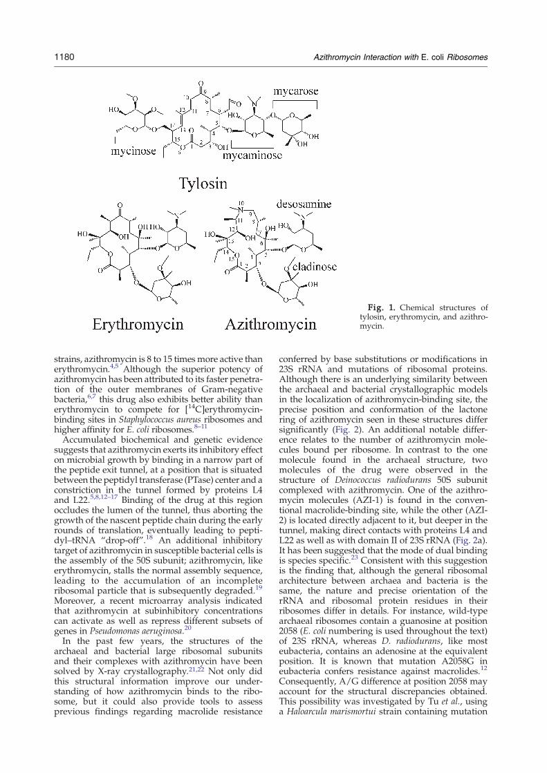
Fig. 1. Chemical structures oftylosin, erythromycin, and azithro-mycin.
1180 Azithromycin Interaction with E. coli Ribosomes
strains, azithromycin is 8 to 15 times more active thanerythromycin.4,5 Although the superior potency ofazithromycin has been attributed to its faster penetra-tion of the outer membranes of Gram-negativebacteria,6,7 this drug also exhibits better ability thanerythromycin to compete for [14C]erythromycin-binding sites in Staphylococcus aureus ribosomes andhigher affinity for E. coli ribosomes.8–11
Accumulated biochemical and genetic evidencesuggests that azithromycin exerts its inhibitory effecton microbial growth by binding in a narrow part ofthe peptide exit tunnel, at a position that is situatedbetween the peptidyl transferase (PTase) center and aconstriction in the tunnel formed by proteins L4and L22.5,8,12–17 Binding of the drug at this regionoccludes the lumen of the tunnel, thus aborting thegrowth of the nascent peptide chain during the earlyrounds of translation, eventually leading to pepti-dyl–tRNA “drop-off”.18 An additional inhibitorytarget of azithromycin in susceptible bacterial cells isthe assembly of the 50S subunit; azithromycin, likeerythromycin, stalls the normal assembly sequence,leading to the accumulation of an incompleteribosomal particle that is subsequently degraded.19
Moreover, a recent microarray analysis indicatedthat azithromycin at subinhibitory concentrationscan activate as well as repress different subsets ofgenes in Pseudomonas aeruginosa.20
In the past few years, the structures of thearchaeal and bacterial large ribosomal subunitsand their complexes with azithromycin have beensolved by X-ray crystallography.21,22 Not only didthis structural information improve our under-standing of how azithromycin binds to the ribo-some, but it could also provide tools to assessprevious findings regarding macrolide resistance
conferred by base substitutions or modifications in23S rRNA and mutations of ribosomal proteins.Although there is an underlying similarity betweenthe archaeal and bacterial crystallographic modelsin the localization of azithromycin-binding site, theprecise position and conformation of the lactonering of azithromycin seen in these structures differsignificantly (Fig. 2). An additional notable differ-ence relates to the number of azithromycin mole-cules bound per ribosome. In contrast to the onemolecule found in the archaeal structure, twomolecules of the drug were observed in thestructure of Deinococcus radiodurans 50S subunitcomplexed with azithromycin. One of the azithro-mycin molecules (AZI-1) is found in the conven-tional macrolide-binding site, while the other (AZI-2) is located directly adjacent to it, but deeper in thetunnel, making direct contacts with proteins L4 andL22 as well as with domain II of 23S rRNA (Fig. 2a).It has been suggested that the mode of dual bindingis species specific.23 Consistent with this suggestionis the finding that, although the general ribosomalarchitecture between archaea and bacteria is thesame, the nature and precise orientation of therRNA and ribosomal protein residues in theirribosomes differ in details. For instance, wild-typearchaeal ribosomes contain a guanosine at position2058 (E. coli numbering is used throughout the text)of 23S rRNA, whereas D. radiodurans, like mosteubacteria, contains an adenosine at the equivalentposition. It is known that mutation A2058G ineubacteria confers resistance against macrolides.12
Consequently, A/G difference at position 2058 mayaccount for the structural discrepancies obtained.This possibility was investigated by Tu et al., usinga Haloarcula marismortui strain containing mutation

Fig. 3. AcPhe–puromycin synthesis in the absence orpresence of antibiotics. Complex C prepared from E. coliribosomes reacted in buffer A for specified time intervalsat 25 °C with (○) 200 μΜ puromycin alone or with amixture containing both (●) 200 μΜ puromycin and0.5 μM azithromycin or (▴) 200 μΜ puromycin and 2 μMtylosin. The values of the percentage of the initially addedcomplex C, which is converted to product, x, have been
Fig. 2. The binding site(s) of azithromycin on the 50S ribosomal subunit. (a) Binding of two molecules of azithromycin(pink) to the D. radiodurans 50S ribosomal subunit (Protein Data Bank accession no. 1NWY). Parts of domain Vand helixH35 of 23S rRNA (light blue) aswell as parts of ribosomal proteins L4 (dark blue) and L22 (light green) are shown by ribbonrepresentation. Important nucleoside residues for macrolide binding are presented by their structures (E. coli numbering);those contributing to hydrogen bonds are connectedwith the drug by dotted lines. (b) Binding position of azithromycin onthe H. marismortui 50S ribosomal subunit (Protein Data Bank accession no. 1M1K). Labeling is the same as in (a).
1181Azithromycin Interaction with E. coli Ribosomes
G2058A.24 It was found that azithromycin binds towild-type and mutant 50S subunits of H. marismor-tui in a virtually identical way, making impossiblethe hypothesis that the variability of azithromycinbinding to bacterial and archaeal 50S subunits maybe attributed to sequence differences at position2058. However, it is worth considering the possi-bility that the two azithromycin-bound moleculesin the 50S ribosomal subunit from D. radioduransmay result from other idiosyncratic structuralcharacteristics of this bacterium23 and/or the nearin vivo ionic conditions followed during the crystalpreparation25 or reflect snapshots of azithromycinbinding at different steps of the drug bindingprocess. With respect to the latter postulation, itshould be mentioned that kinetic and NMR studieshave provided evidence that binding of azithromy-cin to E. coli ribosomes occurs in a two-step process,the first one involving the recognition and selectionof the drug by the ribosomal machinery and thesecond one being the strong interaction responsiblefor the protein synthesis inhibition.10,26,27
In the present study, chemical footprinting andkinetic analysis are applied together in a dualstrategy to investigate the entire course of azithro-mycin binding to E. coli functional ribosomes and tomonitor the effects of ionic environment on thebinding process. In the context of this work, we alsoseize the chance to construct a mechanistic modelthat explains our results as well as crystallographicand other data collected so far.
corrected by the extent of AcPhe–puromycin synthesisachieved in the absence of azithromycin and tylosin. Thetwo upper lines were drawn by linear fitting of the x and tvalues to the equation ln[100/(100−x)]=kobs∙t, where kobsrepresents the observed first-order rate constant of thepuromycin reaction. The lower line was drawn from thecalculated ln[100/(100−x)] values at each time point,using the B-Spline function of the Microcal Origin 6.1program (Microcal Software, Inc). Puro, puromycin; Azi,azithromycin; Tyl, tylosin.
Results
Azithromycin is a slow binding ligand of E. coliribosomes
Sixteen-membered macrolides, such as tylosin,possess a disaccharide branch at position C5 of the
lactone ring (Fig. 1), from which extending towardsthe PTase center perturbs the relative positioning ofthe bound tRNA substrates. Consequently, macro-lides of this type inhibit the PTase activity in mostreference cell-free systems.28–30 In contrast, azithro-mycin has a single desosamine moiety at the C5position, which is too short to reach the PTase center.Consistently, azithromycin fails to inhibit AcPhe–puromycin formation (Fig. 3). Nevertheless, pre-vious studies have demonstrated that azithromycin

Scheme 1. Competition between tylosin (T) andazithromycin (A) for common binding sites in ribosomalcomplex C.
1182 Azithromycin Interaction with E. coli Ribosomes
competes with tylosin for binding to complex C[post state ribosomes programmed with poly(U)and bearing AcPhe–tRNA and deacylated tRNA atthe P- and E-site, respectively].8,10 This is also shownin Fig. 4; when complex C is added in a solutioncontaining 4 μM tylosin and azithromycin, aprogressive decrease in the inactivation of complexC by tylosin occurs as the concentration of azithro-mycin increases. At high concentrations of azithro-mycin (N2 μM), the inactivation of complex C iscompletely reversed. Another feature of the compe-tition mechanism is that the initial slopes of theinactivation plots vary as a function of azithromycinconcentration. The overall kinetic pattern is remi-niscent of that observed in a recent study oferythromycin binding performed under the sameionic conditions (4.5 mMMg2+, 150 mMNH4
+)30 andis in agreement with previous kinetic studies ofazithromycin carried out under conventional ionicconditions (10 mMMg2+, 100 mMNH4
+).10 Thus, it isreasonable to employ a similar kinetic model for thecompetition of tylosin (T) binding by azithromycin(A), as represented by Scheme 1. The values of kon,A,koff,A, KA, and the overall dissociation constant KA
⁎,estimated by nonlinear regression fitting of theobtained data to Eq. (7) (see Materials andMethods),are shown in Table 1.Previous genetic studies have indicated that
mutations in hairpin 35 of domain II and in thecentral loop of domain V of the E. coli 23S rRNAconfer resistance to low concentrations of ery-thromycin.5,31 To check if two of these mutations,U754A and U2609C, also influence the kinetics ofazithromycin binding, the affinity of mutant ribo-somes for azithromycin was estimated in parallelexperiments. As shown in Table 1, mutation U754A
Fig. 4. Inactivation of complex C by tylosin in theabsence or presence of azithromycin. Complex C preparedfrom E. coli ribosomes reacted in buffer A with (×) 4 μMtylosin alone or with a mixture containing both 4 μMtylosin and azithromycin at (○) 25 nM, (●) 50 nM, (▵)100 nM, (▴) 200 nM, (□) 500 nM, and (■) 2000 nM. Afterremoving the excess of each antibiotic, the percentage ofcomplex C input remaining active, x, was titrated with2 mM puromycin. The curves were drawn by nonlinearfitting of the x and t values to Eq. (7). The kon,T and KTvalues were measured via experiments performed in thepresence of tylosin alone and set in the above computersimulation as constant factors. The tangent at zero time ofthe nonlinear plot obtained at 4 μM tylosin and 2 μMazithromycin is indicated by a broken line.
hardly affects the affinity of azithromycin, at boththe initial and final positions of its accommodationwithin complex C. The small alterations in the KA
⁎
and kon,A/koff,A values are not statistically signifi-cant (Pb0.05). In contrast, U2609C mutationincreases the isomerization constant kon,A/koff,A by55%. As a result, the value of the overall dissocia-tion constant for azithromycin, KA⁎, undergoes a40% reduction, reflecting an increase in affinity ofthe drug for the mutant ribosome. By repeating thekinetic analysis of azithromycin binding to wild-type ribosomes in the presence of 100 μM spermine,we observed that the kon,A value is decreased by63%, resulting in a parallel reduction of the iso-merization constant. Consistently, the KA
⁎ valueundergoes a twofold enhancement. Similar altera-tions were recorded by using 50 μM spermine and2 mM spermidine (optimized Mg2+/NH4
+/polya-mine system32), instead of spermine alone (Table 1).Thus, the presence of polyamines leads to anoverall decrease in azithromycin binding.
One molecule of azithromycin binds per E. coliribosome
Scheme 1 assumes that only one molecule ofazithromycin binds per E. coli ribosome at the sametime. To verify this assumption, we performed twoseries of experiments. In the first, C⁎A complex wasexposed to 4 μM tylosin for various time intervals,and its activity, after removing the excess tylosin,was titrated with puromycin. As shown in Fig. 5a,the data of complex C regeneration from complexC⁎A give a simple first-order time plot, which isconsistent with one molecule of azithromycin boundper E. coli ribosome. An alternative explanationcould be that only one molecule of azithromycincompetes with tylosin for a common binding in theribosome. In any case, koff,A is more than two orders
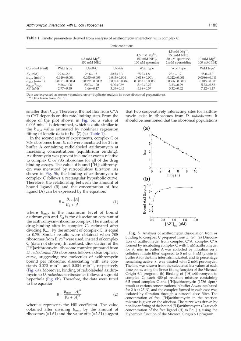
Table 1. Kinetic parameters derived from analysis of azithromycin interaction with complex C
Ionic conditions
4.5 mM Mg2+,150 mM NH4
+
4.5 mM Mg2+,150 mM NH4
+,100 μM spermine
4.5 mM Mg2+,150 mM NH4
+,50 μM spermine,2 mM spermidine
10 mM Mg2+,100 mM NH4
+
Constant (unit) Wild type U2609C U754A Wild type Wild type Wild typea
KA (nM) 29.6±2.6 26.6±1.5 30.5±2.3 25.0±1.8 23.4±1.9 48.0±5.0kon,A (min−1) 0.049±0.004 0.055±0.003 0.045±0.004 0.018±0.001 0.022±0.001 0.0086±0.011koff,A (min−1) 0.0051±0.0004 0.0037±0.0002 0.005±0.0004 0.0053±0.0003 0.0066±0.0005 0.015±0.001kon,A/koff,A 9.66±1.08 15.03±1.00 9.00±0.96 3.40±0.27 3.33±0.29 5.73±0.82KA⁎ (nM) 2.77±0.38 1.66±0.17 3.05±0.43 5.68±0.57 5.32±0.62 7.12±1.17
Data are expressed as means±standard error (duplicate analysis in three ribosomal preparations).a Data taken from Ref. 10.
Fig. 5. Analysis of azithromycin dissociation from orbinding to complex C prepared from E. coli. (a) Dissocia-tion of azithromycin from complex C⁎A; complex C⁎Aformed by incubating complex C with 1 μΜ azithromycinfor 80 min in buffer A was collected by filtration on acellulose nitrate filter, exposed to 5 ml of 4 μΜ tylosin inbuffer A for the time intervals indicated, and its percentageremaining active, x, was titrated with 2 mM puromycin.The line was drawn from the calculated lnx values at eachtime point, using the linear fitting function of the MicrocalOrigin 6.1 program. (b) Binding of [3H]azithromycin tocomplex C; each 400-μl reaction mixture containing6.5 pmol complex C and [3H]azithromycin (1786 dpm/pmol) at various concentrations in buffer Awas incubatedfor 2 h at 25 °C, and the complex formed in each case wasisolated by filtration through a nitrocellulose filter. Theconcentration of free [3H]azithromycin in the reactionmixture is given on the abscissa. The curve was drawn bynonlinear fitting of the bound [3H]azithromycin (B) at eachconcentration of the free ligand (A) to Eq. (1), using theHyberbola function of the Microcal Origin 6.1 program.
1183Azithromycin Interaction with E. coli Ribosomes
smaller than kon,T. Therefore, the net flux from C⁎Ato C⁎T depends on this rate-limiting step. From theslope of the plot shown in Fig. 5a, a value of0.005 min−1 is determined, which is quite similar tothe koff,A value estimated by nonlinear regressionfitting of kinetic data to Eq. (7) (see Table 1).In the second series of experiments, complex C or
70S ribosomes from E. coli were incubated for 2 h inbuffer A containing radiolabeled azithromycin atincreasing concentrations (equilibrium binding).Azithromycin was present in a molar excess relativeto complex C or 70S ribosomes for all of the drugbinding assays. The value of bound [3H]azithromy-cin was measured by nitrocellulose filtration. Asshown in Fig. 5b, the binding of azithromycin tocomplex C follows a rectangular hyperbolic curve.Therefore, the relationship between the amount ofbound ligand (B) and the concentration of freeligand (A) can be expressed by the equation:
B =Bmax A½ �Kd + A½ � ð1Þ
where Bmax is the maximum level of boundazithromycin and Kd is the dissociation constant ofthe azithromycin–ribosome complex. The number ofdrug-binding sites in complex C, estimated afterdividing Bmax by the amount of complex C, is equalto 0.75. Similar results were obtained when 70Sribosomes from E. coliwere used, instead of complexC (data not shown). In contrast, dissociation of the[3H]azithromycin–ribosome complex prepared fromD. radiodurans 70S ribosomes follows a clear biphasiccurve, suggesting two molecules of azithromycinbound per ribosome, dissociating with rate con-stants 0.020 min−1 and 0.004 min−1, respectively(Fig. 6a). Moreover, binding of radiolabeled azithro-mycin toD. radiodurans ribosomes follows a sigmoidhyperbola (Fig. 6b). Therefore, the data were fittedto the equation
B =Bmax � A½ �n
Kd + A½ �n ð2Þ
where n represents the Hill coefficient. The valueobtained after dividing Bmax by the amount ofribosomes (=1.61) and the value of n (=2.31) suggest
that two cooperatively interacting sites for azithro-mycin exist in ribosomes from D. radiodurans. Itshould be mentioned that the ribosomal populations

Fig. 6. Analysis of [3H]azithromycin dissociation fromor binding to 70S ribosomes from D. radiodurans. (a)Dissociation of the azithromycin–ribosome complex; 70Sribosomes (6.5 pmol) were incubated with 30 μl of buffer Bcontaining 1 μΜ [3H]azithromycin for 80 min at 25 °C,filtered through a nitrocellulose filter, and then exposed to5 ml of buffer B at 25 °C for the indicated time intervals.After washing with buffer B, the trapped radioactivity onthe filters was determined by scintillation counting. Thetwo linear portions of the biphasic curve were drawn fromthe calculated values of ln[AZI]bound at each time, using thelinear fitting function of the Microcal Origin 6.1 program.(b) Binding of [3H]azithromycin to 70S ribosomes; bindingwas carried out in buffer B (400 μl) containing 6.5 pmol 70Sribosomes and [3H]azithromycin (1786 dpm/pmol) atvarious concentrations. The mixture was incubated at25 °C for 2 h (equilibrium binding) and the amount ofbound azithromycin was measured by nitrocellulosefiltration and radioactivity counting. The concentration offree [3H]azithromycin in the reaction mixture is given onthe abscissa. The curve was drawn by nonlinear fitting ofthe bound [3H]azithromycin (B) at each concentration ofthe free ligand (A) to Eq. (2), using the Hill hyperbolafunction of the Microcal Origin 6.1 program.
1184 Azithromycin Interaction with E. coli Ribosomes
isolated from E. coli and D. radiodurans were 75%and 84.3% capable of binding, respectively, as foundby titration with [14C]erythromycin. Thus, correct-ing for the binding efficiency, the number of drugmolecules bound per E. coli and D. radioduransribosomes becomes 1 and 1.9, respectively.
Footprinting of azithromycin–ribosomecomplexes
To identify the individual drug–ribosome interac-tions occurring in the encounter complex CA andthe final complex C⁎A, we followed a kinetic-footprinting approach, recently applied in studyingthe interactions of erythromycin and tylosin with E.
coli ribosomes.30 Footprinting of the CA complexwas achieved by incubating complex C preparedfrom E. coli ribosomes with azithromycin in buffer Afor 10 s, the latter at concentration 50×KA, thusensuring that most of the binding sites are occupied.Because the equilibrium C+A⇄CA is establishedrapidly, while the isomerization reaction proceedsslowly (t1/2=13 min), the product during this timeinterval mainly consists of complex CA (N95%). Theformed CA complex was then probed with chemicalreagents that attack nucleosides of 23S rRNA thatare neither occupied by the drug nor participating insecondary or tertiary interactions. The incubationtime of complex C with azithromycin was extendedto 100 min to footprint the C⁎A complex. Becausethe isomerization constant is higher than 3 (Table 1),it is reasonable to presume that at the end of thistime interval (≈8× t1/2), most of the added complexC should be in the form of complex C⁎A.Representative autoradiograms obtained by pri-
mer extension analysis are shown in Figs. 7 and 8.The final modification results are summarized inTable 2. Azithromycin at the CA binding state and inthe absence of polyamines strongly protects A2058and A2059 from dimethyl sulfate (DMS) and G2505from kethoxal modification, while moderately redu-cing the accessibility of U2609 to 1-cyclohexyl-3-(2-morpholinoethyl) carbodiimide metho-p-toluene(CMCT). A weak protection effect is also observedon C2611, although this residue is not normallyaccessible to DMS, due to its base pairing withG2057.33 Interestingly, in the presence of polya-mines, the reactivity of C2611 against DMS is lost,both in ribosome–azithromycin complexes and incomplex C alone. Consequently, it is reasonable tosuppose that at low Mg2+ concentrations, polya-mines reestablish the C2611/G2057 base pair. In theC⁎A binding state, new protection effects appear onA788, U790, and U2586. Also, protection of U2609becomes stronger, whereas protections at A2058 andG2505 soften and the accessibility of A752 is slightlyenhanced. The presence of spermine hardly affectsthe protection pattern of complex CA. However, incomplex C⁎A, it stimulates the accessibility of allnucleotides implicated in the binding of azithromy-cin, in particular of U2609 and those situated in helix35 of 23S rRNA. The increased accessibility indicatesreduced binding of the drug, in agreement with theincreased dissociation constant KA
⁎ when polya-mines are present.
Discussion
High-resolution crystallographic structures ofribosomal particles and their complexes with azi-thromycin have elucidated basic concepts in azithro-mycin binding mode at the molecular level.21,22,24
However, a disadvantage of crystallography is thatthe structures offer only a snapshot of stably boundazithromycin position on the ribosome, where-as the complete picture of the drug binding pro-cess may be significantly more complex. Kinetic

Fig. 7. Alterations in the chemical reactivity of nucleosides in domain II of 23S rRNA, caused by azithromycin bindingto complex C prepared from E. coli ribosomes. Complex C was incubated in the absence or presence of azithromycin inbuffer A (lanes 1 to 4) or in the same buffer also containing 100 μM spermine (lanes 5 to 8). The resulting complexes werethen probed with DMS or CMCT. Modification sites were detected by primer extension analysis. U, A, G, and C denotedideoxy sequencing lanes. Lanes 1 and 5, unmodified complex Cwith no drug present; lanes 2 and 6, complex C probed inthe absence of azithromycin; lanes 3 and 7, complex C incubated with azithromycin for 10 s and then probed; lanes 4 and8, complex C incubated with azithromycin for 100 min and then probed. Numbering of nucleotides for the sequencinglanes is indicated at the left, while nucleotides with altered reactivity against chemical probes as well as reference bandsare indicated by arrows. AZI, azithromycin.
1185Azithromycin Interaction with E. coli Ribosomes
analysis and chemical footprinting used in thepresent work provide additional tools to (i) inves-tigate the entire course of azithromycin binding toE. coli ribosomes and (ii) rationalize the availablecrystallographic, genetic, and other data collectedso far.According to our kinetic results, the overall
association rate constant of azithromycin bindingto complex C, (kon,A+koff,A)/KA, equals 3×10⁴ M−1
s−1, a value that is two orders below the upper limitset for the characterization of a drug as a slowbinding ligand.34 Corroborative evidence that azi-thromycin is a slow binder is also derived from thepreincubation effect, namely, the strengthening ofthe azithromycin competition potency against tylo-sin when complex C is preincubated with azithro-mycin before the addition of tylosin (data notshown). Moreover, the ratio kon,A/koff,A is muchhigher than 1, regardless of ionic conditions used, afact implying that azithromycin is a slowly rever-sible ligand as well. Another important character-istic of the azithromycin kinetic behavior is that theinitial slope of each inactivation plot in Fig. 4decreases as the concentration of azithromycinincreases. This is consistent with a two-step mechan-ism of binding, since otherwise, the initial slopeshould be independent of azithromycin concentra-tion. According to this mode, the first step equili-
brates rapidly, while the second one representingthe accommodation of azithromycin at its finalposition is established slowly, within the minutetimescale (t1/2=13 min). A two-step mechanism hasalso been suggested by transferred nuclear Over-hauser effect studies,26 as well as by previous kineticstudies.10 Comparing the results presented herewith previous studies performed at 10 mM Mg2+,we conclude that a decrease of Mg2+ concentrationfrom 10 to 4.5 mM enhances azithromycin affinity atboth steps of binding. Noteworthy is the threefoldreduction in the koff,A value, suggesting that rela-tively low Mg2+ concentrations preserve complexC⁎A from disassembly. Although polyamines donot affect the initial step of binding, they obstruct theaccommodation of azithromycin at its final position,primarily by lowering the kon,A value. Our resultsemphasize the role that ionic conditions may have inthe ligand/ribosome binding free energy. Compar-ing azithromycin with erythromycin binding toribosomes at optimal ionic conditions for efficientand accurate protein synthesis (4.5 mM Mg2+,150 mM NH4
+, 50 μM spermine, and 2 mMspermidine),32 we realize that azithromycin exhibitsa higher affinity for complex C (KE⁎/KA
⁎=19.9) andforms a more stable final complex with ribosomes(koff,E/koff,A=14.8). Although the efficacy of a drugdepends on many factors, our data at least partially
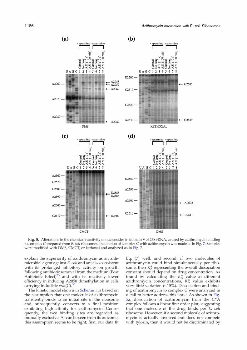
Fig. 8. Alterations in the chemical reactivity of nucleosides in domain Vof 23S rRNA, caused by azithromycin bindingto complex C prepared from E. coli ribosomes. Incubation of complex C with azithromycin was made as in Fig. 7. Sampleswere modified with DMS, CMCT, or kethoxal and analyzed as in Fig. 7.
1186 Azithromycin Interaction with E. coli Ribosomes
explain the superiority of azithromycin as an anti-microbial agent against E. coli and are also consistentwith its prolonged inhibitory activity on growthfollowing antibiotic removal from the medium (PostAntibiotic Effect)35 and with its relatively lowerefficiency in inducing A2058 dimethylation in cellscarrying inducible erm(C).36
The kinetic model shown in Scheme 1 is based onthe assumption that one molecule of azithromycintransiently binds to an initial site in the ribosomeand, subsequently, converts to a final positionexhibiting high affinity for azithromycin. Conse-quently, the two binding sites are regarded asmutually exclusive. As can be seen from its outcome,this assumption seems to be right; first, our data fit
Eq. (7) well, and second, if two molecules ofazithromycin could bind simultaneously per ribo-some, then KA
⁎ representing the overall dissociationconstant should depend on drug concentration. Asfound by calculating the KA
⁎ value at differentazithromycin concentrations, KA
⁎ value exhibitsvery little variation (b15%). Dissociation and bind-ing of azithromycin to complex C were analyzed indetail to better address this issue. As shown in Fig.5a, dissociation of azithromycin from the C⁎Acomplex follows a linear first-order plot, suggestingthat one molecule of the drug binds per E. coliribosome. However, if a second molecule of azithro-mycin is actually involved but does not competewith tylosin, then it would not be discriminated by
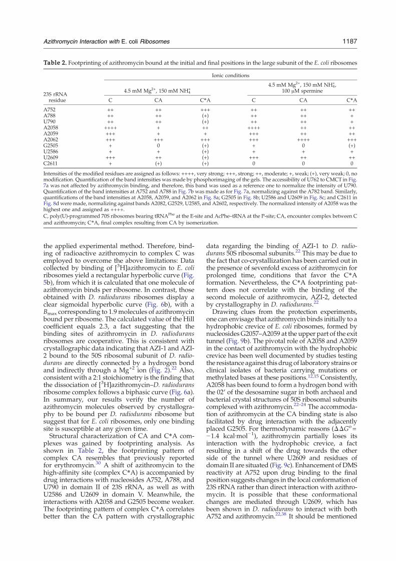
Table 2. Footprinting of azithromycin bound at the initial and final positions in the large subunit of the E. coli ribosomes
23S rRNAresidue
Ionic conditions
4.5 mM Mg2+, 150 mM NH4+
4.5 mM Mg2+, 150 mM NH4+,
100 μM spermine
C CA C⁎A C CA C⁎A
A752 ++ ++ +++ ++ ++ ++A788 ++ ++ (+) ++ ++ +U790 ++ ++ (+) ++ ++ +A2058 ++++ + ++ ++++ ++ ++A2059 +++ + + +++ ++ ++A2062 +++ +++ +++ +++ ++++ +++G2505 + 0 (+) + 0 (+)U2586 + + (+) + + +U2609 +++ ++ (+) +++ ++ ++C2611 + (+) (+) 0 0 0
Intensities of the modified residues are assigned as follows: ++++, very strong; +++, strong; ++, moderate; +, weak; (+), very weak; 0, nomodification. Quantification of the band intensities was made by phosphorimaging of the gels. The accessibility of U762 to CMCT in Fig.7a was not affected by azithromycin binding, and therefore, this band was used as a reference one to normalize the intensity of U790.Quantification of the band intensities at A752 and A788 in Fig. 7b was made as for Fig. 7a, normalizing against the A782 band. Similarly,quantifications of the band intensities at A2058, A2059, and A2062 in Fig. 8a; G2505 in Fig. 8b; U2586 and U2609 in Fig. 8c; and C2611 inFig. 8d were made, normalizing against bands A2082, G2529, U2585, and A2602, respectively. The normalized intensity of A2058 was thehighest one and assigned as ++++.C, poly(U)-programmed 70S ribosomes bearing tRNAPhe at the E-site and AcPhe–tRNA at the P-site; CA, encounter complex between Cand azithromycin; C⁎A, final complex resulting from CA by isomerization.
1187Azithromycin Interaction with E. coli Ribosomes
the applied experimental method. Therefore, bind-ing of radioactive azithromycin to complex C wasemployed to overcome the above limitations: Datacollected by binding of [3H]azithromycin to E. coliribosomes yield a rectangular hyperbolic curve (Fig.5b), from which it is calculated that one molecule ofazithromycin binds per ribosome. In contrast, thoseobtained with D. radiodurans ribosomes display aclear sigmoidal hyperbolic curve (Fig. 6b), with aBmax corresponding to 1.9molecules of azithromycinbound per ribosome. The calculated value of the Hillcoefficient equals 2.3, a fact suggesting that thebinding sites of azithromycin in D. radioduransribosomes are cooperative. This is consistent withcrystallographic data indicating that AZI-1 and AZI-2 bound to the 50S ribosomal subunit of D. radio-durans are directly connected by a hydrogen bondand indirectly through a Mg+2 ion (Fig. 2).22 Also,consistent with a 2:1 stoichiometry is the finding thatthe dissociation of [3H]azithromycin–D. radioduransribosome complex follows a biphasic curve (Fig. 6a).In summary, our results verify the number ofazithromycin molecules observed by crystallogra-phy to be bound per D. radiodurans ribosome butsuggest that for E. coli ribosomes, only one bindingsite is susceptible at any given time.Structural characterization of CA and C⁎A com-
plexes was gained by footprinting analysis. Asshown in Table 2, the footprinting pattern ofcomplex CA resembles that previously reportedfor erythromycin.30 A shift of azithromycin to thehigh-affinity site (complex C⁎A) is accompanied bydrug interactions with nucleosides A752, A788, andU790 in domain II of 23S rRNA, as well as withU2586 and U2609 in domain V. Meanwhile, theinteractions with A2058 and G2505 become weaker.The footprinting pattern of complex C⁎A correlatesbetter than the CA pattern with crystallographic
data regarding the binding of AZI-1 to D. radio-durans 50S ribosomal subunits.22 This may be due tothe fact that co-crystallization has been carried out inthe presence of sevenfold excess of azithromycin forprolonged time, conditions that favor the C⁎Aformation. Nevertheless, the C⁎A footprinting pat-tern does not correlate with the binding of thesecond molecule of azithromycin, AZI-2, detectedby crystallography in D. radiodurans.22
Drawing clues from the protection experiments,one can envisage that azithromycin binds initially to ahydrophobic crevice of E. coli ribosomes, formed bynucleosidesG2057–A2059 at the upper part of the exittunnel (Fig. 9b). The pivotal role of A2058 and A2059in the contact of azithromycin with the hydrophobiccrevice has been well documented by studies testingthe resistance against this drugof laboratory strains orclinical isolates of bacteria carrying mutations ormethylated bases at these positions.12,15 Consistently,A2058 has been found to form a hydrogen bond withthe 02′ of the desosamine sugar in both archaeal andbacterial crystal structures of 50S ribosomal subunitscomplexed with azithromycin.22–24 The accommoda-tion of azithromycin at the CA binding state is alsofacilitated by drug interaction with the adjacentlyplaced G2505. For thermodynamic reasons (ΔΔGo=−1.4 kcal∙mol−1), azithromycin partially loses itsinteraction with the hydrophobic crevice, a factresulting in a shift of the drug towards the otherside of the tunnel where U2609 and residues ofdomain II are situated (Fig. 9c). Enhancement of DMSreactivity at A752 upon drug binding to the finalposition suggests changes in the local conformation of23S rRNA rather than direct interaction with azithro-mycin. It is possible that these conformationalchanges are mediated through U2609, which hasbeen shown in D. radiodurans to interact with bothA752 and azithromycin.22,38 It should be mentioned
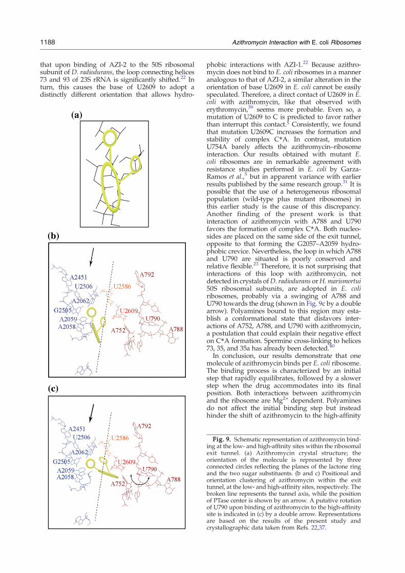
1188 Azithromycin Interaction with E. coli Ribosomes
that upon binding of AZI-2 to the 50S ribosomalsubunit of D. radiodurans, the loop connecting helices73 and 93 of 23S rRNA is significantly shifted.22 Inturn, this causes the base of U2609 to adopt adistinctly different orientation that allows hydro-
phobic interactions with AZI-1.22 Because azithro-mycin does not bind to E. coli ribosomes in a manneranalogous to that of AZI-2, a similar alteration in theorientation of base U2609 in E. coli cannot be easilyspeculated. Therefore, a direct contact of U2609 in E.coli with azithromycin, like that observed witherythromycin,39 seems more probable. Even so, amutation of U2609 to C is predicted to favor ratherthan interrupt this contact.5 Consistently, we foundthat mutation U2609C increases the formation andstability of complex C⁎A. In contrast, mutationU754A barely affects the azithromycin–ribosomeinteraction. Our results obtained with mutant E.coli ribosomes are in remarkable agreement withresistance studies performed in E. coli by Garza-Ramos et al.,5 but in apparent variance with earlierresults published by the same research group.31 It ispossible that the use of a heterogeneous ribosomalpopulation (wild-type plus mutant ribosomes) inthis earlier study is the cause of this discrepancy.Another finding of the present work is thatinteraction of azithromycin with A788 and U790favors the formation of complex C⁎A. Both nucleo-sides are placed on the same side of the exit tunnel,opposite to that forming the G2057–A2059 hydro-phobic crevice. Nevertheless, the loop in which A788and U790 are situated is poorly conserved andrelative flexible.23 Therefore, it is not surprising thatinteractions of this loop with azithromycin, notdetected in crystals ofD. radiodurans orH.marismortui50S ribosomal subunits, are adopted in E. coliribosomes, probably via a swinging of A788 andU790 towards the drug (shown in Fig. 9c by a doublearrow). Polyamines bound to this region may esta-blish a conformational state that disfavors inter-actions of A752, A788, and U790 with azithromycin,a postulation that could explain their negative effecton C⁎A formation. Spermine cross-linking to helices73, 35, and 35a has already been detected.40
In conclusion, our results demonstrate that onemolecule of azithromycin binds per E. coli ribosome.The binding process is characterized by an initialstep that rapidly equilibrates, followed by a slowerstep when the drug accommodates into its finalposition. Both interactions between azithromycinand the ribosome are Mg2+ dependent. Polyaminesdo not affect the initial binding step but insteadhinder the shift of azithromycin to the high-affinity
Fig. 9. Schematic representation of azithromycin bind-ing at the low- and high-affinity sites within the ribosomalexit tunnel. (a) Azithromycin crystal structure; theorientation of the molecule is represented by threeconnected circles reflecting the planes of the lactone ringand the two sugar substituents. (b and c) Positional andorientation clustering of azithromycin within the exittunnel, at the low- and high-affinity sites, respectively. Thebroken line represents the tunnel axis, while the positionof PTase center is shown by an arrow. A putative rotationof U790 upon binding of azithromycin to the high-affinitysite is indicated in (c) by a double arrow. Representationsare based on the results of the present study andcrystallographic data taken from Refs. 22,37.
1189Azithromycin Interaction with E. coli Ribosomes
site. Nevertheless, the general characteristics of thebinding model are conserved, regardless of the ionicconditions used. Therefore, the apparent discrepan-cies between the antibiotic binding modes obtainedfor E. coli and D. radiodurans cannot be attributed todifferences in the ionic conditions used. As pro-posed by Schlünzen et al.,22 it seems more probablethat these discrepancies predominantly result fromspecies-specific differences in the ribosomal proteinsand rRNA constituting the drug-binding sites. Thepresent work shows that the simultaneous bindingof two azithromycin molecules to D. radioduransribosomes is cooperative. This observation may beimportant for the future rational design of macro-lides that work in a synergistical fashion.
Materials and Methods
Reagents and materials
Puromycin dihydrochloride, tylosin tartrate, sperminetetrahydrochloride, spermidine trihydrochloride, tRNAPhe
from E. coli, DMS, and DMS stop solution were fromSigma-Aldrich. Kethoxal and CMCTwere purchased fromMP Biomedicals and Fluka Biochemicals, respectively.Radioactive materials L-[2,3,4,5,6-3H]phenylalanine and[α-32p]ATP were from Amersham Biosciences. AMVreverse transcriptase, dNTPs and ddNTPs were obtainedfrom Roche Diagnostics. Cold and radioactive azithromy-cin (9.9 Ci/mmol) was kindly provided by Pfizer (Groton/New London Laboratories). Cellulose nitrate filters (typeHA; 0.45 μm pore size) were from Millipore Corp.
Bacterial strains and biochemical preparations
70S ribosomes were prepared from E. coli K12 and D.radiodurans cells as described elsewhere.41 E. coli TA531cells lacking chromosomal rrn alleles but containingpKK3535 plasmids expressing wild-type or mutated 23SrRNA (U2609C or U754A) were kindly provided by Prof.A. S. Mankin, University of Illinois. These strains wereused to prepare ribosomes in studying the effect of 23SrRNA mutations on the mechanism of azithromycinbinding. Ac[3H]Phe–tRNA charged to 80% and posttran-slocation complex of poly(U)-programmed ribosomes,complex C, bearing tRNAPhe at the E-site and Ac[3H]Phe–tRNA at the P-site were prepared and purifiedaccording to Dinos et al.41 The percentage of ribosomes,active in AcPhe–tRNA binding, was more than 65%. Thisfraction was 90–95% reactive towards puromycin.
Inactivation of complex C by tylosin in the absence orpresence of azithromycin
Complex C in buffer A [100 mM Tris/HCl, pH7.2,4.5 mM Mg (CH3COO)2, 150 mM NH4Cl, and 6 mM 2-mercaptoethanol] containing 4 μM tylosin and azithro-mycin at specified concentrations was incubated at 25 °Cfor the desired time intervals. As previously proved,30 theapparent rate constant of complex C inactivation, F, isgiven by the equation
F = Fs + Fo � Fsð Þ � e�k V�t ð3Þ
where Fs and Fo are the inactivation constants at thesteady state and zero time, respectively, while k′ is theapparent equilibration rate constant for the attainmentof equilibrium between complex C and the drugs. Forthe derivation of Eq. (3), we assumed that the equilibriaC+T⇄CT and C+A⇄CA in Scheme 1 are establishedrapidly, while the isomerization reactions CT⇄C⁎T andCA⇄C⁎A are established slowly. Additionally, weassumed that the values of koff,T and koff,A might be lessthan those of kon,T and kon,A, respectively, for appreciableformation of C⁎T and C⁎A. Each of the kinetic parametersFs, Fo, and k′ in Eq. (3) is related to the concentrations ofazithromycin (A) and tylosin (T), through the followingequations:
Fo =kon;T T½ �
KT 1 + A½ �KA
� �+ T½ �
ð4Þ
Fs =kon;Tkoff;A T½ �
kVKT 1 + T½ �KT
+ A½ �KA
� � ð5Þ
kV= koff;A + kon;AA½ �KA
1 + T½ �KT
+ A½ �KA
ð6Þ
The values of kon,A, koff,A, and KA were calculated bynonlinear regression fitting of kinetic data obtained fromthree independently performed experiments to theequation
lnx = 4:6� Fs � t�Fo � Fsð Þ
kV1� e�k Vt� �
ð7Þ
that is derived from Eq. (3) by integration. The variable xin Eq. (7) is the percentage of the initially added complexC, which remains active (reactive towards puromycin)with time. The constant factor in Eq. (7) is valid byassuming that the extent of puromycin reaction used fortitration of complex C is 100%; otherwise, it is corrected.The kon,T and KT constants for tylosin, measured viaexperiments performed in the presence of tylosin alone,were in good agreement with those obtained previously30
and set in the above computer simulation as constantfactors. Once kon,A, koff,A, and KA were calculated, theoverall dissociation constant KA⁎, concerning both steps ofazithromycin binding to complex C, was estimatedthrough the equation
K4A =KA
koff;Akoff;A + kon;A
� �ð8Þ
Details on the derivation of the above equations areavailable in the Supplementary Data of Ref. 30.
Regeneration of complex C from complex C⁎A
To determine the koff,A value by an alternative way andto check whether one or more molecules of azithromycinbind per E. coli ribosome, complex C⁎A formed byincubating complex C (6.5 pmol) with 1 μΜ azithromycinin buffer A (total volume: 30 μl) for 80 min was adsorbedon a cellulose nitrate filter, washed with buffer A, and thenimmersed in 5 ml of buffer A containing 4 μM tylosin forspecified time intervals. After removing the excess oftylosin, the percentage (x) of the complex C remainingactive was determined by titration with 2 mM puromycin.
1190 Azithromycin Interaction with E. coli Ribosomes
The koff,A value was estimated from the slope of theobtained lnx versus time plot and compared with thosecalculated through Eq. (7).Kinetic analysis of the dissociation of the azithromycin–
ribosome complexwas also performed by incubatingE. coli70S ribosomes (6.5 pmol) with 1 μM [3H]azithromycin(1786 dpm/pmol) in 30 μl of buffer A for 80 min at 25 °C.The [3H]azithromycin–ribosome complex formed wasthen collected by filtration through a cellulose nitrate filter,washed with buffer A, and immersed in 5 ml of buffer A at25 °C. The amount of azithromycin remaining bound toribosomes with time was monitored by washing the filterrapidly with cold buffer A and measuring the trappedradioactivity on the filter in a scintillation spectrometer.Controls without ribosomes were included in each experi-ment and the values obtained were subtracted. The sameprocedure was followed when D. radiodurans 70S ribo-somes were used. In the later case, however, buffer B[100 mM Tris/HCl, pH7.2, 10 mM Mg (CH3COO)2,100 mM NH4Cl, and 6 mM 2-mercaptoethanol] wasused, since 70S ribosomes from D. radiodurans wereunstable in buffer A.
Binding of azithromycin to complex C or 70Sribosomes
Complex C or 70S ribosomes (6.5 pmol) from E. coli orD. radiodurans were incubated with varying concentra-tions of [3H]azithromycin (1786 dpm/pmol) in 400 μl ofbuffer A or B, respectively, at 25 °C for 2 h (equilibriumbinding). The bound drug to complex C or 70S ribosomeswas then collected on a nitrocellulose filter, washed threetimes (3 ml each) with cold buffer, and dissolved in 10 mlof Bray's solution, and its radioactivity was measured in aliquid scintillation spectrometer. Nonspecific binding wasdetermined by assessing the displacement of [3H]azithro-mycin with a 100-fold excess of unlabeled azalide.
Probing of CA and C⁎A complexes
ComplexC at 100 nMwas incubated alone orwith 1.4 μΜazithromycin (50×KA) in 100 μl of buffer C[Hepes/KOH,pH7.2, 4.5 mMMg(CH3COO)2, 150 mMNH4Cl, and 5 mMdithiothreitol] in the absence or presence of 100 μΜspermine at 25 °C, either for 10 s (CA probing) or for8× t1/2min (C⁎Aprobing). The term t1/2 represents the half-life for the attainment of equilibrium between complex Cand the drug and was calculated through the equation,
t1=2 =0:693kq
ð9Þ
where k′′ is given by the equation
kq = koff;A + kon;A �A½ �
KA + A½ � ð10Þ
Complexes CA and C⁎A were then probed with DMS,kethoxal, or CMCT, as described previously.30 Reactionswere stopped, and the rRNA was recovered from CA orC⁎A complexes by extraction with phenol, phenol–chloro-form, and chloroform. Finally, rRNA was precipitated byethanol and resuspended in Milli-Q water.
Primer extension
The modifications in 23S rRNA were monitored byprimer extension analysis using reverse transcriptase,
according to Stern et al.42 The used primers were com-plementary to the sequences 888–906, 2099–2116, 2561–2578, and 2677–2694. The cDNA products were separatedby electrophoresis on 6% polyacrylamide/7 M urea gels.The positions of stops in cDNA synthesis were identifiedby reference to dideoxy sequencing reactions, run inparallel on unmodified rRNA template. Three replicationswere carried out for each experiment. Quantification of theband intensities was made by phosphorimaging of thegels (Fujifilm, FLA-3000, Berthold; Image Quant SoftwareAIDA, Raytest). Background values in each “control” lanewere subtracted from the corresponding bands of interest.Each lane was searched for a reference nucleotide whoseaccessibility to the chemical probe is not affected byazithromycin binding. The intensity of this reference bandwas then used to normalize the intensities of the bands ofinterest in this lane (vertical comparison). The relativeintensity of the reference band between lanes was finallyused to correct for variability between lanes (horizontalcomparison).
Statistics
All data indicated in Table 2 and in the figures denotemean values obtained from three independently per-formed experiments. Data variability was determined byANOVA. Significant differences between mean valueswere measured by the F Scheffé test.
Acknowledgements
This work was supported by a grant (B115) fromthe Research Committee of the University of Patras(Programme K. Karatheodori) and the DeutscheForschungsgemeinschaft (WI3285/1-1 to D.N.W.).We wish to thank Dr. Dennis Synetos and Dr. DennisDrainas for many fruitful discussions, SpyridoulaAngelakopoulou for technical assistance, ElisavetKalpaxis for generating Fig. 9, and Dr. Alexander S.Mankin (University of Illinois) for providing us withmutated strains of E. coli. Also, we wish to thank Dr.Nikos Baibas of Pfizer Hellas A.E. and Dr. DonnieW. Owens, Compound Transfer Manager of PfizerInc., for [3H]azithromycin supply.
References
1. Fiese, E. F. & Steffen, S. H. (1990). Comparison of theacid stability of azithromycin and erythromycin A. J.Antimicrob. Chemother. 25, 39–47.
2. Lalak, N. J. & Morris, D. L. (1993). Azithromycinclinical pharmacokinetics. Clin. Pharmacokinet. 25,370–374.
3. Zuckerman, M. D. (2000). The newer macrolides:azithromycin and clarithromycin. Infect. Dis. Clin.North Am. 14, 449–462.
4. Gordillo, M. E., Singh, K. V. & Murray, B. E. (1993).In vitro activity of azithromycin against bacterialenteric pathogens. Antimicrob. Agents Chemother. 37,1203–1205.
5. Garza-Ramos, G., Xiong, L., Zhong, P. &Mankin, A. S.(2001). Binding site of macrolide antibiotics on theribosome: new resistance mutation identifies a specific
1191Azithromycin Interaction with E. coli Ribosomes
interaction of ketolides with rRNA. J. Bacteriol. 183,6898–6907.
6. Vaara, M. (1993). Outer membrane permeabilitybarrier to azithromycin, clarithromycin, and roxithro-mycin in gram-negative enteric bacteria. Antimicrob.Agents Chemother. 37, 354–356.
7. Imamura, Y., Higashiyama, Y., Tomono, K., Izumikawa,K., Yanagihara, K., Ohno, H. et al. (2005). Azithromycinexhibits bactericide effects on Pseudomonas aeruginosathrough interaction with the outer membrane. Anti-microb. Agents Chemother. 49, 1377–1380.
8. Retsema, J., Girard, A., Schelkly, W., Manousos, M.,Anderson, M., Bright, G. et al. (1987). Spectrum andmode of action of azithromycin (CP-62, 993), a new 15-membered-ring macrolide with improved potencyagainst gram-negative organisms. Antimicrob. AgentsChemother. 31, 1939–1947.
9. Goldman, R. C., Fesik, S. W. & Doran, C. C. (1990).Role of protonated and neutral forms of macrolides inbinding to ribosomes from Gram-positive and Gram-negative bacteria. Antimicrob. Agents Chemother. 34,426–431.
10. Dinos, G. P., Michelinaki, M. & Kalpaxis, D. L. (2001).Insights into the mechanism of azithromycin interac-tion with an Escherichia coli functional ribosomalcomplex. Mol. Pharmacol. 59, 1441–1445.
11. Yan, K., Hunt, E., Berge, J., May, E., Copeland, R. A. &Gontarek, R. R. (2005). Fluorescence polarizationmethod to characterize macrolide–ribosome interac-tions. Antimicrob. Agents Chemother. 49, 3367–3372.
12. Vester, B. & Douthwaite, S. (2001). Macrolide resis-tance conferred by base substitutions in 23S rRNA.Antimicrob. Agents Chemother. 45, 1–12.
13. Leclercq, R. & Courvalin, P. (2002). Resistance tomacrolides and related antibiotics in Streptococcuspneumoniae.Antimicrob. Agents Chemother. 46, 2727–2734.
14. Farrell, D. J., Douthwaite, S., Morrissey, I., Bakker, S.,Poehlsgaard, J., Jakobsen, L. & Felmingham, D. (2003).Macrolide resistance by ribosomal mutation in clinicalisolates of Streptococcus pneumoniae from the PRO-TECT 1999–2000 study. Antimicrob. Agents Chemother.47, 1777–1783.
15. Katz, L. & Ashley, G. W. (2005). Translation andprotein synthesis: macrolides. Chem. Rev. 105, 499–527.
16. Pfister, P., Corti, N., Hobbie, S., Bruell, C., Zarivach,R., Yonath, A. & Böttger, E. C. (2005). 23S rRNAbase pair 2057–2611 determines ketolide susceptibil-ity and fitness cost of the macrolide resistancemutation 2058A→G. Proc. Natl Acad. Sci. USA, 102,5180–5185.
17. Pereyre, S., Renaudin, H., Charron, A., Bébéar, C. &Bébéar, C. M. (2006). Emergence of a 23S rRNAmutation inMycoplasma hominis associated with a lossof the intrinsic resistance to erythromycin andazithromycin. J. Antimicrob. Chemother. 57, 753–756.
18. Lovmar, M., Tenson, T. & Ehrenberg, M. (2004).Kinetics of macrolide action: the josamycin anderythromycin cases. J. Biol. Chem. 279, 53506–53515.
19. Chittum, H. S. & Champney, W. S. (1995). Erythro-mycin inhibits the assembly of the large ribosomalsubunit in growing Escherichia coli cells. Curr. Micro-biol. 30, 273–279.
20. Nalca, Y., Jänsch, L., Bredenbruch, F., Geffers, R., Buer,J. & Häussler, S. (2006). Quorum-sensing antagonisticactivities of azithromycin in Pseudomonas aeruginosaPAO1: a global approach. Antimicrob. Agents Che-mother. 50, 1680–1688.
21. Hansen, J. L., Ippolito, J. A., Ban, N., Nissen, P., Moore,P. B. & Steitz, T. A. (2002). The structures of four
macrolide antibiotics bound to the large ribosomalsubunit. Mol. Cell, 10, 117–128.
22. Schlünzen, F., Harms, J. M., Franceschi, F., Hansen,H.A. S., Bartels, H., Zarivach, R. & Yonath, A. (2003).Structural basis for the antibiotic activity of ketolidesand azalides. Structure, 11, 329–338.
23. Wilson, D. N., Harms, J. M., Nierhaus, K. H.,Schlünzen, F. & Fucini, P. (2005). Species-specificantibiotic–ribosome interactions: implications fordrug development. Biol. Chem. 386, 1239–1252.
24. Tu, D., Blaha, G., Moore, P. B. & Steitz, J. A. (2005).Structures of MLSBK antibiotics bound to mutatedlarge ribosomal subunits provide a structural expla-nation for resistance. Cell, 121, 257–270.
25. Berisio, R., Corti, N., Pfister, P., Yonath, A. & Böttger,E. C. (2006). 23S rRNA 2058A→G alteration mediatesketolide resistance in combination with deletion inL22. Antimicrob. Agents Chemother. 50, 3816–3823.
26. Bertho, G., Gharbi-Benarous, J., Delaforge, M. &Girault, J. P. (1998). Transferred nuclear Overhausereffect study of macrolide–ribosome interactions:correlation between antibiotic activities and boundconformations. Bioorg. Med. Chem. 6, 209–221.
27. Novak, P., Tatić, I., Tepeš, P., Koštrum, S. & Barber, J.(2006). Systematic approach to understanding macro-lide–ribosome interactions: NMR and modeling stu-dies of oleandomycin and its derivatives. J. Phys. Chem.A, 110, 572–579.
28. Dinos, G., Synetos, D. & Coutsogeorgopoulos, C.(1993). Interaction between the antibiotic spiramycinand a ribosomal complex active in peptide bondformation. Biochemistry, 32, 10638–10647.
29. Poulsen, S.M., Kofoed, C. & Vester, B. (2000). Inhibitionof the ribosomal peptidyl transferase reaction by themycarose moiety of the antibiotics carbomycin, spir-amycin and tylosin. J. Mol. Biol. 304, 471–481.
30. Petropoulos, A. D., Kouvela, E. C., Dinos, G. P. &Kalpaxis, D. L. (2008). Step-wise binding of tylosinand erythromycin to Escherichia coli ribosomes, char-acterized by kinetic and footprinting analysis. J. Biol.Chem. 283, 4756–4765.
31. Xiong, L., Shah, S., Mauvais, P. & Mankin, A. S.(1999). A ketolide resistant mutation in domain IIof 23S rRNA reveals the proximity of hairpin 35 tothe peptidyl transferase centre. Mol. Microbiol. 31,633–639.
32. Szaflarski, W., Vesper, O., Teraoka, Y., Phitta, B.,Wilson, D. N. & Nierhaus, K. H. (2008). New featuresof the ribosome and ribosomal inhibitors: non enzy-matic recycling, misreading and back-translocation. J.Mol. Biol. 380, 193–205.
33. Egebjerg, J., Larsen, N. & Garrett, R. A. (1990).Structural map of 23S rRNA. In The Ribosome:Structure, Function and Evolution (Dahlberg, A., Hill,W. E., Garrett, R. A., Moore, P. B., Schlessinger, D. &Warner, J. R., eds), pp. 168–179, American Society forMicrobiology, Washington, DC.
34. Morrison, J. F. &Walsh, C. T. (1988). The behavior andsignificance of slow-binding enzyme inhibitors. Adv.Enzymol. Relat. Areas Mol. Biol. 61, 201–301.
35. Fuentes, F., Izquierdo, J., Martin, M. M., Gomez-Lus,M. L. & Prieto, J. (1998). Postantibiotic and sub-MICeffects of azithromycin and isepamicin against Sta-phylococcus aureus and Escherichia coli. Antimicrob.Agents Chemother. 42, 414–418.
36. Bailey, M., Chettiath, T. & Mankin, A. S. (2008).Induction of erm(C) expression by noninducingantibiotics. Antimicrob. Agents Chemother. 52, 866–874.
37. Schuwirth, B. S., Borovinskaya, M. A., Hau, C. W.,
1192 Azithromycin Interaction with E. coli Ribosomes
Zhang,W., Vila-Sanjurjo, A., Holton, J. M. & Cate, J. H.(2005). Structures of the bacterial ribosome at 3.5 Åresolution. Science, 310, 827–834.
38. Davydova, N., Streltsov, V., Wilce, M., Liljas, A. &Garber, M. (2002). L22 ribosomal protein and effect ofits mutation on ribosome resistance to erythromycin.J. Mol. Biol. 322, 635–644.
39. Schlünzen, F., Zarivach, R., Harms, J., Bashan, A.,Tocilj, A., Albrecht, R. et al. (2001). Structural basis forthe interaction of antibiotics with the peptidyltransferase centre in eubacteria. Nature, 413, 814–821.
40. Xaplanteri, M. A., Petropoulos, A. D., Dinos, G. P. &Kalpaxis, D. L. (2005). Localization of spermine
binding sites in 23S rRNA by photoaffinity labeling:parsing the spermine contribution to ribosomal 50Ssubunit functions. Nucleic Acids Res. 33, 2792–2805.
41. Dinos, G., Wilson, D. N., Teraoka, Y., Szaflarski, W.,Fucini, P., Kalpaxis, D. L. & Nierhaus, K. H. (2004).Dissecting the ribosomal inhibition mechanisms ofedeine and pactamycin: the universally conservedresidues G693 and C795 regulate P-site RNA binding.Mol. Cell, 13, 113–124.
42. Stern, S., Moazed, D. & Noller, H. F. (1988). Structuralanalysis of RNA using chemical and enzymaticprobing monitored by primer extension. MethodsEnzymol. 164, 481–489.
Paper 4
Chemistry & Biology
Article
Interplay between the Ribosomal Tunnel,Nascent Chain, and MacrolidesInfluences Drug InhibitionAgata L. Starosta,1,2 Viktoriya V. Karpenko,3 Anna V. Shishkina,4 Aleksandra Mikolajka,1,2 Natalia V. Sumbatyan,3
Frank Schluenzen,5 Galina A. Korshunova,4 Alexey A. Bogdanov,3,4,* and Daniel N. Wilson1,2,*1Gene Center and Department of Biochemistry2Center for Integrated Protein Science MunichUniversity of Munich, LMU, Munich D-81377, Germany3Department of Chemistry4A.N. Belozersky Institute of Physico-Chemical Biology,
M.V. Lomonosov Moscow State University, Moscow 119992, Russia5Deutsches Elektronen-Synchrotron, Hamburg D-22603, Germany
*Correspondence: [email protected] (A.A.B.), [email protected] (D.N.W.)
DOI 10.1016/j.chembiol.2010.04.008
SUMMARY
Accumulating evidence suggests that, during trans-lation, nascent chains can form specific interactionswith ribosomal exit tunnel to regulate translation andpromote initial folding events. The clinically impor-tant macrolide antibiotics bind within the exit tunneland inhibit translation by preventing progressionof the nascent chain and inducing peptidyl-tRNAdrop-off. Here, we have synthesized amino acid–and peptide-containing macrolides, which are usedto demonstrate that distinct amino acids and pep-tides can establish interaction with components ofthe ribosomal tunnel and enhance the ribosome-binding and inhibitory properties of the macrolidedrugs, consistent with the concept that the exittunnel is not simply a Teflon-like channel. Surpris-ingly, we find that macrolide antibiotics do not inhibittranslation of all nascent chains similarly, but ratherexhibit polypeptide-specific inhibitory effects, pro-viding a change to our general mechanistic under-standing of macrolide inhibition.
INTRODUCTION
The translational machinery represents one of the major targets
within the cell for antibiotics (reviewed by Spahn and Prescott,
1996; Wilson, 2009). The majority of antibiotics inhibit protein
synthesis by binding at the active centers on the ribosome; for
example, the tetracyclines and aminoglycosides (gentamicin)
bind within the decoding site on the small subunit, whereas the
phenylpropanoids (chloramphenicol) and oxazolidinones (line-
zolid) bind within the A-site of peptidyltransferase center (PTC)
to block peptide-bond formation. In contrast, the clinically
important class of macrolide antibiotics bind within the ribo-
somal exit tunnel, where they are thought to inhibit translation
504 Chemistry & Biology 17, 504–514, May 28, 2010 ª2010 Elsevier
by blocking elongation of the nascent polypeptide chain, which,
in turn, induces peptidyl-tRNA drop-off (Mankin, 2008; Poehls-
gaard and Douthwaite, 2003).
Macrolides are a diverse family of antibiotics that contain
a central 12–16-membered lactone ring to which various sugar
substituent’s are attached; for example, erythromycin has a
14-membered ring with C3 cladinose and C5 desosamine
sugars (Figure 1), whereas tylosin has a 16-membered ring with
a C5 mycaminose-mycarose disaccharide as well as a C23
mycinose (Figures 1 and 2). Crystal structures of macrolides
bound to the large ribosomal subunit reveal that the drugs bind
within the ribosomal exit tunnel adjacent to the peptidyltransfer-
ase center (Figures 2A and 2B) (Hansen et al., 2002; Schlunzen
et al., 2001). Surprisingly, 16-membered macrolides that have a
C6 ethyl aldehyde (with the aldehyde functional group at C20),
such as tylosin (Figure 1), form a reversible covalent bond with
the N6 of adenine 2062 (A2062; Escherichia coli numbering
used throughout) of the 23S ribosomal RNA (Figure 2B) (Hansen
et al., 2002). The lactone rings of different macrolides are gener-
ally positioned in the tunnel with very similar orientations, such
that the C5 sugars extend up the tunnel toward the PTC (Hansen
et al., 2002; Poulsen et al., 2000; Schlunzen et al., 2001; Wilson
et al., 2005) (Figure 2B). Consistently, macrolides such as eryth-
romycin with only a single C5 sugar allow synthesis of short
oligopeptides of six to eight amino acids before peptidyl-tRNA
drop-off occurs, whereas macrolides with C5 disaccharides,
such as tylosin, allow only two to four amino acids to be synthe-
sized (Tenson et al., 2003), and can even directly inhibit the pep-
tidyltransferase reaction (Karahalios et al., 2006).
It should be noted, however, that the length of the pep-
tide synthesized before peptidyl-tRNA drop-off appears to be
dependent on the sequence being translated (Tenson et al.,
2003). Translation of short nascent pentapeptides of defined
sequence can lead to dissociation of macrolide antibiotics from
the ribosome and thus confer macrolide resistance (Lovmar
et al., 2006; Tenson et al., 1996; Tenson and Mankin, 2001).
Moreover, different pentapeptide sequences can confer resis-
tance to distinct macrolide members, suggesting a direct and
discriminating interaction between the nascent chain and the
Ltd All rights reserved
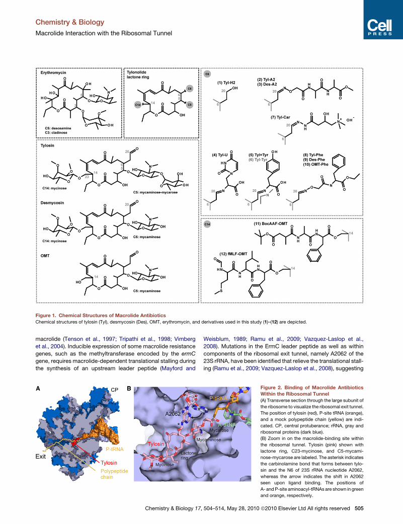
Figure 1. Chemical Structures of Macrolide Antibiotics
Chemical structures of tylosin (Tyl), desmycosin (Des), OMT, erythromycin, and derivatives used in this study (1)–(12) are depicted.
Chemistry & Biology
Macrolide Interaction with the Ribosomal Tunnel
macrolide (Tenson et al., 1997; Tripathi et al., 1998; Vimberg
et al., 2004). Inducible expression of some macrolide resistance
genes, such as the methyltransferase encoded by the ermC
gene, requires macrolide-dependent translational stalling during
the synthesis of an upstream leader peptide (Mayford and
Chemistry & Biology 17,
Weisblum, 1989; Ramu et al., 2009; Vazquez-Laslop et al.,
2008). Mutations in the ErmC leader peptide as well as within
components of the ribosomal exit tunnel, namely A2062 of the
23S rRNA, have been identified that relieve the translational stall-
ing (Ramu et al., 2009; Vazquez-Laslop et al., 2008), suggesting
Figure 2. Binding of Macrolide Antibiotics
Within the Ribosomal Tunnel
(A) Transverse section through the large subunit of
the ribosome to visualize the ribosomal exit tunnel.
The position of tylosin (red), P-site tRNA (orange),
and a mock polypeptide chain (yellow) are indi-
cated. CP, central protuberance; rRNA, gray and
ribosomal proteins (dark blue).
(B) Zoom in on the macrolide-binding site within
the ribosomal tunnel. Tylosin (pink) shown with
lactone ring, C23-mycinose, and C5-mycami-
nose-mycarose are labeled. The asterisk indicates
the carbinolamine bond that forms between tylo-
sin and the N6 of 23S rRNA nucleotide A2062,
whereas the arrow indicates the shift in A2062
seen upon ligand binding. The positions of
A- and P-site aminoacyl-tRNAs are shown in green
and orange, respectively.
504–514, May 28, 2010 ª2010 Elsevier Ltd All rights reserved 505
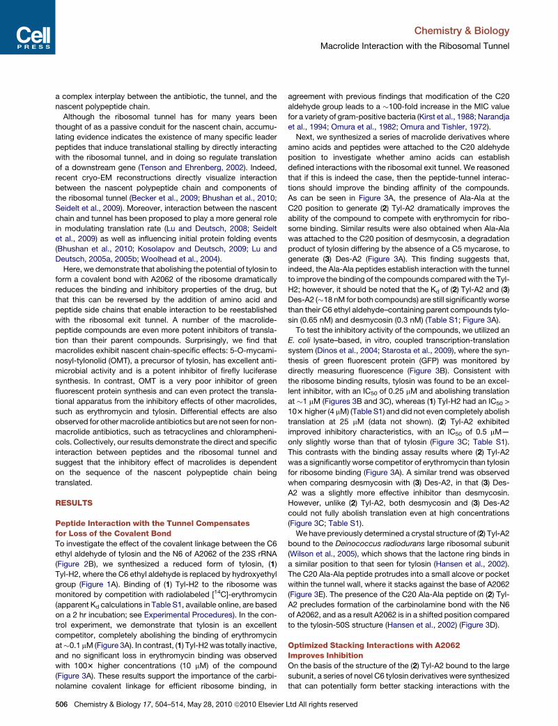
Chemistry & Biology
Macrolide Interaction with the Ribosomal Tunnel
a complex interplay between the antibiotic, the tunnel, and the
nascent polypeptide chain.
Although the ribosomal tunnel has for many years been
thought of as a passive conduit for the nascent chain, accumu-
lating evidence indicates the existence of many specific leader
peptides that induce translational stalling by directly interacting
with the ribosomal tunnel, and in doing so regulate translation
of a downstream gene (Tenson and Ehrenberg, 2002). Indeed,
recent cryo-EM reconstructions directly visualize interaction
between the nascent polypeptide chain and components of
the ribosomal tunnel (Becker et al., 2009; Bhushan et al., 2010;
Seidelt et al., 2009). Moreover, interaction between the nascent
chain and tunnel has been proposed to play a more general role
in modulating translation rate (Lu and Deutsch, 2008; Seidelt
et al., 2009) as well as influencing initial protein folding events
(Bhushan et al., 2010; Kosolapov and Deutsch, 2009; Lu and
Deutsch, 2005a, 2005b; Woolhead et al., 2004).
Here, we demonstrate that abolishing the potential of tylosin to
form a covalent bond with A2062 of the ribosome dramatically
reduces the binding and inhibitory properties of the drug, but
that this can be reversed by the addition of amino acid and
peptide side chains that enable interaction to be reestablished
with the ribosomal exit tunnel. A number of the macrolide-
peptide compounds are even more potent inhibitors of transla-
tion than their parent compounds. Surprisingly, we find that
macrolides exhibit nascent chain-specific effects: 5-O-mycami-
nosyl-tylonolid (OMT), a precursor of tylosin, has excellent anti-
microbial activity and is a potent inhibitor of firefly luciferase
synthesis. In contrast, OMT is a very poor inhibitor of green
fluorescent protein synthesis and can even protect the transla-
tional apparatus from the inhibitory effects of other macrolides,
such as erythromycin and tylosin. Differential effects are also
observed for other macrolide antibiotics but are not seen for non-
macrolide antibiotics, such as tetracyclines and chlorampheni-
cols. Collectively, our results demonstrate the direct and specific
interaction between peptides and the ribosomal tunnel and
suggest that the inhibitory effect of macrolides is dependent
on the sequence of the nascent polypeptide chain being
translated.
RESULTS
Peptide Interaction with the Tunnel Compensatesfor Loss of the Covalent BondTo investigate the effect of the covalent linkage between the C6
ethyl aldehyde of tylosin and the N6 of A2062 of the 23S rRNA
(Figure 2B), we synthesized a reduced form of tylosin, (1)
Tyl-H2, where the C6 ethyl aldehyde is replaced by hydroxyethyl
group (Figure 1A). Binding of (1) Tyl-H2 to the ribosome was
monitored by competition with radiolabeled [14C]-erythromycin
(apparent Kd calculations in Table S1, available online, are based
on a 2 hr incubation; see Experimental Procedures). In the con-
trol experiment, we demonstrate that tylosin is an excellent
competitor, completely abolishing the binding of erythromycin
at�0.1 mM (Figure 3A). In contrast, (1) Tyl-H2 was totally inactive,
and no significant loss in erythromycin binding was observed
with 1003 higher concentrations (10 mM) of the compound
(Figure 3A). These results support the importance of the carbi-
nolamine covalent linkage for efficient ribosome binding, in
506 Chemistry & Biology 17, 504–514, May 28, 2010 ª2010 Elsevier
agreement with previous findings that modification of the C20
aldehyde group leads to a �100-fold increase in the MIC value
for a variety of gram-positive bacteria (Kirst et al., 1988; Narandja
et al., 1994; Omura et al., 1982; Omura and Tishler, 1972).
Next, we synthesized a series of macrolide derivatives where
amino acids and peptides were attached to the C20 aldehyde
position to investigate whether amino acids can establish
defined interactions with the ribosomal exit tunnel. We reasoned
that if this is indeed the case, then the peptide-tunnel interac-
tions should improve the binding affinity of the compounds.
As can be seen in Figure 3A, the presence of Ala-Ala at the
C20 position to generate (2) Tyl-A2 dramatically improves the
ability of the compound to compete with erythromycin for ribo-
some binding. Similar results were also obtained when Ala-Ala
was attached to the C20 position of desmycosin, a degradation
product of tylosin differing by the absence of a C5 mycarose, to
generate (3) Des-A2 (Figure 3A). This finding suggests that,
indeed, the Ala-Ala peptides establish interaction with the tunnel
to improve the binding of the compounds compared with the Tyl-
H2; however, it should be noted that the Kd of (2) Tyl-A2 and (3)
Des-A2 (�18 nM for both compounds) are still significantly worse
than their C6 ethyl aldehyde–containing parent compounds tylo-
sin (0.65 nM) and desmycosin (0.3 nM) (Table S1; Figure 3A).
To test the inhibitory activity of the compounds, we utilized an
E. coli lysate–based, in vitro, coupled transcription-translation
system (Dinos et al., 2004; Starosta et al., 2009), where the syn-
thesis of green fluorescent protein (GFP) was monitored by
directly measuring fluorescence (Figure 3B). Consistent with
the ribosome binding results, tylosin was found to be an excel-
lent inhibitor, with an IC50 of 0.25 mM and abolishing translation
at �1 mM (Figures 3B and 3C), whereas (1) Tyl-H2 had an IC50 >
103 higher (4 mM) (Table S1) and did not even completely abolish
translation at 25 mM (data not shown). (2) Tyl-A2 exhibited
improved inhibitory characteristics, with an IC50 of 0.5 mM—
only slightly worse than that of tylosin (Figure 3C; Table S1).
This contrasts with the binding assay results where (2) Tyl-A2
was a significantly worse competitor of erythromycin than tylosin
for ribosome binding (Figure 3A). A similar trend was observed
when comparing desmycosin with (3) Des-A2, in that (3) Des-
A2 was a slightly more effective inhibitor than desmycosin.
However, unlike (2) Tyl-A2, both desmycosin and (3) Des-A2
could not fully abolish translation even at high concentrations
(Figure 3C; Table S1).
We have previously determined a crystal structure of (2) Tyl-A2
bound to the Deinococcus radiodurans large ribosomal subunit
(Wilson et al., 2005), which shows that the lactone ring binds in
a similar position to that seen for tylosin (Hansen et al., 2002).
The C20 Ala-Ala peptide protrudes into a small alcove or pocket
within the tunnel wall, where it stacks against the base of A2062
(Figure 3E). The presence of the C20 Ala-Ala peptide on (2) Tyl-
A2 precludes formation of the carbinolamine bond with the N6
of A2062, and as a result A2062 is in a shifted position compared
to the tylosin-50S structure (Hansen et al., 2002) (Figure 3D).
Optimized Stacking Interactions with A2062Improves InhibitionOn the basis of the structure of the (2) Tyl-A2 bound to the large
subunit, a series of novel C6 tylosin derivatives were synthesized
that can potentially form better stacking interactions with the
Ltd All rights reserved
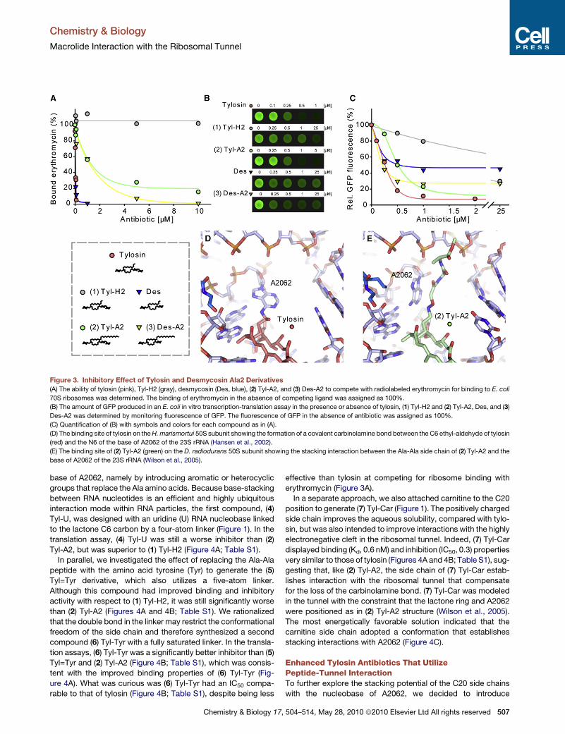
Figure 3. Inhibitory Effect of Tylosin and Desmycosin Ala2 Derivatives
(A) The ability of tylosin (pink), Tyl-H2 (gray), desmycosin (Des, blue), (2) Tyl-A2, and (3) Des-A2 to compete with radiolabeled erythromycin for binding to E. coli
70S ribosomes was determined. The binding of erythromycin in the absence of competing ligand was assigned as 100%.
(B) The amount of GFP produced in an E. coli in vitro transcription-translation assay in the presence or absence of tylosin, (1) Tyl-H2 and (2) Tyl-A2, Des, and (3)
Des-A2 was determined by monitoring fluorescence of GFP. The fluorescence of GFP in the absence of antibiotic was assigned as 100%.
(C) Quantification of (B) with symbols and colors for each compound as in (A).
(D) The binding site of tylosin on the H. marismortui 50S subunit showing the formation of a covalent carbinolamine bond between the C6 ethyl-aldehyde of tylosin
(red) and the N6 of the base of A2062 of the 23S rRNA (Hansen et al., 2002).
(E) The binding site of (2) Tyl-A2 (green) on the D. radiodurans 50S subunit showing the stacking interaction between the Ala-Ala side chain of (2) Tyl-A2 and the
base of A2062 of the 23S rRNA (Wilson et al., 2005).
Chemistry & Biology
Macrolide Interaction with the Ribosomal Tunnel
base of A2062, namely by introducing aromatic or heterocyclic
groups that replace the Ala amino acids. Because base-stacking
between RNA nucleotides is an efficient and highly ubiquitous
interaction mode within RNA particles, the first compound, (4)
Tyl-U, was designed with an uridine (U) RNA nucleobase linked
to the lactone C6 carbon by a four-atom linker (Figure 1). In the
translation assay, (4) Tyl-U was still a worse inhibitor than (2)
Tyl-A2, but was superior to (1) Tyl-H2 (Figure 4A; Table S1).
In parallel, we investigated the effect of replacing the Ala-Ala
peptide with the amino acid tyrosine (Tyr) to generate the (5)
Tyl=Tyr derivative, which also utilizes a five-atom linker.
Although this compound had improved binding and inhibitory
activity with respect to (1) Tyl-H2, it was still significantly worse
than (2) Tyl-A2 (Figures 4A and 4B; Table S1). We rationalized
that the double bond in the linker may restrict the conformational
freedom of the side chain and therefore synthesized a second
compound (6) Tyl-Tyr with a fully saturated linker. In the transla-
tion assays, (6) Tyl-Tyr was a significantly better inhibitor than (5)
Tyl=Tyr and (2) Tyl-A2 (Figure 4B; Table S1), which was consis-
tent with the improved binding properties of (6) Tyl-Tyr (Fig-
ure 4A). What was curious was (6) Tyl-Tyr had an IC50 compa-
rable to that of tylosin (Figure 4B; Table S1), despite being less
Chemistry & Biology 17,
effective than tylosin at competing for ribosome binding with
erythromycin (Figure 3A).
In a separate approach, we also attached carnitine to the C20
position to generate (7) Tyl-Car (Figure 1). The positively charged
side chain improves the aqueous solubility, compared with tylo-
sin, but was also intended to improve interactions with the highly
electronegative cleft in the ribosomal tunnel. Indeed, (7) Tyl-Car
displayed binding (Kd, 0.6 nM) and inhibition (IC50, 0.3) properties
very similar to those of tylosin (Figures 4A and 4B; Table S1), sug-
gesting that, like (2) Tyl-A2, the side chain of (7) Tyl-Car estab-
lishes interaction with the ribosomal tunnel that compensate
for the loss of the carbinolamine bond. (7) Tyl-Car was modeled
in the tunnel with the constraint that the lactone ring and A2062
were positioned as in (2) Tyl-A2 structure (Wilson et al., 2005).
The most energetically favorable solution indicated that the
carnitine side chain adopted a conformation that establishes
stacking interactions with A2062 (Figure 4C).
Enhanced Tylosin Antibiotics That UtilizePeptide-Tunnel InteractionTo further explore the stacking potential of the C20 side chains
with the nucleobase of A2062, we decided to introduce
504–514, May 28, 2010 ª2010 Elsevier Ltd All rights reserved 507

Figure 4. Stacking Interaction with A2062
(A) The ability of tylosin, (1) Tyl-H2, (4) Tyl-U, (5) Tyl=Tyr, (6) Tyl-Tyr, and (7) Tyl-Car to compete with radiolabeled erythromycin for binding to D. radiodurans 70S
ribosomes was determined. The binding of erythromycin in the absence of competing ligand was assigned as 100%.
(B) Quantification of the fluorescence of GFP produced in an E. coli in vitro transcription-translation assay in the presence or absence of tylosin, (1) Tyl-H2, (4)
Tyl-U, (5) Tyl=Tyr, (6) Tyl-Tyr, and (7) Tyl-Car. The fluorescence of GFP in the absence of antibiotic was assigned as 100%.
(C) Model based on Tyl-A2 for the binding site of (7) Tyl-Car (yellow) on the D. radiodurans 50S subunit showing the stacking interaction between the carinitine
sidechain of (7) Tyl-Car and the base of A2062 of the 23S rRNA.
(D) The ability of tylosin, desmycosin (Des), OMT, (8) Tyl-Phe, (9) Des-Phe, and (10) OMT-Phe to compete with radiolabeled erythromycin for binding to
D. radiodurans 70S ribosomes was determined. The binding of erythromycin in the absence of competing ligand was assigned as 100%.
(E) Quantification of the fluorescence of GFP produced in an E. coli in vitro transcription-translation assay in the presence or absence of tylosin, desmycosin (Des),
OMT, (8) Tyl-Phe, (9) Des-Phe, and (10) OMT-Phe. The fluorescence of GFP in the absence of antibiotic was assigned as 100%.
(F) Model based on (2) Tyl-A2 for the binding site of (8) Tyl-Phe (purple) on the D. radiodurans 50S subunit showing the stacking interaction between the linker of (8)
Tyl-Phe and the base of A2062, as well as a between the phenylalanine aromatic residue and the base of C2586 of the 23S rRNA.
Chemistry & Biology
Macrolide Interaction with the Ribosomal Tunnel
a nonpolar phenylalanine (Phe), while extending the linker, but
maintaining its overall flexibility. Binding studies indicate that
this compound, (8) Tyl-Phe, improved the ribosome bind-
ing affinity (Kd, 3.6 nM) compared to (1) Tyl-H2 (Figure 4D;
Table S1). Consistently, (8) Tyl-Phe was found to be a very
potent inhibitor of translation (Figure 4E), performing slightly
better than tylosin but significantly better than (2) Tyl-A2 and
(1) Tyl-H2 (Table S1). The Phe side chain may stabilize the bind-
ing of (8) Tyl-Phe by establishing additional stacking interac-
tions with 23S rRNA nucleotides (Figure 4F). However, the
improved ribosome binding affinity cannot be the sole explana-
tion for the increased translational inhibitory activity relative to
tylosin, because (8) Tyl-Phe was significantly less effective
compared to tylosin at competing erythromycin from the ribo-
some (Figure 4D).
508 Chemistry & Biology 17, 504–514, May 28, 2010 ª2010 Elsevier
We also synthesized (9) Des-Phe and (10) OMT-Phe, where the
Phe-containing linker side chain was attached to the C20 posi-
tion of desmycosin and OMT, respectively (Figure 1). OMT is
a precursor of tylosin, which lacks both the C5 mycarose and
the C23 mycinose (Figure 1). Our erythromycin competition
studies indicate that both (9) Des-Phe and (10) OMT-Phe were
worse competitors than their respective parent compounds (Fig-
ure 4D). The order of affinities of (8) Tyl-Phe > (9) Des-Phe > (10)
OMT-Phe is the same as for tylosin > desmycosin > OMT, and
correlates with the predicted surface areas that these
compounds bury on the ribosome; that is, more surface area
buried equals higher binding affinity (Hansen et al., 2002).
However, that each derivative had a lower binding affinity than
the parent compound emphasizes again the important influence
that the C6-ethyl aldehyde and presumably the carbinolamine
Ltd All rights reserved
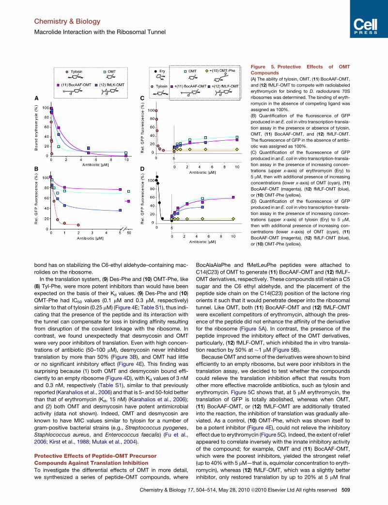
Figure 5. Protective Effects of OMT
Compounds
(A) The ability of tylosin, OMT, (11) BocAAF-OMT,
and (12) fMLF-OMT to compete with radiolabeled
erythromycin for binding to D. radiodurans 70S
ribosomes was determined. The binding of eryth-
romycin in the absence of competing ligand was
assigned as 100%.
(B) Quantification of the fluorescence of GFP
produced in an E. coli in vitro transcription-transla-
tion assay in the presence or absence of tylosin,
OMT, (11) BocAAF-OMT, and (12) fMLF-OMT.
The fluorescence of GFP in the absence of antibi-
otic was assigned as 100%.
(C) Quantification of the fluorescence of GFP
produced in an E. coli in vitro transcription-transla-
tion assay in the presence of increasing concen-
trations (upper x-axis) of erythromycin (Ery) to
5 mM, then with additional presence of increasing
concentrations (lower x-axis) of OMT (cyan), (11)
BocAAF-OMT (magenta), (12) fMLF-OMT (blue),
or (10) OMT-Phe (yellow).
(D) Quantification of the fluorescence of GFP
produced in an E. coli in vitro transcription-transla-
tion assay in the presence of increasing concen-
trations (upper x-axis) of tylosin (Ery) to 5 mM,
then with additional presence of increasing con-
centrations (lower x-axis) of OMT (cyan), (11)
BocAAF-OMT (magenta), (12) fMLF-OMT (blue),
or (10) OMT-Phe (yellow).
Chemistry & Biology
Macrolide Interaction with the Ribosomal Tunnel
bond has on stabilizing the C6-ethyl aldehyde–containing mac-
rolides on the ribosome.
In the translation system, (9) Des-Phe and (10) OMT-Phe, like
(8) Tyl-Phe, were more potent inhibitors than would have been
expected on the basis of their Kd values. (9) Des-Phe and (10)
OMT-Phe had IC50 values (0.1 mM and 0.3 mM, respectively)
similar to that of tylosin (0.25 mM) (Figure 4E; Table S1), thus indi-
cating that the presence of the peptide and its interaction with
the tunnel can compensate for loss in binding affinity resulting
from disruption of the covalent linkage with the ribosome. In
contrast, we found unexpectedly that desmycosin and OMT
were very poor inhibitors of translation. Even with high concen-
trations of antibiotic (50–100 mM), desmycosin never inhibited
translation by more than 50% (Figure 3B), and OMT had little
or no significant inhibitory effect (Figure 4E). This finding was
surprising because (1) both OMT and desmycosin bound effi-
ciently to an empty ribosome (Figure 4D), with Kd values of 3 nM
and 0.3 nM, respectively (Table S1), similar to that previously
reported (Karahalios et al., 2006) and that is 5- and 50-fold better
than that of erythromycin (Kd, 15 nM) (Karahalios et al., 2006);
and (2) both OMT and desmycosin have potent antimicrobial
activity (data not shown). Indeed, OMT and desmycosin are
known to have MIC values similar to tylosin for a number of
gram-positive bacterial strains (e.g., Streptococcus pyogenes,
Staphlococcus aureus, and Enterococcus faecalis) (Fu et al.,
2006; Kirst et al., 1988; Mutak et al., 2004).
Protective Effects of Peptide-OMT PrecursorCompounds Against Translation InhibitionTo investigate the differential effects of OMT in more detail,
we synthesized a series of peptide-OMT compounds, where
Chemistry & Biology 17,
BocAlaAlaPhe and fMetLeuPhe peptides were attached to
C14(C23) of OMT to generate (11) BocAAF-OMT and (12) fMLF-
OMT derivatives, respectively. These compounds still retain a C5
sugar and the C6 ethyl aldehyde, and the placement of the
peptide side chain on the C14(C23) position of the lactone ring
orients it such that it would penetrate deeper into the ribosomal
tunnel. Like OMT, both (11) BocAAF-OMT and (12) fMLF-OMT
were excellent competitors of erythromycin, although the pres-
ence of the peptide did not enhance the affinity of the derivative
for the ribosome (Figure 5A). In contrast, the presence of the
peptide improved the inhibitory effect of the OMT derivatives,
particularly, (12) fMLF-OMT, which inhibited the in vitro transla-
tion reaction by 50% at �1 mM (Figure 5B).
Because OMT and some of the derivatives were shown to bind
efficiently to an empty ribosome, but were poor inhibitors in the
translation assay, we decided to test whether the compounds
could relieve the translation inhibition effect that results from
other more effective macrolide antibiotics, such as tylosin and
erythromycin. Figure 5C shows that, at 5 mM erythromycin, the
translation of GFP is totally abolished, whereas when OMT,
(11) BocAAF-OMT, or (12) fMLF-OMT are additionally titrated
into the reaction, the inhibition of translation was gradually alle-
viated. As a control, (10) OMT-Phe, which was shown itself to
be a potent inhibitor (Figure 4E), could not relieve the inhibitory
effect due to erythromycin (Figure 5C). Indeed, the extent of relief
appeared to correlate inversely with the innate inhibitory activity
of the compound; for example, OMT and (11) BocAAF-OMT,
which were the poorest inhibitors, yielded the strongest relief
(up to 40% with 5 mM—that is, equimolar concentration to eryth-
romycin), whereas (12) fMLF-OMT, which was a slightly better
inhibitor, only restored translation by up to 20% at 5 mM final
504–514, May 28, 2010 ª2010 Elsevier Ltd All rights reserved 509
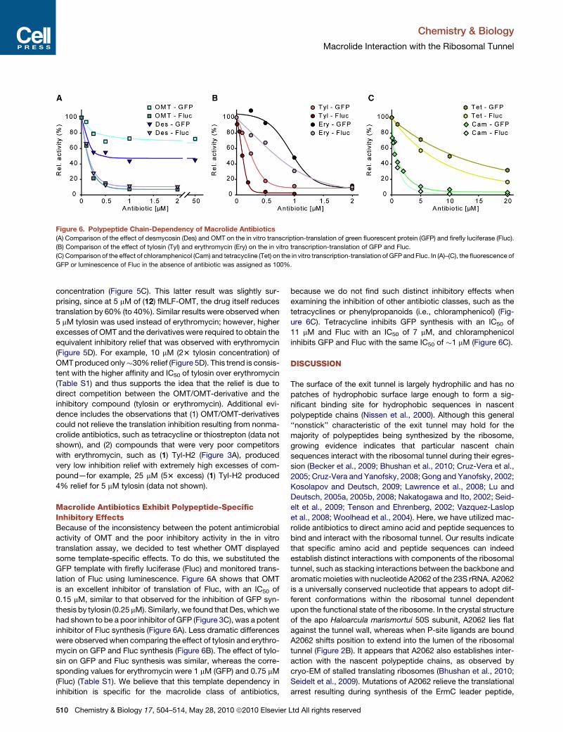
Figure 6. Polypeptide Chain-Dependency of Macrolide Antibiotics
(A) Comparison of the effect of desmycosin (Des) and OMT on the in vitro transcription-translation of green fluorescent protein (GFP) and firefly luciferase (Fluc).
(B) Comparison of the effect of tylosin (Tyl) and erythromycin (Ery) on the in vitro transcription-translation of GFP and Fluc.
(C) Comparison of the effect of chloramphenicol (Cam) and tetracycline (Tet) on the in vitro transcription-translation of GFP and Fluc. In (A)–(C), the fluorescence of
GFP or luminescence of Fluc in the absence of antibiotic was assigned as 100%.
Chemistry & Biology
Macrolide Interaction with the Ribosomal Tunnel
concentration (Figure 5C). This latter result was slightly sur-
prising, since at 5 mM of (12) fMLF-OMT, the drug itself reduces
translation by 60% (to 40%). Similar results were observed when
5 mM tylosin was used instead of erythromycin; however, higher
excesses of OMT and the derivatives were required to obtain the
equivalent inhibitory relief that was observed with erythromycin
(Figure 5D). For example, 10 mM (23 tylosin concentration) of
OMT produced only�30% relief (Figure 5D). This trend is consis-
tent with the higher affinity and IC50 of tylosin over erythromycin
(Table S1) and thus supports the idea that the relief is due to
direct competition between the OMT/OMT-derivative and the
inhibitory compound (tylosin or erythromycin). Additional evi-
dence includes the observations that (1) OMT/OMT-derivatives
could not relieve the translation inhibition resulting from nonma-
crolide antibiotics, such as tetracycline or thiostrepton (data not
shown), and (2) compounds that were very poor competitors
with erythromycin, such as (1) Tyl-H2 (Figure 3A), produced
very low inhibition relief with extremely high excesses of com-
pound—for example, 25 mM (53 excess) (1) Tyl-H2 produced
4% relief for 5 mM tylosin (data not shown).
Macrolide Antibiotics Exhibit Polypeptide-SpecificInhibitory EffectsBecause of the inconsistency between the potent antimicrobial
activity of OMT and the poor inhibitory activity in the in vitro
translation assay, we decided to test whether OMT displayed
some template-specific effects. To do this, we substituted the
GFP template with firefly luciferase (Fluc) and monitored trans-
lation of Fluc using luminescence. Figure 6A shows that OMT
is an excellent inhibitor of translation of Fluc, with an IC50 of
0.15 mM, similar to that observed for the inhibition of GFP syn-
thesis by tylosin (0.25 mM). Similarly, we found that Des, which we
had shown to be a poor inhibitor of GFP (Figure 3C), was a potent
inhibitor of Fluc synthesis (Figure 6A). Less dramatic differences
were observed when comparing the effect of tylosin and erythro-
mycin on GFP and Fluc synthesis (Figure 6B). The effect of tylo-
sin on GFP and Fluc synthesis was similar, whereas the corre-
sponding values for erythromycin were 1 mM (GFP) and 0.75 mM
(Fluc) (Table S1). We believe that this template dependency in
inhibition is specific for the macrolide class of antibiotics,
510 Chemistry & Biology 17, 504–514, May 28, 2010 ª2010 Elsevier
because we do not find such distinct inhibitory effects when
examining the inhibition of other antibiotic classes, such as the
tetracyclines or phenylpropanoids (i.e., chloramphenicol) (Fig-
ure 6C). Tetracycline inhibits GFP synthesis with an IC50 of
11 mM and Fluc with an IC50 of 7 mM, and chloramphenicol
inhibits GFP and Fluc with the same IC50 of �1 mM (Figure 6C).
DISCUSSION
The surface of the exit tunnel is largely hydrophilic and has no
patches of hydrophobic surface large enough to form a sig-
nificant binding site for hydrophobic sequences in nascent
polypeptide chains (Nissen et al., 2000). Although this general
‘‘nonstick’’ characteristic of the exit tunnel may hold for the
majority of polypeptides being synthesized by the ribosome,
growing evidence indicates that particular nascent chain
sequences interact with the ribosomal tunnel during their egres-
sion (Becker et al., 2009; Bhushan et al., 2010; Cruz-Vera et al.,
2005; Cruz-Vera and Yanofsky, 2008; Gong and Yanofsky, 2002;
Kosolapov and Deutsch, 2009; Lawrence et al., 2008; Lu and
Deutsch, 2005a, 2005b, 2008; Nakatogawa and Ito, 2002; Seid-
elt et al., 2009; Tenson and Ehrenberg, 2002; Vazquez-Laslop
et al., 2008; Woolhead et al., 2004). Here, we have utilized mac-
rolide antibiotics to direct amino acid and peptide sequences to
bind and interact with the ribosomal tunnel. Our results indicate
that specific amino acid and peptide sequences can indeed
establish distinct interactions with components of the ribosomal
tunnel, such as stacking interactions between the backbone and
aromatic moieties with nucleotide A2062 of the 23S rRNA. A2062
is a universally conserved nucleotide that appears to adopt dif-
ferent conformations within the ribosomal tunnel dependent
upon the functional state of the ribosome. In the crystal structure
of the apo Haloarcula marismortui 50S subunit, A2062 lies flat
against the tunnel wall, whereas when P-site ligands are bound
A2062 shifts position to extend into the lumen of the ribosomal
tunnel (Figure 2B). It appears that A2062 also establishes inter-
action with the nascent polypeptide chains, as observed by
cryo-EM of stalled translating ribosomes (Bhushan et al., 2010;
Seidelt et al., 2009). Mutations of A2062 relieve the translational
arrest resulting during synthesis of the ErmC leader peptide,
Ltd All rights reserved

Figure 7. Factors Influencing Macrolide Inhibition
(A) Schematic view of a transverse section through the large ribosomal subunit revealing a P-site tRNA (yellow) attached to a polypeptide chain (orange) in the
tunnel exit. The nucleotide A2062 (blue) has been proposed to interact with some polypeptide chains (Ramu et al., 2009; Vazquez-Laslop et al., 2008).
(B) Tylosin (red) binds within the tunnel where it forms a covalent bond with A2062 of the 23S rRNA and allows only translation of short peptides.
(C) Reducing the C6 ethylaldehyde of tylosin to produce (1) Tyl-H2 prevents the covalent interaction with A2062, reducing the binding affinity and allowing trans-
lation in the presence of the drug.
(D) Addition of amino acids or peptides to the C20 position prevents the covalent interaction with A2062, but establishes new compensatory interaction with the
tunnel that increases the binding affinity and thus allows only translation of short peptides.
(E) OMT (cyan) binds within the tunnel where it forms a covalent bond with A2062 of the 23S rRNA and allows only translation of short peptides of the Fluc protein.
(F) Translation of short pentapeptide sequences confers resistance to macrolide antibiotics, such as erythromycin (Tenson et al., 1996). The peptide is thought to
interact with the drug and remove it from the binding site when the peptide is released from the tRNA (Tenson and Mankin, 2001).
(G) Translation of GFP is not inhibited by OMT (cyan) either because the polypeptide chain removes the drug from its binding site or can pass the drug
undisturbed.
Chemistry & Biology
Macrolide Interaction with the Ribosomal Tunnel
suggesting that this nucleotide might be involved in monitoring
the nascent chain as it passes through the tunnel (Figure 7A)
(Vazquez-Laslop et al., 2008).
A2062 is also involved in the interaction of 16-membered mac-
rolides, such as tylosin, with the ribosome because it forms
a covalent bond with the drug. Here we show that reducing the
C6-ethyl aldehyde of tylosin (Tyl-H2), which abolishes the poten-
tial to form a covalent linkage with the N6 of A2062, dramatically
reduces the Kd value by >100-fold (Table S1). The corresponding
loss in effectiveness of this antibiotic in translation inhibition is
seen by a reduction in IC50 by >10-fold (Table S1), suggesting
that the nascent polypeptide chain can now expel the drug
from the ribosome (Figure 7C). It should be noted, however,
that the presence of a C6-ethyl aldehyde is not strictly necessary
for efficient binding and potent inhibitory activity because eryth-
romycin has a C6-hydroxyl and thus cannot and does not form
the carbinolamine bond (Tu et al., 2005). The differences in orien-
tation (Hansen et al., 2002; Tu et al., 2005; Wilson et al., 2005)
and kinetics (Petropoulos et al., 2009) of binding between
14-membered macrolides, such as erythromycin, and 16-
membered macrolides, such as tylosin, seems to obviate the
C6-ethyl aldehyde in the former case. It would be interesting in
the future to analyze the characteristics of C6-ethyl aldehyde
containing 14-membered macrolides, with respect to binding
Chemistry & Biology 17,
modes, kinetics, and formation of the carbinolamine interaction
with A2062. Nevertheless, we could show that the loss of the
covalent bond can be compensated for by addition of amino
acid or peptide side chains to the C6(C23) position of the drug
(Figure 7D). On the basis of the observation that (2) Tyl-A2 stacks
upon the nucleobase of A2062 (Figures 3E and 7D), a series of
compounds was designed to explore different modes of interac-
tions with this region of the ribosome, culminating in a series of
compounds—(6) Tyl-Tyr, (7) Tyl-Car, (8) Tyl-Phe, (9) Des-Phe,
and (10) OMT-Phe—that were similar or better inhibitors of trans-
lation compared to tylosin (Figure 4; Table S1). These results also
demonstrate the potential to use the ribosome antibiotic struc-
tures as a basis for rational design of novel derivatives of antimi-
crobial agents.
One observation with respect to many of the macrolide-
peptide derivatives is the lack of correlation between the Kd
and IC50 values, namely that all of the macrolide-peptide deriva-
tives are less effective at competing erythromycin from the ribo-
some than are tylosin, but are relatively better inhibitors of trans-
lation. This suggests that the binding affinity per se does not
solely determine the effectiveness of the compound and that
the C20-side chains contribute an additional factor, perhaps by
sterically blocking the path of the nascent polypeptide chain
(Figure 7D). The most dramatic example of this is seen for
504–514, May 28, 2010 ª2010 Elsevier Ltd All rights reserved 511
Chemistry & Biology
Macrolide Interaction with the Ribosomal Tunnel
OMT and (10) OMT-Phe, because OMT has a Kd value �93
lower than that of (10) OMT-Phe (Table S1), yet (10) OMT-Phe
is a vastly superior inhibitor of GFP synthesis (Figure 4E). The
ability of OMT and the peptide-OMT derivatives, (11) BocAAF-
OMT and (12) fMLF-OMT to restore translation in the presence
of inhibitory concentrations of erythromycin or tylosin (Figures
5C and 5D) is consistent with the binding data (Figures 4D and
5A), supporting the conclusion that these compounds do bind
to the ribosome, and thus are likely to reverse the effect of eryth-
romycin and tylosin by competing the drugs from their binding
site in tunnel.
The lack of effect of OMT on GFP synthesis (Figures 5E and
6A) was surprising because OMT is known to be a potent antimi-
crobial agent that targets the translation machinery (Fu et al.,
2006; Kirst et al., 1988; Mutak et al., 2004). This finding prompted
us to test the effect of macrolides on translation of Fluc, which
we could monitor using luminescence. OMT was found to be a
potent inhibitor of Fluc synthesis (Figure 7E), as was tylosin
(Figure 6A). Similarly, we found differential effects for desmyco-
sin, which was a poor inhibitor of GFP but an excellent inhibitor of
Fluc synthesis (Figure 6A). In contrast, few significant differences
were observed between the inhibition of GFP and Fluc synthesis
by other macrolides, such as tylosin (Figure 6B), erythromycin,
(Figure 6C), nor for other classes of antibiotics, such as chloram-
phenicols and tetracyclines (Figure 6C). We also generated
chimeric Fluc-GFP templates where the first 5–15 amino acids
of Fluc were placed at the N terminus of GFP and vice versa.
Unfortunately, the efficiency of translation of these proteins ap-
peared to be dramatically affected, such that the interpretation
of the results was inconclusive (data not shown).
Collectively, the data suggest that the sequence of the
nascent chain may influence the inhibitory ability of particular
macrolide antibiotics. Precedents for this include the observa-
tion that translation of specific pentapeptides can confer resis-
tance to macrolide antibiotics by chasing the drug from the ribo-
some (Figure 7F). Distinct pentapeptides sequences are specific
for particular macrolide or ketolide members, leading to the
proposal of a direct interaction between the nascent chain and
the drug in the ribosomal tunnel (Lovmar et al., 2006; Tenson
et al., 1996; Tenson and Mankin, 2001; Tenson et al., 1997;
Tripathi et al., 1998; Vimberg et al., 2004). By analogy, we
propose that the GFP nascent polypeptide chain establishes
specific interactions with OMT, leading to its removal from the
ribosome (Figure 7G).
Because macrolide antibiotics cannot bind to and inhibit
translating ribosomes (Contreras and Vazquez, 1977; Tai et al.,
1974), we do not believe that the differential inhibitory effects
of OMT on GFP and Fluc synthesis result from the ability of the
GFP, but not Fluc, nascent polypeptide chain to pass the drug
in the exit tunnel, but we cannot totally exclude this possibility.
Macrolide antibiotics have been shown to allow synthesis of up
to six to eight amino acids before peptidyl-tRNA drop-off occurs
(Tenson et al., 2003). This may hint that the N-terminal six to eight
residues of translating nascent chains, in this case GFP, inter-
acts with drug (OMT) and removes it from the tunnel by con-
tinued translation of the nascent chain (Figure 7G) as suggested
previously (Mankin, 2008), in contrast to drug removal via pep-
tidyl-tRNA hydrolysis, as occurs in the case of the macrolide-
resistance peptides (Figure 7F) (Tenson and Ehrenberg, 2002).
512 Chemistry & Biology 17, 504–514, May 28, 2010 ª2010 Elsevier
The proposed model suggests that the translation of a particular
subset of proteins in vivo may not be inhibited by macrolide anti-
biotics, which could potentially provide the cell an additional
regulatory mechanism, for example, to up-regulate relevant
resistance genes. Future works needs to address the ubiquity
of this ‘‘translating nascent chain-mediated macrolide resis-
tance’’ and to determine whether specific consensus sequences
exist for distinct macrolides.
EXPERIMENTAL PROCEDURES
Synthesis of Peptide Derivatives of Macrolides
(1) Tyl-H2 was prepared by reduction of Tyl with sodium borohydride (Kirst
et al., 1988). (2) Tyl-A2 and (3) Des-A2 were synthesized as described else-
where (Sumbatyan et al., 2003). (8) Tyl-Phe, (9) Des-Phe, and (10) OMT-Phe
were prepared by analogy with (2) Tyl-A2 and (3) Des-A2 (Sumbatyan et al.,
2003) by the condensation of 2-aminooxyacetylphenylalanyl ethyl ester with
Tyl (Des or OMT) in 0.4 M Na-acetate buffer (pH 4.7) at 50�C (Sumbatyan
et al., 2010). (4) Tyl-U and (5) Tyl = Tyr were prepared by coupling of Tyl with
3-(uracyl-1-yl)alanine (or tyrosine) in methanol in the presence of sodium meth-
ylate at room temperature. (5) Tyl = Tyr was reduced with sodium borohydride
to give (6) Tyl-Tyr (Sumbatyan et al., 2010). The compounds were purified by
the column chromatography on silica gel, and their homogeneity and structure
were confirmed by TLC, HPLC, NMR, and mass spectra. BocAAF-OMT and
fMLF-OMT were synthesized as described elsewhere (Korshunova et al.,
2007). (7) Tyl-Car was synthesized starting from tylosin (24 mg, 0.026 mmol,
1 eq.) and carnitine hydrazide (Oka et al., 1963) (8 mg, 0.045 mmol, 1.7 eq.),
which were dissolved in Na-acetate buffer (pH 4.7) (1.5 ml), and the mixture
was stirred for 12 hr at 50�C. The crude product was extracted with chloroform
and purified by silica gel column chromatography in chloroform-methanol (3:1,
v/v) system to give (7) Tyl-Car as white crystals: the yield, 42% (11.1 mg, 0.011
mmol); TLC: Rf (CHCl3 - CH3OH, 3:1) 0;7, Rf (CHCl3 - CH3OH – CH3COOH,
6:1:0.1) 0;25, Rf (CHCl3 - CH3OH, 6:1) 0;28, HPLC: tR = 19.7 min (gradient of
CH3CN in 0,1% CF3COOH 0%–60%); MALDI MS: m/z calculated. for
C53H93N4O18 1073.7; found 1070.6; NMR 1H (600 MHz, 303K, DMSO): 0.86
(3H, t, H17), 0.94 (3H, d, H18), 1.08 (3H, d, H5’’CH3), 1.15 (3H, s, H3’’CH3),
1.23 (3H, d, H21), 1.25 (3H, d, H50CH3), 1.28 (3H, d, H50’’CH3), 1.58 (1H, m,
H16), 1.65 (2H, m, H4, H2’’), 1.80 (3H, s, H22), 1.82 (4H, m, H7, H16, H2,’’
Ha Car), 1.90 (3H, m, H2, H7, H16), 1.95 (1H, m, H2), 2.48 (6H, 1H, s, m,
N(CH3)3, H30 ), 2.77 (2H, m, H6, H19), 2.88 (1H, m, H14), 2.98 (1H, dd, H20’’),
3.13 (m, 4H, H8, H40, H50), 3.38 (1H, m, H20), 3.39 (9H, s, N+(CH3)3Car), 3.41
(2H, m, H5,’’ H3), 3.44 (3H, s, H20’’OCH3), 3.50 (3H, s, H30’’OCH3), 3.52 (3H,
m, Hg Car, H23), 3.61 (3H, m, H30’’, H5, H50’’), 3.80 (1H, m, H23), 4.12 (3H, m,
H10, H4,’’ Hb Car), 4.38 (1H, m, H40’’), 4.47 (1H, d, H10’’), 4.95 (3H, m, H15,
H1,’’ NHCar), 5.82 (1H, d, H13), 6.49 (1H, d, H10), 7.10 (1H, d, H11), 8.16
(1H, s, H20).
In Vitro Transcription-Translation Assay
All coupled transcription-translation experiments were performed using an
E. coli lysate–based system in the presence and absence of antibiotics, as
described elsewhere (Dinos et al., 2004; Starosta et al., 2009; Szaflarski
et al., 2008). Two microliters of each reaction was diluted with 50 mL of buffer
A (10 mM HEPES/KOH [pH 7.8], 10 mM MgCl2, 60 mM NH4Cl, and 4 mM
b-mercaptoethanol), mixed, and then transferred into black 96-well chimney
flat bottom microtiter plates. The GFP fluorescence was either on a Tecan
Infinite� M1000 with an excitation wavelength of 395 nm and emission
509 nm, or measured at 520 nm (filter cutoff) with a Typhoon Scanner 9400
(Amersham Bioscience) using a Typhoon blue laser module (excited at
488 nm). Images were then quantified using ImageQuantTL (GE Healthcare)
and were represented graphical using SigmaPlot (Systat Software, Inc.).
Synthesis of firefly luciferase (Fluc) was performed as described above for
GFP, but using pIVEX-2.3MCS with Fluc cloned into the NdeI and SacI restric-
tion sites as a template. After incubation at 30�C with shaking for 4–5 hr, 2 mL of
each reaction was added directly to white 96-well chimney flat bottom micro-
titer plates, after which 50 mL of luminol substrate (Promega) was quickly
added, shaken for 3 s, and the luminescence was immediately detected using
Ltd All rights reserved

Chemistry & Biology
Macrolide Interaction with the Ribosomal Tunnel
a Tecan Infinite� M1000. All measurements were repeated in triplicate and
had a standard deviation of less than 10%.
Binding Assay
Binding of all compounds to empty ribosomes was examined using a compe-
tition assay with radiolabeled [14C]Erythromycin (Perkin Elmer), as described
elsewhere (Karahalios et al., 2006; Petropoulos et al., 2009). Briefly, all
reactions contained 0.25 mM D. radiodurans 70S ribosomes and 1.25 mM
[14C]erythromycin in binding buffer (10 mM HEPES/KOH [pH 7.8], 30 mM
MgCl2, 150 mM NH4Cl, and 6 mM b-mercaptoethanol), which equated with
60% binding from the saturation curve (data not shown). The saturation curve
indicated that 80% of ribosomes were active (data not shown). To measure the
apparent Kd value for each of the compound, reactions were performed in the
absence or presence of increasing concentrations of the competing com-
pounds. After incubation at room temperature for 2 hr, reactions were passed
through nitrocellulose filters, type HA, 0.45 mm pore size (Millipore). Filters were
washed three times with binding buffer and then scintillation counted in the
presence of Filtersafe (Zinsser Analytic) scintillant. All measurements were
repeated in duplicate and had a standard deviation of 5%–10%.
Modeling and Figure Preparation
All chemical structures were drawn with ChemSketch and then were modified
with Adobe Illustrator. Small molecule structures for macrolide-peptide deriv-
atives were generated using ChemSketch. The lactone rings were then
replaced with the lactone ring from D. radiodurans 50S subunit in complex
with (2) Tyl-A2 (Wilson et al., 2005), and the derivative sidechains were
modeled on the basis of the position of the Ala-Ala sidechain of (2) Tyl-A2.
The models of the macrolide-peptide compounds in complex with the large
subunit were then minimized using CNS (Brunger et al., 1998). All 3D structural
figures were produced using PyMol (http://www.pymol.org).
SUPPLEMENTAL INFORMATION
Supplemental information includes one table and can be found with this article
online at doi:10.1016/j.chembiol.2010.04.008.
ACKNOWLEDGMENTS
We thank Shura Mankin and Roland Beckmann for helpful discussions, and
the Beckmann laboratory for generous support and creating a stimulating
work environment. This study was financed by the Deutsche Forschungsge-
meinschaft (grant WI3285/1-1 to D.N.W.) and the Russian Foundation for Basic
Research (grant 07-04-00902a to G.A.K.). V.K. would like to acknowledge the
FEBS Fellowship Committee for granting her a Collaborative Experimental
Scholarship for Central and Eastern Europe. A.A.B. would like to thank the
Alexander von Humboldt Foundation for a very generous support.
Received: February 23, 2010
Revised: March 22, 2010
Accepted: April 2, 2010
Published: May 27, 2010
REFERENCES
Becker, T., Bhushan, S., Jarasch, A., Armache, J.P., Funes, S., Jossinet, F.,
Gumbart, J., Mielke, T., Berninghausen, O., Schulten, K., et al. (2009). Struc-
ture of monomeric yeast and mammalian Sec61 complexes interacting with
the translating ribosome. Science 326, 1369–1373.
Bhushan, S., Gartmann, M., Halic, M., Armache, J.P., Jarasch, A., Mielke, T.,
Berninghausen, O., Wilson, D.N., and Beckmann, R. (2010). a-helical nascent
polypeptide chains visualized within distinct regions of the ribosomal exit
tunnel. Nat. Struct. Mol. Biol. 17, 313–317.
Brunger, A.T., Adams, P.D., Clore, G.M., DeLano, W.L., Gros, P., Grosse-
Kunstleve, R.W., Jiang, J.S., Kuszewski, J., Nilges, M., Pannu, N.S., et al.
(1998). Crystallography & NMR system: a new software suite for macromolec-
ular structure determination. Acta Crystallogr. D Biol. Crystallogr. 54, 905–921.
Chemistry & Biology 17,
Contreras, A., and Vazquez, D. (1977). Cooperative and antagonistic interac-
tions of peptidyl-tRNA and antibiotics with bacterial ribosomes. Eur. J. Bio-
chem. 74, 539–547.
Cruz-Vera, L.R., and Yanofsky, C. (2008). Conserved residues Asp16 and
Pro24 of TnaC-tRNAPro participate in tryptophan induction of Tna operon
expression. J. Bacteriol. 190, 4791–4797.
Cruz-Vera, L.R., Rajagopal, S., Squires, C., and Yanofsky, C. (2005). Features
of ribosome-peptidyl-tRNA interactions essential for tryptophan induction of
tna operon expression. Mol. Cell 19, 333–343.
Dinos, G., Wilson, D.N., Teraoka, Y., Szaflarski, W., Fucini, P., Kalpaxis, D., and
Nierhaus, K.H. (2004). Dissecting the ribosomal inhibition mechanisms of
edeine and pactamycin: the universally conserved residues G693 and C795
regulate P-site tRNA binding. Mol. Cell 13, 113–124.
Fu, H., Marquez, S., Gu, X., Katz, L., and Myles, D.C. (2006). Synthesis and
in vitro antibiotic activity of 16-membered 9-O-arylalkyloxime macrolides.
Bioorg. Med. Chem. Lett. 16, 1259–1266.
Gong, F., and Yanofsky, C. (2002). Instruction of translating ribosome by
nascent peptide. Science 297, 1864–1867.
Hansen, J.L., Ippolito, J.A., Ban, N., Nissen, P., Moore, P.B., and Steitz, T.A.
(2002). The structures of four macrolide antibiotics bound to the large ribo-
somal subunit. Mol. Cell 10, 117–128.
Karahalios, P., Kalpaxis, D.L., Fu, H., Katz, L., Wilson, D.N., and Dinos, G.P.
(2006). On the mechanism of action of 9-O-arylalkyloxime derivatives of
6-O-mycaminosyltylonolide, a new class of 16-membered macrolide antibi-
otics. Mol. Pharmacol. 70, 1271–1280.
Kirst, H.A., Toth, J.E., Debono, M., Willard, K.E., Truedell, B.A., Ott, J.L.,
Counter, F.T., Felty-Duckworth, A.M., and Pekarek, R.S. (1988). Synthesis
and evaluation of tylosin-related macrolides modified at the aldehyde function:
a new series of orally effective antibiotics. J. Med. Chem. 31, 1631–1641.
Korshunova, G.A., Sumbatyan, N.V., Fedorova, N.V., Kuznetsova, I.V., Shish-
kina, A.V., and Bogdanov, A.A. (2007). Peptide derivatives of tylosin-related
macrolides [in Russian]. Bioorg. Khim. 33, 235–244.
Kosolapov, A., and Deutsch, C. (2009). Tertiary interactions within the ribo-
somal exit tunnel. Nat. Struct. Mol. Biol. 16, 405–411.
Lawrence, M.G., Lindahl, L., and Zengel, J.M. (2008). Effects on translation
pausing of alterations in protein and RNA components of the ribosome exit
tunnel. J. Bacteriol. 190, 5862–5869.
Lovmar, M., Nilsson, K., Vimberg, V., Tenson, T., Nervall, M., and Ehrenberg,
M. (2006). The molecular mechanism of peptide-mediated erythromycin resis-
tance. J. Biol. Chem. 281, 6742–6750.
Lu, J., and Deutsch, C. (2005a). Folding zones inside the ribosomal exit tunnel.
Nat. Struct. Mol. Biol. 12, 1123–1129.
Lu, J., and Deutsch, C. (2005b). Secondary structure formation of a transmem-
brane segment in Kv channels. Biochemistry 44, 8230–8243.
Lu, J., and Deutsch, C. (2008). Electrostatics in the ribosomal tunnel modulate
chain elongation rates. J. Mol. Biol. 384, 73–86.
Mankin, A.S. (2008). Macrolide myths. Curr. Opin. Microbiol. 11, 414–421.
Mayford, M., and Weisblum, B. (1989). ermC leader peptide: amino acid
sequence critical for induction by translational attenuation. J. Mol. Biol. 206,
69–79.
Mutak, S., Marsic, N., Kramaric, M.D., and Pavlovic, D. (2004). Semisynthetic
macrolide antibacterials derived from tylosin: synthesis and structure-activity
relationships of novel desmycosin analogues. J. Med. Chem. 47, 411–431.
Nakatogawa, H., and Ito, K. (2002). The ribosomal exit tunnel functions as
a discriminating gate. Cell 108, 629–636.
Narandja, A., Suskovic, B., Kelneric, Z., and Djokic, S. (1994). Structure-
activity relationship among polyhydro derivatives of tylosin. J. Antibiot. (Tokyo)
47, 581–587.
Nissen, P., Hansen, J., Ban, N., Moore, P.B., and Steitz, T.A. (2000). The struc-
tural basis of ribosome activity in peptide bond synthesis. Science 289,
920–930.
Oka, Y., Yonemoto, H., and Yurugi, S. (1963). Carnitine derivatives. Takeda
Kenkyusho Nenpo 22, 13–18.
504–514, May 28, 2010 ª2010 Elsevier Ltd All rights reserved 513

Chemistry & Biology
Macrolide Interaction with the Ribosomal Tunnel
Omura, S., and Tishler, M. (1972). Relationship of structures and microbiolog-
ical activities of the 16-membered macrolides. J. Med. Chem. 15, 1011–1015.
Omura, S., Miyano, K., Matsubara, H., and Nakagawa, A. (1982). Novel dimeric
derivatives of leucomycins and tylosin, sixteen-membered macrolides.
J. Med. Chem. 25, 271–275.
Petropoulos, A.D., Kouvela, E.C., Starosta, A.L., Wilson, D.N., Dinos, G.P., and
Kalpaxis, D.L. (2009). Time-resolved binding of azithromycin to Escherichia
coli ribosomes. J. Mol. Biol. 385, 1179–1192.
Poehlsgaard, J., and Douthwaite, S. (2003). Macrolide antibiotic interaction
and resistance on the bacterial ribosome. Curr. Opin. Investig. Drugs 4,
140–148.
Poulsen, S.M., Kofoed, C., and Vester, B. (2000). Inhibition of the ribosomal
peptidyl transferase reaction by the mycarose moiety of the antibiotics carbo-
mycin, spiramycin and tylosin. J. Mol. Biol. 304, 471–481.
Ramu, H., Mankin, A., and Vazquez-Laslop, N. (2009). Programmed drug-
dependent ribosome stalling. Mol. Microbiol. 71, 811–824.
Schlunzen, F., Zarivach, R., Harms, J., Bashan, A., Tocilj, A., Albrecht, R.,
Yonath, A., and Franceschi, F. (2001). Structural basis for the interaction of
antibiotics with the peptidyl transferase centre in eubacteria. Nature 413,
814–821.
Seidelt, B., Innis, C.A., Wilson, D.N., Gartmann, M., Armache, J.P., Villa, E.,
Trabuco, L.G., Becker, T., Mielke, T., Schulten, K., et al. (2009). Structural
insight into nascent polypeptide chain-mediated translational stalling. Science
326, 1412–1415.
Spahn, C.M.T., and Prescott, C.D. (1996). Throwing a spanner in the works:
antibiotics and the translational apparatus. J. Mol. Med. 74, 423–439.
Starosta, A.L., Qin, H., Mikolajka, A., Leung, G.Y., Schwinghammer, K., Nico-
laou, K.C., Chen, D.Y., Cooperman, B.S., and Wilson, D.N. (2009). Identifica-
tion of distinct thiopeptide-antibiotic precursor lead compounds using transla-
tion machinery assays. Chem. Biol. 16, 1087–1096.
Sumbatyan, N.V., Korshunova, G.A., and Bogdanov, A.A. (2003). Peptide
derivatives of antibiotics tylosin and desmycosin, protein synthesis inhibitors.
Biochemistry (Mosc.) 68, 1156–1158.
Sumbatyan, N.V., Kuznetsova, I.V., Karpenko, V.V., Federova, N.V., Chertkov,
V.A., Korshunova, G.V., and Bogdanov, A.A. (2010). Amino acid and peptide
derivatives of the tylosin family of antibiotics modified by aldehyde function.
Russ. J. Bioorganic Chem. 36, 245–256.
514 Chemistry & Biology 17, 504–514, May 28, 2010 ª2010 Elsevier
Szaflarski, W., Vesper, O., Teraoka, Y., Plitta, B., Wilson, D.N., and Nierhaus,
K.H. (2008). New features of the ribosome and ribosomal inhibitors: non-enzy-
matic recycling, misreading and back-translocation. J. Mol. Biol. 380,
193–205.
Tai, P.-C., Wallace, B.J., and Davis, B.D. (1974). Selective action of erythro-
mycin on initiating ribosomes. Biochemistry 13, 4653–4659.
Tenson, T., and Ehrenberg, M. (2002). Regulatory nascent peptides in the ribo-
somal tunnel. Cell 108, 591–594.
Tenson, T., and Mankin, A. (2001). Short peptides conferring resistance to
macrolide antibiotics. Peptides 22, 1661–1668.
Tenson, T., Deblasio, A., and Mankin, A. (1996). A functional peptide encoded
in the Escherichia coli 23S rRNA. Proc. Natl. Acad. Sci. USA 93, 5641–5646.
Tenson, T., Xiong, L.Q., Kloss, P., and Mankin, A.S. (1997). Erythromycin resis-
tance peptides selected from random peptide libraries. J. Biol. Chem. 272,
17425–17430.
Tenson, T., Lovmar, M., and Ehrenberg, M. (2003). The mechanism of action of
macrolides, lincosamides and streptogramin B reveals the nascent peptide
exit path in the ribosome. J. Mol. Biol. 330, 1005–1014.
Tripathi, S., Kloss, P.S., and Mankin, A.S. (1998). Ketolide resistance conferred
by short peptides. J. Biol. Chem. 273, 20073–20077.
Tu, D., Blaha, G., Moore, P., and Steitz, T. (2005). Structures of MLSBK antibi-
otics bound to mutated large ribosomal subunits provide a structural explana-
tion for resistance. Cell 121, 257–270.
Vazquez-Laslop, N., Thum, C., and Mankin, A.S. (2008). Molecular mechanism
of drug-dependent ribosome stalling. Mol. Cell 30, 190–202.
Vimberg, V., Xiong, L., Bailey, M., Tenson, T., and Mankin, A. (2004). Peptide-
mediated macrolide resistance reveals possible specific interactions in the
nascent peptide exit tunnel. Mol. Microbiol. 54, 376–385.
Wilson, D.N. (2009). The A-Z of bacterial translation inhibitors. Crit. Rev. Bio-
chem. Mol. Biol. 44, 393–433.
Wilson, D.N., Harms, J.M., Nierhaus, K.H., Schlunzen, F., and Fucini, P. (2005).
Species-specific antibiotic-ribosome interactions: Implications for drug devel-
opment. Biol. Chem. 386, 1239–1252.
Woolhead, C.A., McCormick, P.J., and Johnson, A.E. (2004). Nascent
membrane and secretory proteins differ in FRET-detected folding far inside
the ribosome and in their exposure to ribosomal proteins. Cell 116, 725–736.
Ltd All rights reserved

Paper 5

Chemistry & Biology
Article
Identification of Distinct Thiopeptide-AntibioticPrecursor Lead Compounds Using TranslationMachinery AssaysAgata L. Starosta,1,2,5 Haiou Qin,3,5 Aleksandra Mikolajka,1,2,5 Gulice Y.C. Leung,4 Kathrin Schwinghammer,1,2
Kyriacos C. Nicolaou,4 David Y.-K. Chen,4 Barry S. Cooperman,3,* and Daniel N. Wilson1,2,*1Gene Center and Department of Chemistry and Biochemistry2Center for Integrated Protein Science MunichLudwig-Maximilians University of Munich, Feodor Lynen Strasse 25, 81377, Munich, Germany3Department of Chemistry, University of Pennsylvania, Philadelphia, PA 19104-6323, USA4Chemical Synthesis Laboratory @ Biopolis, Institute of Chemical and Engineering Sciences, Agency for Science, Technology and Research,
11 Biopolis Way, The Helios Block, #03-08, Singapore 138667, Singapore5These authors contributed equally to this work
*Correspondence: [email protected] (D.N.W.), [email protected] (B.S.C.)
DOI 10.1016/j.chembiol.2009.09.016
SUMMARY
Most thiopeptide antibiotics target the translationalmachinery: thiostrepton (ThS) and nosiheptide (NoS)target the ribosome and inhibit translation factorfunction, whereas GE2270A/T binds to the elongationfactor EF-Tu and prevents ternary complex forma-tion. We have used several in vitro translationalmachinery assays to screen a library of thiopeptideantibiotic precursor compounds and identified fourfamilies of precursor compounds that are eitherthemselves inhibitory or are able to relieve theinhibitory effects of ThS, NoS, or GE2270T. Some ofthese precursors represent distinct compoundswith respect to their ability to bind to ribosomes.The results not only provide insight into the mecha-nism of action of thiopeptide compounds but alsodemonstrate the potential of such assays for identi-fying lead compounds that might be missed usingconventional inhibitory screening protocols.
INTRODUCTION
The translational machinery represents one of the major targets
within the cell for antibiotics (reviewed by Spahn and Prescott,
1996; Wilson, 2004). Many clinical important classes of antibi-
otics, such as the tetracyclines, phenylpropanoids (chloramphen-
icol), macrolides (erythromycin), and aminoglycosides (genta-
micin), inhibit translation by binding to the ribosome. Despite
the potency of many of these drug classes, antibiotic resistance
among clinically relevant pathogens is an increasing problem
and thus the need for new antibiotics is more urgent than ever
before. One class of antibiotics that has received renewed interest
in recent years is the thiopeptide family (reviewed by Bagley
et al., 2005; Nicolaou et al., 2009) because of their effectiveness
against Gram-positive bacteria, in particular, methicillin-resistant
Staphlococcus aureus, as well as against the malarial parasite
Plasmodium falciparum (McConkey et al., 1997). Thiopeptide
Chemistry & Biology 16, 1087–1
antibiotics are composed of oxazoles and thiazoles, as well as
non-natural amino acids that are linked together to form complex
macrocyclic frameworks (Figures 1A–1D).
Two distinct families of thiopeptide compounds target the
translational apparatus, one that targets the ribosome, referred
to here as Class I thiopeptides, and the other, referred to as
Class II thiopeptides, which targets the elongation factor
EF-Tu. The best characterized of the Class I compounds include
thiostrepton (ThS) and nosiheptide (NoS) (Figures 1A and 1B),
both of which have been crystallized bound to the large ribo-
somal subunit (Harms et al., 2008) (see later Figures 6A, 6B,
and 6G). These structures reveal that the Class I thiopeptides
bind within a region of the ribosome that is part of the GTPase-
associated center, so-named because it is involved in translation
G protein factor binding and stimulation of GTPase activity
(Wilson and Nierhaus, 2005). Biochemically, Class I thiopeptides
have been shown to inhibit 70S initiation complex (70SIC) forma-
tion by interfering with the initiation G protein IF2 (Brandi et al.,
2004, and references therein; Grigoriadou et al., 2007) as well
as elongation by interfering with both the G proteins EF-Tu
(Gale et al., 1981; Gonzalez et al., 2007; Modolell et al., 1971),
which is necessary for rapid cognate aminoacyl-tRNA binding
to the ribosome, and EF-G, which catalyzes translocation of
the tRNA2-mRNA complex from the A and P sites to the P and
E sites (Pan et al., 2007; Pestka, 1970; Rodnina et al., 1999;
Seo et al., 2006; Weisblum and Demohn, 1970). In contrast,
the structurally similar Class II thiopeptides (Figure 1D) do not
bind to the ribosome, but instead interact directly with EF-Tu
(reviewed by Parmeggiani and Nissen, 2006). The crystal struc-
ture of the Class II thiopeptide GE2270A bound to EF-Tu reveals
that the drug binds within a cleft between domains I and II of
EF-Tu and directly overlaps with the binding site of the terminal
end of the aminoacyl-tRNA (Parmeggiani et al., 2006; Parmeg-
giani and Nissen, 2006) (see later Figures 7A–7C). GE2270A is
thought to prevent the closing of domain I and II, which is neces-
sary for the induced-fit binding of EF-Tu to the tRNA, thereby
preventing ternary complex formation (Parmeggiani and Nissen,
2006).
Although ThS is already in veterinary usage, its low water
solubility and poor bioavailability has so far precluded its use in
096, October 30, 2009 ª2009 Elsevier Ltd All rights reserved 1087
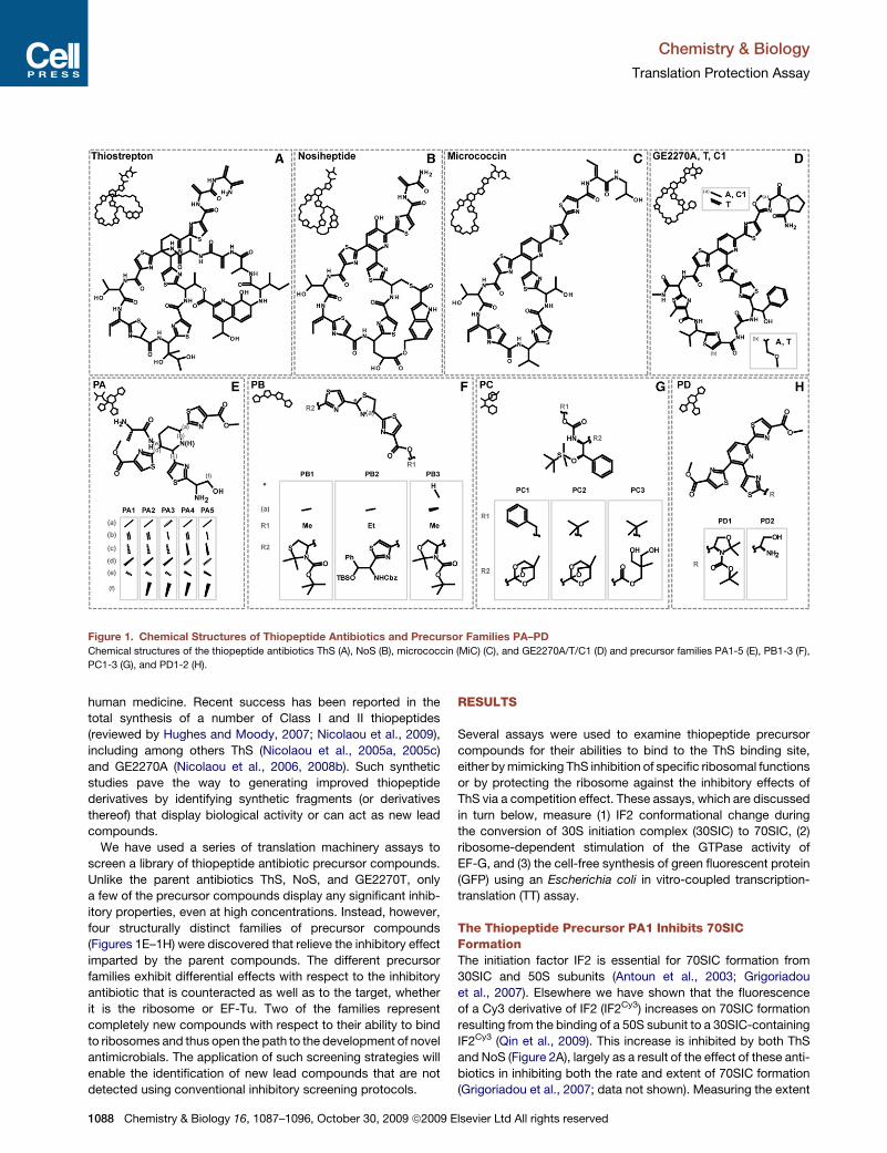
Chemistry & Biology
Translation Protection Assay
Figure 1. Chemical Structures of Thiopeptide Antibiotics and Precursor Families PA–PD
Chemical structures of the thiopeptide antibiotics ThS (A), NoS (B), micrococcin (MiC) (C), and GE2270A/T/C1 (D) and precursor families PA1-5 (E), PB1-3 (F),
PC1-3 (G), and PD1-2 (H).
human medicine. Recent success has been reported in the
total synthesis of a number of Class I and II thiopeptides
(reviewed by Hughes and Moody, 2007; Nicolaou et al., 2009),
including among others ThS (Nicolaou et al., 2005a, 2005c)
and GE2270A (Nicolaou et al., 2006, 2008b). Such synthetic
studies pave the way to generating improved thiopeptide
derivatives by identifying synthetic fragments (or derivatives
thereof) that display biological activity or can act as new lead
compounds.
We have used a series of translation machinery assays to
screen a library of thiopeptide antibiotic precursor compounds.
Unlike the parent antibiotics ThS, NoS, and GE2270T, only
a few of the precursor compounds display any significant inhib-
itory properties, even at high concentrations. Instead, however,
four structurally distinct families of precursor compounds
(Figures 1E–1H) were discovered that relieve the inhibitory effect
imparted by the parent compounds. The different precursor
families exhibit differential effects with respect to the inhibitory
antibiotic that is counteracted as well as to the target, whether
it is the ribosome or EF-Tu. Two of the families represent
completely new compounds with respect to their ability to bind
to ribosomes and thus open the path to the development of novel
antimicrobials. The application of such screening strategies will
enable the identification of new lead compounds that are not
detected using conventional inhibitory screening protocols.
1088 Chemistry & Biology 16, 1087–1096, October 30, 2009 ª2009
RESULTS
Several assays were used to examine thiopeptide precursor
compounds for their abilities to bind to the ThS binding site,
either by mimicking ThS inhibition of specific ribosomal functions
or by protecting the ribosome against the inhibitory effects of
ThS via a competition effect. These assays, which are discussed
in turn below, measure (1) IF2 conformational change during
the conversion of 30S initiation complex (30SIC) to 70SIC, (2)
ribosome-dependent stimulation of the GTPase activity of
EF-G, and (3) the cell-free synthesis of green fluorescent protein
(GFP) using an Escherichia coli in vitro-coupled transcription-
translation (TT) assay.
The Thiopeptide Precursor PA1 Inhibits 70SICFormationThe initiation factor IF2 is essential for 70SIC formation from
30SIC and 50S subunits (Antoun et al., 2003; Grigoriadou
et al., 2007). Elsewhere we have shown that the fluorescence
of a Cy3 derivative of IF2 (IF2Cy3) increases on 70SIC formation
resulting from the binding of a 50S subunit to a 30SIC-containing
IF2Cy3 (Qin et al., 2009). This increase is inhibited by both ThS
and NoS (Figure 2A), largely as a result of the effect of these anti-
biotics in inhibiting both the rate and extent of 70SIC formation
(Grigoriadou et al., 2007; data not shown). Measuring the extent
Elsevier Ltd All rights reserved
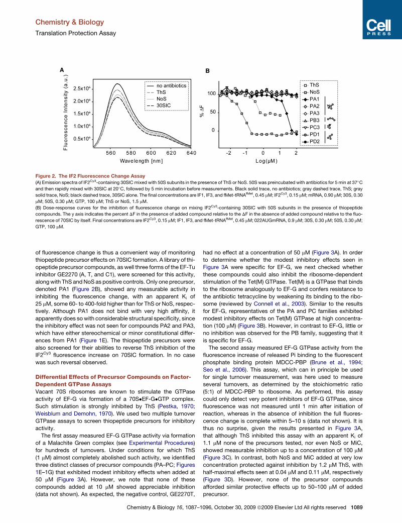
Chemistry & Biology
Translation Protection Assay
Figure 2. The IF2 Fluorescence Change Assay
(A) Emission spectra of IF2Cy3-containing 30SIC mixed with 50S subunits in the presence of ThS or NoS. 50S was preincubated with antibiotics for 5 min at 37�C
and then rapidly mixed with 30SIC at 20�C, followed by 5 min incubation before measurements. Black solid trace, no antibiotics; gray dashed trace, ThS; gray
solid trace, NoS; black dashed trace, 30SIC alone. The final concentrations are IF1, IF3, and fMet-tRNAfMet, 0.45 mM; IF2Cy3, 0.15 mM; mRNA, 0.90 mM; 30S, 0.30
mM; 50S, 0.30 mM; GTP, 100 mM; ThS or NoS, 1.5 mM.
(B) Dose-response curves for the inhibition of fluorescence change on mixing IF2Cy3-containing 30SIC with 50S subunits in the presence of thiopeptide
compounds. The y axis indicates the percent DF in the presence of added compound relative to the DF in the absence of added compound relative to the fluo-
rescence of 70SIC by itself. Final concentrations are IF2Cy3, 0.15 mM; IF1, IF3, and fMet-tRNAfMet, 0.45 mM; 022AUGmRNA, 0.9 mM; 30S, 0.30 mM; 50S, 0.30 mM;
GTP, 100 mM.
of fluorescence change is thus a convenient way of monitoring
thiopeptide precursor effects on 70SIC formation. A library of thi-
opeptide precursor compounds, as well three forms of the EF-Tu
inhibitor GE2270 (A, T, and C1), were screened for this activity,
along with ThS and NoS as positive controls. Only one precursor,
denoted PA1 (Figure 2B), showed any measurable activity in
inhibiting the fluorescence change, with an apparent Ki of
25 mM, some 60- to 400-fold higher than for ThS or NoS, respec-
tively. Although PA1 does not bind with very high affinity, it
apparently does so with considerable structural specificity, since
the inhibitory effect was not seen for compounds PA2 and PA3,
which have either stereochemical or minor constitutional differ-
ences from PA1 (Figure 1E). The thiopeptide precursors were
also screened for their abilities to reverse ThS inhibition of the
IF2Cy3 fluorescence increase on 70SIC formation. In no case
was such reversal observed.
Differential Effects of Precursor Compounds on Factor-Dependent GTPase AssaysVacant 70S ribosomes are known to stimulate the GTPase
activity of EF-G via formation of a 70S�EF-G�GTP complex.
Such stimulation is strongly inhibited by ThS (Pestka, 1970;
Weisblum and Demohn, 1970). We used two multiple turnover
GTPase assays to screen thiopeptide precursors for inhibitory
activity.
The first assay measured EF-G GTPase activity via formation
of a Malachite Green complex (see Experimental Procedures)
for hundreds of turnovers. Under conditions for which ThS
(1 mM) almost completely abolished such activity, we identified
three distinct classes of precursor compounds (PA–PC; Figures
1E–1G) that exhibited modest inhibitory effects when added at
50 mM (Figure 3A). However, we note that none of these
compounds added at 10 mM showed appreciable inhibition
(data not shown). As expected, the negative control, GE2270T,
Chemistry & Biology 16, 1087–
had no effect at a concentration of 50 mM (Figure 3A). In order
to determine whether the modest inhibitory effects seen in
Figure 3A were specific for EF-G, we next checked whether
these compounds could also inhibit the ribosome-dependent
stimulation of the Tet(M) GTPase. Tet(M) is a GTPase that binds
to the ribosome analogously to EF-G and confers resistance to
the antibiotic tetracycline by weakening its binding to the ribo-
some (reviewed by Connell et al., 2003). Similar to the results
for EF-G, representatives of the PA and PC families exhibited
modest inhibitory effects on Tet(M) GTPase at high concentra-
tion (100 mM) (Figure 3B). However, in contrast to EF-G, little or
no inhibition was observed for the PB family, suggesting that it
is specific for EF-G.
The second assay measured EF-G GTPase activity from the
fluorescence increase of released Pi binding to the fluorescent
phosphate binding protein MDCC-PBP (Brune et al., 1994;
Seo et al., 2006). This assay, which can in principle be used
for single turnover measurement, was here used to measure
several turnovers, as determined by the stoichiometric ratio
(5:1) of MDCC-PBP to ribosome. As performed, this assay
could only detect very potent inhibitors of EF-G GTPase, since
fluorescence was not measured until 1 min after initiation of
reaction, whereas in the absence of inhibition the full fluores-
cence change is complete within 5–10 s (data not shown). It is
thus no surprise, given the results presented in Figure 3A,
that although ThS inhibited this assay with an apparent Ki of
1.1 mM none of the precursors tested, nor even NoS or MiC,
showed measurable inhibition up to a concentration of 100 mM
(Figure 3C). In contrast, both NoS and MiC added at very low
concentration protected against inhibition by 1.2 mM ThS, with
half-maximal effects seen at 0.04 mM and 0.11 mM, respectively
(Figure 3D). However, none of the precursor compounds
afforded similar protective effects up to 50–100 mM of added
precursor.
1096, October 30, 2009 ª2009 Elsevier Ltd All rights reserved 1089

Chemistry & Biology
Translation Protection Assay
Figure 3. Effect of Thiopeptides and Precursor Compounds on GTPase Activity of EF-G and Tet(M)
(A) Inhibition of uncoupled ribosome-dependent Tet(M) GTPase by ThS (1 mM) and precursors PA2, PB2, and PC2 (50 mM). Closed circles indicate GTPase activity
of Tet(M) in the absence of antibiotic.
(B) Inhibition of uncoupled ribosome-dependent TetM GTPase by ThS (1 mM) and precursors PA2, PB1, and PC2 (100 mM). Closed circles indicate GTPase activity
of TetM in the absence of antibiotic.
(C) The dose-response curves of Pi release in the presence of ThS, NoS, MiC, or precursor compounds. The y axis indicates the percent DF due to Pi release in the
presence of added compound relative to the DF in the absence of added compound relative to the fluorescence from EF-G interaction with the ribosome in the
absence of any compound. The final concentrations are 70S, 0.3 mM; EF-G, 0.75 mM; MDCC-PBP, 1.5 mM; GTP, 100 mM;7-methylguanine, 200 mM; nucleotide
phosphorylase, 0.3 U/ml.
(D) Dose-response curves for reversal of ThS inhibition of Pi release by NoS, MiC, or precursor compounds. The final concentrations for each component are 70S,
0.3 mM; EF-G, 0.75 mM; ThS, 1.2 mM; MDCC-PBP, 1.5 mM; GTP, 100 mM; 7-methylguanine, 200 mM; nucleotide phosphorylase, 0.3 U/ml.
Protective Effects of Precursor Compounds onThiopeptide-Mediated Translation InhibitionAlthough, as expected, ThS, NoS, and GE2270T were potent
inhibitors of GFP synthesis using an in vitro TT assay (Figure 4,
lanes 2–4), none of the precursor compounds tested displayed
any significant inhibitory activity in this assay (Figure 4, white
bars), even at high concentrations (50–100 mM). In contrast,
addition of 50 mM of representative precursor compounds from
four structural distinct classes PA–PD (Figures 1E–1G) could
reverse the inhibitory effect of 5 mM ThS (Figure 4, gray bars).
The most effective protection was seen with PA2, which restored
translation back to levels observed in the absence of antibiotic
(Figure 4, lane 6). In comparison, PB1, PC1, and PD1 restored
translation to 40%–60% of the original levels (Figure 4, lanes
13–28). Additionally, we find that the 5S, 6R stereoisomer of
PA2 (PA4) exhibited some protective properties (�35%
compared to 100% for PA2). The structural specificity of these
effects is clear from the failure of precursors that are chemically
related to PA2 to exhibit similar protective effects against ThS
inhibition. These include PA3 (Figure 4, lanes 6 and 8), which
differs from PA2 by lacking only a double bond within the central
1090 Chemistry & Biology 16, 1087–1096, October 30, 2009 ª2009
dehydropiperidine ring (Figure 1E), and PD2 (Figure 4, lanes 26
and 28), which, with respect to PD1, has an altered side chain
on one of the thiazole rings (Figure 1H).
Interestingly, PA2, PB1, PC1, and PD1 displayed marked
differences in their abilities to reverse the inhibitory effects of
NoS (5 mM) and GE2270T (25 mM), as compared to the inhibitory
effects of ThS. Thus, as shown in Figure 5, PA2 was an omnipo-
tent protector of translation, restoring translation levels in the
presence of all three thiopeptide inhibitors, with the following
order of efficiency: ThS (100%) > GE2270T (80%) > NoS (60%).
PD1 rescued translation in the presence of ThS and GE2270T,
but not NoS, and PB1 and PC1 efficiently rescued translation
only against ThS. As was true for ThS, neither PA3, PB2, PC2,
nor PD2 were able to reverse inhibition by NoS or GE2270T
(data not shown).
Interaction of Thiopeptide Precursorswith the RibosomeThe specific protective effect of the precursor compounds
against ThS suggests that these compounds specifically com-
pete with ThS for binding to the ribosome. Structural (Harms
Elsevier Ltd All rights reserved
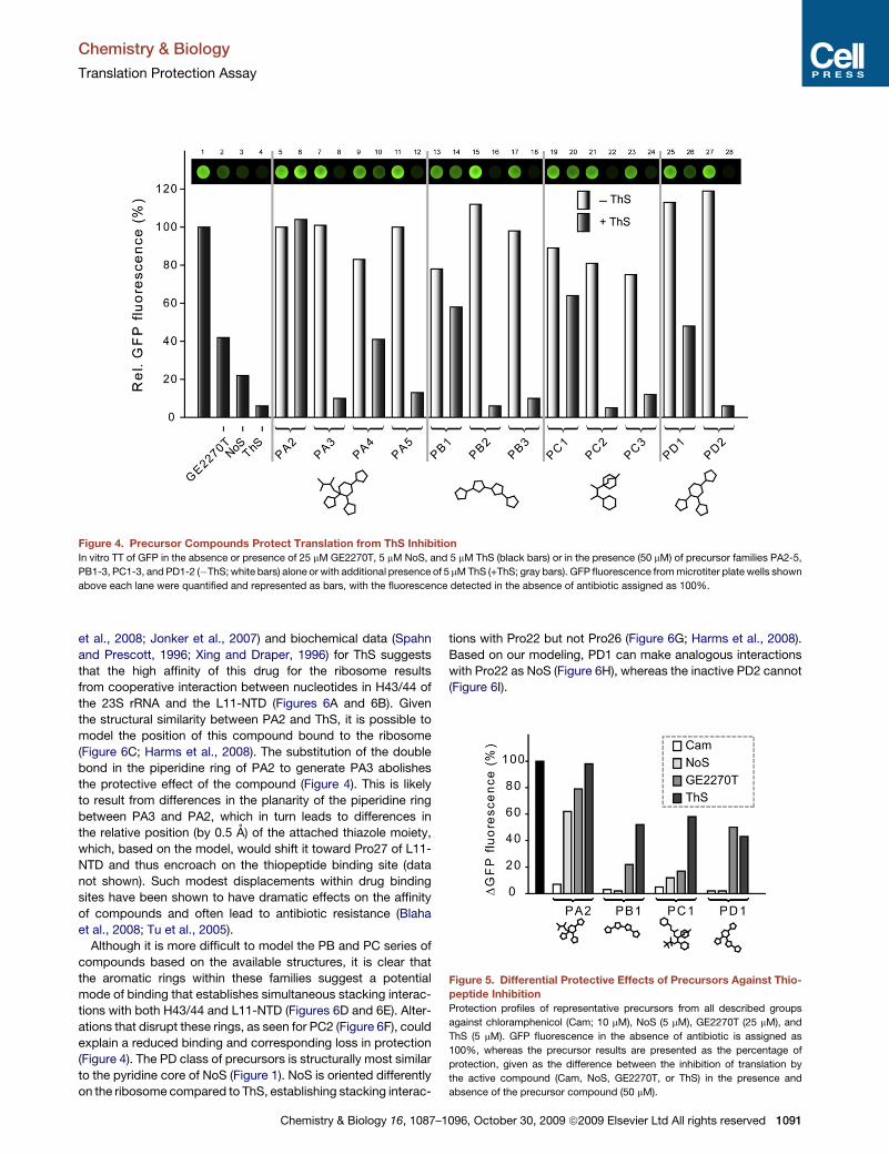
Chemistry & Biology
Translation Protection Assay
Figure 4. Precursor Compounds Protect Translation from ThS Inhibition
In vitro TT of GFP in the absence or presence of 25 mM GE2270T, 5 mM NoS, and 5 mM ThS (black bars) or in the presence (50 mM) of precursor families PA2-5,
PB1-3, PC1-3, and PD1-2 (�ThS; white bars) alone or with additional presence of 5 mM ThS (+ThS; gray bars). GFP fluorescence from microtiter plate wells shown
above each lane were quantified and represented as bars, with the fluorescence detected in the absence of antibiotic assigned as 100%.
et al., 2008; Jonker et al., 2007) and biochemical data (Spahn
and Prescott, 1996; Xing and Draper, 1996) for ThS suggests
that the high affinity of this drug for the ribosome results
from cooperative interaction between nucleotides in H43/44 of
the 23S rRNA and the L11-NTD (Figures 6A and 6B). Given
the structural similarity between PA2 and ThS, it is possible to
model the position of this compound bound to the ribosome
(Figure 6C; Harms et al., 2008). The substitution of the double
bond in the piperidine ring of PA2 to generate PA3 abolishes
the protective effect of the compound (Figure 4). This is likely
to result from differences in the planarity of the piperidine ring
between PA3 and PA2, which in turn leads to differences in
the relative position (by 0.5 A) of the attached thiazole moiety,
which, based on the model, would shift it toward Pro27 of L11-
NTD and thus encroach on the thiopeptide binding site (data
not shown). Such modest displacements within drug binding
sites have been shown to have dramatic effects on the affinity
of compounds and often lead to antibiotic resistance (Blaha
et al., 2008; Tu et al., 2005).
Although it is more difficult to model the PB and PC series of
compounds based on the available structures, it is clear that
the aromatic rings within these families suggest a potential
mode of binding that establishes simultaneous stacking interac-
tions with both H43/44 and L11-NTD (Figures 6D and 6E). Alter-
ations that disrupt these rings, as seen for PC2 (Figure 6F), could
explain a reduced binding and corresponding loss in protection
(Figure 4). The PD class of precursors is structurally most similar
to the pyridine core of NoS (Figure 1). NoS is oriented differently
on the ribosome compared to ThS, establishing stacking interac-
Chemistry & Biology 16, 1087–
tions with Pro22 but not Pro26 (Figure 6G; Harms et al., 2008).
Based on our modeling, PD1 can make analogous interactions
with Pro22 as NoS (Figure 6H), whereas the inactive PD2 cannot
(Figure 6I).
Figure 5. Differential Protective Effects of Precursors Against Thio-
peptide Inhibition
Protection profiles of representative precursors from all described groups
against chloramphenicol (Cam; 10 mM), NoS (5 mM), GE2270T (25 mM), and
ThS (5 mM). GFP fluorescence in the absence of antibiotic is assigned as
100%, whereas the precursor results are presented as the percentage of
protection, given as the difference between the inhibition of translation by
the active compound (Cam, NoS, GE2270T, or ThS) in the presence and
absence of the precursor compound (50 mM).
1096, October 30, 2009 ª2009 Elsevier Ltd All rights reserved 1091

Chemistry & Biology
Translation Protection Assay
Figure 6. Binding Site of Precursor Compounds on the Ribosome
(A) Overview of thiopeptide binding site on the large ribosomal subunit. Interface view with helix 43 and 44 (H43/44; orange), L11 (yellow), and ThS (green) high-
lighted with surface representation (from PDB ID 3CF5) (Harms et al., 2008).
(B) The thiazole rings of ThS (green) interact with the RNA bases at the tips of H43/44 as well as the prolines in the N-terminal domain of L11 (yellow).
(C) Model for precursor PA2 bound to the ribosome, based on the position of ThS. PA1, but not PA3 (see text), bind similarly.
(D–F) Possible modes of binding for precursors PB1 and PC1 based on ring stacking interactions with RNA and protein components of the ribosome, whereas
PC2 lacks one phenyl ring compared to PC1.
(G) NoS (pink) interacts with the RNA bases at the tips of H43/44 as well as the N-terminal domain of L11 (yellow), but in a distinct manner compared to ThS (using
PDB ID 2ZJP) (Harms et al., 2008).
(H and I) Model for precursor PD1 and PD2 bound to the ribosome, based on the position of NoS. PD2 lacks one ring moiety, suggesting binding would be desta-
bilized.
Interaction of Thiopeptide Precursors with EF-TuThe thiopeptide GE2270A has been crystallized in complex with
EF-Tu, revealing that the drug binds within a covered groove in
domain II and spans across the active site cleft of EF-Tu, the
G domain, to interact with the domain I (Figure 7A; Parmeggiani
et al., 2006). GE2270A overlaps the binding site of the terminal
A76 and aminoacyl moiety of the tRNA (Figure 7B) and is
believed to prevent the closing of domains I and II necessary
for the induced fit binding of aa-tRNA (Figure 7C), thereby
preventing ternary complex (EF-Tu�GTP�tRNA) formation
(reviewed by Parmeggiani and Nissen, 2006). The structural simi-
larities between PA and PD and GE2270A (Figure 1) suggest that
these compounds would also bind within the groove of domain II
of EF-Tu (Figure 7D) and overlap with the A76 of the tRNA
(Figure 7E). However, the truncated nature of these compounds
prevents them from establishing additional interactions with
1092 Chemistry & Biology 16, 1087–1096, October 30, 2009 ª2009
domain I, even in the closed tRNA-bound ternary complex state
of EF-Tu (Figure 7F).
DISCUSSION
Development of improved antimicrobial agents will be necessary
to combat the prevalence of multi-drug-resistant bacteria. A
number of biochemical approaches have been taken to identify
functionally important hotspots on the ribosome that do not
overlap with previously known antibiotic binding sites (Laios
et al., 2004; Llano-Sotelo et al., 2009b; Yassin et al., 2005; Yassin
and Mankin, 2007). In addition, a recent study has developed an
assay using fluorescently labeled ribosomal proteins to monitor
binding of small molecules, such as antibiotics, to the ribosome,
which is amenable to high-throughput screening (Llano-Sotelo
et al., 2009a). Here we present several translation-related assays
Elsevier Ltd All rights reserved
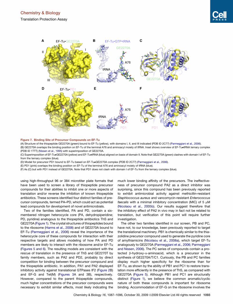
Chemistry & Biology
Translation Protection Assay
Figure 7. Binding Site of Precursor Compounds on EF-Tu
(A) Structure of the thiopeptide GE2270A (green) bound to EF-Tu (yellow), with domains I, II, and III indicated (PDB ID 2C77) (Parmeggiani et al., 2006).
(B) GE2270A overlaps the binding position on EF-Tu of the terminal A76 and aminoacyl moiety of tRNA. Inset shows overview of EF-Tu�tRNA ternary complex
(PDB ID 1TTT) (Nissen et al., 1995) with superimposition of GE2270A.
(C) Superimposition of EF-Tu�GE2270A (yellow) and EF-Tu�tRNA (blue) aligned on basis of domain II. Note that GE2270A (green) clashes with domain I of EF-Tu
from the ternary complex (blue).
(D) Model for precursor PD1 bound to EF-Tu based on EF-Tu�GE2270A complex (PDB ID 2C77) (Parmeggiani et al., 2006).
(E) PD1 (pink) overlaps the binding position on EF-Tu of the terminal A76 and aminoacyl moiety of tRNA (blue).
(F) As (C) but with PD1 instead of GE2270A. Note that PD1 does not clash with domain I of EF-Tu from the ternary complex (blue).
using high-throughput 96 or 384 microtiter plate formats that
have been used to screen a library of thiopeptide precursor
compounds for their abilities to inhibit one or more aspects of
translation and/or reverse the inhibition of known thiopeptide
antibiotics. These screens identified four distinct families of pre-
cursor compounds, termed PA–PD, which could act as potential
lead compounds for development of novel antimicrobials.
Two of the families identified, PA and PD, contain a six-
membered nitrogen heterocycle core (PA, dehydropiperidine;
PD, pyridine) analogous to the thiopeptide antibiotics ThS and
GE2270A (Figure 1). The crystal structures of thiopeptides bound
to the ribosome (Harms et al., 2008) and of GE2270A bound to
EF-Tu (Parmeggiani et al., 2006) reveal the importance of the
heterocycle core of these compounds for interaction with their
respective targets and allows modeling of how PA and PD
members are likely to interact with the ribosome and/or EF-Tu
(Figures 5 and 6). The resulting models are consistent with the
rescue of translation in the presence of ThS and GE2270T by
family members, such as PA2 and PD2, probably by direct
competition for binding between the precursor compound and
the thiopeptide antibiotic. In addition, PA1 and PA2 displayed
inhibitory activity against translational GTPases IF2 (Figure 2B)
and EF-G and Tet(M) (Figures 3A and 3B), respectively.
However, compared to the parent thiopeptide compounds,
much higher concentrations of the precursor compounds were
necessary to exhibit similar effects, most likely indicating the
Chemistry & Biology 16, 1087–
much lower binding affinity of the precursors. The ineffective-
ness of precursor compound PA2 as a direct inhibitor was
surprising, since this compound has been previously reported
to exhibit antimicrobial activity against methicillin-resistant
Staphlococcus aureus and vancomycin-resistant Enterococcus
faecalis with a minimal inhibitory concentration (MIC) of 5 mM
(Nicolaou et al., 2005b). Our results suggest therefore that
the inhibitory effect of PA2 in vivo may in fact not be related to
translation, but verification of this point will require further
investigation.
The other two families identified in our screen, PB and PC,
have not, to our knowledge, been previously reported to target
the translational machinery. PB1 is chemically similar to the thia-
zolidine precursor compound used to generate the pyridine core
of amythiamicins (Nicolaou et al., 2008a), which target EF-Tu
analogously to GE2270A (Parmeggiani et al., 2006; Parmeggiani
and Nissen, 2006). The PC series of compounds contain a pro-
tected b-hydroxy-a-aminoacid, which is a precursor in the
synthesis of GE2270A/T/C1. Curiously, the PB and PC families
display much higher specificity for the ribosome than for
EF-Tu, as shown by the ability of PB1 and PC1 to restore trans-
lation more efficiently in the presence of ThS, as compared with
GE2270A (Figure 5). Although PB1 and PC1 are structurally
distinct (Figure 1), we believe the common aromatic/cyclic
nature of both these compounds is important for ribosome
binding. Accommodation of EF-G on the ribosome involves the
1096, October 30, 2009 ª2009 Elsevier Ltd All rights reserved 1093
Chemistry & Biology
Translation Protection Assay
insertion of domain V of EF-G into the crevice between H43/44
and L11-NTD. Inhibition by Class I thiopeptides has been
proposed to stem in part from their physically linking L11-NTD
to H43/44, thereby locking the cleft shut (Harms et al., 2008).
We suggest that PB1 and PC1 can also span the L11-rRNA
crevice (Figures 6D and 6E) and perform this locking function,
analogous to ThS/PA2 (Figures 6B and 6C) and NoS/PD1
(Figures 6G and 6H). Similarly to PA/D, the high concentrations
of PB/C required to inhibit the ribosome-dependent GTPase
activity of EF-G are indicative of their low binding affinities for
the ribosome. Such low affinity may allow facile displacement
of precursors from the ribosome, as a result of translation factors
(IF2 or EF-G) binding, or from EF-Tu, during ternary complex
formation, thus explaining the absence of any direct inhibitory
effect of any of the precursors on GFP synthesis. The differential
effects of the precursors on the GTPase assays compared to
the TT assay is probably related to the ribosome concentrations
in the GTPase assays being �103–1003 less (30–300 nM)
compared to the TT assay (�2 mM) and to the putative higher
affinity of EF-G for translating rather than empty ribosomes
(Sergiev et al., 2005).
The majority of clinically used antibiotics targeting the ribo-
some bind either to the decoding region on the small subunit
or within either the peptidyltransferase center or the adjacent
peptide exit tunnel of the large subunit, where they interact
almost exclusively with ribosomal RNA (Spahn and Prescott,
1996; Wilson, 2004). The Class I thiopeptide compounds,
however, are distinct in that they target a different region of the
ribosome, namely the GTPase-associated region or translation
factor binding site, where they interact with both rRNA and ribo-
somal protein L11. As a consequence, no cross-resistance has
been found between thiopeptide antibiotics and other clinically
important drugs. The compounds such as PA–PD identified in
our study provide lead structures for the development of novel
antimicrobial agents that target this region of the ribosome.
Furthermore, the ability of some precursor compounds, such
as PA1 and PD1, to bind both EF-Tu and the ribosome suggests
the feasibility of developing antimicrobials that are dual inhibitors
of ribosomes and ternary complex formation.
SIGNIFICANCE
The translational machinery represents one of the major
targets within the cell for antibiotics, with many clinical
important classes of antibiotics inhibiting translation by
binding to the ribosome. Despite the potency of many of
these drug classes, antibiotic resistance among clinically
relevant pathogens is an increasing problem and there is
an urgent need for improved antibiotics. We present herein
a series of translation machinery assays that can be used
to screen for lead compounds that not only inhibit specific
steps of translation but also relieve the inhibitory effects of
other inhibitory compounds. Using these assays to screen
a library of thiopeptide precursor compounds, we have iden-
tified four distinct families of compounds that inhibit either
IF2, EF-G, and/or Tet(M), as well as confer protective effects
against thiopeptide translation inhibitors of both the ribo-
some and EF-Tu. Our findings not only elucidate the mecha-
nism of action of thiopeptide compounds but also illustrate
1094 Chemistry & Biology 16, 1087–1096, October 30, 2009 ª2009
the potential of such high-throughput assays to identify
distinct lead compounds that might be missed using
conventional inhibitory screening protocols. Whereas the
IF2 and EF-G GTPase assays are specifically useful for
screening antibiotics interfering with translation G factor
proteins, the TT assay is generally applicable for screening
all classes of translation inhibitors, including those targeting
the peptidyltransferase, and decoding centers of the ribo-
some and other ribosomal sites, in addition to those inter-
fering with translation G factor proteins.
EXPERIMENTAL PROCEDURES
Component Preparation
GE2270A, T, and C1 and the library of thiopeptide precursor compounds were
synthesized as described previously (Nicolaou et al., 2005b, 2006, 2008a,
2008b). ThS was purchased from Sigma, whereas NoS was a gift from
H.G. Floss and micrococcin P1 was supplied by T. Stachlhaus. The tetM
gene (Tn916) cloned into the pET24b vector was a gift from V. Burdett.
Tet(M) protein was expressed in BL21 (DE3) pRIL cells in 20�C with 0.2 mM
IPTG. E. coli EF-G cloned into pQE70 vector was expressed in XL1 blue cells.
Both proteins were purified using Ni-NTA metal affinity chromatography
(QIAGEN), followed by gel filtration chromatography on a HiLoad 26/60 Super-
dex 75 prep grade column (GE Healthcare). For the experiments described in
Figures 2 and 3C and 3D, ribosomes, IF2Cy3, IF1, IF3, 30S subunits, MDCC-
labeled phosphate-binding protein (MDCC-PBP), 022AUG mRNA, and fMet-
tRNAfMet were prepared as described previously (Qin et al., 2009) as was
EF-G (Pan et al., 2007).
IF2Cy3 Fluorescence Change Assay
Reactions were carried out in a 384 well assay plate. 50S subunits were pre-
incubated (15 min at 37�C) with a series of concentrations of the test
compounds in DMSO that are transferred from a premade compound plate
to the assay plate by a PerkinElmer Evolution P3 liquid handler. Reaction
was initiated by addition of 30SIC to each well of the plate. Fluorescence
(579 nm) was read with a 2103 EnVision Multilabel Plate Reader on excitation
at 550 nm. For the reversal experiment, 30SIC was preincubated with ThS
(10 min at 37�C), followed by a second preincubation with test compounds
(10 min at 37�C), and reaction was initiated by 50S addition.
GTPase Activity Assays
For both assays described below, reactions performed in the absence of ribo-
somes were used as a background signal to account for the intrinsic GTPase
activity of EF-G or Tet(M).
By Malachite Green
GTPase activity was measured using the Malachite Green Phosphate Kit
(BioAssay) that quantifies the green complex formed between Malachite
Green, molybdate, and free orthophosphate. All reactions contained 30 nM
E. coli 70S ribosomes, 20 mM GTP, and 60 nM protein in the presence or
absence of antibiotics as necessary. Reactions were transferred into 96 well
microtiter plates and color formation was measured on Tecan Infinite M1000
microplate reader at 650 nm.
By MDCC-Labeled PBP
GTPase activity was measured using the MDCC-labeled PBP complex, which
measures free phosphate release as an increase in fluorescence and uses
a Pi-MOP system to minimize the background due to phosphate present in
the original medium (Brune et al., 1994; Seo et al., 2006). Reactions were
carried out in a 384 well assay plate. Ribosomes were preincubated (15 min
at 37�C) with a series of concentrations of the test compounds in DMSO
that are transferred from a premade compound plate to the assay plate by
a PerkinElmer Evolution P3 liquid handler. Reaction was initiated by addition
of an ice-cold solution containing EF-G, MDCC-PBP, and GTP to each
well of the plate, a process that was completed in under 30 s. Fluorescence
(450 nm) was read within 1 min using a 2103 EnVision Multilabel Plate Reader,
on excitation at 405 nm. For the reversal experiment, ribosomes were preincu-
bated with ThS (10 min at 37�C), followed by a second preincubation with test
Elsevier Ltd All rights reserved
Chemistry & Biology
Translation Protection Assay
compounds (10 min at 37�C), and reaction was initiated by EF-G, MDCC-PBP,
and GTP as above.
In Vitro Transcription-Translation Assay
All coupled TT experiments were performed using an E. coli lysate-based
system in the presence and absence of antibiotics as described previously
(Dinos et al., 2004; Szaflarski et al., 2008). Reactions were transferred into
96 well microtiter plates and the GFP fluorescence was measured with
a Typhoon Scanner 9400 (GE Healthcare) using a Typhoon blue laser module
(GE Healthcare). Images were then quantified using ImageQuantTL (GE Health-
care) and represented graphically using SigmaPlot (Systat Software, Inc.).
Modeling and Figure Preparation
Chemical structures for the precursor compounds were drawn and converted
to 3D coordinates using ChemDraw (Advanced Chemistry Development, Inc.).
PA2 models used the ThS binding position on the Deinococcus radiodurans
50S (D50S) subunit (PDB ID 3CF5) (Harms et al., 2008), whereas PD1 and
PD2 were based on the D50S-NoS complex (PDB ID 2ZJP) (Harms et al.,
2008). PyMol (http://www.pymol.org) was used to model the PB1 and PC
compounds, align EF-Tu�GE2270A (yellow; PDB ID 2C77) (Parmeggiani
et al., 2006) and EF-Tu�tRNA (blue; PDB ID 1TTT) (Nissen et al., 1995) on
the basis of domain II, as well as to prepare all X-ray structure figures.
ACKNOWLEDGMENTS
We would like to thank R. Beckmann and his coworkers for generous support
and creating a stimulating work environment. This work was financed by the
Deutsche Forschungsgemeinschaft (WI3285/1-1 to D.N.W.) and by the
National Institutes of Health (GM071014 to B.S.C.).
Received: July 28, 2009
Revised: September 6, 2009
Accepted: September 10, 2009
Published: October 30, 2009
REFERENCES
Antoun, A., Pavlov, M.Y., Andersson, K., Tenson, T., and Ehrenberg, M. (2003).
The roles of initiation factor 2 and guanosine triphosphate in initiation of protein
synthesis. EMBO J. 22, 5593–5601.
Bagley, M.C., Dale, J.W., Merritt, E.A., and Xiong, X. (2005). Thiopeptide anti-
biotics. Chem. Rev. 105, 685–714.
Blaha, G., Gurel, G., Schroeder, S.J., Moore, P.B., and Steitz, T.A. (2008).
Mutations outside the anisomycin-binding site can make ribosomes drug-
resistant. J. Mol. Biol. 379, 505–519.
Brandi, L., Marzi, S., Fabbretti, A., Fleischer, C., Hill, W., Lodmell, J., and
Gualerzi, C. (2004). The translation initiation functions of IF2: targets for thio-
strepton inhibition. J. Mol. Biol. 335, 881–894.
Brune, M., Hunter, J.L., Corrie, J.E., and Webb, M.R. (1994). Direct, real-time
measurement of rapid inorganic phosphate release using a novel fluorescent
probe and its application to actomyosin subfragment 1 ATPase. Biochemistry
33, 8262–8271.
Connell, S.R., Tracz, D.M., Nierhaus, K.H., and Taylor, D.E. (2003). Ribosomal
protection proteins and their mechanism of tetracycline resistance. Antimi-
crob. Agents Chemother. 47, 3675–3681.
Dinos, G., Wilson, D.N., Teraoka, Y., Szaflarski, W., Fucini, P., Kalpaxis, D., and
Nierhaus, K.H. (2004). Dissecting the ribosomal inhibition mechanisms of
edeine and pactamycin: the universally conserved residues G693 and C795
regulate P-site tRNA binding. Mol. Cell 13, 113–124.
Gale, E.F., Cundliffe, E., Reynolds, P.E., Richmond, M.H., and Waring, M.J.
(1981). Antibiotic inhibitors of ribosome function. In The Molecular Basis of
Antibiotic Action, E.F. Gale, E. Cundliffe, P.E. Reynolds, M.H. Richmond,
and M.J. Waring, eds. (Bristol, UK: John Wiley and sons), pp. 278–379.
Gonzalez, R.L., Jr., Chu, S., and Puglisi, J.D. (2007). Thiostrepton inhibition of
tRNA delivery to the ribosome. RNA 13, 2091–2097.
Chemistry & Biology 16, 1087
Grigoriadou, C., Marzi, S., Kirillov, S., Gualerzi, C.O., and Cooperman, B.S.
(2007). A quantitative kinetic scheme for 70 S translation initiation complex
formation. J. Mol. Biol. 373, 562–572.
Harms, J.M., Wilson, D.N., Schluenzen, F., Connell, S.R., Stachelhaus, T.,
Zaborowska, Z., Spahn, C.M., and Fucini, P. (2008). Translational regulation
via L11: molecular switches on the ribosome turned on and off by thiostrepton
and micrococcin. Mol. Cell 30, 26–38.
Hughes, R.A., and Moody, C.J. (2007). From amino acids to heteroaromatics—
thiopeptide antibiotics, nature’s heterocyclic peptides. Angew. Chem. Int. Ed.
Engl. 46, 7930–7954.
Jonker, H.R., Ilin, S., Grimm, S.K., Wohnert, J., and Schwalbe, H. (2007). L11
domain rearrangement upon binding to RNA and thiostrepton studied by NMR
spectroscopy. Nucleic Acids Res. 35, 441–454.
Laios, E., Waddington, M., Saraiya, A.A., Baker, K.A., O’Connor, E.,
Pamarathy, D., and Cunningham, P.R. (2004). Combinatorial genetic tech-
nology for the development of new anti-infectives. Arch. Pathol. Lab. Med.
128, 1351–1359.
Llano-Sotelo, B., Hickerson, R.P., Lancaster, L., Noller, H.F., and Mankin, A.S.
(2009a). Fluorescently labeled ribosomes as a tool for analyzing antibiotic
binding. RNA 15, 1597–1604.
Llano-Sotelo, B., Klepacki, D., and Mankin, A.S. (2009b). Selection of small
peptides, inhibitors of translation. J. Mol. Biol. 391, 813–819.
McConkey, G.A., Rogers, M.J., and McCutchan, T.F. (1997). Inhibition of Plas-
modium falciparum protein synthesis. Targeting the plastid-like organelle with
thiostrepton. J. Biol. Chem. 272, 2046–2049.
Modolell, J., Cabrer, B., Parmeggiani, A., and Vazquez, D. (1971). Inhibition by
siomycin and thiostrepton of both aminoacyl-tRNA and factor G binding to
ribosomes. Proc. Natl. Acad. Sci. USA 68, 1796–1800.
Nicolaou, K.C., Safina, B.S., Zak, M., Lee, S.H., Nevalainen, M., Bella, M.,
Estrada, A.A., Funke, C., Zecri, F.J., and Bulat, S. (2005a). Total synthesis of
thiostrepton. Retrosynthetic analysis and construction of key building blocks.
J. Am. Chem. Soc. 127, 11159–11175.
Nicolaou, K.C., Zak, M., Rahimipour, S., Estrada, A.A., Lee, S.H., O’Brate, A.,
Giannakakou, P., and Ghadiri, M.R. (2005b). Discovery of a biologically active
thiostrepton fragment. J. Am. Chem. Soc. 127, 15042–15044.
Nicolaou, K.C., Zak, M., Safina, B.S., Estrada, A.A., Lee, S.H., and Nevalainen,
M. (2005c). Total synthesis of thiostrepton. Assembly of key building blocks
and completion of the synthesis. J. Am. Chem. Soc. 127, 11176–11183.
Nicolaou, K.C., Zou, B., Dethe, D.H., Li, D.B., and Chen, D.Y. (2006). Total
synthesis of antibiotics GE2270A and GE2270T. Angew. Chem. Int. Ed. Engl.
45, 7786–7792.
Nicolaou, K.C., Dethe, D.H., and Chen, D.Y. (2008a). Total syntheses of amy-
thiamicins A, B and C. Chem. Commun. (Camb.) 2632–2634.
Nicolaou, K.C., Dethe, D.H., Leung, G.Y., Zou, B., and Chen, D.Y. (2008b).
Total synthesis of thiopeptide antibiotics GE2270A, GE2270T, and
GE2270C1. Chem. Asian J. 3, 413–429.
Nicolaou, K.C., Chen, J.S., Edmonds, D.J., and Estrada, A.A. (2009). Recent
advances in the chemistry and biology of naturally occurring antibiotics.
Angew. Chem. Int. Ed. Engl. 48, 660–719.
Nissen, P., Kjeldgaard, M., Thirup, S., Polekhina, G., Reshetnikova, L., Clark,
B.F., and Nyborg, J. (1995). Crystal structure of the ternary complex of
Phe-tRNAPhe, EF-Tu, and a GTP analog. Science 270, 1464–1472.
Pan, D., Kirillov, S.V., and Cooperman, B.S. (2007). Kinetically competent
intermediates in the translocation step of protein synthesis. Mol. Cell 25,
519–529.
Parmeggiani, A., and Nissen, P. (2006). Elongation factor Tu-targeted antibi-
otics: four different structures, two mechanisms of action. FEBS Lett. 580,
4576–4581.
Parmeggiani, A., Krab, I.M., Okamura, S., Nielsen, R.C., Nyborg, J., and
Nissen, P. (2006). Structural basis of the action of pulvomycin and GE2270 A
on elongation factor Tu. Biochemistry 45, 6846–6857.
Pestka, S. (1970). Thiostrepton: a ribosomal inhibitor of translocation.
Biochem. Biophys. Res. Commun. 40, 667–674.
–1096, October 30, 2009 ª2009 Elsevier Ltd All rights reserved 1095

Chemistry & Biology
Translation Protection Assay
Qin, H., Grigoriadou, C., and Cooperman, B.S. (2009). Interaction of IF2 with
the ribosomal GTPase-associated center during 70S initiation complex forma-
tion. Biochemistry 48, 4699–4706.
Rodnina, M.V., Savelsbergh, A., Matassova, N.B., Katunin, V.I., Semenkov,
Y.P., and Wintermeyer, W. (1999). Thiostrepton inhibits the turnover but not
the GTPase of elongation factor G on the ribosome. Proc. Natl. Acad. Sci.
USA 96, 9586–9590.
Seo, H.S., Abedin, S., Kamp, D., Wilson, D.N., Nierhaus, K.H., and Cooper-
man, B.S. (2006). EF-G-dependent GTPase on the ribosome. Conformational
change and fusidic acid inhibition. Biochemistry 45, 2504–2514.
Sergiev, P.V., Lesnyak, D.V., Kiparisov, S.V., Burakovsky, D.E., Leonov, A.A.,
Bogdanov, A.A., Brimacombe, R., and Dontsova, O.A. (2005). Function of the
ribosomal E-site: a mutagenesis study. Nucleic Acids Res. 33, 6048–6056.
Spahn, C.M.T., and Prescott, C.D. (1996). Throwing a spanner in the works:
antibiotics and the translational apparatus. J. Mol. Med. 74, 423–439.
Szaflarski, W., Vesper, O., Teraoka, Y., Plitta, B., Wilson, D.N., and Nierhaus,
K.H. (2008). New features of the ribosome and ribosomal inhibitors: non-
enzymatic recycling, misreading and back-translocation. J. Mol. Biol. 380,
193–205.
1096 Chemistry & Biology 16, 1087–1096, October 30, 2009 ª2009
Tu, D., Blaha, G., Moore, P., and Steitz, T. (2005). Structures of MLSBK antibi-
otics bound to mutated large ribosomal subunits provide a structural explana-
tion for resistance. Cell 121, 257–270.
Weisblum, B., and Demohn, V. (1970). Inhibition by thiostrepton of the
formation of a ribosome-bound guanine nucleotide complex. FEBS Lett. 11,
149–152.
Wilson, D.N. (2004). Antibiotics and the inhibition of ribosome function. In
Protein Synthesis and Ribosome Structure, K. Nierhaus and D. Wilson, eds.
(Weinheim: Wiley-VCH), pp. 449–527.
Wilson, D.N., and Nierhaus, K.H. (2005). Ribosomal proteins in the spotlight.
Crit. Rev. Biochem. Mol. Biol. 40, 243–267.
Xing, Y., and Draper, D.E. (1996). Cooperative interactions of RNA and thio-
strepton antibiotic with two domains of ribosomal protein L11. Biochemistry
35, 1581–1588.
Yassin, A., and Mankin, A.S. (2007). Potential new antibiotic sites in the
ribosome revealed by deleterious mutations in RNA of the large ribosomal
subunit. J. Biol. Chem. 282, 24329–24342.
Yassin, A., Fredrick, K., and Mankin, A.S. (2005). Deleterious mutations in
small subunit ribosomal RNA identify functional sites and potential targets
for antibiotics. Proc. Natl. Acad. Sci. USA 102, 16620–16625.
Elsevier Ltd All rights reserved

Paper 6
Chemistry & Biology
Article
Differential Effects of Thiopeptideand Orthosomycin Antibioticson Translational GTPasesAleksandra Mikolajka,1,2,4 Hanqing Liu,3,4 Yuanwei Chen,3 Agata L. Starosta,1 Viter Marquez,1 Marina Ivanova,1
Barry S. Cooperman,3,* and Daniel N. Wilson1,2,*1Gene Center and Department of Biochemistry2Center for Integrated Protein Science Munich (CiPSM)University of Munich, LMU, Feodor Lynen Str. 25, 81377 Munich, Germany3Department of Chemistry, University of Pennsylvania, Philadelphia, PA 19104-6323, USA4These authors contributed equally to this work
*Correspondence: [email protected] (B.S.C.), [email protected] (D.N.W.)DOI 10.1016/j.chembiol.2011.03.010
SUMMARY
The ribosome is a major target in the bacterial cell forantibiotics. Here, we dissect the effects that thethiopeptide antibiotics thiostrepton (ThS) and micro-coccin (MiC) as well as the orthosomycin antibioticevernimicin (Evn) have on translational GTPases.We demonstrate that, like ThS, MiC is a translocationinhibitor, and that the activation by MiC of the ribo-some-dependent GTPase activity of EF-G is depen-dent on the presence of the ribosomal proteins L7/L12 as well as the G0 subdomain of EF-G. In contrast,Evn does not inhibit translocation but is a potentinhibitor of back-translocation as well as IF2-depen-dent 70S-initiation complex formation. Collectively,these results shed insight not only into fundamentalaspects of translation but also into the unappreci-ated specificities of these classes of translationalinhibitors.
INTRODUCTION
Protein synthesis occurs on large macromolecular particles
called ribosomes, which are composed of RNA and protein. In
bacteria the 70S ribosome can be split into a small (30S) and
large (50S) subunit. The bacterial translational machinery repre-
sents a major target within the cell for antibiotics (reviewed by
Blanchard et al., 2010; Wilson, 2009). Many clinically important
classes of antibiotics inhibit translation by binding to the active
centers of ribosome. For example the tetracyclines and amino-
glycosides bind at the decoding site on the small subunit, and
the chloramphenicols, macrolides/ketolides, oxazolidinones,
and lincosamides bind at the peptidyltransferase center (PTC)
on the large subunit (Sohmen et al., 2009a, 2009b). Despite the
potency of many of these drug classes, antibiotic resistance
among clinically relevant pathogens is an increasing problem,
and thus, the need for new antibiotics is more urgent than ever
before. Ideally, the new antibiotics should have nonoverlapping
sites with the currently used antimicrobial agents, so that the
Chemistry & Biology 18,
occurrence of cross-resistance is reduced or prevented. Two
such classes are the thiopeptides and orthosomycins (Figures
1A–1C), which bind to distinct sites on the large ribosomal
subunit that are located far from the PTC (Figures 1D and 1E).
The orthosomycins, such as evernimicin (Evn), are oligosac-
charide antibiotics (Figure 1A) that display excellent antimicro-
bial activity against a broad range of Gram-positive bacteria,
both in vivo and in vitro. Although attempts to introduce Evn clin-
ically as Ziracin (Schering-Plough) were unsuccessful, a related
compound, avilamycin (Avn), is used as a growth promoter in
animal feeding. A multitude of resistance mutation/modification
and chemical footprinting studies indicate that the orthosomy-
cin-binding site is located at the base of the L7/L12 stalk (Figures
1D and 1E), �50 A from the PTC. Mutations in ribosomal protein
L16 (Aarestrup and Jensen, 2000; Adrian et al., 2000b; McNicho-
las et al., 2001; Zarazaga et al., 2002), and in helix 89 (H89) and
H91 of the 23S rRNA, as well as methylation of G2470 (E. coli
numbering used throughout) in H89 (Mann et al., 2001), confer
resistance to Evn and Avn (Adrian et al., 2000a; Belova
et al., 2001). In addition, Evn and Avn protect 23S rRNA nucleo-
tides, e.g., A2482 in H89 and A2534 in H91, from chemical modi-
fication (Belova et al., 2001; Kofoed and Vester, 2002). It is also
noteworthy that mutations in rplP (L16 gene) confer relatively
low-level resistance (MIC <12 mg ml�1), whereas higher-level
resistance (MIC >256 mg ml�1) is obtained by EmtA-mediated
methylation or rRNA mutations (Belova et al., 2001; Mann
et al., 2001). Taken together, these results suggest that the
orthosomycin-binding site spans from the minor groove of H89
to the loop region of H91 (Figure 1E) and that mutations in L16
confer resistance indirectly via perturbation of the 23S rRNA.
In agreement with this novel location, Evn does not inhibit
peptide-bond formation (Belova et al., 2001) or compete
with several other ribosomal antibiotics for ribosome binding
(McNicholas et al., 2000). Although some effect of Avn on
aa-tRNA binding to ribosomes has been observed (Wolf,
1973), Evn is better known as an initiation inhibitor; Evn inhibits
the formation of fMet-puromycin in an IF2-dependent manner
(Belova et al., 2001), although the exact step of inhibition remains
unclear. Moreover, to our knowledge, the effects of orthosomy-
cins on translation factors other than IF2 and EF-Tu have not yet
been addressed.
589–600, May 27, 2011 ª2011 Elsevier Ltd All rights reserved 589
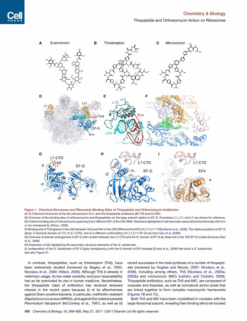
Figure 1. Chemical Structures and Ribosomal-Binding Sites of Thiopeptide and Orthosomycin Antibiotics
(A–C) Chemical structures of the (A) orthosomycin Evn, and the thiopeptide antibiotics (B) ThS and (C) MiC.
(D) Overview of the binding sites of orthosomycins and thiopeptides on the large subunit relative to EF-G. R-proteins L1, L11, and L7 are shown for reference.
(E) Putative binding site of orthosomycins spanning from H89 and H91 of the 23S rRNA. Residues highlighted in red have been associated biochemically with Evn
or Avn (reviewed by Wilson, 2009).
(F) Binding site of ThS (green) in the cleft between H43 and H44 of the 23S rRNA and the NTD of L11 (L11-ThS) (Harms et al., 2008). The relative positions of EF-G
(blue), C-terminal domain of L7/L12 (L7-CTD), and of a different conformation of L11 (L11-EF-G) are from Gao et al. (2009).
(G) Overview of domain arrangement of EF-G with contact between the L7-CTD and the G0 domain of EF-G as observed in the 70S-EF-G crystal structure (Gao
et al., 2009).
(H) Expansion of (G) highlighting the secondary structure elements of the G0 subdomain.
(I) Juxtaposition of the G0 subdomain of EF-G (gray transparency) with the G-domain of EF4 (orange) (Evans et al., 2008) that lacks a G0 subdomain.
See also Figure S1.
Chemistry & Biology
Thiopeptide and Orthosomycin Action on Ribosomes
In contrast, thiopeptides, such as thiostrepton (ThS), have
been extensively studied (reviewed by Bagley et al., 2005;
Nicolaou et al., 2009; Wilson, 2009). Although ThS is already in
veterinary usage, its low water solubility and poor bioavailability
has so far precluded its use in human medicine. Nevertheless,
the thiopeptide class of antibiotics has received renewed
interest in the recent years because (i) of its effectiveness
against Gram-positive bacteria, in particular, methicillin-resistant
Staphylococcus aureus (MRSA), and against the malarial parasite
Plasmodium falciparum (McConkey et al., 1997), as well as (ii)
590 Chemistry & Biology 18, 589–600, May 27, 2011 ª2011 Elsevier
recent successes in the total synthesis of a number of thiopepti-
des (reviewed by Hughes and Moody, 2007; Nicolaou et al.,
2009), including among others, ThS (Nicolaou et al., 2005a,
2005b) and micrococcin (MiC) (Lefranc and Ciufolini, 2009).
Thiopeptide antibiotics, such as ThS and MiC, are composed of
oxazoles and thiazoles, as well as nonnatural amino acids that
are linked together to form complex macrocyclic frameworks
(Figures 1B and 1C).
Both ThS and MiC have been crystallized in complex with the
large ribosomal subunit, revealing their binding site to be located
Ltd All rights reserved
Chemistry & Biology
Thiopeptide and Orthosomycin Action on Ribosomes
in a cleft formed by the N-terminal domain (NTD) of ribosomal
protein L11 and H43/H44 of the 23S rRNA (Figures 1D and 1F)
(Harms et al., 2008), consistent with a vast wealth of prior
biochemical studies (reviewed by Wilson, 2009). This region is
part of the GTPase-associated center (GAC), so named because
it is involved in binding of translation factors and stimulation of
their GTPase activities. Consistently, thiopeptide antibiotics
have been shown to inhibit IF2-dependent 70S-initiation
complex (70SIC) formation (Brandi et al., 2004; Grigoriadou
et al., 2007), EF-Tu-dependent delivery of aminoacyl-tRNAs to
the ribosome (Brandi et al., 2004; Gonzalez et al., 2007; Modolell
et al., 1971; Otaka and Kaji, 1974), translocation of the tRNA2-
mRNA complex through the ribosome (Munro et al., 2010; Pan
et al., 2007; Pestka, 1970; Pestka and Brot, 1971; Rodnina
et al., 1997), and stringent factor RelA-dependent synthesis of
ppGpp (Cundliffe and Thompson, 1981; Knutsson Jenvert and
Holmberg Schiavone, 2005). Surprisingly, however, ThS and
MiC exhibit differential effects on the uncoupled ribosome-
dependent EF-G GTPase activities: ThS strongly inhibits
multiple-turnover GTP hydrolysis of EF-G (Pestka, 1970; Weis-
blum and Demohn, 1970) by preventing inorganic phosphate
(Pi) release and, thus, trapping EF-G on the ribosome (Rodnina
et al., 1999; Seo et al., 2006). The overlap between the ThS
and EF-G binding sites on the ribosome (Figures 1D and 1F)
(Harms et al., 2008) suggests that ThS stabilizes an initial binding
state of EF-G (Rodnina et al., 1999; Seo et al., 2006), which has
weaker affinity than a subsequently formed, accommodated
state (Cameron et al., 2002; Seo et al., 2006). In contrast, MiC
does not prevent Pi release (Starosta et al., 2009) and actually
stimulates the multiple-turnover GTP hydrolysis activity of EF-
G (Cameron et al., 2002; Cundliffe and Thompson, 1981; Lentzen
et al., 2003).
The G domains of translational GTPases have a well-
conserved architecture, with the exception of a region located
between the G4 and G5 motifs, which, in EF-G, are termed the
G0 subdomain (see Figure S1 available online). In EF-G the G0
subdomain consists of �90 amino acids that form four consec-
utive b strands (2G–5G) followed by three a helices (AG–CG)
(Figures 1G and 1H; Figure S1). In contrast, translational
GTPases, such as elongation factor EF-Tu, initiation factor IF2,
the ribosome-associated stress response factor BipA (deLivron
et al., 2009; deLivron and Robinson, 2008; Owens et al., 2004),
and the back-translocation factor LepA (EF4) (Liu et al., 2010;
Qin et al., 2006), are completely lacking the G0 subdomain (Fig-
ure 1I), whereas the ribosomal protection protein TetM (Connell
et al., 2003a) has a partial G0 subdomain, lacking three b strands
(3G–5G) (Figure S1). Interaction of the G0 subdomain of EF-G with
the C-terminal domain of ribosomal protein L7/L12 (L7-CTD) is
observed structurally (Connell et al., 2007; Datta et al., 2005;
Gao et al., 2009; Harms et al., 2008; Helgstrand et al., 2007)
(Figures 1G and 1H) and is required for efficient GTP hydrolysis
(Diaconu et al., 2005; Nechifor et al., 2007; Savelsbergh et al.,
2000, 2005) and Pi release (Diaconu et al., 2005; Savelsbergh
et al., 2005), leading to the suggestion that enhanced recycling
of EF-G by MiC results from stabilization of this interaction
(Harms et al., 2008).
Here, we show that although MiC stimulates the multiple-turn-
over ribosome-dependent EF-G GTPase, it inhibits the GTPase
activities of other translational GTPases, such as TetM, EF4,
Chemistry & Biology 18,
BipA, and IF2, which have reduced, or completely absent, G0
subdomains. Furthermore, deletion of the G0 subdomain from
EF-G removes the stimulatory effect of MiC, as does the
absence of L7/L12 on the ribosome. Despite the differential
effects of MiC and ThS on EF-G GTPase, we show that MiC,
like ThS, is a potent inhibitor of the EF-G catalyzed translocation
process. In contrast the orthosomycin Evn, although not inter-
fering with EF-G GTPase and translocation activities, is a potent
inhibitor of the ribosome-dependent IF2 and EF4 GTPase activ-
ities, as well as of EF4-mediated back-translocation and IF2-
dependent 70SIC formation. Collectively, our results delineate
the specific steps of interference and reveal the differential
effects that these inhibitors have on translocation factor func-
tion—an important step for the development of new, improved
antimicrobial agents.
RESULTS
Differential Effects of Thiopeptide Antibioticson GTPase Activities of Translational FactorsThe suggestion that MiC stimulates the uncoupled ribosome-
dependent GTPase (rdGTPase) of EF-G by stabilizing the inter-
action of L7-CTD with the G0 subdomain of EF-G (Harms et al.,
2008) prompted us to investigate the effect of MiC on the un-
coupled rdGTPase activities of other translational GTPases
that have reduced or completely absent G0 subdomains (such
as TetM, EF4, and BipA), as determined using the malachite
green assay (Starosta et al., 2009). We found rdGTPase activity
of EF-G to be inhibited by ThS (Figure 2A), as expected from
previous reports (Pan et al., 2007; Pestka, 1970; Rodnina et al.,
1999; Weisblum and Demohn, 1970). The rdGTPase activities
of TetM, EF4, and BipA were also inhibited by ThS (1 mM) (Figures
2B–2D), as was the rdGTPase activity of IF2 (Figure 2E), as re-
ported previously (Grunberg-Manago et al., 1972). Similar trends
were found for all factors independent of the excess of the factor
over the ribosome (Figure S2) or the concentration of antibiotics
used (data not shown). Consistent with these results, ThS has
been shown previously to inhibit the rdGTPase activity of
a related ribosome protection protein, TetO (Connell et al.,
2003b), the formation of a stable complex between TetM and
the ribosome in the presence of GDPNP and GTP (Dantley
et al., 1998), and the IF2-dependent formation of 70SIC (Grigor-
iadou et al., 2007). However, we could not reproduce the recently
reported stimulatory effects of ThS on IF2 GTPase (Brandi et al.,
2004; Cameron et al., 2002).
Guided by a comparison of the crystal structures of T. thermo-
philus EF-G (PDB 1FNM; Laurberg et al., 2000) with E. coli EF4
(PDB 3CB4; Evans et al., 2008), we also generated an E. coli
EF-G lacking the G0 subdomain (EF-GDG0): EF-GDG0 has a
deletion of amino acids 172–265, thus truncating the G0 subdo-
main before b3G and after b61 (Figure S1). In contrast to previous
attempts to produce an EF-GDG0 protein (Nechifor et al., 2007),
soluble protein was obtained under native conditions, and
therefore, refolding or purification under denaturing conditions
was unnecessary (see Experimental Procedures). The purified
E. coli EF-GDG0 had an intrinsic GTPase activity comparable
with that of wild-type E. coli EF-G (data not shown), suggesting
that the protein was not misfolded. Moreover, although the
rdGTPase was significantly slower (>103) than wild-type
589–600, May 27, 2011 ª2011 Elsevier Ltd All rights reserved 591
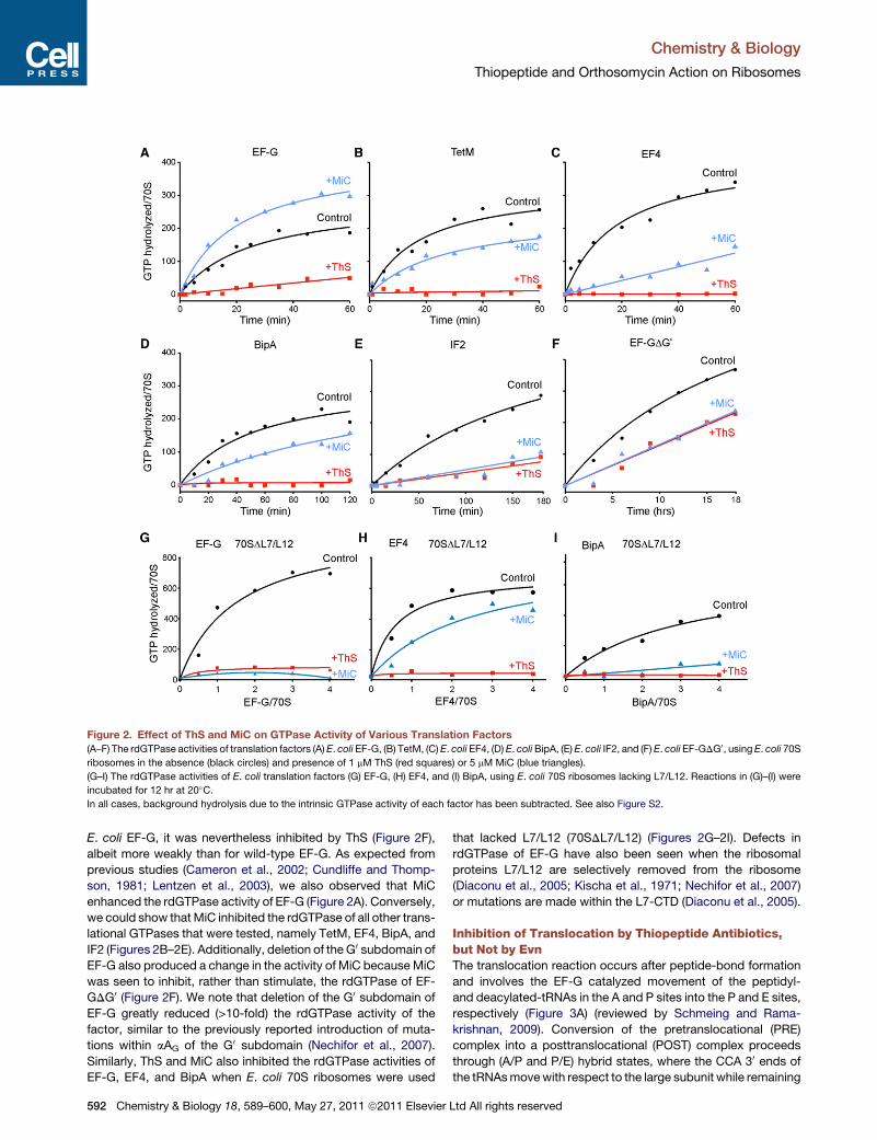
Figure 2. Effect of ThS and MiC on GTPase Activity of Various Translation Factors
(A–F) The rdGTPase activities of translation factors (A) E. coli EF-G, (B) TetM, (C) E. coli EF4, (D) E. coli BipA, (E) E. coli IF2, and (F) E. coli EF-GDG0, using E. coli 70S
ribosomes in the absence (black circles) and presence of 1 mM ThS (red squares) or 5 mM MiC (blue triangles).
(G–I) The rdGTPase activities of E. coli translation factors (G) EF-G, (H) EF4, and (I) BipA, using E. coli 70S ribosomes lacking L7/L12. Reactions in (G)–(I) were
incubated for 12 hr at 20�C.
In all cases, background hydrolysis due to the intrinsic GTPase activity of each factor has been subtracted. See also Figure S2.
Chemistry & Biology
Thiopeptide and Orthosomycin Action on Ribosomes
E. coli EF-G, it was nevertheless inhibited by ThS (Figure 2F),
albeit more weakly than for wild-type EF-G. As expected from
previous studies (Cameron et al., 2002; Cundliffe and Thomp-
son, 1981; Lentzen et al., 2003), we also observed that MiC
enhanced the rdGTPase activity of EF-G (Figure 2A). Conversely,
we could show that MiC inhibited the rdGTPase of all other trans-
lational GTPases that were tested, namely TetM, EF4, BipA, and
IF2 (Figures 2B–2E). Additionally, deletion of the G0 subdomain of
EF-G also produced a change in the activity of MiC because MiC
was seen to inhibit, rather than stimulate, the rdGTPase of EF-
GDG0 (Figure 2F). We note that deletion of the G0 subdomain of
EF-G greatly reduced (>10-fold) the rdGTPase activity of the
factor, similar to the previously reported introduction of muta-
tions within aAG of the G0 subdomain (Nechifor et al., 2007).
Similarly, ThS and MiC also inhibited the rdGTPase activities of
EF-G, EF4, and BipA when E. coli 70S ribosomes were used
592 Chemistry & Biology 18, 589–600, May 27, 2011 ª2011 Elsevier
that lacked L7/L12 (70SDL7/L12) (Figures 2G–2I). Defects in
rdGTPase of EF-G have also been seen when the ribosomal
proteins L7/L12 are selectively removed from the ribosome
(Diaconu et al., 2005; Kischa et al., 1971; Nechifor et al., 2007)
or mutations are made within the L7-CTD (Diaconu et al., 2005).
Inhibition of Translocation by Thiopeptide Antibiotics,but Not by EvnThe translocation reaction occurs after peptide-bond formation
and involves the EF-G catalyzed movement of the peptidyl-
and deacylated-tRNAs in the A and P sites into the P and E sites,
respectively (Figure 3A) (reviewed by Schmeing and Rama-
krishnan, 2009). Conversion of the pretranslocational (PRE)
complex into a posttranslocational (POST) complex proceeds
through (A/P and P/E) hybrid states, where the CCA 30 ends of
the tRNAs move with respect to the large subunit while remaining
Ltd All rights reserved
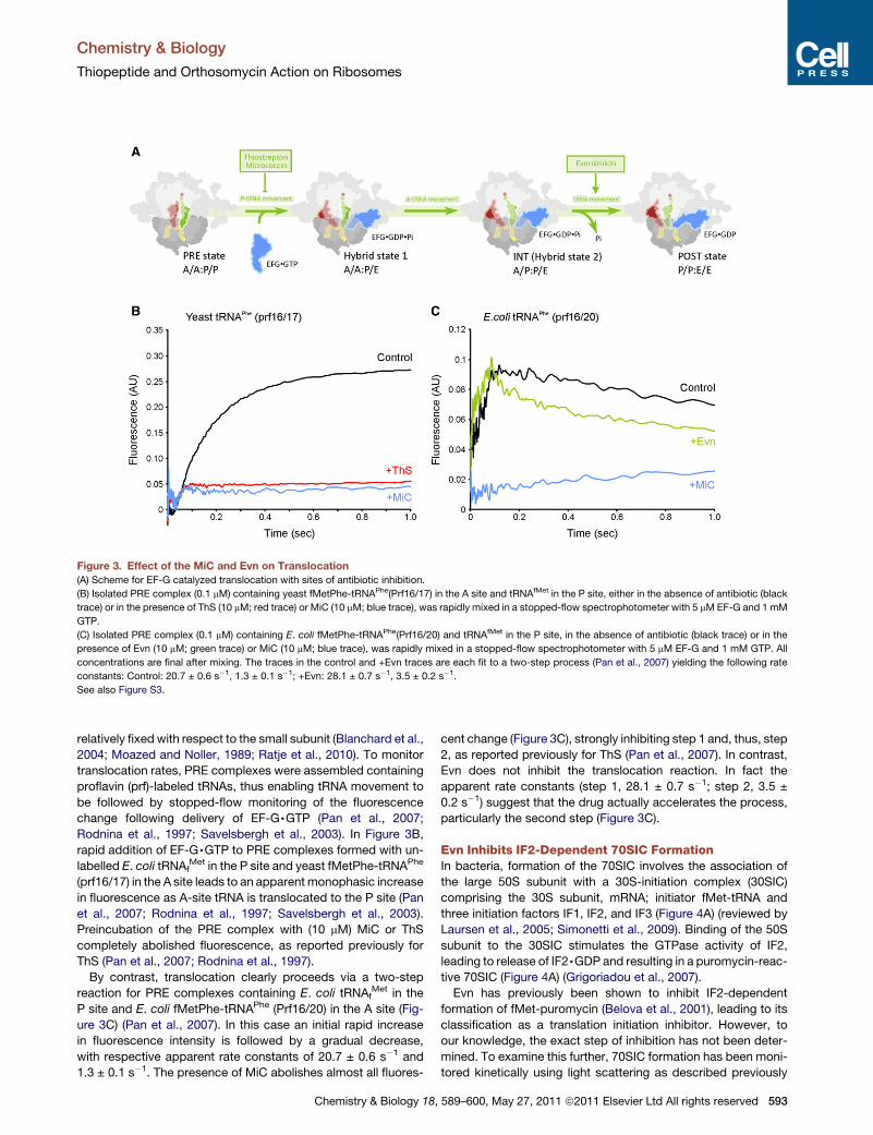
Figure 3. Effect of the MiC and Evn on Translocation
(A) Scheme for EF-G catalyzed translocation with sites of antibiotic inhibition.
(B) Isolated PRE complex (0.1 mM) containing yeast fMetPhe-tRNAPhe(Prf16/17) in the A site and tRNAfMet in the P site, either in the absence of antibiotic (black
trace) or in the presence of ThS (10 mM; red trace) or MiC (10 mM; blue trace), was rapidly mixed in a stopped-flow spectrophotometer with 5 mM EF-G and 1 mM
GTP.
(C) Isolated PRE complex (0.1 mM) containing E. coli fMetPhe-tRNAPhe(Prf16/20) and tRNAfMet in the P site, in the absence of antibiotic (black trace) or in the
presence of Evn (10 mM; green trace) or MiC (10 mM; blue trace), was rapidly mixed in a stopped-flow spectrophotometer with 5 mM EF-G and 1 mM GTP. All
concentrations are final after mixing. The traces in the control and +Evn traces are each fit to a two-step process (Pan et al., 2007) yielding the following rate
constants: Control: 20.7 ± 0.6 s�1, 1.3 ± 0.1 s�1; +Evn: 28.1 ± 0.7 s�1, 3.5 ± 0.2 s�1.
See also Figure S3.
Chemistry & Biology
Thiopeptide and Orthosomycin Action on Ribosomes
relatively fixed with respect to the small subunit (Blanchard et al.,
2004; Moazed and Noller, 1989; Ratje et al., 2010). To monitor
translocation rates, PRE complexes were assembled containing
proflavin (prf)-labeled tRNAs, thus enabling tRNA movement to
be followed by stopped-flow monitoring of the fluorescence
change following delivery of EF-G,GTP (Pan et al., 2007;
Rodnina et al., 1997; Savelsbergh et al., 2003). In Figure 3B,
rapid addition of EF-G,GTP to PRE complexes formed with un-
labelled E. coli tRNAfMet in the P site and yeast fMetPhe-tRNAPhe
(prf16/17) in the A site leads to an apparent monophasic increase
in fluorescence as A-site tRNA is translocated to the P site (Pan
et al., 2007; Rodnina et al., 1997; Savelsbergh et al., 2003).
Preincubation of the PRE complex with (10 mM) MiC or ThS
completely abolished fluorescence, as reported previously for
ThS (Pan et al., 2007; Rodnina et al., 1997).
By contrast, translocation clearly proceeds via a two-step
reaction for PRE complexes containing E. coli tRNAfMet in the
P site and E. coli fMetPhe-tRNAPhe (Prf16/20) in the A site (Fig-
ure 3C) (Pan et al., 2007). In this case an initial rapid increase
in fluorescence intensity is followed by a gradual decrease,
with respective apparent rate constants of 20.7 ± 0.6 s�1 and
1.3 ± 0.1 s�1. The presence of MiC abolishes almost all fluores-
Chemistry & Biology 18,
cent change (Figure 3C), strongly inhibiting step 1 and, thus, step
2, as reported previously for ThS (Pan et al., 2007). In contrast,
Evn does not inhibit the translocation reaction. In fact the
apparent rate constants (step 1, 28.1 ± 0.7 s�1; step 2, 3.5 ±
0.2 s�1) suggest that the drug actually accelerates the process,
particularly the second step (Figure 3C).
Evn Inhibits IF2-Dependent 70SIC FormationIn bacteria, formation of the 70SIC involves the association of
the large 50S subunit with a 30S-initiation complex (30SIC)
comprising the 30S subunit, mRNA; initiator fMet-tRNA and
three initiation factors IF1, IF2, and IF3 (Figure 4A) (reviewed by
Laursen et al., 2005; Simonetti et al., 2009). Binding of the 50S
subunit to the 30SIC stimulates the GTPase activity of IF2,
leading to release of IF2,GDP and resulting in a puromycin-reac-
tive 70SIC (Figure 4A) (Grigoriadou et al., 2007).
Evn has previously been shown to inhibit IF2-dependent
formation of fMet-puromycin (Belova et al., 2001), leading to its
classification as a translation initiation inhibitor. However, to
our knowledge, the exact step of inhibition has not been deter-
mined. To examine this further, 70SIC formation has been moni-
tored kinetically using light scattering as described previously
589–600, May 27, 2011 ª2011 Elsevier Ltd All rights reserved 593
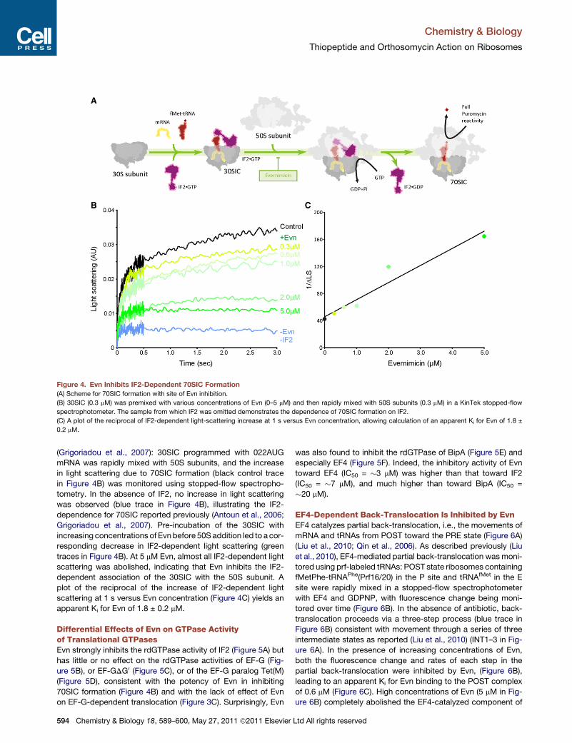
Figure 4. Evn Inhibits IF2-Dependent 70SIC Formation
(A) Scheme for 70SIC formation with site of Evn inhibition.
(B) 30SIC (0.3 mM) was premixed with various concentrations of Evn (0–5 mM) and then rapidly mixed with 50S subunits (0.3 mM) in a KinTek stopped-flow
spectrophotometer. The sample from which IF2 was omitted demonstrates the dependence of 70SIC formation on IF2.
(C) A plot of the reciprocal of IF2-dependent light-scattering increase at 1 s versus Evn concentration, allowing calculation of an apparent Ki for Evn of 1.8 ±
0.2 mM.
Chemistry & Biology
Thiopeptide and Orthosomycin Action on Ribosomes
(Grigoriadou et al., 2007): 30SIC programmed with 022AUG
mRNA was rapidly mixed with 50S subunits, and the increase
in light scattering due to 70SIC formation (black control trace
in Figure 4B) was monitored using stopped-flow spectropho-
tometry. In the absence of IF2, no increase in light scattering
was observed (blue trace in Figure 4B), illustrating the IF2-
dependence for 70SIC reported previously (Antoun et al., 2006;
Grigoriadou et al., 2007). Pre-incubation of the 30SIC with
increasing concentrations of Evn before 50S addition led to a cor-
responding decrease in IF2-dependent light scattering (green
traces in Figure 4B). At 5 mM Evn, almost all IF2-dependent light
scattering was abolished, indicating that Evn inhibits the IF2-
dependent association of the 30SIC with the 50S subunit. A
plot of the reciprocal of the increase of IF2-dependent light
scattering at 1 s versus Evn concentration (Figure 4C) yields an
apparent Ki for Evn of 1.8 ± 0.2 mM.
Differential Effects of Evn on GTPase Activityof Translational GTPasesEvn strongly inhibits the rdGTPase activity of IF2 (Figure 5A) but
has little or no effect on the rdGTPase activities of EF-G (Fig-
ure 5B), or EF-GDG0 (Figure 5C), or of the EF-G paralog Tet(M)
(Figure 5D), consistent with the potency of Evn in inhibiting
70SIC formation (Figure 4B) and with the lack of effect of Evn
on EF-G-dependent translocation (Figure 3C). Surprisingly, Evn
594 Chemistry & Biology 18, 589–600, May 27, 2011 ª2011 Elsevier
was also found to inhibit the rdGTPase of BipA (Figure 5E) and
especially EF4 (Figure 5F). Indeed, the inhibitory activity of Evn
toward EF4 (IC50 = �3 mM) was higher than that toward IF2
(IC50 = �7 mM), and much higher than toward BipA (IC50 =
�20 mM).
EF4-Dependent Back-Translocation Is Inhibited by EvnEF4 catalyzes partial back-translocation, i.e., the movements of
mRNA and tRNAs from POST toward the PRE state (Figure 6A)
(Liu et al., 2010; Qin et al., 2006). As described previously (Liu
et al., 2010), EF4-mediated partial back-translocation was moni-
tored using prf-labeled tRNAs: POST state ribosomes containing
fMetPhe-tRNAPhe(Prf16/20) in the P site and tRNAfMet in the E
site were rapidly mixed in a stopped-flow spectrophotometer
with EF4 and GDPNP, with fluorescence change being moni-
tored over time (Figure 6B). In the absence of antibiotic, back-
translocation proceeds via a three-step process (blue trace in
Figure 6B) consistent with movement through a series of three
intermediate states as reported (Liu et al., 2010) (INT1–3 in Fig-
ure 6A). In the presence of increasing concentrations of Evn,
both the fluorescence change and rates of each step in the
partial back-translocation were inhibited by Evn, (Figure 6B),
leading to an apparent Ki for Evn binding to the POST complex
of 0.6 mM (Figure 6C). High concentrations of Evn (5 mM in Fig-
ure 6B) completely abolished the EF4-catalyzed component of
Ltd All rights reserved
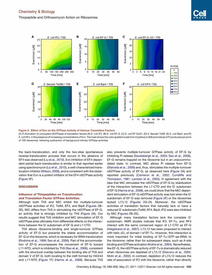
Figure 5. Effect of Evn on the GTPase Activity of Various Translation Factors
(A–F) Activation of uncoupled rdGTPases of translation factors (A) E. coli IF2, (B) E. coli EF-G, (C) E. coli EF-GDG0, (D) S. faecalis TetM, (E) E. coli BipA, and (F)
E. coli EF4, in the presence of increasing concentrations of Evn. The inset shows the color gradient scale from 0 (yellow) to 800 pmol (blue) of Pi produced per pmol
of 70S ribosomes, following subtraction of background intrinsic GTPase activities.
Chemistry & Biology
Thiopeptide and Orthosomycin Action on Ribosomes
the back-translocation, and only the two-step spontaneous
reverse-translocation process that occurs in the absence of
EF4 was observed (Liu et al., 2010). Evn inhibition of EF4-depen-
dent partial back-translocation is similar to that reported earlier
using spectinomycin (Liu et al., 2010), a well-characterized trans-
location inhibitor (Wilson, 2009), and is consistent with the obser-
vation that Evn is a potent inhibitor of the EF4 rdGTPase activity
(Figure 5F).
DISCUSSION
Influence of Thiopeptides on Translocationand Translation Factor GTPase ActivitiesAlthough both ThS and MiC inhibit the multiple-turnover
rdGTPase activities of IF2, TetM, EF4, and BipA (Figures 2B–
2E), MiC differs from ThS in stimulating the rdGTPase of EF-G,
an activity that is strongly inhibited by ThS (Figure 2A). Our
results suggest that ThS inhibition and MiC stimulation of EF-G
rdGTPase arise ultimately from differential effects on the interac-
tions between the G0 subdomain of EF-G and L7-CTD.
ThS allows ribosome-binding and single-turnover GTPase
activity of EF-G but prevents the stable accommodation of
EF-G on the ribosome, which is necessary for tRNA translocation
(Rodnina et al., 1999; Seo et al., 2006). Part of the accommoda-
tion of EF-G encompasses the movement of EF-G toward
L11-NTD, which is inhibited by ThS (Seo et al., 2006), consistent
with the structural overlap between the binding site of ThS and
domain V of EF-G, both locating to the cleft formed by H43/44
and L11-NTD (Figure 1F) (Harms et al., 2008). Because ThS
Chemistry & Biology 18,
also prevents multiple-turnover GTPase activity of EF-G by
inhibiting Pi release (Savelsbergh et al., 2003; Seo et al., 2006),
EF-G remains trapped on the ribosome but in an unaccommo-
dated state. In contrast, MiC allows Pi release from EF-G
(Starosta et al., 2009) and, thus, stimulates the multiple-turnover
rdGTPase activity of EF-G, as observed here (Figure 2A) and
reported previously (Cameron et al., 2002; Cundliffe and
Thompson, 1981; Lentzen et al., 2003). In agreement with the
idea that MiC stimulates the rdGTPase of EF-G by stabilization
of the interaction between the L7-CTD and the G0 subdomain
of EF-G (Harms et al., 2008), we could show that the MiC-depen-
dent stimulation of EF-G rdGTPase activity was lost when the G0
subdomain of EF-G was removed (Figure 2F) or the ribosomes
lacked L7/L12 (Figures 2G–2I). Moreover, the rdGTPase
activities of translation factors that naturally lack or have a
reduced G0 subdomain (TetM, EF4, BipA, IF2) were also inhibited
by MiC (Figures 2B–2E).
Although many translation factors lack the complete G0
subdomain, NMR studies indicate that IF2, EF-Tu, and RF3
interact with the same conserved region of L7-CTD as EF-G
(Helgstrand et al., 2007). L7/L12 has been proposed to interact
with helix aD1 of domain I of EF-Tu. However, this interaction is
more important for initial binding of EF-Tu,GTP,aa-tRNA to
the ribosome, rather than for subsequent steps, such as A-site
binding and GTPase activation (Kothe et al., 2004). Nevertheless,
like EF-G, the rdGTPase activity of EF-Tu is dramatically reduced
when ribosomes are depleted of L7/L12 (Diaconu et al., 2005;
Mohr et al., 2002). In contrast, depletion of L7/L12 reduces the
rate of association of IF2 with the ribosome, rather than directly
589–600, May 27, 2011 ª2011 Elsevier Ltd All rights reserved 595

Figure 6. Evn Inhibits EF4-Mediated Back-Translocation(A) Scheme for EF4-catalyzed back-translocation with sites of antibiotic inhibition.
(B) Fluorescence changes over different time scales. Isolated POST complex (0.1 mM) containing fMetPhe-tRNAPhe(Prf) in the P site and tRNAfMet in the E site,
premixed with 0.15 mM tRNAfMet, to increase E-site occupancy, were rapidly mixed in a stopped-flow spectrophotometer with 3 mM EF4, 0.5 mM GDPNP, and
different concentrations of Evn as indicated on the figure. All concentrations are final after mixing. Lines through the traces are fit to a kinetic model (Liu et al., 2010)
in which back-translocation proceeds via a three-step process in the absence of Evn (the first two of which are catalyzed by EF4) and via a two-step process at
saturating Evn.
(C) A plot of the reciprocal of the apparent magnitude of the fluorescence change for the second, EF4-catalyzed step versus Evn concentration, giving an
apparent Ki for Evn binding to the POST complex of 0.6 ± 0.1 mM.
Chemistry & Biology
Thiopeptide and Orthosomycin Action on Ribosomes
affecting GTP hydrolysis and Pi release (Huang et al., 2010). We
observed that the rdGTPase of LepA and BipA with 70SDL7/L12
was significantly reduced but was also inhibited by both MiC and
ThS (Figures 2H and 2I). Further work will be needed to distin-
guish between factor binding versus GTPase activity defects.
Despite the contrasting effects of MiC and ThS on the GTPase
activities of EF-G (Figure 2A) (Cameron et al., 2002; Cundliffe and
Thompson, 1981; Lentzen et al., 2003), our kinetic analysis
demonstrates that MiC, like ThS, is a potent inhibitor of the trans-
location reaction (Figure 3B). The finding that MiC targets trans-
location is in agreement with original conclusions of Pestka and
Brot (1971), which were based upon its inhibition of poly(U)-
dependent poly(Phe) synthesis but its lack of affect on either
aa-tRNA binding or peptide-bond formation. Given the similarity
in binding site between MiC and ThS, it thus appears likely that
both MiC and ThS inhibit translocation analogously—namely,
by preventing the transition of EF-G from an initially weaker
binding state to a fully accommodated state on the ribosome,
which, we would suggest, is necessary for translocation (Seo
et al., 2006). We note that despite their diverse effects on the
rdGTPase of EF-G, the inhibitory potency of MiC and ThS with
respect to in vitro transcription-translation systems is compa-
rable (both have an IC50 of �3 mM) (Figure S3).
596 Chemistry & Biology 18, 589–600, May 27, 2011 ª2011 Elsevier
Influence of the Orthosomycin Evn on TranslationFactor ActivitiesUnlike thiopeptides, we find that the orthosomycin Evn has no
inhibitory effect on rdGTPase activity of EF-G (Figure 5B) or on
the EF-G-mediated translocation reaction (Figure 3C). This is
consistent with the lack of overlap between the putative Evn-
binding site and the binding position of EF-G determined by
structural studies (Connell et al., 2007; Gao et al., 2009; Ratje
et al., 2010) (Figure 7A). Similarly, Evn does not inhibit the
rdGTPase of Tet(M) (Figure 5D) or of Tet(O) (data not shown),
which interacts with the ribosome in an analogous manner to
EF-G (Spahn et al., 2001). In contrast we find that Evn is a potent
inhibitor of the rdGTPase activity of IF2 (Figure 5A), BipA, and
EF4 (Figures 5E and 5F). Although little is known about the struc-
ture or function of BipA on the ribosome (deLivron et al., 2009),
structures of EF4 alone (Evans et al., 2008) and bound to the
ribosome (Connell et al., 2008) reveal an overall similarity with
EF-G. One exception is the unique CTD of EF4 (Evans et al.,
2008), which on the ribosome is oriented back toward the large
subunit (Connell et al., 2008) and encroaches upon the Evn-
binding site (Figure 7B). Such overlap is consistent with our
finding that Evn is a potent inhibitor EF4-mediated back-translo-
cation reaction (Figure 6B). Our results demonstrating potent
Ltd All rights reserved
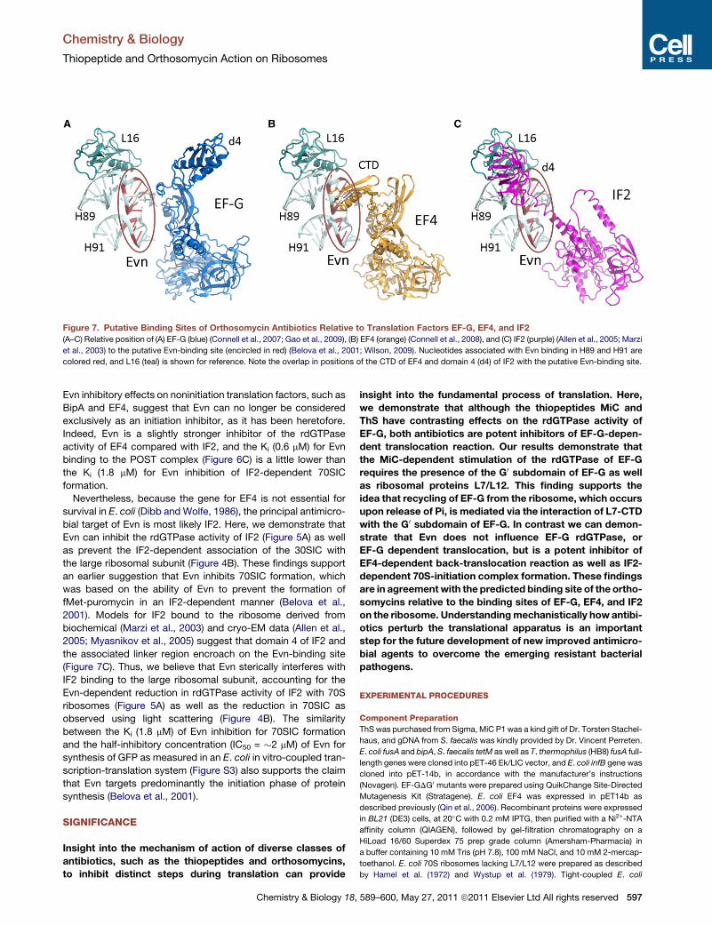
Figure 7. Putative Binding Sites of Orthosomycin Antibiotics Relative to Translation Factors EF-G, EF4, and IF2
(A–C) Relative position of (A) EF-G (blue) (Connell et al., 2007; Gao et al., 2009), (B) EF4 (orange) (Connell et al., 2008), and (C) IF2 (purple) (Allen et al., 2005; Marzi
et al., 2003) to the putative Evn-binding site (encircled in red) (Belova et al., 2001; Wilson, 2009). Nucleotides associated with Evn binding in H89 and H91 are
colored red, and L16 (teal) is shown for reference. Note the overlap in positions of the CTD of EF4 and domain 4 (d4) of IF2 with the putative Evn-binding site.
Chemistry & Biology
Thiopeptide and Orthosomycin Action on Ribosomes
Evn inhibitory effects on noninitiation translation factors, such as
BipA and EF4, suggest that Evn can no longer be considered
exclusively as an initiation inhibitor, as it has been heretofore.
Indeed, Evn is a slightly stronger inhibitor of the rdGTPase
activity of EF4 compared with IF2, and the Ki (0.6 mM) for Evn
binding to the POST complex (Figure 6C) is a little lower than
the Ki (1.8 mM) for Evn inhibition of IF2-dependent 70SIC
formation.
Nevertheless, because the gene for EF4 is not essential for
survival in E. coli (Dibb and Wolfe, 1986), the principal antimicro-
bial target of Evn is most likely IF2. Here, we demonstrate that
Evn can inhibit the rdGTPase activity of IF2 (Figure 5A) as well
as prevent the IF2-dependent association of the 30SIC with
the large ribosomal subunit (Figure 4B). These findings support
an earlier suggestion that Evn inhibits 70SIC formation, which
was based on the ability of Evn to prevent the formation of
fMet-puromycin in an IF2-dependent manner (Belova et al.,
2001). Models for IF2 bound to the ribosome derived from
biochemical (Marzi et al., 2003) and cryo-EM data (Allen et al.,
2005; Myasnikov et al., 2005) suggest that domain 4 of IF2 and
the associated linker region encroach on the Evn-binding site
(Figure 7C). Thus, we believe that Evn sterically interferes with
IF2 binding to the large ribosomal subunit, accounting for the
Evn-dependent reduction in rdGTPase activity of IF2 with 70S
ribosomes (Figure 5A) as well as the reduction in 70SIC as
observed using light scattering (Figure 4B). The similarity
between the Ki (1.8 mM) of Evn inhibition for 70SIC formation
and the half-inhibitory concentration (IC50 = �2 mM) of Evn for
synthesis of GFP as measured in an E. coli in vitro-coupled tran-
scription-translation system (Figure S3) also supports the claim
that Evn targets predominantly the initiation phase of protein
synthesis (Belova et al., 2001).
SIGNIFICANCE
Insight into the mechanism of action of diverse classes of
antibiotics, such as the thiopeptides and orthosomycins,
to inhibit distinct steps during translation can provide
Chemistry & Biology 18,
insight into the fundamental process of translation. Here,
we demonstrate that although the thiopeptides MiC and
ThS have contrasting effects on the rdGTPase activity of
EF-G, both antibiotics are potent inhibitors of EF-G-depen-
dent translocation reaction. Our results demonstrate that
the MiC-dependent stimulation of the rdGTPase of EF-G
requires the presence of the G0 subdomain of EF-G as well
as ribosomal proteins L7/L12. This finding supports the
idea that recycling of EF-G from the ribosome, which occurs
upon release of Pi, is mediated via the interaction of L7-CTD
with the G0 subdomain of EF-G. In contrast we can demon-
strate that Evn does not influence EF-G rdGTPase, or
EF-G dependent translocation, but is a potent inhibitor of
EF4-dependent back-translocation reaction as well as IF2-
dependent 70S-initiation complex formation. These findings
are in agreement with the predicted binding site of the ortho-
somycins relative to the binding sites of EF-G, EF4, and IF2
on the ribosome. Understanding mechanistically how antibi-
otics perturb the translational apparatus is an important
step for the future development of new improved antimicro-
bial agents to overcome the emerging resistant bacterial
pathogens.
EXPERIMENTAL PROCEDURES
Component Preparation
ThS was purchased from Sigma, MiC P1 was a kind gift of Dr. Torsten Stachel-
haus, and gDNA from S. faecalis was kindly provided by Dr. Vincent Perreten.
E. coli fusA and bipA, S. faecalis tetM as well as T. thermophilus (HB8) fusA full-
length genes were cloned into pET-46 Ek/LIC vector, and E. coli infB gene was
cloned into pET-14b, in accordance with the manufacturer’s instructions
(Novagen). EF-GDG0 mutants were prepared using QuikChange Site-Directed
Mutagenesis Kit (Stratagene). E. coli EF4 was expressed in pET14b as
described previously (Qin et al., 2006). Recombinant proteins were expressed
in BL21 (DE3) cells, at 20�C with 0.2 mM IPTG, then purified with a Ni2+-NTA
affinity column (QIAGEN), followed by gel-filtration chromatography on a
HiLoad 16/60 Superdex 75 prep grade column (Amersham-Pharmacia) in
a buffer containing 10 mM Tris (pH 7.8), 100 mM NaCl, and 10 mM 2-mercap-
toethanol. E. coli 70S ribosomes lacking L7/L12 were prepared as described
by Hamel et al. (1972) and Wystup et al. (1979). Tight-coupled E. coli
589–600, May 27, 2011 ª2011 Elsevier Ltd All rights reserved 597
Chemistry & Biology
Thiopeptide and Orthosomycin Action on Ribosomes
ribosomes, cloned E. coli His-tagged proteins EF-G, EF-Tu, IF1, IF2, and IF3,
and E. coli [35S]fMet-tRNAfMet; E. coli [3H]Phe-tRNAPhe were prepared as
described (Liu et al., 2010). MFK-mRNA was purchased from Dharmacon
(Lafayette, CO, USA) with sequences 50-GGG AAG GAG GUA AAA AUG
UUU AAA CGU AAA UCU ACU-30 (initiator codon underlined).
IF2-Dependent 70SIC Formation Light-Scattering Assay
This assay was performed as described (Grigoriadou et al., 2007). 30SIC
was formed by mixing 0.3 mM 30S, 0.45 mM IF1, 0.45 mM IF3, 0.45 mM fMet-
tRNAfMet, 0.15 mM IF2, 0.9 mM AUG022-mRNA (Grigoriadou et al., 2007),
and 100 mM GTP, premixed with various concentrations of Evn (0–5 mM) and
then rapidly mixed with 50S subunits (0.3 mM) in a KinTek stopped-flow spec-
trophotometer. Excitation was at 436 nm, and light scattering was determined
using a 455 nm cutoff filter. All concentrations are final after mixing.
Back-Translocation Assay
All of the following complexes were made up in buffer A (20 mM HEPES-KOH
[pH 7.5], at 0�C, 150 mM NH4Ac, 4.5 mM MgAc2, 4 mM b-mercaptoethanol,
0.05 mM spermine, and 2 mM spermidine) at 37�C. Initiation complex was
formed by incubating WT ribosomes (2 mM) with mRNA MFK (8 mM), IF1
(3 mM), IF2 (3 mM), IF3 (3 mM), GTP (1 mM), and [35S]fMet-tRNAfMet (3 mM) for
25 min. Ternary complex was formed by incubating EF-Tu (6 mM) with labeled
Phe-tRNAPhe (3 mM), GTP (1 mM), phosphoenolpyruvate (Roche Diagnostics)
(1.5 mM), and pyruvate kinase (Roche Diagnostics) (0.015 mg/ml) for 15 min.
POST complexes were formed by incubating ternary complex and initiation
complex at 37�C briefly for 45 s and then in the presence of EF-G (molar ratio
of EF-G:ribosome was 0.2:1) and GTP (1 mM) at 37�C for 10 min. Then they
were purified by ultracentrifugation through a 1.1 M Sucrose cushion in buffer
A (450,000 3 g, 40 min, 4�C). POST complex concentration was calculated
from the amount of ribosome-bound fMet-[3H]Phe-tRNAPhe. Stopped-flow
fluorescence experiments were performed using an SX.18MV Stopped-Flow
Spectrofluorometer (Applied Photophysics). POST complex (0.1 mM) contain-
ing fMetPhe-tRNAPhe(Prf) in the P site and tRNAfMet in the E site was rapidly
mixed with 0.15 mM tRNAfMet, 3 mM EF4,GDPNP, and various concentrations
of Evn (0–5 mM). prf was excited at 460 nm, and fluorescence was monitored
using a 495 nm long-pass filter. Lines through the data are fit to triple-exponen-
tial equations using the program IGOR Pro (WaveMetrics).
Translocation Assay
PRE complexes were formed by incubating initiation complex and ternary
complex at 37�C for 45 s. Then they were purified by ultracentrifugation
through a 1.1 M Sucrose cushion in buffer A with 20 mM Mg2+ (450,000 3 g,
40 min, 4�C). PRE complex concentration was calculated from the amount
of ribosome-bound fMet-[3H]Phe-tRNAPhe. Stopped-flow fluorescence exper-
iments were performed using an SX.18MV Stopped-Flow spectrofluorometer.
prf was excited at 460 nm, and fluorescence was monitored using a 495 nm
long-pass filter. Data are fit to double-exponential equations using the
program IGOR Pro (WaveMetrics).
Malachite Green GTPase Activity Assays
GTPase activity was measured using the Malachite Green Phosphate Kit
(BioAssay) that quantifies the green complex formed between malachite
green, molybdate, and free orthophosphate. Unless otherwise mentioned, all
reactions contained 30 nM E. coli 70S ribosomes, 20 mM GTP, and 60 nM
protein in the presence or absence of antibiotics as necessary. Reactions
were transferred into 96-well microtiter plates, and color formation was
measured on Tecan Infinite M1000 microplate reader at 650 nm. Reactions
performed in the absence of ribosomes were used as a background signal
to account for the intrinsic GTPase activity of the translation factor.
In Vitro Transcription-Translation Assay
All coupled transcription-translation experiments were performed using an
E. coli lysate-based system in the presence and absence of antibiotics as
described previously (Starosta et al., 2009, 2010). Reactions were transferred
into 96-well microtiter plates, and the GFP fluorescence was measured with
a Typhoon Scanner 9400 (Amersham Bioscience) using a Typhoon blue laser
module (Amersham Bioscience). Images were then quantified using Image-
598 Chemistry & Biology 18, 589–600, May 27, 2011 ª2011 Elsevier
Quant TL (GE Healthcare) and represented graphically using SigmaPlot (Systat
Software, Inc.).
Figure Preparation
Chemical structures for the precursor compounds were drawn using
ChemDraw (Advanced Chemistry Development, Inc., Toronto, Canada), and
all structural figures were prepared with PyMOL (http://www.pymol.org).
SUPPLEMENTAL INFORMATION
Supplemental Information includes three figures and can be found with this
article online at doi:10.1016/j.chembiol.2011.03.010.
ACKNOWLEDGMENTS
We would like to thank Torsten Stachelhaus for preparation of the Micrococcin
P1 and Dr. Vincent Perreten for providing S. faecalis gDNA. This work was
financed by the EMBO young investigator program (to D.N.W.), Deutsche
Forschungsgemeinschaft (WI3285/1-1 to D.N.W.), and by the National Insti-
tutes of Health (GM071014 to B.S.C.).
Received: February 6, 2011
Revised: March 7, 2011
Accepted: March 14, 2011
Published: May 26, 2011
REFERENCES
Aarestrup, F.M., and Jensen, L.B. (2000). Presence of variations in ribosomal
protein L16 corresponding to susceptibility of enterococci to oligosaccharides
(Avilamycin and evernimicin). Antimicrob. Agents Chemother. 44, 3425–3427.
Adrian, P.V., Mendrick, C., Loebenberg, D., McNicholas, P., Shaw, K.J.,
Klugman, K.P., Hare, R.S., and Black, T.A. (2000a). Evernimicin (SCH27899)
inhibits a novel ribosome target site: analysis of 23S ribosomal DNA mutants.
Antimicrob. Agents Chemother. 44, 3101–3106.
Adrian, P.V., Zhao, W., Black, T.A., Shaw, K.J., Hare, R.S., and Klugman, K.P.
(2000b). Mutations in ribosomal protein L16 conferring reduced susceptibility
to evernimicin (SCH27899): implications for mechanism of action. Antimicrob.
Agents Chemother. 44, 732–738.
Allen, G.S., Zavialov, A., Gursky, R., Ehrenberg, M., and Frank, J. (2005). The
cryo-EM structure of a translation initiation complex from Escherichia coli. Cell
121, 703–712.
Antoun, A., Pavlov, M.Y., Lovmar, M., and Ehrenberg, M. (2006). How initiation
factors tune the rate of initiation of protein synthesis in bacteria. EMBO J. 25,
2539–2550.
Bagley, M.C., Dale, J.W., Merritt, E.A., and Xiong, X. (2005). Thiopeptide anti-
biotics. Chem. Rev. 105, 685–714.
Belova, L., Tenson, T., Xiong, L.Q., McNicholas, P.M., and Mankin, A.S. (2001).
A novel site of antibiotic action in the ribosome: interaction of evernimicin with
the large ribosomal subunit. Proc. Natl. Acad. Sci. USA 98, 3726–3731.
Blanchard, S.C., Kim, H.D., Gonzalez, R.L., Jr., Puglisi, J.D., and Chu, S.
(2004). tRNA dynamics on the ribosome during translation. Proc. Natl. Acad.
Sci. USA 101, 12893–12898.
Blanchard, S.C., Cooperman, B.S., and Wilson, D.N. (2010). Probing transla-
tion with small-molecule inhibitors. Chem. Biol. 17, 633–645.
Brandi, L., Marzi, S., Fabbretti, A., Fleischer, C., Hill, W., Lodmell, J., and
Gualerzi, C. (2004). The translation initiation functions of IF2: targets for thio-
strepton inhibition. J. Mol. Biol. 335, 881–894.
Cameron, D.M., Thompson, J., March, P.E., and Dahlberg, A.E. (2002).
Initiation factor IF2, thiostrepton and micrococcin prevent the binding of elon-
gation factor G to the Escherichia coli ribosome. J. Mol. Biol. 319, 27–35.
Connell, S.R., Tracz, D.M., Nierhaus, K.H., and Taylor, D.E. (2003a). Ribosomal
protection proteins and their mechanism of tetracycline resistance.
Antimicrob. Agents Chemother. 47, 3675–3681.
Ltd All rights reserved
Chemistry & Biology
Thiopeptide and Orthosomycin Action on Ribosomes
Connell, S.R., Trieber, C.A., Dinos, G.P., Einfeldt, E., Taylor, D.E., and
Nierhaus, K.H. (2003b). Mechanism of Tet(O)-mediated tetracycline resis-
tance. EMBO J. 22, 945–953.
Connell, S.R., Takemoto, C., Wilson, D.N., Wang, H., Murayama, K., Terada,
T., Shirouzu, M., Rost, M., Schuler, M., Giesebrecht, J., et al. (2007).
Structural basis for interaction of the ribosome with the switch regions of
GTP-bound elongation factors. Mol. Cell 25, 751–764.
Connell, S.R., Topf, M., Qin, Y., Wilson, D.N., Mielke, T., Fucini, P., Nierhaus,
K.H., and Spahn, C.M. (2008). A new tRNA intermediate revealed on the
ribosome during EF4-mediated back-translocation. Nat. Struct. Mol. Biol.
15, 910–915.
Cundliffe, E., and Thompson, J. (1981). Concerning the mode of action of
micrococcin upon bacterial protein synthesis. Eur. J. Biochem. 118, 47–52.
Dantley, K.A., Dannelly, H.K., and Burdett, V. (1998). Binding interaction
between Tet(M) and the ribosome: requirements for binding. J. Bacteriol.
180, 4089–4092.
Datta, P.P., Sharma, M.R., Qi, L., Frank, J., and Agrawal, R.K. (2005).
Interaction of the G0 domain of elongation factor G and the C-terminal domain
of ribosomal protein L7/L12 during translocation as revealed by cryo-EM. Mol.
Cell 20, 723–731.
deLivron, M.A., and Robinson, V.L. (2008). Salmonella enterica serovar
Typhimurium BipA exhibits two distinct ribosome binding modes. J. Bacteriol.
190, 5944–5952.
deLivron, M.A., Makanji, H.S., Lane, M.C., and Robinson, V.L. (2009). A novel
domain in translational GTPase BipA mediates interaction with the 70S ribo-
some and influences GTP hydrolysis. Biochemistry 48, 10533–10541.
Diaconu, M., Kothe, U., Schlunzen, F., Fischer, N., Harms, J.M., Tonevitsky,
A.G., Stark, H., Rodnina, M.V., and Wahl, M.C. (2005). Structural basis for
the function of the ribosomal L7/12 stalk in factor binding and GTPase activa-
tion. Cell 121, 991–1004.
Dibb, N.J., and Wolfe, P.B. (1986). lep operon proximal gene is not required for
growth or secretion by Escherichia coli. J. Bacteriol. 166, 83–87.
Evans, R.N., Blaha, G., Bailey, S., and Steitz, T.A. (2008). The structure of
LepA, the ribosomal back translocase. Proc. Natl. Acad. Sci. USA 105,
4673–4678.
Gao, Y.G., Selmer, M., Dunham, C.M., Weixlbaumer, A., Kelley, A.C., and
Ramakrishnan, V. (2009). The structure of the ribosome with elongation factor
G trapped in the posttranslocational state. Science 326, 694–699.
Gonzalez, R.L., Jr., Chu, S., and Puglisi, J.D. (2007). Thiostrepton inhibition of
tRNA delivery to the ribosome. RNA 13, 2091–2097.
Grigoriadou, C., Marzi, S., Kirillov, S., Gualerzi, C.O., and Cooperman, B.S.
(2007). A quantitative kinetic scheme for 70 S translation initiation complex
formation. J. Mol. Biol. 373, 562–572.
Grunberg-Manago, M., Dondon, J., and Graffe, M. (1972). Inhibition by thio-
strepton of the IF-2-dependent ribosomal GTPase. FEBS Lett. 22, 217–221.
Hamel, E., Koka, M., and Nakamoto, T. (1972). Requirement of an E. coli 50S
ribosomal protein component for effective interaction of the ribosome with T
and G factors and with guanosine triphosphate. J. Biol. Chem. 247, 805–814.
Harms, J.M., Wilson, D.N., Schluenzen, F., Connell, S.R., Stachelhaus, T.,
Zaborowska, Z., Spahn, C.M., and Fucini, P. (2008). Translational regulation
via L11: molecular switches on the ribosome turned on and off by thiostrepton
and micrococcin. Mol. Cell 30, 26–38.
Helgstrand, M., Mandava, C.S., Mulder, F.A., Liljas, A., Sanyal, S., and Akke,
M. (2007). The ribosomal stalk binds to translation factors IF2, EF-Tu, EF-G
and RF3 via a conserved region of the L12 C-terminal domain. J. Mol. Biol.
365, 468–479.
Huang, C., Mandava, C.S., and Sanyal, S. (2010). The ribosomal stalk plays
a key role in IF2-mediated association of the ribosomal subunits. J. Mol.
Biol. 399, 145–153.
Hughes, R.A., and Moody, C.J. (2007). From amino acids to heteroaromatics—
thiopeptide antibiotics, nature’s heterocyclic peptides. Angew. Chem. Int. Ed.
Engl. 46, 7930–7954.
Kischa, K., Moller, W., and Stoffler, G. (1971). Reconstitution of a GTPase
activity by a 50S ribosomal protein from E. coli. Nat. New Biol. 233, 62–63.
Chemistry & Biology 18,
Knutsson Jenvert, R.M., and Holmberg Schiavone, L. (2005). Characterization
of the tRNA and ribosome-dependent pppGpp-synthesis by recombinant
stringent factor from Escherichia coli. FEBS J. 272, 685–695.
Kofoed, C.B., and Vester, B. (2002). Interaction of avilamycin with ribosomes
and resistance caused by mutations in 23S rRNA. Antimicrob. Agents
Chemother. 46, 3339–3342.
Kothe, U., Wieden, H.J., Mohr, D., and Rodnina, M.V. (2004). Interaction of
helix D of elongation factor Tu with helices 4 and 5 of protein L7/12 on the ribo-
some. J. Mol. Biol. 336, 1011–1021.
Laurberg, M., Kristensen, O., Martemyanov, K., Gudkov, A.T., Nagaev, I.,
Hughes, D., and Liljas, A. (2000). Structure of a mutant EF-G reveals domain
III and possibly the fusidic acid binding site. J. Mol. Biol. 303, 593–603.
Laursen, B.S., Sorensen, H.P., Mortensen, K.K., and Sperling-Petersen, H.U.
(2005). Initiation of protein synthesis in bacteria. Microbiol. Mol. Biol. Rev. 69,
101–123.
Lefranc, D., and Ciufolini, M.A. (2009). Total synthesis and stereochemical
assignment of micrococcin P1. Angew. Chem. Int. Ed. Engl. 48, 4198–4201.
Lentzen, G., Klinck, R., Matassova, N., Aboul-ela, F., and Murchie, A. (2003).
Structural basis for contrasting activities of ribosome binding thiazole antibi-
otics. Chem. Biol. 10, 769–778.
Liu, H., Pan, D., Pech, M., and Cooperman, B.S. (2010). Interrupted catalysis:
the EF4 (LepA) effect on back-translocation. J. Mol. Biol. 396, 1043–1052.
Mann, P.A., Xiong, L., Mankin, A.S., Chau, A.S., Mendrick, C.A., Najarian, D.J.,
Cramer, C.A., Loebenberg, D., Coates, E., Murgolo, N.J., et al. (2001). EmtA,
a rRNA methyltransferase conferring high-level evernimicin resistance. Mol.
Microbiol. 41, 1349–1356.
Marzi, S., Knight, W., Brandi, L., Caserta, E., Soboleva, N., Hill, W.E., Gualerzi,
C.O., and Lodmell, J.S. (2003). Ribosomal localization of translation initiation
factor IF2. RNA 9, 958–969.
McConkey, G.A., Rogers, M.J., and McCutchan, T.F. (1997). Inhibition of
Plasmodium falciparum protein synthesis. Targeting the plastid-like organelle
with thiostrepton. J. Biol. Chem. 272, 2046–2049.
McNicholas, P.M., Najarian, D.J., Mann, P.A., Hesk, D., Hare, R.S., Shaw, K.J.,
and Black, T.A. (2000). Evernimicin binds exclusively to the 50S ribosomal
subunit and inhibits translation in cell-free systems derived from both Gram-
positive and Gram-negative bacteria. Antimicrob. Agents Chemother. 44,
1121–1126.
McNicholas, P.M., Mann, P.A., Najarian, D.J., Miesel, L., Hare, R.S., and Black,
T.A. (2001). Effects of mutations in ribosomal protein L16 on susceptibility and
accumulation of evernimicin. Antimicrob. Agents Chemother. 45, 79–83.
Moazed, D., and Noller, H.F. (1989). Intermediate states in the movement of
transfer RNA in the ribosome. Nature 342, 142–148.
Modolell, J., Cabrer, B., Parmeggiani, A., and Vazquez, D. (1971). Inhibition by
siomycin and thiostrepton of both aminoacyl-tRNA and factor G binding to
ribosomes. Proc. Natl. Acad. Sci. USA 68, 1796–1800.
Mohr, D., Wintermeyer, W., and Rodnina, M.V. (2002). GTPase activation of
elongation factors Tu and G on the ribosome. Biochemistry 41, 12520–12528.
Munro, J.B., Wasserman, M.R., Altman, R.B., Wang, L., and Blanchard, S.C.
(2010). Correlated conformational events in EF-G and the ribosome regulate
translocation. Nat. Struct. Mol. Biol. 17, 1470–1477.
Myasnikov, A.G., Marzi, S., Simonetti, A., Giuliodori, A.M., Gualerzi, C.O.,
Yusupova, G., Yusupov, M., and Klaholz, B.P. (2005). Conformational transi-
tion of initiation factor 2 from the GTP- to GDP-bound state visualized on the
ribosome. Nat. Struct. Mol. Biol. 12, 1145–1149.
Nechifor, R., Murataliev, M., and Wilson, K.S. (2007). Functional interactions
between the G0 subdomain of bacterial translation factor EF-G and ribosomal
protein L7/L12. J. Biol. Chem. 282, 36998–37005.
Nicolaou, K.C., Safina, B.S., Zak, M., Lee, S.H., Nevalainen, M., Bella, M.,
Estrada, A.A., Funke, C., Zecri, F.J., and Bulat, S. (2005a). Total synthesis of
thiostrepton. Retrosynthetic analysis and construction of key building blocks.
J. Am. Chem. Soc. 127, 1159–1175.
Nicolaou, K.C., Zak, M., Safina, B.S., Estrada, A.A., Lee, S.H., and Nevalainen,
M. (2005b). Total synthesis of thiostrepton. Assembly of key building blocks
and completion of the synthesis. J. Am. Chem. Soc. 127, 11176–11183.
589–600, May 27, 2011 ª2011 Elsevier Ltd All rights reserved 599

Chemistry & Biology
Thiopeptide and Orthosomycin Action on Ribosomes
Nicolaou, K.C., Chen, J.S., Edmonds, D.J., and Estrada, A.A. (2009). Recent
advances in the chemistry and biology of naturally occurring antibiotics.
Angew. Chem. Int. Ed. Engl. 48, 660–719.
Otaka, T., and Kaji, A. (1974). Micrococcin: acceptor-site-specific inhibitor of
protein synthesis. Eur. J. Biochem. 50, 101–106.
Owens, R.M., Pritchard, G., Skipp, P., Hodey, M., Connell, S.R., Nierhaus,
K.H., and O’Connor, C.D. (2004). A dedicated translation factor controls the
synthesis of the global regulator Fis. EMBO J. 23, 3375–3385.
Pan, D., Kirillov, S.V., and Cooperman, B.S. (2007). Kinetically competent
intermediates in the translocation step of protein synthesis. Mol. Cell 25,
519–529.
Pestka, S. (1970). Thiostrepton: a ribosomal inhibitor of translocation.
Biochem. Biophys. Res. Commun. 40, 667–674.
Pestka, S., and Brot, N. (1971). Studies on the formation of transfer ribonucleic
acid-ribosome complexes. IV. Effect of antibiotics on steps of bacterial protein
synthesis: some new ribosomal inhibitors of translocation. J. Biol. Chem. 246,
7715–7722.
Qin, Y., Polacek, N., Vesper, O., Staub, E., Einfeldt, E., Wilson, D.N., and
Nierhaus, K.H. (2006). The highly conserved LepA is a ribosomal elongation
factor that back-translocates the ribosome. Cell 127, 721–733.
Ratje, A., Loerke, J., Mikolajka, A., Brunner, M., Hildebrand, P.W., Starosta,
A.L., Donhofer, A., Connell, S.R., Fucini, P., Mielke, T., et al. (2010). Head
swivel on the ribosome facilitates translocation by means of intra-subunit
tRNA hybrid sites. Nature 468, 713–716.
Rodnina, M.V., Savelsbergh, A., Katunin, V.I., and Wintermeyer, W. (1997).
Hydrolysis of GTP by elongation factor G drives tRNA movement on the ribo-
some. Nature 385, 37–41.
Rodnina, M.V., Savelsbergh, A., Matassova, N.B., Katunin, V.I., Semenkov,
Y.P., and Wintermeyer, W. (1999). Thiostrepton inhibits the turnover but not
the GTPase of elongation factor G on the ribosome. Proc. Natl. Acad. Sci.
USA 96, 9586–9590.
Savelsbergh, A., Mohr, D., Wilden, B., Wintermeyer, W., and Rodnina, M.V.
(2000). Stimulation of the GTPase activity of translation elongation factor G
by ribosomal protein L7/12. J. Biol. Chem. 275, 890–894.
Savelsbergh, A., Katunin, V.I., Mohr, D., Peske, F., Rodnina, M.V., and
Wintermeyer, W. (2003). An elongation factor G-induced ribosome rearrange-
ment precedes tRNA-mRNA translocation. Mol. Cell 11, 1517–1523.
Savelsbergh, A., Mohr, D., Kothe, U., Wintermeyer, W., and Rodnina, M.V.
(2005). Control of phosphate release from elongation factor G by ribosomal
protein L7/12. EMBO J. 24, 4316–4323.
600 Chemistry & Biology 18, 589–600, May 27, 2011 ª2011 Elsevier
Schmeing, T.M., and Ramakrishnan, V. (2009). What recent ribosome
structures have revealed about the mechanism of translation. Nature 461,
1234–1242.
Seo, H.S., Abedin, S., Kamp, D., Wilson, D.N., Nierhaus, K.H., and
Cooperman, B.S. (2006). EF-G-dependent GTPase on the ribosome.
Conformational change and fusidic acid inhibition. Biochemistry 45, 2504–
2514.
Simonetti, A., Marzi, S., Jenner, L., Myasnikov, A., Romby, P., Yusupova, G.,
Klaholz, B.P., and Yusupov, M. (2009). A structural view of translation initiation
in bacteria. Cell. Mol. Life Sci. 66, 423–436.
Sohmen, D., Harms, J.M., Schlunzen, F., and Wilson, D.N. (2009a). Enhanced
SnapShot: antibiotic inhibition of protein synthesis II. Cell 139, 212–212.e1.
Sohmen, D., Harms, J.M., Schlunzen, F., and Wilson, D.N. (2009b). SnapShot:
antibiotic inhibition of protein synthesis I. Cell 138, 1248.e1.
Spahn, C.M., Blaha, G., Agrawal, R.K., Penczek, P., Grassucci, R.A., Trieber,
C.A., Connell, S.R., Taylor, D.E., Nierhaus, K.H., and Frank, J. (2001).
Localization of the ribosomal protection protein Tet(O) on the ribosome and
the mechanism of tetracycline resistance. Mol. Cell 7, 1037–1045.
Starosta, A.L., Qin, H., Mikolajka, A., Leung, G.Y., Schwinghammer, K.,
Nicolaou, K.C., Chen, D.Y., Cooperman, B.S., and Wilson, D.N. (2009).
Identification of distinct thiopeptide-antibiotic precursor lead compounds
using translation machinery assays. Chem. Biol. 16, 1087–1096.
Starosta, A.L., Karpenko, V.V., Shishkina, A.V., Mikolajka, A., Sumbatyan,
N.V., Schluenzen, F., Korshunova, G.A., Bogdanov, A.A., and Wilson, D.N.
(2010). Interplay between the ribosomal tunnel, nascent chain, and macrolides
influences drug inhibition. Chem. Biol. 17, 1–10.
Weisblum, B., and Demohn, V. (1970). Inhibition by thiostrepton of the
formation of a ribosome-bound guanine nucleotide complex. FEBS Lett. 11,
149–152.
Wilson, D.N. (2009). The A-Z of bacterial translation inhibitors. Crit. Rev.
Biochem. Mol. Biol. 44, 393–433.
Wolf, H. (1973). Avilamycin, an inhibitor of the 30S ribosomal subunits function.
FEBS Lett. 36, 181–186.
Wystup, G., Teraoka, H., Schulze, H., Hampl, H., and Nierhaus, K.H. (1979).
50S subunit from Escherichia coli ribosomes. Isolation of active ribosomal
proteins and protein complexes. Eur. J. Biochem. 100, 101–113.
Zarazaga, M., Tenorio, C., Del Campo, R., Ruiz-Larrea, F., and Torres, C.
(2002). Mutations in ribosomal protein L16 and in 23S rRNA in Enterococcus
strains for which evernimicin MICs differ. Antimicrob. Agents Chemother. 46,
3657–3659.
Ltd All rights reserved

Paper 7

LETTERdoi:10.1038/nature09547
Head swivel on the ribosome facilitates translocationby means of intra-subunit tRNA hybrid sitesAndreas H. Ratje1,2, Justus Loerke1, Aleksandra Mikolajka3,4, Matthias Brunner1, Peter W. Hildebrand1, Agata L. Starosta3,Alexandra Donhofer3, Sean R. Connell5, Paola Fucini5, Thorsten Mielke1,6, Paul C. Whitford7, Jose N. Onuchic8, Yanan Yu9,Karissa Y. Sanbonmatsu7, Roland K. Hartmann2, Pawel A. Penczek10, Daniel N. Wilson3,4 & Christian M. T. Spahn1
The elongation cycle of protein synthesis involves the delivery ofaminoacyl-transfer RNAs to the aminoacyl-tRNA-binding site(A site) of the ribosome, followed by peptide-bond formation andtranslocation of the tRNAs through the ribosome to reopen theA site1,2. The translocation reaction is catalysed by elongation factorG (EF-G) in a GTP-dependent manner3. Despite the availability ofstructures of various EF-G–ribosome complexes, the precise mech-anism by which tRNAs move through the ribosome still remainsunclear. Here we use multiparticle cryoelectron microscopy analysisto resolve two previously unseen subpopulations within Thermusthermophilus EF-G–ribosome complexes at subnanometre resolu-tion, one of them with a partly translocated tRNA. Comparison ofthese substates reveals that translocation of tRNA on the 30S sub-unit parallels the swivelling of the 30S head and is coupled tounratcheting of the 30S body. Because the tRNA maintains contactwith the peptidyl-tRNA-binding site (P site) on the 30S head andsimultaneously establishes interaction with the exit site (E site) onthe 30S platform, a novel intra-subunit ‘pe/E’ hybrid state is formed.This state is stabilized by domain IV of EF-G, which interacts withthe swivelled 30S-head conformation. These findings provide directstructural and mechanistic insight into the ‘missing link’ in termsof tRNA intermediates involved in the universally conservedtranslocation process.
After peptide-bond formation, pre-translocational (PRE) ribosomescarry a peptidyl-tRNA at the A site and a deacylated tRNA at theP site1,3,4. This is a highly dynamic state of the ribosome, which fluc-tuates between classical states with A tRNA and P tRNA and hybridstates with A/P tRNAs (A/P denotes that the tRNA is in the A site onthe 30S subunit and the P site on the 50S subunit) and P/E tRNAs5–8.Hybrid state formation is coupled to spontaneous rotation of the 30Ssubunit relative to the 50S subunit9–11 and is stabilized by the binding ofEF-G12–14. The ratchet-like subunit rearrangement induced by EF-Gand eukaryotic elongation factor 2 (eEF2) also includes a swivel move-ment of the head that is roughly orthogonal to the inter-subunit rota-tion of the ribosomal subunits14–16. EF-G catalyses translocation of thehybrid-state tRNAs on the 30S subunit to form a post-translocational(POST) state ribosome with tRNAs located at classical P and E sites.The translocation process is accelerated by the GTPase activity of EF-Gstimulated by the ribosome17,18. However, it is still not known howtRNAs are translocated with respect to the 30S subunit and how themessenger RNA is advanced by one codon.
Structural snapshots of the translocation process come from cryo-electron microscopy and X-ray analysis of EF-G bound to ribosomecomplexes12–14,19. Despite considerable effort12,20–22, no direct structuralinformation is available for ribosomal PRE complexes simultaneously
containing EF-G and an A tRNA. It seems that this state is too dynamicand transient to be captured, resulting in either a POST state contain-ing EF-G or a PRE state without EF-G bound20,22. Indeed, the stableEF-G-bound POST state determined by X-ray crystallography revealsa non-ratcheted ribosome with tRNAs in classical P/P and E/E sites19.Therefore, structural insights into intermediate states of transloca-tion—that is, ratcheted ribosomal EF-G complexes—have used com-plexes without an A-site peptidyl-tRNA12–14.
In this study we used the antibiotic fusidic acid (FA) to stall EF-G onthe 70S ribosome. FA permits the hydrolysis of GTP by EF-G but pre-vents the associated changes in EF-G that normally accompany hydro-lysis19. After complex formation and the collection of cryoelectronmicroscopy data, we employed multiparticle refinement to resolvethe heterogeneity of the data (Supplementary Fig. 1). In the first phaseof multiparticle refinement a large population of particle images ofribosomes containing EF-G was obtained that yielded a structure in aratcheted conformation. As refinement progressed to higher resolution,the presence of intrinsic conformational heterogeneity necessitated asecond phase of multiparticle refinement, resulting in two final recon-structions of the 70S–EF-G–GDP–FA complex (Fig. 1), at resolutions of7.6 A and 7.8 A (using a 0.5 Fourier shell correlation cut-off criterion),respectively (Supplementary Fig. 2). Both maps have density attributedto EF-G, but they show significant conformational differences (Sup-plementary Movies 1 and 2): specifically, the substates are distinguishedby different degrees of subunit ratcheting and positioning of the L1protuberance, and also by the swivel movement of the head of the30S subunit relative to the body/platform (Fig. 1e, f, SupplementaryFigs 3 and 4, and Supplementary Table 1). Unlike previous cryoelectronmicroscopy and X-ray structures of ribosome–EF-G complexes, move-ment of the head and body/platform of the 30S subunit is uncoupled: insubstate I, the 30S subunit is ratcheted by about 7u relative to the 50Ssubunit, but there is only a modest (roughly 5u) swivelling of the 30Shead. In contrast, the 30S in substate II is only ratcheted by about 4u, butthere is a very large (roughly 18u) swivel of the head (SupplementaryTable 1).
The identification of two different ratcheted substates within the70S–EF-G–GDP–FA complex prompted us to investigate whethersuch intrinsic heterogeneity also exists in our previous 70S–EF-G–GMPPNP cryoelectron microscopy data set14. Additional multiparticlerefinement did indeed reveal that the 70S–EF-G–GMPPNP complexcould be subdivided into two substates that seemed to be equivalent tothose identified in the 70S–EF-G–GDP–FA complex (SupplementaryFig. 5 and Supplementary Table 1). Our findings here provide evidencesupporting the emerging energy-landscape model that allows thesampling of several metastable conformations for a defined ribosomal
1Institut fur Medizinische Physik und Biophysik, Charite – Universitatsmedizin Berlin, Ziegelstrasse 5–9, 10117-Berlin, Germany. 2Institut fur Pharmazeutische Chemie, Philipps-Universitat Marburg,35037 Marburg, Germany. 3Gene Center and Department of Biochemistry, Ludwig-Maximilians-Universitat, Feodor-Lynenstrasse 25, 81377 Munchen, Germany. 4Center for Integrated Protein Science,Ludwig-Maximilians-Universitat Munchen, 81377 Munchen, Germany. 5Frankfurt Institute for Molecular Life Sciences, Institute of Organic Chemistry and Chemical Biology, Goethe University Frankfurt,Max-von Laue-Strasse 7, D-60438 Frankfurt am Main, Germany. 6UltraStrukturNetzwerk, Max Planck Institute for Molecular Genetics, 14195 Berlin, Germany. 7Theoretical Biology and Biophysics Group,TheoreticalDivision, Los Alamos National Laboratory, Los Alamos,New Mexico 87545, USA. 8Center for TheoreticalBiological Physics and DepartmentofPhysics, University of California, San Diego, La Jolla,California 92093, USA. 9Florida State University, Department of Computer Science, Tallahassee, Florida 32306, USA. 10The University of Texas – Houston Medical School, 6431 Fannin, Houston, Texas77030, USA.
2 D E C E M B E R 2 0 1 0 | V O L 4 6 8 | N A T U R E | 7 1 3
Macmillan Publishers Limited. All rights reserved©2010

complex8. The subnanometre resolution of the 70S–EF-G–GDP–FAsubpopulations enabled the visualization of secondary structure andthus the generation of molecular models (Fig. 1) by applying ournewly developed MDFIT algorithm (see Methods). Comparison withavailable structures reveals that substate I is similar in conformation tothe ratcheted substate of the PRE complex9,10 (Supplementary Table 1)and also that the tRNA is bound in a hybrid P/E site (Fig. 2a). Wetherefore consider substate I to be related to a pre-translocational inter-mediate (TIPRE).
In contrast, substate II of the 70S–EF-G–GDP–FA complex repre-sents a novel conformational state of the 70S ribosome. The anticodonstem-loop (ASL) of the tRNA has moved by 8–10 A compared with theP/E position of TIPRE as it maintains strong association with the P-sitecomponents of the head and follows the large 18u swivel movement ofthe head (Fig. 2b and Supplementary Movie 3). Because the tRNAinteracts simultaneously with P-site components of the head as wellas E-site components of the platform, it can be thought of as a 30S
intra-subunit hybrid site (Fig. 2b). Moreover, because the contacts ofthe CCA end of the tRNA with the E site on the 50S subunit remainunaffected, we extend the previous nomenclature of hybrid sites5 andrefer to this newly identified state as a pe/E hybrid state (P site on headand E site on platform of the 30S subunit, and E site on the 50S subunit).
The ASL of the pe/E tRNA together with the bound mRNA codon isvery close to the position of a fully translocated E/E tRNA (Fig. 2c andSupplementary Movie 4). Apparently, head-swivelling coupled withpartial unratcheting of the body/platform of the 30S subunit leads totRNA translocation, suggesting that substate II of the 70S–EF-G–GDP–FA complex is related to a post-translocational intermediatestate (TIPOST). We note that although the intermediate states visua-lized here contain only one tRNA, a second tRNA (ap/P) can be super-imposed on the TIPOST state without steric interference with thebinding position of EF-G (Supplementary Fig. 6). Thus, the structurespresented here seem to be valid models for translocation intermediates(see also Supplementary Information for further discussion), butstructures of translocation intermediates with two tRNAs will benecessary for the validation of these predictions. The presence of theratchet-like subunit rearrangement in the yeast 80S–eEF2–sordarincomplex15 hints that translocation in prokaryotes and eukaryotesmay use related intermediate conformations. This structure showeda strong head swivel comparable to that of bacterial TIPOST combinedwith a strong inter-subunit rotation of bacterial TIPRE. Thus, the con-formation of the 80S–eEF2–sordarin complex15, although obtained byclassical single-particle methods, may present a further intermediatebetween TIPRE and TIPOST.
Whereas the P/E and pe/E tRNAs are in a twisted conformation(Fig. 2d), the overall conformations of EF-G in TIPRE and TIPOST areremarkably similar to each other (Fig. 1) and to that observed in thecryoelectron microscopy reconstruction of the 70S–EF-G–GMPPNP14
as well as in the recent X-ray structure of EF-G–GDP–FA bound to aPOST-state ribosome19 (Fig. 3a). However, one difference between thetwo EF-G–GDP–FA substates relates to the interaction patterns ofdomain IV of EF-G: in the TIPRE state, domain IV does not seem to
a b
c d
e f
L1
CP
L7
EF-G
H69
pe/E
P/E
bk
sp
h
b
pt
18°5°
4°7°
Figure 1 | Substates I (TIPRE) and II (TIPOST) of the 70S–EF-G–GDP–FAcomplex. a–d, Cryoelectron microscopy maps of TIPRE (a, b) and TIPOST
(c, d) of the 70S–EF-G–GDP–FA complex are shown in mesh form withdocked models in ribbon representation: EF-G (red), tRNA (green), 23S/5SrRNA (blue), 50S ribosomal proteins (orange), 16S rRNA (yellow) and the 30Sribosomal proteins (magenta). The maps are shown from the 30S side with the30S subunit computationally removed (a, c) and from the 50S side with the 50Ssubunit computationally removed (b, d). CP, central protuberance; bk, beak; sp,spur; pt, platform; h, head; b, back; L1 and L7, ribosomal proteins L1 and L7,respectively; H69, helix 69 of 23S rRNA. e, f, The 30S subunit of TIPRE (e) andTIPOST (f) (yellow) is compared with the 30S subunit of the POST state19 (grey)by aligning the respective 50S subunits. Arrows with numbers indicate thedirection and magnitude (Supplementary Table 1) of the inter-subunit rotationand the head-swivel from the unrotated state to TIPRE or TIPOST, respectively.
a b
c dA PE
mRNAP/E pe/E
P/E P A/TP/E pe/E
Figure 2 | Localization and conformation of the tRNA of substates I (TIPRE)and II (TIPOST). a, b, Close-up of the tRNA-binding regions of the 30S subunitof TIPRE (a) and TIPOST (b). The 30S subunit and tRNAs are shown as yellowand blue ribbons, respectively; ribosomal residues that contact A, P and E tRNA(magenta, green and orange) are shown as spheres. c, In a common 50Salignment, the P/E tRNA (green) of TIPRE and the pe/E tRNA (magenta) ofTIPOST together with their respective mRNA codons are compared with mRNAand classical A, P and E tRNA positions (grey). d, Density for the tRNAs (wiremesh) with molecular models for the P/E tRNA of TIPRE (left, green) and thepe/E tRNA of TIPOST (middle, magenta). On the right, the model for P/E tRNA(green), which is essentially the same as that for pe/E-tRNA (root mean squareddeviation 1.5 A), is compared with a classical P tRNA (blue) and an A/T tRNA(yellow) by alignment of the acceptor-stem, D-stem and T-stem loops.
RESEARCH LETTER
7 1 4 | N A T U R E | V O L 4 6 8 | 2 D E C E M B E R 2 0 1 0
Macmillan Publishers Limited. All rights reserved©2010
interact significantly with the ribosome (Fig. 3b), whereas a small shiftin the binding position of EF-G and the large swivel of the headfacilitate a more extensive interaction of domain IV of EF-G with helixh34 of the 16S rRNA in the TIPOST state (Fig. 3c). Together, h34 andthe nucleotide 530 region of 16S rRNA form the so-called latch of themRNA entry channel. Because of the head-swivel in the TIPOST state,h34 has moved about 12 A away from the 530 region, leading to anopening of the latch (Fig. 3c) similar to that observed previously wheneEF2 was trapped on the yeast 80S ribosome with sordarin15. This mayfacilitate the movement of the mRNA–tRNA2 complex. Consistent
with this observation, transient protection of h34 by EF-G againstchemical modification during the translocation reaction has beenreported previously23. The direct interaction of domain IV of EF-Gwith h34 may therefore bias the energy landscape of the ribosometowards TIPOST.
Until now, intermediate states of inter-subunit rotation have beenconsidered to be intermediates on the pathway to the fully rotatedstate16. The present findings implicate unratcheting (in combinationwith the large swivel of the 30S head), rather than ratcheting, as beingcoupled to the translocation movement of the tRNAs and the mRNAwith respect to the 30S subunit. Collectively, the insights gained fromthe structures of the TIPRE and TIPOST states enable us to provide astructural explanation for the process of translocation in a model inwhich tRNA movements are facilitated by head-swivel, ratcheting andunratcheting motions of the ribosome (Fig. 4). These motions may beinfluenced by the GTPase reaction on EF-G; a network of interactionsinvolving domain III and the ordered switch I region of EF-G and thec-phosphate of GTP was proposed to stabilize the rotated state of the30S subunit14. Accordingly, fast GTP hydrolysis by EF-G17 could desta-bilize the direct and indirect interactions of switch I of EF-G with themaximally rotated 30S subunit14,24, therefore increasing the propensityof the 30S subunit to rotate backwards. The unratcheting motionproduces a counter-movement of the body/platform with respect tothe head, thereby reducing the distance that the tRNAs have to travelduring translocation. Intra-subunit hybrid states allow the 30S subunitto maintain partial contacts with the tRNAs at any time of the trans-location reaction. In the context of the ribosome’s functioning duringtranslocation as a Brownian ratchet machine25, our model suggeststhat EF-G acts as a dynamic pawl, decoupling the unratchetingmotions of the ribosome from the transition of hybrid-state tRNAsback into classical states. EF-G thereby provides directionality andaccelerates translocation of the tRNAs by means of several intermediateinter-subunit and intra-subunit hybrid states into the classical P/P andE/E sites of the POST state.
METHODS SUMMARYTight-coupled 70S ribosomes from Thermus thermophilus were isolated by sucrose-gradient centrifugation and incubated with EF-G in the presence of GTP and FA.The resulting complexes were flash-frozen and imaged under low-dose conditions
a b
c
EF-G
pe/EE/E
bk
sp
h
pt
EF-G
530
h34
h34Latch
h34
pe/E
P/E
IV
VG
III
II
h44
h44
IV
IV
Figure 3 | EF-G stabilizes the swivelled head movement in the TIPOST state.a, Comparison of the position of FA-stalled EF-G and the 30S subunit betweenTIPOST and the POST-state 70S–EF-G–GDP–FA complex19. All showncomponents of the POST-state 70S–EF-G–GDP–FA complex19 are depicted asblue ribbons. The 30S, EF-G and pe/E tRNA of TIPOST are represented by yellow,red and orange ribbons, respectively. b, c, Close-up of the decoding region anddomain IV of EF-G in the same orientation as in a. The surfaces of TIPRE (b) andTIPOST (c) are transparent with molecular models in ribbon representation (30Ssubunit, yellow; EF-G, red, P/E tRNA, green; pe/E tRNA, orange). The arrowsmark the closed latch between helix h34 and the 530 region of TIPRE (b) and theinteraction between h34 and domain IV of EF-G within TIPOST.
PRE state
30S
50S
30S
Ratcheting of head/body
A/A and P/P tRNAs A/P and P/E tRNAs A/P and P/E tRNAs ap/P and pe/E tRNAs P/P and E/E tRNAs
EF-G–GTP
EF-G–GTP
GTP
GTP–Pi
GTP–Pi
GTP
EF-G–GDP
EF-G–GDP
Ratcheting of head/body Back-ratcheting of body Back-ratcheting of body
Head
Head
Body
Body
Hybrid state TIPRE TIPOST
Swivelling of head Swivelling of head
POST state
Back-swivelling of head
a b c d e
Figure 4 | Model for translocation. a, b, The PRE ribosome exists in adynamic equilibrium between base states with classical A/A and P/P tRNAs(a) and rotated states with hybrid A/P and P/E tRNAs6,7,9–11 (b). c, Binding ofEF-G–GTP to PRE state (a) or hybrid state (b) stabilizes the ratcheted state12 asobserved in TIPRE. d, Fast GTP hydrolysis by EF-G17 accelerates translocationby means of an unlocking step on the 30S subunit18. Domain IV of EF-Guncouples unratcheting from the reverse movement of the A/P and P/E tRNAs
back into classical states; that is, a doorstop function. Through a head-swivelling and unratcheting motion, the tRNAs move from aa/P and pp/E intothe 30S intra-subunit ap/P and pe/E hybrid states. e, Complete unratcheting ofthe 30S subunit leads to the POST-state 70S–EF-G complex19. Back-swivellingof the 30S head re-establishes tRNAs in the classical (pp/P) P and E (ee/E)states. Translocation is completed by the dissociation of EF-G–GDP.
LETTER RESEARCH
2 D E C E M B E R 2 0 1 0 | V O L 4 6 8 | N A T U R E | 7 1 5
Macmillan Publishers Limited. All rights reserved©2010

with the use of an FEI Polara G2 electron microscope. The collected data weredigitized and processed with multiparticle refinement protocols implemented inSPIDER26. To interpret the resulting cryoelectron microscopy maps in molecularterms, a newly developed algorithm (MDFIT)27 that integrates molecular simu-lation with experimental maps was employed.
Full Methods and any associated references are available in the online version ofthe paper at www.nature.com/nature.
Received 5 February; accepted 30 September 2010.
1. Frank, J. & Spahn, C. M. The ribosome and the mechanism of protein synthesis.Rep. Prog. Phys. 69, 1383–1417 (2006).
2. Schmeing, T. M. & Ramakrishnan, V. What recent ribosome structures haverevealed about the mechanism of translation. Nature 461, 1234–1242 (2009).
3. Shoji, S., Walker, S. E. & Fredrick, K. Ribosomal translocation: one step closer to themolecular mechanism. ACS Chem. Biol. 4, 93–107 (2009).
4. Ramakrishnan, V. Ribosome structureand the mechanismof translation. Cell 108,557–572 (2002).
5. Moazed, D. & Noller, H. F. Intermediate states in the movement of transfer RNA inthe ribosome. Nature 342, 142–148 (1989).
6. Munro, J. B., Altman, R. B., O’Connor, N. & Blanchard, S. C. Identification of twodistincthybrid state intermediates on the ribosome.Mol. Cell25, 505–517 (2007).
7. Blanchard,S.C.et al. tRNAdynamicson the ribosomeduring translation. Proc.NatlAcad. Sci. USA 101, 12893–12898 (2004).
8. Munro, J. B., Sanbonmatsu, K. Y., Spahn, C. M. & Blanchard, S. C. Navigating theribosome’smetastableenergy landscape.TrendsBiochem.Sci.34,390–400(2009).
9. Agirrezabala, X. et al.Visualization of the hybrid state of tRNAbinding promoted byspontaneous ratcheting of the ribosome. Mol. Cell 32, 190–197 (2008).
10. Julian, P. et al. Structure of ratcheted ribosomes with tRNAs in hybrid states. Proc.Natl Acad. Sci. USA 105, 16924–16927 (2008).
11. Fischer, N. et al. Ribosome dynamics and tRNA movement by time-resolvedelectron cryomicroscopy. Nature 466, 329–333 (2010).
12. Valle, M. et al. Locking and unlocking of ribosomal motions. Cell 114, 123–134(2003).
13. Frank, J. & Agrawal, R. K. A ratchet-like inter-subunit reorganization of theribosome during translocation. Nature 406, 318–322 (2000).
14. Connell, S. R. et al. Structural basis for interaction of the ribosome with the switchregions of GTP-bound elongation factors. Mol. Cell 25, 751–764 (2007).
15. Spahn, C.M.et al. Domainmovements of elongation factor eEF2 and the eukaryotic80S ribosome facilitate tRNA translocation. EMBO J. 23, 1008–1019 (2004).
16. Zhang, W., Dunkle, J. A. & Cate, J. H. Structures of the ribosome in intermediatestates of ratcheting. Science 325, 1014–1017 (2009).
17. Rodnina, M., Savelsbergh, A., Katunin, V. I. & Wintermeyer, W. Hydrolysis of GTP byelongation factor G drives tRNA movement on the ribosome. Nature 385, 37–41(1997).
18. Savelsbergh, A. et al. An elongation factor G-induced ribosome rearrangementprecedes tRNA-mRNA translocation. Mol. Cell 11, 1517–1523 (2003).
19. Gao, Y. G. et al. The structure of the ribosome with elongation factor G trapped inthe posttranslocational state. Science 326, 694–699 (2009).
20. Penczek, P.A., Frank, J.& Spahn, C.M.A method of focusedclassification, based onthe bootstrap 3D variance analysis, and its application to EF-G-dependenttranslocation. J. Struct. Biol. 154, 184–194 (2006).
21. Agrawal, R. K. et al. EF-G-dependent GTP hydrolysis induces translocationaccompaniedby largeconformational changes in the70S ribosome.NatureStruct.Biol. 6, 643–647 (1999).
22. Scheres, S. H. et al. Disentangling conformational states of macromolecules in 3D-EM through likelihood optimization. Nature Methods 4, 27–29 (2007).
23. Matassova, A. B., Rodnina, M. V. & Wintermeyer, W. Elongation factor G-inducedstructural change inhelix34of16S rRNA related to translocation on the ribosome.RNA 7, 1879–1885 (2001).
24. Ticu, C.et al.Conformational changes inswitch I of EF-Gdrive itsdirectional cyclingon and off the ribosome. EMBO J. 28, 2053–2065 (2009).
25. Spirin, A. S. The ribosome as a conveying thermal ratchet machine. J. Biol. Chem.284, 21103–21119 (2009).
26. Schuette, J. C. et al. GTPase activation of elongation factor EF-Tu by the ribosomeduring decoding. EMBO J. 28, 755–765 (2009).
27. Whitford, P.C.et al. Accommodation ofaminoacyl-tRNA into the ribosome involvesreversible excursions along multiple pathways. RNA 16, 1196–1204 (2010).
Supplementary Information is linked to the online version of the paper atwww.nature.com/nature.
Acknowledgements The present work was supported by grants from the DeutscheForschungsgemeinschaft (DFG; SFB 740 TP A3 and TP Z1, SP 1130/2-1 to C.M.T.S.,FU579 1-3 to P.F., HA 1672/7-5 to R.K.H. and WI3285/1-1 to D.N.W.), the EuropeanUnion 3D-EM Network of Excellence (to C.M.T.S.), the European Union andSenatsverwaltung fur Wissenschaft, Forschung und Kultur Berlin(UltraStructureNetwork, Anwenderzentrum) and US National Institutes of Health (NIH;grantGM60635 toP.A.P.), the Clusterof Excellence ‘Macromolecular complexes’ at theGoethe University Frankfurt (DFG Project EXC 115 to P.F. and S.C.), and the HumanFrontiers of Science Program Young Investigators Award HFSP67/07 (to P.F.). Wethank the New Mexico Computing Application Center for generous time on the EncantoSupercomputer. P.C.W. is currently funded by a LANL Director’s Fellowship. This workwas also supported by the Center for Theoretical Biological Physics sponsored by theNational Science Foundation (NSF; grant PHY-0822283) with additional support fromNSF-MCB-0543906, the LANL LDRD program and NIH grant R01-GM072686.
Author Contributions A.M., A.L.S. and A.D. prepared the complexes. A.H.R. and T.M.collected the cryoelectron microscopy data. A.H.R, J.L., M.B., S.R.C. and C.M.T.S. did theimage processing. P.C.W., Y.Y., J.O. and K.Y.S. developed and employed the MDFitmethod. P.W.H. participated in docking and analysed the FA-binding site. A.H.R., R.K.H.,S.R.C., P.F., P.A.P., D.N.W. and C.M.T.S. discussed the results and wrote the paper.
Author Information The electron density maps and models of the TIPRE and the TIPOST
complexes have been deposited in the 3D-EM database with accession numbersEMD-1798 and EMD-1799, and in the Protein Data Bank database with PDB IDs 2xsy,2xtg, 2xux and 2xuy. Reprints and permissions information is available atwww.nature.com/reprints. The authors declare no competing financial interests.Readers are welcome to comment on the online version of this article atwww.nature.com/nature. Correspondence and requests for materials should beaddressed to C.M.T.S. ([email protected]) or D.N.W.([email protected]).
RESEARCH LETTER
7 1 6 | N A T U R E | V O L 4 6 8 | 2 D E C E M B E R 2 0 1 0
Macmillan Publishers Limited. All rights reserved©2010

METHODSFormation of the EF-G–70S–GDP–FA complex. The fusA gene, encoding EF-G,was cloned from Thermus thermophilus HB8 genomic DNA into pET-46 Ek/LICvector using primers (TthEFG_for, 59-GCG CGC CCG GTG GTG ATG CAGCTC TTC CTG GGC TCC GCC CTG AAG AAC-39; TthEFG_rev, 59-GTT CTTCAG GGC GGA GCC CAG GAA GAG CTG CAT CAC CAC CGG GCG CGC-39)in accordance with the manufacturer’s instructions (Novagen) and expressedin BL21 (DE3) cells. Recombinant EF-G protein was then purified with a Ni21-nitrilotriacetate affinity column, followed by gel filtration in a buffer containing10 mM Tris pH 7.8, 100 mM NaCl and 10 mM 2-mercaptoethanol. Tight-coupled70S ribosomes were purified from exponential-phase T. thermophilus cells by usingsucrose-density-gradient centrifugation, as described previously for 30S subunits28.As observed previously, the ribosomes contained a co-purified tRNA14,29. Binding ofEF-G to 70S ribosomes was done by incubating 20mM purified EF-G protein for15 min with 5mM T. thermophilus 70S ribosomes, 500mM GTP and 500mM FA at65 uC, in a buffer containing 10 mM HEPES-KOH pH 7.8, 30 mM MgCl2 and75 mM NH4Cl. The occupancy of EF-G in the complexes was about 60–70%, asjudged by centrifugal binding assay28.Cryoelectron microscopy and image processing. Ribosomal complexes werediluted to a concentration of 30 nM and subsequently frozen onto Quantifoil gridsusing a Vitrobot (FEI) device. Micrographs where collected on a Tecnai G2 Polara(FEI) at 300 kV and a magnification of 339,000 under low-dose conditions(19 e2 A22) and scanned on a D8200 Primscan drum scanner (HeidelbergerDruckmaschinen) with a step size of 4.758mm, corresponding to 1.26 A on thespecimen scale.
The Contrast Transfer Function defocus values for the micrographs were deter-mined with CTFind30. Ribosomal projection images were automatically identifiedwith the program Signature31 and were subsequently screened visually or auto-matically. From the selected projections, a reconstruction was generated by pro-jection matching procedures and refined with the SPIDER software package32. Thecomplete data set comprised 586,848 projection images collected from 677 micro-graphs at a defocus range of 1.3–4.8mm. During the later refinement rounds,positivity of the reference volumes was enforced, the power spectrum of thecryoelectron microscopy map was scaled to the power spectrum of a model densityderived from the atomic coordinates of the X-ray structure of the 70S ribosome33,and the map was subsequently low-pass filtered according to the current resolu-tion estimate.
After a first phase of multiparticle refinement20,26,34,35, performed with three-times or two-times decimated pictures, we obtained a major subpopulation (52%;303,665 particle images) that had strong EF-G density (Supplementary Fig. 1).However, as refinement progressed and the resolution reached the subnanometrerange the data set was deemed heterogeneous. Parts of the 30S subunit, especiallythe head domain, became partly disordered. A second phase of multiparticlerefinement was therefore employed, leading to the subdivision of the data intotwo further substates having EF-G (Supplementary Fig. 1). Both data subsets werefurther refined individually at full image size. The final reconstructions ofsubstate I (113,214 particle images) and substate II (156,332 particle images)reached resolutions of 7.8 A and 7.6 A, respectively (Supplementary Fig. 2).
Using a similar strategy we revisited the previous data set (362,361 particleimages; 371 micrographs) of the 70S–EF-G–GMPPNP complex14. In a first phaseof multiparticle refinement a major population (118,991 particle images) wasfurther sorted by a second phase of multiparticle refinement resulting insubstate I (58,911 particle images) and substate II (38,055 particle images). Theresolutions of the maps were 9.6 A and 10.5 A, respectively. As the resolution forthe reconstructions of the 70S–EF-G–GMPPNP complex was significantly lower(as a result of the smaller size data set) than the resolutions obtained for the 70S–EF-G–GDP–FA complex, we restricted the comparison to a dissection of only theglobal conformational changes, such as ratcheting and head swivelling.
Nevertheless, this analysis suggests that the 70S–EF-G–GMPPNP complexcoexists in two substates that resemble TIPRE and TIPOST of the 70S–EF-G–GDP–FA complex (Supplementary Fig. 5 and Supplementary Table 1). The ratioof particles within each of the two substates of the 70S–EF-G–GMPPNP complexis inverted with respect to the 70S–EF-G–GDP–FA complex: in the 70S–EF-G–GMPPNP complex the particle ratio of TIPRE and TIPOST is about 3:2(58,911:38,055), whereas in the 70S–EF-G–GDP–FA complex the ratio is about2:3 (113,214:156,332). This means that most of the ribosomes in the 70S–EF-Gcomplex stalled with a non-hydrolysable GTP analogue are in TIPRE (substate I),having a fully ratcheted 30S subunit but only a modest head swivel. However, inthe 70S–EF-G complex stalled with GTP and FA, TIPOST dominates and here anintermediate inter-subunit rotation is coupled with a large head swivel instead(Supplementary Figs 4 and 5 and Supplementary Table 1).Structure-based simulation fitting (MDFIT). To determine atomic models con-sistent with the cryoelectron microscopy densities, we employed structure-based
molecular simulation27,36,37 together with an energetic term developed in ref. 38,which incorporates the correlation between the simulated and experimental elec-tron density throughout the simulation. Tama and co-workers38 developed asimilar method, which used a standard explicit solvent force field, as opposed tothe structure-based force field. The advantage of the structure-based force field isthat, because the potential energy function is defined by the X-ray structure,MDFIT retains tertiary contacts present in the X-ray structure without specialconstraints. Furthermore, because MDFIT explicitly includes all non-hydrogenatoms, there are no atomic clashes and proper stereochemistry is maintained in allfits.
We began the MDFIT procedure with a structure-based potential energy func-tion defined by the classical unratcheted conformation. To induce hybrid-stateformation and subunit pivoting we introduced the energetic term based on thecorrelation between the simulated cryoelectron microscopy map and the experi-mentally determined cryoelectron microscopy map. Specifically, the potentialenergy function is
V~VSBzVmap~VSB{WX
ijk
rsimijk r
expijk ð1Þ
where W is the energetic weight of the map and rexpijk and rsim
ijk are the normalized
experimental and simulated electron densities at voxel (i,j,k), respectively. Thequantity VSB is the structure-based potential energy function. To calculate thesimulated map, each atom is described by a Gaussian function of width 5 A withthe tail truncated at 1% of the peak value. Here, because the structure-basedforcefield has 1 unit (all calculations were in reduced units) of stabilizing energyper atom (by construction), we set W to a comparable value of 150,000. Thecontributions to the force due to Vmap were updated every 200 time steps.Fitting simulations employed Langevin dynamics. All simulations were performedwith code based on Gromacs version 4.0.5 (refs 39, 40). Calculations were per-formed on the Encanto Supercomputer. The structure-based force field is freelyavailable online (http://smog.ucsd.edu).Structural models. The crystallographic structure of the 70S ribosome in complexwith EF-G19 (PDB IDs 2WRI and 2WRJ) was used as an initial structure forMDFIT. Proteins without side chains in the X-ray structure were removed. Thecarboxy-terminal domain of ribosomal protein L7 from PDB entry 1RQU41 wasinserted by hand, before fitting. The E-site tRNA was also removed from the initialX-ray structure in accordance with the cryoelectron microscopy map. The P-sitetRNA was included in the fitting process. To facilitate fitting of the tRNA moleculeinto a P/E conformation, stabilizing interactions between the tRNA and the ribo-some were removed. Stabilizing interactions between EF-G and the ribosome (dueto their proximity in the crystal structure) were also excluded from the calcula-tions, as were stabilizing interactions between L7 and all other components in thesystem. Further, crystallographic interactions found between the 39-CCA end ofthe E-site tRNA with the E site of the 50S subunit were reintroduced as short-range(of the type 6-12; see refs 27, 36, 37) attractive interactions between the 39-CCAend of the fitted tRNA and the E site of the 50S subunit. Introducing these inter-actions ensured that the 39-CCA end of the P/E tRNA was in a conformationidentical to that of a classically bound E-site tRNA, although because these inter-actions were short-range they only affected the process once the major rearrange-ments in the tRNA had already been achieved. Codon–anti-codon interactionswere restrained by harmonic interactions with minima corresponding to theclassical configuration.
The tRNA–ribosome–EF-G crystal structure was first manually aligned inVMD42, as a single rigid unit, to the map of TIPRE. The first round of fitting wasperformed with the TIPRE map after subjecting it to a 4-A Gaussian low-pass filter.This filter decreased noise, effectively smoothing the energetic profile associatedwith Vmap, which permitted more rapid fits. After 106 integration steps, the fit wascontinued for an additional 106 steps with Vmap based on the TIPRE map filtered at2 A. The TIPOST map, filtered at 2 A, was then fitted, using the TIPRE 4-A fittedstructure as the initial structure.
28. Sharma, M. R. et al. Interaction of Era with the 30S ribosomal subunit implicationsfor 30S subunit assembly. Mol. Cell 18, 319–329 (2005).
29. Yusupov, M. M. et al. Crystal structure of the ribosome at 5.5 A resolution. Science292, 883–896 (2001).
30. Mindell, J. A.&Grigorieff,N.Accuratedeterminationof localdefocusandspecimentilt in electron microscopy. J. Struct. Biol. 142, 334–347 (2003).
31. Chen, J. Z. & Grigorieff, N. SIGNATURE: a single-particle selection system formolecular electron microscopy. J. Struct. Biol. 157, 168–173 (2006).
32. Frank, J. et al. SPIDER and WEB: processing and visualization of images in 3Delectron microscopy and related fields. J. Struct. Biol. 116, 190–199 (1996).
33. Selmer, M. et al. Structure of the 70S ribosome complexed with mRNA and tRNA.Science 313, 1935–1942 (2006).
34. Spahn, C. M. & Penczek, P. A. Exploring conformational modes of macromolecularassemblies by multiparticle cryo-EM. Curr. Opin. Struct. Biol. 19, 623–631 (2009).
LETTER RESEARCH
Macmillan Publishers Limited. All rights reserved©2010
35. Connell, S. R. et al. A new tRNA intermediate revealed on the ribosome during EF4-mediated back-translocation. Nature Struct. Mol. Biol. 15, 910–915 (2008).
36. Whitford, P. C. et al. An all-atom structure-based potential for proteins: bridgingminimal models with all-atom empirical forcefields. Proteins 75, 430–441 (2009).
37. Whitford, P. C. et al. Nonlocal helix formation is key to understandingS-adenosylmethionine-1 riboswitch function. Biophys. J. 96, L7–L9 (2009).
38. Orzechowski, M. & Tama, F. Flexible fitting of high-resolution x-ray structures intocryoelectron microscopy maps using biased molecular dynamics simulations.Biophys. J. 95, 5692–5705 (2008).
39. Lindahl, E., Hess, B. & van der Spoel, D. J. GROMACS 3.0: a package for molecularsimulation and trajectory analysis. J. Mol. Model. 7, 306–317 (2001).
40. Berendsen, H. J. C., van der Spoel, D. & van Drunen, R. GROMACS: a message-passing parallelmolecular dynamics implementation. Comput. Phys.Commun. 91,43–56 (1995).
41. Bocharov, E. V. et al. From structure and dynamics of protein L7/L12 to molecularswitching in ribosome. J. Biol. Chem. 279, 17697–17706 (2004).
42. Humphrey, W., Dalke, A. & Schulten, K. VMD: visual molecular dynamics. J. Mol.Graph. 14, 33–38 (1996).
RESEARCH LETTER
Macmillan Publishers Limited. All rights reserved©2010
Paper 8
1
Formation of ε(R)-β-lysyl-hydroxylysine on translation
elongation factor EF-P requires YjeK, YjeA and YfcM
Lauri Peil1,4,5, Agata L. Starosta2,4, Kai Virumäe3, Gemma C. Atkinson1, Tanel
Tenson1, Jaanus Remme3,*, Daniel N. Wilson2,*
1 Institute of Technology, University of Tartu, Tartu, Estonia 2 Gene Center and Department for Biochemistry, University of Munich, Munich,
Germany 3 Institute of Molecular and Cell Biology, University of Tartu, Tartu, Estonia
4 L.P. and A.L.S. contributed equally to this work. 5 Present address: Wellcome Trust Centre for Cell Biology, University of Edinburgh,
Edinburgh, United Kingdom.
* To whom correspondence should be addressed: [email protected] or
2
Abstract
Elongation factor P (EF-P) is a highly conserved bacterial protein that interacts with
the ribosome and stimulates peptide bond formation. Lysine 34 (K34) of
Escherichia coli EF-P is lysinylated by YjeK and YjeA. Both EF-P and the
modification are important for growth, drug resistance and virulence in pathogenic
bacteria. Here we show that the currently accepted EF-P modification pathway is
incomplete and that in vivo endogenous EF-P carries a unique ε(R)-β-lysyl-
hydroxylysine modification. Firstly, we demonstrate that lysinylation of EF-P by
YjeK and YjeA results in the addition of β-lysine to the ε-amino group of K34.
Additionally, we have discovered a hydroxylase, YfcM, which subsequently modifies
the C4/C5 position of K34 of lysinylated EF-P. YfcM is distinct from the
deoxyhypusine hydroxylase involved in the maturation of eIF5A, the eukaryotic
homolog of EF-P, indicating that bacteria and eukaryotes have evolved alternative
pathways to maintain the fully modified state of these translation factors.
3
Introduction
Elongation factor P (EF-P) was originally identified as a soluble protein that binds to
the ribosome and stimulates peptide bond formation1-4. Under some conditions EF-P
has been shown to enhance the translation of natural and synthetic mRNAs in vitro3.
EF-P is detected in the monosome and polysome fractions5 suggesting that EF-P not
only promotes formation of the first peptide bond, but may also be involved during
translation elongation. Deletion of the efp gene leads to defects in (i) growth of
E. coli6 and Acinetobacter baylyi7, (ii) swarming8 and sporulation9 of Bacillus subtilis,
and (iii) virulence of Agrobacterium tumefaciens10. EF-P has a ubiquitous
distribution, being conserved in all bacteria11 and orthologous to archaeal and
eukaryotic initiation factor 5A (a/eIF-5A)12. Similarly to EF-P, eIF-5A was also
identified based on its ability to stimulate Met-puromycin formation (reviewed by 13).
Moreover, eIF-5A is essential in yeast and higher eukaryotes, and depletion studies in
yeast indicate that loss of eIF-5A leads to a significant reduction in protein synthesis
in vivo13-16.
Crystal structures of bacterial EF-P reveal a three domain (I-III)
architecture6,17,18, whereas a/eIF-5A lack the C-terminal domain III of EF-P19,20. A
strictly conserved lysine in domain I of eIF-5A is the site of a unique modification,
called hypusine (reviewed by13). Co-sedimentation experiments indicate that eIF-5A
binds to translating ribosomes in a hypusine-dependent manner13. The equivalent
lysine 34 (K34) of endogenous E. coli EF-P has also been reported to carry a
modification of ~144 Da5. Bound to the ribosome, K34 of EF-P, and by analogy the
hypusine of eIF-5A, would interact with the CCA-end of the P-tRNA, suggesting that
EF-P and eIF-5A stimulate peptide bond formation through stabilization and
positioning of the acceptor end of the P-tRNA18.
Hypusine is added post-translationally to eIF-5A in two enzymatic steps13;
first, deoxyhypusine synthase (DHS) attaches the amino-butyl moiety of spermidine
to the lysine of eIF-5A, and then the attached butyl moiety is hydroxylated by
deoxyhypusine hydroxylase (DOHH). eIF-5A and DHS are essential in yeast, and
while DOHH is dispensable in yeast, it is essential in higher eukaryotes13. Although
the DHS and DOHH genes are absent from the E. coli genome, two unrelated
enzymes, YjeK and YjeA (also termed PoxA/GenX), have been implicated in the EF-
P modification pathway6,11,21. Deletion of yjeA or yjeK in Salmonella typhimurium
4
leads to growth defects, antibiotic sensitivity and attenuation of virulence21-24, similar
to that reported for Δefp in A. tumefaciens10. YjeK is a lysine 2,3-aminomutase that
converts (S)-α-lysine to (R)-β-lysine25, whereas YjeA has homology to class II lysine-
tRNA synthetases but lacks the tRNA anticodon recognition domain11. In vitro
biochemical assays indicate that YjeA cannot aminoacylate tRNALys,26 but can
activate lysine and transfer it to K34 of EF-P6,21. Recently, (R)-β-lysine was shown to
be 100-fold more efficient as a substrate for lysinylation of EF-P by YjeA than either
(S)-β-lysine or α-lysine27. Curiously, mass spectrometry analyses indicate that
lysinylation of K34 of EF-P by YjeA results in an ~128 Da modification6,21,27, leaving
a difference of ~16 Da unexplained and unaccounted for when compared with the
~144 Da reported for endogenous EF-P5.
We have used an E. coli lysate-based immuno-precipitation approach, coupled
with high-resolution mass spectrometry (MS) and SILAC labeling, to characterize the
chemical nature of the E. coli EF-P modification and delineate the modification
pathway in vivo. Here we demonstrate that endogenous E. coli EF-P bears a
+144.09 Da modification, resulting from the formation of ε(R)-β-lysyl-hydroxylysine
of K34. The sequential action of YjeK and YjeA is responsible for the addition of
128.09 Da, with MS fragmentation data supporting the presence of β-lysine, rather
than α-lysine, attached to the ε-amino group of K34 of EF-P. Additionally, we have
discovered that the hypothetical protein YfcM is responsible for the discrepancy of
16 Da between the reported in vitro and in vivo results. Indeed, we can show that
YfcM is involved in the modification of either the C4 (γ) or C5 (δ) position of K34 of
EF-P. YfcM is unrelated in origin and mechanism to the eukaryotic DOHH and
although lysyl-5-hydroxylases have been reported in eukaryotes, this would be the
first report of C4/C5-hydroxylase activity in bacteria.
Results
Lysine 34 of endogenous E. coli EF-P is modified by 144.09 Da
Previous mass spectrometry (MS) analyses of tryptic fragments of in vitro
modified EF-P reported mass increases of 127.7 Da6 and 128 Da21, compared with
143.77 ± 0.15 Da for endogenous EF-P5. Because of limited accuracy of the MS and
the lack of supporting MS spectra in the latter publication, we decided to re-examine
the modification state of endogenous E. coli EF-P. To do this, endogenous EF-P was

5
isolated from the S100 fraction of E. coli strain MRE600 using multi-step
chromatography28,29 and analyzed using high resolution and high mass accuracy nano-
LC-MS/MS approach. MS of the full-length endogenous EF-P revealed a mass
(mono-isotopic, unless otherwise stated) of 20,591.6 Da (Fig. 1a), which is
144.2 ± 0.1 Da larger than the expected mass of unmodified EF-P (20,477.4 Da, with
N-terminal methionine cleaved off). To identify the site of modification, endogenous
EF-P was immuno-precipitated from cell lysate using antibodies raised against
recombinant E. coli EF-P and peptide fragments were generated by proteolysis using
either chymotrypsin, which preferentially cleaves on the carboxyl side of Leu, Tyr,
Trp and Phe, or the peptidase LysC, which preferentially cleaves on the carboxyl side
of unmodified Lys. As expected, the chymotrypsin fragment containing K34
(F.30VKPGK*GQAF38.A) was the only peptide observed to have an increased mass,
namely 1074.62 Da, which is 144.09 Da larger than the theoretical mass of 930.53 Da
(Fig. 1b), and similar to that reported previously for endogenous EF-P5. Similarly,
digestion of endogenous EF-P with LysC produced a peptide
(K.32PGK*GQAFARVK42.L) with a mass of 1301.76 Da - again an increase of
exactly 144.09 Da compared with the theoretical size of 1157.67 Da (Fig. 1c).
Although the +144.09 Da modification state appears to be the predominant form of
EF-P in the cell, peptides with masses corresponding to the partially modified
(+128 Da) state were also detected, but only at ~1-2% of the level of the fully
modified form (Supplementary Fig. 1).
Fragmentation of both the chymotrypsin peptide (F.VKPGK*GQAF.A) and
the LysC peptide (K.PGK*GQAFARVK.L) revealed the modification site to be K34
(Fig. 1b and 1c), as expected from previous studies5,6,11,21. In the case of the LysC
peptide, fragmentation of the doubly charged peptide precursor gave rise to four
intense doubly charged peaks corresponding to the modified LysC peptide
(K.PGK*GQAFARVK.L), but with nominal mass losses of 86 Da, 104 Da, 128 Da
and 145 Da (peaks labeled X1-X4, respectively, in Fig. 1c). Similar peaks were also
observed when a triply charged peptide precursor was fragmented in the MS (Fig.
3c,d, Fig. 4a,b and Supplementary Table 1). These mass peaks are consistent with
the loss of a series of fragments from the modified K34, but not from C-terminal K42,
as determined by [18]O-labeling (Supplementary Table 1 and Supplementary Fig.
2): Peak X4 has a mass loss of 145 Da, which would result if the C6-N6 bond of the
lysinylated K34 was fragmented (F4 in Fig. 1d), whereas the mass loss of 128 Da
6
(peak X3) is consistent with fragmentation at the amide bond between the ε-amino
group of K34 and the carbonyl carbon of the added lysine (F3 in Fig. 1d). These
fragmentation peaks therefore support the attachment of the activated lysine to the ε-
amino group of K34 during lysinylation by YjeA, as assumed previously6,11,21. In
contrast, mass losses of 86 Da and 104 Da (peaks X1 and X2) would arise when the
linkage between the C2 (α) and C3 (β) positions of the added lysine are fragmented,
with the additional loss of water (18 Da) from the peptide in peak X2 (F1 and F2 in
Fig. 1d). Importantly, the loss of the 86 Da mass is explainable only if β-lysine, rather
than α-lysine, is attached to K34. If α-lysine were present then fragmentation at the
same bond would generate a mass loss of only 72 Da and such fragmentation event
would be very rare and unexpected. Collectively, the fragmentation data suggest that
modification of endogenous EF-P comprises β-lysine attached to ε-amino group of
K34 accounting for 128 Da, and that the additional 16 Da modification is associated
with K34 of endogenous EF-P, rather than the added lysine.
In vivo lysinylation of EF-P by YjeA requires YjeK
In order to demonstrate that YjeA and YjeK are responsible for the
modification of K34 of EF-P in vivo, endogenous EF-P was immuno-precipitated
from E. coli AT713 strains lacking the genes for yjeA (ΔyjeA) or yjeK (ΔyjeK), as
performed above for the parental strain. In the case of ΔyjeA, only chymotrypsin
could be used since LysC proteolysis resulted in digestion of unmodified K34-
containing region into small peptides that were undetectable in our analysis.
Chymotrypsin proteolysis of EF-P from the ΔyjeA strain produced a fragment
containing K34 (F.30VKPGKGQAF38.A) that had a mass of 930.53 Da (Fig. 2a),
corresponding exactly with the expected mass of 930.53 Da for the unmodified form.
This is consistent with the finding that YjeA is the critical enzyme responsible for the
in vitro modification of K34 of EF-P6,21,27. When YjeA was exogenously
overexpressed from a plasmid (pYjeA) in the ΔyjeA strain, two LysC fragments
containing modified K34 were detected: One fragment had a mass of 128.09 Da (data
not shown) corresponding exactly with the predicted mass increase (+128.09 Da)
when activated lysine is transferred to the ε-amino group of K34 of EF-P, as
illustrated in Fig. 1d. The second fragment was 144.09 Da larger than the unmodified
peptide (data not shown) and thus was identical to the mass detected in the
endogenous EF-P from the parental wildtype strain, as illustrated in Fig. 1c).

7
Similarly, modified and unmodified chymotrypsin fragments were also
detected upon proteolysis of EF-P isolated from the ΔyjeK strain (Fig. 2b, c). The
modified form of the chymotrypsin fragment had a mass of 1058.62 Da (Fig. 2c),
corresponding to a mass increase of +128.09 Da when compared with the expected
mass (930.53 Da) of the unmodified form (note that the +144 Da-modified peptide
was not detected). Quantification of the relative intensities of the peak areas for the
modified (4.46 x 10⁶) and unmodified fragments (2.18 x 10⁸) reveals that the modified
form is present at only ~2 % of the level of the unmodified form, assuming that these
peptides ionize with similar efficiency (Fig. 2d). These observations indicate that
while YjeA can perform the lysinylation of EF-P in vivo in the absence of YjeK (and
therefore in the absence of (R)-β-lysine), the reaction appears to be extremely
inefficient. Indeed, in vitro lysinylation of EF-P by YjeA performed in the absence of
YjeK was also inefficient, requiring incubations of between 2-4 hours6,21. However,
the reaction was 100-fold more efficient when (R)-β-lysine, rather than either (S)-β-
lysine or α-lysine, was used as a substrate27. Similarly, efficient modification of
exogenously overexpressed EF-P was only observed when EF-P was co-expressed
with both YjeA and YjeK6. Nevertheless, it should be re-emphasized that in the
reported in vitro lysinylation and co-expression experiments, the modification of K34
of EF-P was shown to be ~128 Da6,21, rather than ~144 Da of endogenous EF-P, as
shown here (Fig. 1) and reported previously5.
Fully modified EF-P requires the presence of YfcM
Given the discrepancy of 16 Da between the modification mass of the
endogenous EF-P (+144 Da; Fig. 1)5 and in vitro lysinylated EF-P (+128 Da)6,21, we
reasoned that a third enzyme should exist that is also involved in the modification of
K34 of EF-P in vivo. Moreover, the enzyme should be dependent on the action of
YjeK and YjeA, since the absence of either of these genes leads to a predominance of
completely unmodified EF-P in vivo (Fig. 2). We therefore used the STRING (Search
Tool for the Retrieval of Interacting Genes/Proteins) database (http://string.embl.de)
to search for proteins associated with YjeA, YjeK and EF-P. A query using E. coli
PoxA (YjeA), for example, revealed an association (confidence value 0.6) with five
proteins: EF-P, YjeK and three additional proteins YfcM, HflD and ECs0957. HflD is
already reported to be involved in the λ lysis-lysogeny switch and ECs0957 has
8
homology with pyruvate dehydrogenases. In contrast, BLAST searches indicate that
YfcM has no reasonable homology with any protein of known structure or function.
To investigate the potential involvement of YfcM in the modification of EF-P
in vivo, we immuno-precipitated endogenous EF-P from the E. coli AT713 strain
lacking the yfcM gene (ΔyfcM). Chymotrypsin proteolysis of EF-P from the ΔyfcM
strain produced a fragment containing K34 (F.30VKPGKGQAF38.A) with a mass of
1058.62 Da (Fig. 3a) - a mass increase of 128.09 Da when compared with the
expected mass of 930.53 Da for the unmodified fragment. When YfcM was
exogenously overexpressed from a plasmid (pYfcM) in the ΔyfcM strain, the mass of
the chymotrypsin fragment increased to 1074.62 Da (Fig. 3b), which is 144.09 Da
larger than the unmodified fragment and identical to the mass increase observed for
endogenous EF-P (see Fig. 1b,c). Similarly, the LysC fragment of EF-P containing
K34 (K.32PGK*GQAFARVK42.L) isolated from the ΔyfcM strain had a mass of
1285.76 Da (Fig. 3c), exactly 128.09 Da larger than the unmodified fragment
(1157.67 Da). The mass of the LysC fragment increased to 1301.76 Da with YfcM
overexpression (Fig. 3d), indicating the YfcM is required for the addition of exactly
15.995 Da of the 144 Da that comprises the modification of K34 of endogenous EF-P.
The precision of the mass difference, i.e. 15.995 Da, is indicative of one atom of
molecular oxygen, which has a monoisotopic mass of 15.995 Da, thus revealing
YfcM as a potential hydroxylase (mono-oxygenase) that modifies K34 of lysinylated
EF-P.
The C4/C5 position of lysine 34 of EF-P is hydroxylated
Eukaryotic lysine hydroxylases modify the C5 position of lysine, which plays
a critical role in the formation and stabilization of collagen, and loss of this activity
leads to connective tissue disorders such as Ehlers–Danlos syndrome30. In contrast, to
our knowledge, the only lysine hydroxylase activities reported in bacteria are the
lysine N6-hydroxylase involved in the synthesis of siderophores, such as aerobactin31
and the lysine-2-mono-oxygenases involved in lysine degradation32. YfcM has no
apparent homology with any of these enzyme families. Thus, in order to determine the
site of hydroxylation by YfcM on K34 of EF-P, we utilized differentially deuterated
lysine isoforms, namely 3,3,4,4,5,5,6,6-D8-L-lysine (D8-Lys) and 4,4,5,5-D4-L-
lysine (D4-Lys) where the hydrogen atoms (1.008 Da) of the C3-C6 and C4-C5
positions of L-lysine, respectively, are substituted with deuterium (2.014 Da).
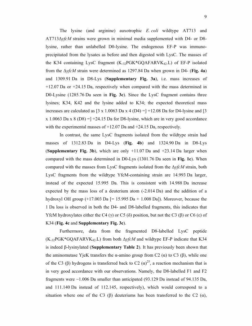
9
The lysine (and arginine) auxotrophic E. coli wildtype AT713 and
AT713ΔyfcM strains were grown in minimal media supplemented with D4- or D8-
lysine, rather than unlabelled D0-lysine. The endogenous EF-P was immuno-
precipitated from the lysates as before and then digested with LysC. The masses of
the K34 containing LysC fragment (K.32PGK*GQAFARVK42.L) of EF-P isolated
from the ΔyfcM strain were determined as 1297.84 Da when grown in D4- (Fig. 4a)
and 1309.91 Da in D8-Lys (Supplementary Fig. 3a), i.e. mass increases of
+12.07 Da or +24.15 Da, respectively when compared with the mass determined in
D0-Lysine (1285.76 Da seen in Fig. 3c). Since the LysC fragment contains three
lysines; K34, K42 and the lysine added to K34; the expected theoretical mass
increases are calculated as [3 x 1.0063 Da x 4 (D4) =] +12.08 Da for D4-lysine and [3
x 1.0063 Da x 8 (D8) =] +24.15 Da for D8-lysine, which are in very good accordance
with the experimental masses of +12.07 Da and +24.15 Da, respectively.
In contrast, the same LysC fragments isolated from the wildtype strain had
masses of 1312.83 Da in D4-Lys (Fig. 4b) and 1324.90 Da in D8-Lys
(Supplementary Fig. 3b), which are only +11.07 Da and +23.14 Da larger when
compared with the mass determined in D0-Lys (1301.76 Da seen in Fig. 1c). When
compared with the masses from LysC fragments isolated from the ΔyfcM strain, both
LysC fragments from the wildtype YfcM-containing strain are 14.993 Da larger,
instead of the expected 15.995 Da. This is consistent with 14.988 Da increase
expected by the mass loss of a deuterium atom (-2.014 Da) and the addition of a
hydroxyl OH group (+17.003 Da [= 15.995 Da + 1.008 Da]). Moreover, because the
1 Da loss is observed in both the D4- and D8-labelled fragments, this indicates that
YfcM hydroxylates either the C4 (γ) or C5 (δ) position, but not the C3 (β) or C6 (ε) of
K34 (Fig. 4c and Supplementary Fig. 3c).
Furthermore, data from the fragmented D8-labelled LysC peptide
(K.32PGK*GQAFARVK42.L) from both ΔyfcM and wildtype EF-P indicate that K34
is indeed β-lysinylated (Supplementary Table 2). It has previously been shown that
the aminomutase YjeK transfers the α-amino group from C2 (α) to C3 (β), while one
of the C3 (β) hydrogens is transferred back to C2 (α)25, a reaction mechanism that is
in very good accordance with our observations. Namely, the D8-labelled F1 and F2
fragments were ~1.006 Da smaller than anticipated (93.129 Da instead of 94.135 Da,
and 111.140 Da instead of 112.145, respectively), which would correspond to a
situation where one of the C3 (β) deuteriums has been transferred to the C2 (α),
10
thereby indicating that these fragments can only be derived from β-lysine
(Supplementary Table 1 and Supplementary Fig. 2). Both F3 and F4 fragments
have a mass identical to that of theoretical lysine fragments containing eight
deuteriums, confirming the conclusion that one deuterium is transferred from the C3
(β) to C2 (α) and not lost during the reaction (Supplementary Fig. 2).
In conclusion, fragmentation data using the D4- and D8-labelled LysC
fragments, together with [18]O-labeling, is consistent with our previous suggestion
that the additional 16 Da mass corresponding to lysine hydroxylation is associated
with K34 of EF-P and not with the added β-lysine (Supplementary Table 1).
Substrate specificity of the YfcM hydroxylase
The eukaryotic lysine-5-hydroxylases utilize Fe2+, O2 and α-ketoglutarate to
carry out oxidation, and then use ascorbic acid to return the iron to its oxidized state30.
Similarly, human DOHH activity is also dependent on Fe2+ and O233, but not α-
ketoglutarate or ascorbic acid34. In contrast, the substrates for the bacterial lysine-N6-
hydroxylases include NADPH, H+, O2 and the reaction requires FAD31, whereas
lysine-2-monoxygenases utilize O2 and FAD but do not require metals32. To screen
for potential substrates of YfcM, we utilized differential scanning fluorimetry (DSF),
which monitors the increase in thermal stability of a protein due to the interaction
with a ligand35. The thermal stability of recombinant YfcM protein was measured
across a library of diverse ligands, including α-ketoglutarate, ascorbic acid, co-
enzyme A (CoA), FAD, NAD, NADH, NADP, NADPH, ATP, ADP, PLP, L-lysine,
thiamine, oxaloacetate, tetrahydrofolate, succinate as well as metals, such as Fe2+ and
Mg2+ (Supplementary Fig. 4). The largest increases in thermal stability for YfcM
were observed with nucleotide-based chemicals, for example, the maximum shift of
~5°C was observed in the presence of <600 µM NADP or CoA (Fig. 5a). In contrast,
other ligands, such as Fe2+, α-ketoglutarate or ascorbic acid did not stabilize YfcM
(Supplementary Fig. 4), even at significantly higher concentrations (<10 mM).
These findings suggest that YfcM is more likely to use a NAD- or FAD-based co-
factor similar to other bacterial lysine hydroxylases, rather than Fe2+ or α-
ketoglutarate/ascorbic acid co-factors as used by eukaryotic lysine hydroxylases.

11
The genomic distribution and context of yfcM
Previous bioinformatic analysis of 725 bacterial genomes revealed the
presence of genes for efp, yjeA and yjeK in 200 genomes (28%)11. Here we have
extended this analysis to include predicted proteomes of 1268 completely sequenced
bacterial genomes and detect the presence of the YfcM protein in 179 species
clustering within the γ-proteobacteria phyla (Fig. 5b, Supplementary Table 2 and
Supplementary Fig. 5). All bacterial species that contain the yfcM gene also have
efp, yjeA and, with one exception (Haemophilus influenzae strain PittGG), yjeK
(Supplementary Table 2). Moreover, we find that a lysine, equivalent to position 34
in E. coli EF-P, is strictly conserved (100%) in organisms that contain YfcM
(Supplementary Table 2). However, unlike yjeA and yjeK that often cluster together
in the same operon as efp11, yfcM is always located in a distinct region of the
chromosome. The six-gene operon structure of the prmC-aroC-mepA-yfcA-yfcM-yfcL
operon is highly conserved within the Enterobacteriaceae family (Fig. 5b). yfcA and
yfcL are hypothetical proteins of unknown function, whereas aroC and mepA are
annotated as chorismate synthase and Murein DD-endopeptidase, respectively.
Curiously, the yfcM gene often neighbors other modification enzymes, such as the
methyltransferases prmB and prmC, which modify ribosomal protein L3 and release
factor RF1/RF2, respectively, as well as mnmC and yrdC (rimN) that are involved in
tRNA modification36,37 (Fig. 5b).
Discussion
Here we have used high-resolution mass spectrometry to characterize the chemical
composition of the lysinylation modification of EF-P as well as the enzymes of
modification pathway. We demonstrate that the conserved K34 of endogenous E. coli
EF-P bears a 144.09 Da modification, which results from two distinct reactions,
namely a 128.09 Da lysinylation performed by YjeK and YjeA, followed by an
additional 16 Da hydroxylation by YfcM (Fig. 6a). Our mass spectrometry
fragmentation data support not only the attachment of the lysine to the ε-amino group
of K34, but also indicate the presence of β-lysine, rather than α-lysine, as a product of
the lysinylation reaction (Fig. 1c,d, Supplementary Table 1, and Supplementary
Fig. 3). Moreover, the low level of modified peptide observed in the absence of yjeK
(Fig. 2b-d), suggests that the conversion of (S)-α-lysine to (R)-β-lysine by YjeK
12
precedes the lysinylation of EF-P by YjeA (Fig. 6a). This is consistent with the
observation that lysinylation of EF-P by YjeA is 100-fold more efficient using (R)-β-
lysine instead of α-lysine27 and that lysinylation of exogenously overexpressed EF-P
requires co-expression of both YjeK and YjeA6. We believe that YfcM is the last
enzyme in the EF-P modification pathway: The lack of +16 Da modified EF-P
peptides in any of the ΔyjeK or ΔyjeA MS spectra suggests that YfcM cannot
hydroxylate unmodified EF-P and therefore acts after YjeK and YjeA. Moreover, the
+144 Da modified peptide was never detected in the ΔyjeK strain, which most likely
indicates that α-lysinylated EF-P is a poor substrate for YfcM and that β-lysinylated
EF-P is preferred. This is similar to the eukaryotic situation where
deoxyhypusinylation of eIF-5A by DHS is a prerequisite for the subsequent
hydroxylation by DOHH13 (Fig. 6b). In contrast to eIF-5A where the added
spermidine moiety is hydroxylated by DOHH, our fragmentation data support the
hydroxylation by YfcM of the C4 or C5 position of K34 of EF-P, but not
hydroxylation of the added β-lysine (Fig. 1c,d, Supplementary Table 1, and
Supplementary Fig. 3).
The crystal structure of T. thermophilus EF-P bound to the 70S ribosome
suggests that EF-P stimulates peptide bond formation by interacting with and
stabilizing the tRNA at the P-site18. Modeling E. coli EF-P on the ribosome using the
T. thermophilus structure, suggests that K34 and the lysinylation moiety would extend
towards the peptidyl-transferase center and stabilize the CCA-end of the tRNA (Fig.
6c)18. Based on this model, hydroxylation of the C5 (δ), but not of the C4 (γ), position
of K34 would allow additional potential stabilizing hydrogen bond interactions with
the P-tRNA (Fig. 6c). EF-P and the lysinylation pathway are critical for attenuation of
virulence in bacteria10,21-24. The taxonomic distribution reveals yfcM to be present
with the yjeK and yjeA genes in most common Gram-negative pathogenic bacteria,
such as Acinetobacter baumannii, Escherichia coli O157:H7, Haemophilus
influenzae, Klebsiella pneumoniae, Legionella pneumophila, Salmonella
typhimurium, Vibrio cholerae and Yesinia pestis (see Fig. 5b and Supplementary
Table 2). Proteomics studies comparing wildtype S. typhimurium with ∆yjeA or
∆yjeK strains reveal large changes in the expression of proteins involved in virulence
and metabolic adaption21,24, suggesting that EF-P and the lysinylation modification
are important for translational regulation of particular mRNAs. With a complete
13
description of the EF-P modification and pathway now in hand, this opens the way for
future studies to address the mechanism of action of EF-P.
Methods Strains. The parental strain E. coli K-12 BW25113 and the single gene deletion
mutants Δefp (JW4147), ΔyjeK (JW4106), ΔyjeA (JW4116) and ΔyfcM (JW5381)
were obtained from the Keio collection (National BioResource Project (NBRP) at the
National Institute of Genetics (NIG), Japan38). Note that the ΔyjeK strain was taken
from plate 38, since the isolate on plate 37 is not a bone fide yjeK knock-out strain39.
The E. coli strain AT71340, which is auxotrophic for arginine and lysine, was kindly
provided by Matthias Selbach (Max-Delbruch Zentrum, Buch-Berlin, Germany). P1
transduction from the Keio strains was used to generate the equivalent single gene
deletions Δefp, ΔyjeK, ΔyjeA and ΔyfcM in the AT713 background40. The deletions
were confirmed using PCR and the strains were confirmed to be auxotrophic for
arginine and lysine (data not shown).
Plasmids and protein expression. The efp, yjeK, yjeA and yfcM genes were PCR
amplified from gDNA of E. coli K-12 strain MC4100 using the primers listed in
Supplemental Table 3. For protein overexpression, the efp gene was cloned into
pET14b (Novagen) using NdeI and BamHI restriction sites, whereas yfcM was cloned
into pET21b (Novagen) using NdeI and SacI restriction sites. Both plasmids were
transformed into BL21 (Novagen) and the overexpressed protein was purified using
Ni-NTA (Novagen) chromatography according to manufacturers instructions,
followed by gel filtration on a Superdex G75 column (GE Healthcare) in a buffer
containing 100 mM Hepes-KOH pH 8.0, 125 mM NaCl, 25 mM KCl, 5 mM β-
mercaptoethanol, 10% glycerol. Purified recombinant EF-P was used to generate
rabbit polyclonal antibodies (Davids Biotechnologie, Regensburg, Germany). For the
rescue experiments, the yjeK, yjeA and yfcM genes were cloned into pQE70 (Qiagen)
and transformed into the appropriate AT713 deletion strain. Unfortunately, for
technical reasons the rescue experiment expressing YjeA in the ΔyjeA AT713 strain
was inconclusive.
Stable isotopic labeling and sample preparation. E. coli wildtype AT713 and
deletion strains ΔyjeA, ΔyjeK and ΔyfcM in the AT713 background were grown in
14
50 ml MOPS minimal medium41 supplemented with 0.2% glucose and 100 µg/ml of
each amino acid42. For stable isotopic labeling, ’light’ lysine was substituted with
’heavy’ lysine: (4,4,5,5-D4)-lysine or (3,3,4,4,5,5,6,6-D8)-lysine (Cambridge Isotope
Laboratories Inc., USA). Collected and PBS-washed cells were resuspended in 350 µl
cold lysis buffer: 20 mM Tris-HCl pH 7.5, 100 mM NH4Cl, 6 mM Mg-acetate, 2 mM
PMSF). After 30 min incubation on ice, 1 mg/ml lysozyme, 20 U/ml DNase I, 0.5%
DOC, 1% Brij were added. Cells were disrupted by sonication with multiple short
bursts of maximum intensity, and the cell debris and aggregated proteins were
collected by centrifugation at 13000 x g for 15 min at 4 °C. The supernatant was pre-
cleared with 5 µl protein A bead suspension (Protein A Sepharose Fast Flow, GE
Healthcare) for at least 30 min and then centrifuged. 30 µg of polyclonal antibody
(1 mg/ml IgG was taken equal to 1.36 A280 units) was added to the pre-cleared lysate
and incubated at 4 °C for 1 hour, followed by 40 µl of protein A beads and an
additional incubation for 1 hour at 4 °C on rotator. The Sepharose beads were
collected and washed several times with lysis buffer and the beads were resuspended
in 1x SDS sample buffer, heated and EF-P protein was separated by SDS-PAGE.
Proteins were in-gel digested with either LysC (Wako, Japan) or chymotrypsin
(Promega, USA), according to standard procedures43. In case of [18]O-labeling, all
steps were as before except for digestion which was carried out in [18]O-enriched
water (Sigma, USA). Extracted peptides were purified on C18-StageTips44 and
analyzed via nano LC-MS/MS.
Nano-LC-MS/MS and direct infusion MS. Briefly, peptides were separated by
reversed-phase chromatography using an Agilent 1200 series nanoflow system
(Agilent Technologies) connected to a LTQ Orbitrap classic mass-spectrometer
(Thermo Electron, Bremen, Germany) equipped with a nanoelectrospray ion source
(Proxeon, Odense, Denmark). C18-StageTip purified peptides were dissolved in 0.1%
trifluoroacetic acid, loaded on a fused silica emitter (75 µm × 150 mm, Proxeon)
packed in-house with Reprosil-Pur C18-AQ 3 µm particles (Dr. Maisch, Germany)
using a flow rate of 700 nl/min and separated with a 30 min 3-40% B gradient (A:
0.5% acetic acid, B: 0.5% acetic acid/80% acetonitrile) at a flow-rate of 200 nl/min.
Eluted peptides were sprayed directly into LTQ Orbitrap mass-spectrometer operated
at 180 °C capillary temperature and 2.0-2.2 kV spray voltage. The LTQ Orbitrap was
operated in data-dependent mode with up to five MS/MS scans being recorded for
15
each precursor ion scan. Precursor ion spectra were recorded in profile in the Orbitrap
(m/z 300-1900, R = 60 000); data-dependent MS/MS spectra were acquired in
centroid either in the LTQ for low-resolution data (CID NCE 35%, wideband
activation enabled) or in the Orbitrap for high-resolution data (CID NCE 35%,
R = 7500). Mono-isotopic precursor selection was enabled, singly charged ions and
ions with an unassigned charge state were rejected, each fragmented ion was
dynamically excluded for 90 s. All measurements in the Orbitrap mass analyzer were
performed with lock-mass option enabled (lockmasses m/z 445.12003 and
519.13882). Fragment MS/MS spectra from raw files were extracted as MSM files
and then merged to peak lists using Raw2MSM version 1.11, selecting the top eight
peaks for each 100 Da45. MSM files were searched with the Mascot 2.3 search engine
(Matrix Science) against the protein sequence database composed of E. coli strain
MG1655 sequences from UniprotKB and common contaminant proteins, such as
trypsin, keratins etc. Search parameters were: 5 ppm for precursor mass tolerance,
0.6 Da for ion-trap MS/MS mass tolerance or 0.02 Da for Orbitrap MS/MS mass
tolerance, up to three missed cleavages plus a number of variable modifications such
as oxidation (M), lysinylation (K), and hydroxylation (K) plus their isotopically-
labeled variants. Peptide hits returned by Mascot were manually validated and
annotated when needed.
For full protein analysis, C8-StageTip purified sample was directly infused
into the mass-spectrometer using borosilicate static nanospray emitters (Proxeon,
Denmark) and positive ionization mode. Measurements were done with an ionization
voltage of 1000 V, m/z range of 800-1500 and resolution of 60,000 @ m/z 400, 100
micro-scans were combined into one FT transient and external calibration was used.
Differential scanning fluorimetry. Thermal-dependent unfolding of YfcM was
measured using differential scanning fluorimetry35. Purified YfcM (15 µM) was pre-
mixed with SYPROrange dye according to manufacturers instructions (Invitrogen).
YfcM stability was measured within a temperature range of 25-95°C using iCycler5
(Bio-Rad). Data points were measured every 0.3°C with 25s pre-incubation time.
Concentration-dependent stability of YfcM in the presence of NADP, NADPH, NAD,
NADH, FAD, ADP, ATP, GTP, coenzyme A, tetrahydrofolic acid, PLP, oxaloacetic
acid and thiamine was tested at concentrations 0-600 µM, while higher concentrations
(0-250 mM) of L-ascorbic acid and α-ketoglutarate were necessary. Stocks of each of
16
the ligands (Sigma Aldrich) were prepared in DMSO and diluted using YfcM buffer
(100 mM Hepes-KOH pH 8.0, 125 mM NaCl, 25 mM KCl, 5 mM β-mercaptoethanol,
10% glycerol).
Phylogenetic analyses. Protein sequences were retrieved by BlastP searching against
the NCBI RefSeq database and aligned using MAFFT version 6.626b46. Maximum
likelihood phylogenetic analyses were carried out using RaxML version 7.0.447 run on
the Cipres server (http://www.phylo.org/sub_sections/portal/) with the
PROTCATWAG model and 100 bootstrap replicates. Ambiguously aligned regions of
alignments were not included. To determine the presence and absence of yfcM, yjeK
and yjeA genes across bacteria, hidden markov models (HMMs) were constructed
from multiple sequence alignments of each family using HMMer 3.0b248, and
searched against 1273 genomes across the bacterial tree of life. The genomic
neighborhood was retrieved using Entrez Gene49.
Acknowledgments
This research was supported by grants from the Deutsche Forschungsgemeinschaft
WI3285/1-1 (to D.N.W.), Human Frontiers of Science Foundation (RGY88/2008),
and the EMBO young investigator grant. L.P. and G.A. are supported by the European
Social Fund program Mobilitas grant MJD144 and MJD99, respectively. Mass-
spectrometric analyses were in part supported by the European Regional Development
Fund through the Center of Excellence in Chemical Biology (Institute of Technology,
University of Tartu).
Author contributions:
L.P., A.L.S., J.R., D.N.W designed research; L.P. performed and analyzed MS data,
A.L.S. and K.V. performed biochemistry; G.A. performed bioinformatics; L.P.,
A.L.S., T.T., J.R. and D.N.W analyzed data and wrote the paper.

17
References
1. Glick, B. R. & Ganoza, M. C. Identification of a soluble protein that stimulates
peptide bond synthesis. Proc. Natl Acad. Sci. USA 72, 4257-4260. (1975).
2. Glick, B. R., Chladek, S. & Ganoza, M. C. Peptide bond formation stimulated by
protein synthesis factor EF-P depends on the aminoacyl moiety of the acceptor.
Eur. J. Biochem. 97, 23-28. (1979).
3. Ganoza, M. C. & Aoki, H. Peptide bond synthesis: function of the efp gene
product. Biol. Chem. 381, 553-559 (2000).
4. Swaney, S. et al. Characterization of a high-throughput screening assay for
inhibitors of elongation factor P and ribosomal peptidyltransferase activity. J.
Biochem. Screen. 11, 736-742 (2006).
5. Aoki, H. et al. Interactions of elongation factor EF-P with the Escherichia coli
ribosome. FEBS J. 275, 671-681 (2008).
6. Yanagisawa, T., Sumida, T., Ishii, R., Takemoto, C. & Yokoyama, S. A paralog of
lysyl-tRNA synthetase aminoacylates a conserved lysine residue in translation
elongation factor P. Nat. Struct. Mol. Biol. 17, 1136-43 (2010).
7. de Crecy, E. et al. Development of a novel continuous culture device for
experimental evolution of bacterial populations. Appl. Microbiol. Biotechnol. 77,
489-496 (2007).
8. Kearns, D. B., Chu, F., Rudner, R. & Losick, R. Genes governing swarming in
Bacillus subtilis and evidence for a phase variation mechanism controlling surface
motility. Mol. Microbiol. 52, 357-369 (2004).
9. Ohashi, Y. et al. Expression profiling of translation-associated genes in
sporulating Bacillus subtilis and consequence of sporulation by gene inactivation.
Biosci. Biotechnol. Biochem. 67, 2245-2253 (2003).
10. Peng, W. T., Banta, L. M., Charles, T. C. & Nester, E. W. The chvH locus of
Agrobacterium encodes a homologue of an elongation factor involved in protein
synthesis. J. Bacteriol. 183, 36-45 (2001).
11. Bailly, M. & de Crecy-Lagard, V. Predicting the pathway involved in post-
translational modification of elongation factor P in a subset of bacterial species.
Biol. Direct 5, 3 (2010).
12. Kyrpides, N. C. & Woese, C. R. Universally conserved translation initiation
factors. Proc. Natl Acad. Sci. USA 95, 224-228 (1998).

18
13. Park, M. H., Nishimura, K., Zanelli, C. F. & Valentini, S. R. Functional
significance of eIF5A and its hypusine modification in eukaryotes. Amino Acids
38, 491-500 (2010).
14. Saini, P., Eyler, D. E., Green, R. & Dever, T. E. Hypusine-containing protein
eIF5A promotes translation elongation. Nature 459, 118-21 (2009).
15. Gregio, A. P., Cano, V. P., Avaca, J. S., Valentini, S. R. & Zanelli, C. F. eIF5A
has a function in the elongation step of translation in yeast. Biochem. Biophys.
Res. Commun. 380, 785-790 (2009).
16. Henderson, A. & Hershey, J. W. Eukaryotic translation initiation factor (eIF) 5A
stimulates protein synthesis in Saccharomyces cerevisiae. Proc. Natl Acad. Sci.
USA 108, 6415-6149 (2011).
17. Hanawa-Suetsugu, K. et al. Crystal structure of elongation factor P from Thermus
thermophilus HB8. Proc. Natl Acad. Sci. U S A 101, 9595-9600 (2004).
18. Blaha, G., Stanley, R. E. & Steitz, T. A. Formation of the first peptide bond: the
structure of EF-P bound to the 70S ribosome. Science 325, 966-70 (2009).
19. Kim, K. K., Hung, L. W., Yokota, H., Kim, R. & Kim, S. H. Crystal structures of
eukaryotic translation initiation factor 5A from Methanococcus jannaschii at 1.8
Å resolution. Proc. Natl Acad. Sci. USA 95, 10419-10424 (1998).
20. Peat, T. S., Newman, J., Waldo, G. S., Berendzen, J. & Terwilliger, T. C.
Structure of translation initiation factor 5A from Pyrobaculum aerophilum at 1.75
Å resolution. Structure 6, 1207-1214 (1998).
21. Navarre, W. W. et al. PoxA, yjeK, and elongation factor P coordinately modulate
virulence and drug resistance in Salmonella enterica. Mol. Cell 39, 209-221
(2010).
22. Kaniga, K., Compton, M. S., Curtiss, R., 3rd & Sundaram, P. Molecular and
functional characterization of Salmonella enterica serovar typhimurium poxA
gene: effect on attenuation of virulence and protection. Infect. Immun. 66, 5599-
5606 (1998).
23. Bearson, S. M., Bearson, B. L. & Rasmussen, M. A. Identification of Salmonella
enterica serovar Typhimurium genes important for survival in the swine gastric
environment. Appl. Environ. Microbiol. 72, 2829-36 (2006).
24. Bearson, S. M., Bearson, B. L., Brunelle, B. W., Sharma, V. K. & Lee, I. S. A
mutation in the poxA gene of Salmonella enterica serovar typhimurium alters
19
protein production, elevates susceptibility to environmental challenges, and
decreases swine colonization. Foodborne Pathog. Dis. 8, 725-732 (2011).
25. Behshad, E. et al. Enantiomeric free radicals and enzymatic control of
stereochemistry in a radical mechanism: the case of lysine 2,3-aminomutases.
Biochemistry 45, 12639-46 (2006).
26. Ambrogelly, A., O'Donoghue, P., Soll, D. & Moses, S. A bacterial ortholog of
class II lysyl-tRNA synthetase activates lysine. FEBS Lett. 584, 3055-60 (2010).
27. Roy, H. et al. The tRNA synthetase paralog PoxA modifies elongation factor-P
with (R)-beta-lysine. Nat. Chem. Biol., epub (2011).
28. Glick, B. R. & Ganoza, M. C. Characterization and site of action of a soluble
protein that stimulates peptide-bond synthesis. Eur. J. Biochem. 71, 483-491
(1976).
29. Glick, B. R., Green, R. M. & Ganoza, M. C. Purification of factor EF-P, a protein
that stimulates peptide bond synthesis with certain aminoacyl-tRNA analogues.
Can. J. Biochem. 57, 749-757. (1979).
30. Loenarz, C. & Schofield, C. J. Physiological and biochemical aspects of
hydroxylations and demethylations catalyzed by human 2-oxoglutarate
oxygenases. Trends Biochem. Sci. 36, 7-18 (2011).
31. Plattner, H. J., Pfefferle, P., Romaguera, A., Waschutza, S. & Diekmann, H.
Isolation and some properties of lysine N6-hydroxylase from Escherichia coli
strain EN222. Biol. Met. 2, 1-5 (1989).
32. Nakazawa, T., Hori, K. & Hayaishi, O. Studies on monooxygenases. V.
Manifestation of amino acid oxidase activity by L-lysine monooxygenase. J. Biol.
Chem. 247, 3439-44 (1972).
33. Kim, Y. S. et al. Deoxyhypusine hydroxylase is a Fe(II)-dependent, HEAT-repeat
enzyme. Identification of amino acid residues critical for Fe(II) binding and
catalysis [corrected]. J. Biol. Chem. 281, 13217-25 (2006).
34. Abbruzzese, A., Park, M. H. & Folk, J. E. Deoxyhypusine hydroxylase from rat
testis. Partial purification and characterization. J. Biol. Chem. 261, 3085-9 (1986).
35. Niesen, F. H., Berglund, H. & Vedadi, M. The use of differential scanning
fluorimetry to detect ligand interactions that promote protein stability. Nat.
Protoc. 2, 2212-21 (2007).
20
36. Bujnicki, J. M. et al. Identification of a bifunctional enzyme MnmC involved in
the biosynthesis of a hypermodified uridine in the wobble position of tRNA. RNA
10, 1236-42 (2004).
37. Harris, K. A., Jones, V., Bilbille, Y., Swairjo, M. A. & Agris, P. F. YrdC exhibits
properties expected of a subunit for a tRNA threonylcarbamoyl transferase. RNA
17, 1678-87 (2011).
38. Baba, T. et al. Construction of Escherichia coli K-12 in-frame, single-gene
knockout mutants: the Keio collection. Mol. Syst. Biol. 2, 2006 0008 (2006).
39. Yamamoto, N. et al. Update on the Keio collection of Escherichia coli single-gene
deletion mutants. Mol. Syst. Biol. 5, 335 (2009).
40. Taylor, A. L. & Trotter, C. D. Revised linkage map of Escherichia coli. Bacteriol.
Rev. 31, 332-353 (1967).
41. Neidhardt, F. C., Bloch, P. L. & Smith, D. F. Culture medium for enterobacteria.
J. Bacteriol. 119, 736-747 (1974).
42. Siibak, T. et al. Antibiotic-induced ribosomal assembly defects result from
changes in the synthesis of ribosomal proteins. Mol. Microbiol. 80, 54-67 (2010).
43. Shevchenko, A., Tomas, H., Havlis, J., Olsen, J. V. & Mann, M. In-gel digestion
for mass spectrometric characterization of proteins and proteomes. Nat. Protoc. 1,
2856-60 (2006).
44. Rappsilber, J., Mann, M. & Ishihama, Y. Protocol for micro-purification,
enrichment, pre-fractionation and storage of peptides for proteomics using
StageTips. Nat. Protoc. 2, 1896-1906 (2007).
45. Olsen, J. V. et al. Parts per million mass accuracy on an Orbitrap mass
spectrometer via lock mass injection into a C-trap. Mol. Cell Proteomics 4, 2010-
21 (2005).
46. Katoh, K., Kuma, K., Toh, H. & Miyata, T. MAFFT version 5: improvement in
accuracy of multiple sequence alignment. Nucleic Acids Res. 33, 511-8 (2005).
47. Stamatakis, A. RAxML-VI-HPC: maximum likelihood-based phylogenetic
analyses with thousands of taxa and mixed models. Bioinformatics 22, 2688-90
(2006).
48. Eddy, S. R. Profile hidden Markov models. Bioinformatics 14, 755-63 (1998).
49. Maglott, D., Ostell, J., Pruitt, K. D. & Tatusova, T. Entrez Gene: gene-centered
information at NCBI. Nucleic Acids Res. 35, D26-31 (2007).
21
Figure Legends
Figure 1: Lysine 34 of endogenous EF-P is modified by 144.09 Da. (a) MS
spectrum of full-length endogenous EF-P from wildtype E. coli strain MRE600. (b)
MS/MS spectra of chymotrypsin fragment with mass of 1074.62 Da, mass increase of
144.09 Da compared to theoretical mass. (c) MS/MS spectra of LysC fragment with
mass of 1301.76 Da, mass increase of 144.09 Da compared to theoretical mass.
Fragmentation of LysC peptide with y and b peptides indicated as well as peaks F1-
F4. The b and y ions correspond to N- and C-terminal charge-carrying fragments of
peptide, respectively, where peptide backbone cleavage has taken place at CO-NH
bonds. Under the conditions used, y ions are preferentially produced. (d) Chemical
structures of lysinylated K34 with predicted chemical structures of mass loss
fragments for peaks F1-F4, indicated with their respective molecular weights.
Figure 2: Lysinylation of EF-P in vivo requires YjeK and YjeA. (a) MS/MS
spectra of chymotrypsin fragment of EF-P from AT713ΔyjeA with mass of
930.53 Da. (b,c) MS/MS spectra of chymotrypsin fragments of EF-P from
AT713ΔyjeK with masses of (b) 930.53 Da and (c) 1058.62 Da. (d) Quantification of
(b) and (c) reveals that modified fragment is present at 2% level of the unmodified.
Figure 3: Fully modified EF-P is dependent on the presence of YfcM. (a,b)
MS/MS spectra of chymotrypsin fragment of EF-P from (a) AT713ΔyfcM with mass
of 1058.62 Da, and (b) AT713ΔyfcM/pYfcM with mass of 1074.62 Da. (c,d) MS/MS
spectra of LysC fragment of EF-P from (c) AT713ΔyfcM with mass of 1285.76 Da,
and (d) AT713ΔyfcM/pYfcM with mass of 1301.76 Da.
Figure 4: YfcM hydroxylates the C4/C5 position of K34 of EF-P. (a,b) MS/MS
spectra of LysC fragment of EF-P from (a) AT713ΔyfcM and (b) wildtype AT713
cells grown in D4-lysine, with masses of 1297.84 Da and 1312.83 Da, respectively.
(c) Chemical structure of lysinylated K34 with potential hydroxyl positions indicated.
Figure 5: Characterization of the hydroxylase YfcM. (a) Differential scanning
fluorimetry (DSF) of YfcM in the presence of indicated nucleotide analogues. Upper
graph shows the relative fluorescence change as a function of temperature at 600µM
22
ligand concentration, whereas the lower panel shows the change (°C) in temperature
stability maximum (ΔTm) as a function of ligand concentration (µM). (b)
Phylogenetic tree (left) and genomic neighborhood (right) of representative YfcM
proteins in bacteria (full tree in Supplementary Fig. 5). Only bootstrap support
values >50% are shown.
Figure 6: Modification pathways of EF-P and eIF-5A. (a) The FeS protein YjeK
converts (S)-α-lysine to (R)-β-lysine using S-adenosyl-l-methionine (SAM) and
pyridoxal-5′-phosphate (PLP)25. YjeA uses ATP to lysinylate EF-P by the addition of
β-lysine to the ε-amino group of K3427. YfcM uses molecular oxygen to hydroxylate
the C4 (γ) or C5 (δ) position of K34 of EF-P. (b) Deoxyhypusine synthase (DHS)
uses NAD to transfer the amino-butyl moiety from spermidine to the ε-amino group
of K50 of human eIF5A, which is then hydroxylated by deoxyhypusine hydroxylase
(DOHH)13. (c) Relative positions of K34 of EF-P (green) and the acceptor stem of the
P-tRNA (blue) when bound on the ribosome. The γ, δ and ε carbon atoms of K34 and
the C74, C75 and A76 positions of the tRNA are indicated. The model was built by
mutation of Arg to Lys (equivalent to K34 in E. coli) in the T. thermophilus EF-
P●70S structure18.
VKPGK GQAFLys
b2 b3 b5
y7 y6 y5 y4 y2 y1
GQAFARVKLys
y8 y7 y6 y5y4 y3
PGK
c LysC peptidase
Chymotrypsin
d
m/z
0
20
40
60
80
100
Re
lati
ve A
bu
nd
an
ce
200 300 400 500 600 700 800 900
y1
b2
y2 b3
* ++y 7
++y7
y4 b5y5 y6
*y 7
y7
m/z
a
1030.0 1030.5 1031.0 1031.5 1032.0 1032.5 1033.0
0
20
40
60
80
100
Re
lati
ve A
bu
nd
an
ce
1031.19z=20
1031.29z=201031.09
z=20
1031.39z=20
1030.99z=20
1031.54z=20
1030.89z=20
Endogenous EF-P
y3
b3-F1
y5y4
y6y7
y8
0
20
40
60
80
100
Re
lati
ve A
bu
nd
an
ce
200 300 400 500 600 700 800 900 1000
m/z
X3
X4
X2
X1
538.82
z=2
539.32
z=2
539.32
z=2
Re
lati
ve A
bu
nd
an
ce
m/z
Experimental mass
1074.62 Da
Theoretical mass
930.53 Da
�mass = 144.09 Da
651.89
z=2
652.39
z=2
652.54
z=2
Re
lati
ve A
bu
nd
an
ce
m/z
Experimental mass
1301.76 Da
Theoretical mass
1157.67 Da
�mass = 144.09 Da
CHHN
CH2
H N2
H C2
CH2
CH
OHN
H C2
H C2
O
CHN
HGP G QAFARVK
EF-P K34
CH2
H N2
H C2
CH2
CH2
(+16 Da)
F3 (128 Da) F4 (145 Da)
CH2
CH2
H N2
C
O
CH2
H N2
H C2
CH2
CH2
HC
H N2
H N2
CH
O
CH2
H C2
CH2
CH2H N2
H N2
F2 (104 Da)F1 (86 Da)
���
���
������
b
Experimental mass
20591.61 Da
Theoretical mass
20447.38 Da
�mass = 144.23 Da
Peil et alFigure 1
(+H O)2
CHHN
CH2
H N2
H C2
CH2
X2
1100 1200
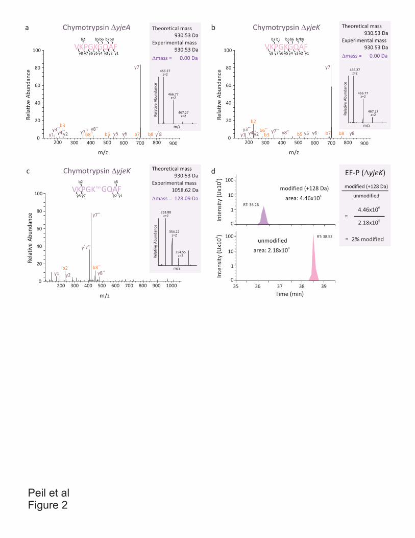
Peil et alFigure 2
a
c d
Chymotrypsin �yjeK
0
20
40
60
80
100
Re
lati
ve A
bu
nd
an
ce
200 300 400 500 600 700 800 900
m/z
y1
b2++
y3 ++y4y2
++y7 ++
y8 y5 y6b5 b7 b8
y7
y8++
b6b3
VKPGKGQAFb2 b5
y7 y6 y5 y3 y1
b6 b7b8
y8 y4 y2
Chymotrypsin �yjeA
200 300 400 500 600 700 800 900
m/z
0
20
40
60
80
100
Re
lati
ve A
bu
nd
an
ce
y1
b3++
y3 ++y4y2
++y7
++y8
y5 y6++b8 b5 b7 b8
y7
*y 8
b
466.27z=2
466.77z=2
467.27z=2R
ela
tive
Ab
un
da
nce
m/z
Experimental mass
930.53 Da
Theoretical mass
930.53 Da
�mass = 0.00 Da
466.27z=2
466.77z=2
467.27z=2R
ela
tive
Ab
un
da
nce
m/z
Experimental mass
930.53 Da
Theoretical mass
930.53 Da
�mass = 0.00 Da
VKPGK GQAFLys
b2 b8
y7y8 y2 y1
Chymotrypsin �yjeK
0
20
40
60
80
100
Re
lati
ve A
bu
nd
an
ce
200 300 400 500 600 700 800 900
m/z
y1b2
++b8
y2
* ++y 7
++y8
++y7
1000
353.88z=2
354.22z=2
354.55z=2R
ela
tive
Ab
un
da
nce
m/z
Experimental mass
1058.62 Da
Theoretical mass
930.53 Da
�mass = 128.09 Da
VKPGKGQAFb2 b5
y7 y6 y5 y3 y1
b6 b7b8
y8 y4 y2
b3
EF-P (�yjeK)
4.466
x10
2.188
x10=
unmodified
modified (+128 Da)
2% modified=
35 36 37 38 39
unmodified
area: 2.188
x10
RT: 38.52
Time (min)
RT: 36.26
modified (+128 Da)
area: 4.466
x10
1
10
100
0
5In
ten
sity
(U
x10
)
1
10
100
0
5In
ten
sity
(U
x10
)
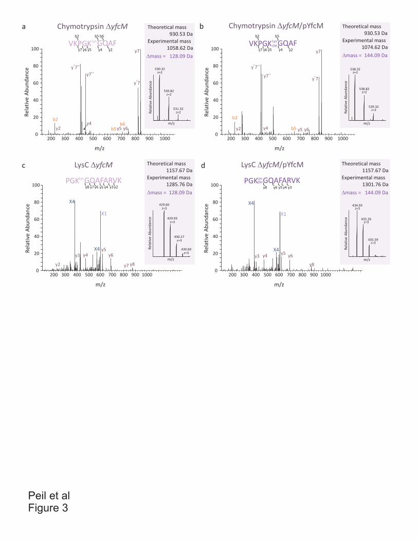
Peil et alFigure 3
a b
c d
0
20
40
60
80
100
Re
lati
ve A
bu
nd
an
ce
200 300 400 500 600 700 800 900
m/z
y2
b2
b5
* ++y 7
++y7
y4y5 y6
b6
y7
*y 7
10000
20
40
60
80
100
Re
lati
ve A
bu
nd
an
ce
200 300 400 500 600 700 800 900
m/z
1000
y2
b2
b5
* ++y 7
++y7
y4 y5 y6
y7
*y 7
0
20
40
60
80
100
Re
lati
ve A
bu
nd
an
ce
200 300 400 500 600 700 800 900
m/z
1000
y2
y3 y4y5
y6
y7 y8
X4
X4
X1
0
20
40
60
80
100
Re
lati
ve A
bu
nd
an
ce
200 300 400 500 600 700 800 900
m/z
1000
y3 y4y5
y6
y8
X4
X4
X1
VKPGK GQAFLys
b2 b5
y7 y6 y5 y4 y2
b6
VKPGK GQAFLys
OH
b2 b5
y7 y6 y5 y4 y2
y8 y6 y5 y4 y3
PGK GQAFARVKLysOHPGK GQAFARVKLys
y8 y6 y5 y4 y3y7 y2
Chymotrypsin �yfcM Chymotrypsin �yfcM/pYfcM
LysC �yfcM LysC �yfcM/pYfcM
530.32z=2
530.82z=2
531.32z=2R
ela
tive
Ab
un
da
nce
m/z
Experimental mass
1058.62 Da
Theoretical mass
930.53 Da
�mass = 128.09 Da
538.32z=2
538.82z=2
539.32z=2
Re
lati
ve A
bu
nd
an
ce
m/z
Experimental mass
1074.62 Da
Theoretical mass
930.53 Da
�mass = 144.09 Da
429.60z=3
429.93z=3
430.27z=3
Re
lati
ve A
bu
nd
an
ce
m/z
430.60z=3
Experimental mass
1285.76 Da
Theoretical mass
1157.67 Da
�mass = 128.09 Da
434.93z=3
435.26z=3
435.59z=3
Re
lati
ve A
bu
nd
an
ce
m/z
Experimental mass
1301.76 Da
Theoretical mass
1157.67 Da
�mass = 144.09 Da
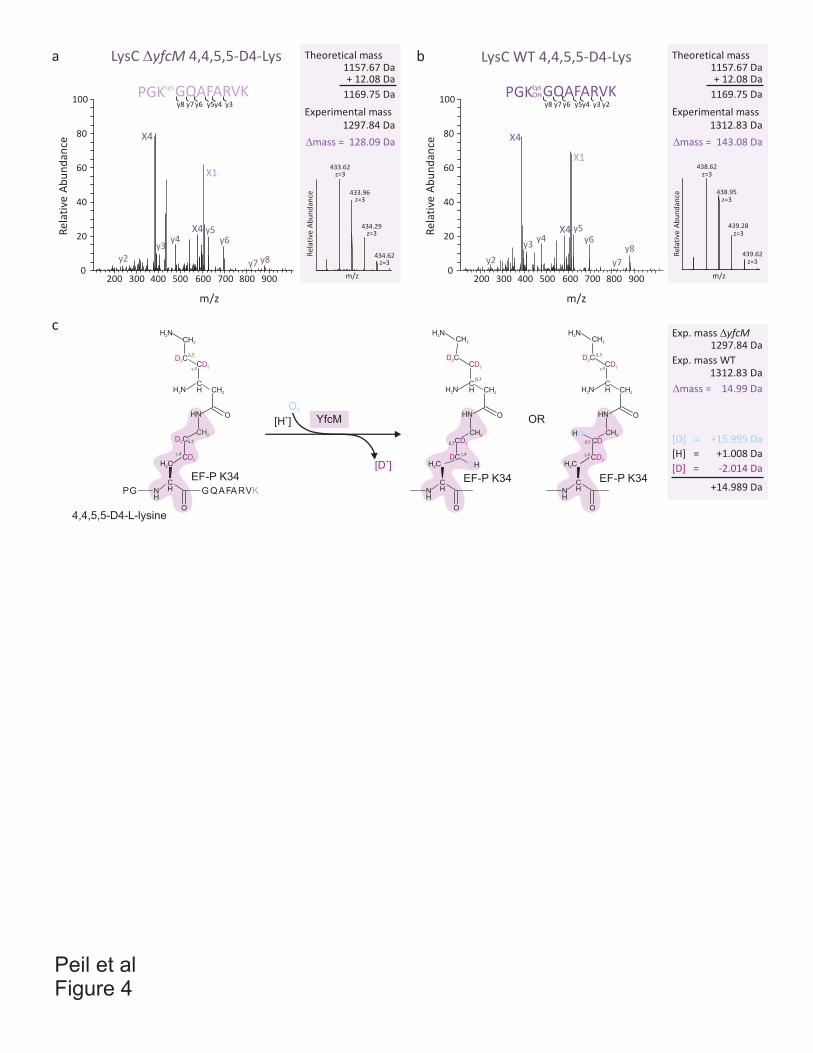
Peil et alFigure 4
c
4,4,5,5-D4-L-lysine
PGK GQAFARVKLys
y8 y6 y5y4 y3y7
LysC �yfcM 4,4,5,5-D4-Lysa
0
20
40
60
80
100
Re
lati
ve A
bu
nd
an
ce
200 300 400 500 600 700 800 900
m/z
y2
y3y4
y5y6
y7 y8
X4
X1
b
PGK GQAFARVKLys
y8 y6 y5y4 y3y7
OH
y2
LysC WT 4,4,5,5-D4-Lys
0
20
40
60
80
100
Re
lati
ve A
bu
nd
an
ce
200 300 400 500 600 700 800 900
m/z
y2
y3y4 y6
y7
y8
y5
X4
X1438.62
z=3
438.95z=3
439.28z=3
Re
lati
ve A
bu
nd
an
ce
m/z
439.62z=3
O2
CH
OHN
H C2
O
CHN
H
EF-P K34
CH2
H N2
D2CCD2
CH2
CD2
CH2
H N2
CDOH
���
���
��
CH
OHN
D2C
H C2
O
CHN
H
EF-P K34
CH2
H N2
D2CCD2
CH2
CD2
CH2
H N2
���
��
���
��
CH
OHN
C
H C2
O
CHN
H
EF-P K34
CH2
H N2
D2CCD2
CH2
CD2
CH2
H N2
���DHO
��
���
��
+[D ]
YfcM+[H ] OR
GP G QAFARVK
+15.995 Da
+1.008 Da
-2.014 Da
+14.989 Da
433.62z=3
433.96z=3
434.29z=3
Re
lati
ve A
bu
nd
an
ce
m/z
434.62z=3
Experimental mass
Theoretical mass
�mass = 128.09 Da
1157.67 Da
1297.84 Da
+ 12.08 Da
1169.75 Da
Experimental mass
Theoretical mass
�mass = 143.08 Da
1157.67 Da
1312.83 Da
+ 12.08 Da
1169.75 Da
Exp. mass WT
Exp. mass �yfcM
�mass = 14.99 Da
1297.84 Da
1312.83 Da
[O] =
[H] =
[D] =
X4 X4

Peil et alFigure 5
yfcM yfcLmepA yfcAprmB aroC
320155713_Vibrio_vulnificus
253689184_Pectobacterium_carotovorum
307544340_Halomonas_elongata
56459999_Idiomarina_loihiensis
332305344_Glaciecola_agarilytica
169634117_Acinetobacter_baumannii
292488897_Erwinia_amylovora
333928401_Serratia_sp._AS12
90410689_Photobacterium_profundum
319776414_Haemophilus_influenzae
254785282_Teredinibacter_turnerae
152979116_Actinobacillus_succinogenes
251788968_Dickeya_zeae
218559239_Escherichia_coli
336251388_Enterobacter_aerogenes
119946747_Psychromonas_ingrahamii
52842124_Legionella_pneumophila
290475972_Xenorhabdus_bovienii
119945217_Psychromonas_ingrahamii
296113770_Moraxella_catarrhalis
194444155_Salmonella_enterica
308050329_Ferrimonas_balearica
85
94
66
100
94
99
79
87
75
prmB aroC mepA yfcA yfcM yfcL mnmC fabB
prmB aroC mepA yfcA yfcM yfcL mnmC fabB
prmB aroC mepA yfcA yfcM yfcL mnmC fabB
mepA yfcA yfcM yfcL mnmC fabBprmB aroC
mepA yfcA yfcM yfcL mnmC fabBprmB aroC
mepA yfcA yfcM yfcL mnmC fabBprmB aroC
yfcMprmB aroC AcoX-K yfcL mnmCMFS_1 fabB
yfcM yfcL mnmC fabBMParoCprmB
UDQ4D yfcM yfcLBNADFUDQ4E
yfcM 4HBT pseudo
yfcMHPGSTHP
yfcM SEL1 RF1 RDD prmC
yfcM TR yrdCL32 HP
yfcML32 HP yrdC L17
yfcM pseudo yfcL
yfcM
yfcM AcoX-K yfcL mnmC fabB
prmB aroC mepA yfcA yfcM yfcL mnmC fabB
yfcM HP IF3 L35DNAPIV HP
prmB aroC yfcM ecnA DAGK
yfcM AcoX-K5’-3’ pdxB
a b
Ligand [μM]
100 300 5000 200 400 6000
1
2
3
4
5 NADP
NAD
NADH
ADP
NADPH
ATP
CoA
�Tm
(°C
)
35 45 55 6540 50 60 701
2
3
4
5
Tm (�C)
Re
l. f
luo
resc
en
ce (
U)
+ NADP
+ NADPH
+ CoA
YfcM
�Tm (�C)
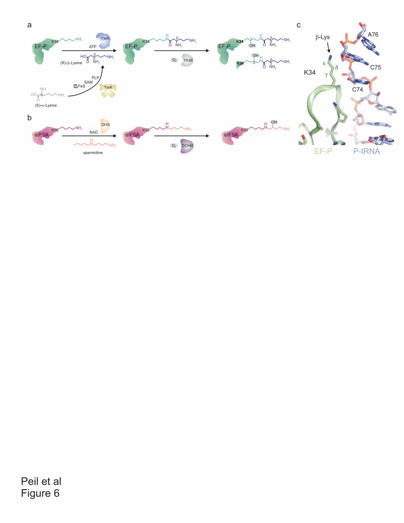
Peil et alFigure 6
c
EF-P P-tRNA
K34 �
��
�-Lys A76
C75
C74
a
b
FeS
PLPSAM
YjeK
(S)-�-Lysine
(R)-�-Lysine
YjeA
ATP
NH2K34
EF-P
NK34
NH2
NH2
O
H
EF-P
NH2
NH2HO
O
�
NH2
NH2HO
O
�
�
YfcMO2
NK34
NH2
NH2
O
H
EF-P
� �
NK34
NH2
NH2
O
HOH
��
OHOHK34
K34
NH2K50
eIF5A
NH
NH2
DHSN
K50
eIF5A
H
NH2
NAD
DOHHO2
NK50
eIF5A
H
NH2
OH
spermidine

Agata Starosta
Agata Lucyna Starosta
Gene Center, LMU Munich Nationality: Polish
Born on 03.09.1983 in Rzeszow, Poland [email protected]
Scientific experience 10/2007 – 01/2012 PhD student In the laboratory of Dr. Daniel N. Wilson, Gene Center, Department of Biochemistry, University of Munich, Germany.
Topic: Antibiotics, Inhibition of Protein Synthesis
10/2002 – 06/2007 Master student In the laboratory of Prof. Dr. Andrzej Kozik, Analytical Biochemistry, Department of Biochemistry, Jagiellonian University, Crakow, Poland
Topic: Thiamin monophosphate synthase – dissecting enzyme’s activity Education 31.01.2012 PhD exam, grade: summa cum laude 10/2007 – 01/2012 PhD thesis in the group of Dr. Daniel N. Wilson, in Munich, Germany 27/06/2007 Master exam, grade: very good (A), MSc.. Master thesis: Thiamin monophosphate synthase – a key enzyme in thiamin biosynthesis pathway, Analytical Biochemistry. Supervisor Dr. M. Rapala-Kozik. 10/2002 – 06/2007 Master studies in biology, Faculty of Biology and Earth Science, Jagiellonian University, Cracow, Poland. Specialization: biochemistry, Faculty of Biochemistry, Biophysics, and Biotechnology, Jagiellonian University, Cracow, Poland. Publications Mikolajka, A., H. Liu, Y. Chen, A.L. Starosta, V. Márquez, M. Ivanova, B.S. Cooperman, and
D.N. Wilson. 2011. Differential effects of thiopeptide and orthosomycin antibiotics on translational GTPases. Chem Biol 18 (5): 589-600.
Ratje, A. H., J. Loerke, A. Mikolajka, M. Brunner, P. W. Hildebrand, A. L. Starosta, A. Donhofer,
S. R. Connell, P. Fucini, T. Mielke, P. C. Whitford, J. N. Onuchic, Y. Yu, K. Y. Sanbonmatsu, R. K. Hartmann, P. A. Penczek, D. N. Wilson, and C. M. Spahn. 2010. Head
Agata Starosta
swivel on the ribosome facilitates translocation by means of intra-subunit tRNA hybrid sites. Nature 468 (7324):713-716.
Starosta, A. L., V. V. Karpenko, A. V. Shishkina, A. Mikolajka, N. V. Sumbatyan, F. Schluenzen,
G. A. Korshunova, A. A. Bogdanov, and D. N. Wilson. 2010. Interplay between the ribosomal tunnel, nascent chain, and macrolides influences drug inhibition. Chem Biol 17 (5):504-14.
Bhushan, S., H. Meyer, A. L. Starosta, T. Becker, T. Mielke, O. Berninghausen, M. Sattler, D. N.
Wilson, and R. Beckmann. 2010. Structural basis for translational stalling by human cytomegalovirus and fungal arginine attenuator peptide. Mol Cell 40 (1):138-46.
Starosta, A. L., H. Qin, A. Mikolajka, G. Y. Leung, K. Schwinghammer, K. C. Nicolaou, D. Y.
Chen, B. S. Cooperman, and D. N. Wilson. 2009. Identification of distinct thiopeptide-antibiotic precursor lead compounds using translation machinery assays. Chem Biol 16 (10):1087-96.
Petropoulos, A. D., E. C. Kouvela, A. L. Starosta, D. N. Wilson, G. P. Dinos, and D. L. Kalpaxis.
2009. Time-resolved binding of azithromycin to Escherichia coli ribosomes. J Mol Biol 385 (4):1179-92.
Palaniappan, N., V. Dhote, S. Ayers, A. L. Starosta, D. N. Wilson, and K. A. Reynolds. 2009.
Biosynthesis of the aminocyclitol subunit of hygromycin A in Streptomyces hygroscopicus NRRL 2388. Chem Biol 16 (11):1180-9.
Dhote, V., A. L. Starosta, D. N. Wilson, and K. A. Reynolds. 2009. The final step of hygromycin
A biosynthesis, oxidation of C-5''-dihydrohygromycin A, is linked to a putative proton gradient-dependent efflux. Antimicrob Agents Chemother 53 (12):5163-72.
Wilson, D. N., F. Schluenzen, J. M. Harms, A. L. Starosta, S. R. Connell, and P. Fucini. 2008.
The oxazolidinone antibiotics perturb the ribosomal peptidyl-transferase center and effect tRNA positioning. Proc Natl Acad Sci U S A 105 (36):13339-44.
Ebbing, B., K. Mann, A. Starosta, J. Jaud, L. Schols, R. Schule, and G. Woehlke. 2008. Effect of
spastic paraplegia mutations in KIF5A kinesin on transport activity. Hum Mol Genet 17 (9):1245-52.
Rapala-Kozik, M., M. Olczak, K. Ostrowska, A. Starosta, and A. Kozik. 2007. Molecular
characterization of the thi3 gene involved in thiamine biosynthesis in Zea mays: cDNA sequence and enzymatic and structural properties of the recombinant bifunctional protein with 4-amino-5-hydroxymethyl-2-methylpyrimidine (phosphate) kinase and thiamine monophosphate synthase activities. Biochem J 408 (2):149-59.