Anti-VEG F utilization in RVO and DME
11
© 2014 Kiss et al. This work is published by Dove Medical Press Limited, and licensed under Creative Commons Attribution – Non Commercial (unported, v3.0) License. The full terms of the License are available at http://creativecommons.org/licenses/by-nc/3.0/. Non-commercial uses of the work are permitted without any further permission from Dove Medical Press Limited, provided the work is properly attributed. Permissions beyond the scope of the License are administered by Dove Medical Press Limited. Information on how to request permission may be found at: http://www.dovepress.com/permissions.php Clinical Ophthalmology 2014:8 1611–1621 Clinical Ophthalmology Dovepress submit your manuscript | www.dovepress.com Dovepress 1611 ORIGINAL RESEARCH open access to scientific and medical research Open Access Full Text Article http://dx.doi.org/10.2147/OPTH.S60893 Clinical utilization of anti-vascular endothelial growth-factor agents and patient monitoring in retinal vein occlusion and diabetic macular edema Szilárd Kiss 1 Ying Liu 2 Joseph Brown 3 Nancy M Holekamp 4,5 Arghavan Almony 6 Joanna Campbell 2 Jonathan W Kowalski 2 1 Weill Cornell Medical College, New York, NY; 2 Allergan, Inc., Irvine, CA; 3 IMS Health, Woodland Hills, CA; 4 Pepose Vision Institute, Chesterfield, MO; 5 Washington University School of Medicine, St Louis, MO; 6 Carolina Eye Associates, Southern Pines, NC, USA Correspondence: Szilárd Kiss Weill Cornell Medical College New York Presbyterian Hospital, 1305 York Avenue – 11th Floor, New York, NY 10021, USA Tel +1 646 962 2217 Fax +1 646 962 0609 Email [email protected] Purpose: To examine the utilization of bevacizumab and ranibizumab and disease monitoring in patients with branch or central retinal vein occlusion (BRVO/CRVO) or diabetic macular edema (DME) in clinical practice. Patients and methods: This retrospective claims analysis included newly diagnosed patients with one or more bevacizumab or ranibizumab injections. Bevacizumab or ranibizumab utiliza- tion was assessed by year of first injection: 2008–2010 cohorts (12-month follow-up), January to June 2011 cohort (6-month follow-up). The main outcome measures were mean annual numbers of injections, ophthalmologist visits and optical coherence tomography examinations, and proportion of patients with additional laser or intravitreal triamcinolone (IVTA) use. Results: A total of 885 BRVO, 611 CRVO, and 2,733 DME patients treated with bevacizumab were included, with too few ranibizumab-treated patients for meaningful analysis. Across the 2008, 2009, and 2010 cohorts, mean annual numbers of bevacizumab injections increased, but remained low (BRVO 2.5, 3.1, 3.3; CRVO 3.1, 3.1, 3.5; and DME 2.2, 2.5, 3.6, respec- tively); mean ophthalmologist visits ranged between 4.4 and 6.5, and mean optical coherence tomography examinations ranged between 3.1 and 3.9 across all conditions. A total of 42.0% of BRVO, 16.5% of CRVO, and 57.7% of DME patients received additional laser or IVTA therapy. The number of bevacizumab injections was positively associated with laser use in BRVO (3.3 versus 2.9, P0.03), and with laser or IVTA use in DME (laser, 3.3 versus 2.7, P0.03; IVTA, 3.3 versus 3.0, P0.05). Conclusion: During the study period (2008–2011), bevacizumab was the main anti-VEGF therapy used in clinical practice for BRVO, CRVO, and DME. Patients treated with bevaci- zumab were monitored less frequently and received fewer injections than patients in major clinical trials of ranibizumab. Keywords: anti-vascular endothelial growth factor, bevacizumab, ranibizumab, diabetic macular edema, retinal vein occlusion, intravitreal Introduction Retinal vein occlusion (RVO), divided into central (CRVO) or branch (BRVO), and diabetic macular edema (DME) are among the most common retinal vascular diseases responsible for vision loss and blindness. 1–4 RVO and DME are estimated to affect approximately 16.4 million and 21 million people worldwide, respectively. 5,6 Although the pathogeneses of CRVO, BRVO, and DME are multifactorial, upregulation of VEGF is a common underlying source of vision loss in all three diseases. 7–10 Treatment options for patients with macular edema secondary to BRVO, CRVO, or DME include laser surgery, intravitreal corticosteroids, and intravitreal anti- VEGF agents. Anti-VEGF agents, including ranibizumab (Lucentis ® ; Genentech,
Transcript of Anti-VEG F utilization in RVO and DME
© 2014 Kiss et al. This work is published by Dove Medical Press Limited, and licensed under Creative Commons Attribution – Non Commercial (unported, v3.0) License. The full terms of the License are available at http://creativecommons.org/licenses/by-nc/3.0/. Non-commercial uses of the work are permitted without any further
permission from Dove Medical Press Limited, provided the work is properly attributed. Permissions beyond the scope of the License are administered by Dove Medical Press Limited. Information on how to request permission may be found at: http://www.dovepress.com/permissions.php
Clinical Ophthalmology 2014:8 1611–1621
Clinical Ophthalmology Dovepress
submit your manuscript | www.dovepress.com
Dovepress 1611
O r i g i n a l r e s e a r C h
open access to scientific and medical research
Open access Full Text article
http://dx.doi.org/10.2147/OPTH.S60893
Clinical utilization of anti-vascular endothelial growth-factor agents and patient monitoring in retinal vein occlusion and diabetic macular edema
szilárd Kiss1
Ying liu2
Joseph Brown3
nancy M holekamp4,5
arghavan almony6
Joanna Campbell2
Jonathan W Kowalski2
1Weill Cornell Medical College, new York, nY; 2allergan, inc., irvine, Ca; 3iMs health, Woodland hills, Ca; 4Pepose Vision institute, Chesterfield, MO; 5Washington University school of Medicine, st louis, MO; 6Carolina eye associates, southern Pines, nC, Usa
Correspondence: szilárd Kiss Weill Cornell Medical College new York Presbyterian hospital, 1305 York avenue – 11th Floor, new York, nY 10021, Usa Tel +1 646 962 2217 Fax +1 646 962 0609 email [email protected]
Purpose: To examine the utilization of bevacizumab and ranibizumab and disease monitoring
in patients with branch or central retinal vein occlusion (BRVO/CRVO) or diabetic macular
edema (DME) in clinical practice.
Patients and methods: This retrospective claims analysis included newly diagnosed patients
with one or more bevacizumab or ranibizumab injections. Bevacizumab or ranibizumab utiliza-
tion was assessed by year of first injection: 2008–2010 cohorts (12-month follow-up), January
to June 2011 cohort (6-month follow-up). The main outcome measures were mean annual
numbers of injections, ophthalmologist visits and optical coherence tomography examinations,
and proportion of patients with additional laser or intravitreal triamcinolone (IVTA) use.
Results: A total of 885 BRVO, 611 CRVO, and 2,733 DME patients treated with bevacizumab
were included, with too few ranibizumab-treated patients for meaningful analysis. Across
the 2008, 2009, and 2010 cohorts, mean annual numbers of bevacizumab injections increased,
but remained low (BRVO 2.5, 3.1, 3.3; CRVO 3.1, 3.1, 3.5; and DME 2.2, 2.5, 3.6, respec-
tively); mean ophthalmologist visits ranged between 4.4 and 6.5, and mean optical coherence
tomography examinations ranged between 3.1 and 3.9 across all conditions. A total of 42.0% of
BRVO, 16.5% of CRVO, and 57.7% of DME patients received additional laser or IVTA therapy.
The number of bevacizumab injections was positively associated with laser use in BRVO
(3.3 versus 2.9, P0.03), and with laser or IVTA use in DME (laser, 3.3 versus 2.7, P0.03;
IVTA, 3.3 versus 3.0, P0.05).
Conclusion: During the study period (2008–2011), bevacizumab was the main anti-VEGF
therapy used in clinical practice for BRVO, CRVO, and DME. Patients treated with bevaci-
zumab were monitored less frequently and received fewer injections than patients in major
clinical trials of ranibizumab.
Keywords: anti-vascular endothelial growth factor, bevacizumab, ranibizumab, diabetic macular
edema, retinal vein occlusion, intravitreal
IntroductionRetinal vein occlusion (RVO), divided into central (CRVO) or branch (BRVO), and
diabetic macular edema (DME) are among the most common retinal vascular diseases
responsible for vision loss and blindness.1–4 RVO and DME are estimated to affect
approximately 16.4 million and 21 million people worldwide, respectively.5,6 Although
the pathogeneses of CRVO, BRVO, and DME are multifactorial, upregulation of VEGF
is a common underlying source of vision loss in all three diseases.7–10
Treatment options for patients with macular edema secondary to BRVO, CRVO,
or DME include laser surgery, intravitreal corticosteroids, and intravitreal anti-
VEGF agents. Anti-VEGF agents, including ranibizumab (Lucentis®; Genentech,
Journal name: Clinical OphthalmologyJournal Designation: Original ResearchYear: 2014Volume: 8Running head verso: Kiss et alRunning head recto: Anti-VEGF utilization in RVO and DMEDOI: http://dx.doi.org/10.2147/OPTH.S60893
Clinical Ophthalmology 2014:8submit your manuscript | www.dovepress.com
Dovepress
Dovepress
1612
Kiss et al
San Francisco, CA, USA) and bevacizumab (Avastin®;
Genentech), have become standard therapy in these condi-
tions. Ranibizumab was initially approved in the US for the
treatment of neovascular (wet) age-related macular degenera-
tion (nAMD) in 2006; it received approvals for BRVO and
CRVO in June 2010, and for DME in August 2012. Bevaci-
zumab is not US Food and Drug Administration-approved
for intraocular use, but is widely used off-label for all four
indications. A third agent, aflibercept (Eylea®; Regeneron
Pharmaceuticals, Inc., Tarrytown, NY, USA) was approved
for CRVO in September 2012 and is currently under regula-
tory evaluation for DME.
Since 2009, major Phase II and III trials have dem-
onstrated that the use of ranibizumab, either alone or in
conjunction with laser photocoagulation, is associated with
significant improvement in visual acuity in BRVO, CRVO,
and DME when compared with observation (BRVO and
CRVO) or laser therapy (DME).11–18 Although these studies
differ in baseline-population characteristics, as a whole they
suggest a trend toward greater visual benefit associated with
more frequent injections. In DME, studies utilizing more
frequent injection regimens (RESOLVE12 and RISE/RIDE15)
have resulted in a greater mean number of letters gained
over 12 months than studies adopting less frequent injection
regimens (RESTORE13 and the Diabetic Retinopathy Clini-
cal Research Network [DRCR.net] Protocol I17,18), despite
all following a monthly follow-up schedule. In BRVO and
CRVO, where fewer pivotal trials of anti-VEGF agents have
been conducted, a similar trend was observed when compar-
ing the pivotal trials BRAVO in BRVO11 and CRUISE in
CRVO,14 using monthly injections and monitoring visits in
the initial 6 months with their extension phases (including the
second 6-month “as-needed” phase of BRAVO and CRUISE,
as well as HORIZON16), which employed considerably fewer
injections and monitoring visits. This trend is also consis-
tent with that observed in pivotal studies of nAMD, where
there is both direct evidence (2-year CATT19) and indirect
evidence (comparison of ANCHOR,20 MARINA,21 1-year
CATT,22 IVAN,23 and HARBOR24 studies) to suggest that
monthly follow-up and frequent ranibizumab or bevaci-
zumab injections are required to achieve optimal visual
improvement.
Relatively little is known about the use of bevacizumab
and ranibizumab in BRVO, CRVO, and DME in clinical
practice, which began on an off-label basis as early as 2006. It
is also unclear whether the publication of major randomized
clinical trials since late 2009 has had any impact on the use
of these two agents in clinical practice. Furthermore, there is
limited information about the prevalence of focal/grid laser
photocoagulation and/or corticosteroid therapies among
patients treated with anti-VEGF agents in these conditions.
This study addresses these issues using one of the largest
fully integrated claims databases in the US. The study period
(2008–2011) starts approximately 1 year before the findings
of the pivotal ranibizumab BRVO (BRAVO)11 and CRVO
(CRUISE)14 trials were first presented at the 2009 Annual
Meeting of the American Academy of Ophthalmology,
and ends about 1 year after the publication of Phase
III ranibizumab DME studies in 2010 (RESTORE and
Protocol I).13,17,18
Patients and methodsData sourceThis retrospective analysis used claims data from 2008–2011
from the IMS LifeLink™ health plan claims database, a
fully integrated insurance database covering some 64 million
unique patients from approximately 80 health plans across
the US. This database consists primarily (about 70%) of a
commercially insured population, with the remaining 30%
being self-insured, on Medicaid, or Medicare managed care
patients. The commercial population in the IMS LifeLink
database comprises patients covered under an employer-
sponsored private health plan, regardless of age; accord-
ingly, it also includes retirees with supplemental insurance
through their former employer. The Medicare patients in
the database are represented by Medicare risk plans (mostly
health maintenance organization [HMO] plans, in which
the patient pays a flat fee to a Medicare risk contractor) and
Medicare cost (Medigap) plans (a mixture of HMO and non-
HMO plans, in which the patient purchases supplemental
Medicare insurance). No personal identifying information
was available in the database, and this study did not require
human patient review.
inclusion/exclusion criteriaTo ensure the inclusion of newly diagnosed and anti-VEGF
treatment-naïve patients, the analysis sample consisted of
individuals who: 1) had a first diagnosis (index diagnosis) of
DME (International Classification of Diseases [ICD]-9 codes
of 362.07, 362.83, or 362.53 with a diagnosis of diabetes
[250.xx] reported within the preceding 365 days, as based
on previous studies),25,26 BRVO (ICD-9 code of 362.36),
or CRVO (ICD-9 code of 362.35) in January 2007 or
later; 2) had no same diagnosis in the 12 months before
the index diagnosis; 3) received the first bevacizumab
or ranibizumab injection (index date) between January

Clinical Ophthalmology 2014:8 submit your manuscript | www.dovepress.com
Dovepress
Dovepress
1613
anti-VegF utilization in rVO and DMe
2008 and June 2011 and within 12 months of the index
diagnosis; 4) did not receive bevacizumab or ranibizumab
injections before the index diagnosis; 5) were continuously
enrolled from at least 12 months before the index diagnosis
through 12 months after the index date; and 6) were 18 years
of age or older at the time of the index date. Patients were
assigned to distinct cohorts according to the anti-VEGF
agent received (bevacizumab or ranibizumab) and year of
first injection: 2008, 2009, and 2010 cohorts with 12 months’
follow-up available, and January to June 2011 cohort
with 6 months’ follow-up available. To maximize internal
validity, the study excluded patients with diagnoses of more
than one of the retinal diseases that are commonly treated
with anti-VEGF agents (ie, BRVO, CRVO, DME, and
nAMD [ICD-9 codes 362.42, 362.43, or 362.52, and those
with an ICD-9 code of 362.50 listed on the same claim with
an intravitreal injection]) and those who were treated with
both bevacizumab and ranibizumab.
health care resource utilization identificationAnti-VEGF injections were identified using Healthcare
Common Procedure Coding System (HCPCS) J codes
(bevacizumab, J3490 [2008 onward], J3590 [2008 onward],
J9035, Q2024 [fourth quarter of 2009 only], and C9257
[2010 onward]; ranibizumab, J2778 [2008 onward] and
C9233 [2007 only]). Similarly, intravitreal triamcinolone
(IVTA) was identified by HCPCS codes J3300, J3301, J3302,
and J3303. Laser treatment and diagnostic tests for the index
diagnosis were identified by Current Procedural Terminology
(CPT) codes (laser, 67210 and 67220; optical coherence
tomography [OCT] examinations, 92135 [until June 2011]
and 92134 [January 2011 onward]; and fluorescein angio-
graphy [FA] examinations, 92235). Anti-VEGF and IVTA
injections were both required to have the index diagnosis on
the same claim or an intravitreal injection (CPT 67028) on
the same date. All treatment measures and diagnostic tests
were capped at a maximum of two per patient per day.
As claims databases, including the one used for this
study, do not have an identifier for each unique office visit,
office visits were broadly defined as the number of days the
patient had one or more outpatient claims. Ophthalmologist
visits were defined as office visits with the provider specialty
listed as ophthalmologist/optometrist for the index diagnosis,
or had an OCT examination and/or intravitreal injection.
Because the database does not separate ophthalmologist from
optometrist in the provider specialty field, our measure was
an upper-bound number of ophthalmologist visit days.
Outcome measuresAnti-VEGF utilization was measured by the mean time to the
first anti-VEGF treatment after initial diagnosis, the mean and
distribution of the annual number of bevacizumab or ranibi-
zumab injections, and the proportion of patients receiving ten
or more injections (an approximation of monthly injections)
annually. The statistical significance of the differences across
cohort-years was compared using one-way analysis of vari-
ance. Claims databases typically do not provide a reliable
identifier of which eye receives an intravitreal injection. This
analysis therefore estimated the number of injections per
patient, which is always equal to or greater than the number
of injections per treated eye.
Laser or IVTA use in this population was evaluated by
assessing the proportions of patients who received either
or both therapies after the initial diagnosis, distinguishing
between those who received laser or IVTA before the ini-
tiation of anti-VEGF therapy, and those who initiated laser
or IVTA at the same time as or after their first anti-VEGF
treatment.
To determine whether laser or ITVA use had an impact
on anti-VEGF utilization, the mean annual numbers of
anti-VEGF injections among those with or without laser
or IVTA use were compared. The statistical significance
of between-group differences was assessed using a two-
sided t-test assuming equal variances. To gauge the extent
to which patients switched from anti-VEGF treatment to
laser or IVTA, the proportion of patients who did not have
a claim for additional anti-VEGF treatment after initiation
of IVTA or laser but had at least two subsequent claims
for an ophthalmologist visit for the index diagnosis was
determined (ie, the upper-bound proportion of “potential
switchers”). Patient-monitoring practices were measured by
mean numbers of any office visits, ophthalmologist visits,
and OCT and FA examinations during the 12 months after
the index date.
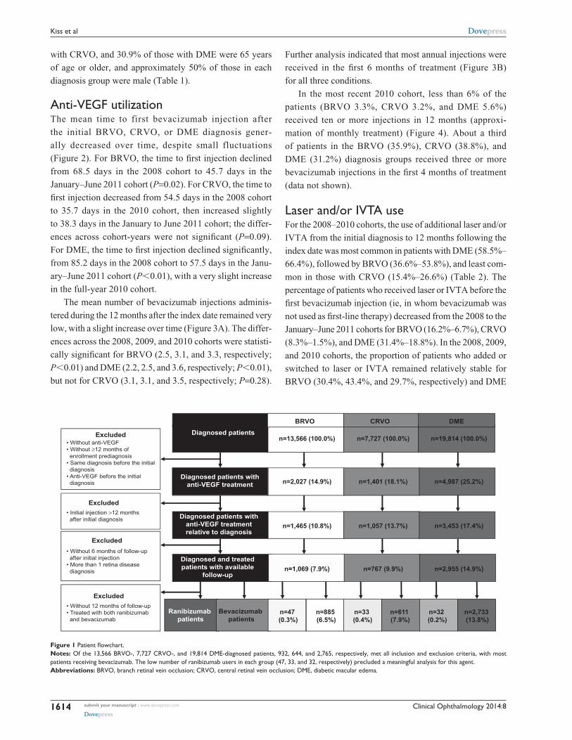
Resultssample characteristicsOf the 13,566, 7,727, and 19,814 patients diagnosed with
BRVO, CRVO, and DME, respectively, 932, 644, and 2,765,
respectively, met all inclusion and exclusion criteria (Figure 1).
The vast majority of these patients received treatment with
bevacizumab. The number of ranibizumab users in each
diagnosis group (47, 33, and 32, respectively) was too low
for meaningful analysis. Therefore, only results pertaining
to patients treated with bevacizumab were reported. Among
these patients, 51.0% of those with BRVO, 59.4% of those
Clinical Ophthalmology 2014:8submit your manuscript | www.dovepress.com
Dovepress
Dovepress
1614
Kiss et al
with CRVO, and 30.9% of those with DME were 65 years
of age or older, and approximately 50% of those in each
diagnosis group were male (Table 1).
anti-VegF utilizationThe mean time to first bevacizumab injection after
the initial BRVO, CRVO, or DME diagnosis gener-
ally decreased over time, despite small fluctuations
( Figure 2). For BRVO, the time to first injection declined
from 68.5 days in the 2008 cohort to 45.7 days in the
January–June 2011 cohort (P=0.02). For CRVO, the time to
first injection decreased from 54.5 days in the 2008 cohort
to 35.7 days in the 2010 cohort, then increased slightly
to 38.3 days in the January to June 2011 cohort; the differ-
ences across cohort-years were not significant (P=0.09).
For DME, the time to first injection declined significantly,
from 85.2 days in the 2008 cohort to 57.5 days in the Janu-
ary–June 2011 cohort (P0.01), with a very slight increase
in the full-year 2010 cohort.
The mean number of bevacizumab injections adminis-
tered during the 12 months after the index date remained very
low, with a slight increase over time (Figure 3A). The differ-
ences across the 2008, 2009, and 2010 cohorts were statisti-
cally significant for BRVO (2.5, 3.1, and 3.3, respectively;
P0.01) and DME (2.2, 2.5, and 3.6, respectively; P0.01),
but not for CRVO (3.1, 3.1, and 3.5, respectively; P=0.28).
Further analysis indicated that most annual injections were
received in the first 6 months of treatment (Figure 3B)
for all three conditions.
In the most recent 2010 cohort, less than 6% of the
patients (BRVO 3.3%, CRVO 3.2%, and DME 5.6%)
received ten or more injections in 12 months (approxi-
mation of monthly treatment) (Figure 4). About a third
of patients in the BRVO (35.9%), CRVO (38.8%), and
DME (31.2%) diagnosis groups received three or more
bevacizumab injections in the first 4 months of treatment
(data not shown).
laser and/or iVTa useFor the 2008–2010 cohorts, the use of additional laser and/or
IVTA from the initial diagnosis to 12 months following the
index date was most common in patients with DME (58.5%–
66.4%), followed by BRVO (36.6%–53.8%), and least com-
mon in those with CRVO (15.4%–26.6%) (Table 2). The
percentage of patients who received laser or IVTA before the
first bevacizumab injection (ie, in whom bevacizumab was
not used as first-line therapy) decreased from the 2008 to the
January–June 2011 cohorts for BRVO (16.2%–6.7%), CRVO
(8.3%–1.5%), and DME (31.4%–18.8%). In the 2008, 2009,
and 2010 cohorts, the proportion of patients who added or
switched to laser or IVTA remained relatively stable for
BRVO (30.4%, 43.4%, and 29.7%, respectively) and DME
Excluded• Without anti-VEGF• Without ≥12 months of enrollment prediagnosis• Same diagnosis before the initial diagnosis• Anti-VEGF before the initial diagnosis
Diagnosed patients
Ranibizumab patients
BRVO DME
• Initial injection >12 months after initial diagnosis
Excluded
• Without 6 months of follow-up after initial injection• More than 1 retina disease diagnosis
Excluded
• Without 12 months of follow-up• Treated with both ranibizumab and bevacizumab
Excluded
Diagnosed patients withanti-VEGF treatment
Diagnosed patients withanti-VEGF treatmentrelative to diagnosis
Diagnosed and treatedpatients with available
follow-up
Bevacizumab patients
CRVO
n=13,566 (100.0%)
n=2,027 (14.9%)
n=1,465 (10.8%)
n=1,069 (7.9%)
n=47 (0.3%)
n=885 (6.5%)
n=33 (0.4%)
n=611 (7.9%)
n=32 (0.2%)
n=2,733 (13.8%)
n=7,727 (100.0%)
n=1,401 (18.1%)
n=1,057 (13.7%)
n=767 (9.9%)
n=19,814 (100.0%)
n=4,987 (25.2%)
n=3,453 (17.4%)
n=2,955 (14.9%)
Figure 1 Patient flowchart.Notes: Of the 13,566 BrVO-, 7,727 CrVO-, and 19,814 DMe-diagnosed patients, 932, 644, and 2,765, respectively, met all inclusion and exclusion criteria, with most patients receiving bevacizumab. The low number of ranibizumab users in each group (47, 33, and 32, respectively) precluded a meaningful analysis for this agent.Abbreviations: BrVO, branch retinal vein occlusion; CrVO, central retinal vein occlusion; DMe, diabetic macular edema.

Clinical Ophthalmology 2014:8 submit your manuscript | www.dovepress.com
Dovepress
Dovepress
1615
anti-VegF utilization in rVO and DMe
Table 1 sample size and demographic characteristics of patients treated with bevacizumab or ranibizumab, by diagnosis group and cohort
BRVO (N=932)
CRVO (N=644)
DME (N=2,765)
Bevacizumab (n=885)
Ranibizumab (n=47)
Bevacizumab (n=611)
Ranibizumab (n=33)
Bevacizumab (n=2,733)
Ranibizumab (n=32)
Index cohorta
2008 148 3 120 2 338 42009 221 0 172 0 560 62010 306 21 188 9 1,009 132011 (Jan–Jun) 210 23 131 22 826 9age 65 years, % 51.0 60.0 59.4 61.0 30.9 34.0Male, % 49.6 40.0 54.9 39.0 54.5 53.0
Notes: aIndex year was based on time of the first bevacizumab or ranibizumab injection. The 2008, 2009, and 2010 cohorts had 12 months of follow-up available, and the January–June 2011 cohort had 6 months of follow-up available.Abbreviations: BrVO, branch retinal vein occlusion; CrVO, central retinal vein occlusion; DMe, diabetic macular edema.
68.5
61.3
49.9
45.7
54.5
45.1
35.738.3
85.2
77.1 78.5
57.5
0
2008
(n=1
48)
2009
(n=2
21)
2010
(n=3
06)
2011
(Jan
–Jun
) (n=
210)
10
20
30
40
50
60
70
80
90
Mea
n nu
mbe
r of d
ays
from
inde
x di
agno
sis
to fi
rst i
njec
tion
BRVOP=0.02*
CRVOP=0.09*
DMEP<0.01*
2008
(n=1
20)
2009
(n=1
72)
2010
(n=1
88)
2011
(Jan
–Jun
) (n=
131)
2008
(n=3
38)
2009
(n=5
60)
2010
(n=1
,009)
2011
(Jan
–Jun
) (n=
826)
Figure 2 Mean days from index diagnosis to first bevacizumab injection, by diagnosis group and cohort.Notes: *One-way analysis of variance comparing mean time across cohorts. among those with a diagnosis of BRVO, there was a significant decrease in the mean days from initial diagnosis to first injection: 68.5 days in 2008, 61.3 days in 2009, 49.9 days in 2010, and 45.7 days in 2011 (January–June cohort, P=0.02). although a decrease in time from initial diagnosis to first injection was observed in those with a diagnosis of CrVO (mean 54.5 days in 2008, 45.1 days in 2009, 35.7 days in 2010, and 38.3 days in 2011 [January–June cohort]), the differences in mean time across the cohorts were not significant (P=0.09). in the DMe group, the mean time from initial diagnosis to first injections decreased significantly over time (from mean of 85.2 days in 2008, to 77.1 days in 2009, 78.5 days in 2010, and 57.5 days in 2011 [January–June cohort]; P0.01). Abbreviations: BrVO, branch retinal vein occlusion; CrVO, central retinal vein occlusion; DMe, diabetic macular edema.
(29.6%, 35.9%, and 31.1%, respectively), but decreased for
CRVO (from 18.3% to 11.7%).
Of the patients treated with bevacizumab across
the full-year 2008–2010 cohorts, a minority of patients
(15.9% BRVO, 5.6% CRVO, and 15.8% DME) were
identified as potential switchers (ie, patients who dis-
continued bevacizumab use once laser or IVTA was
initiated, but had at least two additional ophthalmologist
visits) (Figure 5).
Across all cohorts, the annual number of bevacizumab
injections was positively associated with laser use in BRVO
(3.3 versus 2.9, P0.03) and with laser or IVTA use in
DME (laser, 3.3 versus 2.7, P0.03; IVTA, 3.3 versus 3.0,
P0.05), although the number of bevacizumab injections
was low regardless of whether laser or IVTA was used
(Table 3).
Patient monitoringAcross the 2008–2010 cohorts, the mean annual number of
all doctor visits ranged between 16.6 and 17.4, 19.1 and 20.5,
and 23.7 and 25.0 among patients diagnosed with BRVO,
CRVO, and DME, respectively (Table 4). The number of
ophthalmologist visits was generally low, but increased
over time in the 2008, 2009, and 2010 cohorts for BRVO
(5.1, 5.3, and 5.6, respectively) and DME (4.4, 4.8, and 5.3,
respectively). The corresponding numbers for CRVO
were 6.5, 5.8, and 5.9, respectively. During the same period,
the mean annual numbers of OCT examinations ranged
between 3.7 and 3.9 in patients with BRVO, 3.4 and 3.8 in
those with CRVO, and 3.1 and 3.8 in those with DME;
the mean annual numbers of FA examinations ranged
between 0.9 and 1.2 for BRVO-, 0.7 and 1.0 for CRVO-,
and 0.9 and 1.1 for DME-diagnosed patients.

Clinical Ophthalmology 2014:8submit your manuscript | www.dovepress.com
Dovepress
Dovepress
1616
Kiss et al
Mea
n (S
D) n
umbe
r of i
njec
tions
over
6 m
onth
s
BRVOMean number
across cohorts: 2.4P<0.001*
CRVOMean number
across cohorts: 2.4P<0.001*
DMEMean number
across cohorts: 2.2P<0.001*
2.1 (1.3)
2.4 (1.4)
2.6 (1.7)
2.9 (1.7)
2.3 (1.5)
2.3 (1.4)
3.0 (1.8)
2.7 (1.7)
1.8 (1.3)
2.0 (1.3)
2.6 (2.0)
3.0 (2.5)
0
2008
(n=1
48)
2009
(n=2
21)
2010
(n=3
06)
0.5
1
1.5
2
2.5
3
3.5
2008
(n=1
20)
2009
(n=1
72)
2010
(n=1
88)
2008
(n=3
38)
2009
(n=5
60)
2010
(n=1
,009)
2011
(Jan
–Jun
) (n=
210)
2011
(Jan
–Jun
) (n=
131)
2011
(Jan
–Jun
) (n=
826)
2.5 (2.0)
3.1 (2.3)
3.3 (2.6)
3.1 (2.4) 3.1 (2.5)
3.5 (2.7)
2.2 (2.0)
2.5 (2.0)
3.6 (3.2)
0
2008
(n=1
48)
2009
(n=2
21)
2010
(n=3
06)
0.5
1
1.5
2
2.5
3
3.5
4M
ean
(SD
) num
ber o
f inj
ectio
nsov
er 1
2 m
onth
s
BRVOMean number
across cohorts: 3.1P<0.01*
CRVOMean number
across cohorts: 3.2P=0.28*
DMEMean number
across cohorts: 3.0P<0.01*
2008
(n=1
20)
2009
(n=1
72)
2010
(n=1
88)
2008
(n=3
38)
2009
(n=5
60)
2010
(n=1
,009)
A B
Figure 3 (A) Mean number (standard deviation [SD]) of bevacizumab injections over 12 months, by diagnosis group and cohort; (B) mean number (sD) of bevacizumab injections over 6 months, by diagnosis group and cohort.Notes: *One-way analysis of variance comparing mean time across cohorts. In each diagnosis group, the mean number of injections administered over the 12-month period after the first injection (index date) increased, with mean differences across the cohorts being statistically significant in the BRVO and DME groups, but not in the CRVO group.Abbreviations: BrVO, branch retinal vein occlusion; CrVO, central retinal vein occlusion; DMe, diabetic macular edema.
0
5
10
15
20
25
30
35
Perc
enta
ge o
f pat
ient
s
BRVO(n=306)
DME(n=1,009)
Number of injections per patientCRVO
(n=188)
29.7
18.6
16.0
11.8
6.95.9
2.9 3.31.6
3.3
30.7
20.9
11.4
9.1
6.4 6.0
3.9 4.2
1.8
5.6
1 2 3 4 5 6 7 8 9 10+ 1 2 3 4 5 6 7 8 9 10+ 1 2 3 4 5 6 7 8 9 10+
31.4
12.2
18.6
12.2
4.86.4
4.8
2.73.7 3.2
Figure 4 Distribution of the number of injections over 12 months in the 2010 cohort, by diagnosis group.Notes: In analyses of the distribution of the number of injections in the 2010 cohort, small percentages of patients in each diagnosis group received 10 injections during the 12 months after their index diagnosis.Abbreviations: BrVO, branch retinal vein occlusion; CrVO, central retinal vein occlusion; DMe, diabetic macular edema.
DiscussionThis retrospective claims analysis of a large US insurance
database identified over 2,000 patients newly diagnosed with
BRVO, CRVO, or DME who were treated with bevacizumab
or ranibizumab between January 2008 and December 2011.
The majority of patients were treated with bevacizumab. This is
consistent with a higher overall use of bevacizumab in the retinal
therapeutic area.27 It is also in large part due to ranibizumab not
being reimbursed by health plans until after its approval for
BRVO or CRVO in June 2010 and for DME in August 2012.

Clinical Ophthalmology 2014:8 submit your manuscript | www.dovepress.com
Dovepress
Dovepress
1617
anti-VegF utilization in rVO and DMe
Table 2 Proportions of bevacizumab-treated patients who received laser therapy or intravitreal triamcinolone, by diagnosis group and cohort
Index cohort, %
BRVO CRVO DME
All Before first bevacizumaba
After first bevacizumabb
All Before first bevacizumaba
After first bevacizumabb
All Before first bevacizumaba
After first bevacizumabb
2008c n=148 n=120 n=338laser or iVTa 46.6 16.2 30.4 26.6 8.3 18.3 61.0 31.4 29.6
laser 39.9 13.5 26.4 4.2 0 4.2 53.2 27.5 25.7iVTa 14.9 2.7 12.2 24.1 8.3 15.8 21.3 6.8 14.5
2009c n=221 n=172 n=560laser or iVTa 53.8 10.4 43.4 16.3 4.7 11.6 66.4 30.5 35.9
laser 46.2 7.7 38.5 4.1 1.2 2.9 59.3 27.5 31.8iVTa 14.0 3.6 10.4 13.4 3.5 9.9 21.3 5.9 15.4
2010c n=306 n=188 n=1,009laser or iVTa 36.6 6.9 29.7 15.4 3.7 11.7 58.5 27.4 31.1
laser 32.7 5.6 27.1 2.1 0 2.1 53.1 25.2 27.9iVTa 7.2 2.0 5.2 11.7 3.7 8.0 15.3 4.2 11.1
2011 Jan–Jund n=210 n=131 n=826laser or iVTa – 6.7 – – 1.5 – – 18.8 –
laser – 5.2 – – 0 – – 17.8 –iVTa – 1.4 – – 1.5 – – 1.5 –
Notes: aProportion of patients who received laser or ITVA before the first bevacizumab injection; bproportion of patients who added or switched to laser or iVTa at the same time or after the first bevacizumab injection; cthese were indexed (time of the first anti-VEGF injection) from January 1 to December 31 of the calendar year and followed for 12 months; dthis cohort was indexed during January 1 to June 30, 2011 and followed for 6 months. en dashes denote no data.Abbreviations: BrVO, branch retinal vein occlusion; CrVO, central retinal vein occlusion; DMe, diabetic macular edema; iVTa, intravitreal triamcinolone.
34.4(n=232)
13.3(n=64)
32.2(n=615)
18.1(n=122)
6.0(n=29)
18.0(n=343)15.9
(n=107)
5.6(n=27)
15.8(n=301)
0
10
20
30
40
50
60
DME (N=1,907)CRVO (N=480)BRVO (N=675)
Perc
enta
ge o
f pat
ient
s
Patients receiving initial bevacizumab therapy with concomitant or subsequent laser or IVTAPatients receiving initial bevacizumab therapy with concomitant or subsequent laser or IVTA, and with no further bevacizumabPatients receiving initial bevacizumab therapy with concomitant or subsequent laser or IVTA, and with no further bevacizumab but with ≥2 further ophthalmologist visits ("Potential Switchers")
Figure 5 Use of laser and/or intravitreal triamcinolone relative to the timing of bevacizumab use during the first 12 months following the index date, by diagnosis group (2008–2010 cohorts).Notes: in those with BrVO, CrVO, and DMe, approximately 15.9%, 5.6%, and 15.8%, respectively, of the total diagnosis group were considered to have switched to either laser or iVTa treatment from bevacizumab treatment (ie, received laser or iVTa at the time of or after bevacizumab treatment, and then discontinued bevacizumab but continued to visit an ophthalmologist for at least two visits).Abbreviations: BrVO, branch retinal vein occlusion; CrVO, central retinal vein occlusion; DMe, diabetic macular edema; iVTa, intravitreal triamcinolone.
Our results revealed that an increasing number of
patients were treated with anti-VEGF agents across BRVO,
CRVO, and DME during the study period. In addition, the
time between the initial diagnosis and anti-VEGF initiation
decreased over time. However, the mean annual number of
bevacizumab injections remained below four, despite a slight
increase during 2008–2011. In the most recent 2010 cohort
that included patients followed through 2011, less than 6%
of the patients in each diagnosis group met the criteria for
monthly injections, and less than 16% of patients in each
diagnosis group met the criteria for monthly ophthalmologist
visits (additional data not reported in tables/figures). These
data are in sharp contrast to major ranibizumab Phase II and
III pivotal trials, where the patients were monitored monthly
and the mean annual numbers of injections were more than
twofold greater: 8.4 in patients with BRVO (BRAVO trial),11
8.8 in patients with CRVO (CRUISE trial),14 and 7.0–11.0 in
patients with DME (RESTORE,13 Protocol I,17 and
RISE/RIDE15 trials).
A variety of factors may have contributed to the very
low numbers of monitoring visits and bevacizumab injec-
tions in clinical practice. Patients in a clinical practice may
be inherently different from those enrolled in clinical trials.
Our study period largely preceded the publication of the
major clinical trials of monthly ranibizumab in BRVO and
CRVO (the initial BRAVO28 and CRUISE29 papers were
published in mid-2010), and entirely preceded publica-
tion of clinical trials of monthly ranibizumab in DME (the
RIDE/RISE paper15 was published in 2012). The lack of
level 1 randomized controlled trial evidence likely contrib-
uted in large part to the wide variation in treatment schedules
and overall suboptimal treatment frequencies. In addition,
large bevacizumab trials conducted at the time, such as the
Clinical Ophthalmology 2014:8submit your manuscript | www.dovepress.com
Dovepress
Dovepress
1618
Kiss et al
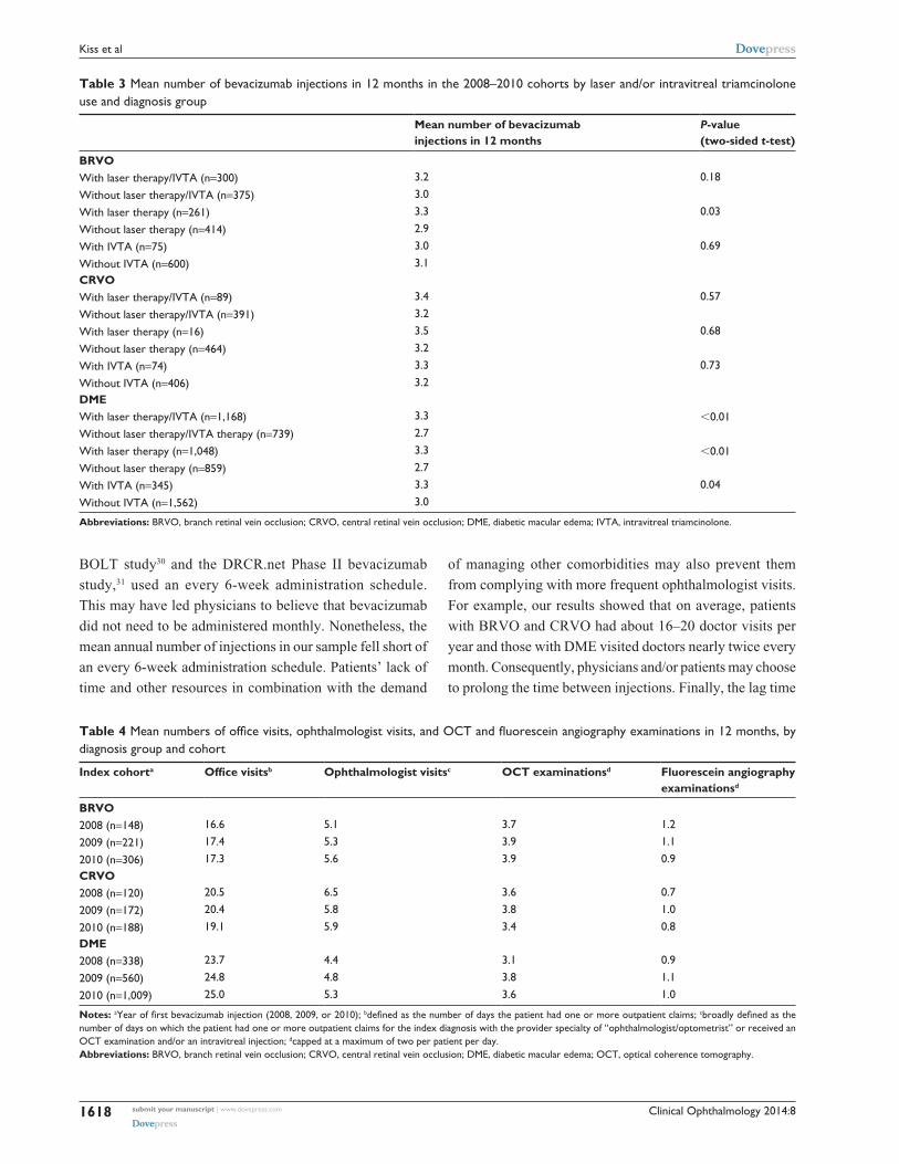
Table 3 Mean number of bevacizumab injections in 12 months in the 2008–2010 cohorts by laser and/or intravitreal triamcinolone use and diagnosis group
Mean number of bevacizumab injections in 12 months
P-value (two-sided t-test)
BRVOWith laser therapy/iVTa (n=300) 3.2 0.18
Without laser therapy/iVTa (n=375) 3.0
With laser therapy (n=261) 3.3 0.03
Without laser therapy (n=414) 2.9
With iVTa (n=75) 3.0 0.69
Without iVTa (n=600) 3.1CRVOWith laser therapy/iVTa (n=89) 3.4 0.57
Without laser therapy/iVTa (n=391) 3.2
With laser therapy (n=16) 3.5 0.68
Without laser therapy (n=464) 3.2
With iVTa (n=74) 3.3 0.73
Without iVTa (n=406) 3.2DMEWith laser therapy/iVTa (n=1,168) 3.3 0.01Without laser therapy/iVTa therapy (n=739) 2.7
With laser therapy (n=1,048) 3.3 0.01Without laser therapy (n=859) 2.7
With iVTa (n=345) 3.3 0.04
Without iVTa (n=1,562) 3.0
Abbreviations: BrVO, branch retinal vein occlusion; CrVO, central retinal vein occlusion; DMe, diabetic macular edema; iVTa, intravitreal triamcinolone.
Table 4 Mean numbers of office visits, ophthalmologist visits, and OCT and fluorescein angiography examinations in 12 months, by diagnosis group and cohort
Index cohorta Office visitsb Ophthalmologist visitsc OCT examinationsd Fluorescein angiography examinationsd
BRVO2008 (n=148) 16.6 5.1 3.7 1.2
2009 (n=221) 17.4 5.3 3.9 1.1
2010 (n=306) 17.3 5.6 3.9 0.9CRVO2008 (n=120) 20.5 6.5 3.6 0.7
2009 (n=172) 20.4 5.8 3.8 1.0
2010 (n=188) 19.1 5.9 3.4 0.8DME2008 (n=338) 23.7 4.4 3.1 0.9
2009 (n=560) 24.8 4.8 3.8 1.1
2010 (n=1,009) 25.0 5.3 3.6 1.0
Notes: aYear of first bevacizumab injection (2008, 2009, or 2010); bdefined as the number of days the patient had one or more outpatient claims; cbroadly defined as the number of days on which the patient had one or more outpatient claims for the index diagnosis with the provider specialty of “ophthalmologist/optometrist” or received an OCT examination and/or an intravitreal injection; dcapped at a maximum of two per patient per day.Abbreviations: BrVO, branch retinal vein occlusion; CrVO, central retinal vein occlusion; DMe, diabetic macular edema; OCT, optical coherence tomography.
BOLT study30 and the DRCR.net Phase II bevacizumab
study,31 used an every 6-week administration schedule.
This may have led physicians to believe that bevacizumab
did not need to be administered monthly. Nonetheless, the
mean annual number of injections in our sample fell short of
an every 6-week administration schedule. Patients’ lack of
time and other resources in combination with the demand
of managing other comorbidities may also prevent them
from complying with more frequent ophthalmologist visits.
For example, our results showed that on average, patients
with BRVO and CRVO had about 16–20 doctor visits per
year and those with DME visited doctors nearly twice every
month. Consequently, physicians and/or patients may choose
to prolong the time between injections. Finally, the lag time

Clinical Ophthalmology 2014:8 submit your manuscript | www.dovepress.com
Dovepress
Dovepress
1619
anti-VegF utilization in rVO and DMe
between randomized clinical trial publications and the incor-
poration of new treatment paradigms into practice may be
another important contributing factor, as supported by the
slow but increasing trend in numbers of injections over time
in our study. This is similar to the trends noted in anti-VEGF
treatment patterns in patients with nAMD.32
The large disparity in injection frequency, and more
importantly in monitoring visit frequency, between the major
clinical studies and routine clinical practice raises the pos-
sibility that patients’ vision outcomes in the clinical setting
may be substantially lower than those reported by the major
clinical trials. This is supported by evidence from Phase II and
III randomized controlled trials suggesting that in addition to
monthly follow-up, more frequent injections generally result
in higher absolute levels of vision improvement in DME and
nAMD (Figure 6).12,13,15,17,20–22,30,33–35 However, few studies
have examined the relationship between injection frequency
and vision outcomes in BRVO, CRVO, or DME outside the
clinical trial setting, although retrospective effectiveness
studies of bevacizumab and ranibizumab in nAMD have
found both injection frequency and vision improvement
to be well below those reported in large published clinical
trials.36–39
Our analysis indicated that the use of laser or IVTA is
very common among bevacizumab-treated patients with
BRVO and DME and less so in those with CRVO. The use
of laser or IVTA as first-line therapy decreased substantially
in patients with BRVO and CRVO and somewhat moderately
in DME over time. In contrast, about 30%–43% of patients
with BRVO and DME continued to add or switch to laser or
IVTA either at the same time or after the initiation of beva-
cizumab injections. While many believe that the use of laser
or IVTA could potentially extend the duration of anti-VEGF
treatment, the use of laser in BRVO and the use of laser or
IVTA in DME were positively associated with the number of
bevacizumab injections in our study. This suggests that the
use of concomitant and adjunctive treatment may be more
indicative of the underlying disease severity or an overall
generally more aggressive treatment strategy.
The strengths of this study include the use of a large
national database composed of a diverse patient population.
Our inclusion/exclusion criteria ensured high internal validity
for the findings. Potential limitations include an insufficient
sample of ranibizumab-treated patients during the study
period. Because of an inability to distinguish between unilat-
eral and bilateral treatment, per-patient numbers of injections
and monitoring visits in this study are an upper bound for esti-
mates of the numbers of injections and monitoring visits per
DMEnAMD
–5
0
2 4 6 8 10 12
5
10
15
20
Mea
n B
CVA
cha
nge
in 1
2 m
onth
s
Mean number of anti-VEGF injections in 12 months
PIER
SAILOREXCITE
RESTORE
RESOLVEANCHOR
RISE RIDE
BOLTPROTOCOL I
CATT
CATTCATT
MARINACATT
Figure 6 Correlations between number of bevacizumab and ranibizumab injections and mean ETDRS letters gained in 12 months in major published Phase II and III clinical trials in naMD and DMe.Note: Evidence from major prospective clinical trials suggests a positive correlation between the administration frequency of bevacizumab or ranibizumab and visual improvement.Abbreviations: BCVa, best-corrected visual acuity; DMe, diabetic macular edema; eTDrs, early Treatment Diabetic retinopathy study; naMD, neovascular age-related macular degeneration.
eye. In the absence of visual acuity or anatomical outcomes
in claims databases, we were unable to identify potential
outcome factors that may have influenced ongoing treatment
decisions (eg, reduction or discontinuation of the treatments
due to a lack or stabilization of visual acuity response, or
achievement of acceptable vision outcomes with low injec-
tion frequencies). Nevertheless, the similarly low numbers
of monitoring visits and OCT exams suggest that treatment
outcomes may have only had limited impact on injection fre-
quency. The stringent sample selection criteria were designed
to maximize internal validity, but may limit the ability to
generalize our findings to all patients with BRVO, CRVO,
or DME. Finally, similar to other administrative databases,
the IMS LifeLink health plan claims database lacks clinical
details regarding severity of illness, and coding of diagnoses
and procedures may be inaccurate or incomplete. The triple
ICD-9 diagnostic codes for DME (ie, 362.07, a DME-specific
code introduced in 2007, and the combination of 362.53 [cys-
toid macular edema] or 362.83 [retinal edema] with 250.xx
[diabetes mellitus]), were derived from a validated algorithm
that showed high sensitivity and specificity in identifying
DME from pre-2007 claims data.25 These codes, which have
been used in a previous claims analysis,26 represent relatively
broad inclusion criteria for DME. However, in a related
retrospective claims analysis using a different database, we
found that the use of a more stringent DME-specific code
(ie, 362.07, introduced first in 2007), which underestimated
the prevalence of DME patients in the database, resulted in

Clinical Ophthalmology 2014:8submit your manuscript | www.dovepress.com
Dovepress
Dovepress
1620
Kiss et al
a similar mean annual number of injections. We therefore
used the broad set of ICD-9 codes for a more complete char-
acterization of the clinical DME population.
ConclusionThis large retrospective claims analysis provides the first
comprehensive look at the utilization of anti-VEGF therapies
in BRVO, CRVO, and DME in US clinical practice during
the period leading up to publication of findings from major
clinical trials of anti-VEGF agents in these indications. Our
results show that the number of patients receiving bevaci-
zumab for these conditions steadily increased. However,
patients in clinical practice were monitored less frequently
and received far fewer injections compared with patients in
major clinical trials. Further research is necessary to confirm
these findings in larger ranibizumab samples, to determine
factors that may contribute to the observed low injection
frequency, and to evaluate the visual outcomes associated
with these reduced utilization patterns.
Author contributionsSK, YL, JB, NMH, AA, JC, and JWK were responsible for
the design of the study, SK, YL, JB, NMH, AA, JC, and
JWK for conduct of the study, collection, management,
analysis, and interpretation of the data, and SK, YL, JB,
NMH, AA, JC, JWK for preparation, review, or approval
of the manuscript.
AcknowledgmentSupport for third-party writing assistance by Susan Ruffalo,
PharmD of Med Write, Inc., was provided by Allergan, Inc.
DisclosureThis analysis was sponsored by Allergan, Inc., Irvine, CA. SK
reports the following: consulting fees or honoraria – Alimera
Sciences Inc., Allergan, Alcon, EyeTech, Genentech,
Merge/OIS, Optos, Regeneron, and Thrombogenics; speakers
bureaus – Alimera, Allergan, Genentech, Optos, Regeneron,
and ThromboGenics; clinical research projects – Allergan,
Genentech, Optos, and Regeneron; stock/stock options –
Merge/OIS. YL, JC, and JWK are employees of Allergan
and hold equity and/or options in Allergan. JB is a full-time
employee of IMS Health. NMH reports the following: con-
sulting fees or honoraria – Genentech, Allergan, Regeneron,
Sequenom, Inc., Alimera, and Notal Vision; board member-
ship – Katalyst Surgical; speakers bureaus – Sequenom,
Genentech, and Regeneron; clinical research projects – Notal
Vision; stock/stock options – Katalyst Surgical. AA reports
consulting fees or honoraria from Allergan. The authors
declare no other conflicts of interest.
References 1. Romero-Aroca P. Managing diabetic macular edema: the leading cause
of diabetes blindness. World J Diabetes. 2011;2(6):98–104. 2. Yau JW, Lee P, Wong TY, Best J, Jenkins A. Retinal vein occlusion:
an approach to diagnosis, systemic risk factors and management. Intern Med J. 2008;38(12):904–910.
3. Turello M, Pasca S, Daminato R, et al. Retinal vein occlusion: evalua-tion of “classic” and “emerging” risk factors and treatment. J Thromb Thrombolysis. 2010;29(4):459–464.
4. Institute for Clinical and Economic Review (ICER). Technology Assess-ment Report: Anti-Vascular Endothelial Growth Factor Treatment for Diabetic Macular Edema. Boston: ICER; 2012. Available from: http://www.cms.gov/Medicare/Coverage/DeterminationProcess/downloads/id85TA.pdf. Accessed June 10, 2013.
5. Rogers S, McIntosh RL, Cheung N, et al. The prevalence of retinal vein occlusion: pooled data from population studies from the United States, Europe, Asia, and Australia. Ophthalmology. 2010;117(2): 313–319.
6. Yau JW, Rogers SL, Kawasaki R, et al. Global prevalence and major risk factors of diabetic retinopathy. Diabetes Care. 2012;35(3):556–564.
7. Aiello LP, Avery RL, Arrigg PG, et al. Vascular endothelial growth factor in ocular fluid of patients with diabetic retinopathy and other retinal disorders. N Engl J Med. 1994;331(22):1480–1487.
8. Andreoli CM, Miller JW. Anti-vascular endothelial growth factor ther-apy for ocular neovascular disease. Curr Opin Ophthalmol. 2007;18(6): 502–508.
9. Yoshimura T, Sonoda KH, Sugahara M, et al. Comprehensive analysis of inflammatory immune mediators in vitreoretinal diseases. PLoS One. 2009;4(12):e8158.
10. Sohn HJ, Han DH, Kim IT, et al. Changes in aqueous concentrations of various cytokines after intravitreal triamcinolone versus bevacizumab for diabetic macular edema. Am J Ophthalmol. 2011;152(4):686–694.
11. Brown DM, Campochiaro PA, Bhisitkul RB, et al. Sustained benefits from ranibizumab for macular edema following branch retinal vein occlusion: 12-month outcomes of a phase III study. Ophthalmology. 2011;118(8):1594–1602.
12. Massin P, Bandello F, Garweg JG, et al. Safety and efficacy of ranibi-zumab in diabetic macular edema (RESOLVE study): a 12-month, randomized, controlled, double-masked, multicenter phase II study. Diabetes Care. 2010;33(11):2399–2405.
13. Mitchell P, Bandello F, Schmidt-Erfurth U, et al. The RESTORE study: ranibizumab monotherapy or combined with laser versus laser monotherapy for diabetic macular edema. Ophthalmology. 2011;118(4): 615–625.
14. Campochiaro PA, Brown DM, Awh CC, et al. Sustained benefits from ranibizumab for macular edema following central retinal vein occlusion: twelve-month outcomes of a phase III study. Ophthalmology. 2011; 118(10):2041–2049.
15. Nguyen QD, Brown DM, Marcus DM, et al. Ranibizumab for diabetic macular edema: results from 2 phase III randomized trials: RISE and RIDE. Ophthalmology. 2012;119(4):789–801.
16. Heier JS, Campochiaro PA, Yau L, et al. Ranibizumab for macular edema due to retinal vein occlusions: long-term follow-up in the HORIZON trial. Ophthalmology. 2012;119(4):802–809.
17. Diabetic Retinopathy Clinical Research Network, Elman MJ, Aiello LP, et al. Randomized trial evaluating ranibizumab plus prompt or deferred laser or triamcinolone plus prompt laser for diabetic macular edema. Ophthalmology. 2010;117(6):1064–1077.
18. Elman MJ, Bressler NM, Qin H, et al. Expanded 2-year follow-up of ranibizumab plus prompt or deferred laser or triamcinolone plus prompt laser for diabetic macular edema. Ophthalmology. 2011;118(4): 609–614.

Clinical Ophthalmology
Publish your work in this journal
Submit your manuscript here: http://www.dovepress.com/clinical-ophthalmology-journal
Clinical Ophthalmology is an international, peer-reviewed journal covering all subspecialties within ophthalmology. Key topics include: Optometry; Visual science; Pharmacology and drug therapy in eye diseases; Basic Sciences; Primary and Secondary eye care; Patient Safety and Quality of Care Improvements. This journal is indexed on
PubMed Central and CAS, and is the official journal of The Society of Clinical Ophthalmology (SCO). The manuscript management system is completely online and includes a very quick and fair peer-review system, which is all easy to use. Visit http://www.dovepress.com/testimonials.php to read real quotes from published authors.
Dovepress
Clinical Ophthalmology 2014:8 submit your manuscript | www.dovepress.com
Dovepress
Dovepress
1621
anti-VegF utilization in rVO and DMe
19. Martin DF, Maguire MG, Fine SL, et al. Ranibizumab and bevacizumab for treatment of neovascular age-related macular degeneration: two-year results. Ophthalmology. 2012;119(7):1388–1398.
20. Brown DM, Kaiser PK, Michels M, et al. Ranibizumab versus verte-porfin for neovascular age-related macular degeneration. N Engl J Med. 2006;355(14):1432–1444.
21. Rosenfeld PJ, Brown DM, Heier JS, et al. Ranibizumab for neovas-cular age-related macular degeneration. N Engl J Med. 2006;355(14): 1419–1431.
22. Martin DF, Maguire MG, Ying GS, et al. Ranibizumab and bevaci-zumab for neovascular age-related macular degeneration. N Engl J Med. 2011;364(20):1897–1908.
23. IVAN Study Investigators, Chakravarthy U, Harding SP, et al. Ranibi-zumab versus bevacizumab to treat neovascular age-related macular degeneration: one-year findings from the IVAN randomized trial. Ophthalmology. 2012;119(7):1399–1411.
24. Busbee BG, Ho AC, Brown DM, et al. Twelve-month efficacy and safety of 0.5 mg or 2.0 mg ranibizumab in patients with subfoveal neovas-cular age-related macular degeneration. Ophthalmology. 2013;120(5): 1046–1056.
25. Bearelly S, Mruthyunjaya P, Tzeng JP, et al. Identification of patients with diabetic macular edema from claims data: a validation study. Arch Ophthalmol. 2008;126(7):986–989.
26. Shea AM, Curtis LH, Hammill BG, et al. Resource use and costs associ-ated with diabetic macular edema in elderly persons. Arch Ophthalmol. 2008;126(12):1748–1754.
27. Jumper JM, Mittra RA. ASRS 2012 Preferences and Trends Member-ship Survey. Paper presented at: 30th Annual Meeting of the Ameri-can Society of Retina Specialists; August 25–29, 2012; Las Vegas, Nevada.
28. Campochiaro PA, Heier JS, Feiner L, et al. Ranibizumab for macular edema following branch retinal vein occlusion: six-month primary end point results of a phase III study. Ophthalmology. 2010;117(6): 1102–1112.
29. Brown DM, Campochiaro PA, Singh RP, et al. Ranibizumab for macu-lar edema following central retinal vein occlusion: six-month primary end point results of a phase III study. Ophthalmology. 2010;117(6): 1124–1133.
30. Michaelides M, Kaines A, Hamilton RD, et al. A prospective random-ized trial of intravitreal bevacizumab or laser therapy in the management of diabetic macular edema (BOLT study) 12-month data: report 2. Ophthalmology. 2010;117(6):1078–1086.
31. Diabetic Retinopathy Clinical Research Network; Scott IU, Edwards AR, et al. A phase II randomized clinical trial of intravitreal bevacizumab for diabetic macular edema. Ophthalmology. 2007;114(10):1860–1867.
32. Holekamp NM, Yeh WS, Chia Y, Kiss S, Almony A, Kowalski JW. Real-world utilization of intravitreal anti-vascular endothelial growth factor agents in common retinal diseases. Invest Ophthalmol Vis Sci. 2012;53(6):4179.
33. Boyer DS, Heier JS, Brown DM, Francom SF, Ianchulev T, Rubio RG. A Phase IIIb study to evaluate the safety of ranibizumab in subjects with neovascular age-related macular degeneration. Ophthalmology. 2009;116(9):1731–1739.
34. Schmidt-Erfurth U, Eldem B, Guymer R, et al. Efficacy and safety of monthly versus quarterly ranibizumab treatment in neovascular age-related macular degeneration: the EXCITE study. Ophthalmology. 2011; 118(5):831–839.
35. Regillo CD, Brown DM, Abraham P, et al. Randomized, double-masked, sham-controlled trial of ranibizumab for neovascular age-re-lated macular degeneration: PIER study year 1. Am J Ophthalmol. 2008; 145(2):239–248.
36. Fong DS, Custis P, Howes J, Hsu JW. Intravitreal bevacizumab and ranibizumab for age-related macular degeneration: a multicenter, ret-rospective study. Ophthalmology. 2010;117(2):298–302.
37. Rotsos T, Patel PJ, Chen FK, Tufail A. Initial clinical experience of ranibizumab therapy for neovascular age-related macular degeneration. Clin Ophthalmol. 2010;4:1271–1275.
38. Bandukwala T, Muni RH, Schwartz C, Eng KT, Kertes PJ. Effectiveness of intravitreal ranibizumab for the treatment of neovascular age-related macular degeneration in a Canadian retina practice: a retrospective review. Can J Ophthalmol. 2010;45(6):590–595.
39. Bandello F, Holz FG, Gillies MC, et al. Safety, efficacy, treatment patterns of ranibizumab therapy for neovascular age-related macular degeneration: the LUMINOUS studies. Invest Ophthalmol Vis Sci. 2012; 53(6):2031.