Anti-CK7/CK20 Immunohistochemistry Did Not Associate with ...
12
Citation: Court, A.; Laville, D.; Dagher, S.; Grosjean, V.; Dal-Col, P.; Yvorel, V.; Casteillo, F.; Bayle-Bleuez, S.; Vergnon, J.-M.; Forest, F. Anti-CK7/CK20 Immunohistochemistry Did Not Associate with the Metastatic Site in TTF-1-Negative Lung Cancer. Diagnostics 2022, 12, 1589. https://doi.org/10.3390/ diagnostics12071589 Academic Editor: Cesar A. Moran Received: 27 April 2022 Accepted: 28 June 2022 Published: 29 June 2022 Publisher’s Note: MDPI stays neutral with regard to jurisdictional claims in published maps and institutional affil- iations. Copyright: © 2022 by the authors. Licensee MDPI, Basel, Switzerland. This article is an open access article distributed under the terms and conditions of the Creative Commons Attribution (CC BY) license (https:// creativecommons.org/licenses/by/ 4.0/). diagnostics Article Anti-CK7/CK20 Immunohistochemistry Did Not Associate with the Metastatic Site in TTF-1-Negative Lung Cancer Alice Court 1 , David Laville 1 , Sami Dagher 1 , Vincent Grosjean 1 , Pierre Dal-Col 1 , Violaine Yvorel 1,2 , François Casteillo 1 , Sophie Bayle-Bleuez 3 , Jean-Michel Vergnon 3 and Fabien Forest 1,2, * 1 Department of Pathology, University Hospital of Saint Etienne, 42055 Saint Etienne, France; [email protected] (A.C.); [email protected] (D.L.); [email protected] (S.D.); [email protected] (V.G.); [email protected] (P.D.-C.); [email protected] (V.Y.); [email protected] (F.C.) 2 Department of Molecular Biology of Solid Tumors, University Hospital of Saint Etienne, 42270 Saint Etienne, France 3 Department of Pneumology, University Hospital of Saint Etienne, 42270 Saint Etienne, France; [email protected] (S.B.-B.); [email protected] (J.-M.V.) * Correspondence: [email protected]; Tel.: +33-(0)-4-77-12-77-34; Fax: +33-(0)-4-77-82-82-96 Abstract: Anti-CK7 and anti-CK20 immunohistochemistry is sometimes used to establish a diagnosis of primary lung cancer. We performed a retrospective study on the value of anti-CK7 and anti-CK20 immunohistochemistry in 359 biopsies of patients with suspected lung carcinoma in order to assess the usefulness of these antibodies in the evaluation of lung tumors in biopsies. Our results showed TTF-1 positivity in 73.3% of patients. EGFR mutations and ALK rearrangements were significantly different between TTF-1 positive and TTF-1 negative tumors (p < 0.001 and p = 0.023, respectively). Our results show a significant difference (p < 0.001) between TTF-1 positive and TTF-1 negative carcinomas with a median survival of 21.97 months (CI95% = 17.48–30.9 months) and 6.52 months (CI95% = 3.34–10.3 months), respectively. In the group of TTF-1 negative patients, anti-CK7 and CK20 immunohistochemistry was performed in 70 patients and showed CK7+/CK20- staining in 61 patients (87.1%), CK7-/CK20- in 4 patients (5.7%), CK7+/CK20+ in 3 patients (4.3%), and CK7- /CK20- in 2 patients (2.8%). No specific or molecular pattern was found in these groups of CK7/CK20 combinations. In total, this work brings arguments concerning the uselessness of anti-CK7/CK20 immunohistochemistry in the case of suspicion of primary lung cancer in biopsies. Keywords: adenocarcinoma; TTF-1; CK7; CK20; EGFR 1. Introduction Lung cancer is a common tumor, unfortunately often diagnosed at the metastasis stage. The three main types of lung cancer include adenocarcinoma, squamous cell car- cinoma, and small cell carcinoma. The diagnosis is often made on small specimens such as biopsies. Immunohistochemistry is a protein detection technique commonly used in pathology. This technique allows to detect the presence of certain proteins, but also to localize these proteins on the subcellular level [1]. Immunohistochemistry in diagnostic pathology is based on the reaction between an antigen, a primary antibody, and a secondary antibody [2]. Immunohistochemistry is the cornerstone of diagnosis for lung tumors [2]. Indeed, immunohistochemistry is important to better distinguish the histological type of lung cancer [2]. Prior to the advent of targeted therapies, there was little difference in treatment by histological type in the non-small cell carcinoma group. One of the most important issues for the pathologist was to distinguish small cell carcinoma from non-small cell carcinoma, which is often possible on morphology alone. With the introduction of targeted therapies in the treatment of lung cancer, it has become crucial to determine the Diagnostics 2022, 12, 1589. https://doi.org/10.3390/diagnostics12071589 https://www.mdpi.com/journal/diagnostics
-
Upload
khangminh22 -
Category
Documents
-
view
1 -
download
0
Transcript of Anti-CK7/CK20 Immunohistochemistry Did Not Associate with ...
Citation: Court, A.; Laville, D.;
Dagher, S.; Grosjean, V.; Dal-Col, P.;
Yvorel, V.; Casteillo, F.; Bayle-Bleuez,
S.; Vergnon, J.-M.; Forest, F.
Anti-CK7/CK20
Immunohistochemistry Did Not
Associate with the Metastatic Site in
TTF-1-Negative Lung Cancer.
Diagnostics 2022, 12, 1589.
https://doi.org/10.3390/
diagnostics12071589
Academic Editor: Cesar A. Moran
Received: 27 April 2022
Accepted: 28 June 2022
Published: 29 June 2022
Publisher’s Note: MDPI stays neutral
with regard to jurisdictional claims in
published maps and institutional affil-
iations.
Copyright: © 2022 by the authors.
Licensee MDPI, Basel, Switzerland.
This article is an open access article
distributed under the terms and
conditions of the Creative Commons
Attribution (CC BY) license (https://
creativecommons.org/licenses/by/
4.0/).
diagnostics
Article
Anti-CK7/CK20 Immunohistochemistry Did Not Associate withthe Metastatic Site in TTF-1-Negative Lung CancerAlice Court 1, David Laville 1, Sami Dagher 1, Vincent Grosjean 1, Pierre Dal-Col 1, Violaine Yvorel 1,2,François Casteillo 1, Sophie Bayle-Bleuez 3, Jean-Michel Vergnon 3 and Fabien Forest 1,2,*
1 Department of Pathology, University Hospital of Saint Etienne, 42055 Saint Etienne, France;[email protected] (A.C.); [email protected] (D.L.);[email protected] (S.D.); [email protected] (V.G.);[email protected] (P.D.-C.); [email protected] (V.Y.);[email protected] (F.C.)
2 Department of Molecular Biology of Solid Tumors, University Hospital of Saint Etienne,42270 Saint Etienne, France
3 Department of Pneumology, University Hospital of Saint Etienne, 42270 Saint Etienne, France;[email protected] (S.B.-B.); [email protected] (J.-M.V.)
* Correspondence: [email protected]; Tel.: +33-(0)-4-77-12-77-34; Fax: +33-(0)-4-77-82-82-96
Abstract: Anti-CK7 and anti-CK20 immunohistochemistry is sometimes used to establish a diagnosisof primary lung cancer. We performed a retrospective study on the value of anti-CK7 and anti-CK20immunohistochemistry in 359 biopsies of patients with suspected lung carcinoma in order to assessthe usefulness of these antibodies in the evaluation of lung tumors in biopsies. Our results showedTTF-1 positivity in 73.3% of patients. EGFR mutations and ALK rearrangements were significantlydifferent between TTF-1 positive and TTF-1 negative tumors (p < 0.001 and p = 0.023, respectively).Our results show a significant difference (p < 0.001) between TTF-1 positive and TTF-1 negativecarcinomas with a median survival of 21.97 months (CI95% = 17.48–30.9 months) and 6.52 months(CI95% = 3.34–10.3 months), respectively. In the group of TTF-1 negative patients, anti-CK7 andCK20 immunohistochemistry was performed in 70 patients and showed CK7+/CK20- staining in61 patients (87.1%), CK7-/CK20- in 4 patients (5.7%), CK7+/CK20+ in 3 patients (4.3%), and CK7-/CK20- in 2 patients (2.8%). No specific or molecular pattern was found in these groups of CK7/CK20combinations. In total, this work brings arguments concerning the uselessness of anti-CK7/CK20immunohistochemistry in the case of suspicion of primary lung cancer in biopsies.
Keywords: adenocarcinoma; TTF-1; CK7; CK20; EGFR
1. Introduction
Lung cancer is a common tumor, unfortunately often diagnosed at the metastasisstage. The three main types of lung cancer include adenocarcinoma, squamous cell car-cinoma, and small cell carcinoma. The diagnosis is often made on small specimens suchas biopsies. Immunohistochemistry is a protein detection technique commonly used inpathology. This technique allows to detect the presence of certain proteins, but also tolocalize these proteins on the subcellular level [1]. Immunohistochemistry in diagnosticpathology is based on the reaction between an antigen, a primary antibody, and a secondaryantibody [2]. Immunohistochemistry is the cornerstone of diagnosis for lung tumors [2].Indeed, immunohistochemistry is important to better distinguish the histological type oflung cancer [2]. Prior to the advent of targeted therapies, there was little difference intreatment by histological type in the non-small cell carcinoma group. One of the mostimportant issues for the pathologist was to distinguish small cell carcinoma from non-smallcell carcinoma, which is often possible on morphology alone. With the introduction oftargeted therapies in the treatment of lung cancer, it has become crucial to determine the
Diagnostics 2022, 12, 1589. https://doi.org/10.3390/diagnostics12071589 https://www.mdpi.com/journal/diagnostics
Diagnostics 2022, 12, 1589 2 of 12
histological type of these tumors. Indeed, targeted therapies have little or no indicationin squamous cell carcinomas. The development of anti-p40 immunohistochemistry andits sensitivity close to 100% for the diagnosis of squamous cell carcinoma has alloweda better typing of lung carcinomas and a better separation of squamous cell carcinomasfrom “non-small cell non-squamous” carcinomas [3]. In parallel with the need to type andsubtype lung carcinomas, complementary techniques for therapeutic purposes such asmolecular biology have required changes in practice for pathologists with the sparing oftissue material [4]. Recommendations regarding tissue sparing were issued in 2011 andrecommended to use minimal stains to diagnose non-small cell lung carcinoma [5].
When a patient presents with a lung tumor, clinically the probability that it is ametastasis of a cancer of another origin is high [6]. Primary lung tumors are less commonthan lung metastases in clinical practice [6]. Anti-CK7 and CK20 antibodies are importantin suggesting the site of origin of the most common cancers. In pathological routinepractice, anti-CK7 and CK20 immunohistochemistry may be requested for diagnosticguidance, especially for TTF-1 negative and p40 negative lung cancer [7]. However, alllabeling combinations are possible in primary lung tumors, although the CK7+/CK20-profile is the most common [8]. For example, the CK7-/CK20+ combination may point to acolorectal origin. On the other hand, the CK7+/CK20- combination may be consistent with apulmonary origin, but may also be found in thyroid, salivary gland, mammary, endometrial,ovarian, or mesothelial origins. Nevertheless, primary lung adenocarcinomas can bepositive for CK20 especially in mucinous, colloid, and enteric subtypes [9]. Furthermore,CK7 tends to stain more often adenocarcinoma than squamous cell carcinoma, but cannotbe used to discriminate adenocarcinoma from squamous cell carcinoma [10]. Squamouscell carcinomas on the other hand are mostly negative with the anti-CK20 antibody [11].The CK7/CK20 pair has long been used in the diagnosis of lung tumors. Moreover, asthe most frequent lung tumor is a metastasis of another cancer, it may be discussed toperform these immunohistochemical markers, which are the basis of important orientationsin the case of carcinomas of unknown origin. TTF-1 is frequently expressed in 3/4 oflung adenocarcinomas and can be used as a tumor origin marker. ALK and ROS1 areimmunohistochemical markers used to screen patients with rearrangements of these genesin routine practice. Our study was performed only in patients with suspected primary lungcarcinoma. Nevertheless, the value of anti-CK7 and CK20 immunohistochemistry might beuseful in the case of suspected metastasis.
Given that recommendations for tissue sparing are based on unproven expert opinion,in addition in current practice, the combination of anti-CK7/CK20 antibodies is some-times requested, and we propose to investigate the diagnostic value of CK7 and CK20immunohistochemistry in bronchial biopsies. As these recommendations are based onexpert recommendations, and have not been proven on small specimens, we propose toevaluate the value of the combination of anti-CK7/CK20 immunohistochemistry on smallspecimens in a retrospective cohort study of patients with clinically suspected primarylung carcinoma. We propose to correlate the immunohistochemical data with molecu-lar biological data and clinical follow-up. The purpose of our work is to support expertrecommendations with robust real life clinical data.
2. Material and Methods2.1. Patients
This study was approved by the ethics committee of University Hospital Center ofSaint Etienne (IRBN112022/CHUSTE, Terres d’Ethique). Our work follows the recommen-dations of the European General Data Protection Regulation and all patients were informedabout the study. All patients with bronchial or lung biopsy without clinical suspicion oflung metastasis from another cancer, with p40 negative carcinoma diagnosed between 2012and 2020, were included. We collected the following clinical data: age at diagnosis, sex, lo-calization of metastasis, date of last follow-up, or date of death. All patients were discussed

Diagnostics 2022, 12, 1589 3 of 12
in multidisciplinary case studies in our reference center. The site of origin was discussedbetween pathologists and oncologists in light of a complete evaluation of tumor extension.
2.2. Histopathologic Evaluation
All diagnoses were reviewed in light of the WHO 2021 classification [12]. Anti-CK7and CK20 immunohistochemistry never exhausted the sample for further techniques suchas immunohistochemistry or molecular biology. Automated immunohistochemistry wasperformed on 4 µm sections using the Omnis platform (Dako-Agilent, Courtaboeuf, France)according to the manufacturer’s instructions. Immunohistochemistry was performed onOMNIS (Dako-Agilent) using anti-TTF-1 antibodies (8G7G3/1, 1/50, Dako-Agilent). TTF-1expression was considered positive if more than 5% of the tumor cells were nuclear stained.In the case of negativity, anti-CK7 (OV-TL 2/30, 1/600, Dako-Agilent) and CK20 (Ks20.8,1/100, Dako-Agilent) were used. Mucin stain was used to confirm the adenocarcinomain the case of negative TTF-1 and Napsin A (IP64, Leica) immunohistochemistry wasperfomed in negative TTF-1.
ALK (D5F3, 1/100, Cell Signaling Technology, Danvers, MA, USA) and ROS1 (D4D6,1/40 Cell Signaling Technology) antibodies were used for the screening of ALK and ROS1rearrangements. When positive or doubtful, another technique was used to confirm ALKor ROS1 rearrangement.
2.3. Molecular Analysis
Molecular testing was performed as previously described [4,13]. EGFR (epidermalgrowth factor receptor) mutations were screened using a peptic nucleic acid (PNA) clampdetection kit (Entrogen detection kit, Entrogen Inc., Woddland Hill, CA, USA) or via next-generation sequencing (NGS) on the Personal Genome Machine (Thermofisher, Dardilly,France). KRAS (V-Ki-ras2 Kirsten Rat Sarcoma viral oncogene Homolog) mutations werescreened via PNA clamp, Snapshot, or NGS. Exon 15 mutations of BRAF (v-Raf murinesarcoma viral oncogene homolog B) were analyzed using Snapshot or NGS. ERBB2 (HER2)mutations of exon 20 were screened for via direct sequencing or NGS. When the tumour cellproportion was below the detection limit of techniques, the results were not considered [14].
Library preparation was carried out using amplicon technology with the Ion AmpliSeq ™Colon and Lung V2 ready-to-use panel (Thermofisher), which amplifies genomic hot spotregions that are frequently mutated in human cancer genes. This panel consists of a sin-gle pool of primers and associated reagents used in multiplex PCR for the preparationof amplicon libraries for NGS and is designed to amplify 92 amplicons covering vari-ous tumour-associated mutations across 22 genes (EGFR, ALK, ERBB2, ERBB4, FGFR1,FGFR2, FGFR3, MET, DDR2, KRAS, PIK3CA, BRAF, AKT1, PTEN, NRAS, MAP2K1, STK11,NOTCH1, CTNNB1, SMAD4, FBXW7, TP53).
Clonal amplification and loading of sequencing chips were performed using the IonChef (Thermofisher), and sequencing was carried out on the Ion PGM (Thermofisher) with500× minimum.
2.4. Statistical Analysis
Statistical analysis was performed using R software (version 3.4.1) with R Studio forWindows (version 1.0.143) [15]. The “survival” package was used for survival analysis(version 2.44-1.1) [16]. Fisher and χ2 tests were used to compare categorical variables whenappropriate. Overall survival was calculated via the Kaplan–Meier method. The dateof diagnosis to the date of death or censoring (if patients were alive at the time of lastfollow-up) was used to calculate overall survival.
3. Results3.1. Patient Characteristics
Three hundred and fifty-nine patients were included in this study, suspected clin-ically and on imaging to be primary lung tumors. The mean age at diagnosis was
Diagnostics 2022, 12, 1589 4 of 12
65.5 ± 11.1 years. There were 225 men (62.7%) and 134 women (37.3%). The final diagnosiswas adenocarcinoma in 302 patients (84.1%), non-small cell carcinoma in 54 patients (15%),carcinomatous lymphangitis in 2 patients (0.6%), and suspicion of sarcomatoid carcinomain 1 patient (0.3%).
3.2. TTF-1 Negative versus TTF-1 Positive Groups
TTF-1 was positive in 263 (73.3%) patients and negative in 91 (25.3%), and TTF-1 wasnot performed in 5 patients because the tumor material was not sufficient in order to favormolecular biology testing.
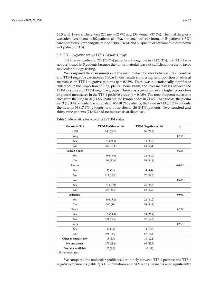
We compared the dissemination at the main metastatic sites between TTF-1 positiveand TTF-1 negative carcinomas (Table 1); our results show a higher proportion of adrenalmetastases in TTF-1 negative patients (p = 0.039). There was no statistically significantdifference in the proportion of lung, pleural, bone, brain, and liver metastases between theTTF-1 positive and TTF-1 negative groups. There was a trend towards a higher proportionof pleural metastases in the TTF-1 positive group (p = 0.089). The most frequent metastaticsites were the lung in 70 (21.8%) patients, the lymph nodes in 71 (22.1%) patients, the pleurain 33 (10.3%) patients, the adrenals in 66 (20.6%) patients, the brain in 113 (35.2%) patients,the liver in 56 (17.4%) patients, and other sites in 26 (8.1%) patients. Two hundred andthirty-nine patients (74.4%) had no metastasis at diagnosis.
Table 1. Metastatic sites according to TTF-1 status.
Metastatic Site TTF-1 Positive, n (%) TTF-1 Negative, n (%) p
n (%) 240 (66.5) 81 (22.4)
Lung 0.716
Yes 51 (19.4) 19 (20.9)
No 189 (71.8) 62 (68.1)
Lymph nodes 0.206
Yes 49 (18.6) 22 (24.2)
No 191 (72.6) 59 (64.8)
Pleura 0.089 *
Yes 29 (11) 4 (4.4)
No 211 (80.2) 77 (84.6)
Bone 0.634
Yes 84 (31.9) 26 (28.6)
No 156 (59.3) 55 (60.4)
Adrenals 0.039
Yes 40 (15.2) 22 (24.2)
No 200 (76) 59 (64.8)
Brain 0.224
Yes 89 (33.8) 24 (26.4)
No 151 (57.4) 57 (62.6)
Liver 0.965
Yes 42 (16) 14 (15.4)
No 198 (75.3) 67 (73.6)
Other metastatic site 15 (5.7) 11 (12.1)
No metastasis 179 (68.6) 60 (65.9)
Data not available 23 (8.8) 10 (11)*: Fisher exact test.
We compared the molecular profile used routinely between TTF-1 positive and TTF-1negative carcinomas (Table 2). EGFR mutations and ALK rearrangements were significantly
Diagnostics 2022, 12, 1589 5 of 12
different between TTF-1 positive and TTF-1 negative tumors (p < 0.001 and p = 0.023respectively). EGFR mutations were present in 16.3% of TTF-1 positive patients, while theywere present in only 1.1% of TTF-1 negative patients. ALK rearrangements were foundin 5.3% of TTF-1 positive patients, whereas no ALK-rearranged patient was found amongTTF-1 negative patients. There was no significant difference between the two groups forKRAS mutations, BRAF, and ROS1 rearrangements.
Table 2. Molecular alterations between TTF-1 positive and TTF-1 negative groups.
TTF-1 Positive, n (%) TTF-1 Negative, n (%) p
EGFR <0.001
Mutated 43 (16.3) 1 (1.1)
Wild type 192 (73) 79 (86.8)
Not performed 28 (10.6) 11 (12.1)
KRAS
Mutated 75 (28.5) 33 (36.3) 0.148
Wild type 157 (59.7) 47 (51.6)
Not performed 31 (11.8) 11 (12.1)
BRAF 0.65
Mutated 11 (4.2) 3 (3.3)
Wild type 196 (74.5) 72 (79.1)
Not performed 56 (21.3) 16 (17.6)
ALK 0.023
Rearranged 14 (5.3) 0 (0)
Not rearranged 214 (81.4) 80 (87.9)
Not performed 35 (13.3) 11 (12.1)
ROS1 0.106
Rearranged 6 (2.3) 0 (0)
Not rearranged 150 (57) 66 (72.5)
Not performed 107 (40.7) 25 (27.5)
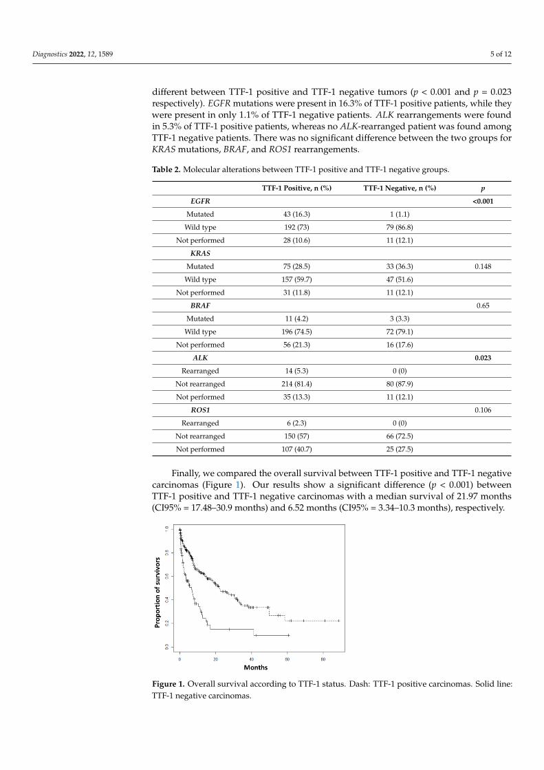
Finally, we compared the overall survival between TTF-1 positive and TTF-1 negativecarcinomas (Figure 1). Our results show a significant difference (p < 0.001) betweenTTF-1 positive and TTF-1 negative carcinomas with a median survival of 21.97 months(CI95% = 17.48–30.9 months) and 6.52 months (CI95% = 3.34–10.3 months), respectively.
Diagnostics 2022, 12, x FOR PEER REVIEW 6 of 12
Figure 1. Overall survival according to TTF-1 status. Dash: TTF-1 positive carcinomas. Solid line:
TTF-1 negative carcinomas.
3.3. Anti-CK7 and CK20 Immunohistochemistry
As TTF-1 negativity did not provide an argument for primary pulmonary origin, we
performed anti-CK7 and CK20 immunohistochemistry in this group. Napsin A was per-
formed in TTF-1 negative patients, 3 of them were Napsin A positive. In the group of TTF-
1 negative patients, anti-CK7 and CK20 immunohistochemistry was performed in 70 pa-
tients and showed CK7+/CK20- staining in 61 patients (87.1%), CK7-/CK20- in 4 patients
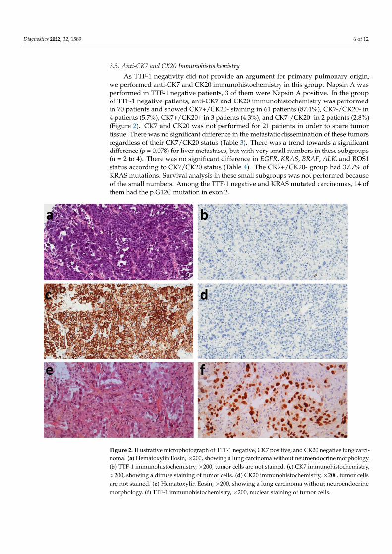
(5.7%), CK7+/CK20+ in 3 patients (4.3%), and CK7-/CK20- in 2 patients (2.8%) (Figure 2).
CK7 and CK20 was not performed for 21 patients in order to spare tumor tissue. There
was no significant difference in the metastatic dissemination of these tumors regardless
of their CK7/CK20 status (Table 3). There was a trend towards a significant difference (p
= 0.078) for liver metastases, but with very small numbers in these subgroups (n = 2 to 4).
There was no significant difference in EGFR, KRAS, BRAF, ALK, and ROS1 status accord-
ing to CK7/CK20 status (Table 4). The CK7+/CK20- group had 37.7% of KRAS mutations.
Survival analysis in these small subgroups was not performed because of the small num-
bers. Among the TTF-1 negative and KRAS mutated carcinomas, 14 of them had the
p.G12C mutation in exon 2.
Table 3. Metastatic sites according to CK7/CK20 status in TTF-1 negative carcinomas.
Metastatic Site for TTF-1 Negative CK7+/CK20-,
n (%)
CK7-/CK20-,
n (%)
CK7+/CK20+,
n (%)
CK7-/CK20+,
n (%) p
n (%) 61 (87.1) 4 (5.7) 3 (4.3) 2 (2.8)
Lung 0.771 *
Yes 10 (16.4) 0 (0) 1 (33.3) 0 (0)
No 44 (72.1) 2 (50) 2 (66.7) 2 (100)
Lymph nodes 0.892 *
Yes 16 (26.2) 0 (0) 1 (33.3) 1 (50)
No 38 (62.3) 2 (50) 2 (66.7) 1 (50)
Pleura 0.191 *
Yes 2 (3.3) 0 (0) 0 (0) 1 (50)
No 52 (85.2) 2 (50) 3 (100) 1 (50)
Figure 1. Overall survival according to TTF-1 status. Dash: TTF-1 positive carcinomas. Solid line:TTF-1 negative carcinomas.
Diagnostics 2022, 12, 1589 6 of 12
3.3. Anti-CK7 and CK20 Immunohistochemistry
As TTF-1 negativity did not provide an argument for primary pulmonary origin,we performed anti-CK7 and CK20 immunohistochemistry in this group. Napsin A wasperformed in TTF-1 negative patients, 3 of them were Napsin A positive. In the groupof TTF-1 negative patients, anti-CK7 and CK20 immunohistochemistry was performedin 70 patients and showed CK7+/CK20- staining in 61 patients (87.1%), CK7-/CK20- in4 patients (5.7%), CK7+/CK20+ in 3 patients (4.3%), and CK7-/CK20- in 2 patients (2.8%)(Figure 2). CK7 and CK20 was not performed for 21 patients in order to spare tumortissue. There was no significant difference in the metastatic dissemination of these tumorsregardless of their CK7/CK20 status (Table 3). There was a trend towards a significantdifference (p = 0.078) for liver metastases, but with very small numbers in these subgroups(n = 2 to 4). There was no significant difference in EGFR, KRAS, BRAF, ALK, and ROS1status according to CK7/CK20 status (Table 4). The CK7+/CK20- group had 37.7% ofKRAS mutations. Survival analysis in these small subgroups was not performed becauseof the small numbers. Among the TTF-1 negative and KRAS mutated carcinomas, 14 ofthem had the p.G12C mutation in exon 2.
Diagnostics 2022, 12, x FOR PEER REVIEW 8 of 12
Figure 2. Illustrative microphotograph of TTF-1 negative, CK7 positive, and CK20 negative lung
carcinoma. (a) Hematoxylin Eosin, ×200, showing a lung carcinoma without neuroendocrine mor-
phology. (b) TTF-1 immunohistochemistry, ×200, tumor cells are not stained. (c) CK7 immunohisto-
chemistry, ×200, showing a diffuse staining of tumor cells. (d) CK20 immunohistochemistry, ×200,
tumor cells are not stained. (e) Hematoxylin Eosin, ×200, showing a lung carcinoma without neuro-
endocrine morphology. (f) TTF-1 immunohistochemistry, ×200, nuclear staining of tumor cells.
4. Discussion
Our work on this retrospective cohort demonstrates that anti-CK7/CK20 immuno-
histochemistry in biopsies in patients with suspected TTF-1 and p40 negative primary
lung cancer is of little value. The use of CK7/CK20 immunohistochemistry in this setting
did not demonstrate with certainty that the carcinoma was of another origin. Moreover,
the different CK7/CK20 profiles do not allow to identify a particular population either in
terms of metastatic evolution or in terms of molecular profile. This work brings additional
arguments demonstrating the uselessness of the use of the anti-CK7/CK20 couple in this
context. The strength of our work is that the pathological data are correlated with the
clinical follow-up data and molecular data. Our work was performed in a particular pop-
ulation where the pathological examination is performed considering the clinical context.
Indeed, when the biopsy was performed to authenticate a metastatic progression of a
known cancer, it was not included in our study. In this specific case, which differs from
the conditions of inclusion of our work, the anti-CK7/CK20 immunohistochemistry can be
an element of orientation in certain cases [7]. It is thus crucial to examine the biopsies with
all the appropriate clinical information, thus making better use of the tumor material,
Figure 2. Illustrative microphotograph of TTF-1 negative, CK7 positive, and CK20 negative lung carci-noma. (a) Hematoxylin Eosin, ×200, showing a lung carcinoma without neuroendocrine morphology.(b) TTF-1 immunohistochemistry, ×200, tumor cells are not stained. (c) CK7 immunohistochemistry,×200, showing a diffuse staining of tumor cells. (d) CK20 immunohistochemistry, ×200, tumor cellsare not stained. (e) Hematoxylin Eosin, ×200, showing a lung carcinoma without neuroendocrinemorphology. (f) TTF-1 immunohistochemistry, ×200, nuclear staining of tumor cells.
Diagnostics 2022, 12, 1589 7 of 12
Table 3. Metastatic sites according to CK7/CK20 status in TTF-1 negative carcinomas.
Metastatic Site for TTF-1 Negative CK7+/CK20-,n (%)
CK7-/CK20-,n (%)
CK7+/CK20+,n (%)
CK7-/CK20+,n (%) p
n (%) 61 (87.1) 4 (5.7) 3 (4.3) 2 (2.8)
Lung 0.771 *
Yes 10 (16.4) 0 (0) 1 (33.3) 0 (0)
No 44 (72.1) 2 (50) 2 (66.7) 2 (100)
Lymph nodes 0.892 *
Yes 16 (26.2) 0 (0) 1 (33.3) 1 (50)
No 38 (62.3) 2 (50) 2 (66.7) 1 (50)
Pleura 0.191 *
Yes 2 (3.3) 0 (0) 0 (0) 1 (50)
No 52 (85.2) 2 (50) 3 (100) 1 (50)
Bone 0.672 *
Yes 23 (37.7) 0 (0) 1 (33.3) 0 (0)
No 31 (50.8) 2 (50) 2 (66.7) 2 (100)
Adrenals 0.258 *
Yes 14 (22.9) 0 (0) 2 (66.7) 1 (50)
No 40 (65.6) 2 (50) 1 (33.3) 1 (50)
Brain 0.621 *
Yes 19 (31.1) 0 (0) 0 (0) 0 (0)
No 38 (62.3) 2 (50) 3 (100) 2 (100)
Liver 0.078
Yes 7 (11.5) 0 (0) 2 (66.7) 0 (0)
No 47 (77) 2 (50) 1 (33.3) 0 (0)
Other metastatic site 9 (14.7) 0 (0) 0 (0) 0
No metastasis 35 (57.4) 2 (50) 1 (33.3) 2 (100)
Data not available 7 (11.5) 2 (50) 0 (0) 0 (0)
*: Fisher exact test.
Table 4. Molecular alterations between CK7/CK20 groups in TTF-1 negative carcinomas.
CK7+/CK20-,n (%)
CK7-/CK20-,n (%)
CK7+/CK20+,n (%)
CK7-/CK20+,n (%) p
EGFR 61 4 3 2 0.081
Mutated 0 (0) 0 (0) 1 (33.3) 0 (0)
Wild type 53 (86.9) 4 (100) 2 (66.7) 2 (100)
Not performed 8 (11.5) 0 (0) 0 (0) 0 (0)
KRAS 0.118
Mutated 23 (37.7) 0 (0) 0 (0) 0 (0)
Wild type 30 (49.2) 4 (100) 3 (100) 2 (100)
Not performed 8 (13.1) 0 (0) 0 (0) 0 (0)
BRAF 0.397
Mutated 2 (3.3) 1 (25) 0 (0) 0 (0)
Diagnostics 2022, 12, 1589 8 of 12
Table 4. Cont.
CK7+/CK20-,n (%)
CK7-/CK20-,n (%)
CK7+/CK20+,n (%)
CK7-/CK20+,n (%) p
Wild type 48 (78.7) 3 (75) 3 (100) 2 (100)
Not performed 11 (18) 0 (0) 0 (0) 0 (0)
ALK 1
Rearranged 0 (0) 0 (0) 0 (0) 0 (0)
Not rearranged 53 (86.9) 4 (100) 2 (66.7) 2 (100)
Not performed 8 (13.1) 0 (0) 1 (33.3) 0 (0)
ROS1 1
Rearranged 0 (0) 0 (0) 0 (0) 0 (0)
Not rearranged 43 (70.5) 4 (100) 0 (0) 2 (100)
Not performed 18 (29.5) 0 (0) 3 (100) 0 (0)
4. Discussion
Our work on this retrospective cohort demonstrates that anti-CK7/CK20 immuno-histochemistry in biopsies in patients with suspected TTF-1 and p40 negative primarylung cancer is of little value. The use of CK7/CK20 immunohistochemistry in this settingdid not demonstrate with certainty that the carcinoma was of another origin. Moreover,the different CK7/CK20 profiles do not allow to identify a particular population either interms of metastatic evolution or in terms of molecular profile. This work brings additionalarguments demonstrating the uselessness of the use of the anti-CK7/CK20 couple in thiscontext. The strength of our work is that the pathological data are correlated with the clini-cal follow-up data and molecular data. Our work was performed in a particular populationwhere the pathological examination is performed considering the clinical context. Indeed,when the biopsy was performed to authenticate a metastatic progression of a known cancer,it was not included in our study. In this specific case, which differs from the conditionsof inclusion of our work, the anti-CK7/CK20 immunohistochemistry can be an elementof orientation in certain cases [7]. It is thus crucial to examine the biopsies with all theappropriate clinical information, thus making better use of the tumor material, sparing itfor complementary techniques such as molecular biology, and answering the questionsasked by the pathology exams prescribers as accurately as possible.
In contrast, the distinction between TTF-1 positive and TTF-1 negative non-squamouscarcinomas seems to be clinically relevant with a more favorable prognosis in the case ofTTF-1 positivity. In contrast, the distinction between TTF-1 positive and TTF-1 negativenon-squamous carcinomas is more relevant. This distinction seems to be clinically relevantwith a more favorable prognosis in the case of TTF-1 positivity. Moreover, at the molecularlevel, the probability of finding an EGFR mutation or an ALK rearrangement is higher in thecase of TTF-1 positivity. These data correlate with what has already been described in theliterature where TTF-1 has been shown to be a better prognostic factor in adenocarcinomasof all stages and where TTF-1 positivity increases the probability of finding EGFR or ALKmutations [17–23]. TTF-1 is a marker present in more than 60% of primary lung adeno-carcinomas, and is rarely expressed in adenocarcinomas of other origin [2,17,24]. TTF-1is a transcription factor involved in morphogenesis, differentiation, and surfactant pro-duction by normal pneumocytes [25]. The different frequency of EGFR and ALK subtypesbetween TTF-1 positive and TTF-1 negative subtypes might be linked to the fact that TTF-1negative tumors in biopsies represent a group enriched in rare adenocarcinomas subtypes.These subtypes are mostly represented by invasive mucinous adenocarcinoma representingroughly 10% of lung tumors and are often EGFR wild type and TTF-1 negative [2]. It thusseems logical to distinguish TTF-1 positive non-squamous carcinomas, which would bemore differentiated, from TTF-1 negative ones, which would be less differentiated. Finally,
Diagnostics 2022, 12, 1589 9 of 12
it seems that, among TTF-1 negative carcinomas, the proportion of rare carcinomas suchas carcinomas muted for SMARCA4 would be more frequent [26,27]. The exact place ofSMARCA4 testing as far as there is a specific antibody for this diagnosis is not resolvedin the setting of the diagnosis on small biopsy samples [26,27]. Furthermore, because ofthe recent description of SMARCA4 deficient tumors, the therapy is not as well-knowncompared with lung adenocarcinoma [28]. The subtyping of rare lung carcinoma such asenteric carcinoma and sarcomatoid carcinoma, especially on bronchial biopsies, is difficultor not feasible, and there is often little or no difference in the treatment between thesesubtypes [29].
Cytokeratins belong to the family of intermediate filaments. Cytokeratin 7 marksthe simple epithelia, while cytokeratin 20 marks the simple epithelia of the digestivetract and Merkel cells [11]. The anti-CK7/CK20 pair may be of diagnostic interest in thecontext of adenocarcinoma metastasis [10]. Furthermore, primary lung adenocarcinomascan have any combination, although the CK7 positive and CK20 negative combination isthe most common [7]. Among TTF-1 negative carcinomas, the proportion of carcinomaswith a profile other than CK7+/CK20- remains rare and concerns only 12.8% of cases inour series. The possible interest of using cytokeratins is to prove that it is an epithelialtumor. The vast majority of non-epithelial tumors do not express cytokeratins unlikecarcinomas [10]. However, the vast majority of pulmonary malignancies are carcinomas,and it is morphologically possible to suspect that it is an epithelial tumor.
The limitations of our work are inherent to those of retrospective work on existingdata with a proportion of missing or unrealized data. Moreover, this is a single-centerstudy in a university hospital of reference in which clinical and pathological collaborationis important. Indeed, the pathologists systematically have the patient’s medical history,the indication for endoscopy, and the endoscopic data. However, clinical information isnot always provided to the pathologist in less specialized centers. Another limitation ofour work is that we are interested in a particular subgroup concerning TTF-1 negativecarcinomas. This induces de facto a low number of patients in the subgroup of TTF-1negative patients and limits the relevance of the statistical analysis. Biopsies are fixed informalin immediately and there is no delay in fixation, as may be the case for surgicalspecimens. However, the different pre-analytical conditions can induce differences inmarking, which is not the case in this work [30].
The diagnosis of lung cancer has completely changed in recent years. Indeed, patholog-ical diagnosis has had to adapt to molecular techniques to save as much tissue as possiblefor these techniques. Indeed, even with new generation sequencing capable of sequencingseveral genes in parallel, it is necessary to have a maximum amount of tumor tissue tolimit the risk of false negative or exhausted samples in molecular biology [4,31]. Moreover,concerning immunohistochemical techniques, it is recommended to privilege as much aspossible techniques having a therapeutic impact, such as anti-PD-L1 immunohistochem-istry [32,33]. Molecular techniques are of major importance, for example, concerning theEGFR gene, which is mutated in 10–15% of non-Asian patients and in 40–60% of Asian pa-tients with lung adenocarcinoma. EGFR allows to determine the therapy; that is, metastaticpatients with activating mutations of EGFR can be treated with tyrosine kinase inhibitors,while negative patients will not benefit from these specific therapies. However, the progno-sis of EGFR-mutated metastatic patients treated with tyrosine kinase inhibitors is muchbetter than that of the EGFR-wild type group [34]. It is thus more logical to focus on themost clinically impactful techniques. A biopsy is a small sample on which it is necessary tomake a diagnosis and determine which molecular or immunohistochemical markers arepresent. These samples can be quickly exhausted by successive complementary techniques.Furthermore, there is no single technique that can provide all the data needed for treatment.Techniques to determine markers for therapy are multiple and include immunohistochem-istry for ALK, ROS1, and PD-L1, but also sequencing ideally of DNA as well as of RNA forgene rearrangements [35]. Pathologists need to be aware of therapeutic developments tobetter support diagnostic and therapeutic techniques and better manage tissue material.
Diagnostics 2022, 12, 1589 10 of 12
Artificial intelligence techniques, without the need for additional techniques using tissue,could be of interest to better classify these tumours. Their use in routine practice is still inthe early stages, but could be of interest in the future [36].
Overall, this work brings arguments concerning the uselessness of anti-CK7/CK20immunohistochemistry in most cases of suspicion of primary lung cancer in biopsies inroutine clinical practice. This pair of antibodies does not allow the identification of relevantsubgroups in terms of molecular group, metastasis, or prognosis in our work. The use ofthis pair of antibodies could suggest a primary tumor of another origin and lead to a length-ening of the management time and the realization of unnecessary clinical complementaryexaminations. In addition, the use of antibodies leads to tissue consumption, whereasit is essential to spare tumor tissue for the realization of complementary examinationsthat could lead to the loss of tumor tissue. In the era of personalized therapies, it is thuslegitimate that recommendations privilege tissue sparing rather than the realization ofcomplementary techniques. The use of immunohistochemistry should be done sparingly.No diagnosis of cancer origin can be made on immunohistochemistry alone. The diagnosisof the origin of a cancer must imperatively integrate the associated clinical and radiologicaldata. However, our study was performed in patients with suspected lung cancer. It is notapplicable in patients suspected of having metastasized from another cancer. Our resultsare thus applicable in a selected population and do not account for situations that aresometimes particular. Our results support the international recommendations that shouldbe applied according to individual clinical situations.
To conclude, in the case of suspicion of primary lung carcinoma, we show that the useof anti-CK7 and CK20 antibodies does not help the diagnosis, and our work supports therecommendations made.
Author Contributions: Conceptualization, A.C. and F.F.; methodology, F.F.; software, F.F.; validation,all authors.; formal analysis, F.F.; investigation, A.C., D.L., S.D., V.G., P.D.-C., V.Y., F.C., S.B.-B. andJ.-M.V.; resources, F.F.; data curation, A.C.; writing—original draft preparation, A.C. and F.F.; writing—review and editing, all authors; visualization, F.F.; supervision, F.F.; project administration, F.F.;funding acquisition, F.F. All authors have read and agreed to the published version of the manuscript.
Funding: No funding was received to assist with the preparation of this manuscript.
Institutional Review Board Statement: This study was approved by local Ethical Committee of theUniversity Hospital of Saint Etienne.
Informed Consent Statement: According to the European General Data Protection Regulation,patients were informed of the study. Informed consent was obtained from all subjects involved inthe study.
Data Availability Statement: Data available on request.
Acknowledgments: The authors thank Philippe Cosmo from Centre de Ressources Biologiques deCHU Saint-Etienne for his assistance in data retrieval.
Conflicts of Interest: The authors declare no conflict of interest.
References1. Forest, F.; Thuret, G.; Gain, P.; Dumollard, J.-M.; Peoc’h, M.; Perrache, C.; He, Z. Optimization of immunostaining on flat-mounted
human corneas. Mol. Vis. 2015, 21, 1345–1356. [PubMed]2. Pelosi, G.; Scarpa, A.; Forest, F.; Sonzogni, A. The impact of immunohistochemistry on the classification of lung tumors. Expert
Rev. Respir. Med. 2016, 10, 1105–1121. [CrossRef] [PubMed]3. Bishop, J.A.; Teruya-Feldstein, J.; Westra, W.H.; Pelosi, G.; Travis, W.D.; Rekhtman, N. p40 (∆Np63) is superior to p63 for the
diagnosis of pulmonary squamous cell carcinoma. Mod. Pathol. 2012, 25, 405–415. [CrossRef] [PubMed]4. Forest, F.; Stachowicz, M.-L.; Casteillo, F.; Karpathiou, G.; Gouzy-Grosjean, F.; Guilaubey, C.; Cottier, M.; Beal, J.; Clemenson, A.;
Péoc’h, M. EGFR, KRAS, BRAF and HER2 testing in metastatic lung adenocarcinoma: Value of testing on samples with poorspecimen adequacy and analysis of discrepancies. Exp. Mol. Pathol. 2017, 103, 306–310. [CrossRef] [PubMed]
5. Travis, W.D.; Brambilla, E.; Noguchi, M.; Nicholson, A.G.; Geisinger, K.R.; Yatabe, Y.; Beer, D.G.; Powell, C.A.; Riely, G.J.;Van Schil, P.E.; et al. International Association for the Study of Lung Cancer/American Thoracic Society/European RespiratorySociety International Multidisciplinary Classification of Lung Adenocarcinoma. J. Thorac. Oncol. 2011, 6, 244–285. [CrossRef]
Diagnostics 2022, 12, 1589 11 of 12
6. Gerull, W.D.; Puri, V.; Kozower, B.D. The epidemiology and biology of pulmonary metastases. J. Thorac. Dis. 2021, 13, 2585–2589.[CrossRef] [PubMed]
7. Montezuma, D.; Azevedo, R.; Lopes, P.; Vieira, R.; Cunha, A.L.; Henrique, R. A panel of four immunohistochemical markers(CK7, CK20, TTF-1, and p63) allows accurate diagnosis of primary and metastatic lung carcinoma on biopsy specimens. VirchowsArch. 2013, 463, 749–754. [CrossRef]
8. Travis, W.D.; Brambilla, E.; Noguchi, M.; Nicholson, A.G.; Geisinger, K.; Yatabe, Y.; Powell, C.A.; Beer, D.; Riely, G.; Garg, K.; et al.International Association for the Study of Lung Cancer/American Thoracic Society/European Respiratory Society: Internationalmultidisciplinary classification of lung adenocarcinoma—An executive summary. Proc. Am. Thorac. Soc. 2011, 8, 381–385.[CrossRef]
9. Geles, A.; Gruber-Moesenbacher, U.; Quehenberger, F.; Manzl, C.; Al Effah, M.; Grygar, E.; Juettner-Smolle, F.; Popper, H.H.Pulmonary mucinous adenocarcinomas: Architectural patterns in correlation with genetic changes, prognosis and survival.Virchows Arch. 2015, 467, 675–686. [CrossRef]
10. Chu, P.; Wu, E.; Weiss, L.M. Cytokeratin 7 and Cytokeratin 20 Expression in Epithelial Neoplasms: A Survey of 435 Cases. Mod.Pathol. 2000, 13, 962–972. [CrossRef]
11. Vidarsdottir, H.; Tran, L.; Nodin, B.; Jirström, K.; Planck, M.; Jönsson, P.; Mattsson, J.S.M.; Botling, J.; Micke, P.; Brunnström, H.Immunohistochemical profiles in primary lung cancers and epithelial pulmonary metastases. Hum. Pathol. 2019, 84, 221–230.[CrossRef] [PubMed]
12. WHO. Classification of Tumors Editorial Board Thoracic Tumors, 5th ed.; International Agency for Research on Cancer: Lyon, France,2021; ISBN 978-92-832-4506-3.
13. Forest, F.; Laville, D.; Habougit, C.; Da Cruz, V.; Casteillo, F.; Yvorel, V.; Bard-Sorel, S.; Godard, W.; Picot, T.; Tiffet, O.; et al.Histopathological and molecular profiling of lung adenocarcinoma skin metastases reveals specific features. Histopathology 2021,79, 1051–1060. [CrossRef] [PubMed]
14. Bourhis, A.; Remoué, A.; Le Flahec, G.; Marcorelles, P.; Uguen, A. Avoiding non-contributive molecular results in cancer samples:Proposal of a score-based approach for sample choice. Pathology 2019, 51, 524–528. [CrossRef] [PubMed]
15. R Core Team. R: A Language And Environment for Statistical Computing. The R Project for Statistical Computing: Online. 2018,Volume 2. Available online: https://www.R-project.org (accessed on 26 April 2022).
16. Therneau, T. A Package for Survival Analysis in S.; R Package Version; Survival: London, UK, 2012.17. Schilsky, J.B.; Ni, A.; Ahn, L.; Datta, S.; Travis, W.D.; Kris, M.G.; Chaft, J.E.; Rekhtman, N.; Hellmann, M.D. Prognostic impact of
TTF-1 expression in patients with stage IV lung adenocarcinomas. Lung Cancer 2017, 108, 205–211. [CrossRef] [PubMed]18. Casteillo, F.; Guy, J.-B.; Dal-Col, P.; Karpathiou, G.; Pommier, B.; Bayle-Bleuez, S.; Fournel, P.; Vassal, F.; Forest, F. Pathologic
Subtypes of Lung Adenocarcinoma Brain Metastasis Is a Strong Predictor of Survival After Resection. Am. J. Surg. Pathol. 2018,42, 1701–1707. [CrossRef] [PubMed]
19. Da Cruz, V.; Yvorel, V.; Casteillo, F.; Tissot, C.; Luchez, A.; Bayle-Bleuez, S.; Fournel, P.; Tiffet, O.; Péoc’h, M.; Forest, F.; et al.Histopathological subtyping is a prognostic factor in stage IV lung adenocarcinoma. Lung Cancer 2020, 147, 77–82. [CrossRef]
20. Kadota, K.; Nitadori, J.; Sarkaria, I.S.; Sima, C.S.; Jia, X.; Yoshizawa, A.; Rusch, V.W.; Travis, W.D.; Adusumilli, P.S. Thyroidtranscription factor-1 expression is an independent predictor of recurrence and correlates with the IASLC/ATS/ERS histologicclassification in patients with stage I lung adenocarcinoma. Cancer 2013, 119, 931–938. [CrossRef]
21. Qian, H.; Xu, T.; Cai, X.; Ji, T.; Guo, H. Prognostic value of TTF-1 expression in patients with non-small cell lung cancer: Ameta-analysis. Clin. Chim. Acta 2015, 451, 208–214. [CrossRef]
22. Fallet, V.; Cadranel, J.; Doubre, H.; Toper, C.; Monnet, I.; Chinet, T.; Oliviero, G.; Foulon, G.; De Cremoux, H.; Vieira, T.; et al.Prospective screening for ALK: Clinical features and outcome according to ALK status. Eur. J. Cancer 2014, 50, 1239–1246.[CrossRef]
23. Rodriguez, E.F.; VandenBussche, C.J.; Chowsilpa, S.; Maleki, Z. Molecular genetic alterations in thyroid transcription factor1–negative lung adenocarcinoma in cytology specimens: A subset with aggressive behavior and a poor prognosis. CancerCytopathol. 2018, 126, 853–859. [CrossRef]
24. Casteillo, F.; Fournel, P.; Da Cruz, V.; Karpathiou, G.; Boutet, C.; Jacquin, J.; Tissot, C.; Meyer-Bisch, V.; Péoc’h, M.; Forest, F.TTF-1-positive Metastatic Endometrioid Carcinoma. Appl. Immunohistochem. Mol. Morphol. 2020, 28, e6–e9. [CrossRef] [PubMed]
25. Little, D.R.; Gerner-Mauro, K.N.; Flodby, P.; Crandall, E.D.; Borok, Z.; Akiyama, H.; Kimura, S.; Ostrin, E.J.; Chen, J. Transcriptionalcontrol of lung alveolar type 1 cell development and maintenance by NK homeobox 2-1. Proc. Natl. Acad. Sci. USA 2019, 116,20545–20555. [CrossRef] [PubMed]
26. Serre, I.; Garcia, S.; Forest, F. SMARCA4-deficient Thoracic Sarcomas. Int. J. Surg. Pathol. 2018. [CrossRef]27. Perret, R.; Chalabreysse, L.; Watson, S.; Serre, I.; Garcia, S.; Forest, F.; Yvorel, V.; Pissaloux, D.; Thomas de Montpreville, V.;
Masliah-planchon, J.; et al. SMARCA4-deficient Thoracic Sarcomas. Am. J. Surg. Pathol. 2019, 43, 455–465. [CrossRef]28. Le Loarer, F.; Watson, S.; Pierron, G.; de Montpreville, V.T.; Ballet, S.; Firmin, N.; Auguste, A.; Pissaloux, D.; Boyault, S.;
Paindavoine, S.; et al. SMARCA4 inactivation defines a group of undifferentiated thoracic malignancies transcriptionally relatedto BAF-deficient sarcomas. Nat. Genet. 2015, 47, 1200–1205. [CrossRef]
29. Forest, F.; Yvorel, V.; Karpathiou, G.; Stachowicz, M.L.; Vergnon, J.M.; Fournel, P.; Tiffet, O.; Trombert, B.; Péoc’H, M. Histomolec-ular profiling of pleomorphic, spindle cell, and giant cell carcinoma of the lung for targeted therapies. Hum. Pathol. 2016, 49,99–106. [CrossRef]
Diagnostics 2022, 12, 1589 12 of 12
30. Forest, F.; Cote, G.; Laville, D.; Da Cruz, V.; Dal Col, P.; Camy, F.; Mobarki, M.; Clemenson, A.; Yvorel, V.; Péoc’h, M. Impactof delayed fixation and decalcification on PD-L1 expression: A comparison of two clones. Virchows Arch. 2019, 475, 693–699.[CrossRef]
31. Boureille, A.; Ferraro-Peyret, C.; Pontarollo, G.; Confavreux, C.; Pialat, J.-B.; Isaac, S.; Forest, F.; Yvorel, V.; Watkin, E.;Girard, N.; et al. Rapid detection of EGFR mutations in decalcified lung cancer bone metastasis. J. Bone Oncol. 2020, 21, 100277.[CrossRef]
32. Forest, F.; Casteillo, F.; Da Cruz, V.; Yvorel, V.; Picot, T.; Vassal, F.; Tiffet, O.; Péoc’h, M. Heterogeneity of PD-L1 expression in lungadenocarcinoma metastasis is related to histopathological subtypes. Lung Cancer 2021, 155, 1–9. [CrossRef]
33. Adam, J.; Forest, F.; Mansuet-Lupo, A.; Ilié, M.; pour le groupe PATTERN (PAThologistes Thoraciques de valorisation del’Expertise, de la R. et de l’iNnovation F.S.L.C. Recent updates regarding PD-L1 testing in non-small cell lung carcinoma. Ann.Pathol. 2019, 39, 303–304. [CrossRef]
34. Soria, J.C.; Ohe, Y.; Vansteenkiste, J.; Reungwetwattana, T.; Chewaskulyong, B.; Lee, K.H.; Dechaphunkul, A.; Imamura, F.;Nogami, N.; Kurata, T.; et al. Osimertinib in untreated EGFR-Mutated advanced non-small-cell lung cancer. N. Engl. J. Med. 2018,378, 113–125. [CrossRef] [PubMed]
35. McLeer-Florin, A.; Duruisseaux, M.; Pinsolle, J.; Dubourd, S.; Mondet, J.; Phillips Houlbracq, M.; Magnat, N.; Fauré, J.; Chatagnon,A.; de Fraipont, F.; et al. ALK fusion variants detection by targeted RNA-next generation sequencing and clinical responses tocrizotinib in ALK-positive non-small cell lung cancer. Lung Cancer 2018, 116, 15–24. [CrossRef] [PubMed]
36. Yu, K.H.; Wang, F.; Berry, G.J.; Ré, C.; Altman, R.B.; Snyder, M.; Kohane, I.S. Classifying non-small cell lung cancer types andtranscriptomic subtypes using convolutional neural networks. J. Am. Med. Inform. Assoc. 2020, 1, 757–769. [CrossRef] [PubMed]