Anti-cancer effects of blue-green alga Spirulina platensis, a natural source of bilirubin-like...
11
Anti-cancer effects of blue-green alga Spirulina platensis, a natural source of bilirubin-like tetrapyrrolic compounds Renata Konícková,* Katerina Vanková,* Jana Vaníková,* Katerina Vánová,* Lucie Muchová,* Iva Subhanová,* Marie Zadinová, † Jaroslav Zelenka, ‡ Aleš Dvorák,* ,‡ Michal Kolár, § Hynek Strnad, § Silvie Rimpelová, || Tomáš Ruml, || Ronald J. Wong, ¶ Libor Vítek* , ** * Institute of Medical Biochemistry and Laboratory Diagnostics, 1st Faculty of Medicine, Charles University, Prague, Czech Republic. † Institute of Biophysics, 1st Faculty of Medicine, Charles University, Prague, Czech Republic. ‡ Institute of Physiology, Academy of Sciences of the Czech Republic, Prague, Czech Republic. § Institute of Molecular Genetics, Academy of Sciences of the Czech Republic, Prague, Czech Republic. || Department of Biochemistry and Microbiology, Institute of Chemical Technology, Prague, Czech Republic. ¶ Department of Pediatrics, Stanford University School of Medicine, Stanford, CA, USA. ** 4th Department of Internal Medicine, 1st Faculty of Medicine, Charles University, Prague, Czech Republic. ABSTRACT Spirulina platensis is a blue-green alga used as a dietary supplement because of its hypocholesterolemic properties. Among other bioactive substances, it is also rich in tetrapyrrolic compounds closely related to bilirubin molecule, a potent antioxidant and anti-proliferative agent. The aim of our study was to evaluate possible anticancer effects of S. platensis and S. platensis-derived tetrapyrroles using an experimental mo- del of pancreatic cancer. The anti-proliferative effects of S. platensis and its tetrapyrrolic components [phycocyanobilin (PCB) and chlorophyllin, a surrogate molecule for chlorophyll A] were tested on several human pancreatic cancer cell lines and xenotransplanted nude mice. The effects of experimental thera- peutics on mitochondrial reactive oxygen species (ROS) production and glutathione redox status were also evaluated. Compared to untreated cells, experimental therapeutics significantly decreased prolifera- tion of human pancreatic cancer cell lines in vitro in a dose-dependent manner (from 0.16 g·L-1 [S. platensis], 60 μM [PCB], and 125 μM [chlorophyllin], p<0.05). The anti-proliferative effects of S. platensis were also shown in vivo, where inhibition of pancreatic cancer growth was evidenced since the third day of treatment (p < 0.05). All tested compounds decreased generation of mitochondrial ROS and glutathione redox status (p = 0.0006; 0.016; and 0.006 for S. platensis, PCB, and chlorophyllin, respectively). In conclusion, S. platensis and its tetrapyrrolic components substantially decreased the proliferation of experimental pancreatic cancer. These data support a chemopreventive role of this edible alga. Furthermore, it seems that dietary supplementation with this alga might enhance systemic pool of tetrapyrroles, known to be higher in subjects with Gilbert syndrome. Key words. Bilirubin. Chlorophyll. Heme oxygenase. Phycocyanin. Phycocyanobilin. Correspondence and reprint request: Libor Vítek, MD, PhD Institute of Medical Biochemistry and Laboratory Diagnostics. 1st Faculty of Medicine, Charles University in Prague. Na Bojišti 3. Praha 2, 12000. Czech Republic. Tel.: +420 2 2496 4203. Fax: +420 2 2496 4203 E-mail: [email protected] Manuscript received: November 27, 2013. Manuscript accepted: December 12, 2013. March-April, Vol. 13 No. 2, 2014: 273-283 ORIGINAL ARTICLE INTRODUCTION Spirulina platensis is a blue-green freshwater alga widely used as a dietary supplement. It is rich in proteins, carotenoids, essential fatty acids, vita- min B complex, vitamin E, and minerals such as copper, manganese, magnesium, iron, selenium, and zinc. 1 S. platensis has garnered much attention not only because of its high nutritional value; but also, it is a source of potent antioxidants including spirulans (sulphated polysaccharides), selenocom- pounds, phenolic compounds, and phycobiliproteins (C-phycocyanin and allophycocyanin). 1 In fact, numerous studies have demonstrated that dietary supplementation of S. platensis is helpful in the prevention and treatment of atherosclerosis, diabetes, and/or cancers (for review see Gershwin, 2008 2 ). C-phycocyanin is a light-harvesting biliprotein possibly implicated in biological effects of S. platensis. 2 C-phycocyanin contains a covalently- linked chromophore called phycocyanobilin (PCB), 3 a linear tetrapyrrolic molecule structurally resembling biliverdin, an antioxidant bile pigment < < < < < < <
-
Upload
independent -
Category
Documents
-
view
0 -
download
0
Transcript of Anti-cancer effects of blue-green alga Spirulina platensis, a natural source of bilirubin-like...
273Anti-cancer effects of blue-green alga Spirulina platensis. , 2014; 13 (2): 273-283
Anti-cancer effects of blue-green alga Spirulina platensis, anatural source of bilirubin-like tetrapyrrolic compounds
Renata Konícková,* Katerina Vanková,* Jana Vaníková,* Katerina Vánová,* Lucie Muchová,*Iva Subhanová,* Marie Zadinová,† Jaroslav Zelenka,‡ Aleš Dvorák,*,‡ Michal Kolár,§ Hynek Strnad,§
Silvie Rimpelová,|| Tomáš Ruml,|| Ronald J. Wong,¶ Libor Vítek*,**
* Institute of Medical Biochemistry and Laboratory Diagnostics, 1st Faculty of Medicine, Charles University, Prague, Czech Republic.† Institute of Biophysics, 1st Faculty of Medicine, Charles University, Prague, Czech Republic.
‡ Institute of Physiology, Academy of Sciences of the Czech Republic, Prague, Czech Republic.§ Institute of Molecular Genetics, Academy of Sciences of the Czech Republic, Prague, Czech Republic.
|| Department of Biochemistry and Microbiology, Institute of Chemical Technology, Prague, Czech Republic.¶ Department of Pediatrics, Stanford University School of Medicine, Stanford, CA, USA.
** 4th Department of Internal Medicine, 1st Faculty of Medicine, Charles University, Prague, Czech Republic.
ABSTRACT
Spirulina platensis is a blue-green alga used as a dietary supplement because of its hypocholesterolemicproperties. Among other bioactive substances, it is also rich in tetrapyrrolic compounds closely related tobilirubin molecule, a potent antioxidant and anti-proliferative agent. The aim of our study was to evaluatepossible anticancer effects of S. platensis and S. platensis-derived tetrapyrroles using an experimental mo-del of pancreatic cancer. The anti-proliferative effects of S. platensis and its tetrapyrrolic components[phycocyanobilin (PCB) and chlorophyllin, a surrogate molecule for chlorophyll A] were tested on severalhuman pancreatic cancer cell lines and xenotransplanted nude mice. The effects of experimental thera-peutics on mitochondrial reactive oxygen species (ROS) production and glutathione redox status werealso evaluated. Compared to untreated cells, experimental therapeutics significantly decreased prolifera-tion of human pancreatic cancer cell lines in vitro in a dose-dependent manner (from 0.16 g·L-1 [S. platensis],60 μM [PCB], and 125 μM [chlorophyllin], p<0.05). The anti-proliferative effects of S. platensis were alsoshown in vivo, where inhibition of pancreatic cancer growth was evidenced since the third day oftreatment (p < 0.05). All tested compounds decreased generation of mitochondrial ROS and glutathioneredox status (p = 0.0006; 0.016; and 0.006 for S. platensis, PCB, and chlorophyllin, respectively). In conclusion,S. platensis and its tetrapyrrolic components substantially decreased the proliferation of experimentalpancreatic cancer. These data support a chemopreventive role of this edible alga. Furthermore, it seemsthat dietary supplementation with this alga might enhance systemic pool of tetrapyrroles, known to behigher in subjects with Gilbert syndrome.
Key words. Bilirubin. Chlorophyll. Heme oxygenase. Phycocyanin. Phycocyanobilin.
Correspondence and reprint request: Libor Vítek, MD, PhDInstitute of Medical Biochemistry and Laboratory Diagnostics. 1st Faculty ofMedicine, Charles University in Prague.Na Bojišti 3. Praha 2, 12000. Czech Republic.Tel.: +420 2 2496 4203. Fax: +420 2 2496 4203E-mail: [email protected]
Manuscript received: November 27, 2013.Manuscript accepted: December 12, 2013.
March-April, Vol. 13 No. 2, 2014: 273-283
ORIGINAL ARTICLE
INTRODUCTION
Spirulina platensis is a blue-green freshwateralga widely used as a dietary supplement. It is richin proteins, carotenoids, essential fatty acids, vita-min B complex, vitamin E, and minerals such ascopper, manganese, magnesium, iron, selenium, and
zinc.1 S. platensis has garnered much attention notonly because of its high nutritional value; but also,it is a source of potent antioxidants includingspirulans (sulphated polysaccharides), selenocom-pounds, phenolic compounds, and phycobiliproteins(C-phycocyanin and allophycocyanin).1 In fact,numerous studies have demonstrated that dietarysupplementation of S. platensis is helpful in theprevention and treatment of atherosclerosis, diabetes,and/or cancers (for review see Gershwin, 20082).
C-phycocyanin is a light-harvesting biliproteinposs ib ly impl icated in b io log ica l e f fects o fS. platensis.2 C-phycocyanin contains a covalently-linked chromophore called phycocyanobilin(PCB),3 a linear tetrapyrrolic molecule structurallyresembling biliverdin, an antioxidant bile pigment
< < < < <
< <
Konícková R, et al. , 2014; 13 (2): 273-283274 <
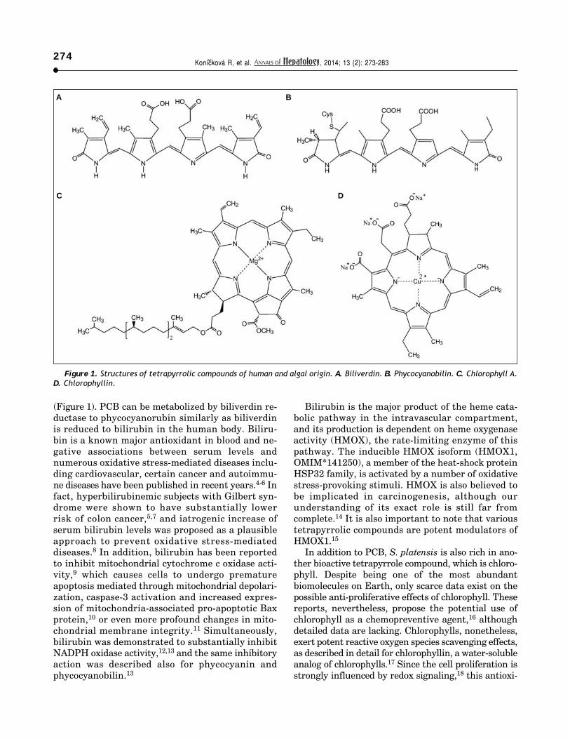
Figure 1. Structures of tetrapyrrolic compounds of human and algal origin. A. Biliverdin. B. Phycocyanobilin. C. Chlorophyll A.D. Chlorophyllin.
A B
C D
(Figure 1). PCB can be metabolized by biliverdin re-ductase to phycocyanorubin similarly as biliverdinis reduced to bilirubin in the human body. Biliru-bin is a known major antioxidant in blood and ne-gative associations between serum levels andnumerous oxidative stress-mediated diseases inclu-ding cardiovascular, certain cancer and autoimmu-ne diseases have been published in recent years.4-6 Infact, hyperbilirubinemic subjects with Gilbert syn-drome were shown to have substantially lowerrisk of colon cancer,5,7 and iatrogenic increase ofserum bilirubin levels was proposed as a plausibleapproach to prevent oxidative stress-mediateddiseases.8 In addition, bilirubin has been reportedto inhibit mitochondrial cytochrome c oxidase acti-vity,9 which causes cells to undergo prematureapoptosis mediated through mitochondrial depolari-zation, caspase-3 activation and increased expres-sion of mitochondria-associated pro-apoptotic Baxprotein,10 or even more profound changes in mito-chondrial membrane integrity.11 Simultaneously,bilirubin was demonstrated to substantially inhibitNADPH oxidase activity,12,13 and the same inhibitoryaction was described also for phycocyanin andphycocyanobilin.13
Bilirubin is the major product of the heme cata-bolic pathway in the intravascular compartment,and its production is dependent on heme oxygenaseactivity (HMOX), the rate-limiting enzyme of thispathway. The inducible HMOX isoform (HMOX1,OMIM*141250), a member of the heat-shock proteinHSP32 family, is activated by a number of oxidativestress-provoking stimuli. HMOX is also believed tobe implicated in carcinogenesis, although ourunderstanding of its exact role is still far fromcomplete.14 It is also important to note that varioustetrapyrrolic compounds are potent modulators ofHMOX1.15
In addition to PCB, S. platensis is also rich in ano-ther bioactive tetrapyrrole compound, which is chloro-phyll. Despite being one of the most abundantbiomolecules on Earth, only scarce data exist on thepossible anti-proliferative effects of chlorophyll. Thesereports, nevertheless, propose the potential use ofchlorophyll as a chemopreventive agent,16 althoughdetailed data are lacking. Chlorophylls, nonetheless,exert potent reactive oxygen species scavenging effects,as described in detail for chlorophyllin, a water-solubleanalog of chlorophylls.17 Since the cell proliferation isstrongly influenced by redox signaling,18 this antioxi-

275Anti-cancer effects of blue-green alga Spirulina platensis. , 2014; 13 (2): 273-283
dant action of chlorophylls might account for theirpresumed anti-proliferative properties.
The aim of our study was to evaluate possibleanticancer effects of S. platensis and its tetrapyrroliccomponents on the model of experimental pancreaticcancer. More specifically, our focus was placed ontheir potential impact on the heme catabolicpathway and mitochondrial redox metabolism.
MATERIAL AND METHODS
Chemicals
S. platensis was purchased from Martin BauerGmbH (Vestenbergsgreuth, Germany). Bilirubin andhemin were from Frontier Scientific (Logan, UT,USA), C-phycocyanin was purchased from ProZyme(Hayward, CA, USA), chlorophyllin (a water solu-ble, semi–synthetic derivate of chlorophyll common-ly used in food industry, was employed in all in vitrostudies as a surrogate for non-polar chlorophylls)and other chemicals were from Sigma-Aldrich (St.Louis, MO, USA).
Preparation of a water extract of S. platensis
Distilled water was added to freeze-dried S. platensispowder (30 mL·g-1 of alga). The suspension wassonicated for 15 min, incubated for 10 min atroom temperature, centrifuged (8,000 x g, 30 min, 10 °C)and finally freeze-dried overnight. Prior to use,the extract was dissolved in culture medium andsterile-filtered.
PCB isolation from S. platensis
PCB was isolated from freeze-dried S. platensisaccording to the method of Terry.19 Identification ofPCB was confirmed by HPLC (Luna C8 column,4.6·150 mm, 3m/100A, Phenomenex, Torrance, CA,USA; isocratic mobile phase with methanol/water/TBA, 59:40:1 w/w/w, flow rate of 0.5 mL·min-1), andmass spectrometry.
Cell lines
The following pancreatic cancer cell lines wereused for the in vitro studies: PA-TU-8902 (DSMZ,Braunschweig, Germany), Mia PaCa-2 and BxPC-3(ATCC, Manassas, VA, USA). All cell lines weremaintained in a humidified atmosphere (containing5% CO2 at 37 °C) and in the following media supple-mented with 10% fetal bovine serum (FBS): PA-TU-
8902 and Mia PaCa-2 in DMEM, BxPC-3 in RPMI.Authentication of PA-TU-8902 cell line used in majo-rity of described studies was confirmed by indepen-dent laboratory.
MTT assay
Viability of cancer cell lines was examined byMTT assay. The cells were cultured in a 96-well platewith tested therapeutics for 24 h. Then the cellswere incubated for 2 h with the new medium contain-ing MTT. The absorbance of formazan-reductionproduct of MTT corresponding to the relative viablecell number was measured at 545 nm with standardELISA reader. Experimental therapeutics werefound not to interfere with the reduction of MTT.
Selenium analysis
To assess possible contribution of selenium and/or selenocompounds to the biological effects ofS. platensis, its content in the water extract of S.platensis used in our in vitro studies was determi-ned by atomic absorption spectrometry using astandard clinical chemistry procedure.
Peroxyl radical scavenging capacity
Peroxyl radical scavenging capacity was measu-red fluorometrically as a proportion of antioxidantconsumption relative to that of Trolox, as describedpreviously.20
Determination ofheme oxygenase enzyme activity
PA-TU-8902 cells were incubated for 24 h withtested therapeutics. After incubation, the cells werequickly washed with phosphate buffered saline(PBS, 0.1 M, pH 7.4), harvested, centrifuged, andeach pellet was dispersed in PBS, and then sonica-ted. HMOX activity was determined by measure-ments of CO production using gas chromatographywith a reduction gas analyzer (Peak Instruments,Menlo Park, CA, USA) as described previously.21
HMOX activity was calculated as pmol CO/h/mg ofprotein.
Determination of mitochondrialreactive oxygen species production
The production of reactive oxygen species (ROS)in the mitochondrial matrix was determined as a

Konícková R, et al. , 2014; 13 (2): 273-283276 <
time-dependent increase in the fluorescence intensi-ty of selective MitoSOX (Life Technologies, Pleasan-ton, CA, USA) dye in the cells exposed to testedtherapeutics using flow cytometry (BD LSRII, BDBiosciences, San Jose, CA, USA). After 24 h, Mito-SOX was added for 15 min to cells. Immediately beforeeach measurement, cells were treated with rotenone(10 μM) to enhance the production of mitochondrialROS. The rate of fluorescence intensity increase wasdetermined in 1-min intervals for 16 min by flowcytometry.
Determination ofGSH/GSSG
The cells exposed to tested therapeutics for 24 hwere re-suspended in distilled water, and then ly-zed with chloroform. The lysates were centrifugedat 1,000 x g for 5 min, upper aqueous phase contai-ning glutathione was collected, snap-frozen in li-quid nitrogen, and stored at liquid nitrogen untilanalysis. Samples (in 40 mM PBS, pH 7.0) wereanalyzed by capillary electrophoresis (voltage = 30kV, detection at 195 nm, Agilent 7100, Agilent,Santa Clara, CA, USA) equipped with a polyimide-coated fused silica capillary (68 cm x 50 µm) as des-cribed previously.22
High-resolution respirometry
The respiration of intact and treated cells inthe complete medium was measured with theOroboros Oxygraph-2k (Oroboros Instruments,Innsbruck, Austria). Oxygen consumption wasmeasured in the basal state, after treatment witholigomycin (2 mg·L-1), a Complex V inhibitorused to determine the relative coupling of respi-ration and phosphorylation, and FCCP (5 µM), aionophore and mitochondrial uncoupling agentused to determine maximal respiratory capacityof the cells.
Fluorescence microscopydetermination of PCB and C-phycocyanin
within PA-TU-8902 cells
To determine the uptake of PCB and C-phycocya-nin, PA-TU-8902 cells (105·well-1) were seeded onmicroscopic dishes and left to adhere overnight (16h). Attached cells were incubated with PCB and C-phycocyanin (0.2 to 5.0 µM) dissolved in the comple-te phenol red-free medium at 37°C for 1, 3, 6, and 20h. In an additional set of experiments that focusedon the determination of intracellular localization ofpigment uptake, cells were exposed to C-phycocya-nin (0.2 µM) for 1 and 20 h, and MitoTracker®Green FM (100 nM) and LysoTracker® Green DND-26 (75 nM) (Molecular Probes, Life Technologies,Carlsbad, CA, USA) for staining of mitochondriaand lysosomes, respectively.
Intracellular localization was assessed by real-time live-cell fluorescence microscopy. The imageswere acquired by an inverse fluorescent microscopeOlympus IX-81 with a confocal unit Cell® System(Olympus, Tokyo, Japan) using high-stability 150 Wxenon arc burner and EM-CCD camera C9100-02(Hamamatsu, Ammersee, Germany).
Reverse Transcription quantitativePolymerase Chain Reaction (RT-qPCR)
PA-TU-8902 cells were incubated for 24 h beforetreatment. Chlorophyllin (30 µM), unconjugated bi-lirubin (10 µM), PCB (30 µM), and S. platensisextract (0.3 g·L) were added and RNA was isolatedafter 1 h (PerfectPure RNA Cultured Cell Kit,5PRIME, Hamburg, Germany). Primers (sequencesin table 1) were designed using Primer 3 softwarehttp://frodo.wi.mit.edu/primer3/) and synthesized byGeneri Biotech (Hradec Kralove, Czech Republic).RT-qPCR were performed in 20-µL reactionvolumes, containing 4 µL of 10-fold diluted cDNAtemplate from a completed reverse transcription
Table 1. Primer sequences for RT-qPCR target genes.
Forward primer Reverse primer PCR product (bp)
NOX2 GATTCTCTTGCCAGTCTGTCG ATTCCTGTCCAGTTGTCTTCG 94P22 CTTCACCCAGTGGTACTTTGG GGCGGTCATGTACTTCTGTCC 130HMOX1 GGGTGATAGAAGAGGCCAAGA AGCTCCTGCAACTCCTCAAA 67BLVRA tccctctttggggagctttc ggacccagacttgaaatggaag 180HPRT CACTGGCAAAACAATGCAGAC GGGTCCTTTTCACCAGCAAG 92
NOX2: NADPH oxidase. p22: p22phox NADPH oxidase. HMOX1: heme oxygenase 1. BLVRA: biliverdin reductase A. HPRT: hypoxanthine phosphoribosyl-transferase (a control gene).
277Anti-cancer effects of blue-green alga Spirulina platensis. , 2014; 13 (2): 273-283
reaction, 1x SYBR Green Master Mix (AppliedBiosystems, Foster City, CA, USA), and 200-1000nM of forward and reverse primers. All RT-qPCRwere run on a ViiATM™ (Applied Biosystems).
In vivo experiments
In vivo studies were performed on nude mice(strain CD-1, Charles River WIGA, Sulzfeld, Ger-many) xenotransplanted subcutaneously withPA-TU-8902 cells (107 per mouse; n = 6 for eachtreatment group). After initiation of tumor growth(7-10 days after xenotransplantation), mice receivedoral treatment (intragastrically once daily via a gas-tric tube) with a placebo (water) or a water suspen-sion of freeze-dried S. platensis (0.5 g·kg-1). Anassessment of subcutaneous tumor size was perfor-med by measurements of the 2 greatest perpendicu-lar diameters, measured every 3 days with acaliper.23 After 14 days of treatment, mice were sa-
crificed. Blood and tumor tissue specimens weresampled and stored at -80 °C until analyzed.
To confirm antioxidant effects of S. platensistreatment (10 g·kg-1 BW for 4 days), Wistar rats(n = 9) were used in a separate study using thesame protocol as described above. To determinethe total peroxyl scavenging activity in sera, bloodwas sampled from the ocular sinus prior to initiationof the feeding and from vena cava at sacrifice.
Ethics statement
All aspects of the animal studies and all protocolsmet the accepted criteria for the care and experimen-tal use of laboratory animals, and were approved bythe Animal Research Committee of the 1st Facultyof Medicine, Charles University in Prague (underregistration No. 356/10). All procedures were per-formed under lege artis conditions and all effortswere made to minimize animal suffering.
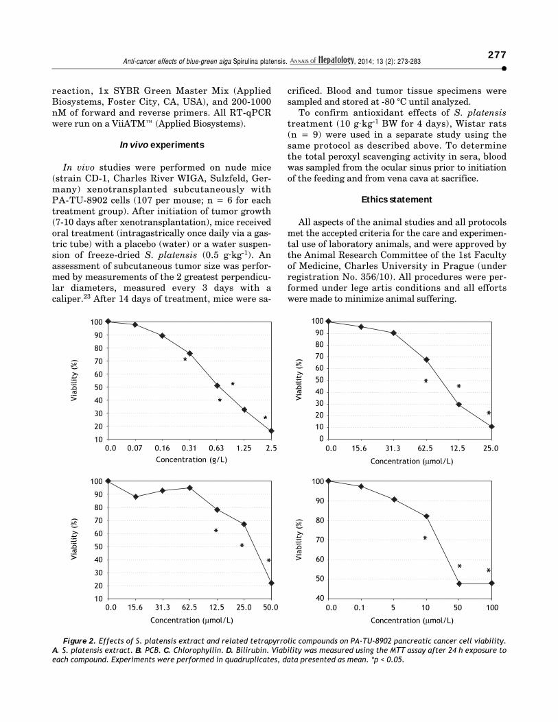
Figure 2. Effects of S. platensis extract and related tetrapyrrolic compounds on PA-TU-8902 pancreatic cancer cell viability.A. S. platensis extract. B. PCB. C. Chlorophyllin. D. Bilirubin. Viability was measured using the MTT assay after 24 h exposure toeach compound. Experiments were performed in quadruplicates, data presented as mean. *p < 0.05.
100
90
80
70
60
50
40
30
20
10
Viab
ility
(%
)
100
90
80
70
60
50
40
30
20
10
0
Viab
ility
(%
)
100
90
80
70
60
50
40
30
20
10
Viab
ility
(%
)
100
90
80
70
60
50
40
Viab
ility
(%
)
0.0 0.07 0.16 0.31 0.63 1.25 2.5Concentration (g/L)
0.0 15.6 31.3 62.5 12.5 25.0
Concentration (μmol/L)
0.0 15.6 31.3 62.5 12.5 25.0 50.0
Concentration (μmol/L)
0.0 0.1 5 10 50 100
Concentration (μmol/L)
*
*
*
*

Konícková R, et al. , 2014; 13 (2): 273-283278 <
Statistical analyses
Differences between variables were evaluated bythe Mann-Whitney Rank Sum test. Group mean diffe-rences in tumor size were measured by repeated mea-sures analysis of variance (RM ANOVA) withHolm–Šidák posthoc testing, when p-values were sig-nificant. When needed, log transform values of tumorsize were used for comparisons to comply with nor-mality and equal variance requirements. Linear re-gression analyses were used to compare the effects ofS. platensis treatment on total peroxyl scavenging ac-tivity in vitro. Paired t-tests were used to comparethe effects of S. platensis on total peroxyl scavengingactivity in vivo. Depending on their normality, dataare presented as the mean ± SD or the median and25%-75% range. Differences were considered statisti-cally significant when p-values were less than 0.05.
RESULTS
Effects of S. platensis extractand its tetrapyrrolic components on
pancreatic cancer cell viability
All therapeutics efficiently decreased pancreaticcancer cell viability in a dose-dependent manner.The most sensitive cancer cell line was PA-TU-8902,whose viability was substantially decreased follow-ing treatment with S. platensis (from 0.16 g·L-1,p < 0.05) (Figure 2). As low as 10 µM concentrationsof bilirubin (corresponding to its levels in humanserum) substantially suppressed PA-TU-8902 cellviability (p < 0.05); whereas, doses of approximately60 µM PCB, and 125 µM chlorophyllin were neededto reach the same effect (Figure 2). For other pan-creatic cancer cell lines (Mia PaCa-2 and BxPC-3),higher concentrations of all compounds (~3 x) wererequired to reduce cell viability to 50% (datanot shown). No selenium was detected in theS. platensis extract (detection limit of the method = 9ng·mL) indicating that selenoproteins were notresponsible for observed anti-proliferative effects ofS. platensis treatment.
Based on these findings, the PA-TU-8902 cell linewas used for further studies.
Effects of S. platensis extractand its tetrapyrrolic components on
HMOX in PA-TU-8902 cells
The HMOX pathway was investigated for tworeasons:
• HMOX has been reported to affect tumor cell pro-liferation24 and tetrapyrrolic compounds used inour study might modulate HMOX in a feedbackmechanism; and
• HMOX is a potent effector of the antioxidant de-fense system.
Therefore, the effects of S. platensis extract andits related tetrapyrrolic compounds on HMOX inthe PA-TU-8902 cells were analyzed. Neither theS. platensis extract, PCB, nor bilirubin had any effecton HMOX activity (Figure 3) or HMOX1 mRNAexpression. However, only chlorophyllin (30 µM)significantly inhibited HMOX activity (to 51 ± 6%of control values, p < 0.001) (Figure 3) and mRNAexpression (to 55 ± 32% of control values, p =0.016, data not shown). Interestingly, the same inhi-bitory effect of chlorophyllin on HMOX activity wasalso observed for Mia PaCa-2 pancreatic cancer cells(data not shown). Expression of biliverdin reductase(OMIM * 109750), another important gene in theheme catabolic pathway, was not affected by any ofthese compounds (data not shown).
Effects of S. platensis extract and itstetrapyrrolic components on mitochondrial
redox status of PA-TU-8902 cells
Proliferation of normal and cancer cells is stron-gly influenced by redox signaling.18 Because PCB is
Figure 3. Effects of S. platensis extract and related tetra-pyrrolic compounds on HMOX enzyme activity. PA-TU-8902pancreatic cancer cells were incubated with S. platensis ex-tract (0.3 g·L-1), hemin (30 μM), PCB (30 μM), chlorophyllin(30 μM), and bilirubin (10 μM) for 24 h. Heme oxygenase acti-vity expressed as percentage of control (100%).
Cont
rol
Hem
in
S.pl
aten
sis PCB
Chlo
roph
yllin
Bilir
rubi
n
200
180
160
140
120
100
80
60
40
20
0
HM
OX
acti
vity
(%
)
p < 0.001
p < 0.001

279Anti-cancer effects of blue-green alga Spirulina platensis. , 2014; 13 (2): 273-283
structurally related to the potent antioxidants bili-rubin and biliverdin, its effects on the cellular redoxbalance were investigated. The main source of intra-cellular free radicals under basal conditions is froma leakage of superoxide from the mitochondrial res-piratory chain.25 Even more importantly, NADPHoxidase-derived superoxide has an important func-tion in pancreatic cancer cell proliferation due to K-ras/Rac1-dependent NADPH oxidase induction26 andthe importance of this pathway was demonstratedalso for liver carcinogenesis.27 Therefore, we wereinterested whether S. platensis and/or S. platensis-derived tetrapyrroles could influence mitochondrialproduction of ROS.
Indeed, we found that 24 h after exposure of PA-TU-8902 cells to S. platensis extract, PCB, andchlorophyllin resulted in significant decreases inintramitochondrial ROS production (Figure 4A),despite the fact that mRNA expressions of NOX2and p22 NADPH oxidase subunits were not affec-ted by 1 h incubation of cells with either S. platen-sis extract, PCB, or chlorophyllin (data notshown).
We then investigated whether this effect is robustenough to modify the cellular redox balance by de-termination of the oxidized and reduced glutathioneratio. We found that pre-incubation of cells with S.platensis extract, PCB, or chlorophyllin shifted theratio towards increased reduction (p = 0.0006;0.016; and 0.006, respectively) (Figure 4B).
Finally, we investigated whether a decreasedleakage of mitochondrial ROS was due to direct an-tioxidant properties of tested therapeutics, or whe-ther these compounds influence the ROS leakage by
modulation of activity and efficiency of mitochon-drial respiratory chain.25 We measured mito-chondrial respiration of the cells maintained in thecomplete medium in a basal state and during maxi-mal stimulation with an uncoupling agent FCCP,and basal proton leakage during inhibition ofComplex V with oligomycin. Interestingly, we foundno changes in the respiration of the PA-TU-8902cells after exposure to S. platensis extract, PCB, orchlorophyllin (data not shown) suggesting that de-creased leakage of ROS was due to direct antioxidantproperties of S. platensis and its tetrapyrroles,PCB, and/or chlorophyllin.
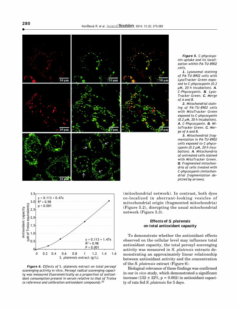
Uptake and localization ofC-phycocyanin and
PCB in PA-TU-8902 cells
Live cell imaging studies demonstrated thatboth C-phycocyanin and PCB incubated withPA-TU-8902 cells can easily enter the cells (Figure5.1, data for PCB not shown). Accumulation ofboth pigments within the cells was apparentwithin 1 h of treatment, and gradually increasedin time- and concentration-dependent manners.Localizations of C-phycocyanin and PCB in thecells were observed mainly in the form of vesicleslocalized in perinuclear region. The co-localiza-tion of LysoTracker Green with C-phycocyaninwas seen in most of the observed vesicles withinthe cells (Figure 5.1). Co-localization study with amitochondrial marker MitoTracker Green revealedthat C-phycocyanin was absent in the mitochon-dria as shown by their typical elongated shape
Figure 4. Effects of S. platensis and related tetrapyrrolic compounds on (A) mitochondrial production of ROS and (B) gluta-thione redox status. PA-TU-8902 pancreatic cancer cells were incubated with S. platensis extract (0.3 g·L-1), PCB (50 μM), andchlorophyllin (50 μM) for 24 h. ROS: reactive oxygen species. GSH: reduced glutathione. GSSG: oxidized glutathione.
35
30
25
20
15
10
5
00 2 4 6 8 10 12 14 16 18
Time (min)
Mit
ocho
ndri
al R
OS
prod
ucti
on
10
9
8
7
6
5
4
3
2
1
0Control S. platensis PCB Chlorophyllin
GSS
G /
GSS
G +
GSH
p = 0.0006
p = 0.016
p = 0.006
ControlS. platensisChlorophyllinPCB
Konícková R, et al. , 2014; 13 (2): 273-283280 <
(mitochondrial network). In contrast, both dyesco-localized in aberrant-looking vesicles ofmitochondrial origin (fragmented mitochondria)(Figure 5.2), disrupting the usual mitochondrialnetwork (Figure 5.3).
Effects of S. platensison total antioxidant capacity
To demonstrate whether the antioxidant effectsobserved on the cellular level may influence totalantioxidant capacity, the total peroxyl scavengingactivity was measured in S. platensis extracts de-monstrating an approximately linear relationshipbetween antioxidant activity and the concentrationof the S. platensis extract (Figure 6).
Biological relevance of these findings was confirmedin our in vivo study, which demonstrated a significantincrease (132 ± 22%, p = 0.002) in antioxidant capaci-ty of rats fed S. platensis for 5 days.
Figure 5. C-phycocya-nin uptake and its locali-zation within PA-TU-8902cells.
1. Lysosomal stainingof PA-TU-8902 cells withLysoTracker Green expo-sed to C-phycocyanin (0.2μM, 20 h incubation). A.C-Phycocyanin. B. Lyso-Tracker Green. C. Mergeof A and B.
2. Mitochondrial stain-ing of PA-TU-8902 cellswith MitoTracker Greenexposed to C-phycocyanin(0.2 μM, 20 h incubation).A. C-Phycocyanin. B. Mi-toTracker Green. C. Mer-ge of A and B.
3. Mitochondrial frag-mentation in PA-TU-8902cells exposed to C-phyco-cyanin (0.2 μM, 20 h incu-bation). A. Mitochondriaof untreated cells stainedwith MitoTracker Green.B. Fragmented mitochon-dria of cells treated withC-phycocyanin (mitochon-drial fragmentation de-picted by arrows).
Figure 6. Effects of S. platensis extract on total peroxylscavenging activity in vitro. Peroxyl radical scavenging capaci-ty was measured fluorometrically as a proportion of antioxi-dant consumption present in serum relative to that of Trolox(a reference and calibration antioxidant compound).20
3.5
3.0
2.5
2.0
1.5
1.0
0.5
00 0.2 0.4 0.6 0.8 1 1.2 1.4 1.6
S. platensis extract (g/L)
Anti
oxid
ant
capa
city
(μM
of
Trol
ox e
quiv
alen
ts)
y = 0.113 + 0.47xR2 = 0.98p = 0.001
y = 0.113 + 1.47xR2 = 0.98P = 0.001
281Anti-cancer effects of blue-green alga Spirulina platensis. , 2014; 13 (2): 273-283
In vivo anticancer effects of S. platensis
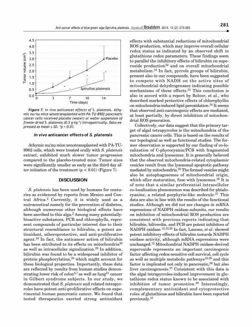
Athymic nu/nu mice xenotransplanted with PA-TU-8902 cells, which were treated orally with S. platensisextract, exhibited much slower tumor progressioncompared to the placebo-treated mice. Tumor sizeswere significantly smaller as early as the third day af-ter initiation of the treatment (p < 0.01) (Figure 7).
DISCUSSION
S. platensis has been used by humans for centu-ries as evidenced by reports from Mexico and Cen-tral Africa.2 Currently, it is widely used as anutraceutical namely for the prevention of diabetes,although numerous other biological effects havebeen ascribed to this alga.2 Among many potentially-bioactive substances, PCB and chlorophylls, repre-sent compounds of particular interest due to theirstructural resemblance to bilirubin, a potent an-tioxidant, atheroprotective, and anti-proliferativeagent.24 In fact, the anticancer action of bilirubinhas been attributed to its effects on mitochondria28
as well as intracellular signalization.29 In addition,bilirubin was found to be a widespread inhibitor ofprotein phosphorylation,30 which might account forthese biological properties. Importantly, these dataare reflected by results from human studies demon-strating lower risk of colon5,7 as well as lung31 cancerin Gilbert syndrome subjects. In our study, wedemonstrated that S. platensis and related tetrapyr-roles have potent anti-proliferative effects on expe-rimental human pancreatic cancer. We found thattested therapeutics exerted strong antioxidant
effects with substantial reductions of mitochondrialROS production, which may improve overall cellularredox status as indicated by an observed shift inglutathione redox parameters. These findings seemto parallel the inhibitory effects of bilirubin on supe-roxide production32 and on overall mitochondrialmetabolism.33 In fact, pyrrole groups of bilirubin,present also in our compounds, have been suggestedto compete with NADH on the active sites ofmitochondrial dehydrogenases indicating possiblemechanisms of these effects.33 This conclusion isalso in accord with a report by Boloor, et al., whodescribed marked protective effects of chlorophyllinon mitochondria-induced lipid peroxidation.34 It seemsthat observed anti-carcinogenic effects are mediated,at least partially, by direct inhibition of mitochon-drial ROS generation.
Collectively, our data suggest that the primary tar-get of algal tetrapyrroles is the mitochondria of thepancreatic cancer cells. This is based on the results ofour topological as well as functional studies. The for-mer observation is supported by our finding of co-lo-calization of C-phycocyanin/PCB with fragmentedmitochondria and lysosomes. It is generally believedthat the observed mitochondria-related cytoplasmicvesicles result from the lysosomal apoptotic pathwaymediated by mitochondria.35 The formed vesicles mightalso be autophagosomes of mitochondrial origin,which after maturation, fuse with lysosomes.36 It isof note that a similar preferential intracellularco-localization phenomenon was described for phtalo-cyanine, a related porphyrin-like molecule.37 Thisdata are also in line with the results of the functionalstudies. Although we did not see changes in mRNAexpressions of NADPH oxidase subunits, our resultson inhibition of mitochondrial ROS production areconsistent with previous reports indicating thatbilirubin, biliverdin, and PCB are potent inhibitors ofNADPH oxidase.12,13,38 In fact, Lanone, et al. showedpotent inhibitory effects of bilirubin towards NADPHoxidase activity, although mRNA expressions wereunchanged.12 Mitochondrial NADPH oxidase-derivedsuperoxide represents an important carcinogenicfactor affecting redox-sensitive cell survival, cell cycleas well as multiple metabolic pathways18,39 and thisfactor is implicated not only in pancreatic,26 but alsoliver carcinogenesis.27 Consistent with this data isthe algal tetrapyrroles-induced improvement in glu-tathione redox status known to be associated withinhibition of tumor promotion.40 Interestingly,complementary antioxidant and cytoprotectiveroles of glutathione and bilirubin have been reportedpreviously.41
Figure 7. In vivo anticancer effects of S. platensis. Athy-mic nu/nu mice xenotransplanted with PA-TU-8902 pancreaticcancer cells received placebo (water) or water suspension offreeze-dried S. platensis (0.5 g·kg-1) intragastrically. Data ex-pressed as mean ± SD. *p < 0.01.
Tum
or v
olum
e (c
m3 )
4.5
4.0
3.5
3.0
2.5
2.0
1.5
1.0
0.5
0.00 3 7 10 14
Time (days)
Placebo
Spirulina platensis

Konícková R, et al. , 2014; 13 (2): 273-283282 <
Interestingly, we also observed a marked inhibi-tory effect of chlorophyllin on both HMOX1 mRNAexpression and total HMOX enzyme activity, whilethe other tetrapyrroles did not have any effect. Thisinhibitory action, which is in accord with opposingeffects of various metalloporphyrins on HMOX,15
might contribute to anti-carcinogenic effects observedin our study and also, at least in part, account forchemopreventive effects of chlorophyll-rich nutrients.
One of the limitations of our study is that wehave not directly identified all particular compo-nents in the S. platensis extract responsible for theobserved biological effects, and focused only onthe algal tetrapyrroles. However, it should beemphasized that exactly the same preparation, i.e.pulverized S. platensis, is commonly used in a globalscale. Our data thus provide evidence for all theconsumers on the anti-cancer potential of this alga.
In conclusion, S. platensis, as well as its tetra-pyrrolic components, substantially decreased prolife-ration of pancreatic cancer. These effects were atleast partially due to their potent antioxidant activi-ty, inhibition of mitochondrial production of ROSand subsequent changes in intracellular redoxstatus. These data support a chemopreventive roleof this edible alga with promising potential for itsbroad use in the chemoadjuvant treatment of cancerdiseases. This assumption of more generalized use ofS. platensis in cancer chemoprevention is supportedalso by recent data demonstrating anti-cancerpotential of this alga on liver39 as well as breast42
carcinogenesis. It also should be noted, that dietarysupplementation with this alga might enhancesystemic pool of tetrapyrroles in a more naturalway, as compared to xenobiotic-induced hyperbiliru-binemia approach proposed recently.43
FINANCIAL SUPPORT
This work was supported by grants Nos. 55210,438911, LH11030, PRVOUK-P25/LF1/2, and RVO-VFN64165/2013 given by the Granting Agency of theCharles University in Prague, Czech Ministry ofEducation and Czech Ministry of Health, respective-ly. The granting agencies had no influence upon thecollection, analysis, interpretation of data, writingof the report, nor on the decision to submit the pa-per for publication.
ACKNOWLEDGEMENTS
Authors thank to Drs. Pavel Coufal and TomášKrizek from Department of Analytical Chemistry,
Faculty of Science of the Charles University in Pra-gue for kindly providing us with capillary electro-phoresis equipment, Magdalena Kadlecová and OlgaŠvejdová for excellent technical assistance.
REFERENCES
1. Dillon JC, Phuc AP, Dubacq JP. Nutritional value of the algaSpirulina. World Rev Nutr Diet 1995; 77: 32-46.
2. Gershwin ME, Belay A. Spirulina in human nutrition andhealth. Boca Raton: CRC Press; 2008.
3. Padyana AK, Bhat VB, Madyastha KM, Rajashankar KR, Ra-makumar S. Crystal structure of a light-harvesting proteinC-phycocyanin from Spirulina platensis. Biochem BiophysRes Commun 2001; 282: 893-8.
4. Novotny L, Vitek L. Inverse relationship between serumbilirubin and atherosclerosis in men: a meta-analysis of pu-blished studies. Exp Biol Med 2003; 228: 568-71.
5. Jiraskova A, Novotny J, Novotny L, Vodicka P, Pardini B,Naccarati A, Schwertner HA, et al. Association of serumbilirubin and promoter variations in HMOX1 and UGT1A1genes with sporadic colorectal cancer. Int J Cancer 2012;131: 1549-55.
6. Vitek L, Muchova L, Jancova E, Pešicková S, Tegzová D,Peterová V, Pavelka K, et al. Association of systemic lupuserythematosus with low serum bilirubin levels. Scand JRheumatol 2010; 39: 480-4.
7. Zucker SD, Horn PS, Sherman KE. Serum bilirubin levels inthe US population: Gender effect and inverse correlationwith colorectal cancer. Hepatology 2004; 40: 827-35.
8. McCarty MF. «Iatrogenic Gilbert syndrome»-a strategyfor reducing vascular and cancer risk by increasing plas-ma unconjugated bilirubin. Med Hypotheses 2007; 69:974-94.
9. Malik SG, Irwanto KA, Ostrow JD, Tiribelli C. Effect of bili-rubin on cytochrome c oxidase activity of mitochondriafrom mouse brain and liver. BMC Res Notes 2010;3: 162.
10. Rodrigues CM, Sola S, Brites D. Bilirubin induces apoptosisvia the mitochondrial pathway in developing rat brain neu-rons. Hepatology 2002; 35: 1186-95.
11. Rodrigues CM, Sola S, Brito MA, Brites D, Moura JJ. Biliru-bin directly disrupts membrane lipid polarity and fluidity,protein order, and redox status in rat mitochondria. J He-patol 2002; 36: 335-41.
12. Lanone S, Bloc S, Foresti R, Almolki A, Taillé C, CallebertJ, Conti M, et al. Bilirubin decreases nos2 expressionvia inhibition of NAD(P)H oxidase: implications for pro-tection against endotoxic shock in rats. FASEB J 2005;19: 1890-2.
13. Zheng J, Inoguchi T, Sasaki S, et al. Phycocyanin andphycocyanobilin from Spirulina platensis protect againstdiabetic nephropathy by inhibiting oxidative stress. Am JPhysiol Regul Integr Comp Physiol 2013; 304: R110-R120.
14. Was H, Dulak J, Jozkowicz A. Heme oxygenase-1 in tumorbiology and therapy. Curr Drug Targets 2010; 11: 1551-70.
15. Drummond GS, Kappas A. Prevention of neonatal hyperbili-rubinemia by tin protoporphyrin IX, a potent competitiveinhibitor of heme oxidation. Proc Natl Acad Sci USA 1981;78: 6466-70.
16. Hayatsu H, Negishi T, Arimoto S, Hayatsu T. Porphyrins aspotential inhibitors against exposure to carcinogens andmutagens. Mutat Res 1993; 290: 79-85.
17. Kumar SS, Devasagayam TP, Bhushan B, Verma NC. Sca-venging of reactive oxygen species by chlorophyllin: anESR study. Free Radic Res 2001; 35: 563-74.
<
<< < <
283Anti-cancer effects of blue-green alga Spirulina platensis. , 2014; 13 (2): 273-283
18. Antico Arciuch VG, Elguero ME, Poderoso JJ, Carreras MC.Mitochondrial regulation of cell cycle and proliferation.Antioxid Redox Signal 2012; 16: 1150-80.
19. Terry MJ, Maines MD, Lagarias JC. Inactivation of phyto-chrome- and phycobiliprotein-chromophore precursors byrat liver biliverdin reductase. J Biol Chem 1993; 268:26099-106.
20. Iuliano L, Piccheri C, Coppola I, Pratico D, Micheletta F,Violi F. Fluorescence quenching of dipyridamole associa-ted to peroxyl radical scavenging: a versatile probe tomeasure the chain breaking antioxidant activity of biomo-lecules. Biochim Biophys Acta 2000; 1474: 177-82.
21. Vreman HJ, Stevenson DK. Heme oxygenase activity asmeasured by carbon monoxide production. Anal Biochem1988; 168: 31-8.
22. Maeso N, Garcia-Martinez D, Ruperez FJ, Cifuentes A,Barbas C. Capillary electrophoresis of glutathione to moni-tor oxidative stress and response to antioxidant treat-ments in an animal model. J Chromatogr B Analyt TechnolBiomed Life Sci 2005; 822: 61-9. Doi: 10.1016/j.jchromb.2005.05.015.
23. McKeage MJ, Kelland LR, Boxall FE, Valenti MR, Jones M,Goddard PM, Gwynne J, et al. Schedule dependency of orallyadministered bis-acetato-ammine-dichloro-cyclohexylamine-platinum(IV) (JM216) in vivo. Cancer Res 1994; 54:4118-22.
24. Vitek L, Schwertner HA. The heme catabolic pathwayand its protective effects on oxidative stress-mediateddiseases. Adv Clin Chem 2007; 43: 1-57.
25. Jezek P, Hlavata L. Mitochondria in homeostasis of reactiveoxygen species in cell, tissues, and organism. Int JBiochem Cell Biol 2005; 37: 2478-503.
26. Du J, Liu J, Smith BJ, Tsao MS, Cullen JJ. Role of Rac1-dependent NADPH oxidase in the growth of pancreaticcancer. Cancer Gene Ther 2011; 18: 135-43. Doi: 10.1038/cgt.2010.64.
27. Teufelhofer O, Parzefall W, Kainzbauer E, Ferk F, FreilerC, Knasmüller S, Elbling L, et al. Superoxide generationfrom Kupffer cells contributes to hepatocarcinogenesis:studies on NADPH oxidase knockout mice. Carcinogenesis2005; 26: 319-29.
28. Keshavan P, Schwemberger SJ, Smith DLH, Babcock GF,Zucker SD. Unconjugated bilirubin induces apoptosis in co-lon cancer cells by triggering mitochondrial depolariza-tion. Int J Canc 2004; 112: 433-45.
29. Ollinger R, Kogler P, Troppmair J, Hermann M, Wurm M,Drasche A, Königsrainer I, et al. Bilirubin inhibits tumorcell growth via activation of ERK. Cell Cycle 2007; 6:3078-85.
30. Hansen TW, Mathiesen SB, Walaas SI. Bilirubin has wides-pread inhibitory effects on protein phosphorylation.Pediatr Res 1996; 39: 1072-77.
31. Horsfall LJ, Rait G, Walters K, Swallow DM, Pereira SP, Na-zareth I, Petersen I. Serum bilirubin and risk of respira-tory disease and death. JAMA 2011; 305: 691-7.
32. Nakamura H, Uetani Y, Komura M, Takada S, Sano K, Mat-suo T. Inhibitory action of bilirubin on superoxide produc-tion by polymorphonuclear leukocytes. Biol Neonate 1987;52: 273-8.
33. Noir BA, Boveris A, Garaza Pereira AM, Stoppani AO. Biliru-bin: a multi-site inhibitor of mitochondrial respiration.FEBS Lett 1972; 27: 270-4.
34. Boloor KK, Kamat JP, Devasagayam TP. Chlorophyllin as aprotector of mitochondrial membranes against gamma-radiation and photosensitization. Toxicology 2000; 155:63-71.
35. Boya P, Andreau K, Poncet D, Zamzami N, Perfettini JL,Metivier D, Ojcius DM, et al. Lysosomal membrane per-meabilization induces cell death in a mitochondrion-depen-dent fashion. J Exp Med 2003; 197: 1323-34.
36. Arstila AU, Trump BF. Studies on cellular autophagocytosis.The formation of autophagic vacuoles in the liver afterglucagon administration. Am J Pathol 1968; 53: 687-733.
37. Rodriguez ME, Kim J, Delos Santos GB, Azizuddin K, Berlin J,Anderson VE, Kenney ME, et al. Binding to and photo-oxi-dation of cardiolipin by the phthalocyanine photosensiti-zer Pc 4. J Biomed Opt 2010; 15: 051604.
38. Fujii M, Inoguchi T, Sasaki S, Maeda Y, Zheng J, KobayashiK, Takayanagi R. Bilirubin and biliverdin protect rodentsagainst diabetic nephropathy by downregulating NAD(P)Hoxidase. Kidney Int 2010; 78: 905-19.
39. Nazarewicz RR, Dikalova A, Bikineyeva A, Ivanov S,Kirilyuk IA, Grigor’ev IA, Dikalov SI. Does scavenging ofmitochondrial superoxide attenuate cancer prosurvivalsignaling pathways? Antioxid Redox Signal 2013; 19:344-9.
40. Perchellet JP, Perchellet EM, Orten DK, Schneider BA. De-creased ratio of reduced/oxidized glutathione in mouseepidermal cells treated with tumor promoters. Carcinoge-nesis 1986; 7: 503-6.
41. Sedlak TW, Saleh M, Higginson DS, Paul BD, Juluri KR, Sn-yder SH. Bilirubin and glutathione have complementary an-tioxidant and cytoprotective roles. Proc Natl Acad SciUSA 2009; 106: 5171-6.
42. Ouhtit A, Ismail MF, Osman A, Fernando A, Abdraboh ME,El-Kott AF, Azab YA, et al. Chemoprevention of rat mam-mary carcinogenesis by Spirulina. Am J Pathol 2013. Doi:10.1016/j.ajpath.2013.10.025 [Epub ahead of print].
43. Dekker D, Dorresteijn MJ, Pijnenburg M, Heemskerk S, Ra-sing-Hoogveld A, Burger DM, Wagener FA, et al. The biliru-bin-increasing drug atazanavir improves endothelialfunction in patients with type 2 diabetes mellitus. Arte-rioscler Thromb Vasc Biol 2011; 31: 458-63.