ANNUAL - CORE
443
ANNUAL REPORT 2004
-
Upload
khangminh22 -
Category
Documents
-
view
1 -
download
0
Transcript of ANNUAL - CORE
ANNUALREPORT
2004

COLD SPRING HARBOR LABO

ANNUAL REPORT 2004© 2005 by Cold Spring Harbor Laboratory
Cold Spring Harbor LaboratoryOne Bungtown RoadCold Spring Harbor, New York 11724
Web Site: www.cshl.edu
Managing Editors Jeff Picarello, Lisa BeckerProduction Editor Rena SteuerCopy Editor Dorothy BrownDevelopment Manager Jan ArgentineProject Coordinators Maria Falasca, Nora RiceProduction Manager Denise WeissDesktop Editor Susan SchaeferNonscientific Photography Miriam Chua, Bill GeddesCover Designer Denise WeissBook Designer Emily Harste
Front cover: McClintock Laboratory (right) and Carnegie Library(left) (photos by Miriam Chua)
Back cover: Magnolia Kobus on grounds of Cold Spring HarborLaboratory (photo by Bruce Stillman)
Section title pages: Miriam Chua, Rena Steuer

ContentsOfficers of the Corporation and Board of Trustees iv-vGovernance viCommittees of the Board vii
Rollin Hotchkiss (1911-2004) viii
Ralph Landau (1916-2004) ix
David B. Pall (1914-2004) xi
PRESIDENT'S REPORTHighlights of the Year 3
CHIEF OPERATING OFFICER'S REPORT 21
RESEARCHCancer: Gene Expression 27Cancer: Genetics 51
Cancer: Cell Biology 84Neuroscience 110Plant Development and Genetics 156Bioinformatics and Genomics 173CSHL Fellows 187Author Index 194
25
WATSON SCHOOL OF BIOLOGICAL SCIENCESDean's Report 199Courses 212Undergraduate Research Program 219Partners for the Future 221
Nature Study Program 222
197
COLD SPRING HARBOR LABORATORY MEETINGS AND COURSESAcademic Affairs 224Symposium on Quantitative Biology 225Meetings 228Postgraduate Courses 275Seminars 327
223
BANBURY CENTERExecutive Director's Report 331
Meetings 333
329
DOLAN DNA LEARNING CENTERExecutive Director's Report 3692004 Workshops, Meetings, and Collaborations 382
367
COLD SPRING HARBOR LABORATORY PRESS2004 Publications 388Executive Director's Report 389
387
FINANCEFinancial Statements 394Financial Support of the Laboratory 398
Grants 398Development 407Capital and Program Contributions 408Annual Contributions 410
393
LABORATORY STAFF 424
iii

Officers of the Corporation
Eduardo G. Mestre, Chairman"Lola N. Grace, Vice Chairman*Edward Travaglianti, Secretary/Treasurer"
Board of Trustees
Bruce Stillman, Ph.D., President and Chief Executive OfficerJames D. Watson, Ph.D., ChancellorW. Dillaway Ayres, Jr., Chief Operating Officer
Individual Trustees
Trudy H. CalabreseLloyd Harbor New Y.
Kristina Perkin DavisonPartner, iEurope Capital LLC
Jacob GoldfieldNew York, New York
Lola N. GraceManaging Director Sterling GraceCapital Management
Charles E. HarristChairman and CEO,Ha. 8 Hants GrouP,
Laurie Landeau, V.M.D."PjC
Thomas C. QuickPalm Beach, Florida
Robert D. LindsayCo-Managing Partner,Goldberg Lindsay 8 Co.
Nancy Marks'Carl Marks 8 Co., Inc.
Eduardo G. MestreVice Chairman, EvercorePartners
Douglas P. MorrisChairman and CEO,
V.'s, Music Group
William S. RobertsonNaples, Florida
Howard Solomon,Chad., of the Board andCEO, Forest Laboratories
Arthur M. SpiroKings Point, New York
Alan C. StephensonPartner Cmvath Swains &Moore, LLP
James M. StoneChairman, The Plymouth
Rock Company
"Term began in November 2004.tTerm concluded in November 2004.
IV
Jerome Swartz, Ph.D."Old Field, New Y.
Edward TravagliantiPresident, Commerce BankLong Island
James D. Watson, Ph.D.Chancellor,Cold Spring Harbor Laboratory
Roy J. ZuckerbergSenior Director, GoldmanSachs 8 Company

Scientific Trustees
Laurence Abbott, Ph.D..Vo len Center, Brandeis University
David Botstein, Ph.D.Director, Lewis-Siger Institute,Princeton University
Titia de Lange, Ph.D.Leon Hess Professor TheRockefeller University
Jeff HawkinsEcund s r d Bewlhe Drector.Redwood Naxos., ce Thsttute
Susan Hockfield, Ph.D.President, MassachusettsInstitute of Technology
Susan Lee Lindquist, Ph.D. Charles J. Sher, M.D., Ph.D., Bruce Stillman, Ph.D. Robert E. Wittes, M.D..Director, Whitehead Institute forBiomedical Research
Chairman, Department of TumorCell &logy HHMI/St. Jude's
President and CEO.Cold Sprang Harbor Laboratory
Physician in CNef, MemorialSloan-Kettering Cancer Center
Children's Research Hospital
Honorary Trustees
Bayard Clarkson, M.D.Memorial Sioan-KetteringCancer Center New York
John P. Cleary, Esq.Locust Valley, New York
Charles F. DolanOyster Bay New York
Helen Ann DolanOyster Bay, New Yale
H. Bentley Glass, Ph.D.Boulder, Cobra.
Townsend J. Knight, Esq.New York, New York
Evelyn H. LauderEstee Lauder Companies,
William R. Miller.New York, New York
William E. Murray, Esq.New York, New York
Wendy Vander Poel RussellOyster Bay, New York
Mary D. LindsayNew York New York
David L Luke IIINew York, New York
"Term began in November 2004.germ concluded in November 2004.

Governance
The Laboratory is governed by a Board of Trustees of up to 35 members which meets three or four timesa year. Authority to act for the Board of Trustees between meetings is vested in the Executive Committeeof the Board of Trustees. The Executive Committee is composed of the Officers of the Board and anyother members who may be elected to the Executive Committee by the Board of Trustees. Additionalstanding and ad hoc committees are appointed by the Board of Trustees to provide guidance and advicein specific areas of the Laboratory's operations.
Representation on the Board of Trustees itself is divided between business and community leadersand scientists from major educational and research institutions.
The Laboratory is chartered as an educational and research institution by the Board of Regents of the
Education Department of the State of New York. It is authorized to operate a graduate program underthe name "Cold Spring Harbor Laboratory, Watson School of Biological Sciences" and thereat to conferthe degrees of Doctor of Philosophy (Ph.D.), Master of Science (M.S.), and Doctor of Science (Sc.D.),Honorary.
It is designated as a "public charity" under Section 501(c)(3) of the Internal Revenue Code.
vi

Committees of the Board*
Audit
Lola N. Grace, ChairArthur M. SpiroAlan Stephenson
Development
Joseph Donohue, ChairKristina Perkin DavisonLaurie Landau, V.M.D.Robert D. LindsayNancy MarksThomas QuickJames D. Watson, Ph.D.Roy J. Zuckerberg
Executive
Eduardo G. Mestre, ChairW. Dillaway Ayres, Jr.Joseph DonohueLola N. GraceBruce Stillman, Ph.D.Edward TravagliantiRoy J. Zuckerberg
Finance
Lola N. Grace, ChairW. Dillaway Ayres, Jr.Jacob GoldfieldRobert D. LindsayWilliam S. RobertsonLari C. RussoEdward Travaglianti
Ad Hoc Commercial Relations
James Stone, ChairW. Dillaway Ayres, Jr.David BosteinTitia de LangeJohn P Maroney, Esq.
*November 2004-November 2005
Alan StephensonBruce Stillman, Ph.D.Jerome Swartz, Ph.D.
Investment
Edward Travaglianti, ChairW. Dillaway Ayres, Jr.Jacob GoldfieldLola N. GraceWilliam S. Robertson
Nominating
Robert D. Lindsay, ChairW. Dillaway Ayres, Jr.Kristina Perkin DavisonEduardo G. MestreBruce Stillman, Ph.D.Roy J. Zuckerberg
Robertson Research Fund
Carl Schafer, ChairW. Dillaway Ayres, Jr.Hollis Cline, Ph.D.Lola N. GraceEduardo G. MestreBruce Stillman, Ph.D.
Family Representatives
Katherine ErnstWalter Meier, M.D.William S. Robertson
Tenure and Appointments
Bruce Stillman, Ph.D., ChairLaurence Abbott, Ph.D.David Botstein, Ph.D.Titia de Lange, Ph.D.Susan Lee Lindquist, Ph.D.Robert E. Wittes, M.D.
Other Committees
Building
Arthur BringsJohn P. Cleary, Esq.Helen Ann DolanMary D. LindsayNancy MarksWendy Vander Poel RussellElizabeth Watson
Dolan DNA Learning Center
Arthur M. Spiro, ChairMarion Wood AhmedW. Dillaway Ayres, Jr.Edward A. ChernoffLola N. GraceSuzanne KleinknechtLaurie Landau, V.M.D.David A. Mick losLawrence Scherr, M.D.Bruce Stillman, Ph.D.Edward TravagliantiMarianne Dolan Weber
Higher Education
James D. Watson, Ph.D., ChairDavid Botstein, Ph.D.Robert D. LindsayAlan StephensonJames M. Stone
Research
Bruce Stillman, Ph.D., ChairLaurence Abbott, Ph.D.Hollis Cline, Ph.D.Titia de Lange, Ph.D.Jacob GoldfieldEdward Harlow, Ph.D.Jeff HawkinsRobert E. Wittes, M.D.Roy J. Zuckerberg
vii

Photo courtesy of The Rockefeller University
Rollin Hotchkiss (1911-2004)Trustee, Cold Spring Harbor Laboratory,1954-1966 and 1974-1978
Rollin Hotchkiss was trained as an organic chemist at Yale. He was one of the first to change voca-tions to the rapidly growing field of microbial genetics. After joining the Rockefeller Institute for MedicalResearch, Rollin worked with Rene Dubos on the first antibiotics: gramicidin and tyrocidin.Unfortunately, they were toxic and could only be used topically. Yet it was due to this project that Rollinand I met. In 1945, my teacher from the Bronx High School of Science took me to Rockefeller, becausemy lab project was to isolate gramicidin. I never dreamt that I would one day be a colleague of his.
When Avery, MacLeod, and McCarty-all at Rockefeller-claimed that, in pneumococcus, DNA actedlike genetic material, it caught Rollin's eye. When Avery retired, MacLeod went to NYU, and McCarty wentto head the rheumatic fever department in The Rockefeller Hospital, Rollin took up the essential question:How much protein was in the transforming DNA? He found it to be less than .02 of the material-notmuch protein. Moving on, he took advantage of mutants resistant to sulfanomides. He generalized thephenomenon as they would transform and indicated that DNA was generic genetic material.
With his first wife, Shirley, Rollin had two children: Paul and Cynthia. His second wife, Magda, is ascientist, and they worked together until he retired in 1983.
Rollin was a fun person, always playful. If he had one problem it was that itwas difficult to talk with him "mano a mano." He mumbled and talked inter-minably. One of my students used to take a stopwatch when he went to talkto Rollin. On the other hand, Rollin gave the most clear and witty speeches; Ialways felt that I was incompetent when we spoke in tandem. Rollin had fivegraduate students who have all done well. The senior people were P. Model,A. Tomasz, and myself. We will all miss him.
Recker Reading Room, Blackford Hall, 1992 The dedication of Delbruck Laboratory, 1981
Photos courtesy of the Cold Spring Harbor Laboratory Archives
VIII
Norton Zinder, Ph.D.Professor Emeritus
The Rockefeller University

Ralph Landau (1916-2004)
Ralph Landau was born in Philadelphia, Pennsylvania on May 19, 1916. As a high school studentthere, he read a newspaper article about the new and glamorous field of chemical engineering andimmediately decided it was the career for him. As a scholarship student at the University ofPennsylvania, he received a Bachelor of Science in chemical engineering in 1936 and a Ph.D. inchemical engineering from Massachusetts Institute of Technology five years later. Just before com-pleting his doctorate, he married Claire in 1940.
Ralph took a position in 1941 with M.W. Kellogg, one of the first engineering firms that special-ized in design and development for the oil refining and chemical industries. Within a few years, hewas named head of the chemical department of Kellex Corp., a Kellogg subsidiary, and chargedwith building a large-scale facility at Oak Ridge, Tennessee, to aid in the efforts of World War II.There, he was responsible for the separation of uranium 235, needed for the atomic bomb, from itspredominant isotope, and for designing the equipment to produce fluorine, a highly reactive sub-stance needed to make the uranium hexafluoride used in the gaseous diffusion process.
After the war, Ralph joined Harry Rehnberg, a construction engineer he met at Oak Ridge, andstarted Scientific Design Company in 1946. The firm eventually became one of the most success-ful engineering and design firms worldwide, developing and commercializing nearly a dozenprocesses for producing petrochemicals. Ralph served as Executive Vice President of ScientificDesign, then as Chairman and CEO of Halcon International (later Ha Icon SD Group), and co-found-ed Oxirane Company with Atlantic Richfield Corp. The company developed many invaluable sub-stances and held several significant patents, but it is most noted for its process for terepthalic acid,the co-product route for the production of propylene oxide, and the development of ethylene glycolby thermal hydration-the chief component of antifreeze and used in the process of making Dacron
polyester fiber.In 1983, Ralph turned his sights to education, serving as a consulting professor of economics at
the Stanford Institute for Economic Policy and, the following year, joining Harvard University'sKennedy School of Government as a research fellow.
Ralph's influence spanned the world. The recipient of more than 50 awards and honors, hereceived both the Fuels and Petrochemicals Division Heritage Award and the John Fritz Medal ofthe American Institute of Chemical Engineers, in addition to the Chemical Industry Medal, the PerkinMedal, the Winthop-Sears Award for Chemical Entrepreneurship, and the Founders Award of theNational Academy of Engineering. In 1985, Ralph received the National Medal of Technology, andin 1997, he was awarded the first Othmer Gold Medal of the Chemical Heritage Foundation. He wasa life member of the M.I.T Corporation, a senior trustee of Caltech, a trustee of the University of
ix

Pennsylvania, and former chair of the Princeton University School of Engineering Advisory Council.Ralph was awarded honorary degrees from New York Polytechnic, Clarkson College, Ohio State,and the University of Pennsylvania.
Ralph and I first met in May 1982 upon our jointly receiving honorary degrees from ClarksonCollege. Apparent immediately was his keen intelligence and likeable self-confidence in making gov-ernmental bodies and economic systems operate more effectively. To my surprise, I learned he andhis family had a weekend home on Long Island Sound in Asharoken, only 10 miles from the Lab. InJuly, he and his wife Claire first came to the Lab, with Liz and I soon after spending a delightfulevening at their family beach compound. Initially, I feared that he had too many prior obligations toconsider being one of our Board of Trustees. But upon being asked, he showed no hesitancy, join-ing us at our November 1982 meeting.
As a founding member of our Commercial Relations Committee, he helped devise our first poli-cies for commercializing lab discoveries and inventions. He also gave much sound advice formounting our first Capital Fund Drive (The Second Century Campaign). By his being on the CaltechBoard, he was a friend of Arnold Beckman and able to play a key role in facilitating the gift from theBeckman Foundation that made possible the construction of our Neuroscience Center. His great-est long-term legacy to us though was the inclusion of his family in the mission of Cold SpringHarbor Laboratory. His daughter, Laurie Landeau, remains an active member of the Laboratorycommunity, serving both on our Board of Trustees and the Corporate Advisory Board of the Dolan
DNA Learning Center.Ralph and Claire lived life to its fullest, later having a home in San Francisco as well as New York
City. From their Russian Hill apartment looking down on San Francisco Bay, Ralph could easily moveto and from his faculty apartment and office on the Stanford campus. A high point of every year wasthe anniversary dinner party held at Lutece, then New York's premier French restaurant presidedover by the legendary chef, Andre Soltner. Liz and I felt most honored to be included on many ofthese sparkling occasions.
The last several years of Ralph's life were marked by medical complications that restricted hispast ability to joyously zip through life. Happily, his brain never slowed down, and he retained thecapacity to reflect intelligently on the world's problems until his life ended on April 5, 2004.
Ralph's friendship to many leading academic institutions, his impact on the world of chemicalengineering, and his strong values of scholarship, philanthropy, and integrity positively changedthe lives of many in the United States and abroad. We will cherish his memory for many years tocome.
James D. Watson
x

David B. Pall (1914-2004)
David Pall was born in Thunder Bay, Ontario, Canada on April 2, 1914. Growing up on a farm with nocentral heating or plumbing in rural Saskatchewan, he learned to read by deciphering the comic stripsin the daily newspaper. By age five, he began volume one of A History of the World, a foreshadow ofthe quest for knowledge that would direct his entire life. In high school, he was deeply influenced byMadam Curie, the biography of the chemist Marie Curie, about her discovery that liquids, particularlywater, were often contaminated by infectious bacteria, which could be killed by boiling or reduced innumber by filtration.
In 1939, he graduated with a Ph.D. in Physical Chemistry from McGill University as the highest-rank-ing graduate student in Canada. David moved to New York City and was in dire need of friends andanxious to meet a suitable girl to marry. A friend suggested that he phone Hester Blatt, a youngchemist. David telephoned Hester but reached her younger sister, Josephine, instead. Josephineresponded positively to his call, and on February 3, 1940, the two were married. During the next 12years, they had three children, Stephanie, William, and Ellen.
In 1941, with the United States at war, David's laboratory had one of only three electron micro-scopes in existence worldwide and was requisitioned by the U.S. military to work on the highly confi-dential "Manhattan Project," to develop the atomic bomb. Working with a group of Ph.D.s at ColumbiaUniversity, David was able to make several significant contributions to a secret project that separatedthe metal uranium into its two isomers-U232 and U235-the latter heavier, which was used to makethe bomb.
With the end of World War II, David realized that he might be able to use some of the technologyhe developed to improve aircraft reliability by creating stainless steel filters for oil used in the hydraulicsystem-a key component for landing gear and ailerons. With $3000 financing provided by his McGillfriend, Abraham Appel, he founded in 1946 the Micro Metallic Corporation located in a storefront inForest Hills. By building a furnace to melt stainless steel powder and developing a new method bywhich to mold the porous sheets, David developed the first successful, economically viable stainlesssteel filter for aircraft. He quickly moved to larger space in Brooklyn and began producing his filters forthe Air Force's F-106, allowing the then-conceptual plane to finally reach production. Key to David'slater success was his 1950 recruitment of his neighbor, accountant Abe Krasnoff, who ran the com-mercial aspects of his ever-growing company that became the Pall Corporation in 1957 after moving
to Glen Cove on Long Island.In the late 1950s, David's wife, Josephine, was diagnosed as anemic, and her physician arranged
for her to have monthly donations of transfused blood. The doctor advised David that after about 12months, her immune system would reject the transfused blood and she would die-as she did in 1959.The 12-month "limit" was caused by white cells contained in the donated blood, which were recog-
xi

nized as "foreign bodies" in the recipient. David devoted his time to learning about the components andfunctions of human blood and the use of webs of plastic fiber for filtration, and he quickly began devel-opment on a filter to remove white cells from transfused blood.
David organized a group of Pall Corporation volunteers to donate blood, which was then passedthrough experimental masses of plastic fibers. White blood cell counts were then taken to determinethe fibers' efficiency in removing white cells. Using the data so obtained, David designed and con-structed filters for use when patients are given donated blood. The filters removed substantially all ofthe white cells from transfused blood. Use of David's filters rapidly became mandatory for blood dona-tions, launching a substantial increase in Pall Corporation sales and earnings. More important, thou-sands of lives have been saved from his thoughtful invention.
During his active years with the corporation, David Pall acquired about 180 U.S. patents issued withhis name as the inventor. In 1998, he retired from Pall Corporation as chairman, exiting a business withsales in excess of $1 billion. In 1990, he was awarded the prestigious National Medal of Technology byPresident George H.W. Bush. The medal is the United States' highest honor in technology and isawarded to recognize exceptional contributions to the well being of the nation through technologicalinnovation and commercialization.
In 1960, David married Helen Rosenthal Stream, and her two daughters, Jane and Abigail, becamehis own. Together they developed a major collection of American and European art that graced theirhome in Roslyn Estates, Long Island. Although Liz and I first met the Palls in 1972, it was not until 1987that our lives seriously intersected. A chance July meeting at the North Shore Hospital dedication ofthe Marks-Boas Research Building gave David the opportunity to seek my help. Helen had a seriousblood disorder that might be cured by treatment with GCSF, a newly discovered growth factor beingdeveloped by the new California biotech company Amgen. My help was sought to let Helen be the firstidiopathic neutropenia patient so treated. Knowing I was soon to be in Los Angeles, I arranged after-ward to go to Amgen and ask its president George Rathman to let Helen receive GCSF at MemorialHospital. He happily consented, and her neutropenia soon disappeared. A very grateful David joinedour Board of Trustees that November. A year later, I was most pleased when David asked me to jointhe Board of the Pall Corporation, of which I still am a member. David and Helen gave generously tothe Second Century Campaign, later giving monies that allow us to attach the Pall name to a newupper campus cabin used by the attendees of our meetings and courses.
A most cherished feature of Helen and David's marriage was their biannual visits to St. Moritz inSwitzerland's Engedine. There they stayed at the massive Suvretta House, whose opulent bluePeacock Room mirrored the gilded lives of the hotel's immediate uphill Iranian royal family neighbors.Being in Switzerland transformed Helen from a breast cancer survivor into an accomplished langlaufer(cross-country skier). David in turn became an expert skier who went straight downhill through his early80s. I witnessed them so much at home on snow when my first appearance at the World EconomicForum allowed me afterward to take the 2-hour trip by train that separated St. Moritz from Davos. Thefollowing year Liz and I went to Switzerland to be their guests for a weeklong holiday, taking care tobring the formal dress expected of those dining in the Peacock Room. Several more invitations toDavos allowed me again to show my inexpertise on snow with my son Duncan, then working inMoscow, joining us in February 1996. David, sensing Duncan's outfit not up to Suvretta standards, gift-ed him with a blue blazer appropriate to the night.
Helen's strong health was the first to go, her coming down with Parkinson's disease before beingdiagnosed with the colon cancer from which she died in 1998. Coping with Helen's final months wasa great burden for David, whose then untypical memory lapses soon metamorphosed into the awful-ness of the Alzheimer's disease from which he died on September 21, 2004 at the age of 90.
David Pall's more than 70 years of intellectual inquiry and discovery generated an extraordinary widearray of Pall products which have much improved peoples' lives throughout the world. We will longremember a man whose firm heart and keen mind stayed on course.
James D. Watson
xii

PRESIDENT'S REPORT
Throughout its history, research in the biological and biomedical sciences has been driven by thepassion, intellect, and vision of individual scientists. Even when collaboration produced a great dis-covery or advance, the investigators concerned brought their unique perspective to the teameffort. A large proportion of the research funds provided by the National Institutes of Health (NIH)supports investigator-initiated research. The extent of this support and the pool of available scien-tific talent are among the chief reasons that the United States has led international biomedicalresearch for many decades. But there are disturbing signs that this lead is being undermined.
Breakthroughs in science are unpredictable. Little did we know that research on flower pigmentsin the early 1990s or investigation of gene expression in soil worms in 1998 would make possibleone of the most surprising biological discoveries in recent times: the ability of small RNA moleculesto control gene expression by interfering with either gene transcription or protein synthesis. NowRNA interference (RNAi) is being studied in many laboratories. Greg Hannon and his Cold SpringHarbor colleagues have made important insights into its biochemistry by identifying many of the keyenzymes involved in producing small RNAs in the cells which guide destruction of the mRNA thatis translated into protein. Recent collaborative studies with Leemor Joshua-Tor on the structure ofone of these enzymes have indicated how RNAi-directed suppression of mRNA works.
Understanding the biochemistry of RNAi has allowed Greg to develop libraries of RNAi-based mol-ecules that can selectively turn off the expression of any human, mouse, or rat gene. These librariesare powerful tools for biomedical research and have attracted the interest of biotech and pharma-ceutical companies. A collaboration between teams of Cold Spring Harbor scientists led by GregHannon and Scott Lowe has shown that the small RNAi molecules can be expressed in animal cellsto control gene expression in specific tissues or produce genetically defined tumors for identifyingand validating cancer therapy targets. RNAi technology has made possible a new era of geneticanalysis in mammalian cells and has stimulated the formation of biotechnology companies usingRNAi to screen for new therapies and even the application of RNAi molecules as drugs. Their suc-cess would be yet another example of the importance of basic research to medical and industrialdevelopment.
Sometimes, the execution of investigator-initiated ideas requires large numbers of scientists towork in a coordinated manner. The development of radar during World War II by a team of scien-tists assembled at the Massachusetts Institute of Technology and led by Alfred Loomis, as chron-icled in Jennet Conant's book Tuxedo Park, is an excellent example. This successful, team-drivenscience followed the breakthrough British discovery of the resonant cavity magnetron, but theteam approach was essential to moving the science from basic discovery to important application.Team science has emerged in biological research, most notably in the Human Genome Projectwhere, first, many scientists reached a consensus that obtaining the complete sequence of humanDNA would be good for all of research and, second, international teams of scientists worked col-lectively from 1990 to 2003 to achieve a goal that has transformed how we do biology.
During the past year, I have been engaged in planning such an approach to the improvementof cancer diagnosis and treatment. Asked by the director of the National Cancer Institute, Andrewvon Eschenbach, to advise on the best application of new technologies, a small committee chairedby Lee Hartwell and Eric Lander has produced a report that was presented in draft form to theNational Cancer Advisory Board in September 2004 and in final form in February 2005. The com-mittee, and many of its advisors, strongly favors the establishment of a national, team-based effortto identify all of the major cancer-causing genes in the approximately 50 major types of humancancer. The approach taken in this Human Cancer Genome Project is the collection of tumor sam-ples from patients who have undergone treatment and identify in their cancer genome the geneticalterations. Such alterations include point mutations in the DNA-coding and -noncoding regions ofgenes, or amplifications and deletions of genes or segments of chromosomes. We even envisioned
1

identifying the epigenetic changes in cancer genomes that might lead to the lack of expression ofa particular tumor suppressor gene. All of this information, if collected from enough patients, com-pared with data from conventional pathology, and correlated with clinical outcomes, could be usedfor the diagnosis and prognosis of tumors, helping to guide oncologists to the best available cur-rent treatment. Such DNA-based diagnosis and prognosis relies on the too few therapeuticapproaches now available for treating cancer. Importantly, the identification of genes that are over-active in human tumors may suggest new targets for cancer therapy that could be validated usingthe new RNAi technology. Within a few years, DNA-based diagnosis of human cancer could be inroutine clinical use. But will there be enough research funds to make this happen?
Research is supported by the NIH from discretionary funds voted by Congress. Because of thelarge current federal budget deficits, discretionary spending is now more limited than in recent years,and the latest NIH budget did not keep pace with inflation. Because of existing commitments to 4-year grants, funding for new work is being progressively reduced. Already only one in six applicationsto the National Cancer Institute (NCI) will be funded, and this proportion may drop to the historic lowsof 10% that were experienced in the late 1980s. If a career in science comes to be seen as worry-ing about obtaining necessary resources, more than accomplishing goals, fewer talented youngAmericans will apply to science graduate schools. It is already the case that many foreign graduatestudents are reluctant to study in the United States at the present time because of restrictions onvisas and travel. The effect of these trends on all of biomedical research in this country may be chal-lenging and long-lasting. At a minimum, we will be unable to move forward with sufficient speed onhigh priorities such as in the use of genome information to diagnose and treat cancer.
Restrictions on funding are coinciding with attacks on some kinds of research, particularly in thehigh-profile area of embryonic stem cell research. Federal support for human embryonic stem cellresearch is not possible, prompting individual states to fund independent research initiatives.However, specialists in developmental and regenerative biology who live and work in states thatlack such support will not easily be able to contribute to this complex area of research, despitetheir expertise and their potential to improve stem cell science. Even more troublesome are restric-tions placed on universities. Because research on human embryonic stem cells cannot be per-formed in a building where federal research funds are being used, universities and research insti-tutions must build isolated research facilities for embryonic stem cell research, at a cost of manytens of millions of dollars that more logically could support the research itself.
The pressures of limited funding and the political debate about some aspects of biology haveled some advocates of biomedical research, often people without training in science, to overstatetheir case, making claims that research is ripe for major advances in regenerative therapy or hint-ing that cures are just around the corner. This is almost certainly not true for human embryonicstem cell research. Such blind advocacy is dangerous for the research enterprise, raising falseexpectations for those afflicted with a disease or disability. But support for this type of research isabsolutely necessary because if it is not done, we will never realize the potential that exists.
A widely anticipated report from the National Academy of Sciences is expected in early 2005 toestablish guidelines for the advancement of human embryonic stem cell research and I hope thatit will be the basis for more rational thinking in this overheated debate. We will only find out if trulyvaluable treatments will emerge from this or any other promising area of biomedical research if inthe future, research is allowed to move forward with adequate funds, we have sufficient ability toattract talent, and there is a reduction in political interference.
Some areas of research, however, are ripe for major inroads in diagnosis and perhaps therapy.Certain types of cancer fall into this category. The success in targeting therapy to molecularly char-acterized tumors is the wave of the future and now requires coordination and funds. If academiacan pull together to approach complex medical problems as a coordinated community, like it didwith sequencing the human genome, then we will have made a significant advance in the sociol-ogy of science. Such an approach to science, however, should still respect the unique talents ofthose involved because even in large-scale and coordinated research efforts, individual scientistswill generate the ideas to get the job done.
2

HIGHLIGHTS OF THE YEAR
Research
Genomics and Bioinformatics
By using a powerful and sensitive genome research method they initially developed for can-cer gene discovery, Mike Wig ler and his colleagues have uncovered what is likely to be oneof the most significant sources of normal genetic variation in the humangenome. They are now using the same method to begin to study the geneticbasis of mental illness and brain disorders including autism, schizophrenia,and Parkinson's disease.
The method, called ROMA (for representational oligonucleotide microarrayanalysis), was developed by Rob Lucito and Mike Wig ler. When used in can-cer research, ROMA compares the DNA harvested from normal cells andtumor cells. Such "normal-to-tumor" comparisons have already revealed sev-eral chromosomal amplifications (excess copies of DNA segments) and dele-tions (missing DNA segments) associated with breast, ovarian, and pancreat-ic cancer, as well as leukemia and lymphoma. The identification of thesegenetic alterations provides the basis for a better understanding of cancerbiology and for developing improved diagnostic and therapeutic measures.
In the course of that work, when "normal-to-normal" comparisons of DNA from differentindividuals were carried out as an experimental control, Mike's lab uncovered several large-scale variations in the human genome that they dubbed copy-number polymorphisms orCNPs. We are all supposed majority of cells inour body-one from mom and one from dad. But it is now clear that we all have alterationsto this Mendelian pattern: Some people have only one copy of a gene due to a deletion, andothers have more than two copies due to amplifications of a particular part of a chromo-some. In part to aid their cancer gene discovery efforts, Mike, CSHL Senior Fellow JonathanSebat, and their colleagues have mapped more than 80 such CNPs in the human genomeand found that, on average, the genomes of two individuals differ by about a dozen CNPs.Many more of these CNPs will be discovered.
Mike believes that many CNPs are likely to be associated with inherited susceptibility toneurological and cardiovascular diseases, diabetes, cancer, obesity, or other disorders. Hislab has recently set out to use ROMA to search directly for the genetic basis of autism, schiz-ophrenia, and Parkinson's disease. Dr. Scott Powers, once a postdoctoral fellow at ColdSpring Harbor Laboratory, returned as a faculty member this year from industry to ramp upthe applications of ROMA to cancer gene discovery.
Mike Wigler
Molecular and Structural Biology
RNA interference (RNAi) has emerged as a widespread biological regulatory mechanism, asa powerful tool for both basic and applied research, and as a therapeutic strategy of enor-mous potential. In organisms from fungi and flies to plants and humans, RNAi has an essen-tial multifaceted role in controlling gene expression. Small endogenous RNA molecules pro-duced in the cell, or similar RNAs designed and introduced into cells by scientists, shut offthe expression of genes either by blocking transcription of the gene or by blocking the trans-
3

Leemor Joshua-Tor
lation of the genetic code into protein. One of the best-studied RNAimechanisms is the quashing of gene expression through the cleavageand destruction of templates for protein synthesis called messengerRNA, a biochemical process worked out in Greg Hannon's laboratory.
Until recently, however, the identity of the molecular scissors thatactually cut messenger RNA during RNAi has remained elusive. A col-laborative effort led by molecular biologist Greg Hannon and X-raycrystallographer Leemor Joshua-Tor has solved this puzzle by reveal-ing that a protein called Argonaute2 provides the cutting action or"Slicer" activity of RNA'.
Greg and his colleagues focused on sorting out the functions offour distinct yet related mammalian Argonauts proteins (Argonaute1,2, 3, and 4). With a biochemical approach, they found that only
Argonaute2 is part of the multisubunit molecular machine that comprises Slicer activity. Toextend these findings, Greg's group showed that messenger RNA cleavage by RNAi is abol-ished in mouse cells lacking Argonaute2 and that DNA encoding human Argonaute2 couldrestore Slicer activity in mouse cells lacking Argonaute2. These results were consistent withthe idea that Argonaute2 itself provides the Slicer activity of RNAi. However, the possibilitythat a different protein provides Slicer activity could not be ruled out.
The work of Leemor Joshua-Tor's group clinched the case that Argonaute2 provides theSlicer activity of RNA'. Leemor and her colleagues were studying an Argonaute protein fromthe archaebacterium, Pyrococcus furiosus, by using X-ray crystallography (a method thatreveals the three-dimensional structure of molecules at the atomic level). Determining a pro-tein's structure by X-ray crystallography frequently provides valuable, if not decisive, cluesabout how that protein functions.
When the three-dimensional structure of P furiosus Argonaute emerged from their dataand was compared to other proteins of known structure and function, Leemor's group soonnoticed that part of P furiosus Argonaute was the spitting image of the "RNase H" family ofproteins, whose members were known to cut RNA. With guidance from Leemor, Greg's labdid a final experiment based on the P furiosus Argonauts structure that confirmedArgonaute2 as the protein that provides the Slicer activity of RNAi in mammals. Other infor-mation led Leemor to propose a model that explains precisely how Argonauts binds and cutsmessenger RNA during RNAi.
The discoveries by Greg, Leemor, and their colleagues are a significant advance toward acomprehensive understanding of one of the most intriguing biological phenomena to beuncovered in recent years.
Cell Biology
In 1958, five years after he helped discover the double helix structure of DNA, Francis Crickcoined the term "Central Dogma" to characterize the cellular processes whereby DNA istranscribed into RNA and RNA is translated into protein. Since then, researchers have typi-cally explored individual aspects of these processes in isolation by developing separate sys-tems for studying transcription and translation. David Spector and his colleagues have devel-oped the first system for viewing how the Central Dogma unfolds in its entirety, from DNA toRNA to protein, within living cells.
David and postdoctoral fellow Susan Janicki developed a multicomponent, fluorescencemicroscopy imaging system in which the DNA near an inducible gene is labeled green, the
4

messenger RNA encoded by the gene is labeled yellow, and the proteinencoded by the messenger RNA is labeled blue. The system was thenused to capture time-lapse images in live cells as the inducible gene wasswitched on: First, the DNA architecture in the region of the gene becameless compacted. Next, RNA appeared, was spliced in the nucleus, andsubsequently exported to the cytoplasm. Finally, the protein appeared.
Although scientists know that protein production involves regulatedinteractions among many molecules that carry out transcription, RNAsplicing, translation, and other processes, they have been unable untilnow to simultaneously track all of the products of these processes asthey are produced and move within living cells.
David and his colleagues have used their system to detect specificevents that transform the architecture of chromosomes from a transcrip-tionally silent state to an actively transcribed state. This work has revealed fundamental infor-mation about how genes are switched on and off in the context of living cells. The system isbeing used by many researchers to explore how a variety of dynamic processes involvingDNA, RNA, and protein are regulated in normal cells, as well as how those processes or theirregulation might be altered in cancer or other diseases.
David Spector
Neuroscience
The ability to form long-lasting memories shapes who we are and most often enriches (butsometimes impairs) our lives. Understanding the molecular and cellular principles that under-lie learning and memory is one of the principal goals of our neuroscienceprogram. This year, faculty members Roberto Malinow and Tony Zadorcollaborated in a study of a form of associative, "Pavlovian" learningknown as fear conditioning.
In humans, fear conditioning involves the association of an otherwiseneutral stimulus (e.g., a particular place or sound) with an unpleasantexperience. In experimental animals, in which a tone might be paired witha foot shock, a "freezing" response is used to measure fear. In bothhumans and animals, long after the initial learning period, the neutralstimulus alone elicits fear.
Roberto's group-in collaboration with Hollis Cline, Karel Svoboda,Linda Van Aelst, and others-has previously uncovered several of themolecular "rules" that govern long-term potentiation (LTP), a processwhereby synpases become strengthened, that has emerged as a lead-ing candidate mechanism for long-term memory.
Those studies focused on a brain region called the hippocampus.They revealed that the controlled movement of neurotransmitter recep-tors called AMPA (a-amino-3-hydroxy-5-methyl-4-isoazole) receptorsinto synapses is likely to be a key event in the formation of memory.Importantly, these studies also led to the development of a number ofpowerful tools that Roberto and Tony used in their recent work.
A region of the brain called the amygdala is known to be required forlearning and memory formation during fear conditioning. By using a recom-binant version of AMPA receptors that specifically tags newly strengthenedsynapses, Roberto and Tony first tested whether fear conditioning in rats
Roberto Malinow
Tony Zador
5

leads to the strengthening of synapses in the amygdala. They found that as many as one thirdof the neurons in the amygdala strengthen synapses in response to fear conditioning.
This finding indicates that rather than being restricted to a comparatively small proportionof neurons, long-term memories are widely distributed among a large proportion of neurons.However, on the basis of other evidence (see below), Roberto and Tony do not believe thatthis wide distribution of memory-associated synaptic changes serves to make memoriesresistant to being disrupted (e.g., by brain damage or other perturbations). To test whetherthe strengthening of synapses in the amygdala is required for learning, the researchers useda different recombinant version of AMPA receptors-one that blocks the strengthening ofsynapses. They found that blocking synapse strengthening in the amygdala during fear con-ditioning disrupts the learning process that leads to memory formation.
Interestingly, Roberto and Tony also discovered that blocking synapse strengthening in asfew as approximately 10-20% of the relevant neurons was sufficient to impair memory for-mation. This finding contradicts the conventional view that widely distributed memories aretolerant to perturbation and will change the thinking of many neuroscientists in this field.
Short-term or "working" memory is also an important process that enables us to interactin meaningful ways with others and comprehend the world around us on a moment-to-moment basis. A classic, albeit purely practical, example of short-term or "working memo-ry" is our ability to look up a telephone number, remember it just long enough to dial it, andthen promptly forget it. However, working memory is believed to be fundamental to manyother cognitive processes, including reading, writing, holding a conversation, playing or lis-tening to music, decision-making, and thinking rationally in a general sense.
Carlos Brody is exploring how neurons interact with one another to formneural networks that underlie working memory and other rapid and flexiblecognitive processes. As part of an ongoing collaboration with Ranulfo Romo(Universidad Nacional Autonoma de Mexico), Carlos' group is developingmathematical models for interpreting data collected by his collaborators, whouse animals (macaque monkeys) to perform a simple task that involves work-ing memory. In one version of the task, Romo's animals were trained to com-pare an initial stimulus (a vibration applied to a fingertip) with a second stim-ulus applied a few seconds later and to immediately provide a "yes" or "no"answer to the question: Was the first vibration faster than the second?
This behavior requires the animals to load the initial stimulus into theirworking memory ("loading phase"), hold information about that stimulus in
Carlos Brody their working memory ("memory phase"), compare that information to thesecond stimulus, and then make a decision based on the comparison ("decison phase").
At the outset of the study, Carlos and postdoctoral fellow Christian Machens hoped todevelop a mathematical model-based on known properties of "spiking" neurons-thatwould explain how the brain carries out just the memory phase of the behavior. To their sur-prise, the simple "mutual inhibition" model they developed yielded a neural network architec-ture that explains not only the memory phase, but also the loading phase and the decisionphase of the behavior. The model makes several predictions about the neurological basis ofworking memory that can be tested to confirm the likelihood that the model is a significantadvance toward understanding fundamental properties of brain structure and function.
The human brain is estimated to contain 100 billion neurons (the number one followedby 11 zeros). Because a typical neuron forms approximately 1000 synaptic connections toother neurons, the total number of synapses in the brain is estimated to be 100 trillion (thenumber one followed by 14 zeros). The thin projections from neurons that form connections
6

with one another (axons and dendrites) can be thought of as the biological "wiring" of thebrain.
Neuroscientists already know that brain neurons can and do form specific rather than ran-dom connections with one another to generate the observed wiring diagram of the brain.However, the precise patterns of such nonrandom connections, how the patterns areformed, and how these patterns underlie the brain's extraordinary information processingcapacity are important questions that CSHL researchers are addressing in various ways.
Dmitri Chklovskii and his colleagues are using statistical analysis and mathe-matical modeling-coupled with in vivo, experimental observations-to searchfor recurrent, nonrandom patterns of local connectivity within the vast thickets ofbrain wiring diagrams. Finding such patterns would be strong evidence for thepresence of functional modules (e.g., local cortical circuits) that process informa-tion. This year, Dmitri and his colleagues have potentially uncovered such func-tional modules by using two complementary approaches.
In one study, they chose the nematode worm Caenorhabditis elegans as a rel-atively simple model system. Previous studies had determined that this organismhas 302 neurons and had partially mapped which neurons connect with which.However, these studies did not characterize nonrandom patterns of connectivityin a rigorous way. When Dmitri and his colleagues completed the worm's wiringconnectivity map and considered all 13 possible patterns of connectivity that canoccur among three neurons (one such "triplet" pattern being "neuron A connects to B, Bconnects to C, and A connects to C"), they found that three particular patterns, including theone above, stood out as appearing far more frequently in the C. elegans nervous systemthan they would by chance. They also discovered that some triplet patterns were less com-mon than predicted by chance. Taking the analysis a step further, Dmitri and his colleaguesfound that among all 199 possible quadruplet patterns of connectivity that can occur amongfour neurons, one particular pattern stood out in C. elegans as appearing more frequentlythan it would by chance.
Significantly, Dmitri and his colleagues considered whether the frequent connectivity pat-terns or "motifs" that they discovered might be accounted for by previously known principlesof neurobiology. They found no such explanation for the existence of the motifs, indicatingthat further analysis of the motifs may reveal important information about nervous systemstructure and function.
Because it was based purely on anatomical data collected by electron microscopy,Dmitri's C. elegans study did not include information about the strengths of connectionsbetween neurons. Therefore, to extend his findings into the physiological realm, Dmitri col-laborated with researchers at Brandeis University. The Brandeis group had previously col-lected one of the largest electrophysiological data sets of its kind ever recorded-measure-ments of the connectivity of some 3000 individual neurons in the rat visual cortex.
Dmitri recognized that the Brandeis data could be used to explore his ideas concerningfunctional modules in the brain. He and his colleagues detected some of the very same non-random patterns of connectivity in the rat brain that they had observed in C. elegans. Moreimportantly, they found that most connections formed by neurons in the rat visual cortex areweak and that the stronger connections (-17% of all connections) account for as much ashalf of the total synaptic strength of a particular network. In part because more strongly con-nected neurons fire more reproducibly, Dmitri proposes that strong cortical synapses-withparticular connectivities-act as a network "scaffold" that is likely to generate reproduciblepatterns of activity and have an important role in brain function.
Dmitri Chklovskii
7

Cold Spring Harbor Laboratory Board of Trustees
At our November Board of Trustees meeting, we traditionally honor those who are conclud-ing their service with us. This occasion was notable this year, since we said a special good-bye and thank you to our chairman William R.Miller, who led the Board through two 3-yearterms. He will now join us at future Board meet-ings as our newest honorary trustee.
Following Bill's departure, the Board unani-mously elected Eduardo G. Mestre as our newChairman, Lola Grace as our vice chair, andEdward Travaglianti as our secretary/treasurer.Their vision and experience will help CSHL toexpand its research and educational goals, espe-cially in the fields of cancer and neuroscience.Eduardo, a member of the CSHL Board ofTrustees since 2001, is currently vice chairman ofEvercore Partners, a leading investment andadvisory firm, where he is responsible for thefirm's corporate advisory practice. Lola, manag-ing director of Sterling Grace Capital Manage-ment, previously served as the treasurer and sec-retary of our Board as well as chair of the financeand audit committees and a member of theDolan DNA Learning Center committee. Ed,President of Commerce Bank Long Island and a 34-year veteran ofcommercial banking in the metropolitan New York market, joined theCSHL Board of Trustees in 2003.
Susan Hockfield, a scientific trustee since 1998, concluded her termas a scientific trustee of the Laboratory after being named the 16thPresident of Massachusetts Institute of Technology-the first woman inthe institution's history. Although it is sad to see her leave, we are proudof Susan, who first came to know CSHL as a research scientist here inthe 1980s. Scientific trustee Charles J. Sherr, M.D., Ph.D., and individ-ual trustees Charles Harris and Howard Solomon also concluded theirterms in November.
We welcomed new scientific trustees Laurence Abbott, Ph.D., of the Volen Center atBrandeis University, and Robert E. Wittes, M.D., physician in chief at Memorial Sloan-Kettering Cancer Center. Laurie Landeau, V.M.D., Nancy Marks, and Jerome Swartz, Ph.D.,joined the Board as individual trustees this year.
Charles Harris Susan Hockfield
William R. Miller Charles J. Sherr
Howard Soloman
Watson School of Biological Sciences Commencement Convocation
Founded with the mission to bestow the Ph.D. degree in biology in an unprecedented 4years, the Watson School of Biological Sciences achieved its goal on April 25, 2004 whenAmy A. Caudy, Ira Hall, Patrick J. Paddison, Emiliano Rial Verde, Elizabeth E. Thomas, and
8
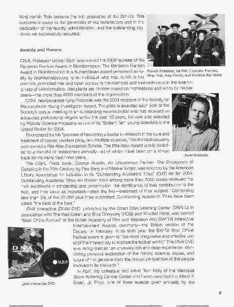
Niraj Harish Tolia became the first graduates of the School. Thisoutcome is owed to the generosity of our benefactors and to thededication of the faculty, administration, and the outstanding stu-dents we successfully recruited.
Awards and Honors
CSHL Professor Lincoln Stein was named the 2004 laureate of theBenjamin Franklin Award in Bioinformatics. The Benjamin FranklinAward in Bioinformatics is a humanitarian award presented annu- Patrick Paddison, Ira Hall, Elizabeth Thomas,
ally by Bioinformatics.org to an individual who has, in his or her Niraj To lia, Amy Gaudy, and Emiliano Rial Verde
practice, promoted free and open access to the methods and materials used in the scientif-ic field of bioinformatics. Recipients are chosen based on nominations and votes by his/herpeers-the more than 8000 members of the organization.
CSHL neuroscientist Karel Svoboda was the 2004 recipient of the Society forNeuroscience Young Investigator Award. The prize is awarded each year at theSociety's annual meeting to an outstanding neuroscientist who has received anadvanced professional degree within the past 10 years. He was also selectedby Popular Science magazine as one of its "Brilliant Ten" young scientists in theUnited States for 2004.
Recognized for his "promise of becoming a leader in research in the cure andtreatment of cancer, cerebral palsy, and multiple sclerosis," Senthil Muthuswamywas named a Rita Allen Foundation Scholar. The Rita Allen Award is only award-
a tenure
track for no more than three years.The CSHL Press book, George Beadle, An Uncommon Farmer: The Emergence of
Genetics in the 20th Century, by Paul Berg and Maxine Singer, was selected by the AmericanLibrary Association for inclusion in its "Outstanding Academic Titles" (OAT) list for 2004.Outstanding Academic Titles are chosen from among more than 7000 books reviewed "fortheir excellence in scholarship and presentation, the significance of their contribution to thefield, and their value as important-often the first-treatment of their subject." Comprisingless than 3% of the 23,000 plus titles submitted, Outstanding Academic Titles have beencalled "the best of the best."
DNA Interactive (DNAI) DVD, produced by the Dolan DNA Learning Center (DNALC) inassociation with The Red Green and Blue Company (RGB) and Windfall Films, was named"Best Offline Factual" at the British Academy of Film and Television Arts (BAFTA) Interactive
Entertainment Awards ceremony-the British version of theOscars-in February. In its sixth year, the BAFTA Best OfflineFactual award is given to "the most imaginative and effective useof offline interactivity to explore the factual world." The DNAi DVDwas recognized as "an unusually rich and deep experience, stim-ulating personal exploration of the history, science, issues, andfuture of the genome from the unique perspective of the peopleinvolved in its research."
In April, my colleague and friend Tom Kelly of the MemorialSloan-Kettering Cancer Center and I were awarded the Alfred P.Sloan, Jr. Prize, one of three awards given annually by the
Karel Svoboda
DNA Interactive DVD
9

General Motors Cancer Research Foundation (GMCRF). The Sloan Prize recognizes themost outstanding recent contribution in basic science related to cancer research.
The Sabin Vaccine Institute honored CSHL Chancellor James D. Watson, Ph.D., with theSabin Humanitarian Award at their Salute to Lifesaving Discoveries benefit dinner in May. Theawards program is a yearly tradition for the Institute and extols the contributions made byscientists, philanthropists, and humanitarians who share in some aspect of the goals ofadvancing vaccine science for the benefit of humanity.
The United States Rice Genome Consortia, Cooperative State Research, Education, andExtension Service (Tucson, Arizona)-of which Cold Spring Harbor Laboratory is a principalmember-were honored with the U.S. Department of Agriculture (USDA)'s Secretary'sAward, presented by Agriculture Secretary Ann M. Veneman at the 58th Annual Secretary'sHonor Awards Ceremony on June 25. Considered the highest award the USDA can bestow,the members of the United States Rice Genome Consortia were honored in the "EnhancingEconomic Opportunities for Agricultural Producers" category for leading the United Statespartnership in the multinational achievement to decode the rice genome to advance knowl-edge, improve nutrition, and alleviate world hunger. W. Richard McCombie, Melissa Kramer,Lance Palmer, Robert Martienssen, Maureen Bell, Sujit Dike, Lidia Nascimento, AndrewO'Shaughnessy, and Lori Spiegel are among the members of the CSHL staff involved in thisproject.
Development
Capital and Program Contributions
Private funding is essential to our research programs, enabling successful and innovativeprojects not yet eligible for public funding. For this reason, we are especially grateful to thosesupporters who made major gifts in 2004 to our cancer and neuroscience research pro-grams. We gratefully acknowledge donors of $100,000 or more to our cancer program: theDe Matteis Family Foundation, a first-time grant for colon cancer research; The MiracleFoundation; The Breast Cancer Research Foundation; and one anonymous donor.
Our neuroscience program was also generously supported, and we acknowledge donorsto that program, including Jo-Ellen and Ira Hazen, The Seraph Foundation, The DartFoundation, The G. Harold and Leila Y. Mathers Charitable Foundation, and the St. GilesFoundation. We also received a special gift this year from Trustee Jerome Swartz, and hisSwartz Foundation, which provided more than $215,000 for the establishment of the TheSwartz Center for Computational Neuroscience and additional research support in brainstructure and neuroscience research. The Swartz Center has become an integral part of ourstrong neurobiology program, supporting both research and neuroscience programs. TheThomas Hartman Foundation for Parkinson's Research also pledged $4.4 million over thenext 5 years to support Parkinson's research at CSHL and to establish The Thomas HartmanParkinson's Research Laboratory. We also received significant and continued support fromThe Simons Foundation, to fund autism research at CSHL.
Robertson Research Fund
The Robertson Research Fund has been the primary in-house support for our scientists formore than three decades. During 2004, Robertson Funds supported research in the labs ofJosh Dubnau, Masaaki Hamaguchi, Leemor Joshua-Tor, Alexei Koulakov, Adrian Krainer, Yuri
10

Lazebnik, Wolfgang Lukowitz, Bud Mishra, Partha Mitra, Scott Powers, Cordula Schulz, andMichael Wig ler.
Watson School of Biological Sciences
Now in its second phase of funding and led by Robert D. Lindsay, the Watson School hasreceived additional support in 2004 for the Dean's Chair, fellowships, and lectureships,enabling the Watson School to continue to grow and influence the field of biological sci-ences. We appreciate new gifts of $100,000 or more made this year by Mr. and Mrs. RobertD. Lindsay and Family, Curt Engelhorn, and The Seraph Foundation, as well as ongoing sup-port received from Bristol-Myers Squibb Company, Mr. and Mrs. Alan E. Goldberg, theFlorence Gould Foundation, and the Lita Annenberg Hazen Foundation.
The Dolan DNA Learning Center
Thanks to a very generous gift from The Dana Foundation, the Dolan DNALC has embarkedon creating Genes to Cognition (G2C) Online: A Network-driven Internet Site on ModernBrain Research, an Internet portal exploring the genes of cognition and learning. In addition,the Dolan DNALC received significant support from the Pfizer Foundation to continue itsPfizer Leadership Institute in Human and Genomic Biology.
Carnegie Building
We are continuing plans to renovate the existing Carnegie Library to enhance its ability toserve the Laboratory community. This project received significant support this year fromalumni Philip A. Sharp, Ph.D.; Drs. Joan Brooks and James Garrels; and Thomas P. Maniatis,Ph.D.; and from The Koshland Foundation and The Lehrman Institute.
Capital Campaign
As CSHL prepares to embark on a capital campaign to raise funding for new research space,we are very grateful to significant gifts received to support the buildings intended for thesouthwest corner of our campus. In 2004, we received pledges from Mrs. Leslie C. Quick,Jr., for a cancer research facility named for her husband, and a pledge from the Wendt FamilyCharitable Foundation of Community Foundation Sonoma County for a neuroscienceresearch facility. Mrs. William L. Matheson made a significant gift in 2003 for this project, toname a facility for her late husband. CSHL also received significant support for these pro-jects from Gillian and Eduardo Mestre and Mrs. George N. Lindsay. We will continue to plan
this campaign throughout 2005.
Additional Support
The Laboratory was fortunate to receive support for many ongoing projects in 2004. TheJoseph G. Goldring Foundation made a significant gift to support research, and the LouisMorin Charitable Trust made a significant contribution to support the work of Drs. LeemorJoshua-Tor, Josh Dubnau, and David Spector. We received support from the Estates ofElisabeth S. Livingston, Florence Strelkowski, and Adele C. Diaz to complete our housing
11

project at Uplands Farm, and Dr. and Mrs. Walter C. Meier, through the Banbury Fund, madea significant gift to restore and renovate Robertson House on our Banbury campus. Mr. andMrs. Charles E. Harris II made a gift this year to support capital projects, and the Roy J.Zuckerberg Family Foundation provided support for consultants in fund-raising and devel-opment. We are very grateful to these close supporters for their continued generosity.
Breast Cancer Groups
A crucial component to our breast cancer research program is the support we receive fromlocal grassroots breast cancer groups who provide direct research support for our program,in addition to a myriad of patient care and educational services to thousands of constituents.This year, we were fortunate to receive support from Long Islanders Against Breast Cancer(L.I.A.B.C.); 1 in 9: The Long Island Breast Cancer Action Coalition; Breast Cancer H.E.L.P.,Inc.; the New York State Grand Lodge Order Sons of Italy; EA.C.T. (Find a Cure Today); TheElisabeth McFarland Fund; The Long Island 2 Day Walk to Fight Breast Cancer; The JudiShesh Memorial Foundation; the Long Beach Breast Cancer Coalition; and The WALK forWomen Breast Cancer Fund. We also gratefully acknowledge continued support from TheBreast Cancer Research Foundation. The generous support we receive from these groups,year after year, is truly propelling our breast cancer research.
Benefit for the Brain
John Sebastian and the J-Band played to a packed Grace Auditorium on October 30, 2004at the first "Benefit for the Brain." The event realized a net profit of more than $225,000 for
John Sebastian
12
Kay Jamison signing a copy of Exuberance for Joan Spiro.

CSHL's Alzheimer's and Parkinson's research, thanks to the efforts of William S. Robertsonand the Banbury Fund, who underwrote the event, and event co-chairs Edward Travagliantiand Kathy Di Maio and their committee. The event honored Monsignor Thomas J. Hartman,founder of The Thomas Hartman Foundation for Parkinson's Research.
Exuberance: The Passion for Life
On December 5, Dr. Kay Redfield Jamison, best-selling author and internationally renownedauthority on mood disorders, discussed the feeling of exuberance and how it fuels our mostimportant creative and scientific achievements at the Dolan DNALC. Sponsored by Arthurand Joan Spiro, this event benefited the educational opportunities hosted annually at theDNALC.
Library and Archives
The Library and Archives enjoyed a very exciting year in 2004. A major goal of the Library isto provide scientists with digital access to all publications vital to their research. In responseto the expanding scope of science research performed at the Laboratory, the Library sub-scribed to many new journal titles this year. In addition, the Library purchased an electronicbook collection composed of 134 e-books. In 2004, an SFX system was installed that elec-tronically interconnects the Library's resources so that patrons can navigate the entire Librarycollection through one interface. Within the past year, the Interlibrary Loan office implement-ed Ariel, an advanced electronic delivery service that allows the Library to receive requestedarticles as PDFs via e-mail.
The Library's Advisory Committee, composed of professors, postdocs, and WatsonSchool students, convened this year to discuss library services and resources. In 2004, theLibrary collaborated on particular projects with other medical and academic libraries at TheRockefeller University, Harvard University, and the Marine Biological Laboratory at WoodsHole. The Library, represented by director Mila Pollock, was also invited to the Annual NaturePublishing Group International Library Committee to take part in discussions of interest to
today's library market.In 2004, the Archives were fortunate to acquire the personal collection of Elof Carlson, a
prominent geneticist and professor at Stony Brook University. The Oral History Office hasalso continued to expand, now comprising more than 100 scientific interviews, half of whichare already accessible online. In 2004, the Sloan Foundation formally recognized the digitalarchives project, the Memory Board, an online forum in which the lab community documentsthe history of the Laboratory by contributing first-hand accounts, reminiscences, originalmaterials, photos, and video clips. The Sloan Foundation will support the redesign of theMemory Board and advocate its use as a model that other institutions could adapt for doc-umenting their own history. Conceived by librarian and archivist Mila Pollock, the MemoryBoard has attracted much attention and many interesting anecdotes. The exhibition BuildingBlocks of CSHL was created this year and will be on display at Blackford Hall for two yearsand online in digital format. This project explores the history of the buildings at CSHL (andthe life of its community) through documents, photos, and personal memories. This year, theLibrary's exhibit Honest Jim: Watson the Writer moved to the Charite Universitatsmedizin inBerlin, Germany and will be exhibited in Moscow, Russia next year.
13

Building Projects
The Laboratory continues to improve and expand its facilities and, during 2004, undertookseveral construction and renovation projects. Several projects were completed in theDemerec building, including alteration and renovation of laboratory spaces and the con-struction of a new tissue culture laboratory. Grace Auditorium received attention as well, witha complete remodeling of the bookstore to accommodate DNA Stuff -the Laboratory'sbookstore and nonprofit retail operation. The Laboratory also continued its project to replacethe HVAC systems in Grace and Harris and to update the emergency power system that
supplies those buildings.A major renovation of the James building was begun in 2004, updating laboratory, sup-
port, and office spaces to meet the increased demands on the building's facilities the
Laboratory has experienced in recent years. Work was begun on the Banbury pool, whichhad been leaking badly for years. The entire pool has been reconstructed, and final work is
to be completed by the spring of 2005.The Laboratory has continued to improve its student and scientist housing program. The
housing project at Uplands Farm is nearing completion and will provide much-needed hous-
ing close to the main campus. The last of the apartments in the Hooper building was reno-vated, and the Weghorn House, which lies at the northern tip of the campus, was purchased
and will be renovated in 2005.A major milestone in 2004 was the approval of the Laboratory's updated Master Plan and
its Upper Campus project by the Village of Laurel Hollow. This approval came at the end of
a 2-year process and has paved the way for the Laboratory to break ground on the largest
construction project it has ever undertaken in Spring 2005.
100 Years of Genetics at Cold Spring Harbor
In 1904, the Carnegie Institution of Washington founded a Station for Experimental Evolution
at Cold Spring Harbor, launching a century of genetics that placed CSHL at the forefront of
biomedical research. The occasion was marked by a very special 2004 Cultural Series filled
with engaging lectures and inspiring music and art.
Public Lectures
April 19 Mark Hallett, Vice President of the American Academy of Neurology: HowYour Brain Recovers from a Stroke.
May 11 Vincent Li, Director of the Angiogenesis Foundation: Prevention andReversal of Skin Cancer and Other Skin Diseases.
May 18 Carter Burwell, award-winning composer: From Cold Spring Harbor to the
Coen Brothers: A Composer's Journey.
May 24 Andrew Solomon, award-winning author of The Noonday Demon: An Atlasof Depression: Depression Too, Is a Thing with Feathers.
Sept. 28 Zach Mainen, CSHL Associate Professor, and Sharron McCarthy, Directorof the Society of Wine Educators: Demystifying the Sommelier: The Art andScience of Wine Tasting.
14

Sylvia Nasar
Antoine Tamestit
October 5 Sylvia Nasar, author of A Beautiful Mind: Genius, Madness, Recovery.
December 7 Richard Stone, Britain's youngest royal portrait artist: Painting England'sQueen and DNA's Dean.
Concerts
April 24May 1May 8May 22September 11October 2October 9October 16
Exhibits
The Molinaro-Levy Project, piano and harmonicaYunjie Chen, pianoMikhail Simonyan and Alexei Podkorytov, violin and pianoHsing-ay Hsu, pianoDmitri Berlinksky and Elena Baksht, violin and pianoVassilis Varvaresos, pianoAlexandre Bouzlov, celloAntoine Tamestit, viola
From July 9 to August 1, 2003-2004 Artist-in-Residence Eduardo De Soignie displayed hiswork, Paintings From Another Domain, in Bush Lecture Hall. Inspired by his Cuban roots, DeSoignie's work is influenced by artists such as Jose Bedia and Tomas Esson.
Events
Gavin Borden Visiting Fellows
The 10th annual Gavin Borden Fellow Lecture, created by Jim Watson in memory of GavinBorden, publisher of Molecular Biology of the Cell, was held on March 17 in Grace
15

Richard Losick and Bruce Stillman at the Gavin Borden Lecture
Auditorium. Dr. Richard Losick, the Maria Moors Cabot Professor of Biology at HarvardUniversity, spoke on "Cell fate, polarity, and cannibalism in bacteria." Dr. Losick is interna-tionally acclaimed for his research on microbial development and is developing computer-based animations and video for teaching introductory molecular biology.
Symposium
The 69th Annual Cold Spring Harbor Laboratory Symposium, "Epigenetics," was oversub-scribed again this year with nearly 500 scientists from around the world in attendance. TheDorcas Cummings Lecture, in memory of long-time Laboratory friend and former Director ofthe Long Island Biological Association Dorcas Cummings, was given by David Haig ofHarvard University on the subject of "The Divided Self-Brains, Brawns, and the Superego."This endowed lecture is traditionally open to friends and neighbors of the Laboratory and isfollowed by dinners generously hosted in the homes of our neighbors for our visiting scien-tists and faculty and staff.
Other Lectures
Winship Herr ("Why Our Cells Multiply") and Nick Tonks ("Plague, Pox, and Phosphatases")participated in our lecture series for fourth- to sixth-grade students and their parents, co-hosted with the Cold Spring Harbor School District, at the Dolan DNALC.
Huntington Hospital continued to host their fall/spring lecture series on cardiovascularhealth and related diseases in Grace Auditorium.
16

Laboratory Employees
New Staff
Scott Powers returned to CSHL in October as Associate Professor and Director of theHuman Cancer Genome Center in the Cancer Genome Research Center. Scott came fromTularik Genomics Division, which was initially a company spun out from the Laboratory to useRDA (representational difference analysis) technology to identify cancer genes. Scottreceived his graduate degree from Columbia University and performed seminal work as apostdoc with Mike Wig ler on the study of RAS genes in yeast.
David Wu joined Scott as a Research Investigator at the Human Cancer Genome Center.David received his graduate degree from Berkeley and as a postdoc with Aziz Sancar devel-oped the first biochemical system for mammalian DNA repair with purified proteins.
Promotions
Dr. Lilian Clark Gann became Dean of the Watson School of Biological Sciences on July 1.Lilian joined the Laboratory in March 1999 as Assistant Dean of the Watson School and waspromoted to Associate Dean in January 2002. She received her Ph.D. from the University ofSt. Andrews, Scotland in 1988, for her studies on DNA-protein interactions in transcription-al control elements of DNA tumor viruses, and her M.B.A. from the University of Westminster,London in 1996. Since her arrival at CSHL, Lilian had played a crucial role in the develop-ment of the School's innovative Ph.D. program, while also enhancing the educational andtraining environment of CSHL for students and postdoctoral fellows in general. Her uniquebackground in science, education, and business made her an obvious choice to develop andoversee the programs of the Watson School. She was joined by Dr. Bill Tansey who wasappointed as director of graduate studies in the Watson School of Biological Sciences andLita Annenberg Hazen Professor of Biological Sciences on the same day.
A number of other faculty members were promoted in 2004, including Dmitri Chklovskii toAssociate Professor, Zachary Mainen to Associate Professor, Ravi Sachidanandam to SeniorComputer Scientist, Lincoln Stein to Professor, Karel Svoboda to Professor, Yi Zhong toProfessor, and Anthony Zador to Associate Professor. Ira Hall and Patrick Paddison wereboth named CSH Fellows.
Departures
Dr. Winship Herr stepped down as Dean of the Watson School of Biological Sciences effec-tive July 1 to concentrate his efforts on his research. Beginning in 1995, Winship spear-headed the effort that resulted, in September 1998, in the Laboratory's accreditation as aPh.D. degree-granting institution by the Board of Regents of the University of the State ofNew York, on behalf of the State Education Department. This enabled the establishment ofthe Watson School of Biological Sciences. Soon thereafter, Winship became the Foundingdean of the Watson School. During the last 5 years, we have seen the School grow andbecome one of the most innovative programs in the country, attracting outstanding students.Winship has nurtured and shaped the Watson School in its first formative years, culminatingin our first commencement convocation this spring. In the coming year, he and NouriaHernandez will move to Nouria's native Switzerland to become professors at a new institutein Lausanne. I thank Winship for his remarkable effort in establishing a graduate school atCold Spring Harbor and for his sound advice to me. The establishment of a graduate school
17

has transformed Cold Spring Harbor Laboratory and will have a long-lasting effect on theintellectual environment here.
Nei lay Dedhia, Research Investigator; David Helfman, Professor; Terence Strick, CSHFellow; and Jerry Yin, Associate Professor, all departed the Laboratory in 2004. David spear-headed the understanding of cancer progression through his research on the cell biology ofcell architecture and I wish him the best as he continues his research at the University ofMiami Cancer Center.
Long-term Service
The following employees celebrated milestone anniversaries in 2004:
30 years
25 years
Lane Smith
Maureen Berejka, Judith Cuddihy, Katya Davey, James Hope, John Meyer,James Parsons, Susan Schultz, Bruce Stillman
20 years Carmelita Bautista, Dessie Carter, Robert Gensel,Mary Ellen Goldstein, Daniel Miller, Robert Pace, Steven Tang
15 years Leslie Allen, James Bense, Sharon Bense,Charlene De Poto, Janice Douglas, Jan Eisenman,Helena Johnson, Robert Martienssen, Jacqueline Matura, Alison McDermott,Eleanor Sidorenko, Halina Swidzinski, Ryszard Swidzinski, Spencer Teplin
1st row, left to right: Eleanor Sidorenko, James Bense, Sharon Bense, Charlene De Poto,Susan Schultz, Carmelita Bautista, Jan Eisenman, Katya Davey2nd row, left to right: Rob Gensel, James Parsons, Mary Ellen Goldstein, Alison McDermott, Maureen Berejka,Daniel Miller3rd row, left to right: John Meyer, James Watson, Spencer Teplin, Helena Johnson, Dessie Carter4th row, left to right: Bruce Stillman, James Hope, Steve Tang
18

CSHL team at the Long Island Walk to D'Feet ALS.
Community Outreach
In May, Laboratory employees Sandy Neuwald, Lisa Manche, Carla Margulies, Barbara Misk,Kathryn Borowski, Barbara Purcell, and Theresa Saia served dinner at the Ronald McDonaldHouse in nearby New Hyde Park, which provides temporary housing to families of seriouslyill children at the Schneider Children's Hospital. In June, Shuly Avraham, Jason Evans, KimGronachan, Jo Leonardo, Jianli Li, Erin Maroney, Liya Ren, Theresa Saia, and Doreen Wareplanted six flats of flowers at Haven House, a homeless shelter in Huntington Station, pur-chased with money donated by CSHL employees. A CSHL Team also participated in theLong Island Walk to D'Feet ALS (Lou Gehrig's disease) for the The ALS Association GreaterNew York Chapter at Eisenhower Park in East Meadow on September 26.
Cold Spring Harbor Laboratory is a vibrant and dynamic institution, and like all such insti-tutions involved in the increasingly expensive research in the genomic age, we are even moredependent on support from individuals and foundations to enable us to remain one of theleading research centers in the world. As we move forward, we need the type of support thatcan be used to ensure that our scientists have the facilities and resources to achieve theirgoals. Increasingly at Cold Spring Harbor, our research focuses on how to use all the accu-mulated knowledge to develop new ideas for diagnosing and treating human disease. I thankall those who have supported our efforts to date, helping us to make Cold Spring HarborLaboratory a truly unique education and research center.
Bruce W. Stillman, Ph.D., F.R.S.President
19


CHIEF OPERATINGOFFICER'S REPORT
There are many positives to report about the Laboratory's performance in 2004. The researchand academic programs continued to flourish. The endowment funds enjoyed another out-standing year of investment returns and appreciation. Operating results exceeded forecastsand the budget was balanced after depreciation expense. The latter, however, was accom-plished only with rigorous management of administrative expenses and some extraordinaryfactors.
Drs. Watson and Stillman have been heard to say, over the last year, that the research pro-gram at the Laboratory is more exciting now than it has ever been in their experience. Havingnow worked at Cold Spring Harbor Laboratory for 38 and 26 years, respectively, their obser-vations carry considerable weight. Powerful technologies and techniques, developed here atCold Spring Harbor, allow for rapid identification of genes associated with various cancers aswell as psychiatric diseases such as autism, depression, and schizophrenia. The promise ofthe "DNA Biopsy" for tailoring therapy to an individual's specific genetic alteration is extraordi-nary and suddenly attainable.
Our academic programs remain strong and continue to expand. Cold Spring HarborLaboratory Meetings and Courses are the most sought after in the world of biology; theyattracted a record number of attendees in 2004. The Cold Spring Harbor Laboratory Press,which continues to publish high-quality scientific books and journals, had an excellent year aswell. The Dolan DNA Learning Center is fulfilling its mission of providing unique educationalexperiences for middle and high school students via hands-on laboratory and Internet instruc-tion. The Banbury Center continues to attract scholars from around the world to its special-ized scientific conferences. The innovative Watson School of Biological Sciences celebratedits first commencement in 2004, awarding the degree of Ph.D. to six outstanding students, allof whom completed their degrees in the requisite four years. Honorary degrees were award-ed to former Laboratory Trustees Joan Steitz (Professor of Molecular Biophysics andBiochemistry at Yale), Shirley Tilghman (President of Princeton), and, most fittingly, to JamesD. Watson.
Forecasting the Laboratory's operating revenue is a challenging task due to the variednature of the revenue streams and shifts in timing that are difficult to anticipate. Consequently,a relatively conservative approach is taken in the budgeting process as we project incomefrom grants, meetings, book sales and advertising, unrestricted fund-raising, and technologylicensing activities. We anticipated another difficult year in 2004 and forecasted a deficit fromoperations of $775,000 after $5.7 million in depreciation expense. Fortunately, results werebetter than expected, and a balanced budget was achieved for the year. Operating and pro-gram income rose to $95.4 million, an 8% increase over 2003. Indirect cost revenue on grantswas ahead of forecast and the Cold Spring Harbor Laboratory Press exceeded expectationson stronger book and journal sales combined with printing cost reductions. It should be point-ed out, however, that one accounting change involving the allocation and timing of certainexpense items had the extraordinary effect of improving the reported bottom line results forthe year by approximately $200,000.
21

Full-time a. Part-time Technical Support, Core Services, Publications, Meetings, Library, Public Affairs,Buildings a. Grounds, Administrative, Personnel, Banbury Center, .d Dolan DNA Learning Center
1000
900
800
700
600
0 500
400
300
r200
StaffMA ScientistsE=1 Support-4,- Total
Operating Expenses100,000,000
90,000,000 -
80,000,000 -
70,000,000-
Oro6 ,000,000
0 50,000,000 -
40,000,000 -
30,000,000 -
20,000,000 -
10,000,000
22
I I Actual Expense
Expense Adjusted for Inflation
189 90 91 92 93 94 95 96 97 98 99 00 01
Years
87 88
8000
7000
6000
5000
tr4000
3000
2000
1000
, 64 3, 96 33 08
Yea s
J
04
Meetings & Course Participants
85 86 87 88 89 90 92 93 94 95 96
Years97 98 99 00 01 02 03 04

It is certainly satisfying to be able to report a balanced budget, particularly after experienc-ing substantial operating deficits in the two prior years. This, however, was not easily achievedand required aggressive management of expenses, including caps and deferrals of adminis-trative salary increases. Although such steps were necessary and prudent, they present veryreal difficulties for hundreds of loyal employees whose lives are very much affected by risingfuel prices, property taxes, and other cost of living pressures that outpace their compensationincreases.
Investment returns on our endowment funds were exceptionally strong for the second con-secutive year. The total managed portfolio returned 12.1% for the calendar year versus ourbenchmark index of 8.2%. Our total equity portfolio was up 15.7%, considerably ahead of the10.9% appreciation in the Standard & Poor's. The fixed-income portfolio also outperformedthe indexes, returning 5.1% for the year. At year-end, the market value of the endowmentfunds totaled $235 million after following our long-held policy of spending no more than 4%annually. The Investment Committee has functioned well and has worked throughout the yearwith our advisor, Merrill Lynch Consulting Group, to review and refine the asset allocationmodel and strategy. To diversify and lessen correlation in the equity portfolio, three new man-agers were added-two in international equities and one in the alternative asset/hedge fundcategory. We express our gratitude to Lola Grace for her years of hard work asSecretary/Treasurer of the Board and Chair of this committee. At year-end, she passed thebaton to Ed Travaglianti, who has assumed these important roles with equal wisdom andenthusiasm.
One of the reasons for the ongoing success and vitality of the Laboratory is the wonderfulsupport it receives from the local community. The Cold Spring Harbor Laboratory Associationhas a long history of volunteer outreach and involvement with the Lab. Its directors work hardto communicate the Laboratory's mission, welcome scientists into the local community, orga-nize events, and raise unrestricted funds to support critical research. Trudy Calabrese hascompleted a particularly successful term as president of this important organization. Under herinspired leadership, we enjoyed another year of wonderful concerts, dinners, and lectures and,at the same time, surpassed the $1 million threshold with our Annual Fund for the second con-secutive year. We thank Trudy for her dedication to the Laboratory, and we welcome her suc-cessor, Joe Donohue.
A great deal of progress was made over the course of the year toward the Laboratory'splans to construct a cluster of new research buildings on the main campus. Chief FacilitiesOfficer Art Brings worked very effectively with the diligent, but cooperative, Village of LaurelHollow Board of Trustees to obtain approval for the project. It was a complex process involv-ing the preparation of an amazingly detailed environmental impact statement that requiredreview by numerous local and state agencies. By year-end, the necessary approvals were inplace and a building permit should be forthcoming early in 2005. Ground breaking on the siteis planned for the spring.
Clearly, critical expansion plans cannot be realized without major private financial support.Last year, we acknowledged the generous pledge from long-time friend Mardi Matheson forfunds to construct one of the cancer research buildings. Her generosity, along with a $20 mil-lion pledge from the State of New York, allowed us to confidently move forward with the pro-ject. In 2004, we received two additional major gifts toward the initiative, also from long-timefriends of Cold Spring Harbor. Jean Quick, wife of late CSHL Trustee Leslie Quick, pledged$10 million for the second cancer research building. Ongoing support and involvement fromfamilies like the Quicks and the Mathesons is immensely gratifying. Mrs. Quick's gift was fol-
23

lowed, at the end of the year, by another $10 million pledge from former CSHL Trustee andpharmaceutical industry executive Henry Wendt and his wife Holly. In addition, Trustees BobLindsay and Howard Solomon made seven-figure contributions to the effort. We express ourtremendous gratitude to these individuals for their generosity and commitment to the institu-tion and its future.
The Laboratory could not function effectively without a strong Board of Trustees. Five val-ued members of this body-Charles Harris; Susan Hockfield, Ph.D.; Charles J. Sherr, M.D.,Ph.D.; Howard Solomon; and William Miller-completed their terms at year-end. We are trulygrateful for their years of service, advice, and dedication. We express particular gratitude toBill Miller who served on the board for 15 years, the last six as chairman. His devotion of time,energy, wisdom, and resources to Cold Spring Harbor Laboratory has been of immeasurablevalue to the institution. We are pleased that he was immediately elected as an HonoraryTrustee and welcome his continued involvement. We also welcome and look forward to work-ing with newly elected Trustees Laurence Abbott, Ph.D.; Robert Wittes, Ph.D.; Laurie Landau,V.M.D.; Nancy Marks; and Jerome Swartz, Ph.D. It is comforting to see smooth and seamlessleadership transition on the board. We thank Eduardo Mestre for assuming chairmanship ofthe Board and look forward to his leadership.
It is exciting to embark on a new stage in the growth of Cold Spring Harbor Laboratory-new directions in promising areas of research, expanding facilities, and a growing studentbody. There is much to look forward to and much planning to be done, but there are also sig-nificant challenges to be faced. Our funding requirements for capital projects, endowment,and research are substantial. We are, for the time being, enjoying relatively robust financialmarkets and economic conditions that create a positive environment for fund-raising. On theother hand, the federal budget deficit is necessitating a belt-tightening that will have a majorimpact on National Institutes of Health (NIH) funding for biomedical research. After years ofdouble-digit increases in the pool of research support, the NIH budget will now contract in realterms over the next several years. Fewer federal grants-the lifeblood of research institu-tions-will be awarded. At the same time, the cost of doing good science is increasing dra-matically. This set of circumstances will require us to do a number of things extremely well. Wemust continue to attract the best scientists to Cold Spring Harbor Laboratory and maintain anenvironment where they can do exceptional work. At the same time, we must aggressivelypursue private sources of funding for research in addition to attracting significant federal sup-port.
Success in these areas is essential not only to the realization of the Laboratory's scientificmission, but also to the well being of our dedicated staff. Our administrative staff is lean andfaces increasing demands and pressures as the scope of activity expands. The institution can-not function without this talented team of people and we must reward them appropriately. Thiswill be accomplished only if we are successful in attracting new sources of funding. We areconfident it can be done but know that it will be hard work.
Dill AyresChief Operating Officer
24

4;

See previous page for photos of the following scientific staff.
Row 1: Izolda Mileva; Galen Collins; Ileng Ramachandran, Prakash Kumar;Maria Simone Temporal; Sabrina Boettcher, Michael Golding
Row 2: Albert Mel lick; Rotem Karni; Shih-Chieh (Jack) Lin; Nishi Sinha;Hans Guido Wendel; Rebecca Ewald, Jennifer Bestman
Row 3: Chih-Chi Yuan; Xiaoyun Wu; Shweta Tyagi; ; Catherine CormierMiguel Zaratiegui, Derek Goto; Keisha John; ; Supriya Gangadharan;Hao-Chi Hsu
Row 4: Marissa Nolan; Farida Emran; Fuqiang Geng; Fabio Nogueira; JuanEncinas; Stephen Schuck
Row 5: Elvin Garcia; Christopher Johns; Theresa Zutavern;Vincenzo De Paola
Row 6: Allen Ludwig; David Mu; Alexandra Lucs; Peter O'Brien;Daniel Nolan; Jennifer Meth
Row 7: Amitabh Mohanty; Evan Santo; Jason Williams; Maria Yuneva;Vanisree Velamoor; Pranav Sharma

CANCER: GENE EXPRESSION
Discoveries by Leemor Joshua-Tor and Greg Hannon made a significant advancement toward a compre-hensive understanding of one of the most intriguing biological phenomena to be uncovered in recentyears: RNA interference (RNAi). Through X-ray crystallography, Joshua-Tor and her colleagues have deter-mined the atomic, three-dimensional structure of an RNAi component called Argonaute. When the 3-Dstructure of Argonaute emerged from their data and was compared to other proteins with known struc-tures and functions, Joshua-Tor's group found that part of Argonaute was the spitting image of the"RNAse H" family of proteins, whose members were known to cut RNA. In collaboration with GregHannon's group, a final experiment based on the Argonaute structure clinched the case for Argonaute2as being the protein that provides the Slicer activity of RNAi in mammals. Other information led Joshua-Tor's group to propose a model that explains precisely how Argonaute binds to and cuts messenger RNAduring RNAi.
Winship Herr uses the herpes simplex virus to study a human protein called host-cell factor 1 (HCF-1).HCF-1 was first identified as a key factor in herpesvirus replication. However, Herr recently showed thatHCF-1 is involved in other steps of the cell division process: Without HCF-1, human cells often do notcomplete the division process, and even if they do, the daughter cells frequently receive unequal numbersof each chromosome. This and other findings indicate that the HCF-1 protein is required for the efficientalignment and sorting of chromosomes into daughter cells during cell division.
Jacek Skowronski studies how an AIDS virus protein called Nef commandeers the protein sorting andsignaling machineries of human T cells. He has recently identified several T-cell proteins that associate with
Nef and is exploring how these protein-protein interactions contribute to viral pathology. Skowronski'swork has revealed a potentially effective strategy for developing improved anti-HIV therapies.
Even the relatively well-understood pathways of messenger RNA biosynthesis continue to surprise sci-entists with new twists. Nouria Hernandez has discovered two new components of the "RNA polymeraseIII" transcription machinery by biochemically reconstituting the process with purified proteins. Her group'swork led to the definition of a set of minimum factors required for transcription and revealed a surprisingrole for a protein called actin in the process. The role of actin as a component of the cell's structural scaf-folding (the cytoskeleton) has long been known, making its emergence as a factor required for transcrip-tion by RNA polymerase III an intriguing discovery. Hernandez's group also discovered an unexpected rolefor an enzyme called casein kinase II in RNA polymerase-Ill-mediated transcription.
In addition to his studies of proteins involved in messenger RNA processing, Rui-Ming Xu investigatesthe shapes and functions of proteins that are important for the assembly and remodeling of "higher-orderchromatin structure" or structures that result from the wrapping of DNA into specifically coiled architec-tures. Xu's group has recently used X-ray crystallography to determine the three-dimensional shapes oftwo proteins (Orc1-Sir1 and Sir3) that help determine chromatin structure and thereby have significantimpacts on gene expression and other DNA-related processes.
Adrian Krainer's group explores how identical pre-messenger RNA molecules can be alternativelyspliced to ultimately produce different proteins. Part of Krainer's work involves the study of a family of pre-mRNA splicing factors called SR proteins, which bind to specific sites on pre-mRNA and recruit the splic-ing machinery to those sites. Krainer and his colleagues recently discovered a role for a particular SR pro-tein in mRNA "surveillance." They found that high levels of the SR protein promote the destruction ofdefective messenger RNAs, thus preventing the production of defective proteins. In other work, they char-acterized a novel kind of splicing mutation in the LKB1 tumor suppressor gene that causes Peutz-Jeghers
syndrome and susceptibility to cancer.William Tansey studies a potent growth control protein called Myc. When hyperactive and/or present in
excess of its normal levels, Myc contributes to cancer. Tansey and his colleagues have recently focusedon a little-studied subdomain of Myc called Myc box III (MbIII). They found that Mblll is important for induc-ing tumor formation in a mouse model of B-cell lymphoma. Moreover, they discovered that through Mblll,
27

the Myc protein can impede a natural anticancer function of cells called programmed cell death. Thesefindings establish that the oncogenic capacity of Myc is directly linked to its ability to block programmed
cell death.Michael Myers is taking the accumulated understanding of genes and proteins to the next level by iden-
tifying how proteins interact with one another to form the complex, dynamic networks that ultimately deter-mine cell biology. His group is determining the global architecture of protein interaction networks andrevealing why these networks are robust (error tolerant) and adaptable. Myers is also investigating howprotein interaction networks are altered in cancer and what key molecular "nodes" of the networks might
be the best targets for cancer therapy.
Kenneth Chang, research investigator, Greg Hannon's lab.
28

MECHANISMS OF TRANSCRIPTION
N. Hernandez F. Emran K. Samudre Y. SunP. Hu A. Saxena S. WuB. Ma M. Shanmugan C.-C. YuanT. Oztaskin
RNA polymerase III (pol III) synthesizes a collectionof essential RNA molecules that are themselves com-ponents of the protein synthetic and RNA processingmachineries, such as the ribosomal 5S RNA genes,tRNA genes, and other genes including the U6 smallnuclear RNA (snRNA) gene. Because most pol IIItranscripts are both abundant and stable, pol III tran-scription is highly regulated with cell growth and pro-liferation. In resting cells, only the small number ofpol III transcripts that decay over time must bereplaced, and pol III activity is low. In contrast, inrapidly dividing cells, an entire complement of theabundant 5S, tRNA, and other molecules must be syn-thesized in just one generation time, and pol III tran-scription is highly active. Indeed, pol III transcriptionis invariably up-regulated in malignant cells.
pol III recognizes three main classes of promoterstructures. Classes I and II are present in the 5S RNAand tRNA genes, respectively, and are gene-internal.Class III is present in the U6 snRNA gene and othergenes and is gene-external. The class III core promot-ers consist of two essential elements, a proximalsequence element (PSE) and a TATA box. Until recent-ly, the factors required for pol III transcription fromthese various types of promoters were poorly definedin higher eukaryotic cells. However, in the previousyear, we developed a well-defined in vitro transcriptionsystem for transcription of the type-3 human U6 pro-moter. The system consists of recombinant snRNA-activating protein complex (SNAP), a PSE-bindingfactor composed of five types of subunits, which weexpress in insect cells with the baculovirus system;recombinant Brf2-TFIIIB, a TATA-box-binding activi-ty composed of the TATA-box-binding proteinTBP, theTFIIB-related factor Brf2, and the SANT domain pro-tein Bdp 1 , all expressed in Escherichia coli; and ahighly purified pol III complex, which we isolatedfrom HeLa cells expressing a doubly tagged pol IIIsubunit. When combined, these factors are sufficient todirect several rounds of correctly initiated and termi-nated transcription from the U6 promoter. The onlynonrecombinant factor in the system is the pol III com-
plex, and so we have focused on the characterization ofthe factors present in this complex besides pol III sub-units that might contribute to transcription activity.
Besides pol III subunits, the pol III complex wepurified contained spectrin, a-myosin, clathrin, a-actinin 4, HSC70, P-tubulin, I3-actin, and calmodulin,as detected by mass spectrometry analysis. On theother hand, a number of other proteins such as topoi-somerase 1, PC4, NF-1, the La polypeptide, and CK2have been implicated in pol III transcription by otherinvestigators. To determine the role, if any, of suchproteins in pol III transcription, we subjected the polIII complex to one additional purification step over a"mini Q" column. We found that (3- actin, P-tubulin,and calmodulin continued to copurify with pol III. Ofthe factors implicated in pol III transcription by others,we could detect CK2 in the pol III complex. We thenset out to determine whether these two factors mighthave a role in pol III transcription.
CK2 is a nuclear and cytoplasmic protein kinasepresent in all cell types, composed of two regulatory ((3)and two catalytic subunits, of which two isoforms thatare 85% identical exist in human cells (a and a'). CK2has been implicated in pol III transcription in both yeastand human cells, where it has been reported to have apositive effect on transcription. We found that CK2 isassociated not only with pol III, but also with the U6promoter in vivo as shown by chromatin immunopre-cipitations. We observed that CK2 activity was requiredfor in vitro transcription and that it was required priorto transcription initiation. This last observation, cou-pled with our ability to reconstitute transcription fromwell-defined, separate components, allowed us to askwhich factor(s) needed to be phosphorylated by CK2for active transcription. Our results revealed that it wasthe pol III complex itself that needed to be phosphory-lated to be active. Since, in our experiment, CK2 couldbe active only before the start of transcription, andsince once phosphorylated, pol III could direct severalrounds of transcription, the phosphorylation eventappeared to be a switch that somehow converted pol III-from an inactive to an active state.
29

In its function of pol III phosphorylation, CK2 hasa positive effect on transcription. In addition, howev-er, we also found that CK2 phosphorylation of theBrf2-TFIIIB components inhibited pol III transcrip-tion, even if pol III itself was phosphorylated. We thenset up to identify which of the three Brf2-TFIIIB com-ponents-TBP, Brf2, or Bdpl -was the target of thisnegative regulation. For this purpose, we simply treat-ed each component separately either with CK2 fol-lowed by inhibitor, which allowed phosphorylation, orwith an inhibitor followed by CK, which preventedphosphorylation, thus keeping all of the reagents inthe reactions constant. In addition, we treated pol IIIwith CK2 followed by inhibitor, since we knew CK2phosphorylation of pol III is required for transcrip-tion, and SNAP, with inhibitor followed by CK2,since we knew that SNAP, did not need to be phos-phorylated by CK2 in vitro for activity. The factorswere then used for reconstitution of U6 transcriptionin vitro. The results were clear-cut and showed thatCK2 treatment of Bdpl, but not Brf2 or TBP, inhibit-ed U6 transcription. We then showed that Bdpl isindeed phosphorylated by CK2, and we mapped thephosphorylation sites to one or several of five serinesand threonines.
We then asked what the in vivo relevance of thisnegative regulation might be. We found that Bdplbecomes highly phosphorylated during mitosis.Extracts from mitotic cells were transcriptionallyinactive and could be reactivated by addition of a CK2inhibitor and recombinant, unphosphorylated Bdpl.By fluorescent microscopy as well as through bio-chemical fractionation, we determined that duringmitosis, Bdpl dissociates from the chromatin, and thisphenomenon was inhibited by treatment of the cellswith the CK2 inhibitor 4,5,6,7-tetrabromobenzotria-zole (TBB). One model that incorporates these obser-vations, shown in Figure 1, proposes that pol IIIrepression during mitosis occurs in part through CK2phosphorylation of Bdpl, which perhaps results inBdp1 dissociation from the chromatin.
The observation that inhibition of CK2 in mitoticextracts together with addition of Bdpl restored tran-scription was striking, because we had observed pre-viously that in extracts from actively growing cells,addition of a CK2 inhibitor repressed transcription.Indeed, we then showed that in extracts from S-phasecells, which are very active for pol III transcription,addition of a CK2 inhibitor also repressed transcrip-tion. This suggests a model in which CK2 has differ-ent roles at different stages of the cell cycle, as illus-
30
trated in the figure. In M phase, it represses transcrip-tion by phosphorylating Bdpl, whereas in phases ofthe cell cycle when pol III transcription is active, forexample, S phase, it activates transcription by phos-phorylating an unknown target. Since CK2 phospho-rylation of the pol III complex is required for activetranscription, this unknown target may well be the polIII complex itself.
As mentioned above, another protein that wefound to be associated with pol III was 0-actin. I3-actinhas been extensively studied as a cytoplasmiccytoskeletal protein that has roles in intracellular traf-ficking as well as in driving changes in cellular shapeassociated with cell motility, cytokinesis, endocytosis,and cell adhesion. 0-actin is, however, also a nuclearprotein, and in this cellular compartment, it is thoughtto have roles in the constitution of the nucleoskeleton,in RNA processing and export, and in transcription.So, when we found 0-actin tightly associated with polIII, we asked whether it might also be present on theU6 promoter in vivo. Indeed, we could localize I3-actinon this promoter by chromatin immunoprecipitations.To determine the role of I3-actin in pol III transcrip-tion, we then set out to identify conditions underwhich 13-actin might dissociate from the rest of theenzyme. One of the conditions we tested was treat-ment of cells with the genotoxic agent methanemethylsulfonate (MMS), a treatment known to inhibitpol III transcription. We found that in IMR90 cells(but not in HeLa cells), such a treatment resulted indissociation of I3-actin from the enzyme, and thisenzyme missing 0-actin was inactive in the reconsti-tuted U6 transcription assay. To regain activity, we
FIGURE 1 CK2: A molecular switch for the regulation of U6 polIII transcription.

then needed not only to treat pol III with CK2, butalso to add recombinant 13-actin. These results estab-lished a direct role for 3 -actin in pol III transcription.Remarkably, concomitant with our results, two reportsappeared showing that 0-actin is required for pol I andpol II transcriptions, and is part of the pol II initiationcomplex. Thus, 0-actin, a protein extensively studiedas a cytoplasmic cytoskeletal protein having roles inintracellular trafficking and cell shape changes,
appears to correspond also to a transcription factorused by all three RNA polymerases.
PUBLICATIONS
Hu P., Wu S., and Hernandez N. 2004. A role for 13-actin in RNApolymerase Ill transcription. Genes Dev. 18: 3010-3015.
Hu P., Samudre K., Wu S., Sun Y., and Hernandez N. 2004. CK2phosphorylation of Bdp1 executes cell cycle-specific RNApolymerase III transcription repression. Mol. Cell 16: 81-92.
Mayilvahan Shanmugam, postdoc, Noun's Hernandez's lab.
31

TRANSCRIPTIONAL REGULATION
W. Herr D. Aufiero H. Ding C. PapazogluA. Bubulya Y. Liu S. TyagiA.L. Chabes M. Mangone X. Zhao
We all originate from a single cell that contains twocomplete sets of instructions-one from each par-ent-encoding how we should develop into a humanbeing. Those instructions direct the cells to proliferateand at the appropriate time to differentiate into theapproximately 250 different types of cells that makeup an individual. Cells proliferate via repeated cyclesof cell growth and division, and they differentiate byexpressing specific sets of genes that permit them toperform specific functions. Both processes involveintricate patterns of regulation of gene expression,which often result from the regulation of gene tran-scription. In eukaryotes, the genes are packaged intochromatin and the regulation of gene transcriptioninvolves the ability of transcriptional activators andrepressors to direct changes in the structure of chro-matin and to recruit one of three nuclear RNA poly-merases-pol I, pol II, and pol III-to specific pro-moters.
Because viruses frequently target and reveal theroles of critical cellular regulators of host-cell process-es, we have used herpes simplex virus (HSV), a preva-lent human pathogen, to probe transcriptional regula-tion in human cells. The infecting HSV virion containsa protein called VP16 that initiates the viral lytic pro-gram by activating viral-gene transcription. Uponinfection, VP16 associates with two cellular factors-POU-homeodomain transcription factor called Oct-1and the host-cell factor HCF-1-to form a transcrip-tional activating complex, called the VP16-inducedcomplex. Recently, our efforts have focused on (1)how the basal transcriptional machinery responds totranscriptional regulators like VP16 and (2) the cellu-lar functions of HCF-1. Below are described our stud-ies reported this year.
A NONCONSERVED SURFACE OF THE TFIIBZINC-RIBBON DOMAIN HAS A DIRECT ROLEIN RNA POLYMERASE II RECRUITMENT
This project continues to make contributions to ourunderstanding of the mechanisms by which the basal
32
transcriptional machinery responds to sequence-spe-cific activators. Last year, we described how theTATA-binding protein TBP interacts with the TFIIBfamily of basal RNA polymerase II (pol II) and RNApolymerase III (pol III) transcription factors, of whichTFIIB itself is the sole member involved in pol IItranscription. TFIIB is highly conserved and is essen-tial for accurate pol II transcription. It consists of asingle polypeptide with two conserved structuraldomains, an amino-terminal zinc-ribbon structure(TFIIBzR) and a carboxy-terminal core. ThomasTubon, in collaboration with William Tansey (CSHL),analyzed the role of the amino-terminal region ofhuman TFIIB in transcription in vivo and in vitro. Heidentified a small nonconserved surface of the TFIIBzRthat is required for pol II transcription in vivo and dif-ferent types of basal pol II transcription in vitro.Consistent with a general role in transcription, thisTFIIBzR surface is directly involved in recruitment ofpol II to a TATA-box-containing promoter. Curiously,although the amino-terminal human TFIIBzR domaincan recruit both the human and yeast Saccharomycescerevisiae pol II, the yeast TFIIB amino-terminalregion recruits yeast pol II but not human pol II. Thus,a critical process in transcription from many differentpromoters-pol II recruitment-has changed insequence specificity during eukaryotic evolution.These experiments were particularly satisfyingbecause after studying the regulation of transcriptionby pol II for more than 20 years, it was the first timethat our laboratory has directly addressed the role ofregulatory interactions with pol II, the enzyme actual-ly responsible for the process whose regulation wehave been studying.
A SWITCH IN MITOTIC HISTONE H4LYSINE 20 METHYLATION STATUSIS LINKED TO M-PHASE DEFECTSUPON LOSS OF HCF-1
With each year, our studies of HCF-1 grow in excit-ing new directions, leading to the result that, as this
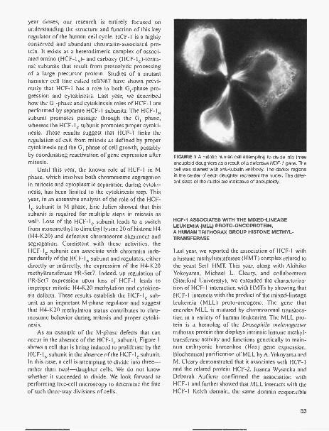
year closes, our research is entirely focused onunderstanding the structure and function of this keyregulator of the human cell cycle. HCF-1 is a highlyconserved and abundant chromatin-associated pro-tein. It exists as a heterodimeric complex of associ-ated amino (HCF-10- and carboxy (HCF-1c)-termi-nal subunits that result from proteolytic processingof a large precursor protein. Studies of a mutanthamster cell line called tsBN67 have shown previ-ously that HCF-1 has a role in both G, -phase pro-gression and cytokinesis. Last year, we describedhow the G1 -phase and cytokinesis roles of HCF-1 areperformed by separate HCF-1 subunits: The HCF-1Nsubunit promotes passage through the G, phase,whereas the HCF-1c subunit promotes proper cytoki-nesis. These results suggest that HCF-1 links theregulation of exit from mitosis as defined by propercytokinesis and the G, phase of cell growth, possiblyby coordinating reactivation of gene expression aftermitosis.
Until this year, the known role of HCF-1 in Mphase, which involves both chromosome segregationin mitosis and cytoplasmic separation during cytoki-nesis, has been limited to the cytokinesis step. Thisyear, in an extensive analysis of the role of the HCF-1 subunit in M phase, Eric Julien showed that thissubunit is required for multiple steps in mitosis aswell. Loss of the HCF- 1 subunit leads to a switchfrom monomethyl to dimethyl lysine 20 of histone H4(H4-K20) and defective chromosome alignment andsegregation. Consistent with these activities, theHCF- lc subunit can associate with chromatin inde-pendently of the HCF-1N subunit and regulates, eitherdirectly or indirectly, the expression of the H4-K20methyltransferase PR-Set7. Indeed, up regulation ofPR-Set7 expression upon loss of HCF-1 leads toimproper mitotic H4-K20 methylation and cytokine-sis defects. These results establish the HCF- 1 sub-unit as an important M-phase regulator and suggestthat H4-K20 methylation status contributes to chro-mosome behavior during mitosis and proper cytoki-nesis.
As an example of the M-phase defects that canoccur in the absence of the HCF- lc subunit, Figure 1shows a cell that is being induced to proliferate by theHCF-1N subunit in the absence of the HCF- lc subunit.In this case, a cell is attempting to divide into three-rather than two!-daughter cells. We do not knowwhether it succeeded to divide. We look forward toperforming live-cell microscopy to determine the fateof such three-way divisions of cells.
FIGURE 1 A mitotic human cell attempting to divide into threeaneuploid daughters as a result of a defective HCF-1 gene. Thecell was stained with anti-tubulin antibody. The darker regionsin the center of each daughter represent the nuclei. The differ-ent sizes of the nuclei are indicative of aneuploidy.
HCF-1 ASSOCIATES WITH THE MIXED-LINEAGELEUKEMIA (MLL) PROTO-ONCOPROTEIN,A HUMAN TRITHORAX GROUP HISTONE METHYL-TRANSFERASE
Last year, we reported the association of HCF-1 witha histone methyltransferase (HMT) complex related tothe yeast Set 1 HMT. This year, along with AkihikoYokoyama, Michael L. Cleary, and collaborators(Stanford University), we extended the characteriza-tion of HCF-1 interaction with HMTs by showing thatHCF-1 interacts with the product of the mixed-lineageleukemia (MLL) proto-oncogene. The gene thatencodes MLL is mutated by chromosomal transloca-tion in a variety of human leukemias. The MLL pro-tein is a homolog of the Drosophila melanogastertrithorax protein that displays intrinsic histone methyl-transferase activity and functions genetically to main-tain embryonic homeobox (Hox) gene expression.Biochemical purification of MLL by A. Yokoyama andM. Cleary demonstrated that it associates with HCF-1and the related protein HCF-2. Joanna Wysocka andDeborah Aufiero confirmed the association withHCF-1 and further showed that MLL interacts with theHCF-1 Kelch domain, the same domain responsible
33

for HCF-1 association with Setl. These results linkHCF-1 to the maintenance of stable patterns of geneexpression as well as to oncogenesis.
PUBLICATIONS
Julien E. and Herr W. 2004. A switch in mitotic histone H4 lysine20 methylation status is linked to M phase defects upon
loss of HCF-1. Mol. Cell 14: 713-725.Tubon T.C., Tansey W.P., and Herr W. 2004. A nonconserved
surface of the TFIIB zinc ribbon domain plays a direct rolein RNA polymerase II recruitment. Mol. Cell Biol. 24:2863-2874.
Yokoyama A., Wang Z., Wysocka J., Sanyal M., Aufiero D.J.,Kitabayashi I., Herr W., and Cleary M.L. 2004. Leukemiaproto-oncoprotein MLL forms a SET1-like histone methyl-transferase complex with menin to regulate Hox geneexpression. Mol. Cell Biol. 24: 5639-5649.
Shweta Tyagi, postdoc, Winship Herr's lab.
34

STRUCTURAL BIOLOGY OF NUCLEIC ACIDREGULATORY PROCESSES
L. Joshua-Tor C. Cho T. Hu S. SmithE. Enemark P.R. Kumar J.J. SongH. He P. O'Farrell N. To lia
We study the molecular basis of cell regulatoryprocesses by using the tools of structural biology andbiochemistry to examine proteins and protein com-plexes associated with these processes. X-ray crystal-lography enables us to obtain the three-dimensionalstructures of individual proteins and their interactionswith other molecules. Biochemistry and molecularbiology allow us to study properties that can be corre-lated to protein structure and function. Our effortslargely center on protein complexes involved in nucle-ic acid regulatory processes.
Mechanisms of RNAi
J.J. Song, N. Iota, S. Smith [in collaboration withG.J. Hannon, Cold Spring Harbor Laboratory]
The introduction of exogenous double-stranded RNA(daRNA) into a cell can trigger the gene silencingprocess called RNA interference or RNAi. An RNaseIII family enzyme, Dicer, initiates silencing by releas-ing approximately 20 base duplexes, with 2-nucleotide3' overhangs called siRNAs. The RNAi pathway alsomediates the function of endogenous, noncoding reg-ulatory RNAs called microRNAs (miRNAs). BothmiRNAs and short interfering RNAs (siRNAs) guidesubstrate selection by similar effector complexescalled the RNA-induced silencing complex (RISC).These contain single-strand versions of the small RNAand additional protein components. Of those, the sig-nature element, which virtually defines a RISC, is amember of the Argonaute family of proteins.Argonaute proteins are defined by the presence ofPAZ and PIWI domains. In a central RNAi pathway,guided by the siRNA, RISC directs the cleavage ofmRNA substrates. However, the nuclease responsiblefor cleavage, dubbed "Slicer," has so far escaped iden-tification.
There has been remarkable progress in unravelingthe components of the RNAi machinery, but to get atrue mechanistic understanding of this process, how
these components fit together, and how they function,we must understand how they work at a molecularlevel. Therefore, we embarked on structural and bio-chemical studies of key proteins in the RNAi pathway,the protein-RNA and protein-protein interactionsinvolved.
In an effort to further our understanding of the roleof Argonaute in RNAi, this past year, we determinedthe crystal structure of the full-length Argonaute pro-tein from Pyrococcus furiosus at 2.25 A resolution(Fig. 1). The structure reveals a crescent-shaped basemade up of the amino-terminal, middle, and PIWIdomains. The PAZ domain is held above the base by a"stalk"-like region. The PIWI domain is similar toRNase H, with conserved active site aspartateresidues, strongly implicating Argonaute as "Slicer."Several known characteristics of mRNA cleavage byRISC are consistent with an RNase-H-like enzyme.The architecture of the molecule and placement of thePAZ and PIWI domains define a groove for substrate
PAZ
PIWI
FIGURE 1 The crystal structure of Argonauts from P furiosus.Ribbon representation of Argonauts showing the amino-termi-nal domain, the "stalk, the PAZ domain, the middle domain, andthe PIWI domain.
35

binding. Based on the structure of PAZ with a "mini-siRNA," we could model the siRNA binding with its3' end in the PAZ cleft and the siRNA-mRNA doublehelix extending into the groove, and suggest a mecha-nism for siRNA guided mRNA cleavage. The notionthat RISC "slicer" activity resides in Argonaute itselfwas tested in a mammalian system by mutationalanalysis of hAgo2 in collaboration with GregHannon's laboratory. Conserved active site aspartatesin hAgo2 were altered and the mutants lost nucleaseactivity while retaining siRNA binding. This supportsthe model in which Argonaute itself functions as theSlicer enzyme in the RNAi pathway.
The DNA-binding and Assembly of thePapillomavirus Initiator Protein El
E. Enemark, H. He, A. Auster [in collaboration withA. Stenlund, Cold Spring Harbor Laboratory]
Papillomaviruses are a large family of closely relatedviruses that give rise to warts in their hosts. Infectionof the genital tract by the human papillomaviruses(HPV) from this group represents one of the few firm-ly established links between viral infection and thedevelopment of cervical cancer, as HPV DNA isfound in practically all cervical carcinomas. The Elprotein belongs to a family of multifunctional viralproteins whose main function is related to viral DNAreplication. These proteins bind to the origin of DNAreplication, melt the DNA duplex, possess DNA heli-case activity, and recruit other cellular replication pro-teins such as DNA polymerase a and replication fac-tor A (RPA). Most likely, different oligomeric formsof El are responsible for the different activities, andthe sequential assembly El complexes ensures anordered transition between these different activities.Ultimately, El forms a hexameric ring helicase oneach strand that serve as replicative DNA helicasesthat unwind the DNA in front of the replication fork.
In collaboration with Arne Stenlund's group hereat CSHL, we embarked on structural studies to pro-vide high-resolution structural information about El,its DNA-binding activity, and its assembly on DNA,which would provide general insight into the bio-
36
chemical events that are involved in viral DNA repli-cation. They can also provide a basis for the develop-ment of clinical intervention strategies. Second, theviral DNA replication machinery itself represents anobvious target for antiviral therapy and detailed infor-mation such as high-resolution structures of viral pro-teins required for replication will greatly facilitate thedevelopment and testing of antiviral agents.
Previously, we have solved the structures of the ElDNA-binding domain from bovine papillomavirus(BPV) both unbound and in two stages of assembly onthe origin (dimer and tetramer). During the past twoyears, we have extended our characterization of DNAbinding by El to include human papillomavirus type18 (HPV-18). HPV-18 is a high-risk strain of papillo-mavirus that causes cervical carcinoma, one of themost frequent causes of cancer death in women world-wide. In particular, HPV-18 causes adenocarcinoma,which is associated with poor prognosis. We deter-mined the crystal structure of the monomeric HPV-18El DNA-binding domain refined to 1.8 A resolutionand demonstrated biochemically that the analogousresidues required for El dimerization in BPV-1 andthe low-risk HPV type 11 are also required for HPV-18 El. We also found evidence that the HPV-18 ElDNA-binding domain does not share the samenucleotide and aminoDNA recognition as BPV-1 and HPV-11 El.
PUBLICATIONS
Auster A.S. and Joshua-Tor L. 2004. The DNA-binding domainof human papillomavirus type 18 El: Crystal structure,dimerization, and DNA binding. J. Biol. Chem. 279: 3733-3742.
Joshua-Tor L. 2004. siRNAs at RISC. Structure 12: 1120-1122.Joshua-Tor L. and Johnston S.A. 2004. The Bleomycin hydro-
lases. In Handbook of proteolytic enzymes, 2nd edition (ed.A.J. Barrett et al), pp. 1197-1201, Elsevier, London.
Liu J., Carmell MA., Rivas F.V, Marsden C.G., Thomson J.M.,Song J.-J., Hammond S.M., Joshua-Tor L., and HannonG.J. 2004. Argonaute2 is the catalytic engine of mam-malian RNAi. Science 305: 1437-1441. Published onlineJuly 29, 2004 (10.1126/science.1102513).
Song J.J., Smith S.K., Hannon G.J., and Joshua-Tor L. 2004.Crystal structure of Argonauts and its implications for RISCSlicer activity. Science 305: 1434-1437. Published onlineJuly 29, 2004 (10.1126/science.1102514).

RNA SPLICING
A.R. Krainer E. AllemandJ. CalarcoM. HastingsY. Hua
R. Karni F. RocaL. Manche S. ShawH. Okunola R. Sinha
MECHANISMS OF CONSTITUTIVE AND ALTERNATIVEPRE-mRNA SPLICING
RNA splicing is an essential step in the expression ofmost eukaryotic protein-coding genes. The spliceo-some selects and pairs authentic splice sites withextremely high fidelity, which requires precise inter-pretation of limited and dispersed sequence informa-tion present throughout introns and exons. In humans,about 75% of genes are expressed via alternative splic-ing, giving rise to multiple protein isoforms. Thechoice of alternative splice sites is commonly regulat-ed to alter gene expression, either tissue-specificallyor in response to a developmental program or to sig-naling pathways. The fact that multiple protein iso-forms can be expressed from individual genes demon-strates that the classical "one gene-one enzyme" par-adigm is no longer valid and provides an explanationfor the unexpectedly small number of genes uncoveredby genome-sequencing projects.
Both constitutive and alternative splicing mecha-nisms involve numerous protein components, as wellas RNA components that are part of small nuclearribonucleoprotein (snRNP) particles. The work in ourlab focuses primarily on the identification and molec-ular characterization of protein factors and sequenceelements that are necessary for the catalysis and fideli-ty of splicing and/or for the regulation of alternativesplice site selection. We are interested in how thespliceosome correctly identifies the exons on pre-mRNA, and how certain point mutations in either exonor intron sequences cause aberrant splicing, leading tovarious genetic diseases. A brief summary of three ofour recently published studies is given below.
INVOLVEMENT OF SR PROTEINS IN mRNASURVEILLANCE
In the course of experiments aimed at addressing therelationship between premature termination codons(PTCs) and splice site selection, we uncovered a rolefor SR proteins in nonsense-mediated mRNA decay
P. SmithQ.-S. ZhangZ. Zhang
(NMD). SR proteins are factors required for splicing,and they also regulate alternative splicing in a concen-tration-dependent manner. NMD is a quality-controlpathway that down-regulates mRNAs with PTCs.Model minigenes were constructed to test the effect ofPTCs placed between two alternative 5' splice sites on5' splice site selection (Fig. 1). In transiently trans-fected cells, the distal 5' splice site was selected bydefault, in both the presence and absence of the PTCs(not shown). We overexpressed various SR proteins(e.g., SF2/ASF and SC35) to promote use of the prox-imal 5' splice site and observed dramatically differentresults in the presence of PTCs, depending on whichSR protein was overexpressed. As expected, bothSF2/ASF and SC35 promoted use of the proximal sitewith the wild-type minigene to equivalent extents(lanes 3 and 5); however, when a PTC was present, noproximal SF2/ASF overex-pression (lane 4), whereas SC35 overexpressionresulted in proximal splicing at the same level as withthe wild-type construct (lane 6). Abrogating NMD invarious ways, including cycloheximide treatment,down-regulation of hUPF1 or hUPF2 by RNA inter-ference, or expression of dominant-negative hUPF1,rescued expression of the proximal spliced mRNAwith a PTC in the presence of SF2/ASF (not shown).Many natural alternatively spliced mRNA isoformspossess PTCs, and our studies suggest that the extentof their expression depends on the coexpression of theparticular SR proteins that promote the appropriatealternative splicing events, as well those that do or donot trigger their degradation by NMD. PTCs also arisefrom nonsense and frameshift mutations, as well asfrom splicing mutations that change the readingframe. We found that transient overexpression ofsome, but not all, SR proteins strongly enhances thedegradation of standard NMD substrates, i.e., consti-tutively spliced transcripts with a PTC. Although thiseffect on the NMD pathway was elicited most strong-ly by SF2/ASF, which is a shuttling SR protein, wealso observed it with a mutant version of the proteinthat is retained in the nucleus.
37

ME
4
G(jv
Pm WMWM
1 2 3 4 5 6
GFP
pre-mRNA
mRNA proximal
mRNA distal
FIGURE 1 Effect of different SR proteins on sensitivity to a PTC in the context of alternative splicing. Model human f3-globin genes wereconstructed with an internal duplication (gray) spanning the 5' splice site in the first intron and short flanking exonic and intronic seg-ments. The mutant construct (M) has a stop codon after each 5' splice site, which interrupts the reading frame when the proximal (down-stream) 5' splice site is selected. The wild-type construct (W) has no premature termination codons (PTCs). He La cells were transfectedwith W and M constructs, and cotransfected with plasmids for overexpression of SF2/ASF or SC35. A GFP plasmid was also cotrans-fected as an internal reference for transfection efficiency, RNA recovery, and loading. The transiently expressed globin transcripts wereanalyzed by RPA, with a probe spanning the first exon, the duplicated region, and partial intron-1 sequences. fl-globin and GFP probeswere used simultaneously; undigested probes (P) are shown in lane 1, corresponding to <1% of the amount of probe used in each RPAreaction. The bands corresponding to I3-globin pre-mRNA, proximal and distal spliced mRNAs, and GFP, are indicated. For simplicity,the diagrams show only the first two exons and first intron of 13-globin. (m): DNA markers.
NOVEL LKB1 SPLICING MUTATION ASSOCIATEDWITH PEUTZ-JEGHERS SYNDROME
In collaboration with N. Resta, A. Stella, and G.Guanti (University of Bari, Italy), we characterized anovel splicing mutation associated with Peutz-Jegherssyndrome (PJS). PJS is an autosomal dominant disor-der associated with gastrointestinal polyposis andincreased cancer risk. PJS is caused by germ-linemutations in the tumor suppressor gene LKB1, whichencodes a protein kinase. One familial mutation,IVS2+1A>G, alters the second intron 5' splice site,which has sequence features of a U12-type AT-ACintron. This rare type of intron normally begins witheither A or G. We found that in patients, LKB1 RNAsplicing occurs from the mutated 5' splice site to sev-eral cryptic, noncanonical 3' splice sites immediately
38
adjacent to the normal 3' splice site. In vitro splicinganalysis demonstrated that this aberrant splicing ismediated by the U12-dependent spliceosome. Theseresults indicate that the minor spliceosome can use avariety of 3' splice site sequences to pair to a given 5'splice site, albeit with tight constraints for maintainingthe 3' splice site position. The unusual splicing defectassociated with this PJS-causing mutation uncoveredinteresting differences in splice site recognitionbetween the major and minor pre-mRNA splicingpathways.
REGULATION OF HNRNP Al SHUTTLING BYPHOSPHORYLATION
Heterogeneous nuclear ribonucleoprotein (hnRNP) A 1is an abundant RNA-binding protein involved in the

regulation of alternative splicing and other cellularprocesses. hnRNP Al is mainly nuclear, although itshuttles rapidly between nuclear and cytoplasmiccompartments. Genotoxic stress, such as osmoticshock, activates the MKK3/6-p38 signaling pathway,which in turn results in accumulation of hnRNP Al inthe cytoplasm. This effect modulates alternative splic-ing regulation in vivo-by decreasing the level ofnuclear hnRNP Al-and correlates with increasedhnRNP Al phosphorylation. In collaboration with J.Caceres (Edinburgh), M. Myers (CSHL), and J.Moscat (Madrid), we have now characterized the mol-ecular mechanism involved in the cytoplasmic accu-mulation of hnRNP Al in cells subjected to osmoticshock. This treatment results in serine-specific phos-phorylation within a carboxy-terminal peptide,dubbed the "F-peptide," which is adjacent to the M9motif that mediates bidirectional transport of hnRNPAl. Analysis of mutants in which the F-peptide serineswere replaced by aspartic acids or alanines showedthat F-peptide phosphorylation is required for the sub-cellular redistribution of hnRNP Al in cells subjectedto osmotic shock. Furthermore, we showed that F-pep-tide phosphorylation modulates the interaction ofhnRNP Al with transportin Trn 1 . These findings sug-gest that the phosphorylation of F-peptide by signalingpathways regulates the rate of hnRNP Al nuclearimport.
PUBLICATIONS
Muraki M., Ohkawara B., Hosoya T., Onogi H., Koizumi J.,Koizumi T., Sumi K., Yomoda J., Murray M.V., Kimura H.,Furuichi K., Shibuya H., Krainer AR., Suzuki M., andHagiwara M. 2004. Manipulation of alternative splicing by anewly developed inhibitor of Clks. J. BioL Chem. 279:24246-24254,
Zatkova A., Messiaen L, Vandenbroucke I., Wieser R., FonatschC., Krainer AR., and Wimmer K. 2004. Disruption of exon-ic splicing enhancer elements is the principal cause of exonskipping associated with seven nonsense or missense alle-les of NF1 . Hum. Mutat. 24: 491-501.
Zhang Z. and Krainer A.R. 2004. Involvement of SR proteins inmRNA surveillance. MoL Cell 16: 597-607.
In Press
Allemand E., Guil S., Myers M., Moscat J., Caceres J.F., andKrainer A.R. 2005. Regulation of heterogeneous nuclearribonucleoprotein Al transport by phosphorylation in cellsstressed by osmotic shock. Proc. Natl. Acad. Sc!. 102:3605-3610.
Coutinho G., Xie J., Du L., Brusco A., Krainer AR., and GattiR.A. 2005. Functional significance of a deep intronic muta-tion in the ATM gene and evidence for a new alternativeexon, 28a. Hum. Mutat. 25: 118-124.
Hastings M.L., Resta N., Traum D., Stella A., Guanti G., andKrainer A.R. 2005. An LKB1 AT-AC intron mutation causesPeutz-Jeghers syndrome via splicing at noncanonical cryp-tic splice sites. Nat. Struct. Mol. Biol. 12: 54-59.
Roca X., Sachidanandam R., and Krainer A.R. 2005.Determinants of the inherent strength of human 5' splicesites. RNA (in press).
Rotem Karni, postdoc, Adrian Krainer's lab.
39

PROTEOMICS
M. Myers R. Bish L.L. SchmidtG. Pegoraro C. van der MeijdenD. Perkowski
The completion of the human genome has ushered ina new age of biological discovery. This accomplish-ment has essentially identified all of the players gov-erning human biology. The important work of assign-ing functions to this myriad of proteins has becomeone of the principal tasks of modern biology. Althoughmany functions will be assigned using genetics orbioinformatics, the majority of this functional charac-terization will be performed by proteomics.
The goal of our laboratory is to understand howproteins and protein complexes regulate cellularbehavior. In the environment of a cell, almost all of theproteins can be found in a highly interactive network.Our laboratory is focused on understanding the globalarchitecture of this network and how this network gen-erates robustness (error tolerance) and adaptability tothe system and how this network is altered to produceand survive complex diseases, such as cancer.
Optimization of Protein IdentificationD. Perkowski, L.L. Schmidt
Protein identification is the major tool of proteomics.We have been optimizing mass spectrometry (MS) forprotein identification. MS has many advantages overother techniques for protein identification, especially itssensitivity. However, the improved sensitivity comes ata price, as the sample preparation becomes increasing-ly important to ensure success. We have been optimiz-ing a number of parameters to increase this success rate.
As part of the optimization, we have found thatnanoscale chromatography gives the best sensitivity.Using conventional chromatography media (packed bedsof small, functionalized beads), we have been fabricatingour own nanoscale (75 -gm internal diameter) columnsthat operate at flow rates of 200-500 nl/min. The sensi-tivity and resolution of these nanoscale columns arerelated to their internal diameter. However, the internalpressures required for chromatography increase dramat-ically as diameter decreases, limiting the practical utilityof miniaturizing conventional chromatography media.To overcome the pressure problem, we have developed amesoporous, monolithic chromatography medium that is
40
well suited for miniaturization. Monolithic medium iscomposed of a single highly cross-linked entity that isformed during the polymerization of silicas (sol-gels) ororganic polymers (plastics). Initially, we incorporatedoctadecyl (C18) or octyl (C8) functional groups into ourmonoliths in order to create a reverse-phase resin, and weare currently working on producing monoliths with addi-tional functionality, such as those that can be used forion-exchange chromatography.
A significant loss in signal occurs due to poor iontransfer between the nanocolumns and the mass spec-trometer. We have optimized a number of parametersto improve our electrospray interface. In collaborationwith Patrik Kallback (GE Healthcare), we have devel-oped a novel high-voltage interface that maximizeselectrical contact with the column effluent, resulting ina more robust ionization at lower voltages. In addition,this interface is resistant to fouling. One of the draw-backs of chromatography-coupled electrospray inter-faces is that the voltage must be adjusted during theanalysis in order to maintain a single, stable Taylorcone. To improve Taylor cone stability, we have adapt-ed a static nanospray chip to our system. Importantly,the nanospray chip improves the potential differencebetween the column effluent and the mass spectrome-ter, resulting in stable Taylor cone formation through-out the chromatography run.
Monitoring PosttranslationalModificationsL.L. Schmidt, D. Perkowski
One of the challenges in the postgenomic era is tounderstand how proteins are regulated by posttransla-tional modifications. These modifications are respon-sible for controlling the activity of proteins and ulti-mately determining how a cell responds to its environ-ment. Phosphorylation is considered to be the mostcommon posttranslational modification, and its wideuse as a regulatory mechanism has been attributed toits reversible nature. Alterations in protein phosphory-lation have been shown to be the hallmark of manypathological conditions including cancer and diabetes.

Therefore, efficient identification of phosphorylatedproteins, as well as mapping the specific phosphoryla-tion sites, has become one of the primary goals of pro-teomics. We have been attempting to develop sensitiveand robust MS methods for characterizing this impor-tant posttranslational modification. Characterization ofphosphorylated residues is complicated by three attrib-utes of phosphorylation: (1) phosphorylation suppress-es peptide detection by MS, (2) phosphorylation isgenerally substoichiometric, and (3) phosphorylatedresidues readily undergo neutral loss of the phosphorylgroup. We have expended a significant effort exploringwhy phosphorylation suppresses peptide signals duringMS. Conventional wisdom dictates that the poor signalof phosphopeptides was due solely to their strong neg-ative charge. Our analysis indicated that this is not thecase. For example, phosphorylated peptides are equal-ly suppressed regardless of whether positive or nega-tive ions are detected, and sulfated peptides (whichcarry a similar negative charge) do not exhibit the samesuppression as their phosphorylated cousins. Ourresults suggest that the suppression of phosphopeptidesis likely the result of a combination of the poor volatil-ity of phosphoric acid and/or instability of the phos-phopeptides in the gas phase. In fact, the suppressiondue to phosphorylation can be alleviated by altering theionization conditions to improve volatility and stabilityduring the ionization process.
"Small World" Proteomics
C. van der Meijden, G. Pegoraro [in collaborationwith R. Sachidanandam, Cold Spring Harbor Laboratory]
We have modeled the data from several high-through-put protein interaction screens as a network. We havechosen to model these data as a network because it isone of the only ways to make sense of these large andcomplicated data sets. In this model, each protein istreated as a node, and the interactions are treated aslinks between the nodes. In this way, the yeast proteinnetwork ends up looking very similar to the network ofcomputers that make up the World Wide Web or thenetwork of human social interactions that make this a"small world." On the basis of the network properties ofthe yeast protein network, we have been able to classi-fy the yeast network as a scale-free network, in whichonly a fraction of the proteins are responsible for mak-ing the lion's share of the connections. One predictionfrom the network model is that these highly connectedproteins are essential for yeast viability. In fact, we find
that this is the case, as essential proteins are highlyenriched in the pool of highly connected proteins.Importantly, not all highly connected genes are essen-tial, and we are focusing on trying to understand thedifferences between these proteins and those which areessential. The overall goal of this study is to understandhow these protein networks provide adaptability togenetic alterations, such as loss or duplication of a node(gene) that occurs during tumorigenesis. Importantly,nongenotoxic stresses, such as high salt or nutrientdepletion, increase the spontaneous mutation rate inyeast and in a number of other organisms. The stress-induced mutations are believed to provide a mechanismfor rapid adaptation to changing environmental condi-tions. We are currently investigating whether the essen-tial genes are protected from these mutations, and wehave discovered that the essential and nonessentialgenes have different genomic distributions.
We were hoping that our analysis of the yeast pro-teome network would reveal properties of mammaliannetworks and perhaps even how these networks getreprogrammed during tumorigenesis. One of thelessons from our work in yeast was that even this rela-tively small-scale network (6000 genes) was incrediblycomplex, suggesting that it would be difficult to makeinroads into the tumor-specific network. We have cho-sen to take advantage of adeno-associated virus (AAV)to help us get a handle on the tumor-specific network.AAV has no known pathology in humans and has actu-ally been described as having tumor-suppressive activ-ity. We hope that an exploration of AAV-dependenttumor suppression would uncover important hubs inthe tumor-specific network. Using MS, we have identi-fied a number of host-cell proteins that associate withAAV-Rep68, an AAV protein required for viral replica-tion. We have found that Rep68 associates with a num-ber of proteins involved in DNA damage response(Ku70, Ku80, and DNA-dependent protein kinase). Wehave also found that AAV-Rep68 associates withANP32B, a poorly characterized protein of uncertainfunction. Importantly, microarray data has demonstrat-ed that ANP32B and AAV-Rep68 act synergistically toeffect host-cell gene transcription.
PUBLICATIONS
Janardhan A., Swigut T, Hill B., Myers M.P., and Skowronski J.2004. HIV-1 Nef binds the DOCK2-ELMO1 complex toactivate rac and inhibit lymphocyte chemotaxis. PloS Biol.2: 65-76.
Myers M.P., Khanna R., Lee E.J., and Papazian D.M. 2004.Voltage sensor mutations differentially target misfoldedchannel subunits to proteosomal and non-proteosomaldisposal pathways. FEBS Lett. 568: 110-116.
41

CELL SIGNALING IN HIV PATHOGENESIS
J. Skowronski M. Gierszewska M. KwofieK. Hrecka Z. ThakkarL. Kozaczkiewicz
Our interest lies in understanding the molecular mech-anisms underlying the pathogenesis of AIDS and, inparticular, understanding the functional consequencesof interactions between viral proteins and the cellularregulatory machinery. The main focus of our researchis to understand the functions of Nef, a regulatory pro-tein of human and simian immunodeficiency viruses(HIV and SIV) that is an important determinant of vir-ulence. We directed a major effort toward the identifi-cation of mechanisms and downstream effectors thatmediate the effect of Nef on protein sorting and signaltransduction machineries. and our experiments havebeen focused in two areas. First, we have focusedheavily on the isolation and identification of cellularproteins that mediate the effects of Nef on signal trans-duction and endocytic machineries. This has ledrecently to purification and microsequencing of sever-al cellular factors that associate with Nef in T lym-phocytes. Importantly, we found that Nef targets a crit-ical molecular switch that regulates Rac GTPasesdownstream from chemokine- and antigen-initiatedsignaling pathways. This interaction enables Nef toinfluence multiple aspects of T-cell function, and thusprovides an important mechanism by which Nefimpacts pathogenesis by primate lentiviruses. We havealso begun to verify selected novel interactions and toaddress their relevance to previously known and pos-sibly novel functions of Nef, as well as to AIDS patho-genesis. Second, we continued studies to address therelevance of individual Nef activities for rate of pro-gression of lentiviral replication in vivo. To this end,we completed the studies of the impact of down-mod-ulation of class I major histocompatibility complex(MHC) by Nef on antiviral immune response andfound that this Nef function indeed limits in importantways the generation of virus-specific CDS+ T cells inthe infected host.
IDENTIFICATION AND CHARACTERIZATION OF NOVELNef EFFECTORS
Nef is a small adaptor protein that acts through associ-ations with large protein complexes. We recently puri-
42
fied to near homogeneity Nef and its associated pro-tein complexes from T-cell and monocyte lines stablyexpressing Nef using an immunoaffinity purificationprotocol and identified their components by massspectroscopy. This approach allowed us to detect aseries of polypeptides with apparent molecular massesranging from 20 kD to more than 250 kD that copuri-fied with HIV-1 and SIV Nef proteins. Next, we deter-mined their sequences by mass spectroscopic analysis,in collaboration with Michael Myers here at the CSHLProtein Chemistry Shared Resource. This led to theidentification of several common polypeptides associ-ated with both 7.AH Nef and 239.AH Nef, includingsubunits of the DOCK2- ELMO1 -Rac2 complex andadditional polypeptides involved in control of varioushousekeeping functions of a cell. During the last year,we have continued to study these associations. We havealso characterized in detail the complex comprisingNef and isoforms of N-myristoyltransferase (NMT).
Nef ASSOCIATES SELECTIVELY WITH ISOFORM 1OF NMT
All known functions of Nef require that it be myris-toylated at its amino terminus. This reaction is cat-alyzed by NMTs, which transfer myristate from myris-toyl coenzyme A (myristoyl-CoA) to the amino-termi-nal glycine of substrate proteins. Two NMT isoforms(NMT-1 and NMT-2) are expressed in mammaliancells. Interestingly, NMT-1 but not NMT-2 was identi-fied as one of relatively abundant cellular proteins thatassociated with both HIV-1 and SIV Nef. The Nef-NMT-1 complex is most likely a transient intermediateof the myristoylation reaction of Nef because its for-mation requires the amino-terminal myristoylationsignal in Nef and is modulated by agents that affect thesize of the myristoyl-CoA pool in the cell. We alsoexamined two other proteins that bear an amino-ter-minal myristoylation signal, HIV-1 Gag and Hck pro-tein tyrosine kinase. Surprisingly, we found that Gagbound preferentially the NMT-2 isoform, whereas Hckbound mostly to NMT-1. Recognition of differentNMT isoforms by these viral and cellular substrate

proteins suggests nonoverlapping roles for theseenzymes in vivo and reveals a potential for the devel-opment of inhibitors that target the myristoylation ofspecific viral substrates more selectively.
ANTIVIRAL IMMUNE RESPONSE AND SIVVIRULENCE
Presentation of antigens derived from viral proteinsexpressed in infected cells by class I MHC moleculesto competent effector T cells is the key event requiredfor the recognition and elimination of infected cells bythe immune system of the host. We showed previous-ly that Nef proteins down-regulate expression of classI MHC complexes by inducing their endocytosis fromthe cell surface. Down-regulation of class I MHCexpression could be very important for the establish-ment of persistent SIV/HIV infection once strongantiviral responses develop in the infected host, butwhether it is so has been a subject of controversy.
To address directly the role of this Nef function aswell as a broader issue of the role of antiviral immuneresponse in immunodeficiency virus infection andprogression to AIDS, we studied in detail class I MHCdown-regulation by Nef protein derived from a patho-genic SIVmac strain. First, we identified mutations inthe carboxy-terminal domain of SIVmac 239 Nef thatselectively disrupt class I MHC down-regulation, hav-ing no detectable effect on other functions of Nef suchas the down-regulation of CD4 and CD3 surfaceexpression, and the stimulation of SIV virion infectiv-ity and induction of SIV replication from peripheralblood mononuclear cells (PBMCs) infected in theabsence of stimulation. Second, we constructed sever-al mutant Nef proteins with small deletions in the car-boxy-terminal region (NefA239 -240, Nef238stop/fs/fs). These mutations still selectively disrupted onlyclass I MHC down-regulation, but are less likely torevert in vivo than point mutations. These mutantsturned out to be powerful reagents to study the impor-tance of class I MI-IC down-regulation for SIV repli-cation in the context of a functional immune system inrhesus macaques. Then, to define the effect of theseselective mutations on anti-SIV immune response wecollaborated with Dr. R.C. Desrosiers (New EnglandPrimate Center, Harvard) who infected rhesus mon-keys with SIVmac viruses carrying nefA239-240,nef238stop/fs/fs and, as controls, SIVmac virusescontaining wild-type nef, or a nef mutant with a 181 -nucleotide deletion that abrogates all known function-al activities of Nef.
Nef LIMITS ANTIVIRAL RESPONSE BY COB* T CELLS
The ability of CD8+ T lymphocytes from infectedmonkeys to react with antigenic peptide and an MHCI tetramer corresponding to the Mamu-A*01-restrict-ed Gag181-189 epitope CTPYDINQM was moni-tored. The four monkeys infected with the SIV strainscarrying the MHC-selective nef mutations exhibitedhigher levels of CD8+ T-cell cellular responses than thefour monkeys infected with the parental wild-typeSIV239 at 4-14 weeks after infection. These differ-ences were statistically significant. Differencesbecame less discernible by 16-20 weeks postinfection.
CD8+ T-cell responses were significantly higher inmonkeys that received the MHC-selective mutantSIVs than in monkeys that received parental SIV239,from 4 to 14 weeks postinfection. Stronger CD8+antiviral responses would be expected to translate intolower viral loads, but we were unable to demonstrateany statistically significant differences in viral loadsamong the groups, excluding of course the SIVAnefanimals. It seems likely that the evolution of restoredMHC down-regulating activity minimized the magni-tude and duration of any effects of the mutations onviral load. The number of monkeys used was neces-sarily limited, and significant differences may havebeen observed with much larger numbers of monkeys.
A controlling immune response is likely to consistof multifactorial components; the effects of the MIICactivity of Nef influenced the levels of only one ofthese multifactorial components: virus-specific CD8+cellular responses. SIV destruction of virus-specificCD4+ helper cells may have limited the effectivenessof increased numbers of virus-specific CD8+ T cellsthat resulted from the loss of MHC down-regulatingactivity. Nonetheless, although we were not able todemonstrate significant differences in viral load, theselective pressure for sequence change to restoreMHC down-regulating activity clearly indicates thatthe absence of this activity did serve to limit the repli-cation of the virus at least somewhat.
CLASS I MHC DOWN-REGULATION BY MUTANT NefPROTEINS IS RESTORED THROUGH SELECTION OFSECOND-SITE COMPENSATORY MUTATIONS
To address the normalization in numbers of Gag-spe-cific CD8+ T cells at later timepoints, we next deter-mined the evolution of sequence changes in nef overtime. nef sequences present at 16 and 32 weeks postin-fection were amplified by polymerase chain reaction
43

(PCR) of DNA from cells infected with recoveredvirus. Little variation was observed in the week-16samples from the two control animals infected with thewild-type virus. In contrast, greater levels of variationwere observed in the monkeys infected with the MHC-selective mutants at 16 weeks. In all clones analyzed at16 and 32 weeks after infection in our current study,the difficult-to-revert NefA239-240 and Nef 238stop/fs/fs mutations were retained, forcing the virus to try tocompensate by changing residues elsewhere in the Nefprotein. The changes in Nef progressively increasedthe ability to down-regulate MHC from weeks 0 to 16to 32 such that by week 32 the ability to down-regulateMHC class I was similar to that of the parentalSIVmac 239 Nef. Based on the large number ofsequence changes and the time required to restore fullMHC down-regulating activity, the virus was clearlyunder strong selective pressure over a prolonged peri-od to restore the activity; this provides unambiguousevidence for the contribution of MHC down-regulationto the ability of SIV to replicate in monkeys. The abil-ity to restore the activity by sequence changes quitedistant in the linear sequence is testimony to the
44
remarkable flexibility and multiplicity built into theNef protein structure.
PUBLICATIONS
Janardhan A., Swigut T., Hill B., Myers M.P., and Skowronski J.2004. HIV-1 Nef binds the DOCK2-ELMO1 complex toactivate Rac and inhibit lymphocyte chemotaxis. PloS Blot2: 65-76.
Schindler M., Munch J., Brenner M., Stahl-Hennig C.,Skowronski J., and Kirchhoff F. 2004. Comprehensiveanalysis of nef functions selected in simian immunodefi-ciency virus-infected macaques. J. Virot 78:10588-10597.
Swigut T., Alexander L., Morgan J., Lifson J., Mansfield KG.,Lang S., Johnson R.P., Skowronski J., and Desrosiers R.2004. Impact of Nef-mediated downregulation of major his-tocompatibility complex class I on immune response tosimian immunodeficiency virus. J. ViroL 78: 13335-13344.
In Press
Hill B.T. and Skowronski J. 2005. Human N-myristoyltrans-ferases form stable complexes with lentiviral nef and otherviral and cellular substrate proteins. J. ViroL 79:1133-1141.
Katarzyna Hrecka, visiting grad student, Jacek Skowronski's lab.

TRANSCRIPTIONAL CONTROL AND THE UBIQUITIN-PROTEASOME SYSTEM
W. P. Tansey A. Daulny M. MurataniE. Ezhkova S. SalghettiF. Geng K. TworkowskiA. Girard L. WangJ. Kurland
Correct regulation of gene expression is mediated, inpart, by the action of sequence-specific transcriptionfactors that bind to control elements within genes andstimulate the recruitment and activity of RNA poly-merase II (pol II). The importance of transcription fac-tors to the control of normal cellular processes is evi-denced by the fact that many transcription factors are theproducts of oncogenes and tumor suppressors.Oncoprotein transcription factors such as Myc, forexample, drive the expression of a battery of genesrequired for cell cycle progression and cell growth.Ectopic expression of Myc leads to uncontrolled prolif-eration and eventually tumorigenesis, and deregulatedMyc levels are commonly found in a variety of humancancers. Indeed, it has been estimated that fully one thirdof cancer deaths in the United States can be attributed toaberrant expression or activity of the Myc protein.
Consistent with their potent growth-promotingproperties, the activity of proteins such as Myc aretightly limited within the cell. Perhaps one of the moststriking ways in which transcription factor function isrestricted is via their rapid proteolysis. Indeed, tran-scription factors-most notably those involved inresponding to a transient signal from outside thenucleus-are generally unstable proteins that aredestroyed by the ubiquitin (Ub)-proteasome system.Our research is targeted toward understanding howtranscription factors such as Myc are destroyed by theUb-proteasome system, and the significance of thisdestruction to the regulation of gene expression.
CONNECTION BETWEEN TRANSCRIPTION FACTORACTIVITY AND DESTRUCTION
Although transcriptional activation and Ub-mediatedproteolysis are two processes that have apparentlyvery little in common, research from a number of lab-oratories, including our own, has revealed that thesetwo processes come together to control gene activity.Our efforts in this area began with the observationthat, within Myc, the element that signals Myc ubiqui-
tylation (the so-called "degron") overlaps preciselywith the domain that activates transcription (the tran-scriptional activation domain, or TAD). Subsequentstudies have revealed that the overlap of TADs anddegrons occurs in most unstable transcription factors,that the relationship between these elements is inti-mate, and that, in some cases, the ability of an activa-tor to engage the Ub-proteasome system is essentialfor transcription activation. Together with other find-ings demonstrating that Ub ligases can function astranscriptional coactivators, that some general tran-scription factors have Ub ligase activity, and that com-ponents of the proteasome have a nonproteolytic rolein transcriptional elongation and chromatin modifica-tions, these observations suggest a deep mechanisticconnection between the transcription and ubiquitinsystems that we are anxious to explore.
Research in our laboratory is divided into two par-allel, but complementary, strands. To understand thebasic mechanisms through which Ub-dependent trans-actions regulate gene expression, we study model tran-scription factors in the yeast Saccharomyces cerevisi-ae. To probe the significance of the Ub-transcriptionconnection to mammalian cell growth control, we askhow the basic mechanisms we learn about from ouryeast studies apply to regulation and activity of theMyc oncoprotein.
TWO DISTINCT MODES OF UB-MEDIATEDPROTEOLYSIS DIFFERENTIALLY REGULATETHE YEAST TRANSCRIPTION FACTOR Ga14
Our previous studies have revealed that some tran-scription factors must be ubiquitylated to activate tran-scription. We have interpreted this requirement to be atype of a "licensing" mechanism, whereby the activityof transcription factors is limited to a narrow windowof time between when it is ubiquitylated and when it isultimately destroyed by the proteasome. Interestingly,in this scenario, Ub ligases-the enzymes that mediatetranscription factor ubiquitylation-serve as essential
45

transcriptional coactivators. The concept that a Ub lig-ase can stimulate the activity of a protein that it ulti-mately destroys is very different from traditionalviews, where the Ub system acts to terminate proteinfunction when no longer required. Although thelicensing model accounts for the close relationshipbetween transcription factor activity and destruction,and for the growing number of Ub ligases that havebeen shown to have transcriptional coactivator func-tion, this model cannot explain all regulation of tran-scription factors by the Ub system. The tumor sup-pressor transcription factor p53, for example, is ubiq-uitylated via the Mdm2 Ub ligase, and it is clear thatMdm2 antagonizes p53 activity. Similarly, we haveshown that Myc activity is stimulated by a Ub ligasecalled Skp2, whereas other groups have shown thatthe Ub ligase Fbw7 destroys Myc and blocks its func-tion. Clearly, therefore, the regulation of transcriptionfactors by components of the Ub system is complex.
To resolve some of this complexity, we asked howthe native yeast transcription factor Ga14 -which acti-vates genes required for galactose utilization-inter-acts with the Ub system under conditions in which it isinactive (growth in media containing raffinose as acarbon source) versus active (galactose carbon source).Our hypothesis was that the Ub system may regulatetranscription factors differently depending on theiractivity. To this end, we developed a method that wouldallow us to hone in on transcriptionally active popula-tions of Ga14 protein. We took advantage of observa-tions made by the Ptashne, Hopper, and Sadowski lab-oratories that revealed that Gal4 is phosphorylated bypol-II-associated kinases as a consequence of activat-ing transcription. Although these phosphorylationevents are not themselves required for transcription,we reasoned that we could use them-and their char-acteristic effects on the electrophoretic mobility ofGa14-to identify pools of Ga14 that we could verifyhad functionally interacted with the transcriptionmachinery. Under inactive (raffinose) conditions,Western blotting reveals two Ga14 isoforms, termed aand b (Fig. 1A). Under active (galactose) conditions,isoforms a and b persist, but a new isoform, termed c,appears. Gal4c formation is mediated by basal factor-associated kinases and (unlike Gal4a and b) isabsolutely dependent on the ability of Ga14 to activatetranscription. We conclude that Gal4c corresponds tothe pool of Ga14 that either is activating or has activat-ed transcription. Gal4a/b, in contrast, correspond toinactive Ga14 protein. Having distinguished these pop-ulations of Ga14 in this way, we asked how each Ga14population interacts with the Ub system.
46
When yeast are grown under noninducing (raffi-nose) conditions, Gal4a/b are moderately ustable, withan approximate half-life of 20 minutes (Fig. 1B). Theturnover of Gal4a/b is mediated by the Ub system andis critically dependent on a Ub ligase called Grrl. Inthe absence of Grrl, Gal4a/b are stable, and Ga14 pro-tein accumulates. Interestingly, disruption of Grrlstimulates Ga14 activity, resulting in ectopic GAL geneactivation, even in raffinose media (Fig. IC). Thesefindings suggest that Grrl normally acts to restrictGa14 function and are consistent with traditional viewsof how the Ub system controls protein activity (e.g.,the p53-Mdm2 relationship).
When yeast are grown under inducing (galactose)conditions, however, a different pattern of behavior isobserved (Fig. 1D). Gal4a/b are stabilized, whereas theactive Gal4c isoform is extremely unstable, turningover with a half-life of just a few minutes. Destructionof Gal4c is mediated by the Ub-proteasome system andis dependent on a Ub ligase called Dsgl. In the absenceof Dsgl, Gal4c is stable, and the active isoform accu-mulates. Our studies have shown that Dsgl is essentialfor Ga14 ubiquitylation under galactose growth condi-tions and that Dsgl binds to chromatin at sites overlap-ping with the Ga14 protein, suggesting that Dsgl medi-ates Ga14 ubiquitylation and destruction at target genepromoters. The role of Dsgl in GAL gene activation isintriguing: In the absence of Dsgl, Ga14 can stimulatetranscription of its target genes, but the resulting RNAsare not translated (Fig. 1EF). The translational defectsof these RNAs are related to defects in the postinitia-tion phosphorylation of RNA pol II, and the inability ofpol II complexes to mature and to functionally associ-ate with RNA processing machinery (e.g., cappingenzyme). These findings are consistent with the licens-ing model, because Dsgl is functioning as a Ga14 coac-tivator, and argue that Ub-mediated destruction of Ga14(and possibly other components of the transcriptionalapparatus) is required to drive important transitionsthat define different steps in the transcription process(initiation, elongation, mRNA processing, etc.).
In summary, these studies of Ga14 reveal that a sin-gle transcription factor can be dealt with by the Ubsystem in very different ways, depending on whetherit is activating transcription. Furthermore, our resultsdemonstrate that the Ub system regulates an unex-pected step in transcription, where RNAs are signaledto become functional messenger RNAs. Finally, ourfindings imply that Ub-mediated proteolysis may haveimportant roles in other transitions required for the"transcription cycle." As pol II transcribes genes, itscomplement of interacting proteins must change not

A B Raf
Gal4
D
Raf Gal
+ CHX
,c-b
24 24 min
WT 'iii' - -bAd.'
+CHX
0 15 35 60 min
WT
Agra IMO a=lb
WT =I(long exposure)
E
2 3 4
Raffinose Galactose
WT r,egt WT privlGal4 + - + - + - + -
Gal4[D] -Myc - + - + - + - +
a HA 4,, SZI
a 13-501 OOP
2 3 4 5 6 7 8
F50
40
.1 30
ca 20
10
100
0-80.;
60
112-
-g 40
20
0
m,Agra
Raffinose
WT rjiddsgl
LacZRNA
Raffinose Galactose
FIGURE 1 Two modes of proteolysis regulate Gal4 function. (A) Tracking active pools of Ga14 protein. Westernblot, showing patterns of Ga14 phosphoisoforms that can be observed in media containing either raffinose (Raf;inactive) or galactose (Gal; active) as a carbon source. Formation of the Gal4c isoform is tightly connected to tran-scriptional activation. (8) The F-box protein Grrl is required for Gal4a/b turnover in raffinose media. Wild-type orcongenic -null yeast were grown in raffinose media, cyclohexamide was added to inhibit protein synthesis,and protein samples were taken at the indicated time points ("cyclohexamide chase"). Gal4a/b disappear quick-ly in wild-type yeast, but persist in the girl -null cells. (C) Deletion of Grrl results in ectopic activation of a GAL1-lacZ reporter gene. p-galactosidase activity was assayed from wild-type and Agrrl yeast growing in raffinose-con-taining media. (D) The F-box protein Dsgl is required for Gal4c turnover in galactose media. Cyclohexamidechase. In galactose media, Gal4a/b are stable (top panel), but Gal4c disappears quickly following inhibition of pro-tein synthesis. Gal4c turnover depends on the presence of the Dsgl gene. (E) Deletion of Dsgl results in a fail-ure to functionally activate a GALl-lacZ reporter gene. Western blotting was used to detect P-galactosidase pro-tein induced by either Ga14 (odd-number lanes) or a synthetic activator in which the Ga14 TAD was replaced withthe Myc TAD (Gal4[D]-Myc; even-number lanes). In the absence of Dsgl, Gal4c accumulates (top panel lane 7;a-HA), but there is no detectable induction of p-galactosidase protein (lower panel lane 7; a(3 -gal). The Ga14-Mycactivator is unaffected by loss of Dsgl. (F) Deletion of Dsgl results in an accumulation of RNAs from the GALl-lacZ reporter. RNA was isolated from the same cells assayed in E, and lacZ RNAs quantified by reverse tran-scription, real-time polymerase chain reaction (PCR). Despite the failure to detect p-galactosidase protein in Adsg 1cells, lacZ RNAs accumulate to high levels in these yeast.
only during the elongation checkpoint, but also duringevents involved in termination and 3"-end processing,and when pol II encounters a site of DNA damage. Theinvolvement of proteolysis in these transitions wouldnot only actively drive disassembly of these complex-es, but would ensure that events important for RNAprocessing and polymerase disengagement occur in anirreversible manner. Perhaps, therefore, as in the cellcycle, proteolysis provides an unequivocal signal ofdirectionality to the transcription process.
REGULATION OF Myc ONCOPROTEIN FUNCTIONBY THE UB SYSTEM
The intimate connection between transcription factoractivity and destruction provides us with a uniqueopportunity to learn more about how Myc proteinfunctions. Our thesis is that, if activity and destruction
are linked, then we should be able to use one of theseactivities to learn about the other. Recently, for exam-ple, we identified a novel element in Myc that isrequired for its rapid proteolysis. The connectionbetween transcription factor activity and destructionprompted us to ask whether this element is alsorequired for Myc function. In collaboration with theLowe laboratory here at CSHL, we showed that thiselement is indeed required for cellular transformationby Myc and appears to function by attenuating the nat-ural ability of Myc to induce apoptosis. We have nowidentified several proteins that interact specificallywith this region of the Myc molecule and are pursuingthe connection of these proteins to the regulation ofapoptosis. In a similar vein, again in collaborationwith the Lowe laboratory, we have examined the activ-ity of various cancer-associated Myc mutants that weshowed a number of years ago block Myc destruction
47

at a postubiquitylation stage. Curiously, these mutantsallow Myc to transform cells without the requirementfor loss of p53, suggesting that blocking Myc destruc-tion does not simply lead to overexpression of Mycprotein, but rather results in a qualitative shift in Mycbehavior. The ability of stabilized forms of Myc to cir-cumvent the apoptotic response further emphasizesthe intimate connection between Myc activity andturnover, and suggests that further study of Mycdestruction will reveal fundamental aspects of howMyc promotes oncogenesis.
PUBLICATIONS
Ezhkova E. and Tansey W.P. 2004. Proteasomal ATPases linkubiquitylation of histone H2B to methylation of histone H3.Mol. Cell 13: 435-442.
Herbst A., Salghetti S.E., Kim S.-Y., and Tansey W.P. 2004.
48
Multiple cell-type-specific elements regulate Myc proteinstability. Oncogene 23: 9448-9457.
Janicki S.M., Tsukamoto T, Salghetti S.E., Tansey W.P.,Sachidanandam R., Prasanth KY., Ried T., Shav-Tal Y.,Bertrand E., Singer R.H., and Spector D.L. 2004. Fromsilencing to gene expression: Real-time analysis in singlecells. Cell 116: 683-698.
Tubon T.C., Tansey W.P., and Herr W. 2004. A non-conservedsurface of the TFIIB zinc ribbon domain plays a direct rolein RNA polymerase II recruitment. Mol. Cell. Biol. 24:2863-2874.
In Press
Herbst A., Hemann M.T., Tworkowski K., Salghetti S.E., LoweS.W., and Tansey W.P. 2005. A conserved element in Mycthat negatively regulates its pro-apoptotic activity. EMBORep. (in press).
Muratani M., Kung C., Shokat K.M., and Tansey W.P. 2005. TheF-box protein Dsgl/Mdm30 is a transcriptional co-activatorthat stimulates Ga14 turnover and cotranscriptional mRNAprocessing. Cell (in press).
Fugiang Geng, postdoc, Bill Tansey's lab.

STRUCTURAL BIOLOGY
R.-M. Xu Y. Huang P. -C. LinH.-C. Hsu J. MinZ. Li
Our research is directed toward understanding themolecular mechanisms by which eukaryotic genes areregulated at both the transcriptional and posttranscrip-tional levels. We continue to focus our research onstructural studies of proteins involved in mRNA pro-cessing and proteins that are important for the assem-bly and remodeling of high-order chromatin struc-tures. During 2004, we determined the structures of anOrclp-Sirlp complex and the amino-terminal domainof Sir 3p, which are part of our systematic investiga-tion of the molecular mechanism of epigenetic controlof gene expression in yeast.
In the budding yeast Saccharomyces cerevisiae,cryptic mating-type genes, HML and HMR, are epige-netically silenced. The silent HM loci are flanked byspecific cis-acting DNA sequences called the E and Isilencers, which contain binding sites for DNA-bind-ing proteins, the origin recognition complex (ORC),Rap 1p and Abflp. Silencing at the HM loci alsorequires four silent information regulator (SIR) pro-teins, Sirlp-4p. Orclp, the largest subunit of ORC,interacts with Sirlp, and this interaction is importantfor the establishment of silencing. In collaborationwith Bruce Stillman, we have previously solved thecrystal structure of the Sirlp-interacting amino-termi-nal domain of Orclp (Orc1N), which contains abromo-adjacent homology (BAH) domain found inmany chromatin-associated proteins.
We have determined a 2.5 A cocrystal structure ofthe amino-terminal domain of Orclp in complex withthe Orclp-interacting domain (OID) of Sirlp. Thestructure reveals that Sirlp OID has a bilobal struc-ture-an a/13 amino-terminal lobe and a carboxy-ter-minal lobe resemble the fold of the Tudor domain"royal family" members, which have been implicatedin binding histone tails. The amino-terminal lobe ofSirlp binds in a shallow groove between a helical sub-domain and the BAH domain of Orclp (Fig. 1). Thestructure is useful for understanding the specificity ofOrclp-Sirlp interaction and the mechanism for theestablishment of silencing.
We have also determined the structure of theamino-terminal domain of Sir3p (Sir3N), which shares
FIGURE 1 Crystal structure of the Orcl N-Sirl p OID complex.The structures are shown in a ribbon representation.
significant sequence homology with Orc1N. The twohomologous domains have similar but distinct func-tions in transcriptional silencing, and the mechanism bywhich Sir3N functions in silencing is not understood.The 1.9 A resolution structure reveals that Sir3N sharesa very similar overall structure with that of Orc1N; nev-ertheless, the amino-terminal tails of Sir3N and Orc1Nhave significantly different conformations, despite100% sequence identity of the first eight residuesbetween the two proteins. In the Orc1N structure, theamino-terminal tail is involved in interacting withneighboring molecules. This region of Sir3N is impor-tant for silencing and the same region is also involvedin protein-protein interactions between the two mole-cules within the same asymmetric unit. It is possiblethat homodimeric interaction between the two Sir3amino-terminal domains is important for silencing.
49

1'44
.(6,-(0) kb)
FIGURE 2 Sir3N packs into a super-helical polymer in the crystal-a view along the superhelical axis.
In the crystal lattice, the Sir3N dimer in the asym-metric unit packs with symmetry-related molecules toform a contiguous left-handed super helix. Figure 2shows a view of the crystal packing along the super-helical axis. Three "strands" of parallel protein dimerswrap around each other to form a tubular architecture.The inner diameter of the super helix is approximate-ly 80A. The pitch of the super helix is approximately197 A. Although the organization of the super helix inthe crystal lattice is under a rather artificial conditionof crystal packing, nevertheless, packing interactionscan sometimes stabilize weak and transient interac-tions that might be difficult to detect in solution. It isinteresting to note that the minimal diameter of anucleosome core particle is about 85-90 A.Considering the physiological function of Sir3p inspreading along the chromatin, it is possible that Sir3pcoats the nucleosomes within the silent chromatindomain in a fashion similar to the one observed in thecrystal packing.
50
In addition to the structural characterization ofyeast silencing protein complexes, we are continuingour analyses of RNA-splicing factors and various mul-tiprotein complexes of the spliceosome. Our overallgoal is to determine the structures of a number ofspliceosomal components that will enable us to assem-ble the structures together to gain insights into struc-ture and function of the large and complex splicing"machine."
PUBLICATIONS
Chiodi H., Corioni M., Giordano M., Valgardsdottir R.,
Cobianchi F, Xu R.M., Rive S., and Biamonti G. 2004. RNAbinding domain 2 directs the recruitment of SF2/ASF tonuclear stress bodies. Nucleic Acids Res. 32: 4127-4136.
Shi H., Cordin 0., Minder C.M., Linder P., and Xu R.M. 2004.Crystal structure of the human ATP-dependent splicing andexport factor UAP56. Proc. Natl. Acad, Sci, 101: 17628-17633.

CANCER: GENETICS
Despite tight control of DNA replication, errors inevitably creep in. Most are corrected, but some go unre-paired and lead to mutations or changes in the cell's genetic code. Eli Hatchwell is using genomic screen-ing methods to compare large chunks of human DNA to detect the rare mutations that occur during thecell divisions that produce eggs and sperm. These mutations lead to nonheritable or "sporadic" geneticdisease in offspring. Traditionally, these mutations have been difficult to detect, but Hatchell's method hasbeen successful in uncovering these rare events. Understanding such sporadic genetic diseases helpsresearchers understand the origin of more common disorders.
The gain ("amplification") or loss ("deletion") of DNA segments has emerged as a major driving force ofcancer. Michael Wig ler and Rob Lucito have developed a powerful method for measuring such differencesin copy number between normal and cancer cells. By using their method, called representational oligonu-cleotide microarray analysis (ROMA), Wig ler and Lucito have identified a number of genes that have pre-viously undiscovered roles in the biology of cancer. For example, to gain a better understanding of howprimary tumors spread or metastasize to other regions of the body (a frequently lethal process), Lucito isusing ROMA to compare the DNA of premetastatic and postmetastatic ovarian tumors. In addition to theirfindings from studying breast, prostate, colon, pancreatic, and other cancers, Wig ler, Lucito, and their col-leagues have identified a region of DNA that is specifically amplified in half of the postmetastatic ovariancancers they analyzed. This observation identifies a prime molecular target for therapies to halt the spread
of not only primary ovarian tumors, but also of other cancers.By using a technology on which ROMA is based, Scott Powers and his colleagues previously identi-
fied a gene-called PPM1D-that was amplified and upregulated in 15% of the breast cancer specimensthey analyzed. Whether PPM1D had any role in a larger patient population was unknown. Recently, how-ever, Powers has shown that Ras- and ErbB2-induced breast tumor formation is significantly impaired inPPMID- deficient mice. This finding suggests that if particular challenges can be overcome, small mole-cule or other inhibitors of PPM1D could form a new class of broad-spectrum cancer therapies.
The p63 gene is related to p53-a tumor suppressor gene implicated in more than half of all humancancers. In an effort to investigate the role of p63 both in normal development and tumor formation, AleaMills has developed a series of mouse models that provide systems for investigating p63's role in vivo. Themodels her group has generated include animals with the complete absence of p63, those in which thetiming and tissue specificity of p63 expression can be precisely controlled, and others with specific p63amino acid mutations that correlate with "hot-spot" mutations in p53 in human cancers. These animalmodels are a powerful tool for exploring the role of p63 in development, stem cell maintenance, and can-cer. In other work that also uses animal models for studying cancer biology, Mills is deleting specificregions of the mouse genome that correspond to a region of the human genome that is frequently delet-ed in a variety of cancers.
RNA interference (RNAi) has emerged as a widespread biological regulatory mechanism, as a power-ful tool for both basic and applied research, and as a therapeutic strategy of enormous potential. In organ-isms from fungi and flies to plants and humans, RNAi has an essential, multifaceted role in controlling geneexpression. Greg Hannon, a pioneer of RNAi research, continues to identify new components of the RNAipathway and to determine how they function. Recently, he and his colleagues have begun to unravel how"microRNAs"-small, noncoding RNAs that regulate gene expression through RNAi-are matured. Thisinvolves the action of the "Microprocessor complex" and a newly discovered component of that complex,called Pasha. In other work, a collaboration between Hannon and Leemor Joshua-Tor (see below) led tothe identification of Argonaute2 as the key catalytic component of RNAi. Finally, Hannon has collaboratedwith Dick McCombie here at CSHL, Steve El ledge of Harvard, and colleagues at Merck and OSIPharmaceuticals to develop large-scale RNAi libraries that will eventually enable scientists to shut off vir-
tually every gene in the human and mouse genomes for a wide variety of cancer research and other bio-
medical applications.
51

In part of his recent work, Scott Lowe used RNAi to study the p53 tumor suppressor pathway. Hisgroup showed that suppressing the production of "PUMA" (a target of p53 action) by RNAi dramaticallyaccelerated the development of lymphoma in mice. These findings indicate that despite the large numberof genes controlled by p53, disruption of a single p53 effector can largely disable p53 tumor suppressorfunctions in some contexts. In other research, Lowe's group showed that a particular two-drug combina-tion therapy leads to the complete remission of a mouse model of B-cell lymphoma in all of the treatedanimals. Because the therapy targets pathways that are involved in the majority of human tumor types, itestablishes a new paradigm for treating many forms of human cancer and has entered clinical trials.
In addition to its use as a research tool, RNAi is showing promise as a potential therapy. Vivek Mittalstudies "angiogenesis" or the recruitment of a new blood vessel supply to the sites of tumors. Mittal'sgroup has demonstrated that RNAi directed at a key factor involved in blood vessel formation can delaytumor growth. They are examining the molecular pathways that are involved in the mobilization of cells thatform blood vessels and are hopeful that their approach will aid the development of anti-angiogenesis ther-apies for cancer and other diseases.
Programmed cell death or "apoptosis" is carried out by specialized molecular machinery that rids pre-cancerous cells from the body. One hallmark of cancer is that the apoptosis machinery is inactivated bymutation in a tumor cell. Selectively restoring the ability of cancer cells to kill themselves via apoptosis istherefore an attractive therapeutic strategy. Yuri Lazebnik is exploring the possibility that cancer cells mightin fact carry the seeds of their own destruction on the basis of his paradoxical discovery that under cer-tain conditions, some oncogenes can trigger programmed cell death. His group is continuing to investi-gate therapies for cancer based on selectively restoring apoptosis in cancer cells.
Masaaki Hamaguchi is investigating the function of a gene called DBC2 (for deleted in breast cancer)that he previously discovered was missing from or inactive in a large proportion of breast and lung can-cers. By both decreasing or increasing production of the Dbc2 protein in human cell cultures, Hamaguchiand his colleagues found that two cellular networks react significantly to changes in Dbc2 protein levels.One of the networks regulates growth through cell cycle control and programmed cell death. The othernetwork is related to cytoskeleton and membrane trafficking. These findings reveal new targets for treat-ing cancers that lack Dbc2 protein expression.
52

ANALYSIS OF A TUMOR SUPPRESSOR GENE
M. Hamaguchi S. Goldman J.L. Meth V. SiripurapuE. Greenberg C.B. Meyer C.A. SorensenN. Kobayashi R. Shudo
Our lab is interested in the discovery and characteriza-tion of cancer-related genes, especially tumor suppres-sor genes that are ablated in cancer cells. The identifi-cation of these genes and subsequent studies into theirbiological functions have provided vast new insightsinto the development of cancer. A great deal is nowknown about the relationship between cancer develop-ment and cellular functions. We are characterizingDBC2 (deleted in breast cancer 2) that is likely to be atumor suppressor gene for sporadic breast cancer.Activation of DBC2 in a breast cancer cell line result-ed in growth arrest of the cells, whereas naturallyoccurring DBC2 mutants do not suppress tumorgrowth. The ultimate goal of our research is elucida-tion of the tumor suppression mechanisms of DBC2and development of new cancer therapy based on thefindings.
DBC2 and Gene Expression
M. Hamaguchi, V. Siripurapu, N. Kobayashi, J.L. Meth
Clarification of the physiological functions of DBC2will provide clues to understand the mechanisms ofbreast cancer development since more than half ofbreast cancers were found to exploit inactivation ofDBC2. To identify biological pathways that involveDBC2, the functions of cellular genes are monitoredwhile DBC2 activity is manipulated. We apply twomolecular biological technologies to control DBC2activity: an inducible gene expression system andRNA interference (RNAi). In the inducible geneexpression system, DBC2 is placed under a promoterthat can be activated by an insect hormone, ecdysone,or its homolog, Muristerone A. When the inducer isadministered, DBC2 will be expressed. The geneexpression level is proportional to the quantity of theinducer. Since these chemicals do not normally existin mammalian cells, they will not affect expression ofthe other mammalian genes. Reversely, DBC2 expres-sion can be suppressed by using RNAi. We havedemonstrated that carefully designed short RNA frag-
ments can inhibit DBC2 translation effectively in var-ious types of cells, resulting in inactivation of DBC2.We can now study consequences of DBC2 ablation atthe cellular level by using this system.
One of our approaches is a microarray analysisthat allows us to monitor a large number of genes atthe same time. DNA microarrays contain thousands ofDNA probes in a small area, e.g., 1 inch x 2 inch, on aglass slide. When a sample DNA is incubated with amicroarray chip, DNA fragments in the samplehybridize with corresponding probes. As a result, thesignal intensity of a probe represents the amount of thematching sequences in the sample. When cDNA froma specimen is used as a sample, gene expression levelscan be profiled. This is particularly useful when weanalyze global changes in gene expression.
Using methods described above, DBC2 expressionwas manipulated and gene expression profiles wereexamined. Approximately 700 genes were found to beaffected by altered DBC2 expression. The gene listwas overlaid onto a huge cellular pathway map in adatabase so that we could identify biological pathwaysinfluenced by DBC2. We have successfully discoveredfour cellular pathways that are significantly disturbedby DBC2 expression (Table 1). They are related toapoptosis, cell cycle control, membrane trafficking,and cytoskeleton (Siripurapu et al.). This discovery isparticularly important because a gene family,RHOBTB, where DBC2 belongs, has not been charac-terized; it would provide a framework for functionalstudies of this potentially important gene family.
DBC2 and Protein-Protein Interaction
M. Hamaguchi, N. Kobayashi, R. Shudo,S. Goldman [in collaboration with N. Sato,Tokyo Metropolitan Institute of Medical Science]
In addition to the global approach described above, weare studying predicted functional domains of DBC2 tounderstand the biological roles of DBC2.Computational analysis revealed that DBC2 contains
53

TABLE 1 Networks with high scores ( >10)
Networks Functions Score Genes
AATF, ATF2, BRCA1, CBX5, CCNE1, CCNE2, CD44*,CDK2, CHAF1A, CHK *, DDIT3, DHFR", FBLN1 *, FEN1,FN1, FTH1, ID2, IL6, ITGA2, ITGA5, LIG1, MAX, MCM6,MFAP1, PCNA, POLD3, RB1, RBBP7, RPA1, RRM1,TFAP2C, TFDP1 (TRANSCRIPTION FACTOR Dp1)",UBE2I, UBTF, WEE1
ACTR2, ACTR3, BET1, BET1 L, CDC42, CDC42EP5, CEP2,COPB, COPG, GOLGB1, GOSR2, MSF, NEDD5, NSF,PNUTL1, RAB4A *, RAB5A, RBET1, SEC22A, SEPT6, SEPT8,SLY1P, SNAP25 *, STX16, STX4A, SYBL1, SYPL, TAP2,TGFBR3, VAMP2, VDP,VPS45A, WAS, WASPIP, YKT6
ARHGEF12, ATM, CASP3, CASP7, CAV2, CCND3, CD59,CDC2L2, CSPG6, EIF2S1, GCLC, GCLM, GNA11, GNA13,GNAI2, GNAI3, GNAQ, GNB1, GNB5, GSN, LGN, NUMA1,PCDH7, PIK3CA, PPP1CA, PPP1R3C, PRKR, RAD21*,RDX, SMC1L1, SREBF2, SRF, TARBP2, TAX1BP1, WEE1
ACTB, CASK, CASP3, CDH1, CDH2, CTNNB1, EPB41L1*,FGF1, FGF10, FGF2, FGF7, FGFR1, FGFR2 *, FMR1, FOSL1,FXR2, GRIK2, GSK3B, GYS1, HERPUD1, ITPR1*, JUP,MYO5A, MY05C, PPP1R2, PSEN1, RBBP4, SDC1, SDC2,SERPINE1, SERPINE2, SMARCA3, SMARCA4, U2AF1
Cell cycle control 47 35S phaseG, /S transition
ApoptosisProliferation
CytoskeletonAssembly of GolgiMembrane traffickingFusion
Cell deathApoptosisMorphologyAutophagy
ApoptosisDifferentiationMitogenesisGrowthProliferation
15 18
14 17
12 16
Focus genes are shown in bold type. Asterisks indicate genes that belong to multiple networks.
putative protein-protein interaction domains (BTB).We utilized immunoprecipitation and a yeast two-hybrid system to isolate interacting partners. Golgin-84 was identified as one of the candidates. Subcellularlocalization studies revealed that some DBC2 proteinsmigrate around the Golgi apparatus where Golgin-84resides. Additionally, we found that DBC2 localiza-tion seems not to be affected by Brefeldin A (BFA)which disturbs Golgi structure, whereas BFA causesredistribution of Golgin-84 to cytoplasm, more specif-ically, the endoplasmic reticulum. This indicates thatDBC2 is not a constituent of the Golgi apparatus itselfand that the interaction between DBC2 and Golgin-84requires integrity of the Golgi apparatus. AlthoughDBC2 physically interacts with Golgin-84 in bothyeast and mammalian cells, their functional relation-ship remains unclear.
Transcriptional Regulation of DBC2
M. Hamaguchi [in collaboration with N. Sato, TokyoMetropolitan Institute of Medical Science]
Transcriptional regulation of DBC2 was investigatedsince DBC2 expression is extinguished in approximate-
54
ly half of breast and lung tumors whereas its expressionis detected in other types of cancer. To study transcrip-tional regulation of DBC2, we first determined the pre-cise location of DBC2 promoters. Upstream genomicsequences of the DBC2 gene were screened by compu-tational analysis, leading to the isolation of four candi-date promoter sequences. They were then tested by aluciferase assay in which DNA fragments with promot-er activity turn on a reporter gene (luciferase) and there-fore are identified. All predicted promoter sequencesdemonstrated strong promoter activities in all cells test-ed (Fig. 1). Since the most common mechanism to sup-press promoters is methylation of the promoter region,we are screening cancer cell lines for methylation statusof these DBC2 promoters.
DBC2 and Cell Cycle Control
M. Hamaguchi, V. Siripurapu, C,B. Meyer, C.A. Sorensen
We discovered that DBC2 negatively regulates theCCND1 protein that is essential for the cell cycle con-trol at G1 /S transition. It also down-regulates CCNE,which is downstream from CCND1. Certain onco-genes involved in breast cancer development such as

25
20
15a,
1, 1
cc
0
Luciferase Activity
NC PC A+ A- B+ B- C+ C- D+ D-Promoter Sequences
FIGURE 1 Luciferase assay. Four candidate promoter sequencesthrough D) were tested with Dual Glo Luciferse Assay kit and
EG&G Berthold Lumat LB9507. NC and PC indicate negative andpositive controls provided by the supplier. Plus and minus indicatesense and antisense, respectively. The ordinate represents signalratios between Firefly and Renilla. All four sense sequencesdemonstrated very strong promoter activities.
ras /c-erb -B2 depend entirely on CCND1 to connect tothe cell cycle machinery and some do not. If DBC2demonstrates anti-tumor activity via CCND1, it isconceivable that cancer cells will display different sen-sitivity against DBC2. In fact, DBC2 expression caus-es growth arrest of a cancer cell line with CCND1amplification, although it does not affect the growth ofa cell line driven by an oncogene c-myc. This findingwarrants further questions: Are tumor cells usingCCND1, such as cancers driven by ras and c-erb-B2oncogenes, sensitive to DBC2? Can CCND1 or CCNEreactivation rescue the sensitive phenotype? How doesDBC2 suppress CCND1? These questions must beanswered because they are directly related to the con-trol of tumor growth by DBC2.
REFERENCES
In Press
Siripurapu V, Meth J.L., Kobayashi N., and Hamaguchi M.2005. DBC2 significantly influences cell-cycle, apoptosis,cytoskeleton, and membrane trafficking pathways. J. Mol.Biol. 346: 83-89.
Jennifer Meth, lab tech, Masaaki Hamaguchi's lab.
55

RNA INTERFERENCE: MECHANISMS AND APPLICATIONS
G. Hannon M. Carmell M. Dus J. Liu F. Rivas
K. Chang M. Golding E.P. Murchison J. SilvaA. Dent L. He Y. Qi D. Siolas
Several years ago, work in a free-living nematode,Caenorhabditis elegans, uncovered a previouslyunknown biological response through which an organ-ism exposed to double-stranded RNA specificallysilenced genes that share homology with that nucleicacid. This phenomenon, called RNA interference(RNAi), has since been shown to be an evolutionarilyconserved pathway, present in organisms ranging fromfungi to plants to mammals. We have striven to under-stand the mechanistic basis of this response using bio-chemical approaches in several systems, includingDrosophila and mammalian cells. We have identifiednumerous components of this pathway, including thekey initiating enzyme, Dicer and components of theRISC (RNA-induced silencing complex) effectormachinery. Studies this year have begun to unravelhow initial processing steps for microRNAs(miRNAs), the endogenous noncoding RNAs that reg-ulate gene expression through the RNAi pathway, arematured. This involves the action of the Microproces-sor complex and a newly discovered component ofthat complex, called Pasha. We have also continued tofocus on the inner workings of RISC. Working withLeemor Joshua-Tor here at CSHL, we have identifiedthe key catalytic component of RISC as Argonaute2.Our efforts to understand the biological function of theRNAi machinery in mammals have continued to bearfruit with the creation of a number of animals that lackArgonaute family members. A subset of these mutantanimals die early in development. Additionally, as pre-dicted from our biochemical studies, mice lackingArgonaute2 are unable to mount an experimentalRNAi response. An analysis of the phenotypic defectsin these animals may reveal key aspects of the role ofRNAi in normal development.
Exploiting our understanding of RNAi biochem-istry, we have developed powerful tools for experi-mental manipulation of gene expression in mam-malian cells and animals. With the help of DickMcCombie here at CSHL, Steve Elledge at Harvard,and colleagues at Merck (CSHL alumnus MichelleCleary) and OSI (Julie Kan), we have developed alarge-scale library of RNAi-inducing vectors thatpresently covers two thirds of all genes in the human
56
and mouse genomes. We hope to complete the devel-opment of these tools next year.
Last year saw a number of changes to the labora-tory. Patrick Paddison graduated and accepted a posi-tion as a Cold Spring Harbor Fellow. Jian Du accepteda position at Lily, and Kim Scobie started graduateschool at Columbia. We were joined by two graduatestudents: Monica Dus from the Watson School ofBiological Science and Sihem Cheloufi from StonyBrook University. Mike Golding and Yijun Qi joinedas postdoctoral fellows.
MECHANISMS OF RNA!
Drosha and Pasha:The Microprocessor
A. Denli
RNAi is involved in numerous phenomena in the cells,one of which is the regulation of endogenous genes byhost-encoded transcripts, microRNAs. miRNAs' jour-ney to the RNA-induced silencing complex (RISC)starts in the nucleus, where they are transcribed aslong RNAs, which are often up to kilobases long. Anuclear RNase III enzyme, Drosha, is responsible forthe initial cleavage of these long transcripts intoimperfect hairpins called pre-miRNAs. These RNAsmove into the cytoplasm via exportin-5, where theyare further processed into mature miRNAs by Dicer.These mature miRNAs are incorporated into RISC,which regulates genes by either translational inhibitionor mRNA cleavage. To bring this pathway into betterfocus, I have taken a biochemical approach to identifynew components. By using a candidate gene approachsupported by biochemical purifications, we identifieda Drosha-associated protein, Partner of Drosha(Pasha), and characterized the complex in which theyreside, which we named Microprocessor. We have alsoshown that Pasha is required for miRNA metabolismin flies, humans, and worms. I am currently workingon identification of other proteins in the Microproces-sor complex.

Argonaute2 Is the CatalyticEngine of RNAi
J. Liu
Double-stranded RNA in most eukaryotic cells trig-gers a sequence-specific gene silencing responseknown as RNAi, carried out by the RISC. RISC con-tains two signature components, small interferingRNAs (siRNAs) and Argonaute family proteins. RISCfunctions through several distinct mechanisms:mRNA cleavage and degradation, translational repres-sion, and chromatin modification. We recently showedthat the multiple Argonaute proteins present in mam-mals are both biologically and biochemically distinct,with a single mammalian family member, Argo-naute2, being responsible for mRNA cleavage activi-ty. A nuclease domain within Argonaute contributesthe "Slicer" activity to RISC and provides the catalyt-ic engine for RNAi. I am carrying out further studiesof the RISC complex to identify additional compo-nents of the effector complex, identify and validate thephysiological targets of miRNAs, and dissect themechanism of gene silencing by miRNAs.
Reconsitution of RISC fromPurified Components
I . Hivas
Previously, our laboratory, in collaboration with Dr.Leemor Joshua-Tor here at CSHL, presented genetic,biochemical, and structural studies implicatingArgonaute proteins as the catalytic core of the RNAieffector complex, RISC. Site-directed mutagenesis ofthe putative active site of human Argonaute2, deter-mined in collaboration with Joshua-Tor's laboratory byusing structural information from the PIWI domain ofan archeal Argonaute, Pyrococcus furiosus Ago, inac-tive Slicer activity strongly suggested that Argonaute2is the elusive endonuclease in RISC. To definitelydemonstrate that this is the case, working with NirajTolia in the Joshua-Tor lab, we have now shown thatrecombinant human Argonaute2, purified from bacte-ria, can combine with an siRNA to form minimalRISC that accurately cleaves substrate RNAs.Recombinant RISC shows many of the enzymaticproperties of RISC purified from human or Drosophilacells in terms of Km, lcca Vm., and Mg2+ dependence,but also surprising features. It shows no stimulation byATP, suggesting that factors promoting product release
are missing from the recombinant enzyme. The RISCreconstitution system reveals that the siRNA 5' phos-phate is important for the stability and the fidelity ofthe complex but is not essential for the creation of anactive enzyme. These studies demonstrate thatArgonaute proteins catalyze mRNA cleavage withinRISC and provide a source of recombinant enzyme fordetailed biochemical studies of the RNAi effector com-plex. Following the observation that the activity ofrecombinant Agog was not augmented by ATP, whichis consistent with the existence of an ATP-dependentproduct release factor in vivo, I have now focused onthe identification of this activity, a putative helicasethat we have provisionally termed "Twister."
Biochemical Specialization ofArabidopsis Dicers
Y. Qi
In Arabidopsis, there are four Dicer-like (DCL)enzymes and ten Ago proteins. Although extensivegenetics analyses have yielded significant amounts ofinformation on the genetic aspects of RNA silencingin plants, information about biochemical basis of theseprocesses is mostly lacking.
In the past year, we developed protocols to assayArabidopsis Dicer and RISC activities in vitro. Weinvestigated the biochemical properties of twoArabidopsis DCLs and found that they have distinctproperties. DCL1 produces 21-nucleotide small RNAs,whereas DCL3 produces 24-nucleotide small RNAs.Such different-sized small RNAs have previously beenlinked to different silencing outcomes through geneticstudies and through the use of plant viral inhibitors ofsilencing. We also showed that Ago 1 is the plant"slicer." It is the first foray into the biochemical dis-section of RNAi pathways in Arabidopsis and providesa foundation for exploiting the rich genetic resourcesthat have been developed in this system for under-standing the intricacies of RNA silencing in plants.
Role of Vig and PIWI Proteins in RNAi
M. Dus
Our lab recently showed that Argonaute proteins arenot only one of the hallmarks of RNAi, but also thevery catalytic core of this process. In almost all organ-
57

isms where RNAi has been studied, this family of pro-teins is further subdivided into members that presentcloser homology with either Drosophila PIWI orAG01. Although our understanding of the "AGOs" israpidly developing, knowledge of the functions of thePIWI family members is still lacking. The aim of myresearch is to uncover the biochemical functions of thePIWI subfamily using flies and mice as model sys-tems. In particular, I will test the involvement of theseproteins in transposon regulation and heterochro-matin. Furthermore, I am trying to dissect the cyto-plasmic and nuclear functions of Vasa intronic gene(VIG), especially focusing on its role in heterochro-matin formation.
BIOLOGICAL FUNCTIONS OF THERNA! MACHINERY
Function of Argonaute FamilyMembers in Mammalian Development
M. Carmel]
Argonaute family members have been shown to beessential for RNAi/PTGS (posttranscriptional genesilencing) in several organisms, including Neurospora(QDE-2), Arabidopsis (AGO1), and C. elegans (rde-1).Argonautes are core components of RISC complexesand function both in mRNA destruction and in transla-tional inhibition. Interestingly, Argonaute genes havealso been implicated in control of development in sev-eral organisms. It is unknown what roles of the RISCcomplex are mediated by Argonaute family members,and whether developmental phenotypes result fromloss of discrete regulatory functions or from a moregeneral misregulation of silencing mechanisms. Toaddress the role of Argonautes in the mammalian sys-tem, conventional mouse knockouts of the Argonautegene family are under way. This year, we have shownthat the multiple Argonaute proteins present in mam-mals are both biologically and biochemically distinct,with a single mammalian family member, Argonaute2,being responsible for mRNA cleavage activity. Thisprotein is essential for mouse development, and cellslacking Argonaute2 are unable to mount an experimen-tal response to siRNAs. Analyses of other Argonauteknockouts, including Ago3 and Miwi2, reveal otherdiscrete biological roles, with Ago3 being nonessentialand Miwi2 being important for spermatogenesis.
58
Genetic Studies of RNAiin Mouse EmbryonicStem Cells
E.P. Murchison
In mouse, Dicer is encoded by a single locus whoseprotein product is required for the production of smallRNAs involved in RNAi and related phenomena. Tofurther characterize the role of RNAi in genome regu-lation in mammals, we created conditional Dicermouse embryonic stem (ES) cells in which the onlyremaining copy of Dicer can be deleted by expressionand activity of Cre recombinase.
We find that upon loss of Dicer, ES cells fail toproliferate, revealing a requirement for Dicer activityand Dicer products in the sustained growth of thesecells. As expected, ES cells lacking Dicer are unable tocarry out posttranscriptional gene silencing inresponse to double-stranded RNA and cannot producemature miRNAs. Furthermore, Dicer-deficient EScells accumulate centromere-derived transcripts, anal-ogous to the situation seen in Schizosaccharomycespombe, but retain centromere methylation. We are cur-rently characterizing these phenotypes with the hopeof uncovering the underlying cause of the proliferationphenotype. In addition, we are using the conditionalES cells to generate Dicer conditional mice.
An miRNA as a PotentialHuman Oncogene
L. He
I have been examining potential roles of miRNAs intumor formation. To date, more than 200 miRNAshave been described in humans. Although there iscircumstantial evidence suggesting that these regula-tory, noncoding RNAs have a role in tumor develop-ment, it still remains largely obscure whether amicron can function as an oncogene. Work of a for-mer postdoc, Scott Hammond, led us to focus ourefforts on a particular miRNA cluster, miR17-92.Working with new faculty member Scott Powers, wecompared B-cell lymphoma samples and cell lines tonormal tissues and found that substantially increasedlevels of the mature miRNAs from the miR17-92locus are often found in these cancers. Notably, thegene encoding this miRNA polycistron is present in

the region of DNA amplification found in human B-cell lymphomas. Enforced expression of the miR17-92 cluster cooperates with c-myc, accelerating tumordevelopment in a mouse B-cell lymphoma model, anexperiments carried out with Scott Lowe's lab here atCSHL. Tumors derived from hematopoietic stemcells (HSCs) expressing mir 1 7-92 and c-myc are dis-tinguished by an absence of the apoptosis that is oth-erwise prevalent in c-myc-induced lymphomas. Ourstudies indicate that noncoding RNAs, specificallymiRNAs, can modulate tumor formation and impli-cate the miR17-92 cluster as a potential human onco-gene.
APPLICATION OF RNAi AS A GENETICTOOL IN MAMMALS
Synthetic shRNAs as HighlyPotent RNAi Triggers
D. Siolas
Designing highly potent silencing triggers is key tosuccessful application of RNAi in mammals Recentstudies suggested that assembly of RNAi effectorcomplexes (RISC) is coupled to Dicer cleavage. Itherefore examined whether transfection of optimizedDicer substrates might give an improved RNAiresponse. Dicer cleavage of chemically synthesizedshort hairpin RNAs (shRNAs) with 29-bp stems and2-nucleotide 3' overhangs produced predictablehomogeneous small RNAs comprising the 22 bases atthe 3' end of the stem. Consequently, direct compar-isons of synthetic siRNAs and shRNAs that yield thesame small RNA became possible. Synthetic, 29-mershRNAs were found to be more potent inducers ofRNAi than siRNAs. Maximal inhibition of targetgenes can be achieved at lower concentrations, andsilencing at 24 hours is often greater. These studiesprovide an improved methodology for experimentalsilencing in mammalian cells using highly potentRNAi triggers. This remains a critical issue both forcell culture studies and for potential therapeutic use invivo. Mapping the predominant 22-nucleotidesequence that appears in RISC from each of theseshRNAs now permits the combination of this moreeffective triggering method with rules for effectivesiRNA design.
Using RNAi to Understand EpigeneticControl of Gene Expression
M. Golding
Since arriving in May, my research has focused onapplying the shRNA library to studies of epigeneticgene silencing. Here, I hope to conduct several screensthat will further our understanding of the genetic fac-tors involved in the process of gene silencing. Severalmodel systems are being used to study the processesof epigenetic gene silencing as they relate to tumorsuppressor gene silencing, double-stranded RNA-directed DNA methylation, and genomic imprinting.
The first phase of my project sought to determinewhich promoter elements were best suited to drivingthe new mir30-based shRNA expression constructs.Several different vectors were built to test which pro-moter types were most effective both in transienttransfection and in stable integration. Concurrent withstudies of the different promoter elements, assays tomeasure the expression of several candidate geneswere developed using TaqMan real-time polymerasechain reaction (PCR). shRNA constructs targetingthese genes were placed in the vector system describedand the mRNA expression levels of their targets weremonitored by real-time RT-PCR. This created a reli-able system with which to quantitatively monitor theefficacy of the different vector designs.
The second phase of my studies has sought to finda reliable system in which to study epigenetic genesilencing. Initial trials have begun to look at tumorsuppressor gene silencing and the potential synergisticeffects of the shRNA library with a new class of drugstermed HDAC inhibitors. Trials using the drug SAHAhave begun. Concurrent with these studies, we are col-laborating with Colin Stewart (National Institutes ofHealth) using the shRNA library in studies of genom-ic imprinting.
Using RNAi to Discover Targets forCombination Drug Therapy
J. Silva
RNAi suppression studies are one of the most infor-mative approaches to identify gene function. Using theshRNA library that we have previously developed, Iam performing loss-of-function screens to uncover
59

genes involved in the acquisition of tumor features. Iam interested in two different processes: modulationof the p53 response (activation or attenuation upon astress stimulus) and anchorage-independent growth.Additionally, I am conducting a screen to discover newgenes that enhance the response to anti-EGF (epider-mal growth factor) receptor therapy (Tarceva). Finally,in collaboration with Despina Siolas, I am exploringthe possibility of using RNAi technology to revealsynthetic-lethal interactions with the most commonlyalterated genes in cancer (p53, ras, Rb, p16).
Microarray-based Methodsfor Analyzing Genetic Screensin Mammals
K. Chang
DNA microarray analysis has come a long way sinceits introduction by Patrick Brown (StanfordUniversity, Palo Alto, California) and colleagues in theearly 1990s. In recent years, advancements in the areaof chip fabrication have paved the way for the devel-opment of high-density oligonucleotide microarrays.Its applications have revolutionized the field ofgenomics. During the past year, we have been devel-oping the use of such microarrays in loss-of-functionRNAi screens in mammalian cells. The creation of ourRNAi library has made it possible, for the first time,to perform synthetic-lethal analyses in animal cells inways similar to those that have made yeast such anamenable model organism. In our approach, stablepopulations expressing pools of shRNAs are made bytransducing library cassettes into target cells. Uponselection, cells depleted or enriched from a popula-tion, following treatment (e.g., drug, siRNA knock-down, or left untreated), can be tracked using "DNAbar codes" (24 mers or 60 mers fabricated on DNAmicroarrays) associated with specific shRNAs suchthat any gain or loss in copy number is a direct conse-quence of the expressed shRNA on the cell. Using thisapproach, we have been successful monitoring copychanges reproducibly in mouse and human cells usingdefined pool complexities. In addition, we have beentesting other methods for tracking cells using "RNAbar codes," which can also be used to prevalidate theeffectiveness of our RNAi library.
We are currently applying this paradigm in syn-thetic-lethal screens to elucidate drug action (e.g.,Tarveva, Velcade, SAHA, and Rapamycin) and to gain
60
new insight into the biology of genetic diseases. Thisis an exciting time as this will be a promising newapproach for drug discovery and tailoring therapies forcancer and other genetic diseases, and a new tool forfunctional genomics.
PUBLICATIONS
Carmell M.A. and Hannon G.J. 2004. RNAse III enzymes andthe initiation of gene silencing: Just PAZzing through? Nat.Struct. Mol. Biol. 11: 214-218.
Gaudy A.A. and Hannon G.J. 2004. Induction and biochemicalpurification of RNA-induced silencing complex fromDrosophila S2 cells. Methods Mol. Biol. 265: 59-72.
Cleary M., Kilian K., Wang Y., Bradshaw J., Ge W., Cavet G.,Paddison P.J., Chang K., Sheth N., Leproust E., CoffeyE.M., Burchard J., Linsley P., and Hannon G.J. 2004.Production of complex libraries of defined nucleic acidsequences using highly parallel in situ oligonucleotide syn-thesis. Nat. Methods 1: 241-248.
Denli A., Ketting R., Plasterk R.H.A., and Hannon G.J. 2004.Gripper collaborates with Drosha in the maturation ofmiRNAs. Nature 432: 231-235.
Dike S., Balija V.S., Nascimento L.U., Zuan Z., Ou J., ZutavernT., Palmer L., Hannon G., Zhang M.Q., and McCombieW.R. 2004. The mouse genome: Experimental examinationof gene predictions and transcriptional start sites. GenomeRes. 14: 2424-2429.
Du J. and Hannon G.J. 2004. Suppression of p160ROOKbypasses cell cycle arrest following Aurora-A/STK15 deple-tion. Proc. NatL Acad. Sci. 101: 8975-8980.
Gupta S., Schoer R.A., Egan J.E., Hannon G.J., and Mittal V.2004. Inducible, reversible and stable RNA interference inmammalian cells. Proc. Natl. Acad. Sci. 101: 1927-1932.
Hannon G.J. and Conklin D.S. 2004. RNA interference by shorthairpin RNAs expressed in vertebrate cells. Methods Mol.Biol. 257: 255-266.
Hannon G.J. and Rossi J.J. 2004. RNAi: Unlocking the poten-tial of the human genome. Nature 431: 371-378.
He L. and Hannon G.J. 2004. microRNAs: Small RNAs with abig role in gene regulation, Nat. Rev. Genet. 5: 522-531.
Hemann Zilfou J.T., Zhao Z., Burgess D., Hannon G.J.,and Lowe S.W. 2004. Suppression of tumorigenesis by thep53 target PUMA. Proc. Nett Acad. Sci. 101: 9333-9338.
Liu J., Carmell M.A., Rivas F.V., Marsden C.G., Thomson J.M.,Song J., Hammond S.M., Joshua-Tor L., and Hannon G.J.2004. Argonaute2 is the catalytic engine of mammalianRNAi. Science 305: 1437-1441.
Murchison E.P. and Hannon G.J. 2004. miRNAs on the move:miRNA biogenesis and the RNAi machinery. Curr. Opin.Cell Biol. 16: 223-229.
Paddison P.J., Gaudy A.A., Sachidanadan R., and Hannon G.J.2004. Short hairpin activated gene silencing in mammaliancells. Methods Mol. Biol. 265: 85-100.
Paddison Pl., Cleary M., Silva J.M., Chang K., Sheth N.,Sachidanidan R., and Hannon G.J. 2004. Cloning of shorthairpin RNAs for gene knockdown in mammalian cells. Nat.Methods 1: 163-167.
Paddison P.J., Silva J.M., Conklin D.S., Schlabach M., Li M.,

Aruleba S., Balija V., O'Shaughnessy A., Gnoj L., ScobieK., Chang K., Westbrook T., Cleary M., SachidanandamR., McCombie W.R., El ledge S.J., and Hannon G.J. 2004.A resource for large-scale RNAi based screens in mam-mals. Nature 428: 427-431.
Silva J., Chang K., Hannon G.J., and Rivas F.V. 2004. RNA-interference-based functional genomics in mammaliancells: Reverse genetics comes of age. Oncogene 23:8401-8409.
Silva J.M., Mizuno H., Brady A., Lucito R., and Hannon G.J.2004. RNAi microarrays: High-throughput loss-of-functiongenetics in mammalian cells. Proc. Natl. Acad. Sci. 101:
6548-6552.Siolas D., Lerner C., Burchard J., Ge W., Paddison P.J., Linsley
P., Hannon G.J., and Cleary M. 2004. Synthetic shRNAs ashighly potent RNAi triggers. Nat. Biotech. 23: 227-231.
Song J., Smith S.K., Hannon G.J., and Joshua-Tor L. 2004.
Crystal structure of Argonaute and its implications for RISC
slicer activity. Science 305: 1434-1437.Svoboda P., Stein P., Anger M., Bernstein E., Hannon G.J., and
Schultz R.M. 2004. RNAi and expression of retrotrans-posons MuERV-L and IAP in preimplantation mouseembryos. Dev. Biol. 269: 276-285.
In Press
Kan J.L.C., Griffin G., and Hannon G.J. 2005. Mining thegenome with RNAi to select cancer drug targets. DrugDiscovery Today (in press).
Rivas F.V., Tolia N.H., Song J., Aragon J.R, Liu J., Hannon G.J.,and Joshua-Tor L. 2005. Purified Argonaute2 and ansiRNA form recombinant human RISC. Nat. Struct. Mol.Biol. (in press).
Michael Golding, postdoc, Greg Hannon's lab.
61

SPORADIC HUMAN GENETIC DISEASE
E. Ha-taw/en B. GI dor T. LeeC. Hub ley I. MilevaS. Kantarci J. Roohi
We are interested in the application of modernmethodologies to the analysis of sporadic humangenetic disorders. Approximately 2-3% of all new-borns have a disorder that is at least partially geneticin origin; of these, approximately 50% are sporadicand, by definition, not amenable to linkage analysis.
Historically, whole-genome analysis was based onexamination of chromosomes using light microscopy.Although highly successful, this approach is limitedby a maximum resolution of 10 Mb. It is now knownthat many disorders are associated with genomic copy-number variation of much smaller segments (e.g.,velocardiofacial syndrome [VCFS] which is associat-ed with heterozygous deletions of 3 Mb).
Array-based comparative genomic hybridization(aCGH) for the genome-wide detection of copy-num-ber fluctuations is now possible using a variety of for-mats, including oligo, cDNA (PCR product), and largeclone (based on bacterial artificial chromosomes[BACs]) DNA microarrays. We have generated BACmicroarrays over the last 3 years, at increasing densi-ty, and our current arrays contain a complete tile of thehuman genome (19,000 BACs), with an averageexpected resolution of 100 kb. These were designed incollaboration with James Gergel and were generatedby collaborators at Roswell Park Cancer Institute(RPCI, Dr. Norma Nowak and colleagues). We contin-ue to focus on specific areas:
Autistic spectrum disorderCongenital heart diseaseComplex phenotypes and de novo, apparently, bal-anced translocationsDevelopment of methods to validate putativeaCGH variationsDevelopment of methods to generate haploidy fromdiploidy, in order to more effectively analyze het-erozygous changesGenome-wide array-based analysis of DNA methy-lation, with particular reference to monozygotic(MZ) twins.
We were sorry to have had to say goodbye last year toDr. Sibel Kantarci, a postdoc who was with us foralmost 3 years, and who is currently pursuing a career
62
in clinical molecular genetics at MassachusettsGeneral Hospital, Boston. In addition, our valuedLevel III Technician, Cindy Lee, who was instrumen-tal in setting up our BAC aCGH technology, left at thestart of 2004, to pursue a career as an embryologist atNew York University. We were pleased to welcomeIzolda Mileva, an experienced technician from theGreenwood Genetic Center in South Carolina, andJasmin Roohi, an M.D. Ph.D. student from StonyBrook University, who is our first Cody Center Fellow.
Autism Spectrum Disorder
J. Roohi
We are collaborating with workers at the Cody Center,Stony Brook, in an interdisciplinary study of a cohort ofpatients diagnosed with autism spectrum disorder(ASD). In addition to detailed diagnostic interviews/questionnaires by experts on autism, all individualsundergo examination by a medical geneticist (Dr. DavidTegay) in order to search for dysmorphic features. Allpatients undergo standard chromosome analysis andtesting for Fragile-X type A (FRAXA). To date, approx-imately 80 patients have been assessed. What hasbecome very clear already is how heterogeneous thisdisorder is-fully 50% of patients were dysmorphic orhad physical stigmata reminiscent of genetic syn-dromes. To some extent, this is not altogether surpris-ing, given the number of distinct syndromes known toresult in autistic features (FRAXA, Angelman syn-drome, Rett syndrome, etc.). It is therefore clear thatASD should be viewed as a "catch-all" diagnosis for alarge group of heterogeneous disorders whose commonendpoint is the autistic phenotype. In a sense, the ASDlabel is likely to disappear in the future with improvedphenotypic dissection, as has occurred in other complexdisorders whose etiologies are beginning to be under-stood (e.g., mental retardation, now considered too gen-eral a designation to be useful). It is also important torealize that the application of any genomic technologyto unravel ASD, in the absence of a parallel clinical/phe-notypic analysis, is unlikely to yield answers on its own.

In addition to our medical genetics examinations,a selected group of patients are to undergo detailedimaging analysis, in collaboration with Dr. Benvenisteand colleagues at Brookhaven National Laboratory.We have been applying aCGH using BAC arrays toour cohort of ASD patients and have already detectedmany genomic copy-number abnormalities. The chal-lenge is to (1) validate these changes and (2) estimatewhether any validated changes represent populationvariants or are specific to ASD.
See below for some of our approaches to address-ing both challenges.
Congenital Heart Disease
I. Mileva
We have a long-standing interest in the genetics ofcongenital heart disease (CHD); 1% of all newbornshave CHD. Of these, only 3% are Mendelian and themajority of the remaining 97% are of unknown etiol-ogy, with one major exception. Velocardiofacial syn-drome (VCFS) is a significant cause of CHD, explain-ing approximately 20% of all conotruncal defects (andprobably 10% of all CHD). VCFS is known to becaused by submicroscopic deletions at chromosome22q11 (3 Mb in 90% of patients, 1.5 Mb in -10%). Ittherefore seems reasonable that other genomic copy-number alterations will be found in patients with CHDwho are chromosomally normal and negative for22q11 deletions.
We have recently secured funding from Harrison'sHeart Foundation, a private charity devoted to fundingresearch into this understudied area. We have activecollaborations with two major centers of excellence,from whom we obtain DNA samples on well-charac-terized individuals with CHD: Dr. Piers Daubeney atThe Brompton Hospital, London, and Dr. SteveWebber at The Children's Hospital, Pittsburgh. To date,we have obtained samples from 150 patients with CHDand are planning to study these with our new tilingpath BAC arrays during the course of this next year.
Complex Phenotypes andDe Novo Translocations
S. Kantarci, I. Mileva, B. Gildor
We collaborate actively with Dr. Helga Toriello(Spectrum Health, Grand Rapids, Michigan), a distin-
FIGURE 1 PCR of the "junction fragment" generated by a denovo 2.2 Mb microdeletion in a patient with developmentaldelay reveals at least four distinct bands, suggesting that thedeletion is mosaic at the molecular level.
guished dysmorphologist, who supplies us with sam-ples from chromosomally normal individuals withcomplex disorders. In one such individual, we haveuncovered a de novo 2q31 deletion that we have stud-ied in detail and demonstrated to be 2.2 Mb in size.Analysis of the breakpoint regions in this patient hasrevealed that the deletion is mosaic at the molecularlevel, there being four distinct junction fragments pre-sent in DNA isolated from peripheral blood. This is thefirst such example of which we are aware (see Fig. 1).
De novo translocations may provide importantclues to the etiology of certain phenotypes. In onesuch example which we have studied in depth, we havecloned a novel gene that is interrupted by a de novot(2;22) translocation in a patient with JoubertSyndrome. Studies are under way to search for muta-tions in this gene in Joubert Syndrome patients whoare chromosomally normal.
Validating aCGH-detectedChanges/DeterminingPopulation Frequencies
J. Roohi
There are two major problems to consider:
Validating changes suggested by aCGH.Determining whether validated changes are specif-ic to the phenotype being investigated or whetherthey represent variants in the normal population.
Recent work from a number of groups suggeststhat most normal individuals harbor at least a dozencopy-number changes, when compared to the consen-sus human genome sequence. Clues to this reality
63

have been present historically, including our own dis-covery 3 years ago of a heterozygous microdeletion,182 kb in size, which we demonstrated to be present in5% of all normal individuals. It is imperative that thenew technologies be used to study a large number ofnormals to determine which genomic copy-numberalterations are population variants and, if so, at whatfrequency they occur in different populations. Suchstudies were conducted in the 1960s when chromo-some analysis was performed on 10,000 unselectednewborns, in order to determine the frequencies ofvarious aneuploidies.
Although FISH (fluorescent in situ hybridization)remains the mainstay of validating changes suspectedon aCGH, this method is neither easily scaleable noralways possible, as it relies on the availability of livingcells. Molecular methods for validating such changesinclude the use of polymorphic markers, but thismethod suffers from a general lack of knowledge ofpopulation heterozygosity and the possibility of lackof informativeness in a given individual.
We have recently attempted to use real-time (quan-titative) polymerase chain reaction (PCR) to validateheterozygous deletions and have had significant suc-cesses. Our aim is to continue to optimize qPCR forthis purpose, in order to screen large numbers of indi-viduals with the relevant phenotype and an equal num-ber of controls. We are also developing methodologiesthat will allow the simultaneous interrogation of thou-sands of individuals to determine their status for agiven copy-number alteration.
Development of Methods toGenerate Haploidy from Diploidy
C. Hubley
Once copy-number fluctuations are validated, it isoften of interest to study these in greater detail todetermine precise genomic boundaries and to under-stand possible genomic structural predispositions torearrangement. Liquid DNA is diploid (unless isolatedfrom the germ line) and it is problematic to separatethe two alleles for further study. A traditional approachto separating chromosomal homologs is the generationof somatic cell hybrids. This method, although robust,is extremely time-consuming and labor-intensive.
We are experimenting with the separation of chro-mosome homologs by universal amplification of dilu-tions of chromosome suspensions generated from
64
patients of interest. Chromosomes are diluted to finalconcentrations of approximately 23 chromosomes perwell in a 96-well plate, and the material is amplifiedusing multiple displacement amplification with thehighly processive enzyme phi29. PCR analysis of theamplified material is then used to determine whetherthe two homologs have been separated in differentwells of the same plate. Our results to date are encour-aging, both in terms homolog separation and in ourability to avoid contamination with exogenous DNA.
Genome-wide Array-based Analysisof DNA Methylation, with ParticularReference to Monozygotic Twins
E. Hatchwell
The underlying causes of phenotypic variation inhuman genetic disorders are still largely not under-stood. "Genetic background" is often invoked toexplain variation in nonidentical individuals, but thisappears not to be relevant to monozygotic (MZ) twins(although somatic mutations cannot be excluded). Onepossible source of phenotypic variation in MZ twins isthe epigenome-the state of the genome unrelated toDNA sequence variation. Preliminary evidence sug-gests that MZ twins may vary greatly in their methy-lation. Such studies have, however, been limited in thepast to the examination of specific genes.
We have embarked on the design and optimizationof a genome-wide array, based on the generation ofoligonucleotides, in collaboration with NimbleGen.Our arrays are based on the use of small HpaII frag-ments from the human genome. HpaII cleaves atCCGG, only when the internal C is not methylated. Byrecovering small HpaII fragments from the genome, itis possible to obtain a genome-wide picture of DNAmethylation. Our array design is now ready and weshall study sets of MZ twins, both normal and discor-dant, for a given phenotype (reading ability; a collabo-ration with Ian Craig, Institute of Psychiatry, London).
PUBLICATIONS
Harada N., Hatchwell E., Okamoto N., Tsukahara M., KurosawaK., Kawame H., Kondoh T., Ohashi H., Tsukino R., KondohY., Shimokawa 0., Ida T., Nagai T., Fukushima Y., YoshiuraK., Niikawa N., and Matsumoto N. 2004. Subtelomere spe-cific microarray based comparative genomic hybridization:A rapid detection system for cryptic rearrangements in idio-pathic mental retardation. J. Med. Genet. 41: 130-136.

THE ORIGIN AND VIABILITY OF CANCER CELLS
Y. Lazebnik C. Cormier A. MatapurkarD. Duelli X. Opitz-ArayaS.-C. Lin M. Yuneva
Our research focuses on two questions: How are nor-mal cells converted into cancer cells? How can cancercells be killed by manipulating with the mechanismsthat control their viability?
CELL FUSION AS A CAUSE OF CANCERAND ITS VARIABILITY
Our interest in the first question was triggered by anaccidental observation that cells in tissue culture fusewith each other. Studies of published reports on cellfusion revealed that cell fusion is a process thatinduces aneuploidy, a hallmark of cancer cells, andfurther increases cell diversity by combining, segre-gating, and rearranging epigenetic and other regulato-ry networks of the fusion partners. A century ago, theobservation that cells fusion causes aneuploidy led toa hypothesis, which has been evolving, that accidentalcell fusion can produce cancerous cells. If cell fusioncan indeed lead to cancer, one question is how thisfusion is induced.
In the body, fused cells come from two sources:physiological and aberrant cell fusion. Physiologicalcell fusion is a part of normal development that isrequired to produce the zygote and several types ofmultinuclear somatic cells. Importantly, with theexception of the zygote, cells produced by physiologi-cal cell fusion are terminally differentiated and do notproliferate. Aberrant cell fusion can be caused by avariety of agents, including viruses, chemicals, andfibers, such as asbestos, or by abnormal expression ofcellular proteins.
Viruses are of particular interest as pathologicalfusogens because they are ubiquitous, diverse, ampli-fiable, can persist in cells inconspicuously, and havebeen implicated in carcinogenesis. Survival and pro-liferation of fused cells, most of which die or arrest incell cycle, depend on many factors that are largelyunclear and appear to include cell type and prolifera-tion status of the parental cells. Given these con-straints, the question is whether virus-induced cellfusion can result in proliferating cells that have thepotential to become cancerous.
While investigating why a cell line is fusogenic,we found that the cells released exosomes that con-tained the Mason-Pfizer monkey virus (MPMV), apathogen that is found in primates, including humans.The fusion occurred shortly after exposure to the virusand was unaffected by AZT, an inhibitor of reversetranscriptase, which excluded the requirement of thevirus integration. However, the fusion was also pre-vented by 7-irradiation, which implied a requirementfor an intact viral genome. To reconcile these observa-tions, we proposed a model that interaction of MPMVwith cells induces release of an activity, which wenamed 4i, that fuses uninfected cells. We provided evi-dence in support of this model.
Because proliferation is an absolute requirementfor oncogenic transformation, we tested whether cellsfused by 41 proliferated. We found that cells destined todie could instead produce transformed proliferatingprogeny if fused by 0. In particular, we found thatneighboring cells that are destined to die could insteadproduce transformed progeny if the cells are fused bya virus that happened to be nearby.
Overall, we proposed how a retrovirus can gener-ate proliferating abnormal cells without becoming partof them. A current view is that retroviruses transformcells by introduction of oncogenes or by modifyingnormal gene expression through integration into thehost genome. This concept implies that if a virus is anetiological agent of cancer, cancer cells must carry orhave carried the viral genome. Our model implies thata virus may produce cancer cells without infectingthem, a notion that may help to understand better thenumerous correlative links between retroviruses andhuman cancers.
METABOLIC CHANGES INDUCED BY ONCOGENES ASA BASIS FOR KILLING CANCER CELLS
The second part of our research is based on the notionthat understanding how cancer cells are generated maynot provide a clue on how to kill them. Therefore, weare exploring differences between normal and cancercells that allow for their selective or preferential
65
killing of cancer cells. In a search for these differ-ences, we have been focusing on apoptosis and, in par-ticular, on its regulation by nutrients.
Apoptosis is a type of cell death that is carried outby specialized cellular machinery. The current knowl-edge is sufficient to design and implement tools thatkill cells quickly and efficiently by inducing apopto-sis. However, the major problem is how to induceapoptosis in cancer cells selectively. One approach tosolving this problem is to learn how apoptosis isinduced by oncogenic transformation. This approachis based on a paradoxical observation that some onco-genes, including myc and adenovirus E1A, eitherinduce apoptosis or sensitize cells to cytotoxic agents,including those used for chemotherapy. One implica-tion of this observation is that some oncoproteins arepro-apoptotic activities specific for transformed cells.If true, then understanding how the apoptotic machin-ery is regulated by oncogenes may help us to developways to kill cancer cells selectively.
Caspases are proteases that are activated at the onsetof apoptosis and cause death by cleaving a number ofproteins in a coordinated manner. Caspase activationoccurs in two steps. At the first step, pro-apoptotic sig-nals lead to autocatalytic activation of caspases that arecalled initiators. Activated initiator caspases processeffector caspases, which in him cause cell collapse bycleaving a specific set of substrates. Each initiator cas-pase is activated in response to a subset of signals, indi-cating that a prerequisite for understanding how a spe-cific signal activates apoptosis is finding the initiatorcaspase that mediates it. We have been investigatingwhich caspases are involved in apoptosis, and studyinghow they are activated, how this activation leads to celldeath, and what prevents this activation in drug-resistantcells. The ultimate goal is to understand how caspasescan be activated selectively in cancer cells.
A prevailing approach for activating caspases incancer cells is cell damage that induces activation ofthe apoptotic machinery. For largely historical rea-sons, this damage usually involves DNA, a moleculethat is identical chemically in normal and transformedcells, and modifications of which often cause muta-
66
tions, some of which lead to cancer. An evolving alter-native approach to selective killing of cancer cells isbased on exploiting metabolic differences betweennormal and cancerous cells.
Our way of developing this approach was to inves-tigate how expression of E1A or Myc affects metabo-lism of human cells, and, in particular, how theseoncogenes change dependence of the cells on majornutrients. We found that, in contrast to previous stud-ies, expression of Myc does not affect sensitivity ofthe cells to glucose withdrawal, as both normal cellsand cells that express Myc die without this nutrient,and cell death has no signs of apoptosis. However, wefound that depletion of glutamine induces apoptosis incells that express Myc but does not affect viability ofnormal cells. Previously, glutamine depletion and glu-tamine analogs that interfere with metabolism of thisamino acid were exceptionally efficient in mousemodels of cancer and were used in the clinic but wereabandoned because of their toxicity. We thought thatby understanding how glutamine deficiency causescell death in transformed cells, we would be able tosuggest how to retain the therapeutic benefits of theprevious treatments but avoid their toxicity.
During this year, we determined the pathway ofapoptosis involved and began to analyze how a deple-tion of glutamine can activate this pathway. We evalu-ated several possibilities suggested by previous stud-ies, such as interference with energy metabolism orprotein synthesis, and found that none of them canexplain our observations. We are currently investigat-ing other options.
Overall, our studies continued to use simple exper-imental systems in order to understand how cancercells can be killed selectively.
PUBLICATIONS
Dorstyn L, Mills K., Lazebnik Y., and Kumar S. 2004. The twocytachrome c species, DC3 and DC4, are not required forcaspase activation and apoptosis in Drosophila cells. J.Cell Biol. 167: 405-410.

REGULATION OF APOPTOSIS AND SENESCENCEBY CANCER GENES
S. Lowe A. Bric M. HemannD. Burgess M. McCurrachE. Cepero M. NaritaE. de Stanchina M. NaritaR. Dickins
S. NunezS. RayC. RosenthalM. Spector
Apoptosis is a genetically controlled form of cell deaththat is important for normal development and tissuehomeostasis. Senescence produces "genetic death" inthat the senescent cell is incapable of further propaga-tion. Both processes are frequently disrupted in cancercells, implying that each can limit tumor development.Moreover, radiation and many chemotherapeuticagents can induce either apoptosis or senescence, rais-ing the possibility that the integrity of these pro-grammed responses influences the outcome of cancertherapy in patients. The goal of our research is tounderstand how cancer genes control apoptosis andsenescence in normal cells, and how mutations that dis-rupt these processes impact tumor development andtherapy. Our approach emphasizes genetics, and wehave increasingly relied on new types of animal mod-els and gene manipulation technologies to study tumordevelopment and cancer therapy in vivo.
Control of Apoptosis byOncogenes and TumorSuppressor Genes
A. Bric, D. Burgess, E. Cepero, E. de Stanchina,R. Dickins, M. Hemann, M. McCurrach, L. Zander,J. Zilfou [in collaboration with G. Hannon andW. Tansey, Cold Spring Harbor Laboratory;D. Livingston, Dana Farber Cancer Center; andformer laboratory member A. Samuelson]
Normal cells possess intrinsic tumor suppressor mech-anisms that limit the consequences of aberrant prolif-eration (for review, see Lowe et al. 2004). For exam-ple, deregulated expression of the c-Myc or disruptionof the Rb (retinoblastoma) pathway in normal cellscan force aberrant S-phase entry and predispose cellsto apoptotic cell death. This increased sensitivity toapoptosis acts in part via the p53 tumor suppressorpathway and limits tumor development. We have pre-viously shown that oncogenes can engage the ARF-
H. WendelM. YangL. ZenderJ. Zilfou
p53 pathway to promote apoptosis and that disruptionof this pathway cooperates with oncogenes to promoteoncogenic transformation in vitro and tumor develop-ment in vivo. We are currently interested in identifyingadditional components of these programs and inunderstanding how they function in a "tumor suppres-sor network."
The adenovirus E1A oncoprotein promotes prolif-eration and transformation by binding cellular pro-teins, including members of the Rb protein family, thep300/CBP transcriptional coactivators, and thep400/TRRAP chromatin-remodeling complex. E1Aalso promotes apoptosis, in part, by engaging theARF-p53 tumor suppressor pathway described above.This year, we completed a study showing that E1Ainduces ARF, p53, and promotes apoptosis in normalfibroblasts by physically associating with Rb and ap400/TRRAP complex and that its interaction withp300 is largely dispensable for these effects(Samuelson et al. 2005). We further showed that E1Aincreases p400 expression and, conversely, that sup-pression of p400 using stable RNA interferencereduces the levels of ARF, p53, and apoptosis in E1A-expressing cells. Therefore, whereas E1A inactivatesRb, it requires p400 to efficiently promote cell death.These results identify p400 as a regulator of the ARF-p53 pathway and a component of the cellular machin-ery that couples proliferation to cell death.
Like E1A, the cellular c-Myc oncoprotein drivesproliferation and cell death. This year, we collaboratedwith William Tansey to characterize the role of a high-ly conserved, but little studied, element within the cen-tral region of c-Myc, termed "Myc box III" (MbIII).We show that MbIII is important for transcriptionalrepression by Myc and for transformation both in vitroand in a mouse model of lymphomagenesis (Herbst etal. 2005). Curiously, disruption of MbIII decreasestransformation activity by increasing the efficiencywith which Myc can induce, apoptosis, suggesting thatMbIII is a negative regulator of programmed celldeath. These findings reveal a role for MbIII in Myc
67

functions and establish that the oncogenic capacity ofMyc is linked directly to its ability to temper the apop-totic response. We are continuing to study Myc activ-ity in vivo and, in particular, are studying the onco-genic properties of naturally occurring myc pointmutants found in Burkitt's lymphoma.
An important aspect of our research in apoptosiscontinues to explore the biological roles of p53 intumor suppression. p53 regulates diverse antiprolifer-ative processes such that cells acquiring p53 muta-tions have impaired cell cycle checkpoints, senes-cence, apoptosis, and genomic stability. This year, weused stable RNA interference (RNAi) to examine therole of PUMA, a p53 target gene and pro-apoptoticmember of the Bc12 family, in p53-mediated tumorsuppression. PUMA short hairpin RNAs (shRNAs)efficiently suppressed PUMA expression and p53-dependent apoptosis but did not impair nonapoptoticfunctions of p53 (Hemann et al. 2004). Like p53shRNAs, PUMA shRNAs promoted oncogenic trans-formation of primary murine fibroblasts by theElA/ras oncogene combination and dramaticallyaccelerated myc-induced lymphomagenesis withoutdisrupting p53-dependent cell cycle arrest. However,the ability of PUMA to execute p53 tumor suppressorfunctions was variable because, in contrast to p53shRNAs, PUMA shRNAs were unable to cooperatewith oncogenic ras in transformation. These resultsdemonstrate that the p53 effector functions involvedin tumor suppression are context-dependent and, insome settings, depend heavily on the expression of asingle effector. Additionally, they demonstrate theutility of RNAi for evaluating putative tumor suppres-sor genes in vivo.
Control of CellularSenescence
D. Burgess, E. de Stanchina, M. Narita, M. Narita,S. Nunez, J. Zilfou [in collaboration with G. Hannon,Cold Spring Harbor Laboratory; and David Livingston,Dana Farber Cancer Institute; also involving formerlaboratory members E. Querido and G. Ferbeyre]
Cellular senescence was originally described as theprocess that accompanies replicative exhaustion incultured human fibroblasts, and it is characterized bya series of poorly understood markers. Senescent cellsremain metabolically active but are unable to prolifer-ate and display changes in gene expression that could
68
alter tissue physiology. As such, they are genetically"dead" and cannot contribute to tumor development.Although "replicative" senescence is triggered bytelomere attrition and can be prevented by telomerase,an identical endpoint can be produced acutely inresponse to activated oncogenes, DNA damage, oxida-tive stress (Lowe et al. 2004), and suboptimal cell cul-ture conditions. These observations have led us to pro-pose that senescence acts in parallel with apoptosis asa cellular response to stress. Indeed, based on thisanalogy, much of our work on senescence is guided byour past experience on apoptosis.
We continue to identify and characterize new fac-tors that modulate senescence. For example, we previ-ously showed that the promyelocytic leukemia (PML)gene is up-regulated during senescence and now haveextended these observations by showing that PML is ap53 target gene that contributes to both senescenceand apoptosis (de Stanchina et al. 2004). Our resultsidentify a new element of PML regulation and estab-lish PML as a mediator of p53 tumor suppressor func-tions. Recent work indicates that the role of PML inmediating p53 responses may extend to many tissuesand cell types. We also worked with David Livingstonto characterize the role of the p400 protein in regulat-ing cellular senescence. This study showed that p400is a component of the p53 cell-cycle-arrest pathway,having a key role in regulating the transcription of p21(a cyclin-dependent kinases inhibitor). Notably, acutedepletion of p400 expression using RNAi led to pre-mature senescence of untransformed human fibrob-lasts in a p53/p21-dependent manner (Chan et al.2004). Taken together with our studies on apoptosis,these data imply that p400 acts as a central player incellular decisions that couple proliferation to celldeath and senescence.
We are also interested in identifying componentsof the senescence "machinery," i.e., molecules thatcontribute to the irreversibility of the arrest. Werecently reported that some senescent human fibrob-lasts accumulate a novel type of chromatin structure(named senescence-associated heterochromatic foci,SAHF) that is dependent on the Rb tumor suppressorand is associated with the stable silencing of E2F tar-get genes (see comment in Narita and Lowe 2004). Togain insights into the chromatin changes linked withsenescence, we have begun a proteomics analysis toidentify molecules that selectively associate with chro-matin in senescent cells. As we identify new chro-matin-associated proteins, we will progress toward invivo studies, where we hope to deconstruct the processusing forward and reverse genetic approaches. Here,

our interactions with the Hannon lab on stable RNAitechnology are crucial for our future plans.
Evolution of Genomic Instabilityin Human Tumors
M. Hemann, J. Zilfou [in collaboration with E. Hernando,C. Cordon-Cardo, W. Gerald, and R. Benezra, MemorialSloan-Kettering Cancer Center; and former laboratory mem-ber Z. Nahle]
Aneuploidy is a hallmark of advanced human cancers,yet the molecular defects underlying this trait, andwhether they are a cause or a consequence of themalignant phenotype, are not clear. For several years,we have studied the role of p53 in controlling chromo-somal stability, and the relevance of this function forp53 tumor suppressor activities. This year, we began tostudy the role of the Rb protein in controlling genomicintegrity (Hernando et al. 2004). Notably, mutationsthat disable the Rb pathway are also common in humancancers. These mutations promote tumor development,in part, by deregulating the E2F family of transcriptionfactors, leading to uncontrolled cell cycle progression.We showed that the mitotic checkpoint protein Mad2 isa direct transcriptional target of E2F and, as a conse-quence, is aberrantly overexpressed in cells withdefects in the Rb pathway. Concordantly, Mad2 is over-expressed in several tumor types, where it correlateswith high E2F activity and poor patient prognosis.Generation of Rb pathway lesions in normal and trans-formed cells produces aberrant Mad2 expression andmitotic defects leading to aneuploidy. Remarkably, Rbinactivation requires Mad2 up-regulation to fully pro-duce ploidy alterations. Our results reveal how chro-mosome instability can arise as a byproduct of defectsin cell cycle control that compromise the accuracy ofmitosis and identify a new mechanism of genomicinstability that may contribute to the high frequency ofaneuploidy observed in advanced human cancers.
Molecular Genetics of DrugSensitivity and Resistance
E. Cepero, M. Hemann, S. Ray, C. Rosenthal, A.,M. Spector, H. Wendel, M. Yang, L. Zender, J. Zilfou[in collaboration with C. Cordon-Cardo, MemorialSloan-Kettering Cancer Center; J. Pelletier, McGillUniversity; S. Kogan, University of California, SanFrancisco; also with former laboratory memberJ. Fridman]
A major goal of our research is to understand the bio-logical and molecular basis of drug sensitivity andresistance in tumors. Conventional approaches toidentify factors that dictate treatment sensitivity oftenrely on human tumor cell lines treated in vitro or asectopic xenografts. As an alternative approach, we areusing transgenic mouse models to study drug action inspontaneous tumors. One system exploits the Eli-myetransgenic mouse, which develops B-cell lymphomasat short latency with high penetrance. Using this sys-tem, we have identified a number of biologic andgenetic determinants of treatment sensitivity in vivo(for review, see Lowe et al. 2004).
This year, we examined how constitutive signalingby Akt compared to Bcl -2 overexpression in promot-ing oncogenesis and drug resistance in vivo (Wendel etal. 2004). We showed that Akt, like Bc1-2, cooperateswith Myc during lymphomagenesis, in part by evadingp53 action, leading to highly disseminated lymphomasthat are pathologically indistinguishable. We alsoshowed that the mTOR inhibitor rapamycin (whichinterferes with one of several pathways downstreamfrom Akt) synergizes with conventional therapy inlymphomas expressing Akt but not those expressingBcl -2. Interestingly, enforced expression of eIF4E,which acts downstream from mTOR to regulate trans-lation initiation, recapitulated Akt's ability to promoteoncogenesis and drug resistance, although eIF4E-expressing lymphomas were refractory to rapamycin.
These results have important ramifications forunderstanding Akt action in oncogenesis and drugresistance and for considering the use of targeted ther-apeutics in the clinic (see commentary by McCormick,Nature 428 267 [2004]). First, our study provided thefirst demonstration that Akt and eIF4E could promotedrug resistance in vivo, and thus implied that transla-tional regulation via eIF4E can be important for Akt-mediated survival signaling. Second, the similaritiesbetween Akt an Bcl -2 (a purely anti-apoptotic protein)provided a formal demonstration that, despite thediverse impact Akt signaling has on cell physiology, itsanti-apoptotic functions are crucial for oncogenesis.Third, our observation that Akt- mediated chemoresis-tance (but not that produced by Bcl -2 and eIF4E) couldbe reversed by rapamycin showed for the first time thatsmall molecules capable of reversing apoptotic defectscan restore drug sensitivity in tumors. However, theyalso underscored the importance of tailoring cancertherapy based on tumor genotype.
We continue to study how Akt signaling influencestumor development, drug resistance, and rapamycinsensitivity and are testing a number of novel "target-
69

ed" therapies in the Eµ -myc model in order to deter-mine whether any can reverse or circumvent the drugresistance produced by p53 loss. Finally, we began amulti-institutional effort to use mouse AML (acutemyelogenous leukemia) models to uncover drug resis-tance mechanisms and to evaluate new therapies, withthe goal of translating this information to humanpatients. We are excited about the potential of the pro-gram, as it provides all of the elements necessary tovalidate the use of new mouse models as preclinicaltest systems and translate this information into clini-cal trials. Finally, we have developed a new mousemodel of hepatocellular carcinoma that should allowus to extend our analyses to epithelial malignancies.
PUBLICATIONS
de Stanchina E., Querido E., Narita M., Davuluri R., PandolfiRP, Ferbeyre G., and Lowe S.W. 2004. PML is a directp53 target that modulates p53 effector functions. MoL Cell13: 523-535.
Hemann M.T., Zilfou J. Zhao Z., Burgess D.J., Hannon G.J.,and Lowe S.W. 2004. Suppression of tumorigenesis by thep53 target PUMA. Proc. Natl. Acad. Sci. 101: 9333-9338.
Hernando E., Nahle Z., Juan G., Diaz-Rodriguez E., AlaminosM., Hemann M., Michel L., Mittal V., Gerald W., Benezra R.,Lowe SM., and Cordon-Cardo C. 2004. Rb inactivationpromotes genomic instability by uncoupling cell cycle pro-gression from mitotic control. Nature 430: 797-802.
Lowe SM., Cepero E., and Evan G. 2004. Intrinsic tumor sup-pression. Nature 432: 307-315.
Narita M. and Lowe S.W. 2004. Executing cell senescence. CellCycle 3: 244-246.
Wendel H.G. and Lowe S.W. 2004. Reversing drug resistancein vivo. Cell Cycle 3: 847-849.
70
Wendel H.G., de Stanchina E., Fridman J.S., Malina A., Ray S.,Kogan S., Cordon-Cardo C., Pelletier J., and Lowe S.W.2004. Survival signalling by Akt and elF4E in oncogenesisand cancer therapy. Nature 428: 332-337.
In Press
Chan H.M., Narita M., Lowe S.W., and Livingston D.L. 2005.The p400 E1A-associated protein participates in fibroblastlifespan maintenance. Genes Dee (in press).
Fernandez Y., Verhaeguen M., Miller T.P., Rush J.L., Steiner P.,Opiariti A.W., Jr., Lowe SM., and Soengas M.S. 2005.Differential regulation of noxa in normal melanocytes andmelanoma cells by proteosome inhibition: Therapeuticimplications. Cancer Res. (in press).
Gruber J., Fei P., Finnberg N., Rodolph D., Bric A., Burns T.F.,Ajuhua H., Page R., Wu G.S., Chen Y., Bernhard E.J.,Lowe S.W., Mak T.W., and EI-Deiry M.S. 2005. DR5 knock-out mice are compromised in radiation-induced apoptosis.Mot Cell. Biol. (in press).
He L., Thomson J.M., Hemann MT, Hernando E., Mu D.,Goodson S., Powers S., Lordon-Cardo C., Lowe S.W.,Hannon G.J., and Hammond S. 2005. A microRNA poly-cistron as a potential human oncogene. Nature (in press).
Helmrich A., Lee S., O'Brien P., Dorken B., Lowe S.W., SchrockE., and Schmitt C.A. 2005. Chromosomal aberrations inINK4a/ARF defective primary lymphomas predict drugresponses in vivo. Oncogene (in press).
Hemann M.T. and Lowe S.W. 2005. p53 links cancer geneticsto cancer therapy. In 25 years of p53 research (ed. K.Wiman). Kluwar, The Netherlands. (In press.)
Herbst A., Hemann M.T., Tworkowski K.A., Salghetti S.E., LoweS.W., and Tansey W.P. 2005. An anti-apoptotic function ofMyc important for oncogenesis. EMBO Rep. (in press).
Samuelson A.V., Narita M., Chan H., Jin J., Stanchina E.,McCurrach M., Narita M., Fuchs M., Livingston D.M., andLowe S.W. 2005. p400 is required for MA to promoteapoptosis. J. Biol. Chem. (in press).

GENOMIC MICROARRAY ANALYSIS OF CANCER
R. Lucito A. Brady 0. DovirakT. Auletta S. EI -ftesiS. Chen
This work was carried out in collaboration with M.Wig ler, J. Healy, and A. Reiner here at CSHL.Mutation of the genome is central to the developmentand progression of cancer. Mutations occur in thegenome of precancerous cells and accumulate, alter-ing gene function until the growth of these cells goesunchecked. The genes responsible for cancer must beidentified if we are to understand the cellular pathwaysthat get subverted to allow the cell to become cancer-ous. We have developed a genomic microarray tech-nique, representational oligonucleotide microarrayanalysis (ROMA), to identify copy-number fluctua-tions, which borrows the methodology of complexityreducing representations developed for RDA (repre-sentational difference analysis) to increase hybridiza-tion efficiency and increase signal to noise. A repre-sentation is a reproducible sampling of the genome,produced by first cleaving the genome with a restric-
of adaptors, andpolymerase chain reaction (PCR) amplification. BglIIrepresentations of tumor and normal that are differen-tially labeled are compared on such an array to identi-fy copy-number fluctuations. The array is composedof oligonucleotides based on the sequence of thehuman genome. We are using microarrays photochem-ically synthesized by Nimblegen Systems, Inc.Currently, we are using arrays with 85,000 probes dis-tributed throughout the genome and soon will be mov-ing to an array of 390,000 probes.
We have begun surveying the genome of severalcancer types to identify regions that have undergoneincreased or decreased gene copy number, namely,amplifications or deletions, since these changes can beused as markers for the location of oncogenes or tumorsuppressor genes, respectively. Regions are beinginformatically searched for gene candidates and thenfunctional analysis of these candidates is being carriedout to determine which genes have oncogenic poten-tial. Once identified, these gene functions can be inves-tigated to understand their role in the path to tumorige-nesis. In addition, by analyzing the regions alongsideclinical data, we will determine if there are any regionsor combination of regions that correlate with clinicaloutcome. The application of this type of an array spans
many areas of biology, from cancer to genetic diseases.We will be using this microarray method to categorizethe mutations that occur primarily in two tumor typesinitially, pancreatic and ovarian cancer.
PANCREATIC CANCER
There will be an estimated 30,000 cases of pancreaticcancer this year. Of those, 29,700 patients will suc-cumb to the disease. Although the number of cases islow in comparison to several other cancers, such ascolon, lung, or breast cancer, the survival rate for pan-creatic cancer is one of the lowest. Treatments canextend survival or alleviate pain but seldom cure thepatient. In fact, the mean survival time is approxi-mately 6 months. Because lifespan after diagnosis isvery short, the number of patients that receive tumorresections is very low. This translates to few samplesavailable for analysis. Because of this, we are collabo-rating with many clinicians to put together a usefultumor bank of pancreatic tissue for analysis byROMA. Currently, we are collaborating with Dr.Ralph Hruban of the Johns Hopkins University Schoolof Medicine, Dr. Daniel Von Hoff of the ArizonaCancer Center, Dr. Vijay Yajnic of Mass General, andDr. Murray Abrams of Memorial Sloan-KetteringCancer Center who will be providing pancreatic spec-imens and invaluable clinical information and exper-tise. To date, we have analyzed approximately 50 sam-ples and have confirmed that many of the knownmutated regions are altered in this set; for example, theINK4a-ARF locus, DPC4, p53, and c-myc are alllesions commonly identified in this type of tumor. Inaddition, there are other less-characterized regionsdeleted (2p23, 3p, 8p23, 10p15, and 12p) and ampli-fied (1q12, 1q42-43, 2q32, 3q26, 8q, 12q, and 19q)frequently in this cancer, and we are continuing tostudy these regions to identify possible gene candidatetumor suppressors and oncogenes.
We also have two ongoing projects utilizingROMA to study other aspects of pancreatic cancer.The first is the study of changes that have occurred inpancreatic metastasis. One of our collaborators, Dr.Christine Iacabuzzo, has developed a unique program
71

within Johns Hopkins Medical Center-a rapid autop-sy program whereby cancer tissue specimens arerapidly ressected from patients postmortem. Thesesamples are then transferred to mice to grow asxenografts. We have analyzed seven primary tumorsand from two to four metastases for each tumor. Themetastases are from liver, lung, lymph node, etc. Wehave found that the metastases have almost identicalcopy-number patterns. However, there are severalregions that are different among the samples of prima-ry to metastases. The samples of primary that we areanalyzing are most likely a heterogeneous populationof cells that made up the original primary tumor.Interestingly, it appears that there are also regions thatare altered more in the metastases than in the primary,possibly suggesting that the metastases had this regionaltered and were selected as the metastases. We haveidentified gene candidates within these regions and areattempting to functionally perform characterizationassays to determine the role they have in metastasis.
A second project is an attempt to identify if thereare possible copy-number polymorphisms (CNPs),small changes in the genome found in apparently nor-mal people, which correlate with the genetic suscepti-bility of pancreatic cancer. Johns Hopkins MedicalCenter has a bank of DNA from normal individualswho have a higher risk of developing pancreatic can-cer. This increased risk is based on the fact that theseindividuals have more than one family member whohas developed pancreatic cancer, a normally rare can-cer. The DNA samples from these patients are ana-lyzed by ROMA and the CNPs are identified. Theseare then compared to data of CNPs found in a normalpopulation to remove all those that are found com-monly in a set of patients who do not have anincreased risk of pancreatic cancer. We have foundone interesting CNP that is deleted in one family withincreased risk of pancreatic cancer as compared to thereference population. This deletion is very close to theINK/ARF locus, but does not delete any genes withinthis locus. More specifically, a cluster of interferongenes are deleted. Our clinical collaborators have seenhomozygous deletion of this region without deletionof INK/ARF in some patients with pancreatic cancer.We are continuing to characterize this region with thehelp of our clinical collaborator Dr. Ralph Hruban.
OVARIAN CANCER
We are also focusing on the analysis of ovarian cancerusing ROMA. Ovarian cancer has a relatively high
72
incidence and an approximately 50% survival rate. Inmany patients, the cancer is diagnosed late, often hav-ing metastasized, with the first symptom being anaccumulation of fluid in the abdominal cavity. Therehave been few genes discovered that are involved inthe progression of ovarian cancer. We will be collabo-rating with Dr. Michael Pearl of Stony BrookUniverrsity for access to tissue and clinical informa-tion. We will also be performing ROMA on a tumorset of approximately 200 ovarian cancer samples toidentify gene-copy-number fluctuations to identifycandidate tumor suppressor and oncogenes.
At present, we have analyzed 75 tumors and havecompiled the data to identify regions commonlyamplified or deleted. We have identified uncharacter-ized regions commonly amplified (1q21, 3q26, 6p,11q13, 12p, and 20p13) and regions that are deletedfrequently (1p35, 3p26, 4p15, 4q34, 5q14, 5q34, 6q22,9q22, 12q, 13q13, 16q, 19p, and 22q13). We havecharacterized these regions for gene content and havechosen several to move further for gene characteriza-tion as described below.
We have also analyzed a set of ovarian tumorsobtained from Dr. Anne-Lise Borresen-Dale from theNorwegian Radium Hospital. This tumor set hasalready been analyzed by Dr. Patrick Brown and col-leagues for expression analysis. This analysis was ableto separate the tumors into two distinct sets that corre-lated with the clinical findings of survival. We haverecently finished the analysis of these tumors and arenow informatically analyzing the data to determine ifthe copy-number fluctuations can be also used to iden-tify the same two groups of patients.
Mouse Cancer Analysis by ROMA
T. Auletta, S. Chen, A. Brady [in collaboration with S. Lowe,Cold Spring Harbor Laboratory; S. Powers, Tularik, Inc;S. Orsulik, Massachusetts General Hospital]
We have developed a mouse ROMA array and havebegun analysis of mouse copy number differences inthe model systems and liver cancer in collaborationwith Dr. Scott Lowe. We have performed mouseROMA on ten liver cancer samples and have identi-fied several alterations. Three of the tumors have anamplification of a region of chromosome 9 near thecentromere. In collaboration with Dr. Scott Powers,the human syntenic region of the mouse chromosomewas searched for amplification within a ROMA data

set of approximately 50 primary human tumor sam-ples. This region was also found to be amplified with-in the human data set and the genes within this regionare now being functionally characterized by Dr. ScottLowe's group to determine if one gene has oncogenicactivity.
Since this model system for analysis of cancer hasworked well, we have begun a similar project for ovar-ian cancer. We already have 75 primary tumor samplesanalyzed and will be analyzing another 30 shortly. Wehave initiated a collaboration with Dr. Sandra Orsulikwho has developed a mouse model of ovarian cancer.We have analyzed ten mouse ovarian cancer samples,comparing the data from mouse tumors with those ofhuman tumors, and we are currently looking for com-mon syntenic regions that are mutated.
Gene Candidate Characterization
C. Flutter, S. Chen [in collaboration with S. Powers, Tularik,Inc.; R. McCombie and L. Van Aelst, Cold Spring HarborLaboratory]
Regions identified from the above studies are movingforward with the identification and functional charac-terization of gene candidates. If we are to characterizethe candidate genes' abilities to affect cellular growth,it is imperative that we have the full-length genes andare aware of splice variants. If one gene were beingstudied, this could easily be handled by more standardtechniques. However, this approach will not be suffi-cient for multiple regions, each with multiple candi-date genes. We are collaborating with Dr. RichardMcCombie who has extensive experience in genomicsequencing, and his group has developed a systematicapproach in which gene discovery is performed byintegrating existing gene annotation and gene predic-tions into an experimental pipeline geared to (1) con-firm the existence of genes, (2) identify the transcrip-tion start site and termination site, and (3) determine ifthe gene annotation is correct regarding possiblesplice variants.
We are currently focusing on two regions of ampli-fication, one present in ovarian cancer and the other inpancreatic cancer. The region found in ovarian isfound to be amplified in approximately 80% of tumorsthat are aneuploid. Generally, these are the mostaggressive tumors clinically. Within the data set thatwe are studying, the amplifications vary widely indimension and amplitude. Most often, the amplifica-
tion is very large and uninformative from a positionalcloning standpoint. However, one specific tumor hadamplified a relatively small region of 3Mb and anamplitude of approximately tenfold; 17 gene candi-dates from this region were identified. This is a largenumber of candidates to study for functional charac-terization and validation. We have begun first steps indeveloping high-throughput genomic approaches tothe functional characterization of the gene candidateswithin the regions studied by using RNA interference(RNAi) studies utilizing the short hairpin RNA(shRNA) library constructed in Dr. Hannon's lab hereat CSHL. Cell lines having this region amplified willbe infected with shRNA constructs and growth assayswill be performed. This approach is also being takenwith a region frequently amplified in pancreatic can-cer. One advantage that we have for this region is a cellline that was produced from the very tumor that had afocal amplification of this region, by Dr. ChrisIacobuzio of the Johns Hopkins University School ofMedicine. This tumor cell line will be a valuable toolfor gene validation studies for this region.
Methylation-specific OligonucleotideMicroarray Analysis
0. Dovirak, S. Chen, A. Brady, J. Healy
In addition to genetic mutation, such as amplificationand deletion, there are epigenetic mechanisms used toinfluence the transcriptional activity of a gene. Onesuch mechanism is methylation of the cytosines pre-sent in the DNA of the transcriptional control region,which often suppresses the expression of the gene. Ithas been known that methylation of DNA has beeninvolved in the silencing of gene expression inimprinting and in cancer. Recent advances includingtechnical as well as the sequencing of the genome havemade detection of methylation at loci more reliableand accurate. However, few methods can identifymethylation changes over the entire genome. We haveadapted ROMA to methylation detection oligonu-cleotide microarray analysis (MOMA). Currently, weare utilizing this to survey the changes that occur incancer, but in principle, this method would have appli-cations for identifying methylation differencesinvolved in imprinting or other syndromes that do notinvolve genetic mutation.
We have taken advantage of several enzymes thatdiscriminate methylated and unmethylated cytosines
73
(MOMA). For example, a sample can be treated withtwo restriction enzymes, McrBC, which cleaves at(G/A)mC (N40_3.) (G/A)mC, and Mspl, which cleavesat all sites of CCGG, in effect depleting the represen-tation of fragments that have methylated CpGs. A sec-ond aliquot of the same sample is mock-digested forMcrBC and digested with HpaII, which is anisoschizomer of MspI (recognizes the same sequence),but cleavage is blocked if the cytosine is methylated.The two differently processed samples are then com-pared.
We have analyzed several cell lines and tumors formethylation changes, and have made several interest-ing observations. First, reports of global hypomethy-lation in cancer appear to be correct. We have com-pared the methylation pattern of tumor samples to sev-eral normal tissue samples, either a heterogeneouspopulation of skin or embryonic fibroblasts. Weobserve large pockets of hypomethylation in thetumor in comparison to the normal. In contrast, thereis only sporadic hypermethylation of the tumorgenome. The one type of region we recurrentlyobserve as hypermethylated in cancer are regions ofgenomic amplification. We have further investigatedthis by amplifying the DHFR locus in response tomethotrexate. Initially, the response of cells is to over-express the DHFR gene, but with increased amountsof methotrexate, the cells genomically amplify theregion. Once the region is genomically amplified, theregions becomes hypermethylated. However, thehypermethylation does not affect the expression levelof the genes within the amplified region. Apparentlythere is a difference between methylation of totalgenome CpGs and islands of CpGs. Interestingly, inother cell lines and tumors that have amplifications,only with high-level amplifications do we see corre-sponding hypermethylation of the genomic region.High-level amplification is often amplified in the cellas double minutes (DMs), regions of the genome thatare excised and form a circular piece of DNA. If theseDMs have origin of replications, they autonomouslyreplicate sorting randomly during cytokinesis. We arecurrently testing whether hypermethylation onlyoccurs in amplified regions that are DMs or if hyper-
74
methylation occurs in high-level amplifications withinthe chromosome, usually termed HSRs, or homoge-neously staining regions. In the future, we will be uti-lizing the tools we have developed to tease out themembers of the methylation pathway that are respon-sible for this phenomenon.
We are continuing to develop this method. Wehave redesigned the array to represent known CpGislands as well as CG-rich regions of the genome, tomake an array that is more specific for methylationdetection and transcription control. It is becomingclear that methylation silencing has an important rolein cancer, and we intend to marry both the ROMA andMOMA techniques. Both mutation and methylationhave an effect on the level of gene transcription, agood example being the deletion of one copy of theINK/ARF locus and the hypermethylation of several ofthe CpG islands within this locus, specifically that infront of the p16 gene. By analyzing those regions ofthe genome that have been physically mutated,whether amplified or deleted, and those regions of thegenome that are differentially methylated, we willobtain a more complete picture of the cancer cell.Ultimately, all of these data will be incorporatedtogether and analyzed alongside the clinical informa-tion to determine if any genetic or epigenetic factorscorrelate to clinical outcome.
PUBLICATIONS
Daruwala R.S., Rudra A., Ostrer H., Lucito R., Wigler M., andMishra B. 2004. A versatile statistical analysis algorithm todetect genome copy number variation. Proc. Natl. Acad.Sci. 101: 16292-16297.
Olshen A., Venkatraman ES., Lucito R., and Wigler M. 2004.Circular binary segmentation for the analysis of array-basedDNA copy number data. Biostatistics 4: 557-572.
Sebat J., Lakashmi B., Troge J., Alexander J., Young J., LundinP., Maner S., Massa H., Walker M., Chi M., Navin N., LucitoR., Healy J., Reiner A. Hicks J., Ye K., Gilliam T.C., Trask B.,Patterson N., Zetterberg A., and Wigler M. 2004. Large-scale copy number polymorphism in the human genome.Science 305: 525-528.
Silva J.M., Mizuno H., Brady A., Lucito R., and Hannon G.J.2004. RNA interference microarrays: High-throughput loss-of-function genetics in mammalian cells. Proc. Natl. Acad.Sci. 101: 6548-6552.
MAMMALIAN FUNCTIONAL GENOMICS
A. Mills A. Bagchi W. KeyesE. Garcia C. PapazogluX. Guo Y. Wu
Our laboratory generates novel mouse models forcharacterizing mammalian gene function in vivo. Byinvestigating normal developmental processes, weare able to better understand molecular mechanismsthat contribute to human diseases such as cancer. Thelink between mammalian development and cancer isat the heart of understanding how to design moreeffective cancer therapies that do not have adverseside effects.
We are able to tailor-make mouse strains in aneffort to model specific human diseases. For exam-ple, we can add or remove genes, we can "cut, copy,and paste" specific regions of a chromosome, and wecan even make subtle changes within a chosen gene.This ability to manipulate the mouse genome makesit possible to address a diverse array of biologicalquestions to investigate gene function. In addition,these mouse models are invaluable for understandingpathogenesis and for designing novel treatments forhuman disease.
In our laboratory, we use gene targeting in embry-onic stem cells to generate mouse strains that haveprecise modifications of the genome. We are usingtwo different approaches to achieve this goal: (1) ana-lyzing the role of groups of genes within specificregions of the genome, and (2) analyzing the role ofindividual genes known to be associated with humandisease. The first approach uses chromosome engi-neering to generate mouse strains that have precisechromosome rearrangements that correlate with thosefound in human diseases. These mouse strains are use-ful for providing disease models and for determiningthe function of genes within specific regions of thegenome. The second research emphasis in the labora-tory is to perform an in-depth exploration of a singlegene known to cause several human disease syn-dromes. This gene, p63, is related to the p53 tumorsuppressor gene-a gene that is mutated in approxi-mately one half of all human cancers. Understandingthe molecular and genetic mechanisms of p63 actionwill allow us to gain insight into the processes ofdevelopment, cancer, and aging.
Generation of MegabaseChromosome Rearrangements:Human Chromosome 1p
A. Bagchi, Y. Wu
Goals: To generate mouse models that mimic humancancer syndromes and to functionally annotate genesmapping to human chromosome 1p.
Chromosome rearrangements such as deletions, dupli-cations, and inversions are associated with a multitudeof human diseases. These chromosome abnormalitiesare complex because they affect large numbers ofgenes within specific chromosomal regions. To under-stand how these rearrangements cause disease, we aregenerating mouse strains that have the samerearrangement as those found in human patients.Defined chromosome rearrangements are made usingchromosome engineering-an approach that com-bines the power of gene targeting with Cre /loxP tech-nology. The similarities between the human and mousegenome make it possible to generate mouse models ofhereditary and spontaneous diseases. We are currentlyusing this approach to generate models of hereditarycancer syndromes. We are focusing our efforts onhuman chromosome 1p-a region containing severalas yet unidentified tumor suppressor genes. A diversearray of human tumors have deletions at 1p, suggest-ing that genes underlying the tumorigenic process arelocated in this region of the genome. We are generat-ing models of hereditary cancer by creating mousestrains that have the same deletions as those found inhuman tumors. We first make specific deletions inembryonic stem cells, and then use these cells to gen-erate mouse models that transmit the modified alleleto their progeny. To make this approach feasible on agenome-wide scale, we have created a system thatgreatly reduces the effort required for generatinggene-targeting constructs. This system is composed oftwo genomic libraries of essentially premade gene-tar-
75

geting vectors that contain all of the features requiredfor generating Cre-induced chromosome rearrange-ments. In addition, constructs isolated from theselibraries contain genes that alter the coat color of micethat harbor them; this feature has the advantage thatmice containing a specific rearrangement in theirgenomes are visibly distinguishable from normalmice. This greatly reduces the cost and labor requiredfor maintaining these mouse colonies, and also allowsthese models to be implemented in genetic screens.
Within the past year, we have begun to developnovel approaches for determining gene function and formodeling human tumorigenesis. The first approach is tomodel human spontaneous cancers using a conditionalchromosome engineering approach for generating spe-cific chromosome deletions somatically. This strategywill allow us to design mouse models that more accu-rately reflect human spontaneous cancers in which agenomic alteration within a rare somatic cell leads totumor formation. Another approach that we have beendeveloping in a collaborative effort with DavidLargaespada's laboratory (University of Minnesota,Minneapolis) combines chromosome engineering withtransposon-mediated mutagenesis. The strategy makesit possible to thoroughly examine a defined genomicinterval to uncover novel cancer genes.
Analyzing the Role of Specific DiseaseGenes: The p63 Gene
E. Garcia, X. Guo, W. Keyes, C. Papazoglu
Goal: To uncover molecular and genetic mechanismswhereby p63 regulates development, cancer, andaging.
The p53 tumor suppressor gene has an important rolein human cancer. Indeed, approximately one half of allhuman cancers have either lost p53 altogether or haveinactivated it by mutation. Mice designed to lack p53are viable and develop tumors at a very early age.Thus, p53-deficient models are extremely valuable forinvestigating the molecular and genetic events associ-ated with tumor formation.
We discovered p63-a gene that is strikingly sim-ilar to the p53 tumor suppressor gene. We used genetargeting to create mice that lackp63; these mice havesevere developmental defects that affect craniofacial,limb, and skin development. Mice that inherit a non-
76
functional p63 gene are lacking specific structures,such as hair, teeth, mammary and sebaceous glands,nails, and prostate. These observations provided animportant clue that led to the discovery that mutationsin p63 cause five different human developmental dis-ease syndromes that are characterized clinically by aspectrum of malformations affecting development ofthe limbs, skin, and the craniofacial region. Howmutations in p63 bring about the striking abnormali-ties in these patients is currently unknown.
We are investigating p63's role in morphogenesisof the ectoderm and its related structures. Althoughmice lacking p63 altogether have been useful fordetermining that p63 is essential for embryogenesis,these mice have such profound developmental abnor-malities that they die shortly after birth. To be able todetermine how p63 functions in the adult, specifical-ly with regard to aging and cancer, we have generateda conditional p63 model that allows us to first gener-ate viable mice and then to ablate p63 within specifictissues at particular stages of development. Using thisapproach, we are able to inactivate p63 within specif-ic tissues. This is the first system that provides a toolfor directly analyzing the role ofp63 in the adult.
We have also generated mouse models that mimicthe human developmental disease syndromeEctrodactyly, Ectodermal dysplasia, Clefting (EEC)syndrome. EEC is caused by specific mutations in theDNA-binding domain of p63; interestingly, thesemutations correlate precisely with cancer-causingmutations in the p53 gene. To model EEC, we usedgene targeting to replace the normal copy ofp63 witha version of p63 that has an EEC mutation. Mice car-rying the EEC mutation have a phenotype that corre-lates with the clinical features found in EEC patients.Further characterization of this model will allow us tomore thoroughly understand the underlying cause ofthe pathogenesis of EEC and will form a basis for bet-ter treatment of patients afflicted with this disease.
In a collaborative effort with Gerry Cunha's labo-ratory (University of California, San Francisco), weused the p63 mouse models we have developed todetermine that p63 dictates epithelial cell fate (Fig. 1).When tissue recombination experiments betweenectoderm and mesenchyme were performed, it wasshown that p63 expression drives the uncommittedMullerian epithelial cell to commit to the cervicovagi-nal lineage, whereas in p63's absence, these cells weredestined to become uterine epithelia. The ability todrive epithelial cell fate highlights p63's master role indifferentiation.

Mullerianepithelia +p63 -p63
CVE
11
UtE
FIGURE 1 p63 dictates epithelial cell fate. The female reproduc-tive tract is lined with two distinct types of epithelia. The cervico-vaginal epithelia that line the cervix and vagina perform a protec-tive role, whereas the uterine epithelia that line the uterus performa glandular/secretory role. Although these two types of epitheliaare distinct, they arise from a common precursor: the Mullerianepithelial cell. In response to p63 activation, these undifferentiat-ed cells commit to the cervicovaginal lineage, whereas in p63'sabsence, these cells are destined to differentiate into uterineepithelia. Tissue recombination experiments indicate that p63 isa master controller of this differentiation program.
Our analysis of the different p63 models that wehave developed have recently uncovered an unantici-pated link between cancer and aging. We found thatunlike p53-deficient mice, mice with compromisedp63 levels were not cancer-prone. In fact, we foundthat reduced levels of p63 functioned to decreasetumor incidence in the highly cancer-prone p53-com-promised mouse. Indeed, the rare tumors that devel-
oped in p63-compromised mice were molecularly andhistologically distinct from those of p53-deficientmice. Thus, p63 does not have a role in tumor sup-pression equivalent to that of p53. In fact, decreasedp63 function appears to be tumor protective.
Although we found that p63-deficient mice werenot tumor prone, we discovered that they aged prema-turely. Mice with decreased p63 levels had a 23%reduction in life span relative to wild-type mice andshowed features of accelerated aging. When we ana-lyzed the effect of ablating p63 specifically within theskin of adult mice, we found that aging was dramati-cally accelerated: These mice developed baldness,skin defects, curvature of the spine, weight loss, etc.Interestingly, we found that p63 loss correlated with astriking increase in the number of senescent cells, bothin vitro and in vivo. Thus, p63 deficiency appears toprovide tumor protection at the cost of accelerating theaging process.
PUBLICATIONS
Koster M.I., Kim S., Mills A.A., DeMayo F.J., and Roop D.R.2004. p63 is the molecular switch for initiation of an epithe-lial stratification program. Genes Dev. 18: 126-131.
Kurita T, Mills A.A., and Cunha G.R. 2004. Roles of p63 in thediethylstilbestrol-induced cervicovaginal adenosis. Develop-ment 131: 1639-1649.
Kurita T., Medina R.T., Mills A.A., and Cunha G.R. 2004. Role ofp63 and basal cells in the prostate. Development 131:4955-4964.
77

TUMOR ANGIOGENESIS
V. Mittal D. Nolan S. GuptaA. Mellick E. HeikampW. Xue
The presence of several genetic and epigenetic alter-
ations in the genome of a cancer cell has compromisedthe effectiveness of anticancer therapeutics. In con-trast, the genetically stable tumor-associated vascula-
ture, intrinsic for the survival, proliferation, and
metastasis of a majority of tumors, has been regardedas an attractive target for cancer therapy. However,clinical trials with antiangiogenic factors for the treat-ment of human cancer have not been as effective, sug-
gesting that tumor angiogenesis might be orchestrated
by a more complex set of growth factors and collabo-
ration with as yet unrecognized cell types. Identifyingand targeting these cells may be vital key to success-
ful antiangiogenic therapy.Indeed, emerging evidence suggests that bone-
marrow-derived endothelial progenitor cells (EPCs)contribute to de novo blood vessel formation in the
adult. EPCs are mobilized from the bone marrow into
the peripheral circulation, recruited, and incorporated
into sites of active neovascularization during tissue
ischemia, vascular trauma, or tumor growth. Notably,preclinical and pioneering clinical studies have shown
that introduction of these progenitor cells can restoretissue vascularization after ischemic events in limbs,
retina, and myocardium. These initial findings have
provoked extensive follow-up studies-some casting
doubts on the biological significance and even the
contribution of the progenitor cells to the tumor vas-
culature.Little is known about the critical role of EPCs in
the progression of tumors, and many questionsremain
to be addressed to understand the molecular and cellu-
lar mechanisms governing EPC mobilization, differ-
entiation, and incorporation into functional tumor vas-culature. For example, what constitutes a homoge-neous EPC population in the total bone marrow? How
are the EPCs mobilized into the peripheral circulation
in response to tumor growth factors, and how do they
home in to the sites of the tumor and incorporate into
the functional vasculature? Will blocking the mobi-
lization and incorporation of EPCs prevent tumor pro-
gression? In the last year, the focus of research in our
laboratory has been to answer some of these challeng-
78
ing questions by applying powerful genomic technolo-
gies to mouse genetic models.To understand how tumors direct the expansion,
mobilization, and incorporation of EPCs into the func-tional tumor neovasculature, we have established arobust murine bone marrow transplantation (BMT)
model. In this transplant model, green fluorescent pro-tein (GFP)-marked donor bone-marrow-derived EPCs
are transplanted into lethally irradiated recipients andtracked in the peripheral blood and in the tumor vas-culature in vivo. Four weeks posttransplantation, fluo-
rescence-activated cell sorting (FACS) analysis ofblood showed greater then 85% GFP cells, indicatingefficient reconstitution of recipient hematopoiesis by
the donor cells. We first investigated whether the
transplanted EPCs can be mobilized from the bone
marrow into the peripheral circulation in response to
tumor challenge. Upon tumor challenge, a sevenfoldincreased mobilization of GFP+ VEGFR2+ cells thatlacked the myelomonocytic marker CD1 lb /CD45 wasobserved as compared to unchallenged mice. We next
evaluated whether these mobilized circulatingendothelial precursor (CEP) cells were able to home in
and incorporate into functional vessels in the tumor.
Microscopic analysis of tumor sections for colocaliza-
tion of GFP and multiple endothelial cell and pericyte
markers (e.g., PECAM, VE-cadherin, NG2, and
PDGF) showed significant incorporation of GFP+
transplanted cells into the tumor vasculature. A moredetailed quantitation suggested that approximately
60% of these cells are derived from the donor bone
marrow. These cells were present in multiple architec-tural forms, such as in tubes, vessels, single cells, and
strings of cells. Detailed characterization ofthese cells
is in progress. Optical sectioning of serial sectionsconfirmed the colocalization of GFP and CD31 sig-
nals within the same individual cell, suggesting that
the incorporated endothelial cell was derived from the
transplanted EPCs.We were next interested in understanding if the
contribution of the bone marrow precursors had anybiological significance in angiogenesis-mediatedtumor growth and metastasis. Toward this goal, we

B.
A.350
00
E 250
O 200
O 150
E +0.1- 50
0
50 Days Post Tumor Implantation
Non-Sp
Tumor Growth
Wild TypeNon-Sp shRNAIdl shRNA
Idl shRNAC. c
M500 2
Limo
0)1500
,6 2000
FIGURE 1 Blocking EPC-mediated tumor angiogenesis byRNAi in vivo. (4) Tumor growth in wild-type, nonspecific shRNAand Id/-specific shRNA transduced lineage negative bone mar-row cells. (8) Tumors on day 14 in mouse transplanted withbone marrow precursors expressing a nonspecific shRNA (left),and an /dl -shRNA (right). (C) RT-PCR analysis showing foldsuppression of Idl, Id3, and a myelomonocytic marker CD45mRNA in bone marrow as a result of Idl shRNA. The compari-son is with the bone marrow expressing a nonspecific shRNA.
identified that transcription factors Idl and Id3 wereup-regulated in tumorNEGF-challenged bone mar-row precursors. We asked if up-regulation of Id geneexpression was critical for EPC-mediated tumorangiogenesis and would suppression of Id geneexpression by RNA interference block EPC function.Effective short hairpin RNAs (shRNAs) targeting Idland Id3 mRNA were identified by using a rapid andquantitative screening approach developed previouslyin the laboratory (Kumar et al., Genome Res. 13: 2333[2003]). shRNA-mediated suppression of Id geneexpression resulted in a dramatic reduction inendothelial tube formation in matrigel assays, pheno-copying the angiogenic defect observed in Id knock-out mice.
Strikingly, animals with bone marrow transducedwith lentivirus-mediated Idl- specific shRNA showedsignificant delay in tumor growth (40% reduction), ascompared to animals transplanted with bone marrowexpressing a nonspecific shRNA or wild-type animalsthat had not undergone bone marrow transplantation.
The tumors were next characterized to determine theimpact of shRNA-mediated Id gene suppression on thecontribution of donor-derived cells in establishing theneovasculature. Analysis of tumor sections showed adramatic reduction in GFP± endothelial cells in thepresence of Idl shRNA, when compared to animalscarrying wild-type GFP' bone marrow or nonspecificshRNA expressing bone marrow. RT-PCR (reversetranscriptase-polymerase chain reaction) analysis ofbone marrow cells showed that the Idl- specificshRNA caused a dramatic reduction in the levels ofIdl mRNA in vivo. Suppression of Id/ did not affectnormal hematopoiesis (levels of CD45 mRNA, amyelomonocytic marker remained unchanged), inagreement with published observations.
In summary, we now have evidence that not onlysupports and extends the concept for the incorporationof bone marrow precursor cells in the tumor neoves-sels, but also establishes their functional importance inangiogenesis-mediated tumor growth. We have identi-fied genes that mediate recruitment of bone marrowprecursors to the site of active angiogenesis. Strikingly,shRNAs targeting these genes in the bone marrow pre-cursors resulted in a rapid delay in tumor growth as aresult of reduced bone marrow contribution to tumorneovascularization.
We are currently identifying pure EPC populationsresident in the total bone marrow, dissecting the role ofthe Id pathway, and exploiting EPCs to serve as cellu-lar vehicles for delivering effective therapeutics in theform of (1) DNA-encoded inhibitory shRNAs, target-ing proangiogenic genes (e.g., components of the Idpathway), and (2) other therapeutic cargos (e.g., sui-cide genes and antiangiogenic gene products), directlyand specifically to the sites of the tumor neovascula-ture to inhibit its growth and metastasis.
PUBLICATIONS
Gupta S., Schoer R.A., Egan J.E., Hannon G.J., and Mittal V.2004, Inducible, reversible, and stable RNA interference inmammalian cells. Proc. Nett Acad. Sci. 17: 1927-1932.
Hernando E., Nahle Z., Juan G., Diaz-Rodriguez E., AlaminosM., Hemann M., Michel L., Mittal V. Gerald W., Benezra R.,Lowe S.W., and Cordon-Cardo C. 2004. Rb inactivationpromotes genomic instability by uncoupling cell cycle pro-gression from mitotic control. Nature 430: 797-802.
Lippman Z., Gendrel A.V., Black M., Vaughn M.W., Dedhia N.,McCombie W.R., Lavine K., Mittal V. May B., KasschauK.D., Carrington J.C., Doerge R.W., Colot V, andMartienssen R. 2004. Role of transposable elements in het-erochromatin and epigenetic control. Nature 430: 471-476.
79

CANCER GENES
S. Powers D. Mu
In October of this past year, we started our laboratoryand began looking in human tumors for genomic alter-ations that are likely to be causally involved in cancerprogression.
Besides our own initiatives, we are keyed up by theinteractive and collaborative spirit of CSHL, andbegan what we expect will be productive collabora-tions with different groups here at CSHL. Our firstcollaborative effort was with Robert Lucito and ScottLowe, who had previously teamed up to look at sec-ondary genetic alterations that occur in mouse livertumors initiated by overexpression of MYC in combi-nation with suppression of p53. Rob and Scott hadfound a recurrent amplicon in mouse tumors thatlooked virtually identical to an amplicon we discov-ered in human liver tumors. We are hopeful that thisresult is an indication that other human genetic lesionswe discover can be functionally examined in the righttissue type and in the appropriate environment, ratherthan relying solely on human cancer cell line modelsfor functional analysis.
Our first initiative was to demonstrate the utility ofROMA (representational oligonucleotide microarrayanalysis) in breast cancer gene discovery. Using datawe generated ourselves, together with data from MikeWigler's laboratory here at CSHL, we tracked down anovel recurrent amplicon at chromosomal region17822. This amplicon is found in some breast tumorsthat do not harbor ERBB2/HER2 amplification, butmore often, it is found in tumors harboringERBB2 /HER2 amplification. We are utilizing shorthairpin RNA (shRNA) from Greg Hannon's libraryhere at CSHL to examine the effects of shRNA-medi-ated knockdown of overexpressed genes from theamplicon in appropriate breast cancer cell lines, andcollaborating with Senthil Muthaswamy (also atCSHL) to examine the effects of overexpressing thiscandidate oncogene along with ERBB2 in the mousemodel his group has developed for breast cancer.
We also initiated ROMA analysis of gene-dosageeffects in colon cancer. Based on our initial look at 75colon tumors and cell lines, the major application incolon cancer gene discovery will be identification ofcandidate tumor suppressors. We say this becauseunlike breast cancers, which often have highly local-
80
ized amplicons that can pinpoint candidate amplifiedoncogenes, colon tumors often have many highlylocalized deletions, many of which appear to behomozygous. Curiously, we did not find localizeddeletions that affected either APC or TP53, the twomost prevalently mutated cancer genes in colon can-cer. Nor did we find any larger deletions affectingthese genes, which implies that the allelic loss whichaccompanies point mutations in these two suppressorgenes occurs by a mechanism that does not affectDNA copy number. However, we did find localizeddeletions that affected DPC4 and MSH2 (Fig. 1), twowell-established tumor suppressor genes.
We are thus optimistic that we can identify novelcolon cancer tumor suppressors. As a cautionary note,the cancer field is littered with candidate tumor sup-pressor genes that are not commonly accepted as beingclinically or functionally relevant. A major challengethis coming year will be to focus our research so thatwe can identify truly relevant tumor suppressor genes.
One of the major hopes people have for the utilityof ROMA is that it will lead to the development ofDNA-based diagnostic tests that can provide earlydetection or help guide specific treatment choices.Currently, DNA-based diagnostic tests for early detec-tion of colon cancer catch a little over 50% of what is
10
: -MSH2 deletion
Chromosome 2 Nucleotide Position
FIGURE 1 Chromosomal plot of DNA copy number alongchromosome 2 for a colon tumor sample. Note the sharp dele-tion affecting only MSH2.

captured by the standard invasive test, which is notenough to change clinical practice. The current DNA-based test detects sequence variation in four well-established colon cancer genes (including TP53,KRAS, and APC) and could be greatly strengthened byincluding detection of the most commonly homozy-gously deleted regions discovered by ROMA.
PUBLICATIONS
Harrison M., Li J., Degenhardt Y., Hoey T., and Powers S. 2004.Wip1-deficient mice are resistant to common cancergenes. Trends Mol. Med. 10: 359-361.
Sin W.C., Zhang Y., Zhong W., Adhikarakunnathu S., PowersS., Hoey T., An S., and Yang J. 2004. G protein-coupledreceptors GPR4 and TDAG8 are oncogenic and overex-pressed in human cancers. Oncogene 19: 6299-6303.
David Mu, research investigator, Scott Powers' lab.
81

MAMMALIAN CELL GENETICS
M. Wig ler J. AlexanderM. ChiD. EspositoH. Grasmo-WendlerV Grubor
J. HealyJ. HicksE. LeibuL. MuthuswamyN. Navin
This year marks a major turning point in the develop-ment of our laboratory. Traditionally, we have been acancer research laboratory. Even during the periodwhen the major problems we studied were embodiedin model organisms-the yeasts, Saccharomyces cere-visiae and Schizosaccaromyces pombe-our effortwas to understand abnormal growth regulation as aconsequence of altered signal transduction. Althoughcancer is still one of our major efforts, we have takenon a new problem, the genetics of complex human dis-eases. Representational oligonucleotide microarrayanalysis (ROMA), the tool we developed for exploring"copy-number differences" between normal and can-cer genomes, can equally well be used to explore thegenetic differences among humans that may predis-pose individuals to medical disorders.
ROMA reveals that there are large differences inthe number of genes in individual human genomes.One distinction of the complex metazoans is the sizeof gene families. Species evolve, at least in part, bygene duplication followed by specialization. It shouldnot surprise us that we find this process alive and wellin humans (Sebat et al. 2004). What does surprise us,given the intense scrutiny of the human genome byother methods, is that its extent was not previouslyappreciated. We expect to find differences in the num-ber of copies of genes that account for disease suscep-tibilities, and we are determining if these differencescontribute to autism.
BREAST CANCER AND LEUKEMIAS
We have now examined the abnormalities in a modestseries of primary breast cancers. The bulk of our studycomes from our collaborator Anders Zetterberg at theKarolinska Institutet in Stockholm, Sweden, who hasframed the following question: Can we correlate theprognosis of a cancer with its genomic profile? Thesimple answer appears to be yes. There are moregenomic lesions in the patients that succumb to theircancers than in those that survive, as well as specific
82
A. ReinerM. RiggsL. RodgersJ. SebatJ. Troge
loci that when unperturbed appear to correlate withsurvival. We seek to extend and confirm these obser-vations in a larger series of experiments.
The same set of experiments have confirmed imbal-ances at well-known loci that contain oncogenes andtumor suppressor genes, but they have also revealedcommon imbalances at loci where there are not knowncandidate tumor genes. We are in the process of prepar-ing to determine by correlation analysis specific pat-terns of imbalances that might suggest the pathwaysthat are disturbed in cancer development, and the orderin which these disturbances arise. Preliminary resultsindicate that a few very common lesions characterizethe early stages of breast cancers.
In a collaboration with Nick Chiorazzi at NorthShore-L.I.J. Research Institute, we have taken similarapproaches to study chronic lymphocytic leukemia(CLL). There are fewer lesions present in CLL than inadvanced stage breast cancer, but again, as in earlystage breast cancer, there are a few common lesions. Wewish to extend these studies to a larger series, to gain afoothold in predicting the outcome of the disease.
LARGE-SCALE POLYMORPHISM IN HEALTHYAND IMPAIRED HUMANS
As reported last year, we have applied our methods tothe comparison of genomes from apparently healthyhumans and humans with various disorders. There area large number of extensive regions of copy-numbervariation between any two humans, approximatelyequal amounts of deletions and duplications. We havenow examined on the order of several hundred indi-viduals and find that we detect on the order of a dozendifferences of 100 kb and larger between any two peo-ple. These regions contain one gene on average. Weestimate that we see about half the lesions of this sizeor larger, and lesions that are smaller largely escapeour present methods. Although copy-number polymor-phisms exist throughout the chromosomes, they areconspicuously rare on the X chromosome and on thegene-rich chromosome 19.

A statistically significant difference appears to existbetween autistic individuals and normal control popu-lations: There is an increase both in the total amount ofgenome lost in autistics and in the size of their largestdeletions. This is a property of the population as awhole, not one of individuals per se. Nevertheless, wedo see genetic differences in autistics at specific loci,and in particular at loci that have been previously linkedto mental disorders such as Tourrette's Syndrome andobsessive-compulsive disorders.
Considerably more effort is required to validatethe significance of the differences we observe. Wewill soon apply similar methods to schizophrenia,congenital heart disease, and a variety of other humanimpairments.
TECHNOLOGY DEVELOPMENT
As before, we strive to improve both the resolution ofour methods and their range of application. Theseefforts are largely focused on two objectives: increas-ing the density of probes that can be arrayed, and thusthe total number of loci that can be observed, anddiminishing the number of cells required for analysis.The former goal will enable us to screen smallerlesions that might upset gene function, and we arepursuing technology development in combinationwith NimbleGen Systems, the company that fabricatesour arrays. A number of computational-algorithmicproblems have needed to be solved, as well as thedevelopment of new laboratory protocols. In terms of"miniaturization," we have preliminary results indi-cating clear success with as few as 100 cells, andanalysis of single cells is not beyond our reach. Suchmethods would enable us to examine the role ofsomatic rearrangement in aging, development, and inthe evolution of the malignant phenotype.
All of our methods must transpire in a milieu ofcomputationally based informatics processing, and soan increasingly large percentage of our effort isexpended on creating mathematical tools for dataanalysis, and creating environments in which the largenumber of experiments and results of analysis can beviewed. Foremost in these efforts has been the devel-opment of reliable methods of "segmentation"(Daruwala et al. 2004; Olshen et al. 2004; Sebat et al.2004). In these matters, we have been aided by collab-orators (see authors of publications). The results ofsegmentation are validated by fluorescent in situhybridization in collaboration with Dr. AndersZetterberg's group at the Karolinska Institutet.
PUBLICATIONS
Daruwala R.-S., Rudra A., Ostrer H., Lucito R., Wigler M., andMishra B. 2004. A versatile statistical analysis algorithm todetect genome copy number variation. Proc. Natl. Acad.Sc!. 101: 16292-16297.
Olshen A., Venkatraman E.S., Lucito R., and Wigler M. 2004.Circular binary segmentation for the analysis of array-basedDNA copy number data. Biostatistics 4: 557-572.
Sebat J., Muthuswamy L., Troge J., Alexander J., Young J.,Lundin P., Maner S., Massa H., Walker M., Chi M., NavinN., Lucito R., Healy J., Hicks J., Ye K., Reiner A., GilliamTC., Trask B., Patterson N., Zetterber A., and Wigler M.2004. Large-scale copy number polymorphism in thehuman genome. Science 305: 525-528.
Thomas E.E., Srebro N., Sebat J., Navin N., Healy J., Mishra B.,and Wigler M. 2004. Distribution of short, paired duplica-tions in mammalian genomes. Proc. Natl. Acad. Sci. 101:10349-10354.
In Press
Jobanputra V., Sebat J., Chung W., Anyane-Yeboa K., WiglerM., and Warburton D. 2005. Application of ROMA (repre-sentational oligonucleotide microarray analysis) to patientswith known cytogenetic rearrangements. Genet. Med. 7:111-118.
83

CANCER: CELL BIOLOGY
The most basic requirement of living organisms is their ability to copy themselves by duplicating theirgenetic material and then dividing it up into two new daughter cells through cell division. Each human cellcontains about six feet of DNA that must be precisely coiled and tightly condensed into chromosomesbefore cell division can take place. Tatsuya Hirano studies how cells organize and manage chromosomestructure in preparation for cell division. His current studies are focusing on two protein complexes, calledcondensin I and II, that have key roles in compacting DNA and ensuring that each daughter cell receivesone, and only one, copy of each chromosome. Hirano recently collaborated with CSH Fellow TerenceStrick to precisely measure the forces that act on DNA as it is compacted by condensin I. They discoverdthat condensin I actively compacts DNA through an energy-dependent process that reversibly introducesloops along the DNA.
Bruce Stillman is focusing in part on how cells are able to efficiently copy or "replicate" their chromo-somal DNA in preparation for cell division. To copy their DNA, cells rely on large complexes of proteins thatrecognize specific regions within the genome as start sites or "origins" of DNA replication. Stillman stud-ies a protein complex called the origin recognition complex (ORC) that attaches specifically to those sitesand then triggers DNA replication. Recent work in his lab has established that particular ORC proteinshave additional roles in coordinating chromosome duplication with cell division. In cancer cells, the mech-anisms that regulate cell division are compromised. Therefore, Stillman's findings concerning how normalcells coordinate chromosome duplication and cell division are revealing clues to how these processes goawry in cancer.
Viruses are masters of cellular manipulation. By studying how the human papillomavirus co-opts theDNA replication machinery of the cells it infects into making more virus, Arne Stenlund is uncoveringmechanisms that govern human DNA replication. He has revealed how the viral initiator protein El recog-nizes and engages the origin of DNA replication and has demonstrated that the El protein binds tightly tothe origin through two different DNA-binding activities present in the same protein.
David Spector and his colleagues have developed the first system for viewing how "the Central Dogma"of biology unfolds in its entirety-from DNA to RNA to protein-within living cells. The researchers creat-ed a fluorescence microscopy imaging system in which the DNA near an inducible gene is labeled green,the messenger RNA encoded by the gene is labeled yellow, and the protein encoded by the messengerRNA is labeled blue. The system is then used to capture time-lapse images as the inducible gene isswitched on: First, the DNA architecture in the region of the gene becomes less compacted. Next, RNAappears, is spliced in the nucleus, and subsequently exported to the cytoplasm. Finally, the proteinappears. Spector's group has used the system to detect specific events that transform the architecture ofchromosomes from a transcriptionally silent "off" state to an actively transcribed "on" state. This work hasrevealed fundamental information about how genes are switched on and off in normal cells and is beingused to investigate how the production of RNA and proteins is altered in cancer or other diseases.
Understanding how cancer cells grow and spread requires studying their interactions with the cells thatsurround them in three-dimensional space. To this end, Senthil Muthuswamy uses three-dimensional cellcultures (in contrast to traditional two-dimensional cultures) that mimic the normal glandular structures ofhuman breast tissue. He then activates cancer-promoting genes and studies how this activation disruptsnormal breast tissue architecture and cell growth controls. When Muthuswamy's group activated theErbB2 oncogene, they noticed that cells lost the normal junctions between neighboring cells, began toproliferate, and generated a disorganized mass of cells. Muthuswamy is exploring the role of thesechanges in cell architecture in the early stages of invasive breast cancer.
Like Muthuswamy, David Helfman (now at the University of Miami) explores how cancer cells manageto break free from their biological moorings and metastasize to other parts of the body. In collaborationwith Andrei Bakin (Roswell Park Cancer Institute), Helfman's group recently showed that hyperactivatingthe Ras oncogene pathway disrupts the formation of cytoskeletal elements that normally prevent cells
84

from spreading to other parts of the body. These findings could ultimately lead to the development of newstrategies for preventing the spread of cancer cells.
Many times, proteins that are first described as having a role in a particular process turn out to beimportant for other, seemingly unrelated processes. Linda Van Aelst has previously revealed how pro-teins-called Rho and Rac GTPases-function in cell proliferation, cell/cell adhesion, and tumor invasive-ness. Recently, Van Aelst and her colleagues discovered a role for regulators of Rho and Rac GTPases inbrain development. They found that oligophrenin-1, an activator of Rho GTPase known to be involved inX-linked mental retardation, is required for the proper shape and functioning of brain neurons. In relatedwork, Van Aelst has established that DOCK7, a newly discovered activator of Rac GTPases, is requiredduring early stages of brain development for the projections from neurons called axons to form properly.
Nicholas Tonks and his colleagues were the first to show that abnormalities in proteins called proteintyrosine phosphatases (PTPs) can lead to cancer. Tonks has uncovered roles for PTPs in virtually everyaspect of the life of a cell from proliferation, to cell motility, to cell architecture. Recently, Tonks has beenstudying a process whereby cell signaling results in the production of reactive oxygen species (ROS). Theproduction of reactive oxygen species typically leads to the inactivation of PTPs, thereby removing the"brakes" that PTPs normally provide and allowing particular signaling pathways to proceed. Tonks is inves-tigating these mechanisms and their therapeutic implications for cancer and other diseases.
Santhosh Vadivelu, postdoc, Bruce Stillman's lab.
85

THE CYTOSKELETON IN CANCER
D.M. Helfman L. Connell
After almost 23 years at Cold Spring HarborLaboratory, I took a faculty position in July 2004, atthe Sylvester Comprehensive Cancer Center andDepartment of Cell Biology and Anatomy in theMiller School of Medicine at the University of Miami.I first arrived at Cold Spring Harbor in the fall of 1981as a postdoctoral fellow, and for more than twodecades, it has been an exciting and rewarding envi-ronment in which to carry out research. It was a priv-ilege to be part of such a unique place, and I wish onlythe best in the future for the Laboratory and its mem-bers. Although I cannot mention all of the peoplewhom I had the pleasure to interact with, I would liketo take this opportunity to thank my postdoctoral advi-sors Jim Feramisco and Steve Hughes for giving methe opportunity to work in their labs at a time whenmolecular biology was not done using kits, JimWatson for his support during my early years as a staffmember, and Barbara McClintock and Yasha Gluzmanfor their friendship and words of wisdom.
My lab is focused on understanding variousaspects of oncogene-mediated changes in actin fila-ment dynamics of tumor cells. Alterations in the actin-based cytoskeleton are an integral part of the neoplas-tic phenotype. We are investigating the signaling path-ways by which specific components of actin structuresare targeted and deregulated by oncogenes and, moreimportantly, how the accompanying changes in actinfilament organization contribute mechanistically tooncogenesis. Studies from our lab have demonstratedthat alterations in the actin cytoskeleton result fromactivation and inactivation of specific signaling path-ways that are part of the oncogenic program. Thesestudies are providing valuable new insights into theregulation of the actin cytoskeleton by oncogenes andhow changes in the actin cytoskeleton contribute toenhanced cell motility and invasiveness, as well asaberrant signaling of cancer cells. Furthermore, spe-cific components of the cytoskeleton might serve as atarget for the development of prognostic, diagnostic,and therapeutic targets in cancer. Below is a briefdescription of our studies.
86
Role of TGF-{3 Signaling inOncogenesis and TumorSuppression via Regulationof Actin Filament Dynamicsin Breast and Cervical Cancers
D.M. Helfman
The transforming growth factor-I3 (TGF-(3) signalingpathway is a major cellular growth inhibitory andproapoptotic pathway in normal epithelial, endothelial,hematopoietic, and other cell types. However, clinicaland experimental studies indicate that metastatic can-cers of the breast and other tissues express elevated lev-els of TGF-I3 that appear to support the metastaticbehavior of tumor cells. This apparent paradox has beenassociated with a progressive decline in the antitumori-genic function and a gain of pro-tumorigenic activitiesof TGF-I3, including induction of epithelial to mes-enchymal transition (EMT), tumor cell migration andinvasion. The actin cytoskeleton has a central role in theregulation of cellular processes linked to metastasis,including cell proliferation, apoptosis, anchorage-inde-pendent cell growth, cell migration, and invasion.Prolonged exposure of normal epithelial cells withTGF-I3 results in the formation of stress fibers andfocal adhesions, accompanied by inhibition of cellmotility and induction of apoptosis. Thus, the loss ofTGF-13-induced stress fibers is an essential characteris-tic of a prometastatic conversion of TGF-13 function.Establishing the cellular targets involved in TGF-I3 sig-naling is critical to understanding cancer progression.During the past year, in collaboration with Dr. AndreiBakin (Roswell Park Cancer Institute, Buffalo, NewYork), we discovered a critical role for a family of actin-filament-binding proteins-tropomyosins (TMs) -required for TGF-I3 regulation of the actin cytoskeletonin breast and cervical cancers. We demonstrated thatexpression of TMs mediated by Smad and p38Mapksignaling is required for TGF-13 regulation of stressfibers and inhibition of cell motility. We showed that
the oncogenic Ras-ERK pathway antagonizes TGF-Pinduction of stress fibers by suppressing expression ofTMs. These results suggest that loss of TGF-P-induced stress fibers is an essential characteristic of apro-metastatic conversion of TGF-P function and thatthe aberrant regulation of TMs is a critical feature inbreast cancer. We hypothesize that the loss of TGF-Pinduction of stress fibers is an essential characteristicof a pro-metastatic conversion of TGF-P function andthat restoration of this response represents a potentialtarget for the development of effective antimetastatictherapies.
Distinct Roles of MLCK and ROCKin the Regulation of EpithelialCell Survival
L. Connell
Recent studies have demonstrated the importance ofintact actin filaments for cell survival. Myosin II acti-vation is essential for stress fiber and focal adhesionformation and is implicated in integrin-mediated sig-naling events. To determine the role of myosin II incell survival, we studied whether inhibition of myosinlight-chain kinase (MLCK) or Rho-kinase (ROCK)leads to apoptosis in both normal and ras-transformedMDCK and MCF-10A epithelial cells. Treatment ofcells with pharmacological inhibitors of MLCK (ML-7 and ML-9), or expression of dominant-negativeMLCK, led to apoptosis in normal and transformed
cells. In contrast, treatment of cells with the ROCKinhibitor (Y-27632) did not induce apoptosis in thesecells. Apoptosis following inhibition of myosin II acti-vation through MLCK is likely meditated through thedeath receptor pathway because expression of CrmAor a dominant-negative FADD (Fas-associated deathdomain) blocked apoptosis. In addition, apoptosisobserved after MLCK inhibition is inhibited by pre-treatment of cells with integrin-activating antibodies,suggesting the participation of an integrin-dependentsignaling pathway. These studies demonstrate a novelrole for MLCK in the generation of antiapoptotic sig-nals in both untransformed and transformed epithelialcells and support previous work suggesting distinctcellular roles for ROCK- and MLCK-dependent regu-lation of actomyosin contractility.
PUBLICATIONS
Bakin A.V., Salina A., Rinehart C., Daroqui C., Darbary H., andHelfman D.M. 2004. A critical role of tropmyosins in TGF-pregulation of the actin cytoskleton and cell motility in epithe-lial cells. Mol. Biol. Cell 15: 4682-4694.
Dhawan J. and Hellman D.M. 2004. Modulation of acto-myosincontractility in skeletal muscle myoblasts uncouples growtharrest from differentiation. J. Cell ScL 117: 3735-3748.
Lee S. and Heitman D.M. 2004. Cytoplasmic p21Cip1 is
involved in Ras-induced inhibition of the ROCK/LIMK/COFILIN pathway. J. Biol. Chem. 279: 1885-1891.
Pawlak G, McGarvey TW., Nguyen TB., Malkowicz S.B., andHelfman D.M. 2004. Alterations in tropomyosin isoformexpression in human transitional cell carcinoma (TCC) ofthe urinary bladder. Int. J. Cancer 110: 368-373.
87

HIGHER-ORDER CHROMOSOME DYNAMICS
T. Hirano N. AonoR. GandhiP. GillespieM. Hirano
A. LosadaI. OnnT. One
The long-term goal of our research program is tounderstand the molecular mechanisms of mitotic chro-mosome segregation in eukaryotic cells. Our currentefforts are focused on studying two multiprotein com-plexes, cohesin and condensin, that have central rolesin sister chromatid cohesion and condensation, respec-tively. At the heart of the two complexes lie membersof a large family of chromosomal ATPases, the struc-tural maintenance of chromosomes (SMC) family.SMC proteins are also conserved in bacterial andarchaeal species, implicating the existence of an evo-lutionarily conserved theme of higher-order chromo-some organization and dynamics. To gain insight intohow SMC proteins work both in vitro and in vivo, wetake multidisciplinary approaches including biochem-istry, cell biology, genetics, and biophysics.
Spatial and Temporal Regulationof Condensins I and II
T. Ono, T. Hirano In collaboration with Y. Fang andD. Spector, Cold Spring Harbor Laboratory]
Our previous studies showed that vertebrate cells havetwo different condensin complexes (condensins I andII) that make distinct contributions to metaphase chro-mosome architecture. During the past year, we haveshown that the spatial and temporal distribution ofcondensins I and II are differentially regulated duringthe cell cycle in HeLa cells. Condensin II is predomi-nantly nuclear during interphase and contributes toearly stages of chromosome assembly in prophase. Incontrast, condensin I is sequestered in the cytoplasmfrom interphase through prophase and gains access tochromosomes only after the nuclear envelope breaksdown in prometaphase. The two complexes alternatealong the axis of metaphase chromatids, but they arearranged into a unique geometry at the centromere/kinetochore region, with condensin II enriched nearthe inner kinetochore plate. This region-specific dis-tribution of condensins I and II is severely disrupted
88
upon depletion of aurora B, although their associationwith the chromosome arm is not. Depletion of con-densin subunits causes defects in kinetochore struc-ture and function, leading to aberrant chromosomealignment and segregation. Our results suggest thatthe two condensin complexes act sequentially to initi-ate the assembly of mitotic chromosomes and thattheir specialized distribution at the centromere/kineto-chore region may have a crucial role in placing sisterkinetochores into the back-to-back orientation.
Real-time Detection ofSingle-DNA-MoleculeCompaction by Condensin I
T. Hirano [in collaboration with T. Kawaguchi andHarbor Laboratory]
To understand how condensin I works at a mechanis-tic level, we have performed single-molecule DNAnanomanipulation experiments. In these experiments,a linear DNA molecule is tethered at one end to a glasssurface and at the other end to a small magnetic bead.Permanent magnets are used to pull on and rotate thebead, allowing us to quantitatively control the mechan-ical constraints applied to the DNA. The three-dimen-sional position of the magnetic bead is determinedusing video microscopy and real-time particle trackingalgorithms, allowing us to determine the end-to-endextension of the DNA. By using this sophisticateddevice, we demonstrated for the first time that con-densin I, purified from Xenopus egg extracts, physi-cally compacts DNA in an ATP-hydrolysis-dependentmanner. A mitotically activated form of condensin, butnot its interphase form, supports this compaction reac-tion. Discrete and reversible DNA compaction eventsare observed in the presence of competitor DNA whenthe DNA is subjected to weak stretching forces (F =0.4 pN). The distribution of step sizes is broad and dis-plays a peak at approximately 60 nm (-180 bp) as wellas a long tail. Increasing the force to F = 10 pN drives

the system toward step-wise reversal of compaction.The distribution of step sizes observed upon disrup-tion of condensin-DNA interactions displays a sharppeak at about 30 nm (-90 bp) as well as a long tailstretching out to hundreds of nanometers. Our resultssuggest that the condensin complex may induce DNAcompaction by dynamically and reversibly introduc-ing and stabilizing loops along the DNA.
Biochemical and StructuralDissection of Condensins I and II
I. Onn, M. Hirano, T. Hirano
Previous studies from our laboratory demonstratedthat condensin I, purified from either Xenopus eggextracts or human tissue culture cells, is able to intro-duce positive superhelical tension into DNA in anATP-dependent manner. However, functional contri-bution of each subunit to this activity remains to bedetermined. Moreover, it is unknown whether thenewly discovered condensin II may have an activitysimilar to that of condensin I. To address these ques-tions, we are taking two complementary approaches.First, we purify condensins I and II separately fromHe La cells and compare and contrast biochemicalactivities associated with these native complexes.Effects of Cdkl -dependent phosphorylation on theseactivities are also investigated. Second, we reconsti-tute condensins I and II from their recombinant sub-units by using the baculovirus expression system. Wehave successfully constructed a panel of recombinantviruses that express individual subunits of the humancomplexes. Pairwise coexpression experiments, fol-lowed by specific immunoprecipitation, are inprogress to make a comprehensive map of subunit-subunit interactions. Protocols are also being estab-lished for expressing and purifying holo- and sub-complexes of condensins I and II.
Mitotic and Nonmitotic Functionsof Condensins in XenopusEgg Extracts
N. Aono, T. Hirano
Accumulating lines of evidence from a number ofmodel organisms suggest that the fundamental roles
of condensins in chromosome organization anddynamics are far beyond the condensation of chromo-somes during mitosis. We have started to explore non-mitotic functions of condensins by using Xenopus eggcell-free extracts. An initial effort is focused on askingwhether condensins may be involved in the DNAreplication and damage checkpoint pathways, as hasbeen implicated from a genetic study in fission yeast.We are now testing how checkpoint kinases behave inresponse to double-strand DNA breaks, in the extractsthat have been mock depleted or depleted of con-densins. We are also interested in understanding howthe two different condensin complexes, condensins Iand II, are differentially regulated throughout the cellcycle. Potential roles of Cdk and non-Cdk mitotickinases are being tested in the cell-free extracts.
Functional Contribution of Pds5Proteins to Cohesin-mediatedCohesion
A. Losada, T. Hirano
The cohesin complex has a central role in sister chro-matid cohesion during mitosis and meiosis. Previousgenetic studies in fungi identified Pds5/BimD/Spo76as an additional factor implicated in cohesion. Duringthe past year, we have performed the biochemical andfunctional characterization of two Pds5-like proteins,Pds5A and Pds5B, from vertebrate cells. In HeLacells, Pds5 proteins physically interact with cohesinand associate with chromatin in a cohesin-dependentmanner. Depletion of the cohesin subunit Scc I byRNA interference (RNAi) leads to the assembly ofchromosomes with severe cohesion defects. A similaryet milder set of defects is observed in cells withreduced levels of Pds5A or Pds5B. In Xenopus eggextracts, mitotic chromosomes assembled in theabsence of Pds5A and Pds5B display no discernibledefects in arm cohesion, but centromeric cohesion isapparently loosened. Unexpectedly, these chromo-somes retain an unusually high level of cohesin. Thus,Pds5 proteins seem to affect the stable maintenance ofcohesin-mediated cohesion and its efficient dissolu-tion during mitosis. We propose that Pds5 proteinshave both positive and negative roles in sister chro-matid cohesion, possibly by directly modulating thedynamic interaction of cohesin with chromatin.
89

Scc2 Couples Replication Licensing Regulation of Centromericto Sister Chromatid Cohesion Cohesion
P. Gillespie, T. Hirano
Previous genetic studies in yeast showed that Scc2, aHEAT-repeat containing protein, is required for theloading of cohesin onto chromatin. We have identifiedtwo isoforms of Scc2 in humans and Xenopus (termedScc2A and Scc2B), which are encoded by a singlegene but have different carboxyl termini created byalternative splicing. Both Scc2A and Scc2B bind tochromatin concomitant with cohesin during DNAreplication in Xenopus egg extracts. Simultaneousimmunodepletion of Scc2A and Scc2B from theextracts impairs the association of cohesin with chro-matin, leading to severe defects in sister chromatidpairing in the subsequent mitosis. The loading of Scc2onto chromatin is inhibited in extracts treated withgeminin, but not with p21c1n, suggesting that this stepdepends on replication licensing but not on the initia-tion of DNA replication. Upon mitotic entry, Scc2 isremoved from chromatin through a mechanism thatrequires cdc2 but not aurora B or polo-like kinase. Ourresults suggest that vertebrate Scc2 couples replica-tion licensing to sister chromatid cohesion by facilitat-ing the loading of cohesin onto chromatin.
Reconstitution of a RecombinantCohesin Complex
P. Gillespie, T. Hirano
Despite recent progress in our understanding of thediverse cellular functions of cohesin, it remains elu-sive how this complex might work at a mechanisticlevel. To study the action of cohesin in great detail, wehave reconstituted a four-subunit cohesin complexfrom its recombinant subunits by using the bac-ulovirus protein expression system. We have alsointroduced a set of mutations into the SMC core sub-units that would block the SMC ATPase cycle at threedifferent stages. Ongoing functional assays using thewild-type and mutant complexes should address howthe mechanical cycle of cohesin is coupled to its cat-alytic cycle. Critical comparison between the actionsof cohesin and condensin will also be of great impor-tance to obtain a comprehensive molecular picture ofchromosome mechanics in eukaryotes.
90
R. Gandhi, T. Hirano
In vertebrate cells, most (if not all) of cohesin disso-ciates from chromosome arms during mitoticprophase, whereas a subpopulation of cohesinremains at centromeres until the metaphase-anaphasetransition. We are interested in understanding howcentromeric cohesion and arm cohesion are differen-tially regulated during the cell cycle. It is most likelythat a large number of factors are involved in this reg-ulation. Such factors may include direct modulatorsof cohesin (Scc2 and Pds5), inner centromeric pro-teins (e.g., Sgo/Mei-5332 and aurora B) and hete-rochromatin components (e.g., HP1). By combiningcell biological and pseudogenetic (RNAi) approaches,we are trying to dissect this regulatory network inhuman tissue culture cells. This project will con-tribute to our understanding of the molecular mecha-nism that ensures the high-fidelity transmission ofchromosomes during mitosis.
Mechanistic Analysisof a Bacterial SMCProtein Complex
M. Hirano, T. Hirano
SMC proteins are conserved not only in eukaryotes,but also in most (if not all) bacterial and archaealspecies. We have been using the Bacillus subtilis SMCprotein as a model system for studying the basicmechanism of action of SMC proteins. An emergingtheme is that the action of SMC proteins is highlydynamic and plastic, possibly involving a diverse arrayof intramolecular and intermolecular protein-proteininteractions. During the past year, we have construct-ed and characterized a large number of mutant pro-teins that allow us to dissect the complex behaviors ofSMC proteins in vitro. In particular, we have success-fully identified key amino acid residues that are essen-tial for SMC's DNA binding. The information fromthis project will be useful for studying the actions ofcondensin and cohesin, because most of the keyresidues identified in the bacterial SMC protein areinvariable among eukaryotic SMC proteins.

PUBLICATIONS
Almagro S., Rive line D., Hirano T., Houchmandzadeh B., andDimitrov S. 2004. The mitotic chromosome is an assemblyof rigid elastic axes organized by structural maintenance ofchromosomes (SMC) proteins and surrounded by a softchromatin envelope. J. Bio /. Chem. 279: 5118-5126.
Gillespie P.J. and Hirano T 2004. Scc2 couples replicationlicensing to sister chromatid cohesion in Xenopus eggextracts. Cure. Biol. 14: 1598-1603.
Hirano T 2004. Chromosome shaping by two condensins. CatCycle 3: 26-28.
Hirano M. and Hirano T 2004. Positive and negative regulationof SMC-DNA interactions by ATP and accessory proteins.EMBO J. 23: 2664-2673.
Kireeva N., Lakonishok M., Kireev I., Hirano T, and BelmontA.S. 2004. Visualization of early chromosome condensa-tion: A hierarchical folding, axial glue model of chromo-some structure. J. Cell Biol. 166: 775-785.
Ono T, Fang Y., Spector D.L., and Hirano T 2004. Spatial andtemporal regulation of condensins I and II in mitotic chro-mosome assembly in human cells. Mol. Biol. Cell 15:3296-3308.
Strick T.R., Kawaguchi T, and Hirano T. 2004. Real-time detec-tion of single-molecule DNA compaction by condensin I.Cure. Biol. 14: 874-880.
In press
Hirano T 2005. Condensins: Organizing and segregating thegenome. Cure. Biol. (in press).
Hirano T. 2005. Holding sisters for repair. Nature 433: 467-468.Hirano T. 2005. SMC proteins and chromosome mechanics:
From bacteria to humans. Philos. Trans. R. Soc. Lond. B.Bio/. Sci. (in press).
Losada A., Yokochi T, and Hirano T 2005. Functional contri-bution of PdsS to cohesin-mediated cohesion in humancells and Xenopus egg extracts. J. Cell Sci. (in press).
Nobuki Aono, postdoc, Tatsuya Hirano's lab.
91

EPITHELIAL CELL BIOLOGY AND CANCER
S. Muthuswamy V. Aranda A. RosenbergT. Haire I. SujkaA. Lucs B. XiangM. Moore L. Zhang
EARLY EVENTS IN BREAST CANCER
Disruption of epithelial cell architecture and loss ofproliferation control are thought to be early events incancer. Changes in tissue architecture and prolifera-tion rates are the primary tools used by pathologists tocategorize early lesions. Although we understand agreat deal about the mechanisms that control prolifer-ation, our understanding of the mechanisms that con-trol epithelial cell architecture is limited.
In epithelial cells lining glandular structures (e.g.,breast, prostate, pancreas, thyroid, endometrium, andcervix), proteins and cell-cell junction complexes aredistributed asymmetrically in the apical-basal axis, aproperty referred to as epithelial cell polarity. Variousaspects of epithelial polarity are lost early in carcino-genesis. Moreover, highly metastatic carcinomas dis-play a complete loss of apical-basal polarity. Identifi-cation of the mechanisms that govern loss of epithelialarchitecture may provide us with important insightsinto initiation and progression of carcinoma and maylead to identification of novel targets for diagnosis andtreatment of early-stage cancers.
Normal breast epithelial cells, present in glandularstructures in vivo, have low proliferation rates, where-as epithelial cells present in hyperplastic lesions orcarcinoma have higher proliferation rates, suggestingthat proliferation control is lost early in carcinoma.Mammary epithelial cells (MECs) grown in three-dimensional (3D) matrices form structures that shareproperties with resting acini in vivo such as low pro-liferation rates and empty central lumen space. In con-trast, breast-tumor-derived cells neither form acini-like structures nor undergo proliferation arrest whenplaced under 3D culture conditions, suggesting thatthe mechanisms that regulate 3D morphogenesis arelost during transformation.
The ErbB family of oncogenic receptor tyrosinekinases contains four members; namely, ErbB I (epi-dermal growth factor receptor/human epidermalgrowth factor receptor [HER]), ErbB2 (HER-2/Neu),ErbB3, and ErbB4. ErbB2 overexpression and/oramplification has been implicated in a number of
92
epithelial cancers, including those of the breast, ovary,and prostate. Amplification of ErbB2 is observed in20-30% of breast cancers and is correlated with poorclinical prognosis in node-positive patients.
Herceptin, an antibody targeted against ErbB2, isused as a therapeutic agent in adjuvant settings forpatients who have breast cancers that express high lev-els of ErbB2. Despite the compelling evidence for therole of the ErbB family in breast cancer, it has beenchallenging to decipher the signaling and biologicalspecificities of ErbB receptor activation because lig-and binding induces both homo- and heterodimeriza-tion among the ErbB receptors, thus resulting in com-binatorial interactions and complicating our ability todetermine how different ErbB dimers transform mam-mary epithelia. To investigate how specific ErbB dim-mers regulate transformation of epithelial cells, wehave developed a method to control dimerization andactivation of ErbB receptors without contributionfrom endogenous receptors.
In addition, we have adapted cell culture approach-es either to generate 3D acini-like epithelial structuresor to generate suspended layers of epithelia. Both cul-ture methods are characterized by the presence of asingle layer of polarized, proliferation-arrested epithe-lial cells akin to those observed in resting ducts invivo. Using a combination of controlled dimerizationand modified cell culture systems, we investigate howactivation of oncogenes, such as ErbB2, deregulatesproliferation control and epithelial architecture.
How Is Epithelial Cell PolarityLost during Transformation?
T. Haire, V. Aranda
In normal polarized epithelia, the boundary betweenthe apical and basolateral membranes is defined by thepresence of at least three classes of transmembraneproteins, namely, Occludins, Claudins, and Junctionaladhesion molecules (JAM), collectively referred to as

the apical junction complex (AJC). The mechanism bywhich AJCs are positioned during polarization ofepithelial cells involves a molecular complex contain-ing the mammalian homolog of Caenorhabditis ele-gans, partitioning-defective 3 (mPar3)/mPar6/atypicalprotein kinase C/CDC42 or Rac (referred to as the Parcomplex). Consistent with this view, overexpressionof mPar6, an inactive form of aPKC, active versionsof CDC42 or Rac, cause either a delay or disrupt for-mation of AJC. Although the Par protein complex iscritical for establishment of cell polarity, it is notknown whether it is affected by oncogenic changesthat disrupt polarity during cancer progression.
We have defined the steps for oncogene-induceddisruption of polarity and, in doing so, discovered thatmislocalization of an AJC-associated protein, ZO-1,begins at junctions that involve three cells (tricellularjunctions) and not bicellular junctions. We have alsoshown that almost 60% of primary human breast can-cers overexpressing ErbB2 have aberrant localizationof ZO-1 at tricellular corners, suggesting that we haveuncovered a novel target for oncogene action both invitro and in vivo. We are now investigating the mech-anisms by which oncogenes such as ErbB2 disruptepithelial cell polarity.
Regulators of Epithelial CellPolarity and Oncogenesis
M. Moore, A. Rosenberg
Studies in Drosophila melanogaster, C. elegans, andmammals have identified three protein complexes thatare important for establishment of epithelial cellpolarity, namely, the Par protein complex (Par3/Par6/aPKC/CDC42), which regulates formation of apicaljunctions; the Crumbs complex (Crumbs/Pals/PATJ),which regulates apical polarity; and the Scribble com-plex (Scribble, Discs large [Dig] and Lethal [2] giantlarvae [Lg1]), which regulates basolateral polarity.
Loss-of-function mutation analyses in Drosophilasuggest that epithelial architecture can induce uncon-trolled proliferation of epithelial cells. For instance,mutation of genes that control epithelial cell polaritysuch as lgl, dlg, and scrib not only result in loss ofepithelial cell polarity, but also induce dramatic over-growth of epithelial cells of imaginal disc and follicu-lar epithelium. In addition, a loss-of-function muta-tion in the Drosphila Fat gene (fly homolog of humanE-cadherin) results in neoplastic growth. These obser-
vations argue that proper epithelial architecture servesas a proliferation control signal in vivo and that loss ofsuch a signal results in uncontrolled proliferation.
We are analyzing the role regulators of polarityhave in human breast cancer by investigating whetherpolarity genes are amplified or deleted in breast can-cers and whether they effect transformation of mam-mary epithelial cells in cell culture and mouse models.
How Does ErbB2 Induce Disruptionof Epithelial Cell Polarity?
A. Lucs
Activation of ErbB2 induces disruption of apical-basalpolarity of epithelial monolayers. Dimerization ofErbB2 results in phosphorylation of five tyrosineresidues in its cytoplasmic tail. These phosphotyrosineresidues are the primary mediators of ErbB2's trans-forming ability and have been previously shown to dif-ferentially regulate transformation of cells in cultureand induce tumors in mice. To understand how ErbB2disrupts epithelial cell polarity, the five autophospho-rylation tyrosine residues were mutated to phenylala-nine residues and are being analyzed for their abilityto disrupt polarity.
How Does ErbB2 Reinitiate Proliferationin Growth-arrested 3D Acini?
B. Xiang
Amplification/overexpression of ErbB2 occurs inalmost 50% of ductal carcinoma in situ (DCIS) of thebreast. We identified MCF-10A as a human breastepithelial cell line that harbors genomic changesobserved in early lesions and hence serve as a goodmodel for ErbB2 overexpression. Using MCF-10Acells overexpressing ErbB2, we investigated the mech-anism by which activation of ErbB2 induces cell cycleprogression. We demonstrated that MCF-10A cellsundergo a G1 -phase cell cycle arrest during 3D mor-phogenesis and activation of ErbB2-induced prolifera-tion by increasing cyclin E/Cdk2 activity, but notcyclin Dl/Cdk4 activity. ErbB2 increased Cdk2 activ-ity by coordinately up-regulating cyclin E and down-regulating the cell cycle inhibitor p27''P'. RNA-inter-ference-mediated knockdown of cyclin E and Cdk2,but not cyclin DI and Cdk4, inhibited the ability ofErbB2 to disrupt 3D acini structures. Activation of
93

ErbB2 also regulated cyclin E expression in twogenetically diverse cell lines (S1 and SkBr3) grownunder 3D culture conditions. In addition, overexpres-sion of ErbB2 correlated with high levels of cyclin E,and not cyclin DI, in a panel of breast-cancer-derivedcell lines. Thus, using a breast epithelial cell culturemodel for DCIS, we demonstrate that cyclin E is a tar-get for ErbB2-induced proliferation in premalignantbreast cancer.
Do ErbB Homodimers and HeterodimersDifferentially Regulate EpithelialCell Proliferation and Polarity?
L. Zhang, B. :Kong
Several ErbB ligands, such as EGF, TGF-a,Amphiregulin, NRG1, Cripto, and receptors ErbB I,ErbB2, and ErbB3, have been shown to be overex-pressed in number of carcinomas, including those ofthe breast and prostate. Expression of high levels ofErbB2 is thought to favor homodimeric interactionsamong ErbB2 receptors. However, ErbB2/ErbB3 het-erodimers have been shown to be more aggressive intheir transforming ability, and more recent observa-tions suggest that ErbB2 and ErbB3 are co-overex-pressed in breast cancer, suggesting a strong role forthe ErbB2/ErbB3 heterodimer.
It is not known whether ErbB dimers differ in theirability to transform polarized, proliferation-arrestedepithelial cells. We have modified our dimerizationstrategy such that we can induce formation of het-erodimers between two members of the ErbB receptorfamily using small-molecule ligands. We have gener-ated cell lines in which we can induce either homod-imers or heterodimers within a single cell by chosingdifferent types of dimerizing ligands. We plan toinvestigate whether receptor dimers differentially reg-ulate epithelial cell polarity and proliferation.
Rapid Mouse Models to StudyOncogene-Tumor Suppressor GeneInteractions in the Mammary Gland
B. Xiang, L. Zhang, A. Rosenberg, I. Sujka
Mouse models serve as valuable tools to study tumorinitiation, progression, and treatment. Transgenic
94
mouse models of breast cancer have provided signif-icant insight into our understanding of oncogenicpotential, tumor latency, and the relationshipbetween genes and tumor histology. Conventionaltransgenic and knockout mice do not lend themselvesto large-scale analyses of gene interactions due thetime and money required for generating multiplegenetic combinations. In the era of RNA interference(RNAi), we are in a position to down-regulateexpression of genes without having to generatemouse strains by the long process of homologousrecombination. In addition, RNAi allows us to down-regulate large numbers of genes, and more impor-tantly, it empowers us with the ability to generatehypomorphic alleles akin to the gene products withreduced activity observed in human tumors.Although generating transgenic mammary glands, inan otherwise wild-type animal, by transplanting cellsengineered in culture into developing mammaryglands has been attempted in the past, the use of thistechnology to study gene interactions has not gainedgeneral acceptance in part due to the technical chal-lenges associated with delivering transgenes into pri-mary epithelial cells in culture.
We have optimized the culture conditions to effi-ciently transduce retrovirus-expressing transgenes andin addition, use short hairpin RNA (shRNA)-express-ing vectors to down-regulate expression of genes.Transplantation of engineered cells resulted in mam-mary structures that express the transgene (GFP) and,more importantly, combined overexpression of onco-genic Ras and down-regulation of the tumor suppres-sor p53 resulted in formation of tumors within 12weeks, demonstrating that we can rapidly generatetransgenic mammary glands and analyze gene interac-tions during mammary tumorigenesis.
PUBLICATIONS
Bill H.M., Knudsen B., Moores S.L., Muthuswamy S.K., RaoV.R., Brugge J.S., and Miranti C.K. 2004. Epidermal growthfactor receptor-dependent regulation of integrin-mediatedsignaling and cell cycle entry in epithelial cells. Mo/ Cell.Biol. 24: 8586-8599.
Seton-Rogers S.E., Lu Y., Hines L.M., Koundinya M., LaBaer J.,Muthuswamy S.K., and Brugge J.S. 2004. Cooperation ofthe ErbB2 receptor and transforming growth factor beta ininduction of migration and invasion in mammary epithelialcells. Proc. Natl. Acad. Sci. 101: 1257-1262.
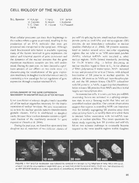
CELL BIOLOGY OF THE NUCLEUS
D.L. Spector P. BubulyaM. CamioloY.-C. ChenL. Denis
Y. Fang S.M. JanickiS. Hearn I. KumaranM. Hubner K.V. Prasanth
Most cellular processes can trace their beginnings tothe nucleus where a gene is activated, resulting in theproduction of an RNA molecule that must getprocessed and transported to the cytoplasm. Althoughmuch biochemical information is available regardingmany of the factors involved in gene expression, thespatial and temporal aspects of gene expression andthe dynamics of the nuclear domains that the geneexpression machinery occupies are less well under-stood. During the past year, we have focused a signif-icant amount of our efforts on two main areas: (1)characterizing the establishment of the gene expres-sion machinery in daughter nuclei after mitosis and (2)examining a new paradigm for the regulation of geneexpression through a nuclear retained RNA.
ESTABLISHMENT OF THE GENE EXPRESSIONMACHINERY IN DAUGHTER NUCLEI AFTER MITOSIS
Upon completion of mitosis, daughter nuclei assembleall of the nuclear organelles necessary for the imple-mentation of nuclear functions. We have been interest-ed in how the nuclear speckle pattern (interchromatingranule clusters, IGCs) is established in daughternuclei because these nuclear domains contain a signif-icant fraction of the machinery essential for geneexpression (Saitoh et al. 2004).
During the past year, we found that upon entry intodaughter nuclei, members of two different classes of
a
0411.
11.14.
0 min
pre-mRNA splicing factors, small nuclear ribonucleo-protein particles (snRNPs) and serine-arginine (SR)proteins, do not immediately colocalize in nuclearspeckles (Bubulya et al. 2004). SR proteins accumu-lated in patches around active nucleolar organizingregions that we refer to as NOR-associated patches(NAPs), whereas snRNPs were enriched at othernuclear regions. NAPs formed transiently, persistingfor 15-20 minutes (Fig. 1) before dissipating asnuclear speckles began to form in G1. In the absenceof RNA polymerase II transcription, NAPs increasedin size and persisted for at least 2 hours, with delayedlocalization of SR proteins to nuclear speckles. Inaddition, SR proteins in NAPs are hypophosphorylat-ed, and the SR protein kinase Clk/STY colocalizeswith SR proteins in NAPs, suggesting that phosphory-lation releases SR proteins from NAPs and their initialtargets are transcription sites.
In mammalian cells, it is not clear how pre-mRNAprocessing factors are activated to a splicing-compe-tent state in newly forming nuclei that have not yetassembled nuclear speckles. Our current observationssuggest that regions surrounding NORs are importantsites for initially segregating SR proteins away fromother splicing factors, to modify them or to allow themto interact before association with pre-mRNA tran-scripts or nuclear speckles. This raises questions forfuture study regarding how SR proteins and snRNPsfollow two separate pathways to eventually becometargeted to the same speckle regions within daughter
FIGURE 1 YFP-SF2/ASF localization was followed by confocal microscopy during telophase in living HeLa cells(a-d),where it transiently accumulates in bright NAPs (a-c, arrows) that persist for approximately 15-20 min, then disappear (c)as YFP-SF2/ASF localizes to nuclear speckles (c-d, arrowheads). (From Bubulya at al. 2004.) Bar, 5 gm.
95

nuclei. It will be important to determine if SR proteinsand snRNPs first meet at transcription sites beforerecycling of complexes through nuclear speckles. Italso raises questions about how different nuclear com-partments assemble/disassemble and communicatewith one another as nuclear domains are establishedafter mitosis.
The results of this study are key to building a timeline of nuclear compartment assembly following mito-sis. They also bring to light the fact that, in general,constituents of nuclear bodies, including pre-rRNAprocessing factors, components of Cajal bodies andPML bodies, and pre-mRNA processing factors, donot immediately localize to and/or assemble their res-ident bodies in daughter nuclei. Instead, there is a lagphase following mitosis as nuclear proteins first enterdaughter nuclei and accumulate temporarily with pro-teins of similar function(s) before nuclear bodies (i.e.,nuclear speckles and Cajal bodies) are established inG1. These transient associations of subsets of proteinsmay be necessary for modification/maturation, or par-tial assembly of multimolecular subcomplexes beforelocalization to nuclear bodies in G1. The subsequentorganization of nuclear compartments such as nuclearspeckles would then provide further modification/maturation and recycling/maintenance of proteincomplexes during interphase.
A NUCLEAR RETAINED RNA REGULATES THE LEVELOF ITS PROTEIN-CODING PARTNER
In addition to harboring the cells' genome, the nucle-us contains distinct substructures in the form of spe-cialized domains/compartments that are characterizedby the absence of delineating membranes, but containdefining sets of proteins (for review, see Spector,Annu. Rev. Cell Biol. 9: 265 [1993] and I Cell Sci.114: 2891 [2001]; Prasanth and Spector, in press). Adynamic interplay between these domains and/or theirconstituents and the genome is thought to foster theefficient progression of gene expression. The centraldogma of molecular biology holds that genetic infor-mation normally flows from DNA to RNA to protein.As a consequence, it has been assumed that genesgenerally encode proteins and that proteins fulfill notonly structural and catalytic functions, but also mostregulatory functions in cells. However, early studiesdemonstrated that RNA can execute functions withoutbeing translated into protein. Such RNAs includehousekeeping RNAs (ribosomal RNA, transfer RNA,and uridine-rich small nuclear RNAs and small nucle-
96
olar RNAs [snRNAs and snoRNAs, respectively]),regulatory RNAs (small interfering RNAs that partic-ipate in the RNAi pathway, microRNAs that functionprimarily as modulators of mRNA translation and sta-bility, naturally occurring antisense transcripts and X-inactive-specific transcript [XIST] associated with theinactive X chromosome in female mammals), and cat-alytic RNAs (telomerase RNA and ribozymes).
Earlier studies from our laboratory have identifieda nuclear population of poly(A)+ RNA that is notchased into the cytoplasm upon termination of tran-scription. We have been interested in identifying andcharacterizing the RNA species in this nuclear popu-lation of RNA. Because these RNAs, for the most part,are not diffusely distributed throughout the nucleo-plasm, we were also interested in identifying thenuclear domains to which they are localized.
We biochemically purified interchromatin granuleclusters and associated nuclear domains from mouseliver nuclei, and constructed a cDNA library frompoly(A)+ RNA. Clones were sequenced and those thatdid not show significant similarity to annotated pro-tein-coding genes were further characterized by RNAfluorescence in situ hybridization (RNA-FISH) in var-ious mouse cell lines. Our screen was to identify RNAsthat presented interesting localization patterns thatwere different from what would be expected for pro-tein-coding transcripts. One of the identified cDNAclones localized in a micropunctate nuclear distribu-tion, and in a population of cells, it also localized to asubnuclear domain referred to as paraspeckles.Northern blot and genomic analysis revealed that thisclone encodes a -9-kb nuclear-retained poly(A)+ RNA,transcribed from the mouse cationic amino acid trans-porter 2 (mCAT2) gene, hence named CAT2 tran-scribed nuclear-RNA (CTN-RNA). The mCAT2 genealso encodes the protein coding mCAT2 mRNA. Bothof these transcripts share all protein-coding exons(exons 2-11) and all introns are properly spliced.However, CTN-RNA is transcribed from promoter Aand utilizes the distal most poly(A) site, whereasmCAT2 mRNA is transcribed predominantly from pro-moters D and E, and utilizes the proximal poly(A) site.CTN-RNA shows tissue-specific expression; elevatedlevels are observed in liver, brain, and lung.Interestingly, CTN-RNA exhibits adenosine-to-inosineediting (A to I) in its 3' UTR (untranslated region),which is likely involved in its nuclear retention.Knockdown of CTN-RNA using antisense oligonu-cleotides designed from the 5' UTR or 3' UTR ofCTN-RNA, which do not overlap with mCAT2 mRNA,
also resulted in the knockdown of mCAT2 transcripts,suggesting that the nuclear-retained CTN-RNA posi-tively regulates the levels of the protein-coding mCAT2mRNA. This regulation may occur through the seques-tration of destabilizing factors by CTN-RNA. SincemCAT2 mRNA and CTN-RNA share all protein-cod-ing exons, they could compete for binding of such fac-tors. Therefore, the knock down of CTN-RNA mayallow access of these factors to the mCAT2 transcripts,resulting in their degradation. These data suggest aninteresting paradigm whereby a nuclear-retained RNAregulates the level of a protein-coding mRNA.
The cellular function of the mCAT2 protein pro-vides some insight into why its mRNA may be highlyregulated. The mCAT2 protein is a cell surface recep-tor that is a member of the cationic amino acid trans-porter family of proteins involved in the uptake ofextracellular L-arginine, which is essential for sus-tained NO (nitric oxide) production via nitric oxidesynthase 2 (NOS2 or iNOS). However, overproductionof NO has been shown to have a pivotal role in inflam-matory responses. Persistent activation of iNOS canlead to the production of toxic levels of NO, resultingin several diseases including multiple sclerosis, HIV-induced dementia, Huntington's disease, Parkinson'sdisease, stroke, and amyotrophic lateral sclerosis. Thishighlights the importance of the cellular uptake of L-arginine as a method of regulating inducible NObiosynthesis. Furthermore, a lack of L-arginine in cellscauses the production of superoxide via the iNOSpathway resulting in cell death. Because the NO path-way is induced in cells under various stress conditionsincluding viral infection and wound healing as a part
of a cellular defense mechanism, it may have evolveda "rapid response" mechanism for regulating the levelof mCAT2 mRNA in cells.
PUBLICATIONS
Bubulya P.A. and Spector D.L. 2004. "On the movements ofnuclear components in living cells. Exp. Cell Res. 296:4-11.
Bubulya RA., Prasanth K.V., Deerinck T.J., Gerlich D.,
Beaudouin J., Ellisman M.N., Ellenberg J., and SpectorD.L. 2004. Hypophosphorylated SR splicing factors tran-siently localize around active nucleolar organizing regions intelophase daughter nuclei. J. Cell BioL 167: 51-63.
Fang Y. and Spector D.L. 2004. Tissue-specific expression anddynamic organization of SR splicing factors in Arabidopsis.Mol. Biol. Cell. 15: 2664-2673.
Janicki S.M., Tsukamoto T., Salghetti S.E., Tansey W.R,Sachidanandam R., Prasanth K.V., Ried T., Shav-Tal Y.,Bertrand E., Singer R.N., and Spector D.L. 2004. Fromsilencing to gene expression: Real-time analysis in singlecells. Cell 116: 683-698.
Ono T., Fang Y., Spector D.L., and Hirano T. 2004. Spatial andtemporal regulation of condensins I and II in mitotic chro-mosome assembly in human cells. Mol. Biol. Cell 15:3296-3330.
Prasanth S.G., Prasanth K.V., Siddiqui K., Spector D.L., andStillman B. 2004. Human Orc2 localizes to centrosomes,centromeres and heterochromatin during chromosomalinheritance. EMBO J. 23: 2651-2663.
Saitoh N., Spahr C.S., Patterson S., Sacco-Bubulya P., NewaldA.F., and Spector D.L. 2004. Proteomic analysis of inter-chromatin granule clusters. Mol. Biol. Cell 15: 3876-3890.
Shav-Tal Y., Darzacq X., Shenoy S.M., Fusco D., Janicki S.M.,Spector D.L., and Singer R.H. 2004. Dynamics of singlemRNPs in nuclei of living cells. Science 304: 1797-1800.
Spector D.L. 2004. Stopping for FISH and chips along thechromatin fiber superhighway. Mol. Cell 15: 844-846.
97

MOLECULAR BIOLOGY OF PAPILLOMAVIRUSES
A. Stenlund A. LeeS. Schuck
The papillomaviruses are a group of viruses that infectand transform the basal epithelium inducing prolifera-tion of the cells at the site of infection. The resultingtumors (warts) are in most cases benign and will usu-ally regress after some time, but certain types ofhuman papillomaviruses (HPVs) give rise to tumorsthat are prone to progress toward malignancy, espe-cially frequently cervical carcinoma. Indeed, HPVinfection appears to be a necessary cause of invasivecervical carcinoma and thus represents one of the fewfirmly established links between viral infections andthe development of cancer.
An impediment to the study of papillomaviruseshas been the inability to define simple in vitro cell cul-ture systems for analysis of the viral life cycle. Theseviruses normally require specialized differentiatingcells that only with difficulty can be generated in cellculture. However, for bovine papillomavirus type 1(BPV-1), a convenient cell culture system exists whereviral gene expression, oncogenic transformation, andviral DNA replication can be studied. Thus, BPV hasbecome a useful model for these aspects of the virallife cycle. The DNA replication properties of thepapillomaviruses show some unique and interestingcharacteristics. As part of their normal lifecycle, theseviruses can exist in a state of latency, which is charac-terized by maintenance of the viral DNA as a multi-copy plasmid in infected cells. The copy number of theviral DNA is tightly controlled and the viral DNA isstably inherited. Papillomaviruses therefore provide aunique opportunity to study plasmid replication inmammalian cells. In addition, the viral DNA replica-tion machinery represents one of the most promisingtargets for antiviral therapy.
In previous years, we have reported the character-ization of the papillomavirus replicon and the identifi-cation of the viral components that are required forviral DNA replication. In recent years, we have direct-ed our attention toward the biochemical events that areassociated with initiation of DNA replication. We arestudying the biochemical properties of the viral Eland E2 proteins and how these two proteins interactwith the viral origin of DNA replication and with thecellular replication machinery to generate initiation
98
complexes. Our studies demonstrate that the El pro-tein has all the characteristics of an initiator protein,including on recognition, DNA-dependent ATPaseactivity, and DNA helicase activity. The transcriptionfactor E2, whose precise function in DNA replicationhas remained more elusive, appears to serve largely asa loading factor for El. Through direct physical inter-actions with both El and the ori, E2 provides sequencespecificity for the formation of the initiation complex.
ASSEMBLY OF A DOUBLE HEXAMERIC HELICASE
Initiation of DNA replication requires binding of theinitiator to the ori, initial opening of the DNA duplex(melting), as well as the assembly and loading of theEl replicative helicase at the replication fork. Formany years, it has been possible to reproduce the sumof these activities in vitro in assays such as the form-U assay, and notably in cell-free DNA replication sys-tems. Since the El protein clearly is capable of pro-viding all of the activities required for initiation andcarry out these functions in sequence, proteins such asEl have held great promise as a means to achieve aprecise molecular understanding of how DNA replica-tion is initiated in a system of low complexity. In addi-tion, since the end product is a double hexameric heli-case, information about El assembly would clarifyhow such enzymes are formed. Currently, it is notknown how double hexameric helicases are assembled,how they engage their substrates, or how they proces-sively unwind double-stranded DNA, although manydifferent models have been proposed. A contributingreason for the limited understanding is the uncertaintyabout what constitutes a bona fide substrate for areplicative DNA helicase. Most helicases have there-fore been characterized on artificial model substratesof uncertain relevance. A great advantage of the viralinitiator proteins is that they, in the process of assem-bly, generate the substrate for the helicase, obviatingthe need for artificial substrates.
Unfortunately, although the El double hexamerhas long been recognized as the replicative DNA heli-case, and therefore the end product of the assembly

process, only limited information about steps preced-ing double hexamer formation exists. Consequently,how and when in the assembly process melting of thetemplate occurs, whether specific precursors arerequired for double hexamer formation, and what rolenucleotide binding and hydrolysis have, are allunknown. The electrophoretic mobility-shift assay(EMSA) has been a particularly powerful tool forunderstanding how protein complexes form on DNA,and we have therefore in the past year developedEMSAs to analyze the El complexes that form on theviral origin of DNA replication. By using very pre-cisely defined conditions, we have been able to reca-pitulate the sequence of events that are required forthe assembly of the double hexameric helicase. TheEl protein binds initially as a dimer. Upon ATP bind-ing, El forms a novel double trimeric complex.Formation of this complex depends on cooperation oftwo different DNA-binding activities, present in theDNA-binding domain (DBD) and the helicasedomain, respectively. ATP binding, but not hydrolysis,is required for formation of the double trimer. Thedouble trimer melts the DNA, providing the firstexample of a defined complex capable of meltingdouble-stranded DNA. Hydrolysis of the bound ATPtriggers formation of a double hexamer, using thedouble trimer as a precursor. The double hexamer hasthe capacity to unwind the partially melted DNA in anATP-dependent manner, providing the openingrequired for initiation of DNA synthesis.
MUTATIONAL ANALYSIS OF THE El DBD SURFACE
The DBD of the El protein clearly is responsible forthe highly site-specific DNA binding that the El pro-tein is capable of, which in turn is responsible forrecognition of the origin of DNA replication in theviral genome. Furthermore, on the basis of both struc-tural and biochemical studies of the El DBD, theDBD also provides the dimerization surface for the E 1
protein and also contains an interaction surface for theDBD from the E2 protein. In addition, based on imag-ing analysis of, for example, SV40 T antigen, it is like-ly that the El DBD has an active part in the formationof large complexes such as the double hexamer, whichis the replicative DNA helicase. To identify additionalfunctions in the DBD, we have performed a completesurface mutagenesis of the E1 DBD. Based on thehigh-resolution structure of the E 1 DBD, we have sub-stituted approximately 70 surface residues for alanine,avoiding areas known from the structural analysis tobe involved in DNA binding and dimerization. Afterscreening these El mutants for expression and for invivo DNA replication activity, we were able to identi-fy 18 mutants with defects in DNA replication. Six ofthese were defective for interaction with E2, and theremaining 12 mutants showed unimpaired interactionwith E2 and consequently were defective for DNAreplication for other reasons. To determine what bio-chemical functions might be defective in thesemutants, we expressed and purified the 12 mutant Elproteins and tested them in five different in vitroassays related to DNA replication. None of themutants showed a defect in either the ATPase or thenonspecific DNA helicase activities, indicating thatthe protein structure was not greatly affected by themutations and that most likely, the ability to formhexameric structures is unaffected by the mutations.However, all 12 mutants had severe defects for in vitroDNA replication as well as defects in melting activityand in fragment unwinding activity. By using ourrecently developed EMSAs, we have been able to testthese mutants directly for the formation of doubletrimers and double hexamers. Of the 12 mutants, 5 haddefects in double hexamer formation, indicating thatthe DBD has an active part in the assembly of the dou-ble hexamer. Interestingly, however, mutations thataffect dimerization by the E1 DBD are not severelydefective for double hexamer formation, indicatingthat the double hexamer is unlikely held together bythis dimer interaction.
99

DNA REPLICATION AND CHROMATIN INHERITANCE
B. Stillman A. Chabes J. Mendez Y.-J. Sheu S. VadiveluV. Ellison S.G. Prasanth K. Siddiqui P. WendelM. Hoek M. Rossmann C. Speck Z. ZhangA. Mazurek
Our research focuses on the inheritance of chromo-somes and chromatin structures in eukaryotic cells.The primary interest is how DNA replication occurs,how it is regulated, and how it is coordinated withother events such as chromosome segregation duringmitosis. Most of our research now focuses on themechanism of initiation of the process that duplicatesthe genome which encodes our genetic information.The focus of this research is the origin recognitioncomplex (ORC) which binds to DNA sequences thatlocalize where DNA replication begins along chromo-somes. ORC binds a number of proteins during thecell division cycle, and together, they coordinate thechromosome duplication process.
Our previous studies have shown that ORC inter-acts with a regulatory protein called Cdc6 in an ATP-dependent manner and that this interaction changes
year, we have investigated this reaction in more detailand have identified a new regulatory role for originDNA sequences in controlling ORC function. Theaddition of Cdc6 protein to the ORC-DNA complexcauses a dramatic change in the way both ORC andCdc6 bind to DNA. ORC normally binds to about 48by in the origin DNA and protects them from digestionin vitro by deoxyribonuclease I (DNase I). Upon addi-tion of Cdc6, and only in the presence of ATP, theDNase I footprint is greatly extended to about 85 bp,resembling the region of the origin that binds proteinsin vivo during the G, phase of the cell cycle, prior tothe initiation of DNA replication. ATP hydrolysis byORC is required for formation of the extended foot-print over the origin DNA since mutant versions ofORC or Cdc6 that lack ATPase activity cannot supportthe reaction. Furthermore, nonhydrolyzable analogs ofATP fail to support formation of the extended foot-print, even though they do support Cdc6 binding toORC on origin DNA. In collaboration with Huilin Li'sgroup at the Brookhaven National Laboratory, wehave begun to investigate the structure of ORC, theORC-Cdc6 complex, and the structure of these com-plexes bound to DNA.
100
What is interesting about these reactions at the ori-gin of DNA replication is the dual role of specificDNA sequences in the control of events prior to theinitiation of DNA replication. Stephen Bell's laborato-ry (MIT) has shown that when ORC is bound to theorigin of DNA replication, the ORC ATPase activity issuppressed. We have now shown that when Cdc6 isadded to the origin, the ORC ATPase activity is acti-vated and the ATPase requires specific DNAsequences in the origin, particularly the B1 and B2elements in the origin. These two DNA elements arethe region of the origin where the extended footprintoccurs. Thus, in one circumstance, ORC ATPase issuppressed by the origin DNA sequences, and later,when Cdc6 binds, ORC ATPase activity is activated bythe same origin sequences. We think that this ATPaseswitch is required to load the minichromosome main-tenance (MCM) proteins onto the origin to form theprereplicative complex (pre-RC) that renders the ori-gin competent for subsequent initiation of DNA repli-cation in the S phase of the cell cycle. These resultsalso imply that ORC ATPase activity is cell-cycle-reg-ulated since Cdc6 is only present in yeast cells frommitosis until the cell commits to a new round of celldivision.
In human cells, the regulation of ORC is morecomplicated. We have demonstrated that ORC is not astable complex in the cell division cycle, but that dif-ferent complexes exist at different times. Neverthe-less, just like in yeast cells, continually proliferatinghuman cells form the pre-RC as they exit mitosis in aprocess that requires ORC, Cdc6, Cdtlp, and theMCM proteins. In collaboration with Steve Reed'slaboratory (Scripps Research Institute), we investigat-ed the regulation of MCM loading onto chromatin bycyclin E.
Cyclin E is often overexpressed in human cancercells and patients with such tumors, particularly thosewith breast carcinomas who have a poor outcome forsurvival. Steve Reed's laboratory has shown that cellsthat overexpress cyclin E accumulate genomic abnor-malities that could cause additional mutations in the

DNA and accelerate cancer progression. In collabora-tion with his laboratory, we have demonstrated thatoverexpression of cyclin E causes a reduction in theloading of Mcm4 protein, one of the key pre-RC com-ponents. As a result, such cells progress through theG, phase of the cell cycle with fewer pre-RCs and thisin turn causes slow progression through S phase. Wehave suggested that progression through S phase witha low number of competent origins of DNA replica-tion may cause some cells to enter mitosis withoutcompleting DNA replication, resulting in chromoso-mal abnormalities.
PUBLICATIONS
Austin CR, Battey J.F., Bradley A., Bucan M., Capecchi M.,Collins ES., Dove W.F., Duyk G., Dymecki S., Eppig J.T.,Grieder EB, Heintz N., Hicks G., Insel TR., Joyner A.,Koller B.H., Lloyd K.C.K., Magnuson T., Moore MAN., NagyA., Pollock J.D., Roses AD., Sands AT, Seed B., SkarnesW.C., Snoddy J., Soriano R, Stewart D.J., Stewart F,Stillman B., Varmus H., Varticovski L., Verma I.M., Vogt
T.F., von Melchner H., Witkowski J., Woychik R.P, WurstW., Yancopoulos G.D., Young S.G., and Zambrowicz B.2004. The knockout mouse project. Nat. Genet. 36:921-924.
Ekholm-Reed S., Mendez J., Tedesco D., Zetterberg A.,Stillman B., and Reed S.I. 2004. Deregulation of cyclin E inhuman cells interferes with pre-replication complex assem-bly. J. Cell Biol. 165: 789-800.
Prasanth S.G., Mendez J., Prasanth K.V., and Stillman B. 2004.Dynamics of pre-replication complex proteins during thecell division cycle. Philos. Trans. P. Soc. Lond. B Biol. Sci.359: 7-16.
Prasanth S.G., Prasanth K.V., Siddiqui K., Spector D.L., andStillman B. 2004. Human Orc2 localizes to centrosomes,centromeres and heterochromatin during chromosomeinheritance. EMBO J. 23: 2651-2663.
In Press
Spruck C.H., Smith A.P.L., Reed S.E., Sangfelt 0., Keck J.,Strohmaier H., Mendez J., Widschwendter M., Stillman B.,Zetterberg A., and Reed S.I. 2005. Deregulation of cyclin Eand genomic instability. In Hormonal carcinogenesis, vol. IV(ed. J.J. Li et al.). Springer-Verlag. (In press.)
Stillman B. 2005. Origin recognition and the chromosomecycle. FEBS 579: 877-884.
Yi-Jun Sheu, postdoc, Bruce Stillman's lab.
101

PROTEIN TYROSINE PHOSPHATASES AND THE CONTROL OFSIGNAL TRANSDUCTION
N.K. Tonks J.N. Andersen N. Gorovits A. PicciniD.A. Buckley A. Hague Z. YaoH.H. Chen S. Hinton M. YeR.L. Del Vecchio G. Lin L. Zhang
The phosphorylation of tyrosyl residues in proteins isa key component of the regulation of signaling path-ways that control many fundamental physiologicalprocesses including cell proliferation and differentia-tion. Protein phosphorylation is a reversible, dynamicprocess in which the net level of phosphate observedin a target substrate reflects the activity of the kinasesthat phosphorylate it and the protein phosphatases thatcatalyze the dephosphorylation reaction. We study thefamily of protein tyrosine phosphatases (PTPs),which, like the kinases, comprise both transmem-brane, receptor-linked forms as well as nontransmem-brane, cytoplasmic species and represent a major fam-ily of signaling enzymes. We are integrating a varietyof experimental strategies to characterize the physio-logical function of members of the PTP family.Disruption of normal patterns of tyrosine phosphory-lation has been implicated in several human diseases.Therefore, insights into the mechanisms involved inmodulating PTP function may ultimately yield impor-tant information to help counter such diseases.
During the last year, two graduate students,Aftabul Hague and Guang Lin, from the MCBProgram at Stony Brook carried out rotation projectsand have joined the lab to conduct their Ph.D. studies.
THE COMPOSITION AND DIVERSITYOF THE PTP FAMILY
Since the human genome sequence became available,one emphasis of the lab has been to define the compo-sition of the PTP family. We have now cataloged thehuman PTP genes and more recently extended ourgenomic sequence analysis to other organisms. ThePTPs, which are defined by the active site signaturemotif HC(X)5R are divided into two broad categories:the classical, pTyr-specific phosphatases and the dualspecificity phosphatases (DSPs). The DSPs are opera-tionally defined as members of the PTP family thathave the capability of dephosphorylating both pTyrand pSer/pThr residues in vitro. Nonetheless, in terms
102
of physiological function, the "DSPs" may actuallyshow preference for either Tyr or Ser/Thr residues.Some DSPs even target nonprotein substrates such asinositol pholipids and RNA. This study was initiated asa collaboration with Dr. Niels Peter Moller and his col-leagues at Novo Nordisk in which we determined thatthe total number of classical PTP genes in humans is38. We have found direct orthologs of each of thesehuman PTP genes in the mouse genome. In contrast,we have also discovered 12 PTP pseudogenes that areunique to humans. Interestingly, some of these pseudo-genes yield transcripts; however, they would only pro-duce truncated/mutated proteins, the function ofwhich, if any, is unclear. We have continued thesegenomic analyses independently, most recently pub-lishing a "Practical Guide to Bioinformatics and DataResources for Computational Analysis of PTPs" inwhich we define the classical PTPs in a variety oforganisms. Furthermore, we have extended this study,in collaboration with Ravi Sachidanandam and his col-leagues here at CSHL, to search the human genomesequence for DSPs that are related to VH1, the proto-typic DSP, a 20-kD protein that is a virulence factor ofVaccinia virus. This proved to be a greater challengethan our similar analysis of the classical PTPs because,although they contain the active site signature motif,the DSPs are less well-conserved overall and thereforeharder to identify on the basis of their sequence thanthe classical PTPs. We have identified 43 human VH1-like DPSs, all but one of which are also found inmouse. Similar searches have been performed in vari-ous other genome sequences, including Drosophiliamelanogaster, Caenorhabditis elegans, Fugu rubripes,and Arabidopsis thaliana, to shed light on the evolu-tionary history of the PTP superfamily. These data arecurrently being prepared for publication. In summary,the PTP family in humans is encoded by 106 genes asfollows: 38 classical PTPs, 43 VH1-like DSPs, and,from other published studies, 3 cdc25s, 16 mytobular-in-like phosphatases, 1 low-molecular-weight PTP, 2inositol-4- phosphatases, and 3 Sac domain inositol 5-

phosphatases. In comparison, there are 90 human PTKgenes, illustrating comparable levels of complexitybetween the two families. However, it is important toremember that this reflects the minimal level of com-plexity, with diversity being enhanced by such mecha-nisms as alternative mRNA splicing, alternative pro-moter usage, proteolytic processing, etc. Nonetheless,this provides a foundation for future studies aimed atprofiling PTP sequences at the protein level.
To facilitate research on the PTPs within the sig-nal transduction community, we are making our analy-sis of the PTP family available as a Web-accessibleresource at two locations: http://ptp.cshl.edu and areplica site at http://science.novonordisk.com/ptp.This resource contains our database of PTP accessionnumbers, multiple sequence alignments, phylogenetictrees, structure files, annotated molecular graphicsfiles, chromosomal mapping data, analysis of exonstructure, pseudogenes, and disease linkages. The PTPdatabase can be searched by key words or by sequencesimilarity using our BLAST server, which also pro-vides a tool for phylogenetic classification of anony-mously submitted sequences based on PTP domainhomology and neighbor joining trees. This is provingto be a popular resource, and we are continuing toupdate the site with the goal of maintaining it as thedefinitive source of information on the PTP family forthe scientific community.
RECEPTOR PTPli AND ENDOTHELIALCELL FUNCTION
PTPp, is a receptor PTP, the extracellular segment ofwhich consists of one Ig (immunoglobin) domain, fourFN III (fibronectin type III) repeats, and a MAM (Mu,A5 and Meprin homology) domain, the latter being aconserved domain unique to PTP t, x, p, X, and somecell adhesion molecules. Previously, we had shown thatPTP1.1 is a homophilic adhesion protein, such that theligand for one molecule of PTP1.1 is the extracellularsegment of another PTPu molecule expressed on thesurface of an apposing cell. Furthermore, we identifiedthe Ig domain as being necessary and sufficient forbinding. We have also demonstrated that PTPp, inter-acts with cadherin-catenin cell adhesion complexes andmay regulate the adhesive properties of classical cad-herins by dephosphorylating cell junction proteins.PTPµ is expressed predominantly in endothelial cells.In subconfluent cell cultures, PTPµ resides in an intra-cellular membrane pool; however, as culture densityincreases and cell contacts form, the phosphatase islocalized to sites of cell-cell contact and its expression
level increases. These characteristics of PTPp., whichare consistent with a role in cell-cell adhesion, suggestthat control of subcellular localization and protein levelare important mechanisms to regulate the function ofthis phosphatase. Furthermore, we noted that overex-pression of PTPpt in a bovine aortic endothelial cellline, GM7372, reduced cell number in culture. Theseeffects were most pronounced when cell density waslow, the condition that coincides with low levels ofendogenous PTPu, and resulted from increased apopto-sis, rather than decreased proliferation. To gain a betterunderstanding of the regulation of PTPµ function, weexplored further a structure-function analysis of theenzyme, focusing on the extracellular segment, in par-ticular the importance of the conserved Ig domain, con-taining the homophilic binding site. We observed thatthe deletion of the Ig domain greatly reduced the local-ization of PTPu to cell-cell contacts and resulted in dis-persal of the mutant protein across the cell surface.Similar results were observed in endothelial and epithe-lial cells, suggesting that the localization of the enzymewas a property of the PTP itself and not a phenomenonunique to particular cells. In addition, deletion of the Igdomain affected localization of PTPii in subconfluentcells, when homophilic binding to another PTPu mole-cule on an apposing cell is not possible, leading to anaccumulation of the PTP at the cell periphery in theregion occupied by focal adhesions. This apparentlocalization coincided with changes in the morphologyof focal adhesions, as well as in the distribution ofcytoskeletal components such as vinculin, paxillin, andactin. These effects were accompanied by increasedapoptosis; in fact, deletion of the Ig domain enhancedthe apoptotic effect of PTP1.1 to the highest levels seenwith any of the mutant constructs tested. This studytherefore illustrates the critical role of the Ig domain inregulation of the localization of PTP1.t and the impor-tance of such control for the maintenance of normalcell physiology.
The vascular endothelium represents a barrier thatregulates paracellular movement of macromoleculesinto extravascular compartments. This paracellularpathway of macromolecular flux is controlled in partby tyrosine phosphorylation of adherens junction (AJ)proteins, which integrate cell-cell contact and actincytoskeleton. Enhanced tyrosine phosphorylation isassociated with increased trans-endothelial flux (i.e.,decreased barrier function). Several AJ proteins areknown to be tyrosine-phosphorylated, such as VE-cad-herin and catenins, but the mechanistic links betweentyrosine phosphorylation and barrier function remainto be defined. In collaboration with Dr. Simeon
103

Goldblum (University of Maryland) we have beenexploring potential roles for PTPli in regulating AJtyrosine phosphorylation and barrier function in pul-monary vascular endothelial cells. We observed thatthe PTI4t was expressed in postconfluent human pul-monary artery and lung microvascular endothelial cells(HMECs), where it was almost exclusively restricted tocell-cell boundaries. Ablation of PTP1.1 expression byRNA interference dramatically impaired barrier func-tion. Conversely, overexpression of wild-type PTPixenhanced barrier function. These data are consistentwith PTPµ exerting a regulatory influence over barrierfunction. We have been trying to identify physiologicalsubstrates of PTI,t to explore the mechanisms under-lying these effects. In light of its association with clas-sical cadherins, we tested whether there was an inter-action between PTP11 and VE-cadherin. We observeddirect binding between the recombinant proteins invitro and colocalization and coimmunoprecipitation ofPT131.t and VE-cadherin from HMECs. Interestingly,overexpression of PTPI.t in HMECs led to a decrease intyrosine phosphorylation of VE-cadherin, with speci-ficity indicated by the fact that there were no changesin tyrosine phosphorylation of 13 catenin. Whether thisis the critical regulatory event in the control of barrierfunction remains to be established.
THE DUAL SPECIFICITY PHOSPHATASES
Many of the 43 DSPs we defined in our analysis of thehuman genome sequence are uncharacterized.Phylogenetic analyses divided these enzymes intothree classes, revealing characteristics that were usedto prioritize further studies. Currently, we are focusingon the following.
Jsp-1
Multiple DSPs have been identified that have the abil-ity to dephosphorylate and inactivate various MAPkinases, constituting a complex response network forattenuation of MAPK-dependent signals. We identi-fied a novel DSP and observed, contrary to expecta-tion, that it had the capacity to activate Jnk specifical-ly, hence, JSP-1 (Jnk stimulatory phosphatase 1). Inaddition, we observed enhanced phosphorylation ofMKK-4, which phosphorylates and activates Jnk.Furthermore, expression of a dominant-negativemutant form of MKK-4 abolished the activation of Jnkby JSP-1, suggesting a site of action of the phos-phatase upstream of MKK-4 in the Jnk signaling cas-cade. This study illustrates a new potential tier of con-trol of the Jnk signaling pathway and a novel aspect of
104
the role of protein phosphatases in the control ofMAPK signaling. This raised the possibility that JSP-1 may offer a different perspective to the study of var-ious inflammatory and proliferative disorders associ-ated with dysfunctional Jnk signaling. We have nowgenerated JSP-1 knockout mice, which are born inMendelian ratios, and we are characterizing their phe-notype. Thus far, we have observed that ablation ofJSP-1 leads to attenuation of the inflammatoryresponse to Gram-negative bacterial endotoxin, sug-gesting an important role in the innate immune sys-tem. The mechanistic basis for this effect is currentlyunder investigation. In addition, preliminary resultsindicate that following treatment of JSP-1--/- micewith MPTP (1-methy1-4-pheny1-1,2,4,6-tetrahydropy-ridine), the activation of Jnk is attenuated compared towild-type controls. MPTP is a neurotoxin that repli-cates most of the neuropathological features ofParkinson's disease. Currently, we are investigatingthe mechanism by which JSP-1 regulates the Jnk path-way in this context and the extent to which manipula-tion of JSP-1 may influence neurodegeneration inParkinson's disease.
Two Distinct DSPs Encoded in Alternative OpenReading Frames of a Single Gene at HumanChromosone 10q22.2
We identified a gene located on chromosome 10q22.2,which utilizes alternative open reading frames (ORFs)to encode TMDP (testis and skeletal specific DSP) anda novel enzyme that we term MDSP (muscle restrictedDSP). This gene organization is conserved in mice.Although this is reminiscent of the p 1 6Ink4/p19Arflocus, to our knowledge, this is the first example of agene from which two distinct proteins of the same fam-ily are expressed using alternative ORFs. We demon-strated that both MDSP and TMDP are restricted tospecific tissues, skeletal muscle and testis, respective-ly. Expression of both proteins was markedly increasedat approximately the third week after birth and contin-ued to increase gradually into adulthood, implying thatthe physiological functions of both DSPs are specificto the mature organs. More recently, we have found thatMDSP may be overexpressed in certain tumors, and weare currently focusing on a functional characterizationof the enzyme.
DUSP23
The gene for this DSP is located at chromosome1q22.23, a region that is associated with amplifica-tions, translocations, and other abnormalities in multi-ple cancers. Our phylogenetic analysis assigned this
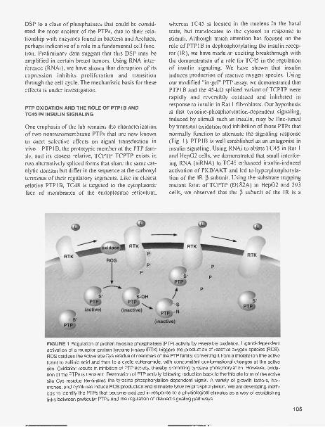
DSP to a class of phosphatases that could be consid-ered the most ancient of the PTPs, due to their rela-tionship with enzymes found in bacteria and Archaea,perhaps indicative of a role in a fundamental cell func-tion. Preliminary data suggest that this DSP may beamplified in certain breast tumors. Using RNA inter-ference (RNAi), we have shown that disruption of itsexpression inhibits proliferation and transitionthrough the cell cycle. The mechanistic basis for theseeffects is under investigation.
PTP OXIDATION AND THE ROLE OF PTP1B ANDTC45 IN INSULIN SIGNALING
One emphasis of the lab remains the characterizationof two nontransmembrane PTPs that are now knownto exert selective effects on signal transduction invivo-PTP1B, the prototypic member of the PTP fam-ily, and its closest relative, TCPTP. TCPTP exists intwo alternatively spliced forms that share the same cat-alytic domain but differ in the sequence at the carboxylterminus of their regulatory segments. Like its closestrelative PTP1B, TC48 is targeted to the cytoplasmicface of membranes of the endoplasmic reticulum,
whereas TC45 is located in the nucleus in the basalstate, but translocates to the cytosol in response tostimuli. Although much attention has focused on therole of PTP1B in dephosphorylating the insulin recep-tor (IR), we have made an exciting breakthrough withthe demonstration of a role for TC45 in the regulationof insulin signaling. We have shown that insulininduces production of reactive oxygen species. Usingour modified "in-gel" PTP assay, we demonstrated thatPTP1B and the 45 -kD spliced variant of TCPTP wererapidly and reversibly oxidized and inhibited inresponse to insulin in Rat 1 fibroblasts. Our hypothesisis that tyrosine-phosphorylation-dependent signaling,induced by stimuli such as insulin, may be fine-tunedby transient oxidation and inhibition of those PTPs thatnormally function to attenuate the signaling response(Fig. 1). PTP1B is well established as an antagonist ininsulin signaling. Using RNAi to ablate TC45 in Rat 1and HepG2 cells, we demonstrated that small interfer-ing RNA (siRNA) to TC45 enhanced insulin-inducedactivation of PKB/AKT and led to hyperphosphoryla-tion of the IR [3 subunit. Using the substrate trappingmutant form of TCPTP (D182A) in HepG2 and 293cells, we observed that the 13 subunit of the IR is a
ROS
S-PTP
(active)
P
P
S-OH
S-PTP
PTP
(inactive) PTP -N
inactive)
P
P
P
S-PTP
FIGURE 1 Regulation of protein tyrosine phosphatase (PTP) activity by reversible oxidation. Ligand-dependentactivation of a receptor protein tyrosine kinase (RTK) triggers the production of reactive oxygen species (ROS).ROS oxidizes the active site Cys residue of members of the PTP family, converting it from a thiolate ion (the activeform) to sulfinic acid and then to a cyclic sulfenamide, with concomitant conformational changes at the activesite. Oxidation results in inhibition of PTP activity, thereby promoting tyrosine phosphorylation. However, oxida-tion of the PTPs is transient. Restoration of PTP activity following reduction back to the thiolate form of the activesite Cys residue terminates the tyrosine-phosphorylation-dependent signal. A variety of growth factors, hor-mones, and cytokines induce ROS production and stimulate tyrosine phosphorylation. We are developing meth-ods to identify the PTPs that become oxidized in response to a physiological stimulus as a way of establishinglinks between particular PTPs and the regulation of defined signaling pathways.
105

direct substrate of TCPTP. Furthermore, blotting ofimmunoprecipitates of IR 13 from siRNA-treated cellswith phospho-specific antibodies indicated that TC45displays preferential recognition of particular siteswithin the receptor.
We have explored further the effects of PTP andTC45 on insulin signaling using fibroblasts (MEFs)derived from PTP1B-/- and TCPTP-/- mice, in col-laboration with Tony Tiganis (Monash University,Australia). Ablation of PTP resulted in enhanced,insulin-induced phosphorylation of both IR [3 andIRS-1, in contrast to TCPTP-/- MEFs in which hyper-phosphorylation of IR 13 occurred in the absence of aneffect on IRS-1. Furthermore, whereas PKB phospho-rylation was elevated/prolonged in cells that lackedeither PTP or TCPTP, compared to their wild-typeor reconstituted counterparts, Erk 1/2 phosphorylationwas enhanced only in PTP1B-/- cells. These data sug-gest that the two PTPs may exert differential effects oninsulin signaling. To explore further the mechanism ofaction of these PTPs, we examined the phosphoryla-tion status of IR [3 using phospho-specific antibodiesto Y1162/63, from the activation loop of the IR PTK,and Y972, a site that contributes to the recruitment ofIRS-1. Insulin-induced phosphorylation of both siteswas enhanced in the absence of PTP1B; however, thekinetics of phosphorylation were similar in the pres-ence or absence of the PTP, peaking at 2 minutes anddeclining rapidly thereafter. In contrast, phosphoryla-tion of Y1162/63 was not altered at early time pointsof insulin stimulation in the absence of TCPTP;instead, phosphorylation was sustained over a pro-longed time course out to 100 minutes, compared toTCPTP+/+ cells in which phosphorylation haddeclined to baseline by this time. In addition, weobserved that phosphorylation of Y972 was enhancedin TCPTP-/- MEFs early in the time course of insulinstimulation, when Y1162/63 phosphorylation was notaltered significantly, indicating that TCPTP exerts dif-ferential, independent effects on these two phosphory-lation sites. The effects of these PTPs can be distin-guished further using RNAi to suppress TCPTPexpression in PTP1B-/- MEFs. In PTP1B-/- MEFs,we observed that insulin-induced phosphorylation ofPKB declined at 40-60 minutes, whereas phosphory-lation was maintained for at least 100 minutes follow-ing RNAi-induced suppression of TCPTP in thesecells. In contrast, suppression of TCPTP did not alterthe time course of Erk2 activation, illustrating speci-ficity and that the ablation of TCPTP did not producea general enhancement of signaling. Furthermore, we
1 flA
observed that suppression of TCPTP by RNAi inPTP1B-/- MEFs led to prolonged phosphorylation ofY1162/63 in IR [3. These observations illustrate that ina cellular context, PTP and TCPTP are not redun-dant, but rather act in a coordinated manner to exertdifferential effects in the regulation of IR 13 phospho-rylation and signaling in vivo.
PUBLICATIONS
Andersen J.N., Jansen P.G., Echwald S.M., Mortensen O.H.,Fukada T., Del Vecchio R., Tonks N.K., and Moller N.P.2004. A genomic perspective on protein tyrosine phos-phatases: Gene structure, pseudogenes and genetic dis-ease linkage. FASEB J. 18: 8-30.
Bukczynska P., Klingler-Hoffmann M., Mitchelhill K.I., Lam M.H.,Ciccomancini M., Tonks N.K., Sarcevic B., Kemp B.E., andTiganis T. 2004. The T-cell protein tyrosine phosphatase isphosphorylated on Ser-304 by cyclin-dependent proteinkinases in mitosis. Biochem. J. 380: 939-949. (Epub. 19March 2004, doi:10.1042/BJ20031780.)
Chen H.H., Luche R., Wei B., and Tonks N.K. 2004.Characterization of two distinct dual specificity phos-phatases encoded in alternative open reading frames of asingle gene located on human chromosome 10q22.2. J.Biol. Chem. 279: 41404-41413. (Published online 13 July2004, DOI 10.1074/jbc. M405286200.)
Meng T.C., Buckley D.A., Galic S., Tiganis T., and Tonks N.K.2004. Regulation of insulin signaling through reversible oxi-dation of the protein-tyrosine phosphatases TC45 andPTP1B. J. Biol. Chem. 279: 37716-37725. (Publishedonline 10 June 2004, DOI 10.1074/jbc.M404606200.)
In Press
Andersen J.N., Del Vecchio R., Kannan N., Gergel J., NeuwaldA.F., and Tonks N.K. 2005. Computational analysis of pro-tein tyrosine phosphatases: Practical guide to bioinformat-ics and data resources. Methods 35: 90-114.
Del Vecchio R.L. and Tonks N.K. 2005. The conservedimmunoglobulin domain controls the subcellular localiza-tion of the homophilic adhesion receptor protein tyrosinephosphatase It J. Biol. Chem. 280: 1603-1612.(Published online 18 October 2004, DOI 10.1074/jbc.M410181200.)
Galic S., Hauser C., Kahn B.B, Fawaz G.H., Neel B.G., TonksN.K., and Tiganis T. 2005. Coordinated regulation of insulinsignaling by the protein tyrosine phosphatases PTP1B andTCPTP. Md. Cell. Biol. 25: 819-829.
Meng T.C., Hsu J.S.F., and Tonks N.K. 2005. Development of amodified in-gel assay to identify protein tyrosine phos-phatases that are oxidized and inactivated in vivo. Methods35: 28-36.
Sui X.F., Kiser T.D., Hyun S.W., Angelini D.J., Del Vecchio R.L.,Young B.A., Hasday J., Romer L.H., Passaniti A., TonksN.K., and Goldblum S.E. 2005. Receptor protein tyrosinephosphatase associates with and dephosphorylates zonu-la adherens proteins in pulmonary vascular endothelia andregulates barrier integrity. Am. J. Pathol. (in press).

RAS AND RHO GTPASES AND THE CONTROL OFSIGNAL TRANSDUCTION
L. Van Aelst B. Boettner V. VelamoorE.-E. Govek M. UchidaJ. Janas M. ZhaoR. Packer
Our research focuses on signal transduction pathwaysinvolving members of the Ras and Rho GTPases andthe physiological processes that they regulate. Ras andRho family members have been documented to havekey roles in cellular activities controlling cell growthcontrol, differentiation, and morphogenesis. Altera-tions that affect normal Ras and Rho function havebeen found to result in the development of several dis-ease processes, including cancer, as well as inflamma-tory and neurological disorders. To understand howalterations in Ras and Rho signaling components con-tribute to these disease processes, it is essential tocomprehend their normal functions at both a molecu-lar and a cellular level. Efforts in my laboratory havefocused on defining the role and mechanisms bywhich Ras and Rho family members exert their effectson specific aspects of tumorigenesis and on neuronaldevelopment. Below are highlighted the main projectsthat have been carried out during the past year.
ROLE OF RAP1 SIGNALINGIN EPITHELIAL MIGRATION ANDCELL-CELL ADHESION
The Rapl protein, a member of the Ras family, wasoriginally identified as an antagonist of oncogenicRas, but more recent studies indicate that the functionof Rapl is largely independent of Ras. Rapl is funda-mental for the maintenance of cell-cell contactsbetween nascent epithelial cells and promotes cellmovement. Importantly, a growing body of evidencesuggests a dysregulation of Rapl signaling in malig-nant processes. The signaling pathways mediatingthese effects of Rapl remain largely unknown. We pre-viously described the identification of the mammalianjunctional protein AF-6 as a Rapl-interacting protein.To demonstrate the physiological relevance of thisinteraction, we decided to carry out functional analy-ses of their orthologs (DRapl and Canoe) in the moregenetically tractable Drosophila system. We foundthat both DRapl and Canoe are required for epithelial
migration events in the embryo and that Canoe acts asa downstream effector of DRapl in this process.Together, these experiments show that Canoe/AF-6 isa "bone fide" effector of Rapl in the process of epithe-lial migration.
More recently, we identified a Drosophila Rapl-specific exchange factor, DPDZGef, which is respon-sible for Rapl activation in migrating embryonicepithelia. Genetic and biochemical experimentsshowed that the DPDZGef gene is indispensable forepithelial migration in the embryo and that it activatesthe Rapl /Canoe pathway. Interestingly, the mam-malian counterpart of the DPDZGef protein has beendescribed to interact with the adherens junctional pro-tein, 0-catenin and, like AF-6/Canoe, localizes to cell-cell junctions. Our data reveal a novel Rapl-dependentpathway key in regulating epithelial cell migration andadhesive processes that likely is conserved acrossspecies.
ROLE OF DOK PROTEINS IN LEUKEMIASUPPRESSION
p62"k (also called Dok-1) was cloned as a major phos-phorylation substrate of the p2106o""' oncoprotein inCML (chronic myelogenous leukemia) blasts. Thisprotein was termed Dok (downstream of kinases),since it was also found to be a common substrate ofmany receptor and cytoplasmic tyrosine kinases.Subsequently, four additional Dok family membershave been identified. Among them, Dok-1 and Dok-2share in common the ability to bind to a negative reg-ulator of Ras, Ras-GAP, suggesting that they mayserve critical, but possibly redundant, functions. In lastyear's Annual Report, we reported that Dok-1 acts as anegative regulator of growth-factor-induced cell pro-liferation and that it does so at least in part by nega-tively influencing the Ras/MAPK pathway. We alsoreported that Dok-1 inactivation causes a significantshortening of the latency of the fatal myeloprolifera-tive disease induced by p2106cr-ab1. These findings
107

implicated a role for Dok-1 in the negative regulationof p210ber-a' signaling and leukemogenesis.
Dok-1 (or Dok-2) single knockout (KO) mice didnot show gross aberrant phenotypes and displayednormal steady-state hematopoiesis. This observationsuggested that the two proteins could exert redundantfunction(s). In collaboration with Dr. Pandolfi's group(Memorial Sloan-Kettering Cancer Center), werecently characterized Dok-1/Dok-2 double KO(DKO) mice. Concomitant Dok-1 and Dok-2 inactiva-tion resulted in aberrant hematopoiesis and Ras/MAPkinase activation. Inactivation of both genes increasedthe proliferative potential of myeloid bone marrow(BM) cells and protected them from growth-factor-deprivation-induced cell death. DKO BM cells dis-played elevated levels of Ras and MAPK upon GM-GSF stimulation. Strikingly, all Dok-1/Dok-2 DKOmutants spontaneously developed transplantableCML-like myeloproliferative disease, likely resultingfrom increased cellular proliferation and reducedapoptosis. These findings reveal a critical role forDok 1 and Dok2 in the control of the hematopoieticcompartment homeostasis.
REGULATORS OF RHO GTPASES CONTROLLINGKEY ASPECTS OF NEURONAL FUNCTION ANDDEVELOPMENT
Accumulating data indicate that the Rho family ofGTPases and their regulatory molecules have criticalroles in many aspects of neuronal development, par-ticularly in determining neuronal morphology viatheir various actions on the actin cytoskeleton. Giventheir importance in neuronal processes, it is thereforenot surprising that mutations in genes encoding anumber of regulators and effectors of the Rho-GTPases are associated with diseases affecting thenervous system. Aberrant Rho signaling has beenshown to underlie various forms of mental retardationas well as defects in axon regeneration. We have beenfocusing on two regulators of the Rho GTPases,oligophrenin-1 and DOCK7, for which we demon-strated roles in dendritic morphogenesis and axon for-mation, respectively.
Oligophrenin-I (OPHN1) codes for a protein thathas a Rho-GAP domain shown to negatively regulateRho family members. It was originally identified bythe analysis of a balanced translocation t(X;12)observed in a female patient with mental retardation(MR). Subsequently, a mutation within the OPHN1-coding sequence was identified in a family with non-
108
syndromic X-linked MR (MRX 60). In both cases,OPHN1 mutations were associated with a loss of ordramatic reduction in, mRNA product. More recentstudies have also reported the presence of OPHN1mutations in families with MR associated with epilep-sy and/or cerebellar hypoplasia. Until recently, noth-ing was known about the function of OPHN1 in thebrain.
Upon characterizing the localization of OPHN1,we found that the protein is expressed in neurons inmajor regions of the brain, including the hippocampus,cortex, and cerebellum, and is present in axons, den-drites, and dendritic spines. We obtained evidence forthe requirement of OPHN1 in dendritic spine morpho-genesis of hippocampal neurons. Using RNA interfer-ence and antisense approaches, we showed that knock-down of OPHN1 levels in CA1 pyramidal neurons inhippocampal slices results in a significant decrease indendritic spine length and that this spine length pheno-type is mediated by the Rho/Rho-kinase pathway. Inaddition, we recently demonstrated a biochemicalinteraction between OPHN1 and Homer, a postsynap-tic adapter molecule involved in spine morphogenesisand synaptic transmission. An interaction betweenOPHN1 and Homer raises the intriguing possibilitythat OPHN1 acts downstream from glutamatergicreceptors to regulate RhoA activity in spines, and thusspine morphogenesis. Our future experiments aregeared toward addressing these interactions and theirfunctional significance in determining dendritic spinemorphology, calcium signaling, and synaptic plasticity.Together, our studies will provide insight as to howOPHNI mutations compromise cognitive function.
DOCK7 is novel Rac-interacting protein werecently identified in a search for regulators of Racl.Specifically, we conducted yeast two-hybrid (YTH)screens, using a human fetal brain library with thedominant-negative mutant form of Racl, Rac1N17, asbait. Further analysis showed that DOCK7 specifical-ly associates with and activates Rac GTPases. Blastsearches revealed that DOCK7 is a member of theDOCK180-related superfamily of proteins, whichemerged as novel activators of the Rho GTPases.Among the DOCK180 family members, DOCK180and DOCK2 have been most extensively studied.DOCK180 has crucial roles as upstream activators ofRac in cytoskeletal remodeling, cell migration, andphagocytosis, and DOCK2, in lymphocyte homing tolymphoid tissues.
We began the characterization of DOCK7 bydetermining its presence, distribution, and subcellular

localization in the brain. We found that DOCK7 ishighly expressed in major regions of the brain duringearly stages of development. Moreover, we observedthat in hippocampal neurons, DOCK7 becomes selec-tively localized to the axon as the neuron polarizes,suggesting a role for this protein in the early steps ofaxon formation, and hence in neuronal polarization.Using a culture of dissociated primary hippocampalneurons as a model system, we demonstrated that thisis indeed the case. Ectopic expression of DOCK7resulted in the formation of multiple axons, whereasknock down of DOCK7 protein levels prevented axonformation. Our preliminary data support a model inwhich a DOCK7 protein complex triggers local Racactivation resulting in altered cytoskeletal dynamics,which is key for axon formation and elongation.Together, our studies contribute to defining the mole-cular mechanisms by which neurons acquire theirpolarity, which is crucial for nearly every aspect ofneuronal function. These studies may also shed lighton axon regeneration processes.
PUBLICATIONS
Benitah S.A., Valeron P.F., Van Aelst L., Marshall C.J., and LacalJ.C. 2004. Rho GTPases in human cancer: An unresolved
link to upstream and downstream transcriptional regulation.Biochim. Biophys. Acta 1705: 121-132.
Govek EE., Newey S.E., Akerman C.J., Cross J.R., Van derVeken L., and Van Aelst L. 2004. The X-linked mental retar-dation protein oligophrenin-1 is required for dendritic spinemorphogenesis. Nat. Neurosci. 7: 364-372.
Jakubiec A., Notaise J., Tournier V., Hericourt F., Block M.A.,Drugeon G., Van Aelst L., and Jupin I. 2004. Assembly ofturnip yellow mosaic virus replication complexes:Interaction between the proteinase and polymerasedomains of the replication proteins. J. Virol. 78:7945-7957.
Niki M., Di Cristofano A., Zhao M., Honda H., Hirai H., Van AelstL., Cordon-Cardo C., and Pandolfi P.P. 2004. Role of Dok-1 and Dok-2 in leukemia suppression. J. Exp. Med. 200:1689-1695.
Van Aelst L. and Cline H. 2004. Rho GTPases and activity-dependent dendrite development. Cure Opin. NeurobioL14: 297-304.
In Press
Boettner B. and Van Aelst L. 2005. Noncanonical effector tar-gets of oncogenic Ras proteins. In Small GTPases (ed. A.Ridley). Kluwer Academic Publishers. (In press.)
Govek E.E., Newey S.E, and Van Aelst L. 2005. The role of theRho GTPases in neuronal development. Genes Dev. 19:1-49.
Newey S.E., Velamoor V., Govek E.E., and Van Aelst L. 2005.The Rho GTPases, dendritic structure and mental retarda-tion. J. NeurobioL (in press).
Ronnie Packer, lab tech, Linda Van Aelst's lab.
109

NEUROSCIENCE
By using a wide variety of powerful approaches, CSHL neuroscientists are making extraordinaryprogress in understanding nervous system structure and function on many levels-from genes, to pro-teins, to synapses, to the vast networks of brain neurons that ultimately make us who we are.
Several scientists here are using the fruit fly Drosophila to systematically study various neurologicalprocesses, most-if not all-of which also occur in humans or are relevant to human biology or dis-ease. Yi Zhong is using the power of Drosophila molecular genetics to study the pathology of humanAlzheimer's disease. Last year, he found that producing the human amyloid-p peptide 442 in the fruitfly brain was sufficient to cause nerve cell death, "plaque" formation, and age-dependent memory lossreminiscent of Alzheimer's disease. Zhong is now using these "Alzheimer's flies" to explore the mech-anisms underlying 442-induced age-dependent memory loss and neurodegeneration.
Tim Tully's group has recently obtained evidence from their Drosophila studies that confirms a pre-viously controversial role for proteins called NMDA (N-methyl-o-aspartate) receptors in Pavlovian learn-ing and long-term memory formation. Because NMDA receptors function in the brains of both flies andhumans, Tully's findings reveal a fundamental, evolutionarily conserved role for these receptors in ver-tebrate and invertebrate learning and memory.
By studying a Drosophila memory mutant called radish, Josh Dubnau has uncovered the first cluesabout the molecular and anatomical basis of a form of long-lasting memory-also present in humans-known as anesthesia-resistant memory. Dubnau's group found that the radish gene encodes a partic-ular enzyme (phospholipase-A2), and that the enzyme is present in a novel anatomical region of the flybrain not previously associated with learning and memory. These discoveries enable the researchers toexplore many new aspects of the molecular and cellular basis of anesthesia-resistant memory.
Using rodents, Karel Svoboda focuses on understanding the mechanism of memory formation. Hehas developed laser-based tools that can rapidly measure changing neural circuits. Svoboda's grouphas discovered that neural networks in the mammalian cortex (the portion of the brain that processessensory experience) wire up in a highly directed and precise fashion in response to experience. Thesefindings suggest that cortical neural circuits respond to sensory experience in a tightly controlled andpredictable pattern.
Josh Huang studies how the brain forms neural networks that respond to GABA, the primaryinhibitory neurotransmitter or "off switch" for neurons. His research revealed that GABA synapses in thevisual cortex (the part of the brain that processes visual signals) develop "automatically" by geneticallyencoded mechanisms, without sensory input. Subsequently, Huang's group found that after the basicGABA synapses are in place, they undergo a long period of maturation that is shaped by sensory expe-rience. Part of Huang's ongoing research is uncovering the molecular mechanisms that allow GABAsynapses to change according to experience.
A complete understanding of brain function requires the comprehensive mapping of the brain'smicrocircuitry. In other words, the detailed three-dimensional geometry of individual neurons must befully documented, as must the patterns of connections between the neurons and the strengths of thoseconnections. Dmitri Chklovskii has discovered strongly preferred patterns of connectivity or "scaffolds"within the wiring diagram of the rat brain. The patterns are likely to correspond to modules that havean important role in brain function not only in rats, but also in humans.
Alexei Koulakov has developed a theoretical model of the visual system that predicts how axons(nerve cell projections that propagate signals) can work in coordinated groups. The model approxi-mates the relative strengths of the connections between axons and their targets. The model fits in vivoexperimental observation and makes testable predictions about neural network structure and function.
Chklovskii and Koulakov have also collaborated to develop a method for mapping brain microcir-cuitry. They created software that automatically aligns images of successive slices of brain tissue cap-tured by electron microscopy. This state-of-the-art software generates three-dimensional views of the
110

complete microcircuitry of the region analyzed.Holly Cline studies the development of neural connections in the brain and spinal cord in tadpoles,
which have the advantage of being transparent, allowing Cline and her colleagues to watch the ner-vous system develop using time-lapse microscopy. Combining this imaging approach with gene trans-fer methods led Cline's group to the discovery that a protein called CPG15 controls the developmentof synaptic connections between motor neurons in the spinal cord and muscles. Cline had previouslyshown that this same protein performs a similar function in the brain. Her continuing work will examinethese and other key aspects of brain development and plasticity.
Grigori Enikolopov studies the molecular events that guide the generation of new neurons in thebrain. In particular, he is interested in the role of the versatile signaling molecule nitric oxide (NO), whichguides stem cells as they develop into mature neurons. Using Drosophila, amphibians, and mice asmodel systems, Enikolopov and his colleagues have examined how NO controls neurogenesis duringdevelopment and in adulthood. In other preclinical studies, they are manipulating NO signals to expandbone marrow hematopoietic stem cells for medical applications. The researchers are also studying theeffects of therapeutic drugs on the generation of new neurons in the adult brain with the goal of coax-ing stem cells to replace neurons that die due to neurodegenerative disorders such as Parkinson's dis-ease.
Lee Henry is working to understand the unique processes that control how taste buds develop. Bydeveloping pioneering methods for analyzing the minute quantities of genetic instructions (messengerRNA) produced by single cells, he is profiling how changes in gene expression patterns enable prog-enitor cells to develop into taste receptor cells. These changes in gene expression patterns are pro-viding valuable clues concerning the development, structure, and function of taste buds.
Anthony Zador is one of a number of CSHL scientists who are exploring the unique computationalstyle the brain uses to solve hard computations that remain far beyond the reach of the fastest digitalcomputers. Zador is approaching this problem by studying "selective auditory attention" or the ability ofthe brain to focus its conscious awareness on particular sounds. Most of what is known about the audi-tory cortex (the part of the brain that interprets sound) comes from studies of anesthetized animals. Tounderstand selective auditory attention, however, the cortex of unanesthetized animals must be studied.Zador has developed methods for studying the activity of neurons in the auditory cortex of awakerodents. His group has found that the diversity of neural responses in awake rodents is much greaterthan that in anesthetized rodents, and they are currently elucidating the causes of this diversity.
In other work this year, Zador collaborated with Roberto Malinow in a study of a form of associa-tive, Pavlovian learning known as fear conditioning. By using a recombinant version of AMPA (a-amino-3-hydroxy-5-methyl-4-isoazole) receptors that specifically tags newly strengthened synapses, theresearchers found that up to one third of the neurons in the amygdala strengthen their synapses inresponse to fear conditioning. This indicates that rather than being restricted to a small subset of neu-rons, long-term memories are widely distributed among a large proportion of neurons. Malinow andZador also discovered that blocking synapse strengthening in as few as 10-20% of the relevant neu-rons was sufficient to impair long-term memory formation. This finding contradicts the conventionalview that widely distributed memories are tolerant to perturbation and will transform the accepted wis-dom in the field.
Short-term or "working" memory is an important process that enables us to interact in meaningfulways with others and to comprehend the world around us on a moment-to-moment basis. Workingmemory is believed to be fundamental to many cognitive processes including reading, writing, holdinga conversation, playing or listening to music, decision making, and thinking rationally in a general sense.Carlos Brody's group is exploring how neurons interact with one another to form neural networks thatunderlie working memory. Based on behavioral studies of decision making and mathematical model-ing with "spiking neurons," the researchers have discovered a surprisingly simple network architecturethat supports all three neurological phases of decision-making behavior (loading phase -memoryphasedecision phase). The model is a significant advance toward understanding a fundamentalproperty of brain function.
111

Zach Mainen is combining behavioral studies, recordings of neuronal microcircuit activity, and com-putational methods to explore aspects of learning, goal-directed behavior, and pathologies includingschizophrenia, obsessive-compulsive disorder, and Parkinson's disease. By studying the activity ofensembles of brain neurons in awake behaving rats, Mainen's group has uncovered clues about howbrain circuits normally function to process information, as well as how they may malfunction in the
aforementioned pathologies.Partha Mitra combines theoretical, experimental, and computational approaches to understanding
brain function. In one study, Mitra is exploring how birds learn to sing as a means of uncovering prin-ciples that govern brain development, learning, and memory across the board, including in humans. Byrecording and analyzing all of the vocalizations that young zebra finches make as they learn to sing,Mitra and his colleagues have discovered that when the birds awaken from sleep, they are dramatical-ly worse singers than they were the previous day. In addition, individual birds who are the least tunefulduring their "morning rehearsals" eventually become the best singers of all. More work is required toexplain this "one step back, two steps forward" effect of sleep on the brain circuits that consolidatevocal learning. For now, a useful analogy is the tempering of steel, in which to gain its ultimate struc-ture and strength, the metal is first weakened. Vocal learning in songbirds bears similarities to humanspeech acquisition: Novice birds go through a period of "screeching" before learning to imitate songsaccurately, much as babies babble before grasping words. Therefore, the new research highlights theneed for a quantitative study of the effects of sleep on learning in humans.
Stephanie Dennison, lab tech, Josh Dubnau's lab.
112

COMPUTATIONAL SYSTEMS
C.D. Brody S. Chakraborty C. MachensC. Hollweg A. Penel
Our lab is interested in how neurons interact with eachother to form networks that underlie flexible cognitiveacts, such as decision-making and short-term memory.We are also interested in how time and temporal pat-terns are sensed and represented in the brain. During2004, Carlos Brody (PI) and Christian Machens (post-doc) completed a theoretical model for understandinghow properties of neuronal responses can be flexiblymodulated. We devised practical, experimentally fea-sible tests of the model and further generalized thetheoretical framework to a variety of architectures ofnetworks of neurons Amandine Penel (postdoc) andChris Hollweg (technician) used a new psychophysicalreporting procedure to assess perception of temporalintervals. The procedure was successfully validated bytesting, and confirming, a surprising set of predictionsthat followed from applying a signal detection analysisto the procedure's results. Santanu Chakraborty(Watson School graduate student) continued his col-laboration with David Tank (Princeton), studying net-work properties of neurons in the goldfish brain thatsupport the short-term memory of eye positions. Hefound slow oscillating patterns in these neurons'responses and devised a simple model that accountsfor the unexpected presence of these oscillations.Carlos Brody provided assistance to Sandra Kuhlman(postdoc, Huang laboratory, CSHL) in the develop-ment of a model that describes interactions betweeninhibitory mechanisms and synaptic plasticity rulesduring ocular dominance column formation.
A Robust Neural Model ofTwo-stimulus-intervalDiscrimination
C. Machens, C. Brody
In our daily lives, our minds can flit from thought tothought with remarkable speed and flexibility. A sim-plified task that requires rapid shifts between differentmental actions is known as two-stimulus-interval dis-crimination. Subjects must first perceive a brief stim-
ulus (f1), maintain it in working memory for severalseconds, and then compare it with a brief second stim-ulus (12) to immediately decide which of the two stim-uli was larger. On the basis of electrophysiologicaldata from awake behaving monkeys, recorded byRanulfo Romo (UNAM, Mexico), we developed asimple model, based on two mutually inhibitory popu-lations of neurons, that captures the three phases of thetask within a single framework. This model required aprecise tuning of its synaptic parameters.
In a further exploration of the framework underly-ing the model, we are now using ideas regardingrobust short-term memory that were put forth by AlexKoulakov and Adam Kepecs here at CSHL, amongothers, to now make the neurons of our previousmodel both heterogeneous and hysteretic. This has ledto a spiking neural network model of sequential dis-crimination that (1) is more robust against changes inits synaptic parameters and (2) displays a larger vari-ety of behaviors across different neurons, in compari-son to our original model. Surprisingly, this variety ofbehaviors reproduces much (but not all) of the varietyfound in experimental data recorded during a sequen-tial discrimination task.
Human Time Perceptionin Sequences
A. Penel, C. Hollweg
We have developed a new psychophysical task for theinvestigation of time perception in sequences. Itinvolves participants' spatial translation of their tem-poral representation: After hearing a sequence of threesounds, they have to position a vertical line within ahorizontal bar representing the sequence's first andlast sounds. With sequences that are 1 second long anda second tone distributed uniformly within this inter-val, pilot data had shown a categorization of respons-es to two 500-msec intervals when the second tonewas presented within a range centered around 500msec after the first sound. An experiment conducted
113

with sequences 1 second and 1.2 seconds confirmedthis categorization to an equal subdivision of thesequence when the second tone is presented aroundthe midpoint.
From the data obtained, predictions could be gen-erated for a classical psychophysical task. Data con-firming these predictions were a necessary conditionfor the validation of the new "spatial translation oftemporal representation" method. More precisely, in atask in which participants had to compare twosequences presented successively (say whether in thesecond sequence, compared to the first, the middlesound was played closer to the first or to the lastsound), heightened sensitivity was predicted at thecategorization zone's boundaries. A second experi-ment confirmed these predictions. This work showsthat the perception of a time interval in a sequence isfundamentally different from that of the same intervalpresented in isolation. It provides the first evidencefor temporal categorical perception and shows that thehuman brain favors a temporally regular (isochrone)representation of sequences.
Network Interactions in theGoldfish Oculomotor System
S. Chakraborty [in collaboration with David Tank, Princeton]
We are studying the underlying architecture of a sys-tem of neurons in the goldfish called the oculomotorintegrator. Understanding the mechanism by whichneurons in this system generate persistent firing in
114
response to a transient burst of input could be impor-tant in understanding short-term memory-like behav-ior in many other systems.
We have analyzed the firing rate data from pairs ofoculomotor neurons and combined it with modeling topredict the nature of synapses involved in connectingthem. We also see evidence of underlying oscillationsin the firing of these neurons despite being persistenton average. Currently, we are building models tounderstand what kinds of underlying neural circuitscould give rise to the observed oscillations in an effortto predict the circuits that are responsible for persis-tent activity in the goldfish.
PUBLICATIONS
Machens C.K., Wehr M.S., and Zador A.M. 2004. Linearity ofcortical receptive fields measured with natural sounds. J.Neurosci. 24: 1089-1100.
Penel A. and Drake C. 2004. Timing variations in music perfor-mance: Musical communication, perceptual compensa-tion, and/or motor control? Percept, Psychophys. 66:545-562.
Repp B.H. and Penel A. 2004. Rhythmic movement is attract-ed more strongly to auditory than to visual rhythms.Psychol. Res. 68: 252-270.
In Press
Machens C.K., Romo R., and Brody C.D. 2005. Flexible controlof mutual inhibition: A neural model of two-interval discrim-ination. Science (in press).
Penel A. and Jones M.R. 2005. Speeded detection of a toneembedded in a quasi-isochronous sequence: Effects of atask-irrelevant temporal irregularity. Music Percept. (in
press).

PRINCIPLES OF BRAIN DESIGN
D.B. Chklovskii B. Chen A. StepanyantsA. Nikitchenko Q. WenS. Song
Full understanding of the brain seems impossiblewithout a comprehensive account of neuronal cir-cuits. As the pattern of synapses between neuronsunderlies information processing in neuronal circuits,we must describe this pattern and discover the basicprinciples underlying synaptic connectivity. As theshape of a single neuron affects transformations per-formed on its inputs, for example, through its electro-tonic properties, we must describe neuronal shapesand understand their significance. Full understandingof development also requires a comprehensivedescription of neuronal shape and synaptic connectiv-ity; they are, after all, the objective of development.We focus on understanding synaptic connectivity andneuronal shape with the goal of discovering basicprinciples of brain design and function. We pursuethis goal on several levels through developing auto-mated image processing algorithms and creative dataanalysis and building a theoretical framework basedon engineering principles.
RECONSTRUCTION OF THE C. ELEGANS WIRINGDIAGRAM FROM ELECTRON MICROSCOPY
Because synapses are submicron objects, they can beunequivocally detected only with electron microscopy(EM). However, describing synapses of even a singleneuron requires reconstructing neuropil over severalhundred microns. In principle, this range of scales canbe covered by serial section EM. In practice, however,neuronal circuit reconstructions are rare because thistechnique is laborious and time-consuming. The mostextensive reconstruction, that of the Caenorhabditiselegans nervous system, relied on approximately 104serial sections and took about 10 years. Even thisreconstruction was not completed-the exact synapticconnectivity of the ventral cord has never been pub-lished. We are finalizing the C. elegans reconstructionby using White's EM photographs and lab notebooks,as well as newly obtained EM photographs (in collab-oration with D. Hall, Albert Einstein College ofMedicine). This reconstruction is all but complete andprovides the first "proof of principle" connectivitymatrix of the full nervous system.
HIGH-THROUGHPUT AUTOMATED EM
Reconstructions on the scale larger than C. eleganscannot be done manually and require high-throughputautomated EM, which could be based on one of thefollowing technologies: transmission EM, high-volt-age EM tomography, and serial block-face scanningEM. With serious efforts to develop high-throughputEM currently under way in several laboratories, weexpect that the leading candidate will emerge in thenext couple of years. Meanwhile, we are developingautomated reconstruction algorithms that will be nec-essary to handle gigantic data sets to be generated byhigh-throughput EM (in collaboration with A.Koulakov, CSHL). A successful algorithm must pro-duce a connectivity matrix of neuronal circuits such asthe one for C. elegans. In addition, it must extract theshapes of neurons and the locations of individualsynapses. Although this project is extremely challeng-ing, we believe that this is the right time for it becauseof recent developments in digital image processing,continuing improvements in computer performance,and the existence of high-level programming environ-ments such as MATLAB.
AUTOMATED ALIGNMENT OF SERIAL SECTIONS
The crucial first step for reconstructions using transmis-sion EM and, to a lesser extent, for the other two tech-nologies, is alignment of serial sections. The need foralignment arises from irregular placement of the sectionson the grid and from their physical distortion, includingstretching and shear. Traditionally, such alignment isper-formed manually and is rather time-consuming, espe-cially if significant distortion is present. We developedan automatic algorithm for section alignment that over-comes distortion (including nonlinear). Currently, weperform alignment of real-life stacks of hundreds ofser-ial EMs for our collaborators. If funding becomes avail-able, we will build a Web site for automated reconstruc-tion open to the neuroscience community.
SEGMENTATION OF EMs AND 3D ASSEMBLY
In our approach, we first segment images into cross sec-tions of individual neurons in 2D and then string them
115

together in 3D. Traditionally, 2D segmentation is doneby manually tracing contours of the objects of interest.Our algorithm performs segmentation automaticallyand for the whole volume at once. One of the challengeswas to overcome a nonuniform contrast across theimage. The next step of the reconstruction is to assem-ble 2D cross sections into 3D shapes of axons and den-drites. We have developed an automatic algorithm basedon evaluating proximity and similarity of the 2D crosssections. To validate our algorithm, we compare theresults of automatic reconstructions with those donemanually by the experts on the same data set (Fig. 1).
Development of the automated reconstruction algo-rithms follows a hybrid approach. We aim to maximal-ly automate reconstruction while retaining manual con-trol over the process, using convenient monitoring andediting tools. Eventually, experience gained with ourmanual tools will be incorporated into the automaticalgorithm. We expect that fully reconstructing a ner-vous system of an organism, such as Drosophila, willmake an impact on neurobiology comparable to that ofsequencing DNA on molecular biology.
STATISTICAL DESCRIPTION OF SYNAPTICCONNECTIVITY
Although a complete description of neuronal circuitscan only be achieved with EM, there are several rea-sons other approaches must be pursued in parallel.First, the complete reconstruction of a complex circuitsuch as the mammalian cortical column is many yearsaway, yet demand already exists (among experimental-ists and theorists) for an approximate description ofcircuitry. Second, synaptic connectivity varies amonganimals and within one animal over time and EM can-not be applied in vivo and on a large enough scale toexplore variability. Third, EM is not well suited todetect differential gene expression, an important mark-er of neuronal class and cell processes. This is why, inparallel with reconstructions from EM serial sections,we analyze light microscopy and electrophysiologydata. Such description must necessarily be statistical,or probabilistic, because the presence of a synapse can-not be unequivocally established (in light microscopy)and because only a small subset of neurons can beprobed in each experiment, meaning that data from dif-ferent brains must be somehow combined.
We are assembling geometric connectivity mapsusing light microscopic 3D reconstructions of axonaland dendritic arbors. This approach is based on the con-cept of potential synapse, a location in neuropil wherean axon and a dendrite come within a certain distanceso that a synapse can be formed by growing a spine or
116
Manual reconstruction Automatic reconstruction
FIGURE 1 Dendrite segment reconstructed by our automaticalgorithm compared with the manual reconstruction by the experts(J. Fiala and K. Harris, http://synapses.mcg.edu). Although thegeneral quality of automatic reconstruction is reasonable, somediscrepancies are present. We are developing manual editing toolsfor online correction of the automatic reconstruction.
a bouton. The exact distance depends on the type of asynapse that can exist. If the dendrite is spiny, the dis-tance is given by the spine length (-2 pm). Otherwise,the distance between the dendritic and axonal centroidsis given by the sum of their radii. Using 3D reconstruc-tions of neurons from different cortical layers, we cal-culate geometric input and output maps. Such mapsshow the number of potential synapses expectedbetween neurons at given locations. As expected, thesemaps exhibit layer specificity in potential synaptic con-nectivity. Yet, the potential connectivity domain is ratherwide, a fewated with the cortical column. Therefore, potentialsynaptic connectivity within a cortical column is closeto all-to-all. In other words, any two neurons within acortical column can make a synapse if they need to.
GEOMETRIC CONNECTIVITY MAP IS A USEFULSTARTING POINT FOR DESCRIBING SYNAPTICCONNECTIVITY
As potential synapse is a necessary condition for anactual synapse, the number of potential synapses pro-vides an upper bound on the number of actual synaps-es. Geometric connectivity maps are particularly sig-nificant in view of the structural plasticity in adultneocortex: Actual synapses can appear and disappearwhile potential synapses stay put. Therefore, an invari-ant description of synaptic connectivity should bedone on the level of potential synapses.
COMPARISON OF GEOMETRIC CONNECTIVITYMAPS WITH MAPS FROM PHOTOSTIMULATIONEXPERIMENTS
Although potential synaptic connectivity provides aconvenient description of a neuronal circuit, activity ofneurons is still determined by the currents injected atthe actual synapses. To understand how potential
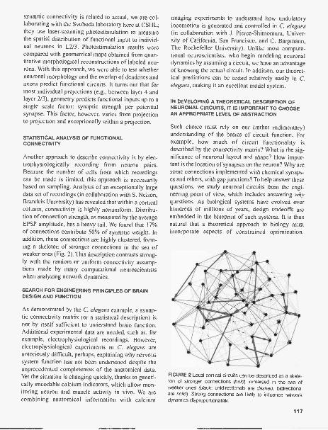
synaptic connectivity is related to actual, we are col-laborating with the Svoboda laboratory here at CSHL;they use laser-scanning photostimulation to measurethe spatial distribution of functional input to individ-ual neurons in L2/3. Photostimulation results werecompared with geometrical maps obtained from quan-titative morphological reconstructions of labeled neu-rons. With this approach, we were able to test whetherneuronal morphology and the overlap of dendrites andaxons predict functional circuits. It turns out that formost individual projections (e.g., between layer 4 andlayer 2/3), geometry predicts functional inputs up to asingle scale factor: synaptic strength per potentialsynapse. This factor, however, varies from projectionto projection and exceptionally within a projection.
STATISTICAL ANALYSIS OF FUNCTIONALCONNECTIVITY
Another approach to describe connectivity is by elec-trophysiologically recording from neuron pairs.Because the number of cells from which recordingscan be made is limited, this approach is necessarilybased on sampling. Analysis of an exceptionally largedata set of recordings (in collaboration with S. Nelson,Brandeis University) has revealed that within a corticalcolumn, connectivity is highly nonuniform. Distribu-tion of connection strength, as measured by the averageEPSP amplitude, has a heavy tail. We found that 17%of connections contribute 50% of synaptic weight. Inaddition, these connections are highly clustered, form-ing a skeleton of stronger connections in the sea ofweaker ones (Fig. 2). This description contrasts strong-ly with the random or uniform connectivity assump-tions made by many computational neuroscientistswhen analyzing network dynamics.
SEARCH FOR ENGINEERING PRINCIPLES OF BRAINDESIGN AND FUNCTION
As demonstrated by the C. elegans example, a synap-tic connectivity matrix (or a statistical description) isnot by itself sufficient to understand brain function.Additional experimental data are needed, such as, forexample, electrophysiological recordings. However,electrophysiological experiments in C. elegans arenotoriously difficult, perhaps, explaining why nervoussystem function has not been understood despite theunprecedented completeness of the anatomical data.Yet the situation is changing quickly, thanks to geneti-cally encodable calcium indicators, which allow mon-itoring neuron and muscle activity in vivo. We arecombining anatomical information with calcium
imaging experiments to understand how undulatorylocomotion is generated and controlled in C. elegans(in collaboration with J. Pierce-Shimomura, Univer-sity of California, San Francisco, and C. Bargmann,The Rockefeller University). Unlike most computa-tional neuroscientists, who begin modeling neuronaldynamics by assuming a circuit, we have an advantageof knowing the actual circuit. In addition, our theoret-ical predictions can be tested relatively easily in C.elegans, making it an excellent model system.
IN DEVELOPING A THEORETICAL DESCRIPTION OFNEURONAL CIRCUITS, IT IS IMPORTANT TO CHOOSEAN APPROPRIATE LEVEL OF ABSTRACTION
Such choice must rely on our (rather rudimentary)understanding of the basics of circuit function. Forexample, how much of circuit functionality isdescribed by the connectivity matrix? What is the sig-nificance of neuronal layout and shape? How impor-tant is the location of synapses on the neuron? Why aresome connections implemented with chemical synaps-es and others, with gap junctions? To help answer thesequestions, we study neuronal circuits from the engi-neering point of view, which includes answering whyquestions. As biological systems have evolved overhundreds of millions of years, design tradeoffs areembedded in the blueprint of such systems. It is thusnatural that a theoretical approach to biology mustincorporate aspects of constrained optimization.
FIGURE 2 Local cortical circuits can be described as a skele-ton of stronger connections (bold) immersed in the sea ofweaker ones (black; unidirectionals are dashed, bidirectionalare solid). Strong connections are likely to influence networkdynamics disproportionately.
117

Particular success has been enjoyed by the wiring opti-mization principle, which is rooted in Cajal's laws ofwiring economy. By using this principle, we are able toexplain many aspects of brain design, such as neuronalplacement, dimensions of axons, dendrites, and theexistence of spines, as well as the segregation of theneocortex into the gray and white matter.
WHAT IS THE ROLE OF AXONAL AND DENDRITICARBORS IN INFORMATION PROCESSING?
Since C. elegans neurons do not have elaborate arbors,we addressed this question in the context of corticalneurons. We find that wiring up of a highly intercon-nected network such as the cortical column is a diffi-cult problem. Solution of such a problem in the allot-ted volume requires all the major features of neuronalmorphology, such as axon and spiny dendrites.Therefore, one need not look further to find a reasonfor their existence. We are extending this work tounderstand branching and specific placement ofsynapses. Comparing optimization predictions withanatomy will help us to understand the functional sig-nificance of neuronal shape.
STATISTICAL SEARCH FOR NEURONAL MODULES INWIRING DIAGRAMS
Another difficulty with using the connectivity matrix isthat such representation of the neuronal circuit is toodetailed for comprehensive understanding. In anattempt to simplify the description of the neuronal cir-cuit, we search for multineuron modules (smaller thaninvertebrate ganglia or vertebrate nuclei and corticalcolumns). The advantage of modular description isillustrated by electrical engineering, where a descriptionof a circuit in terms of operational amplifiers, logicalgates, and memory registers is often preferred to thatshowing each transistor, resistor, and diode. However,unlike electrical engineers who designed these modulesthemselves, neurobiologists need to discover such mod-ules. Specifically, we search for a few neuron connec-tivity patterns that are statistically overrepresented in C.elegans relative to the expectations from the randommatrix ensemble. We performed similar analysis on theneocortical connectivity data, obtained by sampling.Interestingly, we have found that similar patterns, whichwe call motifs, are overrepresented in both C. elegansand the rat neocortex. In particular, reciprocally con-nected neuronal pairs are four times more common thanexpected by chance. Several triplet connectivity pat-terns are overrepresented in both C. elegans and the cor-
118
13
ci
10
0.1
Layer 5 pyramidsfrom rat visual cortex
, m=iTi24;_
2\ :'14\ 1:\ 1,V,N1:4 /%0
4 5 6 6 6 ID 91 12 13 14 95
FIGURE 3 We discovered similar overrepresented connect vitypatterns, or motifs, in rat neocortex and C. elegans. These motifsmay reflect general computational constraints on the circuits.
tex (Fig. 3). A notable exception is the feedback loop(#11), which is overrepresented only in the neocortex. Inthe future, we plan to determine the significance ofthese motifs for circuit function.
PUBLICATIONS
Chklovskii D.B. 2004. Synaptic connectivity and neuronal mor-phology: Two sides of the samecoin. Neuron 43: 609-617.
Chklovskii D.B. 2004. Exact solution for the optimal neuronallayout problem. Neural Comput. 16: 2067-2078.
Chklovskii D.B. and Koulakov A. 2004. Maps in the brain: Whatcan we learn from them? Ann. Rev. Neurosci. 27: 369-392.
Chklovskii D.B., Mel B., and Svoboda K. 2004. Cortical wiringand information storage. Nature 431: 782-788.
Reigl M., Alon U., and Chklovskii D.B. 2004. Search for compu-tational modules in the C. elegans brain. BMC Biol. 2: 25.
Shefi 0., Harel A., Chklovskii D.B., Ben-Jacob E. and Ayali A.2004. Biophysical constraints on neuronal branching.Neurocomputing 58-60: 487-495.
Stepanyants A., Tarnas G., and Chklovskii D.B. 2004. Class-specific features of neuronal wiring. Neuron 43: 251-259.
In Press
Song S., Sjostrom P.J., Reigl M., Nelson S., and Chklovskii D.B.2005. Highly nonrandom features of synaptic connectivityin local cortical circuits. PLoS Biol. 3: e68.

BRAIN DEVELOPMENT AND PLASTICITY
H.T. Cline C. AizenmanC. AkermanJ. BestmanK. Bronson
K. BurgosM. ChenS.-L. ChiuJ. Demas
R. Ewald E. Rial VerdeK. Haas E. RuthazerA. Javaherian P. SharmaK. Jensen K.V. Thirumalai
My lab is interested in determining the function ofexperience in regulating brain development. It is nowcommonly understood that sensory experience shapesthe development of circuits within the brain and thatexperience during brain development governs brainconnectivity, perception, cognition, and behavior.Nevertheless, the experience-dependent cellular andmolecular mechanisms that operate to establish andmodify brain connections during development arepoorly understood. By understanding the basic mech-anisms governing brain development, we expect togarner critical information of the origins of nervoussystem dysfunction, as seen in many neurological dis-orders.
We examine the structural and functional develop-ment of the visual system in amphibian tadpoles. Theseanimals are transparent, which allows us to observedirectly the development of the brain in living animals.In addition, we assess neuronal function using electro-physiological assays of synaptic connectivity andsynaptic plasticity. We combine these studies with genetransfer methods that allow us to test the function ofgenes of interest in brain development. Given the highdegree of conservation of mechanisms related to braindevelopment and plasticity, our work will identify keyregulatory mechanisms governing brain plasticityacross species. Below is a representative project com-pleted in the laboratory during the last year.
STRUCTURAL DEVELOPMENT OF MOTORNEURON AXONS USING TIME-LAPSEIN VIVO IMAGING
The development of motor neuron axon arbors gov-erns the connectivity between the central nervous sys-tem and the musculature. Once the motor neuron axonreaches the target, the growing axon branches andforms synapses. In many cases, it is thought that exu-berant connections are made and that these are prunedto shape the final axon arbor and neuromuscular con-nections. The development of the motor neuron axonarbors has been characterized as a sequence of events
in which the axons initially branch exuberantly, fol-lowed by a period of synapse and branch elimination;however, the initial phase of axon arbor elaborationand synaptogenesis has not been directly observed invivo. The possibility that motor axon innervation ofthe periphery could develop through a dynamicprocess of branch addition, maintenance, and retrac-tion, concurrent with the formation of synaptic con-nections, has not been addressed. To test this hypothe-sis, we collected time-lapse images of motor neuronaxons as they elaborated complex arbors and formedneuromuscular synapses.
We found that motor neuron axon terminal arborsshow continuous branch dynamics over 3 days oftime-lapse imaging. Using a combination of cyan-flu-orescent protein-tagged synaptophysin (synapto-physin-CFP) and yellow fluorescent protein (YFP) tovisualize the dynamics of presynaptic structures andaxon branches and Texas-Red-tagged a-bungarotoxin(TR-aBTX) to label postsynaptic acetylcholine recep-tors (AChR), we show that new branches emerge fromsites of presynaptic specialization, the majority ofwhich are apposed to AChR. The new branches formneuromuscular synapses. These data suggest thatmechanisms that govern neuromuscular synaptogene-sis also have an important role in regulating motorneuron axon arbor development.
To probe the role of synaptogenesis in controllingaxon arbor development, we tested whether a proteincalled candidate plasticity gene 15 (CPG15, alsoknown as Neuritin) affects this process. We previouslydemonstrated that CPG15 enhances the developmentof visual system connections by increasing the elabo-ration of optic tectal cell dendrites, retinal axon arbors,and the strength of retinotectal synapses. CPG15 isexpressed in the developing motor neurons. CPG15expression enhances the development of motor neuronaxon arbors by promoting neuromuscular synaptogen-esis and by increasing the addition of new axonbranches. These data indicate that motor neuron axonelaboration and synaptogenesis are concurrent anditerative.
119

PUBLICATIONS In Press
Ruthazer E.S. and Cline H.T. 2004. Insights into activity-dependent map formation from the retinotectal system: Amiddle-of-the-brain perspective. J. Neurobiol. 59:134-146.
Van Aelst L. and Cline H.T. 2004. Rho GTPases and activity-dependent dendrite development. Curr. Opin. Neurobiol.14: 297-304.
Cline H. 2005. Synaptogenesis: A balancing act between exci-tation and inhibition. Curr. Biol. 15: R203-205.
Foa L., Jensen K., Rajan I., Bronson K., Gasperini R., Worley RE,Tu J.C., and Cline H.T. 2005. Homer expression in the Xenopus
tadpole nervous system. J. Comp. NeuroL 487: 42-53.Javaherian A. and Cline H.T. 2005. Coordinated motor neuron
axon growth and neuromuscular synaptogenesis are pro-moted by CPG15 in vivo. Neuron 45: 505-512.
Shu-Ling Chiu, WSBS student, Holly Cline's lab.
120

GENETICS OF MEMORY IN DROSOPHILA
J. Dubnau A. AltickA. BlumS. Dennison
The long-term goal of our research is to understandmemory. Dissection of complex behaviors such asmemory and learning will require a multidisciplinaryapproach that will include discovery and manipulationof the relevant genetic, cellular, and anatomical path-ways, as well as computational modeling of how infor-mation is processed in the brain. Work in geneticmodel systems such as Drosophila can contribute toour understanding in two main ways. First, by enablingdiscovery of genes and genetic pathways underlyingbehavior, genetic model systems provide entry pointsfor subsequent interventionist experiments. Second,systematic manipulation of gene function in relevantanatomical loci of the brain allows a conceptual inte-gration of findings from cellular and behavioral neu-roscience.
We use the Drosophila model system, which offersan economy of scale to discover and develop hypothe-ses in a relatively cost-effective and rapid manner.Then, given the remarkable evolutionary conservationof genetic, cellular, and behavioral functions, thesehypotheses can be pursued in mammalian model sys-tems.
Gene Discovery with DNA Microarrays
J. Dubnau [in collaboration with T. Tully and N. Sinha,Cold Spring Harbor Laboratory]
In collaboration with the Tully lab, we have used acombination of behaviorally specific training proto-cols and expression profiling with DNA chips to iden-tify transcriptional responses during memory consoli-dation (Dubnau et al., Curr. Biol. 13: 286 [2003]). Wehave used this approach to compare gene expressionprofiles after spaced training, which induces bothshort-term and protein-synthesis-dependent long-termmemory, and massed training, which only inducesshort-lived memory. We have identified a large num-ber of candidate memory genes differentiallyexpressed at three different retention intervals afterspaced versus massed training. Using real-time poly-
merase chain reaction (PCR) follow-up assays, wehave confirmed differential expression for a growingnumber of these transcripts. This effort has identifiedcandidate memory genes (CMGs), which serve asentry points for molecular genetic investigation ofgene function in memory. Ultimate confirmation thata gene is involved in memory formation rests with thedemonstration that in vivo modulation (disruption) ofthe gene alters that process with some specificity.With that aim in mind, we are focusing on local trans-lational control, one of several pathways suggestedfrom the array experiments. Our genetic studiesalready support a role in memory for several compo-nents of this pathway. These include staufen and oskar,which are known components of a cellular mRNAlocalization machinery in oocytes, and pumilio, whichis a translational repressor protein. A large number ofthe known components of the mRNA localizationmachinery, as well as of the apparatus for regulatingcytoplasmic polyadenylation-stimulated translationalcontrol, also are differentially expressed.
Drosphila 4E-BP
A. Altick, J. Dubnau [in collaboration with L. Gradyand T. Tully, Cold Spring Harbor Laboratory]
thor, which encodes a fly homolog of 4E-BR a trans-lational regulator, is one of the CMGs induced byspaced training relative to massed training. 4E-BPsbind to 4E, the cap-binding protein, thereby inhibitingtranslation. Upon phosphorylation, however, 4E-BPsare removed from the complex, allowing 4G to inter-act with 4E, and translation initiation to occur. Wehave demonstrated that a hypomorphic mutation inthor yields a defect in long-term memory measured 24hours after spaced training (Fig. 1). Importantly, learn-ing and shorter-term forms of memory, which arebelieved to be protein-synthesis-independent, are nor-mal in these mutant animals. We now are using a dom-inant-negative transgenic form of thor to block 4E-BP-mediated translation. This transgene, which con-
121

50
45
40
35
30
25
20
15
10
5
III wild type control
thorl mutant
2 minmemory
10X Sp 10XMa:24 hourmemory
FIGURE 1 thor is a long-term memory mutant. thorl mutantanimals display normal levels of memory measured 2 minutesafter a single training session. In contrast, thorl mutants exhib-it defective long-term memory. Memory is reduced in thorlmutants when measured 24 hours after ten sessions of spacedtraining (10XSp), but not after ten sessions of massed training(1 OXMa).
tains an amino acid substitution at the site of phos-phorylation, blocks 4E-BP-mediated translation byconstitutively binding 4E. This transgenic approachwill allow us to acutely inactivate 4E-BP-mediatedtranslation during memory formation and also willprovide the means to spatially restrict expression todistinct neuroanatomical loci.
Genetic Dissection ofAnesthesia-resistant Memory
A. Blum, J. Dubnau
In both vertebrate and invertebrate animals, anestheticagents cause retrograde amnesia for recently experi-enced events. In contrast, older memories are resistant
122
to the same treatments. In Drosophila, anesthesia-resistant memory (ARM) and long-term memory(LTM) are genetically distinct forms of long-livedmemory that exist in parallel for at least 1 day aftertraining. ARM is disrupted in radish mutants but isnormal in transgenic flies overexpressing a CREBrepressor transgene. In contrast, LTM is normal inradish mutants but is disrupted in CREB repressortransgenic flies. Until now, nothing was known aboutthe molecular, genetic, or cell biological pathwaysunderlying ARM. We have identified radish as a phos-pholipase-A2, providing the first clue about signalingpathways underlying ARM in any animal (Chiang et al.2004). An enhancer-trap allele of radish (C133) alsoprovides an entry point to study the anatomical circuitsunderlying ARM. rshci"-driven reporter expressiondoes not label mushroom bodies, which are the prima-ry anatomical focus of olfactory memory research inDrosophila. Instead, rse133-driven expression reveals anumber of novel neuronal types, several of which pro-ject into mushroom bodies. Transgenic expression ofPLA2 driven by the rslicn3 Ga14 driver restores normallevels of ARM to radish mutants, whereas transientdisruption of neural activity in rshc133 neurons inhibitsmemory retention. Identification of radish as a phos-pholipase-A2 and the neural expression pattern of thisenhancer trap significantly broaden our understandingof the biochemistry and anatomy underlying olfactorymemory in Drosophila. These findings also are consis-tent with pharmacological studies of PLA2 signaling inmemory formation in vertebrate animals. Our currentefforts are focused on a biochemical characterizationof the radish PLA2, as well as an anatomical dissectionof the neural circuits in which radish functions.
Reversible Disruption ofSynaptic Transmission
J. Dubnau, A. Blum, S. Denison in collaborationwith A.S. Chiang, Taiwan]
To map the neural circuits required for memory for-mation, storage, and retrieval, we are using a tempera-ture-sensitive dynamin transgene, which disruptssynaptic transmission reversibly and on a time scale ofminutes. We are using a panel of Ga14 enhancer dri-vers to focus expression of this dynamin transgenewithin specific subpopulations of neurons. With thisapproach, we are now able to reversibly silence repro-ducibly specific groups of neurons in vivo. We already

FIGURE 2 Anatomical dissection of anesthesia-sensitive andanesthesia-resistant memory (ARM). (Top panels) GFP reporterexpression in several neurons that participate in the circuitryunderlying olfactory memory. (Left panel) Dorsal-paired medialneuron massively innervates the axonal outputs of the mushroombodies. (Middle panel) Dorsal-anterior-lateral neuron sends fibersinto parts of the posterior superior protocerebrum, including themushroom body dendritic field. (Right panel) Ventral-anterior-medial neurons innervate the alpha lobes of the mushroom bod-ies (gray). (Bottom panel) Expression of the temperature-sensitivedynamin transgene under control of Ga14 drivers permits focusedtransient inhibition of dynamin-dependent activity in specific neu-ronal subtypes. Three Ga14 lines were used: one that expressesin DPM and DAL, one that expresses in VAM, and a third thatexpresses in mushroom bodies. In the case of the DPM and DALline as well as the VAM line, transient heating to the restrictivetemperature (H) causes a partial amnesia measured 3 hours aftertraining (compare control treatment (C] with heat shifted [H]). Incontrast, this heat shift has no effect on a control strain or on aline that expresses the dynamin transgene in mushroom bodies.Application of a cold-shock anesthesia cold-shock treatmentcompletely erases anesthesia-sensitive memory, providing a puremeasure of ARM. Use of cold shock in combination with the heatshift (HC) reveals an anatomical dissection of anesthesia-sensitivememory from anesthesia-resistant memory. In the DPM/DAL expressing animals, this combination treatment erases all memory, sug-gesting that the memory disrupted by dynamin alone (H) is ARM. In contrast, the combined treatment in VAM neurons reveals that asignificant portion of the memory remaining after the dynamin effect is ARM. Thus, inhibition of VAM-dynamin mostly disrupts anes-thesia-sensitive memory.
50
45
40
35
30
10
.5
-10
C H HC C H HC C H HC C II
have demonstrated a role for synaptic transmission inmushroom body (MB) neurons during memoryretrieval, but surprisingly not during acquisition orstorage of early memory (Dubnau et al., Nature 411:476 [2001]). These data suggest that the synaptic plas-ticity underlying olfactory associative learning initial-ly reside in MB dendrites and/or upstream of the MBand that the resulting alterations in synaptic strengthmodulate MB output during memory retrieval. Butvirtually nothing is known about the neural circuitryinvolved in the subsequent consolidation of short-term, middle-term, anesthesia-resistant, or CREB-dependent long-term memory phases-and nothing isknown about the circuitry of memory retrieval.
In collaboration with A.S. Chiang, we have identi-fied a number of Ga14 enhancer lines that yieldexpression in neurons that send projections into differ-ent substructures of the MB. These identified MB"extrinsic" neurons constitute a significant fraction ofthe inputs, outputs, and modulation of MB. With the
dynamin method, we now can functionally manipulatethese neuronal populations as well as a number ofadditional neuroanatomical loci identified by reporterexpression from a panel of approximately 60 memorymutants (Dubnau et al., Cum Biol. 54: 238 [2003]).Using this method of reversible neuronal disruption,we already have demonstrated an anatomical dissec-tion of the requirements for dynamin-dependent activ-ity during formation and storage of anesthesia-sensi-tive versus anesthesia-resistant memory (Fig. 2).Ongoing efforts are focused on filling in the rest of thecircuitry for these "early" memory phases, as well asfor long-term memory and for memory retrieval.
PUBLICATIONS
Chiang A.S., Blum A., Barditch J., Chen Y.H., Chiu S.L.,Regulski M., Armstrong J.D., Tully T., and Dubnau J. 2004.Radish encodes a phospholipase-A2 and defines a neuralcircuit involved in anesthesia-resistant memory. Curr.14: 263-272.
123

STEM CELLS, SIGNAL TRANSDUCTION, AND DIFFERENTIATION
G. Enikolopov J. Encinas L. Manganas K. RaviJ. Hemish T. Michurina J. RoigB. Kuzin B. Mish V ScheinkerM. Maletic-Savatic N. Peunova
Stem cells have a unique ability to self-renew and toproduce progenitor cells that eventually generate dif-ferentiated cells. This cascade of transitions from stemcells to their differentiated progeny is under dynamiccontrol, ensuring a rapid response to demands formore cells due to stress, damage, or altered environ-ment. We use flies, frogs, and mice as models to studysignals that regulate distinct steps in the differentiationcascade and mediate the interactions between the stemcells and their microenvironment. Our main focus ison stem cells in the adult brain. We have generatedseveral models to study how stem cells decide to giverise to progenitors and, ultimately, neurons. We alsouse these models to study how disease and therapeuticdrugs affect adult neurogenesis.
Much of our interest is related to a versatile sig-naling molecule, nitric oxide (NO). We found that, inaddition to its numerous other tasks such as fightinginfection, regulating blood pressure, and mediatingneurotransmission, NO controls cell proliferation anddifferentiation. In several developmental settings, NOsuppresses cell division, helping to control the balancebetween proliferation and differentiation. For instance,we have found that NO acts as an negative regulator ofcell division in the adult mammalian brain, such thatby experimentally reducing NO levels, we canincrease the number of neural stem/progenitor cells inthe adult brain. Our findings highlight the role of NOas an essential negative regulator of cell proliferationin the differentiation cascade. We are now beginningto dissect the genetic and molecular interactionsbetween NO and the signaling pathways that controlstem cell division and differentiation.
STEM CELLS IN THE ADULT BRAIN
Stem and progenitor cells receive specific cues thatcontrol their self renewal and their transition throughdifferentiation cascades. We are interested in stemcells in the adult organism and the signaling moleculesthat control the division of stem and progenitor cellsand guide them toward their differentiated fate. To fol-
124
low neuronal differentiation, we generated a series oftransgenic reporter mouse lines in which neural stemand early progenitor cells of the embryonic and adultbrain are marked by the expression of the green, cyan,and red fluorescent proteins (GFP, CFP, RFP) whichare under control of the nestin gene regulatory ele-ments. We found that these labeled cells faithfullyreport the status of neural stem cells after exposure todrugs or in response to gene ablation.
We also found that in some nonneural tissues ofour model animals, fluorescence marks stem/progeni-tor cells for these tissues. For instance, we found thatnestin-GFP-expressing cells can be found in the bulgeregion of the hair follicle, where stem cells for the fol-licle are located. By a number of criteria, these cellsindeed behave as progenitor population of the hair; inaddition, they can generate progeny of diverse lineage,including neurons. We used transcriptional profilingto confirm the neural potential of these cells and todetermine their relation to other types of stem cells.We now study the ability of nestin-expressing cellsfrom nonneural tissues to reveal their neural potentialboth in vitro and in vivo, after transplantation into thedeveloping and adult mouse brain.
An important focus of our studies remains the roleof NO as a regulator of adult neurogenesis. We haverecently found that NO acts to suppress the generationof new neurons in the adult brain. In a pharmacologi-cal approach, we blocked production of NO by intro-ducing inhibitors of NO synthase (NOS) into the ven-tricles of the adult brain. In a genetic approach, wegenerated a null mutant for the neuronal NOS gene. Inboth models, the number of new cells generated inneurogenic areas, the olfactory subependyma and thedentate gyrus, was strongly augmented. Importantly,increased proliferation of progenitor cells did not alterthe fate of the extra cells. Our results reveal a role forNO as an essential negative regulator controlling pro-duction of new neurons in the adult brain. This findingparallels our recent results indicating that NO also actsas a negative regulator of stem/progenitor cells in thebone marrow. It also suggests a strategy for enhancing

neurogenesis in the damaged central nervous system.At the same time, it is unclear which steps within theneural differentiation cascade are controlled by NO: Itmay control the last steps in the cascade (for instance,permitting advanced precursors cells to proceed withan extra division) or it may act upon stem or very earlyprogenitor cells, accelerating their rate of division.Furthermore, it is not known whether NO is producedin the differentiating cells themselves, or in neighbor-ing cells (thus acting on stem or differentiating cells ina transcellular fashion). We now use our reporter lines(described above) to determine whether deficit ofNOS activity changes the number of neural stem/progenitor cells in the adult brain or affects moreadvanced neuronal precursors. Our results point to aspecific step in the differentiation cascade that is con-trolled by NO. We are now applying a similar strategyto investigate how therapeutic drugs and selectedgenes affect adult neurogenesis and to determine theirpoints of action within the neuronal differentiationcascade.
NO AND EARLY XENOPUS DEVELOPMENT
Vertebrate body plan is shaped during embryogenesisthrough precise coordination of cell proliferation andmorphogenetic cell movements. We investigated therole of NO in early Xenopus development using gain-and loss-of-function approaches by microinjectingeither mRNA encoding Xenopus NOS (XNOS) or achemical NO donor; conversely, we blocked NOSactivity by injecting either mRNA encoding a domi-nant-negative form of XNOS, or chemical NOSinhibitors. Our results show that NO suppresses celldivision and regulates cell movements during earlydevelopment of Xenopus. Excess NO productiondecreases cell proliferation in the embryo. In contrast,deficits in NO production result in excessive cell pro-liferation in the ectoderm and distortion of cell move-ments that underlie normal axis extension and neuraltube closure during gastrulation and neurulation. Atlater stages, the deficit of NO leads to profounddefects in organogenesis. We have identified molecu-lar targets and pathways used by NO to control cellcycle and cell motility. Our results indicate that NO-RhoA-ROCK signaling mediates both convergentextension and cell division. Concurrent control by NOhelps ensure that the crucial processes of cell prolifer-ation and morphogenetic movements are coordinatedduring early development.
NO AND DROSOPHILA DEVELOPMENT
NO is an essential regulator of Drosophila develop-ment and physiology: It has been implicated in visualsystem development, immunity, behavior, response tohypoxia, osmoregulation, and regulation of cell cycleprogression during development. We found a novelmode of regulation of NOS function that employsendogenously produced truncated protein isoforms ofDrosophila NOS (DNOS). These isoforms inhibit NOSenzymatic activity in vitro and in vivo, reflecting theirability to form complexes with the full-length DNOS1protein. Truncated isoforms suppress the antiprolifera-tive action of DNOS1 in the eye imaginal disc, yield-ing hyperproliferative phenotypes in pupae and adultflies. We found that NO regulates the cell cycle by act-ing upon the retinoblastoma pathways, suppressing theeffect of RBF and enhancing the effect of E2F. Ourresults indicate that endogenous products of the DNOSlocus act as dominant-negative regulators of NOSactivity during Drosophila development, pointing to anovel mechanism for the regulation of NO productionin animal cells. More generally, negative control ofhomodimeric enzymes by endogenous truncated iso-forms may represent an important regulatory mecha-nism controlling development and physiology.
In a related collaborative study with MichaelRegulski and Tim Tully here at CSHL, a wide screen forlethal EMS-induced mutations around the DNOS locusproduced a set of lethal mutations in five complemen-tation groups, the largest group corresponding toDNOS. Sequence analysis of the genomic DNA of oneof these lines identified a single nucleotide transitionwhich created a Gly1942Glu substitution. This residueis conserved across all known NOS proteins; althoughit does not fall into the active center of enzyme, itsmutation abolishes the enzymatic activity of DNOS.These results provide the first direct evidence that NOSfunction is indispensable for development.
PUBLICATIONS
Davidoff M.S., Middlendorff R., Enikolopov G., Rietmacher D.,Holstein A. -F., and Muller D. 2004. Progenitor cells of thetestosterone-producing Leydig cells revealed. J. Cell. Biol.167: 935-944.
Michurina T., Krasnov P., Balazs A., Nakaya N., Vasilieva T.,Kuzin B., Khrushchov N., Mulligan R.C., and Enikolopov G.2004. Nitric oxide is a regulator of hematopoietic stem cellactivity. Mo/. Ther 10: 241-248.
Mignone J.L., Kukekov V., Chiang A.S., Steindler D., andEnikolopov G. 2004. Neural stem and progenitor cells in
125

nestin-GFP transgenic mice. J. Comp. Neurol. 469:311-324.
Regulski M., Stasiv Y., Tully T., and Enikolopov G. 2004.Essential function of nitric oxide synthase in Drosophila.Curr. Biol. 14: R881-882.
Seaberg R.M., Smukler S.R., Kieffer T.J., Enikolopov G., AsgharZ., Wheeler M.B., Korbutt G., and van der Kooy D. 2004.Clonal identification of multipotent precursors from adultmouse pancreas that generate neural and pancreatic lin-eages. Nat. Biotechnol. 22: 1115-1124.
Stasiv Y., Kuzin B., Regulski M., Tully T., and Enikolopov G.2004. Regulation of multimers via truncated isoforms: A
126
novel mechanism to control nitric oxide signaling. GenesDev. 18: 1812-1823.
In Press
Encinas J.M., Manganas L., and Enikolopov G. 2005. Nitricoxide and multiple sclerosis. Cum. Neur. Neurosci. Rep. 5:(in press).
Packer M., Hemish J., Mignone J., John S., Pugach I., andEnikolopov G. 2005. Transgenic mice expressing nNOS inthe adult nervous system. Cell. Mol. Biol. 50: (in press).
Kandasamy Ravi, postdoc, Grisha Enikolopov's lab.

CONSTRUCTION AND PLASTICITY OF THE GABAERGIC CIRCUITS
Z.J. Huang J.-R. Ango C. WuB. Chattopadhyaya P. WuG. diCristo H. YangS. Kulhman
Much progress in neuroscience in the past fewdecades has been made in understanding informationcoding, storage, and plasticity in the brain. Gluta-matergic synapses on principal neurons-with theirvast numbers, elaborate electrobiochemical signalingmachinery, and capacity for modification- have beena major focus of investigation. On the other hand, per-ceptual and cognitive processes also require largeneural networks to dynamically retrieve and combinevarious stored information, and to relate them toincoming sensory input, usually with high speed andtemporal precision, on the order of tens of millisec-onds. Evidence in the past few years increasingly sug-gests that the GABAergic system is particularly effec-tive in regulating the precise timing of electrical sig-naling both within a principal neuron and among pop-ulations of principal neurons. Networks ofGABAergic interneurons have been hypothesized toentrain populations of principal neurons and thusimpose a coordinated "context" for the "content" car-ried by networks of principal neurons during percep-tion, cognition, movement control, learning, and mem-ory. Aberrant GABAergic functions have been impli-cated in devastating neurological and psychiatric dis-orders such as epilepsy and schizophrenia.
Our laboratory is studying the assembly, plasticity,and function of the GABAergic inhibitory network inmammalian brain. We focus on the following threeareas: (1) mechanisms of subcellular organization ofdistinct classes of GABAergic synapses; (2) mecha-nisms of neuronal activity-dependent plasticity ofGABAergic synapses; and (3) role of GABAergicfunction in critical-period plasticity in primary visualcortex. Combining cell-type-specific transcriptionalpromoters, bacterial artificial chromosome (BAC)transgenic reporter mice, and high-resolution imaging,our laboratory has established powerful in vivo and invitro systems to visualize and manipulate definedclasses of GABAergic interneurons. Our progress inthe past year is beginning to define the logic and out-line of the construction of GABAergic circuits in themammalian brain.
Subcellular Synapse OrganizationIs Independent ofExperience-driven Pruning
G. diCristo, C. Wu
The subcellular spatial organization of GABAergicinnervation is crucial for the temporal precision in theirregulation of neuronal excitability, integration, andsynchrony. Since subcellular synapse targeting is pre-ceded by a largely genetically determined axon guid-ance process and followed by synapse formation andexperience-driven plasticity, it is not obvious whetherthis intermediate step of circuit assembly is directedmainly by genetic programs or experience-dependentrefinement. By combining cell-type-specific promot-ers, BAC engineering, and 2-photon imaging, distinctclasses of GABAergic synapses and their postsynapticpyramidal neurons were visualized in primary visualcortex and in organotypic cultures of transgenic mice.We show that the subcellular organization of periso-matic and dendritic-targeted GABAergic innervationsin organotypic cultures was indistinguishable com-pared to that in primary visual cortex. Therefore, sub-cellular targeting of GABAergic synapses is likelyguided by cell surface molecular labels and experi-ence-independent forms of neuronal activity.
Subcellular Organization ofGABAergic Synapses Directedby L1CAMs and Ankyrin-basedMembrane Skeletons
J.-R. Ango, H. Yang, P. Wu
We are defining the molecular signals underlying sub-cellular synapse targeting. We have discovered so farthat the ankyrinG -based membrane cytoskeleton is thekey molecular machinery which, through subcellularrecruitment of the Ll family immunoglobulin cell
127

adhesion molecules (IgCAMs), directs GABAergicinnervation to the axon initial segment-the site ofaction potential initiation. Importantly, both ankyrinand L1CAM families contain multiple memberslocalized to distinct subcellular compartments. Wehave significant evidence that CHL 1 (a close homologof L1) is involved in targeting a different class ofGABAergic synapses to neuronal dendrites. Together,these results led us to propose a general hypothesis:Members of L1CAMs recruited to subcellulardomains by different ankyrins may constitute a set of"compartmental code" in principal neurons to directsubcellular organization of GABAergic synapses.
An important implication of these discoveries isthat subcellular segregation of GABAergic synapsesis superimposed on an elaborate subcellular organiza-tion of ion channels. Such spatial alignment of bio-physical properties and synaptic inputs, in some casesupon the same macromolecular complex, may conferthe ultimate temporal precision of neuronal signalingwithin and among neurons. These studies suggest anunexpected link from subcellular molecular organiza-tion, neuronal connectivity, to network properties. Inaddition, there should be a family of receptors on dif-ferent classes of GABAergic neurons to "read" thecompartmental code.
Neuronal Activity-dependentPlasticity of GABAergicSynapses and Circuits
B. Chattopadhyaya, G. diCristo, C. Wu
Although precision and speed of transmission areprominent features of at least certain classes ofGABAergic neurons, it has been well documented thatexperience and injury profoundly alter the connectiv-ity and function of GABAergic synapses. The logicand mechanism of neuronal activity-dependent plas-ticity of the GABAergic network are poorly under-stood.
Using high-resolution imaging of a defined class ofGABA synapses, we discovered that following the ini-tial targeting to a defined subcellular domain, there is aprolonged maturation of GABA innervation, character-ized by elaboration of multiple terminals and prolifera-tion of synapses. This maturation process can proceedto a substantial extent in an organotypic culture system,
128
but it is regulated by level/pattern of neuronal activityand sensory experience during a restricted postnatalperiod in visual cortex. Using "single-cell genetics" inorganotypic cultures, we are making significantprogress in understanding the molecular mechanism ofactivity-dependent morphologic plasticity of a majorclass of GABA synapses. First, we discovered that,although GABA transmission is "inhibitory," GABAsynthesis and signaling are essential in synaptic miser -vation and maturation (Fig. 1). This unexpected findingsuggests that in addition to its role as an inhibitory neu-rotransmitter, GABA has a key role in axon branching,synapse proliferation, target coverage, and thus the con-struction of circuits. Second, we discovered that the Fyntyrosine kinase, neural cell adhesion molecule(NCAM), and its polysialic acid (PSA) modification allhave a role in the morphologic plasticity of GABAergicpresynaptic terminals. These results begin to suggest apathway in which activity-dependent regulation ofGABA synthesis and signaling through the GABABreceptors and G proteins could regulate Fyn kinaseactivity, which then modulates the voltage-gated calci-um channel and therefore Ca" dynamics in presynapticterminals. The spatial and temporal dynamics of Ca"signaling have been shown to trigger a variety of mor-phogenic events.
Our studies suggest that the logic and mechanismof the plasticity of GABAergic synapses may be fun-damentally different from that of the glutamatergicexcitatory synapses. For example, synapse specificityin the plasticity of glutamatergic synapses is the basisfor information coding and storage. However, the plas-ticity of the GABAergic synapses that we observed isnot restricted to particular terminals, but "cell wide"and, in this sense, global. These results have importantimplications in the function of GABAergic plasticityon neural network activities.
GABAergic Transmissionand Critical-period Plasticityin Visual Cortex
S. Kuhlman
During early postnatal life, visual deprivation alterssynaptic transmission in visual cortex such that binoc-ular cortical neurons cease to respond to stimulation ofthe deprived eye. Long-term depression (LTD)

FIGURE 1 GABA synthesis and signaling influence the construction of GABAergiccircuits. (Left) A wild-type basket interneuon projects highly exuberant local axonarbors and branches, which bear dense synaptic boutons (arrow). Deletion of aGABA synthetic enzyme in a single basket cell (right) greatly reduces its axon termi-nal branches and synaptic boutons, and thus the innervation of its target neurons.
induced via spike-timing-dependent plasticity (STDP)rules has been implicated in competition-basedHebbian plasticity and selective weakening ofdeprived input. However, the precise mechanisms bywhich input from the two eyes compete within the cor-tical circuit for control of postsynaptic action potentialis not known. GABAergic transmission has beenimplicated in the temporal precision of spike genera-tion and is necessary for ocular dominance (OD) plas-ticity, but the cellular mechanism is obscure.
We hypothesize that rapid and transient recruit-ment of GABAergic inhibition by open eye inputsuppresses the deprived pathway from driving post-synaptic spikes, resulting in synaptic depression viaSTDP rules. We developed a two-pathway stimula-tion paradigm at layer 4 to layer 2/3 connections invisual cortical slices and demonstrated that there is a
developmental shift in the interaction of convergingsynaptic inputs, from summation/coordination tosuppression/competition. This developmentalincrease of competition is in part mediated by thematuration of GABAergic inhibition, which dynami-cally regulates spike generation. Modeling studiesfurther suggest that mature GABA transmission pro-motes depression of uncorrelated synaptic inputs. Wepropose that GABAergic inhibition recruited by cor-related input is a potent competitive mechanism tosuppress spike probability of uncorrelated input,thereby engaging molecular cascades leading tosynaptic depression. This study reveals a plausibleGABAergic mechanism in critical-period plasticity,which is distinguishable from (but regulates) thewell-known "core plasticity machinery" at gluta-matergic synapses and neurons.
129

Genetic Logic of GABAergicCell Types and GeneExpression Profiles
J.-R. Ango, P. Wu [in collaboration with Dr. S. Nelson,Brandeis University]
We continue to generate BAC transgenic mice whichlabel different classes of interneurons with fluorescentproteins. We have initiated a new effort to characterizegene expression profiles in several classes ofGABAergic interneurons. This was made possible bydeveloping a procedure to manually purify GFP-labeled interneurons from our BAC transgenic reportermice. A systematic understanding of gene expressionprofiles among different classes of interneuronsthroughout development and in response to stimulation
130
will yield fundamental insight into the logic of thedesign and function of the GABAergic system.
PUBLICATIONS
Ango F., di Cristo G., Higashiyama H., Bennett V., Wu P., andHuang Z.J. 2004. Ankyrin -based subcellular gradient ofneurofascin, an immunoglobulin family protein, directsGABAergic innervation at Purkinje axon initial segment. Ce/I119: 257-272.
Chattopadhayaya B., Di Cristo G., Higashiyama H., Knott G.W.,Kuhlman S.J.. Welker E., and Huang Z.J. 2004. Experienceand activity-dependent maturation of perisomaticGABAergic innervation in primary visual cortex during apostnatal critical period. J. Neurosci. 24: 9598-9611.
Di Cristo G., Wu C., Chattopadhyaya B., Ango F., Knott G.,Welker E., Svoboda K., and Huang Z.J. 2004. Subcellulardomain-restricted GABAergic innervation in primary visualcortex in the absence of sensory and thalamic inputs. Nat.Neurosci. 7: 1184-1186.
Jean-Raymond Ango, postdoc, Josh Huang's lab.

BIOPHYSICAL BASIS FOR NEURAL COMPUTATION
A. Koulakov M. NikitchenkoD. Tsigankov
Neurons in the brain display the sort of duality that hasinspired philosophers for many centuries. On the onehand, neural networks process information. In doingso, they represent our ideas, feelings, and thoughts. Onthe other hand, information is processed by a finelytuned physiological apparatus, extremely sophisticatedper se. It is not even clear what level of function of thenervous system is more complex, information process-ing or physiological organization. In our lab, weattempt to connect these two features of the nervoussystem. We argue that the latter often provides cuesabout the former and vice versa, and that these twocomponents cannot be understood without one another.
Hysteretic Model forNeural Integrator
M. Nikitchenko, A. Koulakov
Persistent firing is a likely candidate for the neuralcorrelate of short-term memory. In some cases, thevariable stored in memory is continuous in nature.Examples of such variables include position of anobject in the sensory space and tension of a muscle, orvariables representing accumulated sensory evidence.Such variables are encoded in persistent neuronal fir-ing, which has a graded set of values. Perhaps the best-studied system of this type is the oculomotor neuralintegrator. Graded persistent activity in this system islikely to be maintained by positive feedback. Thisposes a basic problem of robustness, since mistuningof the feedback leads to instabilities, which are hard toavoid in realistic systems. It was hypothesized that theproblem of robustness could be solved by introducinghysteresis into neuronal responses. A recent study inthe goldfish oculomotor integrator (Aksay et al.,Cerebr. Cortex 13: 1173-1184 [2003]) may determinewhether hysteretic mechanism of robustness is indeedused. Two basic observations are made about historydependence of neuronal responses in this system: (1)firing rate of one neuron versus the other exhibits hys-teresis and (2) the size of this hysteresis varies from apair of cells to a pair of cells. In this study, we devel-
op a mathematical approach, which allows us toaddress these observations. We derive a kinetic equa-tion, which can describe an ensemble of hystereticunits effectively and accurately. Using this approach,we consider dynamics of an ensemble of hystereticunits with different values of hysteresis, similarly tothe experiments in goldfish. We conclude that such anensemble exhibits history dependence of firing rates,including hysteresis in the firing rate of one cell ver-sus the other. Thus, experimental observation numberone, cited above, may follow from observation numbertwo. This conclusion follows from entanglement of thedistribution function of activated units in the phasespace of the system. We confirm this conclusion bystudying an integrator based on a single neuron with100 hysteretic dendritic compartments.
Complete Structure of TopographicMaps in Ephrin-A-deficient Mice
D. Tsigankov, A. Koulakov
Axons of retinal ganglion cells establish orderly pro-jections to the superior colliculus of the midbrain.Axons of neighboring cells terminate proximally inthe superior colliculus, thus forming a topographical-ly precise representation of the visual world.Coordinate axes are encoded in retina and in the targetthrough graded expression of chemical labels.Mapping based on chemical labels alone does notyield required specificity of connections. Additionalsharpening is provided by electrical activity, which iscorrelated between neighboring axons. In this work,we propose a quantitative model that allows combin-ing the effects of chemical labels and correlated activ-ity in a single approach. Using this model, we study acomplete structure of two-dimensional topographicmaps in mutant mice, in which one of the labels,encoding a horizontal retinal coordinate (ephrin-A), isreduced/eliminated. We show that topographic mapsin ephrin-A-deficient mice display granular structure,with the regions of smooth mapping separated by lin-ear discontinuities reminiscent of fractures observed
131

Retina SC, ephrin-/-
FIGURE 1 Topographic maps in ephrin-A2/A5 -/- knockoutmice are analyzed by a computational model that includes chem-icals labels and correlated activity-based refinement. The result-ing map contains domains of continuous mapping separated bylinear discontinuities similar to fractures in orientation preferencemaps in other mammals, with more complex organization of visu-al maps. Thus, a loss-of-function experiment becomes a gain-of-function experiment. SC indicates superior colliculus.
in the maps of preferred orientation. We also demon-strate the presence of point singularities, similar topinwheels in orientation maps.
Activity and Competition inSurgically ManipulatedRetinotectal/Collicular Maps
D. Tsigankov, A. Koulakov
We analyze a theoretical model of retinotectal/collicu-lar map development in surgically manipulated sys-tems. Our model is based on the total affinity of reti-nal ganglion cell axons to their targets in tectum. Thistotal affinity includes chemoaffinity, activity-depen-dent Hebbian contribution, and competition for posi-tive factors in the target. We show that many observed
132
phenomena may be described in the same frameworkin terms of the interplay of different contributions tothe total axonal affinity. Thus, after removal of a partof the retina or tectum, the final topographic map isstretched or compressed. This behavior results in ourmodel from the axonal competition for positive fac-tors, which is similar to one considered for the neuro-muscular junctions, and chemoaffinity. The competi-tion maximizes the number of branches for each axon,ensuring approximately the same number of them forevery axon. Chemoaffinity arranges these connectionsin a topographically precise order. In another experi-ment, when lesions are made in the contralateral reti-na, the sprouting capacity of the ipsilateral axons isincreased after chronic NMDA (N-methyl-D-aspar-tate) receptor blockade. In our description, this phe-nomenon appears to be due to interplay between theaxonal competition and activity-dependent Hebbiancontribution to the total affinity. The activity blockeliminates the competitive advantage of the contralat-eral axons and leads to the increased sprouting capac-ity of the ipsilateral axons. Finally, if the part of thetectum is surgically reimplanted after a 180° rotation,as was done in goldfish, the activity-independent and-dependent mechanisms may themselves oppose oneanother. The resulting map within the reimplantedregion is reversed if the region size is greater thansome critical value, and it is unchanged otherwise.Thus, we propose the description that unites the activ-ity-independent and -dependent mechanisms of theretinotectal/collicular map formation together withaxonal competition within the same framework.
Automatic Alignment ofSerial-section ElectronMicrographs
A. Koulakov [in collaboration with D. Chklovskii, Cold SpringHarbor Laboratory]
Electron microscopy (EM) of ultrathin serial sectionsproduces a series of misaligned micrographs. Thishappens because sections are placed on the grid some-what irregularly and because the sections are subjectto physical distortions. Therefore, the three-dimen-sional reconstruction requires section alignment, orregistration. We designed an algorithm for fully auto-matic registration of stacks of serial-section EMimages. Our algorithm is based on sequential pairwisealignment of neighboring images in the stack. One ofthe images in the pair remains unchanged during the

alignment procedure (reference), whereas the other istransformed using linear (shift, rotation, magnifica-tion, and shear) and polynomial (2nd and 3rd degree)transformations. The quality of alignment is assessedusing a measure of similarity between the referenceand transformed images. As a measure of similarity,we use normalized cross-correlation. To performalignment automatically, we maximize cross-correla-tion with respect to the parameters of transformations.To implement the search of the optimal set of parame-ters, we use two popular optimization methods: simu-lated annealing and genetic algorithm. In this work,we compare the performance of the annealing andgenetic procedures on the stack of EM images andcompare various types of spatial transformations: lin-ear, 2nd, and 3rd degree polynomial. The same methodwith small modifications can be used to repair imagescontaining substantial tears and folds. We describe analgorithm and show results of fully automatic recoveryof such images.
Analysis of Mitral Cell OdorResponses in Behaving Mice:Spikes and Oscillations
A. Koulakov [in collaboration with D. Rinberg,F. 011inger, and A. Gelpering, Monell ChemicalSenses Center, Philadelphia]
We performed extracellular recordings in the olfacto-ry bulb of awake behaving mice. The water-deprivedmice were trained in a computer-controlled olfactome-ter to discriminate odorants. A microdrive with threeelectrodes that could be moved independently by threemicromotors was implanted to record from the olfac-tory bulb. Responses of cells from the mitral cell layerwere recorded from three electrodes simultaneouslytogether with behavioral events.
We addressed the question of how neuronalresponses recorded in behaving mice contribute toodor discrimination. We distinguished three compo-nents of the responses recorded simultaneously on allelectrodes: breathing pattern at approximately 3-4 Hz,oscillations of the local field potential (LFP) at 50-60Hz, and neuronal action potentials. We observed thefollowing: (1) Firing rate and LFP oscillations aremodulated by behavioral events and correlated withthe stimulus. This information can be used to discrim-inate the odorants. (2) The amplitude of LFP oscilla-tions is modulated by the breathing pattern. (3) Thetiming of action potentials is correlated with the phase
of LFP oscillations. (4) Oscillations on different elec-trodes are correlated but not identical. These observa-tions provide insights into invariants and variability inneural representations of olfactory stimuli.
Mitral Cell Responses in Awake andAnesthetized Mice
A. Koulakov [in collaboration with D. Rinberg, F. 011inger, andA. Gelpering, Monell Chemical Senses Center, Philadelphia]
The vast majority of our knowledge about the functionof neurons in the mammalian olfactory bulb wasobtained in anesthetized preparations. A few record-ings from mitral cells in behaving animals suggest thatneuronal dynamics is quite different in behaving ani-mals compared to anesthetized animals.
A microdrive with three electrodes that can bemoved independently by three micromotors wasimplanted to record from the mouse olfactory bulb.The water-deprived mouse was previously trained in acomputer-controlled olfactometer to discriminatemultiple odors. The implanted microdrive allows us torecord activity of individual cells for 1-5 days. After abehavioral session, a mouse is anesthetized, and theresponses of the same cells that were recorded in theawake animal are recorded in the anesthetized animal.
Recording of mitral cell activity in anesthetizedand awake states showed a striking difference. First,the spontaneous activity in the awake state was muchhigher and did not show an obvious synchronizationwith the breathing. Second, the odor tuning of themitral cells was much broader in the anesthetizedstate. A cell responsive to an odor onset in the anes-thetized state did not respond to the same odor in theawake state. Third, the same cells changed their activ-ity in the awake state in response to behavioral cues.
A simple phenomenological model is proposed toexplain our results, and implications for the olfactorycode in the awake animal are discussed.
PUBLICATIONS
Chklovskii D.B. and Koulakov A.A. 2004. Maps in the brain:What can we learn from them? Annu. Rev. Neurosci. 27:369-392.
Koulakov A.A. and Tsigankov D.N. 2004. A stochastic model forretinocollicular map development. BMC Neurosci. 5: 30.
Warren D.J., Koulakov A., and Norman R.A. 2004.Spatiotemporal encoding of a bar's direction of motion byneural ensembles in cat primary visual cortex. Ann.Biomecl. Eng. 32: 1265-1275.
133

THE NEURAL ORGANIZATION OF OLFACTORY BEHAVIOR
Z.F. Mainen V. EggerC. FeiersteinH. GurdenA. KepecsM. Quirk
S. RanadeD. SosulskiN. UchidaH. Zariwala
Our laboratory is interested in the neural basis ofmammalian behavior. We focus on understanding thenature of the electrical activity of single neurons, thechief currency through which genes and moleculesexpress themselves in the functioning brain. We areusing the rodent olfactory system as a model forstudying neural coding, the problem of how informa-tion is represented in neuronal spike trains. We viewneural coding in the broader framework of decisionmaking, that is, how neurons work together to selectone of a repertoire of possible motor actions on thebasis of experience, current goals, and sensory data.We are especially interested in the temporal coordina-tion of activity between neurons and between regionsof the brain that are necessary for coherent and direct-ed action. Our principal experimental technique ischronic multi-electrode recordings from rodentstrained to perform sensory discrimination tasks. Weare actively developing new ways of monitoring andcontrolling both behavior and neural activity. We arealso using intrinsic optical imaging to study the linkbetween electrical activity (information) and metabol-ic activity (energy) in the brain and two-photon imag-ing and whole-cell recording to study the synaptic andelectrical properties of individual neurons. To helpunderstand the relationship between neural and behav-ioral phenomena, we are using neural network and sta-tistical theories that can be expressed in computation-al models and are thereby related to our data. Ourlong-term goal is to apply these approaches andinsights to human psychiatric disease, especiallyschizophrenia.
Psychophysical Constraints onOlfactory Coding
N. Uchida
How is odor information coded in the brain?Psychophysical experiments can have a crucial role in
134
elucidating neural coding because behavior is the ulti-mate readout of the neural code. Reaction time exper-iments, in which the time it takes to make a decision ismeasured, can provide particularly crucial constraintson the temporal aspects of neural coding. Olfaction issometimes thought of as a slow sense, and thus it hasbeen proposed that relatively slow temporal process-ing might play a role in odor coding. We recently test-ed this idea using an odor mixture discrimination taskin rats (Fig. 1). We found that across a wide range ofconditions, rats discriminated odors very rapidly, witha median odor sampling duration of <300 ms (Uchidaand Mainen Nat. Neurosci. 6: 1224 [2003]). Mea-surement of the rats' sniffing activity revealed thatoptimal performance was achieved with a single sniffat theta frequency (8-10 Hz). Since this original study,we have continued to systematically explore parame-ters of the task that might impact the speed and accu-racy of olfactory decisions. These studies have led usto focus on the contribution of motivation in deter-mining choice behavior (see below).
In a related set of experiments, we tested the rat'sbehavioral strategy in an odor mixture discriminationtask. After training, rats were tested using probe stim-uli that possessed different absolute odor componentconcentrations but identical odor component ratios asthe stimuli used in the training set. Performance waspredicted by a ratio-based discrimination strategy butnot by alternatives such as an intensity-based discrim-ination strategy. This result indicates that molar ratiosof the components of a mixture are the critical deter-minants of choice in this task. We propose that odorquality is coded by the ratio of receptors that bind thedifferent odorants present in a stimulus, that is, a "ratiocoding" scheme based on how many receptors bind tomolecules of odor A versus how many bind moleculesof odor B when a mixture containing odors A and B ispresented. To understand how ratio coding might arisefrom ensemble activity in the olfactory bulb and cor-tex, we are currently recording spike activity in behav-ing rats using multi-electrode techniques.
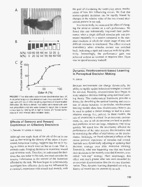
B 4
A(%) 100 80 68 56B(%) 0 20 32 44
100
Uta)
3 50
0
u_ 0 50 100Odor A CYO
FIGURE 1 Two-alternative odor mixture discrimination task. (4)A rat performing odor discrimination task. Rats can perform thistask with 200 ms of odor sampling regardless of discriminationdifficulties. (B) Mixture stimuli. Two odors were mixed with vari-ous proportions and rats were trained to report the dominantcomponent by making left or right a choice. (C) Typical perfor-mance in odor mixture discrimination tasks.
!!17144 32 20 056 68 80100
Effects of Sensory and RewardExperience on Olfactory Decisions
H. Zariwala, N. Uchida, A. Kepecs
Although one might think of the job of the rat in ourtask as determining the identity of the odors it is pre-sented, behavioral ecology suggests that the rat is try-ing to obtain as much water as fast as it can. That is,animals make foraging decisions to maximize rewardand minimize work. Therefore, to understand the deci-sions that rats make in this task, one must consider thesensory information in the context of the incentivesafforded by the task. We have begun to systematicallyinvestigate how olfactory decisions are influenced byan animal's experience with stimuli and rewards, with
the goal of elucidating the underlying neural mecha-nisms of how this influencing occurs. We find thatsensory-guided decisions can be rapidly biased bychanges in the relative value of the two reward alter-natives present in our task.
In a recent study, we examined the effect of chang-ing the stimulus context on a rat's performance. Wefound that rats substantially improved their perfor-mance when a single difficult stimulus pair was pre-sented repeatedly in a session compared to the stan-dard condition in which the whole range of easy anddifficult mixtures is interleaved. This effect reversedimmediately when stimulus context was switchedback, indicating a rapid and transient underlying plas-ticity. Interestingly, the performance gain wasachieved without an increase in response time: Therewas no speed/accuracy tradeoff.
Dynamic Reinforcement-based Learningin Perceptual Decision Making
N. Uchida
Because environments can change dynamically, theability to rapidly update behavioral strategies is crucialfor survival. Recently, neuroscientists have begun tostudy adaptive decision making using statistical learn-ing theory. This mathematical framework provides atheory for describing the optimal learning and execu-tion of choice behavior. In particular, reinforcementlearning models show how reinforcement history canbe used to update a choice strategy to lead to a moreadaptive strategy (i.e., policy). In these models, therole of uncertainty is critical. In an uncertain environ-ment, i.e., one in which outcomes are hard to predictand prediction errors are large, adaptation may occurquickly. We tested how the uncertainty affected theperformance of the odor mixture discrimination taskby examining the effect of trial history on the perfor-mance. Strikingly, we found substantial effects of therecent previous experience of stimulus and reward.Animals were dynamically adjusting or updating theirdecision strategy even after extensive training.Essentially, what was happening was that rats biasedtheir decisions to the recently rewarded direction, a so-called "win-stay" strategy. More surprisingly, thiseffect was much greater after rats were rewarded foran uncertain discrimination than for an easy discrimi-nation. Thus, dynamic learning depended not only onhistory of reward but on uncertainty as well.
135

Control of Sniffing: Active Sensationand Olfactory Behavior
A. Kepecs, N. Uchida
Olfaction depends critically on an active motorprocess, sniffing, which draws air across the nasalepithelium. Respiration is proximally controlled by acentral pattern generator (CPG) in the brain stem, butlittle is known about the central regulation of the res-piratory rhythm during behavior.
Nasal thermocouples (small temperature-sensingprobes) allow us to monitor respiration in freelybehaving animals. Using this method, we are examin-ing the regulation of respiration in rats performing atwo-alternative olfactory discrimination task. Weexamine its frequency and phase in relation to odorsampling, and also to motor and reward events.Respiration frequency varied over a wide range (<2 Hzto >12 Hz) during the course of a behavioral session,systematically changing in relation to significantbehavioral events such as entry into the odor samplingor reward ports. Interestingly, rather than a continuousdistribution of frequencies, discrete frequency modeswere observed. Rats invariably switched to a theta fre-quency mode (8-10 Hz) shortly before (self-initiated)odor port entry and remained tightly locked to this fre-quency mode throughout odor presentation. Theswitch between different modes occurred very rapidly,almost always within a single respiration cycle.Interestingly, the timing of respiration was related notonly to the sensory demands of odor sampling but alsoto other motor processes. In particular, the timing ofhead retractions from the odor and choice ports wasprecisely coordinated with the respiratory cycle. Theseobservations reveal a powerful and rapid top-downcontrol of respiratory CPGs, allowing both expecta-tions to direct sensory acquisition as well as the coor-dination of respiratory and motor processes.
Single Unit Encoding ofGoal-directed Behavior inOrbitofrontal Cortex
C. Feierstein, M. Quirk, N. Uchida
Every day we face the need to make decisions: Wechoose between different alternatives to achievedesired goals. This is the essence of "goal-directed"behavior. Making a decision is a complex process thatdepends on integrating information about the environ-
136
ment, the alternative actions and their consequences,and choosing what is more likely to result in theaccomplishment of our goal. We are interested inunderstanding the neural basis of decision-making andgoal-directed behavior. In particular, we study olfacto-ry-guided decisions. We are using chronic multi-elec-trode recording techniques to monitor the activity ofensembles of neurons in rats performing a two-alter-native odor discrimination task (Fig. 1). Our studieshave focused on the orbitofrontal cortex (OFC) andassociated cortical and subcortical areas.
Orbitofrontal cortex is a region of the prefrontalcortex critical to incorporating predictions aboutexpected outcomes (e.g., rewards) into decision mak-ing. Previous work shows that single OFC neurons dif-ferentially encode odors and visual stimuli accordingto associated rewards and punishments, but it is notknown whether OFC neurons encode motor orresponse variables that would be required to learn thelinks between actions and their consequences or tomake goal-directed choices. We find that a large frac-tion of OFC neurons are indeed tuned for responsedirection, either alone or in combination with stimulusor reward tuning. Analysis of error trials showed thatsingle OFC neurons dynamically integrated olfactory(sensory) and spatial (response) information in a waythat allows them to predict the outcome of a trial or toremember the stimulus associated with a deliveredreward. As a population, OFC neurons are predomi-nantly tuned for correct stimulus-response associa-tions in task-trained animals and thereby correctlymodel the contingencies of the task. Our findings sug-gest that in the rat OFC, circuits not only link stimuliand reward but, like other areas of the prefrontal cor-tex, process information about the relationshipsbetween stimuli and actions.
Local Network Interactions inPrefrontal Microcircuits
M. Quirk, C. Feierstein
Although much is known about the cellular and synap-tic components of cortical circuits, considerably less isknown about how these circuits behave in the func-tioning brain. Using multi-electrode recording tech-niques, we are able to isolate action potentials arisingfrom multiple cells within a small local volume of tis-sue (diameter <100 um). By applying these recordingsto the prefrontal cortex of rats performing our two-alternative olfactory discrimination task (Fig. 1), we

are studying how information is represented withinthese microcircuits during the formation of a binaryperceptual decision. In contrast to the typical colum-nar organization of primary sensory areas, even near-by cells display diverse response properties. The het-erogeneity of responses across cells recorded in thesame electrode was comparable to that of cells acrosselectrodes, indicating that salient task variables(including stimulus, response, and reward informa-tion) represented in prefrontal cortex are availablelocally within individual microcolumns. To begin tocharacterize the functional architecture of a corticalmicrocircuit during a behavioral task, we analyzedpair-wise statistical interactions of the spike trains ofsimultaneously recorded cells. We find stronglyenhanced interactions between neighboring neuronscompared to distal neurons, suggesting that localinputs contribute to firing properties of cells.Interestingly, the spike trains of neighboring neuronsare very often anticorrelated. The function of this inhi-bition seems to be twofold. First, inhibition betweencells with opposite tuning properties promotes a formof "winner-take-all" competition essential for effec-tive decision making. Second, inhibition between cellswith similar tuning profiles provides a mechanism foreffectively controlling the temporal flow of informa-tion within the circuit by limiting the number of cellsactive at a given moment in time. Together, theseobservations suggest a local circuit mechanism bywhich prefrontal cortex learns stimulus-responseassociations and uses them for decision making.
Neural Mechanisms ofContext-dependent Decisions
D. Sosulski, M. Quirk
One of the biological foundations of cognitive flexi-bility-the ability to adapt one's behavior and think-ing in the face of new or rapidly changing circum-stances-is the brain's ability to integrate sensoryinformation from several modalities, create an inter-nal representation of the environment, and use thisrepresentation to subsequently guide its selection ofbehavioral response in a given situation. It has longbeen known that the prefrontal cortex is critical in sit-uations when mappings exist between a single stimu-lus and a number of behavioral responses whoseappropriateness depends on the current situation;however, the mechanisms by which the brain actuallyselects among possible behavioral responses remains
unclear. To investigate the neural mechanisms bywhich stimulus-response association information andinformation about context interact in prefrontal cortex,we established a context-dependent decision-makingtask that requires rodents to respond in different waysto a stimulus, depending on the spatial context inwhich they experience it. In addition, we have recent-ly started to use multi-electrode arrays to simultane-ously record the activity of many single neurons in ratsperforming this context-dependent decision task. Byanalyzing how neurons that code for different stimu-lus-response associations are modulated by spatialcontext, we hope to gain some insight into how thebrain integrates information about learned responsesand the environment, and uses this information to suc-cessfully guide its behavior in a world that is continu-ally in flux.
Inter-areal Coordination among BrainRegions during Olfactory Decisions
A. Kepecs
The brain has many interconnected areas specializedto carry out different functions. As with any complexnetwork, it is essential for the brain to coordinate theactivity of these many regions and to do so dynami-cally. For instance, if you were asked to switch fromwriting down what you are hearing to what you aresmelling, your brain would have to dynamically mod-ulate the flow of information between its auditory,olfactory, and motor areas, turning off an auditory-motor pathway and turning on an olfactory-motorpathway. Yet the long-range wiring of the brain isessentially fixed. How then do neural networks in thebrain accomplish dynamic and flexible routing ofinformation? Or, to put it another way, how does themassively parallel and asynchronously operating braincoordinate its operation?
Although there is no global clock, similar to thosein computers, oscillations in neural activity have beenwidely observed at specific frequencies and in identi-fied behavioral states. If large-scale coherent neuralactivity could be established across areas and dynam-ically controlled, specificity in neural communicationcould be achieved. This leads to the suggestion thattransient and dynamic oscillatory synchronizationbetween brain regions is used to differentially routeand thus coordinate information flow. To examine thisidea we are recording local field potentials (represent-ing the summed coherent activity in local neuronal
137

populations) simultaneously from the olfactory bulb,olfactory cortex, and hippocampus in rats performingan olfactory discrimination task (Fig. 1). We are usingspectral analysis to examine how features of the localfield potentials correspond to different behavioralepochs and the nature of the relationship betweenoscillations in different regions. Strong theta (4-10Hz) oscillations appear at all sites during the task,whereas beta (15-30 Hz) frequency oscillations aretransient, occurring specifically around the time ofchoice in the olfactory bulb and ventral hippocampus.Interestingly, the ventral hippocampus shows differen-tial coordination with different anatomical partners:theta band coherence with the dorsal hippocampus andtransient beta band coherence with the olfactory bulb.These findings are beginning to paint a broad-scalepicture of the spatiotemporal organization of inter-areal coordination during olfactory discrimination.
Olfactory Discriminationin Mice
S. Ranade, N. Uchida
We have developed and extensively characterized thebehavior of rats during the performance of a two-alter-native olfactory discrimination task (Fig. 1). However,to harness the power of transgenic gene manipulationtechnologies for use in the study of the neural basis ofdecision making and other cognitive functions, it isimportant to develop behavioral paradigms suitablefor use with mice, for which many genetic models andmanipulations currently exist. With this goal in mind,we have trained mice to perform a two-alternativeolfactory discrimination task. We find that, similar torats, mice can perform this task with high accuracy(>95% total score) and they require only 4-6 sessions(1000 trials) to reach criterion performance (>85%).Performance remains stable over days and evenmonths. In addition, the reaction time (odor samplingtime) of mice performing the two-alternative task isvery similar to rats (-300 ms). Using this well-con-trolled behavioral task we can explore differentaspects of brain function from sensation to action. Incollaboration with Karel Svoboda's group here atCSHL, we are applying transgenic technologies toinducibly inhibit neural activity in restricted popula-tions of olfactory sensory neurons. This approach willenable us to examine the effect of specific manipula-tions of sensory input on an animal's perception asassayed by his performance.
138
Neural and Metabolic Activity inOlfactory Glomeruli
H. Gurden, S. Ranade, N. Uchida
Neural activity is mirrored by metabolic activity,which in turn causes physiological signals that can bedetected using functional imaging techniques. Themechanisms that link these processes are the subjectof intense interest. We are investigating the linkbetween odor-evoked neural activity and intrinsic opti-cal signals (IOS) in the olfactory glomeruli. IOS areused for studying the representation of odors, but wehope to clarify how to interpret IOS as a readout ofglomerular activity and the mechanisms that link neur-al activity to metabolism and functional imaging sig-nals.
We used in vivo imaging and simultaneous localpharmacology to study how sensory-evoked neuralactivity is transduced into IOS in the well-defined cir-cuitry of the olfactory glomerulus. Odor-evoked IOSwere tightly coupled to release of glutamate and werestrongly modulated by exogenous and endogenousactivation of presynaptic dopamine and y-amino-n-butyric acid (GABA)-B receptors. Surprisingly, IOSwere independent of postsynaptic transmissionthrough ionotropic glutamate receptors. Instead, IOSdepended on the activation of astrocytic glutamatetransporters. Thus, glutamate uptake by astrocytes isan important pathway by which neural activity is cou-pled to functional imaging signals and through whichmetabolic and information processing networks shareinformation in the brain.
Calcium Dynamics UnderlyingDendritic Integration in OlfactoryBulb Granule Cells
V. Egger [in collaboration with K. Svoboda, Cold SpringHarbor Laboratory]
In the mammalian olfactory bulb, axonless granulecells process synaptic input and output reciprocallywithin large spines. The nature of the calcium signalsthat underlie the pre- and postsynaptic function ofthese spines is largely unknown. Using two-photonimaging in acute rat brain slices and glomerular stim-ulation of mitral/tufted cells, we observed two formsof action potential-independent synaptic Ca" signalsin granule cell dendrites. Weak activation of mitral/

tufted cells produced stochastic Ca" transients inindividual granule cell spines. These transients werestrictly localized to the spine head, indicating a localpassive boosting or spine spike. Ca" sources forthese local synaptic events included NMDA recep-tors, voltage-dependent calcium channels and Ca"-induced Ca" release from internal stores. Strongeractivation of mitral/tufted cells produced a low-threshold Ca' spike (LTS) throughout the granulecell apical dendrite. This global spike was mediatedby T-type Ca" channels and represents a candidatemechanism for subthreshold lateral inhibition in theolfactory bulb. The coincidence of local input andLTS in a spine resulted in summation of local andglobal Ca' signals, a dendritic computation thatcould endow granule cells with subthreshold associa-tive plasticity.
PUBLICATIONS
Egger V., Svoboda K., and Mainen Z.F. 2004. Glomerular inputresults in distinct modes of calcium signaling in olfactorybulb granule cell dendrites. Soc. Neurosci. Abstr .30:Program 67.19.
Feierstein C.E., Quirk M.C., and Mainen Z.F. 2004. Spatial cod-ing in piriform and orbitofrontal cortex during olfactory dis-crimination. Soc. Neurosci. Abstr 30: Program 781.19.
Kepecs A. and Mainen Z.F. 2004. Inter-areal interactions duringolfactory discrimination in rats. Soc. Neurosci. Abstr. 30:Program 899.7.
Ranade S.P. and Mainen Z.F. 2004. Serotonin modulates odor-evoked sensory input in the rat main olfactory bulb. Soc.Neurosci. Abstr. 30: Program 531.8.
Uchida N. and Mainen Z.F. 2004. Psychophysical characteriza-tion of concentration-invariant olfactory recognition. Soc.Neurosci. Abstr. 30: Program 531.9.
Zariwala H.A., Kepecs A., and Mainen Z.F. 2004 Contextualadaptation in rat olfactory discrimination. Soc. Neurosci.Abstr. 30: Program 899.8.
Sachin Ranade, grad student, Zach Mainen's lab.
139

TRANSMISSION AND PLASTICITY IN MAMMALIAN CENTRALSYNAPSES
R. Malinow A. Barria H.W.H.G. Kessels B. LiJ. Boehm M. Klein S. RumpelI. Ehrlich C. Kopec W. WeiR. Huganir J. LeDoux
This laboratory is directed toward an understanding ofsynaptic function, synaptic plasticity, and synapticdysfunction. Through such an understanding, we hopeto elucidate how learning and memory are achievedand how diseases corrupt them.
This year, we continued to examine the regulationof a-amino-3-hydroxy-5-methyl -3-isoazole (AMPA)and N-methyl-o-aspartate (NMDA) receptors atsynapses that may underlie plasticity and the malfunc-tion that could lead to diseases. Some of our studiesour summarized below.
Postsynaptic Receptor TraffickingUnderlying a Form of AssociativeLearning
S. Rumpel, J. LeDoux, R. Malinow [in collaboration withA. Zador, CSHL]
To elucidate the molecular, cellular, and circuit changesthat occur in the brain during learning, we investigatedthe role of a glutamate receptor subtype in fear condi-tioning. In this form of learning, animals associate twostimuli, such as a tone and a shock. In this study, wefound that fear conditioning drives AMPA-type gluta-mate receptors into the postsynapse of a large fraction ofneurons in the lateral amygdala, a brain structure essen-tial for this learning process. Furthermore, memory wasreduced if AMPA receptor (AMPA-R) synaptic incorpo-ration was blocked in as few as 10-20% of lateral amyg-dala neurons. Thus, the encoding of memories in the lat-eral amygdala is mediated by AMPA-R trafficking, iswidely distributed, and displays little redundancy.
NMDA-R Subunit CompositionControls Synaptic Plasticityby Regulating Binding to CaMKII
A. Barria, R. Malinow
Calcium entry through postsynaptic NMDA-Rs andsubsequent activation of calcium/calmodulin-depen-
140
dent protein kinase II (CaMKII) are events that triggersynaptic plasticity in many brain regions. Upon acti-vation, CaMKII can associate with NMDA-Rs, but thephysiological role of this interaction is not well-under-stood. In this study, we tested if association betweenactive CaMKII and synaptic NMDA-Rs is required forsynaptic plasticity. We used organotypic hippocampalslices in which long-term potentiation (LTP) requiresCaMKII activity and NR2B, an NMDA-R subunit thatbinds with high affinity to CaMKII. We found thatswitching synaptic NR2B-containing NMDA-Rs withthose containing NR2A, a subunit that shows lowaffinity for CaMKII, dramatically reduces LTP.Expression of NR2A with mutations that increaseassociation to active CaMKII recovered LTP. Finally,driving into synapses NR2B with mutations thatreduce association to active CaMKII prevented LIP.We conclude that association between active CaMKIIand NR2B is required for hippocampal LTP in thisearly developmental period. The switch from NR2B toNR2A content in synaptic NMDA-Rs normallyobserved in many brain regions may control plasticityby reducing the binding of active CaMKII.
Spine Enlargement PrecedesAMPA-R Exocytosis during LTP
C. Kopec, B. Li, W. Wei, J. Boehm, R. Malinow
The changes in synaptic morphology and receptorcontent that underlie neural plasticity are poorlyunderstood. In this study, we used a pH-sensitivegreen fluorescent protein (GFP) to tag recombinantglutamate receptors and showed that chemicallyinduced LTP drives robust exocytosis of AMPA-Rsonto dendritic spines. In contrast, the same stimulusproduced a small reduction of NMDA-Rs from spines.LTP produced a similar modification of small andlarge spines. Interestingly, during LTP induction,spines increased in volume before surface incorpora-

tion of AMPA-Rs, indicating that distinct mechanismsunderlie changes in morphology and receptor content.
Differential SubcellularOverexpression of AMPA-RSubunits in CA1 Neurons
H.W.H.G. Kessels, M. Klein, R. Malinow
The role of AMPA-type glutamate receptors in synap-tic plasticity can be studied by the introduction ofrecombinant AMPA subunits in glutamatergic neu-rons. Here we show that although transient expressionof AMPA subunits leads to considerable overproduc-tion in neuronal somata of organotypic hippocampalslices, at dendrites, they remain close to endogenouslevels. These results provide evidence for a tightlycontrolled transport mechanism of AMPA-R complex-es traveling from soma to dendritic compartments.
Direct Phosphorylation by PKCat a Novel Site on an AMPA-RSubunit GIuR1 Controls SynapticIncorporation during LTP
J. Boehm, R. Huganir, R. Malinow
Incorporation of G1uR1- containing AMPA-Rs intosynapses has an important role in several forms ofneural plasticity. A number of signaling pathwayshave been identified, but the direct modifications ofG1uR1 that control its synaptic incorporation have notbeen found. Previous studies indicate that activationof protein kinase C (PKC) is required to generate LTP,a leading model of synaptic plasticity. However, thetargets of PKC responsible for LTP have not beendetermined. We find that G1uR1 Ser-818, which ishighly conserved evolutionarily, is phosphorylated byPKC in vitro and by LTP in hippocampal slices. Acutephosphorylation by PKC at GluRl Ser-818 as well asmolecular mimicking phosphorylation at this site dri-ves G1uR1 into synapses. Preventing GluR1 Ser-818phosphorylation blocks LTP and PKC-driven synapticincorporation of GluRI. Thus, GluRl Ser-818 israpidly phosphorylated by PKC during LTP inductionand is required for its incorporation into synapses.
PSD-95 Controls AMPA-R Incorporationduring Long-term Potentiation andExperience-driven Synaptic Plasticity
I. Ehrlich, R. Malinow
The regulated delivery of AMPA-type glutamatereceptors to synapses is an important mechanismunderlying synaptic plasticity. Here we ask if thesynaptic scaffolding protein PSD-95 participates inAMPA-R incorporation during two forms of synapticplasticity. In hippocampal slice cultures, expression ofPSD-95-GFP increases AMPA-R currents by selec-tively delivering GluR 1 -containing receptors tosynapses, thus mimicking LTP. Mutational analysisshows that the amino terminus of PSD-95 (includingthe first two PDZ domains) is necessary and sufficientto mediate this effect. Further supporting a role insynaptic plasticity, wild-type PSD-95 occludes LTP,and dominant-negative forms block LTP. Moreover,we demonstrate that PSD-95 participates also inAMPA-R delivery during experience-driven plasticityin vivo. In barrel cortex from experience-deprived ani-mals, expression of PSD-95-GFP selectively increasesAMPA-R currents, mimicking experience-driven plas-ticity. In nondeprived animals, PSD-95-GFP producesno further potentiation, indicating common mecha-nisms between PSD-95-mediated potentiation andexperience-driven synaptic strengthening. A domi-nant-negative form of PSD-95 blocks experience-dri-ven potentiation of synapses. Pharmacological analy-sis in slice cultures reveals that PSD-95 acts down-stream from other signaling pathways involved in LIP.We conclude that PSD-95 controls activity-dependentAMPA-R incorporation at synapses via PDZ interac-tions not only during LTP in vitro, but also duringexperience-driven synaptic strengthening by naturalstimuli in vivo.
PUBLICATIONS
Ehrlich I. and Malinow R. 2004. PSD-95 controls AMPA-recep-tor incorporation during long-term potentiation and experi-ence-driven synaptic plasticity. J. Neurosci. 24: 916-927.
In Press
Rumpel S., Ledoux J., Zador A., and Malinow R. 2005.Postsynaptic receptor trafficking underlying a form of asso-ciative learning. Science (in press).
141

INTEGRATIVE SYSTEMS NEUROBIOLOGY
P.P. Mitra P. AndrewsH. BokilH. Maniar
Partha Mitra (previously located at Bell Laboratories,Lucent Technologies) joined the faculty at CSHL inthe fall of 2003 when this research effort began.Hemant Bokil is a postdoctoral fellow continuing hiswork on signal processing applications to neurobio-logical time series with special emphasis on neuralprostheses. Hiren Maniar, a postdoctoral fellow, isworking as part of a consortium funded by the HumanFrontiers Science Foundation on the role of neuralsynchrony in integration across sensory modalities.Peter Andrews joined the group in August 2004 afterfinishing his Ph.D. in physics and a postdoc at YaleUniversity, where he was responsible for the program-ming and data analysis for the QUEST quasar survey.Peter is performing the dual role of providing softwareinfrastructure for the group, as well as participating inthe open source software project Chronux to developsoftware for neurobiological data analysis.
Apart from this group of core personnel, ourresearch effort includes close collaborative ties withmultiple research groups both locally and globally. Acollaborative effort with Niko Schiff and Keith Purpuraat the Weill Medical School of Cornell University,where Partha is an adjunct associate professor, involveselectrophysiology in the awake behaving macaque. Asecond research effort on how zebra finches learn theirsong vocalizations is being carried out in collaborationwith Ofer Tchernichovski at City College of New York.Recently, this research effort has been broadened into aconsortium of zebra finch researchers based in four dif-ferent universities. A third collaboration is with thegroup of Rodolfo Llinas at New York UniversityMedical College. A fourth consortium-based researcheffort to study cross-modal integration between thesenses is funded by the Human Frontiers ScienceProgram, including the laboratories of ShinsukeShimojo (California Institute of Technology), PascalFries (EC. Donders Research Center at Nijemegen),Shiny' a Nishida at the Nippon Telegraph and TelephoneCorporation Labs in Japan, and Alan Johnston atUniversity College, London in the United Kingdom.
Our basic research philosophy is to bring computa-tional and theoretical tools to bear on biological ques-
142
tions in general and neurobiological questions in par-ticular, to make sense of biological complexity and toprovide understanding and potential therapies for dis-ease. There are two basic branches to the research: neu-roinformatics and theoretical engineering. The formerinvolves the application of statistical and computation-al tools to neurobiological data, primarily single-chan-nel and multichannel time series, including pointprocesses (spike trains) and brain imaging data. Theapplications are geared toward exploratory analysis oflarge volumes of data, as well as confirmatory analysisfor testing of specific hypotheses. This is a continuingresearch effort that reflects several years of work and isassociated with a course entitled "Neuroinformatics"held each summer at the Marine Biological Labora-tories in Massachusetts. The second branch of researchdeals with the question of theoretical principles under-lying the design of biological systems. Since biologicalorganisms are evolved to perform their tasks robustlyunder uncertain circumstance, in terms of both theenvironment and parametric uncertainty in compo-nents making up the system, it is expected that themathematical theories developed by engineers to helpdesign man-made systems that perform under similar-ly challenging circumstances may also apply to thesesystems. The goal of this research is to identify theengineering principles most germane to biology and tostudy selected examples drawn from different levels ofthe organizational hierarchy.
Chronux: Open Source Software forAnalyzing NeurobiologicalTime Series Data
H. Bokil, P. Andrews
The nervous system stores and executes behaviors andadapts these behaviors to the environment throughsensory monitoring. Studies of the activity of the ner-vous system consist primarily of monitoring thedynamics of units at different levels of hierarchy of thesystem, ranging from single neurons measured electri-

cally to large groups of neurons monitored indirectlythrough a variety of imaging methodologies. The rel-evant measurements consist of single or multichanneltime series data, in the form of continuous or pointprocesses. Therefore, the statistical analysis of timeseries data is central to studying the nervous system.
During the last several years, our research hasinvolved the development of algorithmic and compu-tational tools for the analysis of multichannel timeseries data from the neurosciences. We are now encod-ing these tools into an open source software packageentitled Chronux. This project will proceed in stagesand involve the development of a high-quality numer-ical analysis library, data I0 and management utilities,and a user interface that provides experimental neuro-scientists access to advanced analysis tools. The pro-ject will proceed in stages, where we move from thecurrent implementation of the relevant routines asMATLAB toolboxes, to a fully open source and self-contained software. Although there are a number ofsignal processing libraries available, the goal of thiseffort is to develop domain-specific tools that will aidneuroscience research. We expect that tools such asthese will be critical to advancing our understandingof systems neuroscience.
The past year has seen substantial improvementsmade to Chronux. On the algorithmic side, we haveadded routines that provide robust estimates of firingrates based on local regression methods. We have alsocreated the Chronux "knowledge environment," whichis a Web-based tool for documenting Chronux andcollaboratively working on algorithm development.We continue to work on developing new statisticaltools for time series analysis, and recent advancesinclude extensions of some standard measures fromspectral analysis to particular cases relevant to neuro-biological data analysis. We have recently presented areview of the growing field of neural time seriesanalysis (Brown et al. 2004).
Temporal Structure in Neural Activityduring Working Memory
P.P. Mitra [in collaboration with K. Purpura and N. Schiff,Weill Medical School, Cornell University]
Holding items in short-term memory is thought tocorrespond to appropriate holding patterns of electri-cal activity in the brain. An experimental model sys-tem much studied in this regard involves memory sac-
cades in awake behaving macaques, where the subjectremembers a target location during a memory periodand performs a visual saccade to the target on anappropriate cue. Area LIP, on the lateral bank of theintraparietal sulcus, is known to encode intended sac-cade directions. Individual neurons may be found inthis area which show a transiently elevated firing rateduring the memory period of a saccade memory task,with tuning to the direction of the intended saccade.
In collaborative work with Richard Andersen's labat Caltech, we have previously discovered that in addi-tion to the firing rates, the dynamics of the neuralactivity, as reflected in spectral measures, also showstuning to intended saccade direction. This project hasbeen transferred to the Weill Medical School ofCornell University, where it is being carried out in col-laboration with K. Purpura and N. Schiff. In the previ-ous year, we completed the setup phase of the new lab-oratory and began recording from one monkey withdual chambers over the parietal and frontal cortices.Preliminary recordings from the frontal eye fieldsshow behaviorally tuned spectral structure in the neur-al activity, analogous to the original findings forrecordings from area LIP. These results were presentedat the annual Society for Neuroscience meeting (2004).
Song Learning in the Zebra Finch
P.P. Mitra [in collaboration with 0. Tchernichovskt,City College of New York]
The neural substrate and evolutionary origins of linguis-tic behavior in humans is of great biological interest, butdifficult to study, since there are no other species wherethe acoustic communication behavior is seen to exhibitthe same level of syntactic complexity. Therefore, therehas been great interest in studying what has been termedthe faculty of language in a broad sense, one aspect ofwhich is the ability to learn vocalizations. This last isitself infrequently found in animals-a reason whysongbirds, which exhibit learning of complex vocalrepertoires, have been the subject of intensive study. Thespecies of choice in this research is the zebra finch,where all aspects of the song system have been scruti-nized, ranging from the sensory motor circuitry in thebrain to the production apparatus. We have developedtechniques for detailed quantification of song develop-ment, through the analysis of continuously acquiredacoustic signals from birds subjected to a controlledtraining protocol.
143

In this ongoing research project, SebastienDergnaucourt, a postdoc in the Tchernichovski laborato-ry, has demonstrated that sleep affects song learning,particularly in the early stages: It appears that song per-formance degrades after night sleep during the first fewweeks after exposure to the song model. This "morningdeterioration" is absent in the adult bird. Somewhatcounterintuitively, the birds that show the largest morn-ing deterioration also show the best final imitation.Although we do not yet fully understand the mechanis-tic basis of this effect, we have demonstrated that it isdirectly related to sleep: The effect was seen to occurafter induction of sleep using melatonin. It is known thatthe zebra finch brain exhibits a replay of the neural pat-terns associated with song during sleep, leading to thehypothesis that the observed deterioration reflects anactive process that occurs during sleep. This work wasrecently reported in Nature (Deregnaucourt et al. 2005).
Vocal Development in Infants
P.P. Mitra
Given the success of the extensive longitudinal record-ings of song from the developing zebra finch, we havestarted a study of human vocal development. The ideais to carry out longitudinal recordings of infant vocal-izations over an extended period and to analyze theresultant data to quantify the vocal development.Similar studies in the past have relied on human tran-scriptions of infant sounds, or sparse recordings. Thecurrent study will increase the coverage in terms ofrecordings and will develop automated tools for analy-sis and quantification.
Source Localization of NeuralActivity in MEG and EEG
H. Maniar, P.P. Mitra
Noninvasive imaging techniques have a critical role instudies pertaining to cognitive processes and in deter-mining pathological conditions in the human brain.Ideally, the imaging technique would concurrentlyexhibit both high temporal and spatial resolution.Techniques based on hemodynamic changes, such as
144
PET, SPECT, and especially fMRI, have a relativelyhigh spatial resolution but poor temporal resolution. Incontrast, techniques measuring neural electrical activitysuch as MEG and EEG have high temporal resolution,but a spatial resolution dependent on the inversemethodology. We have been working on the computa-tional implementation of a new inverse methodology,entitled Local Basis Expansions, for localization ofneural activity. This work is part of the Human FrontiersScience Program consortium effort with ShinsukeShimojo, Pascal Fries, and others, where our methodswill be employed to localize broadband oscillatoryactivity in MEG and EEG measurements. The tool willeventually be made part of the Chronux platform.
PUBLICATIONS
Brown E.N., Kass R.E., and Mitra P.P. 2004. Multiple neuralspike train data analysis: State of the art and future chal-lenges. Nat. Neurosci. 7: 456-461.
Deregnaucourt S., Mitra RP., Feher 0., Maul K.K., Lints T.J.,and Tchernichovski 0. 2004. Song development: In searchof the error-signal. Ann. N.Y. Acad. Sci. 1016: 364-376.
Maniar H., Polyakov A., and Mitra P.P. 2004. A semi-parametricmethod for source localization using local basis expan-sions. BIOMAG meeting, Boston.
Menzer D., Hudson A.E., Schiff N.D., Peseran B., Bokil H.,Purpura K.P., and Mitra P.P. 2004. Time-frequency modu-lation of the monkey frontal lobe EEG produced during amemory-quided saccade task. Poster presented at the34th Annual Meeting of the Society for Neuroscience, SanDiego, California.
Mitra RR, Polyakov A., and Maniar H. 2004. A semi-parametrictechnique for MEG/EEG source localization using localizedbasis expansions (LBE)g. Soc. Neurosci. San Diego.
Tchernichovski 0., Lints T.J., Deregnaucourt S., Cimenser A., andMitra P.P. 2004. Studying the song development process:Rationale and methods. Ann. N.Y. Acad. Sci. 1016: 348-363.
Wegener L.G.L., Povinelli M.L., Green AG., Mitra RR, StarkJ.B., and Littlewood P.B. 2004. The effect of propagationnonlinearities on the information capacity of WDM opticalfiber systems: Cross-phase modulation and four-wave mix-ing. Physica D-Nonlinear Phenomena 189: 81-99.
In Press
Deregnaucourt S., Mitra RP., Feher 0., Pytte C., andTchernichovski 0. 2005. How sleep affects the develop-mental learning of bird song. Nature 433: 710-716.
Menzer D., Hudson A.E., Schiff N.D., Peseran B., Bokil H.,Purpura K.P., and Mitra P.P. 2005. Task dependent modu-lation of monkey front lobe field potentials during visualworking memory. Poster presented at the 12th AnnualCognitive Neuroscience Society Meeting, New York.

EXPERIENCE-DEPENDENT PLASTICITY OF SYNAPSES ANDSYNAPTIC CIRCUITS
K. Svoboda B. BurbachI. BureauV De PaolaN. GrayC. Harvey
A. HoltmaatA. KarpovaT. O'ConnorL. PetreanuT. Sato
V Scheuss L. WilbrechtG. Shepherd R. YasudaA. Sobczyk C. ZhangG. Tervo H. ZhongR. Weimer K. Zito
The functional properties of the brain must change inresponse to salient sensory experiences. The nature ofthese changes at the level of synapses, neurons, andtheir networks, also known as the engram, is unknown.We are dissecting the mechanisms of experience-depen-dent plasticity in the developing and adult neocortex.
Neocortical tissue is dauntingly complex: 1 mm3contains nearly a million neurons, each of which con-nects to thousands of other neurons. To probe neuronsand synapses within the intact network, we build anduse sensitive tools. 2-photon laser-scanning microscopy(2PLSM) allows us to image single synapses in intacttissues and to track changes in intracellular calcium andsignal transduction events. Excitation of neuronal ele-ments by focal uncaging of neurotransmitters allows usto probe the connectivity of neural networks with highefficiency. We combine these optical methods withelectrophysiological measurements of synaptic currentsand potentials and molecular manipulations of neurons.
We use both in vivo measurements to address sys-tem level questions and in vitro methods to get atdetailed mechanisms. As a model system, we use therodent barrel cortex, where whiskers are representedin a topographic manner, with information from eachwhisker represented by a small cortical region (barrel).Whisker maps are shaped by experience during devel-opment and reshaped in the adult. The cellular mech-anisms underlying sensory map plasticity are likely toshare mechanisms with those underlying learning andmemory in other brain regions and other species.
[Ca21 Signaling in SingleDendritic Spines
V Scheuss, ArSobnyk
Long-term changes in synaptic efficacy are triggeredby N-methyl-o-aspartate (NMDA) receptor-mediatedincreases in [Ca'] in the postsynaptic neuron. We havedeveloped a variety of assays to learn about the molec-
ular pathways of Ca' influx, including the counting ofindividual Ca' permeable channels and receptors inspines. W have recently discovered that different spinesare preferentially populated with different types ofNMDA receptors (NMDA-Rs) (NR2A vs. NR2B).Interestingly, these different types of receptors havedifferent Ca' permeabilities, indicating that NMDA-Rs in synapses have the ability to regulate their Ca"permeability independent of their conductance.
[Ca21-dependent Signalingin Single Dendritic Spines
R. Yasuda, C. Harvey, H. Zhong
Postsynaptic Ca' activates diverse Ca"-dependent sig-nal transduction mechanisms. We have designed high-sensitivity fluorescence resonance energy transfer(FRET) sensors for Ras and MAPK activation.Fluorescence lifetime imaging microscopy (FLIM)allows us to image Ca'-dependent signal transductioncascades activated by synaptic transmission in dendritesand spines. We can for the first time study the biochem-ical dynamics of Ca"-dependent Ras signaling in neu-ronal microcompartments. For example, we find thatRas acts as a Ca2tdependent switch and that activationof single synapses can lead to local activation of Ras.
Experience-dependent Plasticityin the Adult Cortex In Vivo
A. Holtmaat, L. Wilbrecht, V. DePaola [in collaborationwith G. Knott, Lausanne]
Sensory representations in the adult brain are stable,yet we are able to learn. Which neural elements areplastic in the neocortex, especially in response tonovel sensory experience? Answers to these questionsare fundamental to the mechanisms of plasticity andthe memory capacity of the brain. Our approach has
145

been to image structural dynamics in vivo. A break-through was the development of long-term (months)time-lapse imaging at the level of individual synapsesin the adult cortex of transgenic mice together withretrospective serial-section electron microscopy.
We find that the large-scale arborization of axonsand dendrites is stable, but that neurons display a richrepertoire of micrometer-level structural plasticity ofdendritic spines, axonal terminals, and axonal branchtips. Plasticity differs among cell types. We havefocused on understanding the plasticity of L5B neu-rons. L5B spines consist of two distinct populations:small "transient" spines that live for days and large"persistent" spines that live much longer (months).Under baseline conditions, at least half of the transientspines have synapses. Induction of plasticity by trim-ming a subset of whiskers transiently (-1-4 days)increases spine turnover, followed by stabilization(after -1 week) with new persistent synapses.
Circuit Mechanisms ofExperience-dependent Plasticity
G. Shepherd, I. Bureau, L. Petreanu [in collaborationwith M. Chklovskii, Cold Spring Harbor Laboratory]
The wiring diagram is fundamental to understandingcortical function and plasticity. However, little quanti-tative information about functional circuits is avail-able. What are the sources of input to a neuronal sub-type in a particular layer and column and what aretheir relative strengths? Which connections changewith novel sensory experience?
We developed laser-scanning photostimulation(LSPS) into a quantitative and rapid tool for circuit analy-sis. In brain slices, we mapped the excitatory circuitsimpinging onto L2/3 neurons. We discovered "septal"columns between the "barrel" columns. These columnarsystems are part of parallel thalamocortical circuits. Todetermine if morphology can predict functional circuitsbetween excitatory neurons, we directly compared func-tional LSPS maps with "geometric circuits" computedfrom quantitative reconstructions of axons and dendrites.Functional connections were accurately predicted bygeometry within a particular projection (with interestingexceptions), but the ratio of functional to geometric con-nectivity differed greatly (>20-fold) between projections.What is the cellular basis for the differences betweenthese projections? The synaptic weight per synapse? Thedensity of synapses per wire length? The fraction of con-nected neurons or the number of synapses between con-
146
nected neurons? To address these and related questions,we will develop novel approaches for circuit mappingwith single-cell resolution.
Also using LSPS, we measured the functional andanatomical development of cortical columns, findingremarkable specificity, without detectable "overgrowthand pruning." We further performed an unbiased searchfor experience-dependent synaptic pathways duringdevelopment. The strengths of excitatory L4->L2/3connections changed with opposing signs in barrel andseptal columns, explaining previously observed recep-tive field changes. The identification of these pathwaysis allowing us to determine the cellular mechanisms ofexperience-dependent synaptic plasticity.
Development ofThalamocortical Axons
R. Weimer [in collaboration with C. Portera-Cailliau,University of California, Los Angeles]
The development of mammalian cortical axons hasbeen studied extensively in histological preparations.However, important questions about axonal develop-ment require time-lapse imaging. What is the balancebetween growth and pruning? How rapid are growthand pruning? Do all axons develop with similarsequences or are there multiple strategies for axon elab-oration? To address these issues, we have used 2-photontime-lapse microscopy to image axons in GFP (greenfluorescent protein) transgenic mice during the first 3weeks of postnatal development. This period spans thearrival of thalamocortical (TC) axon collaterals to layer1, the presence and disappearance of Cajal-Retzius(CR) neurons, cortical neuronal migration, and barrelcortex critical periods. The elaboration of local axonalarbors relies on a careful balance of growth and pruningof axon segments that favors overall growth only slight-ly. Pruning involved either retraction of small axon seg-ments (tens of micrometers) or fragmentation of muchlarger branches (hundreds of micrometers). The dynam-ics of axonal arbors of CR and TC neurons differed pro-foundly. TC axonal branch tips lack distinct growthcones and grow rapidly in straight paths, with frequentintersitial branch additions. The axon tips of CR axonshave large growth cones with numerous long filopodiaand grow slowly following tortuous paths. Given theidentical terrain upon which CR and TC axons grow,these two distinct modes of growth likely reflect differ-ent intrinsic growth programs for long-range projectionneurons versus local interneurons.

Cellular Mechanisms ofSynapse Development
K. Zito
Most of the excitatory synapses in the cortex occur ondendritic spines, small protrusions extending fromneuronal dendrites. To determine if spine growth isassociated with synapse formation, we studiedNeurabin-I (NrbI), a neuronal specific actin-bindingprotein. A severely truncated GFP-NrbI caused a dra-matic increase (more than twofold) in the number ofspines. Using electron microscopy, we found that Nrbis concentrated at sites of cell-cell contact and thatmorphological changes reflect an increase in the num-ber of synapses. Nrb truncations drove the polymer-ization of spine actin to favor spine growth andsynapse formation.
Molecular Methods to ReversiblyInactivate Synapses In Vivo
A. Karpova, G. Tervo, N. Gray, B. Burbach
Inducible and reversible silencing of selected neuronsin vivo is critical to understanding the structure anddynamics of brain circuits. We have developed mole-cules for inactivation of synaptic transmission(MISTs) that can genetically be targeted to allow thereversible inactivation of neurotransmitter release.MISTs consist of modified presynaptic proteins thatinterfere with the synaptic vesicle cycle when cross-linked by small molecule "dimerizers." MISTs basedon the vesicle proteins VAMP2/Synaptobrevin andSynaptophysin induced rapid (-10 min) and reversibleblock of synaptic transmission in cultured neurons andbrain slices. In transgenic mice expressing MISTs inPurkinje neurons, administration of dimerizer reducedlearning and performance of the rotarod behavior.MISTs allow for specific, inducible, and reversiblelesions in neuronal circuits and may provide treatmentof disorders associated with neuronal hyperactivity.
PUBLICATIONS
Brecht M., Fee M.S., Garaschuk 0., Helmchen F., Margrie T.W.,Svoboda K., and Osten P. 2004. Novel approaches tomonitor and manipulate single neurons in vivo. J. Neurosci.24: 9223-9227.
Bureau I., Shepherd G.M., and Svoboda K. 2004. Precise
development of functional and anatomical columns in theneocortex. Neuron 42: 789-801.
Chen B., Trachtenberg J.T., Holtmaat A.J., and Svoboda K.2004.. Long-term, high-resolution imaging in the neocortexin vivo. In Live cell imaging (ed. R.D. Goldman and D.L.Spector), pp. 423-434. Cold Spring Harbor LaboratoryPress, Cold Spring Harbor, New York.
Chkloyskii D.B., Mel B.W., and Svoboda K. 2004. Corticalrewiring and information storage. Nature 431: 782-788.
Di Cristo G.D., Wu C., Chattopadhyaya B., Ango F., Knott G.,Welker E., Svoboda K., and Huang Z.J. 2004. Subcellulardomain-restricted GABAergic innervation in primary visualcortex in the absence of sensory and thalamic inputs. Nat.Neurosci. 7: 1184-1186.
Holtmaat A.J., Wilbrecht L., Karpova A., Portera-CailliauBurbach B., Trachtenberg J.T., and Svoboda K. 2004.Long-term, high-resolution imaging of neurons in the neo-cortex in vivo. In Imaging neurons (ed. R. Yuste and A.Konnerth), pp. 627-638. Cold Spring Harbor LaboratoryPress, Cold Spring Harbor, New York.
Maravall M., Stern E.A., and Svoboda K. 2004. Development ofintrinsic properties and excitability of layer 2/3 pyramidalneurons during a critical period for sensory maps in rat bar-rel cortex. J. Neurophysiol. 92: 144-156.
Maravall M., Koh I.Y., Lindquist W.B., and Svoboda K. 2004.Experience-dependent changes in basal dendritic branch-ing of layer 2/3 pyramidal neurons during a critical periodfor developmental plasticity in rat barrel cortex. Cereb.Cortex 14: 655-664.
Nimchinsky E.A., Yasuda R., Oertner T.G., and Svoboda K.2004. The number of glutamate receptors opened bysynaptic stimulation in single hippocampal spines. J.
Neurosci. 24: 2054-2064.Pologruto TA., Yasuda R., and Svoboda K. 2004. Monitoring
neural activity and [Ca2+] with genetically encoded Ca2+indicators. J. Neurosci. 24: 9572-9579.
Svoboda K. 2004. Do spines and dendrites distribute dye even-ly? Trends Neurosci. 27: 445-446.
Wang Y., Guo H., Pologruto TA., Hannan E, Hakker I.,
Svoboda K., and Zhong Y. 2004. Stereotyped odor-evokedactivity in the mushroom body of Drosophila revealed bygreen fluorescent protein-based Ca2+ imaging. J.
Neurosci. 24: 6507-6514.Yasuda R., Nimchinsky E.A., Scheuss V., Pologruto TA.,
Oertner T.G., Sabatini B.L., and Svoboda K. 2004. Imagingcalcium concentration dynamics in small neuronal com-partments. Sci. STKE 2004: p15.
Zito K., Knott G., Shepherd G.M., Shenolikar S., and SyobodaK. 2004. Induction of spine growth and synapse formationby regulation of the spine actin cytoskeleton. Neuron 44:321-334.
In Press
Holtmaat A.J., Trachtenberg J.T., Wilbrecht L., Shepherd G.M.,Zhang X., Knott G.W., and Svoboda K. 2005. Transient andpersistent dendritic spines in the neocortex in vivo. Neuron45: 279-291.
Shepherd G.M.G., Stepanyants A., Bureau I., Chklovskii D.B.,and Svoboda K. 2005. Geometric and functional organiza-tion of cortical circuits. Nat. Neurosci. (in press).
147

NEUROGENETICS OF MEMORY IN DROSOPHILA
T. Tully J. Barditch Y. Hua D. Leong M. RegulskiF. Bolduc K. lijima-Ando J. Luo N. SinhaL. Grady J. Kui C. Margulies S. Xia
Studies on Pavlovian learning in many animals haverevealed remarkably similar behavioral properties ofassociative learning. Surprisingly, Pavlovian learningin fruit flies also displays these behavioral properties,suggesting a common underlying mechanism. Neuralarchitecture of the Drosophila brain bears no resem-blance to mammalian brains, of course, which sug-gests that the behavioral "homology of function" mustresult from conserved cellular/molecular mechanisms.Consistent with this view, several genes and geneticpathways have been identified to function in both ver-tebrate and invertebrate associative learning, includingCREB and cAMP signaling, staufen, and regulation oflocal protein translation.
We have established a role for N-methyl-D-aspar-tate (NMDA) receptors (NMDA-Rs) in Drosophilalearning and long-term memory (LTM) formation.The Drosophila genome carries homologs of verte-brates NRI and NR2, subunits of which must coassem-ble to form functional NMDA channels in vertebrates.Similarly, dNR1 and dNR2 must be coexpressed toform voltage-dependent NMDA-responsive channelsin flies (see Fig. 1). Flies homozygous for either oftwo P-element insertion mutations of dNR1 displaymoderate defects in olfactory associative learning,
5 mM NMDA
0 [Mg"] = 0 mM[Mg "] =20 mM
-100 -50
-1.5
FIGURE 1 Drosophila NMDA-R function is voltage dependentin the presence of magnesium, a key property of vertebrateNMDA-Rs.
148
which can be rescued via transgenic expression ofwild-type dNR1. Acute expression of anti-RNA fordNR1 in adult flies disrupts 1-day memory afterspaced but not massed training, indicating a specificdisruption of CREB-dependent LTM. NR1 and NR2appear to be expressed widely throughout the adultbrain, with preferential expression in a few novel neu-rons. These data demonstrate an evolutionarily con-served role for NMDA-Rs in associative learning andmemory.
MUTANTS AND MICROARRAYS
More recent investigations of NMDA-R function inDrosophila have been accomplished by engineeringtransgenic flies carrying an upstream activationsequence (UAS)-driven anti-NR1 RNAi (RNA interfer-ence) transgene. We have crossed these transgenic flieswith MB (mushroom body) GAL4 drivers to determinethe role of NMDA-Rs in MBs. Preliminary data suggestthat acute disruption (using hs-GAL4) of NR1 in thismanner yields defects specific to middle-term memory(MTM), but does not affect learning or LTM.
We continue experiments using DNA microarraysto detect gene expression changes associated withLTM formation. To date, we have done follow-up Q-PCR (quantitative polymerase chain reaction) on 137candidate memory genes (CMGs) and have confirmed51, yielding a true-positive "hit rate" of 37%. Our ulti-mate goal is to provide in vivo validation that theseconfirmed CMGs are involved in adult memory for-mation. To this end, we have developed a method toscreen short interfering RNA (siRNA) sequences fortarget genes in cultured S2 cells, using branched-DNAmethodology. Foldback RNAi transgenes are then con-structed with a DNA sequence surrounding the effec-tive siRNA sequence. These foldback RNAi constructsare cloned into a UAS-driven vector, which we haveconstructed to optimize the expression of these RNAiconstructs in vivo. So far, we have generated trans-genic flies for NR1 (a positive control for our UAS-driven vector system) and have identified effectivesiRNA sequences for slmb, aur, and thor. Transgenicflies carrying RNAi constructs for these genes are cur-

rently being generated and will be induced in adults(using hs-GAL4) to assess an acute role for each CMGin memory formation.
In collaboration with Gengxin Chen in MichaelZhang's lab and Qing-Shuo Zhang in Adrian Krainer'slab here at CSHL, we have developed a bioinformaticsapproach to identify candidate genes carrying a nanosresponse element (NRE) in their mRNA transcripts, towhich PUMILIO likely binds. On the basis of our pre-viously published work, CMGs that also carry NREsare likely to represent genes, the transcripts of whichare involved in the local control of protein translationduring LTM formation. This approach serves toexpand the pumilio/staufen pathway to include addi-tional genes and molecular mechanisms.
In collaboration with Dr. Yi Zhong and co-workersat CSHL, we have shown that Notch functions duringLTM formation in Drosophila. Interestingly, overex-pression of Notch leads to enhanced memory forma-tion in a manner similar to that for overexpression ofthe CREB activator (see Publications below). Theseobservations again identify additional genes that mayimpinge on the pumilio/staufen pathway.
CIRCUITS AND PHASES
We continue spatiotemporal experiments, which revealwhere in the adult brain CREB-dependent memoryformation occurs. To date, we have determined thatUAS-driven expression of a CREB-repressor trans-gene, using several different MB GAL4 drivers, doesNOT block LTM formation. Conversely, we have iden-tified one enhancer GAL4 line driving expression ofUAS-CREB-r (repressor) in central complex that doesblock spaced training-specific 1-day memory (LTM).We are currently characterizing additional enhancerGAL4 drivers with overlapping expression in centralcomplex, including some of the enhancer GAL4 linesidentified by us as memory mutants.
We have developed a new method to cell-sort GFP(green fluorescent protein)-expressing neurons fromdissociated adult brains. To maximize the protocol, weexpressed GFP in MBs, sorting dissociated cells intoGFP' versus GFP- subsets. Under these conditions, Q-PCR reveals a 512-fold higher GFP expression inGFP' versus GFP- cells. Q-PCR-derived expression ofCREB, Adfl, fasll, and rut were -.8-, -.5-, 8-, and 2-fold higher in GFP' versus GFP- cells. On the basis ofpublished immunostaining observations, we expect toobserve no differences in expression for CREB and
Adfl and preferential expression of fasII and rut inMB cells. We are currently repeating and extendingthese results and perfecting our cell-sorting method. Ifproven reliable, this new methodology will allowimportant new molecular and biochemical experi-ments on cell types identified to be involved in differ-ent aspects of memory formation (see above).
GENETIC MODELS OF DISEASE
In vertebrate systems, STAUFEN is known to associ-ate with Fragile-X protein (FMRP) in neural granules.On the basis of this observation, we have begun tostudy the fly homolog of FMRP. To date, we haveshown that FMRI mutants are defective in olfactorylearning, and this learning deficit can be rescued bytransgenic expression of a genomic construct. We arecurrently evaluating an acute role for FMRI in memo-ry formation.
We also have begun to study a Drosophila modelof Parkinson's disease. In many vertebrate manifesta-tions of this form of heritable neurodegeneration, neu-rons overexpress a-synuclein. Similarly, transgenicoverexpression of a-synuclein yields neurodegenera-tion in flies. Accordingly, we have begun DNAmicroarray studies in cultured Drosophila cells over-expressing a-synuclein to identify candidate genesinvolved in this form of neurodegeneration.
PUBLICATIONS
Chiang A.-S., Blum A., Barditch J., Chen Y.-H., Chiu S.-L.,Regulski M., Armstrong J.D., Tully T., and Dubnau J. 2004.radish encodes a phospholipase-A2 and defines a neuralcircuit involved in anesthesia resistant memory. Curr. Biol.14: 1-20.
Ge X., Hannan F., tile Z., Feng C., Tully T., Zhou H., Xie Z., andZhong Y. 2004. Notch signaling in Drosophila long-term mem-ory formation. Proc. Natl. Acad. Sci. 101: 10172-10176.
Regulski M., Stasis Y., Tully T., and Enikolopov G. 2004.Mutational analysis of nitric oxide synthase in Drosophilareveals an essential function. Corr. Biol. 14: R881-882.
Stasis Y., Kuzin B., Regulski M., Tully T., and Enikolopov G.2004. Regulation of multimers via truncated isoforms: Anovel mechanism to control nitric oxide signaling. GenesDev. 18:1 -12.
In Press
Xia S., Miyashita T., Fu T.-F., Lin W.-Y., Wu C.-L., Pyzocha L.,Lin I.-R., Saitoe M., Tully T., and Chiang A-S. 2005. NMDAreceptors mediate olfactory learning and memory inDrosophila. Curr. Biol. (in press).
149
NEURAL SUBSTRATE OF SELECTIVE AUDITORY ATTENTIONIN THE CORTEX
A. Zador H. Asan H. OviedoM. DeWeese S. RumpelT. Hromadka L.-H. TaiG. Otazu M. Wehr
We use a combination of theoretical and experimentalapproaches to study how the brain computes. Thebrain is able to solve hard computations that remainfar beyond the reach of the fastest computers. Our goalis to understand this computation at the synaptic, cel-lular, behavioral, and algorithmic levels.
One example of such a hard computation is the"cocktail party problem." When we tune in to onevoice at a cocktail party, and tune out the others-atask that remains beyond the capacity of modern com-puters-we are engaging in a form of selective audito-ry attention. Our ability to attend selectively is notlimited to the auditory domain: Analogous tasksdemand selective attention in the visual and evensomatosensory domains. In monkeys, visual attentionselectively enhances neural activity even in the earlieststages of visual cortical processing. This enhancementis surprising because the areas associated with thesefirst stages of visual processing have traditionallybeen thought of as representing the sensory worldfaithfully, in a way that depends only on the propertiesof the sensory input itself. The discovery of attention-al modulation overturns the notion that the peripheralsensory cortex is a passive "TV screen" available forviewing by a "homunculus" buried deep within thecortex.
The specific projects in our laboratory fall intotwo main categories. First, we are interested in howneurons represent auditory stimuli, and how theserepresentations are computed from the cochlearinputs half a dozen synapses away. To address thesequestions, we are using electrophysiological andimaging approaches in anesthetized rats, as well ascomputational approaches, to characterize the prop-erties of natural sounds. Second, we are interested inhow these representations are modified dynamical-ly-within seconds to hours or longer-in awakebehaving rats by attention and other forms of learn-ing.
150
Variability of Coding in theAuditory Cortex
M. DeWeese
Computers rely on extremely precise, low-noise com-ponents to compute, whereas the components thatmake up neural circuits appear to be very noisy.Nevertheless, brains outperform computers on thekinds of hard computational problems required forsurvival in the real world. To understand how brainscompute in the presence of such high levels of appar-ent noise, we are characterizing the sources of vari-ability (i.e., noise) in single neurons. Using in vivowhole-cell patch-clamp recording techniques, we areexamining the trial-to-trial variability of the postsy-naptic potential (PSP) elicited by brief tone pips. Insome neurons, trial-to-trial variability in the PSP issmall, consistent with "private" sources limited to onlythe neuron under study; but for other neurons,"shared" sources of variability produce circuit-widefluctuations in the synaptic drive to the neuron and itsneighbors, greatly increasing the apparent noise in thePSP. These stimulus-independent correlations couldprovide a substrate for feedback underlying cognitiveprocesses, such as attention and motivation.
Sparse Synchronous Inputs DriveNeurons in Auditory Cortex In Vivo
M. DeWeese, T. Hromadka
Cortical neurons receive most of their synaptic drivefrom other cortical neurons. This fact imposes a strongconstraint on models of cortical activity: The dynam-ics of one neuron's spiking output must be consistentwith the ensemble activity of the population of neu-

rons that synapse onto it. The usual solution to thisself-consistency constraint posits that both excitatoryand inhibitory neurons fire asynchronously, varyingtheir rates only slowly; according to this model, sub-threshold fluctuations in membrane potential shouldbe well-described by a random walk. However, basedon our experiments using the in vivo whole-cell patch-clamp technique, we have developed an alternatemodel in which inputs are organized into synchronousvolleys; according to this model, subthreshold fluctu-ations should be small most of the time, punctuated bylarge excursions corresponding to the arrival of vol-leys. Our results show that activity is organized intosynchronous volleys superimposed on a relativelyquiet background. This observation has implicationsfor coding and computation. In particular, the correla-tions we have found among the synaptic populationare precisely what is required for sparsely encodedsignals to successfully propagate from one stage ofcortical processing to the next.
Separation of SoundSources by AwakeBehaving Animals
G. Otazu, L.-H. Tai
Sounds in the natural world rarely occur in isolation,but rather as part of a mixture. To survive, the audito-ry system must be able to attend selectively to onesound source and ignore others-and it does so moreeffectively than any artificial system yet devised. Tounderstand how this is performed, we are using amulti-electrode recording (tetrode) technology tomonitor the activity of many neurons simultaneouslyin awake, behaving rodents performing an auditorydiscrimination task.
Synaptic Mechanism UnderlyingTone-shock Learning
S. Rumpel [in collaboration with R. Malinow,Cold Spring Harbor Laboratory]
Does learning change the strength of neuronal con-nections in the brain? Insertion of new postsynapticAMPA (a- amino -3- hydroxy -5- methyl -4- isoazole)
receptors has been identified as a major process lead-ing to increased synaptic strength. However, theseresults have been obtained primarily in cultured neu-rons, and the relationship of these processes to learn-ing in the intact animal has remained unclear. To elu-cidate the molecular, cellular, and circuit changes thatoccur in the brain during learning, we investigated therole of a glutamate receptor subtype in fear condition-ing. In this form of learning, animals associate twostimuli, such as a tone and a shock. We have found thatfear conditioning drives AMPA-type glutamate recep-tors into the postsynapse of a large fraction of neuronsin the lateral amygdala, a brain structure essential forthis learning process. Furthermore, memory isreduced if AMPA receptor synaptic incorporation isblocked in as few as 10-20% of lateral amygdala neu-rons. Thus, the encoding of memories in the lateralamygdala is mediated by AMPA receptor trafficking,is widely distributed, and displays little redundancy.We are currently extending these results to the audito-ry cortex.
Synaptic Mechanisms ofForward Masking inRat Auditory Cortex
M. Wehr
In the auditory cortex, brief sounds elicit a powerfulsuppression of responsiveness known as forwardmasking. Forward masking has usually been attributedto synaptic (GABAergic) inhibition. We are usingwhole-cell recordings in vivo to assess the role ofsynaptic inhibition to forward masking in the auditorycortex. We are measuring the excitatory and inhibito-ry synaptic conductances elicited by brief sounds pre-sented at intervals from tens to hundreds of millisec-onds. We find that inhibitory conductances rarely lastlonger than 50-100 msec, whereas spike responsesand synaptic inputs remain suppressed for hundreds ofmilliseconds, indicating that at these longer intervals,mechanisms other than inhibition dominate. We con-clude that postsynaptic inhibition contributes to for-ward masking for only the first 50-100 msec after astimulus and that long-lasting suppression in corticalcells is instead due to other mechanisms such assynaptic depression.
151

Sparse Representations for theCocktail Party Problem
H. Asari [in collaboration with B. Pearlmutter, NationalUniversity of Ireland, Maynooth]
To extract the behaviorally relevant informationimbedded in natural acoustic environments, animalsmust be able to separate the auditory streams that orig-inate from distinct acoustic sources (the "cocktailparty problem"). The auditory cortex has severalorders of magnitude more neurons than does thecochlea, so that many different patterns of corticalactivities may faithfully represent any given pattern ofcochlear activity. We have been exploring the hypoth-esis that the cortex exploits this excess "representa-tional bandwidth" by selecting the sparsest representa-tion within an overcomplete set of features. We haveconstructed a model showing how sparseness can be
152
used to separate sources perceived monaurally. Themodel makes testable predictions about the dynamicnature of representations in the auditory cortex. Ourresults support the idea that sparse representationsmay underlie efficient computations in the auditorycortex.
PUBLICATIONS
DeWeese M.R. and Zador A.M. 2004. Shared and private vari-ability in the auditory cortex. J. Neurophysiol. 92:1840-1855.
Machens C.K., Wehr M.S., and Zador A.M. 2004. Linearity ofcortical receptive fields measured with natural sounds. J.Neurosci. 24: 1089-1100.
In Press
Rumpel S., LeDoux J., Zador A., and Malinow R. 2005.Postsynaptic receptor trafficking underlying amygdalaassociative learning. Science (in press).
Gonzalo Hugo Otazu Aldana, postdoc, Anthony Zador's lab.

NEURAL BASIS OF LEARNING AND MEMORY IN DROSOPHILA
Y. Zhong H.-F. Guo K. lijimaI. Hakker A. MamiyaF. Hannan Y. WangI.S. Ho
We are interested in the neural basis of learning andmemory and are taking an approach of combiningfunctional analyses with genetic manipulation in thestudy of Drosophila. Currently, we are pursuing twomajor projects: First, we are establishing Drosophilamodels for studying genes involved in human neu-rodisorders that impair learning and memory. In par-ticular, we are interesting in genes known to contributeto neurofibromatosis 1 (Nfl) and Alzheimer's disease.Nfl patients are identified by neurofibromas and othersymptoms including learning defects. We are investi-gating a hypothesis that the tumor-suppressor geneNfl not only acts as a Ras-specific GTPase-activatingprotein (GAP), but also is involved in mediating Gprotein- and Ras-stimulated activation of adenylylcyclase (AC); NF1-dependent AC pathways arerequired for learning and memory. For Alzheimer'sdisease, we have shown that overexpression of APpeptides in Drosophila is able to recapitulate manyaspects of clinical manifestations, including age-dependent learning defects, late onset of neurodegen-eration, and accumulation of AP deposits. We are nowattempting to understand the molecular basis for APtoxicity. Second, we are investigating how odors areencoded by population neuronal activity in the flybrain and will ultimately study learning and memoryat the level of population neural activity. Such study iscarried out in living flies via optical recordings. Thespecific projects are described below.
Genetic Dissection of NPI-regulatedPathways in Drosophila
F. Hannan, I.S. Ho, I. Hakker
NF1 is an inherited disorder that affects 1 in 3500people worldwide. A large number of different muta-tions are seen in NF1 patients, including somechanges that only alter a single amino acid out of thewhole NF1 protein, yet still result in NF1 disease.Several of these missense mutations have been recon-
structed in our lab and introduced into Nfl mutantDrosophila. These mutated human NF 1 s can over-come some cAMP-dependent defects in Nfl mutantflies, including small body size and learning defects,indicating that the cAMP pathway is not affected bythese mutations.
Two mutations in the GAP-related domain (GRD),however, show defective long-term memory eventhough learning is normal in these flies. This suggeststhat learning and memory may be differentially regu-lated in flies by NF1 acting through cAMP and Raspathways, respectively. Verification of this hypothesiswill provide important insights into the mechanismwhereby NF1 affects learning. We propose to generatemore missense mutants and some large deletionmutants, to look at their effects in Nfl mutant flies, totry to pinpoint the region(s) associated with cAMPactivity versus Ras activity.
We have also recently identified a novel pathwayfor NF1-dependent AC activation in flies that involvesgrowth factor stimulation of Ras. This pathwayrequires both Ras and NF1 activity to stimulate cAMPproduction. A second pathway requiring NF1 (but notRas) activates the rutabaga AC which is critical forlearning in flies. The mutant human NF 1 s provide animportant resource for studying the role of NF1 in thisnew pathway because many other components of thepathway cannot be fully knocked out in flies since theyhave lethal effects.
A Drosophila Model ofAlzheimer's Disease
K. lijima
Alzheimer's disease (AD) is a neurodegenerative dis-order characterized clinically by the progressivedecline in memory accompanied by histologicalchanges, including neuronal loss and the formation ofneurofibrillary tangles (NFTs) and senile plaques(SPs). The accumulation of A1342 peptide, the major
153

component of SPs, has been hypothesized as the pri-mary event in AD pathogenesis. Multiple pathogenicmutations have been identified in 13-amyloid precursorprotein (APP), Presenilinl (PS1), and Presenilin2(PS2) genes in familial AD (FAD) and indicated thatall of these mutations caused excessive accumulationof A1342. Therefore, decreasing the level of A1342 as wellas its toxicity in the brain would be one of the mostpromising strategies among mechanism-based therapyfor AD. However, little is known about the molecularmechanisms underlying memory defects and neurode-generation caused by Af342.
To address this question at animal level, we usedthe fruit fly Drosophila as a model organism. In addi-tion to the state-of-the-art genetic tools, a number ofbehavioral tasks such as learning and memory enableus to monitor disease progression during aging. Wehave demonstrated for the first time that the trans-genic Drosophila expressing human A1342 can faithful-ly reproduce AD-like phenotypes, including adultonset, progressive AL342 amyloidosis, olfactory memo-ry defects, and severe neuronal loss. This fly model isespecially unique in terms of its extensive cell death,which is observed in AD patients but rarely in existingmouse AD models. This study strongly supports theidea that excessive accumulation of A1342 is sufficientto cause memory defects and neuronal cell deathresembling AD and suggests that the molecular basisunderlying the A1342 toxicity can be conserved in thefruit fly. We are using this fly AD model to conductseveral approaches to understand the molecular andcellular mechanisms underlying A1342-induced memo-ry defects and neurodegeneration.
Visualization of OlfactoryLearning and Memory inMushroom Bodies
Y. Wang, A. Mamiya
The Drosophila mushroom body (MB) is a higherbrain center that is crucial to associative olfactorylearning and memory. It is a symmetrical structure,consisting of about 2500 intrinsic neurons in eachbrain hemisphere. There are three types of morpho-logically distinct MB intrinsic neurons, also calledKenyon cells (KCs), in which axons are bundled sep-arately into respective a/13, a'/13', and y lobes. Using a
154
green fluorescent protein (GFP)-based Ca2+ sensor, G-CaMP, we have attempted to visualize learning-and-memory-associated changes in neuronal activities inthe MB. Two different systems have been employed:an immobilized living fly and an isolated brain. In theliving fly system, MB neuronal activities are moni-tored while the fly is being trained and tested for learn-ing and memory. The training follows a classicalPavlovian paradigm, in which odor stimulation isgiven to the fly paired with electric shock to its legs.Neuronal activities in the MB are imaged through atiny window cut in the fly head. In the isolated flybrain system, the aversive olfactory learning is mimic-ked by paired stimulation of the antennal nerve (AN)and the ventral nerve cord (VNC). This system alsoallows mimicking association between olfaction andother sensory modalities, such as visual and taste stim-ulations.
After pairing of the stimulation of the AN andVNC, activities in the y and pi' lobes are enhanced inresponse to stimulation of the AN alone. Thisenhancement can last up to 1 hour. Multiple sessionsof paired stimulation of the AN and VNC with aninterval of 5-10 minutes lead to significant enhance-ment of activities in the a' lobe in response to stimu-lation of the AN alone. Our finding is consistent witha behavioral study which suggests that the a and a'lobes are involved in long-term memory, whereas the7 lobe is required for short-term memory.
Role of Akt in SynapticPlasticity and Learning
H.-F. Guo
Long-term modification of synaptic strength isimportant for refining neuronal circuits and isbelieved to be the cellular substrate for learning andmemory. The Drosophila larval neuromuscular junc-tion (NMJ) is a well-established preparation enablingquantitative analyses of synaptic physiology at iden-tifiable synapses. Various forms of short-term synap-tic plasticity have been demonstrated at theDrosophila NMJ, but long-term plasticity such aslong-term potentiation (LTP) and long-term depres-sion (LTD) has not been shown. Identification andcharacterization at the Drosophila NMJ would enablethe use of powerful Drosophila genetic tools for ana-

lyzing long-term plasticity and provide additionalfunction assays for studying functions of learning andmemory genes.
We have extensively explored long-term plasticityat the Drosophila NMJ. To this end, we have identifiedand characterized synaptic LTD at these glutamatergicsynapses. We found that applying tetanic stimulation(tetanus) of defined frequency and duration can reli-ably induce robust LTD. The level of depression isdependent on tetanus frequency and Ca" concentra-tion. The LTD is primarily due to presynaptic changes.Genetic analysis indicates that Akt is essential forLTD, which is greatly impaired in akt mutants, and theimpairment was rescued by acutely induced expres-sion of the akt transgene. In addition, preliminary datasuggest that Akt is also important for Drosophilaolfactory learning.
PUBLICATIONS
Ge X., Hannan F., Xie Z., Feng C., Tully T., Zhou H., Xie Z., andZhong Y. 2004. Notch signaling in Drosophila long-termmemory formation. Proc. Natl. Acad. Sci. 101:10172-10176.
lijima K., Liu H.P., Chiang A.-S., Hearn S.A., Konsolaki M., andZhong Y. 2004. Dissecting the pathological effects ofhuman Ap40 and Ap42 in Drosophila: A potential model forAlzheimer's disease. Proc. Natl. Acad. Sci. 101:6623-6628.
Wang Y. and Zhong Y. 2004. TARGETing "when" and "where."Sci STKE. 220: 5.
Wang Y., Guo H.F., Pologruto T.A., Hannan E, Hakker I.,Svoboda K., and Zhong Y. 2004. Stereotyped odor-evokedactivity in the mushroom body of Drosophila revealed bygreen fluorescent protein-based Ca2-, imaging. J.Neurosci. 24: 6507-6514.
Zhong Y. and Wu C.-F. 2004. Neuronal activity and adenylylcyclase in environment-dependent plasticity of axonal out-growth in Drosophila. J. Neurosci. 24: 1439-1445.
Inessa Hakker, lab tech, Yi Zhong's lab.
155

PLANT DEVELOPMENT AND GENETICS
From roots and stems to leaves and flowers, plants have evolved into an astonishing variety of formstailored by their ecological niches. Exploring the genetic programs that enable plants to develop fromseed to maturity reveals information that not only advances biological knowledge in general, but alsohas potentially significant applications in agriculture, forestry, horticulture, and environmental conserva-tion.
Soon after a pollen fertilizes an ovum, the resulting plant zygote begins to specialize, dividing asym-metrically to produce two distinctly different daughter cells. Each of these cells then takes on a spe-cialized role: The larger of the two cells will grow to nourish and support the embryo proper, whichdevelops from the smaller of the two cells. Wolfgang Lukowitz is teasing apart the molecular steps thatmediate this specialization event. He has identified a series of three proteins that appear to form partof a signaling pathway, which ensures normal development of the zygote and its daughter cell deriva-tives. One of the proteins probably associates with a cell surface receptor, another is an enzyme thatcoordinates biological signaling within cells, and the third protein activates gene expression inside thecell. According to Lukowitz's research, these three proteins form the basis of an important signalingcascade that is likely to be triggered by an as-yet-undiscovered signal emanating from outside thezygote.
Once the plant has begun to send up shoots, form leaves, and eventually flower, a new set of sig-nals must be transmitted as the plant grows. David Jackson studies the transport of regulatory pro-teins through "plasmodesmata" or channels through which biological signaling molecules can be trans-ported from cell to cell. He is identifying segments of proteins or "motifs" that act as molecular board-ing passes, allowing only proteins that bear the motif to be transported through the channel. Jacksonand his colleagues have defined the characteristics of the first such motif that enables cell-to-cell pro-tein transport through plasmodesmata. In other work, they have identified genes that control a plant'sability to make branches and regulate the geometries in which its leaves are positioned. Jackson'sstudies of these "architectural" genes (abphyll, ramosa3, and fasciated ear2) are revealing fundamen-tal principles of plant growth and form, and may be useful genetic tools for crop improvement.
Marja Timmermans studies how unspecialized stem cells are instructed to take on specialized roles,for example, as parts of the (very different) top layers versus bottom layers of leaves. She and her col-leagues are exploring the functions of several gene products (e.g., Leafbladelessl and Rolled leafl) inthis "dorsoventral axis" stem cell patterning process in maize (corn). Interestingly, Timmermans has dis-covered that the Rolled leafl gene is regulated through RNA interference (RNAi) by short strands ofspecialized RNA called microRNAs (miRNAs). miRNAs are emerging as major, previously unrecognizedplayers in a wide variety of organisms and biological processes. Timmermans has shown that particu-lar miRNAs restrict production of the Rolled leafl gene to the top layers of leaves. Her findings revealthat by regulating other genes, miRNAs can act as a developmental signal that ultimately determinesthe shapes of developing maize leaves. The similarity of these findings to those made through the studyof the distantly related plant, Arabidopsis, indicates that the control of plant development by RNAi hasbeen highly conserved in evolution.
Part of Rob Martienssen's work involves investigating the role of RNAi in establishing silent chro-mosome architecture, gene regulation, and plant development. His group has shown that miRNAsrestrict production of the PHABULOSA transcription factor to the top layers of Arabidopsis leaves,where it has a role in leaf polarity. In addition, Martienssen's group directly established a role for RNAiin plant development by showing that mutations in a known component of the RNAi machinery calledArgonaute yield striking developmental defects, namely, plants with "inside-out" leaves and flowers.
156

CELL-FATE DECISIONS IN THE EARLY PLANT EMBRYO
W. Lukowitz S. AlabasterS. KotkinJ. Williams
In almost all higher plants, the early stages of embryodevelopment follow a predictable sequence of celldivisions and cell-shape changes. This pattern reflectscoordinated fate decisions that lay down the referencepoints of the plant's body plan. Our goal is to identifyand understand the genetic network regulating thisprocess. We are working with the small weedArabidopsis, which has proven to be a productive andinexpensive experimental model with good compara-bility to economically important species.
A MAP Kinase Pathway PromotingExtraembryonic Fate
W. Lukowitz, S. Alabaster
Plant embryo development begins with an asymmetricdivision of the zygote. This division results in a firstand fundamental fate decision that sets the stage for allsubsequent patterning events: The small apical daugh-ter cell will produce the embryo, whereas the largebasal daughter cell will mainly form the extraembry-onic suspensor.
Our previous work has revealed that the decisionbetween embryonic versus extraembryonic cell fate isregulated by the MAPKK kinase gene YDA (see Fig.1). Loss of YDA activity essentially eliminates forma-
FIGURE 1 The YDA MAPKK kinase is part of amolecular switch regulating formation of theextraembryonic suspensor. The suspensor, a fileof cells highlighted with stars in B, normally sup-ports the developing embryo pushing it towardthe center of the immature seed. Loss of YDA
function results in suspensorless embryos thatremain in close contact with the seed coat (A).
Hyperactive versions of YDA suppress develop-ment of the embryo, often to the extent that onlya stalk of cells is formed (C). All panels show anoptically cleared immature seed viewed usingNomarski microscopy. Bar, 50 gm.
tion of the extraembryonic suspensor. Conversely,hyperactive forms of YDA suppress formation of theembryo, often to the extent that all daughters of thezygote appear suspensor-like. On the basis of thesefindings, we have proposed that YDA acts as part of amolecular switch: YDA is active in the zygote andrequired for its normal elongation. After the asymmet-ric division of the zygote, YDA activity is turned off inthe apical cell and its daughters, which in turn becomefree to develop the pro-embryo. YDA remains active inthe basal cell and its daughters to promote formationof the suspensor.
Our genetic screens have identified mutations intwo other genes, SSP and GRD, which suppress sus-pensor development in a manner similar to loss ofYDA. This similarity made it seem likely that all threegenes operate in the same signaling pathway. A genet-ic analysis is consistent with this view. Doublemutants of yda with ssp or grd do not have a moresevere phenotype than yda single mutants.Furthermore, dominant hyperactive versions of YDAcan suppress the phenotype associated with ssp muta-tions, but have little effect on the phenotype associat-ed with grd mutations. The simplest explanation forthese observations is that SSP acts upstream of YDA,whereas GRD acts downstream from YDA.
We have recently succeeded in cloning the SSPand GRD genes by a map-based approach. SSP
157

encodes a cytoplasmic serine-threonine kinase of thePelle/receptor-like superfamily. This superfamily con-stitutes by far the largest group of protein kinases inplants (with more than 600 genes in Arabidopsis) andcontains almost all plant receptor kinases. Plant recep-tor-like kinases form a monophyletic group with ani-mal kinases of the Pelle family. Drosophila Pelle andits mammalian counterparts, the interleukin-1 recep-tor-associated kinases (IRAKs), interact with cell sur-face receptors of the Toll/interleukin-1 receptor fami-ly, which regulate such diverse processes as embryon-ic patterning and innate immune recognition. In mam-mals, activation of IRAKI triggers activation of trans-forming growth factor-13-activating kinase 1 (TAK1),which in turn triggers a MAP kinase cascade as wellas redistribution of the transcription factor NFiB tothe nucleus. By analogy, SSP might mediate signaltransduction from an as yet unknown cell surfacereceptor to the YDA MAPKK kinase. This idea is sup-ported by the sequence-based prediction that SSP ismyristoylated at its amino terminus and thus likelyperipherally associated with the plasma membrane.
The GRD gene encodes an RWP-RK protein.RWP-RK proteins constitute a small family of puta-tive transcription factors that have only been found ingreen algae and higher plants. The first RWP-RK genedescribed in the literature was minus dominance (mid)from the unicellular alga Chlamydomonas. The midgene dominantly determines the mating type ofgametes. It is present in mating -type cells, but absentin mating-type cells. Introduction of mid into mating-type cells is sufficient to make them differentiate asminus gametes. The second RWP-RK gene reportedwas nodule inception (nin) from the legume Lotusjaponicus. The nin gene regulates a developmentalfate decision triggered by the environment, namely,the symbiontic interaction between Lotus roots andMesorhizobium bacteria that results in the formationof nitrogen-fixing nodules. One of the first steps innodule formation involves signaling from the bacteriato the roots via a secreted lipochitosaccharide, termedNod-factor. The nin gene is thought to act downstreamfrom the cell surface receptors for Nod-factor to pro-mote formation of infection threads and inception ofnodules.
The defining feature of RWP-RK proteins is aconserved sequence stretch around a core of invariantRWP-RK residues (Pfam02042 motif). This sequencecontains a leucine zipper motif and shares some simi-larity with the DNA-binding/dimerization domain ofbZIP and bHLH transcription factors, but it does not
158
comply with a strict consensus of either class. Becauseof this similarity, it has been suggested that RWP-RKproteins function as transcriptional regulators.
Taken together, our results have allowed us todevelop a tentative outline of the signaling pathwayregulating extraembryonic cell fate in Arabidopsis. Atthe center of the proposed pathway is a MAP kinasecascade that includes the YDA gene product and oper-ates downstream from an as yet unknown cell surfacereceptor. This receptor is most likely triggered by anextracellular stimulus. The receptor-like cytoplasmickinase SSP probably functions in association with ordownstream from the receptor complex to activateYDA. Targets of the YDA MAP kinase cascade likelyinclude transcription factors that execute suspensor-specific developmental programs. A candidate targetis the putative transcription factor GRD.
We hope that a more complete analysis of the mol-ecules involved in the YDA signal transduction path-way will eventually allow us to understand importantaspects about the logic behind early plant develop-ment. For example, what is the nature of the signal thattriggers the pathway, and where does it originate (per-haps in the endosperm surrounding the zygote, or thematernal tissue of the seed coat)? How is down-regu-lation of the pathway in the apical cell and its daugh-ters tied into the asymmetric division of the zygote?
Role of the YDA MAPKK KinaseGene in Maintaining the StemCell Population of the ShootApical Meristem
W. Lukowitz, S. Kotkin in collaboration with G.V. Reddy andE.M. Meyerowitz, California Institute of Technology]
Plant growth is dependent on the continuous produc-tion of new organs, such as leaves, flowers, or lateralshoots. All aerial organs of higher plants are initiatedat the tips of shoots by an elaborately organized groupof cells termed shoot apical meristem. At the tip of theshoot apical meristem is a small population of stemcells. The size of this stem cell population is thoughtto be regulated by a feedback loop between the stemcells themselves and the cells directly below them,referred to as the organizing center. Stem cells secretethe small protein CLV3 that is perceived by a cell sur-face receptor complex, including CLV1 and CLV2,and represses transcription of the homeodomain geneWUS. As a consequence, WUS expression is limited to
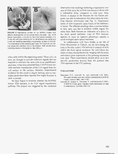
FIGURE 2 Hyperactive version of the MAPKK kinase YDAaffects development of the apical shoot meristem. Two smallleaves have been produced by the shoot apical meristem of a5-day-old wild-type seedling (4). No small leaves are visible in a5-day-old seedling expressing a hyperactive version of YDA (B).The cotyledons of this seedling are fused, forming a narrow hol-low where the meristem should be located. Both panels showscanning electron micrographs. Bar, 200 um.
a few cells within the organizing center. These cells, inturn, are thought to provide inductive signals that arerequired to maintain the stem cells in an undifferenti-ated state. It has been proposed that a MAP kinase cas-cade mediates transduction of the CLV signal from thecell surface to the nucleus. However, experimentalevidence for this claim is largely lacking, and no can-didate genes have been reported that might function insuch a cascade.
We have begun to examine whether the MAPKKkinase YDA operates in the CLV signal transductionpathway. The project was triggered by the incidental
observation that seedlings harboring a hyperactive ver-sion of YDA often fail to form true leaves or do so witha substantial delay compared to wild type. SeanKotkin, a student in the Partners for the Future pro-gram, was able to substantiate this observation by scan-ning electron microscopy (see Fig. 2). Hyperactiveforms of YDA frequently cause fusion of the embryon-ic leaves. The affected seedlings show a narrow hollowat their apex and fail to produce visible organs formany days. Both features are indicative of a defect inthe shoot apical meristem. Loss of YDA functionappears to cause an opposite phenotype, namely, amuch enlarged shoot apical meristem.
In collaboration with Venu Reddy in the lab ofElliot Meyerowitz at Caltech, we are developing thetools to forcibly induce YDA activity in subsets of cellswithin the shoot apical meristem. We will follow theeffect of these manipulations by imaging cell divisionsand marker gene expression in the growing meristemsover time. These experiments should allow us to testspecific predictions derived from the premise thatYDA operates in the CLV pathway.
PUBLICATIONS
Bergmann D.C., Lukowitz W., and Somerville C.R. 2004.Stomatal development and pattern controlled by a MAPKKkinase. Science 304: 1494-1497.
Lukowitz W., Roeder A., Parmenter D., and Somerville C. 2004.A MAPKK kinase gene regulates extra-embryonic cell fatein Arabidopsis. Cell 116: 109-119.
159

PLANT DEVELOPMENTAL GENETICS AND FUNCTIONALGENOMICS
R. Martienssen K. ArneyJ.-M. ArroyoM. ByrneD. GarciaD. Goto
C. KidnerZ. LippmanB. MayP. RabinowiczU. Ramu
D. Roh M. TanurdzicM. Ronemus U. UmamaheswariR. Shen M. VaughnJ. Simorowski M. Zaratiegui-BiurrunA. Tang
Epigenetic mechanisms regulate transposon silencing,gene control, and stem cell function in plants, whichprovide a useful model for higher eukaryotes. Stemcell function and axis formation in Arabidopsisdepend on asymmetric leaves] and argonaute, whichhas an important role in RNA interference. We havedemonstrated that spatially restricted small RNA canguide leaf polarity via AGO1. In fission yeast, RNAinterference of centromeric transcripts regulate his-tone modification, and we have found similar tran-scripts in Arabidopsis, where small RNA, DNAmethylation, and chromatin remodeling regulate hete-rochromatin through transposons and repeats. Duringthe last year, Pablo Rabinowicz, Mary Byrne, andCatherine Kidner left for faculty positions at TheInstitute for Genome Research (TIGR), the John InnesInstitute, and Edinburgh University, respectively, andsenior technician Juana-Mari Arroyo returned toSpain. We were joined by postdoctoral fellows MikelZaratiegui-Biurrun, Milos Tanurdzic, and DamienGarcia, as well as by our URP, Kat Arney.
Spatially Restricted microRNADirects Leaf Developmentvia ARGONAUTE1
C. Kidner, M. Ronemus, K. Arney, D. Garcia,R. Martienssen
microRNAs (miRNAs) are endogenous small RNAmolecules that regulate the expression of matchinggenes via RNA interference (RNAi). In animals, thisregulation occurs by translational control, but inplants, more perfectly matched miRNAs promote site-specific cleavage of the target by argonaute proteins.In Arabidopsis, the homeodomain HD-ZIP III genesPHABULOSA (PHB) and PHAVOLUTA (PHI/) areexpressed on the adaxial (upper) side of leaf primordiawhere they direct cell fate. They are targets of themiRNAs miR165 and miR166. We have used in situhybridization to show that miR165 accumulates on the
160
abaxial side of the leaf primordia where it cleavesPHB and PHV mRNA (Fig. 1). This pattern of expres-sion changes during development, implicating miRNAin signaling between the meristem and the leaf pri-mordium. argonaute) (ago]) mutants are defective inRNAi and have a pleiotropic phenotype. Leaves andflowers are adaxialized and PHB mRNA accumulatesthroughout the leaf primordium, whereas miR165 isectopically expressed in the adaxial domain (Fig. 1).Thus, AGO1 regulates both accumulation and functionof miRNA, perhaps accounting for enhancement ofboth loss-of-function and gain-of-function alleles ofPHB-related genes in ago]. We have examined theexpression of thousands of genes in normal andmutant lines of Arabidopsis using microarrays.Several families of related genes have substantiallyaltered expression in the RNAi mutants, and many ofthese genes match miRNA. However, not all miRNAtargets are altered in abundance, perhaps reflecting thetissue-specific expression of miRNA. Some up-regu-lated genes may be silenced by second-strand synthe-sis and degradation in addition to mRNA cleavage.Weak alleles of ago] have different phenotypes in dif-ferent Arabidopsis accessions. We are mapping thegenetic loci responsible for these variations. The iden-tification of such modifiers will give us new insightinto the role of RNAi in natural diversity in plants.
Role of ASYMMETRIC LEAVES1in Leaf Polarity and Stem Cell Fate
M. Byrne, D. Garcia, C. Kidner, J.M. Arroyo,R. Martienssen
Hox genes in animals are expressed in discrete domainsin early development, responding to a suite of mor-phogens responsible for axis specification. Geneexpression patterns subsequently come under epigenet-ic control, retaining a memory of early patterning longafter the signaling responsible has ceased. In plants,TALE class homeobox genes, such as SHOOTMERIS-

FIGURE 1 miRNA directs leaf polarity via AG01. In Arabidopsis,miRNA miR165 corresponding to the homeodomain genePHABULOSA (PHB) is restricted to the abaxial side of wild-typeleaves (4), but it is also found in the meristem in early embryos (C).miR165 is more widely distributed in strong ago] mutant leaves(D). This pattern restricts PHB expression in wild type, but not inago] where PHB is uniformly expressed (E) and directs leaf polar-ity. The precursor alone (minus the miRNA) cannot be detected incontrol hybridizations (8). The weak allele ago] -12 (hairy) has aleucine residue substituted for an absolutely conserved histidinein the PIWI domain. It has inside-out organs, resulting in ectopicovules (F) and trumpet-shaped leaves (G). These phenotypesresemble miRNA-resistant dominant alleles of PHB.
TEMLESS, BREVIPEDICELLUS, and BELLRINGER,are expressed in the early embryo where they specifythe shoot apical meristem, from which postembryonicorgans derive. These genes interact in a negative regu-latory network with the SANT domain gene ASYM-METRIC LEAVES] and the LOB domain gene ASYM-
METRIC LEAVES2 to maintain the distinction betweenmeristem cells and leaf founder cells in postembryonicdevelopment. PHB, PHV, and REVOLUTA belong toanother class of homeobox genes, the HD-ZIPIII class,and are expressed on the upper side of the leaf, as wellas in meristematic domains. Elsewhere in the plant,RNAi helps to silence these genes via ARGONAUTEIand miRNA, as described above. These two systemsinteract genetically, and we are exploring the mecha-nism of this interaction via enhancer analysis of asl .Loss of adaxial modifiers called PIGGYBACK result inectopic lamina outgrowths on the adaxial leaf surface in
as], but have little effect in wild-type backgrounds.Loss of proximodistal modifiers, on the other hand,results in a palmate compound leaf. These modifiersdemonstrate a role for AS I in both proximodistal anddorsoventral axis specification.
Genomic Analysis of RNAiin Fission Yeast
D. Goto, M. Zaratiegui-Biurran, D. Roh, M. Vaughn,R. Martienssen [in collaboration with T. Volpe, NorthWesternUniversity; T Cech, University of Colorado; J. Bahler, SangerCenter; and G. Thon, University of Copenhagen]
We previously demonstrated that RNAi of centromer-ic repeat transcripts in fission yeast S. pombe wasrequired for chromatin modification and centromerefunction, and we are extending these studies to the restof the genome. ago]- (argonaute), dcrl- (dicer) andrdrl- (RNA-dependent RNA polymerase) mutantshave very similar microarray profiles: Few genes areup-regulated, suggesting a limited role for miRNA, butcentromeric small interfering RNA (siRNA) matches arecQ homolog found at the telomeres. Silencing ofthis gene depends on Ago I and Dcr I , but not on Rdrl,and is presumably posttranscriptional. Transcriptsfrom retrotransposable elements are only slightly up-regulated in RNAi mutants despite the loss of H3mK9from their long terminal repeats (LTRs). Microarrayprofiles of RNAi mutants were similar to those fromthe H3K9 methyltransferase clr4-, but profiles fromother silencing mutants had more widespread effects.These results are in agreement with our observationsin Arabidopsis that transposon silencing can be main-tained independently of RNAi. We are currently usingtiling arrays of fission yeast heterochromatin to moreprecisely map centromeric transcripts as well as chro-matin modifications.
Several genes involved in RNAi and heterochro-matic silencing are found in the genome of fissionyeast but not in baker's yeast Saccharomyces cerevisi-ae, suggesting that other RNAi genes may be coelim-inated. We used phyletic profiling of five highereukaryote genomes, the two yeast species, and severalother fungal species to predict likely Schizosaccharo-myces pombe participants in RNAi-mediated silenc-ing. We refined this candidate list by using proteininteraction data from higher eukaryotes to identify S.pombe proteins, many of which were hypothetical pro-teins, that might interact with the RNAi machinery.These and other analyses have been used to constructan enhanced S. pombe model organism database.
161

Transposable Elements, EpigeneticControl, and the Origin ofHeterochromatin
Z. Lippman, M. Vaughn, B. May, R. Martienssen [incollaboration with W.R. McCombie and V Mittal,Cold Spring Harbor Laboratory; J. Carrington, OregonState University; R.W. Doerge, Purdue University;and V. Co lot, INRA/CNRS Evry, France]
Transposable elements and related tandem repeats arelarge components of eukaryotic genomes and areenriched in heterochromatic regions. The transcriptionalactivity of these sequences is suppressed by epigeneticsilencing mechanisms that involve the covalent modifi-cation of DNA and associated histones. In particular,methylation of cytosine residues in DNA and of histoneH3 Lys-9 are evolutionarily conserved modificationsinvolved in heterochromatin silencing. In the fissionyeast S. pombe, which lacks DNA methylation, histoneH3 Lys-9 methylation and silencing of centromericrepeats are regulated by RNAi. In plant genomes, DNAmethylation is primarily restricted to transposons andrelated repeats. In an effort to determine if RNAi, Lys-9methylation, and DNA methylation are mechanisticallylinked in repeat sequence silencing, epigenetic mutantsin Arabidopsis thaliana affecting each process wereassayed for silencing defects and compared to wild-typeplants as well as to each other. Transposons and tandemrepeats, but not genes, were selectively enriched in bothDNA methylation and histone H3 Lys-9 methylation,which correlated with the presence of siRNA. Despitetheir similar localization in cytologically defined hete-rochromatin, different classes of transposons were sub-jected to different forms of epigenetic regulation, sug-gesting the existence of multiple silencing pathways orcomplexes. The chromatin remodeling gene Decrease inDNA Methylationl (DDM1), the maintenance CGmethyltransferase (MET1), and the type I histonedeacetylase Silencing I (SIL1) are required to maintainmost transposon silencing, and DDM1 and MET1 fur-ther influence the accumulation of siRNA.Reestablishment of silencing occurred when met] andsill were backcrossed to wild type, but only whensiRNA was retained in the mutants, suggesting a role forsiRNA for silencing in cis. Genes with transposonsinserted within or near them were under transposon con-trol and, consequently, regulated by DDM1, MET], andRNAi. For example, transposons and tandem repeatsintegrated near the imprinted genes FWA and MEDEAcorrespond to siRNA and contribute to silencing,although their role in imprinting is still unclear.
162
Arabidopsis Centromeric RepeatTranscripts
B. May, Z. Lippman, R.Martienssen in collaboration withD. Spector, Cold Spring Harbor Laboratory]
Centromeres interact with the spindle apparatus toenable segregation of chromosomes into daughtercells and are typically flanked by many thousands ofsatellite repeats interspersed with retrotransposons.Although the function of these repeats is unclear, cen-tromere selection has a strong epigenetic component.Yeast centromeres are much simpler, but those of S.
pombe are also flanked by heterochromatic repeatswhose transcription contributes to centromere func-tion via RNAi. We have investigated the transcriptionand modification of centromeric repeats in A.
thaliana, which unlike yeast has methylated DNA.One strand of the 180-bp satellite repeat arrays is tran-scribed, and the transcripts remain associated with het-erochromatin. These transcripts are posttranscription-ally silenced by RNAi and histone modification, butnot by the canonical CpG DNA methyltransferaseMET1. Other 180-bp repeats are transcribed from bothstrands and silenced by MET], SILl, and DDM1.Centromeric transcripts of the first class are inheritedepigenetically. Those of the second class are alsoinherited epigenetically from ddml, which losessiRNA, but not from met] and sill, which retainsiRNA. Histone H3 Lys-9 methylation depends onAGO] as well as on the histone modification machin-ery. Cytological examination has yet to reveal thepresence of lagging chromosomes at anaphase in anyof these mutants, although some lose a comparableproportion of histone H3 Lys-9 methylation to fissionyeast mutants that display chromosome segregationdefects. It is possible that differences in plant andyeast cell division mechanisms are responsible.
Chromatin Modification, DNAReplication, and Polyploidy
M. Tanurdzic, Z. Lippman, P. Rabinowicz, M. Vaughn,R. Martienssen [in collaboration with L. Comai,University of Washington; T. Osborn, University ofWisconsin; W. Thompson, North Carolina StateUniversity; J. Murray, Cambridge University; andR.W. Doerge, Purdue University]
Polyploidy in plants results in a variety of genetic andepigenetic changes in gene expression from generationto generation following the establishment of allopoly-

ploids. The mechanisms that underlie these changes areimportant in the short term (hybrid vigor) as well as inthe long term, as polyploids are stabilized during evolu-tion. We are profiling chromatin modifications that mayunderlie novel gene expression in synthetic allopoly-ploids derived from crosses between tetraploid A.thaliana and tetraploid A. arenosa, as well as thosederived from crosses between doubled haploids ofBrassica oleracea and B. rapa. We have found thatchanges in heterochromatin are relatively modest, butthat some genes and some transposons are activated inthe allotetraploids. We are also comparing diploid chro-matin modification profiles to the pattern of replicationof chromosomal DNA. By labeling synchronized cellswith BrdU, and immunoprecipitating the replicatedDNA at different time points, we can determine theapproximate location of chromosomal origins in thesecells. We will compare these profiles to gene expression,DNA methylation, and chromatin modification profiles.
Development of Informatics Resourcesfor the Arabidopsis Genome
M. Vaughn, R. Martienssen
We have constructed a model organism database sys-tem for A. thaliana based on PostgreSQL, Apache, andPerl. This resource integrates gene information derivedfrom the reference TIGR genome releases; third partyannotations provided by TAIR, MIPS, and others; andnotes derived from published and in-house analyses.Annotations can be queried by a simple Web form or,for more expressivity, via a Perl interface which imple-ments BioPerl objects. This system is very flexible andhas been extended to treat features from our genomictiling arrays, the Affymetrix AtGenome 1 and ATH1arrays, the TAMU 26k 60-mer array, and the SalkInstitute whole-genome array as computational featuresmapped onto the genome. Values are assigned to thearray features based on normalized hybridization val-ues from any number of experiments, facilitating com-parison of equivalent features across microarray plat-forms and experiments. Other computations, such ascopy number, small RNA homology, MPSS tag abun-dance, and insertional mutant flanking sequence loca-tions also extend the default genome annotation. Wehave taken full advantage of the open-source GenericGenome Browser platform (from L. Stein, Cold SpringHarbor Laboratory) to construct a Web-based visual-ization tool to present the data contained in the annota-tion and microarray database as an easy-to-interpret,track-based image ( http : / /chromatin.cshl.org/ddml/).
The integrated visualization of annotation, computa-tion, and profiling data has allowed scientists from ColdSpring Harbor Laboratory, INRA/CNRS PlantGenomics Research Unit (Evry, France), and the NSFFunctional Genomics of Polyploidy project to explorequestions of epigenomic context.
Methylation Filtration of Sorghum andOther Plant Genomes
P. Rabinowicz, R. Martienssen [in collaboration withW.R. McCombie, Cold Spring Harbor Laboratory,and Orion Genomics LLC, St Louis, Missouri]
Methylation filtration (MF) is a gene-enrichment tech-nology that has been successfully applied to sorghumand maize, where gene enrichment is proportional togenome size. We have applied MF to a broad variety ofplant species spanning a wide range of genome sizes.Enrichment for genes was observed in all species test-ed, from nonvascular to vascular plants, but differencesbetween the expected and the observed level of enrich-ment were noted. In wheat and pine, only a low level ofgene enrichment was observed, which may reflect anabundance of methylated pseudogenes in these recentpolyploids. Sorghum bicolor is a close relative ofmaize and is a staple crop in Africa and much of thedeveloping world because of its superior tolerance ofarid growth conditions. We have generated sequencefrom the hypomethylated portion of the sorghumgenome. At least 90% of the genes have been taggedwith an average coverage across more than 60% oftheir length after generating a raw coverage of less than300 Mb of the 735-Mb genome. Methylation filtrationpreferentially captures exons and introns, promoters,miRNAs, and simple sequence repeats and minimizesinterspersed repeats, thus providing a robust view ofthe functional parts of the genome. Thousands of hypo-thetical gene predictions in rice and Arabidopsis aresupported by the sorghum set.
Functional Genomics with Transposonsin Maize and Arabidopsis
B. May, M. Vaughn, D. Roh, J. Simorowski, J.-M. Arroyo,R. Shen, A. Tang, U. Ramu, R. Martienssen [in collaborationwith W.R. McCombie, L. Stein, and D. Spector, Cold SpringHarbor Laboratory, and E. Lam, Rutgers University]
Our collection of Arabidopsis gene-trap and enhancertrap transposon lines has grown to more than 37,700individual insertions, about half of which have been
163

mapped to the genome by polymerase chain reaction(PCR) and sequencing. The resulting knockouts, alongwith phenotypic and expression data, are made avail-able to the public via TRAPPER (http://genetrap.cshl.org), an interactive database and ordering system. Wehave expanded this system to include gene-trap trans-posons with /ad-GFP chromosome charting beaconsthat enable chromosomal locations to be visualized inthe nucleus. We continue to serve the maize commu-nity through MTM (maize-targeted mutagenesis), anefficient system for site-selected transposon mutagen-esis. MTM selects insertions in genes of interest froma library of 45,000 plants using PCR. Pedigree, knock-out, sequence, phenotype, and other information arestored in a powerful interactive database (mtmDB)that enables analysis of the entire population as well ashandling knockout requests. By monitoring Mutatoractivity, we conclude that more than one half of allmutations arising in this population are suppressed onlosing Mutator activity. More than 200 orders werefilled for maize and Arabidopsis lines in 2004.
Arabidopsis BRCA2
J. Simorowski, U. Ramu, R. Martienssen
The breast cancer susceptibility 2 gene BRCA2 hasbeen studied extensively and has been found inmam-mals, birds, plants, and fungi. Mutations in the humanortholog predispose the patient to breast and ovariantumors. The model plant A. thaliana has two copies ofBRCA2 in its genome, and we have been studying thephenotypes corresponding to insertional mutagenesisof these genes. Using our collection of gene-trap lines,as well as mutant lines from other collections, we havecreated BRCA2 double mutants. Although the singlehomozygous BRCA2 plants are phenotypically nor-mal, the double homozygous plants have a markedreduction in fertility, ranging from semisterile to fullysterile. Double heterozygous mutant plants are pheno-typically similar to wild-type and single mutant plants.We are investigating triple mutants between theBRCA2 lines and BRCA2-interacting genes, such asRADS], which encodes a protein involved in DNArepair that interacts directly with BRCA2 duringrecombination.
164
PUBLICATIONS
Bastow R., Mylne J.S., Lister C., Lippman Z., Martienssen R.A.,and Dean C. 2004. Vernalization requires epigenetic silenc-ing of FLC by histone methylation. Nature 427: 164-167.
Groover A., Fontana J.R., Dupper G., Ma C., Martienssen R.,Strauss S., and Meilan R. 2004. Gene and enhancer traptagging of vascular-expressed genes in poplar trees. PlantPhysiol. 134: 1742-1751.
Kidner C. and Martienssen R.A. 2004. Spatially restrictedmicroRNA directs leaf polarity through ARGONAUTE1.Nature 428: 81-84.
Lippman Z. and Martienssen R. 2004. The role of RNA interfer-ence in heterochromatic silencing. Nature 431: 364-370.
Lippman Z., Gendrel A.-V, Black M., Vaughn M., Dedhia N.,McCombie W.R., Lavine K., Mittel V., May B., Kasschau K.,Carrington J.C., Doerge R.W., Colot V., and Martienssen R.2004. Role of transposable elements in heterochromatinand epigenetic control. Nature 430: 471-476.
Martienssen R.A. 2004. Crop plant genome sequence: What isit good for? Crop Sci. 44: 1898-1899.
Martienssen R.A., Rabinowicz RD., O'Shaughnessy A., andMcCombie W.R. 2004. Sequencing the maize genome.Curr. Opin. Plant Biol. 7: 102-107.
In Press
Bedell J.A., Budiman M.A., Nunberg A., Citek R.W., RobbinsD., Jones J., Flick E., Rohlfing T., Fries J., Bradford K.,McMenamy J., Smith M., Holeman H., Roe B.A., Wiley G.,Korf I.F., Rabinowicz RD., Lakey N., McCombie W.R.,Jeddeloh J.A., and Martienssen R.A. 2005. Sorghumgenome sequencing by methylation filtration. PloS Biol. 3:103-115.
Gendrel A.V., Lippman Z., Martienssen R., and Colot V 2005.Profiling histone modification patterns in plants usinggenomic tiling microarrays. Nat. Methods 2: 213-218.
Hansen K.R., Burns G., Mata J., Volpe T.A., Martienssen R.A.,Bahler J., and Thon G. 2005. Global effects on geneexpression in fission yeast by silencing and RNA interfer-ence machineries. Md. Cell Biol. 25: 590-601.
Kidner C.A. and Martienssen R.A. 2005. The developmentalrole of microRNA in plants. Curr. Opin. Plant Biol. 8: 38-44.
Lippman Z., Gendrel A.V., Colot V. and Martienssen R. 2005.Profiling DNA methylation patterns using genomic tilingmicroarrays. Nat. Methods 2: 219-224.
Madlung A., Tyagi A.P., Watson B., Jiang H., Kagochi T.,Doerge R.W., Martienssen R., and Comai L. 2005.Genomic changes in synthetic Arabidopsis polyploids.Plant J. 41: 221-230.
Mandell J.G., Bahler J., Volpe TA., Martienssen R.A., and CechT.R. 2005. Global expression changes resulting from loss oftelomeric DNA in fission yeast. Genome Biol. 6: R1.
Ronemus M. and Martienssen R. 2005. RNA interference:Methylation mystery. Nature 433: 472-473.

PLANT CELL SIGNALING AND GENETICS
D. Jackson Y. Benitez K. Lau N. Satoh-NagasawaP. Bommert B.H. Lee J. WangM. Cilia J. Linder P. YinM. Jan A. Mohanty
Our research aims to understand fundamental mecha-nisms in developmental biology, and how morphologyand architecture arise and are maintained in biologicalsystems. We use plants as a convenient model systembecause of their ease of genetic manipulation, inter-esting developmental strategies, and applications toagriculture and our food supply. In the past year, wehave concentrated on the isolation of novel genes thatregulate development and have invested efforts tocharacterize the function of recently sequenced plantgenomes. In an era where we can now predict the iden-tity of all genes required to make a plant, such tech-nologies are increasingly important. They will allowus to understand the "big picture" of interacting pro-teins and pathways that create the integrated organism.
We also continue to advance our understanding ofsignaling pathways that regulate stem cell fate anddevelopment. We have found novel mutants in cell-to-cell protein trafficking and are pursuing their isola-tion. We also identified genetic interactions regulatingstem cell proliferation in the maize inflorescence andisolated a novel phosphatase gene that controls inflo-rescence branching. As plant development is also inti-mately associated with hormonal signaling, we areinvestigating a novel role for cytokinin hormones instem cell proliferation and leaf patterning.
Isolation of the RAMOSA3 Geneof Maize by Positional Cloning
N. Satoh-Nagasawa, J. Linder in collaboration withN. Nagasawa and H. Sakai, DuPont Agriculture andNutrition, Wilmington, Delaware]
Inflorescence branching is a major yield factor inmany crops and is determined by the activities ofshoot meristems. Because of our general interest ininflorescence development and shoot meristem orga-nization, and the importance of inflorescence branch-ing in development of maize as a crop, we have beencontinuing the characterization of the ramosa3 (ra3)
mutant, which has enhanced inflorescence branching.We observed ra3 ear development by scanning elec-tron microscopy and found that, in general, RA3 func-tions to impose determinacy and identity on the dif-ferent meristem types in the inflorescences. Althoughthe inflorescence meristems are normal, the spikeletpair, spikelet, and floral meristems are all more inde-terminate and have converted or delayed identity inra3 mutants compared to wild type.
Double-mutant analyses suggest that RA3 actssynergistically with another branching gene, RA2,because the double mutants are much more severethan either single mutant. To more fully understand therole of RA3 in inflorescence development, we used theemerging genomic resources in maize to isolate thegene by positional cloning. RA3 encodes a proteinwith a highly conserved phosphatase domain, suggest-ing that it acts in a signal transduction cascade to reg-ulate meristem determinacy and identity. This class ofphosphatases has been implicated in stress protectionand in metabolic signaling, and our discovery is thefirst indication that these proteins are also involved indevelopmental regulation.
Reverse transcriptase-polymerase chain reaction(RT-PCR) and in situ hybridization experiments indi-cate that RA3 is expressed in the developing inflores-cences, in a cup-shaped domain at the base of themeristems (Fig. 1A). This expression pattern allowsus to hypothesize that RA3 may process a signal thatmoves into the meristem to regulate determinacy andidentity. RA3 also has an interesting genomic organi-zation; sequencing of a RA3 BAC clone indicates thatthe adjacent gene is a RA3 homolog, which we calledsister of RA3 (SRA). However, there is only a singleortholog of RA3 in rice, which leads us to imagine aninteresting evolutionary story whereby RA3 may haveevolved a developmental role relatively recently.Since there are limited studies at the interfacebetween metabolic control and development, analysisof RA3 may give unique insights into the mechanismsof branching in maize inflorescences as well as inother crop plants.
165

FIGURE 1 Patterns in plant development. (A) The RAMOSA3 gene is expressed in cup-shaped domains (arrows) at the base ofmeristems and may process a signal that regulates their determinacy. (8) ABPHYL1 is expressed in stripes in the shoot apical meris-tem (arrowheads). (C) Subcellular localization of a YFP-tagged Arabidopsis protein of unknown function shows that it localizes to thenuclei of root tips. Bar, 20 gm.
Studies of Maize Phyllotaxy
B.H. Lee, J. Wang
Mutations in the cytokinin-inducible response regulatorhomolog ABPHYLI (ABPHI) in maize result in decus-sate phyllotaxy, where leaves are initiated in oppositepairs. This is very distinct from the normal pattern,where leaves are initiated singly in an alternating pat-tern. To gain insight into the functions of this gene fam-ily in regulating leaf patterns across different plantspecies, we are now using the model dicot Arabidopsis.Mutants defective in Arabidopsis cytokinin-inducibleresponse regulators and other cytokinin signaling com-ponents have been obtained, and their developmentalphenotypes are being monitored.
Auxin is another classical plant hormone that haslong been coupled with cytokinins in its mechanism ofaction, and it has also been implicated in the regulationof phyllotaxy. To examine the interaction betweenauxin and cytokinin in the regulation of phyllotaxy,two approaches are being used. First, we are analyzingthe ABPHI expression domain in auxin polar transportmutants or auxin polar transport inhibitor (N-1-naph-thylphthalamic acid [NPA])- treated plants. ABPHI isexpressed in stripes in the meristem that predict theposition of leaf initiation (Fig. 1B). Our preliminary insitu hybridization data suggest that NPA treatmentalters the ABPHI expression domain in the meristem.An ABPHI antibody has been made and will be alsoused for this expression domain analysis. Second,using an anti-PINFORMED1 antibody, we will alsoinvestigate the expression patterns of putative polarauxin transport proteins in abphl mutants. To facilitatethe studies of the ABPHI expression domain, anABPHI yellow fluorescent protein (YFP) transgenicmaize line is being generated. The YFP has been inter-nally integrated in-frame into the ABPHI- coding166
sequence under the control of its own promoter, usingthe fluorescent tagging of full-length proteins (FTFLP)method developed in our lab.
To identify additional factors involved in the con-trol of phyllotaxy, we are characterizing another maizephyllotaxy mutant, abph2. The abph2 mutant shootmeristem size appears to be bigger than that of thewild type, and the ABPHI expression domain isenlarged. These results suggest that ABPH2 mightfunction in the same phyllotaxy regulatory pathway asABPHI. We are planning to map ABPH2, with thegoal of isolating the gene by positional cloning.
Toward Characterization of theArabidopsis, Maize, and RiceProteomes by Native Expressionof Tagged Full-length Proteins
A. Mohanty [in collaboration with M. Myers andW.R. McCombie, Cold Spring Harbor Laboratory]
Arabidopsis thaliana, the first plant to have its genomecompletely sequenced, has more than 29,000 predictedgenes. However, to this date, less than 10% of the geneactivities have been verified experimentally, and thereare only very limited reports of systematic analysis ofgene function. To address this problem, we have devel-oped high-throughput methodologies to tag genes forfunctional analysis, including subcellular localizationand identification of interacting proteins. Our first useof this approach, termed FTFLP, aims to analyzeexpression patterns and subcellular localization ofArabidopsis gene products in planta. Our FTFLP-based approach offers two significant advantages: (1)It produces internally tagged full-length proteins thatare likely to exhibit their correct intracellular localiza-tion, and (2) it yields information about the tissuespecificity of gene expression, by the use of native pro-

moters. To demonstrate how FTFLP may be used forcharacterization of the Arabidopsis proteome, wetagged a series of known proteins with diverse subcel-lular targeting patterns, as well as several proteins withunknown function, and unassigned subcellular local-ization (Fig. 1C). The results are available through aWeb database (http://aztec.stanford.edu/gfp/).
Recently, we have applied this methodology to theeconomically important crops rice and maize. Rice isthe first cereal crop plant to have its genome com-pletely sequenced. However, the function of only ahandful of rice genes is known. For the rice-taggingproject, we are using a plant-optimized tandem affini-ty purification (TAP) tag with the aim to study pro-tein-protein interactions. We are currently generatingTAP-tagged lines for purification of the protein com-plexes, with the aim of making a comprehensive pro-tein interaction map. We are also applying the fluores-cent tagging approach to maize, to generate fluores-cent-protein-tagged lines for developmental andgenomics studies. It is expected that our study willpave the way for deciphering the function of a largenumber of plant genes at the cellular level.
Molecular Genetic Characterizationof Plant Intercellular Channels
Y. Benitez, M. Cilia, M. Jan, J. Wang, P. Yin [in collaborationwith J.Y. Kim, Gyeongsang National University, Korea)
The intercellular transport of metabolites, the spread ofRNAi (RNA interference), and viral movement are allprocesses regulated by the permeability of plasmodes-mata (PD) channels. PDs are also involved in regulat-ing symplastic communication during developmentand plant cell fate. Despite their importance, we under-stand very little about the mechanisms regulating eithertargeted or nontargeted movement through PD. Ourgroup has invested significant effort in screens to iso-late genes involved in PD transport pathways and in theidentification of signals for PD targeting.
In the last year, we reported the isolation of a num-ber of mutants arrested in the intercellular movementof the green fluorescent protein (GFP) named gfpaltered trafficking (gat, pronounced "gate"). We havecharacterized two of these so far; gad, for which wehave two alleles, is recessive and has severe develop-mental phenotypes. In contrast, gat3 is dominant andmorphologically normal. The gat mutants were isolat-ed based on their block in GFP transport out of theconductive phloem tissues, but they also differ in GFP
movement, and perhaps in PD size exclusion limit, innonphloem tissues. For instance, gad is also impairedin GFP movement between epidermal cells comparedwith wild-type seedlings. However, GFP moves nor-mally between gat3 epidermal cells. We also observedPDs indirectly using the marker PD41, a GFP- partialcDNA fusion that labels PDs in wild-type seedlings.gat] mutant seedlings showed only diffuse PD41 GFPfluorescence at the cell periphery, suggesting that PDsare disrupted in this mutant. gad mutants make onlyone or two small leaf primordia and have reducedexpression of a meristem regulatory gene (STM).
We are now fine mapping two of the gat mutants,in order to identify which genes are disrupted. We havemapped gat] to a small region of chromosome 2around the marker THY1, and gat2, which has a phe-notype similar to that of gatl, to chromosome 1. In themeantime, we continue in-depth analysis of the gatmutants. We will assess movement of RNAi signals orof low-molecular-weight dyes and observe PD struc-ture in the mutants by transmission electronmicroscopy or using fluorescent markers to understandthe roles of the GAT genes in cell-to-cell transport.
We also designed a new approach to ask what pro-portion of Arabidopsis proteins are capable of targetedtrafficking through PD, to identify additional PD-traf-ficking signals, and to isolate mutants in targetedficking of KNOTTED1 (KN1). KN1 was the first plantdevelopmental protein found to traffic through PD, andit is now the founding member of a growing class ofnoncell autonomous developmental proteins. For thisaim, we will use a novel system that we developed toeasily monitor protein trafficking by phenotypic screen-ing of leaf hairs or trichomes (Fig. 2A,B). We hope thatthis approach will provide genome-wide insight into theprevalence of targeted PD trafficking and identify shorttrafficking signals that could be useful in targeting pre-dictions or to regulate the trafficking of other proteins.
Regulation of Meristem Size andSeed Yield in Maize
P. Bommert, K. Lau, J. Wang
Plant growth is characterized by reiterative develop-mental events that depend on the activity of meristems.The aerial part of the plant body is elaborated from theshoot apical meristem (SAM), which produces leaves,stems, and axillary meristems in a repetitive manner.Established during embryogenesis, the SAM serves asa reservoir of stem cells during the plant life cycle. It
167

FIGURE 2 Morphology and plant development. The glabrousl mutant of Arabidopsis lacks leaf hairs or trichomes (4). We deviseda scheme to complement this mutation using cell-to-cell trafficking of the GL1 protein fused to KN1, which results in trichome pro-duction (8). In C, a greatly enlarged maize ear meristem is produced by a combination of mutations in tdl and fea2 genes. Thesegenes may regulate yield through controlling seed number on the ear.
can be divided morphologically and functionally into acentral zone (CZ), in which the stem cells are located,and a surrounding peripheral zone (PZ), where cellsbecome incorporated into lateral organ primordia.
In Arabidopsis, analysis of fasciated mutants hasidentified some of the genes involved in maintainingthe balance between stem cell fate in the CZ and theonset of differentiation in the PZ. Mutations in genesin the CLAVATA signaling pathway lead to increasedcell numbers in meristems. The current workingmodel postulates that the CLAVATA3 ligand is secret-ed from stem cells and binds to a receptor complex
CLAVATA2, and other signaltransduction molecules. Activation of the receptorresults in the restriction of transcription ofWUSCHEL, a positive regulator of stem cell identity.
We are investigating the role of the thick tasseldwarfl (tdl) and fasciated earl (fea2) genes in maize,which encode CLAVATA1 and 2 orthologs. Bothmutants develop severely enlarged inflorescencemeristems, indicating that they function in a mannersimilar to CLV1 and 2 in Arabidopsis. The phenotypicanalysis of tdl, fea2 double mutants, however, demon-strated that tdl and fea2 do not act exclusively in areceptor complex to regulate meristem size (Fig. 2C).To gain insight into the nature of the receptor com-plex, we plan to isolate the fea2 complex by using atransgenic FEA2 TAP-tagged maize line. A constructcontaining the FEA2-coding sequence fused to aplant-optimized TAP tag has been generated and iscurrently being transformed into maize.
Besides this biochemical approach, we also addressthe question of whether fea2 has a role in the evolutionof maize, especially with respect to the cob structure ofmaize, which is unique among the grasses. In teosinte,the ancestor of maize, the seeds are arranged in twoalternating rows, whereas modern maize lines have upto 18 rows of seeds. Seed row number is dependent on
168
the number of meristems, called spikelet pair meristems(SPMs), initiated by the inflorescence meristem. It istempting to speculate that the number of SPMs is direct-ly dependent on the size of the inflorescence meristem.Our analysis of inflorescence meristem size and seedrow number in different maize inbred lines supports thisassumption, as we observe a significant positive corre-lation. To address whether allelic variation in the fea2gene also has an impact on seed row number, we areperforming a high-resolution QTL analysis using amaize recombinant inbred population. Our analysis ofseveral independent data sets indicates that indeed, allel-ic variation in fea2 may regulate seed row number, asone of the most significant QTLs is located on chromo-some 4, corresponding to the fea2 map position.
PUBLICATIONS
Cilia M.L. and Jackson D. 2004. Plasmodesmata form andfunction. Trends Cell Biol. 16: 500-506.
Giulini A., Wang J., and Jackson D. 2004. Control of phyllotaxyby the cytokinin inducible response regulator homologueABPHYL1. Nature 430: 1031-1034.
Tian G.W., Mohanty A., Narasimha Chary A.S., Li S., Paap B.,Drakakaki G., Kopec C.D., Li J., Ehrhardt D., Jackson D.,Rhee S.Y., Raikhel N.V., and Citovsky V. 2004. High-throughput fluorescent tagging of full-length Arabidopsisgene products in planta. Plant Physiol. 135: 25-38.
In Press
Bommert R, Satoh-Nagasawa N., Jackson D., and Hirano H.Y.2005. Genetics and evolution of grass inflorescence andflower development. Plant Cell Physiol. 46: 69-78.
Bommert P., Lunde C., Nardmann J., Vollbrecht E., Running M.,Jackson D., Hake S., and Werr W. 2005. thick tasseldwarfl encodes a putative maize ortholog of theArabidopsis CLAVATA1 leucine-rich repeat receptor-likekinase. Development 132: 1235-1245.
Jackson D. 2005. Transcription factor movement through plas-modesmata. In Plasmodesmata (ed. K. Oparka). BlackwellPress, United Kingdom. (In press.)

PLANT DEVELOPMENTAL GENETICS
M. Timmermans M. GuoM. JuarezS. MadiP. Mazur
F. NogueiraT. Phelps-DurrJ. Thomas
ANALYSES OF GENES INVOLVED IN MERISTEMFUNCTION AND LEAF INITIATION
This work was done in collaboration with M. Scanlon,University of Georgia, Athens; P Schnable, Iowa StateUniversity, Ames; B. Buckner and D. Janick-Buckner,Truman State University Kirksvile, Missouri.
One of the most intriguing aspects of plants is theirability to generate organs postembryonically. Thegrowing tip of a plant, referred to as shoot apicalmeristem (SAM), contains a population of stem cellsthat divide to maintain the SAM and to generatedaughter cells from which lateral organs, such asleaves and flowers, arise. These different cell types areorganized in distinct histological and functionaldomains. For instance, the stem cells are located at themost apical tip in the so-called central zone, whereaslateral organ founder cells are located on the flanks ofthe SAM (Fig. 1). In addition, meristems comprisedistinct epidermal and subepidermal cell layers.Traditional genetic analyses have identified severalgenes that are required for meristem function or forthe development of lateral organs from the SAM. Suchstudies have also shown the importance of this meris-tematic organization for normal plant development.
To identify novel and potentially redundant oressential genes that function in discrete domains of theSAM or developing leaf primordia, we are using atechnique called laser capture microdissection (LCM).LCM allows the isolation of transcripts from specificcell types within a tissue or organ. For use in LCM,
target tissues are fixed, sectioned, and immobilizedonto microscope slides. Cells of interest are dissectedand separated from the rest of the tissue using a laserbeam. RNA is then extracted from the captured cellsand used in microarray analyses to compare globalgene expression patterns between different cell popu-lations. During the past year, we have optimized theLCM technique, which was originally designed for thedissection of animal cells, for use on the small-cell-wall-encapsulated cells within the plant SAM. Wehave found that acetone-fixed and paraffin-embeddedSAMs preserve tissue integrity without compromisingthe efficiency of LCM and subsequent RNA isolation.RNA collected from as few as a thousand cells wassufficient for the preparation of cDNA for use inmicroarray analyses of gene expression.
We are using maize as our experimental systembecause of the relatively large size of its vegetativemeristem when compared, for instance, to Arabidopsis.However, maize has the disadvantage that its genomeproject is still at an early stage. To identify genes thatshould be represented on the microarrays, we have alsoinitiated a "small" expressed sequence tag (EST) pro-ject. We have currently sequenced nearly 10,000 ESTsfrom meristem-enriched cDNA libraries, and the ulti-mate goal is to generate approximately 35,000 3' ESTs.We intend to compare global expression patternsbetween approximately 20 different functional meris-tematic and leaf primordia domains, or experimentalconditions. Examples of the types of comparisons are asfollows: (1) between the expression profiles of the stem
FIGURE 1 Genetic interactions between stem cells and their differentiatingdescendants in the incipient leaf. Adaxial/abaxial patterning of lateral organsrequires two meristem-derived signals. One signal, which may exert its effectsvia the START lipid-sterol-binding domain of HD-ZIPIII proteins, is derivedfrom the stem cells in the SAM (*). The miR166 signal initially accumulatesimmediately below the incipient leaf but gradually spreads into the abaxialdomain, thus restricting hd-zipIll expression to the adaxial side. hd-ziplIlexpression also depends on Ibll , which may act via the stem-cell-derived sig-nal. hd-zip111 genes specify adaxial fate and signal back to the SAM to main-tain stem cell function.
stem cellsignal -4 lb/1
hd-ziplIl
miR166
169

cells, their immediate derivatives in the SAM, and theinitials of lateral organs; (2) between the different tissuelayers of the SAM; (3) between different stages of leafdevelopment; (4) between the upper and lower surfacesof developing leaves; and (5) between SAMs and/orleaf primordia from developmental mutants and wild-type plants. Our initial results indicate that nearly 1300genes are differentially expressed among the SAM,young leaf primordia, and older leaf tissues. Potentialdifferentially expressed genes will be verified by moretraditional expression analyses such as real-timereverse transcriptase-polymerase chain reaction (RT-PCR) and in situ hybridization. The function of genesof particular interest will be determined using function-al genomics resources available in maize andArabidopsis. Even though this project is still in itsinfancy, it will soon provide novel insight into meristemfunction and organogenesis.
ADAXIAL/ABAXIAL PATTERNING OFLATERAL ORGANS IN MAIZE
Signaling between the different cell populations with-in the meristem is important for stem cell function. Inaddition, stem cell function and meristem mainte-nance rely on signals from differentiating cells withindeveloping organs. A positive feedback loop existsbetween stem cells in the SAM and their differentiat-ing derivatives in the leaf. Early studies indicated thata stem-cell-derived signal sets up adaxial/abaxialpolarity in lateral organs, which leads to distinct upperand lower leaf surfaces. This past year, we have shownthat adaxial/abaxial patterning also requires amicroRNA (miRNA) signal that originates within themeristem (Fig. 1). Both signals act on members of theclass III homeodomain leucine zipper (hd-zipIII) fam-ily, which specify upper cell fate but are also requiredfor SAM formation and maintenance.
The semidominant Rolled leafl (Rid]) mutantfrom maize is characterized by an upward curling ofthe leaf blade due to adaxialization or partial reversalof leaf polarity. We have cloned the rldl gene andfound that it encodes a member of the HD-ZIP IIIfamily of proteins. rldl is normally expressed in thecentral zone of the SAM, in vasculature and, consis-tent with a role for rldl in adaxial cell fate specifica-tion, in nondetermined cells on the adaxial side of leafprimordia. The meristematic expression pattern ofrldl was unaffected in Rldl, but we observed that rldlis misexpressed on the abaxial side of Rldl mutantleaves. These and other observations indicate thatpolarized hd-zipIII expression is essential for the for-
170
mation of distinct upper and lower leaf surfaces.However, our in situ hybridization analysis showedthat rldl and other hd-zipIH family members areexpressed uniformly throughout the leaf founder cells.We were able to show that polarized hd-zipIII expres-sion is subsequently set up by an miRNA-directedRNA-interference-like process.
Transcripts of the hd- zipIII genes contain a comple-mentary site for miRNA165 (miR165) and miR166.miRNAs are endogenous small (-22 mer) noncodingRNAs that mediate the cleavage or translational repres-sion of target transcripts containing a complementarysequence, and thus control gene expression posttran-scriptionally. We found that all four dominant Rldl alle-les result from a single nucleotide change in this site.This, together with the misexpression of rldl in theRldl mutant, suggests that miRNAs mediate the cleav-age of rldl transcripts on the abaxial side of leaf pri-mordia. In other words, miR165/166 may specify abax-ial cell fate by restricting expression of rldl and otherhd-zipIII genes to the adaxial side. To test directlywhether miRNAs can establish patterns of tissue differ-entiation during development, we used in situhybridization to determine a precise cellular expressionpattern for miR166. We found that rldl and miR166exhibit complementary expression patterns in develop-ing leaf primordia consistent with a role for miR166 inthe spatial regulation of rldl transcripts. More impor-tantly, our in situ hybridization data indicated that thepeak of miR166 expression occurs just below thedomain in which miR166 acts on the hd-zipIII genesand that an expression gradient of miR166 is estab-lished in developing leaves. These observations led usto suggest that miR166 is mobile, and this offers theintriguing possibility that miRNAs can function as mor-phogen-type signals in development. We are currentlystudying the basis for this dynamic expression patternof miR166, by determining the cis- and trans-actingfactors required for miR166 expression and possiblemovement.
As mentioned above, adaxial/abaxial patterningrequires a second meristem-derived signal. This stemcell signal is essential for adaxial cell fate and isthought to act via the HD-ZIPIII proteins, which con-tain a START-like lipid-sterol-binding domain. As afirst step toward identifying the potential adaxializingsignal, we have purified the START domain fromRLD1 and are using biochemical approaches to deter-mine which lipid-sterols bind to this START domain.In addition, we are taking a genetic approach to iden-tify genes that produce, convey, or respond to the stemcell signal. Recessive mutations in leafbladeless I

(lbll) cause the formation of abaxialized leaves, sug-gesting that lb 11 is required for adaxial cell identity.Expression of the hd-zipIII genes in the SAM and onthe adaxial side of leaf primordia is reduced in lb 11,indicating that lb 11 acts upstream of rldl and other hd-zipIII family members. Interestingly, the level and pat-tern of rldl expression in the vasculature of lbll arenot affected. Because vascular hd- zipIII expression isalso controlled by miR166, these observations suggestthat lbll controls hd-zipIII expression independent ofmiR166. We are in the process of cloning the Ibll geneusing transposon-tagging and map-based approaches.
In the past few years, genetic analyses in severaldiverse plant species have shown that a signal for theadaxial side of the leaf is required to maintain the SAMand its stem cell population. We have isolated severalmutant alleles of lbll. The most severe alleles fail to ini-tiate a SAM during embryogenesis. Mutant embryoslack hd- zipIII expression. In addition, these embryosfail to express knotted], which encodes a homeodomainprotein known to be required for meristem function in awide variety of plant species. By studying the geneticpathway involving 1b11, rldl, and miR166, we hope toimprove our understanding of the interdependence ofstem cells and their differentiating descendants and therole of miRNAs as developmental signals.
REPRESSION OF STEM CELL FATEDURING ORGAN DEVELOPMENT
This work was carried out with contributions fromArooba Alam and Samantha Meyerhoz, Locust ValleyHigh School.
knotted] and other members of the knox homeoboxgene family are required for meristem initiation andmaintenance. These genes are expressed in the indeter-minate cells of the SAM but are excluded from deter-minate leaf founder cells. Moreover, down-regulationof knox genes is essential for normal leaf development.Misexpression of knox genes within developing leavesleads to cell division and differentiation defects, notunlike tumor formation in animals. The rough sheath2(rs2) gene from maize and the ASYMMETRICLEAVES] (AS1) gene from Arabidopsis encode orthol-ogous MYB-domain proteins that act as negative regu-lators of knox gene expression. On the basis of expres-sion and genetic analyses, we proposed that these pro-teins act as epigenetic regulators to keep knox genes inan "off" state in developing lateral organs, thus pre-venting differentiating cells from reverting into indeter-minate stem cells. Our recent observations confirm this
hypothesis. We have shown that RS2 and AS1 formhighly conserved repressor complexes that include thezinc finger transcription factor ASYMMETRICLEAVES2 (AS2), an RNA-binding protein namedRIK, and the histone chaperone HIRA and may recruitprotein phosphatase 2A to the knox loci. HIRA proteinsare histone chaperones that have a role in both euchro-matic and heterochromatic gene silencing in yeast andanimals. The mechanism by which HIRA establisheseuchromatic gene silencing is unknown. HIRA is aunique histone chaperone because it can incorporate orexchange nucleosomes and histone variants indepen-dent of DNA synthesis. In addition, HIRA can interactwith histone deacetylases. Both these activities couldlead to changes in local chromatin state. We are cur-rently testing whether knox gene silencing is associatedwith changes in chromatin organization or histonemodifications.
To establish that the AS 1 complex binds directly tothe knox loci, we have generated transgenic lines thatexpress a fusion protein comprising the DNA-bindingdomain of the floral transcription factor LEAFY (LFY-DB) and the non-MYB domain of AS I . More than halfof the plants carrying this transgene failed to formflowers, which is typical of strong /fi, mutants. Controlplants expressing the LFY-DB alone were normal, sug-gesting that LFY-DB expression does not cause a dom-inant-negative phenotype. Replacement of the LFYactivation domain with the non-MYB domain of AS 1thus blocks activation of LFY targets. This suggeststhat AS1 acts as a transcriptional corepressor and maybind directly to the knox genes. We therefore used chro-matin immunoprecipitation to determine which regionsof BP, one of the knox genes, mediate AS1 complexbinding. We have identified three regions within the BPgene that interact with the AS1 complex. We are cur-rently defining these cis-acting sequences more pre-cisely and are analyzing which proteins in the AS1complex mediate these interactions.
We are also using the reverse genetic resourcesavailable in Arabidopsis to determine the roles of theAS1-interacting proteins in plant development, partic-ularly in knox gene regulation. Loss-of-function muta-tions in AS2 are known to affect knox gene silencingduring leaf development. We have isolated mutations inthe Arabidopsis RIK and HIRA proteins. Loss-of-func-tion mutants in RIK are indistinguishable from wildtype. In contrast, weak mutant alleles of hira developleaf and floral defects that resemble the defectsobserved in the as] and as2 mutants. Moreover, hiramutant leaves misexpress several knox genes. AS2expression is unaffected in hira mutants and ASI tran-
171

script levels are increased, indicating that the defects inhira do not result from loss of ASI or AS2. Double-mutant analysis revealed that hira enhances the asland as2 mutant defects. Taken together, our observa-tions suggest AS1 and AS2 assemble into a proteincomplex that targets HIRA and possibly other chro-matin remodeling proteins to the knox loci in organfounder cells. As a result, stem cell fate remainsrepressed in determinate differentiating cells of devel-oping lateral organs. Deletion of HIRA in humans con-tributes to the DiGeorge syndrome, which is thought toarise from developmental defects in the neural crest.Thus, HIRA may also have a role in the repression ofstem cell fate in animals. Our studies could thereforehighlight fundamental properties of establishing/main-taining determinacy in plants and animals.
172
PUBLICATIONS
Juarez M.T., Kui J.S., Thomas J., Heller B.A., and TimmermansM.G. 2004. microRNA mediated repression of rolled leaflspecifies maize leaf polarity. Nature 428: 84-88.
Juarez M.T., Twiggy R.W., and Timmermans M.C. 2004.Specification of adaxial cell fate during maize leaf develop-ment. Development 131: 4533-4544.
Timmermans M.C., Brutnell T.P., and Becraft P.W. 2004. The46th Annual Maize Genetics Conference: Unlocking thesecrets of the maize genome. Plant Physiol. 136:2633-2640.
In Press
Timmermans M.C., Juarez M.T., and Phelps-Durr T. 2005. Aconserved microRNA signal specifies leaf polarity. ColdSpring Harbor Symp. Quant. Blot 69: (in press).
Fabio Nogueira, postdoc, Marja Timmermans' lab.

BIOINFORMATICS AND GENOMICS
Genome research has always involved the determination of the complete DNA sequences of organ-isms, the development of methods for assigning meaning to particular DNA, RNA, and proteinsequences, and the exploration of how changes in gene expression govern normal biology and con-tribute to a variety of pathological states including cancer. As the fields of bioinformatics and genomeresearch expand and mature, scientists are perfecting their ability to detect and decipher the funda-mental patterns and other properties of the nucleic acids and proteins that ultimately shape all of life.This work involves the use of mathematics, statistics, and computer science to answer a wide varietyof biological questions.
W. Richard McCombie's group, in collaboration with the Zhang, Martienssen, and Hannon labs, hasdeveloped a method to identify and verify the positions of genes within both plant and animal genomes.Using this method, the scientists discovered significant errors in what were considered "well-annotat-ed" mouse genes. Similar work with rice showed that many of the rice genes that were considered full-length gene sequences in the rice genome database may instead represent only a fraction of the truenumber of rice genes.
Andrew Neuwald's group has developed software that detects subtle similarities between proteinsof unknown function and proteins that have well-defined functions. Neuwald's group used the softwareto compare three distantly related protein families and to identify important structural similarities amongthe families that had previously been overlooked.
One of Lincoln Stein's interests includes creating software that compares DNA sequences andreveals common patterns that diverse organisms use to carry out similar life processes. For example,Stein has created software that enables scientists to compare the genomes of the major cereal crops(rice, wheat, corn, and oats) and thereby reveal beneficial genetic variants in one of these crops thatmight be exploited in the others. The results of these comparisons help plant breeders to develop newvarieties with improved abilities to withstand disease, changes in climate (e.g., drought), and a varietyof other adverse conditions. In other work that explored the genomic basis of "speciation" (i.e., the for-mation of new species), Stein took advantage of the newly completed genome sequence of the round-worm C. briggsae. (With some 500,000 species [after insects], roundworms comprise the secondlargest group of animals on Earth.) Stein's group compared the C. briggsae genome with that of themore commonly studied roundworm, C. elegans, both of which diverged from a common ancestorapproximately 100 million years ago. Through this work, Stein and his colleagues have provided a com-prehensive view of how genome evolution leads to the formation of new species.
Michael Zhang's group has developed a set of software tools for locating transcription-factor-bind-ing site (TFBS) motifs-DNA sequences where gene regulators attach themselves and activate theseries of events that lead to protein production. Obtaining a complete view of the types and locationsof such binding sites, as Zhang's work enables, is crucial for understanding how and when genes areturned on and off in response to various biological and pathological cues.
173

GENOME SEQUENCE ANALYSIS
W.R. McCombie V. Ba lija F. Katzenberger L. PalmerM. de la Bastide B. Miller L. SpiegelS. Dike S. Muller M. YuJ. Gergel L. Nascimento T. ZutavernM. Katari A. O'Shaughnessy
SEQUENCING OF THE RICE GENOME
This work was done in collaboration with Rod Wing(University ofArizona, Tuscon).
We have been collaborating with the Arizona GenomeInstitute (AGI) as part of a larger collaboration tosequence the genome of Oryza sativa, more common-ly known as rice. The larger collaboration, theInternational Rice Genome Research Program(IRGSP), has been focused on completing a finished-quality sequence of the Nipponbare strain of rice. Thisis a strain of rice that is grown commonly in temper-ate regions such as Japan, as opposed to the more trop-ical strains of rice, such as Indica, which are morecommonly grown in Southeast Asia. Several otherlarge-scale sequencing projects have been under wayby other groups on both Nipponbare and Indica. Thesegroups, as well as ours, have produced and releaseddraft-quality sequences of both the Nipponbare strainand an Indica strain. None of these sequences are offinished quality.
We and the other U.S. group, The Institute forGenomic Research, were funded to sequence chromo-somes 3 and 10. We completed the sequence of chro-mosome 10 previously and were on schedule to com-plete chromosome 3 in 2004. It became clear, however,that several other groups around the world, whose com-pletion schedule was moved forward due to competi-tion, would be unlikely to complete their assignedregions in 2004. Moreover, a number of very difficultregions of the rice genome (such as heterochromaticsequences derived from pericentric regions) were par-ticularly vexing for some of the groups participating inthe project due to their relative lack of experience indealing with such regions. As a result, we received addi-tional funding and were assigned to finish regions onchromosomes 5, 9, and 11. We were also assigned thetask of more globally attacking the remaining regions ofgaps in the sequence. Finally, we were to carry out atraining exercise in Asia, where we would work to pro-vide additional training, particularly in dealing with dif-ficult regions, to other finishers in the IRGSP.
174
In 2004, we completed on schedule our region ofchromosome 3 and began the analysis of the region inpreparation for a publication describing the sequenceof that chromosome. We also completed our assignedregions on chromosomes 5, 9, and 11. These regionsare also being analyzed. We participated in the globalanalysis of the genome as well. These analysis effortsand the publications ensuing from them will continueinto 2005.
As part of this effort, we finished 10,689,516bases in 2004. This ranged widely from month tomonth, with the early part of the year having muchhigher monthly totals since we were pushing to com-plete the bulk of the sequencing by the deadline. Sincethis was finished, we focused primarily on difficultclones, thereby reducing the amount of sequencingthat was completed each month. We are still workingon a small number of such clones.
In addition to BAC (bacterial artificial chromo-some) clones assigned to the minimal tiling path of therice genome by our collaborators at AGI, there are anumber of clones (2200) that are not assigned to aposition in the genome. This happens in any largegenome project, and the clones comprise a range offactors from contaminating DNA to legitimate clonesthat are too repetitive to map uniquely within thegenome. To begin resolving these unassigned clones,we made four subclone libraries, each containing 96pooled BAC clones, and we randomly sequenced 5244of the subclones from each library. Analysis of theresulting sequence indicated that a high percentage ofthe sequence reads represented unique DNA that wasprobably derived from rice. As a result, we have begunsequencing a number of these clones individually atlow coverage and finishing those clones that appear tobe actual rice clones which had not been sequenced.This approach has already filled several gaps in thegenome, and we will be continuing it into 2005.
We also successfully carried out the finishingtraining workshop. This workshop was patterned aftersegments of the postgraduate course in DNA sequenc-ing that we taught at CSHL for a number of years. The

course was conducted in conjunction with the RiceGenome Forum in Tsukuba, Japan. The Japanese RiceGenome Program and the Society for Techno-Innovation of Agriculture, Forestry, and Fisheries col-laborated with us in setting up and running the course.Representatives from five different centers (four dif-ferent countries) participated.
ANALYSIS OF TRANSCRIPTIONAL START SITES INMOUSE GENES
This work was done in collaboration with MichaelZhang and Greg Hannon both here at CSHL. We havebecome very interested in the composition of themammalian transcriptome. This is a fundamentalquestion, and we are concerned that current work failsto recognize substantial numbers of genes as well as toaccurately describe the transcriptional start site andalternatively spliced variants of many of these genes.
As a first step in addressing this problem, wesought to analyze the accuracy of a number of geneannotations in the mouse. These annotations rangedfrom those to which one would a priori assign a highlevel of confidence to gene predictions that are notsupported by any experimental evidence and are notconsidered in the canonical gene set.
A total of 300 gene predictions or annotated geneswere selected for analysis and grouped into five dif-ferent categories on the basis of the quality and quan-tity of supporting evidence for the gene model. Thesegene categories included well-characterized genes inthe Eukaryotic Promoter Database (EPD category),genes from the RefSeq gene set (RefSeq category),automated gene predictions which are linked to multi-ple expressed sequence tags (ESTs)/mRNAs (catego-ry B), as well as computational predictions with a sin-gle EST match (category C) or no extant empiricalsupport (category D). The results of RACE-PCR(polymerase chain reaction) amplification of the 5'ends of these genes in 15 pooled mouse tissues/embryonic stages are summarized in Table 1.
Modifications were made to the 5' RACE-PCR pro-cedure, allowing pooling of RNA samples, resultingin a scaleable procedure.
The results illustrate potential errors or omissionsin the current 5 '-end annotations in 58% of the genesdetected. In testing experimentally unsupported genepredictions (categories C and D), we were able toidentify 58 that are not in the canonical gene set butproduced spliced transcripts (-25% success rate). Inaddition, in many genes, we were able to detect novel
TABLE 1 Description of gene categories and analysis of 5'end annotations
No. of RACE
No. of Genes sequences that
successfully differed from
No. of Genes amplified by the 5' geneCategory in category 5' RACE (%) annotation (%)
EPD 13 13 (100%) 4 (31%)RefSeq 27 20 (74%) 8 (40%)B 23 15 (65%) 7 (47%)C 169 40 (24%) 30 (75%)D 68 18 (26%) 12 (67%)
Totals 300 106 (35%) 61 (58%)
exons not predicted by any gene prediction algo-rithms. In 19.8% of the genes detected in this study,multiple transcript species were observed. These datashow an urgent need to provide direct experimentalvalidation of gene annotations. Moreover, these resultsshow that direct validation using RACE-PCR can bean important component of genome-wide validation.This approach can be a useful tool in the ongoingefforts to increase the quality of gene annotations,especially transcriptional start sites, in complexgenomes.
SEQUENCING OF sHRNAi LIBRARIES
We have been collaborating with the Hannon lab hereat CSHL to make large-scale libraries containing shorthairpin RNAi clones targeted to the human and mousegenome. These will be described more completely inGreg Hannon's annual report, but we wanted todescribe the sequencing of the libraries in our report.
The most cost-effective way to make the librariesis in a random fashion. This greatly reduces the cost ofoligonucleotides but also requires sequencing largenumbers of random clones and selecting those withthe correct sequence. A side benefit of this approach isthat all of the clones in the library are sequence-veri-fied.
Sequencing of these libraries required us to modi-fy our protocols since we wanted short read lengths(hence, more runs per day per machine), and the hair-pin structure of the clones was problematic. We madethese modifications and developed a high-throughputpipeline for sequencing these clones. In 2004, wesequenced 652,609 hairpins for this project.
175

PUBLICATIONS
Cleary M.A., Kilian K., Wang Y., Bradshaw J., Cavet G., Wei G.,Kulkarni A., Paddison P.J., Chang K., Sheth N., DeproustE., Coffey E.M., Burchard J., McCombie W.R., Linsley P.,and Hannon G.J. 2004. Production of complex nucleic acidlibraries using highly parallel in situ oligonucleotide synthe-sis. Nat. Meth. 3: 241-248.
Dike S., Balija V., Nascimento L., Xuan Z., Ou J., Zutavern T,Palmer L., Hannon G., Zhang M., and McCombie W.R.2004. The mouse genome: Experimental examination ofgene predictions and transcriptional start sites. GenomeRes. 14: 424-429.
Gao M., Li G., Yang B., McCombie W.R., and Quiros C.F. 2004.Comparative analysis of a Brassica BAC clone containingseveral major aliphatic glucosinolate genes with the corre-sponding Arabidopsis sequence. Genomics 47: 666-679.
Gill B.S., Appels R., Botha-Oberholster A.M., Buell C.R.,Bennetzen J.L., Chalhoub B., Chum ley F., Dvorak J.,Iwanaga M., Keller B., Li W., McCombie W.R., Ogihara Y.,Quetier F., and Sasaki T. 2004. A workshop report on Wheat
176
Genome Sequencing International Genome Research onWheat consortium. Genetics 168: 1087-1096.
Lippman Z., Gendrel A.V., Black M., Vaughn M.W., Dedhia N.,McCombie W.R., Lavine K, Mittal V, May B., KasschauK.D., Carrington J.C., Doerge R.W., Co lot V., andMartienssen R. 2004. Role of transposable elements in het-erochromatin and epigenetic control. Nature 430: 471-476.
Martienssen R.A., Rabinowicz P.D., O'Shaughnessy A.A., andMcCombie W.R. 2004. Sequencing the maize genome.Curr. Opin. Plant Biol. 7: 102-107.
Paddison P.J., Silva J.M., Conklin D.S., Schlabach M., Li M.,Aruleba S., Balija V. O'Shaughnessy A., Gnoj L., Scobie K.,Chang K., Westbrook T, Cleary M., Sachidanandam R.,McCombie W.R., El ledge S.J., and Hannon G.J. 2004. Aresource for large-scale RNA-interference-based screensin mammals. Nature 428: 427-431.
Singh N.K., Raghuvanshi S., Srivastava S.K., Gaur A., Pal A.K.,Dalai V. Singh A., Ghazi I.A., Bhargav A., Yadav M., et al.2004. Sequence analysis of the long arm of rice chromo-some 11 for rice-wheat synteny. Funct. Integr. Genomics 4:102-117.
Fred Katzenberger, lab tech, Dick McCombie's lab.

OBTAINING MECHANISTIC CLUES FROM THE EVOLUTIONARYCONSTRAINTS IMPOSED ON PROTEIN SEQUENCES
A.F. Neuwald N. Kannan
The selective constraints acting upon proteins duringevolution are revealed in multiple sequence and struc-tural alignments as coconserved residues and atomicinteractions. These patterns of conservation containimplicit information about underlying protein mecha-nisms, just as crystalline X-ray diffraction patternscontain implicit information about a protein's struc-ture. Our previous research led to the development ofan approach called contrast hierarchical alignment andinteraction network (CHAIN) analysis, which mea-sures and characterizes selective constraints based oncoconserved patterns and then fits these constraints, inlight of other biological information, to possiblemechanistic models for protein function. Just as thequantity and quality of X-ray diffraction patternsdetermines how well one can fit an atomic model tothe inferred electron density, the quantity and qualityof coconserved patterns determine how well one canfit possible mechanisms to inferred selective con-straints. In principle, given sufficient data, biologicalmechanisms may be identified with fairly high cer-tainty. Nevertheless, even at "low resolution," theseinferred constraints can prune the possibilities down toa manageable number. Moreover, because naturalselection imposes constraints on whole organismswithin their native environments, these analyses lackthe artifactual biases sometimes associated with invitro experimental systems or with in vivo cell cul-tures. Furthermore, they can reveal functionally criti-cal features that biologists have entirely overlookeddue to inherent limitations of current experimentalmethods. In this way, our research taps into nonhy-pothesis-driven (sequence, structural, and taxonomic)experimental data to generate working hypotheses thatcan fuel further experimentation.
This year, we have continued to develop newmethods and to apply them mainly to classes of pro-teins for which vast amounts of sequence, structural,and other experimental data are available, in whichcase, powerful statistical procedures can be used todetect subtle yet significant characteristics. We con-tinue to focus on biomedically important proteins,such as protein kinases, which are key targets for can-cer therapies.
Multiple Alignment through Monte CarloOptimization of a Hidden Markov Model
A. Neuwald [in collaboration with J. Liu, Department ofStatistics, Harvard University]
Our ability to quantify selective constraints stronglydepends on the quality of our multiple alignments,which thus motivated development of theoretical con-cepts and strategies to improve alignment of conservedregions within large sets of distantly related sequences.This work, which was published this year, describes ahidden Markov model (HMM), an algebraic system,and Markov chain Monte Carlo (MCMC) samplingstrategies for alignment of multiple sequence motifs.The MCMC sampling strategies are useful both foralignment optimization and for adjusting our measuresof selective pressure for alignment uncertainties. Thesealso use powerful Bayesian statistical procedures thatprovide an objective measure of alignment quality aswell as automatic gap penalty optimization and thatthus can distinguish very subtle, yet significantsequence conservation from random "noise." In con-trast, essentially all currently widely used multiplealignment programs, such as ClustalW and T-coffee,and newer "state-of-the-art" programs, such as MUS-CLE and MAFFT, are unsuitable for statistical analysisbecause these align randomly generated sequences(implying that they easily confuse noise with biologi-cally valid conservation). Thus, our new methods oftenmore accurately align very distantly related sequencesand thereby provide a better measure of selective con-straints. Programs implementing these methods havebeen made available, including GISMO (Gibbs sam-pling with multiple operations) and GARMA (geneticalgorithm for recombinant multiple alignment).
Analysis of Eukaryotic Protein Kinases
N. Kannan, A. Neuwald
An analysis of coconserved residues associated withfunctional specificity of cyclin-dependent kinases
177

(CDKs), mitogen-activated protein kinases (MAPKs),glycogen synthase kinases (GSKs), and CDK-likekinases (CLKs), which are collectively termed theCMGC group, was published this year. Many of theinferred functional constraints shared by and specificto CMGC kinases correspond to a region between thestart of an activation loop, which undergoes a confor-mational change upon phosphorylation to turn onthese kinases, and a CMGC-conserved insert segmentassociated with coprotein binding. The strongest suchconstraint is imposed on a "CMGC-arginine," the sidechain of which has a role both in substrate recognitionand kinase activation. These and other CMGC fea-tures suggest a functional association between copro-tein binding, substrate recognition, and kinase activa-tion. Constraints shared by and specific to individualsubfamilies point to mechanisms for kinase special-ization and provide direction for design of drugs withhigh specificity. We also have extended our analysis ofkinases further by using the GISMO and GARMAprograms to align, statistically analyze, and compareeukaryotic protein kinases (EPKs) with distantly relat-ed EPK-like kinases and with very distantly relatedatypical protein kinases. This has revealed an ancientconserved component that appears to couple substratephosphorylation to nucleotide exchange-an inherentmechanism that seems to have been recruited by EPKregulatory components during evolution. EPKs phos-phorylate constituents of various cellular signalingpathways and thus must be tightly regulated to avoidphysiological catastrophes.
Analysis of AAA+ ATPases
A. Neuwald
We continue to study aspects of AAA+ ATPase mech-anisms. These proteins are chaperones involved in theassembly, operation, and disassembly of multiproteincomplexes. Our current emphasis is on DNA poly-merase clamp loader complexes, which load ontoDNA and unload a clamp that links the replicationcomplex to DNA during polynucleotide synthesis.Last year, we initiated an analysis of prokaryotic DNApolymerase III y and S' clamp loader subunits andpublished an analysis of corresponding 13-clamps.This year we have extending our analysis of clamploaders to eukaryotic replication factor C (RFC) sub-units. These studies have required further refinement
178
of various CHAIN analysis procedures, including thedevelopment of a multiple alignment program that uti-lizes curated alignments-one for each AAA+ familyand based on detailed motif analyses and structuralcomparisons. This program allows us to obtain veryaccurate alignments of proteins sharing only weaklyconserved motifs separated by highly variable inserts(as for the AAA+ class). Likewise, we have developedmethods for measuring precise changes in selectiveconstraints between closely related protein familieswhose functions differ only slightly. These measuresare particularly relevant to analysis of certain eukary-otic RFC clamp loader ATPases, because, unlike theirbacterial counterpart (the y subunit), these have diver-sified into three distinct families of ATPases havingsubtle functional distinctions. By quantifying the dif-ferences in the selective constraints imposed on theseproteins, we thus were better able to decipher specificaspects of their underlying mechanisms.
Expanding into OtherProtein Classes
A. Neuwald
We also have initiated CHAIN analyses of additionalkey protein constituents of the cellular machinery.This was reported as preliminary studies for a recent-ly submitted National Institutes of Health grant pro-posal to expand our analyses to many other proteinclasses and to create a database of the coconservedstructural components thus identified. These studiesinvolved about a dozen domain classes includingsuperfamily 1 and 2 helicases, ATP-binding cassette(ABC) ATPases, RNA recognition motifs (RRMs), theactin/HSP70/sugar kinase superfamily, amino acidtRNA synthetases, a,13-hydrolase fold enzymes, pyro-doxal phosphate-binding proteins, dehalogenases,FAD/NAD-binding proteins, N-acetyl transferases,methyltransferases, histone fold proteins, histidinekinases, and helix-turn-helix-binding domains. Someof these classes are vast and well characterized andthus are highly amenable to analysis. Several of theseproteins are of interest to other CSHL research groups,who plan to follow up experimentally on hypothesesgenerated by this project. Drs. Tonks andMuthuswamy here at CSHL are currently following upon certain preliminary analyses of protein phos-phatases and kinases, respectively.

A CHAIN Analysis Database
A. Neuwald, N. Kannan in collaboration with L. Stein,Cold Spring Harbor Laboratory]
Work on our proposed CHAIN analysis database thusfar has involved setting up "contrast heirarchical align-ment" files and files for rendering three-dimensionalstructural views of specific proteins. These files will becreated for each published analysis. Each alignmentfile represents a particular category of selective con-straints imposed on a query sequence, which corre-sponds to the protein whose structure is shown in thecorresponding three-dimensional rendering file. Thethree-dimensional rendering file uses a color scheme torepresent the various categories of selective con-straints. These and other files will be made availableover the World Wide Web to allow researchers to visu-alize the coconserved structural components, inferredselective constraints, and proposed molecular mecha-nisms coming out of our analyses.
PUBLICATIONS
Kannan N. and Neuwald A.F. 2004. Evolutionary constraintsassociated with functional specificity of the CMGC proteinkinases MAPK, CDK, GSK, SRPK, DYRK, and CK2a.Protein Sci. 13: 2059-2077.
Neuwald A.F. and Liu J.S. 2004. Gapped alignment of proteinsequence motifs through Monte Carlo optimization of ahidden Markov model. BMC Bioinformatics 5: 157.
Saitoh N., Spahr C.S., Patterson S.D., Bubulya P., NeuwaldA.F., and Spector D.L. 2004. Proteomic analysis of inter-chromatin granule clusters. Mol. Blot Cell. 15: 3876-3890.
In Press
Andersen J.N., Del Vecchio AL., Kannan N., Gergel J.,Neuwald AE, and Tonks N.K. 2005. Computational analy-sis of protein tyrosine phosphatases: Practical guide tobioinformatics and data resources. Methods 35: 90-114.
Neuwald A.F. and J.S. Liu. 2005. Measuring evolutionary con-straints as protein properties reflecting underlying mecha-nisms. In Encyclopedia of genetics, genomics, proteomicsand bioinformatics (ed. L.B. Jorde et al.). John Wiley &Sons, Hoboken, NJ. (In press.)
Andy Neuwald
179

COMMUNITY ACCESS TO GENOME RESOURCES
L. Stein S. Avraham M. Gillespie K. KumarS. Cain G. Gopinathrao C. LiangP. Canaran T. Harris C. MaherN. Chen B. Hurwitz L. MatthewsB. Faga G. Joshi-Tope S. McKayT. Fiedler L. Krishnan L. Ren
GRAMENE: A COMPARATIVE MAPPING RESOURCEFOR GRAINS
The Gramene database (www.gramene.org) is a com-parative mapping resource for rice and other grains.Gramene allows researchers to compare the genetic andphysical maps of the major monocot crops, namely,maize, barley, oats, sorghum, and wheat, to the emerg-ing rice genomic sequence. This allows researchers toidentify candidate genes in the rice genome that corre-spond to genetically mapped mutants and quantitativetraits in the nonrice crop they are studying. Hence, theresource allows researchers studying traits in maize,barley, and so forth the benefit of genomic sequencingwithout waiting for the sequencing of these much larg-er genomes.
In addition to comparative maps, Gramene offersup-to-date genomic annotation of the rice genome,including both predicted and confirmed genes, and thecurrent physical maps of rice and sorghum. We havemapped more than 1 million monocot expressedsequence tags (ESTs) to the rice genome, allowinggene predictions to be further refined based on cross-species comparisons. During this year, we developed anew integrated map of the rice and maize genomesthat allows researchers to move back and forthbetween various genetic and physical maps of thesespecies in order to apply the knowledge developed inone organism to finding functionally significant genesin the other.
Curation is an important component of Gramene.In collaboration with Susan McCouch's laboratory atCornell, we have curated more than 28,000 rice pro-teins, assigning them classifications in the GeneOntology (www.geneontology.org). In addition, wehave classified nearly 1000 rice mutants using a traitontology that we have developed. We are currentlycurating the rice biological literature, much of whichis in non-English languages, and making this informa-tion available via Gramene as well.
During the past year, Gramene increased its hold-ing substantially by adding a variety of important
180
S. SchmidtA.V. SmithW. SpoonerG. Thorisson
P. van BurenG. WuC. Youens-ClarkW. Zhang
comparative mapping resources, including high-throughput gene-rich sequences from maize, sorghum,rye grass, and a series of mapped transposon inser-tions useful for functional genomics. We have alsobegun a project to reconstruct the genomes of 11strains of wild rice that include multiple independentpolyploidization events. This will be a unique oppor-tunity to understand the biological basis of this agri-culturally important phenomenon.
WORMBASE: A RESOURCE FOR C. ELEGANSGENOME AND BIOLOGY
Our lab continues to be a major developer and main-tainer of the WormBase database (www.wormbase.org), an online information resource for the small free-living nematode, Caenorhabditis elegans. This organ-ism is favored as a simple model animal because of itssmall genome size, experimental malleability, andwell-understood cellular anatomy. WormBase is acurated model organism database developed as part ofan international collaboration that includes theCalifornia Institute of Technology, WashingtonUniversity at St. Louis, and the Sanger Centre. Our labis responsible for the Web site, user interface, and soft-ware architecture for the project.
The resource, which is available to the public atwww.wormbase.org, contains the complete C. elegansgenome and key annotations, including predictedgenes, alternative splicing patterns, oligonucleotideprobes, and evolutionarily conserved segments. It alsocontains many other types of biological information,including the C. elegans cell pedigree, the organism'sneuroanatomy, its genetic map, and the physical mapfrom which the genomic sequence was derived.
During 2004, we have enhanced WormBase byadding mapping and sequencing information from thegenome of C. briggsae, a sister species of C. elegansthat diverged approximately 100 million years ago.This information allowed us to discover and confirm anew family of chemosensory (olfactory) receptorgenes, which culminated in a publication in the

Proceedings of the National Academy of Sciences. Wehave also added considerable new functional informa-tion to WormBase, including a large protein-proteininteraction data set and SAGE (serial analysis of geneexpression) data. These data set the stage for a sys-tems biology approach to C. elegans, in which thecommunity reconstructs the control circuits of thenematode's genetic regulatory pathways. AnotherWormBase-related project that is under way is thesequencing of four additional Caenorhabditis nema-todes. These genomes will be available fromWormBase beginning in the spring of 2005.
REACTOM E
Reactome (www.reactome.org; formerly "GenomeKnowledgebase") is a collaboration with the EuropeanBioinformatics Institute (EBI) and the Gene OntologyConsortium to develop a Web-accessible resource for
curated information about biological processes.Reactome is organized like a review journal.
Bench biologists are invited to create modules thatsummarize a particular aspect of their field. Currently,summations include DNA replication, transcription,translation, intermediary metabolism, the cell cycle,RNA splicing, and hemostasis, together coveringapproximately 10% of the human division ofSwissProt. Many more modules are under way.Modules are similar to minireviews, except that eachparagraph of text is reduced to a series of logical asser-tions that is entered into a database of processes andmacromolecules. The database is then used to drive aWeb site that can be browsed like a textbook orsearched with queries to discover pathways and con-nections.
During 2004, we completely revamped the Website to create an intuitive "birds eye view" of humanmetabolism (see Fig. 1). Reactome received favorable
Fl
TOG Data ltndal Sc Extended .11,1 ; Citi
with An or c,
2005-01-.
- GI I9 DNA Damage C.d...1,53...ndent GI'S D
damage Gherkpolni
G2.1 Ore,ord
G2 arrest
of dvcmented ra type. d7:,
FIGURE 1 Visualizing pathways with Reactome. This shows a page from the Cell CycleCheckpoints module. At the top is a graphical view of all pathways in Reactome. Those pathwaysthat are involved in cell cycle checkpoints are highlighted in dark blue in the color Web page(www/Reactome.org). In the middle of the page is a human-readable summary of the process anda diagram. At the bottom are fields from the database that list all the proteins and complexes knownto be involved in the process and provide access to orthologous events in other vertebrates. Byclicking on the expandable outline on the left, the user can drill down into increasingly detailedpages.
181

reviews in Science, Nature, and other high-profilejournals, and an article about the resource will appearin the January 2005 edition of Nucleic Acids Research.
GENERIC MODEL ORGANISM DATABASE PROJECT
In collaboration with the model organism system data-bases, FlyBase, SGD, and MGD, the Generic ModelOrganism Database (GMOD) project is developing aset of database schemas, applications, and interfacessuitable for creating a model organism system data-base. The hope is to significantly reduce the time andexpense required to create new databases to curategenomic information coming out of various modelorganism system sequencing projects (e.g., rat,Dictyostelium, and Plasmodium).
By an informal count, several hundred laboratoriesare now using one or more of the GMOD tools, andincreasing numbers of new databases are proposing toadopt GMOD for their infrastructure.
THE HUMAN HAPLOTYPE MAP
The International HapMap Project (www.hapmap.org)seeks to map out regions of common genetic variabil-ity in the human genome by genotyping three majorworld populations at a resolution of 1 marker per 5kbp. When complete, the resulting "haplotype map"will greatly reduce the cost of genetic associationstudies to find cancer susceptibility genes and otherdisorders with genetic components.
Our lab is a central participant in this project in ourrole as the Data Coordinating Center (DCC). We man-age the central database for the project, allocate singlenucleotide polymorphisms (SNPs) to the 11 genotyp-ing centers, coordinate data submission, qualitychecks, and quality control, and manage the publicrelease of project data. The HapMap Web site, whichwas developed in our lab, describes the project in the
182
five languages of the project participants (English,French, Chinese, Japanese, Yoruba) and providesaccess to the data both for bulk download and forinteractive querying and browsing.
During 2004, we greatly enhanced the Web site byadding data visualization and analysis tools based onthe GMOD infrastructure. In particular, we added amechanism for viewing patterns of linkage disequilib-rium within a population and for examining the differ-ences among populations. We also provided a mecha-nism for downloading selected sets of genotypes in aformat that can be used for haplotype block analysis.
As of late 2004, the project had nearly reached itstarget density of 1 genotyped SNP every 5 kb, and apublication describing patterns of linkage disequilibri-um is in preparation. However, in light of the potentialutility that it has already shown, the project has recent-ly been extended to genotype an additional 2.5 millionSNPs and the project is slated to continue through 2005.
PUBLICATIONS
Chen N., Lawson D., Bradnam K., Harris T.W., and Stein L.D.2004. WormBase as an integrated platform for the C. ele-gans ORFeome. Genome Res. 14: 2155-2161.
Harris T.W., Chen N., Cunningham F., Tello-Ruiz M.,Antoshechkin I., Bastiani C., Bieri T., Blasiar D., BradnamK., Chan J., Chen C.K., Chen W.J., Davis P., Kenny E.,Kishore R., Lawson D., Lee R., Muller H.M., Nakamura C.,Ozersky P., Petcherski A., Rogers A., Sabo A., SchwarzE.M., Van Auken K., Wang Q., Durbin R., Spieth J.,Sternberg P.W., and Stein L.D. 2004. WormBase: A multi-species resource for nematode biology and genomics.Nucleic Acids Res. 32 (Database issue): D411-117.
Stein L.D. 2004. Human genome: End of the beginning. Nature431: 915-916.
In Press
Chen N., Pai S., Zhao Z., Mah A., Newbury R., Johnsen R.C.,Altun Z., Moerman D.G., Bathe D.L., and Stein L.D. 2005.Identification of a nematode chemosensory gene family.Proc. Natl. Acad. Sci. 102: 146-151.

COMPUTATIONAL GENOMICS
M.Q. Zhang N. BanerjeeG.X. ChenD. DasR. Das
N. Dimitrova L. LiuN. Hata D. SchonesC. Jiang A.D. SmithM. Kato S. Srivastava
Our main achievement last year was to continue build-ing our own large-scale genome analysis infrastruc-ture. We further developed our promoter annotationpipeline to include manually curated information oncancer-related transcription factors, their binding sites,and target gene regulation. Most significantly, wedeveloped a whole suite of novel computational algo-rithms/tools for identification of transcription-factor-binding site (TFBS) motifs by taking all three large-scale genomics resources: genomic DNA sequences,ChIP-chip, and expression microarray data. First ofall, we clustered the existing known TFBSs in TRANS-FAC and JASPER and developed a rigorous distancemeasure for measuring similarity between any givenpair of motifs using Matcompare. Second, we devel-oped a pair of novel discriminant motif finders, calledDWE (discriminant word enumerator) and DME (dis-criminant matrix enumerator), which allows users toidentify differentially "expressed" motifs between twoarbitrary (target and control) sequence sets. Third, wedeveloped a MARS (multivariate adaptive regressionsplines)-based motif finder MARSMotif, which allowsusers to automatically select functionally significantmotifs and motif combinations by correlating thissequence information with microarray data. Thesetools have been successfully tested to find tissue-spe-cific motifs in liver and in muscle when combiningsequence information with microarray data. We are inthe process of integrating these resources to systemat-ically and automatically identify a more comprehen-sive list of tissue-specific TFBS motifs and motifcombinations (modules) in the human genome.
TRED: A Transcriptional RegulatoryElement Database and a Platformfor In Silico Gene Regulation Studies
F. Zhao, Z.Y. Xuan, L. Liu, M.Q. Zhang
To understand gene regulation, accurate and compre-hensive knowledge of transcriptional regulatory ele-
P. SumazinJ.H. WangZ.Y. XuanF. Zhao
ments is essential. Here, we report our efforts in build-ing a mammalian transcriptional regulatory elementdatabase (TRED) with associated data analysis func-tions. It collects cis- and trans-regulatory elements andis dedicated to easy data access and analysis for bothsingle-gene-based and genome-scale studies. Distin-guishing features of TRED include (1) relatively com-plete genome-wide promoter annotation for human,mouse, and rat; (2) availability of gene transcriptionalregulation information including TFBS and experimen-tal evidence; (3) data accuracy ensured by hand cura-tion; (4) efficient user interface for easy and flexibledata retrieval; and (5) implementation of on-the-flysequence analysis tools. TRED can provide good train-ing data sets for further genomewide cis-regulatory ele-ment prediction and annotation, assist detailed func-tional studies, and facilitate the decipher of gene regu-latory networks (http://rulai.cshl.edu/TRED).
Similarity of Position FrequencyMatrices for TFBSs
D. Schones, P. Sumazin, M.Q. Zhang
TFBSs in promoter sequences of higher eukaryotesare commonly modeled using position frequencymatrices (PFMs). The ability to compare PFMs repre-senting binding sites is especially important for denovo sequence motif discovery, where it is desirable tocompare putative matrices to one another and toknown matrices. We have described a PFM similarityquantification method based on product multinomialdistributions, demonstrated its ability to identify PFMsimilarity, and shown that it has a better false-positiveto false-negative ratio compared to existing methods.We grouped TFBS frequency matrices from twolibraries into matrix families and identified the matri-ces that are common and unique to these libraries. Weidentified similarities and differences between theskeletal-muscle-specific and non-muscle-specific fre-quency matrices for the binding sites of Mef-2, Myf,
183

Sp-1, SRF, and TEF of Wasserman and Fickett (J. Mol.Biol. 278: 167-181 [1998]). We further identifiedknown frequency matrices and matrix families thatwere strongly similar to the matrices given byWasserman and Fickett. We provided methodologyand tools to compare and query libraries of frequencymatrices for TFBSs.
DWE: Discriminating WordEnumerator
P. Sumazin, G.X. Chen, N. Hata, A.D. Smith, M.Q. Zhang[in collaboration with T. Zhang, Merck ResearchLaboratories, Boston]
Tissue-specific TFBSs provide insight into tissue-spe-cific transcription regulation. We have described aword-counting-based tool for de novo tissue-specificTFBS discovery using expression information in addi-tion to sequence information. We incorporated tissue-specific gene expression through gene classification topositive expression and repressed expression. A directstatistical approach was used to find overrepresentedTFBSs in a foreground promoter sequence set againsta background promoter sequence set. Our approachnaturally extends to a synergistic TFBS search.Putative TFBSs were found overrepresented in theproximal promoters of liver-specific genes relative toproximal promoters of liver-independent genes. Ourresults indicate that binding sites for hepatocytenuclear factors (especially HNF-1 and HNF-4) andCCAAT/enhancer-binding protein (C/EBPbeta) are themost overrepresented in proximal promoters of liver-specific genes. The results also suggest that HNF-4 hasstrong synergistic relationships with HNF-1, HNF-4,and HNF-3beta and with C/EBPbeta.
Identifying Tissue-selective TFBSs inVertebrate Promoters with DME
A.D. Smith, P. Sumazin, M.Q. Zhang
We demonstrated a computational method, DME (dis-criminant matrix enumerator), aimed at systematical-ly identifying tissue-selective TFBSs. Our methodfocuses on the differences between sets of promotersthat are associated with differentially expressed genes,and it is effective at identifying the highly degeneratemotifs that characterize vertebrate TFBSs. Results on
184
simulated data indicate that our method detects motifswith greater accuracy than the leading methods, andits detection of strongly overrepresented motifs isnearly perfect. We demonstrated motifs identified byour method as the most overrepresented in promotersof liver- and muscle-selective genes, showing that ourmethod accurately identifies known TFBSs and previ-ously uncharacterized motifs.
Using MARS to Study InteractingModels of CooperativeGene Regulation
D. Das, N. Banerjee, M.Q. Zhang
Cooperation between transcription factors is critical togene regulation. Current computational methods donot take adequate account of this salient aspect. Toaddress this issue, we used a computational methodbased on multivariate adaptive regression splines(MARS) to correlate the occurrences of transcription-factor-binding motifs in the promoter DNA and theirinteractions with the logarithm of the ratio of geneexpression levels. This allows us to discover both theindividual motifs and synergistic pairs of motifs thatare most likely to be functional and to enumerate theirrelative contributions at any arbitrary timepoint forwhich mRNA expression data are available. We pre-sent results of simulations and focus specifically onthe yeast cell cycle data. Inclusion of synergistic inter-actions can increase the prediction accuracy over lin-ear regression to as much as 1.5- to 3.5-fold.Significant motifs and combinations of motifs areappropriately predicted at each stage of the cell cycle.We believe our multivariate adaptive regressionsplines-based approach will become more significantwhen applied to higher eukaryotes, especially mam-mals, where cooperative control of gene regulation isabsolutely essential. Our integrated software tool iscalled MARSMotif.
Computational DissectingTranscriptional RegulationNetwork in Yeast Cell Cycle
M. Kato, N. Hata, N. Banerjee, M.Q. Zhang
Combinatorial interaction of transcription factors(TFs) is important for gene regulation. Although vari-

ous genomic data sets are relevant to this issue, eachdata set provides relatively weak evidence on its own.Developing methods that can integrate differentsequence, expression, and localization data havebecome important. We use a novel method that inte-grates chromatin immunoprecipitation (ChIP) datawith microarray expression data and with combinato-rial TF-motif analysis. We systematically identifycombinations of transcription factors and motifs. Thevarious combinations of TFs involved multiple bind-ing mechanisms. We reconstruct a new combinatorialregulatory map of the yeast cell cycle in which cellcycle regulation can be drawn as a chain of extendedTF modules. We find that the pairwise combination ofa TF for an early cell cycle phase and a TF for a laterphase is often used to control gene expression at inter-mediate times. Thus, the number of distinct times ofgene expression is greater than the number of tran-scription factors. We also see that some TF modulescontrol branch points (cell cycle entry and exit), and inthe presence of appropriate signals, they can allowprogress along alternative pathways. Combining dif-ferent data sources can increase statistical power asdemonstrated by detecting TF interactions and com-posite TF-binding motifs. The original picture of achain of simple cell cycle regulators can be extendedto a chain of composite regulatory modules: Differentmodules may share a common TF component in thesame pathway or a TF component cross-talking toother pathways.
From Worm to Human: BioinformaticsApproaches to Identify FOXOTarget Genes
Z.Y. Xuan, M.Q. Zhang
Longevity regulatory genes include the Forkhead tran-scription factor FOXO, in addition to the NAD-depen-dent histone deacetylase silent information regulator 2(Sir2). The FOXO/DAF-16 family of transcriptionfactors constitutes an evolutionarily conserved sub-group within a larger family known as winged helix orForkhead transcriptional regulators. In a study ofaging, we have demonstrated how to identify FOX()target genes and their potential cis-regulatory bindingsites in the promoters via bioinformatics approaches.These results provide new testable hypotheses for fur-ther experimental verifications.
Evidence and Characteristics ofPutative Human AlphaRecombination Hot Spots
M.Q. Zhang [in collaboration with X.G. Zhang, TsinghuaUniversity, China)
Understanding recombination rate variation is veryimportant for studying genome diversity and evolu-tion, and for investigation of phenotypic associationand genetic diseases. Recombination hot spots havebeen observed in many species and are well studied inyeast. Recent study has demonstrated that recombina-tion hot spots are also a ubiquitous feature of thehuman genome. But the nature of human hot spotsremains largely unknown. We have developed and val-idated a novel computational method for testing theexistence of hot spots as well as for localizing themwith either unphased or phased genotyping data. Tostudy the characteristics of hot spots within or close togenes, we scanned for unusually high levels of recom-bination, using the European population samples inthe SeattleSNPs database, and found evidence for theexistence of human alpha hot spots similar to those ofyeast. This type of hot spot, found at promoter regions,accounts for about half of the total detected andappears to depend on some specific transcription-fac-tor-binding sites (such as CGCCCCCGC). These char-acteristics can explain the observed weak correlationbetween hot spots and GC content, and their variationmay contribute to the diversity of hot spot distributionamong different individuals and species. These long-sought putative human alpha recombination hot spotsdeserve further experimental investigations.
PUBLICATIONS
Chen G.X., Hata N., and Zhang M.Q. 2004. Transcription factorbinding element detection using functional clustering ofmutant expression data. Nucleic Acids Res. 32:2362-2371
Das D., Banerjee N., and Zhang M.Q. 2004. Interacting modelsof cooperative gene regulation. Proc. Natl. Acad. Sci. 101:16234-16239.
Dike S., Balija VS., Nascimento L.U., Xuan Z., Ou ,J., ZutavernT, Palmer L.E., Hannon G., Zhang M.Q., and McCombieW.R. 2004. The mouse genome: Experimental examinationof gene predictions and transcriptional start sites. GenomeRes. 14: 2424-2429.
Kato M., Hata N., Banerjee N., Futcher B., and Zhang M.Q.2004. Identifying combinatorial regulation of transcriptionfactors and binding motifs. Genome Biol. 5: R56.
185

Long F, Liu H., Hahn C., Sumazin P., Zhang M.O., andZilberstein A. 2004. Genome-wide prediction and analysisof function-specific transcription factor binding sites. InSilico Biol. 4: 0033.
Schones D.E., Sumazin P., and Zhang M.Q. 2004. Similarity ofposition frequency matrices for transcription factor bindingsites. Bioinformatics 21: 307-313.
Zhang J., Li F, Li J., Zhang M.O., and Zhang X.G. 2004.Evidence and characteristics of putative human alpharecombination hotspots. Hum. Mot. Genet. 13: 2823-2828.
186
In Press
Smith A.D., Sumazin R , and Zhang M.Q. 2005. Identifying tis-sue-specific transcription factor binding sites in vertebratepromoters. Proc. Natl. Acad. Sci. (in press).
Xuan Z.Y. and Zhang M.Q. 2005. From worm to human:Bioinformatics approaches to identify FOXO target genes.Mech. Ageing Dev. 126: 209-215.
Zhao F., Xuan Z.Y., Liu L., and Zhang M.Q. 2005. TRED: A tran-scription regulatory element database and a platform for in sil-ico gene regulation studies. Nucleic Acids Res. 33: D103-107.
Members of Michael Zhang's lab.

COLD SPRING HARBOR LABORATORY FELLOWS
In 1986, Cold Spring Harbor Laboratory began a Fellows program to encourage independent researchby outstanding young scientists who, during their graduate studies, displayed exceptional promise ofbecoming leading scientists of the future. The purpose of this program is to provide an opportunity forthese young scientists to work independently at the Laboratory for a period of up to 4 years on pro-jects of their own choosing. Fellows are provided with a salary, research support, and technical assis-tance so that they can accomplish their goals free of distraction. The interaction among researchgroups at the Laboratory and the program of courses and meetings on diverse topics in biology con-tribute to a research environment that is ideal for innovative science by these Fellows.
Previous Cold Spring Harbor Laboratory Fellows Adrian Krainer (1987), Scott Lowe (1995), andMarja Timmermans (1998) are currently members of the faculty at the Laboratory. After 9 years at theLaboratory, Carol Greider (1988) left to join the Department of Molecular Biology and Genetics at JohnsHopkins University School of Medicine. Eric Richards (1989) is currently in the Department of Biologyat Washington University. After finishing his fellowship, David Barford (1991) returned to the Laboratoryof Molecular Biophysics at Oxford University. Ueli Grossniklaus (1994) was a member of our facultybefore leaving to join the Friedrich Miescher Institut in Basel, Switzerland in 1998. Terence Strick joinedthe Laboratory in 2000 after earning his Ph.D. in molecular and cellular biology at Eco le Norma leSuperieure in Paris with David Bensimon and Vincent Croquette. Terence is using single-molecular bio-physics to study the mechanical response of DNA to stretching and twisting by enzymes that alter DNAtopology, thus elucidating the properties of those enzymes. Terrence left last year to join the l'InstitutJacques Monod at the Centre National de la Recherche Scientifique.
The Laboratory currently has three CSHL Fellows, Gilbert (Lee) Henry, who joined the Laboratory in2000, and Patrick Paddison and Ira Hall, both of whom joined the Laboratory this year. Their reports arelisted below. Lee joined us from Doug Melton's laboratory at Harvard University where he earned his Ph.D.for studies on Xenopus development. Lee is studying taste bud development and the regulation of taste-bud-specific genes. Patrick Paddison was a graduate student at the Watson School of BiologicalSciences here at CSHL, where he worked in Greg Hannon's lab generating an RNA interference (RNAi)library. Patrick plans to use this library to determine the genes that are required for specification of embry-onic stem cell fates during differentiation. Ira Hall was also a graduate student at the Watson School ofBiological Sciences, where he worked in Shiv Crewel's lab on the biology of small RNAs and RNAi. He iscurrently examining the potential role of copy-number polymorphisms in controlling genetic variation.
Investigation of DNACopy-number Fluctuationusing Genomic Microarrays
I. Hall
We are interested in pursuing a genome-wide examina-tion of genetic instability and plasticity and in charac-terizing the contribution of DNA copy-number poly-morphisms to genetic variation. The arrangement andcopy number of chromosomal segments may varybetween species, strains, and individuals, and sponta-neous DNA rearrangements are recognized to be causalin the clonal evolution of cancers and in the etiology ofcertain human diseases. Ancestral sequence relation-ships within and between mammalian genomes indi-cate a major architectural role for duplication and dele-
tion in shaping genomes over evolutionary time, but lit-tle is known about how these processes contribute togenetic variation across more rapid time scales withinnormal populations and organisms. During the nextfew years, we plan to use the laboratory mouse as amodel system to investigate the frequency and natureof DNA copy-number fluctuation in mammals utilizinga form of comparative genome hybridization termedrepresentational oligonucleotide microarray analysis(ROMA) (Lucito et al., Genome Res.13: 2291 [2000]).
Internal relationships within sequenced genomesshow that a significant portion (2-5%) of chromosomalDNA in mammals is contained within segmental dupli-cations (defined as stretches of DNA within a singlegenome >1 kb in length and >90% identical to oneanother), and duplicative events underlie the historicalamplification and diversification of many important
187

gene families. Sequence comparisons are essential toidentify ancient copy-number changes, but are not wellsuited to detect recent and ongoing genetic eventsbecause a complete genome sequence (1) is usuallyderived from a single individual, (2) relies on assemblymethods that are confounded by large identical repeats,and (3) is still impractical to attain for large genomes.Traditional molecular techniques are inevitably limited(and directed) to a small number of loci, and cytogenet-ic methods lack sufficient resolution to detect mostchanges. ROMA allows for the simultaneous detectionof DNA copy-number polymorphisms (CNP) andrestriction-fragment-length polymorphisms (RFLP)between any two related DNA samples through compar-ative hybridization of simplified genomic representa-tions to high-density oligonucleotide microarrays. Thistechnique assays a large portion of the genome in a rel-atively unbiased manner and requires only that a com-plete genome sequence exists for the organism in ques-tion. Using ROMA, the Wigler group here at CSHL hasrecently discovered extensive copy-number polymor-phism between individual humans (Sebat et al., Science305: 525 [2004]), indicating that CNPs may account fora substantial fraction of human genetic variation.
The apparent evolutionary importance and contem-porary prevalence of segmental copy-number variationraises fundamental questions: How often do new CNPsarise? Do different tissues or cellular lineages differ intheir susceptibility to genetic alteration? Are differentchromosomal regions, classes of DNA sequence, or genefamilies more variable than others? Are there geneticpathways or environmental conditions that compromisegenome stability? Do different cells or cell types of anorganism always have the same DNA content?
The laboratory mouse is an ideal model system forsuch investigations in that the human and mousegenomes appear to have been shaped by similar muta-tional forces, and the mouse offers the obvious exper-imental advantages of controlled crosses, genomicmanipulation, and mutant analysis. Furthermore, dur-ing the past century, a large number of phenotypicallydiverse inbred strains have been derived from a smallnumber of founder mice through brother-sister mat-ing. Because these founders were mixed descendantsof Asian and European subspecies, each moderninbred line contains a unique, recombinant mixture ofchromosomal segments of distinct genetic origin.These genetic differences are thought to underlie themany interesting physical and behavioral traits forwhich modern strains vary.
The work outlined below relies heavily upon tech-nology, infrastructure, and expertise developed in the
188
Wigler and Lucito laboratories here at CSHL duringthe past few years.
HOW GENETICALLY DIVERSE ARELABORATORY MICE?
To get a broad picture of the current genetic composi-tion of the laboratory mouse and the utility of ROMAas a tool for mouse genetics, we have compared ahandful of different commonly used strains to a refer-ence. These preliminary experiments show that a largenumber of ROMA polymorphisms exist among differ-ent inbred strains (3-5% of probes) and that thesepolymorphisms are distributed throughout the genomein a punctate, nonrandom fashion that presumablyreflects the ancestral origin of chromosome segments.The majority of these polymorphisms appear to repre-sent variation existing before the separation of inbredlines, and rough estimates suggest that a significantportion of these are CNPs (-10-15%). In general,these polymorphic markers have only two variants,indicating that the original genetic pool giving rise tomodern strains was very limited. Statistical interpreta-tion of these patterns requires new algorithms, and weplan to develop these in collaboration with M. Wigler.These tools, and a knowledge of the differences inhaplotype distribution and gene copy number betweenstrains, will represent a major experimental resourcefor future studies.
The oldest inbred lines have been propagated bybrother-sister mating for more than 200 generationsand are thought to be genetically uniform. Our pilotexperiments comparing separate individuals from vari-ous strains indicate that although mice within a givenstrain are highly inbred, they are not identical. They donot retain ancestral polymorphisms (those that distin-guish strains) but do exhibit an unexpected number ofunique CNPs that presumably have arisen recently andhave not yet been fixed within, or lost from, theinbreeding lineage. Some of these CNPs contain well-known genes. Our experiments thus far are not suffi-cient in scale to gauge the extent to which unique CNPsare segregating within otherwise inbred lines, but theydo suggest a relatively high rate of de novo formation.
HOW COMMON ARE GENOME REARRANGEMENTS?
To assess the importance of segmental duplication anddeletion in generating de novo genetic variation, weplan to examine genome stability through normalcycles of somatic development and germ-line trans-mission by measuring the frequency and pattern of

DNA copy-number alterations in diverse cellular lin-eages of the mouse. These studies will also provide thenecessary context for future work characterizing mol-ecular pathways and environmental stimuli that pro-mote, or inhibit, genome stability through a directeffect on chromosome behavior.
Mutation in the germ line is the basis for the intro-duction of new alleles to a species and is relevant to ourunderstanding of evolution, population structure, andsporadic human disease. Previous estimates of thegerm-line mutation rate have relied on the observationof spontaneous loss of function at a small number ofgenes and do not adequately distinguish between class-es of mutations. The rates of de novo segmental dupli-cation and deletion are not known. We have initiatedstudies to estimate these parameters by analyzing thefrequency at which new CNPs arise in families of mice.
A description of the genetic variation normally pre-sent in somatic cells is important for a few major rea-sons. Each cancer is thought to begin with a single cellthat has genetically diverged from its relatives, and var-ious theories contend that the accumulation of muta-tions is a primary cause of aging. DNA rearrangementis also an established regulatory mechanism; our ownbodies generate a staggering diversity of immunologicalmolecules precisely through such "mutagenic" process-es, and more simple genetic systems (such as variousyeasts and bacteria) utilize reversible DNA rearrange-ments to make heritable transcriptional decisions. It isnot known how common such mechanisms are in mam-mals. In collaboration with J. Hicks here at CSHL, weare pursuing a survey of somatic cell diversity. As a firstpass, we are simply analyzing different tissues from thesame individual, but we plan to obtain more pure cellpopulations via flow cytometry and microdissection. Weare also collaborating with K. Eggan (HarvardUniversity) to test the genetic equivalence of differentcells and cell types using somatic cell nuclear transfer.We hope that these studies result in an estimate of clon-al variegation in mammals and help to identify unstableor potentially hypervariable regions of the genome.
Structural and Functional Studiesof the Vertebrate Taste Bud
L. Henry, M. Siddiqui, P. O'Brien
The goal of our research is to understand the molecu-lar basis of vertebrate taste receptor cell genesis.These specialized sensory cells are housed withinovoid structures called taste buds, which are embed-
ded in the epithelium of the tongue, and to a lesserextent the epithelium of the palate and upper pharynx.Consisting of 70-100 cells, the vertebrate taste bud isa highly dynamic structure that possesses both epithe-lial and neuronal qualities. Like all other sensory celltypes, the taste receptor cell membrane depolarizes inthe presence of a suitable stimulus. Similar to theepithelial cells that line the intestine and other areas ofthe gut, taste receptor cells turn over at a rapid rate(-8-10 days in rodents). Thus, the mammalian tastebud is a dynamic structure consisting of mitoticallyactive progenitor cells at its periphery and differentiat-ed taste receptor cells within its core. Unlike the olfac-tory system, where the axons of newly formed recep-tor cells project for some distance back toward andsynapse on to neurons in the glomeruli of the olfacto-ry bulb, newly formed taste receptor cells lack classi-cal projections and synapse on to sensory afferentsthat are associated with the taste bud.
The basic goal of our research is to understandhow the daughters of the progenitors select a particu-lar fate. Because different receptor cells are known torespond to different classes of tastants, we hope thatunderstanding this differentiation process will help toilluminate details of both taste bud structure and func-tion. Toward this end, we have undertaken an exhaus-tive effort to define at the level of transcription the cel-lular diversity of the murine taste bud. To do this, sin-gle-cell profiling is being used to analyze the tran-scriptomes of taste receptor and progenitor cells.
A prerequisite for much of our work is the devel-opment of a quantitative method for the analysis ofmRNA expression in single or small numbers of cells.A typical eukaryotic cell contains picogram amountsof mRNA, whereas the vast majority of the analyticalprocedures that we use to analyze gene expressionrequire microgram amounts of material. Thus, themRNA expressed in a single cell must be amplifiedbefore it can be studied. We have developed a novelscheme for performing such amplifications. In thistechnique, mRNA is bound to a magnetic bead, cova-lently coupled cDNA is produced, and after a series ofmolecular manipulations, the cDNA is released andamplified by either PCR (polymerase chain reaction)or in vitro transcription.
We initially struggled with severe signal-to-noiseproblems using this system, which was expectedbecause similar but solution-based assays have similarissues. This problem has been largely eliminatedthrough the use of a novel oligonucleotide design, andduring the previous year, we have concentrated on thechemistry involved in linking the capture/probe
189

oligonucleotide to the magnetic bead. The linker musthave three properties: covalent attachment to the beadsurface, the ability to cleave oligomer/CNDA free fromthe bead in a specific manner, and sufficient length toposition the oligomer far from the bead. All of theserequirements have been met through the synthesis of a12-24-unit polyethylene glycol (PEG)-containing link-er that possesses a free amine for attachment to car-boxyl functionalized magnetic beads on one end and at-butyl carbamate (BOC)-protected amine on the other.Proximal to the BOC-protected amine is a cis diol thatcan be specifically cleaved by periodate oxidation inaqueous conditions. Once attached to the bead, theBOC-protecting group is removed and the free aminoacylated with a commercially available adapter thatallows the linker to form a stable thioether bond with 5'thiol-modified oligonucleotides. We are currently test-ing the effectiveness of our background reductionmethod utilizing beads covalently attached to oligonu-cleotides through the aforementioned linker. We willalso access the degree to which the linker influencesthe kinetics of both mRNA hybridization and the vari-ous enzymatic manipulations that are needed to convertthe mRNA into cDNA.
ProteinDNA Interactions
T.R. Strick
Because my laboratory has just moved to Paris,France, this is my last Annual Report as a Cold SpringHarbor Laboratory Fellow. I would like to thank theLaboratory for the excellent working conditions,exciting atmosphere, and generous suppport it hasprovided me during the last four years. It has been agreat privilege to spend time as a part of the ColdSpring Harbor Laboratory community.
This past year, we have made exciting progress inour efforts to detect proteinDNA interactions in realtime. In particular, we have been using single-mole-cule nanomanipulation techniques to modify andstudy the structure and topology of DNA in processesranging from gene transcription to the packaging ofDNA into mitotic chromosomes. These methods yieldimportant new mechanistic insights into these molec-ular motors and complex systems that transcribe thegenome or maintain its integrity. At the same time,they also shed light on the nature of DNA structuraltransitions and their influence on gene expression.
DNA nanomanipulation allows us to observe andcontrol proteinDNA interactions at the resolution of
190
individual molecules. Continuous monitoring of a sin-gle pair of interacting molecules (for instance, an RNApolymerase molecule and a DNA molecule) gives usaccess to the full time course of the interaction, there-by allowing us to directly observe the interaction in realtime-something that is simply not possible using tra-ditional biochemistry and molecular biology. Fromthis, one can measure with unprecedented accuracyand precision the time scales of interactions betweenthe protein and its DNA substrate and perform quanti-tative analysis of, for instance, association and dissoci-ation rates, and their dependence on activators (such asCAP protein) or inhibitors (such as antibiotics).
In addition, nanomanipulation of the DNA allowsus to control the level of mechanical strain (i.e., super-coiling) applied to it. In this way, we can dynamicallyand quantitatively reproduce in vitro the mechanicalconstraints to which DNA is thought to be subjected toin vivo. This allows us to elucidate mechanical fea-tures of the interaction, such as how protein bindinginduces twisting, bending, or looping of the DNA. Italso allows us to determine how the proteinDNAinteraction responds to changes in DNA topology andstrain. Such experiments help us to better understandhow many proteins function-by changing their andtheir substrates' conformations.
The single-molecule experiment we have imple-mented is depicted in Figure 1. A 2-kb linear DNAmolecule (-0.6 pm long) is shown anchored at one
FIGURE 1 Depiction of the experimental setup. (Inset) DNA istethered at one end to the wall of a glass capillary tube and atthe other to a magnetic bead. (Overview) The capillary tube ismounted on an inverted microscope whose focus is controlledby a computer. The computer also controls the displacements(translation and rotation) of the magnets used to manipulate thebead. A CCD camera connected to the microscope relaysvideo images of the magnetic bead to the computer. The com-puter extracts from these images the mean position and theBrownian fluctuations of the bead, which can be used to deter-mine the DNA's end-to-end extension I, which results from anapplied stretching force F and a supercoiling of n turns.

end to a glass surface and at the other end to a 1-1.1m-diameter magnetic bead. The field generated by mag-nets located above the sample is used to pull on androtate the magnetic bead, thus stretching and twistingthe tethered DNA. The stretching force applied to theDNA via the bead is calibrated as a function of mag-net position (the closer the magnets, the higher theforce). The torsion imparted to the DNA is exactlyequal to the number of clockwise or counterclockwiserotations performed by the magnets and imposed onthe bead. The double helix is thus quantitatively andreversibly supercoiled while held under tension. Bydetermining the position of the magnetic bead, onemeasures and calibrates the end-to-end extension ofthe DNA molecule, and thus its mechanical responseto stretching and twisting.
Single-molecule Studies ofTranscription Initiation
A. Reyyakin, R.H. Ebright, T.R. Strick
The transcription of DNA (gene) into RNA (message)is the first step in the regulation of gene expression.Transcription begins when RNA polymerase binds tothe promoter sequence, which heralds the beginning ofa gene. The enzyme unwinds the DNA at the promot-er site (forming a "transcription bubble") to better readthe sequence of base pairs encoding the gene. It thenbegins to synthesize short RNA fragments as itattempts to break free from the promoter site, in anapparently futile process known as "abortive initia-tion." Only after several rounds of abortive initiationwill the enzyme be able to break free from the pro-
0.7
0.6
0.5
,E 0.4
100 200Time (s)
300
moter site and begin productive elongation of the RNAtranscript. After the end of the gene and upon reachingthe "termination sequence," the RNA polymerasereleases the RNA transcript and itself dissociates fromthe DNA.
We have recently made significant progress inusing single-DNA nanomanipulation to perform real-time studies of the interactions between RNA poly-merase and DNA. These experiments allow us toobserve a single RNA polymerase molecule as itworks to transcribe a single DNA molecule. In thepast, our research had focused on the first few steps oftranscription, in particular the mechanical unwindingof the promoter site by RNA polymerase (Fig. 2A).
In the last year, technical improvements to ournanomanipulation system have allowed us to increaseour temporal resolution by at least a factor of 2, there-by making it possible to detect RNA polymerase/DNAinteractions on a time scale of a few hundred millisec-onds. We have exploited this improvement to extendour measurements of RNA polymerase/DNA interac-tions to all of the events that follow transcription initi-ation, i.e., abortive initiation, promoter escape, pro-ductive elongation, and the termination of transcrip-tion. We are thus able to observe in real time the entiretranscription cycle, detecting one RNA polymeraseafter another as it completes its transcription "burst"on the DNA molecule that we control by nanomanip-ulation.
In Figure 2B, we present data showing transcrip-tion of the nanomanipulated DNA by four successiveRNA polymerase molecules. Each transcription"pulse" by a single RNA polymerase is composed ofthe succession of stages described in the two para-graphs above. In the absence of a transcription bubble,
0.7
-5 0.6
w 0.5
E 0.4
100 200Time (s)
300
FIGURE 2 Real-time measurement of DNA extension allows for detection of RNA polymerase/DNA interaction. Raw data points arein light gray; the dark gray line corresponds to -0.5-sec averaging. Vi) In the absence of nucleotides, reversible promoter unwindingby a single RNA polymerase is observed as a transient decrease (state 2) in DNA extension. (8) RNA polymerase electrocardiogram:observation of the full transcription cycle by four successive RNA polymerase upon addition of all four nucleotides (ATP, UTP, GTP,and CTP). Labels for states 1-5 generated by the third of four RNA polymerases are as described in the text.
191

the DNA is in the state labeled 1. Initial promoterunwinding is labeled as state 2.
Initial promoter unwinding rapidly gives way toabortive cycling, heralded by a strong but transientexpansion of the transcription bubble and labeled 3.Abortive cycling appears to precede all promoterescape events and may thus be a requirement for pro-moter escape. During promoter escape, the transcrip-tion bubble rewinds noticeably, and the RNA poly-merase then transcribes the ensuing DNA transcript(state 4). Upon reaching a terminator sequence, thetranscription bubble closes (state 5) and the single-molecule time trace returns to baseline (state as in 1).These experiments open many important new perspec-tives for understanding how these various stages of thetranscription cycle all participate in the regulation ofgene expression.
DNA Compaction by SMC Proteins
T. Kawaguchi, T. Hirano, T.R. Strick
The formation of compact mitotic chromosomes, asthey are observed in human karyotypes, is mediatedby the structural maintenance of chromosome (SMC)protein complex known as condensin. To better under-stand the vital process of chromosome condensation,we analyzed the response of nanomanipulated DNA topurified Xenopus laevis condensin.
Using DNA nanomanipulation, we were able toobserve large-scale, rapid, and yet reversible conden-sation of the DNA upon addition of purified X laeviscondensin and ATP. This required both the phosphory-lation patter consistent with the mitotic state of the cellcycle and access to hydrolyzable ATP. Control experi-ments performed in the absence of ATP show thatunder these conditions, the condensin complex bindsreversibly to DNA but does not cause its condensation.At low protein concentration, the incremental com-paction of DNA was observed as large (-90 nm) anddiscrete changes in DNA extension, and presumablycorresponds to the association and dissociation of asingle condensin complex from DNA. At higher con-densin concentrations, when several condensins havebound to the nanomanipulated DNA, protein-proteininteractions generate large-scale loops along the DNA.The formation of compact chromosomes would thuspresumably be based on the formation of such loopsalong DNA.
192
Torque-induced StructuralTransitions in DNA
T.R. Strick, R. Sachidanandam
These experiments aim to better understand the physi-cal properties of DNA. We have observed that anunwinding torque applied to DNA induces abnormallyslow and transient alterations of the molecule's sec-ondary structure. Analysis of these conformationalchanges could help us understand how DNA supercoil-ing affects processes such as the initiation of DNAreplication or the termination of transcription. In thelast year, we showed that these fluctuations can becaused by DNA sequences containing imperfect invert-ed repeats, or palindromes. We are currently assessingthe role of such fluctuations in the living cell.
Genes Required for Stem CellSpecification
P.J. Paddison
Mouse embryonic stem (ES) cells are cell linesderived from preimplantation embryos that can beexpanded in culture while retaining the functionalattributes of pluripotent early embryo cells. Mouse EScells can be induced to differentiate in vitro withincomplex cellular aggregates called embryoid bodies(EBs). Previous studies have demonstrated that EBsundergo a developmental program which recapitulatesmany of the early events of mammalian embryogene-sis with respect to the kinetics of development and dif-ferential gene expression. Fully differentiated somaticcell types, which in the developing mouse embryo arederived from precursor cells in each of the three pri-mary germ layers, i.e., ectoderm, endoderm, andmesoderm, can now be specified in vitro from EBs.
At my bench, the initial goal of my research is tofunctionally map the genetic requirements for ES cellspecification. To this end, I will introduce geneticlesions into ES cells that serve to block individualgene function and ask whether cell specification isalso blocked at a particular stage: pregerm, germ, orpostgerm layer. Presumably, blocks occurring earlyon during lineage commitment will affect most lin-eages, whereas those occurring later will only affectcells derived from particular lineages (e.g., neural-ectodermal).

To block individual gene functions in ES cells andalso "stage" particular developmental lesions, I willuse the CSHL genome-wide RNA interference (RNAi)libraries (Paddison et al. 2004) in combination withlineage-specific mouse ES cell reporter lines.Hopefully, these studies will not only offer new insightinto stem cell biology and mammalian development,but also provide a toehold for preclinical applicationsof stem cells in cell replacement therapies.
PUBLICATIONS
Cleary M.A., Kilian K., Wang Y., Bradshaw J., Cavet G., Ge W.,Amit Kulkarni A., Paddison P.J., Chang K., Sheth N.,Leproust E., Coffey E.M., Burchard J., McCombie W.R.,Linsley P., and Hannon G.J. 2004. Production of complexnucleic acid libraries using highly parallel in situ oligonu-cleotide synthesis. Nat. Methods 1: 241-248.
Paddison P.J. and Hannon G.J. 2004. RNAi in mammals:Journey to the center of human disease. In Molecular can-cer therapeutics: Strategies for drug discovery and devel-opment (ed. G.C. Prendergast), pp. 55-72, Wiley, NewYork.
Paddison P.J., Gaudy A.A., Sachidanandam R., and HannonG.J. 2004. Short hairpin activated gene silencing in mam-malian cells. Methods Md. Biol. 265: 85-100.
Paddison P.J., Cleary M., Silva J.M., Chang K., Sheth N.,Sachidanandam R., and Hannon G.J. 2004. Cloning ofshRNAs for gene knock-down in mammalian cells. Nat.Methods 1: 163-167.
Paddison P.J., Silva J.M., Conklin D.S., Schlabach M., Li M.,Arubela S., Balija V., O'Shaughnessy A., Gnoj L., Scobie K.,Chang K., Westbrook T., Cleary M., Sachidanandam R.,McCombie W.R., Elledge S.J., and Hannon G.J. 2004. Aresource for large-scale RNA-interference-based screensin mammals. Nature 428: 427-431.
Revyakin A., Ebright R.H., and Strick T.R. 2004. Promoterunwinding and promoter clearance by RNA polymerase:Detection by single-molecule DNA nanomanipulation.Proc. Natl. Acad. Sci. 101: 4776-4780.
Siolas D., Lerner C., Burchard J., Ge W., Linsley PS., PaddisonP.J., Hannon G.J., and Cleary M.A. 2004. SyntheticshRNAs as potent RNAi triggers. Nat. Biotech. 23:227-231.
Strick TR., Kawaguchi T., and Hirano T. 2004. Real-time detec-tion of single-molecule DNA compaction by condensin I.Curr. Biol. 14: 874-880.
In Press
Revyakin A., Ebright R.H., and Strick T.R. 2005. Single-mole-cule DNA nanomanipulation: Improved resolution throughuse of shorter DNA fragments. Nature Methods 2:127-138.
193

AUTHOR INDEXAizenman, C., 119Akerman, C., 119Alabaster, S., 157Alexander, J., 82Allemand, E., 37Altick, A., 121Andersen, J.N., 102Andrews, P., 142Ango, J.-R., 127Aono, N., 88Aranda, V, 92Arney, K., 160Arroyo, J.-M., 160Asari, H., 150Aufiero, D., 32Auletta, T., 71Avraham, S., 180
Bagchi, A., 75Balija, V., 174Banerjee, N., 183Barditch, J., 148Barria, A., 140Benitez, Y., 165Bestman, J., 119Bish, R., 40Blum, A., 121Boehm, J., 140Boettner, B., 107Bokil, H., 142Bolduc, F., 148Bommert, P., 165Brady, A., 71Bric, A., 67Brody, C.D., 113Bronson, K., 119Bubulya, A., 32Bubulya, P., 95Buckley, D.A., 102Burbach, B., 145Bureau, I., 145Burgess, D., 67Burgos, K., 119Byrne, M., 160
Cain, S., 180Calarco, J., 37Camiolo, M., 95Canaran, P., 180Carmell, M., 56Cepero, E., 67Chabes, A., 100Chabes, AL, 32Chakraborty, S., 113Chang, K., 56Chattopadhyaya, B., 127Chen, B., 115Chen, G.X., 183Chen, H.H., 102Chen, M., 119Chen, N., 180
194
Chen, S., 71Chen, Y.-C., 95Chi, M., 82Chiu, S.-L., 119Chklovskii, D.B., 115Cho, C., 35Cilia, M., 165Cline, H.T., 119Connell, L., 86Cormier, C., 65
Das, D., 183Das, R., 183Daulny, A., 45de Is Bastide, M., 174de Stanching, E., 67Del Vecchio, R.L., 102Demas, J., 119Denis, L., 95Denli, A., 56Dennison, S., 121DePaola, V., 145DeWeese, M., 150Dickins, R., 67diCristo, G., 127Dike, S., 174Dimitrova, N., 183Ding, H., 32Dovirak, 0., 71
J., 121Duelli, D., 65Dus, M., 56
Ebright, R.H., 191Egger, V, 134Ehrlich, I., 140El-ftesi, S., 71Ellison, V., 100Emran, F., 29Encinas, J., 124Enemark, E., 35Enikolopov, G., 124Esposito, D., 82Ewald, R., 119Ezhkova, E., 45
Fagg, B., 180Fang, Y., 95Feierstein, C., 134Fiedler, T., 180
Gandhi, R., 88Garcia, D., 160Garcia, E., 75Geng, F., 45Gergel, J., 174Gierszewska, M., 42Gildor, B., 62Gillespie, M., 180Gillespie, P., 88Girard, A., 45Golding, M., 56
Goldman, S., 53Gopinathrao, G., 180Gorovits, N., 102Goto, D., 160Govek, E.-E., 107Grady, L., 148Grasmo-Wendler, H., 82Gray, N., 145Greenberg, E., 53Grubor, V., 82Guo, H.-F., 153Guo, M., 169Guo, X., 75Gupta, S., 78Gurden, H., 134
Haas, K., 119Haire, T., 92Hakker, I., 153Hall, I., 187Hamaguchi, M., 53Hannan, F., 153Hannon, G., 56Hague, A., 102Harris, T., 180Harvey, C., 145Hastings, M., 37Hata, N., 183Hatchwell, E., 62He, H., 35He, L., 56Healy, J., 82Hearn, S., 95Heikamp, E., 78Helfman, D.M., 86Hemann, M., 67Hemish, J., 124Henry, L., 189Hernandez, N., 29Herr, W., 32Hicks, J., 82Hinton, S., 102Hirano, M., 88Hirano, T., 88, 192Ho, I., 153Hoek, M., 100Hollweg, C., 113Holtmaat, A., 145Hrecka, K., 42Hromadka, T., 150Hsu, H.-C., 49Hu, P., 29Hu, T., 35Hua, Y., 37, 148Huang, Y., 49Huang, Z.J., 127Hubley, C., 62HObner, M., 95Huganir, R., 140Hurwitz, B., 180
lijima, K., 153lijima-Ando, K., 148
Jackson, D., 165Jan, M., 165Janas, J., 107Janicki, S.M., 95Javaherian, A., 119Jensen, K., 119Jiang, C., 183Joshi-Tope, G., 180Joshua-Tor, L., 35Juarez, M., 169
Kannan, N., 177Kantarci, S., 62Karni, R., 37Karpova, A., 145Katari, M., 174Kato, M., 183Katzenberger, F, 174Kawaguchi, T., 192Kepecs, A., 134Kessels, H.W.H.G., 140Keyes, W., 75Kidner, C., 160Klein, M., 140Kobayashi, N., 53Kopec, C., 140Kotkin, S., 157Koulakov, A., 131Kozaczkiewicz, L., 42Krainer, AR., 37Krishnan, L., 180Kui, J., 148Kulhman, S., 127Kumar, K., 180Kumar, P.R., 35Kumaran, I., 95Kurland, J., 45Kuzin, B., 124Kwofie, M., 42
Lau, K., 165Lazebnik, Y., 65LeDoux, J., 140Lee, A., 98Lee, B.H., 165Lee, T., 62Leibu, E., 82Leong, D., 148Li, B., 140Li, Z., 49Liang, C., 180Lin, G., 102Lin, P. -C., 49Lin, S.-C., 65Linder, J., 165Lippman, Z., 160Liu, J., 56Liu, L., 183

Liu, Y., 32Losada, A., 88Lowe, S., 67Lucito, R., 71Lucs, A., 92Lukowitz, W. 157Luo, J., 148
Ma, B., 29Machens, C., 113Madi, S., 169Maher, C., 180Mainen, Z.F., 134Maletic-Savatic, M., 124Malinow, R., 140Mamiya, A., 153Manche, L, 37Manganas, L., 124Mangone, M., 32Maniar, H., 142Margulies, C., 148Martienssen, R., 160Matapurkar, A., 65Matthews, L., 180May, B., 160Mazur, P., 169Mazurek, A., 100McCombie, W.R., 174McCurrach, M., 67McKay, S., 180Mellick, A., 78Mendez, J., 100Meth, J.L., 53Meyer, C.B., 53Michurina, T., 124Mileva, I., 62Miller, B., 174Mills, A., 75Min, J., 49Mish, B., 124Mitra, RR, 142Mittal, V, 78Mohanty, A., 165Moore, M., 92Mu, D., 80Muller, S., 174Muratani, M., 45Murchison, E.R, 56Muthuswamy, L., 82Muthuswamy, S., 92Myers, M., 40
Narita, M., 67Narita, M., 67Nascimento, L., 174Navin, N., 82Neuwald, A.F., 177Nikitchenko, A., 115Nikitchenko, M., 131Nogueira, F., 169Nolan, D., 78Nunez, S., 67O'Brien, P., 189
O'Connor, T., 145O'Farrell, R, 35O'Shaughnessy, A., 174Okunola, H., 37Onn, I., 88Ono, T., 88Opitz-Araya, X., 65Otazu, G., 150Oviedo, H., 150Oztaskin, T, 29
Packer, R., 107Paddison, P.J., 192Palmer, L., 174Papazoglu, C. , 32, 75Pegoraro, G., 40Penel, A., 113Perkowski, D., 40Petreanu, L., 145Peunova, N., 124Phelps-Durr, T., 169Piccini, A., 102Powers, S., 80Prasanth, K.V., 95Prasanth, S.G., 100
QI, Y., 56Quirk, M., 134
Rabinowicz, R, 160Ramu, U., 160Ranade, S., 134Ravi, K., 124Ray, S., 67Regulski, M., 148Reiner, A., 82Ren, L., 180Revyakin, A., 191Rial Verde, E., 119Riggs, M., 82Rivas, F., 56Roca, F., 37Rodgers, L., 82Roh, D., 160Roig, J., 124Ronemus, M., 160Roohi, J., 62Rosenberg, A., 92Rosenthal, C., 67Rossmann, M., 100Rumpel, S., 140, 150Ruthazer, E., 119
Sachidanandam, R., 192Salghetti, S., 45Samudre, K., 29Sato, T., 145Satoh-Nagasawa, N.,
165Saxena, A., 29Scheinker, V., 124Scheuss, V., 145Schmidt, L.L., 40Schmidt, S., 180
Schones, D., 183Schuck, S., 98Sebat, J., 82Shanmugan, M., 29Sharma, R, 119Shaw, S., 37Shen, R., 160Shepherd, G., 145Sheu, Y.-J., 100Shudo, R., 53Siddiqui M., 189Siddiqui, K., 100Silva, J., 56Simorowski, J., 160Sinha, N., 148Sinha, R., 37Siolas, D., 56Siripurapu, V, 53Skowronski, J., 42Smith, A.D., 183Smith, A.V., 180Smith, P., 37Smith, S., 35Sobczyk, A., 145Song, J.J., 35Song, S., 115Sorensen, C.A., 53Sosulski, D., 134Speck, C., 100Spector, D.L., 95Spector, M., 67Spiegel, L., 174Spooner, W., 180Srivastava, S., 183Stein, L., 180Stenlund, A., 98Stepanyants, A., 115Stillman, B., 100Stick, T.R., 190, 191, 192Sujka, I., 92Sumazin, P., 183Sun, Y., 29Svoboda, K., 145
Tai, L.-H., 150Tang A., 160Tansey, W.P., 45Tanurdzic, M., 160Tervo, G., 145Thakkar, Z., 42Thirumalai, K.V., 119Thomas, J., 169Thorisson, G., 180Timmermans, M., 169Tolia, N., 35Tonks, N.K., 102Troge, J., 82Tsigankov, D., 131Tully, T., 148Tworkowski, K., 45Tyagi, S., 32Uchida, M., 107
Uchida, N., 134Umamaheswari, U., 160
Vadivelu, S., 100Van Aelst, L., 107van Buren, P., 180van der Meijden, C., 40Vaughn, M., 160Velamoor, V., 107
Wang, J., 165Wang, J.H., 183Wang, L., 45Wang, Y., 153Wehr, M., 150Wei, W., 140Weimer, R., 145Wen, Q., 115Wendel, H., 67Wendel, R, 100Wigler, M., 82Wilbrecht, L., 145Williams, J., 157Wu, C., 127Wu, G., 180Wu, R, 127Wu, S., 29Wu, Y., 75
Xia, S., 148Xiang, B., 92Xu, R.-M., 49Xuan, Z.Y., 183Xue, W., 78
Yang, H., 127Yang, M., 67Yao, Z., 102Yasuda, R., 145Ye, M., 102Yin, F, 165Youens-Clark, C., 180Yu, M., 174Yuan, C.-C., 29Yuneva, M., 65
Zador, A., 150Zaratiegui-Biurrun, M., 160Zariwala, H., 134Zender, L., 67Zhang, C., 145Zhang, L., 92, 102Zhang, M.O., 183Zhang, Q.-S., 37Zhang, W., 180Zhang, Z., 37, 100Zhao, F., 183Zhao, M., 107Zhao, X., 32Zhong, H., 145Zhong, Y., 153Zilfou, J., 67Zito, K., 145Zutavern, T., 174
195


Winship Herr, Ph.D., Dean, Lita Annenberg Hazen Professor of Biological Sciences (January-July)Lilian Clark Gann, Ph.D., M.B.A., Assistant Dean (January-June), Dean (July)William Tansey. Ph.D., Director of Graduate Studies, Lita Annenberg Hazen Professor of Biological Studies (July-)Alyson Kass-Eisler, Ph.D., Postdoctoral Program Officer and Curriculum AdministratorDawn Meehan, B.A., Admissions, Recruitment, and Student Affairs ManagerMark E. Beavers, B.S., Administrative Assistant and Assistant to the Dean
EXECUTIVE COMMITTEE
ChairWinship Herr (January-July)Lilian Clark Gann (July-)
Vice ChairWilliam Tansey
MembersHollis ClineScott LoweW. Richard McCombieLincoln SteinNicholas TonksJan Witkowski
Student RepresentativesDarren Burgess, Watson School
(March-)Catherine Y. Cormier, Watson
School (January-February)Marissa Moore, Stony Brook
University (January-August)Marlies Rossmann, Stony Brook
University (September-)
EXTERNAL ADVISORY COMMITTEE
SecretaryMark Beavers (September-)Dawn Meehan (January-July)
ADMISSIONS COMMITTEE
ChairMarja Timmermans
MembersCarlos BrodyGregory HannonDavid JacksonAdrian R. KrainerAlea A. MillsMichael R MyersAndrew NeuwaldKarel SvobodaLinda Van Aelst
SecretaryDawn Meehan
ObserverLilian Clark Gann
Keith Yamamoto (Chair)Executive Vice Dean, School of MedicineUniversity of California, San Francisco
Victor CorcesProfessor, Department of Biology, The Johns Hopkins University
Gail MandelInvestigator, Howard Hughes Medical Institute, Professor, Department of
Neurobiology and Behavior, Stony Brook University
Marguerite ManginAcademic Programs Director, Office of the PresidentSenior Research Associate, The Rockefeller University
Barbara MeyerInvestigator, Howard Hughes Medical Institute, Professor, Department of
Molecular and Cell Biology, University of California, Berkeley
Frank SolomonProfessor, Department of Biology and Center for Cancer Research,Massachusetts Institute of Technology
198
QUALIFYING EXAM COMMITTEE
ChairLinda Van Aelst
MembersHollis ClineScott Lowe
CURRICULUM DEVELOPMENTAND INTEGRATION COMMITTEE
ChairWilliam Tansey
MembersLilian Clark GannDavid L. SpectorNicholas Tonks

WATSON SCHOOL OF BIOLOGICAL SCIENCESDEAN'S REPORT
This year saw two memorable events in the short history of the Watson School. First, on April 25, theefforts and dedication of our students and faculty came to fruition with the awarding of the School'sfirst doctoral degrees. This was a tremendously gratifying and inspiring experience. Second was theretirement on July 1 of our founding dean, Winship Herr. The School and Cold Spring HarborLaboratory (CSHL) will be forever indebted to Winship for his vision, leadership, and creativity, and forbuilding a solid foundation from which the Watson School can continue to prosper.
First Students Graduate from the Watson School
On April 25, 2004, exactly 51 years after the publication of the double-helical structure of DNA byJames Watson and Frances Crick, six Watson School students-Amy Gaudy, Elizabeth Thomas, NirajTolia, and Emiliano Rial Verde from the first Entering Class of 1999, and Ira Hall and Patrick Paddisonfrom the second Entering Class of 2000-received Ph.D. degrees conferred by Cold Spring HarborLaboratory. The average time to degree for the six graduates was exactly four years! Also on that day,James Watson, Joan Steitz (a previous graduate student of Jim's), and Shirley Tilghman, president ofPrinceton University, all received honorary doctorates. It was a truly momentous occasion in the histo-
ry of the Laboratory.
(Front row, left to right) Lilian Clark Gann, Patrick Paddison, Elizabeth Thomas, Amy Gaudy, Emiliano Rial Verde,Winship Herr. (Back row, left to right) Ira Hall, Niraj Tolia, Bruce Stillman
199

The graduation was a wonderful affair. All six students spoke about their most treasured experiencesat the Laboratory. Keith Yamamoto, the founding chair of the Watson School's External AdvisoryCommittee, spoke about the courage the first pioneering students had to have in order to join this newschool. He suggested that the first students were akin to students joining a new skydiving school wherethe instructors were teaching an untested skydiving method with which they had no prior experienceof their own! Shirley Tilghman gave the keynote address and on a more serious note emphasized theneed for continued government support of all the sciences. In her commencement speech, Dr.Tilghman had this to say about the Watson School:
You have shown that a graduate program can be accommodated successfully in four to fiveyears without confining your intellectual thoughts to a small, narrow box. On the contrary, thisschool has taught you to think in broader terms than many graduate programs do-integratinginstruction and research throughout your time here.
The graduates were an impressive group indeed. Amy Gaudy, Emiliano Rial Verde, and ElizabethThomas were all recipients of the esteemed Howard Hughes Medical Institute predoctoral fellowship;
THESIS DISSERTATION DEFENSES
ENTERING CLASS OF 1999
Michelle L. Cilia, September 28, 2004
Mechanisms of Intercellular Communication in Arabidopsisthaliana.
Thesis Examining CommitteeChair: David L. SpectorResearch Mentor: David JacksonAcademic Mentor: Nouria HernandezCommittee Member: Philip BenfeyCommittee Member: Robert MartienssenExternal Examiner: Richard Nelson, Samuel Roberts
Noble Foundation, Inc.
Emiliano M. Rial Verde, April 1, 2004
Role of the Immediate-Early Gene Arc in SynapticTransmission.
Thesis Examining CommitteeChair: Roberto MalinowResearch Mentor: Hollis ClineAcademic Mentor: Jan A. WitkowskiCommittee Member: David HeitmanCommittee Member: Paul WorleyExternal Examiner: Gail Mandel, Stony Brook
University
Elizabeth E. Thomas, April 2, 2004
Short, Local Duplications: A New Type of GenomicChange.
Thesis Examining CommitteeChair: Lincoln SteinResearch Mentor: Michael WiglerAcademic Mentor: William TanseyCommittee Member: Dmitri ChklovskiiCommittee Member: W. Richard McCombieExternal Examiner: Paul Bingham, Stony Brook
University
Niraj H. Tolia, March 31, 2004
Structural and Biochemical Studies of the Malaria ParasitePlasmodium Falciparum Protein EBA -t 75 Involved inErythrocyte Invasion.
Thesis Examining CommitteeChair: Rui-Ming XuResearch Mentor Leemor Joshua-TorAcademic Mentor: David HelfmanCommittee Member: Yuri LazebnikCommittee Member: Scott LoweExternal Examiner: Stephen A. Johnston, University of
Texas-Southwestern MedicalCenter
ENTERING CLASS OF 2000
Zachary B. Lippman, December 15, 2004
Transposons, Heterochromatin, and Epigenetic Landscapesin Arabidopsis thaliana.
Thesis Examining CommitteeChair: David JacksonResearch Mentor: Robert MartienssenAcademic Mentor: William TanseyCommittee Member: W. Richard McCombieCommittee Member: Eric RichardsExternal Examiner: Michael Ashburner, University of
Cambridge, United Kingdom
Patrick J. Paddison, April 1, 2004
RNA Interference in Mammals: The Quest for IlluminatingGene Function.
Thesis Examining CommitteeChair: Scott LoweResearch Mentor: Gregory HannonAcademic Mentor: Adrian R. KrainerCommittee Member: Senthil K. MuthuswamyCommittee Member: Michael WiglerExternal Examiner: Stephen J. Elledge, Harvard
Medical School
200

Amy Caudy won the prestigious Harold M. Weintraub Graduate Student Award for being one of the top15 doctoral students selected in an international competition by the Fred Hutchinson Cancer ResearchCenter in 2003; and Ira Hall was co-winner of the Newcomb Cleveland Prize for being a co-author oneach of two articles selected as the best to be published by Science magazine in 2002. Some of thestudents (Amy Caudy and Elizabeth Thomas) have gone off to pursue postdoctoral experiences else-where, others (Ira Hall and Patrick Paddison) are Cold Spring Harbor Fellows, and the other two(Emiliano Rial Verde and Niraj Tolia) are continuing postdoctoral studies at theLaboratory. We wish them all every success!
Founding Dean Winship Herr Steps Down
Winship Herr stepped down as dean of the Watson School of Biological Sciences atCold Spring Harbor Laboratory on July 1, 2004-having dedicated the better part ofa decade to the establishment of our successful, innovative Ph.D. program-to con-centrate on his research.
Beginning in 1995, Winship spearheaded an effort that resulted, in September1998, in the Laboratory's accreditation as a Ph.D. degree-granting institution by theBoard of Regents of the University of the State of New York, on behalf of the StateEducation Department. And so the Watson School became a reality. Soon thereafter,Winship became founding dean of the Watson School. Since its founding, we haveseen the School grow and become one of the most innovative programs in the coun-try, attracting outstanding students. Winship nurtured and shaped the Watson School
Winship Herr
Winship Herr with students at their first lec-ture using 3D molecular graphics projection.
Winship Herr with founding instructors.
201

in its first formative years, culminating in the first Commencement Convocation this spring. We all thankWinship for all his outstanding efforts on the School's behalf. He has helped shape the future of ColdSpring Harbor Laboratory in a unique way.
In addition to being founding dean, Winship's roles at the School were many and varied, including serv-ing on the School's Executive Committee, Admissions Committee, and Qualifying Exam Committee. Hewas an instructor on the Scientific Reasoning and Logic core course and the Mechanisms of Transcriptional
Regulation: From E. coli to Elephants specialized disciplines course, as well as a guest instructor for theScientific Exposition and Ethics core course. If that was not enough, Winship was also a laboratory rota-tion advisor as well as a thesis research advisor. Winship has certainly left us with very big shoes to fill!
I was deeply honored that the Laboratory appointed me as Winship's successor as dean. I joined theLaboratory in March 1999 as assistant dean of the School and was promoted to associate dean inJanuary 2002. On July 1, Dr. William Tansey joined me at the "helm" of the Watson School. Bill wasappointed director of graduate studies and the Lita Annenberg Hazen Professor of Biological Sciences.We are very excited to be given this opportunity to lead the School and to build on its initial successes.
The Fall Term Curriculum Is Revamped
The Curriculum Development and Integration Committee, comprising Bill Tansey (chair), myself, DavidSpector, and Nicholas Tonks, continues to carefully monitor and develop the curriculum. For fall 2004, theScientific Reasoning and Logic (SRL) core course underwent a significant reorganization, although thecontent of many of the modules remained the same. Winship Herr and Leemor Joshua-Tor stepped down
as full instructors, and Carlos Brody and Marja Timmermans were recruited to fill these spots. The courseis now divided into six modules: Gene Expression, Signal Transduction, Cell Division Cycle, Development,
Neuroscience, and Study Section. The total number of lectures and discussions did not change, andmany of the lectures themselves remained the same. Importantly, there was only one (standard length)problem set per module, thus reducing by approximately half the amount of material students needed towrite and submit for this course, giving them more time to think about the topic and the problem sets.The Study Section module-in which students participated in a panel review of National Institutes ofHealth RO1 grant applications-was entirely new. The logic behind this module is that the skill of under-standing published work is different from the skill of analyzing science that is not yet done-evaluating thequestions before the answers are known, evaluating routes toward discovery before knowing where theywill end, and making critical judgments about how to proceed in the face of an uncertain future.
The Scientific Exposition and Ethics (SEE) core course was also restructured considerably, althoughseveral of the weekly lectures remained the same. This restructuring was motivated by a number of fac-tors. Adrian Krainer, who was one of the founding instructors of the SEE course, decided not to teachin 2004. Yuri Lazebnik was recruited as his replacement. The instructors also wanted to include a sig-nificant, multiweek, assignment that focused on grant-writing skills, and this was difficult to achieve withthe old course format. The grant-writing exercise is meant to complement the SRL Study Section mod-ule, and its primary purpose is to guide students through the preparation of a grant proposal (which, inreality, is very close to a Watson School thesis proposal). The topic for the grant proposal wasRegulation of Transcription by Ga14. This topic was selected because it was a common theme both inthe SRL transcription lectures and in the Molecules to Networks specialized disciplines course; mate-rial from both of these courses was used as the basis for the grant application. It is hoped that thisrestructuring of the SEE course will provide a better format to develop students' writing skills.
The Curriculum Development and Integration Committee interacts closely with potential specializeddisciplines course instructors not only to identify topics, but also to help them develop courses andtheir methods of assessment, and to monitor workloads. For fall 2004, the Systems Neurosciencecourse was replaced by Molecules to Networks (a proteomics course), taught by W. RichardMcCombie (lead instructor), Leemor Joshua-Tor, and Michael Myers.
202

ENTERING CLASS OF 2004
Daniel H. Chitwood, University of California, DavisGeorge A. and Marjorie H. Anderson FellowNational Science Foundation Graduate
Research Fellow
Galen A. Collins, Wabash CollegeBeckman Graduate Student
Oliver I. Fregoso, University of California, Santa CruzWilliam Randolph Hearst ScholarSeraph Cancer Research Scholar
Keisha A. John, University of Maryland, Baltimore CountyWilliam Randolph Hearst ScholarFord Foundation Fellow
Shraddha S. Pai, University of WaterlooCharles A. Dana Fellow
Molly R. Perkins, Harvard UniversityCashin Fellow
Evan E. Santo, Rochester Institute of TechnologyRobert and Teresa Lindsay Fellow
David R. Simpson, University of California, DavisBeckman Graduate Student
Entering Class of 2004On August 30, 2004, the Watson School opened its doors for the sixth time to welcome yet anothernew class. Eight students-Daniel Chitwood, Galen Collins, Oliver Fregoso, Keisha John, ShraddhaPai, Molly Perkins, Evan Santo, and David Simpson-comprise the entering class of 2004. DanielChitwood and Keisha John are the recipients of a National Science Foundation predoctoral fellowship,and a Ford Foundation predoctoral fellowship, respectively.
(Left to right) Daniel Chitwood, David Simpson, Galen Collins, Molly Perkins, Keisha John, Evan Santo, Oliver Fregoso, Shraddha Pai
203

Recruiting Efforts
Recruitment of the graduate program's entering class of 2005 and of participants for our summer 2005Undergraduate Research Program (URP) will be managed by Dawn Meehan, the School's admissions,recruitment, and student affairs manager. Throughout 2004, Dawn traveled the length and breadth ofthe country to recruit students to our graduate and undergraduate programs. She has done an out-standing job at representing Cold Spring Harbor Laboratory and the Watson School and I am deeplyindebted to her for her efforts. Dawn, assisted at times by faculty, students, and members of theSchool's administration, attended recruitment fairs and conferences in California, Maryland,Massachusetts, New York, Texas, and Washington D.C.
The School is also deeply indebted to its graduate program's Admissions Committee, who review,interview, and select candidates for our doctoral program. The Admissions Committee for the 2004entering class comprised Marja Timmermans (chair), Carlos Brody, Gregory Hannon, Adrian Krainer,Alea Mills, Michael Myers, Andrew Neuwald, Karel Svoboda, and Linda Van Aelst. They are a trulyremarkable team!
Student
DOCTORAL THESIS RESEARCH
AcademicMentor
ResearchMentor Thesis Research
ENTERING CLASS OF 2000
Santanu ChakrabortyGeorge A. and Marjorie H. Anderson Fellow
Elena S. EzhkovaEngelhorn Scholar
Rebecca C. EwaldEngelhom Scholar
Zachary Bela LippmanBeckman Graduate StudentThesis Defense: Dec. 2004
Marco MangoneCharles A. Dana Foundation Fellow
Masafumi MurataniGeorge A. and Marjorie H. Anderson Fellow
Patrick J. PaddisonBeckman Graduate StudentThesis Defense: April 2004
ENTERING CLASS OF 2001
Catherine Y. CormierBeckman Graduate StudentNSF Graduate Research Fellow
Claudia E. FeiersteinGeorge A. and Marjorie H. Anderson Fellow
Tomas IiromodkaEngelhom Scholar
Charles D. KopecGoldberg-Lindsay FellowNRSA Graduate Research Fellow
Ji-Joon SongBristol-Myers Squibb Fellow
Dougal G.R. (Gowan) TervoGeorge A. and Marjorie H. Anderson FellowHoward Hughes Medical Institute Predoctoral
Fellow
Michael Wigler
Jan A. Witkowski
Bruce Stillman
William Tansey
Linda Van Aelst
Nouria Hernandez
Adrian R. Krainer
David J. Stewart
Undo Van Aelst
William Tansey
Anthony Zador
Scott Lowe
Carlos D. Brody
Carlos D. Brody
William Tansey
Hollis Cline
Robert Martienssen
Winship Herr
William Tansey
Gregory Hannon
Yuri Lazebnik
Zachary Mainen
Anthony Zador
Roberto Malinow
Mechanisms of robust short-term memory inbiological networks
Role of the proteasome in transcription
NMDA receptor trafficking and its impact onneuronal functionality and morphology
Comprehensive analysis of chromatin status onArabidopsis chromosome 4
Role of HCF-1 in cell proliferation
Gene regulation by ubiquitin-mediatedproteolysis
An RNAi-based screen in mouse embryofibroblasts for transformation-lethal genetargets
Caspase activation in unidentified apoptoticpathways
Odor coding and neural correlates of behavioralchoice in the olfactory cortex
Stimulus optimization in the auditory coil.
Mapping the trafficking of AMPA receptors indendritic compartments
Leemor Joshua-Tor Structural studies of RNAi
Karel Svoboda An inducible and reversible lesion of thecorticothalmic projection
204

Graduate Student Seminar Series
One of the most important but often overlooked elements of the graduate student experience at ColdSpring Harbor Laboratory is the weekly Graduate Student Seminar Series. The Laboratory is special inthat it has a diverse multi-institutional graduate student community, especially mixing Stony BrookUniversity and Watson School students. The graduate student seminars, which are held eachSeptember through May, allow all graduate students studying at the Laboratory to hear seminars fromtheir colleagues.
Students present their research once a year, with two students presenting each week moderatedby a graduate student chair. In attendance are the graduate students and two members of a core setof five faculty mentors whose attendance rotates each week. The seminar series serves three impor-tant roles: (1) It gives students an opportunity to hone their oral presentation skills; (2) it enables stu-dents to defend their research in the absence of their research mentors, such that they experiencestanding on their own two feet as they will have to throughout their careers; and (3) it allows the stu-dents in the audience to ask questions within their own peer group, thus learning the important roles
Student
DOCTORAL THESIS RESEARCH (continued)
Academic ResearchMentor Mentor Thesis Research
ENTERING CLASS OF 2002
Allison L BlumBarbara McClintock Fellow
Darren BurgessEngelhom Scholar
Beth L. ChenBeckman Graduate Student
Shu-Ling ChiuElisabeth Sloan Livingston Fellow
Jonathan KuiAlfred Hershey Fellow
Elizabeth MurchisonElisabeth Sloan Livingston Fellow
ENTERING CLASS OF 2003
Hiroki AsariFarish-Gerry Fellow
Rebecca BishDavid H. Koch Fellow
Frangois BolducWilliam R. Miter Fellow
Monica DusEngelhom Scholar
Angelique GirardFlorence Gould Fellow
Christopher HarveyDavid and Fanny Luke Fellow
Izabela SujkaBeckman Graduate Student
Wei WeiLeslie C. Quick Jr Fellow
Leemor Joshua-Tor Josh Dubnau Genetic, behavioral, and anatomical characteri-zation of Radish-dependent memory
Nicholas Tonks Scott Lowe Mammalian RNA/ genetic screens: Discoveryand characterization of genes mediating theresponse to cancer therapy
Senthil K. Muthuswamy Dmitri Chklovskii Neuronal network of C. elegans: From anatomyto behavior
Alea A. Mills Hollis Cline Role of insulin receptors in the development ofneuronal structure and function
David Jackson Tim Tully Identification and characterization of candidatememory genes in Arleekin, a Drosophilamemory mutant
John Inglis Gregory Hannon Role of Dicer in mammalian development
Z. Josh Huang
Linda Van Aelst
Hollis Cline
John Inglis
Jan A. Witkowski
Adrian R. Krainer
Marja Timmermans
Jan A. Witkowski
Anthony Zador Sparse overcomplete representation as aprinciple for computation in the brain
Michael Myers A proteomics approach to the study ofubiquitylation
Tim Tully Role of dFMR1 and the RNAi pathway inDrosophila learning and memory
Gregory Hannon Characterization of the biological roles of thePIWI subfamily
To be determined
Karel Svoboda Visualization of MAPK activity in neurons
Senthil K. Muthuswamy Involvement of polarity genes in ErbB1- andErbB2-induced tumorigenesis in vivo
Roberto Malinow Activity-dependent modulation of APPprocessing and Alt production in rathippocampal neurons
205

of audience participation for the advancement of research. At the end of the evening, the two facultymentors give each presenting student a critique of the seminar. In addition, members of the audiencecomplete (anonymously) seminar evaluation forms, which are then given to the presenting students.
Josh Dubnau, Terri Grodzicker, Alea Mills, and Arne Stenlund continue to serve as faculty mentors, andthey were joined this year by Rui-Ming Xu who brings expertise in the areas of biophysics and structural biol-
ogy.
Academic Mentoring
The Watson School takes great pride in the level of mentoring it offers to its students. One of the veryspecial aspects in this regard is its academic mentoring program, led by Bill Tansey. In this program,entering students select by mutual agreement a member of the research or nonresearch faculty toserve as an academic mentor-a watchful guardian to look over and encourage the student throughthe sometimes trying process of a doctoral education. This program continues to receive much sup-port from the faculty who volunteer to be academic mentors, and it has rightfully become a vital ingre-dient in the Watson School's success. This year's new academic mentors for the entering class of2004 are:
Alea A. Mills (Daniel Chitwood)Marja Timmermans (Galen Collins)Adrian R. Krainer (Oliver Fregoso)Bill Tansey (Keisha John)
Interinstitutional Academic Interactions
Anthony Zador (Shraddha Pai)John R. Inglis (Molly Perkins)Vivek Mittal (Evan Santo)Scott Lowe (David Simpson)
It is important to always bear in mind that most of the graduate students studying at Cold SpringHarbor Laboratory are not Watson School students. Indeed, the largest percentage of students comefrom Stony Brook University. The Watson School provides an on-site "home" for all of these studentsto help them feel that they are a part of the Laboratory community and to help them as much as pos-sible with the complexities of performing doctoral research away from the parent institution. This year,the students listed below joined us.
Student
NEW STUDENTS FROM SHARED GRADUATE PROGRAMS
CSHL Research Mentor Program
Magda Gierszewska Jacek Skowronski
Teresa Haire Senthil K. Muthuswamy
Aftabul Hague Nicholas Tonks
Katarzyna Hrecka Jacek Skowronski
Justyna Janes Linda Van Aelst
Noriko Kobayashi Masaaki Hamaguchi
Lukasz Kozaczkiewicz Jacek Skowronski
Guang Lin Nicholas Tonks
Daniel J. Nolan Vivek Mittal
Jasmin Roohi Eli Hatchwell
Rahul Sinha Adrian R. Krainer
Francisca von Saint Paul Karel Svoboda
Seugtai 'loon Michael Wig ler
Chao lin Zhang Michael Q. Zhang
Zhen Zhao Scott Lowe
Medical University (Gdansk), Biotechnology
Stony Brook, Microbiology
Stony Brook, Molecular & Cell Biology
Medical University (Gdansk), Biotechnology
Medical University (Gdansk), Biotechnology
University of Tokyo, Virology/Genetics
International Max-Planck, Molecular Biology
Stony Brook, Molecular and Cell Biology
Stony Brook, Genetics
Stony Brook, Genetics
Stony Brook, Molecular and Cell Biology
International Max-Planck, Neuroscience
Stony Brook, Statistics
Stony Brook, Biomedical Engineering
Stony Brook, Genetics
206

Faculty Changes
One new research faculty member joined the Watson School this year. Scott Powers joined us as anassociate professor and director of the Laboratory's Human Cancer Genome Center. Scott came tothe Watson School from Tularik Genomics Division, which used RDA technology to identify cancergenes. At the Laboratory, his research will focus on utilizing ROMA technology to identify cancer geneswith an emphasis on functional validation. We are excited to have him here and look forward to his par-
ticipation in Watson School activities.We also saw this year the departure of David Helfman who had been an integral part of the School
since its inception. David's laboratory focused on studying the expression, structure, and function ofcytoskeletal components in normal and transformed cells. Like Winship, David played a much-valuedrole in the Watson School. He served on the School's Executive Committee and was a faculty mentor(and the organizer) for the Graduate Student Seminar Series. He was an instructor on the ScientificReasoning and Logic core course and the Cellular Structure and Function specialized disciplinescourse. David also served on two Watson School students thesis committees and was an academicmentor for the School. We are deeply indebted to him for all of his efforts and wish him well in his "new
home" in the Department of Cell Biology and Anatomy at the University of Miami School of Medicine.
Graduate Students and Postdoctoral Fellow Departures
With each year comes not only new arrivals, but also new departures. The following graduate studentsand postdoctoral fellows departed from the Laboratory:
Graduate Students
Nilanjana Banerjee Michelle L. Cilia
Jeong-Gu Kang Sungwoo LeeMichelle T. Juarez Manpreet Katari
Anca Radulescu Yvette R. Seger
Niraj H. To lia Andriy Tovkach
Postdoctoral Fellows
Carlos Aizenman Colin Akerman
Anna Lena Chabes Wim Karel Deleu
Jian Du James Egan
Francois Grenier Sunita Gupta
Andreas Herbst Ping Hu
Sibel Kantarci Catherine Kidner
Ana Losada Juan Mendez
Naoki Nakaya Sarah Newey
Edward Ruthazer Andrey Shabes
Sen Song Armen Stepanyants
Joseph West Xuemei Zhao
Chrostoph GrunwaldArlinda LeePatrick J. PaddisonElizabeth E. Thomas
Emiliano Rial Verde
Mary ByrneEric DrierViola Ellison
Kurt HaasAshkan JavaherianYingfang LiuMarta MoitaPablo RabinowiczRyushi ShudoJinhua Wang
The Undergraduate Research Program
The summer Undergraduate Research Program (URP) has come under the umbrella of the Watson
School since 1999, but its administration was handled elsewhere at Cold Spring Harbor Laboratory.
Jane Reader, the scientific administrator of James Laboratory who hadbeen the URP administrator for
15 years (and did an outstanding job!), relocated with her family to Massachusetts in June 2004. The
207

Watson School, and in particular Dawn Meehan, took over the administration of the URP, resulting in asomewhat more hectic summer than usual. But this also had its pluses. We were able to gain first-handknowledge of the workings of the program and as such will be able to streamline for the future. TheURP is proving to be a great source of graduate students for the Watson School. Fully 25% of our stu-dents have come via this program, including Galen Collins, Keisha John, and Shraddha Pai of theentering class of 2004.
The Postdoctoral Program at Cold Spring Harbor Laboratory
Now in its second year, the Postdoctoral Program Office at Cold Spring Harbor Laboratory is continu-ing its efforts to meet the needs of the Laboratory's postdoctoral fellows (postdocs) and faculty, underthe day-to-day management of Alyson Kass-Eisler. This year, the Laboratory welcomed 42 new post-docs and saw 32 depart. The departing postdocs went on to positions at Brown University, Eli Lilly,Indiana University, The John Innes Centre, Merck Research Laboratories, Northeastern University,Oxford University, The Institute for Genomic Research, the Spanish National Cancer Institute, theUniversity of British Columbia, and the University of Edinburgh, to name but a few. We wish them everysuccess for the future.
Our second postdoc working group convened in October 2003 and was tasked with recommend-ing to the Laboratory's President, Bruce Stillman, the structure of a comprehensive postdoctoral train-ing program. Membership of the group included Winship Herr (chair); myself; Alyson Kass-Eisler (sec-retary); faculty members Leemor Joshua-Tor, Adrian R. Krainer, Senthil K. Muthuswamy, and NicholasTonks; and postdocs Kurt Haas, Michael Hemann, Sarah Newey, Vatsala Thirumalai, and Michael Wehr(who took over for Kurt when he left in January to take up a faculty position at the University of BritishColumbia). This group met seven times, concluding on June 6, 2004. Alyson has been busy synthe-sizing the wealth of ideas from these discussions and will shortly submit the group's recommendationsto Bruce.
To complement our local efforts, we have been involved in workshops at a national level aimed atshaping the postdoctoral experience. In 2004, we became founding members of the National PostdocAssociation (NPA). The NPA was founded by a dynamic group of postdocs from around the countrywho attended the 2002 National Postdoc Network meeting. A Laboratory representative has attendedeach of the National Postdoc Network and NPA meetings since 2001. Alyson Kass-Eisler, ourPostdoctoral Program Officer, attended The National Academies Committee on Science, Engineering,and Public Policy, 2nd Convocation on "Enhancing the Postdoctoral Experience." At this meeting ofleaders interested in improving the postdoctoral experience, Alyson presented a poster highlighting thepostdoctoral experience at CSHL. Her presentation was well received by postdocs, administrators,faculty, and others in attendance.
Assisting postdocs with career development continues to be an important goal of CSHL. TheLaboratory's partnership with the New York Academy of Science, Science Alliance has given our post-docs additional opportunities for networking and obtaining career development advice. This year theseminar titles included:
From IDEA to IPO: The Technology Venture CourseBioscience Research: "Where the Jobs Are"-A Career Fair and SymposiumWomen in Science: Turning Choices into SuccessYOU KNOW YOUR SCIENCE, BUT CAN YOU TEACH IT?: The Top Ten Things That SuccessfulCollege Professors Do
A new workshop on the Responsible Conduct of Research was developed this year for postdocsat the Laboratory. This two-part workshop includes instruction in the use of animals and humans inresearch, biosafety, scientific publishing and funding, instruction on data handling, scientific miscon-duct, patents and intellectual property, and scientific communication. The first of the two workshops
208

took place on August 25. Instructors included Diane Esposito, a research investigator and chair of theLaboratory's Institutional Review Board, who discussed "Protecting Human Subjects" and also provid-ed an historical overview of the use of human subjects in research. Jan Gnadt, DVM, the Lab's veteri-nary consultant, and Lisa Bianco, manager of Animal Resources, gave a presentation on "Ethics andAnimal-based Research," which covered the humane and ethical treatment of animals, USDA andPublic Health Service animal welfare regulations, and the use of nonanimal alternatives in research.John Pisciotta, the Laboratory's manager of Environmental Health and Safety, talked about "Biosafetyin the Research Laboratory." He described the National Institutes of Health guidelines for the use ofrecombinant DNA and the more recent regulations regarding the use of select agents, those identifiedby the Centers for Disease Control as posing a risk for use as biological weapons.
This year, with the help of CSHL's Department of Media Arts and Visualization, we launched a much-improved Postdoctoral Research Web site. Our site now has a listing of distinguished alumni, links tofaculty research expertise, a description of the Laboratory's National Cancer Institute (NCI) traininggrant and NCI Cancer Center resources, and links to Laboratory benefits for postdocs. The CSHLPostdoctoral Association (PDA) continues to hold meetings every three months or so to discuss life asa postdoc at CSHL. These meetings are open to all postdocs at the Laboratory. One area the PDA hasbeen hard at work on is developing their own Web site, with a view to providing practical informationon being a CSHL postdoc, which will no doubt be a valuable resource for current and future postdocsat the Laboratory. In addition, Alyson Kass-Eisler continues to act as a contact and provider of infor-mation and resources for postdocs locating to, or thinking of locating to, the Laboratory. She isextremely well placed to do this, having been a CSHL postdoctoral fellow herself!
Executive Committee
A large measure of the Watson School's success can be traced to the sage advice, guidance, and gov-ernance of the School's Executive Committee. The year saw several major changes in the membershipof the Committee, due in large part to Winship Herr stepping down as dean.
January 2004 saw the induction of two new faculty members-W. Richard McCombie and NicholasTonks. In July, Winship Herr retired and I was appointed chair of the Executive Committee with BillTansey as vice chair. Alyson Kass-Eisler took over from Mark Beavers in September as committee sec-retary, and as happens each year, there was also turnover among the student representatives. TheWatson School representative Catherine Cormier was replaced by Darren Burgess and the StonyBrook University representative Marissa Moore was replaced by Mar lies Rossmann. The School isindeed thankful for their frank, honest, and thoughtful advice.
External Advisory Committee
Since its inception in 1998, the Watson School has relied heavily on advice from the External AdvisoryCommittee (EAC). With the program now more than five years old, I was most pleased that the EAC,led by Keith Yamamoto, was able to come in September for a site visit. The site visit focused on fivemain areas: (1) faculty, (2) students, (3) curriculum, (4) a review and views on the first five years of theWatson School, and (5) the next five years and beyond. As usual, the team met with students, faculty,and administrators, and the overall feeling was that our "grand experiment" was indeed working. At thesite visit, we were presented with the findings of their previous visit where they had this to say aboutour faculty: "Their dedication and tenacity have been inspiring ... What is remarkable and impressive isthe faculty's stamina in remaining focused on the many responsibilities inherent in creating an out-standing program."
The EAC's continued commitment to the success of the Watson School has been a humbling experi-ence-Thank you so much guys!
209

Special Events: The Gavin Borden Fellow
The annual Gavin Borden Visiting Fellow (so named after the energetic and charismatic publisher ofMolecular Biology of the Cell, who died of cancer in 1991) brings to the Laboratory an eminentresearcher and educator to give the Gavin Borden Lecture, which is dedicated to the graduate stu-dents at the Laboratory. Richard Losick from Harvard University was this year's Fellow. He was a par-ticularly appropriate choice given that he is co-author on the recently published fifth edition of JamesWatson's Molecular Biology of the Gene. In addition to his lecture on his studies of development in thesporulating bacterium Bacillus sublilus, Rich spoke at a roundtable with the students about the life ofa scientist.
Changes in the Watson School
With the departure of founding dean Herr came the opportunity to review the administration and over-sight of the Watson School and its programs, which currently include:
The WSBS graduate programAll other graduate programs at CSHL, mainly students from our shared programs with Stony BrookUniversityThe CSHL postdoctoral programThe Undergraduate Research ProgramThe Partners for the Future (PFF) Program for high school studentsThe Nature Study Program, a summer program for elementary and secondary school students
(Left to right) Mark Beavers, Dawn Meehan, Lilian Clark Gann,Alyson Kass-Eisler, Bill Tansey
I was appointed dean on July 1, 2004 and, by virtueof office, also became one of the Laboratory's seven divi-sion directors. Bill Tansey was appointed director of grad-uate studies and Lita Annenberg Hazen Professor ofBiological Sciences. Upon assuming my appointment asdean, I carried out a review of the administration of theSchool and decided at that time not to make too manychanges, but rather to reshuffle and expand the roles andresponsibilities of the existing "Team Watson." DawnMeehan's promotion to admissions, recruitment, and stu-dent affairs manager relieved her of curriculum adminis-tration. To fill this gap, Alyson Kass-Eisler stepped up tothe plate; she switched from working three days a weekto full time. She is still the Postdoc Program Officer threedays a week-on the other two days, she administers theWatson School graduate program curriculum under the
direction of Bill Tansey. Mark Beavers continues to provide outstanding administrative support to all atthe School as well as fulfilling the role of assistant to the dean. We are indeed very fortunate to havesuch a dedicated and hardworking team-I cannot thank them enough for all of their efforts.
The Watson School Continues to Benefit from Generous Benefactors
The Watson School has been extremely fortunate to be the beneficiary of many generous benefactorswho have provided grants and endowed funds for student, faculty, and administrative support.
This fall, I was most pleased to host Jacqueline Dorrance, Executive Director of the Arnold andMabel Beckman Foundation, for a site visit to CSHL, with the help of our chief development officer,
210

Charles Prizzi. During the visit, Ms. Dorrance had an opportunity to meet with faculty members HollisCline, Gregory Hannon, Winship Herr, and Bill Tansey; Laboratory President Bruce Stillman; ChancellorJames Watson; and of course the students. Watson School students Beth Chen, Catherine Cormier,Ira Hall, Zachary Lippman, Patrick Paddison, and Izabela Sujka, who are all supported by the Arnoldand Mabel Beckman Foundation, participated in a mini-symposium for Ms. Dorrance during which theydescribed their thesis research. The students were very impressive and all greatly enjoyed the occa-
sion.
Dean Gann Appointed to the Regents Advisory Council
A major milestone for the Watson School is its involvement in reviewing the academic credentials ofother New York State educational institutions. Having completed my training as a peer reviewer forinstitutional accreditation for the New York State Education Department, I was invited to participate inan accreditation site visit of the Graduate College of Union University in Schenectady in May. I chaired
the site visit team and found the experience invaluable as I learned what the State EducationDepartment and its reviewers look for when evaluating doctoral programs. In addition, in November, Iwas appointed to the New York State Board of Regents Advisory Council for Institutional Accreditation
for an initial term of three years.
In closing, I would also like to share with you some of the nonacademic highlights the WatsonSchool has seen this year. In May, we saw the birth of Ji-Joon Song's (entering class of 2001) secondchild Juho Nathan. Michelle Cilia (entering class of 1999) married H. Kern Reeve in November. And inDecember, Darren Burgess (entering class of 2003) became engaged to Rona Holloway. Our congrat-
ulations and best wishes to all of them!
Lilian Clark GannDean
211

SPRING CURRICULUM
TOPICS IN BIOLOGY
ARRANGED BY Dawn MeehanJan A. Witkowski
FUNDED IN PART BY The Daniel E. Koshland, Jr. Visiting Lectureship; The David Pall Visiting Lectureship;The Fairchild Martindale Visiting Lectureship; The Lucy and Mark Ptashne VisitingLectureship; The Michel David-Weill Visiting Lectureship
Each year, one or a team of invited instructors offer seven-day courses at the Banbury ConferenceCenter to explore specialized topics outside the expertise of the Cold Spring Harbor Laboratory facul-ty. These courses include morning or evening lectures as well as afternoon sessions during which stu-dents read assigned papers. These intensive courses are modeled on the Cold Spring HarborLaboratory Lecture Courses held each summer at the Banbury Conference Center. In spring 2004,there were two such courses: Immunology and Microbial Pathogenesis.
Immunology
Attended by the entering classes of 2000 and 2003
INSTRUCTOR Hidde Ploegh. Harvard Medical School
TEACHING FELLOWS Marianne Boes, Harvard Medical SchoolAndre Catic, Harvard Medical SchoolSilke Paust, Harvard Medical School
This course introduced the elements of the immune system that make it a unique object of biologicalstudies, with cross references to some of the more applied aspects. Its challenge was to convey a
(Left to right) Marianne Boes, Santanu Chakraborty, Despina Siolas, Masafumi Muratani, Silke Paust, Izabela A. Sujka,Hidde Ploegh, Angelique Girard, Christopher D. Harvey, Wei Wei, Francois Bolduc, Hiroki Asari, Rebecca A. Bish,Rebecca C. Ewald, Andre Catic, Khalid Siddiqui, Marco Mangone
212

sense of what the immune system is about. The course ran from Sunday to Saturday, February29-March 6, and was organized and taught by Hidde Ploegh. Three teaching fellows participated in allaspects of the course. As in previous years, the course was highly rated by all of the students.
Microbial Pathogenesis
Attended by the entering classes of 2001 and 2002
INSTRUCTORS Stanley Maloy, San Diego State UniversityRonald K. Taylor, Dartmouth Medical School
GUEST LECTURERS Andrew Darwin, New York University School of MedicineHeran Darwin, Weill Medical College of Cornell UniversityDarren Higgins, Harvard Medical SchoolTheresa Koehler, University of Texas, Houston Medical SchoolPaula Sundstrom, Dartmouth Medical School
This course focused on mechanisms of microbial pathogenesis and the host response, and the scien-tific approaches that are used to investigate these mechanisms. How do microbes adhere to hostcells? How do environmental cues direct the response of microbial pathogens? How do microbialpathogens modulate host cells to expedite virulence? How do host cells respond to microbialpathogens? How does the host immune system react to microbial pathogens? What does genomicstells us about how microbial pathogens evolve? How do emerging pathogens take advantage of newecological niches? Although there are numerous microbial pathogens, the answers to these questionsindicate that many pathogens use similar approaches to solve common problems.
The course ran from Sunday to Saturday, May 2-9, and was organized and largely taught by StanleyMaloy and Ronald Taylor. It integrated lectures by the instructors, daily quizzes, directed readings ofresearch papers, and seminars by the instructors plus the five invited guest lecturers who specialize invarious aspects of bacterial pathogenesis. The students, many of whom had little prior awareness ofthe subject, rated the course very highly.
(Top row, left to right) Ronald K. Taylor, Darren Higgins, Beth L. Chen, Shu-Ling Chiu, Stanley Ma loy, Claudia E. Feierstein,
Elizabeth Murchison, Jonathan Kui, Dougal G.R. (Gowan) Tervo, Darren Burgess. (Bottom row, left to right) Ji-Joon Song,Tomas Hromadka, Catherine Y. Cormier, Allison L. Blum, Charles D. Kopec
213

Teaching Experience at the Dolan DNA Learning Center
DIRECTOR David A. Mick los
INSTRUCTORS Scott BronsonUwe HilgertCraig HinkleyKimberly KesslerErin MaroneyAmanda Mc BrienMike O'Brien
As science has an increasing role in society, there is also an increasing need for biologists to educate nonscientists ofall ages about biology. The Watson School of Biological Sciences doctoral program offers its students unique teach-ing experiences through the Laboratory's Dolan DNA Learning Center, where students teach laboratory courses to highschool and middle school students. From these teaching experiences, they learn how to communicate with nonbiol-ogists and to inspire and educate creative young minds. The teaching module entailed pairs of students teaching onemorning or afternoon a week for 12 weeks. In the initial weeks, the Dolan DNA Learning Center instructors taught theWatson School students the didactic process-it was not until the fifth week that the graduate students taught on theirown. At the end of the 12 weeks, the students were very excited about their teaching experiences.
Laboratory Rotations
The most important element of a doctoral education is learning to perform independent research that leads to a uniquecontribution to human knowledge. After the fall course term, students participate in laboratory rotations. These rota-tions provide students and faculty with the opportunity to get to know one another and to explore possibilities for doc-toral thesis research. At the end of each rotation, students make short presentations of their studies to the other stu-dents and their rotation advisors. These talks give students an opportunity to share their laboratory experiences andto learn how to give a scientific presentation. With this latter goal in mind, in addition to the research mentors, theinstructors of the Scientific Exposition and Ethics core course attend the talks and give individual feedback to studentson their presentations. This year, 16 faculty members served as rotation mentors, some mentoring more than onestudent.
ROTATION MENTORS
214
Hollis ClineGregory HannonZ. Josh HuangScott LoweRoberto MalinowAlea A. MillsPartha P. MitraSenthil K. Muthuswamy
Michael P. MyersKarel SvobodaWilliam TanseyMarja TimmermansTim TullyLinda Van AelstJan A. WitkowskiAnthony Zador

FALL COURSE CURRICULUM
CORE COURSES
The Leslie C. Quick, Jr. Core Course on ScientificReasoning and Logic
FUNDED IN PART BY The Arnold and Mabel Beckman Foundation
INSTRUCTORS Gregory Hannon (Lead)Carlos D. BrodyHollis ClineSenthil K. MuthuswamyArne StenlundMarja Timmermans
GUEST LECTURERS Grigori Enikolopov Zachary MainenZ. Josh Huang Patrick J. PaddisonDavid Jackson Cordula SchulzAdrian R. Krainer Bruce StillmanWolfgang Lukowitz William Tansey
VISITING LECTURERS Howard Crawford, Stony Brook UniversityWilliam C. Merrick, Case Western Reserve UniversityHoward Sirotkin, Stony Brook University
A fundamental aspect of earning the Ph.D. is training in the pursuit of knowledge. In this core course,which forms the heart of the fall course curriculum, students (1) acquired a broad base of knowledgein the biological sciences, (2) learned the scientific method, and (3) learned how to think critically aboutbiological concepts. This course consisted of six 2-week modules, each of which had a differenttheme. For each module, students read an assigned set of research articles (generally four articles) andprovided written answers to a problem set that guided them through two (or, occasionally, one) of thearticles. Twice weekly, students attended lectures related to the week's topic, which included conceptsand experimental methods. During the week, the students met among themselves to discuss theassigned papers not covered by the problem set. At the and of each week, the students spent theevening discussing these papers with faculty. In the final module, Study Section, the students partici-pated in a mock review of real funded Federal R01 grants including oral presentations and written cri-tiques.
Module 1 Gene Expression Module 4 DevelopmentModule 2 Cell Division Cycle Module 5 NeuroscienceModule 3 Signal Transduction Module 6 Study Section
215
LI

The Darrell Core Course on ScientificExposition and Ethics
FUNDED IN PART BY The Arnold and Mabel Beckman Foundation; The John P. and Rita M. ClearyVisiting Lectureship; The Seraph Foundation Visiting Lectureship;The Susan T. and Charles E. Harris Visiting Lectureship
INSTRUCTORS William Tansey (Lead)Yuri LazebnikJan A. Witkowski
GUEST LECTURERS Terri GrodzickerAdrian R. Krainer
VISITING LECTURERS Robert P. Charrow, Esq., Greenberg Traurig LLPJasemine Chambers, United States Patent and Trade OfficePeter Petre, Fortune MagazinePhilip Reilly, J.D., M.D., CEO of Interleukin GeneticsDavid Vaux, Walter and Eliza Hall Institute
This core course offered instruction in the fundamental elements of scientific exposition-writing skillsand public speaking-and ethics. The ability to communicate effectively and to appreciate the intrica-cies of ethical issues are essential skills for biologists; both subjects were taught in a series of exam-ple-based lectures and discussion groups. Writing skills included the fundamentals of modern scientif-ic English and the organization and preparation of papers, research abstracts, and grant applications.Oral presentation skills were taught by scientists with excellent, albeit different, modes of presentation.Together with instructors, students critiqued formal seminar presentations at the Laboratory. Instructionand discussions about ethics included the ethical implications of biological discovery on society (e.g.,the implications of the cloned sheep Dolly for human cloning) as well as the nature and boundaries ofethical behavior of scientists and their rights and responsibilities. A primary objective of the course wasthat students consider exposition and ethics an integral part of scientific research.
Research Topics
ARRANGED BY Mark BeaversLilian Clark Gann
This core course provided students with an in-depth introduction to the fields of research thatLaboratory scientists investigate. Students and faculty attended a weekly Research Topics seminar, atwhich faculty members presented their current research topics and methods of investigation eachWednesday evening over dinner. The students learned how to approach important problems in biolo-gy. These seminars, together with the annual fall Laboratory In-house Symposium, provided studentswith a basis for selecting laboratories in which to do rotations. Weekly speakers were:
Week 1 Andrew Neuwald, Rui-Ming XuWeek 2 Scott Lowe, Linda Van Aelst, David L. SpectorWeek 3 Alea A. Mills, Senthil K. MuthuswamyWeek 4 William Tansey, Michael Wig ler
Week 5 Robert Martienasen, W. Richard McCombie
Week 6 Tatsuya Hirano, Bruce Stillman, Arne Stenlund
Week 7 Josh Dubnau, Robert Lucito, Karel Svoboda
Week 8 Dmitri Chklovskii, Zachary Mainen, Tim Tully
216

Week 9 David Jackson, Wolfgang Lukowitz, Marja Timmermans
Week 10 Masaaki Hamaguchi, Eli Hatchwell, Vivek Mittal
Week 11 Carlos D. Brody, Hollis Cline, Anthony ZadorWeek 12 Grigori Enikolopov, Z. Josh Huang, Leemor Joshua-Tor
Week 13 Alexei Koulakov, Lincoln Stein, Michael Q. Zhang
Week 14 Gregory Hannon, Partha P. Mitra, Yi Zhong
Week 15 Adrian R. Krainer, Michael P. Myers
SPECIALIZED DISCIPLINES COURSES
Molecules to NetworksFUNDED IN PART BY The George W. Cutting Lectureship; The Klingenstein
Lectureship; The William Stamps Farrish Lectureship
INSTRUCTORS
VISITING LECTURERS
W. Richard McCombie (Lead)Leemor Joshua-TorMichael P. Myers
Brian Chait, The Rockefeller UniversityMichael Tyers, University of TorontoMarc Vidal, Harvard University
The history of molecular biology has been focused on the reduction of biology to the basic buildingblocks of the cell. Recent advances in genomics, proteomics, and structural biology open the possi-bility of moving in the other direction, toward an integrated view of how the cell functions. We can nowtake the knowledge we have of simple circuits, interactions, and protein structure and study how theseelements combine into the complex networks that regulate cellular processes. This course focused onthe macromolecular interactions that are used to regulate gene expression, from the simplest protein-protein interface to higher-order protein networks that coordinate patterns of gene activity. It began withanalysis of a basic genetic regulatory circuit, a discussion of the fundamentals of protein structure, anda description of protein-protein interactions. It then expanded the discussion to reveal how these basicprincipals are elaborated and repeated, with slight variation, to form complex networks in the cell.Simple bacterial systems were used to demonstrate several basic themes, but the majority of thecourse highlighted the galactose gene regulation system of yeast, which has been extensively studiedat both the reductionist and holistic levels. At the conclusion of the course, students were expected tounderstand not only the basics of gene regulation, structure, and protein analysis, but also how elab-orate protein networks evolve and operate.
217

Cellular Structure and Function
FUNDED IN PART BY The Mary D. Lindsay Lectureship; The Sigi Ziering Lectureship;The Martha F. Gerry Visiting Lectureship
INSTRUCTORS David L. Spector (Lead)Linda Van Aelst
GUEST LECTURER Tatsuya Hirano
VISITING LECTURERS Michael Caplan. Yale UniversityGregg Gundersen, Columbia UniversityMarc Symons, Picower InstituteGraham Warren, Yale University
This course highlighted the use of various molecular and biochemical approaches to address problems in cellbiology. The overall goal was to give students an idea as to how different approaches are used and integrat-ed to address specific research questions. We discussed how molecular biology could be used to dissectvarious problems in cell biology, including elucidating the primary structure of a protein, preparation of recom-binant proteins, introduction of wild-type and mutant proteins into cells, structure-function studies, and tar-geted disruptions. Using specific examples, we introduced the students to the use of biochemistry in under-standing problems in cell biology.
Genetics
FUNDED IN PART BY The Edward H. and Martha F. Gerry Lectureship; The Pfizer Lectureship;The George B. Rathmann Lectureship; The Edward H. Gerry Visiting Lectureship
INSTRUCTORS Tim Tully (Lead)Josh DubnauLincoln Stein
VISITING LECTURER Bambos Kyriacou, University of Leicester, United Kingdom
The Human Genome Sequence and continued advances in molecular biological techniques have initiated aparadigm shift in the biological sciences, from phenomenological description to genetic perspective. Genesnow can be manipulated in experiments, permitting interventionist studies of their roles in various aspects ofbiological function. These experimental data were integrated "vertically" to understand how causal models ofgene function are inferred across various levels of biological organization and lead to molecular mechanisms.Data were also integrated "horizontally" to understand how genetic pathways have been conserved evolu-tionarily as variations on a theme.
This course placed classic organismal genetics into the context of modern molecular biology andgenomics. History, perspective, and technique were described around four levels of analysis: phenotype,genotype, variation, and genome. How do gene mutations disrupt phenotypic processes? How are complextraits genetically dissected into functional components? What concepts and techniques are used to organizegenes into pathways and networks? How are genes mapped, cloned, and engineered to identify functionaldomains of proteins? What gene variation exists in natural populations? What are the functional conse-quences of gene variation? How is it detected? How are genomes organized and coordinately regulated?How can genomic information be cataloged, organized, and mined? These questions and concepts werefleshed out, using examples from the literature.
218

UNDERGRADUATE RESEARCH PROGRAM
Program Director: Senthil K. Muthuswamy
Program Administrator: Jane Reader (January-June)Dawn Meehan (June-)
An important aspect of the summer program at the Laboratory is the participation of college under-graduate students in active research projects under the supervision of full-time faculty members. Theprogram was initiated in 1959. Since that year, 651 students have participated in the course and manyhave gone on to productive careers in biological science.
The objectives of the program are to provide (1) a greater understanding of the fundamental princi-ples of biology; (2) an increased awareness of experimental approaches to science; (3) a deeper under-standing of the major issues in the fields of biochemistry, genetics, molecular cellular and structural biol-ogy, neuroscience, bioinformatics, and genomics; and (4) a familiarization with research, research work-
ers, and centers for study.During the program, the students are housed together on the Laboratory grounds. Nearly all of the
students arrive at the same time and share the entire experience. For programmatic reasons, we limitthe number of students to 25. In this manner, we ensure a cohesive program with substantial scientif-ic and social interactions among the students. The students are required, at the beginning of the pro-gram, to present to their peers a concise oral description of the background and the design of theexperiments they will be performing. At the end of the 10-week program, the undergraduates presenta 15-minute seminar describing the background, design, and results of the experiments during thecourse of a 2-day undergraduate symposium. During their stay, the participants attend a series of fac-ulty talks given by both young and established scientists at the Laboratory. These seminars are attend-ed by only the students to assure that the seminars remain at a level appropriate for the undergradu-ates. It also encourages questions in an informal and comfortable setting. In addition to scientific dis-cussion, these presentations cover important issues such as personal experiences and choices thatled the scientists to their current area of research and position. The following students, selected from448 applicants, took part in the 2004 program:
(Front row, left to right) Johanna Berberena, Heeran Buhecha, Miranda Kim, Srinjan Basu, Beatrice Tapawan. (Second row, leftto right) Pawel Mazur, Emily Heikamp, Bao Pham, Carol Cho. (Third row, left to right) Amy Leung, Max Jan, Jakob Macke,Siddharth Srivastava, Boaz Gildor, Nicholas Wall, John Calarco, Juan Aragon, Katrina Gold, Larissa Meyer, Matthew Klein,
Brianna Burden, Eric Sullivan.
219

Juan Aragon, Armstrong Atlantic State UniversityAdvisor: Gregory HannonSponsor: Burroughs WellcomeMapping of the interaction of the 5' end of siRNA with
Argonaute.
Srinjan Basu, Cambridge UniversityAdvisor: Senthil K. MuthuswamySponsor: Shakespeare FundRole of par genes in cell proliferation.
Johanna Berberena, Hunter CollegeAdvisor: Josh DubnauSponsor: Garfield FundExpression study of long-term memory gene Thor (4E-BP).
Heeran Buhecha, Cambridge UniversityAdvisor: Bruce StillmanSponsor: Burroughs WellcomeCharacterization of hORC1 ubiquitination.
Brianna Burden, University of California, Los AngelesAdvisor: Linda Van AelstSponsor: Jephson Educational TrustMolecular characterization of DOCK7.
John Calarco, University of TorontoAdvisor: Adrian R. KrainerSponsor: The Past URPs FundSMN alternative splicing and spinal muscular atrophy.
Carol Cho, Seoul National UniversityAdvisor: Leemor Joshua-TorSponsor: Emmanuel Ax FundThe genetic switch: Elucidating the structural components of
the GAL transcription system.
Boaz Gildor, Tel Aviv UniversityAdvisor: Eli HatchwellSponsor: Burroughs WellcomeGene expression analysis in putative centromere position-
effect.
Katrina Gold, Cambridge UniversityAdvisor: Rob MartienssenSponsor: Burroughs WellcomeHow are RNA-dependent RNA polymerases and Dicers
involved in microRNA-based gene regulation?
Emily Heikamp, Duke UniversityAdvisor: Vivek MittalSponsor: Bliss FundA role for Id1 and Id3 in tumor angiogenesis.
Max Jan, Princeton UniversityAdvisor: David JacksonSponsor: Steamboat FoundationCharacterization of a defect in protein trafficking in
Arabidopsis.
220
Miranda Kim, Amherst CollegeAdvisor: W. Richard McCombieSponsor: Glass FundEpigenetic modification in cancer.
Matthew Klein, Reed CollegeAdvisor: Roberto MalinowSponsor: Libby FundHow I failed to cure Alzheimer's disease in ten weeks.
Amy Leung, Cornell UniversityAdvisor: Yi ZhongSponsor: Von Stade FundDissecting the pathological effects of 4142 assemblies in the
Drosophila Alzheimer's model.
Jakob Macke, Oxford UniversityAdvisor: Karel SvobodaSponsor: Burroughs WellcomeTracking dynamics of synapses in the intact brain.
Pawel Mazur, Warsaw UniversityAdvisor: Marja TimmermansSponsor: Burroughs WellcomeCharacterization of miRNA166 expression pattern during leaf
dorsoventral patterning in Arabidopsis.
Carissa Meyer, Harvard UniversityAdvisor: Masaaki HamaguchiSponsor: Jephson Educational TrustCell-cycle-mediated growth suppression of breast cancer cells
by DBC2.
Bao Pham, Trinity CollegeAdvisor: Ravi SachidanandamSponsor: Olney FundStaufen: A case study in evolution.
Siddharth Srivastava, Columbia UniversityAdvisor: Michael Q. ZhangSponsor: Read FundMapping pancreatic-specific promoters in zebrafish.
Eric Sullivan, Wesleyan UniversityAdvisor: Zachary MainenSponsor: Von Stade FundIs our children learning? Selective attention and set shifting in
rodents.
Beatrice Tapawan, Mt. Holyoke CollegeAdvisor: Scott LoweSponsor: The Baltimore Family FundSuppression of target genes in the ATM-p53 pathway by RNAi.
Nicholas Wall, California Institute of TechnologyAdvisor: Z. Josh HuangSponsor: The Baltimore Family FundDevelopment of dendritically targeted GABAergic synapses in
the hippocampus and neocortex.

PARTNERS FOR THE FUTURE
Program Director: Yuri Lazebnik
Program Administrator: Lynn Hardin
The Partners for the Future Program, established in 1990, brings Long Island high school students intoCold Spring Harbor laboratories and gives them a taste of the real world of biomedical research. Theprogram is open to all Long Island high school students entering their senior year; each high schoolscience chairperson may nominate three students from his or her school during their junior year. Theeight students selected spend a minimum of 10 hours per week, September through March of theirsenior year, doing original research under the watchful eye of a scientist mentor. At the conclusion, thestudents give oral presentations of their research projects to an enthusiastic audience of the students'scientific mentors, Lab administrators, parents, and teachers. Although the students learn a great dealabout modern biology and state-of-the-art research techniques, the main advantage of the program isin exposing the students to day-to-day life in a working lab. Debunking the mythical scientist in a labcoat image, the students are introduced to a world of relatively young scientists and their interactivesupport staff in a relaxed, problem-solving atmosphere. The 2004-2005 Partners for the Future are:
Partner
Saroja BangaruKhadeejah BariSarah GoldmanChristopher HubleyNoah JakimoSean KotkinEric PaniaguaAdam Robbins
High School CSHL Mentor Laboratory
Roslyn High SchoolPortledge SchoolRoslyn High SchoolOceanside High SchoolSyosset High SchoolHerricks High SchoolMassapequa High SchoolOyster Bay High School
Linda WilbrechtCordula SchulzMasaaki HamaguchiEli HatchwellRavi SachidanandamWolfgang LukowitzMitya ChklovskiiDom Duelli
Karel SvobodaYuri Lazebnik
(First row, left to right) Adam Robbins, Saroja Bangaru, Noah Jakimo, Khadeejah Ban, Eric Paniagua,
Sarah Goldman. (Second row, left to right) Christopher Hubley, Sean Kotkin.
221

NATURE STUDY PROGRAM
The Nature Study Program gives elementary and secondary school students the opportunity to acquirea greater knowledge and understanding of their environment. Through a series of specialized fieldcourses, younger students can engage in introductory programs such as Nature Bugs, NatureDetectives, and Nature Discovery, and older students can enroll in more advanced programs such asMarine Biology.
During the summer of 2004, 379 students participated in 26 courses within the program. The class-es were held outdoors, weather permitting, at the Southdown School. The Laboratory has equipped andmaintains classroom and laboratory facilities at Southdown School. This facility is used as a base for thestudent's exploration of the local environment. Field classes are held on Laboratory grounds, St. John'sPreserve, Fire Island National Seashore, the Nature Conservancy, and the Cold Spring Harbor FishHatchery and Aquarium, as well as in other local preserves and sanctuaries.
In addition to the three, 2-week sessions, the Adventure Education course met on Friday, July 16,for a 6-mile canoe trip on the Nissequogue River in Smithtown to navigate and explore the waters ofLong Island. The course emphasizes plant and animal life indigenous to the area as well as historic
points of interest.This year, we offered Nature Photography I and II. The course
focused on taking pictures out in the field as well as studio shots. Adarkroom was set up so that the students could develop and printtheir own black and white film, as well as experiment with differentphotographic techniques. In Photography II, the students were intro-duced to digital photography. One of their projects was to make aphoto collage of their pictures. We provided a computer, color print-er, and a darkroom to further develop their skills.
222
PROGRAM DIRECTOR: William M. Payoski, M.A., Adjunct Professor, Nassau Community College
REGISTRAR: Sharon Bense, Cold Spring Harbor Laboratory
INSTRUCTORS: Jessica Badalucco, B.S. in Biology, Ade 1phi UniversityAmy Friedank, B.S. in Marine Science, Long Island University, Southampton CollegeJimmie Hamilton, Pratt Institute, Graphic Design/PhotographyAnn Marie LaRuffa, B.A in Natural Science, Ade 1phi UniversityMichael Zarzicki, B.A. in English, Ade 1phi University
COURSES
Nature Bugs (Kindergarten): Exploration, games, stories, and dramatics are used to introduce the young child tothe variety of natural habitats.
Nature Detectives (Grades 1-2): An introductory course in nature study, stressing interrelationships betweenplants and animals. A variety of habitats are explored.
Nature Discovery (Grades 1-2): Students continue their discovery of nature through activities and concepts.Ecology Explorers (Grades 3-4): Natural communities, food webs, and the succession of communities are stud-
ied. Students study the diversity of plant and animal forms native to the Cold Spring Harbor area.Pebble Pups (Grades 3-4): Elementary geology for students interested in making a basic study of rocks and min-
erals available on Long Island. Each student completes a rock and mineral collection. Dinosaurs and fossils arefeatured themes. Some of the highlights include field trips to local museums.
Frogs, Flippers, and Fins (Grades 3-4): Designed for younger students as an introduction to aquatic ecosystems.Fresh water and marine habitats are explored.
Seashore Life (Grades 5-7): Children examine plant and animal life found below the tidemark. Fish, marine worms,algae, shellfish, beach plants, and shore birds are studied.
Freshwater Life (Grades 5-7): Study of vertebrate, invertebrate, and plant life found in area bogs, ponds, lakes,and streams.
Nature Photography (Grades 5-7): Students take pictures and use the darkroom to learn techniques of printingand developing. Photographic techniques relating to nature photography are emphasized.
Adventure Education (Grade 6-10): A 6-mile canoe trip up and down the Nissequouge River explores the floraand fauna of the waterway.
Marine Biology (Grades 8-10): A more sophisticated study is offered of plants and animals native to the inner andouter harbors. This course provides field trips, dissection, use of the microscope, and laboratory experiments.

COLD SPRING HARBOR LABORATORYMEETINGS AND COURSES
-A,

ACADEMIC AFFAIRS
The academic program at the Laboratory serves to communicate new discoveries, concepts, and method-ologies to an international community of scientists. The program consists of advanced laboratory and lecturecourses, as well as large meetings and biotechnology conferences that are held almost year-round. Last year,almost 7000 scientists came from around the world to attend these events.
This year, 22 major meetings were held at the Laboratory, including the yearly Symposium. Although themeetings calendar is full with many meetings that recur on a 2-year cycle, there is always room for new con-ferences that represent rapidly moving areas of science. This year, successful new neurobiology conferenceson Computational and Systems Neuroscience and Channels, Receptors, and Synapses were held. The lat-ter meeting was organized by Robert Huganir, Lily Jan, and Morgan Sheng and is scheduled again for the2006 season. Many of the meetings were oversubscribed, including the Symposium, The Biology ofGenomes, Retroviruses, and Translational Control meetings. At most meetings, approximately 25% of theparticipants are graduate students, whereas an additional 25-30% are postdoctoral fellows. This indicatesthe extent to which the meetings are a popular venue for young scientists and it is at these meetings thatmany of them give their first presentations.
The 69th Symposium on "Epigenetics" featured 68 talks and 210 poster presentations. Opening nighttalks were delivered by Davor Solter, Barbara Meyer, David Allis, and Andrew Fire. Daniel Gottschling from theFred Hutchinson Cancer Research Center closed the meeting with a summary. Gary Felsenfeld gave theReginald G. Harris Lecture on "Chromatin Boundaries and Chromatin Domains." The Dorcas CummingLecture, which is open to friends and neighbors of the Laboratory, was given by David Haig of HarvardUniversity on the subject of "The Divided Self-Brains, Brawn, and the Superego."
Twenty-eight lecture and laboratory courses were held, covering a diverse array of topics in neurobiology,structural biology, molecular biology, and genetics and bioinformatics. These courses allow the student toreturn home and immediately apply the latest relevant techniques to their own research. Courses are alwaysbeing updated by the instructors to include new methodologies and directions. The success of the coursesis due, in large part, to the skills, talents, and hard work of the faculty instructors, lecturers, and assistantswho come from institutions around the world.
We must make note this year of several heroic instructors who have been teaching courses for 10 yearsor more. These include Dick Burgess and Sue-Hwa Lin, instructors for the spring course on ProteinPurification and Characterization; Cary Lai and Jim Boulter for the Advanced Techniques in MolecularNeuroscience summer course; Bill Pearson and Randy Smith, instructors for the Computational Genomicsfall course; Carlos Barbas and Gregg Silverman, who teach the course on Phage Display of Protein andPeptides; and the entire teaching faculty of the X-ray Crystallography course: Bill Furey, Gary Gilliland, AlexMcPherson, and Jim Pflugrath.
The extensive offerings would not be possible without a series of grants from federal and private sources.These include the National Institutes of Health, the National Science Foundation, the Howard Hughes MedicalInstitute, the Esther and Joseph A. Klingenstein Fund, and the Eppley Foundation. Core support for the meet-ings program is provided by the Laboratory's Corporate Benefactors, Sponsors, Affiliates, and Contributors.In addition, the Laboratory receives valuable support from many companies that donate supplies and lendequipment for the courses.
The success of the very large number of meetings and courses is also due to the skilled work of manyCold Spring Harbor staff and faculty who contribute their expertise, efforts, and good humor to the program.Without their diligence, the programs would shine very much less brightly. We particularly acknowledge theefforts of Mike Cannavarro, our information technology specialist, and Jenna Williams, meeting administrator,who departed during the course of the year.
Terri GrodzickerAssistant Director for Academic Affairs
David StewartExecutive Director, Meetings & Courses
224

69TH COLD SPRING HARBOR SYMPOSIUM ONQUANTITATIVE BIOLOGY
Epigenetics
June 2-7 460 participants
ARRANGED BY Bruce Stillman and David Stewart, Cold Spring Harbor Laboratory
Until recently, the general view of heredity has been shaped by the lens of DNA. Indeed, the 2003Symposium on "The Genome of Homo Sapiens" contributed to that view by emphasizing the impor-tance of DNA sequence and its origins. But increasingly, investigators are exploring a set of secondaryphenomena that give rise to heritable changes in gene function that occur without a change in theunderlying DNA sequence-epigenetic mechanisms such as DNA methylation, histone acetylation,imprinting, RNA interference, gene silencing, and paramutation. A growing body of evidence indicatesthat epigenetic changes are important contributors to the pathogenesis of disease in humans, animals,and plants and may lie at the heart of many important gene-environment interactions. And so itseemed timely to hold a Symposium explicitly devoted to epigenetics.
Previous Symposia that have in part examined the role of the macromolecular context in which theprimary genetic information is found include the 1941 Symposium on "Genes and Chromosomes:Structure and Organization," which emphasized a biophysical approach to these structures; the twoclosely separated Symposia that examined "Chromosome Structure and Function" (1973) and"Chromatin" (1977), at which latter meeting the nature of the nucleosome was unveiled; and the 1993Symposium on "DNA and Chromosomes," by which time the Human Genome Project with its focuson the well under way. The 69th Symposium, however, was the first to fullyexplore the heritable aspects to these and related biochemical phenomena.
The field of epigenetics as we know it today was prominently introducedat the 1951 Symposium on "Genes and Mutations." There, Ed Lewis pre-sented data on position effect variegation in Drosophila, a phenomenon thathas played an important role in the history of the field. Equally importantly,Barbara McClintock presented her ideas about heterochromatin and mov-able genetic elements, the so-called Ac-Ds system in maize that opened upunderstanding of transposition and its links to gene silencing and hete-rochromatin. This year's Symposium witnessed the first molecular descrip-tion of her ideas, including links to RNAi.
The 69th Symposium ran for five days and included 460 participants with68 oral presentations and 210 poster presentations. We wish to thank Dr.David Haig for his superb Dorcas Cummings Memorial public lecture pre-senting a theoretical approach to epigenetics on the subject of The DividedSelf-Brains, Brawn, and the Superego. We also wish to thank the first nightspeakers, Drs. Davor Solter, Barbara Meyer, David Allis, and Andrew Fire fortheir superb overview presentations, and we particularly thank Dr. DanielGottschling for agreeing to summarize the Meeting.
Essential funds to run this meeting were obtained from the NationalCancer Institute, a branch of the National Institutes of Health. In addition,financial help from the corporate benefactors, sponsors, affiliates, and con-tributors of our meetings program is essential for these Symposia to remaina success and we are most grateful for their continued support. CorporateBenefactors: Amgen Inc.; Aventis Pharma AG; Bristol-Myers SquibbCompany; Eli Lilly and Company; GlaxoSmithKline; Novartis Institutes for D. Haig, Dorcas Cummings Lecture
225

BioMedical Research; Pfizer Inc. Corporate Sponsors: Applied Biosystems; AstraZeneca; BioVentures,Inc.; Cogene BioTech Ventures, Ltd.; Diagnostic Products Corporation; Forest Laboratories, Inc.;Genentech, Inc.; Hoffmann-La Roche Inc.; Johnson & Johnson Pharmaceutical Research &Development, LLC; Kyowa Hakko Kogyo Co., Ltd.; Lexicon Genetics, Inc.; Merck ResearchLaboratories; New England Biolabs, Inc.; OSI Pharmaceuticals, Inc.; Pall Corporation; Schering-Plough Research Institute; Wyeth Genetics Institute. Plant Corporate Associates: ArborGen; MonsantoCompany. Corporate Affiliates: Affymetrix, Inc.; Agencourt Biosciences Corporation. CorporateContributors: Axxora; Biogen, Inc.; EMD Bioscience; Epicentre; Technologies, Illumina; Integrated DNATechnologies; IRx Therapeutics; KeyGene. Foundations: Albert B. Sabin Vaccine Institute, Inc.
B. Stillman, P. Sharp
Catching the live broadcast in Grace Lobby
D. Solter, W. Herr
226
S. Emmons, G. Ruvkun, B. Meyer

PROGRAM
IntroductionB. Stillman, Cold Spring Harbor Laboratory
Nuclear Reprogramming, Chromosome Inactivation,and Imprinting
Chairperson: A. Bird, University of Edinburgh, United Kingdom
Small RNAs 1Chairperson: P. Sharp, Massachusetts Institute of Technology
Cambridge
DNA Methylation: Mechanism and RegulationChairperson: A. Surani, Wellcome Trust/Cancer Research
United Kingdom, Cambridge
Reginald G. Harris Lecture: Position Effects andNuclear Organization
Chairperson: F. Felsenfeld, National Institutes ofHealth/NIDDK, Bethesda, Maryland
Position Effects and Nuclear OrganizationChairperson: G. Almouzni, Institut Curie Recherche, Paris,
France
Chromatin Inheritance and AssemblyChairperson: M. Kuroda, Harvard Medical School, Boston,
Massachusetts
Epigenetic Regulation of Gene ExpressionChairperson: V. Chandler, University of Arizona, Tucson
Histone ModificationsChairperson: J. Lee, Massachusetts General Hospital,
Boston
Epigenetic TwistsChairperson: D. Stewart, Cold Spring Harbor Laboratory
Dorcas Cummings Lecture: The Divided Self: Brains,Brawn, and the Superego
Speaker: David Haig, Harvard University Cambridge,Massachusetts
Small RNAs IIChairperson: S. Henikoff, Fred Hutchinson Cancer Research
Center, Seattle, Washington
Epigenetics and DiseaseChairperson: M. Grunstein, University of California, Los
Angeles
SummaryDaniel Gottschling, Fred Hutchinson Cancer Research Center,
Seattle, Washington
J. Watson, R. Wickner, J. Witkowski
227

MEETINGS
Systems Biology: Genomic Approaches to TranscriptionalRegulation
March 4-7 242 participants
ARRANGED BY Stephen Small, New York UniversityGary Stormo, Washington UniversityMichael Zhang, Cold Spring Harbor Laboratory
Deciphering the network of regulatory interactions that control development and responses to the envi-ronment is one of the major challenges in modern molecular biology. Complete genome sequencesand a variety of new technological advances provide copious data relevant to the discovery of the net-work components and connections. Optimum utilization of these data requires a concerted effort byexperimental and computational biologists, each bringing their own expertise to the problem, but inter-acting vigorously to ensure that the right problems are being addressed and the best methods arebeing employed. This second meeting brought together both groups, covering a range of modelorganisms, biological questions, and experimental and computational approaches, for a highly cross-disciplinary and interactive forum. Sessions described state-of-the-art methods for analyzing protein-DNA interactions and identifying the network connections on whole-genome scales. The newest high-throughput techniques, and some of the most recent findings from those studies, were described. Twosessions emphasized the latest computational methods for analyzing the data, including approachesthat simultaneously take advantage of the experimental results from one organism and the conservedgenomic sequences from additional organisms to help identify the most fundamental, conserved fea-tures in the genomes. A final session on emerging techniques closed the meeting. Numerous posterswere presented, and lively, informal discussions were ongoing throughout the meeting. It was clear thatsignificant progress in both experiments and computational analyses has taken place even since theprevious meeting a year ago. A planning session was held, and a steering committee was appointed
to foster similar interactions in the future.
G. Stormo, N. Gershenzon, A. Sengupta
228

PROGRAM
Keynote Address: A Draft Transcriptional RegulatoryCode for Yeast
R. Young, Whitehead Institute/Massachusetts Institute ofTechnology Cambridge
Transcriptional Regulation and GenomicsChairpersons: S.Small, New York University; R. Young,
Whitehead Institute/Massachusetts Institute of TechnologyCambridge
Computational Approaches to Identifying RegulatoryElements in Genomic DNA
Chairpersons:P. Benfey, Duke University, Durham, NorthCarolina; C. Lawrence, Wadsworth Center, Albany New York
Transcription NetworksChairpersons: J. Reinitz, Stony Brook University New York; A.
Keating, Massachusetts Institute of Technology Cambridge
Experimental Approaches to Genome-wideTranscriptional Regulation
Chairpersons:M. Johnston, Washington University School ofMedicine, St. Louis, Missouri; J. Kadonaga, University ofCalifornia, San Diego
Computational II and Comparative GenomicsChairpersons:M. Zhang, Cold Spring Harbor Laboratory; M.
Tompa, University of Washington, Seattle
Emerging TechnologiesChairpersons:E. Davidson, California Institute of Technology
Pasadena; G. Stormo, Washington University School ofMedicine, St. Louis, Missouri
Keynote Address: Functional Properties of the GeneRegulatory Network for Early Sea Urchin Development
E. Davidson, California Institute of Technology
C. Tang, M. Kloster
R. Lake, A. Nagy
S. Lincoln, B. Aronow, D. Slonim
229
Computational and Systems Neuroscience (COSYNE)
March 24-28 329 participants
ARRANGED BY Carlos Brody, Cold Spring Harbor LaboratoryAlexander Pouget, University of RochesterMichael Shad len, University of WashingtonTony Zador, Cold Spring Harbor Laboratory
Systems neuroscience focuses on how systems of many connected neurons operate and demands bothexperimental and theoretical/computational approaches in its efforts to understand brain function. The latterare used to both analyze the rich, complex data the experiments produce and to help formalize-and thusmore rigorously explore-proposed hypotheses regarding mechanisms underlying the phenomenology. Ameeting combining equal contributions from both experimental and computational investigators was hereto-fore lacking. The first Computational and Systems Neuroscience meeting provided such a forum. Judgingfrom the highly enthusiastic response of the participants, COSYNE was successful in providing a fruitful venuefor abundant cross-disciplinary interaction. Topics ranged from sensory processing to the neuroscience ofcognition, and all topics blended together presentations that put forth experimental data, theoretical princi-ples, and sophisticated data analysis methods.
Major funding for this meeting was provided by the Swartz Foundation and the Redwood NeuroscienceInstitute. Funding was also provided in part by the National Science Foundation and the National Institute ofMental Health, a branch of the National Institutes of Health. Additional support was provided by AdvancedAcoustic Concepts, Inc.
Pre-COSYNE Workshop 1: Auditory Processing of Vocalizationsand Other Complex Sounds
March 24 40 participants
ARRANGED BY Tony Zador, Cold Spring Harbor Laboratory
There has been increasing interest in the computational and experimental analysis of themechanisms by which complex sounds such as animal vocalizations are processed inthe nervous system. This one-day workshop on processing of complex soundsaddressed a number of important questions, including (1) How far can we go with linearmodels? What is the best basis for describing complex sounds? How can it be found?(2) What processing is done at each level of the auditory pathway? (3) What role dosparse representations have in auditory processing? (4) Does the nervous system treatconspecific vocalizations differently from other complex sounds? (5) How distinct areavian and mammalian strategies for processing complex sounds? (6) What role doesbehavioral context have?
M. Long, C. Weaver
230
A. Zador

Pre-COSYNE Workshop 2: Neurobiology of Decision MakingMarch 24 56 participants
ARRANGED BY Carlos Brody, Cold Spring Harbor LaboratoryMichael Shad len, University of WashingtonXiao-Jing Wang, Brandeis University
How are decisions made in the brain? Both psychologists and neuroscientists have addressed this question,often by using simple two-alternative decision tasks in the lab (e.g., asking subjects to decide "is this lightbright or dark?"). In this workshop, we explored the surprising, and very exciting, convergence between themathematical frameworks built by psychologists to describe the behavior of whole subjects, and the frame-works built by neuroscientists to describe the behavior of neurons recorded from animals performing two-alternative decision tasks. The convergence of methods and results, starting from two very differentapproaches, suggests that we may be close to an understanding of the key principles and neural mecha-nisms behind simple decision-making behaviors.
Pre-COSYNE Workshop 3: Microcircuitry of Cortical ColumnsMarch 23-24 65 participants
ARRANGED BY Dmitry Chklovskii, Cold Spring Harbor LaboratoryArmen Stephanyants, Cold Spring Harbor Laboratory
The microcircuitry of the Cortical Column workshop gathered researchers who use a variety of anatomical,physiological, genetic, and computational approaches to study cortical connectivity. Despite different tech-niques, there was a strong convergence number of themes. In particular, understanding of theroles of specificity and randomness in synaptic connectivity received much attention.
V. Uzzell, K. Briggman, J. Shlensi
J. Kretzberg, A. Schaefer
231
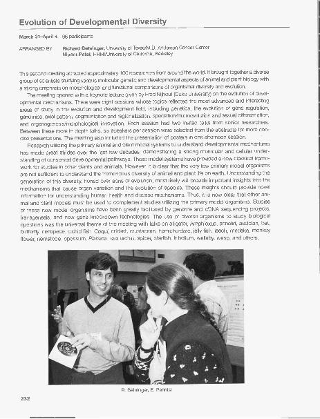
Evolution of Developmental Diversity
March 31-April 4 96 participants
ARRANGED BY Richard Behringer, University of Texas/M.D. Anderson Cancer CenterNipam Patel, HHMI/University of California, Berkeley
This second meeting attracted approximately 100 researchers from around the world. It brought together a diverse
group of scientists studying various molecular genetic and developmental aspects of animal and plant biology with
a strong emphasis on morphological and functional comparisons of organismal diversity and evolution.The meeting opened with a keynote lecture given by Fred Nijhout (Duke University) on the evolution of devel-
opmental mechanisms. There were eight sessions whose topics reflected the most advanced and interesting
areas of study in the evolution and development field, including genetics, the evolution of gene regulation,genomics, axial pattern, segmentation and regionalization, speciation/microevolution and sexual differentiation,and organogenesis/morphological innovation. Each session had two invited talks from senior researchers.Between these more in-depth talks, six speakers per session were selected from the abstracts for more con-cise presentations. The meeting also included the presentation of posters in one afternoon session. '
Research utilizing the primary animal and plant model systems to understand developmental mechanismshas made great strides over the last few decades, demonstrating a strong molecular and cellular under-standing of conserved developmental pathways. These model systems have provided a now classical frame-work for studies in other plants and animals. However, it is clear that the very few primary model organismsare not sufficient to understand the tremendous diversity of animal and plant life on earth. Understanding thegeneration of this diversity, honed over eons of evolution, most likely will provide important insights into themechanisms that cause organ variation and the evolution of species. These insights should provide novelinformation for understanding human health and disease mechanisms. Thus, it is now clear that other ani-mal and plant models must be used to complement studies utilizing the primary model organisms. Studiesof these new model organisms have been greatly facilitated by genome and cDNA sequencing projects,transgenesis, and new gene knockdown technologies. The use of diverse organisms to study biologicalquestions was the universal theme of the meeting with talks on alligator, Amphioxus, annelid, ascidian, bat,butterfly, centipede, cichid fish, Coqui, cricket, crustacean, hemichordate, jelly fish, leech, medaka, monkeyflower, nematode, opossum, Planaria, sea urchin, spider, starfish, tribolium, wallaby, wasp, and others.
R. Behringer, E. Pennisi
232
PROGRAM
Keynote Address: The Evolution of DevelopmentalMechanisms
F Nijhout, Duke University
Genomics IChairperson: A. Sanchez, University of Utah School of
Medicine, Salt Lake City
Genetics IChairperson: S. Hake, University of California, Berkeley
Organogenesis / Morphological InnovationChairperson: L. Holland, University of California, San Diego
Segmentation/Regionalization Genetics IIChairperson: S. Brown, Kansas State University, Manhattan Chairperson: R. Sommer, Max-Planck Institute for
Developmental Biology Tubingen, GermanyAxial PatternChairperson: J. Gerhart, Harvard Medical School, Boston,
Massachusetts Evolution of Gene RegulationChairperson: C. Amemiya, Benaroya Research Institute at
Speciation / Microevolution and Sexual Differentiation Virginia Mason, Seattle, WashingtonChairperson: A. Sinclair, University of Melbourne, Royal
Children's Hospital, Australia
S. Alvarado, W. Jeffrey N. Patel, A. Burke
R. Palmer, P. Brakefield, F. Nijhout
233

Channels, Receptors, and Synapses
April 21-25 238 participants
ARRANGED BY Richard Huganir, HHMI/Johns Hopkins University School of MedicineLily Jan, HHMI/University of California, San FranciscoMorgan Sheng, HHMI/Massachusetts Institute of Technology
The brain consists of a vast network of excitable cells (neurons) that conduct electrical impulses and communi-cate with each other via specialized junctions (synapses). Information is processed and stored in the nervous sys-tem through patterns of electrical activity and via changes in the strength and structure of synapses. As masterregulators of neuronal excitability and synaptic communication, ion channels and receptors lie at the heart of neu-
robiology. In recent years, molecular and cell biological analyses of neuronal ion channels and receptors have rev-
olutionized our understanding of the basic mechanisms that control electrical signaling and synaptic function inthe nervous systems. The convergence of advances in biochemistry, molecular genetics, microscopic imaging,and electrophysiology has made this interdisciplinary field one of the most exciting and rapidly growing in neuro-science. Several landmark advances have transformed the phenomenology in this field into underlying molecularmechanisms, including the genomic cataloging of all channels and receptors, the discovery of the three-dimen-sional "crystal" structure of a membrane ion channel, the identification of specific anchoring proteins that bind tothe cytoplasmic tails of ion channels and receptors, and the discovery of dynamic andregulated trafficking of receptors/ion channels. The fundamental aspects of thesemechanisms seem to be evolutionarily conserved from primitive nervous systems of
worms to the human brain.The entire field is poised for further breakthroughs that will not only illuminate basic
workings of the brain, but also shed light on neurological and psychiatric diseases thatstem from abnormal neuronal excitability and synaptic dysfunction. Such break-throughs will be facilitated by cross-fertilization of ideas and technologies between sci-entists studying related questions in diverse organisms using different methodologicalapproaches. To provide a forum that unites this exciting multidisciplinary area, a newCSHL meeting on Channels, Receptors, and Synapses was started in April 2004.
The inaugural meeting was extremely successful, bringing together nearly 240participants from United States, Europe, and Asia in an atmosphere of social and scientific exchange. A widerange of topics were discussed, including the structure and function of ion channels and receptors, new class-es of channels/receptors, regulation of ion channel/receptor function by phosphorylation, membrane traffickingand degradation; molecular mechanisms of synaptic transmission/plasticity and their roles in brain function; andthe involvement of ion channels/receptors in human disease. More than 60 attendants were selected to give
M. Wampler, S. Thompson
234
M. Kennedy, W. Ford, H.H. Beale,H. Carlisle, A. Tapper

oral presentations of their work, while the majority of the rest presented posters. Special lectures were given byRod MacKinnon, 2003 Nobel laureate, on the three-dimensional structures of ion channels, and by RobertoMalinow (CSHL professor), on trafficking of glutamate receptors in synaptic plasticity. The overall response tothe meeting was extremely positive, especially given that this was the first conference. Participants particularlyenjoyed the breadth of subjects covered and the chance to interact with investigators in related but distinctfields. The opportunity for junior investigators (including postdocs and students) to present their own work wasespecially applauded. Based on the success of the first meeting, this conference will take place again in twoyears. It is anticipated that this Cold Spring Harbor meeting will become a "must-attend" conference for learn-ing the latest developments in molecular and cellular physiology of the brain.
PROGRAM
Modulation of Ion Channels and ReceptorsChairpersons: S. Moss, University of Pennsylvania, Philadelphia;
A. Dolphin, University College London, United Kingdom
Ion Channels and Receptors in Brain Function andDisease
Chairpersons: M. Mishina, Tokyo University Japan; L. Ptacek,University of Utah, Salt Lake City
Special Lecture: Three-dimensional Structure of IonChannels
R. MacKinnon, The Rockefeller University New York
Structure and Function of Ion Channel/ReceptorsChairpersons: D. Clapham, Children's Hospital, Boston, Massachu-
setts; G. Yellen, Harvard Medical School, Boston, Massachusetts
Presynaptic MechanismsChairpersons: T. Sudhof, University of Texas Southwestern
Medical Center Dallas; R.W. Tsien, Stanford University California
Postsynaptic Signaling MechanismsChairpersons: M. Greenberg, Children's Hospital, Boston,
Massachusetts; M. Kennedy, California Institute ofTechnology Pasadena
Synaptic PlasticityChairpersons: R. Malenka, Stanford University Palo Alto,
California; G. Turrigiano, Brandeis University Waltham,Massachusetts
Special Lecture: Receptor Trafficking in SynapticPlasticity
R. Malinow, Cold Spring Harbor Laboratory
Development of SynapsesChairpersons: H. Cline, Cold Spring Harbor Laboratory; M.
Bear, Massachusetts Institute of Technology Cambridge
Targeting, Trafficking, and Degradation of Ion Channels/Receptors
Chairpersons: G. Westbrook, Vollum Institute, Portland,Oregon; J. Kaplan, Massachusetts General Hospital, Boston
H. Kataoka, L. Jan
D. Clapham, A. Maffei
235

Gene Expression and Signaling in the Immune System
April 28-May 2 294 participants
ARRANGED BY Doreen Cantrell, University of DundeeRichard Flavell, HHMI/Yale UniversityRudolf Grosschedl, University of MunichStephen Smale, HHMI/University of California School of Medicine
This second meeting in the series attracted more than 290 participants from around the world. It maintainedits unique emphasis on mechanistic studies that most significantly advance our understanding of the generegulation and signal transduction circuitry within the immune system, as well as the use of the immune sys-
tem as a tool for elucidating fundamental molecular processes. Invited and oral presentations selected fromthe abstracts covered a broad range of topics concerning developmental regulation of the immune systemand immune responses to infection. One highlight of the meeting was a series of presentations that arebeginning to firmly define the molecular events that regulate cell fate decisions as hematopoietic stem cellschoose between various lymphocyte and myeloid cell lineages. Another highlight was a spirited discussionof the activation-induced deaminase (AID) protein that is essential for both somatic hypermutation and classswitching of immunoglobulin genes. Conflicting evidence concerning potential roles of AID in RNA process-ing or, alternatively, direct deamination of nucleotides within variable regions and switch regions duringhypermutation and switching was debated. Other central topics included the epigenetic regulation of devel-opment, mechanisms involved in the T helper 1-T helper 2 lineage decision, mechanisms of tolerance induc-
tion, and Toll-like receptor signaling mechanisms.Special thanks to the following organizations for support of this meeting: Abbott Laboratories, Biogen
IDEC, Boehringer Ingelheim Pharmaceuticals, Inc., Hoffmann-La Roche, Merck & Co., Inc., and Pfizer. Thismeeting was funded in part by the National Institute of Allergy and Infectious Diseases, a branch of theNational Institutes of Health.
S. Smale, R. Flavell, R. Grosschedl
236 R. Sen, A. Roy, Z. Desgranges

PROGRAM
Stem Cells and Early Development DecisionsChairperson: I. Weissman, Stanford University School of
Medicine, California
Regulation of Immune Cell DevelopmentChairperson: K. Ca lame, Columbia University College of
Physicians & Surgeons, New York
Antigen Receptor Gene Assembly, SomaticHypermutation, and Class Switching
Chairperson: E Alt, HHMI/Children's Hospital, Boston,Massachusetts
Chromatin Structure and Epigenetic RegulationChairperson: A. Tarakhovsky, The Rockefeller University New
York
Lymphocyte DifferentiationChairperson: L. Glimcher, Harvard School of Public Health,
Boston, Massachusetts
Signal Transduction in Immune CellsChairperson: D. Cantrell, University of Dundee, United
Kingdom
Regulation of Lymphocyte Function IChairperson: C. Benoist, Joslin Diabetes Center, Boston,
Massachusetts
Regulation of Lymphocyte Function IIChairperson: S. Ghosh, Yale University School of Medicine,
New Haven, Connecticut
J. Madrenas, C. Bueno
X. Lin, G. Koretzky, M. Reth
K. Mowen, S. Szabo
237

Molecular Chaperones and Heat Shock Response
May 5-9 298 participants
ARRANGED BY James Bardwell, University of MichiganElizabeth Craig, University of WisconsinJonathan Weissman, University of California, San Francisco
This meeting, 22 years after the inaugural meeting, featured advances in the areas of structure and mechanismof action of molecular chaperones; mechanisms of induction of the stress response; the role of chaperones in
protein degradation; and chaperone function in disease and aging. In the first area,
further advances were described for the classic heat shock proteins, the GroEUES,the small heat shock factors, Hsp70, 90, and 100 classes, as well as more recent-ly identified chaperones such as CsA in eubacteria. In addition, the first high-reso-lution structure of the Ero1 protein, which has an essential role in oxidative proteinfolding in eukaryotes, was presented. In a session of particular interest, the rela-tionship between chaperone function and aging was featured with talks presentingexperiments in Caenorhabditis elegans and Drosophila establishing a role of chap-erone induction in extending lifespan, as well as the effects of aging on the propen-sity to form intracellular toxic protein aggregates. Several presentations revealedadvances in our understanding of the function of ATP-dependent regulatory sub-units such as the proteosome regulatory particle in eukaryotes and the related CIpAcomplex in bacteria in catalyzing the unfolding of proteins prior to their degradation.In the area of regulation of stress responses, a focus of the meeting was theadvances in our understanding of the mechanism and biological consequences ofthe quality control machinery that prevents the secretion and ultimately degradesmisfolded proteins in the endoplasmic reticulum (ER) as well as the unfolded proteinresponse which induces a multifaceted response when misfolded proteins accu-mulate in the ER. In addition, progress toward an understanding of the function of heat shock transcription fac-tors in complex organisms was discussed. In the area of protein misfolding and disease, a number of talks dealtwith progress in the use of model systems in yeast, mice, and worms to attack this complex problem. The role
B. Bukau, A. Norwich, E. Craig
S. Gupta, J. Bardwell, D. Cyr, W. Antoine
238
N. Benaroudj, 0. Kandror

oligomeric intermediates, which are often formed transiently during protein aggregation, in contributing to cellu-lar toxicity and disease was also discussed. Finally, a number of talks focused on the biogenesis and mainte-nance of prions in yeast, and the roles that different chaperones have in this process including evidence thatHsp1 04 promotes prion propagation by acting to divide seeds, thereby allowing the creation of more infectiousprion units. Thus, rather paradoxically, a chaperone can have the net effect of promoting protein misfolding.
This meeting was funded in part by the National Institute of Neurological Disorders & Stroke, and theNational Institute of Child Health and Human Development, branches of the National Institutes of Health;Merck & Co., Inc., and the Alzheimer's Association.
PROGRAM
Diseases of Protein MisfoldingChairperson: S. Lindquist, Whitehead Institute, Cambridge,
Massachusetts
Quality Control and Protein TraffickingChairperson: R. Kopito, Stanford University California
Cellular Response to StressChairperson: C. Gross, University of California, San Francisco
Chaperone Function in Disease, Aging, andDevelopment
Chairperson: R. Morimoto, Northwestern University Evanston,Illinois
Regulation of the Stress ResponseChairperson: D. Ron, New York University School of Medicine
Chaperones and ProteolysisChairperson: T. Baker, HHMI/Massachusetts Institute of
Technology Cambridge
Chaperone Biochemistry and Protein FoldingChairperson: B. Bukau, Universitat Heidelberg, Germany
P. Bross, S. Vang
P Carrigan, D. Smith, B. Matts
239

The Biology of Genomes
May 12-16 504 participants
ARRANGED BY Thomas Hudson, McGill UniversitySvante Paabo, Max-Planck InstituteJane Rogers, The Sanger CenterEdward Rubin, Lawrence Berkeley National Laboratory
This annual meeting was the 15th annual gathering of genome scientists in this setting. The past decade ormore has seen remarkable progress in the mapping, sequencing, and annotation of the genomes of many"model organisms" and the publication of a finished and draft sequences of the human, mouse, and ratgenomes, in addition to several model organisms and hundreds of bacteria. Just over 500 people from aroundthe world attended the meeting, with 331 abstracts presented describing a broad array of topics relating tothe analysis of genomes from a number of different organisms.
The session topics included areas such as biological insights from comparative genomics, functionalgenomics, organismal biology, computational genomics, and the use of sequence variations to study popula-
tions and mechanisms of disease. Once again, projection-style, interactive computer demonstrations in GraceAuditorium effectively highlighted the critical new bioinformatics tools being developed for storing, organizing,and analyzing various genomic data sets, including expression, mapping, and sequence data. An ELSI(Ethical, Legal, and Social Implications) panel discussion focused on issues surrounding the process of com-munity engagement in large-scale population genomic studies, using the case study of the Yoruba population
of lbadan Nigeria and the Hap Map project.The major themes of the meeting included the analysis of large-scale variation in genetic variation across
the human genome (a product of the International Hap Map Consortium), annotation of the human genomeusing multispecies sequence comparisons, and a myriad of large-scale approaches to understand complexbiological processes. The Friday afternoon keynote talks were delivered by Stuart Schreiber and Craig Venter.
This meeting was funded in part by the National Human Genome Research Institute, a branch of theNational Institutes of Health.
S. Paabo, E. Rubin, T. Hudson
240

PROGRAM
Comparative GenomicsChairpersons: K. Peichel, Fred Hutchinson Cancer Research
Center, Seattle, Washington; A. Sidow, Stanford University,California
Variation Technology and Haplotype StructuresChairpersons: D. Altshuler, Massachusetts General Hospital,
Harvard Medical School, Boston; M. Przeworski, BrownUniversity Providence, Rhode Island
High-throughput BiologyChairpersons: A. Bradley, Wellcome Trust Sanger Institute,
Hinxton, United Kingdom; M. Snyder, Yale University NewHaven, Connecticut
Nonhuman Species and Environmental GenomicsChairpersons: D. Reiman, Stanford University California; D.
Rokhsar, DOE Joint Genome Institute, Walnut Creek,California
ELSI Panel DiscussionModerator: J. McEwen, National Human Genome Research
Institute, Bethesda, MarylandPanelists: C. Adebamowo, University of lbadan, Nigeria, Africa; P
Marshall, Case Western Reserve University, School of Medicine,Cleveland, Ohio; C. Rotimi, Howard University, Washington,D.C.; C. Royal, Howard University, Washington, D.C.
Computational GenomicsChairpersons: P. Bork, European Molecular Biology
Laboratory Heidelberg, Germany; L. Pachter, University ofCalifornia, Berkeley
Polymorphisms and Their Relationship to BiologyChairpersons: J. Kere, Karolinska Institutet, Huddinge, Sweden;
K. Frazer, Perlegen Sciences, Mountain View, California
Keynote SpeakersS. Schreiber, Harvard University Cambridge, Massachusetts;
C. Venter, The Center for the Advancement of Genomics,Rockville, Maryland
Genome Structure and EvolutionChairpersons: W.-H. Li, University of Chicago, Illinois; C.-I.
Wu, University of Chicago, Illinois
T. Andersson, J. Larkin
J. Hancock, P Little, S. Brown
K. Kimura, Y. Suzuki, A. Fujiyama241
The Cell Cycle
May 19-23 341 participants
ARRANGED BY Orna Cohen-Fix, National Institutes of Health/NIDDKStephen El ledge, HHMI/Harvard UniversityJ. Wade Harper, Harvard Medical SchoolDavid Morgan, University of California, San Francisco
This 8th biannual meeting attracted participants from around the world and is internationally recognized for itsability to bring together scientists who study cell cycle regulation in eukaryotes ranging from yeast to humans.We were particularly fortunate this year to have Kim Nasmyth as our keynote speaker. Dr. Nasmyth, anacknowledged leader in the yeast cell cycle regulation field, opened the meeting with a wide-ranging and excit-ing talk describing the discovery of the separin-cohesin pathway and its role in the control of the metaphaseto anaphase transition in eukaryotes ranging from yeast to mammals. Three broad themes were dominantthroughout the meeting: the interaction of cell cycle control with developmental and cancer biology; the mech-anisms of action of cell cycle checkpoints and their integration with signal pathways that control mitotic tran-sitions; and the emerging use of high-throughput and whole-genome approaches to uncover new aspects ofcell biology. Research in proteolysis for both G1 /S and G2/M regulation in particular continues to receive intensefocus, with much progress reported. As in previous years, there was also emphasis on the long-standing prob-lem of regulation of DNA replication in the cell cycle, its onset, and its restriction to once per cell cycle.Significant insight into the conservation of transcriptional regulatory pathways involved in the G1 /S transitionwas presented when it was found that the G1 cyclins phosphorylate and inactivate a repressor of the S-phasetranscription factors. This is analogous to the Rb/E2F switch in mammals. New to the discussion was a moredetailed understanding of how different Cdk/cyclin complexes choose their substrates during the cell cycle,with more than one surprise in that area. There was also an emphasis on the use of emerging technologies,both in tissue culture cells and in live animals such as Caenorhabditis elegans, to study the dynamics of sig-naling pathways and rapid changes in the localization of components of the mitotic apparatus in response tocheckpoint activation. As in other years, scientists studying cell cycle regulation in yeast, Xenopus, Drosophila,nematodes, and mammals were well-represented. Once again, the striking phylogenetic conservation of cellcycle regulatory mechanisms was readily evident. This meeting also served to demonstrate the impact ofwhole-genome analysis on basic cell biology, and these achievements will no doubt continue to drive rapid
2420. Cohen-Fix, S. El ledge, W. Harper, D. Morgan

progress in this field. The cell cycle conference was another landmark meeting for the cell cycle field, and theparticipants all continued to look forward to equally exciting meetings in future years.
This meeting was funded in part by the National Cancer Institute and the National Institute on Aging,branches of the National Institutes of Health.
PROGRAM
Cell Cycle Control MechanismsChairperson: F. Cross, The Rockefeller University New York
G, Control IChairpersons: N. Dyson, Massachusetts General Hospital
Cancer Center, Charlestown; M. Tyers, Samuel LunenfeldResearch Institute, Toronto, Canada
G, Control IIChairpersons: J. Roberts, Fred Hutchinson Cancer Research
Center, Seattle, Washington; G. Hannon, Cold Spring HarborLaboratory
S PhaseChairpersons: J. Li, University of California, San Francisco; J.
Diffley, Imperial Cancer Research Fund, South Mimms,United Kingdom
M. Saito, A. Taylor,M. Polymenis, R. Pathak
M Phase IChairpersons: J. Pines, University of Cambridge, United
Kingdom; C. Rieder, Wadsworth Center, Albany New York
M Phase IIChairpersons: A. Amon, Massachusetts Institute of
Technology Cambridge; K. Gould, HHMINanderbiltUniversity Nashville, Tennessee
Checkpoints IChairpersons: A. Carr, University of Sussex, United Kingdom;
D. Toczyski, University of California, San Francisco
Checkpoints IIChairpersons: S. Biggins, Fred Hutchinson Cancer Research
Center, Seattle, Washington; H. Yu, University of TexasSouthwestern Medical Center, Dallas
A. Devault, J. Li
M. Tyers, V. Duronio
243

Retroviruses
May 25-30 442 participants
ARRANGED BY Eric Freed, NCI-FrederickSusan Ross, University of Pennsylvania
This 29th annual CSHL meeting originated in 1975 as a meeting on RNA Tumor Viruses and evolved to itscurrent focus on Retroviruses in 1993. In our view, the 29th meeting was highly successful. The short oralpresentations and posters were complemented by a keynote talk from Dr. Neil Copeland, who presentedimportant findings from his and Nancy Jenkins' lab regarding the use of high-throughput analysis to mapretroviral integration sites in tumors induced by murine leukemia viruses. The Jenkins and Copeland labshave used infection of various hematopoietic stem cells followed by transplantation into lethally irradiatedmice to identify not only novel proto-oncogenes, but also cooperating cancer genes, tumor suppressors, andgenes involved in stem cell transformation and immortalization. Although the topic of the keynote addresswas somewhat outside the mainstream of the conference, the talk was well-received.
There were 332 abstracts this year, with oral presentations arranged into 11 sessions, and three postersessions. The oral sessions began with the entry phase of the replication cycle, proceeded with postentryevents, assembly and release, integration, reverse transcription, pathogenesis/host factors, RNA-relatedevents (transcription, processing, export, and packaging), and finished with antivirals. Although the moststriking developments this year involved postentry events and assembly/release, significant progress wasmade toward elucidating a number of aspects of the retroviral replication cycle. Highlights included recentprogress in understanding the mechanism by which the lentiviral Vif protein counteracts the antiviral effectsof the cellular enzyme APOBEC3G and novel findingsrelating to the identification of TRIM5alpha as the factorrestricting postentry events in HIV-1 infection of nonhu-man primate cells.
In other matters, the pattern of arranging postersalphabetically was continued this year. The cuisine con-tinues at the enjoyable level noted for last several years,a big change from previous years. Posters were held inthe evening this year after the wine and cheese party,rather than talks, which are difficult for some to manageafter wine and beer. The Sunday morning session itselfcontinues to be a bit of a problem as it is difficult for peo-ple to stay on, get up, and/or be enthusiastic at this finalsession. Finally, we very much appreciated the help andguidance of the CSHL staff, who made the meeting easyand enjoyable to organize. One final notable develop-ment: This year, we were asked to write a review of themeeting for the new on-line BioMed Central journalRetrovirology. The review is available at http://www.retro-virology. com/content/1/1/25.
Contributions from our Corporate Benefactors, Corpo-rate Sponsors, Plant Corporate Associates, CorporateAffiliates, Corporate Contributors, and Foundations provid-ed core support for this meeting.
244
P. Jolicoeur, D. Derse

PROGRAM
Entry IChairpersons: D. Lindemann, Technische Universitat Dresden,
Germany; L. Albritton, University of Tennessee HealthSciences Center, Memphis
Entry IIChairperson: H. Garoff, Karolinska, Institutet, Huddinge,
Sweden
Postentry IChairperson: P. Bieniasz, Aaron Diamond AIDS Research
Center, New York
Postentry IIChairpersons: C. Aiken, Vanderbilt University, Nashville,
Tennessee; G. Towers, University College London, UnitedKingdom
Assembly/Release IChairpersons: X.F. Yu, Johns Hopkins University School of
Public Health, Baltimore, Maryland; R. Montelaro, Universityof Pittsburgh School of Medicine, Pennsylvania
Keynote SpeakerNeal Copeland, NCI-FrederickIntegrationChairpersons: H. Levin, National Institutes of Health,
Bethesda, Maryland; D. Grandgenett, St. Louis UniversityHealth Sciences Center, Missouri
Assembly/Release IIChairpersons: P. Spearman, Vanderbilt University School of
Medicine, Nashville, Tennessee; E. Barklis, Oregon Health &Science University Portland
Reverse Transcription/RecombinationChairpersons: W.-S. Hu, NCI-Frederick, Maryland; B.
Berkhout, University of Amsterdam, The Netherlands
Pathogenesis/Host FactorsChairpersons: M. Lairmore, Ohio State University Columbus;
H. Fan, University of California, Irvine
Transcription, RNA Processing, Export, PackagingChairpersons: K. Boris-Lawrie, Ohio State University
Columbus; J. Dudley, University of Texas, Austin
AntiviralsChairpersons: A. Kaplan, University of North Carolina, Chapel
Hill; A. Lever, University of Cambridge, United Kingdom
M. Laasko, R. Swanstrom
C. Fumes, D. Dobbs
A. Fassati, M. Pizzato
S. Kim, C. Woodward
245

New York Structural Biology Group
August 6 252 participants
ARRANGED BY Leemor Joshua-Tor, Cold Spring Harbor LaboratoryLarry Shapiro, Columbia University College of Physicians & Surgeons
This summer meeting was the sixth in day-long meetings allowing structural biologists from all over theregion to meet and discuss their latest results. The meeting was open to structural biologists from dif-ferent disciplines, including crystallographers, spectroscopists, computational biologists, and bio-chemists, with more than 250 participants from academia and industry from the tristate area. The pro-gram featured nine talks and a poster session and concluded with a beach barbecue, allowing a won-derful opportunity for informal interactions. This meeting complements the bimonthly evening meetingsof the group held at The Rockefeller University. No registration was required and participants wereencouraged to set up posters.
Financial support was provided by Amersham Biosciences, Hampton Research, Rigaku MSC andProterion, Inc.
PROGRAM
Session I
Chairpersons: L. Shapiro, Columbia University College ofPhysicians & Surgeons; H. Wu, Weill Medical College ofCornell University; C. Wang, Columbia University College ofPhysicians & Surgeons (Palmer lab); J. Truglio, Stony BrookUniversity (Kisker lab); J. Ferrara, Rigaku/MSC
Session 11
Chairpersons: D. Stokes, Skirball Institute, New YorkUniversity Medical School; D. Patel, Memorial Sloan-Kettering Cancer Center; Y. Song, City College of New York(Gunner lab); D. Jain, The Rockefeller University (Darst lab);D. Raleigh, Stony Brook University
Session III
Chairpersons: L. Joshua-Tor, Cold Spring Harbor Laboratory;B. Honig, Columbia University College of Physicians &Surgeons; J. Williams, Columbia University College ofPhysicians & Surgeons (Hendrickson lab); C. Lima, MemorialSloan-Kettering Cancer Center; E. Crocker, Stony BrookUniversity (Smith lab); T. Walz, Harvard Medical School
246

Cancer Genetics and Tumor Suppressor Genes
August 18-22 405 participants
ARRANGED BY Jacqueline Lees, MIT Center for Cancer ResearchScott Lowe, Cold Spring Harbor LaboratoryCharles Sawyers, University of California, Los AngelesCharles Sherr, HHMI/St. Jude's Children's Research Hospital
This fourth meeting focused on topics related to cancer genetics and biology. Most of the 400 scientists pre-
sented unpublished research through oral or poster presentations. The talks were highlighted by two keynotelectures: Anton Berns discussed recent advances using mice to model human cancer, highlighting theirpotential utility for identifying new cancer genes and for studying cancer therapeutics. In the second keynote,Stephen El ledge discussed recent advances in elucidating DNA damage responses and their roles in pre-venting tumorigenesis. An international team of established investigators, whose expertise covered many ofthe topics highlighted during the meeting, chaired the individual sessions. The first oral session discussednew mouse models, which continue to be an important topic at the meeting. Indeed, many other talks andposters highlighted the use of cancer-prone mice to study gene function and to model human cancer. Othersessions highlighted recent advances in our understanding of DNA damage responses, apoptosis, and cel-lular senescence, the Rb and p53 tumor suppressor pathways, and signal transduction pathways that arealtered in human tumors. This meeting continued to be enthusiastically supported, and the lectures andposter presentations led to extensive discussions and exchanges of information and ideas.
This meeting was funded in part by the National Cancer Institute, a branch of the National Institutes ofHealth.
G. Evan, L. Attardi, S. Lowe
247

PROGRAM
Mouse ModelsChairpersons: T. Van Dyke, University of North Carolina,
Chapel Hill; T. Jacks, HHMI/Massachusetts Institute ofTechnology Cambridge
Keynote Address: Analysis of the Multifunctionality ofBRCA1
D. Livingston, Dana-Farber Cancer Institute, Harvard MedicalSchool
Rb and E2FChairpersons: S. Morrison, HHMI/University of Michigan, Ann
Arbor; J. Lees, MIT Center for Cancer Research,Cambridge, Massachusetts
TherapeuticsChairpersons: C. Thompson, University of Pennsylvania,
Philadelphia; C.L. Sawyers, University of California, LosAngeles
Keynote Address: Cancer Targets in the Ras pathwayChairperson: Frank McCormick, University of California, San
Francisco
ARF/p53 PathwayChairpersons: G. Evan, University of Califomia Comprehensive
Cancer Center, San Francisco; K. Vousden, Beatson Labora-tories, Institute for Cancer Research, Glasgow, United Kingdom
Genome-wide Analysis and ScreensChairpersons: S.A. Courtneidge, Van Andel Research Institute,
Grand Rapids, Michigan; M. Van Lohuizen, University ofCalifornia, San Francisco
DNA Damage/Chromosomal InstabilityChairpersons: J. Lukas, Danish Cancer Society Copenhagen;
T De Lange, The Rockefeller University New York
Signaling IChairpersons: B. Schulman, St. Jude Children's Research
Hospital, Memphis, Tennessee; E. Holland, Memorial Sloan-Kettering Cancer Center, New York
Signaling IIChairpersons: M.F. Roussel, St. Jude Children's Research
Hospital, Memphis, Tennessee; L.F. Parada, University ofTexas Southwestern Medical Center, Dallas
P. Van Sloun, T. Hughes M. Fried, J. Jenkins, Y. Ito
J. Lukas, D. Livingston
248
D. Ray, A. Datta

Molecular Genetics of Bacteria and Phages
August 24-29 209 participants
ARRANGED BY Tina Henkin, Ohio State UniversityCatherine Squires, Tufts University School of MedicineRyland Young, Texas A&M University
The meeting featured 98 talks arranged in 10 oral presentation sessions, and two poster sessions. Youngscientists gave most of the talks, and a senior scientist who presented an introductory overview moderatedeach session. Topics included ribosomes, translation, and posttranscriptional regulation; bacteriophagedevelopment and host interactions; cell division and development; transcriptional regulation; cell surfaces-structure, function, and signaling; host/pathogen interactions; bacterial genomics and proteomics; DNAstructure, recombination, and transposition; and stress response, chaperones, and protein degradation.
An interesting theme tying seemingly disparate sessions together was the variety of roles that small or nonen-
coding RNAs have in influencing gene expression. This theme started with the Nat Sternberg Award winner'slecture (A. Nahvi from the Breaker group at Yale) describing riboswitches, RNAs whose structure is altered whencertain small molecules are sensed, leading to a dramatic change in gene expression. Similarly, the Henkingroup (Ohio State) demonstrated that the T-box system using binding of charged/uncharged tRNAs to RNA andregulating gene expression works in vitro, providing evidence that no other effectors are needed for the RNA-mediated control. Somewhat unexpectedly, the Losick group at Harvard found that a small RNA inhibits cell lysis
in Bacillus subtilis and another, RNAIII, was found by collaborating groups from Strasberg, Lyon, and Paris tobe involved in regulating expression of pathogenesis genes in Staphylococcus aureus. Substantial physiologi-cal changes are noted in Escherichia coli (Remme group, Estonia) and C. crescentus (Keller group at PennState) when the small RNA responsible for rescuing stalled ribosomes and tagging incomplete peptides fordestruction, tmRNA, is lacking. Further studies by the Weisberg group at NIH of a small RNA encoded by phage
HK022 called PUT described how PUT acts in cis to alter RNA polymerase into an antitermination-competentform that can transcribe through terminators. In Archaea, Pat Dennis (UBC) described the sequence and struc-tural constraints of small RNAs needed as guides for rRNA modification complexes. The description of newsmall, noncoding RNAs in E. coli (G. Storz group, NIH) suggests that there are more important roles for theseRNAs that are not yet fully appreciated. These intriguing reports, and the wide variety of systems giving rise tothem, suggest that in future meetings we can expect progress in understanding the contributions to geneexpression and cell physiology from a host of different forms of RNAs.
This meeting was funded in part by the National Science Foundation.
C. Squires, 0. Amster-Choder
249

PROGRAM
Ribosomes, Translation, and PosttranscriptionalRegulation
Chairperson:R. Green, Johns Hopkins University Baltimore,Maryland
Bacteriophage Development and Host InteractionsChairperson: M. Gottesman, Columbia University
New York
RNA Polymerase: Structure, Function, and RegulationChairperson: R. Landick, University of Wisconsin, Madison
Cell Division and DevelopmentChairperson: W. Margolin, University of Texas Houston
Medical School
Transcriptional RegulationChairperson: C. Yanofsky, Stanford University California
Cell Surfaces-Structure, Function, and SignalingChairperson: M. Manson, Texas A&M University College
Station
Host Pathogen InteractionsChairperson: H. Shuman, Columbia University New York
Bacterial Genomics and ProteomicsChairperson: J. Lawrence, University of Pittsburgh,
Pennsylvania
DNA Structure, Recombination, and TranspositionChairperson: K. Derbyshire, New York State Department of
Health and University at Albany
Stress Response, Chaperones, and Protein DegradationChairperson: S. Wickner, National Institutes of Health/NCI,
Bethesda, Maryland
W. Champness, L. Snyder
Z. Pragai, S. Semsey
D. Cully, L. Hernandez, H. Shuman
250
M. Leibman, J. Roberts

Mouse Molecular Genetics
September 1-5 311 participants
ARRANGED BY Francois Guillemot, National Institute for Medical ResearchHiroshi Hamada, Osaka UniversityTerry Magnuson, University of North Carolina, Chapel HillJanet Rossant, Samuel Lunenfeld Research Institute
Held every other year, this meeting attracted more than 300 researchers from across the globe. Scientistsattending the meeting also come from very diverse areas of biology and are united by the common use ofmouse mutants in their research. As a result, the meeting covers very diverse areas of biology, in which thecontribution of mouse genetics is essential to the progress of knowledge. The sustained interest of all par-ticipants throughout the meeting is ensured by the commonality of techniques, as well as by increasing evi-dence that basic mechanisms are conserved across diverse experimental systems.
The meeting opened with the Annual Rosa Beddington Lecture given by Elisabeth Robertson. The meet-ing was organized into ten sessions. In a slight departure from the format of previous years, each sessionwas arranged around not two, but three talks of general interests, given by two senior and one more juniorinvited researchers. Between these more in-depth talks, five to six speakers per session were selected fromthe abstracts for more concise presentations. The reason for adding one invited speaker to each session wasto further increase the visibility and attractiveness ofthe meeting. The meeting also included the presenta-tion of several hundred posters in two afternoon ses-sions. The Genetics Society of America sponsoredcash prizes for the five best poster presentations asjudged by the organizers and other selected Pls. Themeeting ended with a farewell to Hiroshi Hamada, whostepped down after 4 years as an organizer of themeeting.
Some of the sessions have shifted in their empha-sis this year. The neurobiology session has evolved torepresent systems neuroscience, and Linda Buck(2004 Nobel Prize for Medicine) gave an invited lecturein this session on information coding in the olfactorysystem. The growing emphasis on genetics/genomicsand on human disease models, already noted in previ-ous years, has also continued. Aravinda Chakravartiwas invited to provide links between mouse andhuman genetics. Given the increasing importance of the T. Magnuson, D. Stewart
mouse as a model to investigate disease mechanisms, and the sophistication with which diseases can nowbe modeled and analyzed in the mouse, the organizing committee has decided to emphasize the linkbetween mouse and human genetics.
This meeting was funded in part by the National Cancer Institute, the National Institute of Diabetes andDigestive and Kidney Diseases, the National Institute of Child Health and Human Development, the NationalHuman Genome Research Institute, and the National Institute of Neurological Disorders and Stroke, branch-es of the National Institutes of Health.
Voil))))1111))0\
251

PROGRAM
Rose Beddington Lecture: Combinatorial SmadActivities Regulate Nodal-dependent Patterningof the Early Embryo
Elizabeth Robertson, Harvard University, Cambridge,Massachusetts
Stem Cells and DifferentiationChairperson: F. Miller, Hospital for Sick Children, Toronto,
Canada
Models of Human DiseaseChairperson: R. Lifton, Yale University School of Medicine,
New Haven, Connecticut
OrganogenesisChairperson: A. McMahon, Harvard University Cambridge,
Massachusetts
NeurobiologyChairperson: L. Buck, HHMI/University of Washington, Seattle
Embryonic PatterningChairperson: V. Papaioannou, Columbia University, New York
EpigeneticsChairperson: J. Lee, Massachusetts General Hospital, Boston
Genetics and GenomicsChairperson: L. Flaherty, Wadsworth Center, Troy, New York
SignalingChairperson: G. Martin, University of California, San Francisco
252
tiW.
S. Blaess, A. Sudarov
J. Sanes, T. Grodzicker, G. Martin

Translational Control
September 7-12 475 participants
ARRANGED BY Alan Hinnebusch, National Institutes of Health/NICHDTatyana Pestova, SUNY Downstate Medical CenterJoel Richter, University of Massachusetts Medical School
This meeting attracted more than 470 participants from around the world and included three keynote speak-ers, nine platform speakers, and three poster sessions that covered a record 415 abstracts. Several note-worthy reports concerned the mechanism of translation. Contrary to the prevalent model, it was shown thatinitiation factors elF1 and elF3 are not released from 48S complexes after elF5-stimulated GTP hydrolysis,but rather later when the 60S subunit joins. Evidence was provided forcooperative assembly of elFs 1, 1A, and ternary complex on 40S ribo-somes in vivo, and FRET analysis showed that AUG recognitiondecreases interaction between elF1 and elF1A and weakens elF1 bind-ing to the 40S ribosome. The NMR structure of elF2a revealed an unex-pected similarity with eEF1B, the GEF for elongation factor eEF1A.Furthermore, it was shown that elF2a kinases and eIF2B (the GEF forelF2) recognize overlapping surfaces in elF2a. Regarding translation ter-mination, two reports indicated that RRF does not function as a tRNAmimic in catalyzing ribosome disassembly, based on its mode of bind-ing to the 50S subunit and its independence of the translocation activi-ty of EF-G. It was shown that eRF3 is not involved in recycling eRF1 butparticipates in the decoding process or peptide chain release.
The cryo-EM structure of the CrPV internal ribosome entry (IRES)T. Pestova, J. Richter, A. Hinnebusch
bound to the 80S ribosome revealed specific contacts with the A, P, and E sites and suggested that it func-tions as RNA-based translation factor that "pushes" the ribosome into an elongation mode. A polypyrimidine-rich motif that binds PTB was shown to be sufficient to direct PTB-dependent IRES when present as a dou-ble-stranded stem.
On the topic of translational control, it was shown that translational regulation of mammalian ATF4 mRNAvia elF2a phosphorylation closely resembles the reinitiation mechanism involving uORFs described for yeastGCN4. Phosphorylation of elF2a also was shown to activate NF-xB in cells experiencing ER stress, and toincrease resistance to hypoxic stress in tumor cells. Stress activation of the cytostatic protein kinase Pak2leads to phosphorylation of eIF4G and the inhibition of translation in vivo. It was reported that phosphoryla-tion of the Glu/Pro-tRNA synthetase and the ribosomal protein L13a releases them from the multisynthetasecomplex and the 60S ribosomal subunit to collaborate in a complex that binds a stem-loop in the 3'UTR ofceruloplasmin mRNA to repress translation. In a similar vein, ribosomal protein L30 was implicated as part ofthe SECIS-binding element in the 3'UTR of mRNAs that incorporate selenocysteine-tRNA at UGA codons.Evidence was presented that base pairing of a liver-specific microRNA (miR122) to the 5'UTR of hepatitis Cvirus is required for virus replication, and it was found that interferon induces the RNA editing enzyme ADAR,which specifically deaminates HCV RNA and blocks virus replication. The maskin model for mRNA-specifictranslational control by proteins that bind to the 3'UTR and disrupt the eIF4E -eIF4G interaction at the cap hasemerged as a major paradigm for gene-specific translational control in metazoan development. For exam-ple, Drosophila embryos contain two new maskin-like molecules: Cup, whose control of mRNA-specifictranslation is necessary for oocyte and embryo development, and Mid line, whose similar mRNA-specifictranslation is required for axon pathfinding in the developing central nervous system.
In the arena of mRNA decay, evidence was presented that a yeast ribosome at a premature stop codonis inefficiently released relative to a natural stop codon and triggers nonsense-mediated decay because of itsseparation from mRNP complexes in the 3'UTR. mRNA decay and NMD were found to occur within P-bod-ies in yeast. The crystal structure of the DcpS scavenger decapping enzyme was shown to be an asymmet-ric dimer containing two distinct active sites bound by two cap molecules. Finally, it was shown that tyrosine
253

phosphorylation of the Xenopus polysome-associated endoribonuclease PMR1 targets it to the substrate mRNAin polysomes.
This meeting was funded in part by the National Cancer Institute and the National Institute of Child Healthand Human Development, branches of the National Institutes of Health.
PROGRAM
Keynote Addresses: Decoding of mRNA by the RibosomeV. Ramakrishnan, MRC Laboratory of Molecular Biology,
University of Cambridge, United Kindom
Intracellular Signaling from the Endoplasmic Reticulumto the Nucleus
P. Walter, HHMI/University of California, San Francisco
RNA Regulation in the Germ LineJ. Kimble, HHMI/University of Wisconsin, Madison
Initiation Factors: Structure and FunctionChairperson: R. Jackson, University of Cambridge, United
Kingdom
Viral Regulatory StrategiesChairperson: W.A. Miller, Iowa State University Ames
Translation Mechanisms IChairperson: G. Wagner, Harvard Medical School, Boston,
Massachusetts
Translation Mechanisms IIChairperson: Y. Nakamura, University of Tokyo, Japan
Developmental RegulationChairperson: E. Gavis, Princeton University New Jersey
Regulation of FactorsChairperson: J. Traugh, University of California, Riverside
mRNA TurnoverChairperson: M. Kiledjian, Rutgers University New Brunswick,
New Jersey
Regulatory Elements in mRNAChairperson: D. Gallie, University of California, Riverside
Internal InitiationA. Willis, Leicester University United Kingdom
T. Michael, L. Ryabova
254
K. Sherrill, S. Timmerman

CSHL/WELLCOME TRUST CONFERENCEFunctional Genomics of Host-Pathogen Interactions
September 9-12 105 participants
ARRANGED BY Matthew Berriman, The Wellcome Trust Sanger InstituteJohn Boothroyd, Stanford UniversityJulian Parkhill, The Wellcome Trust Sanger InstituteGeorge Weinstock, Baylor College of Medicine
The first meeting on functional genomics of host-pathogen interactions was held at the Wellcome TrustGenome Campus in Hinxton, United Kingdom, and was a joint project of CSHL and the Wellcome Trust. Theintention of the meeting was to bring together scientists working on host-pathogen interactions in a wide vari-ety of systems, both eukaryotic and prokaryotic, to explore potential areas of overlap and cross-talk betweenthese sometimes disparate fields. To support this intention, the sessions of the meeting were not themed,and each contained an eclectic mix of invited and offered talks on a wide variety of systems. Subjects cov-ered included bacterial pathogens of man and animals; bacterial symbionts of parasitic worms and insectvectors; parasite interactions with mammalian (and plant) hosts and insect vectors; fungal pathogens ofplants, animals, and humans; and viral pathogens of humans and animals. The limited size and broad spreadof the meeting allowed for a great deal of productive interactions among the participants outside the formalsessions, and the meeting appeared to be very well received by the attendees.
PROGRAM
Session 1Chairpersons: R. Maize's,
Edinburgh, United Kingdom; J. Barnwell,Centers for Disease Control & Prevention,Atlanta, Georgia
Session 2Chairpersons: J. Ewbank, University of
Marseilles, France; G. Dougan, The SangerInstitute, Cambridge, United Kingdom
Session 3Chairpersons: F Kafatos, EMBL, Heidelberg,
Germany; B.J. Hinnebusch, NationalInstitutes of Health/NIAID, Hamilton,Montana
Session 5Chairpersons: D. Smith, Imperial College,
London, United Kingdom; M. Hook, TexasA&M University Health Science Center,Houston
Session 6Chairpersons: C. d'Enfert, Institut Pasteur,
Paris, France; A. Tait, University of Glasgow,United Kingdom
Keynote Address: Host-pathogen Encounters: MultipleGenomes at Play
D. Reiman, Stanford University Palo Alto, California
Session 7Chairpersons: A.B. van't Wout, University of Washington,
Seattle; S. Aksoy, Yale University School of Public Health,New Haven, Connecticut
Wellcome Trust Sanger Institute, Hinxton, UK
255

Axon Guidance and Neural Plasticity
September 18-22 422 participants
ARRANGED BY Cori Bargmann, HHMI/University of California, San FranciscoMu-Ming Poo, University of California, San DiegoJoshua R. Sanes, Harvard University
The remarkable information processing performed by the human brain is made possible by the intricate con-nections between nerve cells, or neurons. The magnitude of the task involved in wiring the nervous system isstaggering. In adult humans, billions of neurons each make connections with, on average, more than 1000 tar-get cells. A stereotyped initial pattern of connections is modifiedby experience and learning to create sophisticated memoriesand behaviors. How is the pattern of neuronal wiring generatedduring embryogenesis with the necessary precision and reliabili-ty? Neuronal connections form when each developing neuronsends out an axon, tipped by a growth cone, which migratesthrough the embryonic environment to its synaptic targets, guid-ed by attractive and repulsive proteins that instruct it to migratein particular directions. Once in the appropriate target regions,axons must seek out particular target cells with which to formsynaptic connections. These connections are then furtherrefined, through the making and breaking of synaptic contacts,under the control of specific patterns of electrical activity in theneurons and targets, until a highly tuned circuit is established. J. Sanes, C. Bargmann
In the past decade, our understanding of the mechanisms that control axon growth and guidance, synap-togenesis, and the remodeling of neural circuits during development has progressed rapidly from phenome-nology to the identification of specific molecular control mechanisms. Progress has been assisted by the find-ing that these mechanisms are highly conserved across evolution, so that both biochemical approaches invertebrates and genetic approaches in invertebrates (and increasingly, in vertebrates as well) have led tomutually reinforcing discoveries that have helped fuel further advances.
As the pace of discovery has quickened, the field has grown enormously, making it more difficult for sci-entists to keep abreast of new developments. To help facilitate communication in the field, a biennial CSHLconference series on Axon Guidance and Developmental Plasticity of the Nervous System was initiated in1998. This year, the fourth of these meetings involved sessions devoted to particular problems in the assem-bly of the nervous system, with speakers chosen from among the participants submitting abstracts by ses-sion chairs who are leaders in the field. Other abstracts were presented as posters.
As for the first three meetings, the response of the field to this conference was one of overwhelmingenthusiasm. Of the 422 registrants, 288 submitted abstracts; 62 abstracts were selected for talks, in eightsessions. Senior researchers, starting assistant professors, postdoctoral fellows, and graduate studentswere well represented as speakers and participants. All of the major areas of research in the field were cov-ered, as were all of the major approaches (cellular, physiological, anatomical, biochemical, and genetic). Inaddition, there were two keynote addresses: Dr. Joe LeDoux (New York University) focused on neural plas-ticity and the basis of emotional behavior, and Dr. Atsushi Miyawaki (RIKEN) focused on the use of fluores-cent reporter technologies for measuring intracellular signaling events with high spatial and temporal preci-sion. The meeting provided an important clearinghouse for ideas and approaches and helped scientists inthe field get the most up-to-date information, as well as enabling them to meet, to network, and to establishcollaborations. Based on the uniformly enthusiastic comments of the participants, the intensity of the oral andposter sessions, and the large crowds that stayed up late every night at the bar to discuss science, the meet-ing was a great success.
This meeting was funded in part by the National Institute of Neurological Disorders and Stroke, a branchof the National Institutes of Health.
256

PROGRAM
Axon Guidance: MoleculesChairpersons:L. Zipursky, HHMI/University of California, Los
Angeles; C. Holt, University of Cambridge, United Kingdom
Synaptic Plasticity, Arborization, and CircuitsChairpersons: L. Katz, HHMI/Duke University Medical Center,
Durham, North Carolina; T. Hensch, RIKEN Brain ScienceInstitute, Saitama, Japan
Special Lecture: Emotional SynapsesJ. LeDoux, New York University
Regeneration and RemodelingChairpersons:S. Strittmatter, Yale University New Haven,
Connecticut; 0. Hobert, Columbia University College ofPhysicians & Surgeons, New York
Cellular and Molecular MapsChairpersons:D. O'Leary, Salk Institute, La Jolla, California; C.
Mason, Columbia University New York
Cell BiologyChairpersons:K. Svoboda, HHMI/Cold Spring Harbor
Laboratory; A. Ghosh, University of California, San Diego
Synapse FormationChairpersons: J. Lichtman, Washington University St. Louis,
Missouri; G. Turrigiano, Brandeis University Waltham,Massachusetts
Special LectureA. Miyawaki, RIKEN Brain Science Institute
Dendrites and DiversityChairpersons:H. Cline, Cold Spring Harbor Laboratory; T.
Bonhoeffer, Max-Planck Institute for NeurobiologyMartinsried, Germany
SignalingChairperson: F. Gertler, Massachusetts Institute of Technology
Cambridge
T. Hensch, K. Svoboda
F Rathjen, E. Stoeckli
G. Little, A. Rinker, S. Miller, J. Dolan
K. Brose, J. Huang
257

CSHL/WELLCOME TRUST CONFERENCEGenome Informatics
September 22-26 285 participants
ARRANGED BY Ewan Birney, European Bioinformatics InstituteSuzanna Lewis, University of California, BerkeleyLincoln Stein, Cold Spring Harbor Laboratory
This third conference on Genome Informatics focused on large-scale genome annotation and utilization. Therationale behind organizing a joint conference in Europe derives from the increasingly competitive academicconference market, with many institutions in the United States and Europe openly planning to emulate the suc-cess of the CSHL meetings model. The conference was held at the Hinxton Hall Conference Centre, locatedseveral miles south of Cambridge in the United Kingdom, which forms part of the Wellcome Trust GenomeCampus, together with the Sanger Centre, the European Bioinformatics Institute, and the HGMP-MRCResource Centre. The conference followed a format similar to traditional CSHL meetings, in that the majority oforal presentations were drawn from openly submitted abstracts.
The explosion of biological data requires a concomitant increase in the scale and sophistication of information
technology, ranging from the storage of data and their associated data models, to the design of effective algo-rithms to uncover nonobvious aspects of these datasets, to ontologies to concisely describe biological informa-tion, and finally to software systems to support curation, visualization, and exploration. The conference broughttogether some of the leading scientists in this growing field, and researchers from other large-scale informationhandling disciplines were also invited to attend. Meeting sessions included comparative genomics, genome-wide
analyses, genome databases, and genomic pipe lines.
PROGRAM
Large-scale Genomics IChairpersons: M. van Baren, Washington University St. Louis,
Missouri; S. Searle, Wellcome Trust Sanger Institute,Hinxton, United Kingdom
Genome DatabasesChairperson: C.J. Bult, Jackson Laboratory Bar Harbor, Maine
Comparative GenomicsChairperson: E. Sonnhammer, Karolinska Institutet,
Stockholm, Sweden
Transcription and RNA AnalysisChairperson: A. Krogh, University of Copenhagen, Denmark
Interactions and NetworksChairperson: P. Carninci, RIKEN Genomic Science Laboratory
Wako, Japan
Functional Annotation and ProteomicsChairpersons: J. Swedlow, University of Dundee, United
Kingdom; A. Pandey, Johns Hopkins University Baltimore,Maryland
Large-scale Genomics IIChairpersons: M. van Baren, Washington University St. Louis,
Missouri; S. Searle, Wellcome Trust Sanger Institute,Hinxton, United Kingdom
Keynote AddressM. Scott, Stanford University School of Medicine
Genomic PipelinesChairperson: D. Rokhsar, Joint Genome Institute, Walnut
Creek, California
258 Welcome Trust Sanger Institute, Hinxton, UK

Dynamic Organization of Nuclear FunctionSeptember 29-October 3 254 participants
ARRANGED BY Wendy Bickmore, MRC Human Genetics UnitMichael P. Rout, The Rockefeller UniversityDavid Spector, Cold Spring Harbor Laboratory
This meeting focused on the relationships between nuclear structure and function. The opening session high-lighted defects in nuclear structure that occur in human disease, from both the perspective of nuclear pro-teins and DNA sequence. Mutations in lamin A are found in an increasing number of diseases (laminopathies).C. Stewart, B. Burke, and J. Lammerding showed that lamin A mutations weaken the mechanical strengthof the nucleus and affect signaling and gene expression. Compelling evidence for a connection between thecytoskeleton and the nucleus was presented. Most intruigingly, the lamin-A mutations found in a form ofprogeria were suggested to profoundly alter chromatin structure (R. Goldman) and the nuclear abnormalitiesin these cells could be reverted through modulating splic-ing of lamin-A mRNA (P. Scaffidi). The roles of D4Z4repeats, the FRG1P protein, and nuclear organization inanother form of muscular dystrophy (FSHD) remain anenigma (S. van Koningsbruggen and J. Lawrence).
The session on chromosomes and the cell cycle illus-trated the power of conditional mutagenesis, and RNAi,in dissecting the function of proteins involved in chromo-some segregation (W. Earnshaw, T. Fukagawa, M.Mazumdar). Complementing this, W. Earnshaw (usingmass spectrometry) and H. Funabiki (using an expres-sion screen in Xenopus), both identified a novel chromo-some passenger protein (Borealin/DasraB) involved inkinetochore-microtubule interactions and spindle stabili-ty.
The role of nuclear organization in regulating gene expression is becoming more widely recognized. A cen-tral theme of the session on chromosomes and gene regulation was the importance of concentrating genesand proteins at specific sites within the nucleus, to enhance the efficiency of either transcription or genesilencing. A. Taddei showed how pathways for silencing and perinuclear anchoring can be dissected in yeastand revealed the anchoring role of Esc1, an acidic coiled-coil protein of the inner nuclear membrane, whichmay be a functional homolog of lamin. Nuclear colocalization of loci from the Antennapedia and bithoraxcomplexes at concentrations of polycomb protein were shown in Drosophila nuclei (G. Cavalli). On the otherhand, C. Osborne presented evidence to suggest that transcriptionally active genes colocalize in the nucle-us, at concentrations of RNA polymerase II (transcription factories). W. Bickmore used a biophysicalapproach to suggest that changes in the structure of the basic 30-nm chromatin fiber affect chromatin con-densation in the nucleus and the subnuclear location of loci relative to chromosome territories. The nuclearorganization of chromatin proteins and transcription factors, relative to heterochromatin and other nuclearstructures, was also described (W. de Laat, J. Mateos-Langerak, F. Schaufele).
The session on RNA processing and dynamics touched on a variety of RNA species and processingsteps. Two talks examined the dynamics of a population of nuclear poly(A). RNA that is not nascent and isfound in nuclear speckles. Using different approaches, one study showed that this RNA population is highlydynamic and exchanges with the nucleoplasmic pool in an ATP-independent manner (J. Ritland-Politz),whereas the second study indicated that 50% of the mRNA present in speckles is immobile (U. Schmidt). Anexamination of the rate of transcription in living cells indicated that RNA synthesis occurs at an average rateof 1.5 kb/min; however, the rate varied along the template (X. Darzacq). A study examining the entry of pre-mRNA processing factors into daughter nuclei after mitosis showed that hypophosphorylated SR proteinswere transiently associated with the periphery of nucleolar organizing regions; the duration of this interaction
W. Bickmore, D. Spector, M. Rout
259

M. Seguar-Valdez, L. Jimenez-Garcia W. Earnshaw, Y. Lazebnik
could be extended by inhibiting the synthesis of pre-mRNA transcripts (PA. Bubulya). snoRNAs form a seriesof distinct RNP complexes that are involved in different RNA-processing events. U.T. Meier examined theassembly of such complexes and found that NAP57 and NOP10 provide an initial platform for subsequent
RNP assembly.For many years, the mechanism of the nucleocytoplasmic exchange of macromolecules has been mys-
terious. Recent advances in our understanding of the nuclear pore complex (NPC)-mediated transportprocess were introduced by S. Wente, who then described work in which strains carrying various deletions
in the NPC-associated karyopherin docking sites were assessed for their ability to support various types ofnucleocytoplasmic transport. Her data favor a model in which diffusion-restricted passage across the NPCis catalyzed by the binding of karyopherins to specific docking sites. The structure of the NPC has alsobeen
investigated using cryo-electron tomography techniques on isolated, transport-competent nuclei (0.
Medalia). New details of the NPC's structure were revealed, which agreed well with a map of the NPC gen-erated by synthesizing various biochemical and structural data (M. Rout). L. Gerace used an ingenious assayto investigate how proteins are targeted to the inner nuclear membrane and proposed as a result that this
process is diffusion-mediated and energy-dependent. Connections between the processes of nuclear trans-
port and nuclear assembly were introduced by D. Forbes, who described how one karyopherin, importin-f3,
behaved as an inhibitor of the first stages of nuclear reassembly after mitosis. The protein Pom121 was
shown to be crucial to this assembly process (W. Antonin), and titin (a huge protein associated with muscle)
was shown to be important for the structure of nuclear envelopes in Caenorhabditis elegans (M. Zastrow).
Indeed, C. Brown indicated that the general organization of the nuclear periphery is crucial for the correct
expression of genes, as genes in Saccharomyces can dynamically localize closer to the NPC upon their acti-
vation.mRNA can only be translated if it leaves the nucleus. K. Prasanth described a noncoding RNA that is
retained in the nucleus and that localizes to paraspeckles. Most intriguingly, this RNA is A-to-I edited, and it
was suggested that this functions to retain this RNA in the nucleus. Normal mRNAs are exported from the
nucleus as mRNP particles. Exciting evidence was presented that, in yeast, MLP proteins attached to the
nuclear pore act to check the quality of mRNPs for nuclear export (F. Stutz, M. Fasken). Other unexpected
connections were made between the nuclear pore and DNA repair (V. Doye).
There was significant progress on understanding the structure, composition, and function of nuclear bod-
ies. J. Gall described the unexpected finding that Cajal bodies, in the germinal vesicle of Xenopus, contain
acetylated histone H4. He also showed that in Drosophila Cajal bodies localize at histone genes, as has been
shown in mammalian cells. Developing this theme, G. Matera showed that mammalian Cajal bodies localize
to a BAC transgene that contains inducible U2 snRNA genes and that this interaction is dependent on RNA.
Cajal bodies are known to be involved in the biogenesis of snRNAs and snoRNAs. M. Terns presented evi-
dence that they may also be involved in the biogenesis of telomerase RNA. The function of PML bodies
remain somewhat of an enigma. H. de The gave insight into the complexity of PML function by explaining
260

the different functions attributed to these bodies and the subnuclear localization of PML protein isoforms.Progress in understanding nuclear structure and function has depended on the development of new tech-
nologies and experimental approaches. The session on emerging technologies promises even more to come.D. Bazzett Jones showed how energy-filtered transmission electron microscopy can be used to investigatethe distribution of proteins and nucleic acid at PML bodies. The resolution limit of the light microscope is alsobeing improved. J. Sedat presented provocative new data on the structure of metaphase chromosomesusing method-saturated structured illumination microscopy. C. Cremer presented examples of applicationsusing 4Pi-, spatially modulated illumination, and spectral precision distance microscopy. Finally, proteomicsis having an important role in defining not only the components of nuclear bodies, but also their dynamicbehavior. A. Lamond showed how heavy isotope labeling combined with mass spectrometry can elucidatechanges in nucleolar composition in response to metabolic stimuli and the cell cycle
Contributions from our Corporate Benefactors, Corporate Sponsors, Plant Corporate Associates,Corporate Affiliates, Corporate Contributors, and Foundations provided support for this meeting.
PROGRAM
Nuclear Structure and DiseaseChairperson: C.L. Stewart, National Cancer Institute,
Frederick, Maryland
Chromosomes and the Cell CycleChairperson: W.C. Earnshaw, Wellcome Trust Centre for Cell
Biology Edinburgh, United Kingdom
Emerging Technologies to Assess Nuclear FunctionChairperson: J. Sedat, University of California, San Francisco
The Nuclear PeripheryChairperson: S.R. Wente, Vanderbilt University Medical
Center, Nashville, Tennessee
RNA Processing and DynamicsChairperson: T. Pederson, University of Massachusetts
Medical School, Worcester
Chromosome Organization and Gene RegulationChairperson: A.G. Fisher, Medical Research Council, London,
United Kingdom
Transcription and mRNA ExportChairperson: W. Bickmore, MRC Human Genetics Unit,
Edinburgh, United Kingdom
Nuclear BodiesChairperson:H. de The, CNRS HOpital St. Louis, Paris,
France
I. Uings, S. Hauschka
261

Molecular Genetics of Aging
October 6-10 224 participants
ARRANGED BY Ronald DePinho, Dana Farber Cancer InstituteLeonard Guarente, Massachusetts Institute of TechnologyLinda Partridge, University College London
Aging is a process that leads to many degenerative changes and death. Recently, specific processes close-ly associated with aging, as well as genes that may regulate aging have been identified. This meeting focusedon these advances and provided a forum for discussion of many theories-old and new. Two regulators ofaging were featured in several sessions, one involving signaling pathways by insulin-like molecules, and theother a novel class of NAD-dependent deacetylases, called SIR2 proteins. Altering these pathways wasshown to extend lifespan in yeast, Caenorhabditis elegans, Drosophila, and mice. A session was also dedi-cated to stem cell biology and transcription profiling of aging cells. Two other sessions related to telomerebiology and cellular senescence. Premature aging syndromes and diseases of aging were also featured.Finally, recent findings relating aging to metabolism and oxidative stress closed the conference. It was clearthat very rapid progress is being made to understand aging and its regulation at a molecular level.
This meeting was funded in part by the National Institute on Aging, a branch of the National Institutes ofHealth. Contributions from our Corporate Benefactors, Corporate Sponsors, Plant Corporate Associates,Corporate Affiliates, Corporate Contributors, and Foundations also provided support for this conference.
J. Tower, C. Kenyon, L. Partridge
262
L. Miller, L. Guarente

PROGRAM
Model Systems IChairpersons: G. Ruvkun, Massachusetts General Hospital,
Boston; S. Helfand, University of Connecticut Health Center;Farmington
Stem Cells and GenomicsChairpersons: S.K. Kim, Stanford University Medical Center,
California; R. McKay, National Institutes of Health/NINDS,Bethesda, Maryland
Cancer Teleomeres, Etc.Chairpersons: V. Lundblad, Baylor College of Medicine,
Houston, Texas; J. Hoeijmakers, Erasmus UniversityRotterdam, The Netherlands
Cell SenescenceChairpersons: J. Campisi, Lawrence Berkeley National
Laboratory California; S. Korsmeyer, Dana-Farber CancerInstitute, Harvard Medical School, Boston, Massachusetts
Model Systems IIChairpersons: L. Partridge, University College London, United
Kingdom; C. Kenyon, University of California, San Francisco
Mice Altered AgingChairpersons: A. Bartke, Southern Illinois University School of
Medicine, Springfield; R. Weindruch, University of Wisconsin,Madison
Mouse Models DiseaseChairpersons: D. Price, Johns Hopkins University Baltimore,
Maryland; B. Spiegelman, Dana-Farber Cancer Institute,Boston, Massachusetts
Biochemistry and PharmacologyChairpersons: A. Richardson, University of Texas Health
Science Center, San Antonio; D.A. Sinclair, Harvard MedicalSchool, Boston, Massachusetts
M. Zhang, A. Samuelson, G. Ruvkun
D. Smith, T. Sasaki
263

Germ Cells
October 13-17 227 participants
ARRANGED BY Blanche Capel, Duke University Medical CenterJudith Kimble, HHMI/University of Wisconsin
Germ cells are the only cells in the body with the potential for immortality. During the past few years, we havelearned that many of the fundamental molecular mechanisms required for establishment and function of germcells have been conserved during evolution. This meeting was organized with the goal of bringing togetherresearchers who study germ-line development in many organisms, including humans, mice, nematodes, andflies. The big questions addressed were: How are germ-line stem cells specified? How are germ cells regulatedto continue in the mitotic cell cycle or transition to the meiotic cell cycle? Do germ cells differ from embryonic stemcells? An important and far-reaching concern was the importance of regulation at the level of DNA and chromatin,
as well as regulation at the level of RNA and translation. Both DNA and RNA controls have profound and specif-ic effects on germ-line development. The analyses of these controls in germ cells have led to the discovery ofconserved regulators that control germ cells throughout the animal kingdom. Given our growing understandingof molecular mechanisms that control germ-line development and maturation in model organisms, the implica-tions of these findings for stem cell biology and human reproduction were actively discussed. The next Germ Cells
meeting will be held in the fall of 2006 with Susan Strome and Azim Surani as the coorganizers.This meeting was funded in part by the National Institute of Child Health and Human Development, a branch
of the National Institutes of Health; the March of Dimes; and the Lalor Foundation.
B. Capel, S. Malki
264
J. Kimble, R. Wharton

PROGRAM
Germ-line Genome and Its RegulationChairpersons: M. Fuller, Stanford University School of
Medicine, California; S. Strome, Indiana University,Bloomington
RNA Regulation of the Germ LineChairpersons: R. Braun, University of Washington School of
Medicine, Seattle; E. Goodwin, University of Wisconsin,Madison
Sexual Dimorphism and Its ControlChairpersons: S. DiNardo, University of Pennsylvania School
of Medicine, Philadelphia; R. Lovell-Badge, MRC NationalInstitute of Medical Research, London, United Kingdom
Mitosis vs. MeiosisChairpersons: D. Page, HHMI/Massachusetts Institute of
Technology Cambridge; A. Villeneuve, Stanford UniversitySchool of Medicine, California
Signaling and the Germ LineChairpersons: J. Eppig, Jackson Laboratory, Bar Harbor,
Maine; E. Matunis, Johns Hopkins University Baltimore,Maryland
Germ Cell Transits and TransitionsChairpersons: R. Lehmann, Skirball Institute, New York
University School of Medicine; C. Wylie, Children's HospitalResearch Foundation, Cincinnati, Ohio
Stem CellsChairpersons: A. McLaren, University of Cambridge, United
Kingdom; H. Scholer, University of Pennsylvania, KennettSquare
Germ Cell SpecificationChairpersons: G. Seydoux, Johns Hopkins University School
of Medicine, Baltimore, Maryland; A. Surani, Wellcome/CRCInstitute, Cambridge, United Kingdom
A. Toure, P. Burgoyne
T. Sunanaga, K. Kawamura, A. Mahowald, Y. Niki
265
Identification of Functional Elements in Mammalian Genomes
November 11-13 239 participants
ARRANGED BY Eric Green, National Human Genome Research Institute/NIHWebb Miller, Pennsylvania State UniversityMichael Q. Zhang, Cold Spring Harbor Laboratory
The third CSHL Workshop on Computational Biology focused on Identification of Functional Elements inMammalian Genomes and was preceded by a two-day gathering of the ENCODE (Encyclopedia of DNAElements) project. This is a public consortium launched by the National Human Genome Research Institutein 2003, which attracted over 100 participants, aimed eventually at providing a fully annotated referencesequence of the human genome. The main workshop attracted almost 240 participants and featured thesessions listed below. Each session began with an introduction by an expert in the field, which provided auseful overview for seasoned and junior scientists alike, and was ably summarized by Rick Myers.
E. Green, R. Myers, H. Sussman
266
M. Pollock, F. Collins
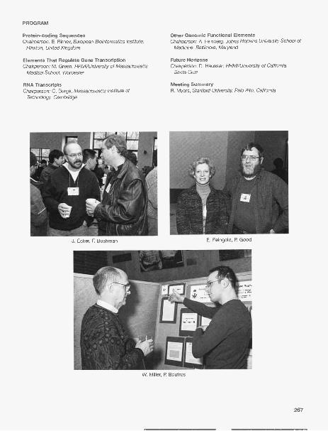
PROGRAM
Protein-coding SequencesChairperson: E. Birney, European Bioinformatics Institute,
Hinxton, United Kingdom
Elements That Regulate Gene TranscriptionChairperson: M. Green, HHMI/University of Massachusetts
Medical School, Worcester
RNA TranscriptsChairperson: C. Burge, Massachusetts Institute of
Technology, Cambridge
Other Genomic Functional ElementsChairperson: A. Feinberg, Johns Hopkins University School of
Medicine, Baltimore, Maryland
Future HorizonsChairperson: D. Haussler, HHMI/University of California,
Santa Cruz
Meeting SummaryR. Myers, Stanford University Palo Alto, Califomia
J. Ecker, F. Bushman E. Feingold, P. Good
W. Miller, P. Boutros
267

Pharmacogenomics
November 18-21 243 participants
ARRANGED BY David Bentley, The Wellcome Trust Sanger InstituteJ. Steven Leeder, Children's Mercy HospitalMunir Pirmohamed, University of LiverpoolRichard M. Weinshilboum, Mayo Medical School
This second annual meeting was a joint project of CSHL and the Wellcome Trust and was funded in part by theNational Institute of General Medical Sciences, a branch of the National Institutes of Health. The primary objec-tive of the meeting was to stimulate interest from a broadly based constituency representing basic scientists andclinical investigators from both academic and corporate environments by focusing on diverse issues related to the
application of genomic approaches to investigations of variability in drug response in humans. The meetingopened with a session presenting pharmacogenomics from the perspectives of the pharma-ceutical industry and academia, as well as an example of the potential promise of personal-ized medicine. Major subject areas for the meeting included pharmacogenomics of drug bio-transformation and transport, cancer pharmacogenomics, cardiopulmonary pharmacoge-nomics, and neuropsychiatric pharmacogenomics. Presentations highlighting economic andsocial issues related to the impact of pharmacogenomics on clinical practice stimulated con-siderable audience participation, and issues related to special populations, such as childrenand ethnic minorities, were addressed in a panel discussion. Perusal of the meeting content,both oral and poster presentations, revealed a movement of experimental approaches awayfrom a focus on single-nucleotide polymorphisms in single genes to more broadly basedstrategies involving haplotypes of multiple genes and gene pathways.
The meeting was attended by 243 registrants from 18 countries, compared to 140attendees from 15 countries at the previous meeting. Women represented 35% of atten-dees, whereas Ph.D. students and postdoctoral fellows constituted 23%. Feedback from the meeting hasbeen positive and future efforts will directed toward two main goals: (1) increasing representation at the meet-ing from the genetics and genomics community, and (2) providing more opportunities for trainees and juniorfaculty members to present their work in oral platform sessions. This meeting provides an outstanding forumto explore the impact of genomics on the treatment of human disease to describe the science underlyingpharmacogenomics and to discuss the accompanying benefits and challenges relevant to the implementa-tion of pharmacogenomics in both drug development and clinical settings.
S. Leeder
268 A. Marianki, S. Koukouritaki

This meeting was funded in part by the National Institutes of General Medical Sciences, a branch of theNational Institutes of Health.
PROGRAM
Opening SessionChairpersons: R. Weinshilboum, Mayo Medical School,
Rochester, Minnesota; R. Long, National Institutes ofHealth/NIGMS, Bethesda, Maryland
Pharmacogenomics of Metabolism and TransportChairpersons: K. Giacomini, University of California, San
Francisco; D. Flockhart, Indiana University School ofMedicine, Indianapolis
Cancer PharmacogenomicsChairpersons: C.R. Wolf, University of Dundee, United
Kingdom; M. Ratain, University of Chicago, Illinois
Cardiopulmonary PharmacogenomicsChairpersons: A. Rane, Karolinska Institutet, Stockholm,
Sweden; J. Johnson, University of Florida, Gainesville
RemarksJ.D. Watson, Cold Spring Harbor Laboratory
Neuropsychiatric PharmacogenomicsChairpersons: J.S. Leeder, Children's Mercy Hospital, Kansas
City Missouri; M. Pirmohamed, University of Liverpool,United Kingdom
Panel Session: Special Populations andPharmacogenomics, Future Directions
Chairpersons: A. Breckenridge, Medicines and HealthcareProducts Regulatory Agency London, United Kingdom; D.Bentley, Wellcome Trust Sanger Institute, Hinxton, UnitedKingdom
K. Philips, C. Stephens, C. Ross
A. Daly, A. Alfirevic, J. Foster269

CSHL Winter Biotechnology Conference: Drug Discovery inNeurodegenerative Diseases
December 2-5 122 participants
ARRANGED BY Sam Gandy, Farber Institute for Neurosciences at Thomas Jefferson UniversityVirginia M.-Y. Lee, University of Pennsylvania School of MedicineMarcy MacDonald, Massachusetts General Hospital/Harvard Medical School
As many as one half of people aged 65 years or older will develop debilitating degenerative disease of thecentral nervous system, usually characterized by a decade or more of dependent living, accompanied by pro-gressive failure of cognitive function and/or coordinated movement. Although these illnesses appear mostcommonly in the absence of obvious heredibility or identifiable genetic mutations, it has been possible dur-ing the past 20 years to discover risk-modifying DNA changes in some examples and predictable causativechanges in others. From these findings, transgenic technologyhas rapidly led to the development of mouse, fruit fly, andnematode model systems that partially recapitulate the clinicalabnormalities of the human diseases, as well as some of thehallmark molecular and morphological pathology of the condi-tions.
Rational biochemical and cell-based screens have generat-ed lead compounds that show promise in the living animalmodels. Most importantly, the animal models have enabled dis-covery of entirely unanticipated therapeutic strategies (such asamyloid-13 immunotherapy). In December 1999, some of these
rationally discovered compounds and unexpectedimmunotherapies entered Phase I clinical trials. The progress ofcompounds and rational strategies from the animal model tothe human clinical trial; the design, results, and conclusions oftrials; and the return to the animal model with questions raisedduring human trials are the areas of particular emphasis forthese biannual Winter Biotechnology Meetings instituted inDecember 2000 with the explicit goal of facilitating the transla-tion of "breakthrough" science into effective medicines.
For the 2004 meeting, seven 3- or 4-hour platform sessions were organized around common technolog-ical themes. Chairpersons, invited speakers, and speakers selected from submitted abstracts were drawnfrom the academic and pharma sectors. Although the discussion of new, unpublished data was emphasized,the group was tolerant of a wide range of intellectual property conventions. Poster presentations were alsoencouraged: Posters were displayed for an extended period during the meeting, and poster viewing wasespecially encouraged during the cocktail hours before the evening meals.
Topics included protein aggregation inhibitors, protein processing enzymology and pharmacology,immunotherapy, design and interpretation of genomic analyses in mammalian and lower systems, bioinfor-matics, mouse, nematode, and fly models of neurological disease, mechanisms of neurodegeneration, meta-bolic and hormonal influences on disease protein metabolism in cell culture and living animal models, as wellas in human clinical trials, and the importance of applying high-throughput screening strategies to academicefforts ("chemical genomics"), as well as its typical use in drug discovery. Diseases considered includedAlzheimer's, Huntington's, spinocerebellar atrophies, ALS, prion diseases, Parkinson's, tauopathies, and
synucleinopathies.
I. Gozes, V.M.-Y. Lee
270

PROGRAM
Pharmacology of ProteolysisChairpersons: S. Gandy, Thomas Jefferson University
Philadelphia, Pennsylvania; M. MacDonald, MassachusettsGeneral Hospital. Charlestown
The Way Forward: NIH Roadmap and Innovative HTSStrategies
Chairperson: V. Man-Yee Lee, University of Pennsylvania,Philadelphia
Intracellular Aggregation IChairperson: M. MacDonald, Massachusetts General
Hospital, Charlestown
Intracellular Aggregation IIChairperson: V Man-Yee Lee, University of Pennsylvania,
Philadelphia
Intracellular Aggregation IIIChairperson: M. MacDonald, Massachusetts General Hospital,
Charlestown
Extracellular Aggregates IChairperson: S. Gandy, Thomas Jefferson University
Philadelphia, Pennsylvania
Extracellular Aggregates IIChairperson: S. Gandy, Thomas Jefferson University
Philadelphia, Pennsylvania
Intracellular Aggregation IVChairpersons: M. MacDonald, Massachusetts General
Hospital, Charlestown; V Man-Yee Lee, University ofPennsylvania, Philadelphia
M. Sekuma, C. Mizumarau, T. Suzuki, K. lijima-Ando
271

Plant Genomes: From Sequence to Phenome
December 9-12 115 participants
ARRANGED BY Gloria Coruzzi, New York UniversityMichael Snyder, Yale UniversitySusan Mc Couch, Cornell University
This meeting featured 37 talks in 6 sessions, a poster session, and a workshop on Resources and NewTechnology. The theme was to bring plant genomics to the next level beyond sequencing, to discover genefunction through phenomic approaches. A new organizational feature of the plant genome meeting was tostimulate cross-talk between plant genomics andgenomics in other model organisms. To accomplish this,genome speakers from nonplant organisms were invited tospeak in each session including yeast, Caenorhabditis ele-gans, and Chlamydomonas. Two keynote speakersopened the meeting: Claire Fraser (President of TheInstitute for Genomic Research) surveyed the exponentialgrowth of sequence information from the more than 1200genome sequencing projects under way, noting that novel,strain-specific genes were still being discovered with everynew organism to be sequenced. She noted that the mainlimitation to linking sequence with phenotype had to dowith deficiencies in annotation and in understanding therole of genes with products of unknown function. JoeEcker presented life's complexity pyramid and outlined theeffort to identify and name all the "parts" and to under-stand how they all work together in the context of a whole,living organism. The talks were selected from abstractsand covered progress in sessions on regulatory networks
S. Mc Couch, Z. Lippman
and gene expression, proteomics, phenomics, genome organization and evolution, new genomes and ele-ments, and genomics approaches to small molecules. Speakers looked at cell-to-cell communication in theapical meristems of shoots and roots, at regulatory pathways and protein interaction networks in yeast, atclusters of genes associated with specific phenotypes in worms (phenoclusters), at MADS box genes andtheir relationship to petal formation, and at proteins that are required to make flagellar basal bodies inChlamydomonas and their relationship to human disease.Others presented strategies for characterizing the pro-teome, for classifying promoters, for sequencing themaize genome, for conducting array-based genotyping,and for exploring the evolution of new cell types. Therewas the announcement of the new JGI effort to sequenceSelaginella, an early, non-seed-bearing land plant that hasno true leaves and represents a key node in plant evolu-tion. Speakers provided insights into the abundance andfunction of small RNAs in plants, into systems for virus-induced gene silencing, and into endomembrane traffick-ing. The final keynote address by Marc Johnston offereda view of what is known about protein-protein interactionsin Saccharomyces cerevisiae, with the goal of "complet-ing yeast as an organism by 2007." The genomics work-shop highlighted areas of plant genomics that are neces-sary for the plant community to "catch up" with the
272
R. Martienssen, M. Snyder

genomic resources in yeast and C. elegans, including ORFeome, proteomics, and DNA-protein interac-tions. This wish list was inspired by the ideas that had been exchanged at this small but provocative con-ference.
PROGRAM
Keynote AddressesC. Fraser, The Institute for Genomic Research, Bethesda,
Maryland; J. Ecker, The Salk Institute for Biological Studies,La Jolla, California
Regulatory Networks and Gene ExpressionChairperson: E. Meyerowitz, California Institute of Technology,
Pasadena
ProteomicsChairperson: B. Chait, The Rockefeller University New York
PhenomicsChairperson: C. Boone, University of Toronto, Canada
Genome Organization and EvolutionChairperson: S. Dutcher, Washington University St. Louis,
Missouri
New Genomes and ElementsChairperson: D. Weigel, Max-Planck Institute for
Developmental Biology Tubingen, Germany
Workshop: Resources and New TechnologyChairpersons: J. Ecker, Salk Institute for Biological Studies,
La Jolla, California; D. Preuss, University of Chicago,Illinois; M. Snyder, Yale University New Haven, Connecticut
Keynote SpeakerChairperson: M. Johnston, Washington University School of
Medicine, St. Louis, Missouri
Genomics Approaches to Small MoleculesChairperson: N. Raikhel, University of California, Riverside
G. Coruzzi, R. Flavell
Q. Dong, S. Gopalan
273


POSTGRADUATE COURSES
The Postgraduate Courses program at Cold Spring Harbor Laboratory is aimed at meeting the specialneed for training in interdisciplinary subjects that are either so new or so specialized that universitiesdo not adequately treat them. Our aim is to provide intensive study in the most recent developmentsand techniques in these subjects and to prepare students to enter directly into research in a particulararea. To ensure up-to-date coverage of current research work, we bring together course faculty frommany laboratories around the world and supplement this staff with a series of seminar speakers.
Protein Purification and CharacterizationApril 14-27
INSTRUCTORS
ASSISTANTS
R. Burgess, University of Wisconsin, MadisonA. Courey, University of California, Los AngelesS.-H. Lin, M.D. Anderson Cancer Center/University of Texas, HoustonK. Severinov, Waksman Institute, Rutgers University, Piscataway, New Jersey
K. Adelman, Cornell University, Ithaca, New YorkV. Bergendahi, University of Wisconsin, MadisonH. Duong, University of California, Los AngelesN. Thompson, University of Wisconsin, Madison0. Tsai, M.D. Anderson Cancer Center/University of Texas,
Houston
W.-W. Tsai, M.D. Anderson Cancer Center/University ofTexas, Houston
C. Winkler, University of California, Los AngelesN. Zenkin, Rutgers University, New Brunswick, New Jersey
275

This course was for scientists who were not familiar with techniques of protein isolation and charac-terization. It was a rigorous program that included laboratory work all day and a lecture with a discus-sion session every evening. Each student became familiar with each of the major techniques in proteinpurification by actually performing four separate isolations, including (1) a regulatory protein from mus-cle tissue, (2) a sequence-specific DNA-binding protein, (3) a recombinant protein overexpressed in E.coli, and (4) a membrane-bound receptor. A variety of bulk fractionation, electrophoretic, and chro-matographic techniques included precipitation by salts, pH, and ionic polymers; ion-exchange, gel fil-tration, hydrophobic interaction, and reversed-phase chromatography; lectin affinity, ligand affinity,oligonucleotide affinity, and immunoaffinity chromatography; polyacrylamide gel electrophoresis andelectroblotting; and high-performance liquid chromatography. Procedures were presented for solubiliz-ing proteins from inclusion bodies and refolding them into active monomeric forms. Methods of proteincharacterization were utilized to include immunological and biochemical assays, peptide mapping,amino acid analysis, protein sequencing, and mass spectrometry. Emphasis was placed on strategiesof protein purification and characterization. Guest lecturers discussed protein structure, modification ofproteins, methodologies for protein purification and characterization, and applications of protein bio-chemistry to cell and molecular biology. This course was supported with funds provided by the NationalCancer Institute.
PARTICIPANTS
Chandra, D., Ph.D., M.D. Anderson Cancer Center/Universityof Texas, Smithville
Conlon, F, Ph.D., University of North Carolina, Chapel HillDemirel, M., Ph.D., Penn State University, University ParkEl ling, A., B.S., Iowa State University, AmesFeldman, M., B.S., Tel-Aviv University, IsraelGibson, K., Ph.D., Massachusetts Institute of Technology,
CambridgeGovindarajan, V., Ph.D., Baylor College of Medicine, Houston,
TexasKettunen, P, Ph.D., University of Bergen, Norway
SEMINARS
Bergendahl, V, University of Wisconsin, Madison: Use ofLRET-based assays for drug discovery and biochemistry;RNA polymerase-sigma factor interactions.
Burgess, R., University of Wisconsin, Madison: Introduction toprotein purification and immunoaffinity chromatography.
Courey, A., University of California, Los Angeles: Sumoylationand the response to stress.
Hart, G., Johns Hopkins University, Baltimore, Maryland:Protein glycosylation: The most ubiquitous posttranslationalevent in eukaryotes.
Joshua-Tor, L., Cold Spring Harbor Laboratory: Protein crys-tallography intro-The PAZ domain of Argonaute-2: siRNAat RISC.
276
Lipardi, C., Ph.D., National Institutes of Health/NCI, Bethesda,Maryland
Makarevitch, I., M.S., University of Minnesota, St. PaulMarcand, S., Ph.D., CEA/Saclay, Gif-sur-Yvette, FranceNorville, J., B.S., Massachusetts Institute of Technology,
CambridgePhan, A.T., Ph.D., Memorial Sloan-Kettering Cancer Center,
New YorkSformo, T, M.S., University of Alaska, FairbanksTavanez, J., B.S., Hospital Santa Maria, Lisbon, PortugalTudor, H., Ph.D., Hospital for Special Surgery, New York
Lin, S.-H., M.D. Anderson Cancer Center, Houston, Texas:Novel approach to understanding prostate cancer bonemetastasis.
Mische, S., Boehringer-lngelheim, Ridgefield, Connecticut:Protein purification and characterization for drug discovery.
Muir, T, The Rockefeller University, New York: Cutting andpasting proteins: What's happening with inteins.
Severinov, K., Rutgers University, New Brunswick, NewJersey: Role of RNA polymerase core enzyme in transcrip-tion initiation.
Stillman, B., Cold Spring Harbor Laboratory: The origin recog-nition complex and the chromosome inheritance cycle.

Cell and Developmental Biology of Xenopus
April 16-27
INSTRUCTORS K. Cho, University of California, IrvineJ. Christian, Oregon Health and Science University, Portland
ASSISTANTS I. Blitz, University of California, IrvineG. Dalgin, Oregon Health and Science University, PortlandD. Goldman, Oregon Health and Science University, PortlandT. Nakayama, University of Virginia, Charlottesville
The frog Xenopus is an important vertebrate model for studies of maternal factors, regulation and mol-ecular mechanisms of tissue inductions, and regulation of cell fate decisions. In addition, Xenopusoocytes and embryos provide a powerful system in which to conduct a number of cell biological andgene regulation assays. This course provided extensive laboratory exposure to the biology, manipula-tion, and use of oocytes and embryos of Xenopus. The course consisted of intensive laboratory ses-sions, supplemented by daily lectures and demonstrations from experts in cellular, experimental, andmolecular development. Areas covered included (1) care of adults; (2) oocyte isolation and embryo pro-duction; (3) stages of embryonic development and anatomy; (4) whole-mount in situ hybridization andimmunocytochemistry; (5) microinjection of eggs and oocytes with lineage tracers, DNA constructs,mRNA, and antisense oligonucleotides; (6) micromanipulation of embryos, including explant and trans-plantation assays; (7) in vivo time-lapse confocal imaging; (8) preparation of transgenic embryos; and(9) use of Xenopus tropicar/s for genetic analyses. This course was suited both for investigators who
277

have had no experience with Xenopus and for those who have worked with Xenopus and wish to learnnew and cutting-edge techniques. All applicants had current training in molecular biology and someknowledge of developmental biology. Guest lecturers included Amaya Enrique, Ken Cho, John Gurdon,Janet Heasman, Ray Keller, and Mark Mercola.
PARTICIPANTS
Boulware, M., Ph.D., University of Minnesota, MinneapolisBurns, M., Ph.D., University of Virginia, CharlottesvilleChen, Y., Ph.D., Medical University of South Carolina,
CharlestonHandrigan, G., B.S., Dalhousie University, Halifax, Nova
Scotia, CanadaHeine, D., M.S., University of Aberdeen, Scotland, United
KingdomIslam, A., M.Phil., Tottori University, Yonago, JapanKieckens, L., B.S., Katholieke Universiteit Leuven, BelgiumMarinescu, V., Ph.D., Children's Hospital Boston,
Massachusetts
SEMINARS
Amaya, E., University of Cambridge, United Kingdom:Functional genomics in Xenopus.
Cho, K., University of California, Irvine: Genomic approach tostudying BMP signaling.
Gurdon, J., Wellcome Trust, Cambridge, United Kingdom:Nuclear reprogramming.
Heasman, J., Children's Hospital Medical Center, Cincinnati,Ohio: Antisense technology to study maternal gene products.
Keller, R., University of Virginia, Charlottesville: The cell move-
278
Nichane, M., M.S., Free University of Brussels, BelgiumNy, A., Ph.D., Katholieke Universiteit Leuven, BelgiumParker, B., M.S., Edison Biotechnology Institute/Ohio
University, AthensRangarajan, J., M.S., National Institutes of Health, Bethesda,
MarylandRouhana, L., Ph.D., University of Wisconsin, MadisonVanamala, S., M.V.S., Institut de Recherches Cliniques de
Montreal, Quebec, CanadaWang, B., Ph.D., Weill Medical College of Cornell University,
New YorkWare, S., Ph.D., Baylor College of Medicine, Houston, Texas
ments, patterning, and biomechanical gastrulation inamphibians.
Mead, P. St. Jude Children's Research Hospital, Memphis,Tennessee: Insertional mutagenesis using transposons.
Mercola, M., Burnham Institute, San Diego, California: Heartinduction.
Niehrs, C., Deutsches Krebsforschungszentrum, Heidelberg,Germany: Large-scale analysis of embryonic gene expres-sion in Xenopus.

Genetics of Complex Human Diseases
June 9-15
INSTRUCTORS A. Al-Chalabi, FRCP, Institute of Psychiatry, Kings College, London, United KingdomL. Almasy, Southwest Foundation for Biomedical Research, San Antonio, Texas
Complex diseases are conditions that are influenced by the actions of multiple genes, their interactionswith each other and with the environment. This lecture course considered the difficulties in studying thegenetic basis of complex disorders such as diabetes, cardiovascular disease, cancer, Alzheimer's dis-ease, schizophrenia, and epilepsy. We discussed genetic-epidemiologic study designs, including fam-ily, twin, case/control, and adoption studies, as well as methods for quantifying the strength of thegenetic influences on a disease. A major focus was the identification of specific gene effects using bothlinkage and association analyses and their variants. We discussed the efficiency and robustness of dif-ferent designs for such analysis and how evidence from epidemiologic studies informs both the designand interpretation of molecular genetic studies. Study design and methods for analysis of quantitativerisk factors related to complex diseases were covered, as well as the latest ideas in data reduction such
as haplotype mapping and SNP tagging. An overview of high-throughput laboratory methods wasgiven to provide participants with insight into the applications of these techniques. Illustrations wereprovided through discussion of results from ongoing studies of a variety complex diseases and related
risk factors.
279

PARTICIPANTS
Barreto, M., B.S., Instituto Gulbenkian de Ciencia, Oeiras,Portugal
Body, S., M.D., Brigham & Women's Hospital, Boston,Massachusetts
Buervenich, S., Ph.D., National Institutes of Mental Health,Bethesda, Maryland
Clarimon, J., Ph.D., National Institutes of Health, Rockville,Maryland
Congdon, E., M.A., Stony Brook University, New YorkDavuluri, R., Ph.D., Ohio State University, ColumbusDe Jager, P., Ph.D., Harvard Medical School, Boston,
MassachusettsDuga, S., B.S., University of Milan, ItalyFerguson, B., Ph.D., Oregon National Primate Research
Center, BeavertonHe, Q., B.S., Michigan State University, East Lansing
SEMINARS
Al-Chalabi, A., Institute of Psychiatry, Kings College, London,United Kingdom: An introduction to genetic linkage.
Almasy, L., Southwest Foundation for Biomedical Research,San Antonio, Texas: An introduction to variance compo-nents.
Altshuler, D., Massachusetts General Hospital, HarvardMedical School, Boston: The International Hap Map Project.
Blangero, J., Southwest Foundation for BiomedicalResearch, San Antonio, Texas: From QTL to gene.
Borecki, I., Washington University School of Medicine, St.Louis, Missouri: Study designs.
Edenberg, H., Indiana University, Indianapolis: High-through-
280
Morley, M., M.S., Children's Hospital of Philadelphia,Pennsylvania
Rasmussen, E., Ph.D., Roche Pharmaceuticals, Nutley, NewJersey
Rosen, M., M.D., University of California, San FranciscoSchlesinger, D., M.D., University of Sao Paulo, BrazilSimen, A., Ph.D., Yale University, New Haven, ConnecticutStambolian, D., Ph.D., University of Pennsylvania,
PhiladelphiaTraynor, B., M.D., Massachusetts General Hospital,
CharlestownTukel, T., Ph.D., Mt. Sinai School of Medicine, New YorkViel, K., B.S., Emory University, Atlanta, GeorgiaWanke, K., Ph.D., National Cancer Institute, Bethesda,
Maryland
put laboratory methods.Goldstein, D., University College London, United Kingdom:
Data reduction, SNP tagging, and pharmacogenomics.Knight, J., Institute of Psychiatry, London, United Kingdom:
Association studies; TDT-based methods.Krainer, A., Cold Spring Harbor Laboratory: Genetic mecha-
nisms in complex diseases.Merikangas, K., National Institute of Mental Health, Bethesda,
Maryland: Genetic epidemiological approaches to complexdisorders.
Ottman, R., Columbia University School of Public Health,New York: An introduction to complex genetics.

Advanced Bacterial Genetics
June 9-29
INSTRUCTORS K. Hughes, University of Washington, SeattleU. Jena!, University of Basel, SwitzerlandK. Pogliano, University of California, San Diego, La Jolla
ASSISTANTS D. Broder, University of California, San Diego, La JollaB. Christen, University of Basel, SwitzerlandC. Wozniak, University of Washington, Seattle
The course presented logic and methods used in the genetic dissection of complex biological process-es in eubacteria. Laboratory methods included classical mutagenesis using transposons, mutatorstrains, and chemical and physical mutagens; the mapping of mutations using genetic and physicaltechniques; modern approaches to the generation and analysis of targeted gene disruptions andfusions using PCR and cloning methods; epitope insertion mutagenesis; and site-directed mutagene-sis. Key components of the course were use of sophisticated genetic methods in the analysis of model
281

eubacteria and the use of the wealth of new genomic sequence information to motivate these meth-ods. Invited lecturers presented various genetic approaches to study eubacterial mechanisms ofmetabolism, development, and pathogenesis. This course was supported with funds provided by theNational Science Foundation.
PARTICIPANTS
Black, S., B.S., University of Aberdeen, United KingdomBjornsdottir, S., B.S., University of Iceland, ReykjavikChevance, F., Ph.D., University of Konstanz, GermanyColangeli, R., Ph.D., University of Medical & Dentistry of New
Jersey, NewarkDrummond, D.A., B.S.E., California Institute of Technology,
PasadenaFrank, K., B.S., Mayo Clinic, Rochester, MinnesotaGescher, J., Diplom., University of Freiburg, GermanyHuang, K. C., B.S., Massachusetts Institute of Technology,
Cambridge
SEMINARS
Adhya, S., National Institutes of Health/NCI, Bethesda,Maryland: Structure, assembly, and dynamics of a generegulatory nucleoprotein complex containing a DNA loop.
Brun, Y., Indiana University, Bloomington: Generation andmaintenance of cellular asymmetry in Caulobacter.
Errington, J., University of Oxford, United Kingdom: HowBacillus subtilis finds the mid-cell site for division andavoids bisecting its chromosome.
Gross, C., University of San Francisco, California: Sensingand responding to envelope stress.
Harshey, R., University of Texas, Austin: Surface sensing:Swarming motility in S. typhimurium.
282
Klausen, M., M.S., Technical University of DenmarkKonjufca, V., Ph.D., Washington University, St. Louis, MissouriRaffatellu, M., M.D., Texas A&M University, College StationSchneeweiss, J., B.S., University of Oxford, United KingdomShanks, R., Ph.D., Dartmouth Medical School, Hanover,
New HampshireThompson, K., B.S., National Cancer Institute, Bethesda,
MarylandTurner, S., Ph.D., University of Auckland, New ZealandWolf, D., Ph.D., Lawrence Berkeley National Labs., Berkeley,
California
Maloy, S., San Diego State University, California: Geneticbasis of host specificity in Salmonella.
Nystrom, T, Goteborg University, Sweden: RpoS regulationin stationary phase.
Otteman, K., University of California, Santa Cruz: Swimmingin the stomach: How chemotaxis helps Helicobacter pyloriinfect.
Taylor, R., Dartmouth Medical School, Hanover, NewHampshire: The role of pill in Vibrio cholerae infection.
Weinstock, G., Baylor College of Medicine, Houston, Texas:From genetics to genomics.

Ion Channel Physiology
June 9-29
INSTRUCTORS M. Farrant, University College London, United KingdomM. Hausser, University College London, United KingdomN. Spruston, Northwestern University, Evanston, Illinois
ASSISTANTS B. Clark, University College London, United KingdomT. Jarsky, Northwestern University, Chicago, IllinoisB. Steil, University of California, Los Angeles
The primary goal of this course was to investigate, through lectures and laboratory work, the proper-ties of ion channels that allow neurons to carry out their unique physiological functions in a variety ofneural systems. Areas of particular interest included channels that (1) are activated by neurotransmit-ter at central and peripheral synapses, (2) are activated by voltage changes in axons and dendrites, (3)respond to neuromodulators with changes in functional properties, or (4) are developmentally requiredand regulated. The research interests of guest lecturers reflected these areas of emphasis.
The laboratory component of the course introduced students to electrophysiological approaches forthe study of ion channels in their native environments. Hands-on exercises included patch-clamprecording of ion channel activity in acutely isolated or cultured cells or neurons in brain slice prepara-tions. Different recording configurations were used (e.g., whole-cell, cell-free, and nucleated patches)
283
to examine macroscopic or single-channel activity. Similarly, various methods of ligand and drug appli-
cation were demonstrated. The advantages and disadvantages of each method, preparation, and
recording technique were considered in relation to the specific scientific questions being asked.
PARTICIPANTS
Barker, L., BA., University of California, San FranciscoBuonanno, A., Ph.D., National Institutes of Health, Bethesda,
MarylandCooke, B., Ph.D., Northwestern University, Evanston, IllinoisFisher, T., B.S., Wake Forest University, Winston-Salem,
North CarolinaJocoy, E., B.S., University of California, Los AngelesLuu, P., Ph.D., University of California, Berkeley
SEMINARS
Bean, B., Harvard Medical School, Boston, Massachusetts:Channel integration and firing.
Diamond, J., National Institutes of Health, Bethesda,Maryland: Glutamate EPSCs and uptake.
Fitzsimonds, R., Yale University School of Medicine, NewHaven, Connecticut: Synaptic plasticity during develop-
ment.Jonas, P., Albert-Ludwigs-Universitat, Freiburg, Germany:
Cortical microcircuits.Larkum, M., University of Bern, Switzerland: Dendritic inte-
gration.Llinas, R., New York University School of Medicine, New
York: Ion channels and integration.Nerbonne, J., Washington University School of Medicine, St.
Louis, Missouri: Voltage-gated K channels in heart and neu-
rons.
284
Nair, A., Ph.D., University of Cambridge, United KingdomNorris, A., B.A., Washington University, St. Louis, MissouriNuriya, M., B.S., Johns Hopkins University, Baltimore,
MarylandPetreanu, L., Ph.D., Cold Spring Harbor LaboratorySandberg, M., Ph.D., Karolinska Institutet, Stockholm,
SwedenSarah, P., B.S., University of Bern, Switzerland
Nicoll, R., University of California, San Francisco:Cannabinoids and synaptic plasticity.
Nusser, Z., Institute of Experimental Medicine, Budapest:Hyperpolarization-activated cation channels.
Raman, I., Northwestern University, Evanston, Illinois: Action
potental, Hodgkin-Huxley.Raman, I., Northwestern University, Evanston, Illinois:
Voltage-gated Na channels.Scanziani, M., University of California, San Diego, La Jolla:
Synaptic plasticity.Soltesz, I., University of California, Irvine: GABA receptors
and inhibition.Svoboda, K., Cold Spring Harbor Laboratory: Imaging meth-
ods for studying ion channels.Takahashi, T, University of Tokyo Graduate School of
Medicine, Japan: Voltage-gated Ca channels.

Molecular Embryology of the Mouse
June 9-29
INSTRUCTORS T. Lufkin, Mt. Sinai School of Medicine, New YorkC. Stewart, National Cancer Institute, Frederick, Maryland
ASSISTANTS D. Escalante-Alcalde, Institute of Cellular Physiology-UNAM, MexicoJ. Liu, Mt. Sinai School of Medicine, New York
This intense laboratory and lecture course was designed for biologists interested in applying theirexpertise to the study of mouse embryonic development. Laboratory components provided an intro-duction into the technical aspects of working with and analyzing mouse embryos, and lectures pro-vided the conceptual basis for contemporary research in mouse development. Experimental tech-niques covered in the practicals included in vitro culture and manipulation of pre- and postimplantationembryos; transgenesis by DNA microinjection, embryo transfer, establishment, culture, and geneticmanipulation of embryonic stem cells; production of chimeras by aggregation with and injection ofembryonic stem cells; and the analysis of development by whole-mount in situ hybridization, skeletal
preparation, and transgene expression.
285

PARTICIPANTS
Attie, A., Ph.D., University of Wisconsin, MadisonBennett, V., Ph.D., West Virginia University School of
Medicine, MorgantownChal, J., M.S., Stowers Institute for Medical Research,
Kansas City, MissouriHerrera, E., Ph.D., Columbia University, New YorkJackerott, M., Ph.D., University of Copenhagen, DenmarkKatusic A., B.S., University of Zagreb, CroatiaKobayashi, Y., Ph.D., University of Iowa College of Medicine,
Iowa City
SEMINARS
Behringer, R., M.D. Anderson Cancer Center/University ofTexas, Houston: Gene targeting 101 and conditional genet-ic manipulation.
Bestor, T, Columbia College of Physicians & Surgeons, NewYork: Epigenetics and DNA methylation.
Brannstrom, M., The Sahlgrenska Academy, Goteborg,Sweden: Surgical transplantation of uteri.
Capel, B., Duke University Medical Center, Durham, NorthCarolina: Gonad organogenesis.
Duncan, S., Medical College of Wisconsin Neurobiology andAnatomy, Madison: Genetic regulation of organogenesis:Liver development.
Heard, E., Curie Institute, Paris, France: Dynamics of X-chro-mosome inactivation.
Jenkins, N., National Cancer Institute, Frederick, Maryland:Mouse genetics.
Jessell, T, Columbia University, New York: Neural develop-ment.
Joyner, A., New York University Medical Center, New York:Genetic approaches in mouse to study CNS patterning.
Ko, M., National Institute of Aging, Baltimore, Maryland: Genearrays and the analysis of mouse development.
Lewandoski, M., CDBL, National Cancer Institute, Frederick,Maryland: Genetic approaches to studying signaling in limbdevelopment.
Lovell-Badge, R., National Institute for Medical Research,London, United Kingdom: Mammalian sex determination.
Lufkin, T, Mount Sinai School of Medicine, New York:Homeobox gene control of embryonic patterning.
Magnuson, T, University of North Carolina, Chapel Hill:Genetic approaches to development.
McLaren, A., Wellcome/CRC Institute, Cambridge,Massachusetts: Germ cells.
Nagy, A., Mount Sinai Hospital/Samuel Lunenfeld ResearchInstitute, Ontario, Canada: ES cells and chimeras.
286
Mayer, A., Ph.D., Medical College of Wisconsin, MilwaukeeMurchison, E., B.S., Cold Spring Harbor LaboratoryMusacchio, M., Ph.D., University of California, IrvineOstman, N., M.S., Turku Center of Biotechnology, FinlandStauber, M., Ph.D., Cancer Research UK, London, United
KingdomWamstad, J., B.A., University of MinnesotaWuelling, M., Ph.D., Max-Planck Institute for Molecular
Genetics, Berlin, Germany
Overbeek, P., Baylor College of Medicine, Houston, Texas:Transposon-mediated mutagenesis.
O'Brien, T., Jackson Laboratory, Bar Harbor, Maine: Anenhanced GFP-based dual-color reporter to recover muta-tions in mice.
Perry, T, RIKEN Center for Developmental Biology, Kobe,Japan: Micromanipulation of embryos and for ICSI, trans-genesis, and nuclear transplantation.
Rosenthal, N., EMBL-Monterotondo, Rome, Italy: Heartdevelopment.
Rossant, J., Samuel Lunenfeld Research Institute, Ontario,Canada: Regulation of extraembryonic development.
Rowitch, D., Dana-Farber Cancer Institute, HarvardUniversity, Boston, Massachusetts: Ga14/Uas binary genesystem and conditional gene expression.
Sharpe, J., MRC Human Genetics Unit, Edinburgh,Scotland: OPT microscopy and the Edinburgh mouseatlas.
Shen, M., UMDNJ-Robert Wood Johnson Medical School,Piscataway, New Jersey: Left-right patterning.
Solter, D., Max-Planck Institute, Freiburg, Germany: Fromegg to blastocyst: Hard road to follow.
Stewart, C., National Cancer Institute, Frederick, Maryland:Early mouse development and implantation.
Stewart, A.E., Dresden University of Technology, Germany:Targeting strategies: Their design, construction, andintegration.
Tam, R, Children's Medical Research Institute, Wentworth-villa, Australia: Overview of mouse development and genet-ics. Gastrulation and morphogenesis: Building the bodyplan.
Threadgill, D., University of North Carolina, Raleigh-Durham:Quantitative traits and bioinformatics.
Yamaguchi, T, CDBL, National Cancer Institute, Frederick,Maryland: Wnt pathways in development.

Computational Neuroscience: Vision
June 18-July 1
INSTRUCTORS J. Demb, University of Michigan, Ann ArborP. Glimcher, New York University, New YorkE. Simoncelli, New York University, New York
ASSISTANTS B. Borghuis, Utrecht University, The NetherlandsN. Rust, New York University, New York
Computational approaches to neuroscience produced important advances in our understanding ofneural processing. Prominent success came in areas where strong inputs from neurobiological, behav-ioral, and computational investigation can interact. The theme of the course was that an understand-ing of the computational problems, the constraints on solutions to these problems, and the range ofpossible solutions helped guide research in neuroscience. Through a combination of lectures andhands-on experience in a computer laboratory, this intensive course examined color vision, spatial pat-tern analysis, motion analysis, oculomotor function, attention, and decision-making. Lecturers this yearincluded Edward Adelson, David Brainard, Matteo Carandini, Eduardo Chichilnisky, Kathleen Cullen,Bruce Cumming, James Di Carlo, David Heeger, Daniel Kersten, J. Anthony Movshon, PamelaReinagel, Fred Rieke, Ruth Rosenholtz, Michael Shad len, Stefan Treue, and Preeti Verghese. The
287

course was held at the Laboratory's Banbury Conference Center located on the north shore of Long
Island. All participants stay within walking distance of the Center, close to the tennis court, pool, and
private beach.
PARTICIPANTS
Amstrong, K., B.S.E., Stanford University, CaliforniaBasole, A., B.S., Duke University Medical Center, Durham,
North CarolinaBonin, V., M.S., The Smith-Kettlewell Eye Research Institute,
San Francisco, CaliforniaChallinor, K., B.Psych., Macquarie University, Sydney,
AustraliaCohen, M., B.S., Stanford University, CaliforniaDoerschner, K., BA., New York University, New YorkDunn, F., B.S., University of Washington, SeattleField, G., B.S., University of Washington, SeattleHanks, T., B.S., University of Washington, SeattleLarsson, J., Ph.D., New York University, New YorkListon, D., B.S., Salk Institute, La Jolla, CaliforniaMaciokas, J., Ph.D., University of California, Davis
SEMINARS
Brainard, D., University of Pennsylvania, Philadelphia;Adelson, E., Massachusetts Institute of Technology,Cambridge: Constancy/adaptation; Context; Trichromacy;Elements of early vision.
Carandini, M., The Smith-Kettlewell Eye Research Institute,San Francisco, California; Simoncelli, E., New YorkUniversity, New York: V1 physiology II; Image statistics II:
Sparseness/ICA/nonlinearities.Chichilnisky, E.J., The Salk Institute, La Jolla, California:
Linear systems; Linearity in the retina.Chichilnisky, E.J., The Salk Institute, La Jolla, California;
Demb, J., University of Michigan, Ann Arbor: Retinal gaglioncell encoding; RGC nonlinearities.
Chichilnisky, E.J., The Salk Institute, La Jolla, California;Brainard, D., University of Pennsylvania, Philadelphia: RGCpopulation code; Linear algebra.
Cullen, K., McGill University, Montreal, Canada: Eye move-ment.
Cumming, B., National Eye Institute/NIH, Bethesda,Maryland: Binocular stereo: Perception and physiology.
Demb, J., University of Michigan, Ann Arbor; Glimcher, P.,New York University, New York; Simoncelli, E., New YorkUniversity, New York: Course themes: Tuning/population/adaptation/estimation.
Demb, J., University of Michigan, Ann Arbor; Chichilnisky,
288
Macuga, K., B.S., University of Califonria, Santa BarbaraMalone, B., Ph.D., University of California, Los AngelesMitchell, J., Ph.D., The Salk Institute, La Jolla, CaliforniaNaito, T., Ph.D., Osaka University, JapanNienborg, H., M.D., National Eye Institute/NIH, Bethesda,
MarylandPieper, F., Diplom., German Primate Center, Gottingen,
GermanySather, B., Ph.D., University of Pittsburgh, PennsylvaniaSchoppik, D., B.A., University of California, San FranciscoSundberg, K., B.S., The Salk Institute, La Jolla, CaliforniaTailby, C., Ph.D., New York University, New YorkYin, L., B.S., University of Pennsylvania, PhiladelphiaZemach, I., M.S., University of Washington, Seattle
E.J., The Salk Institute, La Jolla, California: RGC nonlinearmechanisms; RGC contrast adaptation.
Di Carlo, J., HHMI/Baylor College of Medicine, Houston,Texas: Object/shape physiology.
Glimcher, P., New York University, New York: Decision/games.Heeger, D., Stanford University, California: Human Vl/fMRI.Kersten, D., Max-Planck Institute, Tubingen, Germany:
Object/ shape perception.Movshon, J.A., New York University, New York: MT physiology.Reinagel, P., University of California, San Diego, La Jolla;
Simoncelli, E., New York University, New York: LGN; ImageStatistics I. PCA/SpectralModels.
Reinagel, P., University of California, San Diego, La Jolla;Carandini, M., The Smith-Kettlewell Eye Research Institute,San Francisco, California, Efficient coding; V1 Physiology I.
Rieke, F, University of Washington, Seattle: Signal detectiontheory; Rods/photos; Entropy/information; Spikes.
Rosenholtz, R., NASA Ames, Moffett Field, California: Patternperception/texture.
Shad len, M., University of Washington, Seattle: Motion per-ception/decisions.
Treue, S., German Primate Center, Goettingen, Germany:Attention physiology.
Verghese, P., The Smith-Kettlewell Eye Research Institute,San Francisco, California: Attention psychophysics.

DNA Microarray Applications
June 22-29
INSTRUCTORS V. lyer, University of Texas, AustinA. Khodursky, University of Minnesota, St. PaulJ. Lieb, University of North Carolina, Chapel Hill
ASSISTANTS M. Buck, University of North Carolina, Chapel HillP. Killion, University of Texas, AustinJ. Kim, University of Texas, AustinP. Mieczkowski, University of North Carolina, Chapel Hill
DNA microarrays are simple, inexpensive, and versatile tools for experimental explorations of genomestructure, gene expression programs, gene function, and cell and organismal biology. In this hands-oneight-day course, students were introduced to the underlying technology, and then designed and car-ried out a number of experiments for analysis by DNA microarray hybridization, data analysis, display,and interpretation. Experimental applications covered in the course included systematic studies ofglobal gene expression programs, inferring gene function using microarrays, genome-wide DNA-pro-tein interactions, and measuring changes in gene copy number. Guest lecturers presented state-of-the-art new technology, experimental applications, and interpretation of large genomic data sets.Students who completed this course were equipped with the skills and know-how to introduce orapply DNA microarray technology to their own laboratories or institutions. This course was supportedwith funds provided by the National Cancer Institute.
289

PARTICIPANTS
Aguilera Alvarez, C., M.S., Catholic University of Chile,Santiago
Bak, M., M.S., University of Copenhagen, DenmarkButchko, R., B.S., United States Department of Agriculture,
Peoria, IllinoisChen, H., M.D., Johns Hopkins University School of
Medicine, Baltimore, MarylandEnmark, E., B.S., Karolinska Institutet, Huddinge, SwedenHubal, M., B.S., University of Massachusetts, AmherstMeza-Zepeda, L., Ph.D., The Norwegian Radium Hospital,
Oslo, NorwayMiller, K., Ph.D., Fisheries and Oceans, British Columbia,
CanadaNally, J., Ph.D., University of California, Los AngelesNielsen, K., Ph.D., Danish Institute for Food and Veterinary
Research, Copenhagen, Denmark
SEMINARS
Botstein, D., Princeton University, New Jersey: Functionalgenomics: Getting beyond descriptive studies.
Buck, M., University of North Carolina, Chapel Hill: Analysisof ChIP-ChiP data.
Churchill, G., The Jackson Laboratory, Bar Harbor, Maine:Statistical design and analysis of gene expressionmicroarray data.
Diaz, E., University of California, Berkeley: Using R formicroarray data analysis.
Khodursky, A., University of Minnesota, St. Paul: Advanceddata analysis and statistical considerations.
Killion, P., University of Texas, Austin: Submitting andretrieving data with the longhorn array database;Advanced features of the longhorn array database.
290
Orlando, E., Ph.D., St. Mary's College of Maryland, St.Mary's City
Rada, A., B.S., Uppsala University, SwedenSloane, M., B.S., Vienna University, AustriaSobko, A., Ph.D., IOGEN Corporation, Ontario, CanadaStraub, P, Ph.D., Richard Stockton College, Pomona, New
JerseySuarez-Castillo, E., B.S., University of Puerto Rico, San JuanSubramaniam, S., M.S., Centre for Cellular & Molecular
Biology, Hyberabad, IndiaTammoja, K., M.S., National Institutes for Health
Development, Tallinn, EstoniaWang, C. K., Ph.D., Medimmune Vaccines, Mountain View,
CaliforniaYu, M., Ph.D., Cold Spring Harbor Laboratory
Mieczkowski, P, University of North Carolina, Chapel Hill:Data analysis for aCGH experiments.
Murphy, C., University of California, San Francisco:Experimental design from a biologist's point of view.
Sherlock, G., Stanford University, California: Analysis andorganization of DNA microarray data.
Sherlock, G., Stanford University, California: Analysis andorganization of DNA microarray data II.
Verdnik, D., Axon Instruments, Foster City, California:Microarray scanning and quantitation with Genepix Pro;Submitting data to acuity; Advanced features of acuity.
Whitfield, M., Dartmouth Medical School, Stanford,California: Considerations for the design and analysis oftime-course experiments.

Advanced Techniques in Plant Science
July 1-21
INSTRUCTORS J. Bender, Johns Hopkins University School of Public Health, Baltimore, MarylandL. Hobbie, Ade 1phi University, Garden City, New YorkH. Ma, Penn State University, University ParkS. McCormick, Plant Gene Expression Center, Albany, CaliforniaR. Pruitt, Purdue University, W. Lafayette, Indiana
ASSISTANTS S. Knapke, Purdue University, W. Lafayette, IndianaW. Li, Penn State University, University ParkD. Love, Ade 1phi University, Garden City, New YorkP. Woo, Ade 1phi University, Garden City, New York
This course provided an intensive overview of topics in plant physiology and development, focusing onmolecular genetic approaches to understanding plant biology. It emphasized recent results fromArabidopsis thaliana and other plants and provided an introduction to current methods used in plantmolecular biology. It was designed for scientists with some experience in molecular techniques or inplant biology who wish to work with Arabidopsis and other plants using the latest technologies ingenetics and molecular biology. The course consisted of a vigorous lecture series, a hands-on labora-tory, and informal discussions. Discussions of important topics in plant research were presented by theinstructors and by invited speakers. These seminars included plant morphology and anatomy; plantdevelopment (development of flowers, leaves, male and female gametophytes, and embryos); percep-tion of light and photomorphogenesis; synthesis, function, and perception of hormones. Lectures
291
describing bioinformatics tools available to the plant community, and the resources provided by plantgenome projects, were also included. Speakers provided overviews of their fields, followed by in-depthdiscussions of their own work. The laboratory sessions provided an introduction to important tech-niques currently used in plant research. These included studies of plant development, meiosis, pollendevelopment, fern reproduction, plant cell-imaging techniques, laser-assisted tissue microdissection,mutant analysis, transient gene expression, applications of green fluorescent protein fusions, pro-teomics approaches, and techniques commonly used in genetic and physical mapping. The coursealso included several short workshops on important themes in genetics and a short workshop on sci-entific writing. This course was supported by the National Science Foundation.
PARTICIPANTS
Bernacki, S., North Carolina State University, RaleighBliss, B., M.S., Penn State University, University ParkBrown, L.-A., B.S., University of Leeds, United KingdomCosta, S., Lic., Guilbenkian Institute for Science, Oeiras,
PortugalJohansen, W., M.S., Hedmark University College, Hamar,
NorwayKaufmann, K., Diplom., FSU Jena, GermanyKlanrit, P., B.S., Purdue University, W. Lafayette, IndianaLevall, M., B.S., Syngenta Seeds AB, Landskrona, SwedenLevesque, E., B.A., The Arnold Arboretum of Harvard
SEMINARS
Banks, J., Purdue University, W. Lafayette, Indiana:Gametophytes and evolution.
Bender, J., Johns Hopkins University School of Public Health,Baltimore, Maryland: Epigenetics I.
Bowman, J., University of California, Davis: Leaves;Mutagenesis.
Briggs, W., Carnegie Institution of Washington, Stanford,California: Light signaling.
Grossniklaus, U., University of Zurich, Switzerland: Femalegametophyte.
Hangarter, R., Indiana University, Bloomington: Light respons-es.
Hobbie, L., Ade 1phi University, Garden City, New York: Auxinsignaling; Genetics workshop I; Genetics workshop II.
Jackson, D., Cold Spring Harbor Laboratory: Plant cell-cellcommunication.
Kao, T., Penn State University, University Park: Self-incom-patibility.
Kellogg, E., University of Missouri, St. Louis: Evolution of reg-ulatory genes; Phylogenetics.
Liu, Z., University of Maryland, College Park: Flower develop-ment.
Lukowitz, W., Cold Spring Harbor Laboratory: Embryodevelopment.
Ma, H., Penn State University, University Park: Plant anato-my; Microsporogenesis.
292
University, Cambridge, MassachusettsLiang, H., Ph.D., Penn State University, University ParkRomero, C., M.S., International Center for Tropical
Agriculture, Cali, ColombiaRon, M., M.S., Tel-Aviv University, IsraelSvennerstam, H., M.S., Swedish University of Agricultural
Sciences, Umea, SwedenThingnaes, E., Ph.D., Agricultural University of Norway, AasVandesteene, L., M.S., Catholic University Leuven, BelgiumYe, S., M.A., University of Minnesota, St. Paul
Malamy, J., University of Chicago, Illinois: Roots.May, B., Cold Spring Harbor Laboratory: Enhancer trap.McCormick, S., Plant Gene Expression Center, Albany,
California: Pollen.Mc Court, R, University of Toronto, Ontario, Canada:
Hormone networks.McNellis, T., Penn State University, University Park: Plant-
pathogen interactions.Nelson, T, Yale University, New Haven, Connecticut:
Vascular development.Nuhse, T., The Sainsbury Laboratory, Norwich, United
Kingdom: Proteomics.Pruitt, R., Purdue University, W. Lafayette, Indiana: Organ
fusion.Reiser, L., Carnegie Institution of Washington, Stanford,
California: Database mining.Richards, E., Washington University, St. Louis, Missouri:
Epigenetics II.Schaller, E., Dartmouth College, Hanover, New Hampshire:
Ethylene.Sussex, I., Yale University, New Haven, Connecticut:
Introduction to plants.Timmermans, M., Cold Spring Harbor Laboratory: Maize
shoot patterning.Vaugh, M., Cold Spring Harbor Laboratory: Enhancer trap.

Neurobiology of Drosophila
July 2-22
INSTRUCTORS R. Baines, University of Warwick, Coventry, United KingdomS. De Belle, University of Nevada, Las VegasD. Van Vactor, Harvard Medical School, Boston, Massachusetts
COINSTRUCTORS K. Johnson, Harvard Medical School, Boston, MassachusettsJ. Waters Shuler, Harvard Medical School, Boston, Massachusetts
ASSISTANTS K. Miller, Harvard Medical School, Cambridge, MassachusettsE. Pym, University of Warwick, Coventry, United KingdomC. Serway, University of Nevada, Las Vegas
This laboratory/lecture course was intended for researchers at all levels from beginning graduate stu-dents through established primary investigators who want to use Drosophila as an experimental sys-tem for nervous system investigation. The three-week course was divided into the study of develop-ment, physiology/function, and behavior. Daily seminars introduced students to a variety of researchtopics and developed those topics by including recent experimental contributions and outstandingquestions in the field. Guest lecturers brought original preparations for viewing and discussion anddirect laboratory exercises and experiments in their area of interest. The course provided students withhands-on experience using a variety of experimental preparations that are used in the investigation of
293

current neurobiological questions. The lectures and laboratories focused on both the development ofthe nervous system and its role in controlling larval and adult behaviors. In addition to an exposure tothe molecular genetic approaches available in Drosophila, students learned a variety of techniques,including embryo immunohistochemistry, labeling of identified neurons, electrophysiological recordingfrom nerves and muscles, and the analysis of larval and adult behavior. Collectively, the course provid-ed a comprehensive and practical introduction to modern experimental methods for studying theDrosophila nervous system. This course was supported with funds provided by the National Instituteof Mental Health and the Howard Hughes Medical Institute.
PARTICIPANTS
Butler, C., Ph.D., University of Pennsylvania School ofMedicine, Philadelphia
Cziko, A-M., B.S., University of Arizona, TucsonDe Pace, A., Ph.D., University of California Berkeley Center for
Integrated Genomics, BerkeleyEdwards, T., B.S., Dalhousie University, Nova Scotia, CanadaFiddian, B., Ph.D., University of Cambridge, United
Kingdom
SEMINARS
Auld, V., University of British Columbia, Vancouver: Glialdevelopment; Bioinformatics 101.
Baines, R., University of Warwick, United Kingdom: Synapticplasticity; CNS electrophysiology.
Bashaw, G., University of Pennsylvania, Philadelphia: Axonguidance.
Blau, J., New York University, New York: Rythmicity.Cagan, R., Washington University, St. Louis, Missouri: Death
and the eye.de Belle, S., University of Nevada, Las Vegas: Behavior 101;
Learning and memory: Hardware.Dubnau, J., Cold Spring Harbor Laboratory: Learning and
memory: Software.Eberl, D., University of Iowa, Iowa City: Hearing and smell.Godenschwege, T, University of Massachusetts, Amherst;
Allen, M., University of Kent, Canterbury, United Kingdom:Giant fiber system.
Johnson, K., Pomona College, California: Neurogenesis.Kravitz, E., Harvard Medical School, Boston, Massachusetts:
Aggression.
294
Hoyer, S., M.S., University of Wuerzburg, GermanyLi, X., Ph.D., Harvard Medical School, Boston,
MassachusettsLiu, X., M.S., Baylor College of Medicine, Houston, TexasLong, A., B.S., University of Kentucky, LexingtonLu, C., B.S., Brandeis University, Waltham, MassachusettsReddy, S., M.S., University of Pennsylvania, PhiladelphiaTasic, B., B.S., Stanford University, California
Landgraf, M., Cambridge University, United Kingdom:Neuornal identity.
Macleod, G., University of Toronto, Canada: Imaging neuralactivity.
O'Dell, K., University of Glasgow, United Kingdom:Courtship.
Shaw, P., Washington Medical School, St. Louis, Missouri:Sleep.
Stewart, B., University of Toronto, Canada: Electrophysiology101; Synaptic release.
Strauss, R., Theodor Boveri Institute, Wurzburg, Germany:Motor behavior; Visual system.
Tepass, U., University of Toronto, Canada: The fly embryoand adult.
Van Vactor, D., Harvard Medical School, Boston,Massachusetts: Genetics 101 (techniques).
Waters-Shuler, J., Harvard Medical School, Boston,Massachusetts: Light microscopy; Confocal microscopy.
Zhang, B., University of Texas, Austin: The fly NMJ.

Molecular Mechanisms of Human Neurological Diseases
July 6-12
INSTRUCTORS S. Gandy, Thomas Jefferson University, Philadelphia, PennsylvaniaJ. Hardy, National Institutes of Health/NIA, Bethesda, MarylandH. Orr, University of Minnesota, Minneapolis
How and why do neurons dysfunction or die in specific acute or chronic human neurological disorders?What are the molecular and biochemical manifestations of specific genetic lesions in specific neuro-logical disorders? Do different pathological dysfunctions share common mechanisms? What practicaltreatments can be contemplated? This lecture course explored possible answers to these importantquestions. Recent advances in neurogenetics and in molecular and cell biology shed light on the mech-anisms that underlie nervous system injury in disease states such as stroke, epilepsy, Alzheimer's dis-ease, Parkinson's disease, frontotemporal dementia, prion diseases, lissencephaly, Fragile X mentalretardation, paraneoplastic neurological degeneration, and polyglutamine repeat disorders. Takingadvantage of small class size and extensive discussion, invited faculty lecturers examined critical issuesin their areas of expertise. Overview was provided, and course participants were not required to havefamiliarity with neurological diseases, although a background in basic nervous system structure andorganization was extremely helpful.
295

The course focused principally on the specific hypotheses and approaches driving current research.
Emphasis was placed on the highly dynamic interface between basic and clinical investigation, includ-
ing the interdependence of clinical research and disease model development, and the value of disease
research in understanding the normal function of the nondiseased nervous system. The course was
held at the Laboratory's Banbury Conference Center located on the north shore of Long Island. All par-
ticipants stay within walking distance of the Center, close to tennis court, pool, and private beach. In
2004, partial support for the course was provided by the Howard Hughes Medical Institute.
PARTICIPANTS
Bandopadhyay, R., Ph.D., Royal Free and University CollegeLondon Medical School, United Kingdom
Berger, Z., B.S., University of Cambridge, United KingdomBetts, J., B.S., University of Newcastle Upon Tyne, United
KingdomEnglund, U., Ph.D., H. Lundbeck A/S, Copenhagen,
DenmarkGreenway, M., M.D., National Centre for Medical Genetics,
Dublin, IrelandGregory, G., B.T., Prince of Wales Medical Research Institute,
Sydney, AustraliaHasegawa, S., M.S., University of Tokyo, JapanHong, S., Ph.D., Johns Hopkins University, Baltimore,
MarylandKatzov, H., B.S., Centre for Genomics and Bioinformatics,
Stockholm, Sweden
SEMINARS
Collinge, J., Imperial College/St. Mary's Hospital, London,United Kingdom: Prion diseases.
Cookson, M., National Institutes of Health/NIA, Bethesda,Maryland: Molecular pathogenesis of Parkinson'sdisease.
Darnell, R., HHMI/The Rockefeller University, New York:Neurobiology of the paraneoplastic neurological diseasesand Fragile-X mental retardation.
Davidson, B., University of Iowa College of Medicine, IowaCity: Animal models of triplet repeat diseases.
Duff, K., New York University, Orangeburg: Tauopathies II:Animal models.
Frackowiak, R., Institute of Neurology, London, UnitedKingdom: Structural and functional basis of cerebral coticalfunction.
Gandy, S., Thomas Jefferson University, Philadelphia,Pennsylvania: Amyloid and Alzheimer's I: Molecular basis
for anti-amyloid therapeutics.Hardy, J., National Institutes of Health/NIA, Bethesda,
Maryland; Gwinn Hardy, K., National Institutes ofHealth/NINDS, Bethesda, Maryland: Introduction to neuro-genetics I; Introduction to neurogenetics II.
Holtzman, D., Washington University School of Medicine, St.Louis, Missouri: Amyloid and Alzheimer's II: Role of Abeta
296
Leblanc, G., Ph.D., National Institutes of Health/NINDS,Bethesda, Maryland
Lewis, P, B.S., Institute of Neurology, London, UnitedKingdom
Liu, J., Ph.D., Whitehead Institute for Biomedical Research,Cambridge, Massachusetts
Munoz-Cabello, A., B.S., University of Seville, SpainPark, L., M.S., Michigan State University, East LansingRavits, J., M.D., Benaroya Research Institute, Seattle,
WashingtonSanders, H., B.S., E. Carolina University Brody School of
Medicine, Greenville, North CarolinaShimoji, M., Ph.D., National Institutes of Health/NIA,
Bethesda, MarylandZhang, L., Ph.D., Johns Hopkins University, Baltimore,
Maryland
binding molecules and anti-Abeta antibodies in ADpathogenesis, diagnosis, and therapy.
Hutton, M., Mayo Clinic, Jacksonville, Florida: Tauopathies I:Genetics.
ladecola, C., Weill Cornell Medical Center, New York:Molecular pathogenesis of stroke.
Macdonald, M., Massachusetts General Hospital,Charlestown: Trinucleotide repeat diseases: I-Huntington'sdisease.
Malinow, R., Cold Spring Harbor Laboratory: Amyloid andAlzheimer's III: Physiology of A.
McNamara, J., Duke University Medical Center, Durham,North Carolina: Cellular and molecular mechanisms ofepilepsy.
Murphy, D., National Institutes of Health/NINDS, Rockville,Maryland: Extramural programs at NINDS.
Orr, H., University of Minnesota, Minneapolis: II: Spinocere-bellar ataxia.
Siegel, J., Veterans Affairs Greater Los Angeles Health CareSystem, North Hills, California: Hypocretin (orexin): Role innarcolepsy and normal behavior.
Walsh, C., Beth Israel Deaconess Medical Center, HIM,Boston, Massachusetts: Genetics and molecular biology ofhuman cerebral cortical development.

Advanced Techniques in Molecular Neuroscience
July 7-22
INSTRUCTORS J. Boulter, University of California, Los AngelesT. Hughes, Montana State University, BozemanC. Lai, Scripps Research Institute, La Jolla, CaliforniaD. Lavery, Purdue Pharma LP, Cranbury, New Jersey
ASSISTANTS J. Arjomand, Purdue Pharma LP, Cranbury, New JerseyI. Cheung, Scripps Research Institute, La Jolla, CaliforniaD. Hanway, Purdue Pharma LP, Cranbury, New JerseyA. Klaassen, Interdepartmental Neuroscience Program, Los Angeles, CaliforniaW. Walwyn, University of California, Los Angeles
This newly revised laboratory and lecture course was designed to provide neuroscientists at all levelswith a conceptual and practical understanding of several of the most advanced techniques in molec-ular neuroscience. The course curriculum was divided into three sections: an extensive and up-to-dateset of laboratory exercises, daily lectures covering the theoretical and practical aspects of the variousmethods used in the laboratory, and a series of evening research seminars. The informal and interac-tive evening lectures were given by leading molecular neuroscientists and served to illustrate the ways
297
in which the various experimental approaches have been used to advance specific areas of neurobiol-ogy. In this year's course, the laboratory portion included topics such as an introduction to the designand use of animal virus vectors in neurobiology; the use of small interfering RNAs (siRNA) for regulat-ing the expression of specific genes in neurons; practical exercises in gene delivery systems includingmammalian cell transfection protocols and single-cell electroporation techniques for targeted genetransfer in vivo; an introduction to overall strategies, use and design of BAC transgenic vectors; multi-plex and whole-genome expression analyses using the most recent DNA microarray technologies(including labeled probe preparation, data analyses, mining, and interpretation); quantitative real-timeRT-PCR analyses from small numbers of cells (RNA purification, PCR optimization, interpretation ofresults); single-cell PCR and cDNA library construction; and methods and application of RNA amplifi-cation (aRNA). Each laboratory module was followed by comprehensive data analyses and interpreta-tion, protocol troubleshooting, and suggestions for ways to improve or modify the existing technique.Finally, course participants were introduced to bioinformatics and a wide range of Internet resourcesthat are available to molecular neuroscientists. This course was supported with funds provided by theNational Institute of Mental Health and the Howard Hughes Medical Institute.
PARTICIPANTS
Bolduc, F., FRCPC, Cold Spring Harbor LaboratoryBrasnjo, G., B.S., University of California, Los AngelesDrew, M., Ph.D., Columbia University Medical Center, New YorkGollan, L., Ph.D., Columbia University, New YorkGumy, L., M.S., University of Cambridge, United KingdomHanson, J., Ph.D., Stanford University School of Medicine,
CaliforniaKalbaugh, T., Ph.D., National Institutes of Health/NINDS,
Bethesda, Maryland
SEMINARS
Berlot, C., Weis Center for Research, Danville, Pennsylvania:Visualization of G-protein targeting and signaling using GFPfusions and biomolecular fluorescence complementation.
Darnell, R., The Rockefeller University, New York: Moleculartechniques to dissect neurologic disease: The emergingrole of RNA-binding protein in the brain.
Eberwine, J., University of Pennsylvania, Philadelphia:Neuronal dendrites: mRNA localization, translation, andsome surprises.
Haas, K., University of British Columbia, Vancouver, Canada:Probing gene functions: Targeted electroporation strategiesfor in vivo transfection of single cells or entire tissues.
Hannon, G., Cold Spring Harbor Laboratory: RNAi:Mechanism and application.
298
Kase lin, J., M.S., Abo Akademi University, FinlandKreitzer, A., Ph.D., Stanford University, Palo Alto, CaliforniaMcDermott, B., Ph.D., The Rockefeller University, New YorkMolyneaux, B., B.A., Harvard University, Boston,
MassachusettsRissman, E., Ph.D., University of Virginia, CharlottesvilleSoviknes, A., M.S., University of Bergen, NorwayWetherington, J., Ph.D., Emory University, Atlanta, GeorgiaYokota, Y., B.S., University of North Carolina, Chapel Hill
Hughes, R., Prolexys Pharmaceuticals, Inc., Salt Lake City,Utah: Protein interactions in Huntington's disease.
Mombaerts, P., The Rockefeller University, New York:Olfaction targeted.
Tessarollo, L., National Cancer Institute, Frederick, Maryland:Dissecting neurotrophin functions in vivo: Lessons fromengineered mouse models.
Walsh, C., Harvard Medical School and HHMI Beth IsraelDeaconess Medical Center, Boston, Massachusetts:Mapping and cloning genes that regulate development ofthe human brain.
Yang, W., University of California, Los Angeles: A bacterialartificial chromosome (BAC)-mediated transgenic approachto study function and dysfunction of the mammalian brain.

ProteomicsJuly 9-22
INSTRUCTORS P. Andrews, University of Michigan Medical School, Ann ArborJ. La Baer, Harvard Institute of Proteomics, Cambridge, MassachusettsA. Link, Vanderbilt University School of Medicine, Nashville, Tennessee
ASSISTANTS M. Fernandez, Harvard Institute of Proteomics, Cambridge, MassachusettsE. Hainsworth, Harvard Medical School, BCMP, Boston, MassachusettsJ. Jennings, Vanderbilt University School of Medicine, Nashville, TennesseeT. Murthy, Harvard Medical School, Cambridge, MassachusettsN. Ramachandran, Harvard Institute of Proteomics, Cambridge, MassachusettsD. Veine, University of Michigan, Ann Arbor
This intensive laboratory and lecture course focused on two major themes in proteomics: protein pro-filing and functional proteomics. In the profiling section of the course, students learned about cutting-edge protein separation methods, including hands-on experience with two-dimensional gel elec-trophoresis, multidimensional liquid chromatography, and affinity purification of protein complexes. Thecourse covered both MALDI and ESI high-sensitivity mass spectrometry, including peptide mass map-ping and tandem mass spectrometry, quantification, and phosphoproteomics. Students learned to useseveral informatics tools available for analyzing the data. In the functional proteomics section of the
299

course, students learned about recombinational cloning, high-throughput protein isolation, and proteinmicroarrays. Students used robots to execute high-throughput methods, including expression, purifi-cation, and characterization of proteins. They also printed and analyzed their own self-assembling pro-tein microarrays, which were used for protein-protein interaction studies. The overall aim of the coursewas to provide students with the fundamental knowledge and hands-on experience necessary to beable to perform and analyze proteomics experiments, and to learn to identify new opportunities inapplying proteomics approaches to their own research. This course was supported with funds provid-ed by the National Cancer Institute.
PARTICIPANTS
Agarwalla, S., Ph.D., University of California, San FranciscoAmbrosio, J., Ph.D., University of Mexico, Mexico CityArnesen, T, M.S., University of Bergen, NorwayBogusz, A., M.D., University of Chicago, IllinoisBoukh-Viner, T, B.S., Concordia University, Montreal, CanadaCastro-Rivera, E., Ph.D., University of Texas Southwestern
Medical Center, DallasGray, R., B.S., Michigan State University, East LansingHennington, B., Ph.D., Tougaloo College, Clinton,
Mississippi
SEMINARS
Chait, B., The Rockefeller University, New York: Identifyingposttranslational modifications.
Clauser, K., Broad Institute of MIT and Harvard, Cambridge,Massachusetts: Biomaker discovery and LC/MS/MS datainterpretation.
Biomarker discovery and LC/MS/MS data interpretation.Macbeath, G., Harvard University, Cambridge, Massa-
chusetts: Applications of protein microarrays.Pappin, D., Applied Biosystems, Foster City, California: Novel
quantitative mass spectrometry approaches.
300
Lonergan, E., Ph.D., Iowa State University, AmesKawaguchi, R., M.S., Penn State University, University ParkKlaper, R., Ph.D., University of Wisconsin, MilwaukeeLenassi, M., B.S., University of Ljubljana, SloveniaLiu, H., M.S., Massachusetts Institute of Technology,
CambridgeMoik, D., Diplom., University Clinic of Duesseldorf, GermanyPolanowska, J., Ph.D., INSERM, Marseille, FranceRoux, M., B.S., Randse Afrikaanse Universiteit, Republic of
South Africa
Phinney, B., Michigan State University, East Lansing:Applications of FT mass spectrometry in proteomics.
Reyzer, M., Vanderbilt University, Nashville, Tennessee:Imaging mass spectrometry.
Snyder, M., Yale University, New Haven, Connecticut: Proteinmicroarrays.
Tempst, P., Memorial Sloan-Kettering Cancer Center, NewYork: Mass spectrometry and medical diagnostics.
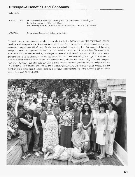
Drosophila Genetics and Genomics
July 14-27
INSTRUCTORS M. Ashburner, Cambridge University and EBI, Cambridge, United KingdomK. Burtis, University of California, DavisR.S. Hawley, Stowers Institute for Biomedical Research, Kansas City, Missouri
ASSISTANT S. Langley, University of California, Berkeley
This intensive seminar course provided an introduction to the theory and practice of methods used toanalyze and manipulate the Drosophila genome. It is suitable for graduate students and researcherswith some experience with Drosophila who are interested in expanding their knowledge of the widerange of genetic and genomic techniques now available for use with this organism. Topics coveredincluded chromosome mechanics, the design and execution of genetic screens, and the use of trans-posable elements as genetic tools. We reviewed the current understanding of the genome sequenceand discussed methodologies for genomic analysis (e.g., microarrays, gene finding methods, compar-ison with the sequences of related species, and the heterochromatin genomic project) and proteomicsin Drosophila. The course was held at the Laboratory's Banbury Conference Center located on thenorth shore of Long Island. All participants stay within walking distance of the Center, close to tenniscourt, pool, and private beach.
301

PARTICIPANTS
Anderl, I., M.S., Umea University, SwedenBjorkmo, M., B.S., University of Oslo, NorwayBoundy, S., Ph.D., University of Bath, United KingdomBrogli, K., Diplom., ETH Zurich, SwitzerlandDasari, S., M.S., University of Kentucky, LexingtonDu, T, B.S., University of Massachusetts, WorcesterGrave ley, B., Ph.D., University of Connecticut Health Center,
FarmingtonHaflidadottir, B., M.S., University of Iceland, ReykavikKatsani, K., Ph.D., Institut Curie, Paris, FranceKind, J., M.S., EMBL Heidelberg, GermanyKocks, C., Ph.D., Massachusetts General Hospital, Boston
SEMINARS
Baker, B., Stanford University, California: Behavioral neuroge-netics in Drosophila.
Burtis, Kenneth, University of California, Davis: Drosophilagenome and genomics.
Cooley, L., Yale University, New Haven, Connecticut:Fluorescent proteins in Drosophila: Cell biology and pro-teomics.
Ganetzky, B., University of Wisconsin, Madison: Segregationdistortion/neurogenetics.
Golic, K., University of Utah, Salt Lake City: Mosaic systemsand P-element technologies.
Hawley, R.S., Stowers Institute for Biomedical Research,Kansas City, Missouri; Ashburner, M., Cambridge Universityand EBI, Cambridge, United Kingdom: Introduction to
302
Leinonen, A., B.S., University of Tampere, FinlandLiebert, S., M.S., Baylor College of Medicine, Houston,
TexasLindmo, K., M.S., The Norwegian Radium Hospital, Oslo,
NorwayMarconi, S., M.S., University of Bari, ItalyMendes, C., B.S., The Rockefeller University, New YorkSeppa, M., B.A., Washington University, St. Louis, MissouriSierralta, J., Ph.D., University of Chile, SantiagoStier, S., M.S., Uppsala University, SwedenSopory, S., Ph.D., Oregon Health and Science University,
Portland
Drosophila; Basic genetics of Drosophila.Hawley, R.S., Stowers Institute for Biomedical Research,
Kansas City, Missouri: Meiosis and mitosis.Karpen, G., University of California, Berkeley: Chromosome
structure.Langley, C., University of California, Davis: Genome structure
and Drosophila populations.Lehman, R., New York University School of Medicine, New
York: Genetic screens.Perrimon, N., Harvard Medical School, Boston,
Massachusetts: Functional genomics (RNAi).White, K., Yale University School of Medicine, New Haven,
Connecticut: Functional genomics (microarrays and net-work annotation).
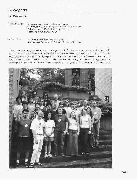
C. elegansJuly 27-August 16
INSTRUCTORS B. Bowerman, University of Oregon, EugeneS. Clark, New York University School of Medicine, New YorkM. Labouesse, IGBMC, Strasbourg, FranceI. Mori, Nagoya University, Japan
ASSISTANTS C. Carter, University of Oregon, EugeneC. Chiu, New York University School of Medicine, New York
This course was designed to familiarize investigators with C. elegans as an experimental system, withan emphasis on both classical genetic analysis and reverse genetic approaches. A major goal was toteach students how to successfully exploit the information generated by the C. elegans genome pro-ject. The course was suited both for those who have current training in molecular biology and someknowledge of genetics, but have no experience with C. elegans, and for students with some prior
303

worm experience who wished to expand their repertoire of expertise. The following topics were cov-ered both in the laboratory and by lectures from experts in the field: worm picking, C. elegans data-bases and worm bioinformatics, anatomy and development, forward genetics, chemical and transpo-son mutagenesis, generation of transgenic animals, expression pattern analysis, reverse genetics, con-struction and screening of deletion libraries, and RNA inactivation. The course was designed to impartsufficient training to students in the most important attributes of the C. elegans system to enable stu-dents to embark on their own research projects after returning to their home institutions. This coursewas supported with funds provided by the National Institute of Child Health and Human Development.
PARTICIPANTS
Beale, E., Ph.D., Texas Tech. University Health SciencesCenter, Lubbock
Garrett, M., Ph.D., Vanderbilt University Medical Center,Nashville, Tennessee
Grez, P. B.S., Pontificia Universidad Catolica de Chile,Santiago
Holmes, C., B.S., University of British Columbia, Vancouver,Canada
Honda, Y., Ph.D., Tokyo Metropolitan Institute of Gerontology,Japan
Kosarek, J., B.S., University of Texas Southwestern MedicalCenter, Dallas
Lo, S., Ph.D., Chang Gung University, Tao Yuan, ROC
SEMINARS
Ewbank, J., CIML, Marseille, France: Host-pathogen interac-tions in C. elegans.
Greenwald, I., Columbia University, New York: LIN-12/Notchsignaling and cell-cell interactions in C. elegans.
Hall, D., Einstein University School of Medicine, New York:Introduction to Worm Atlas, an anatomy database for C.elegans researchers.
Harris, T, Cold Spring Harbor Laboratory: Worm Base: A livecrash-free course, and Worm Base tools for data miningand comparative genomics.
Herman, R., University of Minnesota, Minneapolis: C. elegansgenetic suppressors, enhancers, redundancy, and mosaics.
Mango, S., University of Utah, Salt Lake City: Organogenesis
304
Maro, G., Ph.D., INSERM U368, Paris, FranceMertens, C., M.A., Ghent University, BelgiumMichael, M., Ph.D., Harvard University, Cambridge,
MassachusettsNakachi, Y., M.S., Saitama Medical School, JapanNilsen, H., Ph.D., London Research Institutes, Hens, United
KingdomRockman, M., B.S., Duke University, Durham, North
CarolinaShorto, A., Ph.D., University of Bristol, United KingdomSimonetta, S., M.S., University of Quilmes, Buenos Aires,
ArgentinaWinkelbauer, M., B.S., University of Alabama, Birmingham
and transcriptional networks in C. elegans.Nonet, M., Washington University School of Medicine, St.
Louis, Missouri: Regulation of synaptic transmission byRab GTPases.
Rougvie, A., University of Minnesota, St. Paul: Keeping timewith microRNAs in C. elegans.
Schafer, W., University of California, San Diego, La Jolla:Imaging neuronal activity and behavior in C. elegans.
Strome, S., Indiana University, Bloomington: Germ-line devel-opment in C. elegans.
Sugimoto, A., RIKEN Center for Developmental Biology,Kobe, Japan: A C. elegans phenome project: Toward agenomic understanding of the developmental program.

Eukaryotic Gene Expression
July 27-August 16
INSTRUCTORS L.D. Attardi, Stanford University, CaliforniaW.L. Kraus, Cornell University, Ithaca, New YorkA. Shilatifard, St. Louis University, MissouriL. Tora, IGBMC, Illkirch, France
ASSISTANTS T. Hilton, IGBMC, Illkirch, FranceT. Johnson, Stanford University, CaliforniaM.Y. Kim, Cornell University, Ithaca, New YorkA. Wood, St. Louis University Health Sciences Center, Missouri
This course was designed for students, postdocs, and principal investigators who had recently venturedinto the exciting area of gene regulation. The course focused on state-of-the-art strategies and techniquesemployed in the field. Emphasis was placed both on in vitro and in vivo protein-DNA interactions and onnovel methodologies to study gene regulation. Students made recombinant tagged proteins, nuclear
305

extracts, performed in vitro transcription reactions and measured RNA levels using Si nuclease mapping.Characterizations of the DNA-binding properties of site-specific transcription factors were carried outusing electrophoretic mobility shift and DNase I footprinting assays. In addition, students learned tech-niques for the assembly and analysis of chromatin in vitro, and in vivo. This included transcription assays,nucleosome mapping, chromatin remodeling assays, and restriction enzyme accessibility assays.
During the past few years, the gene regulation field has developed in vivo approaches to study generegulation. Students were exposed to the chromatin immunoprecipitation technique. They also used RNAifor specific knock-dOwn experiments in mammalian cells. In addition, determining cellular gene expres-sion profiles had been accelerated tremendously by DNA microarray technology. Students receivedhands-on training in performing and interpreting results from DNA microarrays. Experience with basicrecombinant DNA techniques was a prerequisite for admission to this course. Lectures by the instructorscovered the current status of the gene expression field, theoretical aspects of the methodology, andbroader issues regarding strategies for investigating the regulation of gene expression in eukaryotes.Guest lecturers discussed contemporary problems in eukaryotic gene regulation and technical approach-es to their solution. This course was supported with funds provided by the National Cancer Institute.
PARTICIPANTS
Binda, 0., B.S., McGill University, Montreal, CanadaFischer, A., M.S., University of Geneva, SwitzerlandHeldring, N., M.S., Karolinska Institutet, Huddinge,
Swedenlijima, K. A., Ph.D., Cold Spring Harbor LaboratoryKent, J., Ph.D., University of California, Santa CruzKodandera, D., M.S., Emory University, Atlanta, GeorgiaKotekar, A., Ph.D., National Institutes of Health, Rockville,
MarylandLakowski, B., Ph.D, Institut Pasteur, Paris, FranceLarsen, L., M.S., Steno Diabetes Center, Gentofte, Denmark
SEMINARS
Attardi, L., Stanford University, California: Using mouse mod-els to dissect the function of the p53 trans-activator intumor suppression.
Bartolomei, M., HHMI/University of Pennsylvania School ofMedicine, Philadelphia: Regulation of genomic imprinting.
Carthew, R., Northwestern University, Evanston, Illinois: Genesilencing by small RNAs.
Conaway, J., Stowers Institute for Medical Research, KansasCity, Missouri: Regulational of initiation and elongation byRNA polymerase II.
Cote, J., Laval University Cancer Research Center, QuebecCity, Canada: Chromatin modification by the NuA4 com-plex: An early step in gene activation.
Dynlacht, B., New York University School of Medicine, NewYork: Transcriptional control of the cell cycle.
Fuller, M., Stanford University School of Medicine, California:Developmental regulation of gene expression by tissue-spe-cific transcription machinery.
Gill, G., Harvard Medical School, Boston, Massachusetts:Regulation of transcription factor activity by SUMO modifi-cation.
Graves, B., University of Utah, Salt Lake City: The ETS familyof transcription factors: Routes to specificity.
Kraus, W. L., Cornell University, Ithaca, New York: Role of lig-ands, coactivators, and chromatin in estrogen-receptor-dependent transcription.
306
Lizcano, F, Ph.D., La Sabana University, Chia, ColumbiaLundgren, J., M.S., Stockholm University, SwedenMagklara, A., Ph.D., National Institutes of Health/NCI,
Bethesda, MarylandMarinescu, V, Ph.D., Children's Hospital Boston, Enders,
MassachusettsNally, K., Ph.D., National University of Ireland, CorkRamachandran, A., Ph.D., Tufts University School of
Medicine, Boston, MassachusettsTan, M.-H., MRCP Van Andel Research Institute, Grand
Rapids, Michigan
Nyborg, J., Colorado State University, Fort Collins: Regu-lation of HTLV-I transcription in a chromatin context.
Sassone-Corsi, P., Institut de Genetique at de BiologieMoleculaire at Cellulaire, Illkirch, France: Unique chromatinremodeling and transcriptional rules in male germ cells.
Schultz, P., Institut de Genetique at de Biologie Moleculaireat Cellulaire, Illkrich, France: Structural studies of large tran-scription complexes of electron microscopy and imageprocessing.
Shilatifard, A., St. Louis University School of Medicine,Missouri: A COMPASD and a GPS in defining histone mod-ifications, transcriptional regulation, and human cancer:The coordinates of the genome.
Timmers, M., University Medical Centre, Utrecht, TheNetherlands: Dynamic regulation of transcription by RNApolymerase II.
Tora, L., Institut de Genetique at de Biologie Moleculaire atCellulaire, Illkrich, France: Different sets of promoter recog-nition factors regulate the transcription initiation of RNApolymerase II at distinct promoters.
Whitfield, M., Dartmouth Medical School, Hanover, NewHampshire: Genome-wide analysis of gene expression inbasic biology and complex human disease.
Workman, J., Stowers Institute for Medical Research,Kansas City, Missouri: Protein complexes that modify chro-matin.

Imaging Structure and Function in the Nervous SystemJuly 27-August 16
INSTRUCTORS F. Engert, Harvard University, Cambridge, MassachusettsF. Helmchen, Max-Planck Institut fur Medizinische Forschung, Heidelberg, GermanyD. Kleinfeld, University of California, San Diego, La JollaT. Murphy, University of British Columbia, Vancouver, CanadaM. Oheim, Ecole Superieure de Physique at Chimie Industrielles, Paris, France
ASSISTANTS V. Emiliani, Institut Jacques Monod, Paris, FranceT. Nevian, Max-Planck Institut fijr Medizinische Forschung, Heidelberg, GermanyY. Otsu, University of British Columbia, Vancouver, CanadaP. Tsai, University of California, San Diego, La JollaW. Tyler, Harvard University, Cambridge, MassachusettsR. Vislay, Harvard University, Cambridge, MassachusettsJ. Wadiche, Vollum Institute, Portland, OregonJ. Waters, Max-Planck Institut, Heidelberg, Germany
Advances in light microscopy and digital image processing, and the development of a variety of pow-erful fluorescent probes, presented expanding opportunities for investigating the nervous system, fromsynaptic spines to networks in the brain. This intensive laboratory and lecture course provided partic-ipants with the theoretical and practical knowledge to utilize emerging imaging technologies. The pri-
307

mary emphasis of the course was on vital light microscopy. Students learned the principles of lightmicroscopy, as well as use of different types of electronic cameras, laser-scanning systems, function-al fluorophores, delivery techniques, and digital image-processing software. In addition to transmittedlight microscopy for viewing cellular motility, the course examined a variety of molecular probes of cellfunction, including calcium-sensitive dyes, voltage-sensitive dyes, photo-activated ("caged") com-pounds, and exocytosis tracers. Issues arising in the combination of imaging with electrophysiologicalmethods were covered. Particular weight was given to multi-photon laser-scanning microscopy and tonewly available biological fluorophores, especially green fluorescent protein (GFP) and its variants. Weused a spectrum of neural and cell biological systems, including living animals, brain slices, and cul-tured cells. Applicants had a strong background in the neurosciences or in cell biology. This coursewas supported with funds provided by the National Institute of Mental Health, the National Institute onDrug Abuse, and the Howard Hughes Medical Institute
PARTICIPANTS
Awatramani, G., Ph.D OSHUNollum Institute, Portland,Oregon
Beckman, M., Ph.D., Yale University/The University ofMontana, New Haven, Connecticut
Bezzi, P., Ph.D., University of Lausanne, SwitzerlandBrasnjo, G., B.S., University of California, Los AngelesDan, C., Ph.D., Harvard University, Cambridge,
Massachusetts
SEMINARS
Almers, W., Oregon Health and Science University, Portland:TIRF.
Betz, W., University of Colorado Medical School, Denver:FM143.
Cohen, L., Yale University, New Haven, Connecticut: Voltagesensitive dyes.
Denk, W., Max-Planck Institute for Medical Research,Heidelberg, Germany: Block-face neuronal reconstruction.
Engert, F., Harvard University, Cambridge, Massachusetts:Loading in the tadpole (introduction of a basic prep),
Glaser, J., Microbrightfield, Inc.: Reconstruction of neurons.Helmchen, F., Max-Planck Institut fir Medizinische
Forschung, Heidelberg, Germany: Ionic indicators/buffer-ing/basics.
Hu, C.-D., Purdue University, West Lafayette, Indiana: Seeingis believing: Visualization of transcription factor interac-tions...using bimolecular fluorescence complementation.
Kalatsky, V., University of California, San Francisco: Intrinsicimaging.
Keller, E., Carl Zeiss, Inc., Thornwood, New York: Diffractiontheory on image formation.
Kleinfeld, D., University of California, San Diego, La Jolla:
308
Khosravani, H., M.S., University of Calgary, CanadaKomiyama, T., B.S., Stanford University, Palo Alto, CaliforniaMaggi, L., Ph.D., EMBL-Monterotondo, Roma, ItalyMrsic-Flogel, T., Ph.D., Max-Planck Institute of Neurobiology,
Martinsried, GermanyPaw lu, C., M.D., University of Freiburg, GermanyTopolnik, L., Ph.D., University of Montreal, CanadaTsui, J., BA., Stanford University, Palo Alto, California
Basic optics.Lanni, F., Carnegie-Mellon University, Pittsburgh,
Pennsylvania: Point spread functions.Lichtmann, J., Washington University, St. Louis, Missouri:
Confocal microscopy.Mertz, J., Ecole Superieure de Physique at Chimie
Industrie Iles, Paris, France: Advances in nonlinearmicroscopy.
Moomaw, B., Spring Branch, Texas: CCD imaging, princi-ples, and technology.
Murphy, T., University of British Columbia, Vancouver,Canada: Back to basics, experimental design, issues withlive cell imaging.
Oheim, M., Ecole Superieure de Physique at ChimieIndustrie Iles, Paris, France: Basics of TIRF microscopy.
Shepherd, G., Cold Spring Harbor Laboratory: Caged trans-mitters and circuit analysis.
Svoboda, K., Cold Spring Harbor Laboratory: Basics of 2-photon microscopy.
Tsien, R., University of California, San Diego, La Jolla: Theworld of functional XFPs.

Yeast Genetics
July 27-August 16
INSTRUCTORS D. Amberg, SUNY, Syracuse, New YorkD. Burke, University of Virginia, CharlottesvilleJ. Strathern, National Cancer Institute, Frederick, Maryland
ASSISTANTS C. Copeland, University of Virginia, CharlottesvilleM. Derbyshire, National Center for Biotechnology, National Institutes of Health, Bethesda, MarylandM. Farah, SUNY Upstate Medical University, Syracuse, New YorkB. Keyes, University of Virginia, CharlottesvilleF. Malagon, National Cancer Institute, Frederick, Maryland
This modern, state-of-the-art laboratory course is designed to teach the students the full repertoire ofgenetic approaches needed to dissect complex problems in the yeast Saccharomyces cerevisiae.Combinations of classical genetic approaches are emphasized, including the isolation and characteri-zation of mutants, tetrad analysis, complementation, and mitotic recombination. Molecular genetictechniques, including various types of yeast transformation, gene replacement with plasmids and PCR,construction and analysis of gene fusions, and generation of mutations in cloned genes, were alsoemphasized. Students used classical and molecular approaches to gain experience in identifying andinterpreting various kinds of genetic interactions, including suppression and synthetic lethality. Students
were immersed in yeast genomics and performed and interpreted experiments with DNA arrays.Students gained first-hand experience in modern cytological approaches such as epitope tagging andimaging yeast cells using indirect immunofluorescence, GFP-protein fusions, and a variety of fluores-cent indicators for various subcellular organelles. Lectures on fundamental aspects of yeast geneticswere presented along with seminars given by outside speakers on topics of current interest. Thiscourse was supported with funds provided by the National Cancer Institute.
PARTICIPANTS
Arnason, E., Ph.D., University of Iceland, ReykjavikBrady, T, B.S., Iowa State University, AmesBulteau, A. L., Ph.D., Case Western Reserve University,
Cleveland, OhioChabes, A. L., Ph.D., Cold Spring Harbor LaboratoryCharvin, G., B.S., The Rockefeller University, New YorkGarbitt, R., B.S., B.A., Tufts University, Boston,
MassachusettsGresham, D., Ph.D., Princeton University, New JerseyMagwene, P., Ph.D., University of Pennsylvania, PhiladelphiaMercer, J., Ph.D., Duke University, Durham, North Carolina
SEMINARS
Basrai, M., National Cancer Institute, Bethesda, Maryland:Molecular determinants of faithful chromosome transmis-sion and checkpoint regulation.
Boone, C., University of Toronto, Ontario, Canada: Globalmapping of the yeast genetic interaction network.
Botstein, D., Princeton University, New Jersey: Functionalgenomics: Getting beyond descriptive studies.
Bretscher, A., Cornell University, Ithaca, New York:Combining genetics and cell biology to elucidate the mech-anism of polarized growth and organelle segregation.
Curcio, J., University at Albany School of Public Health, NewYork: Retrotransposons to the rescue: How Tyl elementsrespond to challenges to the genome.
Emr, S., HHMI/University of California, San Diego: Fromgenes to functions: Genetics of phosphoinositide lipid andubiquitin signaling in receptor sorting at the endosome.
Johnston, M., Washington University School of Medicine, St.Louis, Missouri: Feasting, fasting and fermenting: Glucosesensing and signaling in yeasts.
310
Paumi, C., Ph.D., Johns Hopkins University School ofMedicine, Baltimore, Maryland
Saerens, S., M.S., Catholic University of Leuven, BelgiumSandberg, R., M.S., Karolinska Institutet, Stockholm,
SwedenSengpta, A., Ph.D., Rutgers University, Piscataway, New
JerseyTong, Z., B.S., University of Pennsylvania, PhiladelphiaTseng, C., A.B., Harvard Medical School, Boston,
MassachusettsWagner, M., B.S., University of California, San Diego, La Jolla
Konopka, J., Stony Brook University, New York: Geneticanalysis of the pheromone pathway: The meaning ofscreening and the basis for epistasis.
Michaelis, S., Johns Hopkins School of Medicine, Baltimore,Maryland: Biogenesis of the a-Factor mating pheromone inS. cerevisiae: From yeast mating to human disease.
Michels, C., Queens College/City University of New York,Flushing: The regulation of maltose fermentation: A com-plex brew.
Pringle, J., University of North Carolina, Chapel Hill:Morphogenesis and cytokinesis in yeast.
Smith, M.M., University of Virginia, Charlottesville: Geneticsof chromatin structure and function.
Stearns, T, Stanford University, California: Yeast genetics:Teaching it and doing it-at the same time!
Tyers, M., Samuel Lunenfeld Research Institute/Mt. SinaiHospital, Toronto, Canada: Genetics of size control: Fromthe classic to the systematic.

Stem Cells
July 29-August 11
INSTRUCTORS G. Keller, Mt. Sinai School of Medicine, New YorkR. McKay, National Institutes of Health, Bethesda, MarylandA. McLaren, University of Cambridge, United KingdomJ. Rossant, Samuel Lunenfeld Research Institute, Toronto, CanadaA. Surani, University of Cambridge, United Kingdom
This two-week lecture course brought together leading researchers in the stem cell field with a smallgroup of international students. Stem cells construct organs in development. They sustain tissues inthe adult and restore them after injury. Because of these properties, isolating and manipulating stemcells has become a major new element in biomedical science. This lecture and discussion course cov-ered a series of subjects including the cells of the early embryo, the nature of germ cells, the mecha-nisms that control the number of stem cells, and their stability and transformation into other cell types.The clinical potential and political impact of stem cell technology was also presented in depth by invit-ed speakers. A key feature of the course was the easy access to the instructors and the invited lec-turers for informal discussion. The purpose of the course provided participants with an opportunity toachieve an advanced understanding of the scientific and clinical importance of stem cells. The coursewas held at the Laboratory's Banbury Conference Center located on the north shore of Long Island.All participants stayed within walking distance of the Center, close to tennis court, pool, and privatebeach. This is a unique learning environment. Partial support for the course was provided by the Eppley
Foundation for Research and the Howard Hughes Medical Institute.
311

PARTICIPANTS
Abeyewickreme, A., B.A., Institute of Child Health, London,United Kingdom
Bao, J., Ph.D., Washington University, St. Louis, MissouriBian, Z., Ph.D., Karolinska lnstitutet, Stockholm, SwedenErices, A., Ph.D., Universidad Andres Bello, Santiago, ChileFossati, V., Ph.D., University of Bologna, ItalyGangemi, R., M.D., National Institute of Cancer Research,
Genova, ItalyHipp, J., B.S., Wake Forest University School of Medicine,
Winston-Salem, North CarolinaLin, S.-Y., Ph.D., Mackay Memorial Hospital, Taipei, TaiwanMaeda, T., B.E., Osaka University, JapanMantle, I., Ph.D., University of Michigan, Ann ArborNewey, S., Ph.D., Cold Spring Harbor Laboratory
SEMINARS
Battey, J., National Institutes of Health/NIDCD, Bethesda,Maryland: Politics of stem cells.
Burden, S., New York University School of Medicine, NewYork: Muscle function and regeneration.
Coffey, R., Vanderbilt University School of Medicine,Nashville, Tennessee: Colon cancer.
Eidelberg, D., North Shore-Long Island Jewish HealthSystem, Manhasset, New York: Cell therapy in Parkinson'sdisease.
Geijsen, N., Massachusetts General Hospital, Charlestown:Derivation of germ cells from ES cells.
Gould, E., Princeton University, New Jersey: Neurogensis inthe adult brain.
Johnston, L., Columbia University, New York: Control of cellgrowth and cell division during development.
Keller, G., Mt. Sinai School of Medicine, New York:Hematopoiesis by ES cells.
Kikyo, N., University of Minnesota, Minneapolis: Genomicreprogramming,
Krause, D., Yale University, New Haven, Connecticut:Transdifferentiation of bone marrow.
Lehmann, R., Skirball Institute, New York University, NewYork: Drosophila germ cells.
Lemischka, I., Princeton University, Rutgers, New Jersey:
312
Pillai, A., B.S., University of California, San Diego, La JollaRiva, F., Ph.D., University of Pavia, ItalyRochford, C., B.S., European Neuroscience Institute,
Gottingen, GermanySchenke-Layland, K., Ph.D., Friedrich Schiller University,
Jena, GermanySchmelzer, E., Ph.D., University of North Carolina, Chapel HillTesar, P., Ph.D., National Institutes of Health and University of
Oxford, Bethesda, MarylandTesta, G., Ph.D., Dresden University of Technology/Max-
Planck Institute, GermanyWahle, J., B.S., University of South Florida, TampaWirta, V., M.S., Royal Institute of Technology, Stockholm,
Sweden
Transcriptional signature of stem cells.Mandel, G., Stony Brook University, New York: Mechanisms
regulating neuronal phenotype and behavior.McKay, R., National Institutes of Health/NINDS, Bethesda,
Maryland: Neural stem cells.Schier, A., Skirball Institute of Biomolecular Medicine, New
York: Nodal signaling in vertebrate development.Simmons, P., Peter Mac Callum Cancer Institute, Australia:
Cells in the bone marrow.Smith, A., Institute for Stem Cell Research, United Kingdom:
Embryonic stem cells.Snoeck, H., Mt. Sinai School of Medicine, New York:
Genetics of hematopoiesis.Tam, R, University of Sydney, Australia: Mechanisms of tis-
sue patterning.Trumpp, A., Swiss Institute for Experimental Cancer
Research, Switzerland: Development and tumorigenesis.van Lohuizen, M., Netherlands Cancer Institute, The
Netherlands: Epigenetic regulation of transcriptional statesby higher-order chromatin structures.
Wicha, M., University of Michigan Comprehensive CancerCenter, Ann Arbor: Breast cancer.
Zoloth, L., Northwestern University, Evanston, Illinois: Stemcell ethics.

X-ray Methods in Structural Biology
October 18-November 2
INSTRUCTORS W. Furey, V.A. Medical Center, Pittsburgh, PennsylvaniaG. Gilliland, National Institute of Standards & Technology, Gaithersburg, MarylandA. McPherson, University of California, IrvineJ. Pflugrath, Rigaku/MSC, Inc., The Woodlands, Texas
ASSISTANT I. Persakova, Rutgers, The State University of New Jersey, New Brunswick
Crystallography and X-ray diffraction yield a wealth of structural information unobtainable through othermethods. This intensive laboratory/computational course focused on the major techniques used todetermine the three-dimensional structures of macromolecules. It was designed for scientists with aworking knowledge of protein structure and function, but who are new to macromolecular crystallog-raphy. Topics covered included basic diffraction theory, crystallization (proteins, nucleic acids, andcomplexes), crystal characterization, X-ray sources and optics, synchrotrons, crystal freezing, data col-
lection, data reduction, multiple isomorphous replacement, multiwavelength anomalous diffraction,molecular replacement, solvent flattening, noncrystallographic symmetry averaging, electron densityinterpretation, structure refinement, structure validation, molecular graphics, and coordinate deposi-
313

tion. Participants learned through extensive hands-on experiments and formal presentations. Theycrystallized, determined, and refined a protein structure in parallel with lectures on the theory and infor-mal discussions behind the techniques. Extensive use of Linux workstations was made throughout thecourse for computational aspects of the process. This course was supported by the National CancerInstitute.
PARTICIPANTS
Beg lova, N., Brigham & Women's Hospital, Boston,Massachusetts
Cochrane, J., Yale University, New Haven, ConnecticutDombrauckas, J., University of Illinois, ChicagoDzivenu, 0., Fox Chase Cancer Center, Philadelphia,
PennsylvaniaKamenski, T., University of Munich, GermanyKawate, T., Columbia University, New YorkLee, M., University of Sydney, AustraliaLitowski, J., Ph.D., Columbia University, New York
SEMINARS
Dauter, Z., Brookhaven National Laboratory, Upton, NewYork: Anomalous data collection.
Gilliland, G., National Institute of Standards and Technology,Gaithersburg, Maryland: The protein data bank.
Hendrickson, W., Columbia University, New York: MADphasing: Theory and practice.
Joshua-Tor, L., Cold Spring Harbor Laboratory: Argonauts:The secret of Slicer.
Kjeldgaard, M., Aarhus University, Denmark: Electron densityfitting from A to 0.
Kleywegt, G., University of Uppsala, Sweden: Just becauseit's in Nature, doesn't mean it's true...(macromolecularstructure validation).
Perrakis, A., Netherlands Cancer Institute, Amsterdam, The
314
Loukachevitch, L., Vanderbilt University School of Medicine,Nashville, Tennessee
Mylona, A., European Molecular Biology Laboratory,Grenoble, France
Rhodes, W., University of Southern Maine, PortlandSivakumar, N., Institute of Molecular Agrobiology, SingaporeWan, Q., University of Vermont, BurlingtonWei, Y., Brookhaven National Laboratory, Upton, New YorkWilson, K. -A., The Rockefeller University, New YorkYip, C., University of British Columbia, Vancouver, Canada
Netherlands: Shaping the human genome: Structuralinsights on the action of retrotransposons.
Pflugrath, J., Rigaku/MSC, Inc., The Woodlands, Texas:Away from the edge: Sulfur SAD with chromiumradiation.
Richardson, D., Duke University Medical Center, Durham,North Carolina; Richardson, J., Duke University MedicalCenter, Durham, North Carolina: Detection and repair ofmodel errors using all atom contacts.
Strobel, S., Yale University, New Haven, Connecticut: RNAcrystallography: Structure of a self-splicing intron reactionintermediate.
Terwilliger, T, Los Alamos National Laboratory, New Mexico:Solve and resolve.

Bioinformatics: Writing Software for Genome Research
October 20-November 2
INSTRUCTORS S. Lewis, University of California BerkeleyS. Prochnik, University of California, BerkeleyL. Stein, Cold Spring Harbor LaboratoryJ. Tisdall, DuPont Experimental Station, Wilmington, Delaware
ASSISTANTS J. Aerts, Wageningen University, The NetherlandsS. Cain, Cold Spring Harbor LaboratoryK. Clark, Cold Spring Harbor LaboratoryM. Dybbs, University of California, BerkeleyR. Halgren, Michigan State University, East LansingG. Hartzell, University of California, BerkeleyC. Kennedy, University of California, BerkeleyB. O'Connor, University of California, Los AngelesJ. Stein, Cereon Genomics, Cambridge, MassachusettsC. Yamada, University of Cambridge, United Kingdom
The desktop computer has rapidly become an indispensable tool in the biologist's tool chest. The suc-cess of the human genome project created an explosion of information: billions of bits of biologicalinformation stashed electronically in databases around the globe just waiting for the right key to unlockthem. New technologies such as DNA microarrays and high-throughput genotyping are creating aninformation overload that the traditional laboratory notebook cannot handle. To exploit the informationrevolution in biology, biologists moved beyond canned Web interfaces and Excel spreadsheets. They
315

took charge of the data by creating their own software to fetch, manage, and integrate it. The goal of
this course was to provide biologists with the tools needed to deal with this changing landscape.
Designed for students and researchers with little prior knowledge of programming, this two-week
course taught the fundamentals of the Unix operating system, Peri scripting, dynamic Web page devel-
opment with the CGI protocol, and database design. The coursecombined formal lectures with hands-
on experience in which students worked to solve a series of problem sets drawn from common sce-
narios in biological data acquisition, integration, and laboratory workflow management. For their final
projects, students were posed problems using their own data and worked with each other and the fac-
ulty to solve them. Note that the primary focus of this course provided students with the practical
aspects of software development, rather than presenting a detailed description of the algorithms used
in computational biology. For the latter, we recommend the Computational Genomics course. This
course was supported by the National Human Genome Research Institute.
PARTICIPANTS
Acosta, I., B.S., Yale University, New Haven, ConnecticutChalkia, D., M.S., Penn State University, University ParkCrayton, Ill, M., Ph.D., University of North Carolina, Chapel
HillEmes, R., Ph.D., University College London, United KingdomGillespie, M., Ph.D., Cold Spring Harbor LaboratoryGopinathrao, G., Ph.D., Cold Spring Harbor LaboratoryGoto, D., Ph.D., Cold Spring Harbor LaboratoryHsiao, W., Ph.D., Simon Fraser University, Burnaby, CanadaHur le, B., Ph.D., National Human Genome Research Institute,
NIH, Bethesda, MarylandKaralius, J., B.S., University of Cambridge, United KingdomMapelli, M., Ph.D., European Institute of Oncology, Milan, Italy
SEMINARS
Ashburner, M., University of Cambridge, United Kingdom:Biological oncologies.
Brent, M., Washington University, St. Louis, Missouri: Gene
prediction.Hartzell, G., University of California, Berkeley: The tsao of
pert.Marth, G., Boston College, Chestnut Hill, Massachusetts:
Sequence variation analysis.Pearson, W., University of Virginia, Charlottesville: Sequence
316
Marques, T., Ph.D., Universitat Pompeu Fabra, Barcelona,
SpainMund, C., B.S., German Cancer Research Center,
Heidelberg, GermanyNikolaidis, N., Ph.D., Penn State University, University ParkPrzeworski, M., Ph.D., Brown University, Providence, Rhode
IslandSamuelson, A., Ph.D., Massachusetts General Hospital,
BostonTammoja, K., M.S., National Institute for Health
Development, Tallinn, EstoniaYi, S., Ph.D., Georgia Institute of Technology, Atlanta,
Georgia
similarity analysis.Peitzsch, R., Pfizer Global R&D, Groton, Connecticut:
Modeling biological data in relational databases.Stein, L., Cold Spring Harbor Laboratory: Generic genome
browser.Troyanskay, O., Princeton University, New Jersey: Mircroarray
analysis.Yandell, M., HHMI/University of California, Berkeley: The
BLAST algorithm; Interpreting BLAST statistics.

Immunocytochemistry, In Situ Hybridization, and Live Cell Imaging
October 20-November 2
INSTRUCTORS A. Dernburg, Lawrence Berkeley National Laboratory, Berkeley, CaliforniaJ. Murray, University of Pennsylvania School of Medicine, PhiladelphiaJ. Swedlow, University of Dundee, United Kingdom
ASSISTANTS T. Howard, University of New Mexico, AlbuquerqueK. Hu, Scripps Research Institute, La Jolla, CaliforniaW. Moore, The University of Dundee, United KingdomJ. Peng, University of California, BerkeleyM. Platani, EMBL, Heidelberg, Germany
This course focused on specialized techniques in microscopy, in situ hybridization, immunocytochem-istry, and live cell imaging related to localizing DNA, RNA, and proteins in fixed cells, as well as proteinand RNA dynamics in living cells. The course emphasized the use of the latest equipment and tech-niques in fluorescence microscopy, including confocal laser-scanning microscopy, deconvolution meth-ods, digital image processing, and time-lapse imaging of living specimens. The course was designedto present students with state-of-the-art technology and scientific expertise in the use of lightmicroscopy to address basic questions in cellular and molecular biology. The course was alsodesigned for the molecular biologist who is in need of microscopic approaches and for the cell biolo-gist who is not familiar with the practical application of the advanced techniques presented in thecourse. Among the methods presented were the preparation of tagged nucleic acid probes, fixationmethods, detection of multiple DNA sequences in single nuclei or chromosome spreads, comparativegenomic hybridization, cellular localization of RNA, localization of nucleic acids and proteins in the
317

same cells, use of a variety of reporter molecules and nonantibody fluorescent tags, indirect antibodylabeling, detection of multiple proteins in a single cell, and the use of GFP variants to study proteinexpression, localization, and dynamics. In each method, several experimental protocols were present-ed, allowing the students to assess the relative merits of each and to relate them to their own research.Students were encouraged to bring their own nucleic acid, protein, or antibody probes to the course,which were used in addition to those provided by the instructors. The laboratory exercises were sup-plemented with lectures given by invited distinguished scientists, who presented up-to-the-minutereports on current methods and research using the techniques being presented. This course was sup-ported with funds provided by the National Cancer Institute
PARTICIPANTS
Bourke, S., B.S., University of Leeds, United KingdomDemarco, I., M.S., University of Virginia, CharlottesvilleFehrenbacher, N., M.A., Danish Cancer Society,
Copenhagen, DenmarkGallegos, M., Ph.D., University of California, San Franciscolvakhno, S., B.S., Institute of Molecular Biology and Genetics,
Kyiv, UkraineLarsen, L., M.S., Steno Diabetes Center, Gentofte, DenmarkManning, L., B.S., University of Pittsburgh, PennsylvaniaNovak, I., B.S., Karolinska Institutet, Stockholm, SwedenPsatha, M., Ph.D., Brigham & Women's Hospital, Boston,
Massachusetts
SEMINARS
Day, R., University of Virginia, Charlottesville: Seeingcolors: Applications and limitations of the fluorescentproteins.
Dernburg, A., Lawrence Berkeley National Laboratory,Berkeley, California: Basics of DNA FISH.
Difilippantonio, M., National Cancer Institute/NIH, Bethesda,Maryland: Mechanisms and consequences of chromosomalaberrations in cancer cells.
Howard, T., University of New Mexico, Albuquerque: Basicsof RNA FISH.
Hu, K., Scripps Research Institute, La Jolla, California: Basicintroduction to light and fluorescence microscopy.
Murray, J., University of Pennsylvania School of Medicine,Philadelphia: Immunocytochemistry.
Murray, J., University of Pennsylvania School of Medicine,
318
Pugacheva, E., Ph.D., Fox Chase Cancer Center,Philadelphia, Pennsylvania
Schauss, A., Diplom., Max-Planck Institute for BiophysicalChemistry, Goettingen, Germany
Specktor, A., B.A., New York University School of Medicine,New York
Steindler, L., M.S., Tel Aviv University, IsraelStephano, J., Ph.D., Universidad Autonoma de Baja
California, Ensenada, MexicoWang, H., Ph.D., University of Massachusetts Medical
School, WorcesterWang, L., Ph.D., University of Pittsburgh, Pennsylvania
Philadelphia; Swedlow, J., University of Dundee, UnitedKingdom: Principles of confocal microscopy and deconvo-lution techniques.
Spector, D., Cold Spring Harbor Laboratory: Localization ofgene expression by FISH and in living cells.
Stout, A., University of Pennsylvania, Philadelphia: TIRFmicroscopy: Implementation and application to live-cellimaging.
Svoboda, K., Cold Spring Harbor Laboratory: Applications ofZ-photon microscopy to imaging in vivo.
Tran, P, University of Pennsylvania, Philadelphia: Camerasand digital imaging fundamentals.
Waterman-Storer, C., Scripps Research Institute, La Jolla,California: Quantitative fluorescent speckle microscopy instudies of cell migration.

Computational GenomicsNovember 3-9
INSTRUCTORS W. Pearson, University of Virginia, CharlottesvilleR. Smith, GlaxoSmithKline, King of Prussia, Pennsylvania
ASSISTANT B. Cantarel, University of Virginia, Charlottesville
Beyond BLAST and FASTA-This course presented a comprehensive overview of the theory and prac-tice of computational methods for gene identification and characterization from DNA sequence data.The course focused on approaches for extracting the maximum amount of information from proteinand DNA sequence similarity through sequence database searches, statistical analysis, and multiplesequence alignment. Additional topics included gene recognition (exon/intron prediction), identifyingsignals in unaligned sequences, and integration of genetic and sequence information in biological data-bases. The course combined lectures with hands-on exercises; students were encouraged to posechallenging sequence analysis problems using their own data. The course made extensive use of localWWW pages to present problem sets and the computing tools to solve them. Students used Windowsand Mac workstations attached to a Unix server; participants should be comfortable using the Unixoperating system and a Unix text editor. The course was designed for biologists seeking advancedtraining in biological sequence analysis, computational biology core resource directors and staff, and
319
scientists in other disciplines, such as computer science, who wish to survey current research prob-lems in biological sequence analysis. The primary focus of this course was the theory and practice ofalgorithms used in computational biology, with the goal of using current methods more effectively anddeveloping new algorithms. Students more interested in the practical aspects of the advanced soft-ware development were encouraged to apply to course on Advanced Bioinformatics. This course wassupported by the National Human Genome Research Institute.
PARTICIPANTS
Abernathy, B., B.S., Purdue University, West Lafayette,Indiana
Almedia, H, B.S., Instituto Gulbenkian de Ciencia, Oeiras,Portugal
Bai, Y., M.S., University of Texas, ArlingtonBoukhgalter, B., Ph.D., Broad/MIT Institute for Genome
Research, Cambridge, MassachusettsDimitrova, N., Ph.D., Cold Spring Harbor LaboratoryHagen, D., B.S., Texas A&M University, College StationHecht, S., Ph.D., Grand Valley State University, Allendale,
MichiganHladish, T., B.S., University of Maryland Biotechnology
Institute, RockvilleNorth, L., Ph.D., University of California, DavisHuebinger, R., M.S., Texas A&M University, College
Station
SEMINARS
Altschul, S., National Center for Biotechnology Information,Bethesda, Maryland: Statistics of sequence similarityscores; Iterated protein database searches with PSI -BLAST.
Birney, E., EMBL-EBI, Hinxton, United Kingdom: TheENSEMBL genome database; Genome computation II-ens-mart.
Cooper, P., National Center for Biotechnology Information,
320
Hung, L.-W., Los Alamos National Laboratory, Berkeley,California
Kelley, J., B.S., University of Cambridge, United KingdomKhatun, J., Ph.D., University of North Carolina, Chapel HillProctor, R., Ph.D., NCAUR-Agriculture Research Service-
USDA, Peoria, IllinoisRainey, S., Ph.D., Queen's University Belfast, North IrelandRen, X.-Y., M.S., Wageningen University and Research
Center, The NetherlandsRobertson, C., Ph.D., Geospiza, Inc., Seattle, WashingtonSequin, A., Ph.D., Natural Resources Canada, Sainte-Foy,
QuebecShanks, 0., Ph.D., U.S. Environmental Protection Agency,
Cincinnati, OhioVinayaka, C., Ph.D., National Biomedical Research
Foundation, Washington, D.C.
National Library of Medicine, Bethesda, Maryland: NCBIgenome resources.
Rubin, E., Joint Genome Institute, Berkeley, California:Comparative genomics and biology.
Yandell, M., HHMI/University of California, Berkeley: Genomeannotation; Large-scale computation on genomes-1.

Phage Display of Proteins and Peptides
November 9-22
INSTRUCTORS C. Barbas, Scripps Research Institute, La Jolla, CaliforniaD. Siegel, University of Pennsylvania School of Medicine, PhiladelphiaG. Silverman, University of California, La Jolla
ASSISTANTS K. Noren, New England Biolabs, Beverly, MassachussettsJ. Struthers, University of California, San DiegoC. Tuckey, New England Biolabs, Beverly, Massachussetts
Recent advances in the generation and selection of antibodies from combinatorial libraries allow for therapid production of antibodies from immune and nonimmune sources. This intensive laboratory/lecturecourse focused on the construction of combinatorial antibody libraries expressed on the surface ofphage and selection of desired antibodies from the library. Students learned the theoretical and prac-tical aspects of constructing combinatorial libraries from immune and nonimmune sources, as well asthe construction of synthetic antibody libraries. Antibodies were selected from the library by panning.Production, purification, and characterization of Fab fragments expressed in E coil were covered.Peptide phage libraries were also selected and characterized.
The lecture series, presented by a number of invited speakers, emphasized PCR of immunoglobu-lin genes; the biology of filamentous phage and the utility of surface expression libraries; expression ofantibodies in E coil and mammalian cells, antibody structure, and function; catalytic antibodies; direct-ed protein evolution; retroviral and cell display libraries; the immunobiology of the antibody response;
321

and recent results on the use of antibodies in therapy. The theory and practical implications for selec-tion from phage displayed libraries of random peptides, cDNA products, and semisynthetic proteinswere also explored. This course was supported with funds provided by the Howard Hughes MedicalInstitute.
PARTICIPANTS
Daad, G.A., M.S., Texas A&M University, College StationAggarwal, S., M.S., The Johns Hopkins University, Baltimore,
MarylandAlm, T., M.S., Royal Institute of Technology, Stockholm,
SwedenBarman, I., M.A., University of California, San FranciscoBosnar, M., B.S., Proizvodnja lijekova i vakcina-Research
Institute, Ltd., Zagreb, CroatiaBratkovic, T., B.S., Lek Pharmaceutical Company, Ljubljiana,
Slovenia
Friedman, M., Ph.D., Royal Institute of Technology,Stockholm, Sweden
Grippo, V., M.S., Institute of Genetics Engineering andMolecular Biology Research, Buenos Aires, Argentina
SEMINARS
Barbas, C., Scripps Research Institute, La Jolla, California:Software and hardware for genomes: Polydactyl zinc fingerproteins and the control of endogenous genes.
Goodyear, C., University of California, San Diego:Immunobiology of B cell and antibody responses.
Lowman, H., Genentech, Inc., San Francisco, California: SARof peptides using phage.
Noren, C., New England Biolabs, Beverly, Massachusetts:Phage peptide libraries: The PhD for peptides.
Sidho, S., Genentech, Inc., San Francisco, California:Antibody phage display and chemical diversity in antigenrecognition.
322
Gronwall, C., M.S., Royal Institute of Technology, Stockholm,Sweden
Lee, S., Ph.D., Singapore General Hospital, SingaporeLove, K., B.S., Harvard Medical School, Boston,
MassachusettsMahajan, S., Ph.D., Paradigm Pharmaceuticals, Roseville,
MinnesotaMason, P., B.Sc., Shriners Hospital/McGill University,
Montreal, Quebec, CanadaMir, S., M.S., Max-Planck Institute for Biophysics, Frankfurt
Am Main, GermanyRamasoota, P., Ph.D., Mahidol University, Bangkok, ThailandSato, S., B.A., Harvard Medical School, Southborough,
Massachusetts
Siegel, D., University of Pennsylvania Medical Center,Philadelphia: Cell surface selection of combinatorial Fablibraries.
Silverman, G., University of California, San Diego: Selectionof complex human antibody libraries.
Smith, G., University of Missouri, Columbia: Phage display ofpeptides.
Wilson, I., Scripps Research Institute, La Jolla, California:Structural biology of the immune system.
Wittrup, K.D., Massachusetts Institute of Technology,Cambridge: Yeast display libraries.

The Genome Access Course
May 4-5, August 31-September 1, December 1-2
TRAINERS J. Gergel, Cold Spring Harbor LaboratoryG. Howell, The Jackson LaboratoryP. Rabinowicz, The Institute for Genomic Research
Initiated in 2002, this course is an intensive two-day introduction to bioinformatics that was held threetimes in 2004 and trained 73 participants in total. The core of the course was designed to cover themanipulation and analysis of sequence information. The course was broken into modules designed togive a broad overview of a given topic, with ample time for examples chosen by the instructors. Eachmodule included three parts, consisting of a discussion of theory and methods, coverage of softwareand Web resources, and use of selected tools with examples (including those supplied by the stu-dents). The modular design allowed the instructors to tailor the presentation to the interests of the stu-dents. Modules included Electronic Sequence Information; Pairwise Sequence Comparisons; MultipleSequence Alignments; Gene Prediction; Genome Analysis; Sequence Variation; Protein Classificationand Structural Analysis; Proteomics; and Phylogenetic Analysis. Applications to the course were opento all on a first-come-first-served basis, subject to basic eligibility requirements. The course was heldat the Laboratory's Cancer Genome Research Center at Woodbury located seven miles south of themain Laboratory campus. Each student was provided with a PC laptop with wireless modem for theduration of the course. Students were encouraged to supply problem sets and sequences of interestto the trainers for possible incorporation as examples in the modules. Materials were made availableon the Web and students continued to ask questions of the trainers as they applied what they hadlearned in their individual endeavors.
May 4-5
Aho, E,. Ph.D., Concordia College, Moorhead, MinnesotaBasu, C., Ph.D., University of Tennessee, KnoxvilleBlander, J., Ph.D. Yale University School of Medicine, New
Haven, ConnecticutCameron, P., Ph.D., University of Illinois, Chicago
Chun, Y.H., Ph.D., University of Michigan, Ann ArborDas, S., Ph.D., University of Nebraska Medical Center,
OmahaGaboyard, S., Ph.D., University of Illinois, ChicagoHansen, C., Ph.D., Givaudan Flavors, Cincinnati, Ohio
323

Heby, 0., Ph.D., University of Umea, Sweden 90187Holt, J., Ph.D., University of Chicago, IllinoisHwang, B.J., Ph.D., California Institute of Technology,
Pasadena, CaliforniaJones, E., Ph.D., Pioneer Hi-Bred, Bondurant, IowaKlaper, R., Ph.D., University of Wisconsin, MilwaukeeKudla, G., B.S., International Institute of Molecular and Cell
Biology, Warsaw, PolandLiao, F, Ph.D., Pfizer, Kalamazoo, MichiganLysakowski, A., University of Illinois, ChicagoMaher, B., Ph.D., University of Pittsburgh, PennsylvaniaNielsen, A., Ph.D., Novozymes NS, Bagsvaerd, DenmarkPirity, M., Ph.D., Albert Einstein College of Medicine, Bronx,
New YorkPittenger Alley, L., B.S., USDA-ARS, Athens, Georgia
August 31-September 1
Bazinet, C., Ph.D., St. John's University, Jamaica, New YorkBowman, M., B.S., Lancaster Labs, Richmond, VirginiaBrundage, J., B.S., Northwestern University, Evanston, IllinoisChen, J., Ph.D., Mt. Sinai School of Medicine, New YorkChugh, S., Ph.D., Oregon Health & Science University,
PortlandCohen, P., Ph.D., Albert Einstein College of Medicine, Bronx,
New YorkGlynn, N., Ph.D., USDA-ARS, Canal Point, FloridaGuo, X., Ph.D., Cold Spring Harbor LaboratoryGuris, D., B.S., University of Chicago, IllinoisKennerson, M., Ph.D., Neurobiology Laboratory ANZAC
Research Institute, Concord, New South Wales, AustraliaKutty Selva, N., Ph.D., Lund University, SwedenLatkany, P., Ph.D., New York Eye and Ear Infirmary, New YorkLee, K., B.S., Baylor College of Medicine, Houston, TexasLee, Y., Ph.D., Tufts University, Boston, MassachusettsMacMillan, T, B.S., Defence R&D Canada-Suffield Medicine
Reeleder, R., Ph.D., Agriculture and Agri-Food Canada,London, England
Snyder, D., B.S., Yale University, New Haven, ConnecticutTan, M.-H., Ph.D., Van Andel Research Institute, Grand
Rapids, MichiganUngvari, Z., Ph.D., New York Medical College, ValhallaVinayaka, C., Ph.D., National Biomedical Research
Foundation, Washington, D.C.Wellen, K., B.S., Harvard School of Public Health, Boston,
MassachusettsWong, A., B.S., University of California, DavisZhao, Z., Ph.D., University of Michigan, Ann Arbor
Hat, Alberta, CanadaMcMahon, A., Ph.D., Harvard University, Cambridge,
MassachusettsNoehammer, C., Ph.D., ARC Seibersdorf Research GmbH,
Seibersdorf, AustriaRen, L., Ph.D., Cold Spring Harbor LaboratoryRidley, S., Ph.D., National Science Foundation, Arlington,
VirginiaSerspinski, T, B.S., Fairleigh Dickinson University,
Edgewater, New JerseyShoemaker, K., Ph.D., ZymoGenetics, Seattle, WashingtonSvetlanov, A., Ph.D., Albert Einstein College of Medicine,
Bronx, New YorkWard, J., Ph.D., Middlebury College, VermontWitonsky, P., Ph.D., Retired Professor: West Chester
University, Cambridge, MassachusettsWong, D., Ph.D., Western Regional Research Center,
USDA-ARS, Albany, California
324

December 1-2
Avila, A., National University of Mexico-Nitrogen Fixiation,Cuernavaca
Boczek, L., B.S., U.S. Environmental Protection Agency,Cincinnati, Ohio
Ca lie, P., Ph.D., Eastern Kentucky University, RichmondChen, L., B.S., ZymoGenetics, Seattle, WashingtonCsiszar, A., Ph.D., New York Medical College, ValhallaDominguez Vidana, R., B.S., UNAM, Cuernavaca, MexicoFisher, G., Ph.D., Ridgefield High School, ConnecticutGonzaga-Jauregui, C., B.S., National University of Mexico,
Mexico CityGriffis, J., B.S., Pioneer Hi-Bred Inc., International, Johnston,
Iowa
Jobanputra, V., Ph.D., Columbia University, New York
Kelty, C., B.S., USEPA, Cincinnati, OhioLevon, K., Ph.D., Polytechnic University, Brooklyn, New YorkLi, G., Ph.D.,The University of Hong Kong, Hong KongMesri, E., Ph.D., Weill Cornell Medical College, New YorkMorales, L., B.S., National University of Mexico-Nitrogen
Fixiation, CuernavacaSingh, C., B.S., West Chester University of Pennsylvania,
West ChesterSong, J.-J., B.S., Cold Spring Harbor LaboratoryThiagarajan, R., B.S., Transgenomic, Gaithersburg, MarylandYang, Y., Ph.D., GlaxoSmithKline Pharmaceuticals, King of
Prussia, PennsylvaniaZheng, X., Ph.D., Children's Hospital of Philadelphia,
Pennsylvania
The Laboratory would like to acknowledge the generosity of the following companies who loaned equipment andreagents to the various courses:
Affymetrix Inc. Codonics Inc. Kinetic Systems Inc. Promega Corp.Agencourt Bioscience Corning Inc. Kleindiek Nanotechnik 0-Imaging
Corporation Dagan Corporation LC Packings Qiagen Inc.Agilent Technologies, Inc. Dage-MTI Inc. Leica Microsystems Inc. Robbins ScientificAmbion Inc. David Kopf Instruments Luigs & Neumann CorporationAmersham Biosciences Delaware Diamond Knives Majer Precision Engineering Roche DiagnosticsA.M.P.I. Diagnostic Instruments, Inc. Inc. SD Instruments Inc.Applied Biosystems Dionex Corp. Med Associates Inc. Santa Cruz BiotechnologyApplied Precision Inc. Drummond Scientific MJ Research Inc. Inc.ASI Inc. Company Modulation Optics, Inc. Schleicher & Schuell Inc.Astro-Med Inc. Eastman Kodak Company Molecular Devices Sigma-AldrichAxon Instruments Erie Scientific Company Corporation Singer Instrument CompanyBecton Dickinson Labware E-Y Laboratories Molecular Probes Inc. Ltd.Bellco Glass, Inc. Fotodyne Inc. Nalge Nunc International StratageneBerthold Technologies USA,
LLCGalil Motion ControlHamamatsu Photonic
Narishige International USA,Inc.
Stoelting Co.Sutter Instruments
Bio-Rad Laboratories Systems New England Biolabs, Inc. Tecan US Inc.Bioptechs Inc. Hampton Research New Objective, Inc. Ted Pella Inc.BioVision Technologies Harvard Apparatus, Inc. Newport Corp. Thermo Electron CorporationBrinkmann Instruments IBM Life Sciences Solutions Nikon Inc. Thermo FinniganBurleigh Instruments, Inc. Improvision Inc. Novagen Inc. Thermo LabsystemsCambrex Bio Science Instrutech Corp. Olympus America Inc. TILL. Photonics Gmbh
Rockland, Inc. Intelligent Imaging Innovations Optical Insights, LLC Torrey Pines ScientificCarl Zeiss, Inc. Inc. Optronics Universal Imaging CorporationCell and Molecular Invitrogen Corporation PE Biosystems Vector Laboratories
Technologies, Inc. Jackson lmmunoresearch Perkin Elmer Life Sciences Warner InstrumentsChroma Technology Laboratories Pierce Chemical Co. Vysis Corporation
Corporation Jouan, Inc. PolyLC
325

SEMINARS
INVITED SPEAKER PROGRAM
Each year, Cold Spring Harbor Laboratory invites speakers from outside the institution to present theirfindings on a weekly basis. These seminars keep the CSHL staff current on the latest developmentsand broaden their perspectives. Graduate students and postdoctoral fellows meet with the seminarspeakers for lunch immediately after the seminar, allowing an opportunity for the exchange of ideas inan informal setting.
JANUARY
Dr. Frank Rauscher Ill, The Wistar Institute. Coordinating his-tone modification, HP1 deposition, and DNA methylation: Amodel for gene variegation at euchromatic loci in mammaliancells. (Host: Winship Herr)
Dr. Ray Deshaies, Division of Biology and HHMI, CaliforniaInstitute of Technology. Mechanism and regulation of theSCF ubiquitin ligase-proteasome pathway. (Host: WilliamTansey)
FEBRUARY
Dr. David Haig, Department of Organismic and EvolutionaryBiology, Harvard University. Broken symmetries: Genomicimprinting and social behavior. (Host: Dmitri Chklovskii)
Dr. John Allman, California Institute of Technology. The spindleneurons of fronto-insular and anterior cingulate cortex: Aphylogenetic specialization of apes and humans related tosocial cognition. (Host: Dmitri Chklovskii)
Dr. Zena Werb, Department of Anatomy, University ofCalifornia, San Francisco. The tumor microenvironment:Friend or foe? (Host: Senthil Muthuswamy)
MARCH
Dr. Ulrike Heberlein, Department of Anatomy, Universityof California, San Francisco. Studying addiction inDrosophila. (Host: Hollis Cline)
Dr. Roger Nicoll, University of California, San Francisco.Glutamate receptor trafficking and synaptic plasticity.(Host: Roberto Malinow)
Dr. Pamela Silver, Dana Farber Cancer Institute, HarvardMedical School. Connecting the genome to the cytoplasm.(Host: Adrian Krainer)
326
APRIL
Dr. Michael Rosbash, Department of Biology, HHMI/Brandeis University. Circadian rhythms in flies:Molecules, cells, systems, and behavior. (Host:Josh Huang)
Dr. Stanislas Leib ler, The Rockefeller University.Fluctuations, noise, and phenotypic variability. (Host:Yuri Lazebnik)
OCTOBER
Dr. Alan Hall, MRC Laboratory for Molecular Cell Biology andCell Biology Unit, University College London. Rho GTPasesand the control of directed cell migration. (Host: Linda VanAelst)
NOVEMBER
Dr. Ann-Shyn Chiang, Brain Research Center, National TsingHua University, Taiwan. Mapping candidate memory circuitsin the brain. (Host: Tim Tully)
Dr. Chris Walsh, Howard Hughes Medical Institute, HarvardMedical School. Genes that control the shape and size ofthe human cerebral cortex. (Host: Grigori Enikolopov)
DECEMBER
Dr. Liqun Luo, Stanford University, Department of BiologicalSciences. Exploring neural circuits using genetic mosaicmethods in flies and mice. (Host: Linda Van Aelst)
Dr. Danesh Moazed, Department of Cell Biology, HarvardMedical School. Targeting RNAi to the chromosome. (Host:William Tansey)
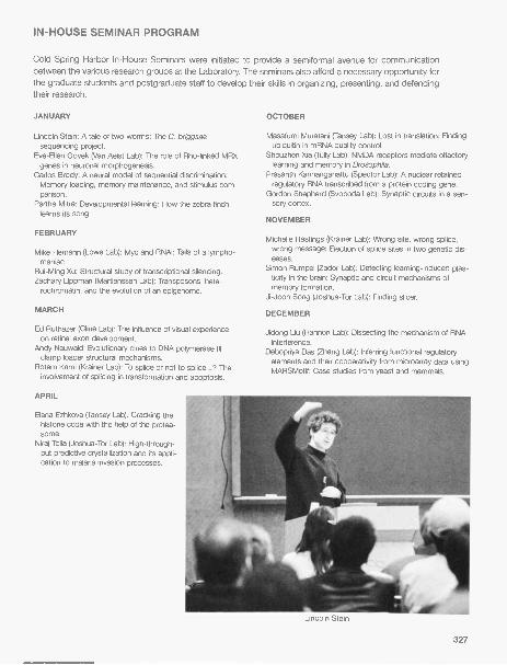
IN-HOUSE SEMINAR PROGRAM
Cold Spring Harbor In-House Seminars were initiated to provide a semiformal avenue for communicationbetween the various research groups at the Laboratory. The seminars also afford a necessary opportunity forthe graduate students and postgraduate staff to develop their skills in organizing, presenting, and defendingtheir research.
JANUARY
Lincoln Stein: A tale of two worms: The C. briggsaesequencing project.
Eve-Ellen Govek (Van Aelst Lab): The role of Rho-linked MRXgenes in neuronal morphogenesis.
Carlos Brody: A neural model of sequential discrimination:Memory loading, memory maintenance, and stimulus com-parison.
Partha Mitra: Developmental learning: How the zebra finchlearns its song.
FEBRUARY
Mike Hemann (Lowe Lab): Myc and RNAi: Tails of a lympho-maniac.
Rui-Ming Xu: Structural study of transcriptional silencing.Zachary Lippman (Martienssen Lab): Transposons, hete-
rochromatin, and the evolution of an epigenome.
MARCH
Lab): The influence of visual experienceon retinal axon development.
Andy Neuwald: Evolutionary clues to DNA polymerase IIIclamp loader structural mechanisms.
Rotem Karni (Krainer Lab): To splice or not to splice...? Theinvolvement of splicing in transformation and apoptosis.
APRIL
Elena Ezhkova (Tansey Lab): Cracking thehistone code with the help of the protea-some.
Niraj Tolia (Joshua-Tor Lab): High-through-put predictive crystallization and its appli-cation to malaria invasion processes.
OCTOBER
Masafumi Muratani (Tansey Lab): Lost in translation: Findingubiquitin in mRNA quality control.
Shouzhen Xia (Tully Lab): NMDA receptors mediate olfactorylearning and memory in Drosophila.
Prasanth Kannanganattu (Spector Lab): A nuclear retainedregulatory RNA transcribed from a protein coding gene.
Gordon Shepherd (Svoboda Lab): Synaptic circuits in a sen-sory cortex.
NOVEMBER
Michelle Hastings (Krainer Lab): Wrong site, wrong splice,wrong message: Election of splice sites in two genetic dis-eases.
Simon Rumpel (Zador Lab): Detecting learning-induced plas-ticity in the brain: Synaptic and circuit mechanisms ofmemory formation.
Ji-Joon Song (Joshua-Tor Lab): Finding slicer.
DECEMBER
Jidong Liu (Hannon Lab): Dissecting the mechanism of RNAinterference.
Debopriya Das (Zhang Lab): Inferring functional regulatoryelements and their cooperativity from microarray data usingMARSMotif: Case studies from yeast and mammals.
Lincoln Stein
327


'
r


BANBURY CENTEREXECUTIVE DIRECTOR'S REPORT
Banbury Center continues to be a thriving setting for specialized, focused meetings on pressing andemerging topics in biomedical research. In 2004, there were 18 meetings at Banbury Center, with 568invited participants; 80% of the latter came from the United States, representing 38 states. As usual, NewYork, California, Maryland, and Massachusetts contributed the most. Foreign scientists came to Banburyfrom 21 countries. The Center continues to be used intensively all year round except during the depths ofwinter. In addition to the regular program of meetings, the Watson School of Biological Sciences held twoweek-long Topics in Biology courses, and there were five lecture courses in the Laboratory's summer neu-roscience program. Two local groups also made use of the Center.
The 2004 Banbury Center program again explored a variety of topics, ranging from discussions ongenetic disorders to exploratory meetings on theoretical biology. This year also marked a significant increasein meetings related to neurobiology, a shift that mirrors the Laboratory's expanding programs on brain devel-opment and disease. In March, the Center hosted a meeting on the severe neuromuscular disorder: spinalmuscular atrophy. The meeting-Spinal Muscular Atrophy: What Is the Molecular Basis of Neuron Loss? -organized by Adrian Krainer (Cold Spring Harbor Laboratory), Alex MacKenzie (Children's Hospital ofEastern Ontario), and Kenneth Fischbeck (National Institute of Neurological Disease and Stroke) andreviewed the molecular pathology of this disorder. We continued our examination of Fragile X syndrome withthe fifth meeting in a series on that disorder, New Pharmacological and Neurobiological Approaches to theTreatment of Fragile X, organized by Will Spooren (F. Hoffman-La Roche) and William Greenough (University
of Illinois at Urbana-Champaign). It is remarkable to follow the way in which these meetings have movedfrom covering basic research to reviewing possible therapies. T-type Calcium Channels: Their Role inNormal and Pathological CNS Function, organized by Rodolfo Llinas (New York University Medical Center)and Ed Perez-Reyes (University of Virginia), went from the molecular biology of these channels to their rolesin epilepsy, pain, and manic depression, whereas Communication in Brain Systems, organized by TerrySejnowski (The Salk Institute for Biological Studies), dealt much more with theoretical issues, using model-ing approaches to understand the structure and engineering of the brain. A fascinating and ambitious meet-ing, Origins and Evolution of the Nervous System, was organized by Ian Meinertzhagen (DalhousieUniversity, Halifax, Nova Scotia) and Volker Hartenstein (University of California, Los Angeles). Participantsreviewed "simple" organisms to see how the morphology, organization, and functioning of their nervous sys-tems change with the differing behavioral requirements of each organism. Although some organisms werefamiliar to Banbury-Escherichia coli, Caenorhabditis elegans, and Drosophila-others, including sponges,jellyfish, Ascidians, and planarians, made their first appearance at a Banbury Center meeting.
Another parallel to current Laboratory research was our meeting in August on RNAi (RNA interference).One remarkable thing about RNAi is that it provides a tool for the experimental manipulation of gene expres-sion and we are also continuing to discover that it has a key role in cellular function. RNA/- related Processesin Plants: Chromatin, Development, and Defense, organized byJames Carrington (Oregon State University), Steve Jacobson(University of California, Los Angeles), and Detlef Weigel (Max-Planck
Institute for Developmental Biology), reviewed the ways in whichplants use RNAi in their everyday life. The timing was just right-par-ticipants were able to hear first-hand from Leemor Joshua-Tor andGreg Hannon about their latest studies on Dicer, published a fewweeks earlier in the journal Science.
Issues of bioterrorism continue to raise great concerns. TheDepartment of Homeland Security funded Microbial Forensics, a fol-low-up meeting to one held in 2003. Organized by Bruce Budowle(Federal Bureau of Investigation Laboratory), Steven Schutzer(UMDNJ-New Jersey Medical School), and James Burans (U.S. Robertson House, the former family residence
331

Meier House provides housing accommodations forparticipants at Banbury Center
332
Department of Homeland Security), participants discussed issuesrelating to novel infectious diseases affecting humans, agriculturallyimportant animals, and crop plants. In addition to technical discus-sions about new methods for the rapid identification of pathogens,there were reviews of gaps in procedures and what the first stepsshould be to ensure that these gaps are filled. Infectious diseases ofa different kind were also explored at the annual meeting sponsoredby the Albert B. Sabin Vaccine Institute-Pandemic Disease Threat:Can We Develop a Global Vaccine Policy?-which was organized byLewis A. Miller (Intermedia, Inc.), Dean D. Mason, and Veronica Korn(both from the Albert B. Sabin Vaccine Institute). Vaccination remains
the most effective and most cost-effective way to prevent infectiousdiseases, and yet there is probably less attention currently paid to
developing and distributing vaccines than at any previous time.
As in many other institutes around the world, genome-based genetics research is a major Laboratoryfocus and the major focus of meetings at Banbury. There are great expectations that the intricacies ofgenetic mutations will be elucidated using genomic tools, and some of these were included in BreastCancer Research: A Critical Review for Future Strategies. Bruce Stillman (Cold Spring Harbor Laboratory)and Joe Sambrook (Peter Mac Callum Cancer Institute) organized this small but intense meeting, which hada strong international cast. Understanding what a DNA sequence means continues to be a major theoret-ical and experimental challenge. Finding the Functional Elements of the Genome, organized by Ewan Birney(European Bioinformatics Institute), Aravinda Chakravarti (Johns Hopkins University School of Medicine),Lincoln Stein (Cold Spring Harbor Laboratory), and Richard Young (Whitehead Institute for BiomedicalResearch), covered topics ranging from comparative genomics to using experimental techniques to findtranscriptional regulatory elements. One of the most exciting areas of genetics research is in epigenetics-those changes in genetic information that do not affect the DNA sequence itself yet can be inherited. Butthere are problems in deciding how to integrate epigenetic data in DNA sequence databases. BioinformaticStrategies for the Epigenome organized by Denise P. Barlow (Center of Molecular Medicine GmbH of theOsterreichische Akademie der Wissenschafiten) and Robert Martienssen (Cold Spring Harbor Laboratory)tackled how this might be done.
There is an increasing emphasis on theoretical and mathematical modeling approaches for orderingand understanding biological data. Integrating Disparate Data to Simulate Lymphocyte Function, orga-nized by Suzanne Vernon and William C. Reeves (Centers for Disease Control and Prevention), examinedwhether there is sufficient information about the functions of lymphocytes, and their interactions with othercells, to model their behavior during infection. Talks included such recondite ones as "Application of prob-abilistic inference and machine learning to lymphocyte function."
Banbury Center has had a continuing interest in promoting genetics education. We held such meetingsfor science journalists and Congressional staff in conjunction with the Dolan DNA Learning Center andplanned meetings on genetics education for nurses. A similar meeting, Summit Meeting on Genetic Training,held in 2004, was organized by Bruce Korf (University of Alabama, Birmingham). Participants included rep-
resentatives from the key professional associations involved with genetics and genetic counseling. Theycame to review the current state of genetic teaching to physicians and other healthcare professionals, andto discuss what might be done to give genetics training a higher profile in medical education.
The success of the Banbury Center depends on many people. Bea To liver, Ellie Sidorenko, and KatyaDavey make sure that the meetings run properly, and Chris McEvoy and Joe Ellis keep the estate lookingbeautiful. The audiovisual staff, housekeeping, and the meetings office work closely with us as theLaboratory's meetings programs expand. And, of course, the Center could not work at all without the enthu-siasm of organizers and participants.
Jan WitkowskiExecutive Director

Spinal Muscular Atrophy: What Is the Molecular Basisof Neuron Loss?
March 7-10
FUNDED BY Spinal Muscular Atrophy Foundation
ARRANGED BY K.H. Fischbeck, National Institute of Neurological Disorders and Stroke, NIHA. Krainer, Cold Spring Harbor LaboratoryA. MacKenzie, Children's Hospital of Eastern Ontario
Introduction and Welcome: J.A. Witkowski, Banbury Center, Cold Spring Harbor LaboratoryD. Singh, Spinal Muscular Atrophy Foundation, New YorkA. MacKenzie, Children's Hospital of Eastern Ontario, Ottawa, Canada
SESSION 1: The Path to SMA Therapeutics: The Big PictureChairperson: S.C. Landis, National Institute of Neurological
Disorders and Stroke, NIH, Bethesda, Maryland: NINDS mission
J. Heemskerk, National Institute of Neurological Disorders andStroke, NIH, Bethesda, Maryland: The SMA project:Therapeutics development at NINDS.
SESSION 2: Animal Models of Motor Neuron Disease IChairperson: S.C. Landis, National Institutes of Health/
NINDS, Bethesda, Maryland
C.E. Beattie, The Ohio State University, Columbus: Motor neurondevelopment in a zebrafish model of SMA.
T.M. Jesse II, Columbia University, New York: Motor neuronontogeny.
A.H.M. Burghes, Ohio State University, Columbus: SMA mice:When and where SMN corrects of phenotype.
J. Melki, INSERM, Evry, France: Mouse models of SMA:Valuable tools to evaluate pathophysiology and to designtherapeutic strategies.
SESSION 3: Animal Models of Motor Neuron Disease IIChairperson: A.H.M. Burghes, Ohio State University,
Columbus
C.J. Di Donato, Children's Memorial Institute for Education &Research, Chicago, Illinois: How to evaluate in vivo testing ofdrugs in animal models.
G.A. Cox, The Jackson Laboratory, Bar Harbor, Maine:Transgenic rescue of motor neuron disease in the nmd mousemodel of SMARD1.
A.M. Schaefer, Washington University School of Medicine, St.Louis, Missouri: Motor axon pruning and growth in a mousemodel of ALS: In also studies.
E. Holzbaur, University of Pennsylvania, Philadelphia: Axonaltransport defects in motor neuron degeneration.
G. Dreyfuss, K. Fischbeck
D. Singh, G. Fischbach
SESSION 4: SMN Splicing, Function, and Cellular Sub localization IChairperson: A.H.M. Burghes, Ohio State University, Columbus
A. Krainer, Cold Spring Harbor Laboratory: Determinants ofexon 7 inclusion in SMN1/SMN2.
SESSION 5: SMN Splicing, Function, and Cellular Sub localization IIChairperson: K.H. Fischbeck, National Institute of Neurological Disorders and Stroke, NIH,
Bethesda, Maryland
G. Dreyfuss, HHMI/University of Pennsylvania School of G.J. Bassell, Albert Einstein College, Bronx, New York:
Medicine, Philadelphia: Functions of SMN. Active transport of the survival of motor neuron protein in
U. Fischer, University of Wuerzburg, Germany: Analysis of the axons and growth cones.cellular function of SMN: Implications for spinal muscular M. Sendtner, Universitat Wuerzburg, Germany: Axonal
atrophy. defects in motor neurons from mouse models of SMA.
SESSION 6: Axonal Pathfinding, Maturation, and Maintenance IChairperson: K.H. Fischbeck, National Institute of Neurological Disorders and Stroke, NIH,
Bethesda, Maryland
L.F. Parada, University of Texas Southwestern Medical S.L. Pfaff, Salk Institute, La Jolla, California: LIM transcriptionCenter, Dallas: Lessons to be learned from modeling neu- factors and spinal motor neuron development.rofibromatosis.
SESSION 7: Axonal Pathfinding, Maturation, and Maintenance IIChairperson: G.D. Fischbach, Columbia University, New York
K. Zinn, California Institute of Technology, Pasadena:Development and maintenance of neuromuscular junctions.
0. Steward, University of California, Irvine: Sorting and intra-
cellular transport of mRNA in neurons.H. Keshishian, Yale University, New Haven, Connecticut:
Synaptic plasticity in model genetic organism.
SESSION 8: Pharmacologic and Cellular Approaches to, and Clinical Trial Design for, SMA Therapy IChairperson: G.D. Fischbach, Columbia University, New York
M. Gurney, deCode Genetics, Inc., Woodbridge, Illinois: Drugdiscovery step by step.
G. Dreyfuss, HHMI/University of Pennsylvania School ofMedicine, Philadelphia: SMA therapeutics development.
B.R. Stockwell, Whitehead Institute for Biomedical Research,Cambridge, Massachusetts: Diagramming disease net-works using chemical and biological tools.
SESSION 9: Pharmacologic and Cellular Approaches to, and Clinical Trial Design for, SMA Therapy IIChairperson: D.C. De Vivo, The Neurological Institute, New York
B. Wirth, Institute of Human Genetics, Bonn, Germany:Search for drugs that increase the expression SMN2.
C.T. Sumner, National Institute of Neurological Disorders andStroke, NIH, Bethesda, Maryland: Histone deacetylaseinhibitors as treatment for spinal muscular atrophy.
A. MacKenzie, Children's Hospital of Eastern Ontario,Ottawa, Canada: Apoptosis-based modulation of SMA.
D. Kerr, Johns Hopkins Hospital, Baltimore, Maryland:
Embryonic stem-cell-derived motoneurons from SMA mice.T. Crawford, Johns Hopkins Hospital, Baltimore, Maryland:
Natural history of human SMA and potential surrogatemeasures necessary to powering clinical trials.
K.J. Swoboda, University of Utah School of Medicine, SaltLake City: Phenotype/genotype correlates: SMN2 copynumber, SMN protein measurement, data, and the role ofmodifying factors.
SESSION 10: Where Do We Go From Here? Setting Priorities for Finding Therapies for SMADiscussion Leader: K.H. Fischbeck, National Institute of Neurological Disorders and Stroke, NIH,
Bethesda, Maryland
334
Finding the Functional Elements of the Genome
March 21-24
FUNDED BY Cold Spring Harbor Laboratory Corporate Sponsor Program
ARRANGED BY E. Birney, European Bioinformatics InstituteA. Chakravarti, Johns Hopkins University School of MedicineL. Stein, Cold Spring Harbor LaboratoryR. Young, Whitehead Institute for Biomedical Research
Introductory Remarks: J.A. Witkowski, Banbury Center, Cold Spring Harbor Laboratory
SESSION 1: Genome Sequence and Protein-coding GenesChairperson: E. Birney, European Bioinformatics Institute, Cambridge, United Kingdom
E. Green, National Human Genome Research Institute, NIH,Bethesda, Maryland: Identification of highly conservedgenomic elements by multispecies comparative sequenc-ing.
M. Zody and M. Clamp, The Broad Institute, Cambridge,Massachusetts: Feasibility of annotating the humangenome through light sequencing of multiple mammals.
B.A. Cohen, University of Washington School of Medicine,St. Louis, Missouri: Comparative genomics in yeast.
D. Kulp, University of Massachusetts, Amherst: A generalmodel of experimental transcriptional evidence.
C.P. Ponting, University of Oxford, United Kingdom:Variations in evolutionary rates of mammalian genes.
SESSION 2: Experimental Techniques to Discover FunctionChairperson: A. Chakravarti, Johns Hopkins University School of Medicine, Baltimore, Maryland
D. Bail lie, Simon Fraser University, Burnaby British Columbia,Canada: Using SAGE to identify functional elements in theC. elegans genome.
J. Schimenti, The Jackson Laboratory, Bar Harbor, Maine:Relative merits of various in vivo functional genomics strate-gies in mice.
M. Vidal, Dana-Farber Cancer Institute, Boston,
Massachusetts: Networks in biology.R.A. Young, Whitehead Institute for Biomedical Research,
Cambridge, Massachusetts: A draft transcriptional regulatorycode for yeast.
J. Hein, University of Oxford, United Kingdom: Two ideas:Rooting by irreversibility and disentangling overlapping selectiveconstraints.
J. Hein, R. Wilson
335

SESSION 3: Genome-wide Conservation AnalysisChairperson: E. Green, National Human Genome Research Institute, NIH, Bethesda, Maryland
M. Dermitzakis, University of Geneva Medical School,Switzerland: Divergence, polymorphism, and evolutionarycharacteristics of conserved nongenic sequences.
L. Pachter, University of California, Berkeley: Phylogenetic
SESSION 4: Specific Nongenic Features IChairperson: L.D. Stein, Cold Spring Harbor Laboratory
S. Griffiths-Jones, The Wellcome Trust Sanger Institute,Cambridge, United Kingdom: Progress toward a one-stopnoncoding RNA annotation shop.
E. Fraenkel, Whitehead Institute, Cambridge, Massachusetts:Combining diverse data sources to identify transcription-factor-binding sites.
G.D. Stormo, Washington University Medical School, St.Louis, Missouri: Finding regulatory motifs using coregulatedgenes from multiple species.
E. Birney, European Bioinformatics Institute, Cambridge,United Kingdom: Cis-regulatory discovery in vertebrates.
methods in comparative genomics and applications tofunctional element identification.
G. Bejerano, University of California, Santa Cruz: Large-scaleclustering and analysis of human noncoding DNA.
E. Segal, Stanford University, California: Genome-wide dis-covery of cis-regulatory modules using sequences, expres-sion, and location data.
S. Jones, Genome Sciences Centre, Vancouver, BritishColumbia, Canada: Integrated approaches to regulatoryelement detection using the Sockeye platform.
A. Chakravarti, Johns Hopkins University School of Medicine,Baltimore, Maryland: Coding and noncoding mutations inhuman complex disease.
L.D. Stein, Cold Spring Harbor Laboratory: An ontology offunctional elements.
Summary
E. Birney, European Bioinformatics Institute, Cambridge, United KingdomA. Chakravarti, Johns Hopkins University School of Medicine, Baltimore, MarylandL.D. Stein, Cold Spring Harbor LaboratoryR.A. Young, Whitehead Institute for Biomedical Research, Cambridge, Massachusetts
J. Kent, D. Kulp
336

Neuronal and Behavioral Effects of Ras/MAPK Signaling
April 4-7
FUNDED BY Cold Spring Harbor Laboratory Corporate Sponsor Program
ARRANGED BY A. Silva, University of California, Los AngelesJ.D. Sweatt, Baylor College of MedicineL. Van Aelst, Cold Spring Harbor LaboratoryJ.J. Zhu, University of Virginia School of Medicine
Introduction: J.A. Witkowski, Banbury Center, Cold Spring Harbor Laboratory
SPECIAL SESSIONChairperson: L. Van Aelst, Cold Spring Harbor Laboratory:
S. Tonegawa, Massachusetts Institute of Technology,Cambridge: Translational control by MAPK signaling inlong-term synaptic plasticity and memory.
SESSION 1: Learning and MemoryChairperson: A. Silva, University of California, Los Angeles:
J.D. Sweatt, Baylor College of Medicine, Houston, Texas:Regulation and targets of ERK in the hippocampus.
S.A. Siegelbaum, Columbia University College of Physicians& Surgeons, New York: p38 MAPK/12-lipoxygenase signal-ing in mGluR LTD.
Introduction
E.R. Kandel, HHMI/Columbia University, New York: On mole-cular mechanisms for synaptic growth and the perpetua-tion of long-term synaptic plasticity.
Introduction
K. Rosenblum, Haifa University, Israel: Role of MAPK inmemory and LTP consolidation,
D.R. Storm, University of Washington School of Medicine,Seattle: A new mechanism for sensitization of Erk MAPkinase during memory formation.
R. Nicoll, D. Bredt, E. Kandel
E. Kandel, J.D. Watson
337

SESSION 2: Learning and MemoryChairperson: R. Malinow, Cold Spring Harbor Laboratory: Introduction
R. Brambilla, DIBIT-HSR, Milano, Italy: Ras/MAPK signallingin stratum-dependent behavioral plasticity.
Y. Elgersma, Erasmus MC, Rotterdam, The Netherlands: Role
SESSION 3: Cognitive Disorders and PathologyChairperson: J.J. Zhu, University of Virginia School of Medicine,
A. Silva, University of California, Los Angeles: Role of neurofi-bromin in the regulation of synaptic inhibition, plasticity, andlearning and memory: Implications to the treatment of NF1.
Y. Zhong, Cold Spring Harbor Laboratory: Ras-Giap (NF1)-medicated signaling in learning vs. memory.
L. Mucke, University of California, San Francisco: Behavioraldeficits in transgenic models of Alzheimer's disease: Are
of presynaptic Ras signaling in learning and memory.P. Stork, Vollum Institute, Oregon Health Sciences University,
Portland: Rap1 activation of ERKs in neuronal cell types.
Charlottesville: Introduction
they due to alterations in MAPK/ERK-related pathways?S. Dudek, National Institute of Environmental Health
Sciences, Research Triangle Park, North Carolina: Somaticvs. synaptic ERK activation: Roles of action potentials, fre-quency, and mode of calcium entry.
E. Thiels, University of Pittsburgh, Pennsylvania: MAPK in hip-pocampal synaptic plasticity: How specific of a signal is it?
SESSION 4: Synaptic and Morphological PlasticityChairperson: M.H. Sheng, HHMI/Massachusetts of Technology, Cambridge: Introduction
D.S. Bredt, University of California, San Francisco: Stargazin:An AMPA receptor subunit.
G. Thomas, Johns Hopkins School of Medicine, Baltimore,Maryland: Binding and phosphorylation of PDZ domain pro-teins by RSK2 regulate synaptic transmission.
H. Cline, Cold Spring Harbor Laboratory: Multiple mecha-nisms of activity-dependent circuit development in vivo.
R.A. Nicoll, Univesity of California, San Francisco:Stargazin/TARPs: Role in AMPAR trafficking and plasticityin the hippocampus.
SESSION 5: Receptors and ChannelsChairperson: J.D. Sweatt, Baylor College of Medicine, Houston, Texas: Introduction
J.W. Hell, University of Iowa, Iowa City: Regulation of postsy-naptic functions by calmodulin, CaMKII, and a-actinin.
B. Lu, National Institute of Neurological Disorders and Stroke,NIH, Bethesda, Maryland: Molecular mechanisms underly-ing acute and long-term synaptic modulation byneurotrophins.
338
D. Johnston, Baylor College of Medicine, Houston, Texas:MAPK regulation of dendritic K* channels in hippocampalneurons.
L. Mei, University of Alabama, Birmingham: Prenylation as anovel mechanism in synapse formation and synaptic plas-ticity.
S. Tonegawa, D. Johnston, K. Rosenblum, and D. Sweatt
Microbial ForensicsApril 18-21
FUNDED BY U.S. Department of Homeland Security (through a grant to theUMDNJ-New Jersey Medical School
ARRANGED BY B. Budowle, FBI LaboratoryJ. Burans, U.S. Department of Homeland SecurityS.E. Schutzer, UMDNJ-New Jersey Medical School
Introduction: J.A. Witkowski, Banbury Center, Cold Spring Harbor LaboratoryB. Budowle, FBI Laboratory, Quantico, VirginiaS.E. Schutzer, UMDNJ-New Jersey Medical School, Newark
SESSION 1: Scenarios Where Microbial Forensics May Be AppliedChairperson: S.E. Schutzer, UMDNJ-New Jersey Medical School, Newark
J.P. Burans, U.S. Department of Homeland Security,Frederick, Maryland: Mission, operation, and structure of theNational Bioforensic Analysis Center. Needs of the BFAC.
B. Budowle, FBI Laboratory, Quantico, Virginia: Points toconsider for microbial forensics.
R.S. Murch, Institute for Defense Analysis, Alexandria,
Virginia: Scenario I: Description and interactive participationwith audience expertise.
R.S. Murch, Institute for Defense Analysis, Alexandria,Virginia: Scenario II: Description and interactive participantwith audience expertise.
SESSION 2: Collection, Handling, and Analysis of Evidentiary SamplesChairperson: B.J. Luft, Stony Brook University, New York
D. Beecher, FBI Laboratory, Quantico, Virginia: Current meth-ods and FBI protocols for sample collection and handling:Bulk material and traces of bacteria, viruses, and toxins.
R. Okinaka, Los Alamos National Laboratory, New Mexico:Merging SNP discovery and rapidly evolving markerstoward forensic signatures.
B. Budowle, A. Walsh, J. Dunn, S. Schutzer
339

R. Winegar, Midwest Research Institute, Palm Bay, Florida:Extraction and recovery of minute quantities of nucleicacids from dilute samples and complex matrices.
J. Ravel, Microbial Genomics, Rockville, Maryland: High-
throughput sequencing and comparative genomic poten-tials for microbial forensics.
P.J. Jackson, Los Alamos National Laboratory, New Mexico:Pathogen genotyping.
SESSION 3: Scientific Approaches (II) and InterpretationChairpersons: P.J. Jackson, Los Alamos National Laboratory, New Mexico; D.L. Rock, USDA
Agricultural Research Service, Greenport, New York
B. Budowle, FBI Laboratory, Quantico, Virginia: Preservationof traditional forensic material: Finding trace evidence to linka suspect to biocrime/bioterrorism.
S. Velsko, Lawrence Livermore National Laboratory,California: Instrumental analysis and interpretation.
R.B. Harris, Commonwealth Biotechnologies, Inc.,Richmond, Virginia: Analysis, confirmation, and identifica-
tion of bio-agent toxins: A tiered mass spectrometryapproach.
R. Chakraborty, University of Cincinnati, Ohio: Issues ofdiversity and variation and impact on interpretation.
D.J. Ecker, IBIS Therapeutics, ISIS Pharmaceuticals,Carlsbad, California: Combinatorial methodologies asapplied to microbial forensics.
SESSION 4: Animal Pathogens; Host ResponseChairperson: D.R. Franz, Midwest Research Institute, Frederick, Maryland
D.L. Rock, USDA Agricultural Research Service, Greenport,New York: Comparative and functional genomics:Application for human and animal microbial forensics.
B.J. Luft, SUNY, Stony Brook, New York and M.S. Ascher,Lawrence Livermore National Laboratory, California:
Antibiotic bioavailability in the host as a potential bioforen-sic marker: Half-lives and tissue deposition.
S.E. Schutzer, UMDNJ-New Jersey Medical School,Newark: Temporal host responses marking specificpathogen exposure.
SESSION 5: Lessons Not Yet Learned: Critical Gaps in Microbial ForensicsChairpersons: B. Budowle, FBI Laboratory, Quantico, Virginia; R. Breeze, Washington, D.C.
S.A. Morse, Centers for Disease Control and Prevention,Atlanta, Georgia: Role of public health system.
B. Budowle and J. Bannan, FBI Laboratory, Quantico,Virginia: Role of the FBI in coordinating investigationof biocrime and bioterrorism with local lawenforcement.
SESSION 6: The Judicial SystemChairperson: L.W.J. Bail lie, Naval Medical Research
Center, Silver Spring, Maryland
R.P. Harmon, Alameda County District Attorney's Office,Oakland, California: Legal issues: Examples from previouscourt cases.
SESSION 7: Critical Issues and How to Proceed to Addressthe Gaps before the Next Attack
Chairpersons: B. Budowle, FBI Laboratory, Quantico,Virginia; S.E. Schutzer, UMDNJ-New Jersey MedicalSchool, Newark
R. Breeze, Washington, D.C.: Agricultural biocrimes are fun-damentally different from attacks on people.
Items for the BFAC
Items That Should be Discussed at all Levels ofGovernment
Research Needed to Fill Gaps in Bioforensics
340
J.W. Ezzell, U.S. Army Medical Research Institute of InfectiousDiseases, Fort Detrick, Maryland; L.W. J. Baillie, NovalMedical Research Center, Silver Spring, Maryland; D.Beecher, FBI Laboratory, Quantico, Virginia; and T Slezak,Lawrence Livermore National Laboratory, California:Experiences from anthrax and ricin attacks.
M. Ascher, J. Ravel, R. Chakraborty

New Pharmacological and Neurobiological Approaches to theTreatment of Fragile X
April 25-28
FUNDED BY NIH-National Institute of Mental Health (through a grant to University of Illinois)
ARRANGED BY W.T. Greenough, University of Illinois, Urbana-ChampaignW. Spooren, F. Hoffmann-La Roche
Introduction: J.A. Witkowski, Banbury Center, Cold Spring Harbor Laboratory
SESSION 1: Fragile X: The Clinical PerspectiveChairperson: E. Berry-Kravis, Rush Children's Hospital, Chicago, Illinois
K. Clapp, FRAXA Research Foundation, Newburyport,Massachusetts: Fragile X: A patient perspective.
D. Bailey, University of North Carolina, Chapel Hill: TheFragile-X syndrome: Phenotype overview and patterns ofmedication use.
R.J. Hagerman, University of California Davis Health System,
SESSION 2: FMR-1 Mouse ModelsChairperson: W.T. Greenough, University of Illinois, Urbana
T. Steckler, Johnson & Johnson Pharmaceutical, Beerse,Belgium: Mutant mouse: Where hopes meet reality.
R.E. Baylor, Baylor College of Medicine, Houston, Texas:Behavior of Fmr/KO mice: Role of genetic background andtherapeutic interventions.
B.A. Oostra, Erasmus Universiteit Rotterdam, The
Sacramento: Psychopharmacological interventions inFragile X.
S.T. Warren, Emory University School of Medicine, Atlanta,Georgia: Molecular mechanisms of Fragile-X phenotypefrom a molecular perspective.
Netherlands: Enhanced LTD at enlarged Purkinje cellspines in the cerebellum causes motor learning deficits inFragile-X syndrome.
M. Toth, Cornell University Medical College, New York:Hyperactivity of Fragile-X mice to sensory stimuli: Networkhyperexcitability and alterations in FMRP-target expression.
D. Nelson, B. Oostra, E. Klann, R Jin, S. Warren
341

SESSION 3: FMR1 and the Neurobiology of Fragile XChairperson: D.L. Nelson, Baylor College of Medicine, Houston, Texas
R.K.S. Wong, SUNY-Health Science Center, Brooklyn, NewYork: Metabotropic glutamate receptors and epileptogene-sis in Fragile-X syndrome.
R.P. Bauchwitz, St. Luke's-Roosevelt Institute of HealthSciences, Columbia University, New York: What elevatedaudiogenic seizure susceptibility in Fragile-X mice revealsabout the etiology of Fragile-X syndrome.
P.W. Vanderklish, Scripps Research Institute, La Jolla,California: Regulatory interactions between synaptic struc-ture and local translation.
E. Klann, Baylor College of Medicine, Houston, Texas:Regulation of translation signaling pathways during mGlur-LTD in Fmr1 knockout mice.
J. Julius Zhu, University of Virginia School of Medicine,Charlottesville: Ras signaling of excitatory synapses ofFMR1 knockout mice.
M.F. Bear, HHMI/Massachusetts Institute of Technology,Cambridge: The mGIuR theory of Fragile X in mentalretardation.
SESSION 4: Emerging Drugable Targets: mGlu ReceptorsChairpersons: W. Spooren, F Hoffmann-La Roche, Basel, Switzerland; G. Bilbe, Novartis Institutes
for BioMedical Research, Basel, Switzerland
M.R. Tranfaglia, FRAXA Research Foundation, Newburyport,Massachusetts: Clinical/psychiatric presentation of Fragile X.
G. Bilbe, Novartis Pharma AG, Basel, Switzerland: A drugdiscovery perspective: Translation from bench to bedside.
V Mutel, Addex Pharmaceuticals SA, Geneva, Switzerland:Drugability of targets.
M.P. Johnson, Eli Lilly and Company, Indianapolis, Indiana:Advances in glutamate receptor pharmacology: Targetsgalore.
F. Gasparini, Novartis Pharma AG, Basel, Switzerland:Allosteric modulation for the mGlu receptors: Antagonists,positive modulators, neutral ligands.
T. Steckler, Johnson & Johnson Pharmaceutical, Beerse,
SESSION 5: Developing Drugs for Fragile XChairperson: D. Bailey, University of North Carolina, Chapel
Hill
T.A. Jongens, University of Pennsylvania School of Medicine,Philadelphia: Effect of pharmacological interventions (MPEP)on phenotype in the Drosophila Fragile-X mutant MPEP:Results in Drosophila.
P Jin, Emory University, Atlanta, Georgia: Potential roles ofmicroRNA and MPEP in Fragile-X syndrome.
H.G.M. Westenberg, University Medical Center Utrecht, TheNetherlands: On the role of dopamine in anxiety disorders:Recent findings OCD and social anxiety disorder.
E. Berry-Kravis, Rush Children's Hospital, Chicago, Illinois:Clinical trial of Ampakine CX516 in Fragile-X syndrome.
SESSION 6: Strategies and Goals for Developing TherapiesModerators: M.R. Tranfaglia, FRAXA Research
Foundation, Newburyport, Massachusetts; W. Spooren, F.Hoffmann-La Roche, Basel, Switzerland
342
Belgium: CRF, antagonists as anxiolytic and antidepressantdrugs.
W. Spooren, F Hoffmann-La Roche, Basel, Switzerland:Neurokinin 3 (NK3) receptors: A new target for the treat-ment of psychosis?
G.R. Dawson, Merck Sharp & Dohme ResearchLaboratories, Essex, United Kingdom: Role of GAGA -Areceptor subtypes in anxiety and cognition.
B. Settler, Universitat Basel, Switzerland: GAGA -B receptors:Drug targets for Fragile-X disorders?
C. Dobkin, New York State Institute for Basic Research,Staten Island: Seizure susceptibility of the Fragile-X mouse
GABAergic system.
J.-L. Claverie, B. Oostra

Breast Cancer Research: A Critical Review for Future StrategiesMay 9-12
FUNDED BY Den Haag Foundation
ARRANGED BY J. Sambrook, Peter Mac Callum Cancer InstituteB. Stillman, Cold Spring Harbor Laboratory
Introduction: J.A. Witkowski, Banbury Center, Cold Spring Harbor LaboratoryB. Stillman, Cold Spring Harbor LaboratoryJ. Sambrook, Peter Mac Callum Cancer Institute, Melbourne, Australia
SESSION 1: Epidemiology and Risk AssessmentChairperson: B.J. Ponder, Cancer Research United
Kingdom, Cambridge
D. Easton, Cancer Research United Kingdom, Cambridge:Risk models for breast cancer: What they tell us and whatwe still need to know.
M. Dowsett, Royal Marsden Hospital, London, UnitedKingdom: Fitting steroid analyses into risk algorithms forbreast cancer: Opportunities and challenges.
Discussion Moderator: B.J. Ponder, Cancer ResearchUnited Kingdom, Cambridge
SESSION 2: Stem Cells and DevelopmentChairperson: L.A. Chodosh, University of Pennsylvania
School of Medicine, Philadelphia
T.D. Tlsty, University of California, San Francisco: Earlygenetic and epigenetic events in breast cancer.
E.M. Rosen, Georgetown University, Washington, D.C.:BRCA-I-endocrine interactions: Implications for breastcancer control and therapy EMR.
Discussion Moderator: L.A. Chodosh, University ofPennsylvania School of Medicine, Philadelphia
B. Stillman, J.A. Witkowski, J. Sambrook
343

SESSION 3: GeneticsChairperson: G. Chenevix-Trench, The Queensland
Institute of Medical Research, Herston, Australia
K. Offit, Memorial Sloan-Kettering Cancer Center, New York:Linkage disequilibrium mapping: A tool for breast cancersusceptibility gene rediscovery?
B.J. Ponder, Cancer Research United Kingdom, Cambridge:Low-penetrance genes and implications for prevention.
M.-C. King, University of Washington School of Medicine,Seattle: An approach for finding more genes for inherited
breast cancer.
Discussion Moderator: G. Chenevix-Trench, TheQueensland Institute of Medical Research, Herston,
Australia
SESSION 4: Decision MakingChairperson: C. Scott, Cold Spring Harbor Laboratory
K. Armstrong, University of Pennsylvania, Philadelphia:Moving into clinical practice: Technology assessment/adop-tion and diffusion.
Discussion Moderator: C. Scott, Cold Spring HarborLaboratory
SESSION 5: Genomic Changes and Targets IChairperson: T. Tlsty, University of California, San
Francisco
A. Zetterberg, Karolinska Institutet, Stockholm, Sweden:Analysis of deletions and amplications in breast cancer withquantitative multigene FISH.
M. Wig ler, Cold Spring Harbor Laboratory: Combinatorialapproaches to identification of targets.
J. Hicks, Cold Spring Harbor Laboratory: ROMA analysis ofgroup cancer cells.
J.W. Gray, Lawrence Berkeley National Laboratory, Berkeley,California: Genomic events in breast cancer: Diagnostic and
therapeutic opportunities.A.-L. Borresen-Dale, The Norwegian Radium Hospital, Oslo,
Norway: Challenges of combining large-scale genomic dataof tumors with patients' genotype and clinical outcome.
L. Norton, Memorial Sloan-Kettering Cancer Center, NewYork: Strategies for improving breast cancer therapy by theuse of mathematical models of tumor growth kinetics andthe integration of molecular profiling in clinical trials.
SESSION 6: Genomic Changes and Targets IIChairperson: B. Stillman, Cold Spring Harbor Laboratory
D. Sgroi, Massachusetts General Hospital, Charlestown:Expression profiling of human breast cancer with capturemicrodissection.
Discussion Moderator: E.M. Rosen, GeorgetownUniversity, Washington, D.C.
SESSION 7: Animal ModelsChairperson: S. Lowe, Cold Spring Harbor Laboratory
J. Green, National Cancer Institute, Bethesda, Maryland:Genomic approaches to understanding mammary cancerevolution using mouse models.
L.A. Chodosh, University of Pennsylvania School of Medicine,Philadelphia: Genetically engineered mouse models forbreast cancer susceptibility. Oncogene reversibility and pro-
gression.
Discussion Moderator: S. Lowe, Cold Spring HarborLaboratory
SESSION 8: Targets and TrialsChairperson: M. Dowsett, Royal Marsden Hospital,
London, United Kingdom
M. Pike, University of California School of Medicine, LosAngeles: Experience with a breast cancer chemopreventionregimen for premenopausal women based on blockingovarian function with a GnRH agonist.
C. Kent Osborne, Baylor College of Medicine, Houston,Texas: Hormone therapy of breast cancer: How ER andgrowth factor receptor pathways collaborate to cause treat-ment failure and how this cross-talk can be overcome torestore effective treatment.
Discussion Moderator: M. Dowsett, Royal MarsdenHospital, London, United Kingdom
SESSION 9: Infrastructure for Breast Cancer ResearchChairperson: J. Sambrook, Peter Mac Callum Cancer
Research Institute, East Melbourne, Australia
SESSION 10: Summary: Goals and StrategiesDiscussion Moderators. B. Stillman, Cold Spring Harbor
Laboratory; J. Sambrook, Peter Mac Callum CancerResearch Institute, East Melbourne, Australia

Communication in Brain Systems
May 16-19
FUNDED BY The Swartz Foundation and The Alfred P. Sloan Foundation
ARRANGED BY T. Sejnowski, The Salk Institute for Biological Studies
Introductions: J.A. Witkowski, Banbury Center, Cold Spring Harbor LaboratoryT. Sejnowski, The Salk Institute for Biological Studies, San Diego, California
SESSION 1: Constraints on CommunicationChairperson: T. Sejnowski, The Salk Institute for Biological Studies, San Diego, California
D. Chklovskii, Cold Spring Harbor Laboratory: Brain architecturemaximizes neuronal connectivity while minimizing time delays.
E. Halgren, Massachusetts General Hospital, Charlestown:Intracolumnar vs. intercolumnar communication in thehuman neocortex.
0. Bertrand, INSERM, Lyon, France: Functional modulationsof local and long-distance 13/y oscillatory synchronization:Evidence from direct intracranial recordings in humans.
S. Makeig, University of California, San Diego: Multiscale evi-dence of multiscale brain communication.
H. Cohen, G. Chin, G. Tanoni
R. Knight, R Fries, 0. Bertrand
345

SESSION 2: Regulation of CommunicationChairperson: S. Makeig, University of California, San Diego
R Fries, University of Nijmegen, The Netherlands: Neuronalcoherence in man and monkey.
E. Buffalo, National Institutes of Health/NIMH, Bethesda,Maryland: Layer-specific attentional modulation of neuronalsynchrony.
T Sejnowski, The Salk Institute for Biological Studies, SanDiego, California: Inhibitory mechanisms for attentional gaincontrol.
SESSION 3: Computation and CommunicationChairperson: J.M. Allman, California Institute of Technology,
E. Salinas, Wake Forest University of Medicine, Winston-Salem, North Carolina: Gain modulation as a mechanismfor the selection of functional circuits.
R. Rao, University of Washington, Seattle: Probabilistic mod-els of cortical computation and communication.
SESSION 4: Communication InfrastructureChairperson: D. Chklovskii, Cold Spring Harbor Laboratory
R. Sarpeshkar, Massachusetts Institute of Technology,Cambridge: Hybrid computation with spikes.
P. Mitra, Cold Spring Harbor Laboratory: Metabolic cost ofreadiness.
G. Tononi, University of Wisconsin, Madison: Traveling wavesand cortical connectivity.
SESSION 5: Command and Control of Communication
R.T. Knight, University of California, Berkeley: Prefrontal mod-ulation of sensory processing.
Z.F. Mainen, Cold Spring Harbor Laboratory: Prefrontal andolfactory cortical circuits engaged during odor
G. Rainer, Max-Planck Institute for Biological Cybernetics,Tubingen, Germany: Phase locking of single neuron activityto theta oscillations during working memory in monkeyextrastriate visual cortex.
M. Kahana, Brandeis University, Waltham, Massachusetts:From oscillations in the immature and mature human cortex.
K.K. Kai la, University of Helsinki, Finland: Infraslow oscilla-tions in the immature and mature human cortex.
Pasadena
J.H. Reynolds, The Salk Institute for Biological Studies, LaJolla, California: Surface-based attention determines domi-nance in binocular rivalry.
X.-J. Wang, Brandeis University, Waltham, Massachusetts:Cortical circuits of working memory and decision-making.
R. da Silveira, Harvard University, Cambridge,Massachusetts: Short paths and signal propagation in amodel cortex.
D. Ballard, University of Rochester, New York: Prospects forsynchronous communication in the cortex.
discrimination in the rat.J.M. Allman, California Institute of Technology, Pasadena:
The fronto-insular cortex and the evolution of socialcognition.
346

T-type Calcium Channels: Their Role in Normal and PathologicalCNS Function
May 23-25
FUNDED BY Cold Spring Harbor Laboratory
ARRANGED BY R. Llinas, New York University Medical CenterE. Perez-Reyes, University of Virginia
Introduction: J.A. Witkowski, Banbury Center, Cold Spring Harbor Laboratory
SESSION 1: Channel PropertiesChairperson: J.R. Huguenard, Stanford University School of Medicine, California
R. Llinas, New York University Medical Center, New York:Calcium T-channel properties and intrinsic neuronal activity.
E. Perez-Reyes, University of Virginia, Charlottesville:Molecular biology of the T-type calcium channel family.
P. Lory, IGH-CNRS UPR, Montpelier, France: New insightsinto the use of recombinant T-channels to probe neuronalexcitability.
Y. Yarom, Hebrew University, Jerusalem, Israel: The densityof T-type calcium channels shapes the electrical behaviorof the neuron.
T.P. Snutch, University of British Columbia, Vancouver,Canada: Development of novel small molecule T-typecalcium channel blockers.
SESSION 2: RhythmsChairperson: H.-S. Shin, Korea Institute of Science and Technology, Seoul
D.A. McCormick, Yale University School of Medicine, NewHaven, Connecticut: Control of rhythmogenesis in thalamicneurons and networks.
J.R. Huguenard, Stanford University School of Medicine,California: T-channel diversity in thalamic circuits: Functionalconsequences for rhythm generation.
V. Crunelli, Cardiff University, United Kingdom: Thalamic T-type calcium channels in EEG slow waves.
D. Contreras, University of Pennsylvania, Philadelphia:Thalamic bursting and thalamic quiescence during electro-graphic seizures.
P. Rhodes, B. Hu, R. Llinas
347

SESSION 3: T-channel Role in Normal FunctionChairperson: D.A. McCormick, Yale University School of Medicine, New Haven,
Connecticut
B. Hu, University of Calgary, Alberta, Canada: T-current andposterior sensory cueing network.
H.-S. Shin, Korea Institute of Science and Technology, Seoul:T-type calcium channels in thalamic sensory gating andregulation of novelty-seeking behavior.
G.A. Marini, Centro di Ricerca Sperimentale, Milano, Italy:Low-frequency (7-12 Hz) neuronal oscillations during brain-
SESSION 4: T Channels in Pathological ConditionsChairperson: V. Crunelli, Cardiff University, United Kingdom
J.L. Noebels, Baylor College of Medicine, Houston, Texas:Thalamic T-type currents in mutant mouse models ofepilepsy.
J. Georgia McGivern, Amgen Inc., Thousand Oaks,California: The potential role of T-type calcium channels inneuropathic pain.
X. Xie, SRI International, Menelo Park, California: Mechanism
activated states of behaving rats.N. Leresche, Neurobiologie Cellulaire, Paris, France:
Paradoxical potentiation of neuronal T-type Cat-1 current byATP at resting membrane potential.
T Bal, Institut de Neurobiologie Alfred Fessard, Gif-sur-Yvette, France: Background synaptic noise, T current, andsignal transfer in thalamocortical cells.
of lamotrigine actions: Implications in bipolar disorder.S. Todorovic, University of Virginia, Charlottesville: Volatile
anesthetics disrupt signaling in the thalamus mediated by aslowly inactivating T-type calcium channel.
D. Jeanmonod, University Hospital Zurich, Switzerland: Low-threshold calcium spike bursts and the human thalamocor-tical dysrhythmia.
Goals and StrategiesModerators: R. Llinas, New York University Medical Center; E. Perez-Reyes,
University of Virginia
Closing Remarks: J.D. Watson, Cold Spring Harbor Laboratory
D. Jeanmonod, V Crunelli, R. Ramirez
348

RNAi-related Processes in Plants: Chromatin, Development,and Defense
August 15-18
FUNDED BY Cold Spring Harbor Laboratory Corporate Sponsor Program
ARRANGED BY J.C. Carrington, Oregon State UniversityS. Jacobsen, University of California, Los AngelesD. Weigel, Max-Planck Institute for Developmental Biology
Introduction: J.A. Witkowski, Banbury Center, Cold Spring Harbor LaboratoryJ.C. Carrington, Oregon State University, Corvallis
SESSION 1: Mechanisms, Components, and IntersectionsChairperson: D. Weigel, Max-Planck Institute for Developmental Biology, TiThingen, Germany
D.C. Baulcombe, The Sainsbury Laboratory, Norwich, UnitedKingdom: Systemic signaling and epigenetic mechanisms inRNA silencing.
H. Vaucheret, Laboratoire de Biologie Cellulaire, Versailles,France: Interconnections between miRNA and siRNA path-ways in plants.
J.C. Carrington, Oregon State University, Corvallis:Diversification, intersection, and evolution of small RNApathways.
O.C. Voinnet, Institute de Biologie Moleculaire des Plantes du
SESSION 2: RNAi, Chromatin, and TranscriptionalSilencing
Chairperson: B. Bartel, Rice University, Houston, Texas
S. Jacobsen, University of California, Los Angeles: RNA-directed chromatin modifications in Arabidopsis.
S. Henikoff, Fred Hutchinson Cancer Research Center,Seattle, Washington: DNA methylation profiling identifiestargets of epigenetic regulators in Arabidopsis.
M. Matzke, Austrian Academy of Sciences, Vienna:Genetic analysis of RNA-mediated transcriptional genesilencing.
J. Bender, Johns Hopkins University, Baltimore, Maryland:RNA-directed DNA methylation of the endogenous PAIgenes in Arabidopsis.
C.S. Pikaard, Washington University, St. Louis, Missouri:Ribosomal RNA gene silencing.
P.M. Waterhouse, CSIRO Plant Industry, Canberra,Australia: siRNA-mediated methylation in plants.
CNRS, Strasbourg, France: In planta imaging of miRNAtranscription and endonucleolytic cleavage activity.
G. Hannon, Cold Spring Harbor Laboratory: RNAi:Mechanism and application.
L. Joshua-Tor, Cold Spring Harbor Laboratory:Slicer-Revealed.
D.J. Patel, Memorial Sloan-Kettering Cancer Center,New York: Protein-RNA recognition events in RNAinterference.
M. Timmermans, J. Fletcher, K. Barton
349

SESSION 3: RNAi, Chromatin, and DevelopmentChairperson: S. Jacobsen, University of California, Los Angeles
V.L. Chandler, University of Arizona, Tucson: Role of tandemrepeats in paramutation at the bi locus in maize.
R. Martienssen, Cold Spring Harbor Laboratory:Heterochromatin, RNAi, and epigenetic gene control.
C. Dean, J. Innes Centre, Norwich, United Kingdom: Dosmall RNAs play a role in FLC regulation?
R. Amasino, University of Wisconsin, Madison: Epigenetic
regulation of flowering via vernalization.X. Chen, Waksman Institute, Rutgers University, Piscataway,
New Jersey: siRNAs in the regulation of natural floweringbehavior in Arabidopsis.
J.-K. Zhu, University of California, Riverside: Small RNAs,transcriptional gene silencing, and their role in abioticstress responses.
SESSION 4: microRNAs and Developmental MechanismsChairperson: D.C. Baulcombe, The Sainsbury Laboratory, Norwich, United Kingdom
B. Bartel, Rice University Houston, Texas: MicroRNA regulation ofNAC-domain targets is required for proper formation and sepa-ration of adjacent embryonic, vegetative, and floral organs.
D. Weigel, Max-Planck Institute for Developmental Biology,Tubingen, Germany: Specificity of plant microRNAs.
K. Barton, Carnegie Institution of Washington, Stanford,California: MicroRNA-mediated methylation of the class IllHD-ZIP genes.
M. Timmermans, Cold Spring Harbor Laboratory: miRNAsignals specify adaxial/abaxial leaf polarity.
J. Fletcher, USDA/University of California, Berkeley, Albany:MicroRNA regulation of Arabidopsis shoot apical meristernactivity.
R. Scott Poethig, University of Pennsylvania, Philadelphia:Role for RNAi in the regulation of vegetative phase changein Arabidopsis.
SESSION 5: Defense, Counterdefense, and Posttranscriptional SilencingChairperson: M. Matzke, Austrian Academy of Sciences, Vienna
S. Huang, Monsanto Company, Mystic, Connecticut: Use of genesuppression to alter the amino acid composition of corn grain.
P Green, University of Delaware, Newark: Novel approaches withpotential for identification of small RNAs and target mRNAs.
T Hall, National Institute of Environmental Health Sciences,Research Triangle Park, North Carolina: Size selectiverecognition of siRNA by an RNA silencing suppressor.
J. Burgyan, Agricultural Biotechnology Center, Godollo,Hungary: Molecular aspects of plant-virus-induced RNAi
and suppression.S.-W. Ding, Center for Plant Cell Biology, University of
California, Riverside: miRNAs in viral pathogenesis.V.B. Vance, University of South Carolina, Columbia:
Dissecting the roles of HC-Pro and Arabidopsis dicer-likeproteins in small RNA metabolism.
B. Ding, Ohio State University, Columbus: Viroid: A smallnoncoding RNA that traffics within a plant and alters devel-opmental processes.
Summary: Moderator: J.C. Carrington, Oregon State University, Corvallis
J. Roberts, T. Adams, S. Huang
350

Origins and Evolution of the Nervous System
August 29-September 1
FUNDED BY Cold Spring Harbor Laboratory Corporate Sponsor Program
ARRANGED BY V. Hartenstein, University of California, Los AngelesI.A. Meinertzhagen, Dalhousie University, Halifax, Nova Scotia
Introduction: J.A. Witkowski, Banbury Center, Cold Spring Harbor LaboratoryV. Hartenstein, University of California, Los AngelesI.A. Meinertzhagen, Dalhousie University, Halifax, Nova Scotia
SESSION 1: To Begin at the Beginning: CNS Component Molecules ("Nuts and Bolts")Chairperson: P.A.V. Anderson, University of Florida, St. Augustine
N.C. Berg, Harvard University, Cambridge, Massachusetts: Ecoli in motion: Physics of a single-celled nervous system.
R.W. Meech, University of Bristol, United Kingdom: Role ofion channels in sponge and jellyfish behavior: Minimalismand beyond.
B.M. Degnan, University of Queensland, Brisbane, Australia:Origins of the nervous systems: Insights from sponges andother basal metazoans.
P.A.V. Anderson, University of Florida, St. Augustine:Properties of the earliest nervous systems.
SESSION 2: Genes, Neural Patterning, and the Bilaterian Ancestor ("Stars and Stripes")Chairperson: V. Hartenstein, University of California, Los Angeles
T. Gojobori, National Institute of Genetics, Shizuoka, Japan:Search for the evolutionary origin of the CNS: Comparativestudies of gene expression in planarian and hydra neural cells.
V. Hartenstein, University of California, Los Angeles:Drosophila head structures compared with vertebrates:What does this reveal about the bilaterian ancestor?
H. Reichert, University of Basel, Switzerland: Origin of the tri-partite bilaterian brain: Developmental genetic insights from
Drosophila.C.J. Lowe, University of California, Berkeley: The problems of
reconstructing ancestral nervous systems from conservedpatterning genes: Insights from hermichordates.
C. Nielsen, University of Copenhagen, Denmark: Apical organsand adult brains in protostomes and deuterostomes.
G. Scholtz, Humboldt University, Berlin, Germany: Levels ofhomology of nervous systems.
351
SESSION 3: Evolution of Cell Types: Genes for Neural Determination and Diversification("Assembling the Team")
Chairperson: P. Lemaire, University of Marseille, France
L. Moroz, University of Florida, St. Augustine: Genomicapproaches to the origin and evolution of the nervoussystem: Insights from molluscan neurogenomics.
P. Callaerts, University of Leuven, Belgium: Making neuronswork: Insights from transcription factor-target gene studies.
D. Arendt, EMBL, Heidelberg, Germany: Tracing cell type
diversification in nervous system evolution: A photorecep-tor-centric view.
P. Lemaire, University of Marseille, France: Neural inductionin chordates: An ancestral role for FGF as a neural inducer.
H. Wada, University of Tsukuba, Japan: Molecular evolution-ary background for the innovation of the neural crest.
SESSION 4: The Chordate and Arthropod Progression ("Prometheus' Herald")Chairperson: I.A. Meinertzhagen, Dalhousie University, Halifax, Nova Scotia
L. Holland, University of California, San Diego: Amphioxusand the evolutionary origins of the midbrain/hindbrain junc-tion and neural crest.
S. Shimeld, University of Reading, United Kingdom: The evo-lutionary origin of vertebrate cranial sensory systems.
C. Ragsdale, University of Chicago, Illinois: Comparative mol-ecular histology on the origins of the mammalian cerebralcortex.
SESSION 5: Evolution of Cells and Circuits "Nexus and Plexus"Chairperson: M. Bate, University of Cambridge, United Kingdom
M. Bate, University of Cambridge, United Kingdom: Bodyplans and circuit diagrams for locomotor networks.
I.A. Meinertzhagen, Dalhousie University, Halifax, NovaScotia: Cells, synapses, and circuits in the fly visual system:New circuits from old cells.
D.H. Paul, University of Victoria, Canada: How a real nervoussystem really evolved: Adding to, deleting from, and mess-
N.J. Strausfeld, University of Arizona, Tucson: Using neuralarchitectures to reconstruct evolutionary trajectories withinthe arthropods.
S. Harzsch, University of Ulm, Germany: Arthropod relation-ships: Linking brain architecture and phylogeny.
R.J. Greenspan, The Neuroscience Institute, San Diego,California: Arousal, attention, and the rudiments of con-sciousness in Drosophila.
ing around with ancient neurobehavioral circuitry.P.S. Katz, Georgia State University, Atlanta: Parallel, conver-
gent, and divergent evolution of neural circuits in sea slugs.D. Chklovskii, Cold Spring Harbor Laboratory: Why are
neurons where they are? Why do neurons have the shapethey do?
Overall General Discussion and SummaryChairpersons: R.J. Greenspan, The Neuroscience
Institute, San Diego, California; H. Reichert,University of Basel, Switzerland; C. Nielsen,University of Copenhagen, Denmark; I.A. Meinertzhagen,Dalhousie University, Halifax, Nova Scotia;
V. Hartenstein, University of California, Los Angeles
H. Bourne, D. Paul352

Integrating Disparate Data to Simulate Lymphocyte Function
September 19-22
FUNDED BY Centers for Disease Control and Prevention
ARRANGED BY W.C. Reeves, Centers for Disease Control & PreventionS. Vernon, Centers for Disease Control & Prevention
Introduction: J.A. Witkowski, Banbury Center, Cold Spring Harbor LaboratoryS. Vernon, Centers for Disease Control & Prevention, Atlanta, Georgia
SESSION 1: Assessment of Normal Lymphocyte Function
Chairperson: U. Vollmer-Conna, University of New South Wales, Sydney, Australia
W.F. Hickey, Dartmouth-Hitchcock Medical Center, Lebanon, M. Dustin, New York University School of Medicine, New
New Hampshire: Leukocyte trafficking in the CNS: York: Integrins and lymphocyte function.
Communication between the brain and the body. M. Gunzer, German Research Centre for Biotechnology,
A. Shaw, Washington University, St. Louis, Missouri: Braunschweig, Germany: Dynamic imaging of immune cell
Mediating lymphocyte function via the immunological migration.
synapse.
SESSION 2: Lymphocyte Function in Persistent InfectionChairs: P.D. White, St. Bartholomew's Hospital, London, United Kingdom
W.F. Hickey, Dartmouth-Hitchcock Medical Center, Lebanon,New Hampshire: Effect of persistent infection on leukocytetrafficking and function in the CNS.
A. Shaw, Washington University, St. Louis, Missouri: Theimmunological synapse of persistently infected lympho-cytes.
M. Dustin, New York University School of Medicine, NewYork: Effect of persistent infection on integrin-mediated T-cell function.
R. Taylor, University of Illinois, Chicago: Stress and lympho-cyte function.
S. Efroni, E. Alakson
P.D. White, A. Lloyd
353

SESSION 3: Managing and Integrating Disparate DataChairperson: A. Lloyd, University of New South Wales, Sydney, Australia
A.K. Chakraborty, University of California, Berkeley:Integrating in silico and in vitro experiments to study howT lymphocytes communicate.
W. Tong, FDA's National Center for Toxicological Research,Jefferson, Arkansas: Integrating disparate data sources withdata mining and visualization to facilitating toxicogenomicsresearch
T Wymore, Pittsburgh Supercomputing Center, Pennsylvania:
Integrating structural bioinformatics and molecularsimulation for the prediction of protein structure andfunction.
L. You, California Institute of Technology, Pasadena: Integratedunderstanding of biological networks by modeling.
S. Efroni, National Institutes of Health/NIAID, Bethesda,Maryland: Exploring emergent complexity: Reactive anima-tion of thymocyte development.
SESSION 4: Computer Models of Living CellsChairpersons: E. Aslakson, Centers for Disease Control & Prevention, Atlanta, Georgia;
B. Gurbaxani, Center for Disease Control & Prevention, Atlanta, Georgia
B. Geortzel, Biomind LLC, Wheaton, Maryland: Application ofprobabilistic inference and machine learning to lymphocytefunction.
A. Rundell, Purdue University, West Lafayette, Indiana:Mathematical modeling and analysis of T-cell signaling.
J. Fostel, National Center for Toxicogenomics, Research Triangle
Park, North Carolina: Object models for system biology data.S. Kumar, DARPA, Information Processing Technology
Office, Arlington, Virginia: Bio-SPICE: An informatics andsimulation tool for cell processes.
G. Broderick, University of Alberta, Canada: A parallelparticle-based approach to whole-cell modeling.
SESSION 5: Brainstorming SessionChairperson: S. Vernon, Centers for Disease Control & Prevention, Atlanta, Georgia
Discussion with facilitatorsIs simulation of a lymphocyte feasible?Should the focus of simulation be on a lymphocytefunction?What are the pros and cons of simulated models?Does the current CFS immune data implicate a cell type orfunction that could be simulated?
Is there a need for wet-lab work to augment our knowl-edge of lymphocyte function?If so, what is the most likely place to start?What are the (other) requirements?
Report by facilitators
Concluding Summary
354

Training and Education in Medical Genetics
October 20-22
FUNDED BY Participant Funding
ARRANGED BY B.R. Korf, University of Alabama, Birmingham
Introduction: J.A. Witkowski, Banbury Center, Cold Spring Harbor Laboratory
Welcome and Overview of Meeting:G. Feldman, Wayne State University, Detroit, MichiganB.R. Korf, University of Alabama, BirminghamG. Wiesner, Case Western Reserve University,Cleveland, Ohio
IntroductionsBrief Presentations
SESSION 1
C. Epstein, University of California, San Francisco: Historyand rationale for current approach to medical geneticstraining.
G. Wiesner, Case Western Reserve University, Cleveland,Ohio: Number of accredited MD Clinical Geneticists overhistory of ABMG.
G. Feldman, Wayne State University, Detroit, Michigan:Number of current accredited medical genetics residencies.
M. Blitzer, University of Maryland, Baltimore: Workforcestudy.
B.R. Korf, University of Alabama, Birmingham: Goals ofphysician training in medical genetics and roles of vari-ous types of professionals.
C. Epstein, V. Proud
I --,SESSION 2
G. Wiesner, Case Western Reserve University, Cleveland,Ohio: Obstacles and challenges being faced in attract-ing trainees, supporting their training, and positioningthem in the field.
G. Feldman, Wayne State University, Detroit, Michigan:Propose approaches to overcome the obstacles andchallenges noted above.
Discussion
SESSION 3:Action items and planned follow-up
355

Pandemic Disease Threat: Can We Develop a Global Vaccine Policy?
October 24-26
FUNDED BY Albert B. Sabin Vaccine Institute, with the support of the Bill and Melinda Gates Foundation
ARRANGED BY V. Korn, Albert B. Sabin Vaccine InstituteD.D. Mason, Albert B. Sabin Vaccine InstituteL.A. Miller, Intermedica, Inc.
Tri-Chairs: D. Heymann, World Health OrganizationA. Osterhaus, Erasmus University Medical CenterL.A. Miller, Intermedica, Inc.
Distinguished Visiting Scholar: J.M. Barry, The Center for Bioenvironmental Research at Tuland and Xavier Universities,Washington, D.C.: The great influenza: The epic story of the deadliest plague in history.
Introduction: J.A. Witkowski, Banbury Center, Cold Spring Harbor Laboratory
Conference Co-Chairs: D.L. Heymann, World Health Organization, Geneva, SwitzerlandL.A. Miller, Intermedica, Inc., Darien, ConnecticutA. Osterhaus, Erasmus University Medical Center, Rotterdam, The Netherlands:
Charge to the Conference.
Keynote Speaker: B. Schwartz, National Vaccine Program Office, Atlanta, Georgia: Global pandemic disease threats.
SESSION 1: Source Recognition and SurveillanceChairperson: A. Osterhaus, Erasmus University Medical Center, Rotterdam, The Netherlands
PanelA. Osterhaus, Erasmus University Medical Center, Rotterdam,
The NetherlandsM. Miller, Fogarty International Center, NIH, Bethesda,
MarylandK. Stohr, World Health Organization, Geneva, Switzerland
M.T. Osterholm, University of Minnesota Academic Health
Center, Minneapolis
M. Miller, Fogarty International Center, NIH, Bethesda,
Maryland, K. Stohr, World Health Organization,Geneva, Switzerland: What are we looking for?
A. Osterhaus, Erasmus University Medical Center,Rotterdam, The Netherlands, and M. Osterholm, University
of Minnesota Academic Health Center, Minneapolis: What
constitutes adequate surveillance?
356

SESSION 2: Planning and Response to Pandemics: Coordination and FeasibilityChairperson: K. Stohr, World Health Organization, Geneva, Switzerland
Panel and how quickly could such a plan be developed?
J.T. Matthews, Aventis Pasteur, Swiftwater, Pennsylvania K. Stohr, World Health Organization, Geneva, Switzerland
D. Salisbury, Skipton House, London, United Kingdom B. Schwartz, National Vaccine Program Office, Atlanta,
J. LeDuc, Centers for Disease Control and Prevention, Georgia: Who takes the lead: How and why?
Atlanta, Georgia, J.T. Matthews, Aventis Pasteur, Swiftwater, Pennsylvania,
T. Tam, Health Canada, Ottawa, Ontario and T Tam, Health Canada, Ottawa, Ontario: How do weB. Schwartz, National Vaccine Program Office, Atlanta, Georgia involve industry in pandemic preparedness?
J. LeDuc, Centers for Disease Control and Prevention,Atlanta, Georgia Open Discussion and Consensus
D. Salisbury, Skipton House, London, United Kingdom: Is afunctional plan for a vaccine response strategy reasonable Recommendations
SESSION 3: Achieving Capacity and Forecasting DemandChairperson: D.L. Heymann, World Health Organization, Geneva, Switzerland
PanelS. Jadhav, Serum Institute of India, Ltd., Pune, IndiaI. Raw, Institute Butantan, San Paulo, BrazilL.K. Gordon, VaxGen, Inc., Brisbane, California: Large-scale
vaccine production.S. Jadhav, Serum Institute of India, Ltd., Pune, IndiaL.K. Gordon, VaxGen, Inc., Brisbane, California: Large-scale
vaccine production.
I. Raw, Institute Butantan, San Paulo, Brazil: How can thedeveloping countries manage if there is a pandemic?
Addressing Regulatory and Liability Concerns
Open Discussion and Consensus
Recommendations
SESSION 4: Political and Economic Considerations on a Global ScaleChairperson: J. LeDuc, Centers for Disease Control and Prevention, Atlanta, Georgia
PanelC. Kang, NIH Korea, NIC Korea, Seoul, KoreaM.A. Chafee, The Harbour Group, Washington, D.C.0. de Oliva, Pan American Health Organization, Washington, D.C.S. Hall, UNICEF Plads, Copenhagen, Denmark0. de Oliva, Pan American Health Organization, Washington, D.C.C. Kang, NIH Korea, NIC Korea, Seoul, Korea: Building a
plan inclusive of developing countries.
S. Hall, UNICEF Plads, Copenhagen, DenmarkM.A. Chafee, Harbour Group, Washington, D.C.: Establishing
political commitment (disclosure, aid, etc.)
Open Discussion and Consensus
Recommendations
Conference Wrap Up/Next Steps from HereL.A. Miller, Intermedica, Inc., Darien Connecticut
L.A. Miller, D. Heymann, J. Barry, D. Mason
357

The Biology of Neuroendocrine Tumors
October 26-29
FUNDED BY The Verto Institute
ARRANGED BY A.J. Levine, Institute for Advanced StudyE. Vosburgh, Verto Institute
Introduction: J.A. Witkowski, Banbury Center, Cold Spring Harbor LaboratoryE. Vosburgh, Verto Institute, Stamford, Connecticut: Verto activities to date.
SESSION 1: Genetics and Epigenetics in Neuroendocrine TumorsChairperson: A.J. Levine, Institute for Advanced Study, Princeton, New Jersey
C. Harris, Verto Institute, Princeton, New Jersey: The poten- A. Rashid, MD Anderson Cancer Center, Houston, Texas:tial role of LINE1 retrotransposons in genomic instability of Genetic and epigenetic alterations in neuroendocrine
human tumors. tumors.A. Levine, Institute for Advanced Study, Princeton, New R.V. Lloyd, Mayo Clinic, Rochester, Minnesota: Epigenetic
Jersey: SNPS in the p53 pathway. regulation of protein expression in carcinoid tumors.
SESSION 2: Whole-genome Studies of Neuroendocrine TumorsChairperson: M.L. Meyerson, Dana Farber Cancer Institute, Boston, Massachusetts
B.T. Teh, Van Andel Research Institute, Grand Rapids, Michigan:Genetic studies of HPRT2 and its clinical implications.
D.C. Chung, Massachusetts General Hospital, Boston: Geneexpression arrays in pancreatic neuroendocrine and GI car-cinoid tumors.
P. Dahia, Dana Farber Cancer Institute, Boston,
SESSION 3: Animal Models of Neuroendocrine TumorsChairperson: J.A. Epstein, University of Pennsylvania, Philadelphia
S.K. Kim and S. Karnik, Stanford University School ofMedicine, California: Disrupted islet cell growth and differ-entiation in mouse genetic models.
Massachusetts: Genome-wide approaches to a hereditarytumor model: potential for gene discovery and identificationof novel signaling interactions in pheochromocytomas.
M.L. Meyerson, Dana Farber Cancer Institute, Boston,Massachusetts: Systemic genome analysis of cancer bySNP arrays and exon sequencing.
K. Pietras, University of California, San Francisco: Amultitargeted, metronomic, and MTD "chemo-switch"regimen is anti-angiogenic, producing objective responses
358

and survival benefit in a mouse model of neuroendocrinecancer.
Y. Xiong, University of North Carolina, Chapel Hill: p18 doublemutants.
T. Look and J. Kanki, Dana Farber Cancer Institute, Boston,Massachusetts: Targeted expression of MYCN selectively
SESSION 4: Clinical UpdatesChairperson: J. Yao, Gastrointestinal Medical Oncology, Houston
M.H. Kulke, Dana Farber Cancer Institute, Boston,Massachusetts: Targeted therapies in the treatment of neu-roendocrine tumors.
J. Yao, Gastrointestinal Medical Oncology, Houston, Texas:Developing targeted strategies for neuroendocrine carcino-ma.
SESSION 5: Neuroendocrine Cell BiologyChairperson: S.K. Kim, Stanford University School of Medicine,
M.L. Meyerson, Dana Farber Cancer Institute, Boston,Massachusetts: Menin is associated with a histone methyl-transferase complex.
X. Hua, University of Pennsylvania, Philadelphia: Regulation of
SESSION 6: Carcinoid Tumor BiologyChairperson: R.V. Lloyd, Mayo Clinic, Rochester, Minnesota
R.V. Lloyd, Mayo Clinic, Rochester, Minnesota: EGFR studiesin carcinoid tumors.
G. Friberg, University of Chicago, Illinois: c-met, p-met statusof carcinoid tumors.
M.N. Kulke, Dana Farber Cancer Institute, Boston,Massachusetts: VEGF, VEGFR, EGFR, c-Kit, P-Kit, and
SESSION 7: Review and Group Discussion led by Session Chairs
causes pancreatic neuroendocrine tumors in transgeniczebrafish.
Discussion: Key Points from the DayA.J. Levine, Institute for Advanced Study, Princeton, New
Jersey, and Evan Vosburgh, Verto Institute, Stamford,Connecticut
, Texas
L. Kvols, University of South Florida, Tampa: An update onradiolabeled peptides as therapy for carcinoid and islet cellcarcinomas.
Group discussion: Is there a need for a U.S.-based coop-erative group for carcinoid and NET?
California
apoptosis by menin.S.K. Kim, Stanford University School of Medicine, California:
Regulators of islet cell growth and differentiation.
CD31 studies of carcinoid tissue microarrays.M. Essand, Uppsala University, Sweden: Gene expression
in midgut carcinoid tumors: Potential targets forimmunotherapy.
F Leu, Verto Insititute, Princeton, New Jersey: Antibodies toextracellular region of hSSTR antibodies) -5.
J. Sack ler, M. Meyerson, A. Levine
359

Chromatin Remodeling and Gene Expression in Male Germ Cells
November 14-17
FUNDED BY Cold Spring Harbor Laboratory Corporate Sponsor Program
ARRANGED BY P. Sassone-Corsi, Institut de Genetique et de Biologie Moleculaireet Cellulaire, Illkirch, France
Introduction: J.A. Witkowski, Banbury Center, Cold Spring Harbor LaboratoryR Sassone-Corsi, Institut de Genetique et de Biologie Moleculaireet Cellulaire, Illkirch, France
SESSION 1: Nuclear Organization
D. Spector, Cold Spring Harbor Laboratory: An overview ofnuclear organization.
R. Camerini-Otero, National Institute of Diabetes andDigestive and Kidney Diseases, NIH, Bethesda, Maryland:The mammalian sex body: What is required to form it andwhat is its role on the evolution of the X chromosome?
P. Moens, York University, Toronto, Canada: Immunocytologi-cal and FISH analysis of meiotic chromosome core, theirassociated proteins, and chromatin loop organization.
SESSION 2: Signaling
M. Matzuk, Baylor College of Medicine, Houston, Texas:Chromatin and germ-cell biology.
R.E. Braum, University of Washington, Seattle: Androgenregulation of mammalian spermatogenesis.
360
SESSION 3: Chromatin and Epigenetics
S. Henikoff, Fred Hutchinson Cancer Research Center,Seattle, Washington: Histone variants and nucleosomeassembly pathways.
A. Nussenzweig, National Cancer Institute, NIH, Bethesda,Maryland: Chromatin remodeling mediated by histoneH2AX
I. Davidson, Institut de Genetique et de Biologic Moleculaireet Cellulaire, Illkirch, France: Specialization of the generaltranscription apparatus and chromatin components in malegerm cells.
M.L. Meistrich, University of Texas/M.D. Anderson CancerCenter, Houston: Abnormal chromatin remodeling in transi-tion protein knockout mice.
P.S. Burgoyne, MRC National Institute for Medical Research,London, United Kingdom: The modulation of X and Y genein meiosis and spermiogenesis in the mouse.
T. Bestor, S. Henikoff

SESSION 4: Epigenetics and Meiosis
P. Sassone-Corsi, Institut de Genetique et de BiologicMoleculaire et Cellulaire, Illkirch, France: Signaling throughaurora kinases in germ cells.
C. Hoog, Karolinska lnstitutet, Stockholm, Sweden:Organization of the mammalian meiotic chromosome axes.
J. Schimenti, Cornell University, Ithaca, New York: Novel
SESSION 5: Mechanisms of Gene Expression
M.A. Handel, The Jackson Laboratory, Bar Harbor,Massachusetts: Chromatin correlates of meiotic transcrip-tional activity.
M.T. Fuller, Stanford University School of Medicine, California:Regulation of the primary spermatocyte transcription pro-gram by tissue-specific TAFs.
M. Wilkinson, University of Texas/M.D. Anderson CancerCenter, Houston: Alternative promoters insulated by tissue-
SESSION 6: Repair Systems
A. Grootegoed, Erasmus University Rotterdam, TheNetherlands: Ubiquitin ligase Rad185c to the XY body andto other chromosomal regions that are unpaired and tran-scriptionally silenced in male meiotic prophase.
A. Shinohara, Osaka University, Japan: Role of Radio -Brel -mediated histone H2B ubiquitylation in the formation of
meiotic mutants in mice recovered by random mutagenesis.T.H. Bestor, Columbia University: Irreversible gene silencing
in the male germ line.J. Trasler, Montreal Children's Hospital Research Institute,
Montreal, Canada: Establishing, maintaining, and perturb-ing DNA methylation patterns in the male germ line.
specific DNA methlation boundaries.D.J. Wolgemuth, Columbia University Medical Center:
Mutation of the testis-specific bromodomain-containinggene Brdt results in defects in the differentiation of sper-matid nuclei and male sterility.
H. Lin, Duke University Medical Center, Durham, NorthCarolina: Role of Piwi/Argonaute family proteins in gameto-genesis.
double-strand breaks during meiotic recombination.D. Georgia De Rooij, Utrecht University, The Netherlands:
Repair gene expression during meiosis and spermatogenicarrests in deficient animals.
Review and Group Discussion
M. Meistrich, H. Lin, P. Sassone-Corsi, N. Kortaja
361

New Insights into Viral Disease from Mathematical Modeling ofBiological Systems
December 5-8
FUNDED BY Institute for Comparative Genomics
ARRANGED BY R. Breeze, Institute for Comparative GenomicsL. Horn, Institute for Comparative GenomicsW. Laegreid, USDA Agricultural Research ServiceD. Rock, USDA Agricultural Research Service
Introduction: J.A. Witkowski, Banbury Center, Cold Spring Harbor LaboratoryR. Breeze, Institute for Comparative Genomics, Washington, D.C.: Why this meeting?
SESSION 1: Problems in Viral PathogenesisChairperson: D.L. Rock, University of Connecticut, Storrs
A. Alcami, Centro Nacional de Biotecnologia, Madrid, Spain ABADRL, Laramie, WyomingB.L. Jacobs, Arizona State University, Tempe E.S. Mocarski, Stanford University School of Medicine,
P.B. Jahr ling, U.S. Army Medical Research Institute of California
Infectious Diseases, Frederick, MarylandG. Letchworth, USDA, Agricultural Research Service, Group Discussion
362

SESSION 2: Modeling Biological SystemsChairperson: W.D. Wilson, Lawrence Livermore National Laboratory, California
H. Bolouri, Institute for Systems Biology, Seattle, Washington C.M. Schaldach, Lawrence Livermore National Laboratory,P. Ghazal, University of Edinburgh Medical School, United California
Kingdom W.D. Wilson, Lawrence Livermore National Laboratory,P.C. Johnson, Icoria, Inc., Research Triangle Park, North California
CarolinaJ. Rose, University of South Carolina, Columbia Group Discussion
SESSION 3: Modeling Virus SystemsChairperson: W. Laegreid, Agricultural Research Service, Clay Center, Nebraska
R.A. Arnaout, Brigham & Women's Hospital, Harvard MedicalSchool, Chestnut Hill, Massachusetts
R. Asquith, Imperial College, London, United KingdomN.M. Ferguson, Imperial College London, United Kingdom
A. Perelson, Los Alamos National Laboratory, New MexicoR.J. Srivastava, University of Connecticut, StorrsL. Weinberger, Berkeley, California
SESSION 4: Discussion of Research Needs/Opportunities/Challenges
SESSION 5: The Way ForwardChairperson: R. Breeze, Institute for Comparative Genomics, Washington, D.C.
C. Cooke, P. Jahr ling
363

Bioinformatic Strategies for the Epigenome
December 12-15
FUNDED BY Cold Spring Harbor Laboratory Corporate Sponsor Program
ARRANGED BY D.P. Barlow, Center of Molecular Medicine GmbH of the OsterreischischeAkademie der WissenschaftenR. Martienssen, Cold Spring Harbor Laboratory
INTRODUCTION: J.A. Witkowski, Banbury Center, Cold Spring Harbor LaboratoryD.P. Barlow, Center of Molecular Medicine GmbH of the OsterreischischeAkademie der Wissenschaften andR. Martienssen, Cold Spring Harbor Laboratory: Goals of Meeting: What arethe key issues for integrating epigenetic data and bioinformatics?
SESSION 1Chairperson: D.P. Barlow, Center of Molecular Medicine GmbH of the Osterreischische Akademie
der Wissenschaften
R. Martienssen, Cold Spring Harbor Laboratory:Transposons, tandem repeats, and small interfering RNA inArabidopsis heterochromatin.
S. Beck, The Sanger Institute, Cambridge, United Kingdom:The human epigenome project: The pilot.
S. Eddy, HHMI/Washington University School of Medicine, St.Louis, Missouri: Computational methods for noncodingRNA structure and sequence analysis.
J. Walter, Saarland University, Saarbrucken, Germany:
SESSION 2Chairperson: T.R. Gingeras, Affymetrix, Santa Clara, California
L. Ringrose, University of Heidelberg, Germany: Bioinformaticprediction of polycomb response elements in flies and ver-tebrates.
H. Stunnenberg, University of Nijmegen, The Netherlands:Functional genomics: Deciphering gene regulatory networksby Mass Spec and ChIP-chip.
J. Lieb, University of North Carolina, Chapel Hill: Nucleosomedynamics and transcription factor target selection in yeast.
B.D. Dynlacht, New York University School of Medicine, NewYork: Understanding mammalian cell cycle control and dif-ferentiation on a genome-wide scale.
G. Karpen, University of California, Berkeley: Epigenetics andchromosome functions.
W.G. Kelly, Emory University, Atlanta, Georgia: Homologypairing effects, genome defense, and sex chromosomeevolution in C. elegans.
SESSION 3Chairperson: S. Eddy, HHMI/Washington University School
of Medicine, St. Louis, Missouri
T.H. Bestor, Columbia University, New York: Global structureof genomic methylation patterns.
R. Lucito, Cold Spring Harbor Laboratory: Microarray-basedapproaches to global methylation detection.
364
Comparative genomics and epigenomics.X. Cheng, Emory University School of Medicine, Atlanta,
Georgia: The chemistry of methylation: How many methylgroups are needed?
L. Joshua-Tor, Cold Spring Harbor Laboratory: Argonaute:The secret life of Slicer.
A. Neuwald, Cold Spring Harbor Laboratory: Using statisti-cally inferred evolutionary constraints on protein sequencesto predict epigenetic mechanisms.
J. Walter, G. Karpen

J. Jurka, Genetic Information Research Institute, MountainView California: CpG decay in Alu repeats: Germ-line-spe-cific differences.
H.H. Kazazian, University of Pennsylvania School ofMedicine, Philadelphia: Biology of mammalian retrotrans-posons.
S.R. Wessler, University of Georgia, Athens: Genome-wide
SESSION 4Chairperson: R. Martienssen, Cold Spring Harbor Laboratory
V. Co lot, Unite de Recherche en Genomique Vegeta le,Cremieux, France: Building Arabidopsis epigenomic maps.
A. Ferguson-Smith, University of Cambridge, UnitedKingdom: Epigenetic features of a 2-Mb imprinted domainin mouse: A small piece of a bigger picture.
B. van Steensel, Netherlands Cancer Institute, The Nether-lands: Chromatin mapping by DamID in flies and humans.
S. Kurdistani, University of California School of Medicine, LosAngeles: Mapping global patterns of histone acetylation to
SESSION 5Chairperson: H.H. Kazazian, University of Pennsylvania School of
L.D. Stein, Cold Spring Harbor Laboratory: Database strategiesfor interconnecting biological pathways with the genome.
N. Brockdorff, Hammersmith Hospital, London, UnitedKingdom: Development of a genome environment browserfor linking epigenetics and genomics.
analysis of transposable element-mediated alterations inrice gene expression.
R.W. Doerge, Purdue University, West Lafayette, Indiana:Mapping gene regulation: QTLs and microarrays.
T.R. Gingeras, Affymetrix, Santa Clara, California: Evidence ofhidden transcriptome: Architecture and possible regulatoryregion.
gene expression.J. Martens, Research Institute of Molecular Pathology,
Vienna, Austria: An epigenetic map of the mouse genome.D.P. Barlow, Center of Molecular Medicine GmbH of the
Osterreischische Akademie der Wissenschaften: ChIP onChip histone profiling using mouse genome path arrays.
Discussion: Global epimapping strategies technical applica-tions.
Medicine, Philadelphia
R. Jorgensen, University of Arizona, Tucson: The PlantChromatin Database: Integrating information on plantchromatin components and complexes.
M. Vaughn, Cold Spring Harbor Laboratory: Mapping theepigenotic potential of Arabidopsis.
Summary/Further ActionModerators: D.P. Barlow, Center of Molecular Medicine GmbH of the Osterreischische Akademie der
Wissenschaften; R. Martienssen, Cold Spring Harbor Laboratory
D. Barlow, V. Co lot
365


DOLAN DNA LEARNING CENTEREXECUTIVE DIRECTOR'S REPORT
Preparing students and families to thrive in the gene age
ADMINISTRATION INSTRUCTION BIOMEDIA TECHNOLOGYDEVELOPMENT
Judy Cumella-Korabik Tracy Behar Shirley Chan Adrian ArvaNancy Daidola Elna Carrasco Eun-Sook Jeong Tom BubulyaMary Lamont Jeanette Collette Susan Lauter Uwe HilgertDavid Mick los Erin Maroney Chun-hua Yang Craig HinkleyKaren Orzel Amanda Mc BrienCarolyn Reid Lauren Weidler
If you needed expert advice on a rare tumor, you would contact the most prominent physician youknow. This physician would put you in contact with the most prominent oncologist he/she knows, who,in turn, would put you in contact with the expert you seek. Each of these well-connected individuals isa node in a social network that positions you just four steps from the critical information you need.
This is the premise of the play Six Degrees of Separation: We live in a "small world" in which onlyseveral social connections separate any two people. Social science research has, indeed, upheld thenotion that it takes only a handful of acquaintances to relay a letter to anyone in the United States oran e-mail to anyone in the world! The small-world principle organizes activities on many levels of com-plex systems. Cells, the basic units of biological function, are networks of communicating molecules.For example, each of the several hundred molecules involved in processing a nervous signal is, on aver-
age, separated by less than four connections from any other molecule.By the same token, people harness the small-world aspect of the Internet-hopping between
prominent nodes to quickly locate information about even the most arcane topic. During the past 18months, we developed the concept for a novel Internet site on modern neuroscience research that isbuilt on this small-world principle. We envisioned a nonlinear "knowledge network" where one can eas-ily move from one piece of information to another through a minimum number of connections.
Public Education in a Revolutionary Science
So we were thrilled when the Dana Foundation announced in October that it would provide $1 millionto support the project over 4 years. In a sense, the grant follows up on my 1990 Dana Award forPioneering Achievement in Education.
The site will be developed in parallel to research emanating from the Genes to Cognition (G2C)Program at the Wellcome Trust Sanger Institute in Cambridge, England. G2C program director SethGrant, who did postdoctoral work at CSHL, is using network theory to study the gene interactions thatresult in human thinking and disorders of thinking. This research base will be expanded with insightsdrawn from CSHL scientists, the neuroscience start-up company Helicon Therapeutics, and neuro-science initiatives supported by the Dana Foundation.
Science education and public outreach typically begin well after a "scientific revolution" has settleddown into what Thomas Kuhn called "normal science"-resulting in a set of facts that can be conve-niently categorized and presented as unchallenged dogma. Rather than presenting science as a com-pleted endeavor, with nothing important left to discover, we want to involve G2C Online users in thisrevolutionary period of neuroscience research. We want them to be online when new insights intohuman memory and new treatments for cognitive disorders appear on the horizon.
369

Thinking about the Biology of Thought
G2C Online essentially will be a three-dimensional, multimedia concept map of the science of thehuman brain, allowing site users to explore the relationships among concepts and to assess their ownlearning. In mimicking both the molecules of thought (and resulting human social behaviors), the sitewill provide users with a real life exercise in "metacognition"-thinking about the biology of thought.
The goal of G2C Online is to help people explore the relationship among genes, thinking humanbehavior, and cognitive and behavioral disorders. Content at the site will embody several key concepts
of modern neuroscience:
Cognition arises from a "thinking machine" whose parts can be understood at different levels ofcomplexity-from anatomical structures of the brain, to neural circuits, to molecular signals.
Neurons and molecules work together in redundant networks that allow rapid communicationthrough key nodes and can compensate for loss of individual components.
Cognitive information is encoded in patterns of nervous activity and decoded by molecular listeningdevices at the junction between nerve cells (the synapse).
The molecules of learning and memory have been conserved by evolution, allowing scientists tomodel cognitive processes in simple organisms.
Although humans inherit genes that set the parameters of learning and memory, the cognitivemachinery is continuously altered by experience.
Cognitive disorders are caused by discrete changes in genes and proteins that can potentially betargeted by therapeutic molecules.
Understanding human cognition has ethical and social consequences; the eugenics programs ofthe 20th century illustrate the tragic outcome of misguided efforts to limit the spread of "unfit"behaviors.
Building a "Knowledge Network"
The computer technology behind G2C Online will be every bit as revolutionary as the science it covers.Each content item-and its relationships to other items-will be stored in a database. A "networkengine" will draw items from the database to construct a network customized according to the prefer-ences and knowledge level of the user. Rich multimedia modules will form the major nodes of the net-work. Key among these materials will be narrated animations, which seamlessly integrate video narra-tion with cell and molecular animations, experiment simulations, and bioinformatics tools.
The site may be used in a linear or nonlinear mode. In the linear mode, one will be able to follow apredetermined path prepared by the editorial team or a path submitted by another user. A person fol-lowing a linear path will see their course ahead and behind, but may also deviate to explore adjacentnodes. In the nonlinear or exploratory mode, one will enter the network by selecting a node of interestand then follow connections out from that node. Alternatively, one can identify a node or set of nodesthat matches terms entered in a search engine. The distribution of these nodes in the graphical viewof the network can provide visual clues to locate other relevant content.
To simplify navigation, a selected path will be extracted from the network and displayed as a "sub-way" map showing only the "stations" (major nodes). Movement along the "subway line" will betracked, and outward excursions (minor nodes) will be displayed upon arrival at each "station." A data-base engine will allow site users to generate a view of the network from any point, chart a coursethrough the network, and store a course for analysis or modification.
We envision three target audiences for G2C Online: biology students, psychology students, andfamilies who are facing mental health problems. Students and teachers are the DNALC's natural con-stituency; online surveys show that they make use of DNALC Internet sites for authoritative information
370

This "demo" version of the G2COnline Internet site illustrates apossible network of content anda linear path along major nodesin the network.
about a variety of class assignments and projects. By targeting the site to the "bright teenager," mate-rials will be at a level of science comprehensible to a broad slice of the adult population. Therefore, thesecond major audience will be families who are facing mental health problems. Many families dealingfor the first time with a behavioral or cognitive disorder do not know where to turn for authoritativeanswers to their questions. Others wish to follow up on cursory information presented by health careproviders. This is the audience that we have been specifically building with Your Genes, Your Health(www.ygyh.org), our multimedia site on common genetic disorders.
The ability to generate custom entry points and views will allow us to design a unique experiencefor each audience. By selecting from a list of questions or information preferences, a user may gener-ate an optimized network and suggested path to follow. So, educational audiences might enter the sitethrough major nodes that address key content standards and teaching syllabuses (for example,Advanced Placement biology or psychology), whereas health care audiences might enter the site byway of individual diseases or common questions about cognitive disorders. As a result, an AdvancedPlacement psychology student will be provided a substantially different network to browse than will afamily member interested in autism. Each user will be provided logical "anchor" points from which toenter the knowledge network and from which to launch further explorations.
371
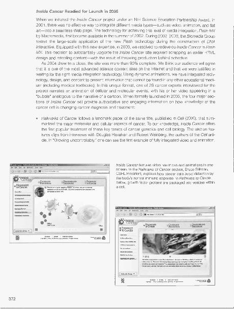
Inside Cancer Readied for Launch in 2005
When we initiated the Inside Cancer project under an NIH Science Education Partnership Award, in2001, there was no effective way to integrate different media types-such as video, animation, and flatart-into a seamless Web page. The technology for achieving this level of media integration, Rash MXby Macromedia, first became available in the summer of 2002. During 2002-2003, the Biomedia Grouptested the large-scale application of the new Rash technology during the construction of DNAInteractive. Equipped with this new expertise, in 2003, we resolved to redevelop Inside Cancer in RashMX. This decision to substantially upgrade the Inside Cancer site required scrapping an earlier HTMLdesign and retooling content-with the result of throwing production behind schedule.
As 2004 drew to a close, the site was more than 90% complete. We think our audience will agreethat it is one of the most advanced science content sites on the Internet and that we were justified inwaiting for the right media integration technology. Using dynamic animations, we have integrated tech-nology, design, and content to present information that cannot be found in any other educational medi-um (including medical textbooks). In this unique format, one of 28 cancer experts interviewed for theproject narrates an animation of cellular and molecular events, with his or her video appearing in a"bubble" analogous to the narrative of a cartoon. When formally launched in 2005, the four major sec-tions of Inside Cancer will provide authoritative and engaging information on how knowledge of thecancer cell is changing cancer diagnosis and treatment:
Hallmarks of Cancer follows a landmark paper of the same title, published in Cell (2000), that sum-marized the major molecular and cellular aspects of cancer. To our knowledge, Inside Cancer offersthe first popular treatment of these key tenets of cancer genetics and cell biology. The section fea-tures clips from interviews with Douglas Hanahan and Robert Weinberg, the authors of the Cell arti-cle. In "Growing Uncontrollably," one can see the first example of fully integrated video and animation.
Inside Cancer features video narrations and animations in onescreen. In the Hallmarks of Cancer section, Bruce Stillman,CSHL President, explains how cancer cells avoid detection bythe body's normal immune response. In Pathways to Cancer,below, growth factor proteins are packaged into vesicles withina cell.
372

Causes and Prevention uses epidemiological data to highlight behaviors and environmental factorsthat increase cancer risk. The overview first debunks the common misconception that inheritanceand toxic chemicals are major causes of cancer. Then users are challenged to link potential causesof cancer to patterns of cancer incidence worldwide. Individual sections highlight how environment,behavior, and inheritance contribute to different major cancers-and how these insights relate topreventing disease. The influences of mold (afflatoxin), diet, viruses, and inheritance are explained inthe context of the key cancers of the liver, prostate, cervix, and colon. Students also explore theevents that spurred the rapid increase in cigarette smoking in the 20th century, which is almostwholly responsible for the continuing "epidemic" of lung cancer.
Diagnosis and Treatment shows how cellular and molecular techniques are used to diagnose andtailor cancer treatment according to specific genetic changes in the patient's tumor. A section onpathology gives site users a chance to try their hand at distinguishing normal and tumor cells, asseen under the microscope, and to follow an endoscope through a patient's colon. In pharmaco-genetics, narrated animations explain how cell receptors and gene profiling are used to tailor treat-ment for breast cancer patients. A section on targeted therapies explain how a new generation ofdrugs block specific molecules that conduct growth signals in the tumor cell-using the examplesof Tamoxifen and Herceptin for breast cancer, and Gleevec for chronic myeloid leukemia.
Pathways to Cancer is a high-quality, three-dimensional animation that follows a common signalingpathway through which growth commands are transmitted from cell surface to the nucleus. The touris narrated by noted voice talent Doug Thomas. The section incorporates a "molecule menu" andfreeze-frame rollovers that allow site users to learn more about the molecules that conduct a growthsignal through the cell and how they are involved in oncogenesis.
NSF Grant to Develop New Laboratories on RNAi
Our effort to strengthen direct ties to CSHL research received additional affirmation inMay when we received a $300,000 grant from the National Science Foundation (NSF) todevelop new laboratories on RNA interference (RNAi) and functional genomics. This pro-ject follows the template of a project initiated last year to develop new experiments inplant genomics. Like the plant project, the RNAi project received unanimous "excellent"ratings from the five reviewers.
During the past 15 years, high school and college biology faculty have implementedkey laboratories that illustrate basic concepts of microbial and molecular genetics. Thecompletion of the Human Genome Project challenges these teachers to move laborato-ry instruction to a higher level of biological integration-the functional analysis of genesand proteins in eukaryotic organisms. Thus, we will develop a module of investigative lab-oratories and bioinformatics exercises that engage students in the new technologies ofRNA-mediated genetic interference and computer-based genome analysis.
Until recently, targeted gene inactivation ("knock-out") or rescue ("knock-in") by homologous recom-bination required significant resources and was confined to research labs. RNAi offers a remarkablysimple system in the roundworm Caenorhabditis elegans. Bacteria are transformed with a plasmidencoding "antisense" RNA to the worm gene of interest, and a lawn of the bacteria is grown on an agarplate. The RNAi response is triggered when worms are released on the agar plate and ingest the trans-formed bacteria expressing the antisense RNA. (Bacteria are a normal food source for C. elegans.) Theeffects of the loss of gene function are then observed in the offspring (either embryos or larvae) of her-maphrodite worms.
We believe that the RNAi/C. elegans experimental system is simple and robust enough to join bac-terial transformation and gel electrophoresis as a mainstay of the high school and college teaching lab-
The C. elegans worm on thelower right illustrates the "blister"phenotype.
373

oratory. After learning basic RNAi techniques, students can make use of freely available resources todesign their own experiments to explore the function of virtually any of the predicted 19,427 genes ofC. elegans. The premise that RNAi labs may prove broadly useful in biology education led to unusualjoint funding of the project by NSF's precollege and collegiate curriculum development programs.
The project is a collaboration among staff at the DNALC, faculty at 2- and 4-year colleges, and sci-entists in Greg Hannon's lab at CSHL, which pioneered RNAi technology. An Advisory Panel of collegefaculty from diverse geographic locations will be involved in all aspects of the project, including devel-opment and testing. Following a year of development, the experiments and computer exercises will betested with 72 biology teachers at three sites around the United States. Instructional and bioinformat-ics resources developed in this project will be disseminated via the DNALC's Internet site, Cold SpringHarbor Laboratory Press, and Carolina Biological Supply Company.
National Science Foundation Fellowships
In summer 2004, two pairs of Faculty-Student Fellows spent 3 weeks at Cold Spring Harbor partici-pating in the NSF-sponsored "Building Leadership to Expand Participation of UnderrepresentedMinorities in Plant Genetics and Genomics" program: Dr. Javier Gozalez-Ramos and Adriana Robbinsof Texas A&M University, and Dr. Olga Kopp and Elise Unice of Utah Valley State College. The Fellowsparticipated in a mix of activities at the Dolan DNALC and the Hazen Genome Sequencing Center.During the first week, Fellows conducted an integrated set of experiments from the NSF-sponsoredGreenomes course. Designed for use in high school and college biology courses, these experimentsuse plant systems to illustrate major concepts of molecular and genomic biology, including the rela-tionship between molecular genotype and phenotype, transposon mutagenesis, chromosome map-ping, homeotic development, functional analysis, and transgene detection. These experiments have
been specifically designed for ease of replication in high school and college teaching labo-ratories. In the fall, we began working with Dr. Ramos and Dr. Kopp to organize regionaltraining workshops on Plant Genetics and Genomics to introduce the plant labs to highschool and college faculty. The workshops-to be held in summer 2005-will be co-taughtby the Fellows and DNALC staff.
A second NSF-sponsored fellowship, "Building Leadership to Develop EducationalBioinformatics Tools in Plant Genomics," was a collaboration between the Dolan DNALC andGramene, an Internet resource for comparative genome analysis of grasses. The mainobjective of this fellowship is to assist with the production of a Student Genome Viewer thatwill allow advanced high school and college classes to directly participate in the annotationof the rice genome. Two fellowship positions were awarded in summer 2004: Mr. RobertWheeler from Pine Creek High School (Colorado Springs, Colorado) and Dr. Debra Burhansfrom Canisius College (Buffalo, New York).
The USDA also provided funds for fellowships at Cold Spring Harbor Laboratory fortwo underrepresented minorities in biological sciences. Cesar Gutierrez, a biology teacher at Jon H.Reagan High School in Austin, Texas and a lead teacher in the NSF-sponsored Bio-Link Center forAdvanced Technological Education at Austin Community College, spent 2 weeks working with DNALCstaff doing advanced development of the Student Genome Viewer. Paul De La Rosa, a biology studentat the Univeristy of Texas, spent 1 week learning about DNA polymorphisms at the DNALC and 1 weeklearning large-scale sequencing techniques at the Hazen Genome Sequencing Center.
NSF Fellows Bob Wheelerand Debra Burhans.
374
Pfizer Leadership Institute
In July, we completed the fourth Leadership Institute under support from the Pfizer Foundation. We hadinitiated this program in the early 1990s with support from the NSF, only to see it wither when the NSFshifted its focus to central school authorities. Pfizer support allowed us to reinstate what we believe isthe most advanced activity to reward the nation's top biology teachers with a summer sojourn at Cold

Participants in the Pfizer Leadership Institute and the NSF Fellowship Program wrap up a 3-week stay in Cold Spring Harbor.
Spring Harbor. However, with the lapse of Pfizer funding in 2005, we will again need to search for sup-
port for this jewel in the DNALC's crown of training courses for biology faculty.The 2004 Pfizer Leadership Institute participants represented a diverse range of backgrounds and
experience. The Institute drew 18 faculty representing 12 different states. One third came from schoolswith high percentages of minority or disadvantaged students, and one third were from rural regions.The American faculty were joined by four teachers from Singapore, who visited as part of our ongoingcollaboration with the Singapore Ministry of Education.
During their 3-week stay, the Leadership participants lived and breathed science-walking in thefootsteps of Nobel Prize winners and taking meals with some of the brightest researchers in the world.The 2004 workshop focused on the study of human and plant genomes. All topics were addressed ina three-pronged approach of lectures/seminars, wet-labs, and computer work. Included in the lab work
was the opportunity to test the DNALC's very latest lab protocols, in advance of their availability at other
institutions or from commercial suppliers. The Institute curriculum also drew heavily on the Laboratory,with scientist seminars and visits to Uplands Farm agricultural field station and the Hazen GenomeSequencing Center. Leadership teachers were introduced to a number of computer-based utilities thatcollect, analyze, and display DNA data, including several developed at the DNALC. A tutorial held bythe DNALC WWW designers introduced participants to technology that enabled them to create per-sonal teaching environments.
Four days were dedicated to independent study or group projects to increase mastery and/or toadapt materials for classroom use. Projects included screening supermarket foods for genetic modifi-cation, sequencing dog mitochondrial DNA, mutating the "glow" gene for green fluorescent protein,and designing a polymerase chain reaction (PCR)-based method to examine polymorphisms in humanolfactory receptor genes. Other teachers expanded computer-based studies on bioinformatics andWeb page construction.
Genetic Origins
Genetic Origins is one of the most unique programs developed here at the DNALC. It consists of anintegrated set of experiments, gratis DNA sequencing, and online tools that allow students to use theirown DNA polymorphisms as the starting point for investigations on human relatedness, evolutionaryhistory, and pharmacogenetics. Genetic Origins presents an accurate analog of human genomeresearch, which incorporates the highest aspirations of "hands-on" learning. This program was initiat-ed with federal grants, but has been maintained with DNALC cash flow during the last several years.
375

To maintain Genetic Origins as a free program to students around the United States and to make need-ed upgrades to the Internet components, we gratefully received a $50,000 grant from our friend andcolleague Mike Gillman, Senior Vice President of Research at Biogen Ides, and a $10,000 grant fromWilliam A. Haseltine, President of the William A. Haseltine Foundation for Medical Science and the Arts.
The redeveloped sites will conform to simple rules for intuitive use: consistent and logical placementof tool bars, minimum use of submenus or "nested" screens, and minimal text. Redevelopment willmake cost-effective use of templates developed for recent DNALC projects and will employ highlydesigned "skins" that give tools and media players the sense of being real onscreen objects. TheGenetics Origins labs will be ported through a lab virtual lab notebook player similar to the GreenomesInternet site we are currently developing. The Bioservers workspaces and data functions will be repack-aged in attractive skins in the manner of Bioinformatics Calculator and Gene Boy.
New Mammalian PCR Labs
Our repertoire of advanced PCR experiments on DNA polymorphisms was significantly strengthenedin 2004. We added two new laboratories that demonstrate the use of single nucleotide polymorphisms(SNPs) in predicting phenotypes. Both experiments use a hybrid method termed amplified restriction-length polymorphism (ampliFLP), in which a PCR product is subsequently cut with a restriction enzyme.A restriction enzyme recognition sequence is incorporated into one PCR primer. Then, an SNP locat-ed at the restriction site is detected as a length polymorphism on an agarose gel.
SNPs and Human Taste examines an SNP in the bitter taste receptor (TAS2R) that correlates withthe ability to taste phenylthiocarbamide (PTC). CSHL researcher Albert Blakeslee determined in 1932that tasting the bitter PTC chemical is inherited in a dominant character. Since that time, it has been aclassic demonstration of Mendelian inheritance in high school genetics classes. However, a new analy-sis, on which our experiment is based, shows that the inheritance of PTC tasting ability is not as sim-ple as students have long been taught. Like many SNPs of medical importance, the SNP we test forcombines with two other SNPs to produce complex combinations (haplotypes) that predict tasting abil-ity-but not perfectly!
NONTASTERALLELE
TASTERALLELE
Human PTC polymorphism genotypes detected by restriction digest (D) of PCR products (U). Genotypes shownare taster/taster (T/T), taster/nontaster (T/N), and nontaster/nontaster (N/N).
376

Canine lvermectin Sensititvity is real test of pharmacogenetic importance to dog owners who givetheir pets lvermectin to control heartworm and other parasites. DNA is isolated from cheek swabs ofpet dogs, amplified by PCR, and examined for an SNP in the mdrl (multidrug resistance) gene. Dogsthat are homozygous for the mdrl mutation cannot metabolize lvermectin and may have a toxic or fatalreaction. Collies, Shetland sheepdogs, Australian shepherds, and Old English sheepdogs are amongthe breeds most at risk for lvermectin sensitivity.
New Visitor Highs in 2004
Visitation to the DNALC facility and to its family of Internet sites reached new highs in 2004. DNALChosted 37,617 visitors, with cumulative visitation topping one-quarter million since our founding in1988! Lab instruction increased 23%, to 27,142 students, with DNALC West allowing us to expand ourreach in Nassau County and New York City. Of these students, 16,903 received instruction at theDNALC or West facilities, and 10,239 received instruction at their own schools. (It is worth noting thatin-school instruction typically amounts to four class visits by a DNALC educator, so that the number ofDNALC-taught labs is several times the number of students reported!)
These numbers keep us solidly in the lead of institutions that offer hands-on instruction in geneticsand DNA for precollege students. Ironically, our next closest competitor is the Singapore ScienceCenter, whose instructional program we helped develop under a partnership with the SingaporeMinistry of Education. In 2004, its first full year of operation, the Science Center provided labs for17,141 students, teachers, and members of the general public!
"Virtual" visitors to the family of Internet sites reached via the DNALC's portal, Gene Almanac,reached 5.44 million-an increase of 11% over 2003. Visitors spent an average of about eight minutesat the informational sites, DNA Interactive and Your Genes, Your Health, and about 10 minutes at ouranimated text, DNA from the Beginning. Visits averaged substantially longer at our historical resource,the Image Archive on the American Eugenics Movement (11.5 minutes) and Bioservers (16 minutes).
Internet SiteAverage visit
length (in minutes) Visits in 2004Increase from
2003 (%)
Gene Almanac 8:23 2,135,981 4.77
DNA from the Beginning 10:06 1,264,919 0.68Your Genes, Your Health 8:48 860,583 15.46
DNA Interactive 8:04 650,096 72.23
Image Archive on the AmericanEugenics Movement 11:25 283,405 14.72
Bioservers 16:06 137,988 4.92
Genetic Origins 9:05 109,022 20.90
All Sites 10:17 5,441,994 11.37
A midyear analysis of our Internet traffic found that only about 10% of visits to Your Genes, YourHealth (YGYH) resulted from referrals by Internet search engines. This was surprising to us, becausethis site contains information on individual genetic diseases that we knew was the subject of manysearches by families and medical professionals. It then dawned on us that the multimedia contentdeveloped in Macromedia Flash was not "visible" to popular search engines, such as Google. "Spiders"and other indexers sent out by search engines could not find content contained in Flash multimediafiles; thus, words in the text of animations and videos in several of most technologically advanced siteswere not being found by relevant Internet searches!
In 2004, we found a solution to this problem. The text files for each animation and video were con-structed into a skeleton of "meta" pages, linked to the homepage, which reflected each site's content
377

and architecture. As indexers revisited the sites, they detected the updated body of text and rebuiltcomprehensive indexes that are now searchable. The results were dramatic. Within 2 months ofimplementing the meta-indexing scheme, searches rose sharply to account for more than 40% of vis-its to YGYH.
Percent of Site Visits Referred by Search Engines
60.00% -
YGYHGene Almanac
50.00% DNAFTBD- - - - Eugenics
Genetic Origins- - - - Bioservers
40.00% DNA Interactive
30.00%."---
20.00%,
10.00%
_____---- -0.00%
Jan-04 Feb-04 Mar-04 Apr-04 May-04 Jun-04 Jul-04 Aug-04 Sep-04 Oct-04 Nov-04 Dec-04 Jan-05
Learning the Effects of Our Summer DNA Camps
We began teaching intensive, week-long "camps" for students in the summer of 1985, when weoffered DNA Science at the Wheatley School in Old Westbury. Since that time, more than 6700 middleand high school students have spent one week at one of seven camps offered each summer.Beginning with Fun With DNA in the fifth grade, a student can take a different workshop each sum-mer-culminating with DNA Science and Vertebrate Genomic Biology in high school. Although weknow that a number of graduates of the summer camp program become interns at the DNALC, wehave long wondered about what the others do after taking one or more of our summer courses. So in2004, we completed a major survey of students who took summer camps from 1990 to 2001.
We received completed surveys from 789 past participants-475 were in high school at the timethey took the survey and 314 were in college. Clearly, the DNALC's workshops cater to a clientele ofinterested and able students. Majorities of secondary school (62%) and college (82%) respondents saidthey had been very interested in science in high school. Virtually all of the students in both groups (97%)said they maintained "A" or "B" averages in their high school science courses.
The DNALC has apparently done a good job of building a clientele for its summer camps. Majoritiesof both groups rated all seven of the DNALC summer courses as very good or excellent. While theaverage college respondent had attended two DNALC camps during his/her precollege years, currentsecondary school students had averaged three camps (with three quarters of respondents still havingone or more years until graduation).
Participation in DNALC camps resulted in a number of beneficial effects for the high school and col-lege respondents. Majorities had discussed topics or issues from a workshop with their families(72-75%) and friends (55-59%). Majorities in both groups said that participation in DNALC workshopshad increased their understanding of science stories in the media, increased their general interest inscience, improved their confidence in science classes, and encouraged them to take more science
378

offerings. Participation in DNALC workshops strongly influenced 46% of college students to considermajoring in science.
Most importantly, the DNALC experience appears to be part of an education that encourages stu-dents to value science and technology and to support scientific research. Note the similar proportionsof high school and college respondents who agree with the following statements:
PercentAgreeing High School College
Science and technology will open up more opportunities for my generation. 93 94
Science and technology are making our lives healthier, easier, and more 92 93
comfortable.
Many scientists want to work on things that will make life better for the 87 88
average person.
The benefits of biotechnology outweigh the risks. 71 73
The United States government spends too much money on scientific research. 16 12
The respondents discriminated in similar ways among a number of different applications of gene tech-nology-favoring most, but drawing the line at the selection of physical traits.
Favoring Percent
Using genetic engineering to produce essential pharmaceuticals. 88 94
Using genetic engineering to treat diseases in adults. 88 94
Using genetic engineering to treat diseases in the embryo. 79 82
Using genetic engineering to produce agricultural crops. 67 72
Using genetic engineering to clone animals, such as sheep, to make drugsand vaccines.
61 68
Using embryonic stem cells in research. 63 64
Using genetic engineering to select for physical traits in the embryo. 21 17
Complete survey results can be viewed at www.dnalc.org/studentsurveys/.
Saturday DNA!
From the DNALC's wealth of offerings, the ongoing, popular Saturday DNA! program has emerged toprovide an in-depth view of our genetic book of life and science education to the general public. Usingcatchy topics such as "CSI: Learning Center" and "Of Lice and Men" as a framework, Saturday DNA!allows children, teens, and adults to conduct hands-on DNA experiments and learn about the latestdevelopments in the biological sciences.
Each 2-hour program is designed to be a thorough and exciting exploration of genetics, genes, andDNA through the use of innovative, intriguing topics. The DNALC staff works to ensure that topics arecurrent and appropriate for two different age groups: groups of students ages 10-13 (with an accompa-nying chaperone) and groups ages 14adult (with accompanying chaperone for participants under 15).
Although programs change and rotate each season, topics such as "Poochie Pedigrees" and "TheMystery of Anastasia Romanov" are mainstays. In examining the disappearance of the Romanov rulingfamily, visitors use a mitochondrial DNA database to determine whether Anna Anderson and AnastasiaRomanov are indeed one and the same. Along the way, they collect evidence and use a variety of
forensic analyses."Poochie Pedigrees" offers the opportunity to extract DNA from your dog's cells and compare it to
379

In the Jumping Genes session of Saturday DNA! in October,participants got a close look at the life and work of NobelPrize recipient Barbara McClintock. Through laboratory andcomputer activities, they learned about the history of classi-cal genetics, and the world of jumping genes.
380
the DNA of different dogs and their ancestors. Analysis between the various samples raises-andanswers-compelling questions such as What is the origin of man's best friend?, Does Fido have wolfor coyote in his bloodlines?, and Are different breeds of dogs really genetically the same? The relatedprogram, "Doggie Diagnosis," works with the older age group to take the information a step further todetermine how breeding has affected your pet's ability (or lack thereof) to metabolize antibiotics.
Staff and Interns
During the summer, we bid farewell to three senior staff members who made significant contributionsto the development of the DNALC. Danielle Sixsmith, Manager of External Relations, moved to NewJersey with her family and began online graduate study at the University of Phoenix. Danielle joinedthe DNALC staff in 1999 as a laboratory instructor, teaching at both the middle and high school lev-els. In 2002, she collaborated with North ShoreLong Island Jewish Health System to bring into oper-ation our satellite facility, DNALC West. Laboratory Manager Scott Bronson left for a position asEducational Programs Administrator at Brookhaven National Laboratory. Scott joined the DNALC in1999, and his prior bench experience with Jacek Skowronski was evident as he simplified PCR foreducation and developed our successful DNA Sequencing Service. Media Producer Bronwyn Terrill leftfor Cambridge, England to manage public outreach at the prestigious Wellcome Trust Sanger Institute.Arriving at the DNALC in 2001, she proved herself to be a world-class science communicator-firstproducing the 1500-square-foot exhibit "The Genes We Share" and then leading production of the
Internet sites DNA Interactive and Inside Cancer.Junior staff members Kim Kessler and Mike O'Brien went in different directions in education. Kim
enrolled in the genetic counseling program at Northwestern University, while Mike joined the New YorkCity Teaching Fellows to prepare for a teaching position at the High School for the Humanities. SeniorDevelopment Officer Erin Wahlgren, who had so effectively run the DNALC golf tournament and annu-
al fund drive, left for a position at the Marfan Foundation.

The major staff changeover brought a raft of new faces and ideas to the DNALC. During the year,we welcomed two Ph.D.-level Laboratory Managers-Tom Bubulya and Craig Hinkley. Although Craigcame from University of Michigan, he had formerly shared a bench with Tom as a postdoctoral fellowin the lab of Winship Herr. Tracy Behar, Jeanette Collette, and Lauren Weidler took positions asLaboratory Instructors. Tracy grew up on Long Island and recently graduated from SUNY College atOld Westbury with a degree in Secondary Education in Biology. Jeanette, who also grew up on LongIsland, has relocated from Buffalo where she attended college and started a family. While working onher degree in human biology from SUNY Albany, Lauren worked as a library aide in the CSHL Library.Local resident Karen Orzel, who has a background in marketing, took over management of theCorporate Advisory Board, annual golf tournament, and annual fund.
Other new additions to the DNALC "family" came in July when middle school educator ElnaCarrasco was married to Michael Gottlieb. Media designer Chun-Hua Yang was married to Shiuh-JyhLin in October. Middle school educator Amanda Mc Brien and her husband, Michael, welcomed their
third son, Robert Michael, in November.High school interns continued to provide key support for our teaching labs, and several carried out
independent research projects under the direction of DNALC and CSHL staff. As part of our new NSFgrant, David Wagman used RNA interference to silence a gene in C. elegans-resulting in visible "blis-ters" in the worm's outer cuticle. Daisy Choi injected the human cancer gene, p53, into C. elegans tostudy tumor suppression. While working in the lab of Dr. Rob Martienssen, Kimberly Izzo used fluores-cent in situ hybridization (FISH) to study centromere function in the model plant Arabidopsis thaliana.
Our sequencing service continues to grow with the help of college interns Alina Duvall (HofstraUniversity) and Andrew Diller (Dowling College). To process the growing number of requests, we nowcollaborate with Dr. Dick McCombie's group at the Woodbury Genome Center. The high-throughputcapillary sequencing available at Woodbury has greatly reduced the turnaround time for completing
sequencing requests.Joining the intern program in 2004 were Andrew Langer (John H. Glen High School, Elwood), Elena
Meliuso (Oyster Bay High School), Tama Mizuno (Northport High School), and Benjamin Tully (NassauCommunity College). Several interns returned from college to assist with summer workshops: LaraAbramowitz (University of Rochester), Yan Huang (Notre Dame University), Marie Mizuno (Binghamton
University), and Alex Witkowski (SUNY Albany).In August, we bid farewell to the following interns as they began their freshman year at college:
Benjamin Blond (Syosset High School) is studying biology at Amherst College; Bryn Donovan (FreeportHigh School) is studying wildlife conservation at the University of Delaware; Alexander Hogg (FriendsAcademy) began a 5-year biology/fine arts program at Brown University; and Michelle Louie (KingsPark High School) began a 7-year biology/M.D. program at George Washington University.
David A. Mick losExecutive Director
381

2004 Workshops, Meetings, and Collaborations
January 6
January 8-12January 10January 24January 26
February 7February 11
January 12
February 21February 24March 6March 10
March 15March 19March 20March 23
March 26March 30April 3April 17April 20
April 26
April 27April 29
May 14
May 15May 20May 31-June 4
June 9June 11June 12June 15June 21-25June 22June 23
June 24
June 28-July 2
382
Site visit by Alastair Balls and Linda Conlon, Life Science Centre, Newcastle, UnitedKingdom
Site visit by Cheong Kam Khow, Singapore Science CenterSaturday DNA! Seminar, DNALCSaturday DNA! Seminar, DNALCInside Cancer interview, Ken Culver, Executive Director, Early Clinical Development,
Novartis Oncology, East Hanover, New JerseySaturday DNA! Seminar, DNALCSite visit and museum tour by members of the Association of Suffolk Superintendents for
Education TechnologySite visit by Alan Fleischman, Senior Vice President, New York Academy of Medicine,
New YorkSite visit by Don McGranaghan, Artistic Resources, LLCSaturday DNA! Seminar, DNALCWest Side School LectureSaturday DNA! Seminar, DNALCSite visit by Cynthia Joyce, Executive Director, and Loren Eng, President, Spinal Muscular
Atrophy Foundation, New YorkWest Side School LectureSaturday DNA! Seminar, DNALCSaturday DNA! Seminar, DNALCSite visit by Jeremiah Barondess, President, and Patricia Volland, Senior Vice President,
The New York Academy of Medicine, New York, and Lawrence Scherr, Dean andChief Academic Officer, North Shore-Long Island Jewish Health System, Great Neck,New York
Site visit by Caroline Lieber, Director, Human Genetics Program, Sarah LawrenceCollege, Bronxville, New York
Site visit and museum tour of DNALC for representatives of "Best of New York" publicationGreat Moments in DNA Science Honors Students Seminar, CSHLSaturday DNA! Seminar, DNALCSaturday DNA! Seminar, DNALCGreat Moments in DNA Science Honors Students Seminar, CSHLSite visit by Ed Rover, President, and Barbara Gill, Vice President, the Dana Foundation,
New YorkSite visit by Jon Fry, President; Bruce Pipes, Provost; Fred Owens, Associate Dean of
Faculty and Professor of Psychology; and Kathy Triman, Professor of Biology, Franklin& Marshall College, Lancaster, Pennsylvania
Presentation at Garden City Public Library, Advocacy for Gifted and Talented Education inNew York State, Inc.
Great Moments in DNA Science Honors Students Seminar, CSHLSite visit by Bryan Sykes, Professor of Human Genetics, University of Oxford, United
KingdomSite visit by Kidgie Williams, diplomats, and family members of UN representatives,
Hospitality Committee for United Nations Delegations, Inc., New YorkSite visit by John Monaghan and Alan Minz, Canadian Consulate General's Office, and
Jay Amer, President, Ontario East Economic Development Corporation, CanadaSaturday DNA! Seminar, DNALCMuseum tour and lab participation by Estee Lauder European Beauty EditorsTeacher-training workshops, National Institute of Education and Singapore Science
Centre, SingaporeSite visit by Caroline Lieber, Sarah Lawrence College, Bronxville, New YorkPresentation of Award of Appreciation from Kings Park Central School District, New YorkSaturday DNA! Seminar, DNALCSite visit by Karl Kuchler, University of Vienna, AustriaNSF Plant Molecular Genetics and Genomics Workshop, Austin Community College, TexasSite visit by Jacinta Duncan, Gene Technology Access Centre, Melbourne, AustraliaInside Cancer interview, William Nelson, The Sidney Kimmel Comprehensive Cancer
Center, and Jennifer E. Axilbund, Genetic Counselor, Cancer Risk AssessmentProgram, The Johns Hopkins Hospital, Baltimore, Maryland
Inside Cancer interview, Louis M. Staudt and Kenneth H. Kraemer, National CancerInstitute, Bethesda, Maryland
Fun With DNA Workshop, DNALC

World of Enzymes Workshop, DNALCDNA Science Workshop, DNALC
July 5-9 Fun With DNA Workshop, DNALCGreen Genes Workshop, DNALCDNA Science Workshop, DNALCGenomic Biology and PCR Workshop, DNALC
July 6 Inside Cancer interview, John Condeelis, Albert Einstein College of Medicine, New YorkJuly 7 Inside Cancer interview, Glorian Sorensen, Harvard School of Public Health, Boston,
MassachusettsJuly 8 Inside Cancer interviews, Nancy Mueller and Graham Colditz, Department of
Epidemiology, and Walter Willett, Department of Nutrition, Harvard School of PublicHealth, Boston, Massachusetts
July 12-16 Bioinformatics in the Classroom Workshop, Union College, Schenectady, New YorkWorld of Enzymes Workshop, DNALCGreen Genes Workshop, DNALCDNA Science Workshop, DNALC
July 12-30 Pfizer Leadership Institute in Human and Plant Genomics, DNALCNSF Faculty Fellowship, Plant and Rice Genomics: Javier Gonzalez-Ramos, Adriana
Robbins, Olga Kopp, Elise Unice, Paul DeLaRosaNSF Faculty Fellowship, Gramene Bioinformatics, Cesar GutierrezTeacher training for Singapore Ministry of Education collaborators, Daniel Chua Wei
Sheong and Yin Leng Tan, and Eugene Wambeck of the Singapore Science CenterJuly 18-23 Fun With DNA Workshop, DNALC
Genetic Horizons Workshop, DNALCJuly 26-30 World of Enzymes Workshop, DNALC
Green Genes Workshop, DNALCJuly 27 Site visit by Arthur Spiro, CSHL Trustee and DNALC Committee Chairman; Russ Hotzler,
John Mogulescu, Nicholas Michelli, Gillian Small, and Tracy Meade, The CityUniversity of New York; Sy Fliegel, Cole Glenn, and Reggie Landeau, Center forEducational Innovation-Public Education Association, New York; Edward Travaglianti,Commerce Bank Long Island, New York; and Lawrence Scherr, North Shore-LongIsland Jewish Health System, Great Neck, New York
July 30 Site visit by members of the American Association of University Women, Washington, D.C.August 2-6 Fun With DNA Workshop, DNALC
World of Enzymes Workshop, DNALCGenetic Horizons Workshop, DNALCDNA Science Workshop, DNALCNSF Plant Molecular Genetics and Genomics Workshop, Clemson University, South
CarolinaAugust 5-6 Site visit by Beverly Matthews, Harvard University, Cambridge, MassachusettsAugust 9-13 Fun With DNA Workshop, DNALC
World of Enzymes Workshop, DNALCGreen Genes Workshop, DNALCNSF Plant Molecular Genetics and Genomics Workshop, University of Wisconsin-
Madison/Madison Area Technical College, Madison, WisconsinAugust 12 Site visit by members of the New York Center for Teacher DevelopmentAugust 16-20 Fun With DNA Workshop, DNALC
World of Enzymes Workshop, DNALCDNA Science Workshop, DNALCGenomic Biology and PCR Workshop, DNALC
August 17 Continuing Medical Education meeting, Lawrence Sherr, Robin Wittenstein, and IreneLeff, North Shore-Long Island Jewish Health System, Great Neck, New York; CarolineLeiber and Jamie Speer, Sarah Lawrence College, Bronxville, New York
August 19 Site visit by Daniel G. Kaufmann, Joan Japha, Chandrika Kulatilleke, Seymour Schulman,Edward Tucker, Mary Jean Holland, and Valerie Schawaroch, faculty members,Department of Natural Sciences, Baruch College, New York
August 20 Inside Cancer interview, Charles Sawyers, University of California, Los AngelesAugust 23-27 Fun With DNA Workshop, DNALC
Genetic Horizons Workshop, DNALCAug. 30-Sept. 3 Fun With DNA Workshop, DNALC
World of Enzymes Workshop, DNALCGenetic Horizons Workshop, DNALC
September 1 Site visit by Jean Caron, Abby Demars, and Holly Herrick, DNA EpiCenter, New London,Connecticut
383

September 6-12
September 16September 25September 30October 15October 16October 21
October 26
October 27November 5
November 8November 9November 9-19
November 13November 15-17November 17
November 18November 20Nov. 22-Dec. 3
Nov. 29-Dec. 10
December 2
384
Tour and lab instruction for members of the Marino Golinelli Foundation, coinstructed byMarina and Marcello Siniscalco of the Marino Golinelli Foundation, Bologna, Italy
Site visit by John Phelan, Trustee and Managing Partner, MSD Capital, New YorkSaturday DNA! Seminar, DNALCSite visit to DNA EpiCenter, New London, ConnecticutSite visit by students from Zhejiang University, Hangzhou, ChinaSaturday DNA! Seminar, DNALCSite visit by Jennifer Heim, Richard Osborn, and Cindy Encarnacion, St. Louis Science
Center, St. Louis, MissouriSite visit by Kevin Ward, George Stranahan, and Andrei Rukinstein, Aspen Institute for
Physics, Aspen, ColoradoWest Side School LectureSite visit by Patricia Volland and Julia Rankin, New York Academy of Medicine, Board of
Education, New YorkSite visit by Glenda Leslie, School of Education at Murdoch University, AustraliaAmerican Association of University Women reception, Washington, D.C.Teacher training for Singapore Ministry of Education collaborators, Tan Hong Kim,
Christine Sim, Chan Ter Yue, and Margaret WongSaturday DNA! Seminar, DNALCSite visit by Abby Demars and Holly Harrick, DNA EpiCenter, New London, ConnecticutWest Side School LectureSite visit by Jacqueline Dorrance, Executive Director, Beckman Foundation, Irvine,
CaliforniaSite visit by Caroline Fostel, Astoria Federal Savings, New YorkSaturday DNA! Seminar, DNALCTeacher-training workshops, National Institute of Education and Singapore Science
Centre, SingaporeTraining for Singapore Ministry of Education collaborators, Mohan Krishnamoorthy, Chng
Swee How James, Andrea Guo Li Ling, Regina Lim, Yap Lih Min Samuel, Goh SuFen, Lim Lay Khim, Sixtus Goh Wee Leng, and Nora Teo
Site visit by staff from DNA EpiCenter, New London, Connecticut

Sites of Major Faculty Workshops 1985-2004
Key: High School College Middle School
ALABAMA University of Alabama, Tuscaloosa 1987-1990ALASKA University of Alaska, Fairbanks 1996ARIZONA Tuba City High School 1988ARKANSAS Henderson State University, Arkadelphia 1992CALIFORNIA California State University, Fullerton 2000
Canada College, Redwood City 1997Contra Costa County Office of Education, Pleasant Hill 2002Foothill College, Los Altos Hills 1997Harbor-UCLA Research & Education Institute, Torrance 2003Laney College, Oakland 1999Lutheran University, Thousand Oaks 1999Pierce College, Los Angeles 1998Salk Institute for Biological Studies, La Jolla 2001San Francisco State University 1991University of California, Davis 1986University of California, Northridge 1993
COLORADO Colorado College, Colorado Springs 1994United States Air Force Academy, Colorado Springs 1995University of Colorado, Denver 1998
CONNECTICUT Choate Rosemary Hall, Wallingford 1987FLORIDA North Miami Beach Senior High School 1991
University of Western Florida, Pensacola 1991Armwood Senior High School, Tampa 1991University of Miami School of Medicine 2000
GEORGIA Fernbank Science Center, Atlanta 1989Morehouse College, Atlanta 1991, 1996Morehouse College, Atlanta 1997
HAWAII Kamehameha Secondary School, Honolulu 1990ILLINOIS Argonne National Laboratory 1986, 1987
University of Chicago 1992, 1997INDIANA Butler University, Indianapolis 1987IDAHO University of Idaho, Moscow 1994IOWA Drake University, Des Moines 1987KANSAS University of Kansas, LawrenceKENTUCKY Murray State University 1988
University of Kentucky, Lexington 1992Western Kentucky University, Bowling Green 1992
LOUISIANA Jefferson Parish Public Schools, Harvey 1990John McDonogh High School, New Orleans 1993
MAINE Bates College, Lewiston 1995Foundation for Blood Research, Scarborough 2002
MARYLAND Annapolis Senior High School 1989Frederick Cancer Research Center, Frederick 1995McDonogh School, Baltimore 1988Montgomery County Public Schools 1990-1992St. John's College, Annapolis 1991
University of Maryland, School of Medicine, Baltimore 1999National Center for Biotechnology Information, Bethesda 2002
MASSACHUSETTS Beverly High School 1986
Biogen, Cambridge 2002Boston University 1994, 1996City Lab, Boston University School of Medicine 1997Dover-Sherborn High School, Dover 1989Randolph High School 1988Winsor School, Boston 1987Whitehead Institute for Biomedical Research, Cambridge 2002
MICHIGAN Athens High School, Troy 1989MISSISSIPPI Mississippi School for Math & Science, Columbus 1990, 1991MISSOURI Stowers Institute for Medical Research, Kansas City 2002
Washington University, St. Louis 1989Washington University, St. Louis 1997
NEW HAMPSHIRE New Hampshire Community Technical College, Portsmouth 1999St. Paul's School, Concord 1986, 1987
NEVADA University of Nevada, Reno 1992NEW JERSEY Coriell Institute for Medical Research, Camden 2003NEW YORK Albany High School 2004
Bronx High School of Science 1987Columbia University, New York 1993Cold Spring Harbor High School 1985, 1987DeWitt Middle School, Ithaca 1991, 1993
385

DNA Learning Center 1988-1995, 2001-2004DNA Learning Center 1990, 1992, 1995, 2000DNA Learning Center 1990-1992Fostertown School, Newburgh 1991Huntington High School 1986Irvington High School 1986Junior High School 263, Brooklyn 1991Lindenhurst Junior High School 1991Mt. Sinai School of Medicine, New York 1997Orchard Park Junior High School 1991Plainview-Old Bethpage Middle School 1991The Rockefeller University, New York 2003State University of New York, Purchase 1989State University of New York, Stony Brook 1987-1990Stuyvesant High School, New York 1998-1999Titusville Middle School, Poughkeepsie 1991, 1993Trudeau Institute, Lake Saranac 2001Union College, Schenectady 2004U.S. Military Academy, West Point 1996Wheatley School, Old Westbury 1985
NORTH CAROLINA CIIT Center for Health Research, Triangle Park 2003North Carolina School of Science, Durham 1987
OHIO Case Western Reserve University, Cleveland 1990Cleveland Clinic 1987North Westerville High School 1990
OKLAHOMA Oklahoma City Community College 2000Oklahoma Medical Research Foundation, Oklahoma City 2001Oklahoma School of Science and Math, Oklahoma City 1994
OREGON Kaiser Permanente-Center for Health Research, Portland 2003PENNSYLVANIA Duquesne University, Pittsburgh 1988
Germantown Academy 1988SOUTH CAROLINA Clemson University, Clemson 2004
Medical University of South Carolina, Charleston 1988University of South Carolina, Columbia 1988
TEXAS Austin Community College, Rio Grande Campus 2000J.J. Pearce High School, Richardson 1990Langham Creek High School, Houston 1991Southwest Foundation for Biomedical Research, San Antonio 2002Taft High School, San Antonio 1991Trinity University, San Antonio 1994University of Texas, Austin 1999, 2004
UTAH University of Utah, Salt Lake City 1993University of Utah, Salt Lake City 1998, 2000
VERMONT University of Vermont, Burlington 1989VIRGINIA Eastern Mennonite University, Harrisonburg 1996
Jefferson School of Science, Alexandria 1987Mathematics and Science Center, Richmond 1990Mills Godwin Specialty Center, Richmond 1998
WASHINGTON Fred Hutchinson Cancer Research Center, Seattle 1999, 2001University of Washington, Seattle 1993, 1998
WASHINGTON, D.0 Howard University 1992, 1996WEST VIRGINIA Bethany College 1989WISCONSIN Blood Center of Southeastern Wisconsin, Milwaukee 2003
Madison Area Technical College 1999Marquette University, Milwaukee 1986, 1987University of Wisconsin, Madison 1988, 1989University of Wisconsin, Madison 2004
WYOMING University of Wyoming, Laramie 1991
AUSTRALIA Walter and Eliza Hall Institute and University of Melbourne 1996CANADA Red River Community College, Winnipeg, Manitoba 1989ITALY Porto Conte Research and Training Laboratories, Alghero 1993
International Institute of Genetics and Biophysics, Naples 1996PANAMA University of Panama, Panama City 1994PUERTO RICO University of Puerto Rico, Mayaguez 1992
University of Puerto Rico, Mayaguez 1992University of Puerto Rico, Rio Piedras 1993University of Puerto Rico, Rio Piedras 1994
RUSSIA Shemyakin Institute of Bioorganic Chemistry, Moscow 1991SINGAPORE National Institute of Education 2001-2004SWEDEN Kristineberg Marine Research Station, Fiskebackskil 1995
Uppsala University, Uppsala 2004
386

COLD SPRING HARBORLABORATORY PRESS

2004 PUBLICATIONS
SERIALS
Genes & Development, Vol. 18, 1-3164(www.genesdev.org)
Genome Research, Vol. 14, 1-2536(www.genome.org)
Learning and Memory, Vol. 11, 1-804(www.learnmem.org)
Protein Science, Vol. 13, 1-3382(www.proteinscience.org)
RNA, Vol. 10, 1-1986(www.rnajournal.org)
Cold Spring Harbor Symposia in Quantitative BiologyVol. 68: The Genome of Homo sapiens(www.cshl-symposium.org)
LABORATORY MANUALS
Live Cell Imaging: A Laboratory Manual, David Spectorand Robert Goldman (eds.), "Live Cell Imaging" (DVD)
Imaging in Neuroscience and Development: A Labor-atory Manual, Rafael Yuste and Arthur Konnerth (eds.)
HANDBOOKS
Fly Pushing, 2nd edition, Ralph J. Greenspan
An Illustrated Chinese-English Guide for BiomedicalScientists, James Samet, Weidong Wu, Yuh-Chin T.Huang, and Xinchao Wang(www.chinese-english.cshl.org)
At the Bench, updated edition, Kathy Barker
Mouse Phenotypes: A Handbook of Mutation Analysis,Virginia E. Papaioannou and Richard R. Behringer
Drosophila: A Laboratory Handbook, MichaelAshburner, Scott Hawley, and Kent Golic
388
TEXTBOOKS
A Genetic Switch (3rd ed.), Phage Lambda Revisited,Mark Ptashne
Bioinformatics: Sequence and Genome Analysis (2nded.), David Mount(www.bioinformaticsonline.org)
MONOGRAPHS
Cell Growth: Control of Cell Size, Michael N. Hall,George Thomas, and Martin Raff (eds.)
Gastrulation: From Cells to Embryo, Claudio Stern (ed.)(www.gastrulation.org)
GENERAL INTEREST
Is It in Your Genes? The Influence of Genes on CommonDisorders and Diseases That Affect You and YourFamily Philip R. Reilly(www.is-it-in-your-genes.org)
Mendel's Legacy: The Origin of Classical Genetics, ElofAxel Carlson
The Writing Life of James D. Watson, Errol C. Friedberg
OTHER
Banbury Center Annual Report 2003
CSHL Annual Report 2003
CSHL Annual Report 2003: Yearbook Edition
Watson School of Biological Sciences Annual Report 2003
WEB SITES
www.BioSupplyNet.com, online directory of scientificsupplies and suppliers
A selection of recently published books
The journal publishing program

COLD SPRING HARBOR LABORATORY PRESSEXECUTIVE DIRECTOR'S REPORT
Cold Spring Harbor Laboratory Press is the largest of the five educational divisions of the Laboratory,with a staff of 49, offices on the Woodbury campus, and marketing operations based in San Diego,California and Oxford, United Kingdom. In 2004, the Press had income of $9.5 million and made a totalcontribution to the Laboratory's economy of more than $1.2 million.
Our research journals, books, and manuals assist the continuing professional education of workingscientists and graduate students, our textbooks educate college undergraduates, and our other booksdelve into the practice, personalities, and history of science and its influence on medicine, business,and social policy.
Journals
Two thirds of Press income now comes from journal publishing. In 2004, against a downward industrytrend, institutional subscriptions to our five journals increased by 4%. Countless numbers of individualreaders online downloaded more than 110,000 articles each month in countries around the world.Scientists in the poorest countries are able to access these articles free of charge. More than 12,000journal pages were published, an increase of over 6%, containing 1200 new articles. Many of thesewere highlighted in the world's major news media, including The New York Times, The Wall StreetJournal, and the BBC News. All five journals had impact factors that were similar to or higher than lastyear's, and Genes & Development remained one of the ten most highly cited of all primary researchjournals in science.
Both RNA and Protein Science, journals published under contract for the RNA and ProteinSocieties, respectively, continued to reach new levels of institutional subscriptions. Our own journalLearning & Memory received a record number of manuscript submissions and, as a result, began pub-lishing monthly issues online and bimonthly issues in print.
Throughout the year, the talk among journal publishers was of an alternative to subscriptions as abusiness model. "Open access" publishing is paid for by authors, rather than readers, and researchpapers are made available free of charge online immediately on publication. The model is new andunproven as a means of sustaining an important journal without grant support or private underwriting,but to assess support, Genome Research has introduced an immediate free-access option for individ-ual papers. The archives of our journals have been freely accessible online for over 5 years, and thisyear, the interval between publication and the free release of papers published in Genes & Developmentand Genome Research was reduced from 1 year to 6 months. All three Cold Spring Harbor journalsare also freely accessible through PubMedCentral.
Some organizations that fund research have begun to voice opinions on the access policies of jour-nals that publish that research. In the United States, the largest supporter of biomedical research is theNational Institutes of Health (NIH). Its Director, Dr. Elias Zerhouni, presented a proposal for a databasethat would provide free access to papers describing NIH-funded research within a very short time ofpublication. In the intense debate among publishers and scientists that followed, the public benefit ofsuch access was weighed against the potential harm to the financial viability of many subscription-based journals, particularly those published by not-for-profit organizations that use that income to sup-port other activities. I was one of a group of publishers who met with Dr. Zerhouni to discuss and clar-ify the details and intent of his proposal. The final version of the NIH policy requires the deposition offinal manuscripts, rather than published papers, within a period of less than 1 year after acceptancethat the authors themselves must decide. The scene is set for conflict between society publishers andtheir members.
389

Books and Electronic Media
Laboratory protocols--instructions for executing research techniques-have been the core of our bookpublishing program for more than 20 years in the form of laboratory manuals widely regarded as thebest in their fields. In 2004, a partnership was struck with the new journal Nature Methods, in whichPress protocols reached a large international audience through two new channels: as a section in eachmonthly issue of the journal and through access by the journal's institutional subscribers to the onlineversion of our best-selling manual Molecular Cloning.
There were two manuals among the 15 new books published in 2004, a list (see p. 388) that includ-ed monographs, handbooks, textbooks, general interest and history books, as well as the AnnualSymposium volume, this year devoted to the human genome. The Symposium series, first publishedin 1933, is one of the oldest and most prestigious in biology. For the first time, individual chapters ofthe proceedings were published online in advance of the printed volume on a Web site that gave view-ers a taste of what was happening as the conference actually took place. Combinations of traditionalprint and digital media were further explored in the manual Live Cell Imaging, which includes a DVDshowing cells in action, and the monograph Gastrulation, whose Web site contains movies of thisimportant event in embryonic development.
Such online enhancements are particularly necessary for textbooks that compete for adoption incrowded markets. Our book Bioinformatics: Sequence and Genome Analysis, by David Mount, cap-tured this emergent field on first publication in 2001 and regained its lead with this year's second,extensively revised edition. And the newly released Molecular Biology of the Gene, the first new edition
in 17 years, also made a strong showing, through the sales expertise of our co-publisher BenjaminCummings.
Our list of professional handbooks grew still further with the addition of new editions of Drosophila,At The Bench, and Fly Pushing, as well as a new guide to mutation analysis in mice and a referencebook for students whose first language is Chinese. Our other books included an ambitious attempt byPhilip Reilly to provide a guide for general readers to the genetics of many complex human diseases.In addition, in The Writing Life of James D. Watson, Errol Friedberg reminded us that the chancellor ofthis Laboratory, world famous as a scientist, also has an extraordinary variety of accomplishments asan author.
It was gratifying to note the frequency and warmth of the reviews of our 2003 and 2004 books. Onereviewer remarked that "Cold Spring Harbor books are usually exceptional"-nicely summarizing ourhopes for all our titles. The biography of geneticist George Beadle, The Uncommon Farmer, by PaulBerg and Maxine Singer, was particularly acclaimed and named one of Choice magazine's top booksof the year. There was also widespread praise for Elof Carlsson's Mendel's Legacy, an exploration ofthe origins of classical genetics. Recognition of the quality of our program has led to an ever-increas-ing number of foreign translations, and in 2004, 15 such agreements were signed for Chinese,Japanese, Korean, and Italian editions.
In South Africa, our 2001 book for teenagers about HIV/AIDS received intense scrutiny from adiverse group of teachers, students, and community workers in rural and urban areas as we workedto tailor a second edition of the book to evolving public attitudes toward the epidemic and the avail-ability of treatment options. Reflecting these changes, Staying Alive has a new, bolder title, You, Meand HIV, but the entirely revised content retains the unique, engaging style of its creators Fran Balkwilland Mic Rolph. With the assistance of a grant from the Gates Foundation, 50,000 copies in Englishand 20,000 copies in Zulu and Afrikaans will be printed for free distribution to schools and health orga-nizations within South Africa, along with a guide to teaching from the book.
In a generally busy year, 25 contracts for new books were signed, including two exciting and inno-vative textbooks. In addition, the online directory of laboratory supplies and suppliers, BioSupplyNet.com, which we reacquired in 2003, was relaunched with a much improved design and new, attractivefunctions that generated substantial advertising revenue. These and other initiatives help pave the wayfor future growth. Other innovations increased efficiency, allowing us to do more with current staff andresources. Significant savings were gained through outsourcing composition and establishing relation-
390

ships with printers abroad, and by further development of electronic data interchange for managingthousands of book orders and journal subscriptions.
Catalogs and exhibits remain important to our marketing strategies, but more extensive use is beingmade of online book and journal promotion. Sales through our own Web site continue to grow, but anever-larger proportion of our books, as much as 60%, now reach our scientist customers throughresellers, particularly Amazon.com, with its extraordinary market penetration. Our roster of wholesalecustomers, international agents, and distributors continues to expand. Scion Publishing, the UnitedKingdom company trading as CSHL Press Europe, had an encouraging first year, meeting sales pro-jections. It will shortly begin publishing its own books that the Press will distribute in the United States.This arrangement is made possible by our professionally staffed customer service and warehouseoperations, which in 2004 processed orders for and shipped 60,000 books and managed inventory toover 99% accuracy. Another recently founded publishing house, Roberts Publishing, has also con-tracted with us for fulfillment services in the United States.
After the financial challenges of the previous year, I am glad to be able to conclude that 2004 wasin contrast a year of growth, new initiatives, and increased confidence for Cold Spring HarborLaboratory Press as we continued to support the Laboratory's educational mission with first-class pub-lications for scientists, students, and the public worldwide.
Staff
The staff members of the Press as of December 2004 are listed elsewhere in this volume. They bringa cheerful commitment to their work that makes them a pleasure to be with.
In 2004, we welcomed five new colleagues: Laureen Connell, Hillary Sussman, Maria Smit, KathyBubbeo, and Mary Mulligan. We also said goodbye to Laurie Goodman, Jill Manero, Joan Pepperman,
and Melissa Frey.In recent years, the activities of the Press have grown in diversity and complexity. The success of
this growth is due to a talented and motivated group of departmental managers: Jan Argentine, IngridBenirschke, Kathy Cirone, Kathryn Fitzpatrick, Nancy Hodson, Geraldine Jaitin, Bill Keen, Guy Keyes,Marcie Siconolfi, Linda Sussman, and Denise Weiss. They have all taken on new responsibilities withlittle or no extra help or reward. The Press owes much to them and also to the energy and flair of theeditors of our journals, Terri Grodzicker at Genes & Development, and Laurie Goodman and her suc-cessor Hillary Sussman at Genome Research. It is also a pleasure to acknowledge the indispensableElizabeth Powers who carries out all manner of essential activities in our office with quiet grace.
John R. InglisExecutive Director
391


:1;00

FINANCIAL STATEMENTS
CONSOLIDATED BALANCE SHEETDecember 31, 2004With comparative financial information as of December 31, 2003
Assets: 2004 2003Cash and cash equivalents 40,744,417 28,971,185Accounts receivable:
Publications 1,308,718 1,257,221Other 1,179,604 1,332,279
Grants receivable 6,975,455 7,068,387Contributions receivable, net 12,492,687 11,452,991
Publications inventory 2,945,099 2,206,640Prepaid expenses and other assets 1,605,707 1,566,991Investments 211,294,366 198,696,165Investment in employee residences 4,830,425 4,774,834Restricted use assets 2,904,923 2,709,576Land, buildings, and equipment, net 110,757,023 109,253,408
Total assets $ 397,038,424 369,289,677
Liabilities and Net Assets:Liabilities:
Accounts payable and accrued expenses $ 7,429,657 5,975,281
Notes payable 107,775 140,958Deferred revenue 3,878,738 4,067,960Bonds payable 45,200,000 45,200,000
Total liabilities 56,616,170 55,384,199
Net assets:Unrestricted 184,397,408 178,415,767
Temporarily restricted 25,419,056 14,223,309
Permanently restricted 130,605,790 121,266,402
Total net assets 340,422,254 313,905,478
Total liabilities and net assets $ 397,038,424 369,289,677
394

CONSOLIDATED STATEMENT OF ACTIVITIESYear ended December 31, 2004With summarized financial information for the year ended December 31, 2003
UnrestrictedTemporarilyRestricted
PermanentlyRestricted
2004Total
2003Total
Revenue and other support:Public support (contributions and
nongovernment grant awards) $ 10,582,015 17,086,618 1,159,665 28,828,298 29,792,003Government grant awards 29,451,302 29,451,302 26,878,120Indirect cost allowances 19,089,893 - - 19,089,893 18,000,004Program fees 3,644,734 - 3,644,734 3,134,443Publications sales 9,743,639 - 9,743,639 10,052,963
Dining services 3,183,440 3,183,440 2,927,668Rooms and apartments 2,632,671 2,632,671 2,347,090Royalty and licensing fees 926,913 - - 926,913 869,826Investment income (interest and
dividends) 3,134,480 - 3,134,480 4,141,495
Miscellaneous 514,559 - 514,559 616,755
Total revenue 82,903,646 17,086,618 1,159,665 101,149,929 98,760,367
Net assets released from restrictions 5,890,871 (5,890,871)
Total revenue and other support 88,794,517 11,195,747 1,159,665 101,149,929 98,760,367
Expenses:Research 52,049,866 - - 52,049,866 45,619,807
Educational programs 13,238,766 - - 13,238,766 12,917,185
Publications 9,380,833 - - 9,380,833 10,631,334
Banbury Center conferences 1,159,499 - 1,159,499 1,136,159
Dolan DNA Learning Center programs 1,611,325 - - 1,611,325 1,809,174
Watson School of Biological Sciencesprograms 2,263,518 - 2,263,518 2,029,275
General and administrative 12,508,768 - 12,508,768 11,976,718
Dining services 4,236,535 4,236,535 4,262,515
Total expenses 96,449,110 96,449,110 90,382,167
(Deficiency) excess of revenue andother support over expenses
(7,654,593) 11,195,747 1,159,665 4,700,819 8,378,200
Other changes in net assets:Net appreciation in fair value
of investments13,636,234 8,179,723 21,815,957 38,128,865
Increase in net assets 5,981,641 11,195,747 9,339,388 26,516,776 46,507,065
Net assets at beginning of year 178,415,767 14,223,309 121,266,402 313,905,478 267,398,413
Net assets at end of year $ 184,397,408 25,419,056 130,605,790 340,422,254 313,905,478
395

CONSOLIDATED STATEMENTS OF CASH FLOWSYear ended December 31, 2004With comparative financial information for the year ended December 31, 2003
2004 2003
Cash flows from operating activities:Increase in net assets $ 26,516,776 46,507,065
Adjustments to reconcile increase in net assetsto net cash used in operating activities:
Depreciation and amortization 5,769,622 5,458,719
Net appreciation in fair value of investments (21,746,064) (37,868,190)
Contributions restricted for long-term investment (13,812,552) (13,460,462)
Changes in assets and liabilities:Decrease (increase) in accounts receivable 101,178 (459,802)
Decrease (increase) in grants receivable 92,932 (1,912,888)
Decrease (increase) in contributions receivable 79,666 (875,375)
(Increase) decrease in publications inventory (738,459) 7,859
(Increase) decrease in prepaid expenses and other assets (38,716) 63,375
(Increase) decrease in restricted use assets (195,347) 355,653
Increase in accounts payable and accrued expenses 1,454,376 802,963
(Decrease) increase in deferred revenue (189,222) 419,507
Net cash used in operating activities (2,705,810) (961,576)
Cash flows from investing activities:Capital expenditures (7,273,237) (6,296,049)
Proceeds from sales and maturities of investments 113,998,317 135,393,757Purchases of investments (104,850,454) (122,075,553)Net change in investment in employee residences (55,591) (427,207)
Net cash provided by investing activities 1,819,035 6,594,948
Cash flows from financing activities:Permanently restricted contributions 1,159,665 4,822,932
Contributions restricted for investment in land, buildings,
and equipment 12,652,887 8,637,530
Increase in contributions receivable (1,119,362) (6,092,438)
Repayment of notes payable (33,183) (29,161)
Net cash provided by financing activities 12,660,007 7,338,863
Net increase in cash and cash equivalents 11,773,232 12,972,235
Cash and cash equivalents at beginning of year 28,971,185 15,998,950
Cash and cash equivalents at end of year $ 40,744,417 28,971,185
Supplemental disclosures:Interest paid $ 780,758 1,014,956
396

COMPARATIVE OPERATING HISTORY 2000-2004(Dollars in Thousands)
2000 2001 2002 2003 2004
Revenue:
Main Lab:Grants and contracts $ 30,345 34,716 37,872 41,749 48,208
Indirect cost allowances 12,718 14,134 14,987 17,869 18,910
Other 10,618 12,528 10,918 10,524 11,611
CSHL Press 8,684 9,941 9,051 10,053 9,744
Banbury Center 1,856 1,666 1,763 1,729 1,779
Dolan DNA Learning Center 1,471 1,878 2,978 2,564 2,314
Watson School of Biological Sciences 682 927 1,496 1,769 1,889
Total revenue 66,374 75,790 79,065 86,257 94,455
Expenses:
Main Lab:Research and training 30,345 34,716 37,872 41,749 48,208
Operation and maintenance of plant 6,589 7,027 8,661 8,702 8,606
General and administrative 6,162 6,492 6,395 7,507 7,836
Other 7,075 9,505 8,550 8,959 9,410
CSHL Press 8,186 9,515 8,962 10,234 8,995
Banbury Center 1,702 1,536 1,597 1,616 1,642
Dolan DNA Learning Center 1,362 1,801 2,780 2,257 2,066
Watson School of Biological Sciences 682 927 1,496 1,769 1,880
Total expenses excluding depreciation 62,103 71,519 76,313 82,793 88,643
Excess before depreciation and(designation) release of funds 4,271 4,271 2,752 3,464 5,812
Depreciation (3,974) (4,620) (5,165) (5,459) (5,770)
(Designation) release of funds 1 (297) 349 1,848 - -Net operating excess (deficit) $ - - (565) (1,995) 42
The above amounts are presented on a combined basis for all funds for which Cold Spring Harbor Laboratory prepares operating budgets.
'Funds designated to underwrite future direct and indirect expenses of new research programs.
Federal Grants44.4%
COLD SPRING HARBOR LABORATORYSOURCES OF REVENUE
DECEMBER 31, 2004
Interest andMiscellaneous
0.4%
Royalty and Licensing1.8%
CorporateContributions and
Grantsn drodwprnees aatnedd5.0%
'a Funds7.5%
Foundation and PrivateContributions
21.9%
Auxiliary Activities19.0%
397

FINANCIAL SUPPORT OF THE LABORATORY
Cold Spring Harbor Laboratory, Banbury Center, and the Dolan DNA Learning Center receive a sub-stantial portion of their funding through grants from the Federal Government and through grants, cap-ital gifts, and annual contributions from private foundations, corporations, and individuals. The follow-ing section summarizes funding that occurred during 2004.
GRANTS January 1, 2004-December 31, 2004
COLD SPRING HARBOR LABORATORY GRANTS
Grantor Program/Principal Investigator Duration of Grant 2004 Funding*
FEDERAL GRANTSNATIONAL INSTITUTES OF HEALTH
Equipment Dr. Myers 04/01/04 03/31/05 $ 211,975 *
Dr. Stillman 09/01/04 08/31/05 984,159 *
Program Projects Dr. Herr 01/01/97 12/31/06 5,041,821Dr. Stillman 08/01/87 07/31/05 3,855,008
Research Support Dr. Brody 07/01/04 04/30/09 304,650 *
Dr. Chklovskii 07/08/04 06/30/09 304,650 *
Dr. Cline 12/10/04 11/30/09 484,030 *
Dr. Cline 03/01/98 03/31/06 452,805Dr. Dubnau 09/15/04 06/30/09 349,800 *
Dr. Hamaguchi 04/01/04 03/31/08 346,707 *
Dr. Hannon 07/01/00 08/31/05 289,690Dr. Hernandez 07/01/04 06/30/08 245,413 *
Dr. Herr 03/01/02 02/28/06 298,800Dr. Hirano 07/01/01 06/30/05 275,560Dr. Hirano 05/01/96 04/30/08 406,000Dr. Huang 08/01/01 06/30/06 415,000Dr. Joshua-Tor 05/01/01 03/31/06 373,500Dr. Joshua-Tor 02/15/02 01/31/07 373,500Dr. Krainer 07/01/03 06/30/07 535,378Dr. Krainer 06/01/01 05/31/06 408,400Dr. Lazebnik 09/01/03 08/31/05 211,666Dr. Lowe 070/1/04 06/30/09 390,832Dr. Lowe 04/01/04 03/31/09 849,999Dr. Mainen 09/23/02 08/31/07 370,813Dr. Malinow 05/01/92 04/30/05 565,477Dr. Malinow 04/01/95 02/29/08 553,671Dr. Martienssen 08/01/03 07/31/07 338,584Dr. Mittal 04/01/04 03/31/09 312,036Dr. Muthuswamy 03/01/03 02/28/07 376,211Dr. Neuwald 09/30/98 08/31/06 373,500Dr. Skowronski 04/01/98 04/30/08 640,187Dr. Spector 04/01/90 03/31/07 652,102Dr. Spector 09/01/04 08/31/08 363,220Dr. Stein 09/01/02 06/30/05 617,776Dr. Stillman 07/01/91 05/31/08 555,907Dr. Svoboda 12/01/03 11/30/08 313,498Dr. Svoboda 12/01/02 11/30/07 281,724
Includes direct and indirect costs.*New grants awarded in 2004.
398

Dr. Svoboda 06/01/03 03/31/08 $ 295,969Dr. Svoboda 03/01/04 02/28/09 169,080
Dr. Tansey 05/01/03 04/30/07 337,365
Dr. Tonks 08/01/91 03/31/06 646,533
Dr. Tonks 05/01/97 06/30/05 348,600
Dr. Tully 03/01/03 02/29/08 380,438
Dr. Tully 08/01/03 05/31/07 380,719
Dr. Van Aelst 05/01/03 04/30/08 338,756Dr. Wig ler 07/15/98 04/30/07 502,725
Dr. Xu 08/01/01 07/31/05 252,320
Dr. Xu 08/01/03 03/31/07 363,619
Dr. Zador 01/23/03 12/31/07 422,500
Dr. M. Zhang 09/19/03 08/31/06 584,205
Dr. Zhong 07/01/03 06/30/08 346,540
Fellowships Dr. Ding 07/01/04 06/30/07 42,976
Dr. Kuhlman 12/01/02 11/30/05 48,928
Dr. Lucito 06/01/02 05/31/07 140,867
Dr. Stepanyants 07/01/04 06/30/09 114,831
Dr. Zilfou 07/01/04 06/30/05 48,928
Course Support Advanced Immunocytochemistry In Situ 07/01/98 08/31/05 76,766
Hybridization and Live Cell ImagingC. elegans 08/01/98 08/01/06 67,152
Cancer Workships 01/01/83 03/31/06 308,282
Cell and Developmental Biology of 04/01/93 03/31/09 19,440
XenopusComputational Genomics Course 06/06/91 08/31/07 46,284
CSHL Neurobiology Short-term Training 07/01/01 06/30/06 160,527
Genome Informatics 07/01/00 06/30/05 50,728
Macromolecular Crystallography 09/01/00 08/31/05 56,773
Making And Using DNA Microarrays 07/01/04 55,635
Proteomics 07/01/03 06/30/06 66,792
Meeting Support 69th Symposium: Epigenetics 05/15/01 04/30/06 5,000
69th Symposium: Epigenetics 06/04/04 05/31/05 11,000
Axon Guidance and Neural Plasticity 06/01/00 05/31/05 19,000
Cancer Genetics Conference 08/01/00 07/31/05 15,000
Cell Cycle 04/01/00 03/31/05 9,000
Evolution of Developmental Diversity 04/17/02 03/31/05 13,000
Gene Expression and Signaling in the 04/19/02 03/31/07 2,000
Immune SystemGerm Cells 07/01/04 06/30/05 6,000
Molecular Chaperones and the Heat 04/01/04 03/31/09 13,000
Shock ResponseMouse Molecular Genetics 07/12/02 06/30/07 17,962
Pharmacogenomics 08/10/04 07/31/09 18,683 *
Translational Control 07/05/02 06/30/07 6,000
NATIONAL SCIENCE FOUNDATION
Research Support Dr. Hannon 09/01/04 02/28/06 200,000 *
Dr. Jackson 03/01/04 02/28/07 130,000 *
Dr. Jackson 08/01/02 07/31/05 120,000Dr. Jackson 10/01/01 09/30/06 284,058Dr. McCombie 09/01/03 08/31/06 1,303,762Dr. McCombie 06/15/04 11/30/05 199,949 'Dr. Stein 09/01/03 08/31/06 556,369Dr. Stein 12/15/03 11/30/07 1,591,214Dr. Timmermans 07/01/02 06/30/05 123,562
+Includes direct and indirect costs.`New grants awarded in 2004.
399
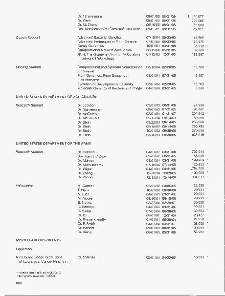
Dr. TimmermansDr. WareDr. M. ZhangDrs. Martienssen/McCombie/Stein/Lucito
09/01/0309/01/0309/15/0309/01/01
08/31/0608/31/0808/31/0808/31/05
$ 120,077290,586
65,000519,921
Course Support Advanced Bacterial Genetics 07/15/04 06/30/09 145,659 "Advanced Techniques in Plant Science 07/01/04 06/30/05 76,875 *
Cereal Genomics 10/01/04 03/31/06 36,030 *
Computational Neuroscience Vision 06/15/04 05/31/05 52,000 *
RCN: The Gramene Community Curation 01/15/03 12/31/05 183,087Network 4 Workshops
Meeting Support Computational and Systems Neuroscience 03/15/04 02/28/05 13,780 *
(Cosyne)Plant Genomes: From Sequence
to Phenome08/01/04 07/31/05 15,187 *
Evolution of Developmental Diversity 03/01/04 02/28/05 16,763 *
Molecular Genetics of Bacteria and Phage 04/01/04 03/31/05 5,000 *
UNITED STATES DEPARTMENT OF AGRICULTURE
Research Support Dr. Jackson 09/01/03 08/31/06 74,988Dr. Martienssen 08/01/03 07/31/05 96,402Dr. McCombie 02/01/04 01/31/07 201,842 *
Dr. McCombie 09/15/04 09/14/09 50,000 "
Dr. Stein 09/22/03 09/14/05 250,000Dr. Stein 09/15/02 09/14/05 783,785Dr. Stein 10/01/02 09/30/05 220,000Dr. Stein 03/16/03 09/30/05 838,016
UNITED STATES DEPARTMENT OF THE ARMY
Research Support Dr. Hannon 04/01/02 03/31/06 732,644Drs. Hannon/Lowe 09/01/02 08/31/05 166,000Dr. Mainen 04/01/04 03/31/06 199,969 *
Dr. Muthuswamy 07/15/04 07/14/05 126,953 *
Dr. Wig ler 04/01/04 03/31/06 1,784,200 *
Dr. Zhong 12/30/04 12/29/05 100,000 *
Dr. Zhong 12/15/04 12/14/08 334,511 *
Fellowships M. Carmel 05/01/02 04/30/05 22,000T. Haire 10/01/04 09/30/06 29,991A. Lucs 04/01/03 03/31/06 29,991M. Moore 08/16/04 08/15/06 29,991S. Nunez 02/27/04 02/28/07 30,000K. Siddiqui 04/01/03 03/31/06 29,991D. Siolas 07/01/04 06/30/06 30,304Dr. Du 06/01/02 12/31/04 20,427Dr. Kannanganattu 01/01/01 06/30/04 27,858Dr. R Smith 04/01/04 03/31/07 108,000Dr. Wendel 09/01/04 08/31/05 100,000Dr. Xiang 06/01/03 05/31/06 56,984
MISCELLANEOUS GRANTS
Equipment
NYS Grand Lodge Order Sonsof Italy/Breast Cancer Help, Inc.
Dr. Stillman 05/01/04 04/30/05 10,000 *
*Includes direct and indirect costs.'New grants awarded in 2004.
400

Research Support
AACR-National Foundation for Cancer Dr. Lowe 07/01/04 06/30/05 $ 50,000 *
Research Professorship in BasicCancer Research
AKC Canine Health Foundation Dr. McCombie 07/19/01 07/20/04 33,000American Cancer Society Dr. Wigler 01/01/96 12/31/04 10,000
American Cancer Society Dr. Wig ler 01/01/01 12/31/05 70,000
The Michael Scott Barish Human Dr. Wig ler 01/01/04 12/31/04 70,025Cancer Grant sponsored by 1 in 9:The Long Island Breast CancerAction Coalition
Breast Cancer Research Foundation Dr. Wig ler 10/01/00 09/30/05 250,000Dorothy Rodbell Cohen Foundation
for Cancer Research Dr. Tonks 06/14/04 06/15/05 20,000 *
The Charles A. Dana Foundation, Inc. Dr. Cline 10/01/03 03/31/05 34,006The Charles A. Dana Foundation, Inc. Dr. Van Aelst 04/01/03 09/30/04 33,773
Dart Neurogenomics Project Dr. Tully 10/01/04 12/31/04 200,000 *
De Matteis Family Foundation Dr. Powers 10/01/04 09/30/05 268,000 *
ELJI/SUSB Consortium Agreement Dr. Huang 01/15/04 01/14/07 70,481 *
Find A Cure Today (F.A.C.T.) Dr. Muthuswamy 07/01/03 02/28/05 39,040FSMA Dr. Krainer 07/15/04 07/14/05 20,000
Genentech Dr. Hannon 01/30/04 01/29/05 1,000,000 *
Irving Hansen Foundation Dr. Tansey 08/01/04 07/31/05 20,000 *
Ira Hazan Dr. Enikolopov 12/01/04 11/30/05 200,000 *
Harrison's Heart Foundation Dr. Hatchwell 12/21/04 06/30/05 30,000 *
Helicon Dr. Tully 07/01/01 06/30/05 84,000Human Frontier of Science Program Dr. Spector 06/01/03 05/31/06 112,500
Organization (HFSPO)Human Frontier of Science Program Dr. Mitra 06/15/03 06/14/06 96,500
Organization (HFSPO)David H. Koch Dr. Powers 12/01/04 11/30/05 50,000The Leukemia & Lymphoma Society Dr. Lowe 10/01/03 09/30/08 1,000,000
Long Beach Breast Cancer Coalition Dr. Muthuswamy 07/15/04 07/14/05 2,000
LI 2 Day Walk to Fight Breast Cancer Dr. Muthuswamy 08/01/04 07/31/05 5,000
LIABC (Long Islanders Against Breast Dr. Wig ler 01/01/04 12/31/04 72,720
Cancer)The March of Dimes Dr. Huang 06/01/04 05/31/07 287,409
The March of Dimes Dr. Mills 06/01/04 05/31/05 68,686
Carol Marcincuk Ovarian Cancer Dr. Lucito 11/01/04 10/31/05 3,695
Research FundThe G. Harold & Leila Y. Mathers Dr. Zador 07/01/03 06/30/06 165,388
Charitable TrustThe Elizabeth McFarland Breast Dr. Wig ler 03/01/04 02/28/05 30,424 *
Cancer FundMerck & Co. Inc Dr. Hannon 04/16/03 04/15/05 700,000
Louis Morin Charitable Trust Dr. Spector 12/01/04 11/30/05 100,000 *
NARSAD Dr. Mainen 09/15/04 09/14/06 50,000 *
NARSAD Dr. Enikolopov 09/15/04 09/14/05 50,000 *
NIH/Affymetrix Consortium Agreement Dr. Stein 08/01/04 05/31/06 90,562 *
NIH/Baylor College of Medicine Dr. Mills 09/01/01 08/31/06 220,787Consortium Agreement
NIH/Baylor College of Medicine Dr. Muthuswamy 03/01/04 12/31/04 56,756 *
Consortium AgreementNIH/Booz Allen Hamilton caBIG Dr. Stein 08/01/04 02/28/05 270,400 *
Consortium AgreementNIH/Caltech Consortium Agreement Dr. Svoboda 04/04/03 02/29/08 144,023NIH/Columbia University Consortium Dr. Lowe 09/30/00 07/31/05 455,447
Agreement
'Includes direct and indirect costs.*New grants awarded in 2004.
401

NIH/Evanston Northwestern Dr. M. Zhang 07/01/03 06/30/06 $ 133,221Consortium Agreement
NIH/Massachusetts Institute of Dr. Stein 09/15/04 08/31/05 492,475 *
Technology ConsortiumAgreement
NIH/Memorial Sloan-Kettering Dr. Mittal 04/01/04 03/31/09 132,411 *
Consortium AgreementNIH/Memorial Sloan Kettering Dr. Van Aelst 07/10/03 06/30/08 326,044
Consortium AgreementNIH/University of Virginia Dr. Stein 08/01/02 07/31/05 225,834
Consortium AgreementNIH/Washington University Dr. Stein 09/01/03 08/31/08 529,246
Consortium AgreementNIH/Washington University Dr. Stein 11/01/03 10/31/06 89,891
Consortium AgreementNIH/Weill Cornell Medical Center Dr. Mitre 02/01/04 01/31/06 76,622 *
Consortium AgreementNSF/North Carolina State University Dr. Martienssen 10/01/04 09/30/09 193,006 *
Consortium AgreementNSF/New York University Drs. McCombie/Martienssen 10/01/04 09/30/09 257,830 *
Consortium AgreementNSF/Rutgers University Consortium Drs. Spector/Martienssen/McCombie 10/01/00 09/30/05 368,052
AgreementNSF/University of Arizona Consortium Dr. Stein 10/01/03 09/30/07 195,862
AgreementNSF/University of Calfornia, Berkeley Drs. Jackson/Martienssen 10/01/01 09/30/06 284,058
Consortium AgreementNSF/University of Georgia Consortium Dr. Timmermans 09/01/03 08/31/07 229,849
AgreementNSF/University of Wisconsin Dr. Martienssen 09/01/00 08/31/05 304,223
Consortium AgreementNSF/University of Wisconsin Dr. Stein 01/01/04 12/31/07 263,887 *
Consortium AgreementOlin Family Foundation Dr. Van Aelst 02/01/04 01/31/05 7,000 *
OSI Pharmaceuticals . Dr. Mittal 10/21/02 10/20/05 450,000Ovarian Cancer Research Fund Dr. Lucito 01/01/04 12/31/04 50,000 *
Pfizer, Inc. Dr. Hannon 12/01/03 11/30/05 312,000Philips Research Dr. M. Zhang 12/01/04 11/30/05 5,000 *
Redwood Neuroscience Institute Dr. Brody 01/01/04 12/31/04 35,000 *
Redwood Neuroscience Institute Dr. Koulakov 01/01/04 12/31/04 35,000 *
SAID-Frederick, Inc. Research Dr. Hannon 10/21/02 12/31/05 507,000Agreement
SAIC-Frederick, Inc. Research Dr. M. Zhang 03/08/04 03/07/05 71,541 *
AgreementThe Seraph Foundation Dr. Enikolopov 10/01/04 09/30/05 69,000 *
The Simons Foundation Dr. Wig ler 11/01/03 10/31/05 1,967,040SMA Foundation Dr. Krainer 06/01/04 05/31/07 59,333 *
SNP Consortium Ltd Dr. Stein 02/24/03 12/31/05 360,415US Army/New York University
Consortium Agreement Dr. Lucito 09/27/04 09/26/09 119,785 *
USDA/Oregon State UniversityConsortium Agreement Dr. Martienssen 12/01/04 11/30/07 12,800 *
WALK For Women Breast Cancer Fund Dr. Muthuswamy 04/01/04 03/31/05 5,500 *
Fellowships
Rita Allen Foundation Dr. Muthuswamy 08/01/04 07/31/05 50,000 *
Alzheimer's Association Dr. Boehm 10/01/03 09/30/05 50,000Alzheimer's Association Dr. Ijima 10/01/03 09/30/05 50,000Burroughs Wellcome Dr. Yasuda 01/01/03 12/31/07 60,000
'Includes direct and indirect costs.*New grants awarded in 2004.
402

Burroughs Wellcome Dr. Zito 09/01/02 07/31/05 $ 58,000Canadian Institute of Health Research Dr. Li 08/01/04 07/31/07 18,000 *
(CIHR)Jane Coffin Childs Foundation Dr. Rivas 07/01/04 06/30/07 42,500 *
Jane Coffin Childs Foundation Dr. Gillespie 07/01/02 06/30/05 44,500
Damon Runyon Cancer Research Dr. Hu 02/16/04 07/31/06 42,500 *
FoundationDamon Runyon Cancer Research Dr. Z. Zhang 04/01/03 03/31/06 42,500
FoundationEuropean Molecular Biology Dr. De Paola 07/01/04 06/30/05 30,600 "
Organization (EMBO) Long-termFellowship
FRAM Research Foundation Dr. Bestman 09/01/03 08/31/05 40,000
Goldring Fellowship Dr. Stillman 09/01/00 08/31/05 75,000
Human Frontier of Science Program Dr. lijima 04/26/02 04/25/05 42,000
Organization (HFSPO)Human Frontier of Science Program Dr. Bechamel 04/01/03 03/31/06 31,500
Organization (HFSPO)Howard Hughes Medical Institute Graduate Student Support 09/01/94 08/31/05 57,500
Karp Foundation Dr. Chklovskii 09/16/04 08/15/05 40,000 *
Klingenstein Foundation Dr. Chklovskii 07/01/04 06/30/07 50,000 *
The Leukemia & Lymphoma Society Dr. Buckley 11/01/03 10/31/06 40,000
The Leukemia & Lymphoma Society Dr. Scott 07/01/03 06/30/06 50,000
The Leukemia & Lymphoma Society Dr. Tansey 07/01/01 06/30/06 100,000
Leukemia Research Foundation Dr. Dickins 07/01/03 06/30/05 30,000
Life Sciences Research Foundation/ Dr. Goto 06/01/04 05/31/07 50,000 *
DOE-Energy Biosciences ResearchFellowship
Maxfield Foundation Dr. Lazebnik 12/01/04 11/30/05 5,000 *
McKnight Endowment Dr. Huang 07/01/04 06/30/07 75,000 *
Minister of Education & Science, Spain Dr. Encinas 05/01/04 04/30/06 23,150 *
NARSAD Dr. Di Cristo 07/01/04 06/30/06 30,000 *
NARSAD Dr. Piccini 07/01/03 06/30/05 30,000
NARSAD Dr. Ruthazer 07/01/04 06/30/06 30,000 *
NYSTAR Dr. Henry 01/01/03 12/31/04 100,000
Pew Charitable Trust Dr. Huang 07/01/02 06/30/06 60,055
Alfred P. Sloan Foundation Dr. Brody 09/16/04 09/15/06 20,000 *
Stony Brook-W. Burghardt Turner M. Juarez 06/01/04 07/31/05 3,000 *Fellowship Summer Funding
Lauri Strauss Leukemia Foundation Dr. Scott 01/01/04 12/31/04 15,000 *
Swartz Foundation Dr. Kepecs 01/01/04 12/31/04 50,000 *
Swartz Foundation Dr. Machens 01/01/04 12/31/04 50,000 *
Swartz Foundation A. Nikitchenko 01/01/04 12/31/04 25,000 *
Swartz Foundation Q. Wen 01/01/04 12/31/04 25,000 *
Swartz Foundation Dr. Mitra 07/01/04 06/30/05 50,000 *
Helen Hay Whitney Foundation Dr. He 04/01/04 03/31/07 42,500 *
Helen Hay Whitney Foundation Dr. Hemann 09/01/03 08/31/06 45,000
Helen Hay Whitney Foundation Dr. Karpova 07/01/02 06/30/05 46,500
Training Support
Baltimore Family Fund Undergraduate Research Program 04/01/04 12/31/04 10,000 *
Jephson Education Trust Undergraduate Research Program 05/01/04 12/31/04 10,000 "
Steamboat Foundation Undergraduate Research Program 05/01/04 12/31/04 12,000 *
Course SupportEppley Foundation for Research Stem Cell 03/01/04 02/28/05 37,500 *
Howard Hughes Medical Institute Neurobiology 01/01/91 12/31/09 330,000Klingenstein Foundation Neurobiology 03/01/02 02/28/05 50,000
*Includes direct and indirect costs.*New grants awarded in 2004.
403

Meeting Support
Abbott Laboratories Gene Expression and Signaling in the 01/01/04 12/31/04 $ 10,000 *
Immune SystemAdvanced Acoustic Concepts Conference on Computational and 01/01/04 12/31/04 1,500 *
Systems Neuroscience (Cosyne)Alzheimer's Association Drug Discovery in Neurodegenerative 09/01/04 08/31/05 2,000 *
DiseasesAlzheimer's Association Molecular Chaperones and the Heat 01/01/04 12/31/04 1,000 *
Shock ResponseBiogen Gene Expression and Signaling in the 01/10/04 12/31/04 1,000 *
Immune SystemBoehringer Ingelheim Pharmaceuticals Gene Expression and Signaling in the 01/01/04 12/31/04 500
Immune SystemHoffman La Roche Gene Expression and Signaling in the 01/01/04 12/31/04 1,000
Immune System
Ina Institute Molecular Chaperones and the Heat 01/01/04 12/31/04 2,000
Shock Response
Lalor Foundation Conference on Germ Cells 10/01/04 09/30/05 5,000 *
March Of Dimes Conference on Germ Cells 08/01/04 01/31/05 3,500 *
Merck & Co. Gene Expression and Signaling in the 01/01/04 12/31/04 1,000 *
Immune System
Merck & Co. Molecular Chaperones and the Heat 02/01/00 12/31/04 1,000 *
Shock Response
Miscellaneous Crystallography 01/01/04 12/31/04 3,750
Pfizer Gene Expression and Signaling in the 10/01/03 09/30/04 5,000 *
Immune System
Qiagen, Inc. Drug Discovery in Neurodegenerative 01/01/04 12/31/04 6,000 *
Diseases MeetingRedwood Neuroscience Institute Conference on Computational and 03/01/04 02/28/05 25,000 *
Systems Neuroscience (Cosyne)
Sloan-Swartz Center Theoretical Neurobiology Annual 07/01/04 06/30/05 44,716 *
Summer Meeting
Swartz Foundation Conference on Computational andSystems Neuroscience (Cosyne) 01/01/04 12/31/04 21,200 *
Library Support
Alfred P. Sloan Foundation To Develop a Memory Board to Capturethe Recent History of Science and
11/01/04 10/31/05 45,000 *
Technology Online
Includes direct and indirect costs.'New grants awarded in 2004.
404

DOLAN DNA LEARNING CENTER GRANTS
Grantor Program/Principal Investigator Duration of Grant 2004 Funding'
FEDERAL GRANTS
National Institutes of HealthELSI Research ProgramScience Education PartnershipAward
National Science FoundationCourse, Curriculum, andLaboratory Improvement Program
NONFEDERAL GRANTS
Biogen ldec FoundationCarolina Biological Supply CompanyClemson UniversityDNA EpiCenterWilliam A. Haseltine FoundationHoward Hughes Medical InstituteNorth Shore-LIJ Health SystemPfizer FoundationPfizer FoundationEdwin S. Webster FoundationRoberson Museum and Science
CenterSingapore Ministry of Education
Eugenics Archive Internet SiteInside Cancer Internet Site
Plant Molecular Genetics and GenomicsRNA InterferenceRice Genome Sequencing EducationGramene Genome Annotation Education
Genetic Origins ProgramResearch supportLicense, training, and developmentLicense, training, and developmentGenetic Origins ProgramDNA InteractiveDNALC West supportLeadership InstituteDNA EpiCenter supportUnrestricted supportLicense, training, and development
License, training, and development
3/98-3/041/01-12/04
2/03-1/066/04-5/069/03-8/0512/03-11/06
200420042004200420041/02-6/0420042004200420042004
2004
The following schools each awarded a grant of $1000 or more for the Curriculum Study Program:
Bellmore-Merrick Central High School DistrictEast Meadow Union Free School DistrictElwood Union Free School DistrictFordham Preparatory SchoolGreat Neck Union Free School DistrictGreen Vale SchoolHarborfields Central School DistrictHerricks Union Free School DistrictIsland Trees Union Free School DistrictJericho Union Free School DistrictKings Park Central School DistrictLevittown Union Free School District
$ 1,2501,2501,2502,7502,5001,2501,2502,5002,5001,2501,5001,250
Long Beach City School DistrictMassapequa Union Free School DistrictNorth Shore Central School DistrictNorth Shore Hebrew Academy High SchoolOceanside Union Free School DistrictOyster Bay-East Norwich Central School DistrictPlainedge Union Free School DistrictPlainview-Old Bethpage Central School DistrictPort Washington Union Free School DistrictSyosset Central School DistrictWest Hempstead Union Free School District
$ 25,168100,089
186,79958,62778,726
105,017
$ 50,00075,00050,000
1,25010,00025,36950,00075,00017,00020,000
6,250
177,715
$1,2502,5002,5002,7501,2502,5001,2501,2501,2501,2501,250
The following schools each awarded a grant of $1000 or more for the Genetics as a Model for Whole Learning Program:
Baldwin Union Free School DistrictBay Shore Union Free School DistrictBellmore Union Free School DistrictBellmore-Merrick Central School DistrictEast Meadow Union Free School DistrictEast Williston Union Free School DistrictElwood Union Free School DistrictFarmingdale Union Free School DistrictFriends AcademyGarden City Union Free School DistrictGreat Neck Union Free School DistrictGreen Vale SchoolHalf Hollow Hills Central School DistrictHarborfields Central School DistrictHuntington Union Free School DistrictJericho Union Free School District
'Includes direct and indirect costs.
$ 1,200 Kings Park Central School District2,160 Lawrence Union Free School District2,750 Locust Valley-Bayville Central School District
13,075 Mamaroneck Union Free School District2,870 North Bellmore Union Free School District2,380 Oyster Bay-East Norwich Central School District3,712 Port Washington Union Free School District2,450 Region 3, Queens3,050 Rockville Center Union Free School District6,935 Sachem Central School District9,050 Scarsdale Union Free School District1,180 South Colonie Central School District5,512 St. Dominic School
12,360 St. Edward School5,427 Syosset Central School District
13,962 Three Village Central School District
$ 1,5957,200
19,1802,8002,0552,1071,1801,6254,7351,7003,5002,4903,5001,520
26,2001,925
405

BANBURY CENTER GRANTS
Grantor Program/Principal InvestigatorDurationof Grant 2004 Funding*
FEDERAL SUPPORT
Centers for Disease Control andPrevention (CDC)
NIH-National Institute of Mental Health(through a grant to University ofIllinois)
U.S. Department of HomelandSecurity (through a grant to UMDNJ-New Jersey Medical School
NONFEDERAL SUPPORT
Meeting Support
Den Haag Foundation
Institute for Comparative Genomics
Albert B. Sabin Vaccine Institute, Inc.(with the support of the Bill & MelindaGates Foundation)
Alfred P. Sloan FoundationSpinal Muscular Atrophy Foundation
The Swartz FoundationVerto Institute, LLC
includes direct and indirect costs.
*New grants awarded in 2004.
406
Integrating Disparate Data to StimulateLymphocyte Function
New Pharmacological Approaches to theTreatment of Fragile X
Microbial Forensics
20042004
2004
Breast Cancer Research: A Critical Review 2004for Future Strategies
New Insights into Viral Disease from Mathematical 2004Modeling of Biological Systems
Pandemic Disease Threat: Can We Develop AGlobal Vaccine Policy? 2004
Communication in Brain Systems 2004Spinal Muscular Atrophy: What is the Molecular
Basis of Neuron Loss? 2004Communication in Brain Systems 2004The Biology of Neuroendocrine Tumors 2004
$ 45,000*35,441*
50,000*
12,500*
42,765*
42,595
16,000*
41,403*17,656*43,244*

DEVELOPMENT
With groundbreaking science as our foundation, Cold Spring Harbor Laboratory has grown from asmall institution to a world leader in the biomedical revolution. This progressive change is largely attrib-uted to the increase in financial support received from our many generous benefactors. Without theirsupport, advances in scientific discovery made during the past year could not have been achieved. Weare extremely grateful to all who know, value, and support our important work.
Charles Prizzi, Chief Development Officer
Cold Spring Harbor Laboratory is a nonprofit research and educational institution, chartered by theState of New York. Less than half of the Laboratory's annual revenues are derived from Federal grantsand contracts, and thus, we rely heavily on support from the private sector: foundations, corporations,and individuals. Contributions from the private sector are tax exempt under the provisions of Section501(c)(3) of the Internal Revenue Code. In addition, the Laboratory is designated a "public charity" and,therefore, is enabled to receive funds resulting from the termination of "private foundations."
Foundations, corporations, and individuals can give to Cold Spring Harbor through a variety of methods:
Gifts of Money can be made directly to Cold Spring Harbor Laboratory.
Securities: Stock certificates may be reassigned directly or transferred through your broker.Appreciated securities should be given outright, which will avoid capital gains taxes on the appreciat-ed value. Securities that have decreased in value should be sold, and the proceeds donated. In thisway, a donor will receive a deduction for both the loss and the charitable contribution.
Life Insurance: You may designate the Laboratory as the beneficiary of an existing or new policy, orirrevocably assign ownership of the policy. There are estate tax benefits in either case. If ownership isassigned, there is an immediate tax deduction.
Pooled Income Funds: Combine gifts from a number of donors in a pool for attractive investment andtax purposes.
Appreciated Real Estate or Personal Property: Sizable tax benefits can result from such donations;the Laboratory can use some in its program and can sell others.
Charitable Remainder Trusts can be structured to suit the donor's specific desires as to extent, tim-ing, and tax needs.
Bequests: Designating Cold Spring Harbor Laboratory as beneficiary ensures that a bequest will be uti-lized as specified.
Conversion of Private Foundation to "Public" Status on Termination: This may be done by cre-ating a separate fund within Cold Spring Harbor Laboratory whereby the assets of the private founda-tion are accounted for as a separate fund and used for the purposes specified by the donor.Alternatively, the private foundation can be established as a supporting organization of Cold SpringHarbor Laboratory.
Matching Gifts: Many employers will match gifts to Cold Spring Harbor Laboratory and/or the WatsonSchool of Biological Sciences. Please check with your employer to augment your gift.
For additional information, please contact the Chief Development Officer, Cold Spring Harbor Laboratory, OneBungtown Road, Cold Spring Harbor, New York 11724. Phone number: 516-367-6865.
407

CAPITAL AND PROGRAM CONTRIBUTIONS
January 1, 2003-December 31, 2004
Contributions of $5000 and above, exclusive of Annual Fund
In 2004, Cold Spring Harbor Laboratory received significant support in the form of capital, program,and gifts-in-kind contributions from individuals, foundations, and corporations.
Arrow Electronics, Inc.Michael Scott Barish Human Cancer
Grant sponsored by 1 in 9: TheLong Island Breast Cancer ActionCoalition
Breast Cancer Help, Inc. and NewYork State Grand Lodge OrderSons of Italy
The Breast Cancer ResearchFoundation*
Bristol-Myers Squibb CompanyHelen M. BrooksThe New York Community Trust-
Cashin Family FundDorothy Rodbell Cohen Foundation
for Cancer ResearchThe Dana Foundation*The Dart FoundationThe De Matteis Family FoundationEstate of Adele C. DiazCurt EngelhornFind a Cure Today (EA.C.T.)Drs. Joan Brooks and James Garrets*Mr. and Mrs. Alan E. GoldbergThe Joseph G. Goldring FoundationThe Florence Gould FoundationThe Irving A. Hansen Memorial
FoundationMr. and Mrs. Charles E. Harris IIHarrison Family Foundation*
*New pledges awarded in 2004.
408
Harrison's Heart FoundationWilliam A. Haseltine Charitable
FoundationJo-Ellen and Ira HazanLita Annenberg Hazen FoundationHelicon Therapeutics, Inc.The Karp FoundationDavid H. KochThe Koshland FoundationLaurie J. Landeau, V.M.D.The Lehrman Institute*Mrs. George N. LindsayMr. and Mrs. Robert D. Lindsay
and Family*Estate of Elisabeth S. LivingstonLong Island 2 Day Walk to Fight
Breast CancerLong Islanders Against Breast
Cancer (L.I.A.B.C.)Thomas P. Maniatis, Ph.D.*The G. Harold and Leila Y. Mathers
Charitable FoundationMrs. William L. MathesonThe Maxfield FoundationBreast Cancer Awareness Day in
memory of Elizabeth McFarlandDr. and Mrs. Walter C. MeierGillian and Eduardo MestreThe Miracle FoundationLouis Morin Charitable Trust
Manyu Oga le
Olin Family FoundationThe Pfizer FoundationPhilips ResearchMrs. Leslie C. Quick, Jr.*Mr. and Mrs. John R. ReeseRoger Hugh SametThe Seraph Foundation"Dr. and Mrs. Phillip A. SharpThe Simons Foundation*Alfred P Sloan Foundation*Joan and Arthur M. SpiroSt. Giles FoundationLaurie Strauss Leukemia FoundationAlan StephensonEstate of Florence StrelkowskiThe Swartz FoundationDr. Waclaw SzybalskiPaul J. TaubmanHelene VictorDr. and Mrs. James D. WatsonEdwin S. Webster FoundationWendt Family Charitable Foundation
of Community FoundationSonoma County*
WALK for Women Breast CancerFund
Mark and Karen ZollerRoy J. Zuckerberg Family
Foundation"

BENEFIT FOR THE BRAINOctober 30, 2004
HonoreeMonsignor Thomas J. Hartman
Committee Co-chairsKathy Di Maio and Edward
Travaglianti
Benefit UnderwriterWilliam S. Robertson and Banbury
Fund
Multiplatinum BenefactorMonique and Douglas Morris
Platinum BenefactorsKathy and Jack Di MaioHelen and Charles DolanNiki and Joseph GregorySandra and Stephen LessingTerry and Bob Lindsay and FamilyNancy Marks
Irene and William R. MillerPall Corporation
Gold BenefactorsAmericana ManhassetJamie and David DemingKristin and James DolanMary LindsayFanny and David L. Luke IIIGillian and Eduardo MestreJulie and Luis RinaldiniThe Tilles FamilyPat and Ed TravagliantiSandy and Howard Tytel
SupportersAstoria Federal Savings BankBarbara and Tom DooleyDorian and Norman FecklR. Brandon FraddThe Elena Melius FoundationCarol NoyesWendy Vander Poel RussellEmily and Jerry Spiegel
Janet and Stephen WalshLiz and Jim WatsonRoy J. Zuckerberg
WOMEN'S PARTNERSHIP FORSCIENCE
June 27, 2004
Committee Co-chairsKristina Perkin DavisonCristina Mariani-MayNancy Tilghman
UnderwriterRobert de Rothschild
SupportersBanfi VintersKate FriedmanLori GarofaloCristina Mariani-MayGillian MestreDeborah Stevenson
Benefit for the Brain Underwriter Bill Robertson andHonoree Monsignor Thomas J. Hartman
Total $27,808,235.64
Women's Partnership for Science Co-chairs Nancy Tilghman,Kristina Perkin-Davison, and Cristina Mariani-May
409

ANNUAL CONTRIBUTIONS
Corporate Sponsor Program
The Corporate Sponsor Program continues to provide critical funding for the vigorous Meetings pro-gram held at Cold Spring Harbor Laboratory, either at Grace Auditorium on the main campus or at theBanbury Center. Without the strong foundation provided by the Program, we could neither plan withconfidence for the year's meetings nor introduce new and unusual topics.
The members of the Program receive special privileges in acknowledgment of their contributions.We waive all on-site fees for 16 representatives of the Corporate Benefactors and eight representativesof the Corporate Sponsors at our meetings. Six and three scientists, respectively, from Benefactors andSponsors may attend meetings at the Banbury Center, where attendance is otherwise only by invita-tion of the organizers. Member companies receive gratis copies of Cold Spring Harbor LaboratoryPress publications, including the journals Genes & Development, Learning & Memory Protein Science,Genome Research, and RNA. We acknowledge our Sponsors in all relevant publications, including thebooks of abstracts given to each of the 7000 participants who come to the meetings each year. Thenames of the sponsoring companies are listed on the poster describing the meetings, which is mailedto approximately 17,000 scientists throughout the world. In addition, the companies are listed on theCold Spring Harbor Laboratory Web site on both the Meetings and Banbury Center pages. Membersfor 2004 included:
CORPORATE BENEFACTORSAmgen Inc.Aventis Pharma AGBristol-Myers Squibb CompanyEli Lilly and CompanyGlaxoSmithKlineNovartis Institutes for BioMedical ResearchPfizer Inc.
CORPORATE SPONSORSApplied BiosystemsAstraZenecaBioVentures, Inc.Cogene BioTech Ventures, Ltd.Diagnostic Products CorporationForest Laboratories, Inc.Genentech, Inc.Hoffmann-La Roche Inc.Johnson & Johnson Pharmaceutical Research
& Development, L.L.C.
410
PLANT CORPORATE ASSOCIATESArbor GenMonsanto Company
FOUNDATIONSAlbert B. Sabin Vaccine Institute, Inc.
Kyowa Hakko Kogyo Co., Ltd.Lexicon Genetics Inc.Merck Research LaboratoriesNew England BioLabs, Inc.OSI Pharmaceuticals, Inc.Pall CorporationSchering-Plough Research InstituteWyeth Research
Total $833,000

Dolan DNALC Corporate Advisory Board Annual Fund
The Corporate Advisory Board, established in 1992, serves the Dolan DNA Learning Center as a liai-son to the corporate community and assists in securing unrestricted support. As a means of raisingsupport and awareness, the Corporate Advisory Board conducts the annual Golf Tournament and theAnnual Fund.
Chairman: Edward A. Chernoff, Motors & Armatures, Inc.CSHL Trustee Liaison: Arthur M. Spiro
Michael Aboff, Aboff's Inc.Rocco S. Barrese, Dilworth & BarreseThomas W. Bogue, Citibank, N.A.Thomas J. Calabrese, Daniel Gale Real EstateRichard A. Catalano, KPMG LLPRobert E. Diller, Brinkmann InstrumentsCandido E. Fuentes-Felix, M.D., F.A.C.S.David Garbus, Huntington Business
Products CentreLawrence Goodman, Curtis, Mallet-Prevost,
Colt & Mos le LLP
ANNUAL FUND CONTRIBUTIONS
GENOME SUPPORTERSContributions of $10,000 or more
Edward A. Chernoff, Motors & Armatures, Inc.Beverley GasnerWilliam Townsend Porter FoundationEstate of Florence Strelkowski
CHROMOSOME SUPPORTERSContributions of $5,000 or more
Drs. Amanda and Cohn GoddardLaurie J. Landeau, V.M.D.The Elena Melius FoundationNewsdayOSI Pharmaceuticals, Inc.Pall CorporationTimes Mirror Fund of the Fidelity Investments
Charitable Gift FundTopspin Partners, LP
GENE SUPPORTERSContributions of $2,500 or more
Daniel Gale Real EstateMr. and Mrs. Jack DiMaioNeil Kaufman-Kaufman 8, Associates LLCKPMG LLPMr. and Mrs. Arthur M. Spiro
EXON SUPPORTERSContributions of $1,000 or more
Brinkmann Instruments, Inc.Harweb FoundationArthur D. HermanQuinn Family FoundationMr. and Mrs. George A. RochePenny Stern, M.D.Grace and Bruce Stillman
Arthur D. Herman, Herman DevelopmentCorporation
Laurie J. Landeau, V.M.D.Lilo Leeds, Institute for Student AchievementNeil M. Kaufman, Kaufman & Associates, LLCJames E. Mattutut, ACI Capital Company, Inc.Patricia Petersen, Daniel Gale Real EstateChristiaan B. Reynolds, TraditionWilliam D. RocheWendy Vander Poel Russell, CSHL
Honorary Trustee
INTRON SUPPORTERSContributions of $500 or more
Bank of AmericaDow Chemical FoundationLori and Stephen GarofaloSharon and Gerard JustvigMr. and Mrs. Joseph Lo CiceroMr. and Mrs. William D. RocheWendy Vander Poel RussellWendy Wang
TRIPLE CODON SUPPORTERSContributions of $250 or more
Nancy CalomirisDr. and Mm. Candido E. Fuentes-FelixDr. and Mrs. Steven M. GoldbergLawrence GoodmanIntermarket Insurance Ageny, Inc.Mr. and Mrs. James E. MattutatMr. and Mrs. William K. OelsnerUse ScroAbraham Sunshine, M.D.Sutton & Edwards, Inc.
NUCLEOTIDE PAIR SUPPORTERSContributions of $100 or more
C.A. BeachBerger Wholesale Meat Co.Jordan M. DarrowKevin DoughertyTed DrauschakHerman FialkovEileen Fontanetta, M.D.Clifford GadsdenBette and Mel Ganz
Iota? MOW $154,311.011
Horst Saalbach, Ph.D., Festo CorporationCharles R. Schueler, CablevisionKevin Seeley, Ph.D., Pall CorporationKurt Timmel, Marsh, Inc.Jeffrey K. Tupper, U.S. Trust Company
of New YorkRobert Van Nostrand,
OSI Pharmaceuticals, Inc.Frederick Volp, Ph.D., CSH Central
School DistrictTony Wills, Newsday
Gristmill Construction CompanySouhad A. Hassan, M.D.Valerie and Warren HermanMr. and Mrs. Albert HorowitzCamillus HughesMagdalen Hull, M.D.Christina JackowskiMr. and Mrs. Valdemar E JacobsenLouise JohnstonTobi & Denny KupfermanL & N Prochilo, Inc.LaMay & Sons, Inc.Warren B. LaneKang Liu, M.D.Irene N. Llovera, M.D.Alan B. MarksDavid MassimillaCynthia and Donald MorrongielloPaul Moulinie, M.D.Eve and Dennis MykytynKenneth PackedThe Paniagua FamilyMrs. Joseph D. Posillico, Jr.Charlie PrizziMr. and Mrs. John PufahlHerbert Rude Associates, Inc.Bret RussellRobert SaleEric SchallerThe Seniuk FamilyDennis SmilerMr. and Mrs. Alexander SmithMr. and Mrs. Tien H. TanTromel ConstructionMr. and Mrs. J. TsunisFelicia and Perry WeitzMr. and Mrs. John E. Welsh IIINorma WhitleyLeslie Kim Williams and James A. Attwood, Jr.Patricia Zerah
411

ELEVENTH ANNUAL GOLF TOURNAMENT SPONSORS
FOUNDING TOURNAMENTSPONSORJPMorgan Chase & Co
MAJOR CORPORATE SPONSORSFortunato Sons, Inc.Marsh, Inc.Motors & Armatures, Inc.Thomas C. Quick
CORPORATE SPONSORSBadge Agency, Inc.Pall Corporation
BREAKFAST SPONSORFleet Bank
PRIZE SPONSORFisher Scientific International Inc.
REGISTRATION TABLE SPONSORU.S. Trust Company of New York
PUTTING GREEN SPONSORSDaniel Gale Real EstateElena Melius Foundation
TEE/GREEN SPONSORSElectronix Systems Central Station
Alarms, Inc.State Bank of Long Island
GOLF CART SPONSORSA Partymaker, Inc.Lewisy Fuel Oil
HOLE-IN-ONE SPONSORSBadge AgencyCompetition BMW of Smithtown
Gold Package SponsorsAll Pro Carpet Co.John N. AndreaBarnes & Noble College BooksellersBergerRait Design Associates, Inc.Brinkmann Instruments, Inc.Brownstone Agency, Inc.Ciardullo Landscapes & Designs Inc.Cogswell Realty Group, LLCCurtis, Mallet-Prevost, Colt & Mos le LLPDaniel Gale Real EstateDavis & Gilbert LLPDilworth & Barrese
Total (net)
Herman Development Corp.Huntington Business Products
Centre, Inc.Kean Development CompanyKPMG LLPNewsdayNorthwood VenturesOSI Pharmaceuticals, Inc.Long Island Eye Surgical Care, PCBernard F. ReynoldsRivkin Radler LLPSilvercup StudiosTitles Family FoundationU.S. Trust Company of New YorkUnited Healthcare
SILVER PACKAGE SPONSORSACI CapitalAdvantage Title AgencyMr. and Mrs. Thomas J. Calabrese, Jr.Cortel Business SolutionsKaufman & Associates, LLCMintz, Levin, Cohn, Ferris, Glovsky &
Popeo, P.C.U.S. Trust Company of New York
BRONZE PACKAGE SPONSORSAboff's Inc.Callan, Koster, Brady & Brennan, LLPCurtis, Mallet-Prevost, Colt & Mos le LLP
DONATIONJack E. Welsh
RAFFLE AND LIVE AUCTIONSPONSORS
Aboff's Inc.Audio Command SystemsBarney'sBaron-Goldstein Design Assoc.Blue Honu RestaurantArt BringsCamden PassageEddie ChernoffCole House RestaurantCompetition BMWCool Fish
Cold Spring Harbor Fish Hatcheryand Aquarium
Cold Spring Harbor LaboratoryDangerfield'sEastway Aviation/Laurie LandeauFoxwoods Resort & Casino
$112,096.154
Dr. Candido Fuentes-FelixGovernor's Comedy CabaretHeckscher Museum of ArtJudith RipkaNeil KaufmanLittle Chefs Ltd.Live with Regis and KellyLong Island Game Farm Wildlife ParkLoafers BakeryMcGuiresMill River InnMim's RestaurantMohegan SunNewsdayNew York IslandersNew York KnicksNew York RangersOheka CastleOptics PlusDr. John PassarelliPassion Fish
Personal Training InstitutePeter AndrewsSbarro, Inc.Shawnee Mountain Ski AreaSt. Andrews RestaurantTitles CenterVanderbilt MuseumRobert Van NostrandFrancis Wahlgren
FAVOR BAG SUPPLIERSCurtis, Mallet-Prevost, Colt & Mos le LLP/
Jude Wu and Larry GoodmanDaniel Gale Real EstateDNA StuffHuntington Business ProductsOSI Pharmaceuticals, Inc.United Healthcare
GOLF CHAIRMANLawrence Goodman
AUCTIONEERFrancis Wahlgren, Senior Vice President,
Christie's
THANK YOUCold Spring Harbor Laboratory
VolunteersDolan DNA Learning Center VolunteersPiping Rock Club Staff
Total Dolan DNALC Annual Fund $266,407.15412

Cold Spring Harbor Laboratory Association (CSHLA)
Officers
Trudy H. Calabrese, PresidentJoseph T. Donohue, Vice PresidentJohn S. Grace, Vice PresidentHenry E. Salzhauer, TreasurerCathy Cyphers Soref, Secretary
Directors
David L. BankerPien BoschMaria L. BrisbaneTimothy S. BroadbentBarbara CandeeGeorge W. Cutting, Jr.Kate Seligson Friedman
Honorary Directors
John P. Cleary, Esq.Mary D. LindsayAnne R. Meier
Lori GarofaloWilliam F. GerryLynn M. GrayFrank GundersenAlan M. Kisner, M.D.Mary Alice KolodnerMichael S. O'Brien
Association President's Report
Jay C. PlourdeLawrence RemmelRaymond G. SchuvilleJohn C. Stevenson, Ph.D.Bruce Stillman, Ph.D.Lynn McGrath ToneJames D. Watson, Ph.D.
In 2004, as the scientific world celebrated 100 Years of Genetics at Cold Spring Harbor, the Cold SpringHarbor Laboratory Association (CSHLA) had a most successful year. Measured in friend-raising and fund-raising terms, our membership grew to almost 1000 strong, and the annual fund, thanks to the loyal supportof our donors, surpassed the goal of $1,000,000.00 and reached $1,079,948.52.
The annual fund, as an unrestricted fund, is a critical resource for our young scientists for their continuedcutting-edge research. This is consistent with the mission of the Association. The annual fund and other pri-vate donations account for nearly half of the Lab's annual funding. It should be noted that Charity Navigatorhas once again recognized CSHL with their highest four-star rating, for being a fiscally responsible institution.As donors, it is gratifying and reassuring to know that our charitable dollars are going to solid research.
The Association directorship continues to be a hardworking and dedicated group. We welcomed severalnew and energetic directors to our ranks in 2004: Pien Bosch, Maria Brisbane, Barbara Candee, KateFriedman, Frank Gundersen, and Raymond Schuville. We bade farewell to Alan Kisner, Mary Alice Kolodner,and Larry Remmel. Each one of them has made significant and lasting contributions to the Lab's outreachand fund-raising efforts. On behalf of all at the Lab, we thank you.
Each year, the Association directors have a series of business and informational meetings. One of the ben-efits is the lecture series presented by some of the best and most promising scientists and administrators atthe Lab. In 2004, we were enlightened by
Cathy Cormier, Watson School student, who spoke of her current research in Yuri Lazebnik's lab to under-stand the role of caspases in the molecular mechanisms of apoptosis, a process that is critical for normalcell development and that can cause disease, including cancer, when it malfunctions.
Art Brings, chief facilities officer, who gave us an update on the Upper Campus building project and satel-lite operations.
413

2004 CSHLA Directors
Wolfgang Lukowitz, assistant professor, who discussed his plant genetics research to identify rele-vant regulatory functions by isolating and analyzing Arabidopsis mutants with aberrations in the early
division pattern of the developing embryo.
Lari Russo, CSHL comptroller, who provided us with an overview of revenue support for research,and capital programs from endowment, government, and private funding.
Bruce Stillman, president, who gave a summary of the Laboratory's current research and plans for
future expansion.
Despite the fact that we directors are lay people and do not understand every scientific word, it hasalways been educational and energizing to hear firsthand of the scientific work going on here and how
it will better our lives in the future.Sunday, February 29, marked the first Association event for 2004, the Annual Meeting, which is
open to all members and their guests. The keynote speaker, Ron McKay, chief of the Laboratory ofMolecular Biology in the Basic Neuroscience Program of the National Institute of Neurological Disordersand Stroke, delivered the lecture: The Science and Politics of Stem Cells. Dr. McKay's work has impor-tance beyond the intrinsic academic interest in the properties of stem cells and strongly influencesstrategies for cell- and gene-based therapies for central nervous system disease. The work of his groupis widely recognized. He has served on the editorial boards of major journals such as Neuron, Genes& Development, Journal of Neuroscience, and European Journal of Neuroscience. The meeting wasfollowed by a hearty cocktail reception.
In March, several directors planned and sponsored a Neighbor/Scientist Cocktail and DinnerParty-with my husband Tom and I, Lucy and George Cutting, Franny and Duncan Elder, and MaryAlice and Michael Kolodner as hosts-to help neighbors get to know CSHL and the scientists.Scientists who attended included Mitya Chklovskii, Ira Hall, Mike Hemann, Leemor Joshua-Tor, ZachMainen, Shuly Avraham, Alea Mills, and Partha Mitra. All described their work and its implications for
human health to the guests.The Association's major event, Jazz at the Lab, was held Saturday, April 17, 2004. Chairs Joe
Donohue and Kate Friedman, with their committee, took this event to a sold-out smash hit, which net-ted well over $100,000. The event featured Diva, a spirited 15-member, all-female big band jazzorchestra. Diva has performed at Carnegie Hall, the Kennedy Center, The Blue Note, Birdland, The JazzStandard, and jazz festivals around the world. We salute our very generous supporters:
414

JAZZ AT THE LAB
April 17, 2004
Top Sponsor
Centerbrook Architects
Lead Sponsors
Mario Thomas and Phil DonahueLori and Stephen GarofaloHildegarde MahoneyU.S. Trust
Sponsors
AKF Engineers LLPLaurie and John BarryLisa and Tim BroadbentTrudy and Tom CalabreseMary and Thomas CatlettAnn and Elliot ConwayLucy CuttingDaniel Gale AgencyFortunato Sons, Inc.
Supporters
Joanne and Jay AndreaDvirka and Bartilucci Consulting
EngineersJennifer and Peter BlivenPien and Hans BoschC & B Consulting Group, Inc.Jamie and David DemingKathy and Jack Di MaioHelen and Charles Dolan
Charles and Helen Dolan and Fritz Coudert at Jazz at the Lab.
Lola and John GraceLynn and Austen GrayRobin T. HadleyTracy and William HamiltonMargy and Skip HargravesBarbara and Jim HooverDebi and Robert HusseyMary Alice and Michael KolodnerRegina and Warren Kraft
Mary Beth and Joe DonohueDiane and Bob FagiolaKate and John FriedmanElizabeth and Frank IngrassiaDebbie and David JensenVirginia and David KnottTerry and Bob LindsayCarolyn and Edward Mangiarotti
Leonard LitwinCora and Clarence MichalisSusan and Hank SalzhauerLisa and Peter SchiffDebbie and Cliff SondockCynthia and Jim StebbinsGrace and Bruce StillmanLynn and Pat ToneHon. Diane and Bernard Yatauro
Gillian and Eduardo MestreWilliam and Joyce O'Neil
Charitable TrustJulie and Luis RinaldiniNgaere and David See lerEdith and Alan SeligsonCathy and Douglas SorefMichelle and Bob Wallach
Gretchen Tibbits, Mike Obuchowski, Lucy Cutting, Mark Tully,Liz Watson, Morgan Browne at Jazz at the Lab.
415

The year 2004 also marked the formation of the Sustaining Council (SC), thereby formalizing a loyal groupof former trustees and directors. The SC, hosted by CSHLA, gathered for the first time on April 20 in GraceAuditorium for a meeting presided over by Dr. Bruce Stillman. Dr. Stillman updated members on the latestscience and where research is headed, the success of the Watson School of Biological Sciences, the finan-cial state of the Laboratory, and future growth of the CSHL campus. It is the plan to hold at least one eventannually with the group to exchange ideas on how the Lab can best serve the community.
The third annual visit from Goose Hill School first graders occurred May 3-4. Booth presentations includ-ed mammalian brains for discussion about human reactions and learning, a variety of plants for learning abouthow mutations occur, microscopes to examine fruit fly brains, computer monitors featuring protein structuresthat children viewed through 3D glasses, and mice with different colored coats for learning how scientistsmodel traits.
The 27th annual Dorcas Cummings Memorial Lecture was held Sunday, June 6 as the finale to the 69thSymposium: Epigenetics. The featured lecturer was Dr. David Haig, Professor of Organismic and EvolutionaryBiology, Harvard University, who spoke about "The Divided Self: Brains, Brawn, and the Superego." Thebiggest breakthrough in genetics in the past two decades has been the discovery of genomic imprinting,which allows us to trace genes to the parent of origin. Dr. Haig has been at the forefront of theorizing thesedevelopments, arguing that paternally and maternally active genes comprise less than 1% of our total genecount and are far from being cooperative. In fact, they have been shown to be in competition with one anoth-er. If Haig's theory holds true, imprinted genes exemplify an extraordinary within-individual conflict, shaking upour fundamental ideas of what it means to be an individual. Following the lecture and reception, many of thevisiting scientists were entertained at dinner parties hosted by friends of the Lab in the surrounding commu-nity. The event was chaired and organized by Pien Bosch and Lynn Tone. This tradition of community involve-ment goes back to the early 1920s and is still a favorite event for scientists and local friends alike. Our hostsincluded:
Joanne and Jay AndreaDonna and Richard Appel!Valerie and Eric BestMary Lenore Blair with Terry and
Bob WaltonPien and Hans Bosch with Margy
and Skip HargravesLisa and Tim Broadbent with Mary
Beth and Joe Donohue
Barbara and Bill CandeeKate and John FriedmanLola and John GraceLynn and Frank GundersenLibby and Rex HubbardDawn and Alan KisnerFanny and David Luke IIINoelle and Robert O'ConnorJohn Passarelli
Nancy and Jim Pirtle with Jennyand Tom Vandeventer
Susan and Hank SalzhauerDebbie and Cliff SondockCathy and Douglas SorefLynn and Pat ToneJanet and John Stevenson with
Ingrid and Nik VetschEthel Woolverton and Peter Stehli
George and Lucy Cutting hosted the Partnership Picnic on Sunday, September 12 on the Lab grounds.George and Lucy provided grilled Wild Alaska Salmon, caught and prepared by the Cutting family! A largercrowd than usual with a number of new neighbors and scientists in attendance, a total of 81 guests, madefor a perfect autumn day. We welcome all who are interested in joining the Partners for Science program. Theintent is for Lab scientists to have an opportunity to meet friends in the community and share some informalsocializing, as well as science. For more information, call the Development Office at 516-367-8471.
Director Ray Schuville, as a member of the New York City Committee, hosted a dinner in New York Cityfor private banking clients of JPMorgan Chase that featured Jim Watson and CSHL neuroscientist Tim Tullyas guest lecturers. They discussed the potential of current research for eventually understanding autism,schizophrenia, and Alzheimer's disease. The evening was well attended and the guests were quite riveted bythe presentation. We thank Ray Schuville for his most generous gesture.
One of the missions of the Association is to help raise scholarship funds for The Mary D. Lindsay ChildCare Center. The Center cares for infants through preschool children of Lab employees. Having the Centeron the Lab campus is a boost for these families. Each holiday season, the Association hosts a celebration atthe Center. Once again, our resident Santa was none other than Association Director John Stevenson. Theexcitement on the children's faces was something to behold.
416

George Cutting fishing for wild Alaskan salmon for the Neighbor/Scientist Partner's Picnic in September.
As I retire from the Association, I can't help but reflect on my eight years as a volunteer here (note: I firstbecame a volunteer when I was 12 years old, thanks to my mother pushing me out the door to "do for others").I have seen the Lab grow in national and international prominence, celebrated the 50th Anniversary of theDouble Helix, seen the Association substantially increase in membership and funding through the volunteerefforts of the Directors, and witnessed the opening of the Child Care Center and the Cancer Genome ResearchCenter in Woodbury. I could go on for pages. Suffice it to say that it has been a privilege and high honor for meto have a turn at being a volunteer member of the Lab family. Special thanks go to the Development staff, DianeFagiola, Jean De Forest, and Jan Eisenman. I send best wishes to all for continued success.
Trudy H. CalabresePresident
Trudy Calabrese and Joe Donohue
417

Members of the Cold Spring Harbor Association
ANGELSContributions of $10,000 or more
Mr. and Mrs. John AndreaLori and Roger BahnikJoyce Bertoldo and Michael J. GriffinBrian CartmellClarkson Family FoundationMr. and Mrs. Roderick H. CushmanMr. and Mrs. Daniel EastonDr. R. Brandon FraddRobert B. Gardner, Jr.Lori and Stephen GarofaloLola and John GraceThe Oliver R. Grace Charitable
FoundationMr. and Mrs. Austen T Gray, Jr.The Janssen FoundationKarches FoundationAda Howe Kent FoundationMrs. George N. LindsayMr. and Mrs. David L. Luke IIIJohn MacBainThe Marcus OrganizationNancy A. MarksMrs. Marjorie A. MathesonGillian and Eduardo MestreMr. and Mrs. William R. MillerMr. and Mrs. Douglas MorrisMrs. Gerald D. OlinThomas C. QuickRedwood Neuroscience InstituteEstate of Florence StrelkowskiTextor Family FoundationSandra and Howard Tytel Family
Charitable FoundationDr. and Mrs. James D. Watson
BENEFACTORSContributions of $5,000 or more
Mr. and Mrs. W. Dillaway Ayres, Jr.Pien and Hans BoschHelen M. BrooksMr. and Mrs. Thomas J.
Calabrese, Jr.Peter B. ConnellCenterbrook ArchitectsNorris Darrell, Jr.Mr. and Mrs. Patrik L. EdsparrMr. and Mrs. Robert A. GayWilliam F. GerryMr. and Mrs. Joseph GregoryRobin T HadleyThe Korein FoundationLaurie J. Landeau, V.M.D.Mrs. David MahoneySheila Mahony and Chuck RiggsDr. Marcia Kramer MayerColin E. NegrychLouise Parent and John CasalyMrs. Richardson Pratt, Jr. and
FamilySalzhauer Family FundJudi Shesh Memorial FoundationEmily and Jerry SpiegelGrace and Bruce StillmanMary Stolfa Cancer FoundationMario Thomas and Phillip J.
DonahueUBS Foundation USAMrs. Philip R. von Stade
PATRONSContributions of $2,500 or more
Mr. and Mrs. Robert H.B. Baldwin
418
Arthur M. Bernhang, M.D., P.C.Richard A. BernsteinLisa and Tim BroadbentMrs. Thomas H. ChoateMr. and Mrs. John P. ClearyThe Craigmyle FoundationMr. and Mrs. Richard T CunniffGeorge W. CuttingMr. and Mrs. David H. DemingMr. and Mrs. Joseph T DonohueJoan and Nicholas Bartilucci-Dvirka
and Bartilucci ConsultingEngineers
The William Ewing FoundationDiane and Bob FagiolaMrs. Edward H. GerryMr. and Mm. Thomas S.T. GimbelMr. and Mrs. Robert M. GraceMr. and Mrs. Frank GundersenBarclay G. Jones IIIAndrea B. and Peter D. KleinMr. and Mrs. Townsend J. KnightThomas KorossyEugene and Shirley LevittDavid M. Livingston, M.D.Bridget and John MacaskillCora and Clarence MichalisFrances and Keith MoffatNastech Pharmaceutical
Company Inc.Norwood Foundation, Inc./Families
of William W. Bancroft andThomas M. Bancroft, Jr.
Mr. and Mrs. Michael S. O'BrienAbby and George O'Neill TrustMr. and Mrs. Preston V PumphreyAudrey and Marvin ScheinMr. and Mrs. David SealerFreddie and Erwin StollerMr. and Mrs. James F. StebbinsDebbie and Jeffrey StevensonThe Tilles Family FoundationLynn and Pat ToneU.S. Trust Company of New YorkDr. and Mrs. Robert F. VizzaMichelle and Robert WallachDiane and Howard Wohl Family
FoundationThe Roy J. Zuckerberg Family
Foundation
ASSOCIATESContributions of $1,000 or more
Betty and Bob AlbertsonAnonymous (2)Applera Corporation-Tony WhiteMr. and Mrs. Robert H. ArnowLisa and Douglas ArthurPamela and David BankerLaurie and John BarryEdmund BartlettRodney B. BerensPam and Gene BernsteinJohn J. BinghamMr. and Mrs. Peter BlivenDr. William BloomJane K. BoorsteinMaria L Brisbane and Wallace
HendersonG. Morgan BrowneMary Griggs BurkeC & B Consulting GroupDr. Rolla D. and Kim K. CampbellMr. and Mrs. William J. Candee IIIMr. and Mrs. Vincent Carosella
Robert B. CatellMr. and Mrs. Thomas Yates CatlettMr. and Mrs. Harry CharlstonMrs. Robert L Clarkson, Jr.James W. ClearyDrs. Joan and Peter CohnCold Spring MotorsMr. and Mrs. John H. ColemanGertrude Whitney ConnerMr. and Mrs. Curtis CushmanThe Davidson Family FoundationMr. and Mrs. Henry P Davison IICarolyn C. DilworthMr. and Mrs. Jack DiMaioHelen and Charles DolanDr. Lester DubnickMr. and Mrs. William T DunnMrs. James A. EisenmanLisa and Johnston EvansMary and Donald EversollMr. and Mrs. John FalkDr. Robert and Marjorie FeeneySusan and Leonard Feinstein
FoundationRenee Fitts and David BotsteinMrs. Adele C. Smithers-FornaciFortunato Sons, Inc.Mr. and Mrs. Roger FradinJohn H. French, Jr.Kate and John FriedmanE. Maxwell Geddes, Jr.Mr. and Mrs. Elbridge T Gerry, Jr.Kevin GershRobert J. Glaser, M.D.Dr. and Mrs. Charles GoodwinRosalyn C. GordonMr. and Mrs. William H. GroverMr. and Mrs. Gordon S. HargravesRichard C. HazlewoodMr. and Mrs. Emil W. HenryMrs. Albert L. HoffmanCathy and Christopher HoggMr. and Mrs. James B. HooverFrederic B. IngrahamElizabeth and Frank IngrassiaWalter B. James Fund #2Mr. and Mrs. David JensenMr. and Mrs. David M. KnottMr. and Mrs. Michael KolodnerMr. and Mrs. Gordon E. LambEdward M. LamontMr. and Mrs. James M. Large, Jr.Lois A. LenahanMrs. Georgie W. LewisThe Fay J. Lindner FoundationMr. and Mrs. Robert D. Lindsay
and FamilyMr. and Mrs. Mario LipariMary Aiken LittauerLeonard LitwinMr. and Mrs. Edward MangiarottiMr. and Mrs. Gerald M. Mayer, Jr.Faith H. McCurdyRobbin Mitchell and Jeff ScruggsLeo Model Foundation, Inc.Constance and Charles MurcottMr. and Mrs. Francis W. Murray IIIJoAnne and Howard OlianMr. and Mrs. Frank J. Olt, Jr.Mr. and Mrs. Richard S. OvertonMr. and Mrs. Edward L. PalmerMr. and Mrs. Nicholas B.
PaumgartenJoyce and John PhelanLiz and Anthony Piazza
Mr. and Mrs. Robert A. PilkingtonClam PotterMr. and Mrs. Peter QuickHope and John ReeseMr. and Mrs. Lawrence RemmelMr. and Mrs. Luis E. RinaldiniMr. and Mm. Arthur M. Rogers, Jr.Mr. and Mrs. Steven RothMiriam K. RothenbergMr. and Mrs. J. Wright
Rurnbough, Jr.Wendy Vander Poel RussellMr. and Mm. Hollis F. RussellVicky and Anthony SbarroIlene and Peter SchaperoMr. and Mrs. Thomas L. SeifertEdith and Alan SeligsonMr. and Mm. Enrique F SeniorSequa Foundation of DelawareAngelo Silveri-Silverite Construction
Co., Inc.Mr. and Mm. Herbert A. SlaterMr. and Mrs. Owen T SmithDeborah K. SolbertMr. and Mrs. Douglas S. SorelMr. and Mm. James L. SpingarnShirley SteereDr. and Mrs. John C. StevensonJudith and Irwin TantleffMr. and Mrs. Stanley S. Trotman, Jr.Drs. Marjorie Van de Stouwe and
Scott RatnerMr. and Mrs. Douglas A. Warner IIIMm. Taggart WhippleGerald I. WhiteMayer Wolf
NEIGHBORSContributions of $500 or more
Aon FoundationAtkinson Koven Feinberg, LLPGerri and Van AyvazianBanff htnters FoundationMr. and Mrs. Peter BlohmScott Broadbent, Jr.Catherine Cahill and William
BernhardMs. Joan Caly and Dr. Charles
BuntschuhRita Chang and Bill QuinlanMr. and Mm. Gilbert W.
Chapman, Jr.Eleanor Chernick, Ed.D.Mr. and Mrs. David C. ClarkMr. and Mrs. Elliot S. ConwayBarry and Donna Cosel-PieperMr. and Mm. Peter 0. CrispRobert R CrosbyChristoph and Paula Cushman Fund
of the Community Foundation ofJackson Hole
Lucy CuttingDaniel Gale Real EstateMr. and Mm. Daniel de RouletRosamond A. DeanNancy and Kostas DouzinasMartin EliasMr. and Mm. Frank FeinbergFort Hill Animal HospitalDonna and Greg GavinLaurie and John GenoveseMr. and Mrs. Charles
GouldingTracy and William HamiltonAmanda H. Haynes-Dale

Barbara and Murray HazanLandon HilliardDebi and Bob HusseyNancy E. Husseylmowitz Koenig & Co., LLPMr. and Mrs. Jeffrey E. KellerMr. and Mrs. John KlingensteinRegina and Warren KraftMr. and Mrs. Leonard A. LauderMr. and Mrs. Robert V LindsayMr. and Mrs. Patrick H. MackayVirginia S. MailmanMr. and Mrs. James P. MartinWilliam B. MattesonNancy Metz and George N.
Lindsay, Jr.Dr. and Mrs. Jeffrey H. MillerMr. and Mrs. John R Morgan IIElaine NicastroCarol R. NoyesMr. and Mrs. Peter O'NeillMartha M. OsterhusMrs. Sterling PileJay C. PlourdeCarol and George PrattMr. and Mrs. Robert RimskySusan L. Rose, Ph.D.Pamela and Richard RubinsteinCarol and Jon SantemmaMr. and Mrs. Peter G. Schiff/The
Schiff FoundationGloria and Marvin SharfinPat and John SheehyMr. and Mrs. Edward ShorinMr. and Mrs. Howard SimpsonSlant/Fin CorporationDebbie and Cliff SondockArnold SteinbergDr. and Mrs. Michael SteinerByam K. Stevens, Jr.Carol P. and James E. SwiggettKathleen and Jim SzymanskiMrs. David S. TaylorJoan P. TilneyPatricia VernamDr. Heidi H. WangHon. Diane and Bernard Yatauro
FRIENDSContributions of $300 or more
Mr. and Mrs. Richard AurelioKaren and Adam AvrickVan J. AyvazianDoris and Henry BachmanRoberta M. BellMr. and Mrs. Stephen BenjaminMary-Lenore BlairKendra D. BlockArthur F. BringsDoris BrisbaneScott and Terry BroadbentKevin N. CloweMary and Miner CraryGerry and Howard CrofootMr. and Mrs. Stefan H. CushmanMr. and Mrs. William R. DenslowSuzanne and Michael DiMaioMr. and Mrs. Charles DrakosMichael DubilierPolly and Ben DukeMollie and John EckelberryJulie C. EichenbergerNoelle and Burt FaureArthur K. FeldmanRisa and Noah FinkelJulian Fisher IIDugald A. FletcherDolores Fredrich and Jeff Keller
Mrs. John F. GardeMr. and Mrs. William C. Gay, Jr.Beth and Larry GellmanJoseph L Goldstein, M.D.Harriet and George GorelickDr. and Mrs. Maurice GreenBeverly Fite HansonCarol and Dave HarringtonCarlotta M. HartmannDeborah and Mitchell HauserMr. and Mrs. H.W. HawkeyMelvin HellerDeborah and Stephen HershowitzHelene and Philip HerzigSealy and Mark HopkinsonPolly S. KahnDoris KetchamPatti and Roy KleinDrs. Barbara B. Knowles and
Davor SolterMagdalene and Harry KoumasMr. and Mrs. Mort KunstlerFrederic R. LaMarcaMr. and Mrs. Henry C. LeeMr. and Mrs. Anthony V LenessDr. and Mrs. Jim Jung-Ching LinIrene N. Llovera, M.D.Mr. and Mrs. Jerome D. LucasCynthia J. MacKayJohn J. McGlincheyMr. and Mrs. Robert M. McLaneMr. and Mrs. Garfield L. Miller IIIDr. Diane Moriarty and
Mr. John BerejkaThomas J. MurrayBarry M. OsbornGilbert R. Ott, Jr.Dawn and Robert PalermoSilvana and Chris PascucciMr. and Mrs. Ralph PascucciCynthia PhippsMr. and Mrs. Richard C. Pisan, Jr.Dr. and Mrs. Richard M. PowersCharlie PrizziMr. and Mrs. Clayton A. PrughWilliam RandMr. and Mrs. Daren A. RathkopfMr. and Mrs. Paul RestainoMr. and Mrs. Hartley R. RogersDaniel RubanoDm. Maria and Charles RyanErika and Michael SaffranDr. Marilyn Moffat SalantBarbara and James SchubauerDr. and Mrs. Peter W. SherwoodDr. and Mrs. Walter Shropshire, Jr.Robert P. SibertSuzanne SlocumPeggy and Barry SpassPeter H. StehliMr. and Mrs. Sean Y. SullivanTassone Equipment CorporationThree Harbors Garden ClubMr. and Mrs. Colton P. WagnerMr. and Mrs. Thomas J. WalshWinifred and Harold WarnerLisa and Lewis WarrenMr. and Mrs. Karl G. WellnerMr. and Mrs. Peter WhiteGail and Paul WickesKim and Joe WillenDouglas WilliamsCharles YanofskyDaniel L. Zimmon
BASICContributions of $700 or more
Robin and Chris Ahrens
Robert A. Aikman, Jr.Dr. and Mrs. Thomas M. AldersonDrs. Harold and Shirley AndersenJ.G. AndersenJean E. AndersenMary and Carl AndersonLynda Anderson and Dr. Iry
ArenbergAnonymous (2)Mr. and Mrs. J. Bryan AnthonyMrs. S. Reed AnthonyDonna and Richard AppallJens and Marion BakkeMr. and Mrs. Benjamin H. BalkindMaureen and Anthony BerejkaMaryAnn and Joseph BetzMarjorie M. BhavnaniJohn C. BierwirthMm. William BinnianMrs. G. H. BostwickJune B. BremerJo and Arthur BrisbaneBlossom BronstienMiranda Brooks and Edward MeyerMr. and Mrs. James L. BrundigeMr and Mrs. David C. BuccoLiz and Dominick BuffaMr. and Mrs. Tim BurkeWard C. CampbellDorothy and Bill CappadonaBernadette CastroWilliam J. Catacosinos-Laurel Hill
Capital Partners, LLCJean CattierTabby D. CochranElizabeth and John CollettC.E. ColleyMaryda S. ColowickJanet and Leonard ConnollyDr. and Mrs. Joseph ConollyMr. and Mrs. Robert M. CoppMr. and Mrs. R. CornHelen M. CrossonJoseph F. CurryAsher B. DannDrs. Carol and Michael DannenbergMr. and Mrs. Junius C. Davenport IllKatya DaveyDr. Martin DavisJean and Curt DeForestJohn Del GiudiceSteven M. Del VecchioMarilyn DeLalioEdward F. DelFoe IIIDouglas M. DeNataleDr. Mark DeNigrisSuzanne V DillenbeckThomas L. DillmannEvelyn and Jack DolceDr. Paul DotyHelen and Leo DunnElisha Dyer, Jr.Mr. and Mrs. Ben EcksteinIli. Edelstein/The Bicycle PlanetMrs. Lee MacCormick EdwardsGail and Jerry EllsteinStephen ElyAndi Emerson and Robert DitkoffWilliam EverdellMr. and Mrs. P.K EwaldDr. and Mm. Richard FalivenaJ. Barry FergusonMeris and David FirstFloral Park Memorial High School
Key ClubDr. and Mm. Floyd ForbesNatalie and Lee FriedmanNancy Friedman and Jim Margolin
Sandy and Bob FriedmanMr. and Mrs. Guy FrittsCharles R. GabalisSusanna GalJean W. and D. Kent GaleClarence E. GalstonMartin GellertHugh GershonHelen and Monte GellerMm. Stephen A. GettingerJudy GilbertJudith Ginsberg and Paul LeClercKerry and Andrew GlanzWendy Glassman, Ph.D.Maxine B. GlennMr. and Mrs. Rudolph GoldJackie and Alan GoldbergRichard L Golden, M.D.Dr. and Mrs. Lewis A. GoldfingerDaniels L. GoldmanDr. and Mrs. Barry GolubGary A. GoodmanDebbie and Howard GraceStacie and Kenneth GreeneLisa Greene and Richard
HamburgerMr. and Mrs. Scott H. GreenfieldStanley GreenmanMr. and Mrs. Alexander S.
GreenwaldDr. and Mrs. Raymond GriffithsSusan Gunduz, M.D.Mr. and Mrs. Brack F. HansonLouisa Thomas HargraveDr. and Mrs. Alan HarrisJacquelyn HarveyMr. and Mrs. Kenneth J. HeimJohn C. HeinMr. and Mm. William T HeisigDeborah and Michael HeldSusy J. and David J. HelmeAnthony S. HendrickGil and Sheila HenochDoris HeyenoortCharles A. HickmannAlbert P. HildebrandtBunny Hoest-Carpenter and
Walter CarpenterMr. and Mrs. Charles R. HolcombDeming P. HolleranKarl B. HoltzschueMary Jo and George HossfeldElizabeth B. HubbardJoan N. HubbardNina R. Huffman and
Fraser L. Hunter, Jr.Mitch HullMr. and Mrs. Richard K. HymanDr. and Mrs. Pavel IllnerMr. and Mrs. Herbert S. InghamSally IngrahamMr. and Mm. Fredd H. lsaksenMr. and Mm. Jay JacobsMr. and Mrs. Valdemar F. JacobsenKathleen and Stuart JamesRosalie JoannouMiani JohnsonMildred JosephSandra and Howard KaneBonnie J. KanslerLisa and Scott KaylinAnn KazakMr. and Mrs. Spencer Kellogg IIJohn A. KenneallyMr. and Mrs. Robert J. KingDr. Stanley KitzisEdward KramerMr. and Mrs. Mort Kramer
419

Peri and Martin Wenz Frederic Miller and Kathy Hill-Miller Jane Matilda Pflug Daniel Searby-Trustee, WapakonetaTatiana and John Kubacki Mr. and Mrs. John Robinson Miller IV Mr. and Mrs. Howard Phipps, Jr. FoundationMitsuko T. Kurahara Bert N. Mitchell Family Foundation Teresa Pickman Paul E. SeidelMrs. John W. Laps ley Dorothy and Elihu Mod lin Ann and Graham Platt Mr. and Mrs. Donald ShakeshaftLaSalle Bank Mr. and Mrs. Walter Mondschein Byrd S. and Thomas C. Platt Mrs. Peter L. SheaSusan E. Lassen F. Warren Moore Mr. and Mrs. Grant A. Porter Mrs. Paul C. SheelineGeorge Lawrence Mr. and Mrs. James Moose Mrs. Joseph D. Posillico, Jr. Dr. and Mrs. Edward M. ShepardSuzanne and Richard Leeds Mr. and Mrs. Henry Morgan Elinore H. Posner Irwin Sicherman, O.D.Mrs. Richard H. Leeds Mr. and Mrs. John Morrison Mr. and Mrs. Charles A. Dorothy and Eliot SilvermanMr. and Mrs. Sanford Levy Mr. and Mrs. Brent M. Mowery Powers, Jr. John D. SimpsonRobert V. Lewis, M.D. Mr. and Mrs. G. Robert Muller Ms. M. Wynona Powers Mrs. William C. SimpsonMaureen and Frank Liantonio Elizabeth B. Munier Mr. and Mrs. Henry W. Rappuhn Chick and Tiny SmithSheila and Tom Lieber Mr. and Mrs. William Muran Mrs. Ewing W. Reilley Mr. and Mrs. John C. SmithMr. and Mrs. John B. Lo Russo Patricia M. Murray Charles J. Reina Mr. and Mrs. Stanley R. SmithMr. and Mrs. Eric J. Lobenfeld Joseph and Galina Muscatel lo Mr. and Mrs. Ron Reiter Herbert L. Smith IIIMr. and Mrs. Brian E. Lonegan Mr. and Mrs. Mark Nardone Residents for a More Beautiful Society of Quantitative Analysts, Inc.Mr. and Mrs. Alan J. Long National Economic Research Syosset Andrew W. SolomonDr. and Mrs. Richard Losick Associates, Inc. Bernard J. Reverdin James and Joan SommermeyerJanet I. MacInnes Mr. and Mrs. F. Barry Nelson Corinne Richards Dr. and Mrs. Charles StacySayre MacLeod Ill Mrs. Mary Helen Neuendorffer- Patricia and Francis Roberts Dr. and Mrs. David StacyMrs. Henry R. Macy Pfeifer Kathy and Doug Robinson Mr. and Mrs. Myron SteinMalverne Police Benevolent Mr. and Mrs. Barry Newburger Elizabeth Roosevelt Ann W. Steinacher
Association Mr. and Mrs. Malcolm Newman Mr. and Mrs. Arnold Rosen Mr. and Mrs. James StewartMichael S. Mamakos, M.D.,
FA.C.S., P.C.Mrs. William S. NivenMr. and Mrs. Francis E. O'Connor
Mr. and Mrs. Stanley W. RothMrs. Walter N. Rothschild
Dr. and Mr. Liam StoryJanice and Hyman Strell
Dr. and Mrs. Anthony Manasia Mr. and Mm. Robert O'Connor Ruth Ryan Nicole and Christopher SuozziBarbara and Paul Mandelik Mr. and Mrs. Christopher O'Hara Gloria and Murray Safran Mr. and Mrs. Brian E. SwiggettLilian M. Marques Innis O'Rourke, Jr. Mrs. Harold P. Salembier Mr. and Mrs. Aldo TarantiniJane and Earl Matchett Lauryl and Robert Palatnick David G. Salten Janine and Roland TatzelMr. and Mrs. William H. Mathers The Paniagua Family Patricia P. Sands Mr. and Mrs. Edward M. ThomasMr. and Mrs. Walter M. Mathews Mr. and Mrs. William Parsons, Jr. Carol Scal George M. ThomasTracy and Bradford Matthews Mr. and Mrs. Scott L Paterson Mr. and Mrs. Tancred V Schiavoni Mr. and Mrs. George H. TilghmanErnst Mayr Drs. Judith and Thoru Pederson Julia C. Schieffelin Stanford TingMr. and Mm. Gregory McMahon Mr. and Mrs. James Peeler Dr. and Mm. Seymour H. Schpoont Christine and John TownsendLinda H. McLaughlin Anne and Nathaniel Peirce Florence Schumacher Mr. and Mrs. Godfrey TruslowMary H. McLaughlin Christine and Peter Pell Mr. and Mrs. John E. Schwartzberg Barbara and Cornelius UlmanLisa Meyer Jennifer Peterkin H. Marshall Schwarz Mr. and Mrs. Stephen Van R. UlmanMill Pond Restaurant Corporation He lane Petrillo
CSHLA Annual Fund
*CSHLA Membership Support $1,065,988.52
Nonmember Support
Endowment Income
Total
'Jazz expenses deducted
$ 2,712.0011,248.00
$1,079,948.52
CORPORATE AND FOUNDATION MATCHING CONTRIBUTORS
Bristol-Myers Squibb Foundation, Inc.Contemporary Electrical Services, Inc.Dow Jones & Company, Inc.Forest LaboratoriesGreenpoint Foundation
420
iStar Financial, Inc.McKinsey & Company, Inc.The JPMorgan Chase FoundationThe New York Times Company
Foundation
Pfizer Foundation Matching GiftsProgram
United Way of Long IslandUnited Way of New York City

MEMORIAL AND GIFT IN HONOR CONTRIBUTORS
Robert A. Aikman, Jr. Mr. and Mrs. Ira M. Ehrenpreis Al Kreitner Mr. and Mrs. Jeffrey A. Reisner
Mr. and Mrs. Scott Wayne Alpert Mr. and Mrs. Robert Elan Rose KeselOfficers and Board of Residents for a
Mr. and Mrs. John Andrea Gail and Andrew M. Elmore LaSalle Bank More Beautiful Syosset
Marilyn and Bryan Anthony Mr. and Mrs. Robert Fagiola Mr. and Mrs. Robert Leffler Corinne Richards
Mr. and Mrs. William R. Appleby Dr. and Mrs. Richard P. Falivena The Lehrman Institute Mr. and Mrs. Rodelli
Mr. and Mrs. Van R. Ayvazian Mr. and Mrs. John Falk Sylvia Lester Mr. and Mrs. Ernan Roman
Eva Banta Colleen and Steve Fortuna Joseph and Debbie Lipari Mr. and Mrs. Arnold Rosen
Joy A.B. Bebry Mr. and Mrs. Irwin A. Frisk Mr. and Mrs. Eric J. Lobenfeld Dr. Barbara and Mr. David Rosen
Lisa M. Becker Susanna Gal Marilyn Maloney Dr. and Mrs. Edwin J. Rosman
Eileen Brennan Judith Ginsberg and Paul LeClerc Malverne Police Benevolent Mr. and Mrs. Bruce Rothenberg
Victoria M. Brodbeck Dr. Wendy Glassman Association Mr. and Mrs. Derek S. Roudebush
Meryl F. Bronsky Maxine B. Glenn Mr. and Mrs. John A. Mangano Ruth Ryan
Blossom Bronstien Robert Goeckel Cristina Mariani May and Family/Banfi Mr. and Mrs. Henry Salzhauer
Mr. and Mrs. David C. Bucco Mr and Mrs. Rudolph Gold Vinters Foundation Mr. and Mrs. Robert J. Schultz
Mr. and Mrs. Thomas J. Lola Goldring Mr. and Mrs. James P. Martin Mr. and Mrs. Walter J. Smith
Calabrese, Jr Mr. and Mrs. Pierre A. Gonthier Dr. Marcia Kramer Mayer Society of Quantitative
Mr. and Mrs. William J. Candee III Mr. and Mrs. Steven Greenhouse Robbin E. Mitchell and Analysts, Inc.
Mr. and Mrs. Richard T. Capewell David Ha lem Jeffrey M. Scruggs Mr. and Mrs. Myron Stein
Mr. and Mrs. Charles M. Chernick Mr. and Mrs. Breck F. Hanson Dr. Diane Moriarty and Ann W. Steinacher
Choice Courier Systems John C. Hein Mr. John Berejka Sylvia K. Stevens
Mr. and Mrs. Steven R. Clark Dr and Mrs. Stephen M. Hershowitz Mr. and Mrs. Ron E. Murphy Anne Sullivan
Congress of Secular Jewish Helene and Philip Herzig Patricia M. Murray Abraham Sunshine, M.D.
Organizations Doris Heyenoort National Economic Research Mr. and Mrs. Roland Tatzel
Rosolino Cracchiolo, Joseph Giletto Abby S. Heyliger Associates, Inc. Cathy and Al Trautwig
and Family Fraser L Hunter, Jr. and Esther and Malcolm Newman Paul T Van Valkenburg
Mr. and Mrs. Michael Crosby Nina R. Huffman Mr. and Mrs. Basil F. Northern Jackie and Warren Wasp
Deborah Davidson Mr. and Mrs. Richard K. Hyman Susanne Olin Dm. Toba A. Weinstein and
Leslie and Gary Deering Mr. and Mrs. Jeremy G. Isaacs Melanie L. Petsch Joel M. Slav
Mr. John Del Giudice Linda Jena Elinore H. Posner Nancy and Craig White
Marilyn DeLalio Mr. and Mrs. James J. Juliano Linda and Frederick Press Kenneth Winston
Mr. and Mrs. James C. Duffy Trish Kelly Mr. and Mrs. Paul W. Prochilo Mr. and Mrs. Thomas R. Zoller
Leo and Helen Dunn Mr. and Mrs. Michael B. Kiviat Mr. and Mrs. Henry W. Rappuhn
IN MEMORY OFNaomi AntonoffMichael BadshDr. Ed CurtisCarol DavisLeonard DeLalioCatherine DeLalioGeorge DeVeaughEdith Ruth EpsteinSusan EricksonAnn R. Fa lesFrank P. FontanaWilliam Love Glenn
Patricia HannonGeorge W. Hemstead, M.D.Patricia HornSidney JacobsMarie KellyRobert LawlorAnita LefflerLouise Stillman LehrmanBarbara LewisClaudette ManganoDr. Egon MayerElizabeth Mills
Denis O'KaneRosemary O'KaneGerald OlinJack RichardsGene RiordanWilliam SchwartzAndrew SeligsonGeorgette SpingarnRoz SteinLen TaubenblattGladys Hawkswood TownsendHenry Weeks Trimble
IN MEMORY OF CAROL MARCINCUKPaula AbisognioPaloma AndersonSadie and Daniel AranaRose and Philip AverbuchNilanjana Banerjee and Rajarshi DasRukhsana BariJoyce and William BaroniunasChristine and Donald BedellMaureen BerejkaMarjorie and Paul BerkoCharlotte and Leonard BermanPatty and John BirdEmma and Eli BluestoneFamilies of Michael T. Bonanza and
Todd BonanzaMelanie and Jan BonanzaGengxin ChenMiriam and Dennis ChuaJudith G. CuddihyDebopriya DasMaureen DaviesJoan Marie DoxeyHaoping Xu and James DuffyJanet B. DuffyCarol and Jeff Du Pree
Julie EhrlichDiane EspositoMarilyn and Robert FoxMichele and Burt GoldfieldJohn K. HealyDavid HellmanNancy and Michael HodsonCSI-IL Human Resources DepartmentTown of Huntington-Department of
Engineering ServicesTown of Huntington-Department of
Maritime ServicesLaura and Richard HymanIlya P. loschikhesRuth and William JoseLarge and John KrafickLinda and Eugene LehnertJo R. LeonardoIdea Mallard]Use M. MancheThomas G. MarrJacquelyn and Daniel MartinMasthead Cove Yacht Club, Inc.Barbara and Henry MatusikLorraine C. Mclnerny
IN HONOR OFTrudy CalabreseAmy CertilmanGlenn CohenJamie and Paul EpsteinKate Seligson FriedmanWendy HorowitzCindy and John ManganoArthur and Carol MaslowDr. William J. Mesibov
John MeyerMaryann and Charles MiceliSandra J. MichelsenSenthil MuthuswamyMargo and Sean MylesHanna Nekvasil and John E. CoraorCheryl and Richard NielsenLoy and Jim NorrisAigoul Nourjauova and
Yuri A. LazebnikKaren P OttoRonnie B. PackerVal PakalukLaurie and John PiantaMarilyn and Jack PosnerBarbara and Michael PurcellAndrew H. ReinerMildred Rima, Paul Rima, Jr., and
Rhonda RimaJean and Richard RobertsLinda K. RodgersRavi SachidanadamTheresa and Lawrence SaiaSusan and Benson SchultzCarol and Arthur Sekunda
HowardCandice ReederAllen SachsJane and Jim SpingarnJoan and Arthur SpiroDr. Bruce StillmanHarlan StoneNanci and Craig TruenSandy Tytel
Senior Citizens Pops Orchestra of L.I.Peter W..SherwoodBobbie and Larry ShulmanMildred SlifsteinMona S. and David L. SpectorMarge F. StellabotteAndrea E. StephensonLinda and David StricklerBeatrice ToliverCatriona Simpson and
Nicholas TonksTonks Lab-Shanta Hinton, Deirdre
Buckley, Zhong Yao, JannikAndersen, Bob DelVecchio,Antonella Piccini, May Chen,Naira Gorovits
Gina and John Tucker, Jr.Kathleen Wall-Rooney and
William P. RooneyBarbara and Fred WeinkauffRegina and Harold WhitakerMargaret A. ZallerXiaoping and Michael ZhangFang Zhao and Yongchang GanJanet and George Ziegler
421

President's Council
The President's Council was formed 11 years ago to bring together leaders from business, research, andbiotechnology who share an interest in science and Cold Spring Harbor Laboratory's research.President's Council members contribute $25,000 or more annually to support the Laboratory's ColdSpring Harbor Fellows-exceptional young Ph.D. and M.D.s who are making great strides toward inde-pendent and important research. The 2004 meeting, co-chaired by Martha Gerry and Charles Harris, tookplace on May 21 and 22 and explored the topic The Performance of Thoroughbred Horses: What Makes
Them Run. Our presenting scientists were engaging speakers and leaders in their field: Edward L. Bowen;Douglas Antczak, V.M.D., Ph.D.; Lawrence R. Bram lage, D.V.M., M.S.; Peter J. Timoney, D.V.M., M.S.;and Thomas Tobin, Ph.D., MRCVS, DABT. The following were members of the 2004 President's Council:
Mr. and Mrs. Abraham AppelMr. and Mrs. Donald Everett AxinnGerald Chan and The Morningside FoundationMr. Michel David-WeillMr. and Mrs. John H. FriedmanDr. and Mrs. Leo A. GuthartMr. and Mrs. Charles E. Harris IIDr. and Mrs. William A. Haseltine
Total
Mr. and Mrs. Walter B. KissingerMr. and Mrs. Thomas J. McGrathMs. Wendy Keys and Mr. Donald PelsMr. and Mrs. Luis E. RinaldiniDr. and Mrs. James H. SimonsDr. and Mrs. James M. StoneDr. and Mrs. Charles L. Stone, Jr.
Starnette Swartz
$329,000.00
Martha Farish Gerry and Eugenie and Walter Kissinger
Edward Travaglianti and Charles E. Harris
422
Peter Timony, Bruce Stillman, andRebecca Ewald

The Harbor Society
Individuals who inform us of their intention to make a gift to Cold Spring Harbor Laboratory from theirestate are invited to become members of The Harbor Society, with benefits that include invitations tospecial events and personal research updates. Helping to build a bigger and stronger endowmentthrough an estate gift ensures that Cold Spring Harbor Laboratory will continue to pursue its missionfor years to come.
In 2004, we were pleased to receive notification of gifts from the estates of Georgette Spingarn andAdele Diaz. We also received additional funds from the estates of Elizabeth Livingston and FlorenceStrelkowski. In addition, we established a new charitable gift annuity program whereby we can offerdonors an excellent return for the rest of their lives on their contributions. At the same time, the donoris qualified for an immediate federal income tax charitable gift deduction and lower capital gains taxeson gifts of securities bought at prices well below current levels.
In the spring and fall of 2004, Estate and Tax Planning seminars were held at the Cancer GenomeResearch Center in Woodbury. Those who attended were shown many ways whereby creative andcustomized planned giving strategies offer worthwhile tax and income benefits while at the same timeproviding the funds necessary to make a difference in the health and well-being of their children, grand-children, and future generations.
Undergraduate Research Program
Founded in 1959, the Undergraduate Research Program allows college students from around theworld to participate in hands-on, inquiry-based avenues of exploration. This educational approachincreases students' understanding of science, nurtures their enthusiasm about science, exposes themto the excitement of scientific discovery, and stimulates their curiosities, while interesting them in pur-suing research careers or other science careers. Twenty-two participants composed of 11 men and 11women stemming from Canada, Israel, Korea, Poland, the United Kingdom, and the United Statesspent 10 weeks during the summer at Cold Spring Harbor Laboratory conducting research under theguidance of staff scientists.
CONTRIBUTORS ENDOWMENTSMr. Benjamin S. AbellaDr. Marvin AppelDr. Clare BakerThe Baltimore Family FundThe Cornelius N. Bliss MemorialEdith C. Blum Foundation, Inc.Mrs. Harry A. BrooksRobert CummingsDr. Roslyn FederDr. Kay RaidsMr. and Mrs. Alan G. FinesilverMs. Hans Sugimoto FukutoDr. Alfred GoldbergDr. Eva Nozik GrayckReverend Linda P. HancockMr. and Mrs. Irving P. HermanMr. Honor Hsin
TotaMir
Jephson Educational TrustDr. David B. KabackMr. Abhijeet J. Le leDr. Laurie Littlepage
Fund Michele LozeronDr. William Lee MeadowNew England BioLabs, Inc.Mr. Jamie NewmanMs. Viktoriya ParoderDr. Nives Pecina-SlausMrs. Pamela P. PostDr. Laurie SmithDr. Philip A. StarrSteamboat FoundationDr. Joel N.H. SternMrs. Joan P. Tilney
$204,713
Emmanuel Ax FundBurroughs Wellcome FundGarfield FellowshipH. Bentley Glass FellowshipLibby FellowshipRobert H. P. Memorial Olney FundJoan Redmond Read FundWilliam Shakespeare FellowshipFrederica von Stade Fund
Total Private Contribution $30,521,304.31
423

LABORATORY STAFF
Bruce Stillman, PresidentJames D. Watson, ChancellorHollis Cline, Director of ResearchW. Dil laway Ayres, Jr., Chief Operating OfficerLilian Clark Gann, Dean, Watson School of Biological SciencesLad C. Russo, ComptrollerJohn P. Maroney, Director, Office of Technology Transfer/In-
House Council
RESEARCH
Professors
Gregory HannonNouria Hernandez*Winship HerrTatsuya HiranoAdrian KrainerYuri LazebnikScott LoweRoberto MalinowRobert MartienssenW. Richard McCombiePartha MitraDavid SpectorLincoln SteinKarel Svoboda*Nicholas TonksTimothy TullyMichael Wig lerMichael ZhangYi Zhong
Associate Professors
Dmitri ChklovskiiGrigori EnikolopovDavid JacksonLeemor Joshua-TorZachary MainenAndrew NeuwaldScott PowersJacek SkowronskiArne StenlundWilliam TanseyLinda Van AelstRui-Ming XuAnthony Zador
Assistant Professors
Carlos BrodyJoshua DubnauMasaaki HamaguchiZ. Josh HuangAlexei KoulakovRobert Lucito
Wolfgang LukowitzAlea MillsVivek MittalSenthil MuthuswamyMichael MyersMarja Timmermans
Investigator
Eli Hatchwell
Senior ComputerScientists
Andrew ReinerRavi Sachidanandam
Computer Scientists
John HealyLakshmi Muthuswamy
Cold Spring HarborFellows
Ira HallGilbert HenryPatrick Paddison
Research Investigators
Kenneth ChangEric EnemarkDiane EspositoDavid MuNatalia PeunovaCordula SchulzMona SpectorDoreen Ware
Adjunct AssociateProfessor
Roussoudan Boudchouladze
Visiting Professors
William BenjaminMatthew Ridley
Employee of the Howard Hughes Medical Institute.
424
Terri I. Grodzicker, Assistant Director of AcademicAffairs
Jan A. Witkowski, Executive Director, Banbury CenterDavid A. Mick los, Executive Director, Dolan DNA
Learning CenterJohn R. Inglis, Executive Director, CSHL PressDavid J. Stewart, Executive Director, Meetings and
Courses
Visiting Scientists
Nevenka DimitrovaJames HicksTao HuJulie KanMirjana Maletic-SavaticTatyana MichurinaFilippo PettiRikkert See le
Senior Fellows
Masashi NaritaJonathan Sebat
Clinical Fellow
Clare Scott
Postdoctoral Fellows
Eric AllemandJannik AndersenJean-Raymond AngoNobuki AonoVictoria Aranda CallejaAnindya BagchiAndres Bards-RomanCarine BecamelYoselin Benitez AlfonsoJennifer BestmanJannic BoehmBenjamin BoettnerHemant BokilPeter BommertAnka BricPaula BubulyaDeirdre BuckleyIngrid BureauEnrique CeperoHsu Hsin ChenShuaili ChenDebopriya DasRajdeep DasAnne DaulnyVincenzo De PaolaElisa DeStanchina
Robert DelVecchioJames DemasLaurence DenisMichael DeweeseGraziella Di CristoRoss DickinsHaiyuan DingDominik DuelliIngrid EhrlichPanda EmranJuan EncinasYuda FangTristan FiedlerRita GandhiSupriya GangadharanDamien GarciaFuqiang GengPeter GillespieMichael GoldingNaira GorovitsDerek GotoEve-Ellen GovekNoah GrayVladimir GruborHui-Fu GuoMengjuan GuoXuecui GuoMichelle HastingsHongzhen HeLin HeMichael HemannShanta HintonMaarten HoekAntonius HoltmaatHao-Chi HsuHailan HuYimin HuaYing HuangKanae lijimaKoichi lijimaSusan JanickiCizhong JiangSitharthan KamalakaranNatarajan KannanPrasanth Kumar
Kannanganattu

Rotem Karni Niraj Tolia Magdalena Gierszewska Chih-Chi Yuan
Alla Karpova Dmitry Tsigankov Angelique Girard Hatim Zariwala
Adam Kepecs Shweta Tyagi Hilde Grasmo-Wendler Chao lin Zhang
Helmut Kessels Mitsuko Uchida Teresa Haire Zhen Zhao
William Keyes Naoshige Uchida Md Aftabul HaqueSandra Kuhlman Santhosh Vadivelu Christopher Harvey Research ProgrammersPrakash Kumar Caroline van der Meijden Jill Hemish
John Kurland Matthew Vaughn Shun Ho Peter Andrews
Byeong Ha Lee Vanisree Velamoor Ikuko Hotta Shulamit Avraham
Bo Li Erwann Vieu* Katarzyna Hrecka Scott Cain
Jianli Li Liang Wang Tomas Hromadka Payan Canaran
Jidong Liu Yalin Wang Helen Hsieh Gengxin Chen
Christian Machens Michael Wehr Justyna Janas Nansheng Chen
Akira Mamiya Robby Weimer Kendall Jensen Maoyen Chi
Louis Manganas Hans Guido Wendel Keisha John Benjamin Faga
Hiren Maniar Linda Wilbrecht Noriko Kobayashi Gopalakrishnan Gopinathrao
Carla Margulies Caizhi Wu Charles Kopec Todd Harris
Bruce May Shouzhen Xia Lukasz Kozaczkiewicz Geeta Joshi-Tope
Anthony Mazurek Bin Xiang Jonathan Kui Sohail Khan
Albert Mel lick Zhenyu Xuan Michael Kwofie Lalitha Krishnan
Jinrong Min Zhong Yao Guang Lin Chengzhi Liang
Amitabh Mohanty Hong Yang Pei-Chun Lin Sheldon McKay
Namiko Nagasawa Ryohei Yasuda Shih-Chieh Lin Nicholas Navin
Fabio Nogueira Maria Yuneva Zachary Lippman Andrew Olson
Poi O'Fearghail Miguel Zaratiegui Biurrun Alexandra Lucs Kiran Kuman Ratnapu
Itay Onn Lars Zender Christopher Maher Liya Ren
Gonzalo Hugo Otazu Aldana Lixing Zhan Marco Mangone Steven Schmidt
Hysell Oviedo Lifang Zhang Anagha Matapurkar Nihar Sheth
Lance Palmer Qing-Shuo Zhang Masafumi Muratani Gumming Wu
Dhruv Pant Zuo Zhang Elizabeth Murchison Charles Youens-Clark
Amandine Penel Fang Zhao Daniel Nolan Wei Zhao
Leopoldo Petreanu* Ming Ming Zhao Marissa Nolan
Tara Phelps-Durr Haining Zhong Sabrina Nunez Research AssistantAntonella Piccini Jack Zilfou Hazeem Okunola
Yijun Qi Karen Zito Togay Oztaskin Timothy O'Connor
Michael Quirk Shraddha Pai
Ileng RamachandranKandasamy Ravi
Graduate StudentsCristian Papazoglu-StatescuMolly Perkins
Research Associates
Joan AlexanderMichael Regulski Hiroki Asari Thomas Pologruto Carmelita BautistaEmiliano Rial Verde Rebecca Bish Sachin Ranade Lisa MancheFabiola Rivas Allison Blum Jasmin Roohi Mila McCurrachFrancesc Roca Francois Bolduc Avi Rosenberg Linda RodgersJose Roig Darren Burgess Mar lies Rossmann Simone SalghettiMichael Ronemus Kasandra Burgos Evan Santo Patricia WendelSimon Rumpel Michelle Carmel! Ashish Saxena
Kalpana Samudre* Santanu Chakraborty Dustin Schones
Angshuman Sarkar Bidisha Chattopadhyaya Stephanie Shaw Laboratory Facility
Takashi Sato Sihem Cheloufi Khalid Siddiqui Managers
Volker Scheuss Beth Chen David Simpson Stephen HearnMayilvahanan Shanmugam Yi-Chun Chen Rahul Sinha Christopher JohnsPranav Sharma Daniel Chitwood Despina Siolas Sang Yong KimGordon Shepherd* Shu-Ling Chiu Aleksander Sobczyk Pamela MoodyYi-Jun Sheu Galen Collins Ji-Joon Song Timothy MulliganJose Silva Catherine Cormier Izabela Sujka Joseph SimorowskiAndrew Smith Ahmet Den li Lung-Hao Tai Spencer TeplinPhilip Smith Monica Dus Dougal Tervo Ming WangChristian Speck Rebecca Ewald Zankhana Thakkar Regina WhitakerPavel Sumazhin Elena Ezhkova Kathryn Tworkowski
Takuya Takahashi Claudia Feierstein Wei Wei
Milos Tanurdzic Elvin Garcia Quan Wen Laboratory Manager
Vatsala Thirumalai James Gergel Min Yu Martha Daddario
425
Scientific Administrators
Paloma AndersonSadie AranaChristine BedellPatricia BirdJo LeonardoIdee MallardiAnita Matthes*Barbara MatusikSandra MichelsenDenise MollerCarol Sekunda*Janelle TurkLyzette WilliamsMadeline Wisnewski
Laboratory Technicians
Sharon AlabasterShola ArulebaDeborah AufieroTheresa AulettaVivekanand BalijaJody BarditchSabrina BoettcherKimberly BronsonBarry Burbach*Matthew CamioloChih-Ming ChenNancy Dawkins-PisaniStephanie DennisonOstap DovirakSamyra El-ftesiMargaret FalkowskiInessa HakkerMichiko HiranoChristopher HollwegFrederick KatzenbergerSo Young KimMelissa KramerDaniel LeongAllen LudwigJunyi LuoZhi Zhang LiBeicong Ma*Shahinez MadiCarolyn MarsdenJennifer MethIzolda MilevaBeth MillerBarbara MishStephanie MullerMasako NaritaUdia NascimentoAigoul NourjanovaPeter O'BrienXimena Opitz-ArayaRonnie PackerDaniel PerkowskiLori PyzochaSagarika Ray
Michael RiggsPatricia RoccanovaDongyon RohChristine RosenthalFlor SalazarUse Lotte SchmidtStephen SchuckMaria SiddiquiNishi SinhaStephanie SmithLori SpiegelYu ling Sun*Hua TangJennifer TrogeHong WangJing WangJeanne WigginsJason WilliamsPriscilla WuYing WuMiao-Miao YangMianjia YeXiaoqun Zhang*Theresa Zutavern
Media Makers/LaboratoryAides
Maria AndradeJune BlanchfordPatricia BradyFlorinda ChavesTerrance ChisumSalvador HenriquezMarcela LaraEvan LeibuVincent MeschanMaria MosqueraUmamaheswari RamuHong Jie ShenRulan ShenRichard SuenJinghong Tang-QiuMargaret Wallace
Research Administration
Maureen BellDenise Roberts
Watson School ofBiological Sciences
Lilian Clark Gann, DeanMark BeaversAlyson Kass-EislerDawn Meehan
DEPARTMENTS
Comptrollers Office andAccounting
Lan C. Russo, Comptroller
* Employee of the Howard Hughes Medical Institute.
426
Sharon BenseKarin BoyceHelen ChaseDamian DesiderioKaren FilaskyMary Ellen GoldsteinLaura GutermanElizabeth JanowPatricia MaroneyAlison McDermottMary Ann MiceliKari PavlovskyPatricia PennerPatricia UrenaNoreen Vance
Banbury Center
Jan A. Witkowski, ExecutiveDirector
Katya DaveyEleanor SidorenkoBeatrice To liver
Central Administration
Maureen BerejkaDelia KingNora RiceJudith Smith
CSHL Press
John R. Inglis, ExecutiveDirector
Director's Office
Elizabeth Powers
Acquisitions
David CrottyAlexander Gann
Finance
William KeenColleen BeckerNancy Hodson
Books: Editorial
Janet ArgentineMary CozzaJudith CuddihyMaryliz DickersonJoan EbertMaria FairchildKaaren JanssenInez Sialiano
Books: Production
Denise WeissPatricia Barker
Daniel deBruinMelissa FreyMa la MazzulloSusan SchaeferRena Steuer
Journals: Editorial
Tern GrodzickerHillary SussmanMargaret CalicchiaLaureen ConnellHeather Cosel-PieperBibiane GariteMichele McDonoughMaria Smit
Journals: Production
Linda SussmanDebra BanningerCynthia BlautPauline HenickMary MulliganRita Wallace
Advertising
Marcie SiconolfiCorrisa Salzman
Customer Service
Geraldine JaitinKathleen CironeJacqueline MaturaMary Saba la
Marketing
Ingrid Benirschke PerkinsKathryn Fitzpatrick
Sales
Guy KeyesKatya Gurbeloshvili
Warehouse and Distribution
Sharon StoryNiriyet GarciaChristopher Vacca
Development
Charles V Prizzi, ChiefDevelopment Officer
Alicia DeRosaliaJean De ForestJan EisenmanDiane FagiolaPaula SmithJill Stone

Special Events
Joan LuiElizabeth Panagot
Dolan DNA LearningCenter
David A. Mick los, ExecutiveDirector
Adrian ArvaTracy BeharAthanasios BubulyaElna CarrascoShirley ChanDaisy ChoiJeanette ColletteJudy Cumella-KorabikNancy DaidolaUwe HilgertCraig HinkleyEun-Sook JeongMary LamontSusan LauterColleen MaroneyAmanda Mc BrienCarolyn ReidDanielle SixsmithLauren WeidlerChun-hua Yang
Facilities
Arthur Brings, Chief FacilitiesOfficer
Facilities Administration
Christopher HubertFrank RussoPeter Stahl
Leslie AllenJanet DuffyDaniel la Ferreira CunhaChristine HursonRandal JonesMarlene RubinoCharles Schneider
Carpentry and Painting
Peter SchwindEdwin BennettPaul EdwardsRomulo EstrellaJohn MeyerWilson RamonesJohn SchindlerRobert Specht Jr.Arthur StrachanBenjamin Veneable IIIHarry Wozniak
Custodial and Housekeeping
Poh Sim KohDessie CarterMing-Kai ChanAngela CruzGabriela CruzLeo FranzWilman GarciaErick GreeneLeo GreeneAdriana HincapieJoseph HouserLouis HunterJoseph JudgeOscar LastraPatricia McAdamsDora MerlinoMary MunoWayne PayTeodosio PerezManuel RamonesRonald RomaniJose RosarioClaudia SchmidCesar SisalimaManuel SisalimaDanuta SlodkowskaSteven TangYew Teo
Electrical
James DeckerJerry ArmstrongJeffrey GoldblumLouis Jagoda
Environmental Health andSafety
John PisciottaSteven SalleseGraham WildtCharles Wolpensinger
Equipment Design and Repair
Clifford SutkevichRobert EifertGeorge Newell IIIJames Robey
Grounds
Daniel MillerEdward AndersonSanto Bel linoDaniel ChapmanDrew ComerJoseph EllisEdward HaabEvan Harmon
Matthew HoyerChristopher McEvoyMichael PelusoHector RomanStanley SchwarzEric Vazquez
Laboratory Animal Resources
Lisa BiancoAmy BurrowsMichael CahnJodi CoblentzJustin CohenCharlene DePotoJared DowningEileen EarlLauren GlenzCourtney HakkerMichael LiclicanLea ManzellaTsepo MorrisGustavo MunozWenbo Yang
Mechanical Services
Robert CollinsWilliam BishopRobert DickersonWesley DreusikeCraig HubbsJeffrey KlaverweidenLane SmithJohn SundmanChristopher Sutkevich
Security
Gerard HollerRichard AbreuGerard BrushFrank CarberryPeter DaleRobert GenselStephen GregorovicKim GronachanWilliam JantzenFrank La BellaKenneth NastriFederico SantiagoHerbert SchulzLeigh TyrrelLee Young
Shipping and Receiving
Daniel JusinoMoises AbreuKevin DonohueWayne HamiltonLeslie WenzelCurtis Williams
Food Service
James Hope, DirectorAndres AlarconCarlos AlfaroXiomara AlfaroNicholas AlgiosJeannette AmatoGloria BlancoJaime BonillaFrancis BowdrenGuiditta CarinoWilliam CarmonaVanessa CancillaJulio CarrascoAd lid Martinez CastilloEladio CastroRosa CiurleoJoan DoxeyPiedad EcheverriJulie EhrlichLucy FigueroaCarolyn FuchsAndres GonzalezBarbara GordonJennifer GordonIngrid GomezHartley GriffithPierre Jean-BaptisteJesus MaganaThomas McllvaineLaurie MollerFrederik MunterJorge RamirezJose ReyesSusana RomanMaria SalazarSuzanne TaverneseFaustino VargasJose VasquezKeith WieczorekJennifer Ziegler
Grants
Susan Schultz, DirectorWendy CrowleyMaureen DaviesCarol DuPreeDiane ErricoPatricia KurfessKaren McKeonKathleen Wall-Rooney
Human Resources
Katherine Raftery, DirectorPatricia CrawfordChristie DeRosaliaLisa KubaskySandra NeuwaldBarbara Purcell
427

Theresa SaiaRashmi SinhaUse VergaRita Wienclaw
Information Technology
R. Michael Townsend,Director
James BensePeter Van BurenElizabeth Cherian-SamuelGregg EvansSalvatore FabozziEric HansenVsevolod IlyushchenkoDerek JohnsonJuan Carlos LeonRafal MadejMichael Ma laveLouis MalfiBartholomew MallioMatthew NewhallBarbara PetersSalvatore SerafinoRussell StrineDavid TrimboliJohn UhlSteven Zielinski
Media Arts andVisualization
Philip Renna, SupervisorJames DuffyCatherine EberstarkKar Wai Pun
Library
Ludmila Pollock, DirectorPaula AbisognioEllen BrennerClare BunceAngela CornwellKiryn Has lingerHelena JohnsonTeresa Kruger
428
Marisa MacariGail ShermanRhonda VerosClaudia Zago
Meetings and Courses
David Stewart, ExecutiveDirector
Edward CampodonicoJoseph CarriedWilliam DickersonGerald McCloskeyAndrew MendelsohnMaureen MorrowAndrea NewellKenneth OrffValerie PakalukJonathan ParsonsLauren PostynRichard SchrieferMary SmithMargaret StellabotteAndrea StephensonJenna WilliamsBarbara Zane
Public Affairs
Jeffrey Picarello, DirectorLisa M. BeckerMargot BennettMiriam ChuaLynn HardinLaura HymanPeter SherwoodJohn Verity
Purchasing
Phil Lembo, DirectorMargaret BrockCarol BrowerSusan De AngeloJeffrey DuPreeBruce FahlbuschChristopher OravitzKrystyna RzoncaBarbara Santos
Leonard SposatoWanda Stolen
Technology Transfer
John P. Maroney, Directorand In-House Counsel
William BaroniunasEdward FennLorraine MclnernyLimin Wen
RESEARCH STAFFDEPARTURES DURING2004
Professor
David Heitman
Associate Professor
Jerry Yin
Research Investigator
Nei lay Dedhia
Visiting Scientists
Boris KuzinRalph LindemannTakao OnoCarlos Portera-CailliauVydianathan RaviVladimir ScheinkerTilak SharmaMarc Trimborn
Cold Spring Harbor Fellow
Terence Strick
Senior Fellow
Edward Ruthazer
Postdoctoral Fellows
Carlos AizenmanColin AkermanMary Byrne
Anna Lena ChabesWim Karel Paul DeleuEric DrierJian DuViola EllisonFrancois GrenierSunita GuptaKurt HaasAndreas HerbstPing HuAshkan JavaherianSibel KantarciCatherine KidnerYingfang LiuAna LosadaJuan MendezMarta MoitaSarah NeweyPablo RabinowiczAndrey ShabesRyushi ShudoSen SongArmen StepanyantsJinhua WangJoseph WestXuemei Zhao
Graduate Students
Nilanjana BanerjeeMichelle JuarezManpreet KatariArlinda LeeSungwoo LeeYvette SegerAndriy Tovkach
Research Programmers
Rafael BaptistaFiona CunninghamSujit DikeJason EvansGupreet KatariSoon-Myung LeeUpal NathAndrew O'ShaughnessyGudmundur Thorisson