Annals of Warsaw University of Life Sciences – SGGW
160
Annals of Warsaw University of Life Sciences – SGGW Animal Science No 56 (1) 2017 ISSN 1898-8830 Annals of Warsaw University of Life Sciences – SGGW Animal Science No 56 (1) 2017
-
Upload
khangminh22 -
Category
Documents
-
view
4 -
download
0
Transcript of Annals of Warsaw University of Life Sciences – SGGW

Annalsof WarsawUniversity
of LifeSciences– SGGW
Animal ScienceNo 56 (1)
2017
ISSN 1898-8830
Annals of W
arsaw U
niversity of Life Sciences – SGG
W A
nimal S
cience No 56 (1) 2017
1898 8830
ISSN 1898-8830

Agriculture(Agricultural and Forest Engineering)Animal ScienceForestry and Wood TechnologyHorticulture and Landscape ArchitectureLand Reclamation
WARSAW UNIVERSITY OF LIFE SCIENCES PRESS
166 Nowoursynowska St., 02-787 WarsawPoland, tel. (48 22) 593 55 20e-mail: [email protected]
Editorial BoardMarian BinekKatarzyna BogackaBogdan BrzezieckiBogdan KlepackiWłodzimierz KlucińskiAnna Kołłajtis-DołowyAndrzej LenartMałgorzata ŁobockaJózef MosiejArkadiusz OrłowskiMałgorzata J. RiedelMarek S. SzyndelJacek WilkowskiJanusz WojdalskiMichał Zasada
DistributionAnnals are distributed by the Bookshop of Warsaw University of Life Sciences Press, 166 Nowoursynowska St., Warsaw, Poland.

Annalsof WarsawUniversity
of LifeSciences– SGGWAnimal Science No 56 (1)
Warsaw 2017
Contents
AMBROZIAK E., REKIEL A. Effect of birth weight of piglets on growth rate and rearing performance up to 8 weeks of age 5
BABICZ M., DANIELAK-CZECH B., KO-ZUBSKA-SOBOCIŃSKA A. Cross-species hybridizations in situ for identification of Robertsonian translocation in wild boar 15
BINIECKA P., JAWORSKI S., BUGAJSKA Ż., DANILUK K. Carbon nanoparticles as transporters of melittin to glioma grade IV U87 cells in in vitro model 23
BRZOZOWSKI M., GACEK L., DZIERŻA-NOWSKA-GÓRYŃ D. Preliminary studies on the relation between polar vixens’ temper-ament type and the values of selected physi-ological indicators and cortisol level in the blood serum 33
BUGAJSKA Ż., JAWORSKI S., DANI -LUK K., BINIECKA P., SOSNOWSKA M. Interaction of hierarchical nanoporous car-bons (HNCs) with chicken embryo red blood cells (RBC) 37

DANILUK K., JAWORSKI S., BINIECKA P., BUGAJSKA Ż., SOSNOWSKA M. Influ-ence of melittin on viability and integrity of cell membrane on grade IV glioma 43
DASIEWICZ A., RANT W., ŚWIĄTEK M.,RADZIK-RANT A. Assessment of slaugh-ter value of sheep on the basis of lin-ear measurements made on carcass digitalimages 53
FLIS M., PANEK M. Gender and agestructure as well as body weight of partridge (Perdix perdix L.) during periods of highand low population density in the LublinUpland 65
GĄBKA J., STAWICKA M., ZAJDEL B., KAMIŃSKI Z. Effect of mating nucs spac-ing and subspecies of honey bee (Apis melli-fera) on the drifting of queens returning from mating flights 75
HOŁDA K., WICZUK W., HAĆ-SZYMAŃ-CZUK E., GŁOGOWSKI R. Comprehensive microbiological evaluation of dry foods for growing dogs marketed in Poland 81
JANOCHA M., KOŚLA T., SKIBNIEW-SKA E.M. Respecting EU cross-compliance requirements as an indicator of animal wel-fare on dairy farms in Poland 91
MILCZAREK A., OSEK M. Meat quality in pigs fed mixtures with low-tannin faba bean 103
RADZIK-RANT A., RANT W., JANKOW-SKA U., KUŹNICKA E. The changes in the milk composition and its lipid fraction during the rearing of lambs in non-milked sheep 113
SOŃTA M., REKIEL A., WIĘCEK J., KUCZYŃSKA B., KNIŻEWSKA W. Meat quality of fattening pigs fed yellow lupin--based diets 121
SOSIN-BZDUCHA E. Comparison of meat quality of the Polish Red-and-White and Simmental young bulls 129
SOSNOWSKA M., KUTWIN M., ADA-MIAK A., GAWIN K., BUGAJSKA Ż., DANILUK K. Green synthesis of silver na-noparticles by using aqueous mint (Mentha piperita) and cabbage (Brassica oleracea var. capitata) extracts and their antibacterial activity 137
WIEWIÓRA M., ŁUKASIEWICZ M. Growth performance parameters and select-ed quality traits of meat and femoral bone of broiler chickens fed diet supplemented with amorphous diatomaceous earth 147

SERIES EDITORIAL BOARD
Editor-in-ChiefAnimal Science series SecretaryAddress of Editorial Office
prof. dr hab. Anna Rekieldr Danuta Dzierżanowska-GóryńWydział Nauk o Zwierzętach SGGWul. Ciszewskiego 8, 02-786 Warszawa, Poland
EDITORS prof. dr hab. Wanda Olech – statistics editorNatalia Filipczak – English language consultant Agata Cienkusz – Polish language consultant
THEME EDITORS dr hab. Elżbieta Michalska – genetics and animal breedingdr hab. Elżbieta Pezowicz – biology and ecologydr hab. Iwona Kosieradzka – animal nutrition and feedstuffsdr hab. Tadeusz Kaleta – behaviour and welfare of animalprof. dr hab. Ewa Sawosz – biological engineering of animaldr hab. Ewa Skibniewska – welfare of animaldr hab. Justyna Więcek – animal husbandry and production technology
SERIES EDITOR Anna Rekiel
SERIES EDITORIAL ADVISORY COUNCILProf. DSc. Andrzej Chwalibóg (Denmark)Prof. DSc. Konrad Dąbrowski (USA)Prof. DSc. Ondrey Debréceni (Slovakia)Prof. Ewgienij Dobruk (Belarus)Prof. dr hab. Robert J. Eckert (Poland)Prof. Dr Sophie Ermidou-Pollet (Greece)Prof. dr hab. Grażyna Garbaczewska (Poland)Prof. DSc. Luis L. Gosálvez (Spain)Prof. DSc. Adrian Harrison (Denmark)
Prof. dr hab. Jarosław O. Horbańczuk (Poland)Prof. dr hab. Marta Kamionek (Poland)Prof. Dr Drago Kompan (Slovenia)Prof. Dr Sándor Kukovics (Hungary)Prof. Dr Stoycho Metodiev (Bulgarian)Prof. DSc. Francois K. Siebrits (RSA) Prof. dr hab. Jacek Skomiał (Poland)Prof. dr hab. Romuald Zabielski (Poland)
The Editorial Board (Office) of “Annals of Warsaw University of Life Sciences – SGGW. Animal Science” informs that the printed version of the journal is the original version.Redakcja „Annals of Warsaw University of Life Sciences – SGGW. Animal Science” informuje, że wersja drukowana czasopisma jest wersją pierwotną (referencyjną).
Covered in: AGRO, Index Copernicus (2014 – 83.35; 2015 – 78.24), CAB Direct, CEON, ARIANTA, ePNP, PBN, POL-INDEX, POLONBazy: AGRO, Index Copernicus (2014 – 83,35; 2015 – 78,24), CAB Direct, CEON – Biblioteka Nauki, ARIANTA, e-Publikacje Nauki Polskiej, PBN, POL-INDEX, POLON
WARSAW UNIVERSITYOF LIFE SCIENCES PRESS
ISSN 1898-8830
EDITORIAL STAFFAnna Dołomisiewicz PRINT: POLIMAX s.c., 161 L Nowoursynowska St.,Elżbieta Wojnarowska 02-787 Warsaw


Annals of Warsaw University of Life Sciences – SGGWAnimal Science No 56 (1), 2017: 5–13(Ann. Warsaw Univ. of Life Sci. – SGGW, Anim. Sci. 56 (1), 2017)DOI 10.22630/AAS.2017.56.1.1
Effect of birth weight of piglets on growth rate and rearing performance up to 8 weeks of age
EMILIA AMBROZIAK, ANNA REKIELDepartment of Animal Breeding and Production, Warsaw University of Life Sciences – SGGW
Abstract: Effect of birth weight of piglets on growth rate and rearing performance up to 8 weeks of age. The aim of the experiment was to determine the effect of the birth weight of piglets on their rearing results up to 56th day of age, as ex-pressed by growth rate and survival. Observations were made on 277 crossbred piglets from 22 lit-ters of F1 sows (Polish Landrace × Polish Large White) derived from crossbred boars (Duroc × Pietrain), which were kept and fed the same way. Piglets were reared with mothers for 5 weeks and observed for 8 weeks. At 1st, 7th, 21st and 56th day of age, piglets were individually weighed. The body weight on day 1 of age served as a basis for dividing the piglets into groups I, II, III and IV (≤1.2; 1.21–1.39; 1.40–1.59; and ≥1.60 kg body weight, respectively). Coefficients of correlation were estimated between body weight on day 1 of age and at day 7, 21 and 56 of life, and daily gains. In the subsequent rearing periods, daily gains in groups I–IV increased and the differences between the groups showed similar relationships. Differences between groups II and III were small (P >0.05), and those between groups I and IV con-siderable and highly significant. The coefficients of correlation for piglets from groups I (the light-est at birth) and IV (the heaviest at birth) confirm the relationship between birth weight and body weight at 7th (P ≤0.01), 21st (P ≤0.01) and 56th day of age (P ≤0.05), with a downward tendency for the calculated relationships. Furthermore, in group I piglet birth weight was correlated with dai-ly gains from 1st to 7th day (r = +0.365, P ≤0.01) and from 1st to 56th day of age (r = +0.291, P ≤0.05). With the increasing mean body weight
at birth, piglet survival increased and was higher in group IV vs I by 13.64 percentage points. The birth weight ≥1.60 kg ensured the best growth rate and survival of the piglets.
Key words: piglets, body weight, daily gains, sur-vival
INTRODUCTION
The biological potential of the species (18 piglets born per litter and 44 piglets per sow per year) is not used to the full, but the performance parameters continue to increase. Improvements in reproduc-tive traits of pigs have been reported in many countries of Europe, including Poland. Over the last three decades of the 20th century, the number of piglets reared per sow per year increased on well-managed farms from 16 to 22, cur-rently standing at 28–30 (Orzechowska and Mucha 2010, Blicharski et al. 2016). Modern sows are characterized by high fertility, prolificacy and milk yield, which has been achieved due to breeding work, selection programmes, crossbreed-ing, as well as improvements in feeding programmes and management (Baxter et al. 2013, Rutherford et al. 2013, Douglas 2015). After substantial progress in litter size was made, neonatal weight was

6 E. Ambroziak, A. Rekiel
observed to decrease (Škorjanc et al. 2007, Wolf et al. 2008, Beaulieu et al. 2010, Douglas 2015, Hales et al. 2015). This is associated with impaired growth and development of the mammalian embryo/fetus or its organs during preg-nancy – IUGR (Wu et al. 2004, Wang et al. 2005, Rekiel et al. 2014b). This phenomenon concerns not only multife-tal and multiparous species, such as pigs, rabbits, mink, chinchillas, dogs, cats, mice or rats (Dzierżanowska et al. 2014, Rekiel et al. 2014b, Święcicka et al. 2016), but also those giving birth to one or 2–3 young, such as sheep, goats, cattle, and horses (Wang et al. 2005, Rekiel and Królewska 2014). Realized fertility of sows was found to be conducive to re-ducing neonatal weight, as confirmed by the correlation r = – 0.46, estimated by Milligan et al. (2002). According to Fox-croft et al. (2006), this relationship may be due to limited nutrient and oxygen transfer to the fetus. Prenatal nutritional deficiencies during myogenesis are con-ducive, according to many authors (Bee 2004, Wang et al. 2005, Królewska et al. 2014, Rekiel et al. 2014b), to reducing birth weight of piglets and slowing post-natal growth and development. In the offspring born to sows underfed during gestation, Bee (2004) observed not only lower body weight, but also increased mortality during the first days of life. Highly significant positive coefficients of correlation between the body weight of suckling piglets and the growth rate of young pigs were reported by Bocian et al. (2011). These relationships are sup-ported by the findings of Václavková et al. (2012). In the context of the presented subject matter, it is important to cite Rehfeldt et al. (2012), who believe that
despite the limited compensatory growth of piglets after birth, it may occur later on in pigs.
The effect of the birth weight of pig-lets on rearing performance and survival during the suckling period is economi-cally important and so has been the sub-ject of many analyses (Bocian et al. 2011, Królewska et al. 2014, Rekiel et al. 2015). It also influences the rate of growth later in the rearing period (i.e. after wean-ing and during fattening), as well as the quality of slaughter material (Gondret et al. 2005, Bocian et al. 2011, Václavková et al. 2012, Rekiel et al. 2014a, b). Fat-teners with low birth weights were char-acterized by increased deposition of fatty tissue, which is unfavourable for proces-sors and consumers (Gondret et al. 2005, Rekiel et al. 2014a).
The aim of the experiment was to determine the effect of the birth weight of piglets on their rearing results up to 56 days of age, as expressed by growth rate and survival.
MATERIAL AND METHODS
The experiment was conducted on a com-mercial farm located in the Mazowieckie province. Subjects were 277 crossbred piglets from 22 litters of F1 sows (Polish Landrace × Polish Large White) derived from crossbred boars (Duroc × Pietrain). Piglets were reared with mothers for 35 days and observed until 56th day of age. All the experimental piglets were subjected to routine veterinary and man-agement procedures, such as: marking, tail docking, teeth clipping, iron supple-mentation, preventive vaccinations; the young boars were castrated. Sows were moved into farrowing pens with crates

Effect of birth weight... 7
7 days before predicted parturition. Sows and their offspring were kept on partially slatted floor, and piglets were warmed with heating mats and infrared heaters. After weaning, piglets from two litters were placed into groups of around 20 and maintained in slatted floor pens without bedding. From the first day of life, all piglets were allowed continuous access to water (nipple drinkers), and from 6–7 days to solid feed (ad libitum feeding). Bonni-M Forte (Sano) was used as the first feed, and a week after weaning (day 42) a farm-produced feed based on cereals and Piglet concentrate (Josera) was introduced; it was fed from automatic feeders for 2 weeks, up to the end of observations at 56th day of age.
At 1st, 7th, 21st and 56th day of age, all piglets were individually weighed. The body weight (b.w.) on first dayof age served as a basis for dividing the piglets into quartile groups, with 66, 73, 72 and 66 piglets in groups I (b.w. ≤1.20 kg), group II (b.w. 1.21–1.39 kg), group III (b.w. 1.40–1.59 kg) and group IV (b.w. ≥1.60–2.51 kg), respectively. The effect of piglet birth weight on growth rate and survival to 56nd day of rearing was monitored.
The results were statistically analysed using one-way analysis of variance (IBM SPSS Statistics 24). Pearson’s coefficients of correlation were estimat-ed within groups between piglet body weight on first day of age, subsequent body weights, and daily gains.
RESULTS AND DISCUSSION
In our study, the number of piglets born per litter averaged 12.59, which is con-sidered satisfactory. The increase in litter
size at birth results from the selection for prolificacy, the creation of lines with very good reproductive traits, and the widespread use of maternal heterosis, which is found in two-breed cross sows. The increases in sow fertility but also piglet mortality have been reported in many European herds of pigs (Boulot et al. 2008, Orzechowska and Mucha 2010). Quiniou et al. (2002) report that when litter size increased from 11 to 16 piglets, the mean birth weight of piglets decreased by around 330 g and the proportion of light piglets (weighing less than 1 kg) increased by 16%. The percentage of stillborn animals increased among newborn piglets with such a low birth weight. There was also an increase in mortality on first day of age. The increased differences in neonatal body weight, similar to the body weight itself, may influence the productive traits, including the growth rate during rearing and fattening, as well as carcass muscling and fatness; low body weight has a slow-ing effect on weight gains and reduces carcass slaughter value (Milligan et al. 2002, Tribout et al. 2003, Gondret et al. 2005, Boulot et al. 2008, Beaulieu et al. 2010).
The mean body weight of piglets obtained in our study (Table 1) is con-sidered satisfactory (Rekiel et al. 2015). According to Quiniou et al. (2002), heavier newborn piglets are more viable and adapt more rapidly to the extrauter-ine environment. Share of 35% of piglets weighing less than 0.8 kg are stillborn, and those whose birth weight averages between 1.2 and 1.4 kg, account for only 4% of stillbirths.
Daily gains during the first week of life were greater in groups II and III

8 E. Ambroziak, A. Rekiel
vs I (P ≤0.05), and in group IV vs I, II, III (P ≤0.01) – Table 2. The differ-ences between groups II and III were not significant (P >0.05). Weight gains in group IV vs I, II and III were greater by 50.3 g (37.1%), 29.8 g (19.1%) and 27.9 g (17.7%), respectively. From 7th to 21st day of rearing, weight gains dif-fered significantly between groups I and III vs IV (P ≤0.01) and between group II vs IV (P ≤0.05). Weight gains in group IV, in relation to groups I, II and III were greater by 24 g (9.8%), 19.3 g (7.7%) and 20.1 g (8.1%), respectively. From 22nd to 56th day of age, daily gains differed sig-nificantly between group I vs II, III, IV(P ≤0.01), and between groups II and III vs IV (P ≤0.05). In group IV compared
to groups I, II and III, piglet weight gains were greater by 69.8 g (24.8%), 37.1 g (11.8%) and 23.8 g (7.5%), respectively. Daily gains from birth to 56 days of age showed significant differences between groups I vs II, III, and IV, and between groups II and III vs IV (P ≤0.01); no statistically significant differences were found between groups II and III (P >0.05). The mean daily gains were greater in group IV compared to groups I, II and III by 55.7 g (21.9%), 31.9 g (11.5%) and 23.9 g (8.3%), respectively. The results obtained between 8th and 21st day of age appear to be indicative of the preliminary stage of compensa-tory growth in piglets from groups I, II and III (Rehfeldt et al. 2012). The coef-
TABLE 1. Characteristics of experimental animals
Trait Descriptive statistics Total Group I Group II Group III Group IV
Body weight of piglet at day 1 of age (kg)
nmeanminmaxSD
2771.400.582.510.30
661.040.581.200.16
731.301.211.390.05
721.491.401.590.06
661.791.602.510.19
Body weight of piglet at day 7 of age (kg)
nmeanminmaxSD
2582.541.124.430.53
582.011.122.690.38
682.401.473.080.35
682.601.763.270.30
643.092.364.430.46
Body weight of piglet at day 21 of age (kg)
nmeanminmaxSD
2536.103.549.760.96
565.463.547.620.79
655.904.217.800.80
686.094.727.700.62
646.864.649.761.03
Body weight of piglet at day 56 of age (kg)
nmeanminmaxSD
24717.309.8424.742.82
5315.339.8420.662.16
6516.8911.9224.742.72
6717.5311.7623.022.35
6219.1612.3424.462.68

Effect of birth weight... 9TA
BLE
2. D
aily
gai
ns o
f pig
lets
Trai
tTo
tal
Gro
up I
b.w
. ≤1.
20G
roup
IIb.
w. 1
.21–
1.39
Gro
up II
Ib.
w. 1
.40–
1.59
Gro
up IV
b.w
. ≥1.
60
x—SE
x—SE
x—SE
x—SE
x—SE
Dai
ly g
ains
from
day
1 to
7 (g
)15
9.4
3.21
213
5.6
Aa
6.64
515
6.1
Ab
6.00
015
8.0
Ab
5.91
018
5.9
B6.
144
Dai
ly g
ains
from
day
8 to
21
(g)
253.
63.
137
245.
2 A
6.67
525
0.0
a6.
027
249.
2 A
5.93
726
9.3
Bb
6.17
2
Dai
ly g
ains
from
day
22
to 5
6 (g
)31
9.9
4.43
628
1.3
A9.
030
314.
0 B
a8.
154
327.
3 B
a8.
032
351.
1 B
b8.
349
Dai
ly g
ains
from
day
1 to
56
(g)
283.
43.
052
254.
6 A
6.07
327
8.3
B5.
484
286.
4 B
5.40
131
0.3
C5.
615
b.w
. – b
ody
wei
ght o
f pig
let a
t day
1 o
f age
(kg)
.A
, B –
mea
ns in
row
s with
diff
eren
t cap
ital l
ette
rs d
iffer
sign
ifica
ntly
at P
≤0.
01.
a, b
– m
eans
in ro
ws w
ith d
iffer
ent s
mal
l let
ters
diff
er si
gnifi
cant
ly a
t P ≤
0.05
.
ficients of correlation for piglets from groups I (the lightest at birth) and IV (the heaviest at birth) confirm the rela-tionship between birth weight and body weight at 7th (P ≤0.01), 21st (P ≤0.01) and 56th day of age (P ≤0.05), with a downward tendency for the calculated relationships (Table 3). Furthermore, in group I piglet birth weight was cor-related with daily gains from 1st to 7th day (r = +0.365, P ≤0.01) and from 1st to 56th day of age (r = +0.291, P ≤0.05). Nissen et al. (2004) (citing Rekiel et al. 2015) demonstrated positive coefficients of correlation between birth weight and weaning weight (r = +0.53), as well as between birth weight, weight at slaugh-ter, and mean gain to slaughter weight(r = +0.29 and r = +0.24). Highly sig-nificant correlations of birth weight with body weight measured during maternal nursing and after weaning were also reported by Škorjanc et al. (2007) and Bocian et al. (2011). The coefficient of correlation estimated by Canario et al. (2010) between the number of piglets born alive and their weight on day 21 was r = +0.40. The correlation obtained by the authors cited above between neonatal weight and body weight at 21 days was (r = +0.59). These relationships show the need to increase the mean body weight of the piglets as well as litter weight at birth, and to make it uniform. This is possible through hormonal and dietary treatment of pregnant sows during intensive fetal myogenesis (Rekiel et al. 2015).
The increasing liter size is paralleled by the increasing variation in neonatal weight, which clearly shows that piglets in the litter are becoming less uniform. The low birth weight combined with high within-litter variation is negatively cor-

related with piglet survival. In our study, mortality was 19.70, 10.96, 6.94 and 6.06% in groups I, II, III and IV, aver-aging 10.83% for the investigated group of piglets. Piglet survival in the groups is presented in the figure. The results obtained in groups III and IV vs I were better by about 13 percentage points and show that the neonatal weight of piglets should be optimized to about 1.4 kg.
According to Quiniou et al. (2002), postnatal survival decreases by 95 to 15% when birth weight of piglets is reduced by 1.80 kg to <0.61 kg. In herds with medium fertility and neonatal weights, the losses do not exceed 6–8%, but an increase in litter size from 11 to 16 was found to result in mortality of 28%. Data confirming the high losses among pig-lets born to high-fertility sows, are also provided by Polish researchers. Accord-ing to Jarczyk et al. (2009), depending on farm and production cycle, mortality ranged from 3–5 to 14–17%. Mortality was 14–16% in litters of 13 to 15 pig-lets, and from 24 to over 30% in litters with more than 16 piglets. Jarczyk et al. (2009) also found a relationship between litter size and birth weight of piglets. The greater the litter size, the lower the neo-natal weight: when sow fertility was ≥16 and ≤9, the mean birth weight of piglets was 1.27 and 1.73 kg, respectively (dif-ference of 0.46 kg). This had an effect on the growth rate and body weight at days 21 and 82. The body weight of piglets from small litters was higher than for piglets from very large litters, by 0.60 kg (P ≤0.05) and 1.02 kg (P ≤0.01) on the above days of rearing, respectively. Sim-ilar results were obtained by Boulot et al. (2008). When litter size increased from 7 to 16 piglets, Quiniou et al. (2002) found TA
BLE
3. C
oeffi
cien
ts o
f cor
rela
tion
Cor
rela
tion
betw
een
body
wei
ght
on d
ay 1
of a
ge
Gro
up I
b.w
. ≤1.
20G
roup
IIb.
w. 1
.21–
1.39
Gro
up II
Ib.
w. 1
.40–
1.59
Gro
up IV
b.w
. ≥1.
60
rP
rP
rP
rP
Bod
y w
eigh
t on
day
7+0
.639
0.00
1+0
.136
0.27
0+0
.102
0.40
6+0
.517
0.00
1
Bod
y w
eigh
t on
day
21+0
.449
0.00
1–0
.052
0.68
2+0
.215
0.07
8+0
.332
0.00
7
Bod
y w
eigh
t on
day
56+0
.342
0.01
2+0
.074
0.55
6–0
.006
0.96
1+0
.312
0.01
4
Wei
ght g
ains
from
day
1 to
7+0
.365
0.00
5–0
.010
0.93
3–0
.092
0.45
7+0
.123
0.33
2
Wei
ght g
ains
from
day
8 to
21
+0.1
940.
152
–0.1
090.
386
+0.1
990.
103
+0.1
200.
343
Wei
ght g
ains
from
day
22
to 5
6+0
.173
0.17
3+0
.102
0.42
0–0
.063
0.61
1+0
.195
0.12
9
Wei
ght g
ains
from
day
1 to
56
+0.2
910.
035
+0.0
550.
662
–0.0
310.
806
+0.2
450.
055
b.w
. – b
ody
wei
ght o
f pig
let a
t day
1 o
f age
(kg)
.

Effect of birth weight... 11
greater differences in neonatal weight, lower litter uniformity, and 5 times as many stillborn piglets. The proportion of the lightest piglets (weighing <1 kg at birth) and the heaviest piglets (weighing >1.8 kg) changed in the litters from 3 and 63 to 15 and 13%, respectively. Quiniou et al. (2002) and Wu et al. (2004) hold the view that in practice, about 15–20% of piglets weigh 1 kg or less at birth, which considerably reduces their survival.
CONCLUSION
It is concluded based on the findings of the present study that the mean body weight of piglets at 7th, 21st and 56th days of age differed between groups I, II, III and IV. Daily gains in groups I–IV increased with each rearing period (day 1–7, 8–21, 22–56). The differences between groups II and III were small (P >0.05), and those between groups I and IV considerable and highly sig-nificant. The coefficients of correlation for piglets from groups I (the lightest at birth) and IV (the heaviest at birth) confirm the relationship between birth weight and body weight at 7th (P ≤0.01),
21st (P ≤0.01) and 56th days of age(P ≤0.05), with a downward tendency for the calculated relationships. Fur-thermore, in group I piglet birth weight was correlated with daily gains from 1st to 7th day (r = +0.365, P ≤0.01) and from 1st to 56th day of age (r = +0.291, P ≤0.05). Mortality among neonatal piglets with low birth weight (≤1.20 kg) was high (19.7%). With an increas-ing mean body weight at birth, piglet survival increased and in group IV it was higher by 13.64 per centage points in relation to group I. The birth weight of piglets ≥1.60 kg ensures the best growth rate and survival of the piglets.
REFERENCES
BAXTER E.M., RUTHERFORD K.M.D., D’EATH R.B., ARNOTT G., TURNER S.P., SANDOE P., MOUSTSEN V.A., THORUP F., EDWARDS S.A., LAWRENCE A.B., 2013: The welfare implications of large litter size in the domestic pig II: management factors. Anim. Welfare 22: 219–238. http://dx.doi.org/10.7120/09627286.22.2.199.
BEAULIEU A.D., AALHUS J.L., WILLIAMS N.H., PATIENCE J.F., 2010: Impact of pig-let birth weight, birth order, and litter size on subsequent growth performance, carcass
FIGURE. Survivability (%) of piglets (age of 1–56 days)
70
75
80
85
90
95
Group I Group II Group III Group IV

12 E. Ambroziak, A. Rekiel
quality, muscle composition and eating qual-ity of pork. J. Anim. Sci. 88: 2767–2778.doi:10.2527/jas.2009-2222.
BEE G., 2004: Effect of early gestation feeding, birth weight, and gender of progeny on mus-cle fiber characteristics of pigs at slaughter.J. Anim. Sci. 82: 826–836.
BLICHARSKI T., HORBAŃCZUK J., ŁUKA-SZEWICZ M., PIERZCHAŁA M., PTAK J., SNOPKIEWICZ M., 2016: Performance re-sults 2015. Pigs. PZHiPTCh „POLSUS”. War-szawa: 1–64.
BOCIAN M., GRAJEWSKA S., KAPELAŃSKA J., WIŚNIEWSKA J., WŁODARSKI W., 2011: Wpływ masy ciała prosiąt przy urodzeniu na efekty ich odchowu i wyniki tuczu. Roczn. Nauk. Zoot. 38 (2): 189–195.
BOULOT S., QUESNEL H., QUINIOU N., 2008: Management of high prolificacy in French herds: can we alleviate side effects on piglet survival? Adv. Pork Prod. 19: 213–220.
CANARIO L., LUNDEHEIM N., BIJMA P., 2010: Pig growth is affected by social genetic effects and social litter effects that depend on group size. Proceedings of the 9th World Con-gress on Genetic Applied to Livestock Produc-tion (WCGALP), 1–6 August 2010, Leipzig, Germany.
DOUGLAS S.L., 2015: Management and nutri-tional strategies to improve the postnatal per-formance of light weight pigs. PhD. School of Agriculture Food and Rural Development. Newcastle University: 1–141.
DZIERŻANOWSKA-GÓRYŃ D., BRZOZO-WSKI M., GÓRAL-RADZISZEWSKA K.,2014: Young chinchillas weight gain, depend-ing on their body mass at birth. Ann. War-saw Univ. Life Sci. – SGGW, Anim. Sci. 53:95–101.
FOXCROFT G.R., DIXON W.T., NOVAK S., PUTMAN C.T., TOWN S.C., VINSKY M.D., 2006: The biological basis for prenatal pro-gramming of postnatal performance in pigs.J. Anim. Sci. 84 (E Suppl.): 105–112.
GONDRET F., LEFAUCHEUR L., LOU -VEAU I., LEBRET B., PICHODO X., Le CO-ZLER Y., 2005: Influence of piglet birth weight on postnatal growth performance, tissue lipo-genic capacity and muscle histological traits at market weight. Livest. Prod. Sci. 93: 137––146.
HALES J., MOUSTSEN V.A., NIELSEN M.B.F., HANSEN C.F., 2015: Temporary confinement of loose-housed hyperprolific sows reduced piglet mortality. J. Anim. Sci. 93: 4079–4088. doi:10.2527/jas2015-8973.
JARCZYK A., MILEWSKA W., WINIARSKI K., KOBAK K., SITKIEWICZ D., 2009: Re-productive performance on Pen Ar Lan sows in the small and large farms. Roczn. Nauk. PTZ 5 (4): 145–154.
KRÓLEWSKA B., REKIEL A., WIĘCEK J., 2014: Effect of birth body weight of piglets on their rearing up to the age of 10 weeks. Ann. Warsaw Univ. Life Sci. – SGGW, Anim. Sci. 53: 21–28.
MILLIGAN B.N., FRASER D., KRAMER D.L., 2002: Within-litter birth weight variation in the domestic pig and its relation to pre-weaning survival, weight gain, and variation in weaning weights. Livest. Prod. Sci. 76: 181–191.
ORZECHOWSKA B., MUCHA A., 2010: Ocena użytkowości rozpłodowej loch. Stan hodowli i wyniki oceny świń. Wyd. IZ, Kraków, 3–19.
QUINIOU N., DAGORN J., GAUDRE D., 2002: Variation of piglets’ birth weight and conse-quences on subsequent performance. Livest. Prod. Sci. 78: 63–70.
REHFELDT C., STABENOW B., PFUHL R., BLOCK J., NÜRNBERG G., OTTEN W., METGES C.C., KALBE C., 2012: Effects of limited and excess protein intakes of pregnant gilts on carcass quality and cellular properties of skeletal muscle and subcutaneous adipose tissue in fattening pigs. J. Anim. Sci. 90: 184–196.
REKIEL A., BARTOSIK J., WIĘCEK J., BA-TORSKA M., KUCZYŃSKA B., ŁOJEK A., 2014a: Effect of piglet birth weight on selected characteristics of pork. Ann. Anim. Sci. 14 (4): 967–975. doi: 10.2478/aoas-2014-0033.
REKIEL A., KRÓLEWSKA B., 2014: Wewnątrz-maciczne zahamowanie wzrostu u zwierząt – syndrom IUGR. Przegl. Hod. 81 (5): 10–12.
REKIEL A., WIĘCEK J., BATORSKA M., KULISIEWICZ J., 2014b. Effect of sow pro-lificacy and nutrition on pre- and postnatal growth of progeny: a review. Ann. Anim. Sci. 14 (1): 3–15. doi: 10.2478/aoas-2013-0060.
REKIEL A. WIĘCEK J., SOŃTA M., 2015: Na-ukowe i praktyczne aspekty optymalizacji uro-dzeniowej masy ciała prosiąt. Przegl. Hod. 4: 15–17.

Effect of birth weight... 13
RUTHERFORD K.M.D., BAXTER E.M., D’EATH R.B., TURNER S.P., ARNOTT G., ROEHE R., ASK B., SANDOE P., MOUSTSEN V.A., THORUP F., EDWARDS S.A., BERG P., LAWRENCE A.B., 2013: The welfare implica-tions of large litter size in the domestic pig I: biological factors. Anim. Welfare 22: 199–218. http://dx.doi.org/10.7120/09627286.222.199.
ŠKORJANC D., BRUS M., POTOKAR M.C., 2007: Effect of birth weight and sex on pre--weaning growth rate of piglets. Arch. Tierz. Dummerstorf 50, 5: 476–486.
ŚWIĘCICKA N., BERNACKA H., ZAWIŚLAK J., 2016: Mean body weight gains in stan-dard mink depending on the number of kits ina litter. Ann. Warsaw Univ. Life Sci. – SGGW, Anim. Sci. 55 (2): 293–300.
TRIBOUT T., CARITEZ J.C., GOGUÉ J., GRU-AND J., BILLON Y., BOUFFAUD M., LA-GANT H., Le DIVIDICH J., THOMAS F., QUESNEL H., GUÉBLEZ R., BIDANEL J.P., 2003: Estimation, par utilisation de semence congelée, du progrès génétique réalisé en Fran-ce entre 1977 et 1998 dans la race porcine Lar-ge White: Résultats pour quelques caractères de reproduction femelle. Journ. Rech. Porc. Fr. 35: 285–292.
VÁCLAVKOVÁ E., DANĚK P., ROZKOT M., 2012: The influence of piglet birth weight on growth performance. Res. Pig Breed. 6 (1): 59–61.
WANG T., HUO Y.J., SHI F.X., XU R.J., HUTZ R.J., 2005: Effects of intrauterine growth retar-dation on development of the gastrointestinal tract in neonatal pigs. Biol. Neonate 88: 66–72. doi. 10.1159/000084645.
WU G., BAZER F.W., CUDD T.A., MEININ-GER C.J., SPENCER T.E., 2004: Maternal nutrition and fetal development. J. Nutr. 134: 2169–2172.
WOLF J., ZAKOVA E., GROENEVELD E., 2008: Within-litter variation of birth weight in hyper-prolific Czech Large White sows and its relation to litter size traits, stillborn piglets and lossed until weaning. Livest. Sci. 115: 195–205.
Streszczenie: Wpływ masy ciała prosiąt przy urodzeniu na tempo wzrostu i wyniki odchowu do wieku 8 tygodni. Celem badań było określe-nie wpływu masy ciała prosiąt przy urodzeniu na wyniki ich odchowu do 56. dnia życia, wyrażone
tempem wzrostu i przeżywalnością. Obserwacja-mi objęto 277 prosiąt mieszańców z 22 miotów od loch F1 (polska biała zwisłoucha × wielka biała polska) po knurach krzyżówkowych (Duroc × Pie-train), jednakowo żywionych i utrzymywanych. Odchów potomstwa przy matkach trwał 5 tygo-dni a obserwacje 8 tygodni. W 1., 7., 21., oraz 56. dniu życia prosięta ważono indywidualnie. Masa ciała w 1. dniu życia była podstawą do po-działu prosiąt na grupy: I, II, III, IV, odpowiednio: ≤1,2; 1,21–1,39; 1,40–1,59; ≥1,60 kg m.c. Osza-cowano współczynniki korelacji między masą ciała prosiąt w 1. dniu życia a w 7., 21. i 56. dniu życia oraz przyrostami dobowymi. W kolejnych okresach odchowu przyrosty dobowe w grupach I–IV zwiększały się, przy czym ich zróżnicowa-nie pomiędzy grupami wykazywało zbliżone za-leżności. Między grupami II i III różnice były nie-wielkie (P >0,05), między grupami I i IV znaczne i wysoko istotne statystycznie. Obliczone dla pro-siąt z grupy I (najlżejsze przy urodzeniu) i z gru-py IV (najcięższe przy urodzeniu) współczynniki korelacji potwierdzają zależność między masą ciała przy urodzeniu a masą ciała w 7. (P ≤0,01), 21. (P ≤0,01) i 56. dniu życia (P ≤0,05), z ten-dencją malejącą dla obliczonych zależności. Po-nadto, w grupie I stwierdzono zależność między masą ciała prosiąt przy urodzeniu a przyrostami dobowymi od 1. do 7. dnia (r = +0,365, P ≤0,01) oraz od 1. do 56. dnia życia (r = +0,291, P ≤0,05). Przy zwiększającej się średniej masie ciała przy urodzeniu przeżywalność prosiąt zwiększała się, była większa w grupie IV w porównaniu z grupą I o 13,64 punktów procentowych. Masa ciała przy urodzeniu ≥1,60 kg gwarantowała najlepsze tem-po wzrostu i przeżywalność prosiąt.
Słowa kluczowe: prosięta, masa ciała, przyrosty dobowe, przeżywalność
MS received 21.02.2017MS accepted 03.04.2017
Authors’ address:Anna RekielKatedra Szczegółowej Hodowli ZwierzątWydział Nauk o Zwierzętach SGGWul. Ciszewskiego 8, 02-786 WarszawaPolande-mail: [email protected]


Annals of Warsaw University of Life Sciences – SGGWAnimal Science No 56 (1), 2017: 15–21(Ann. Warsaw Univ. of Life Sci. – SGGW, Anim. Sci. 56 (1), 2017)DOI 10.22630/AAS.2017.56.1.2
Cross-species hybridizations in situ for identification of Robertsonian translocation in wild boar
MAREK BABICZ1, BARBARA DANIELAK-CZECH2,ANNA KOZUBSKA-SOBOCIŃSKA2
1Department of Pig Breeding and Production Technology, University of Life Sciences in Lublin2 Department of Animal Genomics and Molecular Biology, National Institute of Animal Production, Balice/Kraków
Abstract: Cross-species hybridizations in situ for identification of Robertsonian translocation in wild boar. Homologies and homeologies be-tween human and pig chromosomes enabled hu-man painting probes to be used for identification of chromosomes involved in homozygous cen-tric fusion in the wild boar (Sus scrofa scrofa) with karyotype 36,XY,rob(15;17), which had been provisionally determined on the basis of G-bands (GTG technique). For interspecies hy-bridizations two commercial differently labelled human painting probes for chromosome pairs 2 and 20 were used. FISH with the human WCP 20 probe revealed green fluorescence signals on the short arms of the 15;17 translocated chro-mosomes, and after hybridization with the WCP 2 yellow signals were observed along the long arms of these rearranged acrocentric autosomes as well as on small fragments of the SSC3q arms. The results of cross-species hybridizations in situ have confirmed preliminary cytogenetic evaluation of the karyotype of the Robertsonian translocation-carrying wild boar as well as nu-merous homologies and homeologies between chromosomes of human and species (Sus scrofa domestica and Sus scrofa scrofa) belonging to the Suidae family. The results obtained have con-firmed also the usefulness of commercial human painting probes for identification of chromosome rearrangements in other species that received lit-tle study.
Key words: wild pig, Robertsonian translocation, karyotype 36,XY,rob(15;17), ZOO-FISH
INTRODUCTION
The normal karyotype of the domestic pig (Sus scrofa domestica) contains2n = 38 chromosomes (involving 24 meta- and submetacentric autosomes, 12 acrocentric chromosomes and twoXX and XY heterosomes) is almost identical to that of the wild boar (Sus scrofa scrofa) (Gustavsson 1988, 1990).
However, populations of the wild boar frequently demonstrate chromosome number polymorphism caused by chro-mosome rearrangements of the centric fusion type (Robertsonian translocation), which may lead to a reduction of the chro-mosome number, but not their arm number (NF), which is characteristic for a given species. The investigations performed on domestic pigs and wild boars showed vari-ation of diploid chromosome numbers of 36-37-38 (Gustavsson et al. 1973, Bosma 1976, Sysa et al. 1984, Troshina et al. 1985, Rejduch et al. 2003, Wnuk et al. 2005).

16 M. Babicz, B. Danielak-Czech, A. Kozubska-Sobocińska
In pigs, the Robertsonian translo-cations decrease carriers’ fertility by approx. 5–22%, without any visible phe-notypic changes. Therefore, such karyo-type defects can be widespread in many populations, especially as a result of intensive use of sires affected in artificial insemination (AI) and cause considerable economic losses to breeding organiza-tions (Gustavsson 1990). For this reason, several European countries, including Poland, have developed chromosomal screening programmes involving hypo-prolific sires and, recently, young AI boars analysed before reproduction (Danielak-Czech and Słota 2008, Ducos et al. 2008). In cytogenetic monitoring of breeding pigs performed recently in the French and Polish specialist laboratories, in addition to new reciprocal transloca-tions, also rare centric fusions (13;17, 14;15, 14;17 and 15;18) and the unique tandem fusion der(14;17)(14q29;17q10) were identified (Pinton et al. 2012,Danielak-Czech et al. 2016).
In order to predict breeding conse-quences and prevent the occurrence with early diagnosis, translocations need to be characterized precisely using not only classical cytogenetic techniques but also molecular methods, particularly fluores-cence in situ hybridization (Rubes et al. 2009, Słota and Danielak 2010, Danie-lak-Czech et al. 2013a, 2016). Where commercial pig-specific chromosome probes are not available, non-species--specific probes (most often commercial -human probes) could be used (Danie-lak-Czech et al. 2010, 2013b, Rejduch et al. 2010b).
The aim of the present study was identification of Robertsonian transloca-tion in wild boar, using two commercial
human painting probes in cross-species hybridizations in situ.
MATERIAL AND METHODS
Sus scrofa scrofa has been obtained (after culling) under the planned wildlife management in the Lublin region (Act of 13 November 1995 Hunting Laws, Dz.U. [Official Journal] 1995 No 147, item 713, as amended Dz.U. of 2015, item 2168, of 2016 item 1082). Metaphase chromosome preparations of wild boar studied, were derived from the classical peripheral blood lymphocyte culture. For chromosome staining, conventional Giemsa staining and the standard pro-tocol of GTG-banding technique were applied. The karyotypes were prepared according to the recommendation of the Committee for the Standardized Karyotype of Domestic Pig (Gustavsson 1988).
For interspecies hybridizations in situ two differently labelled commercial human whole-chromosome painting probes (WCP) for chromosome pairs 2 and 20 (Cambio: Human WCP FITC Chromosome 2 – Cat. No. 1083-2F-01 and Human WCP Cy3 Chromosome 20 – Cat. No. 1153-20Cy3-01) were used. The FISH technique was per-formed according to the manufacturer’s procedure. Hybridization signals were observed under a Nikon epifluorescence microscope equipped with appropriate set of filters (single filters for FITC and TexasRed fluorochromes and a triple band pass filter FITC/TR/DAPI). Select-ed cells were recorded and evaluated using the Cyto Vision Imaging System (Applied Imaging, Newcastle upon Tyne, UK).

Cross-species hybridizations... 17
RESULTS AND DISCUSSION
Microscope analysis of Giemsa stained metaphase spreads revealed that the wild boar studied had 36 chromosomes (2n = 36), of which 26 were meta- and submetacentric chromosomes, 8 acro-centric autosomes and XY heterosomes. The GTG-banding technique performed in this animal proved the existence of the two additional submetacentric chromosomes in his chromosome set, resulting from a centric fusion between acrocentric chromosomes of the pairs 15 and 17 (Fig. 1). Based on this pro-cedure, the karyotype of the studied wild boar with Robertsonian transloca-tion in homozygotic form was defined as 36,XY,rob(15;17) – Figure 2. The diagnosis was unequivocally evidenced by FISH technique with human whole chromosome painting probes. Inter-specific hybridizations in situ with the human WCP 20 probe showed green fluorescence signals on the short arms of the 15;17 translocated chromosomes, and after hybridization with the WCP 2 yellow signals were observed along the long arms of these rearranged acrocentric autosomes and on small fragments of the SSC3q arms (Fig. 3).
It is worth noting that similarities between chromosomes of different Suidae species were shown many times in hybrid-izations in situ using probes obtained from Sus scrofa domestica chromosomes by flow-sorting and microdissection of whole chromosomes or their fragments as well as by PCR from genomic DNA using appropriate primers (Słota and Danielak-Czech 2010, Doležel et al. 2012, Dan-ielak-Czech et al. 2013a, 2016). On the other hand, the phenomenon of genetic
FIGURE 1. GTG-banded 15;17 translocated chromosomes in the wild boar with Robertsonian translocation in homozygous form and ideograms of the acrocentic autosomes 15 and 17 involved in this centric fusion
FIGURE 2. GTG-banded karyotype (36,XY, rob15;17) of the wild boar (Sus scrofa scrofa) carrier of the 15;17 Robertsonian translocation in homozygous form

18 M. Babicz, B. Danielak-Czech, A. Kozubska-Sobocińska
conservation between human and pig chromosomes repeatedly enabled human painting probes to be used for identifica-tion of particular chromosomes and chro-mosome rearrangements in Suidae spe-cies (Rettenberger et al. 1995, Frönicke et al. 1996, Jiang and Rothschild 2007, Danielak-Czech et al. 2010, Rejduch et al. 2010b, Kociucka et al. 2014, Kozub-ska-Sobocińska et al. 2014, 2015). Our study showed that the chromosome paint-ing method could effectively supplement classical banding techniques in diagnosis of chromosomal rearrangements.
However, it must be stated that the use of molecular methods for karyotype evaluation is still little developed in pigs because the commercial painting probes for this species are almost not available
(Rubes et al. 2009, Barasc et al. 2014, Danielak-Czech et al. 2016). For this reason it is often necessary to perform interspecies hybridization in situ, espe-cially with human chromosome probes, because the pig genome is of similar size, complexity and genetic information as the human genome. Although some discrepancies exist among the human and pig genome maps, they have con-tributed to an identification of over 170 conserved segments between genomes of these two species, which have helped to further determine the evolutionary relationship between them (Goureau et al. 1996, Frönicke et al. 1996, Yerle et al. 1996, Jiang and Rothschild 2007).
It should be added that the numerous comparative mapping studies (genome
FIGURE 3. The cross-species hybridizations in situ with human painting probes (WCP 2 and WCP 20) for identification of Robertsonian translocation in wild boar with karyotype 36,XY,rob15;17. Yellow fluorescence signals (A) after hybridization with the WCP 2 probe were observed along the long arms of the 15;17 translocated chromosomes and on small fragments of the SSC3q arms. Green signals (B) after hybridization with the WCP 20 probe were observed on the short arms of the 15;17 rearranged acrocentric autosomes
A
A
A
A
B

Cross-species hybridizations... 19
linkage and radiation hybrid maps or ZOO-FISH) also definitely proved that porcine karyotype was nearly com-pletely covered with homologous human segments (Danielak-Czech et al. 2010, 2013b, Rejduch et al. 2010a, Kozub-ska-Sobocińska et al. 2014, 2015). The results obtained in our comparative stud-ies showed conserved segments between human autosome 2 and porcine SSC15, SSC3, as well as between HSA20 and SSC17. Interspecies homologies con-cerning the largest of these segments served as a basis for choosing human WCP 2 and 20 probes for our experi-ment, with the aim of molecular identi-fication of the Robertsonian transloca-tion in the wild boar with karyotype 36,XY,rob(15;17) – Figure 3.
In general, our results illustrate how comparative study based on different modern techniques (as well as FISH) and carried out on different species can add power to precise interpretation of genome rearrangements, including structural changes like centric fusion described here. Moreover, our cross--species hybridization in situ experi-ments substantially prove conservation of the linkage groups and high degree of homology and homeology of chromo-some regions in human and the domes-tic and wild pig genomes.
CONCLUSION
The results of FISH analysis have con-firmed preliminary cytogenetic evalua-tion of the karyotype of the Robertsonian translocation carrying wild boar as well as numerous homologies and home-ologies between human and Sus scrofa scrofa chromosomes.
REFERENCES
BARASC H., FERCHAUD S., MARRY N., CUCCHI M.A., LUCENA A.N., RAYMOND--LETRON I., CALGARO A., BONNET N., DUDEZ A.M., YERLE M., DUCOS A., PIN-TON A., 2014. Cytogenetic analysis of somatic and germinal cells from 38,XX/38,XY phe-notypically normal boars. Theriogenology 81: 368–872.
BOSMA A.A., 1976: Chromosomal polymor-phism and G-banding patterns in the wild boar (Sus scrofa L.) from the Netherlands. Genetica 46: 391–399.
DANIELAK-CZECH B., KOZUBSKA-SOBO-CIŃSKA A., REJDUCH B., 2010: Diagnosis of tandem fusion translocation in the boar using FISH technique with human painting probes. Ann. Anim. Sci. 10: 361–366.
DANIELAK-CZECH B., KOZUBSKA-SO BO-CIŃSKA A., REJDUCH B., 2013a: Diagnos-tyka cytomolekularna w nowoczesnej hodowli zwierząt gospodarskich. Wiad. Zoot. LI 4: 58–64.
DANIELAK-CZECH B., KOZUBSKA-SOBO-CIŃSKA A., REJDUCH B., 2016: Molecular cytogenetics in the diagnostics of balanced chromosome mutations in the pig (Sus scrofa) – a review. Ann. Anim. Sci. 16 (3): 1–21.
DANIELAK-CZECH B., REJDUCH B., KO-ZUBSKA-SOBOCIŃSKA A., 2013b: Iden-tification of telomeric sequences in pigs with rearranged karyotype using PRINS technique. Ann. Anim. Sci. 13 (3): 495–502.
DANIELAK-CZECH B., SŁOTA E., 2008: Kary-otype control system of AI boars in Poland: the current survey. Ann. Anim. Sci. 3: 255–262.
DOLEŽEL J., VRÁNA J., SAFÁŘ J., BAR -TOŠ J., KUBALÁKOVÁ M., SIMKOVÁ H., 2012. Chromosomes in the flow to simplify genome analysis. Funct. Integr. Genomics 12: 397–416.
DUCOS A., REVAY T., KOVACS A., HIDAS A., PINTON A., BONNET-GARNIER A., MOLTENI L., SŁOTA E., ŚWITOŃSKI M., ARRUGA M.V., VAN HAERINGEN W.A., NICOLAE I., CHAVES R., GUEDES-PINO H., ANDERSSON M., IANNUZZI L., 2008: Cytogenetic screening of livestock populations in Europe: an overview. Cytogenet. Genome Res. 120: 26–41.

20 M. Babicz, B. Danielak-Czech, A. Kozubska-Sobocińska
FRÖNICKE L., CHOWDHARY B.P., SCHER-THAN H., GUSTAVSSON I., 1996: A compar-ative map of the porcine and human genomes demonstrates ZOO-FISH and gene mapping--based chromosomal homologies. Mamm. Ge-nome 7: 285–290.
GOUREAU A., YERLE M., SCHMITZ A., RIQUET J., MILAN D., PINTON P., FRELAT G., GELLIN J., 1996: Human and porcine cor-respondence of chromosome segments using bidirectional chromosome painting. Genomics 3: 252–262.
GUSTAVSSON I., 1988: Standard karyotype of domestic pig. Hereditas 109: 151–157.
GUSTAVSSON I., 1990: Chromosomes of the pigs. Adv. Vet. Sci. Comp. Med. 34: 7–107.
GUSTAVSSON I., HAGELTORN M., ZECH L., REILAND S., 1973: Identification of the chro-mosomes in a centric fusion/fission polymor-phic system of the pig (Sus scrofa L.). Heredi-tas 75: 153–155.
JIANG Z., ROTHSCHILD M.F., 2007: Swinegenome science comes to age. Int. J. Biol. Sci. 3: 129–131.
KOCIUCKA B., SZCZERBAL I., BUGAJ S., ORSZTYNOWICZ M., ŚWITOŃSKI M., 2014. A high incidence of adjacent-1 meiotic segregation pattern, revealed by multicolor sperm FISH, in a carrier boar of a new recipro-cal translocation t(6;16)(p13;q23). Cytogenet. Cell Genet. 142: 21–27.
KOZUBSKA-SOBOCIŃSKA A., DANIELAK--CZECH B., BĄK A., BABICZ M., REJ-DUCH B., 2014: Comparative physical map-ping of genes associated with meat production traits in the wild pig genome. Chromosome Res. 22: 414.
KOZUBSKA-SOBOCIŃSKA A., DANIELAK--CZECH B., BABICZ M., 2015: Cross-species hybridizations in situ of genes associated with meat production traits in the wild pig genome. Ann. UMCS, sect. EE, Zootechnica. 33 (3): 31–36.
PINTON A., CALGARO A., BONNET N., MARY N., DUDEZ A.M., BARASC H., PLARD C., YERLE M., DUCOS A., 2012: Contrôle chromosomique des populations porcines en France: bilan 2007–2010. Journées Recherche Porcine 44: 43–44.
REJDUCH B., DANIELAK-CZECH B., KO-ZUBSKA-SOBOCIŃSKA A., 2010a: FISH--based comparative analysis of human and
porcine chromosome region involving obesity--related genes. Ann. Anim. Sci. 10: 367–372.
REJDUCH B., KOZUBSKA-SOBOCIŃSKA A., DANIELAK-CZECH B., 2010b: Use of hu-man painting probes for identification of cen-tric fusion in wild boar. Chromosome Res. 18: 727–728.
REJDUCH B., SŁOTA E., RÓŻYCKI M., KOŚCIELNY M., 2003: Chromosome number polymorphism in a litter of European wild boar (Sus scrofa scrofa L.). Anim. Sci. Pap. Rep. 21: 57–62.
RETTENBERGER G., KLETT C., ZECH -NER U., KUNZ J., VOGL W., HAMEI-STER H., 1995: Visualization of the conserva-tion of synteny between humans and pigs by heterologous chromosomal painting. Genom-ics 26: 372–378.
RUBES J., PINTON A., BONNET-GARNIER A., FILLON V., MUSILOVA P., MICHALO-VA K., KUBICKOVA S., DUCOS A., YERLE M., 2009: Fluorescence in situ hybridization applied to domestic animal cytogenetics. Cy-togenet. Genome Res. 126: 34–48.
SŁOTA E., DANIELAK-CZECH B., 2010: Za-stosowanie metod cytogenetyki molekularnej w diagnostyce i profilaktyce wad genetycznych u świń. In: Zastosowanie osiągnięć naukowych z zakresu genetyki i biotechnologii rozrodu w nowoczesnej produkcji trzody chlewnej. Rocz. Nauk Zoot. Monogr. Rozpr. 44: 9–20.
SYSA P.S., SŁAWOMIRSKI J., GROMADZKA J., 1984: Remarks on cytogenetic studies on crossing wild boar (Sus scrofa fereus) and do-mestic pig (Sus scrofa dom.). Pol. Arch. Wet. 24: 90–95.
TROSHINA A., GUSTAVSSON I., TIKHONOV V.N., 1985: Investigation of two centric fusion translocations of wild pigs by different banding techniques. Hereditas. 202: 155–158.
WNUK M., KOTYLAK Z., BUGNO M., SŁOTA E., 2005: Karyotype analysis in pigs – hybrids of European wild boar (Sus scrofa scrofa) and domestic pig (Sus scrofa domestica). Ann. Anim. Sci. 5: 11–19.
YERLE M., ECHARD G., ROBIC A., MAIRAL A., DUBUT-FONTANA C., RIQUET J., PIN-TON P., MILAN D., LAHBIB-MANSAIS Y., GELLIN J., 1996: A somatic cell hybrid panel for pig regional gene mapping characterized by molecular cytogenetics. Cytogenet. Cell Genet. 73: 194–202.

Cross-species hybridizations... 21
Streszczenie: Międzygatunkowe hybrydyzacje in situ do identyfikacji translokacji Robertsonow-skiej u dzika. Homologie i homeologie między chromosomami człowieka i świni domowej umoż-liwiły zastosowanie ludzkich sond malujących do identyfikacji chromosomów zaangażowanych w homozygotyczną fuzję centryczną u dzika (Sus scrofa scrofa) o kariotypie 36,XY,rob(15;17), któ-ry został wstępnie zdiagnozowany na podstawie prążków G (technika GTG). Do międzygatunko-wych hybrydyzacji wykorzystano dwie komer-cyjne, różnie znakowane ludzkie sondy malują-ce dla chromosomów par 2 i 20. FISH z ludzką sondą WCP 20 ujawniła zielone sygnały hybry-dyzacyjne na krótkich ramionach translokowa-nych chromosomów 15;17, a po hybrydyzacji z WCP 2 żółte sygnały obserwowane były wzdłuż długich ramion tych zreorganizowanych akrocen-trycznych autosomów. Wyniki międzygatunko-wych hybrydyzacji in situ potwierdziły wstępną cytogenetyczną ocenę kariotypu dzika – nosiciela
translokacji robertsonowskiej, a także liczne ho-mologie i homeologie między chromosomami człowieka i gatunków należących do rodziny Sui-dae (Sus scrofa domestica i Sus scrofa scrofa).
Słowa kluczowe: dzik, translokacja Robertsonow-ska, kariotyp 36,XY,rob(15;17), ZOO-FISH
MS received 23.10.2016MS accepted 14.03.2017
Authors’ address:Marek BabiczKatedra Hodowli i Technologii Produkcji Trzody ChlewnejUniwersytet Przyrodniczy w LublinieAkademicka 13, 20-950 LublinPolande-mail: [email protected]


Annals of Warsaw University of Life Sciences – SGGWAnimal Science No 56 (1), 2017: 23–32(Ann. Warsaw Univ. of Life Sci. – SGGW, Anim. Sci. 56 (1), 2017)DOI 10.22630/AAS.2017.56.1.3
Carbon nanoparticles as transporters of melittin to glioma grade IV U87 cells in in vitro model
PAULINA BINIECKA, SŁAWOMIR JAWORSKI, ŻANETA BUGAJSKA, KAROLINA DANILUKDepartment of Animal Nutrition and Biotechnology, Warsaw University of Life Sciences – SGGW
Abstract: Carbon nanoparticles as transport-ers of melittin to glioma cells in in vitro model. Substances derived from nature have natural cy-totoxic properties, melittin, the main component of bee venom is one of them. It has the ability to destroy any lipid bilayer, therefore to be used in a cancer treatment it needs to be targeted. The aim is to create the drug delivery system, which would efficiently deliver the active substance to glioma cells. Carbon nanoparticles are considered to be a good agent in biomedical applications, due to their biocompatibility and small sizes. In this study five types of nanoparticles were used: pristine graphene (GN), nanographene oxide (nGO), graphite (G), nanodiamond (UDD) and hierarchical nanoporous carbons (HNCs) to tar-get the melittin to cancer cells. The visualization of the drug delivery complexes of melittin and nanoparticles was done with transmission elec-tron microscopy, the influence of the complexes on cell morphology and structure was pictured with scanning electron microscope. Moreover, in order to check the viability of the cells treated with melittin and the complexes of melittin and nanoparticles the PrestoBlueTM assay was done, also to specify the way of the cell death the an-nexin V/PI assay was carried out. The results in-dicate that various nanoparticles behave differ-ently in a complex with melittin. The UDD, GN and nGO nanoparticles resulted in higher mortal-ity than the melittin itself. Creating and applying such complexes of melittin with nanoparticles in glioma cancer treatment may be a promising so-lution in the therapy.
Key words: brain tumor, nanoparticles, melittin, drug delivery system
INTRODUCTION
The use of nanoparticles in cancer treatment has been already widely in-vestigated (Yezhelyev et al. 2006, Peer et al. 2007). There are several different approaches of applying them in in vitro experiments. Carbon nanoparticles, such as graphene, nanotubes or nanodiamond, can be applied as a drug itself. Diamond nanoparticles are highly biocompatible and already tested to be effective in inhibition the brain tumor angiogenesis (Grodzik et al. 2011). Nanotubes can be used as both, in detection of cancerous cells and as a drug delivery of small therapeutic molecules to these cells (Ji et al. 2010). Recently, single-walled carbon nanotubes were used in three--dimensional (3D) localization of cancer (Lin et al. 2016). Although the toxicity of graphene nanoparticles is not entirely established, there are some tests already carried out showing that the genotoxic-ity and cytotoxicity of graphene sheets depends on their concentration and size. The smaller size and the higher

24 P. Biniecka et al.
concentration of graphene sheets there is, the higher mortality of the cells occur (Akhavan et al. 2012). Moreover, the influence of reduced graphene oxide was tested on glioma giving the increase in apoptotic cell death (Jaworski et al. 2015). The current research focuses on the most effective treatment, which is targeted drug delivery. Nanoparticles and nanomaterials with the size smaller than 100 nm have high reactivity and can react with other substances practically without complementary energy. A share of surface atoms in nanoparticles is con-siderably greater than in bulk material and increases with reduction of particle size. Chemical bonds of nanoparticles surface atoms are not compensated and it results in appearance of new electrical, chemical, mechanical, toxic and other properties. That is why, nanoparticles can be easily functionalized with other substances and they seem to be perfect for carrying drugs into the cancer cells (Haley 2008, Blanco et al. 2015). Furthermore, many different substances derived from nature has been investigated due to their toxicity towards cancer cells, e.g. curcumin inhibits pancreatic cancer cells growth (Su et al. 2016). Melittin, the major component of bee venom, containing 25 amino acids, is considered to have lytic properties after spontaneous integration into lipid bilayers (Terwilligert et al. 1982). This small protein has already been tested as anticancer drug, on ovarian cancer cells and in cancer immunotherapy (Jo et al. 2012, Liu et al. 2016), giving the promising results. The aim of the work was to determine the effect of five differ-ent carbon nanoparticles as nanocarriers of melittin to glioma cells. The TEM was used to visualize the melittin-nano-
particle complexes and SEM to see the morphology and structure of the exposed cells, as well as the in vitro experiments were designed to test the viability and the way of cell death. The results of this study may advance the future application of carbon nanoparticles combined with active substance in cancer treatment.
MATERIAL AND METHODS
Preparation and characterization of nanoparticles-melittin complexPure melittin peptide was obtained from Sigma Aldrich (Munich, Germany) ina powder form. Then it was dissolved in 1 ml of Milli-Q water. There were pre-pared five different complexes from the melittin stock solution of 20 μg/ml and the solution of five different nanoparticles: pristine graphene – GN, nanographene oxide – nGO, graphite – G, nanodiamond – UDD (SkySpring Nanomaterials, Hudson, USA) and hierarchical nanopo-rous carbons- HNCs (Faculty of Advanced Technologies and Chemistry, Military University of Technologies). Melittin was added to each type of nanoparticle in order to obtain five different complexes in concentration of 20 μg/ml. Then, the complexes were incubated in 37°C and vortexed for 15 min. Previous studies have showed that the concentration of20 μg/ml of nanoparticles was not toxicto glioma cells, therefore to check the effect of melittin itself the same concen-tration was used (Jaworski et al. 2015). Melittin, five different nanoparticles and complexes of nanoparticles and melittin were investigated by the transmission electron microscope (TEM) JEM-1220 (JEOL, Tokyo, Japan) at 80 KeV, with

Carbon nanoparticles... 25
a Morada eleven-megapixel camera (Ol-ympus Soft Imaging Solutions, Münster, Germany). Samples for the TEM were prepared by placing droplets of hydrocol-loids on to Formvarcoated copper grids (Agar Scientific, Stansted, UK). Immedi-ately after drying the droplets in the room temperature, the grids were inserted into the TEM.
Cell cultureHuman glioma U87 cell line was ob-tained from the American Type Culture Collection (Manassas, VA, USA). Cell line was cultured in Dulbecco’s Modified Eagle’s culture Medium containing 10% fetal bovine serum (Life Technologies, Houston, TX, USA) and 1% penicillin and streptomycin (Life Technologies) at 37°C in a humidified air atmosphere containing 5% carbon dioxide in a DH AutoFlow CO2 air-jacketed incubator (NuAire, Plymouth, MN, USA).
Cell morphologyIn order to check the morphology of cells after treatment with tested complexes the SEM examination was done. The U87 cells were seeded in six-well plates (5 × × 105 cells per well) and incubated for24 h. Then the medium was removed, and the complexes of melittin with different nanoparticles as well as the melittin itself (20 μg/ml) were intro-duced to the medium. The next day, cells were washed in PBS (0.01 M, pH 7.2, Sigma), fixed in 2.5% glutaraldehyde (Sigma) for 1 h, washed twice in PBS, and placed on aluminum SEM stubs. The SEM stubs were kept in a moist atmosphere for 1 h, washed in PBS, post fixed in 1% osmium tetroxide (Sigma) for 1 h, rinsed in distilled water, and
dehydrated with progressive alcohol solutions (30–50–70–90–95–99%). The preparations were further dehydrated with a critical point-dried (PolaronCPD 7501, Quorum Technologies, New-haven, East Sussex, UK) and covered by a thin layer of gold (JEE-4C, JEOL Ltd., Tokyo, Japan). The samples were inspected by SEM at 1 KeV (FEI Quanta 200, FEI Co., Hillsboro, OR, USA).
Cell viabilityHuman glioma U87 cells were cultured in 96-well plates (5 × 10³ cells per well) and incubated for 24 h. Then the medium was removed, and the complexes of melittin with five different nanoparticles as well as the melittin itself in concentra-tion of 20 μg/ml were introduced to the medium. The blank tests, medium with nanoparticles, were also prepared. After 24 h, 10 μl of PrestoBlueTM reagent was added to each well and incubated for an additional 2 h at 37°C. The optical den-sity of each well was recorded on ELISA reader (Infinite M200, Tecan, Durham, NC, USA). Cell viability was expressed as the percentage (ODtest-ODblank)//(ODcontrol-ODblank), where “ODtest” is the optical density of cells exposed to melittin complexes, “ODcontrol” is the optical density of the control sample, and “ODblank” is the optical density of wells without glioma cells. Test was performed in triplicates.
Mode of cell deathType of cell death was evaluated with an annexin V/PI assay (Alexa Fluor® 488 Annexin V/Dead Cell Apoptosis Kit-Invitrogen, Carlsbad, CA, USA). After 24-hour incubation of U87 glioma cells in 75 ml flasks (1 × 10⁶ cells per flask),

26 P. Biniecka et al.
the medium was removed, and the complexes of melittin in concentration of 20 μg/ml and melittin in complexes with different nanoparticles were added at 20 μg/ml. After a further 24-hourincubation, the medium was removed and the cells were washed in ice-cold PBS and trypsinized. Harvested cells were suspended in 100 μl annexin-bind-ing buffer (Invitrogen) and afterwards 5 μl of annexin V linked with Alexa Fluor 488 and 1 μl of PI were added (Invitrogen). Cells were analysed using FACStrak (Becton-Dickinson, Ger-many; software – SimulSet), measuring the fluorescence emission at 530 and 575 nm using excitation at 488 nm.
Statistical analysisOne-way variance analysis of viabil-ity was performed by Tukey’s multiple range test. Differences were considered significant at P ≤0.05. For the analysis the Statgraphics Centurion software (StatPoint Technologies, Warrenton, VA, USA) was used.
RESULTS AND DISCUSSION
Drug delivery systems have been lately widely investigated (Torchilin et al. 2010). The observation of connections in tested complexes and cytotoxicity of them is crucial in order to use those complexes in cancer treatment. Moreover, the compo-nents have to be easily functionalized. The nanoparticles are such multifunctional agents, which can interact with different type of cells (Singh et al. 2009). When it comes to carbon nanoparticles, they are more biocompatible compared with dif-ferent materials (Liu et al. 2008, Zhu et al. 2012), accordingly they are broadly used in cancer therapies of cervical cancer cells
and glioma cells (Kim et al. 2008, 2011, Grodzik et al. 2011). The nanocarriers with graphene oxide are used to deliver cancer drug, doxorubicin, directly to the nucleus of the cell (Zhou et al. 2014), as well as nanodiamonds-mediated doxoru-bicin is used to inhibit the lung metastasis of breast cancer (Xiao et al. 2013). In this work the carbon nanoparticles were tested as components of drug delivery system for melittin, the lytic agent, on glioma cells in in vitro cell culture. The self-organization of complexes (melittin with different nanoparticles) was checked with TEM. The morphology of cells after treatment was investigated with SEM. Moreover, the viability was tested with PrestoBlue™ reagent and the way of cell death with flow cytometry using an an-nexin V/PI assay.
The TEM pictures visualize the com-plexes, which were done through self--organization, connecting the melittin with different nanoparticles: UDD, nGO, GN, G and HNCs (Fig. 1). Self-organi-zation of the melittin in complexes was different with different nanoparticle. In order to compare how nanoparticles look in the complexes and by themselves, the TEM pictures of only nanoparticles are shown on Figure 2. For UDD and nGO this small peptide behaved as a linker, but it also stayed on the outside of the complex what allows it to interact well with cancer cells (Fig. 1B, D). Whereas the HNCs and graphite cover the melit-tin from every side, what may result in inefficient treatment (Fig. 1E, F). Graph-ene, which is the one atom-thick layer of bonded carbon (Stankovich et al. 2006), in a complex with melittin resulted in very low viability of glioma cells, the peptide was evenly dispersed on graph-ene sheet (Fig. 1C).

Carbon nanoparticles... 27
In the pictures of glioma cells after treatment with melittin (Fig. 3B) and com-plexes of melittin with different nanopar-ticles – UDD, nGO and GN (Fig. 3C, D, E) there were seen the changes, especially in the protrusions of the cells, treated cells have them less and they are thinner. Whereas, cells in a control group are thick and have long protrusions (Fig. 3A). In reference to HNCs and graphite, the mor-phology of the cells remained unchanged, they looked as in the control group.
Different nanoparticles have diverse effects on in vitro cultured cells. What is more, their cytotoxicity depends on a given dose, their size, surface chem-istry and also the type of the cells and excretion (Jia et al. 2005, Firme et al. 2010). There was a significant difference between control group and cells treated with melittin in a complex with nanodia-mond, where almost the half of the cells died. It was already settled that nanodia-mond is the most biocompatible among
FIGURE 1. Complexes of melittin and nanoparticles. A – melittin, B – melittin with UDD, C – melittin with GN, D – melittin with nGO, E – melittin with HNCs, F – melittin with G (white arrow points the melittin and black arrow points the nanoparticles agglomerates)

28 P. Biniecka et al.
FIGURE 2. Carbon nanoparticles. A – UDD, B – GN, C – G, D – HNCs, E – nGO
other carbon nanoparticles (Zhu et al. 2012). Regarding graphene (GN) and nanographene oxide (nGO) the outcome was also promising, over 40% of dead cells occurred (Fig. 4). Melittin by itself had lower cytotoxicity than in a complex with mentioned above nanoparticles, which indicate their contribution to the more efficient uptake of the component by the cells. The influence of HNCs and graphite in complex with melittin result-ed in high viability of glioma cells, what may be caused by their big tendencyto agglomeration or their structure(Fig. 1E, F).
Apoptosis is a programmed cell death and it is a natural process, too little apoptosis may result in cancer, autoimmune or inflammatory diseases, the macrophages in apoptosis remove the cells’ debris, so that the inflamma-tion does not occur. Hence, the way the cells die is crucial in cancer treatment (Schwartzman et al. 1993). Thus the flow cytometry was used in order to analyze how many cells are apoptotic and how many are necrotic. For this assay only three nanoparticles were chosen (UDD, nGO and GN), which gave the best results in previous tests. The most cells,

Carbon nanoparticles... 29
FIGURE 3. Cell morphology after treatment. A – control group, B – cells treated with melittin, C – cells treated with melittin with UDD, D – melittin with GN, E – melittin with nGO
A
C
CD
A
CD
B
D
0
20
40
60
80
100
120
Pe
rce
nta
ge
of
liv
e c
ell
s
Groups
FIGURE 4. Effect of melittin and complexes of nanoparticles with melittin on viability of glio-ma cells. Columns marked with different letters(A–D) show significant differences between treated groups, they differ at P = 0.0000.C – control, M – melittin, M+nGO – melittin with nanographene oxide, M+HNCs – melittin with hierarchical nanoporous carbons, M+GN – melittin with pristine graphene, M+G – melittin with graphite, M+UDD – melittin with ultradis-persed diamonds

30 P. Biniecka et al.
which undergo apoptosis, were in the complex of melittin with UDD, while there was low level of necrosis (12%), what may indicate that UDD could be useful nanocarrier for drug delivery of the substances. The melittin itself added to the cells resulted in more necrotic cells (Fig. 5). The complexes of melittin with GN and nGO gave similar results to UDD. Present results are preliminary and at this stage they indicate that carbon nanoparticles increase the efficiency of absorption of melittin by the cells and cell adhesion. Consequently, a dose of melittin in a drug delivery system may decrease.
CONCLUSION
Results of the work prove the effective-ness of carbon nanoparticles as nanocar-riers, especially UDD, GN and nGO.
They effectively transport the targeted melittin and help with adhesion of it to the glioma cells. Furthermore, they cause the apoptotic way of cell death. Such complexes can be used in future treatment of cancer, although further in vivo experiments are needed.
AcknowledgementThis report was supported by the grant NCN PRELUDIUM 2013/09/N//N29/01898.
REFERENCES
AKHAVAN O., GHADERI E., AKHAVAN A., 2012: Size-dependent genotoxicity of graphene nanoplatelets in human stem cells. Biomateri-als 33 (32): 8017–8025.
BLANCO E., SHEN H., FERRARI M. 2015: Principles of nanoparticle design for overcom-ing biological barriers to drug delivery. Nature Biotechnology 33 (9): 941–951.
99,8
2823 24
32
0,02
60 5964
60
0
2218
128
0
20
40
60
80
100
120
control M M + GN M + UDD M + nGO
Pe
rce
nta
ge
of
cell
s
Groups
Mode of cell death
living cells
apoptosis
necrosis
FIGURE 5. Comperation of cell deaths among different groups: C – control, M – melittin, M+GN – melittin with pristine graphene, M+UDD – melittin with nanodiamond, M+nGO – melittin with nanographene oxide

Carbon nanoparticles... 31
FIRME C.P. III, BANDARU P.R., 2010: Toxicity issues in the application of carbon nanotubes to biological systems. Nanomedicine 6 (2): 245–256.
GRODZIK M., SAWOSZ E., WIERZBICKI M., ORŁOWSKI P., HOTOWY A., NIEMIEC T., SZMIDT M., MITURA K., CHWALIBOG A., 2011: Nanoparticles of carbon allotropes inhibit glioblastoma multiforme angiogenesis in ovo. Int. J. Nanomedicine 6: 3041–3048.
HALEY B., FRENKEL E., 2008: Nanoparticles for drug delivery in cancer treatment Urol. On-col. 26 (1): 57–64.
JAWORSKI S., SAWOSZ E., KUTWIN M., WIERZBICKI M., HINZMANN M., GRO-DZIK M., WINNICKA A., LIPIŃSKA L., WŁODYGA K., CHWALIBÓG A., 2015:In vitro and in vivo effects of graphene oxide and reduced graphene oxide on glioblastoma. Int. J. Nanomedicine 10 (1): 1585–1596.
JI G., WANG H., YAN L., WANG X., PEI R., YAN T., ZHAO Y., GUO X., 2005: Cytotoxic-ity of Carbon Nanomaterials: Single-Wall Na-notube, Multi-Wall Nanotube and Fullerene. Environ. Sci. Technol. 39 (5): 1378–1383.
JI S.R., LIU C., ZHANG B., YANG F., XU J., LONG J., JIN C., FU D.L., NI Q.X., YU X.J., 2010: Carbon nanotubes in cancer diagnosis and therapy. Biochim. Biophys. Acta 1806 (1): 29–35.
JO M., PARK M.H., KOLLIPARA P.S., AN B.J., SONG H.S., HAN S.B., KIM J.H., SONG M.J., HONG J.T., 2012: Anti-cancer effect of bee venom toxin and melittin in ovarian cancer cells through induction of death receptors and inhibition of JAK2/STAT3 pathway. Toxicol. Appl. Pharmacol. 258 (1): 72–81.
KIM T.W., CHUNG P.W., SLOWING I.I., TSU-NODA M., YEUNG E.S., LIN V.S., 2008: Structurally Ordered Mesoporous Carbon Na-noparticles as Transmembrane Delivery Vehi-cle in Human Cancer Cells. Nano Lett. 8 (11): 3724–3727.
LIN C.W., BACHILO S.M., VU M., BECKING-HAM K.M., BRUCE WEISMAN R., 2016: Spectral triangulation: a 3D method for locat-ing single-walled carbon nanotubes in vivo. Nanoscale 8 (19): 10348–10357.
LIU Z., ROBINSON J.T., SUN X., DAI H., 2008: PEGylated nanographene oxide for delivery of water-insoluble cancer drugs. J. Am. Chem. Soc. 130 (33): 10876–10877.
LIU M., WANG H., LIU L., WANG B., SUN G., 2016: Melittin-MIL-2 fusion protein as a can-didate for cancer immunotherapy. J. Transl. Med. 14 (1): 155.
PEER D., KARP J.M., HONG S., FAROKHZAD O.C., MARGALIT R., LANGER R., 2007: Nanocarriers as an emerging platform for cancer therapy. Nat. Nanotechnol. 2 (12): 751–760.
SCHWARTZMAN R.A., CIDLOWSKI J.A., 1993: Apoptosis: The Biochemistry and Mo-lecular Biology of Programmed Cell Death. Endocr. Rev. 14 (2): 133–151.
SINGH R., LILLARD J.W jr, 2009: Nanopar-ticle-based targeted drug delivery. Exp. Mol. Pathol. 86 (3): 215–223.
STANKOVICH S., DIKIN D.A., DOMMETT G.H., KOHLHAAS K.M., ZIMNEY E.J., STACH E.A., PINER R.D., NGUYEN S.T., RUOFF R.S., 2006: Graphene-based compos-ite materials. Nature 442 (7100): 282–286.
SU J., ZHOU X., WANG L., YIN X., WANG Z., 2016: Curcumin inhibits cell growth and in-vasion and induces apoptosis through down-regulation of Skp2 in pancreatic cancer cells. Am. J. Cancer Res. 6 (9): 1949–1962.
TERWILLIGERT T.C., EISENBERGG D., 1982: The Structure of Melittin, PrinIed in U.S.A. 257 (11): 6016–6022.
TORCHILIN V.P., 2010: Passive and Active Drug Targeting: Drug Delivery to Tumors as an Example. Handb. Exp. Pharmacol. (197): 3–53.
XIAO J., DUAN X., YIN Q., ZHANG Z.,YU H., LI Y., 2013: Nanodiamonds-mediated doxorubicin nuclear delivery to inhibit lung metastasis of breast cancer. Biomaterials 34 (37): 9648–9656.
YEZHELYEV M.V., GAO X., XING Y., Al--HAJJ A., NIE S., O’REGAN R.M., 2006: Emerging use of nanoparticles in diagnosis and treatment of breast cancer. Lancet Oncol. 7 (8): 657–667.
ZHOU T., ZHOU X., XING D., 2014: Control-led release of doxorubicin from graphene oxide based charge-reversal nanocarrier. Biomateri-als 35 (13): 4185–4194.
ZHU Y., LI J., LI W., YANG X., CHEN N., SUN Y., ZHAO Y., FAN C., HUANG Q., 2012: The biocompatibility of nanodiamonds and their application in drug delivery systems. Theranostics 2 (3): 302–312.

32 P. Biniecka et al.
Streszczenie: Nanocząstki węglowe jako trans-portery melityny do komórek glejaka IV stopnia linii U87 w modelu in vitro. Melityna jest jedną z naturalnie występujących substancji w przy-rodzie, jest składnikiem jadu pszczelego. Jest cytotoksyczna i ma silne właściwości lityczne, które niszczą każdą błonę komórkową, co może mieć zastosowanie w zwalczaniu komórek nowo-tworowych. Aby można było ją wykorzystywać w leczeniu, wymagane jest zastosowanie dodat-kowego składnika, który pokierowałby ją w odpo-wiednie miejsce. Celem jest stworzenie systemu kontrolowanego dostarczania leków, z wykorzy-staniem nanocząstek węglowych, które mają małe rozmiary oraz są uważane za biokompatybilne. W badaniach użyto pięć rodzajów nanocząstek: grafenu, nanotlenku grafenu, nanodiamentu, gra-fitu oraz hierarchicznych nanoporowatych nano-cząstek. Do wizualizacji powstałego kompleksu nanocząstek z melityną użyto elektronowego mi-kroskopu transmisyjnego, a do sprawdzenia wpły-wu melityny oraz jej kompleksu z nanocząstkami na morfologię oraz strukturę komórek użyto elek-tronowego mikroskopu skaningowego. W celu sprawdzenia żywotności komórek poddanych działaniu melityny oraz jej kompleksów z nano-cząstkami wykonano test PrestoBlueTM, a w celu specyfikacji drogi śmierci komórek test z jodkiem
propidyny i aneksyną V. Wyniki wskazują, że róż-ne rodzaje nanocząstek węglowych mogą w inny sposób oddziaływać z melityną. Kompleksy me-lityny z nanodiamentem, grafenem oraz nanotlen-kiem grafenu spowodowały większą śmiertelność komórek niż sama melityna. Tworzenie oraz za-stosowanie w praktyce kompleksów melityny z nanocząstkami węglowymi może skutkować efektywniejszym leczeniem glejaka.
Słowa kluczowe: nowotwory mózgu, nanocząstki, melityna, system kontrolowanego dostarczania leków.
MS received 10.10.2016MS accepted 31.10.2017
Authors’ address:Paulina BinieckaZakład NanobiotechnologiiKatedra Żywienia i Biotechnologii ZwierzątWydział Nauk o Zwierzętach SGGWul. Ciszewskiego 802-786 WarszawaPolande-mail: [email protected]

Annals of Warsaw University of Life Sciences – SGGWAnimal Science No 56 (1), 2017: 33–36(Ann. Warsaw Univ. of Life Sci. – SGGW, Anim. Sci. 56 (1), 2017)DOI 10.22630/AAS.2017.56.1.4
Preliminary studies on the relation between polar vixens’ temperament type and the values of selected physiological indicators and cortisol level in the blood serum
MARIAN BRZOZOWSKI1, LESZEK GACEK2,DANUTA DZIERŻANOWSKA-GÓRYŃ1
1 Department of Animal Breeding and Production, Warsaw University of Life Sciences – SGGW2 The National Research Institute of Animal Production, Experimental Station Chorzelów
Abstract: Preliminary study on the relation be-tween polar vixens’ temperament type and the values of selected physiological indicators and cortisol level in the blood serum. The aim of this study was to examine whether there is a corre-lation between temperament type of polar foxes females, determined with the behavioral tests, and the level of selected physiological indicators and the level of cortisol in blood serum. In the result of conducted studies, there were no differ-ences found between the number of breaths or the number of heartbeats in animals of different temperament types. It was found a higher body temperature in vixens with aggressive tempera-ment compared with the vixens of trustful tem-perament. The difference was statistically signif-icant (P <0.05). The level of cortisol in the blood serum of aggressive and fearful foxes was higher compared with trustful animals. The observed trend was statistically confirmed (P <0.01). In conclusion it can be said that the study of body temperature and the cortisol level in a blood se-rum of vixens can be a complementary method to evaluate its suitability as a breeding animals. Full verification of observed dependencies re-quires confirmation from greater material.
Key words: blue fox, females, temperament, phys-iological parameters, cortisol
INTRODUCTION
Polar foxes belong to this group of farm animals, which is in the phase of domestication. Lack of full domestica-tion manifest their distrust of human and, consequently, affect the results of use. If we want to achieve better results in this area, we need to know as much about the criteria that allow you to select from breeding individuals guaranteeing the best indicators of use. This criterion is the temperament of animals. Animals trustful, with a balanced temperament are considered best suited to the farm conditions, and the level of physiologi-cal parameters (heart rate, respiration rate, body temperature) conforms physi-ological for this species (Hansen 1993, Pedersen 1996, Mononen et al. 1999). The breeding results of animal distrusted (aggressive, fearful) are worse than trusted (Kaleta 1982, Brzozowski et al. 1999, Gacek 1999, Zoń et al. 1998). On the other hand, Filistowicz et al. (2003) and Przysiecki et al. (2010) in their study found no clear impact of the type of

34 M. Brzozowski, L. Gacek, D. Dzierżanowska-Góryń
temperament polar vixen on the results of their reproduction.
The aim of this study was to examine whether there is a correlation between temperament type of polar foxes females, determined with the behavioral tests, and both: the level of selected physiological indicators and the level of cortisol in blood serum. The confirmation of such relationship would give breeders an additional opportunity to identify trust-ful animals which potentially better suit for breeding herd.
MATERIAL AND METHODS
The study was conducted at The National Research Institute of Animal Production, Experimental Station Chorzelów on polar foxes. Polar fox females at the age of 1–3 years were selected for the study. Using the “test of hand” (Kaleta 1982, Gacek 1999), animals were divided into experimental groups, depending on their temperament:• A – clear aggressive animals (15 vi -
xens);• T – clear trustful animals (16 vixens);• F – clear fearful animals (10 vixens).
The examination was conducted in August during anestrus period. The body temperature, the pulse and the number of breaths were measured. The study was conducted on the treatment table. After 3–4 min after the capture of the animal and placed on the treatment table, rectal temperature was measured. The pulse rate, expressed in the number of heart beats per 1 minute, was measured by the touch of the chest under the left front limb. The number of breaths was deter-mined on the movements of the abdomi-nal wall at the inhalation and exhalation
of the animal in a minute. Blood samples for determination of cortisol was collect-ed from brachial vein. The concentration of cortisol in the blood serum was tested by radioimmunoassay using a set of cortisol (ORION DIAGNOSTICA) for measurement range of 0–2.000 nmol/l with a sensitivity of 4–7 nmol/l.
The methods based on statistical ana-lysis of variance (SPSS 2006) were used for the evaluation received results.
RESULTS AND DISCUSSION
The obtained results concerning analyzed indicators are presented in the Table.
No significant differences between the heart rate and the number of respiration, depending on females temperament type were observed. The results also indicate the lack of relationship between the esti-mated parameters and the temperament (Bakken et al. 1994). The parameters are consistent with the physiological norms for that species (Pedersen 1996, Mononen et al. 1999).
It was found a statistically significant difference between the body temperature of aggressive and trustful foxes (Table). The temperature of animals classed as aggressive was higher than trustful of 0.66°C (these differences were statisti-cally significant at P <0.05). Similar relationships were described by Zoń et al. (1998). They also observed an increased body temperature of aggressive foxes relative to the trustful animals.
It was also observed a higher level of cortisol in the blood serum of females classified as aggressive, especially when compared with trustful foxes: this dif-ference was statistically significant at level 0.01. It was also observed statis-

Preliminary studies... 35
tically significant difference between trustful and fearful females (P <0.01). The observed results confirms thesis presented by Kowalski (1996), that the fearful animals have an increased levels of corticoids in blood serum. Aggressive and fearful animals are considered to be less adapted to the farm environment and adaptive processes do not run with them in an optimal way (Kowalski 1998).
The values obtained are within the norms considered to physiologically correct for the blue foxes, comprised between values of 49.9 and 140.9 nmol/l (Rekila 1997, Mononen et al. 1999). According to these authors, the difference in the level of cortisol may be related not with the temperament, but with seasonal changes in animal physiology.
CONCLUSION
To summarize it can be concluded thatthe obtained results indicate the existence of relationship between foxes tempera-ment, specified by using behavioral tests, body temperature and the level of corti-sol in the blood when comparing foxes aggressive and fearful with trustful.
The applied parameters may be useful as an additional method for assessing tem-perament foxes; however, full verification of the observed dependences requires confirmation from greater material.
AcknowledgementPhD thesis carried out under the grant PO6E-045-15.
REFERENCES
BAKKEN M., HARRI M., JEPPESEN L., 1994: Production conditions, behaviour and welfare of farm foxes. Scientifur 18 (4): 233–248.
BRZOZOWSKI M., FRINDT A., SUCHODOL-SKA A., KUDERSKA E., 1999: Próba okre-ślenia zależności między typami zachowań sa-mic lisów polarnych a wynikami ich rozpłodu. Zesz. Nauk. Prz. Hod. PTZ 42, 303–309.
FILISTOWICZ A., PRZYSIECKI P., SYTA M., WIERZBICKI H., 2003: Wpływ bojaźliwego i ufnego zachowania samic na wyniki użytko-wania rozpłodowego. Polski Hodowca Zwie-rząt Futerkowych 3 (4): 9–11.
GACEK L., 1999: The proposal of a new behav-ioral test for the Polar fox. Emphatic test. Sci-entifur 23 (3): 201–205.
HANSEN S., 1993: Stress and behavioural biol-ogy research in fur animals at the Natinal Ins. of Anim. Sci. Denmark. Annual report NIAS: 73–79.
TABLE. The value of selected physiological measurements and cortisol level in blood serum in polar vixens with different temperament
Indicator
Group of vixens
aggressive trustful fearful
x— SD x— SD x— SD
Heart rate per min 89.13 11.28 83.62 8.59 87.20 11.32
Number of respirations per min 39.20 5.00 39.62 4.08 39.00 5.68
Body temperature (°C) 40.57a 0.79 39.91a 0.56 40.22 0.78
Cortisol level (nmol/l) 119.53A 38.77 74.19AB 18.33 108.31B 26.74
a – statistical differences significant at level P <0.05.A, B – statistical differences significant at level P <0.01.

36 M. Brzozowski, L. Gacek, D. Dzierżanowska-Góryń
KALETA T., 1982: Behawior agonistyczny sam-ców lisów srebrzystych a ich użytkowość roz-płodowa. Rozprawa doktorska, SGGW [manu-script].
KOWALSKI A., 1996: Behawioralne i hormonal-ne wskaźniki adaptacyjne u szczurów i świń. Acta Acad. Agricult. Techn. Olsten. Vet. 22, Supl. C.
KOWALSKI A., 1998: Poziom hormonów stre-sowych a zmiany w behawiorze zwierząt pod-czas procesów adaptacyjnych. Nowa Wet. 1: 45–52.
MONONEN J., HARRI M., REKILA T., KAS-ANEN S., NURMNIEN L., AHOLA L., 1999: Some correlations between behaviour, physiol-ogy and growth in juvenile male blue foxes. Zesz. Nauk. PTZ 42: 233–244.
PEDERSEN V., 1996: Combined behavioural and physiological measurements as a basis of the assessment of animal welfare. Acta. Agric. Scand. Sect. A, Anim. Sci., Suppl. 27: 69–75.
PRZYSIECKI P., NOWICKI S., NAWROCKI Z., FILISTOWICZ A., OTULAKOWSKI G., FILISTOWICZ A., 2010: Użytkowość repro-dukcyjna samic lisa polanego (Alopex lagopus) o różnym typie zachowań. Aparatura Badaw-cza i Dydaktyczna 1: 39–44.
REKILA T., JALKANEN L., PYYKONEN T., HARRI M., 1997: Seasonal rhythm in serum cortisol in blue foxes. Scientifur 21 (4): 308.
ZOŃ A., SŁAWOŃ J., KALETA T., 1998: Pró-ba określenia przydatności różnych testów behawioralnych w selekcji lisów niebieskich. „Nauka w Polskiej Zootechnice XXI w.”. Sym-pozjum Naukowe. AR Lublin 10–11.09.1998: 315–316.
Streszczenie: Wstępne badania nad zależnością między temperamentem samic lisów polarnych a wartościami wybranych wskaźników fizjolo-
gicznych i poziomem kortyzolu w surowicy krwi. Celem badań była próba określenia, czy istnieje zależność między typem temperamentu lisic po-larnych, określonym na podstawie testów beha-wioralnych, a poziomem wybranych wskaźników fizjologicznych i poziomem kortyzolu w surowi-cy krwi. W wyniku przeprowadzonych badań nie zaobserwowano różnic między liczbą oddechów i tętnem u samic o różnym typie temperamentu. Stwierdzono wyższą temperaturę ciała u samic o agresywnym typie temperamentu w porówna-niu do samic ufnych (różnica istotna na poziomie P <0,05). Poziom kortyzolu w surowicy krwi u lisów agresywnych i bojaźliwych był znacznie wyższy w porównaniu do zwierząt ufnych (różni-ca istotna na poziomie P <0,01). Podsumowując badania, można stwierdzić, że określenie tempe-ratury ciała oraz poziomu kortyzolu w surowicy krwi lisic może być uzupełniającą metodą oceny przydatności samic jako zwierząt hodowlanych. Pełna weryfikacja zaobserwowanych tendencji wymaga ich potwierdzenia w badaniach na licz-niejszym materiale.
Słowa kluczowe: lisy polarne, samice, tempera-ment, parametry fizjologiczne, kortyzol
MS received 28.08.2016MS accepted 13.03.2017
Authors’ address:Marian BrzozowskiKatedra Szczegółowej Hodowli ZwierzątWydział Nauk o Zwierzętach SGGWul. Ciszewskiego 802-786 WarszawaPolande-mail: [email protected]

Annals of Warsaw University of Life Sciences – SGGWAnimal Science No 56 (1), 2017: 37–42(Ann. Warsaw Univ. of Life Sci. – SGGW, Anim. Sci. 56 (1), 2017)DOI 10.22630/AAS.2017.56.1.5
Interaction of hierarchical nanoporous carbons (HNCs) with chicken embryo red blood cells (RBC)
ŻANETA BUGAJSKA, SŁAWOMIR JAWORSKI, KAROLINA DANILUK, PAULINA BINIECKA, MALWINA SOSNOWSKADepartment of Animal Nutrition and Biotechnology, Warsaw University of Life Sciences – SGGW
Abstract: Interaction of hierarchical nanoporous carbons (HNCs) with chicken embryo red blood cells (RBC). The purpose of this study was to characterize toxicity of hierarchical nanoporous carbons (HNCs) on chicken embryo red blood cells (RBC), which are a perfect model to adapt the hemolysis assay in evaluation of the in vitro blood compatibility of nanoparticles. The samples of blood were treated with different concentration of HNCs (10, 50 and 100 μg/ml). Hemolysis as-say showed that the hemolytic activity depends on the dose of HNCs. The microscope observations have shown a difference in morphology between treated and untreated RBC: changes in cell mem-brane shape and the occurrence of pathological erythrocytes forms.
Key words: hierarchical nanoporous carbon, na-noparticles, red blood cells, toxicity
INTRODUCTION
The nanoparticles of carbon allotropes have recently been widely investigated due to their in vitro and in vivo biological activity. The results of experiments on di-verse carbon nanomaterials have shown that their different behavior in living organism may be useful in all biology and medicine fields, including areas such as drug/gene delivery (Feng et al. 2011, Wu et al. 2014), bioimaging, biosensing
(Shen et al. 2012), antibacterial materi-als (Hu et al. 2010), and cancer therapy (Farokhzad et al. 2006, Cho et al. 2008). In one of the studies appeared that carbon nanotubes are needle-like potential carri-ers of bioactives including drug, genes and proteins. Moreover, functionalized nanotubes are more soluble, biocompat-ible, and have greater potential for at-taching certain molecules and targeting them into cancer cells (Torchilin 2011, Mody et al. 2014). Another promising carbon nanoparticle is graphene. Flakes or a surface of graphene may be used as bioactive molecules (Sawosz et al. 2014). Furthermore, the nanodiamonds have a great biocompatibility but they are still a foreign non-degradable material for biological organisms (Zhu et al. 2012).
The new nanoparticles synthetized in the Military University of Technology in Warsaw called hierarchical nanoporous carbons (HNCs) are carbon allotropes, which are the first nanoparticles occur-ring in a cubic shape. The HNCs are closed-cage nanoparticles with really thick walls such as those of a single graphenepetal. The structure of HNCs is analogous to the structure of fullerenes – other carbon nanoparticles, which

38 Ż. Bugajska et al.
were already tested for their anticancer properties. The difference in the structure revealed that the HNCs contain mixture of magnesium and oxalic acid powders (MgO) (Dyjak et al. 2016).
The main parameters for the biocom-patibility of carbon nanoparticles are hemolytic properties and interactions with red blood cells (RBC) thus they have a great potential in drug delivery systems (Mocan 2013). The erythrocyte model is much closer to the physiological condi-tion of living cells and their results are more accredited to be the foundation of the in vivo study. Therefore, we decided to choose chicken erythrocytes since on the contrary to the human ones, they contain a nuclear (Zhang et al. 2014). The proper assessment of potential toxic effects of carriers drug/gene delivery is important forasmuch almost every platform in such systems requires intravenous administra-tion. In the use of nanoparticles in the drug delivery, it is necessary to determine the toxicity of the RBC. Most of the biomedical applications of nanoparticles require intravenous administration, which enables them to interact with RBC and other immune cells. Therefore, hemocom-patibility assays are of utmost importance (Pan et al. 2016).
MATERIAL AND METHODS
Preparation and characterization of hierarchical nanoporous carbons (HNCs)Hierarchical nanoporous carbons (HNCs) synthetized in the Military University of Technology (Warsaw, Poland). Shape and size of HNCs were evaluated using a JEM-2000EX transmission electron
microscope (TEM) at 80 keV (JEOL Ltd, Tokyo, Japan). The samples for TEM were prepared by placing hydrocolloid droplets into formvar-coated copper grids (Agar Scientific, Stansted, UK). The test was performed in triplicate. Zeta potential was measured in milli-Q water by a ZEN3500 Zetasizer Nano ZS (Malvern Instru ments, Malvern, UK). Prior to application, the carbon nanoparticles were dispersed in phosphate buffered saline (PBS) to pre-pare the following concentrations: 10, 50, and 100 μg/ml. The solutions were then sonicated for 30 min.
Embryo modelFertilized eggs (Gallus gallus, n = 15) from Hubbard Flex Line hens were obtained from a commercial hatchery (Dembówka, Poland). After 19 days of eggs incubation (temperature 37°C, 70% humidity, turning once per hour), the embryos were immediately de-capitated while blood samples were collected from the jugular vein. Blood samples were divided into the following groups: control untreated (0% hemo-lysis), positive control treated with 3% hydrogen peroxide (100% hemolysis), HNCs 10 μg/ml, HNCs 50 μg/ml, HNCs 100 μg/ml, hydrocolloids diluted in PBS. The samples were placed in Vacutainer tubes (BD Inc., Franklin Lakes, NJ, USA) containing ethylenediaminetetraacetic acid (EDTA), gently mixed on a rotary shaker, and incubated for 3 h at 37°C. The incubation time was based on Asha-rani et al. (2010). All measurements were performed eight times.
Blood cell morphologyBlood cell morphology was investigated using light microscopy and transmission

Interaction of hierarchical nanoporous carbons... 39
electron microscopy (TEM). Peripheral blood smears were prepared using 5 μl of whole blood, air-dried, stained peripher-ally with May–Grünwald–Giemsa, and examined at a magnification of ×1.000 (Leica DM750, Leica Microsystems, Nussloch, Germany). For the TEM ex-amination, the blood samples were fixed in 2.5% glutaraldehyde and centrifuged at 1,200 rpm. The supernatant was dis-carded, RBCs were dispersed in deion-ized water. The samples for TEM were prepared by placing hydrocolloid drop-lets into formvar-coated copper grids. The test was performed in triplicate.
Hemolytic assayThe hemolysis assay was performed with embryo whole blood. After incubation, the tubes containing blood samples: con-trol untreated (0% hemolysis), positive control treated with 3% hydrogen perox-ide (100% hemolysis), HNCs 10 μg/ml, HNCs 50 μg/ml, HNCs 100 μg/ml were centrifuged for 10 min at 1,200 rpm. The absorbance of the supernatant, which includes plasma and lysed erythrocytes, was measured at 540 nm (Infinite M200,
Tecan, Durham, NC, USA). Percent hemolysis was determined as compared to Shiny et al. (2014).
Statistical analysisStatgraphics Centurion software (StatPoint Technologies, Warrenton, VA, USA) was used for the statistical analysis. The data were analyzed using one-way analysis of variance (ANOVA) followed by Tukey’s multiple range test. Values of P below 0.05 were considered to be statistically significant.
RESULTS AND DISCUSSION
Red blood cells from a chicken embryo are a model that allows high precision in evaluating the toxicity. Moreover, they allow to prove the hemocompatibility (Zhang et al. 2014). With increasing con-centration of HNCs (ζ = –28) a higher hemolytic activity was observed (Fig. 1). In contrast to the control group it in-creased at 10 μg/ml to 8%, at 50 μg/ml to 30% and the percentage of hemolysis was the highest (36%) at a concentration of 100 μg/ml. Both the lack of hemolysis
FIGURE 1. The hemolytic effect on chicken embryo blood exposed to HNCs in 10, 50 and 100 μg/ml concentration. The columns with different letters (a–d) indicate significant differences (P <0.001)
HNCs concentra on (μg/ml)

40 Ż. Bugajska et al.
in the control group and an almost 100% hemolysis rate in the positive control group treated with 3% solution of the hy-drogen peroxide, confirmed the accuracy. HNCs exhibited dose-dependent hemo-lytic activity towards RBC. Compared to other studies of silica nanoparticle, the size and the shape could be another factors causing induced hemolysis other than the concentration (Yu et al. 2011).
The microscope observations have shown a difference in morphologybetween control and treated samples with HNCs (Figs 2, 3). The membranes of RBC were disintegrated. The pictures of samples, which were treated with HNCs, are comparable to positive con-
trol. There were differentiations in the shape of cells, which were deformed. Cells also lost their biconcavity. The observation showed that the increasing level of swollen cells and other patho-logical forms of erythrocytes, such as echinocytes and knizocytes, depends on the degree of hemolysis (Lim et al. 2002). The most pathological forms occurred at 100 μg/ml concentration of HNCs. Moreover, all the HNCs treated groups showed the presence of ghost cells, which are the result of cell lysis. In the pictures of treated samples, the RBC had lost their typical discoid-shape and the degradation of the membranes was clearly visible. Therefore, further studies
FIGURE 2. Results of blood smears stained peripherally with May–Grünwald–Giemsa from light microscope. A – control, B – sample treated with 50 μg/ml HNCs, C – sample treated with 100 μg/ml HNCs, D – sample treated with 10 μg/ml HNCs. Arrows indicate pathological forms of erythrocytes

Interaction of hierarchical nanoporous carbons... 41
should concentrate on reducing the dose and finding other than intravenous ways of administration of the nanoparticles.
CONCLUSION
Treatment with HNCs damages the membrane in RBC differentiation: alters the shape and causes the occurrence of pathological forms such as echinocytes and knizocytes. Cell lysis results in the presence of multiple ghost cells. In the conducted study, HNCs exhibited dose--dependent hemolytic activity towards RBC.
AcknowledgementGrant NCN PRELUDIUM 2013/09/N//N29/01898.
REFERENCES
ASHARANI P.V., SETHU S., VADUKUMPUL-LY S., ZHONG S., LIM C.T., HANDE M.P., VALIYAVEETTIL S., 2010: Investigations on the structural damage in human erythrocytes exposed to silver, gold, and platinum nanopar-ticles. Adv. Funct. Mater. 20: 1233–1242.
CHO K., WANG X., NIE S., CHEN ZG., SHIN DM., 2008: Therapeutic nanoparticles for drug delivery in cancer. Clin. Cancer Res. 14 (5): 1310–1316.
DYJAK S., KICIŃSKI W., NOREK M.,HUCZ KO A., ŁABĘDŹ O., BUDNER B., POLAŃSKI M., 2016: Hierarchical, nanopo-rous graphenic carbon materials through an in-stant, self-sustaining magnesiothermic reduc-tion. Carbon 96: 937–946.
FAROKHZAD O.C., CHENG J., TEPLY BA., SHERIFI I., JON S., KANTOFF P.W., RICHIE J.P., LANGER R., 2006: Targeted nanoparti-cle-apatamer bioconjugates for cancer chem-otherpay in vivo. Proc. Natl. Acad. Sci. USA 103 (16): 6315–6320.
FIGURE 3. Pictures from TEM. A, B – control, C, D – samples treated with 50 μg/ml HNCs. Arrows indicate ghost cells

42 Ż. Bugajska et al.
FENG L., ZHANG S., LIU Z., 2011: Graphene based gene transfection. Nanoscale 3: 1252––1257.
HU W., PENG C., LUO W., LV M., LI X., LI D., HUANG Q., FAN C., 2010: Graphene-based an-tibacterial paper. ACS Nano 4 (7): 4317–4323.
LIM H.W.G., WORTIS M., MUKHOPADHYAY R., 2002: Stomatocyte–discocyte–echinocyte sequence of the human red blood cell: Evi-dence for the bilayer-couple hypothesis from membrane mechanics. Proc. Natl. Acad. Sci. USA 99 (26): 16766–16769.
MOCAN T., 2013: Hemolysis as expression of nanoparticles-induced cytotoxicity in red blood cells. BMBN 9: 7–12.
MODY N., TEKADE R.K., MEHRA N.K., CHOPDEY P., JAIN N.K., 2014: Dendrimer, Liposomes, Carbon Nanotubes and PLGA Na-noparticles: One Platform Assessment of Drug Delivery Potential. AAPS PharmSciTech 15 (2): 388–399.
PAN D., VARGAS-MORALES O., ZERN B., ANSELMO A.C., GUPTA V., ZAKREWSKY M., MITRAGOTRI S., MUZYKANTOV V., 2016: The Effect of Polymeric Nanoparticles on Biocompatibility of Carrier Red Blood Cells. PLOS ONE 11 (3): e0152074.
SAWOSZ E., JAWORSKI S., KUYWIN M., HOTOWY A., WIERZBICKI M., GRODZIK M., KURANTOWICZ N., STROJNY B.,
LIPIŃSKA L., CHWALIBOG A., 2014: Tox-icity of pristine graphene in experiments in a chicken embryo model. Int. J. Nanomedicine 9: 3913–3922.
SHEN H., ZHANG L., LIU M., ZHANG Z., 2012: Biomedical applications of graphene. Theranostics 3: 283–294.
SHINY P.J., MUKHERJEE A., CHANDRASE-KARAN N., 2014: Haemocompatibility as-sessment of synthesised platinum nanoparti-cles and its implication in biology. Bioprocess Biosyst. Eng. 9: 9911–9997.
TORCHILIN V., 2011: Tumor delivery of macro-molecular drugs based on the EPR effect. Adv. Drug. Del. Rev. 63: 131–135.
WU S., ZHAO X., CUI Z., ZHAO C., WANG Y., DU L., LI Y., 2014: Cytotoxicity of graphene oxide and graphene oxide loaded with doxoru-bicin on human multiple myeloma cells. Int. J. Nanomedicine 9: 1413–1421.
YU T., MALUGIN A., GHANDEHARI H., 2011: Impact of silica nanoparticle design on cellular
toxicity and hemolytic activity. ACS Nano 5 (7): 5717–5728.
ZHANG J., HOU X., AHMAD H., ZHANG H., ZHANG L., WANG T., 2014: Assessment of free radicals scavenging activity of seven natu-ral pigments and protective effects in AAPH--challenged chicken erythrocytes. Food Chem. 145: 57–65.
ZHU Y., LI J., LI W., ZHANG Y., YANG X.,CHEN N., SUN Y., ZHAO Y., FAN C., HUANG Q., 2012: The Biocompatibility of Nanodia-monds and Their Application in Drug Delivery Systems. Theranostics 2 (3): 302–312.
Streszczenie: Interakcja hierarchicznych na-noporowatych nanocząstek węglowych (HNCs) z czerwonymi krwinkami zarodka kury. Celem tego badania było określenie toksyczności hie-rarchicznych nanoporowatych nanocząstek wę-glowych (HNCs) wobec krwinek czerwonych, pozyskanych z zarodka kury (RBC). Są one idealnym modelem do oceny testu hemolizy w badaniach zgodności nanoczątek w warun-kach in vitro. Do próbek krwi zarodka kurzego zostały dodane HNCs w różnych stężeniach (10, 50 i 100 μg/ml). Test hemolizy wykazał, że aktywność hemolityczna zależy od dawki HNCs. Podczas obserwacji mikroskopowych zaobserwowano różnice w morfologii między traktowanymi HNCs a nietraktowanymi RBC. Stwierdzono zmianę kształtu błony komórkowej krwinek i występowanie patologicznych form erytrocytów.
Słowa kluczowe: hierarchiczne nanoporowate nanocząstki węglowe, nanocząstki, czerwone krwinki, toksyczność
MS received 10.10.2016MS accepted 27.03.2017
Authors’ address:Żaneta BugajskaZakład NanobiotechnologiiKatedra Żywienia i Biotechnologii ZwierzątWydział Nauk o Zwierzętach SGGWul. Ciszewskiego 802-786 WarszawaPolande-mail: [email protected]

Annals of Warsaw University of Life Sciences – SGGWAnimal Science No 56 (1), 2017: 43–51(Ann. Warsaw Univ. of Life Sci. – SGGW, Anim. Sci. 56 (1), 2017)DOI 10.22630/AAS.2017.56.1.6
Influence of melittin on viability and integrity of cell membrane on grade IV glioma
KAROLINA DANILUK, SŁAWOMIR JAWORSKI, PAULINA BINIECKA, ŻANETA BUGAJSKA, MALWINA SOSNOWSKADepartment of Animal Nutrition and Biotechnology, Warsaw University of Life Sciences – SGGW
Abstract: Influence of melittin on viability and integrity of cell membrane on grade IV glioma. The grade IV glioma is one of the malignant hu-man tumours. Today there is no effective treat-ment for this type of cancer. Alternative methods are sought-after in glioma treatment, and lately melittin has been found to be useful in anticancer therapy. The aim of the study was to investigate the effect of melittin on the viability and the in-tegrity of cell membranes of the grade IV glioma cells. The U87 glioma line cells were treated of melittin in increasing concentrations (5, 10, 15, 20 and 50 μg/mL) and incubated for 24 h. After incubation, the tests were performed in order to investigate the cell morphology, cell viability, membrane integrity and the way of cell death. The results have shown the devastating effect of melittin on the glioma cells. The melittin causes disintegration of cell membranes and induces cell death by apoptosis and less by necrosis.
Key words: glioma, melittin, toxicity, apoptosis
INTRODUCTION
Melittin is the main toxic element of the bee poison. It is composed of 26-amino acid and is an alfa-helical polypeptide with amphipathic character (C-end is hydrophilic and N-end is hydrophobic) (Raghuraman and Chattopadhyay 2007). The peptide has hemolytic
proprieties and attacks every one of lipid membranes (Hoskin et al. 2008). Effects of the polypeptide consist of the destruction of lipid membrane on the physical and chemical level (Lee et al. 2004).
The interaction between melittin and lipid membranes is possible by the exchange of melittin conformation. Four alfa-helical monomers connect to each other and form tetramers. The form of tetramers inside cell membranes cre-ates the ion channels which modify cell membranes permeability (Tosteson and Tosteson 1981). Initially, melittin mono-mers stick parallel to the lipid membrane then, as tetramers, change the location on the perpendicular and made pores inside the cell membranes, leading to the cell death.
Melittin also activates phospholi-pase and calmodulin by facilitat-ing phospholipase A2 (Mollay et al. 1974) and bonds degradation inside lipid membranes (Keith et al. 2010).Traditional medicine used the melittin against many diseases like rheumatism, chronic infections, even cancer. Some research has shown that major element of the bee venom, melittin, has also anti-

44 K. Daniluk et al.
microbial activities. Many studies have demonstrated that melittin has strong anticancer activity (Gajski and Garaj-Vrhovac 2013). The study conducted on the colon, gastric, lung, prostate and ovarian cancer cells gave satisfactory results (Huh et al. 2012, Jo et al. 2012, Park et al. 2012, Zheng et al. 2015, Kong et al. 2016).
Grade IV glioma is a malignant brain tumour which comes from glial cells (www.abta.org). According to World Health Organization (WHO), it has the highest grade of malice. It is the most common malignant brain tumour and also one of the most mortal human tumours. Most-ly, treatment of glioma begins with surgery, but this is a dif-ficult and risky procedure. This tumour can revive from the single cells, so innovative therapies are needed to treat glioma. In 2013 published research sug-gested that melittin may be useful in the anticancer treatment (Shin et al. 2013). Melittin as a factor perforating the cell membrane may cause damage to the cell membranes of grade IV glioma. The aim of the study was to investigate the effect of melittin on the viability and the integ-rity of the outer cell membranes and the mitochondrial membranes using the cell line U87 as a model.
MATERIAL AND METHODS
Cell culturesHuman glioma cell line U87 and was obtained from the American Type Cul-ture Collection (Manassas, VA, USA) and maintained in Dulbecco’s modified Eagle’s culture medium containing 10% fetal bovine serum (Life Technologies,
Houston, TX, USA), 1% penicillin and streptomycin (Life Technologies) at 37°C in a humidified atmosphere of 5% CO2 and 95% air in a NuAire DH Auto Flow CO2 Air-Jacketed Incubator (Plymouth, MN, USA).
Preparation of melittinThe melittin was obtained from Sigma Aldrich (Munich, Germany). The melit-tin powder was solubilising in ultrapure water to prepare stock 1.0 mg/mL solu-tion. The solution was diluted to different concentrations (5, 10, 15, 20, 50 mg/L) with 1× Dulbecco’s modified Eagle’s culture medium (Life Technologies, Houston, TX, USA) immediately prior to exposure to the cells.
Cell morphologyU87 cells were plated in six-well plates (1 × 10⁵ cells per well) and incubated for 24 h. Cells cultured in medium without the addition of melittin were used as the control. Melittin was introduced to the cells at increasing concentrations (5, 10, 15, 20 and 50 μg/mL). The same proce-dure was repeated during following tests. Cell morphology was recorded using an optical microscope (Olympus CKX4, Poland) at 24-hour post-exposure.
Cell viabilityCell viability was evaluated using PrestoBlue® Cell Viability Assay (Life Technologies, Taastrup, Denmark). The reagent added to cells, is reduced by metabolically active cells and turns red in colour, allowing a quick measure of viability and cytotoxicity. U87 cells were incubated on 96-well plates (5 × 10³ cells per well) for 24 h. After incubation, the medium was removed and melittin

Influence of melittin... 45
solutions (5, 10, 15, 20 and 50 μg/mL) were added to the wells at increasing concentration (90 μl per well) and the plates were incubated for an additional 24 h. Cells incubated without melittin were used as a control group. In the next step, 10 μl of PrestoBlue™ Reagent was added directly to cells in culture medium and incubated for 2 h at 37°C. After incubation the optical density of each well was recorded at 570 nm on an enzyme-linked immunosorbent assay reader (Infinite M200, Tecan, Durham, NC, USA). Cell viability was expressed as the percentage (OD-test × 100%) / / (OD-control, where “OD-test” is the op-tical density of cells exposed to melittin and “OD-control” is the optical density of the control sample.
Membrane integrityA lactic dehydrogenase (LDH) test (LDH-based in vitro toxicology assay kit, Sigma-Aldrich) was used to evalu-ate cell membrane integrity. This test is based on enzymatic reactions result-ing in a coloured product determined spectrophotometrically. U87 cells were plated in 96-well plates (5 × 10³ cells per well) and incubated for 24 h. In the next step, the medium was removed and melittin solutions were added to the cells at increasing concentration. After 24 h of incubation, half of the volume of the culture medium was removed and added LDH assay mixture. A final volume for LDH assay was a 100 μl per well. The plate was incubated for 20 min without light at room temperature. The OD was detected as outlined and the LDH leak-age was expressed as the percentage of OD using a formula: 100 – (OD-control / / OD-test) × 100%.
Mitochondrial transmembrane potentialMitochondrial Transmembrane Poten-tial Apoptosis Detection Kit (Abcam,Cambridge, UK) was used to test mito-chondrial transmembrane potential as an indicator of cell death. In this test U87 glioma cells were plated in six-well plates (1 × 10⁵ cells per well) and incubated for 24 h. In the next step, the medium was removed and melittin solu-tions were added to the cells at increas-ing concentration and were incubated for 24 h. Cells cultured in medium without the addition of melittin were used as the control. The cells were harvested and suspended in 1 ml of the diluted MitoCapture solution. After 20 min of incubation with the reagent in 37°C and 5% CO2 mitochondrial permeability was analysed using fluorescence microscope (Olympus CKX41, Poland) 24 h after exposure. MitoCapture that has ag-gregated in the mitochondria of healthy cells fluoresces red, MitoCapture cannot accumulate in mitochondria, it remains as a monomer in the cytoplasm, and fluoresces green.
Apoptosis/necrosis assayAn annexin V/PI assay (Alexa Fluor® 488 Annexin V/Dead Cell Apoptosis Kit with AlexaFluor 488 Annexin V and propidium iodide (PI) for flow cytometry, Life Technologies) was per-formed to test way of cells death. After 24 h incubation of U87 glioma cells in 75 ml flasks (1 × 10⁶ cells per flask), the medium was removed, and melittin solutions were added at 20 μg/mL. After further 24 h incubation, the medium was removed and the cells were washed in

46 K. Daniluk et al.
ice-cold PBS. Harvested cells were sus-pended in 100 μl annexin-binding buffer (Invitrogen, Carlsbad, CA, USA) and subsequently, 5 μl of annexin V linked with Alexa Fluor 488 and 1 μl of PI were added (Invitrogen, Carlsbad, CA, USA). Cells were analysed using FACStrak (Becton-Dickinson, Germany; software – SimulSet), measuring the fluorescence emission at 530 nm and 575 using exci-tation at 488 nm.
Statistical analysisData were analysed using multifactorial analysis of variance with Statgraphics® Plus 4.1 (StatPoint Technologies, War-renton, VA, USA). The differences be-tween groups were tested using Tukey’s multiple range tests. All mean values are presented with the standard deviation.
RESULTS AND DISCUSSION
In this study, we evaluated the influenceof melittin on the morphology, viability, and cell membrane integrity of glioblas-toma cells. Additionally, apoptosis and necrosis were evaluated. Glioma cells were treated with melittin in increas-ing concentration (5, 10, 15, 20 and50 μg/mL). Figure 1 shows representative images from the optical microscope. We observed a significant difference between control group and cells treated with 15μg/mL. After incubation with melittin U87 glioma cells changed their morphology. Their protrusions were thinner in com-parison with the control cells. Moreover, they collapsed similarly to ovarian cancer cells (Bei et al. 2015). However, they used lower concentration (3 μM ~ 8.5 μg/mL) of melittin, thus glioma cells are probably more resistant to bee venom influence.
Zhang et al. (2014) suggested that melittin has a toxic influence in higher concentration on cancer cells and toxicity is dose-dependent. Increased concentra-tions of melittin resulted in decreased vitality on glioma cells. The viabilitywas reduced to 51.6% followed by15 μg/mL of melittin and the lowest viability was observed at the concentra-tion of 50 μg/mL, i.e. 19.5% (Fig. 2). However, the difference between con-trol and treated cells was significant at10 μg/mL. Melittin due to the ability to form pores in lipid membranes can perfo-rate a cell membrane of grade IV glioma. Cells whose integrity of outer membrane was interrupted may be directed to the death or have reduced metabolic activity.
Previous studies proved that melittin inhibits proliferation of human glioma cells and causes cell death in high con-centration (Yang et al. 2007). In this study, membrane integrity was moni-tored by LDH assay. Melittin destabi-lizes cell membrane functionality and integrity, and there were significant differences between melittin-treated groups (Fig. 3). Melittin had the high-est toxicity at concentrations of 20 and50 μg/mL, with membrane disintegration of 72 and 67% respectively. Several stud-ies show that melittin has the ability to perforation of lipid membranes and our results are consistent with this. Melittin also destroys the inner cell membranes, especially mitochondrial (Dombrowski et al. 2012).
The pictures from the fluorescence microscope (Fig. 4) show that control group has a lot of active mitochondria what is seen as lighter areas, marked letter R. It corresponds to the red colour on the fluorescence microscope images.

0
20
40
60
80
100
120
C 5 10 15 20 50
Cell
viab
ility
[%]
Concentra�on [μg/mL]
a
b
c
d
FIGURE 1. Optical microscopy images of melittin-treated and untreated U87 glioma cells. A – control; B – cells treated 5 μg/mL of melittin; C – cells treated 15 μg/mL of melittin; D – cells treated 50 μg/mL of melittin
FIGURE 2. Effect of melittin, on the viability of U87 glioma cells. C – untreated cells, control group. The columns with different letters (a–d) indicate significant differences between groups (P <0.001)

48 K. Daniluk et al.
The melittin-treated cells have much less active mitochondria. On the Figure 4b are noticeable mainly darker areas, marked letter G, corresponding to the green colour on the fluorescence micro-scopy. As melittin has the ability to per-foration of mitochondrial membranes, it may induce the intrinsic apoptosis pathway, however, the cancer cells are the cell type in which very rarely death occurs by apoptosis (Wong 2011). This constitutes one of the main problems in the design of anticancer therapy. Kong et al. (2016) reported that melittin induces apoptosis in human gastric cancer cell. Jo et al. (2012) proved that melittin-induceddeath by apoptosis in ovarian cancer cells. In this study, the way of glioma cell death was examined by melittin. The previous studies showed that melittin induces apoptosis by intrinsic pathway (Kong et al. 2016). We received the similar results using a flow cytometer. Figure 5 shows results from the apoptosis assay. Melit-tin induced apoptosis to a substantial degree in U87 glioma cells (60%). The
degree of necrosis was 22% only. This isa desirable effect because necrosis induces inflammation, which is not present during apoptosis (Stępień 2007). Apoptosis has two well-known path-ways: intrinsic and extrinsic. The mecha-nism of interactions of melittin with cell membrane may induce the outer pathway of apoptosis (LDH assay). However, our research shows that mitochondrial pathway is passible too (Fig. 4). On the picture from fluorescence microscope, the control group has a lot of active mito-chondria what is seen as lighter areas, marked letter R. It corresponds to the red colour on the fluorescence microscope images. The melittin-treated cells have much less active mitochondria. On the Figure 4b are noticeable mainly darker areas, marked letter G, corresponding to the green colour on the fluorescence micro scopy. The mitochondrial trans-membrane potential assay showed that melittin disintegrates mitochondrial membranes, which may indicate that melittin induces apoptosis by a mito-
FIGURE 3. Effect of melittin on the mortality of U87 glioma cells. C – untreated cells, control group. The columns with different letters (a–e) indicate significant differences between groups (P <0.001)
0
10
20
30
40
50
60
70
80
90
c 5 10 15 20 50
Dea
th c
ells
[%
]
Concentra�on of meli�n [μg/mL]
a
b
c
d
e de

Influence of melittin... 49
FIGURE 4. Effect of melittin on the mitochondrial membranes integrity. A – control group, untreated cells; B – cells treated 20 μg/mL of melittin (explanations in the text)
FIGURE 5. Annexin V-Alexa Fluor® 488 and PI assay analysis. Diagrams of cells exposed to20 μL/mL of melittin
99.8
18
0.02
60
0
22
0
20
40
60
80
100
120
control meli�n 20 μg/mL
Cel
ls li
ne U
87 [%
]
live apoptosis necrosis
chondrial pathway in glioma cells, but to know detailed way of glioma cell death by melittin more tests are needed.
To sum up, melittin has a harmful influence on human glioma cells. By the
ability to cell membrane perforation kills cancer cells by apoptosis mostly. This fact is important because the risk of inflam-mation is reduced. Melittin interaction with cell membranes could be exploited

50 K. Daniluk et al.
for anticancer therapy. However, we need more information about the effects of this compound on the level of proteins, genes and enzymes. The present results are preliminary and follow-up research to elucidate the molecular mechanisms involved in the interactions between melittin and cells is necessary. At this stage, the results indicate that the contact between melittin and U87 cell mem-branes may be the key cause of melittin toxicity. Consequently, the morphology of glioma cells, the molecular status of cell lines, and the melittin concentration may act together to regulate the biologi-cal response of glioma cells to melittin.
CONCLUSION
The results demonstrate that cytotoxicity of melittin on glioma cells increases with increasing melittin concentrations from 5 to 50 μg/mL. Melittin causes perforation of cell membranes and mitochondrial membranes. However, melittin induces apoptosis and to less extent necrosis in the U87 cell line, indicating that melittin is a promising candidate for anticancer therapy.
AcknowledgementGrant NCN PRELUDIUM 2013/09/N//N29/01898.
REFERENCES
BEI C., BINDU T., REMANT B.C., PEISHENA X., 2015: Dual secured nano-melittin for the safe and effective eradication of cancer cells. J. Mater. Chem. B. Mater. Biol. Med. 3 (1): 25–29.
DOMBROWSKI Y., PERIC M., KOGLIN S., KAYAMAKANOV N., SCHMEZER V.,
REINHOLZ M., RUZICKA T., SCHAUBER J., 2012: Honey bee (Apis mellifera) venom induces AIM2 inflam-masome activation in human keratinocytes. Allegry 67 (11): 1400––1407.
GAJSKI G., GARAJ-VRHOVAC V., 2013: Melit-tin: a lytic peptide with anticancer properties. Environ. Toxicol. Pharmacol. 36 (2): 697–705.
HOSKIN D.W., RAMAMOORTHY A., 2008: Studies on anticancer activities of antimicro-bial Peptides. Biochim. Biophywhuchs. Acta 1778 (2): 357–375.
HUH J.E., KANG J.W., NAM D., BAEK Y.H., CHOI D.Y., PARK D.S., LEE J.D., 2012: Melittin suppresses VEGF-A-induced tumour growth by blocking VEGFR-2 and the COX--2-mediated MAPK signalling pathway. J. Nat. Prod. 75: 1922–1929.
JO M., PARK M.H., KOLLIPARA P.S., AN B.J., SONG H.S., HAN S.B., KIM J.H., SONG M.J., HONG J.T., 2012: Anti-cancer effect of bee venom toxin and melittin in ovarian cancer cells through induction of death receptors and inhibition of JAK2/STAT3 pathway. Toxicol. Appl. Pharmacol. 258: 72–81.
KEITH D., ESHLEMAN A., JANKOWSKY A., 2011: Melittin stimulates fatty acid release through non-phospholipase-mediated mecha-nisms and interacts with the dopamine trans-porter and other membrane spanning proteins. Eur. J. Pharmacol. 650 (2–3): 501–510.
KONG G.M., TAO W.H., DIAO Y.L., FANG P.H., WANG J.J., BO P., QIAN F., 2016: Melit-tin induces human gastric cancer cell apoptosis via activation of mitochondrial pathway. World J. Gastroenterol. 22 (11): 3186–3195.
LEE M.T., CHEN F.Y., HUANG H.W., 2004: Energetics of pore formation induced by mem-brane active peptides. Biochemistry 43: 3590––3599.
MOLLAY C., KREIL G., 1974: Enhancement of bee venom phospholipase A2 activity by melit-tin, direct lytic factor from cobra venom and polymyxin. B. FEBS 46: 141–144.
PARK M.H., CHOI M.S., KWAK D.H., OH K.W., YOON D.Y., HAN S.B., SONG H.S., SONG M.J., HONG J.T., 2011: Anti-cancer effect of bee venom in prostate cancer cells through ac-tivation of caspase pathway via inactivation of NF-κB. Prostate 71 (8): 801–812.

Influence of melittin... 51
RAGHURAMAN H., CHATTOPADHYAY A., 2007: Melittin: a membrane-active peptide with diverse functions. Biosci. Rep. 27 (4–5): 189–223.
SHIN J.M., JEONG Y.J., CHO H.J., PARK K.K., CHUNG I.K., LEE I.K., KWAK J.Y., CHANG H.W., KIM C.H., MOON S.K., KIM W.J., CHOI Y.H., CHANG Y.C., 2013: Melittin suppresses HIF-1α/VEGF expression through inhibition of ERK and mTOR/p70S6K path-way in human cervical carcinoma cells. PLOS ONE. 8 (7): e69380.
STĘPIEŃ A., IZDEBSKA M., GRZANKA A., 2007: The types of cell death. Postepy Hig. Med. Dosw. 61: 420–428.
TOSTESON M.T., TOSTESON D.C., 1981: The sting. Melittin forms channels in lipid bilayers. Biophys. J. 36 (1): 109–116.
WONG R.S.Y., 2011: Apoptosis in cancer: from pathogenesis to treatment. J. Exp. Clin. Canc. Res. 30: 87.
YANG Z.L., KE Y.Q., XU R.X., PENG P., 2007: Melittin inhibits proliferation and induces ap-optosis of malignant human glioma cells. Nan Fang Yi Ke Da XueXueBao. 27 (11): 1775––1777.
ZHANG H., ZHAO B., HUANG C., MENG X.M., BIAN E.B., LI J., 2014: Melittin restores PTEN expression by down-regulating HDAC2 in human hepatocelluar carcinoma HepG2 cells. PLOS ONE 9: e95520.
ZHENG J., LEE H.L., HAM Y.W., SONG H.S., SONG M.J., HONG J.T., 2016: Anti-cancer ef-fect of bee venom on colon cancer cell growth by activation of death receptors and inhibition of nuclear factor kappa B. Oncotarget. 6 (42): 44437–44451.
Streszczenie: Wpływ melityny na żywotność i in-tegralność błon komórkowych glejaka IV stop-nia. Glejak IV stopnia jest jednym ze złośliwych nowotworów ludzkich. Do dziś nie wynaleziono skutecznego leku do walki z tym nowotworem. Melityna to jeden ze składników jadu pszczelego, który wykazuje działania antyrakowe. Celem ba-dań było zbadanie wpływu melityny na żywotność i integralność błon komórkowych glejaka IV stop-nia linii U87. Komórki glejaka IV stopnia linii U87 były traktowane roztworami melityny we wzrasta-jącym stężeniu (5, 10, 15, 20 i 50 μg/mL) i inku-bowane przed 24 h. Po inkubacji przeprowadzono badania w celu sprawdzenia morfologii komórek, ich żywotności, integralności błon komórkowych oraz drogę śmierci komórkowej. Wyniki wskazują na niszczący wpływ melityny na komórki glejaka. Melityna powoduje dezintegrację błon komórko-wych oraz indukuje śmierć komórkową na drodze apoptozy oraz, w mniejszym stopniu, nekrozy.
Słowa kluczowe: glejak, melityna, toksyczność, apoptoza
MS received 10.10.2016MS accepted 23.03.2017
Authors’ address:Karolina DanilukZakład NanobiotechnologiiKatedra Żywienia Zwierząt i BiotechnologiiWydział Nauk o Zwierzętach SGGWul. Ciszewskiego 8, 02-786 WarszawaPolande-mail: [email protected]


Abstract: Assessment of slaughter value of sheep on the basis of linear measurements made on car-cass digital images. The aim of this study was to develop a method of assessing slaughter value of sheep using digital carcass images. Research ma-terial consisted of 38 ovine carcasses. Carcasses were classified in the EUROP grading system in terms of muscle and fat and photographed from three sides and then computer image analysis were used. Analyzed data of linear and area meas-urements of sheep carcasses using computer im-age analysis and compare them with the results of slaughter analysis conducted by the conventional method. To compare obtained results statistical analysis was performed. The correlation between computer linear measurements and meat cuts weight and EUROP classification were examined. The preliminary analysis showed the possibility of using computer image analysis to evaluate the meat yield of sheep, and the resulting measure-ments are correlated to the proportion of meat cuts and tissues, but in order to obtain high accuracy results further studies are needed.
Key words: meat yield, linear measurements, dig-ital images, sheep carcasses
INTRODUCTION
The increasing consumer demands as well as increasingly stringent regulations
exposes the producers to problem of pro-viding high and repeatable quality (Bro-snan and Sun 2004). A study by Luong (1997) pointed out that food industry destined 1.5–2% of annual income from total production for monitoring the quality. In the connection to the above--mentioned trend more and more studies in field of engineering and manufactur-ing technology focuses on the develop-ment of a fully automated and objective method of quality control and product assessment. Over the past few years sci-entist have tried to develop method that would meet the above requirements, but also minimize time-consuming and cost effectiveness of product evaluation. The result of this study was, i.a., application in the food industry computer tomography and ultrasound (Abdullah et al. 2004). However, none of mentioned methods was fully effective. After application of the computer image analysis expected result were obtained. Computer image analysis as a non-destructive method, allowing to obtain fast, repeatable and objective evaluation of the quality, more and more often is used to measure and
Annals of Warsaw University of Life Sciences – SGGWAnimal Science No 56 (1), 2017: 53–63(Ann. Warsaw Univ. of Life Sci. – SGGW, Anim. Sci. 56 (1), 2017)DOI 10.22630/AAS.2017.56.1.7
Assessment of slaughter value of sheep on the basis of linearmeasurements made on carcass digital images
ALEKSANDRA DASIEWICZ1, WITOLD RANT2, MARCIN ŚWIĄTEK2,AURELIA RADZIK-RANT2
1 Department of Animal Nutrition, The Kielanowski Institute of Animal Physiology and Nutrition – Polish Academy of Sciences
2 Department of Animal Breeding, Warsaw University of Life Sciences – SGGW

54 A. Dasiewicz et al.
predict the quality of raw materials in almost every industry sector. Currently, image analysis is widely used in chemi-cal engineering, medicine, textiles and even architecture (Iqbal et al. 2010). In agriculture computer image analysis is used, among others, in the studies of plant protection products (identification of pests and diseases) to evaluate the efficacy of active substances. It might be also used to evaluate the quality of agricultural products, as well as may be used in agricultural technology, were it becomes an integral part of the tech-nological processes. Assessment based on image analysis enable determine the colour, damage and defects in the surface of vegetables, fruits and meat. It is also possible to estimate the quality level, size and texture. Moreover, products such as bread, cereals and some type of chees could also be tested (Frączek 2005). It should be noted that computer image analysis was widely used also in classification of pig and beef carcasses. Measurements performed by this method are characterised by high accuracy and repeatability, moreover, it is not vitiated by an error resulting from a subjective approach of classifier (Olivier et al. 2010). Those features enabled Meat and Livestock Commission approved computer image analysis as the official method for grading pig carcasses. Differ-ent countries have already begun taking significant steps to put method based on video image analysis into effect on slaughter lines to enhance precision of the official EUROP carcass classifica-tion system for beef, pigs and lambs (Rius-Vilarrasa et al. 2009).
The aim of this study was to analyse the data of linear and surface measure-
ments of ram lambs carcasses of two Polish breeds using computer images and compare them with the results of slaughter analysis conducted by the con-ventional method.
MATERIAL AND METHOD
The study was conducted at the Depart-ment of Breeding Sheep and Goats SGGW in Warsaw. The material con-sisted of 38 ram lambs carcasses derived from the Experimental Farms Sheep and Goats SGGW in Żelazna, Poland. Rams two breeds – Polish lowland sheep (28 animals) and Wrzosówka sheep (10 animals), were slaughtered after reaching weight 35–40 kg of live weight. The animals were slaughtered at the abattoir according to standard commer-cial procedures. The hot carcass weight (CW) was determined after dressing. The carcasses were then chilled at 4°C for 24 h and graded using the six EUROP conformation class notation system (from 6 for S to 1 for P) and the three fat cover class notation system (Council Regulation 1183/2006).
During the evaluation carcasses were hanging in a specially prepared for this purpose rack, and then using an Konica Minolta Z2 digital camera three pictures were taken – from the dorsal, ventral and left lateral side. Carcasses were classified in the EUROP system in terms of muscle and fat, and then weighed. Also the pH24 was studied, measured depth, perimeter and length of the leg, the surface and the width and depth of the loin eye. Besides it was defined the thickness of the fat layer of subcutaneous fat over the musculus longissimus dorsi and the mass and per-centage of kidney with the fat in carcass

Assessment of slaughter value of sheep... 55
weight. Carcases were divided into meat cuts: front and rear shank, ham, lamb neck, shoulder, ribeye, flank, saddle, ten-derloin and haunch. Received meat cuts were weighed and their percentage in the carcass was calculated. There were also conducted leg dissection, which enabled the determination of the percentage and muscle, fat and bones weight in this ele-ment.
The ten linear measurements of car-casses, using the computer images has been elaborated. Carcass measurements on the pictures were carried out using MultiScan (MultiScanBase v. 18.03,Computer Scanning Systems Ltd, Poland), which is a measurement system operating in a Windows environment. On the picture showing the dorsal side of carcass (Fig. 1) seven measurements were performed: haunch length measured as distance from pastern to the tail point (L_1), the widest position below the groin point (W_1), the rump width at the base of the tail (W2), width at the rump narrowest point (W_3), the widest point of the carcase (W4), the thinnest width below W4 and above shoulder (W_5) and area of the rump (A_1). Then pic-tures were analysed on the left side of the carcass, which set three values (Fig. 2): the widest position of the haunch (D_1), the widest position of the chest (D_2) and the length of the carcass from tail point to base of the neck (L_2).
Statistical processing of the data was carried out using SPSS version 23.0 (SPSS for Windows). The data obtained were analyzed statistically by the analy-sis of variance where the effect of breed, EUROP conformation and EUROP fat-ness grade, and body weight at slaugh-ter as a covariate were included in the
FIGURE 1. The measurements performed on the dorsal side of carcass
FIGURE 2. The measurements performed on the left side of carcass

56 A. Dasiewicz et al.
model. Furthermore, the correlation between traits has been analysed. The data was summarized in the tables in the form of least squares means marked as (LSM) and standard error (SE).
RESULTS AND DISCUSSION
The results of analysis of selected fea-tures of slaughter value are presented in Table 1. Obtained by linear measure-ments made on digital carcass images results allow to conclude that there are differences between carcasses of Polish lowland sheep and Wrzosówka sheep. Both in terms the linear traits measured using a computer method and the results of the dissection higher values can be as-signed to the lowland sheep. A significant difference was observed in the mass of valuable parts and percentage of haunch in the carcase. Suchlike results were reported by Rant (2013) where slaughter value of Wrzosówka and Polish lowland ram lambs have been considered. Re-sults of haunch dissection showed thata higher percentage of meat and the lower fat content were observed in Wrzosówka sheep. However this trend is not reflected in the relation to the value specified in kilograms. Similar results have been ob-tained by Kiyanzad (2004), comparing the two Iranian sheep breeds Moghani and Makui. According to Ramsey stud-ies (1991), this discrepancy may be due to differences in nutrition and date of slaughter, and thus the degree fatness of animals. Values of computer meas-urement such as the length and width of the five areas of the carcase Polish lowland sheep were slightly higher than in Wrzosówka. This translates to the larger surface of rump. The results relat-
ing to the value of selected features of the slaughter value and computer linear measurement depending on the classifi-cation scale EUROP shown in Table 2. It was found that the carcasses classified as U were characterized by a highly sig-nificantly higher value of traits measured directly on the carcass. These differences concern: the depth and weight of the haunch the weight of valuable meat cuts and fat weight in the round compared to the class O and P. Likewise the linear measurements obtained by the compu-ter measurements (W_1, W_2, W_3, W_4, W_5, D_1, L_2) were significantly higher. Also in the carcasses qualified for the R class was demonstrated a highly significantly higher value of the studied traits than in the carcasses classified into lower classes of conformation. Similar dependences were presented in study of Oliver et al. (2010), where the relation-ship between body weight, affiliation to the EUROP class and computer analysis of linear measurements of carcasses of regional breeds Spanish bulls have been studied. Another experiment was con-ducted by Rius-Vilarrasa (2008). In his work has studied the possibility of the application of computer image analysis to the EUROP classification system for sheep carcasses. Results of his studies indicated that without considering the carcass weight and degree of fatness, computer analysis is a poor predictor of slaughter value. Thereby, this confirmed results obtained in previous studies of Kempster (1981) and Horgan et al. (1995). Analogous results were obtained by Craigie et al. (2013) in the study focused on relationship between image analysis (VIA system), visual classification and meat yield of cattle of different breed and

Assessment of slaughter value of sheep... 57
TABLE 1. Selected features of the slaughter and computer linear measurements depending on the breed
TraitPLS WRS
LSM SE LSM SE
Circuit of haunch 37.18a 0.483 34.95b 0.794
Depth of haunch 19.78 0.296 19.35 0.486
Lenght of haunch 24.94 0.190 24.65 0.313
Width of loin eye 5.93 0.076 5.73 0.124
Depth of loin eye 2.74 0.069 2.50 0.114
Thickness of fat 0.92 0.113 0.54 0.186
Rib eye weight (kg) 0.51a 0.019 0.43b 0.031
Rib eye content (%) 6.43 0.103 6.14 0.169
Saddle weight (kg) 0.52a 0.018 0.43b 0.030
Saddle content (%) 6.57 0.129 6.18 0.212
Haunch weight (kg) 2.24A 0.076 1.86B 0.125
Haunch content (%) 28.06a 0.252 26.67b 0.414
Valuable meat cuts (kg) 3.27A 0.109 2.72B 0.180
Meat in haunch (kg) 1.58a 0.047 1.36b 0.077
Fat in haunch (kg) 0.23a 0.018 0.15b 0.029
Bone in haunch (kg) 0.39 0.016 0.34 0.026
L_1 23.21 0.188 22.69 0.309
W_1 20.99 0.247 20.32 0.405
W_2 21.28 0.300 20.20 0.493
W_3 15.21 0.273 14.51 0.448
W_4 21.79 0.371 20.95 0.610
W_5 13.93a 0.227 13.04b 0.374
A_1 572.50a 13.084 511.71b 21.499
D_1 10.50 0.194 10.19 0.320
D_2 23.64 0.257 23.64 0.423
L_2 50.65 0.676 50.09 1.111
Values in rows with different letters differ significantly: a, b (P ≤0.05), A, B (P ≤0.01); PLS – Polish lowland sheep; WRS – Wrzosówka sheep; L_1 – haunch length; W_1 – widest position below the groin point; W_2 – the rump width at the base of the tail; W_3 – width at the rump narrowest point;W_4 – widest point of the carcase; W_5 – thinnest width below; W4 and above shoulder; A_1 – area of the rump; D_1 – widest position of the haunch; D_2 – widest position of the chest; L_2 – length of the carcass from tail point to base of the neck

58 A. Dasiewicz et al.
TABLE 2. Selected features of the slaughter value and computer linear measurements depending on the EUROP classification
Trait
U R O P
n = 9 n = 12 n = 10 n = 7
LSM SE LSM SE LSM SE LSM SE
Circuit of haunch (cm) 37.67B 0.704 38.38B 0.737 34.50A 0.707 34.25A 1.581
Depth of haunch (cm) 20.74B 0.406 19.76A 0.424 19.00A 0.407 18.25A 0.911
Lenght of haunch (cm) 24.65 0.323 24.50 0.722 25.08 0.336 24.81 0.322
Width of loin eye (cm) 6.11 0.116 5.90 0.121 5.72 0.116 5.55 0.260
Depth of loin eye (cm) 2.73 0.114 2.86 0.119 2.50 0.115 2.62 0.256
Thickness of fat (mm) 0.86B 0.104 1.51C 0.109 0.49A 0.104 0.38A 0.233
Rib eye (kg) 0.55B 0.020 0.55B 0.027 0.39AB 0.026 0.42A 0.058
Saddle (kg) 0.55B 0.024 0.57B 0.025 0.40AB 0.024 0.46A 0.055
Haunch (kg) 2.37B 0.104 2.41B 0.109 1.76AB 0.105 1.72A 0.234
VMC (kg) 3.47B 0.146 3.54B 0.153 2.55AB 0.107 2.60A 0.328
L_1 23.20 0.326 23.270 0.341 22.749 0.327 22.40 0.731
W_1 21.45B 0.293 21.57B 0.307 19.84A 0.295 18.89A 0.659
W_2 21.79B 0.339 22.08B 0.354 18.52A 0.760 19.55A 0.340
W_3 16.00B 0.338 15.72B 0.353 13.90A 0.339 13.01A 0.759
W_4 22.20B 0.477 23.08B 0.499 20.13A 0.479 19.09A 1.071
W_5 14.46B 0.242 14.49B 0.253 12.27A 0.243 12.36A 0.543
D_1 10.78B 0.267 10.89B 0.279 9.64A 0.268 9.75A 0.599
D_2 24.08 0.375 24.33 0.392 22.62 0.377 22.72 0.843
L_2 51.59B 0.993 52.18B 1.038 47.62A 0.997 48.63A 2.229
A, B, C – values in rows with different letters differ significantly (P ≤0.01); VMC – valuable meat cuts; L_1 – haunch length; W_1 – widest position below the groin point; W_2 – the rump width at the base of the tail; W_3 – width at the rump narrowest point; W_4 – widest point of the carcase; W_5 – thinnest width below; W4 and above shoulder; D_1 – widest position of the haunch; D_2 – widest position of the chest; L_2 – length of the carcass from tail point to base of the neck
gender. Table 3 shows the parameters of selected features of the slaughter value measured directly on carcasses and com-puter measurements according to class of fatness in the EUROP system. As in the Hinz’s studies (2007) shown that almost all of the analysed features demonstrated significantly higher values in carcasses
selected for a third, the highest fatness class compared to the carcasses belong-ing to the first and the second class. Fur-thermore, highly significant differences were also observed between the second and first class. Correlations between the computer measurements and the weight of valuable meat cuts shown in Table 4.

Assessment of slaughter value of sheep... 59
TABLE 3. Selected features of the slaughter value and computer linear measurements depending on the degree of fatness in EUROP classification
Trait
1 2 3
n = 19 n = 11 n = 8
LSM SE LSM SE LSM SE
Circuit of haunch (cm) 35.91A 0.623 37.64A 0.772 39.01B 0.968
Depth of haunch (cm) 19.88 0.359 19.43 0.444 20.19 0.558
Lenght of haunch (cm) 24.66 0.285 24.89 0.352 25.19 0.442
Width of loin eye (cm) 5.83 0.102 5.87 0.127 6.12 0.159
Depth of loin eye (cm) 2.71 0.101 2.72 0.125 2.81 0.157
Kidney weight (kg) 0.12A 0.012 0.19B 0.015 0.17A 0.019
Thickness of fat (mm) 0.61A 0.092 0.88B 0.114 1.90C 0.143
Rib eye (kg) 0.47A 0.023 0.53B 0.028 0.59C 0.036
Saddle (kg) 0.49A 0.022 0.53B 0.027 0.61C 0.033
Haunch (kg) 2.01A 0.092 2.29B 0.114 2.60C 0.143
VMC (kg) 2.97A 0.129 3.36B 0.160 3.80C 0.201
L_1 22.75 0.288 23.29 0.357 23.48 0.448
W_1 20.19A 0.260 21.39B 0.321 22.12C 0.403
W_2 20.18A 0.300 21.74B 0.371 22.74B 0.466
W_3 14.58A 0.299 15.42A 0.370 16.46B 0.465
W_4 20.76A 0.422 22.17A 0.523 23.83B 0.656
W_5 13.31A 0.214 14.26B 0.265 14.87B 0.333
A_1 522.93A 13.511 601.29B 16.727 627.45C 20.992
D_1 10.55 0.236 10.31 0.292 10.83 0.367
D_2 23.41 0.332 24.24 0.411 24.22 0.516
L_2 50.22 0.878 51.57 1.087 51.77 1.365
A, B, C – values in rows with different letters differ significantly (P ≤0.01); VMC – valuable meat cuts; L_1 – haunch length; W_1 – widest position below the groin point; W_2 – the rump width at the base of the tail; W_3 – width at the rump narrowest point; W_4 – widest point of the carcase; W_5 – thinnest width below; W4 and above shoulder; A_1 – area of the rump; D_1 – widest position of the haunch; D_2 – widest position of the chest; L_2 – length of the carcass from tail point to base of the neck
It should be emphasized that all the cor-relations were statistically significant and highly significant. The strongest linear relationship reaching values in the range of 0.76–0.88 exists between the weight
of meat cuts and surface of rump. Also highly statistically significant correla-tions were found between measurements determining the width of the rump (W_1, W_2) with weight of haunch. Similar

60 A. Dasiewicz et al.
TABLE 4. Correlations of computer linear measurements with weight of selected meat cuts
Trait Rib eye (kg) Saddle (kg) Haunch
(kg)
Valuable meat cuts
(kg)
Haunch dissection
meat (kg) fat (kg) bone (kg)
L_1 0.33* 0.33* 0.42** 0.41* 0.44** 0.37* 0.50**
W_1 0.73** 0.68** 0.83** 0.81** 0.82** 0.73** 0.76**
W_2 0.78** 0.75** 0.87** 0.86** 0.87** 0.75** 0.79**
W_3 0.57** 0.62** 0.69** 0.68** 0.67** 0.58** 0.60**
W_4 0.64** 0.64** 0.72** 0.72** 0.66** 0.70** 0.60**
W_5 0.72** 0.73** 0.75** 0.77** 0.70** 0.73** 0.61**
A_1 0.80** 0.80** 0.89** 0.89** 0.86** 0.79** 0.76**
D_1 0.49** 0.55** 0.58** 0.58** 0.56** 0.35* 0.52**
D_2 0.41* 0.33* 0.43** 0.42* 0.45** 0.36* 0.30*
L_2 0.59** 0.62** 0.61** 0.63** 0.51** 0.60** 0.53**
*correlation significant at P ≤0.05; **correlation significant at P ≤0.01.
L_1 – haunch length; W_1 – widest position below the groin point; W_2 – the rump width at the base of the tail; W_3 – width at the rump narrowest point; W_4 – widest point of the carcase; W_5 – thinnest width below W4 and above shoulder; A_1 – area of the rump; D_1 – widest position of the haunch; D_2 – widest position of the chest; L_2 – length of the carcass from tail point to base of the neck
results were obtained also by Oliver et al. (2010). They allow to state that with increasing width of the rear quarter of the carcass increases its content of valuable meat cuts. Correlation analysis of computer linear measurement and classification in EUROP grading system (Table 5) showed highly significant linear relationship. The highest value of the correlation coefficient (r = 0.73) were obtained for the EUROP scale and width in the throat behind the shoulders. The estimated value of the correlation was similar to that defined by La Ville (1996) (r = 0.69) in the study of young Charolais bulls. In the present study also found a strong relationship between the surface area of rump and conformation classification. Correlations between the
computer measurement has shown that the strongest relationship occurs between hindquarter surface area and width. For the measurement at the W_1 point cor-relation coefficient was r = 0.91, and for the point W_2 r = 0.95. The lowest linear relationship was demonstrated for the length of the haunch and EUROP classification and between the length and depth of the haunch. This may be due to the fact that in ovine the haunch is longer the flatter it is and visual degree of “occupancy rate” of muscularity is lower (Hopkins 1996). The results in-dicate that computer measurements of ovine carcases can be used to determine the amount of valuable meat cuts, as well as to estimation of other parameters representing the slaughter value. Due to

Assessment of slaughter value of sheep... 61
TAB
LE 5
. C
orre
latio
ns o
f com
pute
r lin
ear m
easu
rem
ent a
nd E
UR
OP
clas
sific
atio
n
Trai
tsEU
RO
PL_
1W
_1W
_2W
_3W
_4W
_5A
_1D
_1D
_2L_
2
EUR
OP
0.22
0.62
**0.
67**
0.64
**0.
52**
0.73
**0.
70**
0.38
*0.
46**
0.47
**
L_1
0.54
**0.
55**
0.44
**0.
52**
0.44
**0.
55**
0.18
0.32
0.42
**
W_1
0.96
**0.
81**
0.81
**0.
78**
0.91
**0.
44**
0.61
**0.
68**
W_2
0.83
**0.
86**
0.85
**0.
95**
0.51
**0.
58**
0.71
**
W_3
0.82
**0.
79**
0.84
**0.
45**
0.41
**0.
57**
W_4
0.85
**0.
82**
0.45
**0.
53**
0.68
**
W_5
0.89
**0.
55**
0.59
**0.
70**
A_1
0.51
**0.
57**
0.72
**
D_1
0.44
**0.
48**
D_2
0.54
**
L_2
*cor
rela
tion
sign
ifica
nt a
t P ≤
0.05
; **c
orre
latio
n si
gnifi
cant
at P
≤0.
01.
L_1
– ha
unch
leng
th; W
_1 –
wid
est p
ositi
on b
elow
the
groi
n po
int;
W_2
– th
e ru
mp
wid
th a
t the
bas
e of
the
tail;
W_3
– w
idth
at t
he ru
mp
narr
owes
t po
int;
W_4
– w
ides
t poi
nt o
f the
car
case
; W_5
– th
inne
st w
idth
bel
ow W
4 an
d ab
ove
shou
lder
; A_1
– a
rea
of th
e ru
mp;
D_1
– w
ides
t pos
ition
of t
he
haun
ch; D
_2 –
wid
est p
ositi
on o
f the
che
st; L
_2 –
leng
th o
f the
car
cass
from
tail
poin
t to
base
of t
he n
eck

62 A. Dasiewicz et al.
the small number of observations in this study, the work on this issue requires further continuation.
CONCLUSION
The method of linear measurements made on digital carcass images can be considered as a supplement to the as-sessment of sheep carcasses in EUROP grading system. It enables objectively estimate the meat yield and valuable meat cuts content, and thus the profit-ability of the production of lamb. It was found that preliminary analysis shows the possibility of using this method to assess the slaughter value of sheep, and the resulting measurements are correlated to the proportion of meat cuts and tissues, however, to obtain high accuracy results further studies are needed on populations of large numbers.
AcknowledgementThis study was carried out as part of research project N N311081240, sup-ported by the National Science Center of Poland.
REFERENCES
ABDULLAH M.Z., GUAN L.C., LIM K.C., KA-RIM A.A., 2004: The applications of computer vision system and tomographic radar imag-ing for assessing physical properties of food.J. Food Eng. 6: 125–135.
BROSNAN T., SUN D.W., 2004: Improving quality inspection of food products by computer vision – a review. J. Food Eng. 61: 3–16.
CRAIGIE C.R., NAVAJAS E.A., PURCHAS R.W., 2013: A review of the development and use of video image analysis (VIA) for beef carcass evaluation as an alternative to the current EUROP system and other subjective systems. Meat Sci. 92: 307–318.
Council Regulation 1183/2006 of 24 July 2006 concerning the Community scale for the classification of carcases of adult bovine animals (codified version). Official Journal of the European Communities 214.
FRĄCZEK J., 2005: Digital image analysis in agricultural technology. Inżynieria Rolnicza 6 (66): 149–158.
HINTZ A., 2007: Video image analysis (VIA). A summary report for sheep carcases. Avail-able from: www.hccmpw.org.uk/medialibrary/pdf/416.pdf [accessed: 14.11.2016].
HOPKINS D.L., 1996: The relationship between muscularity, muscle – bone ratio and cut dimensions in male and female lamb carcasses and the measurement of muscularity using image analysis. Meat Sci. 44: 307–317.
HORGAN G.W., MURPHY S.V., SIMM G., 1995: Automatic assessment of sheep car-casses by image analysis. Anim. Sci. 60: 197–202.
IQBAL A., VALOUS N. A., MENDOZA F., SUN D.-W., ALLEN P., 2010: Classification of pre-sliced pork and Turkey ham qualities based on image colour and textural features and their relationships with consumer responses. Meat Sci. 84: 455–465.
KEMPSTER A.J., ARNALL D., ALLISTON J.C., BARKER J.D., 1981: An evaluation of two ultrasonic machines (Scanogram and dans-canner) for predicting the body composition of live sheep. Anim. Prod. 34: 249–255.
KIYANZAD M., 2004: Using Linear Body Measurements of Live Sheep to Predict Car-cass Characteristics for Two Iranian Fat-tailed Sheep Breeds. Asian-Australian J. Anim. Sci. 17 (5): 693–699.
La VILLE E., MARTIN V., BASTIEN O., 1996: Prediction of composition traits of young Cha-rolais bull carcasses using a morphometric method. Meat Sci. 44: 93–104.
LUONG J.H.T., BOUVRETTE P., MALE K.B., 1997: Developments and application of bio-sensors in food analysis. Trends Biotechn. 15: 369–377.
OLIVER A., MENDIZABAL J.A., RIPOLL G., ALBERTÍ P., PURROY A., 2010: Predicting meat yields and commercial meat cuts from carcasses of young bulls of Spanish breeds by the SEUROP method and an image analysis system. Meat Sci. 84: 628–633.

Assessment of slaughter value of sheep... 63
RAMSEY C.B., KIRTON A.H., HOGG B., DOB-BIE J.L., 1991: Ultrasonic, needle, and carcass measurements for predicting chemical com-position of lamb carcasses. J. Anim. Sci. 69: 3655–3664.
RANT W., 2013: Wpływ rasy i standardu wa-gowego na wartość rzeźna tusz i cechy ja-kościowe mięsa tryczków utrzymywanych w warunkach hodowli zachowawcze. Roz-prawy Naukowe i Monografie. Wydawnictwo SGGW, Warszawa.
RIUS-VILARRASA E., BÜNGER L., MALTIN C.A. MATTHEWS K., ROEHE R., 2008: Evaluation of video image analysis (VIA) technology to predict lean meat yield of sheep carcasses online under abattoir conditions. Meat Sci. 80 (4): 1138–1149.
RIUS-VILARRASA E., BÜNGER L., MALTIN C.A., MATTHEWS K., ROEHE R., 2009: Evaluation of Video Image Analysis (VIA) technology to predict meat yield of sheep carcasses on-line under UK abattoir conditions. Meat Sci. 82: 94–100.
Streszczenie: Ocena wartości rzeźnej tusz ow-czych z wykorzystaniem komputerowych pomia-rów tusz. Materiał badawczy stanowiło 38 tusz. Tusze fotografowano z trzech stron, a zdjęcia poddano komputerowym pomiarom. Tusze klasy-fikowane były w systemie EUROP pod względem umięśnienia oraz otłuszczenia. Następnie prze-
prowadzono analizę wartości rzeźnej tusz. W celu porównania otrzymanych wyników przeprowa-dzono analizę statystyczną. Zbadano korelacje między poszczególnymi komputerowymi pomia-rami liniowymi z masą wybranych wyrębów oraz klasyfikacją EUROP. Obliczono średnie wartości oraz błąd standardowy wszystkich badanych cech. Stwierdzono, iż wstępna analiza wykazuje możli-wości zastosowania komputerowej analizy obra-zu do oceny wartości rzeźnej owiec, a otrzymane pomiary są skorelowane z proporcjami wyrębów i tkanek, jednak dla uzyskania dużej dokładności wyników niezbędne są dalsze badania na popula-cji o dużej liczebności.
Słowa kluczowe: wartość rzeźna, komputerowe pomiary liniowe, tusze owcze
MS received 12.12.2016MS accepted 03.04.2017
Authors’ address:Aleksandra DasiewiczZakład Żywienia ZwierzątInstytut Fizjologii i Żywienia Zwierząt im. Jana Kielanowskiego PAN w JabłonnieInstytucka 3, 05-110 JabłonnaPolande-mail: [email protected]


Abstract: Gender and age structure as well as body weight of partridge (Perdix perdix L.) dur-ing periods of high and low population density in the Lublin Upland. Studies upon structure of gen-der and age as well as body weight of partridges were carried out in the Lublin Upland in 1988 and 2015, different in relation to the levels of these animals population density. These features were evaluated in 104 individuals culled in early Oc-tober. Nearly four-fold decrease in the popula-tion density of this species was recorded between the compared periods. Over 80-fold decrease in the hunting acquisition of partridge occurred the same period. A decrease in the share of females in the population after the breeding period was also found, which probably resulted from an in-crease in this gender mortality during the breed-ing season. Proportions of the number of juveniles to adult animals (2.4 and 1.7), and to adult males (4.0 and 1.7) in two subsequent study years, i.e. indicators of the young partridges production, did not differ significantly, but it could be mainly due to a small sample in the latter year. The gender structure of juvenile partridges was 0.94 male per female in 1988 and 0.71 male per female in 2015, which did not significantly differ between periods (χ2 = 0.178). Among adult individuals, 1.5 male per female were recorded in 1988, while only males were reported in 2015. Body weight of young partridges decreased between the years of research by 22.5 g, i.e. 6%, while adult animals by 12.5 g, which indicates the decrease in bird
size, and thus the individual condition, presum-ably due to limited food resources. Differences in body weight between periods were statistically significant only at young animals. Such situation can cause enhanced mortality of young partridges in autumn and winter, therefore worsen the exist-ing regress in this species population. The results confirmed that the previous assessment of the causes of the decline in partridges population in Poland, showing an increase in losses during the breeding season, as the main demographic mecha-nism, were valid also for the second decade of the 21st century. It follows that improving the envi-ronmental conditions during the breeding season, besides reducing the number of predators, should be an essential part of the partridge active protec-tion programs.
Key words: grey partridge, body mass, age, sex, population density, Lublin Upland
INTRODUCTION
In recent decades, there has been a sharp decrease in the number of small animals in the most of hunting grounds in Poland, as well as in other European countries. The most drastic decrease concerned hares and partridges (Dziedzic et al. 2002, Smith et al. 2005, Kamieniarz and
Annals of Warsaw University of Life Sciences – SGGWAnimal Science No 56 (1), 2017: 65–74(Ann. Warsaw Univ. of Life Sci. – SGGW, Anim. Sci. 56 (1), 2017)DOI 10.22630/AAS.2017.56.1.8
Gender and age structure as well as body weight of partridge (Perdix perdix L.) during periods of high and low population density in the Lublin Upland
MARIAN FLIS1, MAREK PANEK2
1 Department of Zoology, Animal Ecology and Wildlife Management, University of Life Sciences in Lublin
2 Polish Hunting Association, Research Station in Czempiń

66 M. Flis, M. Panek
Panek 2008, Flis 2009a, Kuijper et al. 2009). The increase in predators’ pres-sure and progressive transformation of the agricultural landscapes are counted to the main reasons for the decreasing number of these species in Poland in previous decades. Increased mortality of hares and partridges as an effect of predation resulted mainly from a signifi-cant increase in fox population (Panek 2005, Panek et al. 2006, Wasilewski 2007), which in turn was conditioned by the introduction of annual preventive vaccination of this species against rabies (Flis 2010, 2013). Transformation of the agricultural landscape manifested mainly as simplifying its structure (increase in the field sizes, elimination of places ex-cluded from crops and overgrown with wild vegetation), changes in the share of individual plant species in the structure of crops, and intensification of agricul-tural production (Panek 1997, Panek and Kamieniarz 1998, 1999, Wübbenhorst and Leuschener 2006, Jezierski 2007, Motyl 2007, Flis 2009a).
Previous studies conducted in Poland and other European countries indicate that the combination of these adverse chang-es in the environment caused a decline in the number of partridges affecting mainly through a negative impact on the course and results of these birds breed-ing (Potts 1986, Panek 2005, Kuijper et al. 2009). In Poland, this concerned mainly the breeding success deteriorat-ing as a result of limiting the availability of nesting sites and increased mortal-ity of adult individuals during breeding, especially hatching females (Panek and Kamieniarz 2000, Panek 2002a, 2005). Additionally, there was some decrease in the survival of chick partridges (Panek
2005) in connection with the reduction of their food resources used during the first weeks of life, i.e. decrease in the abun-dance of some groups of insects, which is negatively affected by an intensive use of chemical plant protection means and simplified structure of the agricul-tural landscape (Rands 1985, Potts 1986, Panek 1997).
In order to reverse the negative trend of the partridge population situation in Poland, various activities of active con-servation of this species are undertaken. One of such measures is to reduce or sus-pend the hunting gaining in areas, where the decline was deep and density of par-tridges reached a low level, which in the first decade of the 21st century concerned the majority of hunting grounds, espe-cially in the west and north of the coun-try (Kamieniarz and Panek 2008, Budny et al. 2011). Since the late 1990s of the 20th century, the settlement using birds coming from captive breeding has been also applied in many areas (Kamieniarz and Panek 2008). The research upon the survival and reproduction of discharged partridges has been conducted as well, i.e. an assessment of the reintroduction effectiveness, as well as measurements of potential resources availability for these birds in their reintroduction sites (Kami-eniarz and Panek 2011, Nasiadka and Świtalska 2014). The attempt to modify habitats in a direction of their optimiza-tion taking into account the ecology of this species should be an important ele-ment of programs for the reconstruction of partridge population (Kamieniarz and Panek 2008, Flis 2009a). However, to carry out the activities to improve the living conditions of partridges, a thor-ough knowledge about the causes of their

Gender and age structure... 67
decline is needed. While the phenom-ena leading to a significant reduction in these birds population in the 1990s have been the subject of detailed analysis (Panek 2005), conditions of their situa-tion during the first 10 years of the 21st century remain poorly known. Neverthe-less, results of the national monitoring of partridges show that after a period of relative stabilization of their population in the first decade of the 21st century, further decrease in the average density of these birds in Poland occurred at the turn of the first and second decade (Budny et al. 2011). One of the reasons for poor recognition of conditions of current par-tridge situation is undoubtedly the lim-ited availability of the research material because of their low densities and the widespread suspension of hunting.
From the above review of the causes of the partridge population decline in Poland in previous decades follows that it should be accompanied by changes in the structure of gender and age in the populations of these birds after the breeding period, resulting from dete-rioration of the results of breeding and chicks survival. In addition, since limit-ing the food resources was pointed as the main cause of the increase in chicks losses, it can also be expected to reduce the weight of young partridges that sur-vived a critical period of dependence on insects as a food.
The aim of the study was to test of the above predictions about the causes and consequences of the partridge number decline in relation to the current situation of these birds. Therefore, the structure of gender and age and body weight of par-tridges during the period with relatively high density before falling of their num-
bers in 1990s, as well as in the second decade of the 21st century, i.e. a period with significantly lower their density, was compared. The evaluation was car-ried out on the basis of material collected in the game shooting district located in the Lublin Upland.
THE STUDY AREA
Lublin Upland is characterized by cher-nozem soil type, and thus it is one of the most fertile areas in the country (Witek 1991). It determines the dominance in the field cultivation of plants with high soil requirements. At the same time, this area is characterized by a rather low in-tensification of agricultural production. In the areas of research, the farmlands were almost entirely owned by indi-vidual farms, which was associated with high fragmentation of fields (the average size of individual field area amounted to 1.1 ha) and mosaicism of crops, i.e. considerable diversity of agricultural landscape. The crop structure was domi-nated by cereals, root crops, and in recent years an increasing share accounted for rape cultivation. In addition, field cul-tures of soft fruits increasingly appeared. The differentiation of the field environ-ment occurring in the area of research seemed to be beneficial for partridges, the species preferring the heterogeneous agrocoenoses (Popławski 1962).
Material about gender, age and weight of partridges was collected in 1988 and 2015 in two game shooting district located in the Lublin Upland. In 1988, it was the district formerly referred to as No 59, which was the Centre for Animal Breeding, Polish Hunting Asso-ciation in Wierzchowiska, located in the

68 M. Flis, M. Panek
present counties of Lublin and Świdnik. The area of this district was 9,700 ha, of which the forest land accounted for 17%. In 2015, the research was conducted in two adjacent game shooting districts: No 202 and No 219, leased by the hunting association and located in the district of Opole Lubelskie. The total area of these districts was 13,600 ha, and the share of forest land amounted to 25%. These were ones of the last districts in Lublin region, where the hunting acquisition of partridges was still conducted at that time. Similar agricultural landscapes, typical for this region dominated in both areas of the research. Mean maximum air temperature in the vicinity of Lublin during the main period of partridge chicks conduction (the second half of June and the first half of July) (Panek 2002b) was similar in the study years (21.7 in 1988 and 21.9 in 2015, data from the Institute of Meteorology and Water Management), thus the weather conditions, known as a factor affecting the living conditions and survival of chick partridges (Potts 1986, Panek 1992) should not have resulted in differences in their situation between the years of the research.
MATERIAL AND METHODS
Data on the number and acquisition of partridges in the area of research in five-year periods preceding the years of collecting the materials about sex, age and weight of these birds, i.e. 1984–1988 and 2010–2014, came from the hunting reports (sheets ŁOW-2, annual hunting plans) and referred to the former Lublin province or present Lublin PZŁ district (areas of these units overlap to a large extent). In the case of population number,
there were estimates made each spring (March) by the tenants and managers of hunting grounds. Based on these data, density of partridges and their acquisi-tion per field unit area in each year, was calculated.
Data on gender, age and body weight were collected for a sample of 85 par-tridges obtained through the so-called foreign exchange hunting on 11–12 October 1988 and 19 partridges acquired while hunting conducted on 4 October 2015. Age and gender was determined for each acquired partridge. The age was assessed on the basis of appearance and shape of the primary quills. An addi-tional element helpful for the evaluation was the color of legs (Popławski 1962). Based on this, birds were qualified as this year’s young and adult ones (over one--year old). Gender of partridges was rated on a basis of the look and shape of the outer shoulder feathers and wing covers, as well as the presence or absence of characteristic thoraco-abdominal feath-ers pigmentation, known as horseshoe (Popławski 1962). Body weight of birds was determined by weighing directly in the field, on the laboratory scale to the nearest 1 g accuracy.
All the material, i.e. total of 104 partridges acquired in two periods of study, was divided into gender-age groups, which allowed for determining the rate of gender (male per female) and age structure (young per adult) of obtained birds. To determine the effect of time, age, and gender on the weight of animals, a multi-factor and single--factor analysis of variance was applied. The proportions were compared using χ2 test. Analyzes were performed using Statistica software.

Gender and age structure... 69
RESULTS
During the five years preceding the first period of study, the spring den-sity of partridges in game shooting districts of Lublin region decreased from 14.4 to 6.8 animal/100 ha, while hunting acquisition of the species was 0.8–2.6 animal/100 ha (Table 1). During the five years pre-ceding the second period of study, density of partridge population in Lublin region showed subsequent decreasing trend from 3.4 to 2.5 animal/100 ha. The hunting acqui-sition amounted then 0.04–0.01 animal/100 ha. Average density of partridges decreased 3.8 times be-tween the two periods (from 10.5 to 2.8 animal/100 ha), while the aver-age acquisition 80 times (from 1.6 to 0.02 animal/100 ha). In the five--year period preceding the study, in the mid-1980s, the indicator of the hunting population exploitation av-eraged to 14.8%. On the other hand, in the years preceding the second period of study, this indicator was over 21 times lower and was at an average level of 0.7%.
The number of partridges quali-fied for each gender-age category and data of average body weight for each category in both study periods are given in Table 2. Struc-ture of young partridges gender was0.94 male per female in 1988, and 0.71 male per female, which did not significantly differ between periods (χ2 = 0.178, P = 0.7). Among adults,1.5 male per female was found in 1988, while in 2015 only males were recorded and the difference between male and
TABLE 1. Indicators of population density and obtain-ing of partridges in the Lublin Upland
Years of assessment
Density(n/100 ha)
Hunting acquisition (n/100 ha)
Indicator of hunting
exploitation of the
population(%)
Period I
1984 14.4 2.6 18.11985 13.2 1.6 12.11986 9.7 1.6 16.51987 8.2 0.8 9.81988 6.8 1.2 17.6
Period II
2010 3.4 0.04 1.22011 3.0 0.02 0.72012 2.6 0.01 0.42013 2.6 0.01 0.42014 2.5 0.02 0.8
TABLE 2. Characteristics of grey partridges shot during two years in Lublin Upland (1988, 2015) the number of individuals, body weight (g), standard device
SpecificationAge
young adult1988 2015 1988 2015
Male (♂)
n 29 5 15 7x— 376.4 355.0 394.0 377.1
SD 32.1 12.3 22.1 10.8
Female (♀)
n 31 7 10 –x— 369.7 347.1 383.0 –
SD 20.6 18.0 17.8 –
Totaln 60 12 25 7x— 372.9 350.4 389.6 377.1
SD 26.8 15.7 20.8 10.8
female proportions in two periods proved to be significant (χ2 = 4.073, P = 0.04), thus the share of females decreased the in last period.

70 M. Flis, M. Panek
The age structure (young per adult) was 2.4 in 1988 and 1.7 in 2015, not dif-fering statistically between periods (χ2 = = 0.402, P = 0.5). Proportion of juvenile to adult males number was 4.0 in 1988 and 1.7 in 2015 (which was equal to the previous indicator, due to the absence of females in a sample from that year), and neither did not differ significantly between periods (χ2 = 2.399, P = 0.1).
The variance analysis of partridge body weight (Table 1) according to period, gender, and age revealed differ-ences between study period (F = 10.98, P = 0.001) and bird’s age (F = 11.14, P = 0.001), whereas there were no statistically significant differences between genders (F = 3.14, P = 0.08). Therefore, weight of adults and juveniles (both genders alto-gether) within two periods was compared separately using a single-factor variance analysis, that showed no significant dif-ferences in the case of adult animals(F = 2.296, P = 0.1), yet it confirmed dif-ferences in body weights of young birds between study periods (F = 7.877, P = = 0.006). Average body weight of young individuals between 1988 and 2015 decreased by 22.5 g, i.e. by 6% (Table 1).
DISCUSSION
Almost four-fold decrease in density of partridges occurred within nearly 30 years in the area of research. The indicator of partridge acquisition per unit area was higher in the 1970s in the region of Lublin Upland than the current spring density of these birds (Pielowski et al. 1993).
Comparison of partridge gender and age structure during former high densi-ties of birds and in the last period, to low states of their populations, at least partially confirmed the predictions aris-
ing from the results of earlier analyzes of the causes of the decline in this species population in Poland. First of all, a sig-nificant change in the gender structure of adult partridges after the breeding period, i.e. decrease in females share, has been shown. The research material consisted of birds acquired in the course of hunt-ing, meanwhile in Poland, it was founda higher susceptibility of adult malesrather than other gender-age groups of partridges to hunting, thus their greater participation among culled individuals than in population present in the area during the hunting season (Olech 1971). Nevertheless, in both years of study, the material was collected by means of the same method. Therefore, reducing of the females share among acquired adult birds undoubtedly resulted from a decrease in the proportion of this gender in the popu-lation at the end of summer. This should be associated with increased mortality of females during reproduction, identi-fied for this species in previous years in Poland, and also in some other European countries (Potts 1986, Bro et al. 2001, Panek 2002a). For example, up to 48% of female partridges acceding to breed were dying in the late 1990s in western Poland within three main months of the breed-ing season (in large part during hatching, mainly caught by carnivores, mainly foxes), while only 11% of males were lost in the same period (Panek 2002a). Any decrease in the ratio of the number of juveniles to adults after a period of reproduction between two periods of study, i.e. reduction in the rate of produc-tion of young in partridge populations, was not observed in Lublin Upland. The observed decrease in the share of females among adults meant, however, that recently the number of juveniles reared

Gender and age structure... 71
within a given year was shared by a rela-tively smaller number of adults than in the case of samples from the past. Hence the potential reduction in the production of young between two periods of tests can be masked by changes in the survival of adult individuals. However, calcula-tion of the percentage of young birds to the number of adult males (which can be regarded as an indicator of the number of breeding pairs accessing to hatching) (Potts 1986), although resulted in over two-fold difference of this parameter between periods, did not allow for rejec-tion of statistical hypo thesis on equality of that proportion for two study periods. Undoubtedly, the results of comparisons were significantly affected by small sample for the last period, which wasa significant factor limiting the possibi -lity of concluding on the basis of collected material. In the second half of the 1960s, within a large sample of partridges culled in different parts of Poland, the proportion of young to adult males ranged in each year from 1.9 to 3.5, with an average of 2.85 (Olech 1971). For Lublin Upland, the indicator was even higher in 1988 yet clearly lower than in 2015. Achieved data suggest therefore that the production of young in partridge populations in Lublin Upland in recent years was lower than in the past. Thus, the conclusions from previous studies indicating some increase in losses during the breeding season, and consequently reducing the production of young, as an important cause of the decline and the persistence of low states of partridges in Poland (Panek 2002a, 2005) were likely to be relevant in relation to the situation of these birds in Lublin Upland in the second decade of the 21st century, as well. It should be noted that the material of 2015 came from one of the
last areas in the region, where hunting of partridge population was continued. This means that density of these birds were probably higher, thus processes leading to its reduction possibly weaker than in other regions of the country.
Average body weight of different gender-age partridges groups during the autumn hunting, according to previous studies in Poland and neighboring coun-tries, included within 300–440 g (Pul-liainen 1965, 1968, Szwykowska 1969, Olech 1971, Chlewski 1980). The values obtained in Lublin Upland in both peri-ods were within this range, even in its central part, and thus they were typical for the species.
The observed differences in body weight of young partridges between two periods confirmed the predictions arising from prior knowledge about the causes of the decline in this species population in Poland. Material was acquired in similar terms, and significant changes in phenol-ogy of partridge breeding seem unlikely, so in both cases, young birds were rather at the same stage of ontogenetic develop-ment. Therefore, decrease in the average weight of young indicates a reduction in their size or individual condition. The most likely cause appears to be the same factor that led to the decline in survival of chick partridges in Europe in recent decades, i.e. reducing their food resources, decreased abundance of insects in fields mainly due to the intensive use of pesticides (Rands 1985, Potts 1986, Kuijper et al. 2009). Relatively small decrease in the survival rate of chick partridges was observed in Poland, in the 1990s and this phenomenon was a secondary reason for that decrease in the number of these birds (Panek 2005). However, at the beginning of the 21st cen-tury, the negative trend of chick partridges

72 M. Flis, M. Panek
survival had apparently continued as the results of national monitoring showed a progressive decrease in the average number of young birds in family flocksof birds observed after reproductionperiod. In the early 1990s, it ranged within 8–10, while at the end of the first decade of 21st century, only 6–8 young birds per flock were recorded (Budny et al. 2011). At the same time in recent decades, the increase in the use of chemical pesticides and fertilizers in agriculture occurred in Poland (Flis 2009b, GUS 2014). Results achieved during this study suggest that depletion of food resources for partridges during breeding not only leads to a decrease in chick survival and production of young, but also to reduce the weight of juveniles, which may adversely affect their survival in the subsequent months.
CONCLUSION
The obtained data allow to formulate the following statements and conclusions:1. In less than a 30-year period, the den-
sity of partridges in Lublin Upland decreased almost 4-fold, and values found in the second decade of the 21st century should be assessed as very low. This resulted in a drastic reduc-tion in hunting partridges acquisition in this region.
2. Comparison of gender structure of adult partridges between two periods of research indicates a reduction in the share of females in population after the breeding period, which prob-ably was associated with an increase in mortality of this gender during the breeding season. Proportion of young to adults number did not differ statisti-cally between periods, but this appar-
ently was due to the small sample for the last period. Thus, the findings suggest that the decrease in density of partridges in the area of study was accompanied by an increase in losses during breeding period and possibly lowering the production of young.
3. Body weight of young partridges de -creased between the periods of study. Such results of the research may suggest a reduction in their individual condition, probably due to limited food resources for chicks, i.e. abundance of insects in the fields (and therefore factor hitherto known from mainly negative effects on the survival of chick). This condi-tion may result in increased mortality of young birds in autumn and winter, and thus deepen the regression of the species population. The subject of the study requires further investigation.
4. Achieved results give rise to a state-ment of the need to undertake action of active conservation of this species, primarily leading to the improve-ment in the conditions during breed-ing season, by optimizing the living environments, control and reduction of predators. Such activities should be accompanied by partridges settle-ment, often run by hunters to supply the local populations of this species.
REFERENCES
BUDNY M., BRESIŃSKI W., KAMIENIARZ R., KOLANOŚ B., MĄKA H., PANEK M., 2011: Sytuacja zwierząt łownych w Polsce w roku łowieckim 2010/2011 (wyniki monitoringu). Biul. Stacji Bad. PZŁ w Czempiniu: 8.
BRO E., REITZ F., CLOBERT J., MIGOT P., MASSOT M., 2001: Diagnosing the environ-mental causes of the decline in grey partridge Perdix perdix survival in France. Ibis 143: 120–132.

Gender and age structure... 73
CHLEWSKI A., 1980: Kuropatwa (Perdix perdix L.) w ekosystemie polnym. Praca doktorska, Akademia Rolnicza w Poznaniu, Poznań [ma-nuscript].
DZIEDZIC R., KAMIENIARZ R., MAJER-DZIEDZIC B., WÓJCIK M., BEEGER S., FLIS M., OLSZAK K., ŻONTAŁA M., 2002: Przyczyny spadku populacji zająca szaraka w Polsce. Wydaw. Ministerstwo Środowiska. Fundacja Ekonomistów Środowiska i Zasobów Naturalnych, Warszawa: 17–34.
FLIS M., 2009a: Zagęszczenie i siedliska wy-korzystane przez kuropatwy Perdix perdix na Wyżynie Lubelskiej w okresie jesień-wiosna 2006/2007. Not. Ornitol. 50 (2): 143–146.
FLIS M., 2009b: Szkody w uprawach rolniczych w świetle szkodliwego oddziaływania rolni-ctwa na ekosystemy. In: Biotop. Zagrożenia biotopów leśnych. Uniwersytet Opolski, Opo-le: 123–132.
FLIS M., 2010: Wścieklizna w województwie lu-belskim w latach 2002–2009 na tle dynamiki liczebności lisów wolno żyjących. Med. Wet. 66 (8): 562–565.
FLIS M., 2013: Sytuacja epizootyczna i epide-miologiczna wścieklizny w Polsce w latach 2002–2011 na tle dynamiki liczebności lisów wolno żyjących. Życie Wet. 88 (8): 657–660.
GUS, 2014: Rocznik statystyczny rolnictwa. War-szawa.
JEZIERSKI W., 2007: Możliwości życiowe nie-których gatunków zwierząt łownych. Syl. 151 (5): 3–16.
KAMIENIARZ R., PANEK M., 2008: Zwierzęta łowne w Polsce na przełomie XX i XXI wieku. Stacja Badawcza – OHZ PZŁ w Czempiniu, Czempiń: 78–83.
KAMIENIARZ R., PANEK M., 2011: Przebieg lęgów wsiedlonych kuropatw pochodzących z hodowli – badania radiotelemetryczne. Syl. 155 (11): 778–783.
KUIJPER D.P.J., OOSERVELD E., WYMENGA E., 2009: Decline and potential recovery of the European grey partridge (Perdix perdix) population – a review. Eur. J. Wildl. Res. 55: 455–463.
MOTYL T., 2007: Stan zwierzyny drobnej w ob-wodach łowieckich warszawskiego okręgu PZŁ. In: Nauka łowiectwu cz. 1. Kryzys zwie-rzyny drobnej i sposoby przeciwdziałania. Wy-daw. Samorząd Województwa Mazowieckiego, Warszawa: 7–12.
NASIADKA P., ŚWITALSKA T., 2014: Ocena potencjalnych zasobów pokarmowych i osłon dla kuropatw (Perdix perdix) w miejscach ich reintrodukcji w warunkach rolnictwa eksten-sywnego. Syl. 158 (7): 539–552.
OLECH B., 1971: Realized production, mortality and sex structure of a Partridge (Perdix perdix L.) population and its utilization for game pur-poses in Poland. Ekol. Pol. 19: 617–650.
PANEK M., 1992: The effect of environmental factors on survival of grey partridge (Perdix perdix) chicks in Poland during 1987–90. J. of Appl. Ecol. 29: 745–750.
PANEK M., 1997: The effect of agricultural land-scape structure on food resources and survival of grey partridge Perdix perdix chicks in Po-land. J. Appl. Ecol. 34 (3): 787–792.
PANEK M., 2002a: Space use, nesting sites and breeding success of grey partridge (Perdix perdix) in two agricultural management sys-tems in western Poland. Game Wildl. Sci. 19: 313–326.
PANEK M., 2002b: Biologia lęgowa kuropatwy Perdix perdix w zachodniej Polsce na podsta-wie danych radiotelemetrycznych. Not. Orni-tol. 43: 137–144.
PANEK M., 2005: Demography of grey par-tridges Perdix perdix in Poland in the years 1991–2004: reasons of population decline. Eur. J. Wildl. Res. 51: 14–18.
PANEK M., KAMIENIARZ R., 1998: Agricultur-al landscape structure and density of grey par-tridge (Perdix perdix) populations in Poland. Giber Faun. Sauv. Game Wildl. 15: 309–320.
PANEK M., KAMIENIARZ R., 1999: Relation-ships between density of brown hare Lepus europaeus and landscape structure in Poland in the years 1981–1995. Acta Theriol. 44: 67–75.
PANEK M., KAMIENIARZ R., 2000: Effect of landscape structure on nest site selection and nesting success of grey partridge Perdix perdix in western Poland. Pol. J. Ecol. 48: 239–247.
PANEK M., KAMIENIARZ R., BRESIŃSKI W., 2006: The effect of experimental removal of red foxes Vulpes vulpes on spring density of brown hares Lepus europaeus in western Po-land. Acta Theriol. 51: 187–193.
PIELOWSKI Z., KAMIENIARZ R., PANEK M., 1993: Raport o zwierzętach łownych. Państwo-wa Inspekcja Ochrony Środowiska. Biblioteka Monitoringu Środowiska, Warszawa.

74 M. Flis, M. Panek
POPŁAWSKI L.,1962: Kuropatwa. PWRiL, War-szawa.
POTTS G.R., 1986: The Partridge: Pesticides, Predation and Conservation. Collins, London.
PULLIANEN E., 1965: Studies on the weight, ford and feeding behaviour of the partridge (Perdix perdix L.) in Finland. Ann. Acad. Scien. Fenn. A, 4: 3–24.
PULLIANEN E., 1968: Autumn weight of the partridge (Perdix perdix L.) in Finland. Ann. Zool. Fenn. 5: 241–244.
RANDS M.R.W. 1985: Pesticide use on cere-als and the survival of grey partridge chicks:A field experiment. J. Appl. Ecol. 22 (1): 49–54.
SMITH R.K., JENNINGS N.V., HARRIS S., 2005: A quantitative analysis of the abundance and demography of European hares Lepus europaeus in relation to habitat type, intensity of agriculture and climate. Mamm. Rev. 35: 1–24.
SZWYKOWSKA M.M., 1969: Seasonal changes of the caloric value and chemical composition of the body of the partridge (Perdix perdix L.). Ekol. Pol. 17: 795–809.
WASILEWSKI M., 2007: Drapieżnictwo a zwie-rzyna drobna. In: Nauka łowiectwu, cz. 1. Kryzys zwierzyny drobnej i sposoby przeciw-działania. Wyd. Samorząd Województwa Ma-zowieckiego, Warszawa: 34–38.
WITEK T., 1991: Warunki przyrodnicze produk-cji rolnej, woj. lubelskie. IUNiG, Puławy.
WÜBBENHORST D., LEUSCHENER Ch., 2006: Vegetation structure at the breeding sites of the partridge (Perdix perdix L.) in central Europe and its possible importance for popula-tion density. Pol. J. Ecol. 54 (1): 57–67.
Streszczenie: Struktura płci i wieku oraz masa ciała kuropatw (Perdix perdix L.) w okresach du-żego i małego zagęszczenia ich populacji na Wyży-nie Lubelskiej. Badania struktury płci i wieku oraz masy ciała kuropatw prowadzone były na Wyży-nie Lubelskiej w latach 1988 i 2015, w których był różny poziom zagęszczenia tych zwierząt. Wymienione cechy oceniono u 104 osobników odstrzelonych na początku października. Między porównywanymi okresami odnotowano prawie czterokrotny spadek poziomu zagęszczenia po-pulacji tego gatunku. W tym samym okresie wy-stąpił ponad 80-krotny spadek łowieckiego pozy-
skania kuropatw. Stwierdzono także zmniejszenie się udziału samic w populacji po okresie rozrodu, co prawdopodobnie wynikało ze zwiększenia się śmiertelności tej płci w sezonie lęgowym. Pro-porcje liczby osobników młodych do dorosłych (2,4 i 1,7) oraz do dorosłych samców (4,0 i 1,7) w dwóch kolejnych latach badań, czyli wskaźniki produkcji młodych, nie różniły się istotnie, choć mogło to wynikać głównie z małej próby w dru-gim z tych lat. Struktura płci młodych kuropatw wynosiła 0,94 (samiec : samica) w 1988 roku oraz 0,71 (samiec : samica) i nie różniła się istotnie między okresami (χ2 = 0,178). Struktura płci osob-ników dorosłych wyniosła w 1988 roku 1,5 (sa-miec : samica). W 2015 roku odnotowano jedynie samce. Masa ciała młodych kuropatw zmniejszyła się między latami badań o 22,5 g, tj. 6%, zaś osob-ników dorosłych o 12,5 g. Wskazuje to na zmniej-szenie wielkości ptaków, a tym samym kondycji osobniczej, prawdopodobnie na skutek ogranicze-nia zasobów pokarmowych. Różnice masy ciała między okresami były istotne tylko u osobników młodych. Tego rodzaju sytuacja może powodo-wać zwiększoną śmiertelność młodych kuropatw jesienią i zimą, a tym samym pogłębiać trwający regres liczebności tego gatunku. Uzyskane wyni-ki potwierdziły, że wcześniejsze oceny przyczyn spadku liczebności kuropatw w Polsce, wskazują-ce wzrost strat w sezonie rozrodczym jako główny mechanizm demograficzny, były aktualne także w odniesieniu do drugiej dekady XXI wieku. Wy-nika stąd, że poprawianie warunków środowisko-wych podczas sezonu rozrodczego, oprócz reduk-cji drapieżników, powinno być zasadniczą częścią programów czynnej ochrony kuropatw.
Słowa kluczowe: kuropatwa, masa ciała, wiek, płeć, zagęszczenie populacji, Wyżyna Lubelska
MS received 18.03.2016MS accepted 10.10.2016
Authors’ address:Marian FlisKatedra Zoologii, Ekologii i ŁowiectwaUniwersytet Przyrodniczy w Lublinieul. Akademicka 13, 20-950 LublinPolande-mail: [email protected]

Abstract: Effect of mating nucs spacing and sub-species of honey bee (Apis mellifera) on the drift-ing of queens returning from mating flights. The loss of honeybee queens during mating flights in-creases the cost of their production. The aim of the study was to examine if the spacing of nucs influ-ences the drifting of queen honey bees, which re-turn from mating flights. The study also compared the drifting of Carniolan (A. m. carnica) and Ital-ian (A. m. ligustica) queens. We examined the to-tal of 89 queens which were placed in mating nucs together with about 1,000 workers. Some of the mating nucs were arranged in rows spaced 30 cm apart, without any landmarks, and other nucs were spaced a few meters apart, next to trees or bushes. Each group of nucs included Carniolan and Italian queens. The results show that significantly more queens failed to return from mating flights to nucs placed in rows without any landmarks (51%) than from those placed next to trees or bushes (7%). The study also showed that there is no significant differences between level of drifting of Carniolan and Italian queens.
Key words: beekeeping, honey bee, mating flights, drifting of honeybee queens, Apis mellifera
INTRODUCTION
During the mating flights, some honey bee queens are lost (Ratnieks 1990, Schlüns et al. 2005, Perez-Sato et al.
2008, Gąbka 2009). Losses of queens may reach 20% (Palmer and Oldroyd 2000, Medina and Gonçalves 2001) but in the colonies standing in a row at a close distance from one another, without any trees or bushes in the vicinity, the losses can be as high as 40% (Gąbka 2009). Also in case of workers about 40% of them drift away from colonies arranged in rows (Pfeiffer and Crailsheim 1998). One factor that is considered important in queen loss is the drifting of returning queens to a foreign hive in the apiary. Such mistakes are almost always fatal because workers kill any alien queens that they detect (Ribbands 1953). On the contrary, foreign foragers carrying a load of honey are accepted in bee colonies. Also drones gain admittance into strange colonies very easily (Washington 1967). Queen loss during mating is disastrous for the colony, because at the time of the queen’s mating flight there is no open brood in the nest to rear a new queen. Besides, in mass production of egg laying honeybee queens, losses of queens during mating flights increase the cost of their production. Most of the bees, which drift, do so during their orientation flights
Annals of Warsaw University of Life Sciences – SGGWAnimal Science No 56 (1), 2017: 75–79(Ann. Warsaw Univ. of Life Sci. – SGGW, Anim. Sci. 56 (1), 2017)DOI 10.22630/AAS.2017.56.1.9
Effect of mating nucs spacing and subspecies of honey bee(Apis mellifera) on the drifting of queens returning from mating flights
JAKUB GĄBKA, MONIKA STAWICKA, BARBARA ZAJDEL,ZBIGNIEW KAMIŃSKIApiculture Division, Warsaw University of Life Sciences – SGGW

76 J. Gąbka et al.
and before they become regular foragers (Free 1958). Also in the case of queens, drifting occurs on the maiden orientation flight. Queens on later flights do not drift (Perez-Sato et al. 2008).
Bees learn the colour of their hive in the close vicinity of the entrance, take little or no notice of colours above the lower brood chamber, and orient to a colour below the entrance more than to one above it. They do not learnt combi-nations of colours, but they distinguish between certain symbols placed immedi-ately above the hive entrance. Moreover, foragers learn the height of their hive, and the height of its entrance above the ground (Free and Spencer-Booth 1961). Always more bees drift from a centre colony to the end colonies of a row than vice versa (Free 1958, Jay 1965, 1966a, b). There is no preference of drifting bees for related colonies (Pfeiffer and Crailsheim 1998). Neumann et al. (1997) found that drift-ers from neighbouring colonies prefer to go to other colonies rather than the neighbour. On the contrary, Pfeiffer and Crailsheim (1998) reported that most bees drift into the colony next to their original colony. According to Jay (1968, 1969a, b, 1971), three methods are effec-tive in reducing drifting between hives: (i) the use of irregular or non-repetive layouts (patterns) of hives, and placing the hives within each layout to face dif-ferent directions; (ii) the use of coloured hives, or coloured strips above or below the hive entrences (black, white, yellow, blue); (iii) the use of landmarks near hives (trees, bushes, fences etc.).
It is commonly believed that Italian bees are more prone to drift than the other subspecies. On the contrary, in Skowronek (1996) investigations level
of drifting for Italian (A. m. ligustica) drones was less than 2%, whereas for middle European (A. m. mellifera) over 20%. Danka et al. (2006) found no dif-ferences in the drifting of yellow Italian bees and black Russian bees.
The aim of the study was to examine the influence of mating nucs spacing on the drifting of honeybee queens return-ing from mating flights. It also compares the drifting level of Carniolan and Italian queens.
MATERIAL AND METHODS
The experiment was performed in Api-culture Division at Warsaw University of Life Sciences – SGGW in 2012. Research was performed on 89 bee queens, includ-ing 46 Carniolan (Apis mellifera carnica) and 43 Italian (Apis mellifera ligustica) ones. Queens rearing was performed in two strong Carniolan nurse-colonies. First, the queens were taken away from these colonies, and after 9 days, when no more open brood was left, emergency queen cells were removed. This way the bees accepted more larvae introduced to be reared as queens (Gąbka et al. 2010). On the next day, 60 larvae from a pure-bred Carniolan queen and 60 larvae from a purebred Italian queen were grafted and introduced into the nurse-colonies. The larvae up to 1 day old were selected, as the age of brood used to rear queens affects their quality (Jordan 1960, Woyke 1971). After 10 days, queen cells were removed from the colonies and placed in three-comb trapezoidal mating nucs together with about one thousand of worker bees in each; 46 nucs had Carniolan queen cells, and 46 had Italian queen cells placed in them. For each subspecies, 25 nucs

Effect of mating nucs... 77
were arranged in rows, 5 nucs in each, spaced about 30 cm apart, and 21 nucs were placed next to various landmarks (bushes or trees) spaced several meters apart. Rows of nucs were situated in two parallel lines. The distance between neighbouring rows was about 10 m. All nucs were faced in the same direction and painted in the same colour. After 3 days, the nucs were examined to see if queens had emerged from queen cells. All queens were individually marked by number tags. After 10 days, following the queens’ mating flights, their presence in the nucs was controlled.
Statistical comparisons were made using the Chi2 test of independence. Cal-culations were performed using SPSS 21 software.
RESULTS AND DISCUSSION
Overall, 46 Carniolan queens and43 Italian queens were obtained from92 queen cells introduced into mating nucs. In the case of nucs arranged in rows without landmarks, 11 (44%) of the 25 Carniolan queens and 14 (58%) of the 24 Italian queens were lost, whereas in the case of nucs placed next to trees or bushes several meters apart, 1 (5%) of the 21 Carniolan queens and 2 (11%)of the 19 Italian queens were lost.
A significant influence of the arrange-ment of mating nucs was observed on the losses of queens returning from mating flights, both among Carniolan queens (χ2 = 5.63, df = 1, P = 0.018) and Italian queens (χ2 = 5.107, df = 1, P = 0.024). In total, in the case of nucs arranged in rows without any landmarks, highly signifi-cantly more queens were lost compared to nucs placed next to trees or bushes
(χ2 = 10.736, df = 1, P = 0.001) – Table 1. This confirms the results previously obtained by Gąbka (2009). In his studies, 40% of queens were lost from colonies arranged in rows without any landmarks, compared to 12.3% of queens from colo-nies arranged in rows next to trees or bushes, and to as few as 2.6% of queens from hives randomly placed close to trees or bushes. Perez-Sato et al. (2008) reported no significant differences in the drifting of queens from mating nucs spaced at various distances from one another: 5% of queens from colonies spaced 2 m apart and 4% of queens, from colonies spaced 5 m apart, were lost. Similarly, in our studies the losses of queens from nucs arranged at distances of several meters away from one another amounted to 7%. Pfeiffer and Crailsheim (1998) reported that about 40% of bees drift away from colonies arranged in rows, and in Boylan-Pett et al. (1991) investigations, on average, only 42% of the total foragers in a colony originated from that colony. It confirms our results for queens.
TABLE 1. Queen loss during flights depending on mating nucs arrangement
Specification
Number of queens
before flights
Number of lost queens
% of lost queens
Nucs in rows without landmarks
49 25 51 a
Nucs in no rows with landmarks
40 3 7 b
Total 89 28 31
Different letters indicate significant differences between the groups (Chi2 test: P <0.05).

78 J. Gąbka et al.
No significant impact of honeybee subspecies on the drifting of queens was found either in the case of nucs arranged in rows (χ2 = 0.327, df = 1, P = 0.567) or those placed next to landmarks (χ2 = 0.41, df = 1, P = 0.522). In total, differences between losses of Carniolan and Italian queens were not significant (χ2 = 0.664, df = 1, P = 0.415) (Table 2). Also, Danka et al. (2006) did not find higher level of drifting for Italian workers and Skow-ronek (1996) stated even more drifting middle European than Italian drones. In contrast to these results, it is commonly believed that Italian bees are more prone to drift than the other subspecies.
TABLE 2. Queen loss during flights depending on honeybee subspecies
Specification
Number of queens
before flights
Number of lost queens
% of lost queens
Apis mellifera carnica
46 12 26 a
Apis mellifera ligustica
43 16 37a
Total 89 28 31
The same letters indicate no significant differenc-es between the groups (Chi2 test: P >0.05).
CONCLUSION
Significantly more queen honey bees are lost during mating flights from nucs arranged in rows without landmarks compared to nucs placed next to trees or bushes.
The honeybee subspecies does not affect the drifting of queens returning from mating flights. Level of drifting for
Italian and Carniolan queen honey bees do not differ.
REFERENCES
BOYLAN-PET W., RAMSDEL D.C., HOOPIN--GARNER R.A., HANCOCK J.F., 1991: Hon-eybee foraging behavior, in-hive survival of infectious, pollen-borne blueberry leaf mottle virus and transmission of the virus in highbush blueberry. Phytopathology 81 (11): 1407–1412.
DANKA R.G., SYLVESTER H.A., BOYKIN D., 2006: Environmental influences on flight activity of USDA-ARS Russian and Italian stocks of honey bees (Hymenoptera: Apidae) during almond pollination. J. Econ. Entomol. 99 (5): 1565–1570.
FREE J.B., 1958: The drifting of honey-bees.J. Agric. Sci. 51 (3): 294–306.
FREE J.B., SPENCER-BOTH Y., 1961: Further experiments on the drifting of honey-bees.J. Agric. Sci. 57 (2): 153–158.
GĄBKA J., 2009: Błądzenie matek pszczelich po-wracających z lotów weselnych. In: Materiały konferencyjne konferencji naukowej pt. Nauka praktyce – produkcja zwierzęca w zrównowa-żonym rozwoju obszarów wiejskich, Ciecha-nowiec: 7.
GĄBKA J., KAMIŃSKI Z., MADRAS-MA-JEWSKA B., 2010: The influence of develop-ment stage of brood used for rearing honeybee queens on the number of obtained queen cells. Rocz. Nauk. PTZ 6 (4): 241–245.
JAY S.C., 1965: Drifting of honeybees in com-mercial apiaries. I. Effect of various environ-mental factors. J. Apicult. Res. 4 (3): 167–175.
JAY S.C., 1966a: Drifting of honeybees in com-mercial apiaries. II. Effect of various factors when hives are arranged in rows. J. Apicult. Res. 5 (2): 103–112.
JAY S.C., 1966b: Drifting of honeybees in com-mercial apiaries. III. Effect of apiary layout.J. Apicult. Res. 5 (3): 137–148.
JAY S.C., 1968: Drifting of honeybees in com-mercial apiaries. IV. Further studies of the effect of apiary layout. J. Apicult. Res. 7 (1): 37–44.
JAY S.C., 1969a: Drifting of honeybees in com-mercial apiaries. V. Effect of drifting on honey production. J. Apicult. Res. 8 (1): 13–17.

Effect of mating nucs... 79
JAY S.C., 1969b: The problem of drifting in com-mercial apiaries. Am. Bee J. 109 (5): 178–179.
JAY S.C., 1971: How to prevent drifting. Bee Wld 52 (2): 53–55.
JORDAN R., 1960: Die Zucht der Königin, aus-gehend vom Ei. Bienenvater 81 (1): 3–7.
MEDINA L.M., GONÇALVES L.S., 2001: Effect of weight at emergence of africanized (Apis mellifera L.) virgin queens on their acceptance and beginning of oviposition. Am. Bee J. 141: 213–215.
NEUMANN P., MORITZ R. F., MAUTZ D., 1997: Testing the reliability of the Schwarzenau honey bee performance apiary using single lo-cus DNA-fingerprinting. Apidologie 28 (3–4): 216–217.
PALMER K.A., OLDROYD B.P., 2000: Evolu-tion of multiple mating in the genus Apis. Api-dologie 31: 235–248.
PEREZ-SATO J.A., HUGHES W.O.H., COU-VILLON M.J., RATNIEKS F.L.W., 2008:Effects of hive spacing, entrance orientation, and worker activity on nest relocation by honey bee queens. Apidologie 39: 708–713.
PFEIFFER K.J., CRAILSHEIM K., 1998: Drift-ing of honeybees. Insect. Soc. 45: 151–167.
RATNIEKS F.L.W., 1990: The evolution of poly-andry by queens in social hymenoptera: The significance of timing of removal of diploid males. Behav. Ecol. Sociobiol. 26: 343–348.
RIBBANDS C.R., 1953: The behaviour and so-cial life of honeybees. Bee Research Associa-tion Limited, London, England.
SCHLÜNS H., MORITZ R.F.A., NEUMANN P., KRYGER P., KOENIGER G., 2005: Multiple nuptial flights, sperm transfer and the evolu-tion of extreme polyandry in honeybee queens. Anim. Behav. 70: 125–131.
SKOWRONEK W., 1996: Drifting behaviour of drones of different races. Pszczeln. Zesz. Nauk. 40 (2): 249–250.
WASHINGTON R.J., 1967: Drones. Am. Bee J. 108: 7–9.
WOYKE J., 1971: Correlations between the age at which honeybee brood was grafted, charac-teristics of the resultant queens, and results of insemination. J. Apicult. Res. 10 (1): 45–55.
Streszczenie: Wpływ ustawienia ulików weselnych oraz podgatunku pszczoły miodnej (Apis melli-fera) na błądzenie matek powracających z lotów godowych. Straty matek pszczelich podczas lotów weselnych zwiększają koszty ich produkcji. Celem pracy było zbadanie, czy sposób ustawienia ulików weselnych wpływa na błądzenie matek powracają-cych z lotów godowych. Porównano również błą-dzenie matek kraińskich (A. m. carnica) i włoskich (A. m. ligustica). Ogółem zbadano 89 matek, które znajdowały się w ulikach weselnych, z około tysią-cem robotnic. Część ulików ustawiono w rzędach w odległości około 30 cm od siebie, a część przy drzewach lub krzewach w odległości kilku metrów. W każdej grupie ulików były matki kraińskie i wło-skie. Stwierdzono, że z ulików weselnych ustawio-nych w rzędzie, bez punktów orientacyjnych ginie podczas lotów godowych istotnie więcej matek (51%) niż z ulików ustawionych przy drzewach lub krzewach (7%). Nie stwierdzono istotnych różnic między błądzeniem matek kraińskich i włoskich.
Słowa kluczowe: pszczelarstwo, pszczoła miodna, loty godowe, błądzenie matek pszczelich, Apis mellifera
MS received 18.03.2016MS accepted 16.02.2017
Authors’ address:Jakub GąbkaPracownia PszczelnictwaWydział Nauk o Zwierzętach SGGWul. Nowoursynowska 166, 02-787 WarszawaPolande-mail: [email protected]


Abstract: Comprehensive microbiological eval-uation of dry foods for growing dogs marketed in Poland. Microbiological safety is one of the most important parts of qualitative assessment and monitoring of commercially available prod-ucts intended for canine nutrition. Twenty com-mercial dry dog foods formulated for growing dogs were surveyed for the prevalence of bacte-rial contamination. To assess total plate counts of mesophilic strains, yeasts and molds, En-terobacteriaceae family and Enterococcus ISO standards were applied. Moreover, the presence of major pathogenic bacteria was evaluated. The growth of molds was detected in five products. Enterobacteriaceae strains were identified in12 examined foods. Escherichia coli was identi-fied in four samples. Half of the analyzed foods showed apparent presence of enterococci. All analyzed samples were free from Staphylococ-cus, Salmonella and Listeria spp. contamination. During microscopic confirmation of suspicious colonies Bacillus spp. were identified in seven products. The results of our pilot study allowed to conclude that the principles of good manufac-turing practice and hygienic regime were gener-ally respected during factory processing, result-ing in a relative low risk, with a clear necessity for continued control.
Key words: microorganism, dog, pet food, safety
INTRODUCTION
The evaluation of the microbiological status of pet foods is an important ele-ment of the nutritional safety for animals and health security for owners. Between the harvesting of pet food ingredients, handling and preparation at home, and finally the consumption of the product, there appear multiple opportunities for microbial populations to proliferate.
Modern techniques of dry dog food production aim at preventing any micro-bial contamination of the final product. But still numerous recalls are being announced, resulting in withdrawal (often voluntary) of particular batches of products, potentially contaminated with Salmonella sp., the main bacteriological concern of the pet food sector (Behravesh et al. 2010).
Not many reports have been published to date related to the general microbial safety of specifically dry kibble diets for dogs (Behravesh et al. 2010, Nemser et al. 2014). However, in Poland some compound feed surveys have been previ-ously conducted (Wojdat et al. 2004).
Annals of Warsaw University of Life Sciences – SGGWAnimal Science No 56 (1), 2017: 81–89(Ann. Warsaw Univ. of Life Sci. – SGGW, Anim. Sci. 56 (1), 2017)DOI 10.22630/AAS.2017.56.1.10
Comprehensive microbiological evaluation of dry foods for growing dogs marketed in Poland
KAROLINA HOŁDA1, WIOLETTA WICZUK2,ELŻBIETA HAĆ-SZYMAŃCZUK2, ROBERT GŁOGOWSKI1
1 Department of Animal Breeding and Production2 Department of Biotechnology Microbiology and Food Evaluation
Warsaw University of Life Sciences – SGGW

82 K. Hołda et al.
In the literature, the prevailing experi-mental setup was to compare all types of pet food for both dogs and cats in one study. In our opinion, it is more clear to present results of comprehensive analy-sis of a particular category of products intended for one species, on the basis of randomly selected group of samples, similar to the approach recently proposed by van Rooijen et al. (2014).
The aim of the present, preliminary study was to ascertain the potential pres-ence of multiple foodborne pathogens in commercial dry products intended for feeding growing dogs.
MATERIAL AND METHODS
Sample collectionTwenty dry dog food products of various brands were purchased in specialized retail stores and through an Internet distribution channel. All examined items were randomly chosen from the previ-ously compiled inventory of extruded products, labeled as intended specifically for young and growing dogs, and are currently available on the market.
Weights of collected bags were from the range of 0.4 to 1.4 kg. After purchas-ing, all factory-sealed bags were stored in room temperature (approx. 18–22°C) until analysis. The remoteness of best before date from all labels was carefully checked before opening. Right before the moment of sampling, all bags were pre-cautionary gently washed with alcohol according to PN-EN ISO 6887-2:2005.
Procedures of sample analysisPreparation of test samples and dilutions for microbial examination.
The general preparation for analyses of tested dog foods was performed basing on International Standard ISO regarding microbiology of food and animal feeding stuffs PN-EN ISO 6887-1:2000. Samples of 20 g were aseptically weighed con-secutively from each bag and transferred with sterile spoon into 180 ml of aseptic peptone water (bioMérieux, Warsaw, Poland). Of each initial dilution 1 ml was taken for further processing and conse-quently inoculated into Petri’s dishes.
Enumeration of the total count of aerobic microorganisms. This assay aimed to reveal the number of colonies grown on the plate count agar (PCA) (Bio-Rad Laboratories, Hercules, USA) medium after incubation under aerobic conditions in 30°C for 72 h. The proce-dure was conducted according to PN-EN ISO 4833-2:2013 standard.
Enumeration of the total count of yeasts and molds. Deep culture plate method using Saubouraud agar with chloramphenicol (BTL, Warsaw, Poland) was applied according to PN-ISO 21527--2:2009 standard. Inoculated plates were incubated at 25°C for 5 days. The mor-phological identification of colonies was performed following the description in Martins et al. (2003).
Enumeration of Enterobacteriaceae. These bacteria have ability to ferment glucose with acid production. Two sets of plates with Violet Red Bile Lactose (VRBL) medium (Bio-Rad Laboratories, Hercules, USA) were deep cultured in compliance with PN-ISO 21528-2:2005 standard. The counting was performed after 48 h incubation at 37°C.
Enumeration of coliforms. Endo agar (BTL, Warsaw, Poland) was used to enumerate coliforms in samples accord-

Comprehensive microbiological evaluation... 83
ing to PN-ISO 4832:2007 standard. Lac-tose fermenting bacteria after incubation at 37°C for 48 h present growth in deep red colonies.
Isolation and enumeration of Ente-rococcus spp. (E. faecium). In compli-ance with PN-EN 15788:2009 standard, Slanetz and Bartley medium (Oxoid, Basingstoke, UK) was used as a selec-tive medium. Plates were incubated at 37°C for 24 h.
Detection of staphylococci. The dif-ferentiation of coagulase-positive bacte-ria is based on Baird Parker Agar with egg-yolk tellurite emulsion (bioMérieux, Warsaw, Poland) according to PN-EN ISO 6888-1:2001 standard. Species that contain lecithinase cause clear zones around the colonies whereas the reduc-tion to elemental Te stains the bacteria black. The potential presence of Staphy-lococcus aureus was tested using cata-lase test.
Salmonella detection. The following procedure was applied according to PN--EN ISO 6579:2003 standard. Carefully weighed 25 g of the sample was trans-ferred to 225 ml of buffered peptone water and incubated in 37°C for 18 ±2 h, for pre-enrichment. Approximately 0.1 ml of the suspension was then incubated on Rappaport-Vassiliadis Soy broth (RVS) (Bio-Rad Laboratories, Hercules, USA) in 42.5°C for 24 ±3 h. Subsequently, the plating on the selective Brilliant Green Agar (BGA) (Oxoid, Basingstoke, UK) and Xylose Lysine Deoxycholate (XLD) (Bio-Rad Laboratories, Hercules, USA) was performed, followed by an incubation at 37°C for 48 ±3 h and24 ±3 h, respectively.
Listeria detection. The procedure for Listeria spp. detection was as follows
(PN-EN ISO 11290-1:1999/A1:2005 modified standard): 25 g of the sample was transferred to 225 ml of Half-Fraser broth (Bio-Rad Laboratories, Hercules, USA) (for incubation in 30°C for 24 h), than transferring 1 ml into Fraser’s broth (Bio-Rad Laboratories, Hercules, USA) (for incubation in 37°C for 48 h). Subse-quently, one loopful from each tube was streaked onto Oxford Listeria Selective Agar Base supplemented with Oxford Listeria Selective Supplement (Merck, Darmstadt, Germany), (in amount of1 vial/500 ml), and incubated at 37°C for 48 h.
Macroscopic and microscopic iden-tifications. The following features were evaluated in all obtained cultures: color, size and surface type of the colony as well as description of color changes of the diagnostic (Martins et al. 2003, Pitt and Hocking 2009). Mold colonies and questionable bacterial cultures (subse-quently to Gram stain test) were screened with MB300 microscope (OPTA-TECH, Warsaw, Poland).
Results were statistically modified using PS IMAGO 3.0 software.
RESULTS AND DISCUSSION
Twenty commercially available dry extruded products intended for young and growing dogs were analyzed for the potential presence of foodborne patho-gens of bacterial and fungal origin (Fig.), showing the growth of at least one strain. The highest number of different microbi-al and/or fungal strains (4) was revealed in samples 4, 17 and 18. Subsequently, in products 1, 3, 8 and 12 the presence of three various pathogenic microorgan-isms was revealed. In the remaining

84 K. Hołda et al.
group of products (13) one or two viable microbial strains were detected. In total, 10 different microbiological groups and genera were assayed in the samples.
Typical growth of aerobic bacteria was detected in 15 products studied ranging between 1.0 × 101 to 2.7 × 10² cfu/g (Table). The growth of moulds was detected only in 5 products (25%) with the highest number of 2.0 × 101 cfu/g. The microscopic examination identified Aspergillus, Penicillium and Rhizopus species within the observed colonies. Brewer’s yeast were listed as an ingre-dient on the labels in 8 of 20 analyzed products but no growth of yeast-like col-onies was observed. Enterobacteriaceae strains were identified in 12 examined products (60%) within the range of1.0 × 101 – 2.3 × 10² cfu/g. Escherichia coli was identified in 4 (20%) samples with relatively low count (10–50 cfu/g).Half of the analyzed foods showed apparent presence of enterococci, with the range of the bacterial presence from 1.0 × 101 to 7.0 × 101 cfu/g. Bacterial
growth, indicating staphylococci pres-ence was noted in 11 samples (55%). However, during macroscopic identifi-cation small size of colonies, no changes in the substrate color and the “in sub-strate” growth were observed. Subse-quent negative catalase test results in all 11 samples suggested that the observed growth was more likely a result of anaerobic enterococci presence. All ana-lyzed pet food samples were free from Salmonella contamination. No bacterial colony observed both on XLD and BGA media showed typical symptoms of the pathogenic growth. Listeria spp. were not isolated from any sample. During microscopic confirmation of suspicious bacterial colonies, stained according to Gram’s method, Bacillus spp. were iden-tified in 7 products.
It is estimated that currently most of the dry pet foods are produced world-wide with the implementation of extru-sion technology. In dry expanded extrud-ed products for pets, the final moisture content has to be lower than 10% to
FIGURE. The number of microbiological contaminations detected in dry dog foods

Comprehensive microbiological evaluation... 85
TABLE. Microbiological presence in dry pet food samples (expressed as log cfu/g)
Product TAMBC Yeastsand molds Enterobacteriaceae Escherichia coli Enterococcus
1 1.301 <1 2.113 <1 1.4772 <1 <1 <1 <1 1.6023 1.602 1.000 1.000 <1 <14 1.301 1.301 2.301 1.477 <15 2.301 <1 2.113 <1 <16 1.845 <1 2.361 <1 <17 <1 <1 2.361 <1 <18 1.954 <1 1.000 <1 1.3019 <1 <1 1.602 <1 <110 <1 <1 <1 <1 1.00011 2.000 <1 <1 <1 1.69812 1.000 1.000 <1 1.000 <113 1.301 1.000 <1 <1 <114 1.954 <1 <1 <1 <115 1.301 <1 <1 <1 1.84516 <1 <1 1.000 <1 1.30117 1.903 <1 1.301 1.000 1.47718 2.431 <1 1.477 1.698 1.00019 1.000 <1 <1 <1 <120 1.602 <1 <1 <1 <1
TAMBC – total aerobic mesophilic bacteria count; <1 – under the detection limit (10 cfu/g).
prevent mold and bacterial growth. The hygienic quality of the final product is affected by the process of conditioning of raw materials with the use of heat, water, pressure and time (Thomas et al. 1997). The producer has to guarantee that within the shelf-life period that is declared on the label, the product will maintain its microbiological safety at specific storage conditions. This decla-ration is based on identified hazards for the product, heat or other preservation treatments and packaging methods and materials. Microbiological evaluation is
a part of raw ingredients vendor control programs, routinely implemented in pet food producing plants, as well as finished product quality testing procedures.
The recent highly publicized outbreaks and recalls have caused a major review of microbiological control programs and reinforced the idea of going beyond traditional factory quality management processes.
Since 1920, various additives have been used in animal foods. The terms additive and preservative are often per-ceived synonymously. The latter has

86 K. Hołda et al.
highly negative implication for consum-ers (pet owners/parents) who are making purchase decisions. However, Annex I to the Regulation (EC) 1881/2006 on additives used in animal nutrition, lists preservatives in the category of “tech-nological additives” defining them as substances or, when applicable, micro-organisms which protect feed against deterioration by micro-organisms or their metabolites’.
On the other hand, obligatory declara-tion of the content of micro-organisms that have a positive, stabilizing effect on the gut flora, added to the formulation must be placed on the label according to Regulation (EC) 767/2009.
In fact, regarding merely few addi-tives with the maximum legal limits from “preservatives” and “micro-organ-isms” groups, the content declaration on the label is mandatory. Another Regula-tion (EC) 1831/2003 allows using exclu-sively names of functional groups on the label. Other feed additives used can be voluntary declared on the labels.
In the current study all positive sam-ples of dog foods showed low levels of contamination regardless of the strain or species assayed. These results confirm earlier reports, describing the Polish compound feed market as generally safe (Wojdat et al. 2004). It has to be stated however, that precise determination of the microbiological safety in the cat-egory of dry dog foods based on com-bined results of the studies cited above is unfeasible. Extruded dry canine diets were previously considered not a good substrate for microbial growth (Adelan-tado et al. 2008).
Considering this, it could be antici-pated that in majority of dry products
commercially available, various addi-tives preventing growth of bacterial and fungal cells were present.
An analysis of the labels content of foods assessed in the current study revealed few examples of voluntary declaration of such substances. On the majority of the labels (13 of 20; 65%) the presence of antioxidants and/or preserva-tives was declared. In two cases an addi-tional note of UE approval of chemicals used was found. One producer declared citric acid used as a “natural” preserva-tive and on another list of technological additives pentasodium triphosphate was reported.
In the current study two products listed “dried Enterococcus faecium fermentation product” after the ingredi-ents heading. Interestingly, its presence was experimentally confirmed in only one of the two products. Moreover, the E. faecium strain was detected in 8 more products, most likely due to the cross contamination during processing or incorrect labeling.
Among the probiotic bacterial species, those of the genus Bacillus are not the most commonly used, but their use and effectiveness are documented in numer-ous reports (Biourge et al. 1998). Their apparent presence in 7 of the assayed products in this study may be the result of their ubiquity in the environment, as much as an apparent reflection of quality control point weaknesses in the process-ing plant.
It has to be noted, that the strict regu-lations overseeing maximum limits of particular bacterial and fungal contami-nation for pet foods have not been yet been established. For example the Regu-lation (EC) 183/2005 stating the general

Comprehensive microbiological evaluation... 87
rules of feed hygiene does not apply to retail pet food.
Apparently more relevant is the Com-mission Regulation (EU) 142/2011, con-cerning health rules of animal by-prod-ucts and derived products not intended for human consumption. According to this document, in processed petfood Salmonella has to be absent in 25 g ran-domly-taken sample, whereas Entero-bacteriaceae must not exceed 3.0 × 10² in 1 g. In the current study, contamina-tion with Enterobacteriaceae was rela-tively high (2.3 × 10² in products 6 and 7) in 2 products, yet the threshold was not exceeded. This observation, along with the complete absence of Salmonella in the examined products likely confirms previously reported general ascertain-ment that dry dog foods pose low micro-biological threat for animals and humans (Adelantado et al. 2008).
In the fungal flora category, one study reported the contamination of various dry pet foods (n = 20) within the range of 101–102 cfu/g underlining low levels of mold presence (Martins et al. 2003). Our assessments are even more encour-aging considering that within the prod-ucts for young and growing dogs in only one sample the cfu count for molds was above 10.
In the recently published paper, both human and pet Salmonella exposure associated with dry pet food was esti-mated. The strengths and weaknesses of current production processes and consumer handling in household were highlighted including a discussion of an ingredients-to-consumer quantitative assessment model. Authors pointed to an urgent need for more information on contamination levels in ingredients and
validation studies of production steps critical to microbial reduction and in-plant cross-contamination prevention (e.g. pre-conditioning, oven drying), as well as increased understanding of cross--contamination routes were would likely improve exposure estimates. Maintaining proper hygienic behavior by consumers can also be crucial in lowering the likeli-hood or the extent of potential exposure. However, the accuracy, precision, and overall usefulness of these models is highly dependent on the availability and quality of input data. Therefore, conclu-sions from modeling exercises should be drawn with caution (Lambertini et al. 2016).
CONCLUSION
Microbiological safety control during the production of complete dog foods with low water activity requires scrutinizing the ingredient quality, numerous micro-bial reduction steps, avoidance of poten-tial cross-contamination and a constant control of moisture. This report presents results that support a relative low risk linked with the dry complete foods for growing dogs. Similar methodological approach, applied to other categories like adult, senior or special pet foods, may improve quality control efficacy of in-plant processing steps that wouldaccordingly improve pets and pet owners safety.
REFERENCESADELANTADO C., LÓPEZ S., INGLADA R.,
VILASECA L., CALVO M.A., 2008: Viability of micro-organisms involved in outbreaks of bacterial food borne diseases in dry extruded pet food. Biotechnology 7: 725–731.

88 K. Hołda et al.
BEHRAVESH C.B., FERRARO A., DEASY M., DATO W., MOLL C., SANDT C., REA N.K., RICKERT R., MARRIOTT C., WARREN K., URDANETA V., SALEHI E., VILLAMIL E., AYERS T., HOEKSTRA R.M., AUSTIN J.L., OSTROFF S., WILLIAMS I.T., 2010: Human Salmonella infections linked to contaminated dry dog and cat food, 2006–2008. Pediatrics 126: 477–483.
BIOURGE V., VALLET C., LEVESQUE A., SERGHERAERT R., CHEVALIER S., ROB-ERTON J.-L., 1998: The use of probiotics in the diet of dogs. J. Nutr. 128: 2730S–2732S.
Commission Regulation (EC) No 1831/2003 of 22 September 2003 on additives for use in ani-mal nutrition. L 268: 29–43.
Commission Regulation (EC) No 183/2005 of12 January 2005 laying down requirements for feed hygiene. L 35: 1–22.
Commission Regulation (EC) No 1881/2006 of 19 December 2006 setting maximum levels for certain contaminants in foodstuffs. L 364: 5–24.
Commission Regulation (EC) No 767/2009 of the European Parliament and of the Council of 13 July 2009 on the placing on the market and use of feed. L 229: 1–28.
Commission Regulation (EU) No 142/2011 of 25 February 2011 implementing Regulation (EC) No 1069/2009 laying down health rules as re-gards animal by-products and derived products not intended for human consumption and im-plementing Council Directive 97/78/EC as re-gards certain samples and items exempt from veterinary checks at the border under that Di-rective. L 54: 1–254.
LAMBERTINI E., BUCHANAN R.L., NAR-ROD C., FORD R.M., BAKER R.C., PRAD-HAN A.K., 2016: Quantitative assessment of human and pet exposure to Salmonella associ-ated with dry pet foods. Int. J. Food Microb. 216: 79–90.
MARTINS M.L., MARTINS H.M., BERNARDO F., 2003: Fungal flora and mycotoxins detec-tion in commercial pet food. Rev. Port. Cienc. Vet. 98: 179–183.
NEMSER S.M., DORAN T., GRABENSTEIN M., McCONNELL T., McGRATH T., PAM-BOUKIAN R., SMITH A.C., ACHEN M., DANZEISEN G., KIM S., LIU Y., ROBESON S., ROSARIO G., McWILLIAMS WILSON
K., REIMSCHUESSEL R., 2014: Investiga-tion of Listeria, Salmonella and toxigenic Es-cherichia coli in various pet foods. Foodborne Pathog. Dis. 11: 706–709.
PITT J.I., HOCKING A.D., 2009: Fungi and food spoilage. Springer Science, New York.
PN-EN 15788:2009. Animal feeding stuffs – Iso-lation and enumeration of Enterococcus (E. faecium) spp.
PN-EN ISO 11290-1:1999/A1:2005. Microbiol-ogy of food and animal feeding stuffs. Horizon-tal method for the detection and enumeration of Listeria monocytogenes. Part 1. detection method.
PN-EN ISO 4833-2:2013. Microbiology of the food chain. Horizontal method for the enu-meration of microorganisms. Part 2. Colony count at 30 degrees C by the surface plating technique.
PN-EN ISO 6579:2003. Microbiology of food and animal feeding stuffs. Horizontal method for the detection of Salmonella spp.
PN-EN ISO 6887-1:2000. Microbiology of food and animal feeding stuffs. Preparation of test samples, initial suspension and decimal solu-tions for microbiological examination.
PN-EN ISO 6887-2:2005. Microbiology of food and animal feeding stuffs. Preparation of test samples, initial suspension and decimal solu-tions for microbiological examination. Part 2. Specific rules for the preparation of meat and meat products.
PN-EN ISO 6888-1:2001. Microbiology of food and animal feeding stuffs. Horizontal method for the enumeration of coagulase-positive sta-phylococci (Staphylococcus aureus and other species). Part 1. Technique using Baird-Parker agar medium.
PN-ISO 21527-2:2009. Microbiology of food and animal feeding stuffs. Horizontal method for the enumeration of yeasts and moulds. Part 1. Colony count technique in products with water activity less than or equal to 0.95.
PN-ISO 21528-2:2005. Microbiology of food and animal feeding stuffs. Horizontal method for the detection and enumeration of Enterobacte-riaceae. Part 2. Colony count method.
PN-ISO 4832:2007. Microbiology of food and animal feeding stuffs. Horizontal method for the enumeration of coliforms. Colony-count technique.

Comprehensive microbiological evaluation... 89
THOMAS M., Van ZUILICHEM D.J., Van Der POEL A.F.B., 1997: Physical quality of pel-leted animal feed. 2. contribution of processes and its conditions. Anim. Feed Sci. Technol. 64: 173–192.
Van ROOIJEN C., BOSCH G., Van Der POEL A.F.B., WIERENGA P.A., ALEXANDER L., HENDRIKS W.H., 2014: Quantitation of Maillard reaction products in commercially available pet foods. J. Agric. Food Chem. 62: 8883–8891.
WOJDAT E., KWIATEK K., ZASADNY R., 2004: Microbiological quality of petfood in Poland. Pol. J. Vet. Sci. 7: 207–209.
Streszczenie: Kompleksowa ocena jakości mikro-biologicznej dostępnych w Polsce suchych karm dla psów rosnących. Jakość mikrobiologiczna to jedno z najistotniejszych kryteriów kontroli systemu produkcji i dystrybucji rynkowej pro-duktów żywienia psów. Dwadzieścia dostępnych w sprzedaży produktów dla psów rosnących pod-dano ocenie czystości mikrobiologicznej. Analizy przeprowadzono ściśle według metodyki zgod-nej z aktualnymi normami PN-ISO dotyczącymi oznaczania bakterii mezofilnych, drożdży i pleśni, przedstawicieli rodzin Enterobacteriaceae oraz Enterococcus. W badaniu oznaczono również obecność ważniejszych organizmów patogen-nych. Wzrost pleśni stwierdzono w pięciu przy-padkach. W dwunastu produktach zaobserwowa-no bakterie z rodziny Enterobacteriaceae. Połowa spośród ocenianych karm okazała się pozytywna pod względem enterokoków. Wszystkie oceniane
produkty były wolne od Staphylococcus, Salmo-nella i Listeria spp. Dla siedmiu produktów prze-prowadzono obserwacje mikroskopowe podejrza-nych kolonii, identyfikując je jako Bacillus spp. Uzyskane w niniejszym, pilotażowym badaniu wyniki pozwalają stwierdzić, że kluczowe ele-menty dobrych praktyk produkcyjnych są na ogół zachowywane podobnie jak kryteria higieniczne podczas procesów technologicznych. Ze stoso-waniem karm suchych wiąże się relatywnie małe ryzyko wystąpienia u psów poważnych schorzeń o podłożu mikrobiologicznym, co nie wyklucza konieczności stałego monitoringu jakości produk-tów dostępnych na polskim rynku.
Słowa kluczowe: mikroorganizm, pies, karma dla pupila, bezpieczeństwo
MS received 04.01.2017MS accepted 08.04.2017
Authors’ address:Robert GłogowskiZakład Hodowli Zwierząt FuterkowychTowarzyszących i KoniKatedra Szczegółowej Hodowli ZwierzątWydział Nauk o Zwierzętach SGGWul. Ciszewskiego 802-786 WarszawaPolande-mail: [email protected]


Abstract: Respecting EU cross-compliance re-quirements as an indicator of animal welfare on dairy farms in Poland. The aim of the study was to determine the level of animal welfare on dairy farms in Poland and to compare the measured parameters with the cross-compliance require-ments. The study was conducted on 46 farms in the winter season. The microclimate measure-ments such as air moisture content, concentra-tions of selected air pollutants and the bright-ness of the room has been taken. A comparison of cattle housing systems on bedding and on the slatted floor has been examined. The barns were divided into four groups, depending on the size of the herd: 10–20 cows (12 barns), 21–40 cows (22 barns), 41–60 cows (7 barns), and 100–180 cows (5 barns), respectively. The largest group consisted of haerds housing 21–40 cows, which represented 48% of all herds involved in the study. Of all the barns that used slatted floors or bedding, we selected three largest herds each in order to compare hygienic parameters between both types of housing. The main findings were as follows: (1) the concentration of selected air pol-lutants in most of the barns did not deviate from the recommendations of the (Polish) National Research Institute of Animal Production; (2) the concentration of selected air pollutants was lower in barns where the cattle was kept on slat-ted floors; (3) the cattle housed on slatted floors also had better lighting conditions; (4) smaller herds of dairy cows were found to have relative air humidity conditions; (5) in 81% of farms, air humidity in the premises remained within the animal welfare standards.
Key words: dairy cows, housing, environment, microclimate, EU requirements
INTRODUCTION
Thanks to the Common Agricultural Policy of the European Union, farm-ers are able to produce food at prices affordable to consumers in exchange for direct payments that support the viability of production (ARiMR 2012). This direct aid is paid subject to the condition that the farm owners to all the standards within the scope of rules called cross-compliance (ARiMR 2012, Nowak 2013). Animal welfare standards have been in force since 1 January 2013 (Pośniak-Sobczyńska 2011). Farmers who keep animals must ensure that the humidity and concentration of selected target gases in the housing facilities are below acceptable levels and that lighting is appropriate. Regulated parameters are stocking density of animals, proper size of the stalls and their appropriate techni-cal standards, sufficient manure removal, adequate care provided to animals and appropriate placement of technological equipment used in the production proc-ess (Sundrum et al. 1994, Bartussek 2000, Veissier et al. 2008, Bartussek et
Annals of Warsaw University of Life Sciences – SGGWAnimal Science No 56 (1), 2017: 91–102(Ann. Warsaw Univ. of Life Sci. – SGGW, Anim. Sci. 56 (1), 2017)DOI 10.22630/AAS.2017.56.1.11
Respecting EU cross-compliance requirements as an indicator of animal welfare on dairy farms in Poland
MONIKA JANOCHA, TADEUSZ KOŚLA, EWA M. SKIBNIEWSKADepartment of Animal Environment Biology, Warsaw University of Life Sciences – SGGW

92 M. Janocha, T. Kośla, E.M. Skibniewska
al. 2011). Failure to comply with cross--compliance requirements will result in reduction or even withdrawal of direct EU payments (Kuczaj 2010, Pośniak--Sobczyńska 2011, Nowak 2013).
The cross-compliance are supervised by the Agency for Restructuring and Modernisation of Agriculture and the Veterinary Inspection. Checks by the Veterinary Inspection include the qual-ity of animal welfare (ARiMR 2012). In the case of dairy cows, cross-compliance requirements do not specify the size of the stalls (Kuczaj 2010). These are dealt with by the Regulation of the Minister of Agriculture and Rural Development No 56, item 344 from 2010 although not all of the provisions contained in the Regulation are cross-compliance requirements (ARiMR 2012).
Dairy cattle housing premises should be equipped with fixed or portable lighting. The construction and arrange-ment of the stalls should eliminate the risk of injuries for the animals and should be made of materials facilitating their cleaning and disinfection. Sick or injured animals should be adequately taken care of and separated from the herd. They should be managed on straw bedding. The cattle must have freedom of movement. The farmer is obliged to ensure optimum microclimate conditions inside the livestock facilities (Averós et al. 2013), involving the control of par-ticulate matter in the air, humidity, tem-perature, and concentration of selected gases (www.mrirw.gov.pl, Wyrębski and Reklewski 2000). The microclimate in the barn can be conditioned by means of ventilation systems and air humidifica-tion or dehumidification equipment. Ani-mals themselves emit heat, moisture and
gases, which has a major impact on the climate in the barn (Radoń 2005, Kołacz and Dobrzański 2006, Kośla 2011).
A dairy cow cubicle must fit to the size of the animal and to the type of tether. The breed of the cow must also be taken into account (Lenard 1993). The material used for the cubicle construction must be safe for the animals, without sharp edges that could cause injury. Cubicle separations should not be too long so as not to interfere with the passageway. It is recommended that they be approx. 30 cm shorter than the resting place of the cow. The curb between the alley and the stall should be 20–25 cm high. The cow is then unable to enter backwards into the stall, or to lie with her hind legs on the passageway (Romaniuk et al. 2004, Litwińczuk and Szulc 2005). Cubicles are mainly made of steel pipes. An important element is the neck rail, which should be installed at a height allowing cows to lie down and get up without risk of injuries. In the USA and Italy, the neck rail is installed at a height of 140 cm. In France, an adjustable neck rail has been designed, which allows changing in the range of from 125 to 130 cm, depending on the height at the withers (Dagorn 2008).
According to Czerniawska-Piątkow-ska et al. (2008), cows housed in free--stall system barns attain higher yields and produce better milk quality in terms of fat and protein, as compared with cows managed in the tie-stall system. Free-stall barn cows also demonstrate higher fertility. Moreover, Kaczor and Paschma (2008) and Kaczor et al. (2013) report that free-stall housing of heifers and cows promotes better cleanliness of animals, as compared with stanchion or tie-stall barns.

Respecting EU cross-compliance... 93
The aim of the study was to determine the level of animal welfare in dairy farms and to compare the measured parameters with the cross-compliance requirements. Specifically, parameters such as carbon dioxide, ammonia, hydrogen sulfide, relative humidity and light intensity we measured and compared.
MATERIAL AND METHODS
The herdsThe data were collected in 46 barns divided into 4 groups, depending on the size of the herd: 10–20 cows (12 barns), 21–40 cows (22 barns), 41–60 cows (7 barns), 100–180 cows (5 barns). Due to the lack of barns housing between 60 and 100 cows, no such group has been cre-ated. The largest group consisted of herds housing 21–40 cows, which represented 48% of all herds involved in the study.
Survey methodsThe survey was carried out during the winter of 2012–2013, in dairy cattle farms located in the Tomaszów Mazowiecki County, Łódź Voivodship, Poland. All measurements were performed between 9:30 and 15:00. In all the evaluated barns, we carried out a single measure-ment of the concentration of selected gases: carbon dioxide (CO2), ammonia (NH3) and hydrogen sulfide (H2S). The measurements were done using the Gas Hunter IR (Alter, Poland), which allows simultaneous measuring of three gases (ppm). Relative humidity (%) was meas-ured using a DT-8820 multifunctional environment measuring instrument. Light intensity (lx) was measured at a cow head height. Information on the floor
type was also collected, whether cows were accommodated on slatted floor or bedding. The parameters were compared in relation to the floor type (slatted floor – bedding). The results were processed using the Statistica 12.0TM software (StatSoft, Inc., Kraków, Poland) package and the differences were compared bya non-parametric test.
RESULTS AND DISCUSSION
Concentration of selected gasesAccording to the Information Bulletins of the National Research Institute of Animal Production (Karta informacyjna IZ 10101), also other literature confirm these data (Kośla 2011, Majchrzak and Mazur 2012, Kaczor et al. 2013, Nowak 2013), the concentration of selected gases in the premises for cattle should not exceed the following levels: CO2 0.3% (3,000 ppm), NH3 0.0026% (26 ppm), and H2S 0.001% (10 ppm).
Cross-compliance requirements for adult cattle do not specify acceptable concentration levels of gases; instead, one can learn that the animals must be kept in conditions that are not “harmful” (Journal of Laws 2010 No 116, item 778, with amendments).
Our study has shown (Table 1 and Fig. 1) that the average concentration of CO2 inside the barns was 2,269 ppm, ranging from 730 to 4,500 ppm. Figure 1 shows the means of the values measured in four groups of barns (with standard deviations). The lowest CO2 levels were measured in barns housing 100–180 cows.
The concentration of CO2 should not exceed 3,000 ppm (Karta informacyjna

94 M. Janocha, T. Kośla, E.M. Skibniewska
TABLE 1. The concentration of selected gases in the cowshed (ppm)
Gas Group Mean N SD Min Max Q25 Median Q75
CO2
1 2 570a 10 1 044 1 500 4 500 1 600 2 400 3 1002 2 240 21 865 730 4 000 1 500 2 000 2 8003 2 486 7 778 1 500 3 500 1 500 2 800 3 1004 1 490b 5 911 700 3 000 850 1 400 1 500all 2 269 43 926 700 4 500 1 500 2 200 3 000
NH3
1 7.30a 10 5.46 3.00 17.00 3.00 5.00 11.002 5.43 21 4.55 3.00 20.00 3.00 4.00 5.003 3.00b 7 1.63 0.00 5.00 2.00 3.00 4.004 1.80b 5 1.64 0.00 3.00 0.00 3.00 3.00all 5.05 43 4.48 0.00 20.00 3.00 3.00 5.00
H2S
1 0.19 10 0.27 0.00 0.70 0.00 0.05 0.502 0.27 21 0.65 0.00 3.00 0.00 0.00 0.303 0.20 7 0.20 0.00 0.50 0.00 0.30 0.304 0.02 5 0.05 0.00 0.10 0.00 0.00 0.00all 0.21 43 0.48 0.00 3.00 0.00 0.00 0.30
1 – herd 10–20 cows, 2 – herd 21–40 cows, 3 – herd 41–60 cows, 4 – herd 100–180 cows.N = cowshed, ab – the differences statistically significant P ≤0.05.
IZ 10101, Kośla 2011, Nowak 2013) our survey shows, however, that the CO2 concentration exceeds the recommend-ed standards in 22% of herds. Mazur (2011), who conducted a spring survey in10 multi-stall barns, observed that CO2 concentration in most rooms exceeded 1,000 ppm. The measured values remained in the range of from 500to 2,960 ppm, which conforms with the standards of the National Research Insti-tute of Animal Production (Karta infor-macyjna IZ 10101). However, according to other studies (Mazur 2012) conducted in six free-stall housing systems with natural air ventilation through ridge exhaust, CO2 concentration in winter ranged from 677 to 1,428 ppm. Maj -chrzak and Mazur (2012) on seven tested
beef cattle barns investigated average concentration of the carbon dioxide did not exceed 1,000 ppm.
The mean concentration of NH3
(Table 1) in the barns was 5.05 ppm, ranging from 0 to 20 ppm. With refer-ence data indicate, that this concentra-tion should not exceed 26 ppm (Karta Informacyjna IZ 10101, Kośla 2011, Nowak 2013). Figure 2 shows that mean air NH3 concentration (and standard deviation) in large herds is much lower compared to small barns. The worst CO2 and NH3 concentration levels were found in barns with usable attic, those equipped with mechanical fans and natural draft chimney vents. According to the studies by Mazur (2012), conducted in six free-stall barns with natural air ventilation

Respecting EU cross-compliance... 95
FIGURE 1. Mean carbon dioxide concentration in barns (ppm). Herd as in Tables 1 and 2
FIGURE 2. Ammonia concentration in barns (ppm). Herd as in Tables 1 and 2
CO2
mean mean±std error mean±1,96*std error
1 2 3 4Group
400
600
800
1 000
1 200
1 400
1 600
1 800
2 000
2 200
2 400
2 600
2 800
3 000
3 200
3 400CO
2
NH3
mean mean±std error mean±1,96*std error
1 2 3 4Group
–2
0
2
4
6
8
10
12
NH3

96 M. Janocha, T. Kośla, E.M. Skibniewska
through ridge exhaust, NH3 concentra-tion remained in the range from 7.8 to 13.2 ppm. In the results of Majchrzak and Mazur (2012) on seven tested beef cattle barns determined the average ammonia concentration from 1.08 to 4.02 ppm.
In our study, the mean concentra-tion of H2S in the barns was 0.21 ppm (Table 1 and Fig. 3). Figure 3 shows that the concentration of this gas in barns housing 100 cows or more is much lower, as compared with smaller herds. The National Research Institute of Animal Production (Karta informacyjna IZ 10101) recommends that the con-centration of H2S should not exceed 10 ppm; more recent studies suggest 5 ppm, though (Kośla 2011, Nowak 2013). The highest concentration observed in the surveyed barns was 3 ppm.
IlluminationInformation Cards of the National Re-search Institute of Animal Production (Karta Informacyjna IZ 10101) recom-mend that light intensity in cow premises be at least 15–30 lx. The exception is the milking parlor, where light intensity
should not be lower than 30–40 lx. A lack of good illumination, be it natural or artificial, adversely affects reproduction of cows. Cows kept in such conditions demonstrate poor heat symptoms, which results in longer calving intervals (Kołacz and Dobrzański 2006).
Regulation of Minister of Agricul-ture and Rural Development (Journal of Laws No 116, item 778 from 2010) recommends farmers to ensure that animals have adequate access to natu-ral or artificial light. In our study, the intensity of light in the premises for dairy cows ranged from 16 to 400 lx. The measurements were performed at daylight, between 9:00 and 14:00. Given the averaged results shown in the Table 2 and diagram in Figure 4, con-siderable differences in lighting can be
seen by individual barns. Large barns (100–180 cows) had better illumination parameters compared to other barns. In the literature (Majchrzak and Mazur 2012) it reported illumination over 30 lx which was in line with animal welfare requirements.
FIGURE 3. Hydrogen sulfide concentration in barns (ppm). Herd as in Tables 1 and 2
H2S
mean mean±std error mean±1,96*std error
1 2 3 4Group
–0,1
0,0
0,1
0,2
0,3
0,4
0,5
0,6
H 2S

Respecting EU cross-compliance... 97
FIGURE 4. Illumination in barns (lx). Herd as in Tables 1 and 2
TABLE 2. Relative air humidity and illumination in the cowshed
Item Group Mean N SD Min Max Q25 Median Q75
Humidity (%)
1 68.75a 12 10.89 52.00 85.00 60.00 71.00 77.502 78.05b 22 10.35 60.00 95.00 70.00 78.00 85.003 72.29 7 12.85 55.00 96.00 65,.00 70.00 80.004 71.00 5 7.42 60.00 80.00 70.00 70.00 75.00all 73.98 46 11.07 52.00 96.00 70.00 73.50 80.00
Illumination (lx)
1 81.92B 12 51.58 20.00 200.00 50.00 60.00 100.002 88.19B 22 59.53 16.00 200.00 50.00 60.00 150.003 87.71B 7 79.09 16.00 200.00 18.00 60.00 200.004 280.00A 5 83.66 200.00 400.00 200.00 300.00 300.00sll 107.33 46 86.51 16.00 400.00 50.00 60.00 150.00
1 – herd 10–20 cows, 2 – herd 21–40 cows, 3 – herd 41–60 cows, 4 – herd 100–180 cows.N = cowshed, ab – the differences statistically significant P ≤0.05, AB – the differences statistically significant P ≤0.01.
lx
mean mean±std error mean±1,96*std error
1 2 3 4Group
0
50
100
150
200
250
300
350
400
lx
Sunlight is the best type of lighting for animals. This light kills bacteria and has a beneficial effect on produc-tivity and wellbeing of animals. Solar radiation involves ultraviolet, which
enhances the production of vitamin D3, essential for proper development of young animals. It is advisable that the ratio of window area to floor area be1 : 12–16 (Kośla 2011).

98 M. Janocha, T. Kośla, E.M. Skibniewska
In the close proximity of windows, there should be no trees, silos or other buildings. The windows are usually installed above the level of the animals. It is recommended that they are placed as high as possible. Also, it must be ensured that door frames, window frames, roof eaves, or thick walls do not limit the flux of incoming light. Artificial light-ing in cattle premises is complementary to natural light. With artificial lighting, adequate light intensity must be ensured. The lamps should be distributed so as to provide an equal level of vis-ibility throughout the barn (Kołacz and Dobrzański 2006). Artificial lighting is particularly useful in winter, as the extension of daylight, which has a posi-tive effect on the productivity of dairy cows. It is also recommended to use lighting at night so that the animals may retain orientation in space (Romaniuk et al. 2004).
Air humidityAccording to the Regulation by the Minister of Agriculture and Rural Devel-
opment 116, item 778 from 2010, cattle should be housed in air humidity that is safe for animals. According to Informa-tion Bulletins of the National Research Institute of Animal Production (Karta informacyjna IZ 10101), the optimum air humidity levels in dairy cow barns should remain within the range of 60–80%. Air humidity in a barn, depends on the venti-lation system and the air exchange in the building, wall thermal insulation, ambi-ent temperature ad relative humidity, the number and size of animals, manure re-moval system, the substrate on which the cows are kept, and the water content in the feed (Kuczaj 2010). Exhaled air and sweat of the cows are the main source of humidity, producing up to 75% of the total humidity of the premises (Kołacz and Dobrzański 2006, Kośla 2011). In some livestock buildings, humidity is so high, that vapor in the air condenses on the ceiling and the walls. The main reason for this is the lack of wall insula-tion (Litwińczuk and Szulc 2005). High air humidity, especially in combination with low temperatures, has a negative
FIGURE 5. Relative air humidity (%) in barns. Herd as in Tables 1 and 2
Humidity
mean mean±std error mean±1,96*std error
1 2 3 4Group
60626466687072747678808284
Hum
idity

Respecting EU cross-compliance... 99
impact on the animals (Kośla 2011). Under such conditions, animals show lower feed intake, a decrease in milk yields, and have problems with breath-ing. In winter, this can lead to colds, pneumonia, as well as muscular and articular rheumatism. High humidity in combination with high temperatures, on the other hand, can cause problems with body thermoregulation. This may result again in reduced yields, digestive tract disorders and apathy (Litwińczuk and Szulc 2005). In our study, air humidity in the barns ranged from 52 to 96%. As can be seen in Table 2 and Figure 4, 28% of barns failed to meet the standards. Rela-tive air humidity in the study by Mazur (2011), carried out on 10 multi-stall barns, remained in the range from 32.2 to 99.9%. The high values indicated poorly functioning ventilation. Majchrzak and Mazur (2012) on seven tested beef cattle barns determined relative humidity of air oscillated between 56.13 and 76%, but in the two of them exceeded optimum value (70%). Kaczor et al. (2013) they found in barns open relative humidity from 60.3 to 85.9%, with average 76.5%, which results in the investigated barns in own research regardless of their size were similar (Table 2). While Daniel
(2008) during the summer said relative humidity in barns for dairy cows during the afternoon hours in the field, which is below the recommended standards (Karta informacyjna IZ 10101, Kośla 2011, Nowak 2013).
Comparing hygienic parameters in barns with bedding versus barns with slatted floorOf all the barns that used slatted floors or bedding, we selected three largest herds each in order to compare hygienic parameters between both types of hous-ing. In barns with straw bedding, CO2 concentration was much higher than in barns with slatted floors (Table 3). According to Kołacz and Dobrzański (2006), CO2 levels in slatted-floor barns is lower in the part occupied by the cows compared to the upper space inside the barn. Although CO2 is heavier than air, the warm air exhaled by animals lifts it up.
Ammonia concentration was low in both housing systems. The concentration of this gas to a large extent depends on the hygiene in the barn (Table 3). Barns with slatted floors, where manure sinks to the gutter under the floor, the air con-centration of NH3 was low (Kołacz and Dobrzański 2006).
TABLE 3. Measured concentrations of gases in selected barns with bedding and slatted floors
Herd size Type of barn CO2 concentration (ppm)
NH3 concentration (ppm)
H2S concentration (ppm)
100 cows bedding 3 000 3 0.1100 cows bedding 1 400 3 056 cows bedding 3 100 0 0150 cows slatted floor 850 0 0180 cows slatted floor 700 3 0110 cows slatted floor 1 500 0 0

100 M. Janocha, T. Kośla, E.M. Skibniewska
No H2S has been detected in any of the three studied slatted-floor barns (Table 3). This may have resulted froma well designed gutter system in the building. Slurry tanks must be tight so that gases are not able to drift back through the gutter into the barn (Kołacz and Dobrzański 2006).
Measurements show that the slatted-floor barns had a lower humidity indi-cator. In both types of barns, however, humidity was maintained within the relevant standards (Table 4). The results of humidity measurements conducted in winter in two multiple-stall barns equipped with side curtains was 66–85% (Daniel 2008). Similar results in humid-ity studies were reported by Kaczor and Paschma (2008). Relative humidity was measured in a heifer shed in the barn and remained in the range of 60–85%.
According to the Regulation of the Minister of Agriculture and Rural Devel-opment 166, item 778 from 2010, cows should be provided with natural or artifi-cial light. Our study (Table 4) shows that the light intensity in bedded barns was 60–200 lx, whereas in the barns with slat-ted floors 300–400 lx. Slatted-floor barns included in the study were equipped with
side curtains and ridge skylights that let in much light. In the case of bedded floor barns, often old or adapted from other buildings, the intensity of the entering light was much lower.
CONCLUSION
The 46-barn survey enabled evaluation of the welfare of dairy cows in Poland. The main findings were as follows:1. The concentration of selected gases in
most of the barns did not deviate from the recommendations of the National Research Institute of Animal Produc-tion.
2. The cattle housed on slatted floors also had better lighting conditions.
3. Smaller herds of dairy cows were found to have poorer air humidity conditions.
4. In 81% of farms, air humidity in the premises remained within the animal welfare standards.
REFERENCES
ARiMR, 2012: Zasada wzajemnej zgodności (cross-compliance). Centrala ARiMR, War-szawa.
Herd size Type of barn Humidity (%) Illumination (lx)100 cows bedding 80 200100 cows bedding 70 20056 cows bedding 70 60150 cows slatted floor 75 300180 cows slatted floor 70 300110 cows slatted floor 60 400
TABLE 4. Results of humidity and illumination measurements in selected barns with bedding and slat-ted floors

Respecting EU cross-compliance... 101
AVERÓS X., APARICIO M.A., FERRARI P., GUY J.H., HUBBARD C., SCHMID O., VLATKO ILIESKI V., SPOOLDER H.A.M., 2013: The Effect of Steps to Promote Higher Levels of Farm Animal Welfare across the EU. Societal versus Animal Scientists’ Percep-tions of Animal Welfare, Animals 3: 786–807: doi:10.3390/ani3030786.
BARTUSSEK H., 2000: Haw to measure wel-fare? Proceedings of the Second NAHWOA Workshop, Cordoba. In: M. Hovi, R.G. Trujillo (Eds) Diversity of Livestock Systems and Def-inition of Animal Welfare: 135–142.
BARTUSSEK H., LEEB C., HELD S., 2011:Animal Needs Index for cattle – ANI 35L/2000 – cattle. Federal Research Insti-tute for Agriculture in Alpine Regions BAL Gumpenstein, Irdning, Austria. Available at http://www.bartussek.at/pdf/anicattle.pdf[accessed: 10.11.2011].
CZERNIAWSKA-PIĄTKOWSKA E., SZEW-CZUK M., SNOPKOWSKA M., 2008: Po-równanie użytkowości mlecznej krów rasy polskiej holsztyńsko-fryzyjskiej w różnych sy-stemach utrzymania. Zesz. Nauk. UP Wrocław 56: 25–33.
DAGORN T., 2008: Lagistalle – boksy legowi-skowe zgodne z dobrostanem krów. Bydło 11: 48–51.
DANIEL Z., 2008: Wpływ mikroklimatu obory na mleczność krów. Inż. Roln. 9: 67–73.
KACZOR A., PASCHMA J., 2008: Wpływ syste-mu utrzymania na warunki bytowania jałówek. Rocz. Nauk. Zoot. 35 (2): 195–202.
KACZOR A., PARAPONIAK P., MANDECKI A., 2013: Warunki termiczno-wilgotnościowe w pomieszczeniach typu otwartego dla krów. Rocz. Nauk. Zoot. 40 (2): 183–196.
Karta informacyjna IZ 10101, 1977: Wskaźniki zoohigieniczne, bydło. Instytut Zootechniki w Krakowie, Kraków.
KOŁACZ R., DOBRZAŃSKI Z., 2006: Higiena i dobrostan zwierząt gospodarskich. Wydaw. AR, Wrocław.
KOŚLA T., 2011: Metodyka badań z higieny zwierząt i prewencji weterynaryjnej. Wydaw. SGGW, Warszawa.
KUCZAJ M., 2010: Hodowla bydła. Standardy unijne i krajowe. Wydaw. UP, Wrocław.
LENARD J., 1993: Budownictwo wiejskie. Wy-daw. SGGW, Warszawa.
LITWIŃCZUK Z., SZULC T., 2005: Hodowla i użytkowanie bydła. PWRiL, Warszawa.
MAJCHRZAK M., MAZUR K., 2012: Analiza mikroklimatu w oborach dla bydła mięsnego w kontekście spełnienia wymagań dobrostanu zwierząt. Probl. Inż. Rol. 4: 131–139.
MAZUR K., 2011: Badania parametrów mikro-klimatu w oborach wolnostanowiskowych w okresie wiosennym. Probl. Inż. Roln. 1: 111–122.
MAZUR K., 2012: Emisja amoniaku i dwutlenku węgla z obór przeznaczonych dla krów mlecz-nych. Probl. Inż. Roln. 2: 141–150.
NOWAK D., 2013: Warunki utrzymania bydła w świetle obowiązujących przepisów. Centrum Doradztwa Rolniczego w Brwinowie, Oddział w Poznaniu, Poznań.
POŚNIAK-SOBCZYŃSKA J., 2011: Wzajemna zgodność. Hodowla i Chów Bydła 7–8: 18–20.
RADOŃ J., 2005: Prognozowanie kształtowania się mikroklimatu budynków inwentarskich – możliwości i ograniczenia. Inż. Roln. 7: 245–253.
ROMANIUK W. et al. (Eds), 2004: Systemy utrzymania bydła. Poradnik. IBMER, Duńskie Służby Doradztwa Rolniczego, Warszawa.
Rozporządzenie Ministra Rolnictwa i Rozwoju Wsi w sprawie wymagań i sposobu postępowa-nia przy utrzymaniu gatunków zwierząt gospo-darskich, dla których normy ochrony zostały określone w przepisach UE (Journal of Laws 2010 No 56, item 344).
Rozporządzenie Ministra Rolnictwa i Rozwoju Wsi w sprawie minimalnych warunków utrzy-mania gatunków zwierząt gospodarskich in-nych niż te, dla których normy ochrony zostały określone w przepisach UE (Journal of Laws 2010 No 116, item 778).
SUNDRUM A., ANDERSSON R., POSTLER G., (Eds), 1994: Tiergerechtheitsindex 200/1994 – Ein Leitfaden zur Beurteilung von Haltungs-systemen [Animal needs index 200/1994 –A manual for the assessment of housing sy-stems]. Köllen Druck und Verlag, Bonn.
VEISSIER I., BUTTERWORTH A., BOCK B., ROE E., 2008: European approaches to ensure good animal welfare. Appl. Anim. Behav. Sci. 113: 279–297.
WYRĘBSKI J., REKLEWSKI Z., 2000: Wyma-gania środowiskowe bydła mlecznego. In: Po-radnik producentów mleka. Warszawa: 81–96.

102 M. Janocha, T. Kośla, E.M. Skibniewska
Streszczenie: Respektowanie unijnych wymogów wzajemnej zgodności jako wskaźnik dobrosta-nu zwierząt w gospodarstwach krów mlecznych w Polsce. Celem pracy było określenie poziomu wskaźników dobrostanu zwierząt i porównanie ich w wymogami wzajemnej zgodności w go-spodarstwach utrzymujących krowy mleczne. Badania zostały przeprowadzone w 46 gospo-darstwach w okresie zimowym. Przeprowadzono pomiary mikroklimatyczne: wilgotności, stężenia wybranych gazów, oświetlenia. Porównano sy-stemy utrzymania bydła na ściółce i na ruszcie. Obory podzielono na cztery grupy w zależności od liczebności stada krów: 10–20 sztuk (12 obór), 21–40 sztuk (22 obory), 41–60 sztuk (7 obór), 100–180 sztuk (5 obór). Największą grupę two-rzą stada liczące 21–40 sztuk krów. Stanowią one 48% wszystkich stad uwzględnionych w ba-daniach. Spośród wszystkich obór rusztowych i ściółkowych wybrano po trzy stada o najwięk-szej liczebności w celu porównania parametrów zoohigienicznych w obu typach obór. Badania pozwoliły na ocenę warunków utrzymania krów mlecznych. Stwierdzono co następuje: (1) stęże-nia gazów szkodliwych w większości obór miesz-
czą się w normach Instytutu Zootechniki; (2) krowy utrzymywane w oborach na ruszcie mają lepsze warunki oświetleniowe; (3) w stadach o mniejszej liczebności krów stwierdzono gorsze warunki wilgotnościowe; (4) w 81% gospodarstw wilgotność w pomieszczeniach jest utrzymana w normach zootechnicznych.
Słowa kluczowe: krowy mleczne, obory, środowi-sko, mikroklimat, wymogi UE
MS received 19.04.2016MS accepted 14.03.2017
Authors’ address:Tadeusz KoślaKatedra Biologii Środowiska ZwierzątWydział Nauk o Zwierzętach SGGWul. Ciszewskiego 802-786 WarszawaPolande-mail: [email protected]

Abstract: Meat quality in pigs fed mixtures with low-tannin faba bean. The aim of the study was to evaluate slaughter values of carcasses and meat (Musculus longissimus lumborum, Musculus se-mimembranosus) quality in pigs fed mixtures with faba beans. Research included 48 fatteners, which were divided into three feeding groups. Pigs in group I were fed mixtures in which ex-tracted soybean meal was used as the only high-protein raw material, whereas animals in groups II and III were fed mixtures with 5/10% or 10/20% of low-tannin faba bean in grower/finisher mix-tures respectively. It was proved, that introduction of faba bean into mixtures increased of meatiness and loin “eye” area and decreased of carcass fat-ness. The significant increase in the share of the most valuable exogenous fatty acids (C18:2n-6, C18:3n-3) in muscles of animals from groups II and III were found. Musculus longissimus lumborum in pigs fed mixtures with faba beans were charac-terized by significantly better water holding ca-pacity WHC (P ≤0.01).
Key words: faba beans, carcass yield, pork, physi-cal properties, chemical composition
INTRODUCTION
High-protein raw materials are es-sential components in feed mixtures for desirable pig growth. The extracted soybean meal is the most commonly used source of protein in animal feed-ing. It is estimated that approximately
98% of soybean comes from geneti-cally modified seeds. Some research proved that genetic modification did not affect not only the nutritional value of soybean meal, but also fattening and slaughter performance and meat quality (Flachovsky et al. 2005, Hanczakowska and Świątkiewicz 2014). Furthermore, Świątkiewicz et al. (2011) did not prove that the transgenetic DNA can be transferred from feeds to animal tis-sues. Despite these findings, consumers in Poland and many other countries are unwilling to accept products made from genetically modified plants. The price of imported soybean meal and the possibility of banning the application of the genetically modified feeds in animal feeding are good reasons to start search-ing for alternative protein sources in pig feeding. Underestimated Fabaceae seeds, including faba bean seeds, can be the basic domestic source of protein in feed mixtures. Low-tannin varieties of faba bean give an opportunity to apply them more widely in pig feeding (Je-zierny et al. 2010, Milczarek and Osek 2016). Every change in the composition of feed mixtures for fattener pigs can influence both productive indices and meat quality, including its dietary and
Annals of Warsaw University of Life Sciences – SGGWAnimal Science No 56 (1), 2017: 103–112(Ann. Warsaw Univ. of Life Sci. – SGGW, Anim. Sci. 56 (1), 2017)DOI 10.22630/AAS.2017.56.1.12
Meat quality in pigs fed mixtures with low-tannin faba bean
ANNA MILCZAREK, MARIA OSEK Department of Animal Nutrition and Feed Management, Siedlce University of Natural Sciences and Humanities

104 A. Milczarek, M. Osek
gustatory values (Gatta et al. 2013, Milczarek and Osek 2014b, Milczarek and Osek 2016). The previous studies (Zijlstra et al. 2008, Smith et al. 2013, Hanczakowska and Świątkiewicz 2014, Milczarek and Osek 2016) did not lead to the unambiguous conclusion that low-tannin faba bean had positive effect on the parameters mentioned above and did not answer the question what the optimal level of the feed can be applied in mixtures.
The research, therefore, was undertak-en and its aim was to evaluate slaughter values of carcasses and meat quality in slow-growing breed pigs, which were fed mixtures with different contents of low-tannin faba bean Amulet variety.
MATERIAL AND METHODS
The feeding experiment was conducted on 48 slow-growing fatteners witha mean body weight 28 kg that were divided into three equal feeding groups (I, II and III), each of 16 pigs. Pigs were raised using a two-phase dietary feed-ing program where the first-phase diets (grower) were fed for 64 days and the second-phase diets (finisher) were fed for 55 days. The animals were fed mix-tures that contained barley, extracted soybean meal, oil and mineral-vitamin additives. The extracted soybean meal was the only high-protein raw mate-rial in mixtures for fatteners in the first group (control), whereas in experimen-tal mixtures in groups II and III, the soybean meal was partially replaced by 5 and 10% of low-tannin faba bean meal Amulet variety in grower mix-tures as well as 10 and 20% in finisher mixtures, respectively. The Nutritional
Recommendations for Pigs (2015) was used to formulate isoprotein (160/147 g protein) and isocaloric (13,2/12,8 MJ ME) grower/finisher feed mixtures.
The fatteners were slaughtered at the average body weight 120 kg accord-ing to the technology required in meat processing plants and carcass meatiness was estimated using the Ultra-Fom 300 apparatus. The initial acidity (pH1) in the longissimus muscles (between the last pectoral vertebra and the first lumbar vertebra) was measured 45 min after the slaughter by means of the portable pH-meter that was equipped with a glass electrode. Next the carcasses were chilled for 24 h at a temperature of 0–4°C. After that the pH24 of longissimus muscles were measured then carcass length, backfat thickness and height and width of loin were measured using the right halves of carcass. The carcass length was measured from the anterior edge of the symphysis pubis to the recess of the first rib. The backfat thickness were five points on the right lying half-carcasses: in the thickest point above the shoul-der blade, on the back above the joint between the last pectoral vertebra and the first lumbar vertebra, and on three points on the lower back (at the level of sacral vertebrae: I, II, III).
Next the samples of musculus long-issimus lumborum and musculus semi-membranosus were determined for the physicochemical and organoleptic prop-erties. Water holding capacity (WHC) of muscles were determined according to Grau and Hamm’s (1953) method. Moreover, drip loss after 48 and 72 h post mortem were determined using methods practiced by Prange et al. (1977). The instrumental evaluation of

Meat quality in pigs fed mixtures... 105
meat colour was performed by means of the photocolorimeter in the system CIE L*a*b*, where L* represented the lightness of a colour that was the spatial vector, whereas a* and b* were trichro-matic coordinates. The chroma index (C) and the colour hue (H) were calcu-lated using the results of colour param-eters a* and b* (Strzyżewski et al. 2008, Milczarek and Osek 2016). The content of basic nutrients (dry matter, crude ash, crude protein, crude fat) were tested according to AOAC (2005). Fatty acids profile of the lipid fraction determined by gas chromatography of methyl esters in a Varian 450-GC fitted with a flame ionisation detector (air-hydrogen).
The obtained results were statistically analyzed with the Statistica software ver. 12 (StatSoft Inc., Tulsa, USA). The arithmetic means and standard devia-
tions (SD) for all features in groups were calculated. One-way analysis of variance were used. The significance of differ-ences between means were evaluated with the Duncan’s test.
RESULTS AND DISCUSSION
Introducing low-tannin faba bean to mixtures for fatteners did not affect the dressing percentage (Table 1). Better meatiness and larger loin “eye” area were found in pigs fed mixtures with faba bean, however, statistically sig-nificant differences were proved only between groups III and I (control). The carcass fatness of fatteners II and III groups was lower than of the control group, due to the fact that the carcasses were characterized by significantly thin-ner backfat that was measured above the
TABLE 1. Post-slaughter results of fatteners
Item
Groups
PI II III
x— ±SD x— ±SD x— ±SD
Dressing percentage (%) 77.02 2.39 74.89 2.09 75.07 2.01 >0.05
Meatiness (%) 51.59b 1.25 52.05ab 1.27 52.95a 1.28 ≤0.05
Carcass length (cm) 84.60 1.25 86.80 1.30 85.90 1.24 >0.05
Backfat thickness (mm)
over the shoulder 32.13A 1.50 28.50B 1.79 28.00B 1.36 ≤0.01
mid back 40.00 1.30 39.25 1.57 40.75 1.44 >0.05
sacrum I 35.38a 1.23 31.88b 1.33 32.75ab 1.46 ≤0.05
sacrum II 30.50A 1.26 24.63B 1.39 22.38B 1.26 ≤0.01
sacrum III 32.13a 1.49 27.88b 1.64 27.63b 1.45 ≤0.05
x— from 5 measurements 34.03A 1.27 30.23B 1.38 29.60B 1.22 ≤0.01
Lard weight (kg) 2.55 0.42 2.48 0.35 2.37 0.29 >0.05
Loin “eye” area (cm2) 50.40B 1.53 50.46B 1.58 52.95A 1.48 ≤0.01
A, B or a, b – means within the same rows with different letter differ significantly P ≤0.01 or P ≤0.05.

106 A. Milczarek, M. Osek
shoulder blade as well as in lower back II and III, which resulted in lower backfat thickness mean from five measurements by approximately 4 mm (P ≤0.01). Zijlstra et al. (2008) and Smith et al. (2013) stated that feed mixtures with different contents of faba bean meal did not significantly influence carcass fatness, however, larger content of faba bean (30%) in mixtures significantly (P <0.05) decreased the dressing percent-age and the backfat thickness (Zijlstra et al. 2008). On the other hand, Smith et al. (2013) did not indicate the influence of the faba bean meal (7.5, 15, 22.5 and 30%) on either dressing percentage or carcass meatiness. The results found in the present studies, i.e. lower carcass fatness and better carcass meatiness after applying low-tannin faba bean in mixtures for pigs, were consistent with the results presented in earlier studies by Milczarek and Osek (2014a).
Many physical parameters (pH, colour, water holding capacity, drip loss) determine the meat quality characteris-tics of pork. The partial substitution of extracted soybean meal by faba bean meal with a low level of tannins in feed mixtures for fatteners did not influence the muscle acidity measured at 45 min and 24 h after slaughter (Table 2).
Scheffler and Gerrard (2007) stated that the ultimate meat quality and its technological properties depend on the rate and range of the decrease in the pH value post mortem. Acidification of muscles 24 h post mortem (pH24) amounted to 5.6–5.7, which was typical of the highest quality meat – the longis-simus lumborum and semimembranosus muscles (Tomović et al. 2013). The high acidification (pH <5.5) of the muscle tis-
sues 24 and 48 h post mortem indicates worse culinary and technological prop-erties of the meat and results in receiving acid meat – the kind of meat is charac-terized by large losses during the heat treatment.
Strzyżewski et al. (2008) proved thatthe pH value greatly influenced meat colour, which undoubtedly is an essen-tial meat quality characteristic that determines the appearance of meat and its attractiveness in the eyes of consum-ers. No significant effects of the appli-cation of faba bean in experimental mixtures on lightness of a colour (L*) and the saturation of a red colour (a*) were proved. On the other hand, the supplementation of feed mixtures with faba bean meal increased (P ≤0.05) the values of parameters such as b* and H in both tested muscles. It could be con-nected with higher antioxidant (lutein, zeaxantin, tocopherols) amount in mus-cles (Dal Bosco et al. 2013). The lack of the influence of faba bean on the parameters of the L* and a* colour in the longissimus muscles was confirmed by Gatta et al. (2013) as well as Milcza-rek and Osek (2014a, 2016).
Another crucial element to evalu-ate meat quality in fatteners’ tissues is the analysis of water holding capacity (WHC) and drip loss (WN) during the meat storage. The longissimus lumbo-rum muscles in pigs fed mixtures with faba bean were characterized by sig-nificantly better water holding capacity (P ≤0.01) and more desirable values of drip loss measured at 48 and 72 h post mortem, compared to the control group.
The results of drip loss proved in the present research showed high quality of

Meat quality in pigs fed mixtures... 107
the meat, since Joo et al. (1999) stated that drip loss in meat of the highest qual-ity amounted to less than 6.0%, whereas Bertram et al. (2000) suggested more rigorous classification, in which the drip loss amounted to less or equal 4.0%. Gatta et al. (2013) as well as Milczarek and Osek (2014a, 2016) did not prove any significant effects of applying mix-tures with low-tannin faba bean on water holding capacity. According to Jennen et al. (2007), worse values of WHC parameters during the meat processing were associated with large financial loss
(due to the decrease in the meat weight), worse dietetic value of meat, lower con-sumer acceptance (limited possibility to sell it as culinary meat), as well as worse properties and technological yield of the meat.
The contents of basic nutrients (pro-tein, fat and minerals) determine the nutritive value of meat. No significant effects of feeding on the nutrient content in both tested muscles were found in the present studies (Table 3).
Moreover, the previous research by Gatta et al. (2013), as well as Milczarek
TABLE 2. Physical characteristics of muscles
Item
Groups
PI II III
x— ±SD x— ±SD x— ±SD
Musculus longissimus lumborum
pH1 6.25 0.19 6.10 0.25 6.13 0.26 >0.05
pH24 5.58 0.09 5.65 0.08 5.66 0.18 >0.05
Water holding capacity (%) 21.75A 3.88 19.64B 2.77 19.67B 3.88 ≤0.01
Drip loss after 48 h (%) 1.96 0.33 1.70 0.29 1.78 0.30 >0.05
Drip loss after 72 h (%) 3.07 0.51 2.71 0.32 2.97 0.48 >0.05
L* 49.06 1.84 46.87 0.98 49.43 3.41 >0.05
a* 6.73 0.62 7.16 0.99 7.01 0.93 >0.05
b* 2.03a 0.60 3.04b 0.76 2.42ab 0.47 ≤0.05
C = [(a*)2 + (b*)2]0.5 7.03 0.48 7.77 1.02 7.42 0.90 >0.05
H = b*/a* 0.30 0.12 0.42 0.11 0.35 0.08 >0.05
Musculus semimembranosus
L* 45.34 1.28 42.35 2.70 44.44 3.74 >0.05
a* 9.40 0.54 9.64 0.59 9.50 1.56 >0.05
b* 1.82a 0.47 3.46ab 1.32 2.41b 0.65 ≤0.05
C = [(a*)2 + (b*)2]0.5 9.57 0.50 10.24 0.90 9.80 1.63 >0.05
H = b*/a* 0.19a 0.13 0.36b 0.12 0.25ab 0.05 ≤0.05
A, B or a, b – means within the same rows with different letter differ significantly P ≤0.01 or P ≤0.05.L* – lightness of colour, a* – redness, b* – yellowness, C – chroma index, H – hue of colour.

108 A. Milczarek, M. Osek
TABLE 3. Basal nutrients (g/100 g) of muscles
Item
Groups
PI II III
x— ±SD x— ±SD x— ±SD
Musculus longissimus lumborum
Dry matter 26.62 0.37 26.57 0.28 26.48 0.56 >0.05
Crude ash 1.11 0.02 1.09 0.03 1.12 0.05 >0.05
Crude protein 22.99 0.33 22.89 0.35 22.81 0.43 >0.05
Crude fat 2.60 0.23 2.55 0.18 2.45 0.17 >0.05
Musculus semimembranosus
Dry matter 26.83 0.54 26.65 0.69 26.52 0.65 >0.05
Crude ash 1.11 0.04 1.09 0.03 1.10 0.04 >0.05
Crude protein 22.32 0.35 22.03 0.36 21.91 0.51 >0.05
Crude fat 3.63 0.27 3.57 0.40 3.57 0.29 >0.05
and Osek (2014b, 2016), did not show the influence of faba bean in animal mixtures on the nutrient content. The intramuscular fat content in the tested muscles was 1.35–1.67% and it can be admitted to be a recommended level, since the fat content lower than1% decreased gustatory values of meat (Schwörer et al. 2000). According to Danish and American researchers, 2–3% of the intramuscular fat content was proved to be the optimal content that can be acceptable for consumers and also from the quality pork meat point of view (Wood et al. 1994, Przybylski et al. 2007). Such fat content positively influ-enced tenderness, tastiness and juici-ness of meat and also decreased losses during the heat treatment (cooking, grilling). Larger content of intramuscu-lar fat (above 3.5%), however, can cause lower acceptance by consumers due to noticeable fat deposits in the meat (Czar-niecka-Skubina et al. 2007).
The dietetic value of meat also depends on the profile of fatty acids in the intermuscular fat. The supplemen-tation of feed mixtures for fatteners with low-tannin faba bean positively modified the lipid profile in muscles (Tables 4 and 5).
The significant increase in the content of the most valuable exogenous fatty acids, i.e. linoleic acid C18:2n-6 and lino-lenic acid C18:3n-3, was found. Further-more, the ratio of PUFA n-3 to PUFA n-6 was more desirable and narrower in pigs that were fed mixtures with faba bean.
The acids mentioned above are essential for human development and to keep the body in good health, but there is often a lack of the acids in human nutrition (Williams 2000). According to WHO (2003), 5–4 : 1 is the most favourable ratio of n-6 to n-3 PUFAs in human diets. The ratio usually is much wider (Kunachowicz et al. 2005), but it is possible to improve the ratio

Meat quality in pigs fed mixtures... 109
TABLE 4. Fatty acids profile (% of sum FA) of Musculus longissimus lumborum
Item
Groups
PI II III
x— ±SD x— ±SD x— ±SD
C 12:0 0.03 0.004 0.03 0.009 0.03 0.003 >0.05
C 14:0 0.84 0.08 0.73 0.13 0.77 0.07 >0.05
C 16:0 27.91a 0.91 26.77b 0.83 26.55b 0.91 ≤0.05
C 16:1 3.53a 0.25 3.15ab 0.52 3.00b 0.15 ≤0.05
C 18:0 9.69 0.34 9.74 0.65 9.88 0.66 >0.05
C 18:1 52.92 1.09 53.33 0.85 52.20 0.95 >0.05
C 18:2 n-6 4.35B 0.52 5.43AB 0.64 6.59A 0.52 ≤0.01
C 18:3 n-3 0.17Bb 0.04 0.27ABa 0.07 0.33Aa 0.12 ≤0.01
C 20:0 0.04 0.02 0.07 0.03 0.07 0.03 >0.05
C 20:1 0.20 0.007 0.21 0.04 0.19 0.03 >0.05
C 20:2 0.04 0.01 0.04 0.004 0.05 0.005 >0.05
C 20:3 n-6 0.02 0.03 0.01 0.005 0.02 0.007 >0.05
C 20:4 n-6 0.13ab 0.02 0.11b 0.02 0.18a 0.08 ≤0.05
SFA 38.52 1.17 37.33 1.30 37.30 1.34 >0.05
UFA 61.36 1.16 62.54 1.31 62.55 1.34 >0.05
MUFA 56.65 1.01 56.68 0.80 55.39 0.96 >0.05
PUFA 4.71B 0.56 5.86AB 0.62 7.15A 0.66 ≤0.01
PUFA n-6:n-3 26.53A 0.85 21.11B 0.63 21.01B 0.73 ≤0.01
DFA 71.05b 0.46 72.28a 0.88 72.43a 0.88 ≤0.05
OFA 28.75a 0.95 27.49b 0.92 27.32b 0.93 ≤0.05
A, B or a, b – means within the same rows with different letter differ significantly P ≤0.01 or P ≤0.05.SFA – saturated fatty acids, UFA – unsaturated fatty acids, MUFA – monounsaturated fatty acids, PUFA – polyunsaturated fatty acids, DFA – neutral or hypocholesterolemic fatty acids = MUFA + C18:0, OFA – hypercholesterolemic fatty acids = C14:0 + C16:0.
by modifying animal diets (Milcza-rek and Osek 2014b). The statistically significant and favourable increase in the contents of neutral fatty acids and hypocholesterolemic acids (DFA) as well as the decrease in the content of hypercholesterolemic acids (OFA) in lipids in both tested muscles were found
in fatteners after the supplementation of feed mixtures with faba bean. The increase (P >0.05) in the linoleic acid content and the PUFA content, and the decrease in the OFA content in pigs of Pulawska breed fed mixtures with 10% of low-tannin faba bean were proved by Milczarek and Osek (2014b).

110 A. Milczarek, M. Osek
TABLE 5. Fatty acids profile (% of sum FA) of Musculus semimembranosus
Item
Groups
PI II III
x— ±SD x— ±SD x— ±SD
C 12:0 0.03 0.006 0.03 0.005 0.03 0.004 >0.05
C 14:0 0.71 0.07 0.74 0.06 0.74 0.07 >0.05
C 16:0 25.58 0.82 24.55 0.71 25.23 0.50 >0.05
C 16:1 3.74a 0.40 3.46ab 0.29 3.34b 0.15 ≤0.05
C 18:0 8.14b 0.60 9.07a 0.78 8.77ab 0.49 ≤0.05
C 18:1 54.53 1.34 54.18 1.10 54.09 1.09 >0.05
C 18:2 n-6 6.35Bb 1.13 6.84Aa 1.31 6.81ABa 0.80 ≤0.01
C 18:3 n-3 0.21A 0.14 0.29B 0.12 0.26B 0.08 ≤0.01
C 20:0 0.05 0.03 0.07 0.03 0.05 0.03 >0.05
C 20:1 0.18b 0.01 0.23a 0.05 0.19 ab 0.02 ≤0.05
C 20:2 0.05Bc 0.007 0.07Aa 0.02 0.06ABb 0.009 ≤0.01
C 20:3 n-6 0.02 0.01 0.03 0.01 0.02 0.007 >0.05
C 20:4 n-6 0.26 0.12 0.28 0.06 0.25 0.11 >0.05
SFA 34.50 1.02 34.46 0.79 34.82 0.83 >0.05
UFA 65.34 1.02 65.37 0.79 65.01 0.83 >0.05
MUFA 58.45 1.53 57.86 1.14 57.62 1.12 >0.05
PUFA 6.89B 1.35 7.51A 1.14 7.39A 0.95 ≤0.01
PUFA n-6:n-3 31.63A 1.27 26.85B 0.78 29.70AB 1.09 ≤0.01
DFA 73.48B 0.84 74.87A 0.73 73.78A 0.54 ≤0.01
OFA 26.28A 0.87 25.29B 0.74 25.97B 0.55 ≤0.01
A, B or a, b, c – means within the same rows with different letter differ significantly P ≤0.01 or P ≤0.05.SFA – saturated fatty acids, UFA – unsaturated fatty acids, MUFA – monounsaturated fatty acids, PUFA – polyunsaturated fatty acids, DFA – neutral or hypocholesterolemic fatty acids = MUFA + C18:0, OFA – hypercholesterolemic fatty acids = C14:0 + C16:0.
CONCLUSION
The research showed that low-tannin faba bean up to 10% in grower and 20% finisher mixtures can be recommended as a partial substitution of extracted soy-bean meal for slow growing pigs due to
the fact that the faba bean meal decreased carcass fatness, increased meatiness and improved water holding capacity. As far as human feeding is concerned, mixtures with low-tannin faba bean meal favour-ably increased the content of essential unsaturated fatty acids and decreased the

Meat quality in pigs fed mixtures... 111
content of hypercholesterolemic acids in the lipid profile in the longissimus lum-borum and semimembranosus muscles of pigs.
REFERENCES
AOAC, 2005: Association of Official Analytical Chemists, Official Methods of Analysis. 18th
Edition by AOAC International, Revision II 2007, USA.
BERTRAM H.C., PETERSEN J.S., ANDERSEN H.J., 2000: Relationship between RN-genotype and drip loss in meat from Danish pigs. Meat Sci. 56: 49–55.
CZARNIECKA-SKUBINA E., PRZYBYLSKI W., JAWORSKA D., WACHOWICZ I., UR-BAŃSKA I., NIEMYJSKI S., 2007: Charakte-rystyka jakości mięsa wieprzowego o zróżni-cowanej zawartości tłuszczu śródmięśniowego. ŻNTJ 6 (55): 285–294.
Dal BOSCO A., RUGGERI S., MATTIOLI S., MUGNAI C., SIRRI F., CASTELLINI C., 2013: Effect of faba bean (Vicia faba var. mi-nor) inclusion in starter and growing diet on performance, carcass and meat characteris-tics of organic slow-growing chickens. Ital. J. Anim. Sci. 76: 472–478.
FLACHOVSKY G., CHESSON A., AURLICH K., 2005: Animal nutrition with feeds from genetically modified plants. Arch. Anim. Nutr. 59: 1–40.
GATTA D., RUSSO C., GIULIOTTI L., MAN-NARI C., PICCIARELLI P., LOMBARDI L., GIOVANNINI L., CECCARELLI N., 2013: Influence of partial replacement of soya bean meal by faba beans or peas in heavy pigs diet on meat quality. residual anti-nutritional fac-tors and phytoestrogen content. Arch. Anim. Nutr. 67 (3): 235–247.
GRAU R., HAMM R., 1953: A simple method for the determination of water binding in muscles. Naturwissenschaften 40: 29–30.
HANCZAKOWSKA E., ŚWIĄTKIEWICZ M., 2014: Legume seeds and rapeseed press cake as replacers of soybean meal in feed for fatten-ing pigs. Ann. Anim. Sci. 14 (4): 921–394.
JENNEN D.G.J., BRINGS A.D., LILU G., JÜNDST H., THOLEN E., JONAS E., TES-FAYE D., SCHELANDER K., PHATSARA C.,
2007: Genetic aspects conceming drip loss and water-holding capacity in porcine longissimus muscle. Meat Sci. 52: 291–297.
JEZIERNY D., MOSENTHIN R., BAUER E., 2010: The use of grain legumes as a protein source in pig nutrition: A review. Anim. Feed Sci. Technol. 157: 111–128.
JOO S.T., KAUFFMAN R.G., LEE S., KIM B.C., PARK G.B., 1999: The relationship of sarco-plasmic and myofibrillar protein solubility to colour and water-holding capacity in porcine longissimus muscle. Meat Sci. 52: 291–297.
KUNACHOWICZ H, NADOLNA I., PRZYGO-DA B., IWANOW K., 2005: Tabele składu i wartości odżywczej żywności. PZWL, War-szawa.
MILCZAREK A., OSEK M., 2014a: Effective-ness of using faba bean seeds and corn distill-ers grains with soubles as a partial replacement the soybean meal in feeding of pulawska pigs. Acta Sci. Pol. Zootech. 13 (4): 55–66.
MILCZAREK A., OSEK M., 2014b: Jakość mięsa świń rasy puławskiej żywionych mieszankami zawierającymi różne źródła białka. Ann. Univ. Mariae Curie Sklodowska Sect. EE Zootech. 32: 43–50.
MILCZAREK A., OSEK M., 2016: Partial re-placement of soya bean with low-tannin faba bean varieties (Albus or Amulet): effects on growth traits, slaughtering parameters and meat quality of Pulawska pigs. Ann. Anim. Sci. 16 (2): 477–487.
Nutritional Recommendations for Pigs, 2015. The Kielanowski Institiute of Animal Physiology and Nutrition PAS, Jabłonna.
PRANGE H., JUGRRT L., SCHARNER E., 1977: Untersuchungen zur Muskel Fleisch qualität beim Schwein. Archiv für ExperimentelleVeterinär Medizin. Leipzig 31 (2): 235–248.
PRZYBYLSKI W., KAJAK-SIEMASZKO K., JAWORSKA D., CZARNIECKA-SKUBINA E., WACHOWICZ I., URBAŃSKA I., 2007: Influence of different level of intramuscular fat on pork quality. Anim. Sci. (1): 112–113.
SCHEFFLER T.L., GERRARD D.E., 2007: Mechanisms controlling pork quality develop-ment: The biochemistry controlling post mor-tem energy metabolism. Meat Sci. 77: 7–16.
SCHWÖRER D., HOFER A., LORENZ D., REBSAMEN A., 2000: Selection progress of intramuscular fat in Swiss pig production.

112 A. Milczarek, M. Osek
EAAP Publication 100. Zurich, Switzerland. 25 August 1999: 69–72.
SMITH L.A., HOUDIJK J.G.M., HOMER D., KYRIAZAKIS I., 2013: Effects of dietary in-clusion of pea and faba bean as a replacement for soybean meal on grower and finisher pig performance and carcass quality. J. Anim. Sci. 91: 3733–3741.
STRZYŻEWSKI T., BLISKA A., KRYSZTO-FIAK K., 2008: Zależność pomiędzy wartością pH mięsa a jego barwą. Nauka Przyroda Tech-nologie 2 (2): 1–9.
ŚWIĄTKIEWICZ M., HANCZAKOWSKA E.,TWARDOWSKA M., MAZUR M., KWIA -TEK K., KOZACZYŃSKI W., ŚWIĄTKIE-WICZ S., SIERADZKI Z., 2011: Effect of genetically modified feeds on fattening results and transfer of transgenic DNA to swine tis-sues. Bull. Vet. Inst. Pulawy 55: 121–125.
TOMOVIĆ V.M., JOKANOVIĆ M.R., PETROWIĆ L.S., TOMOVIĆ M.S., TASIĆ T.A., IKONNIĆ P.M., ŃUMIĆ Z.M., ŃOJIĆ B.V., ŃKALJAC S.B., ŃOSO M.M., 2013: Sensory, physical and chemical characteris-tics of cooked ham manufactured from rapidly chilled and earlier deboned M. semimembrano-sus. Meat Sci. 93: 46–52.
WHO, 2003: Technical Report 916. Diet, nutrition and prevention of chronic diseases. Genewa.
WILLIAMS C.M., 2000: Dietary fatty acids and human health. Ann. Zootech. 49: 165–180.
WOOD J.D., WSEMAN J., COLE D.J.A., 1994: Control and manipulation of meat quality. In: Principles of Pig Science. Nottingham Univer-sity Press: 433–456.
ZIJLSTRA R.T., LOPETINSKY K., BELTRANE-NA E., 2008: The nutritional value of zero-tan-nin faba bean for grower-finisher pigs. Can. J. Anim. Sci. 88 (2): 293–302.
Streszczenie: Jakość mięsa świń żywionych mie-szankami z niskotaninowym bobikiem. Celem ba-dań była ocena wartości rzeźnej i jakości mięsa (musculus longissimus lumborum, musculus se-mimembranosus) świń żywionych mieszankamiz bobikiem. Badaniami objęto 48 tuczników po-chodzących z trzech grup żywieniowych. Świ-nie grupy I żywiono mieszankami zawierają-cymi śrutę poekstrakcyjną sojową jako jedyny surowiec wysokobiałkowy, a tuczniki grup II i III otrzymywały mieszanki grower/finiszer z 5/10% lub 10/20% udziałem bobiku niskotaninowego. Wykazano, że wprowadzenie bobiku do mie-szanek dla tuczników zwiększyło ich mięsność i powierzchnię „oka” polędwicy oraz zmniejszyło otłuszczenie tusz. Ponadto mięśnie świń grup II i III zawierały więcej niezbędnych nienasyconych kwasów tłuszczowych (C18:2n-6, C18:3n-3). Istotnie (P ≤0,01) lepszym wskaźnikiem wodochłonności (WHC) cechował się musculus longissimus lum-borum świń żywionych mieszankami z bobikiem.
Słowa kluczowe: bobik, wartość rzeźna, wieprzo-wina, właściwości fizyczne, skład chemiczny
MS received 15.11.2016MS accepted 13.03.2017
Authors’ address:Anna MilczarekKatedra Żywienia Zwierząt i Gospodarki PaszowejUniwersytet Przyrodniczo-Humanistyczny w Siedlcachul. B. Prusa 14, 08-110 SiedlcePolande-mail: [email protected]

Abstract: The changes in the milk composition and its lipid fraction during the rearing of lambs in non-milked sheep. Studies regarding the effect of lactation stage on milk content and lipid frac-tion composition are mostly conducted on milked animals. The aim of this study was to analyze the changes in the basic milk composition and content of fatty acids in the fat fraction during the rearing of lambs in non-milked sheep. The study was carried out on 22 ewes of Polish low-land sheep of Żelazneńska strain, which reared lambs sold at low weight classes (up to 22 kg). Milk samples were collected at 10th (period 1), 25th (period 2) and 52nd (period 3) day of lacta-tion. There were no differences in the amount of the basic components of milk in the studied peri-ods of lactation beside the fat (P ≤0.05) content. There were also no difference in the content of fatty acid groups in the fat fraction of ewe’s milk examined at 10th, 25th and 52nd day of lacta-tion. Although, in the third period of lactation, the content of saturated fatty acids (SFA) was slightly higher compared to first period. In turn, the content of oleic acid (P ≥0.30) and C18:3 (P ≤0.05) was higher in 10th day of rearing then in 52nd day. A slightly larger share of essential C18 unsaturated fatty acids in ewe’s milk in early lactation may suggest the involvement of adipose tissue in the formation of milk fat.
Key words: non-milked sheep, milk composition, stage of lactation
INTRODUCTION
Sheep’s milk is one of the most valu-able product of animal origin. In addi-tion to basic nutrients it contains many biologically active compounds, which ensure the proper development of lambs, and can affect the quality and health--promoting properties of their meat after slaughter. They have also a posi-tive impact on human health. Content of sheep’s milk and the milkfat composition may depend on many factors, i.e. breed, diet, age of ewes, as well as the stage of lactation (Atti et al. 2006, De La Fuente et al. 2009, Rozbicka-Wieczorek et al. 2015).
There are not many research on changes in the composition of sheep’s milk during lactation especially in non--milked sheep. During lactation, secre-tory cells of mammary gland utilize 80% of the blood circulating metabolites for milk synthesis, depending on the speed of infiltration of precursors of milk com-pounds (i.e. free amino acids, glucose or fatty acids). The reduction of lipogenesis and increase of fatty acid mobilization from adipose tissue at the beginning
Annals of Warsaw University of Life Sciences – SGGWAnimal Science No 56 (1), 2017: 113–120(Ann. Warsaw Univ. of Life Sci. – SGGW, Anim. Sci. 56 (1), 2017)DOI 10.22630/AAS.2017.56.1.13
The changes in the milk composition and its lipid fraction during the rearing of lambs in non-milked sheep
AURELIA RADZIK-RANT, WITOLD RANT, URSZULA JANKOWSKA, EWA KUŹNICKADepartment of Animal Breeding, Warsaw University of Life Sciences – SGGW

114 A. Radzik-Rant et al.
of lactation, induces an increase in the activity of enzymes of mammary gland, to provide substrates for milk fat synthe-sis. The increase in protein catabolism in the serum with the progress of lactation, provides a steady increase substrates for the synthesis of milk protein (Nazifi et al. 2002, Krajnicakova et al. 2003, Darwesh et al. 2013). Changes in these processes during lactation may cause changes in the content of the milk com-ponents depending on its stage. Major changes in the composition of milk fat during lactation are recorded in dairy animals producing more milk as cows or goats (Darwesh et al. 2013, Billal et al. 2014). Also in sheep, stage of lactation may differently influence the composi-tion of milk in milked sheep and those, which only rearing lambs.
The aim of this study was to analyze the changes in the basic milk composi-tion and content of fatty acids in the fat fraction during the rearing of lambs in non-milked sheep.
MATERIAL AND METHODS
Animals, treatment and samplingThe study was carried out on ewes of Polish lowland sheep of Żelazneńska strain, which reared lambs sold at low weight classes. Reared lambs required weight reached about 60 days of age. Chosen 22 ewes at the age of 3–4 years, which were fed according to standards for lactating ewes (Osikowski et al. 1998). The diet was based on meadow hay (3.89 MJ/kg of dry matter [DM], 11.8% of total protein [CP]/kg DM, 29.32% crude fiber [CF]/kg SM) and concentrate (7.02 MJ/kg DM, 18% CP/kg DM) consisting
of: oat meal (30.5%) wheat meal (23%), rapeseed (30.5%), wheat bran (15%) and compound mineral (1%). Ewes were fed twice a day, morning and evening. Fresh water was available ad libitum. All the ewes lambing in approximately the same day.
Milk samples were collected at 10th (period 1), 25th (period 2) and 52nd (period 3) day of lactation. The lambs were separated from their dams 2 h before milk collection. Then ewes after injection 5 units of oxytocin were hand milked. A representative milk sample (100 ml) was taken from the full udder together with residual milk of each ewe and placed in a sterile bottle with a pre-servative (Mlekostat CC). Immediately after collection, milk samples were trans-ferred to the Milk Testing Laboratory in order to determine the chemical compo-sition and the content of fatty acids in the fat fraction.
Chemical analysisThe basic chemical composition, i.e. protein, fat, lactose and total solid (TS) amount were determined by infrared spectrophotometry using Milkoscan FT-120 (Foss Electric, Hillerod, Den-mark).
Milk fat was extracted according to Röse-Gotlieb method (AOAC 1990). Methylation of the fatty acids was madeby transesterification according toEN-ISO 5509:2000. The separation and quantification of fatty acid methyl esters (FAMEs) were carried out by gas chro-matography using a Hewlett Packard 5890 with FID detector equipped with capillary column (length – 60 m; inter-nal diameter – 0.25 mm; film thickness 0.25 μm; Agilent Technologies, Wald-

The changes in the milk... 115
bronn, Germany). Operating conditions were as follows: carrier helium flow 20 cm per 1 s; detector temperature at 240°C, injector temperature at 220°C. The temperature program was as follow: 130°C for 1 min; 130–210°C at 10°C for 1 min; 210°C for 25 min, 210–230°C at 2.5°C for 1 min and 230°C for 18 min. On the basis of retention time relative to the palmitic acid C16:0 selected fatty acids was identified. By the method of the external calibration for both their total fatty positional and geometric iso-mers of quantitative analysis was carried out using reference Supelco and Sigma.
Statistical analysisStatistical analysis of the data was per-formed using the SPSS 23.0 software (2016) using paired t-test for dependent samples.
RESULTS AND DISCUSSION
The content of the basic ingredients of milk in the studied periods of rearing lambs are presented in Table 1. The differences in the content of the most components were not statistically sig-nificant, although as expected during the
peak of lactation, which in sheep falls to four weeks, the fat content in milk was the lowest (P ≤0.05) in comparison to period 3.
Similar changes in the chemical composition of milk during lactation in Corriedale and Friesian sheep were reg-istered by Niżnikowski et al. (1999). In Wrzosówka sheep, unlike in the present study, milk fat content was lower at the beginning of lactation. The increase in fat content was observed from the fourth week of milk secretion (Nowak and Niżnikowski 1994). In another study on the effect of lactation stage on milk com-position in the local goats breeds, also the lowest part in fat amount in its middle stage has been reported (Strzałkowska et al. 2010, Darwesh et al. 2013, Mahmoud et al. 2014). The decrease in the content of this component during the major part of lactation may be related with the effect of dilution due to the increase in milk volume and a decrease in fat mobiliza-tion from adipose tissue that decreases the availability of plasma non-esterified fatty acid (NEFA) for mammary lipid synthesis (Chilliard et al. 2003).
No difference in the content of fatty acid groups in the fat fraction of ewe’s
TABLE 1. The content of basic ingredients in the milk of examined lactation periods (%)
ItemPeriod 1 Period 2 Period 3
AVG SD AVG SD AVG SD
Dry matter 18.86 2.66 18.22 2.46 19.18 1.46
Protein 4.48 0.61 4.56 0.81 4.05 1.40
Fat 8.11 2.16 7.57a 1.13 9.31b 1.54
Lactose 4.83 0.61 5.08 0.39 4.71 0.60
Ash 0.76 0.13 0.84 0.08 0.79 0.13
Means with different letters in rows (a, b) differ significantly at P ≤0.05.

116 A. Radzik-Rant et al.
milk examined at 10th, 25th and 52nd day of lactation was recorded (Table 2). Although, the content of saturated fatty acids (SFA) was higher (P ≥0.40) in the third period of lactation, especially when compared to the first, while the UFA content was lower (P ≥0.50) in period 3 compared to the first. It can only suggest, a slight decrease in the de novo synthe-sis of fatty acids in early stage of lacta-tion and greater mobilization of lipids, mainly NEFA, from adipose tissue for the synthesis of milk fat in connection with possible negative energy balance in this period. The significantly higher concentration of UFA and lower SFA in fat of cow’s milk during early lactation has been reported by many researchers (Auldist et al. 1998, Stoop et al. 2009, Arnould et al. 2010).
In fat milk of studied ewes the largest share in SFA group constituted medium chain fatty acids (MCFA) (C12–C16), and their content at 10th, 25th and 52nd day of the milk secretion was 61.5, 62.4, 62.5% respectively. Of these acids, the special attention is paid to palmitic acid C16:0, whose content can reflect the involvement of the mammary gland
in the synthesis of milk fat (Barber et al. 1997). In the first stage of lactation the content of that acid was lower by about 3% when compared to the second and third period, which may indicate a slight reduction in the de novo syn-thesis in the early lactation (Table 3). The second, regarding amount, in SFA group was acid C18:0. Its highest level was recorded in third period considered. The increase in C16:0 and C18:0 in the final stage of lactation in Churra sheep breed also have been obtained by De La Fuente et al. (2009). It should be noted that the above-mentioned sheep are used for milk production, whose lactation is much longer.
In the group of monounsaturated fatty acids the acid C18:1cis9 was dominated. In the first studied period, its quantity represented 77% of the total MUFA, in the second and third period 76 and 75%, respectively. This acid which is one of the most important fatty acids in the fat fraction of milk, may come from vari-ous sources, including adipose tissue. Its slightly higher content (P ≥0.30) in the first period especially when compared to the third, may suggest that transferred
TABLE 2. The content of fatty acids group in fat milk of examined lactation periods (g/100 g fat)
ItemPeriod 1 Period 2 Period 3
AVG SD AVG SD AVG SD
SFA 58.19 2.99 58.78 1.48 59.13 1.09MUFA 29.12 2.39 29.09 2.28 28.61 0.70PUFA 3.51 0.24 3.52 0.27 3.45 0.26UFA 32.63 2.51 32.61 2.48 32.06 0.72n-6 1.98 0.17 2.01 0.17 1.95 0.19n-3 1.06 0.07 1.05 0.08 1.00 0.06
SFA – saturated fatty acids, MUFA – monounsaturated fatty acids, PUFA – polyunsaturated fatty acids, UFA – unsaturated fatty acids.

The changes in the milk... 117
from a pool of plasma NEFA replaced acids de novo synthesized by the mam-mary gland (Table 3). Negative correla-tion –0.73 (P ≤0.05) between oleic acid and palmitic acid tested on 10th day of rearing lambs and positive (0.62) cor-relation with a total fat content seems
to confirm this. Due to the deficit of energy in early lactation enzymes activ-ity involved in the synthesis of milk fat may be somewhat reduced even in not milked animals characterized by lower milk production (Chiliard et al. 2003). Similar relationships between the above-
TABLE 3. The content of fatty acids in fat milk of examined lactation periods (g/100 g fat)
Fatty acidsPeriod 1 Period 2 Period 3
AVG SD AVG SD AVG SD
C6:0 2.27 0.25 2.16 0.25 2.25 0.23C8:0 1.29 0.11 1.25 0.10 1.24 0.09C10:0 5.73 1.03 5.61 1.11 5.47 0.99C12:0 3.14 0.25 3.19 0.26 3.07 0.15C14:0 8.25 1.41 8.50 1.19 8.79 0.82C15:0 1.16 0.19 1.14 0.20 1.18 0.16C16:0 23.26 1.17 23.83 0.98 23.90 1.38C17:0 1.04 0.15 1.06 0.12 1.05 0.15C18:0 12.05 0.72 12.04 1.16 12.18 0.76C10:1 0.14 0.03 0.13 0.02 0.14 0.02C14:1 1.26 0.22 1.22 0.45 1.25 0.31C15:1 0.22 0.05 0.21 0.03 0.22 0.04C16:1 2.38 0.36 2.59 0.21 2.51 0.21C18:1t11 1.85 0.35 1.93 0.43 2.08 0.55C18:1 0.74 0.09 0.73 0.09 0.8 0 0.05C18:1c9 22.41 2.64 22.16 2.42 21.48 0.86C20:1 0.12 0.03 0.12 0.03 0.14 0.02C18:2 1.81 0.17 1.83 0.16 1.77 0.18C18:2c9t11 0.45a 0.07 0.47a 0.10 0.51b 0.12C18:3 0.68a 0.04 0.66a 0.05 0.63b 0.03C20:3 0.08 0.03 0.08 0.01 0.07 0.01C20:4 0.17 0.02 0.17 0.01 0.18 0.02C20:5 0.09 0.01 0.09 0.01 0.10 0.01C22:5 0.15 0.04 0.15 0.03 0.14 0.03C22:6 0.07 0.01 0.07 0.01 0.06 0.01
Means with different letters in rows (a, b) differ significantly at P ≤0.05.

118 A. Radzik-Rant et al.
mentioned acids was obtained by De La Fuente et al. (2009) in milk of Churra ewes, by Chilliard et al. (2003) in goats and Billal et al. (2014) in cows.
The differences in the content of poly-unsaturated fatty acids in tested peri-ods of lactation, besides linolenic acid (C18:3) and conjugated linoleic acid (C18:2c9t11) have been not confirmed statistically. The content of C18:3 acid in 10th day of rearing was higher (P ≤0.05) compared to 52nd day of lactation (Table 3). It is known that these fatty acids in milk fat does not come from endogenous synthesis, but only from the feed or adipose tissue after their release by lipoprotein lipase in the blood (Clegg et al. 2001). In the study conducted by De la Fuente et al. (2009) on the influence of the stage of lactation in dairy Churra breed in contrast to the results obtained in this study the content of C18:3 increased with advancing lactation. In the present study together with advancing lactation increased (P ≤0.05) the C18:2c9t11 acid content of which the presence in the milk is mainly related to endogenous synthe-sis from vaccenic acid.
CONCLUSION
The period of lactation of studied ewes during the rearing lambs did not affect the content of the basic components of milk.
No clear effect was noted of lacta-tion stage on the contents of groups and individual fatty acids in the fat fraction in milk of studied ewes. An exception was the linolenic acid (C18:3 n-3) which higher contents was registered in the early phase of the rearing lambs.
Both fatty acids content in milk and relationships between some fatty acids only suggest trends for the occurrence of negative energy balance in early lacta-tion in ewes whose milk is used only for rearing lambs. A slightly larger share of essential C18 unsaturated fatty acids in milk in early lactation may suggest the involvement of adipose tissue in the for-mation of milk fat.
AcknowledgementThis study was carried out as a part of research project N N311081240, sup-ported by the National Science Center of Poland.
REFERENCES
AOAC (Association of Official Analytical Chem-ists)., 1990: Food Composition Additives, Natural Cotaminants, 15th edn. Arlington, TX, USA.
ARNOULD V.M.R., HAMMANI H., SOYE -URT H., GENGLER N., 2010: Short commu-nication: Genetic variation of saturated fatty acids in Holsteins in the Walloon region of Belgium. J. Dairy Sci. 93: 4391–4397.
ATTI N., ROUISSI H., OTHMANE M.H., 2006: Milk production, milk fatty acid composition and conjugated linolenic acid (CLA) content in dairy ewes raised on feedlot or greazing pas-ture. Livest. Sci. 104: 121–127.
AULDIST M.J., WALSH B.J., THOMSON N.A., 1998: Seasonal and lactational influences on bovine milk composition in New Zealand. J. Dairy Resch. 65: 401–411.
BARBER M.C., CLEGG R.A., TRAVERS M.T., VERNON R.G., 1997: Lipid metabolism in the lactating mammary gland. Biochimica et Bio-physica Acta 1347: 101–126.
BILLAL G., CUE R.J., MUSTAFA A.F., HAYES J.F., 2014: Effect of parity, age at calving and stage of lactation on fatty acid composition of milk in Canadian Holsteins. Can. J. Animal Sci. 94 (3): 40–41.

The changes in the milk... 119
CHLIARD Y., FERLAY A., ROUEL J., LAM-BERET G., 2003: A review of nutritional and physiological factors affecting goat milk syn-thesis and lipolysis. J. Dairy Sci. 86: 1571––1570.
CLEGG R.A., BARBER M.C., POOLEY L., ERNENS I., LARONDELLE Y., TRAVERS M.T., 2001: Milk fat synthesis and secretion molecular and cellular aspects. Livest. Prod. Sci. 70: 3–14.
De La FUENTE L.F., BARBOSA E., CARRIE-DO J.A., GONZALO C., ARENAS R., FRES-NO J.M., 2009: Factors influencing variation of fatty acid content in ovine milk. J. Dairy Sci. 92: 3791–3799.
DARWESH K.A., MERKHAN K.Y., BUTI E.T.S., 2013: Impact of lactation stage on the body condition and milk quality of black goat. J. Agric. Food Res. 2 (2): 48–52.
EN-ISO 5509:2000. Animal and vegetable fats and oils. Preparation of methyl esters of fatty acids.
KRAJNICAKOVA M., KOVAC G., KOS -TECKY M., VALOCKY I., MARACEK I., SUTIAKOVA I., LENHARDT L., 2003: Se-lected clinical-biochemical parameters in the puerperal period of goats. Bulletin Vet. Inst. Puławy. 47: 177–182.
MAHMOUD N.M.A., El ZUBEIR I.E.M., FADLELMOULA A.A., 2014: Effect of stage of lactation on milk yield and composition of first kinder damascus does in the Sudan. J. An-im. Prod. Adv. 4 (3): 355–362.
NAZIFI S., SAEB M., GHAVAMI S.M., 2002: Serum lipid profile in Iranian fat-tailed sheep in late pregnancy, at parturition and during the post-parturition period. J. Vet. Med. 49: 9–12.
NIŻNIKOWSKI R., RANT W., RADZIK-RANT A., SZTYCH D., KUŹNICKA E., 1999: Po-ziom mleczności oraz możliwości użytkowa-nia mlecznego owiec różnych genotypów na podstawie badań wykonanych w Zakładzie Hodowli Owiec i Kóz SGGW. In: III Owczar-ska Szkoła Wiosenna „Alternatywne kierunki wykorzystania krajowego pogłowia owiec” Krynica, 12–14 kwietnia: 29–45.
NOWAK W., NIŻNIKOWSKI R., 1996: Wpływ wybranych czynników na cechy mleczności maciorek rasy wrzosówka karmiących jagnię-ta. Zesz. Nauk. PTZ 23: 145–160.
OSIKOWSKI M., PORĘBSKA W., KORMAN K., 1998: Normy żywienia owiec. In: Normy żywienia bydła i owiec systemem tradycyj-nym, R. Ryś (Ed.). 12th edn. Wydaw. IŻ, Kra-ków: 29–57.
ROZBICKA-WIECZOREK A.J., RADZIK-RANT A., RANT W., KUCZYŃSKA B., CZAUDER-NA M., 2015: Characterization of the milk lipid fraction in non-dairy sheep breeds. Arch. Anim. Breed. 58: 395–401.
STOOP W.M., BOVENHUIS H., HECK J.M.L., ARENDONK J.A.M., 2009: Effect of lactation stage and energy status on milk fat composi-tion of Holstein-Friesien cows. J. Dairy Sci. 92: 1469–1478.
STRZAŁKOWSKA N., JÓŹWIK A., BAG -NICKA E., KRZYŻEWSKI J., HORBAŃ-CZUK K., PYZEL B., SŁONIEWSKA D., HORBAŃCZUK J.O., 2010: The concentra-tion of free fatty acids in goat milk as related to the stage of lactation, age and somatic cell count. Anim. Sci. Papers and Reports 28, 4: 389–395.
Streszczenie: Zmiany w składzie mleka i jego frakcji tłuszczowej w trakcie odchowu jagniąt u owiec nieużytkowanych mlecznie. Badania dotyczące wpływu laktacji na składniki mleka i skład frakcji lipidowej są w większości prowa-dzone na zwierzętach użytkowanych mlecznie. Celem tych badań było przeanalizowanie zmian w podstawowym składzie mleka i w zawartości kwasów tłuszczowych w jego frakcji tłuszczowej w trakcie odchowu jagniąt u owiec niedoskonalo-nych w kierunku użytkowania mlecznego. Bada-nia prowadzono na 22 maciorkach nizinnych od-miany żelazneńskiej, które odchowywały jagnięta sprzedawane w niskich standardach wagowych (do 22 kg). Próby mleka pobierano w 10. (okres 1), 25. (okres 2) i 52. (okres 3) dniu laktacji. Nie znaleziono różnic w zawartości podstawowych składników mleka w badanych okresach laktacji, oprócz różnic w zawartości tłuszczu. Różnice nie występowały także w zawartości grup kwasów tłuszczowych w frakcji tłuszczowej mleka macio-rek badanych w 10., 25. i 52. dniu laktacji, cho-ciaż zawartość nasyconych kwasów tłuszczowych (SFA) była nieznacznie większa w trzecim okre-sie laktacji w porównaniu z okresem pierwszym. Z kolei zawartość kwasu oleinowego (P ≥0,30) i C18:3 (P ≤0,05) była większa w 10. dniu od-

120 A. Radzik-Rant et al.
chowu jagniąt niż w 52. dniu. Niewiele większy udział niezbędnych nienasyconych kwasów tłusz-czowych C18 w mleku maciorek we wczesnej laktacji może wskazywać na zaangażowanie tkan-ki zapasowej w tworzenie tłuszczu mleka.
Słowa kluczowe: owce niedojone, skład mleka, stadium laktacji
MS received 14.11.2016MS accepted 03.03.2017
Authors’ address:Aurelia Radzik-RantKatedra Szczegółowej Hodowli ZwierzątWydział Nauk o Zwierzętach SGGW ul. Ciszewskiego 8, 02-786 WarszawaPolande-mail: [email protected]

Abstract: Meat quality of fattening pigs fed yel-low lupin-based diets. The 30 crossbred pigs [(Landrace × Yorkshire) × Duroc] were fattened in three-phase fattening period. In their nutrition as a source of protein was used soybean meal (Group C) or soybean meal and seeds of yellow lupine in the amount of 7.5% (Group E1) and 15% (Group E2). After achieving body weight of about 117.5 kg animals were slaughtered. The samples of musculus longissimus lumborum collected from all slaughtered pigs. Significant differences were found in drip loss percentage between groups C and E1 (P ≤0.05). As regards the fatty acids, there were lower proportions of C18:2 in group E1 vs C (P ≤0.05) and of C18:2 and C20:4 in group E2 vs C (P ≤0.01). Differences in PUFA percentage, PUFA/SFA ratio, and proportion of n-6 fatty acids were significant, with lower values of the traits in group E1 vs C (P ≤0.05) and in group E2 vs C (P ≤0.01), which shows that the dietetic value of pork has slightly deteriorated. The chemical com-position and the physical parameters of the meat samples were normal and at a similar level in all the groups.
Key words: fattening pigs, feeding, yellow lupin, meat quality
INTRODUCTION
Production results as well as the quality of raw materials and products are subject to thorough analysis as part of the field-
-to-fork programmes. These also account for the nutrition, quantity and quality of compound feeds for animals, and the acquisition of feed materials for their production. For years studies have been conducted and attempts have been made to replace soybean meal with legume seeds in livestock production (Roth--Maier et al. 2004, Froidmont et al. 2005, Pisařiková et al. 2008, Płazak et al. 2012, Kasprowicz-Potocka et al. 2014). The results obtained are thoroughly evaluated and analysed. Feeds with legume seeds are used to investigate the growth and slaughter parameters of monogastric ani-mals (Zralý et al. 2006, 2007, Płazak et al. 2012, Sońta et al. 2015, 2016) and the qualitative parameters of the slaughter material obtained and the products made (Zralý et al. 2006, 2007, Kim et al. 2011, Mordenti et al. 2012, Hanczakowska and Świątkiewicz 2014, Milczarek and Osek 2014, Sirtori et al. 2015). The results of the studies cited above and many other experiments have confirmed the nutri-tional suitability of legumes for pigs. The analyses of economic efficiency are the final stage of research concerning the utility of legumes for production. The studies performed by Sońta et al.
Annals of Warsaw University of Life Sciences – SGGWAnimal Science No 56 (1), 2017: 121–127(Ann. Warsaw Univ. of Life Sci. – SGGW, Anim. Sci. 56 (1), 2017)DOI 10.22630/AAS.2017.56.1.14
Meat quality of fattening pigs fed yellow lupin-based diets
MARCIN SOŃTA, ANNA REKIEL, JUSTYNA WIĘCEK,BEATA KUCZYŃSKA, WIOLETA KNIŻEWSKADepartment of Animal Breeding and Production, Warsaw University of Life Sciences – SGGW

122 M. Sońta et al.
(2015, 2016) have confirmed the appro-priateness of using legumes in live pig production.
Advances in breeding, the new qual-ity of the produced plant material, and the implementation of novel process-ing technologies for legume seeds have advantageously affected the value and nutritional suitability of different legume species and varieties. There have also been changes in the breeding and productive value of animals. In view of the above, and after analysing vari-ous opinions concerning the nutritional suitability of legumes in the feeding of monogastric animals, the present study was undertaken to determine the effect of partial replacement of soybean meal with yellow lupin meal in growing pig diets on pork quality.
MATERIAL AND METHODS
Thirty crossbred weaners, gilts and barrows [♀ (Landrace × Yorkshire) × × ♂ Duroc] were fattened from 27.2 to 117.5 kg of body weight. Animals were divided into three groups: control C and experimental E1 and E2 (each having 10 animals, 1 : 1 sex ratio) and placed in pens (10 animals per pen) under uniform conditions (Regulation of the Ministry of Agriculture and Rural Development of 15 February 2010). During three fattening stages, animals were fed ad libitum complete diets with constant access to water, as described in Sońta et al. (2016). In the control group, protein was provided by soybean meal, and in the experimental groups E1 and E2 by yellow lupin (7.5 and 15%, respec-tively), which partially replaced soybean meal. At the end of fattening (approx.
117.5 kg of body weight), animals were slaughtered and after 24-hour chilling of the carcasses at 4°C, a muscle sample (approx. 0.5 kg) was collected from the right half-carcasses for quality analysis. Ground samples of meat from musculus longissimus lumborum were analysed for the content of water, protein, fat and col-lagen (PN-A-82109:2010) using a Food--Scan Lab meat analyser (Foss). Fatty acid profile was determined. Fatty acid methylation was performed according to the trans esterification method EN-ISO 5509:2000). Identification of individual fatty acids in crude fat was conducted using an Agilent 7890A GC (Agilent, Waldbronn, Germany) with flame–ioni-zation detector (FID), HP Chem software and Varian Select FAME column (100 m length, 0.25 mm diameter, 0.25 μm film thickness; Varian/Agilent Technologies, Waldbronn, Germany). The separation was performed at pre-programmed tem-perature: 130°C for 1 min; 130–170°C at 6.5°C/min; 170–215°C at 2.75°C/min;215°C for 12 min, 215–230°C at20°C/min and 230°C for 3 min. Each peak was identified using pure methyl ester standards: PUFA 1, Lot LB 75066; PUFA 2, Lot LB 83491; FAME Mix RM-6, Lot LB 68242; Supelco 37 Comp. FAME Mix, Lot LB 68887 (Supelco, Bellefonte, PA, USA). The following fatty acids were determined in the profile: C14:0 – myristic acid, C16:0 – palmitic acid, C16:1 – palmitoleic acid, C18:0 – stearic acid, C18:1 – oleic acid, C18:2 – linoleic acid, C18:3 – li-nolenic acid, C20:4 – arachidonic acid, C20:5 – eicosapentaenoic acid, C22:4 – docosatetraenoic acid, as well as SFA, MUFA, PUFA, n-3 and n-6. The athero-genic index (AI) and the thrombogenic

Meat quality of fattening pigs... 123
index (TI) were calculated according to Ulbricht and Southgate (1991):
IA = (4 × C14:0 + C16:0 + C18:0) / / (MUFA + PUFA)
IT = (C14:0 + C16:0 + C18:0) / / (0.5 × MUFA + 0.5 × n-6 PUFA ++ 3 × n-3PUFA + n-3/n-6 PUFA)
Meat colour parameters were measured in the CIE L*a*b* space with a Chroma Meter CR-400/410 colorimeter (Konica Minolta). The meat colour determina-tion procedure consisted of taking an approx. 2-cm muscle slice and making the measurements at 3 points (the result was averaged). Hue (b*/a*) and chroma [√(a*2 + b*2)] were calculated according to Mordenti et al. (2012).
In order to determine drip loss, an approx. 300 g sample of meat was placed in a polyethylene bag and kept under cold storage conditions (4°C) for 24 h. After this time, the exudate was poured off and its amount was expressed as a percent of the sample weight.
Water holding capacity was deter-mined using the method described by Grau and Hamm (1952) as modified by Pohja and Ninivaara (1957).
Shear force was measured witha Zwick 1120 (Germany) tensiometer equipped with a Warner–Bratzler blade. The samples of meat (approx. 150 g) were roasted at 180°C until the internal temperature reached 76°C in the geo-metrical center of the sample. The sam-ples were cooled at room temperature (18–22°C) and placed into a cold storage room (4°C). Three cube-shaped samples (20 × 20 × 20 mm) were cut from the slice after 24 h. Determinations were
made transversely across the muscle fibres until the sample was cut complete-ly. The maximum force needed to shear the sample was taken as the shear force value. A crosshead speed of 30 mm/min was applied until an initial tension of 2 N was reached, and 50 mm/min was used during the test proper.
The results were statistically analysed with IBM SPSS Statistics 21 software. The normality of data distribution was verified by the Shapiro–Wilk test. Dif-ferences between the groups were tested using the Kruskal–Wallis test.
RESULTS AND DISCUSSION
No statistically significant differences were found in the main chemical compo-nents of musculus longissimus lumborum (MLL) samples from pigs in groups C, E1 and E2 (Table 1). Hue and chroma were comparable. A difference of 1 per-centage point (P ≤0.05) was noted for drip loss (group C vs E1). Shear force was highest for the meat samples from pigs in group C, and lower by 3.62 and 9.41% in groups E1 and E2, respectively. The highest drip loss was noted for group C, with 6.55 and 6.67% lower values for groups E1 and E2, respectively.
Zralý et al. (2006, 2007), when feed-ing pigs with diets containing white lupin, obtained slightly higher moisture content, and lower or comparable pro-tein content in meat samples, compared to our study. Sobotka and Antoszkiewicz (2002), who replaced soybean meal in pig diets with field bean, peas and rape-seed meal, also observed higher moisture content and lower protein content of the meat. Milczarek and Osek (2014), when using field bean and DDGS as a replace-

124 M. Sońta et al.
TABLE 1. Chemical and physical parameters of musculus longissimus lumborum
ItemGroups
Pcontrol experimental 1 experimental 2 AVG SE AVG SE AVG SE
Content of the main chemical components (%)Water 71.78 0.24 71.51 0.20 71.43 0.29 0.470Protein 23.23 0.11 23.38 0.13 23.05 0.10 0.159Fat 3.81 0.22 3.54 0.24 3.83 0.31 0.788Collagen 0.97 0.06 0.84 0.07 0.83 0.06 0.196
Physical propertiesCIE colour coordinatesL* 51.49 0.94 52.29 0.53 51.71 0.86 0.816a* 7.71 0.49 7.38 0.24 7.74 0.36 0.628b* 5.12 0.33 5.13 0.25 5.12 0.16 0.993Hue 0.66 0.03 0.70 0.04 0.66 0.04 0.545Chroma 5.07 0.16 5.00 0.08 5.07 0.08 0.668WHC (cm2/g) 16.95 1.02 15.84 0.97 15.82 1.07 0.712Drip loss (%) 1.23a 0.27 2.23a 0.34 2.10 0.92 0.040Shear force (N) 92.30 8.29 88.96 13.94 83.61 12.23 0.709
a, a – means in rows with the same small letters differ significantly at P ≤0.05.
ment, found slightly higher protein and lower fat content in MLL and in ham compared to the control group. Mordenti et al. (2012) reported no negative effect of removing soybean meal from pig diets on hue and chroma compared to the con-trol group. Sirtori et al. (2015) showed lower hue values for samples of muscu-lus longissimus lumborum muscle in the groups supplemented with peas or vetch in comparison with the control group (by 14.71 and 20.59%, respectively). The same authors obtained similar results for chroma, which suggests that it is appropriate to study the above physical parameters of meat when feeding pigs with diets containing vegetable protein replacers of soybean meal. In our study,
the colour coordinates, hue and chroma were at a similar level regardless of the group.
Statistically significant differences were only found for C18:2, C20:4 and PUFA, for the PUFA/SFA ratio, and for n-6 fatty acids (Table 2).
Froidmont et al. (2005) studied the fatty acid content of muscle tissue and backfat from pigs fed diets in which soybean meal or white lupin, also sup-plemented with α-galactosidase, served as protein source. When comparing the content of fatty acids between the groups for both studied tissues, the authors observed many more significant differ-ences for backfat. In the muscle tissue of the pigs fed the white lupin diet, they

Meat quality of fattening pigs... 125
found a lower proportion of SFA and PUFA, and a higher MUFA proportion. In our study, the direction of change for PUFA and MUFA was similar. In the study by the researchers cited above (Froidmont et al. 2005), the PUFA/SFA ratio was more favourable in the control groups, while the n-6 to n-3 fatty acids ratio was lowest in samples of meat from pigs fed the lupin-based diet. A similar direction of change was observed in our study. Zralý et al. (2007) found a higher
proportion of saturated fatty acids in the meat of pigs receiving a white lupin diet (experimental group), and a higher per-centage of mono- and polyunsaturated fatty acids in the control group. Our results for SFA and PUFA were similar to the findings of Zralý et al. (2007). Values of AI and TI did not differ among the groups (Table 2) and were compara-ble to those reported by Okrouhlá et al. (2013). These authors hold that for both meat and the meat product, the AI and TI
TABLE 2. Fatty acid profile (%) of musculus longissimus lumborum
ItemGroups
Pcontrol experimental 1 experimental 2AVG SE AVG SE AVG SE
C14:0 1.25 0.02 1.32 0.04 1.34 0.03 0.199C16:0 25.10 0.32 25.81 0.21 26.13 0.26 0.106C16:1 3.95 0.14 3.79 0.09 3.87 0.13 0.558C18:0 12.26 0.28 12.51 0.08 12.67 0.36 0.531C18:1 47.41 0.45 48.38 0.36 48.40 0.61 0.286C18:2 8.86Aa 0.54 7.07a 0.27 6.48A 0.22 0.001C18:3 0.29 0.08 0.19 0.01 0.17 0.01 0.335C20:4 0.47A 0.02 0.54 0.02 0.58A 0.01 0.003C20:5 0.08 0.01 0.11 0.01 0.09 0.01 0.064C22:4 0.22 0.03 0.29 0.03 0.27 0.03 0.173SFA 38.82 0.50 39.65 0.24 40.13 0.61 0.263MUFA 51.36 0.42 52.17 0.33 52.27 0.58 0.269PUFA 9.92Aa 0.59 8.21a 0.29 7.60A 0.22 0.002PUFA/SFA 0.26Aa 0.02 0.21a 0.01 0.19A 0.01 0.002n-6 9.55Aa 0.52 7.90a 0.27 7.33A 0.21 0.001n-3 0.37 0.07 0.31 0.02 0.27 0.02 0.322n-6/n-3 30.48 3.14 26.06 1.14 27.79 1.31 0.275IA 0.69 0.01 0.72 0.01 0.74 0.02 0.174IT 1.22 0.03 1.28 0.01 1.31 0.03 0.219
a, a – means in rows with the same small letters differ significantly at P ≤0.05, A, A – means in rows with the same capital letters differ significantly at P ≤0.01.

126 M. Sońta et al.
values can be modified be dietary means. The authors who fed broiler chick-ens (Laudadio et al. 2012) and young slaughter cattle (Cutrignelli et al. 2008, Vicenti et al. 2009) with different protein feed materials, including soybean meal, peas, white lupin and field bean, found no fundamental differences in the IA and IT values.
CONCLUSION
Partial replacement of soybean meal with yellow lupin meal had no effect on chemical and physical parameters of musculus longissimus lumborum, but caused a slight deterioration in the dietetic value of pork.
REFERENCES
CUTRIGNELLI M.I., CALABRO S., BOVERA F., TUDISCO R., D’URSO S., MARCHIEL-LO M., PICCOLA V., INFASCELLI F., 2008: Effects of two protein sources and energy level of diet on the performance of young Marchi-giana bulls. 2. Meat quality. Ital. J. Anim. Sci. 7: 271–285.
EN-ISO 5509:2000. Animal and vegetable fats and oils. Preparation of methyl esters of fatty acids.
FROIDMONT E., WATHELET B., BECKERS Y., ROMNEÉ J.M., DEHARENG F., WA-VREILLE J., SCHOELING O., DECAUW-ERT V., BARTIAUX-THILL N., 2005: Im-provement of lupin seed valorisation by the pig with the addition of α-galactosidase in the feed and the choice of a suited variety. Biot. Agron. Soc. Envir. 9 (4): 225–235.
GRAU R., HAMM R., 1952: Eine einfache Meth-ode zur Bestimmung der Wasserbindung in Fleisch. Fleischwirtschaft 4: 295–297.
HANCZAKOWSKA E., ŚWIĄTKIEWICZ M., 2014: Legume seeds and rapeseed press cake as replacers of soybean meal in feed for fatten-ing pigs. Ann. Anim. Sci. 14 (1): 921–934.
KASPROWICZ-POTOCKA M., ZAWORSKA A., MIKUŁA R., MORAWSKI A., RUTKOW-SKI A., 2014: Krajowe źródła białka w żywie-niu świń – wyniki badań w gospodarstwach produkcyjnych i ekologicznych. Cz. 1. Trz. Chlew. 9: 36–39.
KIM J.C., MULLAN B.P., NICHOLLS R.R., PLUSKE J.R., 2011: Effect of Australian sweet lupin (Lupinus angustifolius L.) inclusion lev-els and enzyme supplementation on the per-formance, carcass composition and meat qual-ity of grower/finisher pigs. Anim. Prod. Sci. 51 (1): 37–43.
LAUDADIO V., NAHASHON S.N., TUFAREL-LI V., 2012: Growth performance and carcass characteristics of guinea fowl broilers fed mi-cronized-dehulled pea (Pisum sativum L.) as a substitute for soybean meal. Poultry Sci. 91 (11): 2988–2996.
MILCZAREK A., OSEK M., 2014: Jakość mięsa świń rasy puławskiej żywionych mieszankami zawierającymi różne źródła białka. Ann. Univ. M.C. Skłod. Sec. EE: Zoot. 32 (4): 43–50.
MORDENTI A., MARTELLI G., BROGNA N., NANNONI E., VIGNOLA G., ZAGHINI G., SARDI L., 2012: Effects of a soybean-free diet supplied to Italian heavy pigs on fattening per-formance, and meat and dry-cured ham quality. Ital. J. Anim. Sci. 80 (11): 459-465.
OKROUHLÁ M., STUPKA R., ČITEK J., ŠPRYSL M., BRZOBOHATÝ L., 2013: Effect of dietary linseed supplementation on the per-formance, meat quality, and fatty acid profile of pigs. Czech J. Anim. Sci. 58 (6): 279–288.
PÍSAŘÍKOVÁ B., ZRALÝ Z., BUŇKY F., TRAČKOVÁ M., 2008: Nutritional value of white lupine cultivar Butan in diets for fatten-ing pigs. Vet. Med. 53: 124–134.
PŁAZAK E., GAJEWCZYK P., AKIŃCZA J., KOSKA M., 2012: Wartość tuczna i rzeźna tuczników żywionych mieszankami pełnopor-cjowymi o zróżnicowanym pochodzeniu kom-ponentów białka. Zesz. Nauk UP we Wrocła-wiu, Biol. Hod. Zw. 64 (586): 37–49.
PN-A-82109:2010. Mięso i przetwory mięsne. Oznaczanie zawartości tłuszczu, białka i wody. Metoda spektrometrii transmisyjnej w bliskiej podczerwieni (NIT) z wykorzystaniem kali-bracji na sztucznych sieciach neuronowych (ANN).

Meat quality of fattening pigs... 127
POHJA N.S., NINIVAARA F.P., 1957: Die Be-stimmung der Wasserbindung des Fleisches mittels der Konstandruckmethode. Fleischwirt-schaft 9: 193–195.
Regulation of the Ministry of Agriculture and Ru-ral Development of 15 February 2010 w spra-wie wymagań i sposobu postępowania przy utrzymaniu gatunków zwierząt gospodarskich, dla których normy ochrony zostały określone w przepisach Unii Europejskiej. Journal of Laws 2010 No 56, item 344.
ROTH-MAIER D.A., BÖHMER B.M., ROTH F.X., 2004: Effects of feeding canola meal and sweet lupin (L. luteus, L. angustifolius) in ami-no acid balanced diets on growth performance and carcass characteristics of growing-finish-ing pigs. Anim. Res. 53 (1): 21–34.
SIRTORI F., CROVETTI A., ACCIAIOLI A., BONELLI A., PUGLIESE C., BOZZI R., CAMPODONI G., FRANCI O., 2015: Effect of replacing a soy diet with Vicia faba and Pisum sativum on performance, meat and fat traits of Cinta Senese pigs. Ital. J. Anim. Sci. 14 (1): 99–104.
SOBOTKA W., ANTOSZKIEWICZ Z., 2002: Skład chemiczny i właściwości fizykoche-miczne mięsa tuczników w zależności od po-chodzenia białka w mieszankach paszowych. Biul. Nauk. UWM w Olsztynie 16: 70–73.
SOŃTA M., REKIEL A., WIĘCEK J., 2015: Efektywność stosowania mieszanek z udzia-łem łubinu wąskolistnego w żywieniu świń rosnących. Roczn. Nauk. PTZ 11 (1): 35–46.
SOŃTA M., REKIEL A., WIĘCEK J., 2016: Efek-tywność tuczu świń mieszankami z udziałem łubinu żółtego Lupinus luteus. Roczn. Nauk. PTZ 12 (2): 9–18.
ULBRICHT T.L.V., SOUTHGATE D.A.T., 1991: Coronary heart disease: seven dietary factors. Lancet 338: 985–992.
VICENTI A., TOTEDA F., DI TURI L., COCCA C., PERRUCCI M., MELODIA L., RAGNI M., 2009: Use of sweet lupin (Lupinus albus L. var. Multitalia) in feeding for Podolian young bulls and influence on productive performances and meat quality traits. Meat Sci. 82: 247–251.
ZRALÝ Z., PÍSAŘÍKOVÁ B., TRČKOVÁ M., HERZIG I., JŮZL M., SIMEONOVOVÁ J., 2006: Effect of lupine and amaranth on growth efficiency, health and carcass characteristics
and meat quality of market pigs. Acta Vet. Brno 75 (3): 363–372.
ZRALÝ Z., PÍSAŘÍKOVÁ B., TRČKOVÁ M., HERZIG I., JŮZL M., SIMEONOVOVÁ J., 2007: The effect of white lupine on the per-formance, health, carcass characteristics and meat quality of market pigs. Vet. Med. 52 (1): 29–41.
Streszczenie: Jakość mięsa tuczników żywionych mieszankami z udziałem łubinu żółtego. Przepro-wadzono trójfazowy tucz 30 świń mieszańców [(Landrace × Yorkshire) × Duroc]. W ich żywie-niu stosowano jako źródło białka poekstrakcyjną śrutę sojową (grupa C) lub poekstrakcyjną śrutę sojową i nasiona łubinu żółtego w ilości 7,5% (grupa E1) i 15% (grupa E2). Po osiągnięciu masy ciała ok. 117,5 kg zwierzęta ubito. Od wszystkich tuczników pobrano po uboju próby musculus lon-gissimus lumborum. Różnice istotne odnotowano w procencie wycieku swobodnego między grupa-mi C i E1 (P ≤0,05). W profilu kwasów tłuszczo-wych stwierdzono: mniejszy udział kwasu C18:2 w grupie E1 względem C (P ≤0,05) oraz C18:2 i C20:4 w grupie E2 względem C (P ≤0,01). Różnice w udziale PUFA, stosunku PUFA/SFA i udziale kwasów z grupy n-6 potwierdzono sta-tystycznie; wartości cech były mniejsze w grupie E1 względem C (P ≤0,05) oraz E2 względem C (P ≤0,01), co świadczy o nieznacznym pogor-szeniu wartości dietetycznej wieprzowiny. Skład chemiczny oraz parametry fizyczne prób mięsa były prawidłowe i na podobnym poziomie we wszystkich grupach.
Słowa kluczowe: tuczniki, żywienie, łubin żółty, jakość mięsa
MS received 09.11.2016MS accepted 14.03.2017
Authors’ address:Anna RekielKatedra Szczegółowej Hodowli ZwierzątWydział Nauk o Zwierzętach SGGWul. Ciszewskiego 8, 02-786 WarszawaPolande-mail: [email protected]


Abstract: Comparison of meat quality of the Polish Red-and-White and Simmental young bulls. The experiment used meat from 16 bulls of the Polish Red-and-White (ZR) (n = 8) and Simmental (SM) (n = 8) breeds. Samples of the longissimus lumborum muscle (MLL) were ana-lyzed for basic chemical composition and fatty acid profile of intramuscular fat. Physicochemical and organoleptic properties of meat were evalu-ated. No differences were found in basic chemi-cal composition of the meat. Intramuscular fat from ZR bulls had a notably lower proportion of polyunsaturated fatty acids (PUFA), but a more favorable n-6/n-3 fatty acids ratio in MLL than meat from Simmental breed. Sensory assessment showed that meat from the conservation breed received higher scores due to greater juiciness, more delicate texture and better aroma.
Key words: cattle, Polish Red-and-White, Sim-mental, meat, quality
INTRODUCTION
The Polish Red-and-White breed conser-vation programme, launched in 2007, aims to reestablish dual-purpose cattle suitable for relatively high milk production and capable of profitable rearing of calves and fattening of bulls based on farm-produced feedstuffs. This goal is implemented by gradually decreasing the percentage of
Holstein-Friesian blood. Research on the quality of products from local breeds increases the added value, which, in turn, may help to improve the profitability and increase the number of animals from endangered populations. Researchers in many countries have worked on modi-fying the health-promoting properties through nutrition, management or the use of prolonged fattening (Sargentini et al. 2010 – Maremmana breed), or the physicochemical and organoleptic properties (Marino et al. 2014 – Podolian breed). In recent years, novel research has been done on dairy performance and milk quality (Litwińczuk et al. 2012, Adamska et al. 2014), and fattening capacity and meat quality of conservation breeds, mainly the White-backed and Polish Red (Litwińczuk et al. 2014a, Litwińczuk et al. 2014b, Dymnicki et al. 2014).
Beef from Polish Red-and-White cow was included on 27 June 2013 on the list of traditional products by the Ministry of Agriculture and Rural Development. Although the Polish Red-and-White breed has the best fattening capacity of all conservation breeds of cattle, there are relatively few studies on the meat quality of this breed, despite the changes
Annals of Warsaw University of Life Sciences – SGGWAnimal Science No 56 (1), 2017: 129–136(Ann. Warsaw Univ. of Life Sci. – SGGW, Anim. Sci. 56 (1), 2017)DOI 10.22630/AAS.2017.56.1.15
Comparison of meat quality of the Polish Red-and-White and Simmental young bulls
EWA SOSIN-BZDUCHADepartment of Animal Genetic Resources Conservation, National Research Institute of AnimalProduction

130 E. Sosin-Bzducha
that have taken place over the years in research methods, the environment and the genotype of the animals, mainly as a result of the breeding policy.
Simmental cattle are a multipurpose breed that is improved for both milk and meat production and according to Choroszy et al. (2009), it complies with the requirements of the meat industry.
The objective of the study was to determine and compare the meat quality of two dual-purpose breeds: the Polish Red-and-White, included in the conser-vation programme, and the Simmental.
MATERIAL AND METHODS
Animals and feedingSubjects were 8 Polish Red-and-White and 8 Simmental bulls. Polish Red-and--White bulls descended from parents qualified for inclusion in the genetic re-sources conservation programme. Bulls were kept and fed in breed groups. Bulls were fed maize silage and hay, or haylage supplemented with concentrate 2.0 kg per day (88.5% DM, 0.96 UFL, 120 g PDIN, 108 g PDIE). The concentrates fed to the bulls contained soybean meal, rapeseed cake, barley, wheat, triticale, ground limestone, and dicalcium phos-phate. Rations were formulated to meet IZ-INRA requirements for weight gains of approx. 1,000 g/day (IZ-INRA 2009). The experiment was terminated after 24 months in summer season and the bulls were subjected to experimental slaughter after 24-hour feed withdrawal. Animals were slaughtered in a commer-cial EU-licensed abattoir, stunned using captive-bolt pistol and dressed according to commercial practice.
Samples and analysisSamples of musculus longissimus lum-borum (MLL) were collected from right side of the carcass chilled at 4 ±1°C for 24 h. The muscle samples were analysed for basic chemical composition using standard AOAC procedures (AOAC 1997). The composition of fatty acids was determined as described by Folch et al. (1957) as methyl esters in hexane by gas chromatography with Shimadzu GC-2010 with Rtx 2330 capillary column (105 m lenght × 0.32 mm internal diameter × 0.2 μ film thickness); injection volume 1.0 μl; temperature programme 60–240°C; injector temperature 250°C; detector temperature 250°C; helium as the carrier gas; according to ISO 12966-2:2011, with slight modifications. Atherogenic index was calculated, according to Ulbricht and Southgate (1991), based on the equation:
AI = (C12:0 + 4 × C14:0 + C16:0) / / (MUFA + PUFA)
Cholesterol was determined by gas chromatography with ShimadzuGC-2010 with ZB column (30 m lenght × 0.25 mm internal diameter × 0.5 μ film thickness); injection volume 1.0 μl; temperature programme 100–360°C; injector temperature 250°C; detector temperature 300°C; helium as the car-rier gas. The vitamin E was determined using using the high-performance liquid chromatography HPLC technique with fluorescent detection on Merck-Hitachi HPLC system (Darmstadt, Germany). A reverse phase LiChroCART™ 250-4Superspher™ 100 RP-18 column was used for chromatographic separation.A mixture of methanol and H2O (96.5 : 3.5 v/v) was used as the eluent (1 mL/min).

Comparison of meat quality... 131
The analyses were performed after spec-trophotometric standardization of stand-ard ethanol solutions.
Physicochemical and organoleptic evaluation of meatPhysicochemical and organoleptic analysis of meat was performed 48 h post mortem. Meat pH was measured with a penetrating electrode (Hanna Instruments FC232D) connected toa pH meter (Hanna Instruments HI 99163) after calibration with two buffers (pH 4.01 and pH 7.01). The pH-meter automatically corrected pH values, taking into account muscle temperature. Meat colour was determined using the model CR-310 Minolta chroma meter fitted with a 50 mm orifice (Boccard et al. 1981) according to CIE-L*a*b* system. The samples of meat were freshly cut into 3-cm thick steaks and were evalu-ated after 30 min of bloom time. Heating loss was determined on 1.5-cm thick steaks in an electric cooker at 165°C to an internal temperature of 70°C accord-ing to Boccard et al. (1981).
The maximum shear force (Fmax expressed in N) was measured witha Warner–Bratzler V-blade on texture analyzer Model TA-XT2 plus (Stable Micro Systems, Godalming, Surrey, Eng-land). Meat samples were cut (1.5 mm/s crosshead speed) into 10-mm2 cubes (minimum of 5 per sample) parallel to muscle fibre orientation. Results of shear force measurements were analysed using testXpert II software.
Sensory evaluation (i.e. colour, aroma, flavor, texture, juiciness and delicacy) following heat treatment (roasting) was performed on a 5-point scale (1 point –
the worst, 5 points – the best) according to the procedures described by Baryłko--Pikielna and Matuszewska (2014). The organoleptic evaluation was performed by a panel of 9 trained judges.
Statistical analysisThe data obtained during the study were statistically analysed with Statistica ver. 9.1 (2009) using Student’s t-test for independent samples.
RESULTS AND DISSCUSION
Chemical composition of meatBreed had no effect (P >0.05) on basic meat composition (Table 1). As reported by Geay et al. (2001), the chemical com-position of muscles is relatively constant, especially for dry matter or protein. The content and composition of lipids stored in muscle depends on genotype, type of ration, its energy, digestion, intestinal absorption, hepatic metabolism and lipid transport systems to muscle (Geay et al. 2001). The meat of ZR bulls was charac-
TABLE 1. Chemical composition of musculus longissimus lumborum from Polish Red-and--White (ZR) and Simmental (SM) bulls (x— ±SD)
ItemBreed
ZR SMMoisture (%) 75.3 ±0.98 75.81 ±1.00Crude protein (%) 22.10 ±1.16 22.38 ±0.23
Crude ash (%) 1.07 ±0.03 1.09 ±0.02Crude fat (%) 1.99 ±0.48 1.50 ±0.64Cholesterol (mg/g) 0.52 ±0.02 0.51 ±0.05
Vitamin E (μg/g) 2.51 ±0.70 2.04 ±0.57

132 E. Sosin-Bzducha
terized by a higher fat content compared to the meat of SM bulls (1.99 vs 1.5%), but the differences were not significant. Genotype was found to have no effect on the level of vitamin E and cholesterol in the meat. Regardless of the breed, the meat of bulls was characterized by a relatively high vitamin E content (more than 2 μg/g) and low cholesterol content (0.51–0.52 mg/g). Węglarz et al. (2000) found the meat of Red-and-White Lowland and Simmental bulls to have a higher cholesterol content (0.575 and 0.597 mg/g, respectively).
Fatty acid profileOur study found no effect of breed on the fatty acid profile of MLL fat except for the saturated fatty acids myristic (C14) and palmitic (C16), the proportion of which was higher in the fat of Polish Red-and-White bulls (Table 2, P ≤0.05). Studies conducted to date on the rela-tionship between fatty acid profile of intramuscular fat and genotype allow a conclusion that genotype-related vari-ation in fatty acids is more noticeable in saturated (SFA) and monounsaturated fatty acids (MUFA) than in polyunsatu-rated fatty acids (PUFA) (Scollan et al. 2006, 2014). Laborde et al. (2001) found Simmental cattle to be characterized by higher activity of Δ-9 desaturase that converts vaccenic acid (C18:1, n-7) to CLA as well as by higher MUFA content. Fat from SM bulls had a higher content of polyunsaturated fatty acids (PUFA), in particular n-6 fatty acids, but the dif-ferences were not significant (P >0.05) (Table 2). Enser et al. (1998) report that in ruminants, polyunsaturated fatty acids are preferentially deposited in phos-pholipids and for this reason less fatty
TABLE 2. Fatty acid composition (% total acid) of intramuscular fat of musculus longissimus lum-borum from Polish Red-and-White (ZR) and Sim-mental (SM) bulls (x— ±SD)
ItemBreed
ZR SMC10:0 0.03 ±0.01 0.05 ±0.01C12:0 0.17 ±0.02 0.18 ±0.04C14:0 1.97 ±0.22a 1.22 ±0.34b
C16:0 25.03 ±2.56a 17.59 ±3.12b
C16:1 2.28 ±0.25 1.77 ±0.14C18:0 21.03 ±1.96 22.09 ±2.48C18:1 33.16 ±2.8 30.83 ±3.71C18:2, n-6 8.90 ±1.25 13.06 ±2.28C20:0 0.12 ±0.03a 0.21 ±0.05b
C20:4, n-6 4.44 ±1.12 9.27 ±2.56C18:3, n-3 1.80 ±0.17 2.05 ±0.2CLA 0.27 ±0.08 0.32 ±0.09CLA c9-t11 0.20 ±0.02 0.29 ±0.04CLA c9-c11 0.05 ±0.00 0.00 ±0.00CLA t9-t11 0.02 ±0.00 0.03 ±0.01C22:0 0.30 ±0.02 0.41 ±0.04C22:1 0.01 ± 0.00 0.02 ±0.00C20:5, EPA, n-3 0.47 ±0.08 0.93 ±0.1C22:6, DHA, n-3 0.02 ±0.00 0.00 ±0.00SFA 48.64 ±4.78 41.76 ±4.02UFA 51.40 ±5.06 58.24 ±4.45MUFA 35.46 ±2.23 32.62 ±4.12PUFA 15.90 ±3.89 25.63 ±4.16PUFA, n-6 13.34 ±3.39 22.33 ±4.85PUFA, n-3 2.29 ±0.35 2.98 ±0.47PUFA/SFA 0.35 ±0.08 0.72 ±0.12PUFA n-6/n-3 5.83 ±1.96 7.49 ±2.32AI* 0.67 ±0.15a 0.41 ±0.13b
* Atherogenic index (C12:0 + 4 × C14:0 + C16:0) // (MUFA + PUFA).a, b – values in rows with different letters are sig-nificantly different at P ≤0.05.

Comparison of meat quality... 133
breeds are generally characterized bya higher proportion of PUFA. The ather-ogenic index was higher in meat from the Polish Red-and-White compared to the Simmental breed (0.67 vs 0.41), which was due to the higher content of saturated fatty acids C12, C14, C16 and the notably lower proportion of PUFA. The atherogenic index alone could sug-gest that intramuscular fat from the meat of ZR bulls has lower health-promoting value, but of equal importance to the consumers is the n-6 to n-3 ratio, which was lower (more favorable) for the ZR breed (5.83 vs 7.49). As recommended by ISSFAL (2004), it is desirable that the human diet should include fat with low atherogenic index and a low n-6 to n-3 fatty acids ratio (2 : 1).
Physicochemical and organoleptic characteristics of meatMeat from Polish Red-and-White bulls was characterized by lower shear force (P ≤0.05), greater juiciness (P ≤0.01), more delicate texture (P ≤0.05), and better overall score (P ≤0.05) (Table 3). However, the meat pH48h post mortem was relatively high for both ZR and SM breeds, but still around recommended range for good quality of meat, i.e. between 5.4 and 5.8 (Węglarz 2010). El-evated pH is mainly due to various types of stress to which animals are exposed (transport, fasting, temperature). Węglarz (2010) found that only 12% of meat from young bulls slaughtered in the summer season had normal pH values of 5.4–5.8, 30% of meat exceeded 6.2, and more than half exhibited intermediate values. Cattle show the signs of physiological stress when temperatures are slightly over 20°C (Davis and Mader 2001).
TABLE 3. Physicochemical and organoleptic characteristics of musculus longissimus lumbo-rum from Polish Red-and-White (ZR) and Sim-mental (SM) bulls (x— ±SD)
Item ZR SMHeating loss (%) 35.08 ±4.13 38.13 ±4.20
pH48h 5.86 ±0.19 5.84 ±0.05ColourL 33.44 ±1.17 34.13 ±1.18a* 19.71 ±0.98 20.02 ±0.73b* 5.40 ±0.74 5.24 ±1.11C* 20.44 ±1.04 20.71 ±0.88Shear force (N) 99.33 ±37.08a 123.07 ±18.5b
Colour 4.67 ±0.10 4.14 ±0.43Aroma – intensity 3.57 ±0.05 3.69 ±0.12
Aroma – desirability 4.29 ±0.09a 3.96 ±0.17b
Flavour – intensity 3.77 ±0.19 3.52 ±0.23
Flavour – desirability 3.79 ±0.09 3.52 ±0.21
Texture – tenderness 2.55 ±0.14 2.65 ±0.02
Juiciness 3.76 ±0.14A 3.10 ±0.16B
Delicacy 2.90 ±0.23a 2.73 ±0.06b
Overall score 3.65 ±0.03a 3.41 ±0.12b
L* – colour lightness, a* – redness, b* – yellow-ness, C* – colour intensity.a, b – values in rows with different letters are sig-nificantly different at P ≤0.05, A, B – values in rows with different letters are significantly differ-ent at P ≤0.01.
In our study, bulls were slaughtered in the summer season when temperatures exceeded 25°C, which could affect the pH values.
Simmental meat was characterized by higher shear force compared to meat

134 E. Sosin-Bzducha
from the Polish Red-and-White breed (P ≤0.05). It is in line with the study of Litwińczuk et al. (2014b) who reported higher shear force value (no statisti-cal differences) for Simmental meat (97.7 N) than for meat of native breed: Polish Red (92.2 N), Polish Black-and--White (89.0 N). Significant differences (P ≥0.05) were obtained between meat of White-backed cattle (108.9 N) and Polish Holstein (84.9 N). Sochor et al. (2005), comparing the Simmental breed (SM) with Charolais (CH), Czech Pied (CP) and Blonde d’Aquitaine (BA) obtained for this breed high shear force values which were similar to those obtained in our study (SM 110.98 N; CH 86.63 N; CP 84.58 N; BA 119.95 N). Between-breed differences in meat ten-derness, due to various course of meat protein degradation, were observed by Iwanowska et al. (2010). The relatively high shear force value (more than 110 N on average), observed in the experiment in comparison with the findings of other authors, results from the difference in the age of animals. The increased tough-ness of meat with age is strictly associ-ated with the changes occurring in the connective tissue and myofibrils, which become more compact and resistant to the influence of physicochemical fac-tors, including temperature.
Meat colour depends on the concen-tration and chemical form of myoglobin, the main heme pigment. No significant differences were found in the colour of meat from the breeds under comparison. The results obtained for colour lightness are lower than those reported for the Italian Maremmana bulls slaughtered at both 18 and 24 months of age (Sargentini et al. 2010 – 33.79 vs 38.4 and 41.1 on
average). The dark meat colour could also result from the relatively high acid-ity of the meat.
The organoleptic traits are already observed to improve when the intramus-cular fat content ranges between 3.5 and 5% (Mandell et al. 1997). In our study, the intramuscular fat content was not that high, but tests of the organoleptic proper-ties showed that the meat of Polish Red--and-White bulls was more delicate, juicy and had more desirable aroma compared to the meat of Simmental bulls.
CONCLUSION
The Polish Red-and-White and Sim-mental bulls did not differ in basic chemical composition of the meat, although the conservation breed bulls had a slightly higher content of intra-muscular fat. Differences between the breeds occurred for the fatty acid profile of intramuscular fat, but they mostly concerned saturated fatty acids. Meat from Simmental bulls was characterized by lower atherogenic index, while meat from Polish Red-and-White bulls had a lower n-6/n-3 PUFA ratio. The meat of the conservation breed bulls earned higher sensory panel ratings due to its higher juiciness, delicate texture and more desirable aroma.
REFERENCES
ADAMSKA A., RUTKOWSKA J., TABASZE-WSKA M., BIAŁEK M., 2014: Milk of Polish Red and White cows as a source of nutrition-ally valuable fatty acids. Arch. Tierz. 57 (10): 1–10.
AOAC, 1997: Official methods of analysis. Asso-ciation of official analytical chemists. 15th edn. Washington. DC.

Comparison of meat quality... 135
BARYŁKO-PIKIELNA N., MATUSZEWSKA I., 2014: Sensory testing of food. Fundamentals. Applications. 2nd edn. Scientific Publishing PTTZ, Kraków.
BOCCARD, R., BUCHTER L., CASTEELS E., COSENTINO E., DRANSFIELD E., HOOD D.E., JOSEPH R.L., McDOUGALL D.B., RHODES D.N., SCHÖN I., TIMBERGEN B.J., TOURAILLE C., 1981: Procedures for measuring meat quality characteristics in beef production experiments. Report of a Working Group in the Commission of the European Communities (CEC) beef production research programme. Livest. Prod. Sci. 8: 385–397.
DAVIS M.S., MADER T.L., 2001: Effects of wa-ter application to feedlot mounds during the summer. In: Proceedings of the ASAE Sixth International Livestock Environment Sympo-sium. Louiville, KY, USA: 165–173.
DYMNICKI E., SOSIN-BZDUCHA E., REK-LEWSKI Z., 2014: Relationships between body weight of Polish Red suckler cows and their progeny (in Polish). Rocz. Nauk. Zoot. 41 (1): 9–19.
ENSER M., HALLET K.G., HEWETT B., FURSEY G.A., WOOD J.D., HARRINGTON G., 1998: Fatty acid content and composition of UK beef and lamb muscle in relation to pro-duction system and implications for human nu-trition. Meat Sci. 49: 329–341.
FOLCH J., LEES M., SLOANE-STANLEY G.H., 1957: A simple method for the isolation and purification of total lipids from animal tissues. J. Biol. Chem. 226: 497–509.
GEAY Y., BAUCHART D., HOCQUETTE J.F., CULIOLI J., 2001: Effect of nutritional factors on biochemical structural and metabolic char-acteristic of muscles in ruminants, consequenc-es on dietetic value and sensorial qualities of meat. Reprod. Nutr. Dev. 41: 1–26.
ISO 12966-2:2011. Animal and vegetable fats and oils. Gas chromatography of fatty acid methyl esters. Part 2. Preparation of methyl esters of fatty acids.
ISSFAL, 2004: The 6th Congress of the Interna-tional Society for the study of fatty acids and lipids. Brighton (UK).
IWANOWSKA A., IWAŃSKA E., GRZEŚ B., MIKOŁAJCZAK B., POSPIECH E., ROSO-CHACKI S., JUSZCZAK-KUBIAK E., ŁYCZYŃSKI A., 2010: Changes in protein
and tenderness of meat from young bulls of four different breeds at three ages over 10 days of cold storage. Anim. Sci. Pap and Rep. 28 (1): 13–25.
IZ-INRA, 2009: Standards for Cattle, Sheep and Goat Nutrition. National Research Institute of Animal Production, IZ-PIB, Kraków.
LABORDE F.L., MANDELL J.N., TOSH J.J., WILTON J.W., BUCHMAN-SMITH J.G., 2001: Breed effects on growth performance, carcass characteristics, fatty acid composition, and palatability attributes in finishing steers. J. Anim. Sci. 79: 355–365.
LITWIŃCZUK Z., BARŁOWSKA J., CHABUZ W., BRODZIAK A., 2012: Nutritional value and technological suitability of milk from cows of three Polish breeds included in the ge-netic resources conservation programme. Ann. Anim. Sci. 12 (3): 423–432.
LITWIŃCZUK Z., ŻÓŁKIEWSKI P., FLOREK M., CHABUZ W., DOMARADZKI P., 2014a: Semi-intensive suitability and slaughter value of young bulls of three Polish native breeds in comparison with Polish Holstein-Friesian and Simmental. Ann. Anim. Sci. 14 (2): 453–460.
LITWIŃCZUK Z., FLOREK M., DOMARADZ-KI P., ŻÓŁKIEWSKI P., 2014b: Właściwości fizykochemiczne mięsa buhajków trzech ro-dzimych ras-polskiej czerwonej, białogrzbie-tej i polskiej czarno-białej oraz simentalskieji polskiej holsztyńsko-fryzyjskiej. ŻNTJ 5 (96): 53–62.
MANDELL I.B., GULLET E.A., WILTON J.W., ALLEN O.B., OSBORNE V.R., 1997: Effect of diet and slaughter endpoint on growth per-formance, carcass composition and beef qual-ity traits in Limousin and Charolais steers. Can. J. Anim. Sci. 77: 23–32.
MARINO R., ALBENZIO M., DELLA MALVA A., BRAGHIERI A., SEVI A., 2014: Nutraceu-tical properties of meat from grazing Podolian young bulls. Food and Nutr. Sci. 5: 618–625.
SARGENTINI C., BOZZI R., LORENZINI G., DEGI’INNOCENTI, MARTINI A., GIOR-GETTI A., 2010: Productive performances of Maremmana young bulls reared following or-ganic rules and slaughtered at 19 and 24 months of age. Italian J. Anim. Sci. 9 (31): 163–168.
SCOLLAN N.D., HOCQUETTE J.F., NUER-ENBERG K., DANNENBERGER D., RICH-ARDSON R. I., MOLONEY A., 2006: Innova-

136 E. Sosin-Bzducha
tions in beef production systems that enhance the nutritional and health value of beef lipids and their relationship with meat quality. Meat Sci. 74 (1): 17–33.
SCOLLAN N.D., DANNENBERGER D., NUER-ENBERG K., RICHARDSON I., MacKIN-TOSH S., HOCQUETTE J.F., MOLONEY A.P., 2014: Enhencing the nutritional and health value of beef lipids and their relationship with meat quality. Meat Sci. 97 (3): 384–394.
SOCHOR J., SIMEONOVOVA J., ŠUBRT J., BUCHAR J., 2005: Effect of selected fattening performance and carcass value traits on tex-tural properties of beef. Czech. J. Anim. Sci. 50 (2): 81–88.
ULBRICHT T.L.V. SOUTHGATE D.A.T., 1991: Coronary heart disease: seven dietary factors. The Lancet 19 (338): 985–992.
WĘGLARZ A., GARDZINA E., ORMIAN M., 2000: Fizykochemiczna i organoleptyczna ja-kość mięsa buhajków rasy cb, czb, simental i mieszańców F1 (cb × simental). Rocz. Nauk. Zoot. supl. 5: 52–58.
WĘGLARZ A., 2010: Meat quality defined based on pH and colour depending on cattle category and slaughter season. Czech. J. Anim. Sci. 55 (12): 548–556.
Streszczenie: Porównanie jakości mięsa mło-dych buhajków ras polskiej czerwono-białej i si-mentalskiej. Materiał doświadczalny stanowiło mięso pochodzące od 16 buhajków ras polskiej
czerwono-białej (ZR) (n = 8) i simentalskiej (SM) (n = 8). W próbkach mięśnia najdłuższego grzbie-tu (MLL) oznaczono podstawowy skład chemicz-ny oraz profil kwasów tłuszczowych tłuszczu śródmięśniowego. Przeprowadzono ocenę fizy-kochemiczną i organoleptyczną mięsa. W podsta-wowym składzie chemicznym mięsa nie stwier-dzono różnic. W tłuszczu śródmięśniowym ZR stwierdzono wyraźnie mniejszy udział kwasów wielonienasyconych (PUFA), lecz korzystniejszy stosunek kwasów n-6/n-3 w tłuszczu śródmięś-niowym MLL. Mięso rasy zachowawczej zostało wyżej ocenione w badaniu sensorycznym z uwagi na większą soczystość, delikatność oraz lepszy zapach.
Słowa kluczowe: bydło, rasa polska czerwono--biała, rasa simentalska, mięso, jakość
MS received 10.06.2016MS accepted 10.01.2017
Author’s address:Ewa Sosin-BzduchaDział Ochrony Zasobów Genetycznych ZwierzątInstytut Zootechniki – Państwowy InstytutBadawczyul. Krakowska 1, 32-083 BalicePolande-mail: [email protected]

Abstract: Green synthesis of silver nanoparticles by using aqueous mint (Mentha piperita) and cab-bage (Brassica oleracea var. capitata) extracts and their antibacterial activity. The objective of this study was the synthesis of silver nanoparticles (Ag-NP) using leaves of mint and cabbage ex-tracts as the reducing and stabilising agents. The presence of nanoparticles was initially confirmed by the obtained colour and next by transmission electron microscope (TEM). Analysis of TEM of obtained Ag-NP indicated that their size ranged 5–50 nm for mint and 10–150 nm for cabbage. The antibacterial activity of nanoparticles against pathogenic strains Escherichia coli, Staphylococ-cus aureus and Salmonella enterica were assessed by evaluation of metabolic activity, using the PrestoBlue and XTT test. The higher inhibition of bacterial viability was observed against Gram--negative (E. coli, S. enterica) than Gram-positive (S. aureus) bacteria.
Key words: silver nanoparticles, green synthesis, bioreduction, Escherichia coli, Staphylococcus aureus, Salmonella enterica
INTRODUCTION
In recent years, an increasing number of studies considering alternative and more eco-friendly processes for the synthesis of nanoparticles has been observed. The main methods used for silver nanopar-
ticles (Ag-NP) synthesis are physical, chemical and biological methods (Prabhu and Poulose 2012). The problem with physiochemical methods is that the synthesis is expensive (necessity to use high pressure, energy and temperature) and involve the use of toxic and harsh chemicals (Kalishwaralal et al. 2008).
Green synthesis of nanomaterials is based on extracts from biological organ-isms such as plants, bacteria, yeasts, fungi and algae. Silver nanoparticles can be synthesised using different parts of plants and their extracts such as leaf (Euphor-bia hirta), seed (Jatropha curcas), fruit (Carica papaya), root (Morinda citrifo-lia), flower (Tagetes erecta), steam and peel (Kuppusamy et al. 2016). Plant extracts are natural sources of non-toxic reducer/stabiliser agents, so they may be useful for manufacturing of nanoparti-cles. Mentha piperita is also good sourc-es of menthol, limonene, pulegone, car-vophyllene and pinene. Menthol reacts in many aspects as a normal secondary alcohol and also can be useful in biosyn-thesis of Ag-NP by alcohol reduction of AgNO3 (Kamatou et al. 2013). Cabbage is a good source of vitamins, especially
Annals of Warsaw University of Life Sciences – SGGWAnimal Science No 56 (1), 2017: 137–145(Ann. Warsaw Univ. of Life Sci. – SGGW, Anim. Sci. 56 (1), 2017)DOI 10.22630/AAS.2017.56.1.16
Green synthesis of silver nanoparticles by using aqueous mint (Mentha piperita) and cabbage (Brassica oleracea var. capitata) extracts and their antibacterial activity
MALWINA SOSNOWSKA, MARTA KUTWIN, ADRIAN ADAMIAK,KAMIL GAWIN, ŻANETA BUGAJSKA, KAROLINA DANILUKDepartment of Animal Nutrition and Biotechnology, Warsaw University of Life Science – SGGW

138 M. Sosnowska et al.
ascorbic acid (30–36 mg in 100 g), min-erals, electrolytes, sulforaphane, indoles, zea-xanthin and lutein (Tamileswari et al. 2015).
Silver nanoparticles have high anti-bacterial, antimalarial, antidiabetic, anti-oxidant and anticancer activity (Jeyaraj et al. 2013, Chung et al. 2016). They were also used to explore their antibacte-rial potential against resistant pathogens (Chung et al. 2016). The objective of this study was to determine the efficiency of Ag-NP synthesis, using mint and cab-bage leaves extracts, and to characterise obtained nanoparticles. Furthermore, the effect of pH and temperature on the size and shape of nanoparticles as well as their antibacterial activity were evalu-ated.
MATERIAL AND METHODS
Preparation of plant extractCommercially available fresh leafs of Mentha piperita and Brassica oleracea were purchased from a local market in Warsaw (Poland). Powder of AgNO3 was obtained from Sigma (Saint Louis, USA). Leaves of M. piperita and B. oleracea were washed thoroughly four-fold with distilled water, dried and cut into small pieces. 2.5 g of mint leaves and 25 g of cabbage were flooded with 25 and 150 ml of distilled water respectively, and boiled for 10 min. Next, the plants extracts were filtered through filter paper round ø 125 mm FILTRAK 388.
Preparation of silver nanoparticlesWe assumed that all amount of used/in-troduced silver nitrate was reduced to Ag-NP and the final concentration of
the obtained Ag-NP corresponds the ratio of silver nitrate (μg) to the solution volume (ml). The assumption has been confirmed by transmission electron microscopy (TEM, JEOL, Japan). For the biosynthesis of Ag-NP using M. piperita, 2.5, 5 and 12.5 ml of filtered M. piperita extract solutions were added to the 50 ml of silver nitrate water solution (170 μg/ml) to obtain after synthesis: 162, 148 and 136 μg/ml concentrations of mint-Ag-NP. For the biosynthesis of cabbage-Ag-NP using white cabbage, 10 ml of filtered cabbage extract was added to 50 ml of 340 μg/ml silver nitrate water solution. The procedure was performed in triplicate to obtain tree separate mix plant extract – silver nitrate solutions. Next, each of AgNO3 – plant extract mix solutions were heated under different temperatures: 30°C, 60°C and 90°C. After bioreduction the concentra-tion of cabbage-Ag-NP was 283 μg/ml. Synthesised Ag-NP were sterilised in autoclave (Prestige Medical, UK) and sonicated for 15 min in ultrasonic bath (Bandelin Electronic, Germany) to avoid agglomeration. All Ag-NP characterisa-tions were performed in triplicate, using TEM and zeta potential analyser (Mal-vern, UK) according to the procedures described by Sawosz et al. (2010).
Evaluation of the antibacterial effect of nanoparticlesSalmonella enterica subspecies enterica serovar Enteritidis (ATCC 13076), Es-cherichia coli (ATCC MP-26) and Sta-phylococcus aureus subspecies aureus (ATCC 12600) were obtained from LGC Standards (Łomianki, Poland). The bacteria were then grown on nutritive agar (2.8%) with the addition of NaCl

Green synthesis... 139
(Bio-Rad, Warsaw, Poland). Sterilisation of media was carried out at 121°C for 30 min (Tuttnauer 2450EL, Tuttnauer Ltd., Jerusalem, Israel). Next, bacterial cultures were prepared overnight. Volu-metric flasks, which contained 10.75 ml of peptone water (Biocorp, Warsaw, Poland), were filled with 50 μl of night culture in six repetitions for each species of bacteria. Subsequently, mint-Ag-NP and cabbage-Ag-NP with the smallest diameter were added (solution 1 for mint and cabbage). The final concentra-tions in volumetric flasks were 1, 8 and16 μg/ml for the mint-Ag-NP and 1, 14 and 28 μg/ml for the cabbage-Ag-NP. Each species of bacteria had its own con-trol, which contained water instead of na-noparticles. During the night, the cultures were shaken up at 100 rpm and 37°C. Then, the cultures were seeded at 5 × 10⁵
cells per well in a sterile 96-well plate. The assessment of antibacterial activity of Ag-NP against E. coli, S. aureus and S. enterica were evaluated by metabolic assays – PrestoBlue (Life Technologies, USA) and XTT test (Roche Protocol, Germany). PrestoBlue and XTT cell viability assays are based on the ability of metabolically active cells to reduce and form a coloured product (PrestoBlue – pink product, XTT – orange product). The reducing environment within viable cells converts PrestoBlue reagent into pink dye. Only living cells are capable to reduce tetrazolium salt (XTT) to forma-zan by trans-plasma membrane electron transport at the cell surface. Cell viability was expressed as the percentage (ODtest – ODblank) / (ODcontrol – ODblank), where “ODtest” is the optical density of cells exposed to Ag-NP, “ODcontrol” is the optical density of the control sample,
and “ODblank” is the optical density of wells without bacterial cells.
Statistical analysisStatistical significance was determined by one-way analysis of variance (ANOVA) with Tukey’s post-test using Statgraph-ics Plus 4.1 (StatPoint Technologies, Warrenton, VA, USA). Differences at P ≤0.05 were defined as statistically significant.
RESULTS AND DISCUSSION
Formation of Ag-NP was confirmed by the change in colour of solution (Fig. 1). The fresh suspensions of mint and cab-bage extracts were yellowish-green and pale yellow respectively. After addition of AgNO3 solution, the colours change was observed within 60 min. After 60 min of the incubation at room temperature, the mint solution had almost black (1 solu-tion), dark (2 solution) and pale brown(3 solution) colour while cabbage solu-tion had almost black (solution 1) and dark brown (solutions 2 and 3) colour.
The addition of 5 ml mint extract was most effective for the synthesis of nanoparticles. Menthol can enhance pen-etration of other agents and have great cooling taste and smell properties. The cooling sensation results from the ability to chemically activate the cold-sensitive transient receptor potential cation chan-nel (TRPM8). Kamatou et al. (2013) reported that when 0.02% menthol solutions held in the mouth, solutions above 37°C seemed warmer than water without menthol of the same temperature (warmth enhancement). However, men-thol solutions below 37°C seemed cooler than water of the same temperature (cold

140 M. Sosnowska et al.
enhancement). Menthol treatments of silver nitrate solution resulted in the con-version of menthol and Ag+ to menthone and Ag0 (Kamatou et al. 2013).
The synthesis of cabbage-Ag-NP was most efficient at 30°C. Thermal treatments of cabbage at 30°C and 60°C resulted in the conversion of L-ascorbic acid(L-AA) and Ag+ to dehydroascorbic acid (DHHA) and Ag0 but treatments at 90°C retained vitamin C as L-AA. The conver-sion is made possible by ascorbic acid oxidase (AAO) – thermolabile enzyme (EC 1.10.3.3) (Munyaka et al. 2010).
Figure 2 shows representative TEM images of obtained Ag-NP. The shape of newly synthesised Ag-NP was regular and rounded, and hydrocolloids showed a high density of Ag-NP. The pictures showed Ag-NP but absence of crystals
salts. Nanoparticles prepared using the mint extract had a smaller diameter (from 5 to 50 nm) than cabbage (from 10 to 100–150 nm). The nanoparticles of smaller diameter can easily pass through the membrane channel of the bacteria (Kuppusamy et al. 2015). The mean zeta-potential for the most stable sam-ples of mint-Ag-NP was –11.5 mV and cabbage-Ag-NP was –18.6 mV. Results showed that use of biological methods allows obtaining stable nanoparticles without stabiliser.
During green synthesis, pH is an important factor for regulating the size and shape of Ag-NP. The pH induces the reactivity of plant extract with silver ions (Vanaja et al. 2013). Fayaz et al. (2009) reported that at low pH nanoparticles of large size were formed. At alkaline pH,
FIGURE 1. Colour change after incubation for 20 and 60 min at room temperature: A – mint-nanosilver, B – cabbage-nanosilver
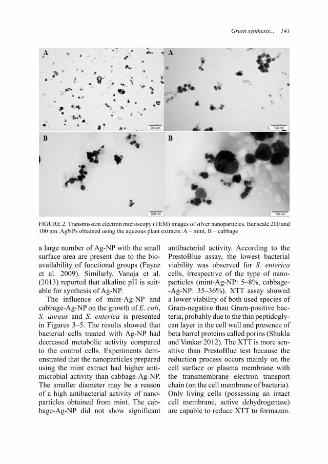
Green synthesis... 141
a large number of Ag-NP with the small surface area are present due to the bio-availability of functional groups (Fayaz et al. 2009). Similarly, Vanaja et al. (2013) reported that alkaline pH is suit-able for synthesis of Ag-NP.
The influence of mint-Ag-NP and cabbage-Ag-NP on the growth of E. coli, S. aureus and S. enterica is presented in Figures 3–5. The results showed that bacterial cells treated with Ag-NP had decreased metabolic activity compared to the control cells. Experiments dem-onstrated that the nanoparticles prepared using the mint extract had higher anti-microbial activity than cabbage-Ag-NP. The smaller diameter may be a reason of a high antibacterial activity of nano-particles obtained from mint. The cab-bage-Ag-NP did not show significant
antibacterial activity. According to the PrestoBlue assay, the lowest bacterial viability was observed for S. enterica cells, irrespective of the type of nano-particles (mint-Ag-NP: 5–8%, cabbage--Ag-NP: 35–36%). XTT assay showed a lower viability of both used species of Gram-negative than Gram-positive bac-teria, probably due to the thin peptidogly-can layer in the cell wall and presence of beta barrel proteins called porins (Shukla and Vankar 2012). The XTT is more sen-sitive than PrestoBlue test because the reduction process occurs mainly on the cell surface or plasma membrane with the transmembrane electron transport chain (on the cell membrane of bacteria). Only living cells (possessing an intact cell membrane, active dehydrogenase) are capable to reduce XTT to formazan.
FIGURE 2. Transmission electron microscopy (TEM) images of silver nanoparticles. Bar scale 200 and 100 nm. AgNPs obtained using the aqueous plant extracts: A – mint, B – cabbage

142 M. Sosnowska et al.
a
b
A
B
a
b
A
B
020406080
100120
M M M B B B M M M B B BC 1 8 16 C 1 14 28 C 1 8 16 C 1 14 28
PrestoBlue XTT
% o
f Con
trol
FIGURE 3. Mean viability of Escherichia coli cells evaluated for two tests, i.e. PrestoBlue and XTT, after treatment with: C – control (no treatment Ag-NP), M – Mentha piperita (1, 8, 16 μg/ml of mint--Ag-NP), B – Brassica oleracea (1, 14, 28 μg/ml of cabbage-Ag-NP). The differences between the treated group Ag-NP and non-treated control group were statistical significance (P ≤0.05). Different let-ters (a, b and A, B) indicate significant differences between the groups (different colours show groups). ANOVA, Tukey post-test
a
b
AB
a
b
A
B
0
20
40
60
80
100
120
M M M B B B M M M B B B
C 1 8 16 C 1 14 28 C 1 8 16 C 1 14 28
PrestoBlue XTT
% o
f Con
trol
FIGURE 4. Mean viability of Staphylococcus aureus cells evaluated for two tests, i.e. PrestoBlue and XTT, after treatment with: C – control (no treatment Ag-NP), M – Mentha piperita (1, 8, 16 μg/ml of mint-Ag-NP), B – Brassica oleracea (1, 14, 28 μg/ml of cabbage-Ag-NP). The differences between the treated group Ag-NP and non-treated control group were statistical significance (P ≤0.05). Differ-ent letters (a, b and A, B) indicate significant differences between the groups (different colours show groups). ANOVA, Tukey post-test
Therefore, the concentration of the dye is proportional to the number of metaboli-cally active cells. Consequently, anti-bacterial activity of Ag-NP is bacteria species dependent.
Most studies used disc diffusion method and measured zone of inhibition. Tamileswari et al. (2015) synthesised
of Ag-NP using cabbage extract. Their results showed good antibacterial and antifungal activity against Klebsiella pneumoniae, Bacillus subtilis, Staphy-lococcus aureus, Escherichia coli and Pneumocystis sp. (Kappusamy 2015, Tamileswari et al. 2015). Some research-ers reported that the mint-Ag-NP have

Green synthesis... 143
antimicrobial effect against E. coli (the maximum zone of inhibition), S. aureus and additionally Pseudomonas aerugino-sa and Bacillus subtilis (MubarakAli et al. 2011, Sarkar and Paul 2017). Saikia et al. (2015) reported that antibacterial activity of Ag-NP is plant extract dependent.
Antibacterial activity of Ag-NP can have several causes e.g. nanoparticles penetration inside and binding DNA, hampering the normal replication, loss of cell viability by modulating tyrosine phosphorylation, attack on the respira-tory chain and finally resulting in cell death (Sarkar and Paul 2017).
The aqueous extract of M. piperita and B. oleracea showed the presence of carbohydrates, amino acids, tannins, fla-vonoids, terpenoids, quinones, phenols, proteins and coumarins (Satya Prasad et al. 2015, Patil et al. 2016). Secondary metabolites act as interfering protein synthesis agents (tannins) and inhibitors of the extracellular enzymes (required
for microbial growth and oxidative phos-phorylation) (Satya Prasad et al. 2015).
Al-Sum et al. (2013) reported that aqueous Mentha species extract was active against six pathogenic bacteria, i.e.: Bacillus fastidiosus (the highest inhibitory effect), Proteus mirabilis, P. vulgaris, Sal-monella choleraesuis, Escherichia coli, Pseudomonas aeruginosa, Klebsiella pneumoniae and Serratia odorifera except for Staphylococcus aureus. According to Patil et al. (2016), the aqueous mint extract showed inhibitory effect against Proteus vulgaris and Staphylococcus aureus but Bacillus cereus and Salmo-nella typhimurium did not show a zone of inhibition. Extracts from Brassica olera-cea exhibit distinct zones of inhibition towards bacterial strains like Escherichia coli, Bacillus subtilis, Staphylococcus aureus, S. epidermis, Salmonella typh-imurium and S. paratyphi (Satya Prasad et al. 2015). Ethanol extract has a greater activity and protein content than the other
FIGURE 5. Mean viability of Salmonella enterica cells evaluated for two tests, i.e. PrestoBlue and XTT, after treatment with: C – control (no treatment Ag-NP), M – Mentha piperita (1, 8, 16 μg/ml of mint-Ag-NP), B – Brassica oleracea (1, 14, 28 μg/ml of cabbage-Ag-NP). The differences between the treated group Ag-NP and non-treated control group were statistical significance (P ≤0.05). Differ-ent letters (a, b and A, B) indicate significant differences between the groups (different colours show groups). ANOVA, Tukey post-test
a
b
A
B
a
b
A
B
0
20
40
60
80
100
120
M M M B B B M M M B B B
C 1 8 16 C 1 14 28 C 1 8 16 C 1 14 28
PrestoBlue XTT
% o
f Con
trol

144 M. Sosnowska et al.
extracts. This extract is the most effective solvent for extracting a broad spectrum of antibacterial compounds from plant origin (Satya Prasad et al. 2015). Antibac-terial nature of plants and their secondary metabolites could be useful to improve the efficiency of nanoparticles.
CONCLUSION
Silver nanoparticles were successfully synthesised from silver nitrate solution using mint and cabbage extracts. Green synthesis using mint and cabbage leaves extracts provides an eco-friendly, stable, simple, cheap and efficient route of Ag--NP synthesis. The pH and temperature play a major role in size control of the Ag-NP. Mint with silver nanoparticles complex had a smaller diameter than cabbage with silver nanoparticles com-plex probably due to the higher pH and lower temperature of mint extract used in nanoparticle synthesis. The obtained nanoparticles showed powerful antibac-terial activity against human pathogens, i.e. E. coli, S. aureus and S. enterica, in-dicating that Ag-NP are good candidates for their usage as antibacterial agents. These nanoparticles had higher antibac-terial activity against Gram-negative than Gram-positive bacteria. In the future na-noparticles synthesised from plants may be a better alternative for elimination of multidrug resistance microorganism than commercial antibiotics.
AcknowledgementThis work was supported by Warsaw University of Life Sciences – SGGW, statutory activity of the Department of Animal Nutrition and Biotechnology.
REFERENCES
Al-SUM B.A., Al-ARFAJ A.A., 2013: Antimi-crobial activity of the aqueous extract of mint plant. Sci. J. Clin. Med. 2: 110–113.
CHUNG I.M., PARK I., SEUNG-HYUN K., THIRUVENGADAM M., RAJAKUMAR G., 2016: Plant-mediated synthesis of silver na-noparticles: their characteristic properties and therapeutic applications. Nanoscale Res. Lett. 11: 1–14.
FAYAZ A.M., BALAJI K., KALAICHELVAN P.T., VENKATESAN R., 2009: Fungal based synthesis of silver nanoparticles – an effect of temperature on the size of particles. Colloids Surf. B. 74: 123–126.
JEYARAJ M., REJESH M., ARUN R., MU-BARAKALI D., SATHISHKUMAR G., SIVANANDHAN G., DEVA G.K., MAN-ICKAVASAGAMA M., PREMKUMAR K., THAJUDDINC N., GANAPATHI A., 2013: An investigation on the cytotoxicity and cas-pase-mediated apoptotic effect of biologically synthesized silver nanoparticles using Pody-phyllum hexandrum on human cervical carci-noma cells. Colloids Surf. B. 102: 708–717.
KALISHWARALAL K., DEEPAK V., RAMKU-MARPANDIAN S., NELLAIAH H., SAN-GILIYANDI G., 2008: Extracellular biosyn-thesis of silver nanoparticles by the culture supernatant of Bacillus licheniformis. Mater. Lett. 62: 4411–4413.
KAMATOU G.P.P., VERMAAK I., VILIOEN A.M., LAWRENCE B.M., 2013: Menthol: A simple monoterpene with remarkable bio-logical properties. Phytochem. 96: 15–25.
KAPPUSAMY P., ICHWAN S.J.A., REDDY PARINE N.R., YUSOFF M.M., MANIAN G.P., GOVINDAN N., 2015: Intracellular bio-synthesis of Au and Ag nanoparticles using ethanolic extract of Brassica oleracea L. and studies on their physicochemical and biologi-cal properties. J. Environ. Sci. 29: 151–157.
KUPPUSAMY P., YUSOFF M.M., MANIAM G.P., GOVINDAN N., 2016: Biosynthesis of metallic nanoparticles using plant derivatives and their new avenues in pharmacological ap-plications – An updated report. Saudi Pharm. J. 24: 473–484.
MUBARAKALI D., THAJUDDIN N., JEGA-NATHAN K., GUNASEKARAN M., 2011:

Green synthesis... 145
Plant extract mediated synthesis of silver and gold nanoparticles and its antibacterial activity against clinically isolated pathogens. Colloids Surf. B. 85: 360–365.
MUNYAKA A.W., MAKULE E.E., OEY I., Van LOEY A., HENDRICKX M., 2010: Thermal stability of L-ascorbic acid and ascorbic acid oxidase in broccoli (Brassica oleracea var. italic). J. Food Sci. 75: 336–340.
PATIL S.R., PATIL R.S., GODGHATE A.G., 2016: Mentha piperita Linn: Phytochemical, antibacterial and dipterian adulticidal approach. Int. J. Pharm. Pharm. Sci. 8: 352–355.
PRABHU S., POULOSE M.K., 2012: Silver nano-particles: mechanism of antimicrobial action, synthesis, medical applications, and toxicity effects. Int. Nano Lett. 2: 1–10.
SAIKIA D., GOGOI P.K., PHUKAN P., BHUY-AN N., BORCHETIA S., SAIKIA J., 2015: Green synthesis of silver nanoparticles using Asiatic pannywort and Bryophyllum leaves extract and their antimicrobial activity. Adv. Mater. Lett. 6: 260–264.
SARKAR D., PAUL G., 2017: Green synthesis of silver nanoparticles using Mentha asiatica (mint) extract and evaluation of their antimi-crobial potential. Int. J. Curr. Res. Biosci. Plant Biol. 4: 77–82.
SATYA PRASAD M., SUMAN JOSHI D.S.D., NARENDA K., NADIYA S.K., MASTHA-NI S.K., PADMAJA PHANI N., KRISHNA SATYA A., 2015: A comparative study of phytochemical analysis and in vitro antimicro-bial activity of three important vegetables from Brassicaceae family. Int. J. Res. Ayurveda Pharm. 6: 767–772.
SAWOSZ E., CHWALIBOG A., SZELIGA J., SAWOSZ F., GRODZIK M., RUPIEWICZ M., NIEMIEC T., KACPRZYK K., 2010: Visualization of gold and platinum nanoparti-cles interacting with Salmonella Enteritidis and Listeria monocytogenes. Int. J. Nanomedicine 5: 631–637.
SHUKLA D., VANKAR P.S., 2012: Synthesis of plant parts mediated gold nanoparticles. Int. J. Green Nanotechnol. 4: 277–288.
TAMILESWARI R., HANIFF NISHA M., JESU-RANI Sr.S., 2015: Green synthesis of silver nanoparticles using Brassica oleracea (cauli-
flower) and Brassica oleracea capitata (cab-bage) and the analysis of antimicrobial activity. IJERT 4: 1071–1074.
VANAJA M., RAJESHKUMAR S., PAULKU-MAR K., GNANAJOBITHA G., MALARKO-DI C., ANNADURAJ G., 2013: Kinetic study on green synthesis of silver nanoparticles using Coleus aromaticus leaf extract. Adv. Appl. Sci. Res. 4: 50–55.
Streszczenie: Zielona synteza nanocząstek srebra przy użyciu wodnych ekstraktów z mięty (Mentha piperita) i kapusty (Brassica oleracea var. capita-ta) oraz ich aktywność przeciwbakteryjna. Celem pracy była synteza nanocząstek srebra (Ag-NP) przy użyciu ekstraktów z liści mięty i kapusty jako czynników redukujących i stabilizujących. Obec-ność nanocząstek była początkowo stwierdzona przez zmianę koloru i następnie przez transmi-syjną mikroskopię elektronową (TEM). Analiza TEM uzyskanych Ag-NP wykazała, że ich roz-miary były w przedziale wielkości 5–50 nm dla mięty i 10–150 nm dla kapusty. Aktywność prze-ciwbakteryjną nanocząstek przeciwko patogen-nym szczepom Escherichia coli, Staphylococcus aureus and Salmonella enterica oceniano przez oszacowanie aktywności metabolicznej z uży-ciem testu PrestoBlue i XTT. Większą inhibicję żywotności bakteryjnej obserwowano przeciwko Gram-ujemnym (E. coli, S. enterica) niż Gram--dodatnim (S. aureus) bakteriom.
Słowa kluczowe: nanocząstki srebra, zielona synteza, bioredukcja, Escherichia coli, Staphylo-coccus aureus, Salmonella enterica
MS received 24.03.2017MS accepted 26.04.2017
Authors’ address:Malwina SosnowskaZakład NanobiotechnologiiKatedra Żywienia Zwierząt i BiotechnologiiWydział Nauk o Zwierzętach SGGWul. Ciszewskiego 8, 02-785 WarszawaPoland e-mail: [email protected]


Abstract: Growth performance parameters and selected quality traits of meat and femoral bone of broiler chickens fed diet supplemented with amorphous diatomaceous earth. This study was aimed at analyzing the effect of the addition of amorphous diatomaceous earth to feed on growth performance parameters and selected quality traits of meat and femoral bone of broiler chickens. The study was conducted with 60 fast-growing Ross 308 broiler chickens, reared until 42nd day of age and divided into a control group (C) and two ex-perimental groups (D2 and D4) (20 birds each). The diatomaceous earth (diatomite) was adminis-tered to the birds’ feed from the groups D2 – 2%, D4 – 4%. Individual body weight, feed intake and mortality of chickens were controlled. On day 42 of rearing, six males were selected from each group for slaughter followed by dissection. Dress-ing percentage, content of muscles and giblets (gizzard, liver and heart), were calculated, and in samples of breast and leg muscles the chemical and physicochemical properties were analyzed. Resistance of the femoral bone to fractures was determined. The addition of diatomite did not af-fect the health status of chickens. Significantly higher body weight at 42nd day of rearing was noted in the group C vs D4 (P ≤0.05). Significant-ly higher (P ≤0.01) content of pectoral muscles and lower (P ≤0.05) fat in the carcass of group D2 vs C were noted. The addition of diatomaceous earth did not affect the chemical composition of the breast muscles. The fat content in leg muscles was significantly reduced (P ≤0.05) and water
content was increased (P ≤0.05) in group D2 vs C. Femoral bones of D4 birds were significantly more resistant (P ≤0.05) to breaking than in C. Di-rect relationship between the amount of diatomite and the strength of the femur was found. The most optimal supplementation was considered as 2%.
Key words: diatomaceous earth (diatomite), broil-er chickens, growth performance, quality of meat and femoral bone
INTRODUCTION
Apart from pork, poultry meat is one of the most frequently consumed types of meat by an average Pole. In the intensive production of broiler chickens, being the main source of poultry meat in Poland, used is made of crosses with a high genetically-determined productive po-tential. Consumers of poultry meat are becoming increasingly interested in the welfare of animals as well as in the qual-ity and safety of food products of animal origin. Many of them pay attention to maintenance conditions and rearing system. From the point of view of both, the producer and the consumer it is im-portant to keep the high growth perform-ance of chickens, but, simultaneously, to
Annals of Warsaw University of Life Sciences – SGGWAnimal Science No 56 (1), 2017: 147–157(Ann. Warsaw Univ. of Life Sci. – SGGW, Anim. Sci. 56 (1), 2017)DOI 10.22630/AAS.2017.56.1.17
Growth performance parameters and selected quality traits of meat and femoral bone of broiler chickens fed diet supplemented with amorphous diatomaceous earth
MAŁGORZATA WIEWIÓRA1, MONIKA ŁUKASIEWICZ2
1 Faculty of Animal Sciences, Aves Scientific Circle 2 Department of Animal Breeding and Production, Warsaw University of Life Sciences – SGGW

148 M. Wiewióra, M. Łukasiewicz
ensure their good health status and body condition. This may be achieved by ap-propriate feeding. For this reason, it is desirable to support bird feeding with additives of natural origin that would positively affect those two traits. Con-sidering the necessity and willingness to satisfy customer demands, producers of broiler chickens use a variety of prepara-tions and feed additives in their diets that may positively influence bird condition and meat quality. In addition, they are still searching for novel solutions. The proposed substance that may be applied in broiler chicken feeding is diatoma-ceous earth. It is a completely natural preparation, unprocessed nor fortified with any chemical substances, which is significant from the point of view of both, consumers and producers. The use of diatomaceous earth in animal feeding and its effect on the body are, however, relatively poorly recognized, especially in Poland, and available study results are full of ambiguities and inaccuracies.
Diatomaceous earth (diatomite) is a natural sedimentary rock, formed mainly by fossilized frustules of diatoms – single-celled algae. These organisms had lived many millions years ago in the aquatic environment, and then – asa result of successive drying out of water bodies – they have formed deposits of dia-tomaceous earth (Round et al. 1990). As reported by Fields (2000), the frustules of diatoms in diatomite are built mainly of silica (SiO2) and trace amounts of other mineral compounds, like: aluminum, iron oxide, lime, magnesium or sodium.
The mechanism of action of diatoma-ceous earth in an animal body may be discussed from two perspectives. The diatomaceous earth administered to
animals with feed passages through the gastrointestinal tract and is absorbed, in a small amount, to the bloodstream in the form of orthosilicic acid (H4SiO4). This acid is synthesized through the reaction of silica with two molecules of water and in nature is the biologically-active form of silica and silicon. Diatoms may, there-fore, constitute the source of bioavail-able silicon (Abraham 2005). In addi-tion, diatomaceous earth is applied as an anti-parasitic agent (Quarles 1992, Fields 2000, Dawson 2004, Maurer et al. 2009, Wiewióra et al. 2015). By passing through the gastrointestinal tract, frustules of dia-toms destroy the encountered parasitic organisms and their forms, and then are excreted. These events are mechanical in character, the rough surface of frustules rubs against forms of parasites, causing their damage, which leads to their death or incapability of further reproduction. In addition, the cylindrical frustules of diatoms have pores that absorb water from the extrinsic environment, which contributes to dehydration of pathogen bodies and leads to their death as a result of dehydration (Haney 2016). Accord-ing to Haney (2016), diatoms are not identical in all deposits of diatomaceous earth. The saltwater deposits contain a mixture of different types of diatoms having various shapes, and their fossil-ized frustules are relatively fragile. In turn, the freshwater deposits are char-acterized by a stable content of diatoms with a uniformly-shaped (cylindrical) and resistant frustules. The stable struc-ture and unchangeable composition of diatomaceous earth are important for the effectiveness of its action. A significant aspect in this case is also the amorphous (non-crystalline, namely unordered) form

Growth performance parameters... 149
of the applied diatomaceous earth. The amorphous silica is delicate, it smoothly rubs against the gastrointestinal tract, causing no damages. In turn, the crystal-line silica has very sharp, hard and rela-tively thick particles that may be hazard-ous to the walls of the gastrointestinal tract of animals – it is mainly applied in filtration systems. According to Norton (2015), owing to its adsorptive proper-ties, diatomaceous earth may be used to eliminate from the body toxins secreted by bacteria, fungi or other infections, including yeast-like Candida. In addition, the administration of diatomite improves the generally understood body condition of animals and may also improve their production results (Carlisle 1986). Any negative impact of the amorphous dia-tomaceous earth on health or condition of animals has been document so far.
This study was aimed at analyzing the effect of the addition of amorphous dia-tomaceous earth to feed on production performance indices and selected quality traits of meat and femoral bone of broiler chickens.
MATERIAL AND METHODS
The experimental material included 60 fast-growing Ross 308 broiler chickens (males) kept until 42nd day of age at the experimental farm of Wilanów-Obory. One-day poults were vaccinated against Marek’s diseases, Gumboro disease, and infectious bronchitis. Afterwards, they were weighed, tagged with individual tags, and randomly allocated to experimental groups. On the first day of experiment, the chicks were randomly divided into three groups of 20 birds (five birds in four replicates), i.e. control group (C) and two
experimental groups in which diets were supplemented with diatomaceous earth: D2 (2%) and D4 (4%). The differentiating factor in the experiment was feeding the chickens from the experimental groups with a feed mixture supplemented with 2 and 4% of diatomaceous earth. The birds were fed ad libitum, in a three-stage system: starter (days 1–14), grower (days 15–35), finisher (days 36–42) – Table 1.
The diatomaceous earth used in the experiment originated from freshwater deposits, its dietary quality was suitable for men and animals and it was ground to the consistency of meal. Its composition was presented in Table 2.
Individual body weights, feed intake and mortality rate of the chickens were controlled throughout the study. On day 42 of production, six males with body weight close to the average body weight in a group were selected from each group. The selected birds were fasted for 12 h, with constant access to water, and then slaughtered. Carcasses were chilled with the air method at a temperature of 4°C for 24 h. Then, dissection was per-formed. Afterwards, a total of 18 carcass-es (six from control group and six from each experimental group) were dissected following the methodology described by Ziołecki and Doruchowski (1989). Dressing percentage was calculated, i.e. content of muscles and content of giblets (gizzard, liver and heart), in respect of the body weight before slaughter. The collected breast and leg muscles were weighed, individually marked, protected, and left for further analyses.
The dissected breast and leg muscles were subjected to the chemical and phys-icochemical analysis. The proximate chemical composition of breast muscles

150 M. Wiewióra, M. Łukasiewicz
TABLE 1. Feed mixture composition and nutri-tional value
Specification
Feed mixturestarter(days 1–14)
grower(days
15–35)
finisher(days
36–42)Component (%)
Corn 10.00 11.40 10.00Wheat 53.00 55.00 60.80Soybean meal 30.60 27.40 21.60
Limestone 1.19 1.20 0.97Sodium bicarbonate 0.20 0.14 0.16
NaCl 0.24 0.28 0.26Dicalcium phosphate 1.18 0.78 0.64
Soybeanoil 2.10 2.40 4.40
Methionine 84%calciumsalt
0.48 0.42 0.28
Lysine 0.36 0.34 0.28Threonine 0.14 0.13 0.10Premix 0.5% 0.50 0.50 0.50
Nutritional valueME (kcal) 2 990 3 047 3 217Crude fat (%) 3.67 4.00 5.92Crude protein (%) 21.99 20.78 18.51
Crude fiber (%) 3.60 2.55 2.41
Crude ash (%) 5.83 5.35 4.67
Lysine (%) 1.38 1.28 0.97Methionine + + cystine (%) 1.08 1.01 0.76
Available phosphorus (%)
0.45 0.38 0.35
TABLE 2. The composition of diatomaceous earth (MSDS diatomite 2016)
Chemical substance Content (%)SiO2 amorphous 92.00SiO2 crystalline <0.10
Al2O3 4.52Fe2O3/FeO 1.16
P2O5 0.02CaO 0.71MgO 0.55
Na2O + K2O 0.28S 0.01Cl 0.02
MnO 0.02
and leg muscles was determined with standard methods: protein content – with the Kjeldahl’s method using a conver-sion factor of 6.25 (PN-75/A-04018); and fat content – with the Soxhlet’s method (AOAC 2005). Color param-eters (L*, a*, b*) were measured in com-minuted breast and leg muscles usinga CR-410 colorimeter (Minolta) follow-ing producer’s instructions. Each param-eter was measured in five replications, and the mean value of five replications was assumed as measurement result. The parameter L* (lightness) may attain values from 0 to 100. Parameters a* and b* are trichromaticity coordinates and may attain positive and negative values: +a* denotes red color, –a* denotes green color, +b* denotes yellow color, and –b* denotes blue color.
Tests of femoral bone resistance to fractures were performed in a testing machine Zwick Roell 25.0 equipped in a head with the maximum force of1 kN. Speed of the loading element was 50 mm/min.

Growth performance parameters... 151
The data obtained were analyzed statistically using a one-way analysis of variance using SPSS 23.0 software (SPSS, Chicago, IL, USA). Differences were found significant at P ≤0.05 and P <0.01.
RESULTS AND DISCUSSION
Results of analyses of growth perform-ance and slaughter analysis of the broiler chickens were presented in Tables 3and 4. On 42nd day of rearing, signifi-cantly (P ≤0.05) highest body weight was noted in the group of control birds (3,249 g). Feed conversion ratio in group D2 (1.57 kg/kg of body weight gain) was the lowest, compared to the other groups (C – 1.59, D4 – 1.64 kg/kg of body weight gain). No deaths of birds
were recorded in any of the groups. It may be indicative of very good micro-climatic conditions and welfare of the chickens maintained throughout the rearing period, as well as of the lack of negative effects of diatomaceous earth administration on birds mortality rate. Worthy of notice is, however, that there are ample research reports and works concerning the positive effect of dietary inclusion of diatomite on production performance and generally understood health status of animals, including poultry (Carlisle 1972, 1976, Bennett et al. 2011). Although some differences were noted between groups in growth performance parameters, the effects achieved should be found satisfactory and comparable with these reported by other authors.
TABLE 3. Growth performance of broilers – average body weight (BW), feed conversion ratio (FCR), mortality
Group BW (g), day 42
FCR (-), days 1–42
Mortality(%)
C 3 249a 1.588 0D2 3 132ab 1.571 0D4 3 072b 1.638 0
SEM 43.88 0.02 –
Means within a column with different superscripts are significantly different at P ≤0.05.
TABLE 4. Average results of slaughter analysis of broiler chickens (%)
Group Dressing percentage
Muscles Giblets Abdominalfatbreast legs gizzard liver heart
C 73.54 23.36B 15.03 0.60 1.80 0.45 0.51ab
D2 76.05 26.56A 14.78 0.55 1.90 0.45 0.48b
D4 74.70 24.46AB 15.26 0.58 1.83 0.45 0.77a
SEM 0.85 0.70 0.33 0.03 0.11 0.01 0.08
Means within a column with different superscripts are significantly different: A, B at P ≤0.01, a, b at P ≤0.05.

152 M. Wiewióra, M. Łukasiewicz
In reference to the analyzed param-eters, no significant differences were demonstrated in dressing percentage – its highest value (at the significance bound-ary of P = 0.056) was noted in group D2 (76.05%), whereas the lowest one in the control group – 73.54% (Table 4). The application of diatomaceous earth in diet for broiler chickens reared until 42nd day of age had a significant effect on the contribution of leg muscles and giblets in carcasses (Table 4). Analyses demon-strated a significant (P ≤0.01) increase in the content of breast muscles and a lower fat content (P ≤0.05) after diet supplementation with 2% of diatomite, which may be of great significant from the consumer’s perspective. According to research reports, today, consumers of poultry meat purchase mainly those parts of carcasses that may be easily and quick-ly prepared for consumption – next to thigh muscles, these parts include breast muscles (Nowak and Trziszka 2010). In addition, consumers pay great attention to the nutritive value of food products, and poultry meat is commonly perceived as dietetic meat, hence a low fat content is desirable in this product (Nowak and Trziszka 2010, Zdanowska-Sąsiadek et al. 2013). The production of chickens with a higher content of breast muscles and a lower content of fat in carcass is, therefore, economically beneficial to the producer.
Results obtained in this study point to the feasibility of applying diatomite in feed mixtures for broiler chickens – though not necessarily in the large--scale commercial production owing to the possibility of reduced body weight, but definitely in the free-range, bio, and household systems – and indicate that it
would potentially yield some economi-cal benefits and would not deteriorate the growth performance of chickens. In case of the broiler chickens from group D2, even improvement was observed in the production effectiveness manifested in a reduced feed conversion ratio, com-pared to the control group, which ensures another economic benefit to the pro-ducer. Results of this study confirm find-ings reported by other authors. Research reports on the effects of diatomaceous earth demonstrate, e.g., the mechanism of agglutination of food particles in the gastrointestinal tract and separation of these particles from one to another (Ewuola et al. 2014). By this means, it is feasible to enlarge the available surface of digestion for enzymes and to retard digesta passage through the gastroin-testinal tract, which results in more effi-cient absorption of nutrients from feed mixtures and in enhanced effectiveness of digestion. In addition, diatomaceous earth affects body detoxification and bowel purgation (also from residues of veterinary pharmaceuticals), which increases feed intake by animals. What is more, according to other researchers, the intake of fine-grain feed additives may improve absorption of nutrients in poul-try (Quisenberry 1967, van der Meulen et al. 2008).
Poultry feeding affects not only the basic indices of post-slaughter yield and nutritive value of meat, but also significantly models the aroma and taste of meat. Inappropriate choice of feed components may cause some deviations towards deterioration of palatability, but through feeding it is also possible to regulate the level of indispensable components in meat, being the basis of

Growth performance parameters... 153
its nutritive value. Meat plays a key role in man’s diet, especially in developed countries (Speedy 2003). It is affected by many factors like, e.g., social status, wealth, the size of animal productions, and socio-economical status of the country, which explains the higher con-sumption of meat in Western populations (Speedy 2003).
A typical muscle tissue consists of about 75% of water, 20% of protein, 3% of fat, and 2% of solubles non-protein substances. Results of chicken meat qual-ity analysis were presented in Tables 5 and 6. In most cases, the addition of dia-tomaceous earth to the feed mixture had no significant effect on the proximate chemical composition of breast muscles.
The chemical composition of leg muscles (Table 5) was, generally, similar in all groups, however significantly the high-est content of crude fat was determined in leg muscles of the chickens from con-trol group (7.34%), and the lowest one – in these of the chickens from group D2 (5.89%). A similar tendency was observed in breast muscles. Generally, leg muscles contain more fat than breast muscles (Castellini et al. 2002), howev-er, it needs to be emphasized that fat has a positive impact on taste, juiciness, and tenderness of meat (Aberle et al. 2001). As reported by Niewiarowicz (1993) the increasing content of fat is accompanied by a decreasing content of protein or by an increasing dry matter content of mus-
TABLE 5. Chemical composition of chicken meat (%)
GroupBreast muscles Leg muscles
water protein fat collagen water protein fat collagenC 74.06 22.10 3.71 1.17 72.33b 19.44 7.34a 1.16
D2 74.39 21.80 3.33 1.11 73.78a 19.68 5.89b 1.22D4 73.69 22.32 3.54 0.96 72.79 19.49 6.91 1.15
SEM 0.28 0.22 0.25 0.09 0.38 0.18 0.45 0.09
Means within a column with different superscripts are significantly different at P ≤0.05.
TABLE 6. Color parameters of breast and leg muscles
GroupBreast muscles Leg muscles
L* a* b* L* a* b*C 50.99 6.44a 3.45 53.76 9.07 6.76a
D2 51.81 4.32b 4.13a 53.13 8.91 5.67D4 50.58 4.65 2.79b 53.59 8.41 5.44b
SEM 1.09 0.62 0.40 0.83 0.46 0.41
a* – represents colors from green (–a) to red (+a), b* – represents colors from blue (–b) to yellow (+b), L* – the axis of lightness. L* is perpendicular to the hue plane and cuts it through at the site of crossing with axis a* and b*. The L* values range from 0 (black) to 100 (white), and between them there are all hues of grey.Means within a column with different superscripts are significantly different at P ≤0.05.

154 M. Wiewióra, M. Łukasiewicz
cles, which was confirmed in this study (Table 5). Simultaneously, as mentioned earlier, the lower content of fat in poultry meat may be desirable by consumers.
Breast muscles of broiler chickens contain ca. 25% of protein (Lonergan et al. 2003). A higher content of protein was determined in the breast than in the leg muscles. The highest content of protein in breast muscles was assayed in group D4 (22.32%), and the lowest one in group D2 (21.80%). In the case of leg muscles, the highest protein con-tent was found in group D2 (19.68%), and the lowest one in the control group (19.44%). Nevertheless that above results are consistent with findings of other authors, according to whom the mean content of protein in breast mus-cles of chickens ranged from 17.8 to 20.2% (Gawęcki and Gornowicz 2000). In case of leg muscles, analyses showed a significantly (P ≤0.05) higher content of water in group D2 than in the control group (73.78 and 72.33%, respectively). Results obtained by Grabowski (1993) demonstrated an inversely proportional dependency between water content and fat content, which was confirmed in our study in case of significant differ-ences. The content of water decreases along with bird age, which is linked with increasing adiposity and processes ongo-ing in muscles.
Color sensation is one of the key crite-ria taken into account during the choice and purchase of a food product by con-sumers. Based on the visual assessment of this trait, a customer is concluding about the freshness or even quality of meat. The statistical analysis of results of color measurements demonstrated that diet supplementation with diatoma-
ceous earth had no effect on the values of L* parameter in breast muscles of birds. Observations confirmed no effect of various dietary doses of diatomite on better muscle coloration compared to the control group (Table 6). The color of meat is determined by, among other things, concentration of heme pigments and pH value. The highest values of saturation with red color (parameter a*) were found in breast and leg muscles of the control birds, however these values were confirmed statistically only in case of breast muscles. The contribution of yellow and blue color is indicated by values of parameter b*. Results obtained regarding this color parameter demon-strated breast and leg muscles saturation with yellow color. The highest value of saturation with yellow color (parameter +b*) was determined in breast muscles of birds from group D2 (4.13) and in leg muscles of birds from the control group (6.76), whereas the lowest values of this parameter were noted in both breast and leg muscles of birds from group D4. According to Kirkpinar et al. (2001), the more beneficial coloration of carcasses of broiler chickens is indicated by lower values of L* parameter and by higher values of a* and b* parameters. There-fore, diet supplementation with diatoma-ceous earth had no explicit positive or negative effect on color parameters of chicken meat.
Current conditions of rearing and breeding as well as feeding and genetic modification of domestic fowl are sup-posed to ensure, most of all, a high growth rate. The fast increase of body weight but also no balance between muscle mass gain and bone mass may increase the risk of deformations or

Growth performance parameters... 155
fractures of different bones. The selec-tion of broiler chickens for the highest possible muscle mass has led to disrup-tion of body homeostasis which results from the non-uniform development of the whole organism (Wnuk et al. 2013). The greatest problems faced by pro-ducers are these linked with skeleton incapability to lift such a heavy body of chickens, especially in the last weeks of the production cycle. Considering bone resistance expressed by means of the breaking force (Table 7), dietary inclu-sion of diatomaceous earth was demon-strated to have a significant effect on the value of this trait. Femoral bones of birds from group D4 were characterized by a significantly greater resistance (395.83 N) to the breaking force than these of the control birds (369.66 N). In addition,a growing tendency was observed in bone resistance along with an increasing diatomite dose in the feed mixture. There is growing need for understanding the bone resistance in poultry because bone fractures and resultant infections often contribute to deterioration of dressing percentage and quality of carcass. One of the aims of diatomite addition to feed mixtures for chickens is to supplement their bodies with silicon. Negative effects of a silicon-poor diet were demonstrated by Carlisle (1972) – deficiency of this
element caused skeleton deformation and incorrect joint development in chickens. The effect of diatomaceous earth on the improvement of skeleton resistance and condition was also confirmed in studies conducted by Calomme et al. (2002) and López-Álvarez et al. (2009). Feed sup-plementation with silicon may, addition-ally, significantly affect the appropriate development of young organisms, which was proved by Carlisle (1972) – one-day cockerels fed a diet based on amino acids were later characterized by inhibited growth and development, whereas those administered a feed mixture supple-mented with silicon were characterized by a 50% higher growth rate and normal development. Another study by Carlisle (1976) demonstrated the presence of sili-con at the active sites of calcium attach-ment in bones of young hens. In turn, our previous studies (Wiewióra 2016 – unpublished data) confirmed the effect of diatomite on skeleton strengthening in hens, i.e. femoral bones of laying hens receiving a feed mixture with the addi-tion of diatomaceous earth were signifi-cantly more resistant to fractures.
CONCLUSION
Results obtained in the study enable presenting the following summary and conclusions: 1. The application of diatomite in the
feed mixture had no effect on dete-rioration of the health status nor on mortality rate of broiler chickens, and ensured other production effects at a comparable level.
2. The amorphous diatomaceous earth may be applied in broiler chicken feed-ing as an additive safe to the environ-
TABLE 7. The strength of the femur bone
Group Strength (N)C 369.7b
D2 371.3ab
D4 395.8a
SEM 8.17
Means within a column with different superscripts are significantly different at P ≤0.05.

156 M. Wiewióra, M. Łukasiewicz
ment and to bird welfare, which pro-vides a source of biologically-active silicon to birds and has a potentially positive effect on their organisms.
3. Diatomaceous earth may be applied in broiler chicken feeding as an additive improving the resistance and condition of bones, which may be desirable from breeder’s and producer’s perspective, and most of all from the perspective of animal welfare.
AcknowledgementThe paper is a part of the MSc thesis.
REFERENCES
ABERLE E.D., FORREST J.C., GERRARD D.E., MILLS E.W., 2001: Principles of Meat. Sci. 4th edn. Kendall/Hut Publ., Dubuque, IA.
ABRAHAM G.E., 2005: The importance of bio-active silicates in human health. The Original Internist. Spring: 13–19.
AOAC, 2005: Official methods of analy-sis of AOAC international. Gaithersburg, Md. AOAC International, USA.
BENNETT D.C., YEE A., RHEE Y.J., CHENG K.M., 2011: Effect of diatomaceous earth on parasite load, egg production, and egg quality of free-range organic laying hens. Poult. Sci. 90: 1416–1426.
CALOMME M.R., SINDAMBIWE J.B., COS P., VYNCKE C., GEUSENS P., VANDEN BERGHE D.A., 2002: Effect of choline sta-bilized orthosilicic acid on bone density in chicks. Calcif. Tissue Int. 70: 292.
CARLISLE E.M., 1972: Silicon: an essential element for the chick. Science 178 (4061): 619–621.
CARLISLE E.M., 1976: In vivo requirement for silicon in articular cartilage and connective tissue formation in the chick. J. Nutr. 106 (4): 478–484.
CARLISLE E.M., 1986: Silicon as an essential trace element in animal nutrition. Ciba Founda-tion Symposium 121 – Silicon Biochemistry: 123–139.
CASTELLINI C., MUGNAI C., Dal BOSCO A., 2002: Effect of organic production system on
broiler carcass and meat quality. Meat Sci. 60: 219–225.
DAWSON R.D., 2004: Efficacy of diatomaceous earth at reducing populations of nest-dwelling ectoparasites in tree swallows. J. Field Orni-thol. 75: 232–238.
EWUOLA E.O., ADEBIYI O.A., SOKUNBI O.A., 2014: Haematological and some serum biochemical response of growing cockerels to dietary diatomaceous earth. J. Anim. Sci. Adv. 4 (1): 699–704.
FIELDS P.G., 2000: Diatomaceous earth: Ad-vantages and limitations. In: Proc. VII Inter-national Working Conference on Stored-Prod-uct Protection. Sichuan Publishing House of Science and Technology, Chengdu, China 1: 781–784.
GAWĘCKI W., GORNOWICZ E., 2000: Ocena podstawowego składu chemicznego mięśni kurcząt brojlerów pochodzących z różnych hodowli zagranicznych. Gosp. Mięsna 52 (7): 42.
GRABOWSKI T., 1993: Surowiec do produkcji mięsa drobiowego. Cz. 2. Pol. Drob. 10: 8–15.
HANEY M., 2016: Perma Guard. Available at http://www.perma-guard.com.pl.
KIRKPINAR F., BOZKURT M., ERKER R., 2001: The effects of dietary dried alfalfa meal on skin pigmentation and performance of broil-er. In: Proc. XV Europ. Symp. Quality Poultry Meat, 9–12 September, Turkey: 199–204.
LONERGAN S.M., DEEB N., FEDLER C.A., LAMONT S.J., 2003: Breast meat quality and composition in unique chicken populations. Poult. Sci. 82 (12): 1990–1994.
LÓPEZ-ÁLVAREZ M., SOLLA E.L., GONZÁLEZ P., SERRA J., LEÓN B., MARQUES A.P., REIS R.L., 2009: Silicon-hy-droxyapatite bioactive coatings (Si-HA) from diatomaceous earth and silica. Study of adhe-sion and proliferation of osteoblast-like cells.J. Mater. Sci. Mater. Med. 20 (5): 1131–1136.
MAURER V., PERLER E., HECKENDOM F., 2009: In vitro efficacies of oils, silicas and plant preparations against the poultry red mite Dermanyssus gallinae. Exp. Appl. Acarol. 48 (1–2): 31–41.
NIEWIAROWICZ A., 1993: Struktura, skład chemiczny, zmiany poubojowe i smakowitość mięsa drobiowego. In: Technologia mięsa drobiowego. T. Garbowski (Ed.). WNT, War-szawa.

Growth performance parameters... 157
NORTON K., 2015: eHow Contributor. Available at http://www.ehow.com/facts_5862967_dia-tomaceous-earth-candida.html.
NOWAK M., TRZISZKA T., 2010: Zachowania konsumentów na rynku mięsa drobiowego. ZNTJ 1 (68): 114–120.
PN-75/A-04018. Produkty rolniczo-żywnościo-we. Oznaczanie azotu metodą Kjeldahla i prze-liczanie na białko.
QUARLES W., 1992: Diatomaceous earth for pest control. The IPM Practitioner. Monitoring the Field of Pest Management 14 (5–6): 1–11.
QUISENBERRY J.H., 1967: The use of a clay in poultry feed. Clays Clay Miner. 16: 267–270.
ROUND F.E., CRAWFORD R.M., MANN D.G., 1990: The diatoms: biology and morphology of the genera. Cambridge University Press, Cam-bridge.
SPEEDY A.W., 2003: Global production and con-sumption of animal source foods. J. Nutr. 133: 4048–4053.
Van Der MEULEN J., KWAKERNAAK C., KAN C.A., 2008: Sand intake by laying hens and its effect on egg production parameters. J. Anim. Physiol. Anim. Nutr. 92 (4): 426–431.
WIEWIÓRA M., ŁUKASIEWICZ M., BAR-TOSIK J., MAKARSKI M., NIEMIEC T., 2015: Diatomaceous earth in the prevention of worm infestation in purebred pigeons. Ann. Warsaw Univ. Life Sci. – SGGW, Anim. Sci. 54 (2): 161–166.
WNUK A., MROCZEK-SOSNOWSKA N., ADAMEK D., KAMASZEWSKI M., ŁUKASIEWICZ M., NIEMIEC J., 2013: Ef-fect of rearing system and gender on histologi-cal profile of chicken breast and leg muscles in hybrid (Cobb × Zk). Ann. Warsaw Univ. Life Sci. – SGGW, Anim. Sci. 52: 211–218.
ZDANOWSKA-SĄSIADEK Ż., MICHAL-CZUK M., MARCINKOWSKA-LESIAK M., DAMAZIAK K., 2013: Czynniki kształtujące cechy sensoryczne mięsa drobiowego. Bromat. Chem. 46 (3): 344–353.
ZIOŁECKI J., DORUCHOWSKI W., 1989: Me-tody oceny wartości rzeźnej drobiu. COBRD Poznań.
Streszczenie: Wskaźniki odchowu oraz wybrane cechy jakości mięsa i kości udowej kurcząt broj-lerów żywionych paszą uzupełnioną amorficzną ziemią okrzemkową. Celem badania była analiza wpływu dodatku amorficznej ziemi okrzemkowej
do paszy na wyniki odchowu oraz wybrane ce-chy jakości mięsa i kości udowej kurcząt brojle-rów. Badanie przeprowadzono na 60 szybkoros-nących kurczętach Ross 308 utrzymywanych do 42. dnia życia i podzielonych na grupę kontrolną (C) i dwie grupy doświadczalne (D2 i D4), po 20 sztuk w każdej. Ziemię okrzemkową (diato-mit) podawano z paszą ptakom z grupy D2 – 2%, D4 – 4%. W doświadczeniu kontrolowano masę ciała, spożycie paszy oraz śmiertelność kurcząt. W 42. dniu odchowu z każdej grupy ubito po sześć kogutów i wykonano dysekcję. Obliczono wydajność rzeźną, udział mięśni oraz podrobów jadalnych (żołądka mięśniowego, wątroby oraz serca), a w próbach z mięśni piersiowych i nóg wykonano analizy chemiczne i fizykochemicz-ne. Określono również wytrzymałość kości udo-wej na złamanie. Dodatek diatomitu nie wpłynął na status zdrowotny kurcząt. Większą masę ciała w 42. dniu odchowu odnotowano w grupie C względem D4 (P ≤0,05). Stwierdzono większy (P ≤0,01) udział mięśni piersiowych oraz mniej-szy (P ≤0,05) udział tłuszczu w tuszce w grupie D2 względem C. Dodatek ziemi okrzemkowej do paszy nie różnicował składu chemicznego mięśni piersiowych. W mięśniach nóg kogutów D2 stwierdzono zmniejszenie (P ≤0,05) zawar-tości tłuszczu oraz zwiększenie (P ≤0,05) za-wartości wody w porównaniu do grupy C. Kości udowe ptaków z grupy D4 charakteryzowały się większą (P ≤0,05) wytrzymałością na złamanie niż kości kurcząt z grupy C. Stwierdzono wprost proporcjonalną zależność między ilością zasto-sowanego diatomitu w paszy a wytrzymałością kości udowej. Za najbardziej optymalną uznano suplementację do paszy na poziomie 2%.
Słowa kluczowe: ziemia okrzemkowa (diatomit), kurczęta brojlery, jakość mięsa i kości udowej
MS received 15.03.2017MS accepted 25.04.2017
Authors’ address:Monika Łukasiewicz Katedra Szczegółowej Hodowli ZwierzątWydział Nauk o Zwierzętach SGGWul. Ciszewskiego 8, 02-786 WarszawaPolande-mail: [email protected]
Agriculture(Agricultural and Forest Engineering)
Animal ScienceForestry and Wood Technology
Horticulture and Landscape ArchitectureLand Reclamation
Annals of Warsaw University of Life Sciences were originally published in 1957 as Zeszyty Naukowe SGGW (Scienti-fic Fascicles of SGGW). In 1980 the na-me was changed to Annals of Warsaw University of Life Sciences.The Annals (5 subject series) are publi-shed once or twice a year and will carry pre-viously unpublished papers that are mainly in English, but also in French, German or Russian, followed by a short summary in Polish. Manuscripts for publication should be typewritten and submitted to the Warsaw
University of Life Sciences Press in two copies. Papers submitted for consideration by the Editorial board should not exceed 0.5 of a printed sheet (about 11 pages includ-ing illustrations, and should consist of the following elements: 1) name and surname of the author, 2) title of the paper, 3) abstract (about 20 lines), 4) text of the paper, 5) date when the paper was sent to the Warsaw Uni-versity of Life Sciences Press and mailing address of the author, 6) summary (one page), 7) tables and figures with captions.