Angel Ripplinger
53
UNIVERSIDADE FEDERAL DE SANTA MARIA CENTRO DE CIÊNCIAS RURAIS PROGRAMA DE PÓS-GRADUAÇÃO EM MEDICINA VETERINÁRIA Angel Ripplinger CONTRIBUIÇÕES PARA O ESTUDO DA DOENÇA DO DISCO INTERVERTEBRAL EM CÃES Santa Maria, RS 2021
-
Upload
khangminh22 -
Category
Documents
-
view
1 -
download
0
Transcript of Angel Ripplinger

UNIVERSIDADE FEDERAL DE SANTA MARIA
CENTRO DE CIÊNCIAS RURAIS
PROGRAMA DE PÓS-GRADUAÇÃO EM MEDICINA VETERINÁRIA
Angel Ripplinger
CONTRIBUIÇÕES PARA O ESTUDO DA DOENÇA DO DISCO
INTERVERTEBRAL EM CÃES
Santa Maria, RS
2021

Angel Ripplinger
CONTRIBUIÇÕES PARA O ESTUDO DA DOENÇA DO DISCO
INTERVERTEBRAL EM CÃES
Tese apresentada ao Curso de Pós-graduação em Medicina Veterinária, Área de Cirurgia e Clínica de Pequenos Animais, da Universidade Federal de Santa Maria (UFSM, RS), como requisito parcial para a obtenção do título de Doutora em Medicina Veterinária.
Orientador: Prof˚ Dr˚. Alexandre Mazzanti
Santa Maria, RS 2021


Angel Ripplinger
CONTRIBUIÇÕES PARA O ESTUDO DA DOENÇA DO DISCO
INTERVERTEBRAL EM CÃES
Tese apresentada ao Curso de Pós-graduação em Medicina Veterinária, Área de Cirurgia e Clínica de Pequenos Animais, da Universidade Federal de Santa Maria (UFSM, RS), como requisito parcial para a obtenção do título de Doutora em Medicina Veterinária.
Aprovado em 26 de fevereiro de 2021:
Alexandre Mazzanti, Dr. (UFSM) (Presidente/Orientador)
Diego Vilibaldo Beckmann, Dr. (Unipampa - Uruguaiana)
Graciane Aiello, Drª. (UNOESC - Xanxerê)
Vitor Marcio Ribeiro, Dr. (PUC MINAS / Santo Agostinho Hospital Veterinário)
Luis Felipe Dutra Corrêa, Dr. (UFSM)
Santa Maria, RS 2021
RESUMO
CONTRIBUIÇÕES PARA O ESTUDO DA DOENÇA DO DISCO INTERVERTEBRAL EM CÃES
AUTOR: ANGEL RIPPLINGER ORIENTADOR: ALEXANDRE MAZZANTI
A doença do disco intervertebral (DDIV) toracolombar (TL) é uma causa comum de disfunção neurológica em cães. DDIV é um termo amplo utilizado para se referir a uma série de alterações que afetam o disco intervertebral de cães e que podem envolver ou não a sua degeneração. A extrusão de disco intervertebral intradural (EDIVI) é um dos tipos raros de DDIV, que pode afetar a região da medula espinhal TL de cães. A EDIVI é de difícil diagnóstico e ainda existem dúvidas quanto ao seu tratamento. O artigo 1 da presente tese apresenta um relato de caso de EDIVI, em que o diagnóstico foi realizado no período transoperatório. As alterações de mielografia foram inconclusivas e a decisão cirúrgica foi tomada com base no histórico, achados clínicos e no exame radiografico simples. O procedimento cirúrgico permitiu a adequada recuperação do paciente e a colheita e análise do material removido, durante a cirurgia, permitiu o diagnóstico definitivo de EDIVI. Já a extrusão de disco intervertebral (EDIV) extradural TL é a apresentação mais comum da DDIV em cães. Cães que apresentam paraplegia sem percepção à dor profunda, secundária à EDIV, possuem prognóstico desfavorável quando permanecem muito tempo sem tratamento cirúrgico. O artigo 2 relata os resultados de retorno à deambulação de cães, submetidos à cirurgia para tratamento de EDIV TL, a partir de 96 horas (quatro dias) ou mais de paraplegia sem percepção à dor profunda. Os resultados permitiram afirmar que existe possibilidade acima de 45% de retorno à deambulação para esses cães quando instituído o tratamento cirúrgico. Por fim, podemos acrescentar que a DDIV ainda apresenta um vasto campo de estudos clínicos para melhor compreensão dos processos nela envolvidos e na abordagem ao paciente acometido.
Palavras-chave: Extrusão de disco intervertebral. Extrusão de disco intervertebral intradural. Neurocirurgia. Dor profunda. Cães.

ABSTRACT
CONTRIBUTIONS TO THE STUDY OF THE INTERVERTEBRAL DISC DISEASE IN DOGS
AUTHOR: ANGEL RIPPLINGER ADVISOR: ALEXANDRE MAZZANTI
Thoracolumbar (TL) intervertebral disc disease (IVDD) is a common cause of neurological dysfunction in dogs. IVDD is a broad term used to refer to a series of diseases that affect the intervertebral disc of dogs, and that may or may not involve degeneration of the disc. Intradural intervertebral disc extrusion (IIVDE) is one of the rare types of IVDD, that can affect the TL spinal cord region of dogs. IIVDE is difficult to diagnose and there are still doubts about treatment. Article 1 of the present thesis presents a case report of IIVDE, whose diagnosis was made during the transoperative period. The changes in myelography were inconclusive and the surgical decision was made based on history, clinical findings and simple radiography. The surgery allowed adequate recovery of the patient and the collection and analysis of the material removed during the surgery allowed the definitive diagnosis of IIVDE. Toracolumbar intervertebral disc extrusion (IVDE) (extradural) is the most common presentation of IVDD in dogs. Dogs with paraplegia without deep pain perception, secondary to IVDE, have questionable prognosis, when they remain without surgical treatment for a long time. The article 2 shows results of the return to ambulation of dogs submitted to surgery to treat TL IVDE, with 96 hours (four days) or more of paraplegia without deep pain perception. The results allow us to affirm that there is a possibility above 45% of return to walking for these dogs when treated surgically. Finally, we can add that IVDD still presents a vast field of clinical studies to better understand the processes involved and to better approach the patient affected by it.
Keywords: Interverterbal disc extrusion. Intradural intervertebral disc estrusion. Neurosurgery. Deep pain. Dogs.

SUMÁRIO
1. INTRODUÇÃO ..................................................................................................... 6
1.1 DOENÇA DO DISCO INTERVERTEBRAL EM CÃES ................................... 6
1.1.1 Extrusão de disco intervertebral intradural/intramedular .................. 7
1.1.2 Extrusão de disco intervertebral (Hansen tipo I) ................................ 7
2. ARTIGO 1. .......................................................................................................... 11
3. ARTIGO 2. .......................................................................................................... 21
4. DISCUSSÃO ...................................................................................................... 46
5. CONCLUSÃO ..................................................................................................... 49
6. REFERÊNCIAS .................................................................................................. 50
1. INTRODUÇÃO
1.1 DOENÇA DO DISCO INTERVERTEBRAL EM CÃES
A doença do disco intervertebral (DDIV) é um termo genérico, amplamente
utilizado em medicina veterinária, e engloba uma série de lesões que afetam o disco
intervertebral (FENN et al., 2020). A DDIV é conhecida desde os anos de 1800,
quando foram feitas as primeiras publicações, ao descrever o material extrudido de
disco intervertebral dentro do canal vertebral (FENN et al., 2020). Com os avanços
dos estudos histológicos e de imagem, hoje, são reconhecidos diferentes
mecanismos patológicos, não somente degenerativos, que envolvem alterações no
disco intervertebral, e que levam a sinais clínicos mais ou menos graves de
disfunção da medula espinhal (FENN et al., 2020).
A degeneração do disco intervertebral é a causa subjacente das formas mais
comuns de hérnia de disco intervertebral (FENN et al., 2020), entretanto, não é fator
crucial para a ocorrência das hérnias de disco, como no caso de extrusão aguda não
compressiva de núcleo pulposo (EANCNPH) e, em alguns casos, de extrusão de
núcleo pulposo hidratado (ENPH) (DE RISIO, et al., 2009; BELTRAN, et al., 2012;
FENN, et al. 2020; SPITZBARTH et al. 2020).
Um sistema de classificação dos diferentes tipos de DDIV foi proposto por
FENN et al. (2020). Esse sistema de classificação diferencia protrusão de disco
intervertebral (PDIV), que ocorre devido ao processo de metaplasia condroide tardio
e envolve, principalmente, cães de raças não condrodistróficas, da extrusão de disco
intervertebral (EDIV), que ocorre devido ao processo precoce de metaplasia
condroide, associado à genética em cães condrodistróficos. Os casos de EDIV
podem levar à uma subclassificação conhecida como EDIV aguda com hemorragia
epidural extensiva ou à EDIV intradural/intramedular (EDIVI). Outras formas de
hérnia de disco incluídas nesse sistema de classificação são a EANCNPH e a
ENPH, que podem não envolver processo degenerativo. A EANCNPH pode também
levar à EDIVI. Além disso, as EDIV traumáticas podem ser provenientes tanto de
disco intervertebral degenerado quanto hidratado. O conhecimento e a correta
classificação da DDIV são de crescente importância, pois a abordagem terapêutica
difere de acordo com a patologia envolvida no processo (SPITZBARTH et al., 2020;
DA COSTA et al., 2020).
7
1.1.1 Extrusão de disco intervertebral intradural/intramedular
O material extrudido do disco intervertebral pode resultar em contusão
medular com ou sem compressão e, raramente, em laceração das meninges com ou
sem penetração no parênquima medular (DE RISIO, 2015). A EDIVI é um
diagnóstico incomum, com poucos casos relatados (FENN et al., 2020). A maioria
dos casos de EDIVI em cães descreve achados de degeneração de disco
intervertebral na análise histológica do material excisado cirurgicamente, sugerindo
uma extrusão de material degenerado de disco intervertebral intradural ou
intramedular e subsequente compressão medular (SANDERS et al., 2002; TARUMA
et al., 2015).
A EDIVI já foi descrita com localização toracolombar, lombar e cervical, e a
maioria dos casos é de apresentação hiperaguda durante exercício ou trauma
(YARROW; JEFFERY, 2000). Além disso, em geral, animais com essa afecção na
região toracolombar apresentam-se paraparéticos ou paraplégicos, e a lateralização
dos sinais consiste em pequenas diferenças na função motora ou nocicepção entre
os membros pélvicos (HAY; MUIR, 2000; LIPTAK et al., 2002; SANDERS et al.,
2002; PACKER et al., 2004; MCKEE; DOWNES, 2008; PONCELET; HEIMANN,
2011; BARNOON et al., 2012), ampliando a gama de diagnósticos diferenciais a
serem considerados, incluindo EANCNP e embolismo fibrocartilaginoso (DE RISIO,
2015).
Os meios diagnósticos da EDIVI são a mielotomografia e a ressonância
magnética (TARUMA et al., 2015), entretanto, na falta dessas ferramentas, a
mielografia pode também ser útil nos casos em que é possível visibilizar um padrão
sugestivo de compressão intradural extramedular ou vazamento de contraste,
sugerindo laceração da dura-máter (YARROW; JEFFERY, 2000; LIPTAK et al.,
2002).
A raridade dessa afecção, bem como os desafios para seu diagnóstico e
tratamento, levaram ao artigo 1 da presente tese.
1.1.2 Extrusão de disco intervertebral (Hansen tipo I)
A EDIV é um dos problemas neurológicos mais comuns encontrados na
prática clínica veterinária (BRISSON, 2010; BERGKNUT et al., 2012), e é, de longe,
a causa mais comum de lesão na medula espinhal em cães (OLBY, 2010). Cães

8
com EDIV apresentam uma variedade de anormalidades neurológicas, causadas
pela combinação de compressão e contusão da medula espinhal, devido à repentina
extrusão do disco intervertebral degenerado do núcleo pulposo para dentro do canal
vertebral (LEVINE et al., 2011; JEFFERY et al., 2016; MOORE et al., 2020). A região
anatômica mais comumente afetada pela EDIV, em cães, é a toracolombar (ITO et
al., 2005; BRISSON, 2010; OLBY, 2010; AIKAWA et al., 2012b). A apresentação
clínica da EDIV toracolombar varia desde dor espinhal até paraplegia sem
percepção à dor profunda (BRISSON, 2010; MOORE et al., 2020).
O diagnóstico da EDIV inicia com a avaliação neurológica, que permite a
localização da lesão e a graduação da gravidade da lesão, associado ao exame
radiográfico, mielografia, tomografia computadorizada (TC), mielotomografia ou
imagem de ressonância magnética (IRM) (BRISSON, 2010). Atualmente, a
mielografia tem sido largamente substituída pela utilização de TC e IRM, que
permitem mais precisão no diagnóstico e melhor direcionamento do tratamento (DA
COSTA et al., 2020). A IRM permite, ainda, a melhor diferenciação entre os tipos de
DDIV, evitando falhas terapêuticas (DA COSTA et al., 2020).
As recomendações de tratamento têm, como base, uma combinação de
fatores, que vão desde a severidade dos sinais neurológicos, disponibilidade de
cuidados especializados, preferências e limitações financeiras dos tutores (MOORE
et al., 2020). As opções de tratamento incluem manejo clínico (ou conservativo), que
consiste em restrição de atividades, fisioterapia, analgésicos e anti-inflamatórios, ou
descompressão cirúrgica da medula espinhal, a fim de remover o material extrudido,
seguido de restrição de movimentos, manejo da vesícula urinária e da dor
(JERRAM; DEWEY, 1999; BRISSON, 2010; MOORE et al., 2020).
Para cães com disfunções neurológicas severas (paraparesia não ambulatória
ou paraplegia) decorrentes de EDIV toracolombar, indica-se o tratamento cirúrgico
por apresentar melhores resultados em relação à recuperação funcional quando
comparado com o tratamento clínico (MOORE et al., 2016; LANGERHUUS; MILES,
2017; MOORE et al., 2020). Também se indica o tratamento cirúrgico para o alívio
da dor em graus menos severos de disfunção neurológica (JEFFERY et al., 2013).
O objetivo primário da cirurgia, em cães com EDIV toracolombar, é a remoção
do material oriundo do disco intervertebral que comprime a medula espinhal além de
prevenir mais extrusão no local (LANGERHUUS; MILES, 2017). A abordagem
cirúrgica mais popular é a hemilaminectomia (BRISSON, 2010; MOORE et al., 2016;

9
MOORE et al., 2020), que é associada a um melhor grau de descompressão e
melhora significativa no pós-operatório, além de menor instabilidade biomecânica
quando comparada com a laminectomia (MUIR et al., 1995). Qualidade de acesso
semelhante é associada à mini-hemilaminectomia, que permite adequada
visualização do aspecto ventrolateral do canal vertebral, permitindo ótimo acesso
para remoção de material extradural, que comprime a medula espinhal ventro-
lateralmente, limitando a manipulação da medula espinhal, além disso, a
preservação das facetas articulares garante uma menor instabilidade do que a
hemilaminectomia (JEFFERY, 1988; JEFFERY et al., 2017; MOORE et al., 2020).
Associado à hemilaminectomia, a literatura corrente suporta a indicação da
realização da fenestração de disco intervertebral profilática, como uma maneira
segura de reduzir futuras extrusões de disco nos locais fenestrados (BRISSON et
al., 2011; AIKAWA et al., 2012a; MOORE et al., 2020). Os resultados de estudos
sugeriram que a fenestração reduz a taxa de recorrência da EDIV e o cirurgião deve
considerar a fenestração, pelo menos dos discos calcificados além do afetado, no
momento da cirurgia (BRISSON et al., 2011; AIKAWA et al., 2012a; MOORE et al.,
2020).
Quanto ao prognóstico para cães paraplégicos, até o momento, a avaliação
da presença de percepção à dor profunda (PDP) é o único método de uso comum,
ainda assim, é considerado insatisfatório porque, embora se saiba que em torno de
50% dos cães sem percepção à dor profunda possam recuperar, não é possível
identificar, no momento da consulta, quais dentre eles realmente vão se recuperar
(JEFFERY et al., 2013). Para os cães sem PDP submetidos ao tratamento cirúrgico,
as taxas de recuperação variam de 30% a 75% em diferentes estudos (SCOTT;
MCKEE, 1999; OLBY et al., 2003; RUDDLE et al., 2006; AIKAWA et al., 2012b;
AIKAWA et al., 2014; JEFFERY et al., 2016). Em geral, cerca de 60% dos cães sem
PDP, devido à EDIV, recuperam a PDP e retornam à deambulação em cerca de seis
meses (OLBY et al., 2020).
Há anos, é discutida a influência do tempo até a descompressão cirúrgica na
recuperação funcional de cães sem percepção à dor profunda, principalmente
aqueles com mais de 48 horas de duração dos sinais (MOORE et al., 2020), e,
ainda. não foi alcançado um consenso, principalmente devido à escassez de
evidências quanto à recuperação funcional dos cães que são tardiamente tratados
(MOORE et al., 2020). Esse foi um dos pontos que levou ao desenvolvimento do

10
projeto, cujos resultados são apresentados no artigo 2.

2. ARTIGO 1.
Extrusão de disco intervertebral intradural em um cão
Angel Ripplinger; Alexandre Mazzanti
Artigo publicado na Acta Scientiae Veterinariae v.48 (suppl.1):153, 2020.

1 1 1
1
Acta Scientiae Veterinariae, 2020. 48(Supp 1): 513.
CASE REPORT
Pub. 513 ISSN 1679-9216
Extrusão de disco intervertebral intradural em um cão
Intradural Disc Extrusion in a Dog
Angel Ripplinger1 & Alexandre Mazzanti4
ABSTRACT
Background: Intervertebral disc extrusion is an important cause of spinal cord dysfunction in dogs. Intradural localization
of the extruded disc material is rare, and is generally associated with a traumatic event or with recurrence of disc extrusion
at a previously affected site. We report the clinical presentation, diagnostic workup, and treatment of a dog with intradural
intervertebral disc extrusion not preceded by a traumatic event.
Case: A 6-year-old male Dachshund was referred for neurological evaluation due to acute onset of hind-end paralysis
preceded by claudication of the left hindlimb. The patient had been receiving conservative treatment to no effect. Neuro-
logical examination revealed asymmetric non-ambulatory paraparesis, absence of postural reactions and decreased muscle
tone in both hindlimbs, a bilaterally diminished patellar reflex, and a hindlimb withdrawal reflex which was normal on
the right and greatly diminished to absent on the left. The lower back was tender to epaxial palpation. Plain radiographs
of the lumbar spine in the lateral projection showed calcified material within the spinal canal between the third and fourth
lumbar vertebrae. Myelography was suggestively abnormal at the same level, with epidural leakage of contrast at L3-L4.
Considering the clinical history, breed, age, neurological signs, and radiographic findings, intervertebral disc disease was
suspected despite the inconclusive myelography findings. A dorsolateral lumbar hemilaminectomy was performed. Intra-
operatively, the diagnosis was confirmed by visualization of a discolored spinal cord and absence of extradural material.
The intradural space was accessed via durotomy. A firm, straw-yellow material was seen compressing the spinal cord and
removed. Subsequent histopathological examination confirmed that this material consisted of extruded intervertebral disc
contents. Postoperatively, the patient underwent physiotherapy and achieved a satisfactory recovery.
Discussion: The most common cause of paraparesis in chondrodystrophic dog breeds is intervertebral disc extrusion.
Intradural extrusion of the intervertebral disc is a rare phenomenon, often associated with vigorous exercise that causes
laceration of the dura mater, allowing penetration of disc material into the intradural space. Although there were no classic
signs of intervertebral disc disease on plain radiography, radiopaque material was visible within the spinal canal, which
can occur in cases of calcified intervertebral disc extrusion. Myelography was inconclusive, but the decision was made
to operate nevertheless, considering that the patient had not responded to conservative treatment and that surgical treat-
ment is the most suitable approach for dogs with non-ambulatory paraparesis or paraplegia secondary to intervertebral
disc extrusion. The surgical technique consisted of a hemilaminectomy and durotomy. Our diagnostic suspicion was con-
firmed intraoperatively, as in most cases of intradural disc extrusion in humans. Intradural disc extrusion is an uncommon
phenomenon in dogs, and the diagnosis is usually only established intraoperatively. This unusual variant of intravertebral
disc disease should be included in the differential diagnosis of spinal cord dysfunction in chondrodystrophic breeds, even
in the absence of a history of trauma or preexisting intervertebral disc disease. Clinical treatment appears ineffective in
these cases. Conversely, surgical treatment can yield good outcomes, and even functional recovery.
Kerywords: intradural extrusion, intervertebral disc disease, intradural compression, paraparesis.
Descritores: extrusão intradural, doença do disco intervertebral, compressão intradural, paraparesia.
DOI: 10.22456/1679-9216.100838
Received: 18 March 2020 Accepted: 17 May 2020 Published: 22 June 2020
1Programa de Pós-Graduação em Medicina Veterinária, Serviço de Neurologia e Neurocirurgia, (SNNV), Hospital Veterinário Universitário (HVU); 4De-
partamento de Clínica de Pequenos Animais, Centro de Ciências Rurais (CCR), SNNV, HVU. CORRESPONDENCE: A. Mazzanti
[[email protected]]. SNNV - HVU - UFSM. Av. Roraima n.1000. CEP 97105-900 Santa Maria, RS, Brazil.

2
A. Ripplinger, G. Aiello, M.R. Wrzerinski, et al. 2020. Extrusão de disco interverttebral intradural em um cão. Acta Scientiae Veterinariae. 48(Supp 1): 513.
2 2
A. Ripplinger, G. Aiello, M.R. Wrzerinski, et al. 2020. Extrusão de disco interverttebral intradural em um cão. Acta Scientiae Veterinariae. 48(Supp 1): 513.
A. Ripplinger, G. Aiello, M.R. Wrzerinski, et al. 2020. Extrusão de disco interverttebral intradural em um cão. Acta Scientiae Veterinariae. 48(Supp 1): 513.
INTRODUÇÃO
Extrusão de disco intervertebral toracolombar
(EDIV) é a causa mais comum de disfunção da me-
dula espinhal em cães [5] e os sinais clínicos podem
variar em severidade, desde dor até paraplegia [10].
Os sinais clínicos são causados pela ruptura dorsal do
anel fibroso, permitindo a herniação do núcleo pulpo-
so degenerado, por vezes calcificado, para dentro do
canal vertebral causando lesão tanto concussiva quanto
compressiva na medula espinhal [8].
A EDIV causa classicamente compressão ex-
tradural da medula espinhal [5]. Não é usual o material
extrudido do disco intervertebral penetrar a dura-máter
e a medula espinhal [7].
Com base nisso, o objetivo do presente traba-
lho é relatar um caso de extrusão de disco intradural
extramedular descrevendo sinais clínicos, achados
diagnósticos, tratamento, bem como a evolução clínica
do paciente.
CASO
Um canino macho Teckel, seis anos, 8,3
kg, com histórico de perda aguda dos movimentos
dos membros pélvicos (MPs) foi encaminhado para
avaliação neurológica. De acordo com o histórico,
o primeiro sinal clínico apresentado foi claudicação
com o MP esquerdo que evoluiu para paraparesia não
ambulatória bilateral. Desde o início dos sinais clíni-
cos, o paciente estava sendo tratado clinicamente com
restrição de movimentos, prednisona1 (0,5mg.kg-1 uma
vez ao dia) e gabapentina2 (10mg.kg-1 duas vezes ao
dia), mas sem melhora.
Ao exame neurológico foi observado estado
mental e postura normais, paraparesia não ambulatória
com assimetria de sinais (MP esquerdo pior). Reações
posturais (posicionamento proprioceptivo e salto) nor-
mais para os membros torácicos (MTs) e ausentes para
os MPs, reflexo flexor e tônus muscular normais para
MTs, tônus muscular diminuído nos MPs, reflexo pate-
lar diminuído bilateral, reflexo flexor normal MP direito
(lateral e medial) e, no MP esquerdo ausente lateral e
diminuído medial. À palpação epaxial notou-se dor na
região lombar. Exame clínico não revelou alterações.
Exames de hemograma e bioquímica sérica não
apresentaram alterações. Radiografia simples, na pro-
jeção lateral, mostrou presença de estrutura radiopaca
no interior do canal vertebral, se estendendo do forame
intervertebral L3-L4 até L2-L3 (Figura 1A). Mielogra-
fia sugeriu alteração na região entre as vértebras L3-
L4, local em que o contraste desvia para epidural, mas
não permitiu adequada visibilização da compressão.
Associando o histórico, raça, idade, sinais clínicos e
os achados de exame neurológico, radiografia simples,
mesmo com a mielografia inconclusiva, considerou-
-se como suspeita clínica principal extrusão de disco
intervertebral entre L3-L4.
O paciente foi encaminhado para cirurgia de
hemilaminectomia lombar dorsolateral [23] esquerda
entre L3-L4. Após acesso ao canal vertebral não foi
encontrado material no canal vertebral (epidural). Foi
possível visualizar através da dura-máter uma alteração
da coloração sugerindo uma afecção intradural (Figura
1 B). Devido a essa alteração, optou-se pela realização
da durotomia e foi observada a presença de material
firme de coloração amarelo palha semelhante a material
proveniente de disco intervertebral degenerado, que foi
removido utilizando uma cureta odontológica e pinça
(Figura 1 C). O material não estava aderido a nenhu-
ma estrutura próxima e foi encaminhado para análise
histopatológica e media 0,9 x 0,3 x 0,2 cm. Após a
remoção do material, a medula espinhal permaneceu
desviada do seu eixo (Figura 1 C).
A análise histopatológica (Figura 1 D) revelou
múltiplos aglomerados de material granular ou crista-
loide densamente basofílico (compatível com minera-
lização), entremeados por finos septos fibrovasculares.
Ocasionalmente foram verificadas pequenas ilhas
irregulares de matriz condroide com mineralização na
periferia, nessas áreas os condrócitos estavam degene-
rados. Os achados histopatológicos foram consistentes
com fragmentos de material de disco intervertebral
degenerado e mineralizado, confirmando a suspeita de
extrusão de disco intervertebral intradural.
Após uma semana da cirurgia, o paciente
foi submetido a sessões de fisioterapia e apresentou
completa recuperação (deambulação sem nenhuma
deficiência neurológica) em um período de 30 dias.
DISCUSSÃO
A laceração da dura-máter associada à extrusão
de disco intervertebral tem sido reportada ocasional-
mente em cães após exercício vigoroso [9,14,15,19,25]
assim como a extrusão intradural do disco interverte-
bral, que provavelmente se segue à laceração da dura-
-máter, a qual, por sua vez, não está necessariamente
associada ao exercício [2,12,20,24].

3
A. Ripplinger, G. Aiello, M.R. Wrzerinski, et al. 2020. Extrusão de disco interverttebral intradural em um cão. Acta Scientiae Veterinariae. 48(Supp 1): 513.
3 3
A. Ripplinger, G. Aiello, M.R. Wrzerinski, et al. 2020. Extrusão de disco interverttebral intradural em um cão. Acta Scientiae Veterinariae. 48(Supp 1): 513.
A. Ripplinger, G. Aiello, M.R. Wrzerinski, et al. 2020. Extrusão de disco interverttebral intradural em um cão. Acta Scientiae Veterinariae. 48(Supp 1): 513.
Existe a hipótese da ocorrência de extrusão
intradural estar relacionada a locais em que já ocor-
reram extrusões antigas [24] embora isso não tenha
sido verificado no presente caso, existe a possibilidade
de um primeiro episódio de extrusão ter apresentado
sinais clínicos leves que não tenha sido reconhecido
pelos tutores.
Devido à raridade de ocorrência desta afecção
em cães não existem estudos com número suficiente
de animais que possam relacionar essa alteração com
a idade, embora estudo recente [24] e relatos de caso
[2,12,20] tenham verificado tal afecção em pacientes
relativamente mais velhos que o relatado pela literatura
para a extrusão extradural de disco em raças condro-
distróficas (três a seis anos) [4].
Na radiografia simples não havia evidência
de redução de tamanho do forame intervertebral ou
diminuição de espaço intervertebral, alterações que
caracterizam doença do disco intervertebral [6,11],
mas havia presença de material radiopaco no forame
intervertebral, sinal sugestivo de extrusão de disco cal-
cificado (Figura 1 A) [6,11], achado este que coincidia
com a neurolocalização da lesão.
Achados mielográficos esperados para casos
de extrusão de disco são os de compressão extradural,
como desvio dorsal da linha de contraste ventral [6,17].
Já na compressão intradural, espera-se encontrar o
sinal de “golf tee” tanto nas projeções lateral quanto
ventro-dorsal [12,20] ou em pelo menos uma delas.
A mielografia realizada no paciente em ques-
tão demonstrou um padrão epidural e subaracnoide
na região em que se suspeitava da lesão, sendo que
nas demais regiões observa-se apenas o padrão
normal da mielografia. Uma hipótese para essa
imagem é a possibilidade da existência de uma
comunicação do espaço subaracnoide com o epidural
por meio de um orifício na dura-máter, o qual
permitiu que o contraste extravasasse para o espaço
epidural.
A laceração da dura-máter por extrusão do
disco intervertebral é descrita na literatura veterinária
[9,14,19,21,25] e essa alteração não foi verificada
durante a cirurgia provavelmente pela não utilização
de lente de magnificação. A não visualização de falha
de contraste não exclui a possibilidade de extrusão
intradural ou intramedular [2] e isso é aplicável para
o presente caso.
Liptak et al. [12] realizaram o manejo con-
servativo dessa variação específica de localização do
conteúdo herniado do disco intervertebral e não veri-
ficaram melhora clínica do paciente, situação também
Figura 1. Extrusão de disco intervertebral intradural em um cão. A- Imagem radiográfica da coluna vertebral, projeção lateral. Nota-se a presença de material radiopaco no interior do canal vertebral entre L2 e L4 B- Medula espinhal com alteração de coloração após hemilaminectomia lombar. C- Frag- mento removido do espaço intradural (círculo branco) após durotomia (dura-máter: seta). D- Fotomicrografia do fragmento com múltiplos aglomerados de material granular densamente basofílico entremeados por fino septos fibrovasculares. Há pequenas ilhas irregulares de matriz condroide (com condrócitos degenerados) com mineralização na periferia (HE 40x).
4
A. Ripplinger, G. Aiello, M.R. Wrzerinski, et al. 2020. Extrusão de disco interverttebral intradural em um cão. Acta Scientiae Veterinariae. 48(Supp 1): 513.
4 4
A. Ripplinger, G. Aiello, M.R. Wrzerinski, et al. 2020. Extrusão de disco interverttebral intradural em um cão. Acta Scientiae Veterinariae. 48(Supp 1): 513.
A. Ripplinger, G. Aiello, M.R. Wrzerinski, et al. 2020. Extrusão de disco interverttebral intradural em um cão. Acta Scientiae Veterinariae. 48(Supp 1): 513.
verificada no presente caso, mesmo com duração de
aproximadamente 20 dias. Provavelmente a melhora
não ocorreu devido às diferenças de microambiente
entre o espaço epidural e intradural que não permitiram
reabsorção ou reorganização do material e consequente
reversão do dano à medula espinhal [26].
Para cães com paraparesia não ambulatória, o
tratamento padrão é cirúrgico de acordo com estudos
na área [3,5,16] e isso, associado à falta de resposta ao
tratamento conservativo anteriormente instituído moti-
vou a opção pela cirurgia descompressiva no presente
caso. Para casos específicos de extrusão intradural, a
literatura incentiva a realização de cirurgia descom-
pressiva e reforça o bom prognóstico para retorno à
deambulação após a mesma [2,20,24].
O diagnóstico de extrusão intradural foi reali-
zado no transcirúrgico assim como ocorre na maioria
das vezes nos diagnósticos de hérnia de disco intradural
em humanos [1,13,18]. Estudo de Tamura et al. [24]
que revisou imagens de mielotomografia e ressonân-
cia magnética em oito cães com extrusão intradural
demonstrou que a ressonância magnética de baixo
campo não identificou claramente achados específicos
sugestivos da presença de extrusão de disco no espaço
subaracnoide e a mielotomografia mostrou-se mais
sensível para esses casos.
A técnica cirúrgica preconizada para os casos
de extrusão intradural é a mesma para os casos de ex-
trusão de disco extradural. O diferencial, no entanto,
é que após a hemilaminectomia dorsolateral, deve-se
realizar a durotomia para efetiva descompressão da
medula espinhal [2,12,20,24]. Diferentemente do que
acontece em humanos, não existe a necessidade de
rafia da dura-máter [22].
Apesar desta complicação da extrusão de
disco intervertebral ser incomum, a recuperação pós-
-cirúrgica foi satisfatória como também é descrito
pela literatura existente [2,20,24], assim, a cirurgia
descompressiva deve ser encorajada nos casos de
extrusão intradural.
A extrusão de disco intradural é uma compli-
cação pouco frequente em cães, o diagnóstico é desa-
fiador e, em geral, acontece durante o procedimento
cirúrgico. Essa variação da extrusão de disco deve
ser incluída no diagnóstico diferencial de cães com
disfunção da medula espinhal, mesmo em raças con-
drodistróficas e sem histórico de trauma ou de doença
do disco intervertebral prévios. O tratamento clínico
parece não ser efetivo nesses casos e o tratamento
cirúrgico demonstra bons resultados na recuperação
funcional dos cães. MANUFACTURERS 1Medley Farmacêutica. Campinas, SP, Brazil. 2Pfizer Brasil. Itapevi, SP, Brazil.
Acknowledgements. We thank Coordenação de Aperfeiçoa-
mento de Pessoal de Nível Superior (CAPES) for the Master
and Doctorate research grants and Conselho Nacional de
Desenvolvimento Científico e Tecnológico (CNPq) process
number 307120/20171.
Declaration of interest. The authors report no conflicts of
interest. The authors alone are responsible for the contents and
writing of the paper.
REFERENCES
1 Arnold P.M. & Wakwaya Y.T. 2011. Intradural disk herniation at L1-L2: reporto f two cases. The Journal of Spinal
Cord Medicine. 34(3): 312-314. DOI: 10.1179/2045772311Y.0000000007
2 Barnoon I., Chai O., Srugo I., Peeri D., Konstantin L., Brenner O. & Shamir M.H. 2012. Spontaneous intradural
disc herniation with focal distension of the subarachnoid space in a dog. The Canadian Veterinary Journal. 53(11):
1191-1194.
3 Bergknut N., Egenvall A., Hagman R., Gustas P., Hazewinkel H.A., Meji B.P. & Lagerstedt A.S. 2012. Incidence
of intervertebral disk degeneration-related diseases and associated mortality rates in dogs. Journal of American Vet-
erinary Medical Association. 240(11): 1300-1309. DOI: 10.2460/javma.240.11.1300
4 Besalti O., Ozak A., Pekcan Z., Tong S., Eminaga S. & Tacal T. 2015. The role of extruded disk material in thoraco-
lumbar intervertebral disk disease: A retrospective study in 40 dogs. The Canadian Veterinary Journal. 46(9): 814-820.
5 Brisson B.A. 2010. Intervertebral disc disease in dogs. Veterinary Clinics of North America Small Animal Practice.
40(5): 829-258. DOI: 10.1016/j.cvsm.2010.06.001
6 Coates J.R. 2000. Intervertebral disk disease. Veterinary Clinics of North America Small Animal Practice. 30(1): 77-
110. DOI: 10.1016/s0195-5616(00)50004-7
7 De Lahunta A., Glass E. & Kent M. 2015. Small animal spinal cord disease. In: Veterinary Neuroanatomy and Clini-
cal Neurology. 4th edn. St. Louis: Elsevier, pp. 257-303.
5
A. Ripplinger, G. Aiello, M.R. Wrzerinski, et al. 2020. Extrusão de disco interverttebral intradural em um cão. Acta Scientiae Veterinariae. 48(Supp 1): 513.
5 5
A. Ripplinger, G. Aiello, M.R. Wrzerinski, et al. 2020. Extrusão de disco interverttebral intradural em um cão. Acta Scientiae Veterinariae. 48(Supp 1): 513.
A. Ripplinger, G. Aiello, M.R. Wrzerinski, et al. 2020. Extrusão de disco interverttebral intradural em um cão. Acta Scientiae Veterinariae. 48(Supp 1): 513.
8 Hansen H.J. 1952. A pathologic-anatomical study on disc degeneration in dog: with special reference to the so-
called enchondrosis intervertebralis. Acta Orthopaedica Scandinavica. 23(sup11): 1-130. DOI: 10.3109/ort.1952.23.
suppl-11.01
9 Hay C.W. & Muir P. 2000. Tearing of the dura mater in three dogs. Veterinary Record. 146 (10): 279-282. DOI:
10.1136/vr.146.10.279
10 Jeffery N.D., Levine J.M., Olby N.J. & Stein V.M. 2013. Intervertebral disk degeneration in dogs: consequences,
diagnosis, treatment, and future directions. Journal of Veterinary Internal Medicine. 27(6): 1318-1333. DOI: 10.1111/
jvim.12183
11 Lamb C.R., Nicholls A., Targett M. & Mannion P. 2002. Accuracy of survey radiographic diagnosis of interverte-
bral disc protrusion in dogs. Veterinary Radiology and Ultrasound. 43(3): 222-228. DOI: 10.1111/j.1740-8261.2002.
tb00994.x
12 Liptak J.M., Allan G.S., Krockenberger M.B., Davis P.E. & Malik R. 2002. Radiographic diagnosis: intramedul-
lary extrusion of an intervertebral disc. Veterinary Radiology and Ultrasound. 43(3): 272-274. DOI: 10.1111/j.1740-
8261.2002.tb01002.x
13 Liu C., Huang C., Lin C. & Liu K. 2011. Intradural disc herniation at L5 level mimicking an intradural spinal tumor.
European Spine Journal. 20(Suppl 2): 326-329. DOI: 10.1007/s00586-011-1772-z
14 Mckee W.M. & Downers C.J. 2008. Rupture of the dura mater in two dogs caused by the peracute extrusion of a
cervical disc. Veterinary Record. 162(15): 479-481. DOI: 10.1136/vr.162.15.479
15 Montavon P.M., Weber U., Guscetti F. & Suter P.F. 1990. What is your diagnosis? Swelling of spinal cord associated
with dural tear between segments T13 and L1. Journal of American Veterinary Medical Association. 196(5): 783-784.
16 Moore S.A., Early P.J. & Hettlich B.F. 2016. Practice patterns in the management of acute intervertebral disc hernia-
tion in dogs. Journal of Small Animal Practice. 57(8): 409-415. DOI: 10.1111/jsap.12496
17 Olby N.J., Dyce J. & Houlton J.E.F. 1994. Correlation of plain radiographic and lumbar myelographic findings with
surgical findings in thoracolumbar disc disease. Journal of Small Animal Practice. 35(7): 345-350. DOI: 10.1111/j.1748-
5827.1994.tb01713.x
18 Öztürk A., Avci E., Yazgan P., Torun F., Yücetaş S. & Karabağ H. 2007. Intradural herniation of intervertebral disc
at the level of lumbar 1-lumbar 2. Turkish Neurosurgery. 17(2):134-137.
19 Packer R.A., Frank P.M. & Chambers J.N. 2004. Traumatic subarachnoid-pleural fistula in a dog. Veterinary Ra-
diology and Ultrasound. 45(6): 523-527. DOI: 10.1111/j.1740-8261.2004.04089.x
20 Poncelet L. & Heimann M. 2011. Intradural vertebral disc herniation in a dog. Veterinary Record. 168(18): 486a.
DOI: 10.1136/vr.c6740.
21 Roush J.K., Douglas J.P., Hertzke D. & Kennedy G.A. 1992. Traumatic dural laceration in a Racing Greyhound.
Veterinary Radiology and Ultrasound. 33(1): 22-24. DOI: 10.1111/j.1740-8261.1992.tb01951.x
22 Sharp N.J.H. & Wheeler S.J. 2005. Thoracolumbar disc disease. In: Small Animal Spinal Disorders Diagnosis and
Surgery. 2nd edn. London: Elsevier, pp.121-160.
23 Shores A. 2017. Thoracolumbar hemilaminectomy. In: Shores A. & Brisson B.A. (Eds). Current Techniques in Canine
and Feline Neurosurgery. Hoboken: John Wiley & Sons, pp.179-182.
24 Tamura S., Doi S., Tamura Y., Takahashi K., Enomoto H., Ozawa T. & Uchida K. 2015. Thoracolumbar intradural
disc herniation in eight dogs: clinical, low-field magnetic resonance imaging, and computed tomographic myelography
findings. Veterinary Radiology and Ultrasound. 56(2): 160-167. DOI: 10.1111/vru.12213
25 Yarrow T.G. & Jeffery N.D. 2000. Dura mater laceration associated with acute paraplegia in three dogs. Veterinary
Record. 146(5): 138-139. DOI: 10.1136/vr.146.5.138
26 Züger L., Fadda A., Oevermann A., Forterre F., Vandevelde M. & Henke D. 2018. Differences in epidural pathol-
ogy between cevical and thoracolumbar intervertebral disk extrusions in dogs. Journal of Veterinary Internal Medicine.
32(1): 305-313. DOI: 10.1111/jvim.14887.
http://seer.ufrgs.br/ActaScientiaeVeterinariae

3. ARTIGO 2.
Functional outcome in dogs with thoracolumbar disc extrusion without
nociception equal to or greater than 96 hours undergoing hemilaminectomy: a
prospective study
Angel Ripplinger, Alexandre Mazzanti
Artigo a submetido ao periódico The Veterinary Journal

1
Original Article 1
2 3
Functional outcome in dogs with thoracolumbar disc extrusion without nociception 4
equal to or greater than 96 hours undergoing hemilaminectomy: a prospective study 5 6
A.A. Ripplinger a, M.R. Wrzesinki a, J.S. Rauber a, M.L. Schwab a, D.A. Ferrarin a, R. 7
Baumhardt a, G. Aiello b, D.V. Beckmann c, A. Mazzanti d, * 8
9 a Veterinary Medicine Postgraduate Program, Small Animal Surgery and Clinic Area, 10
Universidade Federal de Santa Maria, Avenida Roraima 1000, Santa Maria, Rio Grande do 11
Sul, Brazil, Postal Code 97105900. 12 b Universidade do Oeste de Santa Catarina, Xanxerê, Santa Catarina, Brazil, Postal Code 13
89820000. 14 c Universidade Federal do Pampa, Uruguaiana, Rio Grande do Sul, Brazil, Postal Code 15
97501970. 16 d Department of Small Animal Clinic, Veterinary Medicine Course, Universidade Federal de 17
Santa Maria, Avenida Roraima 1000, Santa Maria, Rio Grande do Sul, Brazil, Postal Code 18
97105900. 19
20
21
22
23
* Corresponding author. 24
E-mail address: [email protected] (A. Mazzanti). 25
26

2
Functional outcome in dogs with thoracolumbar disc extrusion without nociception 27
equal to or greater than 96 hours undergoing hemilaminectomy: a prospective study 28
29
Abstract 30
The present study included 36 dogs with paraplegia secondary to intervertebral disc 31
extrusion (IVDE) with a loss of deep pain perception (DPP) duration ranging from four to 60 32
days. All patients underwent hemilaminectomy decompression surgery and fenestration of the 33
affected intervertebral disc and had postoperative follow-up for 180 days to verify the return 34
of DPP and voluntary movements. Although there was a satisfactory recovery of motor 35
function in 47.2% of the cases (n = 17), the same recovery was unsatisfactory in 38.9% of 36
cases (n = 14). A satisfactory recovery was also observed without DPP in 13.9% of the cases 37
(n = 5). Neither the employment of physiotherapy, nor the use of preoperative anti-38
inflammatory drugs, nor age, had any influence on recovery. It was found that the longer the 39
delay in the return of postoperative DPP, the longer the time to satisfactory recovery. The 40
median time to satisfactory recovery was 30 days. The results showed that there is a 41
possibility of satisfactory functional recovery with surgical treatment even with a relatively 42
prolonged time without DPP. 43
44
Keywords: Deep pain perception; Intervertebral disc disease; Neurosurgery; Dog 45
46
Introduction 47
Intervertebral disc extrusion (IVDE) is one of the most common forms of clinical 48
presentation of intervertebral disc disease (Fenn et al., 2020). Dogs with thoracolumbar and 49
lumbar IVDE may present clinical signs ranging from pain to paraplegia with loss of deep 50
pain perception (DPP) (Ruddle et al., 2006; Brisson, 2010, Moore et al., 2020). The prognosis 51
for functional recovery in dogs with IVDE is based on the duration and severity of 52

3
neurological dysfunction, especially the preservation or not of DPP, with patients without 53
DPP being less likely to recover (Wang-Leandro et al., 2017; Olby et al., 2020). 54
55
Decompressive surgery is considered the best method of treatment for dogs with all 56
degrees of neurological dysfunction (McKee, 1992; Scott, 1997; Kazakos et al., 2005; 57
Laitinen & Puerto, 2005; Langerhuus & Miles, 2017), especially for those with non-58
ambulatory paraparesis and paraplegia with or without DPP (Moore et al., 2020). When 59
considering evidence related to the timing of surgical decompression, a clinically important 60
question to consider, according to Moore et al. (2020) is whether dogs without DPP of 61
prolonged duration have reasonable potential for recovery after decompressive surgery. This 62
information is still scarce in the literature. Based on this lack of information, the aim of the 63
present study was to report the percentage of functional recovery of surgically treated dogs 64
with IVDE without DPP for 96 hours or more. The hypothesis of this study was that 65
paraplegic dogs with IVDE with DPP loss lasting 96 hours or more have a chance of motor 66
recovery with surgical treatment, even with a relatively prolonged period without DPP. 67
68
Materials and methods 69
In this prospective study there were included 36 dogs followed from February 2017 to 70
December 2020 by the Veterinary Neurology and Neurosurgery Service of a higher education 71
institution. All patients had a history of acute paraplegia and absence of DPP lasting for 96 72
hours or more, secondary to IVDE. 73
74
The patients included in the study had a presumptive diagnosis by clinical examination 75
and imaging of IVDE between T11 and L6. Confirmation of the diagnosis was by 76
visualization of material from the degenerated nucleus pulposus during decompressive spinal 77

4
cord surgery, associated with histopathological examination of the removed sample. Exclusion 78
criteria were the occurrence of ascending myelomalacia. 79
80
The presence or absence of DPP was assessed by applying strong pressure to the base 81
of the nail, pelvic limb digits (PLs) and the base of the tail (where present) with hemostatic 82
forceps (Scott & McKee, 1999; Olby et al., 2003; Laitinen & Puerto, 2005; Ruddle et al., 83
2006; Jeffery et al., 2016; Wang-Leandro et al., 2017; Lewiss et al., 2020). The lack of a 84
detectable behavioral response (crying, licking, attempting to bite, or turning the head in the 85
direction of the stimulus) or a physiological response (pupil dilation, increased heart or 86
respiratory rate) to the repeated stimulus, was considered to show absence of DPP (Olby et 87
al., 2003; Ito et al., 2005; Laitinen & Puerto, 2005; Ruddle et al., 2006; Levine et al., 2009; 88
Olby et al., 2020). 89
90
As for the time of DPP loss, it was assumed that it was absent at the same moment of 91
the total loss of pelvic limb movements reported by the owners (Scott & McKee, 1999; Ito et 92
al., 2005; Laitinen & Puerto, 2005; Loughin et al., 2005) until the date of surgery. In relation 93
to time without DPP, the patients were distributed into four groups: i) four to seven days 94
without DPP; ii) eight to 15 days without DPP; iii) 16 to 30 days without DPP; and iv) more 95
than 30 days without DPP. 96
97
The use of steroidal (SAID) or non-steroidal (NSAID) anti-inflammatory drugs prior 98
to inclusion in the study was determined based on the prescription given by the referring 99
veterinarian. The imaging examinations performed were plain radiographs, followed by 100
myelography or non-contrast computed tomography (CT) as soon as possible after the 101
patient's clinical care. The myelography examination was performed under general inhalation 102

5
anesthesia with an injection of 0.5 mL kg-1 of iohexol between the L5-L6 vertebrae. 103
Immediately after the injection, radiographic images were taken in the oblique (left and right), 104
ventro-dorsal, and lateral (left and right) projections. CT was performed in an external support 105
clinic. 106
107
The surgical technique used, in all cases, was dorsolateral hemilaminectomy (Shores, 108
2017), associated with fenestration of the affected intervertebral disc. Decompressive surgery 109
was performed as soon as possible after diagnosis. After surgery, the patients remained 110
hospitalized for three to five days. All patients were managed with manual compression of the 111
urinary bladder three times a day until they returned to voluntary urination (Aikawa et al., 112
2012). Associated with this, they received opioid analgesia. In the immediate postoperative 113
period, all patients were given dexamethasone (0.25 mg kg-1), intravenously, as a single dose. 114
115
Physiotherapy was instituted and adapted to each patient's situation, including 116
cryotherapy, deep massage, passive joint movement, neuromuscular electrical stimulation, 117
hydrotherapy, and assisted active exercises. Guardians were instructed to perform manual 118
compression of the urinary bladder, three times a day, on all patients that had not regained the 119
voluntary ability to urinate by the time of hospital discharge (Aikawa et al., 2012). 120
121
Recovery was considered satisfactory if the patient recovered DPP and the ability to 122
walk unsupported, or without falling for 10 consecutive steps (Jeffery et al., 2016). Recovery 123
was considered satisfactory without DPP when the patient did not recover deep pain but 124
developed a reflex spinal gait. Unsatisfactory recovery was considered when the patient did 125
not recover the ability to walk unsupported without faling over, or when there was no change 126
from the initial clinical status (Olby et al., 2003). The maximum follow-up time for patients 127

6
after surgery was 180 days. 128
129
The time of return to ambulation was defined as the number of days from surgical 130
decompression to the time the patient achieved satisfactory recovery (Scott & McKee, 1999). 131
The patients were re-evaluated daily during hospitalization, and thereafter at seven, 10, 15 and 132
30 days after surgery. The re-evaluations were monthly. If the patient presented evolutionary 133
changes between reassessments, the guardians contacted the veterinary and the return for 134
reassessment was anticipated, without prejudice to the subsequent ones. The time to 135
ambulation was categorized into the following groups: zero to 14 days, 15 days to 30 days, 31 136
to 60 days (Aikawa et al., 2012), and over 60 days. Information about urinary and/or fecal 137
incontinence was obtained through a questionnaire to owners carried out in the last 138
assessment of patients. 139
140
For statistical analysis, the quantitative variables were submitted to the Anderson-141
Darling and Shapiro-Wilk normality tests. Considering the absence of normal distribution, 142
Fisher's exact test and the Mann-Whitney U test were used to verify the association between 143
the variable "functional recovery" and the other variables in the database, to know which 144
variables significantly influenced the type of functional recovery. Spearman's correlation 145
coefficient was used to verify which variables had significant influence on the time to 146
functional recovery (in days). The significance level was 5%. 147
148
The present study was approved by the Ethics Committee on Animal Use (CEUA) of 149
the institution under protocol number 3107060816. 150
151
Results 152

7
Thirty-six dogs were included in the study, 19 males and 17 females, with ages 153
ranging from three to 10 years (mean 5.75 years, median 5.5 years) (Table 1). As for the 154
breeds, the most frequent was the Daschshund (41.7%), followed by dogs without defined 155
breed (36.1%). Of the breeds included, 61.1% were chondrodystrophic. The most frequent 156
lesion site was T12-T13, comprising 36.1% of the lesions observed. In three animals, more 157
than one intervertebral space was affected (Tables 1 and 2). 158
159
The time without DPP ranged from 96 hours (four days) to 1440 hours (60 days), with 160
a mean of approximately 14 days and a median of eight days (Table 3). Most dogs, about 161
47.2%, went from four to seven days without DPP; 25% went between eight and 15 days, 162
22% between 16 and 30 days, and 5.6% went above 30 days without DPP (Table 2). 163
164
Overall, 47.2% (n = 17) of patients showed satisfactory recovery to ambulation (with 165
DPP), 38.9% unsatisfactory recovery and 13.9% satisfactory recovery without DPP. Patients 166
with a duration of DPP loss between four and seven days consisted of 47.05% (n = 8) 167
showing satisfactory recovery, 29.41% (n = 5) unsatisfactory recovery and 23.53% (n = 4) 168
satisfactory recovery without DPP. Of the patients without DPP between eight and 15 days, 169
66.67% (n = 6) demonstrated satisfactory recovery, 22.22% (n = 2) unsatisfactory, and 170
11.11% (n = 1) satisfactory recovery without DPP. Of the group of patients without DPP 171
between 16 and 30 days, 25% (n = 2) showed satisfactory functional recovery and 75% (n = 172
6) unsatisfactory recovery. The patients without DPP above 30 days showed 50% (n = 1) 173
satisfactory recovery and 50% (n = 1) unsatisfactory recovery. None of the patients in the no-174
DPP group between 16 and 30 days and over 30 days, showed satisfactory functional 175
recovery without DPP. 176
177

8
Regarding the occurrence of fecal and/or urinary incontinence, of the 17 dogs that had 178
satisfactory recovery, in 13 it was possible to assess the presence of urinary and fecal 179
incontinence. Of these, 46.15% (6/13) had urinary incontinence, 46.15% (6/13) urinary and 180
fecal incontinence and 7.7% (1/13) only fecal incontinence. 181
182
Although physiotherapy was indicated for all patients, only 33.3% (n = 12) of the 36 183
patients in the study had physiotherapy at the institution. Of the 17 patients with satisfactory 184
recovery, 35.29% (n = 6) had physiotherapy. Of the five patients with satisfactory recovery 185
without DPP, 40% (n = 2) had physiotherapy. 186
187
As for the time until satisfactory recovery, four dogs (23.5%) took from zero to 14 188
days, 75% of which with physiotherapy; six dogs (35.3%) took from 15 to 30 days, none of 189
which with physiotherapy; five dogs (29.4%) took from 31 to 60 days for satisfactory 190
recovery and 40% of which with physiotherapy; and two dogs (11.8%) took above 60 days 191
(one took 75 days and the other 90 days) for satisfactory recovery, 50% of which with 192
physiotherapy. The average time for DPP recovery was approximately seven days; for 193
functional recovery, it was approximately 34 days. The complete patient data are presented in 194
Tables 1 and 2. 195
196
At a 5% significance level, breed, gender, site of injury, time without DPP by groups, 197
use of anti-inflammatory drugs, type of imaging exam and physiotherapy had no significant 198
influence on the type of functional recovery (Table 3). On the other hand, when we evaluated 199
the association between functional recovery and the variable time without perception of deep 200
pain in days, we obtained p = 0.048 (Table 4). Thus, we had evidence to infer that the time 201
without DPP (in days) influences the type of functional recovery for patiens with IVDE 202
9
without DPP for 96 hours or more. Thus, by observing the averages, we can see that the fewer 203
the number of days without DPP, the greater the possibility of a satisfactory functional 204
recovery. 205
206
According to the results presented in Table 6, the time of return to DPP (in days) 207
showed a significant correlation with the time to functional recovery: as the correlation value 208
shows positive (0.529), so the time to return to DPP increases, and the time to functional 209
recovery also increases as well. 210
211
Discussion 212
Several studies have reported data pertinent to surgery performed in less than 72 hours 213
(Duval et al., 1996; Scott, 1997; Srugo et al., 2011); others did not specify the time without 214
DPP until surgery (Black, 1988; Olby et al., 2003; Ruddle et al., 2006; Aikawa et al., 2012; 215
Muguet-Chanoit et al., 2012). Others have classified the time without DPP as greater than 48 216
hours (Laitinen & Puerto, 2005; Rousse et al., 2016; Jeffery et al., 2016), not allowing 217
conclusions about the amount of time without DPP above this that the patient may still benefit 218
from surgical treatment. Nevertheless, the number of animals above this time interval 219
included in the studies is low (Moore et al., 2020). This is the first study therefore to present 220
results of the return to ambulation of dogs without DPP for 96 hours or longer, and 47.2% of 221
dogs had satisfactorily recovering ambulation. 222
223
For dogs without DPP equal to or greater than 96 hours, the time without DPP 224
significantly influenced the recovery (p < 0.05), which would indicate the benefits of carrying 225
out decompression surgery as soon as possible, even if the patient arrives late at the 226
specialized center. The other studies, which stated that time without DPP does not influence 227

10
recovery, related to most patients in the range of 24 to 48 hours without DPP or from 228
paraplegia to surgery (Ito et al., 2005; Laitinen & Puerto, 2005; Loughin et al., 2005; Jeffery 229
et al., 2016; Rousse et al., 2016; Takahashi et al., 2020). This difference in results is likely 230
due to the inclusion in the present study of only patients seen late in the course of the disease 231
(≥ 96 hours). 232
233
The percentage of functional recovery verified in the patients of the present study 234
(47.2%) is not very different from that verified for dogs without DPP submitted to surgical 235
procedure in other studies: 61.8% (Scott & Mckee, 1999), 43.5% (Duval et al., 1996), 57.8% 236
(Olby et al., 2003), 52.1% (Aikawa et al., 2012) and 57.7% (Jeffery et al., 2016), the main 237
difference being the longer duration of the absence of DPP (greater than or equal to 96 hours) 238
in all animals evaluated. 239
240
One study claims to have found no evidence that a longer period from DPP loss to 241
surgical decompression might be an indicator of a worse prognosis (Jeffery et al., 2016). 242
However, the group of patients analyzed with DPP loss longer than 48 hours was composed 243
of only seven animals, four of which showed recovery and three of which did not. 244
Furthermore, this study, as well as others (Laitinen & Puerto, 2005; Loughin et al., 2005; 245
Rousse et al., 2016; Takahashi et al., 2020), did not specify how much longer beyond 48 246
hours the absence of DPP or the paraplegia of the dogs lasted until surgery, which left in 247
doubt what action should be taken when a patient without DPP, secondary to IVDE, for 96 248
hours or more, was presented for care. 249
250
The wide range of outcomes after surgical treatment reported for dogs with loss of 251
voluntary motor function and nociception after EDIV suggest that neurological findings do 252

11
not always reflect the degree of structural damage to the spinal cord (Duval et al., 1996; 253
Necas, 1999; Scott & McKee, 1999; Olby et al., 2003; Laitinen & Puerto, 2005; Ruddle et al., 254
2006). Henke et al. (2013) found that in 19% of dogs with absent DPP the lesion was of lower 255
intensity in the spinal cord parenchyma when compared to histopathological findings of 256
patients with milder degrees of neurological dysfunction. Furthermore, the duration of clinical 257
signs was not associated with the histopathological degree of lesion in the white or gray 258
matter of the spinal cord. These results may explain the variable recovery in dogs with 259
absence of nociception that were treated surgically, independent of the duration of absence of 260
DPP, as also verified in this study. 261
262
Data from the present study demonstrated that the longer it takes to return from DPP 263
loss, the longer the functional recovery tends to be. This information has already been 264
presented, as a prognostic indicator, for the functional recovery of dogs undergoing 265
decompression surgery as early as 24 hours without DPP (Park et al., 2008). In this study, the 266
data showed that the return of DPP within an interval of up to five weeks indicates a good 267
prognosis. In contrast, another study showed a high rate of successful recovery for dogs when 268
the return of DPP occurs within two weeks after surgery (Laitinen & Puerto, 2005). 269
270
The time to functional recovery ranged from seven to 90 days (mean 31.1 days and 271
median 30 days), which is not much different to the time presented in the study by Kazakos et 272
al. (2005). Of the 17 dogs that recovered walking ability, 13 (76.47%) recovered walking 273
ability within 7.5 weeks, corroborating other studies (Olby et al., 2003; Ruddle et al., 2006). 274
275
Even dogs that have recovered their ability to walk satisfactorily can have sequelae 276
such as fecal and / or urinary incontinence (Olby et al., 2003). And in the present group of 277

12
dogs studied, results similar to those of Olby et al. (2003) and Aikawa et al. (2012) were 278
obtained in relation to this. However, neither fecal nor urinary incontinence were grounds for 279
euthanasia in this group of patients. 280
281
Recovery of motor function in dogs without persistent DPP may either indicate the 282
development of a reflex spinal gait originating from local spinal cord circuits or the survival 283
of axons crossing the injury site (Olby et al., 2003). This type of functional recovery has been 284
described in other studies (Olby et al., 2003; Gallucci et al., 2017) but was seen in only five 285
(13.89%) dogs in the present study. According to Gallucci et al. (2017), intensive 286
physiotherapy treatment gives the patient a good chance of developing spinal walking. In the 287
present study, 66.7% (24 out of 36) of the dogs did not attend physiotherapy due to 288
difficulties in accessing the institution, and perhaps this reduced the occurrence of spinal 289
walking development in this sample of dogs. 290
291
Typically, dogs that do not have DPP in the PLs are all included in the same clinical 292
grade, but this grade clearly includes a wide range of injury severity, as some dogs recover 293
fully and others remain permanently paraplegic (Olby et al., 2003; Olby et al., 2020). This 294
fact may shed light on the percentages of recovery in the present study, even with a duration 295
of DPP loss equal to or greater than 96 hours. 296
297
The main limitation of the study was the accurate determination of the moment of 298
DPP loss, as already highlighted by other authors (Laitinen & Puerto, 2005; Jeffery et al., 299
2016; Takahashi et al., 2020) since the guardians do not have the necessary knowledge to 300
perform and interpret such an assessment (Jeffery et al., 2016). However, this is a limiting 301
factor common to several studies on the subject (Scott & McKee, 1999; Laitinen & Puerto 302

13
2005; Jeffery et al., 2016). Thus, it was assumed that DPP was lost at the moment of loss of 303
pelvic limb movement, as already described in other studies (Scott & McKee, 1999, Laitinen 304
& Puerto 2005, Ito et al., 2005). Another limiting factor was the absence of MRI as the most 305
appropriate diagnosis and grading of spinal cord injury in these cases, as experienced in other 306
studies (Ruddle et al., 2006; Srugo et al., 2011; Aikawa et al., 2012; Muguet-Chanoit et al., 307
2012; Rousse et al., 2016). 308
309
The clinical relevance of this study was to demonstrate that dogs without DPP for 96 310
hours or more, resulting from thoracolumbar intervertebral disc extrusion, may benefit from 311
spinal cord decompression surgery and recover locomotor function in similar percentages to 312
those operated on without DPP within 48 hours. Nevertheless, these results should not be used 313
as a reason to postpone surgery in dogs in this neurological condition, which should always be 314
performed as soon as possible after localization of the lesion and diagnosis of the disease. 315
316
Conclusions 317
In the present study, it has been concluded that paraplegic dogs with DPP loss lasting 318
96 hours or more have a 47.2% chance of DPP and motor recovery with surgical treatment, 319
even with a relatively prolonged time without DPP. 320
321
Conflict of interest statement 322
The authors declare no conflicts of interest. 323
324
Acknowledgements 325

14
To the Coordenação de Aperfeiçoamento de Pessoal de Nível Superior (CAPES) for 326
the Master and Doctorate scholarships, and to the Conselho Nacional de Desenvolvimento 327
Científico e Tecnológico (CNPq), process number 30712/20171. 328
329
References 330
Aikawa, T., Fujita, H., Kanazono, S., Shibata, M., Yoshigae, Y., 2012. Long-term neurologic 331
outcome of hemilaminectomy and disk fenestration for treatment of dogs with 332
thoracolumbar intervertebral disk herniation: 831 cases (2000-2007). Journal of 333
American Veterinary Medical Association 241, 1617-1626. 334
335
Black, A.P. 1988. Lateral spinal decompression in the dog: a review of 39 cases. Journal of 336
Small Animal Practice 29, 581-588. 337
338
Brisson, B.A., 2010. Intervertebral disc disease in dogs. Veterinary Clinics of North America 339
Small Animal Practice 40, 829-858. 340
341
Duval, J., Dewey, C., Roberts, R., Aron, D., 1996. Spinal cord swelling as a myelographic 342
indicator of prognosis: a retrospective study in dogs with intervertebral disc disease 343
and loss of deep pain perception. Veterinary Surgery, 25, 6-12. 344
345
Fenn, J., Olby, N.J., and the Canine Spinal Cord Injury Consortium (CANSORT-SCI), 2020. 346
Classification of intervertebral disc disease. Frontiers in Veterinary Science, 7, 347
579025. 348
349
Gallucci, A., Dragone, L., Manchetti, M., Gagliardo, T., Pietra, M., Cardinali, M., Gandini, 350
G., 2017. Acquisition of involuntary spinal locomotion (spinal walking) in dogs with 351
irreversible thoracolumbar spinal cord lesion: 81 dogs. Journal of Veterinary Internal 352
Medicine 31, 492-497. 353
354
Henke, D., Vandevelde, M., Doherr, M.G., Stöckli, M., Forterre, F., 2013. Correlations 355
between severity of clinical signs and histopathological changes in 60 dogs with spinal 356
cord injury associated with acute thoracolumbar intervertebral disc disease. The 357
Veterinary Journal 198: 70-75. 358
359
Ito, D., Matsunaga, S., Jeffery, N.D., Sasaki, N., Nishimura, R., Mochisuki, M., Kasahara, M., 360
Fujiwara, R., Ogawa, H., 2005. Prognostic value of magnetic resonance imaging in 361
dogs with paraplegia caused by thoracolumbar intervertebral disk extrusion: 77 cases 362
(2000-2003). Journal of American Veterinary Medical Association 227, 1454-1460. 363
364
Jeffery, N.D., Barker, A.K., Hu, H.Z., Alcott, C.J., Kraus, K.H., Scanlin, E.M., Granger, N., 365
Levine, J.M., 2016. Factors associated with recovery from paraplegia in dogs with loss 366
of pain perception in the pelvic limbs following intervertebral disk herniation. Journal 367
of American Veterinary Medical Association 248, 386-394. 368
369

15
Kazakos, G., Polizopoulou, Z.S., Patsikas, M.N., Tsimopoulos, G., Roubies, N., Dessiris, A, 370
2005. Duration and severity of clinical signs as prognostic indicators in 30 dogs with 371
thoracolumbar disk disease after surgical decompression. Journal of Veterinary 372
Medicine, 52, 147-152. 373
374
Laitinen, O.M., Puerto, D.A., 2005. Surgical decompression in dogs with thoracolumbar 375
intervertebral disc disease and loss of deep pain perception: a retrospective study of 46 376
cases. Acta Veterinaria Scandinavica 46, 79-85. 377
378
Langerhuus, L., Miles, J., 2017. Proportion recovery and times to ambulation for non-379
ambulatory dogs with thoracolumbar disc extrusions treated with hemilaminectomy or 380
conservative treatment: A systematic review and meta-analysis of case-series studies. 381
The Veterinary Journal, 220, 7-16. 382
383
Levine, G.J., Levine, J.M., Budke, C.M., Kerwin, S.C., Au, J., Vinayak, A., Hettlich, B.F., 384
Slater, M.R., 2009. Description and repeatability of a newly developed spinal cord 385
injury scale for dogs. Preventive Veterinary Medicine 89, 121-127. 386
387
Lewiss, M.J., Jeffery, N.D., Olby, N.J., and the Canine Spinal Cord Injury Consortium 388
(CANSORT-SCI), 2020. Ambulation in dogs with absent pain perception after 389
thoracolumbar spinal cord injury. Frontiers in Veterinary Science, 7, 560, 2020. 390
391
Loughin, C.A., Dewey, C.W., Ringwood, P.B., Pettigrew, R.W., Kent, M., Budsberg, S.C., 392
2005. Effect of durotomy on functional outcome of dogs with type I thoracolumbar 393
disc extrusion and absent deep pain perception. Veterinary Compendium of 394
Orthopaedics Traumatology, v. 18, n. 3, p. 141-146, 2005. 395
396
McKee, W.M., 1992. A comparison of hemilaminectomy (with concomitant disc fenestration) 397
and dorsal laminectomy for the treatment of thoracolumbar disc protrusion in dogs. 398
Veterinary Research 4, 296-300. 399
400
Moore, S.A., Tipold, A., Olby, N.J., Stein, V., Granger, N., and the Canine Spinal Cord Injury 401
Consortium (CANSORT-SCI), 2020. Current approaches to the management of acute 402
thoracolumbar disc extrusion in dogs. Frontiers in Veterinary Science, 7, 610. 403
404
Muguet-Chanoit, A.C., Olby, N.J., Lim, J-H., Gallagher, R., Niman, Z., Dillard, S., Campbell, 405
J., Early, P., Mariani, C.L., Muñana, K.R., Freeman, C., Platt, S.R., Kent, M., 406
Giovanella, C., Longshore, R.C., 2012. The cutaneous trunci muscle reflex: a 407
predictor of recovery in dogs with acute thoracomlumbar myelopathies caused by 408
intervertebral disc extrusions. Veterinary Surgery, 41, 200-208. 409
410
Necas, A., 1999. Clinical aspects of surgical treatment of thoracolumbar disc disease in dogs. 411
A retrospective study of 300 cases. Acta Veterinaria Brno 68, 121-130. 412
413
Olby, N.J., Levine, J., Harris, T., Muñana, K., Skeen, T., Sharp, N., 2003. Long-term 414
functional outcome of dogs with severe injuries of the thoracolumbar spinal cord: 87 415
cases (1996-2001). Journal of the American Veterinary Medical Association 222, 762-416
769. 417
418

16
Olby, N.J., da Costa, R.C., Levine, J.M., Stein, V.M., and the Canine Spinal Cord Injury 419
Consortium (CANSORT-SCI), 2020. Prognostic factors in canine intervertebral disc 420
disease. Frontiers in Veterinary Science, 7, 596059. 421
422
Park, S-S., Lim, J-H., Byeon, Y-E., Jang, B-J., Ryu, H-H., Uhm, J-H., Kang, B-J., Kim, W-423
H., Kweon, O-K., 2008. Duration of regain of deep pain perception after 424
decompression surgery as a parameter of surgical outcome for acute thoracolumbar 425
disc herniation Hansen type I with loss of deep pain perception in dogs. Journal of 426
Veterinary Clinics, 25, 529-532. 427
428
Rousse, C.A., Olby, N.J., Williams, K., Harris, T.L., Griffith, E.H., Mariani, C.L., Muñana, 429
K.L., Early, P.J., 2016. Recovery of stepping and coordination in dogs following acute 430
thoracolumbar intervertebral disc herniation. The Veterinary Journal, v. 213, p. 59-63, 431
2016. 432
433
Ruddle, T.L., Allen, D.A., Schertel, E.R., Barnhart, M.D., Wilson, E.R., Lineberger, J.A., 434
Klocke, N.W., Lehenbauer, T.W., 2006. Outcome and prognostic factors in non-435
ambulatory Hansen Type I intervertebral disc extrusions: 308 cases. Veterinary and 436
Comparative Orthopaedics and Traumatology 19, 29-34. 437
438
Scott, H.W., 1997. Hemilaminectomy for the treatment of thoracolumbar disc disease in the 439
dog: a follow up study of 40 cases. Journal of Small Animal Practice 38, 488-494. 440
441
Scott, H.W., McKee, W.M., 1999. Laminectomy for 34 dogs with thoracolumbar 442
intervertebral disc disease and loss of deep pain perception. Journal of Small Animal 443
Practice 40, 417-422. 444
445
Shores, A., 2017. Thoracolumbar hemilaminectomy. In: Shores, A., Brisson, B.A. Current 446
techniques in canine and feline neurosurgery. Wiley Blackwell, Hoboken, NJ, USA, 447
pp. 179-182. 448
449
Srugo, I., Aroch, I., Christopher, M.M., Chai, O., Goralnik, L., Bdolah-Abram, T., Shamir, 450
M.H., 2011. Association of cerebrospinal fluid analysis findings with clinical signs 451
and outcome in acute nonambulatory thoracolumbar disc disease in dogs. Journal of 452
Veterinary Internal Medicine, 25, 846-855. 453
454
Takahashi, F., Honnami, A., Toki, M., Dosaka, A., Fujita, Y., Hara, Y., Yamaguchi, S., 2020. 455
Effect of durotomy in dogs with thoracolumbar disc herniation and without deep pain 456
perception in the hind limbs. Veterinary Surgery, 49, 860-869. 457
458
Wang-Leandro, A., Siedenburg, J.S., Hobert, M.K., Dziallas, P., Rohn, K., Stein, V.M., 459
Tipold A., 2017. Comparison of preoperative quantitative magnetic resonance imaging 460
and clinical assessment of deep pain perception as prognostic tools for early recovery 461
of motor function in paraplegic dogs with intervertebral disk herniations. Journal 462
Veterinary Internal Medicine, 31, 842-848. 463
464
465
466

17
Table 1 467
Distribution according to breed, sex, age, injury site, time without deep pain, return of deep 468
pain, functional recovery, time to functional recovery and physiotherapy of the 36 of 469
paraplegic dogs with absence of deep pain perception equal to or greater than 96 hours, 470
submitted to surgical treatment. 471
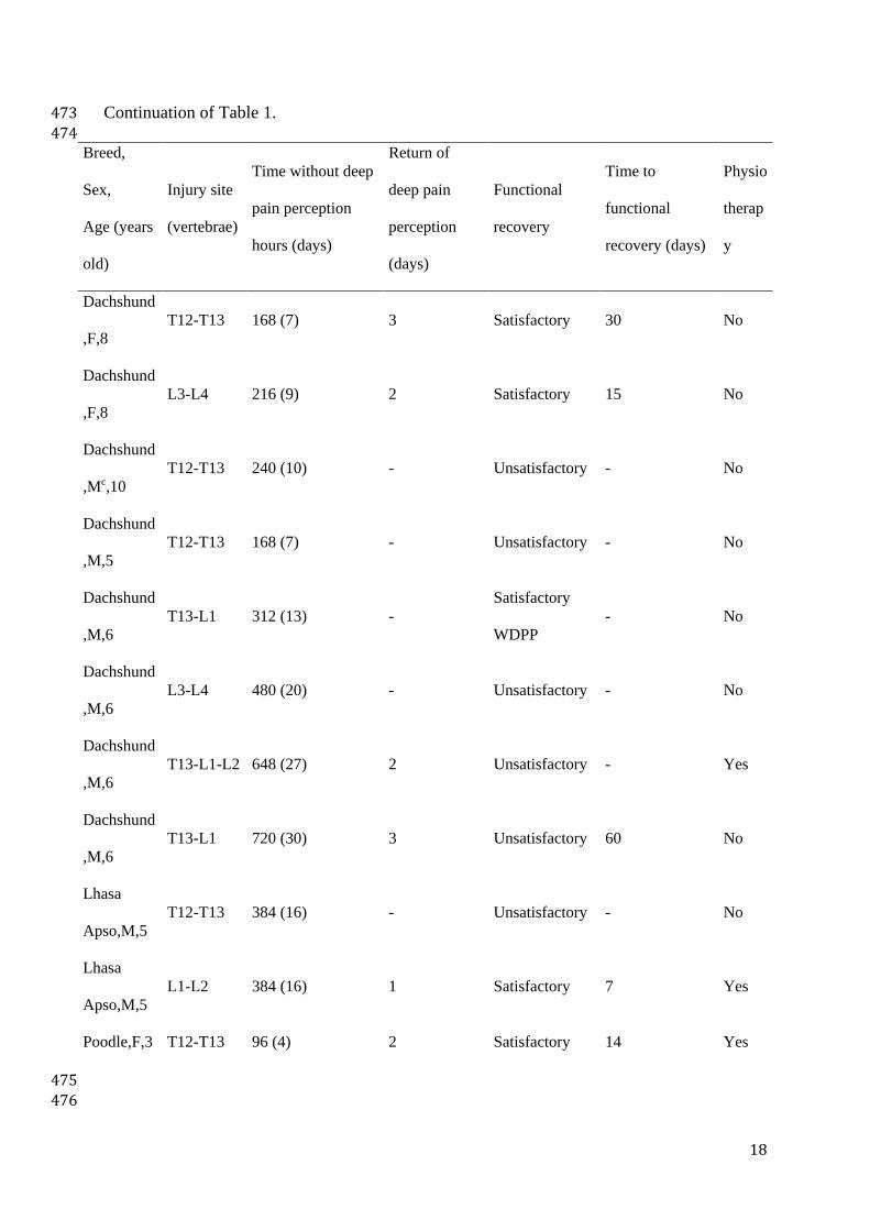
472
Breed,
Sex,
Age (years
old)
Injury site
(vertebrae)
Time without deep
pain perception
hours (days)
Return of
deep pain
perception
(days)
Functional
recovery
Time to
functional
recovery (days)
Physio
therap
y
Cocker
Spaniel,Fa,
7
T11-T12 96 (4) 1 Satisfactory 30 No
Dachshund
,F,4
L4-L5 96 (4) - Unsatisfactory - Yes
Dachshund
,F,5
T12-T13 96 (4) -
Satisfactory
WDPPb
- Yes
Dachshund
,F,5
T13-L1 720 (30) - Unsatisfactory - No
Dachshund
,F,5
L1-L2 720 (30) - Unsatisfactory - No
Dachshund
,F,5
T13-L1-L2 1440 (60) 78 Satisfactory 90 Yes
Dachshund
,F,6
T11-T12 144 (6) -
Satisfactory
WDPP
- No
Dachshund
,F,8
L1-L2 96 (4) -
Satisfactory
WDPP
- Yes
18
Continuation of Table 1. 473
474 Breed,
Sex,
Age (years
old)
Injury site
(vertebrae)
Time without deep
pain perception
hours (days)
Return of
deep pain
perception
(days)
Functional
recovery
Time to
functional
recovery (days)
Physio
therap
y
Dachshund
,F,8
T12-T13 168 (7) 3 Satisfactory 30 No
Dachshund
,F,8
L3-L4 216 (9) 2 Satisfactory 15 No
Dachshund
,Mc,10
T12-T13 240 (10) - Unsatisfactory - No
Dachshund
,M,5
T12-T13 168 (7) - Unsatisfactory - No
Dachshund
,M,6
T13-L1 312 (13) -
Satisfactory
WDPP
- No
Dachshund
,M,6
L3-L4 480 (20) - Unsatisfactory - No
Dachshund
,M,6
T13-L1-L2 648 (27) 2 Unsatisfactory - Yes
Dachshund
,M,6
T13-L1 720 (30) 3 Unsatisfactory 60 No
Lhasa
Apso,M,5
T12-T13 384 (16) - Unsatisfactory - No
Lhasa
Apso,M,5
L1-L2 384 (16) 1 Satisfactory 7 Yes
Poodle,F,3 T12-T13 96 (4) 2 Satisfactory 14 Yes
475
476

19
Continuation of Table 1. 477
478 Breed,
Sex,
Age (years
old)
Injury site
(vertebrae)
Time without deep
pain perception
hours (days)
Return of
deep pain
perception
(days)
Functional
recovery
Time to
functional
recovery (days)
Physio
therap
y
Poodle,M,
3
L5-L6 120 (5) - Unsatisfactory - Yes
Poodle,M,
5
T12-T13 168 (7) 7 Unsatisfactory - No
Shih
Tzu,M,4
T11-T12 192 (8) 20 Satisfactory 37 Yes
MBd,M,6 T12-T13 96 (4) 1 Satisfactory 7 Yes
MB ,F,4 T12-T13 120 (5) 7 Satisfactory 60 No
MB ,F,4 T12-T13 240 (10) - Unsatisfactory - No
MB ,F,4 T13-L1 336 (14) 2 Satisfactory 45 No
MB ,F,5 L1-L2 216 (9) 1 Satisfactory 15 No
MB ,F,7 L1-L2 96 (4) 1 Satisfactory 10 No
MB ,F,7 T13-L1 600 (25) - Unsatisfactory - No
MB ,M,3 L1-L2 1440 (60) - Unsatisfactory - No
MB ,M,5 T11-T12 264 (11) 1 Satisfactory 45 Yes
MB ,M,6 T12-T13 144 (6) - Unsatisfactory - Yes
MB ,M,7 T12-T13 144 (6) 2 Satisfactory 21 No
MB ,M,8 T13-L1 120 (5) 1 Satisfactory 75 No
MB,M,8 T12-T13 192 (8) 1 Satisfactory 15 No
Yorkshire,
M,8
T10-T11-
T12
96 (4) -
Satisfactory
WDPP
- No
a F – Female 479 b WDPP – Without Deep Pain Perception – Spinal Walking 480

20
c M – Male 481 d MB – Mixed Breed 482
483
484
485
486
487
488
489
490
491

21
Table 2 492
Descriptive measures for quantitative variables. 493
Variables N Mina Maxb Mean Mediane SDc
Age (year old) 36 3 10 5,75 5,5 1,68
Time without deep pain perception
(days) 36 4 60 13,67
8
14,02
Deep pain perception return time (days) 19 1 78 7,16 2 17,72
Time to functional recovery (days) 17 7 90 33,88 30 25,17
a Minimum 494 b Maximum 495 c SD – Standard Deviation 496
497
498
499
500
501
502
503
504
505
506
507
508
509
510
511
512

22
Table 3. 513
Fisher's Exact Test to verify the association with the functional recovery variable. 514
Variables Alternatives
Functional recovery
p-value Unsatisfactory Satisfactory
Satisfactory
WDPPa
Breed
Cocker
Spaniel
0 1 0
0,143
0,00% 5,90% 0,00%
Daschshund 7 4 4
50,0% 23,5% 80,0%
Lhasa Apso 1 1 0
7,1% 5,9% 0,0%
Poodle 2 1 0
14,3% 5,9% 0,0%
Shih Tzu 0 1 0
0,0% 5,9% 0,0%
Mixed Breed 4 9 0
28,6% 52,9% 0,0%
Yorkshire 0 0 1
0,0% 0,0% 20,0%
Chondrodystrophic
No 4 9 1
0,317 28,6% 52,9% 20,0%
Yes 10 8 4
71,4% 47,1% 80,0%
Sex
Female 6 9 2
0,903 42,9% 52,9% 40,0%
Male 8 8 3
57,1% 47,1% 60,0%
Injury Site
L1-L2 2 3 1
0,791
14,3% 17,6% 20,0%
L3-L4 1 1 0
7,1% 5,9% 0,0%
L4-L5 1 0 0
7,1% 0,0% 0,0%
L5-L6 1 0 0
7,1% 0,0% 0,0%
T10-T11-T12 0 0 1
0,0% 0,0% 20,0%
T11-T12 0 3 1
0,0% 17,6% 20,0%
T12-T13 6 6 1
42,9% 35,3% 20,0%
T13-L1 2 3 1
14,3% 17,6% 20,0%
T13-L1-L2 1 1 0
7,1% 5,9% 0,0%

23
Continuation of Table 3. 515
516
Variables Alternatives
Functional recovery
p-value Unsatisfactory Satisfactory
Satisfactory
WDPPa
Time without deep
pain perception
4 to 7 days 5 8 4
0,264
35,7% 47,1% 80,0%
8 to 15 days 2 6 1
14,3% 35,3% 20,0%
16 to 30 days 6 2 0
42,9% 11,8% 0,0%
More than 30
days
1 1 0
7,1% 5,9% 0,0%
NSAIb pre
No 11 13 4
1,000 78,6% 76,5% 80,0%
Yes 3 4 1
21,4% 23,5% 20,0%
Corticosteroid pre
No 7 12 4
0,404 50,0% 70,6% 80,0%
Yes 7 5 1
50,0% 29,4% 20,0%
Image examination
Myelography 12 17 5
0,407 85,7% 100,0% 100,0%
CTc 2 0 0
14,3% 0,0% 0,0%
Physioterapy
No 10 11 3
1,000 71,4% 64,7% 60,0%
Yes 4 6 2
28,6% 35,3% 40,0% a WDPP – Without Deep Pain Perception 517 b NSAI – Non-Steroid Anti-Inflammatory 518 c CT – Computed Tomography 519
520
521
522
523
524
525
526
527

24
Table 4. 528
Mann-Whitney U test to verify association with the Functional Recovery variable. 529
Variables
Averages by Functional Recovery
p-value
Unsatisfactory Satisfactory
Satisfactory
WDPPa
Age (year old) 5,29 5,88 6,60 0,198
Time without deep pain (days) 18,36 12,00 6,20 0,048 *
Deep pain perception return time (days) 4,50 7,47 ----- 0,329
a WDPP – Without Deep Pain Perception 530
* Significant difference, p < 0,05. 531
532
533
534
535
536
537
538
539
540
541
542
543
544
545
546
547

25
Table 5. 548
Correlation of Spearman with the variable time for functional recovery. 549
Dependent variable Independent variables Correlation p-value
Time to functional
recovery
Age (year old) -0,098 0,707
Time without deep pain (days) 0,368 0,146
Deep pain perception return time (days) 0,529 0,029 *
* Significant difference, p < 0,05. 550
551
552
553
554
555
556
557
558
559
560
561
562
563

4. DISCUSSÃO
A doença do disco intervertebral é uma das causas mais frequentes de
disfunção da medula espinhal em cães (BRISSON, 2010; OLBY, 2010) e, por
isso, requer contínua atualização e estudos acerca de sua patologia,
apresentação clínica, diagnóstico, tratamento e prognóstico. A compreensão dos
diferentes tipos de doença do disco intervertebral ocorreu ao longo de muitos
anos de estudos e de uma importante evolução em termos de diagnóstico por
imagem (FENN et al., 2020; DA COSTA et al., 2020). A classificação e
caracterização, do que se sabe até hoje, em termos das variações dessa doença
é um importante passo evolutivo a fim de padronizar os termos utilizados pelos
profissionais, bem como a conduta clínico-diagnóstica (FENN et al., 2020).
Apesar de toda essa evolução em termos de patologia e diagnóstico,
muitos locais do mundo, incluindo algumas regiões do Brasil, ainda não dispõem
dos meios ideais para o diagnóstico definitivo “antemortem” dos diferentes tipos
de doença do disco intervertebral. Contudo, isso não pode ser considerado fator
de estagnação e motivo de falta de conhecimento e de aplicação de
conhecimento no meio clínico-cirúrgico.
O diagnóstico de EDIVI é um desafio mesmo com imagem avançada e um
estudo recente, com 20 casos (KIM et al., 2020), demonstrou que mesmo com a
imagem avançada de IRM, ainda a melhor maneira de emitir prognóstico para
esse tipo de afecção é o grau de disfunção neurológica na apresentação clínica
do paciente. Mesmo que estudos demosntrem que a maior parte dos cães
afetados é de raça não condrodistrófica e que exercício intenso ou trauma
antecedem a apresentação da afecção (KIM et al., 2020), não se pode esquecer
que cães condrodistróficos (TAMURA et al., 2015) e sem histórico de trauma ou
exercício também podem apresentar EDIVI (PONCELET; HEIMANN, 2011), como
foi apresentado no artigo 1 da presente tese.
Embora no caso relatado, a modalidade de imagem disponível não tenha
sido fator determinante para o diagnóstico, os achados do exame radiográfico
simples traziam importantes indicativos de DDIV, como aumento da opacidade de
forame intervertebral e presença de material mineralizado dentro do canal
vertebral (LAMB et al., 2002; MCCONNELL; GAROSI, 2004) e, em determinados

47
casos, a mielografia pode apresentar um padrão de compressão intradural
(PONCELET; HEIMANN, 2011). O diagnóstico definitivo foi possível apenas com
a intervenção cirúrgica, visualização do material intradural, remoção desse último
e envio para análise histológica. A falta de análise histológica tem sido ponto de
estrangulamento em casos suspeitos de EDIVI (KIM et al., 2020).
A cirurgia parece não ser essencial para o tratamento da EDIVI (YARROW;
JEFFERY, 2000; KIM et al., 2020), mas a evolução do caso em questão indicou
que foi a escolha adequada, pois o paciente apresentou importante melhora
clínica e total recuperação em pouco tempo após o procedimento cirúrgico, e esse
não foi o primeiro caso de sucesso com tratamento cirúrgico (SANDERS et al.,
2002). Assim, a EDIVI deve ser considerada na lista de diagnósticos diferenciais
em cães com sinais agudos de disfunção medular (KENT et al., 2011), juntamente
com EDIV (Hansen tipo I), embolismo fibrocartilaginoso e EANCNP (KIM et al.,
2020).
Outra questão desafiante para o veterinário, que atende cães com suspeita
de DDIV, está relacionada ao momento da cirurgia descompressiva para cães
paraplégicos sem PDP (MOORE et al., 2020). Ampla gama de estudos tem sido
realizada nessa área e esses têm indicado que o tempo de paraplegia não está
relacionado à recuperação funcional, como se pensava (LANGERHUUS; MILES,
2017). Entretanto, os estudos abrangem poucos animais com tempo prolongado
de paraplegia sem percepção à dor profunda (MOORE et al., 2020).
O segundo artigo da presente tese é apresentado com a intenção de
responder algumas questões relacionadas aos cães com EDIV toracolombar e
lombossacra, submetidos tardiamente à cirurgia descompressiva. Algumas
informações importantes foram encontradas, e vão ao encontro do que é
internacionalmente aceito: o tempo para a descompressão cirúrgica não é fator
determinante para a recuperação funcional (JEFFERY et al., 2016), mas a chance
de recuperação está diminuída pelo tempo. O mais interessante é que incentiva a
intervenção cirúrgica em cães paraplégicos há sete, 10 e até mesmo 30 dias, com
uma chance relativamente alta de retorno à deambulação (47,2%).
Apesar das evidências de recuperação funcional para cães paraplégicos
sem percepção à dor profunda não estar relacionadas ao tempo de paraplegia
(JEFFERY et al 2016), o estudo de MOORE et al. (2016) demonstrou que 65%

48
dos cirurgiões e neurologistas indicam cirurgia o mais rápido possível
independente do tempo de evolução, revelando que há um certo “consenso”
sobre a emergência da situação. Os resultados do estudo aqui apresentado
(artigo 2) não têm o intuito de incentivar o atraso na cirurgia, mas sim de
demonstrar que, caso o paciente seja admitido tardiamente ao atendimento
especializado, existem chances de recuperação funcional com o tratamento
cirúrgico.

5. CONCLUSÃO
A extrusão de disco intervertebral intradural, apesar de rara, deve ser
considerada sempre no dignóstico diferencial de cães com sinais clínicos de
doença do disco intervertebral. Essa variação da extrusão de disco intervertebral
é passível de cirurgia e o prognóstico para recuperação funcional com cirurgia é
bom. A cirurgia, ainda, é indicada como meio de coleta de material para
diagnóstico definitivo dessa afecção.
Cães paraplégicos sem percepção à dor profunda, com extrusão de disco
intervertebral toracolombar, podem ser beneficiados com o tratamento cirúrgico,
mesmo que tardiamente no curso da doença (96 horas ou mais sem percepção à
dor profunda).

6. REFERÊNCIAS
AIKAWA, T. et al. Recurrent thoracolumbar intervertebral disc extrusion after hemilaminectomy and concomitante prophylactic fenestration in 662 chondrodystrophic dogs. Veterinary Surgery, v.41, p.381-390, 2012a. Disponível em: <https://doi.org/10.1111/j.1532-950X.2012.00970.x>. Acesso em: 21 dez. 2020. AIKAWA, T. et al. Long-term neurologic outcome of hemilaminectomy and disk fenestration for treatment of dogs with thoracolumbar intervertebral disk herniation: 831 cases (2000-2007). Jounal of American Veterinary Medical Association, v.241, n.12, p.1617-1626, 2012b. Disponível em: <https://doi.org/10.2460/javma.2003.222.762>. Acesso em: 15 dez. 2020. AIKAWA, T. et al. A comparison of thoracolumbar intervertebral disc extrusion in French bulldogs and dachshunds and association with congenital vertebral anomalies. Veterinary Surgery, v.43, p.301–307, 2014. Disponível em: <https://doi.org/10.1111/j.1532-950X.2012.00970.x>. Acesso em: 18 dez. 2020. BARNOON, I. et al. Spontaneous intradural disc herniation with focal distension of the subarachnoid space in a dog. The Canadian Veterinary Journal, v.53, n.11, p.1191-1194, 2012. Disponível em: <https://www.ncbi.nlm.nih.gov/pmc/articles/PMC3474575/>. Acesso em: 5 dez. 2020. BELTRAN, E. et al. Clinical and magnetic resonance imaging features of canine compressive cervical myelopathy with suspected hydrated nucleus pulposus extrusion. Journal of Small Animal Practice, v.53, p.101–107, 2012. Disponível em: <https://doi.org/10.1111/j.1748-5827.2011.01166.x>. Acesso em: 5 dez. 2020. BERGKNUT, N. et al. Incidence of intervertebral disk degeneration-related diseases and associated mortality rates in dogs. Journal of American Veterinary Medical Association, v.240, n.11, p.1300-1309, 2012. Disponível em: <https://doi.org/10.2460/javma.240.11.1300>. Acesso em 5 dez. 2020. BRISSON, B. A. Intervertebral disc disease in dogs. Veterinary Clinics of North America: Small Animal Practice, v.40, n.5, p.829-858, 2010. Disponível em: <https://doi.org/10.1016/j.cvsm.2010.06.001>. Acesso em 10 dez. 2020. BRISSON, B. A. et al. Comparison of the effect of single-site and multiple-site disc fenestration on the rate of recurrence of thoracolumbar disc extrusion in dogs: a prospective randomized, controlled study. Journal of the American Veterinary Medical Association, v.238, p.1593-1600, 2011. Disponível em: <https://doi.org/10.2460/javma.238.12.1593>. Acesso em: 21 dez. 2020. DA COSTA, R. C. et al. Diagnostic imaging in intervertebral disc disease. Frontiers in Veterinary Science, v.7, 588338, 2020. Disponível em: <https://www.ncbi.nlm.nih.gov/pmc/articles/PMC7642913/>. Acesso em: 15 dez. 2020.
51
DE RISIO L. et al. Association of clinical and magnetic resonance imaging findings with outcome in dogs with presumptive acute noncompressive nucleus pulposus extrusion: 42 cases (2000-2007). Journal of the American Veterinary Medical Association, v.234, p.495–504, 2009. Disponível em: <https://doi.org/10.2460/javma.234.4.495>. Acesso em: 5 dez. 2020. DE RISIO, L. A review of fibrocartilaginous embolic myelopathy and diferente types of peracute non-compressive intervertebral disk extrusions in dogs and cats. Frontiers in Veterinary Science, v. 2, 24, 2015. Disponível em: <https://doi.org/10.3389/fvets.2015.00024>. Acesso em: 5 dez. 2020. FENN, J. et al. Classification of intervertebral disc disease. Frontiers in Veterinary Science, v.7, 579025, 2020. Disponível em: <https://doi.org/10.3389/fvets.2020.579025>. Acesso em: 10 dez. 2020. HAY, C. W.; MUIR, P. Tearing of the dura mater in three dogs. Veterinary Record, v.146, n.10, p.279-282, 2000. Disponível em: <https://doi.org/10.1136/vr.146.10.279>. Acesso em: 5 dez. 2020. ITO, D. et al. Prognostic value of magnetic resonance imaging in dogs with paraplegia caused by thoracolumbar intervertebral disk extrusion: 77 cases (2000-2003). Jounal of American Veterinary Medical Association, v.227, n.9, p.1454-1460, 2005. Disponível em: <https://doi.org/10.2460/javma.2005.227.1454>. Acesso em: 10 dez. 2020. JEFFERY, N. D. Treatment of acute and chronic thoracolumbar disc disease by “mini-hemilaminectomy”. Journal of Small Animal Practice, v.29, p.611-6, 1988. Disponível em: <https://doi.org/10.1111/j.1748-5827.1988.tb02181.x>. Acesso em: 18 dez. 2020. JEFFERY, N. D. et al. Intervertebral disk degeneration in dogs: consequences, diagnosis, treatment, and future directions. Jounal of Veterinary Internal Medicine, v.27, n.6, p.1318-1333, 2013. Disponível em: <https://doi.org/10.1111/jvim.12183>. Acesso em 10, dez, 2020. JEFFERY, N. D. et al. Factors associated with recovery from paraplegia in dogs with loss of pain perception in the pelvic limbs following intervertebral disk herniation. Jounal of the American Veterinary Medical Association, v.248, n.4, p.386-394, 2016. Disponível em: <https://doi.org/10.2460/javma.248.4.386>. Acesso em: 10 dez 2020. JEFFERY, N. D. et al. Choices and decisions in decompressive surgery for thoracolumbar intervertebral disc herniation. Veterinary Clinics of North America: Small Animal Practice, v.48, p.169-186, 2017. Disponível em: <https://doi.org/10.1016/j.cvsm.2017.08.014>. Acesso em: 10 dez. 2020. JERRAM, R.M.; DEWEY, C.W. Acute thoracolumbar disk extrusion in dogs – Part I. Compendium of Continuim Education in Small Animal Practice, v.21, n.10,

52
p.922-930, 1999. KENT, M. et al. Imaging diagnosis – CT myelography in a dog with intramedullary intervertebral disc herniation. Veterinary Radiology and Ultrasound, v.52, n.2, p.185-187, 2011. Disponível em: <https://doi.org/10.1111/j.1740-8261.2010.01755.x >. Acesso em: 21 dez. 2020. KIM, J. et al. Preliminary study of presumptive intradural-intramedullary intervertebral disc extrusion in 20 dogs. Journal of Veterinary Science, v.21, n.4, p.e52, 2020. Disponível em: <https://www.ncbi.nlm.nih.gov/pmc/articles/PMC7402930/>. Acesso em: 10 dez. 2020. LAMB, C. R. et al. Accuracy of survey radiographic diagnosis of intervertebral disc protrusion in dogs. Veterinary Radiology and Ultrasound, v.3, p.222-228, 2002. Disponível em: <https://doi.org/10.1111/j.1740-8261.2002.tb00994.x>. Acesso em: 21 dez. 2020. LANGERHUUS, L.; MILES, J. Proportion recovery and times to ambulation for non-ambulatory dogs with thoracolumbar disc extrusions treated with hemilaminectomy or conservative treatment: a systematic review and meta-analysis of case-series studies. The Veterinary Journal, v.220, p.7-16, 2017. Disponível em: <https://doi.org/10.1016/j.tvjl.2016.12.008>. Acesso em 10 dez 2020. LEVINE, J. M. et al. Naturally occurring disk herniation in dogs: an opportunity for pre-clinical spinal cord injury research. Journal of Neurotrauma, v.28, n.4, p.675-688, 2011. Disponível em: <https://doi.org/10.1089/neu.2010.1645>. Acesso em: 10 dez. 2020. LIPTAK, J. M. et al. Radiographic diagnosis: intramedullary extrusion of an intervertebral disc. Veterinary Radiology and Ultrasound, v.43, n.3, p.272-274, 2002. Disponível em: <https://static1.squarespace.com/static/58051c5bf5e2311a8d64e2bc/t/58176f0c9f7456c33787849d/1477930766999/2002+VRU+intramedullary+IVDD.pdf>. Acesso em: 5 dez. 2020. MCCONNELL, J. F.; GAROSI, L.S. Intramedullary intervertebral disk extrusion in a cat. Veterinary Radiology and Ultrasound, v.45, n.4, p.327-330, 2004. Disponível em: <https://doi.org/10.1111/j.1740-8261.2004.04062.x>. Acesso em 21 dez. 2020. MCKEE, W. M.; DOWNERS, C. J. Rupture of the dura mater in two dogs caused by the peracute extrusion of a cervical disc. Veterinary Record, v.162, n.15, p.479-481, 2008. Disponível em: <http://dx.doi.org/10.1136/vr.162.15.479>. Acesso em: 5 dez. 2020. MOORE, S. A.; EARLY, P. J.; HETTLICH, B. F. Practice patterns in the management for acute intervertebral dis herniation in dogs. Journal of Small

53
Animal Practice, v.57, p.409-415, 2016. Disponível em: <https://doi.org/10.1111/jsap.12496 >. Acesso em: 10 dez. 2020. MOORE, S. A. et al. Current approaches to the management of acute thoracolumbar disc extrusion in dogs. Frontiers in Veterinary Science, v.7, 610, 2020. Disponível em: <https://doi.org/10.3389/fvets.2020.00610>. Acesso em: 5 dez. 2020. MUIR, P. et al. Comparison of hemilaminectomy and dorsal laminectomy for thoracolumbar intervertebral disc extrusion in dachshunds. Jornal of Small Animal Practice, v.36, p.360-367, 1995. Disponível em: <https://doi.org/10.1111/j.1748-5827.1995.tb02950.x>. Acesso em 18 dez. 2020. OLBY, N. et al. Long-term functional outcome of dogs with severe injuries of the thoracolumbar spinal cord: 87 cases (1996-2001). Journal of American Veterinary Medical Association, v.222, v.6, p.762-769, 2003. Disponível em: <https://doi.org/10.2460/javma.2003.222.762>. Acesso em: 10 dez. 2020. OLBY, N. The pathogenesis and treatment of acute spinal cord injuries in dogs. Veterinary Clinics of North America: Small Animal Practice, v.40, p.791–807, 2010. Disponível em: <https://doi.org/10.1016/j.cvsm.2010.05.007>. Acesso em: 5 dez. 2020. OLBY, N. J. et al. Prognostic factors in canine intervertebral disc disease. Frontiers in Veterinary Science, v.7, 596059, 2020. Disponível em: <https://doi.org/10.3389/fvets.2020.596059>. Acesso em: 10 dez. 2020. PACKER, R. A.; FRANK, P. M.; CHAMBERS, J. N. Traumatic subarachnoid-pleural fistula in a dog. Veterinary Radiology and Ultrasound, v.45, n.6, p.523-527, 2004. Disponível em: <https://doi.org/10.1111/j.1740-8261.2004.04089.x>. Acesso em: 5 dez. 2020. PONCELET, L.; HEIMANN, M. Intradural vertebral disc herniation in a dog. Veterinary Record, v.168, n.18, p.486a, 2011. Disponível em: <http://dx.doi.org/10.1136/vr.c6740>. Acesso em: 5 dez. 2020. RUDDLE, T. L. et al. Outcome and prognostic factors in non-ambulatory Hansen Type I intervertebral disc extrusions: 308 cases. Veterinary Compendium Orthopaedics and Traumatology, v. 19, 29-34, 2006. SANDERS, S. G.; BAGLEY, R. S.; GAVIN, P. R. Intramedullary spinal cord damage associated with intervertebral disk material in a dog. Journal of American Medical Association, v.221, p.1594-1596, 74-75, 2002. Disponível em: <https://doi.org/10.2460/javma.2002.221.1594>. Acesso em: 5 dez. 2020. SCOTT, H. W.; MCKEE, W. M. Laminectomy for 34 dogs with thoracolumbar intervertebral disc disease and loss of deep pain perception. Jounal of Small Animal Practice, v.40, n.9, p.417-422, 1999. Disponível em: <https://doi.org/10.1111/j.1748-5827.1999.tb03114.x>. Acesso em: 10 dez. 2020.

54
SPITZBARTH, I. et al. Current insights into the pathology of canine intervertebral disc extrusion-induced spinal cord injury. Frontiers in Veterinary Science, v.7, 595796, 2020. Disponível em: <https://doi.org/10.3389/fvets.2020.595796>. Acesso em 15 dez. 2020. TAMURA, S. et al. Thoracolumbar intradural disc herniation in eight dogs: clinical, low-field magnetic resonance imaging, and computed tomographic myelography findings. Veterinary Radiology and Ultrasound, v.56, n.2, p.160-167, 2015. Disponível em: <https://doi.org/10.1111/vru.12213>. Acesso em: 5 dez. 2020. YARROW, T. G.; JEFFERY, N. D. Dura mater laceration associated with acute paraplegia in three dogs. Veterinary Record, v.146, n.5, p.138-139, 2000. Disponível em: <http://dx.doi.org/10.1136/vr.146.5.138>. Acesso em: 5 dez. 2020.