and δ-Lactams
109
Development of a Biocatalytic Platform For the Enantioselective Hydrolysis of γ- and δ-Lactams Diplomarbeit Zur Erlangung des akademischen Grades eines Diplomingenieurs (Dipl.-Ing.) der Studienrichtung Biotechnologie Begutachter: O. Univ.-Prof. Dr. phil. Kurt FABER Institut für Chemie Organische und Bioorganische Chemie Universität Graz Vorgelegt von Elisabeth Eger 31.03.014
-
Upload
khangminh22 -
Category
Documents
-
view
0 -
download
0
Transcript of and δ-Lactams

Development of a Biocatalytic Platform
For the Enantioselective Hydrolysis
of γ- and δ-Lactams
Diplomarbeit
Zur Erlangung des akademischen Grades
eines Diplomingenieurs (Dipl.-Ing.)
der Studienrichtung Biotechnologie
Begutachter:
O. Univ.-Prof. Dr. phil. Kurt FABER
Institut für Chemie
Organische und Bioorganische Chemie
Universität Graz
Vorgelegt von Elisabeth Eger
31.03.014

ZUSAMMENFASSUNG
II
Zusammenfassung
Nicht natürliche - und -Aminosäuren sind aufgrund ihres Vorkommens als Baustein in der
Synthese von biologisch aktiven Verbindungen und ihrer Rolle als Signalmoleküle im
Zentralnervensystem wichtige Verbindungen in der pharmazeutischen Industrie. Es wurde
bereits eine beträchtliche Anzahl von unterschiedlichen Strategien für die chemische Synthese
dieser Aminosäuren in enantiomerenreiner Form entwickelt, wobei sich die meisten auf die
Verwendung von teuren und giftigen Katalysatoren und mehrere Syntheseschritte stützen.
Eine elegante, einfache und umweltverträgliche Strategie ist die enzymatische Hydrolyse der
entsprechenden - und -Lactame. Bis zu diesem Zeitpunkt sind nur wenige Enzyme, die die
Amidbindung dieser stabilen 5- oder 6-Ringe hydrolysieren können, in der Literatur
beschrieben und die einzigen bekannten Substrat der -Lactamasen sind 2-
Azabicyclo[2.2.1]hept-5-en-3-on, auch bekannt als Vince-Lactam, und zwei Derivate dieser
Verbindung.
Ziel dieser Arbeit war die Identifizierung neuer - und -Lactamasen und ihre Verwendung in
enantioselektiven hydrolytischen Reaktionen mit verschiedenen Lactam-Substraten. Vier
unterschiedliche Enzyme wurden erfolgreich für die Hydrolyse von Vince-Lactam eingesetzt
und zeigten dabei enantiokomplementäre Aktivitäten. Einige dieser Enzyme zeigten auch
hydrolytische Aktivität an drei Derivaten von Vince-Lactam. Zusätzlich konnten drei Enzyme
die perfekte kinetische Racemtrennung von Vince-Lactam, einem N-methylierten und einem
reduzierten Derivat der Verbindung katalysieren. Drei Derivate des Vince-Lactams wurden
von keinem der Enzyme als Substrat akzeptiert.
Zusätzlich wurden 14 unterschiedliche -Lactame und vier -Lactame für ein Aktivitäts-
Screening dieser Enzyme eingesetzt. Leider konnte keine enzymatische Aktivität beobachtet
werden.
Basierend auf einer Analyse der bekannten Literatur scheint die Enantiopräferenz der -
Lactamasen durch ihre Aminosäure-Sequenz und Proteinstruktur vorgegeben zu sein: (+)- -
Lactamasen scheinen zur Amidase-Signature-Familie zugehörig zu sein und zeigen eine
konservierte, charakteristische Sequenz mit einer Ser-Ser-Lys Triade. (-)- -Lactamasen
hingegen gehören zur / -Hydrolase Familie und beinhalten die für Serin-Hydrolasen
typische Ser-His-Asp Triade. Diese vorgeschlagene Klassifizierung wird durch die Ergebnisse
dieser Arbeit unterstützt.

ABSTRACT
III
Abstract
Non-natural γ- and δ-amino acids are important target compounds for the pharmaceutical
industry due to their use as building blocks for the synthesis of biologically active compounds
and their importance as signaling molecules in the central nervous system. A significant
number of different routes for the chemical synthesis of these amino acids in enantiopure
form have been developed; however, most of them rely on expensive and toxic catalysts and
multiple synthetic steps. An elegant, simple and green route is the enzymatic hydrolysis of the
corresponding racemic γ- and δ-lactams. To date, only few enzymes able to hydrolyze the
amide bond of these stable 5- or 6-membered rings are described in the literature and the only
substrates for γ-lactamases known so far are 2-azabicyclo[2.2.1]hept-5-en-3-one, also known
as Vince lactam, and two derivatives thereof.
Aim of this thesis was the identification of new γ- and δ-lactamases and their implementation
in enantioselective hydrolytic reactions with various lactam substrates. Four different
enzymes were successfully employed for hydrolysis of Vince lactam, thereby showing
enantiocomplementary activities. Some of these enzymes also showed activity on three
derivatives of Vince lactam. In addition, three enzymes were able to perform the perfect
kinetic resolution of Vince lactam, an N-methylated and a reduced derivative thereof,
respectively. Three derivatives of Vince lactam were not accepted as substrates by any of the
enzymes.
In addition, 14 different γ-lactams and four δ-lactams were screened for activity with these
enzymes. Unfortunately, no enzymatic activity could be detected.
Based on analysis of the literature, the enantiopreference of γ-lactamases on Vince lactam
derivatives appears to be dictated by their amino acid sequence and protein structure: (+)-γ-
Lactamases seem to belong to the amidase signature family, displaying a conserved signature
sequence and a Ser-Ser-Lys catalytic triad. (-)-γ-Lactamases, on the other hand, are members
of the α/β-hydrolase fold family and contain the Ser-His-Asp catalytic triad typical for serin
hydrolases. This proposed classification is supported by the results presented in this thesis.

ACKNOWLEDGEMENTS
V
Acknowledgements
First and foremost I want to express my gratitude to Prof. Kurt Faber for the opportunity to
write my diploma thesis as member of his group and for his guidance during the course of my
work.
Special thanks I give to Dr. Mélanie Hall for her professional advice, her patience and
understanding as well as for her motivating words in times of frustration and doubts. I thank
for a lot of enlightening discussions as well as the support while I was writing my thesis.
I also want to thank my advisor Dr. Zeinab Assaf for introducing me into the world of
lactamases and her help in the lab.
Next, I would like to express my gratitude to all members of the ElkGroup for the wonderful
working atmosphere and their valuable support. I want to give a lot of thanks to Michael
Tösch, Nikolaus Turrini and Alexander Dennig for their great company and a lot of laughs in
the “big lab”. As well I want to thank Judith Farnberger and Tamara Reiter for our daily
tea/coffee routine in the mornings. I felt honored being part of this group.
My deepest thanks I give to my family. My mother Ingrid and my father Gerhard gave me
their everlasting support and always had great trust in me to go my own way. I want to thank
them and my brother Thomas for encouraging and motivating me and for staying patient and
understanding.
To a very special man in my life: Thank you for your support, your understanding and the
laughter you gave me throughout the short but tough time we know each other.
Last but not least, I would like to thank my friends. Thank you for your encouragement, your
understanding, for keeping me grounded and for making me laugh in times of frustration. You
once again proved me that I can count on you unconditionally. I can't find words to express
my gratitude for having friends like you!

STATUTORY DECLARATION
V
EIDESSTATTLICHE ERKLÄRUNG
Ich erkläre an Eides statt, dass ich die vorliegende Arbeit selbstständig verfasst, andere als die
angegebenen Quellen/Hilfsmittel nicht benutzt, und die den benutzten Quellen wörtlich und
inhaltlich entnommenen Stellen als solche kenntlich gemacht habe.
STATUTORY DECLARATION
I declare that I have authored this thesis independently, that I have not used other than the
declared sources / resources, and that I have explicitly marked all material which has been
quoted either literally or by content from the used sources.
…………………………… ......………………………………………………..
date (signature)

CONTENTS
VI
Contents
1. Introduction 1
1.1. Amino acids - a general introduction 1
1.2. γ- and δ-Amino acids 4
1.2.1. Biological relevance and application of γ- and δ-amino acids 4
1.2.2. Chemical synthesis of γ-amino acids 7
1.2.3. Chemical synthesis of δ-amino acids 12
1.2.4. Synthesis of amino acids by chemical hydrolysis of lactams 13
1.2.5. Biocatalytic synthesis of enantiopure γ-amino acids 20
1.2.6. Enzymatic synthesis of δ-amino acids 25
1.3. Lactamases 26
1.3.1. Kinetic resolution of Vince lactam 29
1.3.2. (-)-γ-Lactamases 33
1.3.3. (+)-γ-Lactamases 35
1.3.4. Proposed classification of γ-lactamases 38
2. Research objectives 39
2.1. Aim of this thesis 39
2.2. The quest for new γ- and δ-lactamases 39
2.3. Substrates 44
3. Results and discussion 46
3.1. Vince lactam and derivatives 47
3.2. γ-Lactam type compounds: non-substrates 52
3.3. δ-Lactams 53
3.4. Screening of esterases 54
4. Conclusion and outlook 55
5. Experimental 57
6. Literature 77
7. Appendix 83

INTRODUCTION
1
1. Introduction
1.1. Amino acids - a general introduction
Proteins and peptides play an essential role in nature having both catalytic and structural
functions. These activities are directly connected to the very specific fold of each protein,
which is the result of interactions between the peptide chain constituted of a combination of
20 (22 respectively, if taking non-canonical amino acids into account) amino acids. These so
called “proteinogenic” amino acids are -amino acids, meaning that their carboxylic acid
group and their amino group are both bound to the -carbon atom. Following this structural
classification according to the respective position of their functional groups, β-, γ- and δ-
amino acids have been described (Figure 1).
Figure 1: Structure of an -amino acid (glycine) and a δ-amino acid
(5-aminovaleric acid)
Aside from the proteinogenic -amino acids, so called non-proteinogenic or non-standard
amino acids exist as well. Some of them can be found as building blocks in proteins after
posttranslational modifications, for example hydroxyproline in collagene,[1]
while others are
intermediates in metabolism (for example ornithine in the urea cycle, Figure 2). Also
spontaneous rearrangement of aspartic acid or asparagine to the corresponding β-structure has
been described in literature. This “ageing-process” of proteins is usually accompanied by loss
of function and has been found in plaques of β-amyloid peptides in association with
Alzheimer’s disease.[2]

INTRODUCTION
2
Figure 2: Structures of hydroxyproline and ornithine
Both proteinogenic and non-proteinogenic amino acids and their derivatives play an important
role outside of proteins. Examples are glutamate, dopamine (obtained by
hydroxylation/decarboxylation of tyrosine) and γ-aminobutyric acid (GABA), which act as
neurotransmitters, or histamine (obtained through decarboxylation of histidine), which
mediates allergic reactions (Figure 3).[3]
Figure 3: Examples of biologically active amino acid (γ-aminobutyric acid)
and derivatives (histamine and dopamine)
Furthermore, amino acids can serve as building blocks for larger molecules. The -amino
acid glycine, for example, is used in the biosynthesis of the porphyrin structure.[3]
The
antibiotic polymyxin B2 contains six molecules of α,γ-diaminobutanoic acid with five of them
incorporated as α-amino acid in the peptide chain and the sixth in a "α and γ" fashion (Figure
4).[2]

INTRODUCTION
3
Figure 4: Polymyxin B2. The structure contains six molecules of α,γ-diaminobutanoic acid incorporated as
α-amino acid (depicted in red) and in α- and γ-fashion (depicted in green).
Synthesis of non-natural amino acids has gained scientific interest because of their large scope
of applications. Incorporation of unnatural amino acids in proteins has been used as tool for
investigation and manipulation of protein structure and function.[4]
Oligomeric structures – so
called foldamers – of those newly synthesized compounds are especially relevant due to their
ability to adopt specific conformations, which might give rise to new pharmaceutical and
material properties.[5]

INTRODUCTION
4
1.2. γ- and δ-Amino acids
Most amino acids whose amino group is not located at the -position have been isolated from
naturally occurring peptide antibiotics or alkaloids.[6]
The γ-amino acid 4-amino-3-
hydroxybutyric acid, for example, is a building block for microsclerodermins, a family of
cyclic peptides displaying antitumor and antifungal activity.[7]
The β-hydroxy-γ-amino acid
(3S,4S)-4-amino-3-hydroxy-6-methylheptanoic acid (statine) is a component of the
hexapeptide antibiotic pepstatine (Figure 5).[8]
Figure 5: Examples of naturally occurring γ-amino acids
Despite their role as building blocks for larger molecules, γ-amino acids, γ-aminobutyric acid
being the most prominent, also serve as important bioactive compounds in the central nervous
system.
1.2.1. Biological relevance and application of γ- and δ-amino acids
γ-Aminobutyric acid (GABA) acts as an inhibitory neurotransmitter in the mammalian central
nervous system. It binds to specific receptors, which can be divided into two classes: the
ionotropic GABAA and GABAA-ρ and the metabotropic GABAB receptors. The GABAA and
GABAA-ρ receptors are anion-selective ligand-gated ion channels, which are mainly
permeable to chloride ions and to some extent to bicarbonate anions.[9]
Upon binding of
GABA, the channel opens, which leads to hyperpolarization of the neuron and thus to a
reduction of action potential. The GABAA-ρ receptor, previously known as GABAC receptor,
is a subtype of GABAA receptors consisting only of ρ-subunits.[10]
The G-protein coupled GABAB receptors are linked to potassium channels which leads again
to hyperpolarization and inhibition of action potential upon binding of GABA.[11]
While the
main effect of GABA receptors is inhibition of action, potential excitatory effects on
developing neurons have been reported.[12]

INTRODUCTION
5
GABA is synthesized from glutamamic acid (Glu) by glutamic acid decarboxylase (GAD) in
the brain since it is not able to cross the blood-brain barrier. It is degraded by GABA
transaminase (GABA-T) by oxidative deamination to succinic semialdehyde (Figure 6).[13]
Figure 6: Biosynthesis and degradation of γ-aminobutyric acid (GABA)
Low levels of GABA in the brain can result in convulsions and lead to epilepsy, Parkinson's
disease, motion disorders and other neurological diseases as well as depression, pain or
alcoholism. The polarity and high flexibility of the structure of GABA prevents the molecule
from passing the blood-brain barrier and renders administration of the molecule as drug
therapy useless. An approach to increase GABA levels in the brain is the design of analogue
structures which are lipophilic and more rigid. Potential targets for therapeutic drugs are
either the GABA receptors or the enzyme GABA-T.[13]
Baclofen, (±)-4-amino-3-(p-chlorophenyl)-butanoic acid, is selectively recognized by the
GABAB receptor and acts as a muscle relexant. The drug is sold as racemate but only the (R)-
enantiomer is active. Vigabatrin (4-aminohex-5-enoic acid) is a suicide substrate for GABA-T
due to its vinyl moiety and thus increases the level of GABA by prohibiting its degradation.
The pharmacologically active enantiomer is the (S)-form of the drug. The (S)-enantiomer of
3-isobutyl GABA is called Pregabalin and is not recognized by GABA receptors. It acts on
calcium channels on the membrane and reduces the release of neurotransmitters and is used to
treat neuropathic pain, generalized anxiety disorder and epilepsy (Figure 7).[13, 14]

INTRODUCTION
6
Figure 7: Examples of GABA-analogues. The GABA sub-structure is depicted in red.
An additional field where γ- and δ-amino acids are employed is the synthesis of
peptidomimetics and foldamers. The term “foldamer” refers to polymers which adopt a
complex and very specific conformation. Proteins or DNA are the prototypes found in nature
for this class of substances.[5]
It has long been known that incorporation of β- or γ-amino acids
in conventional -peptide chains can lead to the formation of stabilized secondary structures
[15] and resistance against hydrolytic cleavage by serum and tissue proteases
[16]. However,
peptides consisting solely of β- or γ-amino acids are also able to adopt stable secondary
structures. Due to the additional carbon atoms in the backbone and the thus generated increase
in flexibility of the molecule, more order and stability than in -peptides can be obtained. The
secondary structures like β-sheets, -helices and turns in β- and γ-peptide chains differ
significantly from their -counterparts. However, it has been shown that these peptides can
mimic -peptidic ligands in peptide-receptor interactions but cannot be cleaved by proteases,
thus providing great means for medicinal chemistry.[2]
Another application of synthetic amino acids lies in the field of nanotechnology for the
preparation of nanotubes. These tubular structures might find application in catalysis, drug
delivery and chemotherapy.[17]
Stacking of cyclic peptides is stablized by hydrogen bonds and
leads to the formation of so-called self-assembling peptide nanotubes (SPNs). Appropriate
modification of the side chains of the amino acids allows functionalization and may produce
nanotubes with high selectivity as ion channels or catalysts.[18, 19]

INTRODUCTION
7
1.2.2. Chemical synthesis of γ-amino acids
Various strategies for the chemical synthesis of γ-amino acids with different substitution
patterns have been developed. Thus, the following examples focus on the more general
applications. The first methods describe homologation reactions, using chiral -amino acids
as starting materials and transforming them into higher homologues.
1.2.2.1. Double Arndt-Eistert homologation
Starting from a carboxylic acid, a series of chemical reactions named the Arndt-Eistert
homologation yields the higher homologue of the acid.
Figure 8: General procedure for Arndt-Eistert homologation [20]
As depicted in Figure 8, homologation of a Boc-protected -amino acid yields the β-amino
acid homologue. Thus, for the γ-homologue a second round of reaction is necessary. While
the preparation of β-amino acids in the first round occurs smoothly, yields drop to 40-50% in
the first step and to 66-67% in the second step towards γ-amino acids. The overall yield of
double Arndt-Eistert homologation is <20%.[20]
Furthermore, the acid chlorides of N-Boc
protected -amino acids are prone to racemization, thus decreasing the enantiomeric excess of
the product. The original Arndt-Einstein homologation uses thionyl chloride for the synthesis
of the acid chloride and diazomethane to yield the diazoketone, however any procedure which
yields the acid chloride and less harmful and dangerous compounds for diazo-group transfer
can be used.
Double Arndt-Eistert homologation of -amino acids yields γ-amino acids, which are not
substituted at the - and β-position.

INTRODUCTION
8
1.2.2.2. Wittig olefination
Chain homologation by a Wittig-type olefination reaction starts from α-amino aldehydes. The
latter can be prepared either by oxidation of 2-amino alcohols or by reduction of the carboxyl
group of the α-amino acid. Figure 9 shows the synthesis starting with reduction of the mixed
anhydride or the acyl fluoride of the N-protected -amino acid with NaBH4 to the
corresponding alcohol (step a or b, Figure 9), which in turn is oxidized by NaOCl to the
aldehyde (step c). Wittig olefination yields the ,β-unsaturated ester, which is converted to
the N-protected γ-amino acid by hydrogenation.[21]
Figure 9: Procedure for homologation of -amino acids via Wittig olefination [21]
The enantiomeric purity of the final product depends mainly on the conditions used for the
preparation of the -amino aldehyde, prone to racemization, and the Wittig reaction.
Oxidation of the amino alcohol by NaOCl/AcNH-TEMPO provided products with high

INTRODUCTION
9
enantiomeric purity and thus presents an efficient method for the preparation of γ-substituted
γ-amino acids with proteinogenic side chains.
1.2.2.4. Examples for enantioselective synthesis of pharmaceutical γ-amino acids
Enantioselective synthesis of (S)-pregabalin was also reported using asymmetric
hydrogenation of 3-cyano-5-methylhex-3-enoic acid as key step. Several Rh-based catalysts
have been described, yielding the corresponding cyano derivative in high yield und
enantiomeric excess (Figure 10).
Figure 10: Enantioselective synthesis of (S)-pregabalin by asymmetric hydrogenation of 3-cyano-5-
methylhex-3-enoic acid and subsequent hydrogenation [22, 23, 24]
The tert-butylammonium salt of 3-cyano-5-methylhexanoic acid was obtained by
hydrogenation catalyzed by the Rh-based catalysts 1-3 in enantiomeric excess ranging from
92% (cat. 3) to 97.7% (cat 1, Figure 10). Catalytic hydrogenation of the cyano function and
treatment with acetic acid provided (S)-pregabalin in 61% yield and an enantiomeric excess of
99.8%.[22, 23, 24]
Another important GABA analogue is the β-arylsubstituted γ-amino acid 4-amino-3-(4-
chlorophenyl)butanoic acid, sold as racemate under the name baclofen. However, several

INTRODUCTION
10
strategies for the synthesis of enantiomerically pure (R)- and (S)-baclofen have been
developed. An elegant possibility is to synthesize a β-aryl lactone by intermolecular
enantioselective Heck-Matsuda arylation and further transformations to afford (R)-baclofen
(Figure 11).
Figure 11: Synthesis of (R)-baclofen by enantioselective Heck-Matsuda arylation [25, 26]
The synthesis of the lactone starts with arylation of the Z-olefin by an aryldiazonium
tetrafluoroborate in the presence of Pd(TFA)2 and a chiral bisoxazoline ligand [(S)-BOX,
Figure 11]. The diastereomeric O-methyl lactols were obtained in high enantiomeric excess
(92%) and were directly converted into the corresponding 4-aryl-γ-lactone, thus eliminating
the stereocenter of the lactol ring. Lactone opening with iodotrimethylsilane and treatment

INTRODUCTION
11
with sodium azide gave the azido ester, which can be converted to (R)-baclofen by hydrolysis,
hydrogenation and treatment with hydrochloric acid.[25, 26]
Another possibility to synthesize (R)-baclofen involves Claisen rearrangement of an allylic
alcohol with triethyl orthoester in the presence of propionic acid. An overview of the
synthesis route is given in Figure 12.
Figure 12: Synthesis of (R)-baclofen by Claisen rearrangement, ozonolysis and hydrolysis [27]
The reaction of the allylic alcohol with the orthoester afforded the (S)-γ,δ-unsaturated ester in
perfect enantiomeric excess and 75% yield. Ozonolysis, followed by treatment with NH4OAc
and hydrolysis with NaOH afforded (R)-baclofen in 73% yield.[27]

INTRODUCTION
12
1.2.3. Chemical synthesis of δ-amino acids
A method for the synthesis of enantiomerically pure 5-amino-2-methylpentanoic acid by
hydrolysis of a chiral auxiliary yielding 4-cyano-2-methylbutanoic acid and hydrogenation
thereof has been developed (Figure 13).[28]
Figure 13: Synthesis of both enantiomers of 5-amino-2-methylpentanoic acid [28]
While (R)-4-benzyl-3-propionyl-2-oxazolidinone was commercially available, the (S)-
enantiomer was prepared by functionalization with propionyl chloride and n-butyllithium
from (S)-4-benzyl-2-oxazolidinone. After functionalization with acetonitrile, the chiral
auxiliary was hydrolyzed and the enantiopure 5-amino-2-methylpentanoic acid hydrochloride
was isolated after hydrogenation with PtO2.[28]

INTRODUCTION
13
Another strategy to synthesize the (S)-enantiomer of 5-amino-2-methylpentanoic acid is the
utilization of the Evans chiral auxiliary (S)-4-benzyl-2-oxazolidinone for stereoselective
introduction of the methyl group at the Cα-position (Figure 14).[29]
Figure 14: Synthesis of (S)-5-amino-2-methylpentanoic acid [29]
1.2.4. Synthesis of amino acids by chemical hydrolysis of lactams
Lactams are cyclic amides and can be converted to the corresponding amino acids by
chemical hydrolysis. While chapter 1.3 (p. 26) describes properties and examples of naturally
occuring lactams, this chapter focuses on the synthesis of lactams and subsequent hydrolysis.
Figure 15: Hydrolysis of γ-lactam
As depicted in Figure 15, hydrolysis of lactams requires concentrated acids and high
temperatures. These conditions are on one hand energy consuming and on the other hand

INTRODUCTION
14
incompatible with sensitive functional groups, which limits the possibilities of potential
substituents of the compound. Furthermore, large amounts of salt are generated by
neutralization.
One more general possibility for the synthesis of lactams is the Michael addition of enolates
to nitroethenes to afford nitro olefins, which can be reduced to the corresponding amines,
followed by spontaneous cyclization to the lactams. Up to three stereogenic centers can be
included in the final product by using 1,2-disubstituted nitroethenes and -substitued enolates
(Figure 16).
Figure 16: Michael addition of enolates to nitroolefins and subsequent reduction of the nitro group to
afford the lactam
Many protocols rely on the use of chiral starting material for the introduction of chirality in
the product. As depicted in Figure 17, Michael addition of acyl-oxazolidinones to nitrostyrene
affords the corresponding nitro olefin. Both diastereomers are formed in a diastereomeric ratio
of >95:5 and can be separated by crystallization.[30, 31, 32]

INTRODUCTION
15
Figure 17: Michael addition of acyl-oxazolidinones to nitrostyrene, reduction and subsequent hydrolysis
to yield the γ-amino acid [30, 31, 32]
Another possibility to introduce chirality is the use of chiral catalysts, for example the
squaramide-based organocatalyst 2 (Figure 18) for the additon of malonic acid hemithioesters
to nitroolefins.

INTRODUCTION
16
Figure 18: Michael addition of malonic acid hemithioesters to nitroolefins catalyzed by a squaramide-
based organocatalyst [33]
The reaction depicted in Figure 18 is mimicking chain elongation of polyketides and fatty
acids by enzymatic activation of malonic acid hemithioesters (MAHTs) to generate ester
enolates. The Michael addition of MAHT 2 to different nitroolefins yields the Michael adduct
in high enantiomeric excess (92-96%) and moderate to good yields (22-81%). Poor
conversions can usually be observed in the case of alkyl-substituted nitroolefins. The Michael
addition occurs in an enantioconvergent fashion, meaning that E/Z-olefins always yield the
same (S)-enantiomer of the Michael adduct. Reduction of the nitro group to the amine is
followed by intramolecular cyclization and yields the corresponding γ-lactam, which can be
hydrolyzed to afford the β-substituted γ-amino acid.[33]
The reaction presented in Figure 18 can be used for the synthesis of the β-alkyl substituted γ-
amino acid (S)-3-aminomethyl-5-methylhexanoic acid, also known as (S)-baclofen, by
addition of a malonic acid hemithioester to trans-β-nitro-4-chlorostyrene (Figure 19).

INTRODUCTION
17
Figure 19: Synthesis of (S)-baclofen by Michael addition of malonic acid half thioesters to trans-β-nitro-4-
chlorostyrene catalyzed by a squaramide-based organocatalyst [33]
The depicted reaction has been run on a 5 g scale and yielded enantiopure (S)-baclofen after
one single step of recrystallization of the hydrolyzed lactam.[33]
The GABA analogue (S)-baclofen can be obtained by synthesis and subsequent hydrolysis of
an aryl-substituted γ-lactam, which is prepared from (1R)-camphor (Figure 20).

INTRODUCTION
18
Figure 20: Synthesis of (S)-baclofen by hydrolysis of the aryl-substituted γ-lactam prepared from (1R)-
camphor [34]
Deprotonation of the methyl ketone synthesized from (1R)-camphor by sodium
bis(trimethylsilyl)amide (NHMDS) yields the sodium enolate, which reacts with the
nitrostyrene derivative upon Michael addition. Addition of an excess of cerium ammonium
nitrate (CAN) and esterification by diazomethane affords the nitromethyl ester, which is
converted to the corresponding lactam by reduction of the nitro group. Hydrolysis and further
neutralization yields the corresponding γ-amino acid (S)-baclofen.[34]
Among other different synthesis protocols, (S)-pregabalin can also be obtained from
hydrolysis of a lactam precursor synthesized via a highly stereoselective conjugate-addition of
potassium alkenyltrifluoroborates to cyclic ,β-unsaturated carbonyl compounds (Figure 21).

INTRODUCTION
19
Figure 21: Stereoselective synthesis of (S)-pregabalin conjugate-addition of potassium
alkenyltrifluoroborates to a cyclic α,β-unsaturated carbonyl compound [35]
The rhodium catalyst used for the synthesis was produced in situ from {[RhCl(C2H4)2]2} and a
chiral ligand (L, Figure 21). The product of the conjugate-addition was isolated in high yield
(97%) and enantiomeric excess (99%) and was subsequently reduced by hydrogenation and
was further hydrolyzed to afford (S)-pregabalin.[35]

INTRODUCTION
20
1.2.5. Biocatalytic synthesis of enantiopure γ-amino acids
1.2.5.1. Enzymatic resolution employing acylase
Derivatives of racemic mixtures of γ-amino acids can undergo enzymatic resolution by
penicillin acylase from E. coli. This enzyme is highly specific for the hydrolysis of the
phenylacetyl group and converts the N-phenylacetyl derivatives to the corresponding γ-amino
acids (Figure 22).
Figure 22: Enzymatic resolution of N-phenylacetyl derivatives of γ-amino acids by penicillin acylase [36]
Penicillin acylase first hydrolyzes the (R)-enantiomer of the amino acid derivative and leaves
the (S)-N-phenylacetyl enantiomer behind, which can be extracted with dichloromethane and
subjected to subsequent hydrolysis with the same enzyme to yield the (S)-γ-amino acid.[36]

INTRODUCTION
21
1.2.5.2. Nitrilase-mediated hydrolysis of γ-amino nitriles
Nitrilases catalyze the enantioselective hydrolysis of nitrile groups and produce the
corresponding carboxylic acid as only product released (Figure 23) in contrast to the nitrile
hydratase- amidase system, in which the amide produced by the nitrile hydratase is
subequently hydrolyzed by the amidase. This single-enzyme system has led to an increase in
the interest for nitrilases in carboxylic acid synthesis.[37]
Figure 23: Nitrilase-catalyzed hydrolysis of γ-amino nitriles using nitrilases NIT-106 and NIT-107[37]
1.2.5.3. Chemo-enzymatic synthesis of γ-amino acids
Several chemoenzymatic processes employing different enzymes for the synthesis of chiral
precursors of γ-amino acids and corresponding pharmaceutical drugs exist.
One strategy starts with enzymatic resolution of a β-cyanodiester by lipases. The process
generates the single (S)-enantiomer that can be converted to the corresponding β-substituted
γ-amino acid upon decarboxylation, hydrolysis and reduction.

INTRODUCTION
22
Figure 24: Preparation of precursors of γ-amino acids by enzymatic resolution of a β-cyanodiester by
lipases. The hydrolysis product was not isolated after extraction and further used for decarboxylation. Ee
values correspond to the resolution by lipase. Conversions correspond to decarboxylated products.[38]
As depicted in Figure 24, hydrolysis of the ester at the C-2 center can result in the formation
of two diastereomers. Since the chirality at C-2 gets lost during decarboxylation, the
diastereoselectivity in the desymmetrization reaction is not relevant; however, all enzymes
were highly diastereoselective. It should be noted that this method can be used to synthesize
pregabalin (product b).[38]
Similarly to this lipase-based route, hydrolytic enzymes, namely porcine liver esterase and
Candida antarctica lipase B (CAL-B), can be employed for the desymmetrization of 3-
alkylglutaric acid diesters. The reaction scheme and further chemical transformations
necessary for the preparation of the corresponding amino acid are depicted in Figure 25.

INTRODUCTION
23
Figure 25: Preparation of precursors of γ-amino acids by desymmetrization of 3-alkylglutaric acid
diesters employing lipases [39]
While full conversions of diethylesters with different residues on R1 and R
2 were achieved in
most cases, smaller residues than the allyl-group on R2 resulted in a reduced enantiomeric
excess. The desymmetrization shown in Figure 25 has been used for the synthesis of a
precursor of (S)-pregabalin, which can be prepared by conversion of the ester to the
corresponding amide, subsequent recrystallization and Hofmann rearrangement, to yield the
enantiopure product.[39]
Another synthesis route involves the asymmetric bioreduction of β-cyanoacrylates by ene-
reductases, thus improving process efficiency by increasing the maximal yield from 50%
associated with kinetic resolutions to 100%.
Figure 26: Preparation of precursors of γ-amino acids by asymmetric bioreduction using
ene-reductases [40]

INTRODUCTION
24
Regarding the formation of both enantiomers, it was observed that (E)-β-cyanoacrylic acid
esters were converted to the (S)-product while the (R)-products could be obtained from the
(Z)-analogues (substrate-stereocontrol). Conversions dropped with increasing sizes of R1 and
R2. Similarly to the example presented in Figure 24, the corresponding γ-amino acid can be
prepared by hydrolysis of the ester moiety and reduction of the cyano group.[40]
In addition to the synthetic strategy of (R)-baclofen introduced in chapter 1.2.2.4 (Figure 11,
p. 10), (R)-p-chlorophenyl lactone can also be produced by a microbiologically mediated
Baeyer-Villiger oxidation (Figure 27).
Figure 27: Preparation of precursors of γ-amino acids by microbiological Bayer-Villiger oxidation [41]
Enzymatic oxidation of 3-(p-chlorophenyl)cyclobutanone by Cunninghamella echinulata
NRLL 3655 afforded the lactone in 31% yield and perfect enantiomeric excess. (R)-Baclofen
can be synthesized by further transformations in analogy to the example presented in Figure
11 (p. 10).[41]

INTRODUCTION
25
1.2.6. Enzymatic synthesis of δ-amino acids
The synthesis of δ-amino acid 5-aminovaleric acid employing L-lysine oxidase is depicted in
Figure 28.
Figure 28: Synthesis of 5-aminovaleric acid employing L-lysine oxidase [42]
The flavin-dependent enzyme L-lysine oxidase oxidizes the -carbon atom of L-lysine using
molecular oxygen. 6-Amino-2-ketocaproic acid is produced and further converted to 5-
aminovaleric acid via oxidative decarboxylation by hydrogen peroxide, which is formed as
by-product during the enzymatic oxidation. Removal of H2O2 by catalase leads to
intramolecular cyclization and the formation of Δ1-piperidine-2-carboxylate.
[42]
N-Carbamoyl-β-alanine amidohydrolase from Agrobacterium tumefaciens (Atβcar) is able to
hydrolyze different N-carbamoyl-, N-acetyl- and N-formyl-amino acids to produce the
corresponding amino acids. Figure 29 shows an example for the formation of α-substituted δ-
amino acids from the N-carbamoyl derivative.
Figure 29: Synthesis of α-substituted δ-amino acids by Atβcar [43, 44]
The enzyme Atβcar hydrolyzes the L-enantiomer of N-carbamoyl-ornithine 6-7 times faster
than the D-enantiomer, thus displaying enantioselectivity for these δ-amino acid precursors.[43,
44]

INTRODUCTION
26
1.3. Lactamases
Lactams – the term being a combination of the words lactone and amide - are cyclic amides
with an 1-azacycloalkan-2-one structure or analogues.[45]
Greek letters as prefixes indicate the
number of atoms in the cyclic system apart from the carboxyl moiety and the nitrogen
heteroatom. A β-lactam, for example, represents a 4-membered ring and a γ-lactam a 5-
membered cyclic system (Figure 30).
Figure 30: General classification of lactams
The most prominent members of the lactam-family are the β-lactams due to their frequent
occurrence as conserved structure in various antibiotics, for example penicillins or
cephalosporins. Larger lactams, like γ- and δ-lactams, occur in nature as structural motifs in
alkaloids and oxindole derivatives. Examples are brevianamide A, a fluorescent metabolite
isolated from Penicillium brevicompactum [46]
, salinosporamide A, containing a γ-lactam-β-
lactone bicyclic structure and displaying anticancer activity [47]
, or horsfiline, an oxindole
alkaloid isolated from Horsfieldia superba [48]
(Figure 31).
Figure 31: γ- and δ-Lactams as structural motifs in brevianamide A, salinosporamide A and
horsfiline [46, 47, 48]

INTRODUCTION
27
Of special importance is the high chemical stability of the amide bond, which arises from its
partial double bond character due to resonance effects (Figure 32).[49]
This stabilization might
also be a reason, why Nature built proteins as polyamides instead of polyesters. Additionally,
the cyclic structure gives lactams of a certain size more chemical stability than linear
structures, thus preventing conventional proteases from hydrolyzing the amide bond.[50]
Figure 32: Partial double bond character of amide bonds
The ring strain, which is a result of compressed or expanded angle values compared to their
optimal value, is counteracting the stabilizing resonance effect. For instance, cyclohexane has
no or very little ring strain, while in smaller cyclic systems ring strain is indirectly
proportional to ring size.[51]
Thus β-lactams are easier hydrolyzed than γ- or even δ-lactams,
posing a problem in clinical research since it renders bacteria resistant against the commonly
used β-lactam antibiotics.
In addition to hydrolysis by chemical means, which was discussed in chapter 1.2.4 (p. 13), the
amide bond of lactams can also be hydrolyzed by enzymes, for example amidases, that cleave
bonds of simple amides, and peptidases, which cleave peptide bonds of peptides and proteins.
Despite the significant amount of studies regarding β-lactamase activity, only a few surveys
about the enzymatic hydrolysis of γ- or δ-lactams have been published.
The only substrate reported so far in enzymatic hydrolysis of γ-lactams is (±)-2-
azabicyclo[2.2.1]hept-5-en-3-one (Figure 33), also known as rac-Vince lactam, and few
derivatives thereof. The double bond in the second 5-membered ring of the lactam allows
different chemical modifications such as epoxidation, fluorination, hydroxylation, etc.[52]

INTRODUCTION
28
Figure 33: Structure of Vince lactam enantiomers and the hydrolysis product of Vince lactam
The hydrolysis product of Vince lactam - cis-4-aminocyclopent-2-ene carboxylic acid - does
not only provide a template for the synthesis of conformationally restricted γ-amino acids but
also for the preparation of carbocyclic nucleosides, since it provides a cyclopentane ring
structure. Carbocyclic nucleosides are nucleosides where the oxygen atom of the sugar moiety
is replaced by a carbon atom, giving rise to compounds with antiviral or anticancer activity.
They are stabile against hydrolysis by nucleases [53]
, which leads to a greater bioavailability of
the compounds.[54]
Examples are abacavir, the commercially available form of the active
agent carbovir, which mimics the structure of 2'-deoxyguanoside and inhibits HIV reverse
transcriptase [55]
, or carbocyclic oxanosine, an anti-HIV-agent (Figure 34).
Figure 34: Carbocyclic nucleosides derived from hydrolized Vince lactam
As indicated in Figure 34, synthesis of these pharmaceutical compounds requires enantiopure
starting material or powerful asymmetric synthesis routes. Strategies for the production of
enantiomerically enriched preparations of Vince lactam include chiral synthesis as well as
resolution of the racemate by preferential crystallisation or kinetic resolution.

INTRODUCTION
29
Attempts of asymmetric synthesis of Vince lactam enantiomers – Diels-Alder reactions either
with aluminium and titanium chiral Lewis acids as catalysts or chiral sulfonyl chlorides to
introduce chirality – did not provide the compound in good yields and acceptable
enantiomeric excess yet.[52]
Preferential crystallization of one enantiomer cannot be applied
because of the oscillating manner of crystallization, which means that one enantiomer
crystallizes until it reaches a certain treashold and the other enantiomer starts to crystallize,
thus leading to low ee values.[56]
1.3.1. Kinetic resolution of Vince lactam [57]
The term "kinetic resolution" describes the selective transformation of one substrate
enantiomer over the other in a racemic mixture by a given catalyst. The enantiomeric excess
of the unreacted starting material rises during the reaction and reaches - in an ideal case - its
maximum (ee >99%) while the preferred enantiomer is completely depleted at a conversion of
50%. Potential catalysts for kinetic resolution are chiral, chemical catalysts or enzymes. In
biocatalytic kinetic resolutions, the enzyme enantiopreference is the result of a better fit of
one enantiomer into the enzyme's active site, leading to a faster reaction.
Enzymes which are able to hydrolyze the amide bond of Vince lactam have been classified as
γ-lactamases. Based on their preference for (+)-2-azabicyclo[2.2.1]hept-5-en-3-one or the
opposite (-)-enantiomer, enzymes are categorized as (+)- or (-)-γ-lactamases respectively
(Figure 35).
Figure 35: Enzymatic kinetic resolution of rac-Vince lactam

INTRODUCTION
30
One example of a chemical approach to the resolution of rac-Vince lactam is the
regioselective hydroarylation of the double bond, which leads to so-called "parallel kinetic
resolution" because both enantiomers are transformed into different products.[58]
One major drawback of kinetic resolution is the maximal yield of 50% of enantiopure
product. However, in the case of Vince lactam, the "unwanted" enantiomer can be used for
further synthesis, because valuable products of both enantiomers exist (examples can be seen
in Figure 34, p. 28).
1.3.1.1. Enzymatic kinetic resolution of Vince lactam
Previous studies focused mainly on the enzymatic hydrolysis of unsubstituted Vince lactam.
Several enantiocomplementary γ-lactamase activities have been identified and are
summarized in Table 1.

INTRODUCTION
31
Table 1: Enzymatic kinetic resolution of Vince lactam
Entry Enzyme Organism Reference
1 ENZA20 Pseudomonas solanacearum [54]
2 ENZA22 Pseudomonas fluorescens [54]
3 Comamonas acidovorans [59]
4 (amidase) Sulfolobus solfataricus MT4 [59]
5 Bradyrhizobium japonicum USDA 6 [55]
Entry Enzyme Organism Reference
6 ENZA1 Rhodococcus sp. [54]
7 ENZA25 Aureobacterium sp. [54]
8 PFEI Pseudomonas fluorescens [60]
9 Mhg Microbacterium hydrocarboxydans [61]
10 Aureobacterium sp. [62]
11 lipase PS Pseudomonas cepacia [63]
12 lipase AK Pseudomonas fluorescens [63]
13 PPL pig [63]
14 CAL-A, CAL-B Candida antarctica [63]

INTRODUCTION
32
First studies were carried out employing whole cells of Rhodococcus sp., Aureobacterium sp.,
Pseudomonas solanacearum and Pseudomonas fluorescens for the hydrolysis. The
enantioselectivity values of the whole cell resolutions with Aureobacterium sp. (ENZA25)
and Pseudomonas fluorescens (ENZA22) were 76 and 94 respectively, however after
purification and immobilization of the (-)-γ-lactamase ENZA25 the E-value of the whole
process increased (E >200). Due to stability issues ENZA22 could not be isolated.[54]
The first
(+)-γ-lactamase to be successfully isolated, cloned and overexpressed in E. coli was the
enzyme from Comamonas acidovorans, which showed perfect kinetic resolution of rac-Vince
lactam (E>200) and high sequence similarity to a formamidase and an acetamidase.[59]
Amidase from Sulfolobus solfataricus is an interesting candidate for industrial processes,
since it originates from a thermophilic archaeon and thus promises high stability.[59]
The
lactamase from Bradyrhizobium japonicum was discovered by rational genome mining: a
BLAST search using (+)-γ-lactamase from Sulfolobus solfataricus as template identified the
unnamed protein, which could hydrolyze (+)-Vince lactam after cloning in E. coli.[55]
An
enzyme with promiscuous bromoperoxidase activity from Aureobacterium sp., which
displayed hydrolytic activity on (-)-Vince lactam, was crystallized and used for elucidation of
the reaction mechanism.[62]
As described in Table 1, lactamase activity was also found in several known hydrolytic
enzyme preparations. Unlike lactamases, lipases are commercially available and already used
on industrial scale. However, reaction rates seemed to be low and as it is the case in all lipase
bioconversions, organic solvents are needed, which can be seen as problem in a green
process.[63]
Furthermore, esterase I from Pseudomonas fluorescens (PFEI) displayed
promiscuous hydrolytic activity on (-)-Vince lactam.[60]
1.3.1.2. Enzymatic kinetic resolution of Vince lactam derivatives
In addition to kinetic resolutions of Vince lactam the activities of an immobilized preparation
of CAL-B on N-protected and reduced Vince lactam and of PFEI on the saturated derivative
were tested.[63, 60]
Positive results are summarized in Table 2.

INTRODUCTION
33
Table 2: Enzymatic kinetic resolution of Vince lactam derivatives
Entry Organism Reference
1 CAL-B Candida antarctica [63]
Entry Organism Reference
2 CAL-B Candida antarctica [63]
Although the hydrolysis of both Vince lactam derivatives by CAL-B was slower than the
reaction with unsubstituted compound, the enzyme showed conserved enantiopreference (E
>200) and the unreacted substrate could be obtained with high enantiomeric excess (>99% for
reduced, >96% for N-protected Vince lactam).[63]
Pseudomonas fluorescens esterase I (PFEI)
did not show activity on the reduced Vince lactam derivative.[60]
1.3.2. (-)-γ-Lactamases
The enzyme isolated from Aureobacterium sp. displaying (-)-γ-lactamase activity on Vince
lactam belongs to the /β-hydrolase fold family.[62]
This tertiary fold is adopted by diverse
hydrolytic enzymes, which do not share significant sequence similarity and have diverged
from a common ancestor. Enzymes which share the /β-hydrolase fold include lipases,
proteases, esterases, dehalogenases, peroxidases and epoxide hydrolases. These enzymes
consist of a mostly parallel, 8-stranded β-sheet which is surrounded by -helices at both sides,
one -strand only is antiparallel (Figure 36). The catalytic residues usually consist of a highly
conserved triad: a nucleophile (Ser, Cys or Asp), an acidic residue (Asp or Glu) and a

INTRODUCTION
34
conserved histidine residue. The nucleophile is located in the "nucleophile elbow", a sharp
turn, which ensures easy approach by the substrate and the hydrolytic water molecule. It also
directs the substrate to the oxyanion hole, made of two backbone nitrogen atoms, which
stabilizes the negatively charged transition state.[64]
Figure 36: Protein structure of the (-)-γ-lactamase from Aureobacterium sp. (PDB code 1HKH). The α-
helices are depicted in red, β-sheets in yellow and loop regions in green. The figure was drawn using
PyMol [65]
The mechanism for the hydrolysis of (-)-Vince lactam was proposed for Aureobacterium γ-
lactamase and follows the classical steps in α/β-hydrolase fold enzyme-catalyzed reactions
(Figure 37):
i. The carbonyl oxygen of Vince lactam binds to the oxyanion hole, which is formed by
Tyr32 and Met99. Deprotonation of Ser98 by His259 yields a nucleophile, which
attacks the activated carbonyl group.
ii. A tetrahedral intermediate is formed. The latter collapses upon donation of a proton
from His259 to the ring nitrogen and an acyl-enzyme complex is formed.
iii. A molecule of water is deprotonated by His259 and attacks the complex. The second
tetrahedral intermediate is formed.
iv. The hydrolyzed product is released by reprotonation of Ser98 by His259.[62]

INTRODUCTION
35
Figure 37: Proposed mechanism for the hydrolysis of (-)-Vince lactam by Aureobacterium sp.
γ-lactamase [62]
1.3.3. (+)-γ-Lactamases
Alignment of the amino acid sequences of the (+)-γ-lactamases of Sulfolobus solfataricus and
Bradyrhizobium japonicum reveals a sequence identity of 49% and the presence of several
conserved regions (Figure 38), such as the catalytic triad and the so called amidase signature
sequence (AS sequence).

INTRODUCTION
36
Figure 38: Sequence alignment of (+)-lactamases from Sulfolobus solfataricus (AMID-SULSO, #P95896)
and Bradyrhizobium japonicum (G7DDC5_BRAJP, #G7DDC5). Active site residues are depicted in red.
The AS sequence (GGSSSGS) lies within the highly conserved block surrounding Ser171 (AMID-SULSO
numbering). The alignment was generated using Clustal Omega.[66]
Members of the "amidase signature family" (AS family) are defined by the amidase signature
sequence, a conserved stretch of amino acids which has been identified by primary structure
analysis of several amidases. A strictly conserved block consisting of GGSSSGS is located in
this approximately 130 residues long consensus sequence.[67,68]
The first serine of this motif is
part of the catalytic triad Ser-Ser-Lys (Figure 39), which is absolutely conserved in the AS
family proteins.

INTRODUCTION
37
Figure 39: Active site of the AS family enzyme peptide amidase Pam from Stenotrophomonas maltophilia
(PDB code 1M22) with catalytic triad. The figure was generated using PyMol.[65]
While the crystal structure of several members of the AS family (peptide amidase, Pam; fatty
acid amide hydrolase, FAAH) has already been solved, no structural data is available for the
(+)-γ-lactamases of Sulfolobus solfataricus. A homology model suggested, that the catalytic
residues are correctly positioned for amide bond hydrolysis and revealed that the oxyanion
hole is likely formed by the backbone amide nitrogen atoms of Gln192 and Gly193.[69]
The proposed mechanism for the hydrolysis of (+)-Vince lactam by γ-lactamase from
Sulfolobus solfataricus is based on the analysis of a homology model (Figure 40) [69]
:
i. The carbonyl group is activated by hydrogen bonds of the oxygen atom with the
backbone amide nitrogen atoms of Gly193 and Gln192. Activation of Ser195 occurs
via a hydrogen bond network with Ser171 and Lys96. The nucleophile attacks the
carbonyl group.
ii. A tetrahedral intermediate is formed. The intermediate collapses following donation of
a proton to the nitrogen atom of the lactam. An acyl-enzyme complex is formed.
iii. A molecule of water is deprotonated by Lys96 and attacks the acyl-enzyme complex.
The second tetrahedral intermediate is formed.
iv. The hydrolyzed product is released upon reprotonation of Ser195.

INTRODUCTION
38
Figure 40: Proposed mechanism for the hydrolysis of (+)-Vince lactam by γ-lactamase from Sulfolobus
solfataricus based on a homology model built on crystal structure of peptide amidase Pam from
Stenotrophomonas maltophilia
1.3.4. Proposed classification of γ-lactamases [57]
Based on the observations from protein structure analysis and related enantiopreference for
Vince lactam enantiomers, a classification for γ-lactamases can be proposed:
i. Enzymes which belong to the class of serine hydrolases, thus sharing the conserved
catalytic triad Ser-His-Asp and the oxyanion hole, display selective catalytic activity
on (-)-Vince lactam.
ii. (+)-γ-Lactamases are members of the amidase signature family. They possess the
amidase signature sequence GGSS(S/G)GS and the catalytic triad consisting of Ser-
Ser-Lys.

RESEARCH OBJECTIVES
39
2. Research objectives
2.1. Aim of this thesis
The aim of this thesis is the identification of new γ- and δ-lactamase activities. The strategy
relies on the investigation of various enzymes (in purified form) with known or suspected
amidase activity. Of importance is the development of biocatalysts with high
enantioselectivity allowing perfect kinetic resolution. Substrates of interest include derivatives
of 2-aza-bicyclo[2.2.1]hepten-3-one (Vince lactam), a synthon used in the preparation of
GABA analogues and carbocyclic nucleosides.
2.2. The quest for new γ- and δ-lactamases
While searching for an enzyme candidate in order to identify new lactamases, two major
considerations were crucial: the enzyme should display hydrolytic activity on amide bonds
and be able to accept bulky substrates to allow 5- and 6-membered lactam rings with different
substituents to fit into the active site. An enzyme meeting both requirements was found in
NfpolyA - a polyamide hydrolyzing amidase isolated from a soil bacterium.
Polyamidase NfpolyA from Nocardia farcinica (#Q5YVW2) was identified from a screening
of microbial hydrolytic activity on polyamides as part of a study dedicated to improving nylon
hydrophilicity by enzymatic surface modifications. Functional groups can be introduced on
the polymer surface via the amino groups formed by hydrolysis of amide bonds. NfpolyA
consists of 4 subunits and has a molecular weight of 190 kDa in its native form. The enzyme
is able to hydrolyze various amides and esters and is classified as aryl acylamidase due to its
activity on p-nitrophenyl butyrate. The enzyme belongs to the AS family displaying both the
conserved signature sequence as well as the catalytic Ser-Ser-Lys triad.[70, 71]
A sequence
identity of 98% with ω-octalactam hydrolase from Rhodococcus sp. Oct1, active on ω-
octalactam, ω-laurolactam but also o-nitroacetanilide [72]
, strongly suggested that NfpolyA
could hydrolyze lactam rings.
Several searches for homologous proteins were conducted using BLAST [73]
and PDBeFold
[74]. An amidase from Rhodococcus globerulus and malonamidase E2 from Bradyrhizobium
japonicum showed sequence similarities of 31% and 36%, respectively, with NfpolyA and

RESEARCH OBJECTIVES
40
were selected for activity screenings. Figure 41 shows a sequence alignment of these
enzymes. Chloroperoxidase T from Streptomyces aureofaciens on the other hand showed
structural similarity to the (-)- -lactamase from Aureobacterium sp. (chapter 1.3.2, p. 33).
Figure 41: Sequence alignment of malonamidase E2 from Bradyrhizobium japonicum (Q9ZIV5-BRAJP),
polyamidase from Nocardia farcinica (Q5YVW2-NOCFA) and amidase from Rhodococcus globerulus
(Q76EV1-RHOGO). The alignment was generated using Clustal Omega.[66]
The amidase AMI from Rhodococcus globerulus (#Q76EV1) is encoded in a gene cluster
responsible for alkylaldoxime metabolism. The other two enzymes responsible for aldoxime
catabolism are aldoxime dehydratase, which converts the aldoxime into the corresponding
nitrile, and nitrile hydratase, which hydrolyzes the nitrile to the corresponding amide. The
amide is in turn hydrolyzed to the carboxylic acid by the amidase.[75]
The amidase is 521
residues long and has a molecular weight of 54.6 kDa.

RESEARCH OBJECTIVES
41
Malonamidase MAE2 from Bradyrhizobium japonicum (#Q9ZIV5) is a 414 amino acid
long, dimeric protein with a molecular weight of 43.6 kDa. It is highly specific for
malonamate, which is hydrolyzed to malonate. The enzyme did not show activity with
substrate analogues like malonate, succinate, acetate, succinamate, malonamide or
acetamide.[76]
Malonamidase belongs to the AS family and is the first member whose crystal
structure has been solved (pdb code: 1OCK). The structure revealed a mixed /β fold
constituted of 12 -helices surrounding a central β-sheet composed of 11 β-strands and differs
from any known protein fold (Figure 42). The conserved AS sequence residues compose
mainly the core of the protein while the upper half of the active site is built from non-
conserved residues. Thus it can be concluded that substrate recognition of each individual
protein of this class is achieved by substitution of these non-conserved amino acids.[77]
Figure 42: Fold of the amidase signature protein malonamidase E2 from Bradyrhizobium japonicum (PDB
code: 1OCK). α-Helices are depicted in red, β-sheets in yellow and loop regions in green respectively. The
figure was generated using PyMol.[65]
Structural data confirmed that MAE2 contains a Ser-cisSer-Lys triad with Ser155 as reactive
nucleophile. Ser131 was found in the unusual cis-conformation, which appeared essential for
the formation of hydrogen bonds with the other active site residues. The residues Ser131 and
Lys62 polarize the nucleophile Ser155.[78]
Furthermore, an arginine at position 158 seems to
stabilize the transition state oxyanion by neutralizing the negative charge of the substrate and
is involved in substrate recognition.[79]

RESEARCH OBJECTIVES
42
Figure 43: Active site of malonamidase E2 from Bradyrhizobium japonicum (PDB code: 1OCK) with
catalytic triad. The figure was generated using PyMol.[65]
Chloroperoxidase CPO-T from Streptomyces aureofaciens (#O31168) belongs to the class of
cofactor-free haloperoxidases. These enzymes mediate the halogenation of organic
compounds by hypohalous acid, which is generated by oxidation of halide ions by hydrogen
peroxide. The nomenclature is based on the most electronegative halide that the enzyme is
able to utilize, thus chloroperoxidase can oxidize chloride, bromide and iodide.[80]
Cofactor-
free haloperoxidases belong to the class of /β-hydrolases and display the catalytic triad Ser-
Asp-His characteristic of serine hydrolases.[81]
The catalytic triad catalyzes the formation of a
peroxoacid in analogy to the mechanism of serine hydrolases: by nucleophilic attack of Ser on
the carboxyl group of an organic acid a tetrahedral intermediate is formed. By elimination of a
water molecule the intermediate forms an acyl-enzyme complex. Hydrolysis of the complex
by nucleophilic attack of hydrogen peroxide forms the peroxo acid. The formation of the
hypohalous acid and the actual halogenation of organic compounds are not catalyzed by the
enzyme.[82]
Cyclic imide hydrolase CIH from Pseudomonas putida (#Q4JG22) is a member of the cyclic
amidohydrolase family.[83]
The imidase is composed of four identical subunits with a weight
of 36 kDa each and contains one atom of zinc per subunit. The tightly bound zinc atom is
required for the enzymatic activity. CIH showed hydrolytic activity on hydantoin,
dihydrouracil, succinimide and maleimide.[84, 85]

RESEARCH OBJECTIVES
43
In addition to the already mentioned enzymes, several hydrolases from Nocardia brasiliensis,
Thermus thermophilus, Clostridium hathewayi and Clostridium botulinum were tested for
lactamase activity on three different substrates. Available data regarding these enzymes are
summarized in Table 3:
Table 3: Overview on data regarding hydrolases from Nocardia brasiliensis, Thermus thermophilus,
Clostridium hathewayi and Clostridum botulinum. The asterisk after the length of the protein indicates the
presence of a signal peptide in front of the actual coding sequence.
Name Enzyme Organism Accession # Length
1_Nb secreted hydrolase Nocardia brasiliensis - 303 aa
2_Nb secreted esterase Nocardia brasiliensis K0EQD2 345 aa
3_Nb secreted lipase Nocardia brasiliensis K0EWE4 244 aa
Tth_5 secreted hydrolase Thermus thermophilus H9ZRI9 306 aa*
Tth_6 carboxylesterase Thermus thermophilus H9ZP60 513 aa*
Est_5 carboxylesterase Clostridium hathewayi D3AU79 414 aa
Est_7 secreted lipase Clostridium botulinum A5I3I2 429 aa*
Est_8 secreted lipase Clostridium botulinum A5I055 452 aa*
Several of these enzymes were active in the degradation of aromatic polymers, which
(together with the observation of lactamase activity of polyamidase NfpolyA) suggested that
they could potentially show lactamase activity. Nocardia brasiliensis and Thermus
thermophilus are both thermophilic organisms and thus provide enzymes with good
thermostability in biocatalytic processes, which makes them good candidates for industrial
applications.

RESEARCH OBJECTIVES
44
2.3. Substrates
The substrates tested included derivatives of 2-aza-bicyclo[2.2.1]hepten-3-one (Vince
lactam), a synthon used in the preparation of GABA analogues and carbocyclic nucleosides.
A short overview of Vince lactam (1a) and derivatives therof – a saturated (2a), a N-
methylated (3a), a reduced and N-methylated (4a) derivative and compounds derived by
dihydroxylation (5a), dihydroxylation and protection (6a) and epoxidation of the C=C double
bond (7a) – is given in Figure 44, their synthesis is described in chapter 5.6.
Figure 44: Vince lactam and derivatives used as substrates in lactamase activity screening
Additionally, several γ- and δ-lactams, succinimide and maleimide derivatives, phthalimide
and hydantion were tested. Structures are summed up in Figure 45.

RESEARCH OBJECTIVES
45
Figure 45: γ- and δ-Lactams (8a-12a, 22a-25a), succinimide and maleimide derivatives (13a-17a),
phthalimide (19a), oxindole (20a) and hydantoin (21a)

RESULTS AND DISCUSSION
46
3. Results and discussion
Several enzymes from different organisms were investigated in pure form regarding their γ-
and δ-lactamase activity on γ- and δ-lactam-type substrates (Figure 46). Substrates included
succinimide and maleimide derivatives, phthalimide, hydantion and derivatives of the bicyclic
lactam 2-azabicyclo[2.2.1]hept-5-en-3-one (Vince lactam). Structures of all substrates tested
are given in Figure 44 (p. 44) and Figure 45 (p. 45).
Figure 46: Biohydrolysis of lactams exemplified by -butyrolactam
The results of the bioconversions will be discussed in the following chapter. Control reactions
(reaction in buffer with no enzyme present) did not show formation of the corresponding
products.
In addition, eight esterases, which displayed hydrolytic activity on aromatic polymers, were
screened for lactamase activity using substrates 1a, 19a, and 26a. The results will be
discussed separately in chapter 3.4.
All samples were analyzed on GC/MS after derivatization, except 6a, 15a and 16a, where
samples were analyzed on LC/MS.

RESULTS AND DISCUSSION
47
3.1. Vince lactam and derivatives
Figure 47: Vince lactam derivatives used in lactamase activity screening
Unsubstituted rac-Vince lactam (rac-1a, Figure 47) and – to some extent – its (-)-enantiomer
[(1R,4S)-1a, (-)-Vince lactam] was converted by all enzymes. The best results were achieved
by CIH with perfect kinetic resolution of the racemic starting material and practically full
conversion of (-)-Vince lactam (E >200). Rac-1a was hydrolyzed by AMI and CPO-T with
high conversions (76 and 66%, respectively) but poor enantioselectivities (E = 8 and 15,
respectively). NfpolyA hydrolyzed rac-1a with good conversion and enantioselectivity (conv.
45% and eeP 96%). MAE2 was not very active on rac-Vince lactam (conv. 5%), but displayed
perfect enantioselectivity (E >200). The structures of substrate and product enantiomers are
depicted in Figure 48. The results are given in Table 4.
Figure 48: Substrate and product enantiomers of Vince lactam 1a

RESULTS AND DISCUSSION
48
Table 4: Biohydrolysis of Vince lactam 1a with pure enzymesa
rac-1a (-)-1a
conv.b [%] eeS
c [%] eeP [%] E
b conv. [%]
NfpolyA 45 77 (1R,4S) 96 (1R,4S) 115 2
AMI 76 >99 (1R,4S) 32 (1R,4S) 8 53
CPO-T 66 >99 (1S,4R) 52 (1S,4R) 15 >99
MAE2 5 5 (1S,4R) >99 (1S,4R) >200 4
CIH 50 >99 (1S,4R) >99 (1S,4R) >200 >99
a Reactions were performed in 0.8 mL phosphate buffer (50 mM, pH 7.5) with 10 mM substrate and 100 µg
enzyme. b
Conversion and E-values were calculated from ee-values (chapter 5.4.). c ee values were determined
after double derivatization (chapter 5.3.)
Enantiocomplementary activities on Vince lactam were obtained: NfpolyA and AMI
hydrolyzed the (1S,4R)-enantiomer and thus displayed (+)-lactamase activity. CPO-T, MAE2
and CIH were (-)-selective. Interestingly, malonamidase seems to prefer the (1R,4S)-
enantiomer although the enzyme belongs to the amidase signature family and was expected to
be a (+)-γ-lactamase according to our classification. However, in contrast to /β-hydrolase
fold enzymes CPO-T and CIH, conversion of enantiopure (-)-1a is very low and is in the same
range as the conversion of rac-1a. This unexpected result may be attributed to the particular
substrate recognition of MAE 2, which is achieved by the non-conserved amino acids of the
upper half of the active site.
The highest conversion of (±)-2-azabicyclo[2.2.1]heptan-3-one (rac-2a) was achieved by
CPO-T. The enzyme allowed perfect kinetic resolution of the substrate with preference for the
(1S,4R)-enantiomer (E >200), which was further confirmed by full conversion of (-)-2a. Rac-
2a as well as (-)-2a was not converted by MAE2, which was already poorly active with 1a
(conversions of 5% and 4% respectively, Table 5). CIH displayed very low activity level
(conversion <1%). NfpolyA and AMI showed moderate conversions (6% and 21%
respectively) of rac-2a, but high enantioselectivities (eeP >99%). The structures of substrate
and product enantiomers are given in Figure 49. All results are summarized in Table 5.

RESULTS AND DISCUSSION
49
Figure 49: Substrate and product enantiomers of reduced Vince lactam 2a
Table 5: Biohydrolysis of reduced Vince lactam 2a with pure enzymesa
rac-2a (-)-2a
conv. b [%] eeS
c [%] eeP [%] E
b conv. [%]
NfpolyA 6 6 (1S,4R) >99 (1S,3R) >200 n.c.
AMI 21 26 (1S,4R) >99 (1S,3R) >200 n.c.
CPO-T 50 >99 (1R,4S) >99 (1R,3S) >200 >99
MAE2 n.c. - - - n.c.
CIH <1 - - - <1
a Reactions were performed in 0.8 mL phosphate buffer (50 mM, pH 7.5) with 10 mM substrate and 100 µg
enzyme. b
Conversion and E-values were calculated from ee-values (chapter 5.4.). c ee values were determined
after double derivatization (chapter 5.3.). n.c.: no conversion.
In comparison with 1a (Table 4), all enzymes were less active on compound 2a. Thus it can
be assumed that the C=C double bond is important for enzymatic activity or substrate
recognition. However, the enantiopreference of all enzymes was conserved in comparison
with substrate 1a.
(±)-2-Methyl-2-azabicyclo[2.2.1]hept-5-en-3-one (rac-3a) was not hydrolyzed by MAE2,
NfpolyA and CIH and only traces of product could be detected with CPO-T (<1%). Perfect
kinetic resolution was achieved with AMI (E >200). The structures of substrate and product
enantiomers are depicted in Figure 50. All results are summarized in Table 6.

RESULTS AND DISCUSSION
50
Figure 50: Substrate and product enantiomers of N-methylated Vince lactam 3a
Table 6: Biohydrolysis of N-methylated Vince lactam 3a with pure enzymesa
a Reactions were performed in 0.8 mL phosphate buffer (50 mM, pH 7.5) with 10 mM substrate and 100 µg
enzyme. b
Conversion and E-values were calculated from ee-values (chapter 5.4.). c ee values were determined
after double derivatization (chapter 5.3.). n.c.: no conversion
AMI showed a much higher activity (conv. 50%, E >200) on rac-3a compared to rac-2a.
NfpolyA, CPO-T, MAE2 and CIH were not active at all on the substrate, which may be
attributed to the methyl group on the nitrogen, which could hinder correct orientation of the
substrate in the active site due to sterical hindrance or interruption of H-bonding caused by the
substitution.
(±)-2-Methyl-2-azabicyclo[2.2.1]hept-3-one (rac-4a) combines both derivatizations of
substrates 2a and 3a and thus was protected at the nitrogen atom by a methyl-group in
addition to a saturated ring. No conversion could be detected with any of the enzymes. It can
be concluded, that in the case of amidase, enzymatic hydrolysis of the substrate is prevented
by the presence of the sterically demanding N-protective group in combination with reduction
of the double bond.
(±)-Exo,cis-5,6-dihydroxy-2-azabicyclo[2.2.1]heptan-3-one (rac-5a) was not hydrolyzed by
any of the enzymes. This can either be attributed to the steric bulk of the two hydroxyl groups
which prevents the substrate from binding in the active site or to some interference with
rac-3a (-)-3a
conv. b [%] eeS
c [%] eeP [%] E
b conv. [%]
NfpolyA n.c. - - - <1
AMI 50 >99 (1R,4S) >99 (1R,4S) >200 n.c.
CPO-T <1 - - - n.c.
MAE2 n.c. - - - n.c.
CIH n.c. - - - n.c.

RESULTS AND DISCUSSION
51
residues in the enzyme's active site pocket (possibly via H-bonds). In order to elucidate this
phenomenon, the acetonide derivative was used, yielding (±)-exo-2-azabicyclo[2.2.1]heptan-
5,6-(dimethylmethylene)dioxy-3-one (rac-6a). All enzymes were inactive on this compound,
which may be attributed to sterical hindrance caused by the protective group.
(±)-Exo-7-oxo-3-oxa-6-azatricyclo[3.2.1.0]octane (rac-7a) was not converted by NfpolyA,
MAE2 and CIH and gave full conversion with AMI and CPO-T, indicating a complete loss of
enantioselectivity upon epoxidation of Vince lactam. The results are given in Table 7.
Table 7: Biohydrolysis of the epoxide derivative of Vince lactam 7a with pure enzymesa
rac-7a
conv. b [%] eeS
c [%] eeP [%] E
b
NfpolyA n.c. - - -
AMI >99 n.a. rac 1
CPO-T >99 n.a. rac 1
MAE2 n.c. - - -
CIH n.c. - - -
a Reactions were performed in 0.8 mL phosphate buffer (50 mM, pH 7.5) with 10 mM substrate and 100 µg
enzyme. b
Conversion and E-values were calculated from ee-values (chapter 5.4.). c ee values were determined
after double derivatization (chapter 5.3.). n.a.: not applicable, n.c.: no conversion.
Surprisingly, the two enzymes active on rac-7a belong to different enzyme classes: AMI
belongs to the amidase signature family and displayed (+)-γ-lactamase activity on Vince
lactam while CPO-T has the α/β-hydrolase fold and is classified as (-)-γ-lactamase, based on
its activity on Vince lactam.

RESULTS AND DISCUSSION
52
3.2. γ-Lactam type compounds: non-substrates
Figure 51: γ-Lactam type compounds used in lactamase activity screening
Several 5-membered ring lactams were tested for hydrolytic activity of all five enzymes.
Substrates included derivatives of γ-butyrolactam (8a-12a), succinimide and maleimide (13a-
17a), a spiro compound (18a), phthalimide (19a), oxindole (20a) and hydantoin (21a) (Figure
51).
Several of these compounds gave more than one substrate peak upon analysis with GC/MS
after derivatization of bioconversion samples. Methylation of the carboxylic acid group of L-
2-pyrrolidinone-5-carboxylic acid (11a) was expected, however methylation and acetylation
reactions occurred on the nitrogen atoms of several substrates (chapter 5.5, p. 61). Since these
derivatization products were also present in the control reactions, it was concluded that these
reactions were caused by a lack of selectivity of the derivatization methods and not by
enzyme-catalyzed reactions.
It has already been reported that imidase is active on succinimide (13a) [82]
. Hydantoin (21a),
also known to be hydrolyzed by CIH, was used as substrate for spectrophotometrical activity
assay.

RESULTS AND DISCUSSION
53
3-Methyl-1-phenylpyrrolidine-2,5-dione (17a), phthalimide (19a) and oxindole (20a) were
not soluble in buffer. Attempts to solubilize the substrates in 10% DMSO were not successful
(a precipitate formed upon addition of the stock solution to buffer). Thus, screenings were
conducted with the appropriate amount of solid substrate in each sample.
None of the compounds tested (Figure 51) was accepted as substrate by any of the enzymes.
3-Methyl-2-pyrrolidinone (8a) and 5-methyl-2-pyrrolidinone (9a) provide the γ-lactam core
of Vince lactam and both mimic the positions of its substitutions. However, none of them
could be hydrolyzed by any of the enzymes. This could either mean that the enzymatic
hydrolytic activity on Vince lactam requires a six-membered ring (see chapter 3.3 for details)
or that substitution on both positions (1 and 3) is required for acceptance or recognition as
substrate.
3.3. δ-Lactams
Figure 52: δ-Lactams used in lactamase activity screening
The tested δ-lactams 22a-25a are given in Figure 52. The derivatization problems that were
encountered with γ-lactams were also found with these compounds: esterification occurred as
expected in the case of 6-oxopiperidine-2-carboxylic acid (22a), only 3,6-dihydropyridin-
2(1H)-one (24a) did not react with the derivatization agents. All peaks from the
biotransformations could be detected in the corresponding control reactions too.
None of the δ-lactams presented in Figure 52 was hydrolyzed by the enzymes.
3,6-Dihydropyridin-2(1H)-one (24a) provides the δ-lactam core of Vince lactam (1a)
including unsaturation, but lacks the bridging atom between C3 and C6. It can be concluded
that this substitution is vital for enzymatic activity. 6-Oxopipecolinic acid (22a), the

RESULTS AND DISCUSSION
54
corresponding methyl ester (23a) and 2-oxopiperidinecarboxylic acid ethyl ester (25a) lack
the double bond but mimic the substitution positions of Vince lactam. However, the
carboxylic acid moieties and their esters are both sterically much more demanding than the
bridging atom of 1a. In addition, their electronic contribution cannot be compared.
3.4. Screening of esterases
Figure 53: Substrates used for activity screening of esterases
Esterases introduced in chapter 2.2 (p. 39) were tested for lactamase activity with Vince
lactam (rac-1a), phthalimide (19a) and maleimide (26a) (Figure 53). It was thought that they
may possess hydrolytic activities on these substrates as NfpolyA does (at least on Vince
lactam), since they were also found active on aromatic polymers. No enzymatic activity could
however be detected.1
1 This work was conducted in collaboration with Doris Ribitsch and the research group of Georg Gübitz (ACIB
and BOKU Tulln).

CONCLUSION AND OUTLOOK
55
4. Conclusion and outlook
Lactamase activity on Vince lactam and derivatives thereof was found in four different
enzymes, namely polyamidase NfpolyA from Nocardia farcinica, amidase AMI from
Rhodococcus globerulus, chloroperoxidase CPO-T from Streptomyces aureofaciens and
cyclic imide hydrolase CIH from Pseudomonas putida. Notably, complementary
enantiopreferences were identified. Furthermore, racemates of Vince lactam and two
derivatives thereof - reduced Vince lactam and the N-methylated derivative - were
successfully resolved employing imidase, chloroperoxidase and amidase, respectively, in
kinetic resolution processes.
In accordance with the proposed classification of γ-lactamases [57]
, introduced in chapter 1.3.4
(p. 38), all (+)-γ-lactamases identified in this study appear to belong to the amidase signature
family, while (-)-γ-lactamase activity can be associated with serine hydrolases of the / -
hydrolase fold family. These observations were confirmed with (-)-lactamases CIH and CPO-
T and (+)-lactamases AMI and NfpolyA. Malonamidase E2, however, displayed a very low
hydrolytic activity level on (-)-Vince lactam despite its classification as amidase signature
enzyme. However, this enzyme was poorly active and conversion levels never exceeded 5%,
indicating that the activity observed might be the result of a poor fit of the substrate into the
enzyme's active site.
No lactamase activity could be observed on substrates other than Vince lactam and its
derivatives.
Regarding the substrate scope of the lactamases described in this thesis, a few conclusions
could be drawn: Enzymatic activity seems to be limited to bicyclic Vince lactam, as no other
monocyclic lactam compound was accepted. The presence of the double bond was found to
favor hydrolytic activity, while N-methylation reduced the activity of most enzymes.
In order to complete the investigation of lactamase substrate scope, N-protection with a larger
protective group could give further information about the importance of an additional group
for substrate recognition, while the role of electronic effects on amide activation could be
further studied by introducing an electron withdrawing group.
Lactamase activity screenings of γ-lactamases initially included 6-azatricyclo-[3.2.1.0]-
octane-7-one as substrate, which can be obtained by cyclopropanation of the double bond of

CONCLUSION AND OUTLOOK
56
Vince lactam. This reaction was attempted via two different synthesis routes but did not
provide the expected compound. Based on structural similarity to 7-oxo-3-oxa-6-
azatricyclo[3.2.1.0]octane, screening of this substrate could offer valuable clues about the
effect of the oxygen heteroatom in the cyclopropane ring and further information about the
importance of the double bond. Additionally, this would provide access to a range of
valuable products.
No true γ-lactamase activity on monocyclic compounds has been identified to date.
As the approach presented in this thesis did only provide enzymes with hydrolytic activity on
Vince lactam and derivatives therof, further activity screenings should also include whole-
cell bioconversions. A promising technique would be induction using γ- and δ-lactams as
sole source of carbon and nitrogen to induce lactamase activity in the microbial strains.

EXPERIMENTAL
57
5. Experimental
5.1. General Information
LC/MS measurements were performed using an Agilent Technologies 1260 Infinity
system equipped with a 6120 Quadrupole LS/MS detector and a Poroshell 120 EC-C18
column (4.6 x 50 mm).
GC/MS analyses were performed using a HP Agilent Technologies 6890 Series GC
system equipped with a 5973 mass selective detector and a 7683 Series injector using a
5% phenyl-methylpolysiloxane capillary column (HP-5 MSI, 30 m, 0.25 mm inner
diameter, 0.25 mm film thickness).
GC-FID analyses were performed using a HP Agilent Technologies 7890A GC system
equipped with a FID detector and a 7693 Injector/Autosampler. The columns used were:
A) Chirasil Chiraldex DEX-CB (25 m, 0.32 mm inner diameter, 0.25 µm film thickness)
B) Restec Rt- DEXse (30 m, 0.32 mm inner diameter, 0.25 µm film thickness)
Samples were dried using a GeneVac EZ-2plus HCl compatible centrifugal evaporator.
Optical rotations were measured on a Perkin Elmer 314 polarimeter at a wavelength of
589 nm (Na-line) in a 10 cm-cuvette.
1H- and
13C-NMR spectra were recorded on a 300 Bruker NMR unit.

EXPERIMENTAL
58
5.2. General procedure for screenings
Enzymatic reactions were conducted in 1.5 mL Eppendorf tubes. To a solution of 10 mM
substrate in phosphate buffer (50 mM, pH 7.5) was added an aliquote of enzyme (final
volume 800 µL). In case of water-insoluble substrates 3-methyl-1-phenylpyrrolidine-2,5-
dione (17a), phthalimide (19a) and oxindole (20a), the required amount of compound was
weighted in an Eppendorf tube and supplemented with phosphate buffer. The enzyme
amount was kept at 100 µg in 800 µL phosphate buffer, except for a few exceptions
(Table 8):
Table 8: Amounts of protein in samples which did not contain 100 µg
Enzyme Protein in sample [µg]
1_Nb 50
2_Nb 58
5_Tth 44
6_Tth 40
The reactions were incubated at 30 ºC and 120 rpm for 24 hours, except those run with
enzymes from Nocardia brasiliensis and Thermus thermophilus, which were incubated at
50 °C.
Samples were i) filtered using syringe filters (Rotilabo® PVDF, 0.22 µm pore size) and
an aliquot of it diluted with water (1:100, end volume 1 mL) for LC/MS analysis, or ii)
concentrated to dryness on SpeedVac in preparation for derivatization before GC
analysis.

EXPERIMENTAL
59
5.3. General procedure for derivatization
A double derivatization protocol was established for analysis of hydrolysis products on
GC, first esterification of the carboxylic acid moiety followed by acetylation of the free
amine (Figure 54).
Figure 54: Derivatization procedure shown for hydrolysis product of Vince lactam
Biotransformation samples were dried by using speed vacuum and solubilized in 120 µL
MeOH and 200 µL EtOAc. A small amount of Na2SO4 was added and the Eppendorf tube
was filled with argon. Esterification was carried out by addition of 5 µL of TMS-CHN2
solution (2 M in diethyl ether, 10 µmol, 1.3 eq. based on maximal theoretical yield) [86]
and incubation for 45 min at 25 °C and 450 rpm. For acetylation, Ac2O (1.5 µL, 16 µmol,
2 eq.), pyridine (0.7 µL, 8.8 µmol, 1.1 eq.) and DMAP (0.1 mg, 0.8 µmol, 0.1 eq.) were
subsequently added. The reaction was shaken at 450 rpm and room temperature for 45
min. The reaction was quenched by addition of KHSO4 (200 µL, 0.5 M) and extracted
with EtOAc (3 x 200 µL). The combined organic layers were washed with H2O and dried
over Na2SO4.
Exceptions from this general procedure were substrates 3a and 4a: Since both substrates
were volatile, compounds were extracted with EtOAc (4 x 200 µL) before lyophilisation.
As the nitrogen atom was already substituted with a methyl group, only the first
derivatization step was carried out. After esterification the reaction was diluted with 650
µL EtOAc and transferred into GC-vials.
Investigation of conversion was conducted on GC/MS or LC/MS. Product references were
only synthesized in cases where conversion could be observed. In several cases, derivatization
yielded more than the expected product(s) which posed the problem of assigning the different
peaks in the chromatograms to the corresponding compounds. However, after observing these
difficulties with the derivatization method, preparation, derivatization and measurement of the

EXPERIMENTAL
60
corresponding products of the substrates seems to be the best way to exclude any uncertainties
regarding potential problems with product derivatization.
5.4. Calculations
The enantiomeric excess was calculated after chiral GC-FID measurements using
Formula 1. Ee values of substrate and product were calculated separately substituting
area1 and area2 with peak areas of base line seperated compound enantiomers.
Formula 1: Calculation of enantiomeric excess
Conversion was calculated from ee values using Formula 2.
Formula 2: Calculation of conversion values from ee values
The enantiomeric ratio [87]
was calculated using Formula 3.
Formula 3: Calculation of enantiomeric ratio

EXPERIMENTAL
61
5.5. Analytical Methods
LC/MS:
Table 9: Retention times on LC/MS
Compound Substrate
tR [min] m/za
6a 6.5 184
15a 1.8 102
16a 1.7 100
a m/z of single compound peak
Conditions: Injection volume 1 µL, flow rate 0.5 mL/min, H2O (containing 0.1% formic
acid) (A) / CH3CN (B) used as mobile phase in gradient: 100% A over 2 min, gradually
increased to 100% B within 5 min, then 100% A for 5 min.
GC/MS:
Table 10: Retention times of substrates and derivatized substrates on GC/MS
Compound Substrate
tR [min] m/z
4a 5.5 125 Substrate
5a
9.8
10.4
10.9
143
143
143
Substrate
Substrate
Substrate
7a 6.9 125 Substrate
8a 4.4 99 Substrate
9a 4.4 99 Substrate
10a 11.9 161 Substrate
11a 7.5 143 Esterified compound

EXPERIMENTAL
62
12a
7.6
7.9
8.3
143
185
157
Substrate
Acetylated compound
Methylated compound
13a 4.3
4.6
113
99
Methylated compound
Substrate
14a
5.3
5.6
6.6
157
129
157
Acetylated compound
-
-
17a
11.1
12.6
12.9
189
221
221
-
Substrate
-
18a 8.3 139 Substrate
19a 8.3
8.7
161
147
Methylated compound
Substrate
20a 8.0
8.5
165
147
-
Substrate
21a 6.4
6.7
114
156
Methylated compound
Methylated compound
22a 9.0
9.7
157
171
Esterified compound
Esterified + methylated comp.
23a 9.0
9.7
157
171
Substrate
Methylated compound
24a 5.3 97 Substrate

EXPERIMENTAL
63
25a 10.2
10.3
171
185
Substrate
Methylated compound
Method: 100 °C, hold for 0.5 min, 10°C/min to 300°C, flow: 0.5 mL/min; injector
temperature: 250 °C; 3.1 psi H2
GC-FID:
Table 11: Retention times on GC-FID
Compound Column Method tR [min]
Substrate Product
1a A A 7.84 (-)/8.06 (+) 11.11 (-)/11.52 (+)
2a A B 9.17 (-)/9.55 (+) 12.18 (-)/12.42 (+)
3a B C 7.93 (-)/8.08 (+) 7.48 (+)/7.62 (-)
Method A: 120 °C, hold for 4 min, 5 °C/min to 137 °C, hold for 2 min, 2 °C/min to 145 °C,
15 °C/min to 180 °C, hold for 2 min; injector temperature: 200 °C, detector temperature: 200
°C, 1 bar He
Method B: 120 °C, hold for 2 min, 5 °C/min to 137 °C, hold for 2 min, 2 °C/min to 145 °C,
15 °C/min to 180 °C, hold for 2 min; injector temperature: 200 °C, detector temperature: 200
°C, 1 bar He
Method C: 100 °C, hold for 2 min, 10 °C/min to 140 °C, 1 °C/min to 145 °C, hold for 2 min,
15 °C/min to 180 °C, hold for 2 min; injector temperature: 230 °C, detector temperature: 230
°C, 1 bar He

EXPERIMENTAL
64
5.6. Synthesis
5.6.1. Synthesis of 2-azabicyclo[2.2.1]heptan-3-one
Figure 55: Synthesis of 2-azabicyclo[2.2.1]heptan-3-one
(±)-2-Azabicyclo[2.2.1]heptan-3-one (rac-2a): To a solution of (±)-2-
azabicyclo[2.2.1]hept-5-en-3-one rac-1a (200 mg, 1.82 mmol) in THF (5 mL) was added
palladium on activated charcoal (10%, 20 mg). The mixture was stirred at room
temperature for 13 h under hydrogen atmosphere and filtered through a pad of Celite. The
solution was concentrated to yield (±)-2-azabicyclo[2.2.1]heptan-3-one as white solid
(120 mg, 60%).
1H NMR (300 MHz, CDCl3) δ (ppm): 6.25 (br. s, 1H, NH), 3.87 (m, 1H, H1), 2.71 (s, 1H,
H3), 1.91-1.79 (m, 3H, H5 + H4a), 1.61-1.56 (m, 2H, H5b + H7b), 1.38-1.35 (m, 1H,
H7a)
(1S,4R)-2-Azabicyclo[2.2.1]heptan-3-one [(-)-2a]: To a solution of (-)-2-
azabicyclo[2.2.1]hept-5-en-3-one (-)-1a (70 mg, 0.64 mmol) in THF (3 mL) was added
10% Pd/C (10 mg). The mixture was stirred at room temperature for 12 h under hydrogen
atmosphere, using a balloon. Pd/C was removed by filtration through a pad of Celite and
the filtrate was concentrated under reduced pressure. (-)-2-Azabicyclo[2.2.1]heptan-3-
one was isolated as a white solid (70 mg, quant.)
1H NMR (300 MHz, CDCl3) δ (ppm): 6.23 (br. s, 1H, NH), 3.87 (m, 1H, H1), 2.71 (s, 1H,
H3), 1.91-1.79 (m, 3H, H5 + H4a), 1.61-1.56 (m, 2H, H5b + H7b), 1.38-1.35 (m, 1H,
H7a)

EXPERIMENTAL
65
13C NMR (100 MHz, D2O) δ (ppm): 181.40, 55.44, 45.05, 41.38, 30.22, 23.70.
[ ]25
D -149 (c 1.2, CHCl3)
5.6.2. Synthesis of 2-methyl-2-azabicyclo[2.2.1]hept-5-en-3-one and 2-methyl-2-
azabicyclo[2.2.1]hept-3-one [88]
Figure 56: Synthesis of 2-methyl-2-azabicyclo[2.2.1]hept-5-en-3-one and 2-methyl-2-azabicyclo[2.2.1]hept-3-one
(±)-2-Methyl-2-azabicyclo[2.2.1]hept-5-en-3-one (rac-3a): To a suspension of NaH (175.8
mg, 7.33 mmol) in anhydrous DMF (4 mL) was added (±)-2-azabicyclo[2.2.1]hept-5-en-3-
one rac-1a (400 mg, 3.67 mmol) under Ar-atmosphere at 0 °C and stirred at room
temperature for 1 h. Methyl iodide (1.18 mL, 18.3 mmol) was added at 0 °C and the mixture
was stirred at room temperature for 2.5 h. After addition of saturated NH4Cl solution the
mixture was extracted with EtOAc. The organic phase was washed with H2O, dried with
Na2SO4 and concentrated. The residue was purified using flash chromatography (eluent:
hexane/EtOAc 7:3) yielding (±)-2-methyl-2-azabicyclo[2.2.1]hept-5-en-3-one as dark orange
oil (289 mg, 64%).
1H NMR (300 MHz, CDCl3) δ (ppm): 6.83 (dd, 1H, H6), 6.63-6.59 (m, 1H, H5), 4.08-4.06
(m, 1H, H1), 3.28 (m, 1H, H4), 2.53 (s, CH3), 2.30-2.26 (m, 1H, H7b), 2.11-2.08 (m, 1H,
H7a)
13C NMR (100 MHz, D2O) δ (ppm): 181.34, 139.14, 138.22, 65.26, 58.24, 53.66, 31.54.
(1R,4S)-2-Methyl-2-azabicyclo[2.2.1]hept-5-en-3-one [(-)-3a]: To a suspension of NaH
(43.9 mg, 1.83 mmol) in anhydrous DMF (2.5 mL) was added (-)-2-azabicyclo[2.2.1]hept-5-
en-3-one (-)-1a (100 mg, 0.916 mmol) under Ar-atmosphere at 0 °C and stirred at room
temperature for 1 h. Methyl iodide (295 µL, 4.58 mmol) was added at 0 °C and the mixture

EXPERIMENTAL
66
was stirred at room temperature for 2.5 h. After addition of saturated NH4Cl solution the
mixture was extracted with EtOAc. The organic phase was washed with H2O, dried with
Na2SO4 and concentrated. The residue was purified using flash chromatography (eluent:
hexane/EtOAc 7:3) yielding (-)-2-methyl-2-azabicyclo[2.2.1]hept-5-en-3-one as dark orange
oil (40 mg, 70%).
1H NMR (300 MHz, CDCl3) δ (ppm): 6.86 (dd, 1H, H6), 6.66-6.63(m, 1H, H5), 4.11-4.09 (m,
1H, H1), 3.32 (m, 1H, H4), 2.66 (s, CH3), 2.33-2.30 (td, 1H, H7b), 2.14-2.11 (s, 1H, H7a)
13C NMR (100 MHz, CDCl3) δ (ppm): 181.18, 139.16, 138.24, 65.25, 58.27, 53.68, 31.56.
[ ]25
D -162 (c 0.875, CHCl3)
(±)-2-Methyl-2-azabicyclo[2.2.1]hept-3-one (rac-4a): To a suspension of NaH (43.2 mg,
1.802 mmol) in anhydrous DMF (2.5 mL) was added (±)-2-azabicyclo[2.2.1]heptan-3-one
rac-2a (90 mg, 0.811 mmol) at 0 °C under Ar-atmosphere and stirred at room temperature for
1 h. Methyl iodide (288 µL, 4.505 mmol) was added at 0 °C and the mixture was stirred at
room temperature for 2 h. After addition of saturated NH4Cl solution the mixture was
extracted with EtOAc. The organic phase was washed with H2O, dried with Na2SO4 and
concentrated. The residue was purified using flash chromatography (eluent: EtOAc/MeOH
9:1) yielding (±)-2-methyl-2-azabicyclo[2.2.1]hept-3-one as yellow oil (30.5 mg, 29 %).
1H NMR (300 MHz, CDCl3) δ (ppm): 3.69 (m, 1H, H1), 3.75 (m, 1H, H4), 2.7 (m, 3H, CH3),
1.86 -1,4 (m, 1H, H7b, 4H, H5 and H6 ), 1.33 (d, 1H, H7a)
13C NMR (300 MHz, CDCl3) δ (ppm): 178.65 (C3), 61.46 (C1), 46.61 (C4), 39.81 (C7),
27.38 (CH3), 26.54 (C6), 24.84 (C5)

EXPERIMENTAL
67
5.6.3. Synthesis of 5,6-Dihydroxy-2-azabicyclo[2.2.1]heptan-3-one [89]
Figure 57: Synthesis of 5,6-Dihydroxy-2-azabicyclo[2.2.1]heptan-3-one
(±)-exo-cis-5,6-Dihydroxy-2-azabicyclo[2.2.1]heptan-3-one (rac-5a): To a solution of (±)-
2-azabicyclo[2.2.1]hept-5-en-3-one rac-1a (100 mg, 0.917 mmol) in acetone (2 mL), water (2
mL) and tert-butyl alcohol (2 mL) was added N-methylmorpholine N-oxide (354.5 mg, 3.03
mmol) and OsO4 (42.0 mg, 0.165 mmol) in tert-butyl alcohol (1 mL). The mixture was stirred
at room temperature for 18 h. The solvents were evaporated and the residue was purified
using flash chromatography (eluent: DCM/MeOH 8.5:1.5) yielding (±)-exo-cis-5,6-
dihydroxy-2-azabicyclo[2.2.1]heptan-3-one as whitish crystals (80 mg, 61%).
1H NMR (300 MHz, D2O) δ (ppm): 4.06 (d, 1H, H5), 4.02 (d, 1H, H6), 3.77 (m, 1H, H1),
2.61 (m, 1H, H4), 2.06 (m, 1H, H7 ).
13C NMR (300 MHz, D2O) δ (ppm): 181.36, 71.05, 67.68, 58.65, 51.2, 35.58.
(1R,4S,5R,6S)-5,6-Dihydroxy-2-azabicyclo[2.2.1]heptan-3-one [(-)-5a]: To a solution of (-
)-2-azabicyclo[2.2.1]hept-5-en-3-one (-)-1a (100 mg, 0.917 mmol) in acetone (2 mL), water
(2 mL) and tert-butyl alcohol (2 mL) was added N-methylmorpholine N-oxide (354.5 mg,
3.03 mmol) and OsO4 (42.0 mg, 0.165 mmol) in tert-butyl alcohol (1 mL). The mixture was
stirred at room temperature for 3.5 h. The solvents were evaporated and the residue was
purified using flash chromatography (eluent: DCM/MeOH 8.5:1.5) yielding (1R,4S,5R,6S)-
5,6-dihydroxy-2-azabicyclo[2.2.1]heptan-3-one as whitish crystals (97mg, 74%).
1H NMR (300 MHz, D2O) δ (ppm): 4.06 (d, 1H, H5), 4.02 (d, 1H, H6), 3.77 (m, 1H, H1),
2.61 (m, 1H, H4), 2.06 (m, 1H, H7).

EXPERIMENTAL
68
13C NMR (300 MHz, D2O, δ (ppm): 181.36, 71.05, 67.68, 58.65, 51.2, 35.58.
5.6.4. Synthesis of (±)-exo-2-azabicyclo[2.2.1]heptan-5,6-(dimethylmethylene)dioxy-3-
one (rac-6a) [90]
Figure 58: Synthesis of (±)-exo-2-azabicyclo[2.2.1]heptan-5,6-(dimethylmethylene)dioxy-3-one
To a solution of (±)-exo-cis-5,6-dihydroxy-2-azabicyclo[2.2.1]heptan-3-one rac-5a (60.3 mg,
0.422 mmol) in acetone (5 mL) was added trifluoroacetic acid (200 µL) and the mixture was
stirred at room temperature for 18 h and concentrated to give the crude product. The residue
was purified by flash chromatography (eluent: EtOAc/hexane 9:1) to yield (±)-exo-2-
azabicyclo[2.2.1]heptan-5,6-(dimethylmethylene)dioxy-3-one as white solid (61.2 mg, 79%).
1H NMR (300 MHz, CDCl3) δ (ppm): 5.82 (br, s, 1H), 4.56 (d, 1H, H5), 4.42 (d, 1H, H6),
3.79 (m, 1H, H1), 2.74 (m, 1H, H4), 2.17-2.04 (dd, 2H, H7), 1.48 (s, 3H, OCH3), 1.36 (s,
3H, OCH3).
13C NMR (300 MHz, CDCl3) δ (ppm): 178.07, 114. 23, 81. 63, 77.95, 56.75, 48.74, 36. 38,
25. 75, 24.55.
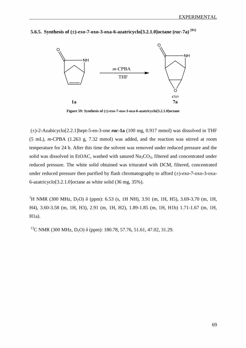
EXPERIMENTAL
69
5.6.5. Synthesis of (±)-exo-7-oxo-3-oxa-6-azatricyclo[3.2.1.0]octane (rac-7a) [91]
Figure 59: Synthesis of (±)-exo-7-oxo-3-oxa-6-azatricyclo[3.2.1.0]octane
(±)-2-Azabicyclo[2.2.1]hept-5-en-3-one rac-1a (100 mg, 0.917 mmol) was dissolved in THF
(5 mL), m-CPBA (1.263 g, 7.32 mmol) was added, and the reaction was stirred at room
temperature for 24 h. After this time the solvent was removed under reduced pressure and the
solid was dissolved in EtOAC, washed with satured Na2CO3, filtered and concentrated under
reduced pressure. The white solid obtained was triturated with DCM, filtered, concentrated
under reduced pressure then purified by flash chromatography to afford (±)-exo-7-oxo-3-oxa-
6-azatricyclo[3.2.1.0]octane as white solid (36 mg, 35%).
1H NMR (300 MHz, D2O) δ (ppm): 6.53 (s, 1H NH), 3.91 (m, 1H, H5), 3.69-3.70 (m, 1H,
H4), 3.60-3.58 (m, 1H, H3), 2.91 (m, 1H, H2), 1.89-1.85 (m, 1H, H1b) 1.71-1.67 (m, 1H,
H1a).
13
C NMR (300 MHz, D2O) δ (ppm): 180.78, 57.76, 51.61, 47.02, 31.29.

EXPERIMENTAL
70
5.6.6. Synthesis of methyl-6-oxopiperidine-2-carboxylate (23a)
Figure 60: Synthesis of methyl-6-oxopiperidine-2-carboxylate
6-Oxopipecolinic acid 22a (50 mg, 0.350 mmol) was dissolved in a mixture of MeOH and
EtOAc (1:2, 3 mL) and a solution of TMS-CHN2 in Et2O (2 M, 352 µL, 0.700 mmol) was
added under Ar-atmosphere. The reaction mixture was stirred at room temperature for 16 h.
Solvents were removed under reduced pressure and the residue was purified using flash
chromatography (eluent: EtOAc) to yield methyl-6-oxopiperidine-2-carboxylate as white
solid (39.4 mg, 65 %).
1H NMR (300 MHz, CDCl3) δ (ppm): 6.64 (br s, 1H), 4.06-4.11 (m, 1H, H2), 3.76 (s, 3H,
CH3), 2.31 – 2,38 (m, 2H, H5), 2.12 – 2.20 (m, 1H, H3b), 1,78 – 1,93 (m, 3H, H3a and
H4).
13C NMR (300 MHz, CDCl3) δ (ppm): 171.72 (CO), 171.54 (CO), 54. 80 (C2), 52.63
(OCH3), 31.12 (C5), 25.46 (C3), 19,54 (C4)

EXPERIMENTAL
71
5.6.7. Synthesis of methyl-2-pyrrolidinon-5-carboxylate (12a)
Figure 61: Synthesis of methyl-2-pyrrolidinon-5-carboxylate
L-2-Pyrrolidinon-5-carbonic acid 11a (50 mg, 0.387 mmol) was dissolved in a mixture of
MeOH and EtOAc (1:2, 3 mL) and a solution of TMS-CHN2 in Et2O (2 M, 390 µL, 0.774
mmol) was added under Ar-atmosphere. The reaction mixture was stirred at room temperature
for 16 h. The solvents were evaporated and the residue was purified by flash chromatography
(eluent gradient: EtOAc/hexanes 8:2 to pure EtOAc) yielding methyl-2-pyrrolidinon-5-
carboxylate as colourless oil (45 mg, 82%).
1H NMR (300 MHz, CDCl3) δ (ppm): 6.8 (br s, 1H), 4.27-4.22 (m, 1H, H2), 3.75 (s, 3H,
CH3), 2.49-2.31 (m, 3H, H3b, H4), 2.26-2.15 (m, 1H, H3a).
13C NMR (300 MHz, CDCl3) δ (ppm): 178.32, 172.66, 55.38, 52. 68, 29.24, 24.76

EXPERIMENTAL
72
5.6.8. Synthesis of 5-hydroxy-1H-pyrrol-2(5H)-one (16a)
Figure 62: Synthesis of 5-hydroxy-1H-pyrrol-2(5H)-one
To a solution of maleimide 26a (100 mg, 1.03 mmol) in MeOH (1 mL) was added cerium(III)
chloride heptahydrate (383.7 mg, 1.03 mmol), and the mixture was stirred for 5 min. After the
solution was cooled to 0 ºC, NaBH4 (38 mg, 1.03 mmol) was added portion wise, and stirred
for 1 h. The reaction mixture was quenched with ice water, and MeOH was removed under
reduced pressure. Flash chromatography (eluent DCM/MeOH 9:1) afforded 5-hydroxy-1H-
pyrrol-2(5H)-one (90 mg, 90 %) isolated as a white solid.
1H NMR (300 MHz, CD3OD) δ (ppm): 7.04 (d, 1H, H4), 6.05 (d, 1H, H3), 5.58 (m, 1H, H5).

EXPERIMENTAL
73
5.6.9. Synthesis of reference compounds
Reference compounds were prepared by acidic hydrolysis of the corresponding substrates in
concentrated hydrochloric acid:
Figure 63: Synthesis of 4-amino-2-cyclopentene-1-carboxyclic acid
The substrate was dissolved in 32 % HCl (12 M) and stirred at room temperature for 2-4 h.
The preparation of 3-carboxycyclopentanaminium chloride rac-2b additionally required
reflux for 1 h. Reactions mixtures were washed with EtOAc and concentrated in vacuo.
3-Aminocyclopentane-1-carboxylic acid (rac-2b) was isolated as white solid from (±)-2-
azabicyclo[2.2.1]heptan-3-one (50 mg, 0.45 mmol): yield 59 mg, 79%.
1H NMR (300 MHz, D2O) δ (ppm): 3.77-3.68 (m, 1H, H4), 3.04-2.94 (m, 1H, H1), 2.43-2.34
(m, 1H, H3a), 2.17-1.74 (m, 5H, H3b + H2+ H5) ppm.
(1R,3S)-3-Aminocyclopenante-1-carboxylic acid ((-)-2b)) was isolated as white solid from
(±)-2-azabicyclo[2.2.1]heptan-3-one (30 mg, 0.27 mmol): yield 35 mg, 79%.
1H NMR (300 MHz, D2O) δ (ppm): 3.77-3.68 (m, 1H, H4), 3.04-2.94 (m, 1H, H1), 2.43-2.34
(m, 1H, H3a), 2.17-1.74 (m, 5H, H3b + H2+ H5) ppm.
13C NMR (300 MHz, D2O) δ (ppm): 179.79, 51.39, 42.21, 33.54, 29.74, 27.60.
[ ]25
D -19 (c 0.875, MeOH)
4-Methylamino-2-cyclopentene-1-carboxylic acid (rac-3b) was isolated as yellow solid
from (±)-2-methyl-2-azabicyclo[2.2.1]hept-5-en-3-one (50 mg, 0,407 mmol): yield 71 mg,
98%.
1H NMR (300 MHz, D2O) δ (ppm): 6.30-6.27 (m, 1H, H3), 6.02-5.99 (m, 1H, H2), 4.35-4.31
(m, 1H, H4), 3.77-3.73 (m, 1H, H1), 2.71-2.61 (m, 5H, CH3 + H5b), 2.17-2.05 (m, 1H, H5a).

EXPERIMENTAL
74
(1S,4R)- 4-Methylamino-2-cyclopentene-1-carboxylic acid ((-)-3b) was isolated as yellow
solid from (±)-2-methyl-2-azabicyclo[2.2.1]hept-5-en-3-one (30 mg, 0.24 mmol): yield 35.3
mg, 82%.
1H NMR (300 MHz, D2O) δ (ppm): 6.30-6.27 (m, 1H, H3), 6.02-5.99 (m, 1H, H2), 4.35-4.31
(m, 1H, H1), 3.78-3.71 (m, 1H, H4), 2.71-2.61 (m, 5H, CH3 + H5b), 2.17-2.09 (m, 1H, H5a)
13C NMR (300 MHz, D2O) δ (ppm): 177. 16, 137.49, 127.62, 64.15, 49.54, 30.19, 29.32.
[ ]25
D -86.3 (c 0.9, MeOH).

EXPERIMENTAL
75
5.6.10. Unsucessful synthesis attempts: Synthesis of 6-azatricyclo-[3.2.1.0]-octane-
7-one
1st strategy:
The first attempt for the synthesis of 6-azatricyclo-[3.2.1.0]-octane-7-one involved
cyclopropanation with TMS-CHN2 [92]
and removal of the TMS-group by TBAF as a second
step [93]
:
Figure 64: Synthesis of 3-trimethylsilyl-6-azatrizyclo-[3.2.1.0]-octane-7-one
3-Trimethylsilyl-6-azatrizyclo-[3.2.1.0]-octane-7-one: To a cooled suspension of (±)-2-
azabicyclo[2.2.1]hept-5-en-3-one (50 mg, 0.459 mmol) and Pd(OAc)2 (5 mg) in dry Et2O was
added TMS-CHN2 (2 M in ether, 460 µL, 0.920 mmol) dropwise under Ar-atmosphere. After
24 h Pd(OAc)2 (5 mg) and TMS-CHN2 (2 M in ether, 460 µL, 0.920 mmol) were again added
since TLC showed incomplete conversion. The reaction was stopped after 48 h by filtration.
GC/MS and NMR measurement of the crude product did not allow conclusions regarding
product formation. After flash chromatography (eluent: hexanes/EtOAc 1:1) no product could
be detected in any of the fractions by GC/MS or NMR.
2nd strategy: [94, 95]
Due to the pyrophoricity of the zinc reagent the second attempt was carried out in a 2-neck
flask equipped with a dropping funnel and dried over night in the heating oven. The flask was
evacuated and flamed out using a heat gun and subsequently flooded with argon.

EXPERIMENTAL
76
Figure 65: Synthesis of 6-azatricyclo-[3.2.1.0]-octane-7-one
A solution of diethyl zinc (1 mL, 1 mmol) was added dropwise to a cooled solution of (±)-2-
azabicyclo[2.2.1]hept-5-en-3-one (50 mg, 0.459 mmol) in dry dichloromethane (2 mL) and
stirred for 15 min. Diiodomethane (147 µL, 1.834 mmol) was added rapidly and the reaction
was stirred at room temperature. The reaction was stopped after 24 h as TLC did not show the
formation of any new spots. The reaction was quenched with saturated NH4Cl solution,
extracted with Et2O, dried with Na2SO4 and concentrated in vacuo. GC/MS and NMR
analysis of the crude product did not show any product formation.

LITERATURE
77
6. Literature
[1] Rapaka, R. S.; Renugopalakrishnan, V.; Urry, D. W.; Bhatnagar, R. S. Biochemistry
1978, 17, 2892-2898
[2] Seebach, D.; Beck, A. K.; Bierbaum, D. J. Chem. Biodiversity 2004, 1, 1111-1239
[3] Voet, D.; Voet, J. G.; Pratt C. W. Lehrbuch der Biochemie, 2nd edt., Wiley-VCH:
Weinheim Germany, 2010
[4] Xie, J.; Schultz, P.G. Curr.Opin.Chem.Biol. 2005, 9, 548-554
[5] Gellman, S.H. Acc. Chem. Res. 1998, 31, 173-180
[6] Wagner, I.; Musso, H. Angew. Chem. Int. Ed. 1983, 22, 816-828
[7] Zhang, X.; Jacob, M. R.; Rao, R.; Wang, Y. H.; Agarwal, A. K.; Newman, D. J.; Khan,
I. A.; Clark, A. M.; Li, X. C. Res. Rep. Med. Chem. 2012, 2, 7-14
[8] Umezawa, H.; Aoyagi, T.; Morishima, H.; Matsuzaki, M.; Hamada, M.; J. Antibiot.
1970, 23, 259–262
[9] Kaila, K.; Pasternack, M.; Saarikoski, J.; Voipio J. J.Physiol 1989, 416, 161-181
[10] Barnard, E. A.; Skolnick, P.; Olsen, R. W.; Mohler, H.; Sieghart, W.; Biggio G.;
Braestrup, C.; Bateson, A. N.; Langer, S. Z. Pharmacol. Rev., 1998, 50, 291-313
[11] Chen, K.; Li, H.; Ye, N.; Zhang, J.; Wang, J. Brain Res Bull, 2005, 67, 310–318
[12] Ben-Ari, Y.; Gaiarsa J. L.; Tyzio R.; Khazipov, R. Physiol. Rev. 2007, 87, 1215-1248
[13] Gajcy, K.; Lochyński, S.; Librowski, T. Curr. Med. Chem. 2010, 17, 2338-2347
[14] Baidya, D. K.; Agarwal, A.; Khanna, P.; Arora, M. K. J. Anaesthesiol. Clin.
Pharmacol. 2011, 27, 307-314
[15] Vasudev P. G. Chatterjee, S.; Shamala, N.; Balaram, B. Chem. Rev. 2011, 111, 657-687
[16] Frackenpohl, J.; Arvidsson, P. I.; Schreiber, J. V.; Seebach, D. Chem. Bio. Chem. 2001,
2, 445-455
[17] Brea, R. J.; Reiriz, C.; Granja, J. R. Chem. Soc. Rev. 2010, 39, 1448-1456
[18] Reiriz, C.; Brea, R. J.; Arranz, R.; Carrascosa, J. L.; Garibotti, A.; Manning, B.;
Valpuesta, J. M.; Ramo´n Eritja, R.; Castedo, L.; Granja, J. R. J. Am. Chem. Soc. 2009,
131, 11335-11337
[19] Amorín, M.; Castedo, L.; Granja, J. R. J. Am. Chem. Soc. 2003, 125, 2844-2845

LITERATURE
78
[20] Hintermann, T.; Gademann, K.; Jaun, B.; Seebach, D. Helv. Chim. Acta 1998, 81, 983-
1002
[21] Loukas, V.; Noula, C.; Kokotos, G. J. Peptide Sci. 2003, 9, 312-319
[22] Burk, M. J.; De Koning, P. D.; Grote, T. M.; Hoekstra, M. S.; Hoge, G.; Jennings, R.
A.; Kisel, W. S.; Le, T. V.; Lennon, I. C.; Mulhern, T. A.; Ramsden, J. A.; Wade, R. A.
J. Org. Chem. 2003, 68, 5731–5734
[23] Hoge, G. J. Am. Chem. Soc. 2003, 125, 10219–10227.
[24] Hoge, G. J. Am. Chem. Soc. 2004, 126, 9920–9921.
[25] Oliveira, C. C.; Angnes, R. A.; Correia, C. R. D. J. Org. Chem. 2013, 78, 4373−4385
[26] Ordóñez, M.; Cativiela, C. Tetrahedron: Asymmetry 2007, 18, 3-99
[27] Brenna, E.; Caraccia, N.; Fuganti, C.; Fuganti, D.; Grasselli, P. Tetrahedron:
Asymmetry 1997, 8, 3801–3805.
[28] Girnys, E. A.; Porter, V. R.; Mosberg, H. I. Bioorg. Med. Chem. 2011, 19, 7425–7434
[29] Orwig, K. S.; Dix, T. A. Tetrahedron Lett. 2005, 46, 7007-7009
[30] Brenner, M.; Seebach, D. Helv. Chim. Acta, 1999, 82, 2365-2379
[31] Seebach, D.; Brenner, M.; Ruepnig, M.; Schweizer, B.; Jaun, B. Chem. Commun. 2001,
207-208
[32] Seebach, D.; Brenner, M.; Rueping, M.; Jaun, B. Chem. Eur. J. 2002, 8, 573-584
[33] Bae, H. J.; Some, S.; Lee, J. H.; Kim, J.-Y.; Song, M. J.; Lee, S.; Zhang, Y. J.; Song, C.
E. Adv. Synth. Catal. 2011, 353, 3196 – 3202
[34] Palomo, C.; Oiarbide, M.; Aizpurua, J. M.; González, A.; García, J. M.; Landa, C.;
Odrizola, I.; Linden, A. J. Org. Chem. 1999, 64, 8193–8200
[35] Yu, H. J.; Shao, C.; Cui, Z.; Feng, C. G.; Lin, G. Q. Chem. Eur. J. 2012, 18, 13274 –
13278
[36] Margoliu, A. L. (Merrell Dow Pharmaceuticals) US Patent 5316944
[37] Winkler, M.; Knall, A. C.; Kulterer, M. R.; Klempier, N. J. Org. Chem. 2007, 72, 7423-
7426
[38] Mukherjee, H.; Martinez, C. A. ACS Catal. 2011, 1, 1010-1013
[39] Jung, J. H.; Yoon, D. H.; Kang, P.; Lee, W. K.; Eum, H.; Ha, H. J. Org. Biomol. Chem.
2013, 11, 3635-3641

LITERATURE
79
[40] Winkler, C. K.; Clay, D.; Dacies, S.; O'Neill, P.; McDaid, P.; Dabarge, S.; Steflik, J.;
Karmilowicz, M.; Wong, J. W.; Faber, K. J. Org. Chem. 2013, 78, 1525-1533
[41] Mazzini, C.; Lebreton, J.; Alphand, V.; Furtoss, R. Tetrahedron Lett. 1997, 38, 1195–
1196
[42] Pukina, A. V.; Boeriu, C. G.; Scott, E. L.; Sanders, J. P. M.; Franssen M. C. R. J. Mol.
Catal. B: Enzym. 2010, 65, 58–62
[43] Martínez-Gómez, A.I. ; Martínez-Rodríguez, S.; Pozo-Dengra, J.; Tessaro, D.; Servi,
S., Clemente-Jiménez, J.M.; Rodríguez-Vico, F.; Las Heras-Vázquez, F. J. Appl.
Environ. Microbiol. 2009, 75, 514-520
[44] Martínez-Gómez, A.I.; Andújar-Sánchez, M.; Clemente-Jiménez, J.M.; Neira, J. L.;
Rodríguez-Vico, F.; Martínez-Rodríguez, S.; Las Heras-Vázquez, F. J. J. Chromatogr.
B 2011, 879, 3277– 3282
[45] IUPAC. Compendium of Chemical Terminology, 2nd ed. (the "Gold Book"). Compiled
by A. D. McNaught and A. Wilkinson. Blackwell Scientific Publications, Oxford
(1997).
[46] Williams, R. M.; Cox, R. J. Acc. Chem. Res. 2003, 36, 127-139
[47] Feling, R. H.; Buchanan, G. O.; Mincer, T. J.; Kauffman, C. A.; Jensen, P. R.; Fenical,
W. Angew. Chem. Int. Ed. 2003, 42, 355-357
[48] Jossang, A.; Jossang, P.; Hadi, H. A.; Sevenet, T.; Bodo, B. J. Org. Chem. 1991, 56,
6527-6530
[49] Kemnitz, C. R.; Loewen, M. J. J. Am. Chem. Soc. 2007, 129, 2521-2528
[50] Faber, K. Biotransformations in Organic Chemistry, 6th ed.; Springer: Berlin
Heidelberg, 2011
[51] Wiberg, K. B. Angew. Chem., Int. Ed. 1986, 25, 312-322
[52] Singh, R.; Vince, R. Chem. Rev. 2012, 112, 4642-4686
[53] Stivers, J. T.; Jiang, Y. L. Chem. Rev. 2003, 103, 2729–2760
[54] Taylor, S. J. C.; McCague, R.; Wisdom, R.; Lee, C.; Dickson, K.; Ruecroft, G.;
O'Brien, F.; Littlechild, J.; Bevan, J.; Roberts, S. M.; Evans, C. T. Tetraheron:
Asymmetry 1993, 4, 1117-l128
[55] Zuh, S.; Gong, C.; Song, D.; Gao, S., Zheng, G. Appl. Environ. Microbiol. 2012, 78,
7492-7495
[56] Potter, G. A.; Garcial, C.; McCague, R.; Adger, B.; Collet, A. Angew. Chem. Int. Ed.
1996, 35, 1666-1668

LITERATURE
80
[57] Assaf, Z.; Faber, K.; Hall, M. Scope, Limitations and Classification of Lactamases,
manuscript to be submitted
[58] Kamlet, A. S.; Préville, C.; Farley, K. A.; Piotrowksi, D. W. Angew. Chem. Int. Ed.
2013, 52, 10607-10610
[59] Toogood, H. S.; Brown, R. C.; Line, K.; Keene P. A.; Taylor, S. J. C.; McCague, R.;
Littlechild, J. A. Tetrahedron 2004, 60, 711-716
[60] Torres, L. L.; Schließmann, A.; Schmidt M.; Silva-Martin, N.; Hermoso, J. A.;
Berenguer, J.; Bornscheuer, U. T.; Hidalgo, A. Org. Biomol. Chem. 2012, 10, 3388-
3392
[61] Yang, M.; Gao, Q.; Wu, S.; Wang, J.; Zheng, G. Biotechnol. Lett. 2012, 34, 275–279
[62] Line, K; Isupov, M. N.; Littlechild, J. A. J. Mol. Biol. 2004, 338, 519-532
[63] Forró, E.; Fülöp, F. Eur. J. Org. Chem. 2008, 5263-5268
[64] Nardini, M.; Dijkstra, B. W. Curr. Opin. Struct. Biol. 1999, 9, 732–737
[65] The PyMol Molecular Graphics System, Version 1.5.0.4 Schrödinger, LLC.
[66] a) Sievers, F.; Wilm, A.; Dineen, D.G.; Gibson, T.J.; Karplus, K.; Li, W.; Lopez, R.;
McWilliam, H.; Remmert, M.; Söding, J.; Thompson, J.D.; Higgins, D. Mol. Syst. Biol.
2011, 7, 1-6; b) Goujon, M.; McWilliam, H.; Li, W.; Valentin, F.; Squizzato, S.; Paern,
J.; Lopez, R. Nucleic Acids Res. 2010, 38, W695-W699
[67] Mayaux, J. F.; Cerbelaut, E.; Soubrier, F.; Yeh, P.; Blanche, F.; Pétré, D. J. Bacteriol.
1991, 173, 6694-6704
[68] Patricelli, M. P.; Cravatt, B. F. J. Biol. Chem 2000, 275, 19177-19184
[69] Cilia, E.; Fabbri, A.; Uriani, M.; Scialdone, G. G.; Ammendola, S. FEBS J. 2005, 272,
4716-4724
[70] Heumann, S.; Eberl, A.; Fischer-Colbrie, G.; Pobeheim, H.; Kaufmann, F.; Ribitsch, D.;
Cavaco-Paulo, A.; Guebitz, G. Biotechnol. Bioeng. 2009, 102, 1003-1011
[71] Acero, E. H.; Ribitsch, D.; Rodriguez, R. D.; Dellacher, A.; Zitzenbacher, S.; Marold,
A.; Greimel, K. J.; Schroeder, M.; Kandelbauer, A.; Heumann, S.; Nyanhongo, G.;
Schwab, H.; Guebitz, G. M. J. Mol. Catal. B: Enzym. 2012, 79, 54-60
[72] Fukata, Y.; Kouzimi, S.; Komeda, H.; Asano, Y. Enzyme Microb. Technol. 2010, 46,
237-245
[73] Altschul, S. F.; Madden, T. L.; Schaffer, A. A.; Zhang, J. H.; Zhang, Z.; Miller, W.;
Lipman, D. J. Nucleic Acids Res. 1997, 25, 3389-3402

LITERATURE
81
[74] Krissinel, E.; Henrick, K. Acta Crystallogr., Sect D: Biol. Crystallogr.2004, 60, 2256-
2268
[75] Xie, S.-X.; Kato, Y.; Komeda, H.; Yoshida, S.; Asano, Y. Biochemistry 2003, 42,
12056-12066
[76] Koo, H. M.; Choi, S.-O.; Kim, H .M.; Kim, Y. S. Biochem. J. 2000, 349, 501-507
[77] Shin, S.; Lee, T.-H.; Ha, N.-C.; Koo, H. M.; Kim, S.-Y.; Lee, H.-S.; Kim, Y.S.; Oh, B.-
H. EMBO J. 2002, 21, 2509-2516
[78] Shin, S.; Yun, Y. S.; Koo, H. M.; Kim, S. Y.; Lee, H. S.; Choi, K. Y.; Oh, B.-H. J. Biol.
Chem. 2003, 278, 24937-24943
[79] Yun, Y. S.; Lee, W.; Shin, S.; Oh, B.-H.; Choi, K. Y. J. Biol. Chem. 2006, 281, 40057-
40064
[80] Wagner, C.; El Omari, M.; Koenig, G. M. J. Nat. Prod. 2009, 72, 540-553
[81] Pelletier, I.; Altenbuchner, J.; Mattes, R. Biochim. Biophys. Acta 1995, 1250, 149-157
[82] Hofman, B.; Toelzer, S.; Pelletier, I.; Altenbuchner, J.; can Pée, K.H.; Hecht, H. J. J.
Mol. Biol. 1998, 279, 889-900
[83] Fan, Z.; Qi, J.; Shi, .Y.; Liu, Y. Acta Crystallogr., Sect. F: Struct. Biol. Cryst. Commun.
2011, 67, 521-523
[84] Shi, Y.-W.; Cui, L. F.; Yuan, J. M. Curr. Microbiol. 2007, 55, 61-64
[85] Shi, Y.-W.; Lui, X.-Q.; Shi, P.; Zhang, X.-Y. Arch. Biochem. Biophys. 2010, 494, 1-6
[86] Kühnel, E.; Laffan, D. P.; Lloyd-Jones, G. C.; Martínez del Campo, T.; Shepperson, I.
R.; Slaughter, J. L. Angew. Chem. Int. Ed. 2007, 46, 7075-7078
[87] Straathof, A. J. J.; Jongejan, J. A. Enzyme Microb. Technol. 1997, 21, 559-571
[88] Rodríguez-Vázques, N.; Salzinger, S.; Silfa, L. F.; Amorín, M.; Granja, J. R. Eur. J.
Org. Chem. 2013, 3477-3493
[89] Szczepankiewicz, B. G.; Dai, H.; Koppetsch, K. J.; Quian, D.; Jiang, F.; Mao, C.; Perni,
R.B. J. Org. Chem. 2012, 77, 7319-7329
[90] Slama, J. T.; Metha, N.; Skrzypczak-Jankun, E. J. Org. Chem. 2006, 71, 7877-7880
[91] Kiss, L.; Forró, E.; Sillanpää, R.; Fülöp, F. Synthesis 2010, 1, 153-160
[92] Bhushan, R. G.; Vince, R. Bioorg. Med. Chem. 2002, 10, 2325-2333
[93] Qui, J.; Silverman, R. B. J. Med. Chem. 2000, 43, 706-720

LITERATURE
82
[94] Zhao, Y.; Yang, T.; Lee, M.; Lee, D.; Newton, M. G.; Chu, C. K. J. Org. Chem. 1995,
60, 5236-5242
[95] Mahler, M.; Reichardt, B.; Hartjen, P.; van Lunzen, J.; Meier, C. Chem. Eur. J. 2012,
18, 11046-11062
[96] Z. Assaf, E. Eger, Z. Vitnik, W. M. F. Fabian, D. Ribitsch, G. M. Guebitz, K. Faber, M.
Hall, Identification and Applications of Enantiocomplementary Lactamases for Vince
Lactam Derivatives, manuscript under revision

APPENDIX
83
7. Appendix
NMR-Spectra
1H-NMR rac-1b
13
C-NMR rac-1b
rac-1b
rac-1b

APPENDIX
84
1H-NMR (1S,4R)-1b
13C-NMR (1S,4R)-1b
1b
(1S,4R)-1b
(1S,4R)-1b

APPENDIX
85
1H-NMR rac-2a
1H-NMR rac-2b
rac-2a
rac-2b

APPENDIX
86
1H-NMR (-)-2a
13C-NMR (-)-2a
(-)-2a
(-)-2a

APPENDIX
87
1H-NMR (1R,3S)-2b
13C-NMR (1R,3S)-2b
(1R,3S)-2b
(1R,3S)-2b

APPENDIX
88
1H-NMR rac-3a
13C-NMR rac-3a
rac-3a
3a rac-3a

APPENDIX
89
1H-NMR (-)-3a
13C-NMR (-)-3a
(-)-3a
(-)-3a
(-)-3a

APPENDIX
90
1H-NMR rac-3b
1H-NMR (-)-3b
(-)-3a
rac-3b

APPENDIX
91
13C-NMR (-)-3b
1H-NMR rac-4a
(-)-3a
rac- 4a

APPENDIX
92
13C-NMR rac-4a
1H-NMR rac-5a
rac- 4a
rac-5a

APPENDIX
93
13C-NMR rac-5a
1H-NMR (1R,4S,5R,6S)-5a
rac-5a
(1R,4S,5R,6S)-5a

APPENDIX
94
13C-NMR (1R,4S,5R,6S)-5a
1H-NMR rac-5b
(1R,4S,5R,6S)-5a

APPENDIX
95
1H-NMR rac-6a
13
C-NMR rac-6a
rac-6a
rac-6a

APPENDIX
96
1H-NMR rac-7a
13
C-NMR rac-7a
rac-7a
rac-7a

APPENDIX
97
1H-NMR rac-7b
1H-NMR 16a
16a
rac-7b

APPENDIX
98
1H-NMR 12a
13
C-NMR 12a
12a
12a

APPENDIX
99
1H-NMR 23a
13
C-NMR 23a
23a
23a

APPENDIX
100
Abbreviations
aa amino acid(s)
Ac2O acetic anhydride
AMI amidase from Rhodococcus globerulus
Asp aspartic acid
AS sequence amidase signature sequence
BLAST basic local alignment search tool
Boc tert-butoxycarbonyl, protection group
CeCl3 cerium(III) chloride
CDCl3 deuterated chloroform
CD3OD deuterated methanol
CH2I2 diiodomethane
CIH cyclic imide hydrolase from Pseudomonas putida 13
C NMR carbon-13 - nuclear magnetic resonance
m-CPBA meta-chloroperoxybenzoic acid
CPO-T chloroperoxidase T from Streptomyces aureofaciens
D2O deuterated water
DCM dichloromethane
DMAP 4-dimethylaminopyridene
DMF dimethylformamide
E enantiomeric ratio
ee enantiomeric excess
Et2O diethyl ether
Et2Zn diethyl zinc
EtOAc ethylacetate
G glycine, 1-letter code
GABA γ-aminobutyric acid
GC-FID gas chromatography with flame ionization detector
GC/MS gas chromatography - mass spectrometry
Gln glutamine
Gly glycine
HCl hydrochloric acid
H2 hydrogen
His histidine 1H NMR proton/hydrogen-1 - nuclear magnetic resonance
kDa kilo-Dalton
KHSO4 potassium bisulfate
LC/MS liquid chromatography - mass spectrometry
m/z mass-to-charge ratio
MAE2 malonamidase E2 from Bradyrhizobium japonicum
MeOH methanol
Met methionine
MHz megahertz
NaBH4 sodium borohydride
NaH sodium hydride
Na2SO4 sodium sulfate
NfpolyA polyamidase from Nocardia farcinica
NH4Cl ammonium chloride
NMO N-methylmorpholine N-oxide
OsO4 osmium tetroxide

APPENDIX
101
Pd/C palladium on activated charcoal
Pd(OAc)2 palladium(II) acetate
rac racemic
rpm rounds per minute
S serine, 1-letter code
Ser serine
TBA tert-butyl alcohol
TBAF tetra-n-butylammonium fluoride
TFA trifluoroacetic acid
THF tetrahydrofuran
TLC thin layer chromatography
TMS-CHN2 trimethylsilyl diazomethane
tR retention time
Tyr tyrosine

APPENDIX
102
CURRICULUM VITAE
PERSONAL DATA
Full name: Elisabeth Eger
Citizenship: Austrian
Date of birth: 23.04.1990
Place of birth: Deutschlandsberg, Austria
Address: Langegg 170, 8511 St. Stefan ob Stainz, Austria
EDUCATION
2011-present Master Study Biotechnology, TU Graz
Master thesis: "Development of a biocatalytic platform for the
enantioselective hydrolysis of γ- and δ-lactams", Prof. Kurt Faber,
Institute of Organic and Bioorganic Chemistry, University Graz
2008-2011 Bachelor Study Chemistry, University Graz and TU Graz
Bachelor thesis: "Characterization of 3CNE from Bacteroides
thetaiotaomicron", Prof. Peter Macheroux, Institute of Biochemistry,
Graz University of Technology
2004-2008 High School in Deutschlandsberg
EXPERIENCE
Summer 2012 Summer student and project laboratory course "Biocatalysis"
"Ene-reductases", Prof. Kurt Faber, Institute of Organic and Bioorganic
Chemistry, University Graz
Summer 2011 Project laboratory course "Bioanalytics"
"Characterization of a flavoprotein", Prof. Peter Macheroux, Institute of
Biochemistry, Graz University of Technology
LANGUAGE SKILLS
German (native), English (fluent in written and spoken), Latin
PUBLICATIONS
(1) T. Knaus, E. Eger, J. Koop, S. Stipsits, C. L. Kinsland, S. E. Ealick, P. Macheroux,
Reverse structural genomics: An unusual flavin binding site in a putative protease from
Bacteroides thetaiotaomicron, J. Biol. Chem. 2012, 287, 27490 – 27498

APPENDIX
103
(2) T. Knaus, E. Eger, J. Koop, S. Stipsits, C. L. Kinsland, S. E. Ealick, P. Macheroux, Reverse
structural genomics: An unusual flavin binding site in a putative protease from Bacteroides
thetaiotaomicron, Poster, Trends in Enzymology, June, 03-06, 2012, Göttingen, Germany
(3) Z. Assaf, E. Eger, D. Ribitsch, G. Gübitz, M. Hall, K. Faber, Development of a
Biocatalytic Platform for the Stereoselective Hydrolysis of -Lactams for Pharmaceutical
Applications, Poster, 11th International Symposium on Biocatalysis and
Biotransformations (Biotrans), July, 21-25, 2013, Manchester, UK.
(4) Z. Assaf, E. Eger, Z. Vitnik, W. M. F. Fabian, D. Ribitsch, G. M. Guebitz, K. Faber, M.
Hall, Identification and Applications of Enantiocomplementary Lactamases for Vince
Lactam Derivatives, manuscript under revision