Precursor discrimination of designer drug benzylpiperazine using δ(13)C and δ(15)N stable isotopes
Upload
gfz-potsdamCategory
view
1download
0
Methods
UV-laser-based microscopic dissection of tree rings – a novelsampling tool for d13C and d18O studies
Karina Schollaen, Ingo Heinrich and Gerhard Helle
GFZ – German Research Centre for Geosciences, Section 5.2 Climate Dynamics and Landscape Evolution, Telegrafenberg, 14473 Potsdam, Germany
Author for correspondence:Karina Schollaen
Tel: +49 (0) 331 2881899
Email: [email protected]
Received: 6 August 2013
Accepted: 2 October 2013
New Phytologist (2014) 201: 1045–1055doi: 10.1111/nph.12587
Key words: dendroclimatology, high-precision sampling, high-resolution stableisotope analysis, plant physiology, tree rings,UV-laser microdissection, wood anatomy.
Summary
� UV-laser-based microscopic systems were utilized to dissect and sample organic tissue for
stable isotope measurements from thin wood cross-sections.� We tested UV-laser-based microscopic tissue dissection in practice for high-resolution isoto-
pic analyses (d13C/d18O) on thin cross-sections from different tree species. The method allows
serial isolation of tissue of any shape and from millimetre down to micrometre scales. On-
screen pre-defined areas of interest were automatically dissected and collected for mass spec-
trometric analysis.� Three examples of high-resolution isotopic analyses revealed that: in comparison to d13C of
xylem cells, woody ray parenchyma of deciduous trees have the same year-to-year variability,
but reveal offsets that are opposite in sign depending on whether wholewood or cellulose is
considered; high-resolution tree-ring d18O profiles of Indonesian teak reflect monsoonal rainfall
patterns and are sensitive to rainfall extremes caused by ENSO; and seasonal moisture signals
in intra-tree-ring d18O of white pine are weighted by nonlinear intra-annual growth dynamics.� The applications demonstrate that the use of UV-laser-based microscopic dissection allows
for sampling plant tissue at ultrahigh resolution and unprecedented precision. This new tech-
nique facilitates sampling for stable isotope analysis of anatomical plant traits like combined
tree eco-physiological, wood anatomical and dendroclimatological studies.
Introduction
Stable isotope analyses (hydrogen (H), nitrogen (N), carbon (C),oxygen (O)) on organic tissue are widely used in plant physiologi-cal, ecological and climatological research (Ehleringer et al.,1993; Ziegler, 1995; Schleser et al., 1999; Dawson et al., 2002;Helle & Schleser, 2004b; Treydte et al., 2006; West et al., 2010;Hietz et al., 2011; Brienen et al., 2012; Werner et al., 2012;Heinrich et al., 2013). State-of-the-art isotope ratio mass spec-trometry (IRMS) and cavity ring-down spectroscopy (CRDS)allow precise determination of stable isotope ratios on minimalsample amounts and large numbers of samples. However, accu-rate preparation of small samples in large numbers is challeng-ing, as this still has to be done manually in most of the researchareas. Despite the high precision and accuracy of modern ana-lytical devices, data quality very much depends on the skills andmotivation of the person responsible for sample preparation.This applies in particular to inter- and intra-annual stableisotope studies of woody tissue attempting to extract valuableseasonal climatic and environmental information (Leavitt &Long, 1982; Loader et al., 1995; Treydte et al., 2006; Schollaenet al., 2013) or assessing plant eco-physiological processes
(Helle & Schleser, 2004a; Gessler et al., 2009; Schubert &Jahren, 2011; Krepkowski et al., 2013) from annual growthrings or parts thereof. Furthermore, high-precision dissection iscrucial in studies using high-resolution stable isotope data for theidentification of anatomically nondistinct annual rings in tropicaltrees (Evans & Schrag, 2004; Verheyden et al., 2004; Pons &Helle, 2011).
Different methods exist for the dissection of wood tissue fromtree rings for stable isotope measurements. One common methodhas been to divide wood segments or cores by cutting tangentialslices, utilizing fixed-blade sledge and rotary microtomes (Ogle &McCormac, 1994; Loader et al., 1995; Barbour et al., 2002;Helle & Schleser, 2004a; Poussart et al., 2004; Verheyden et al.,2004; Og�ee et al., 2009; Pons & Helle, 2011) or scalpel/razorblades (Roden et al., 2009; Managave et al., 2010). With a micro-tome, wood slices down to c. 10 lm thickness can be achieved(Helle & Schleser, 2004a). Another high-resolution samplingmethod is by collecting wood dust from a series of small woodholes drilled in the radial direction with twist drills (Walcroftet al., 1997; Gebrekirstos et al., 2009; Fichtler et al., 2010),robotic micromilling techniques (Wurster et al., 1999; Doddet al., 2008) or an UV-laser ablation system in combination with
� 2013 The Authors
New Phytologist� 2013 New Phytologist TrustNew Phytologist (2014) 201: 1045–1055 1045
www.newphytologist.com
Research

isotope ratio mass spectrometry (LA-C-GC-IRMS), firstdescribed in Schulze et al. (2004). Accurate sample adjustment,as well as unambiguous identification of tree rings, is normallyprovided by visual inspection using a microscope or digital cam-era. After dissection, wood samples are ground to a fine powderand either cellulose is extracted or bulk wood samples are usedfor stable isotope analysis. The latter is the case for the UV-laserablation method where dust is ablated from a wood core or seg-ment in an airtight chamber flushed with helium. From theablation chamber wood dust is directly transferred into a com-bustion oven and converted to CO2. Subsequently, d
13C is mea-sured by an isotope ratio mass spectrometer that is coupledonline to the UV-laser ablation and preparation system. Variousstudies used this method to analyse the intra-annual d13C in treerings responding to carbohydrate storage and remobilization, aswell as short-term climatic effects in conjunction with wooddensity variability (Intra-annual Density Fluctuations, IADFs)(Skomarkova et al., 2006; Battipaglia et al., 2010; De Miccoet al., 2012). The spatial resolution of a UV-laser shot is 40 lm(Schulze et al., 2004) to 150 lm (De Micco et al., 2012). Thewidths of the holes produced by the mechanical drills can varyfrom 500 lm (Gebrekirstos et al., 2009) to 800 lm (Fichtleret al., 2010) up to 1.5 mm (Walcroft et al., 1997) depending onthe diameter of the drill. Robotic micromilling generallyrequires a minimum path width of 400 lm for increment coresamples, whereas individual path widths of < 100 lm are possi-ble (Dodd et al., 2008).
Different drawbacks exist for the dissection methods men-tioned before. For the methods using blades or microtomes, woodsamples must have straight-line ring structure boundaries in bothtangential and transversal directions, as samples are cut in parallellinear segments. However, ring width and curvature of growthrings are rarely strictly parallel and fibre or vessel angles vary bothunder natural conditions and depending on species-specific woodanatomical characteristics. Thus, often only a limited number ofconsecutive tree rings per sample is suitable for high-precisionintra-annual stable isotope measurements when applying thesedissection methods. Generally, cross-contamination during cut-ting, milling and other preparatory steps cannot be excludedespecially when quality control by electronic image documenta-tion is not implemented. For the methods involving drilling woodholes, straight-line boundaries in the transversal direction are lessimportant, but differences in vertical direction can be problem-atic, depending on the drill depth and the deviation of wood fibreor vessel orientation from the vertical (Fig. 1b). LA-C-GC-IRMSsystems are presently confined to carbon isotope analysis. Aerosoltransfer of wood dust between the ablation chamber and the com-bustion furnace requires a rather high helium consumption andtransfer capillaries sometimes may clog up.
The need for methodological improvements to facilitate finedissection of irregular and narrow shapes has emerged lately, sowe assessed a novel method to dissect wood tissue from tree ringsusing the UV-laser microdissection microscopes produce by Leica(LMD7000; Leica Microsystems GmbH, Wetzlar, Germany)
(a)
(b)
Fig. 1 Possible steps of wood sample preparation and high-precision sampling using UV-laser-based microscopic dissection systems. (a) Top view and profileof a metal frame slide with wood cross-sections used for gravity-assisted collection (LMD7000, Leica). Up to three cross-sections of 59-mm length can bemounted between two metal frames. For stable isotope analyses it is possible to cut serial sections at inter- to intra-annual resolution or even to pool samplematerial, for example, for cellulose extraction. To avoid material loss the dissection of tissue can be alternatingly shifted. (b) Schematic view of samplepreparation with a wood segment in radial and cross-section. The use of cross-sections diminishes difficulties of cross-contamination when sampling treerings with tight curvatures, nonparallel boundaries, varying fibre and vessel angles, and/or changing tree-ring width in the radial direction. The dissection ofwood cross-sections ranging in thickness from 100 lm to a maximal 1000 lm is possible. The remaining wood sample can be used for multidisciplinaryapproaches, for example, combining high-resolution stable isotope analysis, quantitative wood anatomy, ring width or wood density analysis.
New Phytologist (2014) 201: 1045–1055 � 2013 The Authors
New Phytologist� 2013 New Phytologist Trustwww.newphytologist.com
Research
NewPhytologist1046

and Zeiss (PALM MicroBeam; Carl Zeiss Microscopy GmbH,Jena, Germany). UV-laser-based microscopic dissection systemsare being applied widely in biomedical (Fend & Raffeld, 2000),animal (Bayona-Bafaluy et al., 2005) and also in plant research(Nelson et al., 2006; Abbott et al., 2010). However, to ourknowledge, this is the first time it has been utilized to preciselydissect plant tissue for stable isotope research. Laser-based micro-scopic dissection uses an UV-laser beam to isolate tissues of inter-est from thin sections of samples. We describe the design andhandling of the two different UV-laser microdissection micro-scopes. Advantages and constraints are discussed on the basis ofhigh-resolution stable isotope analyses on woody plant materialof deciduous and coniferous trees from temperate and tropicalclimate zones. Variability of the new sub-seasonal C and O stableisotope data was evaluated with respect to seasonal changes of cli-matic conditions and wood growth dynamics. Furthermore, wetested whether d13C in the woody ray parenchyma and in xylemcells of a deciduous tree is different.
Materials and Methods
UV-laser-based microscopic dissection
UV-laser microdissection microscopes were used as a precise toolfor dissecting different wood samples in stable isotope studies.We tested two different UV-laser microdissection microscopes ofLeica (LMD7000, software v6.7.1.3952, Leica MicrosystemsGmbH) and Zeiss (PALM MicroBeam, software v4.5.0.9, CarlZeiss Microscopy GmbH). We followed a preparation schemecontaining five steps:(1) manual preparation of thin wood cross-sections (max.1000 lm thickness) with a microtome or a high-precision saw,(2) microscopic identification and pen-screen selection of tissuesof interest,(3) automatic UV-laser-based microscopic dissection of inter-orintra-annual wood sections,(4) semi-automatic sample collection into tin or silver capsulesby gravity or forceps, optional chemical treatment (e.g. celluloseextraction), and(5) stable carbon (C) and oxygen (O) isotope analysis via con-ventional Isotope Ratio Mass Spectrometry (IRMS) coupledonline to a combustion or pyrolysis furnace.
UV-laser-based microscopic dissection enables selection ofrelevant plant cells/tissues on screen by pen, while nonrelevanttissues (e.g. resin ducts, wood rays) may not be selected or beremoved (Fig. 2a,d) before sampling. Any size and area can bedissected, which is important for the precise dissection of asym-metric tree rings or parts thereof, as shown for example by lobategrowth (Fig. 2a), intra-ring density fluctuations (Fig. 2b) orwedging tree rings (Fig. 2c). Furthermore, it is possible to cutserial sections or even to pool sample material, for example if theweight of the dissected tissue from one thin section is insufficientfor a stable isotope measurement. Sample material from the samearray of wood cells can be identified unambiguously on a secondor third cross-section and may be pooled for chemical treatment,like cellulose extraction before IRMS analysis (Fig. 1a).
In general, wood samples of 100-lm to 1000-lm thicknesscan be dissected without any major constraints (Fig. 1b). Dissec-tion samples of up to 1000-lm thickness are achieved by cuttingiterations and adjustment of the z-focus of the UV-laser beam.The use of cross-sections thinner than 100 lm normally resultsin insufficient amounts of material for present-day combustion/pyrolysis systems normally coupled to IRMS. Taking thin sec-tions, however, has the advantage that part of the wood sample isleft intact, providing the opportunity for reanalysis of ring widthsor allowing other investigations, such as, for example, quantita-tive wood anatomy or wood density measurements. Furthermore,the risk of cross-contamination of various tissues is diminished,for example when studying narrow tree rings that have nonparal-lel boundaries and/or have varying fibre and vessel angles(Fig. 1b).
Sample material
In order to test the UV-laser-based microscopic dissectionmethod in practice we focused on three applications. In the firstexperiment, we tested whether the d13C values in xylem cells ofan oak sample (Quercus robur L.) differ from the d13C values inray cells in order to assess potential influences of ray parenchymaon the isotope variability of tree-ring sequences. Therefore, anoak tree was sampled at Telegrafenberg Hill in Potsdam, north-eastern Germany (52°23′N, 13°04′E; 94 m above sea level(a.s.l.)). Germany is characterized by a warm, temperate and
(a) (b)
(c)
(d)
Fig. 2 Schematic examples of different dissected wood tissues from severaltree species for cutting process with an UV-laser microscopic dissectionsystem. Nonrelevant tissues (e.g. resin ducts, wood rays) can be removedbefore stable isotope analysis and any size and area can be dissected. (a)Cross-section of Pinus sylvestris. Resin ducts and asymmetric growth ringscan be easily dissected. (b) Cross-section of P. sylvestris. Densityfluctuations within tree rings are dissected (nos. 2 and 5). (c) Cross-sectionof Juniperus excelsa. Tree rings are marked numerically, includingwedging rings. (d) Cross-section ofQuercus robur. Wood ray (no. 1) ismarked together with tree rings.
� 2013 The Authors
New Phytologist� 2013 New Phytologist TrustNew Phytologist (2014) 201: 1045–1055
www.newphytologist.com
NewPhytologist Research 1047

humid climate of the mid-latitudes with weakening oceanic influ-ences from the northwest to the southeast. In the second andthird example, we dissected wood tissues to analyse high-resolu-tion intra-annual d18O, d13C and wood density data. The mate-rial selected comprises wood from coniferous and broadleavedtrees from two study sites differing in climatic conditions. Thesampling site of the coniferous tree (Pinus strobus L.) is at StArnold, northwestern Germany (52°13′N, 7°23′E; 56 m a.s.l.),having similar climatic conditions as the site of the first exam-ple. The sampling site of the broadleaved tree is in lowlandrainforest in the eastern part of Central Java, Indonesia (07°52′S,111°11′E; 380 m a.s.l.). A 5-mm diameter increment core of aliving teak tree (Tectona grandis L.) was chosen from a collectionthat was gathered during a field campaign in 2008 (Schollaenet al., 2013). At this study site, the climate is characterized by adistinct dry season from June to September and a rainy seasonfrom October to May (Fig. 5b). The growing season for teak inthis region generally lasts from the beginning of October to theend of May. During the dry season the trees are leafless and pro-duce no wood as they are in a state of cambial dormancy (Coster,1928).
Sample preparation
First, increment cores were cut into segments of 5 cm length andsecond, transverse or cross-sections of c. 500-lm thickness for theteak sample, 350-lm thickness for the oak sample and 1000-lmthickness for the pine sample were cut with a high-precision dia-mond saw (ISOMED5000, ITW Test & Measurement GmbH,D€usseldorf, Germany) or a core microtome (G€artner & Niever-gelt, 2010), respectively. Extractives, such as resins and oils, wereremoved from the wood cross-sections by boiling them inde-ionized water and ethanol. For further treatment, the cross-sections of resin-extracted wood were fixed between two stainlessmetal frame slides (Fig. 1a) for use with the Leica system andbetween two conventional microscope slides (269 76 mm,Thermo Fisher Scientific; Menzel GmbH, Braunschweig, Ger-many) for the Zeiss system. Three of these slides, each carrying upto three cross-sections of maximum 6-cm length, can be mountedonto the slide holder of each microscopic dissection system.Hence, in total cross-sections from a wood core measuring c.54 cm in length can be processed by a UV-laser-based dissectionsystem in one operation.
Sample collection
Depending on the thickness of the wood cross-sections, samplesare viewed under the microscopes in transmitted- or reflected-light mode. Series of tissues of interest are first marked withmouse or screen-pen (Figs 2, 3). Every segment drawn was dis-sected with the UV-laser beam and collected for the Leica systemby gravity into single silver (d18O) or tin (d13C) capsules standingin a collection holder. For the Zeiss system tissue was taken upwith a forceps and put into silver/tin capsules.
In the first experiment on an oak sample (Q. robur), resin-extracted wood and cellulose of ray parenchyma and xylem tissue
from six (cellulose) and eight (wood) consecutive tree rings werecut, respectively (Fig. 4a). Cellulose extraction was performedaccording to the standard method as described by Wieloch et al.(2011).
In the second and third experiment, tree-ring parts of interestwere graphically subdivided on a pen-screen in the radial direc-tion into equidistant parts of 150-lm width for the teak sample(Fig. 3) and 20-lm width for the pine sample. Cutting lines weredrawn parallel to wood anatomical structures and tree-ringboundaries independent of their shape. The number of segmentsper year varied depending on the tree-ring width.
Fig. 3 Microscopic image of a teak (Tectona grandis) cross-section (500lm thickness, magnification95). Consecutive sections of intra-annualtree-ring parenchyma and xylem tissue marked with numbers for thecutting process with an UV-laser microdissection microscope (LMD7000;Leica). Screen shots before, during and after UV-laser-based microscopicdissection of wood parenchyma band.
New Phytologist (2014) 201: 1045–1055 � 2013 The Authors
New Phytologist� 2013 New Phytologist Trustwww.newphytologist.com
Research
NewPhytologist1048

Stable isotope analyses
Oxygen isotope ratios were measured using a high temperatureTC/EA pyrolysis oven (at 1340°C) coupled online to an IsotopeRatio Mass Spectrometer (IRMS; Delta V Advantage; ThermoFisher Scientific, Bremen, Germany). The carbon isotope ratioswere measured by combustion (at 1080°C) using an elementalanalyser (Model NA 1500; Carlo Erba, Milan, Italy) coupledonline to an IRMS (Isoprime Ltd, Cheadle Hulme, UK). Samplemasses of 130–220 lg of resin-extracted wood or cellulose wereused for IRMS analyses. Sample replication resulted in a repro-ducibility of better than� 0.1& for d13C values and upto� 0.3& for d18O values. The isotope ratios are given in thedelta (d) notation, relative to the standards VPDB for d13C andVSMOW for d18O (Craig, 1957).
Determination of wood density
Wood density of the intra-annually resolved pine sample wasprecisely calculated from the masses of equidistant subsections ofequal volume (209 10009 4000 lm) that were weighed by amicro balance (AX26 DeltaRange; Mettler Toledo GmbH,Greifensee, Switzerland).
Results
Influence of ray parenchyma on the d13C signature in treerings of oak (Quercus robur)
All d13C values from resin-extracted wood (d13Cwd; Fig. 4b) ofthe ray parenchyma were found to be lower than those of theassociated xylem tissue, with a mean difference of 0.21&. This iswell above the analytical precision of � 0.08&. Despite the sig-nificant general offset, d13C signals of ray and xylem tissue followthe same year-to-year variability and trend as confirmed by a R²
of 0.99. Likewise, cellulose d13C values (d13Ccel; Fig. 4c) fromray and xylem tissue showed the same year-to-year variability.However, the d13Ccel values of ray parenchyma were slightlyhigher compared to the d13Ccel values of xylem tissue. The meandifference is 0.26& with an R² of 0.96 and analytical precision of� 0.08&.
High-resolution intra-annual d18O results from teak(Tectona grandis)
The intra-annual d18O profiles of a teak tree (d18Owd) fromIndonesia showed a clear annual cycle (Fig. 5a). Annual woodformation starts with a parenchyma band showing d18Owd valuesthat are similar to the d18Owd values at the end of the previoustree ring. Wood formed directly after the parenchyma band ischaracterized by rapidly rising d18Owd values up to the seasonalmaximum which appears early in the growing season. Thisd18Owd maximum is followed by a decline to a seasonal mini-mum typically in the 2nd third of each tree ring before d18Owd
marginally rises again in the last third of the growing season. Thepattern described is rather consistent in spite of the differentnumbers of sub-sections per year and follows the annual cycle inrainfall amount and its corresponding isotope signature (Fig. 5b).The high-resolution pattern for 1985 does not show distinct highvalues at the beginning of the growing season and, as in 1984,reveals two minima during the growing season. Note that fortree-ring dating we followed the convention for the southernhemisphere, which assigns to each tree ring the year in whichradial growth begins (Schulman, 1956).
High-resolution intra-annual d13C, d18O and wood densityof white pine (Pinus strobus)
The d13Cwd profile begins with a downward trend of c. 1& dur-ing the first third of the tree ring (Fig. 6b). d13Cwd values rise
(a) (b) (c)
Fig. 4 Comparison between d13C values from resin-extracted wood (d13Cwd) and cellulose (d13Ccel) of ray parenchyma and xylem tissue in tree rings of anoak sample (Quercus robur). (a) Microscopic image of an oak cross-section (350-lm thickness) with schematic example of dissected tree-ring xylem andray tissues for cutting process with the UV-laser-basedmicroscopic dissection method. (b) Tree-ring d13C values (d13Cwd) of ray parenchyma and xylemcells from six consecutive tree rings dissected with the UV-laser microscope. (c) Cellulose d13C values (d13Ccel) of ray parenchyma and xylem cells fromeight consecutive tree rings. The analytical precisions of � 0.08& are shown as error bars. The d13C signals of ray and xylem tissue follow the same year-to-year variability and trend, but reveal offsets that are opposite in sign depending on whether wholewood or cellulose is analysed.
� 2013 The Authors
New Phytologist� 2013 New Phytologist TrustNew Phytologist (2014) 201: 1045–1055
www.newphytologist.com
NewPhytologist Research 1049
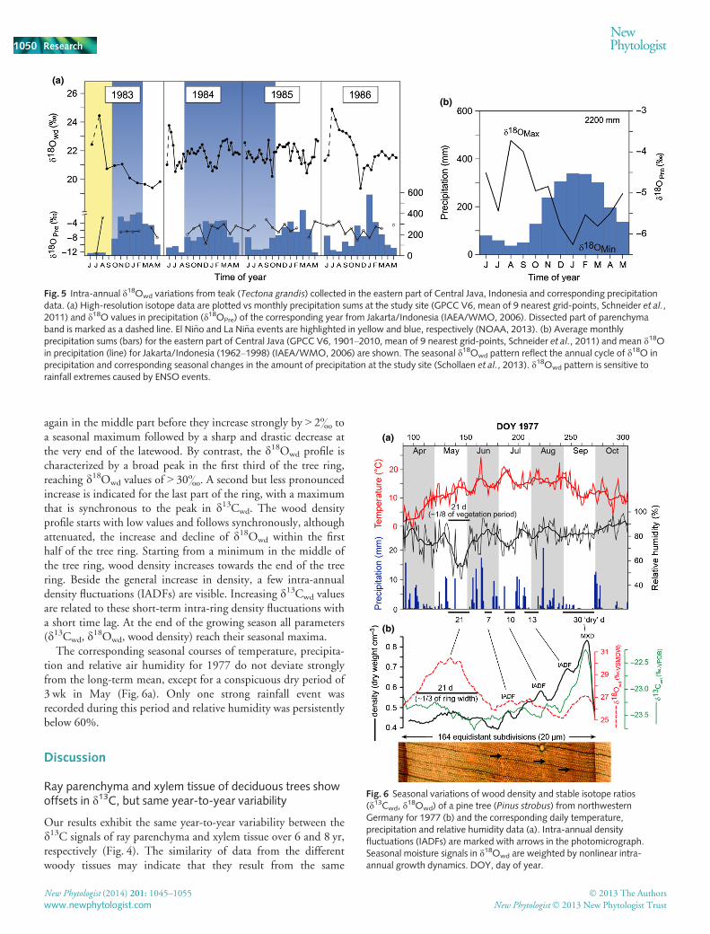
again in the middle part before they increase strongly by > 2& toa seasonal maximum followed by a sharp and drastic decrease atthe very end of the latewood. By contrast, the d18Owd profile ischaracterized by a broad peak in the first third of the tree ring,reaching d18Owd values of > 30&. A second but less pronouncedincrease is indicated for the last part of the ring, with a maximumthat is synchronous to the peak in d13Cwd. The wood densityprofile starts with low values and follows synchronously, althoughattenuated, the increase and decline of d18Owd within the firsthalf of the tree ring. Starting from a minimum in the middle ofthe tree ring, wood density increases towards the end of the treering. Beside the general increase in density, a few intra-annualdensity fluctuations (IADFs) are visible. Increasing d13Cwd valuesare related to these short-term intra-ring density fluctuations witha short time lag. At the end of the growing season all parameters(d13Cwd, d
18Owd, wood density) reach their seasonal maxima.The corresponding seasonal courses of temperature, precipita-
tion and relative air humidity for 1977 do not deviate stronglyfrom the long-term mean, except for a conspicuous dry period of3 wk in May (Fig. 6a). Only one strong rainfall event wasrecorded during this period and relative humidity was persistentlybelow 60%.
Discussion
Ray parenchyma and xylem tissue of deciduous trees showoffsets in d13C, but same year-to-year variability
Our results exhibit the same year-to-year variability between thed13C signals of ray parenchyma and xylem tissue over 6 and 8 yr,respectively (Fig. 4). The similarity of data from the differentwoody tissues may indicate that they result from the same
(a)
(b)
Fig. 5 Intra-annual d18Owd variations from teak (Tectona grandis) collected in the eastern part of Central Java, Indonesia and corresponding precipitationdata. (a) High-resolution isotope data are plotted vs monthly precipitation sums at the study site (GPCC V6, mean of 9 nearest grid-points, Schneider et al.,2011) and d18O values in precipitation (d18OPre) of the corresponding year from Jakarta/Indonesia (IAEA/WMO, 2006). Dissected part of parenchymaband is marked as a dashed line. El Ni~no and La Ni~na events are highlighted in yellow and blue, respectively (NOAA, 2013). (b) Average monthlyprecipitation sums (bars) for the eastern part of Central Java (GPCC V6, 1901–2010, mean of 9 nearest grid-points, Schneider et al., 2011) and mean d18Oin precipitation (line) for Jakarta/Indonesia (1962–1998) (IAEA/WMO, 2006) are shown. The seasonal d18Owd pattern reflect the annual cycle of d18O inprecipitation and corresponding seasonal changes in the amount of precipitation at the study site (Schollaen et al., 2013). d18Owd pattern is sensitive torainfall extremes caused by ENSO events.
(a)
(b)
Fig. 6 Seasonal variations of wood density and stable isotope ratios(d13Cwd, d
18Owd) of a pine tree (Pinus strobus) from northwesternGermany for 1977 (b) and the corresponding daily temperature,precipitation and relative humidity data (a). Intra-annual densityfluctuations (IADFs) are marked with arrows in the photomicrograph.Seasonal moisture signals in d18Owd are weighted by nonlinear intra-annual growth dynamics. DOY, day of year.
New Phytologist (2014) 201: 1045–1055 � 2013 The Authors
New Phytologist� 2013 New Phytologist Trustwww.newphytologist.com
Research
NewPhytologist1050

physiological and biochemical processes. Of course, during thevegetation period assimilates are used at the same time for vessel-,fibre- and ray parenchyma-formation. However, ray cells livelonger, up to several years and thus their structure and chemicalcomposition may change over longer time intervals. Further-more, woody rays are the main storage organs for nonstructuralcarbohydrates, for example sugars and starch. Accumulation andmobilization of nonstructural carbohydrates may be accompa-nied by increased metabolic activity and respiration of parenchy-matic tissue as compared to xylem tissue. Indeed, the offsetsfound between d13C of ray parenchyma and xylem tissue seem toreflect differences in chemical composition and metabolic activ-ity. Ray parenchyma dissected from resin-extracted wood isdepleted in 13C as compared to xylem tissue (Fig. 4b). Our find-ing confirms the findings in a study by Vaganov et al. (2009)using LA-C-GC-IRMS on wood tissue of four consecutive treerings from a Norway spruce (Picea abies) sample. Data revealed asimilar 13C-depletion of ray parenchyma of up to 0.23& in com-parison to tracheids. Vaganov et al. (2009) explained the lowerd13Cwd values as being a result of continuing incorporation of lig-nin into the long-lived parenchyma cells. Lignin is generallydepleted in 13C by up to 3& as compared to cellulose (Wilson &Grinsted, 1977; Loader et al., 2003; Harlow et al., 2006). Hence,a slightly higher content of lignin in ray cells may well cause theobserved 13C depletion over xylem tissue in coniferous wood(Vaganov et al., 2009) and in broadleaved oak of this study.However, UV-laser-based microscopic tissue dissection allowschemical treatment, like cellulose extraction, before IRMS analy-sis. In contrast to a depletion in 13C of resin-extracted wood wefound cellulose of ray parenchyma (d13Ccel) to be slightlyenriched in 13C when compared to cellulose from xylem tissue.This minor, but apparent enrichment of cellulose of woody raysmay be promoted by slightly enhanced catabolic metabolism,which is enhanced respiration making ATP available for the bio-chemical processes of accumulation and mobilization of starch.Gleixner et al. (1993) showed that ‘lighter’ sugar molecules arepreferentially used in catabolic, or respiratory reactions, whereas‘heavier’ ones are involved in polymerization of cellulose, forexample. Hence, an increased carbon isotope partitioningbetween anabolic and catabolic metabolism in ray parenchymamay result in the observed slight enrichment in d13CCel in com-parison with xylem tissue. Note, that the differences in d13Cbetween woody rays and xylem tissue may not be constant(Fig. 6, Vaganov et al., 2009). Varying cellulose to lignin ratiosor respiration rates may cause the observed differences in theoffsets in d13C and may also act on longer timescales.
A recent study on ray parenchyma from Spanish juniper(Juniperus thurifera) revealed that time series based on the abun-dance of ray cells reveal complementary climatic signals to thosederived from tree-ring width chronologies (Olano et al., 2013).Our results demonstrate that the d13C values of ray parenchymaand xylem tissue contain the same year-to-year variability. Hence,we can assume from this pilot study that the d13C signals of raycells may not provide additional information in dendroclimaticstudies. However, the offsets in d13C between ray cells and xylemare dependent on whether wood or cellulose is analysed. This
result indicates the crucial value of the new dissection method asit allows for new approaches towards a better understanding ofthe physiological processes controlling ray parenchyma andxylem cell formation.
High-resolution d18O values in tree rings of Indonesian teakare sensitive to rainfall extremes caused by ENSO
The seasonal tree-ring d18Owd pattern in Indonesian teak ishypothesized to reflect the annual cycle of d18O in precipitationand corresponding seasonal changes in the amount of precipita-tion at the site (Fig. 5). It has been shown that high d18Owd val-ues during the start of the growing season represent the d18OPre
signature of the prior dry season, while the lowest tree-ring d18Ovalues reveal the d18OPre signature of the main rainy season(Schollaen et al., 2013). Towards the end of the growing seasontree-ring d18O values increase again following the d18O-trend ofprecipitation. The described pattern fits closely during the years1983 and 1986. The low rainfall amount during the dry seasonof 1983 was caused by an El Ni~no event (El Ni~no 1982–1983).The two minima in tree-ring d18O values during the 1984, aswell as the missing upward trend at the beginning of the growingseason in 1985, can be explained by rather high rainfall amountsdue to an ongoing La Ni~na phase (blue shaded periods). Thisdemonstrates that sub-seasonal tree-ring d18O records of Indone-sian teak are very sensitive to rainfall extremes caused by ENSO,with high d18Owd values during El Ni~no events and low d18Owd
values during La Ni~na events.Correlations between d18Owd and rainfall amount were also
found in several other studies on tropical or subtropical trees(Poussart et al., 2004; Managave et al., 2010; Brienen et al.,2012; Sano et al., 2012). However, to our knowledge, this is thefirst time that intra-annual d18Owd values in tropical trees havebeen shown to reflect the rainfall pattern over an entire year withdistinct rainy and dry season signals. This underlines the value ofhigh-resolution intra-seasonal isotope measurements of tropicalwood, especially in the light of extreme rainfall events oftenassociated with El Ni~no and La Ni~na.
High-resolution stable isotope variations in white pinereflect seasonal changes of climatic conditions weighted bynonlinear growth dynamics
Tree rings integrate environmental information over the vegeta-tion period. The intra-annual variations in d13Cwd, d
18Owd andwood density are driven mainly by interactions between seasonalvariation in meteorological conditions, soil water availability andplant response. Specific weather events such as dry periods areclearly recorded in the seasonal pattern of d13C, d18O and wooddensity (Barbour et al., 2002; Eilmann et al., 2010; Sarris et al.,2013). In this study a detailed review of the weather conditionsfor 1977 at our site in northwestern Germany indicated severaltime periods with no or less rainfall – so called ‘dry’ days(Fig. 6a). The corresponding intra-annual pattern of d13Cwd
(Fig. 6b) follows a typical course observed for coniferous specieswith a gradual increase in d13Cwd to a maximum in latewood
� 2013 The Authors
New Phytologist� 2013 New Phytologist TrustNew Phytologist (2014) 201: 1045–1055
www.newphytologist.com
NewPhytologist Research 1051

followed by a sharp decline at the very end of the tree ring (cf.Leavitt, 1993; Walcroft et al., 1997; Barbour et al., 2002; Schulzeet al., 2004). Short-term increases in d13Cwd values of the secondhalf of the tree ring were observed after dry periods and are inagreement with the wood density pattern. By contrast, the firstdry period in May is not well observed in the d13Cwd pattern.However, this dry period in late spring, characterized by low rain-fall as well as low relative humidity, is well represented in thed18Owd record by a conspicuous broad peak. It seems thatd18Owd is much more sensitive to changes in moisture conditionsthan d13Cwd and wood density at the beginning of the growingseason. During the second half of the growing season it appearsto be vice versa, that is, d13Cwd and wood density reflect moisturedeficits better than d18Owd does.
Furthermore, environmental information seems to be inte-grated in the tree ring in a nonlinear way. The ‘dry’ period of21 d in May represents approximately only one-eighth of the veg-etation period, while the d18Owd peak represents one-third of thewhole tree-ring width. The ability of a tree ring to integrate envi-ronmental information depends on the rate of wood cell forma-tion and the longevity of cells. Within the vegetation period thesetwo features are inversely related (Fig. 7). While thin-walledearlywood cells are formed at a high rate, they may only live fordays up to a few weeks. Thick-walled latewood cells are built atvery slow rates, but may live up to several months and integrateenvironmental information over a much longer period of time(Fig. 7a). Hence, environmental conditions reflected by intra-annual tree-ring parameters are weighted by seasonal dynamics ofwood formation. Additionally, other seasonal changes related totree physiological processes such as kind and size of carbohydratepools (amount of C and O), partitioning between catabolic andanabolic metabolism, as well as metabolic flux rates in conjunc-tion with corresponding isotope fractionations have to be consid-ered (Fig. 7b and Helle & Schleser, 2004a; Gessler et al., 2009;Werner et al., 2012).
Evaluation and comparison of UV-laser-based microscopicdissection systems
The results presented suggest that UV-laser-based microscopicdissection is a very useful method for sampling woody tissue instable isotope studies. It can be used likewise for dissecting whole
tree rings and intra-annual sampling at very high spatial resolu-tion. The two UV-laser microdissection microscopes of Leica(LMD7000, software v6.7.1.3952, Leica Microsystems GmbH)and Zeiss (PALM MicroBeam, software v4.5.0.9, Carl ZeissMicroscopy GmbH), we tested in these studies, differ concerningtheir practical implementations and applications (Table 1). Thelaser from Leica is moved via optics and the cross-section samplesare mounted on a fixed stage. The Leica system uses high-precision optics to steer the laser beam by means of prisms alongthe desired cut lines on the tissue. As a side effect, the laser canonly cut drawn lines or areas marked within the actual micro-scopic field of view. If larger areas, for example whole tree rings,need to be dissected additional shapes must be defined and addi-tional dissections are required for gathering a single large sample.The dissected sample tissues principally fall down by gravity intocollection vessels, for example, tin or silver capsules. Thus, sam-ples can be prepared directly for conventional autosampler sys-tems coupled to isotope ratio mass spectrometers. An importantlimitation of the Leica system is the lack of an automatic z-focusadjustment that would allow repeated laser cutting of thickercross-sections.
Compared to the Leica microscope, the objectives of the Zeissmicroscope are installed inversely, or underneath the sampleholder. Hence, tissues of interests are marked via mouse or screenpen on the lower sample side and can be selected beyond the visi-ble screen. Thus, tissues of interests can be marked as big as neces-sary, because the laser stays fixed while the sample is moved bythe high-precision stage during the dissection process. The UV-laser passes through the glass slide and the dissected sample tissuesremain in position. After all of the marked tissue has been dis-sected specimens are picked up manually with a forceps and trans-ferred into tin or silver capsules for stable isotope measurements.Dissected sample tissues were too heavy for the laser-inducedsample transfer that comes with the Zeiss system. The Zeiss sys-tem has an automatic z-focus feature that allows easy definition ofthe number of automatic cutting iterations. For each cutting cyclethe focus of the UV-laser beam is adjusted by a pre-defined z-focus delta value. As the Zeiss laser is less powerful than the laserfrom Leica, more cutting iterations are generally required.Together with the manual collection of dissected specimens, theoverall sampling process with the Zeiss system is longer than withthe Leica system. However, the dissection process with the Zeiss
(a) (b)
Fig. 7 (a) Nonlinear seasonal growthincrements and longevity of cells during thevegetation period for conifers at temperatesites (DOY, day of year). (b) Seasonaldynamics of major factors influencing tree-ring stable isotope ratios of wood cellformation is characterized by increasing cellwall thickness, cell longevity and amount ofC/O per cell row (elemental mass/wooddensity), and decreasing cell diameter. EW,early wood; LW, late wood.
New Phytologist (2014) 201: 1045–1055 � 2013 The Authors
New Phytologist� 2013 New Phytologist Trustwww.newphytologist.com
Research
NewPhytologist1052

can be operated automatically, for example overnight, whereasthe Leica system requires the full-time presence of the user: first,the adjustment of z-focus of the laser beam has to be done manu-ally; and second, dissected specimens sometimes tilt, stick and donot fall down into the collection vessels as the cutting line of thelaser beam is much narrower than the thickness of the sample.
The use of UV-laser microdissection microscopes is not neces-sarily faster than traditional methods for the dissection of woodtissue. In general, the cutting process of selected tissue lasts1–2 min, depending on the size of the selected area, the thicknessof the cross-section, density of the wood material and the UV-laser settings. If further cutting iterations are required, furthertime is needed. The average sample throughput per 8-h day mayvary between 20 and 120 samples. This includes the on-screenselection of area, the automatic UV-laser-based microscopic dis-section and the collection of specimens, as well as unpredictableinterferences such as stuck specimens (Leica LMD7000). Withsome modification of the current sample collection methods theLeica system could also be run automatically overnight, whichwould increase sample throughput drastically.
At this stage, the key advantage of using the novel technique ison-screen selection of areas and ultrahigh-resolution sampling ofplant tissue with unprecedented precision. Furthermore, bothsystems provide electronic documentation of the dissection pro-cesses by photo or video sequences, as well as a report of labelledand dissected elements.
Conclusions
We present a novel method for dissecting plant tissue in stableisotope studies by using UV-laser-based microscopic dissection.
Using two commercially available UV-laser microdissectionmicroscopes, we prepared high-resolution intra-annual woodsamples from various tree species of different climate zones for Cand O isotope analyses with conventional IRMS systems.
The UV-laser-based microscopic dissection enables preciseselection of relevant plant cells/tissues on video screen by pen.This procedure allowed us to detect that the d13C contents ofxylem cells and woody ray parenchyma of the deciduous species(Q. robur) have the same year-to-year variability, but also revealoffsets that are opposite in sign depending on whether whole-wood or cellulose is analysed. Furthermore, we showed thatUV-laser-based microscopic dissection facilitates electronicallydocumented sub-seasonal sampling of tree rings with irregularshapes or narrow ring widths. This is a prerequisite for establish-ing long and continuous high-resolution isotope chronologies forhigh-quality climate reconstructions. Single cell rows can be sam-pled and thus short-term climatic events such as droughts orextensive rainfall events (e.g. caused by ENSO) may be relateddirectly. The three applications demonstrate that the use of thisnew technique enables the user to sample plant tissue at ultrahighresolution and unprecedented precision.
This new technique opens new ways for studying the environ-mental information in tree rings nonlinearly integrated over thevegetation period. The combination of direct growth measure-ments, which provide exact growth rates of tree rings (e.g. punch-ing method (Forster et al., 2000; Rossi et al., 2006),dendrometers) and precisely selected and dissected specimensmay provide a better time-match between high-resolution stableisotope ratios and real-time climate data. Comprehensive eco-physiological process studies on stable isotope signal transfer inthe arboreal system can be combined with qualitative and
Table 1 Comparison of the characteristics of the two UV-laser-based microscopic dissection systems tested
Zeiss microdissection microscope (PALMMicroBeam/software v4.5.0.9) Leica microdissection microscope (LMD7000/software v6.7.1.3952)
General characteristics
Laser specifications:Wavelength: 355 nmMax. pulse energy: 100 lJLaser beam stays fixed, stage is moved
Cutting process with different magnifications (e.g.92.5,95 or 910)Best range of sample thickness: 100–1000 lm
Laser specifications:Wavelength: 349 nmMax. pulse energy: 120 lJLaser beam is moved, stage stays fixedCutting process with different magnifications (e.g. 95, 910 or 920)Best range of sample thickness: 100–1000 lm
Advantages
Selection of tissue/shape beyond the visible screenAutomatic z-focus adjustment/cutting iterations possibleBetter software handling and multifarious choice of drawing tools
Laser has more power and less cutting iteration neededFast sample collection process by semi-automatic sample collectioninto tin or silver capsules by gravityAutomatic image documentation before and after each dissection byimage-database (IM500)
Limitations
Nonautomatic collection of selected tissues of interests in tin/silver capsules(specimens are being picked up manually with a forceps and transferredinto tin/silver capsules)Laser has less power and frequent cutting iteration neededNonautomatic image documentation of the laser dissection process
Lack of automatic z-focusDrawing area of interests larger than the field of view is not possibleControl check of dissected samples needed after every cutting cycleas samples may stick and do not fall down automatically
� 2013 The Authors
New Phytologist� 2013 New Phytologist TrustNew Phytologist (2014) 201: 1045–1055
www.newphytologist.com
NewPhytologist Research 1053

quantitative microscopic investigations on anatomical properties.As wood anatomical properties are climate sensitive (Fonti et al.,2010; Liang et al., 2013; Olano et al., 2013), more and morestudies are attempting to combine quantitative wood anatomyand stable isotope analysis (Ponton et al., 2001; Battipaglia et al.,2010; De Micco et al., 2012; Rossi et al., 2013). Such investiga-tions may explicitly profit from UV-laser-based microscopic dis-section. For instance, detailed assessment of C and O isotopevariability in woody parts such as resin ducts, rays, fibres, vesselsor parenchyma cells is possible, as they can now be preciselytargeted on thin sections by the novel method.
The use of UV-laser-based microscopic dissection systemsoffers new possibilities in view of relating plant structure withplant functioning (derived from isotope ratios) in studies on vul-nerability, resilience and adaption of plants to past and presentglobal change. Multidisciplinary analyses (cell structure, wooddensity and wood chemistry analyses) on the same wood samplesare now possible with a complete recovery of the sample due tothe use of thin sections.
Acknowledgements
We are grateful to Heiko Baschek, David G€ohring and SophieWendler for support in the laboratory, to Andreas Hendrich forhelp with the layout of figures, and to Sonia Simard, Wei Liang,Hagen Pieper and Uwe Scholl€an for fruitful discussions. Further-more, we thank three anonymous reviewers and the editor fortheir valuable comments and suggestions. This study was fundedby the ISOWOOD-Breeding (BMBF, 0315427B), INDOPAL(HE3089/1-1) and CADY (BMBF, 03G0813H) project.
References
Abbott E, Hall D, Hamberger B, Bohlmann J. 2010. Laser microdissection of
conifer stem tissues: isolation and analysis of high quality RNA, terpene
synthase enzyme activity and terpenoid metabolites from resin ducts and
cambial zone tissue of white spruce (Picea glauca). BMC Plant Biology 10: 106.Barbour MM, Walcroft AS, Farquhar GD. 2002. Seasonal variation in d13C and
d18O of cellulose from growth rings of Pinus radiata. Plant, Cell &Environment 25: 1483–1499.
Battipaglia G, De Micco V, Brand WA, Linke P, Aronne G, Saurer M,
Cherubini P. 2010. Variations of vessel diameter and d13C in false rings of
Arbutus unedo L. reflect different environmental conditions. New Phytologist188: 1099–1112.
Bayona-Bafaluy M, Blits B, Battersby BJ, Shoubridge EA, Moraes CT. 2005.
Rapid directional shift of mitochondrial DNA heteroplasmy in animal tissues
by a mitochondrially targeted restriction endonuclease. Proceedings of theNational Academy of Sciences, USA 102: 14 392–14 397.
Brienen RJW, Helle G, Pons TL, Guyot JL, Gloor M. 2012.Oxygen isotopes in
tree rings are a good proxy for Amazon precipitation and El Ni~no-Southern
Oscillation variability. Proceedings of the National Academy of Sciences, USA109: 16 957–16 962.
Coster C. 1928. Zur Anatomie und Physiologie der Zuwachszonen- und
Jahresringbildung in den Tropen. Annales du Jardin botanique de Buitenzorg38: 1–114.
Craig H. 1957. Isotopic standards for carbon and oxygen and correction factors
for mass-spectrometric analysis of carbon dioxide. Geochimica et CosmochimicaActa 12: 133–149.
DawsonTE,Mambelli S, Plamboeck AH, Templer PH, TuKP. 2002. Stable
isotopes in plant ecology.Annual Review of Ecology and Systematics 33: 507–559.
De Micco V, Battipaglia G, Brand W, Linke P, Saurer M, Aronne G, Cherubini
P. 2012. Discrete versus continuous analysis of anatomical and d13C variability
in tree rings with intra-annual density fluctuations. Trees 26: 513–524.Dodd JP, Patterson WP, Holmden C, Brasseur JM. 2008. Robotic micromilling
of tree-rings: a new tool for obtaining subseasonal environmental isotope
records. Chemical Geology 252: 21–30.Ehleringer J, Hall AE, Farquhar G. 1993. Stable isotopes and plant carbon–waterrelations. London, UK: Academic Press.
Eilmann B, Buchmann N, Siegwolf R, Saurer M, Cherubini P, Rigling A. 2010.
Fast response of Scots pine to improved water availability reflected in tree-ring
width and d13C. Plant, Cell & Environment 33: 1351–1360.Evans MN, Schrag DP. 2004. A stable isotope-based approach to tropical
dendroclimatology. Geochimica et Cosmochimica Acta 68: 3295–3305.Fend F, Raffeld M. 2000. Laser capture microdissection in pathology. Journal ofClinical Pathology 53: 666–672.
Fichtler E, Helle G,WorbesM. 2010. Stable-carbon isotope time series from
tropical tree rings indicate a precipitation signal. Tree-Ring Research 66: 35–49.Fonti P, von Arx G, Garc�ıa-Gonz�alez I, Eilmann B, Sass-Klaassen U, G€artnerH, Eckstein D. 2010. Studying global change through investigation of the
plastic responses of xylem anatomy in tree rings. New Phytologist 185: 42–53.Forster T, Schweingruber FH, Denneler B. 2000. Increment puncher – a toolfor extracting small cores of wood and bark from living trees. Iawa Journal 21:169–180.
G€artner H, Nievergelt D. 2010. The core-microtome: a new tool for surface
preparation on cores and time series analysis of varying cell parameters.
Dendrochronologia 28: 85–92.Gebrekirstos A, Worbes M, Teketay D, Fetene M, Mitl€ohner R. 2009. Stable
carbon isotope ratios in tree rings of co-occurring species from semi-arid tropics
in Africa: patterns and climatic signals. Global and Planetary Change 66: 253–260.
Gessler A, Brandes E, Buchmann N, Helle G, Rennenberg H, Barnard RL.
2009. Tracing carbon and oxygen isotope signals from newly assimilated sugars
in the leaves to the tree-ring archive. Plant, Cell & Environment 32: 780–795.Gleixner G, Danier HJ, Werner RA, Schmidt HL. 1993. Correlations between
the 13C content of primary and secondary plant products in different cell
compartments and that in decomposing basidiomycetes. Plant Physiology 102:1287–1290.
Harlow BA, Marshall JD, Robinson AP. 2006. A multi-species comparison of
d13C from whole wood, extractive-free wood and holocellulose. Tree Physiology26: 767–774.
Heinrich I, Touchan R, Dorado Li~n�an I, Vos H, Helle G. 2013.
Winter-to-spring temperature dynamics in Turkey derived from tree rings since
AD 1125. Climate Dynamics 41: 1685–1701.Helle G, Schleser GH. 2004a. Beyond CO2-fixation by Rubisco – aninterpretation of 13C/12C variations in tree rings from novel intra-seasonal
studies on broad-leaf trees. Plant, Cell & Environment 27: 367–380.Helle G, Schleser GH. 2004b. Interpreting climate proxies from tree-rings. In:
Fischer H, Floeser G, Kumke T, Lohmann G, Miller H, Negendank JFW, von
Storch H, eds. The KIHZ project: towards a synthesis of Holocene proxy data andclimate models. Berlin, Germany: Springer, 129–148.
Hietz P, Turner BL, Wanek W, Richter A, Nock CA, Wright SJ. 2011.
Long-term change in the nitrogen cycle of tropical forests. Science 334: 664–666.
IAEA/WMO 2006. Global Network of Isotopes in Precipitation. The GNIP
Database. [WWW document] URL http://www.iaea.org/water. [accessed on 5
August 2013].
Krepkowski J, Gebrekirstos A, Shibistova O, Br€auning A. 2013. Stable carbon
isotope labeling reveals different carry-over effects between functional types of
tropical trees in an Ethiopian mountain forest. New Phytologist 199: 431–440.Leavitt SW. 1993. Seasonal 13C/12C changes in tree rings: species and site
coherence, and a possible drought influence. Canadian Journal of ForestResearch 23: 210–218.
Leavitt SW, Long A. 1982. Stable carbon isotopes as a potential supplemental
tool in dendrochronology. Tree-Ring Bulletin 42: 49–55.Liang W, Heinrich I, Simard S, Helle G, Dorado Li~n�an I, Heinken T. 2013.
Climate signals derived from cell anatomy of Scots pine in NE Germany. TreePhysiology 33: 833–844.
New Phytologist (2014) 201: 1045–1055 � 2013 The Authors
New Phytologist� 2013 New Phytologist Trustwww.newphytologist.com
Research
NewPhytologist1054

Loader NJ, Robertson I, McCarroll D. 2003. Comparison of stable carbon
isotope ratios in the whole wood, cellulose and lignin of oak tree-rings.
Palaeogeography, Palaeoclimatology, Palaeoecology 196: 395–407.Loader NJ, Switsur VR, Field EM. 1995.High-resolution stable isotope analysis
of tree rings: implications of “microdendroclimatology” for
palaeoenvironmental research. The Holocene 5: 457–460.Managave SR, Sheshshayee MS, Borgaonkar HP, Ramesh R. 2010. Past
break-monsoon conditions detectable by high resolution intra-annual d18Oanalysis of teak rings. Geophysical Research Letters 37: L05702.
Nelson T, Tausta SL, Gandotra N, Liu T. 2006. Laser microdissection of plant
tissue: what you see is what you get. Annual Review of Plant Biology 57:181–201.
NOAA 2013. National Weather Service. Historical El Ni~no/La Ni~na episodes
(1950-present). [WWW document] URL http://www.cpc.ncep.noaa.gov/
products/analysis_monitoring/ensostuff/ensoyears.shtml. [accessed on 5 August
2013].
Og�ee J, Barbour MM, Wingate L, Bert D, Bosc A, Stievenard M, Lambrot C,
Pierre M, Bariac T, Loustau D et al. 2009. A single-substrate model to
interpret intra-annual stable isotope signals in tree-ring cellulose. Plant, Cell &Environment 32: 1071–1090.
Ogle N, McCormac FG. 1994.High-resolution d13C measurements of oak show
a previously unobserved spring depletion. Geophysical Research Letters 21:2373–2375.
Olano JM, Arzac A, Garc�ıa-Cervig�on AI, von Arx G, Rozas V. 2013. New star
on the stage: amount of ray parenchyma in tree rings shows a link to climate.
New Phytologist 198: 486–495.Pons T, Helle G. 2011. Identification of anatomically non-distinct annual
rings in tropical trees using stable isotopes. Trees - Structure and Function 25:83–93.
Ponton S, Dupouey JL, Breda N, Feuillat F, Bodenes C, Dreyer E. 2001.
Carbon isotope discrimination and wood anatomy variations in mixed
stands of Quercus robur and Quercus petraea. Plant, Cell & Environment 24:861–868.
Poussart PF, Evans MN, Schrag DP. 2004. Resolving seasonality in tropical
trees: multi-decade, high-resolution oxygen and carbon isotope records
from Indonesia and Thailand. Earth and Planetary Science Letters 218:301–316.
Roden JS, Johnstone JA, Dawson TE. 2009. Intra-annual variation in the stable
oxygen and carbon isotope ratios of cellulose in tree rings of coast redwood
(Sequoia sempervirens). The Holocene 19: 189–197.Rossi L, Sebastiani L, Tognetti R, d’Andria R, Morelli G, Cherubini P. 2013.
Tree-ring wood anatomy and stable isotopes show structural and functional
adjustments in olive trees under different water availability. Plant and Soil372: 567–579.
Rossi S, Anfodillo T, Menardi R. 2006. Trephor: a new tool for sampling
microcores from tree stems. Iawa Journal 27: 89–97.Sano M, Xu C, Nakatsuka T. 2012. A 300-year Vietnam hydroclimate and
ENSO variability record reconstructed from tree ring d18O. Journal ofGeophysical Research 117: D12115.
Sarris D, Siegwolf R, K€orner C. 2013. Inter- and intra-annual stable carbon and
oxygen isotope signals in response to drought in Mediterranean pines.
Agricultural and Forest Meteorology 168: 59–68.Schleser GH, Helle G, Lucke A, Vos H. 1999. Isotope signals as climate proxies:
the role of transfer functions in the study of terrestrial archives. QuaternaryScience Reviews 18: 927–943.
Schneider U, Becker A, Finger P, Meyer-Christoffer A, Rudolf B, Ziese M.
2011. GPCC Full Data Reanalysis Version 6.0 at 0.5°: monthly land-surface
precipitation from rain-gauges built on GTS-based and historic data. doi: 10.
5676/DWD_GPCC/FD_M_V6_050. Global Precipitation Climatology
Centre (GPCC, http://gpcc.dwd.de/).
Schollaen K, Heinrich I, Neuwirth B, Krusic PJ, D’Arrigo RD, Karyanto O,
Helle G. 2013.Multiple tree-ring chronologies (ring width, d13C and d18O)
reveal dry and rainy season signals of rainfall in Indonesia. Quaternary ScienceReviews 73: 170–181.
Schubert BA, Jahren AH. 2011.Quantifying seasonal precipitation using
high-resolution carbon isotope analyses in evergreen wood. Geochimica etCosmochimica Acta 75: 7291–7303.
Schulman E. 1956. Dendroclimatic change in semiarid America. Tuscon, AZ,USA: University of Arizona Press.
Schulze B, Wirth C, Linke P, Brand WA, Kuhlmann I, Horna V, Schulze ED.
2004. Laser ablation-combustion-GC-IRMS – a new method for online analysis
of intra-annual variation of d13C in tree rings. Tree Physiology 24: 1193–1201.Skomarkova MV, Vaganov EA, Mund M, Knohl A, Linke P, Boerner A, Schulze
ED. 2006. Inter-annual and seasonal variability of radial growth, wood density
and carbon isotope ratios in tree rings of beech (Fagus sylvatica) growing inGermany and Italy. Trees-Structure and Function 20: 571–586.
Treydte KS, Schleser GH, Helle G, Frank DC, Winiger M, Haug GH, Esper J.
2006. The twentieth century was the wettest period in northern Pakistan over
the past millennium. Nature 440: 1179–1182.Vaganov E, Schulze E-D, Skomarkova M, Knohl A, Brand W, Roscher C. 2009.
Intra-annual variability of anatomical structure and d13C values within tree
rings of spruce and pine in alpine, temperate and boreal Europe. Oecologia 161:729–745.
Verheyden A, Helle G, Schleser GH, Dehairs F, Beeckman H, Koedam N.
2004. Annual cyclicity in high-resolution stable carbon and oxygen isotope
ratios in the wood of the mangrove tree Rhizophora mucronata. Plant, Cell &Environment 27: 1525–1536.
Walcroft AS, Silvester WB, Whitehead D, Kelliher FM. 1997. Seasonal changes
in stable carbon isotope ratios within annual rings of Pinus radiata reflectenvironmental regulation of growth processes. Functional Plant Biology 24:57–68.
Werner C, Schnyder H, Cuntz M, Keitel C, Zeeman MJ, Dawson TE, Badeck
F-W, Brugnoli E, Ghashghaie J, Grams TEE et al. 2012. Progress andchallenges in using stable isotopes to trace plant carbon and water relations
across scales. Biogeosciences 9: 3083–3111.West JB, Bowen GJ, Dawson TE, Tu KP. 2010. Isoscapes: understandingmovement, pattern, and process on Earth through isotope mapping. London, UK:
Springer Science+Business Media B.V.
Wieloch T, Helle G, Heinrich I, Voigt M, Schyma P. 2011. A novel device for
batch-wise isolation of a-cellulose from small-amount wholewood samples.
Dendrochronologia 29: 115–117.Wilson AT, Grinsted MJ. 1977. 12C/13C in cellulose and lignin as
palaeothermometers. Nature 265: 133–135.Wurster CM, Patterson WP, Cheatham MM. 1999. Advances in micromilling
techniques: a new apparatus for acquiring high-resolution oxygen and carbon
stable isotope values and major/minor elemental ratios from accretionary
carbonate. Computers & Geosciences 25: 1159–1166.Ziegler H. 1995. Stable isotopes in plant physiology and ecology. In: Behnke
HD, L€uttge U, Esser K, Kadereit J, Runge M, eds. Progress in botany. Berlin,Heidelberg, Germany: Springer, 1–24.
� 2013 The Authors
New Phytologist� 2013 New Phytologist TrustNew Phytologist (2014) 201: 1045–1055
www.newphytologist.com
NewPhytologist Research 1055
Copyright © 2022 FDOKUMEN