Analysis of infectious virus clones from two HIV1 superinfection cases suggests that the primary...
15
RESEARCH Open Access Analysis of infectious virus clones from two HIV-1 superinfection cases suggests that the primary strains have lower fitness Antoinette C van der Kuyl 1* , Karolina Kozaczynska 1,5 , Kevin K Ariën 2,3 , Youssef Gali 2 , Victoria R Balázs 1 , Stefan J Dekker 1 , Fokla Zorgdrager 1 , Guido Vanham 2,4 , Ben Berkhout 1 , Marion Cornelissen 1 Abstract Background: Two HIV-1 positive patients, L and P, participating in the Amsterdam Cohort studies acquired an HIV- 1 superinfection within half a year from their primary HIV-1 infection (Jurriaans et al., JAIDS 2008, 47:69-73). The aim of this study was to compare the replicative fitness of the primary and superinfecting HIV-1 strains of both patients. The use of isolate-specific primer sets indicated that the primary and secondary strains co-exist in plasma at all time points after the moment of superinfection. Results: Biological HIV-1 clones were derived from peripheral blood CD4 + T cells at different time point, and identified as the primary or secondary virus through sequence analysis. Replication competition assays were performed with selected virus pairs in PHA/IL-2 activated peripheral blood mononuclear cells (PBMC’s) and analyzed with the Heteroduplex Tracking Assay (HTA) and isolate-specific PCR amplification. In both cases, we found a replicative advantage of the secondary HIV-1 strain over the primary virus. Full-length HIV-1 genomes were sequenced to find possible explanations for the difference in replication capacity. Mutations that could negatively affect viral replication were identified in the primary infecting strains. In patient L, the primary strain has two insertions in the LTR promoter, combined with a mutation in the tat gene that has been associated with decreased replication capacity. The primary HIV-1 strain isolated from patient P has two mutations in the LTR that have been associated with a reduced replication rate. In a luciferase assay, only the LTR from the primary virus of patient P had lower transcriptional activity compared with the superinfecting virus. Conclusions: These preliminary findings suggest the interesting scenario that superinfection occurs preferentially in patients infected with a relatively attenuated HIV-1 isolate. Background Viral fitness is the parameter that is defined by the ability of an individual genotype to produce infectious progeny in a specific environment [1,2], and it can be divided into transmission fitness, replicative fitness or immune-evasion fitness. In addition to viral genetics, the host environment, i.e. type of target cells, immune response, antiretroviral drug treatment, plays an impor- tant role in viral fitness [1,2]. To measure replication fit- ness of HIV-1 in vitro, three types of assays have been developed: replication assays, single round infection assays and dual infection/competition assays [1]. The last is considered the ‘gold standard’ for replicative fit- ness determination and involves direct competition between different viral strains in cell culture infections [1,3]. For all assays, either molecular clones (virus gene of interest cloned into standard viral backbone), biologi- cal clones (single virus isolate) or a virus pool (quasi- species) can be used [1]. Competition assays have been used to determine the relative replicative fitness of viruses belonging to HIV-1 group M, HIV-1 group O and HIV-2 [4], to show that HIV-1 fitness increases dur- ing disease progression [5,6], to suggest that HIV-1 attenuates over time [7]. In contrast to the previous study, we and others have reported that viral fitness is * Correspondence: [email protected] 1 Laboratory of Experimental Virology, Department of Medical Microbiology, Centre for Infection and Immunity Amsterdam (CINIMA), Academic Medical Centre of the University of Amsterdam, Meibergdreef 15, 1105 AZ Amsterdam, The Netherlands van der Kuyl et al. Retrovirology 2010, 7:60 http://www.retrovirology.com/content/7/1/60 © 2010 van der Kuyl et al; licensee BioMed Central Ltd. This is an Open Access article distributed under the terms of the Creative Commons Attribution License (http://creativecommons.org/licenses/by/2.0), which permits unrestricted use, distribution, and reproduction in any medium, provided the original work is properly cited.
-
Upload
independent -
Category
Documents
-
view
2 -
download
0
Transcript of Analysis of infectious virus clones from two HIV1 superinfection cases suggests that the primary...

RESEARCH Open Access
Analysis of infectious virus clones from two HIV-1superinfection cases suggests that the primarystrains have lower fitnessAntoinette C van der Kuyl1*, Karolina Kozaczynska1,5, Kevin K Ariën2,3, Youssef Gali2, Victoria R Balázs1,Stefan J Dekker1, Fokla Zorgdrager1, Guido Vanham2,4, Ben Berkhout1, Marion Cornelissen1
Abstract
Background: Two HIV-1 positive patients, L and P, participating in the Amsterdam Cohort studies acquired an HIV-1 superinfection within half a year from their primary HIV-1 infection (Jurriaans et al., JAIDS 2008, 47:69-73). The aimof this study was to compare the replicative fitness of the primary and superinfecting HIV-1 strains of both patients.The use of isolate-specific primer sets indicated that the primary and secondary strains co-exist in plasma at alltime points after the moment of superinfection.
Results: Biological HIV-1 clones were derived from peripheral blood CD4 + T cells at different time point, andidentified as the primary or secondary virus through sequence analysis. Replication competition assays wereperformed with selected virus pairs in PHA/IL-2 activated peripheral blood mononuclear cells (PBMC’s) andanalyzed with the Heteroduplex Tracking Assay (HTA) and isolate-specific PCR amplification. In both cases, wefound a replicative advantage of the secondary HIV-1 strain over the primary virus. Full-length HIV-1 genomes weresequenced to find possible explanations for the difference in replication capacity. Mutations that could negativelyaffect viral replication were identified in the primary infecting strains. In patient L, the primary strain has twoinsertions in the LTR promoter, combined with a mutation in the tat gene that has been associated withdecreased replication capacity. The primary HIV-1 strain isolated from patient P has two mutations in the LTR thathave been associated with a reduced replication rate. In a luciferase assay, only the LTR from the primary virus ofpatient P had lower transcriptional activity compared with the superinfecting virus.
Conclusions: These preliminary findings suggest the interesting scenario that superinfection occurs preferentially inpatients infected with a relatively attenuated HIV-1 isolate.
BackgroundViral fitness is the parameter that is defined by theability of an individual genotype to produce infectiousprogeny in a specific environment [1,2], and it can bedivided into transmission fitness, replicative fitness orimmune-evasion fitness. In addition to viral genetics, thehost environment, i.e. type of target cells, immuneresponse, antiretroviral drug treatment, plays an impor-tant role in viral fitness [1,2]. To measure replication fit-ness of HIV-1 in vitro, three types of assays have been
developed: replication assays, single round infectionassays and dual infection/competition assays [1]. Thelast is considered the ‘gold standard’ for replicative fit-ness determination and involves direct competitionbetween different viral strains in cell culture infections[1,3]. For all assays, either molecular clones (virus geneof interest cloned into standard viral backbone), biologi-cal clones (single virus isolate) or a virus pool (quasi-species) can be used [1]. Competition assays have beenused to determine the relative replicative fitness ofviruses belonging to HIV-1 group M, HIV-1 group Oand HIV-2 [4], to show that HIV-1 fitness increases dur-ing disease progression [5,6], to suggest that HIV-1attenuates over time [7]. In contrast to the previousstudy, we and others have reported that viral fitness is
* Correspondence: [email protected] of Experimental Virology, Department of Medical Microbiology,Centre for Infection and Immunity Amsterdam (CINIMA), Academic MedicalCentre of the University of Amsterdam, Meibergdreef 15, 1105 AZAmsterdam, The Netherlands
van der Kuyl et al. Retrovirology 2010, 7:60http://www.retrovirology.com/content/7/1/60
© 2010 van der Kuyl et al; licensee BioMed Central Ltd. This is an Open Access article distributed under the terms of the CreativeCommons Attribution License (http://creativecommons.org/licenses/by/2.0), which permits unrestricted use, distribution, andreproduction in any medium, provided the original work is properly cited.

increasing over time within the HIV-1 epidemic in TheNetherlands [8,9]. This was also the case in France in1997-2005 [10], but HIV-1 virulence was not changedover time in North America [11].The description of HIV-1 superinfection in vivo is
relatively new [12]. It is likely that parasites, includingviruses, able to establish a productive superinfectionhave increased fitness over the primary infecting strain(see [13,14] and references therein). In line with this,several reports have described superinfection with anon-drug resistant HIV-1 strain in patients first infectedwith a drug-resistant HIV-1 strain with presumed lowerfitness [15-17]. Two studies compared the relative fit-ness of the superinfecting strain with that of the primarystrain in replication assays, but the analysis wasrestricted to the contribution of the pol gene [16,17]. Inboth cases no differences were observed, suggesting thatfitness determining factors may be located elsewhere inthe viral genome, as the superinfecting strains appearedto be more fit in vivo. In another superinfection case,two multidrug-resistant HIV-1 strains were involved, ofwhich the first appeared more fit in competition assays.Not much is known about the relative fitness of theviruses in superinfection cases with HIV-1 variants lack-ing drug-resistance mutations. Therefore we decided tocompare the replicative fitness of the primary and sec-ondary strain in two HIV-1 superinfection cases. Biolo-gical clones were generated and ex vivo competitionassays were performed as described earlier [5]. The exvivo results were compared to the in vivo observations.The competition results suggest that, even though noneof the strains exhibited a severe replication defect, thesuperinfecting virus has a higher replicative capacitythan the primary strain. Analysis of the ratio of the twostrains in blood plasma confirmed this finding. Full gen-ome sequences of the viral clones were investigated todetect mutations that could explain the observed differ-ences in replication capacity.
ResultsPatient LFigure 1A shows the plasma viral load and CD4 + T cellcount of patient L during follow up. Phylogenetic analy-sis of the plasma-derived HIV-1 sequences for env-V3(Figure 1B) and gag (data not shown) were carried outon serial samples from 2005-2006. The subtype B viralsequences from 2005 cluster together and were namedstrain B1. A new subtype B cluster was formed bysequences from January 2006, which was named strainB2. At that time point, the new strain B2 dominated theviral population even though strain B1 could still beamplified. Three months later, in April 2006, both B1and B2 strain sequences persisted. These observationssuggest that patient L was superinfected with a second
HIV-1 strain somewhere between December 2005 andJanuary 2006, coinciding with a marked increase of theviral load (marked by a vertical arrow in Figure 1A).Similar results were obtained for the gag sequences (notshown).Plasma samples from patient L were tested with
strain-specific primers designed to amplify either strainB1 or B2. In December 2005 only the B1 strain wasdetected in both env-V3 and gag assays (not shown). Atall later time-points, gag and env-V3 fragments of theB1 and B2 strain were amplified concurrently.
Patient PFigure 2A shows the plasma viral load and CD4 + T cellcount of patient P during follow up. The env-V3 andgag fragments amplified from plasma samples were ana-lysed by sequencing. Phylogenetic analysis of both genefragments was performed on samples from March 2006,August 2006 and November 2006. Figure 2B shows aneighbour-joining tree of representative plasma-derivedclones for the env-V3 fragment (gag data not shown).The sample from March 2006 showed only a singlecluster - subtype B strain B3, whereas a new cluster,subtype B strain B4, was additionally present in theAugust 2006 sample. Three months later B3 and B4strain sequences were amplified together. These resultssuggested that patient P acquired an HIV-1 superinfec-tion between June 2006 and August 2006, concomitantwith a large increase in the viral load (arrow in Figure2B).To estimate the ratio of strains B3 and B4 over time
in vivo, we performed PCR on plasma samples withvirus specific primers (results not shown). In a samplefrom March 2006 (before superinfection) only strain B3gag and env-V3 fragments could be amplified, asexpected. In plasma samples from August 2006 andNovember 2006 strain B4 gag and env-V3 could alwaysbe amplified, but strain B3 was probably present inlower copy numbers as it could only be amplified forgag (August 2006) or env-V3 (November 2006).
Fitness of biological clonesBiological HIV-1 clones were generated and typed byamplifying and sequencing of gag, vpr, env-V3, and neffragments. This confirmed their identity as the primaryor secondary HIV-1 strain. Since antiretroviral drug-resistance mutations can influence HIV-1 replicativefitness we analysed the protease-reverse transcriptase(PR/RT) coding regions of the pol gene in the StanfordUniversity HIV drug resistance database [18]. None ofthe clones displayed any drug resistance mutations (datanot shown).For patient L we generated approximately 200 biologi-
cal clones from samples collected in November 2005
van der Kuyl et al. Retrovirology 2010, 7:60http://www.retrovirology.com/content/7/1/60
Page 2 of 15

and January 2006. All clones from November 2005appeared to contain complete strain B1 viruses (data notshown). The January 2006 sample yielded biologicalclones from both strain B1 and B2. We subsequentlysequenced the full-length genome of a single clone(B1.1) from November 2005 and two clones (B1.3, andB2.3) from January 2006. Clones B1.1 and B1.3 consistof strain B1 sequences whereas clone B2.3 contains acomplete strain B2 virus (Table 1). No B1-B2 recombi-nant viral clones were identified at the second timepoint.Five clones from patient L were tested for their repli-
cation capacity, alone or in competition experiments, inPHA/IL-2 activated donor PBMC’s. The ex vivo relativefitness of HIV-1 isolates in PBMC cultures correlateswith in vivo disease progression [5,6], making it anexcellent model system with clinical relevance. Thegrowth kinetics of individual strains indicated theabsence of severe replication defects in PBMC’s,although clone B1.3 replicated at a lower level comparedwith the other clones (result not shown).Table 2 presents the results obtained in competitions
between one of the early B1 clones (B1.1; B1.2; B1.3)and one of the late B2 clones (B2.3 and B2.5). The B2clones outcompeted the B1 clone in all six pair-wise
competitions. Clone B2.3 showed the highest relative fit-ness. Overall, the relative fitness of clone B2.5 wasslightly lower than that of clone B2.3, but higher thanthat of the B1 strains. The ranking order of relative fit-ness is: B2.3 ≥ B2.5 > B1.1 ≥ B1.2 >> B1.3. The outcomeof the competition experiments was confirmed bystrain-specific PCR (data not shown).From patient P, only one biological clone was gener-
ated (strain B3) from the March 2006 sample, andtwenty clones were obtained from the August 2006 sam-ple. These 20 clones were roughly analysed by amplify-ing and sequencing gag, vpr, env-V3, and nef genomeregions, and appeared to contain complete strain B4proviruses (data not shown). We exclusively found B4viruses and no B3 or B3-B4 recombinant virusesamongst the biological clones from the August 2006time-point. The only clone generated from the March2006 sample and two clones from the August 2006 sam-ple were completely sequenced. The single clone (B3.1)from March 2006 was confirmed to contain a strain B3provirus and the two clones from August 2006 (B4.2and B4.4) indeed encoded strain B4 proviruses. The factthat only a single clone was obtained from the March2006 sample can probably be attributed to the lowplasma viral load (around 103 copies/ml), which by itself
Patient L
1,00E+02
1,00E+03
1,00E+04
1,00E+05
1,00E+06
1,00E+07
01-1
1-20
05
01-0
1-20
06
01-0
3-20
06
01-0
5-20
06
01-0
7-20
06
01-0
9-20
06
01-1
1-20
06
01-0
1-20
07
01-0
3-20
07
01-0
5-20
07
01-0
7-20
07
01-0
9-20
07
sampling date
pla
sma
vira
l lo
ad l
og
cp
s/m
l
0
100
200
300
400
500
600
700
800
CD
4 T
cel
l co
un
ts x
10E
6/ m
l
viral load
CD4 count
BAPatient L
B1
B2ENVB2.3
HXB
ENVB1.3ENVB1.1
C.ET.96.ETH2220C.92BR025
C.96BW0502D.94UG114
D.CD.83.ELID.CD.84ZR085
100
84
99
70
99
0.1
Figure 1 Virological and immunological characteristics of patient L. (A) Plasma viral load (diamonds) and CD4 + T-cell counts (triangles) ofpatient L from October 2005 till June 2007. An arrow indicates the probable time of HIV-1 superinfection. Biological clones were generated fromPBMC samples collected in November 2005 and January 2006, respectively. (B) NJ tree constructed with representative nucleotide sequencesderived from HIV-1 env-V3 obtained from plasma collected from patient L. Separate clusters formed by strains B1 and B2 are indicated. Envsequences from biological clones are indicated with clone numbers. Symbols in the tree correspond to samples from November 2005 (circles),January 2006 (diamonds) and April 2006 (squares). Reference sequences were HIV-1 strain HXB2 and subtypes C and D strains, respectively. Thescale bar indicates the nucleotide distance between the sequences (as calculated with the Tamura-Nei method [59]).
van der Kuyl et al. Retrovirology 2010, 7:60http://www.retrovirology.com/content/7/1/60
Page 3 of 15
could be an indication for a low replication capacity ofthe viral quasispecies present at that time.A total of five biological clones from patient P were
selected for the competition assays: the single clonefrom the first time-point and four clones from the sec-ond time-point. Individual growth kinetics of selectedclones showed only modest differences between theprimary and superinfecting strains, and no cloneshowed an obvious replication defect (not shown).Table 2 shows the results of the competition
experiments where the single B3 clone, clone B3.1 wascompeted against four B4 clones (B4.1, B4.2, B4.3, andB4.4). The ranking order of relative fitness was: B4.4 =B4.3 > B3.1 >> B4.1 = B4.2. The outgrowth of particu-lar virus strains was confirmed by virus strain-specificPCR (data not shown).
Cellular gene expression profilingHIV-1 is capable of modifying host cell gene expres-sion. Micro-array data on gene modulation by HIV-1
Patient P
1,00E+02
1,00E+03
1,00E+04
1,00E+05
1,00E+06
1,00E+07
01-04-20
06
01-05-20
06
01-06-20
06
01-07-20
06
01-08-20
06
01-09-20
06
01-10-20
06
01-11-20
06
01-12-20
06
sampling date
pla
sma
vira
l lo
ad l
og
cp
s/m
l
0
100
200
300
400
500
600
CD
4 T
cel
l co
un
ts x
10E
6/ m
l
viral load
CD4 count
BAHAART ENVB3.1
HXB
ENVB4.2ENVB4.4
C.ET.96.ETH2220C.92BR025
C.96BW0502D.94UG114
D.CD.83.ELID.CD.84ZR085
98
81
99
96
74
0.1
B3
B4
Patient P
Figure 2 Virological and immunological characteristics of patient P. (A) Plasma viral load (diamonds) and CD4 + T-cell counts (triangles) ofpatient P from March till December 2006. An arrow indicates the probable time of HIV-1 superinfection. A second arrow indicates the start ofhighly active antiretroviral therapy (HAART) in November 2006. Biological clones were generated from PBMC samples collected in March 2006(31 st of March) and August 2006, respectively. (B) NJ tree of HIV-1 env-V3 nucleotide fragments obtained from plasma collected from patient P.Separate clusters comprised of strains B3 and B4 are indicated. Env sequences from biological clones are indicated with clone numbers. Symbolsin the tree correspond to samples from March 2006 (circles), August 2006 (diamonds) and November 2006 (squares). Reference sequences werefrom HIV-1 strain HXB2, and subtypes C and D strains, respectively. The scale bar indicates the nucleotide distance between the sequences (ascalculated with the Tamura-Nei method [59]).
Table 1 HIV-1 subtype B biological clones used in the ex vivo fitness experiments
Patient no. Clone no. Primary/superinfecting virus Sequence analysis Strain Sample date
L B1.1 primary Complete genome B1 Nov 2005
B1.2 primary Fragments only B1 Jan 2006
B1.3 primary Complete genome B1 Jan 2006
B2.3 superinfecting Complete genome B2 Jan 2006
B2.5 superinfecting Fragments only B2 Jan 2006
P B3.1 primary Complete genome B3 March 2006
B4.1 superinfecting Fragments only B4 August 2006
B4.2 superinfecting Complete genome B4 August 2006
B4.3 superinfecting Fragments only B4 August 2006
B4.4 superinfecting Complete genome B4 August 2006
van der Kuyl et al. Retrovirology 2010, 7:60http://www.retrovirology.com/content/7/1/60
Page 4 of 15

suggest that the expression of members of multiplegene families can be changed within a few hours aftervirus entry (reviewed by [19]). To assess whether theability to influence early gene expression patterns isrelated to viral replicative fitness, we performed a real-time PCR analysis of inflammatory cytokine and recep-tor mRNA’s of PBMC cultures infected for 6 hourswith equal TCID50 of 6 biological clones. Inflamma-tory cytokine genes are the most significantly upregu-lated genes upon HIV-1 gp120 binding to primaryblood cells, and are thus a good marker of early eventsafter viral infection. Early gene expression patternswere moderately related to the replicative fitness of theclones established earlier, whereby patterns of virusclones with lower replication capacity, e.g. B1.1 andB4.2, clustered with the patterns of uninfected controlPBMC’s (Figure 3). The patterns induced by potentlyreplicating viruses, B3.1 and B4.4, clustered togetherand away from uninfected PBMC’s (Figure 3). CloneB1.3, demonstrating an intermediate replication
capacity, indeed clustered in the gene expression assaybetween the low and high replicating clones (Figure 3).The only exception was clone B2.5 that showed a goodreplicative fitness, yet yielded an early gene expressionpattern that was more similar to uninfected cells.There clearly is a difference between early events(receptor binding and internalization) and virus repli-cation, suggesting that clone B2.5 is somewhat delayedearly in infection, but then has an above average repli-cative capacity. Although expression levels varied atthe single gene level, a few mRNA’s, e.g. those forCCL4, CCL5, CCL18, and IL9, were upregulated in allinfected cultures compared to uninfected PBMC’s.
HIV-1 sequence analysisComplete genomes of the two virus strains from eachpatient were sequenced to identify mutations. The mostinteresting findings are discussed. For patient L, the LTRpromoter sequences revealed two insertions of 16 and 13nucleotides (nt), respectively, in the low replicating B1
Table 2 Characteristics and results of the competition experiments of selected biological clones
Clone no. Ex vivo competitionresults a
Replication remarks LTR Tat CCR5/CXCR4use b
Patient L
B1.1 Against B2.3: lose B1.1 and B1.2 replicate at similar level exvivo
16 and 13 nt insertions T23N and F32Lmutations
CCR5
Against B2.5: lose
B1.2 Against B2.3: lose n.d. n.d. CCR5
Against B2.5: lose
B1.3 Against B2.3: lose B1.3 replicates at a lower level than B1.1and B1.2 ex vivo.
16 and 13 nt insertions T23N and F32Lmutations
CCR5
Against B2.5: lose
B2.3 Against B1.1: win Destabilizing mutation in TARhairpin
CCR5
Against B1.2: win
Against B1.3: win B2.3 and B2.5 replicate at similar level exvivo
B2.5 Against B1.1: win n.d. CCR5
Against B1.2: win
Against B1.3: win
Patient P
B3.1 Against B4.1: win B3.1 replicates at very low levels in vivo Destabilizing mutation inpoly A hairpin
Short variant (86aa)
CXCR4
Against B4.2: win
Against B4.3: lose
Against B4.4: lose
B4.1 Against B3.1: lose CCR5
B4.2 Against B3.1: lose CCR5
B4.3 Against B3.1: win B4.3 and B4.4 replicate at similar level exvivo
CCR5
B4.4 Against B3.1: win CCR5a Primary clones of patient L (B1.1, B1.2 and B1.3) were competed against the superinfecting clones B2.3 and B2.5. For patient P, primary clone B3.1 wascompeted against all four B4 clones.b As predicted by the Geno2pheno coreceptor prediction algorithm [70]. Additionally, clone B3.1 grows in MT2 cultures, again suggestive of CXCR4 use.
van der Kuyl et al. Retrovirology 2010, 7:60http://www.retrovirology.com/content/7/1/60
Page 5 of 15

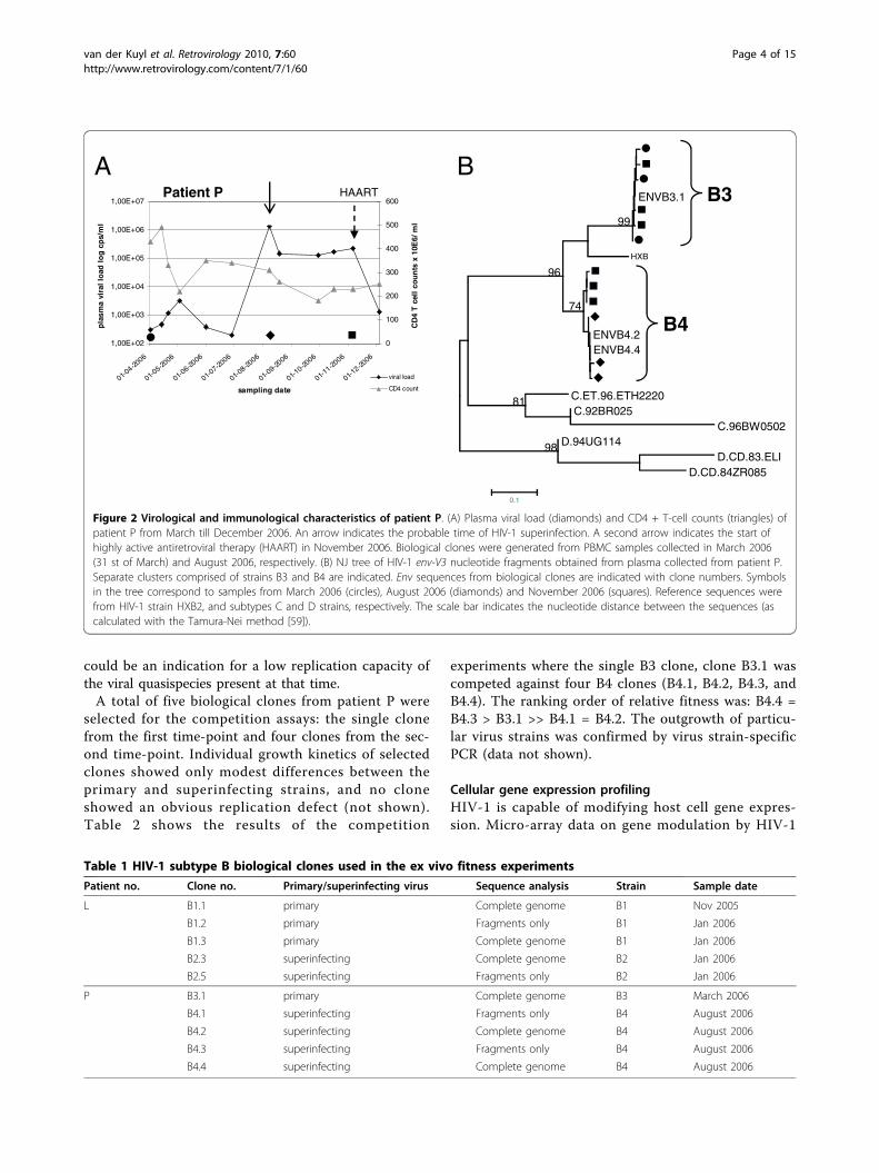
clone compared with B2 viruses and with the HXB2reference sequence (Figure 4A). Moderate insertions inthe LTR are not uncommon in HIV-1 and have beenassociated with disease attenuation [20]. The insertionsin the B1 LTR occur at the type I and type II insertionsites described by Koken et al. [3], but are dissimilar innucleotide sequence. The LTR insertions do not affectthe nef open reading frame. Interestingly, the secondinsertion together with upstream sequences creates anovel NF-�B/NFAT binding site whereas the down-stream common NF-�B/NFAT binding site is hypermu-tated at 4 nucleotides (Figure 4A). The type I insertion inthe B1 LTR is very similar to that described for a virus
with decreased transcriptional activity that was isolatedfrom a long-term non-progressing patient (no. 4) [20].Seven additional B1 biological clones contained identicalLTR sequences, indicating that the insertions in thisregion are not unique to clones B1.1 and B1.3 (result notshown). Clone B2.3 contains a T®C mutation in theTAR region of the LTR that could destabilize the hairpinsecondary structure (Figure 4A). HIV-1 Tat protein acti-vates transcription by binding to the TAR hairpin in theLTR, thereby acting as a potent activator of viral geneexpression. Mutational analysis of four highly conservedaromatic amino acid residues within the Tat activationdomain showed that the F32 L mutation greatly reducedTat activity and virus replication [21]. Interestingly, thisF32 L mutation is present in 15% of the subtype B tatsequences from 2008 [22]. The same mutation is alsopresent in the B1 clones of patient L (Figure 4B), suggest-ing that the B1 strain encodes a Tat protein withdecreased transcription activation capacity. However, theT23 N substitution in strain B1 Tat could possibly com-pensate for the F32 L mutation [23].For patient P, the LTR promoter sequence of the first
infecting virus, strain B3, carried a characteristicTT®CA mutation in the poly A hairpin region(Figure 4A). Such a mutation destabilizes the structureof this hairpin (Figure 5), which may trigger prematurepolyadenylation in the 5′ LTR thus reducing viral geneexpression and replication [24,25]. Analysis of theplasma viral quasispecies at the first time point (whenonly the B3 strain is present) revealed that all 16 HIV-1LTR clones analysed contained the TT®CA substitutionin the LTR (not shown). The Tat protein encoded bythe B3.1 virus clone has 86 amino acid residues, whilethe B4 clones encode a Tat protein of 101 amino acidresidues (Figure 4B). As such a short tat gene was initi-ally observed in laboratory strains, it was suggested thata shorter Tat protein was sufficient only for ex vivo pro-pagation of the virus (reviewed by [26]). A prematurestopcodon at position 86 of the tat gene occursoccasionally in all subtypes, and regularly in almost allsubtype D isolates [22]. In addition, clone B3.1 has an11-codon repeat of the ‘PTAP’ motif at the beginning ofthe gag-p6 protein reading frame that is not present inthe B4 strain (Figure 6A). A sequence repeat of 3-9amino acid residues at this location has been associatedwith low CD4 + T cell counts, drug resistance and poorprognosis [27-29]. Interestingly, gag-p6 PTAP repeatshave linked to the presence of positively charged aminoacid residues at certain positions in the env-V3 loopthat determine co-receptor usage [27]. The 11th positionin the V3 loop of the B3.1 clone encodes the positivelycharged R residue, suggesting CXCR4-usage [30-32], butthe 25th position could not be clearly assigned to acharged amino acid [27,31,32] (Figure 6B). Indeed, clone
Figure 3 PBMC gene expression patterns of HIV-1 biologicalclones. mRNA expression levels in PBMC’s infected with the HIV-1biological clones B1.1, B1.3, B2.5 (patient L) and B3.1, B4.2, and B4.4(patient P), as well as uninfected PBMC’s were analysed with theRT2Profiler™ PCR Array Human Inflammatory Cytokines andReceptors (SABiosciences). Cultures were infected with HIV-1 at anMOI of 0.05. After two hours, the inoculum was removed bycentrifugation. Total RNA was isolated 6 hours after infection.Experiments were performed in triplicate. Expression profiles wereanalysed with the GCNPro™ (Gene Network Central) software [68].Clustering of the gene expression profiles induced by the HIV-1clones is shown for a selection of genes from a representativeexperiment. Green colour indicates increased mRNA expression, redcolour indicates decreased mRNA expression compared to theuninfected PBMC’s.
van der Kuyl et al. Retrovirology 2010, 7:60http://www.retrovirology.com/content/7/1/60
Page 6 of 15
Figure 4 HIV-1 LTR and Tat sequences. (A) Nucleotide sequences of the LTR region from clones B1.1, B1.3, B2.3 (primary and superinfectingstrain from patient L, respectively), and clones B3.1, B4.2, and B4.4 (primary and superinfecting strain from patient P, respectively). Sequenceswere aligned using the HXB2 sequence (GenBank acc. no. K03455) as reference. Binding sites for transcription factors and the two insertionsfound in clone B1.1 (type I and type II) have been boxed. A NF-�B/NFAT binding site immediately followed by an YY1 binding site found only inclone B1.1, are indicated. The TATA-box and the TAR region (nt 504-555) have been underlined. A destabilizing T®C mutation in the TAR hairpinregion in clone B2.3 is boxed. The polyA hairpin (nt 556-602) is shown in bold, a box indicates the destabilizing TT®CA mutation in clone B3.1.(B) Translated amino acid sequences are shown for HIV-1 Tat. Sequences have been aligned with the HXB2 sequence. Clone numbers areindicated. Strains B1 and B2 are the first and superinfecting virus from patient L, respectively. Strains B3 and B4 are the first and superinfectingvirus from patient P, respectively. The Tat T23 N and F32 L mutations in strain B1 associated with increased and decreased Tat activity have beenboxed.
van der Kuyl et al. Retrovirology 2010, 7:60http://www.retrovirology.com/content/7/1/60
Page 7 of 15

B3.1 infected MT-2 cell cultures with induction of syn-cytia, indicative of CXCR4 use (result not shown). Wewere, however, unable to infect U87.CD4 cells expres-sing either CXCR4 or CCR5 [33] with this clone. TheV3-loop of clone B3.1 has remarkable similarity to thatof subtype D virus UG21 that can use the APJ andCCR9 receptor in addition to CXCR4 [34], suggesting itcould be different from common CXCR4 using strains,and possibly have less affinity for U87.CXCR4 cells. Thesecondary virus strain B4 of patient P was predicted touse the CCR5 coreceptor, as were both primary and sec-ondary strains of patient L, but this was not tested inculture. Analysis of viral RNA present in blood plasmaat the first time point confirmed that the env-V3sequence of clone B3.1 is present in all viral genomesanalysed (result not shown). No apparent escape muta-tions were seen in Gag epitopes defined by the patientsHLA type, suggestive of low CTL pressure.
Another intriguing finding is the difference in replica-tion capacity of clones B1.1 and B1.3, where the latterclone exhibits a substantial ex vivo replication disadvan-tage in competition experiments. Yet relatively littlesequence variation was found that could account forthis. A single amino acid difference was noted in theVpu and Rev proteins, as well as 8 amino acid differ-ences in Env (3 in gp120, 5 in gp41). The genetic differ-ence between clones B4.2 and B4.4, of which the formerclone has a replicative disadvantage, was also modest. Inaddition to a single amino acid difference in Vif and onein Vpu, two amino acid changes were found in the envgene, one in the signal peptide and one in the env-V5domain, respectively. Also, an extra glycosylation sitewas present in the env-V4 region of clone B4.4. TheHIV quasispecies in a host consist of many closelyrelated variants, and (modest) differences in replicationcapacity are to be expected. Replication curves of single
Figure 5 Structure of the LTR polyA hairpin. Predicted structure of the LTR polyA hairpin region of the HIV-1 reference strain HXB2 andclones B3.1, B4.2 and B4.4. The free energies of the stem-loop structures were calculated with the Zuker algorithm as available at the mfoldwebserver for nucleic acid folding and hybridization prediction [69], the ΔG values are presented in kilocalories per mole. A box indicates theUU®CA change in clone B3.1.
van der Kuyl et al. Retrovirology 2010, 7:60http://www.retrovirology.com/content/7/1/60
Page 8 of 15

clones, e.g. B4.2 and B4.4, did not show significant dif-ferences in replication when cultured alone (not shown).However, competition experiments can expose andenlarge relatively small differences in replication capacity[3]. Therefore, that two out of four B4 clones lost incompetition experiments from the same clone, whiletwo other B4 clones won the competition does notrepresent evidence that the former B4 clones are largelydeficient in replicative capacity.
LTR promoter activityThe promoter activity of the LTR region of the primaryand superinfecting HIV strains was analysed by cloninga fragment corresponding to nt 2-536 of the HXB2 gen-ome before the luciferase gene and subsequently mea-suring luciferase activity in the presence of increasingamounts of Tat (Figure 7). There is no obvious differ-ence between the LTRs from the B1 and B2 strain frompatient L in the human embryonic kidney cell line used,despite the occurrence of insertions in the B1 LTR.However, the LTR from the primary virus B3 frompatient P has a lower promoter activity than the LTRfrom the superinfecting virus B4 in these cells, despitethe absence of noticeable sequence variation. Using dif-ferent cell types and/or activating the promoters withhomologous Tat protein instead of HIV(LAI) Tat couldinfluence the results, as promoter activity has not onlybeen shown to be cell-type specific, but there might also
be co-evolution between the LTR and tat gene of a par-ticular HIV strain. For example, it would be very infor-mative to analyse LTR activity in PMA and/orionomycin stimulated cells, preferentially in a T-cellline, to determine the true effect of NF-�B and NFATupon transcription.
Figure 6 HIV-1 Gag p1-p6 and Env-V3 sequences. Translated amino acid sequences are shown for HIV-1 Gag p1-p6 region (panel A), andenv-V3 (panel B). Sequences were aligned with the HXB2 reference sequence. Clone numbers are indicated. Strains B1 and B2 are the first andsuperinfecting virus from patient L, respectively. Strains B3 and B4 are the first and superinfecting virus from patient P, respectively. The 11 aaPTAPP repeat in clone B3.1 in Gag-p6 has been boxed. The 11th and 25th amino acid residues in Env-V3, associated with CXCR4 coreceptor usewhen positively charged, are indicated.
0
2
4
6
8
10pGL3-basicB(LAI)B1B2B3B4
pg Tat
Rat
io F
iref
ly/R
enill
a
Figure 7 Transcriptional activity of the LTR promotersequences. Transcriptional activity of the HIV-1 LTR promotersequences from HIV strains B(LAI), B1, B2, B3, and B4, comparedwith the empty vector (pGL3-basic) in a dual firefly/renilla luciferaseassay. LTR fragments cloned from clones B1.1/B1.3 and B4.2/B4.4 areidentical in sequence, so only strain names are indicated.Transcriptional activity of the luciferase gene was tested in thepresence of increasing concentrations of Tat. The value is theaverage of three independent measurements; standard deviationsare indicated.
van der Kuyl et al. Retrovirology 2010, 7:60http://www.retrovirology.com/content/7/1/60
Page 9 of 15

DiscussionIn the present study the relative fitness of viral strainsinvolved in two HIV-1 superinfection cases was ana-lysed. Patients L and P were identified to have experi-enced an HIV-1 superinfection within half a year fromthe seroconversion date by a sudden unexpected rise inthe plasma viral load [35]. Both virus strains in the twopatients are supposedly “wild-type” viruses, meaningthat no drug-resistance mutations in pol or deletions orpremature stopcodons in the nef gene were found.Therefore, these HIV-1 strains are well suited to test thehypothesis that productive superinfection requires a sec-ond virus with a higher relative fitness than the primaryinfecting strain [13,14]. The presence of an initial virusstrain with drug-resistance mutations, likely causing areplication disadvantage, has been reported repeatedly[15-17].To estimate the relative fitness of the virus variants
involved in two HIV-1 superinfections, replication com-petent viruses were obtained by biological cloning. Aninitial genome analysis was performed by PCR amplifica-tion of gene fragments and subsequent sequence analy-sis. For patient L, around 200 clones were obtained inline with the relatively high plasma viral load at thetime points sampled (> 105 copies/ml). Strain B1 cloneswere isolated before and after the superinfectionmoment, strain B2 only after superinfection. Both the invitro competition experiments with multiple pairs of B1and B2 clones and the ratio of the two strains in bloodplasma samples indicated that the second strain B2 isthe better replicating strain, in line with the hypothesisthat a more virulent strain can infect a host that isalready infected with a less virulent strain [13,14]. Forpatient P the situation turned out to be more complex.A major restriction is that only a single clone of theinitial B3 virus was obtained. This is probably due tothe extremely low viral load. In fact, the viral loadremains low for many months before superinfectionoccurs. In blood plasma, gag and env-V3 fragments fromthe B3 strain could not be amplified from all samples,confirming low copy numbers of this strain. In contrast,strain B4 sequences were abundantly present in plasmasamples taken after the superinfection moment. Thisobservation, together with the sustained increasedplasma viral load, suggests a significantly higher level ofreplication of the second strain. In ex vivo experiments,the single and possibly unusual B3 clone was able tooutgrow two B4 clones in the replication assays,although it appeared less fit than two other B4 clones.Probably, the single B3 clone isolated is one of the bet-ter replicating variants of the quasispecies, and thus notfully representative of the B3 quasispecies of patient Pin vivo. In a luciferase assay using human embryonic
kidney cells, the B3.1 LTR was less active as a promoterthan an LTR from the B4 strain, which could suggestthat this could also be the case in the various cell typesinfected in vivo. Alternatively, strain B3 may have anaverage replication capacity, but is severely suppressedin vivo by the immune system resulting in the lowplasma viral RNA levels observed. A second strain couldexperience less immune pressure such that it can repli-cate to higher levels [17]. However, no primary or sec-ondary clone possessed escape mutations in the majorgag or nef epitopes targeted by the HLA-A25, -B18 or-B44 alleles carried by the patient [36-40], thus suggest-ing ineffective cytotoxic T cell responses (result notshown). Both the env-V3 sequence and culture experi-ments using MT2 cells suggested that clone B3.1 usesthe CXCR4 coreceptor, although patient P does notcarry CCR5-Δ32 deletion alleles (not shown). Primaryinfections with CXCR4-using viruses are not unusual, asthought earlier, although they are usually negativelyselected during primary infection. Over 15% of patientswith a primary HIV infection in two European cohortswere infected with CXCR4-using strains [41,42].CXCR4-using viruses are not necessarily more fit thanCCR5-using viruses. Competition experiments with bio-logical clones showed that the average fitness ofCXCR4- and CCR5-using viruses is similar [43]. In con-clusion, the combined data suggest that overall thesuperinfecting virus in patient P is also a better replicat-ing strain than the primary virus.HIV-1 superinfection has been associated with disease
progression, as exemplified by a permanent rise in theplasma viral load and an accelerated decrease in CD4 +T cell numbers (reviewed in [44]). Mathematical model-ling suggests that, except for the direct negative effect ofaccelerated disease progression, co- and super- infec-tions can also have an impact on the virus as a speciesin the epidemic, triggering an increased replicationcapacity and possibly virulence of the pathogen [14]. Invitro experiments with vesicular stomatitis virus (VSV)show that the progeny of co- and super- infections, havea higher fitness than that of single infections as the dualinfections allow for faster adaptation by to environmen-tal changes [45]. Low viral fitness, measured as replica-tive capacity, is associated with lower virulence, e.g. innef-deleted HIV-1 or drug-resistant HIV-1 variants[46,47]. Studies on HIV-1 fitness and evolution havebeen contradictory. A initial study suggested attenuationof HIV-1 over time in Belgium [7], but other studiesreported increasing fitness of HIV-1 in The Netherlandsin the period 1986-2003 [8,9] and in France in 1997-2005 [10]. A fourth study indicated that HIV-1 virulenceis not changing over time in North America [11]. AsHIV-1 co- and super- infections are much more
van der Kuyl et al. Retrovirology 2010, 7:60http://www.retrovirology.com/content/7/1/60
Page 10 of 15

prevalent in Africa (reviewed in [44]), it will be of inter-est to study the evolution of viral fitness in this setting.
ConclusionsThe results obtained from two HIV-1 superinfectioncases suggest that an HIV-1 re-infection that gives riseto a systemic superinfection is facilitated by a primaryinfection with a less fit strain that has a lower replica-tion capacity than the superinfecting strain. It remainsimportant to examine the replication capacity of virusesfrom other patients with an HIV-1 superinfection to seeif the suggestion of a better replicating second virus canbe confirmed.
Materials and methodsPatientsTwo HIV-1 positive patients, L and P, were found tohave an HIV-1 superinfection in an earlier study analys-ing sudden plasma viral load rises in patients followedat the Academic Medical Center in Amsterdam, TheNetherlands [35]. Both individuals initially presentedwith primary HIV-1 subtype B infections; patient L withFiebig stage II (vRNA+ ) and patient P with Fiebig stageV (Western blot+ p31-) [48]. Both experienced an HIV-1superinfection with another subtype B strain within halfa year from their first presentation. The patients partici-pated in the Amsterdam Cohort Studies (ACS) on HIVinfection and AIDS among homosexual men. Writteninformed consent was obtained. HLA-typing of ACSpatients with a primary HIV-1 infection is routinely per-formed at Sanquin Diagnostiek (Amsterdam, The Neth-erlands). Patient L: HLA class I: A2, A24(9), B27, B60(40), Cw 1 and 7; HLA class II: DR1, DR12(5), DR52,DQ5(1), DQ7(3). Patient P: HLA class I: A2, A25(10),B44(12), B18, Cw5; HLA class II: DR15(2), DR12(5),DR51, DR52, DQ1 and DQ3. HIV-1 blood plasma viralload measurements were done at the Laboratory of Clin-ical Virology at the AMC (Amsterdam, The Nether-lands) with the Versant HIV-1 RNA 3.0 assay (BayerDiagnostics Division Tarrytown, N.Y.).
Cloning and sequencing of molecular clones from plasmaRNA was isolated from plasma samples from bothpatients with a method using silica and guanidiumthiocyanate [49]. HIV-1 env-V3 and gag fragmentswere reverse transcribed, amplified, cloned with theTOPO TA cloning kit (Invitrogen, Carlsbad, Calif.),and sequenced with the BigDye Terminator cyclesequencing kit (Applied Biosystems, Foster City, Calif.)as described [50,51]. Electrophoresis and data collec-tion were performed on an ABI PRISM 3100 geneticanalyser (also from Applied Biosystems). At least 16clones were analysed for each patient per time pointand per strain.
Generation of biological clonesFreshly phytohemagglutinin (bioTRADING Benelux,Mijdrecht, The Netherlands), glutamax and interleukin-2 (Proleukin, Chiron, Emeryville, Calif.) stimulated per-ipheral blood mononuclear cells (PBMC’s), obtainedfrom four healthy (HIV-1 negative) human donors, werecombined and cultured in RPMI 1640 medium (Invitro-gen Corporation, Carlsbad, Calif.) supplemented withantibiotics, L-glutamine and 15% heat-inactivated foetalcalf serum for 3 days. CD8 + T cells were depleted after2 days using the Dynabeads M-450 CD8 kit (InvitrogenCorporation, Carlsbad, Calif.). Different concentrationsof PBMC’s from the HIV-1 infected patient (104, 2.5 ×10⁴, 4 × 10⁴ 6 × 10⁴ cells/well) were cocultivated with 1× 10⁶ CD4 + T cells in the same medium in 96-wellsplates for 21 and 28 days, respectively. Each 7 days cul-ture supernatants were tested for the presence of p24with an in-house antigen capture enzyme-linked immu-nosorbent assay (ELISA). At the same time, to propagatethe culture, one-third of the cell culture was transferredto new 96-well plates and fresh PHA, IL-2 stimulatedCD4 + T cells were added. Viruses were considered tobe clonal if less than one-third of the microculturesbecame positive at a given cell number (Poisson distri-bution). HIV-1 clones were expanded by culturing andharvested after 7 days [52]. PBMC’s and supernatantwere cryopreserved at -150°C [53].
Analysis of gag, env, vpr and nefTo characterize the biological clones, a 804 nucleotidefragment of the gag gene, encompassing the entire p17gene and the 5′ part of the p24 gene, and a 264 nucleo-tide HIV-1 V3 fragment of the env gene were amplifiedby PCR as previously described [50,51,54]. The completevpr gene of the biological clones was amplified andsequenced [55]. The nef gene was amplified using 5′End-env-s primer (5′TAG AAG AAT AAG ACA GGGCTT GG3′) and R-LTR3′FM (5′AGA CCC AGT ACAGGC AAA AAG CAG CTG CTT ATA3′) using Ampli-taq (Perkin Elmer 5 units/μl) in a final concentration of1.8 mM MgCl2. Amplification was done for 40 cycles,with each cycle involving three steps: 1 min at 95°C, 1min at 55°C, and 2 min at 72°C, plus a final extensionof 10 min at 72°C.
Full genome sequencingThe complete genomes of three biological clones foreach patient were sequenced (for patient L clones: B1.1,B1.3, and B2.3, for patient P clones: B3.1, B4.2 and B4.4;Table 1). Overlapping fragments of about 600-1000 bpin length, spanning the entire genome, were amplifiedwith different primer sets and sequenced. Sequenceswere compiled with CodonCode Aligner version 2.0.3[56]. Differences between the primary and superinfecting
van der Kuyl et al. Retrovirology 2010, 7:60http://www.retrovirology.com/content/7/1/60
Page 11 of 15

strain, represented by clones B1.1 and B2.3 from patientL and clones B3.1 and B4.2 from patient P is discussedin the Results section. Differences observed betweenclones of the same strain (B1.1 vs. B1.3 and B4.2 vs.B4.4) are discussed in the Results section as well.
Phylogenetic analysisSequences were aligned with the CLUSTAL W sequencealignment tool implemented in BioEdit Sequence Align-ment Editor Version 7.0.9 [57]. Reference sequenceswere obtained from the Los Alamos HIV sequence data-base [22]. The alignments were manually adjusted topreserve in-frame insertions and deletions. Phylogeneticanalyses were performed with the MEGA4 softwarepackage distributed by Sudhir Kumar, Arizona StateUniversity, Tempe [58]. Distances were estimated withthe Tamura-Nei method [59], using the gamma modelto correct for multiple hits and to account for excesstransitions, unequal nucleotide frequencies, and varia-tion of substitution rate among different sites. For theshape parameter alpha, which describes the variationacross sites by a gamma distribution, we used a = 0.38for env-V3 and a = 0.25 for gag [60]. Phylogenetic treeswere generated with the neighbour-joining method andbootstrap resampling with 1000 replicates. Phylogeneticanalyses of vpr and vpu genes were performed in a simi-lar fashion.Transcription factor binding sites were identified in
the long terminal repeat (LTR) sequences withTFSEARCH [61] and Alibaba 2.1 [62].
Virus replication assaysReplication kinetics of biological clones was measured byinfecting pooled PHA/IL-2 stimulated PBMC’s from atleast four healthy donors. Similar amounts of p24 wereused for each viral strain. After 4 hours of infection, theinoculum was removed by centrifugation. PBMC’s wereresuspended in complete medium and cultured for 16days. Virus production was analysed with an in-housep24 antigen capture enzyme-linked immunosorbentassay (ELISA) at days 0, 2, 6, 9, 13 and 16.
Growth competition assaysDifferent HIV-1 biological clones generated as describedabove were selected based on their env-V3 sequence andthe replicative fitness was assessed in growth competi-tion assays. The infectivity of each virus was determinedwith the Reed and Muench method which yields the tis-sue culture dose for 50% infectivity (TCID50) [63].Briefly, each stock of biological clone was serially dilutedin quadruple and then plated with 2 × 10⁵ CD4 + Tcells in a 96-well plate. Virus production was tested ineach well with an in-house p24 antigen capture enzyme-linked immunosorbent assay (ELISA).
The growth competition assays were performed inPHA/IL-2 activated PBMC’s as described previously[4,8]. Pooled PBMC’s from at least four healthy donorswere infected with two different viruses at equal multi-plicity of infection (0.0005 MOI). Uninfected cultureswere used as HIV-1-negative controls and monoinfectedcell cultures of each virus provided the positive controls.Virus mixtures were incubated with 2 × 10⁵ PBMC’s at37°C in 5% CO2 for 25 hours, then washed three timeswith 1 × phosphate buffered saline (PBS) and thenresuspended in complete medium [4,7,8,43]. Cell-freesupernatant was tested for p24 antigen detection 7 dayspostinfection with an in-house ELISA [64]. Two aliquotsof supernatant and cells were harvested at day 7 afterinfection and stored at -80°C for further analysis.
HTA analysis of dual infectionsThe viral DNA of all dual-infected and mono-infectedcultures was extracted from lysed cells with theQIAamp DNA Blood Mini kit (QIAGEN Inc., ValenciaCalif.). HIV-1 DNA was amplified by PCR using a setof external primers (ED14: 5′-TCTTGCCTGGAGCTGTTTGATGCCCCAGAC-3′ and EnvB: 5′-AGAAA-GAGC AGAAGACAGTGGCAATGA-3′), followed bynested primers (E125 5′-CAATTTCTGGGTCCCCTCCTGAGG-3′ and E80 5′-CCAATTCCCATACAT-TATTGTG-3′) [65]. Both the external and nestedPCRs were carried out in a 100 μl reaction mixtureunder defined cycling conditions [4,5]. The nested pro-ducts from env (C3 V3) were analyzed with a hetero-duplex tracking assay (HTA) to determine thecomposition of the virus mixture in the competitionexperiments as described earlier [4,5,7,43]. The radiola-belled DNA probes were amplified from the env regionusing the set of primers described above. For this PCRreaction, one of the primers was labelled with 2 μCi of[g-32 P] ATP using T4 polynucleotide kinase (PKN,Roche Belgium) [4]. Subsequently, labelled probes wereseparated on 1% agarose gel and purified with theQIAquick gel extraction kit (QIAGEN Benelux, Venlo,The Netherlands). The HTA reaction mixtures con-taining DNA annealing buffer (100 mM NaCl, 10 mMTris-HCl pH 7.8, 2 mM EDTA), 10 μl of amplifiedDNA from the dual infection/competition culture and0.1 pmol of radioactive probe were denatured at 95°Cfor 3 min, incubated at 37°C for 5 min and trans-ferred on wet ice for re-annealing. DNA heterodu-plexes were resolved on Criterion 5% TBEnon-denaturated polyacrylamide gels (BIORAD Bel-gium) for 75 min at 200 V. Gels were dried at 80°Cfor 45 min, exposed and scanned with a phosphorimager (Cyclone, Perkin Elmer Inc., Boston, Mass.)and analyzed with the PerkinElmer OptiQuant soft-ware package [5].
van der Kuyl et al. Retrovirology 2010, 7:60http://www.retrovirology.com/content/7/1/60
Page 12 of 15

Viral fitness calculationsIn the competition experiments, the ratio of two virusesproduced in a dual infection was analysed with HTAand compared to the monoinfections [5,65]. The pro-duction of individual HIV clones in a dual infection (fo)was divided by the initial proportion in the inoculum(io). This quotient is referred to as relative fitness (W =fo /io). The ratio of the relative fitness values of eachHIV variant in the competition is a measure of the fit-ness difference (WD) or ratio between two HIV strains(WD = WM/WL), where WM corresponds to the relativefitness of more fit virus and WL corresponds to the rela-tive fitness of less fit virus [4].
Strain-specific PCRThe results of the competition experiments were con-firmed with a virus strain-specific PCR. For this purpose, aspecific nested PCR primer set was designed for each virusstrain. Primers were located in different parts of env-V3 orgag; amplified nested PCR products have a distinct lengthfor each strain. The strain specific primer sets were alsoused for detecting viruses in plasma samples. The amountof DNA was estimated by comparing the intensity of thebands on agarose gels using TINA version 2.09 g.
Gene expression profileEarly gene expression events after HIV-1 infection ofPBMC’s were assessed with the RT2Profiler™ PCR Array:Human Inflammatory Cytokines and Receptors (SABios-ciences, Frederick, MD, USA). Virus stocks were avail-able for the following clones: B1.1, B1.3, B2.5, B3.1, B4.2and B4.4. For each virus 4.105 PBMC’s, isolated andpooled from 4 different healthy donors, were infectedwith a MOI of 0.05. Cells were incubated for 2 hours,spun down, and resuspended in RPMI 1640 medium(Invitrogen Corporation, Carlsbad, Calif.) supplementedwith L-glutamine, 10% heat-inactivated foetal calf serumand IL-2, and cultured for an additional 4 hours. TotalRNA was isolated from the PBMC cultures with theRNeasy® Mini Kit (QIAGEN Benelux B.V., Venlo, TheNetherlands). cDNA was synthesized with the RT2 FirstStrand Kit (SABiosciences, Frederick MD, USA). Real-time PCR reactions were done with the RT2 SYBRGreen/ROX qPCR Master Mix (SABiosciences, Freder-ick, MD, USA), and were analysed with a Taqman 7000system (ABI, Foster City, CA, USA). Results were com-piled with a program available from SABiosciences.
LTR-constructs and luciferase-assaysThe LTR region of the viral genome from the biologicalclones was amplified by nested PCR, amplifying nt 2-560 of the HXB2 genome [GenBank: K03455]. Theouter primer set is located in the nef gene and in theU5 region of the LTR. The 5′nested primer contains a
Kpn I site and is located upstream of the U3 region. Pri-mers were specially designed to amplify LTR’s fromclones B1-B4. PCR products were digested with Kpn Iand Hind III (a Hind III site is present at position 531-536 in the LTR of HIV (HXB2 numbering) and clonedinto the pGL3-Basic vector (Promega, Madison, WI).Because a Kpn I site is already present in the clone B3.1LTR sequence, a nested primer with an added Mlu Isite was used to construct B3.1 LTR-luciferase con-structs. Constructs were verified by sequencing.Human Embryonic Kidney cells 293 T (HEK-293 T
cells) were used in all luciferase experiments. Cells weregrown at 37°C in Dulbecco’s Modified Eagle Mediumwith 10% Fetal Calf Serum under 5% CO2 and transfectedusing nanofectin (PAA Laboratories GmbH, Pasching,Austria). Mixtures contained 2 ng of different LTR-luci-ferase constructs (B1, B2, B3, B4 and B(LAI)), 1 ng ofpRL-CMV plasmid (Promega, Madison, WI) expressingRenilla luciferase as an internal control for transfectionefficiency [66], and pBluescript in such a concentrationthat the total amount of DNA would always be 200 ng.To test the activation of the promoters by tat, constructswere titrated with different concentrations of a tat-expressing plasmid (pTAT). Cells were cultured for twodays and lysed in Passive Lysis Buffer (Promega, Madi-son, WI). Firefly and Renilla luciferase activities weredetermined with the dual-luciferase reporter assay (Pro-mega, Madison, WI) as described previously [66]. Theactivity of different constructs was calculated as the ratioof the firefly and Renilla luciferase activities, andcorrected for between-session variation [67].
AcknowledgementsWe thank Dr Marjan Steenvoorden for help with virus culture and StephanHeynen for performing the CA-p24 ELISA assay. The Amsterdam CohortStudies on HIV infection and AIDS, a collaboration between the AmsterdamHealth Service, the Academic Medical Center of the University ofAmsterdam, Sanquin Blood Supply Foundation and the University MedicalCenter Utrecht, are part of the Netherlands HIV Monitoring Foundation andfinancially supported by the Netherlands National Institute for Public Healthand the Environment. Part of this work was sponsored by a grant from theFWO (1.5.028.08). We also thank the Belgian Federal Government forfinancial support through the Inter-University Attraction Pole grant n° P6/41.
Author details1Laboratory of Experimental Virology, Department of Medical Microbiology,Centre for Infection and Immunity Amsterdam (CINIMA), Academic MedicalCentre of the University of Amsterdam, Meibergdreef 15, 1105 AZAmsterdam, The Netherlands. 2Department of Microbiology, Virology Unit,Institute of Tropical Medicine, Nationalestraat 155, B-2000 Antwerp, Belgium.3Department of Clinical Chemistry, Microbiology and Immunology, Faculty ofMedicine and Health Sciences, Ghent University, De Pintelaan 185, B-9000Gent, Belgium. 4Faculty of Pharmaceutical, Biomedical and VeterinarySciences, University of Antwerp and Faculty of Medical and PharmaceuticalSciences, University of Brussels, Belgium. 5Prosensa BV, Leiden, TheNetherlands.
Authors’ contributionsMC conceived of the study and designed the experiments. MC and ACKanalysed and interpreted the results. ACK drafted the manuscript, and BB
van der Kuyl et al. Retrovirology 2010, 7:60http://www.retrovirology.com/content/7/1/60
Page 13 of 15

critically revised it. KK, KKA, YG and GV performed the competitionexperiments. FK sequenced the patient materials, did replication experimentsand performed the microarray analysis. FK, VRB and KK sequenced thebiological clones. SJD cloned the LTR’s and performed the luciferase assays.All authors have seen and approved the final version of the manuscript.
Competing interestsThe authors declare that they have no competing interests.
Received: 25 May 2010 Accepted: 20 July 2010 Published: 20 July 2010
References1. van Opijnen T, Berkhout B: The host environment drives HIV-1 fitness. Rev
Med Virol 2005, 15:219-233.2. Quinones-Mateu ME, Arts EJ: Virus fitness: concept, quantification, and
application to HIV population dynamics. Curr Top Microbiol Immunol 2006,299:83-140.
3. Koken SE, van Wamel JL, Goudsmit J, Berkhout B, Geelen JL: Naturalvariants of the HIV-1 long terminal repeat: analysis of promoters withduplicated DNA regulatory motifs. Virology 1992, 191:968-972.
4. Arien KK, Abraha A, Quinones-Mateu ME, Kestens L, Vanham G, Arts EJ: Thereplicative fitness of primary human immunodeficiency virus type 1(HIV-1) group M, HIV-1 group O, and HIV-2 isolates. J Virol 2005,79:8979-8990.
5. Quinones-Mateu ME, Ball SC, Marozsan AJ, Torre VS, Albright JL, Vanham G,van Der GG, Colebunders RL, Arts EJ: A dual infection/competition assayshows a correlation between ex vivo human immunodeficiency virustype 1 fitness and disease progression. J Virol 2000, 74:9222-9233.
6. Troyer RM, Collins KR, Abraha A, Fraundorf E, Moore DM, Krizan RW,Toossi Z, Colebunders RL, Jensen MA, Mullins JI, Vanham G, Arts EJ:Changes in human immunodeficiency virus type 1 fitness and geneticdiversity during disease progression. J Virol 2005, 79:9006-9018.
7. Arien KK, Troyer RM, Gali Y, Colebunders RL, Arts EJ, Vanham G: Replicativefitness of historical and recent HIV-1 isolates suggests HIV-1 attenuationover time. AIDS 2005, 19:1555-1564.
8. Gali Y, Berkhout B, Vanham G, Bakker M, Back NK, Arien KK: Survey of thetemporal changes in HIV-1 replicative fitness in the Amsterdam Cohort.Virology 2007, 364:140-146.
9. Gras L, Jurriaans S, Bakker M, van SA, Bezemer D, Fraser C, Lange J, Prins JM,Berkhout B, de Wolf F: Viral load levels measured at set-point have risenover the last decade of the HIV epidemic in the Netherlands. PLoS ONE2009, 4:e7365.
10. Potard V, Weiss L, Lamontagne F, Rouveix E, Beck-Wirth G, Drogoul-Vey MP,Souala MF, Costagliola D: Trends in post-infection CD4 cell counts andplasma HIV-1 RNA levels in HIV-1-infected patients in France between1997 and 2005. J Acquir Immune Defic Syndr 2009, 52:422-426.
11. Herbeck JT, Gottlieb GS, Li X, Hu Z, Detels R, Phair J, Rinaldo C,Jacobson LP, Margolick JB, Mullins JI: Lack of evidence for changingvirulence of HIV-1 in North America. PLoS ONE 2008, 3:e1525.
12. Jost S, Bernard MC, Kaiser L, Yerly S, Hirschel B, Samri A, Autran B, Goh LE,Perrin L: A patient with HIV-1 superinfection. N Engl J Med 2002,347:731-736.
13. May RM, Nowak MA: Superinfection, metapopulation dynamics, and theevolution of diversity. J Theor Biol 1994, 170:95-114.
14. Nowak MA, May RM: Superinfection and the evolution of parasitevirulence. Proc Biol Sci 1994, 255:81-89.
15. Koelsch KK, Smith DM, Little SJ, Ignacio CC, Macaranas TR, Brown AJ,Petropoulos CJ, Richman DD, Wong JK: Clade B HIV-1 superinfection withwild-type virus after primary infection with drug-resistant clade B virus.AIDS 2003, 17:F11-F16.
16. Smith DM, Wong JK, Hightower GK, Ignacio CC, Koelsch KK, Petropoulos CJ,Richman DD, Little SJ: HIV drug resistance acquired throughsuperinfection. AIDS 2005, 19:1251-1256.
17. Yang OO, Daar ES, Jamieson BD, Balamurugan A, Smith DM, Pitt JA,Petropoulos CJ, Richman DD, Little SJ, Brown AJ: Humanimmunodeficiency virus type 1 clade B superinfection: evidence fordifferential immune containment of distinct clade B strains. J Virol 2005,79:860-868.
18. The Stanford University HIV drug resistance database. [http://hivdb.stanford.edu].
19. Giri MS, Nebozhyn M, Showe L, Montaner LJ: Microarray data on genemodulation by HIV-1 in immune cells: 2000-2006. J Leukoc Biol 2006,80:1031-1043.
20. Zhang L, Huang Y, Yuan H, Chen BK, Ip J, Ho DD: Genotypic andphenotypic characterization of long terminal repeat sequences fromlong-term survivors of human immunodeficiency virus type 1 infection.J Virol 1997, 71:5608-5613.
21. Verhoef K, Koper M, Berkhout B: Determination of the minimal amount ofTat activity required for human immunodeficiency virus type 1replication. Virology 1997, 237:228-236.
22. Los Alamos National Laboratory HIV Databases. [http://hiv-web.lanl.gov].23. Reza SM, Shen LM, Mukhopadhyay R, Rosetti M, Pe’ery T, Mathews MB: A
naturally occurring substitution in human immunodeficiency virus Tatincreases expression of the viral genome. J Virol 2003, 77:8602-8606.
24. Das AT, Klaver B, Berkhout B: A hairpin structure in the R region of thehuman immunodeficiency virus type 1 RNA genome is instrumental inpolyadenylation site selection. J Virol 1999, 73:81-91.
25. Klasens BI, Das AT, Berkhout B: Inhibition of polyadenylation by stableRNA secondary structure. Nucleic Acids Res 1998, 26:1870-1876.
26. Jeang KT, Xiao H, Rich EA: Multifaceted activities of the HIV-1transactivator of transcription, Tat. J Biol Chem 1999, 274:28837-28840.
27. Brumme ZL, Chan KJ, Dong WW, Wynhoven B, Mo T, Hogg RS,Montaner JS, O’Shaughnessy MV, Harrigan PR: Prevalence and clinicalimplications of insertions in the HIV-1 p6 Gag N-terminal region in drug-naive individuals initiating antiretroviral therapy. Antivir Ther 2003,8:91-96.
28. Ibe S, Shibata N, Utsumi M, Kaneda T: Selection of humanimmunodeficiency virus type 1 variants with an insertion mutation inthe p6(gag) and p6(pol) genes under highly active antiretroviral therapy.Microbiol Immunol 2003, 47:71-79.
29. Cao J, McNevin J, McSweyn M, Liu Y, Mullins JI, McElrath MJ: Novelcytotoxic T-lymphocyte escape mutation by a three-amino-acid insertionin the human immunodeficiency virus type 1 p6 Pol and p6 Gag latedomain associated with drug resistance. J Virol 2008, 82:495-502.
30. Raymond S, Delobel P, Mavigner M, Cazabat M, Souyris C, Sandres-Saune K,Cuzin L, Marchou B, Massip P, Izopet J: Correlation between genotypicpredictions based on V3 sequences and phenotypic determination ofHIV-1 tropism. AIDS 2008, 22:F11-F16.
31. Fouchier RA, Groenink M, Kootstra NA, Tersmette M, Huisman HG,Miedema F, Schuitemaker H: Phenotype-associated sequence variation inthe third variable domain of the human immunodeficiency virus type 1gp120 molecule. J Virol 1992, 66:3183-3187.
32. Fouchier RA, Brouwer M, Broersen SM, Schuitemaker H: Simpledetermination of human immunodeficiency virus type 1 syncytium-inducing V3 genotype by PCR. J Clin Microbiol 1995, 33:906-911.
33. Bjorndal A, Deng H, Jansson M, Fiore JR, Colognesi C, Karlsson A, Albert J,Scarlatti G, Littman DR, Fenyo EM: Coreceptor usage of primary humanimmunodeficiency virus type 1 isolates varies according to biologicalphenotype. J Virol 1997, 71:7478-7487.
34. Choe H, Farzan M, Konkel M, Martin K, Sun Y, Marcon L, Cayabyab M,Berman M, Dorf ME, Gerard N, Gerard C, Sodroski J: The orphan seven-transmembrane receptor apj supports the entry of primary T-cell-line-tropic and dualtropic human immunodeficiency virus type 1. J Virol 1998,72:6113-6118.
35. Jurriaans S, Kozaczynska K, Zorgdrager F, Steingrover R, Prins JM, van derKuyl AC, Cornelissen M: A sudden rise in viral load is infrequentlyassociated with HIV-1 superinfection. JAIDS 2008, 47:69-73.
36. van Baalen CA, Klein MR, Huisman RC, Dings ME, Kerkhof G Sr, Geretti AM,Gruters R, van Els CA, Miedema F, Osterhaus AD: Fine-specificity ofcytotoxic T lymphocytes which recognize conserved epitopes of theGag protein of human immunodeficiency virus type 1. J Gen Virol 1996,77(Pt 8):1659-1665.
37. Klenerman P, Luzzi G, McIntyre K, Phillips R, McMichael A: Identification ofa novel HLA-A25-restricted epitope in a conserved region of p24 gag(positions 71-80). AIDS 1996, 10:348-350.
38. Kurane I, West K, Tuazon CU, Zeng W, Ennis FA: Definition of two newepitopes on human immunodeficiency virus type 1 gag proteinrecognized by human CD8 + cytotoxic T lymphocyte clones. J Clin Virol2003, 27:38-43.
van der Kuyl et al. Retrovirology 2010, 7:60http://www.retrovirology.com/content/7/1/60
Page 14 of 15

39. Rowland-Jones SL, Dong T, Fowke KR, Kimani J, Krausa P, Newell H,Blanchard T, Ariyoshi K, Oyugi J, Ngugi E, Bwayo J, MacDonald KS,McMichael AJ, Plummer FA: Cytotoxic T cell responses to multipleconserved HIV epitopes in HIV-resistant prostitutes in Nairobi. J ClinInvest 1998, 102:1758-1765.
40. Couillin I, Culmann-Penciolelli B, Gomard E, Choppin J, Levy JP, Guillet JG,Saragosti S: Impaired cytotoxic T lymphocyte recognition due to geneticvariations in the main immunogenic region of the humanimmunodeficiency virus 1 NEF protein. J Exp Med 1994, 180:1129-1134.
41. de MC, Rodriguez C, Garcia F, Eiros JM, Ruiz L, Caballero E, Aguilera A,Leiva P, Colomina J, Gutierrez F, del RJ, Aguero J, Soriano V: Prevalence ofX4 tropic viruses in patients recently infected with HIV-1 and lack ofassociation with transmission of drug resistance. J Antimicrob Chemother2007, 59:698-704.
42. Frange P, Galimand J, Goujard C, Deveau C, Ghosn J, Rouzioux C, Meyer L,Chaix ML: High frequency of X4/DM-tropic viruses in PBMC samplesfrom patients with primary HIV-1 subtype-B infection in 1996-2007: theFrench ANRS CO06 PRIMO Cohort Study. J Antimicrob Chemother 2009,64:135-141.
43. Arien KK, Gali Y, El Abdellati A, Heyndrickx L, Janssens W, Vanham G:Replicative fitness of CCR5-using and CXCR4-using humanimmunodeficiency virus type 1 biological clones. Virology 2006, 347:65-74.
44. van der Kuyl AC, Cornelissen M: Identifying HIV-1 dual infections.Retrovirology 2007, 4:67.
45. Carrillo FY, Sanjuan R, Moya A, Cuevas JM: The effect of co- andsuperinfection on the adaptive dynamics of vesicular stomatitis virus.Infect Genet Evol 2007, 7:69-73.
46. Crotti A, Neri F, Corti D, Ghezzi S, Heltai S, Baur A, Poli G, Santagostino E,Vicenzi E: Nef alleles from human immunodeficiency virus type 1-infected long-term-nonprogressor hemophiliacs with or without latedisease progression are defective in enhancing virus replication andCD4 down-regulation. J Virol 2006, 80:10663-10674.
47. Harrigan PR, Bloor S, Larder BA: Relative replicative fitness of zidovudine-resistant human immunodeficiency virus type 1 isolates in vitro. J Virol1998, 72:3773-3778.
48. Fiebig EW, Wright DJ, Rawal BD, Garrett PE, Schumacher RT, Peddada L,Heldebrant C, Smith R, Conrad A, Kleinman SH, Busch MP: Dynamics of HIVviremia and antibody seroconversion in plasma donors: implications fordiagnosis and staging of primary HIV infection. AIDS 2003, 17:1871-1879.
49. Boom R, Sol CJ, Salimans MM, Jansen CL, Wertheim-van Dillen PM, van derNoordaa J: Rapid and simple method for purification of nucleic acids. JClin Microbiol 1990, 28:495-503.
50. van der Kuyl AC, Kozaczynska K, Van den Burg R, Zorgdrager F, Back N,Jurriaans S, Berkhout B, Reiss P, Cornelissen M: Triple HIV-1 infection. NewEngland Journal of Medicine 2005, 352:2557-2559.
51. Cornelissen M, Jurriaans S, Kozaczynska K, Prins JM, Hamidjaja RA,Zorgdrager F, Bakker M, Back N, van der Kuyl AC: Routine HIV-1genotyping as a tool to identify dual infections. AIDS 2007, 21:807-811.
52. Lefkovits I, Waldmann H: Limiting dilution analysis of the cells of immunesystem I. The clonal basis of the immune response. Immunology Today1984, 5:265-268.
53. Chohan B, Lavreys L, Rainwater SM, Overbaugh J: Evidence for frequentreinfection with human immunodeficiency virus type 1 of a differentsubtype. J Virol 2005, 79:10701-10708.
54. Kozaczynska K, Cornelissen M, Reiss P, Zorgdrager F, van der Kuyl AC: HIV-1sequence evolution in vivo after superinfection with three viral strains.Retrovirology 2007, 4:59.
55. Cornelissen M, Kuiken C, Zorgdrager F, Hartman S, Goudsmit J: Grossdefects in the vpr and vpu genes of HIV type 1 cannot explain thedifferences in RNA copy number between long-term asymptomatics andprogressors. AIDS Res Hum Retroviruses 1997, 13:247-252.
56. CodonCode Corporation. [http://www.codoncode.com/aligner/].57. BioEdit Sequence Alignment Editor version 7.0.9. [http://www.mbio.ncsu.
edu/BioEdit/bioedit.html].58. MEGA 4 software package. [http://www.megasoftware.net].59. Tamura K, Nei M: Estimation of the number of nucleotide substitutions in
the control region of mitochondrial DNA in humans and chimpanzees.Mol Biol Evol 1993, 10:512-526.
60. Leitner T, Kumar S, Albert J: Tempo and mode of nucleotide substitutionsin gag and env gene fragments in human immunodeficiency virus type
1 populations with a known transmission history. J Virol 1997,71:4761-4770.
61. TFSEARCH. [http://www.cbrc.jp/research/db/TFSEARCH.html].62. Alibaba 2.1. [http://www.gene-regulation.com/pub/programs.
html#alibaba2].63. Marozsan AJ, Fraundorf E, Abraha A, Baird H, Moore D, Troyer R, Nankja I,
Arts EJ: Relationships between infectious titer capsid protein levels andreverse transcriptase activities of diverse human immunodeficiency virustype 1 isolates. J Virol 2004, 78:11130-11141.
64. Beirnaert E, Willems B, Peeters M, Bouckaert A, Heyndrickx L, Zhong P,Vereecken K, Coppens S, Davis D, Ndumbe P, Janssens W, van Der GG:Design and evaluation of an in-house HIV-1 (group M and O), SIVmndand SIVcpz antigen capture assay. J Virol Methods 1998, 73:65-70.
65. Delwart EL, Shpaer EG, Louwagie J, McCutchan FE, Grez M, Rubsamen-Waigmann H, Mullins JI: Genetic relationships determined by a DNAheteroduplex mobility assay: analysis of HIV-1 env genes. Science 1993,262:1257-1261.
66. Zhou X, Vink M, Klaver B, Berkhout B, Das AT: Optimization of the Tet-Onsystem for regulated gene expression through viral evolution. Gene Ther2006, 13:1382-1390.
67. Ruijter JM, Thygesen HH, Schoneveld OJ, Das AT, Berkhout B, Lamers WH:Factor correction as a tool to eliminate between-session variation inreplicate experiments: application to molecular biology andretrovirology. Retrovirology 2006, 3:2.
68. Gene Network Central (GCNPro). [http://gncpro.sabiosciences.com/gncpro/gncpro.php].
69. Mfold webserver for nucleic acid folding and hybridization prediction.[http://mfold.bioinfo.rpi.edu].
70. Geno2 pheno coreceptor prediction algorithm. [http://coreceptor.bioinf.mpi-inf.mpg.de/index.php].
doi:10.1186/1742-4690-7-60Cite this article as: van der Kuyl et al.: Analysis of infectious virus clonesfrom two HIV-1 superinfection cases suggests that the primary strainshave lower fitness. Retrovirology 2010 7:60.
Submit your next manuscript to BioMed Centraland take full advantage of:
• Convenient online submission
• Thorough peer review
• No space constraints or color figure charges
• Immediate publication on acceptance
• Inclusion in PubMed, CAS, Scopus and Google Scholar
• Research which is freely available for redistribution
Submit your manuscript at www.biomedcentral.com/submit
van der Kuyl et al. Retrovirology 2010, 7:60http://www.retrovirology.com/content/7/1/60
Page 15 of 15