Análise de propriedades biológicas do MTA em condição ...
184
Índia Olinta de Azevedo Queiroz Tese de Doutorado Análise de propriedades biológicas do MTA em condição normal e hiperglicêmica Orientador: Prof. Titular João Eduardo Gomes Filho Araçatuba – SP 2017
-
Upload
khangminh22 -
Category
Documents
-
view
1 -
download
0
Transcript of Análise de propriedades biológicas do MTA em condição ...
Índia Olinta de Azevedo Queiroz
Tese de Doutorado
Análise de propriedades biológicas do MTA
em condição normal e hiperglicêmica
Orientador: Prof. Titular João Eduardo Gomes Filho
Araçatuba – SP 2017
Índia Olinta de Azevedo Queiroz
Análise de propriedades biológicas do MTA
em condição normal e hiperglicêmica
Tese apresentada à Faculdade de Odontologia de
Araçatuba, Universidade Estadual Paulista “Júlio
de Mesquita Filho” - UNESP como parte dos
requisitos para obtenção do título de Doutor em
Endodontia.
Orientador: Prof. Titular João Eduardo Gomes
Filho
Araçatuba – SP 2017
Catalogação na Publicação (CIP)
Diretoria Técnica de Biblioteca e Documentação – FOA / UNESP
Queiroz, Índia Olinta de Azevedo.
Q3a Análise de propriedades biológicas do MTA em condição
normal e hiperglicêmica : influência do diabetes na biomera-
lização do MTA / Índia Olinta de Azevedo Queiroz. – Araça-
tuba, 2017
184 f. : il. ; tab.
Tese (Doutorado) – Universidade Estadual Paulista,
Faculdade de Odontologia de Araçatuba
Orientador: Prof. João Eduardo Gomes Filho
1. Diabetes mellitus 2. Inflamação 3. Cimentos dentários
4. Calcificação fisiológica I. T.
Black D24
CDD 617.67
Dados Curriculares
Índia Olinta de Azevedo Queiroz
Nascimento 19/04/1986 - Caetité/BA
Filiação Maria Suelly de Souza Azevedo Queiroz
João Queiroz Pinto
2004- 2008:
Curso de Graduação em Odontologia
Universidade Estadual de Montes Claros – UNIMONTES
2010- 2012: Curso de Especialização em Endodontia
Associação Brasileira de Odontologia (ABO/MG)
2011- 2013:
Mestrado em Ciências Odontológicas, área de concentração
em Endodontia na Faculdade de Odontologia de Araçatuba,
Universidade Estadual Paulista “Júlio de Mesquita Filho”
FOA/UNESP
2013- 2016:
Doutorado Ciências Odontológicas, área de concentração
em Endodontia na Faculdade de Odontologia de Araçatuba
Universidade Estadual Paulista “Júlio de Mesquita Filho”
FOA/UNESP
Associações:
Associação Brasileira de Odontologia
Associação Mineira de Cirurgiões Dentistas
Sociedade Brasileira de Pesquisa Odontológica
International Association of Dental Research
American Association of Dental Research

Dedicatória
Dedicatória
Dedico este trabalho...
A Deus
Pela bênção de viver.....
"A vontade de Deus nunca irá leva-lo aonde a
Graça de Deus não possa protegê-lo.”
Francisco Cândido Xavier
Dedicatória
A minha família...
À minha mãe, Maria Suelly de Souza Azevedo Queiroz, “Uma mãe é capaz
de ensinar mais do que cem professores.” A minha inspiração, meu alicerce e
meu exemplo de vida. Simplesmente a pessoa que não mede esforços para me
ver feliz. Obrigado por todos os momentos dedicados a mim, pelos conselhos,
pelo amor, pela honestidade e pelo afeto. Mãe, sem você isso não seria
possível!!!Amo você!!!
Ao meu pai, João Queiroz Pinto, “Na longa jornada da vida muitos mestres
encontramos, alguns seguimos, outros abandonamos, dentre todos, um deles é
o que mais amamos”. Ao meu herói e meu maior exemplo de simplicidade e
bondade. Obrigada por todos os ensinamentos passados, pelo amor, carinho,
respeito e por sempre cuidar de mim...Amo você!!!
À minha irmã, Indira Augusta de Azevedo Queiroz, “A felicidade está em
nossas mãos”. Sua simplicidade, sensibilidade e o jeito como você consegue
abrir mão das coisas em função dos outros me fazem enxergar outro lado da
vida. Obrigada pelo carinho, atenção e amor...Amo você!!!
À minha irmã, Indiane Souza de Azevedo Queiroz. “Pessoas grandes são
aquelas que lutam por ideais”. Sua força de vontade, determinação e coragem
me inspiram a cada dia. Obrigada por compartilhar comigo sua vida e sempre
acreditar em mim. Tenho muito orgulho de ser sua irmã!!! Amo você!!!

Agradecimentos
Especiais
Agradecimentos Especiais
Aos professores responsáveis diretos pelo desenvolvimento deste trabalho...
Prof. Dr. João Eduardo Gomes Filho
Por simplesmente “ser você”. Por ser não somente um orientador e sim um
mentor. Pela preocupação, paciência, respeito, calma e atenção que teve
comigo durante toda a minha formação (mestrado e doutorado). Por acreditar
em mim mesmo com toda a minha inexperiência. Pela liberdade que sempre
me proporcionou para que fizesse minhas próprias escolhas e desenvolvesse
minhas atividades. Por todas as vezes que dividir com você meus medos e
anseios estando sempre disposto a me ajudar e ouvir. Obrigada por me fazer
querer ser uma pessoa melhor. Ter você como meu orientador por todos esses
anos foi uma honra e privilégio. Muito obrigada por tudo!!!
Prof. Dr. Edilson Ervolino
“Meu coorientador”, pela forma responsável e atenciosa com que me acolheu
desde o meu mestrado, pela maneira como me ajudou, conduzi e orientou em
todas as etapas deste trabalho. Muito obrigado pelos ensinamentos em biologia
óssea, seu entusiasmo foi um dos motivos que me fizeram procurar um
laboratório de biologia óssea nos EUA. Por todas as vezes que em que pode
me escutar e me ajudar. Obrigada pela sua amizade e por dividir comigo parte
do seu conhecimento.
Prof. Dra. Sandra Helena Penha de Oliveira
Por ser uma inspiração para mim. Pela forma carinhosa como sempre me
acolheu e me ensinou, bem como nos momentos onde fui “desabafar” e estava
disposta a me ouvir. Pela prontidão em me atender em todos os momentos que
precisei. Por confiar e acreditar em mim e por me oferecer uma oportunidade
que jamais vou esquecer. Muito obrigada pela amizade, carinho e respeito.
Admiro muito você!!!
Prof. Dr. Ivo Kalajzic
Rigor, exigência e competência te definem. Pela maneira como compartilhou
comigo seus conhecimentos e experiências em biologia óssea através das
aulas, reuniões e experimentos. Pela forma acolhedora como me recebeu
Agradecimentos Especiais
durante meu doutorado sanduíche, tentando do seu jeito fazer com que minha
experiência em outro país fosse a melhor possível. Pelos “happy hour” no The
Half Door, pelas conversas, conselhos e risadas no “lab time” (especialmente
nas sextas-feiras). Muito obrigada por tudo!!!
Agradecimentos Especiais
Aos os grandes responsáveis por “hold me” todos esses anos, sem vocês não
seria nada...
Thiago Machado, “Algumas vezes na vida, você encontra uma pessoa
especial; a que muda sua vida simplesmente por estar nela; a que te faz rir até
você não poder mais parar; a que te faz acreditar que realmente tem algo bom
no mundo.” Pelo companheirismo, amizade, atenção, carinho, paciência e
amor. Por estar sempre ao meu lado, principalmente nos momentos em que
mais precisei. E por nunca desistir de mim...Muito obrigada por fazer parte da
minha vida!!!
Loiane Massunari, “Ninguém cruza nosso caminho por acaso e nós não
entramos na vida de alguém sem nenhuma razão. ”Fiote”, às vezes eu me
pergunto o que seria de mim sem você na minha vida para “puxar meu freio”
rsrsrsrs.. Loi, sua amizade foi um dos melhores presentes que a pós-graduação
me ofereceu. Muito obrigada, pela amizade, confiança, apoio, carinho e
atenção que sempre teve comigo. Pelos nossas comilanças nos jantares,
almoços e gelatos.. hehehe.. Adoro você “fiote”!!!
Renata Oliveira Samuel, “Amigo é coisa para se guardar do lado esquerdo do
peito...” pelos momentos de carinho e amizade que divididos desde o primeiro
dia do mestrado. Pelo tempo que passamos juntas, dividindo experiências,
dificuldades, sonhos, anseio, desejos e aflições. Pelo seu jeito corajoso de ir
atrás de seus sonhos. Por ser a melhor “roommate”. Pelas gargalhadas e
lágrimas. Pelas noites regadas a vinho e pizzas. Pelas nossas noites solteiras.
Pelas nossas viagens. Pelo carinho com que sempre teve comigo, mesmo com
meus defeitos. Obrigada por todos esses anos!!!
Nelci Vieira, “Minha segunda mãe”. A mãezona, a que me escuta, me ajuda,
me ensina, me aconselha. Nel, eu não tenho palavras para te descrever, ou
descrever o que você representou e representa na minha vida. Obrigada por
tudo minha mãezona!!! Amo você!!!
Agradecimentos Especiais
Simone Watanabe, minha eterna amiga/supervisora/orientadora. Japinha, eu
devo a você parte da minha formação. Muito obrigada por me ensinar, me
apoiar e estar sempre ao meu lado. Serei eternamente grata...
Gabrielly Cristinni Rezende, pela amizade, compreensão e ajuda. Muito
obrigada pelo carinho com que sempre me acolheu, principalmente quando
estava em Santa Fé. Por me escutar, pelas viagens, pelos tempos
alegres/malucos na micro, pelas festas no rancho e pelas nossas comilanças
nos jantares, almoços e gelatos...hehehe... Adoro você Gaby!!!
Ludmila Santos, “A vida é marcada pela presença de pessoas queridas e que
apesar da distância, ficam sempre em nossos corações”. Lud, esse tempo que
esteve em Araçatuba, você foi capaz de me ajudar a viver novamente e a
lembrar das minhas origens. Muito obrigada por tudo, pela amizade, alegria e
carinho. Sinto sua falta...
Gustavo Sivieri de Araújo, “o amigo”, uma pessoa de uma bondade e
natureza simples que poucos conhecem, escondido atrás do Professor
Sivieri...hehehe...Muito obrigada por ter me deixado conhecer esse seu outro
lado e por dividir uma parte da sua vida comigo nos trabalhos, festas, happy
hours, drinks e viagens...

Agradecimentos
Agradecimentos
À Faculdade de Odontologia de Araçatuba, da Universidade Estadual Paulista
“Júlio de Mesquita filho” – UNESP, na pessoa seu Diretor Prof. Titular Wilson
Roberto Poi e Vice-Diretor Prof. Titular João Eduardo Gomes Filho, pelo
empenho e dedicação com que o conduz.
Ao programa de Pós-Graduação em Ciência Odontológica da Faculdade de
Odontologia de Araçatuba – UNESP representado pelo seu coordenador Prof.
Dr. Luciano Tavares Ângelo Cintra, pela competência e afinco na condução
do programa de pós-graduação.
À Fundação de Amparo à Pesquisa do Estado de São Paulo - FAPESP,
pelo total apoio financeiro para a realização deste trabalho através da
concessão da Bolsa de Doutorado (processo nº 2013/06641-8) e da Bolsa de
Doutorado sanduíche (BEPE) (processo nº 2014/13750-0).
Aos docentes da disciplina de Endodontia da Faculdade de Odontologia de
Araçatuba – UNESP, Prof. Prof. Dr. Luciano Tavares Ângelo Cintra, Dr.
Rogério de Castilho Jacinto, Prof. Dr. Gustavo Sivieri de Araújo, Prof. Dr.
Elói Dezan Júnior, Prof. Dr. José Arlindo Otoboni Filho, Prof. Dr. Mauro
Juvenal Nery, e novamente ao meu orientador Prof. Dr. João Eduardo
Gomes Filho pelo aprendizado, apoio e contribuição durante minha formação.
Ao Departamento de Ciências Básicas da Faculdade de Odontologia de
Araçatuba da Universidade Estadual Paulista “Júlio de Mesquita Filho” -
UNESP, representado pelos Prof. Dr. Edilson Ervolino, pela oportunidade de
realizar o processamento laboratorial imunoistoquímico; pela Profa. Dr. Sandra
Helena Penha de Oliveira, por me proporcionar realizar todo o meu
experimento in vitro; e pela Profa. Dr. Rita Cássia Menegati Dornelles pela
disponibilidade de ceder todo material necessário para análises bioquímicas.
Ao Departamento de Odontologia Infantil e Social da Faculdade de Odontologia
de Araçatuba da Universidade Estadual Paulista “Júlio de Mesquita Filho” -
UNESP, representado pelo Prof. Dr. Alberto Carlos Botazzo Delbem, por
Agradecimentos
disponibilizar o laboratório e equipamentos necessários para realizar para
análises bioquímicas.
Ao Prof. Dr. Luciano Tavares Ângelo Cintra, pelas conversas, conselhos e
por sempre disposto a ensinar/ajudar. Por dividir seus conhecimentos,
experiências e sempre buscar tirar o melhor de cada pessoa. Luciano, obrigada
pelos ensinamentos/lições e por ter sido esse exemplo de
professor/conselheiro/orientador durante minha formação; e ao Prof. Dr. Eloi
Dezan Júnior, pela simplicidade, generosidade e paciência em dividir seus
conhecimentos clínicos. Pelo seu jeito extrovertido, atencioso e por sempre
“pensar” nos seus alunos, buscando ferramentas/meios de nos incentivar a
sermos professionais melhores.
Aos amigos Wagner Garcez de Melo, você realmente é o que podemos
chamar de um excelente Professional/Professor. Muito obrigada pela
inestimável ajuda durante todos os meus experimentos nos finais de semana,
na redação do texto, nas análises estatísticas e no mais importante em dividir
comigo seus conhecimentos de forma tão gentil e espontânea; e Luis Gustavo
Narciso, pela preciosa ajuda durante as coletas sanguíneas.
Aos amigos Marcos Frozoni e Guilherme Bonduki, pela confiança e por me
ajudarem na realização do meu doutorado sanduíche, me proporcionando um
crescimento pessoal e profissional. Serei eternamente grata a vocês pelo que
fizeram e pela amizade dispensada à minha pessoa.
Aos amigos do programa de pós-graduação Ciência Odontológica da
Faculdade de Odontologia de Araçatuba – UNESP, Área de Concentração de
Endodontia, Diego Valentim, Ludmila Santos, Loiane Massunari, Gabrielly
Rezende, Paulo Tobias, Renata Samuel, Marcelo Wayama, Mariane
Azuma, Luciana Louzada, Annelise Katrine, Francine Beneti, Carlos
Bueno, Christine Mem Martins, Renan Dal Fabro, Leticia, Camila, Amanda
e Vanessa, e da iniciação científica Luanna Gonçalves, Larissa Gonçalves e
Aline Ávila, pela amizade e pelos momentos de alegria, conversas,
brincadeiras e descontrações proporcionadas.
Agradecimentos
Aos amigos do Laboratório de Farmacologia do Departamento de Ciências
Básicas da Faculdade de Odontologia de Araçatuba da Universidade Estadual
Paulista “Júlio de Mesquita Filho” - UNESP, Aline Takamiya, Victor Balera,
Dayane Queiroz, Leticia, Carluci Beltran, Maria Fernanda Lopes, Fernanda
Demarqui, pela paciência, apoio, ensinamentos e por sempre estarem
dispostos a me ajudar e ensinar. Vou sentir saudades da “Farmaco B” que com
certeza é a melhor!!!
Aos funcionários do Departamento de Odontologia Restauradora da Faculdade
de Odontologia de Araçatuba da Universidade Estadual Paulista “Júlio de
Mesquita Filho” - UNESP, Nelci Vieira, Cláudia Neves Corrêa, Elaine
Cristina Francischini Ferreira e Peterson Moura, pela amizade, paciência e
colaboração, apoio e incentivo. O que seria desse departamento sem a
presença de vocês!!!
Aos funcionários do Departamento de Cirurgia e Clínica Integrada, Odair,
Dirce e Paulo da Faculdade de Odontologia de Araçatuba da Universidade
Estadual Paulista “Júlio de Mesquita Filho” – UNESP, pelo carinho, dedicação e
simpatia com que sempre me atenderam todas às vezes. Muito obrigada por
me ensinarem e me ajudarem durante as etapas desse trabalho.
Aos funcionários da Seção Técnica de Graduação e Pós-Graduação da
Faculdade de Odontologia de Araçatuba - UNESP, Valéria Queiroz
Marcondes Zagatto, Lílian Sayuri Mada e Cristiane Regina Lui Matos, pela
eficiência e presteza de sempre. Obrigada pela ajuda durante minha
representação discente e por tudo o que fizeram por mim durante esses anos.
À Alice e família, pela amizade e pela forma como me acolheram em
Araçatuba; Aos meus amigos de “Moc”, Amanda Normanha, Geraldo Edson,
Rafael Santiago, Paulo Sergio, Lara Mota, Mariana Silveira e Swed, pela
amizade, apoio e carinho sempre.
A todos aqueles que, direta ou indiretamente, contribuíram para a realização
deste trabalho!!!
Agradecimentos
To the responsible for my USA life experience...
To University of Connecticut Health Center – UCHC, specially to Center of
Regenerative Medicine and Skeletal Development, Department of
Reconstructive Sciences of School of Dental Medicine for received and given
me the opportunity to develop my PhD and improve my knowledge.
To Professor Dr. Ivo Kalajzic, Center of Regenerative Medicine and Skeletal
Development, for open his lab and share with me your knowledge and
experiences, to be a mentor that gave me the opportunity to know more about
bone biology. Thank you for everything!!
To Professor Dr. Mina Mina and Ivana Vidovic, Department of Craniofacial
sciences of School of Dental Medicine, for always be receptive, for the
gentleness in share hers knowledge and for all the help in the in vivo
experiment.
To my co-workers, Emilie Roeder, Xi Wang, Paola Vizzarri, Brya Matthews,
Devin Shaheen and Mara for friendship, affection, dedication and sympathy
that always had with me. Thanks for sharing yours expertise with me and help
me all time and for the fun times together. I hope to see you all again.
To friends from Department of Reconstructive Sciences and Department of
Craniofacial sciences of School of Dental Medicine, Lipin, Yalin, Zhihua Wu
(Lisa), Bharbara and Anu for friendship and attention that always had with me.
To Dipika Gupta, Nidhi Gupta, Tulika Sharma, Marian George and Bandita
Adhikari, my “american/indian family”, thank you for taking care of me during
my USA life, to make my life more happy, to teach me another culture and for
the drinks and party that we shared. I miss our house, girls!!
To Dipika Gupta, roommate and friend, the girl that loves her job, the
inspiration for everyone close to her and an example of researcher. A friend that
listened me when I was crying and happy, holding me when I was drunk (hehe)
Agradecimentos
and capable to do everything to see a friend happy. Dipiii, I miss you so much,
thanks for everything and as I said I will see you again soon.
To Emilie Roeder, for make my life in the lab easier, better and unforgettable,
for sharing and teach me in the lab. For the trips together, for your patience,
friendship and be an amazing friend….
To Mariana Quezado, for help and take care of me when I arrived, for the New
York days, for all the happy hours together and for teach me chemistry and
mathematics. See you soon in BH, Mari.
To the three Musketeers, Fani Memi and Debargha Bassuli, for the friendship,
for listen me in the bad and good times, for trying to speaking and learning
Portuguese, for all the drinks, talking, happy hours, for the new year’s day/eve
and for be the two most incredible friend that I knew…
To all my other USA/UCHC/World friend, Fabiana Saoki, Martinna Bertolini,
Alexandro Lima, Ivana Vidovic, Nilse dos Santos, Melissa Car, Hank
Hrdlicka, Yulia Pustovalova, Alexandra Pozhidaeva, Anushree Vk, Guilia
Vigone, Igor Matic, Ryan Russell, Sara Acevedo, Guivini Gomes, Alberto
Ortega, Scotti Danger Adamson, Tosin Quadri, Nicole Glidden and Anilei
Hoare, Luciana Arraes, Anthar Darwish, Jelena Vidas and Candace Reeve,
for friendship and all the good/funny times that we spend together.

Epígrafe

"Cada sonho que você deixa para trás é
um pedaço do seu futuro que deixa de
existir.”
Steve Jobs

Resumo
Resumo
22
Queiroz, IOA. Análise de propriedades biológicas do MTA em condição
normal e hiperglicêmica. [Tese]. Araçatuba: UNESP – Univ. Estadual
Paulista; 2017.
O objetivo deste estudo foi analisar as propriedades biológicas do MTA em
condição normal e hiperglicêmica. Para tanto, esse trabalho foi dividido em
duas partes, sendo que a primeira teve como objetivo avaliar o efeito do MTA
no processo de reparo do Ligamento Periodontal (PDL) e na diferenciação de
células mesenquimais progenitoras do PDL (PDSCs) e da Medula Óssea
(BMSCc) após injuria dental. Uma perfuração na região de furca do primeiro
molar superior de camundongos transgênicos (αSMACreERT2/Ai9/Col2.3GFP) foi
realizada e os efeitos do MTA após 2, 17 e 30 dias de lesão, foram examinados
e comparados com resina composta (AS) utilizando análise histológica e
epifluorescência. Além disso, BMSCs e PDSCs desses camundongos foram
isoladas, cultivadas e os efeitos do MTA na proliferação celular e diferenciação
osteogênica foram avaliados. Os resultados indicaram que o MTA promoveu a
regeneração do PDL e do osso alveolar na área da injuria dental. No entanto,
demonstrou efeitos negativos na diferenciação osteogênica de PDSCs e
BMSCc. A segunda parte, teve como objetivo avaliar a influência da Diabetes
Mellitus na proliferação celular, produção de citocinas, resposta tecidual,
capacidade de mineralização e na expressão local e sistêmica de marcadores
ósseos. Para alcançar esses objetivos, células de linhagem fibroblásticas L929
foram cultivadas em alta concentração de glicose e a influência do MTA na
proliferação celular e na produção de citocinas das IL-1β, IL-6 e TNF-α foram
observados às 6, 24, 48 e 72 horas; tubos de polietileno foram implantados no
tecido subcutâneo de ratos normais e diabéticos (induzidos pelo Aloxano) e a
influência do MTA na resposta tecidual, produção de citocinas e na capacidade
de mineralização em condição diabética foram observadas através de técnicas
histológicas e imunoistoquímicas aos 07 e 30 dias; analises bioquímicas para
Cálcio, Fósforo e Fosfatase Alcalina e imunoistoquímica para osteocalcina e
osteopontina, aos 07 e 30 dias, também foram realizadas com a finalidade de
verificar a influência do MTA na expressão local e sistêmica de marcadores
ósseos. O quadro hiperglicêmico promoveu, in vitro, um aumento da produção
Resumo
23
de IL-6 e comprometeu a proliferação celular após 72hs. Independente da
condição diabética, a resposta tecidual e a capacidade de produção de IL-1β,
IL-6 e TNF-α de ambos MTA não foi alterada, embora uma redução na
intensidade de fluorescência do MTA Branco foi observada aos 14 dias em
animais diabéticos. Por outro lado, o quadro hiperglicêmico inibiu a produção
local de osteocalcina e osteopontina na presença dos dois MTA e aumentou os
níveis séricos de Fósforo e Fosfatase Alcalina. Assim, concluiu-se que, o MTA
promoveu a regeneração do PDL e do osso alveolar na área da injuria dental,
contudo, apresentou um efeito negativo com relação à diferenciação
osteogênica e, que em condições hiperglicêmicas, o MTA Cinza melhores
resultados biológicos quando comparado ao MTA Branco.
Palavras-chaves: Diabetes Mellitus, Inflamação, Cimentos Dentários,
Calcificação Fisiológica.

Abstract
Abstract
25
Queiroz, IOA. Analysis of biological properties of MTA in normal and
hyperglycemic conditions. [Thesis]. Araçatuba: UNESP – Univ. Estadual
Paulista; 2017.
The aim of this study was to analyze the biological properties of MTA in normal
and hyperglycemic conditions. Therefore, this study were divided into two parts;
the first part aim was to evaluate MTA effect on healing of periodontal ligament
(PDL) and differentiation of mesenchymal progenitor cells in PDL (PDSCs) and
bone marrow stromal cells (BMSCc) following dental injury. Perforation on the
pulp floor in the furcation area in the first maxillary molars of transgenic mice
(αSMACreERT2/Ai9/Col2.3GFP) were performed and the effects of MTA after 2,
17, 30 days of injury, were examined and compared to AS using histological
and epifluorescence analysis. Additionally, BMSCs and PDSCs from these mice
were isolated, cultured and the effects of MTA on cell proliferation and
osteogenic differentiation were evaluated. The results indicated that MTA
promoted regeneration of injured PDL and alveolar bone in the area of dental
injury. However, it has demonstrated negative effects on the osteogenic
differentiation of PDSCs and BMSCs. The aim of second part was to evaluate
the influence of Diabetes Mellitus on cell proliferation, cytokine production,
tissue response, mineralization ability and local and systemic expression of
bone markers. To achieve these goals, L929 fibroblasts cell line were cultured
under high glucose concentration and the influence of MTA on cell proliferation
and production of cytokine IL-1β, IL-6 and TNF-α were observed at 6, 24, 48
and 72 hours; polyethylene tubes were implanted in the subcutaneous tissue of
normal and diabetic rats (induced by Alloxan) and the influence of MTA on
tissue response, cytokines production and mineralization ability in diabetic
condition were observed by histological and immunohistochemical techniques
at 07 and 30 days; biochemical analysis for Calcium, Phosphorus and Alkaline
Phosphatase and immunohistochemistry for osteocalcin and osteopontin were
also performed, at 07 and 30 days, in order to verify the influence of MTA on the
local and systemic expression of bone markers. The hyperglycemic state
promoted an increase on IL-6 production and impaired L929 proliferation after
72hs. Independent of the diabetic condition, the tissue response and ability to
produces IL-1β, IL-6 and TNF-α by both MTA was not change, although a
Abstract
26
reduction on fluorescence intensity of White MTA was observed after 14 days in
diabetic animals. Moreover, hyperglycemia state inhibited the local production
of osteocalcin and osteopontin in the presence of both MTA and increased
serum levels of Phosphorus and Alkaline Phosphatase. Thus, it was concluded
that MTA promoted regeneration of PDL and alveolar bone in the area of dental
injury, moreover, it had a negative effect in relation to osteogenic differentiation;
and under hyperglycemic condition, Gray MTA showed better biological results
when compared with White MTA.
Keywords: Diabetes Mellitus, Inflammation, Dental Cements, Physiological
Calcification.

Lista de
Abreviaturas
Lista de Abreviaturas
28
LISTA DE ABREVIATURAS
ADA – Associação Americana de Diabetes
Al2O3 – Óxido de Alumínio
ALP – Fosfatase Alcalina
ANOVA – Análise de Variância
AS – Compósito resinoso autoadesivo
BMSCs – Células Mesenquimais da Medula Óssea.
BSP – Sialoproteína Óssea
Ca – Cálcio
CaP – Cálcio Fosfato
cDNA – Ácido Desoxirribonucleico complementar
CEUA – Comissão de Ética no Uso Animal
CO2 – Gás carbônico
DM – Diabetes Mellitus
DMEM – Meio Essencial Mínimo de Dulbecco
DNA – Ácido Desoxirribonucleico
EDTA – Ácido Etilenodiaminotetracético
ELISA – Ensaio de Imunoabsorção Enzimática
FAPESP = Fundação de Amparo à Pesquisa do Estado de São Paulo
FBS – Soro Fetal Bovino
FeO – Óxido de Ferro
Fe2O3 – Óxido Férrico
Fig. – Figura
g – Gramas
GAPDH – Gliceraldeído-3-fosfato desidrogenase
GFP - Proteína Verde Fluorescente
GMA – Glicol metacrilato
g/mL– Gramas por Mililitros
GMTA – Mineral Trióxido Agregado Cinza
h – Horas
H&E – Hematoxilina e Eosina
IDF – Federação Internacional de Diabetes
IL1-β – Interleucina 1 beta
Lista de Abreviaturas
29
IL-6 – Interleucina 6
IP – Intraperitoneal
IR – Imunorreatividade
Kg – Quilogramas
L–929 – Células de Linhagem Fibroblástica L–929
M – Molar
Mg – Magnésio
mg – Miligramas
mg/dL– Microgramas por Decilitros
mg/kg – Miligramas por Quilogramas
mg/ml – Miligramas por Mililitros
MgO – Óxido de Magnésio
min – minutos
mL – Mililitros
mm – Milímetro
mM – Milimolar
MSCs – Células mesenquimais indiferenciadas
MTA – Mineral Trióxido Agregado
MTA-CM – Meio de cultura condicionado com Mineral Trióxido Agregado
nm – Nanômetro
OC – Osteocalcina
OCN – Osteocalcina
OH- - Hidroxila
OPN – Osteopontina
OZE – Óxido de Zinco e Eugenol
P – Fósforo
PBS – Tampão fosfato-salino
PDL – Ligamento Periodontal
PDSCs – Células Progenitoras do Ligamento Periodontal.
pH – potencial Hidrogeniônico
RNA – Ácido Ribonucleico
rpm – Rotação por minuto
RT-qPCR – Reação em Cadeia de Polimerase com transcriptase reserva em
tempo real
Lista de Abreviaturas
30
Runx-2 – Fator de transcrição relacionado ao Runt 2
s – Segundos
Si – Silício
TM – Tamoxifeno
TNF-α – Fator de necrose tumoral alfa
UV- Luz Ultravioleta
U/L – Unidades por Litro
U/mL – Unidades por Mililitros
VH – Veículo
VK – Von Kossa
vs – versus
WMTA – Mineral Trióxido Agregado Branco
ZOE – Óxido de Zinco e Eugenol
% – Por cento
°C – Graus Célsius
® – Marca registrada
α – alfa
β – Beta
x – Vezes
n – Tamanho da amostra
α-MEM – Meio Essencial Mínimo alfa
α-SMA – Actina de Músculo Liso alfa
α-SMACreERT2/Ai9/Col2.3GFP – Animal Triplo Transgênico
µg – Microgramas
μm – Micrômetros
µm2 – Micrometros quadrados
µg/g – Microgramas por gramas
µg/ml – Microgramas por Mililitros

Lista de Figuras
e Tabelas
Lista de Tabelas e Figuras
32
Artigo 1:
Figure 1: Effects of experimental perforation of the integrity of PDL and alveolar
bone_________________________________________________________ 61
Figure 2: Effect of MTA on regeneration of PDL and the underlying alveolar
bone_________________________________________________________ 62
Figure 3: Effect of MTA on apical region ____________________________ 63
Figure 4: Effect of MTA-CM on cell viability and osteogenesis of PDLCs___ 64
Figure 5: Effect of MTA-CM on cell viability and osteogenesis of BMSC
cultures______________________________________________________ 65
Figure 6: Effects of MTA-CM on the SMA9+ progenitors and their osteogenic
differentiation in vitro___________________________________________ 66
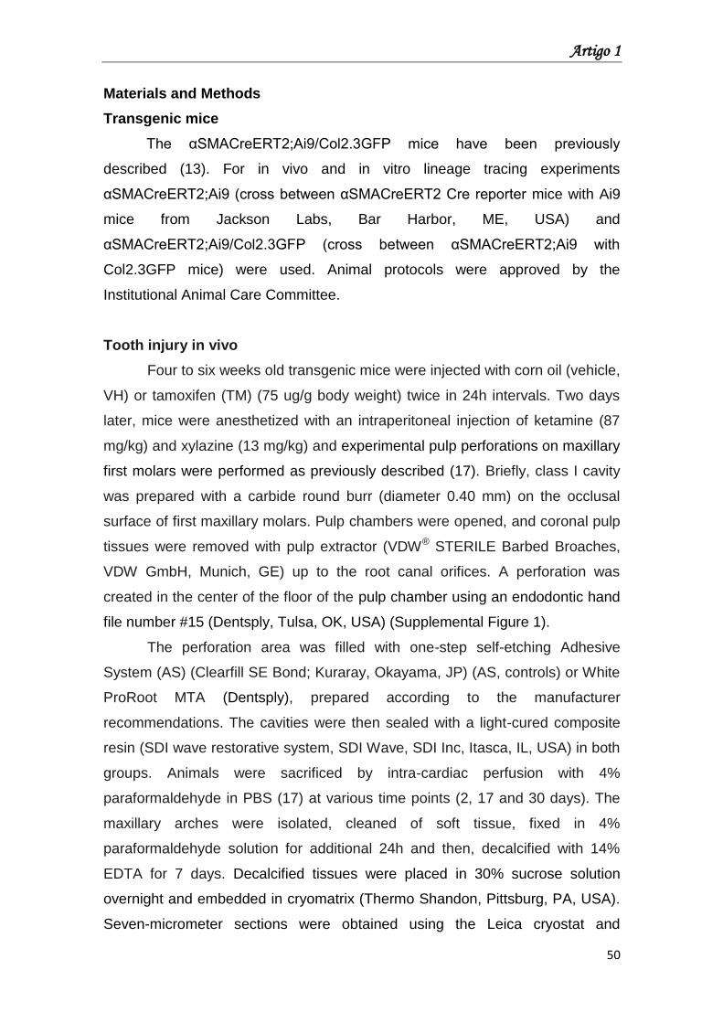
Supplemental Figure 1: Schematic representation of experimental PDL
injury________________________________________________________ 67
Artigo 2:
Figure 1: Effect of both Gray MTA and White MTA extract on L929 proliferation
under high or normal glucose concentration after 6, 24, 48, and 72 hs______ 80
Figure 2: Influence of hyperglycemic condition on IL-6 production by fibroblasts
upon MTA treatment ____________________________________________ 81
Figure 3: Graph showing immunostaining patterns for IL-1β and IL-6 observed
in normal and diabetic groups_____________________________________ 82
Figure 4: Immunostaining patterns for TNF-α observed in the normal and
diabetic groups_________________________________________________ 83
Artigo 3:
Table 1: Inflammatory scores specimens stained with hematoxylin-eosin___ 95
Table 2: Medium of samples in Each Group categorized necrosis, presence of
mineralization and Fluorescence intensity____________________________ 95
Figure 1: Response found in normal group at 30 days__________________ 96
Figure 2: Response found in diabetic group at 30 days_________________ 98
Lista de Tabelas e Figuras
33
Artigo 4:
Figure 1: Graph showing the serum levels of calcium, phosphorus and alkaline
phosphatase for healthy group (a, b, c) and diabetic group (d, e, f) on days 7
and 30______________________________________________________ 118
Figure 2: Graph showing comparison between the calcium, phosphorus and
alkaline phosphatase serum levels in healthy and diabetic groups on day 7 (a,
b, c) and 30 (d, e, f)____________________________________________ 119
Figure 3: Photomicrographs showing the histological appearance of
immunolabelling for OCN and OPN found in healthy and diabetic groups on day
30__________________________________________________________ 120

Sumário
Sumário
40
Sumário
Introdução .................................................................................... 42
Proposição ................................................................................... 46
Artigo 1: Mineral Trioxide Aggregate improves healing response of
periodontal tissue to injury ............................................................. 48
Artigo 2: Hyperglycemic condition interferes on cell proliferation and
IL-6 production stimulated by Gray MTA ........................................ 69
Artigo 3: Diabetes mellitus affects mineralization ability of white
mineral trioxide aggregate ............................................................. 85
Artigo 4: Effect of Diabetes Mellitus on local and systemic bone
marker expression induced by Gray versus White Mineral Trioxide
Aggregate .................................................................................... 101
Conclusão .................................................................................. 121
Referências ................................................................................ 123
Anexos ....................................................................................... 130
Anexo 1 – Comitê de Ética ....................................................... 130
Anexo 2 – Protocolos experimentais – In vitro .......................... 131
Anexo 3 – Protocolos experimentais – In vivo .......................... 137
Anexo 4 - Diretrizes para publicação dos trabalhos .................. 150

Introdução
Introdução
42
Introdução
A "Medicina Endodôntica" visa estudar a relação e/ou associação entre
doenças sistêmicas e as de origem endodônticas (1-4). Entre elas, a Diabetes
Mellitus (DM) que é uma doença complexa, progressiva e debilitante de origem
metabólica caracterizada por um quadro de hiperglicemia crônica que promove
alterações no metabolismo dos carboidratos, lipídios, proteínas, água e
eletrólitos resultantes da insuficiente secreção/ação do hormônio insulina (5).
DM é considerada como um fator modulador das infecções endodônticas
(6). No entanto, esta relação ainda não está completamente elucidada, estudos
mostram que as alterações na reposta imune e a persistência do estado
inflamatório associadas a DM podem interferir e comprometer o reparo dos
tecidos periapicais (7-9).
A hiperglicemia crônica decorrente da DM promove a ativação de vias
que aumentam a inflamação (3,5). Assim, a elevação dos níveis inflamatórios
sistêmicos altera diversas funções do sistema imune (10,11) como o
comprometimento da resposta leucocitária e o aumento da expressão de
citocinas pró-inflamatórias, promovendo uma redução da capacidade de defesa
celular e aumentando a susceptibilidade à infecção e inflamação, afetando
diretamente a integridade dos tecidos pulpares e periapicais e interferindo no
processo de reparo (3, 12-14).
DM também tem sido associada com alterações no processo de reparo
ósseo (15,16), onde mecanismos fisiopatológicos relacionados à perda óssea
como a redução da atividade osteoblástica, diminuição da síntese de colágeno
e alterações no metabolismo do cálcio e fosforo e na expressão de marcadores
de formação óssea tem sido observados em indivíduos diabéticos (17,18).
Entretanto, os mecanismos pelos quais a DM interfere no metabolismo ósseo
e, portanto, no processo de reparo/cicatrização ainda precisam ser
esclarecidos, sabe-se, que controle da inflamação é essencial para que o
processo de reparo ocorra, uma vez que, na presença de um quadro
hiperglicêmico, a persistência da inflamação leva uma estimulação, pelos
neutrófilos, da condrogênese e inibição da osteogênese (19,20).
Introdução
43
O osso é um tecido dinâmico que está em constante remodelação e a
diferenciação osteoblástica é regulada por uma série de hormônios, citocinas e
múltiplos fatores de transcrição (21-23) e que podem ser inibidos e/ou alterados
pelo quadro hiperglicêmico (24). Deste modo, a estimulação da reabsorção
óssea, através da inibição da osteogênese, acarreta no aumento da reabsorção
óssea periapical (25,26). Além disso, em indivíduos diabéticos, a redução da
capacidade de reparo também está associada a diminuição da resistência à
infecção bacteriana e maior susceptibilidade as infecções endodônticas (6, 27,
28).
A infecção endodôntica é tratada através da eliminação dos micro-
organismos patogênicos e o restabelecimento da normalidade dos tecidos
apicais e periapicais afetados, bem como da utilização de materiais capazes de
promover reações de teciduais favoráveis, apresentarem adequadas
propriedades físicas e químicas, que sejam indutores de mineralização e que
possam favorecer e contribuir para a reparo periapical (29,30).
Uma vez que, os cimentos reparadores e obturadores estão em intimo
contato com tecidos perirradiculares, sua composição química, bem como,
compostos tóxicos liberados pelos mesmos podem interferir na resposta
inflamatória e, consequentemente, no processo de reparo (31-33). Assim,
materiais com as mais variadas bases: óxido de zinco e eugenol, resina
epóxica, ionômero de vidro, hidróxido de cálcio e Agregado Trióxido Mineral
(MTA), podem ser encontrados.
Óxido de Zinco e Eugenol (ZOE) é um cimento composto de um pó de
Óxido de Zinco e um líquido o Eugenol, utilizado que nos mais diversos
procedimentos endodônticos: proteção pulpar direta, em selamento provisório,
como obturador endodôntico, em revestimento de cavidades profundas. É um
cimento que apresenta ação antimicrobiana (34), bom selamento marginal (35),
ação anestésica e anti-inflamatória local (36). No entanto, devido ao seu
componente líquido: Eugenol quando aplicado diretamente sobre os tecidos
pode desencadear uma resposta inflamatória crônica dos tecidos periapicais
(37) e danos sobre o tecido pulpar (38).
MTA é um cimento reparador à base de silicato de cálcio que foi
introduzido em 1993 por Torabinejad (39) com a finalidade de proporcionar o
Introdução
44
selamento de comunicações patológicas ou iatrogênicas entre o dente e sua
superfície externa (49,41). Entretanto, devido às suas excelentes propriedades
físicas, químicas e biológicas (42,43) este passou a ser rotineiramente utilizado
nas mais diversas situações clínicas (pulpotomias, capeamentos pulpares,
apicogêneses, apicificações e obturação dos canais radiculares) (41).
Estudos, in vitro e in vivo, mostram que o MTA é um material bioativo
(44); biocompatível (45); promove a proliferação e diferenciação de células
mesenquimais/progenitoras da polpa dentária (46) e ligamento periodontal (47),
além de induz dentinogênese (48), cementogênese (49) e osteogênese (50).
MTA também é capaz de estimular a produção de citocinas (51,52) e a
expressão marcadores ósseos (49, 53). Inclusive em condições hiperglicêmicas
o MTA mostrou-se biocompatível, promoveu mineralização (54) e foi capaz de
induzir a formação de ponte de dentina (55,56).
MTA encontra-se atualmente disponível sob duas formas MTA Cinza e
MTA Branco, onde a principal diferença entre ambos está na redução das
concentrações de Al2O3, MgO, e FeO encontras no MTA Branco (57). Apenas
poucos estudos comparando MTA Cinza e MTA foram realizados, alguns
mostrando semelhanças; são biocompatíveis (58), capazes de induzir a
proliferação celular (59) e de estimular a formação de ponte de dentina (60), e
outros diferenças; MTA Cinza favorece a adesão osteoblástica (61), porém
cementoblastos e queratinócitos crescem melhor na superfície do MTA Branco
(62). Embora, o MTA Branco tenho sido introduzido como uma alternativa para
evitar o pigmentação dental produzida pelo MTA Cinza, estudos in vitro e em in
vivo, também verificaram pigmentação dental causada pelo MTA Branco
(63,64).
Recentemente, o MTA também começou a ser empregado em
procedimentos endodônticos regenerativos (65,66), uma vez que os tecidos
dentários (polpa dentária, ligamento periodontal e osso alveolar) são fonte rica
e acessível de células mesenquimais/progenitoras (67,68), e tais
procedimentos envolvem a interação e diferenciação das células
mesenquimais/progenitoras, bem como a utilização de biomateriais (69). No
entanto, os mecanismos envolvidos nessa interação ainda não estão
totalmente explicados.
Introdução
45
A diferenciação de células osteoprogenitoras é um principais processos
responsáveis pela formação e remodelação óssea, com isso, torna-se um pré-
requisito compreender e analisar as vias envolvidas no desenvolvimento ósseo
(70). Em função disso, investigações tem utilizando animais transgênicos e
marcadores visuais (GFP - proteína verde fluorescente) sob o controle da
actina de músculo liso (α-SMA) (promoter) e do colágeno tipo I 2.3kb
(promoter) expressado por osteoblastos maduros com a finalidade de identificar
a subpopulação de células progenitoras que expressam α-SMA e exibem um
potencial osteogênico (71-73).
Deste modo, como uma das propriedades do MTA é induzir a
mineralização e promover o reparo nos tecidos onde é aplicado, torna-se
relevante compreender e verificar a influência do MTA no processo de
diferenciação osteogênica de células mesenquimais, bem como, no processo
de reparo dos tecidos periodontais após injuria dental, através da utilização de
animais transgênicos. Ao mesmo tempo, como a DM é uma desordem
metabólica que altera a resposta inflamatória e, portanto, afeta o processo de
mineralização, justifica-se o estudo da influência dos MTA Cinza e MTA Branco
na viabilidade celular, resposta tecidual, produção de citocinas, capacidade de
mineralização e na expressão local e sistêmica de marcadores ósseos em
condição diabética.

Proposição
Proposição
46
Proposição
O presente trabalho teve o intuito de avaliar as propriedades biológicas
do MTA em condição normal e hiperglicêmica.
Os objetivos específicos foram:
Avaliar, in vivo, os efeitos do MTA na reparação dos tecidos
periodontais e ósseo após injuria dental (perfuração) usando animais
transgênicos (αSMACreERT2/Ai9/Col2.3GFP);
Avaliar, in vitro, a influência do MTA na proliferação e diferenciação de
células progenitoras da Medula Óssea e do Ligamento Periodontal utilizando
linhagem de animais transgênicos (αSMACreERT2/Ai9/Col2.3GFP);
Avaliar, in vitro, a influência do MTA na viabilidade celular e na
produção de citocinas IL-1β, IL-6 e TNF-α em condição hiperglicêmica;
Avaliar, in vivo, a influência do MTA na resposta tecidual, na produção
de citocinas IL-1β, IL-6 e TNF-α e capacidade de mineralização em condição
diabética;
Avaliar, in vivo, a influência do MTA na produção local (osteocalcina e
osteopontina) e sistêmica (Cálcio, Fósforo e Fosfatase Alcalina) de marcadores
de formação óssea em condição diabética.

Artigo 1
Artigo 1
Artigo 1
48
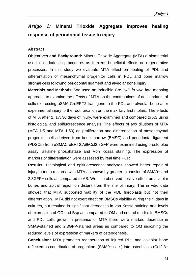
Artigo 1: Mineral Trioxide Aggregate improves healing
response of periodontal tissue to injury
Abstract
Objectives and Background: Mineral Trioxide Aggregate (MTA) a biomaterial
used in endodontic procedures as it exerts beneficial effects on regenerative
processes. In this study we evaluate MTA effect on healing of PDL and
differentiation of mesenchymal progenitor cells in PDL and bone marrow
stromal cells following periodontal ligament and alveolar bone injury.
Materials and Methods: We used an inducible Cre-loxP in vivo fate mapping
approach to examine the effects of MTA on the contributions of descendants of
cells expressing αSMA-CreERT2 transgene to the PDL and alveolar bone after
experimental injury to the root furcation on the maxillary first molars. The effects
of MTA after 2, 17, 30 days of injury, were examined and compared to AS using
histological and epifluorescence analysis. The effects of two dilutions of MTA
(MTA 1:5 and MTA 1:50) on proliferation and differentiation of mesenchymal
progenitor cells derived from bone marrow (BMSC) and periodontal ligament
(PDSCs) from αSMACreERT2;Ai9/Col2.3GFP were examined using presto blue
assay, alkaline phosphatase and Von Kossa staining. The expression of
markers of differentiation were assessed by real time PCR
Results: Histological and epifluorescence analyses showed better repair of
injury in teeth restored with MTA as shown by greater expansion of SMA9+ and
2.3GFP+ cells as compared to AS. We also observed positive effect on alveolar
bones and apical region on distant from the site of injury. The in vitro data
showed that MTA supported viability of the PDL fibroblasts but not their
differentiation. MTA did not exert effect on BMSCs viability during the 9 days in
cultures, but resulted in significant decreases in von Kossa staining and levels
of expression of OC and Bsp as compared to OM and control media. In BMSCs
and PDL cells grown in presence of MTA there were marked decrease in
SMA9-stained and 2.3GFP-stained areas as compared to OM indicating the
reduced levels of expression of markers of osteogenesis.
Conclusion: MTA promotes regeneration of injured PDL and alveolar bone
reflected as contribution of progenitors (SMA9+ cells) into osteoblasts (Col2.3+
Artigo 1
49
cells). In vitro effects of MTA are supportive to viability of the PDL progenitor but
have negative effects on osteogenic differentiation of both PDL and BMSCc.
Keywords: MTA, injury, periodontal ligament, progenitor cells, differentiation
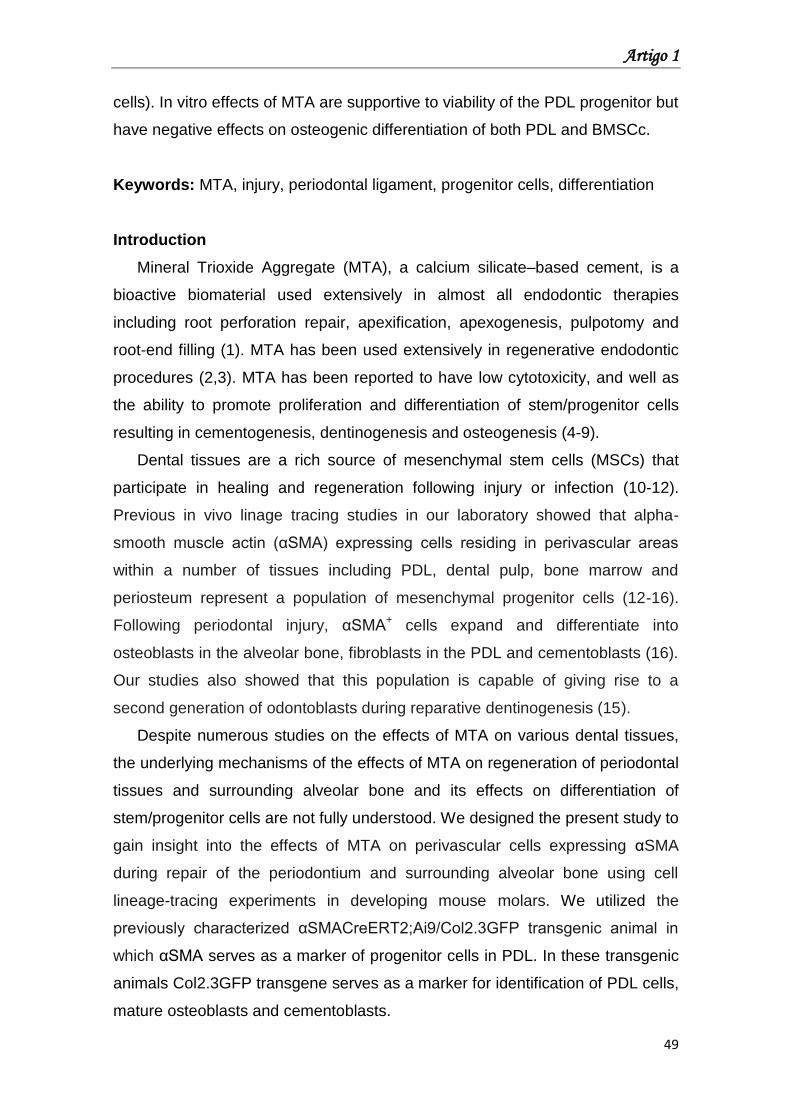
Introduction
Mineral Trioxide Aggregate (MTA), a calcium silicate–based cement, is a
bioactive biomaterial used extensively in almost all endodontic therapies
including root perforation repair, apexification, apexogenesis, pulpotomy and
root-end filling (1). MTA has been used extensively in regenerative endodontic
procedures (2,3). MTA has been reported to have low cytotoxicity, and well as
the ability to promote proliferation and differentiation of stem/progenitor cells
resulting in cementogenesis, dentinogenesis and osteogenesis (4-9).
Dental tissues are a rich source of mesenchymal stem cells (MSCs) that
participate in healing and regeneration following injury or infection (10-12).
Previous in vivo linage tracing studies in our laboratory showed that alpha-
smooth muscle actin (αSMA) expressing cells residing in perivascular areas
within a number of tissues including PDL, dental pulp, bone marrow and
periosteum represent a population of mesenchymal progenitor cells (12-16).
Following periodontal injury, αSMA+ cells expand and differentiate into
osteoblasts in the alveolar bone, fibroblasts in the PDL and cementoblasts (16).
Our studies also showed that this population is capable of giving rise to a
second generation of odontoblasts during reparative dentinogenesis (15).
Despite numerous studies on the effects of MTA on various dental tissues,
the underlying mechanisms of the effects of MTA on regeneration of periodontal
tissues and surrounding alveolar bone and its effects on differentiation of
stem/progenitor cells are not fully understood. We designed the present study to
gain insight into the effects of MTA on perivascular cells expressing αSMA
during repair of the periodontium and surrounding alveolar bone using cell
lineage-tracing experiments in developing mouse molars. We utilized the
previously characterized αSMACreERT2;Ai9/Col2.3GFP transgenic animal in
which αSMA serves as a marker of progenitor cells in PDL. In these transgenic
animals Col2.3GFP transgene serves as a marker for identification of PDL cells,
mature osteoblasts and cementoblasts.
Artigo 1
50
Materials and Methods
Transgenic mice
The αSMACreERT2;Ai9/Col2.3GFP mice have been previously
described (13). For in vivo and in vitro lineage tracing experiments
αSMACreERT2;Ai9 (cross between αSMACreERT2 Cre reporter mice with Ai9
mice from Jackson Labs, Bar Harbor, ME, USA) and
αSMACreERT2;Ai9/Col2.3GFP (cross between αSMACreERT2;Ai9 with
Col2.3GFP mice) were used. Animal protocols were approved by the
Institutional Animal Care Committee.
Tooth injury in vivo
Four to six weeks old transgenic mice were injected with corn oil (vehicle,
VH) or tamoxifen (TM) (75 ug/g body weight) twice in 24h intervals. Two days
later, mice were anesthetized with an intraperitoneal injection of ketamine (87
mg/kg) and xylazine (13 mg/kg) and experimental pulp perforations on maxillary
first molars were performed as previously described (17). Briefly, class I cavity
was prepared with a carbide round burr (diameter 0.40 mm) on the occlusal
surface of first maxillary molars. Pulp chambers were opened, and coronal pulp
tissues were removed with pulp extractor (VDW® STERILE Barbed Broaches,
VDW GmbH, Munich, GE) up to the root canal orifices. A perforation was
created in the center of the floor of the pulp chamber using an endodontic hand
file number #15 (Dentsply, Tulsa, OK, USA) (Supplemental Figure 1).
The perforation area was filled with one-step self-etching Adhesive
System (AS) (Clearfill SE Bond; Kuraray, Okayama, JP) (AS, controls) or White
ProRoot MTA (Dentsply), prepared according to the manufacturer
recommendations. The cavities were then sealed with a light-cured composite
resin (SDI wave restorative system, SDI Wave, SDI Inc, Itasca, IL, USA) in both
groups. Animals were sacrificed by intra-cardiac perfusion with 4%
paraformaldehyde in PBS (17) at various time points (2, 17 and 30 days). The
maxillary arches were isolated, cleaned of soft tissue, fixed in 4%
paraformaldehyde solution for additional 24h and then, decalcified with 14%
EDTA for 7 days. Decalcified tissues were placed in 30% sucrose solution
overnight and embedded in cryomatrix (Thermo Shandon, Pittsburg, PA, USA).
Seven-micrometer sections were obtained using the Leica cryostat and
Artigo 1
51
mounted using a CryoJane tape transfer system (Instrumedics, St Louis, MO,
USA). Sections were imaged using a AxioScan.Z1 (Carl Zeiss). Adjacent
sections were processed for hematoxylin/eosin staining and analyzed by light
microscopy.
Cell isolation and culture
Primary bone marrow stromal cells (BMSCs) were prepared from the
hind limbs of 4-6 week old αSMACreERT2;Ai9/Col2.3GFP mice as previously
described (18, 19). The cells were plated in 12 well culture plates at a density of
106/cm2 for 7 days in basal medium: α-modified essential medium (α-MEM),
10% fetal bovine serum (FBS, Life Technologies, Carlsbad, CA, USA) and 100
U/ml of penicillin, 100 mg/ml of streptomycin (1% PS). Cre activity was induced
by 1µM of 4‐OH‐Tamoxifen added at days 2 and 4 of culture.
At day 7, cells were grown in 4 different conditions including control basal
medium, dilutions of MTA-CM in basal medium and osteogenic media (OM) (α-
MEM 10% FBS + 50ug/ml ascorbic acid + 8mM β–glycerophosphate). Medium
was changed every two days.
PDL cells were isolated from 4-6 week old
αSMACreERT2;Ai9/Col2.3GFP mice as previously described (12). Briefly, the
mandibles and maxilla were dissected from the surrounding tissues, rinsed in
0.12% chlorhexidine digluconate (Clorhexidina, Villevie, Joinville, SC, BRA), for
30 secs, and washed in PBS. Molars with the adherent PDL were removed from
the surrounding alveolar bone and digested in Dulbecco's modified Eagle's
medium (DMEM) with 2 mg/ml Collagenase P (Sigma-Aldrich, Saint Louis, MO,
USA) and 0.25% trypsin (Life Technologies), and digested at least for 2 h at
37°C. Following washing, PDL cells were seeded in DMEM 20% FBS + 1% PS
and cultured in 5% oxygen for 7 days. The medium was changed every 2 days
and the Cre activity was induced by 1µM of 4‐OH‐Tamoxifen added at days 3
and 5 of culture. At day 7, the cells were transferred to normoxic conditions
until confluence (around day 11) and then trypsinized and plated in 24 well
plates at a density 105/cm2. Medium was changed to different treatments the
following day.
Artigo 1
52
MTA Conditioned Medium
White ProRoot MTA was mixed with sterile water according to the
manufacturer’s instructions. MTA discs were prepared under aseptic conditions
as described previously with minor modifications (20). Briefly, the discs were
created by using a sterile cylindrical polyethylene tube (diameter: 6mm; height:
3mm). The MTA discs were kept a 5% CO2 incubator at 37°C for 6 hours for
setting. After 6 hours, the discs were sterilized by ultraviolet (UV) light for 1
hour. The discs were incubated in α-MEM 10% FBS at 37oC in a humidified
atmosphere containing 5% CO2 for 3 days (1 mL of α-MEM 10% FBS for each
disc). After 3 days, the supernatants were collected, filtered through a sterile
0.22µm filter (Sigma-Aldrich, Saint Louis, MO, USA). The supernatant collected
was referred as MTA conditioned media (MTA-CM). Two different dilutions of
MTA-CM were used (high dilution, 1:50 and low dilution, 1:5).
Cell viability assay
Cell viability was determined using a Presto Blue assay (Thermo Fisher
Scientific, Waltham, MA, USA). At various time points the Presto blue reagent
was added to the cell medium, incubated for 2 hours and the fluorescence
intensity was measured (560nm excitation/590nm emission). The experiments
were performed in triplicate.
Histochemical analysis of cell cultures
The histochemical staining for alkaline phosphatase (ALP) was
performed on cultures fixed in 10% formalin for 5 minutes using 86-R Alkaline
Phosphatase kit (Sigma Aldrich) according to the manufacturer instructions. The
number of ALP positive colonies per well was counted. Mineralization was
assessed after 21 days of culture using von Kossa staining as described
previously (21). Plates were imaged on a flat bed scanner and mineralized area
was quantified using ImageJ.
Detection of epifluorescence
Expression of GFP and tdTomato was imaged on an inverted Observer
Z1 microscope (Carl Zeiss). The region scanned covered approximately half the
Artigo 1
53
well area. Fluorescent area proportion for each channel was quantified using
ImageJ.
RNA extraction and gene expression
RNA was extracted using Trizol reagent (Life Technologies) (19).
Reverse transcription was performed using iScript™ cDNA Synthesis Kit (Bio-
Rad, Hercules, CA, USA). The expression of osteogenic genes osteocalcin (Oc,
Mm03413826_mH), bone sialoprotein (Bsp, Mm00492555_m1) was assessed
by RT-qPCR (14). The gene expression was normalized by the expression of a
housekeeping gene (GAPDH).
Statistics
Data were subjected to statistical analysis by using the GraphPad Prism
(version 5.0) software. For all parametric data, ANOVA followed by Tukey´s test
was used. The p value was considered significant at 0.05.
Results
Effects of MTA on periodontal tissue regeneration
In the present study we used the previously characterized
αSMACreERT2;Ai9/Col2.3GFP mouse to examine the effects of MTA on
regeneration of periodontal tissue following injury. Adhesive system (AS)
without MTA has been used as a control for the effects of MTA on healing. In
these experiments perforation on the pulp floor in the furcation area in the first
maxillary molars was performed on 4-6 weeks old TM- injected
αSMACreERT2;Ai9/Col2.3GFP mice. In uninjured tissue, Col2.3GFP
expression (referred to as 2.3GFP) was detected in osteoblasts, osteocytes
within the alveolar bone, cementoblasts on the root surface, odontoblasts lining
the pulp chamber and roots and in PDL fibroblasts surrounding the roots in the
remaining areas of the teeth (Figure 1B-E). Histological analysis of the injured
area showed that perforation at the pulpal floor resulted in local destruction of
dentin, odontoblasts, PDL in the furcation area and the underlying alveolar bone
as evident by the lack of 2.3GFP expression in these locations (Figure 1).
Examination of the area underneath the injury showed presence of a very few
αSMA9-tdTomato+ (referred as SMA9) and 2.3GFP+ cells in teeth filled with AS
Artigo 1
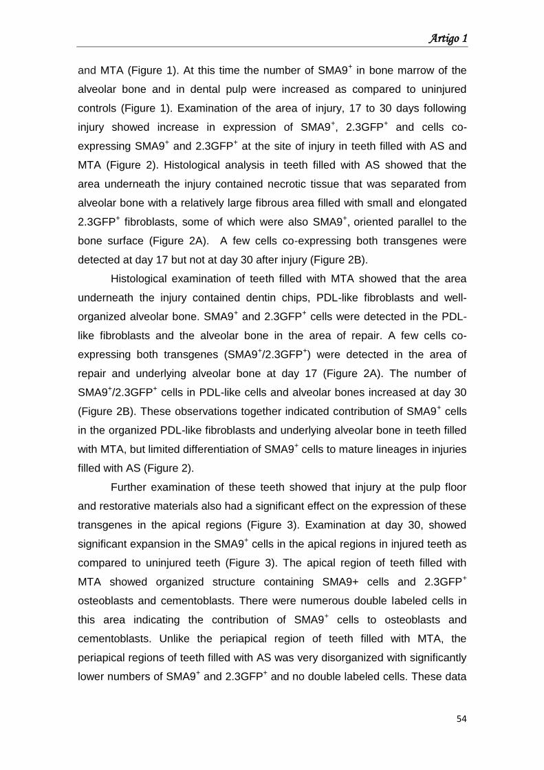
54
and MTA (Figure 1). At this time the number of SMA9+ in bone marrow of the
alveolar bone and in dental pulp were increased as compared to uninjured
controls (Figure 1). Examination of the area of injury, 17 to 30 days following
injury showed increase in expression of SMA9+, 2.3GFP+ and cells co-
expressing SMA9+ and 2.3GFP+ at the site of injury in teeth filled with AS and
MTA (Figure 2). Histological analysis in teeth filled with AS showed that the
area underneath the injury contained necrotic tissue that was separated from
alveolar bone with a relatively large fibrous area filled with small and elongated
2.3GFP+ fibroblasts, some of which were also SMA9+, oriented parallel to the
bone surface (Figure 2A). A few cells co-expressing both transgenes were
detected at day 17 but not at day 30 after injury (Figure 2B).
Histological examination of teeth filled with MTA showed that the area
underneath the injury contained dentin chips, PDL-like fibroblasts and well-
organized alveolar bone. SMA9+ and 2.3GFP+ cells were detected in the PDL-
like fibroblasts and the alveolar bone in the area of repair. A few cells co-
expressing both transgenes (SMA9+/2.3GFP+) were detected in the area of
repair and underlying alveolar bone at day 17 (Figure 2A). The number of
SMA9+/2.3GFP+ cells in PDL-like cells and alveolar bones increased at day 30
(Figure 2B). These observations together indicated contribution of SMA9+ cells
in the organized PDL-like fibroblasts and underlying alveolar bone in teeth filled
with MTA, but limited differentiation of SMA9+ cells to mature lineages in injuries
filled with AS (Figure 2).
Further examination of these teeth showed that injury at the pulp floor
and restorative materials also had a significant effect on the expression of these
transgenes in the apical regions (Figure 3). Examination at day 30, showed
significant expansion in the SMA9+ cells in the apical regions in injured teeth as
compared to uninjured teeth (Figure 3). The apical region of teeth filled with
MTA showed organized structure containing SMA9+ cells and 2.3GFP+
osteoblasts and cementoblasts. There were numerous double labeled cells in
this area indicating the contribution of SMA9+ cells to osteoblasts and
cementoblasts. Unlike the periapical region of teeth filled with MTA, the
periapical regions of teeth filled with AS was very disorganized with significantly
lower numbers of SMA9+ and 2.3GFP+ and no double labeled cells. These data
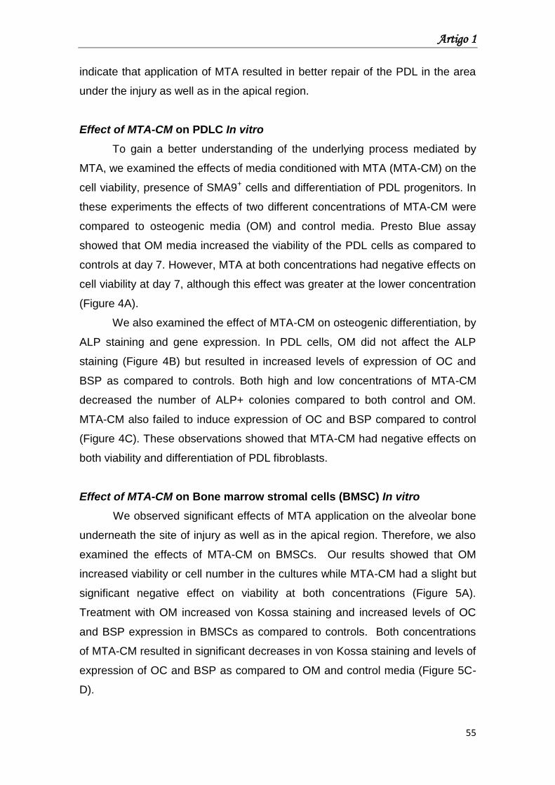
Artigo 1
55
indicate that application of MTA resulted in better repair of the PDL in the area
under the injury as well as in the apical region.
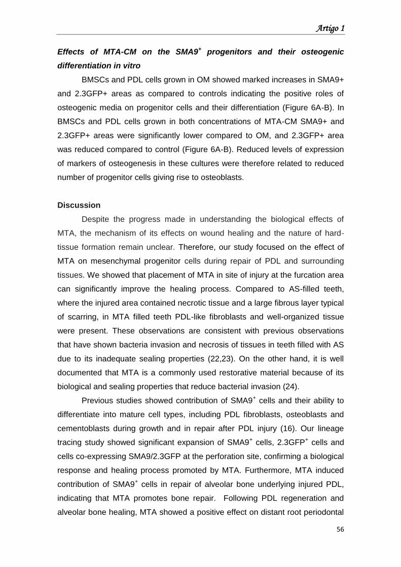
Effect of MTA-CM on PDLC In vitro
To gain a better understanding of the underlying process mediated by
MTA, we examined the effects of media conditioned with MTA (MTA-CM) on the
cell viability, presence of SMA9+ cells and differentiation of PDL progenitors. In
these experiments the effects of two different concentrations of MTA-CM were
compared to osteogenic media (OM) and control media. Presto Blue assay
showed that OM media increased the viability of the PDL cells as compared to
controls at day 7. However, MTA at both concentrations had negative effects on
cell viability at day 7, although this effect was greater at the lower concentration
(Figure 4A).
We also examined the effect of MTA-CM on osteogenic differentiation, by
ALP staining and gene expression. In PDL cells, OM did not affect the ALP
staining (Figure 4B) but resulted in increased levels of expression of OC and
BSP as compared to controls. Both high and low concentrations of MTA-CM
decreased the number of ALP+ colonies compared to both control and OM.
MTA-CM also failed to induce expression of OC and BSP compared to control
(Figure 4C). These observations showed that MTA-CM had negative effects on
both viability and differentiation of PDL fibroblasts.
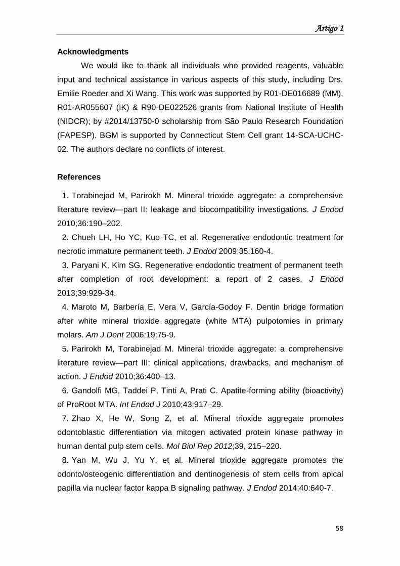
Effect of MTA-CM on Bone marrow stromal cells (BMSC) In vitro
We observed significant effects of MTA application on the alveolar bone
underneath the site of injury as well as in the apical region. Therefore, we also
examined the effects of MTA-CM on BMSCs. Our results showed that OM
increased viability or cell number in the cultures while MTA-CM had a slight but
significant negative effect on viability at both concentrations (Figure 5A).
Treatment with OM increased von Kossa staining and increased levels of OC
and BSP expression in BMSCs as compared to controls. Both concentrations
of MTA-CM resulted in significant decreases in von Kossa staining and levels of
expression of OC and BSP as compared to OM and control media (Figure 5C-
D).
Artigo 1
56
Effects of MTA-CM on the SMA9+ progenitors and their osteogenic
differentiation in vitro
BMSCs and PDL cells grown in OM showed marked increases in SMA9+
and 2.3GFP+ areas as compared to controls indicating the positive roles of
osteogenic media on progenitor cells and their differentiation (Figure 6A-B). In
BMSCs and PDL cells grown in both concentrations of MTA-CM SMA9+ and
2.3GFP+ areas were significantly lower compared to OM, and 2.3GFP+ area
was reduced compared to control (Figure 6A-B). Reduced levels of expression
of markers of osteogenesis in these cultures were therefore related to reduced
number of progenitor cells giving rise to osteoblasts.
Discussion
Despite the progress made in understanding the biological effects of
MTA, the mechanism of its effects on wound healing and the nature of hard-
tissue formation remain unclear. Therefore, our study focused on the effect of
MTA on mesenchymal progenitor cells during repair of PDL and surrounding
tissues. We showed that placement of MTA in site of injury at the furcation area
can significantly improve the healing process. Compared to AS-filled teeth,
where the injured area contained necrotic tissue and a large fibrous layer typical
of scarring, in MTA filled teeth PDL-like fibroblasts and well-organized tissue
were present. These observations are consistent with previous observations
that have shown bacteria invasion and necrosis of tissues in teeth filled with AS
due to its inadequate sealing properties (22,23). On the other hand, it is well
documented that MTA is a commonly used restorative material because of its
biological and sealing properties that reduce bacterial invasion (24).
Previous studies showed contribution of SMA9+ cells and their ability to
differentiate into mature cell types, including PDL fibroblasts, osteoblasts and
cementoblasts during growth and in repair after PDL injury (16). Our lineage
tracing study showed significant expansion of SMA9+ cells, 2.3GFP+ cells and
cells co-expressing SMA9/2.3GFP at the perforation site, confirming a biological
response and healing process promoted by MTA. Furthermore, MTA induced
contribution of SMA9+ cells in repair of alveolar bone underlying injured PDL,
indicating that MTA promotes bone repair. Following PDL regeneration and
alveolar bone healing, MTA showed a positive effect on distant root periodontal

Artigo 1
57
complex. Unlike the disorganized apical region in AS capped teeth, in teeth
filled with MTA this area showed organized structure containing differentiated
SMA9+/2.3GFP+ osteoblasts and cementoblasts. These observations suggest
that MTA mediates regeneration through interactions with periodontal ligament
and alveolar bone progenitor/stem cells.
In contrast to our in vivo observations, in vitro results showed that MTA
conditioned media does not support differentiation of BMSCs and PDLCs. The
lack of differentiation in our in vitro studies is also different from previously
reported positive effects of MTA on the formation of mineralized nodules and
expression of cemento/osteoblastic marker genes in PDLCs and BMSCs
(9,25,26). Explanation for these differences can be the amount of calcium,
aluminum, bismuth and silicon ions released by MTA into media and its
variation depending of concentration utilized, which might inhibit or suppress
cell growth and functions resulting in changes in the cell response, such as
proliferation and differentiation (27-29). It has been also reported that MTA
enhances proliferation of human dental pulp cells through sustained release of
calcium ions (30). In contrast, others have shown that rate of calcium ion
release from MTA were higher during the first three hours with subsequent
decreases thereafter (31). Furthermore, high concentrations of MTA have been
shown to exert cytotoxic effects on human PDL fibroblasts (32). Taken together,
these findings may explain decreased in viability when MTA-CM was used in
PDLC and BMSC cultures. Although the outcomes of the in vitro experiments
may depend on the cell type, different culture systems and concentrations of
MTA, the most likely explanation for these differences is the lack of
mineralization inducing reagents such as ascorbic acid and β–glycerophosphate
in media used to examine the effects of MTA on differentiation.
Although cell studies results are relevant, it is not possible to assess the
complex interactions between materials and host. Therefore, we primarily
evaluated the in vivo effects of the MTA on the progenitor lineages. Collectively,
our findings showed contribution of SMA9+ cells in soft tissue repair and newly
calcified bone matrix formation as well as positive effect of MTA on PDL and
alveolar bone injury.
Artigo 1
58
Acknowledgments
We would like to thank all individuals who provided reagents, valuable
input and technical assistance in various aspects of this study, including Drs.
Emilie Roeder and Xi Wang. This work was supported by R01-DE016689 (MM),
R01-AR055607 (IK) & R90-DE022526 grants from National Institute of Health
(NIDCR); by #2014/13750-0 scholarship from São Paulo Research Foundation
(FAPESP). BGM is supported by Connecticut Stem Cell grant 14-SCA-UCHC-
02. The authors declare no conflicts of interest.
References
1. Torabinejad M, Parirokh M. Mineral trioxide aggregate: a comprehensive
literature review—part II: leakage and biocompatibility investigations. J Endod
2010;36:190–202.
2. Chueh LH, Ho YC, Kuo TC, et al. Regenerative endodontic treatment for
necrotic immature permanent teeth. J Endod 2009;35:160-4.
3. Paryani K, Kim SG. Regenerative endodontic treatment of permanent teeth
after completion of root development: a report of 2 cases. J Endod
2013;39:929-34.
4. Maroto M, Barbería E, Vera V, García-Godoy F. Dentin bridge formation
after white mineral trioxide aggregate (white MTA) pulpotomies in primary
molars. Am J Dent 2006;19:75-9.
5. Parirokh M, Torabinejad M. Mineral trioxide aggregate: a comprehensive
literature review—part III: clinical applications, drawbacks, and mechanism of
action. J Endod 2010;36:400–13.
6. Gandolfi MG, Taddei P, Tinti A, Prati C. Apatite-forming ability (bioactivity)
of ProRoot MTA. Int Endod J 2010;43:917–29.
7. Zhao X, He W, Song Z, et al. Mineral trioxide aggregate promotes
odontoblastic differentiation via mitogen activated protein kinase pathway in
human dental pulp stem cells. Mol Biol Rep 2012;39, 215–220.
8. Yan M, Wu J, Yu Y, et al. Mineral trioxide aggregate promotes the
odonto/osteogenic differentiation and dentinogenesis of stem cells from apical
papilla via nuclear factor kappa B signaling pathway. J Endod 2014;40:640-7.
Artigo 1
59
9. Wang Y, Li J, Song W, Yu J. Mineral trioxide aggregate upregulates
odonto/osteogenic capacity of bone marrow stromal cells from craniofacial
bones via JNK and ERK MAPK signalling pathways. Cell Prolif 2014;47:241-8.
10. Sharpe PT. Dental mesenchymal stem cells. Development
2016;143:2273-80.
11. Shi S, Bartold PM, Miura M, et al. The efficacy of mesenchymal stem cells
to regenerate and repair dental structures. Orthod Craniofac Res 2005;8:191–
199.
12. San Miguel SM, Fatahi MR, Li H, et al. Defining a visual marker of
osteoprogenitor cells within the periodontium. J Period Res 2010; 45:60-70.
13. Grcevic D, Pejda S, Matthews BG, et al. In vivo fate mapping identifies
mesenchymal progenitor cells. Stem Cells 2012;30:187–96.
14. Matthews BG, Grcevic D, Wang L, et al. Analysis of αSMA-labeled
progenitor cell commitment identifies notch signaling as an important pathway in
fracture healing. J Bone Miner Res 2014;29:1283-94.
15. Vidovic I, Banerjee A, Fatahi R, et al. αSMA-Expressing Perivascular Cells
Represent Dental Pulp Progenitors In Vivo. J Dent Res 2016 Nov 10. [Epub
ahead of print]
16. Roguljic H, Matthews BG, Yang W, et al. In vivo identification of
periodontal progenitor cells. J Dent Res 2013;92:709-15.
17. Frozoni M, Balic A, Sagomonyants K, et al. A feasibility study for the
analysis of reparative dentinogenesis in pOBCol3.6GFPtpz transgenic mice. Int
Endod J 2012;45:907-14.
18. Kalajzic I, Kalajzic Z, Kaliterna M, et al. Use of type I collagen green
fluorescent protein transgenes to identify subpopulations of cells at different
stages of the osteoblast lineage. J Bone Miner Res 2002;17:15–25
19. Repic D, Torreggiani E, Franceschetti T, et al. Utilization of transgenic
models in the evaluation of osteogenic differentiation of embryonic stem cells.
Connect Tissue Res 2013;54:296-304.
20. Yoshino P, Nishiyama CK, Modena KC, et al. In vitro cytotoxicity of white
MTA, MTA Fillapex® and Portland cement on human periodontal ligament
fibroblats. Braz Dent J 2013;24:111-6.
21. Kalajzic Z, Li H, Wang LP, et al. Use of an alpha-smooth muscle actin
GFP reporter to identify an osteoprogenitor population. Bone 2008;43:501-510.
Artigo 1
60
22. Tsatsas DV, Meliou HA, Kerezoudis NP. Sealing effectiveness of materials
used in furcation perforation in vitro. Int Dent J 2005;55:133-41.
23. Lodiene G, Kleivmyr M, Bruzell E, Ørstavik D. Sealing ability of mineral
trioxide aggregate, glass ionomer cement and composite resin when repairing
large furcal perforations. Br Dent J 2011;12:210(5):E7.
24. Torabinejad M, Watson TF, Pitt Ford TR. Sealing ability of a mineral
trioxide aggregate when used as a root end filling material. J Endod.
1993;19:591-5.
25. Seo BM, Miura M, Gronthos S, Bartold PM, Batouli S et al. Investigation of
multipotent postnatal stem cells from human periodontal ligament. Lancet
2004;364:149–155.
26. Hakki SS, Bozkurt SB, Hakki EE, Belli S. Effects of mineral trioxide
aggregate on cell survival, gene expression associated with mineralized tissues,
and biomineralization of cementoblasts. J Endod 2009;35:513-9.
27. Hoppe A, Güldal NS, Boccaccini AR. A review of the biological response
to ionic dissolution products from bioactive glasses and glass-ceramics.
Biomaterials 2011;32:2757–2774
28. Wu BC, Kao CT, Huang TH, et al. Effect of verapamil, a calcium channel
blocker, on the odontogenic activity of human dental pulp cells cultured with
silicate-based materials. J Endod 2014;40:1105-11.
29. Chen I, Salhab I, Setzer FC, et al. A New Calcium Silicate-based
Bioceramic Material Promotes Human Osteo- and Odontogenic Stem Cell
Proliferation and Survival via the Extracellular Signal-regulated Kinase Signaling
Pathway. J Endod 2016;42:480-6.
30. Takita T, Hayashi M, Takeichi O, et al. Effect of mineral trioxide aggregate
on proliferation of cultured human dental pulp cells. Int Endod J 2006;39:415-
22.
31. Duarte MA, Demarchi AC, Yamashita JC, Kuga MC, Fraga Sde C. pH and
calcium ion release of 2 root-end filling materials. Oral Surg Oral Med Oral
Pathol Oral Radiol Endod 2003;95:345-7.
32. Keiser K, Johnson CC, Tipton DA. Cytotoxicity of mineral trioxide
aggregate using human periodontal ligament fibroblasts. J Endod 2000;26:288-
91.
Artigo 1
61
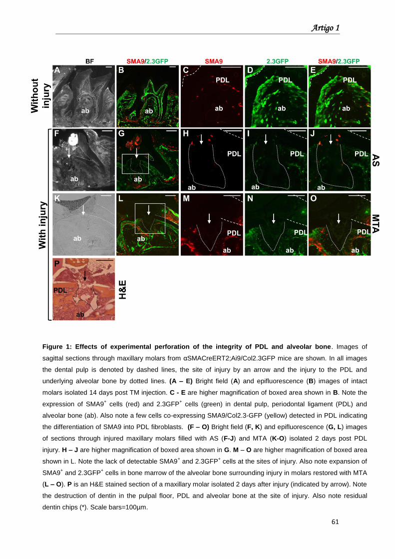
Figure 1: Effects of experimental perforation of the integrity of PDL and alveolar bone. Images of
sagittal sections through maxillary molars from αSMACreERT2;Ai9/Col2.3GFP mice are shown. In all images
the dental pulp is denoted by dashed lines, the site of injury by an arrow and the injury to the PDL and
underlying alveolar bone by dotted lines. (A – E) Bright field (A) and epifluorescence (B) images of intact
molars isolated 14 days post TM injection. C - E are higher magnification of boxed area shown in B. Note the
expression of SMA9+ cells (red) and 2.3GFP
+ cells (green) in dental pulp, periodontal ligament (PDL) and
alveolar bone (ab). Also note a few cells co-expressing SMA9/Col2.3-GFP (yellow) detected in PDL indicating
the differentiation of SMA9 into PDL fibroblasts. (F – O) Bright field (F, K) and epifluorescence (G, L) images
of sections through injured maxillary molars filled with AS (F-J) and MTA (K-O) isolated 2 days post PDL
injury. H – J are higher magnification of boxed area shown in G. M – O are higher magnification of boxed area
shown in L. Note the lack of detectable SMA9+ and 2.3GFP
+ cells at the sites of injury. Also note expansion of
SMA9+ and 2.3GFP
+ cells in bone marrow of the alveolar bone surrounding injury in molars restored with MTA
(L – O). P is an H&E stained section of a maxillary molar isolated 2 days after injury (indicated by arrow). Note
the destruction of dentin in the pulpal floor, PDL and alveolar bone at the site of injury. Also note residual
dentin chips (*). Scale bars=100µm.
Artigo 1
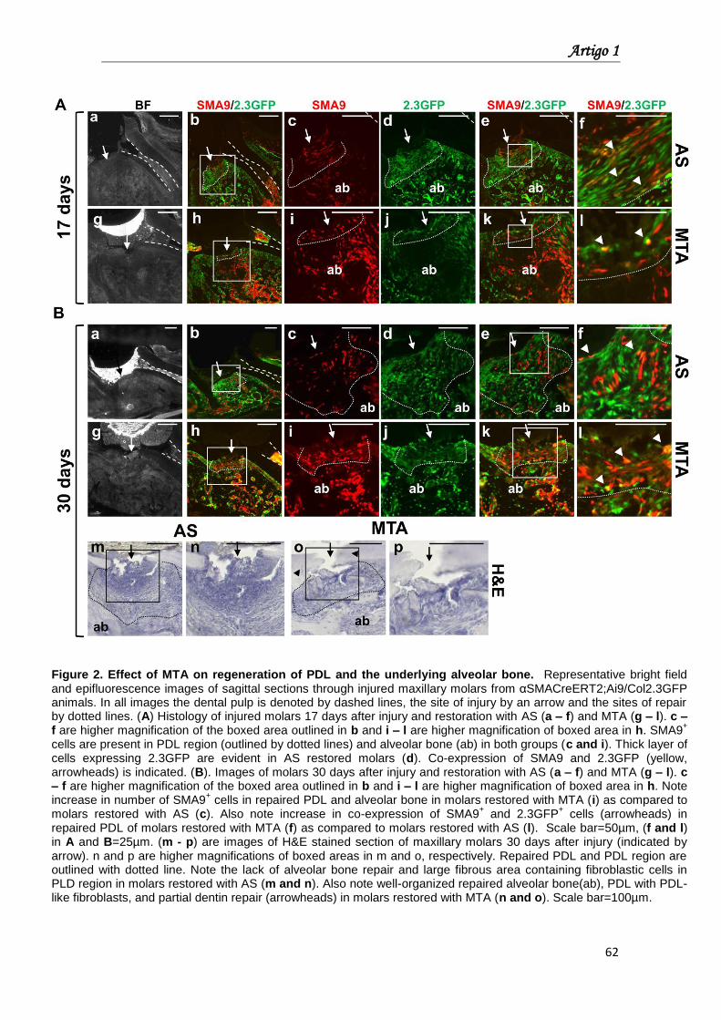
62
Figure 2. Effect of MTA on regeneration of PDL and the underlying alveolar bone. Representative bright field and epifluorescence images of sagittal sections through injured maxillary molars from αSMACreERT2;Ai9/Col2.3GFP animals. In all images the dental pulp is denoted by dashed lines, the site of injury by an arrow and the sites of repair by dotted lines. (A) Histology of injured molars 17 days after injury and restoration with AS (a – f) and MTA (g – l). c – f are higher magnification of the boxed area outlined in b and i – l are higher magnification of boxed area in h. SMA9
+
cells are present in PDL region (outlined by dotted lines) and alveolar bone (ab) in both groups (c and i). Thick layer of cells expressing 2.3GFP are evident in AS restored molars (d). Co-expression of SMA9 and 2.3GFP (yellow, arrowheads) is indicated. (B). Images of molars 30 days after injury and restoration with AS (a – f) and MTA (g – l). c – f are higher magnification of the boxed area outlined in b and i – l are higher magnification of boxed area in h. Note increase in number of SMA9
+ cells in repaired PDL and alveolar bone in molars restored with MTA (i) as compared to
molars restored with AS (c). Also note increase in co-expression of SMA9+ and 2.3GFP
+ cells (arrowheads) in
repaired PDL of molars restored with MTA (f) as compared to molars restored with AS (l). Scale bar=50µm, (f and l) in A and B=25µm. (m - p) are images of H&E stained section of maxillary molars 30 days after injury (indicated by arrow). n and p are higher magnifications of boxed areas in m and o, respectively. Repaired PDL and PDL region are outlined with dotted line. Note the lack of alveolar bone repair and large fibrous area containing fibroblastic cells in PLD region in molars restored with AS (m and n). Also note well-organized repaired alveolar bone(ab), PDL with PDL-like fibroblasts, and partial dentin repair (arrowheads) in molars restored with MTA (n and o). Scale bar=100µm.
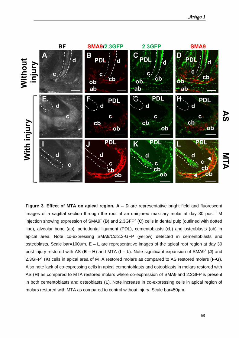
Artigo 1
63
Figure 3. Effect of MTA on apical region. A – D are representative bright field and fluorescent
images of a sagittal section through the root of an uninjured maxillary molar at day 30 post TM
injection showing expression of SMA9+ (B) and 2.3GFP+ (C) cells in dental pulp (outlined with dotted
line), alveolar bone (ab), periodontal ligament (PDL), cementoblasts (cb) and osteoblasts (ob) in
apical area. Note co-expressing SMA9/Col2.3-GFP (yellow) detected in cementoblasts and
osteoblasts. Scale bar=100µm. E – L are representative images of the apical root region at day 30
post injury restored with AS (E – H) and MTA (I – L). Note significant expansion of SMA9+ (J) and
2.3GFP+ (K) cells in apical area of MTA restored molars as compared to AS restored molars (F-G).
Also note lack of co-expressing cells in apical cementoblasts and osteoblasts in molars restored with
AS (H) as compared to MTA restored molars where co-expression of SMA9 and 2.3GFP is present
in both cementoblasts and osteoblasts (L). Note increase in co-expressing cells in apical region of
molars restored with MTA as compared to control without injury. Scale bar=50µm.
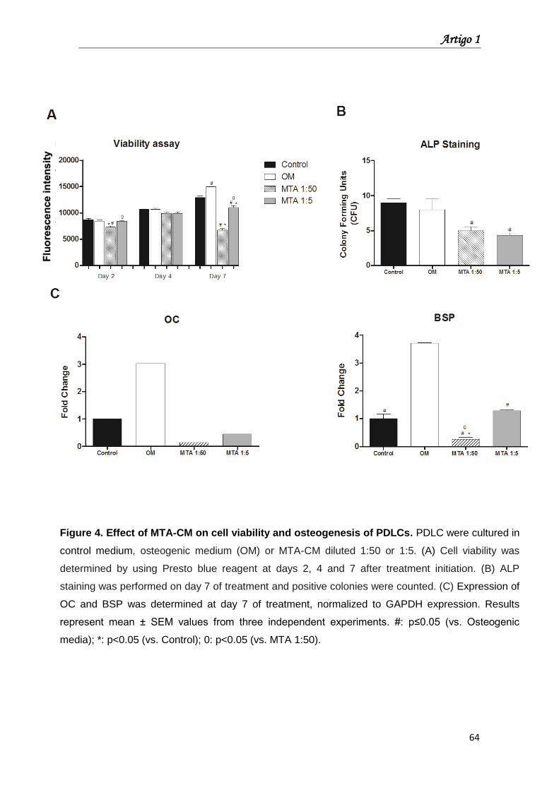
Artigo 1
64
Figure 4. Effect of MTA-CM on cell viability and osteogenesis of PDLCs. PDLC were cultured in
control medium, osteogenic medium (OM) or MTA-CM diluted 1:50 or 1:5. (A) Cell viability was
determined by using Presto blue reagent at days 2, 4 and 7 after treatment initiation. (B) ALP
staining was performed on day 7 of treatment and positive colonies were counted. (C) Expression of
OC and BSP was determined at day 7 of treatment, normalized to GAPDH expression. Results
represent mean ± SEM values from three independent experiments. #: p≤0.05 (vs. Osteogenic
media); *: p<0.05 (vs. Control); 0: p<0.05 (vs. MTA 1:50).
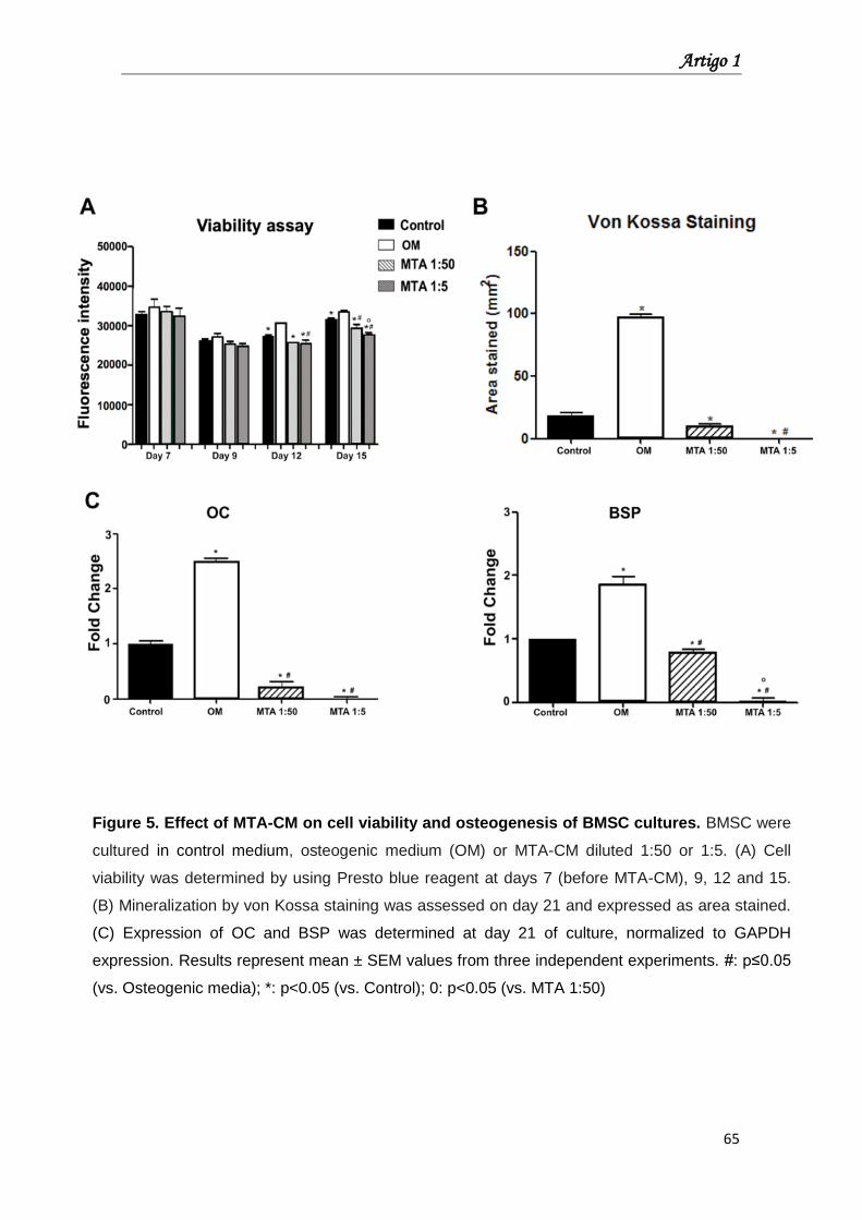
Artigo 1
65
Figure 5. Effect of MTA-CM on cell viability and osteogenesis of BMSC cultures. BMSC were
cultured in control medium, osteogenic medium (OM) or MTA-CM diluted 1:50 or 1:5. (A) Cell
viability was determined by using Presto blue reagent at days 7 (before MTA-CM), 9, 12 and 15.
(B) Mineralization by von Kossa staining was assessed on day 21 and expressed as area stained.
(C) Expression of OC and BSP was determined at day 21 of culture, normalized to GAPDH
expression. Results represent mean ± SEM values from three independent experiments. #: p≤0.05
(vs. Osteogenic media); *: p<0.05 (vs. Control); 0: p<0.05 (vs. MTA 1:50)
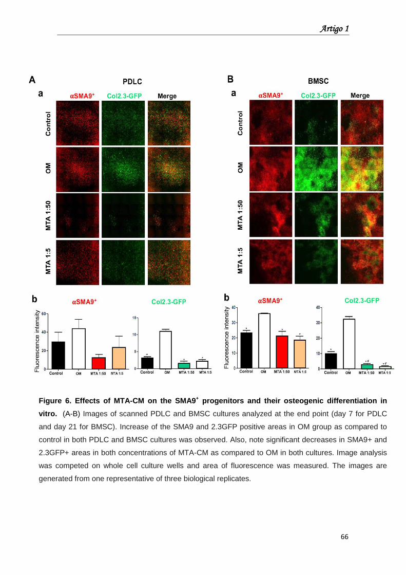
Artigo 1
66
Figure 6. Effects of MTA-CM on the SMA9+ progenitors and their osteogenic differentiation in
vitro. (A-B) Images of scanned PDLC and BMSC cultures analyzed at the end point (day 7 for PDLC
and day 21 for BMSC). Increase of the SMA9 and 2.3GFP positive areas in OM group as compared to
control in both PDLC and BMSC cultures was observed. Also, note significant decreases in SMA9+ and
2.3GFP+ areas in both concentrations of MTA-CM as compared to OM in both cultures. Image analysis
was competed on whole cell culture wells and area of fluorescence was measured. The images are
generated from one representative of three biological replicates.
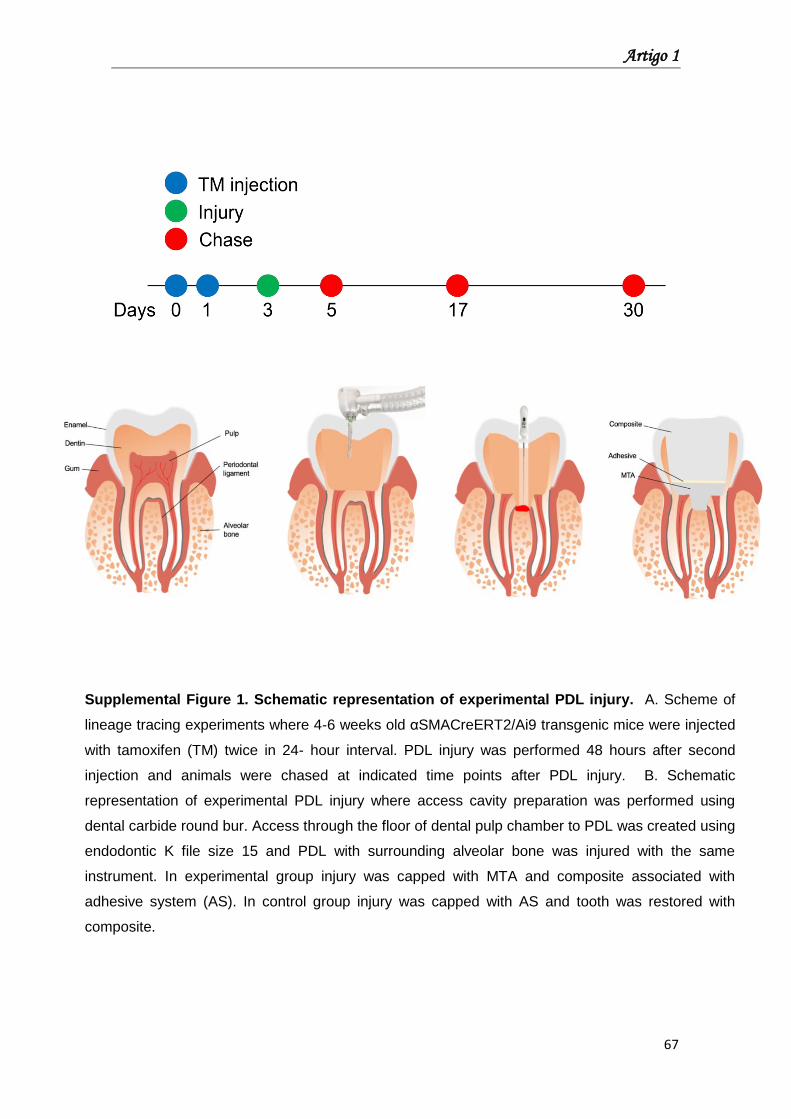
Artigo 1
67
Supplemental Figure 1. Schematic representation of experimental PDL injury. A. Scheme of
lineage tracing experiments where 4-6 weeks old αSMACreERT2/Ai9 transgenic mice were injected
with tamoxifen (TM) twice in 24- hour interval. PDL injury was performed 48 hours after second
injection and animals were chased at indicated time points after PDL injury. B. Schematic
representation of experimental PDL injury where access cavity preparation was performed using
dental carbide round bur. Access through the floor of dental pulp chamber to PDL was created using
endodontic K file size 15 and PDL with surrounding alveolar bone was injured with the same
instrument. In experimental group injury was capped with MTA and composite associated with
adhesive system (AS). In control group injury was capped with AS and tooth was restored with
composite.

Artigo 2
Artigo 2
69
Artigo 2: Hyperglycemic condition interferes on cell
proliferation and IL-6 production stimulated by Gray MTA
Abstract
Introduction: Diabetes mellitus (DM) affects inflammatory and immune
responses and impairs healing processes. We investigated DM influence on cell
proliferation and cytokine production induced by Gray Mineral Trioxide
Aggregate (GMTA) and White MTA (WMTA).
Methods: L929 fibroblasts were cultured under high or normal glucose
concentration. Effects of GMTA and WMTA on cell proliferation and IL-1β, IL-6,
and TNF-α production were investigated using Alamar Blue assay and ELISA,
respectively, at 6, 24, 48, and 72h. Moreover, polyethylene tubes containing
GMTA, WMTA, and empty tubes (control) were implanted into dorsal connective
tissues of Wistar rats previously assigned normal and diabetic groups (Alloxan
induced). After 7 and 30 days, the tubes with surrounding tissues were
removed, fixed, and subjected to immunohistochemical analysis of IL-1β, IL-6,
and TNF-α. Nonparametric and parametric data were statistically analyzed
(p<0.05).
Results: In vitro assays showed no detectable production of IL-1β and TNF-α.
The hyperglycemic condition promoted IL-6 up-regulation production (p<0.05)
and impaired cell proliferation at 72h (p<0.05). Under high glucose condition,
GMTA induced greater cytotoxicity and IL-6 production than WMTA did. In vivo
assay showed no differences in IL-1β, IL-6, and TNF-α production between both
systemic conditions in presence of GMTA and WMTA at all time points.
Conclusion: Hyperglycemic conditions interfered on cell proliferation and IL-6
production stimulated by GMTA. Moreover, no up-regulation of inflammatory
cytokines in diabetic animal tissues was observed in the presence of GMTA and
WMTA.
Keywords: Diabetes Mellitus; Cytokines; Gray MTA; White MTA
Artigo 2
70
Introduction
Diabetes mellitus (DM) is a metabolic disease considered as a modulator
of endodontic infections (1). The hyperglycemic state promoted by DM can alter
immune functions through impairment of leucocyte responses and cellular
defense capacity reduction, leading to increased susceptibility to infection and
inflammation and a direct effect on dental pulp integrity and periapical healing
process (2-7).
During periapical healing, proinflammatory cytokines IL-1β, IL-6, and
tumor necrosis factor alpha (TNF-α) play an essential role in inflammatory
response development (8,9). Moreover, tissue response to biomaterials
depends on innate and nonspecific immune responses (5,8,10). The root end
filling material is set in close contact with periapical tissue and its chemical
composition can interfere on inflammatory response and consequently affect
repair process (9,11).
Mineral Trioxide Aggregate (MTA) has been widely studied as an
endodontic material since it was introduced (12). Both in vivo and in vitro
investigations showed that MTA is biocompatible, shows low cytotoxicity, and
stimulates hard tissue formation (13,14). Besides, MTA stimulates the release
of IL-1β, IL-6, TNF-α, and growth factors from cells (15-17). Even in diabetic
conditions, MTA does not alter tissue response, promotes mineralization and
induces dentin bridge formation (6,14,18). Despite these investigations, the
biological mechanism of MTA, especially its relationship with systemic
conditions, is still not completely elucidated.
DM induces alterations in immune cell function and stimulates expression
of proinflammatory cytokines (3), thus compromising dental pulp and periapical
response. In this study, we aimed to evaluate in vitro and in vivo effects of both
Gray and White MTA on cell viability and cytokine production under diabetic
conditions.
Materials and Methods
In vitro study
Cell Culture
L929 mouse fibroblasts were grown in Dulbecco Modified Eagle’s
Medium (DMEM) supplemented with 10% fetal bovine serum (FBS, GIBCO
Artigo 2
71
BRL, Gaithersburg, MD) streptomycin (50 g/mL), and 1% antibiotic/antimycotic
cocktail (300 U/mL penicillin, 300 µg/mL streptomycin, 5 µg/mL amphotericin B)
(GIBCO BRL, Gaithersburg, MD) under standard cell culture conditions (37°C,
100% humidity, 95% air, and 5% CO2).
The hyperglycemic condition was simulated with cell culture media
supplemented with a high concentration of glucose (25 mM glucose).
MTA conditioned media
Gray and White MTA Angelus® (Angelus Indústria de Produtos
Odontológicos S/A, Londrina, PR, Brazil) were mixed according manufacturer’s
instructions. MTA discs were prepared as previously described (19) with some
modifications. Briefly, the discs were formed using a sterile polyethylene tube (5
mm in diameter and 3 mm in height) and kept in a 5% CO2 incubator at 37°C to
setting for 6h. Then, the discs were removed from the mold and sterilized in
ultraviolet (UV) light for 1h. Each disc was immersed in 1 mL of DMEM with
10% FBS and incubated in a humidified atmosphere containing 5% CO2. After 3
days, the discs were discarded and supernatants (extract) were collected and
filtered through a sterile 0.22 µm filter (Sigma-Aldrich, Saint Louis, MO, USA).
The collected supernatants were referred as the GMTA extract or WMTA
extract. An extract dilution of 1:50 was used in this study.
Cell proliferation assay
Cell proliferation was determined using Alamar Blue reduction assay
(Alamar Blue® Cell Viability Reagent, Thermo Fisher Scientific, Waltham, MA,
USA) following manufacturer’s instructions. L929 fibroblasts were seeded into
24-well plates (104 cells/mL medium per well) and incubated for 24h in a
humidified air atmosphere of 5% CO2 at 37oC. After, both MTA extract (1:50)
and Alamar Blue reagent (1:10) was added to the culture medium and after 6h,
24h, 48h, and 72h, 200 µl of medium was transferred to a 96 well plate. Optical
density (OD) was measured at 570 nm and 600 nm. Alamar blue reduction was
calculated with a manufacturer provided formula. The percentage of reduction
level reflects the cell proliferation. The controls were cultured in media without
MTA extracts. The experiments were performed in triplicate.
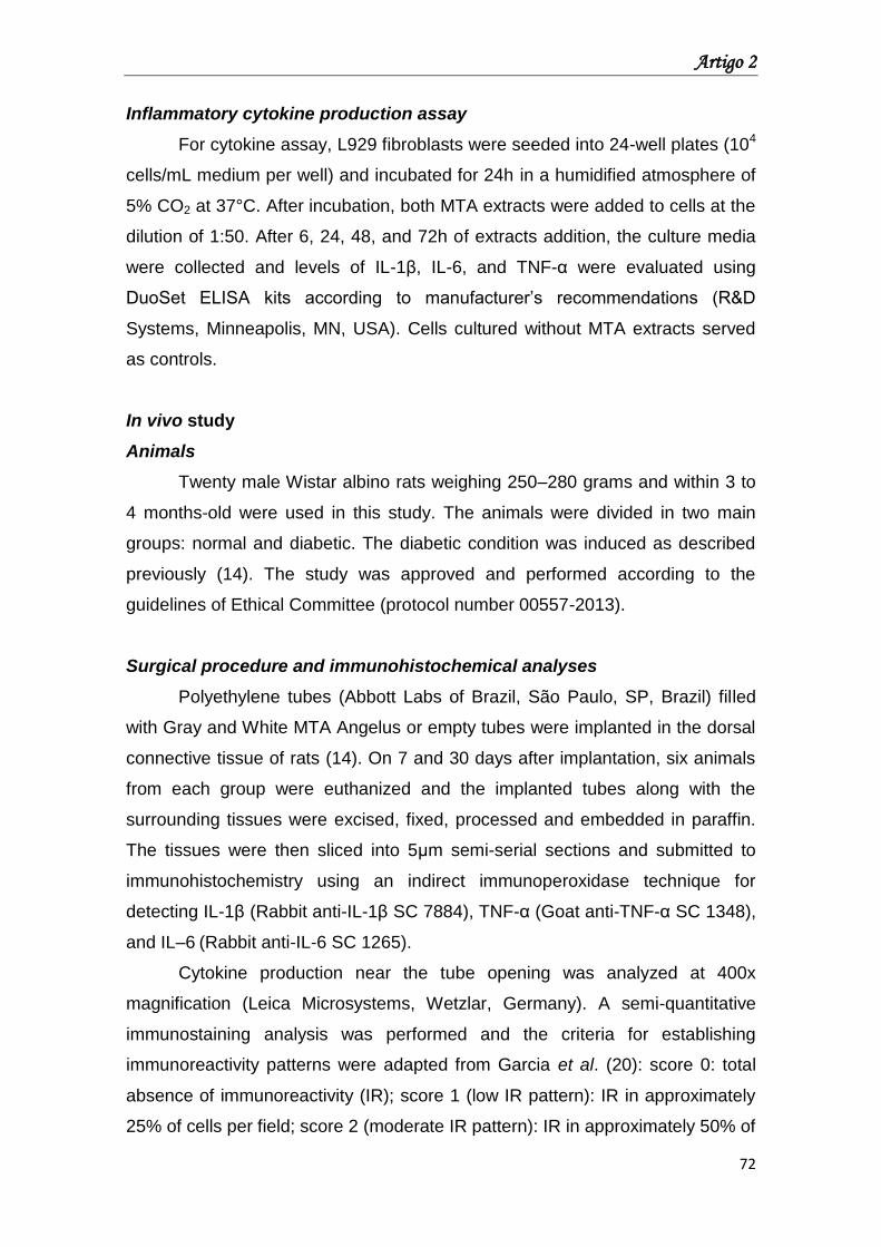
Artigo 2
72
Inflammatory cytokine production assay
For cytokine assay, L929 fibroblasts were seeded into 24-well plates (104
cells/mL medium per well) and incubated for 24h in a humidified atmosphere of
5% CO2 at 37°C. After incubation, both MTA extracts were added to cells at the
dilution of 1:50. After 6, 24, 48, and 72h of extracts addition, the culture media
were collected and levels of IL-1β, IL-6, and TNF-α were evaluated using
DuoSet ELISA kits according to manufacturer’s recommendations (R&D
Systems, Minneapolis, MN, USA). Cells cultured without MTA extracts served
as controls.
In vivo study
Animals
Twenty male Wistar albino rats weighing 250–280 grams and within 3 to
4 months-old were used in this study. The animals were divided in two main
groups: normal and diabetic. The diabetic condition was induced as described
previously (14). The study was approved and performed according to the
guidelines of Ethical Committee (protocol number 00557-2013).
Surgical procedure and immunohistochemical analyses
Polyethylene tubes (Abbott Labs of Brazil, São Paulo, SP, Brazil) filled
with Gray and White MTA Angelus or empty tubes were implanted in the dorsal
connective tissue of rats (14). On 7 and 30 days after implantation, six animals
from each group were euthanized and the implanted tubes along with the
surrounding tissues were excised, fixed, processed and embedded in paraffin.
The tissues were then sliced into 5μm semi-serial sections and submitted to
immunohistochemistry using an indirect immunoperoxidase technique for
detecting IL-1β (Rabbit anti-IL-1β SC 7884), TNF-α (Goat anti-TNF-α SC 1348),
and IL–6 (Rabbit anti-IL-6 SC 1265).
Cytokine production near the tube opening was analyzed at 400x
magnification (Leica Microsystems, Wetzlar, Germany). A semi-quantitative
immunostaining analysis was performed and the criteria for establishing
immunoreactivity patterns were adapted from Garcia et al. (20): score 0: total
absence of immunoreactivity (IR); score 1 (low IR pattern): IR in approximately
25% of cells per field; score 2 (moderate IR pattern): IR in approximately 50% of
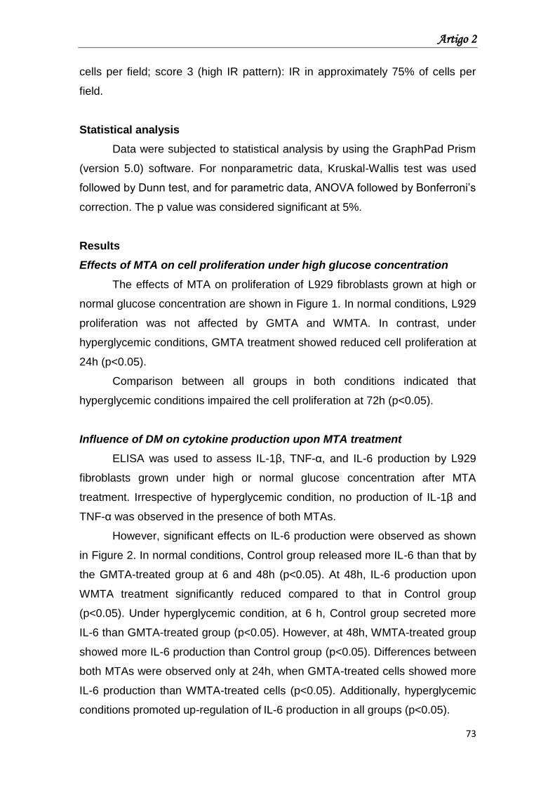
Artigo 2
73
cells per field; score 3 (high IR pattern): IR in approximately 75% of cells per
field.
Statistical analysis
Data were subjected to statistical analysis by using the GraphPad Prism
(version 5.0) software. For nonparametric data, Kruskal-Wallis test was used
followed by Dunn test, and for parametric data, ANOVA followed by Bonferroni’s
correction. The p value was considered significant at 5%.
Results
Effects of MTA on cell proliferation under high glucose concentration
The effects of MTA on proliferation of L929 fibroblasts grown at high or
normal glucose concentration are shown in Figure 1. In normal conditions, L929
proliferation was not affected by GMTA and WMTA. In contrast, under
hyperglycemic conditions, GMTA treatment showed reduced cell proliferation at
24h (p<0.05).
Comparison between all groups in both conditions indicated that
hyperglycemic conditions impaired the cell proliferation at 72h (p<0.05).
Influence of DM on cytokine production upon MTA treatment
ELISA was used to assess IL-1β, TNF-α, and IL-6 production by L929
fibroblasts grown under high or normal glucose concentration after MTA
treatment. Irrespective of hyperglycemic condition, no production of IL-1β and
TNF-α was observed in the presence of both MTAs.
However, significant effects on IL-6 production were observed as shown
in Figure 2. In normal conditions, Control group released more IL-6 than that by
the GMTA-treated group at 6 and 48h (p<0.05). At 48h, IL-6 production upon
WMTA treatment significantly reduced compared to that in Control group
(p<0.05). Under hyperglycemic condition, at 6 h, Control group secreted more
IL-6 than GMTA-treated group (p<0.05). However, at 48h, WMTA-treated group
showed more IL-6 production than Control group (p<0.05). Differences between
both MTAs were observed only at 24h, when GMTA-treated cells showed more
IL-6 production than WMTA-treated cells (p<0.05). Additionally, hyperglycemic
conditions promoted up-regulation of IL-6 production in all groups (p<0.05).
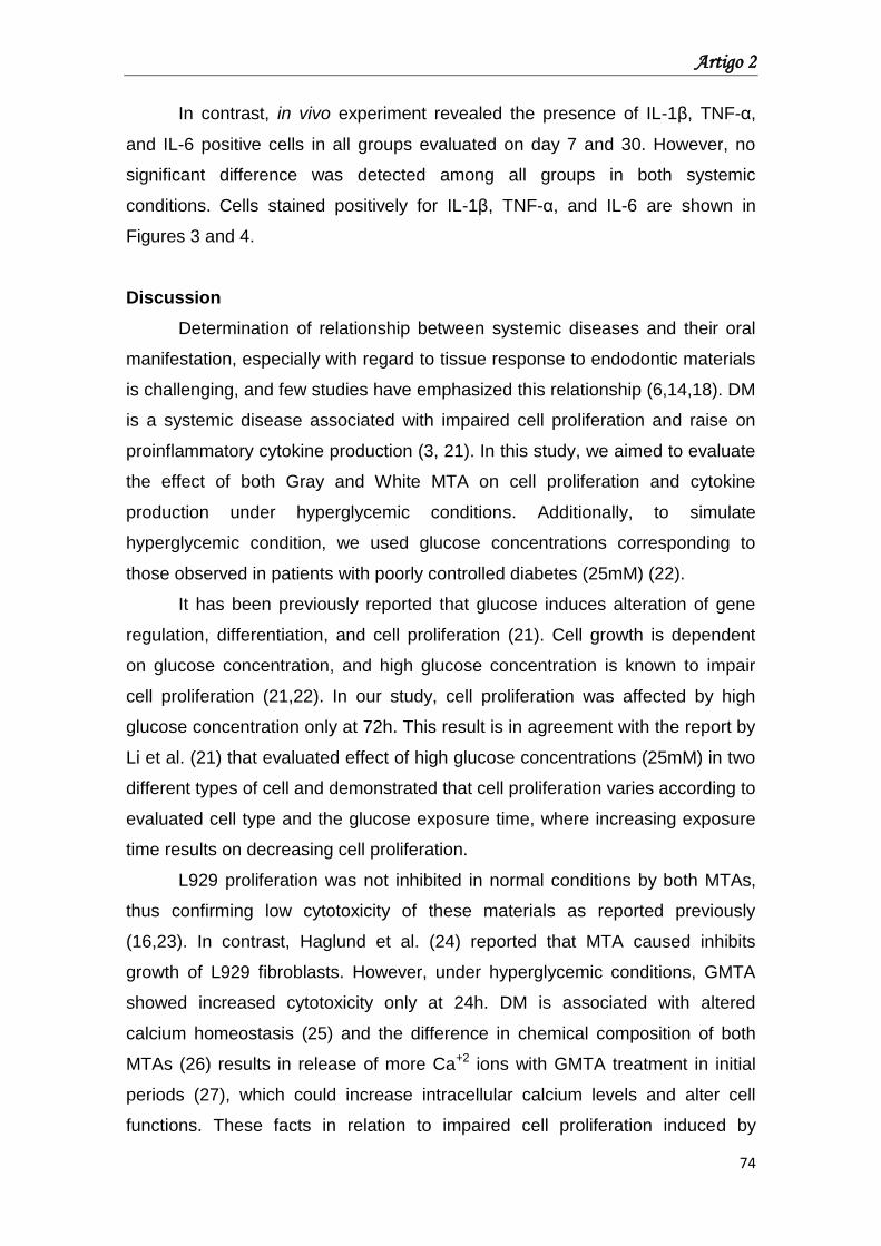
Artigo 2
74
In contrast, in vivo experiment revealed the presence of IL-1β, TNF-α,
and IL-6 positive cells in all groups evaluated on day 7 and 30. However, no
significant difference was detected among all groups in both systemic
conditions. Cells stained positively for IL-1β, TNF-α, and IL-6 are shown in
Figures 3 and 4.
Discussion
Determination of relationship between systemic diseases and their oral
manifestation, especially with regard to tissue response to endodontic materials
is challenging, and few studies have emphasized this relationship (6,14,18). DM
is a systemic disease associated with impaired cell proliferation and raise on
proinflammatory cytokine production (3, 21). In this study, we aimed to evaluate
the effect of both Gray and White MTA on cell proliferation and cytokine
production under hyperglycemic conditions. Additionally, to simulate
hyperglycemic condition, we used glucose concentrations corresponding to
those observed in patients with poorly controlled diabetes (25mM) (22).
It has been previously reported that glucose induces alteration of gene
regulation, differentiation, and cell proliferation (21). Cell growth is dependent
on glucose concentration, and high glucose concentration is known to impair
cell proliferation (21,22). In our study, cell proliferation was affected by high
glucose concentration only at 72h. This result is in agreement with the report by
Li et al. (21) that evaluated effect of high glucose concentrations (25mM) in two
different types of cell and demonstrated that cell proliferation varies according to
evaluated cell type and the glucose exposure time, where increasing exposure
time results on decreasing cell proliferation.
L929 proliferation was not inhibited in normal conditions by both MTAs,
thus confirming low cytotoxicity of these materials as reported previously
(16,23). In contrast, Haglund et al. (24) reported that MTA caused inhibits
growth of L929 fibroblasts. However, under hyperglycemic conditions, GMTA
showed increased cytotoxicity only at 24h. DM is associated with altered
calcium homeostasis (25) and the difference in chemical composition of both
MTAs (26) results in release of more Ca+2 ions with GMTA treatment in initial
periods (27), which could increase intracellular calcium levels and alter cell
functions. These facts in relation to impaired cell proliferation induced by
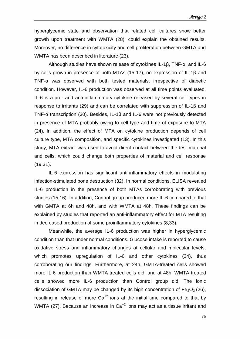
Artigo 2
75
hyperglycemic state and observation that related cell cultures show better
growth upon treatment with WMTA (28), could explain the obtained results.
Moreover, no difference in cytotoxicity and cell proliferation between GMTA and
WMTA has been described in literature (23).
Although studies have shown release of cytokines IL-1β, TNF-α, and IL-6
by cells grown in presence of both MTAs (15-17), no expression of IL-1β and
TNF-α was observed with both tested materials, irrespective of diabetic
condition. However, IL-6 production was observed at all time points evaluated.
IL-6 is a pro- and anti-inflammatory cytokine released by several cell types in
response to irritants (29) and can be correlated with suppression of IL-1β and
TNF-α transcription (30). Besides, IL-1β and IL-6 were not previously detected
in presence of MTA probably owing to cell type and time of exposure to MTA
(24). In addition, the effect of MTA on cytokine production depends of cell
culture type, MTA composition, and specific cytokines investigated (13). In this
study, MTA extract was used to avoid direct contact between the test material
and cells, which could change both properties of material and cell response
(19,31).
IL-6 expression has significant anti-inflammatory effects in modulating
infection-stimulated bone destruction (32). In normal conditions, ELISA revealed
IL-6 production in the presence of both MTAs corroborating with previous
studies (15,16). In addition, Control group produced more IL-6 compared to that
with GMTA at 6h and 48h, and with WMTA at 48h. These findings can be
explained by studies that reported an anti-inflammatory effect for MTA resulting
in decreased production of some proinflammatory cytokines (8,33).
Meanwhile, the average IL-6 production was higher in hyperglycemic
condition than that under normal conditions. Glucose intake is reported to cause
oxidative stress and inflammatory changes at cellular and molecular levels,
which promotes upregulation of IL-6 and other cytokines (34), thus
corroborating our findings. Furthermore, at 24h, GMTA-treated cells showed
more IL-6 production than WMTA-treated cells did, and at 48h, WMTA-treated
cells showed more IL-6 production than Control group did. The ionic
dissociation of GMTA may be changed by its high concentration of Fe2O3 (26),
resulting in release of more Ca+2 ions at the initial time compared to that by
WMTA (27). Because an increase in Ca+2 ions may act as a tissue irritant and
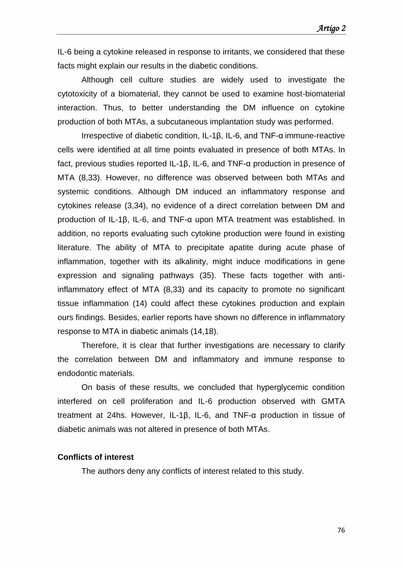
Artigo 2
76
IL-6 being a cytokine released in response to irritants, we considered that these
facts might explain our results in the diabetic conditions.
Although cell culture studies are widely used to investigate the
cytotoxicity of a biomaterial, they cannot be used to examine host-biomaterial
interaction. Thus, to better understanding the DM influence on cytokine
production of both MTAs, a subcutaneous implantation study was performed.
Irrespective of diabetic condition, IL-1β, IL-6, and TNF-α immune-reactive
cells were identified at all time points evaluated in presence of both MTAs. In
fact, previous studies reported IL-1β, IL-6, and TNF-α production in presence of
MTA (8,33). However, no difference was observed between both MTAs and
systemic conditions. Although DM induced an inflammatory response and
cytokines release (3,34), no evidence of a direct correlation between DM and
production of IL-1β, IL-6, and TNF-α upon MTA treatment was established. In
addition, no reports evaluating such cytokine production were found in existing
literature. The ability of MTA to precipitate apatite during acute phase of
inflammation, together with its alkalinity, might induce modifications in gene
expression and signaling pathways (35). These facts together with anti-
inflammatory effect of MTA (8,33) and its capacity to promote no significant
tissue inflammation (14) could affect these cytokines production and explain
ours findings. Besides, earlier reports have shown no difference in inflammatory
response to MTA in diabetic animals (14,18).
Therefore, it is clear that further investigations are necessary to clarify
the correlation between DM and inflammatory and immune response to
endodontic materials.
On basis of these results, we concluded that hyperglycemic condition
interfered on cell proliferation and IL-6 production observed with GMTA
treatment at 24hs. However, IL-1β, IL-6, and TNF-α production in tissue of
diabetic animals was not altered in presence of both MTAs.
Conflicts of interest
The authors deny any conflicts of interest related to this study.
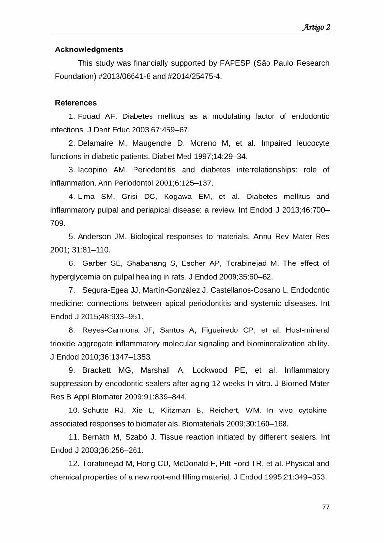
Artigo 2
77
Acknowledgments
This study was financially supported by FAPESP (São Paulo Research
Foundation) #2013/06641-8 and #2014/25475-4.
References
1. Fouad AF. Diabetes mellitus as a modulating factor of endodontic
infections. J Dent Educ 2003;67:459–67.
2. Delamaire M, Maugendre D, Moreno M, et al. Impaired leucocyte
functions in diabetic patients. Diabet Med 1997;14:29–34.
3. Iacopino AM. Periodontitis and diabetes interrelationships: role of
inflammation. Ann Periodontol 2001;6:125–137.
4. Lima SM, Grisi DC, Kogawa EM, et al. Diabetes mellitus and
inflammatory pulpal and periapical disease: a review. Int Endod J 2013;46:700–
709.
5. Anderson JM. Biological responses to materials. Annu Rev Mater Res
2001; 31:81–110.
6. Garber SE, Shabahang S, Escher AP, Torabinejad M. The effect of
hyperglycemia on pulpal healing in rats. J Endod 2009;35:60–62.
7. Segura-Egea JJ, Martín-González J, Castellanos-Cosano L. Endodontic
medicine: connections between apical periodontitis and systemic diseases. Int
Endod J 2015;48:933–951.
8. Reyes-Carmona JF, Santos A, Figueiredo CP, et al. Host-mineral
trioxide aggregate inflammatory molecular signaling and biomineralization ability.
J Endod 2010;36:1347–1353.
9. Brackett MG, Marshall A, Lockwood PE, et al. Inflammatory
suppression by endodontic sealers after aging 12 weeks In vitro. J Biomed Mater
Res B Appl Biomater 2009;91:839–844.
10. Schutte RJ, Xie L, Klitzman B, Reichert, WM. In vivo cytokine-
associated responses to biomaterials. Biomaterials 2009;30:160–168.
11. Bernáth M, Szabó J. Tissue reaction initiated by different sealers. Int
Endod J 2003;36:256–261.
12. Torabinejad M, Hong CU, McDonald F, Pitt Ford TR, et al. Physical and
chemical properties of a new root-end filling material. J Endod 1995;21:349–353.
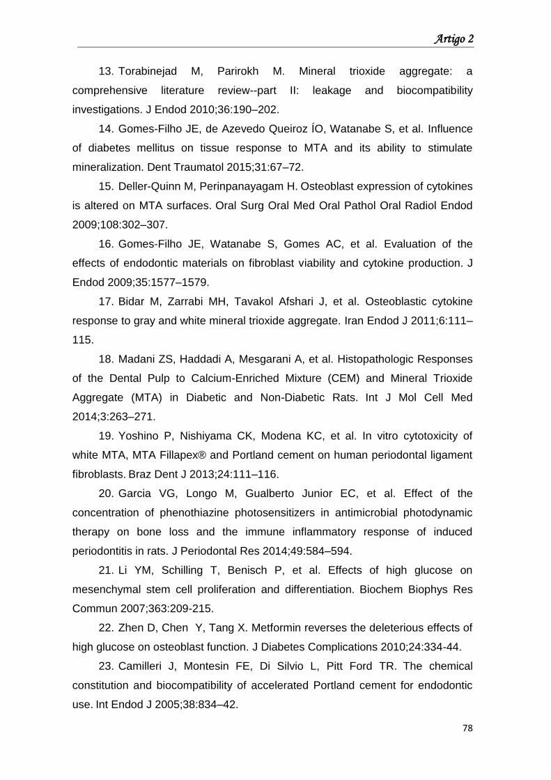
Artigo 2
78
13. Torabinejad M, Parirokh M. Mineral trioxide aggregate: a
comprehensive literature review--part II: leakage and biocompatibility
investigations. J Endod 2010;36:190–202.
14. Gomes-Filho JE, de Azevedo Queiroz ÍO, Watanabe S, et al. Influence
of diabetes mellitus on tissue response to MTA and its ability to stimulate
mineralization. Dent Traumatol 2015;31:67–72.
15. Deller-Quinn M, Perinpanayagam H. Osteoblast expression of cytokines
is altered on MTA surfaces. Oral Surg Oral Med Oral Pathol Oral Radiol Endod
2009;108:302–307.
16. Gomes-Filho JE, Watanabe S, Gomes AC, et al. Evaluation of the
effects of endodontic materials on fibroblast viability and cytokine production. J
Endod 2009;35:1577–1579.
17. Bidar M, Zarrabi MH, Tavakol Afshari J, et al. Osteoblastic cytokine
response to gray and white mineral trioxide aggregate. Iran Endod J 2011;6:111–
115.
18. Madani ZS, Haddadi A, Mesgarani A, et al. Histopathologic Responses
of the Dental Pulp to Calcium-Enriched Mixture (CEM) and Mineral Trioxide
Aggregate (MTA) in Diabetic and Non-Diabetic Rats. Int J Mol Cell Med
2014;3:263–271.
19. Yoshino P, Nishiyama CK, Modena KC, et al. In vitro cytotoxicity of
white MTA, MTA Fillapex® and Portland cement on human periodontal ligament
fibroblasts. Braz Dent J 2013;24:111–116.
20. Garcia VG, Longo M, Gualberto Junior EC, et al. Effect of the
concentration of phenothiazine photosensitizers in antimicrobial photodynamic
therapy on bone loss and the immune inflammatory response of induced
periodontitis in rats. J Periodontal Res 2014;49:584–594.
21. Li YM, Schilling T, Benisch P, et al. Effects of high glucose on
mesenchymal stem cell proliferation and differentiation. Biochem Biophys Res
Commun 2007;363:209-215.
22. Zhen D, Chen Y, Tang X. Metformin reverses the deleterious effects of
high glucose on osteoblast function. J Diabetes Complications 2010;24:334-44.
23. Camilleri J, Montesin FE, Di Silvio L, Pitt Ford TR. The chemical
constitution and biocompatibility of accelerated Portland cement for endodontic
use. Int Endod J 2005;38:834–42.
Artigo 2
79
24. Haglund R, He J, Jarvis J, et al. Effects of root-end filling materials on
fibroblasts and macrophages in vitro. Oral Surg Oral Med Oral Pathol Oral Radiol
Endod 2003;95:739-45.
25. Levy J, Gavin JR 3rd, Sowers JR. Diabetes mellitus: a disease of
abnormal cellular calcium metabolism? Am J Med 1994;96:260-73.
26. Asgary S, Parirokh M, Eghbal MJ, Brink F. Chemical differences
between white and gray mineral trioxide aggregate. J Endod 2005;31:101–3.
27. Garcia LdaF, Chinelatti MA, Rossetto HL, et al. Solubility and
disintegration of new calcium aluminate cement (EndoBinder) containing different
radiopacifying agents. J Endod 2014a;40:261-5.
28. Oviir T, Pagoria D, Ibarra G, Geurtsen W. Effects of gray and white
mineral trioxide aggregate on the proliferation of oral keratinocytes and
cementoblasts. J Endod 2006;32:210–213.
29. Azuma MM, Samuel RO, Gomes-Filho JE, et al. The role of IL-6 on
apical periodontitis: a systematic review. Int Endod J 2014;47:615-21.
30. Schindler R, Mancilla J, Endres S, et al. Correlations and interactions in
the production of interleukin-6 (IL-6), IL-1, and tumor necrosis factor (TNF) in
human blood mononuclear cells: IL-6 suppresses IL-1 and TNF. Blood
1990;75:40-7.
31. Keiser K, Johnson CC, Tipton DA. Cytotoxicity of mineral trioxide
aggregate using human periodontal ligament fibroblasts. J Endod 2000;26:288-
91.
32. Balto K, Sasaki H, Stashenko P. Interleukin-6 deficiency increases
inflammatory bone destruction. Infect Immun. 2001;69:744-50.
33. Barbosa Silva, MJ, Vieira, LQ, Sobrinho, AP. The effects of mineral
trioxide aggregates on cytokine production by mouse pulp tissue. Oral Surg Oral
Med Oral Pathol Oral Radiol Endod 2008;105:e70–e76.
34. Dandona, P, Aljada, A, Bandyopadhyay, A. Inflammation: the link
between insulin resistance, obesity and diabetes. Trends Immunol 2004;25:4–7.
35. Reyes-Carmona, JF, Santos, AR, Figueiredo, CP, et al. In vivo host
interactions with mineral trioxide aggregate and calcium hydroxide: inflammatory
molecular signaling assessment. J Endod 2011;37:1225–1235.
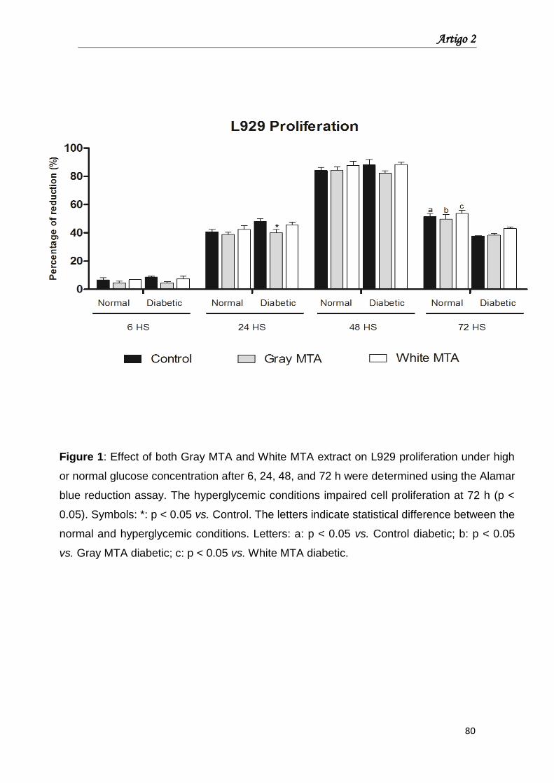
Artigo 2
80
Figure 1: Effect of both Gray MTA and White MTA extract on L929 proliferation under high
or normal glucose concentration after 6, 24, 48, and 72 h were determined using the Alamar
blue reduction assay. The hyperglycemic conditions impaired cell proliferation at 72 h (p <
0.05). Symbols: *: p < 0.05 vs. Control. The letters indicate statistical difference between the
normal and hyperglycemic conditions. Letters: a: p < 0.05 vs. Control diabetic; b: p < 0.05
vs. Gray MTA diabetic; c: p < 0.05 vs. White MTA diabetic.
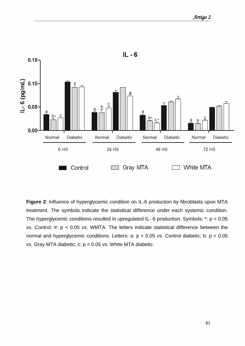
Artigo 2
81
Figure 2: Influence of hyperglycemic condition on IL-6 production by fibroblasts upon MTA
treatment. The symbols indicate the statistical difference under each systemic condition.
The hyperglycemic conditions resulted in upregulated IL- 6 production. Symbols: *: p < 0.05
vs. Control; #: p < 0.05 vs. WMTA. The letters indicate statistical difference between the
normal and hyperglycemic conditions. Letters: a: p < 0.05 vs. Control diabetic; b: p < 0.05
vs. Gray MTA diabetic; c: p < 0.05 vs. White MTA diabetic.
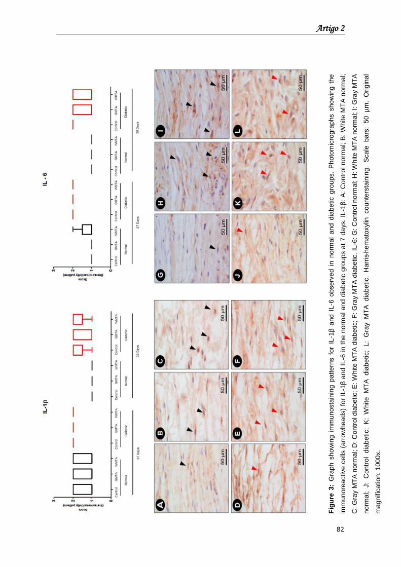
Artigo 2
82
Fig
ure
3:
Gra
ph s
ho
win
g i
mm
unosta
inin
g p
attern
s f
or
IL-1
β a
nd I
L-6
observ
ed i
n n
orm
al
an
d d
iab
etic g
roups.
Photo
mic
rogra
phs s
ho
win
g t
he
imm
unore
active c
ells
(arr
ow
hea
ds)
for
IL-1
β a
nd I
L-6
in t
he n
orm
al and
dia
betic g
roups a
t 7 d
ays.
IL-1
β:
A:
Contr
ol n
orm
al; B
: W
hite M
TA
norm
al;
C:
Gra
y M
TA
norm
al; D
: C
ontr
ol d
iab
etic;
E: W
hite M
TA
dia
betic;
F: G
ray M
TA
dia
betic.
IL-6
: G
: C
ontr
ol n
orm
al; H
: W
hite M
TA
norm
al; I: G
ray M
TA
norm
al; J:
Contr
ol
dia
be
tic;
K:
White M
TA
dia
be
tic;
L:
Gra
y M
TA
dia
betic.
Harr
is-h
em
ato
xylin
co
unte
rsta
inin
g.
Scale
bars
: 50 µ
m.
Origin
al
magnific
ation:
100
0x.
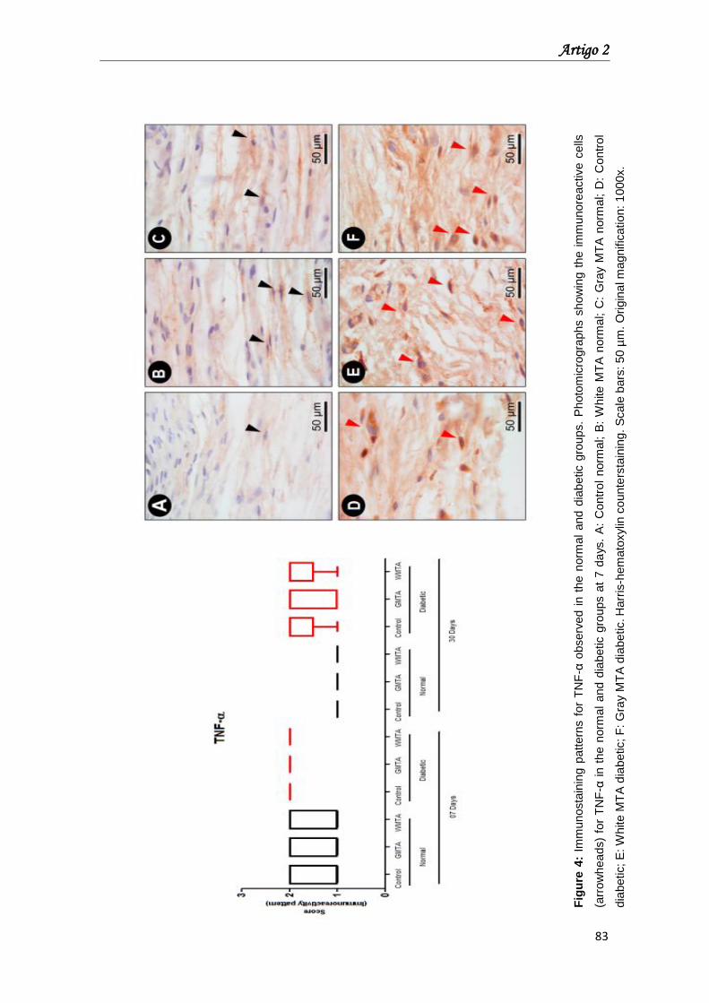
Artigo 2
83
Fig
ure
4:
Imm
unosta
inin
g p
attern
s f
or
TN
F-α
observ
ed i
n t
he n
orm
al
and d
iab
etic g
roups.
Photo
mic
rogra
phs s
how
ing t
he i
mm
unore
active c
ells
(arr
ow
hea
ds)
for
TN
F-α
in t
he n
orm
al
and d
iabetic g
roups a
t 7 d
ays.
A:
Contr
ol
norm
al; B
: W
hite M
TA
norm
al; C
: G
ray M
TA
norm
al; D
: C
ontr
ol
dia
betic; E
: W
hite M
TA
dia
be
tic;
F:
Gra
y M
TA
dia
be
tic. H
arr
is-h
em
ato
xylin
co
unte
rsta
inin
g.
Scale
bars
: 50 µ
m. O
rigin
al m
agnific
ation:
1000x.

Artigo 3
Artigo 3
85
Artigo 3: Diabetes mellitus affects mineralization ability of white
mineral trioxide aggregate
Abstract
Introduction: Diabetes mellitus (DM) is a metabolic disorder that affects tissue
repair capacity. Therewith, the aim of this study was to evaluate the influence of
DM on the tissue response and mineralization ability of gray (GMTA) and white
mineral trioxide aggregate (WMTA).
Methods: Twenty-four Wistar rats were divided into two groups: normal and
diabetic (Alloxan-induced). Four polyethylene tubes were implanted in the
dorsal connective tissue of each rat, three containing GMTA, WMTA, zinc oxide
and eugenol (ZOE) and one empty tube. Six animals from each group received
injections of calcein, alizarin, and oxytetracycline on day 7, 14, and 21,
respectively. The animals were killed after 7 and 30 days of experimentation
and the tubes with surrounding tissues were removed and fixed. All specimens
were prepared for histological analysis with either hematoxylin and eosin or von
Kossa staining, or remained unstained for polarized light or fluorescence
analysis.
Results: All materials in both systemic groups caused moderate reactions after
7 days, which decreased with time. Structures that were positive for von Kossa
or birefringent to polarized light were observed in response to both MTAs but
not to ZOE, regardless of the diabetic condition and time of evaluation (p <
0.05). WMTA exhibited higher fluorescence intensity than GMTA after 14 days
in the normal group (p < 0.05). The fluorescence intensity was lower in the
diabetic group for WMTA after 14 days (p < 0.05).
Conclusion: It was concluded that diabetic conditions altered the fluorescence
intensity of WMTA but not the inflammatory response.
Key Words: Diabetic mellitus; Mineralization; Gray MTA; White MTA
Artigo 3
86
Introduction
The possible association between systemic conditions and oral diseases
has been emphasized (1), especially in relation to diabetes mellitus (DM) (2),
which is a metabolic disease characterized by chronic hyperglycemia that
affects an estimated 382 million people worldwide (3). If uncontrolled, DM leads
to complications that have been associated with activation of pathways that
increase inflammation, oxidative stress, and apoptosis (4).
Considering the relationship between DM and oral diseases, it was
shown that DM can be a modulating factor of endodontic infections and may
compromise the healing process of periapical tissues (5). Hyperglycemia may
increase the inflammatory state because of elevated levels of systemic
inflammation markers (6), and the tissue repair capacity including dental pulp
healing can be hindered (7). Thus, it is paramount to understand the correlation
between systemic and oral diseases, including the effect of DM on tissue
response to endodontic materials.
Mineral trioxide aggregate (MTA) is a root-end filling material used for the
repair of lateral perforations and because of its adequate physical, chemical,
and biological properties (8), it has been used in pulpotomy, pulp capping, and
the treatment of open apexes (9). White MTA (WMTA) was introduced as an
alternative to gray MTA (GMTA), aiming to avoid tooth staining. The main
difference between the two materials is the lower concentration of Al2O3, MgO,
and FeO in WMTA (10). The reduction in FeO results in a reduction of the
aluminoferrite phase, which is responsible for the gray color of GMTA (11).
However, studies have also verified in vitro staining caused by WMTA, where
gray-colored alteration was observed in contact with dentin (12), even in clinical
application of WMTA (13).
Studies comparing WMTA and GMTA showed some similarities; both
materials are non-toxic and induce cell proliferation when hydrated (14). Both
are able to stimulate dentinal bridge formation when used as pulp capping
materials (15). Both materials are considered biocompatible (16, 17). However,
some differences have also been reported. Osteoblast adherence is not favored
by WMTA (18), and cementoblasts and keratinocytes grow better on the surface
of WMTA than on GMTA (19). Moreover, previous data revealed no difference
Artigo 3
87
in tissue response and the mineralization potential of GMTA in diabetic
conditions (20).
Although the differences between WMTA and GMTA have already been
widely explored, there is no study in the literature comparing the tissue
response to these materials in diabetic conditions, which can modify the
systemic and local inflammatory responses. Hence, the aim of this study was to
compare the subcutaneous tissue response and mineralization ability of WMTA
and GMTA in diabetic rats.
Materials and Methods
The present study was approved and performed according to the
guidelines of the Ethical Committee (protocol number 00557-2013). Twenty-four
male albino Wistar rats, aged between 3 and 4 months and weighing around
250–280 g, were used in the study. The rats were divided into two groups of 12
animals each: normal and diabetic.
Diabetes induction
For the diabetes induction, 12 animals received 150 mg/kg of Alloxan
monohydrate (Sigma Aldrich Corp., St. Louis, MO, USA) in a single
intraperitoneal dose. Blood glucose was measured three days after injection to
confirm hyperglycemia. Animals were considered diabetic when they showed
glucose levels higher than 250 mg/dL, and the third day after injection was
considered the first day of diabetes (20).
Sample preparation and surgical procedures
Sterile polyethylene open-ended tubes (Abbott Labs of Brazil, São Paulo,
SP, Brazil) with an internal diameter of 1.0 mm, an external diameter of 1.6 mm,
and a length of 10.0 mm were used for the experiments. Gray MTA, white MTA
(Angelus Indústria de Produtos Odontológicos S/A, Londrina, PR, Brazil), and
zinc oxide Eugenol® (SS White, Artigos Dentários, Rio de Janeiro, RJ, Brazil)
were prepared according to the manufacturers’ recommendations and inserted
into the twenty-four tubes with a lentulo spiral (Maillefer Dentsply, Tulsa, OK,
USA). Twenty-four tubes remained empty and these were used as controls.

Artigo 3
88
On the first day of diabetes, subcutaneous implantation was performed
following previous studies (16, 20).
Fluorochrome injection
The fluorescent markers calcein, alizarin red S, and oxytetracycline
hydrochloride (Sigma Aldrich Corp., St Louis, MO, USA) were injected
intramuscularly at a dose of 20 mg/kg 7, 14, and 21 days after implantation,
respectively, only in 12 animals (six from each group) (20). The fluorochromes
were used to evaluate the time sequence of calcium deposition in the tissue.
Histological procedures and analysis
Seven and 30 days after the implantation, the animals’ blood glucose
levels were measured to confirm diabetic conditions. The animals were killed by
an anesthetic overdose, and the tubes with the surrounding tissues were
removed and fixed in 10% formalin solution. The specimens were processed
and embedded in glycol methacrylate, serially sectioned into 3-μm cuts, and
stained with hematoxylin-eosin. The 10-μm cuts were stained using the von
Kossa technique or were kept unstained for polarized light visualization. The 50-
μm cuts remained unstained and these were observed with fluorescence (20).
Inflammatory reactions in the tissues close to the material were
evaluated according to ISO/TR 7405-1997 (21) as: 0, no or few inflammatory
cells and no reaction; 1, fewer than 25 cells and light reaction; 2, between 25–
125 cells and moderate reaction; and 3, 125 or more cells and severe reaction.
Fibrous capsules were considered to be thin when the thickness was <150μm
and thick at >150μm. Necrosis and mineralization areas were recorded in µm2
using Leica Qwin software (Leica Microsystems, Wetzlar, Germany). An
average value for each material was obtained from the sum of cells counted in
ten separate fields (400× magnification). Fluorescence intensity was measured
using a fluorescence light microscopy (DM 4000B, Leica Microsystems,
Wetzlar, Germany) (534 nm for calcein, 357 nm for alizarin, and ultraviolet 368
nm for oxytetracycline). A measuring area (150µm × 700µm) in the middle of
the tube opening was used, and fluorescence intensity was determined using
the software LAS v4.1 (Leica Applications Suite – version 4.1; Leica
Microsystems, Wetzlar, Germany).

Artigo 3
89
Statistical analysis
The data for inflammatory response were analyzed using Kruskal-Wallis
tests. The results of mineralization and fluorescence intensity were analyzed by
ANOVA and Mann Whitney tests (p < 0.05).
Results
Figures 1 and 2 show the inflammatory response found in the normal and
diabetic group at 30 days. Data comparing inflammatory responses are shown
in Table 1. The presence of mineralization as determined by von Kossa staining
and the fluorescence intensity of calcein, alizarin, and oxytetracycline staining
are shown in Table 2.
Diabetes did not modify the inflammatory response of any of the tubes
including the empty tubes (negative control). After 7 days, there was no
significant difference among the scores of the different groups (median score 2).
At 30 days, a decrease in inflammatory response was observed for all materials
(median score 1), independent of the diabetic condition. There was no
statistically significant difference among the materials for necrotic areas (p >
0.05), which were absent on day 30.
There were significant differences between all materials and the positive
control (p<0.05) in all mineralization parameters, independent of the diabetic
status. Mineralized areas were observed in both groups at 7 and 30 days.
These areas were absent in contact with ZOE at 7 and 30 days, independent of
the diabetic condition.
Significant differences were found in the fluorescence intensity between
each material and the control group, independent of the fluorochrome and
diabetic condition (p<0.05). The positive control showed baseline fluorescence
intensity.
In the normal animals, WMTA showed higher fluorescence intensity than
GMTA at 14 days (p<0.05). Moreover, in diabetic animals, the fluorescence
intensity of WMTA was lower than that in normal animals within the same
experimental period (p<0.05). Additionally, the ZOE group showed lower
fluorescence intensity than both MTAs for all fluorochromes, regardless of the
diabetic status and observation period (p<0.05).
Artigo 3
90
Discussion
Subcutaneous tissue reaction is one of the in vivo biocompatibility tests
that have been used to determine the local effects and biocompatibility of
materials (16,17). The host response to materials is complex and dependent on
the innate and non-specific immune response that, depending on the chemical
and physical properties of the material, might evoke a signaling pathway of
inflammatory cascade, inactivating the inflammatory process, and contributing
or not to a wound healing process (22).
Diabetes mellitus is a disorder that when uncontrolled might lead to oral
problems such as changes in pulp tissues (7). DM changes the inflammatory
response (7) once it reduces the functions of polymorphonuclear leukocytes
(23) and alters the immune responses (24), resulting in increased susceptibility
to infections (25) and delayed healing processes (23). Additionally, DM has
been associated with altered calcium homeostasis (26), which leads to a lower
capacity of bone formation and mineralization.
Both MTAs evoked a moderate inflammatory response in 7 days, which
decreased after 30 days, regardless of the diabetic condition. The current
results corroborate with those of other studies (16, 20, 27), indicating that the
tissue response are similar between GMTA and WMTA. However, the present
findings are in conflict with others that showed differences in the intensity of
inflammatory responses between GMTA and WMTA in a 7-day period (17).
These differences may be attributed to the method used to evaluate the
biocompatibility of dental materials. Whereas previous results were obtained
based on the number of inflammatory cells, the present study is based on the
evaluation of the density of inflammatory cells.
Despite this, GMTA releases more Ca+2 ions than WMTA does, with
decreasing ion concentrations with time (28). In this study, WMTA exhibited
higher fluorescence intensity than GMTA did in the normal group at 14 days.
This was possibly due to the different chemical compositions of WMTA and
GMTA, especially the higher concentration of Fe2O3 present on GMTA (10) that
could interfere in the ionic dissociation and affect calcium release. Besides,
WMTA has been associated with higher calcium release compared with GMTA
(29), which might explain these results. Calcium phosphate and calcium oxide
are released by MTA. Calcium oxide reacts with water in tissue fluids to form
Artigo 3
91
calcium hydroxide, which further dissociates into Ca+2 and OH- ions. The Ca+2
ions react with the carbon dioxide that is present in the tissues and form calcium
carbonate granulations, which would stimulate the deposition of hard tissues
(27). Moreover, Ca+2 ions can react with tissue fluids and produce
hydroxyapatite crystals. This ability makes MTA not only biocompatible but also
bioactive, and these properties can be seen in both MTAs (30).
The present results did not show evidence of the direct correlation
between DM and inflammatory response for both MTAs. However, WMTA
exhibited lower fluorescence intensity in diabetic animals on the 14th day. The
stimulation of repair by deposition of mineralized tissue is associated with the
ability of a biomaterial to release calcium ions (16, 27, 28). The lower presence
of metallic oxides in WMTA (10) can promote slow calcium release. On the
other hand, DM affects the microcirculation (31) and promotes abnormal
metabolism of calcium (26). Thus, we believe that once DM changes the
inflammatory response and alters calcium homeostasis, it could have interfered
with the response on the 14th day, which may justify our findings.
The chemical composition of sealers can positively or negatively affect
the result of biocompatibility tests (22). Thus, ZOE was used as a positive
control, since the toxicity of zinc oxide and eugenol sealers has been
demonstrated both in vivo and in vitro (32, 33). In this study, ZOE presented the
same biological behavior as both MTAs after 30 days, independent of the
diabetic condition. Such results disagree with studies that showed tissue
irritation (32, 33), probably because of ZOE liquid/powder proportion. The high
concentration of eugenol in the tissue is sufficient to inhibit cell respiration and
thus kill cells (34). The fluorescence intensity observed could be explained by
the capacity of ZOE to release calcium ions (35).
It is evident that other studies using different methods are necessary to
elucidate the relationship between DM and the biocompatibility of endodontic
materials, as the correlation is not yet completely understood.
In conclusion, the present results indicate that in general, the
inflammatory response to the materials did not change by diabetic conditions,
but the fluorescence intensity of WMTA at 14th days was altered in diabetic
animals.
Artigo 3
92
Conflicts of interest
The authors deny any conflicts of interest.
Acknowledgments
The authors thank FAPESP (São Paulo Research Foundation) for
financial support. Process number: 2013/06641-8.
References
1. Tavares M, Lindefjeld Calabi KA, San Martin L. Systemic diseases and
oral health. Dent Clin N Am 2014;58:797–814.
2. Lamster IB, Lalla E, Borgnakke WS, Taylor GW. The relationship
between oral health and diabetes mellitus. J Am Dent Assoc
2008;139Suppl:19S–24S.
3. Guariguata L, Whiting DR, Hambleton I, et al. Global estimates of
diabetes prevalence for 2013 and projections for 2035. Diabetes Res Clin
Pract 2014;103:137–49.
4. Brownlee M. The pathobiology of diabetic complications: a unifying
mechanism. Diabetes 2005;54:1615–25.
5. Fouad AF. Diabetes mellitus as a modulating factor of endodontic
infections. J Dent Educ 2003;67:459–67.
6. Dandona P, Aljada A, Bandyopadhyay A. Inflammation: the link between
insulin resistance, obesity and diabetes. Trends Immunol 2004;25:4–7.
7. Garber SE, Shabahang S, Escher AP, Torabinejad M. The effect of
hyperglycemia on pulpal healing in rats. J Endod 2009;35:60–2.
8. Torabinejad M, Hong CU, McDonald F, Pitt Ford TR. Physical and
chemical properties of a new root-end filling material. J Endod 1995;21:349–
53.
9. Torabinejad M, Chivian N. Clinical applications of mineral trioxide
aggregate. J Endod 1999;25:197–205.
10. Asgary S, Parirokh M, Eghbal MJ, Brink F. Chemical differences between
white and gray mineral trioxide aggregate. J Endod 2005;31:101–3.
11. Taylor HFW. Cement Chemistry (2nd ed.) 1990 London: Academy Press.
Artigo 3
93
12. Felman D, Parashos P. Coronal tooth discoloration and white mineral
trioxide aggregate. J Endod 2013;39:484–7.
13. Belobrov I, Parashos P. Treatment of tooth discoloration after the use of
white mineral trioxide aggregate. J Endod 2011;37:1017–20.
14. Camilleri J, Montesin FE, Di Silvio L, Pitt Ford TR. The chemical
constitution and biocompatibility of accelerated Portland cement for
endodontic use. Int Endod J 2005;38:834–42.
15. Parirokh M, Asgary S, Eghbal MJ, et al. A comparative study of white and
grey mineral trioxide aggregate as pulp capping agents in dog's teeth. Dent
Traumatol 2005;21:150–4.
16. Holland R, Souza Vd, Nery MJ, et al. Reaction of rat connective tissue to
implanted dentin tubes filled with a white mineral trioxide aggregate, Portland
cement or calcium hydroxide. Braz Dent J 2001;12:3–8.
17. Shahi S, Rahimi S, Lotfi M, et al. A comparative study of the
biocompatibility of three root-end filling materials in rat connective tissue. J
Endod 2006;32:776–80.
18. Pérez AL, Spears R, Gutmann JL, Opperman LA. Osteoblasts and MG-
63 osteosarcoma cells behave differently when in contact with ProRoot MTA
and White MTA. Int Endod J 2003;36:564–70.
19. Oviir T, Pagoria D, Ibarra G, Geurtsen W. Effects of gray and white
mineral trioxide aggregate on the proliferation of oral keratinocytes and
cementoblasts. J Endod 2006;32:210–3.
20. Gomes-Filho JE, de Azevedo Queiroz ÍO, Watanabe S, et al. Influence of
diabetes mellitus on tissue response to MTA and its ability to stimulate
mineralization. Dent Traumatol 2015;31:67–72.
21. International Organization for Standardization. Dentistry - preclinical
evaluation of biocompatibility of medical devices used in dentistry. Test
methods for dental materials: ISO/TR 7405-1997(E). Switzerland: ISO, 1997.
22. Anderson JM. Biological responses to materials. Annu Rev Mater Res
2001; 31:81–110.
23. Delamaire M, Maugendre D, Moreno M, et al. Impaired leucocyte
functions in diabetic patients. Diabet Med 1997;14:29–34.
24. Geerlings SE, Hoepelman AI. Immune dysfunction in patients with
diabetes mellitus (DM). FEMS Immunol Med Microbiol 1999;26:259–65.
Artigo 3
94
25. Iwama A, Morimoto T, Tsuji M, et al. Increased number of anaerobic
bacteria in the infected root canal in type 2 diabetic rats. Oral Surg Oral Med
Oral Pathol Oral Radiol Endod 2006;101:681–6.
26. Levy J, Gavin JR 3rd, Sowers JR. Diabetes mellitus: a disease of
abnormal cellular calcium metabolism? Am J Med 1994;96:260–73.
27. Holland R, de Souza V, Nery MJ, et al. Reaction of rat connective tissue
to implanted dentin tubes filled with mineral trioxide aggregate or calcium
hydroxide. J Endod 1999;25:161–6.
28. de Vasconcelos BC, Bernardes RA, Cruz SM, et al. Evaluation of pH and
calcium ion release of new root-end filling materials. Oral Surg Oral Med Oral
Pathol Oral Radiol Endod 2009;108:135–9.
29. Garcia Lda F, Chinelatti MA, Rossetto HL, Pires-de-Souza Fde C.
Solubility and disintegration of new calcium aluminate cement (EndoBinder)
containing different radiopacifying agents. J Endod 2014;40:261–5.
30. Reyes-Carmona JF, Felippe MS, Felippe WT. Biomineralization ability
and interaction of mineral trioxide aggregate and white portland cement with
dentin in a phosphate-containing fluid. J Endod 2009;35:731–6.
31. Domingueti CP, Dusse LM, Carvalho Md, et al. Diabetes mellitus: The
linkage between oxidative stress, inflammation, hypercoagulability and
vascular complications. J Diabetes Complications 2016;30:738–45.
32. Ho YC, Huang FM, Chang YC. Mechanisms of cytotoxicity of eugenol in
human osteoblastic cells in vitro. Int Endod J 2006;39:389–93.
33. Bernabé PF, Gomes-Filho JE, Rocha WC, et al. Histological evaluation of
MTA as a root-end filling material. Int Endod J 2007;40:758–65.
34. Valle GF, Taintor JF, Marsh CL. The effect of varying liquid-to-powder
ratio to zinc oxide and eugenol of rat pulpal respiration. J Endod 1980;6:400–
4.
35. Tanomaru-Filho M, Saçaki JN, Faleiros FB, Guerreiro-Tanomaru JM. pH
and calcium ion release evaluation of pure and calcium hydroxide-containing
Epiphany for use in retrograde filling. J Appl Oral Sci 2011;19:1–5.
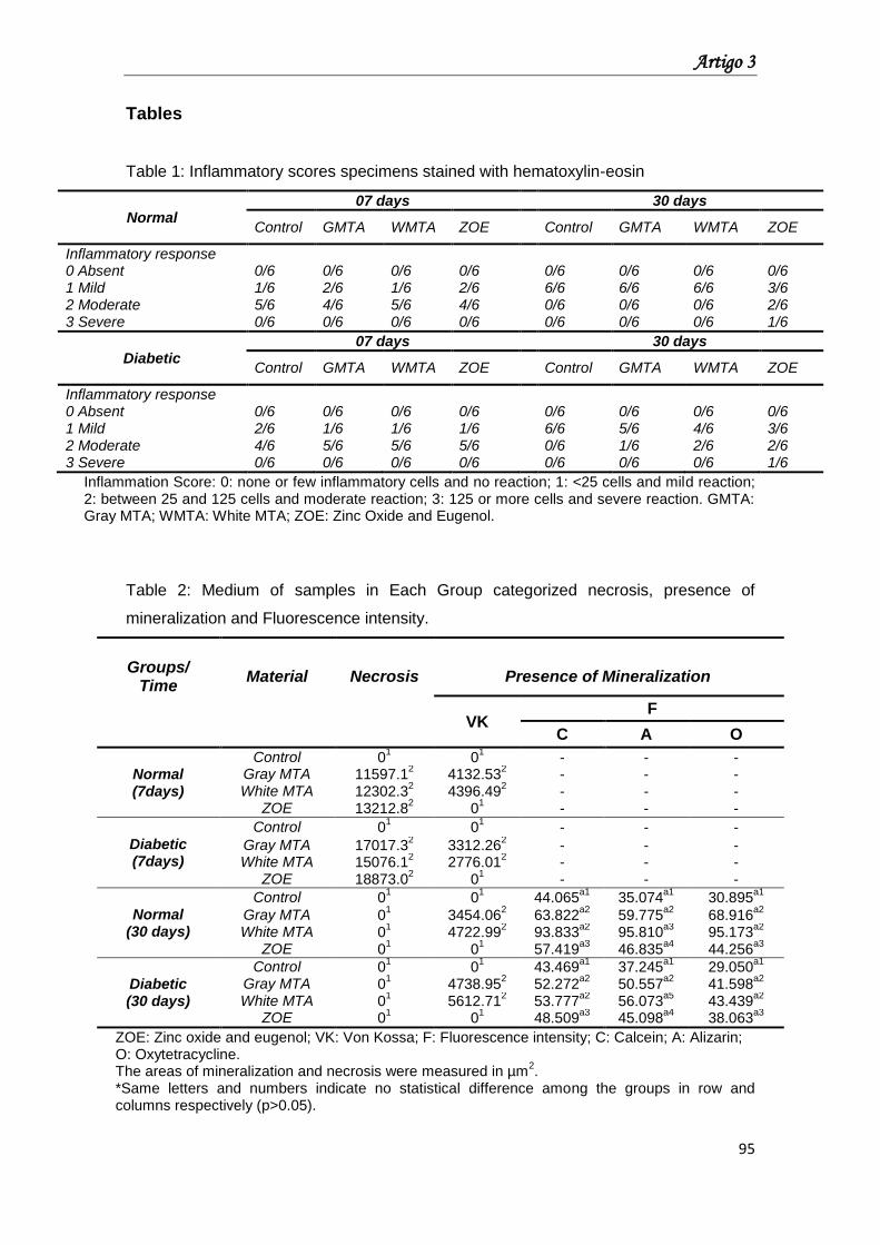
Artigo 3
95
Tables
Table 1: Inflammatory scores specimens stained with hematoxylin-eosin
Normal
07 days 30 days
Control GMTA WMTA ZOE Control GMTA WMTA ZOE
Inflammatory response 0 Absent 0/6 0/6 0/6 0/6 0/6 0/6 0/6 0/6 1 Mild 1/6 2/6 1/6 2/6 6/6 6/6 6/6 3/6 2 Moderate 5/6 4/6 5/6 4/6 0/6 0/6 0/6 2/6 3 Severe 0/6 0/6 0/6 0/6 0/6 0/6 0/6 1/6
Diabetic
07 days 30 days
Control GMTA WMTA ZOE Control GMTA WMTA ZOE
Inflammatory response 0 Absent 0/6 0/6 0/6 0/6 0/6 0/6 0/6 0/6 1 Mild 2/6 1/6 1/6 1/6 6/6 5/6 4/6 3/6 2 Moderate 4/6 5/6 5/6 5/6 0/6 1/6 2/6 2/6 3 Severe 0/6 0/6 0/6 0/6 0/6 0/6 0/6 1/6
Inflammation Score: 0: none or few inflammatory cells and no reaction; 1: <25 cells and mild reaction; 2: between 25 and 125 cells and moderate reaction; 3: 125 or more cells and severe reaction. GMTA: Gray MTA; WMTA: White MTA; ZOE: Zinc Oxide and Eugenol.
Table 2: Medium of samples in Each Group categorized necrosis, presence of
mineralization and Fluorescence intensity.
Groups/
Time
Material
Necrosis
Presence of Mineralization
VK
F
C A O
Normal (7days)
Control 01 0
1 - - -
Gray MTA 11597.12 4132.53
2 - - -
White MTA 12302.32 4396.49
2 - - -
ZOE 13212.82 0
1 - - -
Diabetic (7days)
Control 01 0
1 - - -
Gray MTA 17017.32 3312.26
2 - - -
White MTA 15076.12 2776.01
2 - - -
ZOE 18873.02 0
1 - - -
Normal (30 days)
Control 01 0
1 44.065
a1 35.074
a1 30.895
a1
Gray MTA 01 3454.06
2 63.822
a2 59.775
a2 68.916
a2
White MTA 01 4722.99
2 93.833
a2 95.810
a3 95.173
a2
ZOE 01 0
1 57.419
a3 46.835
a4 44.256
a3
Diabetic (30 days)
Control 01 0
1 43.469
a1 37.245
a1 29.050
a1
Gray MTA 01 4738.95
2 52.272
a2 50.557
a2 41.598
a2
White MTA 01 5612.71
2 53.777
a2 56.073
a5 43.439
a2
ZOE 01 0
1 48.509
a3 45.098
a4 38.063
a3
ZOE: Zinc oxide and eugenol; VK: Von Kossa; F: Fluorescence intensity; C: Calcein; A: Alizarin; O: Oxytetracycline. The areas of mineralization and necrosis were measured in µm
2.
*Same letters and numbers indicate no statistical difference among the groups in row and columns respectively (p>0.05).
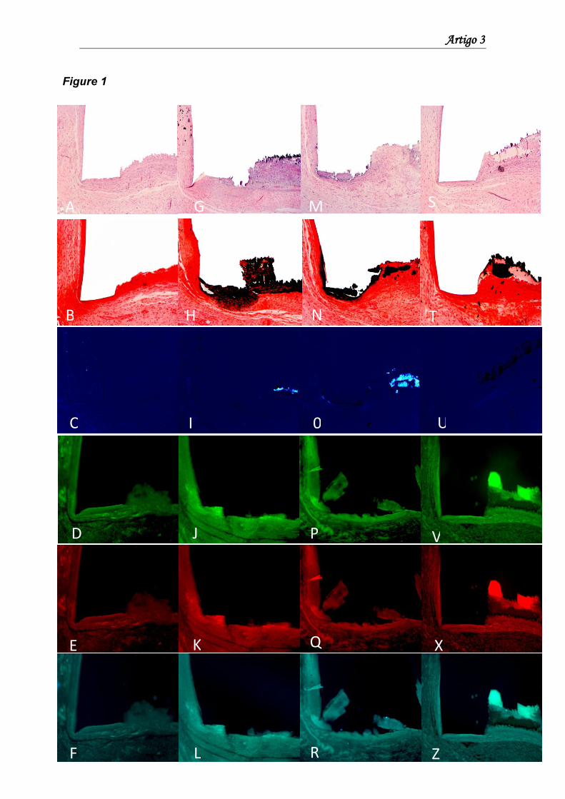
Artigo 3
96
Figure Legends
F L R Z
Figure 1
N B H T
C I 0 U
D J P V
E K Q X
A G M S
Artigo 3
97
Figure 1: Response found in normal group at 30 days. Control: (A) specimens
showing thin fibrous capsule formation and mild inflammatory cell infiltration
consisting of macrophages and lymphocytes (hematoxylin-eosin, 10×); (B)
absence of mineralization (von Kossa, 10×); (C) absence of birefringent
structures to polarized light (polarized light, 10×); (D–F) absence of calcification
areas in close contact with the tube opening (fluorochrome analysis, 10×) for
calcein, alizarin, and oxytetracycline, respectively. Gray MTA and white MTA,
respectively: (G and M) thick fibrous capsule formation and mild inflammatory
cell infiltration (hematoxylin-eosin, 10×); (H and N) presence of dystrophic
calcification on the tube opening (von Kossa, 10×); (I and O) presence of
birefringent structures to polarized light (polarized light, 10×); (J–L and P–R)
calcification areas in close contact with the tube opening (fluorochrome
analysis, 10×) for calcein, alizarin, and oxytetracycline, respectively. ZOE: (S)
presence of mild inflammatory cell infiltration consisting of macrophages and
lymphocytes after 30 days (hematoxylin-eosin, 10×); (T) absence of
mineralization (von Kossa, 10×). Note: dark color observed represent the ZOE
sealer, not mineralization areas; (U) absence of birefringent structures to
polarized light (polarized light, 10×); (V–Z) fluorescence intensity (fluorochrome
analysis, 10×) for calcein, alizarin, and oxytetracycline, respectively.
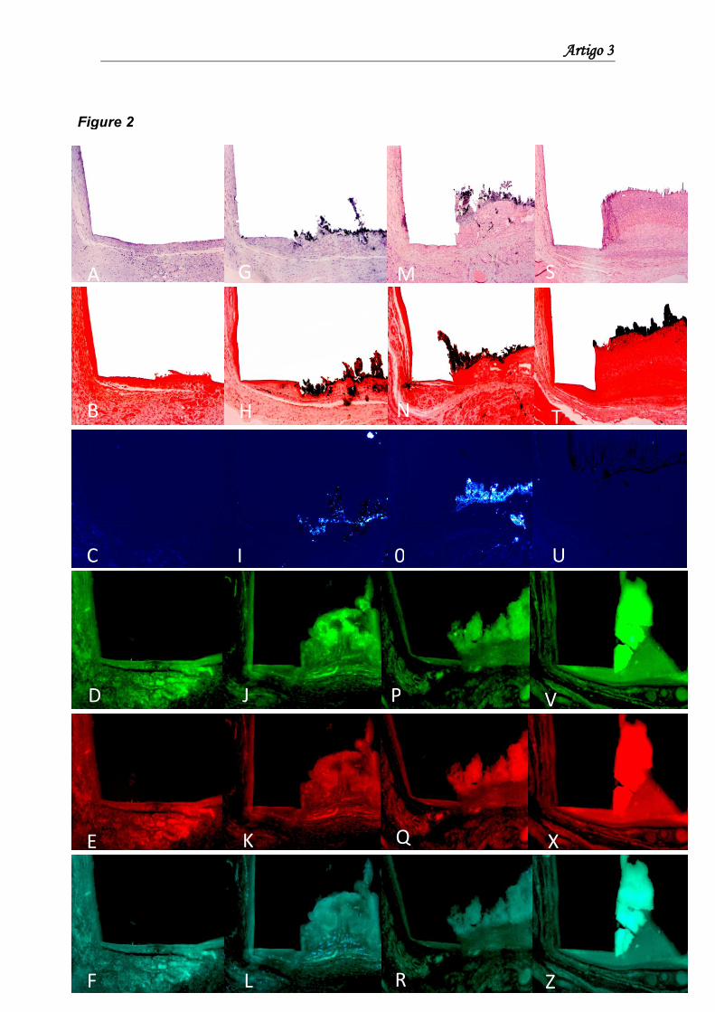
Artigo 3
98
Figure 2
N B H T
C I 0 U
A G M S
D J P V
E K Q X
F L R Z
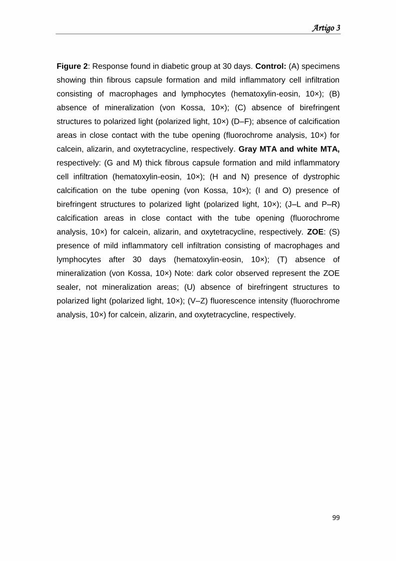
Artigo 3
99
Figure 2: Response found in diabetic group at 30 days. Control: (A) specimens
showing thin fibrous capsule formation and mild inflammatory cell infiltration
consisting of macrophages and lymphocytes (hematoxylin-eosin, 10×); (B)
absence of mineralization (von Kossa, 10×); (C) absence of birefringent
structures to polarized light (polarized light, 10×) (D–F); absence of calcification
areas in close contact with the tube opening (fluorochrome analysis, 10×) for
calcein, alizarin, and oxytetracycline, respectively. Gray MTA and white MTA,
respectively: (G and M) thick fibrous capsule formation and mild inflammatory
cell infiltration (hematoxylin-eosin, 10×); (H and N) presence of dystrophic
calcification on the tube opening (von Kossa, 10×); (I and O) presence of
birefringent structures to polarized light (polarized light, 10×); (J–L and P–R)
calcification areas in close contact with the tube opening (fluorochrome
analysis, 10×) for calcein, alizarin, and oxytetracycline, respectively. ZOE: (S)
presence of mild inflammatory cell infiltration consisting of macrophages and
lymphocytes after 30 days (hematoxylin-eosin, 10×); (T) absence of
mineralization (von Kossa, 10×) Note: dark color observed represent the ZOE
sealer, not mineralization areas; (U) absence of birefringent structures to
polarized light (polarized light, 10×); (V–Z) fluorescence intensity (fluorochrome
analysis, 10×) for calcein, alizarin, and oxytetracycline, respectively.

Artigo 4
Artigo 4
101
Artigo 4: Effect of Diabetes Mellitus on local and systemic
bone marker expression induced by Gray versus White Mineral
Trioxide Aggregate
Abstract
Aim: This study aimed to investigate the relationship between Diabetes Mellitus
(DM) and the local/systemic effects of Gray Mineral Trioxide Aggregate (GMTA)
and White Mineral Trioxide Aggregate (WMTA) on bone marker expression.
Methodology: Wistar rats were divided into two groups (healthy and diabetic),
which were further divided into three subgroups (control, GMTA and WMTA).
Polyethylene tubes filled with sealers were implanted in dorsal connective
tissue. Empty tubes were used as control. On days 7 and 30, blood samples
were collected for calcium, phosphorus and alkaline phosphatase (ALP)
measurement. The animals were euthanized and the implanted tubes with
surrounding tissues were removed, fixed and processed for
immunohistochemical analysis of osteopontin (OPN) and osteocalcin (OCN).
Nonparametric and parametric data were statistically analysed.
Results: No difference in systemic serum calcium levels between both groups
was observed. On day 7, serum phosphorus level of WMTA group of healthy
rats was higher than that of the diabetic group. On day 30, healthy rats
exhibited lesser phosphorus levels than the diabetic ones. At both time points,
the diabetic group showed more ALP activity than the healthy group.
Immunohistochemical analyses of healthy rats revealed OCN and OPN positive
cells in presence of both MTAs. However, under diabetic condition, both OCN
and OPN were absent.
Conclusion: Both MTAs caused an increase in serum calcium, phosphorus
and ALP, suggesting a potential systemic effect, along with induction of OCN
and OPN production. Moreover, DM was capable of inhibiting OCN and OPN
production.
Key Words: Bone markers, Diabetes Mellitus, Mineral Trioxide Aggregate,
Systemic effect
Artigo 4
102
Introduction
Endodontic medicine is a new area of research that studies the
relationship between endodontic variables and systemic health (Segura-Egea et
al. 2015). Fouad (2003) proposed that DM could be a potential modulating
factor for endodontic infections. However, the relationship between these
infections and systemic diseases has not been completely understood, and
systemic alterations arising due to correlation between endodontic diseases and
DM are being investigated (Cintra et al. 2013, Cintra et al. 2014). As described
previously, the hyperglycaemic state due to DM diminishes or alters the immune
response, decreases the leucocyte function and upregulates the expression of
pro-inflammatory cytokines, leading to reduction in the tissue repair capacity
and healing process of the dental pulp and periapical tissues (Delamaire et al.
1997, Iacopino 2001, Fouad 2003).
Root-end filling and repair materials should be biocompatible with the
periradicular tissues, and have suitable chemical and physical properties to
promote complete tissue repair (Saxena et al. 2013). MTA is a calcium silicate-
based cement, developed as a root-end filling material and used in different
clinical procedures (Parirokh & Torabinejad 2010) owing to its attractive
properties (Torabinejad et al. 1995, Holland et al. 1999). Both Gray MTA
(GMTA) and white MTA (WMTA) are known, and studies comparing them have
been conducted (Camilleri et al. 2004, Bozeman et al. 2006, Bidar et al. 2011).
Studies have reported increased levels of ALP, OCN and OPN
expression, and stimulation of cytokine and calcium ion (Ca2+) release upon
MTA treatment (Thomson et al. 2003, Chen et al. 2009). Additionally, diabetic
condition negatively affects the tissue response and the mineralization ability of
MTA (Gomes-Filho et al. 2015).
Ca2+ released upon MTA treatment is an important ion involved in the
regulation of various cellular functions, such as increasing the expression of
bone markers (Rashid et al. 2003, Tada et al. 2010) by active osteoblasts
during different phases of their development, which reflects bone formation
(Seibel 2005, Eriksen 2010). Additionally, MTA promotes alkalization by
increasing the pH due to the release of hydroxyl ions (OH-) (De Vasconcelos et
al. 2009). Alkaline pH activates ALP, which is a phosphate-releasing enzyme

Artigo 4
103
involved in the mineralization process and is considered an important indicator
of bone formation (Seibel 2005, Eriksen 2010).
DM is characterized by abnormalities in the metabolism of
carbohydrates, proteins, lipids, water and electrolytes (ADA 2013). Furthermore,
it is also associated with altered calcium homeostasis and the expression of
bone markers, which results in bone loss and reduced bone formation (Levy et
al. 1994, McCabe 2007). Therefore, the aim of the present study was to
investigate the potential relationship between DM, and the local and systemic
effects of subcutaneous implantation of GMTA and WMTA on bone marker
expression in rats, by immunohistochemical and biochemical analyses.
Material and Methods
Animals
Wistar albino rats, aged 3 to 4 months, weighing 250–280 g were
procured from the School of Dentistry of Araçatuba/UNESP. The animals were
divided in two groups: Healthy and diabetic (n=48 per group). Each group was
further divided in three subgroups: Control, GMTA and WMTA (n=8 per group).
The present study was approved and performed according to the guidelines of
the ethical committee (protocol number 00557-2013).
Induction of Diabetes
Animals belonging to the diabetic group received a single dose (150
mg/kg bw) of Alloxan monohydrate (Sigma Aldrich Corp., St. Louis, MO, USA)
for the induction of diabetes (Gomes-Filho et al. 2015). The animals that
showed blood glucose levels higher than 250 mg/dL were considered diabetic
and used for the study.
Sample preparation and surgical procedure (Subcutaneous implantation)
Sterile polyethylene tubes (Abbott Labs of Brazil, São Paulo, SP, Brazil)
with 1 mm internal diameter, 1.6 mm external diameter, and 10 mm length were
used. Gray and White MTA Angelus® (Angelus Indústria de Produtos
Odontológicos S/A, Londrina, PR, Brazil) were prepared according to the
manufacturer’s recommendations and inserted in the tubes with a lentulo spiral
(Maillefer Dentsply, Tulsa, OK, USA). Empty tubes were used as controls.
Artigo 4
104
The surgical procedure for implantation of the polyethylene tubes in the
dorsal connective tissue was performed according to previous reports (Holland
et al. 1999, Gomes-Filho et al. 2015).
Blood collection and biochemical assays
On days 7 and 30 after implantation, the animals were anaesthetized for
blood collection. Cardiac puncture was performed to collect 5 mL blood from
each animal. Blood was centrifuged at 3000 rpm for 20 min at 20oC, and the
plasma was stored at -20 ºC for the measurement of calcium, phosphorus and
ALP.
Calcium and phosphorus levels were determined using the colorimetric
Calcium Liquiform assay Kit (Labtest Diagnostica SA, MG, Brazil) and the
Phosphorus UV Liquiform assay Kit (Labtest Diagnostica SA, MG, Brazil), which
exhibited sensitivity of 0.1 mg/dL and 0.14 mg/dL respectively. As a marker for
bone formation, serum ALP levels were determined using a commercially
available kit (Labtest Diagnostica SA, MG, Brazil) with sensitivity of 0.125 U/L.
Sample absorbance was read using a spectrophotometer (Hitachi, model U
1100, Japan).
Immunohistochemical processing
After blood collection, the animals were euthanized with an overdose of
the anaesthetic and the implanted tubes along with the surrounding tissues
were removed and fixed in 10% formalin. The formalin-fixed specimens were
processed and embedded in paraffin, sliced into 5-μm semi-serial sections and
subjected to immunohistochemistry using an indirect immunoperoxidase
staining technique for the detection of OPN (using goat anti-osteopontin
antibody, SC10593; Santa Cruz Biotechnology, Santa Cruz, CA) and OCN
(using rabbit anti-osteocalcin antibody, SC18319; Santa Cruz Biotechnology,
Santa Cruz, CA) following the method by Garcia et al. (2014).
OPN and OCN production around the opening of the tube was analysed
using a microscope under 400x magnification (Leica Microsystems, Wetzlar,
Germany). Semi-quantitative immunohistochemistry analysis was performed.
The criteria for establishing the immunoreactivity (IR) score was adapted from
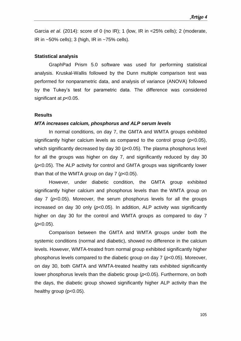
Artigo 4
105
Garcia et al. (2014): score of 0 (no IR); 1 (low, IR in <25% cells); 2 (moderate,
IR in ~50% cells); 3 (high, IR in ~75% cells).
Statistical analysis
GraphPad Prism 5.0 software was used for performing statistical
analysis. Kruskal-Wallis followed by the Dunn multiple comparison test was
performed for nonparametric data, and analysis of variance (ANOVA) followed
by the Tukey’s test for parametric data. The difference was considered
significant at p<0.05.
Results
MTA increases calcium, phosphorus and ALP serum levels
In normal conditions, on day 7, the GMTA and WMTA groups exhibited
significantly higher calcium levels as compared to the control group (p<0.05),
which significantly decreased by day 30 (p<0.05). The plasma phosphorus level
for all the groups was higher on day 7, and significantly reduced by day 30
(p<0.05). The ALP activity for control and GMTA groups was significantly lower
than that of the WMTA group on day 7 (p<0.05).
However, under diabetic condition, the GMTA group exhibited
significantly higher calcium and phosphorus levels than the WMTA group on
day 7 (p<0.05). Moreover, the serum phosphorus levels for all the groups
increased on day 30 only (p<0.05). In addition, ALP activity was significantly
higher on day 30 for the control and WMTA groups as compared to day 7
(p<0.05).
Comparison between the GMTA and WMTA groups under both the
systemic conditions (normal and diabetic), showed no difference in the calcium
levels. However, WMTA-treated from normal group exhibited significantly higher
phosphorus levels compared to the diabetic group on day 7 (p<0.05). Moreover,
on day 30, both GMTA and WMTA-treated healthy rats exhibited significantly
lower phosphorus levels than the diabetic group (p<0.05). Furthermore, on both
the days, the diabetic group showed significantly higher ALP activity than the
healthy group (p<0.05).
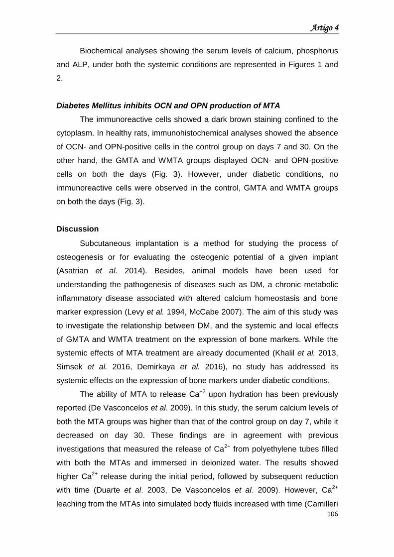
Artigo 4
106
Biochemical analyses showing the serum levels of calcium, phosphorus
and ALP, under both the systemic conditions are represented in Figures 1 and
2.
Diabetes Mellitus inhibits OCN and OPN production of MTA
The immunoreactive cells showed a dark brown staining confined to the
cytoplasm. In healthy rats, immunohistochemical analyses showed the absence
of OCN- and OPN-positive cells in the control group on days 7 and 30. On the
other hand, the GMTA and WMTA groups displayed OCN- and OPN-positive
cells on both the days (Fig. 3). However, under diabetic conditions, no
immunoreactive cells were observed in the control, GMTA and WMTA groups
on both the days (Fig. 3).
Discussion
Subcutaneous implantation is a method for studying the process of
osteogenesis or for evaluating the osteogenic potential of a given implant
(Asatrian et al. 2014). Besides, animal models have been used for
understanding the pathogenesis of diseases such as DM, a chronic metabolic
inflammatory disease associated with altered calcium homeostasis and bone
marker expression (Levy et al. 1994, McCabe 2007). The aim of this study was
to investigate the relationship between DM, and the systemic and local effects
of GMTA and WMTA treatment on the expression of bone markers. While the
systemic effects of MTA treatment are already documented (Khalil et al. 2013,
Simsek et al. 2016, Demirkaya et al. 2016), no study has addressed its
systemic effects on the expression of bone markers under diabetic conditions.
The ability of MTA to release Ca+2 upon hydration has been previously
reported (De Vasconcelos et al. 2009). In this study, the serum calcium levels of
both the MTA groups was higher than that of the control group on day 7, while it
decreased on day 30. These findings are in agreement with previous
investigations that measured the release of Ca2+ from polyethylene tubes filled
with both the MTAs and immersed in deionized water. The results showed
higher Ca2+ release during the initial period, followed by subsequent reduction
with time (Duarte et al. 2003, De Vasconcelos et al. 2009). However, Ca2+
leaching from the MTAs into simulated body fluids increased with time (Camilleri
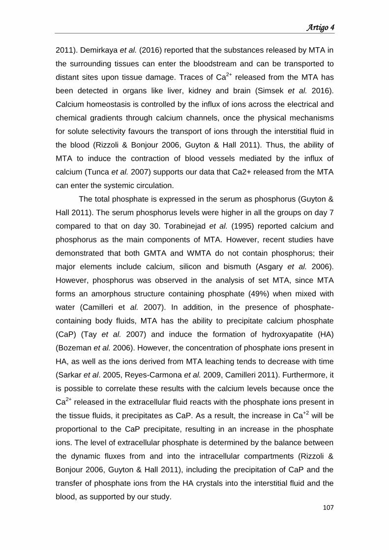
Artigo 4
107
2011). Demirkaya et al. (2016) reported that the substances released by MTA in
the surrounding tissues can enter the bloodstream and can be transported to
distant sites upon tissue damage. Traces of Ca2+ released from the MTA has
been detected in organs like liver, kidney and brain (Simsek et al. 2016).
Calcium homeostasis is controlled by the influx of ions across the electrical and
chemical gradients through calcium channels, once the physical mechanisms
for solute selectivity favours the transport of ions through the interstitial fluid in
the blood (Rizzoli & Bonjour 2006, Guyton & Hall 2011). Thus, the ability of
MTA to induce the contraction of blood vessels mediated by the influx of
calcium (Tunca et al. 2007) supports our data that Ca2+ released from the MTA
can enter the systemic circulation.
The total phosphate is expressed in the serum as phosphorus (Guyton &
Hall 2011). The serum phosphorus levels were higher in all the groups on day 7
compared to that on day 30. Torabinejad et al. (1995) reported calcium and
phosphorus as the main components of MTA. However, recent studies have
demonstrated that both GMTA and WMTA do not contain phosphorus; their
major elements include calcium, silicon and bismuth (Asgary et al. 2006).
However, phosphorus was observed in the analysis of set MTA, since MTA
forms an amorphous structure containing phosphate (49%) when mixed with
water (Camilleri et al. 2007). In addition, in the presence of phosphate-
containing body fluids, MTA has the ability to precipitate calcium phosphate
(CaP) (Tay et al. 2007) and induce the formation of hydroxyapatite (HA)
(Bozeman et al. 2006). However, the concentration of phosphate ions present in
HA, as well as the ions derived from MTA leaching tends to decrease with time
(Sarkar et al. 2005, Reyes-Carmona et al. 2009, Camilleri 2011). Furthermore, it
is possible to correlate these results with the calcium levels because once the
Ca2+ released in the extracellular fluid reacts with the phosphate ions present in
the tissue fluids, it precipitates as CaP. As a result, the increase in Ca+2 will be
proportional to the CaP precipitate, resulting in an increase in the phosphate
ions. The level of extracellular phosphate is determined by the balance between
the dynamic fluxes from and into the intracellular compartments (Rizzoli &
Bonjour 2006, Guyton & Hall 2011), including the precipitation of CaP and the
transfer of phosphate ions from the HA crystals into the interstitial fluid and the
blood, as supported by our study.
Artigo 4
108
Hyperglycaemia results in metabolic changes due to insulin deficiency,
impairment in parathyroid hormone and alteration of vitamin D metabolism
leading to abnormal plasma levels of calcium, phosphorus and alkaline
phosphatase (Joshi et al. 2013). In this study, under diabetic conditions, the
calcium and phosphorus levels in GMTA group were higher than that in the
WMTA group on day 7. On the other hand, only the phosphorus level was high
on day 30 for all the groups. De Vasconcelos et al. (2009) measured the
release of Ca2+ from both the MTAs and showed that GMTA exhibited higher
values. Meanwhile, Garcia et al. (2014a) reported that WMTA is associated with
greater Ca2+ release than GMTA on day 7. The authors proposed that higher
concentration of Fe2O3 in GMTA may have an influence on Ca2+ release.
Furthermore, GMTA produced more CaP precipitate than WMTA (Bozeman et
al. 2006), which correlated with the phosphorus levels. However, studies have
shown that the phosphate ions released after CaP precipitation reduced with
time (Sarkar et al. 2005, Reyes-Carmona et al. 2009). At the same time, an
increase in the serum phosphate level in diabetes has been reported (Raskin &
Pak 1981). DM affects the blood vessels thereby compromising the
microcirculation (Domingueti et al. 2016), and promoting abnormal metabolism
of microelements, such as calcium and phosphorus (ADA 2013). Masuda et al.
(2005) showed that MTA has a positive effect on revascularization of the
connective tissue. The above mentioned facts may help in explaining our
findings under diabetic conditions.
No differences were observed in the calcium levels between normal and
diabetic groups. Levy et al. (1985) also reported no differences in the level of
calcium in the plasma of diabetic animals. Similarly, normal plasma calcium
levels were reported in diabetic rats, despite an increase in the renal calcium
excretion (Anwana & Garland 1990). Nevertheless, elevated calcium levels due
to an increase in the intracellular influx of calcium were observed in the diabetic
condition (Levy et al. 1986, Gilon et al. 2014). In contrast, phosphorus levels of
the WMTA group of normal rats was higher than that of the diabetic group on
day 7. Alterations in electrolyte metabolism due to DM may modify the
composition of the interstitial fluid, which can affect the rate of chemical
reactions leading to the gradual release of calcium hydroxide, which may
interfere with its own ionic dissociation and CaP precipitation. Moreover, on day
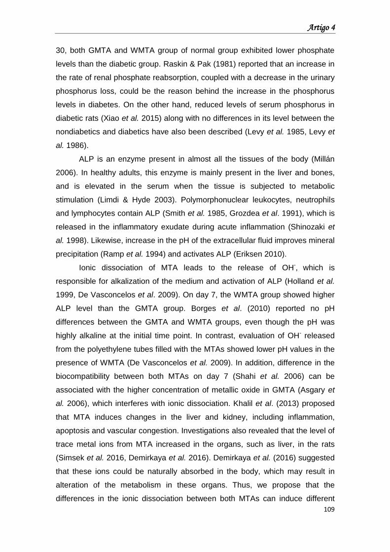
Artigo 4
109
30, both GMTA and WMTA group of normal group exhibited lower phosphate
levels than the diabetic group. Raskin & Pak (1981) reported that an increase in
the rate of renal phosphate reabsorption, coupled with a decrease in the urinary
phosphorus loss, could be the reason behind the increase in the phosphorus
levels in diabetes. On the other hand, reduced levels of serum phosphorus in
diabetic rats (Xiao et al. 2015) along with no differences in its level between the
nondiabetics and diabetics have also been described (Levy et al. 1985, Levy et
al. 1986).
ALP is an enzyme present in almost all the tissues of the body (Millán
2006). In healthy adults, this enzyme is mainly present in the liver and bones,
and is elevated in the serum when the tissue is subjected to metabolic
stimulation (Limdi & Hyde 2003). Polymorphonuclear leukocytes, neutrophils
and lymphocytes contain ALP (Smith et al. 1985, Grozdea et al. 1991), which is
released in the inflammatory exudate during acute inflammation (Shinozaki et
al. 1998). Likewise, increase in the pH of the extracellular fluid improves mineral
precipitation (Ramp et al. 1994) and activates ALP (Eriksen 2010).
Ionic dissociation of MTA leads to the release of OH-, which is
responsible for alkalization of the medium and activation of ALP (Holland et al.
1999, De Vasconcelos et al. 2009). On day 7, the WMTA group showed higher
ALP level than the GMTA group. Borges et al. (2010) reported no pH
differences between the GMTA and WMTA groups, even though the pH was
highly alkaline at the initial time point. In contrast, evaluation of OH- released
from the polyethylene tubes filled with the MTAs showed lower pH values in the
presence of WMTA (De Vasconcelos et al. 2009). In addition, difference in the
biocompatibility between both MTAs on day 7 (Shahi et al. 2006) can be
associated with the higher concentration of metallic oxide in GMTA (Asgary et
al. 2006), which interferes with ionic dissociation. Khalil et al. (2013) proposed
that MTA induces changes in the liver and kidney, including inflammation,
apoptosis and vascular congestion. Investigations also revealed that the level of
trace metal ions from MTA increased in the organs, such as liver, in the rats
(Simsek et al. 2016, Demirkaya et al. 2016). Demirkaya et al. (2016) suggested
that these ions could be naturally absorbed in the body, which may result in
alteration of the metabolism in these organs. Thus, we propose that the
differences in the ionic dissociation between both MTAs can induce different
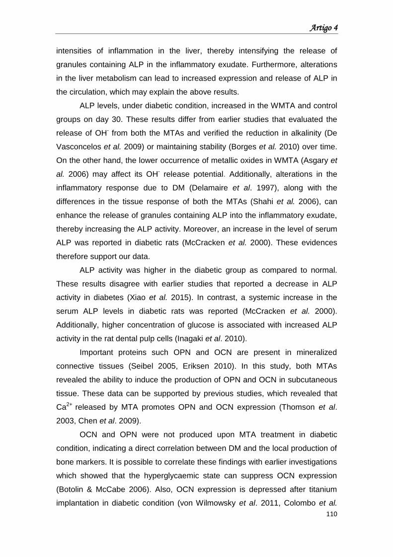
Artigo 4
110
intensities of inflammation in the liver, thereby intensifying the release of
granules containing ALP in the inflammatory exudate. Furthermore, alterations
in the liver metabolism can lead to increased expression and release of ALP in
the circulation, which may explain the above results.
ALP levels, under diabetic condition, increased in the WMTA and control
groups on day 30. These results differ from earlier studies that evaluated the
release of OH- from both the MTAs and verified the reduction in alkalinity (De
Vasconcelos et al. 2009) or maintaining stability (Borges et al. 2010) over time.
On the other hand, the lower occurrence of metallic oxides in WMTA (Asgary et
al. 2006) may affect its OH- release potential. Additionally, alterations in the
inflammatory response due to DM (Delamaire et al. 1997), along with the
differences in the tissue response of both the MTAs (Shahi et al. 2006), can
enhance the release of granules containing ALP into the inflammatory exudate,
thereby increasing the ALP activity. Moreover, an increase in the level of serum
ALP was reported in diabetic rats (McCracken et al. 2000). These evidences
therefore support our data.
ALP activity was higher in the diabetic group as compared to normal.
These results disagree with earlier studies that reported a decrease in ALP
activity in diabetes (Xiao et al. 2015). In contrast, a systemic increase in the
serum ALP levels in diabetic rats was reported (McCracken et al. 2000).
Additionally, higher concentration of glucose is associated with increased ALP
activity in the rat dental pulp cells (Inagaki et al. 2010).
Important proteins such OPN and OCN are present in mineralized
connective tissues (Seibel 2005, Eriksen 2010). In this study, both MTAs
revealed the ability to induce the production of OPN and OCN in subcutaneous
tissue. These data can be supported by previous studies, which revealed that
Ca2+ released by MTA promotes OPN and OCN expression (Thomson et al.
2003, Chen et al. 2009).
OCN and OPN were not produced upon MTA treatment in diabetic
condition, indicating a direct correlation between DM and the local production of
bone markers. It is possible to correlate these findings with earlier investigations
which showed that the hyperglycaemic state can suppress OCN expression
(Botolin & McCabe 2006). Also, OCN expression is depressed after titanium
implantation in diabetic condition (von Wilmowsky et al. 2011, Colombo et al.
Artigo 4
111
2011). Besides, lower levels of serum OCN have been reported in diabetic
patients (Achemlal et al. 2005). On the other hand, Nakajima et al. (2013)
reported that advanced glycation end-products (AGEs) promoted an
upregulation of OCN expression in dental pulp.
OPN is a multifunctional protein (Denhardt et al. 2001, Lund et al. 2009),
whose upregulation is associated with acute and chronic inflammatory
conditions such as DM (Lund et al. 2009, Kahles et al. 2014). Our data differ
from earlier studies, which reported an increase in OPN expression in diabetic
condition (Inagaki et al. 2010, Colombo et al. 2011). Moreover, no inhibitory
effect was established on the inflammatory response by MTA in diabetic
condition (Gomes-Filho et al. 2015). It is necessary to conduct more studies to
substantiate and understand the systemic effects of these endodontic materials.
Conclusion
Based on these results, it was concluded that both MTAs increased the
calcium, phosphorus and ALP serum levels, suggesting a possible systemic
effect. They also promoted OCN and OPN production. Additionally, DM was
capable of inhibiting the MTA-induced production of OCN and OPN.
References
1. Achemlal L, Tellal S, Rkiouak F et al. (2005) Bone metabolism in male
patients with type 2 diabetes. Clinical Rheumatology 24, 493-6.
2. American Diabetes Association (2013) Diagnosis and Classification of
Diabetes Mellitus. Diabetes Care 36 (Suppl 1), S67–S74.
3. Anwana AB, Garland HO (1990) Renal calcium and magnesium handling
in experimental diabetes mellitus in the rat. Acta Endocrinologica 122, 479-86.
4. Asatrian G, Chang L, James AW (2014) Muscle pouch implantation: an
ectopic bone formation model. In: Bruno Christ et al, eds. Animal Models for
Stem Cell Therapy, vol. 1213; pp. 185-91. New York, USA: Springer.
5. Asgary S, Parirokh M, Eghbal MJ, Stowe S, Brink F (2006) A qualitative
X-ray analysis of white and grey mineral trioxide aggregate using compositional
imaging. Journal of Materials Science: Materials in Medicine 17,187-91.
Artigo 4
112
6. Bidar M, Zarrabi MH, Afshari JT et al. (2011) Osteoblastic cytokine
response to gray and white mineral trioxide aggregate. Iran Endodontic Journal
6, 111-5.
7. Borges AH, Pedro FL, Miranda CE, Semenoff-Segundo A, Pécora JD,
Filho AM (2010) Comparative study of physico-chemical properties of MTA-
based and Portland cements. Acta Odontológica Latinoamericana 23, 175-81.
8. Botolin S, McCabe LR (2006) Chronic hyperglycemia modulates
osteoblast gene expression through osmotic and non-osmotic pathways.
Journal of Cellular Biochemistry 99,411-24.
9. Bozeman TB, Lemon RR, Eleazer PD (2006) Elemental analysis of
crystal precipitate from gray and white MTA. Journal of Endodontics 32, 425-8.
10. Camilleri J (2007) Hydration mechanisms of mineral trioxide aggregate.
International Endodontic Journal 40, 462-70.
11. Camilleri J (2011) Evaluation of the effect of intrinsic material properties
and ambient conditions on the dimensional stability of white mineral trioxide
aggregate and Portland cement. Journal of Endodontics 37, 239-45.
12. Camilleri J, Montesin FE, Papaioannou S, McDonald F, Pitt Ford TR
(2004) Biocompatibility of two commercial forms of mineral trioxide aggregate.
International Endodontic Journal 37, 699–704.
13. Chen CL, Huang TH, Ding SJ, Shie MY, Kao CT (2009). Comparison of
calcium and silicate cement and mineral trioxide aggregate biologic effects and
bone markers expression in MG63 cells. Journal of Endodontics 35, 682-5.
14. Cintra LT, da Silva Facundo AC, Prieto AK et al. (2013) Pulpal and
periodontal diseases increase triglyceride levels in diabetic rats. Clinical Oral
Investigations 17, 1595–9.
15. Cintra LT, da Silva Facundo AC, Prieto AK et al. (2014) Profile and
histology in oral infections associated with diabetes. Journal of Endodontics 40,
1139–44.
16. Colombo JS, Balani D, Sloan AJ, Crean SJ, Okazaki J, Waddington RJ
(2011) Delayed osteoblast differentiation and altered inflammatory response
around implants placed in incisor sockets of type 2 diabetic rats. Clinical Oral
Implants Research 22, 578-86.
Artigo 4
113
17. Delamaire M, Maugendre D, Moreno M, Le Goff MC, Allannic H, Genetet
B (1997) Impaired leucocyte functions in diabetic patients. Diabetic Medicine
14, 29–34.
18. Demirkaya K, Can Demirdöğen B, Öncel Torun Z, Erdem O, Çetinkaya
S, Akay C (2016) In vivo evaluation of the effects of hydraulic calcium silicate
dental cements on plasma and liver aluminium levels in rats. European Journal
of Oral Sciences 124, 75-81.
19. Denhardt DT, Noda M, O'Regan AW, Pavlin D, Berman JS (2001)
Osteopontin as a means to cope with environmental insults: regulation of
inflammation, tissue remodelling, and cell survival. The Journal of Clinical
Investigation 107, 1055-61.
20. De Vasconcelos BC, Bernardes RA, Cruz SM et al. (2009) Evaluation of
pH and calcium ion release of new root-end filling materials. Oral Surgery, Oral
Medicine, Oral Pathology, Oral Radiology, and Endodontology 108,135-9.
21. Domingueti CP, Dusse LM, Carvalho Md, de Sousa LP, Gomes KB,
Fernandes AP (2016) Diabetes mellitus: The linkage between oxidative stress,
inflammation, hypercoagulability and vascular complications. Journal of
Diabetes and its Complications 30, 738-45.
22. Duarte MA, Demarchi ACO, Yamashita JC, Kuga MC, de Campos FS
(2003) pH and calcium ion release of 2 root-end filling materials. Oral Surgery,
Oral Medicine, Oral Pathology, Oral Radiology, and Endodontology 95, 345-7.
23. Eriksen EF (2010) Cellular mechanisms of bone remodelling. Reviews in
Endocrine and Metabolic Disorders 11, 219–27.
24. Fouad AF (2003) Diabetes mellitus as a modulating factor of endodontic
infections. Journal of Dental Education 67, 459-67.
25. Garcia Lda F, Chinelatti MA, Rossetto HL, Pires-de-Souza Fde C
(2014a) Solubility and disintegration of new calcium aluminate cement
(EndoBinder) containing different radiopacifying agents. Journal of Endodontics
40, 261-5.
26. Garcia VG, Longo M, Gualberto Junior EC et al. (2014) Effect of the
concentration of phenothiazine photosensitizers in antimicrobial photodynamic
therapy on bone loss and the immune inflammatory response of induced
periodontitis in rats. Journal of Periodontal Research 49, 584–94.
Artigo 4
114
27. Gilon P, Chae HY, Rutter GA, Ravier MA (2014) Calcium signaling in
pancreatic β-cells in health and in Type 2 diabetes. Cell Calcium 56, 340-61.
28. Gomes-Filho JE, de Azevedo Queiroz ÍO, Watanabe S et al. (2015)
Influence of diabetes mellitus on tissue response to MTA and its ability to
stimulate mineralization. Dental Traumatology 31, 67-72.
29. Grozdea J, Vergnes H, Brisson-Lougarre A et al. (1991) Difference in
activity properties and subcellular distribution of neutrophil alkaline phosphatase
between normal individuals and patients with trisomy 21. British Journal of
Haematology 77, 282-6.
30. Guyton AC and Hall J (2011) Tratado de fisiologia médica, 12th ed., Rio
de Janeiro, RJ, BR: Elsevier.
31. Holland R, Souza V, Nery MJ, Otoboni Filho JA, Bernabe PF, Dezan E
(1999) Reaction of rat connective tissue to implanted dentin tubes filled with
mineral trioxide aggregate or calcium hydroxide. Journal of Endodontics 25,
161-6.
32. Iacopino AM (2001) Periodontitis and diabetes interrelationships: role of
inflammation. Annals of Periodontology 6, 125–37.
33. Inagaki Y, Yoshida K, Ohba H et al. (2010) High glucose levels increase
osteopontin production and pathologic calcification in rat dental pulp tissues.
Journal of Endodontics 36, 1014-20.
34. Joshi A, Varthakavi P, Chadha M, Bhagwat N (2013) A study of bone
mineral density and its determinants in type 1 diabetes mellitus. Journal of
Osteoporosis 2013, 397814.
35. Kahles F, Findeisen HM, Bruemmer D (2014) Osteopontin: A novel
regulator at the cross roads of inflammation, obesity and diabetes. Molecular
Metabolism 3, 384-93.
36. Khalil WA, Eid NF (2013) Biocompatibility of BioAggregate and mineral
trioxide aggregate on the liver and kidney. International Endodontic Journal 46,
730-7.
37. Levy J, Gavin JR, Sowers JR (1994) Diabetes mellitus: a disease of
abnormal cellular calcium metabolism? The American Journal of Medicine 96,
260-73.
Artigo 4
115
38. Levy J, Stern Z, Gutman A, Naparstek Y, Gavin JR 3rd, Avioli LV (1986)
Plasma calcium and phosphate levels in an adult noninsulin-dependent diabetic
population. Calcified Tissue International 39, 316-8.
39. Levy J, Teitelbaum SL, Gavin JR 3rd, Fausto A, Kurose H, Avioli LV
(1985) Bone calcification and calcium homeostasis in rats with non-insulin-
dependent diabetes induced by streptozocin. Diabetes 34, 365-72.
40. Limdi JK, Hyde GM (2003) Evaluation of abnormal liver function tests.
Postgraduate Medical Journal 79, 307-12.
41. Lund SA, Giachelli CM, Scatena M (2009) The role of osteopontin in
inflammatory processes. Journal of Cell Communication and Signaling 3, 311-
22.
42. Masuda YM, Wang X, Hossain M et al. (2005) Evaluation of
biocompatibility of mineral trioxide aggregate with an improved rabbit ear
chamber. Journal of Oral Rehabilitation 32, 145-50.
43. McCabe LR (2007) Understanding the pathology and mechanisms of
type I diabetic bone loss. Journal of Cellular Biochemistry 102,1343-57.
44. McCracken M, Lemons JE, Rahemtulla F, Prince CW, Feldman D (2000)
Bone response to titanium alloy implants placed in diabetic rats. The
International Journal of Oral & Maxillofacial Implants 15, 345–354.
45. Millán JL (2006) Alkaline Phosphatases. In: Seibel MJ, Robins SP,
Bilezikian JP, ed. Dynamics of Bone and Cartilage Metabolism: Principles and
Clinical Applications, 2nd edn; pp. 153-164. San Diego, USA: Academic Press.
46. Nakajima Y, Inagaki Y, Hiroshima Y, Kido J, Nagata T (2013) Advanced
glycation end-products enhance calcification in cultured rat dental pulp cells.
Journal of Endodontics 39, 873-8.
47. Parirokh M, Torabinejad M (2010) Mineral trioxide aggregate: a
comprehensive literature review-Part III: Clinical applications, drawbacks, and
mechanism of action. Journal of Endodontics 36, 400-13.
48. Ramp WK, Lenz LG, Kaysinger KK (1994) Medium pH modulates matrix,
mineral, and energy metabolism in cultured chick bones and osteoblast-like
cells. Bone and Mineral 24, 59-73.
49. Rashid F, Shiba H, Mizuno N et al. (2003) The effect of extracellular
calcium ion on gene expression of bone-related proteins in human pulp cells.
Journal of Endodontics 29, 104-7.
Artigo 4
116
50. Raskin P, Pak CY (1981) The effect of chronic insulin therapy on
phosphate metabolism in diabetes mellitus. Diabetologia 21, 50-3.
51. Reyes-Carmona JF, Felippe MS, Felippe WT (2009) Biomineralization
ability and interaction of mineral trioxide aggregate and white portland cement
with dentin in a phosphate-containing fluid. Journal of Endodontics 35, 731-6.
52. Rizzoli R, Bonjour JP (2006) Physiology of Calcium and Phosphate
Homeostases. In: Seibel MJ, Robins SP, Bilezikian JP, ed. Dynamics of Bone
and Cartilage Metabolism: Principles and Clinical Applications, 2nd edn; pp.
487-505. San Diego, USA: Academic Press.
53. Sarkar NK, Caicedo R, Ritwik P, Moiseyeva R, Kawashima I (2005)
Physicochemical basis of the biologic properties of mineral trioxide aggregate.
Journal of Endodontics 31, 97-100.
54. Saxena P, Gupta SK, Newaskar V (2013) Biocompatibility of root-end
filling materials: recent update. Restorative Dentistry & Endodontics 38,119-27.
55. Seibel MJ (2005) Biochemical markers of bone turnover part I:
biochemistry and variability. The Clinical Biochemists. Reviews/Australian
Association of Clinical Biochemists 26, 97-122.
56. Segura-Egea JJ, Martín-González J, Castellanos-Cosano L (2015)
Endodontic medicine: connections between apical periodontitis and systemic
diseases. International Endodontic Journal 48, 933-51.
57. Simsek N, Bulut ET, Ahmetoğlu F, Alan H (2016) Determination of trace
elements in rat organs implanted with endodontic repair materials by ICP-MS.
Journal of Material Science: Materials in Medicine 27, 1-6.
58. Shahi S, Rahimi S, Lotfi M, Yavari H, Gaderian A (2006) A comparative
study of the biocompatibility of three root-end filling materials in rat connective
tissue. Journal of Endodontics 32, 776–80.
59. Shinozaki T, Watanabe H, Takagishi K, Pritzker KP (1998) Allotype
immunoglobulin enhances alkaline phosphatase activity: implications for the
inflammatory response. Journal of Laboratory and Clinical Medicine 132, 320-8.
60. Smith GP, Sharp G, Peters TJ (1985) Isolation and characterization of
alkaline phosphatase-containing granules (phosphasomes) from human
polymorphonuclear leucocytes. Journal of Cell Science 76, 167-78.
61. Tada H, Nemoto E, Kanaya S, Hamaji N, Sato H, Shimauchi H (2010)
Elevated extracellular calcium increases expression of bone morphogenetic
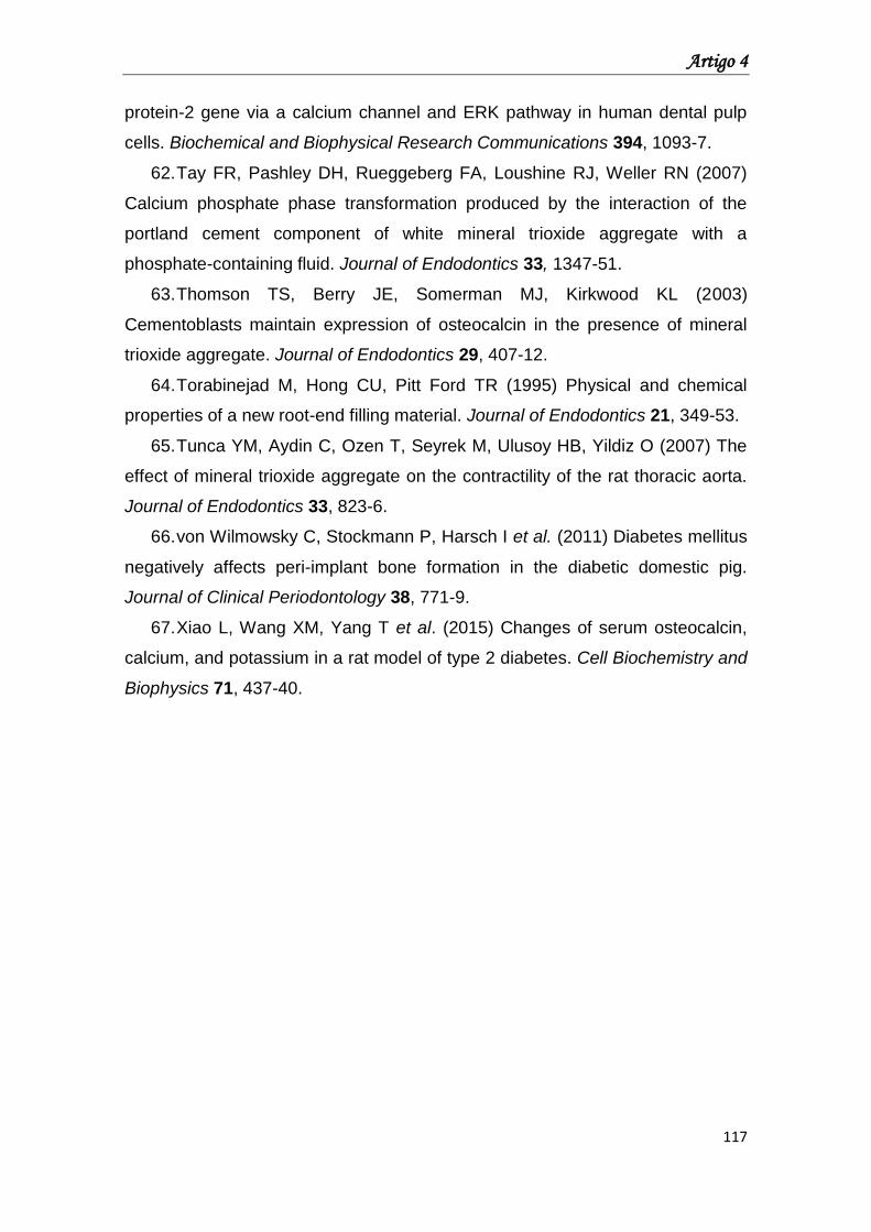
Artigo 4
117
protein-2 gene via a calcium channel and ERK pathway in human dental pulp
cells. Biochemical and Biophysical Research Communications 394, 1093-7.
62. Tay FR, Pashley DH, Rueggeberg FA, Loushine RJ, Weller RN (2007)
Calcium phosphate phase transformation produced by the interaction of the
portland cement component of white mineral trioxide aggregate with a
phosphate-containing fluid. Journal of Endodontics 33, 1347-51.
63. Thomson TS, Berry JE, Somerman MJ, Kirkwood KL (2003)
Cementoblasts maintain expression of osteocalcin in the presence of mineral
trioxide aggregate. Journal of Endodontics 29, 407-12.
64. Torabinejad M, Hong CU, Pitt Ford TR (1995) Physical and chemical
properties of a new root-end filling material. Journal of Endodontics 21, 349-53.
65. Tunca YM, Aydin C, Ozen T, Seyrek M, Ulusoy HB, Yildiz O (2007) The
effect of mineral trioxide aggregate on the contractility of the rat thoracic aorta.
Journal of Endodontics 33, 823-6.
66. von Wilmowsky C, Stockmann P, Harsch I et al. (2011) Diabetes mellitus
negatively affects peri-implant bone formation in the diabetic domestic pig.
Journal of Clinical Periodontology 38, 771-9.
67. Xiao L, Wang XM, Yang T et al. (2015) Changes of serum osteocalcin,
calcium, and potassium in a rat model of type 2 diabetes. Cell Biochemistry and
Biophysics 71, 437-40.
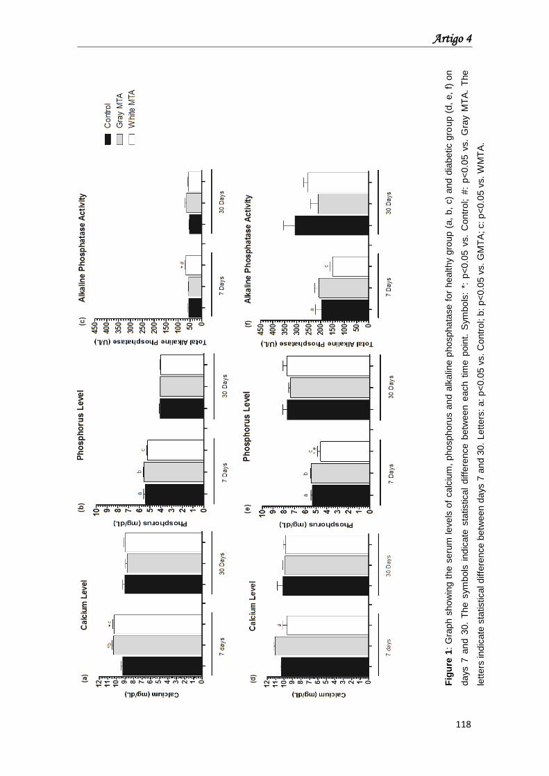
Artigo 4
118
Fig
ure
1:
Gra
ph s
ho
win
g t
he s
eru
m leve
ls o
f calc
ium
, phosphoru
s a
nd a
lkalin
e p
hospha
tase f
or
health
y g
roup (
a,
b,
c)
and d
iabetic g
roup (
d,
e,
f) o
n
da
ys 7
an
d 3
0.
The s
ym
bols
ind
icate
sta
tistica
l diffe
rence b
etw
een e
ach t
ime p
oin
t. S
ym
bo
ls:
*: p
<0.0
5 v
s.
Contr
ol; #
: p
<0.0
5 v
s.
Gra
y M
TA
. T
he
lett
ers
ind
icate
sta
tistical diffe
rence b
etw
een d
ays 7
an
d 3
0.
Lett
ers
: a:
p<
0.0
5 v
s.
Contr
ol; b
: p
<0.0
5 v
s. G
MT
A; c: p
<0.0
5 v
s. W
MT
A.
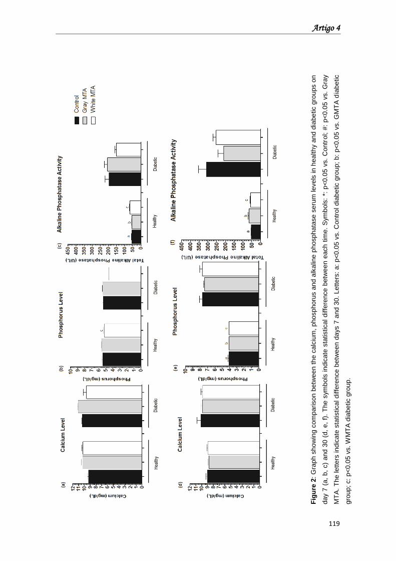
Artigo 4
119
Fig
ure
2:
Gra
ph s
ho
win
g c
om
parison b
etw
een t
he c
alc
ium
, phosphoru
s a
nd a
lkalin
e p
hosph
ata
se s
eru
m levels
in h
ea
lth
y a
nd d
iabe
tic g
roups o
n
da
y 7
(a,
b,
c)
and 3
0 (
d,
e,
f).
The s
ym
bols
indic
ate
sta
tistical diffe
rence b
etw
ee
n e
ach t
ime.
Sym
bols
: *:
p<
0.0
5 v
s.
Contr
ol; #
: p
<0.0
5 v
s.
Gra
y
MT
A.
The lette
rs ind
icate
sta
tistica
l d
iffe
rence b
etw
ee
n d
ays 7
an
d 3
0.
Lett
ers
: a:
p<
0.0
5 v
s.
Contr
ol d
iab
etic g
roup;
b:
p<
0.0
5 v
s.
GM
TA
dia
betic
gro
up; c:
p<
0.0
5 v
s. W
MT
A d
iabetic g
rou
p.
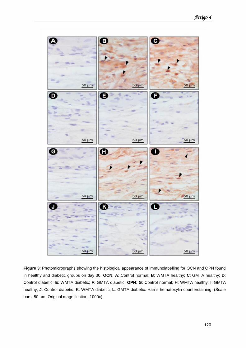
Artigo 4
120
Figure 3: Photomicrographs showing the histological appearance of immunolabelling for OCN and OPN found
in healthy and diabetic groups on day 30. OCN: A: Control normal; B: WMTA healthy; C: GMTA healthy; D:
Control diabetic; E: WMTA diabetic; F: GMTA diabetic. OPN: G: Control normal; H: WMTA healthy; I: GMTA
healthy; J: Control diabetic; K: WMTA diabetic; L: GMTA diabetic. Harris hematoxylin counterstaining. (Scale
bars, 50 µm; Original magnification, 1000x).

Conclusão
Conclusão
121
Conclusão
Concluiu-se, assim, que com base nos objetivos propostos e
metodologias empregadas que:
MTA promoveu a regeneração do Ligamento Periodontal e do osso
alveolar na área da injuria dental (perfuração), favorecendo a formação de
ponte de dentina;
MTA promoveu a proliferação células mesenquimais progenitoras do
Ligamento Periodontal; no entanto, demonstrou efeitos negativos na
diferenciação osteogênica de PDSCs e BMSCs.
A condição hiperglicêmica interferiu na proliferação celular e produção
de IL-6 do MTA Cinza in vitro;
In vivo, a hiperglicemia não produziu alteração na resposta tecidual e
na capacidade de produção de citocinas pro-inflamatórias, no entanto, reduziu
a intensidade de fluorescência do MTA Branco aos 14 dias;
A hiperglicemia impediu a produção de marcadores ósseos pelo MTA e
aumentou os níveis séricos de Fósforo e Fosfatase Alcalina, principalmente,
aos 07 dias na presença do MTA Cinza;
Em condições diabéticas o MTA Cinza demostrou resultados biológicos
mais favoráveis quando comparado ao MTA Branco.

Referências
Referências
123
Referências
1. Segura-Egea JJ, Castellanos-Cosano L, Velasco-Ortega E et al.
Relationship between smoking and endodontic variables in hypertensive
patients. J Endod. 2001;37(6):764-7.
2. Tavares M, Lindefjeld Calabi KA, San Martin L. Systemic Diseases and
Oral Health. Dent Clin North Am. 2014;58:797-814.
3. Lima SM, Grisi DC, Kogawa EM, et al. Diabetes mellitus and
inflammatory pulpal and periapical disease: a review. Int Endod J 2013;46:700-
9.
4. Segura-Egea JJ, Martín-González J, Castellanos-Cosano L. Endodontic
medicine: connections between apical periodontitis and systemic diseases. Int
Endod J. 2015;48:933-51.
5. American Diabetes Association. Standards of medical care in diabetes.
Diabetes Care 2013 January; 36 Suppl 1:S11-S66.
6. Fouad AF. Diabetes mellitus as a modulating factor of endodontic
infections. J Dent Educ 2003;67:459–67.
7. Fouad AF, Burleson J. The effect of diabetes mellitus on endodontic
treatment outcome: data from an electronic patient record. J Am Dent
Assoc. 2003;134:43-51.
8. Marending M, Peters OA, Zehnder M. Factors affecting the outcome of
orthograde root canal therapy in a general dentistry hospital practice. Oral Surg
Oral Med Oral Pathol Oral Radiol Endod. 2005;99:119-24.
9. Cintra LT, Samuel RO, Facundo AC, et al. Relationships between oral
infections and blood glucose concentrations or HbA1c levels in normal and
diabetic rats. Int Endod J. 2014;47:228-37.
10. Geerlings SE, Hoepelman AI. Immune dysfunction in patients with
diabetes mellitus (DM). FEMS Immunol Med Microbiol 1999;26:259–65.
11. Ahmed AS, Antonsen EL. Immune and vascular dysfunction in diabetic
wound healing. J Wound Care. 2016;25 Suppl 7:S35-46.
12. Delamaire M, Maugendre D, Moreno M, et al. Impaired leucocyte
functions in diabetic patients. Diabet Med 1997;14:29-34.
Referências
124
13. Iacopino AM. Periodontitis and diabetes interrelationships: role of
inflammation. Ann Periodontol 2001;6:125-37.
14. Salvi GE, Carollo-Bittel B, Lang NP. Effects of diabetes mellitus on
periodontal and peri-implant conditions: update on associations and risks. J Clin
Periodontol. 2008;35(8 Suppl):398-409.
15. Hamada Y, Kitazawa S, Kitazawa R, et al. Histomorphometric analysis of
diabetic osteopenia in streptozotocin-induced diabetic mice: a possible role of
oxidative stress. Bone. 2007;40:1408-14.
16. Yaturu S. Diabetes and skeletal health. J Diabetes. 2009;1:246-54.
17. Botushanov NP & Orbetzova MM. Bone mineral density and fracture risk
in patients with type 1 and type 2 diabetes mellitus. Folia Medica (Plovdiv)
2009; 51:12-7.
18. Farr JN, Khosla S. Determinants of bone strength and quality in diabetes
mellitus in humans. Bone. 2016;82:28-34
19. Bastian O, Pillay J, Alblas J, et al. Systemic inflammation and fracture
healing. J Leukoc Biol. 2011;89:669-73.
20. Claes L, Recknagel S, Ignatius A. Fracture healing under healthy and
inflammatory conditions. Nat Rev Rheumatol. 2012;8:133-43.
21. Yamaguchi, A, Komori, T and Suda, T. Regulation of osteoblast
differentiation mediated by bone morphogenetic proteins, hedgehogs, and
Cbfa1. Endocr Rev 2000; 21, 393–411.
22. Komori T. Regulation of osteoblast differentiation by transcription factors.
J Cell Biochem 2006; 99:1233–9.
23. Datta HK, Ng WF, Walker JA, Tuck SP, Varanasi SS. The cell biology of
bone metabolism. J Clin Pathol. 2008 May;61(5):577-87.
24. Blakytny R, Spraul M, Jude EB. The Diabetic Bone: A Cellular and
Molecular Perspective. Int J Low Extrem Wounds. 2011;10:16-32.
25. Iwama A, Nishigaki N, Nakamura K et al. The effect of high sugar intake
on the development of periradicular lesions in rats with type 2 diabetes. J Dent
Res. 2003;82:322-5.
26. Graves DT, Al-Mashat H, Liu R. Evidence that diabetes mellitus
aggravates periodontal diseases and modifies the response to an oral pathogen
in animal models. Compend Contin Educ Dent. 2004;25(7 Suppl 1):38-45.
Referências
125
27. Iwama A, Morimoto T, Tsuji M et al. Increased number of anaerobic
bacteria in the infected root canal in type 2 diabetic rats. Oral Surg Oral Med
Oral Pathol Oral Radiol Endod. 2006;101:681-6.
28. Segura-Egea JJ, Castellanos-Cosano L, Machuca G, et al. Diabetes
mellitus, periapical inflammation and endodontic treatment outcome. Med Oral
Patol Oral Cir Bucal. 2012;17: 356-61.
29. Saxena P, Gupta SK, Newaskar V. Biocompatibility of root-end filling
materials: recent update. Restor Dent Endod. 2013;38:119-27.
30. Chang SW, Lee SY, Kang SK, et al. In vitro biocompatibility,
inflammatory response, and osteogenic potential of 4 root canal sealers:
Sealapex, Sankin apatite root sealer, MTA Fillapex, and iRoot SP root canal
sealer. J Endod. 2014;40:1642-8.
31. Anderson JM. Biological responses to materials. Annu Rev Mater Res
2001; 31:81–110.
32. Bernáth M, Szabó J. Tissue reaction initiated by different sealers. Int
Endod J 2003;36:256-61
33. Brackett MG, Marshall A, Lockwood PE, et al. Inflammatory suppression
by endodontic sealers after aging 12 weeks In vitro. J Biomed Mater Res B Appl
Biomater. 2009;91:839-44.
34. Queiroz AM, Nelson-Filho P, Silva LA, et al. Antibacterial activity of root
canal filling materials for primary teeth: zinc oxide and eugenol cement, Calen
paste thickened with zinc oxide, Sealapex and EndoREZ. Braz Dent J.
2009;20(4):290-6.
35. Estrela C, Estrada-Bernabé PF, de Almeida-Decurcio D, et al. Microbial
leakage of MTA, Portland cement, Sealapex and zinc oxide-eugenol as root-
end filling materials. Med Oral Patol Oral Cir Bucal. 2011;16:e418-24.
36. Markowitz K, Moynihan M, Liu M, Kim S. Biologic properties of eugenol
and zinc oxide-eugenol. A clinically oriented review. Oral Surg Oral Med Oral
Pathol. 1992;73:729-37.
37. Bernabé PF, Gomes-Filho JE, Rocha WC, et al. Histological evaluation of
MTA as a root-end filling material. Int Endod J 2007;40:758–65.
38. Valle GF, Taintor JF, Marsh CL. The effect of varying liquid-to-powder
ratio to zinc oxide and eugenol of rat pulpal respiration. J Endod 1980;6:400–4.
Referências
126
39. Torabinejad M, Hong CU, McDonald F, Pitt Ford TRet al. Physical and
chemical properties of a new root-end filling material. J Endod 1995;21:349–-
53.
40. Lee SJ, Monsef M, Torabinejad M. Sealing ability of a mineral trioxide
aggregate for repair of lateral root perforations. Journal of Endodontics 1993;
11:541-4.
41. Parirokh M, Torabinejad M. Mineral trioxide aggregate: a comprehensive
literature review--Part III: Clinical applications, drawbacks, and mechanism of
action. J Endod. 2010;36:400-13.
42. Parirokh M, Torabinejad M. Mineral trioxide aggregate: a comprehensive
literature review--Part I: chemical, physical, and antibacterial properties. J
Endod. 2010a;36:16-27.
43. Torabinejad M, Parirokh M. Mineral trioxide aggregate: a comprehensive
literature review—part II: leakage and biocompatibility investigations. J Endod
2010b;36:190–202.
44. Gandolfi MG, Taddei P, Tinti A, Prati C. Apatite-forming ability
(bioactivity) of ProRoot MTA. Int Endod J 2010;43:917–29.
45. Holland R, de Souza V, Nery MJ, et al. Reaction of rat connective tissue
to implanted dentin tubes filled with mineral trioxide aggregate or calcium
hydroxide. J Endod. 1999;25:161-6.
46. Zhao X, He W, Song Z, Tong Z, Li S, Ni L. Mineral trioxide aggregate
promotes odontoblastic differentiation via mitogen activated protein kinase
pathway in human dental pulp stem cells. Mol. Biol. Rep. 2012; 39, 215–220.
47. Yan P, Yuan Z, Jiang H, Peng B, Bian Z. Effect of bioaggregate on
differentiation of human periodontal ligament fibroblasts. Int. Endod. J. 2010;
43, 1116–1121.
48. Maroto M, Barbería E, Vera V, García-Godoy F. Dentin bridge formation
after white mineral trioxide aggregate (white MTA) pulpotomies in primary
molars. Am J Dent 2006;19:75-9.
49. Thomson TS, Berry JE, Somerman MJ, Kirkwood KL. Cementoblasts
maintain expression of osteocalcin in the presence of mineral trioxide
aggregate. J Endod. 2003;29:407-12.

Referências
127
50. Wang Y, Li J, Song W, Yu J. Mineral trioxide aggregate upregulates
odonto/osteogenic capacity of bone marrow stromal cells from craniofacial
bones via JNK and ERK MAPK signalling pathways. Cell Prolif. 2014;47:241-8.
51. Gomes-Filho JE, Watanabe S, Gomes AC, et al. Evaluation of the effects
of endodontic materials on fibroblast viability and cytokine production. J Endod.
2009;35:1577-9.
52. Bidar M, Zarrabi MH, Tavakol Afshari J, et al. Osteoblastic cytokine
response to gray and white mineral trioxide aggregate. Iran Endod J.
2011;6:111-5.
53. Hakki SS, Bozkurt SB, Hakki EE, Belli S. Effects of mineral trioxide
aggregate on cell survival, gene expression associated with mineralized tissues,
and biomineralization of cementoblasts. J Endod. 2009;35:513-9.
54. Gomes-Filho JE, de Azevedo Queiroz ÍO, Watanabe S, et al. Influence of
diabetes mellitus on tissue response to MTA and its ability to stimulate
mineralization. Dent Traumatol 2015;31:67–72.
55. Garber SE, Shabahang S, Escher AP, Torabinejad M. The effect of
hyperglycemia on pulpal healing in rats. J Endod 2009;35:60–-2.
56. Madani ZS, Haddadi A, Mesgarani A, et al. Histopathologic Responses of
the Dental Pulp to Calcium-Enriched Mixture (CEM) and Mineral Trioxide
Aggregate (MTA) in Diabetic and Non-Diabetic Rats. Int J Mol Cell Med
2014;3:263-71.
57. Asgary S, Parirokh M, Eghbal MJ, Brink F. Chemical differences between
white and gray mineral trioxide aggregate. J Endod 2005;31:101–3.
58. Shahi S, Rahimi S, Lotfi M, et al. A comparative study of the
biocompatibility of three root-end filling materials in rat connective tissue. J
Endod 2006;32:776–80.
59. Camilleri J, Montesin FE, Di Silvio L, Pitt Ford TR. The chemical
constitution and biocompatibility of accelerated Portland cement for endodontic
use. Int Endod J 2005;38:834–42.
60. Parirokh M, Asgary S, Eghbal MJ, et al. A comparative study of white and
grey mineral trioxide aggregate as pulp capping agents in dog's teeth. Dent
Traumatol 2005;21:150–4.
Referências
128
61. Pérez AL, Spears R, Gutmann JL, Opperman LA. Osteoblasts and MG-
63 osteosarcoma cells behave differently when in contact with ProRoot MTA
and White MTA. Int Endod J 2003;36:564–70.
62. Oviir T, Pagoria D, Ibarra G, Geurtsen W. Effects of gray and white
mineral trioxide aggregate on the proliferation of oral keratinocytes and
cementoblasts. J Endod 2006;32:210–3.
63. Felman D, Parashos P. Coronal tooth discoloration and white mineral
trioxide aggregate. J Endod 2013;39:484–7.
64. Belobrov I, Parashos P. Treatment of tooth discoloration after the use of
white mineral trioxide aggregate. J Endod 2011;37:1017–20.
65. Chueh LH, Ho YC, Kuo TC, et al. Regenerative endodontic treatment for
necrotic immature permanent teeth. J Endod 2009;35:160-4.
66. Paryani K, Kim SG. Regenerative endodontic treatment of permanent
teeth after completion of root development: a report of 2 cases. J Endod
2013;39:929-34.
67. Shi S, Bartold PM, Miura M, et al. The efficacy of mesenchymal stem
cells to regenerate and repair dental structures. Orthod Craniofac Res
2005;8:191–199.
68. San Miguel SM, Fatahi MR, Li H, et al. Defining a visual marker of
osteoprogenitor cells within the periodontium. J Period Res 2010; 45:60-70.
69. Costa F, Sousa Gomes P, Fernandes MH. Osteogenic and Angiogenic
Response to Calcium Silicate-based Endodontic Sealers. J Endod 2016;42:113-
9.
70. Paic F, Igwe JC, Nori R, et al. Identification of differentially expressed
genes between osteoblasts and osteocytes. Bone. 2009;45:682-92.
71. Kalajzic I, Kalajzic Z, Kaliterna M, et al. Use of type I collagen green
fluorescent protein transgenes to identify subpopulations of cells at different
stages of the osteoblast lineage. J Bone Miner Res 2002;17:15–25.
72. Kalajzic Z, Li H, Wang LP, et al. Use of an alpha-smooth muscle actin
GFP reporter to identify an osteoprogenitor population. Bone 2008;43:501-510.
73. Roeder E, Matthews BG, Kalajzic I. Visual reporters for study of the
osteoblast lineage. Bone. 2016;92:189-195.

Anexos
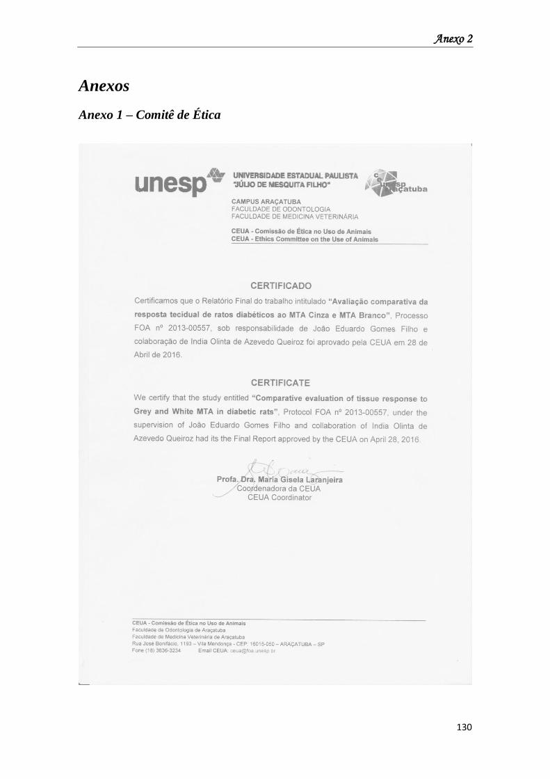
Anexo 2
130
Anexos
Anexo 1 – Comitê de Ética
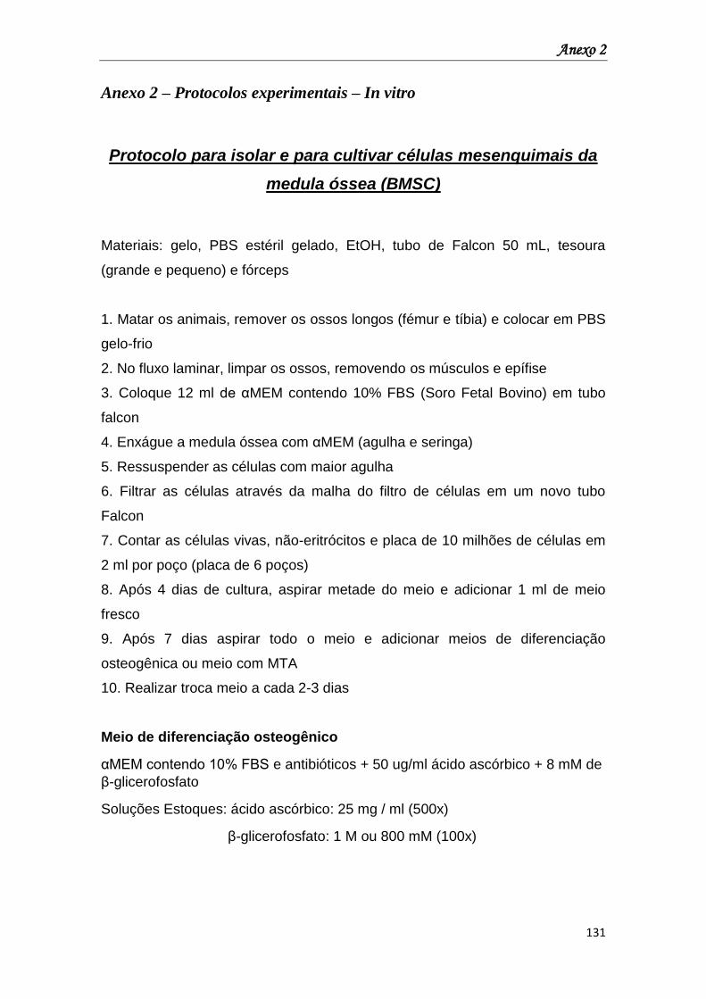
Anexo 2
131
Anexo 2 – Protocolos experimentais – In vitro
Protocolo para isolar e para cultivar células mesenquimais da
medula óssea (BMSC)
Materiais: gelo, PBS estéril gelado, EtOH, tubo de Falcon 50 mL, tesoura
(grande e pequeno) e fórceps
1. Matar os animais, remover os ossos longos (fémur e tíbia) e colocar em PBS
gelo-frio
2. No fluxo laminar, limpar os ossos, removendo os músculos e epífise
3. Coloque 12 ml de αMEM contendo 10% FBS (Soro Fetal Bovino) em tubo
falcon
4. Enxágue a medula óssea com αMEM (agulha e seringa)
5. Ressuspender as células com maior agulha
6. Filtrar as células através da malha do filtro de células em um novo tubo
Falcon
7. Contar as células vivas, não-eritrócitos e placa de 10 milhões de células em
2 ml por poço (placa de 6 poços)
8. Após 4 dias de cultura, aspirar metade do meio e adicionar 1 ml de meio
fresco
9. Após 7 dias aspirar todo o meio e adicionar meios de diferenciação
osteogênica ou meio com MTA
10. Realizar troca meio a cada 2-3 dias
Meio de diferenciação osteogênico
αMEM contendo 10% FBS e antibióticos + 50 ug/ml ácido ascórbico + 8 mM de
β-glicerofosfato
Soluções Estoques: ácido ascórbico: 25 mg / ml (500x)
β-glicerofosfato: 1 M ou 800 mM (100x)
Anexo 2
132
Protocolo para isolar e para cultivar células progenitoras
periodontais (PDSC)
Materiais: gelo, PBS estéril gelado, EtOH, tubo de Falcon 50 mL, tesoura
(grande e pequeno) e fórceps
1. Preparar as soluções:
a. Clorexidina 0,12%
b. Digestion Buffer: Colagenase P(20mg) + PBS(10ml)+ Tripsina(0.05%)
c. Meio de cultura: DMEM + 20% PBS
2. Matar os animais, remover a maxila e mandíbula, remover os tecidos
adjacentes, realizar a hemissecção e colocar em PBS gelo-frio
3. Colocar as peças seccionadas em solução de Clorexidina 0,12% for 30
segundos. Logo após lavar as peças com PBS
4. Colocar as peças em uma nova solução de PBS e realizar a limpeza das
peças (remover tecido gengival) com a ajuda do microscópio
5. Realizar a extração dos molares superiores e inferiores com o auxilio dos
fórceps. Manter os dentes em PBS até a remoção de todos.
6. Colocar os dentes na solução (digestion buffer) por 90min a 370C em
constante agitação.
7. Retirar o sobrenadante e acrescentar ao mesmo DMEM+20%FBS (inativar a
ação da tripsina) e centrifugar por 5min/1576rpm ou 500G
8. Coletar o sobrenadante com cuidado para não remover o pellet.
9. Resuspender o pellet em 1ml de meio de cultura DMEM + 20% PBS.
Adicionar 9ml de meio e cultivar (placa petri) em condições de hipóxia
10. Após 4 dias, remover 5ml do meio de cultura e acrescentar 5ml de novo
meio de cultura.
11. Após 7 dias, remover todo o meio de cultura e acrescentar novo meio.
Transferir as células para condições de normoxia.
12. Trocar o meio a cada dois dias até as células atingirem a confluência.
Realizar a passagem das células (placa 24 poços) e adicionar o meio
osteogênico ou meio com MTA.
10. Realizar troca meio a cada 2-3 dias.
Anexo 2
133
Protocolo para preparação do meio contendo MTA (MTA-CM ou
MTA extract)
1. Manipular o MTA com água estéril de acordo com as instruções do
fabricante e coloca-lo em discos de silicone esteireis.
Obs.: tubos de polietileno com confeccionados nas dimensões (5mm de
diâmetro e 3mm altura)
2. Deixar na estufa a 37°C (5% de CO2, 37°C) por 6 horas para tomar presa.
3. Retirar da estufa e remover os discos dos tubos de silicone
4. Adicionar 1ml de αMEM contendo 10% FBS e antibióticos para cada disco
confeccionado em um tubo falcon
5. Incubar por 72 horas na estufa a 37°C (5% de CO2, 37°C) para se obterem
os ingredientes bioativos do MTA.
6. Após a incubação, remover o sobrenadante e filtrar através de um filtro de
45 μm. O sobrenadante filtrado será o meio de MTA.
Protocolo para coloração celular com Von Kossa
1. Lavar as células com PBS
2. Adicionar formol a 10% e esperar por 5min para fixar as células
3. Adicionar 5% de nitrato de prata
4. Coloque no reticulador UV (Botão de reticulação automática duas vezes 12
segundos)
obs.: não tampar a placa
5. Caso não tenha reticulador UV, deixar a placa sobre a luz da bancada por no
mínimo 30 minutos
6. Remover o nitrato de prata
7. Lavar com água e deixar secar
Protocolo para coloração celular com Fosfatase Alcalina
1. Lavar as células com PBS;
2. Adicionar formol a 10% e esperar por 5min para fixar as células
Anexo 2
134
3. Usar o Kit 86RT da Sigma Aldrich
4. Para fazer 4mL de solução, por exemplo:
a. Pegar um eppendorf e misturar: 100 μl de nitrato de sódio (solução 1)
100 μl de Solução FRV (solução 2)
b. Pegar 4,5 mL de água destilada, misturar com a solução do eppendorf.
c. Adicionar 100 μl de Naphtol (solução 3) e misturar – cor ficará amarela
5. Coloque 1mL da solução final em cada poço
6. Guardar a placa no escuro (longe da luz ) por 30min.
7. Remover o nitrato de prata
8. Lavar com água e deixar secar
Obs.: para cada 4ml de solução usar 100ul das soluções do kit
Protocolo para extração de RNA
Materiais: PBS, EtOH, H2O, Falcon tubo 15 mL, seringa, agulha
1. Lavar as células com PBS
Obs.: em todas as fases deve-se usar gelo
2. Adicionar 1 mL de trizol em cada poço
3. Realizar a remoção das células com o trizol e coloca-las em um eppendorf
4. Utilizar a Polytron por 20 segundos
5. Adicionar 200μl de clorofórmio
6. Agitar vigorosamente por 15 seg.
7. Centrifugar 12 000 rpm / 15 min / 40C
8. Transferir a fase clara superior para um novo tubo eppendord
9. Adicionar 500µl de isopropanol (1 volume) e agitar vigorosamente
10. Armazenar no - 200C por no mínimo uma noite
11. Após o tempo overnight, centrifugar 10 000 RPM / 10 min / 40C
12. Descartar o sobrenadante e colocar no gelo
13. Colocar 1mL de EtOH a 80% e transfira para um novo tubo de 1,5 mL
14. Centrifugar na velocidade máxima / 5 min / 40C
15. Aspirar líquido e deixar o pellet seco no gelo
16. Centrifugar (centrifugação curta) para eliminar o restante EtOH
Anexo 2
135
17. Adicionar 100 μL de água livre de RNase
18. Aquecer a 550C / 5 min, vortex, e repetir 55 0C / 5 min
19. Medir a concentração no nanodrop.
Protocolo do ensaio proliferação - Alamar Blue
1. Adicionar alamar Blue na proporção 1:10 no meio de cultura das células em
cultivo. Deixar poços com meio de cultura somente para a leitura do branco
(Blank) e poços sem célula com meio + alamarBlue (controle negativo).
2. Incubar a 37oC e aguardar a partir de 1h para quantificar por absorbância
(570 e 600 nm) ou fluorescência (excitação 530-560 nm e emissão 590 nm).
3. A avaliação pode ser feita por endpoint (semelhante ao desenho
experimental de MTT, por exemplo) ou por cinética. Neste caso, considerar
plaquear as células em pequena densidade devido à saturação do reagente no
decorrer do tempo.
4. Após a finalização do experimento, as células podem ser recuperadas para
outro ensaio.
Protocolo de preparo Meio de Cultura
DMEM suplementado com 10% FBS - 1L
Materiais:
850mL de H2O Miliq
10,03g de Dulbecos Medium Eagles (DMEM)
3,7g de bicarbonato de sódio
100mL de soro fetal bovino
10mL de penicilina/streptomicina
1mL de gentamicina
300µL de fungisone
20mL de glutamina
1. Colocar H2O Miliq em um bécker, diluir o pacote de DMEM inteiro,
acrescentar o bicarbonato e o restante da água.
2. Colocar o bécker no agitador e ajustar pH 6,9.
Anexo 2
136
3. Dentro do fluxo, acrescentar antibióticos, soro fetal, glutamina (dentro do
fluxo) e filtrar (bomba vácuo).
Meio hiperglicêmico
1. Adicionar ao meio de cultura DMEM com 10% FBS glicose na seguinte
concentração:
Glicose – 0,225g em 50mL
Anexo 3
137
Anexo 3 – Protocolos experimentais – In vivo
Protocolo de indução de diabetes
Material: ALLOXAN MONOHYDRATE, 25 G
Referência do Produto: A7413
Marca: Sigma Aldrich
1. Realizar a pesagem e a dosagem da glicemia dos animais;
2. Preparo da solução de Aloxano de acordo com o número de animais
utilizados para o experimento.
Ex.: Para indução de 50 animais pesando 200g cada
Em um recipiente coloca-se solução salina 0,9% estéril na proporção
de 0,5ml para cada animal: 50 animais = 25 ml de solução salina
Realiza a pesagem da droga para ser utilizada na proporção de
150mg/kg. Cada animal pesa 200g, logo 50 animais = 10000g
150mg/kg: 150mg (droga) ---------- 1000g (droga)
X mg (droga) ______ 10000g (peso total animais)
X = 500mg (peso total da droga)
Em béquer colocar 25 ml da solução salina e ir acrescentando aos
poucos a droga (500mg) com agitação constante usando um agitador
magnético até a completa diluição.
3. Realizar a injeção por via intraperitoneal na dose única de 150mg/kg,
proporcional ao peso do animal com agulha e seringa de insulina;
Dose = peso do rato x volume solução (25ml)
dose droga (500mg)
4. Após a indução deixar somente água e passadas 06 horas oferecer aos
animais glicose a 10% (água com açúcar) e comida;
5. Retirar água com açúcar após 24hs.
6. Após o 3o dia realizar a dosagem da glicemia dos animais.
Anexo 3
138
Protocolo para injeção dos Fluorocromos
Material: Calcein, 10 G
Referência do Produto: C0875 Marca: Sigma Aldrich
Alizarin Red S, 25 G
Referência do Produto: A5533 Marca: Sigma Aldrich
Oxytetracycline hydrochloride, 10 G
Referência do Produto: O5875 Marca: Sigma Aldrich
1. Realizar a pesagem dos animais;
2. Preparo da solução dos fluorocromos de acordo com o número de animais
utilizados para o experimento.
Ex.: Para indução de 10 animais pesando 300g cada
Em um recipiente coloca-se solução salina 0,9% estéril na proporção
de 1ml para cada animal: 10 animais = 10 ml de solução salina
Realiza a pesagem da droga para ser utilizada na proporção de
20mg/kg. Cada animal pesa 300g, logo 10 animais = 3000g
20mg/kg: 20mg (droga) ---------- 1000g (droga)
X mg (droga) ______ 3000g (peso total animais)
X = 60mg ou 0,0060g (peso total da droga)
Em béquer colocar 10 ml da solução salina e ir acrescentando aos
poucos a droga (60mg ou 0,0060g) com agitação constante usando um
agitador magnético até a completa diluição.
3. Realizar a injeção por via intramuscular na dose única de 20mg/kg com
agulha e seringa de insulina. Injetar 1ml da solução para cada animal.
Anexo 3
139
Protocolo para dosagem sanguínea de Cálcio
Material: Cálcio Liquiform (Kit Labtest)
Cálcio está envolvido no processo de mineralização óssea. Para determinação
deve-se preparar o reagente de trabalho.
Reagentes do kit (100 testes)
R1 – Reagente 1 (tampão)
R2- Reagente 2 (cuidado, contém HCl)
R3- Padrão (10mg/dL) (Contém formol, cuidado com evaporação)
Material necessário
Pipeta automática que libere 20µL
Pipeta automática que libere 1mL ou repipetador automático
Ponteiras
Placa para leitura na leitora Elisa (template)
Béqueres para colocar os reagentes a serem pipetados
Amostra (plasma, que não seja coletado com EDTA ou fluoreto)
Tubos de ensaio em duplicata para as amostras
Tubo para padrão e branco
Estante para tubos
Obs: é importante que os tubos estejam bem limpos, de preferência lavados
com HCl e muito bem enxaguados com água destilada.
Procedimento
1. Primeiramente deve-se preparar o reagente de trabalho, onde se mistura 3
volumes do reagente 1 (R1) com 1 volume do reagente 2 (R2), calcular a
quantidade de reagente que será necessário já que se usa 1mL do mesmo por
amostra. No kit a proporção do R1 para o R2 já está certa, então caso vá fazer
60 testes (incluindo padrão e branco), é só misturar o conteúdo todo dos dois
reagentes. Toma-se cuidado para não preparar o reagente de trabalho em
excesso, pois sua estabilidade é de apenas 8 horas, tendo que ser descartado
caso não utilizado.
2. Nos tubos de teste adicionar 20µL do plasma e o padrão no tubo
correspondente e em todos os tubos pipetar 1mL do reagente de trabalho.
Anexo 3
140
Agitar a estante levemente para misturar o reagente com a amostra. A
estabilidade da cor não é citada na bula, porém recomenda-se fazer a leitura o
mais rápido possível, para evitar alguma perda. Se a leitura for feita no
espectrofotômetro manual (570nm) adicionar o branco na cubeta e zerar o
aparelho. Proceder as leituras das amostras. Caso a leitura for feita na leitora
Elisa, deve-se pipetar 300µL da reação em cada poço, de acordo com a ordem
definida (A1- Branco/ A2-Padrão/ A3-Padrão/ A4-amostra).
3. Ligar o computador, entrar no programa KC Junior, open protocol, Cálcio
labtest, modify protocol e arrumar o template de acordo com o que pipetou na
placa. Não esquecer de conferir o comprimento de onda (570nm). Fazer uma
leitura sem placa, só para calibrar o aparelho (read plate). Depois fechar os
resultados (file- close results).
4. Colocar a placa na leitora e realizar a leitura (read plate), salvando com ID
(Cálcioseunome/orientador- dd/mm/AA). Após abrirá a leitura das
absorbâncias, então abra o Excel e copie as informações (template e
absorbâncias menos o branco).
Cálculos
Cálcio (mg/dL) = Abs teste (amostras)/abs padrão x 10
Pode-se fazer o cálculo usando o fator, onde: fator=10/abs padrão
Cálcio (mg/dL)= abs teste x fator
Caso deseje pode se realizar o cálculo no Excel, recomenda-se usar o fator,
multiplicando-o pelas absorbâncias da amostras.
Exemplos de cálculos:
Fator=10/1,346 =7,43
Cálcio = 1,270 x 7,43 = 9,4mg/dL
Protocolo para dosagem sanguínea de Fósforo
Material: Fósforo UV Liquiform (Kit Labtest)
Fósforo está envolvido no processo de mineralização óssea. Sua absorbância
é determinada em comprimento de onda de 340nm sendo necessário fotômetro
que tenha leitura na faixa ultravioleta.
Anexo 3
141
Reagentes do kit (100 testes)
R1 – Reagente de cor (cuidado, cáustico)
R2- Padrão (5,0mg/dL)
Material necessário
Pipeta automática que libere 10µL
Pipeta automática que libere 1mL ou repipetador automático
Ponteiras
Cronômetro
Banho Maria (37°C)
Placa para leitura na leitora Elisa (template)
Béqueres para colocar os reagentes a serem pipetados
Amostra (plasma)
Tubos de ensaio em duplicata para as amostras
Tubo para padrão e branco
Estante para tubos
Obs: é importante que os tubos estejam bem limpos, sem resíduos de
detergentes, uma vez que o fósforo inorgânico é componente comum da
maioria dos detergentes.
Procedimento
1. Nos tubos de teste adicionar 10µL do plasma e em todos os tubos pipetar
1mL do reagente de cor (R1), de preferência com o repipetador pelo tempo de
estabilidade. Agitar a estante levemente para misturar o reagente com a
amostra. Levar ao banho maria que já deve estar na temperatura (37°C), deixar
por 5 minutos. Após deve-se retirar a estante do banho e secá-la. A
absorbância é estável somente por 30 minutos, recomendando-se leitura
imediata. Se a leitura for feita no espectrofotômetro manual (340nm) adicionar
o branco na cubeta e zerar o aparelho. Proceder as leituras das amostras.
Caso a leitura for feita na leitora Elisa, deve-se pipetar 300µL da reação em
cada poço, de acordo com a ordem definida (A1- Branco/ A2-Padrão/ A3-
Padrão/ A4-amostra).
2. Ligar o computador, entrar no programa KC Junior, open protocol, Fósforo
labtest, modify protocol e arrumar o template de acordo com o que pipetou na
placa. Não esquecer de conferir o comprimento de onda (340nm). Fazer uma
Anexo 3
142
leitura sem placa, só para calibrar o aparelho (read plate). Depois fechar os
resultados (file- close results).
3. Colocar a placa na leitora e realizar a leitura (read plate), salvando com ID
(Fósforoseunome/orientador- dd/mm/AA). Após abrirá a leitura das
absorbâncias, então abra o Excel e copie as informações (template e
absorbâncias menos o branco).
Cálculos
Fósforo inorgânico (mg/dL) = Abs teste (amostras)/abs padrão x 5
Pode-se fazer o cálculo usando o fator, onde: fator=5/abs padrão
Fósforo inorgânico(mg/dL)= abs teste x fator
Caso deseje pode se realizar o cálculo no Excel, recomenda-se usar o fator,
multiplicando-o pelas absorbâncias da amostras.
Exemplos de cálculos:
Fator=5/0,247 =20,2
Fósforo inorgânico = 0,190 x 20,2 = 3,8mg/dL
Protocolo para dosagem sanguínea de Fosfatase Alcalina
Material: Fosfatase Alcalina (Kit Labtest)
Fosfatase alcalina óssea é uma enzima, envolvida no processo de
mineralização, portanto não é possível analisar sua quantidade e sim sua
atividade.
Reagentes do kit (100 testes)
R1 – Substrato
R2 – Tampão (pH 10,1- básico “cuidado”)
R3- Reagente de cor (reagente de parada- também alcalino “cuidado”)
R4- Padrão 45U/L
Material necessário
Pipeta automática que libere 50µL
Pipeta automática que libere 500 µL e 2mL ou repipetador automático
(duas ponteiras)
Ponteiras
Anexo 3
143
Cronômetro
Banho Maria (37°C)
Placa para leitura na leitora Elisa (template)
3 béqueres para colocar os reagentes a serem pipetados
Amostra (plasma)
Tubos de ensaio em duplicata para as amostras
Tubo para padrão e branco
Estante para tubos
Procedimento
1. Em todos os tubos pipetar 0,5mL (500µL) do tampão (R2) e 50 µL do
substrato (R1). Levar ao banho maria que já deve estar na temperatura (37°C),
deixar por 2 minutos. Os passos seguintes sugere-se fazer em duas pessoas,
pois a reação exige extrema atenção quanto ao tempo.
2. A adição da amostra (50µL) deve ser feita com intervalos (de 20 em 20
segundos) de acordo com a prática de pipetar de quem manuseia a amostra,
importante que seja calculado de forma que a última amostra seja pipetada
antes de completar o tempo da reação (10 minutos) não se esquecer de pipetar
50µL padrão (R4) no tubo correspondente.
3. Após os 10 minutos da adição da primeira amostra deve-se adicionar 2mL
do reagente de parada (R3) , procedendo sua adição de acordo com o tempo
em que foram adicionadas as amostras (de 20 em 20 segundos), desta forma a
reação em todos os tubos ocorrerá no tempo de 10 minutos. A reação toda
ocorre dentro do banho maria.
4. Após a adição do R3 em todos os tubos, deve-se retirar a estante do banho
e secá-la. A cor é estável durante 120 minutos. Se a leitura for feita no
espectrofotômetro manual (590nm) adicionar o branco na cubeta e zerar o
aparelho. Proceder as leituras das amostras. Caso a leitura for feita na leitora
Elisa, deve-se pipetar 300µL da reação em cada poço, de acordo com a ordem
definida (A1- Branco/ A2-Padrão/ A3-Padrão/ A4-amostra).
5. Ligar o computador, entrar no programa KC Junior, open protocol, fosfatase
alcalina labtest, modify protocol e arrumar o template de acordo com o que
pipetou na placa. Não esquecer de conferir o comprimento de onda (590nm).
Fazer uma leitura sem placa, só para calibrar o aparelho (read plate). Depois
fechar os resultados (file- close results).
Anexo 3
144
6. Colocar a placa na leitora e realizar a leitura (read plate), salvando com ID
(FAseunome/orientador- dd/mm/AA). Após abrirá a leitura das absorbâncias,
então abra o Excel e copie as informações (template e absorbâncias menos o
branco).
Cálculos
Fosfatase alcalina (U/L) = Abs teste (amostras)/abs padrão x 45
Pode-se fazer o cálculo usando o fator, onde: fator=45/abs padrão
Fosfatase alcalina (U/L)= abs teste x fator
Caso deseje pode se realizar o cálculo no Excel, recomenda-se usar o fator,
multiplicando-o pelas absorbâncias da amostras.
Exemplos de cálculos:
Fator=45/0,360 =125
Fosfatase alcalina = 0,295 x 125 = 37 U/L
Metodologia
Roy modificado
Técnica para inclusão em Glicol Metacrilato
(Leica – Historesin)
1. Lavar em água corrente no mínimo 5 horas;
2. Desidratar em álcool 70% por uma noite;
3. No dia seguinte:
a. Álcool 90% por 30 minutos;
b. Álcool 90% por 30 minutos;
c. Álcool 90% por 30 minutos;
d. Álcool 95% por 30 minutos;
e. Álcool 95% por 30 minutos;
f. Álcool 95% por 30 minutos;
4. Colocar as peças desidratadas na solução A ativada e deixar por 72 horas
(temperatura ambiente);
5. Isolar os moldes de plástico próprio para inclusão com spray de teflon,
colocar as peças no interior dos moldes, cobrir as peças com resina para
Anexo 3
145
inclusão (leica – historesin), depois de polimerizada, preencher com resina
acrílica auto polimerizante e aguardar a polimerização.
Técnica para Coloração em Hematoxilina e Eosina (para peças
incluídas em Glicol Metacrilato)
Peças cortadas em 3μm.
1. Hidratar rapidamente em água destilada;
2. Colocar na Hematoxilina – 30 minutos;
3. Lavar em água corrente até remover o excesso de corante;
4. Colocar na Eosina – 5 minutos;
5. Lavar em água corrente até remover o excesso de corante;
6. Deixar secar na estufa, montar com Entelan e lamínula.
Técnica para Coloração em Von Kossa (para peças incluídas
em Glicol Metacrilato)
Peças cortadas em 10μm.
1. Hidratar rapidamente em água destilada;
2. Colocar sobre os cortes nitrato de prata 1% e deixar sob a luz solar por no
mínimo 15 minutos;
3. Lavar em água corrente para remover o excesso de nitrato de prata;
4. Colocar no tiossulfato de sódio 5% - 30 segundos;
5. Lavar em água corrente - 1 minuto;
6. Colocar na safranina 0,5% - 30 segundos;
7. Lavar em água corrente para remover o excesso do corante.
8. Deixar secar na estufa, montar com Entelan e lamínula.
Técnica para Luz Polarizada (para peças incluídas em Glicol
Metacrilato)
Peças cortadas em 10μm.
1. Lâminas secas e montar com Entelan e lamínula.
Anexo 3
146
Técnica para Luz Polarizada (para peças incluídas em Glicol
Metacrilato)
Peças cortadas em 50μm.
1. Após o corte, deixar secar por poucos minutos
2. Lâminas secas e montar com meio de montagem para fluorescência à base
de glicerol e lamínula.
3. Armazenar em geladeira
Técnica para inclusão em Parafina
1. Lavar em água corrente por 12 horas;
2. Realizar as trocas de Álcool e Xilol na sequência:
a) Álcool 70% por 1hora;
b) Álcool 80% por 1hora;
c) Álcool 95% por 1hora;
d) Álcool 100% (I) por 1hora;
e) Álcool 100% (II) por 1hora;
f) Álcool 100% (III) por 1hora;
g) Álcool/Xilol (50/50) por 1hora;
h) Xilol 100% (I) por 1hora;
i) Xilol 100% (II) por 30minutos;
j) Xilol 100% (III) por 30minutos;
k) Parafina (I) por 1hora;
l) Parafina (II) por 1hora;
m) Parafina (III) por 1hora;
3. Retirar as peças da parafina (III) e inclui-las embebidas com o auxílio da
máquina.
Anexo 3
147
Protocolo para Imunoistoquímica
1º Dia
1. Desparafinização
1o. Ciclo: Colocar lâminas na estufa por 30 minutos para derreter a parafina
(Estufa a 56ºC – 60ºC).
2o. Ciclo: Bateria:
a) Xilol I – 5 minutos
b) Xilol II – 5 minutos
c) Xilol III – 10 minutos
d) Álcool 100 I – 2 minutos
e) Álcool 100 II – 2 minutos
f) Álcool 100 III – 2 minutos
g) Álcool 95 – 2 minutos
h) Álcool 70 – 2 minutos
2. Lavagens
a) PBS – 5 minutos
b) PBS – Triton x100 – 5 minutos
c) PBS – 5 minutos
3. Recuperação Antigênica
198 ml de água destilada
2 ml de tampão citrato 100x
4. Lavagens
a) PBS – 5 minutos
b) PBS – Triton x100 – 5 minutos
c) PBS – 5 minutos
5. Bloqueio da Peroxidase Endógena
180 mL de PBS
20 mL de H2O2
6. Lavagens
a) PBS – 5 minutos
b) PBS – Triton x100 – 5 minutos
c) PBS – 5 minutos
5 minutos – 95 º C
1 hora
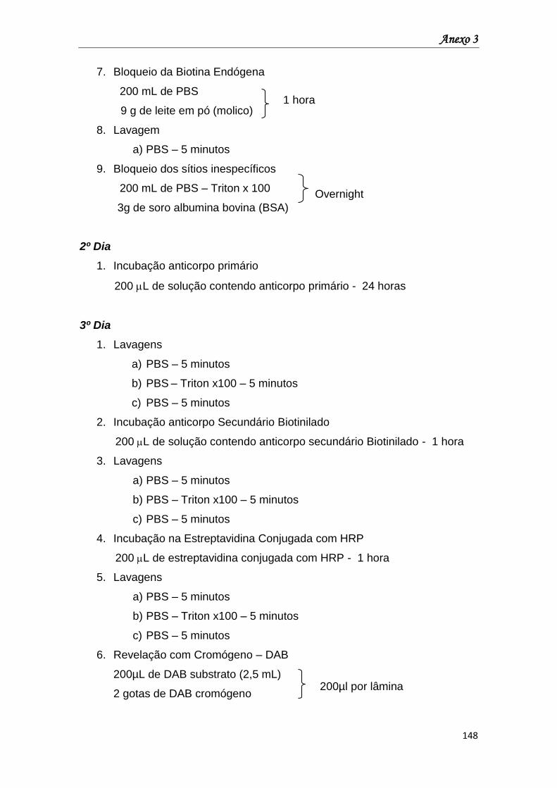
Anexo 3
148
7. Bloqueio da Biotina Endógena
200 mL de PBS
9 g de leite em pó (molico)
8. Lavagem
a) PBS – 5 minutos
9. Bloqueio dos sítios inespecíficos
200 mL de PBS – Triton x 100
3g de soro albumina bovina (BSA)
2º Dia
1. Incubação anticorpo primário
200 L de solução contendo anticorpo primário - 24 horas
3º Dia
1. Lavagens
a) PBS – 5 minutos
b) PBS – Triton x100 – 5 minutos
c) PBS – 5 minutos
2. Incubação anticorpo Secundário Biotinilado
200 L de solução contendo anticorpo secundário Biotinilado - 1 hora
3. Lavagens
a) PBS – 5 minutos
b) PBS – Triton x100 – 5 minutos
c) PBS – 5 minutos
4. Incubação na Estreptavidina Conjugada com HRP
200 L de estreptavidina conjugada com HRP - 1 hora
5. Lavagens
a) PBS – 5 minutos
b) PBS – Triton x100 – 5 minutos
c) PBS – 5 minutos
6. Revelação com Cromógeno – DAB
200µL de DAB substrato (2,5 mL)
2 gotas de DAB cromógeno
1 hora
Overnight
200µl por lâmina
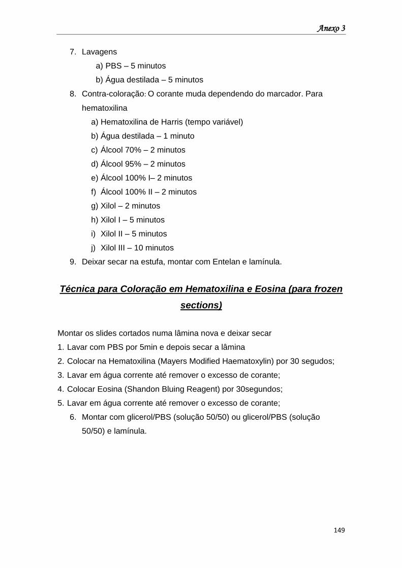
Anexo 3
149
7. Lavagens
a) PBS – 5 minutos
b) Água destilada – 5 minutos
8. Contra-coloração: O corante muda dependendo do marcador. Para
hematoxilina
a) Hematoxilina de Harris (tempo variável)
b) Água destilada – 1 minuto
c) Álcool 70% – 2 minutos
d) Álcool 95% – 2 minutos
e) Álcool 100% I– 2 minutos
f) Álcool 100% II – 2 minutos
g) Xilol – 2 minutos
h) Xilol I – 5 minutos
i) Xilol II – 5 minutos
j) Xilol III – 10 minutos
9. Deixar secar na estufa, montar com Entelan e lamínula.
Técnica para Coloração em Hematoxilina e Eosina (para frozen
sections)
Montar os slides cortados numa lâmina nova e deixar secar
1. Lavar com PBS por 5min e depois secar a lâmina
2. Colocar na Hematoxilina (Mayers Modified Haematoxylin) por 30 segudos;
3. Lavar em água corrente até remover o excesso de corante;
4. Colocar Eosina (Shandon Bluing Reagent) por 30segundos;
5. Lavar em água corrente até remover o excesso de corante;
6. Montar com glicerol/PBS (solução 50/50) ou glicerol/PBS (solução
50/50) e lamínula.
Anexo 4
150
Anexo 4 - Diretrizes para publicação dos trabalhos
Guidelines for Publishing Papers in the Journal of Periodontal
Research
1. GENERAL
The Journal of Periodontal Research is an international research periodical the
purpose of which is to publish original clinical and basic investigations and
review articles concerned with every aspect of periodontology and related
sciences. Reports of scientific meetings in periodontology and related fields are
also published.
Please read the instructions below carefully for details on the submission of
manuscripts, the journal's requirements and standards as well as information
concerning the procedure after a manuscript has been accepted for publication
in the Journal of Periodontal Research. Authors are encouraged to visit Wiley
Blackwell's Author Services for further information on the preparation and
submission of articles and figures.
2. ETHICAL GUIDELINES
The Journal of Periodontal Research adheres to the below ethical guidelines for
publication and research.
2.1. Authorship and Acknowledgements
Authors submitting a paper do so on the understanding that the manuscript
have been read and approved by all authors and that all authors agree to the
submission of the manuscript to the Journal.
The Journal of Periodontal Research adheres to the definition of authorship set
up by The International Committee of Medical Journal Editors (ICMJE).
According to the ICMJE authorship criteria should be based on 1) substantial
contributions to conception and design of, or acquisiation of data or analysis
and interpretation of data, 2) drafting the article or revising it critically for
important intellectual content and 3) final approval of the version to be
published. Authors should meet conditions 1, 2 and 3.
Anexo 4
151
It is a requirement that all authors have been accredited as appropriate upon
submission of the manuscript. Contributors who do not qualify as authors
should be mentioned under Acknowledgements.
Acknowledgements: Under acknowledgements please specify contributors to
the article other than the authors accredited. Acknowledge only persons who
have made substantive contributions to the study. Authors are responsible for
obtaining written permission from everyone acknowledged by name because
readers may infer their endorsement of the data and conclusions. Please also
include specifications of the source of funding for the study and any potential
conflict of interests if appropriate. Suppliers of materials should be named and
their location (town, state/county, country) included.
2.2. Ethical Approvals
All studies using human or animal subjects should include an explicit statement
in the Material and Methods section identifying the review and ethics committee
approval for each study, if applicable. Editors reserve the right to reject papers if
there is doubt as to whether appropriate procedures have been used.
2.3 Photographs of People
The Journal of Periodontal Research follows current HIPAA guidelines for the
protection of patients/subject privacy. If an individual pictured in a digital image
or photograph can be identified, his or her permission is required to publish the
image. The corresponding author may submit a letter signed by the patient
authorizing the Journal of Periodontal Research to publish the image/photo. Or,
a form provided by the Journal of Periodontal Research (available by clicking
the “Instructions and Forms” link in Manuscript Central) may be downloaded for
your use. This approval must be received by the Editorial Office prior to final
acceptance of the manuscript for publication. Otherwise, the image/photo must
be altered such that the individual cannot be identified (black bars over eyes,
tattoos, scars, etc.). The Journal of Periodontal Research will not publish patient
photographs that will in any way allow the patient to be identified, unless the
patient has given their express consent.
2.4 Clinical Trials
Clinical trials should be reported using the CONSORT guidelines available
at www.consort-statement.org. A CONSORT checklist should also be included
in the submission material. The Journal of Periodontal Research encourages
Anexo 4
152
authors submitting manuscripts reporting from a clinical trial to register the trials
in any of the following free, public clinical trials
registries: www.clinicaltrials.gov, http://clinicaltrials.ifpma.org/clinicaltrials/, http://
isrctn.org/. The clinical trial registration number and name of the trial register will
then be published with the paper.
2.5 Conflict of Interest and Source of Funding
Please disclose information concerning sources of institutional, private and
corporate financial support for the work within the manuscript be fully
acknowledged, and any potential conflicts of interest under Acknowledgements
2.6 Appeal of Decision
The decision on a paper is final and cannot be appealed.
2.7. Permissions
If all or parts of previously published illustrations are used, permission must be
obtained from the copyright holder concerned. It is the author's responsibility to
obtain these in writing and provide copies to the Publishers.
2.8. Copyright
If your paper is accepted, the author identified as the formal corresponding
author for the paper will receive an email prompting them to login to Author
Services; where via the WIley Author Licensing Service (WALS), they will be
able to complete the license agreement on behalf of all authors on the paper.
For authors signing the copyright transfer agreement
If the OnlineOpen option is not selected, the corresponding author will be
presented with the copyright transfer agreement (CTA) to sign. The terms and
conditions of the CTA can be previewed in the samples associated with the
Copyright FAQS below: CTA Terms and Conditions
http://authorservices.wiley.com/bauthor/faqs_copyright.asp
For authors choosing OnlineOpen
If the OnlineOpen option is selected, the corresponding author will have a
choice of the following Creative Commons License Open Agreements (OAA):
Creative Commons License OAA Creative Commons Attribution Non
Commercial License OAA Creative Commons Attribution Non-Commercial -
NoDerivs License OAA. To preview the terms and conditions of these open
acess agreements, please visit the Copyright FAQ hosted on Wiley Author
Services http://exchanges.wiley.com/authors/faqs---copyright-_301.html and
Anexo 4
153
visit http://www.wileyopenaccess.com/details/content/12f25db4c87/Copyright--
License.html.
If you select the OnlineOpen option and your research is funded by certain
funders [e.g. The Wellcome Trust and members of the Research Councils UK
(RCUK) or the Austrian Science Fund (FWF)], you will be given the opportunity
to publish your article under a CC-BY license supporting you in complying with
Wellcome Trust and Research Councils UK requirements. For more information
on this policy and the Journal's compliant self-archiving policy, please
visit http://www.wiley.com/go/funderstatement
3. MANUSCRIPT SUBMISSION PROCEDURE
Manuscripts should be submitted electronically via the online submission
site http://mc.manuscriptcentral.com/jre.The use of an online submission and
peer review site enables immediate distribution of manuscripts and
consequentially speeds up the review process. It also allows authors to track
the status of their own manuscripts. Complete instructions for submitting a
paper is available online and below. For further assistance, please contact the
editorial office at [email protected]
3.1. Getting Started
• Launch your web browser (supported browsers include Internet Explorer 6 or
higher, Netscape 7.0, 7.1, or 7.2, Safari 1.2.4, or Firefox 1.0.4) and go to the
journal's online Submission Site: http://mc.manuscriptcentral.com/jre
• Log-in or, if you are a new user, click “register here”
• If you are registering as a new user.
After clicking on 'register here', enter your name and e-mail information and
click 'Next'. Your e-mail information is very important.
Enter your institution and address information as appropriate, and then click
'Next.'
Enter a user ID and password of your choice (we recommend using your e-mail
address as your user ID), and then select your area of expertise. Click 'Finish'.
• If you have an account, but have forgotten your log in details, go to Password
Help on the journals online submission
system http://mc.manuscriptcentral.com/jre and enter your email address. The
system will send you an automatic user ID and a new temporary password.
• Log-in and select “Corresponding Author Center”
Anexo 4
154
3.2. Submitting Your Manuscript
• After you have logged into your “Corresponding Author Center”, submit your
manuscript by clicking the submission link under “Author Resources”.
• Enter data and answer questions as appropriate. You may copy and paste
directly from your manuscript and you may upload your pre-prepared covering
letter.
• Click the 'Next' button on each screen to save your work and advance to the
next screen.
• You are required to upload your files.
Click on the 'Browse' button and locate the file on your computer.
Select the designation of each file in the drop down next to the Browse button.
When you have selected all files you wish to upload, click the 'Upload Files'
button.
• Review your submission (in HTML and PDF format) before sending to the
Journal. Click the 'Submit' button when you are finished reviewing.
3.3. Manuscript Files Accepted
Manuscripts should be uploaded as Word (.doc/.docx) or Rich Text Format (.rtf)
files (not write-protected) plus separate figure files. GIF, JPEG, PICT or Bitmap
files are acceptable for submission, but only high-resolution TIF or EPS files are
suitable for printing. The files will be automatically converted to HTML and PDF
on upload and will be used for the review process. The text file must contain the
entire manuscript including title page, abstract, text, references, figure legends
and tables but no embedded figures. Figure tags should be included in the file.
Manuscripts should be formatted as described in the Author Guidelines below.
3.4. Blinded Review
All manuscripts submitted to the Journal of Periodontal Research will be
reviewed by two experts in the field. The Journal of Periodontal Research uses
single blinded review. The names of the reviewers will thus not be disclosed to
the author submitting a paper.
3.5. Suggest a Reviewer
The Journal of Periodontal Research attempts to keep the review process as
short as possible to enable rapid publication of new scientific data. In order to
facilitate this process, please suggest the names and current email addresses
Anexo 4
155
of 2 potential international reviewers whom you consider capable of reviewing
your manuscript.
3.6. Suspension of Submission Mid-way in the Submission Process
You may suspend a submission at any phase before clicking the 'Submit' button
and save it to submit later. The manuscript can then be located under
'Unsubmitted Manuscripts' and you can click on 'Continue Submission' to
continue your submission when you choose to.
3.7. Email Confirmation of Submission
After submission you will receive an email to confirm receipt of your manuscript.
If you do not receive the confirmation email after 24 hours, please check your
email address carefully in the system. If the email address is correct please
contact your IT department. The error may be caused by some sort of spam
filtering on your email server. Also, the emails should be received if the IT
department adds our email server (uranus.scholarone.com) to their whitelist.
3.8. Manuscript Status
You can access ScholarOne Manuscripts (formerly known as Manuscript
Central) any time to check your 'Author Centre'. The Journal will inform you by
e-mail once a decision has been made.
3.9. Submission of Revised Manuscripts
To submit your revised manuscript, locate your manuscript under 'Manuscripts
with Decisions' and click on 'Submit a Revision'. Please remember to delete any
old files uploaded when you upload your revised manuscript.
4. MANUSCRIPT TYPES ACCEPTED
Original Articles: must describe significant and original experimental
observations and provide sufficient detail so that the observations can be
critically evaluated and, if necessary, repeated. Original articles must conform to
the highest international standards in the field.
Review Articles: are selected for their broad general interest; all are refereed
by experts in the field. Reviews should take a broad view of the field rather than
merely summarizing the authors´ own previous work, so extensive citation of
the authors´ own publications is discouraged.
Mini Reviews are covering a smaller area and may be written in a more free
format.
Anexo 4
156
Short Communications: Short communications, limited to 1-3 pages, including
illustrations and references, will be considered for rapid publication. Such
papers must be based on work that is of special importance or having the
potential for great impact, or a body of work that is complete but of insufficient
scope to warrant a full-length paper. Short communications need not follow the
usual divisions.
Meeting Reports: Reports of scientific meetings in periodontology and related
fields are also published.
5. MANUSCRIPT FORMAT AND STRUCTURE
5.1. Page Charge
Articles exceeding 7 published pages (including figures and tables) are subject
to a charge of GBP70.00 per additional page. For guidance purposes, one
published page amounts approximately to 5,500 characters; text should be
reduced if figures/tables are included within the 7 pages. If authors are unable
to pay additional page fees they will need to reduce the length of their articles.
5.2. Format
Language: The language of publication is English. Authors for whom English is
a second language must have their manuscript professionally edited by an
English speaking person before submission to make sure the English is of high
quality. It is preferred that manuscript is professionally edited. A list of
independent suppliers of editing services can be found
at http://authorservices.wiley.com/bauthor/english_language.asp. All services
are paid for and arranged by the author, and use of one of these services does
not guarantee acceptance or preference for publication
Abbreviations and symbols: Abbreviations should be in accordance with
Guidelines laid down by the American Society of Microbiology. Unless they are
in common usage (e.g. DNA), all terms must be displayed in full in the key
words, and the first time that they appear in the abstract, the main text, tables
and figures, followed by the abbreviation in parentheses. If an abbreviation is
used in the body of figure or table only it must be defined in the figure legend or
table footnotes. The symbol % is to be used for percent, h for hour, min for
minute, and s for second. In vitro and in vivo are to be italicized. Use only
standard abbreviations. All units will be metric. Use no roman numerals in the
text. In decimals, a decimal point, and not a comma, will be used. In cases of
Anexo 4
157
doubt, the spelling orthodoxy of Webster 's Third New International
Dictionary will be adhered to.
Scientific Names: Proper names of bacteria should be binomial and should be
singly underlined in the typescript. The full proper name (e. g. Streptococcus
sanguis ) must be given upon first mention. The generic name may be
abbreviated thereafter with the first letter of the genus (e. g. S. sanguis ). If
abbreviation of the generic name could cause confusion, the full name should
be used. If the vernacular form of a genus name (e. g. streptococci) is used, the
first letter of the vernacular name is not capitalized and the name is not
underlined. Use of two letters of the genus (e. g. Ps .for Peptostreptococcus ) is
incorrect, even though it might avoid ambiguity. With regard to drugs, generic
names should be used instead of proprietary names. It is strongly
recommended that all abbreviations be introduced in the first paragraph in
Materials and Methods. Alternatively, define each abbreviation and introduce it
in parentheses the first time it is used; e.g., "Cultures were grown in Eagle
minimal essential medium (MEM)." Generally, eliminate abbreviations that are
not used at least three times in the text (including tables and figure legends).
5.3. Structure
All manuscripts submitted to the Journal of Periodontal Research should
include: Title page, abstract, main text, references and tables, figures and figure
legends were appropriate.
Title Page: should contain the title of the article, name(s) of the author(s),
initials, and institutional affiliation(s), a running title not to exceed 40 letters and
spaces, and the name and complete mailing address, including email
address, of the author responsible for correspondence. We accept only one
corresponding author per manuscript and in case of co-correspondence a foot
note will be added indicating ‘Both the authors have contributed equally to the
work’.The author must list 4 keywords for indexing purposes.
Abstract: The abstract should consist of 1) the objective 2) the background
data discussing the present status of the field 3) methods 4) results 5)
conclusion.
Main Text of Original Research Articles
Introduction: Summarize the rationale and purpose of the study, giving only
strictly pertinent references. Do not review existing literature extensively.
Anexo 4
158
Material and methods: Materials and methods should be presented in sufficient
detail to allow confirmation of the observations. Published methods should be
referenced and discussed only briefly, unless modifications have been made.
Results: Present your results in a logical sequence in the text, tables, and
illustrations. Do not repeat in the text all of the data in the tables and
illustrations. Important observations should be emphasized.
Discussion: Summarize the findings without repeating in detail the data given in
the Results section. Relate your observations to other relevant studies and point
out the implications of the findings and their limitations. Cite other relevant
studies.
Main Text of Reviews, Short Communications and Meeting Reports
These need not follow the usual divisions.
Acknowledgements: Acknowledge only persons who have made substantive
contributions to the study. Authors are responsible for obtaining written
permission from everyone acknowledged by name because readers may infer
their endorsement of the data and conclusions. Sources of financial support
must be acknowledged.
5.4. References
References should be numbered consecutively in the order in which they
appear in the text, and should be kept to a pertinent minimum. References
should include the beginning and ending page numbers. Identify references in
the text, tables, and figure legends by Arabic numerals in parentheses.
References cited only in the tables or figure legends should be numbered in
accordance with a sequence established by the first notation of that figure or
table in the text. Use the style of the examples below, which is based on Index
Medicus. Manuscripts accepted but not published may be cited in the reference
list by placing ''in press'' after the abbreviated title of the journal. Abstracts and
manuscripts not yet accepted may be cited in full in the text but not in the
reference list. References must be verified by the author(s) against the original
documents. We recommend the use of a tool such as Reference Manager for
reference management and formatting. Reference Manager reference styles
can be searched for here: http://refman.com/downloads/styles
Examples:
Anexo 4
159
(1) Standard journal article (List all authors up to 6; for 7 or more list the first 3
and add ''et al.'') Dockrell H, Greenspan JS. Histochemical identification of T-
cells in oral lichen planus. Oral Surg 1979; 48: 42-49. Thomas Y, Sosman J,
Yrigoyen O, et al. Functional analysis of human T- cell subsets defined by
monoclonal antibodies. I. Collaborative T-T interactions in the immunoregulation
of B-cell differentiation. J Immunol 1980; 125: 2402-2405.
(2) Corporate author. The Royal Marsden Hospital Bone- Marrow
Transplantation Team. Failure of syngeneic bone- marrow graft without
preconditioning in post- hepatitis marrow aplasia. Lancet 1977; 2: 628-630.
(3) No author given Anonymous. Coffee drinking and cancer of the pancreas
[Editorial]. Br Med J 1981; 283: 628-635.
(4) Journal supplement Mastri AR. Neuropathology of diabetic neurogenic
bladder. Ann Intern Med 1980; 92 (2 pt 2): 316- 324.
Frumin AM, Nussbaum J, Esposito M. Functional asplenia: demonstration of
splenic activity by bone marrow scan. Blood 1979; 54 (suppl 1): 26- 28.
(5) Journal paginated by issue Seaman WB. The case of the pancreatic
pseudocyst. Hosp Pract 1981; 16 (Sep): 24-29.
(6) Personal author(s) Eisen HN. Immunology: an introduction to molecular and
cellular principles of the immune response , 5th edn. New York: Harper Row,
1984:406-420.
(7) Editor, compiler, chairman as author Dausset J, Colombani J,
eds. Histocompatibility testing 1972. Copenhagen: Munksgaard, 1973: 12-18.
(8) Chapter in a book Weinstein L, Swartz MN. Pathogenic properties of
invading microorganisms. In: Sodeman WA Jr, Sodeman WA, eds. Pathologic
physiology: mechanisms of disease . Philadelphia: WB Saunders, 1974: 457-
480.
(9) Published proceedings paper DePont B. Bone marrow transplantation in
severe combined immunodeficiency with an unrelated MLC compatible donor.
In: White HJ, Smith R, eds. Proceedings of 3rd Annual Meeting of the
International Society for Experimental Hematology. Houston: International
Society for Experimental Hematology, 1974: 44-50.
(10) Agency publication Ranofsky AL. Surgical operations in short-stay
hospitals: United States - 1975. Hyattsville, Maryland: National Center for
Anexo 4
160
Health Statistics, 1978; DHEW publication no. (PHS) 78-1785. (Vital and health
statistics; series 13; no. 34.)
(11) Dissertation or thesis Cairns RB. Infrared spectroscopic studies of solid
oxygen. Berkeley, CA: University of California, 1965. 156pp. Dissertation.
5.5. Tables, Figures and Figure Legends
Tables: Tables should be numbered consecutively with arabic numerals. Use
titles which are self explanatory. Due regard should be given to the proportions
of the printed page.
Figures: All figures should clarify the text and their number should be kept to a
minimum. Details must be large enough to retain their clarity after reduction in
size. Illustrations should preferably fill a single column width (54 mm) after
reduction, although in some cases 113 mm (double column) and 171 mm (full
page) widths will be accepted. Micrographs should be designed to be
reproduced without reduction, and they should be dressed directly on the
micrograph with a linear size scale, arrows, and other designators as needed.
Preparation of Electronic Figures for Publication: Although low quality
images are adequate for review purposes, print publication requires high quality
images to prevent the final product being blurred or fuzzy. Submit EPS (lineart)
or TIFF (halftone/photographs) files only. MS PowerPoint and Word Graphics
are unsuitable for printed pictures. Do not use pixel-oriented programmes.
Scans (TIFF only) should have a resolution of 300 dpi (halftone) or 600 to 1200
dpi (line drawings) in relation to the reproduction size (see below). EPS files
should be saved with fonts embedded (and with a TIFF preview if possible).
For scanned images, the scanning resolution (at final image size) should be as
follows to ensure good reproduction: lineart: >600 dpi; half-tones (including gel
photographs): >300 dpi; figures containing both halftone and line images: >600
dpi. Further information can be obtained at Wiley Blackwell’s guidelines for
figures: http://authorservices.wiley.com/bauthor/illustration.asp.
Permissions: If all or parts of previously published illustrations are used,
permission must be obtained from the copyright holder concerned. It is the
author's responsibility to obtain these in writing and provide copies to the
Publishers.
5.6. Supporting Material
Anexo 4
161
Supporting Material, such as data sets or additional figures or tables, that will
not be published in the print edition of the journal, but which will be viewable via
the online edition, can be submitted. It should be clearly stated at the time of
submission that the Supporting Material is intended to be made available
through the online edition. If the size or format of the Supporting Material is
such that it cannot be accommodated on the journal's Web site, the author
agrees to make the Supporting Material available free of charge on a
permanent Web site, to which links will be set up from the journal's website. The
author must advise Wiley Blackwell if the URL of the website where the
Supporting Material is located changes. The content of the Supporting Material
must not be altered after the paper has been accepted for publication. The
availability of Supporting Material should be indicated in the main manuscript by
a paragraph, to appear after the References, headed 'Supporting Material' and
providing titles of figures, tables, etc. In order to protect reviewer anonymity,
material posted on the authors Web site cannot be reviewed. The Supporting
Material is an integral part of the article and will be reviewed accordingly.
6. AFTER ACCEPTANCE
Upon acceptance of a paper for publication, the manuscript will be forwarded to
the Production Editor who is responsible for the production of the journal.
6.1 Proof Corrections
The corresponding author will receive an email alert containing a link to a web
site. A working email address must therefore be provided for the corresponding
author. The proof can be downloaded as a PDF (portable document format) file
from this site. Acrobat Reader will be required in order to read this file. This
software can be downloaded (free of charge) from the following Web
site: www.adobe.com/products/acrobat/readstep2.html . This will enable the file
to be opened, read on screen, and printed out in order for any corrections to be
added. Further instructions will be sent with the proof. Hard copy proofs will be
posted if no e-mail address is available; in your absence, please arrange for a
colleague to access your e-mail to retrieve the proofs. Proofs must be returned
to the Production Editor within three days of receipt. Excessive changes made
by the author in the proofs, excluding typesetting errors, will be charged
separately. Other than in exceptional circumstances, all illustrations are retained
Anexo 4
162
by the publisher. Please note that the author is responsible for all statements
made in his work, including changes made by the copy editor.
6.2. Early Online Publication Prior to Print
The Journal of Periodontal Research is covered by Wiley Blackwell's Early
View service. Early View articles are complete full-text articles published online
in advance of their publication in a printed issue. Early View articles are
complete and final. They have been fully reviewed, revised and edited for
publication, and the authors' final corrections have been incorporated. Because
they are in final form, no changes can be made after online publication. The
nature of Early View articles means that they do not yet have volume, issue or
page numbers, so Early View articles cannot be cited in the traditional way.
They are therefore given a Digital Object Identifier (DOI), which allows the
article to be cited and tracked before it is allocated to an issue. After print
publication, the DOI remains valid and can continue to be used to cite and
access the article.
6.3. Production Tracking
Online production tracking is available for your article through Wiley Blackwell's
Author Services. Author Services enables authors to track their article - once it
has been accepted - through the production process to publication online and in
print. Authors can check the status of their articles online and choose to receive
automated e-mails at key stages of production.
Anexo 4
163
Guidelines for Publishing Papers in the Journal of Endodontics
Writing an effective article is a challenging assignment. The following guidelines
are provided to assist authors in submitting manuscripts.
1. The JOE publishes original and review articles related to the scientific and
applied aspects of endodontics. Moreover, the JOE has a diverse readership
that includes full-time clinicians, full-time academicians, residents, students and
scientists. Effective communication with this diverse readership requires careful
attention to writing style.
2. General Points on Composition
Authors are strongly encouraged to analyze their final draft with both software
(e.g., spelling and grammar programs) and colleagues who have expertise in
English grammar. References listed at the end of this section provide a more
extensive review of rules of English grammar and guidelines for writing a
scientific article. Always remember that clarity is the most important feature of
scientific writing. Scientific articles must be clear and precise in their content
and concise in their delivery since their purpose is to inform the reader. The
Editor reserves the right to edit all manuscripts or to reject those manuscripts
that lack clarity or precision, or have unacceptable grammar. The following list
represents common errors in manuscripts submitted to the JOE:
a. The paragraph is the ideal unit of organization. Paragraphs typically
start with an introductory sentence that is followed by sentences that describe
additional detail or examples. The last sentence of the paragraph provides
conclusions and forms a transition to the next paragraph. Common problems
include one-sentence paragraphs, sentences that do not developthe theme of
the paragraph (see also section “c”, below), or sentences with little to no
transition within a paragraph.
b. Keep to the point. The subject of the sentence should support the
subject of the paragraph. For example, the introduction of authors’ names in a
sentence changes the subject and lengthens the text. In a paragraph on sodium
hypochlorite, the sentence, “In 1983, Langeland et al., reported that sodium
hypochlorite acts as a lubricating factor during instrumentation and helps to
flush debris from the root canals” can be edited to: “Sodium hypochlorite acts as
Anexo 4
164
a lubricant during instrumentation and as a vehicle for flushing the generated
debris (Langeland et al., 1983)”. In this example, the paragraph’s subject is
sodium hypochlorite and sentences should focus on this subject.
c. Sentences are stronger when written in the active voice, i.e., the
subject performs the action. Passive sentences are identified by the use of
passive verbs such as “was,” “were,” “could,” etc. For example:
“Dexamethasone was found in this study to be a factor that was associated with
reduced inflammation”, can be edited to: “Our results demonstrated that
dexamethasone reduced inflammation”. Sentences written in a direct and active
voice are generally more powerful and shorter than sentences written in the
passive voice.
d. Reduce verbiage. Short sentences are easier to understand. The
inclusion of unnecessary words is often associated with the use of a passive
voice, a lack of focus or run-on sentences. This is not to imply that all sentences
need be short or even the same length. Indeed, variation in sentence structure
and length often helps to maintain reader interest. However, make all words
count. A more formal way of stating this point is that the use of subordinate
clauses adds variety and information when constructing a paragraph.(This
section was written deliberately with sentences of varying length to illustrate this
point.)
e. Use parallel construction to express related ideas. For example, the
sentence, “Formerly, Endodontics was taught by hand instrumentation, while
now rotary instrumentation is the common method”, can be edited to “Formerly,
Endodontics was taught using hand instrumentation; now it is commonly taught
using rotary instrumentation”. The use of parallel construction in sentences
simply means that similar ideas are expressed in similar ways, and this helps
the reader recognize that the ideas are related.
f. Keep modifying phrases close to the word that they modify. This is a
common problem in complex sentences that may confuse the reader. For
example, the statement, “Accordingly, when conclusions are drawn from the
results of this study, caution must be used”, can be edited to “Caution must be
used when conclusions are drawn from the results of this study”.
Anexo 4
165
g. To summarize these points, effective sentences are clear and precise,
and often are short, simple and focused on one key point that supports the
paragraph’s theme.
3. General Points on the Organization of Original Research Manuscripts
Please Note: Starting in 2009, all abstracts should be organized into sections
that start with a one-word title (in bold), i.e., Introduction, Methods, Results,
Conclusions, etc., and should not exceed more than 250 words in length.
a. Title Page: The title should describe the major conclusion of the
paper. It should be as short as possible without loss of clarity. Remember that
the title is your advertising billboard–it represents your major opportunity to
solicit readers to spend the time to read your paper. It is best not to use
abbreviations in the title since this may lead to imprecise coding by electronic
citation programs such as PubMed (e.g., use “sodium hypochlorite” rather than
NaOCl). The author list must conform to published standards on authorship
(see authorship criteria in the Uniform Requirements for Manuscripts Submitted
to Biomedical Journals at www.icmje.org).
b. Abstract: The abstract should concisely describe the purpose of the
study, the hypothesis, methods, major findings and conclusions. The abstract
should describe the new contributions made by this study. The word limitations
(250 words) and the wide distribution of the abstract (e.g., PubMed) make this
section challenging to write clearly. This section often is written last by many
authors since they can draw on the rest of the manuscript. Write the abstract in
past tense since the study has been completed. Three to ten keywords should
be listed below the abstract.
c. Introduction: The introduction should briefly review the pertinent
literature in order to identify the gap in knowledge that the study is intended to
address. The purpose of the study, the tested hypothesis and its scope should
be described. Authors should realize that this section of the paper is their
primary opportunity to establish communication with the diverse readership of
the JOE. Readers who are not expert in the topic of the manuscript are likely to
skip the paper if the introduction fails to provide sufficient detail. However, many
successful manuscripts require no more than a few paragraphs to accomplish
these goals.
Anexo 4
166
d. Material and Methods: The objective of the methods section is to
permit other investigators to repeat your experiments. The three components to
this section are the experimental design, the procedures employed, and the
statistical tests used to analyze the results. The vast majority of manuscripts
should cite prior studies using similar methods and succinctly describe the
particular aspects used in the present study. The inclusion of a “methods figure”
will be rejected unless the procedure is novel and requires an illustration for
comprehension. If the method is novel, then the authors should carefully
describe the method and include validation experiments. If the study utilized a
commercial product, the manuscript should state that they either followed
manufacturer’s protocol or specify any changes made to the protocol. Studies
on humans should conform to the Helsinki Declaration of 1975 and state that
the institutional IRB approved the protocol and that informed consent was
obtained. Studies involving animals should state that the institutional animal
care and use committee approved the protocol. The statistical analysis section
should describe which tests were used to analyze which dependent measures;
p-values should be specified. Additional details may include randomization
scheme, stratification (if any), power analysis, drop-outs from clinical trials, etc.
e. Results: Only experimental results are appropriate in this section (i.e.,
neither methods nor conclusions should be in this section). Include only those
data that are critical for the study. Do not include all available data without
justification, any repetitive findings will be rejected from publication. All
Figs./Charts/Tables should be described in their order of numbering with a brief
description of the major findings.
f. Figures: There are two general types of figures. The first type of figure
includes photographs, radiographs or micrographs. Include only essential
figures, and even if essential, the use of composite figures containing several
panels of photographs is encouraged. For example, most photo-, radio- or
micrographs take up one column-width, or about 185 mm wide X 185 mm tall. If
instead, you construct a two columns-width figure (i.e., about 175 mm wide X
125 mm high when published in the JOE), you would be able to place about 12
panels of photomicrographs (or radiographs, etc.) as an array of four columns
across and three rows down (with each panel about 40 X 40 mm). This will
require some editing on your part given the small size of each panel, you will
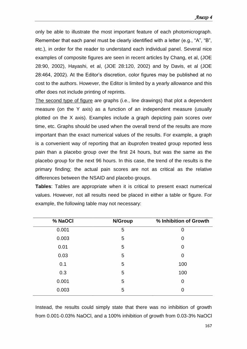
Anexo 4
167
only be able to illustrate the most important feature of each photomicrograph.
Remember that each panel must be clearly identified with a letter (e.g., “A”, “B”,
etc.), in order for the reader to understand each individual panel. Several nice
examples of composite figures are seen in recent articles by Chang, et al, (JOE
28:90, 2002), Hayashi, et al, (JOE 28:120, 2002) and by Davis, et al (JOE
28:464, 2002). At the Editor’s discretion, color figures may be published at no
cost to the authors. However, the Editor is limited by a yearly allowance and this
offer does not include printing of reprints.
The second type of figure are graphs (i.e., line drawings) that plot a dependent
measure (on the Y axis) as a function of an independent measure (usually
plotted on the X axis). Examples include a graph depicting pain scores over
time, etc. Graphs should be used when the overall trend of the results are more
important than the exact numerical values of the results. For example, a graph
is a convenient way of reporting that an ibuprofen treated group reported less
pain than a placebo group over the first 24 hours, but was the same as the
placebo group for the next 96 hours. In this case, the trend of the results is the
primary finding; the actual pain scores are not as critical as the relative
differences between the NSAID and placebo groups.
Tables: Tables are appropriate when it is critical to present exact numerical
values. However, not all results need be placed in either a table or figure. For
example, the following table may not necessary:
% NaOCl N/Group % Inhibition of Growth
0.001 5 0
0.003 5 0
0.01 5 0
0.03 5 0
0.1 5 100
0.3 5 100
0.001 5 0
0.003 5 0
Instead, the results could simply state that there was no inhibition of growth
from 0.001-0.03% NaOCl, and a 100% inhibition of growth from 0.03-3% NaOCl
Anexo 4
168
(N=5/group). Similarly, if the results are not significant, then it is probably not
necessary to include the results in either a table or as a figure. These and many
other suggestions on figure and table construction are described in additional
detail in Day (1998).
g. Discussion: The conclusion section should describe the major
findings of the study. Both the strength and weaknesses of the observations
should be discussed. What are the major conclusions of the study? How does
the data support these conclusions? How do these findings compare to the
published literature? What are the clinical implications? Although this last
section might be tentative given the nature of a particular study, the authors
should realize that even preliminary clinical implications might have value for
the clinical readership. Ideally, a review of the potential clinical significance is
the last section of the discussion.
h. References: The reference style follows Index Medicus and can be
efficiently learned from reading past issues of the JOE. Citations are placed in
parentheses at the end of a sentence or at the end of a clause that requires a
literature citation. Do not use superscript for references. Original reports are
limited to 35 references. There are no limits in the number of references for
review articles.
Anexo 4
169
Guidelines for Publishing Papers in the International
Endodontic Journal
1.GENERAL
International Endodontic Journal publishes original scientific articles, reviews,
clinical articles and case reports in the field of Endodontology; the branch of
dental sciences dealing with health, injuries to and diseases of the pulp and
periradicular region, and their relationship with systemic well-being and health.
Original scientific articles are published in the areas of biomedical science,
applied materials science, bioengineering, epidemiology and social science
relevant to endodontic disease and its management, and to the restoration of
root-treated teeth. In addition, review articles, reports of clinical cases, book
reviews, summaries and abstracts of scientific meetings and news items are
accepted. Please read the instructions below carefully for details on the
submission of manuscripts, the journal's requirements and standards as well as
information concerning the procedure after a manuscript has been accepted for
publication in International Endodontic Journal. Authors are encouraged to
visit Wiley Author Services for further information on the preparation and
submission of articles and figures.
2. ETHICAL GUIDELINES
International Endodontic Journal adheres to the below ethical guidelines for
publication and research.
2.1. Authorship and Acknowledgements
Authors submitting a paper do so on the understanding that the manuscript has
been read and approved by all authors and that all authors agree to the
submission of the manuscript to the Journal. International Endodontic
Journal adheres to the definition of authorship set up by The International
Committee of Medical Journal Editors (ICMJE). According to the ICMJE,
authorship criteria should be based on 1) substantial contributions to conception
and design of, or acquisiation of data or analysis and interpretation of data, 2)
drafting the article or revising it critically for important intellectual content and 3)
final approval of the version to be published. Acknowledgements: Under
acknowledgements please specify contributors to the article other than the
Anexo 4
170
authors accredited. Please also include specifications of the source of funding
for the study and any potential conflict of interests if appropriate. Please find
more information on the conflict of interest form in section 2.6.
2.2. Ethical Approvals
Experimentation involving human subjects will only be published if such
research has been conducted in full accordance with ethical principles,
including the World Medical Association Declaration of Helsinki (version 2008)
and the additional requirements, if any, of the country where the research has
been carried out. Manuscripts must be accompanied by a statement that the
experiments were undertaken with the understanding and written consent of
each subject and according to the above mentioned principles. A statement
regarding the fact that the study has been independently reviewed and
approved by an ethical board should also be included. Editors reserve the right
to reject papers if there are doubts as to whether appropriate procedures have
been used. When experimental animals are used the methods section must
clearly indicate that adequate measures were taken to minimize pain or
discomfort. Experiments should be carried out in accordance with the
Guidelines laid down by the National Institute of Health (NIH) in the USA
regarding the care and use of animals for experimental procedures or with the
European Communities Council Directive of 24 November 1986 (86/609/EEC)
and in accordance with local laws and regulations.
All studies using human or animal subjects should include an explicit statement
in the Material and Methods section identifying the review and ethics committee
approval for each study. The authors MUST upload a copy of the ethical
approval letter when submitting their manuscript. Editors reserve the right to
reject papers if there is doubt as to whether appropriate procedures have been
used.
2.3 Clinical Trials
2.3.1. Randomised control clinical trials should be reported using the
guidelines available at www.consort-statement.org. A CONSORT checklist and
flow diagram (as a Figure) should also be included in the submission material.
The International Endodontic Journal asks that authors submitting manuscripts
reporting from a clinical trial to register the trials in any of the following public
clinical registries: www.clinicaltrials.gov, https://www.clinicaltrialsregister.eu/,
Anexo 4
171
http://isrctn.org/. Other primary registries if named in the WHO network will also
be considered acceptable. The clinical trial registration number and name of the
trial register should be included in the Acknowledgements at the submission
stage.
2.3.2 Epidemiological observational trials
Submitting authors of epidemiological human observations studies are required
to review and submit a 'strengthening the reporting of observational studies in
Epidemiology' (STROBE) checklist and statement. Compliance with this should
be detailed in the materials and methods section. (www.strobe-statement.org)
2.4 Systematic Reviews
Systematic reviews should be reported using the PRISMA guidelines available
at http://prisma-statement.org/. A PRISMA checklist and flow diagram (as a
Figure) should also be included in the submission material.
2.5 DNA Sequences and Crystallographic Structure Determinations
Papers reporting protein or DNA sequences and crystallographic structure
determinations will not be accepted without a Genbank or Brookhaven
accession number, respectively. Other supporting data sets must be made
available on the publication date from the authors directly.
2.6 Conflict of Interest and Source of Funding
International Endodontic Journal requires that all authors (both the
corresponding author and co-authors) disclose any potential sources of conflict
of interest. Any interest or relationship, financial or otherwise that might be
perceived as influencing an author’s objectivity is considered a potential source
of conflict of interest. These must be disclosed when directly relevant or
indirectly related to the work that the authors describe in their manuscript.
Potential sources of conflict of interest include but are not limited to patent or
stock ownership, membership of a company board of directors, membership of
an advisory board or committee for a company, and consultancy for or receipt of
speaker's fees from a company. If authors are unsure whether a past or present
affiliation or relationship should be disclosed in the manuscript, please contact
the editorial office [email protected]. The existence of a conflict of
interest does not preclude publication in this journal. The above policies are in
accordance with the Uniform Requirements for Manuscripts Submitted to
Anexo 4
172
Biomedical Journals produced by the International Committee of Medical
Journal Editors (http://www.icmje.org/).
It is the responsibility of the corresponding author to have all authors of a
manuscript fill out a conflict of interest disclosure form, and to upload all forms
together with the manuscript on submission. The disclosure statement should
be included under Acknowledgements.
2.7 Appeal of Decision
The decision on a paper is final and cannot be appealed.
2.8 Permissions
If all or parts of previously published illustrations are used, permission must be
obtained from the copyright holder concerned. It is the author's responsibility to
obtain these in writing and provide copies to the Publishers.
2.8 Copyright Assignment
If your paper is accepted, the author identified as the formal corresponding
author for the paper will receive an email prompting them to login into Author
Services; where via the Wiley Author Licensing Service (WALS) they will be
able to complete the license agreement on behalf of all authors on the paper.
Your article cannot be published until this has been done.
For authors choosing Online Open
If the Online Open option is selected the corresponding author will have a
choice of the following Creative Commons License Open Access Agreements.
To preview the terms and conditions of these open access agreements please
visit the Copyright FAQs hosted on Wiley Author
Services http://exchanges.wiley.com/authors/faqs---copyright
301.html andvisit http://www.wileyopenaccess.com/details/content/12f25db4c87
/Copyright-License.html.
3. Online Open
OnlineOpen is available to authors of primary research articles who wish to
make their article available to non-subscribers on publication, or whose funding
agency requires grantees to archive the final version of their article. With
OnlineOpen, the author, the author's funding agency, or the author's institution
pays a fee to ensure that the article is made available to non-subscribers upon
publication via Wiley Online Library, as well as deposited in the funding
agency's preferred archive. For the full list of terms and conditions, see
Anexo 4
173
http://wileyonlinelibrary.com/onlineopen#OnlineOpenTerms. Prior to acceptance
there is no requirement to inform an Editorial Office that you intend to publish
your paper OnlineOpen if you do not wish to. All OnlineOpen articles are treated
in the same way as any other article. They go through the journal's standard
peer-review process and will be accepted or rejected based on their own merit.
3.1 MANUSCRIPT SUBMISSION PROCEDURE
Manuscripts should be submitted electronically via the online submission
sitehttp://mc.manuscriptcentral.com/iej. The use of an online submission and
peer review site enables immediate distribution of manuscripts and
consequentially speeds up the review process. It also allows authors to track
the status of their own manuscripts. Complete instructions for submitting a
paper is available online and below.
3.2 Getting Started
Launch your web browser (supported browsers include Internet Explorer 5.5 or
higher, Safari 1.2.4, or Firefox 1.0.4 or higher) and go to the journal's online
Submission. Log-in, or if you are a new user, click on 'register here'. If you are
registering as a new user. After clicking on 'register here', enter your name and
e-mail information and click 'Next'. Your e-mail information is very important.
Enter your institution and address information as appropriate, and then click
'Next’. Enter a user ID and password of your choice (we recommend using your
e-mail address as your user ID), and then select your areas of expertise. If you
are registered, but have forgotten your log in details, please enter your e-mail
address under 'Password Help'. The system will send you an automatic user ID
and a new temporary password. Log-in and select 'Author Center'
3.3. Submitting Your Manuscript
After you have logged into your 'Author Centre', submit your manuscript by
clicking on the submission link under 'Author Resources'. Enter data and
answer questions as appropriate. You may copy and paste directly from your
manuscript and you may upload your pre-prepared covering letter. Click the
'Next' button on each screen to save your work and advance to the next screen.
You are required to upload your files. Click on the 'Browse' button and locate
the file on your computer. Select the designation of each file in the drop down
next to the Browse button. When you have selected all files you wish to upload,
click the 'Upload Files' button. Review your submission (in HTML and PDF
Anexo 4
174
format) before completing your submission by sending it to the Journal. Click
the 'Submit' button when you are finished reviewing.
3.4. Manuscript Files Accepted
Manuscripts should be uploaded as Word (.doc) or Rich Text Format (.rft) files
(not write-protected) plus separate figure files. GIF, JPEG, PICT or Bitmap files
are acceptable for submission, but only high-resolution TIF or EPS files are
suitable for printing. The files will be automatically converted to HTML and PDF
on upload and will be used for the review process. The text file must contain the
abstract, main text, references, tables, and figure legends, but no embedded
figures or Title page. The Title page should be uploaded as a separate file. In
the main text, please reference figures as for instance 'Figure 1', 'Figure 2' etc
to match the tag name you choose for the individual figure files uploaded.
Manuscripts should be formatted as described in the Author Guidelines below.
3.5. Blinded Review
Manuscript that do not conform to the general aims and scope of the journal will
be returned immediately without review. All other manuscripts will be reviewed
by experts in the field (generally two referees). International Endodontic Journal
aims to forward referees´ comments and to inform the corresponding author of
the result of the review process. Manuscripts will be considered for fast-track
publication under special circumstances after consultation with the Editor.
International Endodontic Journal uses double blinded review. The names of the
reviewers will thus not be disclosed to the author submitting a paper and the
name(s) of the author(s) will not be disclosed to the reviewers. To allow double
blinded review, please submit (upload) your main manuscript and title page as
separate files. Please upload: Your manuscript without title page under the file
designation 'main document'. Figure files under the file designation 'figures'.
The title page and Acknowledgements where applicable, should be uploaded
under the file designation 'title page'. All documents uploaded under the file
designation 'title page' will not be viewable in the html and pdf format you are
asked to review in the end of the submission process. The files viewable in the
html and pdf format are the files available to the reviewer in the review process.
3.6. Suspension of Submission Mid-way in the Submission Process
You may suspend a submission at any phase before clicking the 'Submit' button
and save it to submit later. The manuscript can then be located under
Anexo 4
175
'Unsubmitted Manuscripts' and you can click on 'Continue Submission' to
continue your submission when you choose to.
3.7. E-mail Confirmation of Submission
After submission you will receive an e-mail to confirm receipt of your
manuscript. If you do not receive the confirmation e-mail after 24 hours, please
check your e-mail address carefully in the system. If the e-mail address is
correct please contact your IT department. The error may be caused by some
sort of spam filtering on your e-mail server. Also, the e-mails should be received
if the IT department adds our e-mail server (uranus.scholarone.com) to their
whitelist.
3.8. Manuscript Status
You can access ScholarOne Manuscripts any time to check your 'Author
Centre' for the status of your manuscript. The Journal will inform you by e-mail
once a decision has been made.
3.9. Submission of Revised Manuscripts
To submit a revised manuscript, locate your manuscript under 'Manuscripts with
Decisions' and click on 'Submit a Revision'. Please remember to delete any old
files uploaded when you upload your revised manuscript.
4. MANUSCRIPT TYPES ACCEPTED
Original Scientific Articles: must describe significant and original experimental
observations and provide sufficient detail so that the observations can be
critically evaluated and, if necessary, repeated. Original Scientific Articles must
conform to the highest international standards in the field.
Review Articles: are accepted for their broad general interest; all are refereed
by experts in the field who are asked to comment on issues such as timeliness,
general interest and balanced treatment of controversies, as well as on
scientific accuracy. Reviews should generally include a clearly defined search
strategy and take a broad view of the field rather than merely summarizing the
authors´ own previous work. Extensive or unbalanced citation of the authors´
own publications is discouraged.
Mini Review Articles: are accepted to address current evidence on well-
defined clinical, research or methodological topics. All are refereed by experts in
the field who are asked to comment on timeliness, general interest, balanced
treatment of controversies, and scientific rigor. A clear research question,
Anexo 4
176
search strategy and balanced synthesis of the evidence is expected.
Manuscripts are limited in terms of word-length and number of figures.
Clinical Articles: are suited to describe significant improvements in clinical
practice such as the report of a novel technique, a breakthrough in technology
or practical approaches to recognised clinical challenges. They should conform
to the highest scientific and clinical practice standards.
Case Reports: illustrating unusual and clinically relevant observations are
acceptable but they must be of sufficiently high quality to be considered worthy
of publication in the Journal. On rare occasions, completed cases displaying
non-obvious solutions to significant clinical challenges will be considered.
Illustrative material must be of the highest quality and healing outcomes, if
appropriate, should be demonstrated.
Supporting Information: International Endodontic Journal encourages
submission of adjuncts to printed papers via the supporting information website
(see submission of supporting information below). It is encouraged that authors
wishing to describe novel procedures or illustrate cases more fully with figures
and/or video may wish to utilise this facility.
Letters to the Editor: are also acceptable.
Meeting Reports: are also acceptable.
5. MANUSCRIPT FORMAT AND STRUCTURE
5.1. Format Language:
The language of publication is English. It is preferred that manuscript is
professionally edited. A list of independent suppliers of editing services can be
found at http://authorservices.wiley.com/bauthor/english_language.asp. All
services are paid for and arranged by the author, and use of one of these
services does not guarantee acceptance or preference for publication.
Presentation: Authors should pay special attention to the presentation of their
research findings or clinical reports so that they may be communicated clearly.
Technical jargon should be avoided as much as possible and clearly explained
where its use is unavoidable. Abbreviations should also be kept to a minimum,
particularly those that are not standard. The background and hypotheses
underlying the study, as well as its main conclusions, should be clearly
explained. Titles and abstracts especially should be written in language that will
be readily intelligible to any scientist.
Anexo 4
177
Abbreviations: International Endodontic Journal adheres to the conventions
outlined in Units, Symbols and Abbreviations: A Guide for Medical and Scientific
Editors and Authors. When non-standard terms appearing 3 or more times in
the manuscript are to be abbreviated, they should be written out completely in
the text when first used with the abbreviation in parenthesis.
5.2. Structure
All manuscripts submitted to International Endodontic Journal should include
Title Page, Abstract, Main Text, References and Acknowledgements, Tables,
Figures and Figure Legends as appropriate
Title Page: The title page should bear: (i) Title, which should be concise as
well as descriptive; (ii) Initial(s) and last (family) name of each author; (iii) Name
and address of department, hospital or institution to which work should be
attributed; (iv) Running title (no more than 30 letters and spaces); (v) No more
than six keywords (in alphabetical order); (vi) Name, full postal address,
telephone, fax number and e-mail address of author responsible for
correspondence.
Abstract for Original Scientific Articles should be no more than 250 words
giving details of what was done using the following structure:
Aim: Give a clear statement of the main aim of the study and the main
hypothesis tested, if any. Methodology: Describe the methods adopted
including, as appropriate, the design of the study, the setting, entry
requirements for subjects, use of materials, outcome measures and statistical
tests. Results: Give the main results of the study, including the outcome of any
statistical analysis. Conclusions: State the primary conclusions of the study
and their implications. Suggest areas for further research, if appropriate.
Abstract for Review Articles should be non-structured of no more than 250
words giving details of what was done including the literature search strategy.
Abstract for Mini Review Articles should be non-structured of no more than
250 words, including a clear research question, details of the literature search
strategy and clear conclusions.
Abstract for Case Reports should be no more than 250 words using the
following structure:
Aim: Give a clear statement of the main aim of the report and the clinical
problem which is addressed. Summary: Describe the methods adopted
Anexo 4
178
including, as appropriate, the design of the study, the setting, entry
requirements for subjects, use of materials, outcome measures and analysis if
any. Key learning points: Provide up to 5 short, bullet-pointed statements to
highlight the key messages of the report. All points must be fully justified by
material presented in the report.
Abstract for Clinical Articles should be no more than 250 words using the
following structure:
Aim: Give a clear statement of the main aim of the report and the clinical
problem which is addressed. Methodology: Describe the methods adopted.
Results: Give the main results of the study. Conclusions: State the primary
conclusions of the study.
Main Text of Original Scientific Article should include Introduction, Materials
and Methods, Results, Discussion and Conclusion
Introduction: should be focused, outlining the historical or logical origins of the
study and gaps in knowledge. Exhaustive literature reviews are not
appropriate. It should close with the explicit statement of the specific aims of the
investigation, or hypothesis to be tested.
Material and Methods: must contain sufficient detail such that, in combination
with the references cited, all clinical trials and experiments reported can be fully
reproduced.
(i) Clinical Trials should be reported using the CONSORT guidelines available
at www.consort-statement.org. A CONSORT checklist and flow diagram (as a
Figure) should also be included in the submission material.
(ii) Experimental Subjects: experimentation involving human subjects will only
be published if such research has been conducted in full accordance with
ethical principles, including the World Medical Association Declaration of
Helsinki (version 2008) and the additional requirements, if any, of the country
where the research has been carried out. Manuscripts must be accompanied by
a statement that the experiments were undertaken with the understanding and
written consent of each subject and according to the above mentioned
principles. A statement regarding the fact that the study has been independently
reviewed and approved by an ethical board should also be included. Editors
reserve the right to reject papers if there are doubts as to whether appropriate
procedures have been used. When experimental animals are used the methods
Anexo 4
179
section must clearly indicate that adequate measures were taken to minimize
pain or discomfort. Experiments should be carried out in accordance with the
Guidelines laid down by the National Institute of Health (NIH) in the USA
regarding the care and use of animals for experimental procedures or with the
European Communities Council Directive of 24 November 1986 (86/609/EEC)
and in accordance with local laws and regulations. All studies using human or
animal subjects should include an explicit statement in the Material and
Methods section identifying the review and ethics committee approval for each
study, if applicable. Editors reserve the right to reject papers if there is doubt as
to whether appropriate procedures have been used.
(iii) Suppliers: Suppliers of materials should be named and their location
(Company, town/city, state, country) included.
Results: should present the observations with minimal reference to earlier
literature or to possible interpretations. Data should not be duplicated in Tables
and Figures.
Discussion: may usefully start with a brief summary of the major findings, but
repetition of parts of the abstract or of the results section should be avoided.
The Discussion section should progress with a review of the methodology
before discussing the results in light of previous work in the field. The
Discussion should end with a brief conclusion and a comment on the potential
clinical relevance of the findings. Statements and interpretation of the data
should be appropriately supported by original references.
Conclusion: should contain a summary of the findings.
Main Text of Review Articles should be divided into Introduction, Review and
Conclusions. The Introduction section should be focused to place the subject
matter in context and to justify the need for the review. The Review section
should be divided into logical sub-sections in order to improve readability and
enhance understanding. Search strategies must be described and the use of
state-of-the-art evidence-based systematic approaches is expected. The use of
tabulated and illustrative material is encouraged. The Conclusion section should
reach clear conclusions and/or recommendations on the basis of the evidence
presented.
Main Text of Mini Review Articles should be divided into Introduction, Review
and Conclusions. The Introduction section should briefly introduce the subject
Anexo 4
180
matter and justify the need and timeliness of the literature review. The Review
section should be divided into logical sub-sections to enhance readability and
understanding and may be supported by up to 5 tables and figures. Search
strategies must be described and the use of state-of-the-art evidence-based
systematic approaches is expected. The Conclusions section should present
clear statements/recommendations and suggestions for further work. The
manuscript, including references and figure legends should not normally exceed
4000 words.
Main Text of Clinical Reports and Clinical Articles should be divided into
Introduction, Report, Discussion and Conclusion. They should be well illustrated
with clinical images, radiographs, diagrams and, where appropriate, supporting
tables and graphs. However, all illustrations must be of the highest quality
Acknowledgements: International Endodontic Journal requires that all sources
of institutional, private and corporate financial support for the work within the
manuscript must be fully acknowledged, and any potential conflicts of interest
noted. Grant or contribution numbers may be acknowledged, and principal grant
holders should be listed. Acknowledgments should be brief and should not
include thanks to anonymous referees and editors. See also above under
Ethical Guidelines.
5.3. References
It is the policy of the Journal to encourage reference to the original papers
rather than to literature reviews. Authors should therefore keep citations of
reviews to the absolute minimum. We recommend the use of a tool such
as EndNote or Reference Manager for reference management and formatting.
The EndNote reference style can be obtained upon request to the editorial
office ([email protected]). Reference Manager reference styles can be
searched for here: www.refman.com/support/rmstyles.asp
In the text: single or double authors should be acknowledged together with the
year of publication, e.g. (Pitt Ford & Roberts 1990). If more than two authors the
first author followed by et al. is sufficient, e.g. (Tobias et al. 1991). If more than
1 paper is cited the references should be in year order and separated by "," e.g.
(Pitt Ford & Roberts 1990, Tobias et al. 1991).
Reference list: All references should be brought together at the end of the
paper in alphabetical order and should be in the following form. (i) Names and
Anexo 4
181
initials of up to six authors. When there are seven or more, list the first three
and add et al. (ii)Year of publication in parentheses (iii) Full title of paper
followed by a full stop (.) (iv) Title of journal in full (in italics) (v) Volume number
(bold) followed by a comma (,) (vi) First and last pages. Examples of correct
forms of reference follow:
Standard journal article
Bergenholtz G, Nagaoka S, Jontell M (1991) Class II antigen-expressing cells in
experimentally induced pulpitis. International Endodontic Journal 24, 8-14.
Corporate author
British Endodontic Society (1983) Guidelines for root canal
treatment. International Endodontic Journal 16, 192-5.
Journal supplement
Frumin AM, Nussbaum J, Esposito M (1979) Functional asplenia:
demonstration of splenic activity by bone marrow scan
(Abstract). Blood 54 (Suppl. 1), 26a.
Books and other monographs
Personal author(s)
Gutmann J, Harrison JW (1991) Surgical Endodontics, 1st edn Boston, MA,
USA: Blackwell Scientific Publications.
Chapter in a book
Wesselink P (1990) Conventional root-canal therapy III: root filling. In: Harty FJ,
ed. Endodontics in Clinical Practice, 3rd edn; pp. 186-223. London, UK:
Butterworth.
Published proceedings paper
DuPont B (1974) Bone marrow transplantation in severe combined
immunodeficiency with an unrelated MLC compatible donor. In: White HJ, Smith
R, eds. Proceedings of the Third Annual Meeting of the International Society for
Experimental Rematology; pp. 44-46. Houston, TX, USA: International Society
for Experimental Hematology.
Agency publication
Ranofsky AL (1978) Surgical Operations in Short-Stay Hospitals: United States-
1975. DHEW publication no. (PHS) 78-1785 (Vital and Health Statistics; Series
13; no. 34.) Hyattsville, MD, USA: National Centre for Health Statistics.8
Anexo 4
182
Dissertation or thesis
Saunders EM (1988) In vitro and in vivo investigations into root-canal obturation
using thermally softened gutta-percha techniques (PhD Thesis). Dundee, UK:
University of Dundee.
URLs
Full reference details must be given along with the URL, i.e. authorship, year,
title of document/report and URL. If this information is not available, the
reference should be removed and only the web address cited in the text.
Smith A (1999) Select committee report into social care in the community
[WWW document]. URL http://www.dhss.gov.uk/reports/report015285.html
[accessed on 7 November 2003]
5.4. Tables, Figures and Figure Legends
Tables: Tables should be double-spaced with no vertical rulings, with a single
bold ruling beneath the column titles. Units of measurements must be included
in the column title.
Figures: All figures should be planned to fit within either 1 column width (8.0
cm), 1.5 column widths (13.0 cm) or 2 column widths (17.0 cm), and must be
suitable for photocopy reproduction from the printed version of the manuscript.
Lettering on figures should be in a clear, sans serif typeface (e.g. Helvetica); if
possible, the same typeface should be used for all figures in a paper. After
reduction for publication, upper-case text and numbers should be at least 1.5-
2.0 mm high (10 point Helvetica). After reduction, symbols should be at least
2.0-3.0 mm high (10 point). All half-tone photographs should be submitted at
final reproduction size. In general, multi-part figures should be arranged as they
would appear in the final version. Reduction to the scale that will be used on the
page is not necessary, but any special requirements (such as the separation
distance of stereo pairs) should be clearly specified. Unnecessary figures and
parts (panels) of figures should be avoided: data presented in small tables or
histograms, for instance, can generally be stated briefly in the text instead.
Figures should not contain more than one panel unless the parts are logically
connected; each panel of a multipart figure should be sized so that the whole
figure can be reduced by the same amount and reproduced on the printed page
at the smallest size at which essential details are visible. Figures should be on a
white background, and should avoid excessive boxing, unnecessary colour,
Anexo 4
183
shading and/or decorative effects (e.g. 3-dimensional skyscraper histograms)
and highly pixelated computer drawings. The vertical axis of histograms should
not be truncated to exaggerate small differences. The line spacing should be
wide enough to remain clear on reduction to the minimum acceptable printed
size. Figures divided into parts should be labelled with a lower-case, boldface,
roman letter, a, b, and so on, in the same typesize as used elsewhere in the
figure. Lettering in figures should be in lower-case type, with the first letter
capitalized. Units should have a single space between the number and the unit,
and follow SI nomenclature or the nomenclature common to a particular field.
Thousands should be separated by a thin space (1 000). Unusual units or
abbreviations should be spelled out in full or defined in the legend. Scale bars
should be used rather than magnification factors, with the length of the bar
defined in the legend rather than on the bar itself. In general, visual cues (on the
figures themselves) are preferred to verbal explanations in the legend (e.g.
broken line, open red triangles etc.)
Figure legends: Figure legends should begin with a brief title for the whole
figure and continue with a short description of each panel and the symbols
used; they should not contain any details of methods.
Permissions: If all or part of previously published illustrations are to be used,
permission must be obtained from the copyright holder concerned. This is the
responsibilty of the authors before submission.
Preparation of Electronic Figures for Publication: Although low quality
images are adequate for review purposes, print publication requires high quality
images to prevent the final product being blurred or fuzzy. Submit EPS (lineart)
or TIFF (halftone/photographs) files only. MS PowerPoint and Word Graphics
are unsuitable for printed pictures. Do not use pixel-oriented programmes.
Scans (TIFF only) should have a resolution of 300 dpi (halftone) or 600 to 1200
dpi (line drawings) in relation to the reproduction size (see below). EPS files
should be saved with fonts embedded (and with a TIFF preview if possible). For
scanned images, the scanning resolution (at final image size) should be as
follows to ensure good reproduction: lineart: >600 dpi; half-tones (including gel
photographs): >300 dpi; figures containing both halftone and line images: >600
dpi.
Anexo 4
184
5.5. Supporting Information
Publication in electronic formats has created opportunities for adding details or
whole sections in the electronic version only. Authors need to work closely with
the editors in developing or using such new publication formats. Supporting
information, such as data sets or additional figures or tables, that will not be
published in the print edition of the journal, but which will be viewable via the
online edition, can be submitted. It should be clearly stated at the time of
submission that the supporting information is intended to be made available
through the online edition. If the size or format of the supporting information is
such that it cannot be accommodated on the journal's website, the author
agrees to make the supporting information available free of charge on a
permanent Web site, to which links will be set up from the journal's website. The
author must advise Wiley Blackwell if the URL of the website where
the supporting information is located changes. The content of the supporting
information must not be altered after the paper has been accepted for
publication. The availability of supporting information should be indicated in the
main manuscript by a paragraph, to appear after the References, headed
'Supporting Information' and providing titles of figures, tables, etc. In order to
protect reviewer anonymity, material posted on the authors Web site cannot be
reviewed. The supporting information is an integral part of the article and will be
reviewed accordingly.
Preparation of Supporting Information: Although provision of content through
the web in any format is straightforward, supporting information is best provided
either in web-ready form or in a form that can be conveniently converted into
one of the standard web publishing formats: Simple word-processing files (.doc
or .rtf) for text. PDF for more complex, layout-dependent text or page-based
material. Acrobat files can be distilled from Postscript by the Publisher, if
necessary.GIF or JPEG for still graphics. Graphics supplied as EPS or TIFF are
also acceptable. MPEG or AVI for moving graphics. Subsequent requests for
changes are generally unacceptable, as for printed papers. A charge may be
levied for this service.
Anexo 4
185
Video Imaging: For the on-line version of the Journal the submission of
illustrative video is encouraged. Authors proposing the use such media should
consult with the Editor during manuscript preparation.
6. AFTER ACCEPTANCE
Upon acceptance of a paper for publication, the manuscript will be forwarded to
the Production Editor who is responsible for the production of the journal.
6.1. Figures
Hard copies of all figures and tables are required when the manuscript is ready
for publication. These will be requested by the Editor when required. Each
Figure copy should be marked on the reverse with the figure number and the
corresponding author’s name.
6.2 Proof Corrections
The corresponding author will receive an email alert containing a link to a web
site. A working email address must therefore be provided for the corresponding
author. The proof can be downloaded as a PDF (portable document format) file
from this site. Acrobat Reader will be required in order to read this file. This
software can be downloaded (free of charge) from the following Web
site: www.adobe.com/products/acrobat/readstep2.html. This will enable the file
to be opened, read on screen, and printed out in order for any corrections to be
added. Further instructions will be sent with the proof. Hard copy proofs will be
posted if no e-mail address is available; in your absence, please arrange for a
colleague to access your e-mail to retrieve the proofs. Proofs must be returned
to the Production Editor within three days of receipt. As changes to proofs are
costly, we ask that you only correct typesetting errors. Excessive changes made
by the author in the proofs, excluding typesetting errors, will be charged
separately. Other than in exceptional circumstances, all illustrations are retained
by the publisher. Please note that the author is responsible for all statements
made in his work, including changes made by the copy editor.
6.3 Early Online Publication Prior to Print
International Endodontic Journal is covered by Wiley Blackwell's Early
View service. Early View articles are complete full-text articles published online
in advance of their publication in a printed issue. Early View articles are
complete and final. They have been fully reviewed, revised and edited for
Anexo 4
186
publication, and the authors' final corrections have been incorporated. Because
they are in final form, no changes can be made after online publication. The
nature of Early View articles means that they do not yet have volume, issue or
page numbers, so Early View articles cannot be cited in the traditional way.
They are therefore given a Digital Object Identifier (DOI), which allows the
article to be cited and tracked before it is allocated to an issue. After print
publication, the DOI remains valid and can continue to be used to cite and
access the article.
6.4 Online Production Tracking
Online production tracking is available for your article through Blackwell's
Author Services. Author Services enables authors to track their article - once it
has been accepted - through the production process to publication online and in
print. Authors can check the status of their articles online and choose to receive
automated e-mails at key stages of production. The author will receive an e-mail
with a unique link that enables them to register and have their article
automatically added to the system. Please ensure that a complete e-mail
address is provided when submitting the manuscript.
Visit http://authorservices.wiley.com/bauthor/ for more details on online
production tracking and for a wealth of resources including FAQs and tips on
article preparation, submission and more.
6.5 Author Material Archive Policy
Please note that unless specifically requested, Wiley Blackwell will dispose of
all hardcopy or electronic material submitted two months after publication. If you
require the return of any material submitted, please inform the editorial office or
production editor as soon as possible.
6.6 Offprints
Free access to the final PDF offprint of your article will be available via Author
Services only. Please therefore sign up for Author Services if you would like to
access your article PDF offprint and enjoy the many other benefits the service
offers. Additional paper offprints may be ordered online. Please click on the
following link, fill in the necessary details and ensure that you type information
in all of the required fields: Offprint Cosprinters. If you have queries about
offprints please email [email protected] corresponding author will
Anexo 4
187
be sent complimentary copies of the issue in which the paper is published (one
copy per author).
6.7 Author Services
For more substantial information on the services provided for authors, please
see Wiley Blackwell Author Services
6.8 Note to NIH Grantees:
Pursuant to NIH mandate, Wiley Blackwell will post the accepted version of
contributions authored by NIH grant-holders to PubMed Central upon
acceptance. This accepted version will be made publicly available 12 months
after publication. For further information, see www.wiley.com/go/nihmandate
7. Guidelines for reporting of DNA microarray data
The International Endodontic Journal gives authors notice that, with effect from
1st January 2011, submission to the International Endodontic Journal requires
the reporting of microarray data to conform to the MIAME guidelines. After this
date, submissions will be assessed according to MIAME standards. Also,
manuscripts will be published only after the complete data has been submitted
into the public repositories, such as GEO (http://www.ncbi.nlm.nih.gov/geo/) or
Array Express (http://www.ebi.ac.uk/microarray/submissions_overview.html), in
MIAME compliant format, with the data accession number (the identification
number of the data set in the database) quoted in the manuscript. Both
databases are committed to keeping the data private until the associated
manuscript is published, if requested. Prospective authors are also encouraged
to search for previously published microarray data with relevance to their own
data, and to report whether such data exists. Furthermore, they are encouraged
to use the previously published data for qualitative and/or quantitative
comparison with their own data, whenever suitable. To fully acknowledge the
original work, an appropriate reference should be given not only to the database
in question, but also to the original article in which the data was first published.
This open approach will increase the availability and use of these large-scale
data sets and improve the reporting and interpretation of the findings, and in
increasing the comprehensive understanding of the physiology and pathology of
endodontically related tissues and diseases, result eventually in better patient
care.