Repositioning-Adult-Education-to-Address-Youth-Restiveness ...
Upload
khangminh22Category
view
4download
0
626 Current Medical Science 41(3):2021
Yan-ning GUO, E-mail: [email protected]#Corresponding author, E-mail: [email protected]
An Overview of Anterior Repositioning Splint Therapy for Disc Displacement-related Temporomandibular Disorders
Yan-ning GUO1, 2, 3, Sheng-jie CUI1, 2, 3, Yan-heng ZHOU1, 2, 3, Xue-dong WANG1, 2, 3#
1Department of Orthodontics, Peking University School and Hospital of Stomatology, Beijing 100081, China2National Clinical Research Center for Oral Diseases & National Engineering Laboratory for Digital and Material Technology of Stomatology, Beijing 100081, China3Beijing Key Laboratory of Digital Stomatology, Beijing 100081, China
Huazhong University of Science and Technology 2021
Summary: Anterior repositioning splint (ARS) therapy is considered one of the most effective therapies for treating disc displacement-related temporomandibular disorders (TMDs), which account for a large proportion of TMD cases. Owing to the wide application of this therapy, the exact mechanism of remission has increasingly drawn attention. Given that practitioners have different views on ARS therapy, its indications are broadened, and operating methods diverged. This review attempts to provide an overview of ARS therapy and helps practitioners establish indications and suitable operating methods. Representative views in the past 10 years were summarised, and conclusions were drawn as follows: The mechanism of ARS therapy is mainly attributed to internal derangement correction, improvement of stress distribution and recently reported joint remodeling. It has an evident effect in the short term, and the most prevalent operating methods are protruding the mandible to the edge-to-edge position and wearing the ARS for 24 hours daily for 3–6 months. However, long-term stability is not optimal, and thus indications should be selected carefully. Notably, most of the clinical studies in this field are case analyses with low-quality evidence. Well-designed RCTs are required to further validate relevant theories. Key words: anterior repositioning splint; disc displacement; occlusal splint; temporomandibular joint disorders; therapeutics
Temporomandibular disorders (TMDs) are among the most common diseases in the oral and maxillofacial regions and feature pain in the temporomandibular area or jaw muscles, joint sounds and restricted mandibular movement. Disc displacement plays an important role in TMDs. It is observed in 86% of patients with symptomatic TMD, although this rate can reach 25% in asymptomatic patients[1]. In the Diagnostic Criteria for Temporomandibular Disorders released by the International Association for Dental Research, four types of TMDs are directly related to disc displacement, namely, (1) disc displacement with reduction, (2) disc displacement with reduction with intermittent locking, (3) disc displacement without reduction with limited opening and (4) disc displacement without reduction without limited opening[2].
According to Summer et al[3], patients with good disc restoration shows a high rate of symptom relief, indicating the necessity of treating disc displacement.
Anterior repositioning splint (ARS) therapy is
a common conservative method for treating a disc displacement-related TMD. An ARS can be fixed on the maxilla or mandible and it usually maintains the protrusion status through an anterior guidance ramp (fig. 1).
The protrusion of the mandible changes the disc-condyle relationship and is widely used in intra-articular TMD treatment[4–6]. This method can significantly improve pain symptoms[7]. Thus, it can also be used in pain-related TMDs. However, practitioners are often confused when applying this therapy, given the absence of a standardised instruction for operating or an authoritative explanation of the mechanism. Particularly, disagreement exists over the degree of mandible protrusion, the duration of splint wear and to what extent and how the splint can relieve symptoms.
This study aims to provide an overall introduction of ARS therapy and a practical suggestion of application. Records in the past 10 years were carefully scanned and reviewed, from which we summarized the possible mechanism of the therapeutic effect, determined the most suggested operating method and discussed future directions for research on ARS therapy.
Current Medical ScienceDOI https://doi.org/10.1007/s11596-021-2381-7
41(3):626-634,2021

627Current Medical Science 41(3):2021
1 MATERIALS AND METHODS
To provide a general overview of ARS therapy in recent years, we conducted a detailed electronic search in PubMed, Medline and Scopus databases. The search strategy was ((anterior repositioning) OR (occlusal)) AND (splint* OR appliance*) AND ((temporomandibular joint) OR (condyl*) OR (disc displacement) OR (disk displacement)) AND (last 10 year). A total of 1420 records were identified. After screening the duplications, we excluded 817 records. Then, the titles and abstracts of the remaining 603 records were read, and 487 were excluded for not being related to the topic. The remaining 116 articles were read and assessed, and related references were searched. A total of 58 articles were included in this review (fig. 2).
2 ARS THERAPY MECHANISM
2.1 Improvement in the Disc-condyle RelationshipThe starting point of ARS therapy is protruding
the mandible, and thus the procedure changes the relationship between the articular disc and condyle. In the early stage of application, protruding the mandible moves the condyle forward and “catches” the displaced disc, corrects the disc-condyle relationship and facilitates tissue adaption or repair. At the end of therapy, the splint is gradually ground, and as the mandible returns to its original position, the disc-condyle complex is restored to its normal position (fig. 3).
However, William et al[8] held the view that the principle of restoration is to move the condyle inferoanteriorly and allow the disc to slide back to its normal position. Chen et al[9] observed the discs of patients with disc displacement with reduction (DDwR) through MRI and found that 41.9% of the discs that had been recaptured at the edge-to-edge position were displaced again during backing to the minimum protrusive position. This finding provided strong evidence of the speculation of Solberg et al that the possible principle of restoration is the forward and downward condyle movements, which “fix” the disc in a good position and prevent it from displacing again in
Fig. 1 Maxillary full-coverage ARS fabricated in the edge-to-edge position
Fig. 2 Flow diagram of search strategy
Fig. 3 Diagram showing the “recapture” of the discA: the displaced disc; B: The condyle moves forward by the ARS, thus “recaptures” the disc; C: The splint is adjusted gradually, and disc-condyle complex brought back to its normal place.
the mouth closing process (fig. 4). A good disc-condyle relationship can reduce the collision, rebound and release of the disc; thus, chronic damage in abnormal movements can be prevented. 2.2 Improvement in Stress Distribution in the Temporomandibular Joint
Change in internal structure also changes stress distribution. Some researchers believed that reducing unfavourable loading was an important way to alleviate the symptom[4, 10]. When stress is concentrated in some parts, the resulting pressure may cause the degeneration of elastic tissues and articular cartilage[11].
A B C
Identification 1420 records identified through electronic database search
Screening 603 records with their titles and abstracts reviewed
Eligibility 116 records read and assessed, 81 excluded
Included 58 studies included
817 duplicates
487 records not related to the topic
23 records manually searched
628 Current Medical Science 41(3):2021
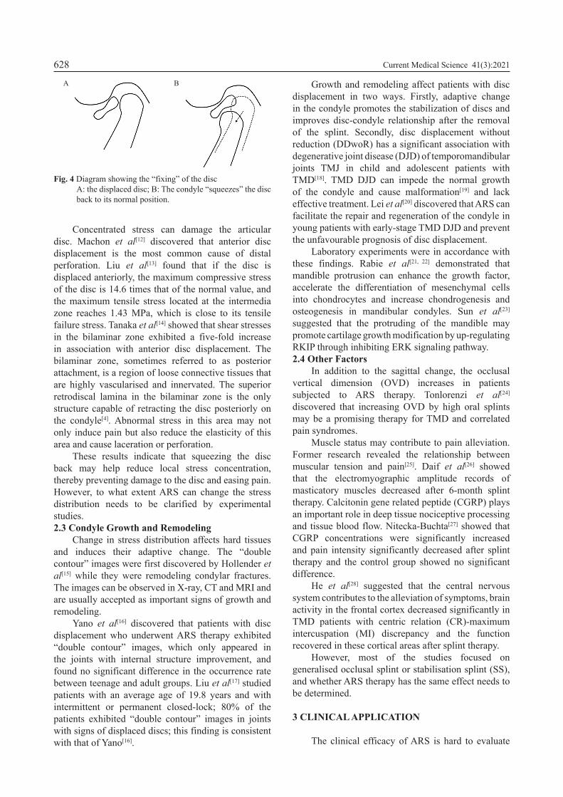
Concentrated stress can damage the articular disc. Machon et al[12] discovered that anterior disc displacement is the most common cause of distal perforation. Liu et al[13] found that if the disc is displaced anteriorly, the maximum compressive stress of the disc is 14.6 times that of the normal value, and the maximum tensile stress located at the intermedia zone reaches 1.43 MPa, which is close to its tensile failure stress. Tanaka et al[14] showed that shear stresses in the bilaminar zone exhibited a five-fold increase in association with anterior disc displacement. The bilaminar zone, sometimes referred to as posterior attachment, is a region of loose connective tissues that are highly vascularised and innervated. The superior retrodiscal lamina in the bilaminar zone is the only structure capable of retracting the disc posteriorly on the condyle[4]. Abnormal stress in this area may not only induce pain but also reduce the elasticity of this area and cause laceration or perforation.
These results indicate that squeezing the disc back may help reduce local stress concentration, thereby preventing damage to the disc and easing pain. However, to what extent ARS can change the stress distribution needs to be clarified by experimental studies.2.3 Condyle Growth and Remodeling
Change in stress distribution affects hard tissues and induces their adaptive change. The “double contour” images were first discovered by Hollender et al[15] while they were remodeling condylar fractures. The images can be observed in X-ray, CT and MRI and are usually accepted as important signs of growth and remodeling.
Yano et al[16] discovered that patients with disc displacement who underwent ARS therapy exhibited “double contour” images, which only appeared in the joints with internal structure improvement, and found no significant difference in the occurrence rate between teenage and adult groups. Liu et al[17] studied patients with an average age of 19.8 years and with intermittent or permanent closed-lock; 80% of the patients exhibited “double contour” images in joints with signs of displaced discs; this finding is consistent with that of Yano[16].
Growth and remodeling affect patients with disc displacement in two ways. Firstly, adaptive change in the condyle promotes the stabilization of discs and improves disc-condyle relationship after the removal of the splint. Secondly, disc displacement without reduction (DDwoR) has a significant association with degenerative joint disease (DJD) of temporomandibular joints TMJ in child and adolescent patients with TMD[18]. TMD DJD can impede the normal growth of the condyle and cause malformation[19] and lack effective treatment. Lei et al[20] discovered that ARS can facilitate the repair and regeneration of the condyle in young patients with early-stage TMD DJD and prevent the unfavourable prognosis of disc displacement.
Laboratory experiments were in accordance with these findings. Rabie et al[21, 22] demonstrated that mandible protrusion can enhance the growth factor, accelerate the differentiation of mesenchymal cells into chondrocytes and increase chondrogenesis and osteogenesis in mandibular condyles. Sun et al[23]
suggested that the protruding of the mandible may promote cartilage growth modification by up-regulating RKIP through inhibiting ERK signaling pathway.2.4 Other Factors
In addition to the sagittal change, the occlusal vertical dimension (OVD) increases in patients subjected to ARS therapy. Tonlorenzi et al[24]
discovered that increasing OVD by high oral splints may be a promising therapy for TMD and correlated pain syndromes.
Muscle status may contribute to pain alleviation. Former research revealed the relationship between muscular tension and pain[25]. Daif et al[26] showed that the electromyographic amplitude records of masticatory muscles decreased after 6-month splint therapy. Calcitonin gene related peptide (CGRP) plays an important role in deep tissue nociceptive processing and tissue blood flow. Nitecka-Buchta[27] showed that CGRP concentrations were significantly increased and pain intensity significantly decreased after splint therapy and the control group showed no significant difference.
He et al[28] suggested that the central nervous system contributes to the alleviation of symptoms, brain activity in the frontal cortex decreased significantly in TMD patients with centric relation (CR)-maximum intercuspation (MI) discrepancy and the function recovered in these cortical areas after splint therapy.
However, most of the studies focused on generalised occlusal splint or stabilisation splint (SS), and whether ARS therapy has the same effect needs to be determined.
3 CLINICAL APPLICATION
The clinical efficacy of ARS is hard to evaluate
A B
Fig. 4 Diagram showing the “fixing” of the discA: the displaced disc; B: The condyle “squeezes” the disc back to its normal position.

629Current Medical Science 41(3):2021
due to the diverse way of operation, evaluation and follow-up examinations. To guarantee the consistency, we conducted a secondary screening in order to exclude the studies that lacked necessary information. The inclusion criteria were as follow: (1) patients included need to have a specific diagnosis; (2) ARS therapy used as a treatment and a clear treatment protocol established; (3) having follow-up visits for the evaluation of therapeutic effect; and (4) clinical study except for case report.
Only 12 studies met the standard, and the other 46 studies related to the topic were excluded (fig. 5).
Therefore, allowing patient to protrude to an edge-to-edge position is recommended for disc restoration.
However, some researchers believe that the more the mandible protrudes, the higher pressure it imposes on the masticatory system; hence, it should not be protruded excessively[30]. Okeson et al[4] suggested initially fabricating a splint in the minimum protrusive position. If the related symptom is not relieved, imaging examinations, such as MRI, should be used in determining good position for restoration.3.1.2 Way of Wearing No standardized instruction for wearing ARS is available thus far. Most researchers suggest that ARS should be worn 24 h per day in order that the disc would not be displaced again. The splint should be ground gradually when the joint symptom is stable, the mandible should be guided to its physiological position and wearing time should be gradually reduced[31]. Another existing view holds that if a patient wears ARS for 24 h, joint structure and occlusion have a risk of permanent change, including posterior open bite and myostatic contracture of the inferior lateral pterygoid muscle. Conti et al[32]
demonstrated that nighttime use can lead to a marked relief in symptoms; thus, he suggested the wearing of splints at night or additional wearing splints in daytime only when pain is felt. However, these suggestions seem to be inconsistent with the “fixing” effect of the ARS.
When referring to total wearing time, researchers may suggest different procedures. Tecco et al[33]
showed that pain intensity, chewing-biting pain and muscle pain decreased continually during a 6-month treatment, and constant pain and joint noises were alleviated significantly after 3 months. Most researchers performed therapies for 3–6 months and adjusted the exact time according to the extent and trend of alleviation. Table 1 shows the way of wearing described in the 12 clinical studies[5–7, 17, 20, 31, 34–39]. 3.2 Clinical Efficacy of ARS
To assess the clinical efficacy of ARS therapy, we need to clarify the goal of a treatment first. This goal changed considerably in the past decades; in the early years, Tallents et al[40] believed the correction of internal derangement is the top concern. Currently, researchers tend to focus on symptoms and functional improvement. Disc-displacement and reciprocal clicking are usually regarded as benign conditions, and medical intervention should be provided only when patients experience pain and functional restriction. 3.2.1 Improvement of Clinical Symptoms ARS has an evident effect in reducing joint pain. Subjective evaluation quantified by VAS score showed that ARS therapy can reduce joint pain significantly in patients with DDwR and DDwoR[5–7, 36, 39]. Over other therapies, ARS has a remarkable advantage. Behavioural therapy (BT) involving patient education, functional exercise
Fig. 5 Diagram of secondary screening
3.1 Operating Methods3.1.1 Locating the Anterior Position Locating the anterior position is one of the key points in ARS therapy. For DDwR, clicking is an important sign to show the status of the relationship between a disc and condyle. The traditional method for defining the protrusive position is to allow patients to open their mouths until clicking occurs. The mandible is then adjusted to a minimum protrusion for the elimination of reciprocal clicking, which is usually called “minimum protrusive position” or “click-free position”.
For DDwoR, arthrocentesis and mandible manipulation are recommended to be performed first. After a successful recapture of a disc, the mandible should be protruded to a position without restriction in movement.
This method has been challenged in recent decades. Garcia et al[29] showed that the average protrusion in minimum protrusive position is not enough to allow discs to return to the ideal condition. Chen et al[9] demonstrated that only 54.8% of patients can obtain an ideal disc-condyle relationship in the minimum protrusive position. However, this rate increases to 93.5% in the edge-to-edge position.
Reason of exclusion
21%
Included studiesDid not specify the diagnosisDid not use ARS or did not show a clear protocolLacking of follow-up examinationsBasic study or case reportClassic clinical study but exceeding the 10-year range
5%
24%
10%31%
9%

630 Current Medical Science 41(3):2021
and behavioural instruction is the basic treatment for patients with TMD. Conti et al[36] showed that in patients with DDwR, the ARS group experienced more relief in joint pain than the BT group in the 6-week and 3-month follow-up evaluations. Pihut et al[7] showed that ARS exerts a better effect in reducing pain than the combination of BT and biostimulation laser in the 4-week and 16-week follow-up examinations. SS is another common type of splint expected to alleviate joint and muscle pain. Tecco et al[41] discovered that the ARS group showed less pain and lower pain intensity than the SS group in monthly follow-ups. Through evaluation by scaling the pain before and after a 6-month treatment, Kurt et al[37] discovered a similar trend but found no significant difference between the groups, which may be attributed to the longer interval of examinations.
ARS has a considerable effect on impaired mandible movement. The most common index for evaluating functional movement is the maximum interincisal opening (MIO). Lei et al[20] showed that the MIO of patients with ARS increased from 40.71±8.75 mm to 50.64±4.72 mm after treatment, which was significantly higher than MIO in the BT group (45.48±4.97 mm). Hersek et al and Shi et al[42, 43]
confirmed that the protrusion excursion and lateral excursion improved after ARS therapy. Pihut et al[7] showed that patients in the ARS group reported less impaired movement of the mandible than the control group in the 4-week and 16-week follow-up examinations.
Reciprocal clicking is no longer emphasised in TMD treatment, but some studies showed that this common symptom can be reduced by ARS. Ma et al[34] found that patients with DDwR had a remarkably low rate of clicking (12.1%) 12 months after treatment and the rate was significantly lower than that before treatment (90.1%).
3.2.2 Improvement of Imaging Appearance Imaging examination, which directly reflects changes in the internal structure of a joint, is an essential measurement in TMD diagnosis. MRI is radiation-free and produces good soft tissue images. Thus, it is usually considered the golden standard for disc position assessment. Evaluation of MR images was based on the location of the disc relative to the condyle in the parasagittal image; if the angle between the vertical axis of posterior band and condyle decreased, the MRI would be considered as “improved”. Cone beam computer tomography (CBCT) facilitates the evaluation of condylar osseous change and is used widely in the diagnosis of degenerative temporomandibular joint diseases. The improvement of CBCT images usually features an enhanced cortical continuity, or the regeneration of condylar bone.
Jung et al[44] compared the MRI appearance with pain symptoms and discovered an apparent relationship between the extent of displacement and pain symptoms, and the probability of pain in moderate and significant displacement cases is 9.69 times that in the normal group. Summer et al[3] evaluated the disc-condyle relationship through MRI and found that symptom was alleviated in 92% patients with normalised discs, 84% of patients had improved disc position and only 49% of patients exhibited persistent disc displacement. These studies indicate that symptom alleviation is positively correlated with internal structure improvement.
Kurita et al[45] showed that when the mandible moves forward to the minimum protrusive position, 75.6% of discs are completely captured with ARS. Liu et al[10] demonstrated that the frequency of successful “disc recapture” can reach 95.7% when DDwR patients receive the anterior repositioning of the mandible to the edge-to-edge position.
Lei et al[20] showed that ARS therapy can not only improve internal structure but also induce the
Table 1 Way of wearingSerial number Author (Published year) Diagnosis Therapeutic position Daily wearing
timeTotal wearing
time1 Lei J, et al. (2020) DDwoR Edge-to-edge position 24 hours 3 months2 Ma Z, et al. (2019) DDwR Edge-to-edge position 24 hours 11.5 months3 Shen P, et al. (2019) DDwR Edge-to-edge position 24 hours 9.5 months4 Lei J, et al. (2019) TMD DJD Edge-to-edge position 24 hours 3 months5 Pihut M, et al. (2018) DDwR Edge-to-edge position 20 hours 4 months6 Chen HM, et al. (2017) DDwR Edge-to-edge position 24 hours 3 months7 Jiang X, et al. (2016) DDwoR 2 mm protrusion Night time 2–4 weeks8 Conti PC, et al. (2015) DDwR Minimum protrusive position Night time 3 months9 Liu MQ, et al. (2012) DDwR&DDwoR Edge-to-edge position 24 hours 3 months10 Kurt H, et al. (2011) DDwR Minimum protrusive position Night time 6 months11 Madani AS. and Mirmortazavi A (2011) DDwR Minimum protrusive position Night time 3 months12 Tecco S, et al. (2010) DDwR Minimum protrusive position 24 hours 6 monthsDDwR: disc displacement with reduction; DDwoR: disc displacement without reduction; TMD DJD: degenerative temporomandibular joint disease

631Current Medical Science 41(3):2021
remodelling of bone destruction. CBCT examinations were performed at the 6th and 12th months after the treatment, and 28.1% of the patients in the ARS group reported repairing, 50% reported regeneration.
Table 2 shows the imaging results in 7 studies that meet the inclusion criteria above and also contain
imaging examinations. The majority of patients receiving ARS therapy reported signs of improvement in a short term. However, whether patients with “double contour” images can have a stable disc-condyle relationship and reduced recurrence needs to be clarified.
Table 2 Result of imaging examinations
Serial number Author Diagnosis Therapeutic
positionImaging
examination Evaluating timeImprovement
of disc-condyle relationship
Ratio of double contour
1 Lei J, et al. (2020)
DDwoR Edge-to-edge position
MRI T0 Before treatmentT1 1 week after successful mandible manipulation
T1: 100%
2 Ma Z, et al. (2019)
DDwR Edge-to-edge position
MRI T0 Before treatmentT1 After insertionT2 End of treatmentT3 12 months after treatment ends
T1: 100%T2: 92.3%T3: 72.5%
64.8%
3 Shen P, et al. (2019)
DDwR Edge-to-edge position
MRI T0 Before treatmentT1 Wearing for 6 monthsT2 End of treatmentT3 Follow up visit
T2: 84.3%
4 Lei J, et al. (2019)
TMD DJD
Edge-to-edge position
CBCT T0 Before treatmentT1 6/12 months after treatment
T1: 78.1% 50%
5 Chen HM, et al. (2017)
DDwR Edge-to-edge position
MRI T0 Before treatmentT1 After insertionT2 6 months after treatment
T1: 100%T2: 40.6%
6 Jiang X, et al. (2016)
DDwoR 2 mm protrusion
MRI T0 Before treatmentT1 2 months after treatment ends
T1: 82%
7 Liu MQ, et al. (2012)
DDwR&DDwoR
Edge-to-edge position
CBCT T0 Before treatmentT1 6 months after treatment
80%
DDwR: disc displacement with reduction; DDwoR: disc displacement without reduction; TMD DJD: degenerative temporomandibular joint disease
3.3 Indication and ContraindicationAs shown in tables 1 and 2, ARS therapy is used
primarily for DDwR/DDwoR. Some studies showed its effect on early-stage TMD DJD.
Several reasons may lead to failures in ARS therapy. Razook et al[46] suggested that time and extent of displacement are critical factors that hinder restoration. Fu et al[47] demonstrated that patients with chronic (over 4 months) DDwoR have a poor chance for restoration[31]. They suggested the use of this therapy only to acute patients within 3 months. Summer and Westesson et al[3, 48] showed that discs that are displaced only in the anterior direction have a good success rate of restoration, but the position is not improved in most cases when accompanied by transverse displacement. Chen et al[35] found that although the insertion can “squeeze” the disc backward, the most backward position is the top of the fossa, and the condyle is likely to move back to its original position after the therapy. Therefore, if the condyles are posteriorly positioned before treatment, the discs will be more likely to be displaced again in a long term. 3.4 Recurrence
Although ARS showed a remarkable effect on most cases in the short term, some studies reported its deficiency in terms of long-term stability. Ma et al[34]
reported that the clinical success rate at the 12-month follow-up is 79.1%. Shen et al[5] showed that the disc-condyle relationship became increasingly unstable in the long term, and 15.2% of patients who regained a normal disc location after ARS therapy showed a secondary occurrence within 3 months of follow-up. In follow-up periods longer than 24 months, this rate increased to 46.9%. However, clinical symptoms were not evaluated in this research, and thus the recurrence rate of clinical symptoms and the relationship between symptom and imaging appearance were not clear. Okeson et al[49] used an 8-week treatment course for 40 patients with internal derangement. A follow-up examination was carried out after an average period of 2.5 years. They found that 65% of the patients exhibited recurrence in clicking and 25% exhibited recurrence in pain.
Based on previous analysis, we can infer that the following aspects may contribute to recurrence: (1) The deformation of joints leads to the instability of the disc-condyle relationship; (2) The time or extent of the protrusion is not enough for the relative tissue to repair or to be remodelled to a stable condition; (3) The discordancy of the occlusion and muscle system leads to mandible position instability and the recurrence of disc displacement.

632 Current Medical Science 41(3):2021
Whether recurrence can be reduced by fixing the occlusion at the protruding position, where new homeostasis is established, remains unclear. Aidar et al[50] studied 32 adolescent patients by using two-phase treatment with Herbst and applied fixed orthodontic treatment with Angle Class Ⅱ division 1 malocclusion. After the protrusion of the mandible, the discs were typically recaptured in 22 joints (34.4%) where the discs were displaced before treatment 8–10 weeks after Herbst appliance placement. However, the discs returned to their original positions at the end of the treatment. Thus, discs still have a strong tendency to be displaced again even if the occlusion is fixed at the protruding position.
Nevertheless, some case reports applied a well-designed orthodontic procedure after ARS therapy and showed acceptable results[51, 52]. Whether these protocols can reduce recurrence effectively needs to be assessed with more cases.
4 DISCUSSION ARS is a prevailing therapy to treat disc-
displacement in the past decades. However, most reviews focus on the general effect of splint therapy or the comparison between different splints. Al-Moraissi et al[53] assessed the effectiveness of various types of occlusal splint in the management of TMDs, although the evidence was of low quality, ARS showed a better effect in reducing pain intensity and reciprocal clicking. Orenstein et al[54] conducted a systematic review, and concluded that ARS therapy could help the recovery of relative tissue, but the operating method should be noticed, and permanent repositioning is unnecessary. Lakshmi et al[55] summarized the operating method of ARS and contributed the therapeutic effect to the unloading of joint.
In this review, we summarized the representative views in the past 10 years and provided a brief introduction to the current cognition of ARS therapy. Through a detailed electronic search, we explored the mechanism of remission and concluded the prevailing view, then updated the indications and the most prevalent operating methods, which is undoubtedly helpful to the application of general practitioners.
The mechanism of ARS therapy is controversial possibly because of the invisibility of the internal structure. In addition, the mechanical properties of a joint disc and related tissue are hard to simulate, and thus changes in stress are unpredictable. Except the theory we introduced above, some researchers hold alternative explanations, such as muscle relaxation and central nervous regulation, but convincing evidence is still lacking[56, 57].
Although the clinical effect of ARS therapy in the treatment of disc displacement-related TMD is widely
recognised, most of the clinical studies in this field are case analyses with low-quality evidence and do not have specific sample sizes. Moreover, differences among diagnostic criteria, among operating methods and among evaluating measures lead to poor homogeneity. This review suggested a common procedure of ARS therapy, and a rather reliable indication. We hope to see more high-quality RCTs to further ascertain suitable indication and find the cause of high recurrence rate.
5 CONCLUSION No standardised instruction exists for ARS therapy
thus far. However, the following conclusions can be drawn from recent studies.
(1) The mechanism of remission is mainly attributed to internal derangement correction, impro-vement of stress distribution and the recently reported joint remodeling.
(2) The edge-to-edge position is considered a good therapeutic position, and most researchers suggest 24-h wear for 3–6 months.
(3) ARS has an evident effect on resolving pain, improving the joint function and reducing the clicking syndrome.
(4) ARS therapy applies to DDwR and acute DDwoR and has a potential effect on TMD DJD patients.
(5) Long-term stability is not highly optimal, and related tissue recovery, joint regeneration and the instability of the occlusion or the muscle system may be some reasonable causes.
Conflict of Interest Statement
We declare that we have no financial and personal relationships with other people or organizations that can inappropriately influence our work.
REFERENCES1 Ribeiro RF, Tallents RH, Katzberg RW, et al. The
prevalence of disc displacement in symptomatic and asymptomatic volunteers aged 6 to 25 years. J Orofac Pain, 1997,11(1):37-47
2 Schiffman E, Ohrbach R, Truelove E, et al. Diagnostic Criteria for Temporomandibular Disorders (DC/TMD) for Clinical and Research Applications: recommendations of the International RDC/TMD Consortium Network* and Orofacial Pain Special Interest Groupdagger. J Oral Facial Pain Headache, 2014,28(1):6-27
3 Summer JD, Westesson PL. Mandibular repositioning can be effective in treatment of reducing TMJ disk displacement. A long-term clinical and MR imaging follow-up. Cranio, 1997,15(2):107-120
4 Okeson JP. Management of temporomandibular disorders and occlusion. 7th ed. St. Louis, Mo: Elsevier/Mosby, 2013.
5 Shen P, Chen X, Xie Q, et al. Assessment of Occlusal

633Current Medical Science 41(3):2021
Appliance for the Reposition of Temporomandibular Joint Anterior Disc Displacement With Reduction. J Craniofac Surg, 2019,30(4):1140-1143
6 Jiang X, Fan S, Cai B, et al. Mandibular manipulation technique followed by exercise therapy and occlusal splint for treatment of acute anterior TMJ disk displacement without reduction. Shanghai Kou Qiang Yi Xue (Chinese), 2016,25(5):570-573
7 Pihut M, Gorecka M, Ceranowicz P, et al. The Efficiency of Anterior Repositioning Splints in the Management of Pain Related to Temporomandibular Joint Disc Displacement with Reduction. Pain Res Manag, 2018,2018:1-6
8 William K, Solberg GTC. Temporomandibular joint problems: Biological diagnosis and treatment. 2nd ed. Chicago: Quintessence Publishing Co., 1980.
9 Chen HM, Fu KY, Li YW, et al. Positional changes of temporomandibular joint disk and condyle with insertion of anterior repositioning splint. Hua Xi Kou Qiang Yi Xue Za Zhi (Chinese), 2009,27(4):408-412
10 Liu MQ, Lei J, Han JH, et al. Metrical analysis of disc-condyle relation with different splint treatment positions in patients with TMJ disc displacement. J Appl Oral Sci, 2017,25(5):483-489
11 Pathria MN, Chung CB, Resnick DL. Acute and Stress-related Injuries of Bone and Cartilage: Pertinent Anatomy, Basic Biomechanics, and Imaging Perspective. Radiology, 2016,280(1):21-38
12 Machon V, Levorova J, Hirjak D, et al. Temporomandi-bular joint disc perforation: a retrospective study. Int J Oral Maxillofac Surg, 2017,46(11):1411-1416
13 Liu Z, Qian Y, Zhang Y, et al. Effects of several temporomandibular disorders on the stress distributions of temporomandibular joint: a finite element analysis. Comput Methods Biomech Biomed Engin, 2016,19(2):
137-14314 Tanaka E, Rodrigo DP, Miyawaki Y, et al. Stress
distribution in the temporomandibular joint affected by anterior disc displacement: A three-dimensional analytic approach with the finite-element method. J Oral Rehabil, 2000,27(9):754-759
15 Hollender L, Lindahl L. Radiographic study of articular remodeling in the temporomandibular joint after condylar fractures. Scand J Dent Res, 1974,82(6):462-465
16 Yano K, Nishikawa K, Sano T, et al. Relationship between appearance of a double contour on the mandibular condyle and the change in articular disc position after splint therapy. Oral Surg Oral Med Oral Pathol Oral Radiol Endod, 2009,108(4):e30-34
17 Liu MQ, Chen HM, Yap AU, et al. Condylar remodeling accompanying splint therapy: a cone-beam computerized tomography study of patients with temporomandibular joint disk displacement. Oral Surg Oral Med Oral Pathol Oral Radiol, 2012,114(2):259-265
18 Moncada G, Cortes D, Millas R, et al. Relationship between disk position and degenerative bone changes in temporomandibular joints of young subjects with TMD. An MRI study. J Clin Pediatr Dent, 2014,38(3):269-276
19 Hu YK, Yang C, Cai XY, et al. Does condylar height decrease more in temporomandibular joint nonreducing disc displacement than reducing disc displacement?:
A magnetic resonance imaging retrospective study. Medicine (Baltimore), 2016,95(35):e4715
20 Lei J, Yap AU, Liu MQ, et al. Condylar repair and regeneration in adolescents/young adults with early-stage degenerative temporomandibular joint disease: A randomised controlled study. J Oral Rehabil, 2019,46(8):
704-71421 Rabie AB, She TT, Hagg U. Functional appliance
therapy accelerates and enhances condylar growth. Am J Orthod Dentofacial Orthop, 2003,123(1):40-48
22 Rabie AB, Xiong H, Hagg U. Forward mandibular positioning enhances condylar adaptation in adult rats. Eur J Orthod, 2004,26(4):353-358
23 Sun L, Zhao J, Wang H, et al. Mechanical stress promotes matrix synthesis of mandibular condylar cartilage via the RKIP-ERK pathway. J Mol Histol, 2017,48(5-6):437-446
24 Tonlorenzi D, Brunelli M, Conti M, et al. An observational study of the effects of using an high oral splint on pain control. Arch Ital Biol, 2019,157(2-3):66-75
25 Glaros AG, Marszalek JM, Williams KB. Longitudinal Multilevel Modeling of Facial Pain, Muscle Tension, and Stress. J Dent Res, 2016,95(4):416-422
26 Daif ET. Correlation of splint therapy outcome with the electromyography of masticatory muscles in temporomandibular disorder with myofascial pain. Acta Odontol Scand, 2012,70(1):72-77
27 Nitecka-Buchta A, Marek B, Baron S. CGRP plasma level changes in patients with temporomandibular disorders treated with occlusal splints - a randomised clinical trial. Endokrynol Pol, 2014,65(3):217-223
28 He SS, Li F, Song F, et al. Spontaneous neural activity alterations in temporomandibular disorders: a cross-sectional and longitudinal resting-state functional magnetic resonance imaging study. Neuroscience, 2014,
278:1-1029 Garcia AR, Folli S, Zuim PR, et al. Mandible protrusion
and decrease of TMJ sounds: an electrovibratographic examination. Braz Dent J, 2008,19(1):77-82
30 Wright EF. Manual of Temporomandibular Disorders. 2007,209(6):322
31 Lei J, Yap AUJ, Li Y, et al. Clinical protocol for managing acute disc displacement without reduction: a magnetic resonance imaging evaluation. Int J Oral Maxillofac Surg, 2020,49(3):361-368
32 Conti PC, Miranda JE, Conti AC, et al. Partial time use of anterior repositioning splints in the management of TMJ pain and dysfunction: a one-year controlled study. J Appl Oral Sci, 2005,13(4):345-350
33 Tecco S, Caputi S, Tete S, et al. Intra-articular and muscle symptoms and subjective relief during TMJ internal derangement treatment with maxillary anterior repositioning splint or SVED and MORA splints: A comparison with untreated control subjects. Cranio, 2006,24(2):119-129
34 Ma Z, Xie Q, Yang C, et al. Can anterior repositioning splint effectively treat temporomandibular joint disc displacement? Sci Rep, 2019,9(1):534
35 Chen HM, Liu MQ, Yap AU, et al. Physiological effects of anterior repositioning splint on temporomandibular joint disc displacement: a quantitative analysis. J Oral

634 Current Medical Science 41(3):2021
Rehabil, 2017,44(9):664-67236 Conti PC, Correa AS, Lauris JR, et al. Management of
painful temporomandibular joint clicking with different intraoral devices and counseling: a controlled study. J Appl Oral Sci, 2015,23(5):529-535
37 Kurt H, Mumcu E, Sulun T, et al. Comparison of Effectiveness of Stabilization Splint, Anterior Repositioning Splint and Behavioral Therapy in Treatment of Disc Displacement with Reduction. Turk Fiz Tip Rehab D, 2011,57(1):25-30
38 Madani AS, Mirmortazavi A. Comparison of three treatment options for painful temporomandibular joint clicking. J Oral Sci, 2011,53(3):349-354
39 Tecco S, Tete S, Crincoli V, et al. Fixed orthodontic therapy in temporomandibular disorder (TMD) treatment: an alternative to intraoral splint. Cranio, 2010,28(1):30-42
40 Tallents RH, Katzberg RW, Macher DJ, et al. Use of protrusive splint therapy in anterior disk displacement of the temporomandibular joint: A 1- to 3-year follow-up. J Prosthet Dent, 1990,63(3):336-341
41 Tecco S, Festa F, Salini V, et al. Treatment of joint pain and joint noises associated with a recent TMJ internal derangement: a comparison of an anterior repositioning splint, a full-arch maxillary stabilization splint, and an untreated control group. Cranio, 2004,22(3):209-219
42 Hersek N, Uzun G, Cindas A, et al. Effect of anterior repositioning splints on the electromyographic activities of masseter and anterior temporalis muscles. Cranio, 1998,16(1):11-16
43 Shi T, Ying L, Guo Y, et al. Efficacy Evaluation of Anterior Repositioning Splint in the Treatment of Temporomandibular Joint Disc Intermittent Locking. J Oral Sci Res (Chinese), 2018,34(03):282-285
44 Jung YW, Park SH, On SW, et al. Correlation between clinical symptoms and magnetic resonance imaging findings in patients with temporomandibular joint internal derangement. J Korean Assoc Oral Maxillofac Surg, 2015,41(3):125-132
45 Kurita H, Kurashina K, Ohtsuka A, et al. Change of position of the temporomandibular joint disk with insertion of a disk-repositioning appliance. Oral Surg Oral Med Oral Pathol Oral Radiol Endod, 1998,85(2):142-145
46 Royle D. TMJ Disorders – Management of the cranio-
mandibular complex. Physiotherapy, 1988,74(11):59047 Fu KY, Zhang HB, Zhao YP, et al. Comparative study
on the clinical appearances between acute and chronic anterior disc displacement without reduction. Zhonghua Kou Qiang Yi Xue Za Zhi (Chinese), 2004,39(6):471-474
48 Westesson PL, Lundh H. Temporomandibular joint disk displacement: Arthrographic and tomographic follow-up after 6 months’ treatment with disk-repositioning onlays. Oral Surg Oral Med, Oral Pathol, 1988,66(3):271-278
49 Okeson JP. Long-term treatment of disk-interference disorders of the temporomandibular joint with anterior repositioning occlusal splints. J Prosthet Dent, 1988,
60(5):611-61650 Aidar LA, Dominguez GC, Yamashita HK, et al.
Changes in temporomandibular joint disc position and form following Herbst and fixed orthodontic treatment. Angle Orthod, 2010,80(5):843-852
51 Joondeph DR. Long-term stability of mandibular orthopedic repositioning. Angle Orthodontist, 1999,
69(3):201-20952 Schupp W, Haubrich J, Neumann I. Invisalign(®)
treatment of patients with craniomandibular disorders. Int Orthod, 2010,8(3):253-267
53 Al-Moraissi EA, Farea R, Qasem KA, et al. Effectiveness of occlusal splint therapy in the management of temporomandibular disorders: network meta-analysis of randomized controlled trials. Int J Oral Maxillofac Surg, 2020,49(8):1042-1056
54 Orenstein ES. Anterior repositioning appliances when used for anterior disk displacement with reduction--a critical review. Cranio, 1993,11(2):141-145.
55 Lakshmi M, Kalekhan S, Mehta R, et al. Occlusal Splint Therapy in Temporomandibular Joint Disorders: An Update Review. J Int Oral Health, 2016,8:639-645
56 Lotze M, Lucas C, Domin M, et al. The cerebral representation of temporomandibular joint occlusion and its alternation by occlusal splints. Hum Brain Mapp, 2012,33(12):2984-2993
57 Pita MS, Ribeiro AB, Garcia AR, et al. Effect of occlusal splint thickness on electrical masticatory muscle activity during rest and clenching. Braz Oral Res, 2011,25(6):506-511
(Received Aug. 10, 2020; accepted Jan. 10, 2021)
Copyright © 2022 FDOKUMEN