An investigation of skeletal remains from 19th to early ... - PLOS
28
RESEARCH ARTICLE Health effects of European colonization: An investigation of skeletal remains from 19th to early 20th century migrant settlers in South Australia Angela Gurr ID 1,2 *, Jaliya Kumaratilake 1,2 , Alan Henry Brook 3,4 , Stella Ioannou 1 , F. Donald Pate ID 5 , Maciej Henneberg 1,2,5,6 1 Biological Anthropology and Comparative Anatomy Research Unit, Adelaide Medical School, University of Adelaide, Adelaide, South Australia, Australia, 2 Discipline of Anatomy and Pathology, Adelaide Medical School, University of Adelaide, Adelaide, South Australia, Australia, 3 School of Dentistry, University of Adelaide, Adelaide, South Australia, Australia, 4 Institute of Dentistry, Queen Mary, University of London, London, United Kingdom, 5 Archaeology, Flinders University, Adelaide, South Australia, Australia, 6 Institute of Evolutionary Medicine, University of Zurich, Zurich, Switzerland * [email protected] Abstract The British colony of South Australia, established in 1836, offered a fresh start to migrants hoping for a better life. A cohort of settlers buried in a section of St Mary’s Anglican Church Cemetery (1847–1927) allocated for government funded burials was investigated to deter- mine their health, with a focus on skeletal manifestations associated with metabolic deficien- cies. Findings of St Mary’s sample were compared with those published for contemporary skeletal samples from two British cemeteries, St Martin’s, Birmingham, and St Peter’s, Wol- verhampton, to explore similarities and differences. To investigate the changing economic background of the St Mary’s cohort, which may have influenced the location of their burial within the cemetery, the number and demographic profile of government funded burials and those in privately funded leased plots were compared. The study sample consisted of the skeletal remains of 65 individuals (20 adults, 45 subadults) from St Mary’s Cemetery ‘free ground’ section. The bones and teeth of individuals in this cohort showed evidence of patho- logical manifestations, including areas of abnormal porosity in bone cortices in 9 adults and 12 subadults and flaring of metaphyses (one subadult) and costochondral junctions of the ribs (one subadult). Porous lesions of orbital roof bones (Types 3 to 4) were seen on three subadults. Macroscopic examination of teeth identified enamel hypoplastic defects and micro-CT scans showed areas of interglobular dentine. Comparison of St Mary’s findings with the British samples revealed that prevalences of manifestations associated with vitamin C deficiency were higher at St Mary’s and manifestations associated with vitamin D defi- ciency were lower respectively. The location of burial pattern at St Mary’s Cemetery, from the mid-1840s to1860s, showed differences in the economic status of migrants. This pattern changed from the 1870s, which reflected improvements in the local economy and the eco- nomic recovery of the colony. PLOS ONE PLOS ONE | https://doi.org/10.1371/journal.pone.0265878 April 6, 2022 1 / 28 a1111111111 a1111111111 a1111111111 a1111111111 a1111111111 OPEN ACCESS Citation: Gurr A, Kumaratilake J, Brook AH, Ioannou S, Pate FD, Henneberg M (2022) Health effects of European colonization: An investigation of skeletal remains from 19th to early 20th century migrant settlers in South Australia. PLoS ONE 17(4): e0265878. https://doi.org/10.1371/journal. pone.0265878 Editor: Gwen Robbins Schug, Appalachian State University, UNITED STATES Received: April 9, 2021 Accepted: March 9, 2022 Published: April 6, 2022 Copyright: © 2022 Gurr et al. This is an open access article distributed under the terms of the Creative Commons Attribution License, which permits unrestricted use, distribution, and reproduction in any medium, provided the original author and source are credited. Data Availability Statement: All relevant data are within the paper and its Supporting information files. Funding: The authors received no specific funding for this work. Competing interests: The authors have declared that no competing issues exist.
-
Upload
khangminh22 -
Category
Documents
-
view
3 -
download
0
Transcript of An investigation of skeletal remains from 19th to early ... - PLOS
RESEARCH ARTICLE
Health effects of European colonization: An
investigation of skeletal remains from 19th to
early 20th century migrant settlers in South
Australia
Angela GurrID1,2*, Jaliya Kumaratilake1,2, Alan Henry Brook3,4, Stella Ioannou1, F.
Donald PateID5, Maciej Henneberg1,2,5,6
1 Biological Anthropology and Comparative Anatomy Research Unit, Adelaide Medical School, University of
Adelaide, Adelaide, South Australia, Australia, 2 Discipline of Anatomy and Pathology, Adelaide Medical
School, University of Adelaide, Adelaide, South Australia, Australia, 3 School of Dentistry, University of
Adelaide, Adelaide, South Australia, Australia, 4 Institute of Dentistry, Queen Mary, University of London,
London, United Kingdom, 5 Archaeology, Flinders University, Adelaide, South Australia, Australia, 6 Institute
of Evolutionary Medicine, University of Zurich, Zurich, Switzerland
Abstract
The British colony of South Australia, established in 1836, offered a fresh start to migrants
hoping for a better life. A cohort of settlers buried in a section of St Mary’s Anglican Church
Cemetery (1847–1927) allocated for government funded burials was investigated to deter-
mine their health, with a focus on skeletal manifestations associated with metabolic deficien-
cies. Findings of St Mary’s sample were compared with those published for contemporary
skeletal samples from two British cemeteries, St Martin’s, Birmingham, and St Peter’s, Wol-
verhampton, to explore similarities and differences. To investigate the changing economic
background of the St Mary’s cohort, which may have influenced the location of their burial
within the cemetery, the number and demographic profile of government funded burials and
those in privately funded leased plots were compared. The study sample consisted of the
skeletal remains of 65 individuals (20 adults, 45 subadults) from St Mary’s Cemetery ‘free
ground’ section. The bones and teeth of individuals in this cohort showed evidence of patho-
logical manifestations, including areas of abnormal porosity in bone cortices in 9 adults and
12 subadults and flaring of metaphyses (one subadult) and costochondral junctions of the
ribs (one subadult). Porous lesions of orbital roof bones (Types 3 to 4) were seen on three
subadults. Macroscopic examination of teeth identified enamel hypoplastic defects and
micro-CT scans showed areas of interglobular dentine. Comparison of St Mary’s findings
with the British samples revealed that prevalences of manifestations associated with vitamin
C deficiency were higher at St Mary’s and manifestations associated with vitamin D defi-
ciency were lower respectively. The location of burial pattern at St Mary’s Cemetery, from
the mid-1840s to1860s, showed differences in the economic status of migrants. This pattern
changed from the 1870s, which reflected improvements in the local economy and the eco-
nomic recovery of the colony.
PLOS ONE
PLOS ONE | https://doi.org/10.1371/journal.pone.0265878 April 6, 2022 1 / 28
a1111111111
a1111111111
a1111111111
a1111111111
a1111111111
OPEN ACCESS
Citation: Gurr A, Kumaratilake J, Brook AH,
Ioannou S, Pate FD, Henneberg M (2022) Health
effects of European colonization: An investigation
of skeletal remains from 19th to early 20th century
migrant settlers in South Australia. PLoS ONE
17(4): e0265878. https://doi.org/10.1371/journal.
pone.0265878
Editor: Gwen Robbins Schug, Appalachian State
University, UNITED STATES
Received: April 9, 2021
Accepted: March 9, 2022
Published: April 6, 2022
Copyright: © 2022 Gurr et al. This is an open
access article distributed under the terms of the
Creative Commons Attribution License, which
permits unrestricted use, distribution, and
reproduction in any medium, provided the original
author and source are credited.
Data Availability Statement: All relevant data are
within the paper and its Supporting information
files.
Funding: The authors received no specific funding
for this work.
Competing interests: The authors have declared
that no competing issues exist.
Introduction
Early 19th century migrant settlers in the new British colony of South Australia would have
hoped for a better life than they had experienced in Britain. The palaeopathological investiga-
tion of this rare skeletal sample from the ‘free ground’ area of St Mary’s Anglican Church Cem-
etery, near the city of Adelaide, South Australia, allows an insight into some of the health and
economic challenges that these migrant settlers faced. The free ground section of this cemetery
was allocated for individuals whose burials were paid for by the Government of South Austra-
lia, when they or their families did not have the funds to cover the costs. This study investigates
the health status of this small cohort of settlers who lived in the region of the village of St
Marys-on-the-Sturt from the 1840s to the1920s, with a focus on the pathological manifesta-
tions that may indicate a disturbance of the metabolism. To determine if the health of these
individuals was different from their British contemporaries, findings are compared to pub-
lished data for skeletal samples from St Martin’s Cemetery in Birmingham, and St Peter’s in
Wolverhampton. Data from St Mary’s Church records and headstones associated with pri-
vately funded burials in leased plots in this cemetery, provide information to compare the
number of burials in each section of the cemetery, as well as assemble demographic profiles
including seasonality of death. These data are valuable in gaining an understanding of the lives
and deaths of these settlers and any changes that occurred in burial location patterns within
the cemetery (1847–1927), to provide the economic background for the cohort, for example,
was the free ground area of the cemetery used continuously during the study period or was
there a ‘peak’ in the burial numbers? How many infants under one year of age were buried in
the free ground?
The aims of this study are to:
1. Investigate skeletal remains of a group of migrant settlers to South Australia who were bur-
ied in the free ground area of St Mary’s Anglican Church Cemetery from 1847 to 1927,
with a focus on the pathological manifestations that may indicate a disturbance of the
metabolism.
2. Compare the findings of the St Mary’s samples with those published for individuals buried
at two 19th century British cemeteries to explore similarities and differences in health, par-
ticularly skeletal manifestations associated with metabolic deficiencies.
3. Compare the number, percentage and demographic profiles of the cohorts buried in the
government funded free ground area of St Mary’s Cemetery and those in privately funded
leased burial plots during the study period (1847 to 1927), to investigate if there were any
changes in the pattern of burial locations within St Mary’s Cemetery.
A new non-custodial colony
Industrialisation of Britain during the 19th century altered the landscape of many parts of the
country, both urban and rural, the economy and the lives of many people. The rapid expansion
of British industries led to the marked increase in employment opportunities and this in turn
led to the mass migration of workers to industrialised centres. Most of these centres lacked the
infrastructure to cope with the increase in the population size. Many people had to live in over-
crowded buildings, where the sanitary conditions were poor and water supplies could be easily
contaminated [1–3]. These conditions, together with long hours of working inside factories
powered by coal fired engines and diets lacking in essential nutrients, contributed to the poor
health of the working classes [2–7].
PLOS ONE Skeletal manifestations of metabolic disturbances in colonial South Australia
PLOS ONE | https://doi.org/10.1371/journal.pone.0265878 April 6, 2022 2 / 28

The indutrialisation of many urban centres also affected the traditional smaller industries
in rural regions of Britain. The inability to compete with the low production costs of commer-
cial items in large factories led to the closure of many small rural trades [8–10]. Furthermore,
importation of large quantities of raw materials, such as iron, copper and tin ores at cheaper
prices to feed large industries caused the downturn of regional mining industries [9, 10]. Poor
harvests and the spread of disease in potato crops (blight), further affected farmers [8–11].
The net outcome was unemployment and economic hardships in regional and rural areas of
Britain.
The British government encouraged the migration of people to new colonies in order to
reduce overcrowding in industrialised centres and unemployment in regional and rural parts
of the country. South Australia was one such colony and established partly to help solve some
of these problems [12, 13]. The South Australian Act (1834) allowed the sale of land in the pro-
posed new settlement to individuals who would establish primary industries such as farming,
mining and manufacturing [13–15]. Thus, emigration to South Australia was extensively
advertised in Britain [16]. The vast size, climate and the natural environment of South Austra-
lia for farming opportunities may have attracted many people to migrate [16]. Development of
this new Australian colony required builders, mechanics, agricultural labourers and miners
who wished to create their own opportunities [17]. A high number of migrants to South Aus-
tralia came from the counties of Cornwall, Devon, Dorset and Somerset, followed by Lanca-
shire, Middlesex, Staffordshire and Warwickshire [16, 18, 19].
The policy for migration to South Australia was designed to maintain a regular supply of
skilled workers to landowners, new industries and for the continued development of the infra-
structure and new settlements [16, 17, 20]. Migration was regulated by the Colonial Land and
Emigration Commission (CLEC), whose agents selected healthy young males and females of
good character in equal numbers [16, 17, 20]. Skilled migrants who could not afford the cost of
the voyage to South Australia were encouraged to apply for an assisted passage. The assisted
passage program was funded by the British Government and the South Australian Company.
Individuals selected for an assisted passage had the opportunity to obtain additional financial
support from local charitable organisations. This money covered the cost of transport to the
port of departure and/or the compulsory deposit required for bedding, utensils and a set of
clothing for all weathers during the long voyage [16, 20].
In total, 186,054 indivduals migrated to South Australia between 1836–1900 from Britain.
The government assisted passage was received by 123,039 migrants (66% of total) [17]. The
CLEC selection criteria were not applied to individuals who paid the full costs of their pas-
sage and had adequate funds to support themselves in the colony. The health of migrants,
who travelled to South Australia was considered a high priority by the British Government.
Therefore, the CLEC monitored conditions on board by appointing a qualified surgeon
superintendent on each ship, who was accountable for the health and wellbeing of all passen-
gers [16, 20, 21].
Political disagreements within the new South Australian Government delayed the surveying
of land for migrant settlements and the development of supporting infrastructure [12]. This
affected the initial establishment of farms, food production, industrial enterprises and perma-
nent housing for migrants. These issues also delayed the development of the economy, caused
unemployment and led to the removal of the first Governor, Captain John Hindmarsh. The
second Governor, Lieutenant Colonel George Gawler, was appointed by the British Govern-
ment in 1838 [19, 22]. This Governor had a proactive approach and commissioned multiple
infrastructure projects to rapidly develop the colony [22, 23]. However, the cost of these proj-
ects was very high, and the British government refused to pay the expenditure and recalled
Governor Gawler back to London in 1841 [19, 24].
PLOS ONE Skeletal manifestations of metabolic disturbances in colonial South Australia
PLOS ONE | https://doi.org/10.1371/journal.pone.0265878 April 6, 2022 3 / 28

South Australia’s third Governor in five years, Captain George Grey, was appointed in
1841. During this decade the colony faced its first economic depression [19, 25]. A lack of
funds to continue with public infrastructure developments and other works meant Governor
Grey faced retrenchment from1841 to 1845. His decision to redirect the majority of the unem-
ployed workforce into agricultural industries [12, 19], resulted in an increase of agricultural
goods, particularly wheat and animal products. This in turn led to the establishment of export
industries to other Australian colonies and Britain [12, 15].
Destitute in South Australia
The climatic conditions of South Australia, such as high summer temperatures and limited
rainfall, often resulted in periods of drought and poor harvests. These conditions may have
also contributed to the poor economic growth and the unemployment experienced during the
development of the colony. Throughout this period, life for many settlers was harsh, particu-
larly those who were unemployed and had to depend on charitable organisations and/or the
government for their survival [23]. State Records of the Government of South Australia [26],
state that 446 sick and destitute people received help in the form of food rations from the Emi-
gration Department in 1839–1840. This number increased to 904 persons during the period of
1840–1841 [23]. The Maintainace Act of 1843 [27], was passed to address the care of “deserted
wives and children and other destitute persons” [27:1]. The establishment of the Destitute
Board followed six years later in 1849. This board offered support to the elderly, chronically
infirm and some widows. The Board also initiated the construction of the Destitute Asylum in
the city of Adelaide during the 1850s [23, 28, 29].
The Destitute Asylum was modelled on the British workhouse system [28–30], with strict
regulations, such as complusory wearing of uniforms for inmates and severe penalties if regu-
lations were not followed [23, 30]. Admittance to the Destitute Asylum to receive indoor
“relief” was a last resort and individuals had to prove that they had no other relatives or means
of support [27, 30]. Deserted women with children could apply for admission to the asylum
[30] and expectant mothers considered to be destitute could also stay for up to six months [23,
30]. Individuals, who still had their own accommodation but no other support were given
weekly food rations and firewood from the Asylum as outdoor “relief” [30]. People who lived
in rural areas either had to travel long distances into the city of Adelaide to receive support
from the asylum or cope as well as they could within their own community.
Health in the early colony
Potential health burdens that early settlers may have faced include the spread of infectious dis-
eases such as diphtheria, typhoid, typhus fever and tuberculosis, and/or metabolic deficiencies
resulting from hardships faced in their new environment [31–37]. Individuals who could not
afford the cost of local medical services may have been badly affected, as the only public hospi-
tal was located in the city. This meant people living in rural areas may have had limited access
to health services or had to travel long distances to receive treatment [36, 37]. Some of the
above mentioned diseases, as well as the interactions between them, may in some chronic
cases have caused changes in the bone or tooth morphology [38–42]. An example of morpho-
logical changes in bone can be seen with a chronic deficiency of vitamin D, which can cause
pathological manifestations in bones of the skeleton such as bending distortion in long bones
[38]. A number of skeletal manifestations, such as abnormal porosity of cortical bones,
enlargement and flaring of costochondral junctions of ribs and/or porous lesions on the bones
of the orbital roof, have been previously interpreted as signs of chronic metabolic disturbance
or deficiencies [43–47].
PLOS ONE Skeletal manifestations of metabolic disturbances in colonial South Australia
PLOS ONE | https://doi.org/10.1371/journal.pone.0265878 April 6, 2022 4 / 28

Bones of the skeleton are a dynamic tissue and undergo remodelling during life, in response
to varying forces acting upon them [48]. Therefore, disease manifestations seen in skeletal
remains, particularly in bone cortices, had occurred during the last remodelling that took
place before death. Careful investigation of the characteristics of the manifestation/s and the
pattern of distribution overall among the bones of the body may help to identify metabolic
deficiencies that a person had experienced [43, 44, 49].
Abnormal porous lesions on bone cortices result from defective calcification of the bone
matrix [50, 51]. Vitamin C and vitamin D deficiencies affect collagen synthesis [52–54] and
affect mineralisation [44, 55, 56], respectively. Both processes could produce abnormal poros-
ity of the bone cortices seen in archaeological skeletal samples. Determining the aetiologies of
porous lesions on the bones of the orbital roof (often referred to as cribra orbitalia) has been
controversial [49, 57, 58]. The morphology of these porous lesions can vary according to the
processes that occurred to produce them. These may include subperiosteal inflammation in
relation to a deficiency of vitamin C, vitamin D, vitamin B12, and/ or related to infection [43,
44, 47, 59, 60]. Some porous lesions in this anatomical location have been associated with anae-
mia. When haemoglobin becomes inadequate, due to the lack of iron in the body, the red
marrow compensates by the overproduction of red blood cells and proliferates causing the
expansion of the trabecular bone and of the diploe [61–63]. In some chronic cases of anaemia,
the surface cortical bone of the orbital roof may ‘thin out’ and expose the underlying trabecular
bone, which could appear as porous lesions. Iron deficiency anaemia could result from chronic
bleeding, malabsorption of iron in the gut and/or parasitic infections and/or a dietary defi-
ciency of iron [59, 62–66]. Furthermore, hemoglobinopathies such as hereditary anaemia, a
combination of the above conditions, or other aetiologies should also be considered [63, 67].
Enamel is a highly specialised dental tissue, in which the only responses to various insults
during tooth development are to form hypoplastic or hypomineralised enamel or both [41, 42,
68]. Unlike bone, it does not remodel after the development of the tooth is complete [40, 41,
68]. Therefore, any lesion/s produced by pathological processes that disrupt the enamel pro-
ducing ameloblast cells remains in the tooth for the rest of an individual’s life. Tooth develop-
ment commences at six weeks in utero and continues until approximately 21 to 23 years of
age [69, 70]. After eruption, the tooth can be affected by erosion, abrasion, attrition and caries,
which may over time affect the appearance of enamel defects [68].
Dentine is also a highly sensitive tissue which can be affected by health insults during the
development of the dentition. Such insults could disrupt the mineralisation process of the den-
tine and produce areas of under-mineralised matrix referred to as interglobular dentine (IGD)
[41, 42, 68]. Unlike enamel, dentine is a vital tissue due to the dentine-pulp complex that
allows dentine to respond to caries (decay), erosion, and trauma after the development of the
tooth is complete [41, 42, 68]. However, the remodelling process in dentine is much less than
that in bone [42]. Therefore, teeth are a valuable source of information for the investigation of
the effects of the environment on an individual’s health.
St Marys-on-the-Sturt—A rural settlement in the colony
St Marys-on-the-Sturt was established in the late 1830s [71]. This was a small village located
eight kilometres south of the city of Adelaide (Fig 1A). The majority of migrants were of Brit-
ish origin and an Anglican Church (St Mary’s) was established in the village. The land sur-
rounding the church was allocated for a cemetery (Fig 1B) and the first burial was in 1847 [72].
A small section of the cemetery at the rear of the church was allocated for individuals whose
burials were funded by the Government of South Australia [73, 74]. This section was referred
to as the ‘free ground’ area and the burials here were unmarked (Fig 1B) [71].
PLOS ONE Skeletal manifestations of metabolic disturbances in colonial South Australia
PLOS ONE | https://doi.org/10.1371/journal.pone.0265878 April 6, 2022 5 / 28

Material and methods
Sample
The Flinders University Social and Behavioural Research Ethics Committee (SBREC project
number 8169) approved the research. The excavation and the study of skeletal remains were
conducted at the request of the St Mary’s Anglican Parish. The burials at the free ground area
Fig 1. A. Location map: Position of St Mary’s Church and Cemetery in relation to the City of Adelaide and
surrounding region. Reprinted [75] and under a CC BY license, with permission from the City of Marion Council,
original copyright 2020. B. A schematic diagram of St Mary’s Anglican Church Cemetery. The hatched rectangle at the
rear of the church building shows the free ground area. Illustrative purposes only and not to scale or representative of
the number of burials/gravestone memorials in this cemetery.
https://doi.org/10.1371/journal.pone.0265878.g001
PLOS ONE Skeletal manifestations of metabolic disturbances in colonial South Australia
PLOS ONE | https://doi.org/10.1371/journal.pone.0265878 April 6, 2022 6 / 28
were unmarked, thus preventing the identification of the buried individuals. No permits were
required for the described study, which complied with all relevant regulations [73].
Skeletal remains of 70 individuals (20 adults and 50 subadults) were excavated in 2000 [73],
from the free ground section of St Mary’s Anglican Church Cemetery, South Road, South Aus-
tralia. The individuals had been buried in this area of the cemetery between 1847 and 1927.
Skeletons were not excavated from any other areas of the cemetery. A site code (SMB—St
Mary’s Burial) and identification number were allocated to each excavated skeleton [73]. Skel-
etal remains of St Mary’s sample are part of an archaeological collection; thus, no destructive
analysis was permitted.
Previous investigations: Immediately after the excavation (in 2000), an examination of the
skeletal samples was conducted by Timothy Anson [73]. This included an assessment of i) the
state of preservation of the remains, ii) an estimation of age range at death and iii) determina-
tion of sex. A summary of the methods used are given below but full details of the methods,
systems, and categories used for these estimations can be found in Anson [73].
i). The state of preservation of each skeleton was estimated as very poor, fair, good to very
good [73].
ii). Age range at death:
Subadults: Dental development and eruption rate charts were used in conjunction with assess-
ment of changes in ossifiction centres of the skeleton during development. [76–78].
Adults: Multiple skeletal changes were investigated such as morphological changes in the
pubic symphysis and auricular surface, and stature [76, 79–81]. Degenerative changes in
the dentition were also used as an indicator of age-related changes [82]. The accuracy of
this method is subjective, and results may differ in each individual [83].
iii). Estimation of sex
Subadults: Determination of sex was not possible for the subadults from St Mary’s sample due
to a lack of changes associated with sexual maturity.
Adults: Observations of morphological changes described by Buikstra and Ubelaker [76],
Scheurer et al. [78] and Bass [84] were used to attribute sex.
The St Mary’s skeletal collection is temporarily held in an osteological laboratory at the Uni-
versity of Adelaide, South Australia. The unidentified specimens are the property of St Mary’s
Anglican Church, with Flinders University, South Australia, having a professional oversight of
the collection.
Scoring of skeletal material
Current study: Examination of the St Mary’s samples found that the skeletons of five subadults
were in a very poor state of preservation (i.e., extremely fragmented with little cortical bone
available). Therefore, these subadults were excluded from this investigation, thus the remain-
ing total sample size was of 20 adults and 45 subadults. Skulls from eight subadults had disinte-
grated post-mortem and therefore were not examined.
Macroscopic examination
Each skeleton was arranged in the anatomical position and an inventory of the bones was pre-
pared as described by Buikstra and Ubelaker. [76] and Mitchell and Brickley [85].
PLOS ONE Skeletal manifestations of metabolic disturbances in colonial South Australia
PLOS ONE | https://doi.org/10.1371/journal.pone.0265878 April 6, 2022 7 / 28

The anatomical sites and criteria used for the identification of skeletal manifestations asso-
ciated with metabolic deficiencies are extensive and are presented in S1 Table. Criteria used
for this assessment were as described by Ortner et al. [43], Brickley et al. [44], Brickley and Ives
[45], Ortner and Mays [46], Ortner & Ericksen [47], and Heron and Grauer [86] and the char-
acterisation of abnormal porosity was taken from Ortner et al. [43], Brickley et al. [44] and
Ortner & Ericksen [47].
Porous lesions seen on the bones of the orbital roof were scored according to the method
described by Stuart Macadam [59:109], for example, Type 1- “capillary-like impression on
bone”, Type 2- “scattered fine foramina”, Type 3 to 5 -ranged from “large and small isolated
foramina to outgrowths from trabecular bone that extended to the surface of the outer table.
Enamel hypoplastic defects were recorded using an adaptation of the Enamel Defect Index
(EDI), as described by Brook [87], Brook et al. [88], and Elcock et al [89]. The above investiga-
tion was carried out using a magnification lamp.
Micro-CT examination
Investigation of the internal structure of tooth samples for interglobular dentine (IGD) was
part of the study. Traditionally, histological techniques were used for this type of investigation,
which required sectioning of the tooth sample. Consequently, a non-destructive method,
X-Ray Computed Tomography (micro-CT) was used. The cost of this method allowed the
investigation of only a selected sample of teeth from the St Mary’s skeletal collection. One
tooth from each of the 19 individuals was selected. The individuals were from a broad age
range (~2 years to 60+ years of age). The collected tooth samples included two permanent inci-
sors, three permanent canines, three permanent premolars, nine permanent first molars, and
two primary molars, as the same tooth type was not available from each individual. Each tooth
was scanned using the Bruker SkyScan 1276 Micro-CT scanner at Adelaide Microscopy, The
University of Adelaide [90]. The scanner was set at source voltage: 100 kV, source current
200 μA, camera binning: 4032 x 2688, filter: aluminium and copper, and pixel size: 9.0 μm.
The tooth sample from SMB 63 was scanned for a second time using the pixel size of 5.21 μm.
Micro-CT scan datasets were reconstructed into a visual image using NRecon, a volumetric
reconstruction software. These reconstructed scan data sets were viewed as either two-dimen-
sional (2D) or three-dimensional (3D) images using Dataviewer, a volume rendering software
and Avizo 9 software [91]. The 2D and 3D images were analysed to identify mineralisation
defects in the teeth. Dentine defects seen on the micro-CT scans were scored, as described by
Colombo et al. [92] and Veselka et al. [93].
St Mary’s Cemetery burial records
Data from St Mary’s Church records, in relation to burials in the ‘free ground’ area of the cem-
etery (1847 to 1927) were used [73:356–381]. Parish burial records recorded the location of an
individual’s burial site within St Mary’s Cemetery. Burials were either in ‘lease’ plots, which
were privately funded in the main section of the cemetery (Fig 1B), with a memorial marker
(i.e., headstone), or in the unmarked government funded ‘free ground’ area at the rear of the
church building (Fig 1B). If an individual was buried in a ‘leased’ plot the church register
recorded the assigned burial plot number and the personal details of the interred individual,
plus any notes regarding the funeral arrangements (73:38). For individuals buried in the free
ground section of this cemetery, minimal information was recorded. Some individuals whose
names are listed in this burial register did not have their burial location site recorded. This
was especially true for many infants. Some infants just had the words “unbaptised-no service”
recorded (73:362). It is difficult to know if the individuals without the location of their burial
PLOS ONE Skeletal manifestations of metabolic disturbances in colonial South Australia
PLOS ONE | https://doi.org/10.1371/journal.pone.0265878 April 6, 2022 8 / 28

recorded were interred in the free ground area of St Mary’s Cemetery. However, as the leased
plots were paid for by the individual or their family, the details of the interred would have been
recorded in full with the identifying number of the assigned burial plot and it is unlikely that
such minimal details would have been acceptable. Therefore, for the purpose of this study
individuals who did not have a location for their burial recorded in the church records were
included in a list of individuals that could have been buried in the free ground area of St
Mary’s Cemetery.
Comparison of St Mary’s findings with those published for two British
skeletal samples
Findings from the St Mary’s samples on pathological manifestations that could indicate a dis-
turbance of the metabolism were compared with those published for the two-19th century to
early 20th century British skeletal samples. This was to assess the effect of the establishment of a
new colony on early migrants. One sample was from St Martin’s-in-the-Bullring Church, Bir-
mingham, England (N = 406) [45, 94, 95], where the majority of burials were between 1810
and 1864, with declining numbers of individuals interred until 1915 [94]. St Martin’s Cemetery
was located in an industrial city where many individuals buried may have been from the work-
ing classes. Thus, this sample was considered appropriate for the comparison with the St
Mary’s sample, as many of the buried individuals were British migrants and were from a simi-
lar socioeconomic working-class background.
The published results for the second comparison sample were individuals from St Peter’s
Collegiate Church overflow burial ground, Wolverhampton, England, (1819 to approximately
1900) [96, 97]. Wolverhampton was originally a market town with a similar mix of agriculture
and small industries to St Mary’s-on-the-Sturt. The industrial development of local mining
activities during the 19th century eventually contributed to the increase in population size in
this British town [96]. Published findings from St Peter’s skeletal samples were also considered
as an appropriate comparison sample to the St Mary’s sample. The majority of the individuals
in St Peter’s sample were from agricultural, industrial and mining backgrounds, and thought
an appropriate sample for comparison with the results of St Mary’s collection [16, 17, 96, 97].
Results
Demography
Estimated age range at death and the sex for the skeletal samples from St Mary’s free ground
are presented in Table 1. Among the 45 subadults in this sample, 36 were under two years of
age.
St Mary’s Cemetery burial records
Data from burial records for St Mary’s Church Cemetery for 1847–1927 (73:356–381), listed
individuals with the location of their burial as the free ground (Fig 1B). Some individuals did
not have the location of their burial site recorded. As previously discussed, for the purpose
of this study, both these types of listings (i.e., burials in free ground and no burial location
recorded) in the burial records were considered to be individuals who could have been buried
at the free ground area of St Mary’s Cemetery. The number of individuals included in this list
amounted to N = 195. This number was made up of 71 individuals listed as buried in the free
ground and 124 individuals with no location of burial site recorded.
A survey of the gravestone memorials associated with privately funded leased burial plots in
the main section of St Mary’s Cemetery (Fig 1B), indicated that at least 227 people were buried
PLOS ONE Skeletal manifestations of metabolic disturbances in colonial South Australia
PLOS ONE | https://doi.org/10.1371/journal.pone.0265878 April 6, 2022 9 / 28

in these plots during the study period (1847–1927). A comparison of the 195 individuals
that could have been buried in the free ground area with those buried in leased grave plots
(n = 227) by the decade of burial is presented in Fig 2.
Analysis of individuals who could have been buried at the free ground, as listed in the
church records, whose age and month of death had also been recorded (n = 191), over the
period of 80 years (1847–1927) is presented in Table 2 and Fig 3. These findings indicated that
63% of these deaths were of infants age range 0–11 months and 64% were of subadults age
range from 0–4 years (Table 2). Four individuals from the original list of N = 195 were not
included in the analysis (Table 2 and Fig 3), as they either did not have their month of death
recorded or their age range listed.
The burial records list 80 infants under the age of one year that could have been buried in
the free ground area of St Mary’s Cemetery. As previously mentioned, there were four individ-
uals (i.e., from the original total of N = 195) that could not be included in Table 2. Three of
these four individuals were infants less than one year old, who had their age at death listed in
the burial register but not the month of death. This meant that they could not be included in
Table 2 or Fig 3 but they could be included in Fig 4, along with the other 77 infants already
listed in Table 2 (n = 80). The fourth individual did not have either an age at death or a month
of death recorded and therefore could not be included in Table 2, Figs 3 or 4.
Fig 4 represent an analysis of the ‘age range at death’ of infants less than one year of age
from the burial list of N = 195 individuals that could have been buried in the free ground. This
analysis shows that the majority of infants buried in the free ground died between 4 months
and 11 months of age.
Summary of observed skeletal manifestations
Pathological manifestations that were observed on the excavated skeletal remains of individu-
als buried at St Mary’s free ground area (N = 65) are presented below.
Bones. i) Abnormal porosities in the bone cortices:
Nine adults and 12 subadults had at least one area of abnormal porosity in the cortical parts of
the following bones: maxilla—infra-temporal surface, alveolar process, and palatine processes
Table 1. Demography. Estimated age range and sex of St Mary’s sample [72].
Age range at death (years) Sex Total
Female Male Undetermined
0–1 0 0 17 17
1–4 0 0 22 22
5–9 0 0 3 3
10–14 0 0 3 3
15–19 1 0 0 1
20–29 1 0 0 1
30–39 2 1 0 3
40–49 3 2 0 5
50–59 1 7 0 8
60+ 0 2 0 2
Subtotal 8 12 45 65
Total sample 65
Adult 8 12 0 20
Subadults 0 0 45 45
https://doi.org/10.1371/journal.pone.0265878.t001
PLOS ONE Skeletal manifestations of metabolic disturbances in colonial South Australia
PLOS ONE | https://doi.org/10.1371/journal.pone.0265878 April 6, 2022 10 / 28

Fig 2. St Mary’s burial records. A comparison of the number of people listed a either buried in the free ground area or with no location of burial listed
(n = 195), with those buried in leased plots with gravestone memorial markers (n = 227) in St Mary’s Cemetery, by decade of burial [73]. Red dashedline indicates the percentage of burials in the free ground area of this cemetery.
https://doi.org/10.1371/journal.pone.0265878.g002
Table 2. St Mary’s Cemetery—The individuals that could have been buried at the free ground area, whose age range and month of death had been recorded (1847–
1927) (n = 191) [73].
Month of burial: Age range (years)
0–11 (months) 1–4 5–9 10–14 15–19 20–29 30–39 40–49 50–59 60+ Total
January 7 4 0 0 0 0 1 1 0 3 16
February 5 2 0 1 0 0 1 0 1 1 11
March 11 6 0 0 0 1 0 0 2 3 23
April 7 2 0 1 0 0 0 1 0 1 12
May 8 5 0 2 1 0 3 0 0 3 22
June 3 2 0 0 0 2 1 0 0 0 8
July 9 4 2 1 1 0 1 1 0 2 21
August 4 4 0 2 0 0 0 1 0 1 12
September 2 6 1 0 1 1 2 4 1 3 21
October 2 0 1 0 0 0 0 2 1 2 8
November 5 4 0 0 1 0 0 0 0 2 12
December 14 6 0 0 0 0 1 2 1 1 25
Total per age group 77 45 4 7 4 4 10 12 6 22 N = 191
https://doi.org/10.1371/journal.pone.0265878.t002
PLOS ONE Skeletal manifestations of metabolic disturbances in colonial South Australia
PLOS ONE | https://doi.org/10.1371/journal.pone.0265878 April 6, 2022 11 / 28

Fig 3. St Mary’s Cemetery—A visual representation of the number of individuals who could have been buried at the free ground
with their month of burial as listed in church records (1847 to 1927) (n = 191) [73].
https://doi.org/10.1371/journal.pone.0265878.g003
Fig 4. St Mary’s Cemetery—Age range at death—The number of infants under one year of age listed in St Mary’s burial records
that could have been buried in the free ground area of the cemetery.
https://doi.org/10.1371/journal.pone.0265878.g004
PLOS ONE Skeletal manifestations of metabolic disturbances in colonial South Australia
PLOS ONE | https://doi.org/10.1371/journal.pone.0265878 April 6, 2022 12 / 28

(Fig 5); mandible—medial surfaces of the coronoid process, alveolar process; sphenoid—
greater wing. One infant, SMB 56 (approximately 6–9 months of age), in addition to the above
bones showed areas of abnormal porosity in the cortices of the lateral and basilar portion of
the occipital bone, scapulae, ribs, vertebral arches, ilia, and the extremities of long bones. The
prevalences of abnormal porosities and other bony changes are presented in Table 3. The
Fig 5. Infant, SMB 56. Palate, inferior view, showing areas of abnormal porosity that extended throughout the alveolar process of the
maxillae.
https://doi.org/10.1371/journal.pone.0265878.g005
Table 3. St Mary’s. prevalences of pathological manifestations associated with metabolic deficiencies in different bones of the skeletons of individuals excavated
from the free ground area of the cemetery.
Age (years) 0–11 mths 1–4 5–9 10–14 15–19 20–29 30–39 40–49 50–59 60+ Total Total Prev.%
Abnormal porosity in the cortex of bones listed below:
Maxillae: infra -temporal surface 1/5 2/11 0/2 0/2 0/1 0/1 0/2 0/3 0/2 0/1 3/30 10
[46:215]
Palatine proceses 1/4 0/13 0/2 0/3 0/1 0/1 0/3 1/5 1/6 0/1 3/39 8
Alveolar process 2/3 4/13 1/2 1/2 0/1 0/1 1/3 0/5 0/6 0/1 9/37 24
Mandible: Coronoid process medial surface 1/9 0/13 0/2 0/3 0/1 0/1 0/3 1/5 0/6 0/1 2/44 5
Alveolar process 1/9 0/15 0/2 1/3 0/1 0/1 0/3 0/5 0/7 0/2 3/48 6
Greater wing of the sphenoid bones 0/3 1/6 1/3 1/2 0/1 0/1 0/3 0/5 0/6 0/2 3/32 9
Other skeletal changes to bones listed below:
Ribs: Enlargement of the costochondral junctions 1/4 0/5 0/1 0/2 0/1 0/1 0/3 0/3 0/2 0/0 1/22 5
Long bones: Flaring of the distal metaphysis 0/13 1/14 0/3 0/1 0/1 0/1 0/3 0/5 0/7 0/1 1/49 2
Orbital roofs porous lesions (any Type) 0/7 2/14 1/3 3/3 1/1 0/1 0/3 0/5 2/8 0/2 9/46 20
Orbital roofs porous lesions (Types 3 or 4) [58] 0/7 1/14 1/3 1/3 0/1 0/1 0/3 0/5 0/6 0/2 3/46 7
Note. Results presented as the number of individuals with the observed sign (-n-) over the total number (-N-) of individuals with the bone available for observation (n/
N), with age groups of the individuals.
https://doi.org/10.1371/journal.pone.0265878.t003
PLOS ONE Skeletal manifestations of metabolic disturbances in colonial South Australia
PLOS ONE | https://doi.org/10.1371/journal.pone.0265878 April 6, 2022 13 / 28

prevalences presented in the table are in relation to the number of individuals who had the
particular bone.
ii) Enlargement and flaring of the costochondral junctions of ribs:
One infant, SMB 56, had bilateral enlargement and flaring of the costochondral junctions
of ribs (Table 3).
iii) Enlargement and flaring of the metaphyses:
One subadult, SMB 8 (approximately 18 months of age), showed flaring and enlargement
of the distal metaphyses of the femora (Table 3).
iv) Porous lesions on the bones of the orbital roof:
Three subadults, SMB 4A (approximately 4 years of age), SMB 19 (approximately 8 years of
age), and SMB 28 (approximately 13 years of age), showed porous lesions on the bones of the
orbital roof of Types 3 and 4 [59]. The lesions observed on SMB 28 were composed of small
and large pores on the right and left orbital roof respectively (Fig 6). The pores on the right
orbital roof penetrated the cortical bone, while those on the left exposed trabecular bone (Fig
6). This subadult also displayed areas of abnormal porosity in the cortex of the greater wing of
the sphenoid bones bilaterally.
v) Subadults, SMB 27B (approximately 2 years of age), SMB 28, SMB 51 (approximately 11
years of age), SMB 56 and SMB 58 (approximately 2 years of age) who had two or more macro-
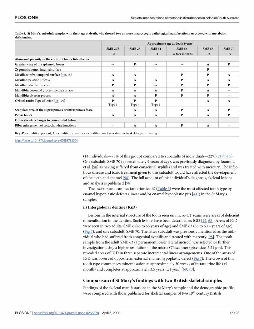
scopic pathological manifestations are summarised and presented in Table 4.
Teeth. i) Enamel—hypoplastic defects
A group of 42 individuals from the St Mary’s excavated sample (n = 65) had teeth available for
examination. Eighteen of these individuals showed evidence of enamel hypoplastic defects and
findings are presented in Table 5. The prevalence of this defect was higher among the adults
Fig 6. Subadult, SMB 28. Porous lesions on the bones of the orbital roof. Red arrow indicates small pores in the right bone cortex. Blue arrow
indicates exposed trabecular bone on the left.
https://doi.org/10.1371/journal.pone.0265878.g006
PLOS ONE Skeletal manifestations of metabolic disturbances in colonial South Australia
PLOS ONE | https://doi.org/10.1371/journal.pone.0265878 April 6, 2022 14 / 28
(14 individuals—78% of this group) compared to subadults (4 individuals—22%) (Table 5).
One subadult, SMB 70 (approximately 9 years of age), was previously diagnosed by Ioannou
et al. [98] as having suffered from congenital syphilis and was treated with mercury. The infec-
tious disease and toxic treatment given to this subadult would have affected the development
of the teeth and enamel [98]. The full account of this individual’s diagnosis, skeletal lesions
and analysis is published [98].
The incisors and canines (anterior teeth) (Table 5) were the most affected tooth type by
enamel hypoplastic defects (linear and/or enamel hypoplastic pits [41]) in the St Mary’s
samples.
ii) Interglobular dentine (IGD)
Lesions in the internal structure of the tooth seen on micro-CT scans were areas of deficient
mineralisation in the dentine. Such lesions have been described as IGD [42, 68]. Areas of IGD
were seen in two adults, SMB 6 (45 to 55 years of age) and SMB 63 (55 to 60 + years of age)
(Fig 7), and one subadult, SMB 70. The latter subadult was previously mentioned as the indi-
vidual who had suffered from congenital syphilis and treated with mercury [98]. The tooth
sample from the adult SMB 63 (a permanent lower lateral incisor) was selected or further
investigation using a higher resolution of the micro-CT scanner (pixel size: 5.21 μm). This
revealed areas of IGD in three separate incremental linear arrangements. One of the areas of
IGD was observed opposite an external enamel hypoplastic defect (Fig 7). The crown of this
tooth type commences mineralisation at approximately 30 weeks of intrauterine life (±1
month) and completes at approximately 3.5 years (±1 year) [69, 70].
Comparison of St Mary’s findings with two British skeletal samples
Findings of the skeletal manifestations in the St Mary’s sample and the demographic profile
were compared with those published for skeletal samples of two 19th century British
Table 4. St Mary’s. subadult samples with their age at death, who showed two or more macroscopic pathological manifestations associated with metabolic
deficiencies.
Approximate age at death (years)
SMB 27B SMB 28 SMB 51 SMB 56 SMB 58 SMB 70
~2 ~13 ~11 ~6 to 9 months ~2 ~ 9
Abnormal porosity in the cortex of bones listed below:
Greater wing of the sphenoid bones - - P - - - - A P
Zygomatic bones: internal surface - - - - - - - - P - -
Maxillae: infra-temporal surface [46:215] A A - - P P A
Maxillae: palatine process A A A P A A
Maxillae: alveolar process P P - - P P P
Mandible: coronoid process medial surface A A A P A - -
Mandible: alveolar process A A P P P - -
Orbital roofs: Type of lesion [58:109] P
Type 1
P
Type 4
P
Type 1
- - A A
Scapulae: area of the supraspinous or infraspinous fossa - - A A P A P
Pelvic bones A A A P A P
Other skeletal changes to bones listed below:
Ribs: enlargement of costochondral junctions - - A A P A - -
Key: P = condition present, A = condition absent, - - = condition unobservable due to skeletal part missing
https://doi.org/10.1371/journal.pone.0265878.t004
PLOS ONE Skeletal manifestations of metabolic disturbances in colonial South Australia
PLOS ONE | https://doi.org/10.1371/journal.pone.0265878 April 6, 2022 15 / 28

cemeteries. One sample is from St Martin’s-in-the-Bullring Church Cemetery, Birmingham,
[94], and the other from St Peter’s Collegiate Church burial ground, Wolverhampton [96, 97].
A comparison of the number and percentage of adults and subadults from each sample is pre-
sented in Table 6.
Skeletal manifestations
A comparison of the macroscopically observed skeletal manifestations among the subadult
samples from the three cemeteries is presented in Fig 8.
Table 5. St Mary’s sample. Individuals who showed enamel hypoplastic defects in permanent dentition.
St Mary’s
burial code
Age range at
death (years)
Sex Total number of
permanent teeth present
Total number of permanent
teeth affected
Percentage of permanent
teeth affected
Permanent tooth type & the
number of teeth affected
SMB 19 5–9 U 21 7 33% I x4, C x2, P x1
SMB 70 5–9 U 16 7 44% I x3, C x2
SMB 51 5–9 U 25 2 8% C x2
SMB 28 10–14 U 32 7 22% I x4, C x 1, M1 x2
SMB 79 15–19 F 28 2 7% C x2
SMB 5 20–29 F 5 2 40% I x 2
SMB 53C 30–39 F 11 4 36% I x4
SMB 9 40–49 M 23 8 35% I x6, C x4
SMB 66B 40–49 M 17 8 47% I x1, C x1, P x4, M2 x4
SMB 73 40–49 M 19 14 74% I x7, C x4, P x2, M1 x1
SMB 6 40–49 M 14 1 7% I x1
SMB 57 50–59 M 26 6 23% I x3, C x3
SMB 72 50–59 M 29 6 21% I x1, Cx2, Px1, M3 x2
SMB 83 50–59 M 15 6 40% C x4, M2 x1, M x1
SMB 59 50–59 M 17 6 35% I x5, C x1
SMB 68 50–59 M 18 6 33% C x2, P x4
SMB 23 50–59 M 23 3 13% I x2, C x1
SMB 63 60+ M 3 2 67% I x2
Note: U = undetermined sex.
Key: For permanent tooth types: I = Incisor, C = Canine, P = either first or second premolar, M1 = first molar, M2 = second molar, M3 = third molar.
https://doi.org/10.1371/journal.pone.0265878.t005
Fig 7. St Mary’s Cemetery. Micro-CT images. Adult, male, SMB 63. (a) Lower lateral permanent incisor with a post-mortem fracture in
the crown. (b) Transverse slice: At the level of the IGD in the crown. (c) Transverse slice: Arrows show four areas of IGD. (d) Transverse
slice: Arrows show three areas of IGD, image was angled to show location of IGD in three concentric layers. (e) White arrow shows IGD
(internal) opposite an enamel hypoplastic defect red arrow (external).
https://doi.org/10.1371/journal.pone.0265878.g007
PLOS ONE Skeletal manifestations of metabolic disturbances in colonial South Australia
PLOS ONE | https://doi.org/10.1371/journal.pone.0265878 April 6, 2022 16 / 28

i) Abnormal porosity in the bone cortices:
Subadults from St Martin’s and St Peter’s Cemeteries had areas of abnormal porosity in the
cortices of bones in the following anatomical sites: maxillary bone—area surrounding the
infra-orbital foramen, alveolar process and the antero-medial portion of the palatine process;
mandible—medial surface of the coronoid process; frontal bone—orbital plate (roof of orbit);
parietal and occipital bones—external surfaces; and scapulae—supraspinous fossae [45, 94,
97]. This manifestation (abnormal porosity in the bone cortex) was higher among St Mary’s
subadults compared to St Martin’s and St Peter’s subadult samples (Fig 8). Nine adults from
the St Mary’s sample also had one area of abnormal porosity in the cortices of cranial bones,
however, this skeletal manifestation was not observed in adults of St Martin’s or St Peter’s sam-
ples [45, 94, 97].
ii) Bending /bowing distortion of long bones:
This skeletal abnormality was seen only among the subadults of St Martins and St Peter’s
samples (Fig 8) [94, 95, 97]. Furthermore, no adults from the three cemeteries were seen with
this skeletal abnormality.
Table 6. Demographic profiles of St Mary’s, St Martins and St Peter’s cemeteries.
Cemetery Total Sample Size N = Adults Subadults
number % number %
St Mary’s (SA) 65 20 31 45 69
St Martin’s (UK) 406 242 60 164 40
St Peter’s (UK) 150 92 61 58 39
https://doi.org/10.1371/journal.pone.0265878.t006
Fig 8. Comparison of the skeletal manifestations observed in the subadult samples from St Mary’s Cemetery, South Australia and St Martin’s and
St Peter’s Cemeteries, England and the number of individuals affected.
https://doi.org/10.1371/journal.pone.0265878.g008
PLOS ONE Skeletal manifestations of metabolic disturbances in colonial South Australia
PLOS ONE | https://doi.org/10.1371/journal.pone.0265878 April 6, 2022 17 / 28

iii) Enlargement and flaring of metaphyses:
St Martin’s had a greater number of subadults with flaring of metaphyses than St Mary’s
and St Peter’s subadult samples (Fig 8) [94, 95, 97]. No adults from the three cemeteries were
seen with this skeletal manifestation.
iv) Porous lesions on the bones of the orbital roof:
The scores for this specific skeletal manifestation were not available for St Martin’s samples,
thus could not be compared between St Mary’s and St Peter’s samples. However, the published
results for St Martin’s Cemetery state that 17 adults and 18 subadults were observed with
“varying degrees” of porous lesions on the bones of the orbital roof [94:135]. These results
have been included in Fig 7 as porous lesions of any Type (i.e., according to the description
supplied by Stuart Macadam [59:109]). Therefore, St Martin’s had the highest number of sub-
adults with porous lesions on the bones of the orbital roof of any Type compared to St Peter’s
(14 subadults) and St Mary’s (6 subadults) samples [94, 97]. A comparison of porous lesions of
Types 2 to 4 [59:109] showed that St Peter’s had nine subadults with Types 2 and 3, while St
Mary’s had three subadults with Types 3 and 4 (Fig 8).
Discussion
St Mary’s Cemetery free ground burial records
St Mary’s burial records and data collected from headstone of leased burial plots for the
decades of 1840s, 1850s and the 1860s, show that the majority of people interred in St Mary’s
Cemetery, during the early years of this colonial settlement, were buried in the free ground sec-
tion (Fig 2). This indicates that for approximately 30 years or more, many individuals who
were buried at this cemetery or members of their family could not pay for a burial. This was a
period of establishment for the new colony, during which time the new settlement experienced
an economic recession and had a high unemployment rate [13, 20]. Therefore, it is likely that a
percentage of the early settlers in the region of St Marys-on-the-Sturt may not have had regular
employment. If individuals or families had economic difficulties, they may have had to depend
on charitable organisations or the government for their survival. This early economic hardship
in the colony is reflected in the need for relief from the Destitute Asylum for many people liv-
ing in or near the city of Adelaide.
A high mortality rate for infants is also reflected in the burial records from St Mary’s
(Table 2 and Fig 4). This could be the result of many factors, including poor living conditions,
lack of social/family support systems and/or inadequate availability of health services. An analy-
sis of the month of burial, for individuals listed in the St Mary’s Church records [73], that
could have been interred in the free ground area of the cemetery from 1847 to 1927, (n = 191)
(Table 2 and Fig 3), showed that a higher number of infant burials took place in the summer
month of December (Table 2). However, the total number of burials per month for this group
(any age group) (Fig 3) showed no seasonal pattern. The weather during a South Australian
summer could be extreme, with temperatures above 100ºF (38ºC). In December 1897, a local
newspaper in Adelaide reported a record heat wave since the foundation of the colony, with
temperatures of “over 90˚F in the shade for 17 days followed by temperatures of over 100˚F in
the shade for a further eight days” [99:5]. They described that multiple deaths occurred in and
around the city of Adelaide due to the heat, including eight deaths at the Destitute Asylum in
two days [99:1]. In addition, a number of children were affected by the high summer tempera-
tures and the public were advised that they could “be brought around by applying ice to the
head” [99:1]. The findings from St Mary’s burial records and the information from this 19th
PLOS ONE Skeletal manifestations of metabolic disturbances in colonial South Australia
PLOS ONE | https://doi.org/10.1371/journal.pone.0265878 April 6, 2022 18 / 28

century newspaper suggests that the summer temperatures in South Australia could have mag-
nified any health conditions that an individual may have been suffering. This finding contrasts
with the published results for colonial settlers buried at Milton, Otago, New Zealand by Buckley
et al. [100]. They were able to identify a pattern in the monthly burials at Milton, with a higher
number of deaths occurring in the winter months of June, July and August and fewer deaths in
the summer months [100]. The difference in seasonal trends may well be related to the different
climatic conditions in South Australia and the South Island of New Zealand [100].
At St Mary’s Cemetery, the location of burial for some infants was not documented in the
burial records [73:364]. This may be the result of an infant having been stillborn or dying a
short time after their birth (Fig 4). The burial records listed 41 infants that were under the age
of three months (Fig 4), which is 21% of the total number of individuals whose age at death
was also listed, that could have been buried in the free ground area (n = 194—one individual
did not have their age recorded). In the new colony no official regulations were in place in rela-
tion to the location of a burial for a stillborn infant, or the requirement to register the birth of a
stillborn child until 1936 [74, 101]. Burial practices of perinates excavated at the 19th century
Parramatta Convict Hospital in New South Wales, Australia, showed they were afforded very
little respect or care [102].
Economic recovery of the new colony
The improved economic condition of the colony following the first depression during the
1840’s could have influenced the economic changes seen at St Marys-on-the-Sturt. A local
financial recovery is reflected in the lower number of burials in the free ground section of St
Mary’s Cemetery (Fig 2) [13, 20]. The percentage of these burials began to decrease during the
1860s and reduced to only 8% of the total burials in the cemetery in the 1920s (Fig 2). This sug-
gests that there was a gradual improvement in the economic status of the individuals who lived
in the region of St Marys-on-the-Sturt and that the majority of the excavated skeletons studied
could be those of individuals who lived during the establishment of the colony. The South Aus-
tralian economy improved sufficiently for the introduction of an old age pension after the turn
of the 20th century which reduced the need for the Destitute Asylum [30].
Skeletal manifestations indicating metabolic disturbance
Abnormal porous lesions seen in the cortical bone of 9 adults and 12 subadults were the com-
mon manifestations present in the St Mary’s skeletal collection (Tables 3 and 4). This abnor-
mality could be caused by a deficiency of vitamin C, which affects the synthesis of collagen in
body tissues including blood vessels and bone, resulting in weak vessel walls and defective pro-
duction of osteoid [45, 51, 103]. Ortner and Ericksen [47:215] have suggested that areas of
abnormal porosity seen on the infra-temporal surface could be the result of an inflammatory
response to “leakage from scorbutic deep temporal arteries”. They propose that abnormal
porous lesions with “fine holes that are less that 1mm in diameter” are a result of localised
increased vascularity seen in response to extravasated blood from weak vessels [47:212]. Differ-
ent processes, including defective mineralisation in cortical bone, could appear as porous
lesions in dry bone samples resulting from the decomposition of the unmineralised bone
matrix [44]. The porous lesions resulting from both vitamin C and vitamin D deficiencies are
similar. Therefore, it is important to differentially diagnose the lesions from normal anatomi-
cal variation, non-specific localised infections that cause an osteoblastic response, trauma, neo-
plastic disorders and/or genetic causes [51, 104, 105]. Furthermore, the location, distribution
and quantity of lesions on the skeleton should be considered [44].
PLOS ONE Skeletal manifestations of metabolic disturbances in colonial South Australia
PLOS ONE | https://doi.org/10.1371/journal.pone.0265878 April 6, 2022 19 / 28

The subadult, SMB 56 (approximately 6 to 9 months of age), had abnormal cortical porous
lesions in multiple locations of the skeleton (Table 4), as well as flaring of the costochondral
junctions of the ribs. These skeletal manifestations could be attributed to either vitamin C or
vitamin D deficiencies or a combination of both [43, 45, 95]. The rapid growth of an infant
bone produces areas of new formation in the skeleton, which could appear as porosity on
the cortical surface of bone. However, this change in surface texture would not penetrate the
cortex of the bone in the same way as an increase of vascularity would as an inflammatory
response from the body [43, 47]. The extensive and symmetrical nature of the porosity seen in
the bone cortices of multiple bones of this infant is suggestive of a chronic systemic disorder
that could have disturbed the metabolism near the time of death [106]. The infant’s frag-
mented and missing cranial bones was a limitation to a full diagnosis and could have biased
the findings. It is possible that other manifestations could have been located on areas of the
skeleton, for example such as porous lesions bilaterally on the greater wing of the sphenoid
bones or bones of the orbital roof that were no longer present.
Five subadults, SMB 27B (approximately 2 years of age), SMB 28 (approximately 13 years of
age), SMB52 (approximately 11 years of age), SMB 58 (approximately 2 years of age) and SMB
70 (approximately 9 years of age), showed two to four skeletal manifestations associated with
metabolic deficiencies (Table 4). Three of these subadults had porous lesions on the bones of
the orbital roof of Types 3 and 4 [59:109] (Tables 3 and 4). These types of porous lesions have
been considered as an indicator of anaemia, which as previously mentioned, may have resulted
from a dietary deficiency of iron and/or vitamin B12, malabsorption of iron from the gut due
to chronic diseases and/or chronic blood loss resulting from gastrointestinal parasites condi-
tions [59, 63–66]. Subadult, SMB 28 had areas of abnormal porosity on orbital roof (Type 4)
and the greater wings of the sphenoid bones (bilaterally) (Table 4). This individual could have
had a co-occurence of vitamin C deficiency and anaemia, as vitamin C enchances the absorp-
tion of iron from the gut [51], therefore, a deficiency of this vitamin may have aggravated any
anaemia that was present in SMB 28.
The remaining adults and subadults in St Mary’s sample only showed one manifestation on
one location of the skeleton, for example subadult, SMB 8 (approximately 18 months of age)
had flaring of the distal metaphyses of the femora, but without any other pathological manifes-
tations observed in their skeleton this subadult was considered as not suffering from a chronic
metabolic deficiency (Table 3). It is possible that these individuals died before changes to the
bone structure could occur and the state of preservation of some of the skeletal remains may
also have biased the findings.
Dental defects
i) Enamel hypoplastic defects
The enamel hyperplastic defects seen in the 18 individuals from the St Mary’s sample were all
on permanent teeth (Table 5). This may be due to developmental timing, as the primary
(deciduous) dentition is less affected by developmental enamel defects than the permanent
dentition [68, 69]. The health insult/s that caused the enamel hypoplastic defect/s occurred
during the development of the specific permanent tooth types (Table 5) [42, 68, 69]. The loca-
tion and distribution of these enamel defects seen in the St Mary’s sample suggest they were
caused by insults between birth and 4 years of age.
The 14 adults and 4 subadults who showed evidence of these enamel hypoplastic defects
represented 43% of the individuals with remaining dentition (n = 42). There are 20 adults in
the St Mary’s collection, two of these adults were edentulous. Fourteen of the remaining 18
adults had hypoplastic defects of the enamel (78%). This percentage is comparable to a cohort
PLOS ONE Skeletal manifestations of metabolic disturbances in colonial South Australia
PLOS ONE | https://doi.org/10.1371/journal.pone.0265878 April 6, 2022 20 / 28

of adults (86%) with enamel hypoplastic defects who were migrants to Milton, New Zealand,
during the 19th century [100]. Isotopic analysis of this New Zealand cohort indicated that none
of them were born locally [107], thus the health insult that caused the enamel hypoplastic
defects would have occurred in their home country [100]. This is also highly likely for the
adults in the St Mary’s sample. The high percentage of adults with enamel hypoplastic defects
in St Mary’s sample and the sample from Milton, New Zealand, suggests that the this dental
defect was common amongst the migrant settlers.
Enamel hypoplastic defects are a general, non specific indicator of disturbance and/or dis-
ease during dental development [108]. The four St Mary’s subadults with enamel hypoplastic
defects (Table 5) also had other skeletal manifestations (Table 4), which have been previously
discussed. The pathological conditions suffered by these individuals may have contributed to
the formation of the enamel hypoplastic defects during dental development.
ii) Interglobular dentine
Two adults and one subadult from St Mary’s sample had areas of IGD on micro-CT scan
images. Adult, SMB 63 (55 to 60 + years of age), had three separate areas of IGD (Fig 4), which
indicates that he could have suffered from three separate episodes of health insults during the
development of this tooth. Again, these health insults could have occurred before migration.
One of the areas of IGD in the internal structure of this tooth was opposite an external enamel
hypoplastic defect. It is possible that the enamel hypoplastic defect could have resulted from
the same health insult that caused the IGD (Fig 4) [109–111].
Comparison of St Mary’s free ground skeletal samples with those of St
Martin’s and St Peter’s samples in Britain
The working-class background of some of the people buried at the free ground area of St
Mary’s Cemetery could be similar to those published for St Martin’s and St Peter’s cemeteries
in Britain [45, 94–97]. However, the demographic profile of the St Mary’s sample is somewhat
different from those of the British samples. The majority of St Mary’s sample was composed of
subadults (Tables 1 and 6), with 60% of subadults under the age of four years. The cause of
deaths of many subadults was from gastrointestinal conditions such as dysentery and/or vom-
iting from contaminated water supplies, or pulmonary conditions from bacterial infection
such as whooping cough [73:372–381]. Some of these deaths may have resulted from limited
access to emergency medical help in the region of St Marys-on-the-Sturt. A study of mortality
records from the neighbouring state of Victoria, Australia, indicated that there had been epi-
demics of scarlet fever and measles between 1853–1916 [112]. These infectious diseases could
have spread across the border to South Australia.
The skeletal manifestation, abnormal porosity in the cortices of bones, was seen in sub-
adults from St Mary’s, St Martin’s and St Peter’s skeletal samples [45, 94, 96]. The prevalence
of such lesions was higher among the subadults from the St Mary’s sample (13%) compared to
that from St Martin’s (4%) and St Peter’s samples (2%). The lower prevalence of this manifesta-
tion (probable vitamin C deficiency) among the subadults of the British samples could be due
to the availability of fresh fruits and vegetables in Birmingham and Wolverhampton. The ini-
tial difficulties and delays in establishing farms and the production of food could have caused
scarcity of provisions rich in vitamin C during the first decades of the development of South
Australia. Subadults affected by this deficiency from the three cemeteries may have been
breastfed infants [45, 94, 96, 97]. Therefore, their insufficient intake of vitamin C may have
resulted from a dietary deficiency of the mother [44, 65, 113]. Elevated nitrogen isotope values
(+ 1.7‰) observed in skeletal remains of some infants from the St Mary’s sample [114],
PLOS ONE Skeletal manifestations of metabolic disturbances in colonial South Australia
PLOS ONE | https://doi.org/10.1371/journal.pone.0265878 April 6, 2022 21 / 28

relative to those of adult females in the same sample, suggested that breastmilk was a principal
source of diet for infants. However, these findings do not indicate whether the child received
adequate amounts of milk during breast feeding. Infants may also have been affected by the
feeding and weaning practices of that period [114–117].
The number of individuals with the manifestation of enlargement and flaring of the costo-
chondral junctions of ribs and metaphyses of long bones was higher among St Martin’s and St
Peter’s subadult samples compared to those of St Mary’s samples (Fig 8) [95, 97]. The manifesta-
tion of bending distortions of long bones was only seen among subadults of St Martin’s and St
Peter’s samples and not observed in any of the subadult samples from St Mary’s (Fig 8). Skeletal
remains of adults from the three cemeteries were free of the above-mentioned manifestations.
These skeletal abnormalities have been linked to a chronic deficiency of vitamin D [95, 97].
Among the St Mary’s subadults the lower incidence of the manifestations linked to vitamin D
deficiency (Fig 7), may well be due to the abundance of sunlight (UV rays) in South Australia.
St Martin’s had the highest number of subadults with porous lesions in the bones of the
orbital roof (Fig 8) [94]. The scores for these porous lesions, as described by Stuart-Macadam
[59], were not available for this sample (Brickley, personal communication, 2018), which made
it difficult to compare the findings from St Martin’s with those of St Mary’s and St Peter’s sam-
ples (Fig 7). A comparison of this manifestation for Types 2 to 4 [59:109], between the St
Mary’s and St Peter’s subadults showed that St Peter’s had more subadults with this pathologi-
cal manifestation (9 subadults) than St Mary’s (3 subadults) (Fig 8). However, St Mary’s had
one subadult with a Type 4 porous lesion compared to St Peter’s subadult samples who had
Type 2 or Type 3 porous lesions on the bones of their orbital roof [59:109]. This may indicate
that the St Mary’s subadult, SMB 28 with the Type 4 porous lesion may have suffered a differ-
ent, or more severe systemic metabolic disturbances, or may have had a co-occurance of multi-
ple conditions that produced this manifestation.
Conclusion
The rare skeletal sample from the free ground of St Mary’s Anglican Church Cemetery in
South Australia, generated an opportunity to understand the effects of the establishment of the
new colony on the health of these migrant settlers. The abnormal manifestations seen in the
bones and teeth of the individuals excavated from St Mary’s reflect the health issues they expe-
rienced. Many of these issues could have been due to the marked hardships brought about by
the unprepared state of the colony for the settlement of the new arrivals and the local environ-
mental conditions. This is supported by the higher percentage of observed skeletal manifesta-
tions indicating a deficiency of vitamin C in St Mary’s sample compared with the two British
skeletal samples. The people who decided to migrate to South Australia during the 19th century
may have arrived with hopes of prosperity through the use of natural resources. However, eco-
nomic differences were seen between migrants, who had lived in the region of St Marys-on-
the-Sturt, in the number of burials at the government funded free ground compared to pri-
vately funded leased burials from the mid-1840s to 1860s, in St Mary’s Cemetery. A decrease
in this number of burials in the free ground area from the 1870s to 1920s, suggests a gradual
improvement in the economic status of these migrants. This local improvement reflected the
economic recovery seen in the rest of the colony.
Supporting information
S1 Table. Combined table of anatomical sites and descriptive features: ‘X’ indicates which
metabolic deficiency the skeletal lesion may be associated with.
(DOCX)
PLOS ONE Skeletal manifestations of metabolic disturbances in colonial South Australia
PLOS ONE | https://doi.org/10.1371/journal.pone.0265878 April 6, 2022 22 / 28
S1 Data. Observed skeletal manifestation listed with location on the skeleton. St Mary’s
Cemetery: Supporting Data 1.
(XLSX)
S2 Data. Summary—Enamel hypoplastic defects_macroscopic examination. St Mary’s
Cemetery: Supporting Data 2.
(XLSX)
Acknowledgments
The authors acknowledge The University of Adelaide including the Faculty of Health and
Medical Sciences Honours and PhD Scholarships for AG. Fr. William Deng, of St Mary’s
Anglican Church for his kind collaboration and continued access to the St Mary’s skeletal sam-
ple. The authors acknowledge the facilities and the scientific and technical assistance, of the
Australian Microscopy & Microanalysis Research Facility at the Adelaide Microscopy, Univer-
sity of Adelaide, especially Ruth Williams for her knowledge of and assistance with micro-CT
scanning and post processing software.
Author Contributions
Conceptualization: Angela Gurr, Jaliya Kumaratilake, Maciej Henneberg.
Data curation: Angela Gurr, Stella Ioannou, Maciej Henneberg.
Formal analysis: Angela Gurr, Jaliya Kumaratilake, Alan Henry Brook, Maciej Henneberg.
Investigation: Angela Gurr, Jaliya Kumaratilake, Alan Henry Brook, Maciej Henneberg.
Methodology: Angela Gurr, Alan Henry Brook, Maciej Henneberg.
Project administration: Angela Gurr.
Resources: F. Donald Pate.
Supervision: Jaliya Kumaratilake, Alan Henry Brook, Maciej Henneberg.
Visualization: Angela Gurr.
Writing – original draft: Angela Gurr.
Writing – review & editing: Angela Gurr, Jaliya Kumaratilake, Alan Henry Brook, Stella Ioan-
nou, F. Donald Pate, Maciej Henneberg.
References1. Betsinger TK, DeWitte SN. The Bioarchaeology of Urbanization: Biological The, Demographic, and
Social Consequences of Living in Cities. 1st ed. Switerland: Springer International Publishing; 2020.
2. Szreter S. Economic Growth, Disruption, Deprivation, Disease, and Death: On the Importance of the
Politics of Public Health for Development. Population and Development Review. 1997; 23(4):693–
728. https://doi.org/10.2307/2137377
3. Szreter S. Urbanization, Mortality, and the Standard of Living Debate: New Estimates of the Expecta-
tion of Life at Birth in Nineteenth-Century British Cities. In: Health and Wealth. Boydell & Brewer;
2005. p. 165.
4. Ashworth WJ. The industrial revolution the State, knowledge and global trade. London: Bloomsbury
Academic; 2017.
5. Brimblecombe P. London air pollution, 1500–1900. Atmospheric Environment (1967). 1977; 11
(12):1157–62. https://doi.org/10.1016/0004-6981(77)90091-9 PMID: 343794
PLOS ONE Skeletal manifestations of metabolic disturbances in colonial South Australia
PLOS ONE | https://doi.org/10.1371/journal.pone.0265878 April 6, 2022 23 / 28

6. Dewitte SN, Hughes-Morey G, Bekvalac J, Karsten J. Wealth, health and frailty in industrial-era Lon-
don. Annals of Human Biology. 2016; 43(3):241–54. https://doi.org/10.3109/03014460.2015.1020873
PMID: 26073638
7. Newman SL, Gowland RL. Dedicated Followers of Fashion? Bioarchaeological Perspectives on
Socio-Economic Status, Inequality, and Health in Urban Children from the Industrial Revolution (18th–
19th C), England. International Journal of Osteoarchaeology. 2017; 27(2):217–29. https://doi.org/10.
1002/oa.2531 PMID: 28553062
8. Lokan J. From Cornish Miner to Farmer in Nineteenth-Century South Australia: A Case Study. Cornish
studies. 2008; 16(1):48–77. https://doi.org/10.1386/corn.16.1.48_1
9. Payton P. Cornish Emigration in Response to Changes in the International Copper Market in 1860s.
Exeter, UK: University of Exeter; 1995.
10. Rowe J. Cornwall in the age of the Industrial Revolution. Liverpool: Liverpool University Press; 1953.
11. Lidwell-Durnin J. Cultivating famine: data, experimentation and food security, 1795–1848. The British
journal for the history of science. 2020; 53(2):159–81. https://doi.org/10.1017/S0007087420000199
PMID: 32484149
12. Price AG. The foundation and settlement of South Australia, 1829–1945: a study of the colonization
movement, based on the records of the South Australian Government and on other authoritative docu-
ments. Adelaide: Libraries Board of South Australia; 1973.
13. Wilkinson GB. South Australia, its advantages and its resources: being a description of that colony,
and a manual of information for emigrants / by George Blakiston Wilkinson. London: J. Murray; 1848.
14. Nance C. The South Australian social experiment 1836–71: a study of some of the aspects of South
Australia’s early social development. Adelaide Flinders University of South Australia 1977.
15. Wilkinson GB. The working man’s handbook to South Australia, with advice to the farmer, and detailed
information for the several class of labourers and artisans. London: Murray; 1849.
16. Haines R. Emigration and the labouring poor: Australian recruitment in Britain and Ireland, 1831–60.
New York: Macmillan; St. Martin’s Press; 1997.
17. Haines R. Nineteenth century government assisted immigrants from the United Kingdom to Australia:
schemes, regulations and arrivals, 1831–1900 and some vital statistics 1834–1860. South. Australia:
Flinders University of South Australia; 1995.
18. Lawrence S, Davies P. Cornish tin-streamers and the Australian gold rush: technology transfer in allu-
vial mining. Post-medieval archaeology. 2015; 49(1):99–113. https://doi.org/10.1179/0079423615Z.
00000000073
19. Pike D. Paradise of dissent: South Australia 1829–1857 / Douglas Pike. 2nd ed. Melbourne: Mel-
bourne University Press; 1967.
20. Haines R. Life and death in the age of sail: the passage to Australia. Sydney: UNSW Press; 2003.
21. Haines R. Medical Superintendence and Child Health on Government-Assisted Voyages to South
Australia in the Nineteenth Century. Health and History. 2001; 3(2):1–29. https://doi.org/10.2307/
40111403
22. Gibbs RM. Under the Burning Sun: A history of colonial South Australia, 1836–1900. Adelaide: South-
ern Heritage; 2013.
23. Dickey B. Rations, residence, resources: a history of social welfare in South Australia since 1836, with
contributions from Elaine Martin and Rod Oxenberry. Martin E, Oxenberry R, editors. Netley, S. Aust.:
Wakefield Press; 1986.
24. Dare R. Paupers’ rights: Governor Grey and the poor law in South Australia. Australian historical stud-
ies. 1992; 25(99):220–43. https://doi.org/10.1080/10314619208595908
25. Jaensch D. The Flinders history of South Australia. Political history. Netley, S. Aust: Wakefield Press;
1986.
26. Government of South Australia State Records. Alphabetical index to return of sick and destitute emi-
grants who have received relief from the emigration department 1993 [cited 2021 25.01]. https://
archives.sa.gov.au/finding-information/discover-our-collection/health-and-welfare/destitution-south-
australia.
27. Attorney-General’s Department. South Australian Legislation: As made Acts 1837–2002: 1841–1850:
1843. [Webpage] 2022 [cited 2022 10 February]; https://www.legislation.sa.gov.au/lists-and-index-to-
sa-legislation/historical-numbered-acts-1837-2002.
28. Piddock S. “An Irregular and Inconvenient Pile of Buildings”: The Destitute Asylum of Adelaide, South
Australia and the English Workhouse. International Journal of Historical Archaeology. 2001; 5(1):73–
95. https://doi.org/10.1023/a:1009597226492 PMID: 20037941
PLOS ONE Skeletal manifestations of metabolic disturbances in colonial South Australia
PLOS ONE | https://doi.org/10.1371/journal.pone.0265878 April 6, 2022 24 / 28

29. Piddock S. A Space of Their Own: The Archaeology of Nineteenth Century Lunatic Asylums in Britain,
South Australia and Tasmania. Orser CE, editor. New York, NY: Springer 2007.
30. Geyer M. Behind the wall: the women of the destitute asylum Adelaide, 1852–1918. Adelaide, S. Aust:
Axiom, in association with the Migration Museum; 1994.
31. The Adelaide Observer. Prevention of Infection from Typhus Fever. The Adelaide Observer. 1845
18.01.1845 (col. 2).
32. The South Australia Register. Pollution of the Torrens. The South Australian Register. 1873
16.12.1874.
33. The South Australia Register. Drinking Typhoid. The South Australian Register. 1898 17.02.1898;
Sect. Register.
34. The South Australian Register. Summer Cholera-Its Causes. The South Australian Register,. 1847
10.02.1847;Sect. Medicine and Surgery.
35. Woodruff P. Two million South Australians. Kent Town, S. Aust: Peacock Publications; 1984.
36. Forbes ILD. From Colonial Surgeon to Health Commission: the government provision of health ser-
vices in South Australia, 1836–1995. Myrtle Bank, S. Aust: I.L.D. Forbes 1996.
37. Manning GH. A colonial experience, 1838–1910: a woman’s story of life in Adelaide, the District of
Kensington and Norwood together with reminiscences of colonial life / Underdale, S. Aust.: Gillingham
Printers; 2001.
38. Roberts CA, Brickley M. Infectious and metabolic diseases: a synergistic relationship. In: Katzenberg
A M., Grauer AL, editors. Biological Anthropology of the Human Skeleton. 3rd ed. Hoboken, NJ, USA:
John Wiley & Sons, Inc; 2018. p. 415–46.
39. Snoddy AME, Buckley H.R, and Halcrow SE. More than metabolic: Considering the broader paleoepi-
demiological impact of vitamin D deficiency in bioarchaeology. American journal of physical anthropol-
ogy; 2016. 160(2):183–196. https://doi.org/10.1002/ajpa.22968 PMID: 26926781
40. Lockau L, Atkinson SA. Vitamin D’s role in health and disease: How does the present inform our under-
standing of the past? International Journal of Paleopathology. 2018; 23:6–14. https://doi.org/10.1016/
j.ijpp.2017.11.005 PMID: 30573166
41. Hillson S. Tooth Development in Human Evolution and Bioarchaeology. Cambridge: Cambridge:
Cambridge University Press; 2014.
42. Nanci A. Ten Cate’s Oral Histology: Development, Structure, and Function. Saint Louis: Saint Louis:
Elsevier; 2012.
43. Ortner DJ, Kimmerle EH, Diez M. Probable evidence of scurvy in subadults from archeological sites in
Peru. American Journal of Physical Anthropology. 1999; 108(3):321. https://doi.org/10.1002/(SICI)
1096-8644(199903)108:3<321::AID-AJPA7>3.0.CO;2-7 PMID: 10096683
44. Brickley MB, Ives R, Mays S. The Bioarchaeology of Metabolic Bone Disease. San Diego: Elsevier
Science & Technology; 2020.
45. Brickley M, Ives R. Skeletal manifestations of infantile scurvy. American Journal of Physical Anthropol-
ogy. 2006; 129(2):163–72. https://doi.org/10.1002/ajpa.20265 PMID: 16323179
46. Ortner DJ, Mays S. Dry bone manifestations of rickets in infancy and early childhood. International
Journal of Osteoarchaeology, 1998. 8(1): p. 45–55.
47. Ortner DJ, Ericksen MF. Bone changes in the human skull probably resulting from scurvy in infancy
and childhood. International Journal of Osteoarchaeology. 1997; 7(3):212–20. https://doi.org/10.1002/
(SICI)1099-1212(199705)7:3<212::AID-OA346>3.0.CO;2-5
48. Kenkre JS, Bassett JHD. The bone remodelling cycle. Annals of Clinical Biochemistry. 2018; 55
(3):308–27. https://doi.org/10.1177/0004563218759371 PMID: 29368538
49. Brickley M. Cribra orbitalia and porotic hyperostosis: A biological approach to diagnosis. American
journal of physical anthropology. 2018; 167(4):896. https://doi.org/10.1002/ajpa.23701 PMID:
30259969
50. Bourne GH. Some experiments on the possible relationship between vitamin C and calcification. The
Journal of physiology. 1943; 102(3):319–28. https://doi.org/10.1113/jphysiol.1943.sp004038 PMID:
16991612
51. Snoddy AME, Buckley HR, Elliott GE, Standen VG, Arriaza BT, Halcrow SE. Macroscopic features of
scurvy in human skeletal remains: A literature synthesis and diagnostic guide. American journal of
physical anthropology. 2018; 167(4):876. https://doi.org/10.1002/ajpa.23699 PMID: 30298514
52. Barlow T. On Cases Described as "Acute Rickets," which are probably a combination of Scurvy and
Rickets, the Scurvy being an essential, and the rickets a variable, element. Medico-chirurgical transac-
tions.1883; 66:159–220. https://doi.org/10.1177/095952878306600112 PMID: 20896608
PLOS ONE Skeletal manifestations of metabolic disturbances in colonial South Australia
PLOS ONE | https://doi.org/10.1371/journal.pone.0265878 April 6, 2022 25 / 28

53. Stark RJ. A proposed framework for the study of paleopathological cases of subadult scurvy. Interna-
tional Journal of Paleopathology. 2014; 5:18–26. https://doi.org/10.1016/j.ijpp.2014.01.005 PMID:
29539464
54. Stone N, Meister A. Function of ascorbic acid in the conversion of proline to collagen hydroxyproline.
Nature. 1962; 194:555. https://doi.org/10.1038/194555a0 PMID: 13917472
55. Bikle D, Christakos S. New aspects of vitamin D metabolism and action—addressing the skin as
source and target. Nature reviews Endocrinology. 2020; 16(4):234–52. https://doi.org/10.1038/
s41574-019-0312-5 PMID: 32029884
56. Mellanby M. Diet and the Teeth: An Experimental Study. Part III. The Effect of Diet on Dental Structure
and Disease in Man. Journal of the American Medical Association. 1934; 102(18):1523-. https://doi.
org/10.1001/jama.1934.02750180075026
57. Rivera F, Lahr M, Beatriz M. New evidence suggesting a dissociated etiology for cribra orbitalia and
porotic hyperostosis. American Journal of Anthropology. 2017; 164:79–96. https://doi.org/10.1002/
ajpa.23258 PMID: 28594081
58. Wapler U, Crubezy E, Schultz M. Is cribra orbitalia synonymous with anemia? Analysis and interpreta-
tion of cranial pathology in Sudan. American Journal of Physical Anthropology. 2004; 123(4):333–9.
https://doi.org/10.1002/ajpa.10321 PMID: 15022361
59. Stuart-Macadam P. Anemia in roman Britain: Poundbury camp. In: Bush H, Zvelebil M, editors. Health
in Past Societies: Biocultural Interpretations of Human Skeletal Remains in Archaeological Contexts.
Oxford: British Archaeological Reserach Series; 1991. p. 101–13.
60. Walker PL, Bathhurst RR, Richman R, Gjerdrum T, Andrushko VA. The causes of porotic hyperostosis
and cribra orbitalia: A reappraisal of the iron- deficiency- anemia hypothesis. American Journal of
Physical Anthropology, 2009. 139(2): p. 109–125. https://doi.org/10.1002/ajpa.21031 PMID:
19280675
61. Oxenham MF, Cavill I. Porotic hyperostosis and cribra orbitalia: the erythropoietic response to iron-
deficiency anaemia. Anthropological Science, 2010. 118(3): p. 199–200.
62. Camaschella C. Iron-Deficiency Anemia, in N. Engl. J. Med. 2015. p. 1832–1843. https://doi.org/10.
1056/NEJMra1401038 PMID: 25946282
63. Godde K, Hens S.M. An epidemiological approach to the analysis of cribra orbitalia as an indicator of
health status and mortality in medieval and post-medieval London under a model of parasitic infection.
American journal of physical anthropology, 2021.
64. Ferguson A, Brydon WG, Brian H, Williams A, Mackie MJ. Use of whole gut perfusion to investigate
gastrointestinal blood loss in patients with iron deficiency anaemia. Gut, 1996. 38(1): p. 120. https://
doi.org/10.1136/gut.38.1.120 PMID: 8566838
65. Jelliffe DB, Blackman J. Bahima disease: Possible "milk anemia" in late childhood. The Journal of pedi-
atrics, 1962. 61: p. 774. https://doi.org/10.1016/s0022-3476(62)80354-0 PMID: 13964591
66. Weiss G, Goodnough LT. Anemia of Chronic Disease. The New England Journal of Medicine, 2005.
352 (10): p. 1011–23.75. https://doi.org/10.1056/NEJMra041809 PMID: 15758012
67. O’Donnell L, Hill E, Anderson A, Edgar H. Cribra orbitalia and porotic hyperostosis are associated with
respiratory infections in a contemporary mortality sample from New Mexico. American journal of physi-
cal anthropology, 2020.
68. Atkinson ME, White FH. Principles of Anatomy and Oral Anatomy for Dental Students. Edinburgh:
Churchill Livingstone; 1992.
69. AlQahtani S. The London atlas: developing an atlas of tooth development and testing its quality and
performance measures. ProQuest Dissertations Publishing; 2012.
70. AlQahtani SJ, Hector MP, Liversidge HM. Brief communication: The London atlas of human tooth
development and eruption. American Journal of Physical Anthropology. 2010; 142(3):481–90. https://
doi.org/10.1002/ajpa.21258 PMID: 20310064
71. Davies J. St Mary’s: A Pioneer Walk Through the Churchyard of St Mary’s South Road. St Mary: St
Mary’s Anglican Church; 1991.
72. Somerset M, Ragless R. St Mary’s Walk: A Historical and Social Perspective of a Significant but Little-
known Suburb of Adelaide. Mitcham, South Australia: City of Mitcham; 2009.
73. Anson TJ. The Bioarchaeology of the St. Mary’s Free Ground Burials: Reconstruction of colonial
South Australian lifeways. Adelaide: University of Adelaide; 2004.
74. Nicol R. At the end of the road: government, society and the disposal of human remains in the nine-
teenth and twentieth centuries. St Leonards, N.S.W.: Allen & Unwin; 1994.
75. City of Marion Council. About Council/ Maps: City of Marion Council; 2020 [cited 2020 1 July]. https://
www.marion.sa.gov.au/about-council/maps.
PLOS ONE Skeletal manifestations of metabolic disturbances in colonial South Australia
PLOS ONE | https://doi.org/10.1371/journal.pone.0265878 April 6, 2022 26 / 28

76. Buikstra JE, Ubelaker DH, Haas J, Aftandilian D, Field Museum of Natural History. Standards for data
collection from human skeletal remains: proceedings of a seminar at the Field Museum of Natural His-
tory, organized by Jonathan Haas / volume editors, Jane E. Buikstra and Douglas H. Ubelaker, assis-
tant editor, David Aftandilian; contributions by D. Aftandilian. . . [et al.]. Fayetteville, Ark.: Fayetteville,
Ark.: Arkansas Archeological Survey; 1994.
77. Ubelaker DH. Human skeletal remains: excavation, analysis, interpretation ( 2nd ed. ed.). Washing-
ton: Taraxacum; 1989.
78. Scheuer L, Black S, Schaefer MC. Juvenile Osteology: A Laboratory and Field Manual: Elsevier Sci-
ence; 2008.
79. Brooks S, Suchey JM. Skeletal age determination based on the os pubis: A comparison of the Acsadi-
Nemeskeri and Suchey-Brooks methods. Human evolution 5, 227–238;1990.
80. Lovejoy CO, Meindl RS, Pryzbeck TR, Mensforth RP. Chronological metamorphosis of the auricular
surface of the ilium: A new method for the determination of adult skeletal age at death. American Jour-
nal of Physical Anthropology. 1985; 68(1), 15–28. https://doi.org/10.1002/ajpa.1330680103 PMID:
4061599
81. Todd TW. Age changes in the pubic bone. American journal of physical anthropology. 1921; 4(1):1–
70. https://doi.org/10.1002/ajpa.1330040102
82. Gustafson AG. Age Determination on Teeth. Journal of the American Dental Assocation; 1950; 41(1),
45–54. https://doi.org/10.14219/jada.archive.1950.0132 PMID: 15428197
83. Buckberry J. The (mis)use of adult age estimates in osteology. Annals of human biology. 2015; 42
(4):323–31. https://doi.org/10.3109/03014460.2015.1046926 PMID: 26190373
84. Bass WM. Human Osteology: a laboratory and field manual (4th ed.). Columbia, Mo: Missouri
Archaeological Society; 1995.
85. Mitchell PD, Brickley M. Updated Guidelines for the Standards for Recording Human Remains. South-
ampton: British Association for Biological Anthropology and Osteology & Institute of Field Archaeolo-
gists; 2017.
86. Heron C, Grauer AL (Ed.): A Companion to Paleopathology. Chromatographia. 2013; 76(23–
24):1795–6. https://doi.org/10.1007/s10337-013-2546-z
87. Brook A. Research contributions to paediatric dentistry in the areas of dental development, orofacial
pathology, prevention, education and patient management 2014.
88. Brook AH, Elcock C, Hallonsten AL, Poulsen S, Andreasen J, Koch G, et al. The development of a
new index to measure enamel defects. In: Brook AH, editor. Dental Morphology. Sheffield: Sheffield
Academic Press; 2001. p. 59–66.
89. Elcock C, Lath DL, Luty JD, Gallagher MG, Abdellatif A, Backman B, et al. The new enamel defects
index: testing and expansion. Eur J Oral Sci. 2006; 114:35–8. https://doi.org/10.1111/j.1600-0722.
2006.00294.x PMID: 16674660
90. Bruker. Products: http://bruker-microct.com/products/downloads.htm. 2018. http://bruker-microct.
com/products/downloads.htm.
91. ThermoFisher Scientific. 3D Visulaization & Analysis Software: Amiro-Avizo 2019. https://www.
thermofisher.com/au/en/home/industrial/electron-microscopy/electron-microscopy-instruments-
workflow-solutions/3d-visualization-analysis-software.html.
92. Colombo A, Ortenzio L, Bertrand B, Coqueugniot H, KnuSel CJ, Kahlon B, et al. Micro-computed
tomography of teeth as an alternative way to detect and analyse vitamin D deficiency. Journal of
Archaeological Science: Reports. 2019; 23:390–5. https://doi.org/10.1016/j.jasrep.2018.11.006
93. Veselka B, Brickley MB, D’Ortenzio L, Kahlon B, Hoogland MLP, Waters-Rist AL. Micro-CT assess-
ment of dental mineralization defects indicative of vitamin D deficiency in two 17th-19th century Dutch
communities. Am J Phys Anthropol. 2019:1–10. Epub 2019/03/19. https://doi.org/10.1002/ajpa.23819
PMID: 30882907.
94. Brickley M, Buteux S, Adams J, Cherrington R. St. Martin’s Uncovered: Investigations in the church-
yard of St. Martin’s-in- the- Bull Ring, Birmingham, 2001. Oxford: Oxbow Books; 2006.
95. Mays S, Brickley M, Ives R. Skeletal manifestations of rickets in infants and young children in a historic
population from England. American Journal of Physical Anthropology. 2006; 129(3):362–74. https://
doi.org/10.1002/ajpa.20292 PMID: 16323190
96. Adams J, Colls K. “Out of the darkness, cometh light’ Life and Death in Nineteenth-Century Wolver-
hampton:Excavation of the overflow burial ground of St Peter’s Collegiate Church, Wolverhampton
2001–2002. Oxford: BAR Publishing- British Archaeological Reports; 2007.
97. Arabaolaza I, Ponce P, Boylston A. Skeletal Analysis. In: Adams J, Colls K, editors. ‘Out of darkness
cometh light’ Life and death in Nineteenth-Century Wolverhampton Excavation of the overflow buril
PLOS ONE Skeletal manifestations of metabolic disturbances in colonial South Australia
PLOS ONE | https://doi.org/10.1371/journal.pone.0265878 April 6, 2022 27 / 28

ground of St Peter’s Collegiate Church, Wolverhampton 2001–2002. Oxford: British Archaeolohical
Reports (BAR) Publishing; 2007. p. 39–70.
98. Ioannou S, Henneberg M, Henneberg RJ, Anson T. Diagnosis of Mercurial Teeth in a Possible Case of
Congenital Syphilis and Tuberculosis in a 19th Century Child Skeleton. Journal of Anthropology
(2010). 2015; 2015:1–11. https://doi.org/10.1155/2015/103842
99. South Australian Register. The Heat Wave: Heavy Mortality. The South Australian Register. 1897 31st
December 1897 (col. 5).
100. Buckley HR, Roberts P, Kinaston R, Petchey P, King C, Domett K, et al. Living and dying on the edge
of the Empire: a bioarchaeological examination of Otago’s early European settlers. Journal of the
Royal Society of New Zealand. 2020; ahead-of-print(ahead-of-print):1–27. https://doi.org/10.1080/
03036758.2020.1837189
101. South Australian Government. State Records: Registration of Life Events. 2021 [cited 2021 31.03].
https://archives.sa.gov.au/finding-information/discover-our-collection/registration-life-events/
registration-life-events.
102. Donlon D, Casey M, Haak W, Adler C, Hack W. Early colonial burial practices for perinates at the Par-
ramatta convict hospital, NSW. Australasian Historical Archaeology. 2008; 26:71–83.
103. Fain O. Musculoskeletal manifestations of scurvy. Joint Bone Spine. 2005; 72(2):124–8. https://doi.
org/10.1016/j.jbspin.2004.01.007 PMID: 15797491
104. Klaus HD. Paleopathological rigor and differential diagnosis: Case studies involving terminology,
description, and diagnostic frameworks for scurvy in skeletal remains. International Journal of Paleo-
pathology. 2015. https://doi.org/10.1016/j.ijpp.2015.10.002 PMID: 29198404
105. Delanghe JR, De Buyzere ML, Speeckaert MM, Langlois MR. Genetic aspects of scurvy and the Euro-
pean famine of 1845–1848. Nutrients. 2013; 5(9):3582–8. https://doi.org/10.3390/nu5093582 PMID:
24036531
106. Brown M, Ortner DJ. Childhood scurvy in a medieval burial from Mačvanska Mitrovica, Serbia. Interna-
tional journal of osteoarchaeology. 2011; 21(2):197–207. https://doi.org/10.1002/oa.1124
107. King CL, Buckley HR, Petchey P, Kinaston R, Millard A, Zech J, et al. A multi-isotope, multi-tissue
study of colonial origins and diet in New Zealand. American journal of physical anthropology. 2020;
172(4):605–20. https://doi.org/10.1002/ajpa.24077 PMID: 32424829
108. Brook AH, Koh KSB, Toh VKL. Influences in a biologically complex adaptive system: Environmental
stress affects dental development in a group of Romano-Britons. International Journal of Design and
Nature and Ecodynamics. 2016; 11(1):33–40. https://doi.org/10.2495/DNE-V11-N1-33-40
109. Brook AH, Smith JM. Hypoplastic enamel defects and environmental stress in a homogeneous
Romano-British population. Eur J Oral Sci. 2006; 114:370–4. https://doi.org/10.1111/j.1600-0722.
2006.00306.x PMID: 16674715
110. Seow WK. Developmental defects of enamel and dentine: challenges for basic science research and
clinical management.(Report). Australian Dental Journal. 2014; 59(s1):143. https://doi.org/10.1111/
adj.12104 PMID: 24164394
111. Nikiforuk G, Fraser D. Etiology of Enamel Hypoplasia and Interglobular Dentin: The Roles of Hypocal-
cemia and Hypophosphatemia. Metabolic Bone Disease and Related Research. 1979; 2(1):17–23.
https://doi.org/10.1016/0221-8747(79)90014-6
112. Roberts PM, Battles HT. Measles and Scarlet Fever Epidemic Synergy and Evolving Pathogenic Viru-
lence in Victoria, Australia, 1853–1916. Social Science History. 2021; 45(1):187–217. https://doi.org/
10.1017/ssh.2020.41
113. Ortner DJ, Butler W, Cafarella J, Milligan L. Evidence of probable scurvy in subadults from archaeolog-
ical sites in North America. American Journal of Physical Anthropology. 2001; 114(4):343–51. https://
doi.org/10.1002/ajpa.1046 PMID: 11275963
114. Pate DF, Anson TJ. Stable Isotope And Dietary Composition In The Mid-Late 19th Century Anglican
Population, Adelaide, South Australia. Journal Of The Anthropological Society of South Australia.
2012; 35(July 2012):1–16.
115. Fildes VA. Breasts, bottles, and babies: a history of infant feeding. Edinburgh: Edinburgh: Edinburgh
University Press; 1986.
116. Lewis M. The Problem of Infant Feeding: The Australian Experience from the Mid-Nineteenth Century
to the 1920s. Journal of the History of Medicine and Allied Sciences. 1980; 35(2):174. https://doi.org/
10.1093/jhmas/xxxv.2.174 PMID: 6988500
117. Molleson T, Cox M. The Spitalfields Project, Vol. 2: The Anthroplogy, The Middling Sort. York,
England.: Council of British Archaeology; 1993.
PLOS ONE Skeletal manifestations of metabolic disturbances in colonial South Australia
PLOS ONE | https://doi.org/10.1371/journal.pone.0265878 April 6, 2022 28 / 28