An integrated microfluidic platform for sensitive and rapid detection of biological toxins
9
www.rsc.org/loc Volume 8 | Number 12 | December 2008 | Pages 1965–2224 ISSN 1473-0197 Miniaturisation for chemistry, biology & bioengineering 1473-0197(2008)8:12;1-3 Point-of-care Microfluidic Diagnostics THEMED ISSUE
-
Upload
independent -
Category
Documents
-
view
5 -
download
0
Transcript of An integrated microfluidic platform for sensitive and rapid detection of biological toxins

Volum
e8|Num
ber12|2008Lab on a C
hip
Pages1965–2224
www.rsc.org/loc Volume8|Number12|December2008|Pages1965–2224
ISSN1473-0197
Miniaturisation for chemistry, biology & bioengineering
1473-0197(2008)8:12;1-3Point-of-careMicrofluidicDiagnostics
THEMED ISSUE
www.rsc.orgRegistered Charity Number 207890
As featured in:
See Weitz et al., Lab Chip,2008, 8(12), 2157–2160.
This is a SEM image of the cross-section of a PDMS channel that has been coated with photoreactive sol-gel onto which polyacrylic acid has been grafted. The polyacrylic acid appears as a rough layer on the channel walls. Grafting the polyacrylic acid changes the wettability of the coated channel from a hydrophobic 105 degrees to a hydrophilic 22 degrees. The grafting can also be controlled spatially. This allows high contrast spatial patterning of microchannel wettability.
Title: Photoreactive coating for high-contrast spatial patterning of microfluidic device wettability
Featuring research from the Weitz group at the Department of Physics, Harvard University, USA.
www.rsc.org/loc Volume 8 | Number 12 | December 2008 | Pages 1965–2224
ISSN 1473-0197
Miniaturisation for chemistry, biology & bioengineering
1473-0197(2008)8:12;1-3Point-of-care Microfluidic Diagnostics
THEMEDISSUE
PAPER www.rsc.org/loc | Lab on a Chip
An integrated microfluidic platform for sensitive and rapid detection ofbiological toxins†
Robert J. Meagher, Anson V. Hatch, Ronald F. Renzi and Anup K. Singh*
Received 1st September 2008, Accepted 13th October 2008
First published as an Advance Article on the web 24th October 2008
DOI: 10.1039/b815152k
Towards designing a portable diagnostic device for detecting biological toxins in bodily fluids, we have
developed microfluidic chip-based immunoassays that are rapid (< 20 minutes), require minimal
sample volume (<10 mL) and have appreciable sensitivity and dynamic range (mM–pM). The
microfluidic chip is being integrated with miniaturized electronics, optical elements, fluid-handling
components, and data acquisition software to develop a portable, self-contained device. The device is
intended for rapid, point-of-care (and, in future, point-of-incident) testing in case of an accidental or
intentional exposure/intoxication to biotoxins. Detection of toxins and potential host-response markers
is performed using microfluidic electrophoretic immunoassays integrated with sample preconcentration
and mixing of analytes with fluorescently labeled antibodies. Preconcentration is enabled by
photopolymerizing a thin, nanoporous membrane with a MW cut-off of �10 kDa in the sample loading
region of the chip. Polymeric gels with larger pores are located adjacent to the size exclusion membrane
to perform electrophoretic separation of antibody–analyte complex and excess antibody. Measurement
of the ratio of bound and unbound immune-complex using sensitive laser-induced fluorescence
detection provides quantitation of analyte in the sample. We have demonstrated electrophoretic
immunoassays for the biotoxins ricin, Shiga toxin I, and Staphylococcal enterotoxin B (SEB). With off-
chip mixing and no sample preconcentration, the limits of detection (LOD) were 300 pM for SEB, 500
pM for Shiga toxin I, and 20 nM for ricin. With a 10 min on-chip preconcentration, the LOD for SEB is
<10 pM. The portable device being developed is readily applicable to detection of proteinaceous
biomarkers of many other diseases and is intended to represent the next-generation diagnostic devices
capable of rapid and quantitative measurements of multiple analytes simultaneously.
Introduction
As people, goods, and information move across the globe at an
ever-increasing pace, the chances of a successful intentional or
accidental biotoxin exposure are increasing. For most biological
toxins such as ricin, Staphylococcal enterotoxin B (SEB), Shiga
toxin, and botulinum, prognosis is poor once the distinguishing
clinical signs manifest,1,2 making early diagnosis critical for
effective treatment of toxin exposure. For SEB, toxin is trans-
ferred into circulation within 30 to 90 minutes after inhalation
exposure3 and toxicity is observed as early as 4 hours post-
exposure from superantigenic effects of the toxin including high
cytokine production. Ingested ricin is absorbed within the first 2
hours by lymphatic and blood vessels and accumulates in the
liver and spleen, while inhaled ricin quickly damages lung tissue.
Microscopic damage to lung tissue may occur by 8–12 hours
following inhalation exposure to ricin, and irreversible
biochemical changes may occur within 60 to 90 minutes,4 even if
outward signs of intoxication do not appear until 12–24 hours
post-exposure. Shiga toxin intoxication is less well understood,
Sandia National Laboratories, Livermore, CA, USA. E-mail: [email protected]; Fax: +1 925 294 6847; Tel: +1 925 294 1260
† Part of a special issue on Point-of-care Microfluidic Diagnostics; GuestEditors—Professor Kricka and Professor Sia.
2046 | Lab Chip, 2008, 8, 2046–2053
but the toxins have a mechanism of action similar to ricin,5 and
the need for a rapid diagnostic is apparent.
Medical devices to rapidly diagnose (or triage) victims of
a release of biological toxins are clearly critical for effective
countermeasures. A rapid and portable diagnostic method will
be useful in many scenarios including facilities with large, tran-
sient populations (e.g. transportation hubs and government
buildings), and populated venues such as sporting events where
the quick screening of a large number of people is vital. These
next-generation devices need to be rapid, cost-effective, specific,
and ideally capable of detecting pre-symptomatic markers to
enable effective countermeasures including therapeutic inter-
vention at the earliest stages of intoxication. Such systems must
also be easy-to-use, automated, rugged, self-contained and
preferably, have a small footprint to allow use in point-of-care
and point-of-incident settings. The device must be cost-effective
so that a large number of people can be screened, though robust
enough to provide a reliable diagnosis.
The need for early detection of toxin exposure is clear, but
many of the current diagnostic methods are simply inadequate
for this task. Conventional bench-top methods (e.g., enzymatic
activity assay, animal assay, ELISA) are limited by expense,
labor demands, long assay times, and the need for highly trained
personnel. The detection of antibodies to toxin in human blood
samples provides a sensitive and specific diagnostic method, but
the approach is limited to detection of seroconverted diseases
This journal is ª The Royal Society of Chemistry 2008

(often 3–20 days post-exposure). The presence of antibody is also
not conclusive for some toxins including SEB; most human
serum contains antibody that binds SEB and other pyrogenic
toxins, meaning titer tests have little diagnostic value.6 DNA-
based diagnostics are less meaningful in the face of a bioterrorism
incident where victims may be exposed to purified protein toxin
or infected by genetically modified microorganisms. An
approach that directly identifies the toxin can be performed
earlier than a method that relies upon seroconversion and is not
confused by prior environmental exposure.
Advances in microtechnologies7–9 are revolutionizing many
areas of biological analysis, including genomics,10,11 proteo-
mics12,13 and clinical diagnostics.14,15 The portability, speed of
analysis, low sample and reagent consumption, and potential for
automation and integration make microfluidic technologies
well-suited to point-of-care diagnostic instrumentation. Elec-
trophoretic immunoassays represent one promising technique
for highly specific detection of protein biotoxins in a micro-
fluidic format. Capillary electrophoresis has previously been
demonstrated as an efficient means to separate immune complex
from free antibody or antigen.16–19 In such systems, a sample
which may contain an analyte of interest is mixed with a specific
detection antibody. Electrophoresis allows the high-resolution
separation of free antibody from the antibody–antigen complex,
based on differences in the electrophoretic mobilities (or charge-
to-friction force ratios) of the two species. A complete descrip-
tion of electrophoretic immunoassays may be found in recent
reviews and references therein.20–22 Other affinity reagents, such
as aptamers, have also been used instead of antibodies in similar
types of mobility-shift electrophoretic assays for targets
including ricin.23
Attaining adequate species discrimination with electropho-
resis-based immunoassays can be challenging. Large analytes
such as antibodies and immune complexes vary little in electro-
phoretic mobility.20 Techniques such as sodium dodecyl sulfate
polyacrylamide gel electrophoresis (SDS-PAGE) allow excellent
discrimination of species by size, but SDS disrupts fragile
immune complexes, making quantitation of complexes impos-
sible. Native (non-denaturing) PAGE techniques, both with and
without detergent, have been shown to retain the biological
activity necessary for intact immune complexes, yet provide
sufficient size-based sieving to allow analyte discrimination.24
Specific advantages of microdevice-based electrophoretic
immunoassays include the potential for shortened incubation
times (as compared to solid-phase immunodiagnostic systems
such as ELISA), simplified assay protocols (as compared to the
multiple wash and detection steps required for conventional
immunodiagnostics), and device form-factors amenable to
system integration and automation. Several groups have previ-
ously demonstrated elegant architectures for conducting micro-
scale immunoassays based on electrophoretic separation14,15,25–28
and other techniques (e.g. solid-phase assays).29–34 Such demon-
strations hold promise for development of automated, high-
throughput systems for medical diagnostics.35
Our group has significant expertise in translating microfluidic
assays from an optical bench into a truly portable, miniaturized
format that can be deployed almost anywhere. Recent proto-
types designed and tested include the Integrated Microfluidic
Platform for Oral Diagnostics, or IMPOD, designed for point-
This journal is ª The Royal Society of Chemistry 2008
of-care detection and quantitation of protein biomarkers of
periodontal disease in saliva using an electrophoretic immuno-
assay36,37 and the MicroChemLab, a handheld portable device
for separation and analysis of biological agents.38–41 Leveraging
these earlier prototypes, we report on development of a portable
diagnostic device for detection of biotoxins, beginning with
immunoassays for SEB, ricin, and Shiga. The device utilizes glass
chips for microchannel electrophoresis in a portable, self-con-
tained device with integrated electronics, miniaturized optics
(diode lasers, mirrors, lenses, filters, and PMT), fluid handling
components, and data acquisition software. It performs rapid
microfluidic chip-based immunoassays (3–20 min) with nano-
molar to picomolar sensitivity. The microfluidic chip incorpo-
rates multiple photopatterned polyacrylamide gel elements,
facilitating sample filtration, target enrichment, on-chip mixing
of sample with detection antibody, and electrophoretic separa-
tion. The electrophoretic immunoassay platform is highly flex-
ible, and can readily be adapted to a wide variety of protein
targets. We report here on the development of immunoassays for
new protein biotoxin targets and the development of a novel
integrated device.
Materials and methods
Microfluidic chip fabrication
Microfluidic chips for electrophoresis were fabricated in glass
using standard photolithography and wet etch techniques.42
Microchannel patterns were designed using AutoCAD software,
and a high-resolution chrome mask with 20 mm wide features was
prepared by Photosciences (Torrance, CA). Photolithography,
single-level etch, bonding, and dicing were performed by Caliper
Life Sciences (Mountain View, CA). A 30 mm isotropic etch
resulted in channels 80 mm wide at the top, and 20 mm wide at the
bottom.
Two microchannel layouts were used for electrophoresis,
illustrated schematically in Fig. 1. For experiments with off-chip
mixing of reagents, a simple ‘‘double-T’’ layout (100 mm offset)
was used for electrophoresis. The entire microchannel was filled
with a photopolymerized crosslinked polyacrylamide gel (6% T,
5% C, where % T refers to the total monomer mass concentra-
tion, and % C refers to the mass of crosslinker as a percentage of
the total monomer), following a procedure described previ-
ously.43 Briefly, the surface was first treated with 3-(trimethox-
ysilyl)propyl methacrylate (Aldrich, St. Louis, MO) to provide
a point of attachment for the polyacrylamide gel. Next, the
microchannel was filled with a solution of acrylamide monomer,
N,N-bisacrylamide crosslinker, and VA-086 photoinitiator in 1�Tris-Glycine buffer (BioRad, Hercules, CA). The solution was
polymerized upon illumination for 10–15 minutes from a 100
watt UV lamp or a Spectrolinker XL 1500 UV oven with 365 nm
tubes. Gel-filled chips were stored in 1� Tris-Glycine buffer at 4�C when not in use. Operation of the double-T chip was similar
to previously described procedures.43
For experiments with integrated preconcentration and reagent
mixing on-chip, a more complex layout with separate reservoirs
for sample and antibody, and multiple buffer and waste reser-
voirs was used, as illustrated in Fig. 1B and as described
Lab Chip, 2008, 8, 2046–2053 | 2047

Fig. 1 Schematic diagram of microchannel layouts. (A) Simple ‘‘double-T’’ chip design for electrophoresis with off-chip mixing of reagents. S ¼ sample,
SW ¼ sample waste, B ¼ buffer, BW ¼ buffer waste. Approximate length from B to BW is 4 cm; from injection zone to BW is 2.9 cm. The detector was
typically positioned 5 mm from the injection zone. (B) Microchannel layout for experiments with integrated sample preconcentration and reagent
mixing; see ref. 37 for greater detail. S ¼ sample, SW ¼ sample waste, Ab ¼ antibody, B ¼ buffer (not used here, but could hold a second sample or
antibody), LB ¼ loading buffer, RB ¼ running buffer, RW ¼ running waste. Photopatterning was used to define multiple gel regions with different
acrylamide concentrations as shown in the figure. The LB well is filled with a larger volume of buffer than the SW well, providing a continuous
hydrodynamic flow of buffer past the ‘‘downstream’’ side of the membrane.
previously.37,42 Gel fabrication was similar to that for the simple
double-T chips described previously, except that three sequential
polymerization steps with different acrylamide concentrations
were used to define different zones. First, a nanoporous poly-
acrylamide size exclusion membrane (22% T, 6% C) was defined
by photopolymerization with shaped UV laser light (355 nm
frequency-tripled Nd : YAG, projected through a slit). Subse-
quently, a separation gel (6% T, 5% C) was defined in the main
separation channel by flood illumination through a photomask.
Finally, a low-concentration loading gel (3.5% T, 5% C) was
defined in the region between the sample and antibody reser-
voirs and the preconcentration membrane. The region down-
stream of the preconcentration membrane was left open (no gel).
Ricin immunoassay
Ricin (Ricinus Communis Agglutinin II, RCA60) and goat
polyclonal antibody to ricin (Anti-Ricinus Communis Agglu-
tinin I and II) were purchased from Vector Laboratories (Bur-
lingame, CA, USA). The antibody was fluorescently labeled by
reaction with Alexa Fluor 546-NHS ester (Invitrogen, Carlsbad,
CA, USA) following the manufacturer’s instructions. The
labeled antibody was purified from unreacted dye using a two-
step purification: concentration with a Microcon spin
ultrafiltration device (Millipore, Billerica, MA, USA), followed
by size-exclusion chromatography in a Centri-Sep spin column
(Princeton Separations, Adelphia, NJ, USA). The degree of
labelling was determined spectrophotometrically to be 4–5 labels
per antibody.
Immunoassays were conducted in 1� Tris Glycine buffer
(BioRad, Hercules, CA, USA), or in fetal bovine serum (Invi-
trogen) diluted 1 : 10 in 1� Tris Glycine buffer. 15 nM of labeled
anti-ricin was mixed (off-chip) with ricin at concentrations
ranging from 5 nM to 970 nM. BSA-Alexa Fluor 555 conjugate
(Invitrogen) was used as an internal standard for each reaction,
at a final concentration of 2.3 nM. Reactions were carried out in
polypropylene microcentrifuge tubes, which had been passivated
by incubating overnight with a 1 mM solution of BSA. The
immunoassay reactions were incubated at room temperature for
>30 minutes, and then transferred to the microfluidic chip for
electrophoretic analysis.
2048 | Lab Chip, 2008, 8, 2046–2053
Native gel electrophoretic analysis was carried out in simple
‘‘double-T’’ chips described above. The chips were mounted in
a custom Delrin chip holder with 2 mm diameter fluidic reser-
voirs and an aluminium compression frame. A custom-built
miniaturized high-voltage power supply with eight independent
channels40 was used to drive electrophoresis, via a LabView
interface providing direct control over voltages, as well as feed-
back control of current. External platinum electrodes were used
to address the fluidic reservoirs. Proteins were transported elec-
trophoretically from the sample well to the sample waste well by
applying a field of 200 V/cm between the sample well and the
sample waste well for 3 minutes, sufficient to fill the injection
zone between the offset-T arms of the chip. For injection and
separation, a field of 225 V/cm was applied between the buffer
well and waste well, with a ‘‘pullback’’ voltage applied to the
sample and sample waste wells for the first 20 seconds following
injection. The detector was positioned 5 mm from the sample
injection zone. At least six repeat injections of each sample were
performed.
On-chip laser-induced fluorescence detection was accom-
plished using a home-built confocal LIF setup with an optical
chopper and lock-in amplifier, similar to that described previ-
ously.43 For the Alexa Fluor 546/555-conjugated proteins, exci-
tation was accomplished with a 532 nm laser (frequency-doubled
Nd : YAG, Melles-Griot, Carlsbad, CA, USA), with a 560DRLP
dichroic reflector (Omega Optical, Brattleboro, VT), and a 560–
610 nm bandpass emission filter (3rd Millenium, Omega).
Fluorescence emission was detected with a PMT module
(Hamamatsu 5784-20) with a home-built low-voltage power
supply. The chip holder was affixed to an xyz-translational stage,
allowing precise positioning of the laser focus inside the micro-
channels.
Shiga toxin I immunoassay
Immunoassays for Shiga Toxin I were performed using off-chip
mixing in a manner similar to that described for ricin. Shiga toxin
1 (Stx1) and murine monoclonal antibody (3C10) to Shiga toxin I
(anti-Stx1) were purchased from Toxin Technology, INC (Sar-
asota, FL, USA). The antibody was fluorescently labelled by
reaction with Alexa Fluor 647-NHS ester, and purified as
This journal is ª The Royal Society of Chemistry 2008

Fig. 2 Microchip electrophoretic immunoassay for detection of ricin.
LIF detection was accomplished 5 mm downstream from the injection
zone, with excitation at 532 nm, and detection centered at 590 nm. Each
assay included 2.3 nM BSA labelled with Alexa Fluor 555 as an internal
standard (*), and 15 nM of polyclonal anti-ricin IgG labelled with Alexa
Fluor 546, in 1� Tris-Glycine buffer, spiked with varying amounts of
ricin (whole toxin, RCA60). The separation was performed in a 6% T, 5%
C polyacrylamide gel, at an electric field strength of �250 V/cm.
Fluorescent signals were normalized to the size of the BSA standard
peak.
described previously. Detection of fluorescent antibody and
antibody–toxin complex was accomplished using a confocal LIF
setup employing a 633-nm HeNe laser and long-pass emission
filter, but otherwise substantially similar to the 532 nm LIF setup
described previously.
Staphylococcal enterotoxin B (SEB) immunoassay
Immunoassays for SEB were performed using both off-chip
mixing, and on-chip mixing with preconcentration. SEB and
murine monoclonal antibody (anti-SEB, MB87) were purchased
from Toxin Technology, Inc. The antibody was fluorescently
labelled by reaction with Alexa Fluor 647-NHS ester.
Immunoassays for SEB were performed both with off-chip
mixing (as described above for ricin), and also with on-chip
sample preconcentration and reagent mixing. Detailed discus-
sion and a schematic of the on-chip preconcentration and mixing
procedure can be found in ref. 37. A simplified run sequence
involves first loading and preconcentrating antibody at the
membrane interface with a field of 50 V/cm for 1 minute. This is
followed by concentrating proteins from the sample well against
the preconcentration membrane at low voltage (20 V/cm)
resulting in rapid mixing of sample proteins with antibody within
a narrow zone adjacent to the membrane interface. Sample
proteins were concentrated for 10 minutes. Finally, the concen-
trated zone of sample protein and antibody is mobilized into the
main separation channel for electrophoretic separation at 250 V/
cm. To minimize concentration polarization effects, the field is
applied for a short (15 s) duration across the membrane with
a pinching field from the buffer reservoir, and then for the
remainder of the separation the membrane is bypassed.
Safety note
Ricin, Shiga toxin, SEB, and other biotoxins are hazardous
materials that may be lethal when ingested, inhaled, or injected in
nanogram or lower doses. Appropriate precautions must be
taken for storage, handling, and disposal of toxins and
contaminated materials. Work should be performed in compli-
ance with all relevant federal and local regulations governing
these substances. Experiments were performed in a BioSaftey
Level 2 (BSL2) laboratory.
Results and discussion
Immunoassays with off-chip mixing
We report here for the first time application of our previously
developed microfluidic electrophoretic immunoassay with pho-
topatterned gel elements37,43 to the detection of SEB, ricin, and
Shiga toxin. Immunoassays for Ricinus Communis Agglutinin
II, Shiga Toxin I, and Staphylococcal enterotoxin B were first
performed with off-chip mixing of toxin samples and antibodies,
followed by microchip native PAGE analysis with fluorescence
detection. A typical set of electropherograms for the ricin
immunoassay is shown in Fig. 2. With no toxin present, the
antibody migrates as a single peak, centered at 30 seconds, with
a width of about 16 seconds (peak width is measured across the
peak at one-half of the maximum height). With addition of ricin
to the sample, a second broad peak appears, centered around 60
This journal is ª The Royal Society of Chemistry 2008
seconds, which represents the ricin-Ab immunocomplex. The
complex peak increases in size with increasing amounts of ricin,
while the antibody peak decreases in size. Immunoassays for SEB
and Stx1 were similar, although the width of the complex peak
and the degree of separation between the antibody and complex
peak differs considerably for each toxin.
The native PAGE technique is particularly useful for situa-
tions where there is a significant difference in the sizes of the
antibody and the complex. For moderate to large toxins (ricin is
66 kDa; Stx1 is 70 kDa), the sieving properties of the gel play an
important role in the separation. For smaller antigens (e.g. SEB,
28 kDa), the antibody and the complex are closer in size, and the
separation is based more on differences in the charge between
the antibody and the complex. Even in such situations, the gel in
the native PAGE technique offers the advantage of an anti-
convective medium, preventing gravity-driven flow that
frequently plagues open-channel electrophoresis in microfluidic
chips. Sieving quality for smaller antigens can also be improved
by increasing the total acrylamide concentration up to 8% T.
The areas of the antibody peak and complex peak can be
measured (relative to the internal standard, fluorescently labelled
BSA) to give a dose–response curve for the immunoassay,
allowing quantitation of toxin in unknown samples. Dose
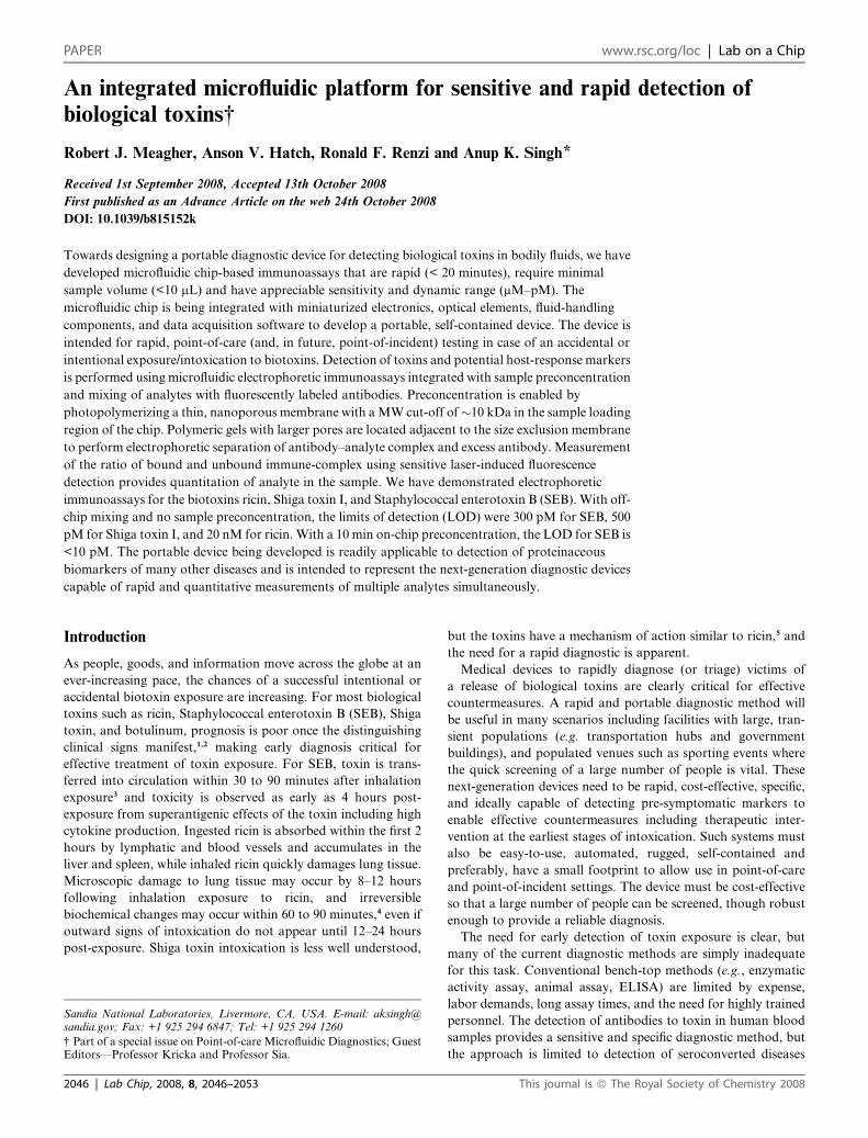
response curves for SEB, Stx1, and ricin are shown in Fig. 3. The
SEB and Stx1 assays give the expected sigmoidal shape, which
can be fit to a logistical curve model, which is commonly used to
model dose–response behavior with saturation at high
Lab Chip, 2008, 8, 2046–2053 | 2049
Fig. 3 Dose–response curves obtained for Staphylococcal enterotoxin B, Shiga Toxin 1, and Ricinus Communis Agglutinin II using electrophoretic
immunoassays with off-chip mixing. Normalized response refers to the peak area or height of the antibody peak at a given toxin concentration, relative
to the peak area or height with no toxin present. The normalized response for SEB and Shiga are based on antibody peak area, whereas the response for
ricin is based on the antibody peak height. Error bars refer to the standard deviation of at least three runs. The solid curves are four-parameter logistical
curve fits.
concentration. For the ricin immunoassays, the antibody peak
and complex peak overlapped significantly, with long tails and
were difficult to model accurately with simple peak shapes. In
this case, the ratio of antibody peak height to the internal stan-
dard (BSA) peak height provided the best correlation to the ricin
concentration. Each of the assays reveals a different dynamic
range for detection, based on different affinities of the antibodies
for their respective toxins. However, the preconcentration
method described next allows us to achieve clinically relevant
limit-of-detection with less dependence on the binding constant
of antibodies.
Fig. 4 Dose–response curves obtained for Staphylococcal enterotoxin B
immunoassays with and without on-chip preconcentration and incuba-
tion. The solid curves are four-parameter logistical curve fits. The 10
minute preconcentration protocol shifts the dose–response curve �2
orders of magnitude lower in concentration, with a larger dynamic range
indicated by a more gradual downslope.
Immunoassays with preconcentration
Low nanomolar detection limits are not always adequate for
screening of biotoxins that may be lethal at picomolar or even
lower concentrations. Taking advantage of our ability to pho-
topattern polymeric elements,44,45 we previously developed
a novel on-chip preconcentrator42 that uses a size-exclusion
membrane fabricated in a channel to concentrate proteins bigger
than the MW cut-off (� 10 kDa) of the membrane. Two poly-
meric elements—a thin (�50 mm) size-exclusion membrane for
preconcentration and a longer (�2 cm) porous monolith for
protein separation—were fabricated by in situ photo-
polymerization. Contiguous placement of the different polymeric
elements in the channels of a microchip enabled simple and zero-
dead volume integration of the preconcentration with PAGE.
Proteins are loaded through a low-concentration polyacrylamide
gel, which does not significantly retard proteins in this size range,
but which provides an important anti-convective effect. In
addition, the low-concentration polyacrylamide loading gel
excludes particles and very large macromolecules present in
bodily fluids from entering the microchannels. In an immuno-
assay with preconcentration, protein biotoxins (as well as other
proteins in the sample) in the sample are trapped and concen-
trated in a narrow zone on the upstream side of the size-exclusion
membrane. In a second step, detection antibodies are trapped
and concentrated in the same narrow zone on the upstream side
of the membrane. The extent of protein and antibody pre-
concentration is easily tuned by varying the duration of
2050 | Lab Chip, 2008, 8, 2046–2053
preconcentration, or by controlling the sample volume loaded.
During the preconcentration step, a continuous gravity-driven
flow of fresh buffer past the downstream side of the membrane
counteracts ion depletion or concentration polarization effects.
Upon preconcentration, both the antibody and toxin are co-
localized in a small volume (axial span <100 mm, volume �10–
100 fL) at high concentration, and the immunocomplex forms
within seconds. Upon switching the direction of the electric field,
the concentrated proteins are directed into the crosslinked
polyacrylamide separation gel for electrophoretic analysis.
The electropherograms obtained with preconcentration
appear similar to those obtained with off-chip mixing (e.g.
Fig. 2), with an antibody peak and a complex peak along with an
internal standard peak. The immunoassay with preconcentration
offers simplified operation (by eliminating off-chip mixing steps),
as well as significantly lower limits of detection. Fig. 4 illustrates
the dose–response curve obtained for SEB with and without
preconcentration. Preconcentration evidently offers greater than
This journal is ª The Royal Society of Chemistry 2008

100-fold improvement in the limit of detection for this toxin, into
the low picomolar range.
With detection limits <10 pM, the immunoassay with pre-
concentration compares favourably to current ‘‘gold-standard’’
approaches such as ELISA for direct detection of toxins. Sample
volume requirements for the microfluidic immunoassay are
significantly reduced compared to a conventional ELISA. The
sample volume that is processed in a single preconcentration
assay is typically on the order of the swept volume of the channel
between the sample well and the preconcentration membrane
(�20 nL) and thus several replicates of the assay can theoretically
be performed with <1 mL of sample. The primary volume
requirement is simply that enough liquid be present in each
reservoir to fully immerse the electrode, and for the liquid in the
reservoir to make electrical contact with the microchannel;
typically this requires <10 mL of sample. The microchip immu-
noassay with on-chip preconcentration and mixing is at least an
order of magnitude faster than ELISA, requiring <20 minutes
per sample, with no user intervention required, apart from
addition of reagents to fluidic reservoirs on the chip. This speed
of detection is of critical importance for toxins such as SEB,
which exhibit toxicity in less time than it takes to complete
a typical ELISA. Since the preconcentration assay essentially
consists of a series of simple electrophoretic steps, with no
moving parts (valves, etc.), it represents a good candidate for
implementation onto a multichannel array device, to achieve
throughput that is competitive with assays performed in a multi-
well microtiter plate.
Immunoassays in the current study were performed with two
spectrally distinct fluorescent labels (Alexa Fluor 546 and Alexa
Fluor 647), using two separate LIF setups. Thus far, the choice of
fluorescent label has had little impact on performance of the
immunoassay. Multiplexed immunoassays using multiple detec-
tion antibodies are possible using spectrally resolved fluorescence
detection, which can readily be achieved with multiple inexpen-
sive solid-state excitation sources, and multiple PMTs or CCD
array-based detectors.
Fig. 5 Prototype of the portable device for toxin diagnostics in a clear
acrylic case showing internal components. The device is approximately 23
cm wide � 20 cm deep � 13 cm tall, and is powered by a conventional
12V–1A DC power adapter (not pictured).
Selection of antibodies for immunoassays
Similar to ELISA or any other antibody-based assay, the selec-
tion and validation of the antibody is of the utmost importance
in an electrophoretic immunoassay. Multiple antibodies are
available for many toxins from several vendors; few are sold with
any reliable data regarding the avidity of the antibody for the
toxin, or the specific part of the toxin targeted by the antibody.
Advances have been made in characterization of monoclonal
antibodies, particularly those designed for human therapeutics,46
but vendors of antibodies for in vitro diagnostics provide greatly
different levels of quality control, sometimes with substantial lot-
to-lot variability. Hence, we devoted a significant amount of
effort and resources in testing and qualifying antibodies from
many different vendors. This issue of quality control is not
trivial—once an immunoassay is validated with a particular
antibody, a reliable, large-scale source of that antibody must be
identified before a portable diagnostic device can be widely
distributed. Our preference is for monoclonal antibodies, as these
provide better lot-to-lot consistency, and uniform (if not neces-
sarily strong) binding of the target in 1 : 1 or 1 : 2 complexes.
This journal is ª The Royal Society of Chemistry 2008
Polyclonal antibodies can be used as well, as demonstrated here
for ricin, for which an appropriate monoclonal antibody was not
identified among several commercially available candidates
tested. Polyclonal antibodies may bind to the target in large,
inhomogeneous complexes, which in an electrophoretic immu-
noassay may lead to a wide or tailing complex peak.
Development of a portable device for toxin detection
In parallel to the devlopment of immunoassays for multiple
biological toxins, we have been developing an integrated device
that will be used for multi-analyte toxin diagnostics. Fig. 5 shows
the photograph of an early prototype being developed. The
prototype incorporates electronics, optics, and chip architecture
to enable multiplexing for higher throughput, which is critical for
diagnostics following a suspected mass release of a toxin, with
associated public demand for medical services placing a strain on
medical infrastructure. The device is roughly the size of a desktop
phone with dimensions of 23 cm wide � 20 cm deep � 13 cm tall,
and weighs 5 lb. A touch-screen LCD provides for an improved,
user-friendly interface, and an in-house designed miniaturized
lock-in amplifier (approximately 3 cm � 4 cm � 12 cm) provides
an estimated order-of-magnitude improvement in sensitivity for
the LIF detection. Two excitation lasers and two detection
channels allow simultaneous detection of antibodies labeled with
spectrally distinct fluorophores. A disposable assay cartridge has
been developed, with the chip, reagents, and electrodes packaged
together, for one-step, snap-in-place replacement of the chip and
reagents. The cartridge is amenable to low-cost mass production
by injection molding. A miniaturized valve prevents pre-mixing
of reagents with the on-chip gel during storage. An in-house-
designed miniaturized syringe pump and microfluidic solenoid
valve can be included for pressure-driven flow for on-board
sample pretreatment. We plan to incorporate chromatographic
removal of high-abundance serum proteins from blood samples
into the portable device, although this technology is still under
development. The biotoxin immunoassays presented in the pre-
ceeding section were performed using several components
designed for the portable device (power supplies and high voltage
Lab Chip, 2008, 8, 2046–2053 | 2051

boards, electronics, optics, and software), but not packaged in
the compact format pictured in Fig. 5. The miniaturized high
voltage boards and the LIF optical system have been described in
detail previously.39,40
We note that several other research groups have presented
innovative strategies for rapid, direct detection of biotoxins in
bodily fluids, with sensitivity reported in the femtomolar to
nanomolar range. These strategies include micropatterned anti-
body arrays,47 DELFIA,48 immuno-PCR,49 and a bead-based
displacement immunoassay.34 To our knowledge, no detection
platform (ELISA or other strategy) is currently in widespread use
for point-of-care detection for biotoxin exposure.
The portable device for point-of-care testing for toxin expo-
sure needs to incorporate the following characteristics
(1) Accepts a readily available sample (blood serum, etc.), with
minimal sample handling or preprocessing outside of the device.
(2) Small volume requirement, including the sample and all
buffers, reagents, and waste streams.
(3) Capability for detection of multiple analytes.
(4) Simple user interface and autonomous control for sample-
to-answer capability with minimally trained personnel.
(5) Rapid results allowing a speedy diagnosis and proactive
treatment.
(6) Low cost per analysis, allowing testing of a large pop-
ulation of potential victims.
Manufacturability is a key concern for developing a low-cost
device. In developing our immunoassays, the in situ gels are
fabricated in small batches, 2–4 chips at a time. Laser patterning
of the membrane is robust and reliable, and is successful nearly
every time. Fabrication of the rest of the gel is less reliable when
these devices are made in small batches. The majority of failures
result from bubble formation in the gel during photo-
polymerization. The failure rate depends on the skill of the
individual performing the fabrication, and ranges from 10–25%.
In a larger scale manufacturing process, rigorous control over the
gel polymerization (oxygen-free processing, precise control of
temperature and radiation exposure, etc.) is expected to improve
the success rate.
Long shelf-life of chips and reagents is an important criterion
in development of our portable diagnostic device. In our expe-
rience, the gels and preconcentration membranes have remained
effective for as long as we have tried storing a chip (on the order
of 4–6 months), provided that the chip is kept wet, at 4 �C.
Ideally, however, all components of the device should have
a shelf life greater than 1 year. This includes not only the poly-
acrylamide separation gel and preconcentration membrane
within the chip, but also the detection antibodies, and any
reagents required for sample pretreatment. Refrigerated storage
(4 �C) will be required to maintain the long-term stability of the
antibodies. Several companies currently sell pre-cast poly-
acrylamide gels with shelf lives greater than 1 year at tempera-
tures from 4–25 �C. Specific details of the gel and buffer
compositions needed for long shelf life are usually proprietary,
but likely include a buffer pH close to neutral to avoid slow
alkaline hydrolysis of the polyacrylamide. Further testing and
development is still required to determine the useful life of our
device.
We are also evaluating the ability of our chips to analyze
multiple bodily fluids. While blood serum will remain the
2052 | Lab Chip, 2008, 8, 2046–2053
primary sample, we also plan to evaluate nasal swabs as they may
provide sufficient traces of toxin for detection following an
inhalation exposure. A variety of methods are being evaluated
for pretreatment of both blood and nasal swab extract, and we
are currently working toward integrating these techniques into
our prototype device to minimize the amount of time-consuming
off-chip sample handling on a potentially infectious blood
sample. Effective depletion of high-abundance serum proteins
(albumin, immunoglobulins, etc.) is particularly important for
assays based upon preconcentration of a low-abundance target,
because all proteins larger than the molecular weight cutoff of
the membrane (�10 kDa) are concentrated simultaneously
(although not generally at the same rate). Thus, even a serum
sample that has been depleted of a large amount of albumin may
still prove problematic if the trace of remaining albumin is
concentrated 100-fold. Commercially available ion exchange and
immunodepletion kits serve as a useful starting point for assay
development, but we anticipate that several novel approaches
currently being developed in our laboratory will prove more
compatible with the chip-based electrophoretic immunoassay,
and will be easier to integrate with the photopatterned poly-
acrylamide gel architecture.
With a footprint less than 500 cm2, the portable diagnostic
device can sit on a countertop in a clinician’s office. The device is
constructed with interlocks and engineered controls to minimize
the possibility for exposure to electrical, laser, chemical, and
biological hazards. These features allow the device to be
deployed in a relatively uncontrolled atmosphere such as a clinic
or a remote location. The small footprint would also allow the
device to be deployed in a highly controlled environment such as
a biosafety 3 or 4 laboratory where space is at a premium.
Concluding remarks
Biological toxins and pathogens pose a serious threat to our
society—mostly in the form of accidental exposure or bio-
terrorism incidents. To rapidly diagnose and treat people in
suspected incidents, there is an urgent need for miniaturized
devices that can detect the presence of toxins rapidly and sensi-
tively with low false positives. We are currently testing a new
portable microscale platform to enable rapid electrophoretic
immunoassays for biotoxins, including on-board pretreatment of
blood serum samples. The immunoassays presented here for
SEB, Shiga Toxin, and ricin form the basis for a sensitive,
portable diagnostic that can be rapidly deployed in case of mass
exposure of a large population to a biotoxin weapon.
Acknowledgements
We thank Mark Claudnic for assistance with design and
construction of the portable diagnostic prototype, and Dan
Throckmorton, Amy Herr (currently at UC Berkeley), and Steve
Binder (Bio-Rad Laboratories, Hercules, CA) for insightful
discussions. This work was supported by the National Institute
of Allergy and Infectious Disease Grant NIAID U01AI075441
(PI: Singh). Sandia is a multiprogram laboratory operated by
Sandia Corp., a Lockheed Martin Co., for the US Department of
Energy under Contract DE-AC04-94AL85000.
This journal is ª The Royal Society of Chemistry 2008

References
1 A. H. Peruski and L. F. Peruski, Clin. Diagn. Lab. Immunol., 2003, 10,506–513.
2 T. L. Pitt and N. A. Saunders, J. Clin. Pathology, 2000, 53, 71–75.3 J. N. Tseng, J. L. Komisar, R. N. Trout, R. E. Hunt, J. Y. J. Chen,
A. J. Johnson, L. Pitt and D. L. Ruble, Infection and Immunity,1995, 63, 2880–2885.
4 D. R. Franz and N. K. Jaax, in Medical aspects of chemical andbiological warfare, ed. S. Sidell, E. T. Takafuji and D. R. Franz,Office of the Surgeon General at TMM Publications, Washington,DC, 1997, pp. 631–642.
5 K. Sandvig and B. van Deurs,Annu.Rev. CellDev. Biol., 2002, 18, 1–24.6 R. G. Ulrich, S. Sidell, T. J. Taylor, D. R. Wilhelmsen and
D. R. Franz, in Medical aspects of chemical and biological warfare,eds. S. Sidell, E. T. Takafuji and D. R. Franz, Office of the SurgeonGeneral at TMM Publications, Washington, DC, 1997, pp. 621–630.
7 C. S. Effenhauser, A. Manz and H. M. Widmer, Anal. Chem., 1993,65, 2637–2642.
8 D. J. Harrison, K. Fluri, K. Seiler, Z. H. Fan, C. S. Effenhauser andA. Manz, Science, 1993, 261, 895–897.
9 S. C. Jacobson, R. Hergenroder, L. B. Koutny and J. M. Ramsey,Anal. Chem., 1994, 66, 1114–1118.
10 S. N. Brahmasandra, V. M. Ugaz, D. T. Burke, C. H. Mastrangeloand M. A. Burns, Electrophoresis, 2001, 22, 300–311.
11 A. T. Woolley and R. A. Mathies, Anal. Chem., 1995, 67, 3676–3680.12 L. Bousse, S. Mouradian, A. Minalla, H. Yee, K. Williams and
R. Dubrow, Anal. Chem., 2001, 73, 1207–1212.13 S. Yao, D. S. Anex, W. B. Caldwell, D. W. Arnold, K. B. Smith and
P. G. Schultz, Proc. Nat. Acad. Sci. USA, 1999, 96, 5372–5377.14 N. Chiem and D. J. Harrison, Anal. Chem., 1997, 69, 373–378.15 L. B. Koutny, D. Schmalzing, T. A. Taylor and M. Fuchs, Anal.
Chem., 1996, 68, 18–22.16 R. G. Nielsen, E. C. Rickard, P. F. Santa, D. A. Sharknas and
G. S. Sittampalam, J. Chromatogr., 1991, 539, 177–185.17 D. Schmalzing, W. Nashabeh, X. W. Yao, R. Mhatre, F. E. Regnier,
N. B. Afeyan and M. Fuchs, Anal. Chem., 1995, 67, 606–612.18 N. M. Schultz and R. T. Kennedy, Anal. Chem., 1993, 65, 3161–3165.19 K. Shimura and B. L. Karger, Anal. Chem., 1994, 66, 9–15.20 W. S. B. Yeung, G. A. Luo, Q. G. Wang and J. P. Ou, J. Chromatogr.
B, 2003, 797, 217–228.21 D. Schmalzing, S. Buonocore and C. Piggee, Electrophoresis, 2000, 21,
3919–3930.22 D. Schmalzing and W. Nashabeh, Electrophoresis, 1997, 18, 2184–
2193.23 A. J. Haes, B. C. Giordano and G. E. Collins, Anal. Chem., 2006, 78,
3758–3764.24 J. P. Ou, Q. G. Wang, T. M. Cheung, S. T. H. Chan and
W. S. B. Yeung, J. Chromatogr. B, 1999, 727, 63–71.25 S. B. Cheng, C. D. Skinner, J. Taylor, S. Attiya, W. E. Lee, G. Picelli
and D. J. Harrison, Anal. Chem., 2001, 73, 1472–1479.26 C. X. Qiu and D. J. Harrison, Electrophoresis, 2001, 22, 3949–3958.27 M. R. Mohamadi, N. Kaji, M. Tokeshi and Y. Baba, Anal. Chem.,
2007, 79, 3667–3672.
This journal is ª The Royal Society of Chemistry 2008
28 E. Yacoub-George, W. Hell, L. Meixner, F. Wenninger, K. Bock,P. Lindner, H. Wolf, T. Kloth and K. A. Feller, Biosens.Bioelectron., 2007, 22, 1368–1375.
29 N. Christodoulides, M. Tran, P. N. Floriano, M. Rodriguez,A. Goodey, M. Ali, D. Neikirk and J. T. McDevitt, Anal. Chem.,2002, 74, 3030–3036.
30 A. Hatch, A. E. Kamholz, K. R. Hawkins, M. S. Munson,E. A. Schilling, B. H. Weigl and P. Yager, Nat. Biotechnol., 2001,19, 461–465.
31 S. K. Sia and G. M. Whitesides, Electrophoresis, 2003, 24, 3563–3576.32 J. M. Yang, J. Bell, Y. Huang, M. Tirado, D. Thomas, A. H. Forster,
R. W. Haigis, P. D. Swanson, R. B. Wallace, B. Martinsons andM. Krihak, Biosens. Bioelectron., 2002, 17, 605–618.
33 J. Moorthy, G. A. Mensing, D. Kim, S. Mohanty, D. T. Eddington,W. H. Tepp, E. A. Johnson and D. J. Beebe, Electrophoresis, 2004, 25,1705–1713.
34 A. J. Haes, A. Terray and G. E. Collins, Anal. Chem., 2006, 78, 8412–8420.
35 L. J. Kricka, J. Clin. Ligand Assay, 2002, 25, 317–324.36 A. E. Herr, A. V. Hatch, W. V. Giannobile, D. J. Throckmorton,
H. M. Tran, J. S. Brennan and A. K. Singh, Ann. N. Y. Acad. Sci.,2007, 1098, 362–374.
37 A. E. Herr, A. V. Hatch, D. J. Throckmorton, H. M. Tran,J. S. Brennan, W. V. Giannobile and A. K. Singh, Proc. Nat. Acad.Sci. U. S. A., 2007, 104, 5268–5273.
38 D. Lindner, Lab Chip, 2001, 1, 15N–19N.39 J. A. Fruetel, R. F. Renzi, V. A. VanderNoot, J. Stamps, B. A. Horn,
J. A. A. West, S. Ferko, R. Crocker, C. G. Bailey, D. Arnold,B. Wiedenman, W. Y. Choi, D. Yee, I. Shokair, E. Hasselbrink,P. Paul, D. Rakestraw and D. Padgen, Electrophoresis, 2005, 26,1144–1154.
40 R. F. Renzi, J. Stamps, B. A. Horn, S. Ferko, V. A. VanderNoot,J. A. A. West, R. Crocker, B. Wiedenman, D. Yee andJ. A. Fruetel, Anal. Chem., 2005, 77, 435–441.
41 J. C. Stachowiak, E. E. Shugard, B. P. Mosier, R. F. Renzi,P. F. Caton, S. M. Ferko, J. L. V. de Vreugde, D. D. Yee,B. L. Haroldsen and V. A. VanderNoot, Anal. Chem., 2007, 79,5763–5770.
42 A. V. Hatch, A. E. Herr, D. J. Throckmorton, J. S. Brennan andA. K. Singh, Anal. Chem., 2006, 78, 4976–4984.
43 A. E. Herr, D. J. Throckmorton, A. A. Davenport and A. K. Singh,Anal. Chem., 2005, 77, 585–590.
44 S. Song, A. K. Singh and B. J. Kirby, Anal. Chem., 2004, 76, 4589–4592.
45 S. Song, A. K. Singh, T. J. Shepodd and B. J. Kirby, Anal. Chem.,2004, 76, 2367–2373.
46 K. L. Carson, Nat. Biotechnol., 2005, 23, 1054–1058.47 F. S. Ligler, C. R. Taitt, L. C. Shriver-Lake, K. E. Sapsford,
Y. Shubin and J. P. Golden, Anal. Bioanal. Chem., 2003, 377, 469–477.
48 A. H. Peruski, L. H. Johnson and L. F. Peruski, J. Immunol. Methods,2002, 263, 35–41.
49 W. L. Zhang, M. Bielaszewska, M. Pulz, K. Becker, A. W. Friedrich,H. Karch and T. Kuczius, J. Clin. Microbiol., 2008, 46, 1292–1297.
Lab Chip, 2008, 8, 2046–2053 | 2053