Analytical Determination of the Distribution of Polishing Time over the Surface of Polished Tiles
Upload
independentCategory
view
2download
0
15 www.ecmjournal.org
JS Hayes et al.. Surface polishing of TAN intermedullary nailsEuropean Cells and Materials Vol. 18 2009 (pages 15-26) ISSN 1473-2262
Abstract
Fractures of the tibia and femoral diaphysis are commonlyrepaired by intra-medullary (IM) nailing. Currently IM nailsare available in either electropolished stainless steel (SS)or in Titanium-Aluminium-Niobium (TAN). After healing,removal of the nails still is common but removal of TANIM nails often has complications whereas SS IM nails ofthe same design are less often associated with problems.We believe the differences in removal are due to the abilityof TAN to promote strong bone on-growth. We havepreviously shown in vivo that polishing cortical screwsreduces removal torque and the percentage of bone-implantcontact. Therefore, we postulate that bony on-growth ontoIM nails can be reduced by means of surface polishing, forease of removal. Here we aim to compare the pull-out forcesfor removal of standard TAN (TAN-S) compared toexperimental paste polished TAN (TAN-PP) IM nails froma bilateral non-fracture sheep tibia model after 12 monthsimplantation. Histological analysis was also performed toassess tissue on-growth to the nails. We show that polishingsignificantly reduces (p=0.05) the extraction force requiredfor TAN IM nail removal. This effect in part is attributableto the distinct tissue-material reaction produced. For TAN-S nails direct bone contact was observed while for TAN-PP nails a fibrous tissue interface was noted. Since TAN ispreferred over SS for IM nailing due to superiorbiocompatibility and mechanical properties, we believethese findings could be used to recommend changes tocurrent surface technologies of intramedullary nails toreduce complications seen with nail removal especially inrapidly growing bone in children.
Keywords: Titanium-7%Aluminium-6%Niobium,Intramedullary Nails, Implant removal, Surfacemodification, in vivo.
*Address for correspondence:R.G. RichardsAO Research Institute DavosAO FoundationClavadelerstrasse 8CH-7270 Davos Platz, Switzerland
Telephone Number: +41 (0)81 414 24 41E-mail: [email protected]
# J.S. Hayes and D.I. Vos contributed equally to this paper
Introduction
Intramedullary (IM) nailing is a widely established andaccepted method in the treatment for fractures of thefemur, tibia and humerus. However, controversysurrounds the indications for its removal (Toms et al.,1996; Boerger et al., 1999; Gösling et al., 2004; Karladaniet al., 2007; Morshed et al., 2007). Removal of an IMnail in symptomatic patients is primarily due to painexperienced by the patient or infection of the device andadjacent tissues (Husain et al., 1996; Luhmann et al.,2003; Flynn et al., 2004; Hui et al., 2007). Removal canalso be due to nail intrusion into a joint, implant breakageor non union of the fracture site. A large number ofextractions still occur in asymptomatic patients, especiallypaediatric patients, in order to prevent subsequent growthdisturbances (Peterson, 2005; Simanovsky et al., 2006;Vierhout et al., 2006; Gogi et al., 2006; Mutimer et al.,2007) and also as a prophylactic measure to prevent futurecomplications in case of joint replacement (knee, hip orshoulder).
In the United Kingdom alone approximately 13% ofall active members of the British Orthopaedic Association(BOA) routinely carry out IM nail removal surgery afteruncomplicated healing (Zenios et al., 2004). Interestingly,however, is that a patient being asymptomatic was listedin the same study as a contraindication by most surgeonsfor nail removal (Zenios et al., 2004). Thus one canpostulate that advocates of nail retention arise not becausethey wholly believe there are benefits for retaining thedevice rather than that the risks and costs associated withremoval outweigh that surrounding nail retention.However, IM nail extraction in children has clearerindications. Within this subset of patients, nail removal isoften warranted to avoid growth complications such aslimb shortening and related growth disturbances(Peterson, 2005; Simanovsky et al., 2006; Vierhout et al.,2006; Gogi et al., 2006; Mutimer et al., 2007). Indeed,approximately 53% of BOA surgeons claim to routinelyremove IM nails in asymptomatic paediatric patients(Zenios et al., 2004). In a more recent survey of 60paediatric surgeons, 98% claim to routinely removedevices (AO Trauma Course–Pediatric FracturesDecember 9–11, 2008, Davos, Switzerland).
Despite the low recorded number of complicationsrelated for this procedure one issue that has arisen is theexcessive bony on-growth on nails that cause intra-operative complications. In a retrospective study of fivecases of IM nail removal, complications associated withexcessive bone on-growth were reported for all patients(Seligson et al., 1997). In only two of these cases was
AN IN VIVO EVALUATION OF SURFACE POLISHING OF TAN INTERMEDULLARYNAILS FOR EASE OF REMOVAL
J.S. Hayes1,3,#, D.I. Vos1,2,#, J. Hahn1, S.G. Pearce1, R.G. Richards1,3,*
1 AO Research Institute, Davos, AO Foundation, Davos, Switzerland2Amphia Hospital, Breda, The Netherlands
3School of Biosciences, Cardiff University, Wales, Great Britain

16
JS Hayes et al.. Surface polishing of TAN intermedullary nails
www.ecmjournal.org
nail removal achieved upon primary attempt, however,both cases resulted in subsequent minimal fractures of thesurrounding site due to difficult removals. For theremaining three cases removal was incomplete upon thefirst extraction attempt. All three cases resulted in the nailbeing re-inserted and only one of these cases subsequentlyresulted in a second successful removal attempt (Seligsonet al., 1997). Therefore, difficulties associated withexcessive bone on-growth are not only surgically relatedcomplications such as increased operative time, blood loss,patient trauma and possible re-fracture but can also producedevice-related complications such as incomplete/failedremoval, debris contamination, and implant/instrumentfailure (Husain et al., 1996; Bombaci and Gorgec, 2003).Whether it is the choice of the surgeon or the patient,fundamentally once fracture healing has occurred thefunction of an IM nail is spent. Therefore, no definitivescientific or clinical reason exists for retention of an IMnail. However, removal of titanium-6%aluminium-7%niobium (TAN) IM nails often has complicationswhereas electropolished stainless steel (SS) IM nails ofthe same design are less often associated with removalproblems (Im and Lee, 2003; Woodruff et al., 2003;Gösling et al., 2005). Since nail design and geometry aresimilar between these two nail types, we believe many ofthe problems to be surface related.
Previous in vitro work by our laboratory (Baxter et al.,2002; Meredith et al., 2007a; Meredith et al. 2007b; Hayeset al., 2007) and others (Boyan et al., 2001; Schneider etal., 2003) have highlighted the importance of surfacemicrotopography in determining cell phenotype.Specifically we have shown that the surface of an implantcan directly influence fibroblastic (soft tissue) andosteoblastic (hard tissue) behaviour (Baxter et al., 2002;Biggs et al., 2007; Meredith et al., 2007a; Meredith et al.2007b; Hayes et al., 2007). In terms of hard tissue responsewe have shown that smoother surfaces compared to theclinically available ‘standard’ micro-rough counterpartshold potential for reducing bony over-growth by essentiallyreducing osteoblast ‘specific’ genotypic expression viaalterations in the cell shape and cytoskeletal organization(Hayes et al., 2008). We have also observed this effecttranslated to a tissue level in vivo, where the importanceof the surface characteristics of clinically used devices werefound to directly control specific tissue outcomes (Welton,2006; Pearce et al., 2008; Schlegel et al., 2009).
With these points in mind we believe that the removaldifficulty reported for TAN IM nails is due to the excellentability of this material to promote strong bone on-growth.Indeed the difficulties encountered during nail removaldue to excessive bony on-growth have been reported(Seligson et al., 1997) but few feasible solutions have beenput forth and experimental data remains restricted toanimals (Miettinen et al., 1992) and human cadavericsamples (Roure et al., 1999). Nevertheless, TAN remainsthe preferred choice over SS IM nails partly due to issuesrelated to SS pertaining to toxic metal ion release (Case etal., 1994; Hanawa, 2004) with the principal constituentsof SS, chromium, cobalt and nickel, being the main culprits,mechanical inferiority under cyclic loading (TAN provides
lower stiffness with higher fatigue strength compared toSS) (Perren et al., 2001; Eschbach et al., 2001), imagingdisadvantages such as increased black spot artefacts formagnetic resonance imaging (Pohler, 2000; Eschbach,2003) and second-rate biocompatibility (Massone et al.,1991; Nichols & Puleo, 1997). Therefore, in an attempt toaddress this issue we hypothesise that a smooth surfacecreated by polishing of standard micro-rough TAN nailswill help to minimise the nail removal problems associatedwith excessive bone-on-growth and will require lower pullout forces than the standard nails with rough TAN surfaces.To this end, this study is aimed primarily at assessing theeffect of polishing on the pull out forces required for theremoval of standard TAN and paste polished TAN IM nailsin a bilateral non-fracture sheep tibia model after a 12month implantation. Prior to this evaluation, an in vivoproof of concept study was carried under similar conditionsto test the pull-out force and histological response of SSversus standard TAN IM nails.
Materials and Methods
Surface CharacterisationCommercially available Synthes® 9.5mm UniversalHumeral Nails (UHN) made of shot-peenedelectropolished stainless steel (SS) (ISO 5832/1) ortitanium-6%aluminium-7%niobium (TAN) (ISO 5832/11)with either a standard micro-rough surface (TAN-S) or anexperimental paste-polished surface (TAN-PP) were used.Paste polishing is a mechanically abrasive technique usedto reduce the surface microtopography of a material.
Samples were characterised with profilometry,scanning electron microscopy (SEM), contact angle andX-ray photoelectron spectroscopy (XPS; Meredith et al.,2007a; Meredith et al., 2007b). The samples wereultrasonically cleaned with ethanol for 10 min before beingair-dried for analysis. Implant surface topographycharacterisation of each nail was assessed quantitativelyusing a non-contact white light FRT MicroProf® (FriesResearch & Technology, Bergisch Gladbach, Germany)on 3 test nails which were not implanted. A roughnessaverage (Ra – arithmetic mean of the roughness heightexpressed in micrometers) was measured from a 1x1mmscan area with a point density of 1000 points/line. Themorphology of the nails surfaces were examined using aHitachi S4100 field emission SEM (Hitachi High-Technologies, Krefeld, Germany). The images were takenin backscattered electron mode with an accelerating voltageof 5 kV. Surface chemical analysis was carried out withXPS. All spectra were recorded on a Kratos Axis Nova(Kratos Analytical, Manchester, UK) usingmonochromated Al Kα radiation (1486.69 eV). Forquantification survey scans with a step width of 0.5 eVwere performed on two spots of 300 × 700 μm2 per sample.Data were evaluated with CasaXPS 2.3.10 (CasaXPS Ltd,UK; www.casaxps.com) using relative sensitivity factorssupplied by the manufacturer. Hydrophobicity wasevaluated with static contact angle measurements(Andrade, 1985) using the Sessil drop method with the

17 www.ecmjournal.org
JS Hayes et al.. Surface polishing of TAN intermedullary nails
Drop Shape Analysis System (Contact Angle MeasuringInstrument G10 and DSA 10 Control Unit, Krüss GmbH,Hamburg, Germany) and analysed using the Drop ShapeAnalysis 1.50 software (Krüss). Samples were placed in apreheated chamber (20°C) with an external water sourceto sustain a constant relative humidity. Using the computercontrolled system a 20μl droplet of distilled water wasdispensed onto the sample and quantified exactly 1 minuteafter dispensed using the analysis software.
In Vivo MethodsApproval to perform this study was granted by the Cantonalanimal ethics committee (GR 5/2006). Fourteen adultfemale Swiss Alpine sheep were selected from a group ofSwiss Alpine sheep subsequent to radiological analysisbeing preformed on both tibiae to control length anddiameter of the tibia. Using a bilateral non-fracture modelseven sheep were used to provide proof of concept bycomparing SS versus TAN-S IM nails implanted withincontralateral tibiae. To assess the influence of polishingon IM nail pull-out force and subsequent material-tissueinteraction another seven sheep were then implanted withan S-TAN nail in one tibia and a PP-TAN nail in thecontralateral tibia. During the course of the surgeries,within our proof of concept group (SS versus TAN-S),nails were locked proximally and distally in orthogonalplanes using a 5.0 mm locking screw made from the samematerial and surface treatment as the nail. Locking wasfacilitated using a custom made aiming device attachedby means of the threads in the proximal aspect of the nailthat were used for attaching the insertion device and theendcap. However, observations in this initial studyindicated that due to the close fit of the human nail inseveral anatomical areas of the reamed sheepintramedullary canal two interlocking screws wereconsidered to be redundant to prevent migration of the nailand to avoid fracture of the highly reamed bone. Therefore,in the interest of reducing surgical time for the sheep andto minimise the chance of fracture the distal locking screwwas not used in the experiment containing the S-TANversus PP-TAN groups.
Animals were anaesthetised (diazepam (Valium ®) 0.3mg/kg i.v. and butorphanol (Morphasol®) 0.08 mg/kg i.v.;thiopenthal (Penthotal®) 4-8 mg/kg i.v.; isoflurane (approx2% in oxygen: oxygen flow rate was approx 0.5 L/min)),and placed in the appropriate lateral recumbency such thatthe operated limb was dependent (the animal was rotatedinto the opposite recumbency for operation of the secondlimb). Both tibiae were prepared for aseptic surgery.Surgery for both groups of sheep involved a 5 cm incisionbeing made on the medial aspect of the patellar ligamentof the stifle joint for both hind limbs. Subsequently, thejoint was flexed approximately 90 degrees in order to gainaccess to the proximal tibia. Approximately half waybetween the patellar tendon and the cranial edge of themedial femoral meniscus a hole was drilled into the jointsurface of the proximal tibia. The tibia was reamed inincrements of 0.5 mm to a maximum diameter of 11 mmwith a rigid reamer in order to create a more uniformamount of contact between the nail and tibial cortex. The
tibia plateau was reamed to 12 mm to ease nail insertion.A human 9.5 mm diameter UHN (humeral nail) wasinserted (with hammering) into the tibia through anextraarticular approach and a proximal locking bolt wasinserted via a small incision through the skin and finallyan endcap was placed at the proximal site of the nail.
The subcutaneous tissue was closed with absorbablesuture material and the skin was closed with stainless steelskin staples. The limbs were bandaged with cotton wooland an adhesive bandage which was left in place for 5-7days or less if believed to be causing irritation to the animal.Post-operatively analgesia consisted of carprofen (4mg/kg subcutaneous, q24h for 3 days, Rimadyl®, Pfizer, NewYork, NY, USA) and buprenorphine (0.01mg/kgsubcutaneous, q12h for 3 days, Temgesic®, ESSEXChemie AG, Luzern, Switzerland). Immediately post-surgery and for the first week of the observation period allanimals were housed in individual boxes in protectionslings that allowed full weight bearing on the legs, but notreclining. No complications were encountered during orsubsequent to surgery. Each sheep was evaluated every 4weeks clinically for the duration of the study and every 8weeks radiographs were taken in two plains latero-medialand cranio-caudal to monitor the nail position stability andany visible signs of bone on-growth. The nails were leftin place for a total implantation time of 12 months. Theanimals were euthanized by means of an intravenousoverdose of barbiturate (Pentobarbital, Vetanarcol® 60 mg/kg).
Figure 1. Testing Device, with adapter screwed intothe endcap holder of the nail within the tibia beforepull out with was performed with a constant force

18
JS Hayes et al.. Surface polishing of TAN intermedullary nails
www.ecmjournal.org
Mechanical TestingAfter euthanasia 6 sheep from each the proof of conceptstudy and also from the subsequent main study were usedfor the pull-out tests. The tibiae were excised, adjacentsoft tissues removed and the tibia plateau was cut just toabove the proximal position of the nail. Distally a 6 mmdrill hole was made 2 cm from the distal central end andthe endcap was removed to allow attachment to the testingdevice (Fig. 1). The locking bolts were removed from thenails after fixing the tibia in the pull-out machine.Subsequent pull-out tests were performed on these nailsusing a mechanical testing machine Instron 5866 (InstronInc, High Wycombe, UK) with a 10kN load-cell and acustom made pull-out device. All nails were removed witha constant force (1mm/min). Load- and displacement-datawas recorded with a sampling rate of 50Hz. Data wereevaluated using Matlab® software (V6.5, Mathworks Inc,Natick, MA, USA) with a custom made procedure. In onesheep from each group the nail remained in situ withoutmechanical tests and was used for histomorphometricanalyses.
Histological Observations After euthanasia one nail from each group was left in situ.The whole intact tibia containing the nail was fixed in 70%methanol for approximately 9 months. Subsequentlydehydration of the samples was achieved by immersingthe samples in increasing concentrations of ethanol (70%,96%, and 100%). Samples were then placed in xylol priorto being embedded with methylmethacrylate. Sectionswere cut with an annular saw, ground (Exact MicroGrinding System,Little Rock, AR, USA) and polished toa thickness of approximately 100μm and stained withGiemsa and eosin. Light microscopic images were takenusing a Zeiss Axioplan microscope (Carl Zeiss, Göttingen,Germany).
Statistical AnalysisStatistical evaluation was performed using SPSS forWindows Version 14.0 (SPSS Inc, Chicago, Illinois, USA).Initially to provide proof of concept the SS versus TAN-Sgroup were evaluated statistically using Wilcoxon signedranks test given that the data for this group had a non-Gaussian distribution. Subsequently, within our main groupthe effect of polishing with a paired model (TAN-S versus
TAN-PP) was assessed using a paired t-test, p-values ≤0.05were deemed significant.
Results
Surface characterisationNon-contact profilometry revealed that the nails made ofstandard micro-rough TAN (S-TAN), shot-peenedelectropolished stainless steel (SS) and paste-polished TAN(PP-TAN) used in this study had an average surfaceroughness of 0.98μm, 0.58μm and 0.18μm, respectively.The XPS results delineating the atomic concentration (%)of elements of the nail surface were for S-TAN: Al 2.1; C26; N 0.9; Na 0.5; Nb 0.2; O 50.9; P 2; Ti 17.2 and for PP-
Figure 2. SEM images of (A) S-TAN showing a rough undulating surface with numerous micro-discontinuities (asused in clinics), (B) PP-TAN showing a smoothened surface with very few micro-discontinuities, (C) SS with asmooth, yet undulating surface (standard shot-peened surface, as used in clinics).
Figure 3. Pullout test results for the proof of conceptstudy comparing S-TAN and SS nails in a paired testwithin individual sheep. As expected, SS IM nails weresignificantly easier to remove compared to S-TAN IMnails (p=0.028). For two sheep in this group, the pulloutforce was very low in both tibiae. While no statisticalcorrelation was found between intramedullary canalwidth, and pull-out force (S-TAN p=0.345; SS p=0.133),it was observed radiologically that the two specimensin question had a wider medullary canal than the otherspecimens, which will clearly influence the forcerequired for nail removal.

19 www.ecmjournal.org
JS Hayes et al.. Surface polishing of TAN intermedullary nails
TAN: Al 2.4; C 27.6; N 1.1; Na 0.6; Nb 0.1; O 49; P 2.5;Ti 16.5. Therefore polishing of the TAN nails followed byanodisation (PP-TAN) did not alter the chemical propertiesof the surface compared to S-TAN controls which werealso anodised. Contact angle measurements gave anindication of the wettability (hydrophobic or hydrophilic)of a surface which also affects cell and tissue adhesion.Results were 78.3±6.6 for SS, 64.2±7.7 for S-TAN and64.1±2.9 for PP-TAN. SEM imaging clearly showed thecharacteristic β-phase niobium-rich inclusions associatedwith the micro-spiked appearance of the S-TAN (Fig. 2A).Paste polishing (Fig. 2B) clearly reduced themicrotopography of the surface successfully to the extentthat the niobium rich inclusions were not easily observed.Imaging of stainless steel (Fig. 2C) highlighted theimportance of using various techniques for characterisingsurfaces. While non contact profilometry assesses thesurface as being relatively rough (Ra 0.58μm), SEMimaging demonstrated that the actual surface was smooth
with an undulating morphology; however, the wavinessencountered on areas of the surface would contribute tothe higher average roughness observed.
Mechanical TestingTo provide proof of concept mechanical pull-out tests ofthe SS nails and S-TAN nail from the same sheep wereperformed. Results demonstrated that as expected the SSnails had a significantly lower (p=0.028) pullout force thanthe S-TAN nails (Fig. 3). For two sheep in this group thepullout force was very low in both tibiae. While nostatistical correlation was found between intramedullarycanal width and pull-out force (S-TAN p=0.345; SSp=0.133) it was observed radiologically that the twospecimens in question had a much wider medullary canalthan the other specimens which will clearly influence theforce required for nail removal.
In our main group (TAN-PP versus TAN-S) designedat testing the influence of surface polishing of TAN IMnails mechanical pullout tests of the experimental PP-TANnails also demonstrated a markedly lower pullout forcecompared to the S-TAN nail implanted in the same(p=0.05) (Fig. 4). However within this group twoexceptions were noted. Firstly, a higher pullout force wasnoted for one PP-TAN (Fig. 4) which was revealed to beslightly higher than the pull out force required for thecontra-lateral tibia containing the S-TAN IM nail.Radiologically, it was noted that there was a corticalreaction for both tibiae containing PP-TAN and S-TANnails. Additionally, during testing of the PP-TAN IM naildue to the tight fit of the nail to the medullary canal of thissmall sheep (over 200 sheep were examined radiologicallyhowever some were clearly smaller) the bone broke (Fig.5A,B). Again no statistical correlation was observedbetween medullary canal width and pull-out force for PP-TAN IM nails (p=0.323); however, radiologically thedifferences in medulla width were evident. Secondly, pullout forces of PP-TAN and S-TAN from a different sheepproduced similar results for both nails. Radiologicalanalysis as well as analysis by eye after the pull out testsshowed bony in-growth of the lower distal hole of boththe PP-TAN and S-TAN nails (Fig. 5C) so that a cylinderof bone of equal volume had to be broken for both nailsaccounting for the similar pull forces noted. Therefore theresults for the pullout test were a reflection of the force tobreak a cylinder of the bone in the hole rather than on theforce required to remove the nail. The data from this sheepwere excluded from the statistical analysis.
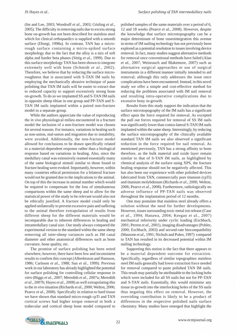
Bone was not observed to grow directly onto thesurfaces of the nail interlocking holes of the polished nailsand any tissue within the screw holes was easily displacedwith a K wire (Fig. 6A). In contrast, within the interlockingholes of standard TAN nails bone on-growth onto thesurface of the holes was evident and this hard tissue couldnot be displaced (Fig. 6B). Bone was also observed on thecutting flutes at the tip of the standard TAN locking boltswhereas for paste polished TAN locking bolts and stainlesssteel ones of the same design bone tissue adhesion wasnot observed
Figure 4. Pullout test results of the main study groupcomparing S-TAN and PP-TAN nails in a paired testwithin individual sheep. Results indicate that reducingthe surface microtopography of TAN IM nails with pastepolishing can significantly ease removal compared toS-TAN IM nails (p=0.05). Two exceptions were noted.Firstly, a higher pullout force was noted for one PP-TAN, (denoted with purple dashed line) compared tothe contra-lateral tibia containing the S-TAN IM nailwhich was due to the tight fit of the nail to the medullarycanal. Consequently the bone broke upon explantation.While no statistical correlation was observed betweenmedullary canal width and pull-out force for PP-TANIM nails (p=0.323), radiologically the differences inmedulla width was evident. Secondly, results from adifferent sheep produced similar pullout forces for bothnails (denoted by blue dashed line). Radiologicalanalysis showed bony in-growth of the lower hole ofboth the PP-TAN and S-TAN nails, so that a cylinderof bone of equal volume had to be broken for both nails,accounting for the similar pull forces noted. The datafrom this sheep were excluded from the statisticalanalysis.
20
JS Hayes et al.. Surface polishing of TAN intermedullary nails
www.ecmjournal.org
Histological ObservationsIn our proof of concept study that compared SS IM nailsto TAN-S IM nails we observed direct bone bonding tothe nail for TAN-S samples. Furthermore, direct bonecontact to TAN-S was noted between the nail and theinterlocking screw (Fig. 7A). In contrast, SS IM nails hada consistent fibrous layer around the circumference of theentire nail separating the nail from direct bone contact.This layer was observed to be only a few cell layers thick(Fig. 7B).
In our main study group aimed at testing the influenceof surface polishing for ease of removal we noted that thedirect bone bonding observed for TAN-S IM nails was
not evident for TAN-PP IM nails. Similar to observationsmade in the previous group for SS IM nails, TAN-PP IMnails had a fibrous layer separating the implant from directbone contact. However, it was noted that this fibrous layerwas thicker than that noted for SS IM nails (Fig. 7C).
Discussion
To date, IM nails are fabricated for orthopaedic surgeonsfrom either TAN or SS. Yet removal of standard TAN (S-TAN) IM nails often produces more extraction relatedcomplications compared SS IM nails of the same design
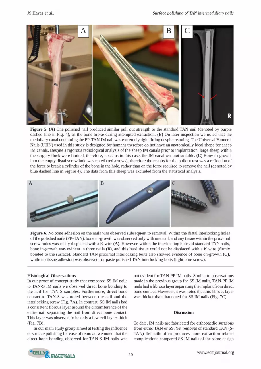
Figure 5. (A) One polished nail produced similar pull out strength to the standard TAN nail (denoted by purpledashed line in Fig. 4), as the bone broke during attempted extraction. (B) On later inspection we noted that themedullary canal containing the PP-TAN IM nail was extremely tight fitting despite reaming. The Universal HumeralNails (UHN) used in this study is designed for humans therefore do not have an anatomically ideal shape for sheepIM canals. Despite a rigorous radiological analysis of the sheep IM canals prior to implantation, large sheep withinthe surgery flock were limited, therefore, it seems in this case, the IM canal was not suitable. (C) Bony in-growthinto the empty distal screw hole was noted (red arrows), therefore the results for the pullout test was a reflection ofthe force to break a cylinder of the bone in the hole, rather than on the force required to remove the nail (denoted byblue dashed line in Figure 4). The data from this sheep was excluded from the statistical analysis.
Figure 6. No bone adhesion on the nails was observed subsequent to removal. Within the distal interlocking holesof the polished nails (PP-TAN), bone in-growth was observed only with one nail, and any tissue within the proximalscrew holes was easily displaced with a K wire (A). However, within the interlocking holes of standard TAN nails,bone in-growth was evident in three nails (B), and this hard tissue could not be displaced with a K wire (firmlybonded to the surface). Standard TAN proximal interlocking bolts also showed evidence of bone on-growth (C),while no tissue adhesion was observed for paste polished TAN interlocking bolts (light blue screw).
A B C

21 www.ecmjournal.org
JS Hayes et al.. Surface polishing of TAN intermedullary nails
Figure 7. Representative histological sections of standard micro-rough TAN (A-C), stainless steel (D-G) and pastepolished TAN (H-J) IM nails stained with Giemsa-Eosin to demonstrate soft tissue (blue) and bone (pink) attachment,after 12 months implantation. Note that with higher magnification, direct bone contact to the standard micro-roughTAN nail is evident adjacent the nail (B) and within the interlocking hole (C), despite the presence of the screw. Itwas evident for SS IM nails that a continuous fibrous layer (only a few cells thick) was present around the circumferenceof the nail (E). In higher magnification this layer clearly separates the implant from bone (white arrows) therebypreventing direct osseointegration (F (magnified area denoted by white box in E) and G). Similar to SS and incontrast to S-TAN, paste polished TAN IM nails appear to have a fibrous tissue layer separating the nail from thesurrounding bone (I-J). No issues of stability were observed for SS or PP-TAN nails. This fibrous tissue layer islikely to contribute to the reduced pull-out strength required for these nails compared to standard micro-rough TANnails
A B C
DE
FG H
I J
22
JS Hayes et al.. Surface polishing of TAN intermedullary nails
www.ecmjournal.org
(Im and Lee, 2003; Woodruff et al., 2003; Gösling et al.,2005). The difficulty in removing nails due to excess strongbone on-growth has not been described for stainless steelwhich for clinical orthopaedics is supplied with a smoothsurface (Disegi, 1998a). In contrast, TAN has a micro-rough surface containing a micro-spiked surfacemorphology due to the fact that the alloy is a mix of softalpha and harder beta phases (Sittig et al., 1999). Due tothis surface morphology TAN has been shown to integrateextremely well with bone (Schmidt et al., 2002).Therefore, we believe that by reducing the surface micro-roughness that is associated with S-TAN IM nails byemploying the mechanically abrasive technique of pastepolishing that TAN IM nails will be easier to extract dueto reduced capacity to support excessively strong boneon-growth. To do so we implanted SS and S-TAN IM nailsin opposite sheep tibiae in one group and PP-TAN and S-TAN IM nails implanted within a paired non-fracturemodel in a separate group.
While the authors appreciate the value of reproducingthe in vivo physiological milieu encountered in a fracturemodel the inclusion of a non-fracture model was chosenfor several reasons. For instance, variations in healing suchas non-union, mal-union and migration due to instability,were avoided. Additionally, the non-fracture modelallowed for conclusions to be drawn specifically relatedto a material-dependent response rather than a biologicalresponse based on variations in healing. Also, since themedullary canal was extensively reamed essentially manyof the same biological stimuli similar to those found infracture healing were evoked. Importantly, however, withinmany countries ethical permission for a bilateral fracturewould not be granted due to the implications to the animal.On top of this the increase in animal number which wouldbe required to compensate for the loss of simultaneouscomparisons within the same sheep and to allow for thestatistical power of the study to be relevant would also notbe ethically justified. A fracture model could only beapplied unilaterally to prevent excessive pain and sufferingto the animal therefore extrapolation of results fromdifferent sheep for the different materials would beincomparable due to inherent differences in healing andintramedullary canal size. Our study always compared theexperimental version to the standard within the same sheepremoving all inter-sheep variances such as IM canaldiameter and other anatomical differences such as bonecurvature, bone quality, etc.
The promise of surface polishing has been notedelsewhere, however, there have been few and inconsistentresults to confirm this concept (Albrektsson and Hansson,1986; Carlsson et al., 1988; Sun et al., 1999). Previouswork in our laboratory has already highlighted the potentialfor surface polishing for controlling cellular response invitro (Biggs et al., 2007; Meredith et al., 2007a; Meredithet al., 2007b; Hayes et al., 2008) as well extrapolating thisto the in vivo situation (Richards et al., 2000; Welton, 2006;Pearce et al., 2008). Specifically in relation to hard tissuewe have shown that standard micro-rough cpTi and TANcortical screws had higher torque removal in both atrabecular and cortical sheep bone model compared to
polished samples of the same materials over a period of 6,12 and 18 weeks (Pearce et al., 2008). However, despitethe knowledge that surface microtopography can be amajor determinant of bone osseointegration this avenuein terms of IM nailing technology has not previously beenexplored as a potential resolution to issues involving deviceremoval. In fact, many studies suggest alternative methodsfor removal once conventional methods have failed (Alanet al., 2007; Weinrauch and Blakemore, 2007) such asalternative surgical approaches or use of surgicalinstruments in a different manner initially intended to aidremoval; although this only addresses the issue oncecomplications have been encountered. Instead, in this novelstudy we offer a simple and cost-effective method forreducing the problems associated with IM nail removaland resulting intra-operative complications due toexcessive bony in-growth.
Results from this study support the indication that thesurface microtopography of the IM nails has a significanteffect upon the force required for removal. As exceptedthe pull out forces required for removal of SS IM nailswas significantly lower than contra-lateral S-TAN IM nailsimplanted within the same sheep. Interestingly, by reducingthe surface microtopography of the clinically availablestandard TAN IM nails we also observed a significantreduction in the force required for nail removal. Asmentioned previously, TAN has a strong affinity to bonetherefore, as the bulk material and oxide layer remainsimilar to that of S-TAN IM nails, as highlighted bychemical analysis of the surface using XPS, the fracturehealing response should not be negatively affected. Thishas also been our experience with other polished devicesfabricated from TAN, commercially pure titanium (cpTi)and titanium molybdenum (Richards et al., 2000; Welton,2006; Pearce et al., 2008). Furthermore, radiologically noadverse influence of PP-TAN nails was observedthroughout the implantation period of 12 months.
One may postulate that stainless steel already offers asolution without the need for further developments.However, issues surrounding toxic metal ion release (Caseet al., 1994; Hanawa, 2004; Keegan et al., 2007)mechanical inferiority under cyclic loading (Eschbach,2001; Perren et al., 2001), imaging disadvantages (Pohler,2000; Eschbach, 2003) and second-rate biocompatibility(Massone et al., 1991; Nichols and Puleo, 1997) comparedto TAN has resulted in its decreased potential within IMnailing technology.
Supporting this notion is the fact that there appears tobe a material dependent outcome for extraction.Specifically, regardless of similar topographies stainlesssteel IM nails generally had lower extraction force neededfor removal compared to paste polished TAN IM nails.This result may partially be attributable to the locking boltswhich were included for all SS nails but not for PP-TANand S-TAN nails. Essentially, this would minimise anytissue in-growth into the interlocking holes of the SS nailsthus negating this effect on removal. However, theoverriding contribution is likely to be a product ofdifferences in the respective polished nails surfacechemistry. Many studies have emerged that highlight the

23 www.ecmjournal.org
JS Hayes et al.. Surface polishing of TAN intermedullary nails
importance of the surface chemistry and in particular theimportance of the oxide layer of fixation materials.Specifically, the surface chemistry of a device has beenimplicated in the initial protein-metal interactions (Meyeret al., 1988; Degasne et al., 1999; Yang et al., 2003) and itis believed that this relationship primes the material forsubsequent cell phenotypic and genotypic performance andas a consequence ensuing tissue response. Essentially thebio-passivity of an implant is attributable to this oxide layer.Many alloying elements tend to show favourable propertiesin terms of low charged species at physiological pH withthe likes of titanium, aluminium and niobium beingaccepted by tissue as ‘inert’ materials thus do not evokeadverse tissue reactions. However, other oxides have beenreported to show either a preference to evoke a foreignbody reaction resulting in a tendency to produce a fibrouscapsule or indeed an outright toxic response (such aschromium and cobalt found in stainless steel). Iron oxide,another of the major elements found in SS, is known toinduce encapsulation through its corrosion products andtheir solubility (Textor et al., 2001) while contrastinglycpTi and its alloys are known to have a superior corrosionresistance compared to SS (Disegi, 1998b). Due to the cleardifferences in oxide thickness between SS (approximately1-2nm) and TAN (approximately 5nm) (Textor et al., 2001)the former affords itself more readily to corrosion than thelatter especially since SS takes a longer period torepassivate thus inadvertently allowing increased time overTAN for metal ion release (Hanawa, 2004). The release ofions into surrounding tissue has previously been shown toplay a role in periprosthetic pathology by contributing toimplant failure by impairing bone repair while allowingfibrous tissue formation following debris-inducedosteolysis (Nichols and Puleo, 1997). Therefore, one canspeculate that the differences in surface oxide chemistrybetween SS and PP-TAN would produce differentultrastructural tissue responses that have been reportedpreviously (Albrektsson and Hansson, 1986). Histologicalobservations from this study challenge the overallassumption that direct bony integration is required forimplant stability. Indeed considering the lack of publisheddata describing direct osseointegration for stainless steelimplants this concept does not come as any great surprise.In fact we and others have published data that distinctlyshow that stainless steel implants support a thin fibrousshell between bone and the device (Albrektsson andHansson, 1986; Pearce et al., 2008; Hayes et al., 2009).Furthermore despite this distinct lack of directosseointegration for steel vast quantities of stainless steelimplants are sold annually without a significantly highernumber of failure rates compared to titanium. Interestinglyin this study we also observed indirect osseointegration ofPP-TAN samples. Throughout the 12 month implantationperiod no problems of instability were noted againsupporting the hypothesis that direct bony integration isnot absolutely necessary for implant stability.
Finally, since infection remains one of the majorlimiting factors to successful fixation the effect of polishingon infection susceptibility is of paramount importance sinceessentially this is one factor that will decide if the
application of this technology in clinics is feasible. To thisend our group have previously shown that polishing TANdiscs reduces in vitro bacterial adhesion in comparison withS-TAN (Harris et al., 2007). The effect of polishing oninfection susceptibility is currently being tested in rabbitsin vivo with these surfaces and materials.
Conclusion
Implant polishing holds great potential for IM nailingtechnology. Here we have shown that surface polishingreduces pull out forces required for nails which aresignificantly less compared to the clinically availablestandard TAN IM nails. Since TAN IM nails are generallypreferred over SS nails due to their excellentbiocompatibility and mechanical properties we believethese findings are extremely important for IM nails to helpwith insertion and removal.
References
Alan RK, Baig R, Voss FR (2007) Exchange femoralnailing: a new technique for removal of a broken nail. AmJ Orthop 36: 500-502.
Albrektsson T, Hansson HA (1986) An ultrastructuralcharacterization of the interface between bone andsputtered titanium or stainless steel surfaces. Biomaterials7: 201-205.
Andrade JD (1985) The contact angle and interfaceenergetics. Chapter 7. In: Surface and Interfacial Aspectsof Biomedical Polymers: Surface Chemistry and Physics,vol. 1 (Andrade JD, ed). Plenum Press, New York andLondon.
Baxter LC, Frauchiger V, Textor M, ap Gwynn I,Richards RG (2002) Fibroblast and osteoblast adhesionand morphology on calcium phosphate surfaces. Eur CellMater 4: 1-17.
Biggs MJ, Richards RG, Gadegaard N, Wilkinson CD,Dalby MJ (2007) The effects of nanoscale pits on primaryhuman osteoblast adhesion formation and cellularspreading. J Mater Sci Mater Med 18: 399-404.
Boerger TO, Patel G, Murphy JP (1999) Is routineremoval of intramedullary nails justified? Injury 30: 79-81.
Bombaci H, Gorgec M (2003) Difficulty in removalof intramedullary nails: The geometry of the distal end ofthe nail. Yonsei Med J 44: 1083-1086.
Boyan BD, Dean DD, Lohmann CH, Cochran DL,Sylvia VL, Schwartz Z (2001) The titanium-bone interfacein vitro: The role of the surface in promotingosseointegration. In: Titanium in Medicine (Brunette DM,Tengvall P, Textor M, Thomsen P, eds). Chapter 17, pp561-586, Springer, New York.
Carlsson L, Rostlund T, Albrektsson B, Albrektsson T(1988) Removal torques for polished and rough titaniumimplants. Int J Oral Maxillofac Implants 3: 21-24.
Case CP, Langkamer VG, James C, Palmer MR, KempAJ, Heap PF, Solomon, L (1994) Widespread dissemination

24
JS Hayes et al.. Surface polishing of TAN intermedullary nails
www.ecmjournal.org
of metal debris from implants. J Bone Joint Surg Br 76:701–712.
Degasne I, Basle MF, Demais V, Hure G, Lesourd M,Grolleau B, Mercier L, Chappard D (1999) Effects ofroughness, fibronectin and vitronectin on attachment,spreading, and proliferation of human osteoblast-like cells(SaOs-2) on titanium surfaces. Calcif Tissue Int 64: 499-507.
Disegi J (1988a) AO ASIF Wrought 18% Chromium-14% Nickel-2.5% Molybdenum Stainless steel implantmaterial. AO ASIF Technical Commission Pamphlet, 1sted.
Disegi, J (1988b) AO ASIF unalloyed titanium implantmaterial. AO ASIF Technical Commission Pamphlet, 4thed.
Eschbach, L (2003) 10 Frequently asked questionsabout magnetic resonance imaging in patients with metalimplants. AO ASIF Materials Expert Group..
Eschbach L, Bigolin G, Hirsiger W, Gasser B (2001)Low-nickel steel for small bone fragment fixation platesand screws. Proc MAEG Meeting, Lausanne, Feb 2001.
Flynn JM, Luedtke LM, Ganley TJ, Dawson J,Davidson RS, Dormans JP, Ecker ML, Gregg JR, HornBD, Drummond DS (2004) Comparison of titanium elasticnails with traction and a spica cast to treat femoral fracturesin children. J Bone Joint Surg Am 86-A: 770–777.
Gogi N, Khan SA, Varshney MK (2006) Limb lengthdiscrepancy following titanium elastic nailing in paediatricfemoral shaft fractures. Acta Orthop Belg 72: 154-158
Gösling T, Hufner T, Hankemeier S, Zelle BA, Muller-Heine A, Krettek C (2004) Femoral nail removal shouldbe restricted in asymptomatic patients. Clin Orthop RelatRes 423: 222-226.
Gösling T, Hüfner T, Hankemeier S, Richter M, KrettekC (2005) Indikation zur Entfernung von Tibiamarknägeln(Indication fort the removal of tibial marrow nails). Chirurg76: 789-794.
Hanawa T (2004) Metal ion release from metalimplants. Mat Sci Eng 24: 745-752.
Harris LG, Meredith DO, Eschbach L, Richards RG(2007) Staphylococcus aureus adhesion to standard micro-rough and electropolished implant materials. J Mater SciMater Med 18: 1151-1156.
Hayes JS, Archer, CW, Richards RG (2007) Reducingbone encasement of metal implants for elective removal.Trans 53rd ORS, San Diego, p 1630.
Hayes JS, Archer CW, Richards RG (2008) An in vitroevaluation for understanding the role of surfacemicrotopography in controlling tissue integration. ProcWorld Biomaterials Congress, Amsterdam.
Hayes JS, Seidenglanz U, Pearce SG, Archer CW,Richards RG (2009) Reducing implant removal relatedmorbidity with surface polishing – a novel in vivo study.Proc Orthopaedic Research Society, Las Vegas.
Hui C, Jorgensen I, Buckley R, Fick G (2007) Incidenceof intramedullary nail removal after femoral shaft fracturehealing. Can J Surg 50: 13-18.
Husain A, Pollak AN, Moehring HD, Olson SA,Chapman MW (1996) Removal of intramedullary nailsfrom the femur: a review of 45 cases. J Orthop Trauma10: 560-562.
Im GI, Lee KB (2003) Difficulties in removing ACEtibial intramedullary nail. Int Orthop 27: 355-358
Karladani AH, Ericsson PA, Granhed H, Karlsson L,Nyberg P (2007) Tibial intramedullary nails — should theybe removed? A retrospective study of 71 patients. ActaOrthop 78: 668-671.
Keegan GM, Learmonth ID, Case CP (2007)Orthopaedic metals and their potential toxicity in thearthroplasty patient: A review of current knowledge andfuture strategies. J Bone Joint Surg Br 89: 567-73
Luhmann SJ, Schootman M, Schoenecker PL, DobbsMB, Gordon JE (2003) Complications of titanium elasticnails for pediatric femoral shaft fractures. J Pediatr Orthop23: 443-447.
Massone L, Anonide A, Borghi S, Isola V (1991)Positive patch test reactions to nickel, cobalt, andpotassium dichromate in a series of 576 patients. Cutis47: 119-122.
Meredith DO, Eschbach L, Riehle MO, Curtis AS,Richards RG (2007a) Microtopography of metal surfacesinfluence fibroblast growth by modifying cell shape,cytoskeleton, and adhesion. J Orthop Res 25: 1523-1533.
Meredith DO, Riehle MO, Curtis AS, Richards RG(2007b) Is surface chemical composition important fororthopaedic implant materials? J Mater Sci Mater Med18: 405-413.
Meyer AE, Baier RE, Natiella JR, Meenaghan MA(1988) Investigation of tissue/implant interactions duringthe first two hours of implantation. J Oral Implantol 14:363-379.
Miettinen H, Mäkelä A, Rokkanen P, Törmälä P, VainioJ (1992) Fixation of diaphyseal femoral osteotomy withself-reinforced biodegradable intramedullary implants: anexperimental study on growing dogs. Clin Mater 9: 31-36.
Morshed S, Humphrey M, Corrales LA, Millett M,Hoffinger SA (2007) Retention of flexible intramedullarynails following treatment of pediatric femur fractures. ArchOrthop Trauma Surg 127: 509-514.
Mutimer J, Hammett RD, Eldridge JD (2007) Assessingleg length discrepancy following elastic stableintramedullary nailing for paediatric femoral diaphysealfractures. Arch Orthop Trauma Surg 127: 325-330.
Nichols KG, Puleo DA (1997) Effect of metal ions onthe formation and function of osteoclastic cells in vitro. J.Biomed. Mater Res 35: 265-271.
Pearce AI, Pearce SG, Schwieger K, Milz S, SchneiderE, Archer CW, Richards RG (2008) Effect of surfacetopography on removal of cortical bone screws in a novelsheep model. J Orthop Res 26: 1377-1383.
Perren SM, Pohler O, Schneider E (2001) Titanium asan implant material for osteosynthesis applications. In:Titanium in Medicine (Brunette DM, Tengvall P, TextorM, Thomsen P, eds), Chapter 23, pp771-823, Springer,New York.
Peterson HA (2005) Metallic implant removal inchildren. J Paediatr Orthop 25: 107-115.
Pohler OEM (2000) Unalloyed titanium for implantsin bone surgery. Injury 31: 7-13.
Richards RG, Persson A, Gasser B, Wieling R (2000)Influence of surface microtopography on formation of

25 www.ecmjournal.org
JS Hayes et al.. Surface polishing of TAN intermedullary nails
capsules: An in vivo study of stainless steel implants inrabbits. 10th Annual Conference European OrhtopaedicResearch Society, Wiesbaden, October 2000; Transactions10: 0-136.
Roure P, Ip WY, Lu W, Chow SP, Gogolewski S (1999)Intramedullary fixation by resorbable rods in a comminutedphalangeal fracture model. A biomechanical study. J HandSurg [Br] 24: 476-481.
Schmidt C, Kaspar D, Sarkar MR, Claes LE, IgnatiusAA (2002) A scanning electron microscopy study of humanosteoblast morphology on five orthopedic metals. J BiomedMater Res 63: 252-261.
Seligson D, Howard PA, Martin R (1997) Difficulty inremoval of certain intramedullary nails. Clin Orthop RelatRes 340: 202-206.
Schlegel P, Hayes JS, Frauchiger VM, Wieling R,Textor M, Richards RG (2009) An in vivo evaluation ofthe biocompatibility of anodic plasma chemical treatmentwith calcium phosphate of titanium. J Biomed Mater ResB Appl Biomater 90: 26-34
Schneider GB, Perinpanayagam H, Clegg M, ZahariasR, Seabold D, Keller J, Stanford C (2003) Implant surfaceroughness affects osteoblast gene expression. J Dent Res82: 372-376.
Simanovsky N, Tair MA, Simanovsky N, Porat S(2006) Removal of flexible titanium nails in children. JPediatr Orthop 26: 188-192.
Sittig C, Hahner G, Marti A, Textor M, Spencer ND,Hauert R (1999) The implant material, Ti6Al7Nb: surfacemicrostructure, composition and properties. J Mater SciMater Med 10: 191-198.
Sun C, Huang G, Christensen FB, Dalstra M, OvergaardS, Bunger C (1999) Mechanical and histological analysisof bone-pedicle screw interface in vivo: titanium versusstainless steel. Chin Med J (Engl ) 112: 456-460.
Textor M, Sittig C, Frauchiger V, Tosatti S, BrunetteDM (2001) Properties and biological significance ofnatural oxide films on titanium and its alloys In: Titaniumin Medicine (Brunette DM, Tengvall P, Textor M, ThomsenP, eds), Chapter 7, pp 171-230, Springer, New York.
Toms AD, Morgan-Jones RL, Spencer-Jones R (1996)Intramedullary femoral nailing: removing the nail improvessubjective outcome. J Orthop Trauma 10: 560-562.
Vierhout BP, Sleeboom C, Aronson DC, Van WalsumAD, Zijp G, Heij HA (2006) Long-term outcome of elasticstable intramedullary fixation (ESIF) of femoral fracturesin children. Eur J Pediatr Surg 16: 432-437.
Woodruff MJ, Hanson JR, Shaw DL (2003)Intramedullary tibial nails: a novel approach to removalwhen the standard method is not possible. Injury 34: 789-790.
Weinrauch PC, Blakemore M (2007) Extraction ofintramedullary nails by proximal stacked wire technique.J Orthop Trauma 21: 663-664.
Welton JL (2006). In vivo evaluation of definedpolished surfaces to prevent soft tissue adhesion. Master’sThesis, University of Wales, Aberystwyth.
Yang Y, Cavin R, Ong JL (2003) Protein adsorptionon titanium surfaces and their effect on osteoblastattachment. J Biomed Mater Res A 67: 344-349.
Zenios M, Malik MHA, Al-Mesri, AR, Vhadra R, KhanSA (2004) Current intramedullary nail insertion andremoval practice in the UK. Eur J Orthop Surg Traumatol14: 19-22.
Discussion with Reviewers
Reviewer II: To what extent did the polishing process alterthe surface roughness? Was this at a cellular level or largerdimensions?Authors: Characterisation of the standard TAN IM nailsindicate that surface roughness is approximately 1μm. Byapplying paste polishing this roughness average wasreduced to approximately 0.2μm. This technique was verysuccessful at reproducibly producing a smooth surface thatwas void of microdiscontinuities – so much so that eventhe beta phase particles characteristic of TAN were difficultto distinguish (as highlighted in figure 2a & b). Given thespectrum of roughness involved, we would suggest thatthese changes act initially on a cellular level to influencethe bone-implant interface. We have recently published(RG Richards, The Relevance of Implant Surfaces in HandFracture Fixation In: Osteosynthesis in the Hand: CurrentConcepts. FESSH Instructional Course 2008. Basel,Karger,, pp 20–30) our hypothesis identifying an ‘effectiveroughness spectrum’ within which cells and tissues willstrongly adhere or integrate to the surface. Cells do notreact to roughnesses that are larger than they are whichdepending upon the cell type may be from 5μm upwardsor around 50μm upwards. Based upon personal experienceof working with cell adhesion in vitro and in vivo andpersonal interpretation of other works within topographicalsurface modification we suggest that cells will interact withstrong effect for fracture fixation implants from 200nm/0.2 μm to 2000nm/2μm. Below the lower limit of thisroughness spectrum cells have been observed to react invitro but these changes have not been observed in vivo.This is likely because the proteins attaching to the surfaceupon implantation completely mask such fine topographiesand their effect on the cells is masked.Furthermore, in previous in vitro work from our laboratory,working with these implant materials with similarroughness averages, we have identified a surface dependentoutcome on rat calvarial osteoblast mRNA. Briefly, wehave shown that these changes in microtopography due topolishing were sufficient to reduce significantlyOsteocalcin mRNA compared to standard microroughcounterparts over a 21 day culture period indicating thatpolished samples support a slower rate of terminaldifferentiation compared to standard microrough samples(or alternatively that terminal differentiation is acceleratedon standard microrough samples). A manuscript describingthis data has also been submitted to this journal and hasbeen accepted for publication pending revision.
Reviewer II: Why was a 12 month post-operative timepoint selected?Authors: The majority of planned implant removalprocedures tend to occur between 6-18 months. This allows
26
JS Hayes et al.. Surface polishing of TAN intermedullary nails
www.ecmjournal.org
for adequate fracture healing to occur in patients prior todevice removal. In many cases complete fracture healinghas not occurred after 6 months. Therefore, we chose a 12month post-operative time point to make the studyclinically relevant in terms of time allowed for extraosseousformation.
Reviewer II: It is clear that bone tissue integration is notspecifically desirable in the use of IM nails and the use ofelectropolished SS IM nails is the preferred method offracture fixation. So why try to use a more biologicallyinteractive material and then alter the surface to make themperform as well as SS nails?Authors: We would not necessarily agree that the use ofelectropolished SS IM nails is the preferred method offracture fixation. SS IM nails were initially introduced forIM nailing systems however the demand for low stiffnessand high fatigue strength to prevent implant failure wasmet by changing from SS to TAN. While SS in theory hasthe ideal surface microtopography several other issues existthat limit its use in fracture fixation which resulted in TANbecoming the preferred material choice for IM nailingsystems. For instance, despite providing an excellent andreproducible smooth surface, the oxide layer/passive filmof SS is much thinner than that of titanium and its alloysand differs dramatically chemically. As a result of thesedifferences SS implants re-passivate their surfaces at a
significantly slower rate compared to TAN which involvesan instantaneous reaction. Due to the slower rate ofrepassivation of SS there is a higher risk of metal ion/particle release into surrounding tissue. Furthermore,particles from cpTi and its alloys comprise mostly insolubletitanium oxides which are recognised to be biologicallyinert. Tissue discoloration due to titanium oxide particlesis sometimes seen around pure titanium implants, but thisseems to have no clinical consequences. In contrast,components of SS such as chromium, cobalt, iron andnickel have been implicated as major contributors in thenegative biological response evoked by metal particulatesand salts and have emerged as potential hazards to thevascular, immune, excretory, reproductive, integumentaryand nervous systems.
Some major differences also exist between themagnetic properties of stainless steel and cpTi and its alloyswith the lowest susceptibility being observed for cpTi, TANand Ti15Mo. Stainless steel reportedly produces artefactsusceptibility ten times higher than that of cpTi. As thehuman body contains mostly water, implants with as closea magnetic susceptibility to water as possible produce theleast amount of artefacts. Water has a magneticsusceptibility of 12.9710-6 while TAN is approximately217.10-6 compared to 2200.10-6 for SS. Thus SS implantsproduce more significant image distortion compared tocpTi and its alloys.
Copyright © 2022 FDOKUMEN