Efficient Synthesis of Gradient Solid Textures - Graphics ...
Upload
khangminh22Category
view
2download
0
1
An efficient synthesis towards the core of Crinipellin and Alliacol-B along with their docking studies
Raghaba Sahu1,*, Ranjan K. Mohapatra2*, Saud I. Al-Resayes3, Debadutta Das4, Pankaj K. Parhi5, Lucia Pintilie6,*, Mohammad Azam3*
1College of Pharmacy, Seoul National University, Seoul 08826, South Korea. 2Department of Chemistry, Government College of Engineering, Keonjhar, Odisha-758002, India. 3Department of Chemistry, College of Science, King Saud University, PO Box 2455, Riyadh 11451, Saudi Arabia. 4Department of Chemistry, Sukanti Degree College, Subarnapur, Odisha-767017, India. 5Department of Chemistry, Fakir Mohan (F.M.) University, VyasaVihar, Nuapadhi, Balasore-756089, Odisha, India. 6Department of Synthesis of Bioactive Substances and Pharmaceutical Technologies, National Institute for Chemical & Pharmaceutical Research and Development, Bucharest, Romania.
*For correspondence-
Dr. RaghabaSahu, E-mail:[email protected] Dr. Lucia Pintilie, Email: [email protected] Dr. Mohammad Azam, Email:[email protected] Dr. Ranjan K. Mohapatra, Email: [email protected]
Preprints (www.preprints.org) | NOT PEER-REVIEWED | Posted: 8 December 2020 doi:10.20944/preprints202012.0206.v1
© 2020 by the author(s). Distributed under a Creative Commons CC BY license.
2
Abstract
In this present work, we are reporting a novel route for the synthesis of the tetracyclic ring
systems, which is a common core of crinipellin via oxidative dearomatization, cycloaddition
and oxa- di-pi-methane rearrangement. We considered to exploring a route to tetracyclic core
(1e) of Crinipellin and tricyclic core (1g) of Allicaol B through intermolecular diels alder
reaction and photochemically 1,2 acyl shift. Moreover, docking study of compound 13 and
16has been investigated against AcrB multidrug efflux pump of Escherichia coli (PDB ID:
1T9U), main protease of SARS COV-2 (PDB ID: 6W63), DNA gyrase of Streptococcus
pneumonia (PDB ID: 4Z2C), human estrogen receptor alpha (PDB ID: 3ERT), human
lanosterol 14-alpha-demethylase (CYP51) (PDB ID: 3JUS) and cyclooxygenase-2
(Prostaglandin Synthase-2) (PDB ID: 1CX2). The obtained results herein are important for
the exploitation of the therapeutic potential of these derivatives as antimicrobial, antiviral,
anticancer, antifungal or anti-inflammatory agents.
Keywords: Crinipellin; Alliacol-B; cycloaddition; dearomatization; docking study
Preprints (www.preprints.org) | NOT PEER-REVIEWED | Posted: 8 December 2020 doi:10.20944/preprints202012.0206.v1

3
Introduction
The polyquinane natural products have generated a sustained interest among synthetic
chemists from the last three decades due to their complex molecular architecture and wide-
ranging biological properties.1a-dIn 1979, the research group of Anke and Steglich reported
the isolation of an antibiotic crinipellin A 1a from the submerged cultures of basidomycete
Crinipellisstipitaria, strain 7612, which was found to be most active against Gram-positive
bacteria. 2Afterward, Steglich and co-workers have isolated some more crinipellins 1b, 1cand
1d(Figure 1). By further investigations on several strains of C. Stipitaria, which were found
to exhibit antibiotic activity.3Crinipellins, are the first group of polyquinane diterpenoids to
contain a tetraquinane framework which integrates together a linear cis:anti:cistriquinane
along with angular triquinane ring systems. Hanson and Thallerl have reported a novel
sesquiterpenealliacolide, from cultures of the basidiomycete Marasmius alliaceus. 4These
substrates show adequate antimicrobial activity and inhibit DNA synthesis in the ascetic form
of Ehrlich carcinoma at concentrations less than 10 µg/mL.2
Studies towards synthesis of architecturally more complex crinipellinsare limitedand there are
only a few total syntheses of crinipellin B. 5Recently, a total synthesis of crinipellin A has
reported by Lee and co-workers. 6Singh et. Al have been interested in the creation of new
O
H
O
1c (crinipellin C)
HO
O
HO
O
H
O
1d (crinipellin D)
HO
O
OO
HO
O
1f (Alliacol B)1e
OO
1g
O
O
H
X1a X= O, Y = OH
(crinipellin A)
1b X= OH, Y = O
(crinipellin B)
Y
Figure 1
Preprints (www.preprints.org) | NOT PEER-REVIEWED | Posted: 8 December 2020 doi:10.20944/preprints202012.0206.v1
4
approach for creating molecular complexity by engaging oxidative dearomatization of o-
hydroxymethyl phenols, cycloaddition, and photochemical reactions. 7Taking into
consideration of interest towards crinipellin1, as well as alliacol B2, we extended our
previous approach towards angular triquinane8 to tetraquinane and alliacol B. Herein, we
wish to report a novel route for the synthesis of the tetracyclic ring systems 1e, which is a
common core of crinipellin via oxidative dearomatization, cycloaddition and oxa-di-pi-
methane rearrangement.
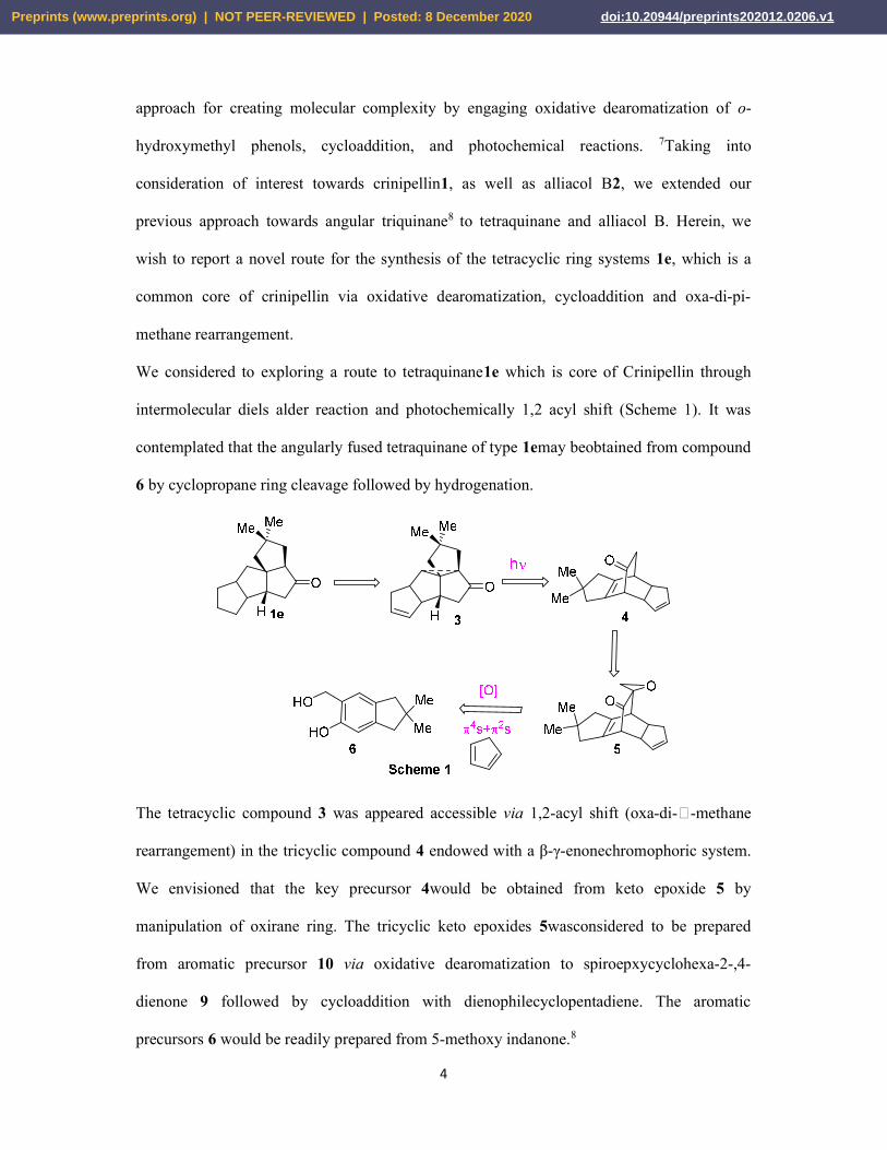
We considered to exploring a route to tetraquinane1e which is core of Crinipellin through
intermolecular diels alder reaction and photochemically 1,2 acyl shift (Scheme 1). It was
contemplated that the angularly fused tetraquinane of type 1emay beobtained from compound
6 by cyclopropane ring cleavage followed by hydrogenation.
The tetracyclic compound 3 was appeared accessible via 1,2-acyl shift (oxa-di--methane
rearrangement) in the tricyclic compound 4 endowed with a β-γ-enonechromophoric system.
We envisioned that the key precursor 4would be obtained from keto epoxide 5 by
manipulation of oxirane ring. The tricyclic keto epoxides 5wasconsidered to be prepared
from aromatic precursor 10 via oxidative dearomatization to spiroepxycyclohexa-2-,4-
dienone 9 followed by cycloaddition with dienophilecyclopentadiene. The aromatic
precursors 6 would be readily prepared from 5-methoxy indanone.8
Preprints (www.preprints.org) | NOT PEER-REVIEWED | Posted: 8 December 2020 doi:10.20944/preprints202012.0206.v1

5
As we all know that the ongoing COVID-19 outbreak driven by highly infectious SARS-
CoV-2 and causing the current pandemic and has turned on the most critical universal health
disaster of this century [9,10]. So, after synthesizing the compounds, we have performed the
molecular docking studies against some selected proteins/enzymes receptors by using CLC
Drug Discovery Workbench Software.
Result and discussion
In continuation of our theme towards synthesis of angular triquinanes, we had also thought of
exploring a synthetic route to angular tetraquinanes along the similar lines as presented
earlier. The tetracyclic chromophoric system 4 was readily synthesized from the aromatic
precursor 6 as described below. Interestingly, oxidative dearomatization of 6 in the presence
of cyclopentadiene directly gave the adduct 5 in reasonably good yield along with some un-
reacted spiroepoxycyclohexa-2,5-dienone 9 (Scheme 2). This is presumably due to high
reactivity of cyclopentadiene which could intercept the cyclohexadienone9 formed in situ
even under ambient conditions. The structure of adduct 5 was confirmed from its spectral
features.
Further, it may be worth noting that adduct 5 is formed in a highly regio- and
stereoselectivecycloaddition wherein the cyclohexadienone behaves as 4-partner and
cyclopentadiene as a 2-partner (dienophile) and that other products arising from alternate
pericyclic modes [such as 4s(cyclopentadiene) + 2s (cyclohexadienone)] was not formed.
The adduct 5 was converted into the desired chromophoric system 4 as shown in Scheme 3.
Thus, the keto-epoxide5was reduced with activated zinc in aqueous methanol (protic solvent)
in the presence of ammonium chloride which gave the keto-alcohol 10 asthe major product
Preprints (www.preprints.org) | NOT PEER-REVIEWED | Posted: 8 December 2020 doi:10.20944/preprints202012.0206.v1

6
(mixture of syn-anti isomers) along with ketone 9 as a minor product. Oxidation of the keto
alcohol 10 with Jones reagent followed by decarboxylation of the resulting -keto-acid
furnished the desired tricyclic compound 4 endowed with a -enonechromophore in good
yield. The structures of all the compounds were supported from their spectral features.
After having obtained the tetracyclic compound 4, it was subjected to sensitized irradiation in
acetone. Chromatography of the photolysate gave both products, the compound 11 containing
cyclobutanone ring formed due to 1,3-acyl shift and pentacyclic compound 12 (formed due to
oxa-di--methane rearrangement) in almost equal yields (Scheme 4).
The 1H NMR and 13C NMR spectra of the more nonpolar product 12 indicated that it is
contaminated with some inseparable hydrocarbon (which could not be separated even after
repeated column chromatography). Therefore, the product 12 was subjected to
dihydroxylation with OsO4 which gave the pentacyclicdiol13 (Scheme 3.14) in excellent
yield as a single diastereoisomer (1H NMR and 13C NMR spectra) whose structure was fully
corroborated with its spectral characteristics. However, stereochemical orientation of the
hydroxyl groups was not easily discernible from spectral features.
The behaviour of tetracyclic compound 4is same with previous tricyclic chromophoric
systems 14 under photochemical transformation.8As, the 1,3-acyl shift product 15 was
Preprints (www.preprints.org) | NOT PEER-REVIEWED | Posted: 8 December 2020 doi:10.20944/preprints202012.0206.v1
7
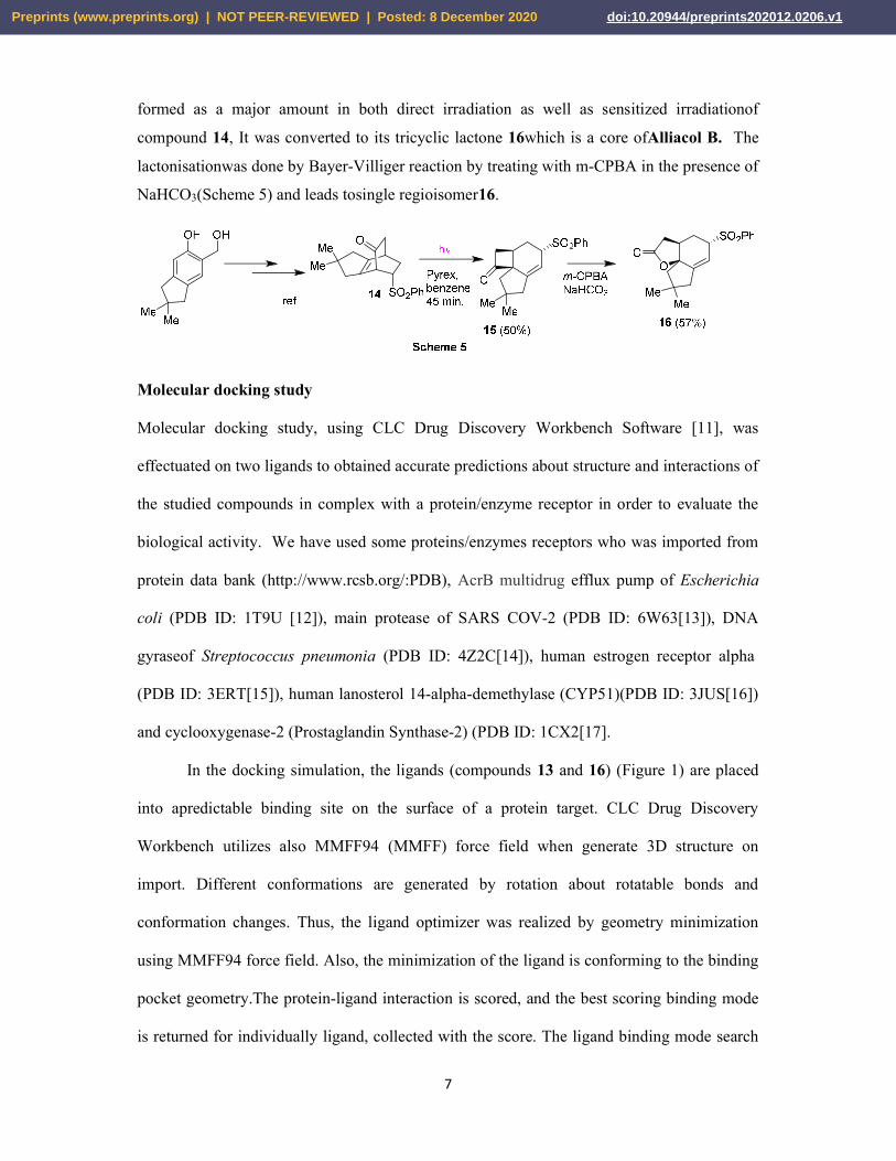
formed as a major amount in both direct irradiation as well as sensitized irradiationof
compound 14, It was converted to its tricyclic lactone 16which is a core ofAlliacol B. The
lactonisationwas done by Bayer-Villiger reaction by treating with m-CPBA in the presence of
NaHCO3(Scheme 5) and leads tosingle regioisomer16.
Molecular docking study
Molecular docking study, using CLC Drug Discovery Workbench Software [11], was
effectuated on two ligands to obtained accurate predictions about structure and interactions of
the studied compounds in complex with a protein/enzyme receptor in order to evaluate the
biological activity. We have used some proteins/enzymes receptors who was imported from
protein data bank (http://www.rcsb.org/:PDB), AcrB multidrug efflux pump of Escherichia
coli (PDB ID: 1T9U [12]), main protease of SARS COV-2 (PDB ID: 6W63[13]), DNA
gyraseof Streptococcus pneumonia (PDB ID: 4Z2C[14]), human estrogen receptor alpha
(PDB ID: 3ERT[15]), human lanosterol 14-alpha-demethylase (CYP51)(PDB ID: 3JUS[16])
and cyclooxygenase-2 (Prostaglandin Synthase-2) (PDB ID: 1CX2[17].
In the docking simulation, the ligands (compounds 13 and 16) (Figure 1) are placed
into apredictable binding site on the surface of a protein target. CLC Drug Discovery
Workbench utilizes also MMFF94 (MMFF) force field when generate 3D structure on
import. Different conformations are generated by rotation about rotatable bonds and
conformation changes. Thus, the ligand optimizer was realized by geometry minimization
using MMFF94 force field. Also, the minimization of the ligand is conforming to the binding
pocket geometry.The protein-ligand interaction is scored, and the best scoring binding mode
is returned for individually ligand, collected with the score. The ligand binding mode search
Preprints (www.preprints.org) | NOT PEER-REVIEWED | Posted: 8 December 2020 doi:10.20944/preprints202012.0206.v1

8
is effectuated inside in the binding site (green sphere with a radius large enough to comprise
all ligands docked to the receptor protein). After the import of the protein receptor from PDB
bank, the next step is the setup binding site and the setup binding pockets; binding pockets
are necessary to guide the docking simulation. After the setup the binding site and the binding
pocket, the co-crystallized- natural ligand was extracted and was redocking in the active
binding site of the protein receptor, for the validation of the method and of the docking
parameters obtained from the molecular docking studies.
Compound 16 Compound 13
Figure 1. Tube representation of the optimized molecular structure of compounds 20 and 33 (Numbering of the atoms was done according to the software) Docking evaluation against AcrB multidrug efflux pump of Escherichia coli
Docking studies was realized to achieve accurate predictions on the optimized conformations
for both the ligands and protein target to form a stable complex. The two compounds was
docked on the crystal structure of AcrB multidrug efflux pump of Escherichia coli (PDB ID:
1T9U). The docking pose of the co-crystallized ciprofloxacin interacting with amino acid
residues of the active site and the hydrogen bonds created with ARG 468 (three bonds: 3.129
Å, 3.062 Å and 3.073 Å) are shown in Figure S1a. The ciprofloxacin was taken as reference
ligand to compare the docking results of the two studied compounds. The docking studies
revealed that the docking score of the two compounds 16and 13 are smaller than docking
score of co-crystallized (docking score: -46.33; RMSD: 0.67 Å). The compound 16, has a
Preprints (www.preprints.org) | NOT PEER-REVIEWED | Posted: 8 December 2020 doi:10.20944/preprints202012.0206.v1

9
docking score: -46.33 (RMSD: 0.67 Å) and shows occurence of three hydrogen bonds,
onewith GLN 469 (2.631 Å) and two with ARG 468 (2.620 Å and 3.112 Å) (Figure 2a). The
compound 13 showed occurence of six hydrogen bonds with ARG 468 (2.884 Å), ASN 391
(3.290 Å), GLY 387 (3.219 Å) and three with SER 389 (2.466 Å, 2.918Å, and 3.158Å)
(Figure S2a). The docking pose of the co-crystallized CPX and of the compounds 20 and 33
interacting with the amino acids residues is presented in Figure S1b, 2b and S2b. The amino
acids residues that formed the interacting group of each ligand are listed in Table S1. After
analyzing the data obtained from the docking study, it was observed that the two studied
compounds were placed in the same binding site of 1T9U as the cocrystallized one (Figure
2c).
Figure 2. (a) Hydrogen bonds (blue dotted lines) between Compound 20 and ARG 468 GLN 469 amino acids, (b)Docking pose of the compound 20interacting with the amino acid residues of binding site of 1T9U, (c) Docking pose of the co-crystallized CPX and of compounds 20 and 33 in the binding site of 1T9U.
Docking evaluation against Main protease of SARS-CoV-2
Docking studies was realized to achieve accurate predictions on the optimized conformations
for both the ligands and protein target to form a stable complex. The two compounds was
docked on the crystal structure of Main protease of SARS-CoV-2 (PDB ID: 6W63). The
docking pose of the co-crystallized X77 interacting with amino acid residues of the active site
and the hydrogen bonds created with GLU 166 (2.721 Å)and GLY 143(3.202Å) are shown in
Figure S3a. The co-crystallized X77 (N-(4-tert-butylphenyl)-N-[(1R)-2-(cyclohexylamino)-2-
Preprints (www.preprints.org) | NOT PEER-REVIEWED | Posted: 8 December 2020 doi:10.20944/preprints202012.0206.v1

10
oxo-1-(pyridin-3-yl)ethyl]-1H-imidazole-4-carboxamide) was taken as reference ligand to
compare the docking results of the compounds16 and 13. The docking studies revealed that
the docking score of the compound 16(docking score: -56.15; RMSD: 0.11 Å) is close to that
of the co-crystallized (docking score: -56.57; RMSD: 1.53Å), but these compound do not
realize hydrogen bonds with the amino acids from the active site of the protein receptor
(Figure 3a). Compound 33 shows occurence of two hydrogenbonds with TYR 54 (2.765Å)
and ASP 187 (2.752Å) (Figure S4a). The docking pose of the co-crystallized and of the
compounds 16 and 13, interacting with the amino acids residues is presented in Figure S3b,
3b and S4b. The amino acids residues who formed the interacting group of each ligand are
listed in Table S2. After analyzing the data obtained from the docking study, it was observed
that the two studied compounds were placed in the same binding site of 1T9U as the
cocrystallized one (Figure 3c).
Figure 3. (a)Doching pose of compound 20, (b) Docking pose of the compound 20 interacting with the amino acid residues of binding site of 6W63, (c) Docking pose of the co-crystallized (purple) and compound 20 (olive) and 33 (yellow) in the binding site of 6W63.
Docking evaluation against Streptococcus pneumoniae DNA gyrase
Docking studies was realized to achieve accurate predictions on the optimized conformations
for both the ligands and protein target to form a stable complex. The two compounds was
docked on the crystal structure of Streptococcus pneumonia DNA gyrase(PDB ID:4Z2C).
The docking pose of the co-crystallized, moxifloxacin, (MFX) interacting with amino acid
residues and nucleotids of the active site and the hydrogen bonds created with SER 436:D
Preprints (www.preprints.org) | NOT PEER-REVIEWED | Posted: 8 December 2020 doi:10.20944/preprints202012.0206.v1

11
(2.953 Å, 3.030 Å, 2.597 Å), DG 1:H (3.099 Å) and DA 2:H (3.112Å) are shown in Figure
S5a. The co-crystallized MFX was taken as reference ligand to compare the docking results
of the compounds 16 and 13. The docking studies revealed that the docking score of the two
compounds 16and 13 are smaller than docking score of co-crystallized (docking score: -
83.33; RMSD: 1.53 Å). The compound 16, has a docking score: -63.48 (RMSD: 0.04 Å) and
showed occurence of two hydrogen bonds, one with GLY 457:D (3.096 Å) and with DG 1:H
(2.898 Å) (Figure 4a). The compound 13 showed occurence of four hydrogen bonds with
ARG 456:D (3.084 Å), GLU 475:D (2.791 Å), and two with DA 5:F (3.015 Å, 3.133 Å)
(Figure S6a). The docking pose of the co-crystallized and of the compounds 16 and 13
interacting with the amino acids residues is presented in Figure S5b, 4b and S6b. The amino
acids residues that formed the interacting group of each ligand are listed in Table S3. After
analyzing the data obtained from the docking study, it was observed that the two studied
compounds were placed in the same binding site of 4Z2C as the cocrystallized one (Figure
4c).
Figure 4. (a) Hydrogen bonds (blue dotted lines) between compound 20 and GLY 457(D) amino acid and DG1(H) nucleotid, (b)Docking pose of the compound 20 interacting with the amino acid residues and nucleotids of binding site of 4Z2C, (c)Docking pose of the co-crystallized (brown) and compound 20(violet) and 33(light green)in the binding site of 4Z2C.
Docking evaluation against Human estrogen receptor alpha
Docking studies was realized to achieve accurate predictions on the optimized conformations
for both the ligands and protein target to form a stable complex. The two compounds was
Preprints (www.preprints.org) | NOT PEER-REVIEWED | Posted: 8 December 2020 doi:10.20944/preprints202012.0206.v1
12
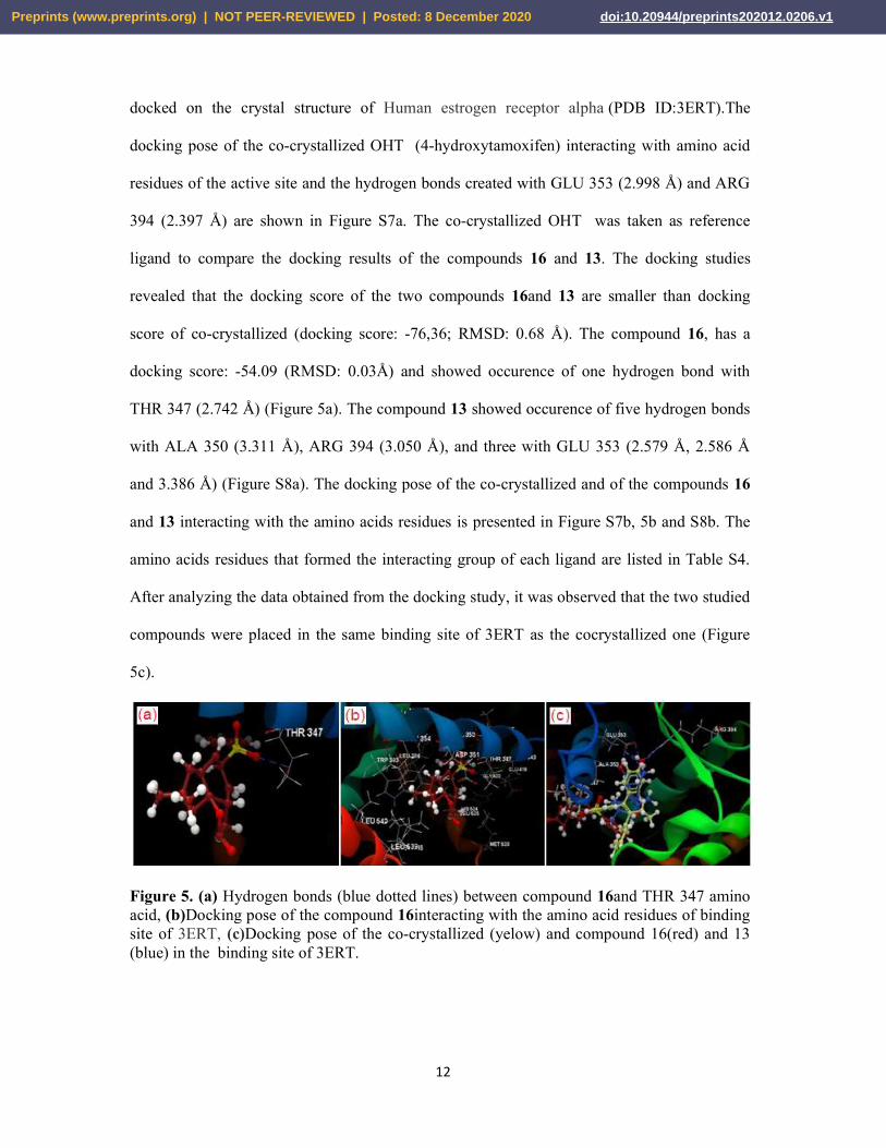
docked on the crystal structure of Human estrogen receptor alpha (PDB ID:3ERT).The
docking pose of the co-crystallized OHT (4-hydroxytamoxifen) interacting with amino acid
residues of the active site and the hydrogen bonds created with GLU 353 (2.998 Å) and ARG
394 (2.397 Å) are shown in Figure S7a. The co-crystallized OHT was taken as reference
ligand to compare the docking results of the compounds 16 and 13. The docking studies
revealed that the docking score of the two compounds 16and 13 are smaller than docking
score of co-crystallized (docking score: -76,36; RMSD: 0.68 Å). The compound 16, has a
docking score: -54.09 (RMSD: 0.03Å) and showed occurence of one hydrogen bond with
THR 347 (2.742 Å) (Figure 5a). The compound 13 showed occurence of five hydrogen bonds
with ALA 350 (3.311 Å), ARG 394 (3.050 Å), and three with GLU 353 (2.579 Å, 2.586 Å
and 3.386 Å) (Figure S8a). The docking pose of the co-crystallized and of the compounds 16
and 13 interacting with the amino acids residues is presented in Figure S7b, 5b and S8b. The
amino acids residues that formed the interacting group of each ligand are listed in Table S4.
After analyzing the data obtained from the docking study, it was observed that the two studied
compounds were placed in the same binding site of 3ERT as the cocrystallized one (Figure
5c).
Figure 5. (a) Hydrogen bonds (blue dotted lines) between compound 16and THR 347 amino acid, (b)Docking pose of the compound 16interacting with the amino acid residues of binding site of 3ERT, (c)Docking pose of the co-crystallized (yelow) and compound 16(red) and 13 (blue) in the binding site of 3ERT.
Preprints (www.preprints.org) | NOT PEER-REVIEWED | Posted: 8 December 2020 doi:10.20944/preprints202012.0206.v1

13
Docking evaluation against Human lanosterol 14-alpha-demethylase (CYP51)
Docking studies was realized to achieve accurate predictions on the optimized conformations
for both the ligands and protein target to form a stable complex. The two compounds was
docked on the crystal structure of human lanosterol 14alpha-demethylase (CYP51) (PDB
ID:3JUS). The docking pose of the co-crystallized ECN (1-[(2S)-2-[(4-chlorobenzyl)oxy]-2-
(2,4-dichlorophenyl)ethyl]-1H-imidazole) interacting with amino acid residues of the active
site and the hydrogen bond created with TYR 145 (3.103 Å) are shown in Figure S9a. The
co-crystallized OHT was taken as reference ligand to compare the docking results of the
compounds 16 and 13. The docking studies revealed that the docking score of the two
compounds 16and 13 are smaller than docking score of co-crystallized (docking score: -
84.04; RMSD: 0.93Å). The compound 16, has a docking score: -70.81 (RMSD: 0.02Å) and
showed occurence of one hydrogen bonds TYR 145 (2.983 Å) (Figure 6a). The docking
studies revealed that the docking score of the compound 13 is -56.78, (RMSD: 0.02 Å) but
this compound does not realize hydrogen bonds with the amino acids from the active site of
the protein receptor (Figure S10a). The docking pose of the co-crystallized and of the
compounds 16 and 13 interacting with the amino acids residues is presented in Figure S9b, 6b
and S10b. The amino acids residues that formed the interacting group of each ligand are
listed in Table S5. After analyzing the data obtained from the docking study, it was observed
that the two studied compounds were placed in the same binding site of 3ERT as the
cocrystallized one (Figure 6c).
Preprints (www.preprints.org) | NOT PEER-REVIEWED | Posted: 8 December 2020 doi:10.20944/preprints202012.0206.v1

14
Figure 6. (a) Hydrogen bonds (blue dotted lines) between compound 16and TYR 145 amino acid, (b)Docking pose of the compound 16interacting with the amino acid residues of binding site of 3JUS, (c)Docking pose of the co-crystallized and compound 16and 13 in the binding site of 3JUS.
Docking evaluation against Cyclooxygenase-2 (Prostaglandin Synthase-2)
Docking studies was realized to achieve accurate predictions on the optimized conformations
for both the ligands and protein target to form a stable complex. The two compounds was
docked on the crystal structure of Human lanosterol 14-alpha-demethylase (CYP51) (PDB
ID: 1CX2). The docking pose of the co-crystallized S58 (1-phenylsulfonamide-3-
trifluoromethyl-5-para bromophenylpyrazole) interacting with amino acid residues of the
active site and the hydrogen bonds created with HIS 90 (2.894 Å and 3.144 Å), ARG 513
(3.035 Å) and SER 353 (2.534 Å) are shown in Figure S11a. The co-crystallized OHT was
taken as reference ligand to compare the docking results of the compounds 16 and 13. The
docking studies revealed that the docking score of the two compounds 16and 13 are smaller
than docking score of co-crystallized (docking score: -80.94; RMSD: 0.04Å). The compound
16, has a docking score: -71.73 (RMSD: 0.04 Å) and showed occurence of three hydrogen
bonds, one with SER 530 (2.876 Å) and two with TYR 385 (2.882 Å and 3.091V Å) (Figure
7a). The compound 33 showed occurence of one hydrogen bond with TYR 355 (2.872 Å).
(Figure S12a). The docking pose of the co-crystallized and of the compounds 16 and 13
interacting with the amino acids residues is presented in Figure S11b, 7b and S12b. The
amino acids residues that formed the interacting group of each ligand are listed in Table S6.
After analyzing the data obtained from the docking study, it was observed that the two
Preprints (www.preprints.org) | NOT PEER-REVIEWED | Posted: 8 December 2020 doi:10.20944/preprints202012.0206.v1

15
studied compounds were placed in the same binding site of 3ERT as the cocrystallized one
(Figure 7c).
Figure 7.(a) Hydrogen bonds (blue dotted lines) between compound 16and SER 539, TYR 385 and TYR 355 amino acids, (b)Docking pose of the compound 20 interacting with the amino acid residues of binding site of 1CX2, (c)Docking pose of the co-crystallized (green) and compound 16(pink) and 13 (ocean blue) in the binding site of 1CX2.
It has been calculated the parameters who can predict if a molecule possesses properties that
might turn it into an active drug, according to the Lipinski’s rule of five [18], the number of
hydrogen donors < 5), the number of acceptors hydrogen < 10), molecular weight < 500 Da,
the octanol-water partition coefficient (log P) < 5. (Table 1). The number of violations of the
Lipinski rules allows to evaluate drug likeness for a molecule. It was observed that
compounds 20 and 33 have zero violation of all the parameters involved in Lipinski’s rule of
five (Lipinski violation is 0).
Table 1. Calculated properties of compounds
Compounds
Ato
ms
Wei
ght
[Dal
ton
s]
Fle
xib
le
bon
ds
Lip
insk
i vi
olat
ion
s
Hyd
roge
n d
onor
s
Hyd
roge
n ac
cep
tors
Log P
Co-crystallized*Cp 42 331.34 3 0 2 6 0.84 Co-crystallized**X77 66 458.58 7 0 1 7 4.59
Co-crystallized***MXF 53 401.43 4 0 2 7 1.62 Co-crystallized****OHT 58 387.51 8 1 1 3 6.78 Co-crystallized*****ECN 39 381.68 6 0 0 3 4.70 Co-crystallized******S58 37 446.24 4 0 2 5 3.65
Compound 20 46 346.44 2 0 0 4 2.10*/2.75**/2.62***/1.97****
2.22*****/2.62******
Compound 33 41 262.34 0 0 2 3 0.99 * PDB ID: 1T9U ; ** PDB ID: 6W63; *** PDB ID: 4ZDC; **** PDB ID: 3ERT; ***** PDB ID: 3JUS; ******PDB ID: 1CX2
Preprints (www.preprints.org) | NOT PEER-REVIEWED | Posted: 8 December 2020 doi:10.20944/preprints202012.0206.v1

16
After analyzing the results of the molecular docking study, it is observed that the two
compounds 16 and 13 possess properties that can turn them into future oral drugs (Lipinski
violation is 0) (Table 1). It was also found that compound 16 could be a drug with
antimicrobial, antiviral, anticancer, antifungal or anti-inflammatory activity. For all molecular
docking studies against the studied targets, it was also observed that compound 16 has a
higher docking score than compound 13 and is close to each co-crystallized ligand taken as
reference (Figure 8).
(1)PDB ID: 1T9U; (2) PDB ID: 6W63; (3)PDB ID: 4ZDC; (4)PDB ID: 3ERT; (5) PDB ID: 3JUS; (6)PDB ID: 1CX2
Figure 8. Docking scores of ligands
Conclusion
In summary, we have described a facile route to complex molecular skeleton of crinipellin
from simple aromatic compound through a short and efficient synthetic route. The sensitized
irradiation of 4 led to oxa-di--methane reaction only to a moderate extent thus limiting its
synthetic application. Nevertheless, the present study provides additional examples of
photoreaction of rigid -enones and demonstrates the effect of structure on the
photoreactivity, and provides novel carbocyclic systems that are not readily accessible
Preprints (www.preprints.org) | NOT PEER-REVIEWED | Posted: 8 December 2020 doi:10.20944/preprints202012.0206.v1
17
otherwise. Moreover, from molecular docking study, it is observed that compounds 16 and 13
possess properties that can turn them into future oral drugs. It was also found that compound
16 could be a drug with antimicrobial, antiviral, anticancer, antifungal or anti-inflammatory
activity. In addition, compound 16 has a higher docking score than compound 13 and is close
to each co-crystallized ligand taken as reference.
Experimental section
Physical measurements
All the glassware was oven dried and chemicals are commercially available from sigma
aldric and, merch and TCI otherwise it is mentioned if prepared. 1H NMR and 13C NMR
spectra were recorded on Bruker 400 MHz and 500 MHz instruments. Samples were
dissolved in CDCl3 containing TMS as internal standard. IR spectra were recorded on Perkin-
Elmer FT-IR instrument. High-resolution mass spectra were recorded on Maxis impact
Bruker mass spectrometer. Melting points were recorded on Buchi M-560 instrument.
Reactions were monitored by thin layer chromatography (TLC) using silica gel and spots
were visualized with iodine vapour. Column chromatography was performed on silica gel for
chromatography (100-200 mesh).
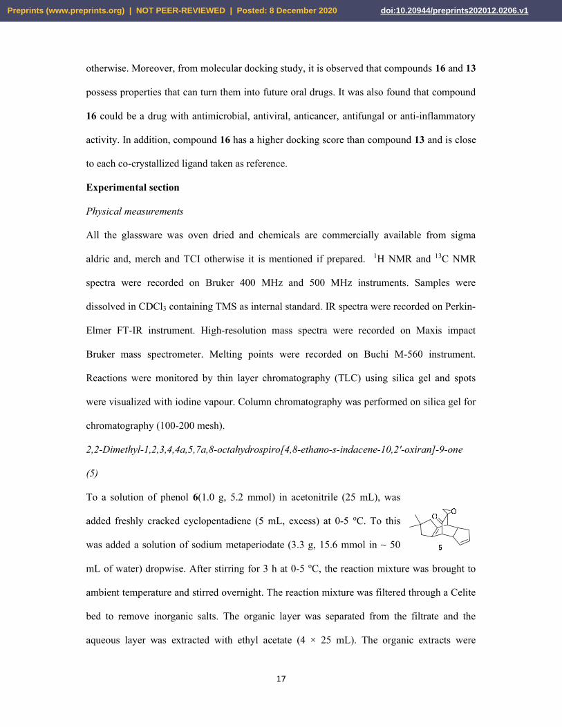
2,2-Dimethyl-1,2,3,4,4a,5,7a,8-octahydrospiro[4,8-ethano-s-indacene-10,2'-oxiran]-9-one
(5)
To a solution of phenol 6(1.0 g, 5.2 mmol) in acetonitrile (25 mL), was
added freshly cracked cyclopentadiene (5 mL, excess) at 0-5 oC. To this
was added a solution of sodium metaperiodate (3.3 g, 15.6 mmol in ~ 50
mL of water) dropwise. After stirring for 3 h at 0-5 oC, the reaction mixture was brought to
ambient temperature and stirred overnight. The reaction mixture was filtered through a Celite
bed to remove inorganic salts. The organic layer was separated from the filtrate and the
aqueous layer was extracted with ethyl acetate (4 × 25 mL). The organic extracts were
Preprints (www.preprints.org) | NOT PEER-REVIEWED | Posted: 8 December 2020 doi:10.20944/preprints202012.0206.v1

18
combined and washed with brine (30 mL) and dried over sodium sulfate. The solvent was
removed under vacuum and crude product was chromatographed on silica gel. Elution with
petroleum ether-ethyl acetate (93:7) afforded the compound 5(0.750 g, 56%) as a colourless
solid. [Rf = 0.7 petroleum ether/ethyl acetate (80:20)]. mp: 79-81 oC. IR (film) νmax: 2952,
1734 cm-1. 1H NMR (400 MHz, CDCl3): δ 5.69-5.64 (m, 1H), 5.43-5.38 (m, 1H), 3.41-3.36
(m, 1H), 3.32 (d, J = 2.8Hz, 1H), 3.10 (Part of an AB system JAB = 6.1 Hz, 1H), 3.07-3.00
(m, 1H), 2.82 (Part of an AB system JAB = 6.1 Hz, 1H), 2.63-2.54 (m. 1H), 2.52 (d, J = 2.8
Hz, 1H), 2.36 (dt, J1 = 13.7Hz, J2 = 2.8 Hz, 1H), 2.28-2.14 (m, 2H), 2.11-2.04 (m, 1H),
1.98-1.90 (m, 1H), 1.07 (s, 3H), 1.06 (s, 3H). 13C NMR (100 MHz, CDCl3): δ 205, 139.7,
135.4, 133.4, 129.5, 58.9, 53.2, 52.4, 50.8, 50.4, 49.4, 44.4, 40.0, 38.3, 36.9, 30.7, 29.9.
HRMS (ESI-MS) (m/z): found 279.1352 (M + Na)+; calcd for C17H20NaO2: 279.1356.
Further elution with petroleum ether-ethyl acetate (85:15) gave the compound 9 as a yellow
liquid (0.160 g, 16%) [Rf= 0.6 petroleum ether/ethyl acetate (80:20), which was identical in
all respects to that obtained earlier.
2,2,10-Trimethyl-1,2,3,4,4a,5,7a,8-octahydro-4,8-ethano-s-indacen-9-one (29) and 10-
(hydroxymethyl)-2,2-dimethyl-1,2,3,4,4a,5,7a,8-octahydro-4,8-ethano-s-indacen-9-one (9)
To a mixture of keto epoxide 5(1.2 g, 4.65 mmol) in methanol-water (90 mL, 6:1) was added
activated zinc (3 g, excess) and ammonium chloride (1.23 g, 23.25 mmol). The reaction
mixture was stirred for 4 h at room temperature, after which it was filtered through a Celite
bed to remove zinc and washed with ethyl acetate (25 mL). The filtrate was concentrated in
vacuum, so as to remove most of the solvent and the residue was diluted with water (20 mL)
and extracted with ethyl acetate (3×30 mL). The combined organic layer was washed with
brine (20 mL) and dried over anhydrous sodium sulfate. The solvent was
removed under vacuum, and the product was chromatographed on silica
gel. Elution with petroleum ether-ethyl acetate (95:05) furnished the
Preprints (www.preprints.org) | NOT PEER-REVIEWED | Posted: 8 December 2020 doi:10.20944/preprints202012.0206.v1
19
ketone 9 as a colorless liquid (0.120 g, 11%), [Rf = 0.6 petroleum ether/ethyl acetate (92:8)].
IR (film) νmax: 2952, 1721 cm-1. 1H NMR (400 MHz, CDCl3): δ5.63-5.58 (m, 1H), 5.38-5.34
(m, 1H), 3.7 (d, J = 2.6Hz, 1H), 2.88-2.82 (m, 1H), 2.68 (t, J = 3.0Hz, 1H), 2.55-2.46 (m,
1H), 2.32 (dt, J1 = 16.1Hz, J2 = 2.2Hz, 1H), 2.20-2.09 (m, 2H), 2.05-1.98 (m, 2H), 1.94-1.86
(m, 1H), 1.12 (d, 7.3 Hz, 3H), 1.04 (s, 3H), 1.03 (s, 3H). 13C NMR (100 MHz, CDCl3): δ
215.6, 143.1, 134.2, 132.8, 130.2, 54.0, 50.9, 50.1, 49.4, 43.9, 43.7, 39.8, 38.5, 34.7, 30.8,
30.0, 28.7, 14.7. HRMS (ESI) (m/z): found 281.1299 (M + K)+; calcd for C17H22KO:
281.1302.
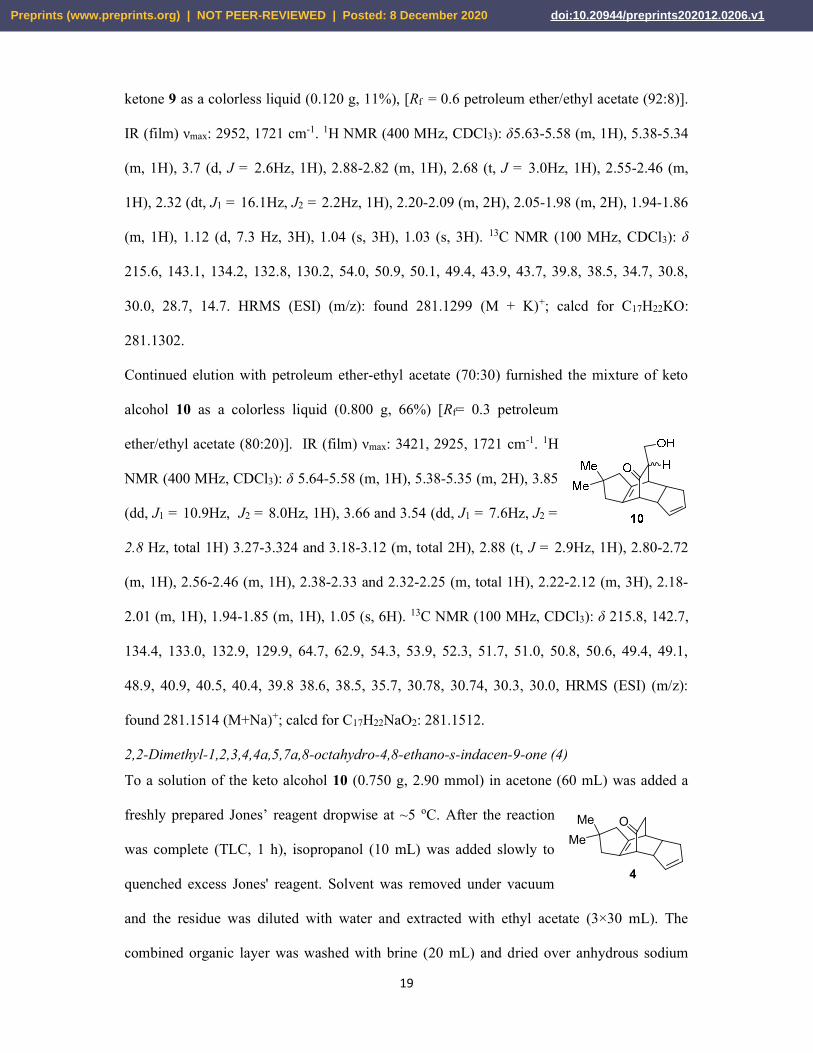
Continued elution with petroleum ether-ethyl acetate (70:30) furnished the mixture of keto
alcohol 10 as a colorless liquid (0.800 g, 66%) [Rf= 0.3 petroleum
ether/ethyl acetate (80:20)]. IR (film) νmax: 3421, 2925, 1721 cm-1. 1H
NMR (400 MHz, CDCl3): δ 5.64-5.58 (m, 1H), 5.38-5.35 (m, 2H), 3.85
(dd, J1 = 10.9Hz, J2 = 8.0Hz, 1H), 3.66 and 3.54 (dd, J1 = 7.6Hz, J2 =
2.8 Hz, total 1H) 3.27-3.324 and 3.18-3.12 (m, total 2H), 2.88 (t, J = 2.9Hz, 1H), 2.80-2.72
(m, 1H), 2.56-2.46 (m, 1H), 2.38-2.33 and 2.32-2.25 (m, total 1H), 2.22-2.12 (m, 3H), 2.18-
2.01 (m, 1H), 1.94-1.85 (m, 1H), 1.05 (s, 6H). 13C NMR (100 MHz, CDCl3): δ 215.8, 142.7,
134.4, 133.0, 132.9, 129.9, 64.7, 62.9, 54.3, 53.9, 52.3, 51.7, 51.0, 50.8, 50.6, 49.4, 49.1,
48.9, 40.9, 40.5, 40.4, 39.8 38.6, 38.5, 35.7, 30.78, 30.74, 30.3, 30.0, HRMS (ESI) (m/z):
found 281.1514 (M+Na)+; calcd for C17H22NaO2: 281.1512.
2,2-Dimethyl-1,2,3,4,4a,5,7a,8-octahydro-4,8-ethano-s-indacen-9-one (4)
To a solution of the keto alcohol 10 (0.750 g, 2.90 mmol) in acetone (60 mL) was added a
freshly prepared Jones’ reagent dropwise at ~5 oC. After the reaction
was complete (TLC, 1 h), isopropanol (10 mL) was added slowly to
quenched excess Jones' reagent. Solvent was removed under vacuum
and the residue was diluted with water and extracted with ethyl acetate (3×30 mL). The
combined organic layer was washed with brine (20 mL) and dried over anhydrous sodium
OMe
Me
4
Preprints (www.preprints.org) | NOT PEER-REVIEWED | Posted: 8 December 2020 doi:10.20944/preprints202012.0206.v1

20
sulfate. Removal of the solvent gave a β-keto acid which was directly subjected to
decarboxylation as follows.
The β-keto acid thus obtained was taken up in THF -water mixture (70 mL, 1:1) and the
reaction mixture was refluxed for 15 h (TLC). THF was removed in vacuum, and the aqueous
layer was extracted with ethyl acetate and dried over anhydrous sodium sulfate. The solvent
was removed and the residue was chromatographed on silica gel. Elution with petroleum
ether-ethyl acetate (90:10) gave the compound 4 as a colorless liquid (0.400 g, 57%), [Rf=
0.5 petroleum ether/ethyl acetate (95:05)]. IR (film) νmax: 2928, 1725 cm-1. 1H NMR (400
MHz, CDCl3): δ 5.60-5.55 (m, 1H), 5.47-5.43 (m, 1H), 3.26-3.15 (m, 1H), 3.08 (d, J =
2.6Hz, 1H), 2.82 (dd, J1 = 5.6Hz, J2 = 2.6Hz, 1H), 2.70-2.61 (m, 1H), 2.55-2.46 (m, 1H),
2.31 (dt, J1 = 15.8Hz, J2 = 2.4Hz, 1H), 2.18 (dt, J1 = 15.8Hz, J2 = 2.4Hz, 1H), 2.12-2.06 (m,
1H), 2.04-1.96 (m, 3H), 1.92-1.84 (m, 1H), 1.02 (s, 3H). 13C NMR (100 MHz, CDCl3): δ
212.5, 141.9, 134.3, 132.5, 130.1, 54.0, 50.7, 49.6, 49.3, 41.1, 40.6, 39.9, 38.6, 37.9, 30.8,
30.0. HRMS (ESI) (m/z): found 251.1403 (M+Na)+; calcd for C16H20NaO: 251.1406.
4,4-Dimethyl-4,5,6a,9,9a,9b-hexahydro-1H-cyclobuta[d]-s-indacen-2(3H)-one (11) and 2,2-
Dimethyl-1,2,3,3b,3c,4,6a,7-octahydro 3a,7ethanocyclopenta[2,3]cyclopropa[1,2a]pentalen-
9-one (12)
Irradiation of a solution of β-enone4 (0.100 g, 0.41 mmol) in degassed
acetone (100 mL) with mercury vapour lamp (125 W) in a Pyrex
immersion well for 1 h under nitrogen atmosphere followed by removal of solvent and
chromatography [petroleum ether/ethyl acetate (95:5)] gave the 1,3 acyl shift product 11
(0.030 g, 30%) as a colorless liquid, [Rf= 0.6 petroleum ether/ethyl acetate (95:5)]. IR (film)
νmax: 2925, 1776 cm-1. 1H NMR (400 MHz, CDCl3): δ 5.90-5.85 (m, 1H), 5.78-5.66 (m, 1H),
5.22 (s, 1H), 3.13 (br s, 1H), 3.02 (dd, J1 = 17.4Hz, J2 = 8.7Hz, 1H), 2.78 (dd, J1 = 17.4Hz,
J2 = 7.3Hz, 1H), 2.58-2.50 (m, 1H), 2.34-2.26 (m, 1H), 2.23-2.17 (m, 2H), 2.06 (part of an
AB system, JAB = 13.6Hz, 1H), 2.03-1.94 (m, 2H), 1.87 (part of an AB system, JAB =
Preprints (www.preprints.org) | NOT PEER-REVIEWED | Posted: 8 December 2020 doi:10.20944/preprints202012.0206.v1

21
13.6Hz, 1H), 1.41 (d, J = 13.6Hz, 1H), 1.15 (s, 3H), 0.89 (s, 3H).13C NMR (100 MHz,
CDCl3): δ 209.5, 136.0, 134.0, 130.3, 118.3, 68.8, 48.5, 47.6, 47.3, 40.7, 38.9, 36.7, 36.2,
30.5, 30.3, 29.6. HRMS (ESI) (m/z): found 251.1405 (M+Na)+; calcd for C16H20NaO:
251.1406.
Further elution with petroleum ether/ethyl acetate (94:6)] gave the
rearranged product 12 (mixture with unknown hydrocarbon) (0.026 g,
26%) [Rf= 0.4 petroleum ether/ethyl acetate (95:5)] which was
inseparable. This mixture was subjected to hydroxylation as follows.
4,5-Dihydroxy-2,2-dimethyldecahydro-3a,7-ethanocyclopenta[2,3]cyclopropa[1,2-
a]pentalen-9-one (13)
To a solution of the above mixture containing compound 12 (0.030 g, 0.13 mmol) in
acetone:water (6:1, 20 mL) were added 4-methylmorpholine N-oxide (0.023 g, 0.197 mmol)
and osmium tetroxide (0.005 g, 0.002 mmol). The reaction mixture was stirred for 24 h under
ambient conditions. After which sodium bisulphite (0.020 g, 0.195 mmol) was added. After
stirring for further 2 h, the reaction mixture was filtered through a Celite pad. The filtrate
was concentrated and the residue was extracted with dichloromethane (3×10 mL), dried over
sodium sulfate, and concentrated in vacuo. The product was purified by column
chromatography on silica gel. Elution with petroleum ether-ethyl acetate (70:30) gave the
dihydroxylated product 13 (0.025 g, 72%) as a thick liquid [Rf= 0.4 petroleum ether/ethyl
acetate (70:30)]. IR (film) νmax: 3424, 2951, 1708 cm-1. 1H NMR (400
MHz, CDCl3): δ 2.97 (dd, J1 = 6.1Hz, J2 = 4.1Hz, 1H), 2.78 (br s,
1H), 2.74 ((dd, J1 = 12.6Hz, J2 = 6.1Hz, 1H), 2.71-269 (m, 1H), 2.40-
2.37 (m, 1H), 2.28 (dd, J1 = 20.3Hz, J2 = 4.1Hz, 1H), 2.14-2.08 (m,
1H), 1.65 (br s, 1H), 1.61 (part of an AB system, JAB = 14.4Hz, 1H), 1.50 (part of an AB
system, JAB = 13.6Hz, 1H), 1.43 (part of an AB system, JAB = 13.6Hz, 1H), 1.38 (d, J = 10.2
Preprints (www.preprints.org) | NOT PEER-REVIEWED | Posted: 8 December 2020 doi:10.20944/preprints202012.0206.v1

22
Hz, 1H), 1.27-1.24 (singlet merged with m, total 4H), 1.05 (part of an AB system, JAB =
14.4Hz, 1H), 1.01 (s, 3H). 13C NMR (100 MHz, CDCl3): δ 213.6, 55.7, 52.9, 49.2, 47.1, 46.6,
46.4, 44.9, 44.3, 42.2, 40.3, 39.9, 38.8, 35.2, 31.3, 29.5. HRMS (ESI) (m/z): found 301.1201
(M+K)+; calcd for C16H22KO3: 301.1201.
Direct irradiation of compound 27: formation of 4,4-Dimethyl-4,5,6a,9,9a,9b-hexahydro-1H-
cyclobuta[d]-s-indacen-2(3H)-one (11)
A solution of β-enone27 (0.050 g, 0.21 mmol) in degassed benzene (100 mL) was irradiated
with a mercury vapour lamp (125 W) in a Pyrex immersion well for 45
min under nitrogen atmosphere. Benzene was removed under vacuum
and the photolysate was chromatographed. Elution with petroleum
ether/ethyl acetate (95:5) gave the 1,3-acyl shift product 11 (0.028 g, 56%) as a colorless
liquid [Rf= 0.5 petroleum ether/ethyl acetate (95:5)] which was identical in all respects to the
compound obtained earlier.
8,8-dimethyl-5-(phenylsulfonyl)-3,3a,4,5,8,9-hexahydroindeno[3a,4-b]furan-2(7H)-one (16)
To a vigorously stirred solution of 15 (0.040 g, 0.12 mmol) in
dichloromethane (20 mL) was added a 0.7 M aqueous solution of
sodium bicarbonate (5 mL) and a solution of 3-chloroperbenzoic acid
( 0.032 g, 0.18 mmol). The reaction mixture was stirred for 2 h at room temperature. Then
the organic phase was washed with saturated sodium bicarbonate solution (50 mL) and
aqeous layer was extracted with dichloromethane (3×30 mL). The combined organic layer
was washed with brine (1×20 mL) and dried over anhydrous sodium sulfate. The solvent was
evaporated under vacuum and the residue was charged on silica gel coloumn. Elution with
petroleum ether/ethyl acetate (60:40)] gave the rearranged product 20 (0.024 g, 57%) as a
colorless solid, mp. 150-152 oC, Rf= 0.4 [petroleum ether/ethyl acetate (70:30)]. (IR (film)
νmax: 2952, 1761 cm-1. 1H NMR (400 MHz, CDCl3): δ 7.89-7.86 (m, 2H), 7.70 (merged dd,
Preprints (www.preprints.org) | NOT PEER-REVIEWED | Posted: 8 December 2020 doi:10.20944/preprints202012.0206.v1

23
J1= J2 = 7.4Hz, 1H), 7.61 (merged dd, J1 = J2 = 7.4 Hz, 2H), 5.62-5.58 (br s, 1H), 3.78-3.73
(br s, 1H), 2.82 (dd, J1 = 17.3Hz, J2 = 7.4Hz, 1H), 2.70-2.65 (m, 1H), 2.57 (dt, J1 = 15.0Hz,
J2 = 2.8Hz, 1H), 2.30 (dd, J1 = 15.0Hz, J2 = 4.9Hz, 1H), 2.20 (dd, J1 = 17.3Hz, J2 = 8.4Hz,
2H), 1.98 (d, J = 4.2Hz, 1H), 1.71-1.67 (m, 1H), 1.59 (merged dd, J1 = J2 =8.4 Hz, 1H), 1.22
(s, 3H), 1.05 (s, 3H). 13C NMR (100 MHz, CDCl3): δ 175.2, 148.1, 138.0, 134.2, 129.6,
129.0, 114.2, 89.2, 60.6, 52.0, 46.6, 36.6, 36.5, 34.5, 31.1, 30.9, 25.6. HRMS (ESI) (m/z):
found 369.1133 (M+Na)+; calcd for C19H22NaO4S: 301.1201.
Acknowledgements
The authors acknowledge the financial support through Researchers Supporting Project
number (RSP-2020/147), King Saud University, Riyadh, Saudi Arabia.
Conflict of interest
There are no conflicts to declare.
Preprints (www.preprints.org) | NOT PEER-REVIEWED | Posted: 8 December 2020 doi:10.20944/preprints202012.0206.v1

24
References
1. (a) Paquette, L. A.; Doherty, A.M. Recent Synthetic Developments in Polyquinane Chemistry;
Springer-verlag: New York,1987 and references therein. (b) Mehta, G.; Srikrishna, A. Chem.
Rev. 1997, 97, 671 and references therein. (c) Singh, V.; Thomas, B.(d) Tetrahedron1998, 54,
3647.Lee, H. -Y. Acc. Chem. Res. 2015, 48, 2308.
2. Kupka, J.; Anke, T.; Oberwinkler, F.; Schramm, G.; Steglich, W. J. Antibiot. 1979, 32, 130.
3. (a) Li, Y.-Y.; Shen, Y.-M. Helv. Chim. Acta 2010, 93,2151. (b)Anke, T.; Heim, J.; Knoch,
F.; Mocek, U.; Steffan, B.; Steglich, W. Angew. Chem., Int. Ed. Engl. 1985, 24, 709.
4. King, T.; Farrell, K.; Halsall, T.; Thaller, V. J. Chem. Soc., Chem. Commun.1977, 20, 727.
5. (a) Schwartz, C. E.; Curran, D. P. J. Am. Chem. Soc. 1990, 112, 9272; (b) Mehta, G.;Rao, K.
S.; Reddy, M. S. J. Chem. Soc. Perkin I 1991, 693 and references therein;(c) Chen, P.;
Carroll, P. J.; Sieburth, S. M. Org. Lett. 2010, 12, 4510; (d) Srikrishna,A.; Gowri, V.
Synlett2011, 2652; (e) Srikrishna, A.; Gowri, V. Tetrahedron2012, 68, 3046; (f) Piers, E.;
Renaud, J. J. Org. Chem. 1993, 58, 11; (g)Piers, E.; Renaud, J.; Rettig, S. J. Synthesis 1998,
590; (h) Wender, P. A.; Dore, T.M. Tetrahedron Lett. 1998, 39, 8589; (i) Kang, T.; Song, S.
B.; Kim, W.-Y.; Kim, B.G.; Lee, H.-Y. J. Am. Chem. Soc. 2014, 136, 10274.
6. Kang, T.; Song, S. B.; Lee, H. Y. J. Am. Chem. Soc. 2014, 136, 10274.
7. (a) Sahu, R.; Singh, V. J. Org. Chem.2017, 82, 6268; (b) Behera, T. K.; Jarhad, D. B.; Mobin,
S. M.; Singh, V. Tetrahedron.2016, 72, 5377; (c) Sahu, R.; Singh, V. Synthesis.2019, 51,
1633; (d) Singh, V.; Sahu, B. C.; Bansal, V.; Mobin, S. M. Org. Biomol. Chem. 2010, 8,
4472; (f) Singh, V. Synlett. 2013, 2641.
8. Sahu, R.; Singh, V. Tetrahedron.2017, 82, 6515.
9. R. K. Mohapatra, P. K. Das, V. Kandi, Diabetes & Metabolic Syndrome: Clinical Research &
Reviews, 2020, 14, 1593-1594.
Preprints (www.preprints.org) | NOT PEER-REVIEWED | Posted: 8 December 2020 doi:10.20944/preprints202012.0206.v1

25
10. R. K. Mohapatra, L. Pintilie, V. Kandi, et al., Chemical Biology & Drug Design, 2020.
https://doi.org/10.1111/cbdd.13761.
11. CLC Drug Discovery Workbench, available from http://www.clcbio.com.
12. Yu, E.W., Aires, J.R., McDermott, G., Nikaido, H.,A Periplasmic Drug-Binding Site of the
AcrB Multidrug Efflux Pump: a Crystallographic and Site-Directed Mutagenesis Study, J.
Bacteriol.,2005, 187, p.6804-6815.
13. Mesecar, A.D., A taxonomically-driven approach to development of potent, broad-spectrum
inhibitors of coronavirus main protease including SARS-CoV-2 (COVID-19), 2020,
DOI: 10.2210/pdb6W63/pdb, to be published.
14. Laponogov, I., Veselkov, D.A., Pan, X.-S., Selvarajah, J., Crevel, I.M.-T., Fisher, L.M.,
Sanderson, M.R., Quinolone (Moxifloxacin)-DNA cleavage complex of gyrase from S.
pneumoniae, 2020,DOI: 10.2210/pdb4z2c/pdb, to be published.
15. Shiau, A.K., Barstad, D., Loria, P.M., Cheng, L., Kushner, P.J., Agard, D.A., Greene, G.L.,
The Structural Basis of Estrogen Receptor/Coactivator Recognition and the Antagonism of
This Interaction by Tamoxifen, Cell, 1998,95, p.927–937.
16. Strushkevich, N.,Sergey A.Usanov, S.A.,Hee-Won Park, H-W., Structural Basis of Human
CYP51 Inhibition by Antifungal Azoles,J. Mol. Biol., 2010, 397(4), p.1067-1078,
https://doi.org/10.1016/j.jmb.2010.01.075.
17. Kurumbail, R.G., Stevens, A.M., Gierse, J.K., McDonald, J.J., Stegeman, R.A., Pak,
J.Y., Gildehaus, D., Miyashiro, J.M., Penning, T.D., Seibert, K., Isakson, P.C., Stallings,
W.C., Structural basis for selective inhibition of cyclooxygenase-2 by anti-inflammatory
agents, Nature, 1996, 384, p. 644-648.
18. Lipinski C.A., Lombardo F., Dominy B.W. and Feeney P.J., Advanced Drug Delivery
Reviews.2001,46, p.3-26.
Preprints (www.preprints.org) | NOT PEER-REVIEWED | Posted: 8 December 2020 doi:10.20944/preprints202012.0206.v1
Copyright © 2022 FDOKUMEN


![Highly efficient one-pot synthesis of 1-substituted-1,2,3,4-tetrahydropyrazino[1,2- a]indoles](https://static.fdokumen.com/doc/165x107/631b5cf6d5372c006e03e8ac/highly-efficient-one-pot-synthesis-of-1-substituted-1234-tetrahydropyrazino12-.jpg)


![ChemInform Abstract: Fast and Efficient Synthesis of Pyrano[3,2-c]quinolines Catalyzed by Niobium(V) Chloride](https://static.fdokumen.com/doc/165x107/6337c73165077fe2dd044088/cheminform-abstract-fast-and-efficient-synthesis-of-pyrano32-cquinolines-catalyzed.jpg)