Aminoquinoline based highly sensitive fluorescent sensor for lead(II) and aluminum(III) and...
6
Aminoquinoline based highly sensitive fluorescent sensor for lead(II) and aluminum(III) and its application in live cell imaging Thangaraj Anand a , Gandhi Sivaraman a , Ayyavu Mahesh b, *, Duraisamy Chellappa a, * a School of Chemistry, Madurai Kamaraj University, Madurai 625021, India b School of Biotechnology, Madurai Kamaraj University, Madurai 625021, India H I G H L I G H T S G R A P H I C A L A B S T R A C T Aminoquinoline derivative was syn- thesized and used to recognize Pb 2+ /Al 3+ . ANQ was high sensitive, selective and turn-on sensor for Pb 2+ /Al 3+ . The Pb 2+ detection limit (2.08 10 9 mol L 1 ) is reported. This fluorescence change was further supported by DFT/TD-DFT calcula- tions. The probe is applied successfully for recognizing intracellular Pb 2+ /Al 3+ within living cells. A R T I C L E I N F O Article history: Received 19 June 2014 Received in revised form 27 October 2014 Accepted 6 November 2014 Available online xxx Keywords: Anthracene Lead(II) Live cell imaging Turn-on Time dependent-density functional theory A B S T R A C T We have synthesized a new probe 5-((anthracen-9-ylmethylene) amino)quinolin-10-ol (ANQ) based on anthracene platform. The probe was tested for its sensing behavior toward heavy metal ions Hg 2+ , Pb 2+ , light metal Al 3+ ion, alkali, alkaline earth, and transition metal ions by UV–visible and fluorescent techniques in ACN/H 2 O mixture buffered with HEPES (pH 7.4). It shows high selectivity toward sensing Pb 2+ /Al 3+ metal ions. Importantly, 10-fold and 5- fold fluorescence enhancement at 429 nm was observed for probe upon complexation with Pb 2+ and Al 3+ ions, respectively. This fluorescence enhancement is attributable to the prevention of photoinduced electron transfer. The photonic studies indicate that the probe can be adopted as a sensitive fluorescent chemosensor for Pb 2+ and Al 3+ ions. ã 2014 Elsevier B.V. All rights reserved. 1. Introduction It is well known that Pb 2+ has adverse effects on human health and is considered to be the second most toxic heavy metal environmental contaminant [1–3]. Pb 2+ , due to its interference with the metabolic processes, is toxic to organs like heart, bones, intensities kidneys, reproductive and nervous systems. Pb 2+ is particularly toxic to children as it hampers the development of the nervous system thereby causing permanent learning and behavior disorders [4,5]. On the other hand Al 3+ ion, being the most abundant metal, is widely used to make vessels, foils [6,7] and so on. Ingestion of Al 3+ ions into human body is known to affect the placenta and the fetus via ion binding proteins which play main role in aluminum storage. Excess of Al 3+ can cause Alzheimer, Parkinson and myopathy [8–11]. Therefore, it is worth developing * Corresponding authors. Tel.: +91 452 2456614; fax: +91 452 2459181. E-mail addresses: [email protected] (A. Mahesh), [email protected] (D. Chellappa). http://dx.doi.org/10.1016/j.aca.2014.11.011 0003-2670/ ã 2014 Elsevier B.V. All rights reserved. Analytica Chimica Acta xxx (2014) xxx–xxx G Model ACA 233578 No. of Pages 6 Please cite this article in press as: T. Anand, et al., Aminoquinoline based highly sensitive fluorescent sensor for lead(II) and aluminum(III) and its application in live cell imaging, Anal. Chim. Acta (2014), http://dx.doi.org/10.1016/j.aca.2014.11.011 Contents lists available at ScienceDirect Analytica Chimica Acta journa l home page : www.e lsevier.com/loca te/aca
-
Upload
mkuniversity -
Category
Documents
-
view
1 -
download
0
Transcript of Aminoquinoline based highly sensitive fluorescent sensor for lead(II) and aluminum(III) and...
Analytica Chimica Acta xxx (2014) xxx–xxx
G ModelACA 233578 No. of Pages 6
Aminoquinoline based highly sensitive fluorescent sensor for lead(II)and aluminum(III) and its application in live cell imaging
Thangaraj Anand a, Gandhi Sivaraman a, Ayyavu Mahesh b,*, Duraisamy Chellappa a,*a School of Chemistry, Madurai Kamaraj University, Madurai 625021, Indiab School of Biotechnology, Madurai Kamaraj University, Madurai 625021, India
H I G H L I G H T S G R A P H I C A L A B S T R A C T
� Aminoquinoline derivative was syn-thesized and used to recognizePb2+/Al3+.
� ANQ was high sensitive, selectiveand turn-on sensor for Pb2+/Al3+.
� The Pb2+ detection limit (2.08 � 10�9
mol L�1) is reported.� This fluorescence change was furthersupported by DFT/TD-DFT calcula-tions.
� The probe is applied successfully forrecognizing intracellular Pb2+/Al3+
within living cells.
A R T I C L E I N F O
Article history:Received 19 June 2014Received in revised form 27 October 2014Accepted 6 November 2014Available online xxx
Keywords:AnthraceneLead(II)Live cell imagingTurn-onTime dependent-density functional theory
A B S T R A C T
We have synthesized a new probe 5-((anthracen-9-ylmethylene) amino)quinolin-10-ol (ANQ) based onanthracene platform. The probe was tested for its sensing behavior toward heavy metal ions Hg2+, Pb2+,light metal Al3+ ion, alkali, alkaline earth, and transition metal ions by UV–visible and fluorescenttechniques in ACN/H2O mixture buffered with HEPES (pH 7.4). It shows high selectivity toward sensingPb2+/Al3+ metal ions. Importantly, 10-fold and 5- fold fluorescence enhancement at 429 nm was observedfor probe upon complexation with Pb2+ and Al3+ ions, respectively. This fluorescence enhancement isattributable to the prevention of photoinduced electron transfer. The photonic studies indicate that theprobe can be adopted as a sensitive fluorescent chemosensor for Pb2+ and Al3+ ions.
ã 2014 Elsevier B.V. All rights reserved.
Contents lists available at ScienceDirect
Analytica Chimica Acta
journa l home page : www.e l sev ier .com/ loca te /aca
1. Introduction
It is well known that Pb2+ has adverse effects on human healthand is considered to be the second most toxic heavy metalenvironmental contaminant [1–3]. Pb2+, due to its interference
* Corresponding authors. Tel.: +91 452 2456614; fax: +91 452 2459181.E-mail addresses: [email protected] (A. Mahesh), [email protected]
(D. Chellappa).
http://dx.doi.org/10.1016/j.aca.2014.11.0110003-2670/ã 2014 Elsevier B.V. All rights reserved.
Please cite this article in press as: T. Anand, et al., Aminoquinoline based hits application in live cell imaging, Anal. Chim. Acta (2014), http://dx.do
with the metabolic processes, is toxic to organs like heart, bones,intensities kidneys, reproductive and nervous systems. Pb2+ isparticularly toxic to children as it hampers the development of thenervous system thereby causing permanent learning and behaviordisorders [4,5]. On the other hand Al3+ ion, being the mostabundant metal, is widely used to make vessels, foils [6,7] and soon. Ingestion of Al3+ ions into human body is known to affect theplacenta and the fetus via ion binding proteins which play mainrole in aluminum storage. Excess of Al3+ can cause Alzheimer,Parkinson and myopathy [8–11]. Therefore, it is worth developing
ighly sensitive fluorescent sensor for lead(II) and aluminum(III) andi.org/10.1016/j.aca.2014.11.011
2 T. Anand et al. / Analytica Chimica Acta xxx (2014) xxx–xxx
G ModelACA 233578 No. of Pages 6
sensors to analyze Pb2+ and Al3+ ions, if present, in living cells andtissues [12–15]. Fluorimetric methods are more preferentialcompared to the other common techniques because of theirsimple way of analysis and high sensitivity [16–18]. Pb2+ and Hg2+
being heavy metal ions are known to quench fluorescence via spinorbit coupling and also by electron and energy transfer pathways[19–21]. Al3+ ion, though light, has weak coordination ability andlack spectroscopic properties. These features make sensing of Al3+
difficult relative to the sensing of other metal ions.In continuation of our interests in the design and synthesis of
fluorogenic chemosensor for anions/cations [22–28] we havedesigned a new probe namely 5-amino-8-hydroxyquinolineappended anthracene. Anthracene is a good fluorophore andSchiff bases containing this fluorophore have been investigatedwidely. The spectral characteristics investigation of the newlyprepared probe reveals its ability in sensing surprisingly both theheavy metal Pb2+ and the soft metal Al3+ ions. Further, it has beendemonstrated that the probe is viable for fluorescent imaging ofPb2+/Al3+ ions in live cells.
2. Experimental
Anthracene-9-carboxaldehyde and 5-amino-8-hydroxyquino-line dihydrochloride were purchased from Sigma–Aldrich. Metalchloride salts were obtained from Merck. All the solvents were ofanalytical grade. 1H and 13C NMR were measured on BRUKER(Advance) 300 MHz instrument. UV–visible spectra were recordedon a JASCO V-550 spectrophotometer. Fluorescence analysis wascarried out using JASCO-spectrofluorometer, the excitation andemission slit width were kept constant at 5 nm. The excitationwavelength is set as 350 nm and auto mode long-pass filter wasused in these measurements. Electrospray ionization massspectrometer studies were carried out by using LCQ fleet ThermoFisher Instruments Limited, US. DFT calculations were carried outat the B3LYP/LANL2DZ level by using the Gaussian 03 program.
2.1. In vitro cytotoxicity testing
MCF-7 cells were obtained from National Centre for Cell Science(NCCS), Pune, India. MCF-7 cells were maintained in a humidifiedatmosphere containing 5% CO2 at 37 �C in Minimum EssentialMedium (Himedia, India), supplemented with 10% fetal bovineserum (FBS) and antibiotics (100 U mL�1 of penicillin and 100 gmL�1 streptomycin, respectively), sodium bicarbonate, sodiumpyruvate and non-essential amino acids (Himedia, India). Briefly,MCF-7 cells with a density 2 � 104 cells per well were seeded into a96-well microtiter plate and the plate was incubated for 24 h under5% CO2. The probe (0–50 mM) was added in each well and then theplate was incubated in 5% CO2 at 37 �C, for 24 h. The probe treatedcells were washed twice with PBS and then treated with 100 mL/well MTT (3-(4,5-dimethylthiazol-2-yl)-2,5-diphenyltetrazoliumbromide) solution (0.5 mg in 1 mL of serum free media) and
Scheme 1. Synth
Please cite this article in press as: T. Anand, et al., Aminoquinoline based
its application in live cell imaging, Anal. Chim. Acta (2014), http://dx.do
incubated for 4 h at 37 �C. Medium was subsequently removedfrom wells and the resulting formazan crystals were solubilized in100 mL DMSO. The cell viability was determined by measuring theabsorbance of each well at 570 nm using a microplate reader andthe percentage of cell viability was calculated.
2.2. Localization study
Localization of ANQ and the ability of ANQ for Pb2+and Al3+recognition were investigated in MCF-7 cell line. Briefly, MCF-7(2 � 103 cells/well) was seeded onto a 96 well cell carriermicroplates (PerkinElmer, US). When the cells reached 80%confluence, the media was changed. Cells were then treated withANQ and the plate was incubated for 3 h in a humidified incubatorat 37 �C with 5% CO2. Later on, the cells were washed twice withPBS buffer and treated with Pb2+ and Al3+ (500 mM), and incubatedfor 30 min. The localization of the probe was performed in live cellsusing High Content Imaging System (Operetta, PerkinElmer, US).
2.3. Synthesis of ANQ
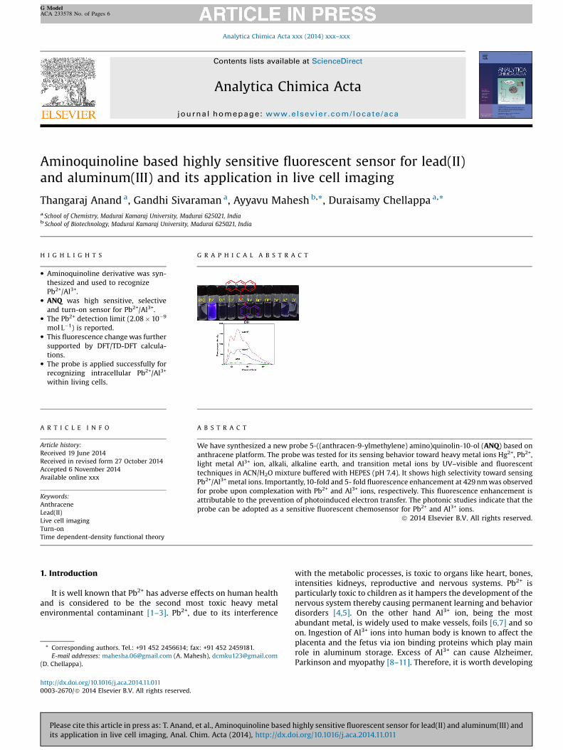
Ethanolic solution containing anthracene-9-carboxaldehyde(0.154 g, 0.74 mmol), 5-amin-8-hydroxyquinoline dihydrochloride(0.17 g, 0.74 mmol) and two drops of triethylamine was refluxed for1 h. The excess solvent was distilled off under reduced pressure.The lime yellow residue was washed with diethylether. Recrystal-lization of the crude of from chloroform yielded dark yellow solid(Scheme 1). Yield: 95%. Melting point: 240 �C. 1H NMR (300 MHz,CDCl3), d: 9.87 (s, 1H), 8.90 (s, 1H), 8.85 (d, J = 9.8 Hz, 3H), 8.60 (s,1H), 8.08 (d, J = 8.3 Hz, 2H), 7.64–7.47 (m, 6H), 7.43 (d, J = 8.1 Hz, 1H),7.30 (s, 1H). 13C NMR (300 MHz, CDCl3) 158.29, 151.02, 148.43,138.17, 133.42, 131.36, 130.84, 129.11, 127.32, 125.67, 125.42, 125.04,124.08, 121.83, 113.78. ESI–MS: calculated: 348. Found: 349.17(ANQ + H)+ (Figs. S1–S3).
3. Results and discussions
The photophysical properties of ANQ were investigated bymonitoring the absorption and fluorescence behavior with theaddition of biologically relevant metal ions such as Mn2+, Fe3+, Ba2+,Co2+, K+, Ni2+, Ca2+, Cu2+, Cr3+, Zn2+, Cd2+, Na+, Hg2+ and Ag+ in ACN:H2O (1:9, V/V) at pH 7.4 (HEPES buffer). The ANQ shows a majorabsorption band at 410 nm in a solvent mixture. During theaddition of Pb2+, the absorbance of ANQ at 410 nm undergoes a redshift by 30 nm which is perceptible by a clear color change fromlime yellow to bluish green (Fig. S4a). Similar experiments forprobe when conducted in a buffer solution with the addition of Al3+, the absorption band at 410 nm suffers a 15 nm blue shift withisobestic points at 431 and 333 nm (Fig. S4b). The well-definedisobestic points throughout the titration of Pb2+/Al3+ clearlyindicates the formation of new species by the influence of Pb2+�ANQ and Al3+�ANQ, respectively. Selectivity of the receptor was
esis of ANQ.
highly sensitive fluorescent sensor for lead(II) and aluminum(III) andi.org/10.1016/j.aca.2014.11.011
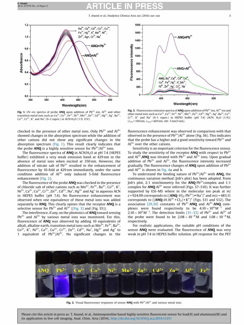
Fig. 1. UV–vis spectra of probe ANQ upon addition of Pb2+ ion, Al3+ and othertransition metal ions such as Cu2+, Co2+, Fe3+, Ni2+, Mn2+, Zn2+, Cd2+, Hg2+, Ag+, Ba2+,Ca2+, Cr3+, K+ and Na+ (0–2 equiv.) in ACN:H2O (1:9, V/V).
Fig. 3. Fluorescence emission spectra of ANQ upon addition of Pb2+ ion, Al3+ ion andother metal ions such as Cu2+, Co2+, Fe3+, Ni2+, Mn2+, Zn2+, Cd2+, Hg2+, Ag+, Ba2+, Ca2+,Cr3+, K+ and Na+ (0–1 equiv.) in HEPES buffer (pH 7.4) (ACN: H2O (1:9)).(lex = 350 nm, lem = 429 nm, slit: 5 nm/5 nm).
T. Anand et al. / Analytica Chimica Acta xxx (2014) xxx–xxx 3
G ModelACA 233578 No. of Pages 6
checked in the presence of other metal ions. Only Pb2+ and Al3+
showed changes in the absorption spectrum while the addition ofother cations did not show any significant changes in theabsorption spectrum (Fig. 1). This result clearly indicates thatthe probe ANQ is a highly sensitive sensor for Pb2+/Al3+ ions.
The fluorescence spectra of ANQ in ACN/H2O at pH 7.4 (HEPESbuffer) exhibited a very weak emission band at 429 nm in theabsence of metal ions when excited at 350 nm. However, theaddition of nitrate salt of Pb2+ resulted in the enhancement offluorescence by 10-fold at 429 nm immediately, under the samecondition addition of Al3+ only induced 5-fold fluorescenceenhancement (Fig. 2).
The fluorescence of the probe ANQ was checked in the presenceof chloride salt of other cations such as Mn2+, Fe3+, Ba2+, Co2+, K+,Ni2+, Ca2+, Cu2+, Cr3+, Zn2+, Cd2+, Na+, Hg2+ and Ag+ in aqueous ACNin HEPES buffer (pH 7.4). No fluorescence enhancement wasobserved when one equivalence of these metal ions was addedseparately to ANQ. This clearly opines that the receptor ANQ is aselective sensor for Pb2+ and Al3+ ((Fig. 3) and (Fig. S5)).
The interference, if any, on the photonics of ANQ toward sensingPb2+ and Al3+ by various metal ions was monitored. For this,fluorescence of ANQ was observed by adding 10 equivalents ofalkali, alkaline earth, transition metal ions such as Mn2+, Fe3+, Ba2+,Co2+, K+, Ni2+, Ca2+, Cu2+, Cr3+, Zn2+, Cd2+, Na+, Hg2+ and Ag+ to1 equivalent of Pb2+/Al3+. No significant changes in the
Fig. 2. Visual fluorescence responses of sensor A
Please cite this article in press as: T. Anand, et al., Aminoquinoline based hits application in live cell imaging, Anal. Chim. Acta (2014), http://dx.do
fluorescence enhancement was observed in comparison with thatobserved in the presence of Pb2+/Al3+ alone (Fig. S6). This indicatesthat the probe has a higher and a good sensitivity toward Pb2+ andAl3+ over the other cations.
Sensitivity is an important criterion for the fluorescence sensor.To study the sensitivity of the receptor ANQ with respect to Pb2+
and Al3+,ANQ was titrated with Pb2+ and Al3+ ions. Upon gradualaddition of Pb2+ and Al3+, the fluorescence intensity increasedgradually. The fluorescence changes of ANQ upon addition of Pb2+
and Al3+ is shown in Fig. 4a and b.To understand the binding nature of Pb2+/Al3+ with ANQ, the
continuous variation method (Job’s plot) has been adopted. FromJob’s plot, 2:1 stoichiometry for the ANQ�Pb2+complex and 1:1complex for ANQ�Al3+ were inferred (Figs. S7–S10). It was furthersupported by ESI–MS where in the molecular ion peak at m/z = 924.09 corresponds to ((ANQ–H)2�Pb2+) + Na+)+ and m/z = 483.11corresponds to ((ANQ–H�Al3+ + Cl2) + K+)+ (Figs. S11 and S12). Theassociation [29,30] constants of Pb2+�ANQ and Al3+�ANQ com-plexes were found respectively to be 4.10 � 104M�1 and2.10 � 102M�1. The detection limits [31–33] of Pb2+ and Al3+ ofthe probe were found to be 2.08 � 10�9M and 1.08 � 10�6M,respectively.
For realistic applications, the suitable pH conditions of thesensor ANQ were evaluated. The fluorescence of ANQ was veryweak in pH 7.4 in HEPES buffer solution. pH response for the PET
NQ with Pb2+/Al3+ and various metal ions.
ighly sensitive fluorescent sensor for lead(II) and aluminum(III) andi.org/10.1016/j.aca.2014.11.011
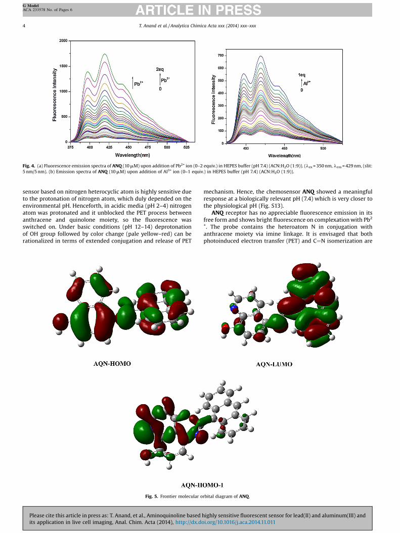
Fig. 4. (a) Fluorescence emission spectra of ANQ (10 mM) upon addition of Pb2+ ion (0–2 equiv.) in HEPES buffer (pH 7.4) (ACN:H2O (1:9)). (lex = 350 nm, lem = 429 nm, (slit:5 nm/5 nm). (b) Emission spectra of ANQ (10 mM) upon addition of Al3+ ion (0–1 equiv.) in HEPES buffer (pH 7.4) (ACN:H2O (1:9)).
4 T. Anand et al. / Analytica Chimica Acta xxx (2014) xxx–xxx
G ModelACA 233578 No. of Pages 6
sensor based on nitrogen heterocyclic atom is highly sensitive dueto the protonation of nitrogen atom, which duly depended on theenvironmental pH. Henceforth, in acidic media (pH 2–4) nitrogenatom was protonated and it unblocked the PET process betweenanthracene and quinolone moiety, so the fluorescence wasswitched on. Under basic conditions (pH 12–14) deprotonationof OH group followed by color change (pale yellow–red) can berationalized in terms of extended conjugation and release of PET
Fig. 5. Frontier molecular o
Please cite this article in press as: T. Anand, et al., Aminoquinoline based
its application in live cell imaging, Anal. Chim. Acta (2014), http://dx.do
mechanism. Hence, the chemosensor ANQ showed a meaningfulresponse at a biologically relevant pH (7.4) which is very closer tothe physiological pH (Fig. S13).
ANQ receptor has no appreciable fluorescence emission in itsfree form and shows bright fluorescence on complexation with Pb2+. The probe contains the heteroatom N in conjugation withanthracene moiety via imine linkage. It is envisaged that bothphotoinduced electron transfer (PET) and C¼N isomerization are
rbital diagram of ANQ.
highly sensitive fluorescent sensor for lead(II) and aluminum(III) andi.org/10.1016/j.aca.2014.11.011
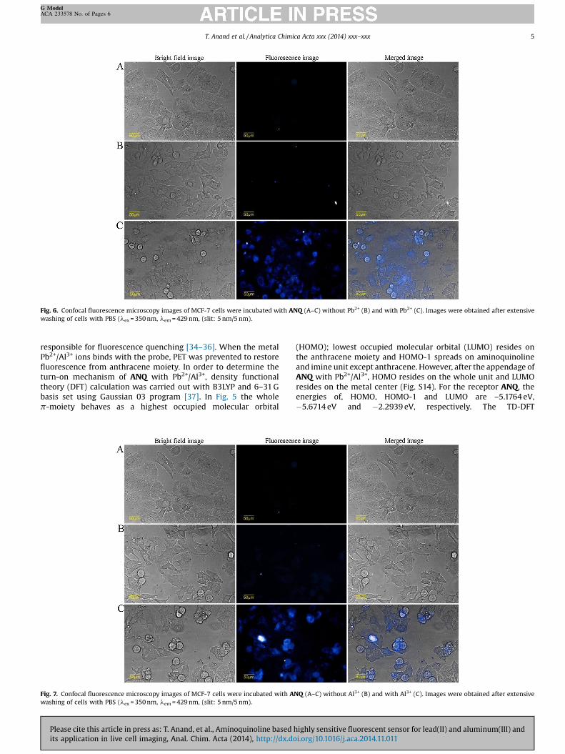
Fig. 6. Confocal fluorescence microscopy images of MCF-7 cells were incubated with ANQ (A–C) without Pb2+ (B) and with Pb2+ (C). Images were obtained after extensivewashing of cells with PBS (lex = 350 nm, lem = 429 nm, (slit: 5 nm/5 nm).
T. Anand et al. / Analytica Chimica Acta xxx (2014) xxx–xxx 5
G ModelACA 233578 No. of Pages 6
responsible for fluorescence quenching [34–36]. When the metalPb2+/Al3+ ions binds with the probe, PET was prevented to restorefluorescence from anthracene moiety. In order to determine theturn-on mechanism of ANQ with Pb2+/Al3+, density functionaltheory (DFT) calculation was carried out with B3LYP and 6–31 Gbasis set using Gaussian 03 program [37]. In Fig. 5 the wholep-moiety behaves as a highest occupied molecular orbital
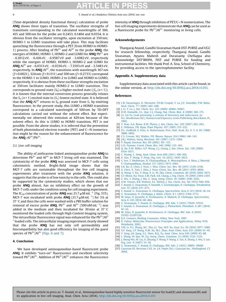
Fig. 7. Confocal fluorescence microscopy images of MCF-7 cells were incubated with Awashing of cells with PBS (lex = 350 nm, lem = 429 nm, (slit: 5 nm/5 nm).
Please cite this article in press as: T. Anand, et al., Aminoquinoline based hits application in live cell imaging, Anal. Chim. Acta (2014), http://dx.do
(HOMO); lowest occupied molecular orbital (LUMO) resides onthe anthracene moiety and HOMO-1 spreads on aminoquinolineand imine unit except anthracene. However, after the appendage ofANQ with Pb2+/Al3+, HOMO resides on the whole unit and LUMOresides on the metal center (Fig. S14). For the receptor ANQ, theenergies of, HOMO, HOMO-1 and LUMO are –5.1764 eV,�5.6714 eV and �2.2939 eV, respectively. The TD-DFT
NQ (A–C) without Al3+ (B) and with Al3+ (C). Images were obtained after extensive
ighly sensitive fluorescent sensor for lead(II) and aluminum(III) andi.org/10.1016/j.aca.2014.11.011
6 T. Anand et al. / Analytica Chimica Acta xxx (2014) xxx–xxx
G ModelACA 233578 No. of Pages 6
(Time-dependent density functional theory) calculation of probeANQ shows three types of transition. The oscillator strengths oftransitions corresponding to the calculated wavelengths of 360,415 and 500 nm for the probe are 0.2453, 0.1404 and 0.0354. It isobvious from the oscillator strengths, upon excitation at 350 nm;HOMO-1 to LUMO transition will take place. This may facilitatequenching the fluorescence through a PET (from HOMO to HOMO-1) process. After binding of Pb2+ and Al3+ to the probe ANQ, theenergies of HOMO, HOMO-1, HOMO-2 and LUMO for ANQ�Pb2+ are�6.1812 eV, �6.418 eV, �6.9587 eV and �3.6982 eV, respectivelywhile the energies of HOMO, HOMO-1, HOMO-2 and LUMO forANQ�Al3+ are �6.0313 eV, �6.9336 eV, �7.3593 eV and �3.5483 eV,respectively. In ANQ�Al3+, the transitions with wavelength 367 nm(f = 0.0021), 324 nm (f = 0.1111) and 500 nm (f = 0.2733) correspondto the HOMO-1 to LUMO, HOMO-2 to LUMO and HOMO to LUMO,respectively. It is obvious from these oscillator strengths excitationat 350 nm facilitates mainly HOMO-2 to LUMO transition. Thiscorresponds to ground state (S0) a higher excited state ((Sn (n > 1)).It is known that the internal conversion process generally relaxesthe Sn (n > 1) excited state to (S1) lowest excited state. It is believedthat the ANQ�Al3+ returns to S0 ground state from S1 by emittingfluorescence. In the present study, this LUMO a HOMO transitioncorrespond to a calculated wavelength of 500 nm by TD-DFTcalculations carried out without the solvent effect. But experi-mentally we observed this emission at 429 nm because of thesolvent effect. As this is LUMO to HOMO transition, PET is notpossible. From the above studies, it is inferred that the preventionof both photoinduced electron transfer (PET) and C¼N isomeriza-tion might be the reason for the enhancement of fluorescence forthe ANQ–Al3+/Pb2+.
3.1. Live cell imaging
The ability of anthracene linked aminoquinoline probe ANQ todetermine Pb2+ and Al3+ in MCF-7 living cell was examined. Thecytotoxicity of the probe ANQ was assessed in MCF-7 cells usingcalorimetric method. Bright-field image shows that theMCF-7 cells are sustainable throughout the live cell imagingexperiments after treatment with the probe ANQ solution, itsuggests that the probe is of low toxicity to the cells. This could alsobe supported by the cytotoxicity studies, which shows that ourprobe ANQ almost, has no inhibitory effect on the growth ofMCF-7 cells under the condition using for cell imaging experiment.The IC50 concentration of probe ANQ was 21.7 mM mL�1 (Fig. S15),MCF-7 was incubated with probe ANQ (21.7 mM mL�1) for 3 h at37 �C and then the cells were washed with a PBS buffer solution forremoval of excess probe ANQ. Pb2+ and Al3+ (500 nM mL�1) wasadded in the medium and then incubated for 30 min at 37 �Cmonitored the loaded cells through High Content Imaging system.The intracellular fluorescence signal was enhanced for the Pb2+/Al3+ loaded cells. The intracellular imaging experiment clearly showedthat the probe ANQ has not only cell permeability andbiocompatibility but also good efficiency for imaging of the guestspecies of Pb2+/Al3+ (Figs. 6 and 7).
4. Conclusion
We have developed aminoquinoline-based fluorescent probeANQ. It exhibits “turn-on” fluorescence and excellent selectivitytoward Pb2+/Al3+. Addition of Pb2+/Al3+ enhances the fluorescence
Please cite this article in press as: T. Anand, et al., Aminoquinoline based
its application in live cell imaging, Anal. Chim. Acta (2014), http://dx.do
intensity of ANQ through inhibition of PET/C¼N isomerization. Thelive cell imaging experiments demonstrate that ANQ can be used asa fluorescent probe for Pb2+/Al3+ monitoring in living cells.
Acknowledgements
Thangaraj Anand, Gandhi Sivaraman thank DST-PURSE and UGCfor research fellowship, respectively. Thangaraj Anand, GandhiSivaraman, Ayyavu Mahesh and Duraisamy Chellappa alsoacknowledge DST’IRHPA, FIST and PURSE for funding andinstrumental facilities. We thank Prof. A. Siva, School of Chemistry,for providing access to the spectrophotometer facility.
Appendix A. Supplementary data
Supplementary data associated with this article can be found, inthe online version, at http://dx.doi.org/10.1016/j.aca.2014.11.011.
References
[1] C.B. Swearingen, D. Wernette, P.D.M. Cropek, Y. Lu, J.V. Sweedler, P.W. Bohn,Anal. Chem. 77 (2005) 442–448.
[2] J. Li, Y. Lu, J. Am. Chem. Soc. 122 (2000) 10466–10467.[3] D.W. Domaille, E.L. Que, C.J. Chang, Nat. Chem. Biol. 4 (2008) 168–175.[4] J.S. Liu Fu, Lead poisoning, a century of discovery and rediscovery, in:
H.L. Needleman (Ed.), Human Lead Exposure, Lewis Publishing, Boca Raton, FL,1992.
[5] Y. Xiao, A.A. Rowe, K.W. Plaxco, J. Am. Chem. Soc. 129 (2007) 262–263.[6] E. Delhaize, P.R. Ryan, Plant Physiol. 107 (1995) 315–332.[7] D.L. Godbold, E. Fritz, A. Huttermann, Proc. Natl. Acad. Sci. U. S. A. 85 (1988)
3888–3892.[8] C.S. Cronan, W.J. Walker, P.R. Bloom, Nature 324 (1986) 140–143.[9] J.R.J. Walton, Inorg. Biochem. 101 (2007) 1275–1284.
[10] P. Nayak, Environ. Res. 89 (2002) 101–115.[11] G.D. Fasman, Coord. Chem. Rev. 149 (1996) 125–165.[12] Q. He, E.W. Miller, A.P. Wong, C.J. Chang, J. Am. Chem. Soc. 128 (2006)
9316–9317.[13] C. Huang, C. Ding, Anal. Chim. Acta 699 (2011) 198–205.[14] X. Sun, Y. Wang, Y. Peng, Org. Lett. 14 (2012) 3420–3423.[15] S. Sen, T. Mukherjee, B. Chattopadhyay, A. Moirangthem, A. Basu, J. Marekd,
P. Chattopadhyay, Analyst 137 (2012) 3975–3981.[16] S.K. Ko, X. Chen, J. Yoon, I. Shin, Chem. Soc. Rev. 40 (2011) 2120–2130.[17] X. Chen, X. Tian, I. Shin, J. Yoon, Chem. Soc. Rev. 40 (2011) 4783–4804.[18] Q. Wang, Y. Xie, Y. Ding, X. Li, W. Zhu, Chem. Commun. 46 (2010) 3669–3671.[19] S.Y. Moon, N.J. Youn, S.M. Park, S.K. Chang, J. Org. Chem. 70 (2005) 2394–2397.[20] Z. Wu, Y. Zhang, J. Ma, G. Yang, Inorg. Chem. 45 (2006) 3140–3142.[21] A.W. Varnes, R.B. Dodson, E.L. Wehry, J. Am. Chem. Soc. 94 (1972) 946–950.[22] T. Anand, G. Sivaraman, P. Anandh, S. Govindarajan, D. Chellappa, Tetrahedron
Lett. 55 (2014) 671–675.[23] T. Anand, G. Sivaraman, D. Chellappa, Spectrochim. Acta A 123 (2014) 18–24.[24] G. Sivaraman, D. Chellappa, J. Mater. Chem. B 1 (2013) 5768–5772.[25] M. Iniya, D. Jeyanthi, K. Krishnaveni, A. Mahesh, D. Chellappa, Spectrochim.
Acta A 120 (2014) 40–464.[26] G. Sivaraman, T. Anand, D. Chellappa, RSC Adv. 3 (2013) 17029–17033.[27] T. Anand, G. Sivaraman, D. Chellappa, J. Photochem. Photobiol. A 281 (2014)
47–52.[28] M. Iniya, D. Jeyanthi, K. Krishnaveni, D. Chellappa, RSC Adv. 4 (2014)
25393–25397120.[29] K.A. Connors, Binding Constants, Wiley, New York, 1987.[30] B. Valeur, Molecular Fluorescence Principles and Applications, Wiley VCH,
Weinheim, 2002.[31] Y.M. Li, X.L. Zhang, B.C. Zhu, J.L. Yan, W.P. Xu, Anal. Sci. 26 (2010) 1077–1080.[32] X.F. Yang, L.P. Wang, H.M. Xu, M.L. Zhao, Anal. Chim. Acta. 631 (2009) 91–95.[33] Y. Yang, J.H. Jiang, G.L. Shen, R.Q. Yu, Anal. Chim. Acta 636 (2009) 83–88.[34] L. Wang, W. Qin, W. Liu, Inorg. Chem. Commun. 13 (2010) 1122–1125.[35] J. Sheng Wu, W. Liu, X. Zhuang, F. Wang, P. Wang, S. Tao, X. Zhang, S. Wu, S. Lee,
Org. Lett. 9 (2007) 33–36.[36] G. Sivaraman, T. Anand, D. Chellappa, RSC Adv. 2 (2012) 10605–10609.[37] Gaussian 03, Revision C.02, in: J.A. Pople (Ed.), Gaussian Inc., Wallingford, CT,
2004.
highly sensitive fluorescent sensor for lead(II) and aluminum(III) andi.org/10.1016/j.aca.2014.11.011